US7374870B2 - Systems and methods for removing viral agents from blood - Google Patents
Systems and methods for removing viral agents from bloodDownload PDFInfo
- Publication number
- US7374870B2 US7374870B2US11/056,347US5634705AUS7374870B2US 7374870 B2US7374870 B2US 7374870B2US 5634705 AUS5634705 AUS 5634705AUS 7374870 B2US7374870 B2US 7374870B2
- Authority
- US
- United States
- Prior art keywords
- plasma
- filter
- container
- path
- kit
- Prior art date
- Legal status (The legal status is an assumption and is not a legal conclusion. Google has not performed a legal analysis and makes no representation as to the accuracy of the status listed.)
- Expired - Fee Related, expires
Links
- 238000000034methodMethods0.000titleclaimsabstractdescription37
- 210000004369bloodAnatomy0.000titledescription25
- 239000008280bloodSubstances0.000titledescription25
- 230000003612virological effectEffects0.000titledescription25
- 239000000463materialSubstances0.000claimsabstractdescription102
- 230000001413cellular effectEffects0.000claimsabstractdescription34
- 239000000356contaminantSubstances0.000claimsabstractdescription19
- 230000005855radiationEffects0.000claimsabstractdescription6
- 238000012546transferMethods0.000claimsdescription38
- 239000000203mixtureSubstances0.000claimsdescription20
- 238000013022ventingMethods0.000claimsdescription6
- 238000002156mixingMethods0.000claimsdescription3
- 238000001914filtrationMethods0.000abstractdescription30
- 210000002381plasmaAnatomy0.000description96
- 210000000265leukocyteAnatomy0.000description39
- 241000700605VirusesSpecies0.000description32
- 238000003860storageMethods0.000description28
- 239000003795chemical substances by applicationSubstances0.000description23
- 238000012545processingMethods0.000description23
- 241000894007speciesSpecies0.000description16
- RBTBFTRPCNLSDE-UHFFFAOYSA-N3,7-bis(dimethylamino)phenothiazin-5-iumChemical compoundC1=CC(N(C)C)=CC2=[S+]C3=CC(N(C)C)=CC=C3N=C21RBTBFTRPCNLSDE-UHFFFAOYSA-N0.000description14
- 229960000907methylthioninium chlorideDrugs0.000description14
- 239000011148porous materialSubstances0.000description12
- 238000001228spectrumMethods0.000description10
- 230000009467reductionEffects0.000description9
- 238000006722reduction reactionMethods0.000description9
- 241000725303Human immunodeficiency virusSpecies0.000description8
- 210000001772blood plateletAnatomy0.000description8
- 230000008569processEffects0.000description8
- 239000000306componentSubstances0.000description7
- 230000007717exclusionEffects0.000description7
- 239000012530fluidSubstances0.000description7
- 239000012528membraneSubstances0.000description7
- XLYOFNOQVPJJNP-UHFFFAOYSA-NwaterSubstancesOXLYOFNOQVPJJNP-UHFFFAOYSA-N0.000description7
- 210000003743erythrocyteAnatomy0.000description5
- 230000002779inactivationEffects0.000description5
- 239000007788liquidSubstances0.000description5
- 239000004033plasticSubstances0.000description5
- 229920003023plasticPolymers0.000description5
- 238000011045prefiltrationMethods0.000description5
- 230000004888barrier functionEffects0.000description4
- 230000015556catabolic processEffects0.000description4
- 238000006731degradation reactionMethods0.000description4
- 239000012678infectious agentSubstances0.000description4
- 238000002428photodynamic therapyMethods0.000description4
- 238000004886process controlMethods0.000description4
- 241000701093Suid alphaherpesvirus 1Species0.000description3
- 230000008901benefitEffects0.000description3
- 210000004027cellAnatomy0.000description3
- 238000010276constructionMethods0.000description3
- 238000011010flushing procedureMethods0.000description3
- 230000006870functionEffects0.000description3
- 150000002632lipidsChemical class0.000description3
- 230000002186photoactivationEffects0.000description3
- 230000001954sterilising effectEffects0.000description3
- 238000004659sterilization and disinfectionMethods0.000description3
- 230000003253viricidal effectEffects0.000description3
- 241000710780Bovine viral diarrhea virus 1Species0.000description2
- 241000725618Duck hepatitis B virusSpecies0.000description2
- 241000710188Encephalomyocarditis virusSpecies0.000description2
- 102000009123FibrinHuman genes0.000description2
- 108010073385FibrinProteins0.000description2
- BWGVNKXGVNDBDI-UHFFFAOYSA-NFibrin monomerChemical compoundCNC(=O)CNC(=O)CNBWGVNKXGVNDBDI-UHFFFAOYSA-N0.000description2
- 241000700588Human alphaherpesvirus 1Species0.000description2
- 238000010521absorption reactionMethods0.000description2
- 230000005540biological transmissionEffects0.000description2
- PPBOKXIGFIBOGK-BDTUAEFFSA-NbvdvChemical compoundC([C@@H](C(=O)N[C@@H](CCCCN)C(=O)N[C@@H](CCC(N)=O)C(=O)N[C@@H]([C@@H](C)CC)C(=O)N[C@@H](CC(N)=O)C(=O)N[C@@H](CCCNC(N)=N)C(=O)NCC(=O)N[C@@H](CC(C)C)C(=O)N[C@@H](CCCCN)C(=O)N[C@@H](CCCCN)C(O)=O)NC(=O)[C@H](CC(C)C)NC(=O)[C@H](CO)NC(=O)[C@H](CC=1C=CC(O)=CC=1)NC(=O)[C@@H](NC(=O)[C@H](CC(C)C)NC(=O)[C@H](CC(O)=O)NC(=O)[C@H](CC(C)C)NC(=O)[C@@H](N)C(C)C)[C@@H](C)CC)C1=CN=CN1PPBOKXIGFIBOGK-BDTUAEFFSA-N0.000description2
- 239000006227byproductSubstances0.000description2
- 229920002678cellulosePolymers0.000description2
- 239000000470constituentSubstances0.000description2
- 229950003499fibrinDrugs0.000description2
- 208000002672hepatitis BDiseases0.000description2
- 238000005286illuminationMethods0.000description2
- 230000003834intracellular effectEffects0.000description2
- QTWZICCBKBYHDM-UHFFFAOYSA-Nleucomethylene blueChemical compoundC1=C(N(C)C)C=C2SC3=CC(N(C)C)=CC=C3NC2=C1QTWZICCBKBYHDM-UHFFFAOYSA-N0.000description2
- 230000007246mechanismEffects0.000description2
- 102000039446nucleic acidsHuman genes0.000description2
- 108020004707nucleic acidsProteins0.000description2
- 150000007523nucleic acidsChemical class0.000description2
- 230000002093peripheral effectEffects0.000description2
- 238000007540photo-reduction reactionMethods0.000description2
- 239000000906photoactive agentSubstances0.000description2
- 239000000049pigmentSubstances0.000description2
- -1poly(ethylene vinyl acetate)Polymers0.000description2
- 229920000728polyesterPolymers0.000description2
- 230000001225therapeutic effectEffects0.000description2
- 208000030507AIDSDiseases0.000description1
- 241000272525Anas platyrhynchosSpecies0.000description1
- 102000015081Blood Coagulation FactorsHuman genes0.000description1
- 108010039209Blood Coagulation FactorsProteins0.000description1
- 102000004506Blood ProteinsHuman genes0.000description1
- 108010017384Blood ProteinsProteins0.000description1
- 241000282461Canis lupusSpecies0.000description1
- 241000450599DNA virusesSpecies0.000description1
- MYMOFIZGZYHOMD-UHFFFAOYSA-NDioxygenChemical compoundO=OMYMOFIZGZYHOMD-UHFFFAOYSA-N0.000description1
- 239000004593EpoxySubstances0.000description1
- 238000012424Freeze-thaw processMethods0.000description1
- 208000031220HemophiliaDiseases0.000description1
- 208000009292Hemophilia ADiseases0.000description1
- 208000032843HemorrhageDiseases0.000description1
- 241000700721Hepatitis B virusSpecies0.000description1
- 208000005176Hepatitis CDiseases0.000description1
- 241000713772Human immunodeficiency virus 1Species0.000description1
- 241000702617Human parvovirus B19Species0.000description1
- 206010061598ImmunodeficiencyDiseases0.000description1
- 208000029462Immunodeficiency diseaseDiseases0.000description1
- 239000004677NylonSubstances0.000description1
- 239000002033PVDF binderSubstances0.000description1
- 206010034960PhotophobiaDiseases0.000description1
- 229920012485Plasticized Polyvinyl chloridePolymers0.000description1
- 229920012266Poly(ether sulfone) PESPolymers0.000description1
- 239000004695Polyether sulfoneSubstances0.000description1
- 230000002745absorbentEffects0.000description1
- 239000002250absorbentSubstances0.000description1
- 229920006243acrylic copolymerPolymers0.000description1
- 238000001994activationMethods0.000description1
- 239000000654additiveSubstances0.000description1
- 208000007502anemiaDiseases0.000description1
- 238000003556assayMethods0.000description1
- 230000000740bleeding effectEffects0.000description1
- 239000010836blood and blood productSubstances0.000description1
- 239000003114blood coagulation factorSubstances0.000description1
- 239000012503blood componentSubstances0.000description1
- 229940125691blood productDrugs0.000description1
- 230000036770blood supplyEffects0.000description1
- 239000000872bufferSubstances0.000description1
- 239000005025cast polypropyleneSubstances0.000description1
- 238000005119centrifugationMethods0.000description1
- 238000006243chemical reactionMethods0.000description1
- 230000006378damageEffects0.000description1
- 238000001514detection methodMethods0.000description1
- 238000000295emission spectrumMethods0.000description1
- 244000309457enveloped RNA virusSpecies0.000description1
- 230000008029eradicationEffects0.000description1
- 239000005038ethylene vinyl acetateSubstances0.000description1
- 238000001704evaporationMethods0.000description1
- 230000008020evaporationEffects0.000description1
- 229960000301factor viiiDrugs0.000description1
- 239000000835fiberSubstances0.000description1
- 238000005194fractionationMethods0.000description1
- 239000004023fresh frozen plasmaSubstances0.000description1
- 239000011521glassSubstances0.000description1
- 239000003365glass fiberSubstances0.000description1
- 230000005484gravityEffects0.000description1
- 230000007813immunodeficiencyEffects0.000description1
- 230000000415inactivating effectEffects0.000description1
- 238000007689inspectionMethods0.000description1
- 230000001678irradiating effectEffects0.000description1
- 208000013469light sensitivityDiseases0.000description1
- 239000006193liquid solutionSubstances0.000description1
- 230000002934lysing effectEffects0.000description1
- 238000004519manufacturing processMethods0.000description1
- 244000309711non-enveloped virusesSpecies0.000description1
- 229920001778nylonPolymers0.000description1
- 239000005026oriented polypropyleneSubstances0.000description1
- 230000001717pathogenic effectEffects0.000description1
- 238000001782photodegradationMethods0.000description1
- 229920001200poly(ethylene-vinyl acetate)Polymers0.000description1
- 229920002492poly(sulfone)Polymers0.000description1
- 239000004417polycarbonateSubstances0.000description1
- 229920000515polycarbonatePolymers0.000description1
- 229920006393polyether sulfonePolymers0.000description1
- 229920002981polyvinylidene fluoridePolymers0.000description1
- 238000003825pressingMethods0.000description1
- 238000003908quality control methodMethods0.000description1
- 238000009877renderingMethods0.000description1
- 230000031070response to heatEffects0.000description1
- 238000007789sealingMethods0.000description1
- 230000035945sensitivityEffects0.000description1
- 238000000926separation methodMethods0.000description1
- 239000000126substanceSubstances0.000description1
- 239000000725suspensionSubstances0.000description1
- 230000008685targetingEffects0.000description1
- 238000012360testing methodMethods0.000description1
- 238000002560therapeutic procedureMethods0.000description1
- 239000001016thiazine dyeSubstances0.000description1
- 230000003582thrombocytopenic effectEffects0.000description1
- 231100000027toxicologyToxicity0.000description1
- 229960000850trioxysalenDrugs0.000description1
- 241001529453unidentified herpesvirusSpecies0.000description1
- 241000712461unidentified influenza virusSpecies0.000description1
- 238000011144upstream manufacturingMethods0.000description1
- 238000001429visible spectrumMethods0.000description1
- 238000011179visual inspectionMethods0.000description1
- 238000003466weldingMethods0.000description1
Images


Classifications
- A—HUMAN NECESSITIES
- A61—MEDICAL OR VETERINARY SCIENCE; HYGIENE
- A61L—METHODS OR APPARATUS FOR STERILISING MATERIALS OR OBJECTS IN GENERAL; DISINFECTION, STERILISATION OR DEODORISATION OF AIR; CHEMICAL ASPECTS OF BANDAGES, DRESSINGS, ABSORBENT PADS OR SURGICAL ARTICLES; MATERIALS FOR BANDAGES, DRESSINGS, ABSORBENT PADS OR SURGICAL ARTICLES
- A61L2/00—Methods or apparatus for disinfecting or sterilising materials or objects other than foodstuffs or contact lenses; Accessories therefor
- A61L2/0005—Methods or apparatus for disinfecting or sterilising materials or objects other than foodstuffs or contact lenses; Accessories therefor for pharmaceuticals, biologicals or living parts
- A61L2/0011—Methods or apparatus for disinfecting or sterilising materials or objects other than foodstuffs or contact lenses; Accessories therefor for pharmaceuticals, biologicals or living parts using physical methods
- C—CHEMISTRY; METALLURGY
- C12—BIOCHEMISTRY; BEER; SPIRITS; WINE; VINEGAR; MICROBIOLOGY; ENZYMOLOGY; MUTATION OR GENETIC ENGINEERING
- C12N—MICROORGANISMS OR ENZYMES; COMPOSITIONS THEREOF; PROPAGATING, PRESERVING, OR MAINTAINING MICROORGANISMS; MUTATION OR GENETIC ENGINEERING; CULTURE MEDIA
- C12N7/00—Viruses; Bacteriophages; Compositions thereof; Preparation or purification thereof
- C12N7/04—Inactivation or attenuation; Producing viral sub-units
- A—HUMAN NECESSITIES
- A61—MEDICAL OR VETERINARY SCIENCE; HYGIENE
- A61K—PREPARATIONS FOR MEDICAL, DENTAL OR TOILETRY PURPOSES
- A61K35/00—Medicinal preparations containing materials or reaction products thereof with undetermined constitution
- A61K35/12—Materials from mammals; Compositions comprising non-specified tissues or cells; Compositions comprising non-embryonic stem cells; Genetically modified cells
- A61K35/14—Blood; Artificial blood
- A61K35/16—Blood plasma; Blood serum
- A—HUMAN NECESSITIES
- A61—MEDICAL OR VETERINARY SCIENCE; HYGIENE
- A61L—METHODS OR APPARATUS FOR STERILISING MATERIALS OR OBJECTS IN GENERAL; DISINFECTION, STERILISATION OR DEODORISATION OF AIR; CHEMICAL ASPECTS OF BANDAGES, DRESSINGS, ABSORBENT PADS OR SURGICAL ARTICLES; MATERIALS FOR BANDAGES, DRESSINGS, ABSORBENT PADS OR SURGICAL ARTICLES
- A61L2/00—Methods or apparatus for disinfecting or sterilising materials or objects other than foodstuffs or contact lenses; Accessories therefor
- A61L2/02—Methods or apparatus for disinfecting or sterilising materials or objects other than foodstuffs or contact lenses; Accessories therefor using physical phenomena
- A61L2/022—Filtration
- A—HUMAN NECESSITIES
- A61—MEDICAL OR VETERINARY SCIENCE; HYGIENE
- A61L—METHODS OR APPARATUS FOR STERILISING MATERIALS OR OBJECTS IN GENERAL; DISINFECTION, STERILISATION OR DEODORISATION OF AIR; CHEMICAL ASPECTS OF BANDAGES, DRESSINGS, ABSORBENT PADS OR SURGICAL ARTICLES; MATERIALS FOR BANDAGES, DRESSINGS, ABSORBENT PADS OR SURGICAL ARTICLES
- A61L2/00—Methods or apparatus for disinfecting or sterilising materials or objects other than foodstuffs or contact lenses; Accessories therefor
- A61L2/02—Methods or apparatus for disinfecting or sterilising materials or objects other than foodstuffs or contact lenses; Accessories therefor using physical phenomena
- A61L2/08—Radiation
- C—CHEMISTRY; METALLURGY
- C12—BIOCHEMISTRY; BEER; SPIRITS; WINE; VINEGAR; MICROBIOLOGY; ENZYMOLOGY; MUTATION OR GENETIC ENGINEERING
- C12N—MICROORGANISMS OR ENZYMES; COMPOSITIONS THEREOF; PROPAGATING, PRESERVING, OR MAINTAINING MICROORGANISMS; MUTATION OR GENETIC ENGINEERING; CULTURE MEDIA
- C12N7/00—Viruses; Bacteriophages; Compositions thereof; Preparation or purification thereof
- C12N7/04—Inactivation or attenuation; Producing viral sub-units
- C12N7/06—Inactivation or attenuation by chemical treatment
- Y—GENERAL TAGGING OF NEW TECHNOLOGICAL DEVELOPMENTS; GENERAL TAGGING OF CROSS-SECTIONAL TECHNOLOGIES SPANNING OVER SEVERAL SECTIONS OF THE IPC; TECHNICAL SUBJECTS COVERED BY FORMER USPC CROSS-REFERENCE ART COLLECTIONS [XRACs] AND DIGESTS
- Y10—TECHNICAL SUBJECTS COVERED BY FORMER USPC
- Y10S—TECHNICAL SUBJECTS COVERED BY FORMER USPC CROSS-REFERENCE ART COLLECTIONS [XRACs] AND DIGESTS
- Y10S435/00—Chemistry: molecular biology and microbiology
- Y10S435/975—Kit
Definitions
- the inventiongenerally relates to the eradication of contaminants using photodynamic therapy.
- the inventionalso generally relates to the processing of whole blood and its components for storage and transfusion.
- the inventionrelates to the extracorporeal treatment of collected whole blood and its components with photoactive materials to eradicate viruses and other pathogenic contaminants.
- the clinically proven components of whole bloodinclude red blood cells, used to treat chronic anemia; platelet-poor plasma, from which Clotting Factor VIII-rich cryoprecipitate can be obtained for the treatment of hemophilia; and concentrations of platelets, used to control thrombocytopenic bleeding.
- photodynamic therapyhas been suggested as a way to eradicate infectious agents from collected blood and its components. Still, there has been a general lack of success in economically adapting the benefits of photodynamic therapy to the demands of the blood banking industry.
- One reason for thisis that not all biological contaminants are carried free within the blood where they can be readily coupled to photoactive agents. Some biological contaminants are entrained on or within white blood cells out of the reach of photoactive agents.
- the inventionprovides improved systems and methods for treating blood constituents to adventitious viral agents.
- One aspect of the inventionprovides systems and methods which remove viral agents from plasma.
- the systems and methodsremove from the plasma targeted cellular matter that does or might entrain viral agents.
- the targeted cellular matterincludes leukocytes.
- the system and methodsadd to the plasma a photoactive material, which binds to viral agents that are free of entrainment by the targeted cellular matter. Radiation emitted at a selected wavelength into the plasma activates the photoactive material and thereby eradicates the free viral agents.
- a system for treating plasmacomprises tubing adapted to be coupled a plasma source, and a filter in the tubing to separate cellular matter from the plasma conveyed from the source.
- the systemincludes a transfer container coupled to the tubing to receive cellular matter-reduced plasma from the filter, and a source of photoactive material to be mixed with the plasma.
- the tubingincludes a path to vent air from the transfer container in a path that bypasses the filter.
- systems and methodsremove viral agents entrained within the cellular matter by conveying plasma in a first path through a filter.
- the systems and methodsconvey the cellular matter-reduced plasma from the filter in a second path, which includes a connected transfer container.
- the systems and methodsmix the cellular matter-reduced plasma with a photoactive material within the transfer container, forming a plasma mixture.
- the systems and methodsconvey a portion of the plasma mixture from the transfer container in a flush path, which includes the second path, to thereby expose residual contaminants in the second path-to the photoactive material.
- the systems and methodsthen separate the transfer container from the filter by severing the second path. After severance from the filter, a remnant of the second path remains attached to the transfer container. However, due to the prior flushing step, all contaminants in the attached second path remnant have been exposed to the photoactive material. Subsequent radiation of the transfer container thereby eradicates contaminants, which are free of entrainment by cellular matter, both within the transfer container and the attached second path remnant.
- the flush pathby passes the filter and also provides a path to vent air from the transfer container.
- Another aspect of the inventionprovides systems and methods for treating plasma using multi-stage filtration, which targets for removal different species of cellular matter.
- the systems and methodsseparate a first species of cellular matter by filtration through a first filter media, thereby removing contaminants entrained within the first species of cellular matter.
- the systems and methodsseparating a second species of cellular matter by filtration through a second filter media, thereby removing contaminants entrained within the second species of cellular matter.
- the systems and methodsadd to the plasma a photoactive material and emit radiation at a selected wavelength into the plasma to activate the photoactive material, thereby eradicating the contaminant that is free of entrainment by cellular matter.
- the first filtration mediatargets leukocytes for removal
- the second filtration mediatargets platelets for removal.
- kitsthat envelopes photoactive material in an overwrap that includes a light filtering material.
- the light filtering materialabsorbs light that activates the photoactive material.
- the presence of the light filtering material in the overwrapprotects the photoactive material from photo-degradation due to absorption of ambient light during handling and storage prior to use.
- the photoactive material within the kitincludes methylene blue.
- the light filtering materialincludes a blue material having phtalocyanine pigments.
- the photoactive materialis contained in liquid form within the kit.
- the overwrapalso includes material that reduces liquid vapor loss from the kit.
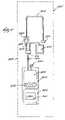
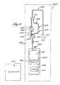
- FIG. 1is a plane view of a blood processing and storage kit for reducing the presence of viral agents in plasma
- FIG. 2is an exploded, perspective view of the laminated walls of the overwrap envelope shown in phantom lines in FIG. 1 ;
- FIG. 3is a side view of the laminated walls of the overwrap envelope shown in FIG. 2 ;
- FIG. 4is a top perspective view of the laminated walls of the overwrap envelope, after having been joined by a peripheral heat seal;
- FIG. 5is an exploded side view of the leukocyte reduction filter that forms a part of the kit shown in FIG. 1 ;
- FIG. 6is a top perspective view of the interior of the outlet housing part for the filter shown in FIG. 5 ;
- FIG. 7is a plane view the kit shown in FIG. 1 being used to convey plasma from a source container, through the leukocyte reduction filter, and into the processing and storage container;
- FIG. 8Ais a plane view the kit shown in FIG. 7 being used to vent air and residual plasma from the processing and storage container in a bypass path around the leukocyte reduction filter;
- FIG. 8Bis a plane view of the kit shown in FIG. 8A being used to flush the tubing section next to the container with photoactive material, to assure exposure of residual viruses occupying the tubing section with photoactive material;
- FIG. 9is a perspective view of the kit shown in FIGS. 8A and 8B , after separation of the processing and storage container and placement of the processing and storage container in an irradiation chamber;
- FIG. 10is a plane view of an alternative embodiment of a blood processing and storage kit for reducing the presence of viral agents in plasma, in which the photoactive material is stored within an auxiliary container whose walls include a light filtering material;
- FIG. 11is a plane view of an alternative embodiment of a blood processing and storage kit for reducing the presence of viral agents in plasma, which includes an integrally attached air reservoir;
- FIG. 12Ais a plane view of the kit shown in FIG. 11 being use to vent air and residual plasma from the processing and storage container into the air reservoir;
- FIG. 12Bis a plane view of the kit shown in FIG. 12A being used to flush the tubing section next to the container with photoactive material, to assure exposure of residual viruses occupying the tubing section with photoactive material;
- FIG. 13is a plane view of another alternative embodiment of a blood processing and storage kit for reducing the presence of viral agents in plasma, which reduces the presence of viral agents in plasma by the removal by filtration of least two different cellular blood species which actually do or potentially can entrain viral agents.
- FIG. 1shows a blood constituent processing and storage set or kit 300 .
- the kit 300is intended, during use, to assist in the removal of viral agents from plasma.
- the viral agentsare either carried free within the plasma or are entrained on or within cellular matter (e.g., red blood cells, platelets, and leukocytes) that the plasma carries.
- the kit 300 shown in FIG. 1will be described in the context of reducing the presence of viral agents in single donor units of plasma, because it is particularly well suited for this purpose.
- the kit 300includes a processing and storage container 302 , which carries an integrally attached length of flexible transfer tubing 304 .
- the transfer tubing 304is made from medical grade plasticized polyvinyl chloride plastic.
- other flexible medical grade plastic materialscan be used.
- the transfer tubing 304includes an integrally attached in-line filter 306 .
- the filter 306includes a filter media 307 (see FIG. 5 ) that removes from plasma cellular matter that does actually or potentially entrain viral agents.
- the filter media 307is encased within a two part housing 348 A and 348 B made, for example, from polycarbonate, although any engineering medical grade plastic with appropriate toxicology characteristics can be used.
- the housing 348 A/ 348 Bis sealed about the filter media 307 by, for example, sonic welding.
- the pore size of the filter media 307can be tailored to remove by exclusion all or some species of cellular matter found in plasma, depending upon the extent to which viral agents sought to be eliminated are entrained by the different cellular species.
- the principal cellular species targets of the filter 306are leukocytes, for it is known that leukocytes entrain many viral agents.
- the filter media 307comprises a non-fibrous membrane having a pore size smaller than the size of leukocytes, to thereby remove leukocytes by exclusion.
- the media 307also includes a prefilter material, which removes fibrin clots and other large size aggregates from the plasma.
- the composition of the membrane for the media 307can vary.
- hydrophilic membranesmade from nylon, acrylic copolymers, polysulfone, polyvinylidene fluoride, mixed cellulose esters, and cellulose ester can be used to remove leukocytes by exclusion.
- Non-hydrophilic membranescan also be treated to serve as a membrane for the filter media 307 .
- the composition of the prefilter for the media 307can vary.
- the prefiltercan comprise fibers of glass or polyester. Material selection takes into account customer preferences, performance objectives, and manufacturing requirements, including sterilization techniques.
- the filter media 307includes three filter media layers 342 , 344 , and 346 .
- the first filter media layer 342comprises USP Grade VI glass fiber or the equivalent.
- the second and third filter media layers 344 and 346comprise polyethersulfone (PES) membranes, which remove leukocytes by exclusion.
- PESpolyethersulfone
- the second and third filter media layers 344 and 346possess pore sizes which are approximately 10 fold smaller than the size of leukocytes and which decrease in the direction of flow.
- the second filter media layer 344has a pore size in the range of about 0.9 ⁇ m to about 2.0 ⁇ m, with an average pore size of about 1.2 ⁇ m.
- the third filter media layer 346has a smaller pore size in the range of about 0.3 ⁇ m to about 1.5 ⁇ m, with an average pores size of about 0.8 ⁇ m.
- the second and third filter media layers 344 and 346also incidently remove red blood cells by exclusion.
- the filter media 307should preferably be capable of filtering 310 ml of plasma, suspended at a head height of 3 feet, in 20 minutes.
- the housing part 348 Aincludes an inlet 350 , which, in use, conveys plasma and leukocytes into contact with the prefilter layer 342 .
- the axis 351 of the inlet 350is generally parallel to the plane of the layer 342 to uniformly perfuse plasma across the entire prefilter layer 342 .
- the housing part 348 Bincludes an outlet 352 , which conveys leukocyte-reduced plasma from the second and third PES filter layers 344 and 346 .
- the interior surface of the housing part 348 Bis grooved, creating a fluid manifold 354 that uniformly distributes leukocyte-reduced plasma to the outlet 352 .
- a length of branch tubing 308is integrally attached to the transfer tubing 304 by conventional Y-connectors 316 .
- the branch tubing 308forms a fluid path bypassing the filter 306 .
- the branch tubing 308serves to vent air.
- the far end of the transfer tubing 304carries an air pillow 310 .
- the air pillow 310prevents collapse of the tubing 304 and 308 caused by pressure differentials during steam sterilization of the kit 300 .
- the transfer tubing 304further includes a conventional in-line frangible cannula 312 between the filter outlet 352 and the processing and storage container 302 .
- the cannula 312normally closes fluid the transfer tubing 304 to fluid flow.
- the cannula 312can be constructed in various ways.
- U.S. Pat. Nos. 4,181,140 and 4,294,247disclose representative constructions for the cannula 312 , which are incorporated herein by reference.
- an external roller clamp or C-clamp of conventional constructioncould be used for the same purpose.
- the branch tubing 308includes a conventional in-line one-way valve 314 .
- the valve 314prevents fluid flow through the branch tubing 308 in the direction of the processing and storage container 302 , while permitting fluid flow in the opposite direction away from the processing and storage container 302 .
- the branch tubing 308also includes an external roller clamp or C-clamp 318 .
- the C-clamp 318normally closes the tubing 308 between the one-way valve 314 and the processing and storage container 302 .
- the processing and storage container 302can be constructed in various ways.
- the container 302includes an interior chamber 320 .
- the transfer tubing 304communicates with the chamber 320 for conveying plasma into the chamber 320 .
- the chamber 320is capable of holding between 235 to 310 mL of plasma.
- a normally sealed outlet port 360also communicates with the chamber 320 . The port 360 is opened when it is time to remove plasma from the chamber 320 .
- the chamber 320holds a photoactive material 326 .
- the photoactive material 326mixes with the plasma introduced into the chamber 320 .
- the photoactive material 320binds to extracellular viruses that plasma introduced into the chamber 326 may carry. When exposed to light energy in a particular spectrum, the photoactive material 326 inactivates the nucleic acids of the bound viruses, rendering them nonviable.
- the photoactive material 326comprises 10 mL of liquid solution containing 83 micrograms of methylene blue in water at pH 3.1, without buffers or other additives.
- Methylene bluea thiazine dye, possesses the ability to bind to nucleic acids with high affinity, targeting the viruses for destruction upon exposure to a particular spectrum of light energy.
- Methylene blueabsorbs light in the 660 nm region of the visible spectrum, which is the spectrum region where plasma is most transparent. Methylene blue inactivates a broad range of viruses, such as HIV, human hepatitis B (HBV), human hepatitis C (HCV), and Parvo virus B19, with minimal loss of therapeutic plasma proteins.
- virusessuch as HIV, human hepatitis B (HBV), human hepatitis C (HCV), and Parvo virus B19, with minimal loss of therapeutic plasma proteins.
- the mixture of plasma and photoactive material 326is irradiation by light within the chamber 320 as part of a viral inactivation process.
- the container 302is therefore made of a material that is substantially transparent to the applied light energy.
- the material for the container 302is also adapted to withstand contemplated storage conditions for the plasma.
- the applied light energyis in the white light spectrum (400 to 700 nm).
- the container 302is therefore made of a plastic, poly(ethylene vinyl acetate) material. This material is transparent to white light and is also resistant to the cold temperatures at which frozen plasma is stored. This material is commercially available and is made and sold, for example, by Baxter Healthcare Corporation under the trademark PL-732® Plastic.
- the container 302also includes a flap 322 , which extends below the chamber 320 .
- the flap 322carries a printed label 324 having identifying indicia. The flap 322 keeps the label 324 away from the chamber 320 , where it could block or impede the irradiating light.
- the container 302also serves after the viral inactivation process to store the viral inactivated plasma at temperatures below ⁇ 30° C., following standard blood banking procedures.
- container 302Further details of container 302 are found in copending U.S. patent application Ser. No. 08/121,820, filed Sep. 15, 1993, and entitled “Container for Irradiation of Blood Products.”
- the kit 300is preferably enclosed for storage and handling before use in an overwrap envelope 328 ( FIG. 1 diagrammatically shows the envelope 328 in phantom lines).
- the overwrap envelope 328serves multiple functions.
- the envelope 328includes a material 332 possessing a relatively low water vapor transmission rate (WVTR).
- WVTRwater vapor transmission rate
- the targeted WVTRis about 0.020 gh ⁇ 1 at 25° C. and 60% relative humidity.
- the particular composition of the water vapor barrier material 332can vary.
- the water vapor barrier material 332comprises an oriented polypropylene material having a thickness of 25 ⁇ m.
- the envelopealso includes a light filtering material 330 possessing the ability to absorb ambient light energy in the spectrum that activates the photoactive material 326 . It has been discovered that, during storage and handling prior to use, the photoactive material 326 absorbs from ambient visible light (400 nm to 700 nm)the spectrum that initiates photoactivation. The incidental absorption of ambient visible light by photoactive material 326 initiates a photoreduction process, creating byproducts that are either partially or completely ineffective for viral inactivation.
- methylene blueFor example, exposure of methylene blue to visible ambient light (whose emission spectrum includes the 660 nm region) converts the methylene blue into colorless leucomethylene blue.
- the leucomethylene blue photoreduction byproductis not effective in inactivating viruses.
- the particular composition of the light filtering material 330will vary according to the light sensitivity spectrum of the particular photoactive material 326 used.
- the light filtering material 330comprises a blue die of phtalocyanine pigments.
- the blue die material 326transmits not more than 1% of light in the range of 600 nm to 700 nm, which is the spectrum in which methylene blue is activated.
- the overwrap envelope 328comprises sheets S 1 and S 2 , each of which comprises a multiple layer laminate L 1 and L 2 .
- the water vapor barrier material 332constitutes one of the exterior layers of each laminated sheet S 1 and S 2 .
- the blue die comprising the light filtering material 330is printed on the interior face of the water vapor barrier material 332 .
- Each laminated sheet S 1 and S 2also preferably includes as another exterior layer a material 334 that flows in response to heat.
- the presence of the material 334makes it possible to heat seal the two sheets S 1 and S 2 together, forming the envelope 328 .
- the particular composition of the heat flowing material 334can vary.
- the material 334comprises a cast polypropylene material having a thickness of about 25 ⁇ m.
- the heat flowing material 334can be attached to the layer 332 , for example, by a polyurethane-polyester resin-epoxy.
- Laminated sheets S 1 and S 2 as described, with the layers 330 , 332 , and 334 and suited for use as the overwrap envelope 328 ,can be purchased from Hosokawa Yoko Co., LTD. (Japan).
- the sheet material from this companyhas a weight of 50 g/m 2 and density 1.0 g/cm 3 .
- the envelope 328is created by laying the sheets S 1 and S 2 of the overwrap laminate together (as FIG. 3 shows) and applying pressure and heat H along the sheet edges in a heat sealing die.
- the pressure and heat Hform a peripheral heat seal 336 , which joins the sheets S 1 and S 2 together, forming the envelope 328 (as FIG. 4 shows).
- the overwrap envelope 328 as above describednevertheless retains sufficient transparency to other visible light spectrums to allow visual inspection of the contents of the overwrap envelope 328 , for quality control or customer inspection purposes.
- the overwrap envelope 328can be used in association with other containers or in other systems which hold liquids or other materials sensitive to ambient light degradation.
- photoactive materials 326 activated in different spectrum regionswill require accordingly different light filtering material 330 .
- 4′-(4-Amino-2-oxa)butyl-4,5′8-trimethylpsoralen (S-59)is a photoactive material usable in conjunction with platelet-containing blood suspensions.
- S-59is activated by ultraviolet-A light and can undergo intramolecular reactions when exposed to ambient UV-A and short wavelength regions of visible light.
- the light filtering material 330can comprise a UV-A absorbent red die.
- the kit 300can include an auxiliary container 362 to store the light activated material 326 before use.
- the walls of the container 362include an appropriate light filtering material 330 to protect the light activated material 326 from ambient light degradation before use.
- the photoactivated material 326is transferred from the auxiliary container 362 to plasma before the light activation process, either before or during filtration, or after filtration when the plasma occupies the processing and storage container 302 .
- a container(like the container 302 ), which is intended to ultimately serve as a light transparent chamber, must remain free or essentially free of a light filtering material.
- the overwrap envelope 328(or 364 in the FIG. 10 embodiment) is torn away when it is time to use the kit 300 .
- a container 338 holding the plasma Pis connected in a sterile fashion to the transfer tubing 304 near the air pillow 310 .
- the source container 338can, for example, hold fresh plasma or plasma that has been frozen and thawed.
- the plasmais harvested by conventional blood banking procedures. These procedures, which are accomplished through centrifugation of whole blood, yield plasma that is essentially free of red blood cells.
- Known sterile connection mechanismslike that shown in Spencer U.S. Pat. No. 4,412,835 can be used for connecting the container 338 to the transfer tubing 304 . These mechanisms form a molten seal between tubing ends, which, once cooled, forms a sterile weld 340 . The air pillow 310 is discarded after sterile connection between the source container 338 and the transfer tubing 304 is made.
- FIG. 7shows, once the sterile connection is made, the source container 338 is suspended above the processing and storage container 302 .
- the operatorchecks to assure that the clamp 318 is closed on the bypass branch tubing 308 .
- the operatorbreaks the cannula 312 , and the plasma P flows by gravity head pressure through the filter 306 .
- the leukocyte-reduced plasmaexits the filter 306 and drains into the chamber 320 of the container 302 .
- the triple layer membrane filter 306 described aboveprovides plasma having a leukocyte level that is below the limit of flow cytometer detection (i.e., less than about one leukocyte per ⁇ L).
- the actual residual level of leukocytes in the plasma after filtration by the filter 306is estimated not to exceed an average theoretical level of 0.004 leukocyte per ⁇ L.
- the leukocyte reduction percentage of the filter 306is estimated to be about 99.99% (log reduction ⁇ 4.0).
- the methylene blue photoactive material 326is mixed with the leukocyte-reduced plasma within the container 302 by manual inversion.
- FIG. 8Ashows, after mixing plasma P and photoactive material 326 within the container chamber 320 , the clamp 318 is opened and the container 302 squeezed. Air A is vented from the container 302 , through the bypass branch tubing 308 back into the source container 338 . As FIG. 8A also shows, the venting of air A also displaces residual plasma-P, out of the transfer tubing 304 between the filter 306 and the container 302 and into the bypass branch tubing 308 . Viruses in the residual plasma P, having never entered the container chamber 320 have not been exposed to the photoactive material 326 and therefore should be removed before undertaking the desired photoactivation process.
- FIG. 8Bshows, as air venting proceeds, an amount of the mixture M of photoactive material 326 and plasma P will enter the section 305 of the transfer tubing 304 between the filter 306 and the container 302 .
- the mixture Mis allowed to drain back into the container 302 .
- the mixture Mflushes this section of the transfer tubing 304 with the photoactive material 326 and plasma mixture.
- the flushing processassures that viruses still occupying this section of the tubing 304 after air venting will become mixed with the photoactive material 326 . This assures that all viruses present in the container 302 and adjacent section 305 of tubing 304 are exposed to the material 326 , to thereby assure the desired virucidal effect during subsequent exposure to light irradiation.
- the tubing 305 next to the container 302is sealed closed using, for example, a dielectric tube sealer.
- FIG. 9shows, the remaining portion of the kit 300 containing the filter 306 is removed and discarded. A remnant of the tubing 305 remains connected to the container 302 .
- the chamber 356comprises twelve fluorescent lamps 358 , which supply output in the visible range (400 to 700 nm) to both sides of the container 302 .
- the chamber 356monitors the light intensity and adjusts exposure time to control total light dosage delivered to the container 306 .
- the lightactivates the methylene blue to release singlet oxygen, which inactivates viruses in the plasma.
- the approximate time of illumination to deliver a targeted dose of 33 J per cm 3is 30 minutes. Further details of a light chamber can be found in Wolf et al, U.S. Pat. No. 5,290,221 and Bischof et al. U.S. Pat. No. 5,300,019.
- the leukocyte-reduced plasmais frozen within the container 302 at less than ⁇ 30° C. for storage using conventional blood bank practices.
- the plasma within the container 302is thawed when fractionation or transfusion is required.
- the kit 300includes written instructions 374 for using the kit for its intended purpose.
- the instructions 374direct the technician to handle the kit in a prescribed way to best accomplish the desired therapeutic objectives, as set forth in the preceding description and shown in FIGS. 7 to 9 .
- the instructions 374may take various forms. Representative instructions 374 direct the technician, upon removal of the overwrap 328 , to convey plasma through the tubing 304 from the source 338 through the filter 306 to separate leukocytes from the plasma. The representative instructions 374 also direct the technician to convey leukocyte-reduced plasma through the tubing 304 from the filter 306 to the transfer container 302 . The representative instructions 374 also instruct the technician to mix the photoactivated material 326 with the plasma and to expose leukocyte-reduced plasma mixed with the photoactive material 326 to light that activates the photoactive material 326 . The representative instructions 374 also direct the technician to store the plasma in the container 302 after the photoactivation process.
- the instructions 374can, of course, include further details based upon the particular configuration of the kit 300 .
- the instructions 374can direct the technician to mix the photoactivated material with leukocyte-reduced plasma within in the container chamber 320 .
- the instructions 374can also direct the technician to expose the container chamber 320 to light that activates the photoactive material 326 mixed within the chamber 320 with the leukocyte-reduced plasma.
- the instructions 374can also direct the technician to vent air from the container chamber 320 in a path that bypasses the filter 306 , which in FIG. 1 comprises the branch tubing 308 .
- the instructions 374can also instruct the technician to flush the tubing 304 downstream of the filter 306 with plasma and photoactive material 326 from the chamber 320 .
- a maximum plasma volume of 310 mLwas employed to provide the lowest concentration of methylene blue and the greatest fluid thickness to be illuminated.
- the nominal targeted light dose of 33 J/cm 2was reduced to 24 or 30 J/cm 2 to further stress the study conditions.
- viruseswere selected to represent the most significant agents that can contaminate fresh frozen plasma and to represent a broad spectrum of physical/chemical forms of viruses (i.e., lipid enveloped and non-lipid enveloped RNA and DNA viruses, as well as intra-cellular viruses).
- the panelincluded the following viruses: BVDV (strain Singer); HIV Type 1 (HIV-1, strain III 8 ); human herpes simplex virus Type 1 (HSV-1, strain MacIntyre); pseudorabies virus (PRV, strain Aujeszky); simian virus Type 40 (SV-40, strain Pa-57); duck hepatitis B DHBV; and cell associated HIV (H-9/HIV, HIV III B chronically infected H-9 cells).
- LRVlog reduction value
- the virus panel and the log reduction values (LRV's) obtained by processing the plasma in the kit 300 in accordance with the instructions 374are summarized in the following Table 1:
- Table 1demonstrates that use of the kit 300 is effective against small and large lipid enveloped viruses with either RNA or DNA genomes. Table 1 also demonstrates the capability of the kit 300 to inactivate certain non-enveloped viruses, which are not resistant to the virucidal action of methylene blue (for example, non-enveloped encephalomyocarditis virus (EMC) has demonstrated a resistance to the virucidal action of methylene blue).
- EMCnon-enveloped encephalomyocarditis virus
- the kit 300provides more reliability and ease of use than the removal of leukocytes from plasma by lysing using conventional freeze-thaw processes.
- the kit 300also provides greater removal of adventitious agents (i.e., viruses) than mere light inactivation (which does not remove intracellular agents) and/or bed-side filtering of plasma (which only removes fibrin clots, and not leukocytes).
- FIG. 11shows, as an alternative embodiment, a kit 300 ′ sharing many of the component parts of the kit 300 shown in FIG. 1 .
- the common elementsinclude the processing and storage container 302 , the transfer tubing 304 , the filter 306 , the photoactive material 326 , and the frangible cannula 312 .
- kit 300 ′ shown in FIG. 11does not include the branch tubing 308 and the air pillow 310 .
- the kit 300 ′also includes an air reservoir 370 integrally connected to the tubing 304 by the Y-connector 316 between the filter 306 and the container 302 .
- the air reservoir 370takes the place of the air pillow 310 . Like the pillow 310 , the reservoir 370 contains a residual amount of air to prevent collapse of the tubing 304 during steam sterilization. The reservoir 370 also serves as a chamber to receive vented air and residual plasma from the container 302 at the end of the filtration process.
- plasma from the source container 338is passed for leukocyte reduction through the filter 306 and mixed with the photoactive material 326 in the container 320 in the same manner previously described and shown in FIG. 7 .
- FIG. 12Ashows, after filtration and mixing, air A is vented from the container 302 into the reservoir 370 . Residual plasma P is also displaced out of the tubing section 305 and into the reservoir 370 .
- FIG. 12Bshows, as air venting proceeds, an amount of the mixture M of photoactive material 326 and plasma P will enter the section 305 of the transfer tubing 304 between the filter 306 and the container 302 . The mixture M flushes this section of the transfer tubing 304 with the photoactive material 326 and plasma mixture.
- FIG. 13shows, as another alternative embodiment, a kit 300 ′′ sharing many of the component parts of the kit 300 shown in FIG. 1 .
- the common elementsinclude the processing and storage container 302 , the transfer tubing 304 , the branch tubing 308 , the filter 306 , the photoactive material 326 , the air pillow 310 , and the frangible cannula 312 .
- the kit 300 ′′ shown in FIG. 13includes an additional in-line filter 376 in the transfer tubing 304 downstream of the filter 306 .
- the filter 376includes a filter media 378 that removes from plasma a second cellular species different than the species removed by the filter media 307 , which second cellular species does actually or potentially entrain viral agents.
- the principal cellular species targeted by the filter media 307are leukocytes
- the second cellular species targeted by the second filter media 378are platelets.
- the pore size of the filter media 378can be tailored to remove platelets from plasma by exclusion. It is believed that candidate materials for the media 307 formed with a pore size range of between 0.3 ⁇ m and 0.45 ⁇ m (which is smaller than the pore size range of the media 307 ) will serve to remove platelets from plasma by exclusion.
- the presence of the second, downstream media 378having a smaller pore size than the first, upstream media 307 , also provides added assurance that the cellular species targeted for removal by the first media 307 (i.e., leukocytes) will, in fact, be depleted or essentially depleted from the plasma.
- the smaller pore size media 378serves both a redundant function of removing leukocytes and an added second step function of removing the smaller platelet species.
- the second filter media 378can, instead of being separately housed as the filter 378 , be integrated as another layer with the already multi-layer filter media 307 .
Landscapes
- Health & Medical Sciences (AREA)
- Life Sciences & Earth Sciences (AREA)
- Chemical & Material Sciences (AREA)
- General Health & Medical Sciences (AREA)
- Veterinary Medicine (AREA)
- Public Health (AREA)
- Engineering & Computer Science (AREA)
- Animal Behavior & Ethology (AREA)
- Epidemiology (AREA)
- Zoology (AREA)
- Medicinal Chemistry (AREA)
- Biomedical Technology (AREA)
- Cell Biology (AREA)
- Immunology (AREA)
- Hematology (AREA)
- Wood Science & Technology (AREA)
- Virology (AREA)
- Genetics & Genomics (AREA)
- Organic Chemistry (AREA)
- Bioinformatics & Cheminformatics (AREA)
- Biotechnology (AREA)
- Molecular Biology (AREA)
- General Engineering & Computer Science (AREA)
- Biochemistry (AREA)
- Microbiology (AREA)
- Developmental Biology & Embryology (AREA)
- Pharmacology & Pharmacy (AREA)
- General Chemical & Material Sciences (AREA)
- External Artificial Organs (AREA)
- Medicines Containing Material From Animals Or Micro-Organisms (AREA)
- Apparatus For Disinfection Or Sterilisation (AREA)
- Micro-Organisms Or Cultivation Processes Thereof (AREA)
- Medical Preparation Storing Or Oral Administration Devices (AREA)
Abstract
Description
| TABLE 1 |
| Results of Study on Viral |
| Inactivation Using the |
| Virus | Model for | Size (nm) | LRV | ||
| HIV | Self | 110 | >6.6 | ||
| at 24 J/cm2 | |||||
| BVDV | HCV | 60-70 | >5.93 ± 0.07 | ||
| at 24 J/cm2 | |||||
| DHBV | HBV | 40 | 3.5 | ||
| at 30 J/cm2 | |||||
| PRV | enveloped DNA | 150-180 | 5.52 ± 0.38 | ||
| virus | at 30 J/cm2 | ||||
| HSV | enveloped DNA | 150-180 | >6.16 ± 0.06 | ||
| virus | at 24 J/cm2 | ||||
| SV-40 | non-enveloped | 55 | 4.27 ± 0.30 | ||
| DNA virus | at 24 J/cm2 | ||||
| HIV/H9 | virus- | No | |||
| infected | Recoverable | ||||
| leukocytes | Viruses after | ||||
| challenge | |||||
| with 1 × 10⁸ | |||||
| HIV/H9 cells | |||||
Claims (5)
Priority Applications (1)
| Application Number | Priority Date | Filing Date | Title |
|---|---|---|---|
| US11/056,347US7374870B2 (en) | 1996-10-28 | 2005-02-11 | Systems and methods for removing viral agents from blood |
Applications Claiming Priority (3)
| Application Number | Priority Date | Filing Date | Title |
|---|---|---|---|
| US08/742,572US6190855B1 (en) | 1996-10-28 | 1996-10-28 | Systems and methods for removing viral agents from blood |
| US09/688,079US6855489B1 (en) | 1996-10-28 | 2000-10-13 | Systems and methods for removing viral agents from blood |
| US11/056,347US7374870B2 (en) | 1996-10-28 | 2005-02-11 | Systems and methods for removing viral agents from blood |
Related Parent Applications (1)
| Application Number | Title | Priority Date | Filing Date |
|---|---|---|---|
| US09/688,079DivisionUS6855489B1 (en) | 1996-10-28 | 2000-10-13 | Systems and methods for removing viral agents from blood |
Publications (2)
| Publication Number | Publication Date |
|---|---|
| US20050186553A1 US20050186553A1 (en) | 2005-08-25 |
| US7374870B2true US7374870B2 (en) | 2008-05-20 |
Family
ID=24985365
Family Applications (3)
| Application Number | Title | Priority Date | Filing Date |
|---|---|---|---|
| US08/742,572Expired - LifetimeUS6190855B1 (en) | 1996-10-28 | 1996-10-28 | Systems and methods for removing viral agents from blood |
| US09/688,079Expired - LifetimeUS6855489B1 (en) | 1996-10-28 | 2000-10-13 | Systems and methods for removing viral agents from blood |
| US11/056,347Expired - Fee RelatedUS7374870B2 (en) | 1996-10-28 | 2005-02-11 | Systems and methods for removing viral agents from blood |
Family Applications Before (2)
| Application Number | Title | Priority Date | Filing Date |
|---|---|---|---|
| US08/742,572Expired - LifetimeUS6190855B1 (en) | 1996-10-28 | 1996-10-28 | Systems and methods for removing viral agents from blood |
| US09/688,079Expired - LifetimeUS6855489B1 (en) | 1996-10-28 | 2000-10-13 | Systems and methods for removing viral agents from blood |
Country Status (11)
| Country | Link |
|---|---|
| US (3) | US6190855B1 (en) |
| EP (1) | EP0870013B1 (en) |
| JP (1) | JP2000507485A (en) |
| KR (1) | KR100490072B1 (en) |
| CN (1) | CN1160453C (en) |
| AU (1) | AU735878B2 (en) |
| CA (1) | CA2239070C (en) |
| DE (1) | DE69739503D1 (en) |
| NO (1) | NO982981L (en) |
| TW (1) | TW346398B (en) |
| WO (1) | WO1998018908A1 (en) |
Cited By (7)
| Publication number | Priority date | Publication date | Assignee | Title |
|---|---|---|---|---|
| US20110192798A1 (en)* | 1999-10-20 | 2011-08-11 | Francis Goudaliez | Methods of Making and Using Filtering Unit for a Virucide Substance |
| DE102012022826A1 (en)* | 2012-11-22 | 2014-05-22 | Hubert Wöllenstein | System for separating portions of liquid in container, has multi-path branch element with two channels arranged between container and filter element, where another container is provided, which is connected at opening |
| US9782707B2 (en) | 2014-03-24 | 2017-10-10 | Fenwal, Inc. | Biological fluid filters having flexible walls and methods for making such filters |
| US9796166B2 (en) | 2014-03-24 | 2017-10-24 | Fenwal, Inc. | Flexible biological fluid filters |
| US9968738B2 (en) | 2014-03-24 | 2018-05-15 | Fenwal, Inc. | Biological fluid filters with molded frame and methods for making such filters |
| US10159778B2 (en) | 2014-03-24 | 2018-12-25 | Fenwal, Inc. | Biological fluid filters having flexible walls and methods for making such filters |
| US10376627B2 (en) | 2014-03-24 | 2019-08-13 | Fenwal, Inc. | Flexible biological fluid filters |
Families Citing this family (72)
| Publication number | Priority date | Publication date | Assignee | Title |
|---|---|---|---|---|
| US5935092A (en)* | 1990-12-20 | 1999-08-10 | Baxter International Inc. | Systems and methods for removing free and entrained contaminants in plasma |
| US5362442A (en)* | 1993-07-22 | 1994-11-08 | 2920913 Canada Inc. | Method for sterilizing products with gamma radiation |
| US20040067157A1 (en)* | 1993-07-22 | 2004-04-08 | Clearant, Inc. | Methods for sterilizing biological materials |
| US6168718B1 (en) | 1996-11-08 | 2001-01-02 | Pall Corporation | Method for purifying blood plasma and apparatus suitable therefor |
| US6197207B1 (en)* | 1997-05-21 | 2001-03-06 | Baxter International Inc. | Method of reducing the possibility of transmission of spongiform encephalopathy diseases by blood products |
| US20040055965A1 (en)* | 1997-06-13 | 2004-03-25 | Hubig Stephan M. | Recreational water treatment employing singlet oxygen |
| US20030194433A1 (en)* | 2002-03-12 | 2003-10-16 | Ecolab | Antimicrobial compositions, methods and articles employing singlet oxygen- generating agent |
| US6669905B1 (en)* | 1998-05-21 | 2003-12-30 | Baxter International Inc. | Systems and methods for collecting plasma that is free or virtually free of cellular blood species |
| US6267745B1 (en) | 1998-05-21 | 2001-07-31 | Baxter International Inc. | Confined air tube and methods for handling air in closed blood processing systems |
| FR2782730B1 (en)* | 1998-08-25 | 2002-05-17 | Biocom Sa | CELL SEPARATION PROCESS FOR THE ISOLATION OF PATHOGENIC CELLS, PARTICULARLY RARE CANCERES, EQUIPMENT AND REAGENT FOR IMPLEMENTING THE PROCESS AND APPLICATION OF THE PROCESS |
| US7445756B2 (en)* | 1999-06-03 | 2008-11-04 | Fenwal, Inc. | Fluid processing sets and organizers for the same |
| US7025877B1 (en)* | 1999-06-03 | 2006-04-11 | Baxter International Inc. | Processing set for processing and treating a biological fluid |
| JP5101778B2 (en)* | 2000-02-11 | 2012-12-19 | ザ ジェネラル ホスピタル コーポレイション | Tissue adhesion by photochemical action |
| US20040086420A1 (en)* | 2000-03-23 | 2004-05-06 | Macphee Martin J. | Methods for sterilizing serum or plasma |
| US9044523B2 (en) | 2000-06-15 | 2015-06-02 | Terumo Bct, Inc. | Reduction of contaminants in blood and blood products using photosensitizers and peak wavelengths of light |
| US6843961B2 (en)* | 2000-06-15 | 2005-01-18 | Gambro, Inc. | Reduction of contaminants in blood and blood products using photosensitizers and peak wavelengths of light |
| DE60141516D1 (en)* | 2000-07-10 | 2010-04-22 | Asahi Medical Co | BLOOD TREATMENT FILTER |
| US6682695B2 (en)* | 2001-03-23 | 2004-01-27 | Clearant, Inc. | Methods for sterilizing biological materials by multiple rates |
| US6696060B2 (en) | 2001-06-14 | 2004-02-24 | Clearant, Inc. | Methods for sterilizing preparations of monoclonal immunoglobulins |
| US6946098B2 (en) | 2001-08-10 | 2005-09-20 | Clearant, Inc. | Methods for sterilizing biological materials |
| US20030031584A1 (en)* | 2001-08-10 | 2003-02-13 | Wilson Burgess | Methods for sterilizing biological materials using dipeptide stabilizers |
| US7252799B2 (en)* | 2001-08-31 | 2007-08-07 | Clearant, Inc. | Methods for sterilizing preparations containing albumin |
| US6749851B2 (en) | 2001-08-31 | 2004-06-15 | Clearant, Inc. | Methods for sterilizing preparations of digestive enzymes |
| US6783968B2 (en) | 2001-09-24 | 2004-08-31 | Clearant, Inc. | Methods for sterilizing preparations of glycosidases |
| US20110091353A1 (en)* | 2001-09-24 | 2011-04-21 | Wilson Burgess | Methods for Sterilizing Tissue |
| US20030185702A1 (en)* | 2002-02-01 | 2003-10-02 | Wilson Burgess | Methods for sterilizing tissue |
| US20030095890A1 (en)* | 2001-09-24 | 2003-05-22 | Shirley Miekka | Methods for sterilizing biological materials containing non-aqueous solvents |
| US7264608B2 (en)* | 2001-12-05 | 2007-09-04 | Fenwal, Inc. | Manual processing systems and methods for providing blood components conditioned for pathogen inactivation |
| US20030124023A1 (en)* | 2001-12-21 | 2003-07-03 | Wilson Burgess | Method of sterilizing heart valves |
| US20030180181A1 (en)* | 2002-02-01 | 2003-09-25 | Teri Greib | Methods for sterilizing tissue |
| US20070020300A1 (en)* | 2002-03-12 | 2007-01-25 | Ecolab Inc. | Recreational water treatment employing singlet oxygen |
| US20040013562A1 (en)* | 2002-07-18 | 2004-01-22 | Wilson Burgess | Methods for sterilizing milk. |
| US6908591B2 (en)* | 2002-07-18 | 2005-06-21 | Clearant, Inc. | Methods for sterilizing biological materials by irradiation over a temperature gradient |
| KR20050071582A (en)* | 2002-10-16 | 2005-07-07 | 아사히 가세이 파마 가부시키가이샤 | Plasma preparation or serum preparation and process for producing the same |
| US7534348B2 (en)* | 2003-09-12 | 2009-05-19 | Fenwal, Inc. | Flow-through removal device and system using such device |
| US20050137517A1 (en)* | 2003-12-19 | 2005-06-23 | Baxter International Inc. | Processing systems and methods for providing leukocyte-reduced blood components conditioned for pathogen inactivation |
| DE102004035352B4 (en)* | 2004-07-21 | 2007-05-10 | Gerätezentrale für Bluttransfusion des Österreichischen Roten Kreuzes GmbH | Closed sterile system for filtering biological or medical fluids, especially whole blood |
| RU2290957C1 (en)* | 2005-06-23 | 2007-01-10 | Закрытое акционерное общество "Научно-производственное предприятие "ИНТЕРОКО" | Device for removing leucocytes from blood products |
| RU2290956C1 (en)* | 2005-06-23 | 2007-01-10 | Закрытое акционерное общество "Научно-производственное предприятие "ИНТЕРОКО" | Device for removing leucocytes from blood products |
| US8631683B2 (en) | 2007-02-06 | 2014-01-21 | Fresenius Medical Care Holdings, Inc. | Dialysis systems including non-invasive multi-function sensor systems |
| US8889004B2 (en) | 2007-11-16 | 2014-11-18 | Fresenius Medical Care Holdings, Inc. | Dialysis systems and methods |
| AU2008322546B2 (en)* | 2007-11-16 | 2013-10-03 | Fresenius Medical Care Holdings, Inc. | Dialysis systems and methods |
| USD611152S1 (en) | 2009-05-18 | 2010-03-02 | Fresenius Medical Care Holdings, Inc. | Dialysis system sorbent cartridge mount |
| WO2011017215A1 (en) | 2009-08-04 | 2011-02-10 | Fresenius Medical Care Holdings, Inc. | Dialysis systems, components, and methods |
| US8449686B2 (en) | 2010-04-26 | 2013-05-28 | Fresenius Medical Care Holdings, Inc. | Methods for cleaning a drain line of a dialysis machine |
| US8784668B2 (en) | 2010-10-12 | 2014-07-22 | Fresenius Medical Care Holdings, Inc. | Systems and methods for compensation of compliant behavior in regenerative dialysis systems |
| EP2497506B1 (en)* | 2011-03-11 | 2015-04-08 | Fenwal, Inc. | Plasma filter with laminated prefilter |
| US8836519B2 (en) | 2011-05-12 | 2014-09-16 | Fresenius Medical Care Holdings, Inc. | Determining the absence or presence of fluid in a dialysis system |
| US9333286B2 (en) | 2011-05-12 | 2016-05-10 | Fresenius Medical Care Holdings, Inc. | Medical tubing installation detection |
| US8906240B2 (en) | 2011-08-29 | 2014-12-09 | Fresenius Medical Care Holdings, Inc. | Early detection of low bicarbonate level |
| CN102397538A (en)* | 2011-10-27 | 2012-04-04 | 上海市血液中心 | Application of virus vaccine prepared by methylene blue photochemical virus inactivation method |
| US8992777B2 (en) | 2011-11-18 | 2015-03-31 | Fresenius Medical Care Holdings, Inc. | Systems and methods for providing notifications in dialysis systems |
| US9165112B2 (en) | 2012-02-03 | 2015-10-20 | Fresenius Medical Care Holdings, Inc. | Systems and methods for displaying objects at a medical treatment apparatus display screen |
| US9060724B2 (en) | 2012-05-30 | 2015-06-23 | Magnolia Medical Technologies, Inc. | Fluid diversion mechanism for bodily-fluid sampling |
| US9022950B2 (en) | 2012-05-30 | 2015-05-05 | Magnolia Medical Technologies, Inc. | Fluid diversion mechanism for bodily-fluid sampling |
| EP2906269B1 (en) | 2012-10-11 | 2018-01-03 | Magnolia Medical Technologies, Inc. | System for delivering a fluid to a patient with reduced contamination |
| US10772548B2 (en) | 2012-12-04 | 2020-09-15 | Magnolia Medical Technologies, Inc. | Sterile bodily-fluid collection device and methods |
| WO2014110149A2 (en)* | 2013-01-08 | 2014-07-17 | Stoneburner Jon F | Method of testing or treating the presence of live viruses |
| US9103842B2 (en) | 2013-01-31 | 2015-08-11 | Biomet Biologics, Llc | Methods for rejuvenating red blood cells |
| US9102918B2 (en) | 2013-01-31 | 2015-08-11 | Biomet Biologics, Llc | Methods for rejuvenating red blood cells |
| US9011408B2 (en)* | 2013-01-31 | 2015-04-21 | Biomet Biologics, Llc | Functionally-closed, sterile blood processing solution system and method |
| EP3194576B1 (en) | 2014-07-23 | 2020-11-18 | Cerus Corporation | Methods for preparing platelet products |
| US20170304363A1 (en) | 2014-10-10 | 2017-10-26 | Cerus Corporation | Compositions and methods for treating viral hemorrhagic fever |
| EP3313418B1 (en) | 2015-06-26 | 2024-03-13 | Cerus Corporation | Cryoprecipitate compositions and methods of preparation thereof |
| CA3003097A1 (en) | 2015-10-23 | 2017-04-27 | Cerus Corporation | Plasma compositions and methods of use thereof |
| MX2018008359A (en) | 2016-01-07 | 2019-01-31 | Cerus Corp | Systems and methods for preparation of platelets. |
| CA3055203A1 (en) | 2017-03-03 | 2018-09-07 | Cerus Corporation | Kits and methods for preparing pathogen-inactivated platelet compositions |
| EP4555929A3 (en) | 2017-09-12 | 2025-06-25 | Magnolia Medical Technologies, Inc. | Fluid control device |
| KR20200088801A (en) | 2017-09-20 | 2020-07-23 | 세루스 코포레이션 | Composition and method for pathogen inactivation of platelets |
| US11554185B2 (en) | 2017-12-29 | 2023-01-17 | Cerus Corporation | Systems and methods for treating biological fluids |
| KR20220024691A (en) | 2019-06-22 | 2022-03-03 | 세루스 코포레이션 | biological fluid treatment system |
| AU2020308035B2 (en) | 2019-06-28 | 2025-07-24 | Cerus Corporation | System and methods for implementing a biological fluid treatment device |
Citations (60)
| Publication number | Priority date | Publication date | Assignee | Title |
|---|---|---|---|---|
| US3876738A (en) | 1973-07-18 | 1975-04-08 | Amf Inc | Process for producing microporous films and products |
| US4025618A (en) | 1974-09-03 | 1977-05-24 | Baxter Travenol Laboratories, Inc. | Method for separation of cryoprecipitate from blook plasma |
| US4150744A (en) | 1976-02-27 | 1979-04-24 | Smith & Nephew Pharmaceuticals Ltd. | Packaging |
| US4246107A (en) | 1978-03-06 | 1981-01-20 | Asahi Kasei Kogyo Kabushiki Kaisha | Separation of lymphocytes from lymphocyte-containing suspension by filtration |
| US4340479A (en) | 1978-05-15 | 1982-07-20 | Pall Corporation | Process for preparing hydrophilic polyamide membrane filter media and product |
| US4396382A (en) | 1981-12-07 | 1983-08-02 | Travenol European Research And Development Centre | Multiple chamber system for peritoneal dialysis |
| US4439179A (en) | 1982-02-16 | 1984-03-27 | Baxter Travenol Laboratories, Inc. | Dual tubing clamp |
| US4473474A (en) | 1980-10-27 | 1984-09-25 | Amf Inc. | Charge modified microporous membrane, process for charge modifying said membrane and process for filtration of fluid |
| US4548605A (en) | 1982-10-30 | 1985-10-22 | Terumo Kabushiki Kaisha | Method for manufacturing plastic container containing infusion solution which does not allow deterioration of infusion solution for long period of time |
| US4673504A (en) | 1980-10-27 | 1987-06-16 | Cuno Inc. | Charge modified microporous membrane |
| US4701267A (en) | 1984-03-15 | 1987-10-20 | Asahi Medical Co., Ltd. | Method for removing leukocytes |
| US4708803A (en) | 1980-10-27 | 1987-11-24 | Cuno Incorporated | Liquid filtration using hydrophilic cationic isotropic microporous nylon membrane |
| US4711793A (en) | 1980-10-27 | 1987-12-08 | Cuno Incorporated | Process for charge modifying a microphorous membrane |
| US4767541A (en) | 1982-06-04 | 1988-08-30 | Miles Laboratories, Inc. | Method of removing platelets and white cells from a red cell concentrate |
| US4900449A (en) | 1987-05-20 | 1990-02-13 | Gelman Sciences | Filtration membranes and method of making the same |
| US4915683A (en) | 1986-11-21 | 1990-04-10 | The Medical College Of Wisconsin, Inc. | Antiviral method, agents and apparatus |
| US4925572A (en) | 1987-10-20 | 1990-05-15 | Pall Corporation | Device and method for depletion of the leukocyte content of blood and blood components |
| US4964990A (en) | 1987-05-20 | 1990-10-23 | Gelman Sciences, Inc. | Filtration membranes and method of making the same |
| US4985153A (en) | 1988-06-23 | 1991-01-15 | Asahi Medical Co., Ltd. | Method for separating blood into blood components, and blood components separator unit |
| US4997577A (en) | 1989-12-20 | 1991-03-05 | Baxter International Inc. | Systems and methods for removing undesired matter from blood cells |
| US5023052A (en) | 1988-01-20 | 1991-06-11 | Fuji Photo Film Co., Ltd. | Element for analyzing body fluids |
| US5076935A (en) | 1990-05-31 | 1991-12-31 | Gelman Sciences, Inc. | Filtration membranes made from polyethersulfone/phenoxy resin blend |
| US5089146A (en) | 1990-02-12 | 1992-02-18 | Miles Inc. | Pre-storage filtration of platelets |
| US5092996A (en) | 1991-02-19 | 1992-03-03 | Miles Inc. | Blood filtering system |
| US5100564A (en) | 1990-11-06 | 1992-03-31 | Pall Corporation | Blood collection and processing system |
| US5102407A (en) | 1990-03-13 | 1992-04-07 | Miles Inc. | Blood separation system |
| US5108607A (en) | 1987-05-20 | 1992-04-28 | Gelman Sciences, Inc. | Filtration membranes and method of making the same |
| US5126054A (en) | 1990-05-24 | 1992-06-30 | Pall Corporation | Venting means |
| US5128048A (en) | 1991-05-22 | 1992-07-07 | Baxter International Inc. | Systems and methods for removing undesired matter from blood cells |
| US5180504A (en) | 1991-05-22 | 1993-01-19 | Baxter International Inc. | Systems and methods for removing undesired matter from blood cells |
| US5217627A (en) | 1990-11-06 | 1993-06-08 | Pall Corporation | System and method for processing biological fluid |
| US5229012A (en) | 1989-05-09 | 1993-07-20 | Pall Corporation | Method for depletion of the leucocyte content of blood and blood components |
| US5252222A (en) | 1990-12-03 | 1993-10-12 | Pall Corporation | Filter for parenteral systems and method of using thereof |
| US5269946A (en) | 1991-05-22 | 1993-12-14 | Baxter Healthcare Corporation | Systems and methods for removing undesired matter from blood cells |
| US5288403A (en) | 1992-02-28 | 1994-02-22 | Nissoh Corporation | Filter for removing leucocytes |
| US5290221A (en) | 1990-12-20 | 1994-03-01 | Baxter International Inc. | Systems for eradicating contaminants using photoactive materials in fluids like blood |
| US5298165A (en) | 1990-09-25 | 1994-03-29 | Asahi Medical Co., Ltd. | Method for removing leukocytes and a filter system for removing the same |
| US5300019A (en) | 1990-12-20 | 1994-04-05 | Baxter International Inc. | Systems and methods for eradicating contaminants using photoactive materials in fluids like blood |
| US5387187A (en) | 1992-12-01 | 1995-02-07 | Haemonetics Corporation | Red cell apheresis method |
| US5399268A (en) | 1989-09-12 | 1995-03-21 | Pall Corporation | Method for processing blood for human transfusion |
| US5403272A (en) | 1992-05-29 | 1995-04-04 | Baxter International Inc. | Apparatus and methods for generating leukocyte free platelet concentrate |
| US5423989A (en) | 1988-05-19 | 1995-06-13 | Chemtrack, Inc. | Plasma forming device |
| WO1995019199A1 (en) | 1994-01-18 | 1995-07-20 | Bracco S.P.A. | A container for diagnostic contrast compositions |
| US5476587A (en) | 1993-06-27 | 1995-12-19 | Terumo Kabushiki Kaisha | Leukocyte-separating filter and leukocytes remover |
| US5494592A (en) | 1993-04-27 | 1996-02-27 | Haemonetics Corporation | Apheresis apparatus and method |
| US5498336A (en) | 1991-02-22 | 1996-03-12 | Terumo Kabushiki Kaisha | Leukocyte-removing filter and leukocyte-removing apparatus furnished therewith |
| US5498340A (en) | 1992-08-27 | 1996-03-12 | Pall Corporation | Processing of protein-containing body fluids |
| US5501795A (en) | 1989-05-09 | 1996-03-26 | Pall Corporation | Device for depletion of the leucocyte content of blood and blood components |
| US5512187A (en) | 1991-05-08 | 1996-04-30 | Baxter International Inc. | Methods for processing red cell products for long term storage free of microorganisms |
| US5527472A (en) | 1993-06-14 | 1996-06-18 | Baxter International Inc. | Closed systems and methods for removing undesired matter from blood cells |
| US5536238A (en) | 1990-12-20 | 1996-07-16 | Baxter International Inc. | Systems and methods for simultaneously removing free and entrained contaminants in fluids like blood using photoactive therapy and cellular separation techniques |
| US5536413A (en) | 1990-12-03 | 1996-07-16 | Pall Corporation | Method for treating a parenteral emulsion-containing medicament fluid |
| US5545339A (en) | 1994-02-25 | 1996-08-13 | Pall Corporation | Method for processing biological fluid and treating separated component |
| US5545516A (en) | 1990-05-01 | 1996-08-13 | The American National Red Cross | Inactivation of extracellular enveloped viruses in blood and blood components by phenthiazin-5-ium dyes plus light |
| US5549834A (en) | 1991-12-23 | 1996-08-27 | Baxter International Inc. | Systems and methods for reducing the number of leukocytes in cellular products like platelets harvested for therapeutic purposes |
| US5591337A (en) | 1993-09-14 | 1997-01-07 | Baxter International Inc. | Apparatus for filtering leukocytes from blood cells |
| US5601727A (en) | 1991-11-04 | 1997-02-11 | Pall Corporation | Device and method for separating plasma from a biological fluid |
| US5639376A (en) | 1994-01-10 | 1997-06-17 | Hemasure, Inc. | Process for simultaneously removing leukocytes and methylene blue from plasma |
| US5660731A (en) | 1994-11-08 | 1997-08-26 | Pall Corporation | Filter for separating photoactive agent |
| US6319662B1 (en) | 1993-12-17 | 2001-11-20 | Baxter International Inc. | Method and apparatus for removing viral contaminants from a body fluid |
Family Cites Families (3)
| Publication number | Priority date | Publication date | Assignee | Title |
|---|---|---|---|---|
| ZA919934B (en)* | 1990-12-20 | 1992-09-30 | Baxter Int | Systems and methods for eradicating contaminants using photoactive materials in fluids like blood using discrete sources of radiation |
| US5536283A (en)* | 1993-07-30 | 1996-07-16 | Norton Company | Alumina abrasive wheel with improved corner holding |
| JP3541245B2 (en)* | 1997-07-15 | 2004-07-07 | 株式会社日立グローバルストレージテクノロジーズ | Magnetic head and magnetic storage device having the same |
- 1996
- 1996-10-28USUS08/742,572patent/US6190855B1/ennot_activeExpired - Lifetime
- 1997
- 1997-10-03JPJP10520488Apatent/JP2000507485A/enactivePending
- 1997-10-03CACA002239070Apatent/CA2239070C/ennot_activeExpired - Lifetime
- 1997-10-03CNCNB971923620Apatent/CN1160453C/ennot_activeExpired - Lifetime
- 1997-10-03DEDE69739503Tpatent/DE69739503D1/ennot_activeExpired - Lifetime
- 1997-10-03WOPCT/US1997/017984patent/WO1998018908A1/enactiveIP Right Grant
- 1997-10-03AUAU48948/97Apatent/AU735878B2/ennot_activeExpired
- 1997-10-03KRKR1019980705000Apatent/KR100490072B1/ennot_activeExpired - Lifetime
- 1997-10-03EPEP97911624Apatent/EP0870013B1/ennot_activeExpired - Lifetime
- 1997-10-14TWTW086115002Apatent/TW346398B/enactive
- 1998
- 1998-06-26NONO982981Apatent/NO982981L/ennot_activeApplication Discontinuation
- 2000
- 2000-10-13USUS09/688,079patent/US6855489B1/ennot_activeExpired - Lifetime
- 2005
- 2005-02-11USUS11/056,347patent/US7374870B2/ennot_activeExpired - Fee Related
Patent Citations (63)
| Publication number | Priority date | Publication date | Assignee | Title |
|---|---|---|---|---|
| US3876738A (en) | 1973-07-18 | 1975-04-08 | Amf Inc | Process for producing microporous films and products |
| US4025618A (en) | 1974-09-03 | 1977-05-24 | Baxter Travenol Laboratories, Inc. | Method for separation of cryoprecipitate from blook plasma |
| US4150744A (en) | 1976-02-27 | 1979-04-24 | Smith & Nephew Pharmaceuticals Ltd. | Packaging |
| US4246107A (en) | 1978-03-06 | 1981-01-20 | Asahi Kasei Kogyo Kabushiki Kaisha | Separation of lymphocytes from lymphocyte-containing suspension by filtration |
| US4340479A (en) | 1978-05-15 | 1982-07-20 | Pall Corporation | Process for preparing hydrophilic polyamide membrane filter media and product |
| US4340479B1 (en) | 1978-05-15 | 1996-08-27 | Pall Corp | Process for preparing hydrophilic polyamide membrane filter media and product |
| US4673504A (en) | 1980-10-27 | 1987-06-16 | Cuno Inc. | Charge modified microporous membrane |
| US4473474A (en) | 1980-10-27 | 1984-09-25 | Amf Inc. | Charge modified microporous membrane, process for charge modifying said membrane and process for filtration of fluid |
| US4708803A (en) | 1980-10-27 | 1987-11-24 | Cuno Incorporated | Liquid filtration using hydrophilic cationic isotropic microporous nylon membrane |
| US4711793A (en) | 1980-10-27 | 1987-12-08 | Cuno Incorporated | Process for charge modifying a microphorous membrane |
| US4396382A (en) | 1981-12-07 | 1983-08-02 | Travenol European Research And Development Centre | Multiple chamber system for peritoneal dialysis |
| US4439179A (en) | 1982-02-16 | 1984-03-27 | Baxter Travenol Laboratories, Inc. | Dual tubing clamp |
| US4767541A (en) | 1982-06-04 | 1988-08-30 | Miles Laboratories, Inc. | Method of removing platelets and white cells from a red cell concentrate |
| US4548605A (en) | 1982-10-30 | 1985-10-22 | Terumo Kabushiki Kaisha | Method for manufacturing plastic container containing infusion solution which does not allow deterioration of infusion solution for long period of time |
| US4701267A (en) | 1984-03-15 | 1987-10-20 | Asahi Medical Co., Ltd. | Method for removing leukocytes |
| US4701267B1 (en) | 1984-03-15 | 1996-03-12 | Asahi Medical Co | Method for removing leukocytes |
| US4915683A (en) | 1986-11-21 | 1990-04-10 | The Medical College Of Wisconsin, Inc. | Antiviral method, agents and apparatus |
| US4900449A (en) | 1987-05-20 | 1990-02-13 | Gelman Sciences | Filtration membranes and method of making the same |
| US4964990A (en) | 1987-05-20 | 1990-10-23 | Gelman Sciences, Inc. | Filtration membranes and method of making the same |
| US5108607A (en) | 1987-05-20 | 1992-04-28 | Gelman Sciences, Inc. | Filtration membranes and method of making the same |
| US4925572A (en) | 1987-10-20 | 1990-05-15 | Pall Corporation | Device and method for depletion of the leukocyte content of blood and blood components |
| US5023052A (en) | 1988-01-20 | 1991-06-11 | Fuji Photo Film Co., Ltd. | Element for analyzing body fluids |
| US5423989A (en) | 1988-05-19 | 1995-06-13 | Chemtrack, Inc. | Plasma forming device |
| US4985153A (en) | 1988-06-23 | 1991-01-15 | Asahi Medical Co., Ltd. | Method for separating blood into blood components, and blood components separator unit |
| US5229012A (en) | 1989-05-09 | 1993-07-20 | Pall Corporation | Method for depletion of the leucocyte content of blood and blood components |
| US5501795A (en) | 1989-05-09 | 1996-03-26 | Pall Corporation | Device for depletion of the leucocyte content of blood and blood components |
| US5399268A (en) | 1989-09-12 | 1995-03-21 | Pall Corporation | Method for processing blood for human transfusion |
| US4997577A (en) | 1989-12-20 | 1991-03-05 | Baxter International Inc. | Systems and methods for removing undesired matter from blood cells |
| US5089146A (en) | 1990-02-12 | 1992-02-18 | Miles Inc. | Pre-storage filtration of platelets |
| US5102407A (en) | 1990-03-13 | 1992-04-07 | Miles Inc. | Blood separation system |
| US5545516A (en) | 1990-05-01 | 1996-08-13 | The American National Red Cross | Inactivation of extracellular enveloped viruses in blood and blood components by phenthiazin-5-ium dyes plus light |
| US5126054A (en) | 1990-05-24 | 1992-06-30 | Pall Corporation | Venting means |
| US5076935A (en) | 1990-05-31 | 1991-12-31 | Gelman Sciences, Inc. | Filtration membranes made from polyethersulfone/phenoxy resin blend |
| US5298165A (en) | 1990-09-25 | 1994-03-29 | Asahi Medical Co., Ltd. | Method for removing leukocytes and a filter system for removing the same |
| US5217627A (en) | 1990-11-06 | 1993-06-08 | Pall Corporation | System and method for processing biological fluid |
| US5100564A (en) | 1990-11-06 | 1992-03-31 | Pall Corporation | Blood collection and processing system |
| US5536413A (en) | 1990-12-03 | 1996-07-16 | Pall Corporation | Method for treating a parenteral emulsion-containing medicament fluid |
| US5252222A (en) | 1990-12-03 | 1993-10-12 | Pall Corporation | Filter for parenteral systems and method of using thereof |
| US5300019A (en) | 1990-12-20 | 1994-04-05 | Baxter International Inc. | Systems and methods for eradicating contaminants using photoactive materials in fluids like blood |
| US5536238A (en) | 1990-12-20 | 1996-07-16 | Baxter International Inc. | Systems and methods for simultaneously removing free and entrained contaminants in fluids like blood using photoactive therapy and cellular separation techniques |
| US5290221A (en) | 1990-12-20 | 1994-03-01 | Baxter International Inc. | Systems for eradicating contaminants using photoactive materials in fluids like blood |
| US5092996A (en) | 1991-02-19 | 1992-03-03 | Miles Inc. | Blood filtering system |
| US5498336A (en) | 1991-02-22 | 1996-03-12 | Terumo Kabushiki Kaisha | Leukocyte-removing filter and leukocyte-removing apparatus furnished therewith |
| US5512187A (en) | 1991-05-08 | 1996-04-30 | Baxter International Inc. | Methods for processing red cell products for long term storage free of microorganisms |
| US5269946A (en) | 1991-05-22 | 1993-12-14 | Baxter Healthcare Corporation | Systems and methods for removing undesired matter from blood cells |
| US5180504A (en) | 1991-05-22 | 1993-01-19 | Baxter International Inc. | Systems and methods for removing undesired matter from blood cells |
| US5128048A (en) | 1991-05-22 | 1992-07-07 | Baxter International Inc. | Systems and methods for removing undesired matter from blood cells |
| US5601727A (en) | 1991-11-04 | 1997-02-11 | Pall Corporation | Device and method for separating plasma from a biological fluid |
| US5549834A (en) | 1991-12-23 | 1996-08-27 | Baxter International Inc. | Systems and methods for reducing the number of leukocytes in cellular products like platelets harvested for therapeutic purposes |
| US5288403A (en) | 1992-02-28 | 1994-02-22 | Nissoh Corporation | Filter for removing leucocytes |
| US5403272A (en) | 1992-05-29 | 1995-04-04 | Baxter International Inc. | Apparatus and methods for generating leukocyte free platelet concentrate |
| US5498340A (en) | 1992-08-27 | 1996-03-12 | Pall Corporation | Processing of protein-containing body fluids |
| US5387187A (en) | 1992-12-01 | 1995-02-07 | Haemonetics Corporation | Red cell apheresis method |
| US5494592A (en) | 1993-04-27 | 1996-02-27 | Haemonetics Corporation | Apheresis apparatus and method |
| US5607579A (en) | 1993-04-27 | 1997-03-04 | Haemonetics Corporation | Apheresis apparatus for separating an intermediate density component from whole blood |
| US5527472A (en) | 1993-06-14 | 1996-06-18 | Baxter International Inc. | Closed systems and methods for removing undesired matter from blood cells |
| US5476587A (en) | 1993-06-27 | 1995-12-19 | Terumo Kabushiki Kaisha | Leukocyte-separating filter and leukocytes remover |
| US5591337A (en) | 1993-09-14 | 1997-01-07 | Baxter International Inc. | Apparatus for filtering leukocytes from blood cells |
| US6319662B1 (en) | 1993-12-17 | 2001-11-20 | Baxter International Inc. | Method and apparatus for removing viral contaminants from a body fluid |
| US5639376A (en) | 1994-01-10 | 1997-06-17 | Hemasure, Inc. | Process for simultaneously removing leukocytes and methylene blue from plasma |
| WO1995019199A1 (en) | 1994-01-18 | 1995-07-20 | Bracco S.P.A. | A container for diagnostic contrast compositions |
| US5545339A (en) | 1994-02-25 | 1996-08-13 | Pall Corporation | Method for processing biological fluid and treating separated component |
| US5660731A (en) | 1994-11-08 | 1997-08-26 | Pall Corporation | Filter for separating photoactive agent |
Cited By (11)
| Publication number | Priority date | Publication date | Assignee | Title |
|---|---|---|---|---|
| US20110192798A1 (en)* | 1999-10-20 | 2011-08-11 | Francis Goudaliez | Methods of Making and Using Filtering Unit for a Virucide Substance |
| US8986237B2 (en)* | 1999-10-20 | 2015-03-24 | Macopharma | Methods of making and using filtering unit for a virucide substance |
| DE102012022826A1 (en)* | 2012-11-22 | 2014-05-22 | Hubert Wöllenstein | System for separating portions of liquid in container, has multi-path branch element with two channels arranged between container and filter element, where another container is provided, which is connected at opening |
| DE102012022826B4 (en) | 2012-11-22 | 2022-10-13 | Hubert Wöllenstein | Process for preparing a pooled platelet concentrate |
| US9782707B2 (en) | 2014-03-24 | 2017-10-10 | Fenwal, Inc. | Biological fluid filters having flexible walls and methods for making such filters |
| US9796166B2 (en) | 2014-03-24 | 2017-10-24 | Fenwal, Inc. | Flexible biological fluid filters |
| US9968738B2 (en) | 2014-03-24 | 2018-05-15 | Fenwal, Inc. | Biological fluid filters with molded frame and methods for making such filters |
| US10159778B2 (en) | 2014-03-24 | 2018-12-25 | Fenwal, Inc. | Biological fluid filters having flexible walls and methods for making such filters |
| US10183475B2 (en) | 2014-03-24 | 2019-01-22 | Fenwal, Inc. | Flexible biological fluid filters |
| US10343093B2 (en) | 2014-03-24 | 2019-07-09 | Fenwal, Inc. | Biological fluid filters having flexible walls and methods for making such filters |
| US10376627B2 (en) | 2014-03-24 | 2019-08-13 | Fenwal, Inc. | Flexible biological fluid filters |
Also Published As
| Publication number | Publication date |
|---|---|
| AU4894897A (en) | 1998-05-22 |
| CA2239070A1 (en) | 1998-05-07 |
| US6855489B1 (en) | 2005-02-15 |
| EP0870013B1 (en) | 2009-07-22 |
| WO1998018908A1 (en) | 1998-05-07 |
| CN1160453C (en) | 2004-08-04 |
| CA2239070C (en) | 2005-06-21 |
| US20050186553A1 (en) | 2005-08-25 |
| AU735878B2 (en) | 2001-07-19 |
| NO982981D0 (en) | 1998-06-26 |
| CN1211277A (en) | 1999-03-17 |
| DE69739503D1 (en) | 2009-09-03 |
| EP0870013A1 (en) | 1998-10-14 |
| KR100490072B1 (en) | 2006-03-23 |
| US6190855B1 (en) | 2001-02-20 |
| NO982981L (en) | 1998-06-26 |
| JP2000507485A (en) | 2000-06-20 |
| TW346398B (en) | 1998-12-01 |
| EP0870013A4 (en) | 2003-02-05 |
| KR19990076873A (en) | 1999-10-25 |
Similar Documents
| Publication | Publication Date | Title |
|---|---|---|
| US7374870B2 (en) | Systems and methods for removing viral agents from blood | |
| JP4239114B2 (en) | System and method for removing free and incorporated contaminants in plasma | |
| JP3038445B2 (en) | System and method for eradicating contaminants in liquids | |
| US5445629A (en) | Blood storage container and methods of using same | |
| US5536238A (en) | Systems and methods for simultaneously removing free and entrained contaminants in fluids like blood using photoactive therapy and cellular separation techniques | |
| US6319662B1 (en) | Method and apparatus for removing viral contaminants from a body fluid | |
| AU698154B2 (en) | System for viral inactivation of blood | |
| US6669905B1 (en) | Systems and methods for collecting plasma that is free or virtually free of cellular blood species | |
| US6997893B2 (en) | Confined air tube and methods for handling air in closed blood processing systems | |
| EP1094846B1 (en) | Disinfectant dye removal |
Legal Events
| Date | Code | Title | Description |
|---|---|---|---|
| AS | Assignment | Owner name:FENWAL, INC.,ILLINOIS Free format text:PATENT ASSIGNMENT;ASSIGNOR:BAXTER INTERNATIONAL INC.;REEL/FRAME:019129/0001 Effective date:20070301 Owner name:FENWAL, INC., ILLINOIS Free format text:PATENT ASSIGNMENT;ASSIGNOR:BAXTER INTERNATIONAL INC.;REEL/FRAME:019129/0001 Effective date:20070301 | |
| AS | Assignment | Owner name:MORGAN STANLEY & CO. INCORPORATED,NEW YORK Free format text:FIRST-LIEN INTELLECTUAL PROPERTY SECURITY AGREEMENT;ASSIGNORS:FENWAL, INC.;FENWAL HOLDINGS, INC.;REEL/FRAME:019280/0211 Effective date:20070228 Owner name:MORGAN STANLEY & CO. INCORPORATED, NEW YORK Free format text:FIRST-LIEN INTELLECTUAL PROPERTY SECURITY AGREEMENT;ASSIGNORS:FENWAL, INC.;FENWAL HOLDINGS, INC.;REEL/FRAME:019280/0211 Effective date:20070228 | |
| AS | Assignment | Owner name:MORGAN STANLEY & CO. INCORPORATED,NEW YORK Free format text:SECOND-LIEN INTELLECTUAL PROPERTY SECURITY AGREEMENT;ASSIGNORS:FENWAL, INC.;FENWAL HOLDINGS, INC.;REEL/FRAME:019297/0168 Effective date:20070228 Owner name:MORGAN STANLEY & CO. INCORPORATED, NEW YORK Free format text:SECOND-LIEN INTELLECTUAL PROPERTY SECURITY AGREEMENT;ASSIGNORS:FENWAL, INC.;FENWAL HOLDINGS, INC.;REEL/FRAME:019297/0168 Effective date:20070228 | |
| STCF | Information on status: patent grant | Free format text:PATENTED CASE | |
| FPAY | Fee payment | Year of fee payment:4 | |
| AS | Assignment | Owner name:FENWAL, INC., ILLINOIS Free format text:RELEASE BY SECURED PARTY;ASSIGNOR:MORGAN STANLEY & CO. LLC;REEL/FRAME:029480/0597 Effective date:20121213 Owner name:FENWAL, INC., ILLINOIS Free format text:RELEASE BY SECURED PARTY;ASSIGNOR:MORGAN STANLEY & CO. LLC;REEL/FRAME:029480/0549 Effective date:20121213 Owner name:FENWAL HOLDINGS, INC., ILLINOIS Free format text:RELEASE BY SECURED PARTY;ASSIGNOR:MORGAN STANLEY & CO. LLC;REEL/FRAME:029480/0597 Effective date:20121213 Owner name:FENWAL HOLDINGS, INC., ILLINOIS Free format text:RELEASE BY SECURED PARTY;ASSIGNOR:MORGAN STANLEY & CO. LLC;REEL/FRAME:029480/0549 Effective date:20121213 | |
| FPAY | Fee payment | Year of fee payment:8 | |
| FEPP | Fee payment procedure | Free format text:MAINTENANCE FEE REMINDER MAILED (ORIGINAL EVENT CODE: REM.); ENTITY STATUS OF PATENT OWNER: LARGE ENTITY | |
| LAPS | Lapse for failure to pay maintenance fees | Free format text:PATENT EXPIRED FOR FAILURE TO PAY MAINTENANCE FEES (ORIGINAL EVENT CODE: EXP.); ENTITY STATUS OF PATENT OWNER: LARGE ENTITY | |
| STCH | Information on status: patent discontinuation | Free format text:PATENT EXPIRED DUE TO NONPAYMENT OF MAINTENANCE FEES UNDER 37 CFR 1.362 | |
| FP | Lapsed due to failure to pay maintenance fee | Effective date:20200520 |