US7344543B2 - Method and apparatus for epicardial left atrial appendage isolation in patients with atrial fibrillation - Google Patents
Method and apparatus for epicardial left atrial appendage isolation in patients with atrial fibrillationDownload PDFInfo
- Publication number
- US7344543B2 US7344543B2US10/882,516US88251604AUS7344543B2US 7344543 B2US7344543 B2US 7344543B2US 88251604 AUS88251604 AUS 88251604AUS 7344543 B2US7344543 B2US 7344543B2
- Authority
- US
- United States
- Prior art keywords
- catheter shaft
- probe
- laa
- distal end
- catheter
- Prior art date
- Legal status (The legal status is an assumption and is not a legal conclusion. Google has not performed a legal analysis and makes no representation as to the accuracy of the status listed.)
- Expired - Fee Related, expires
Links
- 210000005248left atrial appendageAnatomy0.000titleclaimsabstractdescription68
- 238000000034methodMethods0.000titleclaimsabstractdescription27
- 238000002955isolationMethods0.000titleclaimsabstractdescription13
- 206010003658Atrial FibrillationDiseases0.000titledescription13
- 239000000523sampleSubstances0.000claimsabstractdescription75
- 230000033001locomotionEffects0.000claimsabstractdescription17
- 230000007246mechanismEffects0.000claimsabstractdescription15
- FAPWRFPIFSIZLT-UHFFFAOYSA-MSodium chlorideChemical compound[Na+].[Cl-]FAPWRFPIFSIZLT-UHFFFAOYSA-M0.000claimsdescription11
- 239000011780sodium chlorideSubstances0.000claimsdescription11
- 229910001000nickel titaniumInorganic materials0.000claimsdescription6
- HZEWFHLRYVTOIW-UHFFFAOYSA-N[Ti].[Ni]Chemical compound[Ti].[Ni]HZEWFHLRYVTOIW-UHFFFAOYSA-N0.000claimsdescription2
- 238000013459approachMethods0.000description9
- 208000006011StrokeDiseases0.000description6
- 230000000747cardiac effectEffects0.000description5
- 210000005246left atriumAnatomy0.000description5
- 208000007536ThrombosisDiseases0.000description4
- 210000004072lungAnatomy0.000description4
- 210000000038chestAnatomy0.000description3
- 210000000115thoracic cavityAnatomy0.000description3
- 208000032843HemorrhageDiseases0.000description2
- 230000001746atrial effectEffects0.000description2
- 208000034158bleedingDiseases0.000description2
- 230000000740bleeding effectEffects0.000description2
- 238000010586diagramMethods0.000description2
- 230000000694effectsEffects0.000description2
- 238000002594fluoroscopyMethods0.000description2
- 210000002837heart atriumAnatomy0.000description2
- 238000003384imaging methodMethods0.000description2
- 230000002262irrigationEffects0.000description2
- 238000003973irrigationMethods0.000description2
- 239000000463materialSubstances0.000description2
- 238000012986modificationMethods0.000description2
- 230000004048modificationEffects0.000description2
- HLXZNVUGXRDIFK-UHFFFAOYSA-Nnickel titaniumChemical compound[Ti].[Ti].[Ti].[Ti].[Ti].[Ti].[Ti].[Ti].[Ti].[Ti].[Ti].[Ni].[Ni].[Ni].[Ni].[Ni].[Ni].[Ni].[Ni].[Ni].[Ni].[Ni].[Ni].[Ni].[Ni]HLXZNVUGXRDIFK-UHFFFAOYSA-N0.000description2
- 230000008569processEffects0.000description2
- 230000033764rhythmic processEffects0.000description2
- 238000009423ventilationMethods0.000description2
- PJVWKTKQMONHTI-UHFFFAOYSA-NwarfarinChemical compoundOC=1C2=CC=CC=C2OC(=O)C=1C(CC(=O)C)C1=CC=CC=C1PJVWKTKQMONHTI-UHFFFAOYSA-N0.000description2
- 229960005080warfarinDrugs0.000description2
- 208000036828Device occlusionDiseases0.000description1
- 239000004677NylonSubstances0.000description1
- 229910000831SteelInorganic materials0.000description1
- 229910045601alloyInorganic materials0.000description1
- 239000000956alloySubstances0.000description1
- 210000003484anatomyAnatomy0.000description1
- 238000010171animal modelMethods0.000description1
- 230000003466anti-cipated effectEffects0.000description1
- 230000008901benefitEffects0.000description1
- 230000000903blocking effectEffects0.000description1
- 239000008280bloodSubstances0.000description1
- 210000004369bloodAnatomy0.000description1
- 210000004556brainAnatomy0.000description1
- 210000005242cardiac chamberAnatomy0.000description1
- 230000007423decreaseEffects0.000description1
- 238000001514detection methodMethods0.000description1
- 230000001627detrimental effectEffects0.000description1
- 238000003745diagnosisMethods0.000description1
- 238000002474experimental methodMethods0.000description1
- 239000012530fluidSubstances0.000description1
- 239000007943implantSubstances0.000description1
- 230000000977initiatory effectEffects0.000description1
- 208000014674injuryDiseases0.000description1
- 230000003993interactionEffects0.000description1
- 230000002452interceptive effectEffects0.000description1
- 230000003601intercostal effectEffects0.000description1
- 230000003902lesionEffects0.000description1
- 230000004807localizationEffects0.000description1
- 238000012423maintenanceMethods0.000description1
- 229920001778nylonPolymers0.000description1
- 210000003281pleural cavityAnatomy0.000description1
- 229920002635polyurethanePolymers0.000description1
- 239000004814polyurethaneSubstances0.000description1
- 230000002265preventionEffects0.000description1
- 238000009877renderingMethods0.000description1
- 230000011218segmentationEffects0.000description1
- 239000010959steelSubstances0.000description1
- 238000001356surgical procedureMethods0.000description1
- 230000001360synchronised effectEffects0.000description1
- 230000008733traumaEffects0.000description1
- 210000005243upper chamberAnatomy0.000description1
- 238000012800visualizationMethods0.000description1
- 238000007794visualization techniqueMethods0.000description1
Images


Classifications
- A—HUMAN NECESSITIES
- A61—MEDICAL OR VETERINARY SCIENCE; HYGIENE
- A61B—DIAGNOSIS; SURGERY; IDENTIFICATION
- A61B18/00—Surgical instruments, devices or methods for transferring non-mechanical forms of energy to or from the body
- A61B18/04—Surgical instruments, devices or methods for transferring non-mechanical forms of energy to or from the body by heating
- A61B18/12—Surgical instruments, devices or methods for transferring non-mechanical forms of energy to or from the body by heating by passing a current through the tissue to be heated, e.g. high-frequency current
- A61B18/14—Probes or electrodes therefor
- A61B18/1492—Probes or electrodes therefor having a flexible, catheter-like structure, e.g. for heart ablation
- A—HUMAN NECESSITIES
- A61—MEDICAL OR VETERINARY SCIENCE; HYGIENE
- A61B—DIAGNOSIS; SURGERY; IDENTIFICATION
- A61B17/00—Surgical instruments, devices or methods
- A61B2017/00017—Electrical control of surgical instruments
- A61B2017/00022—Sensing or detecting at the treatment site
- A61B2017/00039—Electric or electromagnetic phenomena other than conductivity, e.g. capacity, inductivity, Hall effect
- A61B2017/00044—Sensing electrocardiography, i.e. ECG
- A—HUMAN NECESSITIES
- A61—MEDICAL OR VETERINARY SCIENCE; HYGIENE
- A61B—DIAGNOSIS; SURGERY; IDENTIFICATION
- A61B17/00—Surgical instruments, devices or methods
- A61B17/00234—Surgical instruments, devices or methods for minimally invasive surgery
- A61B2017/00292—Surgical instruments, devices or methods for minimally invasive surgery mounted on or guided by flexible, e.g. catheter-like, means
- A61B2017/003—Steerable
- A—HUMAN NECESSITIES
- A61—MEDICAL OR VETERINARY SCIENCE; HYGIENE
- A61B—DIAGNOSIS; SURGERY; IDENTIFICATION
- A61B17/00—Surgical instruments, devices or methods
- A61B2017/00681—Aspects not otherwise provided for
- A61B2017/00694—Aspects not otherwise provided for with means correcting for movement of or for synchronisation with the body
- A61B2017/00703—Aspects not otherwise provided for with means correcting for movement of or for synchronisation with the body correcting for movement of heart, e.g. ECG-triggered
- A—HUMAN NECESSITIES
- A61—MEDICAL OR VETERINARY SCIENCE; HYGIENE
- A61B—DIAGNOSIS; SURGERY; IDENTIFICATION
- A61B18/00—Surgical instruments, devices or methods for transferring non-mechanical forms of energy to or from the body
- A61B2018/00315—Surgical instruments, devices or methods for transferring non-mechanical forms of energy to or from the body for treatment of particular body parts
- A61B2018/00345—Vascular system
- A61B2018/00351—Heart
- A—HUMAN NECESSITIES
- A61—MEDICAL OR VETERINARY SCIENCE; HYGIENE
- A61B—DIAGNOSIS; SURGERY; IDENTIFICATION
- A61B18/00—Surgical instruments, devices or methods for transferring non-mechanical forms of energy to or from the body
- A61B2018/00636—Sensing and controlling the application of energy
- A61B2018/00773—Sensed parameters
- A61B2018/00839—Bioelectrical parameters, e.g. ECG, EEG
- A—HUMAN NECESSITIES
- A61—MEDICAL OR VETERINARY SCIENCE; HYGIENE
- A61B—DIAGNOSIS; SURGERY; IDENTIFICATION
- A61B90/00—Instruments, implements or accessories specially adapted for surgery or diagnosis and not covered by any of the groups A61B1/00 - A61B50/00, e.g. for luxation treatment or for protecting wound edges
- A61B90/36—Image-producing devices or illumination devices not otherwise provided for
- A61B90/37—Surgical systems with images on a monitor during operation
- A61B2090/376—Surgical systems with images on a monitor during operation using X-rays, e.g. fluoroscopy
- A—HUMAN NECESSITIES
- A61—MEDICAL OR VETERINARY SCIENCE; HYGIENE
- A61B—DIAGNOSIS; SURGERY; IDENTIFICATION
- A61B90/00—Instruments, implements or accessories specially adapted for surgery or diagnosis and not covered by any of the groups A61B1/00 - A61B50/00, e.g. for luxation treatment or for protecting wound edges
- A61B90/30—Devices for illuminating a surgical field, the devices having an interrelation with other surgical devices or with a surgical procedure
Definitions
- This inventionrelates generally to catheter apparatus for cardiac interventional treatment and, in particular, to catheter apparatus for isolation of the left atrial appendage (LAA) in patients with atrial fibrillation (AF).
- LAAleft atrial appendage
- AFatrial fibrillation
- Atrial fibrillationa heart rhythm problem in which the atria (upper chambers of the heart) stop contracting as they fibrillate or quiver, is the most common of all the heart rhythm problems. It is estimated that over 2.2 million patients in the U.S. have AF and over 140,000 new cases are diagnosed every year. Patients with AF have a high risk of stroke. The Framingham Study demonstrated that the diagnosis of AF increased the risk of stroke 3 to 5 times higher and the risk rose from 1.5 percent during the fifth decade of life to over 23% by the eight decade.
- AF related strokesresult from a blood clot that forms in the left atrial appendage (LAA), a small pouch-like structure in the left atrium.
- LAAleft atrial appendage
- Such blood clotscan dislodge from the atrium and travel to the brain, thereby causing a stroke.
- Several large trialshave shown the efficacy of warfarin, a blood thinner, in reducing the risk of stroke. In clinical practice, however, the warfarin is contraindicated or cannot be used because of side effects such as bleeding in over 40 percent of patients.
- LAAIn addition to the LAA being a source of blood clots it can also play a role in the initiation and maintenance of AF.
- lesionsIn treating AF, lesions are placed at strategic locations to prevent the conduction of errant electrical impulses.
- the LAA appendageis routinely removed in a surgical procedure for treating AF called the MAZE procedure. The removal of the LAA has been recommended by the American College of Cardiology-American Heart Association guidelines.
- PLAATOpercutaneous left atrial appendage trans catheter occlusion
- This inventionis for a catheter apparatus used in the isolation of a patient's left atrial appendage (LAA) having a catheter shaft, a probe axially disposed within the catheter shaft, and a control mechanism coupled to both the catheter shaft and the probe.
- the catheter shaftincludes a main body that extends along a central axis and a coaxial tip section attached to the main body. The tip section can be deflected from the central axis in a controlled manner.
- the probecan be extended outward from the catheter shaft and has a distal end that is brought into contact with the LAA.
- the control mechanismcontrols longitudinal movement of the catheter shaft, the degree of deflection of the tip section, and axial movement of the probe with respect to the catheter shaft.
- the catheter shaftcan be rotated about its central axis. More desirable is where the distal end has a pre-stressed curve so that the distal end curves as the probe is extended outward from the catheter shaft.
- the distal endpreferably includes a saline. Most preferred is where the saline is fabricated from nickel-titanium where the alloy has shape memory.
- One very preferred embodimentfinds the distal end able to form a loop when the probe is fully extended from the catheter shaft.
- This loopis selected to have a size that will allow it to substantially encircle the base of the LAA.
- the loopis substantially perpendicular to the axis of the catheter shaft. In other preferred cases, the loop is coplanar with this axis.
- control mechanismis a steering handle. More desirable is where the steering handle includes a first actuator that is mechanically connected to the catheter shaft to control its rotation and axial movement. Most preferred is where the steering handle also includes a second actuator mechanically connected to the shaft's tip section to control the deflection of the tip section. It is highly preferred where the steering handle has a third actuator that is mechanically connected to the probe and that controls the axial movement of the probe within the catheter shaft.
- One very highly desired embodimentfinds the steering handle having a fourth actuator.
- This controlis mechanically connected to a proximal coil on the probe and it enables the distal end to be disengaged from the probe at the point of this proximal coil.
- this inventionprovides a method for isolating the LAA on a patient.
- This methodincludes the steps of introducing a catheter apparatus through an incision in the patient's chest, navigating the catheter apparatus to the LAA, and using the catheter apparatus to isolate the LAA.
- the catheter apparatusis preferably one that includes: (1) a catheter shaft having a main body that is rotatable about its central axis and having a coaxial tip section that can be selectively curved away from the central axis; (2) a probe slidably disposed within the catheter shaft that can be extended outward from the shaft and having a distal end with a pre-stressed curve that takes the shape of the curve when the distal end protrudes from the catheter shaft; and (3) a steering handle coupled to the catheter shaft and the probe where the steering handle is capable of controlling the rotation and axial movement of the catheter shaft, the deflection of the tip section, and the axial movement of the probe within the catheter shaft.
- the distal endincludes a saline. More desirable is where the curve of the distal end forms a loop when the probe is fully extended from the catheter shaft. This loop is sized to be able to circumscribe the base of the LAA and is oriented in a plane substantially perpendicular to the central axis. Highly preferred is where the steering handle is also mechanically connected to a proximal coil on the probe so that it can control the disengagement of the distal end from the probe at this proximal coil.
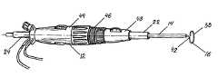
- FIG. 1is a perspective view of a preferred catheter apparatus for use in the isolation of the left atrial appendage (LAA) in accordance with this invention having the tip section of the catheter shaft deflected.
- LAAleft atrial appendage
- FIG. 2is a perspective view of the apparatus of FIG. 1 having the probe partially extending from the catheter shaft.
- FIG. 3is a perspective view of the apparatus of FIG. 1 having the probe form a loop perpendicular to the catheter shaft.
- FIG. 4is a perspective view of the apparatus of FIG. 1 having the probe form a loop coplanar with the catheter shaft.
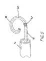
- FIG. 5is an enlarged view of a probe in accordance with this invention with the saline positioned therein.
- FIG. 6depicts a probe positioned around an imaged LAA in accordance with this invention.
- FIG. 7is a flow diagram of a method for LAA isolation.
- the catheter apparatus 10is preferably an endoscopic or thoracoscopic delivery system having a steering handle 12 , a catheter shaft 14 , and a probe 16 .
- Steering handle 12is hollow and has a front end 18 and a rear end 20 .
- a hollow introducer tube 22extends from the front end 18 .
- Introducer tube 22is preferably formed from a rigid material such that introducer tube 22 can be inserted through an incision in a patient.
- a hollow extended tube 24extends from the rear end 20 . Introducer tube 22 , steering handle 12 and extended tube 24 are coaxial with each other along central axis 25 , thereby defining a continuous central opening 26 .
- Rear end 20includes apertures to receive both a light source 28 and a camera 30 .
- Light source 28 and camera 30are fed through introducer tube 22 to forward aperture 32 .
- Light source 28 and camera 30allow images to be displayed in real-time through a display device such as a video monitor so that the physician may monitor the procedure being performed.
- Rear end 20 of steering handle 12also receives an irrigation port 34 that is connected to a supply of irrigation fluid to assist in irrigating and aspirating catheter apparatus 10 as needed.
- Central opening 26is sized to receive catheter shaft 14 .
- Catheter shaft 14is introduced through extended tube 24 so that catheter shaft 14 can extend outward from forward aperture 32 of introducer tube 22 .
- Catheter shaft 14has a hollow, open interior and is sized to receive probe 16 .
- the diameter of catheter shaft 14is not critical but 8 French is preferred.
- Catheter shaft 14is preferably made from polyurethane. Nylon can be added to increase the strength of catheter shaft 14 while flexibility at its tip section 36 can be achieved by eliminating or reducing the amount of stiffening material.
- Two sets of non-conducting wires(not shown) are anchored to the inside of the catheter shaft by thermal bonding to add further strength. These wires can be made from a nickel-titanium alloy such as nitinol or from steel.
- Probe 16is positioned within catheter shaft 14 where probe 16 is free to move forward and backward along the longitudinal axis of catheter shaft 14 .
- the length of probe 16can be selected based on the size, orientation or other anomalies of the LAA.
- the diameter of probe 16is also not critical but 6 French is preferred.
- distal end 38 of probe 16achieves a pre-stressed curve as shown in FIGS. 2-5 .
- the pre-stressed curveis designed to make a tight fit around the base of the LAA. It is anticipated that several different sizes may be needed depending upon the size of the LAA.
- the pre-stressed curve of distal end 38is achieved by using an elongated member or saline 40 as illustrated in FIG. 5 . Saline 40 is positioned inside the body of probe 16 and the distal end of saline 40 is formed to achieve different pre-stressed curves.
- Saline 40is preferably composed of inert wires fabricated from a nickel-titanium alloy having shape memory as, for example, nitinol.
- the cross-sectional area of saline 40 in the pre-stressed curve locationmay be thinner compared to the rest of saline 40 .
- probe 16further includes proximal coil 42 .
- Proximal coil 42is made from several non-conducting wires and encircles the outside circumference of probe 16 .
- Proximal coil 42abuts distal end 38 and serves to achieve disengagement of distal end 38 from the remainder of probe 16 as will be described in detail below.
- Steering handle 12is provided with four actuators 44 , 46 , 48 , 50 .
- First actuator 44is mechanically connected to catheter shaft 14 by non-conducting wire in a manner known to those skilled in the art that gives control over the axial and rotational movement of catheter shaft 14 .
- Second actuator 46controls angled movement of tip section 36 .
- Tip section 36is preferably the distal 1 cm. of catheter shaft 14 .
- Second actuator 46is mechanically connected to tip section 36 to allow tip section 36 to deflect away from central axis 25 as second actuator 46 is rotated. The degree of deflection available is preferably 30 to 60°. Selective adjustment of the longitudinal, rotational, and angular position of catheter shaft 14 permits catheter apparatus 10 to accommodate different sizes and orientations of LAA.
- Third actuator 48is attached by non-conducting wire to probe 16 .
- Third actuator 48controls forward and backward movement of probe 16 within catheter shaft 14 .
- Fourth actuator 50is joined by non-conducting wire to proximal coil 42 . Rotation of fourth actuator 50 in a clockwise direction places tension upon proximal coil 42 to snap or break off the distal portion of probe 16 forward of proximal coil 42 .
- the procedure for isolating the LAA using catheter apparatus 10will now be described. Initially, the patient is positioned in a supine position and a double lumen endotracheal tube is inserted to ventilate the right lung while, at the same time, ventilation is blocked to the left lung so as to create a space in the left pleural cavity. With the left lung deflated, a small incision is made upon the chest to permit one of two approaches that can be used to deliver probe 16 to the LAA site. These approaches will be discussed in more detail below.
- thoracic portA small 4-5 mm. by 3-4 cm. thoracic port is placed over the incision and anchored in place. The thoracic port helps maintain the intercostal space, decreases trauma and maintains pleural access. Introducer tube 22 is inserted through the thoracic port allowing light source 28 and camera 30 to be used to verify lack of left lung ventilation, to identify intrapleural and mediastinal anatomies, and to navigate catheter shaft 14 and probe 16 to the LAA.
- catheter shaft 14is advanced outward from tube 22 until catheter shaft is positioned adjacent to the base of the LAA.
- catheter shaft 14 and probe 16are introduced using the subxiphoid approach.
- Probe 16can be extended from catheter shaft 14 to form a pre-stressed circular loop coplanar with the axis of catheter shaft 14 as shown in FIG. 4 when introduced in this fashion.
- FIG. 6depicts an example of a probe 16 encircling the LAA.
- FIG. 7is a flow diagram illustrating a method for LAA isolation. Acquisition and segmentation of left atrium and LAA at step 100 is similar to what has been disclosed in patent application Ser. No. 10/249,812 incorporated herein by reference. A volume of data is acquired on the cardiac CT using a protocol that is optimized for the left atrium and the LAA. The image dataset is segmented with post processing software to extract the surfaces of the heart chambers, including the left atrium and the LAA. The endocardial view (view from inside) can also be visualized to assess the size and orientation of the LAA.
- the LAA along with the left atriumis visualized using 3D surface and/or volume rendering to create 3D models of the LAA which preferably also include a navigator or endocardial (view from inside) view.
- 3D surface and/or volume renderingcan help eliminate the blurring effect seen due to cardiac motion.
- Another aspect of scanninginvolves scanning of data which is synchronized with the ECG signal (QRS) in order to produce a graphic representation of the examination volume and thus an image of the heart. The correlation between the scanning data and the ECG signals is fixed so that within each number of successive time intervals a database is obtained.
- QRSECG signal
- Anatomical landmarksare identified at step 110 . Furthermore, explicit geometric markers are inserted into the volume at landmarks of interest. These markers may be subsequently visualized in a translucent fashion.
- the saved imagesare then transferred and registered with the fluoroscopy system and/or the computer workstation of another interventional system as shown at step 115 .
- the orientation, size and any anomalies of the LAAare visualized over the interventional system such that probes of the right size can be selected.
- the registration processis similar to that detailed in patent application Ser. Nos. 10/063,064 and 60/531,293 incorporated herein by reference.
- external markerscould be used at the time of registration itself.
- catheter shaft 14 and probe 16are visualized on an interventional system such as the fluoroscopy system and/or the computer workstation of another interventional system. Both are visualized as they navigate towards the appendage being isolated at step 125 .
- This delivery systemis used to isolate the LAA.
- step 130the whole process of imaging and registration can be eliminated completely and a catheter apparatus 10 , using an endoscopic or thoracoscopic delivery system for example, can position catheter shaft 14 and probe 16 around the LAA using direct visualization in the manner discussed above.
- automatic techniquesmay be employed to perform any of the steps by using one or more of the several computer-assisted detection, localization and visualization methods available. Moreover, these methods could be completely automatic or interactive with input from the user. Furthermore, the features described may improve with user input and interaction.
Landscapes
- Health & Medical Sciences (AREA)
- Life Sciences & Earth Sciences (AREA)
- Surgery (AREA)
- Engineering & Computer Science (AREA)
- Plasma & Fusion (AREA)
- Medical Informatics (AREA)
- Otolaryngology (AREA)
- Physics & Mathematics (AREA)
- Cardiology (AREA)
- Biomedical Technology (AREA)
- Heart & Thoracic Surgery (AREA)
- Nuclear Medicine, Radiotherapy & Molecular Imaging (AREA)
- Molecular Biology (AREA)
- Animal Behavior & Ethology (AREA)
- General Health & Medical Sciences (AREA)
- Public Health (AREA)
- Veterinary Medicine (AREA)
- Surgical Instruments (AREA)
Abstract
Description
This application claims the benefit of U.S. Provisional Application No. 60/484,008, filed on Jul. 1, 2003 and U.S. Provisional Application No. 60/531,293 filed on Dec. 19, 2003.
This invention relates generally to catheter apparatus for cardiac interventional treatment and, in particular, to catheter apparatus for isolation of the left atrial appendage (LAA) in patients with atrial fibrillation (AF).
Atrial fibrillation (AF), a heart rhythm problem in which the atria (upper chambers of the heart) stop contracting as they fibrillate or quiver, is the most common of all the heart rhythm problems. It is estimated that over 2.2 million patients in the U.S. have AF and over 140,000 new cases are diagnosed every year. Patients with AF have a high risk of stroke. The Framingham Study demonstrated that the diagnosis of AF increased the risk of stroke 3 to 5 times higher and the risk rose from 1.5 percent during the fifth decade of life to over 23% by the eight decade.
More specifically, previous studies have indicated that more than 90 percent of AF related strokes result from a blood clot that forms in the left atrial appendage (LAA), a small pouch-like structure in the left atrium. Such blood clots can dislodge from the atrium and travel to the brain, thereby causing a stroke. Several large trials have shown the efficacy of warfarin, a blood thinner, in reducing the risk of stroke. In clinical practice, however, the warfarin is contraindicated or cannot be used because of side effects such as bleeding in over 40 percent of patients.
In addition to the LAA being a source of blood clots it can also play a role in the initiation and maintenance of AF. In treating AF, lesions are placed at strategic locations to prevent the conduction of errant electrical impulses. The LAA appendage is routinely removed in a surgical procedure for treating AF called the MAZE procedure. The removal of the LAA has been recommended by the American College of Cardiology-American Heart Association guidelines.
Recently, a catheter technique call PLAATO (percutaneous left atrial appendage trans catheter occlusion) has been tried as a blocking device from the inside of the left atrial chamber. In one study of this technique, the catheter implant sizes ranged significantly, the procedure time ran over 90 minutes and, in 25 percent of these patients, the initial device required removal and replacement with a device of a different size. There was also additional risk of bleeding around the heart, a complication that can at times be life-threatening.
There is thus a need for a method and system for establishing an effective road map of the LAA. There is also a need for a method and system for isolating the LAA that uses a minimally-invasive approach not going inside the left atrial chamber. Such a method and system would eliminate the need for different size catheters and reduce the risk of complications, thereby providing an alternative strategy for the prevention of stroke and the treatment of AF.
It is an object of this invention to provide an improved catheter apparatus for use in the isolation of the LAA that overcomes some of the problems and shortcomings in the prior art, including those referred to above.
This invention is for a catheter apparatus used in the isolation of a patient's left atrial appendage (LAA) having a catheter shaft, a probe axially disposed within the catheter shaft, and a control mechanism coupled to both the catheter shaft and the probe. The catheter shaft includes a main body that extends along a central axis and a coaxial tip section attached to the main body. The tip section can be deflected from the central axis in a controlled manner. The probe can be extended outward from the catheter shaft and has a distal end that is brought into contact with the LAA. The control mechanism controls longitudinal movement of the catheter shaft, the degree of deflection of the tip section, and axial movement of the probe with respect to the catheter shaft.
In one desirable embodiment, the catheter shaft can be rotated about its central axis. More desirable is where the distal end has a pre-stressed curve so that the distal end curves as the probe is extended outward from the catheter shaft. In such embodiments, the distal end preferably includes a saline. Most preferred is where the saline is fabricated from nickel-titanium where the alloy has shape memory.
One very preferred embodiment finds the distal end able to form a loop when the probe is fully extended from the catheter shaft. This loop is selected to have a size that will allow it to substantially encircle the base of the LAA. In certain desirable cases, the loop is substantially perpendicular to the axis of the catheter shaft. In other preferred cases, the loop is coplanar with this axis.
Another preferred embodiment is where the control mechanism is a steering handle. More desirable is where the steering handle includes a first actuator that is mechanically connected to the catheter shaft to control its rotation and axial movement. Most preferred is where the steering handle also includes a second actuator mechanically connected to the shaft's tip section to control the deflection of the tip section. It is highly preferred where the steering handle has a third actuator that is mechanically connected to the probe and that controls the axial movement of the probe within the catheter shaft.
One very highly desired embodiment finds the steering handle having a fourth actuator. This control is mechanically connected to a proximal coil on the probe and it enables the distal end to be disengaged from the probe at the point of this proximal coil.
In another aspect of this invention, it provides a method for isolating the LAA on a patient. This method includes the steps of introducing a catheter apparatus through an incision in the patient's chest, navigating the catheter apparatus to the LAA, and using the catheter apparatus to isolate the LAA. The catheter apparatus is preferably one that includes: (1) a catheter shaft having a main body that is rotatable about its central axis and having a coaxial tip section that can be selectively curved away from the central axis; (2) a probe slidably disposed within the catheter shaft that can be extended outward from the shaft and having a distal end with a pre-stressed curve that takes the shape of the curve when the distal end protrudes from the catheter shaft; and (3) a steering handle coupled to the catheter shaft and the probe where the steering handle is capable of controlling the rotation and axial movement of the catheter shaft, the deflection of the tip section, and the axial movement of the probe within the catheter shaft.
In certain desirable cases, the distal end includes a saline. More desirable is where the curve of the distal end forms a loop when the probe is fully extended from the catheter shaft. This loop is sized to be able to circumscribe the base of the LAA and is oriented in a plane substantially perpendicular to the central axis. Highly preferred is where the steering handle is also mechanically connected to a proximal coil on the probe so that it can control the disengagement of the distal end from the probe at this proximal coil.
Referring toFIGS. 1-4 , acatheter apparatus 10 in accordance with this invention is shown that enables an electrophysiologist, cardiologist and/or surgeon to isolate the left atrial appendage (LAA). Thecatheter apparatus 10 is preferably an endoscopic or thoracoscopic delivery system having asteering handle 12, acatheter shaft 14, and aprobe 16.Steering handle 12 is hollow and has afront end 18 and arear end 20. Ahollow introducer tube 22 extends from thefront end 18.Introducer tube 22 is preferably formed from a rigid material such that introducertube 22 can be inserted through an incision in a patient. A hollow extendedtube 24 extends from therear end 20.Introducer tube 22, steeringhandle 12 andextended tube 24 are coaxial with each other alongcentral axis 25, thereby defining a continuouscentral opening 26.
Asprobe 16 protrudes fromcatheter shaft 14,distal end 38 ofprobe 16 achieves a pre-stressed curve as shown inFIGS. 2-5 . The pre-stressed curve is designed to make a tight fit around the base of the LAA. It is anticipated that several different sizes may be needed depending upon the size of the LAA. The pre-stressed curve ofdistal end 38 is achieved by using an elongated member orsaline 40 as illustrated inFIG. 5 .Saline 40 is positioned inside the body ofprobe 16 and the distal end ofsaline 40 is formed to achieve different pre-stressed curves.Saline 40 is preferably composed of inert wires fabricated from a nickel-titanium alloy having shape memory as, for example, nitinol. The cross-sectional area ofsaline 40 in the pre-stressed curve location may be thinner compared to the rest ofsaline 40.
As shown inFIG. 5 , probe16 further includesproximal coil 42.Proximal coil 42 is made from several non-conducting wires and encircles the outside circumference ofprobe 16.Proximal coil 42 abutsdistal end 38 and serves to achieve disengagement ofdistal end 38 from the remainder ofprobe 16 as will be described in detail below.
Steeringhandle 12 is provided with fouractuators First actuator 44 is mechanically connected tocatheter shaft 14 by non-conducting wire in a manner known to those skilled in the art that gives control over the axial and rotational movement ofcatheter shaft 14.Second actuator 46 controls angled movement oftip section 36.Tip section 36 is preferably the distal 1 cm. ofcatheter shaft 14.Second actuator 46 is mechanically connected to tipsection 36 to allowtip section 36 to deflect away fromcentral axis 25 assecond actuator 46 is rotated. The degree of deflection available is preferably 30 to 60°. Selective adjustment of the longitudinal, rotational, and angular position ofcatheter shaft 14permits catheter apparatus 10 to accommodate different sizes and orientations of LAA.
The procedure for isolating the LAA usingcatheter apparatus 10 will now be described. Initially, the patient is positioned in a supine position and a double lumen endotracheal tube is inserted to ventilate the right lung while, at the same time, ventilation is blocked to the left lung so as to create a space in the left pleural cavity. With the left lung deflated, a small incision is made upon the chest to permit one of two approaches that can be used to deliverprobe 16 to the LAA site. These approaches will be discussed in more detail below.
A small 4-5 mm. by 3-4 cm. thoracic port is placed over the incision and anchored in place. The thoracic port helps maintain the intercostal space, decreases trauma and maintains pleural access.Introducer tube 22 is inserted through the thoracic port allowinglight source 28 andcamera 30 to be used to verify lack of left lung ventilation, to identify intrapleural and mediastinal anatomies, and to navigatecatheter shaft 14 andprobe 16 to the LAA.
Once theintroducer tube 22 is properly positioned,catheter shaft 14 is advanced outward fromtube 22 until catheter shaft is positioned adjacent to the base of the LAA. There are two approaches that can be used to deliverprobe 16 to the LAA site. In the first approach,catheter shaft 14 andprobe 16 are introduced using the subxiphoid approach.Probe 16 can be extended fromcatheter shaft 14 to form a pre-stressed circular loop coplanar with the axis ofcatheter shaft 14 as shown inFIG. 4 when introduced in this fashion.
In the second approach, a small incision is made in the left chest area. In this approach,probe 16 comes out fromcatheter shaft 14 to form a pre-stressed circular loop at a right angle to the axis ofcatheter shaft 14 as illustrated inFIG. 3 . In either approach,catheter shaft 14 has been positioned with respect to the LAA so that asprobe 16 moves forward,distal end 38 ofprobe 16 encircles the base of the LAA. Oncedistal end 38 has formed a loop and circumscribed the base of the LAA, rotation offourth actuator 50 in a clockwise direction will separate break the loop from the rest ofprobe 16 atproximal coil 42.Probe 16 andcatheter shaft 14 along withintroducer tube 22 are then withdrawn.FIG. 6 depicts an example of aprobe 16 encircling the LAA.
Several experiments in the laboratory by the inventor where the LAA had been isolated in animal models without removing it have demonstrated that there are no detrimental effects with this procedure, as no blood clots or other changes were detected during a 6 week follow-up.
Atstep 105, the LAA along with the left atrium is visualized using 3D surface and/or volume rendering to create 3D models of the LAA which preferably also include a navigator or endocardial (view from inside) view. Faster scanning speeds can help eliminate the blurring effect seen due to cardiac motion. Another aspect of scanning involves scanning of data which is synchronized with the ECG signal (QRS) in order to produce a graphic representation of the examination volume and thus an image of the heart. The correlation between the scanning data and the ECG signals is fixed so that within each number of successive time intervals a database is obtained.
Anatomical landmarks are identified atstep 110. Furthermore, explicit geometric markers are inserted into the volume at landmarks of interest. These markers may be subsequently visualized in a translucent fashion. The saved images are then transferred and registered with the fluoroscopy system and/or the computer workstation of another interventional system as shown atstep 115. The orientation, size and any anomalies of the LAA are visualized over the interventional system such that probes of the right size can be selected. The registration process is similar to that detailed in patent application Ser. Nos. 10/063,064 and 60/531,293 incorporated herein by reference. In addition, external markers could be used at the time of registration itself.
In another aspect of the method and system of LAA isolation in patients, as shown atstep 120,catheter shaft 14 andprobe 16 are visualized on an interventional system such as the fluoroscopy system and/or the computer workstation of another interventional system. Both are visualized as they navigate towards the appendage being isolated atstep 125. This delivery system is used to isolate the LAA.
As shown instep 130, the whole process of imaging and registration can be eliminated completely and acatheter apparatus 10, using an endoscopic or thoracoscopic delivery system for example, can positioncatheter shaft 14 andprobe 16 around the LAA using direct visualization in the manner discussed above.
It may be appreciated that automatic techniques may be employed to perform any of the steps by using one or more of the several computer-assisted detection, localization and visualization methods available. Moreover, these methods could be completely automatic or interactive with input from the user. Furthermore, the features described may improve with user input and interaction.
Although the invention has been described in conjunction with specific embodiments thereof, it is evident that many alternatives, modifications and variations will be apparent to those skilled in the art. Accordingly, it is intended to embrace all such alternatives, modifications and variations that fall within the spirit and broad scope of the appended claims. In particular, although the preceding description discusses LAA imaging and isolation, it is understood that the methodology is not limited to the LAA but may be utilized in other cardiac and non-cardiac applications.
Claims (19)
1. A catheter apparatus for use in the isolation of a left atrial appendage (LAA) on a patient comprising:
a catheter shaft having a central axis and a distal tip section, the tip section being deflectable whereby the tip section can be selectively curved away from the central axis;
a probe slidably disposed within the catheter shaft, the probe having a distal end and being selectively movable axially within the catheter shaft, whereby the distal end can be extended outward from the catheter shaft and brought into contact with the LAA;
a handle member sized to slidably receive the catheter shaft; and
a control mechanism positioned upon the handle member and coupled to the catheter shaft and the probe wherein the control mechanism controls axial movement and deflection of the catheter shaft with respect to the handle member and movement of the probe with respect to the catheter shaft.
2. The apparatus ofclaim 1 wherein the catheter shaft is rotatable about the central axis.
3. The apparatus ofclaim 2 wherein the distal end has a pre-stressed curve whereby the distal end curves as the probe is extended from the catheter shaft.
4. The apparatus ofclaim 3 wherein the distal end includes a spline.
5. The apparatus ofclaim 4 wherein the saline is fabricated from nickel-titanium having shape memory.
6. The apparatus ofclaim 3 wherein the distal end forms a loop when the probe is filly extended from the catheter shaft, the loop being sized to substantially circumscribe a base of the LAA.
7. The apparatus ofclaim 6 wherein the loop substantially defines a plane substantially perpendicular to the central axis.
8. The apparatus ofclaim 6 wherein the loop is substantially coplanar with the central axis.
9. The apparatus ofclaim 1 wherein the handle member includes a steering handle and an introducer tube extending outward from a front end of the steering handle.
10. The apparatus ofclaim 9 wherein the control mechanism is positioned upon the steering handle.
11. The apparatus ofclaim 10 wherein the control mechanism includes a first actuator mechanically connected with respect to the catheter shaft whereby rotation and axial movement of the catheter shaft is actuated by the first actuator.
12. The apparatus ofclaim 11 wherein the control mechanism includes a second actuator mechanically connected with respect to the tip section whereby actuation of the second actuator deflects the tip section away from the central axis.
13. The apparatus ofclaim 12 wherein the control mechanism includes a third actuator mechanically connected with respect to the probe whereby actuation of the third actuator moves the probe axially within the catheter shaft.
14. The apparatus ofclaim 13 wherein the probe includes a proximal coil and the control in mechanism includes a fourth actuator mechanically connected to the proximal coil whereby actuation of the fourth actuator disengages the distal end from the probe at the proximal coil.
15. A method for epicardial isolation of a left atrial appendage (LAA) on a patient comprising:
introducing a catheter apparatus through an incision in the patient's chest,
navigating the catheter apparatus to the LAA;
advancing a probe from the catheter apparatus;
substantially circumscribing a base of the LAA with the probe to epicardially isolate the LAA; and
disengaging a distal end of the probe from the catheter apparatus.
16. The method ofclaim 15 wherein the catheter apparatus includes:
a catheter shaft rotatable about a central axis and having a distal tip section, the tip section being deflectable whereby the tip section can be selectively curved away from the central axis;
the probe slidably disposed within the catheter shaft, the probe being selectively movable axially within the catheter shaft and the distal end having a pre-stressed curve whereby the distal end curves as the probe is extended outward from the catheter shaft;
a steering handle sized to slidably receive the catheter shaft; and
a control mechanism positioned upon the steering handle and coupled to both the catheter shaft and the probe wherein the steering handle controls rotation and axial movement of the catheter shaft with respect to the steering handle, deflection of the tip section with respect to the central axis, and movement of the probe with respect to the catheter shaft.
17. The method ofclaim 16 wherein the distal end includes a spline.
18. The method ofclaim 17 wherein the curve of the distal end forms a loop when the probe is fully extended from the catheter shaft, the loop being sized to substantially circumscribe a base of the LAA and substantially defining a plane substantially perpendicular to the central axis.
19. The method ofclaim 18 wherein the probe includes a proximal coil and the control mechanism is mechanically connected to the proximal coil whereby the control mechanism controls disengagement of the distal end from the probe at the proximal coil.
Priority Applications (1)
| Application Number | Priority Date | Filing Date | Title |
|---|---|---|---|
| US10/882,516US7344543B2 (en) | 2003-07-01 | 2004-07-01 | Method and apparatus for epicardial left atrial appendage isolation in patients with atrial fibrillation |
Applications Claiming Priority (3)
| Application Number | Priority Date | Filing Date | Title |
|---|---|---|---|
| US48400803P | 2003-07-01 | 2003-07-01 | |
| US53129303P | 2003-12-19 | 2003-12-19 | |
| US10/882,516US7344543B2 (en) | 2003-07-01 | 2004-07-01 | Method and apparatus for epicardial left atrial appendage isolation in patients with atrial fibrillation |
Publications (2)
| Publication Number | Publication Date |
|---|---|
| US20050033287A1 US20050033287A1 (en) | 2005-02-10 |
| US7344543B2true US7344543B2 (en) | 2008-03-18 |
Family
ID=34119781
Family Applications (1)
| Application Number | Title | Priority Date | Filing Date |
|---|---|---|---|
| US10/882,516Expired - Fee RelatedUS7344543B2 (en) | 2003-07-01 | 2004-07-01 | Method and apparatus for epicardial left atrial appendage isolation in patients with atrial fibrillation |
Country Status (1)
| Country | Link |
|---|---|
| US (1) | US7344543B2 (en) |
Cited By (35)
| Publication number | Priority date | Publication date | Assignee | Title |
|---|---|---|---|---|
| US20060135953A1 (en)* | 2004-12-22 | 2006-06-22 | Wlodzimierz Kania | Tissue ablation system including guidewire with sensing element |
| US20110082495A1 (en)* | 2009-10-02 | 2011-04-07 | Ruiz Carlos E | Apparatus And Methods For Excluding The Left Atrial Appendage |
| US8518063B2 (en) | 2001-04-24 | 2013-08-27 | Russell A. Houser | Arteriotomy closure devices and techniques |
| US8636754B2 (en) | 2010-11-11 | 2014-01-28 | Atricure, Inc. | Clip applicator |
| US8906013B2 (en) | 2010-04-09 | 2014-12-09 | Endosense Sa | Control handle for a contact force ablation catheter |
| US8961541B2 (en) | 2007-12-03 | 2015-02-24 | Cardio Vascular Technologies Inc. | Vascular closure devices, systems, and methods of use |
| US8992567B1 (en) | 2001-04-24 | 2015-03-31 | Cardiovascular Technologies Inc. | Compressible, deformable, or deflectable tissue closure devices and method of manufacture |
| US9017349B2 (en) | 2010-10-27 | 2015-04-28 | Atricure, Inc. | Appendage clamp deployment assist device |
| US9066741B2 (en) | 2010-11-01 | 2015-06-30 | Atricure, Inc. | Robotic toolkit |
| US9265486B2 (en) | 2011-08-15 | 2016-02-23 | Atricure, Inc. | Surgical device |
| US9282973B2 (en) | 2012-01-20 | 2016-03-15 | Atricure, Inc. | Clip deployment tool and associated methods |
| US9345460B2 (en) | 2001-04-24 | 2016-05-24 | Cardiovascular Technologies, Inc. | Tissue closure devices, device and systems for delivery, kits and methods therefor |
| US9408659B2 (en) | 2007-04-02 | 2016-08-09 | Atricure, Inc. | Surgical instrument with separate tool head and method of use |
| US9545265B2 (en) | 2013-04-15 | 2017-01-17 | Transseptal Solutions Ltd. | Fossa ovalis penetration using balloons |
| US9668674B2 (en) | 2015-03-03 | 2017-06-06 | Transseptal Solutions Ltd. | Measurement of appendage openings |
| US9700351B2 (en) | 2013-04-15 | 2017-07-11 | Transseptal Solutions Ltd. | Fossa ovalis penetration |
| US9706982B2 (en) | 2015-03-03 | 2017-07-18 | Transseptal Solutions Ltd. | Treatment of appendage openings |
| US9737309B1 (en) | 2010-06-24 | 2017-08-22 | Niv Ad | System for occlusion of left atrial appendage |
| US9788858B2 (en) | 2013-04-15 | 2017-10-17 | Transseptal Solutions Ltd. | Fossa ovalis penetration using probing elements |
| US9795765B2 (en) | 2010-04-09 | 2017-10-24 | St. Jude Medical International Holding S.À R.L. | Variable stiffness steering mechanism for catheters |
| US9855404B2 (en) | 2013-05-03 | 2018-01-02 | St. Jude Medical International Holding S.À R.L. | Dual bend radii steering catheter |
| US10398503B2 (en) | 2015-10-14 | 2019-09-03 | Transseptal Soulutions Ltd. | Fossa ovalis penetration |
| US10426589B2 (en) | 2003-12-04 | 2019-10-01 | Boston Scientific Scimed, Inc. | System and method for delivering a left atrial appendage containment device |
| US10500371B2 (en) | 2014-10-14 | 2019-12-10 | Transseptal Solutions Ltd. | Fossa ovalis penetration |
| US10617425B2 (en) | 2014-03-10 | 2020-04-14 | Conformal Medical, Inc. | Devices and methods for excluding the left atrial appendage |
| US10631868B2 (en) | 2010-06-24 | 2020-04-28 | Niv Ad | System for occlusion of left atrial appendage |
| US10722240B1 (en) | 2019-02-08 | 2020-07-28 | Conformal Medical, Inc. | Devices and methods for excluding the left atrial appendage |
| US11026695B2 (en) | 2016-10-27 | 2021-06-08 | Conformal Medical, Inc. | Devices and methods for excluding the left atrial appendage |
| US11399842B2 (en) | 2013-03-13 | 2022-08-02 | Conformal Medical, Inc. | Devices and methods for excluding the left atrial appendage |
| US11426172B2 (en) | 2016-10-27 | 2022-08-30 | Conformal Medical, Inc. | Devices and methods for excluding the left atrial appendage |
| US11717303B2 (en) | 2013-03-13 | 2023-08-08 | Conformal Medical, Inc. | Devices and methods for excluding the left atrial appendage |
| US11998212B2 (en) | 2013-11-21 | 2024-06-04 | Atricure, Inc. | Occlusion clip |
| US12004752B2 (en) | 2012-11-21 | 2024-06-11 | Atricure, Inc. | Occlusion clip |
| US12144508B2 (en) | 2019-02-08 | 2024-11-19 | Conformal Medical, Inc. | Devices and methods for excluding the left atrial appendage |
| US12426922B2 (en) | 2013-04-15 | 2025-09-30 | Transseptal Solutions Ltd. | Fossa ovalis penetration catheter |
Families Citing this family (24)
| Publication number | Priority date | Publication date | Assignee | Title |
|---|---|---|---|---|
| US6488689B1 (en)* | 1999-05-20 | 2002-12-03 | Aaron V. Kaplan | Methods and apparatus for transpericardial left atrial appendage closure |
| US20090143808A1 (en)* | 2001-04-24 | 2009-06-04 | Houser Russell A | Guided Tissue Cutting Device, Method of Use and Kits Therefor |
| US7846168B2 (en)* | 2003-10-09 | 2010-12-07 | Sentreheart, Inc. | Apparatus and method for the ligation of tissue |
| US8409219B2 (en) | 2004-06-18 | 2013-04-02 | Medtronic, Inc. | Method and system for placement of electrical lead inside heart |
| US7918865B2 (en)* | 2005-04-07 | 2011-04-05 | Sentreheart, Inc. | Apparatus and method for the ligation of tissue |
| SI2142107T1 (en)* | 2007-03-30 | 2013-05-31 | Sentreheart, Inc. | Devices and systems for closing the left atrial appendage |
| US20120123292A1 (en)* | 2007-05-08 | 2012-05-17 | ProPep Surgical, LLC | Nerve Mapping Surgical System and Method of Use of Dual Function Surgical Instrument Within Such System |
| US8083685B2 (en) | 2007-05-08 | 2011-12-27 | Propep, Llc | System and method for laparoscopic nerve detection |
| WO2009039191A2 (en)* | 2007-09-20 | 2009-03-26 | Sentreheart, Inc. | Devices and methods for remote suture management |
| US8332641B2 (en)* | 2009-01-30 | 2012-12-11 | Freescale Semiconductor, Inc. | Authenticated debug access for field returns |
| AU2010232589B2 (en) | 2009-04-01 | 2014-11-27 | Atricure, Inc. | Tissue ligation devices and controls therefor |
| AU2011241103A1 (en) | 2010-04-13 | 2012-11-08 | Sentreheart, Inc. | Methods and devices for treating atrial fibrillation |
| EP2598075A4 (en) | 2010-07-28 | 2016-11-30 | Medrobotics Corp | SURGICAL POSITIONING AND SUPPORT SYSTEM |
| US8911434B2 (en) | 2010-10-22 | 2014-12-16 | Medtronic Cryocath Lp | Balloon catheter with deformable fluid delivery conduit |
| JP5711380B2 (en) | 2010-10-22 | 2015-04-30 | メドロボティクス コーポレイション | Articulated robotic probe |
| JP6167041B2 (en) | 2010-11-11 | 2017-07-19 | メドロボティクス コーポレイション | Introduction assembly for articulated robotic probes |
| ES2671928T3 (en) | 2011-06-08 | 2018-06-11 | Sentreheart, Inc. | Tissue ligation devices and tension devices for them |
| CN104010773B (en) | 2011-09-13 | 2017-01-18 | 美的洛博迪克斯公司 | Highly Articulated Probes With Anti-Twist Link Arrangement, Methods Of Formation Thereof, And Methods Of Performing Medical Procedures |
| WO2013096610A1 (en) | 2011-12-21 | 2013-06-27 | Oyola Arnold E | Stabilizing apparatus for highly articulated probes with link arrangement, methods of formation thereof, and methods of use thereof |
| BR112015019887A2 (en) | 2013-03-12 | 2017-07-18 | Sentreheart Inc | device for closing a target tissue |
| EP4226881A1 (en) | 2013-10-31 | 2023-08-16 | AtriCure, Inc. | Device for left atrial appendage closure |
| US9936956B2 (en) | 2015-03-24 | 2018-04-10 | Sentreheart, Inc. | Devices and methods for left atrial appendage closure |
| ES2972395T3 (en) | 2015-03-24 | 2024-06-12 | Atricure Inc | Tissue ligation devices |
| EP4331509A3 (en) | 2016-02-26 | 2024-05-15 | AtriCure, Inc. | Devices for left atrial appendage closure |
Citations (90)
| Publication number | Priority date | Publication date | Assignee | Title |
|---|---|---|---|---|
| US3954098A (en) | 1975-01-31 | 1976-05-04 | Dick Donald E | Synchronized multiple image tomographic cardiography |
| US4574807A (en) | 1984-03-02 | 1986-03-11 | Carl Hewson | Method and apparatus for pacing the heart employing external and internal electrodes |
| WO1991007726A1 (en) | 1989-11-21 | 1991-05-30 | I.S.G. Technologies Inc. | Probe-correlated viewing of anatomical image data |
| US5245287A (en) | 1991-08-20 | 1993-09-14 | Siemens Aktiengesellschaft | Nuclear magnetic resonance tomography apparatus having a resonant circuit for generating gradient fields |
| US5274551A (en) | 1991-11-29 | 1993-12-28 | General Electric Company | Method and apparatus for real-time navigation assist in interventional radiological procedures |
| US5304212A (en) | 1987-06-26 | 1994-04-19 | Brigham And Women's Hospital | Assessment and modification of a human subject's circadian cycle |
| US5348020A (en) | 1990-12-14 | 1994-09-20 | Hutson William H | Method and system for near real-time analysis and display of electrocardiographic signals |
| US5353795A (en) | 1992-12-10 | 1994-10-11 | General Electric Company | Tracking system to monitor the position of a device using multiplexed magnetic resonance detection |
| US5391199A (en) | 1993-07-20 | 1995-02-21 | Biosense, Inc. | Apparatus and method for treating cardiac arrhythmias |
| US5431688A (en) | 1990-06-12 | 1995-07-11 | Zmd Corporation | Method and apparatus for transcutaneous electrical cardiac pacing |
| US5464447A (en) | 1994-01-28 | 1995-11-07 | Sony Corporation | Implantable defibrillator electrodes |
| WO1996010949A1 (en) | 1994-10-07 | 1996-04-18 | Medical Media Systems | Video-based surgical targeting system |
| US5568384A (en) | 1992-10-13 | 1996-10-22 | Mayo Foundation For Medical Education And Research | Biomedical imaging and analysis |
| US5738096A (en) | 1993-07-20 | 1998-04-14 | Biosense, Inc. | Cardiac electromechanics |
| US5823958A (en) | 1990-11-26 | 1998-10-20 | Truppe; Michael | System and method for displaying a structural data image in real-time correlation with moveable body |
| US5829447A (en) | 1993-02-22 | 1998-11-03 | Heartport, Inc. | Method and apparatus for thoracoscopic intracardiac procedures |
| US5839440A (en) | 1994-06-17 | 1998-11-24 | Siemens Corporate Research, Inc. | Three-dimensional image registration method for spiral CT angiography |
| US5846254A (en) | 1997-04-08 | 1998-12-08 | Ethicon Endo-Surgery, Inc. | Surgical instrument for forming a knot |
| US5871532A (en) | 1997-05-22 | 1999-02-16 | Sulzer Intermedics Inc. | Epicardial lead for minimally invasive implantation |
| US5951475A (en) | 1997-09-25 | 1999-09-14 | International Business Machines Corporation | Methods and apparatus for registering CT-scan data to multiple fluoroscopic images |
| US5954692A (en) | 1996-02-08 | 1999-09-21 | Symbiosis | Endoscopic robotic surgical tools and methods |
| US6081577A (en) | 1998-07-24 | 2000-06-27 | Wake Forest University | Method and system for creating task-dependent three-dimensional images |
| US6154516A (en) | 1998-09-04 | 2000-11-28 | Picker International, Inc. | Cardiac CT system |
| US6161543A (en)* | 1993-02-22 | 2000-12-19 | Epicor, Inc. | Methods of epicardial ablation for creating a lesion around the pulmonary veins |
| US6233304B1 (en) | 1998-11-25 | 2001-05-15 | General Electric Company | Methods and apparatus for calcification scoring |
| US6234804B1 (en) | 1999-03-02 | 2001-05-22 | Peter Yong | Thoracic training model for endoscopic cardiac surgery |
| US6235038B1 (en) | 1999-10-28 | 2001-05-22 | Medtronic Surgical Navigation Technologies | System for translation of electromagnetic and optical localization systems |
| US6249693B1 (en) | 1999-11-01 | 2001-06-19 | General Electric Company | Method and apparatus for cardiac analysis using four-dimensional connectivity and image dilation |
| US6252924B1 (en) | 1999-09-30 | 2001-06-26 | General Electric Company | Method and apparatus for motion-free cardiac CT imaging |
| US6256368B1 (en) | 1999-10-15 | 2001-07-03 | General Electric Company | Methods and apparatus for scout-based cardiac calcification scoring |
| US6254568B1 (en) | 1999-08-10 | 2001-07-03 | Biosense Webster, Inc. | Deflectable catheter with straightening element |
| US6266553B1 (en) | 1997-09-12 | 2001-07-24 | Siemens Aktiengesellschaft | Spiral scanning computed tomography apparatus, and method for operating same, for cardiac imaging |
| US6289115B1 (en) | 1998-02-20 | 2001-09-11 | Fuji Photo Film Co., Ltd. | Medical network system |
| US6289239B1 (en) | 1998-03-26 | 2001-09-11 | Boston Scientific Corporation | Interactive systems and methods for controlling the use of diagnostic or therapeutic instruments in interior body regions |
| US6311693B1 (en) | 1993-02-22 | 2001-11-06 | Wesley D. Sterman | Method and systems for performing thoracoscopic cardiac bypass and other procedures |
| US6314310B1 (en) | 1997-02-14 | 2001-11-06 | Biosense, Inc. | X-ray guided surgical location system with extended mapping volume |
| US6325797B1 (en) | 1999-04-05 | 2001-12-04 | Medtronic, Inc. | Ablation catheter and method for isolating a pulmonary vein |
| US20020010392A1 (en) | 1993-03-11 | 2002-01-24 | Desai Jawahar M. | Apparatus and method for cardiac ablation |
| US6348793B1 (en) | 2000-11-06 | 2002-02-19 | Ge Medical Systems Global Technology, Company, Llc | System architecture for medical imaging systems |
| US6348034B1 (en) | 1996-02-07 | 2002-02-19 | Pinotage, Llc | System for single-puncture endoscopic surgery |
| US6350248B1 (en) | 1996-08-13 | 2002-02-26 | Heartstent Corporation | Expandable myocardial implant |
| EP1182619A2 (en) | 2000-08-18 | 2002-02-27 | Biosense, Inc. | Method and apparatus for three-dimensional image rendering of body organs |
| US6353445B1 (en) | 1998-11-25 | 2002-03-05 | Ge Medical Systems Global Technology Company, Llc | Medical imaging system with integrated service interface |
| US20020046756A1 (en) | 2000-09-20 | 2002-04-25 | Laizzo Paul A. | System and method for determining tissue contact of an implantable medical device within a body |
| US6381485B1 (en) | 1999-10-28 | 2002-04-30 | Surgical Navigation Technologies, Inc. | Registration of human anatomy integrated for electromagnetic localization |
| US6389104B1 (en) | 2000-06-30 | 2002-05-14 | Siemens Corporate Research, Inc. | Fluoroscopy based 3-D neural navigation based on 3-D angiography reconstruction data |
| US6394948B1 (en) | 1995-09-20 | 2002-05-28 | Medtronic, Inc. | Method and apparatus for temporarily immobilizing a local area of tissue |
| US6411848B2 (en) | 1999-05-21 | 2002-06-25 | Cardiac Pacemakers, Inc. | System providing ventricular pacing and biventricular coordination |
| US6421412B1 (en) | 1998-12-31 | 2002-07-16 | General Electric Company | Dual cardiac CT scanner |
| US6423051B1 (en) | 1999-09-16 | 2002-07-23 | Aaron V. Kaplan | Methods and apparatus for pericardial access |
| US6456867B2 (en) | 1998-07-24 | 2002-09-24 | Biosense, Inc. | Three-dimensional reconstruction of intrabody organs |
| US20020138105A1 (en) | 2001-03-21 | 2002-09-26 | Kralik Michael R. | Temporary biventricular pacing of heart after heart surgery |
| US6468265B1 (en) | 1998-11-20 | 2002-10-22 | Intuitive Surgical, Inc. | Performing cardiac surgery without cardioplegia |
| US6478803B1 (en) | 2000-05-19 | 2002-11-12 | Genzyme Corporation | Device for delivery of surgical materials |
| US6478028B1 (en) | 1999-01-21 | 2002-11-12 | Coroneo, Inc. | Surgical apparatus and method for performing transabdominal cardiac surgery |
| US6484727B1 (en)* | 1996-10-22 | 2002-11-26 | Epicor, Inc. | Apparatus and method for diagnosis and therapy of electrophysiological disease |
| US6490475B1 (en) | 2000-04-28 | 2002-12-03 | Ge Medical Systems Global Technology Company, Llc | Fluoroscopic tracking and visualization system |
| US6490479B2 (en) | 2000-12-28 | 2002-12-03 | Ge Medical Systems Information Technologies, Inc. | Atrial fibrillation detection method and apparatus |
| US20030018251A1 (en) | 2001-04-06 | 2003-01-23 | Stephen Solomon | Cardiological mapping and navigation system |
| US20030023266A1 (en) | 2001-07-19 | 2003-01-30 | Borillo Thomas E. | Individually customized atrial appendage implant device |
| US20030028183A1 (en) | 2001-03-27 | 2003-02-06 | Sanchez Javier E. | Electrophysiologic measure of endpoints for ablation lesions created in fibrillating substrates |
| US6520953B1 (en) | 1997-09-22 | 2003-02-18 | Leonard S. Schultz | Surgical instruments for minimally invasive surgical procedures |
| US6549606B1 (en) | 1999-09-24 | 2003-04-15 | Ge Medical Systems, Sa | Method of reconstruction of a section of an element of interest |
| US6556695B1 (en) | 1999-02-05 | 2003-04-29 | Mayo Foundation For Medical Education And Research | Method for producing high resolution real-time images, of structure and function during medical procedures |
| US20030097219A1 (en) | 2001-10-12 | 2003-05-22 | O'donnell Thomas | System and method for 3D statistical shape model for the left ventricle of the heart |
| US6579285B2 (en)* | 1994-09-09 | 2003-06-17 | Cardiofocus, Inc. | Photoablation with infrared radiation |
| US6584343B1 (en) | 2000-03-15 | 2003-06-24 | Resolution Medical, Inc. | Multi-electrode panel system for sensing electrical activity of the heart |
| EP1321101A2 (en) | 2001-12-19 | 2003-06-25 | Philips Intellectual Property & Standards GmbH | Method for aiding orientation in the vasculature |
| US6606113B2 (en) | 1995-05-24 | 2003-08-12 | Olympus Optical Co., Ltd. | Stereoscopic endocsope system and TV imaging system for endoscope |
| US6614595B2 (en) | 2001-02-16 | 2003-09-02 | Olympus Optical Co., Ltd. | Stereo endoscope |
| US6612980B2 (en) | 1995-07-24 | 2003-09-02 | Medical Media Systems | Anatomical visualization system |
| US6628976B1 (en) | 2000-01-27 | 2003-09-30 | Biosense Webster, Inc. | Catheter having mapping assembly |
| US20030187358A1 (en) | 2001-11-05 | 2003-10-02 | Okerlund Darin R. | Method, system and computer product for cardiac interventional procedure planning |
| US20030220557A1 (en) | 2002-03-01 | 2003-11-27 | Kevin Cleary | Image guided liver interventions based on magnetic tracking of internal organ motion |
| US6711428B2 (en) | 2000-01-27 | 2004-03-23 | Biosense Webster, Inc. | Catheter having mapping assembly |
| US20040087850A1 (en) | 2002-11-01 | 2004-05-06 | Okerlund Darin R. | Method and apparatus for medical intervention procedure planning |
| US6733499B2 (en) | 2002-02-28 | 2004-05-11 | Biosense Webster, Inc. | Catheter having circular ablation assembly |
| US6782284B1 (en) | 2001-11-21 | 2004-08-24 | Koninklijke Philips Electronics, N.V. | Method and apparatus for semi-automatic aneurysm measurement and stent planning using volume image data |
| US6795721B2 (en) | 2000-01-27 | 2004-09-21 | Biosense Webster, Inc. | Bidirectional catheter having mapping assembly |
| US6805128B1 (en)* | 1996-10-22 | 2004-10-19 | Epicor Medical, Inc. | Apparatus and method for ablating tissue |
| US20040225212A1 (en) | 2003-05-07 | 2004-11-11 | Ge Medical Systems Global Technology Company, Llc | Cardiac CT system and method for planning left atrial appendage isolation |
| US20040225331A1 (en) | 2003-05-09 | 2004-11-11 | Ge Medical System Global Technology Company Llc | Cardiac ct system and method for planning atrial fibrillation intervention |
| US20040225328A1 (en) | 2003-05-09 | 2004-11-11 | Ge Medical Systems Global Technology Company Llc | Cardiac ct system and method for planning and treatment of biventricular pacing using epicardial lead |
| US20050038333A1 (en)* | 2002-02-05 | 2005-02-17 | Sra Jasbir S. | Catheter apparatus for treatment of heart arrhythmia |
| US6858026B2 (en)* | 1996-10-22 | 2005-02-22 | Epicor Medical, Inc. | Methods and devices for ablation |
| US6926714B1 (en)* | 2002-02-05 | 2005-08-09 | Jasbir S. Sra | Method for pulmonary vein isolation and catheter ablation of other structures in the left atrium in atrial fibrillation |
| US6942661B2 (en)* | 2000-08-30 | 2005-09-13 | Boston Scientific Scimed, Inc. | Fluid cooled apparatus for supporting diagnostic and therapeutic elements in contact with tissue |
| US20050273090A1 (en)* | 2004-06-07 | 2005-12-08 | Tim Nieman | Methods and devices for directionally ablating tissue |
| US7052493B2 (en)* | 1996-10-22 | 2006-05-30 | Epicor Medical, Inc. | Methods and devices for ablation |
| US7089063B2 (en)* | 2000-05-16 | 2006-08-08 | Atrionix, Inc. | Deflectable tip catheter with guidewire tracking mechanism |
Family Cites Families (2)
| Publication number | Priority date | Publication date | Assignee | Title |
|---|---|---|---|---|
| IT1275924B1 (en)* | 1995-03-16 | 1997-10-24 | Gse Giunio Santi Engineering S | FLEXIBLE HULL LIFE-HYBRIC CHAMBER |
| US6223304B1 (en)* | 1998-06-18 | 2001-04-24 | Telefonaktiebolaget Lm Ericsson (Publ) | Synchronization of processors in a fault tolerant multi-processor system |
- 2004
- 2004-07-01USUS10/882,516patent/US7344543B2/ennot_activeExpired - Fee Related
Patent Citations (93)
| Publication number | Priority date | Publication date | Assignee | Title |
|---|---|---|---|---|
| US3954098A (en) | 1975-01-31 | 1976-05-04 | Dick Donald E | Synchronized multiple image tomographic cardiography |
| US4574807A (en) | 1984-03-02 | 1986-03-11 | Carl Hewson | Method and apparatus for pacing the heart employing external and internal electrodes |
| US5304212A (en) | 1987-06-26 | 1994-04-19 | Brigham And Women's Hospital | Assessment and modification of a human subject's circadian cycle |
| WO1991007726A1 (en) | 1989-11-21 | 1991-05-30 | I.S.G. Technologies Inc. | Probe-correlated viewing of anatomical image data |
| US5431688A (en) | 1990-06-12 | 1995-07-11 | Zmd Corporation | Method and apparatus for transcutaneous electrical cardiac pacing |
| US5823958A (en) | 1990-11-26 | 1998-10-20 | Truppe; Michael | System and method for displaying a structural data image in real-time correlation with moveable body |
| US5348020A (en) | 1990-12-14 | 1994-09-20 | Hutson William H | Method and system for near real-time analysis and display of electrocardiographic signals |
| US5245287A (en) | 1991-08-20 | 1993-09-14 | Siemens Aktiengesellschaft | Nuclear magnetic resonance tomography apparatus having a resonant circuit for generating gradient fields |
| US5274551A (en) | 1991-11-29 | 1993-12-28 | General Electric Company | Method and apparatus for real-time navigation assist in interventional radiological procedures |
| US5568384A (en) | 1992-10-13 | 1996-10-22 | Mayo Foundation For Medical Education And Research | Biomedical imaging and analysis |
| US5353795A (en) | 1992-12-10 | 1994-10-11 | General Electric Company | Tracking system to monitor the position of a device using multiplexed magnetic resonance detection |
| US6079414A (en) | 1993-02-22 | 2000-06-27 | Heartport, Inc. | Method for thoracoscopic intracardiac procedures including septal defect |
| US5829447A (en) | 1993-02-22 | 1998-11-03 | Heartport, Inc. | Method and apparatus for thoracoscopic intracardiac procedures |
| US6401720B1 (en) | 1993-02-22 | 2002-06-11 | John H. Stevens | Method and apparatus for thoracoscopic intracardiac procedures |
| US6161543A (en)* | 1993-02-22 | 2000-12-19 | Epicor, Inc. | Methods of epicardial ablation for creating a lesion around the pulmonary veins |
| US6311693B1 (en) | 1993-02-22 | 2001-11-06 | Wesley D. Sterman | Method and systems for performing thoracoscopic cardiac bypass and other procedures |
| US20020010392A1 (en) | 1993-03-11 | 2002-01-24 | Desai Jawahar M. | Apparatus and method for cardiac ablation |
| US5738096A (en) | 1993-07-20 | 1998-04-14 | Biosense, Inc. | Cardiac electromechanics |
| US5391199A (en) | 1993-07-20 | 1995-02-21 | Biosense, Inc. | Apparatus and method for treating cardiac arrhythmias |
| US5464447A (en) | 1994-01-28 | 1995-11-07 | Sony Corporation | Implantable defibrillator electrodes |
| US5839440A (en) | 1994-06-17 | 1998-11-24 | Siemens Corporate Research, Inc. | Three-dimensional image registration method for spiral CT angiography |
| US6579285B2 (en)* | 1994-09-09 | 2003-06-17 | Cardiofocus, Inc. | Photoablation with infrared radiation |
| WO1996010949A1 (en) | 1994-10-07 | 1996-04-18 | Medical Media Systems | Video-based surgical targeting system |
| US6606113B2 (en) | 1995-05-24 | 2003-08-12 | Olympus Optical Co., Ltd. | Stereoscopic endocsope system and TV imaging system for endoscope |
| US6612980B2 (en) | 1995-07-24 | 2003-09-02 | Medical Media Systems | Anatomical visualization system |
| US6394948B1 (en) | 1995-09-20 | 2002-05-28 | Medtronic, Inc. | Method and apparatus for temporarily immobilizing a local area of tissue |
| US6348034B1 (en) | 1996-02-07 | 2002-02-19 | Pinotage, Llc | System for single-puncture endoscopic surgery |
| US5954692A (en) | 1996-02-08 | 1999-09-21 | Symbiosis | Endoscopic robotic surgical tools and methods |
| US6350248B1 (en) | 1996-08-13 | 2002-02-26 | Heartstent Corporation | Expandable myocardial implant |
| US6805128B1 (en)* | 1996-10-22 | 2004-10-19 | Epicor Medical, Inc. | Apparatus and method for ablating tissue |
| US6484727B1 (en)* | 1996-10-22 | 2002-11-26 | Epicor, Inc. | Apparatus and method for diagnosis and therapy of electrophysiological disease |
| US6858026B2 (en)* | 1996-10-22 | 2005-02-22 | Epicor Medical, Inc. | Methods and devices for ablation |
| US7052493B2 (en)* | 1996-10-22 | 2006-05-30 | Epicor Medical, Inc. | Methods and devices for ablation |
| US6314310B1 (en) | 1997-02-14 | 2001-11-06 | Biosense, Inc. | X-ray guided surgical location system with extended mapping volume |
| US5846254A (en) | 1997-04-08 | 1998-12-08 | Ethicon Endo-Surgery, Inc. | Surgical instrument for forming a knot |
| US5871532A (en) | 1997-05-22 | 1999-02-16 | Sulzer Intermedics Inc. | Epicardial lead for minimally invasive implantation |
| US6266553B1 (en) | 1997-09-12 | 2001-07-24 | Siemens Aktiengesellschaft | Spiral scanning computed tomography apparatus, and method for operating same, for cardiac imaging |
| US6520953B1 (en) | 1997-09-22 | 2003-02-18 | Leonard S. Schultz | Surgical instruments for minimally invasive surgical procedures |
| US5951475A (en) | 1997-09-25 | 1999-09-14 | International Business Machines Corporation | Methods and apparatus for registering CT-scan data to multiple fluoroscopic images |
| US6289115B1 (en) | 1998-02-20 | 2001-09-11 | Fuji Photo Film Co., Ltd. | Medical network system |
| US6289239B1 (en) | 1998-03-26 | 2001-09-11 | Boston Scientific Corporation | Interactive systems and methods for controlling the use of diagnostic or therapeutic instruments in interior body regions |
| US6456867B2 (en) | 1998-07-24 | 2002-09-24 | Biosense, Inc. | Three-dimensional reconstruction of intrabody organs |
| US6081577A (en) | 1998-07-24 | 2000-06-27 | Wake Forest University | Method and system for creating task-dependent three-dimensional images |
| US6154516A (en) | 1998-09-04 | 2000-11-28 | Picker International, Inc. | Cardiac CT system |
| US6468265B1 (en) | 1998-11-20 | 2002-10-22 | Intuitive Surgical, Inc. | Performing cardiac surgery without cardioplegia |
| US6353445B1 (en) | 1998-11-25 | 2002-03-05 | Ge Medical Systems Global Technology Company, Llc | Medical imaging system with integrated service interface |
| US6233304B1 (en) | 1998-11-25 | 2001-05-15 | General Electric Company | Methods and apparatus for calcification scoring |
| US6421412B1 (en) | 1998-12-31 | 2002-07-16 | General Electric Company | Dual cardiac CT scanner |
| US6478028B1 (en) | 1999-01-21 | 2002-11-12 | Coroneo, Inc. | Surgical apparatus and method for performing transabdominal cardiac surgery |
| US6556695B1 (en) | 1999-02-05 | 2003-04-29 | Mayo Foundation For Medical Education And Research | Method for producing high resolution real-time images, of structure and function during medical procedures |
| US6234804B1 (en) | 1999-03-02 | 2001-05-22 | Peter Yong | Thoracic training model for endoscopic cardiac surgery |
| US6325797B1 (en) | 1999-04-05 | 2001-12-04 | Medtronic, Inc. | Ablation catheter and method for isolating a pulmonary vein |
| US6411848B2 (en) | 1999-05-21 | 2002-06-25 | Cardiac Pacemakers, Inc. | System providing ventricular pacing and biventricular coordination |
| US6254568B1 (en) | 1999-08-10 | 2001-07-03 | Biosense Webster, Inc. | Deflectable catheter with straightening element |
| US6423051B1 (en) | 1999-09-16 | 2002-07-23 | Aaron V. Kaplan | Methods and apparatus for pericardial access |
| US6549606B1 (en) | 1999-09-24 | 2003-04-15 | Ge Medical Systems, Sa | Method of reconstruction of a section of an element of interest |
| US6252924B1 (en) | 1999-09-30 | 2001-06-26 | General Electric Company | Method and apparatus for motion-free cardiac CT imaging |
| US6256368B1 (en) | 1999-10-15 | 2001-07-03 | General Electric Company | Methods and apparatus for scout-based cardiac calcification scoring |
| US6381485B1 (en) | 1999-10-28 | 2002-04-30 | Surgical Navigation Technologies, Inc. | Registration of human anatomy integrated for electromagnetic localization |
| US6235038B1 (en) | 1999-10-28 | 2001-05-22 | Medtronic Surgical Navigation Technologies | System for translation of electromagnetic and optical localization systems |
| US6249693B1 (en) | 1999-11-01 | 2001-06-19 | General Electric Company | Method and apparatus for cardiac analysis using four-dimensional connectivity and image dilation |
| US6628976B1 (en) | 2000-01-27 | 2003-09-30 | Biosense Webster, Inc. | Catheter having mapping assembly |
| US6795721B2 (en) | 2000-01-27 | 2004-09-21 | Biosense Webster, Inc. | Bidirectional catheter having mapping assembly |
| US6711428B2 (en) | 2000-01-27 | 2004-03-23 | Biosense Webster, Inc. | Catheter having mapping assembly |
| US6584343B1 (en) | 2000-03-15 | 2003-06-24 | Resolution Medical, Inc. | Multi-electrode panel system for sensing electrical activity of the heart |
| US6490475B1 (en) | 2000-04-28 | 2002-12-03 | Ge Medical Systems Global Technology Company, Llc | Fluoroscopic tracking and visualization system |
| US7089063B2 (en)* | 2000-05-16 | 2006-08-08 | Atrionix, Inc. | Deflectable tip catheter with guidewire tracking mechanism |
| US6478803B1 (en) | 2000-05-19 | 2002-11-12 | Genzyme Corporation | Device for delivery of surgical materials |
| US6389104B1 (en) | 2000-06-30 | 2002-05-14 | Siemens Corporate Research, Inc. | Fluoroscopy based 3-D neural navigation based on 3-D angiography reconstruction data |
| US6650927B1 (en) | 2000-08-18 | 2003-11-18 | Biosense, Inc. | Rendering of diagnostic imaging data on a three-dimensional map |
| EP1182619A2 (en) | 2000-08-18 | 2002-02-27 | Biosense, Inc. | Method and apparatus for three-dimensional image rendering of body organs |
| US6942661B2 (en)* | 2000-08-30 | 2005-09-13 | Boston Scientific Scimed, Inc. | Fluid cooled apparatus for supporting diagnostic and therapeutic elements in contact with tissue |
| US20020046756A1 (en) | 2000-09-20 | 2002-04-25 | Laizzo Paul A. | System and method for determining tissue contact of an implantable medical device within a body |
| US6348793B1 (en) | 2000-11-06 | 2002-02-19 | Ge Medical Systems Global Technology, Company, Llc | System architecture for medical imaging systems |
| US6490479B2 (en) | 2000-12-28 | 2002-12-03 | Ge Medical Systems Information Technologies, Inc. | Atrial fibrillation detection method and apparatus |
| US6614595B2 (en) | 2001-02-16 | 2003-09-02 | Olympus Optical Co., Ltd. | Stereo endoscope |
| US20020138105A1 (en) | 2001-03-21 | 2002-09-26 | Kralik Michael R. | Temporary biventricular pacing of heart after heart surgery |
| US20030028183A1 (en) | 2001-03-27 | 2003-02-06 | Sanchez Javier E. | Electrophysiologic measure of endpoints for ablation lesions created in fibrillating substrates |
| US20030018251A1 (en) | 2001-04-06 | 2003-01-23 | Stephen Solomon | Cardiological mapping and navigation system |
| US20030023266A1 (en) | 2001-07-19 | 2003-01-30 | Borillo Thomas E. | Individually customized atrial appendage implant device |
| US20030097219A1 (en) | 2001-10-12 | 2003-05-22 | O'donnell Thomas | System and method for 3D statistical shape model for the left ventricle of the heart |
| US20030187358A1 (en) | 2001-11-05 | 2003-10-02 | Okerlund Darin R. | Method, system and computer product for cardiac interventional procedure planning |
| US6782284B1 (en) | 2001-11-21 | 2004-08-24 | Koninklijke Philips Electronics, N.V. | Method and apparatus for semi-automatic aneurysm measurement and stent planning using volume image data |
| EP1321101A2 (en) | 2001-12-19 | 2003-06-25 | Philips Intellectual Property & Standards GmbH | Method for aiding orientation in the vasculature |
| US20050038333A1 (en)* | 2002-02-05 | 2005-02-17 | Sra Jasbir S. | Catheter apparatus for treatment of heart arrhythmia |
| US6926714B1 (en)* | 2002-02-05 | 2005-08-09 | Jasbir S. Sra | Method for pulmonary vein isolation and catheter ablation of other structures in the left atrium in atrial fibrillation |
| US6733499B2 (en) | 2002-02-28 | 2004-05-11 | Biosense Webster, Inc. | Catheter having circular ablation assembly |
| US20030220557A1 (en) | 2002-03-01 | 2003-11-27 | Kevin Cleary | Image guided liver interventions based on magnetic tracking of internal organ motion |
| US20040087850A1 (en) | 2002-11-01 | 2004-05-06 | Okerlund Darin R. | Method and apparatus for medical intervention procedure planning |
| US20040225212A1 (en) | 2003-05-07 | 2004-11-11 | Ge Medical Systems Global Technology Company, Llc | Cardiac CT system and method for planning left atrial appendage isolation |
| US20040225331A1 (en) | 2003-05-09 | 2004-11-11 | Ge Medical System Global Technology Company Llc | Cardiac ct system and method for planning atrial fibrillation intervention |
| US20040225328A1 (en) | 2003-05-09 | 2004-11-11 | Ge Medical Systems Global Technology Company Llc | Cardiac ct system and method for planning and treatment of biventricular pacing using epicardial lead |
| US20050273090A1 (en)* | 2004-06-07 | 2005-12-08 | Tim Nieman | Methods and devices for directionally ablating tissue |
Non-Patent Citations (46)
| Title |
|---|
| "Advanced Vessel Analysis" prouct description [online] http://www.gehealthcare.com/usen/ct/clin<SUB>-</SUB>app/products/aswessel.html [retrieved Dec. 1, 2004]. |
| "CardilQ" product description [online], http://egems.gehealthcare.com/geCommunity/Europe/flex<SUB>-</SUB>trial/awFlexTrial/aw3<SUB>-</SUB>1/eflextrial [retrieved Dec. 1, 2004]. |
| "Current Problems in Cardiology-Atrial Fibrillation: Epidemiology, Mechanisms, and Management;" Current Problems in Cardiology, Jul. 2000; pp. 406-524. |
| A.C. Evans et al.; "MRI-PET Correlation in Three Dimensions Using a Volume-of-Interest (VOI) Atlas;" J. Cerb Flow Metab. 1991; 11:A69-A78. |
| ACC/AHA/ESC Practise Guidelines; Eur. Heart J., vol. 22, issue 20, Oct. 2001; pp. 1854-1923. |
| B. Taccardi et al.; "A new intracaitary probe for detecting the site of origin of ectopic ventricular beats during one cardiac cycle;" Circulation; 1987; 75:272-81. |
| B.A. Ardekani et al.; "A Fully Automatic Multimodality Image Registration Algorithm;" Journal of Computer Assisted Tomography; 1995; 19:615-623. |
| C. Pappone, S. Rosanio, G. Augello, G. Gallus, G. Vicedomini, P. Mazzone, S. Gulletta, F. Gugliotta, A. Pppone, V. Santinelli, V. Tortoriello, S. Sal, A. Zangrillo, G. Crescenzi, S. Benussi, and O. Alfieri; "Mortality, Morbidity, and Quality of Life After Circumferential Pulmonary Vein Ablation for Atrial Fibrillation;" Journal of the American College of Cardiology, vol. 42, No. 2; 2003; 185-197. |
| C.A. Pelizzari et al., "Accurate Three-Dimensional Registration of CT, PET, and/or MR Images of the Brain;" J. Comput. Assist. Tomogr. 1989; 13:20-26. |
| C.C. Gornick et al., "Validation of a New Noncontact Catheter System for Electroanatomic Mapping of Left Ventricular Endocardium;" Circulation 1999; 99:829-835. |
| C.L. Grines et al.; "Functional Abnormalities in Isolated Left Bundle Branch Block: The Effect of Interventricular Asynchrony;" Circulation; 1989; 79:845-53. |
| D. R. Ney "Volumetric Rendering of Computed Tomography Data: Principles and Techniques;" IEEE Computer Graphics and Application; 1990; 24-32. |
| Ellen Barlow; "Operating in 3-D" found at www.med.harvard.edu/publications/HMAB/196fo3d.html. |
| F. Wittkampf et al., "Real-Time, Three-Dimensioanl, Nonfluoroscopic Localization of the Lasso Catheter;" J Interven. Cardiac Electrophysiol, 2002, 13:630. |
| F.H.M. Wittkampf et al.; "New Technique for Real-Time 3-Dimensional Localization of Regular Intracardiac Electrodes;" Circulation; 1999; 99:1312-17. |
| G. T. Barnes et al.; "Conventional and Spiral Computed Tomography: Physical Principles and Image Quality Considerations;" Computed Body Tomography, 1998, Lippincot-Raven, Philadelphia, PA pp. 1-20. |
| H. Nikagawa et al., "Role of the Tricuspid Annulus and the Eustachian Valve/Ridge on Atrial Flutter: Relevance to Catheter Ablation of the Septal Isthmus and a New Technique for Rapid Identification of Ablation Success;" Circulation 1996; 94:407-24. |
| H.B. Xia et al., "Differing effects of right ventricular pacing and left bundle branch block on left ventricular function;" Br. Heart J., 1993; 69:166-73. |
| J. Schreieck et al., "Radiofrequency Ablation of Cardiac Arrhythmias Using a Three-Dimensional Real-Time Position Management and Mapping System;" Pacing Clin. Electrophysiol, Dec. 2002, 25(12):1699-707. |
| J. Sra et al., "Cardiac Chamber Geometry Construction, Catheter Navication and Ablation Using Cutaneous Patches;" Supplement to Circulation Oct. 2003, 108 (17):IV-585, Abstract 2667. |
| J. Sra et al., "Current Problems in Cardiology-Atrial Fibrilliation: Epidemiology, Mechanisms, and Management;" Current Problems in Cardiology, Jul. 2000; pp. 406-524. |
| J. Sra et al., "Electroanatomic Mapping to Identify Breakthrough Sites in Recurrent Typical Human Flutter;" Paceing Clin. Electrophysiol 2000: 23:1479-92. |
| J. Sra et al., "Noncontact Mapping for Radiofrequency Ablation of Complex Cardiac Arrhythmias;" J. Interven. Cardiac Electrophysiol 2001; 5:323-331. |
| J. Sra et al., "Three-Dimensional Right Atrial Geometry Construction and Catheter Tracking Using Cutaneous Patches;" J. Interven. Cardiac Electrophysiol, 2003 14: 897. |
| J.L. Cox, J.P. Boineau, R.B. Schuessler, T.B. Ferguson, Jr., M.E. Cain, B.D. Lindsay, P.B. Corr, K.M. Kater, D.G. Lappas; "Operations for Atrial Fibrillation;" Electrophysiology, Pacing and Arrhythmia, Clin. Cardiol. 14, 1991; pp. 827-834. |
| Jasbir Sra, M.D. "Synchronized Registration of Three-Dimensional Left Atrial Images in Atrial Fibrillation", Pacing And Clinical Electgrophysiology, Apr. 2003, vol. 26, No. 4, Part II, pp. 929-1173. |
| Jasbir Sra, M.D., F.A.C.C. "Synchronized Registration of Three-Dimensioanl Left Atrial Computerized Tomographic Images with Cardiac Mapping: Validation of Registration Procedure", Circulation,vol. 108, No. 17, Oct. 28, 2003. |
| L. Gepstein et al., "A Novel Method for Nonfluoroscopic Catheter-Based Electroanatomical Mapping of the Heart: In Vitro and In Vivo Accuracy Results;" Circulation 1997; 95:1611-22. |
| L. Thurfell et al.; "Registration of Neuroimaging Data: Implementation and Clinic Applications;" American Society of Neuroimaging; 2000; 10:39-46. |
| M.D. Leash, T. Trepelse, H. Omran, A. Bartorelli, P. Della Bella, T. Nakai, M. Reisman, D. Fleschenberb, U. Krumsdorf, and D. Scherer; "Tiny Device Blocks 'Usless' Part of Heart, prevents blood clots;" Journal Report; American Heart Association; Apr. 9, 2002. |
| M.Haissaguerre, P. Jais, S.C. Shah, A. Takahashi, M. Hocini, G. Quiniou, S. Garrigue, A. LeMouroux, P. Le Metayer, and J. Clementy; "Spontaneous Initiation of Atrial Fibrillation by Ectopic Beats Originating in the Pulmonary Viens;" The New England Journal of Medicine, vol. 339, No. 10, Sep. 3, 1998; pp. 659-668. |
| M.V. Pitzalls et al., "Cardiac Resynchronization Therapy Tailored by Echocardiographic Evaluation of Ventricular Acnchrony;" J. Am. Coll. Cardiol. 2002; 40:1615-22. |
| Milan Sonka and J. Michael Fitzpatrick (eds); Handbook of Medical Imaging vol. 2. Medical Image Processing and Analysis; pp. 129-174 & 447-506. |
| N. M. Alpert et al., "The Principal Axes Transformation-A Method for Image Registration;" The Journal of Nuclear Medicine; 1990; 31:1717-1722. |
| N. M.S. de Groot et al., "Three-Dimensional Catheter Positioning During Radiofrequency Ablation in Patients: First Application of a Real-Time Position Management System;" J. Interven. Cardiac Electgrophysiol 2001; Nov. 11(11):1183-92. |
| P.A. van den Elsen et al., "Medical Image Matching-A Review with Classification;" IEEE Engineering in Medicine and Biology, 1993; 26-38. |
| PCT Search Report for PCT/US2004/020909. |
| R.J. Schilling et al.; "Simultaneous Endocardial Mapping in the Human Left Ventricle Using a Noncontact Catheter: Comparison of Contact and Reconstructed Electrograms During Sinus Thythm;" Circulation 1998; 98:997-98. |
| R.P. Woods et al.; "Rapid Automated Algorithm for Aligning and Reslicing PET Images;" Journal of Computer Assisted Tomography, 1992; 16:620-633. |
| S. Cazeau et al., "Effects of Multisite Biventricular Pacing in Patients with Heart Failure and Intraventricular Conduction Delay;" N. Engl. J. Med. 2001; 344:873-880. |
| S.A. Ben-Haim et al.; "Nonfluoroscopic, in vivo navigation and mapping technology;" Nature Medicine; 1996, 2:1393-5. |
| S.Shpun et al., "Guidance of Radiofrequency Endocardial Ablation with Real-time Three-dimensional Magnetic Navigation System;" Circulation 1997; 96:2016-21. |
| V. Fuster et al. "ACC/AHA/NASPE 2002 Guidelines Update for Implantation of Pacemakers and Antiarrhythmia Devices;" J. Am. Coll. Cardiol 2001; 38:1-47. |
| W.M. Feinberg et al.; "Prevalence, Age Distribution, and Gender of Patients with Atrial Fibrillation;" Arch. Intern. Med. vol. 155; Mar. 1995; pp. 469-473. |
| W.T. Abraham et al., "Cardiac Resynchronization in Chronic Heart Failure;" N. Engl. J. Med. 2002; 346:1845-1853. |
| Z. Zhang; "Iterative Point Matching for Regitration of Free-Form Curves;" Inria 1992, pp. 1-40. |
Cited By (60)
| Publication number | Priority date | Publication date | Assignee | Title |
|---|---|---|---|---|
| US8992567B1 (en) | 2001-04-24 | 2015-03-31 | Cardiovascular Technologies Inc. | Compressible, deformable, or deflectable tissue closure devices and method of manufacture |
| US9345460B2 (en) | 2001-04-24 | 2016-05-24 | Cardiovascular Technologies, Inc. | Tissue closure devices, device and systems for delivery, kits and methods therefor |
| US8518063B2 (en) | 2001-04-24 | 2013-08-27 | Russell A. Houser | Arteriotomy closure devices and techniques |
| US10426589B2 (en) | 2003-12-04 | 2019-10-01 | Boston Scientific Scimed, Inc. | System and method for delivering a left atrial appendage containment device |
| US20080312643A1 (en)* | 2004-12-22 | 2008-12-18 | Cryocath Technologies Inc. | Tissue ablation system including guidewire with sensing element |
| US20080312642A1 (en)* | 2004-12-22 | 2008-12-18 | Cryocath Technologies Inc. | Tissue ablation system including guidewire with sensing element |
| US20060135953A1 (en)* | 2004-12-22 | 2006-06-22 | Wlodzimierz Kania | Tissue ablation system including guidewire with sensing element |
| US9408659B2 (en) | 2007-04-02 | 2016-08-09 | Atricure, Inc. | Surgical instrument with separate tool head and method of use |
| US8961541B2 (en) | 2007-12-03 | 2015-02-24 | Cardio Vascular Technologies Inc. | Vascular closure devices, systems, and methods of use |
| WO2011041285A1 (en) | 2009-10-02 | 2011-04-07 | Ruiz Carlos E | Apparatus and methods for excluding the left atrial appendage |
| US20110082495A1 (en)* | 2009-10-02 | 2011-04-07 | Ruiz Carlos E | Apparatus And Methods For Excluding The Left Atrial Appendage |
| US8906013B2 (en) | 2010-04-09 | 2014-12-09 | Endosense Sa | Control handle for a contact force ablation catheter |
| US11622806B2 (en) | 2010-04-09 | 2023-04-11 | St Jude Medical International Holding S.À R.L. | Control handle for a contact force ablation catheter |
| US10034706B2 (en) | 2010-04-09 | 2018-07-31 | St. Jude Medical International Holding S.À R.L. | Control handle for a contact force ablation catheter |
| US9795765B2 (en) | 2010-04-09 | 2017-10-24 | St. Jude Medical International Holding S.À R.L. | Variable stiffness steering mechanism for catheters |
| US9737309B1 (en) | 2010-06-24 | 2017-08-22 | Niv Ad | System for occlusion of left atrial appendage |
| US10660649B2 (en) | 2010-06-24 | 2020-05-26 | Niv Ad | System for occlusion of left atrial appendage |
| US10631868B2 (en) | 2010-06-24 | 2020-04-28 | Niv Ad | System for occlusion of left atrial appendage |
| US9017349B2 (en) | 2010-10-27 | 2015-04-28 | Atricure, Inc. | Appendage clamp deployment assist device |
| US11883035B2 (en) | 2010-10-27 | 2024-01-30 | Atricure, Inc. | Appendage clamp deployment assist device |
| US9066741B2 (en) | 2010-11-01 | 2015-06-30 | Atricure, Inc. | Robotic toolkit |
| US8636754B2 (en) | 2010-11-11 | 2014-01-28 | Atricure, Inc. | Clip applicator |
| US9265486B2 (en) | 2011-08-15 | 2016-02-23 | Atricure, Inc. | Surgical device |
| US9282973B2 (en) | 2012-01-20 | 2016-03-15 | Atricure, Inc. | Clip deployment tool and associated methods |
| US12193680B2 (en) | 2012-11-21 | 2025-01-14 | Atricure, Inc. | Occlusion clip |
| US12004752B2 (en) | 2012-11-21 | 2024-06-11 | Atricure, Inc. | Occlusion clip |
| US11399842B2 (en) | 2013-03-13 | 2022-08-02 | Conformal Medical, Inc. | Devices and methods for excluding the left atrial appendage |
| US11717303B2 (en) | 2013-03-13 | 2023-08-08 | Conformal Medical, Inc. | Devices and methods for excluding the left atrial appendage |
| US12376861B2 (en) | 2013-03-13 | 2025-08-05 | Conformal Medical, Inc. | Devices and methods for excluding the left atrial appendage |
| US12082820B2 (en) | 2013-03-13 | 2024-09-10 | Conformal Medical, Inc. | Devices and methods for excluding the left atrial appendage |
| US9545265B2 (en) | 2013-04-15 | 2017-01-17 | Transseptal Solutions Ltd. | Fossa ovalis penetration using balloons |
| US10582950B2 (en) | 2013-04-15 | 2020-03-10 | Transseptal Solutions Ltd. | Fossa ovalis penetration |
| US11540861B2 (en) | 2013-04-15 | 2023-01-03 | Transseptal Solutions Ltd. | Fossa ovalis puncturing catheter |
| US12426922B2 (en) | 2013-04-15 | 2025-09-30 | Transseptal Solutions Ltd. | Fossa ovalis penetration catheter |
| US9788858B2 (en) | 2013-04-15 | 2017-10-17 | Transseptal Solutions Ltd. | Fossa ovalis penetration using probing elements |
| US9700351B2 (en) | 2013-04-15 | 2017-07-11 | Transseptal Solutions Ltd. | Fossa ovalis penetration |
| US10384036B2 (en) | 2013-05-03 | 2019-08-20 | St. Jude Medical International Holding S.À R.L. | Dual bend radii steering catheter |
| US9855404B2 (en) | 2013-05-03 | 2018-01-02 | St. Jude Medical International Holding S.À R.L. | Dual bend radii steering catheter |
| US12076019B2 (en) | 2013-11-21 | 2024-09-03 | Atricure, Inc. | Occlusion clip |
| US11998211B2 (en) | 2013-11-21 | 2024-06-04 | Atricure, Inc. | Occlusion clip |
| US11998212B2 (en) | 2013-11-21 | 2024-06-04 | Atricure, Inc. | Occlusion clip |
| US10617425B2 (en) | 2014-03-10 | 2020-04-14 | Conformal Medical, Inc. | Devices and methods for excluding the left atrial appendage |
| US10500371B2 (en) | 2014-10-14 | 2019-12-10 | Transseptal Solutions Ltd. | Fossa ovalis penetration |
| US11324921B2 (en) | 2014-10-14 | 2022-05-10 | Transseptal Solutions Ltd. | Catheter having partially-braided wall |
| US11896206B2 (en) | 2015-03-03 | 2024-02-13 | Transseptal Solutions Ltd. | Treatment of appendage openings |
| US9668674B2 (en) | 2015-03-03 | 2017-06-06 | Transseptal Solutions Ltd. | Measurement of appendage openings |
| US9706982B2 (en) | 2015-03-03 | 2017-07-18 | Transseptal Solutions Ltd. | Treatment of appendage openings |
| US10238371B2 (en) | 2015-03-03 | 2019-03-26 | Transseptal Solutions Ltd. | Treatment of appendage openings |
| US10398503B2 (en) | 2015-10-14 | 2019-09-03 | Transseptal Soulutions Ltd. | Fossa ovalis penetration |
| US11324549B2 (en) | 2015-10-14 | 2022-05-10 | Transseptal Solutions Ltd. | Interatrial septum penetration |
| US12121292B2 (en) | 2015-10-14 | 2024-10-22 | Transseptal Solutions Ltd. | Interatrial septum penetration |
| US11026695B2 (en) | 2016-10-27 | 2021-06-08 | Conformal Medical, Inc. | Devices and methods for excluding the left atrial appendage |
| US11786256B2 (en) | 2016-10-27 | 2023-10-17 | Conformal Medical, Inc. | Devices and methods for excluding the left atrial appendage |
| US12167856B2 (en) | 2016-10-27 | 2024-12-17 | Conformal Medical, Inc. | Devices and methods for excluding the left atrial appendage |
| US12256938B2 (en) | 2016-10-27 | 2025-03-25 | Conformal Medical, Inc. | Devices and methods for excluding the left atrial appendage |
| US11426172B2 (en) | 2016-10-27 | 2022-08-30 | Conformal Medical, Inc. | Devices and methods for excluding the left atrial appendage |
| US12144508B2 (en) | 2019-02-08 | 2024-11-19 | Conformal Medical, Inc. | Devices and methods for excluding the left atrial appendage |
| US10722240B1 (en) | 2019-02-08 | 2020-07-28 | Conformal Medical, Inc. | Devices and methods for excluding the left atrial appendage |
| US12268394B2 (en) | 2019-02-08 | 2025-04-08 | Conformal Medical, Inc. | Devices and methods for excluding the left atrial appendage |
| US11116510B2 (en) | 2019-02-08 | 2021-09-14 | Conformal Medical, Inc. | Devices and methods for excluding the left atrial appendage |
Also Published As
| Publication number | Publication date |
|---|---|
| US20050033287A1 (en) | 2005-02-10 |
Similar Documents
| Publication | Publication Date | Title |
|---|---|---|
| US7344543B2 (en) | Method and apparatus for epicardial left atrial appendage isolation in patients with atrial fibrillation | |
| US11497482B2 (en) | Subxyphoid epicardial ablation | |
| US20230078216A1 (en) | Deflectable medical probe | |
| JP7617073B2 (en) | Blossom-Like Leaflet Catheter with High-Density Electrode Array | |
| US8409172B2 (en) | Systems and methods for performing minimally invasive procedures | |
| US8784414B2 (en) | Preshaped localization catheter, system, and method for graphically reconstructing pulmonary vein ostia | |
| US20080082109A1 (en) | Robotic surgical system with forward-oriented field of view guide instrument navigation | |
| US11890135B2 (en) | Integrated imaging and device deployment platform | |
| JP7520967B2 (en) | Catheter device with three pull wires | |
| US20090143640A1 (en) | Combination imaging and treatment assemblies | |
| US20050165276A1 (en) | Methods and apparatus for accessing and treating regions of the body | |
| US20070073288A1 (en) | Magnetically navigable telescoping catheter and method of navigating telescoping catheter | |
| US20050054918A1 (en) | Method and system for treatment of atrial fibrillation and other cardiac arrhythmias | |
| US20160270845A1 (en) | Coronary sinus mitral isthmus ablation catheter | |
| US20130172758A1 (en) | Trans-septal Puncture Catheter Incorporating Intra-cardiac Echocardiography | |
| US20050159798A1 (en) | Method and apparatus for cardiac ablation | |
| Pappone et al. | Remote navigation and ablation of atrial fibrillation | |
| JP2002516134A (en) | Endocardial treatment method and device | |
| WO2024182437A1 (en) | Inner-corporeal docking of therapeutic and diagnostic catheters to an existing guidewire | |
| Aktas et al. | Magnetic guidance for cardiac procedures | |
| Ernst et al. | RemoteRemote CatheterCatheter NavigationNavigation SystemsSystems |
Legal Events
| Date | Code | Title | Description |
|---|---|---|---|
| AS | Assignment | Owner name:MEDTRONIC, INC., MINNESOTA Free format text:ASSIGNMENT OF ASSIGNORS INTEREST;ASSIGNOR:SRA, JASBIR S.;REEL/FRAME:018505/0663 Effective date:20060315 | |
| STCF | Information on status: patent grant | Free format text:PATENTED CASE | |
| CC | Certificate of correction | ||
| FPAY | Fee payment | Year of fee payment:4 | |
| FPAY | Fee payment | Year of fee payment:8 | |
| FEPP | Fee payment procedure | Free format text:MAINTENANCE FEE REMINDER MAILED (ORIGINAL EVENT CODE: REM.); ENTITY STATUS OF PATENT OWNER: LARGE ENTITY | |
| LAPS | Lapse for failure to pay maintenance fees | Free format text:PATENT EXPIRED FOR FAILURE TO PAY MAINTENANCE FEES (ORIGINAL EVENT CODE: EXP.); ENTITY STATUS OF PATENT OWNER: LARGE ENTITY | |
| STCH | Information on status: patent discontinuation | Free format text:PATENT EXPIRED DUE TO NONPAYMENT OF MAINTENANCE FEES UNDER 37 CFR 1.362 | |
| FP | Lapsed due to failure to pay maintenance fee | Effective date:20200318 |