US7329265B2 - Uterine artery occlusion clamp - Google Patents
Uterine artery occlusion clampDownload PDFInfo
- Publication number
- US7329265B2 US7329265B2US10/430,880US43088003AUS7329265B2US 7329265 B2US7329265 B2US 7329265B2US 43088003 AUS43088003 AUS 43088003AUS 7329265 B2US7329265 B2US 7329265B2
- Authority
- US
- United States
- Prior art keywords
- clamping
- intravaginal
- clamping device
- uterine
- patient
- Prior art date
- Legal status (The legal status is an assumption and is not a legal conclusion. Google has not performed a legal analysis and makes no representation as to the accuracy of the status listed.)
- Expired - Fee Related, expires
Links
Images


Classifications
- A—HUMAN NECESSITIES
- A61—MEDICAL OR VETERINARY SCIENCE; HYGIENE
- A61B—DIAGNOSIS; SURGERY; IDENTIFICATION
- A61B17/00—Surgical instruments, devices or methods
- A61B17/12—Surgical instruments, devices or methods for ligaturing or otherwise compressing tubular parts of the body, e.g. blood vessels or umbilical cord
- A—HUMAN NECESSITIES
- A61—MEDICAL OR VETERINARY SCIENCE; HYGIENE
- A61B—DIAGNOSIS; SURGERY; IDENTIFICATION
- A61B17/00—Surgical instruments, devices or methods
- A61B17/42—Gynaecological or obstetrical instruments or methods
- A—HUMAN NECESSITIES
- A61—MEDICAL OR VETERINARY SCIENCE; HYGIENE
- A61B—DIAGNOSIS; SURGERY; IDENTIFICATION
- A61B17/00—Surgical instruments, devices or methods
- A61B17/28—Surgical forceps
- A61B17/2812—Surgical forceps with a single pivotal connection
- A—HUMAN NECESSITIES
- A61—MEDICAL OR VETERINARY SCIENCE; HYGIENE
- A61B—DIAGNOSIS; SURGERY; IDENTIFICATION
- A61B17/00—Surgical instruments, devices or methods
- A61B2017/00017—Electrical control of surgical instruments
- A61B2017/00022—Sensing or detecting at the treatment site
- A61B2017/00057—Light
- A—HUMAN NECESSITIES
- A61—MEDICAL OR VETERINARY SCIENCE; HYGIENE
- A61B—DIAGNOSIS; SURGERY; IDENTIFICATION
- A61B17/00—Surgical instruments, devices or methods
- A61B17/28—Surgical forceps
- A61B17/2812—Surgical forceps with a single pivotal connection
- A61B17/2833—Locking means
- A61B2017/2837—Locking means with a locking ratchet
- A—HUMAN NECESSITIES
- A61—MEDICAL OR VETERINARY SCIENCE; HYGIENE
- A61B—DIAGNOSIS; SURGERY; IDENTIFICATION
- A61B90/00—Instruments, implements or accessories specially adapted for surgery or diagnosis and not covered by any of the groups A61B1/00 - A61B50/00, e.g. for luxation treatment or for protecting wound edges
- A61B90/06—Measuring instruments not otherwise provided for
- A61B2090/064—Measuring instruments not otherwise provided for for measuring force, pressure or mechanical tension
- A—HUMAN NECESSITIES
- A61—MEDICAL OR VETERINARY SCIENCE; HYGIENE
- A61B—DIAGNOSIS; SURGERY; IDENTIFICATION
- A61B90/00—Instruments, implements or accessories specially adapted for surgery or diagnosis and not covered by any of the groups A61B1/00 - A61B50/00, e.g. for luxation treatment or for protecting wound edges
- A61B90/36—Image-producing devices or illumination devices not otherwise provided for
- A61B90/37—Surgical systems with images on a monitor during operation
- A61B2090/378—Surgical systems with images on a monitor during operation using ultrasound
Definitions
- the inventionis generally directed to the treatment of uterine disorders by detecting and regulating blood flow through one or both of the patient's uterine arteries.
- Hysterectomysurgical removal of the uterus
- Hysterectomyis often the therapeutic choice for the treatment of uterine cancer, adenomyosis, menorrhagia, prolapse, dysfunctional uterine bleeding (abnormal menstrual bleeding that has no discrete anatomic explanation such as a tumor or growth), and muscular tumors of the uterus, known as leimyoma or uterine fibroids.
- hysterectomyis a drastic treatment, having many undesirable characteristics.
- any method which can approximate the therapeutic result of a hysterectomy without removing the uteruswould be a significant improvement in this field.
- Newer treatment methodshave been developed for some diseases which may spare these women a hysterectomy.
- uterine fibroidscould be treated without hysterectomy using a non-surgical therapy, specifically comprising bilateral intraluminal occlusion of the uterine arteries (Ravina et al., “Arterial Embolization to Treat Uterine Myomata”, Lancet Sep. 9, 1995; Vol. 346; pp. 671-672, incorporated in its entirety herein). This technique is known as “uterine artery embolization”.
- uterine arteriesare accessed via a transvascular route from a common femoral artery into the left and right uterine arteries by means of an intravascular catheter and embolic material, such as small metallic coils, polyvinyl alchohol particulate and the like, is delivered through the catheter to the uterine arteries which quickly become occluded.
- embolic materialsuch as small metallic coils, polyvinyl alchohol particulate and the like
- the uterushas a dual (or redundant) blood supply, the primary blood supply being from the bilateral uterine arteries, and the secondary blood supply from the bilateral ovarian arteries. Consequently, when both uterine arteries are occluded, i.e. bilateral vessel occlusion, the uterus and the fibroids contained within the uterus are both deprived of their blood supply.
- the ischemic effects on the fibroidis greater than the effect on the uterus. In most instances, the fibroid withers and ceases to cause clinical symptoms.
- the inventionis directed to a relatively non-invasive uterine artery occlusion device and system and the procedure for using the device and system for occluding a female patient's uterine artery.
- the instruments and their usemay be utilized in the treatment of uterine fibroids, dysfunctional uterine bleeding, post partum hemorrhage and other uterine disorders by reducing or terminating blood flow through a patient's uterine artery.
- a device embodying features of the inventionincludes an intrauterine clamp which has a pressure applying or clamping member configured to apply pressure against the exterior of the patient's uterine cervix or against the patient's vaginal fornix.
- the intravaginal clampalso has a stabilizing or positioning member which is configured to be inserted into the patient's uterine cervix so as to stabilize at least a portion of the interior of the uterine cervix and facilitate the more effective application of pressure by the pressure applying member to the exterior of the cervix or the vaginal fornix to ensure effective occlusion of the patient's uterine artery.
- the uterine artery occlusionis temporary, and may be partial or complete.
- the distal end of the clamping memberis at or distal to the distal end of the stabilizing member.
- the distal end of the clamping memberis about 0.1 to about 1 inch distal to the distal end of the stabilizing member.
- One method of occluding a blood vesselcomprises clamping the blood vessel effective to compress it so that blood flow through the vessel is reduced, or is abolished.
- Such clamping of a blood vesselmay be direct or may be indirect.
- clamping of a blood vessel effective to compress itis accomplished by applying a non-invasive blood vessel occlusion device to tissue near to a blood vessel (e.g., onto tissue surrounding the vessel).
- a blood vessel occlusion devicemay also be applied directly onto a blood vessel effective to compress the blood vessel.
- a non-invasive blood vessel occluding device(such as a clamp with a sensor) may be applied to a portion of a vaginal wall to detect and/or locate, and to occlude the uterine arteries.
- a vaginal clamp embodying features of the inventionmay be used to sense the location of a uterine artery adjacent a vaginal wall, and may be used to compress and occlude a uterine artery adjacent a vaginal wall.
- the vaginal wallmay be distended by an occlusion device so as to more closely approach a uterine artery; such an approach may aided by applying pressure or force to the uterus (e.g., by pulling on the uterine cervix).
- a uterine cervixmay be grasped or pulled by any suitable device or implement, including forceps, suction devices, and other instruments, such as a tenaculum.
- a non-invasive blood vessel occluding device embodying features of the inventionmay be a non-invasive intravaginal uterine artery occlusion device, comprising a pair of pressure-applying members having opposed tissue-contacting surfaces on distal portions thereof; at least one supporting shaft extending from a proximal extremity of at least one of the pressure-applying members which is configured to adjust the distance between the opposed tissue-contacting surfaces of the pressure-applying members; and at least one blood flow sensing sensor on one of the opposed tissue-contacting surfaces.
- An embodiment of a non-invasive blood vessel occlusion device embodying features of the inventionmay have, for example, a handle, a clamping member configured to apply pressure or force to body tissue, and a sensor for locating a blood vessel.
- a pressure-applying membersuch as a clamping member, may be, e.g., a jaw or jaws configured to engage a blood vessel or to engage tissue adjacent a blood vessel.
- a supporting shaftsuch as a handle, is preferably configured for manipulating the jaw or jaws.
- a pressure-applying membermay be attached to a connecting portion that is configured so that a jaw may be placed within a vagina while a handle remains outside a patient's body and available for use by an operator.
- the clamping memberis preferably provided with a blood flow sensor for locating the blood vessel to be occluded.
- the sensormay sense sound, pressure, strain, stress, chemical entity, electromagnetic radiation and the like, and may be a combination of such sensors.
- a sensoris preferably a Doppler ultrasound sensor.
- the sensoris mounted to the face of a tissue-contacting surface of the clamping member, such as the face of a jaw of a clamp, and is preferably oriented perpendicularly to the clamp face, although other orientations may be employed.
- Ultrasound energy useful for sensing a location of a blood vessel or of blood flow in a blood vesselhas a frequency of less than about 20 MegaHertz (MHz), such as between about 5 MHz and about 19 MHz, and preferably between about 6 MHz and about 10 MHz. In commercially available Doppler sensors the frequency is typically about 8 MHz.
- the EM energyshould have a wavelength of about 500 nanometers (nm) to about 2000 nm, preferably about 700 nm to about 1000 nm.
- a system embodying features of the inventionincludes an blood vessel occluding device as described above with a blood flow sensor on the clamping member for locating the target blood vessel, and a sensor controller which may include an energy source for the sensor.
- the sensor controllermay be configured to aid in detecting a location of a blood vessel, by, e.g., providing a signal related to the output of a sensor that may be readily used by an operator.
- a sensor controllermay include an energy source configured to provide energy for operating a blood flow sensor.
- a method for occluding a uterine arterywhich embodies features of the invention include advancing the clamping device through the patient's vaginal canal, guiding the stabilizing member of the clamping device through the cervical os, into the cervical canal with the clamping member spaced from the stabilizing member so that the pressure applying surfaces of the clamping member is pressed into the patient's vaginal fornix. Adjustment of the clamping member allows the sensor on the distal end thereof to locate the uterine artery a short distance from the surface of the vaginal fornix. With the clamping member adjacent to the target blood vessel, the clamping device can be closed to compress underlying tissue and thereby occlude the uterine artery.
- the uterine arteryis located with the blood flow sensor on the distal end of the clamping member.
- Tensionmay be applied to the uterine cervix with a grasping implement (e.g., by pulling on the uterine cervix) while applying force or pressure to a vaginal wall to occlude a uterine artery.
- the inventionallows for the non-surgical location and occlusion of blood vessels such as the uterine artery, providing effective therapeutic treatment.
- the present inventionallows for the occlusion of a female patient's uterine artery without the need for radiographic equipment or for extensive training in the use of radiographic techniques.
- the devices and methodsare simple and readily used for treating uterine fibroids, dysfunctional uterine bleeding (DUB), adenomyosis, post-partum hemorrhage, and other uterine disorders.
- the devices, systems and methods embodying features of the inventionallow for the separate occlusion of individual uterine arteries which provides effective therapy in those situations in which the uterine anatomy will not allow for the use of a single bilateral artery occlusion device.
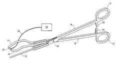
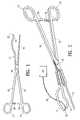
- FIG. 1is an elevational view of a uterine artery clamping device embodying features of the invention in an open configuration.
- FIG. 2is a perspective view of the clamping device shown in FIG. 1 .
- FIG. 3is an enlarged perspective view of the distal portion of an alternative clamping device having a serrated surface and a plurality of teeth to grasp tissue.
- FIG. 4is an enlarged perspective view of an alternative clamping device in which the clamping element and the stabilizer element are provided with teeth to grasp tissue.
- FIG. 5is a schematic illustration of the intrauterine clamp in position to occlude a female patient's uterine artery.
- FIG. 6is a perspective view of an alternative embodiment in which the clamping element and the stabilizing element are angled with respect to the handles of the clamp.
- FIG. 7is a perspective view of an alternative embodiment in which the spacing between the stabilizing member and the clamping member is adjusted by a spring coil.
- FIG. 8is perspective view of an alternative embodiment in which the spacing between the stabilizing member and the clamping member is adjusted by a rack and pinion arrangement.
- FIGS. 1 and 2show a relatively non-invasive intra-uterine occluding clamp 10 embodying features of the invention.
- the clamp 10includes a clamping member 11 having an elongated handle 12 with a finger grip 13 , and pressure-applying clamping element or jaw 14 on the distal end of the clamping element.
- the intra-uterine clampalso includes stabilizing member 15 which is configured to readily follow or track the patient's cervical os and cervical canal.
- the stabilizing member 15has an elongated handle 16 with a finger grip 17 .
- the clamping member 11 and stabilizing member 15are pivotally connected to each other at pivot point 18 and rotation of handles 12 and 16 , preferably by fingers of an operator's hand engaged through grips 13 and 17 respectively, adjust the spacing between the jaw 14 and the distal portion of stabilizing member 15 .
- Each of the handles 12 and 16are provided with a ratchet member 19 and 20 respectively which interact to lock the relative positions of the clamping member 11 and the stabilizing member 15 .
- a uterine artery clamp embodying features of the inventionis preferably provided with a blood flow sensor 22 , preferably a Doppler ultrasonic sensing system, on the leading surface of the jaw 14 .
- a blood flow sensor 22preferably a Doppler ultrasonic sensing system
- Sensor 22is provided with a signal transmission cable 23 which is operatively connected to sensor control device 24 .
- Cable 23may be an insulated wire, plurality of wires, optical fiber, waveguide, or other connection effective to carry signals and/or energy or power between a sensor 22 and sensor controller 24 .
- Sensor 22may be a blood flow sensor for locating a blood vessel, and may be a passive sensor, configured to detect intrinsic signals indicating the presence of a blood vessel (i.e., a sound sensor, a motion sensor, a pH sensor, or other sensor configured to detect a physical, chemical, electrical, or physiological indication of the location of a blood vessel).
- a blood vesseli.e., a sound sensor, a motion sensor, a pH sensor, or other sensor configured to detect a physical, chemical, electrical, or physiological indication of the location of a blood vessel.
- a blood flow sensor for locating a blood vesselmay be an active sensor, configured to emit energy or a signal, and configured to detect signals in response to, or derived from, the emitted energy or signal indicating the presence of a blood vessel (i.e., a source of ultrasound having an ultrasound sensor configured to detect ultrasound reflections from a blood vessel, a source of infrared radiation configured to detect reflections from a blood vessel, or other source of energy and a sensor configured to detect a response indicating the location of a blood vessel).
- the operation of a sensormay be aided by an energy source such as the sensor controller 24 .
- an energy sourcemay provide electrical energy which aids an ultrasound sensor to produce and to detect ultrasound energy (as, e.g., in the MedaSonics® CardioBeat® Blood Flow Doppler with Integrated Speaker (Cooper Surgical, Inc., Trumbull Conn. 06611)).
- Other commercially available Doppler ultrasound sensors suitable for use in the present inventioninclude the Koven model ES 100X MiniDop VRP-8 probe (St. Louis, Mo.) and the DWL/Neuro Scan Medical Systems' Multi-Dop B+ system (Sterling, Va.).
- jaw 14may be provided with a serrated, tissue-grasping surface 25 configured to engage and hold onto tissue when jaw 14 is pressed into tissue of the patient's vaginal fornix.
- tissue-grasping surface 25configured to engage and hold onto tissue when jaw 14 is pressed into tissue of the patient's vaginal fornix.
- one or both of the jaw 14 and/or stabilizer 15may have retractable fingers or teeth 26 to better secure the contacting members to the target tissue.
- FIG. 5illustrates an alternative embodiment wherein the clamping member 11 and the stabilizing member 15 are oriented at an angle ⁇ with respect to the handles 12 and 16 .
- the angulationprovides a more direct attack angle to facilitate insertion of the stabilizing member 15 into the patient's cervix and direction of the jaw 14 toward a desired location at the patient's vaginal fornix to facilitate location and occlusion of the patient's uterine artery.
- Suitable angulation ⁇ of the jaw 14 and stabilizer 15is about 100° to about 175°, preferably about 130° to about 160°.
- Closure of a blood vesselis effected by pressure applied through the wall of the patient's vaginal fornix. Sufficient pressure or force applied to the tissue of the vaginal wall to compress and to at least partially occlude the underlying uterine artery.
- the blood flow sensor for detecting or locating the uterine arteryshould be disposed on the leading face off the clamping element and generally perpendicular to the tissue-contacting surface of a jaw 14 to be effective.
- a non-invasive blood vessel occluding device embodying features of the inventionmay be configured to lock into a clamping position. Such a locked configuration may be temporary and releasable, or may be permanent.
- Non-invasive blood vessel occluding devices embodying features of the inventionmay have a locking mechanism, such as a ratchet, configured to hold at least one pressure-applying member in a pressure-applying position.
- Such locking mechanismsmay include a release mechanism effective to allow the cessation of pressure or force application when desired.
- a non-invasive blood vessel occlusion device embodying features of the inventionmay be configured to release a locking mechanism effective to relieve the occlusion of a blood vessel by ending the application of pressure or force that had been previously applied to occlude a blood vessel.
- a vaginadetermines what size clamping device is suitable, taking into consideration that the clamping device should readily reach the vaginal fornix and be operated from outside of a patient's body.
- a clamping devicemay be between about 5 to about 16 inches in length, preferably between about 6 inch to about 12 inches in length for most applications.
- FIG. 6schematically illustrates in part a human female reproductive system, including a uterus 30 , uterine cervix 31 , uterine artery 32 , vaginal canal 33 and vaginal fornix 34 .
- a method of using the uterine artery clamp embodying features of the inventionincludes introducing the clamp 10 into the patient's vaginal canal 33 and advancing the clamp therein until the distal portions of the clamp are adjacent to the patient's uterine cervix 31 .
- the position of the handles 12 and 16are adjusted to increase the spacing between the jaw 14 and the distal portion of the stabilizer 16 .
- the distal end of the stabilizeris guided through the cervical os 35 into the uterine cervix.
- the distal end of jaw 14is urged against the vaginal fornix 34 and 14 .
- the pressure applying surface of the jawis positioned as close as possible to the patient's uterine artery 32 .
- Sufficient pressureis applied to the uterine artery 32 or the tissue surrounding the uterine artery by jaw 14 to facilitate occlusion of the uterine artery.
- the handles 12 and 16are locked by ratchet members 19 and 20 to press the jaw 14 against the tissue between the jaw 14 and the stabilizer 15 .
- the clamped positionis maintained for about 0.5 to about 48 hours, preferably about 1 to about 24 hours for effective therapeutic treatment of a uterine disorder, e.g. for fibroids, PPH, DUB and the like.
- Blood flow sensor 22is effective to locate uterine artery 32 by detecting blood flow and monitoring the treatment by detecting the lack of blood flow in the artery.
- Blood flow in the left uterine artery 32may be similarly occluded, by a separate uterine artery clamp of the same design or the same clamp can be used on the other side after release of the occlusion of the right uterine artery 32 a.
- the ratchet members 19 and 20are preferably releasable so that clamping member 11 of clamping device 10 can be released after the limited treatment time to re-establish blood flow to the uterine tissue.
- FIGS. 7 and 8illustrate alternate clamp design wherein the spacing between the jaw 14 and stabilizer 15 is controlled by a spring 40 as shown in FIG. 7 and a rack and pinion mechanism 50 as shown in FIG. 8 .
- a variety of other meansmay be employed to open and close the jaw 14 and stabilizer 15 .
- Uterine artery clamp 10 embodying features of the inventionmay be made from any suitable material or combination of materials, including metals such as stainless steel and superelastic shape memory alloys such as nickel titanium alloys having a stable austenite phase at body temperature, high strength plastics, ceramics, and other materials known in the art.
- metalssuch as stainless steel and superelastic shape memory alloys such as nickel titanium alloys having a stable austenite phase at body temperature, high strength plastics, ceramics, and other materials known in the art.
- Biocompatible polymerssuch as polycarbonate, polysulfone, polyester, polyacetal and a variety of fluoropolymers can be suitable for a variety of embodiments of the invention.
- the device or systemmay be designed for single use (disposable) or may be sterilizable and capable of multiple use.
Landscapes
- Health & Medical Sciences (AREA)
- Surgery (AREA)
- Life Sciences & Earth Sciences (AREA)
- Medical Informatics (AREA)
- Animal Behavior & Ethology (AREA)
- Reproductive Health (AREA)
- Veterinary Medicine (AREA)
- Engineering & Computer Science (AREA)
- Biomedical Technology (AREA)
- Heart & Thoracic Surgery (AREA)
- Public Health (AREA)
- Molecular Biology (AREA)
- Nuclear Medicine, Radiotherapy & Molecular Imaging (AREA)
- General Health & Medical Sciences (AREA)
- Gynecology & Obstetrics (AREA)
- Pregnancy & Childbirth (AREA)
- Vascular Medicine (AREA)
- Surgical Instruments (AREA)
- Measuring Pulse, Heart Rate, Blood Pressure Or Blood Flow (AREA)
- Ultra Sonic Daignosis Equipment (AREA)
Abstract
Description
This application is a continuation-in-part of U.S. patent application Ser. No. 10/355,809, entitled “Devices and Methods for Treating Vaginal Delivery PPH”, filed Jan. 30, 2003 which is hereby incorporated by reference in its entirety and from which priority is hereby claimed under 35 U.S.C. § 120.
The invention is generally directed to the treatment of uterine disorders by detecting and regulating blood flow through one or both of the patient's uterine arteries.
Hysterectomy (surgical removal of the uterus) is performed on approximately 600,000 women annually in the United States. Hysterectomy is often the therapeutic choice for the treatment of uterine cancer, adenomyosis, menorrhagia, prolapse, dysfunctional uterine bleeding (abnormal menstrual bleeding that has no discrete anatomic explanation such as a tumor or growth), and muscular tumors of the uterus, known as leimyoma or uterine fibroids.
However, hysterectomy is a drastic treatment, having many undesirable characteristics. Thus, any method which can approximate the therapeutic result of a hysterectomy without removing the uterus would be a significant improvement in this field. Newer treatment methods have been developed for some diseases which may spare these women a hysterectomy.
In 1995, it was demonstrated that uterine fibroids could be treated without hysterectomy using a non-surgical therapy, specifically comprising bilateral intraluminal occlusion of the uterine arteries (Ravina et al., “Arterial Embolization to Treat Uterine Myomata”, Lancet Sep. 9, 1995; Vol. 346; pp. 671-672, incorporated in its entirety herein). This technique is known as “uterine artery embolization”. In this technique, uterine arteries are accessed via a transvascular route from a common femoral artery into the left and right uterine arteries by means of an intravascular catheter and embolic material, such as small metallic coils, polyvinyl alchohol particulate and the like, is delivered through the catheter to the uterine arteries which quickly become occluded.
The uterus has a dual (or redundant) blood supply, the primary blood supply being from the bilateral uterine arteries, and the secondary blood supply from the bilateral ovarian arteries. Consequently, when both uterine arteries are occluded, i.e. bilateral vessel occlusion, the uterus and the fibroids contained within the uterus are both deprived of their blood supply. However, as demonstrated by Ravina et al., the ischemic effects on the fibroid is greater than the effect on the uterus. In most instances, the fibroid withers and ceases to cause clinical symptoms.
However, many physicians do not possess the training or equipment necessary to perform catheter-based uterine artery embolization under radiologic direction. Accordingly, there are substantially fewer uterine artery embolizations performed, worldwide, each year than hysterectomies for symptomatic uterine fibroids.
Recently, fibroid treatment procedures have been described wherein the uterine arteries are temporarily occluded by an intravaginal device which is non-invasively pressed against the patient's vaginal fornix and clamped or otherwise pressed against tissue bundle with the patient's uterine artery being within the bundle. Pressure on the tissue occludes the underlying uterine artery. While these procedures have shown much promise, in many situations the devices described to date do not always allow for accurate placement of the clamping surfaces.
What is needed, therefore, are devices and methods to detect blood vessels and blood flow in blood vessels, and devices and methods to occlude blood flow in blood vessels such as the uterine arteries that can be used by physicians with limited training and equipment.
The invention is directed to a relatively non-invasive uterine artery occlusion device and system and the procedure for using the device and system for occluding a female patient's uterine artery. The instruments and their use may be utilized in the treatment of uterine fibroids, dysfunctional uterine bleeding, post partum hemorrhage and other uterine disorders by reducing or terminating blood flow through a patient's uterine artery.
A device embodying features of the invention includes an intrauterine clamp which has a pressure applying or clamping member configured to apply pressure against the exterior of the patient's uterine cervix or against the patient's vaginal fornix. The intravaginal clamp also has a stabilizing or positioning member which is configured to be inserted into the patient's uterine cervix so as to stabilize at least a portion of the interior of the uterine cervix and facilitate the more effective application of pressure by the pressure applying member to the exterior of the cervix or the vaginal fornix to ensure effective occlusion of the patient's uterine artery. The uterine artery occlusion is temporary, and may be partial or complete. The distal end of the clamping member is at or distal to the distal end of the stabilizing member. Preferably, the distal end of the clamping member is about 0.1 to about 1 inch distal to the distal end of the stabilizing member.
One method of occluding a blood vessel comprises clamping the blood vessel effective to compress it so that blood flow through the vessel is reduced, or is abolished. Such clamping of a blood vessel may be direct or may be indirect. Preferably, clamping of a blood vessel effective to compress it is accomplished by applying a non-invasive blood vessel occlusion device to tissue near to a blood vessel (e.g., onto tissue surrounding the vessel). A blood vessel occlusion device may also be applied directly onto a blood vessel effective to compress the blood vessel.
In one embodiment of the invention, a non-invasive blood vessel occluding device (such as a clamp with a sensor) may be applied to a portion of a vaginal wall to detect and/or locate, and to occlude the uterine arteries. A vaginal clamp embodying features of the invention may used to sense the location of a uterine artery adjacent a vaginal wall, and may be used to compress and occlude a uterine artery adjacent a vaginal wall. The vaginal wall may be distended by an occlusion device so as to more closely approach a uterine artery; such an approach may aided by applying pressure or force to the uterus (e.g., by pulling on the uterine cervix). A uterine cervix may be grasped or pulled by any suitable device or implement, including forceps, suction devices, and other instruments, such as a tenaculum.
A non-invasive blood vessel occluding device embodying features of the invention may be a non-invasive intravaginal uterine artery occlusion device, comprising a pair of pressure-applying members having opposed tissue-contacting surfaces on distal portions thereof; at least one supporting shaft extending from a proximal extremity of at least one of the pressure-applying members which is configured to adjust the distance between the opposed tissue-contacting surfaces of the pressure-applying members; and at least one blood flow sensing sensor on one of the opposed tissue-contacting surfaces. An embodiment of a non-invasive blood vessel occlusion device embodying features of the invention may have, for example, a handle, a clamping member configured to apply pressure or force to body tissue, and a sensor for locating a blood vessel.
A pressure-applying member, such as a clamping member, may be, e.g., a jaw or jaws configured to engage a blood vessel or to engage tissue adjacent a blood vessel. A supporting shaft, such as a handle, is preferably configured for manipulating the jaw or jaws. In some embodiments of devices having features of the invention, a pressure-applying member may be attached to a connecting portion that is configured so that a jaw may be placed within a vagina while a handle remains outside a patient's body and available for use by an operator.
The clamping member is preferably provided with a blood flow sensor for locating the blood vessel to be occluded. The sensor may sense sound, pressure, strain, stress, chemical entity, electromagnetic radiation and the like, and may be a combination of such sensors. A sensor is preferably a Doppler ultrasound sensor. The sensor is mounted to the face of a tissue-contacting surface of the clamping member, such as the face of a jaw of a clamp, and is preferably oriented perpendicularly to the clamp face, although other orientations may be employed. Ultrasound energy useful for sensing a location of a blood vessel or of blood flow in a blood vessel has a frequency of less than about 20 MegaHertz (MHz), such as between about 5 MHz and about 19 MHz, and preferably between about 6 MHz and about 10 MHz. In commercially available Doppler sensors the frequency is typically about 8 MHz. For sensors based on electromagnetic energy useful for sensing a location of a blood vessel or of blood flow in a blood vessel, the EM energy should have a wavelength of about 500 nanometers (nm) to about 2000 nm, preferably about 700 nm to about 1000 nm.
A system embodying features of the invention includes an blood vessel occluding device as described above with a blood flow sensor on the clamping member for locating the target blood vessel, and a sensor controller which may include an energy source for the sensor. The sensor controller may be configured to aid in detecting a location of a blood vessel, by, e.g., providing a signal related to the output of a sensor that may be readily used by an operator. A sensor controller may include an energy source configured to provide energy for operating a blood flow sensor.
A method for occluding a uterine artery which embodies features of the invention include advancing the clamping device through the patient's vaginal canal, guiding the stabilizing member of the clamping device through the cervical os, into the cervical canal with the clamping member spaced from the stabilizing member so that the pressure applying surfaces of the clamping member is pressed into the patient's vaginal fornix. Adjustment of the clamping member allows the sensor on the distal end thereof to locate the uterine artery a short distance from the surface of the vaginal fornix. With the clamping member adjacent to the target blood vessel, the clamping device can be closed to compress underlying tissue and thereby occlude the uterine artery. The uterine artery is located with the blood flow sensor on the distal end of the clamping member. Tension may be applied to the uterine cervix with a grasping implement (e.g., by pulling on the uterine cervix) while applying force or pressure to a vaginal wall to occlude a uterine artery.
The invention allows for the non-surgical location and occlusion of blood vessels such as the uterine artery, providing effective therapeutic treatment. Importantly, the present invention allows for the occlusion of a female patient's uterine artery without the need for radiographic equipment or for extensive training in the use of radiographic techniques. The devices and methods are simple and readily used for treating uterine fibroids, dysfunctional uterine bleeding (DUB), adenomyosis, post-partum hemorrhage, and other uterine disorders. The devices, systems and methods embodying features of the invention allow for the separate occlusion of individual uterine arteries which provides effective therapy in those situations in which the uterine anatomy will not allow for the use of a single bilateral artery occlusion device.
These and other advantages will become more apparent from the following detailed description when taken in conjunction with the accompanying exemlary drawings.
A uterine artery clamp embodying features of the invention is preferably provided with ablood flow sensor 22, preferably a Doppler ultrasonic sensing system, on the leading surface of thejaw 14. This sensor location allows the operator to more easily guide thejaw 14 to the location of the patient's target uterine artery.Sensor 22 is provided with asignal transmission cable 23 which is operatively connected tosensor control device 24.Cable 23 may be an insulated wire, plurality of wires, optical fiber, waveguide, or other connection effective to carry signals and/or energy or power between asensor 22 andsensor controller 24.
As shown inFIG. 3 jaw 14 may be provided with a serrated, tissue-graspingsurface 25 configured to engage and hold onto tissue whenjaw 14 is pressed into tissue of the patient's vaginal fornix. As shown inFIG. 4 , one or both of thejaw 14 and/orstabilizer 15 may have retractable fingers orteeth 26 to better secure the contacting members to the target tissue.
Closure of a blood vessel, which may be partial or total, is effected by pressure applied through the wall of the patient's vaginal fornix. Sufficient pressure or force applied to the tissue of the vaginal wall to compress and to at least partially occlude the underlying uterine artery. The blood flow sensor for detecting or locating the uterine artery should be disposed on the leading face off the clamping element and generally perpendicular to the tissue-contacting surface of ajaw 14 to be effective.
A non-invasive blood vessel occluding device embodying features of the invention may be configured to lock into a clamping position. Such a locked configuration may be temporary and releasable, or may be permanent. Non-invasive blood vessel occluding devices embodying features of the invention may have a locking mechanism, such as a ratchet, configured to hold at least one pressure-applying member in a pressure-applying position. Such locking mechanisms may include a release mechanism effective to allow the cessation of pressure or force application when desired. Thus, a non-invasive blood vessel occlusion device embodying features of the invention may be configured to release a locking mechanism effective to relieve the occlusion of a blood vessel by ending the application of pressure or force that had been previously applied to occlude a blood vessel.
The uterine arteries in human females are located adjacent the vaginal mucosa at a location within a few centimeters (cm) of the vaginal fornix. As a result, for accessing and occluding a uterine artery from within the patient's vaginal canala, the dimensions of a vagina determine what size clamping device is suitable, taking into consideration that the clamping device should readily reach the vaginal fornix and be operated from outside of a patient's body. For example, a clamping device may be between about 5 to about 16 inches in length, preferably between about 6 inch to about 12 inches in length for most applications.
Theratchet members member 11 of clampingdevice 10 can be released after the limited treatment time to re-establish blood flow to the uterine tissue.
While particular forms of the invention have been illustrated and described, it will be apparent that various modifications can be made to the invention and that individual features shown in one embodiment can be combined with any or all the features of another embodiment described herein. Accordingly, it is not intended that the invention be limited to the specific embodiments illustrated. It is therefore intended that this invention be defined by the scope of the appended claims as broadly as the prior art will permit. Terms such as “element”, “member”, “device”, “sections”, “portion”, “section”, “steps” and words of similar import when used herein shall not be construed as invoking the provisions of 35 U.S.C. §112(6) unless the following claims expressly use the terms “means” or “step” followed by a particular function without specific structure or action.
Claims (26)
1. An intravaginal clamping device for occluding a portion of a female patient's uterine artery adjacent to a region of the patient's vaginal fornix, comprising:
a. a stabilizing member which has a cylindrical distal end configured to extend into the patient's uterine cervix;
b. a clamping member with a clamping element at the distal portion thereof which is configured to fit about an exterior side portion of the patient's uterine cervix and which has a leading, pressure applying surface on the distal end that is at or extends beyond the distal end of the stabilizing member and which has a width is sufficient to be pressed against the region of the patient's vaginal fornix adjacent to the exterior side portion of the patient's uterine cervix to occlude the portion of the patient's uterine artery adjacent to the region of the patient's vaginal fornix; and
c. a blood flow sensor on the leading pressure applying surface of the distal end of the clamping member to detect the location of the uterine artery to monitor the blood flow therethrough.
2. The intravaginal clamping device ofclaim 1 wherein a connector is provided between the clamping member and the stabilizing member to adjust spacing between the clamping member and the stabilizing member.
3. The intravaginal clamping device ofclaim 2 wherein the connector is a pivotal connection between the clamping member and the stabilizing member.
4. The intravaginal clamping device ofclaim 1 wherein both the clamping member and the stabilizing member have elongated handles.
5. The intravaginal clamping device ofclaim 4 , wherein the handles have an interconnecting locking mechanism which provide a locked configuration effective to retain the clamping member in a pressure-applying configuration.
6. The intravaginal clamping device ofclaim 1 wherein the clamping member has a pressure applying surface which generally lies in a plane perpendicular to a plane in which the handles rotate.
7. The intravaginal clamping device ofclaim 1 wherein the stabilizing member is longer than the clamping member.
8. The intravaginal clamping device ofclaim 1 wherein the clamping element is paddle-shaped.
9. The intravaginal clamping device ofclaim 8 wherein the paddle-shaped clamping element is at least in part formed of a wire frame.
10. The intravaginal clamping device ofclaim 1 wherein the clamping member and the stabilizing member are curved.
11. The intravaginal clamping device ofclaim 1 , wherein the clamping element is releasably secured to a handle.
12. The intravaginal clamping device ofclaim 1 , wherein the blood flow sensor is a Doppler ultrasound sensor.
13. The intravaginal clamping device ofclaim 12 , wherein the Doppler ultrasound sensor is configured to sense ultrasound energy having a frequency of between about 5 MHz and about 20 MHz.
14. The intravaginal clamping device ofclaim 12 , wherein the Doppler ultrasound sensor is configured to sense ultrasound energy having a frequency of between about 6 MHz and about 10 MHz.
15. The intravaginal clamping device ofclaim 1 , wherein the clamping element has a length between about 1 and about 6 inches.
16. The intravaginal clamping device ofclaim 1 , wherein the clamping member has a length between about 2.5 inches and about 4.5 inches.
17. The intravaginal clamping device ofclaim 1 , wherein the distal end of the clamping element is disposed distal to the distal end of the stabilizing member by a distance of about 0.1 inch to about 1 inch.
18. The intravaginal clamping device ofclaim 1 , wherein the pressure apply surface on the distal end of the clamping element has a width of about 0.5 inch to about 3 inches.
19. The intravaginal clamping device ofclaim 1 , wherein the clamping element is formed at least in part of a loop-shaped wire frame.
20. A system for occluding a female patient's uterine artery, comprising:
a. a uterine artery clamping device which has
i. a clamping member which has an elongated handle and a clamping element with a distal end and a pressure applying surface on the distal end configured to occlude a uterine artery when the pressure applying surface is pressed against the patient's vaginal fornix,
ii. a stabilizing member which has an elongated handle at an obtuse angle with respect to the elongated handle of the clamping member and which has a cylindrical distal end that is configured to enter the patient's uterine cervix and that is at or proximal to the distal end of the clamping element; and
b. an artery location sensor secured to the pressure applying surface on the distal portion of a clamping element to ensure that pressing the pressure applying surface of the clamping element against the patient's vaginal fornix occludes the patient's uterine artery.
21. The system ofclaim 20 wherein the clamping device has a pivotal connection between the clamping member and the stabilizing member so that rotation of the handles about the pivotal connection adjusts spacing between the clamping member and the stabilizing member.
22. The system ofclaim 21 including an elongated signal transmitting member having a proximal end and a distal end and being secured at its distal end to the artery locating sensor and having a connector on its proximal end configured to be connected to a sensor controller.
23. The system ofclaim 20 , wherein the artery location sensor is a Doppler ultrasound sensor.
24. The system ofclaim 21 wherein the handles have a locking mechanism to provide a locked configuration effective to retain the clamping member and the stabilizing member in a pressure-applying configuration.
25. The intravaginal clamp ofclaim 22 wherein the connector is a spring.
26. The intravaginal clamp ofclaim 22 wherein the connector is a rack and pinion mechanism.
Priority Applications (6)
| Application Number | Priority Date | Filing Date | Title |
|---|---|---|---|
| US10/430,880US7329265B2 (en) | 2003-01-30 | 2003-05-06 | Uterine artery occlusion clamp |
| CA2514545ACA2514545C (en) | 2003-01-30 | 2004-01-23 | Uterine artery occlusion clamp |
| JP2006502977AJP4455581B2 (en) | 2003-01-30 | 2004-01-23 | Uterine artery occlusion clamp |
| EP04704961AEP1587432A4 (en) | 2003-01-30 | 2004-01-23 | Uterine artery occlusion clamp |
| PCT/US2004/001935WO2004069025A2 (en) | 2003-01-30 | 2004-01-23 | Uterine artery occlusion clamp |
| AU2004210130AAU2004210130B2 (en) | 2003-01-30 | 2004-01-23 | Uterine artery occlusion clamp |
Applications Claiming Priority (2)
| Application Number | Priority Date | Filing Date | Title |
|---|---|---|---|
| US10/355,809US7404821B2 (en) | 2003-01-30 | 2003-01-30 | Treatment for post partum hemorrhage |
| US10/430,880US7329265B2 (en) | 2003-01-30 | 2003-05-06 | Uterine artery occlusion clamp |
Related Parent Applications (1)
| Application Number | Title | Priority Date | Filing Date |
|---|---|---|---|
| US10/355,809Continuation-In-PartUS7404821B2 (en) | 2003-01-30 | 2003-01-30 | Treatment for post partum hemorrhage |
Publications (2)
| Publication Number | Publication Date |
|---|---|
| US20040153105A1 US20040153105A1 (en) | 2004-08-05 |
| US7329265B2true US7329265B2 (en) | 2008-02-12 |
Family
ID=32770630
Family Applications (3)
| Application Number | Title | Priority Date | Filing Date |
|---|---|---|---|
| US10/355,809Expired - LifetimeUS7404821B2 (en) | 2003-01-30 | 2003-01-30 | Treatment for post partum hemorrhage |
| US10/430,880Expired - Fee RelatedUS7329265B2 (en) | 2003-01-30 | 2003-05-06 | Uterine artery occlusion clamp |
| US11/881,981AbandonedUS20080097473A1 (en) | 2003-01-30 | 2007-07-30 | Treatment for post partum hemorrhage |
Family Applications Before (1)
| Application Number | Title | Priority Date | Filing Date |
|---|---|---|---|
| US10/355,809Expired - LifetimeUS7404821B2 (en) | 2003-01-30 | 2003-01-30 | Treatment for post partum hemorrhage |
Family Applications After (1)
| Application Number | Title | Priority Date | Filing Date |
|---|---|---|---|
| US11/881,981AbandonedUS20080097473A1 (en) | 2003-01-30 | 2007-07-30 | Treatment for post partum hemorrhage |
Country Status (6)
| Country | Link |
|---|---|
| US (3) | US7404821B2 (en) |
| EP (1) | EP1587436A4 (en) |
| JP (1) | JP4455582B2 (en) |
| AU (1) | AU2004207532B2 (en) |
| CA (1) | CA2514546A1 (en) |
| WO (1) | WO2004066818A2 (en) |
Cited By (16)
| Publication number | Priority date | Publication date | Assignee | Title |
|---|---|---|---|---|
| US20020165579A1 (en)* | 2001-03-28 | 2002-11-07 | Burbank Fred H. | Multi-axial uterine artery identification, characterization, and occlusion devices and methods |
| US20030216759A1 (en)* | 1998-12-08 | 2003-11-20 | Vascular Control Systems, Inc. | Devices and methods for occlusion of the uterine arteries |
| US20050033276A1 (en)* | 2003-07-07 | 2005-02-10 | Olympus Corporation | Blood vessel detection device |
| US20080114382A1 (en)* | 2006-11-03 | 2008-05-15 | Ams Research Corporation | Uterine Artery Ligation Devices and Methods |
| US20090093758A1 (en)* | 2006-07-24 | 2009-04-09 | Yossi Gross | Fibroid treatment apparatus and method |
| US20090105698A1 (en)* | 2007-05-14 | 2009-04-23 | Ams Research Corporation | Medical Laser User Interface |
| US20090157064A1 (en)* | 2007-05-11 | 2009-06-18 | Hodel Michael R | RFID System and Method Therefor |
| USD622119S1 (en)* | 2008-05-08 | 2010-08-24 | Kunststoffwerk Ag Buchs | Gripper joint |
| US20110022073A1 (en)* | 2009-07-27 | 2011-01-27 | Fibro Control, Inc. | Balloon with rigid tube for occluding the uterine artery |
| US20140309671A1 (en)* | 2010-04-07 | 2014-10-16 | Miriam Mackovic Basic | Instrument for occlusion of uterine blood vessels |
| US9089365B2 (en) | 2012-04-26 | 2015-07-28 | Imds Llc | Tissue fixation device |
| US9492312B2 (en) | 2010-10-18 | 2016-11-15 | Bioceptive, Inc. | Methods and apparatus for inserting a device or pharmaceutical into a body cavity |
| US9931128B2 (en) | 2006-02-03 | 2018-04-03 | Covidien Lp | Methods for restoring blood flow within blocked vasculature |
| USD835270S1 (en)* | 2016-02-09 | 2018-12-04 | Benson Medical LLC | Tenaculum |
| US10172633B2 (en) | 2009-03-06 | 2019-01-08 | Covidien Lp | Retrieval systems and methods for use thereof |
| US10456560B2 (en) | 2015-02-11 | 2019-10-29 | Covidien Lp | Expandable tip medical devices and methods |
Families Citing this family (39)
| Publication number | Priority date | Publication date | Assignee | Title |
|---|---|---|---|---|
| US20030120306A1 (en)* | 2000-04-21 | 2003-06-26 | Vascular Control System | Method and apparatus for the detection and occlusion of blood vessels |
| US7223279B2 (en)* | 2000-04-21 | 2007-05-29 | Vascular Control Systems, Inc. | Methods for minimally-invasive, non-permanent occlusion of a uterine artery |
| US6550482B1 (en)* | 2000-04-21 | 2003-04-22 | Vascular Control Systems, Inc. | Methods for non-permanent occlusion of a uterine artery |
| US20030120286A1 (en)* | 2001-03-28 | 2003-06-26 | Vascular Control System | Luminal clip applicator with sensor |
| US7351248B2 (en)* | 2002-03-25 | 2008-04-01 | Tri-State Hospital Supply Corporation | Surgical instrument with snag free box hinge |
| WO2004069025A2 (en)* | 2003-01-30 | 2004-08-19 | Vascular Control Systems, Inc. | Uterine artery occlusion clamp |
| US7875036B2 (en)* | 2004-10-27 | 2011-01-25 | Vascular Control Systems, Inc. | Short term treatment for uterine disorder |
| EP1895915B1 (en)* | 2005-06-06 | 2016-03-23 | AMS Research Corporation | Devices for ligating uterine arteries |
| US20060276808A1 (en)* | 2005-06-06 | 2006-12-07 | Arnal Kevin R | Minimally Invasive Methods and Apparatus for Accessing and Ligating Uterine Arteries with Sutures |
| US20090043295A1 (en)* | 2005-06-06 | 2009-02-12 | Ams Research Corporation | Fibroid Treatment Methods and Devices |
| US20070049973A1 (en)* | 2005-08-29 | 2007-03-01 | Vascular Control Systems, Inc. | Method and device for treating adenomyosis and endometriosis |
| US20070112376A1 (en)* | 2005-11-14 | 2007-05-17 | Tri-State Hospital Supply Corporation | Medical tubing clamping apparatus |
| US20090054916A1 (en)* | 2007-08-23 | 2009-02-26 | Peter Meier | Clip-based method for treatment of uterine fibroids by obstruction of the uterine arteries |
| US20090054915A1 (en)* | 2007-08-23 | 2009-02-26 | Peter Meier | Obstruction of uterine arteries to treat uterine fibroids using mechanical instruments to twist the vessels |
| US20090062827A1 (en)* | 2007-08-31 | 2009-03-05 | Peter Meier | Vacuum-based method for obstruction of uterine arteries to treat uterine fibroids |
| USD578648S1 (en)* | 2007-10-12 | 2008-10-14 | Wood William L | Perineal cleaning device |
| US20090105720A1 (en)* | 2007-10-19 | 2009-04-23 | Boone Brenda J | Non-invasive surgical tenaculum |
| WO2011006067A1 (en) | 2009-07-09 | 2011-01-13 | Ams Research Corporation | Apparatus and methods of treatment of pathologic proliferative conditions uterine tissue |
| US9433421B2 (en)* | 2010-03-12 | 2016-09-06 | Jms Co., Ltd. | Surgical tool for anastomosis |
| US9375226B2 (en)* | 2010-03-19 | 2016-06-28 | Empire Technology Development Llc | Surgical instrument |
| US8608738B2 (en) | 2010-12-06 | 2013-12-17 | Soulor Surgical, Inc. | Apparatus for treating a portion of a reproductive system and related methods of use |
| US8562623B2 (en) | 2011-02-09 | 2013-10-22 | ROSS ALAN McDONALD | Vaginal occlusion device |
| WO2012137894A1 (en)* | 2011-04-08 | 2012-10-11 | Kariya Isao | Hemostatic tool, hemostatic device, and hemostatic method for uterine bleeding |
| US8795292B1 (en) | 2011-04-27 | 2014-08-05 | Ashraf El-Dabh | Device and method for treating post-partum hemorrhage |
| MX2012002339A (en) | 2012-02-23 | 2013-09-03 | Jose Arnoldo Guzman Sanchez | Plates for compressing the placenta insertion site used in case of placenta previa. |
| MX2012004494A (en)* | 2012-04-17 | 2013-10-17 | Arnoldo Guzman Sanchez | Compressive system for reducing edge bleeding in classical hysterotomy in cases of placenta praevia. |
| CN103271753A (en)* | 2013-06-21 | 2013-09-04 | 牛芳 | Nabothian cyst extirpation pincers |
| US8992525B2 (en) | 2013-07-31 | 2015-03-31 | Ent Biotech Solutions, Inc. | Surgical instrument |
| US9168061B1 (en) | 2014-12-15 | 2015-10-27 | Robert J. DiBenedetto | Uterine clamp for treating postpartum hemorrhage and facilitating uterine repairs |
| US20200022727A1 (en)* | 2017-02-01 | 2020-01-23 | Partus Llc | Systems, devices, and methods for treating and monitoring a pregnant patient having a prematurely open cervix |
| AT519974B1 (en)* | 2017-10-04 | 2018-12-15 | Dr Friedrich Stoiber | Medical clamp |
| EP4223210A1 (en)* | 2017-11-10 | 2023-08-09 | Hegenbergerspeculum APS | Device |
| USD917049S1 (en)* | 2017-11-19 | 2021-04-20 | ProCell Therapies, LLC | Microdermabrasion paddle |
| US11497526B2 (en)* | 2018-11-13 | 2022-11-15 | T & J Enterprises, Llc | Cervical tenaculum device |
| US10512483B1 (en)* | 2018-11-13 | 2019-12-24 | T & J Enterprises, Llc | Cervical tenaculum device |
| CN110960301A (en)* | 2019-12-30 | 2020-04-07 | 西安医学院第二附属医院 | Obstetric forceps for obstetrical operation |
| US20210204907A1 (en)* | 2020-01-07 | 2021-07-08 | Covidien Lp | Devices, systems, and methods for trans-vaginal, ultrasound-guided hysteroscopic surgical procedures |
| US20230255620A1 (en)* | 2020-07-07 | 2023-08-17 | The General Hospital Corporation | Devices and method for facilitating placement of sutures in a surgical procedure |
| IT202200011453A1 (en)* | 2022-05-31 | 2023-12-01 | Antonio Curcio | Injectable loop recorder extraction device |
Citations (132)
| Publication number | Priority date | Publication date | Assignee | Title |
|---|---|---|---|---|
| US2400251A (en) | 1943-07-29 | 1946-05-14 | Charles E Nagel | Gynecological instrument |
| FR1220773A (en) | 1959-04-29 | 1960-05-27 | Vacuum Extractor Ab | Instrument to facilitate examination of the uterus, oviducts or similar organs |
| US3209753A (en) | 1962-05-04 | 1965-10-05 | Donald B Hawkins | Intestinal clamps and the like |
| US3411505A (en) | 1965-12-15 | 1968-11-19 | Paul D. Nobis | Device for interrupting arterial flow |
| US3777740A (en)* | 1971-10-21 | 1973-12-11 | Administrator For Veterans Aff | Method and apparatus for non-invasively visualizing blood vessels |
| US3779248A (en) | 1971-10-18 | 1973-12-18 | Medical Concepts Inc | Forceps |
| US4120302A (en) | 1976-10-08 | 1978-10-17 | American Hospital Supply Corporation | Disposable pads for surgical instruments |
| US4226240A (en) | 1979-05-30 | 1980-10-07 | Walker Jr William E | Surgical foreceps |
| US4292960A (en) | 1979-04-30 | 1981-10-06 | Rca Corporation | Apparatus and method for application of radioactive and microwave energy to the body |
| US4428379A (en) | 1982-01-07 | 1984-01-31 | Technicare Corporation | Passive ultrasound needle probe locator |
| US4428374A (en) | 1978-12-20 | 1984-01-31 | Auburn Robert M | Umbilical cord clamping assembly |
| US4509528A (en)* | 1981-12-16 | 1985-04-09 | Harvinder Sahota | Hemostat with blood flow sensor |
| US4650466A (en) | 1985-11-01 | 1987-03-17 | Angiobrade Partners | Angioplasty device |
| US4757823A (en) | 1987-01-27 | 1988-07-19 | Hofmeister John F | Method and apparatus for measuring uterine blood flow |
| US4945896A (en) | 1989-01-24 | 1990-08-07 | Gade George F | Surgical retractor assembly having tissue viability sensor embedded therein |
| US4991588A (en) | 1986-07-21 | 1991-02-12 | Pfizer Hospital Products Group, Inc. | Doppler guide wire |
| US4994069A (en) | 1988-11-02 | 1991-02-19 | Target Therapeutics | Vaso-occlusion coil and method |
| US5037433A (en) | 1990-05-17 | 1991-08-06 | Wilk Peter J | Endoscopic suturing device and related method and suture |
| US5081997A (en) | 1989-03-09 | 1992-01-21 | Vance Products Incorporated | Echogenic devices, material and method |
| US5108408A (en) | 1990-04-20 | 1992-04-28 | Lally James J | Uterine-ring hysterectomy clamp |
| US5201314A (en) | 1989-03-09 | 1993-04-13 | Vance Products Incorporated | Echogenic devices, material and method |
| US5226911A (en) | 1991-10-02 | 1993-07-13 | Target Therapeutics | Vasoocclusion coil with attached fibrous element(s) |
| US5261409A (en) | 1991-05-27 | 1993-11-16 | Sulzer Brothers Limited | Puncturing device for blood vessels |
| US5275166A (en)* | 1992-11-16 | 1994-01-04 | Ethicon, Inc. | Method and apparatus for performing ultrasonic assisted surgical procedures |
| US5289831A (en) | 1989-03-09 | 1994-03-01 | Vance Products Incorporated | Surface-treated stent, catheter, cannula, and the like |
| US5336229A (en) | 1993-02-09 | 1994-08-09 | Laparomed Corporation | Dual ligating and dividing apparatus |
| US5336231A (en) | 1992-05-01 | 1994-08-09 | Adair Edwin Lloyd | Parallel channel fixation, repair and ligation suture device |
| US5383922A (en) | 1993-03-15 | 1995-01-24 | Medtronic, Inc. | RF lead fixation and implantable lead |
| WO1995002370A2 (en) | 1993-07-15 | 1995-01-26 | Aws Shakir Mustafa Salim | Tunnelling catheter |
| WO1995002371A3 (en) | 1993-07-15 | 1995-03-09 | Aws Shakir Mustafa Salim | Rectal and rectosigmoid cancer tunnelling umbrella |
| US5427108A (en) | 1993-04-01 | 1995-06-27 | Bollinger; Armin | Ultrasonic Doppler probe with needle guide |
| EP0472368B1 (en) | 1990-08-21 | 1995-06-28 | Med Institute, Inc. | Ablation catheter |
| US5456693A (en) | 1992-09-21 | 1995-10-10 | Vitaphore Corporation | Embolization plugs for blood vessels |
| US5458596A (en) | 1994-05-06 | 1995-10-17 | Dorsal Orthopedic Corporation | Method and apparatus for controlled contraction of soft tissue |
| US5488958A (en) | 1992-11-09 | 1996-02-06 | Vance Products Incorporated | Surgical cutting instrument for coring tissue affixed thereto |
| US5496331A (en) | 1993-07-28 | 1996-03-05 | Terumo Kabushiki Kaisha | Knot-forming instrument and method of forming knots |
| WO1996010365A1 (en) | 1994-10-03 | 1996-04-11 | Apollo Camera, L.L.C. | Low profile tool for applying spring action ligation clips |
| US5507744A (en) | 1992-04-23 | 1996-04-16 | Scimed Life Systems, Inc. | Apparatus and method for sealing vascular punctures |
| US5542944A (en) | 1993-04-19 | 1996-08-06 | Bhatta; Krishan M. | Surgical device and method |
| US5549824A (en) | 1994-03-07 | 1996-08-27 | Ing. A. Maurer Sa | Filter apparatus including stacked intake and discharge plates |
| US5549624A (en) | 1994-06-24 | 1996-08-27 | Target Therapeutics, Inc. | Fibered vasooclusion coils |
| US5556396A (en) | 1994-01-18 | 1996-09-17 | Endovascular, Inc. | Method for tubal electroligation |
| US5562680A (en)* | 1992-01-03 | 1996-10-08 | Hasson; Harrith M. | Apparatus for assisting the performance of pelvic endoscopic procedures |
| US5570692A (en) | 1995-05-19 | 1996-11-05 | Hayashi Denki Co. Ltd. | Ultrasonic doppler blood flow detector for hemorrhoid artery ligation |
| US5582617A (en)* | 1993-07-21 | 1996-12-10 | Charles H. Klieman | Surgical instrument for endoscopic and general surgery |
| US5588960A (en) | 1994-12-01 | 1996-12-31 | Vidamed, Inc. | Transurethral needle delivery device with cystoscope and method for treatment of urinary incontinence |
| US5591173A (en) | 1994-07-28 | 1997-01-07 | Michael Schifano | Schifano obstetric scissors |
| GB2302025A (en)* | 1995-06-10 | 1997-01-08 | Mark Steven Whiteley | Vascular doppler forceps |
| US5598841A (en) | 1993-09-24 | 1997-02-04 | Kowa Company Ltd. | Blood flow measurement system |
| US5614204A (en) | 1995-01-23 | 1997-03-25 | The Regents Of The University Of California | Angiographic vascular occlusion agents and a method for hemostatic occlusion |
| WO1997027897A1 (en) | 1996-02-02 | 1997-08-07 | Transvascular, Inc. | A device, system and method for interstitial transvascular intervention |
| US5658299A (en) | 1995-07-20 | 1997-08-19 | Applied Medical Resources | Surgical ligating device and method for using same |
| US5662676A (en) | 1992-06-24 | 1997-09-02 | K.U. Leuven Research & Development | Instrument set for laparoscopic hysterectomy |
| US5662680A (en) | 1991-10-18 | 1997-09-02 | Desai; Ashvin H. | Endoscopic surgical instrument |
| US5665096A (en) | 1995-03-07 | 1997-09-09 | Yoon; Inbae | Needle driving apparatus and methods of suturing tissue |
| US5672153A (en) | 1992-08-12 | 1997-09-30 | Vidamed, Inc. | Medical probe device and method |
| US5672172A (en) | 1994-06-23 | 1997-09-30 | Vros Corporation | Surgical instrument with ultrasound pulse generator |
| GB2311468A (en) | 1996-03-27 | 1997-10-01 | Valleylab Inc | Electrosurgical interstitial resector |
| US5674243A (en) | 1995-08-03 | 1997-10-07 | Hale; Theodore Mark | Obstetrical forceps |
| US5691314A (en) | 1996-03-18 | 1997-11-25 | The Medical College Of Hampton Roads | Adjunctive therapy |
| US5697937A (en)* | 1996-02-23 | 1997-12-16 | Toma; Doina | Surgical clamp with manipulable guide means |
| US5697942A (en) | 1994-07-31 | 1997-12-16 | Palti; Yoram | Internal vascular clamp |
| WO1997047246A1 (en) | 1996-06-10 | 1997-12-18 | Influence Medical Technologies Ltd. | Suture insertion device for the treatment of urinary stress incontinence |
| US5702407A (en) | 1994-11-29 | 1997-12-30 | Olympus Optical Co., Ltd. | Ligating apparatus |
| US5713942A (en) | 1992-05-01 | 1998-02-03 | Vesta Medical, Inc. | Body cavity ablation apparatus and model |
| US5713371A (en) | 1995-07-07 | 1998-02-03 | Sherman; Dani | Method of monitoring cervical dilatation during labor, and ultrasound transducer particularly useful in such method |
| US5713896A (en) | 1991-11-01 | 1998-02-03 | Medical Scientific, Inc. | Impedance feedback electrosurgical system |
| US5715832A (en) | 1995-02-28 | 1998-02-10 | Boston Scientific Corporation | Deflectable biopsy catheter |
| US5716389A (en) | 1995-11-13 | 1998-02-10 | Walinsky; Paul | Cardiac ablation catheter arrangement with movable guidewire |
| US5720743A (en) | 1996-06-07 | 1998-02-24 | Bischof; John C. | Thermally insulating surgical probe |
| US5749879A (en) | 1989-08-16 | 1998-05-12 | Medtronic, Inc. | Device or apparatus for manipulating matter |
| WO1998019713A1 (en) | 1996-11-06 | 1998-05-14 | Sts Biopolymers Inc. | Echogenic coating containing gaseous spaces for ultrasonography |
| US5759154A (en) | 1996-12-23 | 1998-06-02 | C. R. Bard, Inc. | Print mask technique for echogenic enhancement of a medical device |
| US5766135A (en) | 1995-03-08 | 1998-06-16 | Terwilliger; Richard A. | Echogenic needle tip |
| US5776129A (en) | 1996-06-12 | 1998-07-07 | Ethicon Endo-Surgery, Inc. | Endometrial ablation apparatus and method |
| US5792059A (en) | 1996-11-26 | 1998-08-11 | Esaote S.P.A. | Intraoperative probe, specifically intended for direct-contact observations |
| US5797397A (en) | 1996-11-25 | 1998-08-25 | Hewlett-Packard Company | Ultrasound imaging system and method using intensity highlighting to facilitate tissue differentiation |
| US5800378A (en) | 1992-08-12 | 1998-09-01 | Vidamed, Inc. | Medical probe device and method |
| US5817022A (en) | 1995-03-28 | 1998-10-06 | Sonometrics Corporation | System for displaying a 2-D ultrasound image within a 3-D viewing environment |
| US5836906A (en) | 1996-02-23 | 1998-11-17 | Somnus Medical Technologies, Inc. | Method and apparatus for treatment of air way obstructions |
| US5840033A (en) | 1996-05-29 | 1998-11-24 | Ge Yokogawa Medical Systems, Limited | Method and apparatus for ultrasound imaging |
| WO1999000057A1 (en) | 1997-06-27 | 1999-01-07 | Michigan Instruments, Inc. | Non-invasive aortic impingement |
| US5895386A (en) | 1996-12-20 | 1999-04-20 | Electroscope, Inc. | Bipolar coagulation apparatus and method for arthroscopy |
| US5895395A (en) | 1997-07-17 | 1999-04-20 | Yeung; Teresa T. | Partial to full thickness suture device & method for endoscopic surgeries |
| US5899861A (en) | 1995-03-31 | 1999-05-04 | Siemens Medical Systems, Inc. | 3-dimensional volume by aggregating ultrasound fields of view |
| US5904651A (en) | 1996-10-28 | 1999-05-18 | Ep Technologies, Inc. | Systems and methods for visualizing tissue during diagnostic or therapeutic procedures |
| US5910484A (en) | 1997-05-30 | 1999-06-08 | The General Hospital Corporation | Treatment of ischemic cardiac malfunction |
| US5911691A (en) | 1996-05-21 | 1999-06-15 | Aloka Co., Ltd. | Ultrasound image processing apparatus and method of forming and displaying ultrasound images by the apparatus |
| US5916173A (en) | 1997-02-26 | 1999-06-29 | Kirsner; Vaclav | Methods and apparatus for monitoring fertility status in the mammalian vagina |
| US5921933A (en) | 1998-08-17 | 1999-07-13 | Medtronic, Inc. | Medical devices with echogenic coatings |
| US5922008A (en) | 1997-08-28 | 1999-07-13 | Gimpelson; Richard J. | Surgical forceps |
| US5941889A (en) | 1997-10-14 | 1999-08-24 | Civco Medical Instruments Inc. | Multiple angle disposable needle guide system |
| US5979453A (en) | 1995-11-09 | 1999-11-09 | Femrx, Inc. | Needle myolysis system for uterine fibriods |
| US6013088A (en) | 1998-11-17 | 2000-01-11 | Karavidas; Theocharis | Surgical clamp with removable tips |
| US6015541A (en) | 1997-11-03 | 2000-01-18 | Micro Therapeutics, Inc. | Radioactive embolizing compositions |
| US6019724A (en) | 1995-02-22 | 2000-02-01 | Gronningsaeter; Aage | Method for ultrasound guidance during clinical procedures |
| US6032673A (en) | 1994-10-13 | 2000-03-07 | Femrx, Inc. | Methods and devices for tissue removal |
| US6033398A (en) | 1996-03-05 | 2000-03-07 | Vnus Medical Technologies, Inc. | Method and apparatus for treating venous insufficiency using directionally applied energy |
| US6035238A (en) | 1997-08-13 | 2000-03-07 | Surx, Inc. | Noninvasive devices, methods, and systems for shrinking of tissues |
| US6034477A (en) | 1997-12-16 | 2000-03-07 | U.S. Philips Corporation | High-pressure discharge lamp |
| US6039693A (en) | 1991-11-08 | 2000-03-21 | Mayo Foundation For Medical Education And Research | Volumetric image ultrasound transducer underfluid catheter system |
| US6045508A (en) | 1997-02-27 | 2000-04-04 | Acuson Corporation | Ultrasonic probe, system and method for two-dimensional imaging or three-dimensional reconstruction |
| US6066139A (en) | 1996-05-14 | 2000-05-23 | Sherwood Services Ag | Apparatus and method for sterilization and embolization |
| US6077257A (en) | 1996-05-06 | 2000-06-20 | Vidacare, Inc. | Ablation of rectal and other internal body structures |
| US6080118A (en) | 1999-02-25 | 2000-06-27 | Blythe; Cleveland | Vaginal probe and method of using same |
| US6096051A (en) | 1998-03-20 | 2000-08-01 | Scimed Life Systems, Inc. | Endoscopic suture systems |
| US6106473A (en) | 1996-11-06 | 2000-08-22 | Sts Biopolymers, Inc. | Echogenic coatings |
| US6152874A (en) | 1996-04-26 | 2000-11-28 | Genzyme Corporation | Adjustable multi-purpose coronary stabilizing retractor |
| US6169914B1 (en) | 1998-01-13 | 2001-01-02 | Urometrics, Inc. | Devices and methods for monitoring female arousal |
| US6175751B1 (en) | 1999-03-16 | 2001-01-16 | Allen Maizes | Apparatus and method for sensing oxygen levels in a fetus |
| EP1072282A1 (en) | 1999-07-19 | 2001-01-31 | EndoArt S.A. | Flow control device |
| US6186947B1 (en) | 1998-07-29 | 2001-02-13 | Asahi Kogaku Kogyo Kabushiki Kaisha | Sector scanning, intracavitary ultrasonic probe |
| US6210330B1 (en) | 1999-08-04 | 2001-04-03 | Rontech Medical Ltd. | Apparatus, system and method for real-time endovaginal sonography guidance of intra-uterine, cervical and tubal procedures |
| DE20022012U1 (en) | 2000-04-18 | 2001-05-10 | Hofstetter, Alfons, Prof. Dr.med., 82008 Unterhaching | Medical clamp |
| US6231515B1 (en) | 1999-01-13 | 2001-05-15 | Scimed Life Systems, Inc. | Safety mechanism and method to prevent rotating imaging guide device from exiting a catheter |
| US6254601B1 (en) | 1998-12-08 | 2001-07-03 | Hysterx, Inc. | Methods for occlusion of the uterine arteries |
| US6261234B1 (en) | 1998-05-07 | 2001-07-17 | Diasonics Ultrasound, Inc. | Method and apparatus for ultrasound imaging with biplane instrument guidance |
| US6280441B1 (en) | 1997-12-15 | 2001-08-28 | Sherwood Services Ag | Apparatus and method for RF lesioning |
| WO2001068720A1 (en) | 2000-03-13 | 2001-09-20 | Biocure, Inc. | Embolic compositions |
| US6293954B1 (en)* | 1999-06-21 | 2001-09-25 | Novare Surgical Systems, Inc. | Surgical clamp with replaceable clamp members |
| US6299621B1 (en) | 1999-06-18 | 2001-10-09 | Novare Surgical Systems, Inc. | Surgical clamp pads with elastomer impregnated mesh |
| WO2001080713A2 (en) | 2000-04-21 | 2001-11-01 | Hysterx, Inc. | Methods for non-permanent occlusion of a uterine artery |
| US6368340B2 (en)* | 1995-04-03 | 2002-04-09 | William W. Malecki | Clamp assembly and method of use |
| US6371973B1 (en)* | 1999-08-04 | 2002-04-16 | Ron-Tech Medical Ltd. | Forceps useful for intrabody guiding and/or positioning of a medical instrument |
| WO2002039904A1 (en) | 2000-11-16 | 2002-05-23 | Vascular Control Systems, Inc. | Doppler directed suture ligation device and method |
| US6425867B1 (en) | 1998-09-18 | 2002-07-30 | University Of Washington | Noise-free real time ultrasonic imaging of a treatment site undergoing high intensity focused ultrasound therapy |
| US20020111537A1 (en) | 1996-02-20 | 2002-08-15 | Taylor Charles S. | Surgical instruments and procedures for stabilizing the beating heart during coronary artery bypass graft surgery |
| US20020165579A1 (en) | 2001-03-28 | 2002-11-07 | Burbank Fred H. | Multi-axial uterine artery identification, characterization, and occlusion devices and methods |
| US20030018270A1 (en) | 2001-05-29 | 2003-01-23 | Makin Inder Raj. S. | Tissue-retaining system for ultrasound medical treatment |
| WO2002000192A8 (en) | 2000-06-23 | 2003-06-12 | Carbon Medical Technologies In | Embolization using carbon coated particles |
| US20030120306A1 (en) | 2000-04-21 | 2003-06-26 | Vascular Control System | Method and apparatus for the detection and occlusion of blood vessels |
| US6610074B2 (en) | 2000-02-10 | 2003-08-26 | Albert N. Santilli | Aorta cross clamp assembly |
Family Cites Families (27)
| Publication number | Priority date | Publication date | Assignee | Title |
|---|---|---|---|---|
| US2601513A (en)* | 1948-12-29 | 1952-06-24 | Sidney A Gladstone | Surgical tissue collector device |
| US2587486A (en)* | 1950-07-13 | 1952-02-26 | Kogan Jerome | Cervical speculum |
| US2669991A (en)* | 1951-12-27 | 1954-02-23 | Curutchet Pedro Domingo | Tetradigital grip for long lever pivoted surgical instruments |
| US2842132A (en)* | 1956-10-15 | 1958-07-08 | Luis R Soltero | Surgical clamp |
| US2887111A (en)* | 1957-10-03 | 1959-05-19 | Diaz Ricardo M Leyro | Surgical forceps |
| US3063455A (en)* | 1960-04-07 | 1962-11-13 | Raymond L Markley | Cervical tourniquet instrument |
| US3503398A (en)* | 1965-09-10 | 1970-03-31 | American Hospital Supply Corp | Atraumatic clamp for vascular surgery |
| US4467803A (en)* | 1982-04-01 | 1984-08-28 | Ngo Tuyen N | Oral temporary total hemostatic clamps |
| USD281195S (en)* | 1982-09-29 | 1985-10-29 | Kurz Craven H | Orthodontic utility plier |
| USD275790S (en)* | 1982-12-22 | 1984-10-02 | Marlowe John B | Disposable surgical clamp |
| US5037430A (en) | 1986-01-06 | 1991-08-06 | Hasson Harrith M | Clamp for gynecological instruments |
| USD352644S (en)* | 1992-06-17 | 1994-11-22 | Fields Glenn C | Tongs for cleaning toilets and urinals |
| US5643316A (en)* | 1993-04-27 | 1997-07-01 | The Trustees Of The University Of Pennsylvania | Method of thoracoscopic surgery using hinged tissue grasping forceps |
| US6596000B2 (en)* | 1999-11-05 | 2003-07-22 | Alcon Universal Ltd. | Instrument for positioning an intracorneal optical lens |
| CA2391620A1 (en)* | 1999-12-03 | 2001-06-21 | Applied Medical Resources Corporation | Vessel occlusion clamp |
| US7223279B2 (en)* | 2000-04-21 | 2007-05-29 | Vascular Control Systems, Inc. | Methods for minimally-invasive, non-permanent occlusion of a uterine artery |
| FR2811215B1 (en)* | 2000-07-05 | 2003-03-14 | Bernard Flipo | MULTIFUNCTIONAL FORCEPS FOR MEDICAL USE COMPRISING TWO ARTICULATED JAWS |
| KR100405062B1 (en)* | 2000-09-22 | 2003-11-07 | 학교법인연세대학교 | a Forceps for reduction of mandibular angle |
| US6592603B2 (en)* | 2000-12-15 | 2003-07-15 | Michael Lasner | Manually adjustable scissors or forceps |
| US7354444B2 (en)* | 2001-03-28 | 2008-04-08 | Vascular Control Systems, Inc. | Occlusion device with deployable paddles for detection and occlusion of blood vessels |
| US20030120286A1 (en)* | 2001-03-28 | 2003-06-26 | Vascular Control System | Luminal clip applicator with sensor |
| US6688312B2 (en)* | 2001-05-23 | 2004-02-10 | Sarkis Yeretsian | Closed bloodless hemorrhoidectomy method |
| US7172603B2 (en)* | 2002-11-19 | 2007-02-06 | Vascular Control Systems, Inc. | Deployable constrictor for uterine artery occlusion |
| US20040097961A1 (en)* | 2002-11-19 | 2004-05-20 | Vascular Control System | Tenaculum for use with occlusion devices |
| US7651511B2 (en)* | 2003-02-05 | 2010-01-26 | Vascular Control Systems, Inc. | Vascular clamp for caesarian section |
| US7325546B2 (en)* | 2003-11-20 | 2008-02-05 | Vascular Control Systems, Inc. | Uterine artery occlusion device with cervical receptacle |
| US7686817B2 (en)* | 2003-11-25 | 2010-03-30 | Vascular Control Systems, Inc. | Occlusion device for asymmetrical uterine artery anatomy |
- 2003
- 2003-01-30USUS10/355,809patent/US7404821B2/ennot_activeExpired - Lifetime
- 2003-05-06USUS10/430,880patent/US7329265B2/ennot_activeExpired - Fee Related
- 2004
- 2004-01-23AUAU2004207532Apatent/AU2004207532B2/ennot_activeRevoked
- 2004-01-23JPJP2006502978Apatent/JP4455582B2/ennot_activeExpired - Lifetime
- 2004-01-23WOPCT/US2004/001949patent/WO2004066818A2/enactiveApplication Filing
- 2004-01-23CACA002514546Apatent/CA2514546A1/ennot_activeAbandoned
- 2004-01-23EPEP04704932Apatent/EP1587436A4/ennot_activeWithdrawn
- 2007
- 2007-07-30USUS11/881,981patent/US20080097473A1/ennot_activeAbandoned
Patent Citations (138)
| Publication number | Priority date | Publication date | Assignee | Title |
|---|---|---|---|---|
| US2400251A (en) | 1943-07-29 | 1946-05-14 | Charles E Nagel | Gynecological instrument |
| FR1220773A (en) | 1959-04-29 | 1960-05-27 | Vacuum Extractor Ab | Instrument to facilitate examination of the uterus, oviducts or similar organs |
| US3209753A (en) | 1962-05-04 | 1965-10-05 | Donald B Hawkins | Intestinal clamps and the like |
| US3411505A (en) | 1965-12-15 | 1968-11-19 | Paul D. Nobis | Device for interrupting arterial flow |
| US3779248A (en) | 1971-10-18 | 1973-12-18 | Medical Concepts Inc | Forceps |
| US3777740A (en)* | 1971-10-21 | 1973-12-11 | Administrator For Veterans Aff | Method and apparatus for non-invasively visualizing blood vessels |
| US4120302A (en) | 1976-10-08 | 1978-10-17 | American Hospital Supply Corporation | Disposable pads for surgical instruments |
| US4428374A (en) | 1978-12-20 | 1984-01-31 | Auburn Robert M | Umbilical cord clamping assembly |
| US4292960A (en) | 1979-04-30 | 1981-10-06 | Rca Corporation | Apparatus and method for application of radioactive and microwave energy to the body |
| US4226240A (en) | 1979-05-30 | 1980-10-07 | Walker Jr William E | Surgical foreceps |
| US4509528A (en)* | 1981-12-16 | 1985-04-09 | Harvinder Sahota | Hemostat with blood flow sensor |
| US4428379A (en) | 1982-01-07 | 1984-01-31 | Technicare Corporation | Passive ultrasound needle probe locator |
| US4650466A (en) | 1985-11-01 | 1987-03-17 | Angiobrade Partners | Angioplasty device |
| US4991588A (en) | 1986-07-21 | 1991-02-12 | Pfizer Hospital Products Group, Inc. | Doppler guide wire |
| US4757823A (en) | 1987-01-27 | 1988-07-19 | Hofmeister John F | Method and apparatus for measuring uterine blood flow |
| US4994069A (en) | 1988-11-02 | 1991-02-19 | Target Therapeutics | Vaso-occlusion coil and method |
| US4945896A (en) | 1989-01-24 | 1990-08-07 | Gade George F | Surgical retractor assembly having tissue viability sensor embedded therein |
| US5289831A (en) | 1989-03-09 | 1994-03-01 | Vance Products Incorporated | Surface-treated stent, catheter, cannula, and the like |
| US5201314A (en) | 1989-03-09 | 1993-04-13 | Vance Products Incorporated | Echogenic devices, material and method |
| US5081997A (en) | 1989-03-09 | 1992-01-21 | Vance Products Incorporated | Echogenic devices, material and method |
| US5749879A (en) | 1989-08-16 | 1998-05-12 | Medtronic, Inc. | Device or apparatus for manipulating matter |
| US5108408A (en) | 1990-04-20 | 1992-04-28 | Lally James J | Uterine-ring hysterectomy clamp |
| US5037433A (en) | 1990-05-17 | 1991-08-06 | Wilk Peter J | Endoscopic suturing device and related method and suture |
| EP0472368B1 (en) | 1990-08-21 | 1995-06-28 | Med Institute, Inc. | Ablation catheter |
| US5261409A (en) | 1991-05-27 | 1993-11-16 | Sulzer Brothers Limited | Puncturing device for blood vessels |
| US5226911A (en) | 1991-10-02 | 1993-07-13 | Target Therapeutics | Vasoocclusion coil with attached fibrous element(s) |
| US5662680A (en) | 1991-10-18 | 1997-09-02 | Desai; Ashvin H. | Endoscopic surgical instrument |
| US5713896A (en) | 1991-11-01 | 1998-02-03 | Medical Scientific, Inc. | Impedance feedback electrosurgical system |
| US6039693A (en) | 1991-11-08 | 2000-03-21 | Mayo Foundation For Medical Education And Research | Volumetric image ultrasound transducer underfluid catheter system |
| US5562680A (en)* | 1992-01-03 | 1996-10-08 | Hasson; Harrith M. | Apparatus for assisting the performance of pelvic endoscopic procedures |
| US5507744A (en) | 1992-04-23 | 1996-04-16 | Scimed Life Systems, Inc. | Apparatus and method for sealing vascular punctures |
| US5713942A (en) | 1992-05-01 | 1998-02-03 | Vesta Medical, Inc. | Body cavity ablation apparatus and model |
| US5336231A (en) | 1992-05-01 | 1994-08-09 | Adair Edwin Lloyd | Parallel channel fixation, repair and ligation suture device |
| US5662676A (en) | 1992-06-24 | 1997-09-02 | K.U. Leuven Research & Development | Instrument set for laparoscopic hysterectomy |
| US5672153A (en) | 1992-08-12 | 1997-09-30 | Vidamed, Inc. | Medical probe device and method |
| US5800378A (en) | 1992-08-12 | 1998-09-01 | Vidamed, Inc. | Medical probe device and method |
| US5456693A (en) | 1992-09-21 | 1995-10-10 | Vitaphore Corporation | Embolization plugs for blood vessels |
| US5488958A (en) | 1992-11-09 | 1996-02-06 | Vance Products Incorporated | Surgical cutting instrument for coring tissue affixed thereto |
| US5275166A (en)* | 1992-11-16 | 1994-01-04 | Ethicon, Inc. | Method and apparatus for performing ultrasonic assisted surgical procedures |
| EP0598579B1 (en) | 1992-11-16 | 1997-01-15 | Ethicon, Inc. | Methods and apparatus for performing ultrasonic-assisted surgical procedures |
| US5336229A (en) | 1993-02-09 | 1994-08-09 | Laparomed Corporation | Dual ligating and dividing apparatus |
| US5383922A (en) | 1993-03-15 | 1995-01-24 | Medtronic, Inc. | RF lead fixation and implantable lead |
| US5427108A (en) | 1993-04-01 | 1995-06-27 | Bollinger; Armin | Ultrasonic Doppler probe with needle guide |
| US5542944A (en) | 1993-04-19 | 1996-08-06 | Bhatta; Krishan M. | Surgical device and method |
| WO1995002371A3 (en) | 1993-07-15 | 1995-03-09 | Aws Shakir Mustafa Salim | Rectal and rectosigmoid cancer tunnelling umbrella |
| WO1995002370A2 (en) | 1993-07-15 | 1995-01-26 | Aws Shakir Mustafa Salim | Tunnelling catheter |
| US5582617A (en)* | 1993-07-21 | 1996-12-10 | Charles H. Klieman | Surgical instrument for endoscopic and general surgery |
| US5496331A (en) | 1993-07-28 | 1996-03-05 | Terumo Kabushiki Kaisha | Knot-forming instrument and method of forming knots |
| US5598841A (en) | 1993-09-24 | 1997-02-04 | Kowa Company Ltd. | Blood flow measurement system |
| US5556396A (en) | 1994-01-18 | 1996-09-17 | Endovascular, Inc. | Method for tubal electroligation |
| US5549824A (en) | 1994-03-07 | 1996-08-27 | Ing. A. Maurer Sa | Filter apparatus including stacked intake and discharge plates |
| US5458596A (en) | 1994-05-06 | 1995-10-17 | Dorsal Orthopedic Corporation | Method and apparatus for controlled contraction of soft tissue |
| US5672172A (en) | 1994-06-23 | 1997-09-30 | Vros Corporation | Surgical instrument with ultrasound pulse generator |
| US5549624A (en) | 1994-06-24 | 1996-08-27 | Target Therapeutics, Inc. | Fibered vasooclusion coils |
| US5591173A (en) | 1994-07-28 | 1997-01-07 | Michael Schifano | Schifano obstetric scissors |
| US5697942A (en) | 1994-07-31 | 1997-12-16 | Palti; Yoram | Internal vascular clamp |
| WO1996010365A1 (en) | 1994-10-03 | 1996-04-11 | Apollo Camera, L.L.C. | Low profile tool for applying spring action ligation clips |
| US6032673A (en) | 1994-10-13 | 2000-03-07 | Femrx, Inc. | Methods and devices for tissue removal |
| US5702407A (en) | 1994-11-29 | 1997-12-30 | Olympus Optical Co., Ltd. | Ligating apparatus |
| US5588960A (en) | 1994-12-01 | 1996-12-31 | Vidamed, Inc. | Transurethral needle delivery device with cystoscope and method for treatment of urinary incontinence |
| US5614204A (en) | 1995-01-23 | 1997-03-25 | The Regents Of The University Of California | Angiographic vascular occlusion agents and a method for hemostatic occlusion |
| US6019724A (en) | 1995-02-22 | 2000-02-01 | Gronningsaeter; Aage | Method for ultrasound guidance during clinical procedures |
| US5715832A (en) | 1995-02-28 | 1998-02-10 | Boston Scientific Corporation | Deflectable biopsy catheter |
| US5665096A (en) | 1995-03-07 | 1997-09-09 | Yoon; Inbae | Needle driving apparatus and methods of suturing tissue |
| US5766135A (en) | 1995-03-08 | 1998-06-16 | Terwilliger; Richard A. | Echogenic needle tip |
| US5817022A (en) | 1995-03-28 | 1998-10-06 | Sonometrics Corporation | System for displaying a 2-D ultrasound image within a 3-D viewing environment |
| US5899861A (en) | 1995-03-31 | 1999-05-04 | Siemens Medical Systems, Inc. | 3-dimensional volume by aggregating ultrasound fields of view |
| US6368340B2 (en)* | 1995-04-03 | 2002-04-09 | William W. Malecki | Clamp assembly and method of use |
| US5570692A (en) | 1995-05-19 | 1996-11-05 | Hayashi Denki Co. Ltd. | Ultrasonic doppler blood flow detector for hemorrhoid artery ligation |
| GB2302025A (en)* | 1995-06-10 | 1997-01-08 | Mark Steven Whiteley | Vascular doppler forceps |
| US5713371A (en) | 1995-07-07 | 1998-02-03 | Sherman; Dani | Method of monitoring cervical dilatation during labor, and ultrasound transducer particularly useful in such method |
| US5658299A (en) | 1995-07-20 | 1997-08-19 | Applied Medical Resources | Surgical ligating device and method for using same |
| US5674243A (en) | 1995-08-03 | 1997-10-07 | Hale; Theodore Mark | Obstetrical forceps |
| US5979453A (en) | 1995-11-09 | 1999-11-09 | Femrx, Inc. | Needle myolysis system for uterine fibriods |
| US5716389A (en) | 1995-11-13 | 1998-02-10 | Walinsky; Paul | Cardiac ablation catheter arrangement with movable guidewire |
| WO1997027897A1 (en) | 1996-02-02 | 1997-08-07 | Transvascular, Inc. | A device, system and method for interstitial transvascular intervention |
| US20020111537A1 (en) | 1996-02-20 | 2002-08-15 | Taylor Charles S. | Surgical instruments and procedures for stabilizing the beating heart during coronary artery bypass graft surgery |
| US5697937A (en)* | 1996-02-23 | 1997-12-16 | Toma; Doina | Surgical clamp with manipulable guide means |
| US5836906A (en) | 1996-02-23 | 1998-11-17 | Somnus Medical Technologies, Inc. | Method and apparatus for treatment of air way obstructions |
| US6033398A (en) | 1996-03-05 | 2000-03-07 | Vnus Medical Technologies, Inc. | Method and apparatus for treating venous insufficiency using directionally applied energy |
| US5691314A (en) | 1996-03-18 | 1997-11-25 | The Medical College Of Hampton Roads | Adjunctive therapy |
| GB2311468A (en) | 1996-03-27 | 1997-10-01 | Valleylab Inc | Electrosurgical interstitial resector |
| US6152874A (en) | 1996-04-26 | 2000-11-28 | Genzyme Corporation | Adjustable multi-purpose coronary stabilizing retractor |
| US6077257A (en) | 1996-05-06 | 2000-06-20 | Vidacare, Inc. | Ablation of rectal and other internal body structures |
| US6066139A (en) | 1996-05-14 | 2000-05-23 | Sherwood Services Ag | Apparatus and method for sterilization and embolization |
| US5911691A (en) | 1996-05-21 | 1999-06-15 | Aloka Co., Ltd. | Ultrasound image processing apparatus and method of forming and displaying ultrasound images by the apparatus |
| US5840033A (en) | 1996-05-29 | 1998-11-24 | Ge Yokogawa Medical Systems, Limited | Method and apparatus for ultrasound imaging |
| US5720743A (en) | 1996-06-07 | 1998-02-24 | Bischof; John C. | Thermally insulating surgical probe |
| WO1997047246A1 (en) | 1996-06-10 | 1997-12-18 | Influence Medical Technologies Ltd. | Suture insertion device for the treatment of urinary stress incontinence |
| US5776129A (en) | 1996-06-12 | 1998-07-07 | Ethicon Endo-Surgery, Inc. | Endometrial ablation apparatus and method |
| US5904651A (en) | 1996-10-28 | 1999-05-18 | Ep Technologies, Inc. | Systems and methods for visualizing tissue during diagnostic or therapeutic procedures |
| WO1998019713A1 (en) | 1996-11-06 | 1998-05-14 | Sts Biopolymers Inc. | Echogenic coating containing gaseous spaces for ultrasonography |
| US6106473A (en) | 1996-11-06 | 2000-08-22 | Sts Biopolymers, Inc. | Echogenic coatings |
| US5797397A (en) | 1996-11-25 | 1998-08-25 | Hewlett-Packard Company | Ultrasound imaging system and method using intensity highlighting to facilitate tissue differentiation |
| US5792059A (en) | 1996-11-26 | 1998-08-11 | Esaote S.P.A. | Intraoperative probe, specifically intended for direct-contact observations |
| US5895386A (en) | 1996-12-20 | 1999-04-20 | Electroscope, Inc. | Bipolar coagulation apparatus and method for arthroscopy |
| US5759154A (en) | 1996-12-23 | 1998-06-02 | C. R. Bard, Inc. | Print mask technique for echogenic enhancement of a medical device |
| US5916173A (en) | 1997-02-26 | 1999-06-29 | Kirsner; Vaclav | Methods and apparatus for monitoring fertility status in the mammalian vagina |
| US6045508A (en) | 1997-02-27 | 2000-04-04 | Acuson Corporation | Ultrasonic probe, system and method for two-dimensional imaging or three-dimensional reconstruction |
| US5910484A (en) | 1997-05-30 | 1999-06-08 | The General Hospital Corporation | Treatment of ischemic cardiac malfunction |
| WO1999000057A1 (en) | 1997-06-27 | 1999-01-07 | Michigan Instruments, Inc. | Non-invasive aortic impingement |
| US5895395A (en) | 1997-07-17 | 1999-04-20 | Yeung; Teresa T. | Partial to full thickness suture device & method for endoscopic surgeries |
| US6035238A (en) | 1997-08-13 | 2000-03-07 | Surx, Inc. | Noninvasive devices, methods, and systems for shrinking of tissues |
| US5922008A (en) | 1997-08-28 | 1999-07-13 | Gimpelson; Richard J. | Surgical forceps |
| US5941889A (en) | 1997-10-14 | 1999-08-24 | Civco Medical Instruments Inc. | Multiple angle disposable needle guide system |
| US6015541A (en) | 1997-11-03 | 2000-01-18 | Micro Therapeutics, Inc. | Radioactive embolizing compositions |
| US6280441B1 (en) | 1997-12-15 | 2001-08-28 | Sherwood Services Ag | Apparatus and method for RF lesioning |
| US6034477A (en) | 1997-12-16 | 2000-03-07 | U.S. Philips Corporation | High-pressure discharge lamp |
| US6169914B1 (en) | 1998-01-13 | 2001-01-02 | Urometrics, Inc. | Devices and methods for monitoring female arousal |
| US6096051A (en) | 1998-03-20 | 2000-08-01 | Scimed Life Systems, Inc. | Endoscopic suture systems |
| US6261234B1 (en) | 1998-05-07 | 2001-07-17 | Diasonics Ultrasound, Inc. | Method and apparatus for ultrasound imaging with biplane instrument guidance |
| US6186947B1 (en) | 1998-07-29 | 2001-02-13 | Asahi Kogaku Kogyo Kabushiki Kaisha | Sector scanning, intracavitary ultrasonic probe |
| US5921933A (en) | 1998-08-17 | 1999-07-13 | Medtronic, Inc. | Medical devices with echogenic coatings |
| US6425867B1 (en) | 1998-09-18 | 2002-07-30 | University Of Washington | Noise-free real time ultrasonic imaging of a treatment site undergoing high intensity focused ultrasound therapy |
| US6013088A (en) | 1998-11-17 | 2000-01-11 | Karavidas; Theocharis | Surgical clamp with removable tips |
| US6254601B1 (en) | 1998-12-08 | 2001-07-03 | Hysterx, Inc. | Methods for occlusion of the uterine arteries |
| US6602251B2 (en) | 1998-12-08 | 2003-08-05 | Vascular Control Systems, Inc. | Device and methods for occlusion of the uterine artieries |
| US6231515B1 (en) | 1999-01-13 | 2001-05-15 | Scimed Life Systems, Inc. | Safety mechanism and method to prevent rotating imaging guide device from exiting a catheter |
| US6080118A (en) | 1999-02-25 | 2000-06-27 | Blythe; Cleveland | Vaginal probe and method of using same |
| US6175751B1 (en) | 1999-03-16 | 2001-01-16 | Allen Maizes | Apparatus and method for sensing oxygen levels in a fetus |
| US6299621B1 (en) | 1999-06-18 | 2001-10-09 | Novare Surgical Systems, Inc. | Surgical clamp pads with elastomer impregnated mesh |
| US6293954B1 (en)* | 1999-06-21 | 2001-09-25 | Novare Surgical Systems, Inc. | Surgical clamp with replaceable clamp members |
| EP1072282A1 (en) | 1999-07-19 | 2001-01-31 | EndoArt S.A. | Flow control device |
| US6371973B1 (en)* | 1999-08-04 | 2002-04-16 | Ron-Tech Medical Ltd. | Forceps useful for intrabody guiding and/or positioning of a medical instrument |
| US6210330B1 (en) | 1999-08-04 | 2001-04-03 | Rontech Medical Ltd. | Apparatus, system and method for real-time endovaginal sonography guidance of intra-uterine, cervical and tubal procedures |
| US6610074B2 (en) | 2000-02-10 | 2003-08-26 | Albert N. Santilli | Aorta cross clamp assembly |
| WO2001068720A1 (en) | 2000-03-13 | 2001-09-20 | Biocure, Inc. | Embolic compositions |
| DE20022012U1 (en) | 2000-04-18 | 2001-05-10 | Hofstetter, Alfons, Prof. Dr.med., 82008 Unterhaching | Medical clamp |
| WO2001080713A2 (en) | 2000-04-21 | 2001-11-01 | Hysterx, Inc. | Methods for non-permanent occlusion of a uterine artery |
| US20030120306A1 (en) | 2000-04-21 | 2003-06-26 | Vascular Control System | Method and apparatus for the detection and occlusion of blood vessels |
| WO2002000192A8 (en) | 2000-06-23 | 2003-06-12 | Carbon Medical Technologies In | Embolization using carbon coated particles |
| WO2002039904A1 (en) | 2000-11-16 | 2002-05-23 | Vascular Control Systems, Inc. | Doppler directed suture ligation device and method |
| US20020183771A1 (en) | 2001-03-28 | 2002-12-05 | Vascular Control Systems, Inc. | Method and apparatus for the detection and ligation of uterine arteries |
| WO2002078521A3 (en) | 2001-03-28 | 2003-06-05 | Vascular Control Systems Inc | Device for uterine compression |
| US20020188306A1 (en) | 2001-03-28 | 2002-12-12 | Burbank Fred H. | Multi-axial uterine artery identification, characterization, and occlusion pivoting devices and methods |
| US20020165579A1 (en) | 2001-03-28 | 2002-11-07 | Burbank Fred H. | Multi-axial uterine artery identification, characterization, and occlusion devices and methods |
| US6905506B2 (en)* | 2001-03-28 | 2005-06-14 | Vascular Control Systems, Inc. | Multi-axial uterine artery identification, characterization, and occlusion pivoting devices and methods |
| US20030018270A1 (en) | 2001-05-29 | 2003-01-23 | Makin Inder Raj. S. | Tissue-retaining system for ultrasound medical treatment |
Non-Patent Citations (24)
| Title |
|---|
| "Mick 200-TP Applicator Package", Mick Radio-Nuclear Instruments, Inc., advertisement. |
| "Multiplanar Biopsy Transverse Scan", Bruel & Kjaer Medical Systems, Inc., advertisement. |
| "Seeding Device-Proscan Urologic Ultrasound Imaging System", Teknar, advertisement. |
| "Transrectal Biopsy of the Prostrate Gland", Bruel & Kjaer Medical Systems, Inc., advertisement. |
| Barth, Klemens H. et al., "Long Term Follow-Up of Transcatheter Embolization With Autologous Clot, Oxycel and Gelfoam in Domestic Swine", Investigative Radiology, May-Jun. 1977, vol. 12, pp. 273-290. |
| Bateman, William M.D., "Treatment of intractable menorrhagia by bilateral uterine vessel, Interruption", Am. J. Obst. & Gynec. 89(6):825-827 (Jul. 15, 1964). |
| Brigato, G. et al., "A Noninvasive Instrumental Method in Severe Postpartum Hemorrhages", Minerva Ginecologica 50(7-8):337-339 (1998). |
| Brohim, Robert M. et al., "Development of Independent Vessel Security After Ligation With Absorbable Sutures or Clips", The American Journal of Surgery, Mar. 1993, vol. 165, pp. 345-348. |
| Burbank, Fred et al., "Uterine Artery Occlusion by Embolization or Surgery for the Treatment of Fibroids: A Unifying Hypothesis-Transient Uterine Ischemia", The Journal of the American Association of Gynecologic Laparoscopists, Nov. 2000, vol. 7, No. 7 Supplemental, pp. S3-S49. |
| Fuchs, Karl, "Afibrinogenemia Treated by Ligation of Uterine Arteries", Gynacologic 148:407-411 (1959). |
| Garza Leal, J. et al., "Myoma Treatment by Transient Uterine Ischemia", The Journal of the American Association of Gynecologic Laparoscopists 7(3):S31 (Aug. 2000). |
| Hay, D.L. et al., "Hemostasis in Blood Vessels After Ligation", Am. J. Obstet. Gynecol., Mar. 1989, 160:3, pp. 737-739. |
| Hunerbein, M. et al., "Endoscopic Ultrasound-Guided Real Time Biopsy of Peri-Intestinal Tumors", Surgical Technology International VII, 1998, pp. 91-95. |
| International Preliminary Report of Patentability for Serial No. PCT/US04/01935, mailed Jul. 9, 2005. |
| International Search Report for PCT/US04/01935 mailed Feb. 15, 2005. |
| International Search Report for PCT/US2004/038111, mailed May 3, 2005. |
| International Search Report for PCT/US2004/038276, mailed Mar. 15, 2005. |
| O'Leary, James A., M.D., "Uterine Artery Ligation in the Control of Postcesarean Hemorrhage", The Journal of Reproductive Medicine, Inc., 40(3):189-193 (Mar. 1995). |
| O'Leary, James L., M.D. et al., "Uterine artery ligation in the control of intractable postpartum hemorrhage", Am. J. Obst. & Gynec. 94(7):920-924 (Apr. 1, 1966). |
| Ravina, J.H. et al., "Arterial Embolisation to Treat Uterine Myomata", The Lancet, Sep. 9, 1995, vol. 346, No. 8976, pp. 671-672. |
| Schaefer, C.J. et al., "Absorbable Ligating Clips", Surg. Gynecol. Obstet., 1982, 154:513-516. |
| Sonopsy Ultrasound Guided Breast Biopsy, NeoVision, advertisement. |
| Translation of FR 1 220 773. |
| Written Opinion for PCT/US2004/038111, mailed May 3, 2005. |
Cited By (29)
| Publication number | Priority date | Publication date | Assignee | Title |
|---|---|---|---|---|
| US20030216759A1 (en)* | 1998-12-08 | 2003-11-20 | Vascular Control Systems, Inc. | Devices and methods for occlusion of the uterine arteries |
| US7771357B2 (en)* | 1998-12-08 | 2010-08-10 | Vascular Control Systems, Inc. | Devices and methods for occlusion of the uterine arteries |
| US7594890B2 (en)* | 2001-03-28 | 2009-09-29 | Vascular Control System, Inc. | Multi-axial uterine artery identification, characterization, and occlusion devices |
| US20020165579A1 (en)* | 2001-03-28 | 2002-11-07 | Burbank Fred H. | Multi-axial uterine artery identification, characterization, and occlusion devices and methods |
| US7488292B2 (en)* | 2003-07-07 | 2009-02-10 | Olympus Corporation | Blood vessel detection device |
| US20050033276A1 (en)* | 2003-07-07 | 2005-02-10 | Olympus Corporation | Blood vessel detection device |
| US9931128B2 (en) | 2006-02-03 | 2018-04-03 | Covidien Lp | Methods for restoring blood flow within blocked vasculature |
| US11596426B2 (en) | 2006-02-03 | 2023-03-07 | Covidien Lp | Methods for restoring blood flow within blocked vasculature |
| US10806473B2 (en) | 2006-02-03 | 2020-10-20 | Covidien Lp | Methods for restoring blood flow within blocked vasculature |
| US20090093758A1 (en)* | 2006-07-24 | 2009-04-09 | Yossi Gross | Fibroid treatment apparatus and method |
| US20090318950A1 (en)* | 2006-07-24 | 2009-12-24 | Yossi Gross | Fibroid treatment apparatus and method |
| US8357176B2 (en) | 2006-07-24 | 2013-01-22 | Fibro Control, Inc. | Fibroid treatment apparatus and method |
| US20080114382A1 (en)* | 2006-11-03 | 2008-05-15 | Ams Research Corporation | Uterine Artery Ligation Devices and Methods |
| US20090157064A1 (en)* | 2007-05-11 | 2009-06-18 | Hodel Michael R | RFID System and Method Therefor |
| US20090105698A1 (en)* | 2007-05-14 | 2009-04-23 | Ams Research Corporation | Medical Laser User Interface |
| US10383690B2 (en) | 2007-05-14 | 2019-08-20 | Boston Scientific Scimed, Inc. | Medical laser user interface |
| US9486286B2 (en) | 2007-05-14 | 2016-11-08 | Boston Scientific Scimed, Inc. | Medical laser user interface |
| USD622119S1 (en)* | 2008-05-08 | 2010-08-24 | Kunststoffwerk Ag Buchs | Gripper joint |
| US10172633B2 (en) | 2009-03-06 | 2019-01-08 | Covidien Lp | Retrieval systems and methods for use thereof |
| US8403953B2 (en) | 2009-07-27 | 2013-03-26 | Fibro Control, Inc. | Balloon with rigid tube for occluding the uterine artery |
| US20110022073A1 (en)* | 2009-07-27 | 2011-01-27 | Fibro Control, Inc. | Balloon with rigid tube for occluding the uterine artery |
| US10010345B2 (en)* | 2010-04-07 | 2018-07-03 | Miriam Mackovic Basic | Instrument for occlusion of uterine blood vessels |
| US20140309671A1 (en)* | 2010-04-07 | 2014-10-16 | Miriam Mackovic Basic | Instrument for occlusion of uterine blood vessels |
| US9492312B2 (en) | 2010-10-18 | 2016-11-15 | Bioceptive, Inc. | Methods and apparatus for inserting a device or pharmaceutical into a body cavity |
| US9610099B2 (en) | 2012-04-26 | 2017-04-04 | Imds Llc | Tissue fixation device to grasp, retain and release tissue |
| US9089365B2 (en) | 2012-04-26 | 2015-07-28 | Imds Llc | Tissue fixation device |
| US10456560B2 (en) | 2015-02-11 | 2019-10-29 | Covidien Lp | Expandable tip medical devices and methods |
| US11497895B2 (en) | 2015-02-11 | 2022-11-15 | Covidien Lp | Expandable tip medical devices and methods |
| USD835270S1 (en)* | 2016-02-09 | 2018-12-04 | Benson Medical LLC | Tenaculum |
Also Published As
| Publication number | Publication date |
|---|---|
| AU2004207532A1 (en) | 2004-08-12 |
| JP4455582B2 (en) | 2010-04-21 |
| US7404821B2 (en) | 2008-07-29 |
| US20080097473A1 (en) | 2008-04-24 |
| EP1587436A4 (en) | 2008-12-31 |
| US20040153097A1 (en) | 2004-08-05 |
| AU2004207532B2 (en) | 2009-01-08 |
| WO2004066818A3 (en) | 2005-02-10 |
| CA2514546A1 (en) | 2004-08-12 |
| WO2004066818A2 (en) | 2004-08-12 |
| EP1587436A2 (en) | 2005-10-26 |
| JP2006517128A (en) | 2006-07-20 |
| US20040153105A1 (en) | 2004-08-05 |
Similar Documents
| Publication | Publication Date | Title |
|---|---|---|
| US7329265B2 (en) | Uterine artery occlusion clamp | |
| EP1562492B1 (en) | Apparatus for the detection and occlusion of blood vessels | |
| US20060015144A1 (en) | Uterine artery occlusion staple | |
| US7172603B2 (en) | Deployable constrictor for uterine artery occlusion | |
| EP1919372A1 (en) | Device for treating adenomyosis and endometriosis related applications | |
| AU2004294914B2 (en) | Occlusion device for asymmetrical uterine artery anatomy | |
| CA2514545C (en) | Uterine artery occlusion clamp |
Legal Events
| Date | Code | Title | Description |
|---|---|---|---|
| AS | Assignment | Owner name:VASCULAR CONTROL SYSTEMS, INC., CALIFORNIA Free format text:ASSIGNMENT OF ASSIGNORS INTEREST;ASSIGNORS:BURBANK, FRED H.;JONES, MICHAEL L.;ALTIERI, GREIG E.;REEL/FRAME:014397/0638;SIGNING DATES FROM 20030730 TO 20030731 | |
| FEPP | Fee payment procedure | Free format text:PAYOR NUMBER ASSIGNED (ORIGINAL EVENT CODE: ASPN); ENTITY STATUS OF PATENT OWNER: LARGE ENTITY | |
| FPAY | Fee payment | Year of fee payment:4 | |
| REMI | Maintenance fee reminder mailed | ||
| LAPS | Lapse for failure to pay maintenance fees | ||
| STCH | Information on status: patent discontinuation | Free format text:PATENT EXPIRED DUE TO NONPAYMENT OF MAINTENANCE FEES UNDER 37 CFR 1.362 | |
| FP | Lapsed due to failure to pay maintenance fee | Effective date:20160212 |