US7279007B2 - Method for improving cardiac function - Google Patents
Method for improving cardiac functionDownload PDFInfo
- Publication number
- US7279007B2 US7279007B2US10/212,032US21203202AUS7279007B2US 7279007 B2US7279007 B2US 7279007B2US 21203202 AUS21203202 AUS 21203202AUS 7279007 B2US7279007 B2US 7279007B2
- Authority
- US
- United States
- Prior art keywords
- membrane
- reinforced membrane
- ventricular chamber
- chamber
- heart
- Prior art date
- Legal status (The legal status is an assumption and is not a legal conclusion. Google has not performed a legal analysis and makes no representation as to the accuracy of the status listed.)
- Expired - Lifetime, expires
Links
Images


Classifications
- A—HUMAN NECESSITIES
- A61—MEDICAL OR VETERINARY SCIENCE; HYGIENE
- A61B—DIAGNOSIS; SURGERY; IDENTIFICATION
- A61B17/00—Surgical instruments, devices or methods
- A61B17/12—Surgical instruments, devices or methods for ligaturing or otherwise compressing tubular parts of the body, e.g. blood vessels or umbilical cord
- A61B17/12022—Occluding by internal devices, e.g. balloons or releasable wires
- A—HUMAN NECESSITIES
- A61—MEDICAL OR VETERINARY SCIENCE; HYGIENE
- A61B—DIAGNOSIS; SURGERY; IDENTIFICATION
- A61B17/00—Surgical instruments, devices or methods
- A61B17/00234—Surgical instruments, devices or methods for minimally invasive surgery
- A—HUMAN NECESSITIES
- A61—MEDICAL OR VETERINARY SCIENCE; HYGIENE
- A61B—DIAGNOSIS; SURGERY; IDENTIFICATION
- A61B17/00—Surgical instruments, devices or methods
- A61B17/0057—Implements for plugging an opening in the wall of a hollow or tubular organ, e.g. for sealing a vessel puncture or closing a cardiac septal defect
- A—HUMAN NECESSITIES
- A61—MEDICAL OR VETERINARY SCIENCE; HYGIENE
- A61B—DIAGNOSIS; SURGERY; IDENTIFICATION
- A61B17/00—Surgical instruments, devices or methods
- A61B17/12—Surgical instruments, devices or methods for ligaturing or otherwise compressing tubular parts of the body, e.g. blood vessels or umbilical cord
- A61B17/12022—Occluding by internal devices, e.g. balloons or releasable wires
- A61B17/12099—Occluding by internal devices, e.g. balloons or releasable wires characterised by the location of the occluder
- A61B17/12122—Occluding by internal devices, e.g. balloons or releasable wires characterised by the location of the occluder within the heart
- A—HUMAN NECESSITIES
- A61—MEDICAL OR VETERINARY SCIENCE; HYGIENE
- A61B—DIAGNOSIS; SURGERY; IDENTIFICATION
- A61B17/00—Surgical instruments, devices or methods
- A61B17/12—Surgical instruments, devices or methods for ligaturing or otherwise compressing tubular parts of the body, e.g. blood vessels or umbilical cord
- A61B17/12022—Occluding by internal devices, e.g. balloons or releasable wires
- A61B17/12131—Occluding by internal devices, e.g. balloons or releasable wires characterised by the type of occluding device
- A61B17/12168—Occluding by internal devices, e.g. balloons or releasable wires characterised by the type of occluding device having a mesh structure
- A61B17/12172—Occluding by internal devices, e.g. balloons or releasable wires characterised by the type of occluding device having a mesh structure having a pre-set deployed three-dimensional shape
- A—HUMAN NECESSITIES
- A61—MEDICAL OR VETERINARY SCIENCE; HYGIENE
- A61N—ELECTROTHERAPY; MAGNETOTHERAPY; RADIATION THERAPY; ULTRASOUND THERAPY
- A61N1/00—Electrotherapy; Circuits therefor
- A61N1/02—Details
- A61N1/04—Electrodes
- A61N1/05—Electrodes for implantation or insertion into the body, e.g. heart electrode
- A—HUMAN NECESSITIES
- A61—MEDICAL OR VETERINARY SCIENCE; HYGIENE
- A61B—DIAGNOSIS; SURGERY; IDENTIFICATION
- A61B17/00—Surgical instruments, devices or methods
- A61B17/00234—Surgical instruments, devices or methods for minimally invasive surgery
- A61B2017/00238—Type of minimally invasive operation
- A61B2017/00243—Type of minimally invasive operation cardiac
- A—HUMAN NECESSITIES
- A61—MEDICAL OR VETERINARY SCIENCE; HYGIENE
- A61B—DIAGNOSIS; SURGERY; IDENTIFICATION
- A61B17/00—Surgical instruments, devices or methods
- A61B17/0057—Implements for plugging an opening in the wall of a hollow or tubular organ, e.g. for sealing a vessel puncture or closing a cardiac septal defect
- A61B2017/00575—Implements for plugging an opening in the wall of a hollow or tubular organ, e.g. for sealing a vessel puncture or closing a cardiac septal defect for closure at remote site, e.g. closing atrial septum defects
- A61B2017/00579—Barbed implements
- A—HUMAN NECESSITIES
- A61—MEDICAL OR VETERINARY SCIENCE; HYGIENE
- A61B—DIAGNOSIS; SURGERY; IDENTIFICATION
- A61B17/00—Surgical instruments, devices or methods
- A61B17/0057—Implements for plugging an opening in the wall of a hollow or tubular organ, e.g. for sealing a vessel puncture or closing a cardiac septal defect
- A61B2017/00575—Implements for plugging an opening in the wall of a hollow or tubular organ, e.g. for sealing a vessel puncture or closing a cardiac septal defect for closure at remote site, e.g. closing atrial septum defects
- A61B2017/00592—Elastic or resilient implements
- A—HUMAN NECESSITIES
- A61—MEDICAL OR VETERINARY SCIENCE; HYGIENE
- A61B—DIAGNOSIS; SURGERY; IDENTIFICATION
- A61B17/00—Surgical instruments, devices or methods
- A61B17/0057—Implements for plugging an opening in the wall of a hollow or tubular organ, e.g. for sealing a vessel puncture or closing a cardiac septal defect
- A61B2017/00575—Implements for plugging an opening in the wall of a hollow or tubular organ, e.g. for sealing a vessel puncture or closing a cardiac septal defect for closure at remote site, e.g. closing atrial septum defects
- A61B2017/00597—Implements comprising a membrane
- A—HUMAN NECESSITIES
- A61—MEDICAL OR VETERINARY SCIENCE; HYGIENE
- A61B—DIAGNOSIS; SURGERY; IDENTIFICATION
- A61B17/00—Surgical instruments, devices or methods
- A61B17/0057—Implements for plugging an opening in the wall of a hollow or tubular organ, e.g. for sealing a vessel puncture or closing a cardiac septal defect
- A61B2017/00575—Implements for plugging an opening in the wall of a hollow or tubular organ, e.g. for sealing a vessel puncture or closing a cardiac septal defect for closure at remote site, e.g. closing atrial septum defects
- A61B2017/00632—Occluding a cavity, i.e. closing a blind opening
- A—HUMAN NECESSITIES
- A61—MEDICAL OR VETERINARY SCIENCE; HYGIENE
- A61B—DIAGNOSIS; SURGERY; IDENTIFICATION
- A61B17/00—Surgical instruments, devices or methods
- A61B17/04—Surgical instruments, devices or methods for suturing wounds; Holders or packages for needles or suture materials
- A61B17/0401—Suture anchors, buttons or pledgets, i.e. means for attaching sutures to bone, cartilage or soft tissue; Instruments for applying or removing suture anchors
- A61B2017/044—Suture anchors, buttons or pledgets, i.e. means for attaching sutures to bone, cartilage or soft tissue; Instruments for applying or removing suture anchors with a threaded shaft, e.g. screws
- A—HUMAN NECESSITIES
- A61—MEDICAL OR VETERINARY SCIENCE; HYGIENE
- A61B—DIAGNOSIS; SURGERY; IDENTIFICATION
- A61B17/00—Surgical instruments, devices or methods
- A61B17/04—Surgical instruments, devices or methods for suturing wounds; Holders or packages for needles or suture materials
- A61B17/0469—Suturing instruments for use in minimally invasive surgery, e.g. endoscopic surgery
- A61B2017/048—Suturing instruments for use in minimally invasive surgery, e.g. endoscopic surgery for reducing heart wall tension, e.g. sutures with a pad on each extremity
- A—HUMAN NECESSITIES
- A61—MEDICAL OR VETERINARY SCIENCE; HYGIENE
- A61B—DIAGNOSIS; SURGERY; IDENTIFICATION
- A61B17/00—Surgical instruments, devices or methods
- A61B17/12—Surgical instruments, devices or methods for ligaturing or otherwise compressing tubular parts of the body, e.g. blood vessels or umbilical cord
- A61B17/12022—Occluding by internal devices, e.g. balloons or releasable wires
- A61B2017/1205—Introduction devices
- Y—GENERAL TAGGING OF NEW TECHNOLOGICAL DEVELOPMENTS; GENERAL TAGGING OF CROSS-SECTIONAL TECHNOLOGIES SPANNING OVER SEVERAL SECTIONS OF THE IPC; TECHNICAL SUBJECTS COVERED BY FORMER USPC CROSS-REFERENCE ART COLLECTIONS [XRACs] AND DIGESTS
- Y10—TECHNICAL SUBJECTS COVERED BY FORMER USPC
- Y10S—TECHNICAL SUBJECTS COVERED BY FORMER USPC CROSS-REFERENCE ART COLLECTIONS [XRACs] AND DIGESTS
- Y10S623/00—Prosthesis, i.e. artificial body members, parts thereof, or aids and accessories therefor
- Y10S623/902—Method of implanting
- Y10S623/904—Heart
- Y—GENERAL TAGGING OF NEW TECHNOLOGICAL DEVELOPMENTS; GENERAL TAGGING OF CROSS-SECTIONAL TECHNOLOGIES SPANNING OVER SEVERAL SECTIONS OF THE IPC; TECHNICAL SUBJECTS COVERED BY FORMER USPC CROSS-REFERENCE ART COLLECTIONS [XRACs] AND DIGESTS
- Y10—TECHNICAL SUBJECTS COVERED BY FORMER USPC
- Y10S—TECHNICAL SUBJECTS COVERED BY FORMER USPC CROSS-REFERENCE ART COLLECTIONS [XRACs] AND DIGESTS
- Y10S623/00—Prosthesis, i.e. artificial body members, parts thereof, or aids and accessories therefor
- Y10S623/909—Method or apparatus for assembling prosthetic
- Y10S623/91—Heart
Definitions
- This inventionrelates to a method and device for improving cardiac function.
- Congestive heart failureannually leads to millions of hospital visits internationally. Congestive heart failure is a description given to a myriad of symptoms that can be the result of the heart's inability to meet the body's demand for blood flow. In certain pathological conditions, the ventricles of the heart become ineffective in pumping the blood, causing a back-up of pressure in the vascular system behind the ventricle.
- a myocardial ischaemiamay, for example, cause a portion of a myocardium of the heart to lose its ability to contract.
- Prolonged ischemiacan lead to infarction of a portion of the myocardium (heart muscle) wherein the heart muscle dies and becomes scar tissue. Once this tissue dies it no longer functions as a muscle and cannot contribute to the pumping action of the heart.
- that portion of the myocardiumis said to be hypokinetic, meaning that it is less contractile than the uncompromised myocardial tissue.
- the local area of compromised myocardiummay in fact bulge out as the heart contracts, further decreasing the heart's ability to move blood forward.
- the dyskinetic portion of the myocardiummay stretch and eventually form an aneurysmic bulge.
- Certain diseasesmay cause a global dilated myopathy, i.e., a general enlargement of the heart when this situation continues for an extended period of time.
- the filling pressuresincrease, which stretches the ventricular chamber prior to contraction, greatly increasing the pressure (preload) to the heart.
- the heart tissueremodels to accommodate the chronically increased filling pressures, further increasing the work that the now-compromised myocardium must perform.
- This vicious cycle of cardiac failureresults in the symptoms of congestive heart failure such as shortness of breath on exertion, edema in the periphery, nocturnal dypsnia (a characteristic shortness of breath that occurs at night after going to bed), weight gain, and fatigue, to name a few.
- the enlargementsincrease stress on the myocardium.
- the stress increaserequires a larger amount of oxygen supply, which can result in exhaustion of the myocardium leading to a reduced cardiac output of the heart.
- a method for improving cardiac functionis provided.
- a membraneis inserted into a ventricle of the heart.
- the membraneis mounted in a position in the ventricle to substantially form a division between volumes of the ventricle on opposing sides of the membrane.
- FIG. 1Ais a perspective view of a main frame of a device, according to an embodiment of the invention, for improving cardiac function;
- FIG. 1Bis a view similar to FIG. 1A , illustrating the main frame in hidden lines and further illustrating in solid lines a support frame of the device mounted to the main frame;
- FIG. 1Cis a top plan view illustrating a membrane of the device secured on top of the support frame
- FIG. 2Ais a cross-sectional side view of a heart, a catheter that is inserted into a left ventricle of the heart, and the device as it is packaged within an end of the catheter;
- FIG. 2Bis a perspective view illustrating a device manipulating apparatus within the end of the catheter
- FIGS. 3A–3Dillustrate how the device is secured to a myocardium of the heart
- FIGS. 4A–4Bare graphs illustrating the pressures within the left atrium and the left ventricle, respectively;
- FIGS. 5A–5Cillustrate how the device can be mounted with the support frame to support the membrane in a different plane
- FIG. 6is a top plan view illustrating a larger device, according to another embodiment of the invention, mounted in a lower portion within a left ventricle of a heart;
- FIGS. 7A–7Bare perspective views from different sides, illustrating components of a device according to a further embodiment of the invention.
- FIG. 8is a cross-sectional side view illustrating a sheet that is curved to substantially conform to an inner wall of a heart
- FIG. 9is a cross-sectional end view on 9 — 9 in FIG. 8 ;
- FIG. 10is a cross-sectional side view illustrating a device that is used for closing off a small ventricle of a heart.
- FIG. 11is a cross-sectional side view illustrating the same device as in FIG. 10 , used for closing off a large ventricle of a heart.
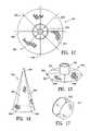
- FIG. 12is a top plan view of a partitioning device embodying features of the invention.
- FIG. 13is an isometric view of the device of FIG. 12 in its extended state.
- FIG. 14is an isometric view of the device of FIG. 12 in a partially collapsed state.
- FIG. 15illustrates the use of a device embodying features embodying features of the invention within a chamber of a patient's heart.
- FIGS. 16A–16Dare operational views, partly broken away, illustrating deployment of the device of FIG. 12 through a tubular instrument.
- FIG. 17is an isometric view of another device embodying features of the invention.
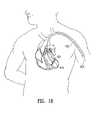
- FIG. 18illustrates the percutaneous delivery of an implantable device having features of the invention.
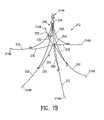
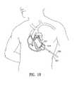
- FIG. 19illustrates the transthoracic delivery of an implantable device embodying features of the invention.
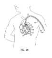
- FIG. 20is a view similar to FIG. 18 , illustrating the percutaneous delivery of two devices embodying features of the invention to treat a ventricular aneurysm and an enlarged ventricle simultaneously.
- FIG. 21is a view similar to FIG. 19 , illustrating the transthoracic delivery of two devices embodying features of the invention to treat a ventricular aneurysm and an enlarged ventricle simultaneously.
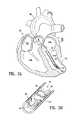
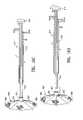
- FIGS. 1A , 1 B, and 1 Cillustrate components of a device 10 , according to an embodiment of the invention, for improving cardiac function.
- the device 10includes a frame construction 12 , a plurality of anchoring formations 14 , and a membrane 16 .
- the frame construction 12includes a main frame 18 and a support frame 20 secured to the main frame 18 .
- the membrane 16is secured on top of the support frame 20 .
- the main frame 18includes a sequence or series of segments 22 . Even segments of the series extend in an upward direction, and odd segments extend downward.
- the sequence formed by the segments 22entirely surrounds a vertical axis 24 . Movement of the segments 22 toward one another causes collapse of the main frame 18 toward the vertical axis 24 .
- the frame construction 12is made of a biocompatible wire-like shape-memory material, for example, nickel-titanium.
- the anchoring formations 14include a distal anchoring screw 14 A, distal anchoring hooks 14 B, and proximal anchoring hooks 14 C. Two or more (in the present example, four) of the segments 22 A are longer, and extend further down than other ones of the segments 22 B. The segments 22 A have their lower ends connected to one another, and the distal anchoring screw 14 A is secured to the lower ends of the segments 22 A.
- the segments 22 A and 22 Bmay be curved, as opposed to being straight, as shown in the figures.
- the distal anchoring hooks 14 Bare secured to lower ends of the segments 22 B. Each distal anchoring hook 14 B curves out and then down and is formed with a lower sharp end 26 .
- the proximal anchoring hooks 14 Care secured to upper ends of the segments 22 A and 22 B. Each one of the proximal anchoring hooks 14 C curves out and then up and terminates in an upper sharp end 28 .
- the anchoring hooks 14 B and 14 Cmove together with the main frame 18 toward the vertical axis 24 when the main frame 18 is collapsed.
- the support frame 20includes six (or more) elements 32 , sequentially after one another and overlaying one another to form a six-pointed star.
- the elements 32can pivot in a scissor-like manner relative to one another. Pivoting of the elements 32 relative to one another moves corners 34 of the star toward one another, while corners 36 on an opposing side of the star move toward one another.
- the support frame 20then has an elongated configuration with the corners 36 at one end and the corners 34 at an opposing end.
- each corner 36is positioned around and slidably secured to a respective one of the segments 22 B.
- the corners 34slide up the segments 22 B to which they are secured, while the corners 36 remain at the bottom of the segments 22 B to which they are secured.
- the support frame 20is in the form of an elongated arrangement extending along the vertical axis 24 , with the corners 34 at the top and the corners 36 at the bottom.
- FIG. 1Calso shows the membrane 16 , in an unfolded condition, secured on the elements 32 of the support frame 20 .
- An edge 40 of the membrane 16is secured to the elements 32 .
- Two of the elements 32form a cross below a center of the membrane 16 , and the other four elements 32 support the membrane 16 between the cross and the edge 40 .
- Collapse of the support frame 20folds the membrane 16 into an elongated folded arrangement extending along the elongated arrangement formed by the collapsed support frame 20 .
- the membrane 16is made of a biocompatible foldable material, for example Gore-Tex®, poly-ethylene terephthalate, or polypropylene mesh.
- FIG. 2Aillustrates the device 10 that is inserted into a heart 42 by means of a catheter 44 .
- the device 10is collapsed and is inserted into an end of the catheter 44 .
- the axis 24shown vertically in FIGS. 1A and 1B , now extends along an axis of an elongated tubular passage 46 in the catheter 44 .
- the device 10is packaged with the distal anchoring screw 14 A protruding from the end of the catheter 44 .
- the catheter 44is non-invasively steered through the aorta 48 and the aortic valve (not shown) into the left ventricle 52 A of the heart 42 .
- the other chambers of the heart 42are the right ventricle 52 B, the left atrium 50 A, and the right atrium 50 B.
- a device manipulating apparatus 54is disposed within the catheter 44 .
- the apparatus 54includes an elongated manipulator 56 , a rotator piece 58 , and a support piece 60 . Only a distal portion of the elongated manipulator 56 is shown.
- a handle(not shown) is attached to a proximal portion of the elongated manipulator 56 .
- the elongated manipulator 56can bend to conform to the curved or bent shape of the catheter 44 , but is relatively rigid against a torque about an elongated axis thereof.
- the rotator piece 58is secured to an end of the elongated manipulator 56 , and the support piece 60 is secured to the elongated manipulator 56 slightly proximal to the rotator piece 58 .
- the rotator piece 58has an internal device engaging formation 62 .
- the device 10is inserted into the formation 62 until proximal surfaces of the device 10 contact the support piece 60 .
- the formation 62conforms to an outer shape of the device 10 , so that the device 10 rotates together with the rotator piece 58 when the rotator piece 58 is rotated by the elongated manipulator 56 .
- the device 10may be fed out of an end of the catheter 44 by the support piece 60 when the elongated manipulator 56 is advanced in an elongated direction of the catheter 44 .
- the support piece 60also prevents movement of the device 10 in an opposite direction into the catheter 44 .
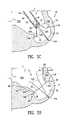
- the myocardium 74 of the hearthas formed an aneurysmic formation or bulge 76 out of the left ventricle 52 A.
- a previous infarction, or cessation of blood supply, to the portion of the myocardium 74 now forming the bulge 76has caused the tissue of that portion of the myocardium 74 to die.
- Continuous exposure of the dyskinetic portion of the myocardium 74 to high pressures in the left ventricle 52 Ahas caused the aneurysmic bulge 76 .
- the catheter 44is steered so that the distal anchoring screw 14 A contacts a base of the bulge 76 .
- the catheter 44is then rotated so that the distal anchoring screw 14 A screws into the myocardium 74 at a target site at the base of the bulge 76 .
- the catheter 44is then retracted over the device 10 with the distal anchoring screw 14 A anchoring the frame construction 12 to the myocardium 74 at the base of the bulge 76 .
- the distal anchoring hooks 14 Bleave the catheter 44 as the catheter 44 is retracted, before the remainder of the device 10 , and bend outwardly under spring action.
- the catheter 44is subsequently withdrawn from proximal anchoring hooks 14 C.
- Proximal portions of the segments 22 A and 22 Bspring outwardly after the proximal anchoring hooks 14 C leave the tubular passage 46 , so that the proximal anchoring hooks 14 C move outwardly into contact with the myocardium 74 .
- a proximal portion of each segment 22 A or 22 Bpivots relative to a distal portion thereof. Pivoting of the segments 22 B rotates the lower sharp ends 26 of the distal anchoring hooks 14 B into the myocardium 74 .
- Embedding of the distal anchoring hooks 14 B into the myocardium 74anchors the segments 22 B to the myocardium 74 .
- Beating of the heart 42causes relative movement between the myocardium 74 and proximal anchoring hooks 14 C, so that the upper sharp ends 28 may also penetrate the myocardium 74 .
- the proximal anchoring hooks 14 Care thereby also embedded into the myocardium 74 , and anchor proximal portions of the segments 22 A and 22 B to the myocardium 74 .
- Each segment 22 A or 22 Bis near the myocardium 74 at all locations along the length of the respective segment 22 A or 22 B, and is anchored to the myocardium 74 through the anchoring formations 14 .
- the corners 34 of the support frame 20slide along the segments 22 B to which they are secured when the segments 22 B rotate outwardly relative to one another.
- FIG. 3DWhen comparing FIG. 3D with FIG. 3C , it can be seen that the support frame 20 is in a plane which is substantially at right angles with respect to the axis of the elongated tubular passage 46 .
- the membrane 16( FIG. 1C ) unfolds and is supported on top of the support frame 20 .
- the membrane 16forms a division between the aneurysmic bulge 76 and a remainder of the left ventricle 52 A.
- the aneurysmic bulge 76After the device 10 is installed, the aneurysmic bulge 76 , having been segregated from the remainder of the left ventricle 52 A, eventually clots off behind the sheet 16 , thereby effectively reducing the internal volume in the left ventricle 52 A. Stretching of the portion of the myocardium 74 forming the aneurysmic bulge 76 is also effectively eliminated.
- properly functioning portions of the myocardium 74can contract normally and use up a normal amount of oxygen.
- properly functioning portions of the myocardium 74are not exhausted and can continue to function properly.
- Cardiac outputincreases, and the likelihood of congestive heart failure is reduced, assuming that all other conditions remain the same.
- a reduction in the strength of the contractions of the properly functioning portions of the myocardiumalso reduces LVESP, with a corresponding reduction in stress of both dyskinetic and properly functioning portions of the myocardium 74 .
- FIGS. 4A and 4Billustrate pressures within the left atrium 50 and the left ventricle 52 A, respectively, of a healthy human being. It can be seen that the peak left ventricular pressure, i.e., the pressure in the left ventricle 52 A during the systolic portion, reaches approximately 120 mm Hg. This pressure acts directly on the membrane 16 . It can be assumed that the pressure on an opposing side of the membrane 16 , i.e., the side of the aneurysmic bulge 76 , is close to zero.
- the support frame 20supports the sheet 16 at a sufficient number of locations and is sufficiently strong to prevent the membrane 16 from collapsing during peak systolic pressure.
- An peak left ventricular pressure in the region of 50 to 60 mm Hg for a sustained period of a few hoursis generally regarded as being incompatible with life.
- anchoring formations 14there are a total of 31 anchoring formations 14 , including the distal anchoring screw 14 A, 14 distal anchoring hooks 14 B, and 16 proximal anchoring hooks 14 C.
- the large number of anchoring formations 14ensure proper anchoring to the myocardium 74 .
- the large number of anchoring formations 14also allows for positioning of the membrane 16 at a select location within the left ventricle 52 A and at a select angle and within a select plane relative to the myocardium 74 .
- the anchoring formations 14and in particular the anchoring hooks 14 B and 14 C, their shape, orientation, and placement, are thus uniquely suited for anchoring of the frame construction 12 , especially when compared with other anchoring formations such as pins, clamps, staples, screws, and surgical thread.
- the anchoring formations 14penetrate through only a portion of the myocardium 74 , and thus do not damage the pericardium.
- none of the anchoring formations 14 or other components of the device 10can bump against the myocardium 74 , to avoid electrostimulation of the myocardium 74 that can lead to arrhythmias.
- FIGS. 5A , 5 B, and 5 Cillustrate one manner in which the support frame 20 and the membrane 16 can be positioned at a select angle relative to the myocardium 74 .
- FIG. 5AWhen comparing FIG. 5A with FIG. 3C , it can be seen that the catheter 44 is positioned closer to a right side (as viewed) of the myocardium 74 .
- the distal anchoring hooks 14 B on the rightengage with the myocardium 74 before the distal anchoring hooks 14 B on the left engage with the myocardium 74 . Further withdrawal of the catheter 44 , as shown in FIG.
- thisserves to illustrate that the membrane 16 can be positioned in different select planes, as may be required, due to the flexibility of the frame construction 12 and various virtual triangles that are formed by connecting locations where the anchoring formations 14 anchor to the myocardium 74 .
- the main frame 18has a vertical height H 1 , a height from the distal anchoring hooks 14 B to the proximal anchoring hooks 14 C H 2 , the membrane 16 has a width W, and the elongated tubular passage 46 has a diameter D.
- the first row in the tablelists the dimensions for the device 10 hereinbefore described which is used for blocking a relatively small aneurysmic bulge 76 .
- Larger aneurysmic bulgescan be blocked using slightly larger devices.
- certain diseases or alcoholismmay cause general enlargement of endocardial cavities of a heart without necessarily creating a specific identifiable bulge.
- Larger devicescan be used to block portions of these enlarged endocardial cavities. In such cases, it may also be possible to use two devices in a side-by-side arrangement or with their membranes overlapping one another.
- FIG. 6illustrates one such a larger device 110 that is inserted in the bottom of the left ventricle 152 of a heart 114 .
- the main frame (not shown) of the device 110is formed into a non-circular shape, so that an outline formed by corners 134 and 136 of a support frame of the device define a non-circular shape.
- a membrane 116 mounted on top of the support framealso defines a non-circular shape.
- the shape of the membrane 116conforms approximately to a non-circular D-shape of the left ventricle 152 at a height where the membrane 116 is positioned.
- the same device 110can be deformed into various different shapes, according to requirement.
- FIGS. 7A and 7Billustrate a frame construction 212 and anchoring formations 214 of a device according to an alternative embodiment of the invention.
- the frame construction 212includes a main frame 218 and a support frame 220 .
- the main frame 218has a plurality of segments 222 having distal ends connected to one another at a common location 224 . Proximal portions of the segments 222 can collapse toward one another and spring outwardly away from one another.
- the anchoring formations 214include a distal anchoring screw 214 A secured at the common location 224 , and proximal anchoring hooks 214 B on proximal ends of the segments 222 .
- the support frame 220includes a plurality of elements 232 .
- the elements 232have ends that are pivotally connected to one another at a common location 254 . An opposing end of each element 232 is slidably secured to a respective one of the segments 222 .
- the manner in which the segments 222 of the main frame 218 collapseis simultaneously replicated by the manner in which the elements 232 of the support frame 220 collapse.
- the distal anchoring screw 214 Ais first screwed into a myocardium.
- a catheteris then withdrawn from the frame construction 212 . Once the catheter is entirely removed from the frame construction 212 , the proximal anchoring hooks 214 B spring outwardly and embed themselves into the myocardium.
- the support frame 220simultaneously moves from its collapsed condition into its expanded condition.
- a membrane(not shown) is secured to, unfolded by, and supported by the support frame 220 .
- the support frame of a devicemay be shaped so that a membrane attached to the support frame has a desired shape.
- FIGS. 8 and 9illustrate a membrane 316 that conforms approximately to a shape defined by an anterior wall 320 and a septum 322 of a heart. As shown in FIG. 8 , the membrane 316 has a portion on the left having a radius R 1 and a portion on the right having a radius R 2 which is a multiple of the radius R 1 .
- the membrane 316may be formed to have more than two radii of curvature. Referring to FIG. 9 , it can be see that the membrane 316 is curved also when viewed on 9 — 9 in FIG. 8 .
- the curved shape of the membrane 316allows the membrane 316 to block off larger portions of the anterior wall 320 and the septum 322 without reducing the internal volume of the left ventricle by too great a degree.
- FIGS. 10 and 11illustrate the same device 410 used for closing off a small ventricle 412 and a large ventricle 414 , respectively.
- the device 410has a frame construction 416 that can spring outwardly, and a membrane 418 secured and expanded by the frame construction 416 .
- the frame construction 416springs out more in FIG. 11 than in FIG. 10 , and the membrane 416 is accordingly unfolded into a larger cross-sectional shape.
- the support frame and anchoring formations of, for example, the device illustrated in FIG. 1Amay be used for other purposes instead of or in addition to supporting a membrane as described.
- the frame construction 18provides an electrically conductive path that can be used for left ventricular pacing.
- one of the proximal anchoring hooks 14 Cmay engage with and be sufficiently long to penetrate from a left ventricle through a septum into a right ventricle of a heart.
- a terminal of a pacemakercan then be inserted into the right ventricle and connected to the hook that penetrates through the septum. Electric current can conduct between the terminal of the pacemaker through the main frame 18 to other ones of the anchoring formations 14 connected to the myocardium of the left ventricle.
- the frame construction 12also provides a strong support for mounting components that can be used for other purposes, such as an annulus component that can be positioned around the mitral valve, or a component that is used for reshaping a papillary muscle.
- the device 10can also be used for delivering of drugs, proteins, stem cells, etc. to the heart.
- a partitioning device 500includes a generally circular membrane 501 which is mounted on a collapsible frame 502 .
- the frame 502has a plurality of stays 503 which extend radially from a central hub 504 for movement between the extended position shown in FIGS. 12 and 13 and a collapsed position as shown in FIG. 14 .
- the stays 503are connected to the hub 504 in such a way that rotation of the hub moves the stays between their extended and collapsed positions.
- Hooks or barbs 506are provided at the outer ends of the stays for attaching the device to the walls of an organ, with the membrane in its extended state.
- the membrane 501divides the organ into two separate compartments. Those compartments will be determined by the physician and typically will consist of a functional compartment and a nonfunctional compartment. For example, in the case of a left ventricular aneurysm (an enlargement of one small part of the left ventricular myocardium), the device is positioned at the mouth of the aneurysm 507 , as illustrated in FIG. 15 , to divide the left ventricle into a nonfunctional part 508 consisting of the aneurysm itself, and a functional part 509 consisting of the remainder of the left ventricle.
- the elimination of communication between the aneurysm and the functional part of the ventriclewill significantly improve the overall performance of the left ventricle. This procedure can be performed either during open chest surgery or by less invasive techniques such as laparoscopic or percutaneous delivery of the device to the inside of the ventricle.
- the ventriclecan similarly be divided into functional and nonfunctional parts, with improved overall ventricular performance.
- the elimination of one portion of the enlarged heartwill prevent further cardiac enlargement and provide improved performance.
- the stomachcould be divided into a nonfunctional part, i.e. the part which does not participate in digestion, and the remainder of the stomach which includes normal digestive processes.
- the membraneis closely attached to the walls of the organ in order to prevent any communication between the two compartments. It can be elastic, non-elastic, non-porous, non-thrombogenic, and/or biocompatible.
- the membraneis fabricated of a mesh of elastin fibers (i.e. elastin proteins or a synthetic material) chosen to provide tensile strength and flexibility.
- the membranecan also have living cells (e.g., endothelial cells, myocytes, smooth muscle cells, or stem cells) and/or certain bioactive substances, such as heparin, on its surface.
- the membrane 501can be rigid, or capable of changing size and shape to accommodate the size and shape of the organ. It can also be composed of hydrogels that change into a tendon-like structure and/or a bioresorbable material. It can also include a protein which will serve to attract the patient's own cells that will turn into myocytes and become a new myocardium.
- partitioning device 500can be deployed with an instrument 511 which has an elongated tubular body 512 .
- the deviceis carried within the tube in a collapsed state toward the distal end of the instrument.
- An operating rod 513extends axially within the tubular body and is threadedly connected to hub 504 for advancing the device from the tubular body and rotating the hub to extend the membrane.
- a control knob 514is provided at the proximal end of the operating rod.
- the instrumentIn operation, the instrument is inserted into the body of the patient, and operating rod 513 is moved forward to advance the partitioning device from the distal end of tubular body 512 .
- control knobAs the device is moving forward, control knob is turned in a clockwise direction to extend the membrane, as illustrated in FIG. 16B . The advancement and expansion continues until the membrane reaches its fully extended position, as illustrated in FIG. 16C . Once barbs 506 have been engaged with the walls of the organ, the control knob is turned in counterclockwise direction to disengage the operating rod from the hub, as illustrated in FIG. 16D , and the instrument is withdrawn from the body of the patient.
- the partitioning deviceconsists of a scaffold or ring 516 which is attached to the walls of the organ first, and a membrane 517 which is attached to the ring after the ring is in place.
- the scaffoldhas means such as mechanical devices (e.g. barbs or hooks) or an adhesive for attaching it to the walls of the organ and to the membrane. Both the scaffold and the membrane are collapsible, and can be introduced into the hollow organ through tubular devices such as an endoscope, a thoracoscope, or a catheter.
- he partitioning devicecan be implanted in a number of ways. It can, for example, be implanted by a surgeon during a surgical procedure. It can also be implanted through an endoscope or a laparoscope during a laparoscopic procedure. Similarly, it can be implanted through a thoracoscope during a thoracoscopic procedure, and it can be delivered to the inside of an organ through a long tubular device such as a catheter, either percutaneously, orally, or by some other minimally invasive approach.
- FIG. 18illustrates the percutaneous insertion of a partitioning device 519 into a chamber of the heart 521 through the brachial artery 522 .
- a catheter 523is passed through the artery and into the heart, and the partitioning device is inserted and deployed through the catheter.
- the catheterpreferably has a mesh-like structure to prevent the occurrence of an embolism during the procedure.
- FIG. 19illustrates the thoracoscopic placement of a partitioning device 519 in heart chamber 521 .
- the deviceis introduced and deployed through a thoracoscope 526 which is inserted into the patient's body through his thorax 527 .
- FIG. 20illustrates the use of two percutaneously inserted membranes 529 , 531 in the simultaneous treatment of a ventricular aneurysm 532 and an enlarged ventricle 533 .
- the membranesare introduced into the ventricle through two separate catheters 534 , 536 which pass through the brachial artery. Alternatively, they can be introduced either simultaneously of successively through a single catheter, if desired.
- FIG. 21illustrates a similar situation in which membranes 529 , 531 are introduced thoracoscopically to threat a ventricular aneurysm 532 and an enlarged ventricle 533 .
- both of the membranesare introduced through a single thoracoscope 537 , with membrane 529 being implanted first, followed by membrane 531 .
- the partitioning devicescan be constructed to facilitate removal from the hollow organ after a period of time.
- the devicescan be implanted in the stomach, and in the event that they becomes too uncomfortable, they can be removed in the same manner in which they were installed.
- the inventionhas a number of important features and advantages. It permits diseases caused by dysfunction of hollow organs to be treated simply and effectively without difficult surgical procedures.
- an inflatable dividing elementmay be used to occlude an atrial appendage of the heart.
- individual features of embodiments of the inventionmay be shown in some drawings and not in others, but those skilled in the art will recognize that individual features of one embodiment of the invention can be combined with any or all the features of another embodiment. Accordingly, it is not intended that the invention be limited to the specific embodiments illustrated. It is intended that this invention to be defined by the scope of the appended claims as broadly as the prior art will permit.
Landscapes
- Health & Medical Sciences (AREA)
- Life Sciences & Earth Sciences (AREA)
- Surgery (AREA)
- Heart & Thoracic Surgery (AREA)
- Public Health (AREA)
- Veterinary Medicine (AREA)
- Engineering & Computer Science (AREA)
- Biomedical Technology (AREA)
- Nuclear Medicine, Radiotherapy & Molecular Imaging (AREA)
- General Health & Medical Sciences (AREA)
- Animal Behavior & Ethology (AREA)
- Molecular Biology (AREA)
- Medical Informatics (AREA)
- Reproductive Health (AREA)
- Vascular Medicine (AREA)
- Cardiology (AREA)
- Radiology & Medical Imaging (AREA)
- Prostheses (AREA)
Abstract
Description
| H1 | H2 | W | D | ||
| 6 | cm | 3 | cm | 2.5 | cm | 1 | cm | ||
| 7 | cm | 4 | cm | 3 | cm | 1.2 | cm | ||
| 8 | cm | 5 | cm | 4 | cm | 1.5 | cm | ||
| 8.5 | cm | 5.5 | cm | 5 | cm | 2 | cm | ||
| 9.5 | cm | 6 | cm | 6 | cm | 2.2 | cm | ||
| 9.5 | cm | 8 | cm | 7 | cm | 2.6 | cm | ||
Claims (27)
Priority Applications (10)
| Application Number | Priority Date | Filing Date | Title |
|---|---|---|---|
| US10/212,032US7279007B2 (en) | 1999-08-09 | 2002-08-01 | Method for improving cardiac function |
| US10/382,962US6852076B2 (en) | 1999-08-09 | 2003-03-06 | Method for improving cardiac function |
| US11/199,633US20060229491A1 (en) | 2002-08-01 | 2005-08-09 | Method for treating myocardial rupture |
| US12/129,443US8529430B2 (en) | 2002-08-01 | 2008-05-29 | Therapeutic methods and devices following myocardial infarction |
| US12/893,832US9078660B2 (en) | 2000-08-09 | 2010-09-29 | Devices and methods for delivering an endocardial device |
| US13/973,868US8827892B2 (en) | 2002-08-01 | 2013-08-22 | Therapeutic methods and devices following myocardial infarction |
| US14/448,778US9592123B2 (en) | 2002-08-01 | 2014-07-31 | Therapeutic methods and devices following myocardial infarction |
| US14/731,161US20150265405A1 (en) | 2000-08-09 | 2015-06-04 | Devices and methods for delivering an endocardial device |
| US15/133,080US10064696B2 (en) | 2000-08-09 | 2016-04-19 | Devices and methods for delivering an endocardial device |
| US15/452,435US10307147B2 (en) | 1999-08-09 | 2017-03-07 | System for improving cardiac function by sealing a partitioning membrane within a ventricle |
Applications Claiming Priority (3)
| Application Number | Priority Date | Filing Date | Title |
|---|---|---|---|
| US14789499P | 1999-08-09 | 1999-08-09 | |
| US63551100A | 2000-08-09 | 2000-08-09 | |
| US10/212,032US7279007B2 (en) | 1999-08-09 | 2002-08-01 | Method for improving cardiac function |
Related Parent Applications (1)
| Application Number | Title | Priority Date | Filing Date |
|---|---|---|---|
| US63551100AContinuation-In-Part | 1999-08-09 | 2000-08-09 |
Related Child Applications (2)
| Application Number | Title | Priority Date | Filing Date |
|---|---|---|---|
| US10/382,962ContinuationUS6852076B2 (en) | 1999-08-09 | 2003-03-06 | Method for improving cardiac function |
| US11/199,633Continuation-In-PartUS20060229491A1 (en) | 1999-08-09 | 2005-08-09 | Method for treating myocardial rupture |
Publications (2)
| Publication Number | Publication Date |
|---|---|
| US20030050685A1 US20030050685A1 (en) | 2003-03-13 |
| US7279007B2true US7279007B2 (en) | 2007-10-09 |
Family
ID=46045478
Family Applications (3)
| Application Number | Title | Priority Date | Filing Date |
|---|---|---|---|
| US10/212,032Expired - LifetimeUS7279007B2 (en) | 1999-08-09 | 2002-08-01 | Method for improving cardiac function |
| US10/212,033Expired - LifetimeUS7303526B2 (en) | 1999-08-09 | 2002-08-01 | Device for improving cardiac function |
| US10/382,962Expired - LifetimeUS6852076B2 (en) | 1999-08-09 | 2003-03-06 | Method for improving cardiac function |
Family Applications After (2)
| Application Number | Title | Priority Date | Filing Date |
|---|---|---|---|
| US10/212,033Expired - LifetimeUS7303526B2 (en) | 1999-08-09 | 2002-08-01 | Device for improving cardiac function |
| US10/382,962Expired - LifetimeUS6852076B2 (en) | 1999-08-09 | 2003-03-06 | Method for improving cardiac function |
Country Status (1)
| Country | Link |
|---|---|
| US (3) | US7279007B2 (en) |
Cited By (68)
| Publication number | Priority date | Publication date | Assignee | Title |
|---|---|---|---|---|
| US20050015109A1 (en)* | 2003-07-16 | 2005-01-20 | Samuel Lichtenstein | Methods and devices for altering blood flow through the left ventricle |
| US20070112372A1 (en)* | 2005-11-17 | 2007-05-17 | Stephen Sosnowski | Biodegradable vascular filter |
| US20080027481A1 (en)* | 2006-07-19 | 2008-01-31 | Paul Gilson | Vascular filter |
| US7749249B2 (en) | 2006-02-21 | 2010-07-06 | Kardium Inc. | Method and device for closing holes in tissue |
| US20100185230A1 (en)* | 2009-01-16 | 2010-07-22 | Steven Horan | vascular filter device |
| US20100185229A1 (en)* | 2009-01-16 | 2010-07-22 | Steven Horan | Vascular filter device |
| US20100228281A1 (en)* | 2009-01-16 | 2010-09-09 | Paul Gilson | Vascular filter system |
| US20100262225A1 (en)* | 2007-12-12 | 2010-10-14 | Peter Schneider | Device and method for tacking plaque to a blood vessel wall |
| US7837610B2 (en) | 2006-08-02 | 2010-11-23 | Kardium Inc. | System for improving diastolic dysfunction |
| US7862500B2 (en)* | 2002-08-01 | 2011-01-04 | Cardiokinetix, Inc. | Multiple partitioning devices for heart treatment |
| US8057507B2 (en) | 2009-01-16 | 2011-11-15 | Novate Medical Limited | Vascular filter |
| US8150499B2 (en) | 2006-05-19 | 2012-04-03 | Kardium Inc. | Automatic atherectomy system |
| US8388672B2 (en)* | 1999-08-09 | 2013-03-05 | Cardiokinetix, Inc. | System for improving cardiac function by sealing a partitioning membrane within a ventricle |
| US8449605B2 (en) | 2006-06-28 | 2013-05-28 | Kardium Inc. | Method for anchoring a mitral valve |
| US8489172B2 (en) | 2008-01-25 | 2013-07-16 | Kardium Inc. | Liposuction system |
| US8500790B2 (en) | 1999-08-09 | 2013-08-06 | Cardiokinetix, Inc. | Retrievable cardiac devices |
| US20140074069A1 (en)* | 2005-09-12 | 2014-03-13 | Cardionomic | Methods and systems for treating acute heart failure by neuromodulation |
| US8747454B2 (en)* | 1999-08-09 | 2014-06-10 | Cardiokinetix, Inc. | System for improving cardiac function |
| US8827892B2 (en) | 2002-08-01 | 2014-09-09 | Cardiokinetix, Inc. | Therapeutic methods and devices following myocardial infarction |
| US8906011B2 (en) | 2007-11-16 | 2014-12-09 | Kardium Inc. | Medical device for use in bodily lumens, for example an atrium |
| US8920411B2 (en) | 2006-06-28 | 2014-12-30 | Kardium Inc. | Apparatus and method for intra-cardiac mapping and ablation |
| US8940002B2 (en) | 2010-09-30 | 2015-01-27 | Kardium Inc. | Tissue anchor system |
| US9011423B2 (en) | 2012-05-21 | 2015-04-21 | Kardium, Inc. | Systems and methods for selecting, activating, or selecting and activating transducers |
| US9039597B2 (en) | 2009-10-26 | 2015-05-26 | Cardiokinetix, Inc. | Ventricular volume reduction |
| US9050066B2 (en) | 2010-06-07 | 2015-06-09 | Kardium Inc. | Closing openings in anatomical tissue |
| US9072511B2 (en) | 2011-03-25 | 2015-07-07 | Kardium Inc. | Medical kit for constricting tissue or a bodily orifice, for example, a mitral valve |
| US9119633B2 (en) | 2006-06-28 | 2015-09-01 | Kardium Inc. | Apparatus and method for intra-cardiac mapping and ablation |
| US9198592B2 (en) | 2012-05-21 | 2015-12-01 | Kardium Inc. | Systems and methods for activating transducers |
| US9204964B2 (en) | 2009-10-01 | 2015-12-08 | Kardium Inc. | Medical device, kit and method for constricting tissue or a bodily orifice, for example, a mitral valve |
| US9332992B2 (en) | 2004-08-05 | 2016-05-10 | Cardiokinetix, Inc. | Method for making a laminar ventricular partitioning device |
| US9332993B2 (en) | 2004-08-05 | 2016-05-10 | Cardiokinetix, Inc. | Devices and methods for delivering an endocardial device |
| US9452016B2 (en) | 2011-01-21 | 2016-09-27 | Kardium Inc. | Catheter system |
| US9480525B2 (en) | 2011-01-21 | 2016-11-01 | Kardium, Inc. | High-density electrode-based medical device system |
| US9492228B2 (en) | 2011-01-21 | 2016-11-15 | Kardium Inc. | Enhanced medical device for use in bodily cavities, for example an atrium |
| US9545322B2 (en) | 2007-12-12 | 2017-01-17 | Intact Vascular, Inc. | Device and method for tacking plaque to blood vessel wall |
| USD777925S1 (en) | 2012-01-20 | 2017-01-31 | Kardium Inc. | Intra-cardiac procedure device |
| USD777926S1 (en) | 2012-01-20 | 2017-01-31 | Kardium Inc. | Intra-cardiac procedure device |
| US9603730B2 (en) | 2007-12-12 | 2017-03-28 | Intact Vascular, Inc. | Endoluminal device and method |
| US9694121B2 (en) | 1999-08-09 | 2017-07-04 | Cardiokinetix, Inc. | Systems and methods for improving cardiac function |
| US9730818B2 (en) | 2007-12-12 | 2017-08-15 | Intact Vascular, Inc. | Endoluminal device and method |
| US9744038B2 (en) | 2008-05-13 | 2017-08-29 | Kardium Inc. | Medical device for constricting tissue or a bodily orifice, for example a mitral valve |
| US10022250B2 (en) | 2007-12-12 | 2018-07-17 | Intact Vascular, Inc. | Deployment device for placement of multiple intraluminal surgical staples |
| US10028783B2 (en) | 2006-06-28 | 2018-07-24 | Kardium Inc. | Apparatus and method for intra-cardiac mapping and ablation |
| US10064696B2 (en) | 2000-08-09 | 2018-09-04 | Edwards Lifesciences Corporation | Devices and methods for delivering an endocardial device |
| US10166127B2 (en) | 2007-12-12 | 2019-01-01 | Intact Vascular, Inc. | Endoluminal device and method |
| US10172549B2 (en) | 2016-03-09 | 2019-01-08 | CARDIONOMIC, Inc. | Methods of facilitating positioning of electrodes |
| US10245167B2 (en) | 2015-01-29 | 2019-04-02 | Intact Vascular, Inc. | Delivery device and method of delivery |
| US10271973B2 (en) | 2011-06-03 | 2019-04-30 | Intact Vascular, Inc. | Endovascular implant |
| US10278839B2 (en) | 2007-12-12 | 2019-05-07 | Intact Vascular, Inc. | Endovascular impant |
| US10307147B2 (en) | 1999-08-09 | 2019-06-04 | Edwards Lifesciences Corporation | System for improving cardiac function by sealing a partitioning membrane within a ventricle |
| US10368936B2 (en) | 2014-11-17 | 2019-08-06 | Kardium Inc. | Systems and methods for selecting, activating, or selecting and activating transducers |
| US10493278B2 (en) | 2015-01-05 | 2019-12-03 | CARDIONOMIC, Inc. | Cardiac modulation facilitation methods and systems |
| US10576273B2 (en) | 2014-05-22 | 2020-03-03 | CARDIONOMIC, Inc. | Catheter and catheter system for electrical neuromodulation |
| US10687930B2 (en) | 2013-03-15 | 2020-06-23 | Novate Medical Limited | Vascular filter device |
| US10722184B2 (en) | 2014-11-17 | 2020-07-28 | Kardium Inc. | Systems and methods for selecting, activating, or selecting and activating transducers |
| US10722716B2 (en) | 2014-09-08 | 2020-07-28 | Cardionomia Inc. | Methods for electrical neuromodulation of the heart |
| US10751183B2 (en) | 2014-09-28 | 2020-08-25 | Edwards Lifesciences Corporation | Apparatuses for treating cardiac dysfunction |
| US10827977B2 (en) | 2012-05-21 | 2020-11-10 | Kardium Inc. | Systems and methods for activating transducers |
| US10894160B2 (en) | 2014-09-08 | 2021-01-19 | CARDIONOMIC, Inc. | Catheter and electrode systems for electrical neuromodulation |
| US10898356B2 (en) | 2015-01-29 | 2021-01-26 | Intact Vascular, Inc. | Delivery device and method of delivery |
| US10898330B2 (en) | 2017-03-28 | 2021-01-26 | Edwards Lifesciences Corporation | Positioning, deploying, and retrieving implantable devices |
| US10993824B2 (en) | 2016-01-01 | 2021-05-04 | Intact Vascular, Inc. | Delivery device and method of delivery |
| US11077298B2 (en) | 2018-08-13 | 2021-08-03 | CARDIONOMIC, Inc. | Partially woven expandable members |
| US11259867B2 (en) | 2011-01-21 | 2022-03-01 | Kardium Inc. | High-density electrode-based medical device system |
| US11389232B2 (en) | 2006-06-28 | 2022-07-19 | Kardium Inc. | Apparatus and method for intra-cardiac mapping and ablation |
| US11559687B2 (en) | 2017-09-13 | 2023-01-24 | CARDIONOMIC, Inc. | Methods for detecting catheter movement |
| US11607176B2 (en) | 2019-05-06 | 2023-03-21 | CARDIONOMIC, Inc. | Systems and methods for denoising physiological signals during electrical neuromodulation |
| US11660218B2 (en) | 2017-07-26 | 2023-05-30 | Intact Vascular, Inc. | Delivery device and method of delivery |
Families Citing this family (79)
| Publication number | Priority date | Publication date | Assignee | Title |
|---|---|---|---|---|
| US7314477B1 (en) | 1998-09-25 | 2008-01-01 | C.R. Bard Inc. | Removable embolus blood clot filter and filter delivery unit |
| US8285393B2 (en)* | 1999-04-16 | 2012-10-09 | Laufer Michael D | Device for shaping infarcted heart tissue and method of using the device |
| US20030109770A1 (en)* | 1999-08-09 | 2003-06-12 | Sharkey Hugh R. | Device with a porous membrane for improving cardiac function |
| US7582051B2 (en)* | 2005-06-10 | 2009-09-01 | Cardiokinetix, Inc. | Peripheral seal for a ventricular partitioning device |
| US7279007B2 (en)* | 1999-08-09 | 2007-10-09 | Cardioklnetix, Inc. | Method for improving cardiac function |
| US7674222B2 (en)* | 1999-08-09 | 2010-03-09 | Cardiokinetix, Inc. | Cardiac device and methods of use thereof |
| US20060229491A1 (en)* | 2002-08-01 | 2006-10-12 | Cardiokinetix, Inc. | Method for treating myocardial rupture |
| US8398537B2 (en)* | 2005-06-10 | 2013-03-19 | Cardiokinetix, Inc. | Peripheral seal for a ventricular partitioning device |
| US20060030881A1 (en) | 2004-08-05 | 2006-02-09 | Cardiokinetix, Inc. | Ventricular partitioning device |
| US7762943B2 (en)* | 2004-03-03 | 2010-07-27 | Cardiokinetix, Inc. | Inflatable ventricular partitioning device |
| US7399271B2 (en)* | 2004-01-09 | 2008-07-15 | Cardiokinetix, Inc. | Ventricular partitioning device |
| US9078660B2 (en)* | 2000-08-09 | 2015-07-14 | Cardiokinetix, Inc. | Devices and methods for delivering an endocardial device |
| US6994093B2 (en) | 2001-02-28 | 2006-02-07 | Chase Medical, L.P. | Ventricular restoration shaping apparatus and method of use |
| US20030181940A1 (en)* | 2001-02-28 | 2003-09-25 | Gregory Murphy | Ventricular restoration shaping apparatus and method of use |
| US20020133227A1 (en)* | 2001-02-28 | 2002-09-19 | Gregory Murphy | Ventricular restoration patch apparatus and method of use |
| US20060025800A1 (en)* | 2001-09-05 | 2006-02-02 | Mitta Suresh | Method and device for surgical ventricular repair |
| US20040243170A1 (en)* | 2001-09-05 | 2004-12-02 | Mitta Suresh | Method and device for percutaneous surgical ventricular repair |
| US7485088B2 (en) | 2001-09-05 | 2009-02-03 | Chase Medical L.P. | Method and device for percutaneous surgical ventricular repair |
| JP4083683B2 (en)* | 2001-09-07 | 2008-04-30 | マーディル, インコーポレイテッド | Method and apparatus for external heart fixation |
| US6700444B2 (en)* | 2002-01-28 | 2004-03-02 | Cree Microwave, Inc. | N-way RF power amplifier with increased backoff power and power added efficiency |
| US9204956B2 (en) | 2002-02-20 | 2015-12-08 | C. R. Bard, Inc. | IVC filter with translating hooks |
| JP4748990B2 (en)* | 2003-02-06 | 2011-08-17 | 株式会社半導体エネルギー研究所 | Manufacturing method of semiconductor device |
| US20060184242A1 (en)* | 2003-10-20 | 2006-08-17 | Samuel Lichtenstein | Method and apparatus for percutaneous reduction of anterior-posterior diameter of mitral valve |
| US20060276684A1 (en)* | 2003-11-07 | 2006-12-07 | Giovanni Speziali | Device and method for treating congestive heart failure |
| US7704267B2 (en) | 2004-08-04 | 2010-04-27 | C. R. Bard, Inc. | Non-entangling vena cava filter |
| US20060135966A1 (en)* | 2004-11-15 | 2006-06-22 | Laurent Schaller | Catheter-based tissue remodeling devices and methods |
| US20070156209A1 (en)* | 2005-01-14 | 2007-07-05 | Co-Repair, Inc. | System for the treatment of heart tissue |
| US20070156210A1 (en)* | 2005-01-14 | 2007-07-05 | Co-Repair, Inc., A California Corporation | Method for the treatment of heart tissue |
| US7455670B2 (en)* | 2005-01-14 | 2008-11-25 | Co-Repair, Inc. | System and method for the treatment of heart tissue |
| US7320665B2 (en) | 2005-03-02 | 2008-01-22 | Venkataramana Vijay | Cardiac Ventricular Geometry Restoration Device and Treatment for Heart Failure |
| US20060199995A1 (en)* | 2005-03-02 | 2006-09-07 | Venkataramana Vijay | Percutaneous cardiac ventricular geometry restoration device and treatment for heart failure |
| US8333777B2 (en) | 2005-04-22 | 2012-12-18 | Benvenue Medical, Inc. | Catheter-based tissue remodeling devices and methods |
| CA2607580C (en) | 2005-05-12 | 2016-12-20 | C.R. Bard Inc. | Removable embolus blood clot filter |
| US8613754B2 (en) | 2005-05-12 | 2013-12-24 | C. R. Bard, Inc. | Tubular filter |
| US12115057B2 (en) | 2005-05-12 | 2024-10-15 | C.R. Bard, Inc. | Tubular filter |
| WO2007021340A1 (en) | 2005-08-09 | 2007-02-22 | C.R. Bard Inc | Embolus blood clot filter and delivery system |
| WO2007027592A1 (en)* | 2005-08-29 | 2007-03-08 | Ams Research Corporation | System for positioning support mesh in a patient |
| CA2621197A1 (en) | 2005-09-01 | 2007-03-08 | Cordis Corporation | Patent foramen ovale closure method |
| US9131999B2 (en) | 2005-11-18 | 2015-09-15 | C.R. Bard Inc. | Vena cava filter with filament |
| EP1957147B1 (en) | 2005-12-09 | 2010-12-29 | Boston Scientific Scimed, Inc. | Cardiac stimulation system |
| EP1986735A4 (en)* | 2006-02-06 | 2011-06-29 | Northwind Ventures | Systems and methods for volume reduction |
| US20070208217A1 (en) | 2006-03-03 | 2007-09-06 | Acorn Cardiovascular, Inc. | Self-adjusting attachment structure for a cardiac support device |
| WO2007133366A2 (en) | 2006-05-02 | 2007-11-22 | C. R. Bard, Inc. | Vena cava filter formed from a sheet |
| US8932348B2 (en) | 2006-05-18 | 2015-01-13 | Edwards Lifesciences Corporation | Device and method for improving heart valve function |
| AU2007266448B2 (en) | 2006-06-01 | 2013-07-18 | Edwards Lifesciences Corporation | Prosthetic insert for improving heart valve function |
| US9326842B2 (en) | 2006-06-05 | 2016-05-03 | C. R . Bard, Inc. | Embolus blood clot filter utilizable with a single delivery system or a single retrieval system in one of a femoral or jugular access |
| WO2007146873A1 (en) | 2006-06-09 | 2007-12-21 | Cordis Corporation | Single disc occlusionary patent foramen ovale closure device |
| US8029556B2 (en)* | 2006-10-04 | 2011-10-04 | Edwards Lifesciences Corporation | Method and apparatus for reshaping a ventricle |
| US8092363B2 (en)* | 2007-09-05 | 2012-01-10 | Mardil, Inc. | Heart band with fillable chambers to modify heart valve function |
| US20090076597A1 (en)* | 2007-09-19 | 2009-03-19 | Jonathan Micheal Dahlgren | System for mechanical adjustment of medical implants |
| EP2082690B1 (en)* | 2008-01-24 | 2012-06-20 | Kardium, Inc. | Medical device to assist diastolic function and prevent ventricular enlargement |
| US9549734B2 (en)* | 2008-04-14 | 2017-01-24 | Boston Scientific Scimed, Inc. | Endoscopic stapling device, related staples, and methods for use |
| US20100274227A1 (en)* | 2009-02-13 | 2010-10-28 | Alexander Khairkhahan | Delivery catheter handle cover |
| US20110054515A1 (en)* | 2009-08-25 | 2011-03-03 | John Bridgeman | Device and method for occluding the left atrial appendage |
| US9526483B2 (en) | 2010-07-15 | 2016-12-27 | Medtronic Vascular Galway | Apical closure system |
| AU2013328871B2 (en) | 2012-10-12 | 2018-08-16 | Diaxamed, Llc | Cardiac treatment system and method |
| US9592399B2 (en) | 2013-06-20 | 2017-03-14 | Cardiac Pacemakers, Inc. | Deployable multi-electrode leadless electrostimulator |
| USD717954S1 (en) | 2013-10-14 | 2014-11-18 | Mardil, Inc. | Heart treatment device |
| US9730701B2 (en) | 2014-01-16 | 2017-08-15 | Boston Scientific Scimed, Inc. | Retrieval wire centering device |
| US10292711B2 (en)* | 2014-02-07 | 2019-05-21 | St. Jude Medical, Cardiology Division, Inc. | Mitral valve treatment device having left atrial appendage closure |
| US20170035433A1 (en)* | 2015-08-06 | 2017-02-09 | Thomas J. Forbes | Left atrial appendage occluder device anchoring system, anchor, and method of attachment |
| RU2738426C1 (en)* | 2015-12-29 | 2020-12-14 | Шэньчжэнь Кид Биомедикал Текнолоджи Ко.Лтд | Left atrial appendage occluder (versions) |
| CN105476733B (en)* | 2016-01-27 | 2017-09-01 | 张刚成 | Cardiac volume reduction device |
| WO2018129691A1 (en)* | 2017-01-12 | 2018-07-19 | 上海心瑞医疗科技有限公司 | Recovery system for use with isolation device, and preloaded and postloaded intervention system |
| CN106667539B (en)* | 2017-01-12 | 2019-04-12 | 上海心瑞医疗科技有限公司 | A kind of interventional systems loaded after isolating device |
| GB2574754B (en)* | 2017-03-23 | 2021-12-08 | Gyrus Acmi Inc | Airway valve apparatus |
| US11173033B2 (en) | 2017-09-22 | 2021-11-16 | Boston Scientific Scimed, Inc. | Dome structure for improved left ventricle function |
| CN109745149B (en)* | 2017-11-07 | 2024-09-20 | 深圳市健心医疗科技有限公司 | Heart valve anchoring device and heart valve |
| EP3720389B1 (en) | 2018-01-22 | 2024-07-03 | Edwards Lifesciences Corporation | Heart shape preserving anchor |
| WO2020056058A1 (en) | 2018-09-13 | 2020-03-19 | Ozgur Kocaturk | Devices and methods for closing openings in tissue structures |
| CN114126540A (en) | 2019-07-17 | 2022-03-01 | 波士顿科学医学有限公司 | Continuously covered left atrial appendage implant |
| EP4563099A3 (en) | 2019-08-30 | 2025-08-06 | Boston Scientific Scimed, Inc. | Left atrial appendage implant with sealing disk |
| WO2021119413A1 (en)* | 2019-12-13 | 2021-06-17 | Procyrion, Inc. | Support structures for intravascular blood pumps |
| WO2021195085A1 (en) | 2020-03-24 | 2021-09-30 | Boston Scientific Scimed, Inc. | Medical system for treating a left atrial appendage |
| JP7627761B2 (en) | 2020-11-30 | 2025-02-06 | ボストン サイエンティフィック サイムド,インコーポレイテッド | Implantable passive mean pressure sensor |
| US12383201B2 (en) | 2021-02-03 | 2025-08-12 | Boston Scientific Scimed, Inc. | Medical system for treating a left atrial appendage |
| EP4358872A1 (en) | 2021-06-22 | 2024-05-01 | Boston Scientific Scimed, Inc. | Left atrial appendage implant |
| CN117615720A (en) | 2021-07-08 | 2024-02-27 | 波士顿科学医学有限公司 | Left auricle closing device |
| WO2023038929A1 (en) | 2021-09-08 | 2023-03-16 | Boston Scientific Scimed, Inc. | Occlusive implant with multi-sharpness split tip soft tissue anchors |
Citations (98)
| Publication number | Priority date | Publication date | Assignee | Title |
|---|---|---|---|---|
| US3874388A (en) | 1973-02-12 | 1975-04-01 | Ochsner Med Found Alton | Shunt defect closure system |
| US4007743A (en) | 1975-10-20 | 1977-02-15 | American Hospital Supply Corporation | Opening mechanism for umbrella-like intravascular shunt defect closure device |
| US4425908A (en) | 1981-10-22 | 1984-01-17 | Beth Israel Hospital | Blood clot filter |
| US4619246A (en) | 1984-05-23 | 1986-10-28 | William Cook, Europe A/S | Collapsible filter basket |
| US4710192A (en) | 1985-12-30 | 1987-12-01 | Liotta Domingo S | Diaphragm and method for occlusion of the descending thoracic aorta |
| US4832055A (en) | 1988-07-08 | 1989-05-23 | Palestrant Aubrey M | Mechanically locking blood clot filter |
| US4917089A (en) | 1988-08-29 | 1990-04-17 | Sideris Eleftherios B | Buttoned device for the transvenous occlusion of intracardiac defects |
| US5104399A (en) | 1986-12-10 | 1992-04-14 | Endovascular Technologies, Inc. | Artificial graft and implantation method |
| US5192301A (en) | 1989-01-17 | 1993-03-09 | Nippon Zeon Co., Ltd. | Closing plug of a defect for medical use and a closing plug device utilizing it |
| US5375612A (en) | 1992-04-07 | 1994-12-27 | B. Braun Celsa | Possibly absorbable blood filter |
| US5385156A (en) | 1993-08-27 | 1995-01-31 | Rose Health Care Systems | Diagnostic and treatment method for cardiac rupture and apparatus for performing the same |
| US5425744A (en) | 1991-11-05 | 1995-06-20 | C. R. Bard, Inc. | Occluder for repair of cardiac and vascular defects |
| US5433727A (en) | 1994-08-16 | 1995-07-18 | Sideris; Eleftherios B. | Centering buttoned device for the occlusion of large defects for occluding |
| US5451235A (en) | 1991-11-05 | 1995-09-19 | C.R. Bard, Inc. | Occluder and method for repair of cardiac and vascular defects |
| US5496277A (en) | 1990-04-12 | 1996-03-05 | Schneider (Usa) Inc. | Radially expandable body implantable device |
| US5527338A (en) | 1992-09-02 | 1996-06-18 | Board Of Regents, The University Of Texas System | Intravascular device |
| US5527337A (en) | 1987-06-25 | 1996-06-18 | Duke University | Bioabsorbable stent and method of making the same |
| US5549621A (en) | 1993-05-14 | 1996-08-27 | Byron C. Sutherland | Apparatus and method for performing vertical banded gastroplasty |
| US5578069A (en) | 1995-12-06 | 1996-11-26 | Vnetritex, Inc. | Electrode deployment mechanism and method using artificial muscle |
| US5634936A (en) | 1995-02-06 | 1997-06-03 | Scimed Life Systems, Inc. | Device for closing a septal defect |
| US5634942A (en) | 1994-04-21 | 1997-06-03 | B. Braun Celsa | Assembly comprising a blood filter for temporary or definitive use and a device for implanting it |
| US5702343A (en) | 1996-10-02 | 1997-12-30 | Acorn Medical, Inc. | Cardiac reinforcement device |
| US5709707A (en) | 1995-10-30 | 1998-01-20 | Children's Medical Center Corporation | Self-centering umbrella-type septal closure device |
| US5791231A (en) | 1993-05-17 | 1998-08-11 | Endorobotics Corporation | Surgical robotic system and hydraulic actuator therefor |
| US5797960A (en) | 1993-02-22 | 1998-08-25 | Stevens; John H. | Method and apparatus for thoracoscopic intracardiac procedures |
| US5797849A (en) | 1995-03-28 | 1998-08-25 | Sonometrics Corporation | Method for carrying out a medical procedure using a three-dimensional tracking and imaging system |
| US5833698A (en) | 1996-07-23 | 1998-11-10 | United States Surgical Corporation | Anastomosis instrument and method |
| US5836968A (en) | 1996-07-17 | 1998-11-17 | Nitinol Medical Technologies, Inc. | Removable embolus blood clot filter |
| US5843170A (en) | 1994-09-02 | 1998-12-01 | Ahn; Sam Seunghae | Apparatus and method for performing aneurysm repair |
| US5861003A (en) | 1996-10-23 | 1999-01-19 | The Cleveland Clinic Foundation | Apparatus and method for occluding a defect or aperture within body surface |
| US5865791A (en) | 1995-06-07 | 1999-02-02 | E.P. Technologies Inc. | Atrial appendage stasis reduction procedure and devices |
| US5865730A (en) | 1997-10-07 | 1999-02-02 | Ethicon Endo-Surgery, Inc. | Tissue stabilization device for use during surgery having remotely actuated feet |
| US5871017A (en) | 1996-10-15 | 1999-02-16 | Mayer; Paul W. | Relative motion cancelling platform for surgery |
| US5876325A (en) | 1993-11-02 | 1999-03-02 | Olympus Optical Co., Ltd. | Surgical manipulation system |
| US5875782A (en) | 1996-11-14 | 1999-03-02 | Cardiothoracic Systems, Inc. | Methods and devices for minimally invasive coronary artery revascularization on a beating heart without cardiopulmonary bypass |
| US5876449A (en) | 1995-04-01 | 1999-03-02 | Variomed Ag | Stent for the transluminal implantation in hollow organs |
| US5879366A (en) | 1996-12-20 | 1999-03-09 | W.L. Gore & Associates, Inc. | Self-expanding defect closure device and method of making and using |
| US5882340A (en) | 1992-04-15 | 1999-03-16 | Yoon; Inbae | Penetrating instrument having an expandable anchoring portion for triggering protrusion of a safety member and/or retraction of a penetrating member |
| US5910150A (en) | 1996-12-02 | 1999-06-08 | Angiotrax, Inc. | Apparatus for performing surgery |
| US5916145A (en) | 1998-08-07 | 1999-06-29 | Scimed Life Systems, Inc. | Device and method of using a surgical assembly with mesh sheath |
| US5925076A (en) | 1995-05-19 | 1999-07-20 | Inoue; Kanji | Appliance to be implanted, method of collapsing the appliance to be implanted and method of using the appliance to be implanted |
| US5928260A (en) | 1997-07-10 | 1999-07-27 | Scimed Life Systems, Inc. | Removable occlusion system for aneurysm neck |
| US5961440A (en) | 1997-01-02 | 1999-10-05 | Myocor, Inc. | Heart wall tension reduction apparatus and method |
| US5961539A (en) | 1997-01-17 | 1999-10-05 | Segmed, Inc. | Method and apparatus for sizing, stabilizing and/or reducing the circumference of an anatomical structure |
| US6024756A (en) | 1996-03-22 | 2000-02-15 | Scimed Life Systems, Inc. | Method of reversibly closing a septal defect |
| US6024096A (en) | 1998-05-01 | 2000-02-15 | Correstore Inc | Anterior segment ventricular restoration apparatus and method |
| US6045497A (en) | 1997-01-02 | 2000-04-04 | Myocor, Inc. | Heart wall tension reduction apparatus and method |
| US6059715A (en) | 1997-01-02 | 2000-05-09 | Myocor, Inc. | Heart wall tension reduction apparatus |
| WO2000027292A1 (en) | 1998-11-06 | 2000-05-18 | Appriva Medical, Inc. | Method and device for left atrial appendage occlusion |
| US6076013A (en) | 1999-01-14 | 2000-06-13 | Brennan; Edward F. | Apparatus and methods for treating congestive heart failure |
| US6077214A (en) | 1998-07-29 | 2000-06-20 | Myocor, Inc. | Stress reduction apparatus and method |
| US6093199A (en) | 1998-08-05 | 2000-07-25 | Endovascular Technologies, Inc. | Intra-luminal device for treatment of body cavities and lumens and method of use |
| US6095968A (en) | 1998-04-10 | 2000-08-01 | Cardio Technologies, Inc. | Reinforcement device |
| US6096347A (en) | 1996-11-05 | 2000-08-01 | Purdue Research Foundation | Myocardial graft constructs |
| US6099832A (en) | 1997-05-28 | 2000-08-08 | Genzyme Corporation | Transplants for myocardial scars |
| US6102887A (en) | 1998-08-11 | 2000-08-15 | Biocardia, Inc. | Catheter drug delivery system and method for use |
| US6125852A (en) | 1993-02-22 | 2000-10-03 | Heartport, Inc. | Minimally-invasive devices and methods for treatment of congestive heart failure |
| US6161543A (en) | 1993-02-22 | 2000-12-19 | Epicor, Inc. | Methods of epicardial ablation for creating a lesion around the pulmonary veins |
| US6193731B1 (en) | 1998-10-27 | 2001-02-27 | Fziomed, Inc. | Laparoscopic insertion and deployment device |
| US6221092B1 (en) | 1998-03-30 | 2001-04-24 | Nissho Corporation | Closure device for transcatheter operations and catheter assembly therefor |
| US6231561B1 (en) | 1999-09-20 | 2001-05-15 | Appriva Medical, Inc. | Method and apparatus for closing a body lumen |
| US6258021B1 (en) | 1993-06-17 | 2001-07-10 | Peter J. Wilk | Intrapericardial assist method |
| US6267772B1 (en) | 1992-05-20 | 2001-07-31 | C. R. Bard, Inc. | Implantable prosthesis |
| WO2001078625A1 (en) | 2000-04-13 | 2001-10-25 | Paolo Ferrazzi | Endoventricular device for the treatment and correction of cardiomyopathies |
| US6334864B1 (en) | 2000-05-17 | 2002-01-01 | Aga Medical Corp. | Alignment member for delivering a non-symmetric device with a predefined orientation |
| US6343605B1 (en) | 2000-08-08 | 2002-02-05 | Scimed Life Systems, Inc. | Percutaneous transluminal myocardial implantation device and method |
| US20020019580A1 (en) | 2000-03-10 | 2002-02-14 | Lilip Lau | Expandable cardiac harness for treating congestive heart failure |
| US6348068B1 (en) | 1999-07-23 | 2002-02-19 | Sulzer Carbomedics Inc. | Multi-filament valve stent for a cardisc valvular prosthesis |
| US20020026092A1 (en) | 1998-05-01 | 2002-02-28 | Buckberg Gerald D. | Ventricular restoration patch |
| US6355052B1 (en) | 1996-02-09 | 2002-03-12 | Pfm Produkte Fur Die Medizin Aktiengesellschaft | Device for closure of body defect openings |
| US20020032481A1 (en) | 2000-09-12 | 2002-03-14 | Shlomo Gabbay | Heart valve prosthesis and sutureless implantation of a heart valve prosthesis |
| US6360749B1 (en) | 1998-10-09 | 2002-03-26 | Swaminathan Jayaraman | Modification of properties and geometry of heart tissue to influence heart function |
| US6364896B1 (en) | 1998-11-24 | 2002-04-02 | Embol-X, Inc. | Compliant framework and methods of use |
| US20020055767A1 (en) | 2000-10-18 | 2002-05-09 | Forde Sean T. | Over-the-wire interlock attachment/detachment mechanism |
| US20020055775A1 (en) | 1999-01-26 | 2002-05-09 | Alain F. Carpentier | Flexible heart valve |
| US6387042B1 (en) | 1998-08-28 | 2002-05-14 | Juan Hernandez Herrero | Apparatus aiding physiologic systolic and diastolic dynamics of cardiac cavities |
| US6406420B1 (en) | 1997-01-02 | 2002-06-18 | Myocor, Inc. | Methods and devices for improving cardiac function in hearts |
| WO2001030266A9 (en) | 1999-10-27 | 2002-08-15 | Atritech Inc | Filter apparatus for ostium of left atrial appendage |
| US6450171B1 (en) | 1998-05-01 | 2002-09-17 | Correstore, Inc. | Anterior and inferior segment ventricular restoration apparatus and method |
| WO2002071977A2 (en) | 2001-03-08 | 2002-09-19 | Atritech, Inc. | Atrial filter implants |
| US20020161394A1 (en) | 1997-09-26 | 2002-10-31 | Macoviak John A. | Aortic filter catheter |
| US20020169360A1 (en)* | 1998-07-16 | 2002-11-14 | Cardiothoracic Systems, Inc., A California Corporation | Surgical procedures and devices for increasing cardiac output of the heart |
| US6482228B1 (en) | 2000-11-14 | 2002-11-19 | Troy R. Norred | Percutaneous aortic valve replacement |
| US20020188170A1 (en) | 2001-04-27 | 2002-12-12 | Santamore William P. | Prevention of myocardial infarction induced ventricular expansion and remodeling |
| US6506204B2 (en) | 1996-01-24 | 2003-01-14 | Aga Medical Corporation | Method and apparatus for occluding aneurysms |
| WO2002030335A3 (en) | 2000-10-06 | 2003-01-16 | Myocor Inc | Endovascular splinting devices |
| US6537198B1 (en) | 2000-03-21 | 2003-03-25 | Myocor, Inc. | Splint assembly for improving cardiac function in hearts, and method for implanting the splint assembly |
| US6551303B1 (en) | 1999-10-27 | 2003-04-22 | Atritech, Inc. | Barrier device for ostium of left atrial appendage |
| US20030109770A1 (en) | 1999-08-09 | 2003-06-12 | Sharkey Hugh R. | Device with a porous membrane for improving cardiac function |
| US6592608B2 (en) | 2001-12-07 | 2003-07-15 | Biopsy Sciences, Llc | Bioabsorbable sealant |
| US6652555B1 (en) | 1999-10-27 | 2003-11-25 | Atritech, Inc. | Barrier device for covering the ostium of left atrial appendage |
| US6685627B2 (en) | 1998-10-09 | 2004-02-03 | Swaminathan Jayaraman | Modification of properties and geometry of heart tissue to influence heart function |
| US6852076B2 (en) | 1999-08-09 | 2005-02-08 | Cardiokinetix, Inc. | Method for improving cardiac function |
| US20050154252A1 (en) | 2004-01-09 | 2005-07-14 | Cardiokinetix, Inc. | Ventricular partitioning device |
| US20050197716A1 (en) | 2004-03-03 | 2005-09-08 | Cardiokinetix, Inc. | Inflatable ventricular partitioning device |
| US20050216052A1 (en) | 1994-07-08 | 2005-09-29 | Ev3 Inc. | Method of forming medical devices; intravascular occlusion devices |
| US20060014998A1 (en) | 2002-08-01 | 2006-01-19 | Sharkey Hugh R | Multiple partitioning devices for heart treatment |
| US20060030881A1 (en) | 2004-08-05 | 2006-02-09 | Cardiokinetix, Inc. | Ventricular partitioning device |
Family Cites Families (16)
| Publication number | Priority date | Publication date | Assignee | Title |
|---|---|---|---|---|
| US547985A (en)* | 1895-10-15 | Art of stuffing leather | ||
| US28981A (en)* | 1860-07-03 | Improvement in plows | ||
| US149333A (en)* | 1874-04-07 | Improvement in watchmen s time-recorders | ||
| DE3128801A1 (en)* | 1980-07-22 | 1982-04-15 | Canon K.K., Tokyo | "IMAGE GENERATION DEVICE" |
| US5192314A (en)* | 1991-12-12 | 1993-03-09 | Daskalakis Michael K | Synthetic intraventricular implants and method of inserting |
| US5614551A (en)* | 1994-01-24 | 1997-03-25 | The Johns Hopkins University | Inhibitors of fatty acid synthesis as antimicrobial agents |
| US5612045A (en)* | 1995-06-07 | 1997-03-18 | Kimberly-Clark Corporation | Inhibition of exoprotein in absorbent article |
| US5618554A (en)* | 1995-06-07 | 1997-04-08 | Kimberly-Clark Corporation | Inhibition of exoprotein using amine compositions in absorbent article and method thereof |
| US6208686B1 (en)* | 1997-07-18 | 2001-03-27 | Innova Corporation | System and method for dynamic amplitude adjustment of modulating signal in frequency modulated transceivers |
| US7128073B1 (en) | 1998-11-06 | 2006-10-31 | Ev3 Endovascular, Inc. | Method and device for left atrial appendage occlusion |
| US8257428B2 (en) | 1999-08-09 | 2012-09-04 | Cardiokinetix, Inc. | System for improving cardiac function |
| US6531435B1 (en)* | 2000-11-28 | 2003-03-11 | Kimberly-Clark Worldwide, Inc. | Compositions for the inhibition of exoprotein production from Gram positive bacteria |
| IL159816A0 (en) | 2001-07-16 | 2004-06-20 | Corassist Cardiovascular Ltd | In-vivo method and device for improving diastolic function of the left ventricle |
| US6534548B1 (en)* | 2001-10-02 | 2003-03-18 | Kimberly-Clark Worldwide, Inc. | Isoprenoid compositions for the inhibition of exoprotein production from gram positive bacteria |
| ES2553983T3 (en) | 2002-08-01 | 2015-12-15 | Cardiokinetix, Inc. | Device to improve cardiac function |
| US7513867B2 (en) | 2003-07-16 | 2009-04-07 | Kardium, Inc. | Methods and devices for altering blood flow through the left ventricle |
- 2002
- 2002-08-01USUS10/212,032patent/US7279007B2/ennot_activeExpired - Lifetime
- 2002-08-01USUS10/212,033patent/US7303526B2/ennot_activeExpired - Lifetime
- 2003
- 2003-03-06USUS10/382,962patent/US6852076B2/ennot_activeExpired - Lifetime
Patent Citations (113)
| Publication number | Priority date | Publication date | Assignee | Title |
|---|---|---|---|---|
| US3874388A (en) | 1973-02-12 | 1975-04-01 | Ochsner Med Found Alton | Shunt defect closure system |
| US4007743A (en) | 1975-10-20 | 1977-02-15 | American Hospital Supply Corporation | Opening mechanism for umbrella-like intravascular shunt defect closure device |
| US4425908A (en) | 1981-10-22 | 1984-01-17 | Beth Israel Hospital | Blood clot filter |
| US4619246A (en) | 1984-05-23 | 1986-10-28 | William Cook, Europe A/S | Collapsible filter basket |
| US4710192A (en) | 1985-12-30 | 1987-12-01 | Liotta Domingo S | Diaphragm and method for occlusion of the descending thoracic aorta |
| US5104399A (en) | 1986-12-10 | 1992-04-14 | Endovascular Technologies, Inc. | Artificial graft and implantation method |
| US5527337A (en) | 1987-06-25 | 1996-06-18 | Duke University | Bioabsorbable stent and method of making the same |
| US4832055A (en) | 1988-07-08 | 1989-05-23 | Palestrant Aubrey M | Mechanically locking blood clot filter |
| US4917089A (en) | 1988-08-29 | 1990-04-17 | Sideris Eleftherios B | Buttoned device for the transvenous occlusion of intracardiac defects |
| US5192301A (en) | 1989-01-17 | 1993-03-09 | Nippon Zeon Co., Ltd. | Closing plug of a defect for medical use and a closing plug device utilizing it |
| US5496277A (en) | 1990-04-12 | 1996-03-05 | Schneider (Usa) Inc. | Radially expandable body implantable device |
| US5425744A (en) | 1991-11-05 | 1995-06-20 | C. R. Bard, Inc. | Occluder for repair of cardiac and vascular defects |
| US5451235A (en) | 1991-11-05 | 1995-09-19 | C.R. Bard, Inc. | Occluder and method for repair of cardiac and vascular defects |
| US5375612A (en) | 1992-04-07 | 1994-12-27 | B. Braun Celsa | Possibly absorbable blood filter |
| US5882340A (en) | 1992-04-15 | 1999-03-16 | Yoon; Inbae | Penetrating instrument having an expandable anchoring portion for triggering protrusion of a safety member and/or retraction of a penetrating member |
| US6267772B1 (en) | 1992-05-20 | 2001-07-31 | C. R. Bard, Inc. | Implantable prosthesis |
| US5527338A (en) | 1992-09-02 | 1996-06-18 | Board Of Regents, The University Of Texas System | Intravascular device |
| US6125852A (en) | 1993-02-22 | 2000-10-03 | Heartport, Inc. | Minimally-invasive devices and methods for treatment of congestive heart failure |
| US5797960A (en) | 1993-02-22 | 1998-08-25 | Stevens; John H. | Method and apparatus for thoracoscopic intracardiac procedures |
| US6161543A (en) | 1993-02-22 | 2000-12-19 | Epicor, Inc. | Methods of epicardial ablation for creating a lesion around the pulmonary veins |
| US5829447A (en) | 1993-02-22 | 1998-11-03 | Heartport, Inc. | Method and apparatus for thoracoscopic intracardiac procedures |
| US5924424A (en) | 1993-02-22 | 1999-07-20 | Heartport, Inc. | Method and apparatus for thoracoscopic intracardiac procedures |
| US5549621A (en) | 1993-05-14 | 1996-08-27 | Byron C. Sutherland | Apparatus and method for performing vertical banded gastroplasty |
| US5791231A (en) | 1993-05-17 | 1998-08-11 | Endorobotics Corporation | Surgical robotic system and hydraulic actuator therefor |
| US6258021B1 (en) | 1993-06-17 | 2001-07-10 | Peter J. Wilk | Intrapericardial assist method |
| US5385156A (en) | 1993-08-27 | 1995-01-31 | Rose Health Care Systems | Diagnostic and treatment method for cardiac rupture and apparatus for performing the same |
| US5876325A (en) | 1993-11-02 | 1999-03-02 | Olympus Optical Co., Ltd. | Surgical manipulation system |
| US5634942A (en) | 1994-04-21 | 1997-06-03 | B. Braun Celsa | Assembly comprising a blood filter for temporary or definitive use and a device for implanting it |
| US20050216052A1 (en) | 1994-07-08 | 2005-09-29 | Ev3 Inc. | Method of forming medical devices; intravascular occlusion devices |
| US5433727A (en) | 1994-08-16 | 1995-07-18 | Sideris; Eleftherios B. | Centering buttoned device for the occlusion of large defects for occluding |
| US5843170A (en) | 1994-09-02 | 1998-12-01 | Ahn; Sam Seunghae | Apparatus and method for performing aneurysm repair |
| US5634936A (en) | 1995-02-06 | 1997-06-03 | Scimed Life Systems, Inc. | Device for closing a septal defect |
| US5797849A (en) | 1995-03-28 | 1998-08-25 | Sonometrics Corporation | Method for carrying out a medical procedure using a three-dimensional tracking and imaging system |
| US5876449A (en) | 1995-04-01 | 1999-03-02 | Variomed Ag | Stent for the transluminal implantation in hollow organs |
| US5925076A (en) | 1995-05-19 | 1999-07-20 | Inoue; Kanji | Appliance to be implanted, method of collapsing the appliance to be implanted and method of using the appliance to be implanted |
| US6132438A (en) | 1995-06-07 | 2000-10-17 | Ep Technologies, Inc. | Devices for installing stasis reducing means in body tissue |
| US5984917A (en) | 1995-06-07 | 1999-11-16 | Ep Technologies, Inc. | Device and method for remote insertion of a closed loop |
| US5865791A (en) | 1995-06-07 | 1999-02-02 | E.P. Technologies Inc. | Atrial appendage stasis reduction procedure and devices |
| US5709707A (en) | 1995-10-30 | 1998-01-20 | Children's Medical Center Corporation | Self-centering umbrella-type septal closure device |
| US5578069A (en) | 1995-12-06 | 1996-11-26 | Vnetritex, Inc. | Electrode deployment mechanism and method using artificial muscle |
| US6506204B2 (en) | 1996-01-24 | 2003-01-14 | Aga Medical Corporation | Method and apparatus for occluding aneurysms |
| US6355052B1 (en) | 1996-02-09 | 2002-03-12 | Pfm Produkte Fur Die Medizin Aktiengesellschaft | Device for closure of body defect openings |
| US6312446B1 (en) | 1996-03-22 | 2001-11-06 | Scimed Life Systems, Inc. | Apparatus and method for closing a septal defect |
| US6024756A (en) | 1996-03-22 | 2000-02-15 | Scimed Life Systems, Inc. | Method of reversibly closing a septal defect |
| US5836968A (en) | 1996-07-17 | 1998-11-17 | Nitinol Medical Technologies, Inc. | Removable embolus blood clot filter |
| US5833698A (en) | 1996-07-23 | 1998-11-10 | United States Surgical Corporation | Anastomosis instrument and method |
| US20030149333A1 (en) | 1996-10-02 | 2003-08-07 | Acorn Cardiovascular, Inc. | Cardiac reinforcement device |
| US5702343A (en) | 1996-10-02 | 1997-12-30 | Acorn Medical, Inc. | Cardiac reinforcement device |
| US6077218A (en) | 1996-10-02 | 2000-06-20 | Acorn Cardiovascular, Inc. | Cardiac reinforcement device |
| US5871017A (en) | 1996-10-15 | 1999-02-16 | Mayer; Paul W. | Relative motion cancelling platform for surgery |
| US5861003A (en) | 1996-10-23 | 1999-01-19 | The Cleveland Clinic Foundation | Apparatus and method for occluding a defect or aperture within body surface |
| US6096347A (en) | 1996-11-05 | 2000-08-01 | Purdue Research Foundation | Myocardial graft constructs |
| US5875782A (en) | 1996-11-14 | 1999-03-02 | Cardiothoracic Systems, Inc. | Methods and devices for minimally invasive coronary artery revascularization on a beating heart without cardiopulmonary bypass |
| US5910150A (en) | 1996-12-02 | 1999-06-08 | Angiotrax, Inc. | Apparatus for performing surgery |
| US5879366A (en) | 1996-12-20 | 1999-03-09 | W.L. Gore & Associates, Inc. | Self-expanding defect closure device and method of making and using |
| US6406420B1 (en) | 1997-01-02 | 2002-06-18 | Myocor, Inc. | Methods and devices for improving cardiac function in hearts |
| US20020169359A1 (en) | 1997-01-02 | 2002-11-14 | Myocor, Inc. | Methods and devices for improving cardiac function in hearts |
| US6059715A (en) | 1997-01-02 | 2000-05-09 | Myocor, Inc. | Heart wall tension reduction apparatus |
| US6045497A (en) | 1997-01-02 | 2000-04-04 | Myocor, Inc. | Heart wall tension reduction apparatus and method |
| US5961440A (en) | 1997-01-02 | 1999-10-05 | Myocor, Inc. | Heart wall tension reduction apparatus and method |
| US5961539A (en) | 1997-01-17 | 1999-10-05 | Segmed, Inc. | Method and apparatus for sizing, stabilizing and/or reducing the circumference of an anatomical structure |
| US6099832A (en) | 1997-05-28 | 2000-08-08 | Genzyme Corporation | Transplants for myocardial scars |
| US5928260A (en) | 1997-07-10 | 1999-07-27 | Scimed Life Systems, Inc. | Removable occlusion system for aneurysm neck |
| US20020161394A1 (en) | 1997-09-26 | 2002-10-31 | Macoviak John A. | Aortic filter catheter |
| US5865730A (en) | 1997-10-07 | 1999-02-02 | Ethicon Endo-Surgery, Inc. | Tissue stabilization device for use during surgery having remotely actuated feet |
| US6221092B1 (en) | 1998-03-30 | 2001-04-24 | Nissho Corporation | Closure device for transcatheter operations and catheter assembly therefor |
| US6095968A (en) | 1998-04-10 | 2000-08-01 | Cardio Technologies, Inc. | Reinforcement device |
| US6450171B1 (en) | 1998-05-01 | 2002-09-17 | Correstore, Inc. | Anterior and inferior segment ventricular restoration apparatus and method |
| US20020026092A1 (en) | 1998-05-01 | 2002-02-28 | Buckberg Gerald D. | Ventricular restoration patch |
| US6024096A (en) | 1998-05-01 | 2000-02-15 | Correstore Inc | Anterior segment ventricular restoration apparatus and method |
| US20020169360A1 (en)* | 1998-07-16 | 2002-11-14 | Cardiothoracic Systems, Inc., A California Corporation | Surgical procedures and devices for increasing cardiac output of the heart |
| US6077214A (en) | 1998-07-29 | 2000-06-20 | Myocor, Inc. | Stress reduction apparatus and method |
| US6093199A (en) | 1998-08-05 | 2000-07-25 | Endovascular Technologies, Inc. | Intra-luminal device for treatment of body cavities and lumens and method of use |
| US5916145A (en) | 1998-08-07 | 1999-06-29 | Scimed Life Systems, Inc. | Device and method of using a surgical assembly with mesh sheath |
| US6102887A (en) | 1998-08-11 | 2000-08-15 | Biocardia, Inc. | Catheter drug delivery system and method for use |
| US6387042B1 (en) | 1998-08-28 | 2002-05-14 | Juan Hernandez Herrero | Apparatus aiding physiologic systolic and diastolic dynamics of cardiac cavities |
| US6685627B2 (en) | 1998-10-09 | 2004-02-03 | Swaminathan Jayaraman | Modification of properties and geometry of heart tissue to influence heart function |
| US6360749B1 (en) | 1998-10-09 | 2002-03-26 | Swaminathan Jayaraman | Modification of properties and geometry of heart tissue to influence heart function |
| US6193731B1 (en) | 1998-10-27 | 2001-02-27 | Fziomed, Inc. | Laparoscopic insertion and deployment device |
| US6152144A (en) | 1998-11-06 | 2000-11-28 | Appriva Medical, Inc. | Method and device for left atrial appendage occlusion |
| WO2000027292A1 (en) | 1998-11-06 | 2000-05-18 | Appriva Medical, Inc. | Method and device for left atrial appendage occlusion |
| US6364896B1 (en) | 1998-11-24 | 2002-04-02 | Embol-X, Inc. | Compliant framework and methods of use |
| US6076013A (en) | 1999-01-14 | 2000-06-13 | Brennan; Edward F. | Apparatus and methods for treating congestive heart failure |
| US20020055775A1 (en) | 1999-01-26 | 2002-05-09 | Alain F. Carpentier | Flexible heart valve |
| US6348068B1 (en) | 1999-07-23 | 2002-02-19 | Sulzer Carbomedics Inc. | Multi-filament valve stent for a cardisc valvular prosthesis |
| US20030109770A1 (en) | 1999-08-09 | 2003-06-12 | Sharkey Hugh R. | Device with a porous membrane for improving cardiac function |
| US6852076B2 (en) | 1999-08-09 | 2005-02-08 | Cardiokinetix, Inc. | Method for improving cardiac function |
| US6328727B1 (en) | 1999-09-20 | 2001-12-11 | Appriva Medical, Inc. | Transluminal anastomosis method and apparatus |
| US6419669B1 (en) | 1999-09-20 | 2002-07-16 | Appriva Medical, Inc. | Method and apparatus for patching a tissue opening |
| US6231561B1 (en) | 1999-09-20 | 2001-05-15 | Appriva Medical, Inc. | Method and apparatus for closing a body lumen |
| US6436088B2 (en) | 1999-09-20 | 2002-08-20 | Appriva Medical, Inc. | Method and apparatus for closing a subcutaneous tissue opening |
| US6290674B1 (en) | 1999-09-20 | 2001-09-18 | Appriva Medical, Inc. | Method and apparatus for closing intracardiac septal defects |
| US20010014800A1 (en) | 1999-09-20 | 2001-08-16 | Frazier Andrew G.C. | Method of reducing the volume of the heart |
| WO2001030266A9 (en) | 1999-10-27 | 2002-08-15 | Atritech Inc | Filter apparatus for ostium of left atrial appendage |
| US6551303B1 (en) | 1999-10-27 | 2003-04-22 | Atritech, Inc. | Barrier device for ostium of left atrial appendage |
| US6652555B1 (en) | 1999-10-27 | 2003-11-25 | Atritech, Inc. | Barrier device for covering the ostium of left atrial appendage |
| US20020028981A1 (en) | 1999-12-23 | 2002-03-07 | Lilip Lau | Expandable cardiac harness for treating congestive heart failure |
| US20020019580A1 (en) | 2000-03-10 | 2002-02-14 | Lilip Lau | Expandable cardiac harness for treating congestive heart failure |
| US6537198B1 (en) | 2000-03-21 | 2003-03-25 | Myocor, Inc. | Splint assembly for improving cardiac function in hearts, and method for implanting the splint assembly |
| WO2001078625A1 (en) | 2000-04-13 | 2001-10-25 | Paolo Ferrazzi | Endoventricular device for the treatment and correction of cardiomyopathies |
| US6334864B1 (en) | 2000-05-17 | 2002-01-01 | Aga Medical Corp. | Alignment member for delivering a non-symmetric device with a predefined orientation |
| US6343605B1 (en) | 2000-08-08 | 2002-02-05 | Scimed Life Systems, Inc. | Percutaneous transluminal myocardial implantation device and method |
| US20020032481A1 (en) | 2000-09-12 | 2002-03-14 | Shlomo Gabbay | Heart valve prosthesis and sutureless implantation of a heart valve prosthesis |
| WO2002030335A3 (en) | 2000-10-06 | 2003-01-16 | Myocor Inc | Endovascular splinting devices |
| US20020055767A1 (en) | 2000-10-18 | 2002-05-09 | Forde Sean T. | Over-the-wire interlock attachment/detachment mechanism |
| US6482228B1 (en) | 2000-11-14 | 2002-11-19 | Troy R. Norred | Percutaneous aortic valve replacement |
| WO2002071977A2 (en) | 2001-03-08 | 2002-09-19 | Atritech, Inc. | Atrial filter implants |
| US20020188170A1 (en) | 2001-04-27 | 2002-12-12 | Santamore William P. | Prevention of myocardial infarction induced ventricular expansion and remodeling |
| US6592608B2 (en) | 2001-12-07 | 2003-07-15 | Biopsy Sciences, Llc | Bioabsorbable sealant |
| US20060014998A1 (en) | 2002-08-01 | 2006-01-19 | Sharkey Hugh R | Multiple partitioning devices for heart treatment |
| US20050154252A1 (en) | 2004-01-09 | 2005-07-14 | Cardiokinetix, Inc. | Ventricular partitioning device |
| US20050197716A1 (en) | 2004-03-03 | 2005-09-08 | Cardiokinetix, Inc. | Inflatable ventricular partitioning device |
| US20060030881A1 (en) | 2004-08-05 | 2006-02-09 | Cardiokinetix, Inc. | Ventricular partitioning device |
Non-Patent Citations (10)
| Title |
|---|
| AGA Medical Corporation, www.amplatzer.com/products, "The Muscular VSD Occluder" and "The Septal Occluder" device descriptions, Apr. 3, 2002. |
| Di Mattia, et al., "Surgical treatment of left ventricular post-infarction aneurysm . . . ," European Journal of Cardio-thoracic Surgery 15, pp. 413-419, 1999. |
| Dor, "Surgery for left ventricular aneurysm," Current Opinion in Cardiology, Current Science, pp. 773-780, 1990. |
| Dor, "The Treatment of Refractory Ischemic Ventricular Tachycardia . . . ," Seminars in Thoracic and Cardiovascular Surgery, vol. 9, No. 2, pp. 146-155, Apr. 1997. |
| Dor, et al., "Ventricular remodeling in coronary artery disease," Current Opinion in Cardiology, Rapid Science Publishers, pp. 533-537, 1997. |
| Gore Medical, www.goremedical.com, "Helex Septal Occluder" product description, Apr. 3, 2002. |
| Katsumata, et al., "An objective appraisal of partial left ventriculectomy . . . ," Journal of Congestive Heart Failure and Circulator Support, pp. 97-106, 1999. |
| Kawata, et al., "Systolic and Diastolic Function After Patch Reconstruction of Left Ventricular Aneurysms," Ann. Thorac. Surg. 59, pp. 403-407, 1995. |
| Khairkhahan, et al., U.S. Appl. No. 11/151,164, entitled "Peripheral seal for a ventricular partitioning device," filed Jun. 10, 2005. |
| Sharkey, et al., U.S. Appl. No. 11/199,633, entitled "Method for treating myocardial rupture," filed Aug. 9, 2005. |
Cited By (204)
| Publication number | Priority date | Publication date | Assignee | Title |
|---|---|---|---|---|
| US9872767B2 (en) | 1999-08-09 | 2018-01-23 | Edwards Lifesciences Corporation | Retrievable cardiac devices |
| US9017394B2 (en) | 1999-08-09 | 2015-04-28 | Cardiokinetix, Inc. | Retrievable cardiac devices |
| US8500790B2 (en) | 1999-08-09 | 2013-08-06 | Cardiokinetix, Inc. | Retrievable cardiac devices |
| US9694121B2 (en) | 1999-08-09 | 2017-07-04 | Cardiokinetix, Inc. | Systems and methods for improving cardiac function |
| US10307253B2 (en) | 1999-08-09 | 2019-06-04 | Edwards Lifesciences Corporation | System for improving cardiac function by sealing a partitioning membrane within a ventricle |
| US10307147B2 (en) | 1999-08-09 | 2019-06-04 | Edwards Lifesciences Corporation | System for improving cardiac function by sealing a partitioning membrane within a ventricle |
| US8388672B2 (en)* | 1999-08-09 | 2013-03-05 | Cardiokinetix, Inc. | System for improving cardiac function by sealing a partitioning membrane within a ventricle |
| US8747454B2 (en)* | 1999-08-09 | 2014-06-10 | Cardiokinetix, Inc. | System for improving cardiac function |
| US10064696B2 (en) | 2000-08-09 | 2018-09-04 | Edwards Lifesciences Corporation | Devices and methods for delivering an endocardial device |
| US7862500B2 (en)* | 2002-08-01 | 2011-01-04 | Cardiokinetix, Inc. | Multiple partitioning devices for heart treatment |
| US8827892B2 (en) | 2002-08-01 | 2014-09-09 | Cardiokinetix, Inc. | Therapeutic methods and devices following myocardial infarction |
| US9592123B2 (en) | 2002-08-01 | 2017-03-14 | Cardiokinetix, Inc. | Therapeutic methods and devices following myocardial infarction |
| US7513867B2 (en) | 2003-07-16 | 2009-04-07 | Kardium, Inc. | Methods and devices for altering blood flow through the left ventricle |
| US20050015109A1 (en)* | 2003-07-16 | 2005-01-20 | Samuel Lichtenstein | Methods and devices for altering blood flow through the left ventricle |
| US20070083076A1 (en)* | 2003-07-16 | 2007-04-12 | Samuel Lichtenstein | Methods and devices for altering blood flow through the left ventricle |
| US9332993B2 (en) | 2004-08-05 | 2016-05-10 | Cardiokinetix, Inc. | Devices and methods for delivering an endocardial device |
| US9332992B2 (en) | 2004-08-05 | 2016-05-10 | Cardiokinetix, Inc. | Method for making a laminar ventricular partitioning device |
| US9480790B2 (en)* | 2005-09-12 | 2016-11-01 | The Cleveland Clinic Foundation | Methods and systems for treating acute heart failure by neuromodulation |
| US20140074069A1 (en)* | 2005-09-12 | 2014-03-13 | Cardionomic | Methods and systems for treating acute heart failure by neuromodulation |
| US9878150B2 (en) | 2005-09-12 | 2018-01-30 | The Cleveland Clinic Foundation | Methods and systems for increasing heart contractility by neuromodulation |
| US20070112372A1 (en)* | 2005-11-17 | 2007-05-17 | Stephen Sosnowski | Biodegradable vascular filter |
| US7749249B2 (en) | 2006-02-21 | 2010-07-06 | Kardium Inc. | Method and device for closing holes in tissue |
| US8337524B2 (en) | 2006-02-21 | 2012-12-25 | Kardium Inc. | Method and device for closing holes in tissue |
| US9572557B2 (en) | 2006-02-21 | 2017-02-21 | Kardium Inc. | Method and device for closing holes in tissue |
| US8532746B2 (en) | 2006-05-19 | 2013-09-10 | Kardium Inc. | Automatic atherectomy system |
| US8150499B2 (en) | 2006-05-19 | 2012-04-03 | Kardium Inc. | Automatic atherectomy system |
| US10028783B2 (en) | 2006-06-28 | 2018-07-24 | Kardium Inc. | Apparatus and method for intra-cardiac mapping and ablation |
| US11399890B2 (en) | 2006-06-28 | 2022-08-02 | Kardium Inc. | Apparatus and method for intra-cardiac mapping and ablation |
| US8672998B2 (en) | 2006-06-28 | 2014-03-18 | Kardium Inc. | Method for anchoring a mitral valve |
| US10820941B2 (en) | 2006-06-28 | 2020-11-03 | Kardium Inc. | Apparatus and method for intra-cardiac mapping and ablation |
| US10828093B2 (en) | 2006-06-28 | 2020-11-10 | Kardium Inc. | Apparatus and method for intra-cardiac mapping and ablation |
| US8920411B2 (en) | 2006-06-28 | 2014-12-30 | Kardium Inc. | Apparatus and method for intra-cardiac mapping and ablation |
| US10828094B2 (en) | 2006-06-28 | 2020-11-10 | Kardium Inc. | Apparatus and method for intra-cardiac mapping and ablation |
| US8449605B2 (en) | 2006-06-28 | 2013-05-28 | Kardium Inc. | Method for anchoring a mitral valve |
| US9987083B2 (en) | 2006-06-28 | 2018-06-05 | Kardium Inc. | Apparatus and method for intra-cardiac mapping and ablation |
| US9987084B2 (en) | 2006-06-28 | 2018-06-05 | Kardium Inc. | Apparatus and method for intra-cardiac mapping and ablation |
| US9192468B2 (en) | 2006-06-28 | 2015-11-24 | Kardium Inc. | Method for anchoring a mitral valve |
| US9119634B2 (en) | 2006-06-28 | 2015-09-01 | Kardium Inc. | Apparatus and method for intra-cardiac mapping and ablation |
| US11389231B2 (en) | 2006-06-28 | 2022-07-19 | Kardium Inc. | Apparatus and method for intra-cardiac mapping and ablation |
| US11389232B2 (en) | 2006-06-28 | 2022-07-19 | Kardium Inc. | Apparatus and method for intra-cardiac mapping and ablation |
| US9119633B2 (en) | 2006-06-28 | 2015-09-01 | Kardium Inc. | Apparatus and method for intra-cardiac mapping and ablation |
| US8647360B2 (en) | 2006-07-19 | 2014-02-11 | Novate Medical Limited | Vascular filter |
| US11135049B2 (en) | 2006-07-19 | 2021-10-05 | Novate Medical Limited | Vascular filter |
| US11877922B2 (en) | 2006-07-19 | 2024-01-23 | Novate Medical Limited | Vascular filter |
| US8162970B2 (en) | 2006-07-19 | 2012-04-24 | Novate Medical Limited | Vascular filter |
| US20080027481A1 (en)* | 2006-07-19 | 2008-01-31 | Paul Gilson | Vascular filter |
| US7837610B2 (en) | 2006-08-02 | 2010-11-23 | Kardium Inc. | System for improving diastolic dysfunction |
| US11033392B2 (en)* | 2006-08-02 | 2021-06-15 | Kardium Inc. | System for improving diastolic dysfunction |
| US20110087203A1 (en)* | 2006-08-02 | 2011-04-14 | Kardium Inc. | System for improving diastolic dysfunction |
| US10905873B2 (en) | 2006-12-06 | 2021-02-02 | The Cleveland Clinic Foundation | Methods and systems for treating acute heart failure by neuromodulation |
| US11986650B2 (en) | 2006-12-06 | 2024-05-21 | The Cleveland Clinic Foundation | Methods and systems for treating acute heart failure by neuromodulation |
| US10828096B2 (en) | 2007-11-16 | 2020-11-10 | Kardium Inc. | Medical device for use in bodily lumens, for example an atrium |
| US11413091B2 (en) | 2007-11-16 | 2022-08-16 | Kardium Inc. | Medical device for use in bodily lumens, for example an atrium |
| US9750569B2 (en) | 2007-11-16 | 2017-09-05 | Kardium Inc. | Medical device for use in bodily lumens, for example an atrium |
| US11331141B2 (en) | 2007-11-16 | 2022-05-17 | Kardium Inc. | Medical device for use in bodily lumens, for example an atrium |
| US11304751B2 (en) | 2007-11-16 | 2022-04-19 | Kardium Inc. | Medical device for use in bodily lumens, for example an atrium |
| US10499986B2 (en) | 2007-11-16 | 2019-12-10 | Kardium Inc. | Medical device for use in bodily lumens, for example an atrium |
| US11432874B2 (en) | 2007-11-16 | 2022-09-06 | Kardium Inc. | Medical device for use in bodily lumens, for example an atrium |
| US10828098B2 (en) | 2007-11-16 | 2020-11-10 | Kardium Inc. | Medical device for use in bodily lumens, for example an atrium |
| US11801091B2 (en) | 2007-11-16 | 2023-10-31 | Kardium Inc. | Medical device for use in bodily lumens, for example an atrium |
| US9877779B2 (en) | 2007-11-16 | 2018-01-30 | Kardium Inc. | Medical device for use in bodily lumens, for example an atrium |
| US11751940B2 (en) | 2007-11-16 | 2023-09-12 | Kardium Inc. | Medical device for use in bodily lumens, for example an atrium |
| US10828097B2 (en) | 2007-11-16 | 2020-11-10 | Kardium Inc. | Medical device for use in bodily lumens, for example an atrium |
| US11633231B2 (en) | 2007-11-16 | 2023-04-25 | Kardium Inc. | Medical device for use in bodily lumens, for example an atrium |
| US10828095B2 (en) | 2007-11-16 | 2020-11-10 | Kardium Inc. | Medical device for use in bodily lumens, for example an atrium |
| US9585717B2 (en) | 2007-11-16 | 2017-03-07 | Kardium Inc. | Medical device for use in bodily lumens, for example an atrium |
| US8932287B2 (en) | 2007-11-16 | 2015-01-13 | Kardium Inc. | Medical device for use in bodily lumens, for example an atrium |
| US8906011B2 (en) | 2007-11-16 | 2014-12-09 | Kardium Inc. | Medical device for use in bodily lumens, for example an atrium |
| US9603661B2 (en) | 2007-11-16 | 2017-03-28 | Kardium Inc. | Medical device for use in bodily lumens, for example an atrium |
| US9839474B2 (en) | 2007-11-16 | 2017-12-12 | Kardium Inc. | Medical device for use in bodily lumens, for example an atrium |
| US9820810B2 (en) | 2007-11-16 | 2017-11-21 | Kardium Inc. | Medical device for use in bodily lumens, for example an atrium |
| US11076913B2 (en) | 2007-11-16 | 2021-08-03 | Kardium Inc. | Medical device for use in bodily lumens, for example an atrium |
| US9603730B2 (en) | 2007-12-12 | 2017-03-28 | Intact Vascular, Inc. | Endoluminal device and method |
| US10022250B2 (en) | 2007-12-12 | 2018-07-17 | Intact Vascular, Inc. | Deployment device for placement of multiple intraluminal surgical staples |
| US9730818B2 (en) | 2007-12-12 | 2017-08-15 | Intact Vascular, Inc. | Endoluminal device and method |
| US10660771B2 (en) | 2007-12-12 | 2020-05-26 | Intact Vacsular, Inc. | Deployment device for placement of multiple intraluminal surgical staples |
| US10299945B2 (en) | 2007-12-12 | 2019-05-28 | Intact Vascular, Inc. | Method of treating atherosclerotic occlusive disease |
| US10278839B2 (en) | 2007-12-12 | 2019-05-07 | Intact Vascular, Inc. | Endovascular impant |
| US10188533B2 (en) | 2007-12-12 | 2019-01-29 | Intact Vascular, Inc. | Minimal surface area contact device for holding plaque to blood vessel wall |
| US10166127B2 (en) | 2007-12-12 | 2019-01-01 | Intact Vascular, Inc. | Endoluminal device and method |
| US10117762B2 (en) | 2007-12-12 | 2018-11-06 | Intact Vascular, Inc. | Endoluminal device and method |
| US9545322B2 (en) | 2007-12-12 | 2017-01-17 | Intact Vascular, Inc. | Device and method for tacking plaque to blood vessel wall |
| US10835395B2 (en) | 2007-12-12 | 2020-11-17 | Intact Vascular, Inc. | Method of treating atherosclerotic occlusive disease |
| US20100262225A1 (en)* | 2007-12-12 | 2010-10-14 | Peter Schneider | Device and method for tacking plaque to a blood vessel wall |
| US10799374B2 (en) | 2007-12-12 | 2020-10-13 | Intact Vascular, Inc. | Device and method for tacking plaque to blood vessel wall |
| US9974670B2 (en) | 2007-12-12 | 2018-05-22 | Intact Vascular, Inc. | Method of treating atherosclerotic occlusive disease |
| US8128677B2 (en)* | 2007-12-12 | 2012-03-06 | Intact Vascular LLC | Device and method for tacking plaque to a blood vessel wall |
| US8489172B2 (en) | 2008-01-25 | 2013-07-16 | Kardium Inc. | Liposuction system |
| US9744038B2 (en) | 2008-05-13 | 2017-08-29 | Kardium Inc. | Medical device for constricting tissue or a bodily orifice, for example a mitral valve |
| US9833304B2 (en) | 2009-01-16 | 2017-12-05 | Novate Medical Limited | Vascular filter device |
| US8668713B2 (en) | 2009-01-16 | 2014-03-11 | Novate Medical Limited | Vascular filter device |
| US20100228281A1 (en)* | 2009-01-16 | 2010-09-09 | Paul Gilson | Vascular filter system |
| US10624731B2 (en) | 2009-01-16 | 2020-04-21 | Novate Medical Limited | Vascular filter system |
| US9763765B2 (en) | 2009-01-16 | 2017-09-19 | Novate Medical Limited | Vascular filter |
| US11672641B2 (en) | 2009-01-16 | 2023-06-13 | Novate Medical Limited | Vascular filter system |
| US8057507B2 (en) | 2009-01-16 | 2011-11-15 | Novate Medical Limited | Vascular filter |
| US20100185230A1 (en)* | 2009-01-16 | 2010-07-22 | Steven Horan | vascular filter device |
| US11413131B2 (en) | 2009-01-16 | 2022-08-16 | Novate Medical Limited | Vascular filter device |
| US10470865B2 (en) | 2009-01-16 | 2019-11-12 | Novate Medical Limited | Vascular filter device |
| US20100185229A1 (en)* | 2009-01-16 | 2010-07-22 | Steven Horan | Vascular filter device |
| US11045298B2 (en) | 2009-01-16 | 2021-06-29 | Novate Medical Limited | Vascular filter |
| US8821530B2 (en) | 2009-01-16 | 2014-09-02 | Novate Medical Limited | Vascular filter |
| US10888443B2 (en) | 2009-06-11 | 2021-01-12 | Intact Vascular, Inc. | Device for holding plaque to blood vessel wall |
| US10779971B2 (en) | 2009-06-11 | 2020-09-22 | Intact Vascular, Inc. | Endovascular implant |
| US10813758B2 (en) | 2009-10-01 | 2020-10-27 | Kardium Inc. | Medical device, kit and method for constricting tissue or a bodily orifice, for example, a mitral valve |
| US9204964B2 (en) | 2009-10-01 | 2015-12-08 | Kardium Inc. | Medical device, kit and method for constricting tissue or a bodily orifice, for example, a mitral valve |
| US9867703B2 (en) | 2009-10-01 | 2018-01-16 | Kardium Inc. | Medical device, kit and method for constricting tissue or a bodily orifice, for example, a mitral valve |
| US10687941B2 (en) | 2009-10-01 | 2020-06-23 | Kardium Inc. | Medical device, kit and method for constricting tissue or a bodily orifice, for example, a mitral valve |
| US10028835B2 (en) | 2009-10-26 | 2018-07-24 | Edwards Lifesciences Corporation | Ventricular volume reduction |
| US9039597B2 (en) | 2009-10-26 | 2015-05-26 | Cardiokinetix, Inc. | Ventricular volume reduction |
| US9364327B2 (en) | 2009-10-26 | 2016-06-14 | Cardiokinetix, Inc. | Ventricular volume reduction |
| US10779968B2 (en) | 2010-05-29 | 2020-09-22 | Intact Vascular, Inc. | Endoluminal device and method |
| US10137013B2 (en) | 2010-05-29 | 2018-11-27 | Intact Vascular, Inc. | Endoluminal device and method |
| US10603022B2 (en) | 2010-06-07 | 2020-03-31 | Kardium Inc. | Closing openings in anatomical tissue |
| US9918706B2 (en) | 2010-06-07 | 2018-03-20 | Kardium Inc. | Closing openings in anatomical tissue |
| US9050066B2 (en) | 2010-06-07 | 2015-06-09 | Kardium Inc. | Closing openings in anatomical tissue |
| US8940002B2 (en) | 2010-09-30 | 2015-01-27 | Kardium Inc. | Tissue anchor system |
| US11399881B2 (en) | 2011-01-21 | 2022-08-02 | Kardium Inc. | Enhanced medical device for use in bodily cavities, for example an atrium |
| US12178490B2 (en) | 2011-01-21 | 2024-12-31 | Kardium Inc. | Enhanced medical device for use in bodily cavities, for example an atrium |
| US11607261B2 (en) | 2011-01-21 | 2023-03-21 | Kardium Inc. | Enhanced medical device for use in bodily cavities, for example an atrium |
| US12059202B2 (en) | 2011-01-21 | 2024-08-13 | Kardium Inc. | Catheter system |
| US11298173B2 (en) | 2011-01-21 | 2022-04-12 | Kardium Inc. | Enhanced medical device for use in bodily cavities, for example an atrium |
| US12383325B2 (en) | 2011-01-21 | 2025-08-12 | Kardium Inc. | Enhanced medical device for use in bodily cavities, for example an atrium |
| US11350989B2 (en) | 2011-01-21 | 2022-06-07 | Kardium Inc. | Catheter system |
| US11896295B2 (en) | 2011-01-21 | 2024-02-13 | Kardium Inc. | High-density electrode-based medical device system |
| US12349955B2 (en) | 2011-01-21 | 2025-07-08 | Kardium Inc. | Enhanced medical device for use in bodily cavities, for example an atrium |
| US9452016B2 (en) | 2011-01-21 | 2016-09-27 | Kardium Inc. | Catheter system |
| US10485608B2 (en) | 2011-01-21 | 2019-11-26 | Kardium Inc. | Catheter system |
| US9675401B2 (en) | 2011-01-21 | 2017-06-13 | Kardium Inc. | Enhanced medical device for use in bodily cavities, for example an atrium |
| US9480525B2 (en) | 2011-01-21 | 2016-11-01 | Kardium, Inc. | High-density electrode-based medical device system |
| US9486273B2 (en) | 2011-01-21 | 2016-11-08 | Kardium Inc. | High-density electrode-based medical device system |
| US9492228B2 (en) | 2011-01-21 | 2016-11-15 | Kardium Inc. | Enhanced medical device for use in bodily cavities, for example an atrium |
| US9492227B2 (en) | 2011-01-21 | 2016-11-15 | Kardium Inc. | Enhanced medical device for use in bodily cavities, for example an atrium |
| US11259867B2 (en) | 2011-01-21 | 2022-03-01 | Kardium Inc. | High-density electrode-based medical device system |
| US11596463B2 (en) | 2011-01-21 | 2023-03-07 | Kardium Inc. | Enhanced medical device for use in bodily cavities, for example an atrium |
| US9526573B2 (en) | 2011-01-21 | 2016-12-27 | Kardium Inc. | Enhanced medical device for use in bodily cavities, for example an atrium |
| US10058318B2 (en) | 2011-03-25 | 2018-08-28 | Kardium Inc. | Medical kit for constricting tissue or a bodily orifice, for example, a mitral valve |
| US9072511B2 (en) | 2011-03-25 | 2015-07-07 | Kardium Inc. | Medical kit for constricting tissue or a bodily orifice, for example, a mitral valve |
| US10271973B2 (en) | 2011-06-03 | 2019-04-30 | Intact Vascular, Inc. | Endovascular implant |
| US10390977B2 (en) | 2011-06-03 | 2019-08-27 | Intact Vascular, Inc. | Endovascular implant |
| US10779969B2 (en) | 2011-06-03 | 2020-09-22 | Intact Vascular, Inc. | Endovascular implant and deployment devices |
| US10285831B2 (en) | 2011-06-03 | 2019-05-14 | Intact Vascular, Inc. | Endovascular implant |
| USD777925S1 (en) | 2012-01-20 | 2017-01-31 | Kardium Inc. | Intra-cardiac procedure device |
| USD777926S1 (en) | 2012-01-20 | 2017-01-31 | Kardium Inc. | Intra-cardiac procedure device |
| US11805974B2 (en) | 2012-05-21 | 2023-11-07 | Kardium Inc. | Systems and methods for selecting, activating, or selecting and activating transducers |
| US9198592B2 (en) | 2012-05-21 | 2015-12-01 | Kardium Inc. | Systems and methods for activating transducers |
| US10918446B2 (en) | 2012-05-21 | 2021-02-16 | Kardium Inc. | Systems and methods for selecting, activating, or selecting and activating transducers |
| US9011423B2 (en) | 2012-05-21 | 2015-04-21 | Kardium, Inc. | Systems and methods for selecting, activating, or selecting and activating transducers |
| US9693832B2 (en) | 2012-05-21 | 2017-07-04 | Kardium Inc. | Systems and methods for selecting, activating, or selecting and activating transducers |
| US12376795B2 (en) | 2012-05-21 | 2025-08-05 | Kardium Inc. | Systems and methods for activating transducers |
| US12376796B2 (en) | 2012-05-21 | 2025-08-05 | Kardium Inc. | Systems and methods for activating transducers |
| US9017320B2 (en) | 2012-05-21 | 2015-04-28 | Kardium, Inc. | Systems and methods for activating transducers |
| US12324636B2 (en) | 2012-05-21 | 2025-06-10 | Kardium Inc. | Systems and methods for selecting, activating, or selecting and activating transducers |
| US12226172B2 (en) | 2012-05-21 | 2025-02-18 | Kardium Inc. | Systems and methods for selecting, activating, or selecting and activating transducers |
| US9017321B2 (en) | 2012-05-21 | 2015-04-28 | Kardium, Inc. | Systems and methods for activating transducers |
| US9572509B2 (en) | 2012-05-21 | 2017-02-21 | Kardium Inc. | Systems and methods for activating transducers |
| US11154248B2 (en) | 2012-05-21 | 2021-10-26 | Kardium Inc. | Systems and methods for activating transducers |
| US11633238B2 (en) | 2012-05-21 | 2023-04-25 | Kardium Inc. | Systems and methods for selecting, activating, or selecting and activating transducers |
| US11589821B2 (en) | 2012-05-21 | 2023-02-28 | Kardium Inc. | Systems and methods for activating transducers |
| US10568576B2 (en) | 2012-05-21 | 2020-02-25 | Kardium Inc. | Systems and methods for activating transducers |
| US9259264B2 (en) | 2012-05-21 | 2016-02-16 | Kardium Inc. | Systems and methods for activating transducers |
| US9439713B2 (en) | 2012-05-21 | 2016-09-13 | Kardium Inc. | Systems and methods for activating transducers |
| US10470826B2 (en) | 2012-05-21 | 2019-11-12 | Kardium Inc. | Systems and methods for selecting, activating, or selecting and activating transducers |
| US9445862B2 (en) | 2012-05-21 | 2016-09-20 | Kardium Inc. | Systems and methods for selecting, activating, or selecting and activating transducers |
| US10827977B2 (en) | 2012-05-21 | 2020-11-10 | Kardium Inc. | Systems and methods for activating transducers |
| US9532831B2 (en) | 2012-05-21 | 2017-01-03 | Kardium Inc. | Systems and methods for activating transducers |
| US11690684B2 (en) | 2012-05-21 | 2023-07-04 | Kardium Inc. | Systems and methods for selecting, activating, or selecting and activating transducers |
| US9888972B2 (en) | 2012-05-21 | 2018-02-13 | Kardium Inc. | Systems and methods for selecting, activating, or selecting and activating transducers |
| US9980679B2 (en) | 2012-05-21 | 2018-05-29 | Kardium Inc. | Systems and methods for activating transducers |
| US11672485B2 (en) | 2012-05-21 | 2023-06-13 | Kardium Inc. | Systems and methods for activating transducers |
| US11672639B2 (en) | 2013-03-15 | 2023-06-13 | Novate Medical Limited | Vascular filter system |
| US10687930B2 (en) | 2013-03-15 | 2020-06-23 | Novate Medical Limited | Vascular filter device |
| US10576273B2 (en) | 2014-05-22 | 2020-03-03 | CARDIONOMIC, Inc. | Catheter and catheter system for electrical neuromodulation |
| US10894160B2 (en) | 2014-09-08 | 2021-01-19 | CARDIONOMIC, Inc. | Catheter and electrode systems for electrical neuromodulation |
| US10722716B2 (en) | 2014-09-08 | 2020-07-28 | Cardionomia Inc. | Methods for electrical neuromodulation of the heart |
| US10751183B2 (en) | 2014-09-28 | 2020-08-25 | Edwards Lifesciences Corporation | Apparatuses for treating cardiac dysfunction |
| US12310853B2 (en) | 2014-09-28 | 2025-05-27 | Edwards Lifesciences Corporation | Systems and methods for treating cardiac dysfunction |
| US11690720B2 (en) | 2014-09-28 | 2023-07-04 | Edwards Lifesciences Corporation | Systems and methods for treating cardiac dysfunction |
| US12133745B2 (en) | 2014-11-17 | 2024-11-05 | Kardium Inc. | Systems and methods for selecting, activating, or selecting and activating transducers |
| US10751006B2 (en) | 2014-11-17 | 2020-08-25 | Kardium Inc. | Systems and methods for selecting, activating, or selecting and activating transducers |
| US12383208B2 (en) | 2014-11-17 | 2025-08-12 | Kardium Inc. | Systems and methods for selecting, activating, or selecting and activating transducers |
| US11026638B2 (en) | 2014-11-17 | 2021-06-08 | Kardium Inc. | Systems and methods for selecting, activating, or selecting and activating transducers |
| US11026637B2 (en) | 2014-11-17 | 2021-06-08 | Kardium Inc. | Systems and methods for selecting, activating, or selecting and activating transducers |
| US10722184B2 (en) | 2014-11-17 | 2020-07-28 | Kardium Inc. | Systems and methods for selecting, activating, or selecting and activating transducers |
| US10368936B2 (en) | 2014-11-17 | 2019-08-06 | Kardium Inc. | Systems and methods for selecting, activating, or selecting and activating transducers |
| US10758191B2 (en) | 2014-11-17 | 2020-09-01 | Kardium Inc. | Systems and methods for selecting, activating, or selecting and activating transducers |
| US10493278B2 (en) | 2015-01-05 | 2019-12-03 | CARDIONOMIC, Inc. | Cardiac modulation facilitation methods and systems |
| US11304836B2 (en) | 2015-01-29 | 2022-04-19 | Intact Vascular, Inc. | Delivery device and method of delivery |
| US10245167B2 (en) | 2015-01-29 | 2019-04-02 | Intact Vascular, Inc. | Delivery device and method of delivery |
| US10898356B2 (en) | 2015-01-29 | 2021-01-26 | Intact Vascular, Inc. | Delivery device and method of delivery |
| US10993824B2 (en) | 2016-01-01 | 2021-05-04 | Intact Vascular, Inc. | Delivery device and method of delivery |
| US10448884B2 (en) | 2016-03-09 | 2019-10-22 | CARDIONOMIC, Inc. | Methods of reducing duty cycle during neurostimulation treatment |
| US10952665B2 (en) | 2016-03-09 | 2021-03-23 | CARDIONOMIC, Inc. | Methods of positioning neurostimulation devices |
| US11229398B2 (en) | 2016-03-09 | 2022-01-25 | CARDIONOMIC, Inc. | Electrode assemblies for neurostimulation treatment |
| US11806159B2 (en) | 2016-03-09 | 2023-11-07 | CARDIONOMIC, Inc. | Differential on and off durations for neurostimulation devices and methods |
| US10172549B2 (en) | 2016-03-09 | 2019-01-08 | CARDIONOMIC, Inc. | Methods of facilitating positioning of electrodes |
| US10188343B2 (en) | 2016-03-09 | 2019-01-29 | CARDIONOMIC, Inc. | Methods of monitoring effects of neurostimulation |
| US10898330B2 (en) | 2017-03-28 | 2021-01-26 | Edwards Lifesciences Corporation | Positioning, deploying, and retrieving implantable devices |
| US11660218B2 (en) | 2017-07-26 | 2023-05-30 | Intact Vascular, Inc. | Delivery device and method of delivery |
| US11559687B2 (en) | 2017-09-13 | 2023-01-24 | CARDIONOMIC, Inc. | Methods for detecting catheter movement |
| US12042655B2 (en) | 2017-09-13 | 2024-07-23 | CARDIONOMIC, Inc. | Systems for detecting catheter movement |
| US11077298B2 (en) | 2018-08-13 | 2021-08-03 | CARDIONOMIC, Inc. | Partially woven expandable members |
| US11648395B2 (en) | 2018-08-13 | 2023-05-16 | CARDIONOMIC, Inc. | Electrode assemblies for neuromodulation |
| US11607176B2 (en) | 2019-05-06 | 2023-03-21 | CARDIONOMIC, Inc. | Systems and methods for denoising physiological signals during electrical neuromodulation |
Also Published As
| Publication number | Publication date |
|---|---|
| US20030050685A1 (en) | 2003-03-13 |
| US7303526B2 (en) | 2007-12-04 |
| US6852076B2 (en) | 2005-02-08 |
| US20030050682A1 (en) | 2003-03-13 |
| US20030163191A1 (en) | 2003-08-28 |
Similar Documents
| Publication | Publication Date | Title |
|---|---|---|
| US7279007B2 (en) | Method for improving cardiac function | |
| CA2494505C (en) | A device for improving cardiac function | |
| US12016538B2 (en) | Tissue shaping device | |
| US11311380B2 (en) | Device and method for modifying the shape of a body organ | |
| US10206778B2 (en) | Tissue shaping device | |
| JP4964235B2 (en) | Perimeter seal for ventricular compartment device | |
| US7674287B2 (en) | Device and method for modifying the shape of a body organ | |
| US20040225304A1 (en) | Endovascular splinting devices and methods | |
| US20060161169A1 (en) | Device and method for modifying the shape of a body organ | |
| US20060241340A1 (en) | Methods and devices for improving mitral valve function | |
| US20080081942A1 (en) | Systems for heart treatment | |
| US20060189840A1 (en) | Transmyocardial delivery of cardiac wall tension relief | |
| JP2005534347A (en) | Treatment method for patients with hyperemic heart disease | |
| EP3146903B1 (en) | Occlusion device for closing an apical hole in the heart wall |
Legal Events
| Date | Code | Title | Description |
|---|---|---|---|
| AS | Assignment | Owner name:SEPTUS, INC., CALIFORNIA Free format text:ASSIGNMENT OF ASSIGNORS INTEREST;ASSIGNORS:NIKOLIC, SERJAN D.;SHARKEY, HUGH R.;REEL/FRAME:013459/0022 Effective date:20021022 | |
| AS | Assignment | Owner name:SEPTUS, INC., CALIFORNIA Free format text:ASSIGNMENT OF ASSIGNORS INTEREST;ASSIGNORS:NIKOLIC, SERJAN D.;SHARKEY, HUGH R.;RADOVANCEVIC, BRANISLAV;REEL/FRAME:013607/0207 Effective date:20021205 | |
| AS | Assignment | Owner name:CARDIOKINETIX, INC., CALIFORNIA Free format text:CHANGE OF NAME;ASSIGNOR:SEPTUS INC.;REEL/FRAME:015009/0417 Effective date:20031218 | |
| STCF | Information on status: patent grant | Free format text:PATENTED CASE | |
| AS | Assignment | Owner name:SILICON VALLEY BANK,CALIFORNIA Free format text:SECURITY AGREEMENT;ASSIGNOR:CARDLOKINETIX, INC.;REEL/FRAME:024539/0631 Effective date:20100614 Owner name:SILICON VALLEY BANK, CALIFORNIA Free format text:SECURITY AGREEMENT;ASSIGNOR:CARDIOKINETIX, INC.;REEL/FRAME:024539/0631 Effective date:20100614 | |
| FPAY | Fee payment | Year of fee payment:4 | |
| AS | Assignment | Owner name:CARDIOKINETIX, INC., CALIFORNIA Free format text:RELEASE;ASSIGNOR:SILICON VALLEY BANK;REEL/FRAME:027504/0897 Effective date:20111228 | |
| AS | Assignment | Owner name:SILICON VALLEY BANK, CALIFORNIA Free format text:SECURITY INTEREST;ASSIGNOR:CARDIOKINETIX, INC.;REEL/FRAME:027931/0845 Effective date:20120305 | |
| AS | Assignment | Owner name:CARDIOKINETIX, INC., CALIFORNIA Free format text:RELEASE;ASSIGNOR:SILICON VALLEY BANK;REEL/FRAME:029993/0546 Effective date:20130307 Owner name:CARDIOKINETIX, INC., CALIFORNIA Free format text:RELEASE BY SECURED PARTY;ASSIGNOR:SILICON VALLEY BANK;REEL/FRAME:029992/0487 Effective date:20130307 | |
| FPAY | Fee payment | Year of fee payment:8 | |
| AS | Assignment | Owner name:CARDIOKINETIX (ASSIGNMENT FOR THE BENEFIT OF CREDITORS), LLC, CALIFORNIA Free format text:ASSIGNMENT OF ASSIGNORS INTEREST;ASSIGNOR:CARDIOKINETIX, INC.;REEL/FRAME:044599/0682 Effective date:20170626 Owner name:CARDIOKINETIX (ASSIGNMENT FOR THE BENEFIT OF CREDI Free format text:ASSIGNMENT OF ASSIGNORS INTEREST;ASSIGNOR:CARDIOKINETIX, INC.;REEL/FRAME:044599/0682 Effective date:20170626 | |
| AS | Assignment | Owner name:EDWARDS LIFESCIENCES CORPORATION, CALIFORNIA Free format text:ASSIGNMENT OF ASSIGNORS INTEREST;ASSIGNOR:CARDIOKINETIX (ASSIGNMENT FOR THE BENEFIT OF CREDITORS), LLC;REEL/FRAME:044348/0922 Effective date:20170921 | |
| FEPP | Fee payment procedure | Free format text:ENTITY STATUS SET TO UNDISCOUNTED (ORIGINAL EVENT CODE: BIG.) | |
| MAFP | Maintenance fee payment | Free format text:PAYMENT OF MAINTENANCE FEE, 12TH YEAR, LARGE ENTITY (ORIGINAL EVENT CODE: M1553); ENTITY STATUS OF PATENT OWNER: LARGE ENTITY Year of fee payment:12 |