US7255565B2 - Anthropomorphic phantoms and method - Google Patents
Anthropomorphic phantoms and methodDownload PDFInfo
- Publication number
- US7255565B2 US7255565B2US10/800,956US80095604AUS7255565B2US 7255565 B2US7255565 B2US 7255565B2US 80095604 AUS80095604 AUS 80095604AUS 7255565 B2US7255565 B2US 7255565B2
- Authority
- US
- United States
- Prior art keywords
- phantom
- recited
- ultrasonic
- mold
- primary mold
- Prior art date
- Legal status (The legal status is an assumption and is not a legal conclusion. Google has not performed a legal analysis and makes no representation as to the accuracy of the status listed.)
- Expired - Lifetime, expires
Links
- 238000000034methodMethods0.000titleclaimsabstractdescription33
- 239000000203mixtureSubstances0.000claimsabstractdescription40
- 239000000126substanceSubstances0.000claimsabstractdescription39
- 239000003795chemical substances by applicationSubstances0.000claimsabstractdescription28
- 238000003384imaging methodMethods0.000claimsabstractdescription24
- 210000004204blood vesselAnatomy0.000claimsabstractdescription9
- 238000007789sealingMethods0.000claimsabstractdescription7
- 239000008280bloodSubstances0.000claimsabstractdescription5
- 210000004369bloodAnatomy0.000claimsabstractdescription5
- 239000007788liquidSubstances0.000claimsabstractdescription5
- 239000000049pigmentSubstances0.000claimsabstract4
- 229920002725thermoplastic elastomerPolymers0.000claimsdescription25
- 238000004519manufacturing processMethods0.000claimsdescription16
- 239000012530fluidSubstances0.000claimsdescription13
- 239000000463materialSubstances0.000claimsdescription13
- 210000003484anatomyAnatomy0.000claimsdescription10
- FPAFDBFIGPHWGO-UHFFFAOYSA-Ndioxosilane;oxomagnesium;hydrateChemical groupO.[Mg]=O.[Mg]=O.[Mg]=O.O=[Si]=O.O=[Si]=O.O=[Si]=O.O=[Si]=OFPAFDBFIGPHWGO-UHFFFAOYSA-N0.000claimsdescription5
- PPBRXRYQALVLMV-UHFFFAOYSA-NStyreneChemical compoundC=CC1=CC=CC=C1PPBRXRYQALVLMV-UHFFFAOYSA-N0.000claimsdescription4
- 150000001875compoundsChemical class0.000claimsdescription4
- 229920001971elastomerPolymers0.000claimsdescription4
- 238000010438heat treatmentMethods0.000claimsdescription4
- 239000011324beadSubstances0.000claimsdescription3
- 239000000806elastomerSubstances0.000claimsdescription3
- 239000011521glassSubstances0.000claimsdescription3
- -1ethylene, butylenesChemical class0.000claimsdescription2
- 239000011800void materialSubstances0.000claims2
- 239000005445natural materialSubstances0.000claims1
- 239000012815thermoplastic materialSubstances0.000claims1
- 230000002159abnormal effectEffects0.000abstractdescription5
- 210000001519tissueAnatomy0.000description14
- 238000002604ultrasonographyMethods0.000description7
- 239000007787solidSubstances0.000description5
- 210000001367arteryAnatomy0.000description4
- 230000003278mimic effectEffects0.000description4
- 238000012285ultrasound imagingMethods0.000description4
- 206010028980NeoplasmDiseases0.000description3
- 238000005266castingMethods0.000description3
- 241001465754MetazoaSpecies0.000description2
- 210000001105femoral arteryAnatomy0.000description2
- 210000000245forearmAnatomy0.000description2
- 239000004033plasticSubstances0.000description2
- 229920003023plasticPolymers0.000description2
- 208000004434CalcinosisDiseases0.000description1
- OKTJSMMVPCPJKN-UHFFFAOYSA-NCarbonChemical compound[C]OKTJSMMVPCPJKN-UHFFFAOYSA-N0.000description1
- 239000002202Polyethylene glycolSubstances0.000description1
- 208000007536ThrombosisDiseases0.000description1
- 208000027418Wounds and injuryDiseases0.000description1
- 239000000853adhesiveSubstances0.000description1
- 230000001070adhesive effectEffects0.000description1
- 230000015572biosynthetic processEffects0.000description1
- 210000001124body fluidAnatomy0.000description1
- 239000010839body fluidSubstances0.000description1
- 230000002308calcificationEffects0.000description1
- QXJJQWWVWRCVQT-UHFFFAOYSA-Kcalcium;sodium;phosphateChemical compound[Na+].[Ca+2].[O-]P([O-])([O-])=OQXJJQWWVWRCVQT-UHFFFAOYSA-K0.000description1
- 238000010276constructionMethods0.000description1
- 230000006378damageEffects0.000description1
- 230000001419dependent effectEffects0.000description1
- 238000002059diagnostic imagingMethods0.000description1
- 238000002405diagnostic procedureMethods0.000description1
- 238000010586diagramMethods0.000description1
- 239000012153distilled waterSubstances0.000description1
- 238000009826distributionMethods0.000description1
- 230000000694effectsEffects0.000description1
- 238000005516engineering processMethods0.000description1
- 239000000576food coloring agentSubstances0.000description1
- 210000000232gallbladderAnatomy0.000description1
- 239000003292glueSubstances0.000description1
- 208000014674injuryDiseases0.000description1
- 238000012986modificationMethods0.000description1
- 230000004048modificationEffects0.000description1
- 238000000465mouldingMethods0.000description1
- 210000003205muscleAnatomy0.000description1
- 210000000056organAnatomy0.000description1
- 210000002394ovarian follicleAnatomy0.000description1
- 229920001223polyethylene glycolPolymers0.000description1
- 239000003755preservative agentSubstances0.000description1
- 230000002335preservative effectEffects0.000description1
- 238000000275quality assuranceMethods0.000description1
- WXMKPNITSTVMEF-UHFFFAOYSA-Msodium benzoateChemical compound[Na+].[O-]C(=O)C1=CC=CC=C1WXMKPNITSTVMEF-UHFFFAOYSA-M0.000description1
- 235000010234sodium benzoateNutrition0.000description1
- 239000004299sodium benzoateSubstances0.000description1
- 229920006132styrene block copolymerPolymers0.000description1
- 210000003462veinAnatomy0.000description1
- XLYOFNOQVPJJNP-UHFFFAOYSA-NwaterChemical compoundOXLYOFNOQVPJJNP-UHFFFAOYSA-N0.000description1
Images


Classifications
- G—PHYSICS
- G09—EDUCATION; CRYPTOGRAPHY; DISPLAY; ADVERTISING; SEALS
- G09B—EDUCATIONAL OR DEMONSTRATION APPLIANCES; APPLIANCES FOR TEACHING, OR COMMUNICATING WITH, THE BLIND, DEAF OR MUTE; MODELS; PLANETARIA; GLOBES; MAPS; DIAGRAMS
- G09B23/00—Models for scientific, medical, or mathematical purposes, e.g. full-sized devices for demonstration purposes
- G09B23/28—Models for scientific, medical, or mathematical purposes, e.g. full-sized devices for demonstration purposes for medicine
- G09B23/30—Anatomical models
- G09B23/303—Anatomical models specially adapted to simulate circulation of bodily fluids
- G—PHYSICS
- G09—EDUCATION; CRYPTOGRAPHY; DISPLAY; ADVERTISING; SEALS
- G09B—EDUCATIONAL OR DEMONSTRATION APPLIANCES; APPLIANCES FOR TEACHING, OR COMMUNICATING WITH, THE BLIND, DEAF OR MUTE; MODELS; PLANETARIA; GLOBES; MAPS; DIAGRAMS
- G09B23/00—Models for scientific, medical, or mathematical purposes, e.g. full-sized devices for demonstration purposes
- G09B23/28—Models for scientific, medical, or mathematical purposes, e.g. full-sized devices for demonstration purposes for medicine
- G09B23/286—Models for scientific, medical, or mathematical purposes, e.g. full-sized devices for demonstration purposes for medicine for scanning or photography techniques, e.g. X-rays, ultrasonics
Definitions
- the present inventionrelates to the field of ultrasound imaging devices including diagnostic medical ultrasound, continuous wave Doppler, pulsed Doppler, duplex Doppler/imaging systems, color power Doppler, color-flow Doppler and methods for using them in medical imaging and other such applications. More particularly, this invention relates to phantoms used to assess the performance of ultrasound imaging devices, to measure quality assurance, and to teach ultrasound imaging and medical ultrasound guided techniques.
- ultrasound-imaging procedureshave traditionally been performed by technicians and interpreted by physicians within radiology departments. Today, such procedures may be performed in outpatient clinics, hospital specialty care units, assisted care facilities, and in patient's homes by qualified nurses, technicians, and physicians.
- a successful medical ultrasound imaging procedureshould cause minimal patient injury and discomfort.
- Factors that determine whether a procedure is successfulinclude the healthcare worker's skill level, his or her ability to properly interpret and identify normal and abnormal anatomic structures and artifacts displayed during the procedure, and his or her understanding of the limitations of the ultrasonic imaging equipment system.
- ultrasonic phantomsfound in the prior art is that they do not have the “look and feel” of human tissue during an ultrasonic imaging procedure.
- a second drawbackis that they are not self-sealing after puncturing with a cannula or needle, thereby limiting their number of uses.
- a third drawbackis that they do not include internal structures designed to simulate normal and abnormal anatomical structures typically found during an ultrasonic imaging procedure on human tissue.
- a fourth drawbackis that they are susceptible to mold growth and quickly dry out when exposed to air for extended periods.
- the anthropomorphic phantom disclosed hereinmade of a moldable chemical composition that closely simulates living human tissue during an ultrasonic imaging procedure.
- the chemical compositionis made of two thermoplastic elastomers, which are melted, slowly mixed together and then poured into a rigid primary mold.
- the primary moldis designed to produce a realistic anatomical structure or a section of tissue placed into a plastic anatomical model.
- the chemical compositioncools and cures, it has self-sealing characteristics that allow repeated punctures by a needle or cannula thereby making it ideal for use as a training tool for ultrasonic imaging training.
- the chemical compositionis also mold resistant and is relatively stable at room temperature for long periods.
- the chemical compositionWhen heated, the chemical composition has sufficient viscosity to evenly suspend varying amounts of scattering agent throughout the entire phantom.
- the amount of scattering agent added to the mixtureis selectively adjusted to produce a phantom that has the “look and feel” of real human tissue.
- the phantomis prepared by a molding process, various internal objects, cavities, and conduits may be formed inside phantom that simulate either normal or abnormal structures and conditions commonly detected in human tissue.
- the internal cavities and conduitsare formed by placing a removable secondary mold inside the primary mold. After the chemical composition has cured, the secondary forms are removed, thereby forming an empty cavity or conduit inside the phantom. Later, the cavities and conduits may be filled with a fluid or material that simulates the natural fluid or material.
- Also disclosed hereinis a method of manufacturing the above-described phantom.
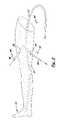
- FIG. 1is a side elevational view of a leg-shaped ultrasonic phantom shown between two mold members that are joined together to make a primary mold.
- FIG. 2is a side elevational view of the ultrasonic phantom shown in FIG. 1 showing a long rod and two short rods being removed from the phantom during the manufacturing process.
- FIG. 3is a perspective view of a section of the ultrasonic phantom showing the formation of a cavity inside the phantom using a removable secondary mold and various small, hollow and solid objects imbedded in the phantom.
- FIG. 4is a perspective view of the upper section of the leg-shaped phantom shown in
- FIGS. 1 , 2 and 4showing the ends of the long and short conduits being filled with blood simulating liquid and sealed off with plugs.
- FIG. 5is a diagram that shows the thermoplastic elastomers and a scattering agent being added to a mixing tank to produce the chemical composition used to manufacture the phantom.
- FIG. 6is a perspective view of the primary mold used to manufacture a phantom that is used with a head and neck model shown in FIG. 7 .
- FIG. 7is a head and neck model with the phantom produced by the primary mold shown in FIG. 6 inserted into the model's phantom cavity.
- FIG. 8is a perspective view of the primary mold used to manufacture a phantom that is used with an arm model shown in FIG. 9 .
- FIG. 9is a perspective view of an arm model with the phantom produced by the primary mold shown in FIG. 8 .
- the chemical composition 11is made of moldable material. thereby enabling the phantom to be formed in a wide variety of different anatomical structures.
- the chemical composition 11is substantially non-reflective during an ultrasonic image procedure, varying amounts of a scattering agent 25 may be added to the chemical composition 11 thereby enabling the manufacturer to adjust the sonographic characteristics of the chemical composition 11 to more closely mimic human tissue.
- the chemical composition 11are thermoplastic elastomers 12 , 13 that are melted and then poured into a rigid primary mold 15 .
- the thermoplastic elastomers 12 , 13are commercially available compositions comprised in part of highly plasticized styrene, ethylene, butylene, styrene block copolymers.
- the individual elastomers 12 , 13are sold under the trademarks TOUGH GRADE and EASY POUR, respectively, by Sutton Industries of North East, Md. While Sutton Technologies makes appropriate thermoplastic elastomers for the invention, multiple mixes can be used.
- the two thermoplastic elastomers 12 , 13are heated, mixed together in a 60:30 ratio.
- the term thermoplastic elastomersis defined as a material which has rubber like, stretchy qualities i.e. easily returns to its original shape when stretched and which can be melted and molded at sufficient temperature.
- the rigid primary mold 15is made of two mold members 16 and 20 with two inside, complimentary-shaped casting surfaces 17 A, 17 B formed therein, respectively.
- FIG. 1shows the two mold members 16 , 20 longitudinally aligned, registered and joined together to form a leg producing primary mold 15 .
- each mold member 16 , 20includes an outer shell 16 A, 20 A and at least one end plate 19 and 21 , respectively.
- the casting surfaces 17 A, 17 B on the mold members 16 , 20respectively, form the anterior and posterior surfaces, respectively, on the phantom 10 .
- thermoplastic elastomers 12 , 13are semi-solids at room temperature and become fluidic at temp above approximately 145 degrees C. Heating may be accomplished using ovens, heated vats, and open pans. Most preferably, a heated tank 24 with a piston pump 26 dispensing mechanism sold by Nordson, Inc, of Duluth, Minn. is used to heat and dispense the thermoplastic elastomers 12 , 13 . When the two thermoplastic elastomers 12 , 13 are melted they are slowly and thoroughly mixed with a scattering agent 25 in the heated tank 24 so that air bubbles are not introduced. After being thoroughly mixed together, the mixture of thermoplastic elastomers 12 , 13 and scattering agent 25 is slowly poured into a fill opening 28 formed on the mold member (mold member 16 shown).
- the scattering agent 25 added to the elastomers 12 , 13imparts sonographic characteristics to the chemical composition 11 that simulate the sonographic characteristics of living human tissue. It is widely known that scattering of the ultrasound occurs when an incident wave is reflected in many directions after interacting with a structure whose dimensions are similar to or less than the wavelength of the ultrasound. The distribution of the scattered ultrasound is critically dependent on the dimensions of the target compared to the wavelength.
- the total amount of scattering agent 25 added to the chemical composition 11is selectively adjusted depending on the type of anatomical structure the phantom 10 represents.
- the scattering agent 25include but not limited to talcum powder, graphite powder, and glass spheres.
- talcum powderis used as a scattering agent because of its ready availability and low cost.
- talcum powderis added in a concentration of 0.5-4 grams per liter.
- the chemical composition 11After connecting the mold members 16 , 20 together to form the primary mold 15 , and after dispensing the chemical composition 11 into the opening 28 , the chemical composition 11 is allowed to gradually cool to room temperature. After the chemical composition 11 has cured for approximately 2 hours, the mold members 16 , 20 are disconnected and separated so that the phantom 10 may be removed.
- the scattering agent 25is mixed with the thermoplastic elastomers 12 , 13 , a uniform sonographic image is produced throughout the entire phantom 10 . In other instances, it may be desirable to vary the sonographic imaging areas by varying amount of scattering agent 25 in different areas in the phantom 10 .
- additional amounts of scattering agent 25may be sprinkled into the chemical composition 11 as it is poured into the primary mold 15 to create small areas with contrasting sonographic characteristics. How much scattering agent 25 may be added and where it is added depends on the type of phantom being manufactured. An example where a localized, increased amount of scattering agent 25 is added to a specific region in the phantom 10 is when a dense anthropomorphic imaging structure, such as large muscles or tumors, is mimicked.
- areas on the phantom 10 where no or small amounts of scattering agent 25 are presentmay be used to mimic areas that show less sonographic imaging, such as gallbladders, hypoechoic masses, arteries, and ovarian follicles.
- One or more long and short conduitsmay be formed in the phantom 10 to simulate blood vessels and ducts.
- one long conduit 27is longitudinally aligned and formed inside the phantom 10 that represents the femoral artery.
- Formed adjacent to the long conduit 27are two short conduits 33 , 33 ′ designed to represent two smaller arteries that extend outward from the femoral artery.
- a long rod 37 and two short rods 38 and 38 ′are placed in the primary mold 15 .
- the end of the long rods 37extend through an opening 22 formed on the end plate 21 on the mold member 20 .
- the short rods 38 , 38 ′are aligned diagonally inside the primary mold 15 with the proximal ends touching the long rod 37 and the distal ends extending through openings 18 , 23 formed on the sides of the outer linings 16 A, 20 A of the two mold members 16 , 20 .
- the rods 37 , 38 , 38 ′can be easily grasped and removed from the phantom 10 after the composition 11 has cured as shown in FIG. 2 .
- the long and short rods 37 , 38 , 38 ′′are made of bendable, heat tolerate material that enable them to withstand temperatures used to melt the thermoplastic elastomers 12 , 13 .
- An example of material used to produce the rodsis a synthetic resinous plastic material sold under the trademark (DelrinTM) by E. I. De Pont Nemours and Company, of Wilmington, Del. This particular product is availability in a wide variety of shapes and size and can be easily shaped as necessary to create a desired form.
- the chemical composition 11is poured into the fill opening 28 to completely fill the inside leg-shaped cavity and to covering the rods 37 , 38 , and 38 ′. After the chemical composition 11 has cooled and cured, the rods 37 , 38 , 38 ′ are removed and the two mold members 16 , 20 are disconnected.
- short plugs 40are placed into the end openings 30 , 36 , 36 ′ on the conduits 27 , 33 , 33 ′, respectively.
- a suitable glue or adhesive 41may be used to hold the short plugs 40 in the conduits 27 , 33 , 33 ′.
- the short plugs 40are made of the chemical composition 11 so that the short plugs 40 blend into the surround phantom 10 and are not visible during the ultrasonic imaging procedure.
- the blood simulating fluid 45is made of 20% polyethylene glycol, 79.5% distilled water, 0.5% red food coloring and 0.2% sodium benzoate which acts as a preservative.
- a red or blue color dyemay also be added to the fluid 45 to indicate if the blood vessel is an artery or vein, respectively.
- the blood-simulating fluid 45may be injected into the conduits 33 , 33 ′ with a suitable syringe and needle.
- the fluid 45may be dispensed via a nozzle 46 connected to a delivery tube 47 and a large volume container.
- the phantom 10may include optional cavities 50 (one shown) that mimic an anthropomorphic cavity in the human tissue.
- the cavity 50may be hollow or filled with a solid object 51 or a body fluid simulating liquid, such as fluid 45 .
- the cavity 50may be filled with molten chemical composition 11 with more or less scattering agent 25 ′′ added thereto to provide a contrasting sonographic image.
- solid, smaller objects 55such as glass beads or marbles, may be scattered in the phantom 10 to mimic small tumors, thrombus or calcifications.
- a spherical or oval-shaped secondary mold 60 with a narrow, elongated handle 62may be placed inside the primary mold 15 at a desired location.
- the end 63 of the elongated handle 62is positioned adjacent to the inside surface of the primary mold's outer lining.
- the chemical composition 11is allowed to cure.
- the phantom 10is removed from the primary mold 15 .
- the end of the elongated handle 62is then pulled to extract the spherical mold 60 from phantom 10 as shown in FIG. 3 .
- the cured chemical composition 11is sufficiently elastic so that the secondary mold 60 may be pulled from the phantom 10 without tearing or rupturing the phantom 10 .
- a solid object 51 or a blood-simulating fluid 45may then be deposited in the cavity 50 with a suitable plug 40 made of chemical composition 11 disposed in the opening to the elongated handle space to prevent leakage.
- the phantom 10represents an anatomical structure, such as a leg as shown in FIGS. 1 , 2 , and 5 .
- the phantom 11is designed to represent a section on a rigid model that represents a large anatomical structure.
- FIG. 6shows a primary mold 15 ′ used to manufacture a phantom 10 ′ designed to be inserted into the cavity 82 in the head and neck model 80 shown in FIG. 7 .
- Phantom 10 ′is made of the chemical composition 11 described above with a plurality of long and short conduits 27 , 33 , 33 ′ formed in the phantom 10 ′ that represents the main blood vessels in the neck.
- FIG. 8shows another primary mold 15 ′′ used to manufacture a phantom 10 ′′ designed to be inserted into the cavity 92 formed in an arm model 90 shown in FIG. 9 .
- the arm model 90includes a forearm shaped section 93 with a hand section 94 . Extending through the hand section 94 is tubing 95 designed to connect to the long conduit 27 formed in the phantom 10 ′′.
- a pump 100may be attached to the opposite ends of tubing 102 , 104 that connect to the long conduit 27 and tubing 95 that extends through the forearm section 93 and hand section 94 respectfully, to circulate a blood simulating fluid 45 through the arm model 90 during training.
- thermoplastic elastomersthat when heated fills said primary mold
- thermoplastic elastomerc. heating the thermoplastic elastomer until melted
Landscapes
- Engineering & Computer Science (AREA)
- Physics & Mathematics (AREA)
- General Physics & Mathematics (AREA)
- Health & Medical Sciences (AREA)
- Mathematical Analysis (AREA)
- Pure & Applied Mathematics (AREA)
- Medical Informatics (AREA)
- Algebra (AREA)
- Computational Mathematics (AREA)
- General Health & Medical Sciences (AREA)
- Chemical & Material Sciences (AREA)
- Mathematical Optimization (AREA)
- Mathematical Physics (AREA)
- Medicinal Chemistry (AREA)
- Business, Economics & Management (AREA)
- Educational Administration (AREA)
- Educational Technology (AREA)
- Theoretical Computer Science (AREA)
- Nuclear Medicine, Radiotherapy & Molecular Imaging (AREA)
- Radiology & Medical Imaging (AREA)
- Ultra Sonic Daignosis Equipment (AREA)
- Instructional Devices (AREA)
Abstract
Description
Claims (23)
Priority Applications (1)
| Application Number | Priority Date | Filing Date | Title |
|---|---|---|---|
| US10/800,956US7255565B2 (en) | 2004-03-15 | 2004-03-15 | Anthropomorphic phantoms and method |
Applications Claiming Priority (1)
| Application Number | Priority Date | Filing Date | Title |
|---|---|---|---|
| US10/800,956US7255565B2 (en) | 2004-03-15 | 2004-03-15 | Anthropomorphic phantoms and method |
Publications (2)
| Publication Number | Publication Date |
|---|---|
| US20050202381A1 US20050202381A1 (en) | 2005-09-15 |
| US7255565B2true US7255565B2 (en) | 2007-08-14 |
Family
ID=34920804
Family Applications (1)
| Application Number | Title | Priority Date | Filing Date |
|---|---|---|---|
| US10/800,956Expired - LifetimeUS7255565B2 (en) | 2004-03-15 | 2004-03-15 | Anthropomorphic phantoms and method |
Country Status (1)
| Country | Link |
|---|---|
| US (1) | US7255565B2 (en) |
Cited By (59)
| Publication number | Priority date | Publication date | Assignee | Title |
|---|---|---|---|---|
| US20060204939A1 (en)* | 2003-04-14 | 2006-09-14 | Bardsley Ryan S | Inoculation training kit |
| US20070148633A1 (en)* | 2005-02-03 | 2007-06-28 | Christopher Sakezles | Dielectric properties models and methods of using same |
| US20080293029A1 (en)* | 2005-02-10 | 2008-11-27 | Wilkins Jason D | Ultrasound Training Mannequin |
| US20080318194A1 (en)* | 2007-06-20 | 2008-12-25 | Lakshmi Atchison | Kit for teaching the composition of blood |
| DE202009004115U1 (en) | 2009-03-24 | 2009-06-04 | Technische Universität Dresden | Models for the training of invasive techniques of medicine |
| US20090142741A1 (en)* | 2007-11-29 | 2009-06-04 | Cedars-Sinai Medical Center | Medical training methods and devices |
| US20090215016A1 (en)* | 2004-07-30 | 2009-08-27 | Hansjoerg Wesp | Device for the determination of parameters particularly for therapeutic compression means on limbs |
| US20090226866A1 (en)* | 2008-03-07 | 2009-09-10 | Harold Durbin | Surgical teaching aid |
| US20090246747A1 (en)* | 2008-03-25 | 2009-10-01 | Operative Experience, Inc. | Simulator for major surgical operations |
| US20090291421A1 (en)* | 2008-02-29 | 2009-11-26 | Laerdal Medical As | Simulator for medical training with detachable self-sealing hollow member |
| US20100216106A1 (en)* | 2009-02-24 | 2010-08-26 | Mckee Patricia Rosalind | System and method for learning structure using envelopes |
| US20110117531A1 (en)* | 2008-07-16 | 2011-05-19 | Waseda University | Mold for producing simulated blood vessel, method of producing simulated blood vessel and simulated blood vessel |
| US20120040323A1 (en)* | 2009-03-20 | 2012-02-16 | Waseda University | Blood vessel model for medical training and method for manufacturing same |
| US8162669B1 (en)* | 2009-07-14 | 2012-04-24 | Shaw Erin L | Ultrasound training assembly |
| US20140069215A1 (en)* | 2012-09-07 | 2014-03-13 | University Of Louisville Research Foundation, Inc. | Multimodal cardiac phantom for imaging |
| US8764452B2 (en) | 2010-10-01 | 2014-07-01 | Applied Medical Resources Corporation | Portable laparoscopic trainer |
| US8801438B2 (en) | 2011-11-23 | 2014-08-12 | Christopher Sakezles | Artificial anatomic model |
| US8911238B2 (en) | 2011-11-28 | 2014-12-16 | BrachyTech LLC | Prostate brachytherapy simulator |
| US8926333B2 (en)* | 2013-03-15 | 2015-01-06 | Simnext, Llc | Device, system, and method for simulating blood flow |
| US8961190B2 (en) | 2011-12-20 | 2015-02-24 | Applied Medical Resources Corporation | Advanced surgical simulation |
| US9218753B2 (en) | 2011-10-21 | 2015-12-22 | Applied Medical Resources Corporation | Simulated tissue structure for surgical training |
| US9342996B2 (en) | 2004-12-02 | 2016-05-17 | The United States Of America, As Represented By The Secretary Of The Army | Trauma training system |
| US9449532B2 (en) | 2013-05-15 | 2016-09-20 | Applied Medical Resources Corporation | Hernia model |
| US9548002B2 (en) | 2013-07-24 | 2017-01-17 | Applied Medical Resources Corporation | First entry model |
| US9558678B1 (en)* | 2014-11-20 | 2017-01-31 | Michael E. Nerney | Near-infrared imager training device |
| US9576503B2 (en) | 2013-12-27 | 2017-02-21 | Seattle Children's Hospital | Simulation cart |
| US20170076635A1 (en)* | 2015-07-06 | 2017-03-16 | Gray's Bone and Joint Models, LLC | Vascular procedure training system |
| US9898937B2 (en) | 2012-09-28 | 2018-02-20 | Applied Medical Resources Corporation | Surgical training model for laparoscopic procedures |
| US9922579B2 (en) | 2013-06-18 | 2018-03-20 | Applied Medical Resources Corporation | Gallbladder model |
| US9940849B2 (en) | 2013-03-01 | 2018-04-10 | Applied Medical Resources Corporation | Advanced surgical simulation constructions and methods |
| US9959786B2 (en) | 2012-09-27 | 2018-05-01 | Applied Medical Resources Corporation | Surgical training model for laparoscopic procedures |
| US10081727B2 (en) | 2015-05-14 | 2018-09-25 | Applied Medical Resources Corporation | Synthetic tissue structures for electrosurgical training and simulation |
| US10121391B2 (en) | 2012-09-27 | 2018-11-06 | Applied Medical Resources Corporation | Surgical training model for laparoscopic procedures |
| US10198966B2 (en) | 2013-07-24 | 2019-02-05 | Applied Medical Resources Corporation | Advanced first entry model for surgical simulation |
| US10198965B2 (en) | 2012-08-03 | 2019-02-05 | Applied Medical Resources Corporation | Simulated stapling and energy based ligation for surgical training |
| US10223936B2 (en) | 2015-06-09 | 2019-03-05 | Applied Medical Resources Corporation | Hysterectomy model |
| US10332425B2 (en) | 2015-07-16 | 2019-06-25 | Applied Medical Resources Corporation | Simulated dissectible tissue |
| US10354556B2 (en) | 2015-02-19 | 2019-07-16 | Applied Medical Resources Corporation | Simulated tissue structures and methods |
| US10395559B2 (en) | 2012-09-28 | 2019-08-27 | Applied Medical Resources Corporation | Surgical training model for transluminal laparoscopic procedures |
| US10490105B2 (en) | 2015-07-22 | 2019-11-26 | Applied Medical Resources Corporation | Appendectomy model |
| US10535281B2 (en) | 2012-09-26 | 2020-01-14 | Applied Medical Resources Corporation | Surgical training model for laparoscopic procedures |
| US10573201B2 (en)* | 2016-05-30 | 2020-02-25 | MAX-PLANCK-Gesellschaft zur Förderung der Wissenschaften e.V. | Method of producing a phantom and phantom |
| US10679520B2 (en) | 2012-09-27 | 2020-06-09 | Applied Medical Resources Corporation | Surgical training model for laparoscopic procedures |
| US10706743B2 (en) | 2015-11-20 | 2020-07-07 | Applied Medical Resources Corporation | Simulated dissectible tissue |
| US10720084B2 (en) | 2015-10-02 | 2020-07-21 | Applied Medical Resources Corporation | Hysterectomy model |
| US10796606B2 (en) | 2014-03-26 | 2020-10-06 | Applied Medical Resources Corporation | Simulated dissectible tissue |
| US10818201B2 (en) | 2014-11-13 | 2020-10-27 | Applied Medical Resources Corporation | Simulated tissue models and methods |
| US10847057B2 (en) | 2017-02-23 | 2020-11-24 | Applied Medical Resources Corporation | Synthetic tissue structures for electrosurgical training and simulation |
| WO2021077216A1 (en)* | 2019-10-21 | 2021-04-29 | Rajagopalan Sumitra | Self-healing compositions for use in medical training simulators and mannequins |
| US11030922B2 (en) | 2017-02-14 | 2021-06-08 | Applied Medical Resources Corporation | Laparoscopic training system |
| US11120708B2 (en) | 2016-06-27 | 2021-09-14 | Applied Medical Resources Corporation | Simulated abdominal wall |
| EP3988167A1 (en)* | 2020-10-23 | 2022-04-27 | Cardiawave SA | Process for testing the accuracy and the performance of an ultrasound transducer |
| US11417241B2 (en) | 2018-12-01 | 2022-08-16 | Syndaver Labs, Inc. | Artificial canine model |
| US11574563B2 (en)* | 2019-05-23 | 2023-02-07 | Black Cat Medical Llc | Ultrasound guided training simulators for cryoneurolysis pain blocks |
| US11776428B1 (en)* | 2015-10-21 | 2023-10-03 | University Of Rochester | Systems, models, and methods for simulating surgery on anatomical organs |
| GB2624617A (en)* | 2022-08-08 | 2024-05-29 | Medisim Ltd | Ultrasonography Training Phantom |
| US12044765B2 (en) | 2015-11-06 | 2024-07-23 | Vigilance Health Imaging Network Inc. | Phantoms having reference members with microchannels and diffusion weighted imaging using same |
| US20240290221A1 (en)* | 2017-04-18 | 2024-08-29 | Teleflex Medical Incorporated | Vascular access training simulator system and transparent anatomical model |
| US12106678B2 (en) | 2021-10-23 | 2024-10-01 | Simulated Inanimate Models, LLC | Procedure guidance and training apparatus, methods and systems |
Families Citing this family (18)
| Publication number | Priority date | Publication date | Assignee | Title |
|---|---|---|---|---|
| US7255564B2 (en)* | 2003-03-14 | 2007-08-14 | Innovative Premiums, Inc. | Anatomical pocket model |
| US7717468B2 (en)* | 2005-12-01 | 2010-05-18 | Innovative Premiums Inc. | Clipboard with an integral three dimensional display |
| WO2008075303A1 (en)* | 2006-12-21 | 2008-06-26 | Koninklijke Philips Electronics N.V. | Anatomically and functionally accurate soft tissue phantoms and method for generating same |
| WO2009010898A2 (en)* | 2007-07-13 | 2009-01-22 | Koninklijke Philips Electronics N.V. | Phantom for ultrasound guided needle insertion and method for making the phantom |
| ES2343778B2 (en)* | 2009-10-01 | 2011-01-21 | Universidad Politecnica De Madrid | HUMAN BLOOD PHANTOM. |
| DE102010028611A1 (en)* | 2010-05-05 | 2011-11-10 | Benning, Benning&Benning Gbr (Vertretungsberechtigter Gesellschafter: Dr. Michael Benning, 52074 Aachen) | Injection trainer e.g. epidural injection trainer for epidural puncture in spinal canal, has pressure additive comprising ultrasonic echo from muscle tissue, and protective sheet comprising ultrasonic echo from skin |
| US20120318038A1 (en)* | 2011-06-17 | 2012-12-20 | TransducerWorks | Ultrasound transducer test apparatus and applications thereof |
| US9965591B2 (en) | 2011-09-13 | 2018-05-08 | Medtronic, Inc. | Physiologic simulator system |
| CN103021246B (en)* | 2012-12-24 | 2015-04-22 | 中国人民解放军第三军医大学 | Production method and application of abdominal organ teaching simulation model applicable to type-B ultrasonic detection |
| US10144154B2 (en)* | 2014-10-09 | 2018-12-04 | Synaptive Medical (Barbados) Inc. | Phantom production tool |
| CN104318840A (en)* | 2014-10-22 | 2015-01-28 | 北京航空航天大学 | Simulation method of medical surgical instrument guide wire on basis of spring proton model |
| US10350833B1 (en)* | 2015-05-12 | 2019-07-16 | Jacques Zaneveld | Methods and systems for creating anatomical models |
| US10864659B1 (en)* | 2015-05-12 | 2020-12-15 | Jacques Zaneveld | Methods and systems for creating anatomical models |
| WO2017155678A1 (en) | 2016-03-10 | 2017-09-14 | Kindheart, Inc | Fake blood for use in simulated surgical procedures |
| CN107144805A (en)* | 2017-06-09 | 2017-09-08 | 创领心律管理医疗器械(上海)有限公司 | A kind of synthesis field method of testing of lower limb model and magnetic resonance imaging safety evaluation |
| CO2018008426A1 (en)* | 2018-08-10 | 2018-10-10 | Medical Phantom S A S | Ql block trainer |
| CO2018008419A1 (en)* | 2018-08-10 | 2018-10-10 | Medical Phantom S A S | Cube and block trainer |
| US12014651B2 (en)* | 2021-06-16 | 2024-06-18 | Nokia Technologies Oy | Perfusive tissue phantom |
Citations (21)
| Publication number | Priority date | Publication date | Assignee | Title |
|---|---|---|---|---|
| US3789518A (en)* | 1972-04-12 | 1974-02-05 | Weatherby Nasco Inc | Simulated human limb |
| US4182054A (en)* | 1978-02-16 | 1980-01-08 | Medical Plastics Laboratory, Inc. | Artificial arm |
| GB2047101A (en)* | 1979-04-11 | 1980-11-26 | Dow Corning | Injection button and method of production thereof |
| US4277367A (en) | 1978-10-23 | 1981-07-07 | Wisconsin Alumni Research Foundation | Phantom material and method |
| US4286168A (en)* | 1979-01-18 | 1981-08-25 | Atomic Products Corp. | Phantom simulation device for scintillation cameras |
| US4286455A (en) | 1979-05-04 | 1981-09-01 | Acoustic Standards Corporation | Ultrasound phantom |
| US4493653A (en)* | 1983-09-27 | 1985-01-15 | Technicare Corporation | Biopsiable ultrasound phantom |
| US4974461A (en) | 1988-10-13 | 1990-12-04 | The United States Of America As Represented By Department Of Health And Human Services | Anthropomorphic cardiac ultrasound phantom |
| US5061187A (en) | 1990-04-12 | 1991-10-29 | Ravinder Jerath | Ultrasound training apparatus |
| US5236363A (en)* | 1991-08-09 | 1993-08-17 | General Electric Company | Phantom for simulating an x-ray exam patient |
| US5419706A (en)* | 1993-06-22 | 1995-05-30 | Levy; Richard C. | Apparatus for forming images of non-visible elements underlying an opaque surface |
| US5625137A (en) | 1995-05-25 | 1997-04-29 | Wisconsin Alumni Research Foundation | Very low scatter liquid and solid tissue mimicking material for ultrasound phantoms and method of making the same |
| US5839904A (en)* | 1997-10-09 | 1998-11-24 | Bloom; Ellen A. | Phlebotomy training device |
| US6083008A (en)* | 1997-09-01 | 2000-07-04 | Agency Of Industrial Science & Technology, Ministry Of International Trade & Industry | Optical phantom of living body and method for producing it |
| US6190915B1 (en) | 1999-06-25 | 2001-02-20 | Wisconsin Alumni Research Foundation | Ultrasound phantoms |
| US6205871B1 (en)* | 1998-12-22 | 2001-03-27 | The Regents Of The University Of California | Vascular phantoms |
| US6318146B1 (en) | 1999-07-14 | 2001-11-20 | Wisconsin Alumni Research Foundation | Multi-imaging modality tissue mimicking materials for imaging phantoms |
| US6352860B1 (en) | 2000-11-17 | 2002-03-05 | Wisconsin Alumni Research Foundation | Liquid and solid tissue mimicking material for ultrasound phantoms and method of making the same |
| US6471519B1 (en)* | 1997-03-27 | 2002-10-29 | The Johns Hopkins University | Bone substitute for training and testing and method for making |
| US20040126746A1 (en)* | 2000-10-23 | 2004-07-01 | Toly Christopher C. | Medical physiological simulator including a conductive elastomer layer |
| US7059168B2 (en)* | 2002-10-01 | 2006-06-13 | Olympus Corporation | Ultrasound phantom |
Family Cites Families (1)
| Publication number | Priority date | Publication date | Assignee | Title |
|---|---|---|---|---|
| AU2864595A (en)* | 1994-06-23 | 1996-01-19 | Janice Marie Thielbar | Custom formed natural fit artificial breast appliance |
- 2004
- 2004-03-15USUS10/800,956patent/US7255565B2/ennot_activeExpired - Lifetime
Patent Citations (22)
| Publication number | Priority date | Publication date | Assignee | Title |
|---|---|---|---|---|
| US3789518A (en)* | 1972-04-12 | 1974-02-05 | Weatherby Nasco Inc | Simulated human limb |
| US4182054A (en)* | 1978-02-16 | 1980-01-08 | Medical Plastics Laboratory, Inc. | Artificial arm |
| US4277367A (en) | 1978-10-23 | 1981-07-07 | Wisconsin Alumni Research Foundation | Phantom material and method |
| US4286168A (en)* | 1979-01-18 | 1981-08-25 | Atomic Products Corp. | Phantom simulation device for scintillation cameras |
| GB2047101A (en)* | 1979-04-11 | 1980-11-26 | Dow Corning | Injection button and method of production thereof |
| US4286455A (en) | 1979-05-04 | 1981-09-01 | Acoustic Standards Corporation | Ultrasound phantom |
| US4493653A (en)* | 1983-09-27 | 1985-01-15 | Technicare Corporation | Biopsiable ultrasound phantom |
| US4974461A (en) | 1988-10-13 | 1990-12-04 | The United States Of America As Represented By Department Of Health And Human Services | Anthropomorphic cardiac ultrasound phantom |
| US5061187A (en) | 1990-04-12 | 1991-10-29 | Ravinder Jerath | Ultrasound training apparatus |
| US5236363A (en)* | 1991-08-09 | 1993-08-17 | General Electric Company | Phantom for simulating an x-ray exam patient |
| US5419706A (en)* | 1993-06-22 | 1995-05-30 | Levy; Richard C. | Apparatus for forming images of non-visible elements underlying an opaque surface |
| US5625137A (en) | 1995-05-25 | 1997-04-29 | Wisconsin Alumni Research Foundation | Very low scatter liquid and solid tissue mimicking material for ultrasound phantoms and method of making the same |
| US5902748A (en) | 1995-05-25 | 1999-05-11 | Wisconsin Alumni Research Foundation | Method of making a solid tissue mimicking material for ultrasound phantoms |
| US6471519B1 (en)* | 1997-03-27 | 2002-10-29 | The Johns Hopkins University | Bone substitute for training and testing and method for making |
| US6083008A (en)* | 1997-09-01 | 2000-07-04 | Agency Of Industrial Science & Technology, Ministry Of International Trade & Industry | Optical phantom of living body and method for producing it |
| US5839904A (en)* | 1997-10-09 | 1998-11-24 | Bloom; Ellen A. | Phlebotomy training device |
| US6205871B1 (en)* | 1998-12-22 | 2001-03-27 | The Regents Of The University Of California | Vascular phantoms |
| US6190915B1 (en) | 1999-06-25 | 2001-02-20 | Wisconsin Alumni Research Foundation | Ultrasound phantoms |
| US6318146B1 (en) | 1999-07-14 | 2001-11-20 | Wisconsin Alumni Research Foundation | Multi-imaging modality tissue mimicking materials for imaging phantoms |
| US20040126746A1 (en)* | 2000-10-23 | 2004-07-01 | Toly Christopher C. | Medical physiological simulator including a conductive elastomer layer |
| US6352860B1 (en) | 2000-11-17 | 2002-03-05 | Wisconsin Alumni Research Foundation | Liquid and solid tissue mimicking material for ultrasound phantoms and method of making the same |
| US7059168B2 (en)* | 2002-10-01 | 2006-06-13 | Olympus Corporation | Ultrasound phantom |
Cited By (109)
| Publication number | Priority date | Publication date | Assignee | Title |
|---|---|---|---|---|
| US7534107B2 (en)* | 2003-04-14 | 2009-05-19 | The General Hospital Corporation | Inoculation training kit |
| US20060204939A1 (en)* | 2003-04-14 | 2006-09-14 | Bardsley Ryan S | Inoculation training kit |
| US8419437B2 (en)* | 2004-07-30 | 2013-04-16 | Paul Hartmann Ag | Device for the determination of parameters particularly for therapeutic compression means on limbs |
| US20090215016A1 (en)* | 2004-07-30 | 2009-08-27 | Hansjoerg Wesp | Device for the determination of parameters particularly for therapeutic compression means on limbs |
| US10347157B2 (en) | 2004-12-02 | 2019-07-09 | The United States Of America, As Represented By The Secretary Of The Army | Trauma training system |
| US9342996B2 (en) | 2004-12-02 | 2016-05-17 | The United States Of America, As Represented By The Secretary Of The Army | Trauma training system |
| US20070148633A1 (en)* | 2005-02-03 | 2007-06-28 | Christopher Sakezles | Dielectric properties models and methods of using same |
| US8137110B2 (en)* | 2005-02-03 | 2012-03-20 | Christopher Sakezles | Dielectric properties models and methods of using same |
| US8512044B2 (en) | 2005-02-03 | 2013-08-20 | Christopher Sakezles | Dielectric properties models and methods of using same |
| US7845949B2 (en)* | 2005-02-10 | 2010-12-07 | Wilkins Jason D | Ultrasound training mannequin |
| US20080293029A1 (en)* | 2005-02-10 | 2008-11-27 | Wilkins Jason D | Ultrasound Training Mannequin |
| US8277225B2 (en)* | 2007-06-20 | 2012-10-02 | Lakshmi Atchison | Kit for teaching the composition of blood |
| US20080318194A1 (en)* | 2007-06-20 | 2008-12-25 | Lakshmi Atchison | Kit for teaching the composition of blood |
| US20090142741A1 (en)* | 2007-11-29 | 2009-06-04 | Cedars-Sinai Medical Center | Medical training methods and devices |
| US8454368B2 (en)* | 2007-11-29 | 2013-06-04 | Cedars-Sinai Medical Center | Medical training methods and devices |
| US20090291421A1 (en)* | 2008-02-29 | 2009-11-26 | Laerdal Medical As | Simulator for medical training with detachable self-sealing hollow member |
| US8100695B2 (en)* | 2008-02-29 | 2012-01-24 | Laerdal Medical As | Simulator for medical training with detachable self-sealing hollow member |
| US20090226866A1 (en)* | 2008-03-07 | 2009-09-10 | Harold Durbin | Surgical teaching aid |
| US20090246747A1 (en)* | 2008-03-25 | 2009-10-01 | Operative Experience, Inc. | Simulator for major surgical operations |
| US20110117531A1 (en)* | 2008-07-16 | 2011-05-19 | Waseda University | Mold for producing simulated blood vessel, method of producing simulated blood vessel and simulated blood vessel |
| US8636520B2 (en)* | 2008-07-16 | 2014-01-28 | Waseda University | Mold for producing simulated blood vessel, method of producing simulated blood vessel and simulated blood vessel |
| US20100216106A1 (en)* | 2009-02-24 | 2010-08-26 | Mckee Patricia Rosalind | System and method for learning structure using envelopes |
| US8469717B2 (en)* | 2009-03-20 | 2013-06-25 | EBM Corporation | Blood vessel model for medical training and method for manufacturing same |
| US20120040323A1 (en)* | 2009-03-20 | 2012-02-16 | Waseda University | Blood vessel model for medical training and method for manufacturing same |
| DE202009004115U1 (en) | 2009-03-24 | 2009-06-04 | Technische Universität Dresden | Models for the training of invasive techniques of medicine |
| US8162669B1 (en)* | 2009-07-14 | 2012-04-24 | Shaw Erin L | Ultrasound training assembly |
| US10854112B2 (en) | 2010-10-01 | 2020-12-01 | Applied Medical Resources Corporation | Portable laparoscopic trainer |
| US8764452B2 (en) | 2010-10-01 | 2014-07-01 | Applied Medical Resources Corporation | Portable laparoscopic trainer |
| US9472121B2 (en) | 2010-10-01 | 2016-10-18 | Applied Medical Resources Corporation | Portable laparoscopic trainer |
| US12154454B2 (en) | 2010-10-01 | 2024-11-26 | Applied Medical Resources Corporation | Portable laparoscopic trainer |
| US9218753B2 (en) | 2011-10-21 | 2015-12-22 | Applied Medical Resources Corporation | Simulated tissue structure for surgical training |
| US12014652B2 (en) | 2011-10-21 | 2024-06-18 | Applied Medical Resources Corporation | Simulated tissue structure for surgical training |
| US11158212B2 (en) | 2011-10-21 | 2021-10-26 | Applied Medical Resources Corporation | Simulated tissue structure for surgical training |
| US8801438B2 (en) | 2011-11-23 | 2014-08-12 | Christopher Sakezles | Artificial anatomic model |
| US8911238B2 (en) | 2011-11-28 | 2014-12-16 | BrachyTech LLC | Prostate brachytherapy simulator |
| US8961190B2 (en) | 2011-12-20 | 2015-02-24 | Applied Medical Resources Corporation | Advanced surgical simulation |
| US11403968B2 (en) | 2011-12-20 | 2022-08-02 | Applied Medical Resources Corporation | Advanced surgical simulation |
| US10198965B2 (en) | 2012-08-03 | 2019-02-05 | Applied Medical Resources Corporation | Simulated stapling and energy based ligation for surgical training |
| US20140069215A1 (en)* | 2012-09-07 | 2014-03-13 | University Of Louisville Research Foundation, Inc. | Multimodal cardiac phantom for imaging |
| US9386960B2 (en)* | 2012-09-07 | 2016-07-12 | University Of Louisville Research Foundation, Inc. | Multimodal cardiac phantom for imaging |
| US11514819B2 (en) | 2012-09-26 | 2022-11-29 | Applied Medical Resources Corporation | Surgical training model for laparoscopic procedures |
| US10535281B2 (en) | 2012-09-26 | 2020-01-14 | Applied Medical Resources Corporation | Surgical training model for laparoscopic procedures |
| US11361679B2 (en) | 2012-09-27 | 2022-06-14 | Applied Medical Resources Corporation | Surgical training model for laparoscopic procedures |
| US11869378B2 (en) | 2012-09-27 | 2024-01-09 | Applied Medical Resources Corporation | Surgical training model for laparoscopic procedures |
| US9959786B2 (en) | 2012-09-27 | 2018-05-01 | Applied Medical Resources Corporation | Surgical training model for laparoscopic procedures |
| US10679520B2 (en) | 2012-09-27 | 2020-06-09 | Applied Medical Resources Corporation | Surgical training model for laparoscopic procedures |
| US10121391B2 (en) | 2012-09-27 | 2018-11-06 | Applied Medical Resources Corporation | Surgical training model for laparoscopic procedures |
| US11990055B2 (en) | 2012-09-27 | 2024-05-21 | Applied Medical Resources Corporation | Surgical training model for laparoscopic procedures |
| US10395559B2 (en) | 2012-09-28 | 2019-08-27 | Applied Medical Resources Corporation | Surgical training model for transluminal laparoscopic procedures |
| US9898937B2 (en) | 2012-09-28 | 2018-02-20 | Applied Medical Resources Corporation | Surgical training model for laparoscopic procedures |
| US9940849B2 (en) | 2013-03-01 | 2018-04-10 | Applied Medical Resources Corporation | Advanced surgical simulation constructions and methods |
| US10991270B2 (en) | 2013-03-01 | 2021-04-27 | Applied Medical Resources Corporation | Advanced surgical simulation constructions and methods |
| US8926333B2 (en)* | 2013-03-15 | 2015-01-06 | Simnext, Llc | Device, system, and method for simulating blood flow |
| US10140889B2 (en) | 2013-05-15 | 2018-11-27 | Applied Medical Resources Corporation | Hernia model |
| US9449532B2 (en) | 2013-05-15 | 2016-09-20 | Applied Medical Resources Corporation | Hernia model |
| US11735068B2 (en) | 2013-06-18 | 2023-08-22 | Applied Medical Resources Corporation | Gallbladder model |
| US11049418B2 (en) | 2013-06-18 | 2021-06-29 | Applied Medical Resources Corporation | Gallbladder model |
| US9922579B2 (en) | 2013-06-18 | 2018-03-20 | Applied Medical Resources Corporation | Gallbladder model |
| US10657845B2 (en) | 2013-07-24 | 2020-05-19 | Applied Medical Resources Corporation | First entry model |
| US10026337B2 (en) | 2013-07-24 | 2018-07-17 | Applied Medical Resources Corporation | First entry model |
| US11450236B2 (en) | 2013-07-24 | 2022-09-20 | Applied Medical Resources Corporation | Advanced first entry model for surgical simulation |
| US11854425B2 (en) | 2013-07-24 | 2023-12-26 | Applied Medical Resources Corporation | First entry model |
| US9548002B2 (en) | 2013-07-24 | 2017-01-17 | Applied Medical Resources Corporation | First entry model |
| US10198966B2 (en) | 2013-07-24 | 2019-02-05 | Applied Medical Resources Corporation | Advanced first entry model for surgical simulation |
| US12288476B2 (en) | 2013-07-24 | 2025-04-29 | Applied Medical Resources Corporation | Advanced first entry model for surgical simulation |
| US9576503B2 (en) | 2013-12-27 | 2017-02-21 | Seattle Children's Hospital | Simulation cart |
| US10796606B2 (en) | 2014-03-26 | 2020-10-06 | Applied Medical Resources Corporation | Simulated dissectible tissue |
| US10818201B2 (en) | 2014-11-13 | 2020-10-27 | Applied Medical Resources Corporation | Simulated tissue models and methods |
| US11887504B2 (en) | 2014-11-13 | 2024-01-30 | Applied Medical Resources Corporation | Simulated tissue models and methods |
| US12211394B2 (en) | 2014-11-13 | 2025-01-28 | Applied Medical Resources Corporation | Simulated tissue models and methods |
| US9558678B1 (en)* | 2014-11-20 | 2017-01-31 | Michael E. Nerney | Near-infrared imager training device |
| US11100815B2 (en) | 2015-02-19 | 2021-08-24 | Applied Medical Resources Corporation | Simulated tissue structures and methods |
| US10354556B2 (en) | 2015-02-19 | 2019-07-16 | Applied Medical Resources Corporation | Simulated tissue structures and methods |
| US12131664B2 (en) | 2015-02-19 | 2024-10-29 | Applied Medical Resources Corporation | Simulated tissue structures and methods |
| US11034831B2 (en) | 2015-05-14 | 2021-06-15 | Applied Medical Resources Corporation | Synthetic tissue structures for electrosurgical training and simulation |
| US10081727B2 (en) | 2015-05-14 | 2018-09-25 | Applied Medical Resources Corporation | Synthetic tissue structures for electrosurgical training and simulation |
| US10733908B2 (en) | 2015-06-09 | 2020-08-04 | Applied Medical Resources Corporation | Hysterectomy model |
| US10223936B2 (en) | 2015-06-09 | 2019-03-05 | Applied Medical Resources Corporation | Hysterectomy model |
| US12175883B2 (en) | 2015-06-09 | 2024-12-24 | Applied Medical Resources Corporation | Hysterectomy model |
| US11721240B2 (en) | 2015-06-09 | 2023-08-08 | Applied Medical Resources Corporation | Hysterectomy model |
| US20170076635A1 (en)* | 2015-07-06 | 2017-03-16 | Gray's Bone and Joint Models, LLC | Vascular procedure training system |
| US12087179B2 (en) | 2015-07-16 | 2024-09-10 | Applied Medical Resources Corporation | Simulated dissectible tissue |
| US11587466B2 (en) | 2015-07-16 | 2023-02-21 | Applied Medical Resources Corporation | Simulated dissectible tissue |
| US10755602B2 (en) | 2015-07-16 | 2020-08-25 | Applied Medical Resources Corporation | Simulated dissectible tissue |
| US10332425B2 (en) | 2015-07-16 | 2019-06-25 | Applied Medical Resources Corporation | Simulated dissectible tissue |
| US10490105B2 (en) | 2015-07-22 | 2019-11-26 | Applied Medical Resources Corporation | Appendectomy model |
| US10720084B2 (en) | 2015-10-02 | 2020-07-21 | Applied Medical Resources Corporation | Hysterectomy model |
| US11721242B2 (en) | 2015-10-02 | 2023-08-08 | Applied Medical Resources Corporation | Hysterectomy model |
| US12243441B2 (en) | 2015-10-02 | 2025-03-04 | Applied Medical Resources Corporation | Hysterectomy model |
| US11776428B1 (en)* | 2015-10-21 | 2023-10-03 | University Of Rochester | Systems, models, and methods for simulating surgery on anatomical organs |
| US12044765B2 (en) | 2015-11-06 | 2024-07-23 | Vigilance Health Imaging Network Inc. | Phantoms having reference members with microchannels and diffusion weighted imaging using same |
| US10706743B2 (en) | 2015-11-20 | 2020-07-07 | Applied Medical Resources Corporation | Simulated dissectible tissue |
| US12217625B2 (en) | 2015-11-20 | 2025-02-04 | Applied Medical Resources Corporation | Simulated dissectible tissue |
| US10573201B2 (en)* | 2016-05-30 | 2020-02-25 | MAX-PLANCK-Gesellschaft zur Förderung der Wissenschaften e.V. | Method of producing a phantom and phantom |
| US11120708B2 (en) | 2016-06-27 | 2021-09-14 | Applied Medical Resources Corporation | Simulated abdominal wall |
| US11830378B2 (en) | 2016-06-27 | 2023-11-28 | Applied Medical Resources Corporation | Simulated abdominal wall |
| US11030922B2 (en) | 2017-02-14 | 2021-06-08 | Applied Medical Resources Corporation | Laparoscopic training system |
| US12243439B2 (en) | 2017-02-14 | 2025-03-04 | Applied Medical Resources Corporation | Laparoscopic training system |
| US10847057B2 (en) | 2017-02-23 | 2020-11-24 | Applied Medical Resources Corporation | Synthetic tissue structures for electrosurgical training and simulation |
| US20240290221A1 (en)* | 2017-04-18 | 2024-08-29 | Teleflex Medical Incorporated | Vascular access training simulator system and transparent anatomical model |
| US11417241B2 (en) | 2018-12-01 | 2022-08-16 | Syndaver Labs, Inc. | Artificial canine model |
| US20230162620A1 (en)* | 2019-05-23 | 2023-05-25 | Black Cat Medical Llc | Ultrasound guided training simulators for cryoneurolysis pain blocks |
| US11574563B2 (en)* | 2019-05-23 | 2023-02-07 | Black Cat Medical Llc | Ultrasound guided training simulators for cryoneurolysis pain blocks |
| US12080185B2 (en)* | 2019-05-23 | 2024-09-03 | Black Cat Medical Llc | Ultrasound guided training simulators for Cryoneurolysis pain blocks |
| WO2021077216A1 (en)* | 2019-10-21 | 2021-04-29 | Rajagopalan Sumitra | Self-healing compositions for use in medical training simulators and mannequins |
| WO2022084547A1 (en)* | 2020-10-23 | 2022-04-28 | Cardiawave | Process for testing the accuracy and the performance of an ultrasound transducer |
| EP3988167A1 (en)* | 2020-10-23 | 2022-04-27 | Cardiawave SA | Process for testing the accuracy and the performance of an ultrasound transducer |
| US12106678B2 (en) | 2021-10-23 | 2024-10-01 | Simulated Inanimate Models, LLC | Procedure guidance and training apparatus, methods and systems |
| GB2624617A (en)* | 2022-08-08 | 2024-05-29 | Medisim Ltd | Ultrasonography Training Phantom |
Also Published As
| Publication number | Publication date |
|---|---|
| US20050202381A1 (en) | 2005-09-15 |
Similar Documents
| Publication | Publication Date | Title |
|---|---|---|
| US7255565B2 (en) | Anthropomorphic phantoms and method | |
| JP2025067920A (en) | Dissectable simulated tissue | |
| JP6806684B2 (en) | Simulated tissue structure and method | |
| US5803746A (en) | Body part model and method of making same | |
| CN101743578B (en) | Phantom for ultrasound-guided needle insertion and method for making the phantom | |
| AU2015235994B2 (en) | Simulated dissectible tissue | |
| AU755575B2 (en) | Clinical and/or surgical training apparatus | |
| Baba et al. | Development of a tailored thyroid gland phantom for fine-needle aspiration cytology by three-dimensional printing | |
| JP7365020B2 (en) | Method for manufacturing objects with gel | |
| Nattagh et al. | A training phantom for ultrasound-guided needle insertion and suturing | |
| US20200170612A1 (en) | Tissue Mimicking Materials | |
| CN209895642U (en) | Human model for teaching and operation training of renal biopsy | |
| CN109036061A (en) | A kind of Digital stomach and intestine machine simulation operations training simulation people and preparation method thereof | |
| JP7437826B2 (en) | Organ model for surgical practice | |
| CN111210705A (en) | A human medical application model made of silica gel and epoxy resin and its production process | |
| WO2024178466A1 (en) | Surgical training apparatus | |
| JP7343109B2 (en) | Ultrasound-guided skin model for puncture technique training and method for adjusting needle tip visibility in echo images of the skin model for ultrasound-guided puncture technique training | |
| CN211506903U (en) | Human medical application model made of silica gel and epoxy resin | |
| RU2682459C1 (en) | Method of forming blood vessel phantoms for endoscopic optical coherent elastography | |
| CN117133176A (en) | Thyrocentesis training model and manufacturing method | |
| Grigorova et al. | Ultrasound Breast Phantom for Breast Biopsy Training | |
| DE202009004115U1 (en) | Models for the training of invasive techniques of medicine | |
| Wilcoski et al. | Gelatin Material Properties and their Role in Increasing Medical Task Trainer Fidelity | |
| US20230035446A1 (en) | Gyn pathology surgical simulation models and systems for surgical training | |
| EP4345798A1 (en) | Ultrasound guided percutaneous surgery mannequin |
Legal Events
| Date | Code | Title | Description |
|---|---|---|---|
| STCF | Information on status: patent grant | Free format text:PATENTED CASE | |
| FPAY | Fee payment | Year of fee payment:4 | |
| AS | Assignment | Owner name:CAE HEALTHCARE, INC., FLORIDA Free format text:ASSIGNMENT OF ASSIGNORS INTEREST;ASSIGNOR:KEEGAN, BRIAN;REEL/FRAME:029266/0861 Effective date:20121105 | |
| AS | Assignment | Owner name:CAE HEALTHCARE CANADA INC., QUEBEC Free format text:CHANGE OF NAME;ASSIGNOR:CAE HEALTHCARE, INC.;REEL/FRAME:030689/0065 Effective date:20120404 | |
| FEPP | Fee payment procedure | Free format text:PAT HOLDER NO LONGER CLAIMS SMALL ENTITY STATUS, ENTITY STATUS SET TO UNDISCOUNTED (ORIGINAL EVENT CODE: STOL); ENTITY STATUS OF PATENT OWNER: LARGE ENTITY | |
| FPAY | Fee payment | Year of fee payment:8 | |
| FEPP | Fee payment procedure | Free format text:MAINTENANCE FEE REMINDER MAILED (ORIGINAL EVENT CODE: REM.); ENTITY STATUS OF PATENT OWNER: LARGE ENTITY | |
| FEPP | Fee payment procedure | Free format text:11.5 YR SURCHARGE- LATE PMT W/IN 6 MO, LARGE ENTITY (ORIGINAL EVENT CODE: M1556); ENTITY STATUS OF PATENT OWNER: LARGE ENTITY | |
| MAFP | Maintenance fee payment | Free format text:PAYMENT OF MAINTENANCE FEE, 12TH YEAR, LARGE ENTITY (ORIGINAL EVENT CODE: M1553); ENTITY STATUS OF PATENT OWNER: LARGE ENTITY Year of fee payment:12 |