US7232409B2 - Method and apparatus for displaying endoscopic images - Google Patents
Method and apparatus for displaying endoscopic imagesDownload PDFInfo
- Publication number
- US7232409B2 US7232409B2US10/718,434US71843403AUS7232409B2US 7232409 B2US7232409 B2US 7232409B2US 71843403 AUS71843403 AUS 71843403AUS 7232409 B2US7232409 B2US 7232409B2
- Authority
- US
- United States
- Prior art keywords
- image
- endoscope
- viewing
- virtual
- view
- Prior art date
- Legal status (The legal status is an assumption and is not a legal conclusion. Google has not performed a legal analysis and makes no representation as to the accuracy of the status listed.)
- Expired - Lifetime, expires
Links
Images


Classifications
- A—HUMAN NECESSITIES
- A61—MEDICAL OR VETERINARY SCIENCE; HYGIENE
- A61B—DIAGNOSIS; SURGERY; IDENTIFICATION
- A61B1/00—Instruments for performing medical examinations of the interior of cavities or tubes of the body by visual or photographical inspection, e.g. endoscopes; Illuminating arrangements therefor
- A61B1/00002—Operational features of endoscopes
- A61B1/00043—Operational features of endoscopes provided with output arrangements
- A61B1/00045—Display arrangement
- A—HUMAN NECESSITIES
- A61—MEDICAL OR VETERINARY SCIENCE; HYGIENE
- A61B—DIAGNOSIS; SURGERY; IDENTIFICATION
- A61B1/00—Instruments for performing medical examinations of the interior of cavities or tubes of the body by visual or photographical inspection, e.g. endoscopes; Illuminating arrangements therefor
- A61B1/00002—Operational features of endoscopes
- A61B1/00004—Operational features of endoscopes characterised by electronic signal processing
- A61B1/00009—Operational features of endoscopes characterised by electronic signal processing of image signals during a use of endoscope
- G—PHYSICS
- G06—COMPUTING OR CALCULATING; COUNTING
- G06T—IMAGE DATA PROCESSING OR GENERATION, IN GENERAL
- G06T3/00—Geometric image transformations in the plane of the image
- G06T3/04—Context-preserving transformations, e.g. by using an importance map
- G06T3/047—Fisheye or wide-angle transformations
Definitions
- the present inventionrelates to systems and methods for displaying images and specifically to the display of endoscopic images for medical and industrial applications.
- Endoscopesare elongated devices used to visualize the insides of cavities. Originally, endoscopes were equipped with eyepieces for direct observation. Today many endoscopes are equipped with electronic cameras, such as CCD or CMOS image sensors. These sensors are used to capture images from the area viewed with the endoscope. Endoscopic imaging is the process of capturing images from internal structures and transmitting them to an external viewer.
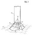
- FIG. 1illustrates specific definitions related to image capture.
- An imaging devicein this case an endoscope 10 , is pointed towards a surface to be viewed 12 in a viewing direction 14 .
- a principal light ray 16travels from a point 18 on the surface 12 , to the endoscope 10 , through the endoscope optics (not shown), and finally to an image sensor (not shown).
- any principal ray leaving a point on the surface 12travels approximately towards a viewing point 20 .
- the viewing point 20is the point from which the view can be thought to have been obtained.
- a plane 22can be thought to exist between the surface 12 and the endoscope 10 , orthogonal to the viewing direction 14 .
- the endoscopehas a field of view 26 .
- the field of view 26is shown as rectangular, although it could have any shape. Portions of the surface 12 that lie within the field of view 26 make up a visible area 28 .
- the viewing orientationis the rotational orientation of the view about the viewing direction 14 .
- the viewing setis defined herein as the combination of the viewing point, the viewing direction, and the viewing orientation. The view is what is seen from the viewing set.
- a viewing situationinvolves a three-dimensional surface.
- the captured imageis only a two-dimensional entity in an image plane that is generally orthogonal to the viewing direction of the endoscope.
- the image generated at the image planeis typically displayed to the user as would be seen looking along the viewing direction of the endoscope from the endoscopic viewing point.
- stereo-viewing endoscopescapture two slightly offset images which are used to provide the user with a three-dimensional image. However, they still only provide a view from the viewing point and in the viewing direction of the endoscope. Because the endoscope, the user, and the internal structure being examined exist in an actual three-dimensional world, tying the user to the viewing set of the endoscope limits the way information about the internal structure can be conveyed.
- the userwill often desire to change the viewing set.
- the only option for the useris to move the endoscope. This is not always convenient or even possible.
- An alternative viewing setcould provide a better perspective of the physical surface and give the user a better sense of the relative locations of viewed features. For example, it would be advantageous to be able to use a viewing set which is aligned with the user's physical position instead of the physical position of the endoscope.
- Other viewing setsmight also be desired, as determined by the preferences of the user.
- the primary object of the present inventionis to provide a versatile method of displaying endoscopic images that includes three-dimensional surfaces, variable viewing points, directions, and orientations. It is a further object of this invention to have this method applicable to all endoscopes regardless of type, viewing direction, or image sensor format.
- a method for displaying an endoscopic imagecomprises receiving an endoscopic image of a viewed surface, providing a virtual surface with said endoscopic image mapped onto said virtual surface, rendering a rendered image of said virtual surface, and providing said rendered image to a user.
- FIG. 1shows an endoscope capturing an image
- FIGS. 2A and 2Bshow the method of the present invention.
- FIGS. 3A and 3Bshow a relationship between different views.
- FIGS. 4A , 4 B, 4 C, and 4 Dshow presentations of an endoscopic image of a surface.
- FIG. 5illustrates the process of mapping an endoscopic image onto a virtual surface.
- FIGS. 6A and 6Bshow presentations of an endoscopic image of a surface with a hidden region.
- FIG. 7shows the output of a display system according to the present invention.
- the preferred embodiment of the inventionis a software program running on a computer.
- the computercommunicates electronically with an endoscope and a display device such as a monitor.
- the computerincludes a graphics processing unit such as those manufactured by NVidia Corporation.
- the graphics processing unitis specifically designed to quickly perform the types of graphics related calculations required by the present invention.
- Other devicesmay be connected to the computer as appropriate for a given application.
- the programuses the graphical display programming library OpenGL.
- OpenGLgraphical display programming library
- This libraryoffers a powerful set of programming tools optimized for displaying textured shapes in three dimensions. It allows a collection of virtual shapes and a virtual viewing set to be defined as data within the computer memory. The collection of virtual shapes is then rendered based on the viewing parameters and displayed on a monitor.
- An alternative library, such as DirectX,could be used without departing from the scope of this invention
- FIG. 2Ashows the method of the present invention for displaying an endoscopic image.
- the computerreceives inputs to determine configuration parameters which are stored in memory.
- the computerreceives a captured endoscopic image.
- a virtual surfaceis defined in the computer with the image textured onto the surface.
- a virtual viewing point, virtual viewing direction, and virtual viewing orientationare defined relative to the virtual surface.
- a rendered imageis then created using a perspective projection of the virtual surface as seen with the defined virtual viewing set. Alternatively, a parallel projection rendering could be used.
- the rendered imageis then displayed to the user.
- Videois displayed on the virtual surface by updating the image texture with each new frame.
- FIG. 2Bconceptually illustrates the method of present invention. Given a real viewing set and a real surface, a captured image is provided by an endoscope. The captured image is mapped onto a virtual surface. The virtual surface and a virtual viewing set are then used to create a rendered image.
- FIGS. 3A and 3Bprovide an example of the spatial relationship between different views.
- an endoscope 10is positioned within a cavity 30 and captures an image of a surface 12 .
- the endoscope 10has a viewing point 20 and a viewing direction 14 .
- a userhas a viewing point 32 and a viewing direction 34 but is unable to see the surface 12 .
- the virtual counterparts to this situation, as defined in the computer,are shown in FIG. 3B .
- the endoscopic viewing point 20has a corresponding virtual endoscopic viewing point 36 ;
- the endoscopic viewing direction 14has a corresponding virtual endoscopic viewing direction 38 ;
- the user viewing point 32has a corresponding virtual user viewing point 40 ;
- the user viewing direction 34has a corresponding virtual user viewing direction 42 .
- the captured endoscopic imageis mapped onto a virtual surface 44 which approximates the real surface 12 .
- a new viewis rendered and displayed. This rendered view is approximately what could be seen by the user if the surface were not occluded by the surrounding anatomy.
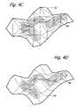
- FIGS. 4A , 4 B, 4 C, and 4 Dshow different presentations of an endoscopic image.
- FIG. 4Ashows a captured endoscopic image 46 featuring a darker region 48 . This is the standard presentation for an endoscopic image.
- FIG. 4Bshows this same image 46 mapped onto a planar virtual surface 50 and seen from a different viewing point than that from which the image 46 was captured. Because of the alternative viewing point, the darker region 48 now appears to have been stretched and compressed in certain directions.
- a planeis the simplest surface available and is only a crude approximation of the real surface. However, a plane is a good choice when computing resources are limited. For a better approximation, polygons can be used to construct a three-dimensional virtual surface.
- FIG. 4Cshows the endoscopic image 46 mapped onto a triangular mesh 52 .
- the darker region 48is stretched in accordance with the topography, giving the user a better sense of the actual geometry.
- An advanced systemcould provide a refined surface approximation, shown in FIG. 4D .
- Such an approximationcould either be a high resolution volumetric data set or a mathematical function. In this case the darker region 48 appears smoother and more natural.
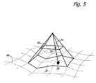
- FIG. 5shows conceptually how a captured image is mapped onto a virtual surface.
- This mapping processcan be thought of as the inverse of the image capturing process.
- the virtual surface 44is an approximation of the actual surface 14 shown in FIG. 1 .
- a point 24 on the image 46 and a corresponding point 58 on the virtual surface 44are matched along a projection line 56 .
- the projection line 56is analogous to the light ray 10 .
- the projection lines 56connect corners of the image to the corresponding corners of the virtual surface.
- Intermediate projection lines 56can be used to increase texturing accuracy on curved virtual surfaces.
- Some situationsmay require image distortion to be corrected by arranging the distortion lines 56 according to the inverse mapping of the distortion. Other specialized mappings may also be used without departing from the scope of the invention.
- the image 46is textured onto the virtual surface 44 by fixing points on the image 46 to the corresponding points on the surface 44 .
- FIG. 6Ashows an endoscopic image 46 of an imaged surface. It has a darker region 48 which appears broken. This discontinuity, along with an apparent contour line 58 , suggests that the imaged surface may have hidden regions.
- FIG. 6Bshows a virtual surface 44 with a folded region 60 that approximates the imaged surface. The folded region 60 is not visible from the endoscopic viewing point and therefore does not have any image data. Such regions may be indicated to the user by a solid color. Thus, even though there is no image information about these regions, the user can get a sense of surface geometry and how it relates to the image.
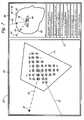
- FIG. 7shows how the viewing information is displayed and how the user interacts with the virtual world.
- a large section 64 of a display screen 62presents a view of a virtual surface 44 as seen from a selected viewing point.
- a smaller section 66 of the screen 62gives an overview of the relative locations of the virtual endoscopic viewing point 36 , a user selected viewing point 40 , and the surface 44 .
- An anatomical model 68 based imaging data or user inputmay also be displayed for clarity.
- a tool line 70which represents the line of action of an endoscopic surgical tool such as a cutter.
- the lower right corner of the display screen 62presents a set of menu buttons 72 which allow the user to interact with the virtual world and also execute basic display and image processing/enhancement functions.
- the configuration parameters required by the computermay be obtained in a variety of ways.
- the virtual surfacecan be constructed from scan data obtained through techniques such as MRI.
- the virtual surfacecould be selected from a collection of standard surfaces.
- the virtual viewing point, viewing direction, and viewing orientationmay be specified using any standard data input technique. Specialized pointers for specifying the viewing set can also be used.
- the relationship between the endoscopic viewing set and the actual viewing surfacecan be input by the user or obtained from stereotactic systems or other sensors.
- the present inventionprovides a new method for viewing endoscopic images which affords the user an enhanced, versatile, and more realistic representation of structures viewed by an endoscope.
Landscapes
- Health & Medical Sciences (AREA)
- Life Sciences & Earth Sciences (AREA)
- Engineering & Computer Science (AREA)
- Surgery (AREA)
- Physics & Mathematics (AREA)
- Biophysics (AREA)
- Veterinary Medicine (AREA)
- Optics & Photonics (AREA)
- Pathology (AREA)
- Radiology & Medical Imaging (AREA)
- Nuclear Medicine, Radiotherapy & Molecular Imaging (AREA)
- Biomedical Technology (AREA)
- Heart & Thoracic Surgery (AREA)
- Medical Informatics (AREA)
- Molecular Biology (AREA)
- Animal Behavior & Ethology (AREA)
- General Health & Medical Sciences (AREA)
- Public Health (AREA)
- Signal Processing (AREA)
- General Physics & Mathematics (AREA)
- Theoretical Computer Science (AREA)
- Endoscopes (AREA)
Abstract
Description
Claims (20)
Priority Applications (1)
| Application Number | Priority Date | Filing Date | Title |
|---|---|---|---|
| US10/718,434US7232409B2 (en) | 2003-11-20 | 2003-11-20 | Method and apparatus for displaying endoscopic images |
Applications Claiming Priority (1)
| Application Number | Priority Date | Filing Date | Title |
|---|---|---|---|
| US10/718,434US7232409B2 (en) | 2003-11-20 | 2003-11-20 | Method and apparatus for displaying endoscopic images |
Publications (2)
| Publication Number | Publication Date |
|---|---|
| US20050113643A1 US20050113643A1 (en) | 2005-05-26 |
| US7232409B2true US7232409B2 (en) | 2007-06-19 |
Family
ID=34591099
Family Applications (1)
| Application Number | Title | Priority Date | Filing Date |
|---|---|---|---|
| US10/718,434Expired - LifetimeUS7232409B2 (en) | 2003-11-20 | 2003-11-20 | Method and apparatus for displaying endoscopic images |
Country Status (1)
| Country | Link |
|---|---|
| US (1) | US7232409B2 (en) |
Cited By (50)
| Publication number | Priority date | Publication date | Assignee | Title |
|---|---|---|---|---|
| US20070060792A1 (en)* | 2004-02-11 | 2007-03-15 | Wolfgang Draxinger | Method and apparatus for generating at least one section of a virtual 3D model of a body interior |
| US20080009674A1 (en)* | 2006-02-24 | 2008-01-10 | Visionsense Ltd. | Method and system for navigating within a flexible organ of the body of a patient |
| US7517320B2 (en) | 2006-06-30 | 2009-04-14 | Broncus Technologies, Inc. | Airway bypass site selection and treatment planning |
| US7609910B2 (en)* | 2004-04-09 | 2009-10-27 | Siemens Medical Solutions Usa, Inc. | System and method for creating a panoramic view of a volumetric image |
| CN103315696A (en)* | 2012-03-21 | 2013-09-25 | 柯惠Lp公司 | System and method for determining camera angles by using virtual planes derived from actual images |
| US8608724B2 (en) | 2004-07-19 | 2013-12-17 | Broncus Medical Inc. | Devices for delivering substances through an extra-anatomic opening created in an airway |
| US8709034B2 (en) | 2011-05-13 | 2014-04-29 | Broncus Medical Inc. | Methods and devices for diagnosing, monitoring, or treating medical conditions through an opening through an airway wall |
| US9101266B2 (en) | 2011-02-07 | 2015-08-11 | Endochoice Innovation Center Ltd. | Multi-element cover for a multi-camera endoscope |
| US9101268B2 (en) | 2009-06-18 | 2015-08-11 | Endochoice Innovation Center Ltd. | Multi-camera endoscope |
| US9101287B2 (en) | 2011-03-07 | 2015-08-11 | Endochoice Innovation Center Ltd. | Multi camera endoscope assembly having multiple working channels |
| US9161679B2 (en) | 2009-08-18 | 2015-10-20 | Olaf Christiansen | Image processing system having an additional piece of scale information to be processed together with the image information |
| US20150313445A1 (en)* | 2014-05-01 | 2015-11-05 | Endochoice, Inc. | System and Method of Scanning a Body Cavity Using a Multiple Viewing Elements Endoscope |
| US9314147B2 (en) | 2011-12-13 | 2016-04-19 | Endochoice Innovation Center Ltd. | Rotatable connector for an endoscope |
| US9320419B2 (en) | 2010-12-09 | 2016-04-26 | Endochoice Innovation Center Ltd. | Fluid channeling component of a multi-camera endoscope |
| US9345532B2 (en) | 2011-05-13 | 2016-05-24 | Broncus Medical Inc. | Methods and devices for ablation of tissue |
| US9402533B2 (en) | 2011-03-07 | 2016-08-02 | Endochoice Innovation Center Ltd. | Endoscope circuit board assembly |
| US9492063B2 (en) | 2009-06-18 | 2016-11-15 | Endochoice Innovation Center Ltd. | Multi-viewing element endoscope |
| US9498231B2 (en) | 2011-06-27 | 2016-11-22 | Board Of Regents Of The University Of Nebraska | On-board tool tracking system and methods of computer assisted surgery |
| US9533128B2 (en) | 2003-07-18 | 2017-01-03 | Broncus Medical Inc. | Devices for maintaining patency of surgically created channels in tissue |
| US9554692B2 (en) | 2009-06-18 | 2017-01-31 | EndoChoice Innovation Ctr. Ltd. | Multi-camera endoscope |
| US9560954B2 (en) | 2012-07-24 | 2017-02-07 | Endochoice, Inc. | Connector for use with endoscope |
| US9560953B2 (en) | 2010-09-20 | 2017-02-07 | Endochoice, Inc. | Operational interface in a multi-viewing element endoscope |
| US9642513B2 (en) | 2009-06-18 | 2017-05-09 | Endochoice Inc. | Compact multi-viewing element endoscope system |
| US9655502B2 (en) | 2011-12-13 | 2017-05-23 | EndoChoice Innovation Center, Ltd. | Removable tip endoscope |
| US9706903B2 (en) | 2009-06-18 | 2017-07-18 | Endochoice, Inc. | Multiple viewing elements endoscope system with modular imaging units |
| US9713415B2 (en) | 2011-03-07 | 2017-07-25 | Endochoice Innovation Center Ltd. | Multi camera endoscope having a side service channel |
| US9713417B2 (en) | 2009-06-18 | 2017-07-25 | Endochoice, Inc. | Image capture assembly for use in a multi-viewing elements endoscope |
| US9814374B2 (en) | 2010-12-09 | 2017-11-14 | Endochoice Innovation Center Ltd. | Flexible electronic circuit board for a multi-camera endoscope |
| US9872609B2 (en) | 2009-06-18 | 2018-01-23 | Endochoice Innovation Center Ltd. | Multi-camera endoscope |
| US9901244B2 (en) | 2009-06-18 | 2018-02-27 | Endochoice, Inc. | Circuit board assembly of a multiple viewing elements endoscope |
| US9986899B2 (en) | 2013-03-28 | 2018-06-05 | Endochoice, Inc. | Manifold for a multiple viewing elements endoscope |
| US9993142B2 (en) | 2013-03-28 | 2018-06-12 | Endochoice, Inc. | Fluid distribution device for a multiple viewing elements endoscope |
| US10080486B2 (en) | 2010-09-20 | 2018-09-25 | Endochoice Innovation Center Ltd. | Multi-camera endoscope having fluid channels |
| US10105149B2 (en) | 2013-03-15 | 2018-10-23 | Board Of Regents Of The University Of Nebraska | On-board tool tracking system and methods of computer assisted surgery |
| US10165929B2 (en) | 2009-06-18 | 2019-01-01 | Endochoice, Inc. | Compact multi-viewing element endoscope system |
| US10203493B2 (en) | 2010-10-28 | 2019-02-12 | Endochoice Innovation Center Ltd. | Optical systems for multi-sensor endoscopes |
| US10219811B2 (en) | 2011-06-27 | 2019-03-05 | Board Of Regents Of The University Of Nebraska | On-board tool tracking system and methods of computer assisted surgery |
| US10272260B2 (en) | 2011-11-23 | 2019-04-30 | Broncus Medical Inc. | Methods and devices for diagnosing, monitoring, or treating medical conditions through an opening through an airway wall |
| US10499794B2 (en) | 2013-05-09 | 2019-12-10 | Endochoice, Inc. | Operational interface in a multi-viewing element endoscope |
| US11116574B2 (en) | 2006-06-16 | 2021-09-14 | Board Of Regents Of The University Of Nebraska | Method and apparatus for computer aided surgery |
| WO2022011282A1 (en)* | 2020-07-10 | 2022-01-13 | Arthrex, Inc. | Endoscope insertion and removal detection system |
| US11278190B2 (en) | 2009-06-18 | 2022-03-22 | Endochoice, Inc. | Multi-viewing element endoscope |
| US11547275B2 (en) | 2009-06-18 | 2023-01-10 | Endochoice, Inc. | Compact multi-viewing element endoscope system |
| US20230196683A1 (en)* | 2010-06-07 | 2023-06-22 | Pfaqutruma Research Llc | Creation and use of virtual places |
| US11864734B2 (en) | 2009-06-18 | 2024-01-09 | Endochoice, Inc. | Multi-camera endoscope |
| US11889986B2 (en) | 2010-12-09 | 2024-02-06 | Endochoice, Inc. | Flexible electronic circuit board for a multi-camera endoscope |
| US11911117B2 (en) | 2011-06-27 | 2024-02-27 | Board Of Regents Of The University Of Nebraska | On-board tool tracking system and methods of computer assisted surgery |
| US12137873B2 (en) | 2009-06-18 | 2024-11-12 | Endochoice, Inc. | Compact multi-viewing element endoscope system |
| US12204087B2 (en) | 2010-10-28 | 2025-01-21 | Endochoice, Inc. | Optical systems for multi-sensor endoscopes |
| US12220105B2 (en) | 2010-06-16 | 2025-02-11 | Endochoice, Inc. | Circuit board assembly of a multiple viewing elements endoscope |
Families Citing this family (17)
| Publication number | Priority date | Publication date | Assignee | Title |
|---|---|---|---|---|
| US9033871B2 (en) | 2004-04-07 | 2015-05-19 | Karl Storz Imaging, Inc. | Gravity referenced endoscopic image orientation |
| US20080015569A1 (en) | 2005-02-02 | 2008-01-17 | Voyage Medical, Inc. | Methods and apparatus for treatment of atrial fibrillation |
| US7967742B2 (en)* | 2005-02-14 | 2011-06-28 | Karl Storz Imaging, Inc. | Method for using variable direction of view endoscopy in conjunction with image guided surgical systems |
| US9055906B2 (en)* | 2006-06-14 | 2015-06-16 | Intuitive Surgical Operations, Inc. | In-vivo visualization systems |
| US8270694B2 (en)* | 2008-04-23 | 2012-09-18 | Aditya Koolwal | Systems, methods and devices for correlating reference locations using image data |
| US8814782B2 (en)* | 2008-07-08 | 2014-08-26 | Karl Storz Imaging, Inc. | Solid state variable direction of view endoscope |
| US8758234B2 (en) | 2008-07-08 | 2014-06-24 | Karl Storz Imaging, Inc. | Solid state variable direction of view endoscope |
| US8771177B2 (en) | 2008-07-08 | 2014-07-08 | Karl Storz Imaging, Inc. | Wide angle flexible endoscope |
| US10092169B2 (en) | 2008-07-08 | 2018-10-09 | Karl Storz Imaging, Inc. | Solid state variable direction of view endoscope |
| US10004387B2 (en)* | 2009-03-26 | 2018-06-26 | Intuitive Surgical Operations, Inc. | Method and system for assisting an operator in endoscopic navigation |
| US8337397B2 (en) | 2009-03-26 | 2012-12-25 | Intuitive Surgical Operations, Inc. | Method and system for providing visual guidance to an operator for steering a tip of an endoscopic device toward one or more landmarks in a patient |
| KR101903307B1 (en)* | 2009-03-26 | 2018-10-01 | 인튜어티브 서지컬 오퍼레이션즈 인코포레이티드 | System for providing visual guidance for steering a tip of an endoscopic device towards one or more landmarks and assisting an operator in endoscopic navigation |
| JP5551957B2 (en)* | 2010-03-31 | 2014-07-16 | 富士フイルム株式会社 | Projection image generation apparatus, operation method thereof, and projection image generation program |
| JP5944395B2 (en) | 2010-10-08 | 2016-07-05 | コーニンクレッカ フィリップス エヌ ヴェKoninklijke Philips N.V. | Flexible tether with integrated sensor for dynamic instrument tracking |
| US9763563B2 (en) | 2012-07-11 | 2017-09-19 | Karl Storz Imaging, Inc. | Endoscopic camera single-button mode activation |
| WO2015049962A1 (en)* | 2013-10-02 | 2015-04-09 | オリンパスメディカルシステムズ株式会社 | Endoscope system |
| JP6886968B2 (en)* | 2015-10-09 | 2021-06-16 | コヴィディエン リミテッド パートナーシップ | How to use an angled endoscope to visualize body cavities using a robotic surgical system |
Citations (46)
| Publication number | Priority date | Publication date | Assignee | Title |
|---|---|---|---|---|
| US3572325A (en) | 1968-10-25 | 1971-03-23 | Us Health Education & Welfare | Flexible endoscope having fluid conduits and control |
| US3880148A (en) | 1972-09-25 | 1975-04-29 | Olympus Optical Co | Endoscope |
| US4697577A (en) | 1986-05-22 | 1987-10-06 | Baxter Travenol Laboratories, Inc. | Scanning microtelescope for surgical applications |
| US5230623A (en) | 1991-12-10 | 1993-07-27 | Radionics, Inc. | Operating pointer with interactive computergraphics |
| US5307804A (en) | 1991-02-21 | 1994-05-03 | Richard Wolf Gmbh | Endoscope having a camera coupled thereto |
| US5313306A (en) | 1991-05-13 | 1994-05-17 | Telerobotics International, Inc. | Omniview motionless camera endoscopy system |
| JPH06269403A (en) | 1993-03-19 | 1994-09-27 | Olympus Optical Co Ltd | Electronic endoscope apparatus |
| WO1995001749A1 (en) | 1993-07-09 | 1995-01-19 | Saturnus A.G. | Tv camera with rotational orientation correction |
| US5515160A (en)* | 1992-03-12 | 1996-05-07 | Aesculap Ag | Method and apparatus for representing a work area in a three-dimensional structure |
| US5531227A (en) | 1994-01-28 | 1996-07-02 | Schneider Medical Technologies, Inc. | Imaging device and method |
| US5617857A (en) | 1995-06-06 | 1997-04-08 | Image Guided Technologies, Inc. | Imaging system having interactive medical instruments and methods |
| US5623560A (en) | 1992-11-27 | 1997-04-22 | Fuji Photo Film Co., Ltd. | Method for adjusting positions of radiation images |
| US5638819A (en)* | 1995-08-29 | 1997-06-17 | Manwaring; Kim H. | Method and apparatus for guiding an instrument to a target |
| US5661519A (en) | 1992-08-14 | 1997-08-26 | Siemens Aktiengesellschaft | Video camera fashioned as a handpiece for observing subjects in mouth of a patient |
| US5677763A (en) | 1996-08-08 | 1997-10-14 | Technology Resources, Inc. | Optical device for measuring physical and optical characteristics of an object |
| US5704897A (en)* | 1992-07-31 | 1998-01-06 | Truppe; Michael J. | Apparatus and method for registration of points of a data field with respective points of an optical image |
| US5776050A (en)* | 1995-07-24 | 1998-07-07 | Medical Media Systems | Anatomical visualization system |
| US5920395A (en) | 1993-04-22 | 1999-07-06 | Image Guided Technologies, Inc. | System for locating relative positions of objects in three dimensional space |
| US5954634A (en) | 1997-04-11 | 1999-09-21 | Olympus Optical Co. Ltd. | Field conversion system for rigid endoscopes |
| US5976076A (en) | 1995-02-22 | 1999-11-02 | Kolff; Jack | Stereo laparoscope with synchronized optics |
| US5995108A (en)* | 1995-06-19 | 1999-11-30 | Hitachi Medical Corporation | 3D image composition/display apparatus and composition method based on front-to-back order of plural 2D projected images |
| US6007484A (en) | 1995-09-15 | 1999-12-28 | Image Technologies Corporation | Endoscope having elevation and azimuth control of camera |
| US6097423A (en) | 1997-06-06 | 2000-08-01 | Karl Storz Imaging, Inc. | Image orientation for endoscopic video displays |
| US6135946A (en)* | 1997-06-23 | 2000-10-24 | U.S. Philips Corporation | Method and system for image-guided interventional endoscopic procedures |
| US6139499A (en)* | 1999-02-22 | 2000-10-31 | Wilk; Peter J. | Ultrasonic medical system and associated method |
| US6167296A (en) | 1996-06-28 | 2000-12-26 | The Board Of Trustees Of The Leland Stanford Junior University | Method for volumetric image navigation |
| WO2001022865A1 (en) | 1999-09-28 | 2001-04-05 | Keymed (Medical & Industrial Equipment) Ltd. | Endoscope with variable direction of view |
| US6283918B1 (en)* | 1997-09-30 | 2001-09-04 | Kabushiki Kaisha Toshiba | Medical image diagnostic apparatus |
| US6371909B1 (en) | 1998-02-19 | 2002-04-16 | California Institute Of Technology | Apparatus and method for providing spherical viewing during endoscopic procedures |
| US20020045855A1 (en) | 1997-02-10 | 2002-04-18 | Essex Technology, Inc. | Rotate to advance catheterization system |
| US20020099263A1 (en) | 2001-01-19 | 2002-07-25 | Hale Eric L. | Apparatus and method for controlling endoscopic instruments |
| US6442417B1 (en)* | 1999-11-29 | 2002-08-27 | The Board Of Trustees Of The Leland Stanford Junior University | Method and apparatus for transforming view orientations in image-guided surgery |
| US6443894B1 (en)* | 1999-09-29 | 2002-09-03 | Acuson Corporation | Medical diagnostic ultrasound system and method for mapping surface data for three dimensional imaging |
| US6464631B1 (en) | 1999-11-17 | 2002-10-15 | Olympus Winter & Ibe Gmbh | Endoscope with a distal video camera and a camera rotating device |
| US6471637B1 (en) | 1999-09-24 | 2002-10-29 | Karl Storz Imaging, Inc. | Image orientation for endoscopic video displays |
| US20020161280A1 (en) | 1999-09-24 | 2002-10-31 | David Chatenever | Image orientation for endoscopic video displays |
| US6500115B2 (en) | 1998-08-28 | 2002-12-31 | Storz Endoskop Gmbh | Endoscope |
| US6505065B1 (en)* | 1999-10-29 | 2003-01-07 | Koninklijke Philips Electronics, N.V. | Methods and apparatus for planning and executing minimally invasive procedures for in-vivo placement of objects |
| US20030016883A1 (en) | 2001-07-20 | 2003-01-23 | Baron John M. | System and method for horizon correction within images |
| US6648817B2 (en) | 2001-11-15 | 2003-11-18 | Endactive, Inc. | Apparatus and method for stereo viewing in variable direction-of-view endoscopy |
| US6663559B2 (en) | 2001-12-14 | 2003-12-16 | Endactive, Inc. | Interface for a variable direction of view endoscope |
| US20040210105A1 (en) | 2003-04-21 | 2004-10-21 | Hale Eric Lawrence | Method for capturing and displaying endoscopic maps |
| US20050015005A1 (en)* | 2003-04-28 | 2005-01-20 | Kockro Ralf Alfons | Computer enhanced surgical navigation imaging system (camera probe) |
| US20050054895A1 (en) | 2003-09-09 | 2005-03-10 | Hoeg Hans David | Method for using variable direction of view endoscopy in conjunction with image guided surgical systems |
| US20050085718A1 (en)* | 2003-10-21 | 2005-04-21 | Ramin Shahidi | Systems and methods for intraoperative targetting |
| US20050228250A1 (en)* | 2001-11-21 | 2005-10-13 | Ingmar Bitter | System and method for visualization and navigation of three-dimensional medical images |
- 2003
- 2003-11-20USUS10/718,434patent/US7232409B2/ennot_activeExpired - Lifetime
Patent Citations (51)
| Publication number | Priority date | Publication date | Assignee | Title |
|---|---|---|---|---|
| US3572325A (en) | 1968-10-25 | 1971-03-23 | Us Health Education & Welfare | Flexible endoscope having fluid conduits and control |
| US3880148A (en) | 1972-09-25 | 1975-04-29 | Olympus Optical Co | Endoscope |
| US4697577A (en) | 1986-05-22 | 1987-10-06 | Baxter Travenol Laboratories, Inc. | Scanning microtelescope for surgical applications |
| US5307804A (en) | 1991-02-21 | 1994-05-03 | Richard Wolf Gmbh | Endoscope having a camera coupled thereto |
| US5313306A (en) | 1991-05-13 | 1994-05-17 | Telerobotics International, Inc. | Omniview motionless camera endoscopy system |
| US5230623A (en) | 1991-12-10 | 1993-07-27 | Radionics, Inc. | Operating pointer with interactive computergraphics |
| US5515160A (en)* | 1992-03-12 | 1996-05-07 | Aesculap Ag | Method and apparatus for representing a work area in a three-dimensional structure |
| US5704897A (en)* | 1992-07-31 | 1998-01-06 | Truppe; Michael J. | Apparatus and method for registration of points of a data field with respective points of an optical image |
| US5661519A (en) | 1992-08-14 | 1997-08-26 | Siemens Aktiengesellschaft | Video camera fashioned as a handpiece for observing subjects in mouth of a patient |
| US5623560A (en) | 1992-11-27 | 1997-04-22 | Fuji Photo Film Co., Ltd. | Method for adjusting positions of radiation images |
| JPH06269403A (en) | 1993-03-19 | 1994-09-27 | Olympus Optical Co Ltd | Electronic endoscope apparatus |
| US5920395A (en) | 1993-04-22 | 1999-07-06 | Image Guided Technologies, Inc. | System for locating relative positions of objects in three dimensional space |
| US5899851A (en) | 1993-07-09 | 1999-05-04 | Saturnus A.G. | TV camera with rotational orientation correction |
| WO1995001749A1 (en) | 1993-07-09 | 1995-01-19 | Saturnus A.G. | Tv camera with rotational orientation correction |
| US5531227A (en) | 1994-01-28 | 1996-07-02 | Schneider Medical Technologies, Inc. | Imaging device and method |
| US5976076A (en) | 1995-02-22 | 1999-11-02 | Kolff; Jack | Stereo laparoscope with synchronized optics |
| US5617857A (en) | 1995-06-06 | 1997-04-08 | Image Guided Technologies, Inc. | Imaging system having interactive medical instruments and methods |
| US5995108A (en)* | 1995-06-19 | 1999-11-30 | Hitachi Medical Corporation | 3D image composition/display apparatus and composition method based on front-to-back order of plural 2D projected images |
| US5776050A (en)* | 1995-07-24 | 1998-07-07 | Medical Media Systems | Anatomical visualization system |
| US5638819A (en)* | 1995-08-29 | 1997-06-17 | Manwaring; Kim H. | Method and apparatus for guiding an instrument to a target |
| US6007484A (en) | 1995-09-15 | 1999-12-28 | Image Technologies Corporation | Endoscope having elevation and azimuth control of camera |
| US6167296A (en) | 1996-06-28 | 2000-12-26 | The Board Of Trustees Of The Leland Stanford Junior University | Method for volumetric image navigation |
| US5677763A (en) | 1996-08-08 | 1997-10-14 | Technology Resources, Inc. | Optical device for measuring physical and optical characteristics of an object |
| US20020045855A1 (en) | 1997-02-10 | 2002-04-18 | Essex Technology, Inc. | Rotate to advance catheterization system |
| US5954634A (en) | 1997-04-11 | 1999-09-21 | Olympus Optical Co. Ltd. | Field conversion system for rigid endoscopes |
| US6097423A (en) | 1997-06-06 | 2000-08-01 | Karl Storz Imaging, Inc. | Image orientation for endoscopic video displays |
| US6135946A (en)* | 1997-06-23 | 2000-10-24 | U.S. Philips Corporation | Method and system for image-guided interventional endoscopic procedures |
| US6283918B1 (en)* | 1997-09-30 | 2001-09-04 | Kabushiki Kaisha Toshiba | Medical image diagnostic apparatus |
| US6371909B1 (en) | 1998-02-19 | 2002-04-16 | California Institute Of Technology | Apparatus and method for providing spherical viewing during endoscopic procedures |
| US6500115B2 (en) | 1998-08-28 | 2002-12-31 | Storz Endoskop Gmbh | Endoscope |
| US6139499A (en)* | 1999-02-22 | 2000-10-31 | Wilk; Peter J. | Ultrasonic medical system and associated method |
| US20020161280A1 (en) | 1999-09-24 | 2002-10-31 | David Chatenever | Image orientation for endoscopic video displays |
| US6471637B1 (en) | 1999-09-24 | 2002-10-29 | Karl Storz Imaging, Inc. | Image orientation for endoscopic video displays |
| US20050027167A1 (en) | 1999-09-24 | 2005-02-03 | David Chatenever | Image orientation for endoscopic video displays |
| US20050020883A1 (en) | 1999-09-24 | 2005-01-27 | David Chatenever | Image orientation for endoscopic video displays |
| WO2001022865A1 (en) | 1999-09-28 | 2001-04-05 | Keymed (Medical & Industrial Equipment) Ltd. | Endoscope with variable direction of view |
| US6443894B1 (en)* | 1999-09-29 | 2002-09-03 | Acuson Corporation | Medical diagnostic ultrasound system and method for mapping surface data for three dimensional imaging |
| US6505065B1 (en)* | 1999-10-29 | 2003-01-07 | Koninklijke Philips Electronics, N.V. | Methods and apparatus for planning and executing minimally invasive procedures for in-vivo placement of objects |
| US6464631B1 (en) | 1999-11-17 | 2002-10-15 | Olympus Winter & Ibe Gmbh | Endoscope with a distal video camera and a camera rotating device |
| US6442417B1 (en)* | 1999-11-29 | 2002-08-27 | The Board Of Trustees Of The Leland Stanford Junior University | Method and apparatus for transforming view orientations in image-guided surgery |
| US6695774B2 (en) | 2001-01-19 | 2004-02-24 | Endactive, Inc. | Apparatus and method for controlling endoscopic instruments |
| US20020099263A1 (en) | 2001-01-19 | 2002-07-25 | Hale Eric L. | Apparatus and method for controlling endoscopic instruments |
| US20030016883A1 (en) | 2001-07-20 | 2003-01-23 | Baron John M. | System and method for horizon correction within images |
| US6648817B2 (en) | 2001-11-15 | 2003-11-18 | Endactive, Inc. | Apparatus and method for stereo viewing in variable direction-of-view endoscopy |
| US20050228250A1 (en)* | 2001-11-21 | 2005-10-13 | Ingmar Bitter | System and method for visualization and navigation of three-dimensional medical images |
| US20040127769A1 (en) | 2001-12-14 | 2004-07-01 | Hale Eric L. | Interface for a variable direction-of-view endoscope |
| US6663559B2 (en) | 2001-12-14 | 2003-12-16 | Endactive, Inc. | Interface for a variable direction of view endoscope |
| US20040210105A1 (en) | 2003-04-21 | 2004-10-21 | Hale Eric Lawrence | Method for capturing and displaying endoscopic maps |
| US20050015005A1 (en)* | 2003-04-28 | 2005-01-20 | Kockro Ralf Alfons | Computer enhanced surgical navigation imaging system (camera probe) |
| US20050054895A1 (en) | 2003-09-09 | 2005-03-10 | Hoeg Hans David | Method for using variable direction of view endoscopy in conjunction with image guided surgical systems |
| US20050085718A1 (en)* | 2003-10-21 | 2005-04-21 | Ramin Shahidi | Systems and methods for intraoperative targetting |
Cited By (103)
| Publication number | Priority date | Publication date | Assignee | Title |
|---|---|---|---|---|
| US9533128B2 (en) | 2003-07-18 | 2017-01-03 | Broncus Medical Inc. | Devices for maintaining patency of surgically created channels in tissue |
| US20070060792A1 (en)* | 2004-02-11 | 2007-03-15 | Wolfgang Draxinger | Method and apparatus for generating at least one section of a virtual 3D model of a body interior |
| US7794388B2 (en)* | 2004-02-11 | 2010-09-14 | Karl Storz Gmbh & Co. Kg | Method and apparatus for generating at least one section of a virtual 3D model of a body interior |
| US7609910B2 (en)* | 2004-04-09 | 2009-10-27 | Siemens Medical Solutions Usa, Inc. | System and method for creating a panoramic view of a volumetric image |
| US8784400B2 (en) | 2004-07-19 | 2014-07-22 | Broncus Medical Inc. | Devices for delivering substances through an extra-anatomic opening created in an airway |
| US11357960B2 (en) | 2004-07-19 | 2022-06-14 | Broncus Medical Inc. | Devices for delivering substances through an extra-anatomic opening created in an airway |
| US10369339B2 (en) | 2004-07-19 | 2019-08-06 | Broncus Medical Inc. | Devices for delivering substances through an extra-anatomic opening created in an airway |
| US8608724B2 (en) | 2004-07-19 | 2013-12-17 | Broncus Medical Inc. | Devices for delivering substances through an extra-anatomic opening created in an airway |
| US20080009674A1 (en)* | 2006-02-24 | 2008-01-10 | Visionsense Ltd. | Method and system for navigating within a flexible organ of the body of a patient |
| US7935048B2 (en)* | 2006-02-24 | 2011-05-03 | Visionsense Ltd. | Method and system for navigating within a flexible organ of the body of a patient |
| US11857265B2 (en) | 2006-06-16 | 2024-01-02 | Board Of Regents Of The University Of Nebraska | Method and apparatus for computer aided surgery |
| US11116574B2 (en) | 2006-06-16 | 2021-09-14 | Board Of Regents Of The University Of Nebraska | Method and apparatus for computer aided surgery |
| US7985187B2 (en) | 2006-06-30 | 2011-07-26 | Broncus Technologies, Inc. | Airway bypass site selection and treatment planning |
| US8668652B2 (en) | 2006-06-30 | 2014-03-11 | Broncus Medical Inc. | Airway bypass site selection and treatment planning |
| US20100305463A1 (en)* | 2006-06-30 | 2010-12-02 | Macklem Peter J | Airway bypass site selection and treatment planning |
| US20090124883A1 (en)* | 2006-06-30 | 2009-05-14 | Broncus Technologies, Inc. | Airway bypass site selection and treatment planning |
| US7517320B2 (en) | 2006-06-30 | 2009-04-14 | Broncus Technologies, Inc. | Airway bypass site selection and treatment planning |
| US9913969B2 (en) | 2006-10-05 | 2018-03-13 | Broncus Medical Inc. | Devices for delivering substances through an extra-anatomic opening created in an airway |
| US11534056B2 (en) | 2009-06-18 | 2022-12-27 | Endochoice, Inc. | Multi-camera endoscope |
| US9642513B2 (en) | 2009-06-18 | 2017-05-09 | Endochoice Inc. | Compact multi-viewing element endoscope system |
| US11864734B2 (en) | 2009-06-18 | 2024-01-09 | Endochoice, Inc. | Multi-camera endoscope |
| US10092167B2 (en) | 2009-06-18 | 2018-10-09 | Endochoice, Inc. | Multiple viewing elements endoscope system with modular imaging units |
| US10165929B2 (en) | 2009-06-18 | 2019-01-01 | Endochoice, Inc. | Compact multi-viewing element endoscope system |
| US11547275B2 (en) | 2009-06-18 | 2023-01-10 | Endochoice, Inc. | Compact multi-viewing element endoscope system |
| US12137873B2 (en) | 2009-06-18 | 2024-11-12 | Endochoice, Inc. | Compact multi-viewing element endoscope system |
| US11471028B2 (en) | 2009-06-18 | 2022-10-18 | Endochoice, Inc. | Circuit board assembly of a multiple viewing elements endoscope |
| US9101268B2 (en) | 2009-06-18 | 2015-08-11 | Endochoice Innovation Center Ltd. | Multi-camera endoscope |
| US9492063B2 (en) | 2009-06-18 | 2016-11-15 | Endochoice Innovation Center Ltd. | Multi-viewing element endoscope |
| US11278190B2 (en) | 2009-06-18 | 2022-03-22 | Endochoice, Inc. | Multi-viewing element endoscope |
| US10638922B2 (en) | 2009-06-18 | 2020-05-05 | Endochoice, Inc. | Multi-camera endoscope |
| US9554692B2 (en) | 2009-06-18 | 2017-01-31 | EndoChoice Innovation Ctr. Ltd. | Multi-camera endoscope |
| US12303106B2 (en) | 2009-06-18 | 2025-05-20 | Endochoice, Inc. | Multi-camera endoscope |
| US10912445B2 (en) | 2009-06-18 | 2021-02-09 | Endochoice, Inc. | Compact multi-viewing element endoscope system |
| US11986155B2 (en) | 2009-06-18 | 2024-05-21 | Endochoice, Inc. | Multi-viewing element endoscope |
| US10905320B2 (en) | 2009-06-18 | 2021-02-02 | Endochoice, Inc. | Multi-camera endoscope |
| US9706905B2 (en) | 2009-06-18 | 2017-07-18 | Endochoice Innovation Center Ltd. | Multi-camera endoscope |
| US9706903B2 (en) | 2009-06-18 | 2017-07-18 | Endochoice, Inc. | Multiple viewing elements endoscope system with modular imaging units |
| US10799095B2 (en) | 2009-06-18 | 2020-10-13 | Endochoice, Inc. | Multi-viewing element endoscope |
| US9713417B2 (en) | 2009-06-18 | 2017-07-25 | Endochoice, Inc. | Image capture assembly for use in a multi-viewing elements endoscope |
| US10791909B2 (en) | 2009-06-18 | 2020-10-06 | Endochoice, Inc. | Image capture assembly for use in a multi-viewing elements endoscope |
| US10791910B2 (en) | 2009-06-18 | 2020-10-06 | Endochoice, Inc. | Multiple viewing elements endoscope system with modular imaging units |
| US9872609B2 (en) | 2009-06-18 | 2018-01-23 | Endochoice Innovation Center Ltd. | Multi-camera endoscope |
| US9901244B2 (en) | 2009-06-18 | 2018-02-27 | Endochoice, Inc. | Circuit board assembly of a multiple viewing elements endoscope |
| US12336686B2 (en) | 2009-06-18 | 2025-06-24 | Endochoice, Inc. | Multi-viewing element endoscope |
| US10765305B2 (en) | 2009-06-18 | 2020-09-08 | Endochoice, Inc. | Circuit board assembly of a multiple viewing elements endoscope |
| US9161679B2 (en) | 2009-08-18 | 2015-10-20 | Olaf Christiansen | Image processing system having an additional piece of scale information to be processed together with the image information |
| US12406440B2 (en)* | 2010-06-07 | 2025-09-02 | Pfaqutruma Research Llc | Creation and use of virtual places |
| US20230196683A1 (en)* | 2010-06-07 | 2023-06-22 | Pfaqutruma Research Llc | Creation and use of virtual places |
| US12220105B2 (en) | 2010-06-16 | 2025-02-11 | Endochoice, Inc. | Circuit board assembly of a multiple viewing elements endoscope |
| US10080486B2 (en) | 2010-09-20 | 2018-09-25 | Endochoice Innovation Center Ltd. | Multi-camera endoscope having fluid channels |
| US9560953B2 (en) | 2010-09-20 | 2017-02-07 | Endochoice, Inc. | Operational interface in a multi-viewing element endoscope |
| US9986892B2 (en) | 2010-09-20 | 2018-06-05 | Endochoice, Inc. | Operational interface in a multi-viewing element endoscope |
| US10203493B2 (en) | 2010-10-28 | 2019-02-12 | Endochoice Innovation Center Ltd. | Optical systems for multi-sensor endoscopes |
| US12204087B2 (en) | 2010-10-28 | 2025-01-21 | Endochoice, Inc. | Optical systems for multi-sensor endoscopes |
| US11543646B2 (en) | 2010-10-28 | 2023-01-03 | Endochoice, Inc. | Optical systems for multi-sensor endoscopes |
| US11497388B2 (en) | 2010-12-09 | 2022-11-15 | Endochoice, Inc. | Flexible electronic circuit board for a multi-camera endoscope |
| US10182707B2 (en) | 2010-12-09 | 2019-01-22 | Endochoice Innovation Center Ltd. | Fluid channeling component of a multi-camera endoscope |
| US9320419B2 (en) | 2010-12-09 | 2016-04-26 | Endochoice Innovation Center Ltd. | Fluid channeling component of a multi-camera endoscope |
| US10898063B2 (en) | 2010-12-09 | 2021-01-26 | Endochoice, Inc. | Flexible electronic circuit board for a multi camera endoscope |
| US9814374B2 (en) | 2010-12-09 | 2017-11-14 | Endochoice Innovation Center Ltd. | Flexible electronic circuit board for a multi-camera endoscope |
| US11889986B2 (en) | 2010-12-09 | 2024-02-06 | Endochoice, Inc. | Flexible electronic circuit board for a multi-camera endoscope |
| US10070774B2 (en) | 2011-02-07 | 2018-09-11 | Endochoice Innovation Center Ltd. | Multi-element cover for a multi-camera endoscope |
| US9101266B2 (en) | 2011-02-07 | 2015-08-11 | Endochoice Innovation Center Ltd. | Multi-element cover for a multi-camera endoscope |
| US9351629B2 (en) | 2011-02-07 | 2016-05-31 | Endochoice Innovation Center Ltd. | Multi-element cover for a multi-camera endoscope |
| US10292578B2 (en) | 2011-03-07 | 2019-05-21 | Endochoice Innovation Center Ltd. | Multi camera endoscope assembly having multiple working channels |
| US9854959B2 (en) | 2011-03-07 | 2018-01-02 | Endochoice Innovation Center Ltd. | Multi camera endoscope assembly having multiple working channels |
| US9402533B2 (en) | 2011-03-07 | 2016-08-02 | Endochoice Innovation Center Ltd. | Endoscope circuit board assembly |
| US9713415B2 (en) | 2011-03-07 | 2017-07-25 | Endochoice Innovation Center Ltd. | Multi camera endoscope having a side service channel |
| US11026566B2 (en) | 2011-03-07 | 2021-06-08 | Endochoice, Inc. | Multi camera endoscope assembly having multiple working channels |
| US9101287B2 (en) | 2011-03-07 | 2015-08-11 | Endochoice Innovation Center Ltd. | Multi camera endoscope assembly having multiple working channels |
| US9486229B2 (en) | 2011-05-13 | 2016-11-08 | Broncus Medical Inc. | Methods and devices for excision of tissue |
| US9345532B2 (en) | 2011-05-13 | 2016-05-24 | Broncus Medical Inc. | Methods and devices for ablation of tissue |
| US9993306B2 (en) | 2011-05-13 | 2018-06-12 | Broncus Medical Inc. | Methods and devices for diagnosing, monitoring, or treating medical conditions through an opening through an airway wall |
| US12016640B2 (en) | 2011-05-13 | 2024-06-25 | Broncus Medical Inc. | Methods and devices for diagnosing, monitoring, or treating medical conditions through an opening through an airway wall |
| US8709034B2 (en) | 2011-05-13 | 2014-04-29 | Broncus Medical Inc. | Methods and devices for diagnosing, monitoring, or treating medical conditions through an opening through an airway wall |
| US10631938B2 (en) | 2011-05-13 | 2020-04-28 | Broncus Medical Inc. | Methods and devices for diagnosing, monitoring, or treating medical conditions through an opening through an airway wall |
| US8932316B2 (en) | 2011-05-13 | 2015-01-13 | Broncus Medical Inc. | Methods and devices for diagnosing, monitoring, or treating medical conditions through an opening through an airway wall |
| US9421070B2 (en) | 2011-05-13 | 2016-08-23 | Broncus Medical Inc. | Methods and devices for diagnosing, monitoring, or treating medical conditions through an opening through an airway wall |
| US11911117B2 (en) | 2011-06-27 | 2024-02-27 | Board Of Regents Of The University Of Nebraska | On-board tool tracking system and methods of computer assisted surgery |
| US12232828B2 (en) | 2011-06-27 | 2025-02-25 | Board Of Regents Of The University Of Nebraska | On-board tool tracking system and methods of computer assisted surgery |
| US9498231B2 (en) | 2011-06-27 | 2016-11-22 | Board Of Regents Of The University Of Nebraska | On-board tool tracking system and methods of computer assisted surgery |
| US10219811B2 (en) | 2011-06-27 | 2019-03-05 | Board Of Regents Of The University Of Nebraska | On-board tool tracking system and methods of computer assisted surgery |
| US10080617B2 (en) | 2011-06-27 | 2018-09-25 | Board Of Regents Of The University Of Nebraska | On-board tool tracking system and methods of computer assisted surgery |
| US10272260B2 (en) | 2011-11-23 | 2019-04-30 | Broncus Medical Inc. | Methods and devices for diagnosing, monitoring, or treating medical conditions through an opening through an airway wall |
| US11291357B2 (en) | 2011-12-13 | 2022-04-05 | Endochoice, Inc. | Removable tip endoscope |
| US12290241B2 (en) | 2011-12-13 | 2025-05-06 | Endochoice, Inc. | Removable tip endoscope |
| US9655502B2 (en) | 2011-12-13 | 2017-05-23 | EndoChoice Innovation Center, Ltd. | Removable tip endoscope |
| US9314147B2 (en) | 2011-12-13 | 2016-04-19 | Endochoice Innovation Center Ltd. | Rotatable connector for an endoscope |
| US10470649B2 (en) | 2011-12-13 | 2019-11-12 | Endochoice, Inc. | Removable tip endoscope |
| CN103315696A (en)* | 2012-03-21 | 2013-09-25 | 柯惠Lp公司 | System and method for determining camera angles by using virtual planes derived from actual images |
| US9560954B2 (en) | 2012-07-24 | 2017-02-07 | Endochoice, Inc. | Connector for use with endoscope |
| US10105149B2 (en) | 2013-03-15 | 2018-10-23 | Board Of Regents Of The University Of Nebraska | On-board tool tracking system and methods of computer assisted surgery |
| US12232699B2 (en) | 2013-03-28 | 2025-02-25 | Endochoice, Inc. | Manifold for a multiple viewing elements endoscope |
| US11925323B2 (en) | 2013-03-28 | 2024-03-12 | Endochoice, Inc. | Fluid distribution device for a multiple viewing elements endoscope |
| US9986899B2 (en) | 2013-03-28 | 2018-06-05 | Endochoice, Inc. | Manifold for a multiple viewing elements endoscope |
| US11793393B2 (en) | 2013-03-28 | 2023-10-24 | Endochoice, Inc. | Manifold for a multiple viewing elements endoscope |
| US10905315B2 (en) | 2013-03-28 | 2021-02-02 | Endochoice, Inc. | Manifold for a multiple viewing elements endoscope |
| US9993142B2 (en) | 2013-03-28 | 2018-06-12 | Endochoice, Inc. | Fluid distribution device for a multiple viewing elements endoscope |
| US10925471B2 (en) | 2013-03-28 | 2021-02-23 | Endochoice, Inc. | Fluid distribution device for a multiple viewing elements endoscope |
| US10499794B2 (en) | 2013-05-09 | 2019-12-10 | Endochoice, Inc. | Operational interface in a multi-viewing element endoscope |
| US20150313445A1 (en)* | 2014-05-01 | 2015-11-05 | Endochoice, Inc. | System and Method of Scanning a Body Cavity Using a Multiple Viewing Elements Endoscope |
| US12035877B2 (en) | 2020-07-10 | 2024-07-16 | Arthrex, Inc. | Endoscope insertion and removal detection system |
| WO2022011282A1 (en)* | 2020-07-10 | 2022-01-13 | Arthrex, Inc. | Endoscope insertion and removal detection system |
Also Published As
| Publication number | Publication date |
|---|---|
| US20050113643A1 (en) | 2005-05-26 |
Similar Documents
| Publication | Publication Date | Title |
|---|---|---|
| US7232409B2 (en) | Method and apparatus for displaying endoscopic images | |
| US8878900B2 (en) | Non photorealistic rendering of augmented reality | |
| CN107564089B (en) | Three-dimensional image processing method, device, storage medium and computer equipment | |
| CN106535806B (en) | The quantitative three-dimensional imaging of surgical scene from multiport visual angle | |
| US9681925B2 (en) | Method for augmented reality instrument placement using an image based navigation system | |
| US9615772B2 (en) | Global endoscopic viewing indicator | |
| US6937268B2 (en) | Endoscope apparatus | |
| JP5430565B2 (en) | Electronic mirror device | |
| TWI520576B (en) | Method and system for converting 2d images to 3d images and computer-readable medium | |
| US11055865B2 (en) | Image acquisition device and method of operating image acquisition device | |
| CN106204431B (en) | The display methods and device of intelligent glasses | |
| US20220215539A1 (en) | Composite medical imaging systems and methods | |
| JP3770280B2 (en) | 3D image display method and apparatus | |
| KR101772187B1 (en) | Method and device for stereoscopic depiction of image data | |
| US20210128243A1 (en) | Augmented reality method for endoscope | |
| JP2000279425A (en) | Navigation device | |
| JP2003091720A (en) | Viewpoint conversion device, viewpoint conversion program, and image processing device for vehicle | |
| WO2021150459A1 (en) | Systems and methods for masking a recognized object during an application of a synthetic element to an original image | |
| JP2020126208A (en) | Three-dimensional image display method, three-dimensional image display device, and program | |
| JPH11337845A (en) | Endoscope device | |
| JP2023004884A (en) | Rendering device for displaying graphical representation of augmented reality | |
| EP2734982A2 (en) | Method and system for performing rendering | |
| CN115623163B (en) | Acquisition and fusion display system and method for two-dimensional and three-dimensional images | |
| JP2008067915A (en) | Medical image display device | |
| JP5065740B2 (en) | Image processing method, apparatus, and program |
Legal Events
| Date | Code | Title | Description |
|---|---|---|---|
| AS | Assignment | Owner name:KARL STORZ DEVELOPMENT CORPORATION, CALIFORNIA Free format text:ASSIGNMENT OF ASSIGNORS INTEREST;ASSIGNOR:ENDACTIVE, INC;REEL/FRAME:016446/0734 Effective date:20050701 | |
| AS | Assignment | Owner name:KARL STORZ DEVELOPMENT CORP., CALIFORNIA Free format text:CORRECTIVE ASSIGNMENT TO CORRECT THE ASSIGNEE NAME PREVIOUSLY RECORDED ON REEL 016446 FRAME 0734;ASSIGNOR:ENDACTIVE, INC;REEL/FRAME:016522/0966 Effective date:20050701 Owner name:KARL STORZ DEVELOPMENT CORP., CALIFORNIA Free format text:CORRECTIVE ASSIGNMENT TO CORRECT THE ASSIGNEE NAME PREVIOUSLY RECORDED ON REEL 016446 FRAME 0734. ASSIGNOR(S) HEREBY CONFIRMS THE ASSIGNMENT OF ASSIGNOR'S INTEREST;ASSIGNOR:ENDACTIVE, INC;REEL/FRAME:016522/0966 Effective date:20050701 | |
| AS | Assignment | Owner name:ENDACTIVE, INC., CALIFORNIA Free format text:ASSIGNMENT OF ASSIGNORS INTEREST;ASSIGNORS:HOEG, HANS DAVID;HALE, ERIC LAWRENCE;SCHARA, NATHAN JON;REEL/FRAME:018553/0801 Effective date:20061023 | |
| STCF | Information on status: patent grant | Free format text:PATENTED CASE | |
| FEPP | Fee payment procedure | Free format text:PAYOR NUMBER ASSIGNED (ORIGINAL EVENT CODE: ASPN); ENTITY STATUS OF PATENT OWNER: LARGE ENTITY Free format text:PAYER NUMBER DE-ASSIGNED (ORIGINAL EVENT CODE: RMPN); ENTITY STATUS OF PATENT OWNER: LARGE ENTITY | |
| FPAY | Fee payment | Year of fee payment:4 | |
| AS | Assignment | Owner name:KARL STORZ IMAGING, INC., CALIFORNIA Free format text:NUNC PRO TUNC ASSIGNMENT;ASSIGNOR:KARL STORZ DEVELOPMENT CORP.;REEL/FRAME:025114/0991 Effective date:20101004 | |
| FPAY | Fee payment | Year of fee payment:8 | |
| MAFP | Maintenance fee payment | Free format text:PAYMENT OF MAINTENANCE FEE, 12TH YEAR, LARGE ENTITY (ORIGINAL EVENT CODE: M1553); ENTITY STATUS OF PATENT OWNER: LARGE ENTITY Year of fee payment:12 |