US7231243B2 - Optical methods for tissue analysis - Google Patents
Optical methods for tissue analysisDownload PDFInfo
- Publication number
- US7231243B2 US7231243B2US10/016,244US1624401AUS7231243B2US 7231243 B2US7231243 B2US 7231243B2US 1624401 AUS1624401 AUS 1624401AUS 7231243 B2US7231243 B2US 7231243B2
- Authority
- US
- United States
- Prior art keywords
- tissue
- analyzing
- motion
- speckle
- tissue structure
- Prior art date
- Legal status (The legal status is an assumption and is not a legal conclusion. Google has not performed a legal analysis and makes no representation as to the accuracy of the status listed.)
- Expired - Fee Related, expires
Links
Images


Classifications
- A—HUMAN NECESSITIES
- A61—MEDICAL OR VETERINARY SCIENCE; HYGIENE
- A61B—DIAGNOSIS; SURGERY; IDENTIFICATION
- A61B5/00—Measuring for diagnostic purposes; Identification of persons
- A61B5/68—Arrangements of detecting, measuring or recording means, e.g. sensors, in relation to patient
- A61B5/6846—Arrangements of detecting, measuring or recording means, e.g. sensors, in relation to patient specially adapted to be brought in contact with an internal body part, i.e. invasive
- A61B5/6885—Monitoring or controlling sensor contact pressure
- A—HUMAN NECESSITIES
- A61—MEDICAL OR VETERINARY SCIENCE; HYGIENE
- A61B—DIAGNOSIS; SURGERY; IDENTIFICATION
- A61B1/00—Instruments for performing medical examinations of the interior of cavities or tubes of the body by visual or photographical inspection, e.g. endoscopes; Illuminating arrangements therefor
- A61B1/00064—Constructional details of the endoscope body
- A61B1/00071—Insertion part of the endoscope body
- A61B1/0008—Insertion part of the endoscope body characterised by distal tip features
- A61B1/00082—Balloons
- A—HUMAN NECESSITIES
- A61—MEDICAL OR VETERINARY SCIENCE; HYGIENE
- A61B—DIAGNOSIS; SURGERY; IDENTIFICATION
- A61B1/00—Instruments for performing medical examinations of the interior of cavities or tubes of the body by visual or photographical inspection, e.g. endoscopes; Illuminating arrangements therefor
- A61B1/06—Instruments for performing medical examinations of the interior of cavities or tubes of the body by visual or photographical inspection, e.g. endoscopes; Illuminating arrangements therefor with illuminating arrangements
- A61B1/0615—Instruments for performing medical examinations of the interior of cavities or tubes of the body by visual or photographical inspection, e.g. endoscopes; Illuminating arrangements therefor with illuminating arrangements for radial illumination
- A—HUMAN NECESSITIES
- A61—MEDICAL OR VETERINARY SCIENCE; HYGIENE
- A61B—DIAGNOSIS; SURGERY; IDENTIFICATION
- A61B1/00—Instruments for performing medical examinations of the interior of cavities or tubes of the body by visual or photographical inspection, e.g. endoscopes; Illuminating arrangements therefor
- A61B1/06—Instruments for performing medical examinations of the interior of cavities or tubes of the body by visual or photographical inspection, e.g. endoscopes; Illuminating arrangements therefor with illuminating arrangements
- A61B1/07—Instruments for performing medical examinations of the interior of cavities or tubes of the body by visual or photographical inspection, e.g. endoscopes; Illuminating arrangements therefor with illuminating arrangements using light-conductive means, e.g. optical fibres
- A—HUMAN NECESSITIES
- A61—MEDICAL OR VETERINARY SCIENCE; HYGIENE
- A61B—DIAGNOSIS; SURGERY; IDENTIFICATION
- A61B5/00—Measuring for diagnostic purposes; Identification of persons
- A61B5/0059—Measuring for diagnostic purposes; Identification of persons using light, e.g. diagnosis by transillumination, diascopy, fluorescence
- A61B5/0082—Measuring for diagnostic purposes; Identification of persons using light, e.g. diagnosis by transillumination, diascopy, fluorescence adapted for particular medical purposes
- A61B5/0084—Measuring for diagnostic purposes; Identification of persons using light, e.g. diagnosis by transillumination, diascopy, fluorescence adapted for particular medical purposes for introduction into the body, e.g. by catheters
- A—HUMAN NECESSITIES
- A61—MEDICAL OR VETERINARY SCIENCE; HYGIENE
- A61B—DIAGNOSIS; SURGERY; IDENTIFICATION
- A61B5/00—Measuring for diagnostic purposes; Identification of persons
- A61B5/02—Detecting, measuring or recording for evaluating the cardiovascular system, e.g. pulse, heart rate, blood pressure or blood flow
- A61B5/02007—Evaluating blood vessel condition, e.g. elasticity, compliance
- A—HUMAN NECESSITIES
- A61—MEDICAL OR VETERINARY SCIENCE; HYGIENE
- A61B—DIAGNOSIS; SURGERY; IDENTIFICATION
- A61B5/00—Measuring for diagnostic purposes; Identification of persons
- A61B5/72—Signal processing specially adapted for physiological signals or for diagnostic purposes
- A61B5/7203—Signal processing specially adapted for physiological signals or for diagnostic purposes for noise prevention, reduction or removal
- A61B5/7207—Signal processing specially adapted for physiological signals or for diagnostic purposes for noise prevention, reduction or removal of noise induced by motion artifacts
- G—PHYSICS
- G01—MEASURING; TESTING
- G01N—INVESTIGATING OR ANALYSING MATERIALS BY DETERMINING THEIR CHEMICAL OR PHYSICAL PROPERTIES
- G01N21/00—Investigating or analysing materials by the use of optical means, i.e. using sub-millimetre waves, infrared, visible or ultraviolet light
- G01N21/17—Systems in which incident light is modified in accordance with the properties of the material investigated
- G01N21/47—Scattering, i.e. diffuse reflection
- G01N21/49—Scattering, i.e. diffuse reflection within a body or fluid
- A—HUMAN NECESSITIES
- A61—MEDICAL OR VETERINARY SCIENCE; HYGIENE
- A61B—DIAGNOSIS; SURGERY; IDENTIFICATION
- A61B5/00—Measuring for diagnostic purposes; Identification of persons
- A61B5/0059—Measuring for diagnostic purposes; Identification of persons using light, e.g. diagnosis by transillumination, diascopy, fluorescence
- A61B5/0062—Arrangements for scanning
- A61B5/0066—Optical coherence imaging
- A—HUMAN NECESSITIES
- A61—MEDICAL OR VETERINARY SCIENCE; HYGIENE
- A61B—DIAGNOSIS; SURGERY; IDENTIFICATION
- A61B5/00—Measuring for diagnostic purposes; Identification of persons
- A61B5/0059—Measuring for diagnostic purposes; Identification of persons using light, e.g. diagnosis by transillumination, diascopy, fluorescence
- A61B5/0062—Arrangements for scanning
- A61B5/0068—Confocal scanning
- A—HUMAN NECESSITIES
- A61—MEDICAL OR VETERINARY SCIENCE; HYGIENE
- A61B—DIAGNOSIS; SURGERY; IDENTIFICATION
- A61B5/00—Measuring for diagnostic purposes; Identification of persons
- A61B5/0059—Measuring for diagnostic purposes; Identification of persons using light, e.g. diagnosis by transillumination, diascopy, fluorescence
- A61B5/0075—Measuring for diagnostic purposes; Identification of persons using light, e.g. diagnosis by transillumination, diascopy, fluorescence by spectroscopy, i.e. measuring spectra, e.g. Raman spectroscopy, infrared absorption spectroscopy
- A—HUMAN NECESSITIES
- A61—MEDICAL OR VETERINARY SCIENCE; HYGIENE
- A61B—DIAGNOSIS; SURGERY; IDENTIFICATION
- A61B5/00—Measuring for diagnostic purposes; Identification of persons
- A61B5/72—Signal processing specially adapted for physiological signals or for diagnostic purposes
- A61B5/7235—Details of waveform analysis
- A61B5/7253—Details of waveform analysis characterised by using transforms
- A61B5/7257—Details of waveform analysis characterised by using transforms using Fourier transforms
Definitions
- the inventionrelates to tissue analysis, and more particularly to characterizing tissue by analyzing speckle patterns formed by light reflected from tissue.
- “Speckle”is an interference phenomenon that occurs when coherent light (e.g., laser light) is reflected from a rough or multiply scattering sample onto a detection plane. Due to scattering of photons from and within the sample, different photons travel different distances to the detection plane. As a result, the light reflected or backscattered from the sample, if spatially and temporally coherent, interferes at the detection plane, producing a grainy pattern known as “speckle.”
- specklehas been used to measure vibrations of tissue, V. Tuchin et al., “Speckle interferometry in the measurements of biotissues vibrations,” SPIE, 1647: 125 (1992), and to measure strain in vascular and cortical tissue in response to forced movement of the tissue, Sean J. Kirpatrick et al., “Laser Speckle Microstrain Measurement in Vascular Tissue,” SPIE, 3598: 121–128 (1999); and Sean J. Kirkpatrick and Brent W. Brooks, “Micromechanical Behavior of Cortical Bone as Inferred from Laser Speckle Data,” J. Biomedical Materials Research, 39(3): 373–79 (1998).
- the inventionis based on the discovery that tissues can be analyzed in vivo using laser speckle to measure microscopic motion, e.g., Brownian motion, of structures and characteristics within the tissue.
- microscopic motione.g., Brownian motion
- the inventionfeatures a method of analyzing tissue, e.g., in vivo, by illuminating a tissue with coherent light, such as laser light, or partially coherent light; receiving light reflected from the tissue at a detector to form a series of speckle patterns; and analyzing changes in the speckle patterns at time intervals sufficient to measure changes caused by motion of objects within the tissue on a microscopic scale, e.g., less than about 1 mm (e.g., less than about 500 or 100 microns), such as Brownian motion of molecules or macromolecules, or motion of cells or cellular organelles, or other non-random forms of motion such as lymph or intracellular transmembrane flow, while eliminating motion on a macroscopic scale, e.g., greater than about 1 mm.
- a microscopic scalee.g., less than about 1 mm (e.g., less than about 500 or 100 microns), such as Brownian motion of molecules or macromolecules, or motion of cells or cellular organelles,
- the speckle patternscan be measured at a near field or at a far field and imaged onto the detector. “Near field” is measurement of the speckle distribution less than one wavelength of light from the surface of a tissue, while “far field” speckle is the interference pattern formed greater than one wavelength of light from the surface.
- the methodcan further include compensating for macroscopic or extrinsic motion, such as a heartbeat, patient motion, or peristalsis, to isolate the microscopic, e.g., Brownian, motion.
- the illuminating stepcan include providing an invasive device coupled to a light source, passing the device into a patient, placing the device in proximity to the tissue, and shining coherent light or partially coherent light from the light source onto the tissue.
- the invasive devicecan be, e.g., a catheter, an endoscope, or a laparoscope.
- the devicecan be placed in direct contact with the tissue (to measure a near field speckle pattern) or may be a given distance from the tissue (to measure a far field or near field speckle pattern).
- the devicecan include a catheter having a first fiber (or fiber array or bundle) that transmits light from the light source to the tissue, and a fiber array or single fiber that receives light remitted from the tissue.
- the fiber arrayscan be one or two-dimensional.
- the analyzing stepcan include comparing each of the series of speckle patterns to a series of reference speckle patterns, and quantifying the temporal correlation differences between the patterns and the reference patterns.
- the analyzing stepcan include digitizing each of the speckle patterns as a function of time and space, and the quantifying step can include evaluating a cross-correlation between the patterns and the reference patterns.
- the analyzing stepcan further include determining a decorrelation rate for the speckle patterns, or analyzing spatial characteristics of the speckle pattern to deduce structural and/or biomechanical characteristics of the tissue. Biomechanical characteristics can include, for example, compliance, elasticity, stress, strain, and viscosity.
- speckle pattern datais a snapshot taken at a specific point in time.
- Speckle pattern correlation datais a measurement of cross-correlation of the speckle pattern as a function of time.
- the methodcan include illuminating multiple locations of the tissue in succession, forming a separate series of speckle patterns for each respective location of the tissue, and then analyzing each separate series of speckle patterns and comparing the separate series to deduce structural and/or biomechanical differences between the respective locations of the tissue.
- the methodincludes gathering reflected light at a light receptor and transmitting the gathered light to the detector, and compensating for macroscopic motion by coupling the receptor to the tissue. Compensating for macroscopic motion can also be done by excluding changes in the speckle patterns caused by non-random motion during the analysis step. Macroscopic or extrinsic motion can also result, for example, from blood flowing between the tissue and the reflector. In those cases, the compensating step can include replacing the blood with a transparent solution and/or eliminating correlated speckle pattern information corresponding to directional blood flow.
- the inventionfeatures a method of analyzing a tissue structure, e.g., for determining the susceptibility to rupture of an atherosclerotic plaque having a lipid pool and a fibrous cap.
- the methodincludes illuminating the tissue structure, e.g., plaque, with coherent or partially coherent light; receiving light reflected from the tissue structure at a detector to form a series of speckle patterns; gathering speckle pattern data at time intervals sufficient to measure microscopic motion, e.g., Brownian motion or other forms of microscopic motion, within the tissue structure or tissue adjacent the tissue structure, such as a lipid pool; and assessing the tissue structure, e.g., assessing a plaque's vulnerability to rupture from the amount of Brownian motion.
- microscopic motione.g., Brownian motion or other forms of microscopic motion
- the methodcan further include analyzing spatial characteristics of the speckle pattern data to determine structural and/or biomechanical characteristics of the tissue structure, e.g., plaque, for example, by assessing the thickness of the tissue structure, e.g., fibrous cap.
- the thickness of the tissuecan be determined by measuring the spatial and temporal decorrelation of the speckle pattern as a function of distance from the incident beam entry point. Near the beam entry point, the speckle pattern will be more stationary. Far away from the bean entry point, the speckle pattern will decorrelate more rapidly. The location of the transition is an indication of thickness. Other methods for determining thickness are described herein.
- a plaqueis considered vulnerable to rupture if the thickness of the fibrous cap is less than about 60 microns.
- the methodcan also be used to assess the viscosity of the lipid pool, wherein the plaque is considered vulnerable to rupture if the viscosity of the lipid pool has a time constant of less than about 200 milliseconds, and considered likely to rupture if the viscosity of the lipid pool has a time constant of less than about 100 milliseconds.
- the inventionalso includes a method of detecting a vulnerable atherosclerotic plaque having a lipid pool and a fibrous cap within a blood vessel by illuminating a segment of the blood vessel in vivo with coherent or partially coherent light; receiving light reflected from the interior vessel wall of the segment at a detector to form a series of speckle patterns; gathering speckle pattern data at time intervals sufficient to measure microscopic, e.g., Brownian, motion within the interior vessel wall; and comparing the speckle pattern correlation data to a known speckle pattern time correlation data.
- One means for comparing the measured speckle pattern correlation data with a reference speckle pattern correlation datais by the time constant, or the time it takes for the speckle pattern to decorrelate by 1/e.
- the decorrelation time constant for any given segment of vesselmay be measured and compared to known time constants for normal vessels, atherosclerotic vessels, lipid pools with thick fibrous caps and lipid pools with thin fibrous caps (vulnerable plaques). If the time constant indicates the presence of a lipid pool ( ⁇ 100 ms), with a thin fibrous cap, spatial characteristics of the speckle pattern data can be further analyzed to determine structural characteristics of the plaque as described herein. In addition, the first (mean) and second (standard deviation) of the probability distribution function pattern (histogram) of the speckle pattern is unique for different plaque types.
- the inventionfeatures a fiber optic probe for detecting speckle patterns in a sample.
- the probeincludes a catheter including a rotatable inner shaft and a transparent outer sheath; a fiber array or single fiber housed within the shaft and comprising one or more first optical fibers for transmitting incident light to the sample, and one or more second optical fibers for transmitting light remitted from the sample; and a mirror arranged near a distal end of the shaft to reflect light passing through the fiber array onto a sample outside the transparent outer sheath and back from the sample through the fiber array.
- the fiber arraycan include one (or several) incident light transmitting fiber, one (or more) remitted light transmitting fiber, and the incident light transmitting fiber can be selected from the array, and thereafter a different fiber can be selected, e.g., in series, to scan the incident light across the sample without moving the probe.
- the beam emanating from the one or more first optical fiberscan be focused onto the tissue by a lens, and the speckle pattern can be imaged onto the detection fiber array or onto a single detection fiber by a lens.
- the shaftcan rotate 360 degrees within the sheath, and an inflatable balloon can be connected to the sheath.
- the inventionfurther includes an optical system for detecting speckle patterns in a sample.
- the systemhas a fiber optic probe as described herein; a coherent or partially coherent light source connected to the central optical fiber within the fiber array; a detector to receive light remitted from the sample; and a processor to process the remitted light and to analyze speckle patterns remitted from the sample.
- the processorcan include reference speckle pattern time constants or a whole library of reference speckle pattern time constants, or reference speckle pattern correlation curves, e.g., for healthy and diseased tissue.
- the systemcan also include an analog-digital converter to convert the analog remitted light into a digital signal.
- tissuemeans any biological structure in or on a body. Tissue includes aggregates of cells, growths, and deposits such as plaque that may contain lipids or other components. Specific components of plaques that can be investigated include lipid pools, calcifications, fibrous regions, and fibrous caps.
- “Speckle”is an interference phenomenon that occurs when coherent or partially coherent light is reflected from a rough or multiply scattering sample onto a detection plane.
- a “speckle pattern”is the intensity pattern that results from interference.
- “Brownian motion”is the random motion of cells, molecules, and other subcomponents within tissue.
- Coherenceis the property of light that allows interference of two or more optical waves. “Partial coherence” refers to waves that can interfere with each other if the path traveled by each wave is equivalent to or within the temporal coherence length of the light at any given point in the specimen.
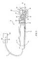
- FIG. 1is a cross-sectional schematic of an optical catheter for gathering speckle data from tissue in vivo.
- FIG. 2Ais a cross-sectional schematic illustrating the catheter of FIG. 1 , with an attached angioplasty balloon, inserted within a blood vessel, with the balloon deflated.
- FIG. 2Bis a cross-sectional schematic illustrating the catheter of FIG. 1 , with an attached angioplasty balloon, inserted within a blood vessel, with the balloon inflated.
- FIG. 4is a schematic illustrating reflectance of incident light from an atherosclerotic plaque.
- FIG. 5is representative raw data speckle images, edge images, and cross-correlation images used to assess the viscosity of a lipid-rich, atherosclerotic plaque in a human aorta.
- FIG. 6is representative raw data speckle images, edge images, and cross-correlation images used to assess the viscosity of normal human aorta tissue.
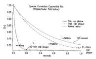
- FIG. 7is an exponential graph showing speckle decorrelation of a thin cap atherosclerotic plaque, a thick cap atherosclerotic plaque, and normal aortic tissue over a time interval.
- tissueis not static. Individual cells move within intercellular fluids, cellular organelles move within cells, and large molecules move back and forth between cells. In non-cellular tissue deposits such as plaques, components such as proteins, lipids, and other molecules also exhibit local motion. These local microscopic motions include “Brownian motion” and are essentially random in nature. Measuring and characterizing the microscopic motion of tissues can provide useful information about the structure, composition, biomechanical characteristics, and stability of the tissue.
- the inventionrelates to using laser speckle to measure microscopic motion, including Brownian motion, of tissue in vivo to gather information about the tissue.
- coherent or partially coherent lightis reflected from a tissue to form a speckle pattern at a detector. Due to motion of reflectors within the tissue, the speckle pattern changes over time or is “decorrelated.” By monitoring the rate of decorrelation, while compensating for “extrinsic,” macroscopic motion of the tissue, microscopic motion in the tissue can be isolated and measured.
- the partially coherent lightcan provide more information about optical properties of the tissue than completely coherent light.
- speckle analysisis used to measure microscopic, e.g., Brownian, motion in atherosclerotic plaques to detect plaques that are vulnerable to rupture, and, more specifically, to determine the plaque's vulnerability to rupture.
- a modified optical catheter (probe) or other instrumentis inserted into a blood vessel (e.g., artery) to locate these plaques, and once a plaque is located, the probe is moved into the proximity of the specific atherosclerotic plaque.
- Light reflected from the interior wall of the blood vessels, and/or from a plaqueis collected and transmitted to a detector, where a speckle pattern is formed.
- speckle patterns of normal tissue and plaque tissueare different, and these differences can be used to detect the plaques. Thereafter, e.g., while compensating for macroscopic motion of the plaque, the speckle pattern is monitored over time to calculate the pattern's rate of decorrelation. From this decorrelation rate, the degree of microscopic motion in the plaque, and therefore the plaque's vulnerability to rupture, can be assessed.
- Atherosclerotic plaquecan lead to acute myocardial infarction, which is a leading cause of death in industrialized countries.
- myocardial infarctionWhen an atherosclerotic plaque ruptures, lipids from the plaque enter the vessel lumen, potentially causing thrombosis, arterial occlusion, myocardial ischemia, and infarction.
- plaques vulnerable to rupturegenerally have a thin, unstable, fibrous cap and a compliant, or less “viscous,” lipid pool.
- lipid poole.g., Virmani et al., “Lesions from sudden coronary death: A comprehensive morphological classification scheme for atherosclerotic lesions,” Arterioscler. Thromb. Vasc. Bio., 20:1262–75 (2000) and Lee et al., “The Unstable Atheroma,” Arteriosclerosis, Thrombosis & Vascular Biology, 17:1859–67 (1997).
- the less viscous lipid poolapplies force to the fibrous cap, compromising the cap and causing rupture.
- a specially modified optical catheter 10includes a rotatable inner shaft 12 and a transparent outer sheath 14 .
- the inner shaft 12houses a fiber array 15 and a mirror 16 near its distal end 18 .
- a central fiber 20 in the fiber arrayconnects to a fixed optical fiber 21 that extends from the catheter proximally to a light source 22 .
- coherent lightsuch as laser light
- beam-splitter 22 acoherent light from light source 22 is transmitted via beam-splitter 22 a , through the fixed optical fiber 21 and central fiber 20 and onto center 23 of mirror 16 .
- the lightis reflected to a tissue sample 24 , such as a layer of static tissue over a layer of moving tissue, such as an atherosclerotic plaque.
- Outer sheath 14can be placed directly in contact with sample 24 (near field), or can be positioned a short distance, e.g., 1 mm to 10 cm away from the sample (far field).
- Light remitted from the sample(arrows 26 ) reflects from mirror 16 to the fibers of array 15 , and is then transmitted by array 15 to a planar charge-coupled device (CCD), or a linear or two-dimensional detector 22 b , via a beam-splitter 22 a , e.g., located within light source 22 .
- CCDcharge-coupled device
- a beam-splitter 22 ae.g., located within light source 22 .
- There may be one or multiple fibers for detection and illumination and detectionmay occur from the same fiber.
- illuminationmay occur through a fiber array where each fiber is selectively illuminated to generate multiple speckle patterns as a function of position on the sample.
- This methodcan provide a scanning of the incident light across a sample while keeping the probe stationary by illuminating one fiber after another in series.
- a speckle patternforms at the CCD detector.
- the resulting speckle patternis then digitized by an analog-digital converter, and analyzed using the procedures described in the analysis section below.
- the entire shaft 12can rotate 360 degrees in the direction of arrow R, allowing catheter 10 to gather images around the entire circumference of a sample.
- catheter 10can gather images of a plaque around the circumference of a vessel wall.
- the diameter of the cathetercan be less than 500 ⁇ m. Larger diameters are also possible.
- the optics of catheter 10can be integrated into other types of instruments, such as endoscopes or laparoscopes.
- the opticscan also form a stand-alone unit passed into the accessory port of standard endoscopes or laparoscopes, or integrated into another type of catheter, such as dual-purpose intravascular ultrasound catheter.
- the opticscan also include a lens that focuses the remitted light 26 onto the distal ends of the fibers in array 15 .
- the lenswould allow formation of a “near field image” (near the sample sight less than one wavelength) rather than a “far field image” (at the detector set more than a wavelength away from the surface of the tissue).
- the cathetercan include a polarization filter to remove all but a certain type of polarized light.
- a cross-polarized filterwould allow only light having a polarization perpendicular to the incident light to reach the detector, while a parallel polarized filter would allow only light having the same polarization as the incident light to pass.
- multiply scattered lightis less likely to retain its initial polarization than single scattered light
- polarization filterscan be used to bias the data toward multiply scattered or single scattered light. Such bias can be used to deduce information about the structure of the sample, since light which has penetrated deeper into the sample will be more highly scattered than light reflected from the surface or remitted from near the surface.
- the detectorcan be, e.g., a photographic plate, an array of photodetectors, or a single detector.
- the light sourcecan illuminate the sample with continuous light or synchronized pulses.
- the light sourcecan be, e.g., as far as one meter, or more, away from the sample.
- the temporal changes in the patternshould indicate movement of reflectors within the plaque, but not indicate movement of the plaque itself or movement of reflectors between the detector and the plaque.
- the changes in the plaque's speckle patternpreferably reflect microscopic or Brownian motion, but not macroscopic motion.
- the intervalFor a time interval to be sufficient to detect microscopic Brownian motion, the interval must be long enough to allow for movement of reflectors in the tissue, such as a lipid pool, but short enough that the random Brownian movements do not cancel out.
- an appropriate time intervalis about 1–200 ms. Shorter time periods may also be possible. If the time intervals are longer, then changes in the speckle pattern may not adequately differentiate rapid Brownian movement (indicating low viscosity) from slower Brownian movement (indicating high viscosity).
- the fiber array 15can be coupled to the plaque tissue using, e.g., an angioplasty balloon. This technique also compensates for minor patient movements.
- a balloon 28is attached to outer sheath 14 , on a far side 30 of the catheter. Once the catheter is positioned within a blood vessel in proximity to the plaque, the balloon is inflated. Referring to FIG. 2B , the inflated balloon abuts the vessel wall 32 , and presses the catheter against plaque 24 , such that a distal region of outer shaft 14 is in direct contact with the plaque. With the catheter coupled to plaque 24 as shown in FIG. 2B , fiber array 15 will move with the plaque when the heart beats, and the gross motion of the plaque will not significantly affect the speckle pattern.
- the ballooncan surround the catheter.
- a transparent balloonsurrounds outer sheath 14 , but is also attached to the sheath.
- the balloonis squeezed between plaque 24 and wall 32 of the vessel. The balloon, therefore will be in direct contact with the plaque, and will move with the plaque when the heart beats. Since the balloon is attached to shaft 14 , and shaft 14 is coupled to array 15 , movement of the vessel wall will not significantly affect the speckle pattern. Additional methods of coupling the catheter to tissue can also be used, including methods that do not employ an angioplasty balloon.
- a second method of compensating for movement caused by heartbeatsis to gather data between heartbeats.
- datais gathered during the relatively still PR interval of the diastole of the heartbeat (when the left ventricle is filling with blood).
- the PR intervallasts for about 0.12–0.2 seconds, providing sufficient time to detect Brownian motion.
- the timingcan be computer-controlled or the detector can be linked to an ECG signal, and programmed to gather data only during the PR interval. Similar techniques can be used to compensate for other bodily movements such as peristalsis.
- the cathetercan be placed in direct contact with the plaque tissue, as described above, thereby preventing blood from flowing between the detector and the plaque.
- blood flowing between the plaque and the cathetercan be removed and replaced with clear saline solution or other clear solutions such as optically transparent blood substitutes.
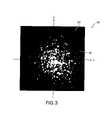
- FIG. 3illustrates a typical speckle pattern 40 formed by reflecting light from the wall of a healthy blood vessel.
- X and Y coordinatesoverlay pattern 40 to facilitate mathematical description of the pattern.
- the patternincludes dark patches, where destructive interference dominates, and brighter patches, where constructive interference dominates. Very subtle movements of reflectors within the multiply scattering sample alter the speckle pattern.
- plaque's vulnerability to rupturecan be assessed.
- This methodincludes: (1) gathering a series of speckle images at short, discrete time intervals; (2) eliminating diffuse reflectance from the data; (3) creating cross-correlation images comparing the speckle images in the series; (4) calculating the maximum correlation between each pair of images to create a one-dimensional data set over time; (5) calculating the rate of decorrelation from the data set; and (6) from the rate of decorrelation, assessing the plaque's viscosity and vulnerability to rupture.
- speckle imagesare gathered for a plaque at discrete intervals over a period of time.
- speckle imagescan be gathered, e.g., at intervals of every 1, 5, 10, 20, or 30 ms for a time period of, e.g., 200 ms.
- time periode.g. 200 ms.
- the shorter the time intervalsthe shorter the time period over which data can be gathered.
- time intervalssuch as 30 ms
- datacan be gathered for, e.g., 1–2 seconds.
- the background, non-coherent diffuse reflectanceis eliminated from the images.
- a number of techniquescan be used to eliminate the tissue's diffuse reflectance.
- the raw data speckle imagescan be converted to edge images.
- Edge imagesare spatial derivatives of the raw data images; an edge image (high pass filter) reflects the change in intensity of an image as a function of space, at all points in the image, rather than the intensity itself.
- Known techniques of edge detectioninclude convolution of the image by a kernel (e.g., Sobel or Robert), Morph gradient (subtraction of an eroded, dilated, closed, or opened image by its original), or high pass filtering.
- the cross-correlation imagesreflect the degree of correlation between the two images as a function of space.
- g(t)is the cross-correlation function
- I(x, y)is intensity of the interference at a point (x, y) in the pattern
- tis time.
- Two-dimensional cross-correlation functionsare described generally in Jae S.
- the cross-correlation imagesare reduced to a one-dimensional data set as a function of time (i.e., a series of correlation values, each value associated with a time t). From this series of correlation values, a time constant, ⁇ , is calculated, where ⁇ represents the rate of decorrelation.
- the time constantis the amount of time it takes g(t) to reach (1/e)g(0).
- the max function of equation (1)is not the only possible mechanism for reducing the cross-correlation images to a number.
- a point to reduce the image to a valuerather than a max function, would not compensate for the “memory effect” of first order correlation of speckle patterns in turbid media. This “memory effect” is described in Feng et al., Science 251: 633–39 (1991).
- Advantages to using a pointare that a minimum number of fibers and detectors can be used.
- the viscosity of the plaque's lipid poolcan be assessed.
- ⁇the higher the Brownian motion in the lipid pool, and the greater the pool's viscosity.
- the smaller ⁇the greater the Brownian motion in the lipid pool, and the lower the viscosity. The lower the viscosity, the more stresses are exerted on the cap, making the plaque more vulnerable
- This informationcan be used to identify plaques likely or vulnerable to rupture. Specifically, if T is about 40–100 ms or lower, then the plaque is likely to rupture, and intervention is warranted. If the plaque ⁇ is about 100–200 ms, then the plaque is somewhat vulnerable, but not yet likely to rupture, and should be monitored over time. If the plaque ⁇ is about 200–300 ms, then the plaque is less vulnerable. Non-plaque covered, healthy vessel wall generally has a time constant greater than 300 ms or 500 ms.
- the speckle datacan be analyzed to deduce spatial characteristics of the plaque, including the thickness of the fibrous cap, or the thickness of any tissue structure or tissue layer for that matter. As discussed above, a thin fibrous cap is another indication that a plaque is vulnerable to rupture. The combination of data relating to viscosity and cap thickness provides the most accurate assessment of plaque vulnerability, although the two characteristics can be assessed and analyzed independently.
- a typical plaque 50includes a fibrous cap 52 and a lipid pool 54 .
- Photons that enter plaque 50(arrow A) are internally scattered by reflectors within the plaque, such as collagen in fibrous cap 52 and lipids in pool 54 .
- the various photonstherefore, exit the plaque at different locations (arrows B).
- the speckle pattern(see FIG. 2 ) has a diameter considerably larger than the width of the original light beam.
- the thickness of fibrous cap 52can be deduced by comparing different regions of the resulting speckle pattern. Referring again to FIG. 3 , light forming intensity signals in the outer portion 60 of the pattern traveled greater distances than light forming signals near the center 62 of the pattern. Thus, outer portion 60 of the pattern is formed by photons that, on average, penetrated deeper into the plaque than photons forming center 62 . By calculating separate time constants for separate regions of the speckle pattern, the viscosity of the plaque at different depths can be determined. Since the fibrous cap generally exhibits less Brownian motion than the lipid pool, the thickness of the fibrous cap can be estimated from spatially dependent data.
- Time constantsare then calculated from the cross-correlation data for each window, in the manner described above.
- the variation of ⁇ as a function of the distance from the center of the speckle patterni.e., as a function of (x o 2 +y o 2 ) 1/2
- ⁇can then be analyzed to determine the thickness of the fibrous cap.
- Plaque cap thickness of less than about 60 ⁇ mis considered to be vulnerable, but this number can vary to some extent depending on the specific patient.
- any tissue structuree.g., a tissue layer that overlies or is adjacent to a different tissue, e.g., a plaque fibrous cap over a lipid pool
- a tissue layer that overlies or is adjacent to a different tissuee.g., a plaque fibrous cap over a lipid pool
- ⁇ (r) and the optical propertiese.g., ⁇ eff
- ⁇ (r) and the optical propertiese.g., ⁇ eff
- r 0 and ⁇ effmay be used as inputs to a look-up table containing tissue layer thickness values.
- separately analyzing different regions of a speckle patternalso allows decorrelation caused by macroscopic motion to be identified and removed from the analysis.
- macroscopic motion caused by gross movement of the plaque tissue or blood flowwill be directional, non-random, and global.
- Brownian motionwill be non-directional and non-uniform (or random).
- the position of maximums of cross-correlation functionswill shift along a vector v , which relates extrinsic motion of the sample with respect to the catheter or detection.
- Brownian motionwill decorrelate the speckle patterns in many random directions, and will result in a broadening of the cross-correlation peak and a decrease in correlation maximum above that predicted by linear motion.
- the rate of decorrelationcan be estimated from single pixel speckle images, rather than full, two-dimensional speckle patterns.
- a catheter with a single optical fibercould transmit data to a single detector, such as a photodiode.
- the speckle data gatheredwould be intensity at the spot as a function of time. From this data, a rate of decorrelation can be calculated directly or only as a function of time as opposed to space, without any spatial cross-correlation analysis.
- Imaging methods that detect single scattered lightsuch as optical coherence tomography (OCT) and confocal microscopy, can also be used. While these imaging methods are less sensitive to speckle modulation than the multiple scattering methods described above, they have the advantage of allowing localization of data to a single point within the sample. Such localization would allow measurement of biomechanical properties of the tissue in three dimensions.
- OCToptical coherence tomography
- motion of the scattererscan produce a Doppler shift on the returned light. The Doppler shift can provide a further basis for measuring viscosity in the sample.
- speckle images formed by reflecting laser light from a cadaveric atherosclerotic plaque in a human aortawere analyzed to assess the plaque's viscosity. A portion of normal aorta was also analyzed for comparison.
- edge detectionwas performed on the 60 raw speckle images, generating 60 edge images.
- the edge imagesreflect the spatial derivative of the raw speckle images (i.e., the light patches in the edge images of row B are locations where the intensity is changing as a function of space).
- Each cross-correlation imagewas generated by multiplying the Fourier transform of the reference image 70 by the complex conjugate of the Fourier transform of the image in question, and then calculating an inverse Fourier transform of the product.
- Image 72was formed by multiplying the Fourier transform of reference image 70 by the complex conjugate of the Fourier transform of image 70 , and then calculating the inverse Fourier transform of the product.
- Each cross-correlation imagerepresents the degree of correlation between the corresponding edge image and the reference edge image 70 (i.e., brighter spots are locations where there is a higher degree of correlation than at darker spots).
- the maximum cross-correlation peak(i.e., the correlation at the maximum point of correlation) was calculated using equation (1).
- the resulting data setincluded 60 cross-correlation values, each value associated with a time t.
- FIG. 6A set of images of normal aorta tissue is shown in FIG. 6 . These images are comparable to the set of images in FIG. 5 for a lipid-rich plaque in the same aorta and were imaged and processed in the same manner.
- the decorrelation raterepresented by the time constant ⁇
- the time constantwas 40 ms.
- the time constantwas 500 ms.
- the plaquewas borderline vulnerable. Thus, had this plaque been analyzed in vivo, using the procedures described above, a physician would have determined that the plaque was a possible candidate for rupture, and may have chosen to intervene, preventing a possible infarction.
- the methods described hereincan also be used to characterize diseased tissue other than atherosclerotic plaques.
- the microscopic and macroscopic constituents of diseased tissuediffer from normal non-pathologic counterparts.
- speckle patternscan be used to diagnose and characterize other tissue pathology such as neoplasia (cancer), infection, tissue viability, or healing response to injury.
- neoplasiacancer
- tumorstypically have an abnormal abundance of one cell type (clonal) and a surrounding abnormal supporting matrix. This cell type may produce and secrete a viscous fluid, such as mucin in adenocarcinoma, which would result in lower speckle decorrelation time constants than normal non-cancerous tissue.
- the surrounding matrixmay be composed of necrotic tissue and an abundance of abnormal vessels that would also serve to decrease the speckle decorrelation time constant.
- Other tumorslike osteosarcoma, produce osteoid or immature bone that would increase the time constant compared to normal tissue.
- Other forms of neoplasiawould have increased time constants due to desmoplastic (abundant) fibrous stroma initiated by cytokines produced by the tumor. Indeed, many tumors, including bronchogenic carcinomas and breast carcinomas are firm upon gross examination due to the fibrous stroma surrounding the malignant cells. This fibrous stroma would increase the time constant relative to surrounding normal tissue.
- abscesseswill be less viscous than surrounding tissue, enabling identification of the infected region by measuring a decrease in the time constant.
- Inflammationmanifested by the influx of activated inflammatory cells will be characterized by a decrease in the speckle decorrelation time constant as these cells degrade the normal supporting tissue in response to a the presence of bacterial, viral, or foreign body antigens.
- Necrotic tissuesuch as burn eschar, diabetic ulcers, necrotic bowel, and ischemic myocardium will have longer time constants than viable tissue from the same organ due to the lack of intravascular and extravascular fluid and flow in these extracellular spaces.
- Speckle decorrelation timesmay also be used to estimate tissue hydration and provide a means for quantifying the state of hydration in a patient. While the above examples elucidate some of the mechanisms that explain how disease affects the biomechanical properties of pathologic tissue, many more exist and are well known in the field of gross anatomic pathology. These differing biomechanical properties and characteristics can be measured by speckle for the purpose of screening, intraoperative margin (e.g., tumor margin) identification, and primary diagnosis.
- intraoperative margine.g., tumor margin
Landscapes
- Health & Medical Sciences (AREA)
- Life Sciences & Earth Sciences (AREA)
- Surgery (AREA)
- Engineering & Computer Science (AREA)
- Physics & Mathematics (AREA)
- General Health & Medical Sciences (AREA)
- Pathology (AREA)
- Public Health (AREA)
- Biomedical Technology (AREA)
- Heart & Thoracic Surgery (AREA)
- Medical Informatics (AREA)
- Molecular Biology (AREA)
- Animal Behavior & Ethology (AREA)
- Biophysics (AREA)
- Veterinary Medicine (AREA)
- Radiology & Medical Imaging (AREA)
- Optics & Photonics (AREA)
- Nuclear Medicine, Radiotherapy & Molecular Imaging (AREA)
- Signal Processing (AREA)
- Physiology (AREA)
- Computer Vision & Pattern Recognition (AREA)
- Artificial Intelligence (AREA)
- Psychiatry (AREA)
- Vascular Medicine (AREA)
- Cardiology (AREA)
- Chemical & Material Sciences (AREA)
- Analytical Chemistry (AREA)
- Biochemistry (AREA)
- General Physics & Mathematics (AREA)
- Immunology (AREA)
- Investigating Or Analysing Materials By Optical Means (AREA)
- Investigating Or Analysing Biological Materials (AREA)
- Investigating, Analyzing Materials By Fluorescence Or Luminescence (AREA)
Abstract
Description
g(t)=max[∫∫I(x,y,0)I(x+x′,y+y′,t)dx′dy′] (1)
where g(t) is the cross-correlation function, I(x, y) is intensity of the interference at a point (x, y) in the pattern, and t is time. Two-dimensional cross-correlation functions are described generally in Jae S. Lim, “Two-Dimensional Signal Processing” (Prentice Hall, Englewood Cliffs, N.J., 1990) and Jain, Anil, K., “Fundamentals of Digital Image Processing” (Prentice Hall, Englewood Cliffs, N.J., 1987).
g(x,y,t)=∫∫I(x,y,0)I(x+x′,y+y′,t)dx′dy′ (2)
at a point, such as x=y=0. However, using a point to reduce the image to a value, rather than a max function, would not compensate for the “memory effect” of first order correlation of speckle patterns in turbid media. This “memory effect” is described in Feng et al.,Science251: 633–39 (1991). Advantages to using a point are that a minimum number of fibers and detectors can be used.
g(x0,y0,t)=max[∫∫w(x′−x0,y′−y0)I(x,y,0)I(x+x′,y+y′,t)dx′dy′] (3)
Claims (50)
Priority Applications (3)
| Application Number | Priority Date | Filing Date | Title |
|---|---|---|---|
| US10/016,244US7231243B2 (en) | 2000-10-30 | 2001-10-30 | Optical methods for tissue analysis |
| US11/534,095US8032200B2 (en) | 2000-10-30 | 2006-09-21 | Methods and systems for tissue analysis |
| US13/251,594US9282931B2 (en) | 2000-10-30 | 2011-10-03 | Methods for tissue analysis |
Applications Claiming Priority (2)
| Application Number | Priority Date | Filing Date | Title |
|---|---|---|---|
| US24425500P | 2000-10-30 | 2000-10-30 | |
| US10/016,244US7231243B2 (en) | 2000-10-30 | 2001-10-30 | Optical methods for tissue analysis |
Related Child Applications (1)
| Application Number | Title | Priority Date | Filing Date |
|---|---|---|---|
| US11/534,095ContinuationUS8032200B2 (en) | 2000-10-30 | 2006-09-21 | Methods and systems for tissue analysis |
Publications (2)
| Publication Number | Publication Date |
|---|---|
| US20020183601A1 US20020183601A1 (en) | 2002-12-05 |
| US7231243B2true US7231243B2 (en) | 2007-06-12 |
Family
ID=22922006
Family Applications (3)
| Application Number | Title | Priority Date | Filing Date |
|---|---|---|---|
| US10/016,244Expired - Fee RelatedUS7231243B2 (en) | 2000-10-30 | 2001-10-30 | Optical methods for tissue analysis |
| US11/534,095Expired - Fee RelatedUS8032200B2 (en) | 2000-10-30 | 2006-09-21 | Methods and systems for tissue analysis |
| US13/251,594Expired - Fee RelatedUS9282931B2 (en) | 2000-10-30 | 2011-10-03 | Methods for tissue analysis |
Family Applications After (2)
| Application Number | Title | Priority Date | Filing Date |
|---|---|---|---|
| US11/534,095Expired - Fee RelatedUS8032200B2 (en) | 2000-10-30 | 2006-09-21 | Methods and systems for tissue analysis |
| US13/251,594Expired - Fee RelatedUS9282931B2 (en) | 2000-10-30 | 2011-10-03 | Methods for tissue analysis |
Country Status (7)
| Country | Link |
|---|---|
| US (3) | US7231243B2 (en) |
| EP (1) | EP1434522B1 (en) |
| JP (1) | JP4241038B2 (en) |
| AT (1) | ATE454845T1 (en) |
| AU (1) | AU2002230842A1 (en) |
| DE (1) | DE60141090D1 (en) |
| WO (1) | WO2002036015A1 (en) |
Cited By (256)
| Publication number | Priority date | Publication date | Assignee | Title |
|---|---|---|---|---|
| US20020198457A1 (en)* | 2001-04-30 | 2002-12-26 | Tearney Guillermo J. | Method and apparatus for improving image clarity and sensitivity in optical coherence tomography using dynamic feedback to control focal properties and coherence gating |
| US20040147844A1 (en)* | 2001-07-16 | 2004-07-29 | Art, Advanced Research Technologies, Inc. | N-time-gate data-type for TPSF-based optical imaging |
| US20050018201A1 (en)* | 2002-01-24 | 2005-01-27 | De Boer Johannes F | Apparatus and method for ranging and noise reduction of low coherence interferometry lci and optical coherence tomography oct signals by parallel detection of spectral bands |
| US20050035295A1 (en)* | 2003-06-06 | 2005-02-17 | Brett Bouma | Process and apparatus for a wavelength tuning source |
| US20050128488A1 (en)* | 2003-11-28 | 2005-06-16 | Dvir Yelin | Method and apparatus for three-dimensional spectrally encoded imaging |
| US20050242298A1 (en)* | 2002-07-18 | 2005-11-03 | Mauna Kea Technologies | Method and equipment for fiber optic high-resolution, in particular confocal, fluorescence imaging |
| US20060058685A1 (en)* | 2003-11-17 | 2006-03-16 | Fomitchov Pavel A | System and method for imaging based on ultrasonic tagging of light |
| US20060058592A1 (en)* | 2004-08-24 | 2006-03-16 | The General Hospital Corporation | Process, system and software arrangement for measuring a mechanical strain and elastic properties of a sample |
| US20060067620A1 (en)* | 2004-09-29 | 2006-03-30 | The General Hospital Corporation | System and method for optical coherence imaging |
| US20060165270A1 (en)* | 2003-02-25 | 2006-07-27 | Jorn Borgert | Intravascular imaging |
| US20060232783A1 (en)* | 2005-01-20 | 2006-10-19 | Michael Choma | Methods and systems for reducing complex conjugat ambiguity in interferometric data |
| US20060247531A1 (en)* | 2005-04-27 | 2006-11-02 | The Trustees Of Dartmouth College | System and method for spectral-encoded high-rate hemodynamic tomography |
| US20060270929A1 (en)* | 2005-05-31 | 2006-11-30 | The General Hospital Corporation | System, method and arrangement which can use spectral encoding heterodyne interferometry techniques for imaging |
| US20070073162A1 (en)* | 2000-10-30 | 2007-03-29 | The General Hospital Corporation | Methods and systems for tissue analysis |
| US20070263227A1 (en)* | 2006-05-12 | 2007-11-15 | The General Hospital Corporation | Processes, arrangements and systems for providing a fiber layer thickness map based on optical coherence tomography images |
| US20080007734A1 (en)* | 2004-10-29 | 2008-01-10 | The General Hospital Corporation | System and method for providing Jones matrix-based analysis to determine non-depolarizing polarization parameters using polarization-sensitive optical coherence tomography |
| US7382949B2 (en) | 2004-11-02 | 2008-06-03 | The General Hospital Corporation | Fiber-optic rotational device, optical system and method for imaging a sample |
| US7418169B2 (en) | 2006-02-01 | 2008-08-26 | The General Hospital Corporation | Apparatus for controlling at least one of at least two sections of at least one fiber |
| US20080234586A1 (en)* | 2007-03-19 | 2008-09-25 | The General Hospital Corporation | System and method for providing noninvasive diagnosis of compartment syndrome using exemplary laser speckle imaging procedure |
| US7447408B2 (en) | 2004-07-02 | 2008-11-04 | The General Hospital Corproation | Imaging system and related techniques |
| US20090015923A1 (en)* | 2007-07-09 | 2009-01-15 | Auld Jack R | Multi-Spot Ophthalmic Laser Probe |
| US20090039235A1 (en)* | 2005-11-16 | 2009-02-12 | Macfarlane Duncan L | Method for determining location and movement of a moving object |
| US20090131801A1 (en)* | 2007-10-12 | 2009-05-21 | The General Hospital Corporation | Systems and processes for optical imaging of luminal anatomic structures |
| US7538859B2 (en) | 2006-02-01 | 2009-05-26 | The General Hospital Corporation | Methods and systems for monitoring and obtaining information of at least one portion of a sample using conformal laser therapy procedures, and providing electromagnetic radiation thereto |
| US7567349B2 (en) | 2003-03-31 | 2009-07-28 | The General Hospital Corporation | Speckle reduction in optical coherence tomography by path length encoded angular compounding |
| US20090192358A1 (en)* | 2008-01-28 | 2009-07-30 | The General Hospital Corporation | Systems, processes and computer-accessible medium for providing hybrid flourescence and optical coherence tomography imaging |
| US20090236541A1 (en)* | 2008-03-24 | 2009-09-24 | General Electric Company | System and Methods for Optical Imaging |
| US7643153B2 (en) | 2003-01-24 | 2010-01-05 | The General Hospital Corporation | Apparatus and method for ranging and noise reduction of low coherence interferometry LCI and optical coherence tomography OCT signals by parallel detection of spectral bands |
| US20100121200A1 (en)* | 2007-04-19 | 2010-05-13 | Institut De Radioprotection Et De Surete Nucleaire | Diagnostic and prognostic assistance device for physiopathological tissue changes |
| US7733497B2 (en) | 2003-10-27 | 2010-06-08 | The General Hospital Corporation | Method and apparatus for performing optical imaging using frequency-domain interferometry |
| US7742173B2 (en) | 2006-04-05 | 2010-06-22 | The General Hospital Corporation | Methods, arrangements and systems for polarization-sensitive optical frequency domain imaging of a sample |
| US20100168586A1 (en)* | 2007-06-29 | 2010-07-01 | The Trustees Of Columbia University In The City Of New York | Optical imaging or spectroscopy systems and methods |
| US7761139B2 (en) | 2003-01-24 | 2010-07-20 | The General Hospital Corporation | System and method for identifying tissue using low-coherence interferometry |
| US7794407B2 (en) | 2006-10-23 | 2010-09-14 | Bard Access Systems, Inc. | Method of locating the tip of a central venous catheter |
| US7796270B2 (en) | 2006-01-10 | 2010-09-14 | The General Hospital Corporation | Systems and methods for generating data based on one or more spectrally-encoded endoscopy techniques |
| US20100280315A1 (en)* | 2007-09-19 | 2010-11-04 | The Research Foundation Of State University Of New York | Optical coherence tomography systems and methods |
| US7843572B2 (en) | 2005-09-29 | 2010-11-30 | The General Hospital Corporation | Method and apparatus for optical imaging via spectral encoding |
| US7865231B2 (en) | 2001-05-01 | 2011-01-04 | The General Hospital Corporation | Method and apparatus for determination of atherosclerotic plaque type by measurement of tissue optical properties |
| US20110051148A1 (en)* | 2009-09-03 | 2011-03-03 | Axsun Technologies, Inc. | Filtered ASE Swept Source for OCT Medical Imaging |
| US20110051143A1 (en)* | 2009-09-03 | 2011-03-03 | Axsun Technologies, Inc. | ASE Swept Source with Self-Tracking Filter for OCT Medical Imaging |
| US7911621B2 (en) | 2007-01-19 | 2011-03-22 | The General Hospital Corporation | Apparatus and method for controlling ranging depth in optical frequency domain imaging |
| US20110071404A1 (en)* | 2009-09-23 | 2011-03-24 | Lightlab Imaging, Inc. | Lumen Morphology and Vascular Resistance Measurements Data Collection Systems, Apparatus and Methods |
| US20110071405A1 (en)* | 2009-09-23 | 2011-03-24 | Lightlab Imaging, Inc. | Apparatus, Systems, and Methods of in-vivo Blood Clearing in a Lumen |
| US7920271B2 (en) | 2006-08-25 | 2011-04-05 | The General Hospital Corporation | Apparatus and methods for enhancing optical coherence tomography imaging using volumetric filtering techniques |
| US7933021B2 (en) | 2007-10-30 | 2011-04-26 | The General Hospital Corporation | System and method for cladding mode detection |
| US7949019B2 (en) | 2007-01-19 | 2011-05-24 | The General Hospital | Wavelength tuning source based on a rotatable reflector |
| US20110122366A1 (en)* | 2009-11-24 | 2011-05-26 | Smith Ronald T | Single-fiber multi-spot laser probe for ophthalmic endoillumination |
| US20110137140A1 (en)* | 2009-07-14 | 2011-06-09 | The General Hospital Corporation | Apparatus, Systems and Methods for Measuring Flow and Pressure within a Vessel |
| US20110144627A1 (en)* | 2009-12-15 | 2011-06-16 | Smith Ronald T | Multi-spot laser probe |
| US20110151980A1 (en)* | 2009-12-22 | 2011-06-23 | Lightlab Imaging, Inc. | Torque limiter for an oct catheter |
| US20110169978A1 (en)* | 2008-07-10 | 2011-07-14 | Ecole Polytechnique Federale De Lausanne (Epfl) | Functional optical coherent imaging |
| US7982879B2 (en) | 2006-02-24 | 2011-07-19 | The General Hospital Corporation | Methods and systems for performing angle-resolved fourier-domain optical coherence tomography |
| US7995210B2 (en) | 2004-11-24 | 2011-08-09 | The General Hospital Corporation | Devices and arrangements for performing coherence range imaging using a common path interferometer |
| US8018598B2 (en) | 2004-05-29 | 2011-09-13 | The General Hospital Corporation | Process, system and software arrangement for a chromatic dispersion compensation using reflective layers in optical coherence tomography (OCT) imaging |
| US20110228280A1 (en)* | 2010-03-17 | 2011-09-22 | Lightlab Imaging, Inc. | Intensity Noise Reduction Methods and Apparatus for Interferometric Sensing and Imaging Systems |
| US20110249912A1 (en)* | 2004-02-12 | 2011-10-13 | Hoshiko Llc | Method and apparatus for photograph finding |
| US8040608B2 (en) | 2007-08-31 | 2011-10-18 | The General Hospital Corporation | System and method for self-interference fluorescence microscopy, and computer-accessible medium associated therewith |
| US8045177B2 (en) | 2007-04-17 | 2011-10-25 | The General Hospital Corporation | Apparatus and methods for measuring vibrations using spectrally-encoded endoscopy |
| US8081316B2 (en) | 2004-08-06 | 2011-12-20 | The General Hospital Corporation | Process, system and software arrangement for determining at least one location in a sample using an optical coherence tomography |
| US8097864B2 (en) | 2009-01-26 | 2012-01-17 | The General Hospital Corporation | System, method and computer-accessible medium for providing wide-field superresolution microscopy |
| US8115919B2 (en) | 2007-05-04 | 2012-02-14 | The General Hospital Corporation | Methods, arrangements and systems for obtaining information associated with a sample using optical microscopy |
| US8145018B2 (en) | 2006-01-19 | 2012-03-27 | The General Hospital Corporation | Apparatus for obtaining information for a structure using spectrally-encoded endoscopy techniques and methods for producing one or more optical arrangements |
| US8175685B2 (en) | 2006-05-10 | 2012-05-08 | The General Hospital Corporation | Process, arrangements and systems for providing frequency domain imaging of a sample |
| US8208995B2 (en) | 2004-08-24 | 2012-06-26 | The General Hospital Corporation | Method and apparatus for imaging of vessel segments |
| US20120170022A1 (en)* | 2010-12-30 | 2012-07-05 | Empire Technology Development Llc | Analyzing Tissue Morphology in Three Dimensions |
| US20120245473A1 (en)* | 2008-06-05 | 2012-09-27 | Mary-Ann Mycek | Multimodal spectroscopic systems and methods for classifying biological tissue |
| US8351665B2 (en) | 2005-04-28 | 2013-01-08 | The General Hospital Corporation | Systems, processes and software arrangements for evaluating information associated with an anatomical structure by an optical coherence ranging technique |
| US20130010108A1 (en)* | 2010-03-24 | 2013-01-10 | National Institute Of Radiological Sciences | Measuring system |
| US8358461B2 (en) | 2008-09-03 | 2013-01-22 | Lightlab Imaging Inc. | Wavelength-tunable light source |
| US8388546B2 (en) | 2006-10-23 | 2013-03-05 | Bard Access Systems, Inc. | Method of locating the tip of a central venous catheter |
| US8388541B2 (en) | 2007-11-26 | 2013-03-05 | C. R. Bard, Inc. | Integrated system for intravascular placement of a catheter |
| US8437833B2 (en) | 2008-10-07 | 2013-05-07 | Bard Access Systems, Inc. | Percutaneous magnetic gastrostomy |
| US8449468B2 (en) | 2006-11-08 | 2013-05-28 | Lightlab Imaging, Inc. | Opto-acoustic imaging devices and methods |
| US8478382B2 (en) | 2008-02-11 | 2013-07-02 | C. R. Bard, Inc. | Systems and methods for positioning a catheter |
| US8582109B1 (en) | 2011-08-01 | 2013-11-12 | Lightlab Imaging, Inc. | Swept mode-hopping laser system, methods, and devices for frequency-domain optical coherence tomography |
| US8582619B2 (en) | 2011-03-15 | 2013-11-12 | Lightlab Imaging, Inc. | Methods, systems, and devices for timing control in electromagnetic radiation sources |
| USRE44605E1 (en) | 2003-10-17 | 2013-11-19 | Axsun Technologies, Inc. | Integrated spectroscopy system |
| US8593619B2 (en) | 2008-05-07 | 2013-11-26 | The General Hospital Corporation | System, method and computer-accessible medium for tracking vessel motion during three-dimensional coronary artery microscopy |
| US8619237B2 (en) | 2009-12-04 | 2013-12-31 | The Trustees Of Columbia University In The City Of New York | Laser-scanning intersecting plane tomography such as for high speed volumetric optical imaging |
| USD699359S1 (en) | 2011-08-09 | 2014-02-11 | C. R. Bard, Inc. | Ultrasound probe head |
| US8721077B2 (en) | 2011-04-29 | 2014-05-13 | The General Hospital Corporation | Systems, methods and computer-readable medium for determining depth-resolved physical and/or optical properties of scattering media by analyzing measured data over a range of depths |
| US8781555B2 (en) | 2007-11-26 | 2014-07-15 | C. R. Bard, Inc. | System for placement of a catheter including a signal-generating stylet |
| US8784336B2 (en) | 2005-08-24 | 2014-07-22 | C. R. Bard, Inc. | Stylet apparatuses and methods of manufacture |
| US8801693B2 (en) | 2010-10-29 | 2014-08-12 | C. R. Bard, Inc. | Bioimpedance-assisted placement of a medical device |
| US8804126B2 (en) | 2010-03-05 | 2014-08-12 | The General Hospital Corporation | Systems, methods and computer-accessible medium which provide microscopic images of at least one anatomical structure at a particular resolution |
| US8831321B1 (en) | 2011-11-07 | 2014-09-09 | Lightlab Imaging, Inc. | Side branch detection methods, systems and devices |
| US8838213B2 (en) | 2006-10-19 | 2014-09-16 | The General Hospital Corporation | Apparatus and method for obtaining and providing imaging information associated with at least one portion of a sample, and effecting such portion(s) |
| US8849382B2 (en) | 2007-11-26 | 2014-09-30 | C. R. Bard, Inc. | Apparatus and display methods relating to intravascular placement of a catheter |
| US8861910B2 (en) | 2008-06-20 | 2014-10-14 | The General Hospital Corporation | Fused fiber optic coupler arrangement and method for use thereof |
| US8922781B2 (en) | 2004-11-29 | 2014-12-30 | The General Hospital Corporation | Arrangements, devices, endoscopes, catheters and methods for performing optical imaging by simultaneously illuminating and detecting multiple points on a sample |
| US8926590B2 (en) | 2009-12-22 | 2015-01-06 | Lightlab Imaging, Inc. | Torque limiter for an OCT catheter |
| US8937724B2 (en) | 2008-12-10 | 2015-01-20 | The General Hospital Corporation | Systems and methods for extending imaging depth range of optical coherence tomography through optical sub-sampling |
| US8953911B1 (en) | 2011-10-28 | 2015-02-10 | Lightlab Imaging, Inc. | Spectroscopic imaging probes, devices, and methods |
| USD724745S1 (en) | 2011-08-09 | 2015-03-17 | C. R. Bard, Inc. | Cap for an ultrasound probe |
| US8997572B2 (en) | 2011-02-11 | 2015-04-07 | Washington University | Multi-focus optical-resolution photoacoustic microscopy with ultrasonic array detection |
| WO2014205256A3 (en)* | 2013-06-19 | 2015-04-09 | The General Hospital Corporation | Apparatus, devices and methods for obtaining omnidirectional viewing by a catheter |
| US9007696B2 (en) | 2007-11-12 | 2015-04-14 | Lightlab Imaging, Inc. | Imaging catheter with integrated reference reflector |
| US9069396B2 (en) | 2013-03-12 | 2015-06-30 | Lightlab Imaging, Inc. | Controller and user interface device, systems, and methods |
| US9069130B2 (en) | 2010-05-03 | 2015-06-30 | The General Hospital Corporation | Apparatus, method and system for generating optical radiation from biological gain media |
| US9087368B2 (en) | 2006-01-19 | 2015-07-21 | The General Hospital Corporation | Methods and systems for optical imaging or epithelial luminal organs by beam scanning thereof |
| US9086365B2 (en) | 2010-04-09 | 2015-07-21 | Lihong Wang | Quantification of optical absorption coefficients using acoustic spectra in photoacoustic tomography |
| US9125578B2 (en) | 2009-06-12 | 2015-09-08 | Bard Access Systems, Inc. | Apparatus and method for catheter navigation and tip location |
| US9164240B2 (en) | 2011-03-31 | 2015-10-20 | Lightlab Imaging, Inc. | Optical buffering methods, apparatus, and systems for increasing the repetition rate of tunable light sources |
| US9178330B2 (en) | 2009-02-04 | 2015-11-03 | The General Hospital Corporation | Apparatus and method for utilization of a high-speed optical wavelength tuning source |
| US9176319B2 (en) | 2007-03-23 | 2015-11-03 | The General Hospital Corporation | Methods, arrangements and apparatus for utilizing a wavelength-swept laser using angular scanning and dispersion procedures |
| US9173591B2 (en) | 2013-03-08 | 2015-11-03 | Lightlab Imaging, Inc. | Stent visualization and malapposition detection systems, devices, and methods |
| US9179843B2 (en) | 2011-04-21 | 2015-11-10 | Hassan Ghaderi MOGHADDAM | Method and system for optically evaluating proximity to the inferior alveolar nerve in situ |
| US20150342508A1 (en)* | 2014-05-28 | 2015-12-03 | Santec Corporation | Non-invasive optical measurement of blood analyte |
| US9211107B2 (en) | 2011-11-07 | 2015-12-15 | C. R. Bard, Inc. | Ruggedized ultrasound hydrogel insert |
| US9226666B2 (en) | 2007-10-25 | 2016-01-05 | Washington University | Confocal photoacoustic microscopy with optical lateral resolution |
| US9226673B2 (en) | 2011-01-10 | 2016-01-05 | East Carolina University | Methods, systems and computer program products for non-invasive determination of blood flow distribution using speckle imaging techniques and hemodynamic modeling |
| US9254089B2 (en) | 2008-07-14 | 2016-02-09 | The General Hospital Corporation | Apparatus and methods for facilitating at least partial overlap of dispersed ration on at least one sample |
| US9271658B2 (en) | 2011-01-10 | 2016-03-01 | East Carolina University | Methods, systems and computer program products for non-invasive determination of blood flow distribution using speckle imaging techniques and hemodynamic modeling |
| US20160058300A1 (en)* | 2014-09-03 | 2016-03-03 | Samsung Electronics Co., Ltd. | Apparatus for and method of monitoring blood pressure and wearable device having function of monitoring blood pressure |
| US20160058421A1 (en)* | 2014-08-29 | 2016-03-03 | Siemens Medical Solutions Usa, Inc. | Dynamic power doppler imaging in ultrasound |
| US9295391B1 (en) | 2000-11-10 | 2016-03-29 | The General Hospital Corporation | Spectrally encoded miniature endoscopic imaging probe |
| US9330092B2 (en) | 2011-07-19 | 2016-05-03 | The General Hospital Corporation | Systems, methods, apparatus and computer-accessible-medium for providing polarization-mode dispersion compensation in optical coherence tomography |
| US9339206B2 (en) | 2009-06-12 | 2016-05-17 | Bard Access Systems, Inc. | Adaptor for endovascular electrocardiography |
| US9341783B2 (en) | 2011-10-18 | 2016-05-17 | The General Hospital Corporation | Apparatus and methods for producing and/or providing recirculating optical delay(s) |
| US9351705B2 (en) | 2009-01-09 | 2016-05-31 | Washington University | Miniaturized photoacoustic imaging apparatus including a rotatable reflector |
| US9351642B2 (en) | 2009-03-12 | 2016-05-31 | The General Hospital Corporation | Non-contact optical system, computer-accessible medium and method for measurement at least one mechanical property of tissue using coherent speckle technique(s) |
| US9351698B2 (en) | 2013-03-12 | 2016-05-31 | Lightlab Imaging, Inc. | Vascular data processing and image registration systems, methods, and apparatuses |
| US9375158B2 (en) | 2007-07-31 | 2016-06-28 | The General Hospital Corporation | Systems and methods for providing beam scan patterns for high speed doppler optical frequency domain imaging |
| US9400237B2 (en) | 2013-03-15 | 2016-07-26 | The United States Of America As Represented By The Administrator Of The National Aeronautics And Space Administration | Optical method for detecting displacements and strains at ultra-high temperatures during thermo-mechanical testing |
| US9415550B2 (en) | 2012-08-22 | 2016-08-16 | The General Hospital Corporation | System, method, and computer-accessible medium for fabrication miniature endoscope using soft lithography |
| US9441948B2 (en) | 2005-08-09 | 2016-09-13 | The General Hospital Corporation | Apparatus, methods and storage medium for performing polarization-based quadrature demodulation in optical coherence tomography |
| US9445734B2 (en) | 2009-06-12 | 2016-09-20 | Bard Access Systems, Inc. | Devices and methods for endovascular electrography |
| US9456766B2 (en) | 2007-11-26 | 2016-10-04 | C. R. Bard, Inc. | Apparatus for use with needle insertion guidance system |
| US9492097B2 (en) | 2007-11-26 | 2016-11-15 | C. R. Bard, Inc. | Needle length determination and calibration for insertion guidance system |
| US9510758B2 (en) | 2010-10-27 | 2016-12-06 | The General Hospital Corporation | Apparatus, systems and methods for measuring blood pressure within at least one vessel |
| US9521961B2 (en) | 2007-11-26 | 2016-12-20 | C. R. Bard, Inc. | Systems and methods for guiding a medical instrument |
| US9532724B2 (en) | 2009-06-12 | 2017-01-03 | Bard Access Systems, Inc. | Apparatus and method for catheter navigation using endovascular energy mapping |
| US9557154B2 (en) | 2010-05-25 | 2017-01-31 | The General Hospital Corporation | Systems, devices, methods, apparatus and computer-accessible media for providing optical imaging of structures and compositions |
| US9554716B2 (en) | 2007-11-26 | 2017-01-31 | C. R. Bard, Inc. | Insertion guidance system for needles and medical components |
| US9610064B2 (en) | 2011-05-31 | 2017-04-04 | Desmond Adler | Multimodal imaging system, apparatus, and methods |
| US9629528B2 (en) | 2012-03-30 | 2017-04-25 | The General Hospital Corporation | Imaging system, method and distal attachment for multidirectional field of view endoscopy |
| US9636031B2 (en) | 2007-11-26 | 2017-05-02 | C.R. Bard, Inc. | Stylets for use with apparatus for intravascular placement of a catheter |
| US9649048B2 (en) | 2007-11-26 | 2017-05-16 | C. R. Bard, Inc. | Systems and methods for breaching a sterile field for intravascular placement of a catheter |
| US9668652B2 (en) | 2013-07-26 | 2017-06-06 | The General Hospital Corporation | System, apparatus and method for utilizing optical dispersion for fourier-domain optical coherence tomography |
| US9677869B2 (en) | 2012-12-05 | 2017-06-13 | Perimeter Medical Imaging, Inc. | System and method for generating a wide-field OCT image of a portion of a sample |
| US20170181626A1 (en)* | 2015-12-23 | 2017-06-29 | Industrial Technology Research Institute | Introcular pressure detecting device and detecting method thereof |
| US20170181636A1 (en)* | 2014-05-23 | 2017-06-29 | Covidien Lp | Systems for imaging of blood flow in laparoscopy |
| US9702762B2 (en) | 2013-03-15 | 2017-07-11 | Lightlab Imaging, Inc. | Calibration and image processing devices, methods, and systems |
| US9733460B2 (en) | 2014-01-08 | 2017-08-15 | The General Hospital Corporation | Method and apparatus for microscopic imaging |
| US9777053B2 (en) | 2006-02-08 | 2017-10-03 | The General Hospital Corporation | Methods, arrangements and systems for obtaining information associated with an anatomical sample using optical microscopy |
| US9784681B2 (en) | 2013-05-13 | 2017-10-10 | The General Hospital Corporation | System and method for efficient detection of the phase and amplitude of a periodic modulation associated with self-interfering fluorescence |
| US9795301B2 (en) | 2010-05-25 | 2017-10-24 | The General Hospital Corporation | Apparatus, systems, methods and computer-accessible medium for spectral analysis of optical coherence tomography images |
| US9833221B2 (en) | 2013-03-15 | 2017-12-05 | Lightlab Imaging, Inc. | Apparatus and method of image registration |
| US9839372B2 (en) | 2014-02-06 | 2017-12-12 | C. R. Bard, Inc. | Systems and methods for guidance and placement of an intravascular device |
| US9901714B2 (en) | 2008-08-22 | 2018-02-27 | C. R. Bard, Inc. | Catheter assembly including ECG sensor and magnetic assemblies |
| US9940723B2 (en) | 2014-12-12 | 2018-04-10 | Lightlab Imaging, Inc. | Systems and methods to detect and display endovascular features |
| US9996921B2 (en) | 2015-05-17 | 2018-06-12 | LIGHTLAB IMAGING, lNC. | Detection of metal stent struts |
| US9993153B2 (en) | 2016-07-06 | 2018-06-12 | Santec Corporation | Optical coherence tomography system and method with multiple apertures |
| US10028725B2 (en) | 2013-03-11 | 2018-07-24 | Lightlab Imaging, Inc. | Friction torque limiter for an imaging catheter |
| US20180211383A1 (en)* | 2017-01-24 | 2018-07-26 | Case Western Reserve University | Phase-decorrelation optical coherence tomography-based parameter measurement |
| US10046139B2 (en) | 2010-08-20 | 2018-08-14 | C. R. Bard, Inc. | Reconfirmation of ECG-assisted catheter tip placement |
| US10058256B2 (en) | 2015-03-20 | 2018-08-28 | East Carolina University | Multi-spectral laser imaging (MSLI) methods and systems for blood flow and perfusion imaging and quantification |
| CN108474740A (en)* | 2015-11-17 | 2018-08-31 | 韩国科学技术院 | Sample characteristic detection device using chaotic wave sensor |
| US10089755B2 (en) | 2015-07-25 | 2018-10-02 | Lightlab Imaging, Inc. | Guidewire detection systems, methods, and apparatuses |
| US10085644B2 (en) | 2010-09-07 | 2018-10-02 | Olympus Corporation | Optical imaging system |
| US10101571B2 (en) | 2012-07-10 | 2018-10-16 | Novadaq Technologies ULC | Perfusion assessment multi-modality optical medical device |
| US10109058B2 (en) | 2015-05-17 | 2018-10-23 | Lightlab Imaging, Inc. | Intravascular imaging system interfaces and stent detection methods |
| US10117576B2 (en) | 2013-07-19 | 2018-11-06 | The General Hospital Corporation | System, method and computer accessible medium for determining eye motion by imaging retina and providing feedback for acquisition of signals from the retina |
| US10140712B2 (en) | 2015-05-17 | 2018-11-27 | Lightlab Imaging, Inc. | Detection of stent struts relative to side branches |
| US10169862B2 (en) | 2015-05-07 | 2019-01-01 | Novadaq Technologies ULC | Methods and systems for laser speckle imaging of tissue using a color image sensor |
| US10172582B2 (en) | 2015-11-18 | 2019-01-08 | Lightlab Imaging, Inc. | X-ray image feature detection and registration systems and methods |
| US10178959B1 (en)* | 2014-07-10 | 2019-01-15 | Verily Life Sciences Llc | Non-invasive flow monitoring |
| US10206567B2 (en) | 2017-07-12 | 2019-02-19 | Santec Corporation | Dual wavelength resampling system and method |
| US10222956B2 (en) | 2015-05-17 | 2019-03-05 | Lightlab Imaging, Inc. | Intravascular imaging user interface systems and methods |
| US10228556B2 (en) | 2014-04-04 | 2019-03-12 | The General Hospital Corporation | Apparatus and method for controlling propagation and/or transmission of electromagnetic radiation in flexible waveguide(s) |
| US10241028B2 (en) | 2011-08-25 | 2019-03-26 | The General Hospital Corporation | Methods, systems, arrangements and computer-accessible medium for providing micro-optical coherence tomography procedures |
| US10245181B2 (en) | 2012-12-21 | 2019-04-02 | Alcon Research, Ltd. | Grin fiber multi-spot laser probe |
| US10285568B2 (en) | 2010-06-03 | 2019-05-14 | The General Hospital Corporation | Apparatus and method for devices for imaging structures in or at one or more luminal organs |
| US10307070B2 (en) | 2014-04-04 | 2019-06-04 | St. Jude Medical Coordination Center Bvba | Intravascular pressure and flow data diagnostic systems, devices, and methods |
| US10338795B2 (en)* | 2015-07-25 | 2019-07-02 | Lightlab Imaging, Inc. | Intravascular data visualization and interface systems and methods |
| US10349847B2 (en) | 2015-01-15 | 2019-07-16 | Samsung Electronics Co., Ltd. | Apparatus for detecting bio-information |
| US10349890B2 (en) | 2015-06-26 | 2019-07-16 | C. R. Bard, Inc. | Connector interface for ECG-based catheter positioning system |
| US10357165B2 (en) | 2015-09-01 | 2019-07-23 | Samsung Electronics Co., Ltd. | Method and apparatus for acquiring bioinformation and apparatus for testing bioinformation |
| US10390718B2 (en) | 2015-03-20 | 2019-08-27 | East Carolina University | Multi-spectral physiologic visualization (MSPV) using laser imaging methods and systems for blood flow and perfusion imaging and quantification in an endoscopic design |
| US10405806B2 (en) | 2015-03-06 | 2019-09-10 | Samsung Electronics Co., Ltd. | Apparatus for and method of measuring blood pressure |
| US10408600B2 (en) | 2017-06-22 | 2019-09-10 | Santec Corporation | Optical coherence tomography with a fizeau-type interferometer |
| US10426548B2 (en) | 2006-02-01 | 2019-10-01 | The General Hosppital Corporation | Methods and systems for providing electromagnetic radiation to at least one portion of a sample using conformal laser therapy procedures |
| US10426336B2 (en) | 2015-06-01 | 2019-10-01 | Santec Corporation | Optical coherence tomography system combining two wavelengths |
| US10426337B2 (en) | 2017-06-01 | 2019-10-01 | Santec Corporation | Flow imaging in an optical coherence tomography (OCT) system |
| US10453190B2 (en) | 2015-11-23 | 2019-10-22 | Lightlab Imaging, Inc. | Detection of and validation of shadows in intravascular images |
| US10453196B2 (en) | 2015-11-18 | 2019-10-22 | Lightlab Imaging, Inc. | Detection of stent struts relative to side branches |
| US10449330B2 (en) | 2007-11-26 | 2019-10-22 | C. R. Bard, Inc. | Magnetic element-equipped needle assemblies |
| US10478072B2 (en) | 2013-03-15 | 2019-11-19 | The General Hospital Corporation | Methods and system for characterizing an object |
| US10499813B2 (en) | 2014-09-12 | 2019-12-10 | Lightlab Imaging, Inc. | Methods, systems and apparatus for temporal calibration of an intravascular imaging system |
| US10502546B2 (en) | 2017-11-07 | 2019-12-10 | Santec Corporation | Systems and methods for variable-range fourier domain imaging |
| US10524691B2 (en) | 2007-11-26 | 2020-01-07 | C. R. Bard, Inc. | Needle assembly including an aligned magnetic element |
| US10534129B2 (en) | 2007-03-30 | 2020-01-14 | The General Hospital Corporation | System and method providing intracoronary laser speckle imaging for the detection of vulnerable plaque |
| US10548520B2 (en) | 2015-04-01 | 2020-02-04 | Santec Corporation | Non-invasive optical measurement of blood analyte |
| US10575737B2 (en) | 2012-04-27 | 2020-03-03 | Novadaq Technologies ULC | Optical coherent imaging medical device |
| US10577573B2 (en) | 2017-07-18 | 2020-03-03 | Perimeter Medical Imaging, Inc. | Sample container for stabilizing and aligning excised biological tissue samples for ex vivo analysis |
| US10593037B2 (en) | 2016-04-14 | 2020-03-17 | Lightlab Imaging, Inc. | Method, apparatus, and system to identify branches of a blood vessel |
| US10631754B2 (en) | 2016-05-16 | 2020-04-28 | Lightlab Imaging, Inc. | Intravascular absorbable stent detection and diagnostic methods and systems |
| US10631718B2 (en) | 2015-08-31 | 2020-04-28 | Gentuity, Llc | Imaging system includes imaging probe and delivery devices |
| US10639008B2 (en) | 2009-10-08 | 2020-05-05 | C. R. Bard, Inc. | Support and cover structures for an ultrasound probe head |
| US10648918B2 (en) | 2011-08-03 | 2020-05-12 | Lightlab Imaging, Inc. | Systems, methods and apparatus for determining a fractional flow reserve (FFR) based on the minimum lumen area (MLA) and the constant |
| US10646198B2 (en) | 2015-05-17 | 2020-05-12 | Lightlab Imaging, Inc. | Intravascular imaging and guide catheter detection methods and systems |
| US10667693B1 (en)* | 2018-11-16 | 2020-06-02 | Perkinelmer Health Sciences, Inc. | Systems, methods, and apparatus for interference filter correction based on angle of incidence |
| US20200170483A1 (en)* | 2018-11-30 | 2020-06-04 | Verily Life Sciences Llc | Endoscopic systems including a multimode optical fiber and a machine learning classifier for interrogating a portion of a body |
| US10677580B2 (en) | 2016-04-27 | 2020-06-09 | Santec Corporation | Optical coherence tomography system using polarization switching |
| US10722173B2 (en) | 2014-10-14 | 2020-07-28 | East Carolina University | Methods, systems and computer program products for visualizing anatomical structures and blood flow and perfusion physiology using imaging techniques |
| US10736494B2 (en) | 2014-01-31 | 2020-08-11 | The General Hospital Corporation | System and method for facilitating manual and/or automatic volumetric imaging with real-time tension or force feedback using a tethered imaging device |
| US10751509B2 (en) | 2007-11-26 | 2020-08-25 | C. R. Bard, Inc. | Iconic representations for guidance of an indwelling medical device |
| US10792012B2 (en) | 2012-11-19 | 2020-10-06 | Lightlab Imaging, Inc. | Interface devices, systems and methods for multimodal probes |
| US10792492B2 (en) | 2014-10-14 | 2020-10-06 | East Carolina University | Methods, systems and computer program products for determining physiologic status parameters using signals derived from multispectral blood flow and perfusion imaging |
| US10820885B2 (en) | 2012-06-15 | 2020-11-03 | C. R. Bard, Inc. | Apparatus and methods for detection of a removable cap on an ultrasound probe |
| US10820858B2 (en) | 2016-10-12 | 2020-11-03 | Samsung Electronics Co., Ltd. | Apparatus and method for estimating biometric information |
| US10838047B2 (en) | 2018-04-17 | 2020-11-17 | Santec Corporation | Systems and methods for LIDAR scanning of an environment over a sweep of wavelengths |
| EP3748338A1 (en) | 2013-06-19 | 2020-12-09 | The General Hospital Corporation | Omni-directional viewing apparatus |
| US10893806B2 (en) | 2013-01-29 | 2021-01-19 | The General Hospital Corporation | Apparatus, systems and methods for providing information regarding the aortic valve |
| US10912462B2 (en) | 2014-07-25 | 2021-02-09 | The General Hospital Corporation | Apparatus, devices and methods for in vivo imaging and diagnosis |
| US10921112B2 (en)* | 2015-09-17 | 2021-02-16 | Technion Research & Development Foundation Ltd. | Reflectance confocal microscopy of blood cells |
| US10959627B2 (en) | 2017-06-20 | 2021-03-30 | Boston Scientific Scimed, Inc. | Devices and methods for determining blood flow around a body lumen |
| US10973584B2 (en) | 2015-01-19 | 2021-04-13 | Bard Access Systems, Inc. | Device and method for vascular access |
| US10992079B2 (en) | 2018-10-16 | 2021-04-27 | Bard Access Systems, Inc. | Safety-equipped connection systems and methods thereof for establishing electrical connections |
| US11000207B2 (en) | 2016-01-29 | 2021-05-11 | C. R. Bard, Inc. | Multiple coil system for tracking a medical device |
| US11020006B2 (en) | 2012-10-18 | 2021-06-01 | California Institute Of Technology | Transcranial photoacoustic/thermoacoustic tomography brain imaging informed by adjunct image data |
| US11067671B2 (en) | 2018-04-17 | 2021-07-20 | Santec Corporation | LIDAR sensing arrangements |
| US11103213B2 (en) | 2009-10-08 | 2021-08-31 | C. R. Bard, Inc. | Spacers for use with an ultrasound probe |
| US11123047B2 (en) | 2008-01-28 | 2021-09-21 | The General Hospital Corporation | Hybrid systems and methods for multi-modal acquisition of intravascular imaging data and counteracting the effects of signal absorption in blood |
| US11137375B2 (en) | 2013-11-19 | 2021-10-05 | California Institute Of Technology | Systems and methods of grueneisen-relaxation photoacoustic microscopy and photoacoustic wavefront shaping |
| US11141044B2 (en)* | 2016-06-17 | 2021-10-12 | Ucl Business Ltd | Method and apparatus for estimating the value of a physical parameter in a biological tissue |
| US11166668B2 (en) | 2014-07-24 | 2021-11-09 | Lightlab Imaging, Inc. | Pre and post stent planning along with vessel visualization and diagnostic systems, devices, and methods for automatically identifying stent expansion profile |
| US11179028B2 (en) | 2013-02-01 | 2021-11-23 | The General Hospital Corporation | Objective lens arrangement for confocal endomicroscopy |
| US11213206B2 (en)* | 2018-07-17 | 2022-01-04 | Hi Llc | Non-invasive measurement systems with single-photon counting camera |
| US11213200B2 (en) | 2018-03-22 | 2022-01-04 | Santec Corporation | Topographical imaging using combined sensing inputs |
| US20220061644A1 (en)* | 2020-08-27 | 2022-03-03 | Nokia Technologies Oy | Holographic endoscope |
| US11278206B2 (en) | 2015-04-16 | 2022-03-22 | Gentuity, Llc | Micro-optic probes for neurology |
| US11311200B1 (en) | 2014-08-27 | 2022-04-26 | Lightlab Imaging, Inc. | Systems and methods to measure physiological flow in coronary arteries |
| US11344373B2 (en) | 2018-05-29 | 2022-05-31 | Lightlab Imaging, Inc. | Stent expansion display, systems, and methods |
| US11350832B2 (en) | 2014-08-27 | 2022-06-07 | St. Jude Medical Coordination Center Bvba | Cardiac cycle-based diagnostic systems and methods |
| US11369280B2 (en) | 2019-03-01 | 2022-06-28 | California Institute Of Technology | Velocity-matched ultrasonic tagging in photoacoustic flowgraphy |
| US11452433B2 (en) | 2013-07-19 | 2022-09-27 | The General Hospital Corporation | Imaging apparatus and method which utilizes multidirectional field of view endoscopy |
| US11490797B2 (en) | 2012-05-21 | 2022-11-08 | The General Hospital Corporation | Apparatus, device and method for capsule microscopy |
| US11530979B2 (en) | 2018-08-14 | 2022-12-20 | California Institute Of Technology | Multifocal photoacoustic microscopy through an ergodic relay |
| US11553844B2 (en) | 2014-10-14 | 2023-01-17 | East Carolina University | Methods, systems and computer program products for calculating MetaKG signals for regions having multiple sets of optical characteristics |
| US11592652B2 (en) | 2018-09-04 | 2023-02-28 | California Institute Of Technology | Enhanced-resolution infrared photoacoustic microscopy and spectroscopy |
| US11672426B2 (en) | 2017-05-10 | 2023-06-13 | California Institute Of Technology | Snapshot photoacoustic photography using an ergodic relay |
| US11684242B2 (en) | 2017-11-28 | 2023-06-27 | Gentuity, Llc | Imaging system |
| US11883107B2 (en) | 2016-09-28 | 2024-01-30 | Lightlab Imaging, Inc. | Stent planning systems and methods using vessel representation obtained via intravascular probe by determining stent effectiveness score and fractional flow reserve |
| US11923067B2 (en) | 2012-12-12 | 2024-03-05 | Lightlab Imaging, Inc. | Method and apparatus for automated determination of stent landing zones based on a maximum diameter of a segmented blood vessel data obtained by intravascular device |
| US20240125694A1 (en)* | 2022-10-12 | 2024-04-18 | Ciena Corporation | Method and apparatus for coherent spread spectrum optical spectroscopy |
| US11986269B2 (en) | 2019-11-05 | 2024-05-21 | California Institute Of Technology | Spatiotemporal antialiasing in photoacoustic computed tomography |
| US12036074B2 (en) | 2017-10-02 | 2024-07-16 | Lightlab Imaging, Inc. | Intravascular data collection probes and related assemblies |
| US12082912B2 (en) | 2009-09-23 | 2024-09-10 | Lightlab Imaging, Inc. | Lumen morphology and vascular resistance measurements data collection systems apparatus and methods |
| US12193789B2 (en) | 2011-05-27 | 2025-01-14 | Lightlab Imaging, Inc. | Optical coherence tomography and pressure based systems and methods |
| US12239412B2 (en) | 2019-05-21 | 2025-03-04 | Spryte Medical, Inc. | Systems and methods for OCT-guided treatment of a patient |
| US12262872B2 (en) | 2018-09-17 | 2025-04-01 | Gentuity, Llc | Imaging system with optical pathway |
| US12313883B2 (en) | 2023-03-03 | 2025-05-27 | santec Holdings Corporation | Photonic beam steering device with wavelength sweep |
| USD1083949S1 (en) | 2021-10-15 | 2025-07-15 | Lightlab Imaging, Inc. | Display screen or portion thereof with graphical user interface |
| US12364385B2 (en) | 2019-04-30 | 2025-07-22 | Gentuity, Llc | Imaging probe with fluid pressurization element |
| US12440114B2 (en) | 2023-12-18 | 2025-10-14 | St. Jude Medical Coordination Center Bvba | Cardiac cycle-based diagnostic systems and methods |
Families Citing this family (104)
| Publication number | Priority date | Publication date | Assignee | Title |
|---|---|---|---|---|
| US6980299B1 (en) | 2001-10-16 | 2005-12-27 | General Hospital Corporation | Systems and methods for imaging a sample |
| CA2413483A1 (en) | 2001-12-26 | 2003-06-26 | Kevin R. Forrester | Motion measuring device |
| EP1324051A1 (en)* | 2001-12-26 | 2003-07-02 | Kevin R. Forrester | Motion measuring device |
| EP1468245B1 (en) | 2002-01-11 | 2011-03-30 | The General Hospital Corporation | Apparatus for OCT imaging with axial line focus for improved resolution and depth of field |
| CA2478085C (en)* | 2002-03-08 | 2013-05-28 | University Of Virginia Patent Foundation | An intuitive ultrasonic imaging system and related method thereof |
| US11243494B2 (en) | 2002-07-31 | 2022-02-08 | Abs Global, Inc. | Multiple laminar flow-based particle and cellular separation with laser steering |
| US7123363B2 (en)* | 2003-01-03 | 2006-10-17 | Rose-Hulman Institute Of Technology | Speckle pattern analysis method and system |
| US20040260182A1 (en)* | 2003-06-23 | 2004-12-23 | Zuluaga Andres F. | Intraluminal spectroscope with wall contacting probe |
| JP2007516009A (en)* | 2003-10-03 | 2007-06-21 | アカデミッシュ メディシュ セントラム | Systems and methods for image processing of substrate reflections. |
| US8060172B2 (en)* | 2004-03-29 | 2011-11-15 | Olympus Corporation | In-vivo information measurement apparatus |
| JP4772036B2 (en)* | 2004-04-29 | 2011-09-14 | コーニンクレッカ フィリップス エレクトロニクス エヌ ヴィ | Apparatus and method for detecting blood flow |
| US7365859B2 (en) | 2004-09-10 | 2008-04-29 | The General Hospital Corporation | System and method for optical coherence imaging |
| US20060241364A1 (en)* | 2004-10-01 | 2006-10-26 | Academisch Medisch Centrum Of The University Van Amsterdam | System and method for imaging the reflectance of a substrate |
| US9131861B2 (en)* | 2004-11-30 | 2015-09-15 | Academisch Medisch Centrum | Pulsed lighting imaging systems and methods |
| US7303280B2 (en) | 2004-12-17 | 2007-12-04 | The Regents Of The University Of California | High-resolution ophthalmic imaging system |
| WO2006069443A1 (en)* | 2004-12-27 | 2006-07-06 | Bc Cancer Agency | Surface roughness measurement methods and apparatus |
| US9060689B2 (en) | 2005-06-01 | 2015-06-23 | The General Hospital Corporation | Apparatus, method and system for performing phase-resolved optical frequency domain imaging |
| WO2007014213A2 (en)* | 2005-07-25 | 2007-02-01 | Massachusetts Institute Of Technology | Tri modal spectroscopic imaging |
| US8000774B2 (en)* | 2007-01-03 | 2011-08-16 | Infraredx, Inc. | Method and system for intra luminal thrombus detection |
| JP2010525361A (en)* | 2007-04-26 | 2010-07-22 | コーニンクレッカ フィリップス エレクトロニクス エヌ ヴィ | Positioning system |
| EP2160217A1 (en) | 2007-06-08 | 2010-03-10 | Prescient Medical, Inc. | Optical catheter configurations combining raman spectroscopy with optical fiber-based low coherence reflectometry |
| US20090024040A1 (en)* | 2007-07-20 | 2009-01-22 | Prescient Medical, Inc. | Wall-Contacting Intravascular Ultrasound Probe Catheters |
| US8280496B2 (en)* | 2007-12-13 | 2012-10-02 | Boston Scientific Scimed, Inc. | Extended spectral sensitivity endoscope system and method of using the same |
| KR101037628B1 (en)* | 2008-08-28 | 2011-05-30 | 숭실대학교산학협력단 | Laser speckle endoscope |
| WO2010068486A1 (en)* | 2008-11-25 | 2010-06-17 | Trustees Of Boston University | Apparatus and method of analyzing arterial plaque |
| US8923945B2 (en)* | 2009-09-24 | 2014-12-30 | Covidien Lp | Determination of a physiological parameter |
| CA2771856A1 (en) | 2009-09-24 | 2011-03-31 | Nellcor Puritan Bennett Llc | Determination of a physiological parameter |
| US11105686B2 (en) | 2010-05-10 | 2021-08-31 | University of Pittshurgh-Of the Commonwealth System of Higher Education | Spatial-domain low-coherence quantitative phase microscopy |
| JP5417268B2 (en)* | 2010-06-28 | 2014-02-12 | 富士フイルム株式会社 | Endoscope system |
| US20120225475A1 (en)* | 2010-11-16 | 2012-09-06 | 1087 Systems, Inc. | Cytometry system with quantum cascade laser source, acoustic detector, and micro-fluidic cell handling system configured for inspection of individual cells |
| US10908066B2 (en) | 2010-11-16 | 2021-02-02 | 1087 Systems, Inc. | Use of vibrational spectroscopy for microfluidic liquid measurement |
| FR2968762B1 (en)* | 2010-12-10 | 2014-06-20 | Centre Nat Rech Scient | METHOD AND DEVICE FOR CHARACTERIZING THE INTERNAL DYNAMICS OF A MATERIAL SAMPLE IN THE PRESENCE OF RIGID DISPLACEMENT |
| EP2661211B1 (en)* | 2011-01-05 | 2022-03-30 | Bar-Ilan University | Imaging system and method using multicore fiber |
| WO2012101644A2 (en)* | 2011-01-28 | 2012-08-02 | Bar Ilan University | Method and system for non-invasively monitoring biological or biochemical parameters of individual |
| US9636041B2 (en) | 2011-01-28 | 2017-05-02 | Bar Ilan University | Method and system for non-invasively monitoring biological or biochemical parameters of individual |
| WO2014020611A1 (en)* | 2012-08-01 | 2014-02-06 | Bar Ilan University | Method and system for non-invasively monitoring biological or biochemical parameters of individual |
| EP2676123B1 (en)* | 2011-02-18 | 2024-07-17 | The General Hospital Corporation | Method for measuring mechanical properties of biological tissue via a laser speckle interferometric measurement |
| US9138148B2 (en)* | 2011-04-13 | 2015-09-22 | St. Jude Medical, Inc. | High speed elastographic property mapping of lumens utilizing micropalpation delivered from an OCT-equipped catheter tip |
| JP2013075035A (en)* | 2011-09-30 | 2013-04-25 | Canon Inc | Ophthalmic apparatus, ophthalmic image processing method, and recording medium |
| JP6045510B2 (en)* | 2012-01-31 | 2016-12-14 | オリンパス株式会社 | Insertion part detection device and insertion part detection system |
| US10835127B2 (en)* | 2012-04-13 | 2020-11-17 | Baker Idi Heart & Diabetes Institute Holdings Limited | Atherosclerotic plaque detection |
| US9186053B2 (en)* | 2012-05-03 | 2015-11-17 | Covidien Lp | Methods of using light to repair hernia defects |
| EP2973405B1 (en)* | 2013-03-15 | 2025-01-29 | Seno Medical Instruments, Inc. | System and method for diagnostic vector classification support |
| US9476962B2 (en)* | 2013-05-02 | 2016-10-25 | The Boeing Company | Device, system and methods using angle of arrival measurements for ADS-B authentication and navigation |
| US8961904B2 (en) | 2013-07-16 | 2015-02-24 | Premium Genetics (Uk) Ltd. | Microfluidic chip |
| FR3011635B1 (en)* | 2013-10-09 | 2017-01-27 | Areva | ULTRASONIC PROBE FOR ULTRASONIC OBJECT EXAMINATION AND CORRESPONDING EXAMINATION METHOD |
| US11796449B2 (en) | 2013-10-30 | 2023-10-24 | Abs Global, Inc. | Microfluidic system and method with focused energy apparatus |
| EP2871461A1 (en)* | 2013-11-06 | 2015-05-13 | F. Hoffmann-La Roche AG | Method for examining a plurality of cultured cells for the presence of periodic structures of at least one target component contained in the cultured cells |
| JP6238690B2 (en) | 2013-11-13 | 2017-11-29 | オリンパス株式会社 | Endoscope system |
| KR20150077184A (en) | 2013-12-27 | 2015-07-07 | 삼성전자주식회사 | Apparatus and Method for determining similarity between lesions in medical image |
| WO2015167005A1 (en)* | 2014-05-02 | 2015-11-05 | 興和株式会社 | Image-processing device, image-processing method and image-processing program |
| JP6463903B2 (en)* | 2014-05-14 | 2019-02-06 | オリンパス株式会社 | Endoscope system |
| US10161855B2 (en) | 2014-10-16 | 2018-12-25 | National Institute Of Advanced Industrial Science And Technology | Optical tomographic imaging method, optical tomographic imaging apparatus, and program |
| JP2018509615A (en) | 2015-02-19 | 2018-04-05 | プレミアム ジェネティクス (ユーケー) リミテッド | Scanning infrared measurement system |
| EP3260037A4 (en)* | 2015-03-31 | 2018-11-07 | Sony Corporation | Illumination light transmission device and illumination light transmission method |
| IL239113A (en) | 2015-06-01 | 2016-12-29 | Elbit Systems Land & C4I Ltd | System and method for determining audio characteristics from within a body |
| US10542961B2 (en) | 2015-06-15 | 2020-01-28 | The Research Foundation For The State University Of New York | System and method for infrasonic cardiac monitoring |
| EP3379234B1 (en)* | 2015-11-17 | 2025-08-27 | Korea Advanced Institute of Science and Technology | Apparatus for detecting sample properties using chaotic wave sensor |
| KR101686766B1 (en)* | 2015-11-17 | 2016-12-15 | 한국과학기술원 | Apparatus and method for detecting microbes or bacteria |
| EP3446478B1 (en) | 2016-04-20 | 2022-03-23 | Laser Associated Sciences, Inc. | Systems and methods for calibrating and correcting a speckle contrast flowmeter |
| WO2018064397A1 (en)* | 2016-09-28 | 2018-04-05 | The Regents Of The University Of Colorado, A Body Corporate | High resolution photoacoustic imaging in scattering media using structured illumination |
| KR101920852B1 (en)* | 2016-11-14 | 2018-11-21 | 주식회사 더웨이브톡 | packaging container for detecting the presence of microorganisms, system for detecting the presence of microorganisms including the same and method for detecting the presence of a microorganism using the same |
| CN110100198B (en)* | 2016-12-23 | 2022-03-22 | 生物辐射实验室股份有限公司 | Reduction of background signal in blot images |
| EP3606592B1 (en) | 2017-04-07 | 2025-01-08 | Bard Access Systems, Inc. | Optical fiber-based medical device tracking and monitoring system |
| US10747999B2 (en)* | 2017-10-18 | 2020-08-18 | The Trustees Of Columbia University In The City Of New York | Methods and systems for pattern characteristic detection |
| GB201718699D0 (en)* | 2017-11-13 | 2017-12-27 | Rolls-Royce Ltd | Measuring surface roughness |
| US10299682B1 (en) | 2017-11-22 | 2019-05-28 | Hi Llc | Pulsed ultrasound modulated optical tomography with increased optical/ultrasound pulse ratio |
| US10016137B1 (en) | 2017-11-22 | 2018-07-10 | Hi Llc | System and method for simultaneously detecting phase modulated optical signals |
| US10368752B1 (en) | 2018-03-08 | 2019-08-06 | Hi Llc | Devices and methods to convert conventional imagers into lock-in cameras |
| US11206985B2 (en) | 2018-04-13 | 2021-12-28 | Hi Llc | Non-invasive optical detection systems and methods in highly scattering medium |
| US11857316B2 (en) | 2018-05-07 | 2024-01-02 | Hi Llc | Non-invasive optical detection system and method |
| US11957441B2 (en) | 2018-05-10 | 2024-04-16 | ContinUse Biometrics Ltd. | System and method for depth flow inspection |
| EP3796998A1 (en) | 2018-05-23 | 2021-03-31 | ABS Global, Inc. | Systems and methods for particle focusing in microchannels |
| KR102055310B1 (en)* | 2019-03-15 | 2019-12-13 | 주식회사 더웨이브톡 | Antibiotics Suitability Test Apparatus With Chaotic Sensor |
| EP4245140A3 (en) | 2019-04-18 | 2024-01-17 | ABS Global, Inc. | System and process for continuous addition of cryoprotectant |
| EP4013338A4 (en) | 2019-08-12 | 2023-08-30 | Bard Access Systems, Inc. | SHAPE DETECTION SYSTEMS AND METHODS FOR MEDICAL DEVICES |
| EP4061272A4 (en) | 2019-11-25 | 2023-11-22 | Bard Access Systems, Inc. | Shape-sensing systems with filters and methods thereof |
| CN112826497B (en) | 2019-11-25 | 2025-09-09 | 巴德阿克塞斯系统股份有限公司 | Optical tip tracking system and method thereof |
| EP4087474A4 (en) | 2020-01-08 | 2024-03-06 | Activ Surgical, Inc. | Laser speckle force feedback estimation |
| US11628439B2 (en) | 2020-01-13 | 2023-04-18 | Abs Global, Inc. | Single-sheath microfluidic chip |
| IL295510B2 (en)* | 2020-02-14 | 2025-08-01 | Activ Surgical Inc | Systems and methods for processing laser spot signals |
| US11474310B2 (en) | 2020-02-28 | 2022-10-18 | Bard Access Systems, Inc. | Optical connection systems and methods thereof |
| CN215461207U (en) | 2020-02-28 | 2022-01-11 | 巴德阿克塞斯系统股份有限公司 | Catheter and medical instrument monitoring system |
| CN113332561A (en) | 2020-03-03 | 2021-09-03 | 巴德阿克塞斯系统股份有限公司 | System and method for optical shape sensing and electrical signal conduction |
| WO2021202589A1 (en) | 2020-03-30 | 2021-10-07 | Bard Access Systems, Inc. | Optical and electrical diagnostic systems and methods thereof |
| CN113842536A (en) | 2020-06-26 | 2021-12-28 | 巴德阿克塞斯系统股份有限公司 | Dislocation detection system |
| WO2022005870A1 (en) | 2020-06-29 | 2022-01-06 | Bard Access Systems, Inc. | Automatic dimensional frame reference for fiber optic |
| WO2022011287A1 (en) | 2020-07-10 | 2022-01-13 | Bard Access Systems, Inc. | Continuous fiber optic functionality monitoring and self-diagnostic reporting system |
| WO2022031613A1 (en) | 2020-08-03 | 2022-02-10 | Bard Access Systems, Inc. | Bragg grated fiber optic fluctuation sensing and monitoring system |
| CN114246583A (en) | 2020-09-25 | 2022-03-29 | 巴德阿克塞斯系统股份有限公司 | Fiber Optic Oximetry Systems for Detection and Confirmation |
| CN114344514A (en) | 2020-10-13 | 2022-04-15 | 巴德阿克塞斯系统股份有限公司 | Disinfection enclosure for fiber optic connectors and method thereof |
| CN114518075A (en) | 2020-11-18 | 2022-05-20 | 巴德阿克塞斯系统股份有限公司 | fiber optic stylet holder |
| EP4248190A4 (en) | 2020-11-23 | 2024-12-25 | ABS Global, Inc. | Modular flow cytometry systems and methods of processing samples |
| WO2022115624A1 (en) | 2020-11-24 | 2022-06-02 | Bard Access Systems, Inc. | Steerable fiber optic shape sensing enabled elongated medical instrument |
| WO2022150411A1 (en) | 2021-01-06 | 2022-07-14 | Bard Access Systems, Inc. | Needle guidance using fiber optic shape sensing |
| US12426954B2 (en) | 2021-01-26 | 2025-09-30 | Bard Access Systems, Inc. | Fiber optic shape sensing system associated with port placement |
| US12419694B2 (en) | 2021-10-25 | 2025-09-23 | Bard Access Systems, Inc. | Reference plane for medical device placement |
| US12318149B2 (en) | 2022-03-08 | 2025-06-03 | Bard Access Systems, Inc. | Medical shape sensing devices and systems |
| US12426956B2 (en) | 2022-03-16 | 2025-09-30 | Bard Access Systems, Inc. | Medical system and method for monitoring medical device insertion and illumination patterns |
| US12089815B2 (en) | 2022-03-17 | 2024-09-17 | Bard Access Systems, Inc. | Fiber optic medical systems and devices with atraumatic tip |
| US12343117B2 (en) | 2022-06-28 | 2025-07-01 | Bard Access Systems, Inc. | Fiber optic medical systems and methods for identifying blood vessels |
| US12349984B2 (en) | 2022-06-29 | 2025-07-08 | Bard Access Systems, Inc. | System, method, and apparatus for improved confirm of an anatomical position of a medical instrument |
| CN115615590B (en)* | 2022-12-02 | 2023-08-22 | 中国科学院武汉岩土力学研究所 | Micro-drilling stress measurement device and method based on multi-camera micro-deformation monitoring |
| CN117517261A (en)* | 2024-01-03 | 2024-02-06 | 中国科学院长春光学精密机械与物理研究所 | Multi-frame imaging device through scattering media for optical detection and imaging method thereof |
Citations (156)
| Publication number | Priority date | Publication date | Assignee | Title |
|---|---|---|---|---|
| US2339754A (en) | 1941-03-04 | 1944-01-25 | Westinghouse Electric & Mfg Co | Supervisory apparatus |
| US4295738A (en) | 1979-08-30 | 1981-10-20 | United Technologies Corporation | Fiber optic strain sensor |
| US4300816A (en) | 1979-08-30 | 1981-11-17 | United Technologies Corporation | Wide band multicore optical fiber |
| EP0110201A2 (en) | 1982-11-25 | 1984-06-13 | Központi Elelmiszeripari Kutato Intezet | Apparatus for providing radiation of controlled spectral composition |
| US4585349A (en) | 1983-09-12 | 1986-04-29 | Battelle Memorial Institute | Method of and apparatus for determining the position of a device relative to a reference |
| US4601036A (en) | 1982-09-30 | 1986-07-15 | Honeywell Inc. | Rapidly tunable laser |
| US4631498A (en) | 1985-04-26 | 1986-12-23 | Hewlett-Packard Company | CW Laser wavemeter/frequency locking technique |
| US4770492A (en) | 1986-10-28 | 1988-09-13 | Spectran Corporation | Pressure or strain sensitive optical fiber |
| US4868834A (en) | 1988-09-14 | 1989-09-19 | The United States Of America As Represented By The Secretary Of The Army | System for rapidly tuning a low pressure pulsed laser |
| US4925302A (en) | 1988-04-13 | 1990-05-15 | Hewlett-Packard Company | Frequency locking device |
| US4928005A (en) | 1988-01-25 | 1990-05-22 | Thomson-Csf | Multiple-point temperature sensor using optic fibers |
| US4965441A (en) | 1988-01-27 | 1990-10-23 | Commissariat A L'energie Atomique | Method for the scanning confocal light-optical microscopic and indepth examination of an extended field and devices for implementing said method |
| US4993834A (en) | 1988-10-03 | 1991-02-19 | Fried. Krupp Gmbh | Spectrometer for the simultaneous measurement of intensity in various spectral regions |
| US5039193A (en) | 1990-04-03 | 1991-08-13 | Focal Technologies Incorporated | Fibre optic single mode rotary joint |
| US5040889A (en) | 1986-05-30 | 1991-08-20 | Pacific Scientific Company | Spectrometer with combined visible and ultraviolet sample illumination |
| US5045936A (en) | 1988-07-25 | 1991-09-03 | Keymed (Medical And Industrial Equipment) Limited | Laser scanning imaging apparatus and method of ranging |
| US5046501A (en) | 1989-01-18 | 1991-09-10 | Wayne State University | Atherosclerotic identification |
| GB2209221B (en) | 1987-09-01 | 1991-10-23 | Litton Systems Inc | Hydrophone demodulator circuit and method |
| US5065331A (en)* | 1981-05-18 | 1991-11-12 | Vachon Reginald I | Apparatus and method for determining the stress and strain in pipes, pressure vessels, structural members and other deformable bodies |
| US5120953A (en) | 1988-07-13 | 1992-06-09 | Harris Martin R | Scanning confocal microscope including a single fibre for transmitting light to and receiving light from an object |
| US5127730A (en) | 1990-08-10 | 1992-07-07 | Regents Of The University Of Minnesota | Multi-color laser scanning confocal imaging system |
| WO1992019930A1 (en) | 1991-04-29 | 1992-11-12 | Massachusetts Institute Of Technology | Method and apparatus for optical imaging and measurement |
| WO1993003672A1 (en) | 1991-08-20 | 1993-03-04 | Redd Douglas C B | Optical histochemical analysis, in vivo detection and real-time guidance for ablation of abnormal tissues using a raman spectroscopic detection system |
| US5197470A (en) | 1990-07-16 | 1993-03-30 | Eastman Kodak Company | Near infrared diagnostic method and instrument |
| US5202745A (en) | 1990-11-07 | 1993-04-13 | Hewlett-Packard Company | Polarization independent optical coherence-domain reflectometry |
| US5248876A (en) | 1992-04-21 | 1993-09-28 | International Business Machines Corporation | Tandem linear scanning confocal imaging system with focal volumes at different heights |
| US5262644A (en) | 1990-06-29 | 1993-11-16 | Southwest Research Institute | Remote spectroscopy for raman and brillouin scattering |
| EP0251062B1 (en) | 1986-06-20 | 1993-12-22 | Fujitsu Limited | Dual balanced optical signal receiver |
| US5291885A (en) | 1990-11-27 | 1994-03-08 | Kowa Company Ltd. | Apparatus for measuring blood flow |
| US5293873A (en) | 1991-08-29 | 1994-03-15 | Siemens Aktiengesellschaft | Measuring arrangement for tissue-optical examination of a subject with visible, NIR or IR light |
| US5293872A (en) | 1991-04-03 | 1994-03-15 | Alfano Robert R | Method for distinguishing between calcified atherosclerotic tissue and fibrous atherosclerotic tissue or normal cardiovascular tissue using Raman spectroscopy |
| US5304810A (en) | 1990-07-18 | 1994-04-19 | Medical Research Council | Confocal scanning optical microscope |
| US5305759A (en) | 1990-09-26 | 1994-04-26 | Olympus Optical Co., Ltd. | Examined body interior information observing apparatus by using photo-pulses controlling gains for depths |
| US5317389A (en) | 1989-06-12 | 1994-05-31 | California Institute Of Technology | Method and apparatus for white-light dispersed-fringe interferometric measurement of corneal topography |
| US5353790A (en) | 1992-01-17 | 1994-10-11 | Board Of Regents, The University Of Texas System | Method and apparatus for optical measurement of bilirubin in tissue |
| US5383467A (en) | 1992-11-18 | 1995-01-24 | Spectrascience, Inc. | Guidewire catheter and apparatus for diagnostic imaging |
| US5419323A (en) | 1988-12-21 | 1995-05-30 | Massachusetts Institute Of Technology | Method for laser induced fluorescence of tissue |
| US5439000A (en) | 1992-11-18 | 1995-08-08 | Spectrascience, Inc. | Method of diagnosing tissue with guidewire |
| US5441053A (en) | 1991-05-03 | 1995-08-15 | University Of Kentucky Research Foundation | Apparatus and method for multiple wavelength of tissue |
| US5450203A (en) | 1993-12-22 | 1995-09-12 | Electroglas, Inc. | Method and apparatus for determining an objects position, topography and for imaging |
| US5459325A (en) | 1994-07-19 | 1995-10-17 | Molecular Dynamics, Inc. | High-speed fluorescence scanner |
| US5465147A (en) | 1991-04-29 | 1995-11-07 | Massachusetts Institute Of Technology | Method and apparatus for acquiring images using a ccd detector array and no transverse scanner |
| US5486701A (en) | 1992-06-16 | 1996-01-23 | Prometrix Corporation | Method and apparatus for measuring reflectance in two wavelength bands to enable determination of thin film thickness |
| US5491524A (en) | 1994-10-05 | 1996-02-13 | Carl Zeiss, Inc. | Optical coherence tomography corneal mapping apparatus |
| US5491552A (en) | 1993-03-29 | 1996-02-13 | Bruker Medizintechnik | Optical interferometer employing mutually coherent light source and an array detector for imaging in strongly scattered media |
| US5526338A (en) | 1995-03-10 | 1996-06-11 | Yeda Research & Development Co. Ltd. | Method and apparatus for storage and retrieval with multilayer optical disks |
| US5565986A (en) | 1994-03-30 | 1996-10-15 | Kn+E,Uml U+Ee Ttel; Alexander | Stationary optical spectroscopic imaging in turbid objects by special light focusing and signal detection of light with various optical wavelengths |
| US5583342A (en) | 1993-06-03 | 1996-12-10 | Hamamatsu Photonics K.K. | Laser scanning optical system and laser scanning optical apparatus |
| US5590660A (en) | 1994-03-28 | 1997-01-07 | Xillix Technologies Corp. | Apparatus and method for imaging diseased tissue using integrated autofluorescence |
| US5600486A (en) | 1995-01-30 | 1997-02-04 | Lockheed Missiles And Space Company, Inc. | Color separation microlens |
| US5623336A (en) | 1993-04-30 | 1997-04-22 | Raab; Michael | Method and apparatus for analyzing optical fibers by inducing Brillouin spectroscopy |
| WO1997032182A1 (en) | 1996-02-27 | 1997-09-04 | Massachusetts Institute Of Technology | Method and apparatus for performing optical measurements using a fiber optic imaging guidewire, catheter or endoscope |
| US5697373A (en) | 1995-03-14 | 1997-12-16 | Board Of Regents, The University Of Texas System | Optical method and apparatus for the diagnosis of cervical precancers using raman and fluorescence spectroscopies |
| US5698397A (en) | 1995-06-07 | 1997-12-16 | Sri International | Up-converting reporters for biological and other assays using laser excitation techniques |
| US5710630A (en) | 1994-05-05 | 1998-01-20 | Boehringer Mannheim Gmbh | Method and apparatus for determining glucose concentration in a biological sample |
| US5719399A (en) | 1995-12-18 | 1998-02-17 | The Research Foundation Of City College Of New York | Imaging and characterization of tissue based upon the preservation of polarized light transmitted therethrough |
| US5735276A (en)* | 1995-03-21 | 1998-04-07 | Lemelson; Jerome | Method and apparatus for scanning and evaluating matter |
| WO1998014132A1 (en) | 1996-10-01 | 1998-04-09 | Leica Lasertechnik Gmbh | Confocal surface-measuring device |
| US5748598A (en) | 1995-12-22 | 1998-05-05 | Massachusetts Institute Of Technology | Apparatus and methods for reading multilayer storage media using short coherence length sources |
| US5784352A (en) | 1995-07-21 | 1998-07-21 | Massachusetts Institute Of Technology | Apparatus and method for accessing data on multilayered optical media |
| US5785651A (en) | 1995-06-07 | 1998-07-28 | Keravision, Inc. | Distance measuring confocal microscope |
| WO1998035203A2 (en) | 1997-02-07 | 1998-08-13 | Massachusetts Institute Of Technology | Method and apparatus for performing optical measurements using a rapidly frequency-tuned laser |
| US5795295A (en) | 1996-06-25 | 1998-08-18 | Carl Zeiss, Inc. | OCT-assisted surgical microscope with multi-coordinate manipulator |
| US5803082A (en) | 1993-11-09 | 1998-09-08 | Staplevision Inc. | Omnispectramammography |
| US5807261A (en) | 1992-09-14 | 1998-09-15 | Sextant Medical Corporation | Noninvasive system for characterizing tissue in vivo |
| US5817144A (en) | 1994-10-25 | 1998-10-06 | Latis, Inc. | Method for contemporaneous application OF laser energy and localized pharmacologic therapy |
| US5840023A (en) | 1996-01-31 | 1998-11-24 | Oraevsky; Alexander A. | Optoacoustic imaging for medical diagnosis |
| US5843000A (en) | 1996-05-07 | 1998-12-01 | The General Hospital Corporation | Optical biopsy forceps and method of diagnosing tissue |
| US5842995A (en) | 1996-06-28 | 1998-12-01 | Board Of Regents, The Univerisity Of Texas System | Spectroscopic probe for in vivo measurement of raman signals |
| US5847827A (en) | 1995-06-23 | 1998-12-08 | Carl Zeiss Jena Gmbh | Coherence biometry and coherence tomography with dynamic coherent |
| US5865754A (en) | 1995-08-24 | 1999-02-02 | Purdue Research Foundation Office Of Technology Transfer | Fluorescence imaging system and method |
| US5871449A (en) | 1996-12-27 | 1999-02-16 | Brown; David Lloyd | Device and method for locating inflamed plaque in an artery |
| DE19542955C2 (en) | 1995-11-17 | 1999-02-18 | Schwind Gmbh & Co Kg Herbert | endoscope |
| US5877856A (en) | 1996-05-14 | 1999-03-02 | Carl Zeiss Jena Gmbh | Methods and arrangement for increasing contrast in optical coherence tomography by means of scanning an object with a dual beam |
| US5887009A (en) | 1997-05-22 | 1999-03-23 | Optical Biopsy Technologies, Inc. | Confocal optical scanning system employing a fiber laser |
| US5920373A (en) | 1997-09-24 | 1999-07-06 | Heidelberg Engineering Optische Messysteme Gmbh | Method and apparatus for determining optical characteristics of a cornea |
| US5920390A (en) | 1997-06-26 | 1999-07-06 | University Of North Carolina | Fiberoptic interferometer and associated method for analyzing tissue |
| US5921926A (en) | 1997-07-28 | 1999-07-13 | University Of Central Florida | Three dimensional optical imaging colposcopy |
| WO1999044089A1 (en) | 1998-02-26 | 1999-09-02 | The General Hospital Corporation | Confocal microscopy with multi-spectral encoding |
| US5949929A (en) | 1996-11-25 | 1999-09-07 | Boston Scientific Corporation | Rotatably connecting optical fibers |
| US5951482A (en) | 1997-10-03 | 1999-09-14 | Intraluminal Therapeutics, Inc. | Assemblies and methods for advancing a guide wire through body tissue |
| US5968064A (en) | 1997-02-28 | 1999-10-19 | Lumend, Inc. | Catheter system for treating a vascular occlusion |
| US5983125A (en) | 1993-12-13 | 1999-11-09 | The Research Foundation Of City College Of New York | Method and apparatus for in vivo examination of subcutaneous tissues inside an organ of a body using optical spectroscopy |
| WO1999057507A1 (en) | 1998-05-01 | 1999-11-11 | Board Of Regents, The University Of Texas System | Method and apparatus for subsurface imaging |
| US5987346A (en) | 1993-02-26 | 1999-11-16 | Benaron; David A. | Device and method for classification of tissue |
| US5991697A (en) | 1996-12-31 | 1999-11-23 | The Regents Of The University Of California | Method and apparatus for optical Doppler tomographic imaging of fluid flow velocity in highly scattering media |
| US5994690A (en) | 1997-03-17 | 1999-11-30 | Kulkarni; Manish D. | Image enhancement in optical coherence tomography using deconvolution |
| US6002480A (en) | 1997-06-02 | 1999-12-14 | Izatt; Joseph A. | Depth-resolved spectroscopic optical coherence tomography |
| US6004314A (en) | 1994-08-18 | 1999-12-21 | Carl Zeiss, Inc. | Optical coherence tomography assisted surgical apparatus |
| US6006128A (en) | 1997-06-02 | 1999-12-21 | Izatt; Joseph A. | Doppler flow imaging using optical coherence tomography |
| US6010449A (en) | 1997-02-28 | 2000-01-04 | Lumend, Inc. | Intravascular catheter system for treating a vascular occlusion |
| US6033721A (en) | 1994-10-26 | 2000-03-07 | Revise, Inc. | Image-based three-axis positioner for laser direct write microchemical reaction |
| US6044288A (en) | 1996-11-08 | 2000-03-28 | Imaging Diagnostics Systems, Inc. | Apparatus and method for determining the perimeter of the surface of an object being scanned |
| US6048742A (en) | 1998-02-26 | 2000-04-11 | The United States Of America As Represented By The Secretary Of The Air Force | Process for measuring the thickness and composition of thin semiconductor films deposited on semiconductor wafers |
| US6053613A (en) | 1998-05-15 | 2000-04-25 | Carl Zeiss, Inc. | Optical coherence tomography with new interferometer |
| WO1998048838A8 (en) | 1997-04-29 | 2000-04-27 | Nycomed Imaging As | Compounds |
| US6069698A (en) | 1997-08-28 | 2000-05-30 | Olympus Optical Co., Ltd. | Optical imaging apparatus which radiates a low coherence light beam onto a test object, receives optical information from light scattered by the object, and constructs therefrom a cross-sectional image of the object |
| US6091984A (en) | 1997-10-10 | 2000-07-18 | Massachusetts Institute Of Technology | Measuring tissue morphology |
| US6111645A (en) | 1991-04-29 | 2000-08-29 | Massachusetts Institute Of Technology | Grating based phase control optical delay line |
| US6117128A (en) | 1997-04-30 | 2000-09-12 | Kenton W. Gregory | Energy delivery catheter and method for the use thereof |
| US6120516A (en) | 1997-02-28 | 2000-09-19 | Lumend, Inc. | Method for treating vascular occlusion |
| WO2000058766A1 (en) | 1999-03-29 | 2000-10-05 | Scimed Life Systems, Inc. | Single mode optical fiber coupling systems |
| US6134010A (en) | 1997-11-07 | 2000-10-17 | Lucid, Inc. | Imaging system using polarization effects to enhance image quality |
| US6151522A (en) | 1998-03-16 | 2000-11-21 | The Research Foundation Of Cuny | Method and system for examining biological materials using low power CW excitation raman spectroscopy |
| US6161031A (en) | 1990-08-10 | 2000-12-12 | Board Of Regents Of The University Of Washington | Optical imaging methods |
| US6159445A (en) | 1994-07-20 | 2000-12-12 | Nycomed Imaging As | Light imaging contrast agents |
| US6166373A (en) | 1998-07-21 | 2000-12-26 | The Institute For Technology Development | Focal plane scanner with reciprocating spatial window |
| US6175669B1 (en) | 1998-03-30 | 2001-01-16 | The Regents Of The Universtiy Of California | Optical coherence domain reflectometry guidewire |
| US6185271B1 (en) | 1999-02-16 | 2001-02-06 | Richard Estyn Kinsinger | Helical computed tomography with feedback scan control |
| US6191862B1 (en) | 1999-01-20 | 2001-02-20 | Lightlab Imaging, Llc | Methods and apparatus for high speed longitudinal scanning in imaging systems |
| US6193676B1 (en) | 1997-10-03 | 2001-02-27 | Intraluminal Therapeutics, Inc. | Guide wire assembly |
| US6198956B1 (en)* | 1999-09-30 | 2001-03-06 | Oti Ophthalmic Technologies Inc. | High speed sector scanning apparatus having digital electronic control |
| US6201989B1 (en) | 1997-03-13 | 2001-03-13 | Biomax Technologies Inc. | Methods and apparatus for detecting the rejection of transplanted tissue |
| US6208887B1 (en) | 1999-06-24 | 2001-03-27 | Richard H. Clarke | Catheter-delivered low resolution Raman scattering analyzing system for detecting lesions |
| US6208415B1 (en) | 1997-06-12 | 2001-03-27 | The Regents Of The University Of California | Birefringence imaging in biological tissue using polarization sensitive optical coherent tomography |
| WO2001038820A1 (en) | 1999-11-24 | 2001-05-31 | Haag-Streit Ag | Method and device for measuring the optical properties of at least two regions located at a distance from one another in a transparent and/or diffuse object |
| WO2001042735A1 (en) | 1999-12-09 | 2001-06-14 | Oti Ophthalmic Technologies Inc. | Optical mapping apparatus with adjustable depth resolution |
| US6308092B1 (en) | 1999-10-13 | 2001-10-23 | C. R. Bard Inc. | Optical fiber tissue localization device |
| US6324419B1 (en)* | 1998-10-27 | 2001-11-27 | Nejat Guzelsu | Apparatus and method for non-invasive measurement of stretch |
| US20010047137A1 (en)* | 1998-10-08 | 2001-11-29 | University Of Kentucky Research Foundation, Kentucky Corporation | Methods and apparatus for in vivo identification and characterization of vulnerable atherosclerotic plaques |
| US20020016533A1 (en)* | 2000-05-03 | 2002-02-07 | Marchitto Kevin S. | Optical imaging of subsurface anatomical structures and biomolecules |
| US6353693B1 (en) | 1999-05-31 | 2002-03-05 | Sanyo Electric Co., Ltd. | Optical communication device and slip ring unit for an electronic component-mounting apparatus |
| US6377349B1 (en) | 1998-03-30 | 2002-04-23 | Carl Zeiss Jena Gmbh | Arrangement for spectral interferometric optical tomography and surface profile measurement |
| WO2002036015A1 (en) | 2000-10-30 | 2002-05-10 | The General Hospital Corporation | Optical methods and systems for tissue analysis |
| US6393312B1 (en) | 1999-10-13 | 2002-05-21 | C. R. Bard, Inc. | Connector for coupling an optical fiber tissue localization device to a light source |
| US6394964B1 (en) | 1998-03-09 | 2002-05-28 | Spectrascience, Inc. | Optical forceps system and method of diagnosing and treating tissue |
| WO2002054027A1 (en) | 2000-12-28 | 2002-07-11 | Coretek, Inc. | Wavelength monitoring system |
| US6445944B1 (en) | 1999-02-01 | 2002-09-03 | Scimed Life Systems | Medical scanning system and related method of scanning |
| US20020122246A1 (en) | 1998-02-26 | 2002-09-05 | Tearney Guillermo J. | Confocal microscopy with multi-spectral encoding and system and apparatus for spectroscopically encoded confocal microscopy |
| US6463313B1 (en) | 1997-07-09 | 2002-10-08 | Thomas R. Winston | Systems for guiding a medical instrument through a body |
| US6469846B2 (en) | 2000-06-29 | 2002-10-22 | Riken | Grism |
| US20020161357A1 (en) | 2000-12-28 | 2002-10-31 | Anderson R. Rox | Method and apparatus for EMR treatment |
| US20020163622A1 (en) | 2001-04-06 | 2002-11-07 | Paul Magnin | Apparatus and method for selective data collection and signal to noise ratio enhancement using optical coherence tomography |
| US6485482B1 (en) | 1999-07-30 | 2002-11-26 | Scimed Life Systems, Inc. | Rotational and translational drive coupling for catheter assembly |
| US6485413B1 (en) | 1991-04-29 | 2002-11-26 | The General Hospital Corporation | Methods and apparatus for forward-directed optical scanning instruments |
| US20020198457A1 (en) | 2001-04-30 | 2002-12-26 | Tearney Guillermo J. | Method and apparatus for improving image clarity and sensitivity in optical coherence tomography using dynamic feedback to control focal properties and coherence gating |
| US20020196446A1 (en) | 2001-01-22 | 2002-12-26 | Roth Jonathan E. | Method and apparatus for polarization-sensitive optical coherence tomography |
| US6501551B1 (en) | 1991-04-29 | 2002-12-31 | Massachusetts Institute Of Technology | Fiber optic imaging endoscope interferometer with at least one faraday rotator |
| US20030026735A1 (en) | 2001-06-22 | 2003-02-06 | Nolte David D. | Bio-optical compact disk system |
| WO2002038040A3 (en) | 2000-11-10 | 2003-02-27 | Gen Hospital Corp | Spectrally encoded miniature endoscopic imaging probe |
| US6549801B1 (en) | 1998-06-11 | 2003-04-15 | The Regents Of The University Of California | Phase-resolved optical coherence tomography and optical doppler tomography for imaging fluid flow in tissue with fast scanning speed and high velocity sensitivity |
| US6558324B1 (en) | 2000-11-22 | 2003-05-06 | Siemens Medical Solutions, Inc., Usa | System and method for strain image display |
| US6564087B1 (en) | 1991-04-29 | 2003-05-13 | Massachusetts Institute Of Technology | Fiber optic needle probes for optical coherence tomography imaging |
| US6564089B2 (en)* | 1999-02-04 | 2003-05-13 | University Hospital Of Cleveland | Optical imaging device |
| US20030135101A1 (en) | 2001-12-21 | 2003-07-17 | Advanced Cardiovascular Systems, Inc. | System and methods for imaging within a body lumen |
| WO2003062802A2 (en) | 2002-01-24 | 2003-07-31 | The General Hospital Corporation | Apparatus and method for rangings and noise reduction of low coherence interferometry lci and optical coherence tomography (oct) signals by parallel detection of spectral bands |
| US6622732B2 (en) | 1998-07-15 | 2003-09-23 | Corazon Technologies, Inc. | Methods and devices for reducing the mineral content of vascular calcified lesions |
| US20030236443A1 (en) | 2002-04-19 | 2003-12-25 | Cespedes Eduardo Ignacio | Methods and apparatus for the identification and stabilization of vulnerable plaque |
| US6680780B1 (en) | 1999-12-23 | 2004-01-20 | Agere Systems, Inc. | Interferometric probe stabilization relative to subject movement |
| US6687007B1 (en) | 2000-12-14 | 2004-02-03 | Kestrel Corporation | Common path interferometer for spectral image generation |
| US6687010B1 (en) | 1999-09-09 | 2004-02-03 | Olympus Corporation | Rapid depth scanning optical imaging device |
| US20040150829A1 (en) | 2001-04-17 | 2004-08-05 | Peter Koch | Interferometric arrangement for determining the transit time of light in a sample |
| US20040166593A1 (en) | 2001-06-22 | 2004-08-26 | Nolte David D. | Adaptive interferometric multi-analyte high-speed biosensor |
| EP1426799A3 (en) | 2002-11-29 | 2005-05-18 | Matsushita Electric Industrial Co., Ltd. | Optical demultiplexer, optical multi-/demultiplexer, and optical device |
| US6980299B1 (en) | 2001-10-16 | 2005-12-27 | General Hospital Corporation | Systems and methods for imaging a sample |
| DE4309056B4 (en) | 1993-03-20 | 2006-05-24 | Häusler, Gerd, Prof. Dr. | Method and device for determining the distance and scattering intensity of scattering points |
Family Cites Families (448)
| Publication number | Priority date | Publication date | Assignee | Title |
|---|---|---|---|---|
| US3090753A (en) | 1960-08-02 | 1963-05-21 | Exxon Research Engineering Co | Ester oil compositions containing acid anhydride |
| GB1257778A (en) | 1967-12-07 | 1971-12-22 | ||
| US3601480A (en) | 1968-07-10 | 1971-08-24 | Physics Int Co | Optical tunnel high-speed camera system |
| JPS4932484U (en) | 1972-06-19 | 1974-03-20 | ||
| US3872407A (en) | 1972-09-01 | 1975-03-18 | Us Navy | Rapidly tunable laser |
| JPS584481Y2 (en) | 1973-06-23 | 1983-01-26 | オリンパス光学工業株式会社 | Naishikiyoushiyahenkankogakkei |
| FR2253410A5 (en) | 1973-12-03 | 1975-06-27 | Inst Nat Sante Rech Med | |
| US3941121A (en) | 1974-12-20 | 1976-03-02 | The University Of Cincinnati | Focusing fiber-optic needle endoscope |
| US4030831A (en) | 1976-03-22 | 1977-06-21 | The United States Of America As Represented By The Secretary Of The Navy | Phase detector for optical figure sensing |
| US4141362A (en) | 1977-05-23 | 1979-02-27 | Richard Wolf Gmbh | Laser endoscope |
| US4224929A (en) | 1977-11-08 | 1980-09-30 | Olympus Optical Co., Ltd. | Endoscope with expansible cuff member and operation section |
| GB2047894B (en) | 1978-03-09 | 1982-11-03 | Nat Res Dev | Speckle interferometric measurement of small oscillatory movements |
| GB2030313A (en) | 1978-06-29 | 1980-04-02 | Wolf Gmbh Richard | Endoscopes |
| US4479499A (en) | 1982-01-29 | 1984-10-30 | Alfano Robert R | Method and apparatus for detecting the presence of caries in teeth using visible light |
| US5302025A (en) | 1982-08-06 | 1994-04-12 | Kleinerman Marcos Y | Optical systems for sensing temperature and other physical parameters |
| DE3401535A1 (en)* | 1984-01-18 | 1985-07-25 | Gesellschaft für Strahlen- und Umweltforschung mbH, 8000 München | METHOD AND CIRCUIT ARRANGEMENT FOR THE CONTACTLESS REAL-TIME DETERMINATION OF SPEEDS |
| US4639999A (en) | 1984-11-02 | 1987-02-03 | Xerox Corporation | High resolution, high efficiency I.R. LED printing array fabrication method |
| US4763977A (en) | 1985-01-09 | 1988-08-16 | Canadian Patents And Development Limited-Societe | Optical fiber coupler with tunable coupling ratio and method of making |
| EP0590268B1 (en)* | 1985-03-22 | 1998-07-01 | Massachusetts Institute Of Technology | Fiber Optic Probe System for Spectrally Diagnosing Tissue |
| US5318024A (en) | 1985-03-22 | 1994-06-07 | Massachusetts Institute Of Technology | Laser endoscope for spectroscopic imaging |
| DE3610165A1 (en) | 1985-03-27 | 1986-10-02 | Olympus Optical Co., Ltd., Tokio/Tokyo | OPTICAL SCAN MICROSCOPE |
| US4607622A (en) | 1985-04-11 | 1986-08-26 | Charles D. Fritch | Fiber optic ocular endoscope |
| US4650327A (en) | 1985-10-28 | 1987-03-17 | Oximetrix, Inc. | Optical catheter calibrating assembly |
| JPS62188001U (en) | 1986-05-20 | 1987-11-30 | ||
| JPH0824665B2 (en) | 1986-11-28 | 1996-03-13 | オリンパス光学工業株式会社 | Endoscope device |
| US4744656A (en) | 1986-12-08 | 1988-05-17 | Spectramed, Inc. | Disposable calibration boot for optical-type cardiovascular catheter |
| JPS63158363A (en) | 1986-12-22 | 1988-07-01 | Daikin Mfg Co Ltd | Seal device for air rotary joint |
| US4751706A (en) | 1986-12-31 | 1988-06-14 | The United States Of America As Represented By The Secretary Of The Army | Laser for providing rapid sequence of different wavelengths |
| US4834111A (en) | 1987-01-12 | 1989-05-30 | The Trustees Of Columbia University In The City Of New York | Heterodyne interferometer |
| US5202931A (en) | 1987-10-06 | 1993-04-13 | Cell Analysis Systems, Inc. | Methods and apparatus for the quantitation of nuclear protein |
| US4909631A (en) | 1987-12-18 | 1990-03-20 | Tan Raul Y | Method for film thickness and refractive index determination |
| US4890901A (en) | 1987-12-22 | 1990-01-02 | Hughes Aircraft Company | Color corrector for embedded prisms |
| US5730731A (en) | 1988-04-28 | 1998-03-24 | Thomas J. Fogarty | Pressure-based irrigation accumulator |
| US4998972A (en) | 1988-04-28 | 1991-03-12 | Thomas J. Fogarty | Real time angioscopy imaging system |
| US4905169A (en) | 1988-06-02 | 1990-02-27 | The United States Of America As Represented By The United States Department Of Energy | Method and apparatus for simultaneously measuring a plurality of spectral wavelengths present in electromagnetic radiation |
| US5242437A (en) | 1988-06-10 | 1993-09-07 | Trimedyne Laser Systems, Inc. | Medical device applying localized high intensity light and heat, particularly for destruction of the endometrium |
| US5214538A (en) | 1988-07-25 | 1993-05-25 | Keymed (Medical And Industrial Equipment) Limited | Optical apparatus |
| US4940328A (en) | 1988-11-04 | 1990-07-10 | Georgia Tech Research Corporation | Optical sensing apparatus and method |
| US4966589A (en) | 1988-11-14 | 1990-10-30 | Hemedix International, Inc. | Intravenous catheter placement device |
| US5085496A (en) | 1989-03-31 | 1992-02-04 | Sharp Kabushiki Kaisha | Optical element and optical pickup device comprising it |
| US5133035A (en) | 1989-11-14 | 1992-07-21 | Hicks John W | Multifiber endoscope with multiple scanning modes to produce an image free of fixed pattern noise |
| US4984888A (en) | 1989-12-13 | 1991-01-15 | Imo Industries, Inc. | Two-dimensional spectrometer |
| KR930003307B1 (en) | 1989-12-14 | 1993-04-24 | 주식회사 금성사 | Stereoscopic projector |
| US5251009A (en) | 1990-01-22 | 1993-10-05 | Ciba-Geigy Corporation | Interferometric measuring arrangement for refractive index measurements in capillary tubes |
| US5241364A (en) | 1990-10-19 | 1993-08-31 | Fuji Photo Film Co., Ltd. | Confocal scanning type of phase contrast microscope and scanning microscope |
| US5250186A (en) | 1990-10-23 | 1993-10-05 | Cetus Corporation | HPLC light scattering detector for biopolymers |
| US5275594A (en) | 1990-11-09 | 1994-01-04 | C. R. Bard, Inc. | Angioplasty system having means for identification of atherosclerotic plaque |
| US5784162A (en) | 1993-08-18 | 1998-07-21 | Applied Spectral Imaging Ltd. | Spectral bio-imaging methods for biological research, medical diagnostics and therapy |
| US6198532B1 (en)* | 1991-02-22 | 2001-03-06 | Applied Spectral Imaging Ltd. | Spectral bio-imaging of the eye |
| US5281811A (en) | 1991-06-17 | 1994-01-25 | Litton Systems, Inc. | Digital wavelength division multiplex optical transducer having an improved decoder |
| US5208651A (en) | 1991-07-16 | 1993-05-04 | The Regents Of The University Of California | Apparatus and method for measuring fluorescence intensities at a plurality of wavelengths and lifetimes |
| US5177488A (en) | 1991-10-08 | 1993-01-05 | Hughes Aircraft Company | Programmable fiber optic delay line, and radar target simulation system incorporating the same |
| ATE150573T1 (en) | 1991-12-30 | 1997-04-15 | Philips Electronics Nv | OPTICAL DEVICE AND DEVICE PROVIDED WITH SUCH AN OPTICAL DEVICE FOR SCANNING AN INFORMATION PLANE |
| US5212667A (en) | 1992-02-03 | 1993-05-18 | General Electric Company | Light imaging in a scattering medium, using ultrasonic probing and speckle image differencing |
| US5217456A (en) | 1992-02-24 | 1993-06-08 | Pdt Cardiovascular, Inc. | Device and method for intra-vascular optical radial imaging |
| US5283795A (en) | 1992-04-21 | 1994-02-01 | Hughes Aircraft Company | Diffraction grating driven linear frequency chirped laser |
| US5411025A (en) | 1992-06-30 | 1995-05-02 | Cordis Webster, Inc. | Cardiovascular catheter with laterally stable basket-shaped electrode array |
| US5716324A (en) | 1992-08-25 | 1998-02-10 | Fuji Photo Film Co., Ltd. | Endoscope with surface and deep portion imaging systems |
| US5348003A (en) | 1992-09-03 | 1994-09-20 | Sirraya, Inc. | Method and apparatus for chemical analysis |
| EP0587514A1 (en) | 1992-09-11 | 1994-03-16 | Welch Allyn, Inc. | Processor module for video inspection probe |
| US5785663A (en) | 1992-12-21 | 1998-07-28 | Artann Corporation | Method and device for mechanical imaging of prostate |
| US5400771A (en) | 1993-01-21 | 1995-03-28 | Pirak; Leon | Endotracheal intubation assembly and related method |
| JPH06222242A (en) | 1993-01-27 | 1994-08-12 | Shin Etsu Chem Co Ltd | Optical fiber coupler and manufacturing method thereof |
| US5414509A (en) | 1993-03-08 | 1995-05-09 | Associated Universities, Inc. | Optical pressure/density measuring means |
| JP3112595B2 (en) | 1993-03-17 | 2000-11-27 | 安藤電気株式会社 | Optical fiber strain position measuring device using optical frequency shifter |
| FI93781C (en) | 1993-03-18 | 1995-05-26 | Wallac Oy | Biospecific multiparametric method of determination |
| US5485079A (en) | 1993-03-29 | 1996-01-16 | Matsushita Electric Industrial Co., Ltd. | Magneto-optical element and optical magnetic field sensor |
| SE501932C2 (en) | 1993-04-30 | 1995-06-26 | Ericsson Telefon Ab L M | Apparatus and method for dispersion compensation in a fiber optic transmission system |
| US5424827A (en) | 1993-04-30 | 1995-06-13 | Litton Systems, Inc. | Optical system and method for eliminating overlap of diffraction spectra |
| US5454807A (en)* | 1993-05-14 | 1995-10-03 | Boston Scientific Corporation | Medical treatment of deeply seated tissue using optical radiation |
| JP3234353B2 (en) | 1993-06-15 | 2001-12-04 | 富士写真フイルム株式会社 | Tomographic information reader |
| US5840031A (en) | 1993-07-01 | 1998-11-24 | Boston Scientific Corporation | Catheters for imaging, sensing electrical potentials and ablating tissue |
| US5995645A (en) | 1993-08-18 | 1999-11-30 | Applied Spectral Imaging Ltd. | Method of cancer cell detection |
| US5411016A (en)* | 1994-02-22 | 1995-05-02 | Scimed Life Systems, Inc. | Intravascular balloon catheter for use in combination with an angioscope |
| WO1996002184A1 (en) | 1994-07-14 | 1996-02-01 | Washington Research Foundation | Method and apparatus for detecting barrett's metaplasia of the esophagus |
| WO1996004839A1 (en) | 1994-08-08 | 1996-02-22 | Computed Anatomy, Incorporated | Processing of keratoscopic images using local spatial phase |
| US5740808A (en)* | 1996-10-28 | 1998-04-21 | Ep Technologies, Inc | Systems and methods for guilding diagnostic or therapeutic devices in interior tissue regions |
| JPH08136345A (en) | 1994-11-10 | 1996-05-31 | Anritsu Corp | Double monochromator |
| JPH08160129A (en) | 1994-12-05 | 1996-06-21 | Uniden Corp | Speed detector |
| US5566267A (en) | 1994-12-15 | 1996-10-15 | Ceram Optec Industries Inc. | Flat surfaced optical fibers and diode laser medical delivery devices |
| US5648848A (en) | 1995-02-01 | 1997-07-15 | Nikon Precision, Inc. | Beam delivery apparatus and method for interferometry using rotatable polarization chucks |
| DE19506484C2 (en) | 1995-02-24 | 1999-09-16 | Stiftung Fuer Lasertechnologie | Method and device for selective non-invasive laser myography (LMG) |
| RU2100787C1 (en)* | 1995-03-01 | 1997-12-27 | Геликонов Валентин Михайлович | Fibre-optical interferometer and fiber-optical piezoelectric transducer |
| US5868731A (en) | 1996-03-04 | 1999-02-09 | Innotech Usa, Inc. | Laser surgical device and method of its use |
| DE19681304T1 (en) | 1995-03-24 | 1998-04-16 | Optiscan Pty Ltd | Confocal imaging system with optical fiber and variable close confocal control |
| US5565983A (en) | 1995-05-26 | 1996-10-15 | The Perkin-Elmer Corporation | Optical spectrometer for detecting spectra in separate ranges |
| US5621830A (en) | 1995-06-07 | 1997-04-15 | Smith & Nephew Dyonics Inc. | Rotatable fiber optic joint |
| US6104945A (en) | 1995-08-01 | 2000-08-15 | Medispectra, Inc. | Spectral volume microprobe arrays |
| US6016197A (en) | 1995-08-25 | 2000-01-18 | Ceramoptec Industries Inc. | Compact, all-optical spectrum analyzer for chemical and biological fiber optic sensors |
| FR2738343B1 (en) | 1995-08-30 | 1997-10-24 | Cohen Sabban Joseph | OPTICAL MICROSTRATIGRAPHY DEVICE |
| WO1997010748A1 (en) | 1995-09-20 | 1997-03-27 | Texas Heart Institute | Detecting thermal discrepancies in vessel walls |
| US6615071B1 (en)* | 1995-09-20 | 2003-09-02 | Board Of Regents, The University Of Texas System | Method and apparatus for detecting vulnerable atherosclerotic plaque |
| US6763261B2 (en) | 1995-09-20 | 2004-07-13 | Board Of Regents, The University Of Texas System | Method and apparatus for detecting vulnerable atherosclerotic plaque |
| US5742419A (en) | 1995-11-07 | 1998-04-21 | The Board Of Trustees Of The Leland Stanford Junior Universtiy | Miniature scanning confocal microscope |
| JP3699761B2 (en) | 1995-12-26 | 2005-09-28 | オリンパス株式会社 | Epifluorescence microscope |
| US5748318A (en) | 1996-01-23 | 1998-05-05 | Brown University Research Foundation | Optical stress generator and detector |
| US5642194A (en) | 1996-02-05 | 1997-06-24 | The Regents Of The University Of California | White light velocity interferometer |
| US5862273A (en) | 1996-02-23 | 1999-01-19 | Kaiser Optical Systems, Inc. | Fiber optic probe with integral optical filtering |
| US5956447A (en)* | 1996-05-07 | 1999-09-21 | Univ Central Florida | Device and method for image acquisition through multi-mode fiber |
| US6020963A (en) | 1996-06-04 | 2000-02-01 | Northeastern University | Optical quadrature Interferometer |
| US6296608B1 (en) | 1996-07-08 | 2001-10-02 | Boston Scientific Corporation | Diagnosing and performing interventional procedures on tissue in vivo |
| US6245026B1 (en) | 1996-07-29 | 2001-06-12 | Farallon Medsystems, Inc. | Thermography catheter |
| US6396941B1 (en) | 1996-08-23 | 2002-05-28 | Bacus Research Laboratories, Inc. | Method and apparatus for internet, intranet, and local viewing of virtual microscope slides |
| US6544193B2 (en) | 1996-09-04 | 2003-04-08 | Marcio Marc Abreu | Noninvasive measurement of chemical substances |
| JPH1090603A (en) | 1996-09-18 | 1998-04-10 | Olympus Optical Co Ltd | Endscopic optical system |
| US5801831A (en) | 1996-09-20 | 1998-09-01 | Institute For Space And Terrestrial Science | Fabry-Perot spectrometer for detecting a spatially varying spectral signature of an extended source |
| US6249349B1 (en)* | 1996-09-27 | 2001-06-19 | Vincent Lauer | Microscope generating a three-dimensional representation of an object |
| US5843052A (en)* | 1996-10-04 | 1998-12-01 | Benja-Athon; Anuthep | Irrigation kit for application of fluids and chemicals for cleansing and sterilizing wounds |
| US5904651A (en) | 1996-10-28 | 1999-05-18 | Ep Technologies, Inc. | Systems and methods for visualizing tissue during diagnostic or therapeutic procedures |
| US5752518A (en) | 1996-10-28 | 1998-05-19 | Ep Technologies, Inc. | Systems and methods for visualizing interior regions of the body |
| US6517532B1 (en) | 1997-05-15 | 2003-02-11 | Palomar Medical Technologies, Inc. | Light energy delivery head |
| US6437867B2 (en) | 1996-12-04 | 2002-08-20 | The Research Foundation Of The City University Of New York | Performing selected optical measurements with optical coherence domain reflectometry |
| US6249630B1 (en) | 1996-12-13 | 2001-06-19 | Imra America, Inc. | Apparatus and method for delivery of dispersion-compensated ultrashort optical pulses with high peak power |
| US5906759A (en) | 1996-12-26 | 1999-05-25 | Medinol Ltd. | Stent forming apparatus with stent deforming blades |
| WO1998029768A1 (en) | 1996-12-31 | 1998-07-09 | Corning Incorporated | Optical couplers with multilayer fibers |
| US5760901A (en)* | 1997-01-28 | 1998-06-02 | Zetetic Institute | Method and apparatus for confocal interference microscopy with background amplitude reduction and compensation |
| JP3213250B2 (en) | 1997-01-29 | 2001-10-02 | 株式会社生体光情報研究所 | Optical measurement device |
| US5801826A (en)* | 1997-02-18 | 1998-09-01 | Williams Family Trust B | Spectrometric device and method for recognizing atomic and molecular signatures |
| US5836877A (en) | 1997-02-24 | 1998-11-17 | Lucid Inc | System for facilitating pathological examination of a lesion in tissue |
| JP2001515382A (en) | 1997-03-06 | 2001-09-18 | マサチューセッツ インスティチュート オブ テクノロジー | Equipment for optical scanning of living tissue |
| US6078047A (en) | 1997-03-14 | 2000-06-20 | Lucent Technologies Inc. | Method and apparatus for terahertz tomographic imaging |
| JPH10267830A (en) | 1997-03-26 | 1998-10-09 | Kowa Co | Optical measuring device |
| JPH10267631A (en) | 1997-03-26 | 1998-10-09 | Kowa Co | Optical measuring device |
| GB9707414D0 (en)* | 1997-04-11 | 1997-05-28 | Imperial College | Anatomical probe |
| JP2001526650A (en) | 1997-04-29 | 2001-12-18 | ニユコメド・イメージング・アクシエセルカペト | Optical contrast agent |
| EP0989822A4 (en) | 1997-06-23 | 2004-07-28 | Focus Surgery Inc | Methods and devices for providing acoustic hemostasis |
| US6014214A (en) | 1997-08-21 | 2000-01-11 | Li; Ming-Chiang | High speed inspection of a sample using coherence processing of scattered superbroad radiation |
| US5892583A (en)* | 1997-08-21 | 1999-04-06 | Li; Ming-Chiang | High speed inspection of a sample using superbroad radiation coherent interferometer |
| US6297018B1 (en) | 1998-04-17 | 2001-10-02 | Ljl Biosystems, Inc. | Methods and apparatus for detecting nucleic acid polymorphisms |
| US5955737A (en) | 1997-10-27 | 1999-09-21 | Systems & Processes Engineering Corporation | Chemometric analysis for extraction of individual fluorescence spectrum and lifetimes from a target mixture |
| US6052186A (en) | 1997-11-05 | 2000-04-18 | Excel Precision, Inc. | Dual laser system for extended heterodyne interferometry |
| US6037579A (en) | 1997-11-13 | 2000-03-14 | Biophotonics Information Laboratories, Ltd. | Optical interferometer employing multiple detectors to detect spatially distorted wavefront in imaging of scattering media |
| US6107048A (en) | 1997-11-20 | 2000-08-22 | Medical College Of Georgia Research Institute, Inc. | Method of detecting and grading dysplasia in epithelial tissue |
| GB2349730B (en) | 1998-01-28 | 2003-04-09 | Ht Medical Systems Inc | Interface device and method for interfacing instruments to medical procedure simulation system |
| US6134033A (en)* | 1998-02-26 | 2000-10-17 | Tyco Submarine Systems Ltd. | Method and apparatus for improving spectral efficiency in wavelength division multiplexed transmission systems |
| RU2148378C1 (en) | 1998-03-06 | 2000-05-10 | Геликонов Валентин Михайлович | Device for performing optic coherent tomography, optic fiber scanning device and method for diagnosing biological tissue in vivo |
| US6174291B1 (en)* | 1998-03-09 | 2001-01-16 | Spectrascience, Inc. | Optical biopsy system and methods for tissue diagnosis |
| US6384915B1 (en)* | 1998-03-30 | 2002-05-07 | The Regents Of The University Of California | Catheter guided by optical coherence domain reflectometry |
| US5975699A (en) | 1998-04-29 | 1999-11-02 | Carl Zeiss, Inc. | Method and apparatus for simultaneously measuring the length and refractive error of an eye |
| US6996549B2 (en) | 1998-05-01 | 2006-02-07 | Health Discovery Corporation | Computer-aided image analysis |
| JPH11326826A (en) | 1998-05-13 | 1999-11-26 | Sony Corp | Illuminating method and illuminator |
| FR2778838A1 (en) | 1998-05-19 | 1999-11-26 | Koninkl Philips Electronics Nv | METHOD FOR DETECTING VARIATIONS IN ELASTICITY AND ECHOGRAPHIC APPARATUS FOR CARRYING OUT THIS METHOD |
| US5995223A (en) | 1998-06-01 | 1999-11-30 | Power; Joan Fleurette | Apparatus for rapid phase imaging interferometry and method therefor |
| JPH11352409A (en) | 1998-06-05 | 1999-12-24 | Olympus Optical Co Ltd | Fluorescence detector |
| JP2000046729A (en) | 1998-07-31 | 2000-02-18 | Takahisa Mitsui | Apparatus and method for high-speed measurement of optical topographic image by using wavelength dispersion |
| US6741884B1 (en) | 1998-09-03 | 2004-05-25 | Hypermed, Inc. | Infrared endoscopic balloon probes |
| US8024027B2 (en) | 1998-09-03 | 2011-09-20 | Hyperspectral Imaging, Inc. | Infrared endoscopic balloon probes |
| IL141864A0 (en) | 1998-09-11 | 2002-03-10 | Spectrx Inc | Multi-modal optical tissue diagnostic system |
| JP2000121961A (en) | 1998-10-13 | 2000-04-28 | Olympus Optical Co Ltd | Confocal optical scanning probe system |
| US6274871B1 (en) | 1998-10-22 | 2001-08-14 | Vysis, Inc. | Method and system for performing infrared study on a biological sample |
| JP2000126116A (en) | 1998-10-28 | 2000-05-09 | Olympus Optical Co Ltd | Photo-diagnosis system |
| US6524249B2 (en) | 1998-11-11 | 2003-02-25 | Spentech, Inc. | Doppler ultrasound method and apparatus for monitoring blood flow and detecting emboli |
| US6516014B1 (en) | 1998-11-13 | 2003-02-04 | The Research And Development Institute, Inc. | Programmable frequency reference for laser frequency stabilization, and arbitrary optical clock generator, using persistent spectral hole burning |
| DE69932485T2 (en) | 1998-11-20 | 2007-01-11 | Fuji Photo Film Co. Ltd., Minamiashigara | Blood vessel imaging system |
| US5975697A (en) | 1998-11-25 | 1999-11-02 | Oti Ophthalmic Technologies, Inc. | Optical mapping apparatus with adjustable depth resolution |
| US6352502B1 (en) | 1998-12-03 | 2002-03-05 | Lightouch Medical, Inc. | Methods for obtaining enhanced spectroscopic information from living tissue, noninvasive assessment of skin condition and detection of skin abnormalities |
| RU2149464C1 (en) | 1999-01-19 | 2000-05-20 | Таганрогский государственный радиотехнический университет | Dynamic memory unit for storage of radio signals |
| US6272376B1 (en)* | 1999-01-22 | 2001-08-07 | Cedars-Sinai Medical Center | Time-resolved, laser-induced fluorescence for the characterization of organic material |
| DE19908883A1 (en) | 1999-03-02 | 2000-09-07 | Rainer Heintzmann | Process for increasing the resolution of optical imaging |
| US20070048818A1 (en) | 1999-03-12 | 2007-03-01 | Human Genome Sciences, Inc. | Human secreted proteins |
| US6859275B2 (en) | 1999-04-09 | 2005-02-22 | Plain Sight Systems, Inc. | System and method for encoded spatio-spectral information processing |
| IL129398A (en)* | 1999-04-12 | 2005-05-17 | Israel Atomic Energy Comm | Metabolism monitoring or body organs |
| US6264610B1 (en) | 1999-05-05 | 2001-07-24 | The University Of Connecticut | Combined ultrasound and near infrared diffused light imaging system |
| JP2001004447A (en) | 1999-06-23 | 2001-01-12 | Yokogawa Electric Corp | Spectroscope |
| US6611833B1 (en) | 1999-06-23 | 2003-08-26 | Tissueinformatics, Inc. | Methods for profiling and classifying tissue using a database that includes indices representative of a tissue population |
| US6993170B2 (en) | 1999-06-23 | 2006-01-31 | Icoria, Inc. | Method for quantitative analysis of blood vessel structure |
| US7426409B2 (en)* | 1999-06-25 | 2008-09-16 | Board Of Regents, The University Of Texas System | Method and apparatus for detecting vulnerable atherosclerotic plaque |
| GB9915082D0 (en) | 1999-06-28 | 1999-08-25 | Univ London | Optical fibre probe |
| AU6093400A (en) | 1999-07-13 | 2001-01-30 | Chromavision Medical Systems, Inc. | Automated detection of objects in a biological sample |
| JP2001046321A (en) | 1999-08-09 | 2001-02-20 | Asahi Optical Co Ltd | Endoscope device |
| US6445939B1 (en) | 1999-08-09 | 2002-09-03 | Lightlab Imaging, Llc | Ultra-small optical probes, imaging optics, and methods for using same |
| US6725073B1 (en) | 1999-08-17 | 2004-04-20 | Board Of Regents, The University Of Texas System | Methods for noninvasive analyte sensing |
| JP3869589B2 (en) | 1999-09-02 | 2007-01-17 | ペンタックス株式会社 | Fiber bundle and endoscope apparatus |
| JP4464519B2 (en) | 2000-03-21 | 2010-05-19 | オリンパス株式会社 | Optical imaging device |
| JP2001174744A (en) | 1999-10-06 | 2001-06-29 | Olympus Optical Co Ltd | Optical scanning probe device |
| WO2001027679A1 (en) | 1999-10-15 | 2001-04-19 | Cellavision Ab | Microscope and method for manufacturing a composite image with a high resolution |
| US6538817B1 (en) | 1999-10-25 | 2003-03-25 | Aculight Corporation | Method and apparatus for optical coherence tomography with a multispectral laser source |
| JP2001125009A (en) | 1999-10-28 | 2001-05-11 | Asahi Optical Co Ltd | Endoscope device |
| IL132687A0 (en) | 1999-11-01 | 2001-03-19 | Keren Mechkarim Ichilov Pnimit | System and method for evaluating body fluid samples |
| CA2392228A1 (en) | 1999-11-19 | 2001-05-25 | Ming Xiao | Compact spectrofluorometer |
| US7236637B2 (en) | 1999-11-24 | 2007-06-26 | Ge Medical Systems Information Technologies, Inc. | Method and apparatus for transmission and display of a compressed digitized image |
| JP4315551B2 (en) | 1999-12-14 | 2009-08-19 | イビデン株式会社 | Ceramic molded body drying equipment |
| JP2001174404A (en) | 1999-12-15 | 2001-06-29 | Takahisa Mitsui | Apparatus and method for measuring optical tomographic image |
| US6738144B1 (en) | 1999-12-17 | 2004-05-18 | University Of Central Florida | Non-invasive method and low-coherence apparatus system analysis and process control |
| US6445485B1 (en) | 2000-01-21 | 2002-09-03 | At&T Corp. | Micro-machine polarization-state controller |
| JP3660185B2 (en) | 2000-02-07 | 2005-06-15 | 独立行政法人科学技術振興機構 | Tomographic image forming method and apparatus therefor |
| US6475210B1 (en) | 2000-02-11 | 2002-11-05 | Medventure Technology Corp | Light treatment of vulnerable atherosclerosis plaque |
| US6556305B1 (en) | 2000-02-17 | 2003-04-29 | Veeco Instruments, Inc. | Pulsed source scanning interferometer |
| US6618143B2 (en) | 2000-02-18 | 2003-09-09 | Idexx Laboratories, Inc. | High numerical aperture flow cytometer and method of using same |
| US6751490B2 (en) | 2000-03-01 | 2004-06-15 | The Board Of Regents Of The University Of Texas System | Continuous optoacoustic monitoring of hemoglobin concentration and hematocrit |
| US6687013B2 (en) | 2000-03-28 | 2004-02-03 | Hitachi, Ltd. | Laser interferometer displacement measuring system, exposure apparatus, and electron beam lithography apparatus |
| AU2001251114A1 (en) | 2000-03-28 | 2001-10-08 | Board Of Regents, The University Of Texas System | Enhancing contrast in biological imaging |
| US6567585B2 (en) | 2000-04-04 | 2003-05-20 | Optiscan Pty Ltd | Z sharpening for fibre confocal microscopes |
| US6692430B2 (en) | 2000-04-10 | 2004-02-17 | C2Cure Inc. | Intra vascular imaging apparatus |
| AU2001259188A1 (en) | 2000-04-27 | 2001-11-07 | Iridex Corporation | Method and apparatus for real-time detection, control and recording of sub-clinical therapeutic laser lesions during ocular laser photocoagulation |
| US6711283B1 (en) | 2000-05-03 | 2004-03-23 | Aperio Technologies, Inc. | Fully automatic rapid microscope slide scanner |
| US6441959B1 (en) | 2000-05-19 | 2002-08-27 | Avanex Corporation | Method and system for testing a tunable chromatic dispersion, dispersion slope, and polarization mode dispersion compensator utilizing a virtually imaged phased array |
| US6301048B1 (en) | 2000-05-19 | 2001-10-09 | Avanex Corporation | Tunable chromatic dispersion and dispersion slope compensator utilizing a virtually imaged phased array |
| US6560259B1 (en) | 2000-05-31 | 2003-05-06 | Applied Optoelectronics, Inc. | Spatially coherent surface-emitting, grating coupled quantum cascade laser with unstable resonance cavity |
| US6975898B2 (en) | 2000-06-19 | 2005-12-13 | University Of Washington | Medical imaging, diagnosis, and therapy using a scanning single optical fiber system |
| JP2002035005A (en) | 2000-07-21 | 2002-02-05 | Olympus Optical Co Ltd | Therapeutic device |
| US6757467B1 (en) | 2000-07-25 | 2004-06-29 | Optical Air Data Systems, Lp | Optical fiber system |
| US6441356B1 (en) | 2000-07-28 | 2002-08-27 | Optical Biopsy Technologies | Fiber-coupled, high-speed, angled-dual-axis optical coherence scanning microscopes |
| US6882432B2 (en) | 2000-08-08 | 2005-04-19 | Zygo Corporation | Frequency transform phase shifting interferometry |
| AU2001279603A1 (en) | 2000-08-11 | 2002-02-25 | Crystal Fibre A/S | Optical wavelength converter |
| CA2424520C (en) | 2000-08-21 | 2007-01-02 | The Cleveland Clinic Foundation | Microneedle array module and method of fabricating the same |
| US7625335B2 (en) | 2000-08-25 | 2009-12-01 | 3Shape Aps | Method and apparatus for three-dimensional optical scanning of interior surfaces |
| DE10042840A1 (en) | 2000-08-30 | 2002-03-14 | Leica Microsystems | Device and method for exciting fluorescence microscope markers in multiphoton scanning microscopy |
| JP2002095663A (en) | 2000-09-26 | 2002-04-02 | Fuji Photo Film Co Ltd | Method of acquiring optical tomographic image of sentinel lymph node and its device |
| JP2002113017A (en) | 2000-10-05 | 2002-04-16 | Fuji Photo Film Co Ltd | Laser treatment device |
| JP3842101B2 (en) | 2000-10-31 | 2006-11-08 | 富士写真フイルム株式会社 | Endoscope device |
| AU1210502A (en) | 2000-10-31 | 2002-05-15 | Forskningsct Riso | Optical amplification in coherent optical frequency modulated continuous wave reflectometry |
| JP2002148185A (en) | 2000-11-08 | 2002-05-22 | Fuji Photo Film Co Ltd | Oct apparatus |
| AU2002216035A1 (en) | 2000-11-13 | 2002-05-21 | Gnothis Holding Sa | Detection of nucleic acid polymorphisms |
| US6665075B2 (en)* | 2000-11-14 | 2003-12-16 | Wm. Marshurice University | Interferometric imaging system and method |
| US6856712B2 (en) | 2000-11-27 | 2005-02-15 | University Of Washington | Micro-fabricated optical waveguide for use in scanning fiber displays and scanned fiber image acquisition |
| US7027633B2 (en) | 2000-11-30 | 2006-04-11 | Foran David J | Collaborative diagnostic systems |
| JP4786027B2 (en) | 2000-12-08 | 2011-10-05 | オリンパス株式会社 | Optical system and optical apparatus |
| US6501878B2 (en) | 2000-12-14 | 2002-12-31 | Nortel Networks Limited | Optical fiber termination |
| WO2002054046A1 (en) | 2000-12-28 | 2002-07-11 | Dmitri Olegovich Lapotko | Method and device for photothermal examination of microinhomogeneities |
| EP1221581A1 (en) | 2001-01-04 | 2002-07-10 | Universität Stuttgart | Interferometer |
| JP2002205434A (en) | 2001-01-10 | 2002-07-23 | Seiko Epson Corp | Image output device and printing system |
| CA2433797A1 (en) | 2001-01-11 | 2002-07-18 | The Johns Hopkins University | Assessment of tooth structure using laser based ultrasonics |
| US7177491B2 (en) | 2001-01-12 | 2007-02-13 | Board Of Regents The University Of Texas System | Fiber-based optical low coherence tomography |
| JP3628615B2 (en) | 2001-01-16 | 2005-03-16 | 独立行政法人科学技術振興機構 | Heterodyne beat image synchronous measurement device |
| US6697652B2 (en) | 2001-01-19 | 2004-02-24 | Massachusetts Institute Of Technology | Fluorescence, reflectance and light scattering spectroscopy for measuring tissue |
| US7973936B2 (en) | 2001-01-30 | 2011-07-05 | Board Of Trustees Of Michigan State University | Control system and apparatus for use with ultra-fast laser |
| US20020140942A1 (en) | 2001-02-17 | 2002-10-03 | Fee Michale Sean | Acousto-optic monitoring and imaging in a depth sensitive manner |
| GB0104378D0 (en) | 2001-02-22 | 2001-04-11 | Expro North Sea Ltd | Improved tubing coupling |
| US6654127B2 (en) | 2001-03-01 | 2003-11-25 | Carl Zeiss Ophthalmic Systems, Inc. | Optical delay line |
| US6721094B1 (en) | 2001-03-05 | 2004-04-13 | Sandia Corporation | Long working distance interference microscope |
| US7244232B2 (en) | 2001-03-07 | 2007-07-17 | Biomed Solutions, Llc | Process for identifying cancerous and/or metastatic cells of a living organism |
| IL142773A (en) | 2001-03-08 | 2007-10-31 | Xtellus Inc | Fiber optical attenuator |
| JP2002263055A (en) | 2001-03-12 | 2002-09-17 | Olympus Optical Co Ltd | Tip hood for endoscope |
| US6563995B2 (en) | 2001-04-02 | 2003-05-13 | Lightwave Electronics | Optical wavelength filtering apparatus with depressed-index claddings |
| US7139598B2 (en) | 2002-04-04 | 2006-11-21 | Veralight, Inc. | Determination of a measure of a glycation end-product or disease state using tissue fluorescence |
| US8046057B2 (en) | 2001-04-11 | 2011-10-25 | Clarke Dana S | Tissue structure identification in advance of instrument |
| US7616986B2 (en) | 2001-05-07 | 2009-11-10 | University Of Washington | Optical fiber scanner for performing multimodal optical imaging |
| US6615062B2 (en) | 2001-05-31 | 2003-09-02 | Infraredx, Inc. | Referencing optical catheters |
| US6701181B2 (en) | 2001-05-31 | 2004-03-02 | Infraredx, Inc. | Multi-path optical catheter |
| DE60219627T2 (en) | 2001-06-04 | 2008-02-07 | The General Hospital Corp., Boston | IDENTIFICATION AND THERAPY OF SENSITIVE PLAQUE WITH PHOTODYNAMIC COMPOUNDS |
| EP1191321B1 (en) | 2001-06-07 | 2002-12-11 | Agilent Technologies, Inc. (a Delaware corporation) | Determination of properties of an optical device |
| US6879851B2 (en) | 2001-06-07 | 2005-04-12 | Lightlab Imaging, Llc | Fiber optic endoscopic gastrointestinal probe |
| DE10129651B4 (en) | 2001-06-15 | 2010-07-08 | Carl Zeiss Jena Gmbh | Method for compensation of the dispersion in signals of short-coherence and / or OCT interferometers |
| US6702744B2 (en) | 2001-06-20 | 2004-03-09 | Advanced Cardiovascular Systems, Inc. | Agents that stimulate therapeutic angiogenesis and techniques and devices that enable their delivery |
| US6723090B2 (en) | 2001-07-02 | 2004-04-20 | Palomar Medical Technologies, Inc. | Fiber laser device for medical/cosmetic procedures |
| US6795199B2 (en) | 2001-07-18 | 2004-09-21 | Avraham Suhami | Method and apparatus for dispersion compensated reflected time-of-flight tomography |
| DE10137530A1 (en) | 2001-08-01 | 2003-02-13 | Presens Prec Sensing Gmbh | Arrangement and method for multiple fluorescence measurement |
| AU2002337666A1 (en) | 2001-08-03 | 2003-02-17 | Joseph A. Izatt | Aspects of basic oct engine technologies for high speed optical coherence tomography and light source and other improvements in oct |
| US20030030816A1 (en) | 2001-08-11 | 2003-02-13 | Eom Tae Bong | Nonlinearity error correcting method and phase angle measuring method for displacement measurement in two-freqency laser interferometer and displacement measurement system using the same |
| US6900899B2 (en) | 2001-08-20 | 2005-05-31 | Agilent Technologies, Inc. | Interferometers with coated polarizing beam splitters that are rotated to optimize extinction ratios |
| US20030045798A1 (en) | 2001-09-04 | 2003-03-06 | Richard Hular | Multisensor probe for tissue identification |
| EP1293925A1 (en) | 2001-09-18 | 2003-03-19 | Agfa-Gevaert | Radiographic scoring method |
| US6961123B1 (en) | 2001-09-28 | 2005-11-01 | The Texas A&M University System | Method and apparatus for obtaining information from polarization-sensitive optical coherence tomography |
| JP2003102672A (en) | 2001-10-01 | 2003-04-08 | Japan Science & Technology Corp | Method and apparatus for automatically detecting and treating or collecting a target site such as a lesion |
| DE10150934A1 (en) | 2001-10-09 | 2003-04-10 | Zeiss Carl Jena Gmbh | Depth resolved measurement and imaging of biological samples using laser scanning microscopy, whereby heterodyne detection and optical modulation is used to allow imaging of deep sample regions |
| US7822470B2 (en) | 2001-10-11 | 2010-10-26 | Osypka Medical Gmbh | Method for determining the left-ventricular ejection time TLVE of a heart of a subject |
| US6658278B2 (en) | 2001-10-17 | 2003-12-02 | Terumo Cardiovascular Systems Corporation | Steerable infrared imaging catheter having steering fins |
| US7006231B2 (en)* | 2001-10-18 | 2006-02-28 | Scimed Life Systems, Inc. | Diffraction grating based interferometric systems and methods |
| US6749344B2 (en) | 2001-10-24 | 2004-06-15 | Scimed Life Systems, Inc. | Connection apparatus for optical coherence tomography catheters |
| US6661513B1 (en) | 2001-11-21 | 2003-12-09 | Roygbiv, Llc | Refractive-diffractive spectrometer |
| EP1463441A4 (en) | 2001-12-11 | 2009-01-21 | C2Cure Inc | Apparatus, method and system for intravascular photographic imaging |
| US20030216719A1 (en) | 2001-12-12 | 2003-11-20 | Len Debenedictis | Method and apparatus for treating skin using patterns of optical energy |
| US20040246583A1 (en) | 2001-12-14 | 2004-12-09 | Emmerich Mueller | Retro-reflecting device in particular for tunable lasers |
| US7365858B2 (en) | 2001-12-18 | 2008-04-29 | Massachusetts Institute Of Technology | Systems and methods for phase measurements |
| US6975891B2 (en) | 2001-12-21 | 2005-12-13 | Nir Diagnostics Inc. | Raman spectroscopic system with integrating cavity |
| EP1324051A1 (en) | 2001-12-26 | 2003-07-02 | Kevin R. Forrester | Motion measuring device |
| US20080154090A1 (en) | 2005-01-04 | 2008-06-26 | Dune Medical Devices Ltd. | Endoscopic System for In-Vivo Procedures |
| EP1468245B1 (en) | 2002-01-11 | 2011-03-30 | The General Hospital Corporation | Apparatus for OCT imaging with axial line focus for improved resolution and depth of field |
| US7072045B2 (en) | 2002-01-16 | 2006-07-04 | The Regents Of The University Of California | High resolution optical coherence tomography with an improved depth range using an axicon lens |
| US7355716B2 (en) | 2002-01-24 | 2008-04-08 | The General Hospital Corporation | Apparatus and method for ranging and noise reduction of low coherence interferometry LCI and optical coherence tomography OCT signals by parallel detection of spectral bands |
| EP1475606A4 (en) | 2002-02-14 | 2007-04-04 | Imalux Corp | Method for studying an object and an optical interferometer for carrying out said method |
| US20030165263A1 (en) | 2002-02-19 | 2003-09-04 | Hamer Michael J. | Histological assessment |
| US7116887B2 (en) | 2002-03-19 | 2006-10-03 | Nufern | Optical fiber |
| US7006232B2 (en) | 2002-04-05 | 2006-02-28 | Case Western Reserve University | Phase-referenced doppler optical coherence tomography |
| US7016048B2 (en) | 2002-04-09 | 2006-03-21 | The Regents Of The University Of California | Phase-resolved functional optical coherence tomography: simultaneous imaging of the stokes vectors, structure, blood flow velocity, standard deviation and birefringence in biological samples |
| US7503904B2 (en) | 2002-04-25 | 2009-03-17 | Cardiac Pacemakers, Inc. | Dual balloon telescoping guiding catheter |
| JP4135551B2 (en) | 2002-05-07 | 2008-08-20 | 松下電工株式会社 | Position sensor |
| JP3834789B2 (en) | 2002-05-17 | 2006-10-18 | 独立行政法人科学技術振興機構 | Autonomous ultra-short optical pulse compression, phase compensation, waveform shaping device |
| RU2242710C2 (en) | 2002-06-07 | 2004-12-20 | Геликонов Григорий Валентинович | Method and device for building object image and device for delivering low coherence optical radiation |
| WO2003105678A2 (en) | 2002-06-12 | 2003-12-24 | Advanced Research And Technology Institute, Inc. | Method and apparatus for improving both lateral and axial resolution in ophthalmoscopy |
| US7272252B2 (en) | 2002-06-12 | 2007-09-18 | Clarient, Inc. | Automated system for combining bright field and fluorescent microscopy |
| JP4045140B2 (en) | 2002-06-21 | 2008-02-13 | 国立大学法人 筑波大学 | Polarization-sensitive optical spectral interference coherence tomography apparatus and method for measuring polarization information inside a sample using the apparatus |
| RU2213421C1 (en) | 2002-06-21 | 2003-09-27 | Южно-Российский государственный университет экономики и сервиса | Dynamic radio-signal memory device |
| US20040039252A1 (en) | 2002-06-27 | 2004-02-26 | Koch Kenneth Elmon | Self-navigating endotracheal tube |
| JP3621693B2 (en) | 2002-07-01 | 2005-02-16 | フジノン株式会社 | Interferometer device |
| WO2004006751A2 (en) | 2002-07-12 | 2004-01-22 | Volker Westphal | Method and device for quantitative image correction for optical coherence tomography |
| JP3950378B2 (en) | 2002-07-19 | 2007-08-01 | 新日本製鐵株式会社 | Synchronous machine |
| JP4258015B2 (en) | 2002-07-31 | 2009-04-30 | 毅 椎名 | Ultrasonic diagnostic system, strain distribution display method, and elastic modulus distribution display method |
| US20040110206A1 (en) | 2002-09-26 | 2004-06-10 | Bio Techplex Corporation | Waveform modulated light emitting diode (LED) light source for use in a method of and apparatus for screening to identify drug candidates |
| US6842254B2 (en) | 2002-10-16 | 2005-01-11 | Fiso Technologies Inc. | System and method for measuring an optical path difference in a sensing interferometer |
| US20040092829A1 (en) | 2002-11-07 | 2004-05-13 | Simon Furnish | Spectroscope with modified field-of-view |
| JP4246986B2 (en) | 2002-11-18 | 2009-04-02 | 株式会社町田製作所 | Vibration object observation system and vocal cord observation processing apparatus |
| US6847449B2 (en) | 2002-11-27 | 2005-01-25 | The United States Of America As Represented By The Secretary Of The Navy | Method and apparatus for reducing speckle in optical coherence tomography images |
| DE10260256B9 (en) | 2002-12-20 | 2007-03-01 | Carl Zeiss | Interferometer system and measuring / machining tool |
| GB0229734D0 (en) | 2002-12-23 | 2003-01-29 | Qinetiq Ltd | Grading oestrogen and progesterone receptors expression |
| US7123363B2 (en) | 2003-01-03 | 2006-10-17 | Rose-Hulman Institute Of Technology | Speckle pattern analysis method and system |
| US8054468B2 (en) | 2003-01-24 | 2011-11-08 | The General Hospital Corporation | Apparatus and method for ranging and noise reduction of low coherence interferometry LCI and optical coherence tomography OCT signals by parallel detection of spectral bands |
| CA2514189A1 (en) | 2003-01-24 | 2004-08-12 | The General Hospital Corporation | System and method for identifying tissue using low-coherence interferometry |
| US7075658B2 (en) | 2003-01-24 | 2006-07-11 | Duke University | Method for optical coherence tomography imaging with molecular contrast |
| US6943892B2 (en) | 2003-01-29 | 2005-09-13 | Sarnoff Corporation | Instrument having a multi-mode optical element and method |
| US7474407B2 (en) | 2003-02-20 | 2009-01-06 | Applied Science Innovations | Optical coherence tomography with 3d coherence scanning |
| JP4338412B2 (en) | 2003-02-24 | 2009-10-07 | Hoya株式会社 | Confocal probe and confocal microscope |
| US7271918B2 (en) | 2003-03-06 | 2007-09-18 | Zygo Corporation | Profiling complex surface structures using scanning interferometry |
| EP2436307B1 (en) | 2003-03-31 | 2015-10-21 | The General Hospital Corporation | Speckle reduction in optical coherence tomography by path length encoded angular compounding |
| JP2004317437A (en) | 2003-04-18 | 2004-11-11 | Olympus Corp | Optical imaging apparatus |
| JP4135550B2 (en) | 2003-04-18 | 2008-08-20 | 日立電線株式会社 | Semiconductor light emitting device |
| US7110109B2 (en) | 2003-04-18 | 2006-09-19 | Ahura Corporation | Raman spectroscopy system and method and specimen holder therefor |
| WO2004098396A2 (en) | 2003-05-01 | 2004-11-18 | The Cleveland Clinic Foundation | Method and apparatus for measuring a retinal sublayer characteristic |
| JP4571625B2 (en) | 2003-05-05 | 2010-10-27 | ディーフォーディー テクノロジーズ エルエルシー | Imaging by optical tomography |
| CN100522043C (en) | 2003-05-12 | 2009-08-05 | 富士能株式会社 | Air bag type endoscope |
| SE527164C2 (en) | 2003-05-14 | 2006-01-10 | Spectracure Ab | Interactive therapy/diagnosis system for tumor, has operation mode selector to optically direct non-ionizing electromagnetic therapeutic and/or diagnostic radiation to tumor site, through radiation conductor |
| US7376455B2 (en) | 2003-05-22 | 2008-05-20 | Scimed Life Systems, Inc. | Systems and methods for dynamic optical imaging |
| WO2004111929A2 (en) | 2003-05-28 | 2004-12-23 | Duke University | Improved system for fourier domain optical coherence tomography |
| US7362500B2 (en) | 2003-05-29 | 2008-04-22 | The Regents Of The University Of Michigan | Double-clad fiber scanning microscope |
| WO2004111661A2 (en) | 2003-05-30 | 2004-12-23 | Duke University | System and method for low coherence broadband quadrature interferometry |
| US6943881B2 (en) | 2003-06-04 | 2005-09-13 | Tomophase Corporation | Measurements of optical inhomogeneity and other properties in substances using propagation modes of light |
| KR101386971B1 (en) | 2003-06-06 | 2014-04-18 | 더 제너럴 하스피탈 코포레이션 | Process and apparatus for a wavelength tunning source |
| US7458683B2 (en) | 2003-06-16 | 2008-12-02 | Amo Manufacturing Usa, Llc | Methods and devices for registering optical measurement datasets of an optical system |
| US7170913B2 (en) | 2003-06-19 | 2007-01-30 | Multiwave Photonics, Sa | Laser source with configurable output beam characteristics |
| US20040260182A1 (en) | 2003-06-23 | 2004-12-23 | Zuluaga Andres F. | Intraluminal spectroscope with wall contacting probe |
| JP4677208B2 (en) | 2003-07-29 | 2011-04-27 | オリンパス株式会社 | Confocal microscope |
| US7307734B2 (en) | 2003-08-14 | 2007-12-11 | University Of Central Florida | Interferometric sensor for characterizing materials |
| US7539530B2 (en) | 2003-08-22 | 2009-05-26 | Infraredx, Inc. | Method and system for spectral examination of vascular walls through blood during cardiac motion |
| JP2005077964A (en) | 2003-09-03 | 2005-03-24 | Fujitsu Ltd | Spectroscope apparatus |
| US20050057680A1 (en) | 2003-09-16 | 2005-03-17 | Agan Martin J. | Method and apparatus for controlling integration time in imagers |
| US20050059894A1 (en) | 2003-09-16 | 2005-03-17 | Haishan Zeng | Automated endoscopy device, diagnostic method, and uses |
| US7935055B2 (en) | 2003-09-19 | 2011-05-03 | Siemens Medical Solutions Usa, Inc. | System and method of measuring disease severity of a patient before, during and after treatment |
| US6949072B2 (en) | 2003-09-22 | 2005-09-27 | Infraredx, Inc. | Devices for vulnerable plaque detection |
| US8172747B2 (en) | 2003-09-25 | 2012-05-08 | Hansen Medical, Inc. | Balloon visualization for traversing a tissue wall |
| JP3796550B2 (en) | 2003-09-26 | 2006-07-12 | 日本電信電話株式会社 | Optical interference tomography device |
| US7142835B2 (en) | 2003-09-29 | 2006-11-28 | Silicon Laboratories, Inc. | Apparatus and method for digital image correction in a receiver |
| US7292792B2 (en) | 2003-09-30 | 2007-11-06 | Lucent Technologies Inc. | High speed modulation of optical subcarriers |
| DE10349230A1 (en) | 2003-10-23 | 2005-07-07 | Carl Zeiss Meditec Ag | Apparatus for interferometric eye length measurement with increased sensitivity |
| EP2280256B1 (en) | 2003-10-27 | 2016-11-16 | The General Hospital Corporation | Method and apparatus for performing optical imaging using frequency-domain interferometry |
| DE10351319B4 (en) | 2003-10-31 | 2005-10-20 | Med Laserzentrum Luebeck Gmbh | Interferometer for optical coherence tomography |
| US7130320B2 (en) | 2003-11-13 | 2006-10-31 | Mitutoyo Corporation | External cavity laser with rotary tuning element |
| JP5214883B2 (en) | 2003-11-28 | 2013-06-19 | ザ ジェネラル ホスピタル コーポレイション | Method and apparatus for three-dimensional spectrally encoded imaging |
| US7359062B2 (en) | 2003-12-09 | 2008-04-15 | The Regents Of The University Of California | High speed spectral domain functional optical coherence tomography and optical doppler tomography for in vivo blood flow dynamics and tissue structure |
| DE10358735B4 (en) | 2003-12-15 | 2011-04-21 | Siemens Ag | Catheter device comprising a catheter, in particular an intravascular catheter |
| JP4414771B2 (en) | 2004-01-08 | 2010-02-10 | オリンパス株式会社 | Confocal microspectroscope |
| RU2255426C1 (en) | 2004-02-19 | 2005-06-27 | Южно-Российский государственный университет экономики и сервиса | Radio-signal dynamic memory device having series binary fiber- optic system |
| JP4462959B2 (en) | 2004-02-25 | 2010-05-12 | 富士通株式会社 | Microscope image photographing system and method |
| EP1722669A4 (en) | 2004-02-27 | 2009-05-27 | Optiscan Pty Ltd | Optical element |
| US20050254059A1 (en) | 2004-05-14 | 2005-11-17 | Alphonse Gerard A | Low coherence interferometric system for optical metrology |
| US7242480B2 (en) | 2004-05-14 | 2007-07-10 | Medeikon Corporation | Low coherence interferometry for detecting and characterizing plaques |
| US7190464B2 (en) | 2004-05-14 | 2007-03-13 | Medeikon Corporation | Low coherence interferometry for detecting and characterizing plaques |
| KR101239250B1 (en) | 2004-05-29 | 2013-03-05 | 더 제너럴 하스피탈 코포레이션 | Process, system and software arrangement for a chromatic dispersion compensation using reflective layers in optical coherence tomography (oct) imaging |
| AU2005270037B2 (en) | 2004-07-02 | 2012-02-09 | The General Hospital Corporation | Endoscopic imaging probe comprising dual clad fibre |
| DE102004035269A1 (en) | 2004-07-21 | 2006-02-16 | Rowiak Gmbh | Laryngoscope with OCT |
| EP1782020B1 (en) | 2004-08-06 | 2012-10-03 | The General Hospital Corporation | Process, system and software arrangement for determining at least one location in a sample using an optical coherence tomography |
| WO2006020605A2 (en) | 2004-08-10 | 2006-02-23 | The Regents Of The University Of California | Device and method for the delivery and/or elimination of compounds in tissue |
| US7365859B2 (en) | 2004-09-10 | 2008-04-29 | The General Hospital Corporation | System and method for optical coherence imaging |
| KR101257100B1 (en) | 2004-09-29 | 2013-04-22 | 더 제너럴 하스피탈 코포레이션 | System and Method for Optical Coherence Imaging |
| US7113625B2 (en) | 2004-10-01 | 2006-09-26 | U.S. Pathology Labs, Inc. | System and method for image analysis of slides |
| SE0402435L (en) | 2004-10-08 | 2006-04-09 | Trajan Badju | Process and system for generating three-dimensional images |
| JP2008517281A (en) | 2004-10-22 | 2008-05-22 | ファーミスカン・オーストラリア・ピーティーワイ・リミテッド | Analysis method and apparatus |
| WO2006050320A2 (en) | 2004-10-29 | 2006-05-11 | The General Hospital Corporation | Polarization-sensitive optical coherence tomography |
| WO2006050453A1 (en) | 2004-11-02 | 2006-05-11 | The General Hospital Corporation | Fiber-optic rotational device, optical system and method for imaging a sample |
| US7417740B2 (en) | 2004-11-12 | 2008-08-26 | Medeikon Corporation | Single trace multi-channel low coherence interferometric sensor |
| US8617152B2 (en) | 2004-11-15 | 2013-12-31 | Medtronic Ablation Frontiers Llc | Ablation system with feedback |
| GB0425419D0 (en) | 2004-11-18 | 2004-12-22 | Sira Ltd | Interference apparatus and method and probe |
| WO2006058187A2 (en) | 2004-11-23 | 2006-06-01 | Robert Eric Betzig | Optical lattice microscopy |
| GB0426609D0 (en) | 2004-12-03 | 2005-01-05 | Ic Innovations Ltd | Analysis |
| JP2006162366A (en) | 2004-12-06 | 2006-06-22 | Fujinon Corp | Optical tomographic imaging system |
| US7450242B2 (en) | 2004-12-10 | 2008-11-11 | Fujifilm Corporation | Optical tomography apparatus |
| US8315282B2 (en) | 2005-01-20 | 2012-11-20 | Massachusetts Institute Of Technology | Fourier domain mode locking: method and apparatus for control and improved performance |
| US7336366B2 (en) | 2005-01-20 | 2008-02-26 | Duke University | Methods and systems for reducing complex conjugate ambiguity in interferometric data |
| US7342659B2 (en) | 2005-01-21 | 2008-03-11 | Carl Zeiss Meditec, Inc. | Cross-dispersed spectrometer in a spectral domain optical coherence tomography system |
| US7330270B2 (en) | 2005-01-21 | 2008-02-12 | Carl Zeiss Meditec, Inc. | Method to suppress artifacts in frequency-domain optical coherence tomography |
| HU227859B1 (en) | 2005-01-27 | 2012-05-02 | E Szilveszter Vizi | Real-time 3d nonlinear microscope measuring system and its application |
| US7267494B2 (en) | 2005-02-01 | 2007-09-11 | Finisar Corporation | Fiber stub for cladding mode coupling reduction |
| US7860555B2 (en) | 2005-02-02 | 2010-12-28 | Voyage Medical, Inc. | Tissue visualization and manipulation system |
| US7664300B2 (en) | 2005-02-03 | 2010-02-16 | Sti Medical Systems, Llc | Uterine cervical cancer computer-aided-diagnosis (CAD) |
| DE102005007574B3 (en) | 2005-02-18 | 2006-08-31 | Siemens Ag | catheter device |
| WO2006090320A1 (en) | 2005-02-23 | 2006-08-31 | Lyncee Tec S.A. | Wave front sensing method and apparatus |
| JP4628820B2 (en) | 2005-02-25 | 2011-02-09 | サンテック株式会社 | Wavelength scanning fiber laser light source |
| US7530948B2 (en) | 2005-02-28 | 2009-05-12 | University Of Washington | Tethered capsule endoscope for Barrett's Esophagus screening |
| DE102005010790A1 (en) | 2005-03-09 | 2006-09-14 | Basf Ag | Photovoltaic cell with a photovoltaically active semiconductor material contained therein |
| KR20080013919A (en) | 2005-04-22 | 2008-02-13 | 더 제너럴 하스피탈 코포레이션 | Apparatus, Systems and Methods for Providing Spectral Domain Polarization Sensitive Optical Coherence Tomography |
| US9599611B2 (en) | 2005-04-25 | 2017-03-21 | Trustees Of Boston University | Structured substrates for optical surface profiling |
| JP2008541096A (en) | 2005-05-13 | 2008-11-20 | ザ ジェネラル ホスピタル コーポレイション | Apparatus, system, and method capable of performing spectral domain optical coherence reflectometry for sensitive detection of chemical and biological samples |
| EP2453241B1 (en) | 2005-05-23 | 2017-01-11 | Harald F. Hess | Optical microscopy with phototransformable optical labels |
| US7859679B2 (en) | 2005-05-31 | 2010-12-28 | The General Hospital Corporation | System, method and arrangement which can use spectral encoding heterodyne interferometry techniques for imaging |
| US9060689B2 (en) | 2005-06-01 | 2015-06-23 | The General Hospital Corporation | Apparatus, method and system for performing phase-resolved optical frequency domain imaging |
| WO2006131859A2 (en) | 2005-06-07 | 2006-12-14 | Philips Intellectual Property & Standards Gmbh | Laser optical feedback tomography sensor and method |
| US7391520B2 (en) | 2005-07-01 | 2008-06-24 | Carl Zeiss Meditec, Inc. | Fourier domain optical coherence tomography employing a swept multi-wavelength laser and a multi-channel receiver |
| US20080218696A1 (en) | 2005-07-01 | 2008-09-11 | Jose Mir | Non-Invasive Monitoring System |
| DE102005034443A1 (en) | 2005-07-22 | 2007-02-22 | Carl Zeiss Jena Gmbh | Sample e.g. cell particle, luminescence microscopy method, involves prevailing one of sample regions for image of sample, so that image has local resolution which is enhanced in relation to excitation radiation distribution |
| US7292347B2 (en) | 2005-08-01 | 2007-11-06 | Mitutoyo Corporation | Dual laser high precision interferometer |
| JP4376837B2 (en) | 2005-08-05 | 2009-12-02 | サンテック株式会社 | Wavelength scanning laser light source |
| EP2267404B1 (en) | 2005-08-09 | 2016-10-05 | The General Hospital Corporation | Apparatus and method for performing polarization-based quadrature demodulation in optical coherence tomography |
| US7668342B2 (en) | 2005-09-09 | 2010-02-23 | Carl Zeiss Meditec, Inc. | Method of bioimage data processing for revealing more meaningful anatomic features of diseased tissues |
| WO2007030835A2 (en) | 2005-09-10 | 2007-03-15 | Baer Stephen C | High resolution microscopy using an optically switchable fluorophore |
| WO2007035553A2 (en) | 2005-09-15 | 2007-03-29 | The Regents Of The University Of California | Methods and compositions for detecting neoplastic cells |
| JP4708937B2 (en) | 2005-09-15 | 2011-06-22 | Hoya株式会社 | OCT observation instrument, fixing instrument, and OCT system |
| KR100743591B1 (en) | 2005-09-23 | 2007-07-27 | 한국과학기술원 | Confocal self-interference microscope with side lobe removed |
| US7843572B2 (en) | 2005-09-29 | 2010-11-30 | The General Hospital Corporation | Method and apparatus for optical imaging via spectral encoding |
| US7450241B2 (en) | 2005-09-30 | 2008-11-11 | Infraredx, Inc. | Detecting vulnerable plaque |
| US7400410B2 (en) | 2005-10-05 | 2008-07-15 | Carl Zeiss Meditec, Inc. | Optical coherence tomography for eye-length measurement |
| US7545504B2 (en) | 2005-10-07 | 2009-06-09 | Biotigen, Inc. | Imaging systems using unpolarized light and related methods and controllers |
| CA2967964A1 (en) | 2005-10-11 | 2007-04-19 | Duke University | Systems and method for endoscopic angle-resolved low coherence interferometry |
| US7636168B2 (en) | 2005-10-11 | 2009-12-22 | Zygo Corporation | Interferometry method and system including spectral decomposition |
| US7408649B2 (en) | 2005-10-26 | 2008-08-05 | Kla-Tencor Technologies Corporation | Method and apparatus for optically analyzing a surface |
| US20070223006A1 (en) | 2006-01-19 | 2007-09-27 | The General Hospital Corporation | Systems and methods for performing rapid fluorescence lifetime, excitation and emission spectral measurements |
| DK1973466T3 (en) | 2006-01-19 | 2021-02-01 | Massachusetts Gen Hospital | BALLOON IMAGING CATHETER |
| US8145018B2 (en) | 2006-01-19 | 2012-03-27 | The General Hospital Corporation | Apparatus for obtaining information for a structure using spectrally-encoded endoscopy techniques and methods for producing one or more optical arrangements |
| GB0601183D0 (en) | 2006-01-20 | 2006-03-01 | Perkinelmer Ltd | Improvements in and relating to imaging |
| US7787129B2 (en) | 2006-01-31 | 2010-08-31 | The Board Of Trustees Of The University Of Illinois | Method and apparatus for measurement of optical properties in tissue |
| US9777053B2 (en) | 2006-02-08 | 2017-10-03 | The General Hospital Corporation | Methods, arrangements and systems for obtaining information associated with an anatomical sample using optical microscopy |
| US8184367B2 (en) | 2006-02-15 | 2012-05-22 | University Of Central Florida Research Foundation | Dynamically focused optical instrument |
| DE102006008990B4 (en) | 2006-02-23 | 2008-05-21 | Atmos Medizintechnik Gmbh & Co. Kg | Method and arrangement for generating a signal corresponding to the opening state of the vocal folds of the larynx |
| JP4347859B2 (en) | 2006-03-24 | 2009-10-21 | 本田技研工業株式会社 | Oil pump |
| JP2007271761A (en) | 2006-03-30 | 2007-10-18 | Fujitsu Ltd | Spectroscopic device and wavelength dispersion control device |
| JP5135324B2 (en) | 2006-04-05 | 2013-02-06 | ザ ジェネラル ホスピタル コーポレイション | Method, arrangement and system for polarization sensitive optical frequency domain imaging of samples |
| US20070253901A1 (en) | 2006-04-27 | 2007-11-01 | David Deng | Atherosclerosis genes and related reagents and methods of use thereof |
| WO2007127395A2 (en) | 2006-04-28 | 2007-11-08 | Bioptigen, Inc. | Methods, systems and computer program products for optical coherence tomography (oct) using automatic dispersion compensation |
| US7782464B2 (en) | 2006-05-12 | 2010-08-24 | The General Hospital Corporation | Processes, arrangements and systems for providing a fiber layer thickness map based on optical coherence tomography images |
| EP1859727A1 (en) | 2006-05-26 | 2007-11-28 | Stichting voor de Technische Wetenschappen | optical triggering system for stroboscopy and a stroboscopic system |
| US7599074B2 (en) | 2006-06-19 | 2009-10-06 | The Board Of Trustees Of The Leland Stanford Junior University | Grating angle magnification enhanced angular sensor and scanner |
| US20070291277A1 (en) | 2006-06-20 | 2007-12-20 | Everett Matthew J | Spectral domain optical coherence tomography system |
| EP2057582A4 (en) | 2006-08-28 | 2011-08-31 | Thermo Electron Scient Instr | Spectroscopic Microscopy with Image Controlled Analysis |
| US8838213B2 (en) | 2006-10-19 | 2014-09-16 | The General Hospital Corporation | Apparatus and method for obtaining and providing imaging information associated with at least one portion of a sample, and effecting such portion(s) |
| US8554035B2 (en) | 2006-10-26 | 2013-10-08 | Cornell Research Foundation, Inc. | Production of optical pulses at a desired wavelength using soliton self-frequency shift in higher-order-mode fiber |
| WO2008053474A2 (en) | 2006-10-30 | 2008-05-08 | Elfi-Tech Ltd. | System and method for in vivo measurement of biological parameters |
| DE102006054556A1 (en) | 2006-11-20 | 2008-05-21 | Zimmer Medizinsysteme Gmbh | Apparatus and method for non-invasive, optical detection of chemical and physical blood values and body constituents |
| US20080204762A1 (en) | 2007-01-17 | 2008-08-28 | Duke University | Methods, systems, and computer program products for removing undesired artifacts in fourier domain optical coherence tomography (FDOCT) systems using integrating buckets |
| WO2008089406A2 (en) | 2007-01-19 | 2008-07-24 | The General Hospital Corporation | Apparatus and method for simultaneous inspection at different depths based on the principle of frequency domain optical coherence tomography |
| JP5227525B2 (en) | 2007-03-23 | 2013-07-03 | 株式会社日立製作所 | Biological light measurement device |
| EP2133423B1 (en) | 2007-03-26 | 2013-01-09 | National University Corporation Tokyo University of Marine Science And Technology | Germ cell marker using fish vasa gene |
| JP5683946B2 (en) | 2007-04-10 | 2015-03-11 | ユニヴァーシティー オブ サザン カリフォルニア | Method and system for blood flow measurement using Doppler optical coherence tomography |
| WO2008137637A2 (en) | 2007-05-04 | 2008-11-13 | The General Hospital Corporation | Methods, arrangements and systems for obtaining information associated with a sample using brillouin microscopy |
| US7799558B1 (en) | 2007-05-22 | 2010-09-21 | Dultz Shane C | Ligand binding assays on microarrays in closed multiwell plates |
| US8166967B2 (en) | 2007-08-15 | 2012-05-01 | Chunyuan Qiu | Systems and methods for intubation |
| WO2009033064A2 (en) | 2007-09-05 | 2009-03-12 | The General Hospital Corporation | Systems, methods and computer-accessible medium for providing spectral-domain optical coherence phase microscopy for cell and deep tissue imaging |
| WO2009049296A2 (en) | 2007-10-12 | 2009-04-16 | The General Hospital Corporation | Systems and processes for optical imaging of luminal anatomic structures |
| US9332942B2 (en) | 2008-01-28 | 2016-05-10 | The General Hospital Corporation | Systems, processes and computer-accessible medium for providing hybrid flourescence and optical coherence tomography imaging |
| JP5192247B2 (en) | 2008-01-29 | 2013-05-08 | 並木精密宝石株式会社 | OCT probe |
| US7898656B2 (en) | 2008-04-30 | 2011-03-01 | The General Hospital Corporation | Apparatus and method for cross axis parallel spectroscopy |
| US8184298B2 (en) | 2008-05-21 | 2012-05-22 | The Board Of Trustees Of The University Of Illinois | Spatial light interference microscopy and fourier transform light scattering for cell and tissue characterization |
| JP5324839B2 (en) | 2008-06-19 | 2013-10-23 | 株式会社トプコン | Optical image measuring device |
| JP5546112B2 (en) | 2008-07-07 | 2014-07-09 | キヤノン株式会社 | Ophthalmic imaging apparatus and ophthalmic imaging method |
| US8133127B1 (en) | 2008-07-21 | 2012-03-13 | Synder Terrance W | Sports training device and methods of use |
| US20110160681A1 (en) | 2008-12-04 | 2011-06-30 | Searete Llc, A Limited Liability Corporation Of The State Of Delaware | Systems, devices, and methods including catheters having light removable coatings based on a sensed condition |
| US8457715B2 (en) | 2009-04-08 | 2013-06-04 | Covidien Lp | System and method for determining placement of a tracheal tube |
| US9089331B2 (en) | 2009-07-31 | 2015-07-28 | Case Western Reserve University | Characterizing ablation lesions using optical coherence tomography (OCT) |
| US20120228523A1 (en) | 2009-11-09 | 2012-09-13 | Tata Institute Of Fundamental Research | Biological laser plasma x-ray point source |
| KR20130004326A (en) | 2010-03-05 | 2013-01-09 | 더 제너럴 하스피탈 코포레이션 | Systems, methods and computer-accessible medium which provide microscopic images of at least one anatomical structure at a particular resolution |
- 2001
- 2001-10-30USUS10/016,244patent/US7231243B2/ennot_activeExpired - Fee Related
- 2001-10-30AUAU2002230842Apatent/AU2002230842A1/ennot_activeAbandoned
- 2001-10-30JPJP2002538830Apatent/JP4241038B2/ennot_activeExpired - Fee Related
- 2001-10-30WOPCT/US2001/048332patent/WO2002036015A1/enactiveApplication Filing
- 2001-10-30DEDE60141090Tpatent/DE60141090D1/ennot_activeExpired - Lifetime
- 2001-10-30ATAT01991092Tpatent/ATE454845T1/ennot_activeIP Right Cessation
- 2001-10-30EPEP01991092Apatent/EP1434522B1/ennot_activeExpired - Lifetime
- 2006
- 2006-09-21USUS11/534,095patent/US8032200B2/ennot_activeExpired - Fee Related
- 2011
- 2011-10-03USUS13/251,594patent/US9282931B2/ennot_activeExpired - Fee Related
Patent Citations (174)
| Publication number | Priority date | Publication date | Assignee | Title |
|---|---|---|---|---|
| US2339754A (en) | 1941-03-04 | 1944-01-25 | Westinghouse Electric & Mfg Co | Supervisory apparatus |
| US4295738A (en) | 1979-08-30 | 1981-10-20 | United Technologies Corporation | Fiber optic strain sensor |
| US4300816A (en) | 1979-08-30 | 1981-11-17 | United Technologies Corporation | Wide band multicore optical fiber |
| US5065331A (en)* | 1981-05-18 | 1991-11-12 | Vachon Reginald I | Apparatus and method for determining the stress and strain in pipes, pressure vessels, structural members and other deformable bodies |
| US4601036A (en) | 1982-09-30 | 1986-07-15 | Honeywell Inc. | Rapidly tunable laser |
| EP0110201A2 (en) | 1982-11-25 | 1984-06-13 | Központi Elelmiszeripari Kutato Intezet | Apparatus for providing radiation of controlled spectral composition |
| US4585349A (en) | 1983-09-12 | 1986-04-29 | Battelle Memorial Institute | Method of and apparatus for determining the position of a device relative to a reference |
| US4631498A (en) | 1985-04-26 | 1986-12-23 | Hewlett-Packard Company | CW Laser wavemeter/frequency locking technique |
| US5040889A (en) | 1986-05-30 | 1991-08-20 | Pacific Scientific Company | Spectrometer with combined visible and ultraviolet sample illumination |
| EP0251062B1 (en) | 1986-06-20 | 1993-12-22 | Fujitsu Limited | Dual balanced optical signal receiver |
| US4770492A (en) | 1986-10-28 | 1988-09-13 | Spectran Corporation | Pressure or strain sensitive optical fiber |
| GB2209221B (en) | 1987-09-01 | 1991-10-23 | Litton Systems Inc | Hydrophone demodulator circuit and method |
| US4928005A (en) | 1988-01-25 | 1990-05-22 | Thomson-Csf | Multiple-point temperature sensor using optic fibers |
| US4965441A (en) | 1988-01-27 | 1990-10-23 | Commissariat A L'energie Atomique | Method for the scanning confocal light-optical microscopic and indepth examination of an extended field and devices for implementing said method |
| US4925302A (en) | 1988-04-13 | 1990-05-15 | Hewlett-Packard Company | Frequency locking device |
| US5120953A (en) | 1988-07-13 | 1992-06-09 | Harris Martin R | Scanning confocal microscope including a single fibre for transmitting light to and receiving light from an object |
| US5045936A (en) | 1988-07-25 | 1991-09-03 | Keymed (Medical And Industrial Equipment) Limited | Laser scanning imaging apparatus and method of ranging |
| US4868834A (en) | 1988-09-14 | 1989-09-19 | The United States Of America As Represented By The Secretary Of The Army | System for rapidly tuning a low pressure pulsed laser |
| US4993834A (en) | 1988-10-03 | 1991-02-19 | Fried. Krupp Gmbh | Spectrometer for the simultaneous measurement of intensity in various spectral regions |
| US5562100A (en) | 1988-12-21 | 1996-10-08 | Massachusetts Institute Of Technology | Method for laser induced fluorescence of tissue |
| US5419323A (en) | 1988-12-21 | 1995-05-30 | Massachusetts Institute Of Technology | Method for laser induced fluorescence of tissue |
| US5046501A (en) | 1989-01-18 | 1991-09-10 | Wayne State University | Atherosclerotic identification |
| US5317389A (en) | 1989-06-12 | 1994-05-31 | California Institute Of Technology | Method and apparatus for white-light dispersed-fringe interferometric measurement of corneal topography |
| US5039193A (en) | 1990-04-03 | 1991-08-13 | Focal Technologies Incorporated | Fibre optic single mode rotary joint |
| US5262644A (en) | 1990-06-29 | 1993-11-16 | Southwest Research Institute | Remote spectroscopy for raman and brillouin scattering |
| US5197470A (en) | 1990-07-16 | 1993-03-30 | Eastman Kodak Company | Near infrared diagnostic method and instrument |
| US5304810A (en) | 1990-07-18 | 1994-04-19 | Medical Research Council | Confocal scanning optical microscope |
| US5127730A (en) | 1990-08-10 | 1992-07-07 | Regents Of The University Of Minnesota | Multi-color laser scanning confocal imaging system |
| US6161031A (en) | 1990-08-10 | 2000-12-12 | Board Of Regents Of The University Of Washington | Optical imaging methods |
| US5305759A (en) | 1990-09-26 | 1994-04-26 | Olympus Optical Co., Ltd. | Examined body interior information observing apparatus by using photo-pulses controlling gains for depths |
| US5202745A (en) | 1990-11-07 | 1993-04-13 | Hewlett-Packard Company | Polarization independent optical coherence-domain reflectometry |
| US5291885A (en) | 1990-11-27 | 1994-03-08 | Kowa Company Ltd. | Apparatus for measuring blood flow |
| US5293872A (en) | 1991-04-03 | 1994-03-15 | Alfano Robert R | Method for distinguishing between calcified atherosclerotic tissue and fibrous atherosclerotic tissue or normal cardiovascular tissue using Raman spectroscopy |
| US5956355A (en) | 1991-04-29 | 1999-09-21 | Massachusetts Institute Of Technology | Method and apparatus for performing optical measurements using a rapidly frequency-tuned laser |
| US6421164B2 (en) | 1991-04-29 | 2002-07-16 | Massachusetts Institute Of Technology | Interferometeric imaging with a grating based phase control optical delay line |
| US6564087B1 (en) | 1991-04-29 | 2003-05-13 | Massachusetts Institute Of Technology | Fiber optic needle probes for optical coherence tomography imaging |
| US6501551B1 (en) | 1991-04-29 | 2002-12-31 | Massachusetts Institute Of Technology | Fiber optic imaging endoscope interferometer with at least one faraday rotator |
| US6485413B1 (en) | 1991-04-29 | 2002-11-26 | The General Hospital Corporation | Methods and apparatus for forward-directed optical scanning instruments |
| US6111645A (en) | 1991-04-29 | 2000-08-29 | Massachusetts Institute Of Technology | Grating based phase control optical delay line |
| US6282011B1 (en) | 1991-04-29 | 2001-08-28 | Massachusetts Institute Of Technology | Grating based phase control optical delay line |
| US6134003A (en) | 1991-04-29 | 2000-10-17 | Massachusetts Institute Of Technology | Method and apparatus for performing optical measurements using a fiber optic imaging guidewire, catheter or endoscope |
| WO1992019930A1 (en) | 1991-04-29 | 1992-11-12 | Massachusetts Institute Of Technology | Method and apparatus for optical imaging and measurement |
| US5459570A (en) | 1991-04-29 | 1995-10-17 | Massachusetts Institute Of Technology | Method and apparatus for performing optical measurements |
| US5465147A (en) | 1991-04-29 | 1995-11-07 | Massachusetts Institute Of Technology | Method and apparatus for acquiring images using a ccd detector array and no transverse scanner |
| US6160826A (en) | 1991-04-29 | 2000-12-12 | Massachusetts Institute Of Technology | Method and apparatus for performing optical frequency domain reflectometry |
| US5321501A (en) | 1991-04-29 | 1994-06-14 | Massachusetts Institute Of Technology | Method and apparatus for optical imaging with means for controlling the longitudinal range of the sample |
| US5441053A (en) | 1991-05-03 | 1995-08-15 | University Of Kentucky Research Foundation | Apparatus and method for multiple wavelength of tissue |
| WO1993003672A1 (en) | 1991-08-20 | 1993-03-04 | Redd Douglas C B | Optical histochemical analysis, in vivo detection and real-time guidance for ablation of abnormal tissues using a raman spectroscopic detection system |
| US5293873A (en) | 1991-08-29 | 1994-03-15 | Siemens Aktiengesellschaft | Measuring arrangement for tissue-optical examination of a subject with visible, NIR or IR light |
| US5353790A (en) | 1992-01-17 | 1994-10-11 | Board Of Regents, The University Of Texas System | Method and apparatus for optical measurement of bilirubin in tissue |
| US5248876A (en) | 1992-04-21 | 1993-09-28 | International Business Machines Corporation | Tandem linear scanning confocal imaging system with focal volumes at different heights |
| US5486701A (en) | 1992-06-16 | 1996-01-23 | Prometrix Corporation | Method and apparatus for measuring reflectance in two wavelength bands to enable determination of thin film thickness |
| US5807261A (en) | 1992-09-14 | 1998-09-15 | Sextant Medical Corporation | Noninvasive system for characterizing tissue in vivo |
| US5601087A (en) | 1992-11-18 | 1997-02-11 | Spectrascience, Inc. | System for diagnosing tissue with guidewire |
| US5439000A (en) | 1992-11-18 | 1995-08-08 | Spectrascience, Inc. | Method of diagnosing tissue with guidewire |
| US5383467A (en) | 1992-11-18 | 1995-01-24 | Spectrascience, Inc. | Guidewire catheter and apparatus for diagnostic imaging |
| US5987346A (en) | 1993-02-26 | 1999-11-16 | Benaron; David A. | Device and method for classification of tissue |
| DE4309056B4 (en) | 1993-03-20 | 2006-05-24 | Häusler, Gerd, Prof. Dr. | Method and device for determining the distance and scattering intensity of scattering points |
| US5491552A (en) | 1993-03-29 | 1996-02-13 | Bruker Medizintechnik | Optical interferometer employing mutually coherent light source and an array detector for imaging in strongly scattered media |
| US5623336A (en) | 1993-04-30 | 1997-04-22 | Raab; Michael | Method and apparatus for analyzing optical fibers by inducing Brillouin spectroscopy |
| US5583342A (en) | 1993-06-03 | 1996-12-10 | Hamamatsu Photonics K.K. | Laser scanning optical system and laser scanning optical apparatus |
| US5803082A (en) | 1993-11-09 | 1998-09-08 | Staplevision Inc. | Omnispectramammography |
| US5983125A (en) | 1993-12-13 | 1999-11-09 | The Research Foundation Of City College Of New York | Method and apparatus for in vivo examination of subcutaneous tissues inside an organ of a body using optical spectroscopy |
| US5450203A (en) | 1993-12-22 | 1995-09-12 | Electroglas, Inc. | Method and apparatus for determining an objects position, topography and for imaging |
| US5590660A (en) | 1994-03-28 | 1997-01-07 | Xillix Technologies Corp. | Apparatus and method for imaging diseased tissue using integrated autofluorescence |
| US5565986A (en) | 1994-03-30 | 1996-10-15 | Kn+E,Uml U+Ee Ttel; Alexander | Stationary optical spectroscopic imaging in turbid objects by special light focusing and signal detection of light with various optical wavelengths |
| US5710630A (en) | 1994-05-05 | 1998-01-20 | Boehringer Mannheim Gmbh | Method and apparatus for determining glucose concentration in a biological sample |
| WO1995033971A1 (en) | 1994-06-02 | 1995-12-14 | Massachusetts Institute Of Technology | Method and apparatus for acquiring images |
| US5459325A (en) | 1994-07-19 | 1995-10-17 | Molecular Dynamics, Inc. | High-speed fluorescence scanner |
| US6159445A (en) | 1994-07-20 | 2000-12-12 | Nycomed Imaging As | Light imaging contrast agents |
| US6004314A (en) | 1994-08-18 | 1999-12-21 | Carl Zeiss, Inc. | Optical coherence tomography assisted surgical apparatus |
| US5491524A (en) | 1994-10-05 | 1996-02-13 | Carl Zeiss, Inc. | Optical coherence tomography corneal mapping apparatus |
| US5817144A (en) | 1994-10-25 | 1998-10-06 | Latis, Inc. | Method for contemporaneous application OF laser energy and localized pharmacologic therapy |
| US6033721A (en) | 1994-10-26 | 2000-03-07 | Revise, Inc. | Image-based three-axis positioner for laser direct write microchemical reaction |
| US5600486A (en) | 1995-01-30 | 1997-02-04 | Lockheed Missiles And Space Company, Inc. | Color separation microlens |
| US5526338A (en) | 1995-03-10 | 1996-06-11 | Yeda Research & Development Co. Ltd. | Method and apparatus for storage and retrieval with multilayer optical disks |
| US5697373A (en) | 1995-03-14 | 1997-12-16 | Board Of Regents, The University Of Texas System | Optical method and apparatus for the diagnosis of cervical precancers using raman and fluorescence spectroscopies |
| US5735276A (en)* | 1995-03-21 | 1998-04-07 | Lemelson; Jerome | Method and apparatus for scanning and evaluating matter |
| US5698397A (en) | 1995-06-07 | 1997-12-16 | Sri International | Up-converting reporters for biological and other assays using laser excitation techniques |
| US5785651A (en) | 1995-06-07 | 1998-07-28 | Keravision, Inc. | Distance measuring confocal microscope |
| US5847827A (en) | 1995-06-23 | 1998-12-08 | Carl Zeiss Jena Gmbh | Coherence biometry and coherence tomography with dynamic coherent |
| US5784352A (en) | 1995-07-21 | 1998-07-21 | Massachusetts Institute Of Technology | Apparatus and method for accessing data on multilayered optical media |
| US5865754A (en) | 1995-08-24 | 1999-02-02 | Purdue Research Foundation Office Of Technology Transfer | Fluorescence imaging system and method |
| DE19542955C2 (en) | 1995-11-17 | 1999-02-18 | Schwind Gmbh & Co Kg Herbert | endoscope |
| US5719399A (en) | 1995-12-18 | 1998-02-17 | The Research Foundation Of City College Of New York | Imaging and characterization of tissue based upon the preservation of polarized light transmitted therethrough |
| US5748598A (en) | 1995-12-22 | 1998-05-05 | Massachusetts Institute Of Technology | Apparatus and methods for reading multilayer storage media using short coherence length sources |
| US5840023A (en) | 1996-01-31 | 1998-11-24 | Oraevsky; Alexander A. | Optoacoustic imaging for medical diagnosis |
| WO1997032182A1 (en) | 1996-02-27 | 1997-09-04 | Massachusetts Institute Of Technology | Method and apparatus for performing optical measurements using a fiber optic imaging guidewire, catheter or endoscope |
| US5843000A (en) | 1996-05-07 | 1998-12-01 | The General Hospital Corporation | Optical biopsy forceps and method of diagnosing tissue |
| US5877856A (en) | 1996-05-14 | 1999-03-02 | Carl Zeiss Jena Gmbh | Methods and arrangement for increasing contrast in optical coherence tomography by means of scanning an object with a dual beam |
| US5795295A (en) | 1996-06-25 | 1998-08-18 | Carl Zeiss, Inc. | OCT-assisted surgical microscope with multi-coordinate manipulator |
| US5842995A (en) | 1996-06-28 | 1998-12-01 | Board Of Regents, The Univerisity Of Texas System | Spectroscopic probe for in vivo measurement of raman signals |
| US6263234B1 (en) | 1996-10-01 | 2001-07-17 | Leica Microsystems Heidelberg Gmbh | Confocal surface-measuring device |
| WO1998014132A1 (en) | 1996-10-01 | 1998-04-09 | Leica Lasertechnik Gmbh | Confocal surface-measuring device |
| US6044288A (en) | 1996-11-08 | 2000-03-28 | Imaging Diagnostics Systems, Inc. | Apparatus and method for determining the perimeter of the surface of an object being scanned |
| US5949929A (en) | 1996-11-25 | 1999-09-07 | Boston Scientific Corporation | Rotatably connecting optical fibers |
| US5871449A (en) | 1996-12-27 | 1999-02-16 | Brown; David Lloyd | Device and method for locating inflamed plaque in an artery |
| US5991697A (en) | 1996-12-31 | 1999-11-23 | The Regents Of The University Of California | Method and apparatus for optical Doppler tomographic imaging of fluid flow velocity in highly scattering media |
| WO1998035203A2 (en) | 1997-02-07 | 1998-08-13 | Massachusetts Institute Of Technology | Method and apparatus for performing optical measurements using a rapidly frequency-tuned laser |
| US5968064A (en) | 1997-02-28 | 1999-10-19 | Lumend, Inc. | Catheter system for treating a vascular occlusion |
| US6010449A (en) | 1997-02-28 | 2000-01-04 | Lumend, Inc. | Intravascular catheter system for treating a vascular occlusion |
| US6120516A (en) | 1997-02-28 | 2000-09-19 | Lumend, Inc. | Method for treating vascular occlusion |
| US6201989B1 (en) | 1997-03-13 | 2001-03-13 | Biomax Technologies Inc. | Methods and apparatus for detecting the rejection of transplanted tissue |
| US5994690A (en) | 1997-03-17 | 1999-11-30 | Kulkarni; Manish D. | Image enhancement in optical coherence tomography using deconvolution |
| WO1998048838A8 (en) | 1997-04-29 | 2000-04-27 | Nycomed Imaging As | Compounds |
| US6117128A (en) | 1997-04-30 | 2000-09-12 | Kenton W. Gregory | Energy delivery catheter and method for the use thereof |
| US5887009A (en) | 1997-05-22 | 1999-03-23 | Optical Biopsy Technologies, Inc. | Confocal optical scanning system employing a fiber laser |
| US20030023153A1 (en) | 1997-06-02 | 2003-01-30 | Joseph A. Izatt | Doppler flow imaging using optical coherence tomography |
| US6002480A (en) | 1997-06-02 | 1999-12-14 | Izatt; Joseph A. | Depth-resolved spectroscopic optical coherence tomography |
| US6006128A (en) | 1997-06-02 | 1999-12-21 | Izatt; Joseph A. | Doppler flow imaging using optical coherence tomography |
| US6208415B1 (en) | 1997-06-12 | 2001-03-27 | The Regents Of The University Of California | Birefringence imaging in biological tissue using polarization sensitive optical coherent tomography |
| US5920390A (en) | 1997-06-26 | 1999-07-06 | University Of North Carolina | Fiberoptic interferometer and associated method for analyzing tissue |
| US6463313B1 (en) | 1997-07-09 | 2002-10-08 | Thomas R. Winston | Systems for guiding a medical instrument through a body |
| US6141577A (en)* | 1997-07-28 | 2000-10-31 | University Of Central Florida | Three dimensional optical imaging colposcopy |
| US5921926A (en) | 1997-07-28 | 1999-07-13 | University Of Central Florida | Three dimensional optical imaging colposcopy |
| US6069698A (en) | 1997-08-28 | 2000-05-30 | Olympus Optical Co., Ltd. | Optical imaging apparatus which radiates a low coherence light beam onto a test object, receives optical information from light scattered by the object, and constructs therefrom a cross-sectional image of the object |
| US5920373A (en) | 1997-09-24 | 1999-07-06 | Heidelberg Engineering Optische Messysteme Gmbh | Method and apparatus for determining optical characteristics of a cornea |
| US5951482A (en) | 1997-10-03 | 1999-09-14 | Intraluminal Therapeutics, Inc. | Assemblies and methods for advancing a guide wire through body tissue |
| US6193676B1 (en) | 1997-10-03 | 2001-02-27 | Intraluminal Therapeutics, Inc. | Guide wire assembly |
| US6091984A (en) | 1997-10-10 | 2000-07-18 | Massachusetts Institute Of Technology | Measuring tissue morphology |
| US6134010A (en) | 1997-11-07 | 2000-10-17 | Lucid, Inc. | Imaging system using polarization effects to enhance image quality |
| US6048742A (en) | 1998-02-26 | 2000-04-11 | The United States Of America As Represented By The Secretary Of The Air Force | Process for measuring the thickness and composition of thin semiconductor films deposited on semiconductor wafers |
| US6341036B1 (en) | 1998-02-26 | 2002-01-22 | The General Hospital Corporation | Confocal microscopy with multi-spectral encoding |
| WO1999044089A1 (en) | 1998-02-26 | 1999-09-02 | The General Hospital Corporation | Confocal microscopy with multi-spectral encoding |
| US20020122246A1 (en) | 1998-02-26 | 2002-09-05 | Tearney Guillermo J. | Confocal microscopy with multi-spectral encoding and system and apparatus for spectroscopically encoded confocal microscopy |
| US6394964B1 (en) | 1998-03-09 | 2002-05-28 | Spectrascience, Inc. | Optical forceps system and method of diagnosing and treating tissue |
| US6151522A (en) | 1998-03-16 | 2000-11-21 | The Research Foundation Of Cuny | Method and system for examining biological materials using low power CW excitation raman spectroscopy |
| US6377349B1 (en) | 1998-03-30 | 2002-04-23 | Carl Zeiss Jena Gmbh | Arrangement for spectral interferometric optical tomography and surface profile measurement |
| US6175669B1 (en) | 1998-03-30 | 2001-01-16 | The Regents Of The Universtiy Of California | Optical coherence domain reflectometry guidewire |
| WO1999057507A1 (en) | 1998-05-01 | 1999-11-11 | Board Of Regents, The University Of Texas System | Method and apparatus for subsurface imaging |
| US6053613A (en) | 1998-05-15 | 2000-04-25 | Carl Zeiss, Inc. | Optical coherence tomography with new interferometer |
| US6549801B1 (en) | 1998-06-11 | 2003-04-15 | The Regents Of The University Of California | Phase-resolved optical coherence tomography and optical doppler tomography for imaging fluid flow in tissue with fast scanning speed and high velocity sensitivity |
| US6622732B2 (en) | 1998-07-15 | 2003-09-23 | Corazon Technologies, Inc. | Methods and devices for reducing the mineral content of vascular calcified lesions |
| US6166373A (en) | 1998-07-21 | 2000-12-26 | The Institute For Technology Development | Focal plane scanner with reciprocating spatial window |
| US20010047137A1 (en)* | 1998-10-08 | 2001-11-29 | University Of Kentucky Research Foundation, Kentucky Corporation | Methods and apparatus for in vivo identification and characterization of vulnerable atherosclerotic plaques |
| US6816743B2 (en)* | 1998-10-08 | 2004-11-09 | University Of Kentucky Research Foundation | Methods and apparatus for in vivo identification and characterization of vulnerable atherosclerotic plaques |
| US6324419B1 (en)* | 1998-10-27 | 2001-11-27 | Nejat Guzelsu | Apparatus and method for non-invasive measurement of stretch |
| US6191862B1 (en) | 1999-01-20 | 2001-02-20 | Lightlab Imaging, Llc | Methods and apparatus for high speed longitudinal scanning in imaging systems |
| US6445944B1 (en) | 1999-02-01 | 2002-09-03 | Scimed Life Systems | Medical scanning system and related method of scanning |
| US6564089B2 (en)* | 1999-02-04 | 2003-05-13 | University Hospital Of Cleveland | Optical imaging device |
| US6185271B1 (en) | 1999-02-16 | 2001-02-06 | Richard Estyn Kinsinger | Helical computed tomography with feedback scan control |
| WO2000058766A1 (en) | 1999-03-29 | 2000-10-05 | Scimed Life Systems, Inc. | Single mode optical fiber coupling systems |
| US6353693B1 (en) | 1999-05-31 | 2002-03-05 | Sanyo Electric Co., Ltd. | Optical communication device and slip ring unit for an electronic component-mounting apparatus |
| US6208887B1 (en) | 1999-06-24 | 2001-03-27 | Richard H. Clarke | Catheter-delivered low resolution Raman scattering analyzing system for detecting lesions |
| US6485482B1 (en) | 1999-07-30 | 2002-11-26 | Scimed Life Systems, Inc. | Rotational and translational drive coupling for catheter assembly |
| US6687010B1 (en) | 1999-09-09 | 2004-02-03 | Olympus Corporation | Rapid depth scanning optical imaging device |
| US6198956B1 (en)* | 1999-09-30 | 2001-03-06 | Oti Ophthalmic Technologies Inc. | High speed sector scanning apparatus having digital electronic control |
| US6393312B1 (en) | 1999-10-13 | 2002-05-21 | C. R. Bard, Inc. | Connector for coupling an optical fiber tissue localization device to a light source |
| US6308092B1 (en) | 1999-10-13 | 2001-10-23 | C. R. Bard Inc. | Optical fiber tissue localization device |
| US6806963B1 (en) | 1999-11-24 | 2004-10-19 | Haag-Streit Ag | Method and device for measuring the optical properties of at least two regions located at a distance from one another in a transparent and/or diffuse object |
| WO2001038820A1 (en) | 1999-11-24 | 2001-05-31 | Haag-Streit Ag | Method and device for measuring the optical properties of at least two regions located at a distance from one another in a transparent and/or diffuse object |
| WO2001042735A1 (en) | 1999-12-09 | 2001-06-14 | Oti Ophthalmic Technologies Inc. | Optical mapping apparatus with adjustable depth resolution |
| US6680780B1 (en) | 1999-12-23 | 2004-01-20 | Agere Systems, Inc. | Interferometric probe stabilization relative to subject movement |
| US20020016533A1 (en)* | 2000-05-03 | 2002-02-07 | Marchitto Kevin S. | Optical imaging of subsurface anatomical structures and biomolecules |
| US6469846B2 (en) | 2000-06-29 | 2002-10-22 | Riken | Grism |
| WO2002036015A1 (en) | 2000-10-30 | 2002-05-10 | The General Hospital Corporation | Optical methods and systems for tissue analysis |
| WO2002038040A3 (en) | 2000-11-10 | 2003-02-27 | Gen Hospital Corp | Spectrally encoded miniature endoscopic imaging probe |
| US6558324B1 (en) | 2000-11-22 | 2003-05-06 | Siemens Medical Solutions, Inc., Usa | System and method for strain image display |
| US6687007B1 (en) | 2000-12-14 | 2004-02-03 | Kestrel Corporation | Common path interferometer for spectral image generation |
| WO2002054027A1 (en) | 2000-12-28 | 2002-07-11 | Coretek, Inc. | Wavelength monitoring system |
| US20020161357A1 (en) | 2000-12-28 | 2002-10-31 | Anderson R. Rox | Method and apparatus for EMR treatment |
| US20020196446A1 (en) | 2001-01-22 | 2002-12-26 | Roth Jonathan E. | Method and apparatus for polarization-sensitive optical coherence tomography |
| US20020163622A1 (en) | 2001-04-06 | 2002-11-07 | Paul Magnin | Apparatus and method for selective data collection and signal to noise ratio enhancement using optical coherence tomography |
| US6552796B2 (en) | 2001-04-06 | 2003-04-22 | Lightlab Imaging, Llc | Apparatus and method for selective data collection and signal to noise ratio enhancement using optical coherence tomography |
| US20040150829A1 (en) | 2001-04-17 | 2004-08-05 | Peter Koch | Interferometric arrangement for determining the transit time of light in a sample |
| US20020198457A1 (en) | 2001-04-30 | 2002-12-26 | Tearney Guillermo J. | Method and apparatus for improving image clarity and sensitivity in optical coherence tomography using dynamic feedback to control focal properties and coherence gating |
| US6685885B2 (en) | 2001-06-22 | 2004-02-03 | Purdue Research Foundation | Bio-optical compact dist system |
| US20040166593A1 (en) | 2001-06-22 | 2004-08-26 | Nolte David D. | Adaptive interferometric multi-analyte high-speed biosensor |
| US20030026735A1 (en) | 2001-06-22 | 2003-02-06 | Nolte David D. | Bio-optical compact disk system |
| US6980299B1 (en) | 2001-10-16 | 2005-12-27 | General Hospital Corporation | Systems and methods for imaging a sample |
| US20030135101A1 (en) | 2001-12-21 | 2003-07-17 | Advanced Cardiovascular Systems, Inc. | System and methods for imaging within a body lumen |
| WO2003062802A2 (en) | 2002-01-24 | 2003-07-31 | The General Hospital Corporation | Apparatus and method for rangings and noise reduction of low coherence interferometry lci and optical coherence tomography (oct) signals by parallel detection of spectral bands |
| US20030236443A1 (en) | 2002-04-19 | 2003-12-25 | Cespedes Eduardo Ignacio | Methods and apparatus for the identification and stabilization of vulnerable plaque |
| EP1426799A3 (en) | 2002-11-29 | 2005-05-18 | Matsushita Electric Industrial Co., Ltd. | Optical demultiplexer, optical multi-/demultiplexer, and optical device |
Non-Patent Citations (653)
| Title |
|---|
| Abbas, G.L., V.W.S. Chan et al., "Local-Oscillator Excess-Noise Suppression for Homodyne and Heterodyne-Detection," Optics Letters, vol. 8, pp. 419-421, Aug. 1983 issue. |
| Acioli, L. H., M. Ulman, et al. (1991). "Femtosecond Temporal Encoding in Barium-Titanate." Optics Letters 16(24): 1984-1986. |
| Agrawal, G.P., "Population Pulsations and Nondegenerate 4-Wave Mixing in Semiconductor-Lasers and Amplifiers," Journal Of The Optical Society Of America B-Optical Physics, vol. 5, pp. 147-159, Jan. 1998. |
| Aigouy, L., A. Lahrech, et al. (1999). "Polarization effects in apertureless scanning near-field optical microscopy: an experimental study." Optics Letters 24(4): 187-189. |
| Akiba, M., K. P. Chan, et al. (2003). "Full-field optical coherence tomography by two-dimensional heterodyne detection with a pair of CCD cameras." Optics Letters 28(10): 816-818. |
| Akkin, T., D. P. Dave, et al. (2003). "Imaging tissue response to electrical and photothermal stimulation with nanometer sensitivity." Lasers in Surgery and Medicine 33(4): 219-225. |
| Akkin, T., D. P. Dave, et al. (2003). "Surface analysis using phase sensitive optical low coherence reflectometry." Lasers in Surgery and Medicine: 4-4. |
| Akkin, T., D. P. Dave, et al. (2004). "Detection of neural activity using phase-sensitive optical low-coherence reflectometry." Optics Express 12(11): 2377-2386. |
| Akkin, T., T. E. Milner, et al. (2002). "Phase-sensitive measurement of birefringence change as an indication of neural functionality and diseases." Lasers in Surgery and Medicine: 6-6. |
| Andreas Zumbusch et al. "Three-dimensional vibrational imaging by coherent anti-Stokes Raman scattering", Physical Review Letters 1999, 82 (20): 4142-4145. |
| Andretzky, P. et al., "Optical Coherence Tomography by Spectral Radar: Improvement of Signal-to-Noise Ratio," The International Society for Optical Engineering, USA, vol. 3915, 2000. |
| Andretzky, P., Lindner, M.W., Herrmann, J.M., Schultz, A., Konzog, M., Kiesewetter, F., Haeusler, G. (1999). "Optical coherence tomography by ‘spectral radar’: Dynamic range estimation and in vivo measurements of skin." Proceedings of SPIE—The International Society for Optical Engineering 3567: pp. 78-87. |
| Antcliff, R. J., M. R. Stanford, et al. (2000). "Comparison between optical coherence tomography and fundus fluorescein angiography for the detection of cystoid macular edema in patients with uveitis." Ophthalmology 107(3): 593-9. |
| Antcliff, R. J., T. J. ffytche, et al. (2000). "Optical coherence tomography of melanocytoma." American Journal of Ophthalmology 130(6): 845-7. |
| Anvari, B., B. S. Tanenbaum, et al. (1995). "A Theoretical-Study of the Thermal Response of Skin to Cryogen Spray Cooling and Pulsed-Laser Irradiation—Implications for Treatment of Port-Wine Stain Birthmarks." Physics in Medicine and Biology 40(9): 1451-1465. |
| Anvari, B., T. E. Milner, et al. (1995). "Selective Cooling of Biological Tissues—Application for Thermally Mediated Therapeutic Procedures." Physics in Medicine and Biology 40(2): 241-252. |
| Arend, O., M. Ruffer, et al. (2000). "Macular circulation in patients with diabetes mellitus with and without arterial hypertension." British Journal of Ophthalmology 84(12): 1392-1396. |
| Arimoto, H. and Y. Ohtsuka (1997). "Measurements of the complex degree of spectral coherence by use of a wave-front-folded interferometer." Optics Letters 22(13): 958-960. |
| Azzolini, C., F. Patelli, et al. (2001). "Correlation between optical coherence tomography data and biomicroscopic interpretation of idiopathic macular hole." American Journal of Ophthalmology 132(3): 348-55. |
| Baba, T., K. Ohno-Matsui, et al. (2002). "Optical coherence tomography of choroidal neovascularization in high myopia." Acta Ophthalmologica Scandinavica 80(1): 82-7. |
| Bail, M. A. H., Gerd; Herrmann, Juergen M.; Lindner, Michael W.; Ringler, R. (1996). "Optical coherence tomography with the "spectral radar": fast optical analysis in volume scatterers by short-coherence interferometry." Proc. SPIE, 2925: p. 298-303. |
| Ballif, J. et al., "Rapid and Scalable Scans at 21 m/s in optical Low-Coherence Reflectometry," Optics Letters, vol. 22, pp. 757-759, Jun. 1997. |
| Baney, D. M. and W. V. Sorin (1993). "Extended-Range Optical Low-Coherence Reflectometry Using a Recirculating Delay Technique." Ieee Photonics Technology Letters 5(9): 1109-1112. |
| Baney, D. M., B. Szafraniec, et al. (2002). "Coherent optical spectrum analyzer." Ieee Photonics Technology Letters 14(3): 355-357. |
| Barakat, R. (1981). "Bilinear Constraints between Elements of the 4by4 Mueller-Jones Transfer-Matrix of Polarization Theory." Optics Communications 38(3): 159-161. |
| Barakat, R. (1993). "Analytic Proofs of the Arago-Fresnel Laws for the Interference of Polarized-Light." Journal of the Optical Society of America a-Optics Image Science and Vision 10(1): 180-185. |
| Barakat, Richard, "Statistics of the Stokes Parameters," J. Opt. Soc. Am. B., vol. 4, No. 7, Jul. 1987, pp. 1256-1263. |
| Barbastathis, G. and D. J. Brady (1999). "Multidimensional tomographic imaging using volume holography." Proceedings of the Ieee 87(12): 2098-2120. |
| Bardal, S., A. Kamal, et al. (1992). "Photoinduced Birefringence in Optical Fibers—a Comparative-Study of Low-Birefringence and High-Birefringence Fibers." Optics Letters 17(6): 411-413. |
| Barfuss H. et al., "Modified Optical Frequency-Domain Reflectometry with High Spatial-Resolution for Components of Integrated Optic Systems," Journal Of Lightwave Technology, vol. 7, pp. 3-10, Jan. 1989. |
| Barsky, S. H., S. Rosen, et al. (1980). "Nature and Evolution of Port Wine Stains—Computer-Assisted Study." Journal of Investigative Dermatology 74(3): 154-157. |
| Barton, J. K., A. J. Welch, et al. (1998). "Investigating pulsed dye laser-blood vessel interaction with color Doppler optical coherence tomography." Optics Express 3. |
| Barton, J. K., A. Rollins, et al. (2001). "Photothermal coagulation of blood vessels: a comparison of high-speed optical coherence tomography and numerical modelling." Physics in Medicine and Biology 46. |
| Barton, J. K., J. A. Izatt, et al. (1999). "Three-dimensional reconstruction of blood vessels from in vivo color Doppler optical coherence tomography images." Dermatology 198(4): 355-361. |
| Bashkansky, et al., "Signal Processing for Improving Field Cross-Correlation Function in Optical Coherence Tomography," Optics & Photonics News, vol. 9, pp. 8137-8138, May 1998. |
| Bashkansky, M. and J. Reintjes (2000). "Statistics and reduction of speckle in optical coherence tomography." Optics Letters 25(8): 545-547. |
| Bashkansky, M., M. D. Duncan, et al. (1997). "Subsurface defect detection in ceramics by high-speed high-resolution optical coherent tomography." Optics Letters 22 (1): 61-63. |
| Baumgartner, A., C. K. Hitzenberger, et al. (1998). "Signal and resolution enhancements in dual beam optical coherence tomography of the human eye." Journal of Biomedical Optics 3(1): 45-54. |
| Baumgartner, A., C. K. Hitzenberger, et al. (2000). "Resolution-improved dual-beam and standard optical coherence tomography: a comparison." Graefes Archive for Clinical and Experimental Ophthalmology 238(5): 385-392. |
| Baumgartner, A., S. Dichtl, et al. (2000). "Polarization-sensitive optical coherence tomography of dental structures." Caries Research 34(1): 59-69. |
| Beaud, P. et al., "Optical Reflectometry with Micrometer Resolution for the Investigation of Integrated Optical-Devices," Leee Journal of Quantum Electronics, vol. 25, pp. 755-759, Apr. 1989. |
| Beaurepaire, E., L. Moreaux, et al. (1999). "Combined scanning optical coherence and two-photon-excited fluorescence microscopy." Optics Letters 24(14): 969-971. |
| Beaurepaire, E., P. Gleyzes, et al. (1998). Optical coherence microscopy for the in-depth study of biological structures: System based on a parallel detection scheme, Proceedings of SPIE—The International Society for Optical Engineering. |
| Bechara, F. G., T. Gambichler, et al. (2004). "Histomorphologic correlation with routine histology and optical coherence tomography." Skin Research and Technology 10 (3): 169-173. |
| Bechmann, M., M. J. Thiel, et al. (2000). "Central corneal thickness determined with optical coherence tomography in various types of glaucoma. [see comments]." British Journal of Ophthalmology 84(11): 1233-7. |
| Bek, T. and M. Kandi (2000). "Quantitative anomaloscopy and optical coherence tomography scanning in central serous chorioretinopathy." Acta Ophthalmologica Scandinavica 78(6): 632-7. |
| Benoit, A. M., K. Naoun, et al. (2001). "Linear dichroism of the retinal nerve fiber layer expressed with Mueller matrices." Applied Optics 40(4): 565-569. |
| Bickel, S. William et al., "Stokes Vectors, Mueller Matrices, and Polarized Scattered Light," Am. J. Phys., vol. 53, No. 5, May 1985 pp. 468-478. |
| Bicout, D., C. Brosseau, et al. (1994). "Depolarization of Multiply Scattered Waves by Spherical Diffusers—Influence of the Size Parameter." Physical Review E 49(2): 1767-1770. |
| Blanchot, L., M. Lebec, et al. (1997). Low-coherence in depth microscopy for biological tissues imaging: Design of a real time control system. Proceedings of SPIE—the International Society for Optical Engineering. |
| Blumenthal, E. Z. and R. N. Weinreb (2001). "Assessment of the retinal nerve fiber layer in clinical trials of glaucoma neuroprotection. [Review] [36 refs]." Survey of Ophthalmology 45(Suppl 3): S305-12; discussion S332-4. |
| Blumenthal, E. Z., J. M. Williams, et al. (2000). "Reproducibility of nerve fiber layer thickness measurements by use of optical coherence tomography." Ophthalmology 107(12): 2278-82. |
| Boas et al., "Diffusing temporal light correlation for burn diagnosis," SPIE, 2979:468-477, 1999. |
| Boppart, S. A., B. E. Bouma, et al. (1996). "Imaging developing neural morphology using optical coherence tomography." Journal of Neuroscience Methods 70. |
| Boppart, S. A., B. E. Bouma, et al. (1997). "Forward-imaging instruments for optical coherence tomography." Optics Letters 22. |
| Boppart, S. A., B. E. Bouma, et al. (1998). "Intraoperative assessment of microsurgery with three-dimensional optical coherence tomography." Radiology 208: 81-86. |
| Boppart, S. A., J. Herrmann, et al. (1999). "High-resolution optical coherence tomography-guided laser ablation of surgical tissue." Journal of Surgical Research 82(2): 275-84. |
| Bouma, B. E. and G. J. Tearney (2002). "Clinical imaging with optical coherence tomography." Academic Radiology 9(8): 942-953. |
| Bouma, B. E. and J. G. Fujimoto (1996). "Compact Kerr-lens mode-locked resonators." Optics Letters 21. |
| Bouma, B. E., G. J. Tearney, et al. (1996). "Self-phase-modulated Kerr-lens mode-locked Cr:forsterite laser source for optical coherence tomography." Optics Letters 21(22): 1839. |
| Bouma, B. E., G. J. Tearney, et al. (2000). "High-resolution imaging of the human esophagus and stomach in vivo using optical coherence tomography." Gastrointestinal Endoscopy 51(4): 467-474. |
| Bouma, B. E., G. J. Tearney, et al. (2003). "Evaluation of intracoronary stenting by intravascular optical coherence tomography." Heart 89(3): 317-320. |
| Bouma, B. E., L. E. Nelson, et al. (1998). "Optical coherence tomographic imaging of human tissue at 1.55 mu m and 1.81 mu m using Er and Tm-doped fiber sources." Journal of Biomedical Optics 3. |
| Bouma, B. E., M. Ramaswamy-Paye, et al. (1997). "Compact resonator designs for mode-locked solid-state lasers." Applied Physics B(Lasers and Optics) B65. |
| Bouma, Brett et al., "Power-Efficient Nonreciprocal Interferometer and Linear-Scanning Fiber-Optic Catheter for Optical Coherence Tomography," Optics Letters, vol. 24, pp. 531-533, Apr. 1999. |
| Bourquin, S., P. Seitz, et al. (2001). "Optical coherence topography based on a two-dimensional smart detector array." Optics Letters 26(8): 512-514. |
| Bourquin, S., V. Monterosso, et al. (2000). "Video-rate optical low-coherence reflectometry based on a linear smart detector array." Optics Letters 25(2): 102-104. |
| Bouzid, A., M. A. G. Abushagur, et al. (1995). "Fiber-optic four-detector polarimeter." Optics Communications 118(3-4): 329-334. |
| Bowd, C., L. M. Zangwill, et al. (2001). "Detecting early glaucoma by assessment of retinal nerve fiber layer thickness and visual function." Investigative Ophthalmology & Visual Science 42(9): 1993-2003. |
| Bowd, C., L. M. Zangwill, et al. (2002). "Imaging of the optic disc and retinal nerve fiber layer: the effects of age, optic disc area, refractive errror, and gender." Journal of the Optical Society of America, A, Optics, Image Science & Vision 19(1): 197-207. |
| Bowd, C., R. N. Weinreb, et al. (2000). "The retinal nerve fiber layer thickness in ocular hypertensive, normal, and glaucomatous eyes with optical coherence tomography." Archives of Ophthalmology 118(1): 22-6. |
| Brand, S., J. M. Poneros, et al. (2000). "Optical coherence tomography in the gastrointestinal tract." Endoscopy 32(10): 796-803. |
| Bréhonnet, F. Le Roy et al., "Optical Media and Target Characterization by Mueller Matrix Decomposition," J. Phys. D: Appl. Phys. 29, 1996, pp. 34-38. |
| Brezinski, M. E. and J. G. Fujimoto (1999). "Optical coherence tomography: high-resolution imaging in nontransparent tissue." IEEE Journal of Selected Topics in Quantum Electronics 5(4): 1185-1192. |
| Brezinski, M. E., G. J. Tearney, et al. (1996). "Imaging of coronary artery microstructure (in vitro) with optical coherence tomography." American Journal of Cardiology 77(1): 92-93. |
| Brezinski, M. E., G. J. Tearney, et al. (1996). "Optical coherence tomography for optical biopsy—Properties and demonstration of vascular pathology." Circulation 93(6): 1206-1213. |
| Brezinski, M. E., G. J. Tearney, et al. (1997). "Assessing atherosclerotic plaque morphology: Comparison of optical coherence tomography and high frequency intravascular ultrasound." Heart 77(5): 397-403. |
| Briers, J. David, "Speckle fluctuations and biomedical optics: implications and applications," Optical Engineering, 32(2):277-283, 1993. |
| Brink, H. B. K. and G. J. Vanblokland (1988). "Birefringence of the Human Foveal Area Assessed Invivo with Mueller-Matrix Ellipsometry." Journal of the Optical Society of America a-Optics Image Science and Vision 5(1): 49-57. |
| Brinkmeyer, E. et al., "Efficient Algorithm for Non-Equidistant Interpolation of Sampled Data," Electronics Letters, vol. 28, p. 693, Mar. 1992. |
| Brinkmeyer, E. et al., "High-Resolution OCDR in Dispersive Wave-Guides," Electronics Letters, vol. 26, pp. 413-414, Mar. 1990. |
| Brosseau, C. and D. Bicout (1994). "Entropy Production in Multiple-Scattering of Light by a Spatially Random Medium." Physical Review E 50(6): 4997-5005. |
| Burgoyne, C. F., D. E. Mercante, et al. (2002). "Change detection in regional and volumetric disc parameters using longitudinal confocal scanning laser tomography." Ophthalmology 109(3): 455-66. |
| Cameron, Brent D. et al., "Measurement and Calculation of the Two-Dimensional Backscattering Mueller Matrix of a Turbid Medium," Optics Letters, vol. 23, No. 7, Apr. 1, 1998, pp. 485-487. |
| Candido, R. and T. J. Allen (2002). "Haemodynamics in microvascular complications in type 1 diabetes." Diabetes-Metabolism Research and Reviews 18(4): 286-304. |
| Cense, B., N. Nassif, et al. (2004). "Ultrahigh-Resolution High-Speed Retinal Imaging Using Spectral-Domain Optical Coherence Tomography." Optics Express 12(11): 2435-2447. |
| Cense, B., T. C. Chen, et al. (2004). "Thickness and birefringence of healthy retinal nerve fiber layer tissue measured with polarization-sensitive optical coherence tomography." Investigative Ophthalmology & Visual Science 45(8): 2606-2612. |
| Cense, Barry et al., "In Vivo Birefringence and Thickness Measurements of the Human Retinal Nerve Fiber Layer Using Polarization-Sensitive Optical Coherence Tomography," Journal of Biomedical Optics, vol. 9, No. 1, Jan./Feb. 2004, pp. 121-125. |
| Cense, Barry et al., "In Vivo Depth-Resolved Birefringence Measurements of the Human Retinal Nerve Fiber Layer by Polarization-Sensitive Optical Coherence Tomography," Optics Letters, vol. 27, No. 18, Sep. 15, 2002, pp. 1610-1612. |
| Chance, B., J. S. Leigh, et al. (1988). "Comparison of Time-Resolved and Time-Unresolved Measurements of Deoxyhemoglobin in Brain." Proceedings of the National Academy of Sciences of the United States of America 85(14): 4971-4975. |
| Chang, E. P., D. A. Keedy, et al. (1974). "Ultrastructures of Rabbit Corneal Stroma—Mapping of Optical and Morphological Anisotropies." Biochimica Et Biophysica Acta 343(3): 615-626. |
| Chartier, T., A. Hideur, et al. (2001). "Measurement of the elliptical birefringence of single-mode optical fibers." Applied Optics 40(30): 5343-5353. |
| Chauhan, B. C., J. W. Blanchard, et al. (2000). "Technique for Detecting Serial Topographic Changes in the Optic Disc and Peripapillary Retina Using Scanning Laser Tomograph." Invest Ophthalmol Vis Sci 41: 775-782. |
| Chen, Z. P., T. E. Milner, et al. (1997). "Noninvasive imaging of in vivo blood flow velocity using optical Doppler tomography." Optics Letters 22(14): 1119-1121. |
| Chen, Z. P., T. E. Milner, et al. (1997). "Optical Doppler tomographic imaging of fluid flow velocity in highly scattering media." Optics Letters 22(1): 64-66. |
| Chen, Z. P., Y. H. Zhao, et al. (1999). "Optical Doppler tomography." Ieee Journal of Selected Topics in Quantum Electronics 5(4): 1134-1142. |
| Cheong, W. F., S. A. Prahl, et al. (1990). "A Review of the Optical-Properties of Biological Tissues." Ieee Journal of Quantum Electronics 26(12): 2166-2185. |
| Chernikov, S. V., Y. Zhu, et al. (1997). "Supercontinuum self-Q-switched ytterbium fiber laser." Optics Letters 22(5): 298-300. |
| Chinn, S.R. et al., "Optical Coherence Tomography Using a Frequency-Tunable Optical Source," Optics Letters, vol. 22, pp. 340-342, Mar. 1997. |
| Cho, S. H., B. E. Bouma, et al. (1999). "Low-repetition-rate high-peak-power Kerr-lens mode-locked Ti:AI/sub 2/0/sub 3/ laser with a multiple-pass cavity." Optics Letters 24(6): 417-419. |
| Choma, M. A., C. H. Yang, et al. (2003). "Instantaneous quadrature low-coherence interferometry with 3 x 3 fiber-optic couplers." Optics Letters 28(22): 2162-2164. |
| Choma, M. A., M. V. Sarunic, et al. (2003). "Sensitivity advantage of swept source and Fourier domain optical coherence tomography." Optics Express 11(18): 2183-2189. |
| Choplin, N. T. and D. C. Lundy (2001). "The sensitivity and specificity of scanning laser polarimetry in the detection of glaucoma in a clinical setting." Ophthalmology 108 (5): 899-904. |
| Christens Barry, W. A., W. J. Green, et al. (1996). "Spatial mapping of polarized light transmission in the central rabbit cornea." Experimental Eye Research 62(6): 651-662. |
| Chvapil, M., D. P. Speer, et al. (1984). "Identification of the depth of burn injury by collagen stainability." Plastic & Reconstructive Surgery 73(3): 438-41. |
| Cioffi, G. A. (2001). "Three common assumptions about ocular blood flow and glaucoma." Survey of Ophthalmology 45: S325-S331. |
| Clark et al., "Tracking Speckle Patterns with Optical Correlation," SPIE, 1772:77-87, 1992. |
| Coleman, A. L. (1999). "Glaucoma." Lancet 354(9192): 1803-10. |
| Collaborative Normal-Tension Glaucoma Study Group (1998). "Comparison of Glaucomatous Progression Between Untreated Patients With Normal Tension Glaucoma and Patients with Therapeutically Reduced Intraocular Pressures." Am J Ophthalmol 126: 4887-97. |
| Collaborative Normal-Tension Glaucoma Study Group (1998). "The effectiveness of intraocular pressure reduction in the treatment of normal-tension glaucoma." Am J Ophthalmol 126: 498-505. |
| Collaborative Normal-Tension Glaucoma Study Group (2001). "Natural History of Normal-Tension Glaucoma." Ophthalmology 108: 247-253. |
| Colston, B. W., M. J. Everett, et al. (1998). "Imaging of hard- and soft-tissue structure in the oral cavity by optical coherence tomography." Applied Optics 37(16): 3582-3585. |
| Colston, B. W., U. S. Sathyam, et al. (1998). "Dental OCT." Optics Express 3(6): 230-238. |
| Congdon, N. G., D. S. Friedman, et al. (2003). "Important causes of visual impairment in the world today." Jama-Journal of the American Medical Association 290(15): 2057-2060. |
| Cregan, R. F., B. J. Mangan, et al. (1999). "Single-mode photonic band gap guidance of light in air." Science 285(5433): 1537-1539. |
| D. Fu et al., "Non-invasive quantitative reconstruction of tissue elasticity using an iterative forward approach", Phys. Med. Biol. 2000 (45): 1495-1509. |
| D. Huang et al., "Optical Coherence Tomography," SCIENCE, vol. 254, pp. 1178-1181, Nov. 1991. |
| DalMolin, M., A. Galtarossa, et al. (1997). "Experimental investigation of linear polarization in high-birefringence single-mode fibers." Applied Optics 36(12): 2526-2528. |
| Danielson, B. L. and C. D. Whittenberg (1987). "Guided-Wave Reflectometry with Micrometer Resolution." Applied Optics 26(14): 2836-2842. |
| Danielson, B.L. et al., "Absolute Optical Ranging Using Low Coherence Interferometry," Applied Optics, vol. 30, p. 2975, Jul. 1991. |
| Dave, D. P. and T. E. Milner (2000). "Doppler-angle mesurement in highly scattering media." Optics Letters 25(20): 1523-1525. |
| Davé, Digant P. et al., "Polarization-Maintaining Fiber-Based Optical Low-Coherence Reflectometer for Characterization and Ranging of Birefringence," Optics Letters, vol. 28, No. 19, Oct. 1, 2003, pp. 1775-1777. |
| de Boer, J. F., C. E. Saxer, et al. (2001). "Stable carrier generation and phase-resolved digital data processing in optical coherence tomography." Applied Optics 40(31): 5787-5790. |
| de Boer, J. F., T. E. Milner, et al. (1998). Two dimensional birefringence imaging in biological tissue using phase and polarization sensitive optical coherence tomography. Trends in Optics and Photonics (TOPS): Advances in Optical Imaging and Photon Migration, Orlando, USA, Optical Society of America, Washington, DC 1998. |
| De Boer, Johannes F. et al., "Determination of the Depth-Resolved Stokes Parameters of Light Backscattered from Turbid Media by use of Polarization-Sensitive Optical Coherence Tomography," Optics Letters, vol. 24, No. 5, Mar. 1, 1999, pp. 300-302. |
| De Boer, Johannes F. et al., "Imaging Thermally Damaged Tissue by Polarization Sensitive Optical Coherence Tomography," Optics Express, vol. 3, No. 6, Sep. 14, 1998, pp. 212-218. |
| De Boer, Johannes F. et al., "Improved Signal-to-Noise Ratio in Spectral-Domain Compared with Time-Domain Optical Coherence Tomography," Optics Letters, vol. 28, No. 21, Nov. 1, 2003, pp. 2067-2069. |
| De Boer, Johannes F. et al., "Polarization Effects in Optical Coherence Tomography of Various Viological Tissues," IEEE Journal of Selected Topics in Quantum Electronics, vol. 5, No. 4, Jul./Aug. 1999, pp. 1200-1204. |
| De Boer, Johannes F. et al., "Review of Polarization Sensitive Optical Coherence Tomography and Stokes Vector Determination," Journal of Biomedical Optics, Jul. 2002, vol. 7, No. 3, pp. 359-371. |
| De Boer, Johannes F. et al., "Review of Polarization Sensitive Optical Coherence Tomography and Stokes Vector Determination," Journal of Biomedical Optics, vol. 7, No. 3, Jul. 2002, pp. 359-371. |
| De Boer, Johannes F. et al., "Two-Dimensional Birefringence Imaging in Biological Tissue by Polarization-Sensitive Optical Coherence Tomography," Optics Letters, vol. 22, No. 12, Jun. 15, 1997, pp. 934-936. |
| Degroot, P. and L. Deck (1993). "3-Dimensional Imaging by Sub-Nyquist Sampling of White-Light Interferograms." Optics Letters 18(17): 1462-1464. |
| Denk, W., J. H. Strickler, et al. (1990). "2-Photon Laser Scanning Fluorescence Microscopy." Science 248(4951): 73-76. |
| Descour, M. R., A. H. O. Karkkainen, et al. (2002). "Toward the development of miniaturized Imaging systems for detection of pre-cancer." Ieee Journal of Quantum Electronics 38(2): 122-130. |
| Dettwiller, L. (1997). "Polarization state interference: A general investigation." Pure and Applied Optics 6(1): 41-53. |
| DiCarlo, C. D., W. P. Roach, et al. (1999). "Comparison of optical coherence tomography imaging of cataracts with histopathy." Journal of Biomedical Optics 4. |
| Ding, Z., Y. Zhao, et al. (2002). "Real-time phase-resolved optical coherence tomography and optical Doppler tomography." Optics Express 10(5): 236-245. |
| Dobrin, P. B. (1996). "Effect of histologic preparation on the cross-sectioned area of arterial rings." Journal of Surgical Research 61(2): 413-5. |
| Donohue, D. J., B. J. Stoyanov, et al. (1995). "Numerical Modeling of the Corneas Lamellar Structure and Birefringence Properties." Journal of the Optical Society of America a-Optics Image Science and Vision 12(7): 1425-1438. |
| Doornbos, R. M. P., R. Lang, et al. (1999). "The determination of in vivo human tissue optical properties and absolute chromophore concentrations using spatially resolved steady-state diffuse reflectance spectroscopy." Physics in Medicine and Biology 44(4): 967-981. |
| Dorrer, C. et al., "Spectral Resolution and Sampling Issues in Fourier-Transform Spectral Interferometry," Journal of the Optical Society of America B-Optical Physics, vol. 17, pp. 1795-1802, Oct. 2000. |
| Drexler, W., A. Baumgartner, et al. (1997). "Biometric investigation of changes in the anterior eye segment during accommodation." Vision Research 37(19): 2789-2800. |
| Drexler, W., A. Baumgartner, et al. (1997). "Submicrometer precision biometry of the anterior segment of the human eye." Investigative Ophthalmology & Visual Science 38(7): 1304-1313. |
| Drexler, W., A. Baumgartner, et al. (1998). "Dual beam optical coherence tomography: signal identification for ophthalmologic diagnosis." Journal of Biomedical Optics 3 (1): 55-65. |
| Drexler, W., C. K. Hitzenberger, et aI. (1995). "Measurement of the Thickness of Fundus Layers by Partial Coherence Tomography." Optical Engineering 34(3): 701-710. |
| Drexler, W., C. K. Hitzenberger, et al. (1996). "(Sub)micrometer precision biometry of the human eye by optical coherence tomography and topography." Investigative Ophthalmology & Visual Science 37(3): 4374-4374. |
| Drexler, W., C. K. Hitzenberger, et al. (1998). "Investigation of dispersion effects in ocular media by multiple wavelength partial coherence interferometry." Experimental Eye Research 66(1): 25-33. |
| Drexler, W., D. Stamper, et al. (2001). "Correlation of collagen organization with polarization sensitive imaging of in vitro cartilage: implications for osteoarthritis." Journal of Rheumatology 28(6): 1311-8. |
| Drexler, W., H. Sattmann, et al. (2003). "Enhanced visualization of macular pathology with the use of ultrahigh-resolution optical coherence tomography." Archives of Ophthalmology 121(5): 695-706. |
| Drexler, W., O. Findl, et al. (1997). "Clinical feasibility of dual beam optical coherence topography and tomography for ophthalmologic diagnosis." Investigative Ophthalmology & Visual Science 38(4): 1038-1038. |
| Drexler, W., O. Findl, et al. (1998). "Partial coherence interferometry: A novel approach to biometry in cataract surgery." American Journal of Ophthalmology 126(4): 524-534. |
| Drexler, W., U. Morgner, et al. (2001). "Ultrahigh-resolution ophthalmic optical coherence tomography. [erratum appears in Nat Med 2001 May;7(5):636]." Nature Medicine 7(4): 502-7. |
| Drexler, W., U. Morgner, et al. (2001). "Ultrahigh-resolution ophthalmic optical coherence tomography." Nature Medicine 7(4): 502-507. |
| Droog, E. J., W. Steenbergen, et al. (2001). "Measurement of depth of burns by laser Doppler perfusion imaging." Burns 27(6): 561-8. |
| Dubois, A., K. Grieve, et al. (2004). "Ultrahigh-resolution full-field optical coherence tomography." Applied Optics 43(14): 2874-2883. |
| Dubois, AL. Vabre, et al. (2002). "High-resolution full-field optical coherence tomography with a Linnik microscope." Applied Optics 41(4): 805-812. |
| Ducros, M. G., J. D. Marsack, et al. (2001). "Primate retina imaging with polarization-sensitive optical coherence tomography." Journal of the Optical Society of America a-Optics Image Science and Vision 18(12): 2945-2956. |
| Ducros, M., M. Laubscher, et al. (2002). "Parallel optical coherence tomography in scattering samples using a two-dimensional smart-pixel detector array." Optics Commuications 202(1-3): 29-35. |
| Ducros, Mathieu G. et al., "Polarization Sensitive Optical Coherence Tomography of the Rabbit Eye," IEEE Journal of Selected Topics in Quantum Electronics, vol. 5, No. 4, Jul./Aug. 1999, pp. 1159-1167. |
| Dudley, J.M. et al., "Cross-Correlation Frequency Resolved Optical Gating Analysis of Broadband Continuum Generation in Photonic Crystal Fiber: Simulations and Experiments," Optics Express, vol. 10, p. 1215, Oct. 2002. |
| Duncan, A., J. H. Meek, et al. (1995). "Optical Pathlength Measurements on Adult Head, Calf and Forearm and the Head of the Newborn-Infant Using Phase-Resolved Optical Spectroscopy." Physics in Medicine and Biology 40(2): 295-304. |
| Eickhoff, W. et al., "Optical Frequency-Domain Reflectometry in Single-Mode Fiber," Applied Physics Letters, vol. 39, pp. 693-695, 1981. |
| Eigensee, A., G. Haeusler, et al. (1996). "New method of short-coherence interferometry in human skin (in vivo) and in solid volume scatterers." Proceedings of SPIE—The International Society for Optical Engineering 2925: 169-178. |
| Eisenbeiss, W., J. Marotz, et al. (1999). "Reflection-optical multispectral imaging method for objective determination of burn depth." Burns 25(8): 697-704. |
| Elbaum, M., M. King, et al. (1972). "Wavelength-Diversity Technique for Reduction of Speckle Size." Journal of the Optical Society of America 62(5): 732-&. |
| Erdelyi et al. "Generation of diffraction-free beams for applications in optical microlithography", J. Vac. Sci. Technol. B 15 (12), Mar./Apr. 1997, pp. 287-292. |
| Ervin, J. C., H. G. Lemij, et al. (2002). "Clinician change detection viewing longitudinal stereophotographs compared to confocal scanning laser tomography in the LSU Experimental Glaucoma (LEG) Study." Ophthalmology 109(3): 467-81. |
| Essenpreis, M., C. E. Elwell, et al. (1993). "Spectral Dependence of Temporal Point Spread Functions in Human Tissues." Applied Optics 32(4): 418-425. |
| Eun, H. C. (1995). "Evaluation of skin blood flow by laser Doppler flowmetry. [Review] [151 refs]." Clinics in Dermatology 13(4): 337-47. |
| Evans, J. A., J. M. Poneros, et al. (2004). "Application of a histopathologic scoring system to optical coherence tomography (OCT) images to identify high-grade dysplasia in Barrett's esophagus." Gastroenterology 126(4): A51-A51. |
| Everett, M.J. et al., "Birefringence Characterization of Biological Tissue by Use of Optical Coherence Tomography," Optics Letters, vol. 23, No. 3, Feb. 1, 1998, pp. 228-230. |
| Facchini et al., "An endoscopic system for DSPI," Optik, 95(1):27-30, 1993. |
| Feldchtein, F. I., G. V. Gelikonov, et al. (1998). "Endoscopic applications of optical coherence tomography." Optics Express 3(6): 257-270. |
| Feldchtein, F. I., G. V. Gelikonov, et al. (1998). "In vivo OCT imaging of hard and soft tissue of the oral cavity." Optics Express 3(6): 239-250. |
| Fercher, A. F., C. Hitzenberger, et al. (1991). "Measurement of Intraocular Optical Distances Using Partially Coherent Laser-Light." Journal of Modern Optics 38(7): 1327-1333. |
| Fercher, A. F., C. K. Hitzenberger, et al. (1993). "In-Vivo Optical Coherence Tomography." American Journal of Ophthalmology 116(1): 113-115. |
| Fercher, A. F., C. K. Hitzenberger, et al. (1994). In-vivo dual-beam optical coherence tomography. Proceedings of SPIE—The International Society for Optical Engineering. |
| Fercher, A. F., C. K. Hitzenberger, et al. (1995). "Measurement of Intraocular Distances by Backscattering Spectral Interferometry." Optics Communications 117(1-2): 43-48. |
| Fercher, A. F., C. K. Hitzenberger, et al. (1996). Ocular coherence interferometry. Proceedings of SPIE—The International Society for Optical Engineering. |
| Fercher, A. F., C. K. Hitzenberger, et al. (2000). "A thermal light source technique for optical coherence tomography." Optics Communications 185(1-3): 57-64. |
| Fercher, A. F., C. K. Hitzenberger, et al. (2001). "Numerical dispersion compensation for Partial Coherence Interferometry and Optical Coherence Tomography." Optics Express 9(12): 610-615. |
| Fercher, A. F., C. K. Hitzenberger, et al. (2002). "Dispersion compensation for optical coherence tomography depth-scan signals by a numerical technique." Optics Communications 204(1-6): 67-74. |
| Fercher, A. F., H. C. Li, et al. (1993). "Slit Lamp Laser-Doppler Interferometer." Lasers in Surgery and Medicine 13(4): 447-452. |
| Fercher, A. F., K. Mengedoht, et al. (1988). "Eye-Length Measurement by Interferometry with Partially Coherent-Light." Optics Letters 13(3): 186-188. |
| Fercher, A. F., W. Drexler, et al. (1994). Measurement of optical distances by optical spectrum modulation. Proceedings of SPIE—The International Society for Optical Engineering. |
| Fercher, A. F., W. Drexler, et al. (1997). "Optical ocular tomography." Neuro- Ophthalmology 18(2): 39-49. |
| Fercher, A. F., W. Drexler, et al. (2003). "Optical coherence tomography—principles and applications." Reports on Progress in Physics 66(2): 239-303. |
| Fercher, Adolf "Optical Coherence Tomography," Journal of Biomedical Optics, vol. 1, pp. 157-173, Apr. 1996. |
| Ferreira, L.A. et al., "Polarization-Insensitive Fiberoptic White-Light Interferometry," Optics Communications, vol. 114, pp. 386-392, Feb. 1995. |
| Ferro, P., M. Haelterman, et al. (1991). "All-Optical Polarization Switch with Long Low-Birefringence Fiber." Electronics Letters 27(16): 1407-1408. |
| Fetterman, M. R., D. Goswami, et al. (1998). "Ultrafast pulse shaping: amplification and characterization." Optics Express 3(10): 366-375. |
| Findl, O., W. Drexler, et al. (2001). "Improved prediction of intraocular lens power using partial coherence interferometry." Journal of Cataract and Refractive Surgery 27 (6): 861-867. |
| Fork, R. L., C. H. B. Cruz, et al. (1987). "Compression of Optical Pulses to 6 Femtoseconds by Using Cubic Phase Compensation." Optics Letters 12(7): 483-485. |
| Foschini, G. J. and C. D. Poole (1991). "Statistical-Theory of Polarization Dispersion in Single-Mode Fibers." Journal of Lightwave Technology 9(11): 1439-1456. |
| Francia, C., F. Bruyere, et al. (1998). "PMD second-order effects on pulse propagation in single-mode optical fibers." Ieee Photonics Technology Letters 10(12): 1739-1741. |
| Fried, D., R. E. Glena, et al. (1995). "Nature of Light-Scattering in Dental Enamel and Dentin at Visible and near-Infrared Wavelengths." Applied Optics 34(7): 1278-1285. |
| Fried, Daniel et al., "Imaging Caries Lesions and Lesion Progression with Polarization Sensitive Optical Coherence Tomography," Journal of Biomedical Optics, vol. 7, No. 4, Oct. 2002, pp. 618-627. |
| Fujii, Yohji, "High-Isolation Polarization-Independent Optical Circulator", Journal of Lightwave Technology, vol. 9, pp. 1239-1243, Oct. 1991. |
| Fujimoto et al., "High Resolution in Vivo Intra-Arterial Imaging with Optical Coherence Tomography," Official Journal of the British Cardiac Society, vol. 82, pp. 128-133 Heart, 1999. |
| Fujimoto, J. G., M. E. Brezinski, et al. (1995). "Optical Biopsy and Imaging Using Optical Coherence Tomography." Nature Medicine 1(9): 970-972. |
| Fukusawa, A. and H. Iijima (2002). "Optical coherence tomography of choroidal osteoma." American Journal of Ophthalmology 133(3): 419-21. |
| Fymat, A. L. (1981). "High-Resolution Interferometric Spectrophotopotarimetry." Optical Engineering 20(1): 25-30. |
| Galtarossa, A., L. Palmieri, et al. (2000). "Measurements of beat length and perturbation length in long single-mode fibers." Optics Letters 25(6): 384-386. |
| Galtarossa, A., L. Palmieri, et al. (2000). "Statistical characterization of fiber random birefringence." Optics Letters 25(18): 1322-1324. |
| Gandjbakhche, A. H., P. Mills, et al. (1994). "Light-Scattering Technique for the Study of Orientation and Deformation of Red-Blood-Cells in a Concentrated Suspension." Applied Optics 33(6): 1070-1078. |
| Garcia, N. and M. Nieto-Vesperinas (2002). "Left-handed materials do not make a perfect lens." Physical Review Letters 88(20). |
| Gelikonov, V. M., G. V. Gelikonov, et al. (1995). "Coherent Optical Tomography of Microscopic Inhomogeneities in Biological Tissues." Jetp Letters 61(2): 158-162. |
| George, N. and A. Jain (1973). "Speckle Reduction Using Multiple Tones of Illumination." Applied Optics 12(6): 1202-1212. |
| Gibson, G. N., R. Klank, et al. (1996). "Electro-optically cavity-dumped ultrashort-pulse Ti:sapphire oscillator." Optics Letters 21(14): 1055. |
| Gil, J. J. (2000). "Characteristic properties of Mueller matrices." Journal of the Optical Society of America a-Optics Image Science and Vision 17(2): 328-334. |
| Gil, J. J. and E. Bernabeu (1987). "Obtainment of the Polarizing and Retardation Parameters of a Nondepolarizing Optical-System from the Polar Decomposition of Its Mueller Matrix." Optik 76(2): 67-71. |
| Gladkova, N. D., G. A. Petrova, et al. (2000). "In vivo optical coherence tomography imaging of human skin: norm and pathology." Skin Research and Technology 6 (1): 6-16. |
| Glaessl, A., A. G. Schreyer, et al. (2001). "Laser surgical planning with magnetic resonance imaging-based 3-dimensional reconstructions for intralesional Nd : YAG laser therapy of a venous malformation of the neck." Archives of Dermatology 137(10): 1331-1335. |
| Glance, B., "Polarization Independent Coherent Optical Receiver," Journal of Lightwave Technology, vol. LT-5, p. 274, Feb. 1987. |
| Gloesmann, M., B. Hermann, et al. (2003). "Histologic correlation of pig retina radial stratification with ultrahigh-resolution optical coherence tomography." Investigative Ophthalmology & Visual Science 44(4): 1696-1703. |
| Glombitza, U., "Coherent Frequency-Domain Reflectometry for Characterization of Single-Mode Integrated-Optical Wave-Guides," Journal of Lightwave Technology, vol. 11, pp. 1377-1384, Aug. 1993. |
| Goldberg, L. and D. Mehuys (1994). "High-Power Superluminescent Diode Source." Electronics Letters 30(20): 1682-1684. |
| Goldsmith, J. A., Y. Li, et al. (2005). "Anterior chamber width measurement by high speed optical coherence tomography." Ophthalmology 112(2): 238-244. |
| Goldstein, L. E., J. A. Muffat, et al. (2003). "Cytosolic beta-amyloid deposition and supranuclear cataracts in lenses from people with Alzheimer's disease." Lancet 361(9365): 1258-1265. |
| Golubovic, B. et al., "Optical Frequency-Domain Reflectometry Using Rapid Wavelength Tuning of a Cr4+:Forsterite Laser," Optics Letters, vol. 11, pp. 1704-1706, Nov. 1997. |
| Golubovic, B., B. E. Bouma, et al. (1996). "Thin crystal, room-temperature Cr/sup 4 +/:forstefite laser using near-infrared pumping." Optics Letters 21(24): 1993-1995. |
| Gonzalez, S. and Z. Tannous (2002). "Real-time, in vivo confocal reflectance microscopy of basal cell carcinoma." Journal of the American Academy of Dermatology 47(6): 869-874. |
| Gordon, M. O. and M. A. Kass (1999). "The Ocular Hypertension Treatment Study: design and baseline description of the participants." Archives of Ophthalmology 117(5): 573-83. |
| Götzinger, Erich et al., "Measurement and Imaging of Birefringent Properties of the Human Cornea with Phase-Resolved, Polarization-Sensitive Optical Coherence Tomography," Journal of Biomedical Optics, vol. 9, No. 1, Jan./Feb. 2004, pp. 94-102. |
| Grayson, T. P., J. R. Torgerson, et al. (1994). "Observation of a Nonlocal Pancharatnam Phase-Shift in the Process of Induced Coherence without Induced Emission." Physical Review A 49(1): 626-628. |
| Greaney, M. J., D. C. Hoffman, et al. (2002). "Comparison of optic nerve imaging methods to distinguish normal eyes from those with glaucoma." Investigative Ophthalmology & Visual Science 43(1): 140-5. |
| Greenfield, D. S., H. Bagga, et al. (2003). "Macular thickness changes in glaucomatous optic neuropathy detected using optical coherence tomography." Archives of Ophthalmology 121(1): 41-46. |
| Greenfield, D. S., R. W. Knighton, et al. (2000). "Effect of corneal polarization axis on assessment of retinal nerve fiber layer thickness by scanning laser polarimetry." American Journal of Ophthalmology 129(6): 715-722. |
| Griffin, R. A., D. D. Sampson, et al. (1995). "Coherence Coding for Photonic Code-Division Multiple-Access Networks." Journal of Lightwave Technology 13(9): 1826-1837. |
| Groner, Warren et al., "Orthogonal Polarization Spectral Imaging: A New Method for Study of the Microcirculation," Nature Medicine Inc., vol. 5 No. 10, Oct. 1999, pp. 1209-1213. |
| Guedes, V., J. S. Schuman, et al. (2003). "Optical coherence tomography measurement of macular and nerve fiber layer thickness in normal and glaucomatous human eyes." Ophthalmology 110(1): 177-189. |
| Gueugniaud, P. Y., H. Carsin, et al. (2000). "Current advances in the initial management of major thermal burns. [Review] [76refs]." Intensive Care Medicine 26(7): 848-56. |
| Guido, S. and R. T. Tranquillo (1993). "A Methodology for the Systematic and Quantitative Study of Cell Contact Guidance in Oriented Collagen Gels—Correlation of Fibroblast Orientation and Gel Birefringence." Journal of Cell Science 105: 317-331. |
| Guo, Shuguang et al., "Depth-Resolved Birefringence and Differential Optical Axis Orientation Measurements with Finer-based Polarization-Sensitive Optical Coherence Tomography," Optics Letters, vol. 29, No. 17, Sep. 1, 2004, pp. 2025-2027. |
| Gurses-Ozden, R., H. Ishikawa, et al. (1999). "Increasing sampling density improves reproducibility of optical coherence tomography measurements." Journal of Glaucoma 8(4): 238-41. |
| Guzzi, R. (1998). "Scattering Theory from Homogeneous and Coated Spheres." 1-11. |
| Haberland, U. B., Vladimir; Schmitt, Hans J. (1996). "Optical coherent tomography of scattering media using electrically tunable near-infrared semiconductor laser." Applied Optics Draft Copy. |
| Haberland, U. H. P. et al., "Chirp Optical Coherence Tomography of Layered Scattering Media," Journal of Biomedical Optics, vol. 3, pp. 259-266, Jul. 1998. |
| Haberland, U. R., Walter; Blazek, Vladimir; Schmitt, Hans J. (1995). "Investigation of highly scattering media using near-infrared continuous wave tunable semiconductor laser." Proc. SPIE , 2389: 503-512. |
| Hajime Tanaka et al., "New Method of Superheterodyne Light Beating Spectroscopy for Brillouin-Scattering Using Frequency-Tunable Lasers", Physical Review Letters 1995, 74 (9): 1609-1612. |
| Hale, G. M. and M. R. Querry (1973). "Optical-Constants of Water in 200-Nm to 200-Mum Wavelength Region." Applied Optics 12(3): 555-563. |
| Hammer, D. X., R. D. Ferguson, et al. (2002). "Image stabilization for scanning laser ophthalmoscopy." Optics Express 10(26): 1542. |
| Hammer, Daniel X. et al., "Spectrally Resolved White-Light Interferometry for Measurement of Ocular Dispersion," Journal of the Optical Society of America A-Optics Image Science and Vision, vol. 16, pp. 2092-2102, Sep. 1999. |
| Hara, T., Y. Ooi, et al. (1989). "Transfer Characteristics of the Microchannel Spatial Light-Modulator." Applied Optics 28(22): 4781-4786. |
| Harland, C. C., S. G. Kale, et al. (2000). "Differentiation of common benign pigmented skin lesions from melanoma by high-resolution ultrasound." British Journal of Dermatology 143(2): 281-289. |
| Hartl, I., X. D. Li, et al. (2001). "Ultrahigh-resolution optical coherence tomgraphy using continuum generation in an air-silica microstructure optical fiber." Optics Letters 26(9): 608-610. |
| Harvey, K. C. et al., "External-Cavity Diode-Laser Using a Grazing-Incidence Diffraction Grating," Optics Letters, vol. 16, pp. 910-912, Jun. 1991. |
| Hassenstein, A., A. A. Bialasiewicz, et al. (2000). "Optical coherence tomography in uveitis patients." American Journal of Ophthalmoloqv 130(5): 669-70. |
| Hattenhauer, M. G., D. H. Johnson, et al. (1998). "The probability of blindness from open-angle glaucoma. [see comments]." Ophthalmology 105(11): 2099-104. |
| Hausler, G., J. M. Herrmann, et al. (1996). "Observation of light propagation in volume scatterers with 10(11)-fold slow motion." Optics Letters 21(14): 1087-1089. |
| Hausler, Gerd et al., "'Coherence Radar' and 'Spectral Radar' New Tools for Dermatological Diagnosis," Journal of Biomedical Optics, vol. 3, pp. 21-31, Jan. 1998. |
| Hazebroek, H. F. and A. A. Holscher (1973). "Interferometric Ellipsometry." Journal of Physics E-Scientific Instruments 6(9): 822-826. |
| Hazebroek, H. F. and W. M. Visser (1983). "Automated Laser Interferometric Ellipsometry and Precision Reflectometry." Journal of Physics E-Scientific Instruments 16(7): 654-661. |
| He, Z. Y., N. Mukohzaka, et al. (1997). "Selective image extraction by synthesis of the coherence function using two-dimensional optical lock-in amplifier with microchannel spatial light modulator." Ieee Photonics Technology Letters 9(4): 514-516. |
| Hee, M. R., C. A. Puliafito, et al. (1995). "Quantitative assessment of macular edema with optical coherence tomography." Archives of Ophthalmology 113(8): 1019-29. |
| Hee, M. R., C. A. Puliafito, et al. (1998). "Topography of diabetic macular edema with optical coherence tomography." Ophthalmology 105(2): 360-70. |
| Hee, M. R., J. A. Izatt, et al. (1993). "Femtosecond Transillumination Optical Coherence Tomography." Optics Letters 18(12): 950-952. |
| Hee, M. R., J. A. Izatt, et al. (1995). "Optical coherence tomography of the human retina." Archives of Ophthalmology 113(3): 325-32. |
| Hee, Michael R. et al., "Polarization-Sensitive Low-Coherence Reflectometer for Birefringence Characterization and Ranging," Journal of the Optical Society of America B(Optical Physics), vol. 9, p. 903-908, Jun. 1992. |
| Hee, Michael R. et al., "Polarization-Sensitive Low-Coherence Reflectometer for Birefringence Characterization and Ranging," J. Opt. Soc. Am. B., vol. 9, No. 6, Jun. 1992, pp. 903-908. |
| Hellmuth, T. and M. Welle (1998). "Simultaneous measurement of dispersion, spectrum, and distance with a fourier transform spectrometer." Journal of Biomedical Optics 3(1): 7-11. |
| Hemenger, R. P. (1989). "Birefringence of a medium of tenuous parallel cylinders." Applied Optics 28(18): 4030-4034. |
| Henry, M. (1981). "Fresnel-Arago Laws for Interference in Polarized-Light—Demonstration Experiment." American Journal of Physics 49(7): 690-691. |
| Herz, P. R., Y. Chen, et al. (2004). "Micromotor endoscope catheter for in vivo, ultrahigh-resolution optical coherence tomography." Optics Letters 29(19): 2261-2263. |
| Hirakawa, H., H. Iijima, et al. (1999). "Optical coherence tomography of cystoid macular edema associated with retinitis pigmentosa." American Journal of Ophthalmology 128(2): 185-91. |
| Hitzenberger, C. K. and A. F. Fercher (1999). "Differential phase contrast in optical coherence tomography." Optics Letters 24(9): 622-624. |
| Hitzenberger, C. K., A. Baumgartner, et al. (1994). "Interferometric Measurement of Corneal Thickness with Micrometer Precision." American Journal of Ophthalmology 118(4): 468-476. |
| Hitzenberger, C. K., A. Baumgartner, et al. (1998). "Dispersion induced multiple signal peak splitting in partial coherence interferometry." Optics Communications 154(4): 179-185. |
| Hitzenberger, C. K., A. Baumgartner, et al. (1999). "Dispersion effects in partial coherence interferometry: Implications for intraocular ranging." Journal of Biomedical Optics 4(1): 144-151. |
| Hitzenberger, C. K., M. Danner, et al. (1999). "Measurement of the spatial coherence of superluminescent diodes." Journal of Modern Optics 46(12): 1763-1774. |
| Hitzenberger, C. K., M. Sticker, et al. (2001). "Differential phase measurements in low-coherence interferometry without 2 pi ambiguity." Optics Letters 26(23): 1864-1866. |
| Hitzenberger, Christopher K. et al., "Measurement and Imaging of Birefringence and Optic Axis Orientation by Phase Resolved Polarization Sensitive Optical Coherence Tomography," Optics Express, vol. 9, No. 13, Dec. 17, 2001, pp. 780-790. |
| Hoeling, B. M., A. D. Fernandez, et al. (2000). "An optical coherence microscope for 3-dimensional imaging in developmental biology." Optics Express 6(7): 136-146. |
| Hoerauf, H., C. Scholz, et al. (2002). "Transscleral optical coherence tomography: a new imaging method for the anterior segment of the eye." Archives of Ophthalmology 120(6): 816-9. |
| Hoffmann, K., M. Happe, et al. (1998). "Optical coherence tomography (OCT) in dermatology." Journal of Investigative Dermatology 110(4): 583-583. |
| Hoh, S. T., D. S. Greenfield, et al. (2000). "Optical coherence tomography and scanning laser plarimetry in normal, ocular hypertensive, and glaucomatous eyes." American Journal of Ophthalmology 129(2): 129-35. |
| Hohenleutner, U., M. Hilbert, et al. (1995). "Epidermal Damage and Limited Coagulation Depth with the Flashlamp-Pumped Pulsed Dye-Laser—a Histochemical-Study." Journal of Investigative Dermatology 104(5): 798-802. |
| Holland, A. J. A., H. C. O. Martin, et al. (2002). "Laser Doppler imaging prediction of burn wound outcome in children." Burns 28(1): 11-17. |
| Hotate Kazuo et al., "Optical Coherence Domain Reflectometry by Synthesis of Coherence Function," Journal of Lightwave Technology, vol. 11, pp. 1701-1710, Oct. 1993. |
| Hotate, K. and T. Okugawa (1994). "Optical Information-Processing by Synthesis of the Coherence Function." Journal of Lightwave Technology 12(7): 1247-1255. |
| Hourdakis, C. J. and A. Perris (1995). "A Monte-Carlo Estimation of Tissue Optical-Properties for Use in Laser Dosimetry." Physics in Medicine and Biology 40(3): 351-364. |
| Hrabovsky, M., "Theory of speckle dispacement and decorrelation: application in mechanics," SPIE, 3479:345-354, 1998. |
| Hu, Z., F. Li, et al. (2000). "Wavelength-tunable narrow-linewidth semiconductor fiber-ring laser." IEEE Photonics Technology Letters 12(8): 977-979. |
| Huang, F., W. Yang, et al. (2001). "Quadrature spectral interferometric detection and pulse shaping." Optics Letters 26(6): 382-384. |
| Huang, X. R. and R. W. Knighton (2002). "Linear birefringence of the retinal nerve fiber layer measured in vitro with a multispectral imaging micropolarimeter." Journal of Biomedical Optics 7(2): 199-204. |
| Huang, Xiang-Run et al.,"Variation of Peripapillary Retinal Nerve Fiber Layer Birefringence in normal Human Subjects," Investigative Ophthalmology & Visual Science, vol. 45, No. 9, Sep. 2004, pp. 3073-3080. |
| Huber, R., M. Wojtkowski, et al. (2005). "Amplified, frequency swept lasers for frequency domain reflectometry and OCT imaging: design and scaling principles." Optics Express 13(9): 3513-3528. |
| Hunter, D. G., J. C. Sandruck, et al. (1999). "Mathematical modeling of retinal birefringence scanning." Journal of the Optical Society of America a-Optics Image Science and Vision 16(9): 2103-2111. |
| Hurwitz, H. H. and R. C. Jones (1941). "A new calculus for the treatment of optical systems II. Proof of three general equivalence theorens." Journal of the Optical Society of America 31(7): 493-499. |
| Huttner, B., B. Gisin, et al. (1999). "Distributed PMD measurement with a polarization-OTDR in optical fibers." Journal of Lightwave Technology 17(10): 1843-1848. |
| Huttner, B., C. De Barros, et al. (1999). "Polarization-induced pulse spreading in birefringent optical fibers with zero differential group delay." Optics Letters 24(6): 370-372. |
| Huttner, B., J. Reecht, et al. (1998). "Local birefringence measurements in single-mode fibers with coherent optical frequency-domain reflectometry." Ieee Photonics Technology Letters 10(10): 1458-1460. |
| Hyde, S. C. W., N. P. Barry, et al. (1995). "Depth-Resolved Holographic Imaging through Scattering Media by Photorefraction." Optics Letters 20(11): 1331-1333. |
| Hyde, S. C. W., N. P. Barry, et al. (1995). "Sub-100-Mu-M Depth-Resolved Holographic Imaging through Scattering Media in the near-Infrared." Optics Letters 20(22): 2330-2332. |
| Iftimia, N. V., B. E. Bouma, et al. (2004). "Adaptive ranging for optical coherence tomography." Optics Express 12(17): 4025-4034. |
| Iida, T., N. Hagimura, et al. (2000). "Evaluation of central serous chorioretinopathy with optical coherence tomography." American Journal of Ophthalmology 129(1): 16-20. |
| Imai, M., H. Iijima, et al. (2001). "Optical coherence tomography of tractional macular elevations in eyes with proliferative diabetic retinopathy. [republished in Am J Ophthalmol. 2001 Sep. 132(3):458-61 ; 11530091.]." American Journal of Ophthalmology 132(1): 81-4. |
| Indebetouw, G. and P. Klysubun (2000). "Imaging through scattering media with depth resolution by use of low-coherence gating in spatiotemporal digital holography." Optics Letters 25(4): 212-214. |
| Inoue, Kyo et al., "Nearly Degenerate 4-Wave-Mixing in a Traveling-Wave Semiconductor-Laser Amplifier," Applied Physics Letters, vol. 51, pp. 1051-1053, 1987. |
| Internation Search Report for Internation Patent application No. PCT/US2005/023664. |
| International Search Report for International Patent application No. PCT/US2001/049704. |
| International Search Report for International Patent application No. PCT/US2004/039454. |
| International Search Report for International Patent application No. PCT/US2005/030294. |
| International Search Report for International Patent application No. PCT/US2005/039740. |
| International Written Opinion for International Patent application No. PCT/US2004/039454. |
| International Written Opinion for International Patent application No. PCT/US2005/023664. |
| International Written Opinion for International Patent application No. PCT/US2005/039740. |
| International Written Opinion for International Patent application No. PCT/US2005/043951. |
| Ip, M. S., B. J. Baker, et al. (2002). "Anatomical outcomes of surgery for idiopathic macular hole as determined by optical coherence tomography." Archives of Ophthalmology 120(1): 29-35. |
| Ismail, R., V. Tanner, et al. (2002). "Optical coherence tomography imaging of severe commotio retinae and associated macular hole." British Journal of Ophthalmology 86(4): 473-4. |
| Ivanov, A. P. et al., "Interferometric Study of the Spatial Structure of a Light-Scattering Medium," Journal of Applied Spectroscopy, vol. 28, pp. 518-525, 1978. |
| Ivanov, A. P. et al., "New Method for High-Range Resolution Measurements of Light Scattering in Optically Dense Inhomogeneous Media," Optics Letters, vol. 1, pp. 226-228, Dec. 1977. |
| Izatt, J. A., M. D. Kulkami, et al. (1997). "In vivo bidirectional color Doppler flow imaging of picoliter blood volumes using optical coherence tomography." Optics Letters 22(18): 1439-1441. |
| Izatt, J. A., M. D. Kulkarni, et al. (1996). "Optical coherence tomography and microscopy in gastrointestinal tissues." IEEE Journal of Selected Topics in Quantum Electronics 2(4): 1017. |
| Izatt, J. A., M. R. Hee, et al. (1994). "Micrometer-scale resolution imaging of the anterior eye in vivo with optical coherence tomography." Archives of Ophthalmology 112 (12): 1584-9. |
| Izatt, J. A., M. R. Hee, et al. (1994). "Optical Coherence Microscopy in Scattering Media." Optics Letters 19(8): 590-592. |
| Jacques, S. L., J. R. Roman, et al. (2000). "Imaging superficial tissues with polarized light." Lasers in Surgery and Medicine 26(2): 119-129. |
| Jacques, S. L., J. S. Nelson, et al. (1993). "Pulsed Photothermal Radiometry of Port-Wine-Stain Lesions." Applied Optics 32(13): 2439-2446. |
| Jang, I. K., B. D. MacNeill, et al. (2002). "In-vivo characterization of coronary plaques in patients with ST elevation acute myocardial infarction using optical coherence tomography (OCT)." Circulation 106(19): 698-698 3440 Suppl. S,. |
| Jang, I. K., B. E. Bouma, et al. (2002). "Visualization of coronary atherosclerotic plaques in patients using optical coherence tomography: Comparison with intravascular ultrasound." Journal of the American College of Cardiology 39(4): 604-609. |
| Jang, I. K., G. J. Tearney, et al. (2000). "Comparison of optical coherence tomography and intravascular ultrasound for detection of coronary plaques with large lipid-core in living patients." Circulation 102(18): 509-509. |
| Jeng, J. C., A. Bridgeman, et al. (2003). "Laser Doppler imaging determines need for excision and grafting in advance of clinical judgment: a prospective blinded trial." Burns 29(7): 665-670. |
| Jesser, C. A., S. A. Boppart, et al. (1999). "High resolution imaging of transitional cell carcinoma with optical coherence tomography: feasibility for the evaluation of bladder pathology." British Journal of Radiology 72: 1170-1176. |
| Jiao, Shuliang et al., "Contrast Mechanisms in Polarization-Sensitive Mueller-Matrix Optical Coherence Tomography and Application in Burn Imaging," Applied Optics, vol. 42, No. 25, Sep. 1, 2003, pp. 5191-5197. |
| Jiao, Shuliang et al., "Depth-Resolved Two-Dimensional Stokes Vectors of Backscattered Light and Mueller Matrices of Biological Tissue Measured with Optical Coherence Tomography," Applied Optics, vol. 39, No. 34, Dec. 1, 2000, pp. 6318-6324. |
| Jiao, Shuliang et al., "Jones-Matrix Imaging of Biological Tissues with Quadruple-Channel Optical Coherence Tomography," Journal of Biomedical Optics, vol. 7, No. 3, Jul. 2002, pp. 350-358. |
| Jiao, Shuliang et al., "Optical-Fiber-Based Mueller Optical Coherence Tomography," Optics Letters, vol. 28, No. 14, Jul. 15, 2003, pp. 1206-1208. |
| Jiao, Shuliang et al., "Two-Dimensional Depth-Resolved Mueller Matrix of Biological Tissue Measured with Double-Beam Polarization-Sensitive Optical Coherence Tomography," Optics Letters, vol. 27, No. 2, Jan. 15, 2002, pp. 101-103. |
| Johnson, C. A., J. L. Keltner, et al. (2002). "Baseline visual field characteristics in the ocular hypertension treatment study." Ophthalmoloqy 109(3): 432-7. |
| Jones, R. C. (1941). "A new calculus for the treatment of optical systems I. Description and discussion of the calculus." Journal of the Optical Society of America 31(7): 488-493. |
| Jones, R. C. (1941). "A new calculus for the treatment of optical systems III. The Sohncke theory of optical activity." Journal of the Optical Society of America 31 (7): 500-503. |
| Jones, R. C. (1942). "A new calculus for the treatment of optical systems. IV." Journal of the Optical Society of America 32(8): 486-493. |
| Jones, R. C. (1947). "A New Calculus for the Treatment of Optical Systems .5. A More General Formulation, and Description of Another Calculus." Journal of the Optical Society of America 37(2): 107-110. |
| Jones, R. C. (1947). "A New Calculus for the Treatment of Optical Systems .6. Experimental Determination of the Matrix." Journal of the Optical Society of America 37(2): 110-112. |
| Jones, R. C. (1948. "A New Calculus for the Treatment of Optical Systems .7. Properties of the N-Matrices." Journal of the Optical Society of America 38(8): 671-685. |
| Jones, R. C. (1956). "New Calculus for the Treatment of Optical Systems .8. Electomagnetic Theory." Journal of the Optical Society of America 46(2): 126-131. |
| Jopson, R. M., L. E. Nelson, et al. (1999). "Measurement of second-order polarization-mode dispersion vectors in optical fibers." Ieee Photonics Technology Letters 11 (9): 1153-1155. |
| Jost, B. M., A. V. Sergienko, et al. (1998). "Spatial correlations of spontaneously down-converted photon pairs detected with a single-photon-sensitive CCD camera." Optics Express 3(2): 81-88. |
| K.J. Koski et al., "Brillouin imaging" Applied Physics Letters 87, 2005. |
| Kaplan, B., E. Compain, et al. (2000). "Phase-modulated Mueller ellipsometry characterization of scattering by latex sphere suspensions." Applied Optics 39 (4): 629-636. |
| Kass, M. A., D. K. Heuer, et al. (2002). "the Ocular Hypertension Treatment Study: a randomized trial determines that topical ocular hypotensive medication delays or prevents the onset of primary open-angle glaucoma." Archives of Ophthalmology 120(6): 701-13; discussion 829-30. |
| Kasuga, Y., J. Arai, et al. (2000). "Optical coherence tomography to confirm early closure of macular holes." American Journal of Ophthalmology 130(5): 675-6. |
| Katrin Kneipp et al., "Single molecule detection using surface-enhanced Raman scattering (SERS)", Physical Review Letters 1997, 78 (9): 1667-1670. |
| Kaufman, T., S. N. Lusthaus, et al. (1990). "Deep Partial Skin Thickness Burns—a Reproducible Animal-Model to Study Burn Wound-Healing." Burns 16(1): 13-16. |
| Kazovsky, L. G. et al., "Heterodyne Detection Through Rain, Snow, and Turbid Media: Effective Receiver Size at Optical Through Millimeter Wavelenghths," Applied Optics, vol. 22, pp. 706-710, Mar. 1983. |
| Kemp, N. J., J. Park, et al. (2005). "High-Sensitivity determination of birefringence in turbid media with enhanced polarization-sensitive optical coherence tomography." Journal of the Optical Society of America a-Optics Image Science and Vision 22(3): 552-560. |
| Kerrigan-Baumrind, L. A., H. A. Quigley, et al. (2000). "Number of ganglion cells in glaucoma eyes compared with threshold visual field tests in the same persons." Investigative Ophthalmology & Visual Science 41(3): 741-8. |
| Kersey, A. D. et al., "Adaptive Polarization Diversity Receiver Configuration for Coherent Optical Fiber Communications," Electronics Letters, vol. 25, pp. 275-277, Feb. 1989. |
| Kesen, M. R., G. L. Spaeth, et al. (2002). "The Heidelberg Retina Tomograph vs clinical impression in the diagnosis of glaucoma." American Journal of Ophthalmology 133(5): 613-6. |
| Kienle, A. and R. Hibst (1995). "A New Optimal Wavelength for Treatment of Port-Wine Stains." Physics in Medicine and Biology 40(10): 1559-1576. |
| Kienle, A., L. Lilge, et al. (1996). "Spatially resolved absolute diffuse reflectance measurements for noninvasive determination of the optical scattering and absorption coefficients ob biological tissue." Applied Optics 35(13): 2304-2314. |
| Kim, B. Y. and S. S. Choi (1981). "Analysis and Measurement of Birefringence in Single-Mode Fibers Using the Backscattering Method." Optics Letters 6(11): 578-580. |
| Kimel, S., L. O. Svaasand, et al. (1994). "Differential Vascular-Response to Laser Photothermolysis." Journal of Investigative Dermatology 103(5): 693-700. |
| Kirkpatrick, Sean J. and Brooks, Brent W., "Micromechanical behavior of cortical bone as inferred from laser speckle data," Journal of Biomedical Materials Research, 39(3):373-379, 1998. |
| Kirkpatrick, Sean J. and Cipolla, Marilyn J., "Laser speckle microstrain measurement in vascular tissue," SPIE, 3598:121-129, 1999. |
| Kloppenberg, F. W. H., G. Beerthuizen, et al. (2001). "Perfusion of burn wounds assessed by Laser Doppler Imaging is related to burn depth and healing time." Burns 27(4): 359-363. |
| Knighton, R. W. and X. R. Huang (2002). "Analytical methods for scanning laser polarimetry." Optics Express 10(21): 1179-1189. |
| Knighton, R. W., X. R. Huang, et al. (2002). "Analytical model of scanning laser polarimetry for retinal nerve fiber layer assessment." Investigative Ophthalmology & Visual Science 43(2): 383-392. |
| Knuettel, A. R. S., Joseph M.: Shay, M.; Knutson, Jay R. (1994). "Stationary low-coherence light imaging and spectroscopy using a CCD camera." Proc. SPIE, vol. 2135: p. 239-250. |
| Knuttel, A. and J. M. Schmitt (1993). "Stationary Depth-Profiling Reflectometer Based on Low-Coherence Interferometry." Optics Communications 102(3-4): 193-198. |
| Knuttel, A. and M. Boehlau-Godau (2000). "Spatially confined and temporally resolved refractive index and scattering evaluation in human skin performed with optical coherence tomography." Journal of Biomedical Optics 5(1): 83-92. |
| Knuttel, A., J. M. Schmitt, et al. (1994). "Low-Coherence Reflectometry for Stationary Lateral and Depth Profiling with Acoustooptic Deflectors and a Ccd Camera." Optics Letters 19(4): 302-304. |
| Kobayashi, M., H. Hanafusa, et al. (1991). "Polarization-Independent Interferometric Optical-Time-Domain Reflectometer." Journal of Lightwave Technology 9(5): 623-628. |
| Kohlhaas, Andreas et al., "High-Resolution OCDR for Testing Integrated-Optical Waveguides: Dispersion-Corrupted Experimental Data Corrected by a Numerical Algorithm," Journal of Lightwave Technology, vol. 9, pp. 1493-1502, Nov. 1991. |
| Kolios, M. C., M. D. Sherar, et al. (1995). "Large Blood-Vessel Cooling in Heated Tissues—a Numerical Study." Physics in Medicine and Biology 40(4): 477-494. |
| Koozekanani, D., K. Boyer, et al. (2001). "Retinal thickness measurements from optical coherence tomography using a Markov boundary model." Ieee Transactions on Medical Imaging 20(9): 900-916. |
| Kop, R. H. J. and R. Sprik (1995). "Phase-sensitive interferometry with ultrashort optical pulses." Review of Scientific Instruments 66(12): 5459-5463. |
| Kramer, R. Z., J. Bella, et al. (1999). "Sequence dependent conformational variations of collagen triple-helical structure." Nature Structural Biology 6(5): 454-7. |
| Kulkarni, et al., "Image Enhancement in Optical Coherence Tomography Using Deconvolution," Electronics Letters, vol. 33, pp. 1365-1367, Jul. 1997. |
| Kulkarni, M. D., T. G. van Leeuwen, et al. (1998). "Velocity-estimation accuracy and frame-rate limitations in color Doppler optical coherence tomography." Optics Letters 23(13): 1057-1059. |
| Kuranov, R.V. et al., "Complementary Use of Cross-Polarization and Standard OCT for Differential Diagnosis of Pathological Tissues," Optics Express, vol. 10, No. 15, Jul. 29, 2002, pp. 707-713. |
| Kwon, Y. H., C. S. Kim, et al. (2001). "Rate of visual field loss and long-term visual outcome in primary open-angle glaucoma." American Journal of Ophthalmology 132(1): 47-56. |
| Kwong, K. F., D. Yankelevich, et al. (1993). "400-Hz Mechanical Scanning Optical Delay-Line." Optics Letters 18(7): 558-560. |
| Landers, J., I. Goldberg, et al. (2002). "Analysis of risk factors that may be associated with progression from ocular hypertension to primary open angle glaucoma." Clin Experiment Ophthalmogy 30(4): 242-7. |
| Larkin, Kieran G., "Efficient Nonlinear Algorithm for Envelope Detection in White Light Interferometry," Journal of the Optical Society of America A-Optics Image Science and Vision, vol. 13, pp. 832-843, Apr. 1996. |
| Laszlo, A. and A. Venetianer (1998). "Heat resistance in mammalian cells: lessons and challenges. [Review] [52 refs]." Annals of the New York Academy of Sciences 851: 169-78. |
| Laszlo, A. and A. Venetianer (1998). Heat resistance in mammalian cells: Lessons and challenges. Stress of Life. 851: 169-178. |
| Laufer, J., R. Simpson, et al. (1998). "Effect of temperature on the optical properties of ex vivo human dermis and subdermis." Physics in Medicine and Biology 43(9): 2479-2489. |
| Lederer, D. E., J. S. Schuman, et al. (2003). "Analysis of macular volume in normal and glaucomatous eyes using optical coherence tomography." American Journal of Ophthalmology 135(6): 838-843. |
| Lee, P. P., Z. W. Feldman, et al. (2003). "Longitudinal prevalence of major eye diseases." Archives of Ophthalmology 121(9): 1303-1310. |
| Lehrer, M. S., T. T. Sun, et al. (1998). "Strategies of epithelial repair: modulation of stem cell and transit amplifying cell proliferation." Journal of Cell Sciences 111(Pt 19): 2867-75. |
| Leibowitz, H. M., D. E. Krueger, et al. (1980). "The Framingham Eye Study monograph: An ophthalmological and epidemiological study of cataract, glaucoma, diabetic retinopathy, macular degeneration, and visual acuity in a general population of 2631 adults, 1973-1975." Survey of Ophthalmology 24(Suppl): 335-610. |
| Leitgeb, R. A., C. K. Hitzenberger, et al. (2003). "Phase-shifting algorithm to achieve high-speed long-depth-range probing by frequency-domain optical coherence tomography." Optics Letters 28(22): 2201-2203. |
| Leitgeb, R. A., L. Schmetterer, et al. (2003). "Real-time assessment of retinal blood flow with ultrafast acquisition by color Doppler Fourier domain optical coherence tomography." Optics Express 11(23): 3116-3121. |
| Leitgeb, R. A., L. Schmetterer, et al. (2004). "Real-time measurement of in vitro flow by Fourier-domain color Doppler optical coherence tomography." Optics Letters 29 (2): 171-173. |
| Leitgeb, R. A., W. Drexler, et al. (2004). "Ultrahigh resolution Fourier domain optical coherence tomography." Optics Express 12(10): 2156-2165. |
| Leitgeb, R. et al., "Spectral measurement of Absorption by Spectroscopic Frequency-Domain Optical Coherence Tomography," Optics Letters, vol. 25, pp. 820-822, Jun. 2000. |
| Leitgeb, R., C. K. Hitzenberger, et al. (2003). "Performance of fourier domain vs. time domain optical coherence tomography." Optics Express 11(8): 889-894. |
| Leitgeb. R., L. F. Schmetterer, et al. (2002). "Flow velocity measurements by frequency domain short coherence interferometry." Proc. SPIE 4619: 16-21. |
| LeRoyBrehonnet, F. and B. LeJeune (1997). "Utilization of Mueller matrix formalism to obtain optical targets depolarization and polarization properties." Progress in Quantum Electronics 21(2): 109-151. |
| Leske, M. C., A. Heijl, et al. (1999). "Early Manifest Glaucoma Trial. Design and Baseline Data." Ophthalmology 106(11): 2144-2153. |
| Leske, M. C., A. M. Connell, et al. (1995). "Risk factors for open-angle glaucoma. The Barbados Eye Study. [see comments]." Archives of Ophthalmology 113(7): 918-24. |
| Leske, M. C., A. M. Connell, et al. (2001). "Incidence of open-angle glaucoma: the Barbados Eye Studies. The Barbados Eye Studies Group. [see comments]." Archives of Ophthalmology 119(1): 89-95. |
| Lewis, S. E., J. R. DeBoer, et al. (2005). "Sensitive, selective, and analytical improvements to a porous silicon gas sensor." Sensors and Actuators B: Chemical 110(1): 54-65. |
| Lexer, F. et al., "Wavelength-Tuning Interferometry of Intraocular Distances," Applied Optics, vol. 36, pp. 6548-6553, Sep. 1997. |
| Lexer, F., C. K. Hitzenberger, et al. (1999). "Dynamic coherent focus OCT with depth- independent transversal resolution." Journal of Modern Optics 46(3): 541-553. |
| Li, X., C. Chudoba, et al. (2000). "Imaging needle for optical coherence tomography." Optics Letters 25: 1520-1522. |
| Li, X., T. H. Ko, et al. (2001). "Intraluminal fiber-optic Doppler imaging catheter for structural and functional optical coherence tomography." Optics Letters 26: 1906-1908. |
| Liddington, M. I. and P. G. Shakespeare (1996). "Timing of the thermographic assessment of burns." Burns 22(1): 26-8. |
| Lindmo, T., D. J. Smithies, et al. (1998). "Accuracy and noise in optical Doppler tomography studied by Monte Carlo simulation." Physics in Medicine and Biology 43(10): 3045-3064. |
| Liu, J., X. Chen, et al. (1999). "New thermal wave aspects on burn evaluation of skin subjected to instantaneous heating." IEEE Transactions on Biomedical Engineering 46(4): 420-8. |
| Loree et al., "Mechanical Properties of Model Atherosclerotic Lesion Lipid Pools," Arteriosclerosis and Thrombosis, 14(2):230-234, 1994. |
| Lu, Shih-Yau et al., "Homogeneous and Inhomogeneous Jones Matrices," J. Opt. Soc. Am. A., vol. 11, No. 2, Feb. 1994, pp. 766-773. |
| Luke, D. G., R. McBride, et al. (1995). "Polarization mode dispersion minimization in fiber-wound piezoelectric cylinders." Optics Letters 20(24): 2550-2552. |
| MacNeill, B. D., I. K. Jang, et al. (2004). "Focal and multi-focal plaque distributions in patients with macrophage acute and stable presentations of coronary artery disease." Journal of the American College of Cardiology 44(5): 972-979. |
| Mahgerefteh, D. and C. R. Menyuk (1999). "Effect of first-order PMD compensation on the statistics of pulse broadening in a fiber with randomly varying birefringence." Ieee Photonics Technology Letters 11(3): 340-342. |
| Maitland, D. J. and J. T. Walsh, Jr. (1997). "Quantitative measurements of linear birefringence during heating of native collagen." Lasers in Surgery & Medicine 20 (3): 310-8. |
| Majaron, B., S. M. Srinivas, et al. (2000). "Deep coagulation of dermal collagen with repetitive Er : YAG laser irradiation." Lasers in Surgery and Medicine 26(2): 215-222. |
| Mansuripur, M. (1991). "Effects of High-Numerical-Aperture Focusing on the State of Polarization in Optical and Magnetooptic Data-Storage Systems." Applied Optics 30(22): 3154-3162. |
| Marc Nikles et al., "Brillouin gain spectrum characterization in single-mode optical fibers", Journal of Lightwave Technology 1997, 15 (10): 1842-1851. |
| Marshall, G. W., S. J. Marshall, et al. (1997). "The dentin substrate: structure and properties related to bonding." Journal of Dentistry 25(6): 441-458. |
| Martin, P. (1997). "Wound healing—Aiming for perfect skin regeneration." Science 276 (5309): 75-81. |
| Martinez, O. E. (1987). "3000 Times Grating Compressor with Positive Group-Velocity Dispersion— Application to Fiber Compensation in 1.3-1.6 Mu-M Region." Ieee Journal of Quantum Electronics 23(1): 59-64. |
| Martinez, O. E., J. P. Gordon, et al. (1984). "Negative Group-Velocity Dispersion Using Refraction." Journal of the Optical Society of America a-Optics Image Science and Vision 1(10): 1003-1006. |
| Matcher, Stephen J. et al., "The Collagen Structure of Bovine Intervertebral Disc Studied Using Polarization-Sensitive Optical Coherence Tomography," Physics in Medicine and Biology, 2004, pp. 1295-1306. |
| McKinney, J. D., M. A. Webster, et al. (2000). "Characterization and imaging in optically scattering media by use of laser speckle and a variable-coherence source." Optics Letters 25(1): 4-6. |
| Miglior, S., M. Casula, et al. (2001). "Clinical ability of Heidelberg retinal tomograph examination to detect glaucomatous visual field changes." Ophthalmology 108 (9): 1621-7. |
| Milner, T. E., D. J. Smities, et al. (1996). "Depth determination of chromophores in human skin by pulsed photothermal radiometry." Applied Optics 35(19): 3379-3382. |
| Milner, T. E., D. M. Goodman, et al. (1995). "Depth Profiling of Laser-Heated Chromophores in Biological Tissues by Pulsed Photothermal Radiometry." Journal of the Optical Society of America a-Optics Image Science and Vision 12 (7): 1479-1488. |
| Milner, T. E., D. M. Goodman, et al. (1996). "Imaging laser heated subsurface chromophores in biological materials: Determination of lateral physical dimensions." Physics in Medicine and Biology 41(1): 31-44. |
| Mishchenko, M. I. and J. W. Hovenier (1995). "Depolarization of Light Backscattered by Randomly Oriented Nonspherical Particles." Optics Letters 20(12): 1356-&. |
| Mistlberger, A., J. M. Liebmann, et al. (1999). "Heidelberg retina tomography an doptical coherence tomography in normal, ocular-hypertensive, and glaucomatous eyes." Ophthalmology 106(10): 2027-32. |
| Mitsui, T. (1999). "High-speed detection of ballistic photons propagating through suspensions using spectral interferometry." Japanese Journal of Applied Physics Part 1-Regular Papers Short Notes & Review Papers 38(5 A): 2978-2982. |
| Mitsui, Takahisa, "Dynamic Range of Optical Reflectometry with Spectral Interferometry," Japanese Journal of Applied Physics Part 1-Regular Papers Short Notes & Review Papers, vol. 38, pp. 6133-6137, 1999. |
| Molteno, A. C., N. J. Bosma, et al. (1999). "Otago glaucoma surgery outcome study: long-term results of trabeculectomy—1976 to 1995." Ophthalmology 106(9): 1742-50. |
| Moreau, Julien et al., "Full-Field Birefringence Imaging by Thermal-Light Polarization-Sensitive Optical Coherence Tomography. I. Theory," Applied Optics, vol. 42, No. 19, Jul. 1, 2003, pp. 3800-3810. |
| Moreau, Julien et al., "Full-Field Birefringence Imaging by Thermal-Light Polarization-Sensitive Optical Coherence Tomography. II. Instrument and Results," Applied Optics, vol. 42, No. 19, Jul. 1, 2003, pp. 3811-3818. |
| Morgan, Stephen P. et al., "Surface-Reflection Elimination in Polarization Imaging of Superficial Tissue," Optics Letters, vol. 28, No. 2, Jan. 15, 2003, pp. 114-116. |
| Morgner, U., F. X. Kartner, et al. (1999). "Sub-two-cycle pulses from a Kerr-lens mode-locked Ti : sapphire laser (vol 24, p. 411, 1999)." Optics Letters 24(13): 920-920. |
| Morgner, U., W. Drexler, et al. (2000). "Spectroscopic optical coherence tomography." Optics Letters 25(2): 111-113. |
| Mourant, J. R., A. H. Hielscher, et al. (1998). "Evidence of intrinsic differences in the light scattering properties of tumorigenic and nontumorigenic cells." Cancer Cytopathology 84(6): 366-374. |
| Muller, M., J. Squier, et al. (1998). "Dispersion pre-compensation of 15 femtosecond optical pulses for high-numerical-aperture objectives." Journal of Microscopy-Oxford 191: 141-150. |
| Muscat, S., N. McKay, et al. (2002). "Repeatability and reproducibility of corneal thickness measureménts by optical coherence tomography." Investigative Ophthalmology & Visual Science 43(6): 1791-5. |
| Musch, D. C., P. R. Lichter, et al. (1999). "The Collaborative Initial Glaucoma Treatment Study. Study Design, Methods, and Baseline Characteristics of Enrolled Patients." Ophthalmology 106: 653-662. |
| Naganuma, Kazunori et al., "Group-Delay Measurement Using the Fourier-Transform of an Interferometric Cross-Correlation Generated by White Light," Optics Letters, vol. 15, pp. 393-395, Apr. 1990. |
| Nassif, N.A. et al., "In Vivo High-Resolution Video-Rate Spectral-Domain Optical Coherence Tomography of the Human Retina and Optic Nerve," Optics Express, vol. 12, No. 3, Feb. 9, 2004, pp. 367-376. |
| Nassif, Nader et al., "In Vivo Human Retinal Imaging by Ultrahigh-Speed Spectral Domain Optical Coherence Tomography," Optics Letters, vol. 29, No. 5, Mar. 1, 2004, pp. 480-482. |
| Neerken, S., Lucassen, G.W., Bisschop, M.A., Lenderink, E., Nuijs, T.A.M. (2004). "Characterization of age-related effects in human skin: A comparative study that applies confocal laser scanning microscopy and optical coherence tomography." Journal of Biomedical Optics 9(2): 274-281. |
| Nelson, J. S., K. M. Kelly, et al. (2001). "Imaging blood flow in human port-wine stain in situ and in real time using optical Doppler tomography." Archives of Dermatology 137(6): 741-744. |
| Newson, T. P., F. Farahi, et al. (1988). "Combined Interferometric and Polarimetric Fiber Optic Temperature Sensor with a Short Coherence Length Source." Optics Communications 68(3): 161-165. |
| Nicusor V. Iftimia et al., "A Portable, Low Coherence Interferometry Based Instrument for Fine Needle Aspiration Biopsy Guidance," Accepted to Review of Scientific Instruments, 2005. |
| Notice of Reasons for Rejection and English translation for Japanese Patent Application No. 2002-538830. |
| November, L. J. (1993). "Recovery of the Matrix Operators in the Similarity and Congruency Transformations—Applications in Polarimetry." Journal of the Optical Society of America a-Optics Image Science and Vision 10(4): 719-739. |
| Oh, Jung-Taek et al., "Polarization-Sensitive Optical Coherence Tomography for Photoelasticity Testing of Glass/Epoxy Composites," Optics Express, vol. 11, No. 14, Jul. 14, 2003, pp. 1669-1676. |
| Oh, W. Y., S. H. Yun, et al. (2005). "Wide tuning range wavelength-swept laser with two semiconductor optical amplifiers." Ieee Photonics Technology Letters 17(3): 678-680. |
| Oka, K. and T. Kato (1999). "Spectroscopic polarimetry with a channeled spectrum." Optics Letters 24(21): 1475-1477. |
| Okoshi, Takanori, "Polarization-State Control Schemes for Heterodyne or Homodyne Optical Fiber Communications," Journal of Lightwave Technology, vol. LT-3, pp. 1232-1237, Dec. 1995. |
| Okugawa, T. and K. Rotate (1996). "Real-time optical image processing by synthesis of the coherence function using real-time holography." Ieee Photonics Technology Letters 8(2): 257-259. |
| Oscar Eduardo Martinez, "3000 Times Grating Compress or with Positive Group Velocity Dispersion," IEEE, vol. QE-23, pp. 59-64, Jan. 1987. |
| Oshima, M., R. Torii, et al. (2001). "Finite element simulation of blood flow in the cerebral artery." Computer Methods in Applied Mechanics and Engineering 191 (6-7): 661-671. |
| Pan, Y. T., H. K. Xie, et al. (2001). "Endoscopic optical coherence tomography based on a microelectromechanical mirror." Optics Letters 26(24): 1966-1968. |
| Parisi, V., G. Manni, et al. (2001). "Correlation between optical coherence tomography, pattern electroretinogram, and visual evoked potentials in open-angle glaucoma patients." Ophthalmology 108(5): 905-12. |
| Park, B. H., M. C. Pierce, et al. (2005). "Real-time fiber-based multi-functional spectral-domain optical coherence tomography at 1.3 mu m." Optics Express 13(11): 3931-3944. |
| Park, B. Hyle et al., "Comment on Optical-Fiber-Based Mueller Optical Coherence Tomography," Optics Letters, vol. 29, No. 24, Dec. 15, 2004, pp. 2873-2874. |
| Park, B. Hyle et al., "In Vivo Burn Depth Determination by High-Speed Fiber-Based Polarization Sensitive Optical Coherence Tomography," Journal of Biomedical Optics, vol. 6 No. 4, Oct. 2001, pp. 474-479. |
| Park, B. Hyle et al., "Jones Matrix Analysis for a Polarization-Sensitive Optical Coherence Tomography System Using Fiber-Optic Components," Optics Letters, vol. 29, No. 21, Nov. 1, 2004, pp. 2512-2514. |
| Park, B. Hyle et al., "Real-Time Multi-Functional Optical Coherence Tomography," Optics Express, vol. 11, No. 7, Apr. 7, 2003, pp. 782-793. |
| Park, D. H., . W. Hwang, et al. (1998). "Use of laser Doppler flowmetry for estimation of the depth of burns." Plastic and Reconstructive Surgery 101(6): 1516-1523. |
| Passy, R. et al., "Experimental and Theoretical Investigations of Coherent OFDR with Semiconductor-Laser Sources," Journal of Lightwave Technology, vol. 12, pp. 1622-1630, Sep. 1994. |
| PCT International Preliminary Report on Patentability for International Application No. PCT/US2004/038404 dated Jun. 2, 2006. |
| Pendry, J. B., A. J. Holden, et al. (1999). "Magnetism from conductors and enhanced nonlinear phenomena." Ieee Transactions on Microwave Theory and Techniques 47(11): 2075-2084. |
| Penninckx, D. and V. Morenas (1999). "Jones matrix of polarization mode dispersion." Optics Letters 24(13): 875-877. |
| Pierce, M. C., M. Shishkov, et al. (2005). "Effects of sample arm motion in endoscopic polarization-sensitive optical coherence tomography." Optics Express 13(15): 5739-5749. |
| Pierce, Mark C. et al., "Advances in Optical Coherence Tomography Imaging for Dermatology," The Society for Investigative Dermatology, Inc. 2004, pp. 458-463. |
| Pierce, Mark C. et al., "Birefringence Measurements in Human Skin Using Polarization-Sensitive Optical Coherence Tomography," Journal of Biomedical Optics, vol. 9, No. 2, Mar./Apr. 2004, pp. 287-291. |
| Pierce, Mark C. et al., "Collagen Denaturation can be Quantified in Burned Human Skin Using Polarization-Sensitive Optical Coherence Tomography," Elsevier, Burns, 2004, pp. 511-517. |
| Pierce, Mark C. et al., "Simultaneous Intensity, Birefringence, and Flow Measurements with High-Speed Fiber-Based Optical Coherence Tomography," Optics Letters, vol. 27, No. 17, Sep. 1, 2002, pp. 1534-1536. |
| Pircher, M., E. Gotzinger, et al. (2003). "Measurement and imaging of water concentration in human cornea with differential absorption optical coherence tomography." Optics Express 11(18): 2190-2197. |
| Pircher, M., E. Gotzinger, et al. (2003). "Speckle reduction in optical coherence tomography by frequency compounding." Journal of Biomedical Optics 8(3): 565-569. |
| Pircher, Michael et al., "Imaging Of Polarization Properties of Human Retina in Vivo with Phase Resolved Transversal PS-OCT," Optics Express, vol. 12, No. 24, Nov. 29, 2004 pp. 5940-5951. |
| Pircher, Michael et al., "Three Dimensional Polarization Sensitive OCT of Human Skin In Vivo," 2004, Optical Society of America. |
| Pircher, Michael et al., "Transversal Phase Resolved Polarization Sensitive Optical Coherence Tomography," Physics in Medicine & Biology, 2004, pp. 1257-1263. |
| Podbielska, H., "Interferometric Methods and Biomedical Research," SPIE, 2732:134-141, 1999. |
| Podoleanu, A. G. and D. A. Jackson (1999). "Noise analysis of a combined optical coherence tomograph and a confocal scanning ophthalmoscope." Applied Optics 38(10): 2116-2127. |
| Podoleanu, A. G., G. M. Dobre, et al. (1998). "En-face coherence imaging using galvanometer scanner modulation." Optics Letters 23(3): 147-149. |
| Podoleanu, A. G., J. A. Rogers, et al. (2000). "Three dimensional OCT images from retina and skin." Optics Express 7(9): 292-298. |
| Podoleanu, A. G., M. Seeger, et al. (1998). "Transversal and longitudinal images from the retina of the living eye using low coherence reflectometry." Journal of Biomedical Optics 3(1): 12-20. |
| Podoleanu, Adrian G., "Unbalanced Versus Balanced Operation in an Optical Coherence Tomography System," Applied Optics, vol. 39, pp. 173-182, Jan. 2000. |
| Poole, C. D. (1988). "Statistical Treatment of Polarization Dispersion in Single-Mode Fiber." Optics Letters 13(8): 687-689. |
| Povazay, B., K. Bizheva, et al. (2002). "Submicrometer axial resolution optical coherence tomography." Optics Letters 27(20): 1800-1802. |
| Price, J. H. V. et al., "Tunable, Femtosecond Pulse Source Operating in the Range 1.06-1.33 mu m Based on an Yb3+-doped Holey Fiber Amplifier," Journal of the Optical Society of America B-Optical Physics, vol. 19, pp. 1286-1294, Jun. 2002. |
| Qi, B., A. P. Himmer, et al. (2004). "Dynamic focus control in high-speed optical coherence tomography based on a microelectromechanical mirror." Optics Communications 232(1-6): 123-128. |
| Radhakrishnan, S., A. M. Rollins, et al. (2001). "Real-time optical coherence tomography of the anterior segment at 1310 nm." Archives of Ophthalmology 119(8): 1179-1185. |
| Ren, Hongwu et al., "Phase-Resolved Functional Optical Coherence Tomography: Simultaneous Imaging of In Situ Tissue Structure, Blood Flow Velocity, Standard Deviation, Birefringence, and Stokes Vectors in Human Skin," Optics Letters, vol. 27, No. 19, Oct. 1, 2002, pp. 1702-1704. |
| Richards-Kortum et al., "Spectral diagnosis of atherosclerosis using an optical fiber laser catheter," American Heart Journal, 118(2):381-391, 1989. |
| Rogers, A. J. (1981). "Polarization-Optical Time Domain Reflectometry—a Technique for the Measurement of Field Distributions." Applied Optics 20(6): 1060-1074. |
| Rollins, A. M. and J. A. Izatt (1999). "Optimal interferometer designs for optical coherence tomography." Optics Letters 24(21): 1484-1486. |
| Rollins, A. M., R. Ung-arunyawee, et al. (1999). "Real-time in vivo imaging of human gastrointestinal ultrastructure by use of endoscopic optical coherence tomography with a novel efficient interferometer design." Optics Letters 24(19): 1358-1360. |
| Rollins, A. M., S. Yazdanfar, et al. (2000). "Imaging of human retinal hemodynamics using color Doppler optical coherence tomography." Investigative Ophthalmology & Visual Science 41(4): S548-S548. |
| Rollins, A. M., S. Yazdanfar, et al. (2002). "Real-time in vivo colors Doppler optical coherence tomography." Journal of Biomedical Optics 7(1): 123-129. |
| Rollins, et al., "In Vivo Video Rate Optical Coherence Tomography," Optics Express, vol. 3, pp. 219-229, Sep. 1998. |
| Roth, Jonathan E. et al., "Simplified Method for Polarization-Sensitive Optical Coherence Tomography," Optics Letters, vol. 26, No. 14, Jul. 15, 2001, pp. 1069-1071. |
| Ruth, B., "Blood flow determination by the laser speckle method," Int J Microcirc: Clin Exp, 9:21-45, 1990. |
| S.B. Adams Jr. et al., "The use of polarization sensitive optical coherence tomography and elastography to assess connective tissue", Optical Soc. of American Washington 2002, p. 3. |
| Sandoz, P. (1997). "Wavelet transform as a processing tool in white-light interferometry." Optics Letters 22(14): 1065-1067. |
| Sankaran, V., J. T. Walsh, et al. (2000). "Polarized light propagation through tissue phanto, ehms containing densely packed scatterers." Optics Letters 25(4): 239-241. |
| Sankaran, V., M. J. Everett, et al. (1999). "Comparison of polarized-light propagation in biological tissue and phantoms." Optics Letters 24(15): 1044-1046. |
| Sarunic, M. V., M. A. Choma, et al. (2005). "Instantaneous complex conjugate resolved spectral domain and swept-source OCT using 3x3 fiber couplers." Optics Express 13(3): 957-967. |
| Sathyam, U. S., B. W. Colston, et al. (1999). "Evaluation of optical coherence quantitation of analytes in turbid media by use of two wavelengths." Applied Optics 38(10): 2097-2104. |
| Saxer, et al., High Speed Fiber-Based Polarization-Sensitive Optical Coherence Tomography of in Vivo Human Skin, Optical Society of America, vol. 25, pp. 1355-1357, Sep. 2000. |
| Schmitt, J. M. (1997). "Array detection for speckle reduction in optical coherence microscopy." Physics in Medicine and Biology 42(7): 1427-1439. |
| Schmitt, J. M. (1999). "Optical coherence tomography (OCT): A review." Ieee Journal of Selected Topics in Quantum Electronics 5(4): 1205-1215. |
| Schmitt, J. M. and A. Knuttel (1997). "Model of optical coherence tomography of heterogeneous tissue." Journal of the Optical Society of America a-Optics Image Science and Vision 14(6): 1231-1242. |
| Schmitt, J. M. et al, "Measurement of Optical-Properties of Biological Tissues By Low-Coherence Reflectometry," Applied Optics, vol. 32, pp. 6032-6042, Oct. 1993. |
| Schmitt, J. M., M. J. Yadlowsky, et al. (1995). "Subsurface Imaging of Living Skin with Optical Coherence Microscopy." Dermatology 191(2): 93-98. |
| Schmitt, J. M., S. H. Xiang, et aI. (1999). "Speckle in optical coherence tomography." Journal of Biomedical Optics 4(1): 95-105. |
| Schmitt, J. M., S. H. Xiang, et al. (1998). "Differential absorption imaging with optical coherence tomography." Journal of the Optical Society of America a-Optics Image Science and Vision 15(9): 2288-2296. |
| Schmitt, J. M., S. L. Lee, et al. (1997). "An optical coherence microscope with enhanced resolving power in thick tissue." Optics Communications 142(4-6): 203-207. |
| Schmitt, J.M. et al., "Cross-Polarized Backscatter in Optical Coherence Tomography of Biological Tissue," Optics Letters, vol. 23, No. 13, Jul. 1, 1998, pp. 1060-1062. |
| Schoenenberger, Klaus et al., "Mapping of Birefringence and Thermal Damage in Tissue by use of Polarization-Sensitive Optical Coherence Tomography," Applied Optics, vol. 37, No. 25, Sep. 1, 1998, pp. 6026-6036. |
| Shapo et al., "Intravascular Strain Imaging: Experiments on an Inhomogeneous Phantom," 1996 IEEE Ultrasonics Symposium, 2:1177-1180, 1996. |
| Shapo et al., "Ultrasonic Displacement and Strain Imaging of Coronary Arteries with a Catheter Array," 1995 IEEE Ultrasoncis Symposium, 2:1511-1514, 1995. |
| Shi, H., I. Nitta, et al. (1999). "Demonstration of phase correlation in multiwavelength mode-locked semiconductor diode lasers." Optics Letters 24(4): 238-240. |
| Shi, H., J. Finlay, et al. (1997). "Multiwavelength 10-GHz picosecond pulse generation from a single-stripe semiconductor diode laser." Ieee Photonics Technology Letters 9(11): 1439-1441. |
| Shribak, Michael et al., "Techniques for Fast and Sensitive Measurements of Two-Dimensional Birefringence Distributions," Applied Optics, vol. 42, No. 16, Jun. 1, 2003, pp. 3009-3017. |
| Silberberg, Y. et al., "Passive-Mode Locking of a Semiconductor Diode-Laser," Optics Letters, vol. 9, pp. 507-509, Nov. 1984. |
| Simon, R. (1982). "The Connection between Mueller and Jones Matrices of Polarization Optics." Optics Communications 42(5): 293-297. |
| Smith, L. Montgomery et al., "Absolute Displacement Measurements Using Modulation of the Spectrum of White-Light in a Michelson Interferometer," Applied Optics, vol. 28, pp. 3339-3342, Aug. 1989. |
| Smith, P. J. M., E.M.; Taylor, C.M.; Selviah, D.R.; Day, S.E.; Commander, L.G. "Variable-Focus Microlenses as a Potential Technology for Endoscopy.". |
| Smithies, D. J., T. Lindmo, et al. (1998). "Signal attenuation and localization in optical coherence tomgraphy studied by Monte Carlo simulation." Physics in Medicine and Biology 43(10): 3025-3044. |
| Somervell, A.R.D. et al., "Direct Measurement of Fringe Amplitude and Phase Using a Heterodyne Interferometer Operating in Broadband Light," Elsevier, Optics Communications, Oct. 2003. |
| Sonnenschein, C. M. et al., "Signal-To-Noise Relationships for Coaxial Systems that Heterodyne Backscatter from Atmosphere," Applied Optics, vol. 10, pp. 1600-1604, Jul. 1971. |
| Sorin, W. V. and D. F. Gray (1992). "Simultaneous Thickness and Group Index Measurement Using Optical Low-Coherence Reflectometry." Ieee Photonics Technology Letters 4(1): 105-107. |
| Sorin, W. V. et al., "A Simple Intensity Noise-Reduction Technique for Optical Low-Coherence Reflectometry," IEEE Photonics Technology Letters, vol. 4, pp. 1404-1406, Dec. 1992. |
| Sorin, W. V. et al., "Measurement of Rayleigh Backscattering at 1.55 mu m with 32 mu m Spatial Resolution," IEEE Photonics Technology Letters, vol. 4, pp. 374-376, Apr. 1992. |
| Srinivas, Shyam M. et al., "Determination of Burn Depth by Polarization-Sensitive Optical Coherence Tomography," Journal of Biomedical Optics, vol. 9, No. 1, Jan./Feb. 2004, pp. 207-212. |
| Sticker, M., C. K. Hitzenberger, et al. (2001). "Quantitative differential phase measurement and imaging in transparent and turbid media by optical coherence tomography." Optics Letters 26(8): 518-520. |
| Sticker, M., M. Pircher, et al. (2002). "En face imaging of single cell layers by differential phase-contrast optical coherence microscopy." Optics Letters 27(13): 1126-1128. |
| Stifter, D. et al., "Polarisation-Sensitive Optical Coherence Tomography for Material Characterisation and Strain-Field Mapping," Applied Physics A 76, Materials Science & Processing, Jan. 2003, pp. 947-951. |
| Stoller, P., B. M. Kim, et al. (2002). "Polarization-dependent optical second-harmonic imaging of a rat-tail tendon." Journal of Biomedical Optics 7(2): 205-214. |
| Strasswimmer, John et al., "Polarization-Sensitive Optical Coherence Tomography of Invasive Basal Cell Carcinoma," Journal of Biomedical Optics, vol. 9, No. 2, Mar./Apr. 2004, pp. 292-298. |
| Sun, C. S. (2003). "Multiplexing of fiber-optic acoustic sensors in a Michelson interferometer configuration." Optics Letters 28(12): 1001-1003. |
| Swanson, E. A. et al., "High-Speed Optical Coherence Domain Reflectometry," Optics Letters, vol. 17, pp. 151-153, Jan. 1992. |
| Swanson, E. A., J. A. Izatt, et al. (1993). "In-Vivo Retinal Imaging by Optical Coherence Tomography." Optics Letters 18(21): 1864-1866. |
| Takada, K. et al., "High-Resolution OFDR with Incorporated Fiberoptic Frequency Encoder," IEEE Photonics Technology Letters, vol. 4, pp. 1069-1072, Sep. 1992. |
| Takada, K., A. Himeno, et al. (1991). "Phase-Noise and Shot-Noise Limited Operations of Low Coherence Optical-Time Domain Reflectometry." Applied Physics Letters 59(20): 2483-2485. |
| Takada, Kazumasa et al., "Narrow-Band light Source with Acoustooptic Tunable Filter for Optical Low-Coherence Reflectometry," IEEE Photonics Technology Letters, vol. 8, pp. 658-660, May 1996. |
| Takada, Kazumasa et al., "New Measurement System for Fault Location in Optical Wave-Guide Devices Based on an Interometric-Technique," Applied Optics, vol. 26, pp. 1603-1606, May 1987. |
| Takenaka, H. (1973). "Unified Formalism for Polarization Optics by Using Group-Theory I (Theory)." Japanese Journal of Applied Physics 12(2): 226-231. |
| Tan-no, N., T. Ichimura, et al. (1994). "Optical Multimode Frequency-Domain Reflectometer." Optics Letters 19(8): 587-589. |
| Tanno, N., T. Ichimura, et al. (1994). "Optical Multimode Frequency-Domain Reflectometer." Optics Letters 19(8): 587-589. |
| Targowski, P., M. Wojtkowski, et al. (2004). "Complex spectral OCT in human eye imaging in vivo." Optics Communications 229(1-6): 79-84. |
| Tateda, Mitsuhiro et al., "Interferometric Method for Chromatic Dispersion Measurement in a Single-Mode Optical Fiber," IEEE Journal Of Quantum Electronics, vol. 17, pp. 404-407, Mar. 1981. |
| Tearney et al., "High-Speed Phase-and Group Delay Scanning with a Grating Based Phase Control Delay Line," Optics Letters, vol. 22, pp. 1811-1813, Dec. 1997. |
| Tearney et al., "Spectrally encoded miniature endoscopy" Optical Society of America; Optical Letters vol. 27, No. 6, Mar. 15, 2002; pp. 412-414. |
| Tearney, et al., "In Vivo Endoscopic Optical Biopsy with Optical Coherence Tomography," SCIENCE, vol. 276, Jun. 1997. |
| Tearney, G. J., B. E. Bouma, et al. (1996). "Rapid acquisition of in vivo biological images by use of optical coherence tomography." Optics Letters 21(17): 1408-1410. |
| Tearney, G. J., B. E. Bouma, et al. (1997). "In vivo endoscopic optical biopsy with optical coherence tomography." Science 276(5321): 2037-2039. |
| Tearney, G. J., H. Yabushita, et al. (2003). "Quantification of macrophage content in atherosclerotic plaques by optical coherence tomography." Circulation 107(1): 113-119. |
| Tearney, G. J., I. K. Jang, et al. (2000). "Porcine coronary imaging in vivo by optical coherence tomography." Acta Cardiologica 55(4): 233-237. |
| Tearney, G. J., M. E. Brezinski, et al. (1995). "Determination of the refractive index of highly scattering human tissue by optical coherence tomography." Optics Letters 20(21): 2258-2260. |
| Tearney, G. J., M. E. Brezinski, et al. (1996). "Catheter-based optical imaging of a human coronary artery." Circulation 94(11): 3013-3013. |
| Tearney, G. J., M. E. Brezinski, et al. (1997). "In vivo endoscopic optical biopsy with optical coherence tomography." Science 276(5321): 2037-9. |
| Tearney, G. J., M. E. Brezinski, et al. (1997). "Optical biopsy in human gastrointestinal tissue using optical coherence tomography." American Journal of Gastroenterology 92(10): 1800-1804. |
| Tearney, G. J., R. H. Webb, et al. (1998). "Spectrally encoded confocal microscopy." Optics Letters 23(15): 1152-1154. |
| Tearney, G. J., S. A. Boppart, et al. (1996). "Scanning single-mode fiber optic catheter- endoscope for optical coherence tomography (vol. 21, p. 543, 1996)." Optics Letters 21(12): 912-912. |
| Thompson et al., "Diffusive media characterization with laser speckle," Applied Optics, 36(16):3726-3734, 1997. |
| Thompson et al., "Imaging in scattering media by use of laser speckle," Opt. Soc. Am. A., 14(9):2269-2277, 1997. |
| Todorovi{hacek over (c)}, Milo{hacek over (s)} et al., "Determination of Local Polarization Properties of Biological Samples in the Presence of Diattenuation by use of Mueller Optical Coherence Tomography," Optics Letters, vol. 29, No. 20, Oct. 15, 2004, pp. 2402-2404. |
| Toide, M. et al., "Two-Dimensional Coherent Detection Imaging in Multiple Scattering Media Based the Directional Resolution Capability of the Optical Heterodyne Method," Applied Physics B(Photophysics and Laser Chemistry), vol. B52, pp. 391-394, 1991. |
| Tower, T. T. and R. T. Tranquillo (2001). "Alignment maps of tissues: I. Microscopic elliptical polarimetry." Biophysical Journal 81(5): 2954-2963. |
| Tower, T. T. and R. T. Tranquillo (2001). "Alignment maps of tissues: II. Fast harmonic analysis for imaging." Biophysical Journal 81(5): 2964-2971. |
| Tripathi, Renu et al., "Spectral Shaping for Non-Gaussian Source Spectra in Optical Coherence Tomography," Optics Letters, vol. 27, No. 6, Mar. 15, 2002, pp. 406-408. |
| Troy, T. L. and S. N. Thennadil (2001). "Optical properties of human skin in the near infrared wavelength range of 1000 to 2200 nm." Journal of Biomedical Optics 6 (2): 167-176. |
| Trutna, W. R. et al., "Continuously Tuned External-Cavity Semiconductor-Laser," Journal of Lightwave Technology, vol. 11, pp. 1279-1286, Aug. 1993. |
| Tsuyoshi Sonehara et al., "Forced Brillouin Spectroscopy Using Frequency-Tunable Continuous-Wave Lasers", Physical Review Letters 1995, 75 (23): 4234-4237. |
| Tuchin, Valery V., "Coherent Optical Techniques for the Analysis of Tissue Structure and Dynamics," Journal of Biomedical Optics, 4(1):106-124, 1999. |
| Uttam, Deepak et al., "Precision Time Domain Reflectometry in Optical Fiber Systems Using a Frequency Modulated Continuous Wave Ranging Technique," Journal of Lightwave Technology, vol. 3, pp. 971-977, Oct. 1985. |
| Vabre, L., A. Dubois, et al. (2002). "Thermal-light full-field optical coherence tomography." Optics Letters 27(7): 530-532. |
| Vakhtin, A. B., D. J. Kane, et al. (2003). "Common-path interferometer for frequency-domain optical coherence tomography." Applied Optics 42(34): 6953-6958. |
| Vakhtin, A. B., K. A. Peterson, et al. (2003). "Differential spectral interferometry: an imaging technique for biomedical applications." Optics Letters 28(15): 1332-1334. |
| Vakoc, B. J., S. H. Yun, et al. (2005). "Phase-resolved optical frequency domain imaging." Optics Express 13(14): 5483-5493. |
| van Leeuwen, T. G., M. D. Kulkarni, et al. (1999). "High-flow-velocity and shear-rate imaging by use of color Doppler optical coherence tomography." Optics Letters 24(22): 1584-1586. |
| Vansteenkiste, N., P. Vignolo, et al. (1993). "Optical Reversibility Theorems for Polarization—Application to Remote-Control of Polarization." Journal of the Optical Society of America a-Optics Image Science and Vision 10(10): 2240-2245. |
| Vargas, O., E. K. Chan, et al. (1999). "Use of an agent to reduce scattering in skin." Lasers in Surgery and Medicine 24(2): 133-141. |
| Victor S. Y. Lin et al., "A Porous Silicon-Based Optical Interferometric Biosensor," Science Magazine, vol. 278, pp. 840-843, Oct. 31, 1997. |
| Von Der Weid, J. P. et al., "On the Characterization of Optical Fiber Network Components with Optical Frequency Domain Reflectometry," Journal of Lightwave Technology, vol. 15, pp. 1131-1141, Jul. 1997. |
| W. Drexler et al., "In Vivo Ultrahigh-Resolution Optical Coherence Tomography," Optics Letters vol. 24, pp. 1221-1223, Sep. 1999. |
| Wang, R. K. (1999). "Resolution improved optical coherence-gated tomography for imaging through biological tissues." Journal of Modern Optics 46(13): 1905-1912. |
| Wang, X. J., T. E. Milner, et al. (1995). "Characterization of Fluid-Flow Velocity by Optical Doppler Tomography." Optics Letters 20(11): 1337-1339. |
| Wang, X. J., T. E. Milner, et al. (1997). "Measurement of fluid-flow-velocity profile in turbid media by the use of optical Doppler tomography." Applied Optics 36(1): 144-149. |
| Wang, Xiao-Jun et al., "Characterization of Dentin and Enamel by Use of Optical Coherence Tomography," Applied Optics, vol. 38, No. 10, Apr. 1, 1999, pp. 2092-2096. |
| Wang, Xueding et al., "Propagation of Polarized Light in Birefringent Turbid Media: Time-Resolved Simulations," Optical Imaging Laboratory, Biomedical Engineering Program, Texas A&M University. |
| Wang, Y. M., J. S. Nelson, et al. (2003). "Optimal wavelength for ultrahigh-resolution optical coherence tomography." Optics Express 11(12): 1411-1417. |
| Wang, Y. M., Y. H. Zhao, et al. (2003). "Ultrahigh-resolution optical coherence tomography by broadband continuum generation from a photonic crystal fiber." Optics Letters 28(3): 182-184. |
| Watkins, L. R., S. M. Tan, et al. (1999). "Determination of interferometer phase distributions by use of wavelets." Optics Letters 24(13): 905-907. |
| Webb RH et al. "Confocal Scanning Laser Ophthalmoscope", Applied Optics 1987, 26 (8): 1492-1499. |
| Wentworth, R. H. (1989). "Theoretical Noise Performance of Coherence-Multiplexed Interferometric Sensors." Journal of Lightwave Technology 7(6): 941-956. |
| Westphal, V., A. M. Rollins, et al. (2002). "Correction of geometric and refractive image distortions in optical coherence tomography applying Fermat's principle." Optics Express 10(9): 397-404. |
| Westphal, V., S. Yazdanfar, et al. (2002). "Real-time, high velocity-resolution color Doppler optical coherence tomography." Optics Letters 27(1): 34-36. |
| Wetzel, J. (2001). "Optical coherence tomography in dermatology: a review." Skin Research and Technology 7(1): 1-9. |
| White, Brian R. et al., "In Vivo Dynamic Human Retinal Blood Flow Imaging Using Ultra-High-Speed Spectral Domain Optical Doppler Tomography," Optics Express, vol. 11, No. 25, Dec. 15, 2003, pp. 3490-3497. |
| Williams, P. A. (1999). "Rotating-wave-plate Stokes polarimeter for differential group delay measurements of polarization-mode dispersion." Applied Optics 38(31): 6508-6515. |
| Wojtkowski, M., A. Kowalczyk, et al. (2002). "Full range complex spectral optical coherence tomography technique in eye imaging." Optics Letters 27(16): 1415-1417. |
| Wojtkowski, M., R. Leitgeb, et al. (2002). "Fourier domain OCT imaging of the human eye in vivo." Proc. SPIE 4619: 230-236. |
| Wojtkowski, M., R. Leitgeb, et al. (2002). "In vivo human retinal imaging by fourier domain optical coherence tomography." Journal of Biomedical Optics 7(3): 457-463. |
| Wojtkowski, M., T. Bajraszewski, et al. (2003). "Real-time in vivo imaging by high-speed spectral optical coherence tomography." Optics Letters 28(19): 1745-1747. |
| Wojtkowski, M., V. J. Srinivasan, et al. (2004). "Ultrahigh-resolution, high-speed, Fourier domain optical coherence tomography and methods for dispersion compensation." Optics Express 12(11): 2404-2422. |
| Wong, B. J. F., Y. H. Zhao, et al. (2004). "Imaging the internal structure of the rat cochlea using optical coherence tomography at 0.827 mu m and 1.3 mu m." Otolaryngology-Head and Neck Surgery 130(3): 334-338. |
| Wong, Brian J.F. et al., "Optical Coherence Tomography of the Rat Cochlea," Journal of Biomedical Optics, vol. 5, No. 4, Oct. 2000, pp. 367-370. |
| Wussling, M. and Schenk, W., "Laser diffraction and speckling studies in skeletal and heart muscle," Biomed. Biochim. Acta, 45(1/2):S 23-S 27, 1986. |
| Wysocki, P.F. et al., "Broad-Spectrum, Wavelength-Swept, Erbium-Doped Fiber Laser at 1.55-Mu-M," Optics Letters, vol. 15, pp. 879-881, Aug. 1990. |
| Yabushita, H. B., B.E.; Houser, S.L.; Aretz, H.T.; Jang, I.; Schlendorf, K.H.; Kauffman, C.R.; Shishkov, M.; Halpern, E.F.; Tearney, G.J. "Measurement of Thin Fibrous Caps in Atherosclerotic Plaques by Optical Coherence Tomography.". |
| Yang, C. H., A. Wax, et al. (2000). "Interferometric phase-dispersion microscopy." Optics Letters 25(20): 1526-1528. |
| Yang, C. H., A. Wax, et al. (2001). "Phase-dispersion optical tomography." Optics Letters 26(10): 686-688. |
| Yang, C., A. Wax, et al. (2001). "Phase-dispersion optical tomography." Optics Letters 26(10): 686-688. |
| Yang, C., A. Wax, et al. (2001). "Phase-referenced interferometer with subwavelength and subhertz sensitivity applied to the study of cell membrane dynamics." Optics Letters 26(16): 1271-1273. |
| Yang, V. X. D., B. Qi, et al. (2003). "In vivo feasibility of endoscopic catheter-based Doppler optical coherence tomography." Gastroenterologv 124(4): A49-A50. |
| Yang, V. X. D., M. L. Gordon, et al. (2002). "Improved phase-resolved optical Doppler tomography using the Kasai velocity estimator and histogram segmentation." Optics Communications 208(4-6): 209-214. |
| Yang, V. X. D., M. L. Gordon, et al. (2003). "High speed, wide velocity dynamic range Doppler optical coherence tomography (Part I): System design, signal processing, and performance." Optics Express 11(7): 794-809. |
| Yang, V. X. D., M. L. Gordon, et al. (2003). "High speed, wide velocity dynamic range Doppler optical coherence tomography (Part II): Imaging in vivo cardiac dynamics of Xenopus laevis." Optics Express 11(14): 1650-1658. |
| Yang, V. X. D., M. L. Gordon, et al. (2003). "High speed, wide velocity dynamic range Doppler optical coherence tomography (Part III): in vivo endoscopic imaging of blood flow in the rat and human gastrointestinal tracts." Optics Express 11(19): 2416-2424. |
| Yang, Ying et al., "Observations of Birefringence in Tissues from Optic-Fibre-Based Optical Coherence Tomography," Measurement Science and Technology, Nov. 2002, pp. 41-46. |
| Yao, G. and L. H. V. Wang (2000). "Theoretical and experimental studies of ultrasound-modulated optical tomography in biological tissue." Applied Optics 39(4): 659-664. |
| Yao, Gang et al., "Propagation of Polarized Light in Turbid Media: Simulated Animation Sequences," Optics Express, vol. 7, No. 5, Aug. 28, 2000, pp. 198-203. |
| Yao, Gang et al., "Two-Dimensional Depth-Resolved Mueller Matrix Characterization of Biological Tissue by Optical Coherence Tomography," Optics Letters, Apr. 15, 1999, vol. 24, No. 8, pp. 537-539. |
| Yasuno, Y. et al., "Birefringence Imaging of Human Skin by Polarization-Sensitive Spectral Interferometric Optical Coherence Tomography," Optics Letters, vol. 27, No. 20, Oct. 15, 2002 pp. 1803-1805. |
| Yasuno, Yoshiaki et al., "Polarization-Sensitive Complex Fourier Domain Optical Coherence Tomography for Jones Matrix Imaging of Biological Samples," Applied Physics Letters, vol. 85, No. 15, Oct. 11, 2004, pp. 3023-3025. |
| Yazdanfar, S. and J. A. Izatt (2002). "Self-referenced Doppler optical coherence tomography." Optics Letters 27(23): 2085-2087. |
| Yazdanfar, S., A. M. Rollins, et al. (2000). "Imaging and velocimetry of the human retinal circulation with color Doppler optical coherence tomography." Optics Letters 25(19): 1448-1450. |
| Yazdanfar, S., A. M. Rollins, et al. (2000). "Noninvasive imaging and velocimetry of human retinal blood flow using color Doppler optical coherence tomography." Investigative Ophthalmology & Visual Science 41(4): S548-S548. |
| Yazdanfar, S., A. M. Rollins, et al. (2003). "In vivo imaging of human retinal flow dynamics by color Doppler optical coherence tomography." Archives of Ophthalmoloqy 121(2): 235-239. |
| Yazdanfar, S., C. H. Yang, et al. (2005). "Frequency estimation precision in Doppler optical coherence tomography using the Cramer-Rao lower bound." Optics Express 13(2): 410-416. |
| Yazdanfar, S., M. D. Kulkarni, et al. (1997). "High resolution imaging of in vivo cardiac dynamics using color Doppler optical coherence tomography." Optics Express 1 (13) : 424-431. |
| Yelin et al., "Double-clad Fiber for Endoscopy" Optical Society of America; Optical Letters vol. 29, No. 20, Oct. 16, 2005; pp. 2408-2410. |
| Yoshimura, T., "Statistical properties of dynamic speckles," J. Opt. Soc. Am. A., 3(7):1032-1054, 1986. |
| Youngquist, Robert C. et al., "Optical Coherence-Domain Reflectometry-A New Optical Evaluation Technique," Optics Letters, vol. 12, pp. 158-160, Mar. 1987. |
| Yun, S. H. et al., "Interrogation of Fiber Grating Sensor Arrays with a Wavelength-Swept Fiber Laser," Optics Letters, vol. 23, pp. 843-845, Jun. 1998. |
| Yun, S. H. et al., "Wavelength-Swept Fiber Laser with Frequency Shifted Feedback and Resonantly Swept Intra-Cavity Acoustooptic Tunable Filter," IEEE Journal of Selected Topics in Quantum Electronics, vol. 3, pp. 1087-1096, Aug. 1997. |
| Yun, S. H., C. Boudoux, et al. (2003). "High-speed wavelength-swept semiconductor laser with a polygon-scanner-based wavelength filter." Optics Letters 28(20): 1981-1983. |
| Yun, S. H., C. Boudoux, et al. (2004). "Extended-cavity semiconductor wavelength- swept laser for biomedical imaging." Ieee Photonics Technology Letters 16(1): 293-295. |
| Yun, S. H., G. J. Tearney, et al. (2004). "Motion artifacts in optical coherence tomography with frequency-domain ranging." Optics Express 12(13): 2977-2998. |
| Yun, S. H., G. J. Tearney, et al. (2004). "Pulsed-source and swept-source spectral- domain optical coherence tomography with reduced motion artifacts." Optics Express 12(23): 5614-5624. |
| Yun, S. H., G. J. Tearney, et al. (2004). "Removing the depth-degeneracy in optical frequency domain imaging with frequency shifting." Optics Express 12(20): 4822-4828. |
| Yun, S.H. et al., "High-Speed Optical Frequency-Domain Imaging," Optics Express, vol. 11, No. 22, Nov. 3, 2003, pp. 2953-2963. |
| Yun, S.H. et al., "High-Speed Spectral-Domain Optical Coherence Tomography at 1.3 μm Wavelength," Optics Express, vol. 11, No. 26, Dec. 29, 2003, pp. 3598-3604. |
| Yung et al., "Phase-Domain Processing of Optical Coherence Tomography Images," Journal of Biomedical Optics, vol. 4, pp. 125-136, Jan. 1999. |
| Yung, K. M., "Phase-Domain Processing of Optical Coherence Tomography Images," Journal of Biomedical Optics, vol. 4, pp. 125-136, Jan. 1999. |
| Zhang, J., J. S. Nelson, et al. (2005). "Removal of a mirror image and enhancement of the signal-to-noise ratio in Fourier-domain optical coherence tomography using an electro-optic phase modulator." Optics Letters 30(2): 147-149. |
| Zhang, Jun et al., "Determination of Birefringence and Absolute Optic Axis Orientation Using Polarization-Sensitive Optical Coherence Tomography with PM Fibers," Optics Express, vol. 11, No. 24, Dec. 1, 2003, pp. 3262-3270. |
| Zhang, Y., M. Sato, et al. (2001). "Numerical investigations of optimal synthesis of several low coherence sources for resolution improvement." Optics Communications 192(3-6): 183-192. |
| Zhang, Y., M. Sato, et al. (2001). "Resolution improvement in optical coherence tomography by optimal synthesis of light-emitting diodes." Optics Letters 26(4): 205-207. |
| Zhao, Y. H., Z. P. Chen, et al. (2000). "Doppler standard deviation imaging for clinical monitoring of in vivo human skin blood flow." Optics Letters 25(18): 1358-1360. |
| Zhao, Y. H., Z. P. Chen, et al. (2000). "Phase-resolved optical coherence tomography and optical Doppler tomography for imaging blood flow in human skin with fast scanning speed and high velocity sensitivity." Optics Letters 25(2): 114-116. |
| Zhao, Y., Z. Chen, et al. (2002). "Real-time phase-resolved functional optical coherence tomography by use of optical Hilbert transformation." Optics Letters 27(2): 98-100. |
| Zhou, D., P. R. Prucnal, et al. (1998). "A widely tunable narrow linewidth semiconductor fiber ring laser." IEEE Photonics Technology Letters 10(6): 781-783. |
| Zhou, Xiao-Qun et al., "Extended-Range FMCW Reflectometry Using an optical Loop with a Frequency Shifter," IEEE Photonics Technology Letters, vol. 8, pp. 248-250, Feb. 1996. |
| Zimnyakov et al., "A Study of Statistical Properties of Partially Developed Speckle Fields as Applied to the Diagnostics of Structural Changes in Human Skin," Optics and Spectroscopy, 76(5):747-753, 1994. |
| Zimnyakov et al., "Spatial speckle correlometry in applications to tissue structure monitoring," Applied Optics, 36(22):5594-5607, 1997. |
| Zimnyakov et al., "Speckle patterns polarization analysis as an approach to turbid tissue structure monitoring," SPIE, 2981:172-180, 1999. |
| Zorabedian, Paul et al., "Tuning Fidelity of Acoustooptically Controlled External Cavity Semiconductor-Lasers," Journal of Lightwave Technology, vol. 13, pp. 62-66, Jan. 1995. |
| Zuluaga, A. F. and R. Richards-Kortum (1999). "Spatially resolved spectral interferometry for determination of subsurface structure." Optics Letters 24(8): 519-521. |
| Zvyagin, A. V., J. B. FitzGerald, et al. (2000). "Real-time detection technique for Doppler optical coherence tomography." Optics Letters 25(22): 1645-1647. |
Cited By (489)
| Publication number | Priority date | Publication date | Assignee | Title |
|---|---|---|---|---|
| US9282931B2 (en)* | 2000-10-30 | 2016-03-15 | The General Hospital Corporation | Methods for tissue analysis |
| US8032200B2 (en)* | 2000-10-30 | 2011-10-04 | The General Hospital Corporation | Methods and systems for tissue analysis |
| US20120022381A1 (en)* | 2000-10-30 | 2012-01-26 | The General Hospital Corporation | Methods and Systems for Tissue Analysis |
| US20070073162A1 (en)* | 2000-10-30 | 2007-03-29 | The General Hospital Corporation | Methods and systems for tissue analysis |
| US9295391B1 (en) | 2000-11-10 | 2016-03-29 | The General Hospital Corporation | Spectrally encoded miniature endoscopic imaging probe |
| US9897538B2 (en) | 2001-04-30 | 2018-02-20 | The General Hospital Corporation | Method and apparatus for improving image clarity and sensitivity in optical coherence tomography using dynamic feedback to control focal properties and coherence gating |
| US20020198457A1 (en)* | 2001-04-30 | 2002-12-26 | Tearney Guillermo J. | Method and apparatus for improving image clarity and sensitivity in optical coherence tomography using dynamic feedback to control focal properties and coherence gating |
| US8150496B2 (en) | 2001-05-01 | 2012-04-03 | The General Hospital Corporation | Method and apparatus for determination of atherosclerotic plaque type by measurement of tissue optical properties |
| US7865231B2 (en) | 2001-05-01 | 2011-01-04 | The General Hospital Corporation | Method and apparatus for determination of atherosclerotic plaque type by measurement of tissue optical properties |
| US8050747B2 (en) | 2001-05-01 | 2011-11-01 | The General Hospital Corporation | Method and apparatus for determination of atherosclerotic plaque type by measurement of tissue optical properties |
| US8788022B2 (en)* | 2001-07-16 | 2014-07-22 | President And Fellows Of Harvard College | N-time-gate data-type for TPSF-based optical imaging |
| US20040147844A1 (en)* | 2001-07-16 | 2004-07-29 | Art, Advanced Research Technologies, Inc. | N-time-gate data-type for TPSF-based optical imaging |
| US20080170225A1 (en)* | 2002-01-24 | 2008-07-17 | The General Hospital Corporation | Apparatus and method for ranging and noise reduction of low coherence interferometry lci and optical coherence tomography oct signals by parallel detection of spectral bands |
| US20050018201A1 (en)* | 2002-01-24 | 2005-01-27 | De Boer Johannes F | Apparatus and method for ranging and noise reduction of low coherence interferometry lci and optical coherence tomography oct signals by parallel detection of spectral bands |
| US7630083B2 (en) | 2002-01-24 | 2009-12-08 | The General Hospital Corporation | Apparatus and method for ranging and noise reduction of low coherence interferometry LCI and optical coherence tomography OCT signals by parallel detection of spectral bands |
| US7797119B2 (en) | 2002-01-24 | 2010-09-14 | The General Hospital Corporation | Apparatus and method for rangings and noise reduction of low coherence interferometry LCI and optical coherence tomography OCT signals by parallel detection of spectral bands |
| US7643152B2 (en) | 2002-01-24 | 2010-01-05 | The General Hospital Corporation | Apparatus and method for ranging and noise reduction of low coherence interferometry LCI and optical coherence tomography OCT signals by parallel detection of spectral bands |
| US7872757B2 (en) | 2002-01-24 | 2011-01-18 | The General Hospital Corporation | Apparatus and method for ranging and noise reduction of low coherence interferometry LCI and optical coherence tomography OCT signals by parallel detection of spectral bands |
| US7355716B2 (en) | 2002-01-24 | 2008-04-08 | The General Hospital Corporation | Apparatus and method for ranging and noise reduction of low coherence interferometry LCI and optical coherence tomography OCT signals by parallel detection of spectral bands |
| US7903257B2 (en) | 2002-01-24 | 2011-03-08 | The General Hospital Corporation | Apparatus and method for ranging and noise reduction of low coherence interferometry (LCI) and optical coherence tomography (OCT) signals by parallel detection of spectral bands |
| US7447539B2 (en)* | 2002-07-18 | 2008-11-04 | Mauna Kea Technologies | Method and equipment for fiber optic high-resolution, in particular confocal, fluorescence imaging |
| US20050242298A1 (en)* | 2002-07-18 | 2005-11-03 | Mauna Kea Technologies | Method and equipment for fiber optic high-resolution, in particular confocal, fluorescence imaging |
| US7761139B2 (en) | 2003-01-24 | 2010-07-20 | The General Hospital Corporation | System and method for identifying tissue using low-coherence interferometry |
| US8174702B2 (en) | 2003-01-24 | 2012-05-08 | The General Hospital Corporation | Speckle reduction in optical coherence tomography by path length encoded angular compounding |
| US8559012B2 (en) | 2003-01-24 | 2013-10-15 | The General Hospital Corporation | Speckle reduction in optical coherence tomography by path length encoded angular compounding |
| US7643153B2 (en) | 2003-01-24 | 2010-01-05 | The General Hospital Corporation | Apparatus and method for ranging and noise reduction of low coherence interferometry LCI and optical coherence tomography OCT signals by parallel detection of spectral bands |
| US9226665B2 (en) | 2003-01-24 | 2016-01-05 | The General Hospital Corporation | Speckle reduction in optical coherence tomography by path length encoded angular compounding |
| US8054468B2 (en) | 2003-01-24 | 2011-11-08 | The General Hospital Corporation | Apparatus and method for ranging and noise reduction of low coherence interferometry LCI and optical coherence tomography OCT signals by parallel detection of spectral bands |
| US20060165270A1 (en)* | 2003-02-25 | 2006-07-27 | Jorn Borgert | Intravascular imaging |
| US7567349B2 (en) | 2003-03-31 | 2009-07-28 | The General Hospital Corporation | Speckle reduction in optical coherence tomography by path length encoded angular compounding |
| US20050035295A1 (en)* | 2003-06-06 | 2005-02-17 | Brett Bouma | Process and apparatus for a wavelength tuning source |
| USRE47675E1 (en) | 2003-06-06 | 2019-10-29 | The General Hospital Corporation | Process and apparatus for a wavelength tuning source |
| US7519096B2 (en) | 2003-06-06 | 2009-04-14 | The General Hospital Corporation | Process and apparatus for a wavelength tuning source |
| US7995627B2 (en) | 2003-06-06 | 2011-08-09 | The General Hospital Corporation | Process and apparatus for a wavelength tuning source |
| US8416818B2 (en) | 2003-06-06 | 2013-04-09 | The General Hospital Corporation | Process and apparatus for a wavelength tuning source |
| US7724786B2 (en) | 2003-06-06 | 2010-05-25 | The General Hospital Corporation | Process and apparatus for a wavelength tuning source |
| USRE44605E1 (en) | 2003-10-17 | 2013-11-19 | Axsun Technologies, Inc. | Integrated spectroscopy system |
| US9812846B2 (en) | 2003-10-27 | 2017-11-07 | The General Hospital Corporation | Method and apparatus for performing optical imaging using frequency-domain interferometry |
| US7733497B2 (en) | 2003-10-27 | 2010-06-08 | The General Hospital Corporation | Method and apparatus for performing optical imaging using frequency-domain interferometry |
| US8355138B2 (en) | 2003-10-27 | 2013-01-15 | The General Hospital Corporation | Method and apparatus for performing optical imaging using frequency-domain interferometry |
| US8384909B2 (en) | 2003-10-27 | 2013-02-26 | The General Hospital Corporation | Method and apparatus for performing optical imaging using frequency-domain interferometry |
| US9377290B2 (en) | 2003-10-27 | 2016-06-28 | The General Hospital Corporation | Method and apparatus for performing optical imaging using frequency-domain interferometry |
| US7969578B2 (en) | 2003-10-27 | 2011-06-28 | The General Hospital Corporation | Method and apparatus for performing optical imaging using frequency-domain interferometry |
| US8705046B2 (en) | 2003-10-27 | 2014-04-22 | The General Hospital Corporation | Method and apparatus for performing optical imaging using frequency-domain interferometry |
| US8103329B2 (en)* | 2003-11-17 | 2012-01-24 | General Electric Company | System and method for imaging based on ultrasonic tagging of light |
| US20060058685A1 (en)* | 2003-11-17 | 2006-03-16 | Fomitchov Pavel A | System and method for imaging based on ultrasonic tagging of light |
| US20050128488A1 (en)* | 2003-11-28 | 2005-06-16 | Dvir Yelin | Method and apparatus for three-dimensional spectrally encoded imaging |
| US7551293B2 (en) | 2003-11-28 | 2009-06-23 | The General Hospital Corporation | Method and apparatus for three-dimensional spectrally encoded imaging |
| US8385691B2 (en)* | 2004-02-12 | 2013-02-26 | Hoshiko Llc | Method and apparatus for photograph finding |
| US11061971B2 (en) | 2004-02-12 | 2021-07-13 | Intellectual Ventures I Llc | Method and apparatus for photograph finding |
| US10055497B2 (en) | 2004-02-12 | 2018-08-21 | Intellectual Ventures I Llc | Method and apparatus for photograph finding |
| US11625441B2 (en) | 2004-02-12 | 2023-04-11 | Intellectual Ventures I Llc | Method and apparatus for photograph finding |
| US10776430B2 (en) | 2004-02-12 | 2020-09-15 | Intellectual Ventures I Llc | Method and apparatus for photograph finding |
| US8315483B2 (en) | 2004-02-12 | 2012-11-20 | Hoshiko Llc | Method and apparatus for photograph finding |
| US9619486B2 (en) | 2004-02-12 | 2017-04-11 | Intellectual Ventures I Llc | Method and apparatus for photograph finding |
| US8867798B2 (en) | 2004-02-12 | 2014-10-21 | Intellectual Ventures I Llc | Method and apparatus for photograph finding |
| US20110249912A1 (en)* | 2004-02-12 | 2011-10-13 | Hoshiko Llc | Method and apparatus for photograph finding |
| US8018598B2 (en) | 2004-05-29 | 2011-09-13 | The General Hospital Corporation | Process, system and software arrangement for a chromatic dispersion compensation using reflective layers in optical coherence tomography (OCT) imaging |
| US7809226B2 (en) | 2004-07-02 | 2010-10-05 | The General Hospital Corporation | Imaging system and related techniques |
| US9664615B2 (en) | 2004-07-02 | 2017-05-30 | The General Hospital Corporation | Imaging system and related techniques |
| US8369669B2 (en) | 2004-07-02 | 2013-02-05 | The General Hospital Corporation | Imaging system and related techniques |
| US7809225B2 (en) | 2004-07-02 | 2010-10-05 | The General Hospital Corporation | Imaging system and related techniques |
| US7447408B2 (en) | 2004-07-02 | 2008-11-04 | The General Hospital Corproation | Imaging system and related techniques |
| US8676013B2 (en) | 2004-07-02 | 2014-03-18 | The General Hospital Corporation | Imaging system using and related techniques |
| US7925133B2 (en) | 2004-07-02 | 2011-04-12 | The General Hospital Corporation | Imaging system and related techniques |
| US8081316B2 (en) | 2004-08-06 | 2011-12-20 | The General Hospital Corporation | Process, system and software arrangement for determining at least one location in a sample using an optical coherence tomography |
| US9226660B2 (en) | 2004-08-06 | 2016-01-05 | The General Hospital Corporation | Process, system and software arrangement for determining at least one location in a sample using an optical coherence tomography |
| US8208995B2 (en) | 2004-08-24 | 2012-06-26 | The General Hospital Corporation | Method and apparatus for imaging of vessel segments |
| US8965487B2 (en)* | 2004-08-24 | 2015-02-24 | The General Hospital Corporation | Process, system and software arrangement for measuring a mechanical strain and elastic properties of a sample |
| US20060058592A1 (en)* | 2004-08-24 | 2006-03-16 | The General Hospital Corporation | Process, system and software arrangement for measuring a mechanical strain and elastic properties of a sample |
| US9254102B2 (en) | 2004-08-24 | 2016-02-09 | The General Hospital Corporation | Method and apparatus for imaging of vessel segments |
| US9763623B2 (en) | 2004-08-24 | 2017-09-19 | The General Hospital Corporation | Method and apparatus for imaging of vessel segments |
| US7366376B2 (en) | 2004-09-29 | 2008-04-29 | The General Hospital Corporation | System and method for optical coherence imaging |
| USRE43875E1 (en) | 2004-09-29 | 2012-12-25 | The General Hospital Corporation | System and method for optical coherence imaging |
| US20060067620A1 (en)* | 2004-09-29 | 2006-03-30 | The General Hospital Corporation | System and method for optical coherence imaging |
| USRE45512E1 (en) | 2004-09-29 | 2015-05-12 | The General Hospital Corporation | System and method for optical coherence imaging |
| US20080007734A1 (en)* | 2004-10-29 | 2008-01-10 | The General Hospital Corporation | System and method for providing Jones matrix-based analysis to determine non-depolarizing polarization parameters using polarization-sensitive optical coherence tomography |
| US7382949B2 (en) | 2004-11-02 | 2008-06-03 | The General Hospital Corporation | Fiber-optic rotational device, optical system and method for imaging a sample |
| US7995210B2 (en) | 2004-11-24 | 2011-08-09 | The General Hospital Corporation | Devices and arrangements for performing coherence range imaging using a common path interferometer |
| US8922781B2 (en) | 2004-11-29 | 2014-12-30 | The General Hospital Corporation | Arrangements, devices, endoscopes, catheters and methods for performing optical imaging by simultaneously illuminating and detecting multiple points on a sample |
| US20060232783A1 (en)* | 2005-01-20 | 2006-10-19 | Michael Choma | Methods and systems for reducing complex conjugat ambiguity in interferometric data |
| US7336366B2 (en)* | 2005-01-20 | 2008-02-26 | Duke University | Methods and systems for reducing complex conjugate ambiguity in interferometric data |
| US8000775B2 (en) | 2005-04-27 | 2011-08-16 | The Trustees Of Dartmouth College | Systems and methods for tomographic image reconstruction |
| US20060247531A1 (en)* | 2005-04-27 | 2006-11-02 | The Trustees Of Dartmouth College | System and method for spectral-encoded high-rate hemodynamic tomography |
| US20090247847A1 (en)* | 2005-04-27 | 2009-10-01 | The Trustees Of Dartmouth College | Systems and methods for tomographic image reconstruction |
| US7962198B2 (en)* | 2005-04-27 | 2011-06-14 | The Trustees Of Dartmouth College | System and method for spectral-encoded high-rate hemodynamic tomography |
| US9326682B2 (en) | 2005-04-28 | 2016-05-03 | The General Hospital Corporation | Systems, processes and software arrangements for evaluating information associated with an anatomical structure by an optical coherence ranging technique |
| US8351665B2 (en) | 2005-04-28 | 2013-01-08 | The General Hospital Corporation | Systems, processes and software arrangements for evaluating information associated with an anatomical structure by an optical coherence ranging technique |
| US20060270929A1 (en)* | 2005-05-31 | 2006-11-30 | The General Hospital Corporation | System, method and arrangement which can use spectral encoding heterodyne interferometry techniques for imaging |
| US7859679B2 (en) | 2005-05-31 | 2010-12-28 | The General Hospital Corporation | System, method and arrangement which can use spectral encoding heterodyne interferometry techniques for imaging |
| US9441948B2 (en) | 2005-08-09 | 2016-09-13 | The General Hospital Corporation | Apparatus, methods and storage medium for performing polarization-based quadrature demodulation in optical coherence tomography |
| US10004875B2 (en) | 2005-08-24 | 2018-06-26 | C. R. Bard, Inc. | Stylet apparatuses and methods of manufacture |
| US11207496B2 (en) | 2005-08-24 | 2021-12-28 | C. R. Bard, Inc. | Stylet apparatuses and methods of manufacture |
| US8784336B2 (en) | 2005-08-24 | 2014-07-22 | C. R. Bard, Inc. | Stylet apparatuses and methods of manufacture |
| US9513276B2 (en) | 2005-09-29 | 2016-12-06 | The General Hospital Corporation | Method and apparatus for optical imaging via spectral encoding |
| US8149418B2 (en) | 2005-09-29 | 2012-04-03 | The General Hospital Corporation | Method and apparatus for optical imaging via spectral encoding |
| US9304121B2 (en) | 2005-09-29 | 2016-04-05 | The General Hospital Corporation | Method and apparatus for optical imaging via spectral encoding |
| US8928889B2 (en) | 2005-09-29 | 2015-01-06 | The General Hospital Corporation | Arrangements and methods for providing multimodality microscopic imaging of one or more biological structures |
| US7872759B2 (en) | 2005-09-29 | 2011-01-18 | The General Hospital Corporation | Arrangements and methods for providing multimodality microscopic imaging of one or more biological structures |
| US8384907B2 (en) | 2005-09-29 | 2013-02-26 | The General Hospital Corporation | Method and apparatus for optical imaging via spectral encoding |
| US8760663B2 (en) | 2005-09-29 | 2014-06-24 | The General Hospital Corporation | Method and apparatus for optical imaging via spectral encoding |
| US8289522B2 (en) | 2005-09-29 | 2012-10-16 | The General Hospital Corporation | Arrangements and methods for providing multimodality microscopic imaging of one or more biological structures |
| US7847949B2 (en) | 2005-09-29 | 2010-12-07 | The General Hospital Corporation | Method and apparatus for optical imaging via spectral encoding |
| US7843572B2 (en) | 2005-09-29 | 2010-11-30 | The General Hospital Corporation | Method and apparatus for optical imaging via spectral encoding |
| US7911207B2 (en)* | 2005-11-16 | 2011-03-22 | Board Of Regents, The University Of Texas System | Method for determining location and movement of a moving object |
| US20090039235A1 (en)* | 2005-11-16 | 2009-02-12 | Macfarlane Duncan L | Method for determining location and movement of a moving object |
| US7796270B2 (en) | 2006-01-10 | 2010-09-14 | The General Hospital Corporation | Systems and methods for generating data based on one or more spectrally-encoded endoscopy techniques |
| US9791317B2 (en) | 2006-01-19 | 2017-10-17 | The General Hospital Corporation | Spectrally-encoded endoscopy techniques and methods |
| US8145018B2 (en) | 2006-01-19 | 2012-03-27 | The General Hospital Corporation | Apparatus for obtaining information for a structure using spectrally-encoded endoscopy techniques and methods for producing one or more optical arrangements |
| US9516997B2 (en) | 2006-01-19 | 2016-12-13 | The General Hospital Corporation | Spectrally-encoded endoscopy techniques, apparatus and methods |
| US10987000B2 (en) | 2006-01-19 | 2021-04-27 | The General Hospital Corporation | Methods and systems for optical imaging or epithelial luminal organs by beam scanning thereof |
| US9646377B2 (en) | 2006-01-19 | 2017-05-09 | The General Hospital Corporation | Methods and systems for optical imaging or epithelial luminal organs by beam scanning thereof |
| US9087368B2 (en) | 2006-01-19 | 2015-07-21 | The General Hospital Corporation | Methods and systems for optical imaging or epithelial luminal organs by beam scanning thereof |
| US8818149B2 (en) | 2006-01-19 | 2014-08-26 | The General Hospital Corporation | Spectrally-encoded endoscopy techniques, apparatus and methods |
| US7418169B2 (en) | 2006-02-01 | 2008-08-26 | The General Hospital Corporation | Apparatus for controlling at least one of at least two sections of at least one fiber |
| US7538859B2 (en) | 2006-02-01 | 2009-05-26 | The General Hospital Corporation | Methods and systems for monitoring and obtaining information of at least one portion of a sample using conformal laser therapy procedures, and providing electromagnetic radiation thereto |
| US9186066B2 (en) | 2006-02-01 | 2015-11-17 | The General Hospital Corporation | Apparatus for applying a plurality of electro-magnetic radiations to a sample |
| US10426548B2 (en) | 2006-02-01 | 2019-10-01 | The General Hosppital Corporation | Methods and systems for providing electromagnetic radiation to at least one portion of a sample using conformal laser therapy procedures |
| US9186067B2 (en) | 2006-02-01 | 2015-11-17 | The General Hospital Corporation | Apparatus for applying a plurality of electro-magnetic radiations to a sample |
| US9777053B2 (en) | 2006-02-08 | 2017-10-03 | The General Hospital Corporation | Methods, arrangements and systems for obtaining information associated with an anatomical sample using optical microscopy |
| USRE46412E1 (en) | 2006-02-24 | 2017-05-23 | The General Hospital Corporation | Methods and systems for performing angle-resolved Fourier-domain optical coherence tomography |
| US7982879B2 (en) | 2006-02-24 | 2011-07-19 | The General Hospital Corporation | Methods and systems for performing angle-resolved fourier-domain optical coherence tomography |
| US7742173B2 (en) | 2006-04-05 | 2010-06-22 | The General Hospital Corporation | Methods, arrangements and systems for polarization-sensitive optical frequency domain imaging of a sample |
| US8175685B2 (en) | 2006-05-10 | 2012-05-08 | The General Hospital Corporation | Process, arrangements and systems for providing frequency domain imaging of a sample |
| US10413175B2 (en) | 2006-05-10 | 2019-09-17 | The General Hospital Corporation | Process, arrangements and systems for providing frequency domain imaging of a sample |
| US9364143B2 (en) | 2006-05-10 | 2016-06-14 | The General Hospital Corporation | Process, arrangements and systems for providing frequency domain imaging of a sample |
| US20070263227A1 (en)* | 2006-05-12 | 2007-11-15 | The General Hospital Corporation | Processes, arrangements and systems for providing a fiber layer thickness map based on optical coherence tomography images |
| US7782464B2 (en) | 2006-05-12 | 2010-08-24 | The General Hospital Corporation | Processes, arrangements and systems for providing a fiber layer thickness map based on optical coherence tomography images |
| US7920271B2 (en) | 2006-08-25 | 2011-04-05 | The General Hospital Corporation | Apparatus and methods for enhancing optical coherence tomography imaging using volumetric filtering techniques |
| US9968245B2 (en) | 2006-10-19 | 2018-05-15 | The General Hospital Corporation | Apparatus and method for obtaining and providing imaging information associated with at least one portion of a sample, and effecting such portion(s) |
| US8838213B2 (en) | 2006-10-19 | 2014-09-16 | The General Hospital Corporation | Apparatus and method for obtaining and providing imaging information associated with at least one portion of a sample, and effecting such portion(s) |
| US9345422B2 (en) | 2006-10-23 | 2016-05-24 | Bard Acess Systems, Inc. | Method of locating the tip of a central venous catheter |
| US8512256B2 (en) | 2006-10-23 | 2013-08-20 | Bard Access Systems, Inc. | Method of locating the tip of a central venous catheter |
| US7794407B2 (en) | 2006-10-23 | 2010-09-14 | Bard Access Systems, Inc. | Method of locating the tip of a central venous catheter |
| US8858455B2 (en) | 2006-10-23 | 2014-10-14 | Bard Access Systems, Inc. | Method of locating the tip of a central venous catheter |
| US8388546B2 (en) | 2006-10-23 | 2013-03-05 | Bard Access Systems, Inc. | Method of locating the tip of a central venous catheter |
| US9265443B2 (en) | 2006-10-23 | 2016-02-23 | Bard Access Systems, Inc. | Method of locating the tip of a central venous catheter |
| US8774907B2 (en) | 2006-10-23 | 2014-07-08 | Bard Access Systems, Inc. | Method of locating the tip of a central venous catheter |
| US9833169B2 (en) | 2006-10-23 | 2017-12-05 | Bard Access Systems, Inc. | Method of locating the tip of a central venous catheter |
| US8449468B2 (en) | 2006-11-08 | 2013-05-28 | Lightlab Imaging, Inc. | Opto-acoustic imaging devices and methods |
| US8753281B2 (en) | 2006-11-08 | 2014-06-17 | Lightlab Imaging Inc. | Opto-acoustic imaging devices and methods |
| US7911621B2 (en) | 2007-01-19 | 2011-03-22 | The General Hospital Corporation | Apparatus and method for controlling ranging depth in optical frequency domain imaging |
| US7949019B2 (en) | 2007-01-19 | 2011-05-24 | The General Hospital | Wavelength tuning source based on a rotatable reflector |
| US20080234586A1 (en)* | 2007-03-19 | 2008-09-25 | The General Hospital Corporation | System and method for providing noninvasive diagnosis of compartment syndrome using exemplary laser speckle imaging procedure |
| US9176319B2 (en) | 2007-03-23 | 2015-11-03 | The General Hospital Corporation | Methods, arrangements and apparatus for utilizing a wavelength-swept laser using angular scanning and dispersion procedures |
| US10534129B2 (en) | 2007-03-30 | 2020-01-14 | The General Hospital Corporation | System and method providing intracoronary laser speckle imaging for the detection of vulnerable plaque |
| US8045177B2 (en) | 2007-04-17 | 2011-10-25 | The General Hospital Corporation | Apparatus and methods for measuring vibrations using spectrally-encoded endoscopy |
| US20100121200A1 (en)* | 2007-04-19 | 2010-05-13 | Institut De Radioprotection Et De Surete Nucleaire | Diagnostic and prognostic assistance device for physiopathological tissue changes |
| US8115919B2 (en) | 2007-05-04 | 2012-02-14 | The General Hospital Corporation | Methods, arrangements and systems for obtaining information associated with a sample using optical microscopy |
| US9655523B2 (en) | 2007-06-29 | 2017-05-23 | The Trustees Of Columbia University In The City Of New York | Optical imaging or spectroscopy systems and methods |
| US11109758B2 (en) | 2007-06-29 | 2021-09-07 | The Trustees Of Columbia University In The City Of New York | Optical imaging or spectroscopy systems and methods |
| US20100168586A1 (en)* | 2007-06-29 | 2010-07-01 | The Trustees Of Columbia University In The City Of New York | Optical imaging or spectroscopy systems and methods |
| US11653834B2 (en) | 2007-06-29 | 2023-05-23 | The Trustees Of Columbia University In The City Of New York | Optical imaging or spectroscopy systems and methods |
| US12232846B2 (en) | 2007-06-29 | 2025-02-25 | The Trustees Of Columbia University In The City Of New York | Optical imaging or spectroscopy systems and methods |
| US20090015923A1 (en)* | 2007-07-09 | 2009-01-15 | Auld Jack R | Multi-Spot Ophthalmic Laser Probe |
| US7566173B2 (en)* | 2007-07-09 | 2009-07-28 | Alcon, Inc. | Multi-spot ophthalmic laser probe |
| US9375158B2 (en) | 2007-07-31 | 2016-06-28 | The General Hospital Corporation | Systems and methods for providing beam scan patterns for high speed doppler optical frequency domain imaging |
| US8040608B2 (en) | 2007-08-31 | 2011-10-18 | The General Hospital Corporation | System and method for self-interference fluorescence microscopy, and computer-accessible medium associated therewith |
| US20100280315A1 (en)* | 2007-09-19 | 2010-11-04 | The Research Foundation Of State University Of New York | Optical coherence tomography systems and methods |
| US8948846B2 (en)* | 2007-09-19 | 2015-02-03 | The Research Foundation Of State University Of New York | Optical coherence tomography systems and methods |
| US20090131801A1 (en)* | 2007-10-12 | 2009-05-21 | The General Hospital Corporation | Systems and processes for optical imaging of luminal anatomic structures |
| US10433733B2 (en) | 2007-10-25 | 2019-10-08 | Washington University | Single-cell label-free photoacoustic flowoxigraphy in vivo |
| US9226666B2 (en) | 2007-10-25 | 2016-01-05 | Washington University | Confocal photoacoustic microscopy with optical lateral resolution |
| US7933021B2 (en) | 2007-10-30 | 2011-04-26 | The General Hospital Corporation | System and method for cladding mode detection |
| US9007696B2 (en) | 2007-11-12 | 2015-04-14 | Lightlab Imaging, Inc. | Imaging catheter with integrated reference reflector |
| US10231753B2 (en) | 2007-11-26 | 2019-03-19 | C. R. Bard, Inc. | Insertion guidance system for needles and medical components |
| US9649048B2 (en) | 2007-11-26 | 2017-05-16 | C. R. Bard, Inc. | Systems and methods for breaching a sterile field for intravascular placement of a catheter |
| US11779240B2 (en) | 2007-11-26 | 2023-10-10 | C. R. Bard, Inc. | Systems and methods for breaching a sterile field for intravascular placement of a catheter |
| US10165962B2 (en) | 2007-11-26 | 2019-01-01 | C. R. Bard, Inc. | Integrated systems for intravascular placement of a catheter |
| US9554716B2 (en) | 2007-11-26 | 2017-01-31 | C. R. Bard, Inc. | Insertion guidance system for needles and medical components |
| US11529070B2 (en) | 2007-11-26 | 2022-12-20 | C. R. Bard, Inc. | System and methods for guiding a medical instrument |
| US9549685B2 (en) | 2007-11-26 | 2017-01-24 | C. R. Bard, Inc. | Apparatus and display methods relating to intravascular placement of a catheter |
| US9526440B2 (en) | 2007-11-26 | 2016-12-27 | C.R. Bard, Inc. | System for placement of a catheter including a signal-generating stylet |
| US9521961B2 (en) | 2007-11-26 | 2016-12-20 | C. R. Bard, Inc. | Systems and methods for guiding a medical instrument |
| US9681823B2 (en) | 2007-11-26 | 2017-06-20 | C. R. Bard, Inc. | Integrated system for intravascular placement of a catheter |
| US11134915B2 (en) | 2007-11-26 | 2021-10-05 | C. R. Bard, Inc. | System for placement of a catheter including a signal-generating stylet |
| US10524691B2 (en) | 2007-11-26 | 2020-01-07 | C. R. Bard, Inc. | Needle assembly including an aligned magnetic element |
| US10238418B2 (en) | 2007-11-26 | 2019-03-26 | C. R. Bard, Inc. | Apparatus for use with needle insertion guidance system |
| US11123099B2 (en) | 2007-11-26 | 2021-09-21 | C. R. Bard, Inc. | Apparatus for use with needle insertion guidance system |
| US9492097B2 (en) | 2007-11-26 | 2016-11-15 | C. R. Bard, Inc. | Needle length determination and calibration for insertion guidance system |
| US8781555B2 (en) | 2007-11-26 | 2014-07-15 | C. R. Bard, Inc. | System for placement of a catheter including a signal-generating stylet |
| US10602958B2 (en) | 2007-11-26 | 2020-03-31 | C. R. Bard, Inc. | Systems and methods for guiding a medical instrument |
| US8849382B2 (en) | 2007-11-26 | 2014-09-30 | C. R. Bard, Inc. | Apparatus and display methods relating to intravascular placement of a catheter |
| US10105121B2 (en) | 2007-11-26 | 2018-10-23 | C. R. Bard, Inc. | System for placement of a catheter including a signal-generating stylet |
| US9999371B2 (en) | 2007-11-26 | 2018-06-19 | C. R. Bard, Inc. | Integrated system for intravascular placement of a catheter |
| US10751509B2 (en) | 2007-11-26 | 2020-08-25 | C. R. Bard, Inc. | Iconic representations for guidance of an indwelling medical device |
| US10449330B2 (en) | 2007-11-26 | 2019-10-22 | C. R. Bard, Inc. | Magnetic element-equipped needle assemblies |
| US9456766B2 (en) | 2007-11-26 | 2016-10-04 | C. R. Bard, Inc. | Apparatus for use with needle insertion guidance system |
| US9636031B2 (en) | 2007-11-26 | 2017-05-02 | C.R. Bard, Inc. | Stylets for use with apparatus for intravascular placement of a catheter |
| US10849695B2 (en) | 2007-11-26 | 2020-12-01 | C. R. Bard, Inc. | Systems and methods for breaching a sterile field for intravascular placement of a catheter |
| US8388541B2 (en) | 2007-11-26 | 2013-03-05 | C. R. Bard, Inc. | Integrated system for intravascular placement of a catheter |
| US10966630B2 (en) | 2007-11-26 | 2021-04-06 | C. R. Bard, Inc. | Integrated system for intravascular placement of a catheter |
| US10342575B2 (en) | 2007-11-26 | 2019-07-09 | C. R. Bard, Inc. | Apparatus for use with needle insertion guidance system |
| US11707205B2 (en) | 2007-11-26 | 2023-07-25 | C. R. Bard, Inc. | Integrated system for intravascular placement of a catheter |
| US20090192358A1 (en)* | 2008-01-28 | 2009-07-30 | The General Hospital Corporation | Systems, processes and computer-accessible medium for providing hybrid flourescence and optical coherence tomography imaging |
| US11123047B2 (en) | 2008-01-28 | 2021-09-21 | The General Hospital Corporation | Hybrid systems and methods for multi-modal acquisition of intravascular imaging data and counteracting the effects of signal absorption in blood |
| US9332942B2 (en) | 2008-01-28 | 2016-05-10 | The General Hospital Corporation | Systems, processes and computer-accessible medium for providing hybrid flourescence and optical coherence tomography imaging |
| US8971994B2 (en) | 2008-02-11 | 2015-03-03 | C. R. Bard, Inc. | Systems and methods for positioning a catheter |
| US8478382B2 (en) | 2008-02-11 | 2013-07-02 | C. R. Bard, Inc. | Systems and methods for positioning a catheter |
| US20090236541A1 (en)* | 2008-03-24 | 2009-09-24 | General Electric Company | System and Methods for Optical Imaging |
| US8593619B2 (en) | 2008-05-07 | 2013-11-26 | The General Hospital Corporation | System, method and computer-accessible medium for tracking vessel motion during three-dimensional coronary artery microscopy |
| US9173572B2 (en) | 2008-05-07 | 2015-11-03 | The General Hospital Corporation | System, method and computer-accessible medium for tracking vessel motion during three-dimensional coronary artery microscopy |
| US20120245473A1 (en)* | 2008-06-05 | 2012-09-27 | Mary-Ann Mycek | Multimodal spectroscopic systems and methods for classifying biological tissue |
| US8694266B2 (en)* | 2008-06-05 | 2014-04-08 | The Regents Of The University Of Michigan | Multimodal spectroscopic systems and methods for classifying biological tissue |
| US8861910B2 (en) | 2008-06-20 | 2014-10-14 | The General Hospital Corporation | Fused fiber optic coupler arrangement and method for use thereof |
| US10617303B2 (en) | 2008-07-10 | 2020-04-14 | Ecole Polytechnique Federale De Lausanne (Epfl) | Functional optical coherent imaging |
| US20110169978A1 (en)* | 2008-07-10 | 2011-07-14 | Ecole Polytechnique Federale De Lausanne (Epfl) | Functional optical coherent imaging |
| US9066686B2 (en)* | 2008-07-10 | 2015-06-30 | Novadaq Technologies Inc. | Functional optical coherent imaging |
| US9757039B2 (en) | 2008-07-10 | 2017-09-12 | Ecole Polytechnique Federale De Lausanne (Epfl) | Functional optical coherent imaging |
| US9254089B2 (en) | 2008-07-14 | 2016-02-09 | The General Hospital Corporation | Apparatus and methods for facilitating at least partial overlap of dispersed ration on at least one sample |
| US10835110B2 (en) | 2008-07-14 | 2020-11-17 | The General Hospital Corporation | Apparatus and method for facilitating at least partial overlap of dispersed ration on at least one sample |
| US9901714B2 (en) | 2008-08-22 | 2018-02-27 | C. R. Bard, Inc. | Catheter assembly including ECG sensor and magnetic assemblies |
| US11027101B2 (en) | 2008-08-22 | 2021-06-08 | C. R. Bard, Inc. | Catheter assembly including ECG sensor and magnetic assemblies |
| US8810901B2 (en) | 2008-09-03 | 2014-08-19 | Lightlab Imaging, Inc. | Wavelength-tunable light source |
| US8358461B2 (en) | 2008-09-03 | 2013-01-22 | Lightlab Imaging Inc. | Wavelength-tunable light source |
| US9907513B2 (en) | 2008-10-07 | 2018-03-06 | Bard Access Systems, Inc. | Percutaneous magnetic gastrostomy |
| US8437833B2 (en) | 2008-10-07 | 2013-05-07 | Bard Access Systems, Inc. | Percutaneous magnetic gastrostomy |
| US8937724B2 (en) | 2008-12-10 | 2015-01-20 | The General Hospital Corporation | Systems and methods for extending imaging depth range of optical coherence tomography through optical sub-sampling |
| US10105062B2 (en) | 2009-01-09 | 2018-10-23 | Washington University | Miniaturized photoacoustic imaging apparatus including a rotatable reflector |
| US9351705B2 (en) | 2009-01-09 | 2016-05-31 | Washington University | Miniaturized photoacoustic imaging apparatus including a rotatable reflector |
| US8097864B2 (en) | 2009-01-26 | 2012-01-17 | The General Hospital Corporation | System, method and computer-accessible medium for providing wide-field superresolution microscopy |
| US9178330B2 (en) | 2009-02-04 | 2015-11-03 | The General Hospital Corporation | Apparatus and method for utilization of a high-speed optical wavelength tuning source |
| US9351642B2 (en) | 2009-03-12 | 2016-05-31 | The General Hospital Corporation | Non-contact optical system, computer-accessible medium and method for measurement at least one mechanical property of tissue using coherent speckle technique(s) |
| US11419517B2 (en) | 2009-06-12 | 2022-08-23 | Bard Access Systems, Inc. | Apparatus and method for catheter navigation using endovascular energy mapping |
| US10349857B2 (en) | 2009-06-12 | 2019-07-16 | Bard Access Systems, Inc. | Devices and methods for endovascular electrography |
| US10271762B2 (en) | 2009-06-12 | 2019-04-30 | Bard Access Systems, Inc. | Apparatus and method for catheter navigation using endovascular energy mapping |
| US10231643B2 (en) | 2009-06-12 | 2019-03-19 | Bard Access Systems, Inc. | Apparatus and method for catheter navigation and tip location |
| US9125578B2 (en) | 2009-06-12 | 2015-09-08 | Bard Access Systems, Inc. | Apparatus and method for catheter navigation and tip location |
| US9339206B2 (en) | 2009-06-12 | 2016-05-17 | Bard Access Systems, Inc. | Adaptor for endovascular electrocardiography |
| US10912488B2 (en) | 2009-06-12 | 2021-02-09 | Bard Access Systems, Inc. | Apparatus and method for catheter navigation and tip location |
| US9445734B2 (en) | 2009-06-12 | 2016-09-20 | Bard Access Systems, Inc. | Devices and methods for endovascular electrography |
| US9532724B2 (en) | 2009-06-12 | 2017-01-03 | Bard Access Systems, Inc. | Apparatus and method for catheter navigation using endovascular energy mapping |
| US11490826B2 (en)* | 2009-07-14 | 2022-11-08 | The General Hospital Corporation | Apparatus, systems and methods for measuring flow and pressure within a vessel |
| US20110137140A1 (en)* | 2009-07-14 | 2011-06-09 | The General Hospital Corporation | Apparatus, Systems and Methods for Measuring Flow and Pressure within a Vessel |
| US9041936B2 (en) | 2009-09-03 | 2015-05-26 | Axsun Technologies, Inc. | ASE swept source with self-tracking filter for OCT medical imaging |
| US9696471B2 (en) | 2009-09-03 | 2017-07-04 | Axsun Technologies Llc | Filtered ASE swept source for OCT medical imaging |
| US8526472B2 (en) | 2009-09-03 | 2013-09-03 | Axsun Technologies, Inc. | ASE swept source with self-tracking filter for OCT medical imaging |
| US8670129B2 (en) | 2009-09-03 | 2014-03-11 | Axsun Technologies, Inc. | Filtered ASE swept source for OCT medical imaging |
| US20110051148A1 (en)* | 2009-09-03 | 2011-03-03 | Axsun Technologies, Inc. | Filtered ASE Swept Source for OCT Medical Imaging |
| US20110051143A1 (en)* | 2009-09-03 | 2011-03-03 | Axsun Technologies, Inc. | ASE Swept Source with Self-Tracking Filter for OCT Medical Imaging |
| US12274532B2 (en) | 2009-09-23 | 2025-04-15 | Lightlab Imaging, Inc. | Lumen morphology and vascular resistance measurements data collection systems apparatus and methods |
| US20110071405A1 (en)* | 2009-09-23 | 2011-03-24 | Lightlab Imaging, Inc. | Apparatus, Systems, and Methods of in-vivo Blood Clearing in a Lumen |
| US20110071404A1 (en)* | 2009-09-23 | 2011-03-24 | Lightlab Imaging, Inc. | Lumen Morphology and Vascular Resistance Measurements Data Collection Systems, Apparatus and Methods |
| US12138017B2 (en) | 2009-09-23 | 2024-11-12 | Lightlab Imaging, Inc. | Lumen morphology and vascular resistance measurements data collection systems apparatus and methods |
| US12285240B2 (en) | 2009-09-23 | 2025-04-29 | Lightlab Imaging, Inc. | Lumen morphology and vascular resistance measurements data collection systems apparatus and methods |
| US9138147B2 (en) | 2009-09-23 | 2015-09-22 | Lightlab Imaging, Inc. | Lumen morphology image reconstruction based on the scan line data of OCT |
| US12402796B2 (en) | 2009-09-23 | 2025-09-02 | Lightlab Imaging, Inc. | Lumen morphology and vascular resistance measurements data collection systems apparatus and methods |
| US9526424B2 (en) | 2009-09-23 | 2016-12-27 | Lightlab Imaging, Inc. | Apparatus, systems, and methods of in-vivo blood clearing in a lumen |
| US8412312B2 (en) | 2009-09-23 | 2013-04-02 | Lightlab Imaging, Inc. | Apparatus, systems, and methods of in-vivo blood clearing in a lumen |
| US12426789B2 (en) | 2009-09-23 | 2025-09-30 | Lightlab Imaging, Inc. | Blood vessel lumen morphology and minimum lumen area measurements data collection by intravascular imaging systems for stenosis or stent planning |
| US12121325B2 (en) | 2009-09-23 | 2024-10-22 | Lightlab Imaging, Inc. | Lumen morphology and vascular resistance measurements data collection systems apparatus and methods |
| US9572495B2 (en) | 2009-09-23 | 2017-02-21 | Lightlab Imaging, Inc. | Optical coherence tomography lumen morphology and vascular resistance measurements methods for blood vessel evaluations |
| US12082912B2 (en) | 2009-09-23 | 2024-09-10 | Lightlab Imaging, Inc. | Lumen morphology and vascular resistance measurements data collection systems apparatus and methods |
| US11103213B2 (en) | 2009-10-08 | 2021-08-31 | C. R. Bard, Inc. | Spacers for use with an ultrasound probe |
| US11998386B2 (en) | 2009-10-08 | 2024-06-04 | C. R. Bard, Inc. | Support and cover structures for an ultrasound probe head |
| US10639008B2 (en) | 2009-10-08 | 2020-05-05 | C. R. Bard, Inc. | Support and cover structures for an ultrasound probe head |
| US20110122366A1 (en)* | 2009-11-24 | 2011-05-26 | Smith Ronald T | Single-fiber multi-spot laser probe for ophthalmic endoillumination |
| US8398240B2 (en) | 2009-11-24 | 2013-03-19 | Alcon Research, Ltd. | Single-fiber multi-spot laser probe for ophthalmic endoillumination |
| US8619237B2 (en) | 2009-12-04 | 2013-12-31 | The Trustees Of Columbia University In The City Of New York | Laser-scanning intersecting plane tomography such as for high speed volumetric optical imaging |
| US8951244B2 (en) | 2009-12-15 | 2015-02-10 | Alcon Research, Ltd. | Multi-spot laser probe |
| US20110144627A1 (en)* | 2009-12-15 | 2011-06-16 | Smith Ronald T | Multi-spot laser probe |
| US8926590B2 (en) | 2009-12-22 | 2015-01-06 | Lightlab Imaging, Inc. | Torque limiter for an OCT catheter |
| US8206377B2 (en) | 2009-12-22 | 2012-06-26 | Lightlab Imaging, Inc. | Torque limiter for an OCT catheter |
| US20110151980A1 (en)* | 2009-12-22 | 2011-06-23 | Lightlab Imaging, Inc. | Torque limiter for an oct catheter |
| US8804126B2 (en) | 2010-03-05 | 2014-08-12 | The General Hospital Corporation | Systems, methods and computer-accessible medium which provide microscopic images of at least one anatomical structure at a particular resolution |
| US9081148B2 (en) | 2010-03-05 | 2015-07-14 | The General Hospital Corporation | Systems, methods and computer-accessible medium which provide microscopic images of at least one anatomical structure at a particular resolution |
| US9408539B2 (en) | 2010-03-05 | 2016-08-09 | The General Hospital Corporation | Systems, methods and computer-accessible medium which provide microscopic images of at least one anatomical structure at a particular resolution |
| US10463254B2 (en) | 2010-03-05 | 2019-11-05 | The General Hospital Corporation | Light tunnel and lens which provide extended focal depth of at least one anatomical structure at a particular resolution |
| US9642531B2 (en) | 2010-03-05 | 2017-05-09 | The General Hospital Corporation | Systems, methods and computer-accessible medium which provide microscopic images of at least one anatomical structure at a particular resolution |
| US8896838B2 (en) | 2010-03-05 | 2014-11-25 | The General Hospital Corporation | Systems, methods and computer-accessible medium which provide microscopic images of at least one anatomical structure at a particular resolution |
| US8948613B2 (en) | 2010-03-17 | 2015-02-03 | Lightlab Imaging, Inc. | Intensity noise reduction methods and apparatus for interferometric sensing and imaging systems |
| US10006753B2 (en) | 2010-03-17 | 2018-06-26 | Lightlab Imaging, Inc. | Intensity noise reduction methods and apparatus for interferometric sensing and imaging systems |
| US20110228280A1 (en)* | 2010-03-17 | 2011-09-22 | Lightlab Imaging, Inc. | Intensity Noise Reduction Methods and Apparatus for Interferometric Sensing and Imaging Systems |
| US9498154B2 (en)* | 2010-03-24 | 2016-11-22 | Shimadzu Corporation | Measuring system capable of separating liquid and determining boundary of separated liquid |
| US20130010108A1 (en)* | 2010-03-24 | 2013-01-10 | National Institute Of Radiological Sciences | Measuring system |
| US9655527B2 (en) | 2010-04-09 | 2017-05-23 | Washington University | Quantification of optical absorption coefficients using acoustic spectra in photoacoustic tomography |
| US9086365B2 (en) | 2010-04-09 | 2015-07-21 | Lihong Wang | Quantification of optical absorption coefficients using acoustic spectra in photoacoustic tomography |
| US9069130B2 (en) | 2010-05-03 | 2015-06-30 | The General Hospital Corporation | Apparatus, method and system for generating optical radiation from biological gain media |
| US9951269B2 (en) | 2010-05-03 | 2018-04-24 | The General Hospital Corporation | Apparatus, method and system for generating optical radiation from biological gain media |
| US9557154B2 (en) | 2010-05-25 | 2017-01-31 | The General Hospital Corporation | Systems, devices, methods, apparatus and computer-accessible media for providing optical imaging of structures and compositions |
| US9795301B2 (en) | 2010-05-25 | 2017-10-24 | The General Hospital Corporation | Apparatus, systems, methods and computer-accessible medium for spectral analysis of optical coherence tomography images |
| US10939825B2 (en) | 2010-05-25 | 2021-03-09 | The General Hospital Corporation | Systems, devices, methods, apparatus and computer-accessible media for providing optical imaging of structures and compositions |
| US10285568B2 (en) | 2010-06-03 | 2019-05-14 | The General Hospital Corporation | Apparatus and method for devices for imaging structures in or at one or more luminal organs |
| US10046139B2 (en) | 2010-08-20 | 2018-08-14 | C. R. Bard, Inc. | Reconfirmation of ECG-assisted catheter tip placement |
| US10085644B2 (en) | 2010-09-07 | 2018-10-02 | Olympus Corporation | Optical imaging system |
| US9510758B2 (en) | 2010-10-27 | 2016-12-06 | The General Hospital Corporation | Apparatus, systems and methods for measuring blood pressure within at least one vessel |
| US9415188B2 (en) | 2010-10-29 | 2016-08-16 | C. R. Bard, Inc. | Bioimpedance-assisted placement of a medical device |
| US8801693B2 (en) | 2010-10-29 | 2014-08-12 | C. R. Bard, Inc. | Bioimpedance-assisted placement of a medical device |
| US9063074B2 (en)* | 2010-12-30 | 2015-06-23 | Empire Technology Development Llc | Analyzing tissue morphology in three dimensions |
| US20120170022A1 (en)* | 2010-12-30 | 2012-07-05 | Empire Technology Development Llc | Analyzing Tissue Morphology in Three Dimensions |
| US9226673B2 (en) | 2011-01-10 | 2016-01-05 | East Carolina University | Methods, systems and computer program products for non-invasive determination of blood flow distribution using speckle imaging techniques and hemodynamic modeling |
| US9271658B2 (en) | 2011-01-10 | 2016-03-01 | East Carolina University | Methods, systems and computer program products for non-invasive determination of blood flow distribution using speckle imaging techniques and hemodynamic modeling |
| US10359400B2 (en) | 2011-02-11 | 2019-07-23 | Washington University | Multi-focus optical-resolution photoacoustic microscopy with ultrasonic array detection |
| US8997572B2 (en) | 2011-02-11 | 2015-04-07 | Washington University | Multi-focus optical-resolution photoacoustic microscopy with ultrasonic array detection |
| US11029287B2 (en) | 2011-02-11 | 2021-06-08 | California Institute Of Technology | Multi-focus optical-resolution photoacoustic microscopy with ultrasonic array detection |
| US12050201B2 (en) | 2011-02-11 | 2024-07-30 | California Institute Of Technology | Multi-focus optical-resolution photoacoustic microscopy with ultrasonic array detection |
| US8948228B2 (en) | 2011-03-15 | 2015-02-03 | Lightlab Imaging, Inc. | Methods, systems, and devices for timing control in electromagnetic radiation sources |
| US8582619B2 (en) | 2011-03-15 | 2013-11-12 | Lightlab Imaging, Inc. | Methods, systems, and devices for timing control in electromagnetic radiation sources |
| US9164240B2 (en) | 2011-03-31 | 2015-10-20 | Lightlab Imaging, Inc. | Optical buffering methods, apparatus, and systems for increasing the repetition rate of tunable light sources |
| US9983356B2 (en) | 2011-03-31 | 2018-05-29 | Lightlab Imaging, Inc. | Optical buffer-based apparatus for increasing light source repetition rate |
| US9179843B2 (en) | 2011-04-21 | 2015-11-10 | Hassan Ghaderi MOGHADDAM | Method and system for optically evaluating proximity to the inferior alveolar nerve in situ |
| US10258350B2 (en) | 2011-04-21 | 2019-04-16 | Live Vue Technologies Inc. | Method and system for optically evaluating drilling proximity to the inferior alveolar nerve in situ |
| US8721077B2 (en) | 2011-04-29 | 2014-05-13 | The General Hospital Corporation | Systems, methods and computer-readable medium for determining depth-resolved physical and/or optical properties of scattering media by analyzing measured data over a range of depths |
| US12193789B2 (en) | 2011-05-27 | 2025-01-14 | Lightlab Imaging, Inc. | Optical coherence tomography and pressure based systems and methods |
| US9610064B2 (en) | 2011-05-31 | 2017-04-04 | Desmond Adler | Multimodal imaging system, apparatus, and methods |
| US11241154B2 (en) | 2011-05-31 | 2022-02-08 | Lightlab Imaging, Inc. | Multimodal imaging system, apparatus, and methods |
| US9330092B2 (en) | 2011-07-19 | 2016-05-03 | The General Hospital Corporation | Systems, methods, apparatus and computer-accessible-medium for providing polarization-mode dispersion compensation in optical coherence tomography |
| US8582109B1 (en) | 2011-08-01 | 2013-11-12 | Lightlab Imaging, Inc. | Swept mode-hopping laser system, methods, and devices for frequency-domain optical coherence tomography |
| US9488464B1 (en) | 2011-08-01 | 2016-11-08 | Lightlab Imaging, Inc. | Swept mode-hopping laser system, methods, and devices for frequency-domain optical coherence tomography |
| US10648918B2 (en) | 2011-08-03 | 2020-05-12 | Lightlab Imaging, Inc. | Systems, methods and apparatus for determining a fractional flow reserve (FFR) based on the minimum lumen area (MLA) and the constant |
| USD754357S1 (en) | 2011-08-09 | 2016-04-19 | C. R. Bard, Inc. | Ultrasound probe head |
| USD699359S1 (en) | 2011-08-09 | 2014-02-11 | C. R. Bard, Inc. | Ultrasound probe head |
| USD724745S1 (en) | 2011-08-09 | 2015-03-17 | C. R. Bard, Inc. | Cap for an ultrasound probe |
| US10241028B2 (en) | 2011-08-25 | 2019-03-26 | The General Hospital Corporation | Methods, systems, arrangements and computer-accessible medium for providing micro-optical coherence tomography procedures |
| US9341783B2 (en) | 2011-10-18 | 2016-05-17 | The General Hospital Corporation | Apparatus and methods for producing and/or providing recirculating optical delay(s) |
| US9435956B1 (en) | 2011-10-28 | 2016-09-06 | Lightlab Imaging, Inc. | Spectroscopic imaging probes, devices, and methods |
| US8953911B1 (en) | 2011-10-28 | 2015-02-10 | Lightlab Imaging, Inc. | Spectroscopic imaging probes, devices, and methods |
| US9211107B2 (en) | 2011-11-07 | 2015-12-15 | C. R. Bard, Inc. | Ruggedized ultrasound hydrogel insert |
| US8831321B1 (en) | 2011-11-07 | 2014-09-09 | Lightlab Imaging, Inc. | Side branch detection methods, systems and devices |
| US9629528B2 (en) | 2012-03-30 | 2017-04-25 | The General Hospital Corporation | Imaging system, method and distal attachment for multidirectional field of view endoscopy |
| US10575737B2 (en) | 2012-04-27 | 2020-03-03 | Novadaq Technologies ULC | Optical coherent imaging medical device |
| US11490797B2 (en) | 2012-05-21 | 2022-11-08 | The General Hospital Corporation | Apparatus, device and method for capsule microscopy |
| US10820885B2 (en) | 2012-06-15 | 2020-11-03 | C. R. Bard, Inc. | Apparatus and methods for detection of a removable cap on an ultrasound probe |
| US10101571B2 (en) | 2012-07-10 | 2018-10-16 | Novadaq Technologies ULC | Perfusion assessment multi-modality optical medical device |
| US9415550B2 (en) | 2012-08-22 | 2016-08-16 | The General Hospital Corporation | System, method, and computer-accessible medium for fabrication miniature endoscope using soft lithography |
| US11020006B2 (en) | 2012-10-18 | 2021-06-01 | California Institute Of Technology | Transcranial photoacoustic/thermoacoustic tomography brain imaging informed by adjunct image data |
| US12408839B2 (en) | 2012-10-18 | 2025-09-09 | California Institute Of Technology | Transcranial photoacoustic/thermoacoustic tomography brain imaging informed by adjunct image data |
| US12127882B2 (en) | 2012-11-19 | 2024-10-29 | Lightlab Imaging, Inc. | Multimodal imaging systems probes and methods |
| US11701089B2 (en) | 2012-11-19 | 2023-07-18 | Lightlab Imaging, Inc. | Multimodal imaging systems, probes and methods |
| US12127881B2 (en) | 2012-11-19 | 2024-10-29 | Lightlab Imaging, Inc. | Interface devices, systems and methods for multimodal probes |
| US10792012B2 (en) | 2012-11-19 | 2020-10-06 | Lightlab Imaging, Inc. | Interface devices, systems and methods for multimodal probes |
| US10359271B2 (en) | 2012-12-05 | 2019-07-23 | Perimeter Medical Imaging, Inc. | System and method for tissue differentiation in imaging |
| US9677869B2 (en) | 2012-12-05 | 2017-06-13 | Perimeter Medical Imaging, Inc. | System and method for generating a wide-field OCT image of a portion of a sample |
| US11923067B2 (en) | 2012-12-12 | 2024-03-05 | Lightlab Imaging, Inc. | Method and apparatus for automated determination of stent landing zones based on a maximum diameter of a segmented blood vessel data obtained by intravascular device |
| US10245181B2 (en) | 2012-12-21 | 2019-04-02 | Alcon Research, Ltd. | Grin fiber multi-spot laser probe |
| US10893806B2 (en) | 2013-01-29 | 2021-01-19 | The General Hospital Corporation | Apparatus, systems and methods for providing information regarding the aortic valve |
| US11179028B2 (en) | 2013-02-01 | 2021-11-23 | The General Hospital Corporation | Objective lens arrangement for confocal endomicroscopy |
| US9173591B2 (en) | 2013-03-08 | 2015-11-03 | Lightlab Imaging, Inc. | Stent visualization and malapposition detection systems, devices, and methods |
| US10028725B2 (en) | 2013-03-11 | 2018-07-24 | Lightlab Imaging, Inc. | Friction torque limiter for an imaging catheter |
| US9351698B2 (en) | 2013-03-12 | 2016-05-31 | Lightlab Imaging, Inc. | Vascular data processing and image registration systems, methods, and apparatuses |
| US10331099B2 (en) | 2013-03-12 | 2019-06-25 | Lightlab Imaging, Inc. | Controller and user interface device, systems, and methods |
| US10687777B2 (en) | 2013-03-12 | 2020-06-23 | Lightlab Imaging, Inc. | Vascular data processing and image registration systems, methods, and apparatuses |
| US9907527B2 (en) | 2013-03-12 | 2018-03-06 | Lightlab Imaging, Inc. | Vascular data processing and image registration systems, methods, and apparatuses |
| US9069396B2 (en) | 2013-03-12 | 2015-06-30 | Lightlab Imaging, Inc. | Controller and user interface device, systems, and methods |
| US9989945B2 (en) | 2013-03-12 | 2018-06-05 | Lightlab Imaging, Inc. | Controller and user interface device, systems, and methods |
| US9833221B2 (en) | 2013-03-15 | 2017-12-05 | Lightlab Imaging, Inc. | Apparatus and method of image registration |
| US10478072B2 (en) | 2013-03-15 | 2019-11-19 | The General Hospital Corporation | Methods and system for characterizing an object |
| US9702762B2 (en) | 2013-03-15 | 2017-07-11 | Lightlab Imaging, Inc. | Calibration and image processing devices, methods, and systems |
| US11435233B2 (en) | 2013-03-15 | 2022-09-06 | Lightlab Imaging, Inc. | Calibration and image processing devices, methods, and systems |
| US9400237B2 (en) | 2013-03-15 | 2016-07-26 | The United States Of America As Represented By The Administrator Of The National Aeronautics And Space Administration | Optical method for detecting displacements and strains at ultra-high temperatures during thermo-mechanical testing |
| US10551251B2 (en) | 2013-03-15 | 2020-02-04 | Lightlab Imaging, Inc. | Calibration and image processing devices, methods, and systems |
| US9784681B2 (en) | 2013-05-13 | 2017-10-10 | The General Hospital Corporation | System and method for efficient detection of the phase and amplitude of a periodic modulation associated with self-interfering fluorescence |
| US11363945B2 (en) | 2013-06-19 | 2022-06-21 | The General Hospital Corporation | Omni-directional viewing apparatus |
| EP3748338A1 (en) | 2013-06-19 | 2020-12-09 | The General Hospital Corporation | Omni-directional viewing apparatus |
| US11766176B2 (en) | 2013-06-19 | 2023-09-26 | The General Hospital Corporation | Apparatus, devices and methods for obtaining omnidirectional viewing by a catheter |
| WO2014205256A3 (en)* | 2013-06-19 | 2015-04-09 | The General Hospital Corporation | Apparatus, devices and methods for obtaining omnidirectional viewing by a catheter |
| US11129535B2 (en) | 2013-06-19 | 2021-09-28 | The General Hospital Corporation | Apparatus, devices and methods for obtaining omnidirectional viewing by a catheter |
| US11452433B2 (en) | 2013-07-19 | 2022-09-27 | The General Hospital Corporation | Imaging apparatus and method which utilizes multidirectional field of view endoscopy |
| US10117576B2 (en) | 2013-07-19 | 2018-11-06 | The General Hospital Corporation | System, method and computer accessible medium for determining eye motion by imaging retina and providing feedback for acquisition of signals from the retina |
| US9668652B2 (en) | 2013-07-26 | 2017-06-06 | The General Hospital Corporation | System, apparatus and method for utilizing optical dispersion for fourier-domain optical coherence tomography |
| US10058250B2 (en) | 2013-07-26 | 2018-08-28 | The General Hospital Corporation | System, apparatus and method for utilizing optical dispersion for fourier-domain optical coherence tomography |
| US11137375B2 (en) | 2013-11-19 | 2021-10-05 | California Institute Of Technology | Systems and methods of grueneisen-relaxation photoacoustic microscopy and photoacoustic wavefront shaping |
| US9733460B2 (en) | 2014-01-08 | 2017-08-15 | The General Hospital Corporation | Method and apparatus for microscopic imaging |
| US10736494B2 (en) | 2014-01-31 | 2020-08-11 | The General Hospital Corporation | System and method for facilitating manual and/or automatic volumetric imaging with real-time tension or force feedback using a tethered imaging device |
| US10863920B2 (en) | 2014-02-06 | 2020-12-15 | C. R. Bard, Inc. | Systems and methods for guidance and placement of an intravascular device |
| US9839372B2 (en) | 2014-02-06 | 2017-12-12 | C. R. Bard, Inc. | Systems and methods for guidance and placement of an intravascular device |
| US10307070B2 (en) | 2014-04-04 | 2019-06-04 | St. Jude Medical Coordination Center Bvba | Intravascular pressure and flow data diagnostic systems, devices, and methods |
| US11559218B2 (en) | 2014-04-04 | 2023-01-24 | St. Jude Medical Coordination Center Bvba | Intravascular pressure and flow data diagnostic systems, devices, and methods |
| US10228556B2 (en) | 2014-04-04 | 2019-03-12 | The General Hospital Corporation | Apparatus and method for controlling propagation and/or transmission of electromagnetic radiation in flexible waveguide(s) |
| US20170181636A1 (en)* | 2014-05-23 | 2017-06-29 | Covidien Lp | Systems for imaging of blood flow in laparoscopy |
| US10869623B2 (en)* | 2014-05-28 | 2020-12-22 | Santec Corporation | Non-invasive optical measurement of blood analyte |
| US20150342508A1 (en)* | 2014-05-28 | 2015-12-03 | Santec Corporation | Non-invasive optical measurement of blood analyte |
| US10178959B1 (en)* | 2014-07-10 | 2019-01-15 | Verily Life Sciences Llc | Non-invasive flow monitoring |
| US11857301B1 (en) | 2014-07-10 | 2024-01-02 | Verily Life Sciences Llc | Non-invasive flow monitoring |
| US11166668B2 (en) | 2014-07-24 | 2021-11-09 | Lightlab Imaging, Inc. | Pre and post stent planning along with vessel visualization and diagnostic systems, devices, and methods for automatically identifying stent expansion profile |
| US10912462B2 (en) | 2014-07-25 | 2021-02-09 | The General Hospital Corporation | Apparatus, devices and methods for in vivo imaging and diagnosis |
| US11350832B2 (en) | 2014-08-27 | 2022-06-07 | St. Jude Medical Coordination Center Bvba | Cardiac cycle-based diagnostic systems and methods |
| US11311200B1 (en) | 2014-08-27 | 2022-04-26 | Lightlab Imaging, Inc. | Systems and methods to measure physiological flow in coronary arteries |
| US11883138B2 (en) | 2014-08-27 | 2024-01-30 | St. Jude Medical Coordination Center Bvba | Cardiac cycle-based diagnostic systems and methods |
| US20160058421A1 (en)* | 2014-08-29 | 2016-03-03 | Siemens Medical Solutions Usa, Inc. | Dynamic power doppler imaging in ultrasound |
| US10624611B2 (en)* | 2014-08-29 | 2020-04-21 | Siemens Medical Solutions Usa, Inc. | Dynamic power doppler imaging in ultrasound |
| US10568527B2 (en)* | 2014-09-03 | 2020-02-25 | Samsung Electronics Co., Ltd. | Apparatus for and method of monitoring blood pressure and wearable device having function of monitoring blood pressure |
| US20160058300A1 (en)* | 2014-09-03 | 2016-03-03 | Samsung Electronics Co., Ltd. | Apparatus for and method of monitoring blood pressure and wearable device having function of monitoring blood pressure |
| US10499813B2 (en) | 2014-09-12 | 2019-12-10 | Lightlab Imaging, Inc. | Methods, systems and apparatus for temporal calibration of an intravascular imaging system |
| US11553844B2 (en) | 2014-10-14 | 2023-01-17 | East Carolina University | Methods, systems and computer program products for calculating MetaKG signals for regions having multiple sets of optical characteristics |
| US10722173B2 (en) | 2014-10-14 | 2020-07-28 | East Carolina University | Methods, systems and computer program products for visualizing anatomical structures and blood flow and perfusion physiology using imaging techniques |
| US10792492B2 (en) | 2014-10-14 | 2020-10-06 | East Carolina University | Methods, systems and computer program products for determining physiologic status parameters using signals derived from multispectral blood flow and perfusion imaging |
| US10878572B2 (en) | 2014-12-12 | 2020-12-29 | Lightlab Imaging, Inc. | Systems and methods to detect and display endovascular features |
| US9940723B2 (en) | 2014-12-12 | 2018-04-10 | Lightlab Imaging, Inc. | Systems and methods to detect and display endovascular features |
| US11461902B2 (en) | 2014-12-12 | 2022-10-04 | Lightlab Imaging, Inc. | Systems and methods to detect and display endovascular features |
| US10349847B2 (en) | 2015-01-15 | 2019-07-16 | Samsung Electronics Co., Ltd. | Apparatus for detecting bio-information |
| US10973584B2 (en) | 2015-01-19 | 2021-04-13 | Bard Access Systems, Inc. | Device and method for vascular access |
| US10405806B2 (en) | 2015-03-06 | 2019-09-10 | Samsung Electronics Co., Ltd. | Apparatus for and method of measuring blood pressure |
| US10390718B2 (en) | 2015-03-20 | 2019-08-27 | East Carolina University | Multi-spectral physiologic visualization (MSPV) using laser imaging methods and systems for blood flow and perfusion imaging and quantification in an endoscopic design |
| US10058256B2 (en) | 2015-03-20 | 2018-08-28 | East Carolina University | Multi-spectral laser imaging (MSLI) methods and systems for blood flow and perfusion imaging and quantification |
| US10548520B2 (en) | 2015-04-01 | 2020-02-04 | Santec Corporation | Non-invasive optical measurement of blood analyte |
| US11278206B2 (en) | 2015-04-16 | 2022-03-22 | Gentuity, Llc | Micro-optic probes for neurology |
| US10169862B2 (en) | 2015-05-07 | 2019-01-01 | Novadaq Technologies ULC | Methods and systems for laser speckle imaging of tissue using a color image sensor |
| US10109058B2 (en) | 2015-05-17 | 2018-10-23 | Lightlab Imaging, Inc. | Intravascular imaging system interfaces and stent detection methods |
| US10222956B2 (en) | 2015-05-17 | 2019-03-05 | Lightlab Imaging, Inc. | Intravascular imaging user interface systems and methods |
| US11367186B2 (en) | 2015-05-17 | 2022-06-21 | Lightlab Imaging, Inc. | Detection of metal stent struts |
| US10140712B2 (en) | 2015-05-17 | 2018-11-27 | Lightlab Imaging, Inc. | Detection of stent struts relative to side branches |
| US9996921B2 (en) | 2015-05-17 | 2018-06-12 | LIGHTLAB IMAGING, lNC. | Detection of metal stent struts |
| US10713786B2 (en) | 2015-05-17 | 2020-07-14 | Lightlab Imaging, Inc. | Detection of metal stent struts |
| US11532087B2 (en) | 2015-05-17 | 2022-12-20 | Lightlab Imaging, Inc. | Stent detection methods and imaging system interfaces |
| US10902599B2 (en) | 2015-05-17 | 2021-01-26 | Lightlab Imaging, Inc. | Stent detection methods and imaging system interfaces |
| US10646198B2 (en) | 2015-05-17 | 2020-05-12 | Lightlab Imaging, Inc. | Intravascular imaging and guide catheter detection methods and systems |
| US10426336B2 (en) | 2015-06-01 | 2019-10-01 | Santec Corporation | Optical coherence tomography system combining two wavelengths |
| US11026630B2 (en) | 2015-06-26 | 2021-06-08 | C. R. Bard, Inc. | Connector interface for ECG-based catheter positioning system |
| US10349890B2 (en) | 2015-06-26 | 2019-07-16 | C. R. Bard, Inc. | Connector interface for ECG-based catheter positioning system |
| US10529093B2 (en) | 2015-07-25 | 2020-01-07 | Lightlab Imaging, Inc. | Guidewire detection systems, methods, and apparatuses |
| US10089755B2 (en) | 2015-07-25 | 2018-10-02 | Lightlab Imaging, Inc. | Guidewire detection systems, methods, and apparatuses |
| US20220244841A1 (en)* | 2015-07-25 | 2022-08-04 | Lightlab Imaging, Inc. | Intravascular Data Visualization And Interface Systems And Methods |
| US20240036720A1 (en)* | 2015-07-25 | 2024-02-01 | Lightlab Imaging, Inc. | Intravascular Data Visualization And Interface Systems And Methods |
| US11768593B2 (en)* | 2015-07-25 | 2023-09-26 | Lightlab Imaging, Inc. | Intravascular data visualization and interface systems and methods |
| US10338795B2 (en)* | 2015-07-25 | 2019-07-02 | Lightlab Imaging, Inc. | Intravascular data visualization and interface systems and methods |
| US11287961B2 (en)* | 2015-07-25 | 2022-03-29 | Lightlab Imaging, Inc. | Intravascular data visualization and interface systems and methods |
| US10631718B2 (en) | 2015-08-31 | 2020-04-28 | Gentuity, Llc | Imaging system includes imaging probe and delivery devices |
| US11937786B2 (en) | 2015-08-31 | 2024-03-26 | Gentuity, Llc | Imaging system includes imaging probe and delivery devices |
| US12232705B2 (en) | 2015-08-31 | 2025-02-25 | Spryte Medical, Inc. | Imaging system includes imaging probe and delivery devices |
| US11064873B2 (en) | 2015-08-31 | 2021-07-20 | Gentuity, Llc | Imaging system includes imaging probe and delivery devices |
| US11583172B2 (en) | 2015-08-31 | 2023-02-21 | Gentuity, Llc | Imaging system includes imaging probe and delivery devices |
| US10357165B2 (en) | 2015-09-01 | 2019-07-23 | Samsung Electronics Co., Ltd. | Method and apparatus for acquiring bioinformation and apparatus for testing bioinformation |
| US10921112B2 (en)* | 2015-09-17 | 2021-02-16 | Technion Research & Development Foundation Ltd. | Reflectance confocal microscopy of blood cells |
| US20200116618A1 (en)* | 2015-11-17 | 2020-04-16 | Korea Advanced Institute Of Science And Technology | Apparatus for Detecting Sample Properties Using Chaotic Wave Sensor |
| CN108474740B (en)* | 2015-11-17 | 2021-03-02 | 韩国科学技术院 | Sample characteristic detection device using chaotic wave sensor |
| US10914665B2 (en)* | 2015-11-17 | 2021-02-09 | Korea Advanced Institute Of Science And Technology | Apparatus for detecting sample properties using chaotic wave sensor |
| US20180372608A1 (en)* | 2015-11-17 | 2018-12-27 | Korea Advanced Institute Of Science And Technology | Apparatus for Detecting Sample Properties Using Chaotic Wave Sensor |
| CN108474740A (en)* | 2015-11-17 | 2018-08-31 | 韩国科学技术院 | Sample characteristic detection device using chaotic wave sensor |
| US10551293B2 (en)* | 2015-11-17 | 2020-02-04 | Korea Advanced Institute Of Science And Technology | Apparatus for detecting sample properties using chaotic wave sensor |
| US11262287B2 (en) | 2015-11-17 | 2022-03-01 | Korea Advanced Institute Of Science And Technology | Apparatus for detecting sample properties using chaotic wave sensor |
| US11633167B2 (en) | 2015-11-18 | 2023-04-25 | Lightlab Imaging, Inc. | X-ray image feature detection and registration systems and methods |
| US10172582B2 (en) | 2015-11-18 | 2019-01-08 | Lightlab Imaging, Inc. | X-ray image feature detection and registration systems and methods |
| US10342502B2 (en) | 2015-11-18 | 2019-07-09 | Lightlab Imaging, Inc. | X-ray image feature detection and registration systems and methods |
| US11020078B2 (en) | 2015-11-18 | 2021-06-01 | Lightlab Imaging, Inc. | X-ray image feature detection and registration systems and methods |
| US12161500B2 (en) | 2015-11-18 | 2024-12-10 | Lightlab Imaging, Inc. | X-ray image feature detection and registration systems and methods |
| US10327726B2 (en) | 2015-11-18 | 2019-06-25 | Lightlab Imaging, Inc. | X-ray image feature detection and registration systems and methods |
| US10453196B2 (en) | 2015-11-18 | 2019-10-22 | Lightlab Imaging, Inc. | Detection of stent struts relative to side branches |
| US11850089B2 (en) | 2015-11-19 | 2023-12-26 | Lightlab Imaging, Inc. | Intravascular imaging and guide catheter detection methods and systems |
| US10453190B2 (en) | 2015-11-23 | 2019-10-22 | Lightlab Imaging, Inc. | Detection of and validation of shadows in intravascular images |
| US10123701B2 (en)* | 2015-12-23 | 2018-11-13 | Industrial Technology Research Institute | Intraocular pressure detecting device and detecting method thereof |
| US20170181626A1 (en)* | 2015-12-23 | 2017-06-29 | Industrial Technology Research Institute | Introcular pressure detecting device and detecting method thereof |
| US11000207B2 (en) | 2016-01-29 | 2021-05-11 | C. R. Bard, Inc. | Multiple coil system for tracking a medical device |
| US10593037B2 (en) | 2016-04-14 | 2020-03-17 | Lightlab Imaging, Inc. | Method, apparatus, and system to identify branches of a blood vessel |
| US11475560B2 (en) | 2016-04-14 | 2022-10-18 | Lightlab Imaging, Inc. | Method, apparatus, and system to identify branches of a blood vessel |
| US10677580B2 (en) | 2016-04-27 | 2020-06-09 | Santec Corporation | Optical coherence tomography system using polarization switching |
| US10631754B2 (en) | 2016-05-16 | 2020-04-28 | Lightlab Imaging, Inc. | Intravascular absorbable stent detection and diagnostic methods and systems |
| US11141044B2 (en)* | 2016-06-17 | 2021-10-12 | Ucl Business Ltd | Method and apparatus for estimating the value of a physical parameter in a biological tissue |
| US9993153B2 (en) | 2016-07-06 | 2018-06-12 | Santec Corporation | Optical coherence tomography system and method with multiple apertures |
| US11883107B2 (en) | 2016-09-28 | 2024-01-30 | Lightlab Imaging, Inc. | Stent planning systems and methods using vessel representation obtained via intravascular probe by determining stent effectiveness score and fractional flow reserve |
| US11666277B2 (en) | 2016-10-12 | 2023-06-06 | Samsung Electronics Co., Ltd. | Apparatus and method for estimating biometric information |
| US10820858B2 (en) | 2016-10-12 | 2020-11-03 | Samsung Electronics Co., Ltd. | Apparatus and method for estimating biometric information |
| US11069055B2 (en)* | 2017-01-24 | 2021-07-20 | Case Western Reserve University | Phase-decorrelation optical coherence tomography-based parameter measurement |
| US20180211383A1 (en)* | 2017-01-24 | 2018-07-26 | Case Western Reserve University | Phase-decorrelation optical coherence tomography-based parameter measurement |
| US11672426B2 (en) | 2017-05-10 | 2023-06-13 | California Institute Of Technology | Snapshot photoacoustic photography using an ergodic relay |
| US10426337B2 (en) | 2017-06-01 | 2019-10-01 | Santec Corporation | Flow imaging in an optical coherence tomography (OCT) system |
| US12303243B2 (en) | 2017-06-20 | 2025-05-20 | Boston Scientific Scimed, Inc. | Devices and methods for determining blood flow around a body lumen |
| US11864878B2 (en) | 2017-06-20 | 2024-01-09 | Boston Scientific Scimed, Inc. | Devices and methods for determining blood flow around a body lumen |
| US10959627B2 (en) | 2017-06-20 | 2021-03-30 | Boston Scientific Scimed, Inc. | Devices and methods for determining blood flow around a body lumen |
| US10408600B2 (en) | 2017-06-22 | 2019-09-10 | Santec Corporation | Optical coherence tomography with a fizeau-type interferometer |
| US10206567B2 (en) | 2017-07-12 | 2019-02-19 | Santec Corporation | Dual wavelength resampling system and method |
| US10577573B2 (en) | 2017-07-18 | 2020-03-03 | Perimeter Medical Imaging, Inc. | Sample container for stabilizing and aligning excised biological tissue samples for ex vivo analysis |
| US10894939B2 (en) | 2017-07-18 | 2021-01-19 | Perimeter Medical Imaging, Inc. | Sample container for stabilizing and aligning excised biological tissue samples for ex vivo analysis |
| US12263047B2 (en) | 2017-10-02 | 2025-04-01 | Lightlab Imaging, Inc. | Intravascular data collection probes and related assemblies |
| US12036074B2 (en) | 2017-10-02 | 2024-07-16 | Lightlab Imaging, Inc. | Intravascular data collection probes and related assemblies |
| US10502546B2 (en) | 2017-11-07 | 2019-12-10 | Santec Corporation | Systems and methods for variable-range fourier domain imaging |
| US11684242B2 (en) | 2017-11-28 | 2023-06-27 | Gentuity, Llc | Imaging system |
| US11213200B2 (en) | 2018-03-22 | 2022-01-04 | Santec Corporation | Topographical imaging using combined sensing inputs |
| US10838047B2 (en) | 2018-04-17 | 2020-11-17 | Santec Corporation | Systems and methods for LIDAR scanning of an environment over a sweep of wavelengths |
| US11067671B2 (en) | 2018-04-17 | 2021-07-20 | Santec Corporation | LIDAR sensing arrangements |
| US11344373B2 (en) | 2018-05-29 | 2022-05-31 | Lightlab Imaging, Inc. | Stent expansion display, systems, and methods |
| US11213206B2 (en)* | 2018-07-17 | 2022-01-04 | Hi Llc | Non-invasive measurement systems with single-photon counting camera |
| US11530979B2 (en) | 2018-08-14 | 2022-12-20 | California Institute Of Technology | Multifocal photoacoustic microscopy through an ergodic relay |
| US11592652B2 (en) | 2018-09-04 | 2023-02-28 | California Institute Of Technology | Enhanced-resolution infrared photoacoustic microscopy and spectroscopy |
| US12262872B2 (en) | 2018-09-17 | 2025-04-01 | Gentuity, Llc | Imaging system with optical pathway |
| US11621518B2 (en) | 2018-10-16 | 2023-04-04 | Bard Access Systems, Inc. | Safety-equipped connection systems and methods thereof for establishing electrical connections |
| US10992079B2 (en) | 2018-10-16 | 2021-04-27 | Bard Access Systems, Inc. | Safety-equipped connection systems and methods thereof for establishing electrical connections |
| US10667693B1 (en)* | 2018-11-16 | 2020-06-02 | Perkinelmer Health Sciences, Inc. | Systems, methods, and apparatus for interference filter correction based on angle of incidence |
| US20200170483A1 (en)* | 2018-11-30 | 2020-06-04 | Verily Life Sciences Llc | Endoscopic systems including a multimode optical fiber and a machine learning classifier for interrogating a portion of a body |
| US12035998B2 (en)* | 2018-11-30 | 2024-07-16 | Verily Life Sciences Llc | Endoscopic systems including a multimode optical fiber and a machine learning classifier for interrogating a portion of a body |
| US11369280B2 (en) | 2019-03-01 | 2022-06-28 | California Institute Of Technology | Velocity-matched ultrasonic tagging in photoacoustic flowgraphy |
| US12364385B2 (en) | 2019-04-30 | 2025-07-22 | Gentuity, Llc | Imaging probe with fluid pressurization element |
| US12239412B2 (en) | 2019-05-21 | 2025-03-04 | Spryte Medical, Inc. | Systems and methods for OCT-guided treatment of a patient |
| US11986269B2 (en) | 2019-11-05 | 2024-05-21 | California Institute Of Technology | Spatiotemporal antialiasing in photoacoustic computed tomography |
| US20220061644A1 (en)* | 2020-08-27 | 2022-03-03 | Nokia Technologies Oy | Holographic endoscope |
| USD1083949S1 (en) | 2021-10-15 | 2025-07-15 | Lightlab Imaging, Inc. | Display screen or portion thereof with graphical user interface |
| US20240125694A1 (en)* | 2022-10-12 | 2024-04-18 | Ciena Corporation | Method and apparatus for coherent spread spectrum optical spectroscopy |
| US12313883B2 (en) | 2023-03-03 | 2025-05-27 | santec Holdings Corporation | Photonic beam steering device with wavelength sweep |
| US12440114B2 (en) | 2023-12-18 | 2025-10-14 | St. Jude Medical Coordination Center Bvba | Cardiac cycle-based diagnostic systems and methods |
Also Published As
| Publication number | Publication date |
|---|---|
| AU2002230842A1 (en) | 2002-05-15 |
| EP1434522A1 (en) | 2004-07-07 |
| US20120022381A1 (en) | 2012-01-26 |
| US20020183601A1 (en) | 2002-12-05 |
| JP2004512538A (en) | 2004-04-22 |
| US20070073162A1 (en) | 2007-03-29 |
| EP1434522B1 (en) | 2010-01-13 |
| JP4241038B2 (en) | 2009-03-18 |
| EP1434522A4 (en) | 2007-01-03 |
| ATE454845T1 (en) | 2010-01-15 |
| DE60141090D1 (en) | 2010-03-04 |
| US8032200B2 (en) | 2011-10-04 |
| US9282931B2 (en) | 2016-03-15 |
| WO2002036015A1 (en) | 2002-05-10 |
Similar Documents
| Publication | Publication Date | Title |
|---|---|---|
| US7231243B2 (en) | Optical methods for tissue analysis | |
| US10314490B2 (en) | Method and device for multi-spectral photonic imaging | |
| US8571640B2 (en) | Catheter based mid-infrared reflectance and reflectance generated absorption spectroscopy | |
| JP4870356B2 (en) | Use of high wave number Raman spectroscopy to measure tissue | |
| US6507747B1 (en) | Method and apparatus for concomitant structural and biochemical characterization of tissue | |
| US7072045B2 (en) | High resolution optical coherence tomography with an improved depth range using an axicon lens | |
| JP4217403B2 (en) | System for characterization and mapping of tissue lesions | |
| US6091984A (en) | Measuring tissue morphology | |
| US7006220B2 (en) | Apparatus and method for determining tissue characteristics | |
| JP5579606B2 (en) | Apparatus, system and method for low coherence interferometry (LCI) | |
| US20070073156A1 (en) | Combined visual-optic and passive infra-red technologies and the corresponding systems for detection and identification of skin cancer precursors, nevi and tumors for early diagnosis | |
| Yuan et al. | Co-registered optical coherence tomography and fluorescence molecular imaging for simultaneous morphological and molecular imaging | |
| US20040010197A1 (en) | Multi-modal optical tissue diagnostic system | |
| US20040073120A1 (en) | Systems and methods for spectroscopy of biological tissue | |
| US20090192358A1 (en) | Systems, processes and computer-accessible medium for providing hybrid flourescence and optical coherence tomography imaging | |
| JP2006138860A (en) | Optical microprobe and spectral analysis method of material | |
| JP2010188114A (en) | Optical tomographic imaging method and optical tomographic imaging apparatus | |
| CN112168144A (en) | Optical coherence tomography system for burned skin | |
| Stoykova et al. | USING OPTICAL TECHNIQUES IN SEPSIS DIAGNOSIS | |
| Chau | Development of an intracoronary Raman spectroscopy |
Legal Events
| Date | Code | Title | Description |
|---|---|---|---|
| AS | Assignment | Owner name:THE GENERAL HOSPITAL CORPORATION, MASSACHUSETTS Free format text:ASSIGNMENT OF ASSIGNORS INTEREST;ASSIGNORS:BOUMA, BRETT E.;TEARNEY, GUILLERMO J.;REEL/FRAME:013083/0371 Effective date:20020617 | |
| STCF | Information on status: patent grant | Free format text:PATENTED CASE | |
| AS | Assignment | Owner name:US GOVERNMENT - SECRETARY FOR THE ARMY, MARYLAND Free format text:CONFIRMATORY LICENSE;ASSIGNOR:THE GENERAL HOSPITAL CORPORATION;REEL/FRAME:023021/0467 Effective date:20090423 | |
| FPAY | Fee payment | Year of fee payment:4 | |
| FPAY | Fee payment | Year of fee payment:8 | |
| FEPP | Fee payment procedure | Free format text:MAINTENANCE FEE REMINDER MAILED (ORIGINAL EVENT CODE: REM.); ENTITY STATUS OF PATENT OWNER: SMALL ENTITY | |
| LAPS | Lapse for failure to pay maintenance fees | Free format text:PATENT EXPIRED FOR FAILURE TO PAY MAINTENANCE FEES (ORIGINAL EVENT CODE: EXP.); ENTITY STATUS OF PATENT OWNER: SMALL ENTITY | |
| STCH | Information on status: patent discontinuation | Free format text:PATENT EXPIRED DUE TO NONPAYMENT OF MAINTENANCE FEES UNDER 37 CFR 1.362 | |
| FP | Lapsed due to failure to pay maintenance fee | Effective date:20190612 |