US7222059B2 - Electrophoretic trace simulator - Google Patents
Electrophoretic trace simulatorDownload PDFInfo
- Publication number
- US7222059B2 US7222059B2US10/295,964US29596402AUS7222059B2US 7222059 B2US7222059 B2US 7222059B2US 29596402 AUS29596402 AUS 29596402AUS 7222059 B2US7222059 B2US 7222059B2
- Authority
- US
- United States
- Prior art keywords
- peak
- function
- simulated
- base
- trace
- Prior art date
- Legal status (The legal status is an assumption and is not a legal conclusion. Google has not performed a legal analysis and makes no representation as to the accuracy of the status listed.)
- Expired - Fee Related, expires
Links
- 230000000694effectsEffects0.000claimsabstractdescription27
- 238000000034methodMethods0.000claimsabstractdescription23
- 238000001502gel electrophoresisMethods0.000claimsabstractdescription6
- 230000003595spectral effectEffects0.000claimsabstractdescription5
- 238000012163sequencing techniqueMethods0.000claimsdescription27
- 239000002773nucleotideSubstances0.000claimsdescription13
- 125000003729nucleotide groupChemical group0.000claimsdescription13
- 238000003860storageMethods0.000claimsdescription3
- 238000004891communicationMethods0.000claimsdescription2
- 239000002157polynucleotideSubstances0.000claims3
- 102000040430polynucleotideHuman genes0.000claims3
- 108091033319polynucleotideProteins0.000claims3
- 239000000499gelSubstances0.000description30
- 108020004414DNAProteins0.000description23
- 238000001962electrophoresisMethods0.000description17
- 238000001712DNA sequencingMethods0.000description15
- 238000013459approachMethods0.000description14
- 238000004364calculation methodMethods0.000description12
- 239000000126substanceSubstances0.000description10
- 238000006243chemical reactionMethods0.000description9
- 238000004088simulationMethods0.000description9
- 230000005684electric fieldEffects0.000description7
- 239000012634fragmentSubstances0.000description7
- 238000000926separation methodMethods0.000description7
- VYPSYNLAJGMNEJ-UHFFFAOYSA-NSilicium dioxideChemical compoundO=[Si]=OVYPSYNLAJGMNEJ-UHFFFAOYSA-N0.000description6
- 239000000203mixtureSubstances0.000description5
- 239000000523sampleSubstances0.000description5
- 238000004458analytical methodMethods0.000description4
- 238000004422calculation algorithmMethods0.000description4
- 238000009826distributionMethods0.000description4
- 238000011068loading methodMethods0.000description4
- 238000013508migrationMethods0.000description4
- 230000005012migrationEffects0.000description4
- 230000003287optical effectEffects0.000description4
- 230000003247decreasing effectEffects0.000description3
- 238000001514detection methodMethods0.000description3
- 238000012886linear functionMethods0.000description3
- 230000007246mechanismEffects0.000description3
- 229920002401polyacrylamidePolymers0.000description3
- 230000037452primingEffects0.000description3
- 238000012935AveragingMethods0.000description2
- 241000713772Human immunodeficiency virus 1Species0.000description2
- 108091028043Nucleic acid sequenceProteins0.000description2
- 230000015572biosynthetic processEffects0.000description2
- 238000011109contaminationMethods0.000description2
- 238000012937correctionMethods0.000description2
- 239000005546dideoxynucleotideSubstances0.000description2
- 238000009792diffusion processMethods0.000description2
- 239000000975dyeSubstances0.000description2
- 238000000295emission spectrumMethods0.000description2
- 230000005284excitationEffects0.000description2
- 238000003205genotyping methodMethods0.000description2
- 230000008569processEffects0.000description2
- 239000000047productSubstances0.000description2
- 238000012502risk assessmentMethods0.000description2
- 238000007873sievingMethods0.000description2
- 238000003786synthesis reactionMethods0.000description2
- 102000016928DNA-directed DNA polymeraseHuman genes0.000description1
- 108010014303DNA-directed DNA polymeraseProteins0.000description1
- 108091005804PeptidasesProteins0.000description1
- 230000002730additional effectEffects0.000description1
- 238000003556assayMethods0.000description1
- 230000008901benefitEffects0.000description1
- 230000015556catabolic processEffects0.000description1
- 230000008859changeEffects0.000description1
- 238000012512characterization methodMethods0.000description1
- 239000007795chemical reaction productSubstances0.000description1
- 239000003153chemical reaction reagentSubstances0.000description1
- 239000003086colorantSubstances0.000description1
- 230000000295complement effectEffects0.000description1
- 230000006835compressionEffects0.000description1
- 238000007906compressionMethods0.000description1
- 239000000356contaminantSubstances0.000description1
- 238000012864cross contaminationMethods0.000description1
- 230000001186cumulative effectEffects0.000description1
- 230000007547defectEffects0.000description1
- 230000002950deficientEffects0.000description1
- 238000006731degradation reactionMethods0.000description1
- 230000003413degradative effectEffects0.000description1
- 230000001934delayEffects0.000description1
- 230000001419dependent effectEffects0.000description1
- 238000013461designMethods0.000description1
- 239000000428dustSubstances0.000description1
- 230000005685electric field effectEffects0.000description1
- 238000005370electroosmosisMethods0.000description1
- 238000005516engineering processMethods0.000description1
- 238000002189fluorescence spectrumMethods0.000description1
- 239000007850fluorescent dyeSubstances0.000description1
- 238000010438heat treatmentMethods0.000description1
- 238000005286illuminationMethods0.000description1
- 238000010348incorporationMethods0.000description1
- 239000000543intermediateSubstances0.000description1
- 238000004519manufacturing processMethods0.000description1
- 239000011159matrix materialSubstances0.000description1
- 238000010946mechanistic modelMethods0.000description1
- 238000002156mixingMethods0.000description1
- 238000012986modificationMethods0.000description1
- 230000004048modificationEffects0.000description1
- 230000035772mutationEffects0.000description1
- 238000010606normalizationMethods0.000description1
- 239000002245particleSubstances0.000description1
- 239000013610patient sampleSubstances0.000description1
- 150000008300phosphoramiditesChemical class0.000description1
- 239000011148porous materialSubstances0.000description1
- 238000002360preparation methodMethods0.000description1
- 238000012545processingMethods0.000description1
- 108090000623proteins and genesProteins0.000description1
- 238000000746purificationMethods0.000description1
- 239000002994raw materialSubstances0.000description1
- 238000012552reviewMethods0.000description1
- 238000005070samplingMethods0.000description1
- 238000012360testing methodMethods0.000description1
Images


Classifications
- G—PHYSICS
- G01—MEASURING; TESTING
- G01N—INVESTIGATING OR ANALYSING MATERIALS BY DETERMINING THEIR CHEMICAL OR PHYSICAL PROPERTIES
- G01N27/00—Investigating or analysing materials by the use of electric, electrochemical, or magnetic means
- G01N27/26—Investigating or analysing materials by the use of electric, electrochemical, or magnetic means by investigating electrochemical variables; by using electrolysis or electrophoresis
- G01N27/416—Systems
- G01N27/447—Systems using electrophoresis
- G01N27/44704—Details; Accessories
- G01N27/44717—Arrangements for investigating the separated zones, e.g. localising zones
- G01N27/44721—Arrangements for investigating the separated zones, e.g. localising zones by optical means
Definitions
- This applicationrelates to a method and an apparatus for simulation electrophoretic traces, particularly of the type generated by DNA sequencers. Electrophoretic traces generated by DNA sequencers routinely differ substantially from the theoretical pattern.
- the technique of automated DNA sequencing on denaturing polyacrylamide gelsinvolves a complicated sequence of chemical, electrophoretic, and detection steps. At minimum, the following steps are required: (1) preparation of the sequencing template; (2) conduct of the sequencing reactions; (3) separation of the sequencing reaction products (DNA fragments) on a denaturing polyacrylamide gel; (4) excitation of the fluorescently-labeled DNA fragments, (5) detection of fluorescence emissions from the DNA fragments, and (6) analysis of the detector's data stream, to produce a called base-sequence.
- the goal of the present inventionis to describe an electrophoretic trace simulation (ETS) method and apparatus that can help in this task.
- ETSelectrophoretic trace simulation
- the present inventiontakes an approach different from manipulation of an experimental data trace, and simulates the expected experimental data trace from an expected base sequence and assumed values for parameters such as system hardware, the type and quality of the gel, the sequencing chemistry and/or the sequencing template.
- the simulated trace prepared using the inventionmay be compared to an experimental trace having the same putative sequence (absent mutations which may be present in the experimental sample), and used to facilitate base calling. Since the simulated trace is determined with reference to an expected base sequence, each peak in the simulated trace is associated with a base position number in this sequence. Corresponding peaks in the experimental data trace are assigned the same base position number.
- the explicit determination of all four bases in a sequenceis desired. In others, however, desired diagnostic information may be available from the explicit determination of the positions of less than all four base types. (See for example U.S. Pat. No. 5,834,189, which is incorporated herein by reference).
- the inventioncan be carried out to assess base positions in a data trace reflecting the positions of a single base or can be carried out on data trace(s) reflecting the positions of two, three or four bases.
- FIGS. 1A–Dshow calculated electrophoresis traces for different experimental conditions.
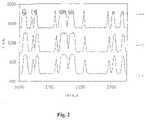
- FIG. 2shows the effect of defocused optics in a simulated electrophoresis trace.
- FIG. 3shows the effect of residual crosstalk in a simulated electrophoresis trace.
- FIGS. 4A and Bshow the effect of high electric field strength on separation in the “biased reptation” regime.
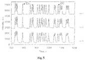
- FIG. 5shows a simulated electrophoresis trace with peak shape distortion.
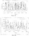
- FIG. 6shows exponential decay of signal intensity as a function of base number.
- FIG. 7shows the effect of a defective primer.
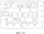
- FIGS. 8A–Cshow simulation of the effect of sample contamination.
- FIGS. 9A and Bshow the use of the electrophoretic trace simulator to add distortions or nosie to experimental data traces.
- the present inventionprovides a method and apparatus for generation of a simulated electrophoretic trace.
- the Electrophoretic Trace Simulatorprovides an algorithm for calculation of a set of electrophoresis traces, corresponding to the output of an automated fluorescent DNA sequencer (slab or capillary gels). This algorithm has several features, which will now be described.
- the term “input file”refers to a user-defined sequence of nucleotide bases, represented as a .txt or text file, that is used as the input to the trace simulator algorithm.
- This textrepresents a DNA sequence of interest, and consists of a string of letters A, C, G and T in the order corresponding to the sequence under consideration.
- the programIn addition to the use of .txt file as an input, the program also allows experimentally recorded data files (output from automated DNA sequencers) to be used. These data files can be manipulated to induce distortions such as shift between traces, stretching, base line drift and jumps, cross talk or noise. Data files modified in such fashion can then be used to test the capabilities of base calling software.
- the usercan define the functional dependence of intensity on N by means of a formula.
- an exponential decay modelis assumed. This is consistent with a mechanistic model of dideoxy-chain-termination DNA sequencing, in which there is a constant probability, at each template position, of chain-termination by incorporation of a dideoxy-nucleotide into the primer-extension product(s).
- the fundamental shape of the function I i (N)is expected to be the same for all DNA sequencing ladders.
- both the absolute intensity and decay ratemay vary from one base type to another.
- the Electrophoretic Trace Simulatortakes this band-broadening effect into account.
- Various physical processescontribute to DNA band broadening during migration of DNA bands through an electrophoretic sequencing gel. The most important of these are: (1) the initial band width defined by loading or over-loading; (2) diffusion (thermal and electric field enhanced) during the electrophoresis; (3) non-homogeneities in the Joule heating or heat-exchange within the gel; (4) electro-osmotic flow (especially for capillary gels).
- the peaks in an electrophoresis runhave a Gaussian shape.
- the width of the Gaussian functionis affected by the above factors, which are considered to be independent.
- the square of total peak width (variance)can be expressed as the sum of squares of the individual factors (see for example, [1–4]).
- a Gaussian peak shape functionFor the peak corresponding to base number N, which migrates past a fixed detector as a function of time t, a Gaussian peak shape function is calculated using the following formula:
- I N ⁇ ( t )I N0 ⁇ N ⁇ 2 ⁇ ⁇ ⁇ ⁇ exp ⁇ ( - ( t - t N ) 2 2 ⁇ ( ⁇ N ) 2 ) ( 3 )
- I N (t)fluorescent intensity at time t, within the peak corresponding to base number N
- I N0intensity maximum of this peak
- ⁇ Nwidth of peak (Gaussian standard deviation)
- t Ncenter of the peak. Note that the calculation is performed in the time domain. Note also that ⁇ N is not a constant, but rather is a function of N.
- the peak width function ⁇ Ndepends not only on N, but also on various experimental conditions such as electric field strength, gel temperature, gel loading conditions, etc.
- the diffusional component of bandwidthis predicted to have the following form: dif2(N) ⁇ N ⁇ 1.8–2 [2]. It should be noted that, if a theoretical model of diffusional peak broadening is used, then this model should also include a term for electric field dependence (see, for example [6]).
- peak skewThe underlying reasons for peak skew are numerous and varied. One factor that causes this type of distortion is a concentration overload. The peak shape in such a situation was studied for example by [8–10]. Another reason for peak skew is the fact that electrophoresis and diffusion are coupled, leading to a trailing edge effect [6]. A third reason may be non-homogeneity in the pore size distribution in the gels [11].
- the Electrophoretic Trace Simulatortreats the migration of DNA bands by introducing a time scale t, in place of a base number scale.
- This time scaleis an analytic representation of the electrophoresis time as a function of base number. Examples of such functions for different experimental conditions and for different models of electrophoresis can be found in the literature (see [12, 13] for example).
- t ⁇ ( N )t 0 + ⁇ i ⁇ a i ⁇ N i ( 4 )
- a iare the polynomial coefficients
- iis the degree of the polynomial.
- the degree of the polynomial and the polynomial coefficientsare found empirically, by using the least-squares method to fit the calculated curve to the experimental data. The same approach can be taken to simulate electrophoretic traces for experimental conditions that correspond to boundaries between different separation regimes, where it may be difficult to suggest an analytic description. 6. Background (or Baseline)
- the Electrophoretic Trace Calculatorhas an option of adding background to a calculated trace, in the form of a polynomial function.
- Electrophoretic Trace Simulatorconsiders two separate cases, which represent different types of noise.
- instrument noisewhich is always present in an experimental data set.
- the typical value of RMS (root mean square) noise in the VGI sequencer's data output streamis about 0.2–1% of the peak intensity.
- Such low values of instrument noisetypically do not affect base calling.
- instrument noisemay become important for traces that exhibit low signal intensities.
- Instrument noiseis modeled by the Electrophoretic Trace Simulator in the form of a sequence of random numbers in the range (0–1), which are multiplied by a coefficient representing maximum amplitude of the noise. This noise is added to the calculated trace.
- the other type of noisecan be designated “chemical noise”. Its origin is to be found in the sequencing chemistry, and typically is due to a combination of false priming and false termination events, catalyzed by the DNA polymerase enzyme. It is exacerbated when an impure sequencing template or impure primers are used.
- Chemical noisemay occasionally be severe enough, especially when combined with a low intensity of the true sequence, to produce base-calling errors.
- a pure simulation of chemical noiseis difficult to describe analytically, with the exception of a specific false priming event, for which the sequence producing the “chemical noise” might be known.
- Such a casecan be modeled by specifying a text file corresponding to the distorting sequence, calculating the resultant electrophoretic trace, and adding it to the trace from the original sequence under simulation.
- Chemical noise that is due to random priming or termination events, and thus unpredictable,could be treated by using, as input to the Electrophoretic Trace Simulator, a random sequence, having an intensity distribution set at some fraction, say 10%, of the primary sequence. The primary sequence trace and the trace due to the chemical noise would then be added together point by point.
- the Electrophoretic Trace Simulatorallows the calculation of traces with over-(or under-) compensation, with an adjustable amplitude. This allows one to estimate the impact on base-calling accuracy.
- Electrophoretic Trace Simulatorcould produce acceptable control traces, we next used the program to model various situations in which instrument factors were expected to limit performance.
- FIG. 1Bshows the results of a trace calculation for a low Signal/Noise ratio.
- the signalis decreased by a factor of ⁇ 3x, and the noise is increased by a factor of ⁇ 2x relative to the trace shown in FIG. 1A .
- the diagnostic “5-0-4” peak, between nucleotide positions 312–331 in the M13mp18 T-ladderis shown.
- FIG. 1Cshows a calculated trace in which a linear baseline drift is introduced.
- the drifthas an approximate magnitude of +3 counts/10 sec. In practice, such an effect can be observed during warm-up of the instrument, or because of instability in the amplifier output.
- FIG. 1Dshows a calculated trace in which a baseline jump of ⁇ 200 counts occurs, shortly after the T-331 peak. In practice, this could be caused by a sudden increase in scattering of excitation laser light, due to the appearance of a bubble in the gel, or a dust particle in the optical path. It could also be caused by an instability in the instrument electronics.
- FIG. 2presents the results of a simple trace calculation, in which optical defocusing is simulated by averaging adjacent data points in the trace. Laser spot diameters of 40, 80, and 200 ⁇ m diameter are simulated in Curves 1 , 2 , 3 respectively. Averaging was performed in the second two simulations on 2 and 5 points respectively. A more sophisticated approach to this problem would involve convoluting the Gaussian peak function (eq. 3) with a blurring or smearing function, that could be computed from an optical analysis [15].
- Gaussian peak functioneq. 3
- the Visible Genetics sequencing platformemploys two dyes (Cy5 and Cy5.5) which display significant overlap in their fluorescence emission spectra.
- the coefficient that describes cross-talk from the Cy5 channel to the Cy5.5 channelhas a value of about 0.5
- the coefficient describing cross-talk from the Cy5.5 channel to the Cy5 channelhas a value of about 0.1.
- the disparity between the two cross-talk coefficientsis the result of the asymmetric shapes of the emission spectra of the two dyes.
- Four-color sequencersalso experience a relatively high level of cross-talk. Appropriate compensation probably is essential even for a low level of cross-talk.
- FIG. 3shows the calculated traces for a situation in which the cross-talk is under-compensated. Curves 1 and 2 in this Figure show the traces calculated with no cross-talk for the A-track and C-track respectively, around position 160–190 in the M13mp18 sequence.
- Curve 3shows a situation in which approximately 10% of the intensity of the C-track is cross-registered in the A channel.
- the cross-registered peaksalthough small, are of positive intensity, and thus could conceivably be mistaken for real peaks by the base-calling software.
- An over-compensationleads to negative peaks, which are not likely to be selected by the base-calling software.
- the chemistry of automated fluorescent DNA sequencingis relatively complex, and there are many opportunities where chemical raw materials, intermediates, and reaction steps may be non-optimal.
- the Electrophoretic Trace Simulatorcan be used to help troubleshoot problems encountered in the sequencing chemistry, as two examples will illustrate.
- FIGS. 6A and Bcompares the results of simulation ( FIG. 6A ) with an actual experimental observation of the T-track of M13mp18 ( FIG. 6B ).
- the Electrophoretic Trace Simulatorcan also be used within a risk analysis framework, to explore the consequences of possible defects in the manufacture of a genotyping kit.
- This simulationconsiders the possibility that, during the stepwise synthesis of a primer using phosphoramidite technology, only the first (m ⁇ 1) synthesis cycles are completed with high efficiency. This would produce a situation in which a truncated primer, (m ⁇ 1) nucleotides long and missing its 5′-terminal nucleotide, was a minor contaminant of the desired final primer (m nucleotides long). If a subsequent primer purification step was not performed, or was inadequate, then a mixture of two primers (of lengths m and m ⁇ 1) would result.
- the Electrophoretic Trace Simulatorcan also be used within a risk analysis framework, to explore the consequences of a common problem in the use of a genotyping kit. In a field setting, it is essential to be able to detect cross-contamination of patient samples.
- the Electrophoretic Trace Simulatorcan be used to model the effects of mixing together two related (but non-identical) templates.
- FIG. 8Ashows a set of four traces (A, C, G, T) that are computed over nucleotide positions 150–180 of the protease gene of a B-subtype HIV-1 sequence (GenBank # HIVHXB2CG).
- FIG. 8Bshows a set of traces for this same region, computed for an F-subtype HIV-1 sequence (GenBank # AY010409).
- FIG. 8Cthe two sequences have been mixed together in a 1:1 ratio (as might be expected to occur for a case of sample contamination). At some of the positions (e.g. #153), there is no change. However, at other positions (e.g. #160) a mixture of two bases (A, G) is observed.
- Electrophoretic tracesmay, in some cases, be too complex to simulate accurately. It may still be important to study the influence of various experimental conditions on them. For example, it would be useful to have the ability to introduce a known distortion into an experimentally recorded trace, and then to observe the effect of this distortion on subsequent base-calling. In order to have this capability, a function is incorporated into the Electrophoretic Trace Simulator, which allows the input of a data file typical for the output of the DNA sequencer comprising a set of data points (fluorescent intensity at each of a series of discrete time points).
- an experimental trace(in all its complexity) is used as a starting point, and is modified by adding special features of interest (residual cross-talk, baseline jumps, excessive instrument noise, etc.)
- This approachconveniently removes the necessity of generating chemical noise, for example, or other features by a theoretical model, which may be simplistic or flawed.
- a sample calculation of this typeis shown in FIG. 9 .
- Panel Aa portion of an experimentally recorded electrophoretic trace from Cy5.5-labeled M13mp18 sequence is shown, from the primer peak to position ⁇ 100.
- the traces (A, C, G, T)are well-aligned and relatively noise-free.
- Panel Bshows the effect of using the Electrophoretic Trace Simulator to introduce time-delays of 60, 60, and 120 see between adjacent traces.
- signal intensitywas decreased and instrument noise was added to the C-trace.
- the Electrophoretic Trace Simulatorwas used to calculate sets of electrophoresis traces that correspond to outputs from an automated fluorescent DNA sequencer.
- a user-defined sequence of bases(.txt file) serves as input for such calculations.
- the ETSemploys user-adjustable functions for the following critical parameters of a DNA sequencing electropherogram: peak shape, peak spacing, dependence of peak intensity on base number, background, noise, and cross talk correction. It was shown that the Electrophoretic Trace Simulator can be used to model or predict the behavior of a DNA sequencing system, taking into account effects at the levels of: (1) the system hardware, (2) the electrophoretic gel, (3) the sequencing chemistry, and (4) the sequencing template.
- a first aspect of the inventionis a method for creating a simulated electrophoretic trace.
- the methodcomprises the steps of obtaining an input file providing a base sequence comprising a string of letters (A, C, G and/or T) in an order corresponding to the input base sequence. These letters correspond to peaks, and are presumed in the initial data set to have equal spacing, equal peak heights (intensities) and equal widths.
- This data file of the input base sequenceis then modified using one or more, and preferably a plurality of functions to take into account perturbations associated with (1) changes in peak intensity as a function of base number; (2) peak shape as a function of base number; (3) peak skew; (4) spacing between peaks; (5) background; (6) noise; (7) spectral cross-talk; (8) instrumental effects and (9) gel electrophoresis effects. It is not required to use all of the these functions to arrive at a suitable simulated electrophoretic trace. Where, for example, the length of the sequence is short, changes to peak intensity, or to peak shape may be minimal between the starting and ending base, making use of these functions unnecessary.
- functions addressing instrumental effects and gel electrophoresis effectsmay have multiple components of which only some may be relevant for a given instrumental system or gel type.
- Appropriate functions for instrumental effects and gel electrophoresis effectscan be determined by reverse calculation from sequencing runs performed under a set of standard conditions (for the instruments and gels being used) using reference standards of known sequence.
- the method of the inventionmay be practiced using an apparatus in accordance with the invention.
- the apparatuscomprises a programmed computer processor, for example a personal computer using any of several standard operating systems (Windows, Unix, Linux and the like) connected for communication to a storage device having stored thereon a stored set of program instructions for carrying out the method.
- Input devicessuch as keyboards, pointing devices, wired or wireless network connections, voice-recognizing audio-input systems, or movable storage media (for example diskettes) may be used to acquire parameter values which are used for processing the input sequence into a simulated electrophoresis trace.
- a video displaymay be provided as part of the apparatus to enhance the parameter input interface and display the results.
- the apparatusmay be a dedicated apparatus which performs no other DNA sequencing-related function, or it may be a combination apparatus in which comparison of the simulated electrophoresis trace with an experimental data trace is performed to achieve analysis of a sample.
Landscapes
- Health & Medical Sciences (AREA)
- Life Sciences & Earth Sciences (AREA)
- Molecular Biology (AREA)
- Chemical & Material Sciences (AREA)
- Chemical Kinetics & Catalysis (AREA)
- Electrochemistry (AREA)
- Physics & Mathematics (AREA)
- Analytical Chemistry (AREA)
- Biochemistry (AREA)
- General Health & Medical Sciences (AREA)
- General Physics & Mathematics (AREA)
- Immunology (AREA)
- Pathology (AREA)
- Measuring Or Testing Involving Enzymes Or Micro-Organisms (AREA)
Abstract
Description
Ii(N)=Ioiexp(−βiN) (1)
where i=type of base (A, C, G, T), N=base number, Ii(N)=intensity of ladder of type i as function of base number, I0i=intensity of unextended primer within ladder of type i, and βi=exponential decay parameter for the intensity of ladder of type i.
Ii(N)=I0i/(N−α)γ (2)
where a and γ are user-adjustable parameters which can be estimated from experimental data sets for different DNA sequencing reactions.
3. Peak Shape as a Function of Base Number
where IN(t)=fluorescent intensity at time t, within the peak corresponding to base number N; IN0=intensity maximum of this peak, σN=width of peak (Gaussian standard deviation), and tN=center of the peak. Note that the calculation is performed in the time domain. Note also that σNis not a constant, but rather is a function of N.
- (1) The first is a combination of Gaussian and linear functions. For a peak with a leftwards skew (tail at the lagging edge), the left part of the peak is calculated is a weighted average of an increasing linear function and a Gaussian function. The right part of the peak is kept as a Gaussian. For the peak with a rightwards skew, the complementary approach is taken.
- (2) The second method for treating peak distortion combines a Gaussian function with the function f=b/(t−tN)c. The constant b is a scaling factor, which in a combination with the power c allows a modification of the shoulder of the peak.
- (3) In the third approach for treating peak distortion, a Gaussian function is used for both halves of the peak, but different peak widths are used for the left and right sides of the peak. This approach gives a smooth curve and considerable flexibility in peak characterization.
5. Spacing Between Peaks
where aiare the polynomial coefficients, and i is the degree of the polynomial. The degree of the polynomial and the polynomial coefficients are found empirically, by using the least-squares method to fit the calculated curve to the experimental data. The same approach can be taken to simulate electrophoretic traces for experimental conditions that correspond to boundaries between different separation regimes, where it may be difficult to suggest an analytic description.
6. Background (or Baseline)
- [1] Giddings, J. C., Dynamics of Chromatography, Marcel Dekker, New York, 1965.
- [2] Brahmasandra, S. N., Burke, D. T., Mastragelo, C. H., Bums, M. A., Electrophoresis 2001, 22, 1046–1062.
- [3] Heller, C., Electrophoresis 1999, 20, 1978–1986.
- [4] Kenndler, E., in: Khaledi, M. G. (Ed.), High-Performance Capillary Electrophoresis. Theory, Techniques and Applications, John Wiley & Sons, Inc., New York, 1998, pp. 25–76.
- [5] Slater, G. W., Guo, H. L., Electrophoresis 1995, 16, 11–15.
- [6] Slater, G. W., in: Heller, C. (Ed.), Analysis of Nucleic Acids by Capillary Electrophoresis, Vieweg Verlagsgesellschaft, Weisbaden 1997, pp. 24–66.
- [7] Djouadi, Z., Bottani, S., Duval, M.-A., Siebert, R., Tricoire H., Valentin, L., Electrophoresis 2001, 22, 3527–3532.
- [8] Mikkers, F. E. P., Everaerts, F. M., Verheggen, Th. P. E. M., J. Chromatogr. 1979, 169, 1.
- [9] Thornann, W., Electrophoresis 1983, 4, 383.
- [10] Pope, H., Anal. Chem. 1992, 64, 1908.
- [11] Yager, T. D., et all, Electrophoresis 1999, 20, 1280–1300.
- [12] Luckey, J. A., Norris, T. B., Smith, L. M., J. Phys. Chem. 1993, 97, 3067–3075.
- [13] Luckey, J. A., Smith, L. M., Electrophoresis 1993, 14, 492–501.
- [14] Izmailov, A., Yager, T. D., Zaleski, H., Darasch S., Electrophoresis 2001, 22, 1906–1914.
- [15] Brigham, E. O., The Fast Fourier Transform, Prentice-Hall, Englewood-Cliffs, 1974.
- [16] Barron, A. E., Heller, C., in: Heller, C. (Ed.), Analysis of Nucleic Acids by Capillary Electrophoresis, Vieweg Verlagsgesellschaft, Weisbaden 1997, pp. 93–124.
Claims (8)
Ii(N)=Ioiexp(−βiN)
Priority Applications (1)
| Application Number | Priority Date | Filing Date | Title |
|---|---|---|---|
| US10/295,964US7222059B2 (en) | 2001-11-15 | 2002-11-15 | Electrophoretic trace simulator |
Applications Claiming Priority (2)
| Application Number | Priority Date | Filing Date | Title |
|---|---|---|---|
| US33247101P | 2001-11-15 | 2001-11-15 | |
| US10/295,964US7222059B2 (en) | 2001-11-15 | 2002-11-15 | Electrophoretic trace simulator |
Publications (2)
| Publication Number | Publication Date |
|---|---|
| US20030120471A1 US20030120471A1 (en) | 2003-06-26 |
| US7222059B2true US7222059B2 (en) | 2007-05-22 |
Family
ID=26969427
Family Applications (1)
| Application Number | Title | Priority Date | Filing Date |
|---|---|---|---|
| US10/295,964Expired - Fee RelatedUS7222059B2 (en) | 2001-11-15 | 2002-11-15 | Electrophoretic trace simulator |
Country Status (1)
| Country | Link |
|---|---|
| US (1) | US7222059B2 (en) |
Cited By (2)
| Publication number | Priority date | Publication date | Assignee | Title |
|---|---|---|---|---|
| US20100074492A1 (en)* | 2008-04-04 | 2010-03-25 | Massachusetts Institute Of Technology | Methods and apparatus for automated base-calling on multiple dna strands |
| US20220236183A1 (en)* | 2019-05-22 | 2022-07-28 | Hitachi High-Tech Corporation | Analysis device and analysis method |
Families Citing this family (10)
| Publication number | Priority date | Publication date | Assignee | Title |
|---|---|---|---|---|
| US7689394B2 (en)* | 2003-08-26 | 2010-03-30 | Siemens Industry, Inc. | System and method for remotely analyzing machine performance |
| US9388462B1 (en)* | 2006-05-12 | 2016-07-12 | The Board Of Trustees Of The Leland Stanford Junior University | DNA sequencing and approaches therefor |
| US11940413B2 (en) | 2007-02-05 | 2024-03-26 | IsoPlexis Corporation | Methods and devices for sequencing nucleic acids in smaller batches |
| US11035823B2 (en) | 2009-03-17 | 2021-06-15 | Qiagen Sciences, Llc | Methods and devices for sequencing nucleic acids in smaller batches |
| US8481259B2 (en) | 2007-02-05 | 2013-07-09 | Intelligent Bio-Systems, Inc. | Methods and devices for sequencing nucleic acids in smaller batches |
| US8623598B2 (en)* | 2008-03-19 | 2014-01-07 | Intelligent Bio Systems, Inc. | Methods and compositions for inhibiting undesired cleaving of labels |
| US9017973B2 (en) | 2008-03-19 | 2015-04-28 | Intelligent Biosystems, Inc. | Methods and compositions for incorporating nucleotides |
| US10745740B2 (en) | 2008-03-19 | 2020-08-18 | Qiagen Sciences, Llc | Sample preparation |
| GB2600066B (en) | 2015-12-04 | 2022-11-02 | Emulate Inc | Open-Top Microfluidic Device With Structural Anchors |
| CN114062469B (en)* | 2021-11-18 | 2022-11-29 | 上海交通大学 | Method for improving electrophoretic separation efficiency of zone |
Citations (111)
| Publication number | Priority date | Publication date | Assignee | Title |
|---|---|---|---|---|
| US3742202A (en) | 1970-11-20 | 1973-06-26 | Bodenseewerk Perkin Elmer Co | Peak integrator |
| US4329591A (en) | 1979-06-08 | 1982-05-11 | Olympus Optical Co., Ltd. | Method for detecting samples |
| US4363705A (en) | 1981-07-16 | 1982-12-14 | Capitol Records, Inc. | Passivating and silver removal method |
| EP0198403A2 (en) | 1985-04-09 | 1986-10-22 | Fuji Photo Film Co., Ltd. | Signal processing method for determining base sequence of nucleic acid |
| US4683202A (en) | 1985-03-28 | 1987-07-28 | Cetus Corporation | Process for amplifying nucleic acid sequences |
| US4683195A (en) | 1986-01-30 | 1987-07-28 | Cetus Corporation | Process for amplifying, detecting, and/or-cloning nucleic acid sequences |
| US4683194A (en) | 1984-05-29 | 1987-07-28 | Cetus Corporation | Method for detection of polymorphic restriction sites and nucleic acid sequences |
| US4720786A (en) | 1985-04-19 | 1988-01-19 | Fuji Photo Film Co., Ltd. | Method of compensating for offset distortion in rows of electrophoretic patterns |
| US4729947A (en) | 1984-03-29 | 1988-03-08 | The Board Of Regents Of The University Of Nebraska | DNA sequencing |
| EP0265293A2 (en) | 1987-01-14 | 1988-04-27 | President And Fellows Of Harvard College | T7 DNA polymerase |
| EP0294524A1 (en) | 1987-06-09 | 1988-12-14 | The Perkin-Elmer Corporation | Real time scanning electrophoresis apparatus for DNA sequencing |
| US4795699A (en) | 1987-01-14 | 1989-01-03 | President And Fellows Of Harvard College | T7 DNA polymerase |
| US4800159A (en) | 1986-02-07 | 1989-01-24 | Cetus Corporation | Process for amplifying, detecting, and/or cloning nucleic acid sequences |
| US4811218A (en) | 1986-06-02 | 1989-03-07 | Applied Biosystems, Inc. | Real time scanning electrophoresis apparatus for DNA sequencing |
| US4823007A (en) | 1986-12-15 | 1989-04-18 | Norand Corporation | DNA sequencing gel reading system and method |
| US4849513A (en) | 1983-12-20 | 1989-07-18 | California Institute Of Technology | Deoxyribonucleoside phosphoramidites in which an aliphatic amino group is attached to the sugar ring and their use for the preparation of oligonucleotides containing aliphatic amino groups |
| US4855225A (en) | 1986-02-07 | 1989-08-08 | Applied Biosystems, Inc. | Method of detecting electrophoretically separated oligonucleotides |
| WO1989007149A1 (en) | 1988-01-28 | 1989-08-10 | Mayo Foundation For Medical Education And Research | Genomic amplification with direct sequencing |
| US4865968A (en) | 1985-04-01 | 1989-09-12 | The Salk Institute For Biological Studies | DNA sequencing |
| US4889818A (en) | 1986-08-22 | 1989-12-26 | Cetus Corporation | Purified thermostable enzyme |
| GB2225139A (en) | 1988-11-16 | 1990-05-23 | Atomic Energy Authority Uk | Method for spectrum matching |
| US4941092A (en) | 1985-05-23 | 1990-07-10 | Fuji Photo Film Co., Ltd. | Signal processing method for determining base sequence of nucleic acid |
| US4942124A (en) | 1987-08-11 | 1990-07-17 | President And Fellows Of Harvard College | Multiplex sequencing |
| US4960999A (en) | 1989-02-13 | 1990-10-02 | Kms Fusion, Inc. | Scanning and storage of electrophoretic records |
| US4965188A (en) | 1986-08-22 | 1990-10-23 | Cetus Corporation | Process for amplifying, detecting, and/or cloning nucleic acid sequences using a thermostable enzyme |
| US4982326A (en) | 1986-03-05 | 1991-01-01 | Fuji Photo Film Co., Ltd. | Method for analyzing autoradiograph for determining base sequence of nucleic acid |
| US5008182A (en) | 1986-01-10 | 1991-04-16 | Cetus Corporation | Detection of AIDS associated virus by polymerase chain reaction |
| US5075216A (en) | 1988-09-23 | 1991-12-24 | Cetus Corporation | Methods for dna sequencing with thermus aquaticus dna polymerase |
| US5079352A (en) | 1986-08-22 | 1992-01-07 | Cetus Corporation | Purified thermostable enzyme |
| US5096557A (en) | 1990-07-11 | 1992-03-17 | Genetype A.G. | Internal standard for electrophoretic separations |
| EP0476712A1 (en) | 1986-03-26 | 1992-03-25 | Fuji Photo Film Co., Ltd. | Signal processing method for determining base sequence of nucleic acid |
| US5108179A (en) | 1989-08-09 | 1992-04-28 | Myers Stephen A | System and method for determining changes in fluorescence of stained nucleic acid in electrophoretically separated bands |
| US5118800A (en) | 1983-12-20 | 1992-06-02 | California Institute Of Technology | Oligonucleotides possessing a primary amino group in the terminal nucleotide |
| US5119316A (en) | 1990-06-29 | 1992-06-02 | E. I. Du Pont De Nemours And Company | Method for determining dna sequences |
| US5122345A (en) | 1988-07-12 | 1992-06-16 | President And Fellows Of Harvard College | Dna sequencing apparatus |
| US5124247A (en) | 1988-12-09 | 1992-06-23 | Europaisches Laboratorium Fur Molekularbiologie (Embl) | Process for sequencing nucleic acids |
| EP0504943A2 (en) | 1991-03-22 | 1992-09-23 | Hitachi Software Engineering Co., Ltd. | Multi-colored electrophoresis pattern reading system |
| WO1992018650A1 (en) | 1991-04-11 | 1992-10-29 | Baxter Diagnostics Inc. | Detection of dna/rna by fluorescence polarization |
| US5171534A (en) | 1984-01-16 | 1992-12-15 | California Institute Of Technology | Automated DNA sequencing technique |
| US5175082A (en) | 1986-03-19 | 1992-12-29 | Imperial Chemical Industries Plc | Method of characterizing genomic dna |
| US5176995A (en) | 1985-03-28 | 1993-01-05 | Hoffmann-La Roche Inc. | Detection of viruses by amplification and hybridization |
| WO1993002212A1 (en) | 1991-07-24 | 1993-02-04 | University Partnerships Pty. Ltd. | Single step amplification and sequencing of nucleic acids |
| WO1993008305A1 (en) | 1991-10-17 | 1993-04-29 | Dynal As | Method of sequencing double stranded dna |
| US5207880A (en) | 1984-03-29 | 1993-05-04 | The Board Of Regents Of The University Of Nebraska | DNA sequencing |
| US5213673A (en) | 1990-11-30 | 1993-05-25 | Hitachi Software Engineering Co., Ltd. | Multi-colored electrophoresis pattern reading apparatus |
| US5246866A (en) | 1987-12-23 | 1993-09-21 | Hitachi Software Engineering Co., Ltd. | Method for transcription of a DNA sequence |
| US5268486A (en) | 1986-04-18 | 1993-12-07 | Carnegie-Mellon Unversity | Method for labeling and detecting materials employing arylsulfonate cyanine dyes |
| US5273632A (en) | 1992-11-19 | 1993-12-28 | University Of Utah Research Foundation | Methods and apparatus for analysis of chromatographic migration patterns |
| US5283171A (en) | 1988-09-09 | 1994-02-01 | Hoffmann-La Roche Inc. | Compositions for and detection of human papillomavirus by specific oligonucleotide polymerase primers using the polymerase chain reaction |
| US5290419A (en) | 1992-04-14 | 1994-03-01 | Hitachi, Ltd. | Fluorescence detection type electrophoresis apparatus |
| EP0592060A1 (en) | 1992-10-09 | 1994-04-13 | The Board of Regents of the University of Nebraska | Digital DNA typing |
| US5308751A (en) | 1992-03-23 | 1994-05-03 | General Atomics | Method for sequencing double-stranded DNA |
| DE4405251A1 (en) | 1993-02-19 | 1994-08-25 | Olympus Optical Co | Method for processing electrophoretic data |
| US5352600A (en) | 1986-08-22 | 1994-10-04 | Hoffmann-La Roche Inc. | Purified thermostable enzyme |
| US5360523A (en) | 1984-03-29 | 1994-11-01 | Li-Cor, Inc. | DNA sequencing |
| US5365455A (en) | 1991-09-20 | 1994-11-15 | Vanderbilt University | Method and apparatus for automatic nucleic acid sequence determination |
| WO1994026894A1 (en) | 1993-05-14 | 1994-11-24 | The Government Of The United States Of America As | VON HIPPEL-LINDAU (VHL) DISEASE GENE AND CORRESPONDING cDNA AND METHODS FOR DETECTING CARRIERS OF THE VHL DISEASE GENE |
| WO1995004140A1 (en) | 1993-07-28 | 1995-02-09 | Akzo Nobel N.V. | Process for isolating nucleic acid from gram positive microorganisms |
| WO1995006756A2 (en) | 1993-09-03 | 1995-03-09 | Abbott Laboratories | OLIGONUCLEOTIDES AND METHODS FOR THE DETECTION OF $i(CHLAMYDIA TRACHOMATIS) |
| WO1995011961A1 (en) | 1993-10-22 | 1995-05-04 | The University Of Utah | Automated hybridization/imaging device for fluorescent multiplex dna sequencing |
| US5418149A (en) | 1990-07-24 | 1995-05-23 | Hoffmann-La Roche Inc. | Reduction of non-specific amplification glycosylase using DUTP and DNA uracil |
| US5419825A (en) | 1991-07-29 | 1995-05-30 | Shimadzu Corporation | Base sequencing apparatus |
| EP0655506A1 (en) | 1994-10-17 | 1995-05-31 | President And Fellows Of Harvard College | DNA polymerases having modified nucleotide binding site for DNA sequencing |
| US5427911A (en) | 1990-05-01 | 1995-06-27 | Yale University | Coupled amplification and sequencing of DNA |
| US5453355A (en) | 1990-01-26 | 1995-09-26 | Abbott Laboratories | Oligonucleotides and methods for the detection of Neisseria gonorrhoeae |
| WO1996001008A1 (en) | 1994-06-30 | 1996-01-11 | Honeywell Inc. | Termination circuit for high speed applications |
| US5484701A (en) | 1990-01-26 | 1996-01-16 | E. I. Du Pont De Nemours And Company | Method for sequencing DNA using biotin-strepavidin conjugates to facilitate the purification of primer extension products |
| WO1996001909A1 (en) | 1994-07-08 | 1996-01-25 | Visible Genetics Inc. | METHOD, REAGENTS AND KIT FOR DIAGNOSIS AND TARGETED SCREENING FOR p53 MUTATIONS |
| US5502773A (en) | 1991-09-20 | 1996-03-26 | Vanderbilt University | Method and apparatus for automated processing of DNA sequence data |
| US5527898A (en) | 1988-09-09 | 1996-06-18 | Hoffmann-La Roche Inc. | Detection of human papillomavirus by the polymerase chain reaction |
| WO1996035810A1 (en) | 1995-05-09 | 1996-11-14 | Curagen Corporation | Apparatus and method for the generation, separation, detection, and recognition of biopolymer fragments |
| US5584983A (en) | 1992-02-26 | 1996-12-17 | Stork Screens, B.V. | Method for the production of a metal foam |
| WO1997002488A1 (en) | 1995-06-30 | 1997-01-23 | Visible Genetics Inc. | Method and system for dna sequence determination and mutation detection |
| US5608063A (en) | 1986-07-02 | 1997-03-04 | E. I. Du Pont De Nemours And Company | Method, system and reagents for DNA sequencing |
| US5614386A (en) | 1995-06-23 | 1997-03-25 | Baylor College Of Medicine | Alternative dye-labeled primers for automated DNA sequencing |
| US5666435A (en) | 1994-12-09 | 1997-09-09 | Genomyx Corporation | System for analysis of x-ray films of nucleotide sequences |
| US5667971A (en) | 1990-08-07 | 1997-09-16 | Dade Chemistry Systems Inc. | Method for determining DNA sequences |
| WO1997040184A1 (en) | 1996-04-18 | 1997-10-30 | Visible Genetics Inc. | Method, apparatus and kits for sequencing of nucleic acids using multiple dyes |
| WO1997041259A1 (en) | 1996-05-01 | 1997-11-06 | Visible Genetics Inc. | Method for sequencing of nucleic acid polymers |
| WO1998000708A1 (en) | 1996-06-27 | 1998-01-08 | Visible Genetics Inc. | Method and apparatus for alignment of signals for use in dna base-calling |
| US5710628A (en) | 1994-12-12 | 1998-01-20 | Visible Genetics Inc. | Automated electrophoresis and fluorescence detection apparatus and method |
| US5712476A (en) | 1995-05-30 | 1998-01-27 | Visible Genetics Inc. | Electrophoresis and fluorescence detection apparatus |
| WO1998011258A1 (en) | 1996-09-16 | 1998-03-19 | University Of Utah Research Foundation | Method and apparatus for analysis of chromatographic migration patterns |
| US5733729A (en) | 1995-09-14 | 1998-03-31 | Affymetrix, Inc. | Computer-aided probability base calling for arrays of nucleic acid probes on chips |
| US5751534A (en) | 1996-05-29 | 1998-05-12 | Lucent Technologies Inc. | Coaxial cable surge protector |
| WO1998024930A1 (en) | 1996-12-05 | 1998-06-11 | The Perkin-Elmer Corporation | Chain-termination type nucleic acid sequencing method including 2'-deoxyuridine-5'-triphosphate |
| US5776737A (en) | 1994-12-22 | 1998-07-07 | Visible Genetics Inc. | Method and composition for internal identification of samples |
| US5789168A (en) | 1996-05-01 | 1998-08-04 | Visible Genetics Inc. | Method for amplification and sequencing of nucleic acid polymers |
| US5821058A (en) | 1984-01-16 | 1998-10-13 | California Institute Of Technology | Automated DNA sequencing technique |
| US5830657A (en) | 1996-05-01 | 1998-11-03 | Visible Genetics Inc. | Method for single-tube sequencing of nucleic acid polymers |
| US5834189A (en) | 1994-07-08 | 1998-11-10 | Visible Genetics Inc. | Method for evaluation of polymorphic genetic sequences, and the use thereof in identification of HLA types |
| US5849542A (en) | 1993-11-17 | 1998-12-15 | Amersham Pharmacia Biotech Uk Limited | Primer extension mass spectroscopy nucleic acid sequencing method |
| WO1998041650A3 (en) | 1997-03-18 | 1999-01-14 | Visible Genetics Inc | Method and kit for quantitation and nucleic acid sequencing of nucleic acid analytes in a sample |
| US5891632A (en) | 1995-07-27 | 1999-04-06 | Fujitsu Limited | Method and device for fragment trace data display in DNA base sequencing |
| US5981186A (en) | 1995-06-30 | 1999-11-09 | Visible Genetics, Inc. | Method and apparatus for DNA-sequencing using reduced number of sequencing mixtures |
| US6013444A (en) | 1997-09-18 | 2000-01-11 | Oligotrail, Llc | DNA bracketing locus compatible standards for electrophoresis |
| WO2000000637A3 (en) | 1998-06-26 | 2000-02-17 | Visible Genetics Inc | Method for sequencing nucleic acids with reduced errors |
| US6027709A (en) | 1997-01-10 | 2000-02-22 | Li-Cor Inc. | Fluorescent cyanine dyes |
| US6068737A (en) | 1997-05-16 | 2000-05-30 | Simon Bolivar University | Simultaneous demetallization and desulphuration of carbonaceous materials via microwaves |
| US6083699A (en) | 1996-05-01 | 2000-07-04 | Visible Genetics Inc. | Method for bi-directional sequencing of nucleic acid polymers |
| WO2000068410A1 (en) | 1999-05-07 | 2000-11-16 | Visible Genetics Inc. | Nucleic acid sequencing with simultaneous quantitation |
| US6195449B1 (en) | 1997-05-18 | 2001-02-27 | Robert Bogden | Method and apparatus for analyzing data files derived from emission spectra from fluorophore tagged nucleotides |
| US6397150B1 (en)* | 2000-07-27 | 2002-05-28 | Visible Genetics Inc. | Method and apparatus for sequencing of DNA using an internal calibrant |
| US6413718B1 (en) | 1996-05-01 | 2002-07-02 | Visible Genetics Inc. | Method for sequencing of nucleic acid polymers |
| US6428955B1 (en)* | 1995-03-17 | 2002-08-06 | Sequenom, Inc. | DNA diagnostics based on mass spectrometry |
| US20020147548A1 (en)* | 2000-08-14 | 2002-10-10 | Dirk Walther | Basecalling system and protocol |
| US6485625B1 (en)* | 1995-05-09 | 2002-11-26 | Curagen Corporation | Apparatus and method for the generation, separation, detection, and recognition of biopolymer fragments |
| US6716394B2 (en)* | 1998-08-11 | 2004-04-06 | Caliper Technologies Corp. | DNA sequencing using multiple fluorescent labels being distinguishable by their decay times |
| US20040072182A1 (en)* | 1996-11-29 | 2004-04-15 | Victor Lyamichev | Methods and compositions for detecting target sequences |
| US20040117130A1 (en)* | 2000-09-08 | 2004-06-17 | Paracel, Inc. | System and method for improving the accuracy of DNA sequencing and error probability estimation through application of a mathematical model to the analysis of electropherograms |
| US20050042668A1 (en)* | 2000-02-15 | 2005-02-24 | Perlin Mark W. | Method and system for DNA analysis |
- 2002
- 2002-11-15USUS10/295,964patent/US7222059B2/ennot_activeExpired - Fee Related
Patent Citations (123)
| Publication number | Priority date | Publication date | Assignee | Title |
|---|---|---|---|---|
| US3742202A (en) | 1970-11-20 | 1973-06-26 | Bodenseewerk Perkin Elmer Co | Peak integrator |
| US4329591A (en) | 1979-06-08 | 1982-05-11 | Olympus Optical Co., Ltd. | Method for detecting samples |
| US4363705A (en) | 1981-07-16 | 1982-12-14 | Capitol Records, Inc. | Passivating and silver removal method |
| US5118800A (en) | 1983-12-20 | 1992-06-02 | California Institute Of Technology | Oligonucleotides possessing a primary amino group in the terminal nucleotide |
| US4849513A (en) | 1983-12-20 | 1989-07-18 | California Institute Of Technology | Deoxyribonucleoside phosphoramidites in which an aliphatic amino group is attached to the sugar ring and their use for the preparation of oligonucleotides containing aliphatic amino groups |
| US5821058A (en) | 1984-01-16 | 1998-10-13 | California Institute Of Technology | Automated DNA sequencing technique |
| US5171534A (en) | 1984-01-16 | 1992-12-15 | California Institute Of Technology | Automated DNA sequencing technique |
| US4729947A (en) | 1984-03-29 | 1988-03-08 | The Board Of Regents Of The University Of Nebraska | DNA sequencing |
| US5360523A (en) | 1984-03-29 | 1994-11-01 | Li-Cor, Inc. | DNA sequencing |
| US5207880A (en) | 1984-03-29 | 1993-05-04 | The Board Of Regents Of The University Of Nebraska | DNA sequencing |
| US4683194A (en) | 1984-05-29 | 1987-07-28 | Cetus Corporation | Method for detection of polymorphic restriction sites and nucleic acid sequences |
| US4683202B1 (en) | 1985-03-28 | 1990-11-27 | Cetus Corp | |
| US5176995A (en) | 1985-03-28 | 1993-01-05 | Hoffmann-La Roche Inc. | Detection of viruses by amplification and hybridization |
| US4683202A (en) | 1985-03-28 | 1987-07-28 | Cetus Corporation | Process for amplifying nucleic acid sequences |
| US4865968A (en) | 1985-04-01 | 1989-09-12 | The Salk Institute For Biological Studies | DNA sequencing |
| EP0198403A2 (en) | 1985-04-09 | 1986-10-22 | Fuji Photo Film Co., Ltd. | Signal processing method for determining base sequence of nucleic acid |
| US4720786A (en) | 1985-04-19 | 1988-01-19 | Fuji Photo Film Co., Ltd. | Method of compensating for offset distortion in rows of electrophoretic patterns |
| US4941092A (en) | 1985-05-23 | 1990-07-10 | Fuji Photo Film Co., Ltd. | Signal processing method for determining base sequence of nucleic acid |
| US5008182A (en) | 1986-01-10 | 1991-04-16 | Cetus Corporation | Detection of AIDS associated virus by polymerase chain reaction |
| US4683195A (en) | 1986-01-30 | 1987-07-28 | Cetus Corporation | Process for amplifying, detecting, and/or-cloning nucleic acid sequences |
| US4683195B1 (en) | 1986-01-30 | 1990-11-27 | Cetus Corp | |
| US4800159A (en) | 1986-02-07 | 1989-01-24 | Cetus Corporation | Process for amplifying, detecting, and/or cloning nucleic acid sequences |
| US4855225A (en) | 1986-02-07 | 1989-08-08 | Applied Biosystems, Inc. | Method of detecting electrophoretically separated oligonucleotides |
| US4982326A (en) | 1986-03-05 | 1991-01-01 | Fuji Photo Film Co., Ltd. | Method for analyzing autoradiograph for determining base sequence of nucleic acid |
| US5175082A (en) | 1986-03-19 | 1992-12-29 | Imperial Chemical Industries Plc | Method of characterizing genomic dna |
| EP0476712A1 (en) | 1986-03-26 | 1992-03-25 | Fuji Photo Film Co., Ltd. | Signal processing method for determining base sequence of nucleic acid |
| US5268486A (en) | 1986-04-18 | 1993-12-07 | Carnegie-Mellon Unversity | Method for labeling and detecting materials employing arylsulfonate cyanine dyes |
| US4811218A (en) | 1986-06-02 | 1989-03-07 | Applied Biosystems, Inc. | Real time scanning electrophoresis apparatus for DNA sequencing |
| US5608063A (en) | 1986-07-02 | 1997-03-04 | E. I. Du Pont De Nemours And Company | Method, system and reagents for DNA sequencing |
| US4889818A (en) | 1986-08-22 | 1989-12-26 | Cetus Corporation | Purified thermostable enzyme |
| US5079352A (en) | 1986-08-22 | 1992-01-07 | Cetus Corporation | Purified thermostable enzyme |
| US5352600A (en) | 1986-08-22 | 1994-10-04 | Hoffmann-La Roche Inc. | Purified thermostable enzyme |
| US4965188A (en) | 1986-08-22 | 1990-10-23 | Cetus Corporation | Process for amplifying, detecting, and/or cloning nucleic acid sequences using a thermostable enzyme |
| US4823007A (en) | 1986-12-15 | 1989-04-18 | Norand Corporation | DNA sequencing gel reading system and method |
| EP0265293A2 (en) | 1987-01-14 | 1988-04-27 | President And Fellows Of Harvard College | T7 DNA polymerase |
| US4795699A (en) | 1987-01-14 | 1989-01-03 | President And Fellows Of Harvard College | T7 DNA polymerase |
| EP0386859A2 (en) | 1987-01-14 | 1990-09-12 | The President And Fellows Of Harvard College | T7 DNA polymerase |
| EP0294524A1 (en) | 1987-06-09 | 1988-12-14 | The Perkin-Elmer Corporation | Real time scanning electrophoresis apparatus for DNA sequencing |
| US4942124A (en) | 1987-08-11 | 1990-07-17 | President And Fellows Of Harvard College | Multiplex sequencing |
| US5246866A (en) | 1987-12-23 | 1993-09-21 | Hitachi Software Engineering Co., Ltd. | Method for transcription of a DNA sequence |
| WO1989007149A1 (en) | 1988-01-28 | 1989-08-10 | Mayo Foundation For Medical Education And Research | Genomic amplification with direct sequencing |
| US5122345A (en) | 1988-07-12 | 1992-06-16 | President And Fellows Of Harvard College | Dna sequencing apparatus |
| US5527898A (en) | 1988-09-09 | 1996-06-18 | Hoffmann-La Roche Inc. | Detection of human papillomavirus by the polymerase chain reaction |
| US5283171A (en) | 1988-09-09 | 1994-02-01 | Hoffmann-La Roche Inc. | Compositions for and detection of human papillomavirus by specific oligonucleotide polymerase primers using the polymerase chain reaction |
| US5075216A (en) | 1988-09-23 | 1991-12-24 | Cetus Corporation | Methods for dna sequencing with thermus aquaticus dna polymerase |
| GB2225139A (en) | 1988-11-16 | 1990-05-23 | Atomic Energy Authority Uk | Method for spectrum matching |
| US5124247A (en) | 1988-12-09 | 1992-06-23 | Europaisches Laboratorium Fur Molekularbiologie (Embl) | Process for sequencing nucleic acids |
| US4960999A (en) | 1989-02-13 | 1990-10-02 | Kms Fusion, Inc. | Scanning and storage of electrophoretic records |
| US5108179A (en) | 1989-08-09 | 1992-04-28 | Myers Stephen A | System and method for determining changes in fluorescence of stained nucleic acid in electrophoretically separated bands |
| US5484701A (en) | 1990-01-26 | 1996-01-16 | E. I. Du Pont De Nemours And Company | Method for sequencing DNA using biotin-strepavidin conjugates to facilitate the purification of primer extension products |
| US5453355A (en) | 1990-01-26 | 1995-09-26 | Abbott Laboratories | Oligonucleotides and methods for the detection of Neisseria gonorrhoeae |
| US5427911A (en) | 1990-05-01 | 1995-06-27 | Yale University | Coupled amplification and sequencing of DNA |
| US5119316A (en) | 1990-06-29 | 1992-06-02 | E. I. Du Pont De Nemours And Company | Method for determining dna sequences |
| US5096557A (en) | 1990-07-11 | 1992-03-17 | Genetype A.G. | Internal standard for electrophoretic separations |
| US5418149A (en) | 1990-07-24 | 1995-05-23 | Hoffmann-La Roche Inc. | Reduction of non-specific amplification glycosylase using DUTP and DNA uracil |
| US5667971A (en) | 1990-08-07 | 1997-09-16 | Dade Chemistry Systems Inc. | Method for determining DNA sequences |
| US5213673A (en) | 1990-11-30 | 1993-05-25 | Hitachi Software Engineering Co., Ltd. | Multi-colored electrophoresis pattern reading apparatus |
| EP0504943A2 (en) | 1991-03-22 | 1992-09-23 | Hitachi Software Engineering Co., Ltd. | Multi-colored electrophoresis pattern reading system |
| US5190632A (en) | 1991-03-22 | 1993-03-02 | Hitachi Software Engineering Co., Ltd. | Multi-colored electrophoresis pattern reading system |
| WO1992018650A1 (en) | 1991-04-11 | 1992-10-29 | Baxter Diagnostics Inc. | Detection of dna/rna by fluorescence polarization |
| WO1993002212A1 (en) | 1991-07-24 | 1993-02-04 | University Partnerships Pty. Ltd. | Single step amplification and sequencing of nucleic acids |
| US5419825A (en) | 1991-07-29 | 1995-05-30 | Shimadzu Corporation | Base sequencing apparatus |
| US5502773A (en) | 1991-09-20 | 1996-03-26 | Vanderbilt University | Method and apparatus for automated processing of DNA sequence data |
| US5365455A (en) | 1991-09-20 | 1994-11-15 | Vanderbilt University | Method and apparatus for automatic nucleic acid sequence determination |
| WO1993008305A1 (en) | 1991-10-17 | 1993-04-29 | Dynal As | Method of sequencing double stranded dna |
| US5584983A (en) | 1992-02-26 | 1996-12-17 | Stork Screens, B.V. | Method for the production of a metal foam |
| US5308751A (en) | 1992-03-23 | 1994-05-03 | General Atomics | Method for sequencing double-stranded DNA |
| US5290419A (en) | 1992-04-14 | 1994-03-01 | Hitachi, Ltd. | Fluorescence detection type electrophoresis apparatus |
| EP0592060A1 (en) | 1992-10-09 | 1994-04-13 | The Board of Regents of the University of Nebraska | Digital DNA typing |
| US5273632A (en) | 1992-11-19 | 1993-12-28 | University Of Utah Research Foundation | Methods and apparatus for analysis of chromatographic migration patterns |
| DE4405251A1 (en) | 1993-02-19 | 1994-08-25 | Olympus Optical Co | Method for processing electrophoretic data |
| WO1994026894A1 (en) | 1993-05-14 | 1994-11-24 | The Government Of The United States Of America As | VON HIPPEL-LINDAU (VHL) DISEASE GENE AND CORRESPONDING cDNA AND METHODS FOR DETECTING CARRIERS OF THE VHL DISEASE GENE |
| WO1995004140A1 (en) | 1993-07-28 | 1995-02-09 | Akzo Nobel N.V. | Process for isolating nucleic acid from gram positive microorganisms |
| WO1995006756A2 (en) | 1993-09-03 | 1995-03-09 | Abbott Laboratories | OLIGONUCLEOTIDES AND METHODS FOR THE DETECTION OF $i(CHLAMYDIA TRACHOMATIS) |
| WO1995011961A1 (en) | 1993-10-22 | 1995-05-04 | The University Of Utah | Automated hybridization/imaging device for fluorescent multiplex dna sequencing |
| US5849542A (en) | 1993-11-17 | 1998-12-15 | Amersham Pharmacia Biotech Uk Limited | Primer extension mass spectroscopy nucleic acid sequencing method |
| WO1996001008A1 (en) | 1994-06-30 | 1996-01-11 | Honeywell Inc. | Termination circuit for high speed applications |
| WO1996001909A1 (en) | 1994-07-08 | 1996-01-25 | Visible Genetics Inc. | METHOD, REAGENTS AND KIT FOR DIAGNOSIS AND TARGETED SCREENING FOR p53 MUTATIONS |
| US5834189A (en) | 1994-07-08 | 1998-11-10 | Visible Genetics Inc. | Method for evaluation of polymorphic genetic sequences, and the use thereof in identification of HLA types |
| EP0655506A1 (en) | 1994-10-17 | 1995-05-31 | President And Fellows Of Harvard College | DNA polymerases having modified nucleotide binding site for DNA sequencing |
| US5666435A (en) | 1994-12-09 | 1997-09-09 | Genomyx Corporation | System for analysis of x-ray films of nucleotide sequences |
| US6005663A (en) | 1994-12-12 | 1999-12-21 | Visible Genetics Inc. | Automated electrophoresis and fluorescence detection apparatus and method |
| US5710628A (en) | 1994-12-12 | 1998-01-20 | Visible Genetics Inc. | Automated electrophoresis and fluorescence detection apparatus and method |
| US5776737A (en) | 1994-12-22 | 1998-07-07 | Visible Genetics Inc. | Method and composition for internal identification of samples |
| US6428955B1 (en)* | 1995-03-17 | 2002-08-06 | Sequenom, Inc. | DNA diagnostics based on mass spectrometry |
| US6485625B1 (en)* | 1995-05-09 | 2002-11-26 | Curagen Corporation | Apparatus and method for the generation, separation, detection, and recognition of biopolymer fragments |
| WO1996035810A1 (en) | 1995-05-09 | 1996-11-14 | Curagen Corporation | Apparatus and method for the generation, separation, detection, and recognition of biopolymer fragments |
| US5712476A (en) | 1995-05-30 | 1998-01-27 | Visible Genetics Inc. | Electrophoresis and fluorescence detection apparatus |
| US5786142A (en) | 1995-05-30 | 1998-07-28 | Visible Genetics Inc. | Electrophoresis and fluorescence detection method |
| US5614386A (en) | 1995-06-23 | 1997-03-25 | Baylor College Of Medicine | Alternative dye-labeled primers for automated DNA sequencing |
| WO1997002488A1 (en) | 1995-06-30 | 1997-01-23 | Visible Genetics Inc. | Method and system for dna sequence determination and mutation detection |
| US6303303B1 (en) | 1995-06-30 | 2001-10-16 | Visible Genetics Inc | Method and system for DNA sequence determination and mutation detection |
| US5981186A (en) | 1995-06-30 | 1999-11-09 | Visible Genetics, Inc. | Method and apparatus for DNA-sequencing using reduced number of sequencing mixtures |
| US5916747A (en) | 1995-06-30 | 1999-06-29 | Visible Genetics Inc. | Method and apparatus for alignment of signals for use in DNA based-calling |
| US5853979A (en) | 1995-06-30 | 1998-12-29 | Visible Genetics Inc. | Method and system for DNA sequence determination and mutation detection with reference to a standard |
| US5891632A (en) | 1995-07-27 | 1999-04-06 | Fujitsu Limited | Method and device for fragment trace data display in DNA base sequencing |
| US5733729A (en) | 1995-09-14 | 1998-03-31 | Affymetrix, Inc. | Computer-aided probability base calling for arrays of nucleic acid probes on chips |
| WO1997040184A1 (en) | 1996-04-18 | 1997-10-30 | Visible Genetics Inc. | Method, apparatus and kits for sequencing of nucleic acids using multiple dyes |
| US5830657A (en) | 1996-05-01 | 1998-11-03 | Visible Genetics Inc. | Method for single-tube sequencing of nucleic acid polymers |
| US5789168A (en) | 1996-05-01 | 1998-08-04 | Visible Genetics Inc. | Method for amplification and sequencing of nucleic acid polymers |
| WO1997041259A1 (en) | 1996-05-01 | 1997-11-06 | Visible Genetics Inc. | Method for sequencing of nucleic acid polymers |
| US6413718B1 (en) | 1996-05-01 | 2002-07-02 | Visible Genetics Inc. | Method for sequencing of nucleic acid polymers |
| US6083699A (en) | 1996-05-01 | 2000-07-04 | Visible Genetics Inc. | Method for bi-directional sequencing of nucleic acid polymers |
| US5751534A (en) | 1996-05-29 | 1998-05-12 | Lucent Technologies Inc. | Coaxial cable surge protector |
| US6554987B1 (en) | 1996-06-27 | 2003-04-29 | Visible Genetics Inc. | Method and apparatus for alignment of signals for use in DNA base-calling |
| WO1998000708A1 (en) | 1996-06-27 | 1998-01-08 | Visible Genetics Inc. | Method and apparatus for alignment of signals for use in dna base-calling |
| WO1998011258A1 (en) | 1996-09-16 | 1998-03-19 | University Of Utah Research Foundation | Method and apparatus for analysis of chromatographic migration patterns |
| US6208941B1 (en) | 1996-09-16 | 2001-03-27 | University Of Utah Research Foundation | Method and apparatus for analysis of chromatographic migration patterns |
| US20040072182A1 (en)* | 1996-11-29 | 2004-04-15 | Victor Lyamichev | Methods and compositions for detecting target sequences |
| WO1998024930A1 (en) | 1996-12-05 | 1998-06-11 | The Perkin-Elmer Corporation | Chain-termination type nucleic acid sequencing method including 2'-deoxyuridine-5'-triphosphate |
| US6027709A (en) | 1997-01-10 | 2000-02-22 | Li-Cor Inc. | Fluorescent cyanine dyes |
| WO1998041650A3 (en) | 1997-03-18 | 1999-01-14 | Visible Genetics Inc | Method and kit for quantitation and nucleic acid sequencing of nucleic acid analytes in a sample |
| US6068737A (en) | 1997-05-16 | 2000-05-30 | Simon Bolivar University | Simultaneous demetallization and desulphuration of carbonaceous materials via microwaves |
| US6195449B1 (en) | 1997-05-18 | 2001-02-27 | Robert Bogden | Method and apparatus for analyzing data files derived from emission spectra from fluorophore tagged nucleotides |
| US6013444A (en) | 1997-09-18 | 2000-01-11 | Oligotrail, Llc | DNA bracketing locus compatible standards for electrophoresis |
| WO2000000637A3 (en) | 1998-06-26 | 2000-02-17 | Visible Genetics Inc | Method for sequencing nucleic acids with reduced errors |
| US6404907B1 (en) | 1998-06-26 | 2002-06-11 | Visible Genetics Inc. | Method for sequencing nucleic acids with reduced errors |
| US6716394B2 (en)* | 1998-08-11 | 2004-04-06 | Caliper Technologies Corp. | DNA sequencing using multiple fluorescent labels being distinguishable by their decay times |
| WO2000068410A1 (en) | 1999-05-07 | 2000-11-16 | Visible Genetics Inc. | Nucleic acid sequencing with simultaneous quantitation |
| US20050042668A1 (en)* | 2000-02-15 | 2005-02-24 | Perlin Mark W. | Method and system for DNA analysis |
| US6397150B1 (en)* | 2000-07-27 | 2002-05-28 | Visible Genetics Inc. | Method and apparatus for sequencing of DNA using an internal calibrant |
| US20020147548A1 (en)* | 2000-08-14 | 2002-10-10 | Dirk Walther | Basecalling system and protocol |
| US20040117130A1 (en)* | 2000-09-08 | 2004-06-17 | Paracel, Inc. | System and method for improving the accuracy of DNA sequencing and error probability estimation through application of a mathematical model to the analysis of electropherograms |
Non-Patent Citations (62)
Cited By (4)
| Publication number | Priority date | Publication date | Assignee | Title |
|---|---|---|---|---|
| US20100074492A1 (en)* | 2008-04-04 | 2010-03-25 | Massachusetts Institute Of Technology | Methods and apparatus for automated base-calling on multiple dna strands |
| US8126235B2 (en)* | 2008-04-04 | 2012-02-28 | Massachusetts Institute Of Technology | Methods and apparatus for automated base-calling on multiple DNA strands |
| US20220236183A1 (en)* | 2019-05-22 | 2022-07-28 | Hitachi High-Tech Corporation | Analysis device and analysis method |
| US12276608B2 (en)* | 2019-05-22 | 2025-04-15 | Hitachi High-Tech Corporation | Analysis system and analysis method |
Also Published As
| Publication number | Publication date |
|---|---|
| US20030120471A1 (en) | 2003-06-26 |
Similar Documents
| Publication | Publication Date | Title |
|---|---|---|
| US7222059B2 (en) | Electrophoretic trace simulator | |
| Butler et al. | Forensic DNA typing by capillary electrophoresis using the ABI Prism 310 and 3100 genetic analyzers for STR analysis | |
| AU707113B2 (en) | Multicomponent analysis method including the determination of a statistical confidence interval | |
| JP2004535198A5 (en) | ||
| JPH0341355A (en) | Method of adjusting and separating macromolecule | |
| JPH0568598A (en) | Determining method for dna arrangement | |
| US6598013B1 (en) | Method for reducing cross-talk within DNA data | |
| Koumi et al. | Evaluation and validation of the ABI 3700, ABI 3100, and the MegaBACE 1000 capillary array electrophoresis instruments for use with short tandem repeat microsatellite typing in a forensic environment | |
| Jamieson | Introduction to Forensic DNA Profiling–The Electropherogram (epg) | |
| Kukita et al. | A single‐strand conformation polymorphism method for the large‐scale analysis of mutations/polymorphisms using capillary array electrophoresis | |
| CN106636466A (en) | Precise quantification method of HBV (hepatitis B virus) cccDNA (covalent closed circular DNA) | |
| De Peña et al. | Enzymatic isolation and microfluidic electrophoresis analysis of residual dsRNA impurities in mRNA vaccines and therapeutics | |
| Alonso et al. | Amplified fragment length polymorphism analysis of the VNTR locus D1S80 in central Spain | |
| Sivapragasam et al. | A recommended workflow for DNase I footprinting using a capillary electrophoresis genetic analyzer | |
| Izmailov et al. | A general approach to the analysis of errors and failure modes in the base‐calling function in automated fluorescent DNA sequencing | |
| Faccinetto et al. | Internal validation study of the next generation sequencing of Globalfiler™ PCR amplification kit for the Ion Torrent S5 sequencer | |
| US20040009521A1 (en) | Methods of detecting DNA variation in sequence data | |
| CN112513618B (en) | Biopolymer analysis method and biopolymer analysis device | |
| Tomlinson et al. | A convenient analytic method for gel quantification using ImageJ paired with Python or R | |
| Zackay et al. | MethVisual-visualization and exploratory statistical analysis of DNA methylation profiles from bisulfite sequencing | |
| Sajantila et al. | Improved separation of PCR amplified VNTR alleles by a vertical polyacrylamide gel electrophoresis | |
| Young et al. | Microchip and capillary electrophoresis for quantitative analysis of hepatitis C virus based on RT-competitive PCR | |
| US20100010749A1 (en) | Sequencing | |
| CN113658639A (en) | Somatic mutation hypersensitivity detection method based on nucleic acid mass spectrometry platform | |
| EP2053401A1 (en) | A method for measuring the biological diversity of a sample |
Legal Events
| Date | Code | Title | Description |
|---|---|---|---|
| AS | Assignment | Owner name:VISIBLE GENETICS INC., CANADA Free format text:ASSIGNMENT OF ASSIGNORS INTEREST;ASSIGNORS:IZMAILOV, ALEXANDRE M.;YAGER, THOMAS;REEL/FRAME:014144/0800;SIGNING DATES FROM 20021203 TO 20021205 | |
| AS | Assignment | Owner name:BAYER HEALTHCARE LLC, NEW YORK Free format text:ASSIGNMENT OF ASSIGNORS INTEREST;ASSIGNOR:VISIBLE GENETICS, INC.;REEL/FRAME:014499/0677 Effective date:20030401 Owner name:BAYER HEALTHCARE LLC,NEW YORK Free format text:ASSIGNMENT OF ASSIGNORS INTEREST;ASSIGNOR:VISIBLE GENETICS, INC.;REEL/FRAME:014499/0677 Effective date:20030401 | |
| AS | Assignment | Owner name:SIEMENS MEDICAL SOLUTIONS DIAGNOSTICS, NEW YORK Free format text:ASSIGNMENT OF ASSIGNORS INTEREST;ASSIGNOR:BAYER HEALTHCARE LLC;REEL/FRAME:019070/0451 Effective date:20070315 | |
| FEPP | Fee payment procedure | Free format text:PAYOR NUMBER ASSIGNED (ORIGINAL EVENT CODE: ASPN); ENTITY STATUS OF PATENT OWNER: LARGE ENTITY | |
| AS | Assignment | Owner name:SIEMENS HEALTHCARE DIAGNOSTICS INC., NEW YORK Free format text:CHANGE OF NAME;ASSIGNOR:SIEMENS MEDICAL SOLUTIONS DIAGNOSTICS;REEL/FRAME:021423/0942 Effective date:20071231 | |
| FPAY | Fee payment | Year of fee payment:4 | |
| REMI | Maintenance fee reminder mailed | ||
| LAPS | Lapse for failure to pay maintenance fees | ||
| STCH | Information on status: patent discontinuation | Free format text:PATENT EXPIRED DUE TO NONPAYMENT OF MAINTENANCE FEES UNDER 37 CFR 1.362 | |
| FP | Lapsed due to failure to pay maintenance fee | Effective date:20150522 |