US7211044B2 - Method for mapping temperature rise using pulse-echo ultrasound - Google Patents
Method for mapping temperature rise using pulse-echo ultrasoundDownload PDFInfo
- Publication number
- US7211044B2 US7211044B2US10/735,045US73504503AUS7211044B2US 7211044 B2US7211044 B2US 7211044B2US 73504503 AUS73504503 AUS 73504503AUS 7211044 B2US7211044 B2US 7211044B2
- Authority
- US
- United States
- Prior art keywords
- ultrasound
- anatomical tissue
- tissue
- imaging
- echo
- Prior art date
- Legal status (The legal status is an assumption and is not a legal conclusion. Google has not performed a legal analysis and makes no representation as to the accuracy of the status listed.)
- Expired - Lifetime, expires
Links
- 238000002604ultrasonographyMethods0.000titleclaimsabstractdescription111
- 238000000034methodMethods0.000titleclaimsabstractdescription62
- 238000013507mappingMethods0.000titleclaimsdescription10
- 230000001419dependent effectEffects0.000claimsabstractdescription20
- 238000011282treatmentMethods0.000claimsdescription60
- 238000003384imaging methodMethods0.000claimsdescription37
- 230000008859changeEffects0.000claimsdescription10
- 238000012935AveragingMethods0.000claimsdescription8
- 238000001914filtrationMethods0.000claimsdescription5
- 238000005259measurementMethods0.000claimsdescription5
- 238000012285ultrasound imagingMethods0.000claimsdescription3
- FGUUSXIOTUKUDN-IBGZPJMESA-NC1(=CC=CC=C1)N1C2=C(NC([C@H](C1)NC=1OC(=NN=1)C1=CC=CC=C1)=O)C=CC=C2Chemical compoundC1(=CC=CC=C1)N1C2=C(NC([C@H](C1)NC=1OC(=NN=1)C1=CC=CC=C1)=O)C=CC=C2FGUUSXIOTUKUDN-IBGZPJMESA-N0.000claims1
- 238000009210therapy by ultrasoundMethods0.000abstractdescription16
- 210000001519tissueAnatomy0.000description78
- 238000002203pretreatmentMethods0.000description8
- 206010020843HyperthermiaDiseases0.000description7
- 230000036031hyperthermiaEffects0.000description7
- 238000012544monitoring processMethods0.000description7
- 238000010586diagramMethods0.000description6
- 239000012636effectorSubstances0.000description6
- 238000002592echocardiographyMethods0.000description5
- 230000006870functionEffects0.000description5
- 238000010438heat treatmentMethods0.000description5
- 238000002595magnetic resonance imagingMethods0.000description5
- 238000013459approachMethods0.000description4
- 230000036962time dependentEffects0.000description4
- 230000008901benefitEffects0.000description3
- 238000009529body temperature measurementMethods0.000description3
- 230000008569processEffects0.000description3
- 238000012545processingMethods0.000description3
- 230000009466transformationEffects0.000description3
- 238000002679ablationMethods0.000description2
- 238000003491arrayMethods0.000description2
- 230000000694effectsEffects0.000description2
- 230000004048modificationEffects0.000description2
- 238000012986modificationMethods0.000description2
- 230000004044responseEffects0.000description2
- 230000008685targetingEffects0.000description2
- 230000001225therapeutic effectEffects0.000description2
- 206010019695Hepatic neoplasmDiseases0.000description1
- 208000000913Kidney CalculiDiseases0.000description1
- 206010028980NeoplasmDiseases0.000description1
- 206010029148NephrolithiasisDiseases0.000description1
- 206010060862Prostate cancerDiseases0.000description1
- 208000000236Prostatic NeoplasmsDiseases0.000description1
- 206010041235SnoringDiseases0.000description1
- FAPWRFPIFSIZLT-UHFFFAOYSA-MSodium chlorideChemical compound[Na+].[Cl-]FAPWRFPIFSIZLT-UHFFFAOYSA-M0.000description1
- 208000002847Surgical WoundDiseases0.000description1
- 210000000683abdominal cavityAnatomy0.000description1
- 230000004075alterationEffects0.000description1
- 230000009704beneficial physiological effectEffects0.000description1
- 239000008280bloodSubstances0.000description1
- 210000004369bloodAnatomy0.000description1
- 239000003086colorantSubstances0.000description1
- 230000008878couplingEffects0.000description1
- 238000010168coupling processMethods0.000description1
- 238000005859coupling reactionMethods0.000description1
- 230000001186cumulative effectEffects0.000description1
- 230000003111delayed effectEffects0.000description1
- 230000008030eliminationEffects0.000description1
- 238000003379elimination reactionMethods0.000description1
- 239000012530fluidSubstances0.000description1
- 230000014509gene expressionEffects0.000description1
- 238000002695general anesthesiaMethods0.000description1
- 238000009499grossingMethods0.000description1
- 210000005003heart tissueAnatomy0.000description1
- 238000001727in vivoMethods0.000description1
- 208000015181infectious diseaseDiseases0.000description1
- 208000014674injuryDiseases0.000description1
- 239000004973liquid crystal related substanceSubstances0.000description1
- 208000014018liver neoplasmDiseases0.000description1
- 238000007620mathematical functionMethods0.000description1
- 230000036407painEffects0.000description1
- 230000002035prolonged effectEffects0.000description1
- 210000000664rectumAnatomy0.000description1
- 239000000523sampleSubstances0.000description1
- 238000001356surgical procedureMethods0.000description1
- 238000002560therapeutic procedureMethods0.000description1
- 230000008733traumaEffects0.000description1
- 210000003462veinAnatomy0.000description1
- 230000000007visual effectEffects0.000description1
Images


Classifications
- A—HUMAN NECESSITIES
- A61—MEDICAL OR VETERINARY SCIENCE; HYGIENE
- A61B—DIAGNOSIS; SURGERY; IDENTIFICATION
- A61B8/00—Diagnosis using ultrasonic, sonic or infrasonic waves
- A61B8/12—Diagnosis using ultrasonic, sonic or infrasonic waves in body cavities or body tracts, e.g. by using catheters
- A—HUMAN NECESSITIES
- A61—MEDICAL OR VETERINARY SCIENCE; HYGIENE
- A61B—DIAGNOSIS; SURGERY; IDENTIFICATION
- A61B5/00—Measuring for diagnostic purposes; Identification of persons
- A61B5/01—Measuring temperature of body parts ; Diagnostic temperature sensing, e.g. for malignant or inflamed tissue
- A61B5/015—By temperature mapping of body part
- A—HUMAN NECESSITIES
- A61—MEDICAL OR VETERINARY SCIENCE; HYGIENE
- A61B—DIAGNOSIS; SURGERY; IDENTIFICATION
- A61B8/00—Diagnosis using ultrasonic, sonic or infrasonic waves
- A61B8/08—Clinical applications
- A—HUMAN NECESSITIES
- A61—MEDICAL OR VETERINARY SCIENCE; HYGIENE
- A61B—DIAGNOSIS; SURGERY; IDENTIFICATION
- A61B8/00—Diagnosis using ultrasonic, sonic or infrasonic waves
- A61B8/44—Constructional features of the ultrasonic, sonic or infrasonic diagnostic device
- A61B8/4483—Constructional features of the ultrasonic, sonic or infrasonic diagnostic device characterised by features of the ultrasound transducer
- A61B8/4488—Constructional features of the ultrasonic, sonic or infrasonic diagnostic device characterised by features of the ultrasound transducer the transducer being a phased array
- A—HUMAN NECESSITIES
- A61—MEDICAL OR VETERINARY SCIENCE; HYGIENE
- A61B—DIAGNOSIS; SURGERY; IDENTIFICATION
- A61B8/00—Diagnosis using ultrasonic, sonic or infrasonic waves
- A61B8/52—Devices using data or image processing specially adapted for diagnosis using ultrasonic, sonic or infrasonic waves
- A61B8/5215—Devices using data or image processing specially adapted for diagnosis using ultrasonic, sonic or infrasonic waves involving processing of medical diagnostic data
- A61B8/5223—Devices using data or image processing specially adapted for diagnosis using ultrasonic, sonic or infrasonic waves involving processing of medical diagnostic data for extracting a diagnostic or physiological parameter from medical diagnostic data
- A—HUMAN NECESSITIES
- A61—MEDICAL OR VETERINARY SCIENCE; HYGIENE
- A61N—ELECTROTHERAPY; MAGNETOTHERAPY; RADIATION THERAPY; ULTRASOUND THERAPY
- A61N7/00—Ultrasound therapy
- A61N7/02—Localised ultrasound hyperthermia
- A61N7/022—Localised ultrasound hyperthermia intracavitary
- G—PHYSICS
- G01—MEASURING; TESTING
- G01S—RADIO DIRECTION-FINDING; RADIO NAVIGATION; DETERMINING DISTANCE OR VELOCITY BY USE OF RADIO WAVES; LOCATING OR PRESENCE-DETECTING BY USE OF THE REFLECTION OR RERADIATION OF RADIO WAVES; ANALOGOUS ARRANGEMENTS USING OTHER WAVES
- G01S7/00—Details of systems according to groups G01S13/00, G01S15/00, G01S17/00
- G01S7/52—Details of systems according to groups G01S13/00, G01S15/00, G01S17/00 of systems according to group G01S15/00
- G01S7/52017—Details of systems according to groups G01S13/00, G01S15/00, G01S17/00 of systems according to group G01S15/00 particularly adapted to short-range imaging
- G01S7/52023—Details of receivers
- G01S7/52036—Details of receivers using analysis of echo signal for target characterisation
- G01S7/52042—Details of receivers using analysis of echo signal for target characterisation determining elastic properties of the propagation medium or of the reflective target
- G—PHYSICS
- G16—INFORMATION AND COMMUNICATION TECHNOLOGY [ICT] SPECIALLY ADAPTED FOR SPECIFIC APPLICATION FIELDS
- G16H—HEALTHCARE INFORMATICS, i.e. INFORMATION AND COMMUNICATION TECHNOLOGY [ICT] SPECIALLY ADAPTED FOR THE HANDLING OR PROCESSING OF MEDICAL OR HEALTHCARE DATA
- G16H50/00—ICT specially adapted for medical diagnosis, medical simulation or medical data mining; ICT specially adapted for detecting, monitoring or modelling epidemics or pandemics
- G16H50/30—ICT specially adapted for medical diagnosis, medical simulation or medical data mining; ICT specially adapted for detecting, monitoring or modelling epidemics or pandemics for calculating health indices; for individual health risk assessment
- A—HUMAN NECESSITIES
- A61—MEDICAL OR VETERINARY SCIENCE; HYGIENE
- A61B—DIAGNOSIS; SURGERY; IDENTIFICATION
- A61B10/00—Instruments for taking body samples for diagnostic purposes; Other methods or instruments for diagnosis, e.g. for vaccination diagnosis, sex determination or ovulation-period determination; Throat striking implements
- A61B10/02—Instruments for taking cell samples or for biopsy
- A61B10/0233—Pointed or sharp biopsy instruments
- A—HUMAN NECESSITIES
- A61—MEDICAL OR VETERINARY SCIENCE; HYGIENE
- A61B—DIAGNOSIS; SURGERY; IDENTIFICATION
- A61B17/00—Surgical instruments, devices or methods
- A61B17/22—Implements for squeezing-off ulcers or the like on inner organs of the body; Implements for scraping-out cavities of body organs, e.g. bones; for invasive removal or destruction of calculus using mechanical vibrations; for removing obstructions in blood vessels, not otherwise provided for
- A61B17/22004—Implements for squeezing-off ulcers or the like on inner organs of the body; Implements for scraping-out cavities of body organs, e.g. bones; for invasive removal or destruction of calculus using mechanical vibrations; for removing obstructions in blood vessels, not otherwise provided for using mechanical vibrations, e.g. ultrasonic shock waves
- A61B17/22012—Implements for squeezing-off ulcers or the like on inner organs of the body; Implements for scraping-out cavities of body organs, e.g. bones; for invasive removal or destruction of calculus using mechanical vibrations; for removing obstructions in blood vessels, not otherwise provided for using mechanical vibrations, e.g. ultrasonic shock waves in direct contact with, or very close to, the obstruction or concrement
- A61B17/2202—Implements for squeezing-off ulcers or the like on inner organs of the body; Implements for scraping-out cavities of body organs, e.g. bones; for invasive removal or destruction of calculus using mechanical vibrations; for removing obstructions in blood vessels, not otherwise provided for using mechanical vibrations, e.g. ultrasonic shock waves in direct contact with, or very close to, the obstruction or concrement the ultrasound transducer being inside patient's body at the distal end of the catheter
- A—HUMAN NECESSITIES
- A61—MEDICAL OR VETERINARY SCIENCE; HYGIENE
- A61B—DIAGNOSIS; SURGERY; IDENTIFICATION
- A61B17/00—Surgical instruments, devices or methods
- A61B17/28—Surgical forceps
- A61B17/29—Forceps for use in minimally invasive surgery
- A—HUMAN NECESSITIES
- A61—MEDICAL OR VETERINARY SCIENCE; HYGIENE
- A61B—DIAGNOSIS; SURGERY; IDENTIFICATION
- A61B90/00—Instruments, implements or accessories specially adapted for surgery or diagnosis and not covered by any of the groups A61B1/00 - A61B50/00, e.g. for luxation treatment or for protecting wound edges
- A61B90/36—Image-producing devices or illumination devices not otherwise provided for
- A61B90/37—Surgical systems with images on a monitor during operation
- A61B2090/378—Surgical systems with images on a monitor during operation using ultrasound
- A61B2090/3782—Surgical systems with images on a monitor during operation using ultrasound transmitter or receiver in catheter or minimal invasive instrument
- A61B2090/3784—Surgical systems with images on a monitor during operation using ultrasound transmitter or receiver in catheter or minimal invasive instrument both receiver and transmitter being in the instrument or receiver being also transmitter
- A—HUMAN NECESSITIES
- A61—MEDICAL OR VETERINARY SCIENCE; HYGIENE
- A61B—DIAGNOSIS; SURGERY; IDENTIFICATION
- A61B90/00—Instruments, implements or accessories specially adapted for surgery or diagnosis and not covered by any of the groups A61B1/00 - A61B50/00, e.g. for luxation treatment or for protecting wound edges
- A61B90/39—Markers, e.g. radio-opaque or breast lesions markers
- A61B2090/3925—Markers, e.g. radio-opaque or breast lesions markers ultrasonic
- A61B2090/3929—Active markers
- A—HUMAN NECESSITIES
- A61—MEDICAL OR VETERINARY SCIENCE; HYGIENE
- A61B—DIAGNOSIS; SURGERY; IDENTIFICATION
- A61B90/00—Instruments, implements or accessories specially adapted for surgery or diagnosis and not covered by any of the groups A61B1/00 - A61B50/00, e.g. for luxation treatment or for protecting wound edges
- A61B90/39—Markers, e.g. radio-opaque or breast lesions markers
- A61B2090/397—Markers, e.g. radio-opaque or breast lesions markers electromagnetic other than visible, e.g. microwave
- A61B2090/3975—Markers, e.g. radio-opaque or breast lesions markers electromagnetic other than visible, e.g. microwave active
- A—HUMAN NECESSITIES
- A61—MEDICAL OR VETERINARY SCIENCE; HYGIENE
- A61N—ELECTROTHERAPY; MAGNETOTHERAPY; RADIATION THERAPY; ULTRASOUND THERAPY
- A61N7/00—Ultrasound therapy
- A61N2007/0078—Ultrasound therapy with multiple treatment transducers
- A—HUMAN NECESSITIES
- A61—MEDICAL OR VETERINARY SCIENCE; HYGIENE
- A61N—ELECTROTHERAPY; MAGNETOTHERAPY; RADIATION THERAPY; ULTRASOUND THERAPY
- A61N7/00—Ultrasound therapy
- A61N7/02—Localised ultrasound hyperthermia
Definitions
- the present inventionrelates generally to ultrasound, and more particularly, to a method for measuring temperature rise using pulse-echo ultrasound waves.
- Ultrasound medical systems and methodsinclude ultrasound imaging of anatomical tissue to identify tissue for medical treatment. Ultrasound may also be used to medically treat and destroy unwanted tissue by heating the tissue. Imaging is done using low-intensity ultrasound waves, while medical treatment is performed with high-intensity ultrasound waves. High-intensity ultrasound waves, when focused at a focal zone a distance away from the ultrasound source, will substantially medically affect tissue in the focal zone. However, the high-intensity ultrasound will not substantially affect patient tissue outside the focal zone, such as tissue located between the ultrasound source and the focal zone. Other treatment regimes of interest include unfocused high-intensity ultrasound wherein the ultrasound energy is distributed over a relatively broad region of tissue rather than being generally concentrated within a focal zone.
- Ultrasound wavesmay be emitted and received by a transducer assembly.
- the transducer assemblymay include a single transducer element, or an array of elements acting together, to image the anatomical tissue and to ultrasonically ablate identified tissue.
- Transducer elementsmay employ a concave shape or an acoustic lens to focus or otherwise direct ultrasound energy.
- Transducer such arraysmay include planar, concave or convex elements to focus ultrasound energy.
- array elementsmay be electronically or mechanically controlled to steer and focus the ultrasound waves emitted by the array to a focal zone to provide three-dimensional medical ultrasound treatment of anatomical tissue.
- the transduceris placed on the surface of the tissue for imaging and/or treatment of areas within the tissue.
- the transduceris surrounded with a balloon which is expanded to contact the surface of the tissue by filling the balloon with a fluid such as a saline solution to provide acoustic coupling between the transducer and the tissue.
- Examples of ultrasound medical systems and methodsinclude: deploying an end effector having an ultrasound transducer outside the body to break up kidney stones inside the body; endoscopically inserting an end effector having an ultrasound transducer into the rectum to medically destroy prostate cancer; laparoscopically inserting an end effector having an ultrasound transducer into the abdominal cavity to destroy a cancerous liver tumor; intravenously inserting a catheter end effector having an ultrasound transducer into a vein in the arm and moving the catheter to the heart to medically destroy diseased heart tissue; and interstitially inserting a needle end effector having an ultrasound transducer into the tongue to medically destroy tissue to reduce tongue volume as a treatment for snoring.
- Methods for guiding an end effector to the target tissueinclude x-rays, Magnetic Resonance Images (“MRI”) and images produced using the ultrasound transducer itself.
- MRIMagnetic Resonance Images
- Low-intensity ultrasound energymay be applied to unexposed subdermal anatomical tissue for the purpose of examining the tissue.
- Ultrasound pulsesare emitted, and returning echos are measured to determine the characteristics of the unexposed subdermal tissue. Variations in tissue structure and tissue boundaries have varying acoustic impedances, resulting in variations in the strength of ultrasound echos.
- a common ultrasound imaging techniqueis known in the art as “B-Mode” wherein either a single ultrasound transducer is articulated or an array of ultrasound transducers is moved or electronically scanned to generate a two-dimensional image of an area of tissue. The generated image is comprised of a plurality of pixels, each pixel corresponding to a portion of the tissue area being examined. The varying strength of the echos is preferably translated to a proportional pixel brightness.
- a cathode ray tube, computer monitor or liquid crystal displaycan be used to display a two-dimensional pixellated image of the tissue area being examined.
- anatomical tissueWhen high-intensity ultrasound energy is applied to anatomical tissue, significant beneficial physiological effects may result. For example, undesired anatomical tissue may be ablated by heating the tissue with high-intensity ultrasound energy. By focusing the ultrasound energy at one or more specific focusing zones within the tissue, thermal effects can be confined to a defined region that may be remote from the ultrasound transducer.
- high-intensity focused ultrasound to ablate tissuepresents many advantages, including: reduced patient trauma and pain; elimination of the need for some surgical incisions and stitches; reduced or obviated need for general anesthesia; reduced exposure of internal tissue; reduced risk of infection and other complications; avoidance of damage to non-targeted tissue; lack of harmful cumulative effects from the ultrasound energy on the surrounding non-target tissue; reduced treatment costs; minimal blood loss; and the ability for ultrasound treatments to be performed at non-hospital sites and/or on an out-patient basis.
- Ultrasound treatment of anatomical tissuemay involve the alternating use of both low-intensity imaging ultrasound and high-intensity treatment ultrasound.
- imagingis first performed to identify and locate the tissue to be treated.
- the identified tissueis then medically treated with high-intensity ultrasound energy, such as for the purpose of destroying the tissue.
- high-intensity ultrasound energysuch as for the purpose of destroying the tissue.
- a subsequent image of the tissueis generated using low-intensity ultrasound energy to determine the results of the ultrasound treatment and provide visual guidance to the user to aid in subsequent treatments.
- This process of applying low-energy ultrasound to assist in guiding the position and focal point of the transducer, followed by high-energy ultrasound to ablate the undesired anatomical tissuemay continue until the undesired tissue has been completely ablated.
- monitoring of the temperature of the tissue being treatedis desirable so that the tissue being treated, as well as the delivered treatment, can be readily visualized and controlled.
- temperature monitoringis essential for hyperthermia treatments wherein tissue is exposed to ultrasound for the purpose of raising the temperature of the tissue.
- Hyperthermia treatmentshave been shown to be effective in the treatment of cancerous tumors, which are known to be more sensitive to heat than healthy tissue.
- Temperature monitoringis also useful for high-intensity ultrasound treatment of tissue, such as ablation, since the temperature of the tissue provides an indication of the extent and spatial distribution of the treatment.
- thermocouple arrayThe temperature of the tissue may be measured directly, such as with a thermocouple array.
- thermocouple arraymust be placed into the region being treatment. This may require invasive placement of the array, thereby eliminating the non-invasive treatment advantages offered by ultrasound.
- thermocouple arraymay have a relatively slow response time, providing the operator with delayed feedback regarding the status of the treatment.
- thermocouple arraysFor example, magnetic resonance imaging (“MRI”) is used in the art to noninvasively measure temperature rises in vivo.
- MRI feedbackrequires an apparatus separate from the ultrasound treatment apparatus to provide the temperature measurement.
- MRI-assisted ultrasound methodsare expensive and cumbersome, making such treatment impractical for routine use in surgical treatments.
- the present inventionovercomes the limitations of the known art by estimating the time-dependent delay between two sets of narrow-band ultrasound signals.
- the travel time of a first signalis measured prior to treating anatomical tissue, then the travel time of a second signal is measured after the tissue is treated.
- the change in travel time between the first and second signalsis used to generate a complete two-dimensional delay map from several two-dimensional multiplication and fast Fourier transform operations, rather than creating a map from a large quantity of individual time delay estimation steps.
- temperature mapscan be generated more quickly as compared to prior methods.
- temperature maps generated using the present inventionhave fewer artifacts.
- FIG. 1Aillustrates ultrasound signals before and after an ultrasound medical treatment
- FIG. 1Bis a graph of changes in travel time for ultrasound echo signals
- FIG. 1Cis a graph of ultrasound echo strain
- FIG. 2is a flow diagram providing an overview of an ultrasound treatment method according to an embodiment of the present invention
- FIG. 3illustrates the relative amplitude and timing of ultrasound image frames and ultrasound treatments according to an embodiment of the present invention
- FIG. 4is a flow diagram of a method for mapping temperature rise using pulse-echo ultrasound according to an embodiment of the present invention
- FIG. 5depicts the relative amplitude and timing of ultrasound image frames and ultrasound treatments according to an alternate embodiment of the present invention
- FIG. 6is a flow diagram of a method for mapping temperature rise using pulse-echo ultrasound according to an alternate embodiment of the present invention.
- FIG. 7shows the relative amplitude and timing of ultrasound image frames according to another alternate embodiment of the present invention.
- FIG. 8depicts a flow diagram providing an overview of an ultrasound treatment method according to another embodiment of the present invention.
- FIG. 9illustrates the relative amplitude and timing of ultrasound image frames and ultrasound treatments of the method of FIG. 8 .
- the travel time of an ultrasound wavevaries with the path length.
- the path lengthmay vary in anatomic tissue due to thermal expansion of the tissue as a result of therapeutic ultrasound treatment, such as hyperthermia and ablation.
- the travel time of an ultrasound wavevaries with the speed of the ultrasound wave, which is a function of path temperature.
- changes in ultrasound wave travel timesmay be used to estimate temperature rise.
- FIGS. 1A-1Cprovide an overview of a method for mapping temperature rise according to an embodiment of the present invention.
- FIG. 1Agenerally illustrates a first ultrasound signal 11 returning to an ultrasound transducer (not shown) before treatment of a region of tissue, while ultrasound signal 13 represents an ultrasound signal returning to the ultrasound transducer after treatment of the tissue.
- the relative delay between ultrasound echos returning to an ultrasound transducer, measured before and after treatment,is estimated as a function of depth, as shown in the graph of FIG. 1 B.
- the slope of the depth-dependent delayrepresents echo strain, as illustrated in FIG. 1 C.
- Echo strainis defined as the slope (time derivative) of the time-dependent (equivalent to depth-dependent) signal delay, as shown in FIGS. 1A-1C .
- the amount of echo strainis proportional to the temperature rise of the heated region, as can be seen by comparing FIGS. 1A-1C .
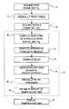
- FIG. 2A flow diagram of the general arrangement of an ultrasound treatment method 10 according to an embodiment of the present invention is shown in FIG. 2 .
- the methodbegins at step 12 by positioning proximate the tissue to be medically treated a transducer capable of transmitting and receiving ultrasound signals.
- a low-intensity imaging ultrasound signalsuch as a B-Mode signal, is emitted from the transducer and the reflected signals are collected to form a pre-treatment image frame “F 1 .”
- RFradio frequency
- image and imagingcomprise, without limitation, any means for creating a visually-perceivable image that may be displayed, for example, on a monitor, screen or display, or printed.
- image and imagingmay further comprise image information that may be used by electronic devices such as computers without being displayed in visually perceivable form.
- Medical treatmentbegins at step 16 by emitting an ultrasound signal from the transducer.
- the term “medical treatment” as used hereinmay comprise several types of ultrasound treatment.
- the medical treatmentmay be a relatively low-intensity ultrasound signal, causing a small temperature rise in the tissue (such as 1° C. or less) that does not alter the tissue.
- Low-intensity ultrasound medical treatmentis relevant for targeting of tissue for higher-intensity medical treatment, wherein the low-intensity ultrasound signal creates a small temperature rise that may be imaged to ensure that the desired region of tissue is targeted by the ultrasound beam.
- the amplitude of the ultrasound signalmay be raised to a higher intensity.
- the high-intensity ultrasound signalmay be used to administer a hyperthermia treatment or to ablate the tissue.
- a post-treatment image frame “F 2 ”is generated at step 18 .
- Information gathered from the pre-treatment and post-treatment image framesis then used at step 20 to compute the change in the temperature of the tissue being treated.
- the results of the temperature computationsmay then be displayed at step 22 .
- the displaymay be in any convenient form to aid the user in interpreting the temperature data.
- the displaymay be in the form of textual data relating to the temperature of the treated tissue.
- the displaymay also include a graphical presentation wherein colors are used to represent temperatures and temperature gradients of the treated tissue.
- the transducermay be re-positioned if desired at step 28 using the image of step 18 as a guide.
- the methodthen returns to step 16 to again medically treat the tissue.
- the post-treatment image frame from the first treatmentmay be used as a pre-treatment image for the second treatment, as shown by arrow 15 of FIG. 2 .
- a new pre-treatment image 14may be obtained, as indicated by arrow 17 of FIG. 2 .
- FIG. 3illustrates the method of FIG. 2 in relation to a time scale t.
- the B-Mode image scans 14 , 18are represented as frames F 1 , F 2 respectively.
- For each framea number of A-lines of raw echo signal data are obtained, the number of each line corresponding to azimuthal position while signal time corresponds to depth.
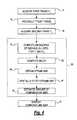
- FIG. 4a method for estimating the temperature rise of anatomical tissue in response to medical treatment according to an embodiment of the present invention is depicted.
- An ultrasound transduceris positioned proximate the anatomical tissue, then at step 14 a first image frame F 1 (illustrated in FIG. 4 ) is acquired.
- the framemay be stored electronically, such as in a computer, magnetic media and solid-state memory.
- the tissuemay then be medically treated such as by hyperthermia or ablating it with high-intensity ultrasound waves, as at step 16 .
- a second image frame F 2is then acquired at step 18 and electronically stored. For each frame, a number of A-lines of raw echo signal data are obtained.
- a line number assigned to each A-linecorresponds to azimuthal position, whereas signal time corresponds to depth.
- the raw echo signals of frames F 1 and F 2may be processed at step 30 , such as to obtain complex analytic signals by means of a conventional Hilbert transformation. For small temperature rises the echo signal p 1 obtained after treatment differs from p 0 , the echo signal obtained prior to treatment, only by a time-dependent delay.
- a 0is the complex amplitude envelope
- ⁇ 0is a nominal center frequency of the signals
- ⁇ tis the time-dependent delay.
- the delay ⁇ tis estimated as a function of time as follows.
- the processed echo signal p 0 from frame F 1is multiplied by the conjugate of p 1 from frame F 2 .
- This conjugate productis spatially filtered to yield a spatial map of a zero-lag cross-correlation between F 1 and F 2 .
- a windowed zero-lag cross-correlationmay be expressed by Equation 3 for a window centered at time t 0 :
- R 01 ⁇ ( 0 , t 0 )e - i ⁇ ⁇ ⁇ 0 ⁇ ⁇ ⁇ ⁇ t ⁇ ⁇ - ⁇ ⁇ ⁇ w ⁇ ( t - t 0 ) ⁇ a 0 * ( t + ⁇ ⁇ ⁇ t ) ⁇ ⁇ d t Equation ⁇ ⁇ 3
- this zero-lag cross-correlationtakes the form of a convolution between the conjugate product of the echoes and a windowing function w, this cross-correlation can be computed efficiently for all times t 0 , using the fast Fourier transform. This eliminates the need for computing separate cross-correlations for multiple windows spanning the region of interest.
- the depth-dependent delayis then given by the phase of the zero-lag cross-correlation R 01 ( 0 ,t 0 ): ⁇ ⁇ ⁇ t ⁇ ( t ) ⁇ - arg ⁇ ⁇ R 01 ⁇ ( 0 , t ) ⁇ ⁇ 0 Equation ⁇ ⁇ 4
- the phase of the conjugate productis computed for all the A-lines, using techniques such as appropriate phase unwrapping, to efficiently estimate the azimuth- and depth-dependent delay ⁇ t between the echo signals of the two frames using Equation 4.
- the relative strain between the two framesis computed as the derivative of this signal delay, taken along the time/range direction.
- This relative strain(termed an “echo strain map” herein) can be computed by known methods, such as finite differences.
- the echo strain maprepresents the slope of the depth-dependent delay and assumes a linear relationship between echo strain and temperature rise.
- the echo strain mapcan optionally be spatially filtered to reduce any strain artifacts. Within a measurable constant of proportionality, the echo strain map can then be used at step 38 to estimate the spatially-dependent temperature rise between frames F 1 and F 2 .
- the present inventionprovides several advantages over prior methods for strain estimation used in pulse-echo ultrasound temperature measurements. For example, the present invention is more efficient due to its relative simplicity, since single-filter operations may be performed on entire two-dimensional frames at once, rather than on numerous individually windowed signals as is the current practice. As a result, the present invention may be easily implemented for real-time measurement of anatomical tissue, using, for example, standard digital signal processing (“DSP”) circuitry. In addition, the present invention provides temperature information having fewer artifacts and improved accuracy as compared to present methods.
- DSPdigital signal processing
- multiple framesmay be grouped together as a “frame set.”
- a diagram of the relative amplitude and timing of ultrasound image frame sets and ultrasound treatmentsis depicted in FIG. 5 .
- Frame sets FS 1 and FS 2may be averaged prior to computing the conjugate product.
- the method of FIG. 4may be utilized with minor modification wherein FS 1 , the average of the signals of the frames comprising frame set FS 1 , is substituted for F 1 of step 14 .
- FS 2the average of the signals of the frames comprising frame set FS 2
- the resulting estimated temperature rise of step 38contains a reduced number of uncertainties and artifacts as compared to the method of FIG. 4 using single frames.
- the present inventionmay include averaging of echo conjugate products from multiple frames before computing and differentiating the phase map to obtain the echo strain map.
- a method according to this embodimentis illustrated by FIG. 5 in combination with FIG. 6 .
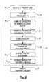
- An ultrasound transduceris positioned proximate the anatomical tissue, then at step 14 a first image frame set FS 1 is acquired.
- the tissuemay then be medically treated such as by hyperthermia or ablating it with high-intensity ultrasound waves, as at step 16 .
- a second image frame set FS 2is then acquired at step 18 .
- the raw echo signals of frame sets FS 1 and FS 2may be processed at step 30 , such as to obtain complex analytic signals by means of a Hilbert transformation, as previously disclosed.
- an average of the conjugate products of step 30is calculated.
- the averagemay be computed, for example, by computing and then averaging echo conjugate products of: F 1 of FS 1 and F 1 of FS 2 ; F 2 of FS 1 and F 2 of FS 2 ; F 1 of FS 1 and F 1 of FS 2 ; and so on.
- the delayis computed at step 32 , then at step 34 an echo strain map is generated using the averaged conjugate products.
- the resulting echo strain mapis then spatially filtered at step 36 , the temperature rise is estimated at 38 , and the temperature map is displayed at 22 , all in the manner previously detailed.
- the averaged conjugate productsserve to reduce uncertainties and artifacts in the estimated temperature maps as compared to the method of FIG. 4 using single frames.
- a correlation coefficientmay be computed by multiplying one of the complex frames by a phase compensation function e ⁇ i ⁇ , where ⁇ is the low-pass filtered phase of the conjugate product.
- the normalized sum of the phase-corrected conjugate productis then a correlation coefficient whose amplitude indicates the reliability of the temperature estimation.
- Windowed sumswhich can be implemented using known fast Fourier transform operations, provide a spatial map of the correlation coefficient. High values (i.e., values approaching 1) correspond to a high confidence in the temperature estimate, while values significantly less than 1 correspond to lower confidence. Estimated temperature maps can then be formed using higher-confidence estimates preferentially.
- the echo conjugate productcan be computed using a two-dimensional filtering operation, in which a two dimensional (i.e., azimuth/array and depth/time directions) window is used instead of the one-dimensional window described above for the third alternate embodiment.
- This method of computing the conjugate productwill result in smoother strain maps, and thus may reduce artifacts in the temperature images.
- the assumed relationship between echo strain and temperature risemay be non-linear rather than the linear relationship currently assumed.
- Such a non-linear relationshipmay be derived from measurements of the non-linear relationships between temperature, sound speed, and thermal expansion.
- the non-linear relationshipmay also be obtained empirically from calibration measurements in tissues of interest.
- smoothing of strain mapsmay be enhanced by performing a thresholding operation before low-pass filtering. For example strain values above a predetermined temperature range of interest may be clipped. In this manner, artifactual strain peaks may be removed more effectively than by low-pass filtering alone.
- the conjugate products of adjacent frames of a frame set FSmay be obtained, computed and averaged.
- the tissueis medically treated such as by hyperthermia or ablating it with high-intensity ultrasound waves.
- An image frame set FSis then acquired at step 18 .
- the raw echo signals of frames F 1 -F nmay be processed at step 30 , such as to obtain complex analytic signals by means of a Hilbert transformation, as previously disclosed.
- an average of the conjugate products of step 30is calculated.
- the averagemay be computed, for example, by computing and then averaging echo conjugate products of adjacent frames, i.e.: F 1 and F 2 of FS; F 2 and F 3 of FS; F 3 and F 4 of FS; and so on.
- the delayis computed at step 32 , then at step 34 an echo strain map is generated using the averaged conjugate products.
- the resulting echo strain maphas reduced noise and artifactual content as compared to other imaging methods.
- the echo strain mapis spatially filtered at step 36 , the temperature change is estimated at 38 , and the temperature map is displayed at 22 , all in the manner previously detailed. The temperature change occurring in the time interval separating adjacent time frames may thus be estimated and displayed.
- the steps of the method of FIG. 2may be generally followed for a series of medical treatments 16 , shown as 16 A- 16 C in FIG. 9.
- a pre-treatment image frame F 1is obtained at step 14 prior to the first treatment 16 A.
- a post-treatment frame F 2is then obtained at step 18 and used with the pre-treatment image F 1 to compute at step 20 the temperature change due to treatment 16 A.
- a temperature mapis displayed at step 22 .
- Frame F 2also serves as a pre-treatment image 14 for a subsequent treatment 16 B, as shown in FIG. 9 .
- frame F 3is a post-treatment frame for treatment 16 B and is also a pre-treatment frame for treatment 16 C.
- frame F 4is a post-treatment image for treatment 16 C. This process of imaging and treatment may be repeated as many times as desired.
- the temperature change at 20may be computed using any conventional mathematical function as previously discussed, such as averaging of frame images and averaging of the computed temperature change.
Landscapes
- Health & Medical Sciences (AREA)
- Life Sciences & Earth Sciences (AREA)
- Engineering & Computer Science (AREA)
- Public Health (AREA)
- General Health & Medical Sciences (AREA)
- Biomedical Technology (AREA)
- Medical Informatics (AREA)
- Veterinary Medicine (AREA)
- Physics & Mathematics (AREA)
- Animal Behavior & Ethology (AREA)
- Pathology (AREA)
- Radiology & Medical Imaging (AREA)
- Molecular Biology (AREA)
- Surgery (AREA)
- Heart & Thoracic Surgery (AREA)
- Nuclear Medicine, Radiotherapy & Molecular Imaging (AREA)
- Biophysics (AREA)
- Computer Networks & Wireless Communication (AREA)
- Physiology (AREA)
- Computer Vision & Pattern Recognition (AREA)
- Gynecology & Obstetrics (AREA)
- General Physics & Mathematics (AREA)
- Radar, Positioning & Navigation (AREA)
- Remote Sensing (AREA)
- Data Mining & Analysis (AREA)
- Databases & Information Systems (AREA)
- Epidemiology (AREA)
- Primary Health Care (AREA)
- Ultra Sonic Daignosis Equipment (AREA)
Abstract
Description
This is a continuation-in-part of application Ser. No. 10/153,241, filed May 22, 2002 now abandoned, which claims priority to U.S. Provisional Application Ser. No. 60/294,135 filed May 29, 2001. The entire disclosures are incorporated herein by reference.
The present invention relates generally to ultrasound, and more particularly, to a method for measuring temperature rise using pulse-echo ultrasound waves.
Ultrasound medical systems and methods include ultrasound imaging of anatomical tissue to identify tissue for medical treatment. Ultrasound may also be used to medically treat and destroy unwanted tissue by heating the tissue. Imaging is done using low-intensity ultrasound waves, while medical treatment is performed with high-intensity ultrasound waves. High-intensity ultrasound waves, when focused at a focal zone a distance away from the ultrasound source, will substantially medically affect tissue in the focal zone. However, the high-intensity ultrasound will not substantially affect patient tissue outside the focal zone, such as tissue located between the ultrasound source and the focal zone. Other treatment regimes of interest include unfocused high-intensity ultrasound wherein the ultrasound energy is distributed over a relatively broad region of tissue rather than being generally concentrated within a focal zone.
Ultrasound waves may be emitted and received by a transducer assembly. The transducer assembly may include a single transducer element, or an array of elements acting together, to image the anatomical tissue and to ultrasonically ablate identified tissue. Transducer elements may employ a concave shape or an acoustic lens to focus or otherwise direct ultrasound energy. Transducer such arrays may include planar, concave or convex elements to focus ultrasound energy. Further, array elements may be electronically or mechanically controlled to steer and focus the ultrasound waves emitted by the array to a focal zone to provide three-dimensional medical ultrasound treatment of anatomical tissue. In some treatments the transducer is placed on the surface of the tissue for imaging and/or treatment of areas within the tissue. In other treatments the transducer is surrounded with a balloon which is expanded to contact the surface of the tissue by filling the balloon with a fluid such as a saline solution to provide acoustic coupling between the transducer and the tissue.
Examples of ultrasound medical systems and methods include: deploying an end effector having an ultrasound transducer outside the body to break up kidney stones inside the body; endoscopically inserting an end effector having an ultrasound transducer into the rectum to medically destroy prostate cancer; laparoscopically inserting an end effector having an ultrasound transducer into the abdominal cavity to destroy a cancerous liver tumor; intravenously inserting a catheter end effector having an ultrasound transducer into a vein in the arm and moving the catheter to the heart to medically destroy diseased heart tissue; and interstitially inserting a needle end effector having an ultrasound transducer into the tongue to medically destroy tissue to reduce tongue volume as a treatment for snoring. Methods for guiding an end effector to the target tissue include x-rays, Magnetic Resonance Images (“MRI”) and images produced using the ultrasound transducer itself.
Low-intensity ultrasound energy may be applied to unexposed subdermal anatomical tissue for the purpose of examining the tissue. Ultrasound pulses are emitted, and returning echos are measured to determine the characteristics of the unexposed subdermal tissue. Variations in tissue structure and tissue boundaries have varying acoustic impedances, resulting in variations in the strength of ultrasound echos. A common ultrasound imaging technique is known in the art as “B-Mode” wherein either a single ultrasound transducer is articulated or an array of ultrasound transducers is moved or electronically scanned to generate a two-dimensional image of an area of tissue. The generated image is comprised of a plurality of pixels, each pixel corresponding to a portion of the tissue area being examined. The varying strength of the echos is preferably translated to a proportional pixel brightness. A cathode ray tube, computer monitor or liquid crystal display can be used to display a two-dimensional pixellated image of the tissue area being examined.
When high-intensity ultrasound energy is applied to anatomical tissue, significant beneficial physiological effects may result. For example, undesired anatomical tissue may be ablated by heating the tissue with high-intensity ultrasound energy. By focusing the ultrasound energy at one or more specific focusing zones within the tissue, thermal effects can be confined to a defined region that may be remote from the ultrasound transducer. The use of high-intensity focused ultrasound to ablate tissue presents many advantages, including: reduced patient trauma and pain; elimination of the need for some surgical incisions and stitches; reduced or obviated need for general anesthesia; reduced exposure of internal tissue; reduced risk of infection and other complications; avoidance of damage to non-targeted tissue; lack of harmful cumulative effects from the ultrasound energy on the surrounding non-target tissue; reduced treatment costs; minimal blood loss; and the ability for ultrasound treatments to be performed at non-hospital sites and/or on an out-patient basis.
Ultrasound treatment of anatomical tissue may involve the alternating use of both low-intensity imaging ultrasound and high-intensity treatment ultrasound. During such treatment, imaging is first performed to identify and locate the tissue to be treated. The identified tissue is then medically treated with high-intensity ultrasound energy, such as for the purpose of destroying the tissue. After a period of exposure to high-intensity ultrasound, a subsequent image of the tissue is generated using low-intensity ultrasound energy to determine the results of the ultrasound treatment and provide visual guidance to the user to aid in subsequent treatments. This process of applying low-energy ultrasound to assist in guiding the position and focal point of the transducer, followed by high-energy ultrasound to ablate the undesired anatomical tissue, may continue until the undesired tissue has been completely ablated.
In addition to imaging, monitoring of the temperature of the tissue being treated is desirable so that the tissue being treated, as well as the delivered treatment, can be readily visualized and controlled. For example, temperature monitoring is essential for hyperthermia treatments wherein tissue is exposed to ultrasound for the purpose of raising the temperature of the tissue. Hyperthermia treatments have been shown to be effective in the treatment of cancerous tumors, which are known to be more sensitive to heat than healthy tissue. Temperature monitoring is also useful for high-intensity ultrasound treatment of tissue, such as ablation, since the temperature of the tissue provides an indication of the extent and spatial distribution of the treatment.
The temperature of the tissue may be measured directly, such as with a thermocouple array. However, this arrangement is not desirable for several reasons. First, the thermocouple array must be placed into the region being treatment. This may require invasive placement of the array, thereby eliminating the non-invasive treatment advantages offered by ultrasound. In addition, the thermocouple array may have a relatively slow response time, providing the operator with delayed feedback regarding the status of the treatment.
Various non-invasive temperature-monitoring methods have been developed to overcome the drawbacks of thermocouple arrays. For example, magnetic resonance imaging (“MRI”) is used in the art to noninvasively measure temperature rises in vivo. However, MRI feedback requires an apparatus separate from the ultrasound treatment apparatus to provide the temperature measurement. As a result, MRI-assisted ultrasound methods are expensive and cumbersome, making such treatment impractical for routine use in surgical treatments.
The prior art has suggested using ultrasound waves to measure temperature. Previous studies have shown that temperature rises in tissue cause local thermal expansion and sound speed changes, which can be estimated from perturbations of ultrasonic echo signals. Since the travel time of an ultrasonic echo depends on both the path length (which is changed by any thermal expansion) and sound speed (which depends on temperature), temperature changes in tissue being treated cause measurable alterations to echo travel times. By estimation and further processing of these travel times, temperature rises can be approximately mapped. This arrangement is highly desirable, since heating effects of ultrasound treatment may be measured using inexpensive, portable, and unobtrusive ultrasound apparatus. For configurations in which therapy and monitoring are performed using the same ultrasound probe, this approach is particularly desirable, since a temperature map may be automatically co-registered with the therapeutic ultrasound beams.
Available approaches to pulse-echo temperature mapping have employed cross-correlation of ultrasound echo signals to estimate changes in travel time (i.e., delay) for each pixel of an image, before and after heating. These travel-time maps are then smoothed and differentiated by signal processing to obtain maps of the echo strain, which is assumed to be proportional to the temperature rise of the tissue being treated. Repetition of this process can follow incremental temperature rises associated with prolonged heating. These approaches suffer from several drawbacks, including low spatial resolution and high artifactual content. Since many time-delay estimation steps are needed to build up a temperature image, these approaches are also computationally inefficient.
There is a need for a method of measuring the temperature rise of tissue being treated by ultrasound that is noninvasive. There is a further need for method of monitoring temperature rise that is not expensive and is not cumbersome. There is a still further need for a method of monitoring temperature measurement that has relatively high spatial resolution and relatively low artifactual content, and is computationally efficient.
The present invention overcomes the limitations of the known art by estimating the time-dependent delay between two sets of narrow-band ultrasound signals. The travel time of a first signal is measured prior to treating anatomical tissue, then the travel time of a second signal is measured after the tissue is treated. The change in travel time between the first and second signals is used to generate a complete two-dimensional delay map from several two-dimensional multiplication and fast Fourier transform operations, rather than creating a map from a large quantity of individual time delay estimation steps. As a result, temperature maps can be generated more quickly as compared to prior methods. In addition, temperature maps generated using the present invention have fewer artifacts.
Further features of the present invention will become apparent to those skilled in the art to which the present invention relates from reading the following specification with reference to the accompanying drawings, in which:
It is well-known that the travel time of an ultrasound wave varies with the path length. The path length may vary in anatomic tissue due to thermal expansion of the tissue as a result of therapeutic ultrasound treatment, such as hyperthermia and ablation. Further, the travel time of an ultrasound wave varies with the speed of the ultrasound wave, which is a function of path temperature. Thus, changes in ultrasound wave travel times may be used to estimate temperature rise.
A flow diagram of the general arrangement of anultrasound treatment method 10 according to an embodiment of the present invention is shown in FIG.2. The method begins atstep 12 by positioning proximate the tissue to be medically treated a transducer capable of transmitting and receiving ultrasound signals. At step14 a low-intensity imaging ultrasound signal, such as a B-Mode signal, is emitted from the transducer and the reflected signals are collected to form a pre-treatment image frame “F1.” For each frame, a number of A-lines of raw echo signal radio frequency (“RF”) data are obtained; a line number corresponds to azimuthal position and signal time corresponds to depth. It is understood that the terms “image” and “imaging” comprise, without limitation, any means for creating a visually-perceivable image that may be displayed, for example, on a monitor, screen or display, or printed. The terms “image” and “imaging” may further comprise image information that may be used by electronic devices such as computers without being displayed in visually perceivable form.
Medical treatment begins atstep 16 by emitting an ultrasound signal from the transducer. The term “medical treatment” as used herein may comprise several types of ultrasound treatment. For example, the medical treatment may be a relatively low-intensity ultrasound signal, causing a small temperature rise in the tissue (such as 1° C. or less) that does not alter the tissue. Low-intensity ultrasound medical treatment is relevant for targeting of tissue for higher-intensity medical treatment, wherein the low-intensity ultrasound signal creates a small temperature rise that may be imaged to ensure that the desired region of tissue is targeted by the ultrasound beam. After targeting is confirmed, the amplitude of the ultrasound signal may be raised to a higher intensity. The high-intensity ultrasound signal may be used to administer a hyperthermia treatment or to ablate the tissue.
After a portion of the tissue has been medically treated, a post-treatment image frame “F2” is generated atstep 18. Information gathered from the pre-treatment and post-treatment image frames is then used atstep 20 to compute the change in the temperature of the tissue being treated. The results of the temperature computations may then be displayed atstep 22. The display may be in any convenient form to aid the user in interpreting the temperature data. For example, the display may be in the form of textual data relating to the temperature of the treated tissue. The display may also include a graphical presentation wherein colors are used to represent temperatures and temperature gradients of the treated tissue. If treatment is complete atstep 24, the method is ended atstep 26. However, if the tissue requires additional treatment, the transducer may be re-positioned if desired atstep 28 using the image ofstep 18 as a guide. The method then returns to step16 to again medically treat the tissue. The post-treatment image frame from the first treatment may be used as a pre-treatment image for the second treatment, as shown byarrow 15 of FIG.2. Alternatively, a newpre-treatment image 14 may be obtained, as indicated byarrow 17 of FIG.2.
Referring now toFIG. 4 in combination withFIG. 3 , a method for estimating the temperature rise of anatomical tissue in response to medical treatment according to an embodiment of the present invention is depicted. An ultrasound transducer is positioned proximate the anatomical tissue, then at step14 a first image frame F1(illustrated inFIG. 4 ) is acquired. The frame may be stored electronically, such as in a computer, magnetic media and solid-state memory. The tissue may then be medically treated such as by hyperthermia or ablating it with high-intensity ultrasound waves, as atstep 16. A second image frame F2is then acquired atstep 18 and electronically stored. For each frame, a number of A-lines of raw echo signal data are obtained. A line number assigned to each A-line corresponds to azimuthal position, whereas signal time corresponds to depth. The raw echo signals of frames F1and F2may be processed atstep 30, such as to obtain complex analytic signals by means of a conventional Hilbert transformation. For small temperature rises the echo signal p1obtained after treatment differs from p0, the echo signal obtained prior to treatment, only by a time-dependent delay. Echo signals p0and p1may be expressed below inEquations 1 and 2:
where a0is the complex amplitude envelope, ω0is a nominal center frequency of the signals, and δt is the time-dependent delay.
where a0is the complex amplitude envelope, ω0is a nominal center frequency of the signals, and δt is the time-dependent delay.
Atstep 32 the delay δt is estimated as a function of time as follows. The processed echo signal p0from frame F1is multiplied by the conjugate of p1from frame F2. This conjugate product is spatially filtered to yield a spatial map of a zero-lag cross-correlation between F1and F2. For the signals ofEquations
Since this zero-lag cross-correlation takes the form of a convolution between the conjugate product of the echoes and a windowing function w, this cross-correlation can be computed efficiently for all times t0, using the fast Fourier transform. This eliminates the need for computing separate cross-correlations for multiple windows spanning the region of interest.
Since this zero-lag cross-correlation takes the form of a convolution between the conjugate product of the echoes and a windowing function w, this cross-correlation can be computed efficiently for all times t0, using the fast Fourier transform. This eliminates the need for computing separate cross-correlations for multiple windows spanning the region of interest.
The depth-dependent delay is then given by the phase of the zero-lag cross-correlation R01(0,t0):
The phase of the conjugate product is computed for all the A-lines, using techniques such as appropriate phase unwrapping, to efficiently estimate the azimuth- and depth-dependent delay δt between the echo signals of the twoframes using Equation 4. Atstep 34 the relative strain between the two frames is computed as the derivative of this signal delay, taken along the time/range direction. This relative strain (termed an “echo strain map” herein) can be computed by known methods, such as finite differences. The echo strain map represents the slope of the depth-dependent delay and assumes a linear relationship between echo strain and temperature rise.
The phase of the conjugate product is computed for all the A-lines, using techniques such as appropriate phase unwrapping, to efficiently estimate the azimuth- and depth-dependent delay δt between the echo signals of the two
Atstep 36, the echo strain map can optionally be spatially filtered to reduce any strain artifacts. Within a measurable constant of proportionality, the echo strain map can then be used atstep 38 to estimate the spatially-dependent temperature rise between frames F1and F2.
The present invention provides several advantages over prior methods for strain estimation used in pulse-echo ultrasound temperature measurements. For example, the present invention is more efficient due to its relative simplicity, since single-filter operations may be performed on entire two-dimensional frames at once, rather than on numerous individually windowed signals as is the current practice. As a result, the present invention may be easily implemented for real-time measurement of anatomical tissue, using, for example, standard digital signal processing (“DSP”) circuitry. In addition, the present invention provides temperature information having fewer artifacts and improved accuracy as compared to present methods.
In a first alternate embodiment, multiple frames may be grouped together as a “frame set.” A diagram of the relative amplitude and timing of ultrasound image frame sets and ultrasound treatments is depicted in FIG.5. Frame sets FS1and FS2may be averaged prior to computing the conjugate product. In this embodiment the method ofFIG. 4 may be utilized with minor modification whereinFS 1, the average of the signals of the frames comprising frame set FS1, is substituted for F1ofstep 14. Likewise,FS 2, the average of the signals of the frames comprising frame set FS2, is substituted for F2ofstep 18. The resulting estimated temperature rise ofstep 38 contains a reduced number of uncertainties and artifacts as compared to the method ofFIG. 4 using single frames.
In a second alternate embodiment, the present invention may include averaging of echo conjugate products from multiple frames before computing and differentiating the phase map to obtain the echo strain map. A method according to this embodiment is illustrated byFIG. 5 in combination with FIG.6. An ultrasound transducer is positioned proximate the anatomical tissue, then at step14 a first image frame set FS1is acquired. The tissue may then be medically treated such as by hyperthermia or ablating it with high-intensity ultrasound waves, as atstep 16. A second image frame set FS2is then acquired atstep 18. The raw echo signals of frame sets FS1and FS2may be processed atstep 30, such as to obtain complex analytic signals by means of a Hilbert transformation, as previously disclosed. At step31 an average of the conjugate products ofstep 30 is calculated. The average may be computed, for example, by computing and then averaging echo conjugate products of:
F1of FS1and F1of FS2;
F2of FS1and F2of FS2;
F1of FS1and F1of FS2;
and so on. The delay is computed atstep 32, then atstep 34 an echo strain map is generated using the averaged conjugate products. The resulting echo strain map is then spatially filtered atstep 36, the temperature rise is estimated at38, and the temperature map is displayed at22, all in the manner previously detailed. The averaged conjugate products serve to reduce uncertainties and artifacts in the estimated temperature maps as compared to the method ofFIG. 4 using single frames.
F1of FS1and F1of FS2;
F2of FS1and F2of FS2;
F1of FS1and F1of FS2;
and so on. The delay is computed at
In a third alternate embodiment, a correlation coefficient may be computed by multiplying one of the complex frames by a phase compensation function e−iθ, where θ is the low-pass filtered phase of the conjugate product. The normalized sum of the phase-corrected conjugate product is then a correlation coefficient whose amplitude indicates the reliability of the temperature estimation. Windowed sums, which can be implemented using known fast Fourier transform operations, provide a spatial map of the correlation coefficient. High values (i.e., values approaching 1) correspond to a high confidence in the temperature estimate, while values significantly less than 1 correspond to lower confidence. Estimated temperature maps can then be formed using higher-confidence estimates preferentially.
In a fourth alternate embodiment of the present invention, the echo conjugate product can be computed using a two-dimensional filtering operation, in which a two dimensional (i.e., azimuth/array and depth/time directions) window is used instead of the one-dimensional window described above for the third alternate embodiment. This method of computing the conjugate product will result in smoother strain maps, and thus may reduce artifacts in the temperature images.
In a fifth alternate embodiment of the present invention, the assumed relationship between echo strain and temperature rise may be non-linear rather than the linear relationship currently assumed. Such a non-linear relationship may be derived from measurements of the non-linear relationships between temperature, sound speed, and thermal expansion. The non-linear relationship may also be obtained empirically from calibration measurements in tissues of interest.
In a sixth alternate embodiment of the present invention, smoothing of strain maps may be enhanced by performing a thresholding operation before low-pass filtering. For example strain values above a predetermined temperature range of interest may be clipped. In this manner, artifactual strain peaks may be removed more effectively than by low-pass filtering alone.
With reference toFIGS. 7 and 8 , in a seventh alternate embodiment of the present invention, the conjugate products of adjacent frames of a frame set FS may be obtained, computed and averaged. Atstep 16 the tissue is medically treated such as by hyperthermia or ablating it with high-intensity ultrasound waves. An image frame set FS is then acquired atstep 18. The raw echo signals of frames F1-Fnmay be processed atstep 30, such as to obtain complex analytic signals by means of a Hilbert transformation, as previously disclosed. At step31 an average of the conjugate products ofstep 30 is calculated. The average may be computed, for example, by computing and then averaging echo conjugate products of adjacent frames, i.e.:
F1and F2of FS;
F2and F3of FS;
F3and F4of FS;
and so on. The delay is computed atstep 32, then atstep 34 an echo strain map is generated using the averaged conjugate products. The resulting echo strain map has reduced noise and artifactual content as compared to other imaging methods. The echo strain map is spatially filtered atstep 36, the temperature change is estimated at38, and the temperature map is displayed at22, all in the manner previously detailed. The temperature change occurring in the time interval separating adjacent time frames may thus be estimated and displayed.
F1and F2of FS;
F2and F3of FS;
F3and F4of FS;
and so on. The delay is computed at
In an eighth alternate embodiment of the present invention the steps of the method ofFIG. 2 may be generally followed for a series ofmedical treatments 16, shown as16A-16C inFIG. 9. A pre-treatment image frame F1is obtained atstep 14 prior to thefirst treatment 16A. A post-treatment frame F2is then obtained atstep 18 and used with the pre-treatment image F1to compute atstep 20 the temperature change due totreatment 16A. A temperature map is displayed atstep 22. Frame F2also serves as apre-treatment image 14 for a subsequent treatment16B, as shown in FIG.9. Likewise, frame F3is a post-treatment frame for treatment16B and is also a pre-treatment frame fortreatment 16C. Finally, frame F4is a post-treatment image fortreatment 16C. This process of imaging and treatment may be repeated as many times as desired. Further, the temperature change at20 may be computed using any conventional mathematical function as previously discussed, such as averaging of frame images and averaging of the computed temperature change.
The foregoing description of several expressions of embodiments and methods of the invention has been presented for purposes of illustration. It is not intended to be exhaustive or to limit the present invention to the precise forms and procedures disclosed, and obviously many modifications and variations are possible in light of the above teaching. It is intended that the scope the invention be defined by the claims appended hereto.
Claims (21)
1. A method for mapping temperature rise of anatomical tissue using pulse-echo ultrasound, comprising the steps of:
a) obtaining a first signal of a first imaging ultrasound wave which has been reflected back from a region in the anatomical tissue at a first time;
b) obtaining a second signal of a second imaging ultrasound wave which has been reflected back from the region in the anatomical tissue at a later second time wherein the tissue has received at least some medical treatment by the second time;
c) computing first and second complex analytic signals from the first and second imaging signals;
d) computing the depth-dependent delay from the conjugate product of the first and second analytic signals;
e) generating an echo strain map from the slope of the depth-dependent delay;
f) using the echo strain map to estimate the amount of temperature rise from the first imaging signal to the second imaging signal; and
g) creating an image showing where temperature rise is occurring in the anatomical tissue.
2. The method ofclaim 1 , further comprising the step of spatially filtering the difference signal.
3. The method ofclaim 2 , further including the step of performing a thresholding operation to remove artifactual strain peaks.
4. The method ofclaim 1 , further including the step of multiplying a signal of an ultrasound imaging wave by a phase compensation function to compute a correlation coefficient indicating the reliability of the temperature estimation.
5. The method ofclaim 4 wherein the signal is the first imaging signal.
6. The method ofclaim 5 wherein the signal is the second imaging signal.
7. The method ofclaim 4 , further including the step of implementing windowed sums to provide a spatial map of the correlation coefficient.
8. The method ofclaim 7 wherein the windowed sums are two-dimensional.
9. The method ofclaim 1 , wherein the echo strain map representing the slope of the depth-dependent delay assumes a linear relationship between echo strain and temperature rise.
10. The method ofclaim 1 , wherein the echo strain map representing the depth-dependent delay assumes a non-linear relationship between echo strain and temperature rise.
11. The method ofclaim 10 wherein the non-linear relationship is derived from measurements of non-linear relationships between temperature, sound speed and thermal expansion.
12. The method ofclaim 10 wherein the non-linear relationship is derived empirically from calibration measurements in the anatomical tissue.
13. The method ofclaim 1 , wherein the medical treatment is ultrasound medical treatment.
14. The method ofclaim 1 , also including steps a) through g) for different regions to image the anatomical tissue, wherein the image includes medically-treated and medically-untreated regions of the anatomical tissue.
15. A method for mapping temperature rise of anatomical tissue using pulse-echo ultrasound, comprising the steps of:
a) obtaining a first set of frames comprising a plurality of imaging ultrasound wave signals which have been reflected back from a region in the anatomical tissue during a first period of time;
b) obtaining a second set of frames comprising a plurality of imaging ultrasound wave signals which have been reflected back from a region in the anatomical tissue at a later second time wherein the tissue has received at least some medical treatment by the second time;
c) averaging together the signals of the first set of frames to obtain an averaged first imaging signal;
d) averaging together the signals of the second set of frames to obtain an averaged second imaging signal;
e) computing first and second complex analytic signals from the first and second averaged imaging signals;
f) computing the depth-dependent delay from the conjugate product of the first and second analytic signals;
g) generating a strain map from the slope of the depth-dependent delay;
h) using the echo strain map to estimate the amount of temperature rise from the first averaged imaging signal to the second averaged imaging signal; and
i) creating an image showing where temperature rise is occurring in the anatomical tissue.
16. The method ofclaim 15 , further comprising the step of spatially filtering the difference signal.
17. The method ofclaim 15 , wherein the medical treatment is ultrasound medical treatment.
18. The method ofclaim 15 , also including steps a) through i) for different regions to image the anatomical tissue, wherein the image includes medically-treated and medically-untreated regions of the anatomical tissue.
19. A method for mapping temperature rise of anatomical tissue using pulse-echo ultrasound, comprising the steps of:
a) obtaining a first set of frames comprising a plurality of imaging ultrasound wave signals which have been reflected back from a region in the anatomical tissue during a first period of time;
b) obtaining a second set of frames comprising a plurality of imaging ultrasound wave signals which have been reflected back from a region in the anatomical tissue at a later second time wherein the tissue has received at least some medical treatment by the second time;
c) computing complex analytic signals from a selected frame from the first set of frames and a selected frame from the second set of frames;
d) computing the conjugate product of the complex analytic signals of step c);
e) repeating steps c) and d) until conjugate products have been computed for all of the frames of the first and second frame sets;
f) computing the average of the conjugate products of step e);
g) computing the depth-dependent delay from the averaged conjugate product of step f);
h) generating an echo strain map from the slope of the depth-dependent delay;
i) using the echo strain map to estimate the amount of temperature rise from the first averaged imaging signal to the second averaged imaging signal; and
j) creating an image showing where temperature rise is occurring in the anatomical tissue.
20. A method for mapping temperature change in anatomical tissue using pulse-echo ultrasound, comprising the steps of:
a) obtaining a set of frames comprising a plurality of imaging ultrasound wave signals which have been reflected back from a region in the anatomical tissue during a period of time;
b) computing complex analytic signals from the set of frames;
c) computing the conjugate product of a pair of adjacent frames of the set of frames;
d) repeating step c) until the conjugate products of all adjacent frames have been computed;
e) averaging the conjugate products of the adjacent frames of step d);
f) computing a depth-dependent delay map from the average conjugate product;
g) generating an echo strain map from the slope of the depth-dependent delay;
h) using the echo strain map to estimate the amount of temperature change from the first frame to the second frame; and
i) creating an image showing where temperature change is occurring in the anatomical tissue.
21. A method for mapping temperature rise of anatomical tissue using pulse-echo ultrasound, comprising the steps of:
a) obtaining a first signal of a first imaging ultrasound wave which has been reflected back from a region in the anatomical tissue at a first time;
b) obtaining a second signal of a second imaging ultrasound wave which has been reflected back from the region in the anatomical tissue at a later second time wherein the tissue has received at least some medical treatment by the second time;
c) computing first and second complex analytic signals from the first and second imaging signals;
d) computing the depth-dependent delay from the conjugate product of the first and second analytic signals;
e) generating an echo strain map from the slope of the depth-dependent delay;
f) using the echo strain map to estimate the amount of temperature rise from the first imaging signal to the second imaging signal;
g) creating an image showing where temperature rise is occurring in the anatomical tissue; and
h) repeating the method at least once by:
i) re-defining the second imaging signal obtained at step b) as the first imaging signal of step a);
ii) obtaining a new second imaging signal at step b); and
ii) repeating steps c) through g).
Priority Applications (3)
| Application Number | Priority Date | Filing Date | Title |
|---|---|---|---|
| US10/735,045US7211044B2 (en) | 2001-05-29 | 2003-12-12 | Method for mapping temperature rise using pulse-echo ultrasound |
| PCT/US2004/041799WO2005058138A2 (en) | 2003-12-12 | 2004-12-10 | Method for mapping temperature rise using pulse-echo ultrasound |
| EP04814036AEP1696799A4 (en) | 2003-12-12 | 2004-12-10 | Method for mapping temperature rise using pulse-echo ultrasound |
Applications Claiming Priority (3)
| Application Number | Priority Date | Filing Date | Title |
|---|---|---|---|
| US29413501P | 2001-05-29 | 2001-05-29 | |
| US10/153,241US20030069502A1 (en) | 2001-05-29 | 2002-05-22 | Ultrasound feedback in medically-treated patients |
| US10/735,045US7211044B2 (en) | 2001-05-29 | 2003-12-12 | Method for mapping temperature rise using pulse-echo ultrasound |
Related Parent Applications (1)
| Application Number | Title | Priority Date | Filing Date |
|---|---|---|---|
| US10/153,241Continuation-In-PartUS20030069502A1 (en) | 2001-05-29 | 2002-05-22 | Ultrasound feedback in medically-treated patients |
Publications (2)
| Publication Number | Publication Date |
|---|---|
| US20040127791A1 US20040127791A1 (en) | 2004-07-01 |
| US7211044B2true US7211044B2 (en) | 2007-05-01 |
Family
ID=34700422
Family Applications (1)
| Application Number | Title | Priority Date | Filing Date |
|---|---|---|---|
| US10/735,045Expired - LifetimeUS7211044B2 (en) | 2001-05-29 | 2003-12-12 | Method for mapping temperature rise using pulse-echo ultrasound |
Country Status (3)
| Country | Link |
|---|---|
| US (1) | US7211044B2 (en) |
| EP (1) | EP1696799A4 (en) |
| WO (1) | WO2005058138A2 (en) |
Cited By (27)
| Publication number | Priority date | Publication date | Assignee | Title |
|---|---|---|---|---|
| US20040158152A1 (en)* | 1999-08-09 | 2004-08-12 | Lizzi Frederic Louis | System and method for ultrasonic harmonic imaging for therapy guidance and monitoring |
| US20070167824A1 (en)* | 2005-11-30 | 2007-07-19 | Warren Lee | Method of manufacture of catheter tips, including mechanically scanning ultrasound probe catheter tip, and apparatus made by the method |
| US20070167825A1 (en)* | 2005-11-30 | 2007-07-19 | Warren Lee | Apparatus for catheter tips, including mechanically scanning ultrasound probe catheter tip |
| US20080165825A1 (en)* | 2007-01-08 | 2008-07-10 | Medison Co., Ltd. | Method of estimating surface temperature of a diagnostic ultrasound probe |
| US20080281200A1 (en)* | 2007-05-10 | 2008-11-13 | Misonix, Incorporated | Elevated coupling liquid temperature during HIFU treatment method and hardware |
| US20090105588A1 (en)* | 2007-10-02 | 2009-04-23 | Board Of Regents, The University Of Texas System | Real-Time Ultrasound Monitoring of Heat-Induced Tissue Interactions |
| US20090227870A1 (en)* | 2004-08-31 | 2009-09-10 | Stichting Voor Fundamenteel Onderzoek Der Materie | Method of determining a parameter representing an acoustic property of a material |
| US20100317960A1 (en)* | 2009-06-10 | 2010-12-16 | Patrick Gross | Thermotherapy device and method to implement thermotherapy |
| US20110040184A1 (en)* | 2001-05-29 | 2011-02-17 | Mast T Douglas | Method for monitoring of medical treatment using pulse-echo ultrasound |
| US20110087097A1 (en)* | 2008-12-29 | 2011-04-14 | Perseus-BioMed Ltd. | Method and system for tissue imaging and analysis |
| US20110288410A1 (en)* | 2010-02-22 | 2011-11-24 | Speyer Gavriel A | Methods and systems for diagnostic ultrasound based monitoring of high intensity focused ultrasound therapy |
| US20120029497A1 (en)* | 2004-12-13 | 2012-02-02 | Tel Hashomer Medical Research Infrastructure And Services Ltd. | Method and System for Monitoring Ablation of Tissues |
| US8232801B2 (en) | 2011-06-30 | 2012-07-31 | General Electric Company | Nuclear quadrupole resonance system and method for structural health monitoring |
| US20130165779A1 (en)* | 2011-12-27 | 2013-06-27 | Samsung Electronics Co., Ltd. | Temperature estimation method and temperature estimation apparatus using the same |
| WO2014004698A1 (en)* | 2012-06-27 | 2014-01-03 | Acoustic Medsystems, Inc. | Noninvasive transvaginal acoustic thermal treatment of female stress urinary incontinence |
| DE102012217143A1 (en)* | 2012-09-24 | 2014-04-17 | Siemens Aktiengesellschaft | Method for determining temperature in volume area of body of patient in medical area, involves calibrating ultrasonic device by MRI sensor for determining temperature in volume area, where device and sensor are monitored in parallel |
| US8870772B2 (en) | 2008-12-29 | 2014-10-28 | Perseus-Biomed Inc. | Method and system for tissue recognition |
| US8882672B2 (en) | 2008-12-29 | 2014-11-11 | Perseus-Biomed Inc. | Method and system for tissue imaging and analysis |
| US9005144B2 (en) | 2001-05-29 | 2015-04-14 | Michael H. Slayton | Tissue-retaining systems for ultrasound medical treatment |
| US9132287B2 (en) | 2004-06-14 | 2015-09-15 | T. Douglas Mast | System and method for ultrasound treatment using grating lobes |
| WO2017214078A1 (en)* | 2016-06-10 | 2017-12-14 | The Regents Of The University Of California | Apparatus for and methods of acoustic thermometry |
| US10010723B2 (en) | 2013-04-18 | 2018-07-03 | Profound Medical Inc. | Therapy system for depositing energy |
| US11020144B2 (en) | 2015-07-21 | 2021-06-01 | 3Dintegrated Aps | Minimally invasive surgery system |
| US11033182B2 (en) | 2014-02-21 | 2021-06-15 | 3Dintegrated Aps | Set comprising a surgical instrument |
| US11039734B2 (en) | 2015-10-09 | 2021-06-22 | 3Dintegrated Aps | Real time correlated depiction system of surgical tool |
| US11331120B2 (en) | 2015-07-21 | 2022-05-17 | 3Dintegrated Aps | Cannula assembly kit |
| US11491352B2 (en)* | 2018-06-05 | 2022-11-08 | Korea Institute Of Science And Technology | High-low intensity focused ultrasound treatment apparatus |
Families Citing this family (16)
| Publication number | Priority date | Publication date | Assignee | Title |
|---|---|---|---|---|
| US7211044B2 (en)* | 2001-05-29 | 2007-05-01 | Ethicon Endo-Surgery, Inc. | Method for mapping temperature rise using pulse-echo ultrasound |
| US7156551B2 (en)* | 2003-06-23 | 2007-01-02 | Siemens Medical Solutions Usa, Inc. | Ultrasound transducer fault measurement method and system |
| CN100401975C (en)* | 2004-06-04 | 2008-07-16 | 北京源德生物医学工程有限公司 | Supersonic inverting method for measuring temperature of human or animal body |
| US7826682B2 (en)* | 2005-04-14 | 2010-11-02 | Agfa Healthcare | Method of suppressing a periodical pattern in an image |
| US10172527B2 (en)* | 2009-07-31 | 2019-01-08 | Supersonic Imagine | Method and apparatus for measuring a physical parameter in mammal soft tissues by propagating shear waves |
| WO2011033454A1 (en) | 2009-09-17 | 2011-03-24 | Koninklijke Philips Electronics N.V. | Medical ultrasound device with temperature detection at distal end |
| JP5399192B2 (en)* | 2009-09-30 | 2014-01-29 | 富士フイルム株式会社 | Ultrasonic diagnostic apparatus and method for operating ultrasonic diagnostic apparatus |
| EP2797516B9 (en) | 2011-12-29 | 2017-04-19 | St. Jude Medical, Atrial Fibrillation Division, Inc. | System and device for ultrasound thermography during a cardiac ablation procedure |
| KR20140055138A (en)* | 2012-10-30 | 2014-05-09 | 삼성전자주식회사 | Method and apparatus for monitoring temperature using ultrasound, system for treatment and diagnosis using ultrasound |
| CN103908751A (en)* | 2012-12-31 | 2014-07-09 | 重庆海扶医疗科技股份有限公司 | Focused-ultrasound treatment device and method |
| FR3008801B1 (en)* | 2013-07-17 | 2016-11-25 | Commissariat Energie Atomique | METHOD AND DEVICE FOR ULTRASONIC IMAGING WITH PREDICTION OF ARTIFACTS INDUCED BETWEEN RECONSTRUCTION MODES |
| US10302741B2 (en)* | 2015-04-02 | 2019-05-28 | Texas Instruments Incorporated | Method and apparatus for live-object detection |
| GB201906743D0 (en) | 2019-05-13 | 2019-06-26 | Versono Medical Ltd | Compact uitrasonic endovascular occlusion crossing guidewire system |
| GB202006665D0 (en) | 2020-05-05 | 2020-06-17 | Versono Medical Ltd | Treatment of ischaemia |
| CN119012977A (en)* | 2021-12-17 | 2024-11-22 | 韦尔索诺医疗有限公司 | Apparatus and method for determining a condition of a structure in a body |
| US12364462B2 (en) | 2022-05-13 | 2025-07-22 | University Of Cincinnati | Ablation monitoring and control using three-dimensional echo decorrelation imaging |
Citations (179)
| Publication number | Priority date | Publication date | Assignee | Title |
|---|---|---|---|---|
| US3779234A (en) | 1971-06-30 | 1973-12-18 | Intersc Res Inst | Ultrasonic catheter with rotating transducers |
| US4315514A (en) | 1980-05-08 | 1982-02-16 | William Drewes | Method and apparatus for selective cell destruction |
| US4323077A (en) | 1980-03-12 | 1982-04-06 | General Electric Company | Acoustic intensity monitor |
| US4484569A (en) | 1981-03-13 | 1984-11-27 | Riverside Research Institute | Ultrasonic diagnostic and therapeutic transducer assembly and method for using |
| US4646756A (en) | 1982-10-26 | 1987-03-03 | The University Of Aberdeen | Ultra sound hyperthermia device |
| US4757820A (en) | 1985-03-15 | 1988-07-19 | Kabushiki Kaisha Toshiba | Ultrasound therapy system |
| US4787394A (en) | 1986-04-24 | 1988-11-29 | Kabushiki Kaisha Toshiba | Ultrasound therapy apparatus |
| US4818954A (en) | 1986-02-15 | 1989-04-04 | Karl Storz Endoscopy-America, Inc. | High-frequency generator with automatic power-control for high-frequency surgery |
| US4858613A (en) | 1988-03-02 | 1989-08-22 | Laboratory Equipment, Corp. | Localization and therapy system for treatment of spatially oriented focal disease |
| US4932414A (en) | 1987-11-02 | 1990-06-12 | Cornell Research Foundation, Inc. | System of therapeutic ultrasound and real-time ultrasonic scanning |
| US4951653A (en) | 1988-03-02 | 1990-08-28 | Laboratory Equipment, Corp. | Ultrasound brain lesioning system |
| US4955366A (en) | 1987-11-27 | 1990-09-11 | Olympus Optical Co., Ltd. | Ultrasonic therapeutical apparatus |
| US4955365A (en) | 1988-03-02 | 1990-09-11 | Laboratory Equipment, Corp. | Localization and therapy system for treatment of spatially oriented focal disease |
| US4960107A (en) | 1987-09-30 | 1990-10-02 | Kabushiki Kaisha Toshiba | Ultrasonic medical treatment apparatus |
| US4984575A (en) | 1987-04-16 | 1991-01-15 | Olympus Optical Co., Ltd. | Therapeutical apparatus of extracorporeal type |
| US4986275A (en) | 1987-08-05 | 1991-01-22 | Kabushiki Kaisha Toshiba | Ultrasonic therapy apparatus |
| US5005580A (en) | 1988-04-22 | 1991-04-09 | Kabushiki Kaisha Toshiba | Destroying wave treatment apparatus |
| US5015929A (en) | 1987-09-07 | 1991-05-14 | Technomed International, S.A. | Piezoelectric device with reduced negative waves, and use of said device for extracorporeal lithotrity or for destroying particular tissues |
| USRE33590E (en) | 1983-12-14 | 1991-05-21 | Edap International, S.A. | Method for examining, localizing and treating with ultrasound |
| US5031626A (en) | 1988-08-17 | 1991-07-16 | Siemens Aktiengesellschaft | Extracorporeal lithotripsy apparatus with an ultrasound locating system |
| US5036855A (en) | 1988-03-02 | 1991-08-06 | Laboratory Equipment, Corp. | Localization and therapy system for treatment of spatially oriented focal disease |
| US5042486A (en) | 1989-09-29 | 1991-08-27 | Siemens Aktiengesellschaft | Catheter locatable with non-ionizing field and method for locating same |
| US5054470A (en) | 1988-03-02 | 1991-10-08 | Laboratory Equipment, Corp. | Ultrasonic treatment transducer with pressurized acoustic coupling |
| US5065740A (en) | 1986-12-26 | 1991-11-19 | Kabushiki Kaisha Toshiba | Ultrasonic medical treatment apparatus |
| US5078144A (en) | 1988-08-19 | 1992-01-07 | Olympus Optical Co. Ltd. | System for applying ultrasonic waves and a treatment instrument to a body part |
| US5080102A (en) | 1983-12-14 | 1992-01-14 | Edap International, S.A. | Examining, localizing and treatment with ultrasound |
| US5095906A (en) | 1987-09-30 | 1992-03-17 | Kabushiki Kaisha Toshiba | Image processing system |
| US5095907A (en) | 1989-06-21 | 1992-03-17 | Kabushiki Kaisha Toshiba | Acoustic wave therapy apparatus |
| US5143074A (en) | 1983-12-14 | 1992-09-01 | Edap International | Ultrasonic treatment device using a focussing and oscillating piezoelectric element |
| US5149319A (en) | 1990-09-11 | 1992-09-22 | Unger Evan C | Methods for providing localized therapeutic heat to biological tissues and fluids |
| US5148809A (en) | 1990-02-28 | 1992-09-22 | Asgard Medical Systems, Inc. | Method and apparatus for detecting blood vessels and displaying an enhanced video image from an ultrasound scan |
| US5150712A (en) | 1983-12-14 | 1992-09-29 | Edap International, S.A. | Apparatus for examining and localizing tumors using ultra sounds, comprising a device for localized hyperthermia treatment |
| US5150711A (en) | 1983-12-14 | 1992-09-29 | Edap International, S.A. | Ultra-high-speed extracorporeal ultrasound hyperthermia treatment device |
| US5158071A (en) | 1988-07-01 | 1992-10-27 | Hitachi, Ltd. | Ultrasonic apparatus for therapeutical use |
| US5158070A (en) | 1983-12-14 | 1992-10-27 | Edap International, S.A. | Method for the localized destruction of soft structures using negative pressure elastic waves |
| US5203333A (en) | 1989-05-15 | 1993-04-20 | Kabushiki Kaisha Toshiba | Acoustic wave therapy apparatus |
| US5209221A (en) | 1988-03-01 | 1993-05-11 | Richard Wolf Gmbh | Ultrasonic treatment of pathological tissue |
| US5240005A (en) | 1990-11-22 | 1993-08-31 | Dornier Medizintechnik Gmbh | Acoustic focussing device |
| US5295484A (en) | 1992-05-19 | 1994-03-22 | Arizona Board Of Regents For And On Behalf Of The University Of Arizona | Apparatus and method for intra-cardiac ablation of arrhythmias |
| US5304115A (en) | 1991-01-11 | 1994-04-19 | Baxter International Inc. | Ultrasonic angioplasty device incorporating improved transmission member and ablation probe |
| US5311869A (en) | 1990-03-24 | 1994-05-17 | Kabushiki Kaisha Toshiba | Method and apparatus for ultrasonic wave treatment in which medical progress may be evaluated |
| US5345940A (en) | 1991-11-08 | 1994-09-13 | Mayo Foundation For Medical Education And Research | Transvascular ultrasound hemodynamic and interventional catheter and method |
| US5354258A (en) | 1992-01-07 | 1994-10-11 | Edap International | Ultra-high-speed extracorporeal ultrasound hyperthermia treatment method |
| US5391197A (en) | 1992-11-13 | 1995-02-21 | Dornier Medical Systems, Inc. | Ultrasound thermotherapy probe |
| US5391140A (en) | 1993-01-29 | 1995-02-21 | Siemens Aktiengesellschaft | Therapy apparatus for locating and treating a zone in the body of a life form with acoustic waves |
| US5398690A (en) | 1994-08-03 | 1995-03-21 | Batten; Bobby G. | Slaved biopsy device, analysis apparatus, and process |
| US5402792A (en) | 1993-03-30 | 1995-04-04 | Shimadzu Corporation | Ultrasonic medical apparatus |
| US5409002A (en) | 1989-07-12 | 1995-04-25 | Focus Surgery Incorporated | Treatment system with localization |
| US5421338A (en) | 1988-03-21 | 1995-06-06 | Boston Scientific Corporation | Acoustic imaging catheter and the like |
| US5431663A (en) | 1990-12-10 | 1995-07-11 | Coraje, Inc. | Miniature ultrasonic transducer for removal of intravascular plaque and clots |
| US5435311A (en) | 1989-06-27 | 1995-07-25 | Hitachi, Ltd. | Ultrasound therapeutic system |
| US5435304A (en) | 1992-04-24 | 1995-07-25 | Siemens Aktiengesellschaft | Method and apparatus for therapeutic treatment with focussed acoustic waves switchable between a locating mode and a therapy mode |
| US5443069A (en) | 1992-11-16 | 1995-08-22 | Siemens Aktiengesellschaft | Therapeutic ultrasound applicator for the urogenital region |
| US5448994A (en) | 1990-02-28 | 1995-09-12 | Kabushiki Kaisha Toshiba | Apparatus for performing medical treatment by using electroacoustic transducer element |
| US5471988A (en) | 1993-12-24 | 1995-12-05 | Olympus Optical Co., Ltd. | Ultrasonic diagnosis and therapy system in which focusing point of therapeutic ultrasonic wave is locked at predetermined position within observation ultrasonic scanning range |
| US5474071A (en) | 1991-03-05 | 1995-12-12 | Technomed Medical Systems | Therapeutic endo-rectal probe and apparatus constituting an application thereof for destroying cancer tissue, in particular of the prostate, and preferably in combination with an imaging endo-cavitary-probe |
| US5485839A (en) | 1992-02-28 | 1996-01-23 | Kabushiki Kaisha Toshiba | Method and apparatus for ultrasonic wave medical treatment using computed tomography |
| US5492126A (en) | 1994-05-02 | 1996-02-20 | Focal Surgery | Probe for medical imaging and therapy using ultrasound |
| US5520188A (en) | 1994-11-02 | 1996-05-28 | Focus Surgery Inc. | Annular array transducer |
| US5522869A (en) | 1994-05-17 | 1996-06-04 | Burdette; Everette C. | Ultrasound device for use in a thermotherapy apparatus |
| US5524620A (en) | 1991-11-12 | 1996-06-11 | November Technologies Ltd. | Ablation of blood thrombi by means of acoustic energy |
| US5526815A (en) | 1993-01-29 | 1996-06-18 | Siemens Aktiengesellschat | Therapy apparatus for locating and treating a zone located in the body of a life form with acoustic waves |
| US5526822A (en) | 1994-03-24 | 1996-06-18 | Biopsys Medical, Inc. | Method and apparatus for automated biopsy and collection of soft tissue |
| US5545195A (en) | 1994-08-01 | 1996-08-13 | Boston Scientific Corporation | Interstitial heating of tissue |
| US5553618A (en)* | 1993-03-12 | 1996-09-10 | Kabushiki Kaisha Toshiba | Method and apparatus for ultrasound medical treatment |
| US5569241A (en) | 1994-06-24 | 1996-10-29 | Vidacare, Inc. | Thin layer ablation apparatus |
| US5571088A (en) | 1993-07-01 | 1996-11-05 | Boston Scientific Corporation | Ablation catheters |
| US5573497A (en) | 1994-11-30 | 1996-11-12 | Technomed Medical Systems And Institut National | High-intensity ultrasound therapy method and apparatus with controlled cavitation effect and reduced side lobes |
| US5575789A (en) | 1994-10-27 | 1996-11-19 | Valleylab Inc. | Energizable surgical tool safety device and method |
| US5582588A (en) | 1993-04-19 | 1996-12-10 | Olympus Optical Co., Ltd. | Ultrasonic therapeutic apparatus |
| US5588432A (en) | 1988-03-21 | 1996-12-31 | Boston Scientific Corporation | Catheters for imaging, sensing electrical potentials, and ablating tissue |
| US5590657A (en) | 1995-11-06 | 1997-01-07 | The Regents Of The University Of Michigan | Phased array ultrasound system and method for cardiac ablation |
| US5596991A (en) | 1994-04-07 | 1997-01-28 | Fuji Photo Optical Co., Ltd. | Catheter type ultrasound probe |
| US5601526A (en) | 1991-12-20 | 1997-02-11 | Technomed Medical Systems | Ultrasound therapy apparatus delivering ultrasound waves having thermal and cavitation effects |
| US5603326A (en) | 1993-03-22 | 1997-02-18 | Siemens Aktiengesellschaft | Method and apparatus for displaying an image obtained by echo signals |
| US5606975A (en) | 1994-09-19 | 1997-03-04 | The Board Of Trustees Of The Leland Stanford Junior University | Forward viewing ultrasonic imaging catheter |
| US5620479A (en) | 1992-11-13 | 1997-04-15 | The Regents Of The University Of California | Method and apparatus for thermal therapy of tumors |
| US5624382A (en) | 1992-03-10 | 1997-04-29 | Siemens Aktiengesellschaft | Method and apparatus for ultrasound tissue therapy |
| US5628743A (en) | 1994-12-21 | 1997-05-13 | Valleylab Inc. | Dual mode ultrasonic surgical apparatus |
| US5643179A (en) | 1993-12-28 | 1997-07-01 | Kabushiki Kaisha Toshiba | Method and apparatus for ultrasonic medical treatment with optimum ultrasonic irradiation control |
| US5649547A (en) | 1994-03-24 | 1997-07-22 | Biopsys Medical, Inc. | Methods and devices for automated biopsy and collection of soft tissue |
| US5657760A (en)* | 1994-05-03 | 1997-08-19 | Board Of Regents, The University Of Texas System | Apparatus and method for noninvasive doppler ultrasound-guided real-time control of tissue damage in thermal therapy |
| US5665054A (en) | 1994-01-27 | 1997-09-09 | Technomed Medical Systems S.A. | Control method for hyperthermia treatment apparatus using ultrasound |
| US5676692A (en) | 1996-03-28 | 1997-10-14 | Indianapolis Center For Advanced Research, Inc. | Focussed ultrasound tissue treatment method |
| US5687729A (en) | 1994-06-22 | 1997-11-18 | Siemens Aktiengesellschaft | Source of therapeutic acoustic waves introducible into the body of a patient |
| US5694936A (en) | 1994-09-17 | 1997-12-09 | Kabushiki Kaisha Toshiba | Ultrasonic apparatus for thermotherapy with variable frequency for suppressing cavitation |
| US5697897A (en) | 1994-01-14 | 1997-12-16 | Siemens Aktiengesellschaft | Endoscope carrying a source of therapeutic ultrasound |
| US5699805A (en) | 1996-06-20 | 1997-12-23 | Mayo Foundation For Medical Education And Research | Longitudinal multiplane ultrasound transducer underfluid catheter system |
| US5699804A (en) | 1995-06-07 | 1997-12-23 | Siemens Aktiengesellschaft | Therapy apparatus having a source of acoustic waves |
| US5703922A (en) | 1995-06-07 | 1997-12-30 | Siemens Aktiengesellschaft | Therapy apparatus with a radiation source |
| JPH1014967A (en) | 1996-06-30 | 1998-01-20 | Shimadzu Corp | Ultrasonic hyperthermia device |
| US5720287A (en) | 1993-07-26 | 1998-02-24 | Technomed Medical Systems | Therapy and imaging probe and therapeutic treatment apparatus utilizing it |
| US5728062A (en) | 1995-11-30 | 1998-03-17 | Pharmasonics, Inc. | Apparatus and methods for vibratory intraluminal therapy employing magnetostrictive transducers |
| US5733315A (en) | 1992-11-13 | 1998-03-31 | Burdette; Everette C. | Method of manufacture of a transurethral ultrasound applicator for prostate gland thermal therapy |
| US5735796A (en) | 1995-11-23 | 1998-04-07 | Siemens Aktiengesellschaft | Therapy apparatus with a source of acoustic waves |
| US5735280A (en) | 1995-05-02 | 1998-04-07 | Heart Rhythm Technologies, Inc. | Ultrasound energy delivery system and method |
| US5738635A (en) | 1993-01-22 | 1998-04-14 | Technomed Medical Systems | Adjustable focusing therapeutic apparatus with no secondary focusing |
| US5743862A (en) | 1994-09-19 | 1998-04-28 | Kabushiki Kaisha Toshiba | Ultrasonic medical treatment apparatus |
| US5746224A (en) | 1994-06-24 | 1998-05-05 | Somnus Medical Technologies, Inc. | Method for ablating turbinates |
| US5762066A (en) | 1992-02-21 | 1998-06-09 | Ths International, Inc. | Multifaceted ultrasound transducer probe system and methods for its use |
| US5769790A (en) | 1996-10-25 | 1998-06-23 | General Electric Company | Focused ultrasound surgery system guided by ultrasound imaging |
| US5769086A (en) | 1995-12-06 | 1998-06-23 | Biopsys Medical, Inc. | Control system and method for automated biopsy device |
| US5776092A (en) | 1994-03-23 | 1998-07-07 | Erbe Elektromedizin Gmbh | Multifunctional surgical instrument |
| US5788636A (en) | 1997-02-25 | 1998-08-04 | Acuson Corporation | Method and system for forming an ultrasound image of a tissue while simultaneously ablating the tissue |
| US5800379A (en) | 1996-02-23 | 1998-09-01 | Sommus Medical Technologies, Inc. | Method for ablating interior sections of the tongue |
| US5807308A (en) | 1996-02-23 | 1998-09-15 | Somnus Medical Technologies, Inc. | Method and apparatus for treatment of air way obstructions |
| US5810742A (en) | 1994-10-24 | 1998-09-22 | Transcan Research & Development Co., Ltd. | Tissue characterization based on impedance images and on impedance measurements |
| US5817049A (en) | 1994-05-09 | 1998-10-06 | Somnus Medical Technologies, Inc. | Method for treatment of airway obstructions |
| US5817021A (en) | 1993-04-15 | 1998-10-06 | Siemens Aktiengesellschaft | Therapy apparatus for treating conditions of the heart and heart-proximate vessels |
| US5820580A (en) | 1996-02-23 | 1998-10-13 | Somnus Medical Technologies, Inc. | Method for ablating interior sections of the tongue |
| US5823962A (en) | 1996-09-02 | 1998-10-20 | Siemens Aktiengesellschaft | Ultrasound transducer for diagnostic and therapeutic use |
| US5836896A (en) | 1996-08-19 | 1998-11-17 | Angiosonics | Method of inhibiting restenosis by applying ultrasonic energy |
| US5840031A (en) | 1993-07-01 | 1998-11-24 | Boston Scientific Corporation | Catheters for imaging, sensing electrical potentials and ablating tissue |
| US5840022A (en) | 1993-03-22 | 1998-11-24 | Siemens Aktiengesellschaft | Method for imaging display of a part of the human body |
| WO1998058588A1 (en) | 1997-06-23 | 1998-12-30 | Focus Surgery, Inc. | Methods and devices for providing acoustic hemostasis |
| US5860974A (en) | 1993-07-01 | 1999-01-19 | Boston Scientific Corporation | Heart ablation catheter with expandable electrode and method of coupling energy to an electrode on a catheter shaft |
| US5873828A (en) | 1994-02-18 | 1999-02-23 | Olympus Optical Co., Ltd. | Ultrasonic diagnosis and treatment system |
| US5873845A (en) | 1997-03-17 | 1999-02-23 | General Electric Company | Ultrasound transducer with focused ultrasound refraction plate |
| US5873902A (en) | 1995-03-31 | 1999-02-23 | Focus Surgery, Inc. | Ultrasound intensity determining method and apparatus |
| EP0897696A1 (en) | 1997-08-14 | 1999-02-24 | United States Surgical Corporation | Ultrasonic dissection and coagulation system |
| US5882302A (en) | 1992-02-21 | 1999-03-16 | Ths International, Inc. | Methods and devices for providing acoustic hemostasis |
| US5893835A (en) | 1997-10-10 | 1999-04-13 | Ethicon Endo-Surgery, Inc. | Ultrasonic clamp coagulator apparatus having dual rotational positioning |
| US5895356A (en) | 1995-11-15 | 1999-04-20 | American Medical Systems, Inc. | Apparatus and method for transurethral focussed ultrasound therapy |
| US5897495A (en) | 1993-03-10 | 1999-04-27 | Kabushiki Kaisha Toshiba | Ultrasonic wave medical treatment apparatus suitable for use under guidance of magnetic resonance imaging |
| US5928169A (en) | 1994-12-23 | 1999-07-27 | Siemens Aktiengesellschaft | Apparatus for treating a subject with focused ultrasound waves |
| US5938600A (en) | 1995-12-14 | 1999-08-17 | U.S. Philips Corporation | Method and device for heating by means of ultrasound |
| US5938608A (en) | 1995-03-03 | 1999-08-17 | Siemens Aktiengesellschaft | Therapy apparatus for carrying out treatment with focused ultrasound |
| US5944663A (en) | 1995-04-28 | 1999-08-31 | Siemens Aktiengesellschaft | Apparatus for treatment with acoustic waves |
| US5964755A (en) | 1994-06-24 | 1999-10-12 | Vioacare International, Inc. | Thin layer ablation apparatus |
| US5976105A (en) | 1997-03-05 | 1999-11-02 | Marcove; Ralph C. | Intra annular ultrasound disc apparatus and method |
| US5984882A (en) | 1996-08-19 | 1999-11-16 | Angiosonics Inc. | Methods for prevention and treatment of cancer and other proliferative diseases with ultrasonic energy |
| US5984881A (en) | 1995-03-31 | 1999-11-16 | Kabushiki Kaisha Toshiba | Ultrasound therapeutic apparatus using a therapeutic ultrasonic wave source and an ultrasonic probe |
| US6004269A (en) | 1993-07-01 | 1999-12-21 | Boston Scientific Corporation | Catheters for imaging, sensing electrical potentials, and ablating tissue |
| US6007499A (en) | 1997-10-31 | 1999-12-28 | University Of Washington | Method and apparatus for medical procedures using high-intensity focused ultrasound |
| US6013031A (en) | 1998-03-09 | 2000-01-11 | Mendlein; John D. | Methods and devices for improving ultrasonic measurements using anatomic landmarks and soft tissue correction |
| US6024718A (en) | 1996-09-04 | 2000-02-15 | The Regents Of The University Of California | Intraluminal directed ultrasound delivery device |
| US6024740A (en) | 1997-07-08 | 2000-02-15 | The Regents Of The University Of California | Circumferential ablation device assembly |
| US6039689A (en) | 1998-03-11 | 2000-03-21 | Riverside Research Institute | Stripe electrode transducer for use with therapeutic ultrasonic radiation treatment |
| US6042556A (en) | 1998-09-04 | 2000-03-28 | University Of Washington | Method for determining phase advancement of transducer elements in high intensity focused ultrasound |
| US6050943A (en) | 1997-10-14 | 2000-04-18 | Guided Therapy Systems, Inc. | Imaging, therapy, and temperature monitoring ultrasonic system |
| US6053868A (en) | 1997-04-24 | 2000-04-25 | Sulzer Osypka Gmbh | Apparatus for a cardiological therapy |
| US6059731A (en) | 1998-08-19 | 2000-05-09 | Mayo Foundation For Medical Education And Research | Simultaneous side-and-end viewing underfluid catheter |
| US6066123A (en) | 1998-04-09 | 2000-05-23 | The Board Of Trustees Of The Leland Stanford Junior University | Enhancement of bioavailability by use of focused energy delivery to a target tissue |
| US6071238A (en) | 1996-06-28 | 2000-06-06 | Technomed Medical Systems | Therapy probe |
| US6071279A (en) | 1996-12-19 | 2000-06-06 | Ep Technologies, Inc. | Branched structures for supporting multiple electrode elements |
| US6071239A (en) | 1997-10-27 | 2000-06-06 | Cribbs; Robert W. | Method and apparatus for lipolytic therapy using ultrasound energy |
| US6088613A (en) | 1989-12-22 | 2000-07-11 | Imarx Pharmaceutical Corp. | Method of magnetic resonance focused surgical and therapeutic ultrasound |
| US6106517A (en) | 1994-06-23 | 2000-08-22 | Situs Corporation | Surgical instrument with ultrasound pulse generator |
| US6106470A (en) | 1998-03-20 | 2000-08-22 | General Electric Company | Method and appartus for calculating distance between ultrasound images using sum of absolute differences |
| US6113558A (en) | 1997-09-29 | 2000-09-05 | Angiosonics Inc. | Pulsed mode lysis method |
| US6117101A (en) | 1997-07-08 | 2000-09-12 | The Regents Of The University Of California | Circumferential ablation device assembly |
| US6135971A (en) | 1995-11-09 | 2000-10-24 | Brigham And Women's Hospital | Apparatus for deposition of ultrasound energy in body tissue |
| US6149598A (en) | 1998-03-31 | 2000-11-21 | Fuji Photo Optical Co., Ltd. | Ultrasound endoscope |
| US6156029A (en) | 1997-11-25 | 2000-12-05 | Eclipse Surgical Technologies, Inc. | Selective treatment of endocardial/myocardial boundary |
| US6159207A (en) | 1997-07-31 | 2000-12-12 | Yoon; Inbae | Protected ablation method and apparatus |
| US6193709B1 (en) | 1998-05-13 | 2001-02-27 | Olympus Optical Co., Ltd. | Ultrasonic treatment apparatus |
| US6206843B1 (en) | 1999-01-28 | 2001-03-27 | Ultra Cure Ltd. | Ultrasound system and methods utilizing same |
| US6210330B1 (en) | 1999-08-04 | 2001-04-03 | Rontech Medical Ltd. | Apparatus, system and method for real-time endovaginal sonography guidance of intra-uterine, cervical and tubal procedures |
| US6221014B1 (en) | 1996-11-22 | 2001-04-24 | Richard Wolf Gmbh | Device for tracking the focus position for a therapy apparatus |
| WO2001034018A2 (en) | 1999-10-25 | 2001-05-17 | Therus Corporation | Use of focused ultrasound for vascular sealing |
| US6246898B1 (en) | 1995-03-28 | 2001-06-12 | Sonometrics Corporation | Method for carrying out a medical procedure using a three-dimensional tracking and imaging system |
| WO2001045550A2 (en) | 1999-12-23 | 2001-06-28 | Therus Corporation | Ultrasound transducers for imaging and therapy |
| US6266552B1 (en) | 1996-06-28 | 2001-07-24 | Siemens-Elema Ab | Method and arrangement for locating a measurement and/or treatment catheter in a vessel or organ of a patient |
| US6340352B1 (en) | 1995-04-06 | 2002-01-22 | Olympus Optical Co., Ltd. | Ultrasound treatment system |
| US6352532B1 (en) | 1999-12-14 | 2002-03-05 | Ethicon Endo-Surgery, Inc. | Active load control of ultrasonic surgical instruments |
| US6371903B1 (en) | 2000-06-22 | 2002-04-16 | Technomed Medical Systems, S.A. | Therapy probe |
| US6425867B1 (en) | 1998-09-18 | 2002-07-30 | University Of Washington | Noise-free real time ultrasonic imaging of a treatment site undergoing high intensity focused ultrasound therapy |
| US6461314B1 (en) | 1999-02-02 | 2002-10-08 | Transurgical, Inc. | Intrabody hifu applicator |
| US6488626B1 (en)* | 1999-04-07 | 2002-12-03 | Riverside Research Institute | Ultrasonic sensing by induced tissue motion |
| US6533726B1 (en) | 1999-08-09 | 2003-03-18 | Riverside Research Institute | System and method for ultrasonic harmonic imaging for therapy guidance and monitoring |
| US6540700B1 (en) | 1998-10-26 | 2003-04-01 | Kabushiki Kaisha Toshiba | Ultrasound treatment apparatus |
| US6562033B2 (en) | 2001-04-09 | 2003-05-13 | Baylis Medical Co. | Intradiscal lesioning apparatus |
| US6575969B1 (en) | 1995-05-04 | 2003-06-10 | Sherwood Services Ag | Cool-tip radiofrequency thermosurgery electrode system for tumor ablation |
| US20030120306A1 (en) | 2000-04-21 | 2003-06-26 | Vascular Control System | Method and apparatus for the detection and occlusion of blood vessels |
| US6635054B2 (en) | 2000-07-13 | 2003-10-21 | Transurgical, Inc. | Thermal treatment methods and apparatus with focused energy application |
| US6659949B1 (en) | 1997-08-19 | 2003-12-09 | Philipp Lang | Technique to measure capillary related interstitial fluid using ultra-sonic methods and devices |
| US20040041880A1 (en) | 2002-08-30 | 2004-03-04 | Canon Kabushiki Kaisha | Recording apparatus |
| US6702745B1 (en) | 1999-01-21 | 2004-03-09 | David Smythe | 3D/4D ultrasound imaging system |
| US6726677B1 (en) | 1995-10-13 | 2004-04-27 | Transvascular, Inc. | Stabilized tissue penetrating catheters |
Family Cites Families (6)
| Publication number | Priority date | Publication date | Assignee | Title |
|---|---|---|---|---|
| DE4229817C2 (en)* | 1992-09-07 | 1996-09-12 | Siemens Ag | Method for the non-destructive and / or non-invasive measurement of a temperature change in the interior of a living object in particular |
| IT236233Y1 (en)* | 1997-11-26 | 2000-08-08 | Eurospital S P A | DEVICE FOR THE CONNECTION OF A PHARMACEUTICAL PRODUCT CONTAINER TO A BAG OF LIQUID PRODUCT TO CARRY OUT THE |
| JP2000215680A (en)* | 1999-01-21 | 2000-08-04 | Mitsubishi Electric Corp | Memory control circuit |
| US7211044B2 (en)* | 2001-05-29 | 2007-05-01 | Ethicon Endo-Surgery, Inc. | Method for mapping temperature rise using pulse-echo ultrasound |
| US7846096B2 (en)* | 2001-05-29 | 2010-12-07 | Ethicon Endo-Surgery, Inc. | Method for monitoring of medical treatment using pulse-echo ultrasound |
| US7166075B2 (en)* | 2002-03-08 | 2007-01-23 | Wisconsin Alumni Research Foundation | Elastographic imaging of in vivo soft tissue |
- 2003
- 2003-12-12USUS10/735,045patent/US7211044B2/ennot_activeExpired - Lifetime
- 2004
- 2004-12-10WOPCT/US2004/041799patent/WO2005058138A2/enactiveApplication Filing
- 2004-12-10EPEP04814036Apatent/EP1696799A4/ennot_activeWithdrawn
Patent Citations (192)
| Publication number | Priority date | Publication date | Assignee | Title |
|---|---|---|---|---|
| US3779234A (en) | 1971-06-30 | 1973-12-18 | Intersc Res Inst | Ultrasonic catheter with rotating transducers |
| US4323077A (en) | 1980-03-12 | 1982-04-06 | General Electric Company | Acoustic intensity monitor |
| US4315514A (en) | 1980-05-08 | 1982-02-16 | William Drewes | Method and apparatus for selective cell destruction |
| US4484569A (en) | 1981-03-13 | 1984-11-27 | Riverside Research Institute | Ultrasonic diagnostic and therapeutic transducer assembly and method for using |
| US4646756A (en) | 1982-10-26 | 1987-03-03 | The University Of Aberdeen | Ultra sound hyperthermia device |
| US5080101A (en) | 1983-12-14 | 1992-01-14 | Edap International, S.A. | Method for examining and aiming treatment with untrasound |
| US5158070A (en) | 1983-12-14 | 1992-10-27 | Edap International, S.A. | Method for the localized destruction of soft structures using negative pressure elastic waves |
| US5150711A (en) | 1983-12-14 | 1992-09-29 | Edap International, S.A. | Ultra-high-speed extracorporeal ultrasound hyperthermia treatment device |
| US5150712A (en) | 1983-12-14 | 1992-09-29 | Edap International, S.A. | Apparatus for examining and localizing tumors using ultra sounds, comprising a device for localized hyperthermia treatment |
| US5143073A (en) | 1983-12-14 | 1992-09-01 | Edap International, S.A. | Wave apparatus system |
| US5143074A (en) | 1983-12-14 | 1992-09-01 | Edap International | Ultrasonic treatment device using a focussing and oscillating piezoelectric element |
| USRE33590E (en) | 1983-12-14 | 1991-05-21 | Edap International, S.A. | Method for examining, localizing and treating with ultrasound |
| US5080102A (en) | 1983-12-14 | 1992-01-14 | Edap International, S.A. | Examining, localizing and treatment with ultrasound |
| US4757820A (en) | 1985-03-15 | 1988-07-19 | Kabushiki Kaisha Toshiba | Ultrasound therapy system |
| US4818954A (en) | 1986-02-15 | 1989-04-04 | Karl Storz Endoscopy-America, Inc. | High-frequency generator with automatic power-control for high-frequency surgery |
| US4787394A (en) | 1986-04-24 | 1988-11-29 | Kabushiki Kaisha Toshiba | Ultrasound therapy apparatus |
| US5065740A (en) | 1986-12-26 | 1991-11-19 | Kabushiki Kaisha Toshiba | Ultrasonic medical treatment apparatus |
| US4984575A (en) | 1987-04-16 | 1991-01-15 | Olympus Optical Co., Ltd. | Therapeutical apparatus of extracorporeal type |
| US4986275A (en) | 1987-08-05 | 1991-01-22 | Kabushiki Kaisha Toshiba | Ultrasonic therapy apparatus |
| US5015929A (en) | 1987-09-07 | 1991-05-14 | Technomed International, S.A. | Piezoelectric device with reduced negative waves, and use of said device for extracorporeal lithotrity or for destroying particular tissues |
| US5095906A (en) | 1987-09-30 | 1992-03-17 | Kabushiki Kaisha Toshiba | Image processing system |
| US4960107A (en) | 1987-09-30 | 1990-10-02 | Kabushiki Kaisha Toshiba | Ultrasonic medical treatment apparatus |
| US4932414A (en) | 1987-11-02 | 1990-06-12 | Cornell Research Foundation, Inc. | System of therapeutic ultrasound and real-time ultrasonic scanning |
| US4955366A (en) | 1987-11-27 | 1990-09-11 | Olympus Optical Co., Ltd. | Ultrasonic therapeutical apparatus |
| US5209221A (en) | 1988-03-01 | 1993-05-11 | Richard Wolf Gmbh | Ultrasonic treatment of pathological tissue |
| US5036855A (en) | 1988-03-02 | 1991-08-06 | Laboratory Equipment, Corp. | Localization and therapy system for treatment of spatially oriented focal disease |
| US4951653A (en) | 1988-03-02 | 1990-08-28 | Laboratory Equipment, Corp. | Ultrasound brain lesioning system |
| US4858613A (en) | 1988-03-02 | 1989-08-22 | Laboratory Equipment, Corp. | Localization and therapy system for treatment of spatially oriented focal disease |
| US4955365A (en) | 1988-03-02 | 1990-09-11 | Laboratory Equipment, Corp. | Localization and therapy system for treatment of spatially oriented focal disease |
| US5054470A (en) | 1988-03-02 | 1991-10-08 | Laboratory Equipment, Corp. | Ultrasonic treatment transducer with pressurized acoustic coupling |
| US5588432A (en) | 1988-03-21 | 1996-12-31 | Boston Scientific Corporation | Catheters for imaging, sensing electrical potentials, and ablating tissue |
| US5421338A (en) | 1988-03-21 | 1995-06-06 | Boston Scientific Corporation | Acoustic imaging catheter and the like |
| US5005580A (en) | 1988-04-22 | 1991-04-09 | Kabushiki Kaisha Toshiba | Destroying wave treatment apparatus |
| US5158071A (en) | 1988-07-01 | 1992-10-27 | Hitachi, Ltd. | Ultrasonic apparatus for therapeutical use |
| US5031626A (en) | 1988-08-17 | 1991-07-16 | Siemens Aktiengesellschaft | Extracorporeal lithotripsy apparatus with an ultrasound locating system |
| US5078144A (en) | 1988-08-19 | 1992-01-07 | Olympus Optical Co. Ltd. | System for applying ultrasonic waves and a treatment instrument to a body part |
| US5203333A (en) | 1989-05-15 | 1993-04-20 | Kabushiki Kaisha Toshiba | Acoustic wave therapy apparatus |
| US5095907A (en) | 1989-06-21 | 1992-03-17 | Kabushiki Kaisha Toshiba | Acoustic wave therapy apparatus |
| US5435311A (en) | 1989-06-27 | 1995-07-25 | Hitachi, Ltd. | Ultrasound therapeutic system |
| US5409002A (en) | 1989-07-12 | 1995-04-25 | Focus Surgery Incorporated | Treatment system with localization |
| US5042486A (en) | 1989-09-29 | 1991-08-27 | Siemens Aktiengesellschaft | Catheter locatable with non-ionizing field and method for locating same |
| US6088613A (en) | 1989-12-22 | 2000-07-11 | Imarx Pharmaceutical Corp. | Method of magnetic resonance focused surgical and therapeutic ultrasound |
| US5448994A (en) | 1990-02-28 | 1995-09-12 | Kabushiki Kaisha Toshiba | Apparatus for performing medical treatment by using electroacoustic transducer element |
| US5148809A (en) | 1990-02-28 | 1992-09-22 | Asgard Medical Systems, Inc. | Method and apparatus for detecting blood vessels and displaying an enhanced video image from an ultrasound scan |
| US5311869A (en) | 1990-03-24 | 1994-05-17 | Kabushiki Kaisha Toshiba | Method and apparatus for ultrasonic wave treatment in which medical progress may be evaluated |
| US5149319A (en) | 1990-09-11 | 1992-09-22 | Unger Evan C | Methods for providing localized therapeutic heat to biological tissues and fluids |
| US5240005A (en) | 1990-11-22 | 1993-08-31 | Dornier Medizintechnik Gmbh | Acoustic focussing device |
| US5431663A (en) | 1990-12-10 | 1995-07-11 | Coraje, Inc. | Miniature ultrasonic transducer for removal of intravascular plaque and clots |
| US5540656A (en) | 1991-01-11 | 1996-07-30 | Baxter International, Inc. | Ultrasonic angioplasty device having surface disruptions |
| US5304115A (en) | 1991-01-11 | 1994-04-19 | Baxter International Inc. | Ultrasonic angioplasty device incorporating improved transmission member and ablation probe |
| US5666954A (en) | 1991-03-05 | 1997-09-16 | Technomed Medical Systems Inserm-Institut National De La Sante Et De La Recherche Medicale | Therapeutic endo-rectal probe, and apparatus constituting an application thereof for destroying cancer tissue, in particular of the prostate, and preferably in combination with an imaging endo-cavitary-probe |
| US5474071A (en) | 1991-03-05 | 1995-12-12 | Technomed Medical Systems | Therapeutic endo-rectal probe and apparatus constituting an application thereof for destroying cancer tissue, in particular of the prostate, and preferably in combination with an imaging endo-cavitary-probe |
| US5345940A (en) | 1991-11-08 | 1994-09-13 | Mayo Foundation For Medical Education And Research | Transvascular ultrasound hemodynamic and interventional catheter and method |
| US5524620A (en) | 1991-11-12 | 1996-06-11 | November Technologies Ltd. | Ablation of blood thrombi by means of acoustic energy |
| US5601526A (en) | 1991-12-20 | 1997-02-11 | Technomed Medical Systems | Ultrasound therapy apparatus delivering ultrasound waves having thermal and cavitation effects |
| US5354258A (en) | 1992-01-07 | 1994-10-11 | Edap International | Ultra-high-speed extracorporeal ultrasound hyperthermia treatment method |
| US5762066A (en) | 1992-02-21 | 1998-06-09 | Ths International, Inc. | Multifaceted ultrasound transducer probe system and methods for its use |
| US5882302A (en) | 1992-02-21 | 1999-03-16 | Ths International, Inc. | Methods and devices for providing acoustic hemostasis |
| US5485839A (en) | 1992-02-28 | 1996-01-23 | Kabushiki Kaisha Toshiba | Method and apparatus for ultrasonic wave medical treatment using computed tomography |
| US5624382A (en) | 1992-03-10 | 1997-04-29 | Siemens Aktiengesellschaft | Method and apparatus for ultrasound tissue therapy |
| US5759162A (en) | 1992-03-10 | 1998-06-02 | Siemens Aktiengesellschaft | Method and apparatus for ultrasound tissue therapy |
| US5435304A (en) | 1992-04-24 | 1995-07-25 | Siemens Aktiengesellschaft | Method and apparatus for therapeutic treatment with focussed acoustic waves switchable between a locating mode and a therapy mode |
| US5295484A (en) | 1992-05-19 | 1994-03-22 | Arizona Board Of Regents For And On Behalf Of The University Of Arizona | Apparatus and method for intra-cardiac ablation of arrhythmias |
| US5391197A (en) | 1992-11-13 | 1995-02-21 | Dornier Medical Systems, Inc. | Ultrasound thermotherapy probe |
| US5733315A (en) | 1992-11-13 | 1998-03-31 | Burdette; Everette C. | Method of manufacture of a transurethral ultrasound applicator for prostate gland thermal therapy |
| US5620479A (en) | 1992-11-13 | 1997-04-15 | The Regents Of The University Of California | Method and apparatus for thermal therapy of tumors |
| US5443069A (en) | 1992-11-16 | 1995-08-22 | Siemens Aktiengesellschaft | Therapeutic ultrasound applicator for the urogenital region |
| US5743863A (en) | 1993-01-22 | 1998-04-28 | Technomed Medical Systems And Institut National | High-intensity ultrasound therapy method and apparatus with controlled cavitation effect and reduced side lobes |
| US5738635A (en) | 1993-01-22 | 1998-04-14 | Technomed Medical Systems | Adjustable focusing therapeutic apparatus with no secondary focusing |
| US5526815A (en) | 1993-01-29 | 1996-06-18 | Siemens Aktiengesellschat | Therapy apparatus for locating and treating a zone located in the body of a life form with acoustic waves |
| US5391140A (en) | 1993-01-29 | 1995-02-21 | Siemens Aktiengesellschaft | Therapy apparatus for locating and treating a zone in the body of a life form with acoustic waves |
| US5897495A (en) | 1993-03-10 | 1999-04-27 | Kabushiki Kaisha Toshiba | Ultrasonic wave medical treatment apparatus suitable for use under guidance of magnetic resonance imaging |
| US5722411A (en) | 1993-03-12 | 1998-03-03 | Kabushiki Kaisha Toshiba | Ultrasound medical treatment apparatus with reduction of noise due to treatment ultrasound irradiation at ultrasound imaging device |
| US5553618A (en)* | 1993-03-12 | 1996-09-10 | Kabushiki Kaisha Toshiba | Method and apparatus for ultrasound medical treatment |
| US5603326A (en) | 1993-03-22 | 1997-02-18 | Siemens Aktiengesellschaft | Method and apparatus for displaying an image obtained by echo signals |
| US5840022A (en) | 1993-03-22 | 1998-11-24 | Siemens Aktiengesellschaft | Method for imaging display of a part of the human body |
| US5402792A (en) | 1993-03-30 | 1995-04-04 | Shimadzu Corporation | Ultrasonic medical apparatus |
| US5817021A (en) | 1993-04-15 | 1998-10-06 | Siemens Aktiengesellschaft | Therapy apparatus for treating conditions of the heart and heart-proximate vessels |
| US5582588A (en) | 1993-04-19 | 1996-12-10 | Olympus Optical Co., Ltd. | Ultrasonic therapeutic apparatus |
| US5575772A (en) | 1993-07-01 | 1996-11-19 | Boston Scientific Corporation | Albation catheters |
| US6004269A (en) | 1993-07-01 | 1999-12-21 | Boston Scientific Corporation | Catheters for imaging, sensing electrical potentials, and ablating tissue |
| US5840031A (en) | 1993-07-01 | 1998-11-24 | Boston Scientific Corporation | Catheters for imaging, sensing electrical potentials and ablating tissue |
| US5571088A (en) | 1993-07-01 | 1996-11-05 | Boston Scientific Corporation | Ablation catheters |
| US5860974A (en) | 1993-07-01 | 1999-01-19 | Boston Scientific Corporation | Heart ablation catheter with expandable electrode and method of coupling energy to an electrode on a catheter shaft |
| US5720287A (en) | 1993-07-26 | 1998-02-24 | Technomed Medical Systems | Therapy and imaging probe and therapeutic treatment apparatus utilizing it |
| US5471988A (en) | 1993-12-24 | 1995-12-05 | Olympus Optical Co., Ltd. | Ultrasonic diagnosis and therapy system in which focusing point of therapeutic ultrasonic wave is locked at predetermined position within observation ultrasonic scanning range |
| US5643179A (en) | 1993-12-28 | 1997-07-01 | Kabushiki Kaisha Toshiba | Method and apparatus for ultrasonic medical treatment with optimum ultrasonic irradiation control |
| US5697897A (en) | 1994-01-14 | 1997-12-16 | Siemens Aktiengesellschaft | Endoscope carrying a source of therapeutic ultrasound |
| US5665054A (en) | 1994-01-27 | 1997-09-09 | Technomed Medical Systems S.A. | Control method for hyperthermia treatment apparatus using ultrasound |
| US5873828A (en) | 1994-02-18 | 1999-02-23 | Olympus Optical Co., Ltd. | Ultrasonic diagnosis and treatment system |
| US5776092A (en) | 1994-03-23 | 1998-07-07 | Erbe Elektromedizin Gmbh | Multifunctional surgical instrument |
| US5649547A (en) | 1994-03-24 | 1997-07-22 | Biopsys Medical, Inc. | Methods and devices for automated biopsy and collection of soft tissue |
| US5526822A (en) | 1994-03-24 | 1996-06-18 | Biopsys Medical, Inc. | Method and apparatus for automated biopsy and collection of soft tissue |
| US5596991A (en) | 1994-04-07 | 1997-01-28 | Fuji Photo Optical Co., Ltd. | Catheter type ultrasound probe |
| US5492126A (en) | 1994-05-02 | 1996-02-20 | Focal Surgery | Probe for medical imaging and therapy using ultrasound |
| US5657760A (en)* | 1994-05-03 | 1997-08-19 | Board Of Regents, The University Of Texas System | Apparatus and method for noninvasive doppler ultrasound-guided real-time control of tissue damage in thermal therapy |
| US5817049A (en) | 1994-05-09 | 1998-10-06 | Somnus Medical Technologies, Inc. | Method for treatment of airway obstructions |
| US5549638A (en) | 1994-05-17 | 1996-08-27 | Burdette; Everette C. | Ultrasound device for use in a thermotherapy apparatus |
| US5522869A (en) | 1994-05-17 | 1996-06-04 | Burdette; Everette C. | Ultrasound device for use in a thermotherapy apparatus |
| US5687729A (en) | 1994-06-22 | 1997-11-18 | Siemens Aktiengesellschaft | Source of therapeutic acoustic waves introducible into the body of a patient |
| US6106517A (en) | 1994-06-23 | 2000-08-22 | Situs Corporation | Surgical instrument with ultrasound pulse generator |
| US5964755A (en) | 1994-06-24 | 1999-10-12 | Vioacare International, Inc. | Thin layer ablation apparatus |
| US5569241A (en) | 1994-06-24 | 1996-10-29 | Vidacare, Inc. | Thin layer ablation apparatus |
| US5746224A (en) | 1994-06-24 | 1998-05-05 | Somnus Medical Technologies, Inc. | Method for ablating turbinates |
| US5545195A (en) | 1994-08-01 | 1996-08-13 | Boston Scientific Corporation | Interstitial heating of tissue |
| US5398690A (en) | 1994-08-03 | 1995-03-21 | Batten; Bobby G. | Slaved biopsy device, analysis apparatus, and process |
| US5694936A (en) | 1994-09-17 | 1997-12-09 | Kabushiki Kaisha Toshiba | Ultrasonic apparatus for thermotherapy with variable frequency for suppressing cavitation |
| US5743862A (en) | 1994-09-19 | 1998-04-28 | Kabushiki Kaisha Toshiba | Ultrasonic medical treatment apparatus |
| US5606975A (en) | 1994-09-19 | 1997-03-04 | The Board Of Trustees Of The Leland Stanford Junior University | Forward viewing ultrasonic imaging catheter |
| US5810742A (en) | 1994-10-24 | 1998-09-22 | Transcan Research & Development Co., Ltd. | Tissue characterization based on impedance images and on impedance measurements |
| US5575789A (en) | 1994-10-27 | 1996-11-19 | Valleylab Inc. | Energizable surgical tool safety device and method |
| US5520188A (en) | 1994-11-02 | 1996-05-28 | Focus Surgery Inc. | Annular array transducer |
| US5573497A (en) | 1994-11-30 | 1996-11-12 | Technomed Medical Systems And Institut National | High-intensity ultrasound therapy method and apparatus with controlled cavitation effect and reduced side lobes |
| US5628743A (en) | 1994-12-21 | 1997-05-13 | Valleylab Inc. | Dual mode ultrasonic surgical apparatus |
| US5928169A (en) | 1994-12-23 | 1999-07-27 | Siemens Aktiengesellschaft | Apparatus for treating a subject with focused ultrasound waves |
| US5938608A (en) | 1995-03-03 | 1999-08-17 | Siemens Aktiengesellschaft | Therapy apparatus for carrying out treatment with focused ultrasound |
| US6246898B1 (en) | 1995-03-28 | 2001-06-12 | Sonometrics Corporation | Method for carrying out a medical procedure using a three-dimensional tracking and imaging system |
| US5873902A (en) | 1995-03-31 | 1999-02-23 | Focus Surgery, Inc. | Ultrasound intensity determining method and apparatus |
| US6086535A (en) | 1995-03-31 | 2000-07-11 | Kabushiki Kaisha Toshiba | Ultrasound therapeutic apparataus |
| US5984881A (en) | 1995-03-31 | 1999-11-16 | Kabushiki Kaisha Toshiba | Ultrasound therapeutic apparatus using a therapeutic ultrasonic wave source and an ultrasonic probe |
| US6340352B1 (en) | 1995-04-06 | 2002-01-22 | Olympus Optical Co., Ltd. | Ultrasound treatment system |
| US5944663A (en) | 1995-04-28 | 1999-08-31 | Siemens Aktiengesellschaft | Apparatus for treatment with acoustic waves |
| US5735280A (en) | 1995-05-02 | 1998-04-07 | Heart Rhythm Technologies, Inc. | Ultrasound energy delivery system and method |
| US6575969B1 (en) | 1995-05-04 | 2003-06-10 | Sherwood Services Ag | Cool-tip radiofrequency thermosurgery electrode system for tumor ablation |
| US6083159A (en) | 1995-05-22 | 2000-07-04 | Ths International, Inc. | Methods and devices for providing acoustic hemostasis |
| US5993389A (en) | 1995-05-22 | 1999-11-30 | Ths International, Inc. | Devices for providing acoustic hemostasis |
| US5703922A (en) | 1995-06-07 | 1997-12-30 | Siemens Aktiengesellschaft | Therapy apparatus with a radiation source |
| US5699804A (en) | 1995-06-07 | 1997-12-23 | Siemens Aktiengesellschaft | Therapy apparatus having a source of acoustic waves |
| US6726677B1 (en) | 1995-10-13 | 2004-04-27 | Transvascular, Inc. | Stabilized tissue penetrating catheters |
| US5590657A (en) | 1995-11-06 | 1997-01-07 | The Regents Of The University Of Michigan | Phased array ultrasound system and method for cardiac ablation |
| US6135971A (en) | 1995-11-09 | 2000-10-24 | Brigham And Women's Hospital | Apparatus for deposition of ultrasound energy in body tissue |
| US5895356A (en) | 1995-11-15 | 1999-04-20 | American Medical Systems, Inc. | Apparatus and method for transurethral focussed ultrasound therapy |
| US5735796A (en) | 1995-11-23 | 1998-04-07 | Siemens Aktiengesellschaft | Therapy apparatus with a source of acoustic waves |
| US5728062A (en) | 1995-11-30 | 1998-03-17 | Pharmasonics, Inc. | Apparatus and methods for vibratory intraluminal therapy employing magnetostrictive transducers |
| US5769086A (en) | 1995-12-06 | 1998-06-23 | Biopsys Medical, Inc. | Control system and method for automated biopsy device |
| US5938600A (en) | 1995-12-14 | 1999-08-17 | U.S. Philips Corporation | Method and device for heating by means of ultrasound |
| US5807308A (en) | 1996-02-23 | 1998-09-15 | Somnus Medical Technologies, Inc. | Method and apparatus for treatment of air way obstructions |
| US5800379A (en) | 1996-02-23 | 1998-09-01 | Sommus Medical Technologies, Inc. | Method for ablating interior sections of the tongue |
| US5820580A (en) | 1996-02-23 | 1998-10-13 | Somnus Medical Technologies, Inc. | Method for ablating interior sections of the tongue |
| US5676692A (en) | 1996-03-28 | 1997-10-14 | Indianapolis Center For Advanced Research, Inc. | Focussed ultrasound tissue treatment method |
| US5699805A (en) | 1996-06-20 | 1997-12-23 | Mayo Foundation For Medical Education And Research | Longitudinal multiplane ultrasound transducer underfluid catheter system |
| US6266552B1 (en) | 1996-06-28 | 2001-07-24 | Siemens-Elema Ab | Method and arrangement for locating a measurement and/or treatment catheter in a vessel or organ of a patient |
| US6071238A (en) | 1996-06-28 | 2000-06-06 | Technomed Medical Systems | Therapy probe |
| JPH1014967A (en) | 1996-06-30 | 1998-01-20 | Shimadzu Corp | Ultrasonic hyperthermia device |
| US5836896A (en) | 1996-08-19 | 1998-11-17 | Angiosonics | Method of inhibiting restenosis by applying ultrasonic energy |
| US5984882A (en) | 1996-08-19 | 1999-11-16 | Angiosonics Inc. | Methods for prevention and treatment of cancer and other proliferative diseases with ultrasonic energy |
| US5823962A (en) | 1996-09-02 | 1998-10-20 | Siemens Aktiengesellschaft | Ultrasound transducer for diagnostic and therapeutic use |
| US6024718A (en) | 1996-09-04 | 2000-02-15 | The Regents Of The University Of California | Intraluminal directed ultrasound delivery device |
| US6063050A (en) | 1996-10-04 | 2000-05-16 | United States Surgical Corp. | Ultrasonic dissection and coagulation system |
| US5769790A (en) | 1996-10-25 | 1998-06-23 | General Electric Company | Focused ultrasound surgery system guided by ultrasound imaging |
| US6221014B1 (en) | 1996-11-22 | 2001-04-24 | Richard Wolf Gmbh | Device for tracking the focus position for a therapy apparatus |
| US6071279A (en) | 1996-12-19 | 2000-06-06 | Ep Technologies, Inc. | Branched structures for supporting multiple electrode elements |
| US5788636A (en) | 1997-02-25 | 1998-08-04 | Acuson Corporation | Method and system for forming an ultrasound image of a tissue while simultaneously ablating the tissue |
| US5976105A (en) | 1997-03-05 | 1999-11-02 | Marcove; Ralph C. | Intra annular ultrasound disc apparatus and method |
| US5873845A (en) | 1997-03-17 | 1999-02-23 | General Electric Company | Ultrasound transducer with focused ultrasound refraction plate |
| US6053868A (en) | 1997-04-24 | 2000-04-25 | Sulzer Osypka Gmbh | Apparatus for a cardiological therapy |
| WO1998058588A1 (en) | 1997-06-23 | 1998-12-30 | Focus Surgery, Inc. | Methods and devices for providing acoustic hemostasis |
| US6117101A (en) | 1997-07-08 | 2000-09-12 | The Regents Of The University Of California | Circumferential ablation device assembly |
| US6024740A (en) | 1997-07-08 | 2000-02-15 | The Regents Of The University Of California | Circumferential ablation device assembly |
| US6159207A (en) | 1997-07-31 | 2000-12-12 | Yoon; Inbae | Protected ablation method and apparatus |
| EP0897696A1 (en) | 1997-08-14 | 1999-02-24 | United States Surgical Corporation | Ultrasonic dissection and coagulation system |
| US6659949B1 (en) | 1997-08-19 | 2003-12-09 | Philipp Lang | Technique to measure capillary related interstitial fluid using ultra-sonic methods and devices |
| US6113558A (en) | 1997-09-29 | 2000-09-05 | Angiosonics Inc. | Pulsed mode lysis method |
| US5893835A (en) | 1997-10-10 | 1999-04-13 | Ethicon Endo-Surgery, Inc. | Ultrasonic clamp coagulator apparatus having dual rotational positioning |
| US6050943A (en) | 1997-10-14 | 2000-04-18 | Guided Therapy Systems, Inc. | Imaging, therapy, and temperature monitoring ultrasonic system |
| US6071239A (en) | 1997-10-27 | 2000-06-06 | Cribbs; Robert W. | Method and apparatus for lipolytic therapy using ultrasound energy |
| US6007499A (en) | 1997-10-31 | 1999-12-28 | University Of Washington | Method and apparatus for medical procedures using high-intensity focused ultrasound |
| US6156029A (en) | 1997-11-25 | 2000-12-05 | Eclipse Surgical Technologies, Inc. | Selective treatment of endocardial/myocardial boundary |
| US6013031A (en) | 1998-03-09 | 2000-01-11 | Mendlein; John D. | Methods and devices for improving ultrasonic measurements using anatomic landmarks and soft tissue correction |
| US6039689A (en) | 1998-03-11 | 2000-03-21 | Riverside Research Institute | Stripe electrode transducer for use with therapeutic ultrasonic radiation treatment |
| US6106470A (en) | 1998-03-20 | 2000-08-22 | General Electric Company | Method and appartus for calculating distance between ultrasound images using sum of absolute differences |
| US6149598A (en) | 1998-03-31 | 2000-11-21 | Fuji Photo Optical Co., Ltd. | Ultrasound endoscope |
| US6066123A (en) | 1998-04-09 | 2000-05-23 | The Board Of Trustees Of The Leland Stanford Junior University | Enhancement of bioavailability by use of focused energy delivery to a target tissue |
| US6193709B1 (en) | 1998-05-13 | 2001-02-27 | Olympus Optical Co., Ltd. | Ultrasonic treatment apparatus |
| US6059731A (en) | 1998-08-19 | 2000-05-09 | Mayo Foundation For Medical Education And Research | Simultaneous side-and-end viewing underfluid catheter |
| US6042556A (en) | 1998-09-04 | 2000-03-28 | University Of Washington | Method for determining phase advancement of transducer elements in high intensity focused ultrasound |
| US6425867B1 (en) | 1998-09-18 | 2002-07-30 | University Of Washington | Noise-free real time ultrasonic imaging of a treatment site undergoing high intensity focused ultrasound therapy |
| US6540700B1 (en) | 1998-10-26 | 2003-04-01 | Kabushiki Kaisha Toshiba | Ultrasound treatment apparatus |
| US6702745B1 (en) | 1999-01-21 | 2004-03-09 | David Smythe | 3D/4D ultrasound imaging system |
| US6206843B1 (en) | 1999-01-28 | 2001-03-27 | Ultra Cure Ltd. | Ultrasound system and methods utilizing same |
| US6461314B1 (en) | 1999-02-02 | 2002-10-08 | Transurgical, Inc. | Intrabody hifu applicator |
| US6488626B1 (en)* | 1999-04-07 | 2002-12-03 | Riverside Research Institute | Ultrasonic sensing by induced tissue motion |
| US6210330B1 (en) | 1999-08-04 | 2001-04-03 | Rontech Medical Ltd. | Apparatus, system and method for real-time endovaginal sonography guidance of intra-uterine, cervical and tubal procedures |
| US6533726B1 (en) | 1999-08-09 | 2003-03-18 | Riverside Research Institute | System and method for ultrasonic harmonic imaging for therapy guidance and monitoring |
| WO2001034018A2 (en) | 1999-10-25 | 2001-05-17 | Therus Corporation | Use of focused ultrasound for vascular sealing |
| US6352532B1 (en) | 1999-12-14 | 2002-03-05 | Ethicon Endo-Surgery, Inc. | Active load control of ultrasonic surgical instruments |
| WO2001045550A2 (en) | 1999-12-23 | 2001-06-28 | Therus Corporation | Ultrasound transducers for imaging and therapy |
| US20030120306A1 (en) | 2000-04-21 | 2003-06-26 | Vascular Control System | Method and apparatus for the detection and occlusion of blood vessels |
| US6371903B1 (en) | 2000-06-22 | 2002-04-16 | Technomed Medical Systems, S.A. | Therapy probe |
| US6635054B2 (en) | 2000-07-13 | 2003-10-21 | Transurgical, Inc. | Thermal treatment methods and apparatus with focused energy application |
| US6562033B2 (en) | 2001-04-09 | 2003-05-13 | Baylis Medical Co. | Intradiscal lesioning apparatus |
| US20040041880A1 (en) | 2002-08-30 | 2004-03-04 | Canon Kabushiki Kaisha | Recording apparatus |
Non-Patent Citations (12)
| Title |
|---|
| EPO Search Report dated Aug. 25, 2006, for related patent application, Application No. EP 02 73 9440. |
| EPO Search Report dated Aug. 31, 2006 for related patent application, Application No. EP 02 73 7136. |
| EPO Search Report dated Jun. 6, 2006 for related patent application, Application No. EP 02 73 9439. |
| EPO Search Report dated Jun. 7, 2006 for related patent application, Application No. EP 02 73 1924. |
| EPO Search Report dated Jun. 8, 2006 for related patent application, Application No. EP 02 74 1739. |
| EPO Search Report dated Oct. 11, 2005 for corresponding patent application, PCT Patent Application No. PCT/US04/41799. |
| International Preliminary Examination Report dated Mar. 30, 2004 for corresponding International patent application PCT/US02/16695. |
| Maass-Moreno, Roberto et al., "Noninvasive temperature estimation in tissue via ultrasound echo-shifts. Part I. Analytical Study". J. Accoust. Soc. Am., vol. 100, No. 4, Pt. 1, Oct. 1998, pp. 2514-2521. |
| Maass-Moreno, Roberto et al., "Noninvasive temperature estimation in tissue via ultrasound echo-shifts. Part II. In vitro study". J. Accoust. Soc. Am., vol. 100, No. 4, Pt. 1, Oct. 1998, pp. 2522-2530. |
| Maass-Moreno, Roberto et al., "Noninvasive temperature estimation in tisue via ultrasound echo-shifts. Part 1. Analytical study", J. Acoust. Soc. Am., vol. 100, No. 4, Pt. 1, Oct. 1996, pp. 2514-2521. |
| Seip, Ralf et al., "Noninvasive Real-Time Multipoint Temperature Control for Ultrasound Phased Array Treatments". IEEE Transactions on ultrasonics, ferroelectrics, and frequency control. vol. 43, No. 6, Nov. 1996. pp. 1063-1073. |
| Simon, Claudio et al., "Two-Dimensional Temperature Estimation Using Diagnostic Ultrasound". IEE Transactions on ultrasonics, ferroelectrics, and frequency control. vol. 45, No. 4, Jul. 1998. pp. 1088-1099. |
Cited By (38)
| Publication number | Priority date | Publication date | Assignee | Title |
|---|---|---|---|---|
| US20090287082A1 (en)* | 1999-08-09 | 2009-11-19 | Frederic Louis Lizzi | System and method for ultrasonic harmonic imaging for therapy guidance and monitoring |
| US20040158152A1 (en)* | 1999-08-09 | 2004-08-12 | Lizzi Frederic Louis | System and method for ultrasonic harmonic imaging for therapy guidance and monitoring |
| US8152723B2 (en)* | 1999-08-09 | 2012-04-10 | Riverside Research Institute | System and method for ultrasonic harmonic imaging for therapy guidance and monitoring |
| US20160113620A1 (en)* | 2001-05-29 | 2016-04-28 | Guided Therapy Systems, Llc | Method for Monitoring of Medical Treatment Using Pulse-Echo Ultrasound |
| US9005144B2 (en) | 2001-05-29 | 2015-04-14 | Michael H. Slayton | Tissue-retaining systems for ultrasound medical treatment |
| US9261596B2 (en)* | 2001-05-29 | 2016-02-16 | T. Douglas Mast | Method for monitoring of medical treatment using pulse-echo ultrasound |
| US20110040184A1 (en)* | 2001-05-29 | 2011-02-17 | Mast T Douglas | Method for monitoring of medical treatment using pulse-echo ultrasound |
| US9132287B2 (en) | 2004-06-14 | 2015-09-15 | T. Douglas Mast | System and method for ultrasound treatment using grating lobes |
| US20090227870A1 (en)* | 2004-08-31 | 2009-09-10 | Stichting Voor Fundamenteel Onderzoek Der Materie | Method of determining a parameter representing an acoustic property of a material |
| US8603015B2 (en)* | 2004-12-13 | 2013-12-10 | Tel Hashomer Medical Research Infrastructure And Services Ltd. | Method and system for monitoring ablation of tissues |
| US20120029497A1 (en)* | 2004-12-13 | 2012-02-02 | Tel Hashomer Medical Research Infrastructure And Services Ltd. | Method and System for Monitoring Ablation of Tissues |
| US20070167824A1 (en)* | 2005-11-30 | 2007-07-19 | Warren Lee | Method of manufacture of catheter tips, including mechanically scanning ultrasound probe catheter tip, and apparatus made by the method |
| US20070167825A1 (en)* | 2005-11-30 | 2007-07-19 | Warren Lee | Apparatus for catheter tips, including mechanically scanning ultrasound probe catheter tip |
| US20080165825A1 (en)* | 2007-01-08 | 2008-07-10 | Medison Co., Ltd. | Method of estimating surface temperature of a diagnostic ultrasound probe |
| US7927011B2 (en)* | 2007-01-08 | 2011-04-19 | Medison Co., Ltd. | Method of estimating surface temperature of a diagnostic ultrasound probe |
| US20080281200A1 (en)* | 2007-05-10 | 2008-11-13 | Misonix, Incorporated | Elevated coupling liquid temperature during HIFU treatment method and hardware |
| US20090105588A1 (en)* | 2007-10-02 | 2009-04-23 | Board Of Regents, The University Of Texas System | Real-Time Ultrasound Monitoring of Heat-Induced Tissue Interactions |
| US8870772B2 (en) | 2008-12-29 | 2014-10-28 | Perseus-Biomed Inc. | Method and system for tissue recognition |
| US8864669B2 (en) | 2008-12-29 | 2014-10-21 | Perseus-Biomed Inc. | Method and system for tissue imaging and analysis |
| US8882672B2 (en) | 2008-12-29 | 2014-11-11 | Perseus-Biomed Inc. | Method and system for tissue imaging and analysis |
| US20110087097A1 (en)* | 2008-12-29 | 2011-04-14 | Perseus-BioMed Ltd. | Method and system for tissue imaging and analysis |
| US20100317960A1 (en)* | 2009-06-10 | 2010-12-16 | Patrick Gross | Thermotherapy device and method to implement thermotherapy |
| WO2011080712A1 (en) | 2009-12-29 | 2011-07-07 | Perseus-Biomed Inc. | Method and system for tissue recognition |
| US20110288410A1 (en)* | 2010-02-22 | 2011-11-24 | Speyer Gavriel A | Methods and systems for diagnostic ultrasound based monitoring of high intensity focused ultrasound therapy |
| US8232801B2 (en) | 2011-06-30 | 2012-07-31 | General Electric Company | Nuclear quadrupole resonance system and method for structural health monitoring |
| US20130165779A1 (en)* | 2011-12-27 | 2013-06-27 | Samsung Electronics Co., Ltd. | Temperature estimation method and temperature estimation apparatus using the same |
| WO2014004698A1 (en)* | 2012-06-27 | 2014-01-03 | Acoustic Medsystems, Inc. | Noninvasive transvaginal acoustic thermal treatment of female stress urinary incontinence |
| US10307620B2 (en) | 2012-06-27 | 2019-06-04 | Acoustic Medsystems, Inc. | Noninvasive transvaginal acoustic thermal treatment of female stress urinary incontinence |
| US11890493B2 (en) | 2012-06-27 | 2024-02-06 | Acoustic Medsystems, Inc. | Noninvasive transvaginal acoustic thermal treatment of female stress urinary incontinence |
| DE102012217143A1 (en)* | 2012-09-24 | 2014-04-17 | Siemens Aktiengesellschaft | Method for determining temperature in volume area of body of patient in medical area, involves calibrating ultrasonic device by MRI sensor for determining temperature in volume area, where device and sensor are monitored in parallel |
| US10010723B2 (en) | 2013-04-18 | 2018-07-03 | Profound Medical Inc. | Therapy system for depositing energy |
| US11033182B2 (en) | 2014-02-21 | 2021-06-15 | 3Dintegrated Aps | Set comprising a surgical instrument |
| US12075981B2 (en) | 2014-02-21 | 2024-09-03 | Cilag Gmbh International | Set comprising a surgical instrument |
| US11020144B2 (en) | 2015-07-21 | 2021-06-01 | 3Dintegrated Aps | Minimally invasive surgery system |
| US11331120B2 (en) | 2015-07-21 | 2022-05-17 | 3Dintegrated Aps | Cannula assembly kit |
| US11039734B2 (en) | 2015-10-09 | 2021-06-22 | 3Dintegrated Aps | Real time correlated depiction system of surgical tool |
| WO2017214078A1 (en)* | 2016-06-10 | 2017-12-14 | The Regents Of The University Of California | Apparatus for and methods of acoustic thermometry |
| US11491352B2 (en)* | 2018-06-05 | 2022-11-08 | Korea Institute Of Science And Technology | High-low intensity focused ultrasound treatment apparatus |
Also Published As
| Publication number | Publication date |
|---|---|
| EP1696799A4 (en) | 2009-05-06 |
| EP1696799A2 (en) | 2006-09-06 |
| WO2005058138A2 (en) | 2005-06-30 |
| WO2005058138A3 (en) | 2005-11-24 |
| US20040127791A1 (en) | 2004-07-01 |
Similar Documents
| Publication | Publication Date | Title |
|---|---|---|
| US7211044B2 (en) | Method for mapping temperature rise using pulse-echo ultrasound | |
| US7846096B2 (en) | Method for monitoring of medical treatment using pulse-echo ultrasound | |
| Fahey et al. | Liver ablation guidance with acoustic radiation force impulse imaging: challenges and opportunities | |
| JP7417740B2 (en) | Adaptive single-bubble-based automatic focus and power adjustment in ultrasound procedures | |
| CN101675888B (en) | Systems and methods for detecting regions of altered stiffness | |
| EP1804670B1 (en) | Ultrasonic image-guided tissue-damaging | |
| EP1803403B1 (en) | Ultrasound diagnostic system of detecting a lesion | |
| EP0661029B1 (en) | Apparatus for ultrasonic medical treatment with optimum ultrasonic irradiation control | |
| US6280402B1 (en) | Ultrasound therapeutic apparatus | |
| EP3897391B1 (en) | Echo-based focusing correction | |
| US20040162507A1 (en) | Externally-applied high intensity focused ultrasound (HIFU) for therapeutic treatment | |
| Miller et al. | Imaging of temperature-induced echo strain: preliminary in vitro study to assess feasibility for guiding focused ultrasound surgery | |
| US20070021744A1 (en) | Apparatus and method for performing ablation with imaging feedback | |
| US20250001215A1 (en) | Ultrasound autofoucsing for short-pulse procedures | |
| Marquet et al. | In-vivo non-invasive motion tracking and correction in High Intensity Focused Ultrasound therapy | |
| JP3187825B2 (en) | Internal temperature measurement device | |
| Shaswary et al. | Measuring Tissue Temperature with Ultrasound | |
| Yao et al. | Noninvasive localized ultrasonic measurement of tissue properties | |
| Ebbini et al. | An Image-Guided Ultrasound Phased Array System for Non-invasive Surgery |
Legal Events
| Date | Code | Title | Description |
|---|---|---|---|
| AS | Assignment | Owner name:ETHICON ENDO-SURGERY, INC., OHIO Free format text:ASSIGNMENT OF ASSIGNORS INTEREST;ASSIGNORS:MAST, T. DOUGLAS;FAIDI, WASEEM;REEL/FRAME:014817/0090 Effective date:20031209 | |
| AS | Assignment | Owner name:ETHICON ENDO-SURGERY, INC., OHIO Free format text:ASSIGNMENT OF ASSIGNORS INTEREST;ASSIGNORS:MAKIN, INDER RAJ S.;BARTHE, PETER G.;SLAYTON, MICHAEL H.;REEL/FRAME:015912/0881;SIGNING DATES FROM 20040914 TO 20040920 | |
| STCF | Information on status: patent grant | Free format text:PATENTED CASE | |
| CC | Certificate of correction | ||
| FPAY | Fee payment | Year of fee payment:4 | |
| FPAY | Fee payment | Year of fee payment:8 | |
| MAFP | Maintenance fee payment | Free format text:PAYMENT OF MAINTENANCE FEE, 12TH YEAR, LARGE ENTITY (ORIGINAL EVENT CODE: M1553); ENTITY STATUS OF PATENT OWNER: LARGE ENTITY Year of fee payment:12 |