US7194118B1 - System for optically sectioning and mapping surgically excised tissue - Google Patents
System for optically sectioning and mapping surgically excised tissueDownload PDFInfo
- Publication number
- US7194118B1 US7194118B1US10/129,719US12971900AUS7194118B1US 7194118 B1US7194118 B1US 7194118B1US 12971900 AUS12971900 AUS 12971900AUS 7194118 B1US7194118 B1US 7194118B1
- Authority
- US
- United States
- Prior art keywords
- tissue
- specimen
- image
- composite image
- microscopic images
- Prior art date
- Legal status (The legal status is an assumption and is not a legal conclusion. Google has not performed a legal analysis and makes no representation as to the accuracy of the status listed.)
- Expired - Lifetime, expires
Links
Images


Classifications
- G—PHYSICS
- G06—COMPUTING OR CALCULATING; COUNTING
- G06T—IMAGE DATA PROCESSING OR GENERATION, IN GENERAL
- G06T7/00—Image analysis
- G06T7/0002—Inspection of images, e.g. flaw detection
- G06T7/0012—Biomedical image inspection
- G—PHYSICS
- G06—COMPUTING OR CALCULATING; COUNTING
- G06V—IMAGE OR VIDEO RECOGNITION OR UNDERSTANDING
- G06V20/00—Scenes; Scene-specific elements
- G06V20/60—Type of objects
- G06V20/69—Microscopic objects, e.g. biological cells or cellular parts
- G06V20/693—Acquisition
- G—PHYSICS
- G06—COMPUTING OR CALCULATING; COUNTING
- G06T—IMAGE DATA PROCESSING OR GENERATION, IN GENERAL
- G06T2200/00—Indexing scheme for image data processing or generation, in general
- G06T2200/32—Indexing scheme for image data processing or generation, in general involving image mosaicing
- G—PHYSICS
- G06—COMPUTING OR CALCULATING; COUNTING
- G06T—IMAGE DATA PROCESSING OR GENERATION, IN GENERAL
- G06T2207/00—Indexing scheme for image analysis or image enhancement
- G06T2207/10—Image acquisition modality
- G06T2207/10056—Microscopic image
- G—PHYSICS
- G06—COMPUTING OR CALCULATING; COUNTING
- G06T—IMAGE DATA PROCESSING OR GENERATION, IN GENERAL
- G06T2207/00—Indexing scheme for image analysis or image enhancement
- G06T2207/20—Special algorithmic details
- G06T2207/20016—Hierarchical, coarse-to-fine, multiscale or multiresolution image processing; Pyramid transform
- G—PHYSICS
- G06—COMPUTING OR CALCULATING; COUNTING
- G06T—IMAGE DATA PROCESSING OR GENERATION, IN GENERAL
- G06T2207/00—Indexing scheme for image analysis or image enhancement
- G06T2207/20—Special algorithmic details
- G06T2207/20092—Interactive image processing based on input by user
- G—PHYSICS
- G06—COMPUTING OR CALCULATING; COUNTING
- G06T—IMAGE DATA PROCESSING OR GENERATION, IN GENERAL
- G06T2207/00—Indexing scheme for image analysis or image enhancement
- G06T2207/30—Subject of image; Context of image processing
- G06T2207/30004—Biomedical image processing
- G06T2207/30024—Cell structures in vitro; Tissue sections in vitro
Definitions
- the present inventionrelates to systems for mapping surgically excised tissue specimens to correspond anatomically to the orientation of the excised tissue with respect to the patient from whom the tissue was removed, and particularly to a system for obtaining images by optical sectioning of a surgically excised tissue specimen and anatomically mapping these images.
- the system(which includes the method which carries out optical sectioning and mapping of surgically excised tissue specimens) may utilize a macroscopic imaging means for capturing a multi-spectral full field macroscopic image of the excised tissue which has been placed in a cassette marked with respect to the orientation of the excised tissue with respect to the patient.
- the imaging meanscaptures images either by white light reflectance or fluorescopic imaging techniques or with confocal imaging means (see U.S. Pat. No. 5,788,639 or 5,880,880).
- a translation stagemay be coupled to the cassette for moving it in orthogonal directions such that different parts of the specimen may be presented to the objective of the imaging means.
- the objective of the imaging meansis translatable in a direction orthogonal to the directions of translation of the stage so as to image different slices on or within the specimen.
- a two-dimensional image blockmay be obtained by two-dimensional scanning of a beam on, or inside, the specimen.
- a map of the image blocksmay be formed by scanning in a serpentine stepwise motion of the cassette with respect to the objective or vice versa.
- a single image blockrepresents a map element. Each block is identified by a row and column in the bit map and the image of each block is digitally stored in memory and may have a pointer associated with each block.
- compressionsuch as with a median filter, a compressed composite image is obtained and stored in memory.
- a mosaic of the map elementsconstitutes a full resolution image.
- a framemay have a group of these elements of the mosaic and thereby displays the image in high resolution sufficient for the physician to locate regions of interest, for example, containing cancerous cells.
- the composite imageis a macroscopic image of compressed or reduced resolution, for example, one pixel per map element obtained by pixel elimination, mean-value-substitution or median filtering, the latter being preferred.
- a frame containing the composite, compressed image or selected blocks of the full resolution mosaiccan be displayed either alternately or in different windows of a screen to the physician. The display shows those blocks in the region which are marked, as by a user interface or mouse, on the composite macroscopic image. At any time different slices may be displayed.
- the regions of the high resolution imagemay be marked by dots of different colors or patterns as may be defined in a look-up table, such as red for cancerous, green for non-cancerous and yellow for uncertain, on the display.
- the informationappears on the macroscopic image as to the location of the potentially cancerous regions and may be used by the surgeon in making further excisions for removal of the cancerous cells.
- a complete two-dimensional image with the markings and the map all oriented with respect to the patient (the anatomic map)may be displayed, if desired.
- the images in any frame either of the composite or of the map elements from the mosaicmay be printed out on a color printer, or the entire two-dimensional image may be displayed.
- FIG. 1is a block diagram of a system for optically sectioning and mapping surgically excised tissue, which embodies the invention
- FIG. 2is a plan view of the cassette which contains the excised tissue specimen and which is shown on a stage in FIG. 1 ;
- FIG. 3is a view similar to FIG. 2 , but with the specimen mounted in the cassette with the deep margin up, the deep margin being oriented to face the objective of the confocal imaging system;
- FIG. 4is a perspective view of the cassette showing the lid of the cassette down with the cassette cover being closed, the cassette cover being the portion of the cassette adjacent to the objective lens in FIG. 1 , and the specimen being placed on the lid in the center thereof, and the cassette cover, which contains a plate or window which compresses the specimen, in the open position;
- FIG. 5is a showing of the serpentine path of the scan, the raster across the specimen and individual elements which make up the mosaic of elements of the high resolution or mosaic image;
- FIG. 6is a view showing the clipping limits or vignetted part of an image elements from which an image of the element is taken so as to remove blurring or reduce intensity at the image margins;
- FIG. 7is a block diagram showing the relationship between the composite macroscopic image and the specimen, and the map of the image as shown on the composite, the figure also showing a block of map elements within an image frame, the map elements being indicated on the diagram of the composite image, and the figure also showing how the block is marked by indicating the neoplasm shown therein as being cancerous, non-cancerous or uncertain or ambiguous as to its cancerous or non-cancerous nature;
- FIG. 8is a view of the composite image showing blocks which have been marked as being cancerous but flipped to reflect the orientation in the patient which is deep margin down rather than up;
- FIGS. 9A , 9 B and 9 Cconstitute a diagram of the flow chart of the method carried out by the system of FIG. 1 , the figures are assembled at the connectors (x) and (x) and (y) and (y);
- FIG. 10shows a complete dermal/epidermal margin and its location on the specimen.
- FIG. 11is a view similar to FIG. 10 , but showing an incomplete dermal/epidermal margin.
- FIG. 1there is shown a confocal imaging system similar to the system shown in FIG. 14 of the above-referenced International Publication WO 00/49392.
- the systemuses as its imaging means a confocal microscope 10 , preferably having the polarization and acetic acid image enhancement facilities of the above-referenced International Publication WO 00/55669.
- the objective (objective lens 12 ) of the microscopeis equipped with a focus control input 14 , which may physically move the objective up and down to focus at a horizontal section (slice) within an excised tissue specimen in a tissue cassette 16 .
- the cassette 16is mounted for X and Y movement (that is in a horizontal plane), as shown by the coordinate system 18 , on a translation stage 20 .
- the controller 24may be operated by a remote control 22 which provides inputs to a controller 24 connected to the microscope 10 .
- the controller 24receives the reflected signal from the specimen, provides scanning signals so as to scan the specimen and create the mosaic image. Signals are provided to the stage 20 via a translator line, marked “cassette position control”, and to the objective 12 via a focus control line.
- the laser power control for controlling the power from the laser in the microscopeis indicated by the connection marked “laser power”.
- the controller 24receives inputs from a computer 26 and the remote control 22 .
- the imaging algorithmsare carried out in the computer 26 .
- the computer 26provides serial communications over a bus for controlling the scan. It also receives a pixel clock, a line clock and a frame clock which marks the pixels individually and the lines and entire frame of the mosaic as they are contained in the reflected signal.
- the computer 26creates the composite macroscopic image as well as the mosaic of map elements and stores them in memory together with their associated pointers.
- the imagemay be outputted to a network for telepathology at a remote source, or even over the Internet, via a network interface indicated at 28 .
- Control for identifying map elements, which are to be viewed in separate frames and to switch between windows or the same window for viewing the composite and high resolution framesis obtained by user input 30 which may be implemented as a “mouse” or with a touch screen.
- the imageis printed out on a printer 32 or may be viewed on a terminal or display 34 with the high resolution image in the center frame window and the composite image as a map in a corner window.
- Soft controls 36 on the display 34are also provided for marking or for viewing different sections or slices of the specimen.
- FIG. 2shows the bottom or lid of the cassette 16 . It shows a mark 54 at the top which indicates the head or top center of the head (superior margin) of the patient. There are marks 56 or blocks indicating five, ten, fifteen and twenty millimeter regions all concentric with each other, as shown in FIG. 3 .
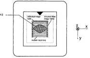
- the cassette 16as shown in FIG. 4 , has a lid 38 and a cover plate 40 which when closed compresses the specimen 42 .
- FIG. 5shows the mosaic of individual image fields or map elements which are created by scanning in X and Y step wise across a region which contains the specimen 42 .
- the scanis called the “mosaic trajectory”. Only the individual map elements or image fields across the first or top traverse of the scan are shown to simplify the illustration.
- the signalsmay be limited at boundaries Y and X so as to vignette the image thereby removing areas at the boundary which may not be unambiguously digitized in the computer, as shown in FIG. 6 .
- the controller 24which receives the reflected signals (either gray scale or in color of the tissue) determines a continuous outer boundary of the tissue shown in the image by first thresholding each of the pixels of the image.
- the pixels above the thresholdare stored as “1s” in a bit map, while all other bits are stored as “0”.
- the area of the bit mapis divided into the blocks shown as the individual image fields in FIG. 5 .
- the area of these blocksmay constitute 80% to 100% of the field of view of the confocal imaging system, which may be from 0.4 to 1.0 millimeters.
- An “exclusive-or” operationmay be performed in the controller on the bits of each block of the bit map, which if the operation results in a “1” for any block, identifies that block as being contained inside the boundary line of the tissue against the window.
- the bit mapforms a map of the tissue in contact with the window or other horizontal slices further away from the window (in the z direction).
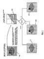
- FIG. 7An example of the information displayed on the display 34 , for a particular case, is illustrated in FIG. 7 .
- the mosaicmay be 15 by 15 individual image blocks or frames, where each frame is stored as 640 by 640 pixels. For example, a mosaic is shown at 50 in FIG. 7 .
- the region containing four image blocks or map elementsis selected, via the controls by viewing the macroscopic image.
- the regionis enlarged and shown at 58 , displays in high resolution four blocks of the image mosaic. From this high resolution image the cancerous cells can be detected by the physician (the pathologist) who observes the confocal image on the display 34 or on a display which is coupled via the network 28 . Then the neoplastic condition can be identified by using the controls 36 and marked on the display or map in different patterns or colors; red for cancerous, green for non-cancerous and yellow for uncertain. Image maps shown at 55 , 52 , and 57 in FIG. 7 are marked cancerous, non-cancerous and uncertain, respectively. It will therefore be apparent that there is a complete anatomical map of the specimen, as well as images of sufficient resolution to detect cells which are neoplastic.
- FIG. 8This image serves as a tumor map and can be used to guide subsequent excisions.
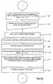
- FIGS. 9A , 9 B and 9 Cthe method and algorithm for carrying out the invention is illustrated.
- FIG. 9Aillustrates the tissue loading and imaging phase
- FIGS. 9B and 9Cillustrate the image formation and display phase, respectively.
- the tissueis excised from the patient in the step of removing the tissue 60 .
- the tissueis marked to indicate the tissue orientation with respect to the patient by the physician. That is, the superior margin toward the head of the patient is important as well as the lateral or sides of the excise specimen.
- This stepis shown at 62 .
- the tissue specimenis then placed on the lid 38 of the cassette 16 with the deep margin facing up, as shown in FIGS. 3 and 4 .
- the superior marginfaces the indicia 54 .
- This stepis shown at 64 .
- the specimenis placed on the lid 38 inside the marked grid of the lid.
- the cassette coverthen is closed and imaging fluid, for example the acetic acid, is inserted into the cassette around the specimen. This step is shown at 66 .
- the userinputs the size of the largest grid which circumscribes the specimen. This is the grid 56 shown in FIG. 3 .
- Thisprovides a tissue size input which is used in creating the mosaic of map elements as shown in FIG. 5 .
- the size of marking stepis indicated at 68 .
- the specimen in its cassette 16is then mounted on the stage by placing it on the receptacle indicated at 55 in FIG. 1 .
- the loading stepis shown at 70 .
- the remaining steps of algorithm and the operations to obtain the imageare apparent from FIGS. 9B and 9C .
- the X, Y and Z parameters of the mapare inputted automatically from the controller at 80 .
- the translator (translation stage) positionwhich corresponds to the center of the cassette is programmed into the controller, initially.
- the focusis then adjusted by the user to obtain the slice or depth (in z) of interest for the particular image at 82 , such as with focus control input 14 .
- the stageis then sent home to a start location at 84 .
- the serpentine scanis then carried out at 86 .
- Each map elementis then stored in memory. Since there are many bits, disk memory may be used. See 88 .
- the clipping or vignetting of the image stepis next carried out at 90 .
- the composite or macro imageis obtained either by pixel elimination, mean value substitution or median filtering and saved in bit map, DICOM format or similar format at 92 .
- the compositeis displayed, this may be in the main window or in the side window on display 34 .
- the userclicks with the mouse on the composite to a desired X, Y location and the four map elements at the X, Y location are displayed in the main window. This is step 96 .
- the four map elementsprovide a high resolution image to the user which was stored in memory at step 88 .
- the usermay select to view live images of the specimen in a window on the display with the confocal microscope imaging of specimen at a desired X-Y location, and the user may manipulate imaging, such as by changing focus (depth) or laser power of the confocal microscope, or may move the translation stage with respect to the objective lens of the confocal microscope about that X-Y location.
- the pathologistthen marks the image as a mapped section of the specimen which has cancerous cells. The margins of the specimen are thoroughly investigated and all cancerous regions marked for further excision and removal during the latter stages of the surgery.
- the marking stepis shown at 96 A.
- steps 96 and 96 Amay be repeated at the same or different X-Y location on the composite map.
- the mapping described abovemay be reactivated (repeated) for the specimen if any setting of the confocal microscope, such as focus, laser power, or other imaging parameter, is changed at step 98 A. After the specimen is examined, a complete two-dimensional image with the markings and the composite map all oriented with respect to the patient may be outputted to the display 34 or color printer 32 .
- FIG. 10shows a complete dermal/epidermal margin.
- the corrugated line in FIG. 10represents the interface between the epidermal layer and the dermal layer.
- FIG. 11shows an incomplete dermal/epidermal junction. The cause for an incomplete junction is due to tissue lifting from the imaging window 40 . If the physician were to see image blocks as shown in the expanded region of FIG. 11 , the physician should mark the region as “uncertain” and investigate the area with live images focusing higher into the tissue until the entire boundary is imaged. This insures that a projection of cancer has not evaded detection.
- imaging modalitiesinclude, optical coherence tomography, multi-photon microscopy or high frequency ultrasound.
- In-vivo mapping of the lesionis also possible by using an in-vivo reflectance confocal microscope to image the surgical field rather than an excised specimen.
Landscapes
- Engineering & Computer Science (AREA)
- Physics & Mathematics (AREA)
- Theoretical Computer Science (AREA)
- Health & Medical Sciences (AREA)
- General Health & Medical Sciences (AREA)
- General Physics & Mathematics (AREA)
- Multimedia (AREA)
- Molecular Biology (AREA)
- Biomedical Technology (AREA)
- Life Sciences & Earth Sciences (AREA)
- Medical Informatics (AREA)
- Nuclear Medicine, Radiotherapy & Molecular Imaging (AREA)
- Radiology & Medical Imaging (AREA)
- Quality & Reliability (AREA)
- Computer Vision & Pattern Recognition (AREA)
- Investigating Or Analysing Materials By Optical Means (AREA)
- Image Processing (AREA)
Abstract
Description
Claims (32)
Applications Claiming Priority (1)
| Application Number | Priority Date | Filing Date | Title |
|---|---|---|---|
| PCT/US2000/030729WO2001035325A1 (en) | 1999-11-10 | 2000-11-10 | System for optically sectioning and mapping surgically excised tissue |
Publications (1)
| Publication Number | Publication Date |
|---|---|
| US7194118B1true US7194118B1 (en) | 2007-03-20 |
Family
ID=37863898
Family Applications (1)
| Application Number | Title | Priority Date | Filing Date |
|---|---|---|---|
| US10/129,719Expired - LifetimeUS7194118B1 (en) | 2000-11-10 | 2000-11-10 | System for optically sectioning and mapping surgically excised tissue |
Country Status (1)
| Country | Link |
|---|---|
| US (1) | US7194118B1 (en) |
Cited By (64)
| Publication number | Priority date | Publication date | Assignee | Title |
|---|---|---|---|---|
| US20040133112A1 (en)* | 2002-03-08 | 2004-07-08 | Milind Rajadhyaksha | System and method for macroscopic and confocal imaging of tissue |
| US20050200324A1 (en)* | 1999-04-07 | 2005-09-15 | Intuitive Surgical Inc. | Non-force reflecting method for providing tool force information to a user of a telesurgical system |
| US20050281484A1 (en)* | 2004-06-17 | 2005-12-22 | Perz Cynthia B | System and method of registering field of view |
| US20060258938A1 (en)* | 2005-05-16 | 2006-11-16 | Intuitive Surgical Inc. | Methods and system for performing 3-D tool tracking by fusion of sensor and/or camera derived data during minimally invasive robotic surgery |
| US20080004603A1 (en)* | 2006-06-29 | 2008-01-03 | Intuitive Surgical Inc. | Tool position and identification indicator displayed in a boundary area of a computer display screen |
| US20080065109A1 (en)* | 2006-06-13 | 2008-03-13 | Intuitive Surgical, Inc. | Preventing instrument/tissue collisions |
| US20090192523A1 (en)* | 2006-06-29 | 2009-07-30 | Intuitive Surgical, Inc. | Synthetic representation of a surgical instrument |
| US20090326318A1 (en)* | 2008-06-27 | 2009-12-31 | Intuitive Surgical, Inc. | Medical robotic system providing an auxilary view including range of motion limitations for articulatable instruments extending out of a distal end of an entry guide |
| US20090326553A1 (en)* | 2008-06-27 | 2009-12-31 | Intuitive Surgical, Inc. | Medical robotic system providing an auxiliary view of articulatable instruments extending out of a distal end of an entry guide |
| US20090326556A1 (en)* | 2008-06-27 | 2009-12-31 | Intuitive Surgical, Inc. | Medical robotic system providing computer generated auxiliary views of a camera instrument for controlling the positioning and orienting of its tip |
| US20100093023A1 (en)* | 2008-10-09 | 2010-04-15 | Ulf Peter Gustafsson | Process for preserving three dimensional orientation to allow registering histopathological diagnoses of tissue to images of that tissue |
| US20100149183A1 (en)* | 2006-12-15 | 2010-06-17 | Loewke Kevin E | Image mosaicing systems and methods |
| US20100164950A1 (en)* | 2008-12-31 | 2010-07-01 | Intuitive Surgical, Inc. | Efficient 3-d telestration for local robotic proctoring |
| US20100166323A1 (en)* | 2008-12-31 | 2010-07-01 | Intuitive Surgical. Inc. | Robust sparse image matching for robotic surgery |
| US20100274087A1 (en)* | 2007-06-13 | 2010-10-28 | Intuitive Surgical Operations, Inc. | Medical robotic system with coupled control modes |
| US20100317965A1 (en)* | 2009-06-16 | 2010-12-16 | Intuitive Surgical, Inc. | Virtual measurement tool for minimally invasive surgery |
| US20100318099A1 (en)* | 2009-06-16 | 2010-12-16 | Intuitive Surgical, Inc. | Virtual measurement tool for minimally invasive surgery |
| US20100331855A1 (en)* | 2005-05-16 | 2010-12-30 | Intuitive Surgical, Inc. | Efficient Vision and Kinematic Data Fusion For Robotic Surgical Instruments and Other Applications |
| US20110050852A1 (en)* | 2005-12-30 | 2011-03-03 | Intuitive Surgical Operations, Inc. | Stereo telestration for robotic surgery |
| US20110116165A1 (en)* | 2009-11-13 | 2011-05-19 | Olympus Corporation | Microscope device |
| US20110202068A1 (en)* | 2010-02-12 | 2011-08-18 | Intuitive Surgical Operations, Inc. | Medical robotic system providing sensory feedback indicating a difference between a commanded state and a preferred pose of an articulated instrument |
| US8260401B2 (en) | 2006-07-26 | 2012-09-04 | University Of Rochester | Non-invasive in-vivo imaging of mechanoreceptors in skin using confocal microscopy |
| US20120238867A1 (en)* | 2001-11-19 | 2012-09-20 | Dune Medical Devices Ltd | Method and apparatus for examining tissue for predefined target cells, particularly cancerous cells, and a probe useful in such method and apparatus |
| FR2984531A1 (en)* | 2011-12-20 | 2013-06-21 | Ecole Polytech | QUANTITATIVE NON-LINEAR OPTICAL MICROSCOPY USING SHAPED BEAM |
| US20140140607A1 (en)* | 2011-07-28 | 2014-05-22 | Medetect Ab | Method for providing images of a tissue section |
| US8780181B2 (en) | 2008-12-05 | 2014-07-15 | Unisensor A/S | Optical sectioning of a sample and detection of particles in a sample |
| US8792963B2 (en) | 2007-09-30 | 2014-07-29 | Intuitive Surgical Operations, Inc. | Methods of determining tissue distances using both kinematic robotic tool position information and image-derived position information |
| US20140296718A1 (en)* | 2013-03-29 | 2014-10-02 | Sony Corporation | Image processing apparatus, image processing method, information processing program, fluorescence observation system, and fluorescence navigation surgery system |
| US8903546B2 (en) | 2009-08-15 | 2014-12-02 | Intuitive Surgical Operations, Inc. | Smooth control of an articulated instrument across areas with different work space conditions |
| US20150117730A1 (en)* | 2013-10-29 | 2015-04-30 | Canon Kabushiki Kaisha | Image processing method and image processing system |
| US9084623B2 (en) | 2009-08-15 | 2015-07-21 | Intuitive Surgical Operations, Inc. | Controller assisted reconfiguration of an articulated instrument during movement into and out of an entry guide |
| US20150220254A1 (en)* | 2012-08-28 | 2015-08-06 | Sony Corporation | Information processing apparatus, information processing method |
| US9138129B2 (en) | 2007-06-13 | 2015-09-22 | Intuitive Surgical Operations, Inc. | Method and system for moving a plurality of articulated instruments in tandem back towards an entry guide |
| US20150356718A1 (en)* | 2009-10-22 | 2015-12-10 | Koninklijke Philips N.V. | Alignment of an ordered stack of images from a specimen |
| US9229210B2 (en) | 2012-02-26 | 2016-01-05 | Caliber Imaging And Diagnostics, Inc. | Tissue specimen stage for an optical sectioning microscope |
| US9250176B2 (en) | 2010-03-04 | 2016-02-02 | Koninklijke Philips N.V. | Flexible sample container |
| US9310598B2 (en) | 2009-03-11 | 2016-04-12 | Sakura Finetek U.S.A., Inc. | Autofocus method and autofocus device |
| CN105606573A (en)* | 2015-12-22 | 2016-05-25 | 深圳先进技术研究院 | Rapid intraoperative pathological diagnosis system and method |
| US9469034B2 (en) | 2007-06-13 | 2016-10-18 | Intuitive Surgical Operations, Inc. | Method and system for switching modes of a robotic system |
| US9492927B2 (en) | 2009-08-15 | 2016-11-15 | Intuitive Surgical Operations, Inc. | Application of force feedback on an input device to urge its operator to command an articulated instrument to a preferred pose |
| US9677869B2 (en) | 2012-12-05 | 2017-06-13 | Perimeter Medical Imaging, Inc. | System and method for generating a wide-field OCT image of a portion of a sample |
| US9789608B2 (en) | 2006-06-29 | 2017-10-17 | Intuitive Surgical Operations, Inc. | Synthetic representation of a surgical robot |
| US9915813B2 (en) | 2009-12-04 | 2018-03-13 | Koninklijke Philips N.V. | System and method for time-related microscopy of biological organisms |
| US10008017B2 (en) | 2006-06-29 | 2018-06-26 | Intuitive Surgical Operations, Inc. | Rendering tool information as graphic overlays on displayed images of tools |
| US10007102B2 (en) | 2013-12-23 | 2018-06-26 | Sakura Finetek U.S.A., Inc. | Microscope with slide clamping assembly |
| US10139613B2 (en) | 2010-08-20 | 2018-11-27 | Sakura Finetek U.S.A., Inc. | Digital microscope and method of sensing an image of a tissue sample |
| US10269094B2 (en) | 2013-04-19 | 2019-04-23 | Sakura Finetek U.S.A., Inc. | Method for generating a composite image of an object composed of multiple sub-images |
| US10433784B2 (en) | 2014-05-05 | 2019-10-08 | Caliber Imaging & Diagnostics, Inc. | System and method for mapping the locations of captured confocal images of a lesion in skin tissue |
| US10456166B2 (en) | 2006-06-13 | 2019-10-29 | Intuitive Surgical Operations, Inc. | Surgical system entry guide |
| US10507066B2 (en) | 2013-02-15 | 2019-12-17 | Intuitive Surgical Operations, Inc. | Providing information of tools by filtering image areas adjacent to or on displayed images of the tools |
| US10577573B2 (en) | 2017-07-18 | 2020-03-03 | Perimeter Medical Imaging, Inc. | Sample container for stabilizing and aligning excised biological tissue samples for ex vivo analysis |
| US10743848B2 (en) | 2015-09-25 | 2020-08-18 | The Regents Of The University Of Michigan | Biopsy device for coherent Raman imaging |
| US10959607B2 (en) | 2005-12-30 | 2021-03-30 | Intuitive Surgical Operations, Inc. | Methods and apparatus to shape flexible entry guides for minimally invasive surgery |
| CN113453606A (en)* | 2018-12-20 | 2021-09-28 | 阿克拉伦特公司 | Endoscope with dual image sensor |
| US11135023B2 (en) | 2005-12-30 | 2021-10-05 | Intuitive Surgical Operations, Inc. | Robotic surgery system including position sensors using fiber bragg gratings |
| US11280803B2 (en) | 2016-11-22 | 2022-03-22 | Sakura Finetek U.S.A., Inc. | Slide management system |
| US11280990B2 (en) | 2018-02-26 | 2022-03-22 | Caliber Imaging & Diagnostics, Inc. | System and method for macroscopic and microscopic imaging ex-vivo tissue |
| EP3951361A4 (en)* | 2019-03-28 | 2022-11-16 | Hamamatsu Photonics K.K. | FLUORESCENCE OBSERVATION DEVICE |
| US11803043B2 (en) | 2019-12-17 | 2023-10-31 | University Of Washington | System and apparatus for dynamically shaped focal surface with a scanning microscope |
| US12023817B1 (en)* | 2023-11-14 | 2024-07-02 | Pramana, Inc. | System and method for a slide marking technique for target tissue extraction and downstream application |
| US12121302B2 (en) | 2020-06-26 | 2024-10-22 | Caliber Imaging & Diagnostics, Inc. | System for providing remote and rapid access to scanned image data |
| US12239396B2 (en) | 2008-06-27 | 2025-03-04 | Intuitive Surgical Operations, Inc. | Medical robotic system providing an auxiliary view including range of motion limitations for articulatable instruments extending out of a distal end of an entry guide |
| US12266040B2 (en) | 2009-03-31 | 2025-04-01 | Intuitive Surgical Operations, Inc. | Rendering tool information as graphic overlays on displayed images of tools |
| US12357400B2 (en) | 2006-06-29 | 2025-07-15 | Intuitive Surgical Operations, Inc. | Synthetic representation of a surgical robot |
Citations (37)
| Publication number | Priority date | Publication date | Assignee | Title |
|---|---|---|---|---|
| US4760385A (en) | 1985-04-22 | 1988-07-26 | E. I. Du Pont De Nemours And Company | Electronic mosaic imaging process |
| US5034613A (en) | 1989-11-14 | 1991-07-23 | Cornell Research Foundation, Inc. | Two-photon laser microscopy |
| US5073857A (en) | 1989-06-01 | 1991-12-17 | Accuron Corporation | Method and apparatus for cell analysis |
| US5120953A (en) | 1988-07-13 | 1992-06-09 | Harris Martin R | Scanning confocal microscope including a single fibre for transmitting light to and receiving light from an object |
| US5161053A (en) | 1988-08-01 | 1992-11-03 | Commonwealth Scientific & Industrial Research | Confocal microscope |
| US5192980A (en) | 1990-06-27 | 1993-03-09 | A. E. Dixon | Apparatus and method for method for spatially- and spectrally-resolved measurements |
| USRE34214E (en)* | 1984-03-15 | 1993-04-06 | Molecular Dynamics, Inc. | Method and apparatus for microphotometering microscope specimens |
| US5235510A (en) | 1990-11-22 | 1993-08-10 | Kabushiki Kaisha Toshiba | Computer-aided diagnosis system for medical use |
| US5297034A (en) | 1987-04-30 | 1994-03-22 | Corabi International Telemetrics, Inc. | Telepathology diagnostic network |
| US5532874A (en)* | 1992-12-18 | 1996-07-02 | Morphometrix Inc. | Multiscanning confocal microscopy |
| US5532873A (en) | 1993-09-08 | 1996-07-02 | Dixon; Arthur E. | Scanning beam laser microscope with wide range of magnification |
| WO1996021938A1 (en)* | 1995-01-13 | 1996-07-18 | The General Hospital Corporation | Video-rate confocal scanning laser microscope |
| US5548661A (en) | 1991-07-12 | 1996-08-20 | Price; Jeffrey H. | Operator independent image cytometer |
| US5602674A (en) | 1993-07-09 | 1997-02-11 | Compucyte Corp. | Computerized specimen encoder |
| US5655029A (en)* | 1990-11-07 | 1997-08-05 | Neuromedical Systems, Inc. | Device and method for facilitating inspection of a specimen |
| US5677966A (en) | 1993-10-12 | 1997-10-14 | Autocyte, Inc. | Interactive automated cytology method incorporating both manual and automatic determinations |
| US5788639A (en) | 1995-07-13 | 1998-08-04 | Lucid Technologies, Inc. | Confocal imaging through thick dermal tissue |
| US5793969A (en) | 1993-07-09 | 1998-08-11 | Neopath, Inc. | Network review and analysis of computer encoded slides |
| US5796861A (en) | 1996-07-12 | 1998-08-18 | Frim International, Inc. | Mosaic construction, processing, and review of very large electronic micrograph composites |
| US5836877A (en)* | 1997-02-24 | 1998-11-17 | Lucid Inc | System for facilitating pathological examination of a lesion in tissue |
| US5848177A (en) | 1994-12-29 | 1998-12-08 | Board Of Trustees Operating Michigan State University | Method and system for detection of biological materials using fractal dimensions |
| US5880880A (en) | 1995-01-13 | 1999-03-09 | The General Hospital Corp. | Three-dimensional scanning confocal laser microscope |
| US5891619A (en) | 1997-01-14 | 1999-04-06 | Inphocyte, Inc. | System and method for mapping the distribution of normal and abnormal cells in sections of tissue |
| US6014451A (en) | 1997-10-17 | 2000-01-11 | Pioneer Hi-Bred International, Inc. | Remote imaging system for plant diagnosis |
| US6031930A (en) | 1996-08-23 | 2000-02-29 | Bacus Research Laboratories, Inc. | Method and apparatus for testing a progression of neoplasia including cancer chemoprevention testing |
| US6049622A (en) | 1996-12-05 | 2000-04-11 | Mayo Foundation For Medical Education And Research | Graphic navigational guides for accurate image orientation and navigation |
| US6067372A (en) | 1996-02-22 | 2000-05-23 | University Of Pittsburgh | Method and system to enhance robust identification of abnormal regions in radiographs |
| US6078681A (en) | 1996-03-18 | 2000-06-20 | Marine Biological Laboratory | Analytical imaging system and process |
| WO2000049392A1 (en) | 1999-02-17 | 2000-08-24 | Lucid, Inc. | Cassette for facilitating optical sectioning of a retained tissue specimen |
| WO2000055669A1 (en) | 1999-03-18 | 2000-09-21 | Lucid, Inc. | System and method for enhancing confocal reflectance images of tissue specimens |
| US6187289B1 (en) | 1997-10-20 | 2001-02-13 | Board Of Regents, The University Of Texas System | Acetic acid as a contrast in reflectance confocal imaging of tissue |
| US6208374B1 (en) | 1996-07-10 | 2001-03-27 | Second Opinion Solutions As | Video display systems |
| US6263233B1 (en) | 1995-07-13 | 2001-07-17 | Lucid, Inc. | Handheld imaging microscope |
| US6272235B1 (en) | 1997-03-03 | 2001-08-07 | Bacus Research Laboratories, Inc. | Method and apparatus for creating a virtual microscope slide |
| US6330106B1 (en) | 1999-02-17 | 2001-12-11 | Lucid, Inc. | Tissue specimen holder |
| US6370422B1 (en)* | 1998-03-19 | 2002-04-09 | Board Of Regents, The University Of Texas System | Fiber-optic confocal imaging apparatus and methods of use |
| US6493460B1 (en) | 1992-10-14 | 2002-12-10 | Monogen, Inc. | Method and apparatus for automatically detecting malignancy-associated changes |
- 2000
- 2000-11-10USUS10/129,719patent/US7194118B1/ennot_activeExpired - Lifetime
Patent Citations (40)
| Publication number | Priority date | Publication date | Assignee | Title |
|---|---|---|---|---|
| USRE34214E (en)* | 1984-03-15 | 1993-04-06 | Molecular Dynamics, Inc. | Method and apparatus for microphotometering microscope specimens |
| US4760385A (en) | 1985-04-22 | 1988-07-26 | E. I. Du Pont De Nemours And Company | Electronic mosaic imaging process |
| US5297034A (en) | 1987-04-30 | 1994-03-22 | Corabi International Telemetrics, Inc. | Telepathology diagnostic network |
| US5120953A (en) | 1988-07-13 | 1992-06-09 | Harris Martin R | Scanning confocal microscope including a single fibre for transmitting light to and receiving light from an object |
| US5161053A (en) | 1988-08-01 | 1992-11-03 | Commonwealth Scientific & Industrial Research | Confocal microscope |
| US5073857A (en) | 1989-06-01 | 1991-12-17 | Accuron Corporation | Method and apparatus for cell analysis |
| US5034613A (en) | 1989-11-14 | 1991-07-23 | Cornell Research Foundation, Inc. | Two-photon laser microscopy |
| US5192980A (en) | 1990-06-27 | 1993-03-09 | A. E. Dixon | Apparatus and method for method for spatially- and spectrally-resolved measurements |
| US5655029A (en)* | 1990-11-07 | 1997-08-05 | Neuromedical Systems, Inc. | Device and method for facilitating inspection of a specimen |
| US5235510A (en) | 1990-11-22 | 1993-08-10 | Kabushiki Kaisha Toshiba | Computer-aided diagnosis system for medical use |
| US5548661A (en) | 1991-07-12 | 1996-08-20 | Price; Jeffrey H. | Operator independent image cytometer |
| US6493460B1 (en) | 1992-10-14 | 2002-12-10 | Monogen, Inc. | Method and apparatus for automatically detecting malignancy-associated changes |
| US5532874A (en)* | 1992-12-18 | 1996-07-02 | Morphometrix Inc. | Multiscanning confocal microscopy |
| US5793969A (en) | 1993-07-09 | 1998-08-11 | Neopath, Inc. | Network review and analysis of computer encoded slides |
| US5602674A (en) | 1993-07-09 | 1997-02-11 | Compucyte Corp. | Computerized specimen encoder |
| US5532873A (en) | 1993-09-08 | 1996-07-02 | Dixon; Arthur E. | Scanning beam laser microscope with wide range of magnification |
| US5677966A (en) | 1993-10-12 | 1997-10-14 | Autocyte, Inc. | Interactive automated cytology method incorporating both manual and automatic determinations |
| US5848177A (en) | 1994-12-29 | 1998-12-08 | Board Of Trustees Operating Michigan State University | Method and system for detection of biological materials using fractal dimensions |
| WO1996021938A1 (en)* | 1995-01-13 | 1996-07-18 | The General Hospital Corporation | Video-rate confocal scanning laser microscope |
| US5880880A (en) | 1995-01-13 | 1999-03-09 | The General Hospital Corp. | Three-dimensional scanning confocal laser microscope |
| US5788639A (en) | 1995-07-13 | 1998-08-04 | Lucid Technologies, Inc. | Confocal imaging through thick dermal tissue |
| US6263233B1 (en) | 1995-07-13 | 2001-07-17 | Lucid, Inc. | Handheld imaging microscope |
| US6067372A (en) | 1996-02-22 | 2000-05-23 | University Of Pittsburgh | Method and system to enhance robust identification of abnormal regions in radiographs |
| US6078681A (en) | 1996-03-18 | 2000-06-20 | Marine Biological Laboratory | Analytical imaging system and process |
| US6208374B1 (en) | 1996-07-10 | 2001-03-27 | Second Opinion Solutions As | Video display systems |
| US5796861A (en) | 1996-07-12 | 1998-08-18 | Frim International, Inc. | Mosaic construction, processing, and review of very large electronic micrograph composites |
| US6101265A (en) | 1996-08-23 | 2000-08-08 | Bacus Research Laboratories, Inc. | Method and apparatus for acquiring and reconstructing magnified specimen images from a computer-controlled microscope |
| US6031930A (en) | 1996-08-23 | 2000-02-29 | Bacus Research Laboratories, Inc. | Method and apparatus for testing a progression of neoplasia including cancer chemoprevention testing |
| US6049622A (en) | 1996-12-05 | 2000-04-11 | Mayo Foundation For Medical Education And Research | Graphic navigational guides for accurate image orientation and navigation |
| US5891619A (en) | 1997-01-14 | 1999-04-06 | Inphocyte, Inc. | System and method for mapping the distribution of normal and abnormal cells in sections of tissue |
| US5836877A (en)* | 1997-02-24 | 1998-11-17 | Lucid Inc | System for facilitating pathological examination of a lesion in tissue |
| US6522774B1 (en) | 1997-03-03 | 2003-02-18 | Bacus Research Laboratories, Inc. | Method and apparatus for creating a virtual microscope slide |
| US6272235B1 (en) | 1997-03-03 | 2001-08-07 | Bacus Research Laboratories, Inc. | Method and apparatus for creating a virtual microscope slide |
| US6014451A (en) | 1997-10-17 | 2000-01-11 | Pioneer Hi-Bred International, Inc. | Remote imaging system for plant diagnosis |
| US6187289B1 (en) | 1997-10-20 | 2001-02-13 | Board Of Regents, The University Of Texas System | Acetic acid as a contrast in reflectance confocal imaging of tissue |
| US6370422B1 (en)* | 1998-03-19 | 2002-04-09 | Board Of Regents, The University Of Texas System | Fiber-optic confocal imaging apparatus and methods of use |
| US6330106B1 (en) | 1999-02-17 | 2001-12-11 | Lucid, Inc. | Tissue specimen holder |
| US6411434B1 (en) | 1999-02-17 | 2002-06-25 | Lucid, Inc. | Cassette for facilitating optical sectioning of a retained tissue specimen |
| WO2000049392A1 (en) | 1999-02-17 | 2000-08-24 | Lucid, Inc. | Cassette for facilitating optical sectioning of a retained tissue specimen |
| WO2000055669A1 (en) | 1999-03-18 | 2000-09-21 | Lucid, Inc. | System and method for enhancing confocal reflectance images of tissue specimens |
Non-Patent Citations (2)
| Title |
|---|
| Gross, Kenneth G. et al., Mohs Surgery, Fundamentals and Techniques, 1999, pp. 49-89, 91-96. |
| Rajadhyaksha, M. et al., Confocal Laser Microscope Images Tissue In Vivo, Laser Focus World, Feb. 1997, pp. 119-127. |
Cited By (165)
| Publication number | Priority date | Publication date | Assignee | Title |
|---|---|---|---|---|
| US20110105898A1 (en)* | 1999-04-07 | 2011-05-05 | Intuitive Surgical Operations, Inc. | Real-Time Generation of Three-Dimensional Ultrasound image using a Two-Dimensional Ultrasound Transducer in a Robotic System |
| US20050200324A1 (en)* | 1999-04-07 | 2005-09-15 | Intuitive Surgical Inc. | Non-force reflecting method for providing tool force information to a user of a telesurgical system |
| US8944070B2 (en) | 1999-04-07 | 2015-02-03 | Intuitive Surgical Operations, Inc. | Non-force reflecting method for providing tool force information to a user of a telesurgical system |
| US9101397B2 (en) | 1999-04-07 | 2015-08-11 | Intuitive Surgical Operations, Inc. | Real-time generation of three-dimensional ultrasound image using a two-dimensional ultrasound transducer in a robotic system |
| US9232984B2 (en) | 1999-04-07 | 2016-01-12 | Intuitive Surgical Operations, Inc. | Real-time generation of three-dimensional ultrasound image using a two-dimensional ultrasound transducer in a robotic system |
| US10271909B2 (en) | 1999-04-07 | 2019-04-30 | Intuitive Surgical Operations, Inc. | Display of computer generated image of an out-of-view portion of a medical device adjacent a real-time image of an in-view portion of the medical device |
| US10433919B2 (en) | 1999-04-07 | 2019-10-08 | Intuitive Surgical Operations, Inc. | Non-force reflecting method for providing tool force information to a user of a telesurgical system |
| US20120238867A1 (en)* | 2001-11-19 | 2012-09-20 | Dune Medical Devices Ltd | Method and apparatus for examining tissue for predefined target cells, particularly cancerous cells, and a probe useful in such method and apparatus |
| US9226979B2 (en)* | 2001-11-19 | 2016-01-05 | Dune Medical Devices Ltd. | Method and apparatus for examining tissue for predefined target cells, particularly cancerous cells, and a probe useful in such method and apparatus |
| US20040133112A1 (en)* | 2002-03-08 | 2004-07-08 | Milind Rajadhyaksha | System and method for macroscopic and confocal imaging of tissue |
| US7653260B2 (en)* | 2004-06-17 | 2010-01-26 | Carl Zeis MicroImaging GmbH | System and method of registering field of view |
| US20050281484A1 (en)* | 2004-06-17 | 2005-12-22 | Perz Cynthia B | System and method of registering field of view |
| US10555775B2 (en) | 2005-05-16 | 2020-02-11 | Intuitive Surgical Operations, Inc. | Methods and system for performing 3-D tool tracking by fusion of sensor and/or camera derived data during minimally invasive robotic surgery |
| US10792107B2 (en) | 2005-05-16 | 2020-10-06 | Intuitive Surgical Operations, Inc. | Methods and system for performing 3-D tool tracking by fusion of sensor and/or camera derived data during minimally invasive robotic surgery |
| US10842571B2 (en) | 2005-05-16 | 2020-11-24 | Intuitive Surgical Operations, Inc. | Methods and system for performing 3-D tool tracking by fusion of sensor and/or camera derived data during minimally invasive robotic surgery |
| US11116578B2 (en) | 2005-05-16 | 2021-09-14 | Intuitive Surgical Operations, Inc. | Methods and system for performing 3-D tool tracking by fusion of sensor and/or camera derived data during minimally invasive robotic surgery |
| US11478308B2 (en) | 2005-05-16 | 2022-10-25 | Intuitive Surgical Operations, Inc. | Methods and system for performing 3-D tool tracking by fusion of sensor and/or camera derived data during minimally invasive robotic surgery |
| US11672606B2 (en) | 2005-05-16 | 2023-06-13 | Intuitive Surgical Operations, Inc. | Methods and system for performing 3-D tool tracking by fusion of sensor and/or camera derived data during minimally invasive robotic surgery |
| US8971597B2 (en) | 2005-05-16 | 2015-03-03 | Intuitive Surgical Operations, Inc. | Efficient vision and kinematic data fusion for robotic surgical instruments and other applications |
| US20060258938A1 (en)* | 2005-05-16 | 2006-11-16 | Intuitive Surgical Inc. | Methods and system for performing 3-D tool tracking by fusion of sensor and/or camera derived data during minimally invasive robotic surgery |
| US20100331855A1 (en)* | 2005-05-16 | 2010-12-30 | Intuitive Surgical, Inc. | Efficient Vision and Kinematic Data Fusion For Robotic Surgical Instruments and Other Applications |
| US11135023B2 (en) | 2005-12-30 | 2021-10-05 | Intuitive Surgical Operations, Inc. | Robotic surgery system including position sensors using fiber bragg gratings |
| US20110050852A1 (en)* | 2005-12-30 | 2011-03-03 | Intuitive Surgical Operations, Inc. | Stereo telestration for robotic surgery |
| US11712312B2 (en) | 2005-12-30 | 2023-08-01 | Intuitive Surgical Operations, Inc. | Robotic surgery system including position sensors using Fiber Bragg Gratings |
| US12042120B2 (en) | 2005-12-30 | 2024-07-23 | Intuitive Surgical Operations, Inc. | Methods and apparatus to shape flexible entry guides for minimally invasive surgery |
| US12251181B2 (en) | 2005-12-30 | 2025-03-18 | Intuitive Surgical Operations, Inc. | Robotic surgery system including position sensors using fiber Bragg gratings |
| US10959607B2 (en) | 2005-12-30 | 2021-03-30 | Intuitive Surgical Operations, Inc. | Methods and apparatus to shape flexible entry guides for minimally invasive surgery |
| US11278364B2 (en) | 2006-06-13 | 2022-03-22 | Intuitive Surgical Operations, Inc. | Surgical system entry guide |
| US10456166B2 (en) | 2006-06-13 | 2019-10-29 | Intuitive Surgical Operations, Inc. | Surgical system entry guide |
| US9345387B2 (en) | 2006-06-13 | 2016-05-24 | Intuitive Surgical Operations, Inc. | Preventing instrument/tissue collisions |
| US11957304B2 (en) | 2006-06-13 | 2024-04-16 | Intuitive Surgical Operations, Inc. | Minimally invasive surgical system |
| EP4018910A1 (en) | 2006-06-13 | 2022-06-29 | Intuitive Surgical Operations, Inc. | Minimally invasive surgical system |
| US12207895B2 (en) | 2006-06-13 | 2025-01-28 | Intuitive Surgical Operations, Inc. | Surgical system entry guide |
| US20080065109A1 (en)* | 2006-06-13 | 2008-03-13 | Intuitive Surgical, Inc. | Preventing instrument/tissue collisions |
| US11659978B2 (en) | 2006-06-13 | 2023-05-30 | Intuitive Surgical Operations, Inc. | Minimally invasive surgical system |
| US11666204B2 (en) | 2006-06-13 | 2023-06-06 | Intuitive Surgical Operations, Inc. | Minimally invasive surgical system |
| US12089809B2 (en) | 2006-06-13 | 2024-09-17 | Intuitive Surgical Operations, Inc. | Minimally invasive surgical system |
| US12310552B2 (en) | 2006-06-13 | 2025-05-27 | Intuitive Surgical Operations, Inc. | Minimally invasive surgical system |
| US10398520B2 (en) | 2006-06-13 | 2019-09-03 | Intuitive Surgical Operations, Inc. | Minimally invasive surgical system |
| US10730187B2 (en) | 2006-06-29 | 2020-08-04 | Intuitive Surgical Operations, Inc. | Tool position and identification indicator displayed in a boundary area of a computer display screen |
| US9801690B2 (en) | 2006-06-29 | 2017-10-31 | Intuitive Surgical Operations, Inc. | Synthetic representation of a surgical instrument |
| US12357400B2 (en) | 2006-06-29 | 2025-07-15 | Intuitive Surgical Operations, Inc. | Synthetic representation of a surgical robot |
| US10137575B2 (en) | 2006-06-29 | 2018-11-27 | Intuitive Surgical Operations, Inc. | Synthetic representation of a surgical robot |
| US20080004603A1 (en)* | 2006-06-29 | 2008-01-03 | Intuitive Surgical Inc. | Tool position and identification indicator displayed in a boundary area of a computer display screen |
| US10773388B2 (en) | 2006-06-29 | 2020-09-15 | Intuitive Surgical Operations, Inc. | Tool position and identification indicator displayed in a boundary area of a computer display screen |
| US10008017B2 (en) | 2006-06-29 | 2018-06-26 | Intuitive Surgical Operations, Inc. | Rendering tool information as graphic overlays on displayed images of tools |
| US20090192523A1 (en)* | 2006-06-29 | 2009-07-30 | Intuitive Surgical, Inc. | Synthetic representation of a surgical instrument |
| US9788909B2 (en) | 2006-06-29 | 2017-10-17 | Intuitive Surgical Operations, Inc | Synthetic representation of a surgical instrument |
| US9789608B2 (en) | 2006-06-29 | 2017-10-17 | Intuitive Surgical Operations, Inc. | Synthetic representation of a surgical robot |
| US9718190B2 (en) | 2006-06-29 | 2017-08-01 | Intuitive Surgical Operations, Inc. | Tool position and identification indicator displayed in a boundary area of a computer display screen |
| US11638999B2 (en) | 2006-06-29 | 2023-05-02 | Intuitive Surgical Operations, Inc. | Synthetic representation of a surgical robot |
| US11865729B2 (en) | 2006-06-29 | 2024-01-09 | Intuitive Surgical Operations, Inc. | Tool position and identification indicator displayed in a boundary area of a computer display screen |
| US10737394B2 (en) | 2006-06-29 | 2020-08-11 | Intuitive Surgical Operations, Inc. | Synthetic representation of a surgical robot |
| US8260401B2 (en) | 2006-07-26 | 2012-09-04 | University Of Rochester | Non-invasive in-vivo imaging of mechanoreceptors in skin using confocal microscopy |
| US20100149183A1 (en)* | 2006-12-15 | 2010-06-17 | Loewke Kevin E | Image mosaicing systems and methods |
| US12097002B2 (en) | 2007-06-13 | 2024-09-24 | Intuitive Surgical Operations, Inc. | Medical robotic system with coupled control modes |
| US9469034B2 (en) | 2007-06-13 | 2016-10-18 | Intuitive Surgical Operations, Inc. | Method and system for switching modes of a robotic system |
| US8620473B2 (en) | 2007-06-13 | 2013-12-31 | Intuitive Surgical Operations, Inc. | Medical robotic system with coupled control modes |
| US9333042B2 (en) | 2007-06-13 | 2016-05-10 | Intuitive Surgical Operations, Inc. | Medical robotic system with coupled control modes |
| US10271912B2 (en) | 2007-06-13 | 2019-04-30 | Intuitive Surgical Operations, Inc. | Method and system for moving a plurality of articulated instruments in tandem back towards an entry guide |
| US10188472B2 (en) | 2007-06-13 | 2019-01-29 | Intuitive Surgical Operations, Inc. | Medical robotic system with coupled control modes |
| US11751955B2 (en) | 2007-06-13 | 2023-09-12 | Intuitive Surgical Operations, Inc. | Method and system for retracting an instrument into an entry guide |
| US20100274087A1 (en)* | 2007-06-13 | 2010-10-28 | Intuitive Surgical Operations, Inc. | Medical robotic system with coupled control modes |
| US9901408B2 (en) | 2007-06-13 | 2018-02-27 | Intuitive Surgical Operations, Inc. | Preventing instrument/tissue collisions |
| US12295681B2 (en) | 2007-06-13 | 2025-05-13 | Intuitive Surgical Operations, Inc. | Method and system for retracting an instrument into an entry guide |
| US10695136B2 (en) | 2007-06-13 | 2020-06-30 | Intuitive Surgical Operations, Inc. | Preventing instrument/tissue collisions |
| US11399908B2 (en) | 2007-06-13 | 2022-08-02 | Intuitive Surgical Operations, Inc. | Medical robotic system with coupled control modes |
| US9629520B2 (en) | 2007-06-13 | 2017-04-25 | Intuitive Surgical Operations, Inc. | Method and system for moving an articulated instrument back towards an entry guide while automatically reconfiguring the articulated instrument for retraction into the entry guide |
| US11432888B2 (en) | 2007-06-13 | 2022-09-06 | Intuitive Surgical Operations, Inc. | Method and system for moving a plurality of articulated instruments in tandem back towards an entry guide |
| US9138129B2 (en) | 2007-06-13 | 2015-09-22 | Intuitive Surgical Operations, Inc. | Method and system for moving a plurality of articulated instruments in tandem back towards an entry guide |
| US8792963B2 (en) | 2007-09-30 | 2014-07-29 | Intuitive Surgical Operations, Inc. | Methods of determining tissue distances using both kinematic robotic tool position information and image-derived position information |
| US20090326553A1 (en)* | 2008-06-27 | 2009-12-31 | Intuitive Surgical, Inc. | Medical robotic system providing an auxiliary view of articulatable instruments extending out of a distal end of an entry guide |
| US11382702B2 (en) | 2008-06-27 | 2022-07-12 | Intuitive Surgical Operations, Inc. | Medical robotic system providing an auxiliary view including range of motion limitations for articulatable instruments extending out of a distal end of an entry guide |
| US10258425B2 (en) | 2008-06-27 | 2019-04-16 | Intuitive Surgical Operations, Inc. | Medical robotic system providing an auxiliary view of articulatable instruments extending out of a distal end of an entry guide |
| US12239396B2 (en) | 2008-06-27 | 2025-03-04 | Intuitive Surgical Operations, Inc. | Medical robotic system providing an auxiliary view including range of motion limitations for articulatable instruments extending out of a distal end of an entry guide |
| US20090326318A1 (en)* | 2008-06-27 | 2009-12-31 | Intuitive Surgical, Inc. | Medical robotic system providing an auxilary view including range of motion limitations for articulatable instruments extending out of a distal end of an entry guide |
| US9717563B2 (en) | 2008-06-27 | 2017-08-01 | Intuitive Surgical Operations, Inc. | Medical robotic system providing an auxilary view including range of motion limitations for articulatable instruments extending out of a distal end of an entry guide |
| US9516996B2 (en) | 2008-06-27 | 2016-12-13 | Intuitive Surgical Operations, Inc. | Medical robotic system providing computer generated auxiliary views of a camera instrument for controlling the position and orienting of its tip |
| US10368952B2 (en) | 2008-06-27 | 2019-08-06 | Intuitive Surgical Operations, Inc. | Medical robotic system providing an auxiliary view including range of motion limitations for articulatable instruments extending out of a distal end of an entry guide |
| US20090326556A1 (en)* | 2008-06-27 | 2009-12-31 | Intuitive Surgical, Inc. | Medical robotic system providing computer generated auxiliary views of a camera instrument for controlling the positioning and orienting of its tip |
| US9089256B2 (en) | 2008-06-27 | 2015-07-28 | Intuitive Surgical Operations, Inc. | Medical robotic system providing an auxiliary view including range of motion limitations for articulatable instruments extending out of a distal end of an entry guide |
| US11638622B2 (en) | 2008-06-27 | 2023-05-02 | Intuitive Surgical Operations, Inc. | Medical robotic system providing an auxiliary view of articulatable instruments extending out of a distal end of an entry guide |
| US8864652B2 (en) | 2008-06-27 | 2014-10-21 | Intuitive Surgical Operations, Inc. | Medical robotic system providing computer generated auxiliary views of a camera instrument for controlling the positioning and orienting of its tip |
| US8501435B2 (en)* | 2008-10-09 | 2013-08-06 | Sti Medical Systems, Llc | Process for preserving three dimensional orientation to allow registering histopathological diagnoses of tissue to images of that tissue |
| US20100093023A1 (en)* | 2008-10-09 | 2010-04-15 | Ulf Peter Gustafsson | Process for preserving three dimensional orientation to allow registering histopathological diagnoses of tissue to images of that tissue |
| US9841593B2 (en) | 2008-12-05 | 2017-12-12 | Koninklijke Philips N.V. | Optical sectioning of a sample and detection of particles in a sample |
| US8780181B2 (en) | 2008-12-05 | 2014-07-15 | Unisensor A/S | Optical sectioning of a sample and detection of particles in a sample |
| US20100164950A1 (en)* | 2008-12-31 | 2010-07-01 | Intuitive Surgical, Inc. | Efficient 3-d telestration for local robotic proctoring |
| US8830224B2 (en) | 2008-12-31 | 2014-09-09 | Intuitive Surgical Operations, Inc. | Efficient 3-D telestration for local robotic proctoring |
| US8184880B2 (en) | 2008-12-31 | 2012-05-22 | Intuitive Surgical Operations, Inc. | Robust sparse image matching for robotic surgery |
| US8639000B2 (en) | 2008-12-31 | 2014-01-28 | Intuitive Surgical Operations, Inc. | Robust sparse image matching for robotic surgery |
| US20100166323A1 (en)* | 2008-12-31 | 2010-07-01 | Intuitive Surgical. Inc. | Robust sparse image matching for robotic surgery |
| US9402690B2 (en) | 2008-12-31 | 2016-08-02 | Intuitive Surgical Operations, Inc. | Efficient 3-D telestration for local and remote robotic proctoring |
| US9310598B2 (en) | 2009-03-11 | 2016-04-12 | Sakura Finetek U.S.A., Inc. | Autofocus method and autofocus device |
| US10495867B2 (en) | 2009-03-11 | 2019-12-03 | Sakura Finetek U.S.A., Inc. | Autofocus method and autofocus device |
| WO2010117684A1 (en) | 2009-03-31 | 2010-10-14 | Intuitive Surgical Operations, Inc. | Synthetic representation of a surgical instrument |
| US11941734B2 (en) | 2009-03-31 | 2024-03-26 | Intuitive Surgical Operations, Inc. | Rendering tool information as graphic overlays on displayed images of tools |
| US10984567B2 (en) | 2009-03-31 | 2021-04-20 | Intuitive Surgical Operations, Inc. | Rendering tool information as graphic overlays on displayed images of tools |
| US12266040B2 (en) | 2009-03-31 | 2025-04-01 | Intuitive Surgical Operations, Inc. | Rendering tool information as graphic overlays on displayed images of tools |
| US10282881B2 (en) | 2009-03-31 | 2019-05-07 | Intuitive Surgical Operations, Inc. | Rendering tool information as graphic overlays on displayed images of tools |
| US9155592B2 (en) | 2009-06-16 | 2015-10-13 | Intuitive Surgical Operations, Inc. | Virtual measurement tool for minimally invasive surgery |
| US9492240B2 (en) | 2009-06-16 | 2016-11-15 | Intuitive Surgical Operations, Inc. | Virtual measurement tool for minimally invasive surgery |
| US20100318099A1 (en)* | 2009-06-16 | 2010-12-16 | Intuitive Surgical, Inc. | Virtual measurement tool for minimally invasive surgery |
| US20100317965A1 (en)* | 2009-06-16 | 2010-12-16 | Intuitive Surgical, Inc. | Virtual measurement tool for minimally invasive surgery |
| US9084623B2 (en) | 2009-08-15 | 2015-07-21 | Intuitive Surgical Operations, Inc. | Controller assisted reconfiguration of an articulated instrument during movement into and out of an entry guide |
| US11596490B2 (en) | 2009-08-15 | 2023-03-07 | Intuitive Surgical Operations, Inc. | Application of force feedback on an input device to urge its operator to command an articulated instrument to a preferred pose |
| US10271915B2 (en) | 2009-08-15 | 2019-04-30 | Intuitive Surgical Operations, Inc. | Application of force feedback on an input device to urge its operator to command an articulated instrument to a preferred pose |
| US9956044B2 (en) | 2009-08-15 | 2018-05-01 | Intuitive Surgical Operations, Inc. | Controller assisted reconfiguration of an articulated instrument during movement into and out of an entry guide |
| US10772689B2 (en) | 2009-08-15 | 2020-09-15 | Intuitive Surgical Operations, Inc. | Controller assisted reconfiguration of an articulated instrument during movement into and out of an entry guide |
| US9492927B2 (en) | 2009-08-15 | 2016-11-15 | Intuitive Surgical Operations, Inc. | Application of force feedback on an input device to urge its operator to command an articulated instrument to a preferred pose |
| US10959798B2 (en) | 2009-08-15 | 2021-03-30 | Intuitive Surgical Operations, Inc. | Application of force feedback on an input device to urge its operator to command an articulated instrument to a preferred pose |
| US8903546B2 (en) | 2009-08-15 | 2014-12-02 | Intuitive Surgical Operations, Inc. | Smooth control of an articulated instrument across areas with different work space conditions |
| US9940719B2 (en)* | 2009-10-22 | 2018-04-10 | Koninklijke Philips N.V. | Alignment of an ordered stack of images from a specimen |
| US20150356718A1 (en)* | 2009-10-22 | 2015-12-10 | Koninklijke Philips N.V. | Alignment of an ordered stack of images from a specimen |
| US9122070B2 (en)* | 2009-11-13 | 2015-09-01 | Olympus Corporation | Microscope device |
| US20110116165A1 (en)* | 2009-11-13 | 2011-05-19 | Olympus Corporation | Microscope device |
| US10317664B2 (en) | 2009-11-13 | 2019-06-11 | Olympus Corporation | Microscope device |
| US9915813B2 (en) | 2009-12-04 | 2018-03-13 | Koninklijke Philips N.V. | System and method for time-related microscopy of biological organisms |
| US20110202068A1 (en)* | 2010-02-12 | 2011-08-18 | Intuitive Surgical Operations, Inc. | Medical robotic system providing sensory feedback indicating a difference between a commanded state and a preferred pose of an articulated instrument |
| US10828774B2 (en) | 2010-02-12 | 2020-11-10 | Intuitive Surgical Operations, Inc. | Medical robotic system providing sensory feedback indicating a difference between a commanded state and a preferred pose of an articulated instrument |
| US9622826B2 (en) | 2010-02-12 | 2017-04-18 | Intuitive Surgical Operations, Inc. | Medical robotic system providing sensory feedback indicating a difference between a commanded state and a preferred pose of an articulated instrument |
| US8918211B2 (en) | 2010-02-12 | 2014-12-23 | Intuitive Surgical Operations, Inc. | Medical robotic system providing sensory feedback indicating a difference between a commanded state and a preferred pose of an articulated instrument |
| US10537994B2 (en) | 2010-02-12 | 2020-01-21 | Intuitive Surgical Operations, Inc. | Medical robotic system providing sensory feedback indicating a difference between a commanded state and a preferred pose of an articulated instrument |
| US9250176B2 (en) | 2010-03-04 | 2016-02-02 | Koninklijke Philips N.V. | Flexible sample container |
| EP3459429A1 (en) | 2010-05-14 | 2019-03-27 | Intuitive Surgical Operations Inc. | Medical robotic system with coupled control modes |
| WO2011143338A1 (en) | 2010-05-14 | 2011-11-17 | Intuitive Surgical Operations, Inc. | Medical robotic system with coupled control modes |
| EP4193904A2 (en) | 2010-05-14 | 2023-06-14 | Intuitive Surgical Operations, Inc. | Medical robotic system with coupled control modes |
| US10139613B2 (en) | 2010-08-20 | 2018-11-27 | Sakura Finetek U.S.A., Inc. | Digital microscope and method of sensing an image of a tissue sample |
| US9311521B2 (en)* | 2011-07-28 | 2016-04-12 | Medetect Ab | Method for providing images of a tissue section |
| US20140140607A1 (en)* | 2011-07-28 | 2014-05-22 | Medetect Ab | Method for providing images of a tissue section |
| US9791682B2 (en) | 2011-12-20 | 2017-10-17 | Ecole Polytechnique | Quantitative nonlinear optical microscopy using a shaped beam |
| FR2984531A1 (en)* | 2011-12-20 | 2013-06-21 | Ecole Polytech | QUANTITATIVE NON-LINEAR OPTICAL MICROSCOPY USING SHAPED BEAM |
| WO2013093275A1 (en)* | 2011-12-20 | 2013-06-27 | Ecole Polytechnique | Quantitative nonlinear optical microscopy using a shaped beam |
| US9229210B2 (en) | 2012-02-26 | 2016-01-05 | Caliber Imaging And Diagnostics, Inc. | Tissue specimen stage for an optical sectioning microscope |
| US10908795B2 (en) | 2012-08-28 | 2021-02-02 | Sony Corporation | Information processing apparatus, information processing method |
| US20190146663A1 (en)* | 2012-08-28 | 2019-05-16 | Sony Corporation | Information processing apparatus, information processing method |
| US10235029B2 (en)* | 2012-08-28 | 2019-03-19 | Sony Corporation | Information processing apparatus, information processing method |
| US20150220254A1 (en)* | 2012-08-28 | 2015-08-06 | Sony Corporation | Information processing apparatus, information processing method |
| US9677869B2 (en) | 2012-12-05 | 2017-06-13 | Perimeter Medical Imaging, Inc. | System and method for generating a wide-field OCT image of a portion of a sample |
| US10359271B2 (en) | 2012-12-05 | 2019-07-23 | Perimeter Medical Imaging, Inc. | System and method for tissue differentiation in imaging |
| US11806102B2 (en) | 2013-02-15 | 2023-11-07 | Intuitive Surgical Operations, Inc. | Providing information of tools by filtering image areas adjacent to or on displayed images of the tools |
| US11389255B2 (en) | 2013-02-15 | 2022-07-19 | Intuitive Surgical Operations, Inc. | Providing information of tools by filtering image areas adjacent to or on displayed images of the tools |
| US10507066B2 (en) | 2013-02-15 | 2019-12-17 | Intuitive Surgical Operations, Inc. | Providing information of tools by filtering image areas adjacent to or on displayed images of the tools |
| US20140296718A1 (en)* | 2013-03-29 | 2014-10-02 | Sony Corporation | Image processing apparatus, image processing method, information processing program, fluorescence observation system, and fluorescence navigation surgery system |
| US10143378B2 (en)* | 2013-03-29 | 2018-12-04 | Sony Corporation | Image processing apparatus, image processing method, information processing program, fluorescence observation system, and fluorescence navigation surgery system |
| US11452453B2 (en) | 2013-03-29 | 2022-09-27 | Sony Corporation | Image processing apparatus, image processing method, information processing program, fluorescence observation system, and fluorescence navigation surgery system |
| US10269094B2 (en) | 2013-04-19 | 2019-04-23 | Sakura Finetek U.S.A., Inc. | Method for generating a composite image of an object composed of multiple sub-images |
| US20150117730A1 (en)* | 2013-10-29 | 2015-04-30 | Canon Kabushiki Kaisha | Image processing method and image processing system |
| US10007102B2 (en) | 2013-12-23 | 2018-06-26 | Sakura Finetek U.S.A., Inc. | Microscope with slide clamping assembly |
| US10433784B2 (en) | 2014-05-05 | 2019-10-08 | Caliber Imaging & Diagnostics, Inc. | System and method for mapping the locations of captured confocal images of a lesion in skin tissue |
| US10743848B2 (en) | 2015-09-25 | 2020-08-18 | The Regents Of The University Of Michigan | Biopsy device for coherent Raman imaging |
| US11419590B2 (en) | 2015-09-25 | 2022-08-23 | The Regents Of The University Of Michigan | Biopsy device for coherent Raman imaging |
| CN105606573B (en)* | 2015-12-22 | 2019-04-05 | 深圳先进技术研究院 | A kind of System and method for of early diagnosis diagnosis |
| CN105606573A (en)* | 2015-12-22 | 2016-05-25 | 深圳先进技术研究院 | Rapid intraoperative pathological diagnosis system and method |
| US11280803B2 (en) | 2016-11-22 | 2022-03-22 | Sakura Finetek U.S.A., Inc. | Slide management system |
| US10577573B2 (en) | 2017-07-18 | 2020-03-03 | Perimeter Medical Imaging, Inc. | Sample container for stabilizing and aligning excised biological tissue samples for ex vivo analysis |
| US10894939B2 (en) | 2017-07-18 | 2021-01-19 | Perimeter Medical Imaging, Inc. | Sample container for stabilizing and aligning excised biological tissue samples for ex vivo analysis |
| US11681133B2 (en) | 2018-02-26 | 2023-06-20 | Caliber Imaging & Diagnostics, Inc. | System and method for macroscopic and microscopic imaging ex-vivo tissue |
| US11280990B2 (en) | 2018-02-26 | 2022-03-22 | Caliber Imaging & Diagnostics, Inc. | System and method for macroscopic and microscopic imaging ex-vivo tissue |
| CN113453606A (en)* | 2018-12-20 | 2021-09-28 | 阿克拉伦特公司 | Endoscope with dual image sensor |
| EP3951361A4 (en)* | 2019-03-28 | 2022-11-16 | Hamamatsu Photonics K.K. | FLUORESCENCE OBSERVATION DEVICE |
| US11921043B2 (en) | 2019-03-28 | 2024-03-05 | Hamamatsu Photonics K.K. | Fluorescence observation device |
| US11803043B2 (en) | 2019-12-17 | 2023-10-31 | University Of Washington | System and apparatus for dynamically shaped focal surface with a scanning microscope |
| US12121302B2 (en) | 2020-06-26 | 2024-10-22 | Caliber Imaging & Diagnostics, Inc. | System for providing remote and rapid access to scanned image data |
| US12023817B1 (en)* | 2023-11-14 | 2024-07-02 | Pramana, Inc. | System and method for a slide marking technique for target tissue extraction and downstream application |
Similar Documents
| Publication | Publication Date | Title |
|---|---|---|
| US7194118B1 (en) | System for optically sectioning and mapping surgically excised tissue | |
| JP7346285B2 (en) | Medical image processing device, endoscope system, operating method and program for medical image processing device | |
| CN113543694B (en) | Medical image processing device, processor device, endoscope system, medical image processing method, and recording medium | |
| EP3586181B1 (en) | Augmented reality microscope for pathology | |
| US20060127880A1 (en) | Computerized image capture of structures of interest within a tissue sample | |
| US5796861A (en) | Mosaic construction, processing, and review of very large electronic micrograph composites | |
| US11449973B2 (en) | Enhanced extended depth of focusing on biological samples | |
| JP2005530138A (en) | Computer-aided image capture of important structures in tissue specimens | |
| JPWO2020012872A1 (en) | Medical image processing equipment, medical image processing system, medical image processing method, and program | |
| US20040264749A1 (en) | Boundary finding in dermatological examination | |
| JP2009531128A (en) | Method and apparatus for stereoscopic image guided surgical navigation | |
| JP5442542B2 (en) | Pathological diagnosis support device, pathological diagnosis support method, control program for pathological diagnosis support, and recording medium recording the control program | |
| US20140184778A1 (en) | Image processing apparatus, control method for the same, image processing system, and program | |
| JP2012010275A (en) | Information processing device, information processing method and program thereof | |
| JP3758894B2 (en) | Mammogram image diagnosis support device | |
| JP2000316837A (en) | Imaging diagnosis aid device | |
| EP4586628A2 (en) | Medical imaging systems and methods | |
| CN104011531A (en) | Image processing device, image display system, image processing method, and image processing program | |
| EP1247246A1 (en) | System for optically sectioning and mapping surgically excised tissue | |
| US6463168B1 (en) | Apparatus and method for rapid connectivity processing of images | |
| CN114830638A (en) | System and method for telestration with spatial memory | |
| US20230177679A1 (en) | Image processing apparatus, image processing method, and image processing system | |
| JPH08251576A (en) | Method and device for displaying image | |
| EP3868281B1 (en) | Medical image processing system and learning method | |
| JP3807541B2 (en) | Breast image display method and apparatus |
Legal Events
| Date | Code | Title | Description |
|---|---|---|---|
| AS | Assignment | Owner name:LUCID, INC., NEW YORK Free format text:ASSIGNMENT OF ASSIGNORS INTEREST;ASSIGNORS:HARRIS, DUNCAN;ZAVISLAN, JAMES M.;REEL/FRAME:013145/0813 Effective date:20020507 | |
| STCF | Information on status: patent grant | Free format text:PATENTED CASE | |
| FPAY | Fee payment | Year of fee payment:4 | |
| AS | Assignment | Owner name:SQUARE 1 BANK, NORTH CAROLINA Free format text:SECURITY AGREEMENT;ASSIGNOR:LUCID, INC.;REEL/FRAME:026795/0600 Effective date:20110720 | |
| AS | Assignment | Owner name:LUCID, INC., NEW YORK Free format text:RELEASE BY SECURED PARTY;ASSIGNOR:SQUARE 1 BANK;REEL/FRAME:028207/0361 Effective date:20120511 | |
| AS | Assignment | Owner name:NORTHEAST LCD CAPITAL, LLC, MAINE Free format text:SECURITY AGREEMENT;ASSIGNOR:LUCID, INC.;REEL/FRAME:028533/0017 Effective date:20120705 | |
| FPAY | Fee payment | Year of fee payment:8 | |
| AS | Assignment | Owner name:CALIBER IMAGING & DIAGNOSTICS, INC., NEW YORK Free format text:CHANGE OF NAME;ASSIGNOR:LUCID, INC.;REEL/FRAME:038226/0225 Effective date:20141120 | |
| FEPP | Fee payment procedure | Free format text:MAINTENANCE FEE REMINDER MAILED (ORIGINAL EVENT CODE: REM.); ENTITY STATUS OF PATENT OWNER: SMALL ENTITY | |
| FEPP | Fee payment procedure | Free format text:11.5 YR SURCHARGE- LATE PMT W/IN 6 MO, SMALL ENTITY (ORIGINAL EVENT CODE: M2556); ENTITY STATUS OF PATENT OWNER: SMALL ENTITY | |
| MAFP | Maintenance fee payment | Free format text:PAYMENT OF MAINTENANCE FEE, 12TH YR, SMALL ENTITY (ORIGINAL EVENT CODE: M2553); ENTITY STATUS OF PATENT OWNER: SMALL ENTITY Year of fee payment:12 | |
| AS | Assignment | Owner name:WESTERN ALLIANCE BANK, CALIFORNIA Free format text:SECURITY INTEREST;ASSIGNOR:CALIBER IMAGING & DIAGNOSTICS, INC.;REEL/FRAME:051094/0824 Effective date:20191120 |