US7189263B2 - Biocompatible bone graft material - Google Patents
Biocompatible bone graft materialDownload PDFInfo
- Publication number
- US7189263B2 US7189263B2US10/771,077US77107704AUS7189263B2US 7189263 B2US7189263 B2US 7189263B2US 77107704 AUS77107704 AUS 77107704AUS 7189263 B2US7189263 B2US 7189263B2
- Authority
- US
- United States
- Prior art keywords
- bone
- bone graft
- graft
- collagen
- graft material
- Prior art date
- Legal status (The legal status is an assumption and is not a legal conclusion. Google has not performed a legal analysis and makes no representation as to the accuracy of the status listed.)
- Expired - Lifetime
Links
- 239000000463materialSubstances0.000titleclaimsabstractdescription189
- 210000000988bone and boneAnatomy0.000titleclaimsabstractdescription151
- 229920000642polymerPolymers0.000claimsabstractdescription23
- QORWJWZARLRLPR-UHFFFAOYSA-Htricalcium bis(phosphate)Chemical compound[Ca+2].[Ca+2].[Ca+2].[O-]P([O-])([O-])=O.[O-]P([O-])([O-])=OQORWJWZARLRLPR-UHFFFAOYSA-H0.000claimsdescription48
- 102000008186CollagenHuman genes0.000claimsdescription47
- 108010035532CollagenProteins0.000claimsdescription47
- 229920001436collagenPolymers0.000claimsdescription47
- 239000001506calcium phosphateSubstances0.000claimsdescription21
- 235000011010calcium phosphatesNutrition0.000claimsdescription19
- 229910000389calcium phosphateInorganic materials0.000claimsdescription17
- 239000012530fluidSubstances0.000claimsdescription16
- 239000002131composite materialSubstances0.000claimsdescription13
- 229910052751metalInorganic materials0.000claimsdescription10
- 239000002184metalSubstances0.000claimsdescription10
- RTAQQCXQSZGOHL-UHFFFAOYSA-NTitaniumChemical compound[Ti]RTAQQCXQSZGOHL-UHFFFAOYSA-N0.000claimsdescription7
- 210000004369bloodAnatomy0.000claimsdescription7
- 239000008280bloodSubstances0.000claimsdescription7
- 239000010936titaniumSubstances0.000claimsdescription7
- PEDCQBHIVMGVHV-UHFFFAOYSA-NGlycerineChemical compoundOCC(O)COPEDCQBHIVMGVHV-UHFFFAOYSA-N0.000claimsdescription6
- 239000004696Poly ether ether ketoneSubstances0.000claimsdescription6
- 229920002530polyetherether ketonePolymers0.000claimsdescription6
- 229910052719titaniumInorganic materials0.000claimsdescription6
- 230000002787reinforcementEffects0.000claimsdescription5
- 229910019142PO4Inorganic materials0.000claimsdescription4
- FAPWRFPIFSIZLT-UHFFFAOYSA-MSodium chlorideChemical compound[Na+].[Cl-]FAPWRFPIFSIZLT-UHFFFAOYSA-M0.000claimsdescription4
- 210000001185bone marrowAnatomy0.000claimsdescription4
- 229910001000nickel titaniumInorganic materials0.000claimsdescription4
- HLXZNVUGXRDIFK-UHFFFAOYSA-Nnickel titaniumChemical compound[Ti].[Ti].[Ti].[Ti].[Ti].[Ti].[Ti].[Ti].[Ti].[Ti].[Ti].[Ni].[Ni].[Ni].[Ni].[Ni].[Ni].[Ni].[Ni].[Ni].[Ni].[Ni].[Ni].[Ni].[Ni]HLXZNVUGXRDIFK-UHFFFAOYSA-N0.000claimsdescription4
- 239000011780sodium chlorideSubstances0.000claimsdescription4
- 229910001220stainless steelInorganic materials0.000claimsdescription4
- 239000010935stainless steelSubstances0.000claimsdescription4
- 210000001519tissueAnatomy0.000claimsdescription4
- 241000283690Bos taurusSpecies0.000claimsdescription3
- 230000037237body shapeEffects0.000claimsdescription3
- 210000000588acetabulumAnatomy0.000claimsdescription2
- ZZUFCTLCJUWOSV-UHFFFAOYSA-NfurosemideChemical compoundC1=C(Cl)C(S(=O)(=O)N)=CC(C(O)=O)=C1NCC1=CC=CO1ZZUFCTLCJUWOSV-UHFFFAOYSA-N0.000claimsdescription2
- NBIIXXVUZAFLBC-UHFFFAOYSA-KphosphateChemical compound[O-]P([O-])([O-])=ONBIIXXVUZAFLBC-UHFFFAOYSA-K0.000claims2
- 239000010452phosphateSubstances0.000claims2
- 239000011147inorganic materialSubstances0.000abstractdescription15
- 229910010272inorganic materialInorganic materials0.000abstractdescription14
- 230000001747exhibiting effectEffects0.000abstract1
- 239000011148porous materialSubstances0.000description35
- 108010010803GelatinProteins0.000description30
- 229920000159gelatinPolymers0.000description30
- 239000008273gelatinSubstances0.000description30
- 235000019322gelatineNutrition0.000description30
- 235000011852gelatine dessertsNutrition0.000description30
- 239000000243solutionSubstances0.000description22
- 239000007943implantSubstances0.000description21
- 230000007547defectEffects0.000description20
- 238000000034methodMethods0.000description17
- 239000000523sampleSubstances0.000description15
- 229910052500inorganic mineralInorganic materials0.000description14
- 239000011707mineralSubstances0.000description14
- 239000007795chemical reaction productSubstances0.000description13
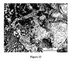
- 238000001878scanning electron micrographMethods0.000description12
- XLYOFNOQVPJJNP-UHFFFAOYSA-NwaterSubstancesOXLYOFNOQVPJJNP-UHFFFAOYSA-N0.000description11
- 239000000203mixtureSubstances0.000description10
- 239000002243precursorSubstances0.000description10
- 239000002002slurrySubstances0.000description9
- IJGRMHOSHXDMSA-UHFFFAOYSA-NAtomic nitrogenChemical compoundN#NIJGRMHOSHXDMSA-UHFFFAOYSA-N0.000description8
- 229920000249biocompatible polymerPolymers0.000description8
- 238000006479redox reactionMethods0.000description8
- 239000011800void materialSubstances0.000description8
- 238000009826distributionMethods0.000description7
- 239000000047productSubstances0.000description7
- PMMYEEVYMWASQN-DMTCNVIQSA-NHydroxyprolineChemical compoundO[C@H]1CN[C@H](C(O)=O)C1PMMYEEVYMWASQN-DMTCNVIQSA-N0.000description6
- 150000001450anionsChemical class0.000description6
- 150000001768cationsChemical class0.000description6
- 230000006835compressionEffects0.000description6
- 238000007906compressionMethods0.000description6
- 239000008367deionised waterSubstances0.000description6
- 229910021641deionized waterInorganic materials0.000description6
- PMMYEEVYMWASQN-UHFFFAOYSA-Ndl-hydroxyprolineNatural productsOC1C[NH2+]C(C([O-])=O)C1PMMYEEVYMWASQN-UHFFFAOYSA-N0.000description6
- 238000011156evaluationMethods0.000description6
- 238000011049fillingMethods0.000description6
- 229960002591hydroxyprolineDrugs0.000description6
- 239000007800oxidant agentSubstances0.000description6
- FGMPLJWBKKVCDB-UHFFFAOYSA-Ntrans-L-hydroxy-prolineNatural productsON1CCCC1C(O)=OFGMPLJWBKKVCDB-UHFFFAOYSA-N0.000description6
- 241001465754MetazoaSpecies0.000description5
- 241001494479PecoraSpecies0.000description5
- 238000002441X-ray diffractionMethods0.000description5
- 102000004169proteins and genesHuman genes0.000description5
- 108090000623proteins and genesProteins0.000description5
- 239000000376reactantSubstances0.000description5
- 229910000391tricalcium phosphateInorganic materials0.000description5
- 102000007350Bone Morphogenetic ProteinsHuman genes0.000description4
- 108010007726Bone Morphogenetic ProteinsProteins0.000description4
- 230000008901benefitEffects0.000description4
- -1bone marrow aspirateSubstances0.000description4
- 229940112869bone morphogenetic proteinDrugs0.000description4
- 239000011248coating agentSubstances0.000description4
- 238000000576coating methodMethods0.000description4
- 239000000945fillerSubstances0.000description4
- 230000004927fusionEffects0.000description4
- 238000010438heat treatmentMethods0.000description4
- 229910052588hydroxylapatiteInorganic materials0.000description4
- 230000006872improvementEffects0.000description4
- 238000007373indentationMethods0.000description4
- 239000007788liquidSubstances0.000description4
- QSHDDOUJBYECFT-UHFFFAOYSA-NmercuryChemical compound[Hg]QSHDDOUJBYECFT-UHFFFAOYSA-N0.000description4
- 229910052753mercuryInorganic materials0.000description4
- 229910052757nitrogenInorganic materials0.000description4
- 239000011368organic materialSubstances0.000description4
- 230000011164ossificationEffects0.000description4
- 230000000278osteoconductive effectEffects0.000description4
- XYJRXVWERLGGKC-UHFFFAOYSA-Dpentacalcium;hydroxide;triphosphateChemical compound[OH-].[Ca+2].[Ca+2].[Ca+2].[Ca+2].[Ca+2].[O-]P([O-])([O-])=O.[O-]P([O-])([O-])=O.[O-]P([O-])([O-])=OXYJRXVWERLGGKC-UHFFFAOYSA-D0.000description4
- 238000012384transportation and deliveryMethods0.000description4
- 238000004383yellowingMethods0.000description4
- 238000005033Fourier transform infrared spectroscopyMethods0.000description3
- MWUXSHHQAYIFBG-UHFFFAOYSA-NNitric oxideChemical groupO=[N]MWUXSHHQAYIFBG-UHFFFAOYSA-N0.000description3
- 230000009286beneficial effectEffects0.000description3
- 238000001816coolingMethods0.000description3
- 238000001035dryingMethods0.000description3
- 239000001307heliumSubstances0.000description3
- 229910052734heliumInorganic materials0.000description3
- SWQJXJOGLNCZEY-UHFFFAOYSA-Nhelium atomChemical compound[He]SWQJXJOGLNCZEY-UHFFFAOYSA-N0.000description3
- 210000001624hipAnatomy0.000description3
- 238000002513implantationMethods0.000description3
- 238000009616inductively coupled plasmaMethods0.000description3
- 229920001610polycaprolactonePolymers0.000description3
- 239000004626polylactic acidSubstances0.000description3
- 238000002459porosimetryMethods0.000description3
- 238000012545processingMethods0.000description3
- 229920006395saturated elastomerPolymers0.000description3
- 238000004626scanning electron microscopyMethods0.000description3
- 239000007787solidSubstances0.000description3
- 238000011282treatmentMethods0.000description3
- QGZKDVFQNNGYKY-UHFFFAOYSA-NAmmoniaChemical compoundNQGZKDVFQNNGYKY-UHFFFAOYSA-N0.000description2
- 241000282472Canis lupus familiarisSpecies0.000description2
- CURLTUGMZLYLDI-UHFFFAOYSA-NCarbon dioxideChemical compoundO=C=OCURLTUGMZLYLDI-UHFFFAOYSA-N0.000description2
- 102000012422Collagen Type IHuman genes0.000description2
- 108010022452Collagen Type IProteins0.000description2
- 238000001157Fourier transform infrared spectrumMethods0.000description2
- 241000124008MammaliaSpecies0.000description2
- 229920000954PolyglycolidePolymers0.000description2
- 239000004372Polyvinyl alcoholSubstances0.000description2
- 229920002125Sokalan®Polymers0.000description2
- QAOWNCQODCNURD-UHFFFAOYSA-NSulfuric acidChemical compoundOS(O)(=O)=OQAOWNCQODCNURD-UHFFFAOYSA-N0.000description2
- 238000010521absorption reactionMethods0.000description2
- 239000002253acidSubstances0.000description2
- BFNBIHQBYMNNAN-UHFFFAOYSA-Nammonium sulfateChemical compoundN.N.OS(O)(=O)=OBFNBIHQBYMNNAN-UHFFFAOYSA-N0.000description2
- 229910052921ammonium sulfateInorganic materials0.000description2
- 235000011130ammonium sulphateNutrition0.000description2
- 238000004458analytical methodMethods0.000description2
- 239000003242anti bacterial agentSubstances0.000description2
- 229940088710antibiotic agentDrugs0.000description2
- 230000004888barrier functionEffects0.000description2
- 230000002051biphasic effectEffects0.000description2
- KGBXLFKZBHKPEV-UHFFFAOYSA-Nboric acidChemical compoundOB(O)OKGBXLFKZBHKPEV-UHFFFAOYSA-N0.000description2
- 239000004327boric acidSubstances0.000description2
- 239000011575calciumSubstances0.000description2
- 230000001413cellular effectEffects0.000description2
- 239000001913celluloseSubstances0.000description2
- 229920002678cellulosePolymers0.000description2
- 239000000919ceramicSubstances0.000description2
- 230000008859changeEffects0.000description2
- 238000006243chemical reactionMethods0.000description2
- 239000003795chemical substances by applicationSubstances0.000description2
- 230000008602contractionEffects0.000description2
- 238000004132cross linkingMethods0.000description2
- 239000013078crystalSubstances0.000description2
- 238000010790dilutionMethods0.000description2
- 239000012895dilutionSubstances0.000description2
- 210000000416exudates and transudateAnatomy0.000description2
- 238000004108freeze dryingMethods0.000description2
- 230000035876healingEffects0.000description2
- 229910001385heavy metalInorganic materials0.000description2
- 210000002758humerusAnatomy0.000description2
- 238000001727in vivoMethods0.000description2
- 238000011065in-situ storageMethods0.000description2
- 230000001788irregularEffects0.000description2
- 210000003127kneeAnatomy0.000description2
- 230000004048modificationEffects0.000description2
- 238000012986modificationMethods0.000description2
- 229920001432poly(L-lactide)Polymers0.000description2
- 239000004633polyglycolic acidSubstances0.000description2
- 229920002451polyvinyl alcoholPolymers0.000description2
- 230000008569processEffects0.000description2
- 230000001737promoting effectEffects0.000description2
- 238000001812pycnometryMethods0.000description2
- 210000003625skullAnatomy0.000description2
- AVPCPPOOQICIRJ-UHFFFAOYSA-Lsodium glycerol 2-phosphateChemical compound[Na+].[Na+].OCC(CO)OP([O-])([O-])=OAVPCPPOOQICIRJ-UHFFFAOYSA-L0.000description2
- 229920002994synthetic fiberPolymers0.000description2
- 238000012360testing methodMethods0.000description2
- GBNXLQPMFAUCOI-UHFFFAOYSA-Htetracalcium;oxygen(2-);diphosphateChemical compound[O-2].[Ca+2].[Ca+2].[Ca+2].[Ca+2].[O-]P([O-])([O-])=O.[O-]P([O-])([O-])=OGBNXLQPMFAUCOI-UHFFFAOYSA-H0.000description2
- 235000019731tricalcium phosphateNutrition0.000description2
- 229940078499tricalcium phosphateDrugs0.000description2
- RZVAJINKPMORJF-UHFFFAOYSA-NAcetaminophenChemical compoundCC(=O)NC1=CC=C(O)C=C1RZVAJINKPMORJF-UHFFFAOYSA-N0.000description1
- 102000001187Collagen Type IIIHuman genes0.000description1
- 108010069502Collagen Type IIIProteins0.000description1
- 208000024779Comminuted FracturesDiseases0.000description1
- 239000004593EpoxySubstances0.000description1
- 229910001200FerrotitaniumInorganic materials0.000description1
- OAKJQQAXSVQMHS-UHFFFAOYSA-NHydrazineChemical compoundNNOAKJQQAXSVQMHS-UHFFFAOYSA-N0.000description1
- VEXZGXHMUGYJMC-UHFFFAOYSA-NHydrochloric acidChemical compoundClVEXZGXHMUGYJMC-UHFFFAOYSA-N0.000description1
- UFHFLCQGNIYNRP-UHFFFAOYSA-NHydrogenChemical compound[H][H]UFHFLCQGNIYNRP-UHFFFAOYSA-N0.000description1
- 241000755266Kathetostoma giganteumSpecies0.000description1
- 229920001730Moisture cure polyurethanePolymers0.000description1
- 206010028980NeoplasmDiseases0.000description1
- NHNBFGGVMKEFGY-UHFFFAOYSA-NNitrateChemical group[O-][N+]([O-])=ONHNBFGGVMKEFGY-UHFFFAOYSA-N0.000description1
- 239000004698PolyethyleneSubstances0.000description1
- 101710172711Structural proteinProteins0.000description1
- CVTZKFWZDBJAHE-UHFFFAOYSA-N[N].NChemical compound[N].NCVTZKFWZDBJAHE-UHFFFAOYSA-N0.000description1
- 238000002835absorbanceMethods0.000description1
- 229920006397acrylic thermoplasticPolymers0.000description1
- 230000009471actionEffects0.000description1
- 239000002671adjuvantSubstances0.000description1
- PNEYBMLMFCGWSK-UHFFFAOYSA-Naluminium oxideInorganic materials[O-2].[O-2].[O-2].[Al+3].[Al+3]PNEYBMLMFCGWSK-UHFFFAOYSA-N0.000description1
- 229910021529ammoniaInorganic materials0.000description1
- 210000003484anatomyAnatomy0.000description1
- 230000003466anti-cipated effectEffects0.000description1
- 229910052586apatiteInorganic materials0.000description1
- 239000007864aqueous solutionSubstances0.000description1
- 229910052785arsenicInorganic materials0.000description1
- 230000003190augmentative effectEffects0.000description1
- 230000004071biological effectEffects0.000description1
- 239000013060biological fluidSubstances0.000description1
- 230000008512biological responseEffects0.000description1
- 210000001124body fluidAnatomy0.000description1
- 230000008468bone growthEffects0.000description1
- 230000037118bone strengthEffects0.000description1
- 210000004556brainAnatomy0.000description1
- 229910052793cadmiumInorganic materials0.000description1
- BDOSMKKIYDKNTQ-UHFFFAOYSA-Ncadmium atomChemical compound[Cd]BDOSMKKIYDKNTQ-UHFFFAOYSA-N0.000description1
- 239000004068calcium phosphate ceramicSubstances0.000description1
- 159000000007calcium saltsChemical class0.000description1
- 239000001569carbon dioxideSubstances0.000description1
- 229910002092carbon dioxideInorganic materials0.000description1
- 239000000969carrierSubstances0.000description1
- 230000015556catabolic processEffects0.000description1
- 210000004027cellAnatomy0.000description1
- 239000004568cementSubstances0.000description1
- 239000003153chemical reaction reagentSubstances0.000description1
- 238000004587chromatography analysisMethods0.000description1
- 230000001427coherent effectEffects0.000description1
- 238000004891communicationMethods0.000description1
- 230000001054cortical effectEffects0.000description1
- 230000003247decreasing effectEffects0.000description1
- 238000006731degradation reactionMethods0.000description1
- 238000001514detection methodMethods0.000description1
- 238000007598dipping methodMethods0.000description1
- 239000003814drugSubstances0.000description1
- 238000012377drug deliveryMethods0.000description1
- 239000003792electrolyteSubstances0.000description1
- 238000005516engineering processMethods0.000description1
- 125000003700epoxy groupChemical group0.000description1
- 238000011067equilibrationMethods0.000description1
- 238000002474experimental methodMethods0.000description1
- 210000002950fibroblastAnatomy0.000description1
- 238000010304firingMethods0.000description1
- 238000007667floatingMethods0.000description1
- 238000005187foamingMethods0.000description1
- 239000012634fragmentSubstances0.000description1
- 239000000499gelSubstances0.000description1
- 235000011187glycerolNutrition0.000description1
- PCHJSUWPFVWCPO-UHFFFAOYSA-NgoldChemical compound[Au]PCHJSUWPFVWCPO-UHFFFAOYSA-N0.000description1
- 239000008240homogeneous mixtureSubstances0.000description1
- 239000001257hydrogenSubstances0.000description1
- 229910052739hydrogenInorganic materials0.000description1
- 238000007654immersionMethods0.000description1
- 208000014674injuryDiseases0.000description1
- 238000007130inorganic reactionMethods0.000description1
- 238000004898kneadingMethods0.000description1
- 230000014759maintenance of locationEffects0.000description1
- 230000013011matingEffects0.000description1
- 239000011159matrix materialSubstances0.000description1
- 238000005259measurementMethods0.000description1
- 230000005541medical transmissionEffects0.000description1
- 150000002739metalsChemical class0.000description1
- 210000001872metatarsal boneAnatomy0.000description1
- VUZPPFZMUPKLLV-UHFFFAOYSA-Nmethane;hydrateChemical compoundC.OVUZPPFZMUPKLLV-UHFFFAOYSA-N0.000description1
- 230000005012migrationEffects0.000description1
- 238000013508migrationMethods0.000description1
- 230000003278mimic effectEffects0.000description1
- 238000002156mixingMethods0.000description1
- 230000000921morphogenic effectEffects0.000description1
- 230000000877morphologic effectEffects0.000description1
- 238000000465mouldingMethods0.000description1
- 229920005615natural polymerPolymers0.000description1
- 238000010606normalizationMethods0.000description1
- 238000010899nucleationMethods0.000description1
- 235000015097nutrientsNutrition0.000description1
- 229920001778nylonPolymers0.000description1
- VSIIXMUUUJUKCM-UHFFFAOYSA-Dpentacalcium;fluoride;triphosphateChemical compound[F-].[Ca+2].[Ca+2].[Ca+2].[Ca+2].[Ca+2].[O-]P([O-])([O-])=O.[O-]P([O-])([O-])=O.[O-]P([O-])([O-])=OVSIIXMUUUJUKCM-UHFFFAOYSA-D0.000description1
- 239000012466permeateSubstances0.000description1
- 239000007793ph indicatorSubstances0.000description1
- 229920003229poly(methyl methacrylate)Polymers0.000description1
- 229920002492poly(sulfone)Polymers0.000description1
- 229920001281polyalkylenePolymers0.000description1
- 239000004632polycaprolactoneSubstances0.000description1
- 229920000647polyepoxidePolymers0.000description1
- 229920000728polyesterPolymers0.000description1
- 229920000573polyethylenePolymers0.000description1
- 229920000098polyolefinPolymers0.000description1
- 238000002360preparation methodMethods0.000description1
- 230000002035prolonged effectEffects0.000description1
- 239000005297pyrexSubstances0.000description1
- 238000007409radiographic assessmentMethods0.000description1
- 238000007634remodelingMethods0.000description1
- 230000008439repair processEffects0.000description1
- 230000010076replicationEffects0.000description1
- 229920005989resinPolymers0.000description1
- 239000011347resinSubstances0.000description1
- 230000000717retained effectEffects0.000description1
- 230000000250revascularizationEffects0.000description1
- 150000003839saltsChemical class0.000description1
- 238000005245sinteringMethods0.000description1
- 238000004513sizingMethods0.000description1
- 238000004544sputter depositionMethods0.000description1
- 210000000130stem cellAnatomy0.000description1
- 230000001954sterilising effectEffects0.000description1
- 238000004659sterilization and disinfectionMethods0.000description1
- 238000012414sterilization procedureMethods0.000description1
- 238000005728strengtheningMethods0.000description1
- 239000000126substanceSubstances0.000description1
- 238000001356surgical procedureMethods0.000description1
- 238000003786synthesis reactionMethods0.000description1
- 229920001059synthetic polymerPolymers0.000description1
- ISXSCDLOGDJUNJ-UHFFFAOYSA-Ntert-butyl prop-2-enoateChemical compoundCC(C)(C)OC(=O)C=CISXSCDLOGDJUNJ-UHFFFAOYSA-N0.000description1
- 230000008733traumaEffects0.000description1
- 210000000689upper legAnatomy0.000description1
Images


Classifications
- A—HUMAN NECESSITIES
- A61—MEDICAL OR VETERINARY SCIENCE; HYGIENE
- A61F—FILTERS IMPLANTABLE INTO BLOOD VESSELS; PROSTHESES; DEVICES PROVIDING PATENCY TO, OR PREVENTING COLLAPSING OF, TUBULAR STRUCTURES OF THE BODY, e.g. STENTS; ORTHOPAEDIC, NURSING OR CONTRACEPTIVE DEVICES; FOMENTATION; TREATMENT OR PROTECTION OF EYES OR EARS; BANDAGES, DRESSINGS OR ABSORBENT PADS; FIRST-AID KITS
- A61F2/00—Filters implantable into blood vessels; Prostheses, i.e. artificial substitutes or replacements for parts of the body; Appliances for connecting them with the body; Devices providing patency to, or preventing collapsing of, tubular structures of the body, e.g. stents
- A61F2/02—Prostheses implantable into the body
- A61F2/28—Bones
- A—HUMAN NECESSITIES
- A61—MEDICAL OR VETERINARY SCIENCE; HYGIENE
- A61B—DIAGNOSIS; SURGERY; IDENTIFICATION
- A61B17/00—Surgical instruments, devices or methods
- A61B17/56—Surgical instruments or methods for treatment of bones or joints; Devices specially adapted therefor
- A61B17/58—Surgical instruments or methods for treatment of bones or joints; Devices specially adapted therefor for osteosynthesis, e.g. bone plates, screws or setting implements
- A61B17/68—Internal fixation devices, including fasteners and spinal fixators, even if a part thereof projects from the skin
- A61B17/80—Cortical plates, i.e. bone plates; Instruments for holding or positioning cortical plates, or for compressing bones attached to cortical plates
- A—HUMAN NECESSITIES
- A61—MEDICAL OR VETERINARY SCIENCE; HYGIENE
- A61L—METHODS OR APPARATUS FOR STERILISING MATERIALS OR OBJECTS IN GENERAL; DISINFECTION, STERILISATION OR DEODORISATION OF AIR; CHEMICAL ASPECTS OF BANDAGES, DRESSINGS, ABSORBENT PADS OR SURGICAL ARTICLES; MATERIALS FOR BANDAGES, DRESSINGS, ABSORBENT PADS OR SURGICAL ARTICLES
- A61L27/00—Materials for grafts or prostheses or for coating grafts or prostheses
- A61L27/40—Composite materials, i.e. containing one material dispersed in a matrix of the same or different material
- A61L27/44—Composite materials, i.e. containing one material dispersed in a matrix of the same or different material having a macromolecular matrix
- A61L27/46—Composite materials, i.e. containing one material dispersed in a matrix of the same or different material having a macromolecular matrix with phosphorus-containing inorganic fillers
- A—HUMAN NECESSITIES
- A61—MEDICAL OR VETERINARY SCIENCE; HYGIENE
- A61L—METHODS OR APPARATUS FOR STERILISING MATERIALS OR OBJECTS IN GENERAL; DISINFECTION, STERILISATION OR DEODORISATION OF AIR; CHEMICAL ASPECTS OF BANDAGES, DRESSINGS, ABSORBENT PADS OR SURGICAL ARTICLES; MATERIALS FOR BANDAGES, DRESSINGS, ABSORBENT PADS OR SURGICAL ARTICLES
- A61L27/00—Materials for grafts or prostheses or for coating grafts or prostheses
- A61L27/50—Materials characterised by their function or physical properties, e.g. injectable or lubricating compositions, shape-memory materials, surface modified materials
- A61L27/54—Biologically active materials, e.g. therapeutic substances
- A—HUMAN NECESSITIES
- A61—MEDICAL OR VETERINARY SCIENCE; HYGIENE
- A61L—METHODS OR APPARATUS FOR STERILISING MATERIALS OR OBJECTS IN GENERAL; DISINFECTION, STERILISATION OR DEODORISATION OF AIR; CHEMICAL ASPECTS OF BANDAGES, DRESSINGS, ABSORBENT PADS OR SURGICAL ARTICLES; MATERIALS FOR BANDAGES, DRESSINGS, ABSORBENT PADS OR SURGICAL ARTICLES
- A61L27/00—Materials for grafts or prostheses or for coating grafts or prostheses
- A61L27/50—Materials characterised by their function or physical properties, e.g. injectable or lubricating compositions, shape-memory materials, surface modified materials
- A61L27/56—Porous materials, e.g. foams or sponges
- A—HUMAN NECESSITIES
- A61—MEDICAL OR VETERINARY SCIENCE; HYGIENE
- A61L—METHODS OR APPARATUS FOR STERILISING MATERIALS OR OBJECTS IN GENERAL; DISINFECTION, STERILISATION OR DEODORISATION OF AIR; CHEMICAL ASPECTS OF BANDAGES, DRESSINGS, ABSORBENT PADS OR SURGICAL ARTICLES; MATERIALS FOR BANDAGES, DRESSINGS, ABSORBENT PADS OR SURGICAL ARTICLES
- A61L27/00—Materials for grafts or prostheses or for coating grafts or prostheses
- A61L27/50—Materials characterised by their function or physical properties, e.g. injectable or lubricating compositions, shape-memory materials, surface modified materials
- A61L27/58—Materials at least partially resorbable by the body
- A—HUMAN NECESSITIES
- A61—MEDICAL OR VETERINARY SCIENCE; HYGIENE
- A61B—DIAGNOSIS; SURGERY; IDENTIFICATION
- A61B17/00—Surgical instruments, devices or methods
- A61B17/56—Surgical instruments or methods for treatment of bones or joints; Devices specially adapted therefor
- A61B17/58—Surgical instruments or methods for treatment of bones or joints; Devices specially adapted therefor for osteosynthesis, e.g. bone plates, screws or setting implements
- A61B17/68—Internal fixation devices, including fasteners and spinal fixators, even if a part thereof projects from the skin
- A61B17/80—Cortical plates, i.e. bone plates; Instruments for holding or positioning cortical plates, or for compressing bones attached to cortical plates
- A61B17/8095—Wedge osteotomy devices
- A—HUMAN NECESSITIES
- A61—MEDICAL OR VETERINARY SCIENCE; HYGIENE
- A61B—DIAGNOSIS; SURGERY; IDENTIFICATION
- A61B17/00—Surgical instruments, devices or methods
- A61B17/56—Surgical instruments or methods for treatment of bones or joints; Devices specially adapted therefor
- A61B17/58—Surgical instruments or methods for treatment of bones or joints; Devices specially adapted therefor for osteosynthesis, e.g. bone plates, screws or setting implements
- A61B17/68—Internal fixation devices, including fasteners and spinal fixators, even if a part thereof projects from the skin
- A61B17/84—Fasteners therefor or fasteners being internal fixation devices
- A61B17/86—Pins or screws or threaded wires; nuts therefor
- A—HUMAN NECESSITIES
- A61—MEDICAL OR VETERINARY SCIENCE; HYGIENE
- A61B—DIAGNOSIS; SURGERY; IDENTIFICATION
- A61B17/00—Surgical instruments, devices or methods
- A61B2017/00004—(bio)absorbable, (bio)resorbable or resorptive
- A—HUMAN NECESSITIES
- A61—MEDICAL OR VETERINARY SCIENCE; HYGIENE
- A61F—FILTERS IMPLANTABLE INTO BLOOD VESSELS; PROSTHESES; DEVICES PROVIDING PATENCY TO, OR PREVENTING COLLAPSING OF, TUBULAR STRUCTURES OF THE BODY, e.g. STENTS; ORTHOPAEDIC, NURSING OR CONTRACEPTIVE DEVICES; FOMENTATION; TREATMENT OR PROTECTION OF EYES OR EARS; BANDAGES, DRESSINGS OR ABSORBENT PADS; FIRST-AID KITS
- A61F2/00—Filters implantable into blood vessels; Prostheses, i.e. artificial substitutes or replacements for parts of the body; Appliances for connecting them with the body; Devices providing patency to, or preventing collapsing of, tubular structures of the body, e.g. stents
- A61F2/02—Prostheses implantable into the body
- A61F2/28—Bones
- A61F2/2803—Bones for mandibular reconstruction
- A—HUMAN NECESSITIES
- A61—MEDICAL OR VETERINARY SCIENCE; HYGIENE
- A61F—FILTERS IMPLANTABLE INTO BLOOD VESSELS; PROSTHESES; DEVICES PROVIDING PATENCY TO, OR PREVENTING COLLAPSING OF, TUBULAR STRUCTURES OF THE BODY, e.g. STENTS; ORTHOPAEDIC, NURSING OR CONTRACEPTIVE DEVICES; FOMENTATION; TREATMENT OR PROTECTION OF EYES OR EARS; BANDAGES, DRESSINGS OR ABSORBENT PADS; FIRST-AID KITS
- A61F2/00—Filters implantable into blood vessels; Prostheses, i.e. artificial substitutes or replacements for parts of the body; Appliances for connecting them with the body; Devices providing patency to, or preventing collapsing of, tubular structures of the body, e.g. stents
- A61F2/02—Prostheses implantable into the body
- A61F2/28—Bones
- A61F2/2875—Skull or cranium
- A—HUMAN NECESSITIES
- A61—MEDICAL OR VETERINARY SCIENCE; HYGIENE
- A61F—FILTERS IMPLANTABLE INTO BLOOD VESSELS; PROSTHESES; DEVICES PROVIDING PATENCY TO, OR PREVENTING COLLAPSING OF, TUBULAR STRUCTURES OF THE BODY, e.g. STENTS; ORTHOPAEDIC, NURSING OR CONTRACEPTIVE DEVICES; FOMENTATION; TREATMENT OR PROTECTION OF EYES OR EARS; BANDAGES, DRESSINGS OR ABSORBENT PADS; FIRST-AID KITS
- A61F2/00—Filters implantable into blood vessels; Prostheses, i.e. artificial substitutes or replacements for parts of the body; Appliances for connecting them with the body; Devices providing patency to, or preventing collapsing of, tubular structures of the body, e.g. stents
- A61F2/02—Prostheses implantable into the body
- A61F2/30—Joints
- A61F2/30767—Special external or bone-contacting surface, e.g. coating for improving bone ingrowth
- A—HUMAN NECESSITIES
- A61—MEDICAL OR VETERINARY SCIENCE; HYGIENE
- A61F—FILTERS IMPLANTABLE INTO BLOOD VESSELS; PROSTHESES; DEVICES PROVIDING PATENCY TO, OR PREVENTING COLLAPSING OF, TUBULAR STRUCTURES OF THE BODY, e.g. STENTS; ORTHOPAEDIC, NURSING OR CONTRACEPTIVE DEVICES; FOMENTATION; TREATMENT OR PROTECTION OF EYES OR EARS; BANDAGES, DRESSINGS OR ABSORBENT PADS; FIRST-AID KITS
- A61F2/00—Filters implantable into blood vessels; Prostheses, i.e. artificial substitutes or replacements for parts of the body; Appliances for connecting them with the body; Devices providing patency to, or preventing collapsing of, tubular structures of the body, e.g. stents
- A61F2/02—Prostheses implantable into the body
- A61F2/30—Joints
- A61F2/3094—Designing or manufacturing processes
- A61F2/30965—Reinforcing the prosthesis by embedding particles or fibres during moulding or dipping
- A—HUMAN NECESSITIES
- A61—MEDICAL OR VETERINARY SCIENCE; HYGIENE
- A61F—FILTERS IMPLANTABLE INTO BLOOD VESSELS; PROSTHESES; DEVICES PROVIDING PATENCY TO, OR PREVENTING COLLAPSING OF, TUBULAR STRUCTURES OF THE BODY, e.g. STENTS; ORTHOPAEDIC, NURSING OR CONTRACEPTIVE DEVICES; FOMENTATION; TREATMENT OR PROTECTION OF EYES OR EARS; BANDAGES, DRESSINGS OR ABSORBENT PADS; FIRST-AID KITS
- A61F2/00—Filters implantable into blood vessels; Prostheses, i.e. artificial substitutes or replacements for parts of the body; Appliances for connecting them with the body; Devices providing patency to, or preventing collapsing of, tubular structures of the body, e.g. stents
- A61F2/02—Prostheses implantable into the body
- A61F2/30—Joints
- A61F2/32—Joints for the hip
- A61F2/34—Acetabular cups
- A—HUMAN NECESSITIES
- A61—MEDICAL OR VETERINARY SCIENCE; HYGIENE
- A61F—FILTERS IMPLANTABLE INTO BLOOD VESSELS; PROSTHESES; DEVICES PROVIDING PATENCY TO, OR PREVENTING COLLAPSING OF, TUBULAR STRUCTURES OF THE BODY, e.g. STENTS; ORTHOPAEDIC, NURSING OR CONTRACEPTIVE DEVICES; FOMENTATION; TREATMENT OR PROTECTION OF EYES OR EARS; BANDAGES, DRESSINGS OR ABSORBENT PADS; FIRST-AID KITS
- A61F2/00—Filters implantable into blood vessels; Prostheses, i.e. artificial substitutes or replacements for parts of the body; Appliances for connecting them with the body; Devices providing patency to, or preventing collapsing of, tubular structures of the body, e.g. stents
- A61F2/02—Prostheses implantable into the body
- A61F2/30—Joints
- A61F2/32—Joints for the hip
- A61F2/36—Femoral heads ; Femoral endoprostheses
- A—HUMAN NECESSITIES
- A61—MEDICAL OR VETERINARY SCIENCE; HYGIENE
- A61F—FILTERS IMPLANTABLE INTO BLOOD VESSELS; PROSTHESES; DEVICES PROVIDING PATENCY TO, OR PREVENTING COLLAPSING OF, TUBULAR STRUCTURES OF THE BODY, e.g. STENTS; ORTHOPAEDIC, NURSING OR CONTRACEPTIVE DEVICES; FOMENTATION; TREATMENT OR PROTECTION OF EYES OR EARS; BANDAGES, DRESSINGS OR ABSORBENT PADS; FIRST-AID KITS
- A61F2/00—Filters implantable into blood vessels; Prostheses, i.e. artificial substitutes or replacements for parts of the body; Appliances for connecting them with the body; Devices providing patency to, or preventing collapsing of, tubular structures of the body, e.g. stents
- A61F2/02—Prostheses implantable into the body
- A61F2/30—Joints
- A61F2/38—Joints for elbows or knees
- A61F2/389—Tibial components
- A—HUMAN NECESSITIES
- A61—MEDICAL OR VETERINARY SCIENCE; HYGIENE
- A61F—FILTERS IMPLANTABLE INTO BLOOD VESSELS; PROSTHESES; DEVICES PROVIDING PATENCY TO, OR PREVENTING COLLAPSING OF, TUBULAR STRUCTURES OF THE BODY, e.g. STENTS; ORTHOPAEDIC, NURSING OR CONTRACEPTIVE DEVICES; FOMENTATION; TREATMENT OR PROTECTION OF EYES OR EARS; BANDAGES, DRESSINGS OR ABSORBENT PADS; FIRST-AID KITS
- A61F2/00—Filters implantable into blood vessels; Prostheses, i.e. artificial substitutes or replacements for parts of the body; Appliances for connecting them with the body; Devices providing patency to, or preventing collapsing of, tubular structures of the body, e.g. stents
- A61F2/02—Prostheses implantable into the body
- A61F2/30—Joints
- A61F2/42—Joints for wrists or ankles; for hands, e.g. fingers; for feet, e.g. toes
- A61F2/4241—Joints for wrists or ankles; for hands, e.g. fingers; for feet, e.g. toes for hands, e.g. fingers
- A—HUMAN NECESSITIES
- A61—MEDICAL OR VETERINARY SCIENCE; HYGIENE
- A61F—FILTERS IMPLANTABLE INTO BLOOD VESSELS; PROSTHESES; DEVICES PROVIDING PATENCY TO, OR PREVENTING COLLAPSING OF, TUBULAR STRUCTURES OF THE BODY, e.g. STENTS; ORTHOPAEDIC, NURSING OR CONTRACEPTIVE DEVICES; FOMENTATION; TREATMENT OR PROTECTION OF EYES OR EARS; BANDAGES, DRESSINGS OR ABSORBENT PADS; FIRST-AID KITS
- A61F2/00—Filters implantable into blood vessels; Prostheses, i.e. artificial substitutes or replacements for parts of the body; Appliances for connecting them with the body; Devices providing patency to, or preventing collapsing of, tubular structures of the body, e.g. stents
- A61F2/02—Prostheses implantable into the body
- A61F2/30—Joints
- A61F2/46—Special tools for implanting artificial joints
- A61F2/4644—Preparation of bone graft, bone plugs or bone dowels, e.g. grinding or milling bone material
- A—HUMAN NECESSITIES
- A61—MEDICAL OR VETERINARY SCIENCE; HYGIENE
- A61F—FILTERS IMPLANTABLE INTO BLOOD VESSELS; PROSTHESES; DEVICES PROVIDING PATENCY TO, OR PREVENTING COLLAPSING OF, TUBULAR STRUCTURES OF THE BODY, e.g. STENTS; ORTHOPAEDIC, NURSING OR CONTRACEPTIVE DEVICES; FOMENTATION; TREATMENT OR PROTECTION OF EYES OR EARS; BANDAGES, DRESSINGS OR ABSORBENT PADS; FIRST-AID KITS
- A61F2/00—Filters implantable into blood vessels; Prostheses, i.e. artificial substitutes or replacements for parts of the body; Appliances for connecting them with the body; Devices providing patency to, or preventing collapsing of, tubular structures of the body, e.g. stents
- A61F2/02—Prostheses implantable into the body
- A61F2/28—Bones
- A61F2002/2817—Bone stimulation by chemical reactions or by osteogenic or biological products for enhancing ossification, e.g. by bone morphogenetic or morphogenic proteins [BMP] or by transforming growth factors [TGF]
- A—HUMAN NECESSITIES
- A61—MEDICAL OR VETERINARY SCIENCE; HYGIENE
- A61F—FILTERS IMPLANTABLE INTO BLOOD VESSELS; PROSTHESES; DEVICES PROVIDING PATENCY TO, OR PREVENTING COLLAPSING OF, TUBULAR STRUCTURES OF THE BODY, e.g. STENTS; ORTHOPAEDIC, NURSING OR CONTRACEPTIVE DEVICES; FOMENTATION; TREATMENT OR PROTECTION OF EYES OR EARS; BANDAGES, DRESSINGS OR ABSORBENT PADS; FIRST-AID KITS
- A61F2/00—Filters implantable into blood vessels; Prostheses, i.e. artificial substitutes or replacements for parts of the body; Appliances for connecting them with the body; Devices providing patency to, or preventing collapsing of, tubular structures of the body, e.g. stents
- A61F2/02—Prostheses implantable into the body
- A61F2/28—Bones
- A61F2002/2825—Femur
- A—HUMAN NECESSITIES
- A61—MEDICAL OR VETERINARY SCIENCE; HYGIENE
- A61F—FILTERS IMPLANTABLE INTO BLOOD VESSELS; PROSTHESES; DEVICES PROVIDING PATENCY TO, OR PREVENTING COLLAPSING OF, TUBULAR STRUCTURES OF THE BODY, e.g. STENTS; ORTHOPAEDIC, NURSING OR CONTRACEPTIVE DEVICES; FOMENTATION; TREATMENT OR PROTECTION OF EYES OR EARS; BANDAGES, DRESSINGS OR ABSORBENT PADS; FIRST-AID KITS
- A61F2/00—Filters implantable into blood vessels; Prostheses, i.e. artificial substitutes or replacements for parts of the body; Appliances for connecting them with the body; Devices providing patency to, or preventing collapsing of, tubular structures of the body, e.g. stents
- A61F2/02—Prostheses implantable into the body
- A61F2/28—Bones
- A61F2002/2835—Bone graft implants for filling a bony defect or an endoprosthesis cavity, e.g. by synthetic material or biological material
- A—HUMAN NECESSITIES
- A61—MEDICAL OR VETERINARY SCIENCE; HYGIENE
- A61F—FILTERS IMPLANTABLE INTO BLOOD VESSELS; PROSTHESES; DEVICES PROVIDING PATENCY TO, OR PREVENTING COLLAPSING OF, TUBULAR STRUCTURES OF THE BODY, e.g. STENTS; ORTHOPAEDIC, NURSING OR CONTRACEPTIVE DEVICES; FOMENTATION; TREATMENT OR PROTECTION OF EYES OR EARS; BANDAGES, DRESSINGS OR ABSORBENT PADS; FIRST-AID KITS
- A61F2/00—Filters implantable into blood vessels; Prostheses, i.e. artificial substitutes or replacements for parts of the body; Appliances for connecting them with the body; Devices providing patency to, or preventing collapsing of, tubular structures of the body, e.g. stents
- A61F2/02—Prostheses implantable into the body
- A61F2/28—Bones
- A61F2/2875—Skull or cranium
- A61F2002/2882—Malar or zygomatic implants
- A—HUMAN NECESSITIES
- A61—MEDICAL OR VETERINARY SCIENCE; HYGIENE
- A61F—FILTERS IMPLANTABLE INTO BLOOD VESSELS; PROSTHESES; DEVICES PROVIDING PATENCY TO, OR PREVENTING COLLAPSING OF, TUBULAR STRUCTURES OF THE BODY, e.g. STENTS; ORTHOPAEDIC, NURSING OR CONTRACEPTIVE DEVICES; FOMENTATION; TREATMENT OR PROTECTION OF EYES OR EARS; BANDAGES, DRESSINGS OR ABSORBENT PADS; FIRST-AID KITS
- A61F2/00—Filters implantable into blood vessels; Prostheses, i.e. artificial substitutes or replacements for parts of the body; Appliances for connecting them with the body; Devices providing patency to, or preventing collapsing of, tubular structures of the body, e.g. stents
- A61F2/02—Prostheses implantable into the body
- A61F2/30—Joints
- A61F2002/30001—Additional features of subject-matter classified in A61F2/28, A61F2/30 and subgroups thereof
- A61F2002/30003—Material related properties of the prosthesis or of a coating on the prosthesis
- A61F2002/30004—Material related properties of the prosthesis or of a coating on the prosthesis the prosthesis being made from materials having different values of a given property at different locations within the same prosthesis
- A61F2002/30011—Material related properties of the prosthesis or of a coating on the prosthesis the prosthesis being made from materials having different values of a given property at different locations within the same prosthesis differing in porosity
- A—HUMAN NECESSITIES
- A61—MEDICAL OR VETERINARY SCIENCE; HYGIENE
- A61F—FILTERS IMPLANTABLE INTO BLOOD VESSELS; PROSTHESES; DEVICES PROVIDING PATENCY TO, OR PREVENTING COLLAPSING OF, TUBULAR STRUCTURES OF THE BODY, e.g. STENTS; ORTHOPAEDIC, NURSING OR CONTRACEPTIVE DEVICES; FOMENTATION; TREATMENT OR PROTECTION OF EYES OR EARS; BANDAGES, DRESSINGS OR ABSORBENT PADS; FIRST-AID KITS
- A61F2/00—Filters implantable into blood vessels; Prostheses, i.e. artificial substitutes or replacements for parts of the body; Appliances for connecting them with the body; Devices providing patency to, or preventing collapsing of, tubular structures of the body, e.g. stents
- A61F2/02—Prostheses implantable into the body
- A61F2/30—Joints
- A61F2002/30001—Additional features of subject-matter classified in A61F2/28, A61F2/30 and subgroups thereof
- A61F2002/30003—Material related properties of the prosthesis or of a coating on the prosthesis
- A61F2002/30004—Material related properties of the prosthesis or of a coating on the prosthesis the prosthesis being made from materials having different values of a given property at different locations within the same prosthesis
- A61F2002/30014—Material related properties of the prosthesis or of a coating on the prosthesis the prosthesis being made from materials having different values of a given property at different locations within the same prosthesis differing in elasticity, stiffness or compressibility
- A—HUMAN NECESSITIES
- A61—MEDICAL OR VETERINARY SCIENCE; HYGIENE
- A61F—FILTERS IMPLANTABLE INTO BLOOD VESSELS; PROSTHESES; DEVICES PROVIDING PATENCY TO, OR PREVENTING COLLAPSING OF, TUBULAR STRUCTURES OF THE BODY, e.g. STENTS; ORTHOPAEDIC, NURSING OR CONTRACEPTIVE DEVICES; FOMENTATION; TREATMENT OR PROTECTION OF EYES OR EARS; BANDAGES, DRESSINGS OR ABSORBENT PADS; FIRST-AID KITS
- A61F2/00—Filters implantable into blood vessels; Prostheses, i.e. artificial substitutes or replacements for parts of the body; Appliances for connecting them with the body; Devices providing patency to, or preventing collapsing of, tubular structures of the body, e.g. stents
- A61F2/02—Prostheses implantable into the body
- A61F2/30—Joints
- A61F2002/30001—Additional features of subject-matter classified in A61F2/28, A61F2/30 and subgroups thereof
- A61F2002/30003—Material related properties of the prosthesis or of a coating on the prosthesis
- A61F2002/3006—Properties of materials and coating materials
- A61F2002/30062—(bio)absorbable, biodegradable, bioerodable, (bio)resorbable, resorptive
- A—HUMAN NECESSITIES
- A61—MEDICAL OR VETERINARY SCIENCE; HYGIENE
- A61F—FILTERS IMPLANTABLE INTO BLOOD VESSELS; PROSTHESES; DEVICES PROVIDING PATENCY TO, OR PREVENTING COLLAPSING OF, TUBULAR STRUCTURES OF THE BODY, e.g. STENTS; ORTHOPAEDIC, NURSING OR CONTRACEPTIVE DEVICES; FOMENTATION; TREATMENT OR PROTECTION OF EYES OR EARS; BANDAGES, DRESSINGS OR ABSORBENT PADS; FIRST-AID KITS
- A61F2/00—Filters implantable into blood vessels; Prostheses, i.e. artificial substitutes or replacements for parts of the body; Appliances for connecting them with the body; Devices providing patency to, or preventing collapsing of, tubular structures of the body, e.g. stents
- A61F2/02—Prostheses implantable into the body
- A61F2/30—Joints
- A61F2002/30001—Additional features of subject-matter classified in A61F2/28, A61F2/30 and subgroups thereof
- A61F2002/30003—Material related properties of the prosthesis or of a coating on the prosthesis
- A61F2002/3006—Properties of materials and coating materials
- A61F2002/3008—Properties of materials and coating materials radio-opaque, e.g. radio-opaque markers
- A—HUMAN NECESSITIES
- A61—MEDICAL OR VETERINARY SCIENCE; HYGIENE
- A61F—FILTERS IMPLANTABLE INTO BLOOD VESSELS; PROSTHESES; DEVICES PROVIDING PATENCY TO, OR PREVENTING COLLAPSING OF, TUBULAR STRUCTURES OF THE BODY, e.g. STENTS; ORTHOPAEDIC, NURSING OR CONTRACEPTIVE DEVICES; FOMENTATION; TREATMENT OR PROTECTION OF EYES OR EARS; BANDAGES, DRESSINGS OR ABSORBENT PADS; FIRST-AID KITS
- A61F2/00—Filters implantable into blood vessels; Prostheses, i.e. artificial substitutes or replacements for parts of the body; Appliances for connecting them with the body; Devices providing patency to, or preventing collapsing of, tubular structures of the body, e.g. stents
- A61F2/02—Prostheses implantable into the body
- A61F2/30—Joints
- A61F2002/30001—Additional features of subject-matter classified in A61F2/28, A61F2/30 and subgroups thereof
- A61F2002/30003—Material related properties of the prosthesis or of a coating on the prosthesis
- A61F2002/3006—Properties of materials and coating materials
- A61F2002/30092—Properties of materials and coating materials using shape memory or superelastic materials, e.g. nitinol
- A—HUMAN NECESSITIES
- A61—MEDICAL OR VETERINARY SCIENCE; HYGIENE
- A61F—FILTERS IMPLANTABLE INTO BLOOD VESSELS; PROSTHESES; DEVICES PROVIDING PATENCY TO, OR PREVENTING COLLAPSING OF, TUBULAR STRUCTURES OF THE BODY, e.g. STENTS; ORTHOPAEDIC, NURSING OR CONTRACEPTIVE DEVICES; FOMENTATION; TREATMENT OR PROTECTION OF EYES OR EARS; BANDAGES, DRESSINGS OR ABSORBENT PADS; FIRST-AID KITS
- A61F2/00—Filters implantable into blood vessels; Prostheses, i.e. artificial substitutes or replacements for parts of the body; Appliances for connecting them with the body; Devices providing patency to, or preventing collapsing of, tubular structures of the body, e.g. stents
- A61F2/02—Prostheses implantable into the body
- A61F2/30—Joints
- A61F2002/30001—Additional features of subject-matter classified in A61F2/28, A61F2/30 and subgroups thereof
- A61F2002/30108—Shapes
- A61F2002/3011—Cross-sections or two-dimensional shapes
- A61F2002/30112—Rounded shapes, e.g. with rounded corners
- A61F2002/30113—Rounded shapes, e.g. with rounded corners circular
- A61F2002/30118—Rounded shapes, e.g. with rounded corners circular concentric circles
- A—HUMAN NECESSITIES
- A61—MEDICAL OR VETERINARY SCIENCE; HYGIENE
- A61F—FILTERS IMPLANTABLE INTO BLOOD VESSELS; PROSTHESES; DEVICES PROVIDING PATENCY TO, OR PREVENTING COLLAPSING OF, TUBULAR STRUCTURES OF THE BODY, e.g. STENTS; ORTHOPAEDIC, NURSING OR CONTRACEPTIVE DEVICES; FOMENTATION; TREATMENT OR PROTECTION OF EYES OR EARS; BANDAGES, DRESSINGS OR ABSORBENT PADS; FIRST-AID KITS
- A61F2/00—Filters implantable into blood vessels; Prostheses, i.e. artificial substitutes or replacements for parts of the body; Appliances for connecting them with the body; Devices providing patency to, or preventing collapsing of, tubular structures of the body, e.g. stents
- A61F2/02—Prostheses implantable into the body
- A61F2/30—Joints
- A61F2002/30001—Additional features of subject-matter classified in A61F2/28, A61F2/30 and subgroups thereof
- A61F2002/30108—Shapes
- A61F2002/3011—Cross-sections or two-dimensional shapes
- A61F2002/30138—Convex polygonal shapes
- A61F2002/30153—Convex polygonal shapes rectangular
- A—HUMAN NECESSITIES
- A61—MEDICAL OR VETERINARY SCIENCE; HYGIENE
- A61F—FILTERS IMPLANTABLE INTO BLOOD VESSELS; PROSTHESES; DEVICES PROVIDING PATENCY TO, OR PREVENTING COLLAPSING OF, TUBULAR STRUCTURES OF THE BODY, e.g. STENTS; ORTHOPAEDIC, NURSING OR CONTRACEPTIVE DEVICES; FOMENTATION; TREATMENT OR PROTECTION OF EYES OR EARS; BANDAGES, DRESSINGS OR ABSORBENT PADS; FIRST-AID KITS
- A61F2/00—Filters implantable into blood vessels; Prostheses, i.e. artificial substitutes or replacements for parts of the body; Appliances for connecting them with the body; Devices providing patency to, or preventing collapsing of, tubular structures of the body, e.g. stents
- A61F2/02—Prostheses implantable into the body
- A61F2/30—Joints
- A61F2002/30001—Additional features of subject-matter classified in A61F2/28, A61F2/30 and subgroups thereof
- A61F2002/30108—Shapes
- A61F2002/30199—Three-dimensional shapes
- A61F2002/30224—Three-dimensional shapes cylindrical
- A61F2002/30225—Flat cylinders, i.e. discs
- A—HUMAN NECESSITIES
- A61—MEDICAL OR VETERINARY SCIENCE; HYGIENE
- A61F—FILTERS IMPLANTABLE INTO BLOOD VESSELS; PROSTHESES; DEVICES PROVIDING PATENCY TO, OR PREVENTING COLLAPSING OF, TUBULAR STRUCTURES OF THE BODY, e.g. STENTS; ORTHOPAEDIC, NURSING OR CONTRACEPTIVE DEVICES; FOMENTATION; TREATMENT OR PROTECTION OF EYES OR EARS; BANDAGES, DRESSINGS OR ABSORBENT PADS; FIRST-AID KITS
- A61F2/00—Filters implantable into blood vessels; Prostheses, i.e. artificial substitutes or replacements for parts of the body; Appliances for connecting them with the body; Devices providing patency to, or preventing collapsing of, tubular structures of the body, e.g. stents
- A61F2/02—Prostheses implantable into the body
- A61F2/30—Joints
- A61F2002/30001—Additional features of subject-matter classified in A61F2/28, A61F2/30 and subgroups thereof
- A61F2002/30108—Shapes
- A61F2002/30199—Three-dimensional shapes
- A61F2002/30224—Three-dimensional shapes cylindrical
- A61F2002/30235—Three-dimensional shapes cylindrical tubular, e.g. sleeves
- A61F2002/30237—Three-dimensional shapes cylindrical tubular, e.g. sleeves partial tubes
- A—HUMAN NECESSITIES
- A61—MEDICAL OR VETERINARY SCIENCE; HYGIENE
- A61F—FILTERS IMPLANTABLE INTO BLOOD VESSELS; PROSTHESES; DEVICES PROVIDING PATENCY TO, OR PREVENTING COLLAPSING OF, TUBULAR STRUCTURES OF THE BODY, e.g. STENTS; ORTHOPAEDIC, NURSING OR CONTRACEPTIVE DEVICES; FOMENTATION; TREATMENT OR PROTECTION OF EYES OR EARS; BANDAGES, DRESSINGS OR ABSORBENT PADS; FIRST-AID KITS
- A61F2/00—Filters implantable into blood vessels; Prostheses, i.e. artificial substitutes or replacements for parts of the body; Appliances for connecting them with the body; Devices providing patency to, or preventing collapsing of, tubular structures of the body, e.g. stents
- A61F2/02—Prostheses implantable into the body
- A61F2/30—Joints
- A61F2002/30001—Additional features of subject-matter classified in A61F2/28, A61F2/30 and subgroups thereof
- A61F2002/30108—Shapes
- A61F2002/30199—Three-dimensional shapes
- A61F2002/30242—Three-dimensional shapes spherical
- A61F2002/30245—Partial spheres
- A—HUMAN NECESSITIES
- A61—MEDICAL OR VETERINARY SCIENCE; HYGIENE
- A61F—FILTERS IMPLANTABLE INTO BLOOD VESSELS; PROSTHESES; DEVICES PROVIDING PATENCY TO, OR PREVENTING COLLAPSING OF, TUBULAR STRUCTURES OF THE BODY, e.g. STENTS; ORTHOPAEDIC, NURSING OR CONTRACEPTIVE DEVICES; FOMENTATION; TREATMENT OR PROTECTION OF EYES OR EARS; BANDAGES, DRESSINGS OR ABSORBENT PADS; FIRST-AID KITS
- A61F2/00—Filters implantable into blood vessels; Prostheses, i.e. artificial substitutes or replacements for parts of the body; Appliances for connecting them with the body; Devices providing patency to, or preventing collapsing of, tubular structures of the body, e.g. stents
- A61F2/02—Prostheses implantable into the body
- A61F2/30—Joints
- A61F2002/30001—Additional features of subject-matter classified in A61F2/28, A61F2/30 and subgroups thereof
- A61F2002/30108—Shapes
- A61F2002/30199—Three-dimensional shapes
- A61F2002/30261—Three-dimensional shapes parallelepipedal
- A61F2002/30265—Flat parallelepipeds
- A—HUMAN NECESSITIES
- A61—MEDICAL OR VETERINARY SCIENCE; HYGIENE
- A61F—FILTERS IMPLANTABLE INTO BLOOD VESSELS; PROSTHESES; DEVICES PROVIDING PATENCY TO, OR PREVENTING COLLAPSING OF, TUBULAR STRUCTURES OF THE BODY, e.g. STENTS; ORTHOPAEDIC, NURSING OR CONTRACEPTIVE DEVICES; FOMENTATION; TREATMENT OR PROTECTION OF EYES OR EARS; BANDAGES, DRESSINGS OR ABSORBENT PADS; FIRST-AID KITS
- A61F2/00—Filters implantable into blood vessels; Prostheses, i.e. artificial substitutes or replacements for parts of the body; Appliances for connecting them with the body; Devices providing patency to, or preventing collapsing of, tubular structures of the body, e.g. stents
- A61F2/02—Prostheses implantable into the body
- A61F2/30—Joints
- A61F2002/30001—Additional features of subject-matter classified in A61F2/28, A61F2/30 and subgroups thereof
- A61F2002/30108—Shapes
- A61F2002/30199—Three-dimensional shapes
- A61F2002/3028—Three-dimensional shapes polyhedral different from parallelepipedal and pyramidal
- A61F2002/30281—Three-dimensional shapes polyhedral different from parallelepipedal and pyramidal wedge-shaped
- A—HUMAN NECESSITIES
- A61—MEDICAL OR VETERINARY SCIENCE; HYGIENE
- A61F—FILTERS IMPLANTABLE INTO BLOOD VESSELS; PROSTHESES; DEVICES PROVIDING PATENCY TO, OR PREVENTING COLLAPSING OF, TUBULAR STRUCTURES OF THE BODY, e.g. STENTS; ORTHOPAEDIC, NURSING OR CONTRACEPTIVE DEVICES; FOMENTATION; TREATMENT OR PROTECTION OF EYES OR EARS; BANDAGES, DRESSINGS OR ABSORBENT PADS; FIRST-AID KITS
- A61F2/00—Filters implantable into blood vessels; Prostheses, i.e. artificial substitutes or replacements for parts of the body; Appliances for connecting them with the body; Devices providing patency to, or preventing collapsing of, tubular structures of the body, e.g. stents
- A61F2/02—Prostheses implantable into the body
- A61F2/30—Joints
- A61F2002/30001—Additional features of subject-matter classified in A61F2/28, A61F2/30 and subgroups thereof
- A61F2002/30108—Shapes
- A61F2002/30199—Three-dimensional shapes
- A61F2002/30291—Three-dimensional shapes spirally-coiled, i.e. having a 2D spiral cross-section
- A—HUMAN NECESSITIES
- A61—MEDICAL OR VETERINARY SCIENCE; HYGIENE
- A61F—FILTERS IMPLANTABLE INTO BLOOD VESSELS; PROSTHESES; DEVICES PROVIDING PATENCY TO, OR PREVENTING COLLAPSING OF, TUBULAR STRUCTURES OF THE BODY, e.g. STENTS; ORTHOPAEDIC, NURSING OR CONTRACEPTIVE DEVICES; FOMENTATION; TREATMENT OR PROTECTION OF EYES OR EARS; BANDAGES, DRESSINGS OR ABSORBENT PADS; FIRST-AID KITS
- A61F2/00—Filters implantable into blood vessels; Prostheses, i.e. artificial substitutes or replacements for parts of the body; Appliances for connecting them with the body; Devices providing patency to, or preventing collapsing of, tubular structures of the body, e.g. stents
- A61F2/02—Prostheses implantable into the body
- A61F2/30—Joints
- A61F2002/30001—Additional features of subject-matter classified in A61F2/28, A61F2/30 and subgroups thereof
- A61F2002/30316—The prosthesis having different structural features at different locations within the same prosthesis; Connections between prosthetic parts; Special structural features of bone or joint prostheses not otherwise provided for
- A61F2002/30317—The prosthesis having different structural features at different locations within the same prosthesis
- A61F2002/30324—The prosthesis having different structural features at different locations within the same prosthesis differing in thickness
- A—HUMAN NECESSITIES
- A61—MEDICAL OR VETERINARY SCIENCE; HYGIENE
- A61F—FILTERS IMPLANTABLE INTO BLOOD VESSELS; PROSTHESES; DEVICES PROVIDING PATENCY TO, OR PREVENTING COLLAPSING OF, TUBULAR STRUCTURES OF THE BODY, e.g. STENTS; ORTHOPAEDIC, NURSING OR CONTRACEPTIVE DEVICES; FOMENTATION; TREATMENT OR PROTECTION OF EYES OR EARS; BANDAGES, DRESSINGS OR ABSORBENT PADS; FIRST-AID KITS
- A61F2/00—Filters implantable into blood vessels; Prostheses, i.e. artificial substitutes or replacements for parts of the body; Appliances for connecting them with the body; Devices providing patency to, or preventing collapsing of, tubular structures of the body, e.g. stents
- A61F2/02—Prostheses implantable into the body
- A61F2/30—Joints
- A61F2002/30001—Additional features of subject-matter classified in A61F2/28, A61F2/30 and subgroups thereof
- A61F2002/30621—Features concerning the anatomical functioning or articulation of the prosthetic joint
- A61F2002/30622—Implant for fusing a joint or bone material
- A—HUMAN NECESSITIES
- A61—MEDICAL OR VETERINARY SCIENCE; HYGIENE
- A61F—FILTERS IMPLANTABLE INTO BLOOD VESSELS; PROSTHESES; DEVICES PROVIDING PATENCY TO, OR PREVENTING COLLAPSING OF, TUBULAR STRUCTURES OF THE BODY, e.g. STENTS; ORTHOPAEDIC, NURSING OR CONTRACEPTIVE DEVICES; FOMENTATION; TREATMENT OR PROTECTION OF EYES OR EARS; BANDAGES, DRESSINGS OR ABSORBENT PADS; FIRST-AID KITS
- A61F2/00—Filters implantable into blood vessels; Prostheses, i.e. artificial substitutes or replacements for parts of the body; Appliances for connecting them with the body; Devices providing patency to, or preventing collapsing of, tubular structures of the body, e.g. stents
- A61F2/02—Prostheses implantable into the body
- A61F2/30—Joints
- A61F2002/30001—Additional features of subject-matter classified in A61F2/28, A61F2/30 and subgroups thereof
- A61F2002/30667—Features concerning an interaction with the environment or a particular use of the prosthesis
- A61F2002/30677—Means for introducing or releasing pharmaceutical products, e.g. antibiotics, into the body
- A—HUMAN NECESSITIES
- A61—MEDICAL OR VETERINARY SCIENCE; HYGIENE
- A61F—FILTERS IMPLANTABLE INTO BLOOD VESSELS; PROSTHESES; DEVICES PROVIDING PATENCY TO, OR PREVENTING COLLAPSING OF, TUBULAR STRUCTURES OF THE BODY, e.g. STENTS; ORTHOPAEDIC, NURSING OR CONTRACEPTIVE DEVICES; FOMENTATION; TREATMENT OR PROTECTION OF EYES OR EARS; BANDAGES, DRESSINGS OR ABSORBENT PADS; FIRST-AID KITS
- A61F2/00—Filters implantable into blood vessels; Prostheses, i.e. artificial substitutes or replacements for parts of the body; Appliances for connecting them with the body; Devices providing patency to, or preventing collapsing of, tubular structures of the body, e.g. stents
- A61F2/02—Prostheses implantable into the body
- A61F2/30—Joints
- A61F2/30721—Accessories
- A61F2/30734—Modular inserts, sleeves or augments, e.g. placed on proximal part of stem for fixation purposes or wedges for bridging a bone defect
- A61F2002/30736—Augments or augmentation pieces, e.g. wedges or blocks for bridging a bone defect
- A—HUMAN NECESSITIES
- A61—MEDICAL OR VETERINARY SCIENCE; HYGIENE
- A61F—FILTERS IMPLANTABLE INTO BLOOD VESSELS; PROSTHESES; DEVICES PROVIDING PATENCY TO, OR PREVENTING COLLAPSING OF, TUBULAR STRUCTURES OF THE BODY, e.g. STENTS; ORTHOPAEDIC, NURSING OR CONTRACEPTIVE DEVICES; FOMENTATION; TREATMENT OR PROTECTION OF EYES OR EARS; BANDAGES, DRESSINGS OR ABSORBENT PADS; FIRST-AID KITS
- A61F2/00—Filters implantable into blood vessels; Prostheses, i.e. artificial substitutes or replacements for parts of the body; Appliances for connecting them with the body; Devices providing patency to, or preventing collapsing of, tubular structures of the body, e.g. stents
- A61F2/02—Prostheses implantable into the body
- A61F2/30—Joints
- A61F2/30767—Special external or bone-contacting surface, e.g. coating for improving bone ingrowth
- A61F2/30771—Special external or bone-contacting surface, e.g. coating for improving bone ingrowth applied in original prostheses, e.g. holes or grooves
- A61F2002/30772—Apertures or holes, e.g. of circular cross section
- A61F2002/30784—Plurality of holes
- A61F2002/30785—Plurality of holes parallel
- A—HUMAN NECESSITIES
- A61—MEDICAL OR VETERINARY SCIENCE; HYGIENE
- A61F—FILTERS IMPLANTABLE INTO BLOOD VESSELS; PROSTHESES; DEVICES PROVIDING PATENCY TO, OR PREVENTING COLLAPSING OF, TUBULAR STRUCTURES OF THE BODY, e.g. STENTS; ORTHOPAEDIC, NURSING OR CONTRACEPTIVE DEVICES; FOMENTATION; TREATMENT OR PROTECTION OF EYES OR EARS; BANDAGES, DRESSINGS OR ABSORBENT PADS; FIRST-AID KITS
- A61F2/00—Filters implantable into blood vessels; Prostheses, i.e. artificial substitutes or replacements for parts of the body; Appliances for connecting them with the body; Devices providing patency to, or preventing collapsing of, tubular structures of the body, e.g. stents
- A61F2/02—Prostheses implantable into the body
- A61F2/30—Joints
- A61F2/30767—Special external or bone-contacting surface, e.g. coating for improving bone ingrowth
- A61F2/30771—Special external or bone-contacting surface, e.g. coating for improving bone ingrowth applied in original prostheses, e.g. holes or grooves
- A61F2002/3082—Grooves
- A61F2002/30827—Plurality of grooves
- A61F2002/30828—Plurality of grooves parallel
- A—HUMAN NECESSITIES
- A61—MEDICAL OR VETERINARY SCIENCE; HYGIENE
- A61F—FILTERS IMPLANTABLE INTO BLOOD VESSELS; PROSTHESES; DEVICES PROVIDING PATENCY TO, OR PREVENTING COLLAPSING OF, TUBULAR STRUCTURES OF THE BODY, e.g. STENTS; ORTHOPAEDIC, NURSING OR CONTRACEPTIVE DEVICES; FOMENTATION; TREATMENT OR PROTECTION OF EYES OR EARS; BANDAGES, DRESSINGS OR ABSORBENT PADS; FIRST-AID KITS
- A61F2/00—Filters implantable into blood vessels; Prostheses, i.e. artificial substitutes or replacements for parts of the body; Appliances for connecting them with the body; Devices providing patency to, or preventing collapsing of, tubular structures of the body, e.g. stents
- A61F2/02—Prostheses implantable into the body
- A61F2/30—Joints
- A61F2/30767—Special external or bone-contacting surface, e.g. coating for improving bone ingrowth
- A61F2/30771—Special external or bone-contacting surface, e.g. coating for improving bone ingrowth applied in original prostheses, e.g. holes or grooves
- A61F2002/30838—Microstructures
- A—HUMAN NECESSITIES
- A61—MEDICAL OR VETERINARY SCIENCE; HYGIENE
- A61F—FILTERS IMPLANTABLE INTO BLOOD VESSELS; PROSTHESES; DEVICES PROVIDING PATENCY TO, OR PREVENTING COLLAPSING OF, TUBULAR STRUCTURES OF THE BODY, e.g. STENTS; ORTHOPAEDIC, NURSING OR CONTRACEPTIVE DEVICES; FOMENTATION; TREATMENT OR PROTECTION OF EYES OR EARS; BANDAGES, DRESSINGS OR ABSORBENT PADS; FIRST-AID KITS
- A61F2/00—Filters implantable into blood vessels; Prostheses, i.e. artificial substitutes or replacements for parts of the body; Appliances for connecting them with the body; Devices providing patency to, or preventing collapsing of, tubular structures of the body, e.g. stents
- A61F2/02—Prostheses implantable into the body
- A61F2/30—Joints
- A61F2/30767—Special external or bone-contacting surface, e.g. coating for improving bone ingrowth
- A61F2/30907—Nets or sleeves applied to surface of prostheses or in cement
- A61F2002/30909—Nets
- A—HUMAN NECESSITIES
- A61—MEDICAL OR VETERINARY SCIENCE; HYGIENE
- A61F—FILTERS IMPLANTABLE INTO BLOOD VESSELS; PROSTHESES; DEVICES PROVIDING PATENCY TO, OR PREVENTING COLLAPSING OF, TUBULAR STRUCTURES OF THE BODY, e.g. STENTS; ORTHOPAEDIC, NURSING OR CONTRACEPTIVE DEVICES; FOMENTATION; TREATMENT OR PROTECTION OF EYES OR EARS; BANDAGES, DRESSINGS OR ABSORBENT PADS; FIRST-AID KITS
- A61F2/00—Filters implantable into blood vessels; Prostheses, i.e. artificial substitutes or replacements for parts of the body; Appliances for connecting them with the body; Devices providing patency to, or preventing collapsing of, tubular structures of the body, e.g. stents
- A61F2/02—Prostheses implantable into the body
- A61F2/30—Joints
- A61F2/30767—Special external or bone-contacting surface, e.g. coating for improving bone ingrowth
- A61F2002/30925—Special external or bone-contacting surface, e.g. coating for improving bone ingrowth etched
- A—HUMAN NECESSITIES
- A61—MEDICAL OR VETERINARY SCIENCE; HYGIENE
- A61F—FILTERS IMPLANTABLE INTO BLOOD VESSELS; PROSTHESES; DEVICES PROVIDING PATENCY TO, OR PREVENTING COLLAPSING OF, TUBULAR STRUCTURES OF THE BODY, e.g. STENTS; ORTHOPAEDIC, NURSING OR CONTRACEPTIVE DEVICES; FOMENTATION; TREATMENT OR PROTECTION OF EYES OR EARS; BANDAGES, DRESSINGS OR ABSORBENT PADS; FIRST-AID KITS
- A61F2/00—Filters implantable into blood vessels; Prostheses, i.e. artificial substitutes or replacements for parts of the body; Appliances for connecting them with the body; Devices providing patency to, or preventing collapsing of, tubular structures of the body, e.g. stents
- A61F2/02—Prostheses implantable into the body
- A61F2/30—Joints
- A61F2/46—Special tools for implanting artificial joints
- A61F2002/4631—Special tools for implanting artificial joints the prosthesis being specially adapted for being cemented
- A—HUMAN NECESSITIES
- A61—MEDICAL OR VETERINARY SCIENCE; HYGIENE
- A61F—FILTERS IMPLANTABLE INTO BLOOD VESSELS; PROSTHESES; DEVICES PROVIDING PATENCY TO, OR PREVENTING COLLAPSING OF, TUBULAR STRUCTURES OF THE BODY, e.g. STENTS; ORTHOPAEDIC, NURSING OR CONTRACEPTIVE DEVICES; FOMENTATION; TREATMENT OR PROTECTION OF EYES OR EARS; BANDAGES, DRESSINGS OR ABSORBENT PADS; FIRST-AID KITS
- A61F2210/00—Particular material properties of prostheses classified in groups A61F2/00 - A61F2/26 or A61F2/82 or A61F9/00 or A61F11/00 or subgroups thereof
- A61F2210/0004—Particular material properties of prostheses classified in groups A61F2/00 - A61F2/26 or A61F2/82 or A61F9/00 or A61F11/00 or subgroups thereof bioabsorbable
- A—HUMAN NECESSITIES
- A61—MEDICAL OR VETERINARY SCIENCE; HYGIENE
- A61F—FILTERS IMPLANTABLE INTO BLOOD VESSELS; PROSTHESES; DEVICES PROVIDING PATENCY TO, OR PREVENTING COLLAPSING OF, TUBULAR STRUCTURES OF THE BODY, e.g. STENTS; ORTHOPAEDIC, NURSING OR CONTRACEPTIVE DEVICES; FOMENTATION; TREATMENT OR PROTECTION OF EYES OR EARS; BANDAGES, DRESSINGS OR ABSORBENT PADS; FIRST-AID KITS
- A61F2210/00—Particular material properties of prostheses classified in groups A61F2/00 - A61F2/26 or A61F2/82 or A61F9/00 or A61F11/00 or subgroups thereof
- A61F2210/0014—Particular material properties of prostheses classified in groups A61F2/00 - A61F2/26 or A61F2/82 or A61F9/00 or A61F11/00 or subgroups thereof using shape memory or superelastic materials, e.g. nitinol
- A—HUMAN NECESSITIES
- A61—MEDICAL OR VETERINARY SCIENCE; HYGIENE
- A61F—FILTERS IMPLANTABLE INTO BLOOD VESSELS; PROSTHESES; DEVICES PROVIDING PATENCY TO, OR PREVENTING COLLAPSING OF, TUBULAR STRUCTURES OF THE BODY, e.g. STENTS; ORTHOPAEDIC, NURSING OR CONTRACEPTIVE DEVICES; FOMENTATION; TREATMENT OR PROTECTION OF EYES OR EARS; BANDAGES, DRESSINGS OR ABSORBENT PADS; FIRST-AID KITS
- A61F2230/00—Geometry of prostheses classified in groups A61F2/00 - A61F2/26 or A61F2/82 or A61F9/00 or A61F11/00 or subgroups thereof
- A61F2230/0002—Two-dimensional shapes, e.g. cross-sections
- A61F2230/0004—Rounded shapes, e.g. with rounded corners
- A61F2230/0006—Rounded shapes, e.g. with rounded corners circular
- A—HUMAN NECESSITIES
- A61—MEDICAL OR VETERINARY SCIENCE; HYGIENE
- A61F—FILTERS IMPLANTABLE INTO BLOOD VESSELS; PROSTHESES; DEVICES PROVIDING PATENCY TO, OR PREVENTING COLLAPSING OF, TUBULAR STRUCTURES OF THE BODY, e.g. STENTS; ORTHOPAEDIC, NURSING OR CONTRACEPTIVE DEVICES; FOMENTATION; TREATMENT OR PROTECTION OF EYES OR EARS; BANDAGES, DRESSINGS OR ABSORBENT PADS; FIRST-AID KITS
- A61F2230/00—Geometry of prostheses classified in groups A61F2/00 - A61F2/26 or A61F2/82 or A61F9/00 or A61F11/00 or subgroups thereof
- A61F2230/0002—Two-dimensional shapes, e.g. cross-sections
- A61F2230/0017—Angular shapes
- A61F2230/0019—Angular shapes rectangular
- A—HUMAN NECESSITIES
- A61—MEDICAL OR VETERINARY SCIENCE; HYGIENE
- A61F—FILTERS IMPLANTABLE INTO BLOOD VESSELS; PROSTHESES; DEVICES PROVIDING PATENCY TO, OR PREVENTING COLLAPSING OF, TUBULAR STRUCTURES OF THE BODY, e.g. STENTS; ORTHOPAEDIC, NURSING OR CONTRACEPTIVE DEVICES; FOMENTATION; TREATMENT OR PROTECTION OF EYES OR EARS; BANDAGES, DRESSINGS OR ABSORBENT PADS; FIRST-AID KITS
- A61F2230/00—Geometry of prostheses classified in groups A61F2/00 - A61F2/26 or A61F2/82 or A61F9/00 or A61F11/00 or subgroups thereof
- A61F2230/0063—Three-dimensional shapes
- A61F2230/0069—Three-dimensional shapes cylindrical
- A—HUMAN NECESSITIES
- A61—MEDICAL OR VETERINARY SCIENCE; HYGIENE
- A61F—FILTERS IMPLANTABLE INTO BLOOD VESSELS; PROSTHESES; DEVICES PROVIDING PATENCY TO, OR PREVENTING COLLAPSING OF, TUBULAR STRUCTURES OF THE BODY, e.g. STENTS; ORTHOPAEDIC, NURSING OR CONTRACEPTIVE DEVICES; FOMENTATION; TREATMENT OR PROTECTION OF EYES OR EARS; BANDAGES, DRESSINGS OR ABSORBENT PADS; FIRST-AID KITS
- A61F2230/00—Geometry of prostheses classified in groups A61F2/00 - A61F2/26 or A61F2/82 or A61F9/00 or A61F11/00 or subgroups thereof
- A61F2230/0063—Three-dimensional shapes
- A61F2230/0071—Three-dimensional shapes spherical
- A—HUMAN NECESSITIES
- A61—MEDICAL OR VETERINARY SCIENCE; HYGIENE
- A61F—FILTERS IMPLANTABLE INTO BLOOD VESSELS; PROSTHESES; DEVICES PROVIDING PATENCY TO, OR PREVENTING COLLAPSING OF, TUBULAR STRUCTURES OF THE BODY, e.g. STENTS; ORTHOPAEDIC, NURSING OR CONTRACEPTIVE DEVICES; FOMENTATION; TREATMENT OR PROTECTION OF EYES OR EARS; BANDAGES, DRESSINGS OR ABSORBENT PADS; FIRST-AID KITS
- A61F2230/00—Geometry of prostheses classified in groups A61F2/00 - A61F2/26 or A61F2/82 or A61F9/00 or A61F11/00 or subgroups thereof
- A61F2230/0063—Three-dimensional shapes
- A61F2230/0082—Three-dimensional shapes parallelepipedal
- A—HUMAN NECESSITIES
- A61—MEDICAL OR VETERINARY SCIENCE; HYGIENE
- A61F—FILTERS IMPLANTABLE INTO BLOOD VESSELS; PROSTHESES; DEVICES PROVIDING PATENCY TO, OR PREVENTING COLLAPSING OF, TUBULAR STRUCTURES OF THE BODY, e.g. STENTS; ORTHOPAEDIC, NURSING OR CONTRACEPTIVE DEVICES; FOMENTATION; TREATMENT OR PROTECTION OF EYES OR EARS; BANDAGES, DRESSINGS OR ABSORBENT PADS; FIRST-AID KITS
- A61F2230/00—Geometry of prostheses classified in groups A61F2/00 - A61F2/26 or A61F2/82 or A61F9/00 or A61F11/00 or subgroups thereof
- A61F2230/0063—Three-dimensional shapes
- A61F2230/0086—Pyramidal, tetrahedral, or wedge-shaped
- A—HUMAN NECESSITIES
- A61—MEDICAL OR VETERINARY SCIENCE; HYGIENE
- A61F—FILTERS IMPLANTABLE INTO BLOOD VESSELS; PROSTHESES; DEVICES PROVIDING PATENCY TO, OR PREVENTING COLLAPSING OF, TUBULAR STRUCTURES OF THE BODY, e.g. STENTS; ORTHOPAEDIC, NURSING OR CONTRACEPTIVE DEVICES; FOMENTATION; TREATMENT OR PROTECTION OF EYES OR EARS; BANDAGES, DRESSINGS OR ABSORBENT PADS; FIRST-AID KITS
- A61F2230/00—Geometry of prostheses classified in groups A61F2/00 - A61F2/26 or A61F2/82 or A61F9/00 or A61F11/00 or subgroups thereof
- A61F2230/0063—Three-dimensional shapes
- A61F2230/0091—Three-dimensional shapes helically-coiled or spirally-coiled, i.e. having a 2-D spiral cross-section
- A—HUMAN NECESSITIES
- A61—MEDICAL OR VETERINARY SCIENCE; HYGIENE
- A61F—FILTERS IMPLANTABLE INTO BLOOD VESSELS; PROSTHESES; DEVICES PROVIDING PATENCY TO, OR PREVENTING COLLAPSING OF, TUBULAR STRUCTURES OF THE BODY, e.g. STENTS; ORTHOPAEDIC, NURSING OR CONTRACEPTIVE DEVICES; FOMENTATION; TREATMENT OR PROTECTION OF EYES OR EARS; BANDAGES, DRESSINGS OR ABSORBENT PADS; FIRST-AID KITS
- A61F2250/00—Special features of prostheses classified in groups A61F2/00 - A61F2/26 or A61F2/82 or A61F9/00 or A61F11/00 or subgroups thereof
- A61F2250/0014—Special features of prostheses classified in groups A61F2/00 - A61F2/26 or A61F2/82 or A61F9/00 or A61F11/00 or subgroups thereof having different values of a given property or geometrical feature, e.g. mechanical property or material property, at different locations within the same prosthesis
- A61F2250/0018—Special features of prostheses classified in groups A61F2/00 - A61F2/26 or A61F2/82 or A61F9/00 or A61F11/00 or subgroups thereof having different values of a given property or geometrical feature, e.g. mechanical property or material property, at different locations within the same prosthesis differing in elasticity, stiffness or compressibility
- A—HUMAN NECESSITIES
- A61—MEDICAL OR VETERINARY SCIENCE; HYGIENE
- A61F—FILTERS IMPLANTABLE INTO BLOOD VESSELS; PROSTHESES; DEVICES PROVIDING PATENCY TO, OR PREVENTING COLLAPSING OF, TUBULAR STRUCTURES OF THE BODY, e.g. STENTS; ORTHOPAEDIC, NURSING OR CONTRACEPTIVE DEVICES; FOMENTATION; TREATMENT OR PROTECTION OF EYES OR EARS; BANDAGES, DRESSINGS OR ABSORBENT PADS; FIRST-AID KITS
- A61F2250/00—Special features of prostheses classified in groups A61F2/00 - A61F2/26 or A61F2/82 or A61F9/00 or A61F11/00 or subgroups thereof
- A61F2250/0014—Special features of prostheses classified in groups A61F2/00 - A61F2/26 or A61F2/82 or A61F9/00 or A61F11/00 or subgroups thereof having different values of a given property or geometrical feature, e.g. mechanical property or material property, at different locations within the same prosthesis
- A61F2250/0023—Special features of prostheses classified in groups A61F2/00 - A61F2/26 or A61F2/82 or A61F9/00 or A61F11/00 or subgroups thereof having different values of a given property or geometrical feature, e.g. mechanical property or material property, at different locations within the same prosthesis differing in porosity
- A—HUMAN NECESSITIES
- A61—MEDICAL OR VETERINARY SCIENCE; HYGIENE
- A61F—FILTERS IMPLANTABLE INTO BLOOD VESSELS; PROSTHESES; DEVICES PROVIDING PATENCY TO, OR PREVENTING COLLAPSING OF, TUBULAR STRUCTURES OF THE BODY, e.g. STENTS; ORTHOPAEDIC, NURSING OR CONTRACEPTIVE DEVICES; FOMENTATION; TREATMENT OR PROTECTION OF EYES OR EARS; BANDAGES, DRESSINGS OR ABSORBENT PADS; FIRST-AID KITS
- A61F2250/00—Special features of prostheses classified in groups A61F2/00 - A61F2/26 or A61F2/82 or A61F9/00 or A61F11/00 or subgroups thereof
- A61F2250/0014—Special features of prostheses classified in groups A61F2/00 - A61F2/26 or A61F2/82 or A61F9/00 or A61F11/00 or subgroups thereof having different values of a given property or geometrical feature, e.g. mechanical property or material property, at different locations within the same prosthesis
- A61F2250/0036—Special features of prostheses classified in groups A61F2/00 - A61F2/26 or A61F2/82 or A61F9/00 or A61F11/00 or subgroups thereof having different values of a given property or geometrical feature, e.g. mechanical property or material property, at different locations within the same prosthesis differing in thickness
- A—HUMAN NECESSITIES
- A61—MEDICAL OR VETERINARY SCIENCE; HYGIENE
- A61F—FILTERS IMPLANTABLE INTO BLOOD VESSELS; PROSTHESES; DEVICES PROVIDING PATENCY TO, OR PREVENTING COLLAPSING OF, TUBULAR STRUCTURES OF THE BODY, e.g. STENTS; ORTHOPAEDIC, NURSING OR CONTRACEPTIVE DEVICES; FOMENTATION; TREATMENT OR PROTECTION OF EYES OR EARS; BANDAGES, DRESSINGS OR ABSORBENT PADS; FIRST-AID KITS
- A61F2250/00—Special features of prostheses classified in groups A61F2/00 - A61F2/26 or A61F2/82 or A61F9/00 or A61F11/00 or subgroups thereof
- A61F2250/0058—Additional features; Implant or prostheses properties not otherwise provided for
- A61F2250/0096—Markers and sensors for detecting a position or changes of a position of an implant, e.g. RF sensors, ultrasound markers
- A61F2250/0098—Markers and sensors for detecting a position or changes of a position of an implant, e.g. RF sensors, ultrasound markers radio-opaque, e.g. radio-opaque markers
- A—HUMAN NECESSITIES
- A61—MEDICAL OR VETERINARY SCIENCE; HYGIENE
- A61F—FILTERS IMPLANTABLE INTO BLOOD VESSELS; PROSTHESES; DEVICES PROVIDING PATENCY TO, OR PREVENTING COLLAPSING OF, TUBULAR STRUCTURES OF THE BODY, e.g. STENTS; ORTHOPAEDIC, NURSING OR CONTRACEPTIVE DEVICES; FOMENTATION; TREATMENT OR PROTECTION OF EYES OR EARS; BANDAGES, DRESSINGS OR ABSORBENT PADS; FIRST-AID KITS
- A61F2310/00—Prostheses classified in A61F2/28 or A61F2/30 - A61F2/44 being constructed from or coated with a particular material
- A61F2310/00005—The prosthesis being constructed from a particular material
- A61F2310/00011—Metals or alloys
- A61F2310/00017—Iron- or Fe-based alloys, e.g. stainless steel
- A—HUMAN NECESSITIES
- A61—MEDICAL OR VETERINARY SCIENCE; HYGIENE
- A61F—FILTERS IMPLANTABLE INTO BLOOD VESSELS; PROSTHESES; DEVICES PROVIDING PATENCY TO, OR PREVENTING COLLAPSING OF, TUBULAR STRUCTURES OF THE BODY, e.g. STENTS; ORTHOPAEDIC, NURSING OR CONTRACEPTIVE DEVICES; FOMENTATION; TREATMENT OR PROTECTION OF EYES OR EARS; BANDAGES, DRESSINGS OR ABSORBENT PADS; FIRST-AID KITS
- A61F2310/00—Prostheses classified in A61F2/28 or A61F2/30 - A61F2/44 being constructed from or coated with a particular material
- A61F2310/00005—The prosthesis being constructed from a particular material
- A61F2310/00011—Metals or alloys
- A61F2310/00023—Titanium or titanium-based alloys, e.g. Ti-Ni alloys
- A—HUMAN NECESSITIES
- A61—MEDICAL OR VETERINARY SCIENCE; HYGIENE
- A61F—FILTERS IMPLANTABLE INTO BLOOD VESSELS; PROSTHESES; DEVICES PROVIDING PATENCY TO, OR PREVENTING COLLAPSING OF, TUBULAR STRUCTURES OF THE BODY, e.g. STENTS; ORTHOPAEDIC, NURSING OR CONTRACEPTIVE DEVICES; FOMENTATION; TREATMENT OR PROTECTION OF EYES OR EARS; BANDAGES, DRESSINGS OR ABSORBENT PADS; FIRST-AID KITS
- A61F2310/00—Prostheses classified in A61F2/28 or A61F2/30 - A61F2/44 being constructed from or coated with a particular material
- A61F2310/00005—The prosthesis being constructed from a particular material
- A61F2310/00179—Ceramics or ceramic-like structures
- A61F2310/00293—Ceramics or ceramic-like structures containing a phosphorus-containing compound, e.g. apatite
- A—HUMAN NECESSITIES
- A61—MEDICAL OR VETERINARY SCIENCE; HYGIENE
- A61F—FILTERS IMPLANTABLE INTO BLOOD VESSELS; PROSTHESES; DEVICES PROVIDING PATENCY TO, OR PREVENTING COLLAPSING OF, TUBULAR STRUCTURES OF THE BODY, e.g. STENTS; ORTHOPAEDIC, NURSING OR CONTRACEPTIVE DEVICES; FOMENTATION; TREATMENT OR PROTECTION OF EYES OR EARS; BANDAGES, DRESSINGS OR ABSORBENT PADS; FIRST-AID KITS
- A61F2310/00—Prostheses classified in A61F2/28 or A61F2/30 - A61F2/44 being constructed from or coated with a particular material
- A61F2310/00005—The prosthesis being constructed from a particular material
- A61F2310/00365—Proteins; Polypeptides; Degradation products thereof
- A61F2310/00383—Gelatin
- A—HUMAN NECESSITIES
- A61—MEDICAL OR VETERINARY SCIENCE; HYGIENE
- A61F—FILTERS IMPLANTABLE INTO BLOOD VESSELS; PROSTHESES; DEVICES PROVIDING PATENCY TO, OR PREVENTING COLLAPSING OF, TUBULAR STRUCTURES OF THE BODY, e.g. STENTS; ORTHOPAEDIC, NURSING OR CONTRACEPTIVE DEVICES; FOMENTATION; TREATMENT OR PROTECTION OF EYES OR EARS; BANDAGES, DRESSINGS OR ABSORBENT PADS; FIRST-AID KITS
- A61F2310/00—Prostheses classified in A61F2/28 or A61F2/30 - A61F2/44 being constructed from or coated with a particular material
- A61F2310/00389—The prosthesis being coated or covered with a particular material
- A61F2310/00395—Coating or prosthesis-covering structure made of metals or of alloys
- A61F2310/00407—Coating made of titanium or of Ti-based alloys
- A—HUMAN NECESSITIES
- A61—MEDICAL OR VETERINARY SCIENCE; HYGIENE
- A61L—METHODS OR APPARATUS FOR STERILISING MATERIALS OR OBJECTS IN GENERAL; DISINFECTION, STERILISATION OR DEODORISATION OF AIR; CHEMICAL ASPECTS OF BANDAGES, DRESSINGS, ABSORBENT PADS OR SURGICAL ARTICLES; MATERIALS FOR BANDAGES, DRESSINGS, ABSORBENT PADS OR SURGICAL ARTICLES
- A61L2300/00—Biologically active materials used in bandages, wound dressings, absorbent pads or medical devices
- A61L2300/40—Biologically active materials used in bandages, wound dressings, absorbent pads or medical devices characterised by a specific therapeutic activity or mode of action
- A61L2300/412—Tissue-regenerating or healing or proliferative agents
- A—HUMAN NECESSITIES
- A61—MEDICAL OR VETERINARY SCIENCE; HYGIENE
- A61L—METHODS OR APPARATUS FOR STERILISING MATERIALS OR OBJECTS IN GENERAL; DISINFECTION, STERILISATION OR DEODORISATION OF AIR; CHEMICAL ASPECTS OF BANDAGES, DRESSINGS, ABSORBENT PADS OR SURGICAL ARTICLES; MATERIALS FOR BANDAGES, DRESSINGS, ABSORBENT PADS OR SURGICAL ARTICLES
- A61L2300/00—Biologically active materials used in bandages, wound dressings, absorbent pads or medical devices
- A61L2300/60—Biologically active materials used in bandages, wound dressings, absorbent pads or medical devices characterised by a special physical form
- A61L2300/64—Animal cells
- A—HUMAN NECESSITIES
- A61—MEDICAL OR VETERINARY SCIENCE; HYGIENE
- A61L—METHODS OR APPARATUS FOR STERILISING MATERIALS OR OBJECTS IN GENERAL; DISINFECTION, STERILISATION OR DEODORISATION OF AIR; CHEMICAL ASPECTS OF BANDAGES, DRESSINGS, ABSORBENT PADS OR SURGICAL ARTICLES; MATERIALS FOR BANDAGES, DRESSINGS, ABSORBENT PADS OR SURGICAL ARTICLES
- A61L2430/00—Materials or treatment for tissue regeneration
- A61L2430/02—Materials or treatment for tissue regeneration for reconstruction of bones; weight-bearing implants
Definitions
- This inventionrelates to biocompatible bone graft materials for repairing bone defects and the application of the bone graft materials disclosed herein.
- the present inventionincorporates the benefits of inorganic shaped bodies having macro, meso, and microporosity and polymers such as collagen.
- Vitoss® Scaffold Synthetic Cancellous Bone Void Filler(Orthovita, Inc., Malvern, Pa., assignee of the present application).
- Synthetic graft materialslike autograft and allograft, serve as osteoconductive scaffolds that promote the ingrowth of bone. As bone growth is promoted and increases, the graft material resorbs and is eventually replaced with new bone.
- Many synthetic bone graftsinclude materials that closely mimic mammalian bone, such as compositions containing calcium phosphates.
- Exemplary calcium phosphate compositionscontain type-B carbonated hydroxyapatite [Ca 5 (PO 4 ) 3x (CO 3 ) x (OH)], which is the principal mineral phase found in the mammalian body.
- the ultimate composition, crystal size, morphology, and structure of the body portions formed from the hydroxyapatiteare determined by variations in the protein and organic content.
- Calcium phosphate ceramicshave been fabricated and implanted in mammals in various forms including, but not limited to, shaped bodies and cements.
- hydroxyapatiteHAP
- TCPtricalcium phosphate
- TTCPtetracalcium phosphate
- Other calcium phosphate salts and mineralshave all been employed to match the adaptability, biocompatibility, structure, and strength of natural bone.
- the role of pore size and porosity in promoting revascularization, healing, and remodeling of bonehas been recognized as a critical property for bone grafting materials.
- the preparation of exemplary porous calcium phosphate materials that closely resemble bonehave been disclosed, for instance, in U.S. Pat. Nos. 6,383,519 and 6,521,246, incorporated herein by reference in their entirety.
- the systemis a resorbable graft containment system composed of various sized porous sheets and sleeves, non-porous sheets and sleeves, and associated fixation screws and tacks made from polylactic acid (PLA).
- PLApolylactic acid
- the sheetsare limited in that they can only be shaped for the body when heated.
- the Synthes SynMeshTMconsists of flat, round, and oval shaped cylinders customized to fit the geometry of a patient's anatomical defect.
- the intended useis for reinforcement of weak bony tissue and is made of commercially pure titanium. Although this mesh may be load bearing, it is not made entirely of resorbable materials that are flexible.
- the present inventionis directed to biocompatible bone graft materials that comprise a biocompatible, resorbable polymer and the oxidation-reduction reaction product of at least one metal cation, at least one oxidizing agent, and at least one oxidizable precursor anion.
- Suitable polymersmay include structural proteins such as collagen.
- the reaction productmay be selected to suit the needs of one skilled in the art but may be inorganic compositions comprising calcium phosphate, biphasic calcium phosphate, or beta tri-calcium phosphate ( ⁇ -TCP).
- the present inventionis an improvement upon the shaped bodies disclosed in U.S. Pat. No. 6,383,519 (“'519 patent”) and U.S. Pat. No. 6,521,246 (“'246 patent”), and the RPR process disclosed in U.S. Pat. No. 5,939,039 (“039 patent”) and U.S. Pat. No. 6,325,987 (“'987 patent”), all assigned to the present assignee and incorporated herein by reference in their entirety.
- the oxidation-reduction reaction product of the present inventionshares the same unique porosity of those shaped bodies of the '519 and '246 patents.
- the reaction productgrants the present invention graft material macro, meso, and microporosity, which allow the graft material to have extraordinary imbibation and absorption properties. Further, the inclusion of a polymer in the present invention material lends improved handling and flexibility.
- the graft materialscan have a finite shape for some applications and are compression resistant or at least partially load bearing. When imbibed with fluids, the bone graft materials are flexible, bendable/deformable, and scalpable, without crumbling or falling apart. Some embodiments have a mesh or plate affixed to the bone graft material for added support.
- the bone graft materialsmay be imbibed with fluids such as bone marrow aspirate, blood, or saline.
- the graft materialsmay be provided in any basic shape, including cylinders, blocks, strips, sheets, and wedges. In one embodiment, the graft materials are provided in basic cylinder or strip form. In other embodiments, the graft materials may have a finite shape or custom shape for specific applications (e.g., semi-spherical for graft acetabular containment, half-tubular long bone wrap or sleeve), or may be “shredded” and housed within a delivery vessel. Yet, in other embodiments, the graft materials may serve as a coating on any orthopaedic appliance such as an intermedullary rod, pedicle screw, plate, hip stem, acetabular cup component and the like. The bone graft materials of the present invention also have the ability to attach to Bone Morphogenic Proteins (BMP).
- BMPBone Morphogenic Proteins
- biocompatible, resorbable compositesmay have up to about 30% by weight of the biocompatible polymer and 70% by weight of the reaction product.
- the amount of biocompatible polymer within the bone graft materialsmay also be up to about 20% by weight or up to about 10% by weight, or alternatively up to about 50% by weight.
- FIG. 1Aillustrates one basic form of the biocompatible graft material in cylinder form.
- FIG. 1Bdepicts the graft material in cylindrical form 80 inserted into a bone void 83 below the femur 81 in the tibial plateau 82 within a human knee.
- FIG. 2illustrates another basic form of the present invention in strip form.
- FIG. 3Aillustrates one embodiment of the biocompatible graft material of the present invention in semi-spherical form used as a graft containment device.
- FIG. 3Bdepicts a semi-spherical form of the graft material 102 used to accommodate an artificial implant 103 .
- the graft material 102contains an acetabular cup 106 , which holds a polyethylene cup 105 , in this embodiment.
- FIG. 4Aillustrates the graft material of the present invention in disc form.
- FIG. 4Billustrates another embodiment of the biocompatible graft material of the present invention used as a cranio-maxillofacial 76 , zygomatic reconstruction 72 , and mandibular implant 74 .
- FIG. 5illustrates one embodiment of a bone graft material described shaped into a block/wedge form and used as a tibial plateau reconstruction that is screwed, bonded, cemented, pinned, anchored, or otherwise attached in place.
- FIGS. 6A and 6Billustrate synthetic resorbable defect filling bone graft materials 272 for bone restoration having mesh 270 attached to one side.
- FIG. 6Cdepicts a synthetic resorbable defect filling bone graft material block in which the mesh 270 is sandwiched between the graft material 272 .
- FIGS. 7A , 7 B, and 7 Cillustrate an embodiment of the biocompatible graft material of the present invention in semi-tubular form used as a long bone reinforcement sleeve.
- the semi-tubemay have a moon cross-section with a uniform thickness ( FIG. 7A ); or a crescent moon cross-section with a tapered radius that comes to a point ( FIG. 7B ) or a tapered radius that is rounded on the edges ( FIG. 7C ).
- FIG. 8is a representative XRD spectra of a bone graft material of the present invention (top) vs. ⁇ -TCP (bottom).
- FIG. 9is a representative FTIR spectrum of bone graft material of the present invention vs. ⁇ -TCP (beta-TCP).
- FIG. 10is an SEM of the bone graft material, 20 ⁇ .
- FIG. 11is an SEM of the bone graft material, 50 ⁇ .
- FIG. 12is an SEM of the bone graft material, 250 ⁇ .
- FIG. 13depicts the Ultimate Indentation Strength for one embodiment of the bone graft material vs. control normalized by adjacent bone at 12 weeks.
- FIG. 14is an SEM of air-dried gelatin treated inorganic material, 23 ⁇ .
- FIG. 15is an SEM of sheep trabecular bone, 25 ⁇ .
- FIG. 16is an SEM of the material shown in FIG. 14 , 2000 ⁇ .
- graft materialscomprising a biocompatible polymer such as collagen, the oxidation-reduction reaction product of at least one metal cation, at least one oxidizing agent, and at least one oxidizable precursor anion. Graft materials are also provided that comprise a collagen and macro-, meso-, and microporous calcium phosphate. Some embodiments may comprise up to 100% Type I collagen. In other embodiments, the collagens used may be predominantly, or up to about 90%, of Type I collagen with up to about 5% of Type III collagen or up to about 5% of other types of collagen.
- the Type I bovine collagenmay be native fibrous insoluble collagen, soluble collagen, reconstituted collagen, or combinations thereof.
- the biocompatible polymermay be combined with the reaction product in slurry form, or combined by blending or kneading, to form a substantially homogenous mixture.
- substantially homogenousmeans that the ratio of components within the mixture is the same throughout. This, upon treatment using various preferred freeze-drying and crosslinking techniques, produces a form of the present invention graft material that may be preferred.
- Collagenhas been found to be particularly suitable in the present invention for service as the biocompatible polymer.
- the admixture of the collagen with the highly porous reaction productresults in a graft that is highly porous with a broad pore size distribution, increased handling properties, and pliability beyond that which is achievable with some forms of the reaction product alone, for instance calcium phosphate.
- the resorption profile of some of the embodiments of the present inventionmay vary depending upon the amount, nature, and source of the collagen or other polymer used. Typically, by twelve weeks in vivo about 85%–95% of the present invention is resorbed.
- One reason that may explain the superior resorption properties of the present inventionis the high degree of porosity retained even upon admixing the collagen with the reaction product.
- the collagenmay be in a polymerized fibrous form that has a long three-dimensional architecture with multiple cross-links.
- Preferable collagenshave beneficial biochemical attributes such as 10% to 20% nitrogen, 10% to 15% of hydroxyproline, or up to 2.5% of ash content.
- the collagensmay be 10.5% to 17% nitrogen, 10.5% to 14% of hydroxyproline, or up to 2.5% of ash content.
- the percent nitrogen of a collagenis a measurement of nitrogen in a sample.
- the amino nitrogen of organic materialis converted to ammonium sulfate.
- the ammonium sulfateis distilled from an alkaline medium, and further decomposes from which the ammonia is absorbed into a boric acid solution containing a pH indicator.
- the ammonia (nitrogen) concentrationdetermined calorimetrically by back titrating the boric acid solution with a standard acid.
- the percent hydroxyproline of a collagenis a measure of hydroxyproline in a sample.
- Collagenis hydrolyzed with dilute Hydrochloric Acid, filtered and diluted. The solution is reacted with several reagents and then measured using ultraviolet (UV)/Vis analysis along with a standard hydroxyproline solution. Using the sample and standard absorbances, the percentage of hydroxyproline can be calculated [(Sample Abs)(Std)(Weight)(dilution factor)]/[(Sample weight)(Std. Abs)(dilution factor)].
- the ash content of collagenis a measure of the amount of residual elements in collagen materials. When collagen is heated to extremely high temperatures, it is converted to mainly carbon dioxide and water. Elements other than collagen and hydrogen are converted to oxides and salts. A small sample of material is heated until there is only ash left. The weight of this ash is considered the gross amount of inorganic/organic material of the original sample.
- Bone graft materials of this inventionthat may be preferred are held together in surgically relevant shapes and sizes by foaming the inorganic reaction product with the collagen.
- the resulting articlesretain substantially all of the biological and chemical properties of the shaped bodies taught in the '519 and '246 patents, while forming a shapeable, flexible unit dose.
- the bone graft materialsmay be manufactured into strips and cylinders of prescribed dimensions and volumes. The graft material will resorb following delivery in the surgical site and exhibit the same beneficial biological responses (e.g., bone formation) as the aforementioned shaped bodies.
- the bone graft materialsmay have up to about 30% by weight of biocompatible polymer.
- the biocompatible polymermay also be up to about 25% by weight in other embodiments. It will be appreciated that embodiments exist wherein the bone graft materials have up to about 20% or 10% by weight of a biocompatible polymer.
- the polymer chosenis a collagen
- the present inventionexhibits a unique mineral ( ⁇ -TCP) to collagen ratio that is unlike the ratios shared by other bone grafts.
- ⁇ -TCPunique mineral
- One skilled in the artmay obtain bone graft materials of variable ratios depending on their particular needs.
- the mass ratio of the reaction product and the collagenis 80:20. In others, it may be 90:10 or 70:30.
- the mass ratiomay be altered without unreasonable testing using methods readily available in the art. It will be appreciated that this ratio is contrary to the mineral ⁇ -TCP to collagen ratios one skilled in the art would find in previous bone grafts while still maintaining all the properties (e.g., porosity, pore size distribution) that attribute to an effective bone graft (e.g., simultaneous bone formation, strength and graft resorption).
- propertiese.g., porosity, pore size distribution
- the implantDue to the high porosity and broad pore size distribution (1 ⁇ m–1000 ⁇ m) of the present invention graft, the implant is not only able to wick/soak/imbibe materials very quickly, but is also capable of retaining them.
- fluidscould be used with the present invention including blood, bone marrow aspirate, saline, antibiotics and proteins such as bone morphogenetic proteins (BMPs).
- Materials of the present inventioncan also be imbibed with blood, cells (e.g. fibroblasts, mesenchymal, stromal, marrow and stem cells), protein rich plasma other biological fluids and any combination of the above.
- cellse.g. fibroblasts, mesenchymal, stromal, marrow and stem cells
- protein rich plasma other biological fluidse.g. hematoma, hematoma, hematoma, hematoma, hematoma, hematoma, hematoma, hematoma, hematoma, hematoma, hematoma, hematoma, hematoma, hematoma, hematoma, hematoma, hematoma, hematoma, hematoma, hematoma, hematoma, hematoma, hematoma, hematoma, hematoma, hematoma, hematoma, hematoma, hematoma, hematoma, hematoma
- Bone graft materials of the present inventionthat may be preferred exhibit high degrees of porosity. It is also preferred that the porosity occur in a broad range of effective pore sizes.
- preferred embodiments of the inventionmay have, at once, macroporosity, mesoporosity, and microporosity.
- Macroporosityis characterized by pore diameters greater than about 100 ⁇ m and, in some embodiments, up to about 1000 ⁇ m to 2000 ⁇ m.
- Mesoporosityis characterized by pore diameters between about 100 ⁇ m and 10 ⁇ m, while microporosity occurs when pores have diameters below about 10 ⁇ m.
- macro-, meso-, and microporosityoccur simultaneously and are interconnected in products of the invention. It is not necessary to quantify each type of porosity to a high degree. Rather, persons skilled in the art can easily determine whether a material has each type of porosity through examination, such as through the preferred methods of mercury intrusion porosimetry, helium pycnometry and scanning electron microscopy. While it is certainly true that more than one or a few pores within the requisite size range are needed in order to characterize a sample as having a substantial degree of that particular form of porosity, no specific number or percentage is called for. Rather, a qualitative evaluation by persons skilled in the art shall be used to determine macro-, meso-, and microporosity.
- Porositycan be measured by Helium Pycnometry. This procedure determines the density and true volume of a sample by measuring the pressure change of helium in a calibrated volume. A sample of known weight and dimensions is placed in the pycnometer, which determines density and volume. From the sample's mass, the pycnometer determines true density and volume. From measured dimensions, apparent density and volume can be determined. Porosity of the sample is then calculated using (apparent volume ⁇ measured volume)/apparent volume. Porosity and pore size distribution may also be measured by mercury intrusion porosimetry.
- Pore volumes in excess of about 30%may be achieved in accordance with this invention while materials having pore volumes in excess of 50% or 60% may also be routinely attainable. Some embodiments of the invention may have pore volumes of at least about 70%. Some embodiments that may be preferred have pore volumes in excess of about 75%, with 80% being still more preferred. Pore volumes greater than about 90% are possible as are volumes greater than about 92%. In some preferred cases, such high pore volumes are attained while also attaining the presence of macro- meso-, and microporosity as well as physical stability of the materials produced. It is believed to be a great advantage to prepare graft materials having macro-, meso-, and microporosity simultaneously with high pore volumes that also retain some compression resistance and flexibility when wetted.
- a reactive blend in accordance with the inventionmay be imbibed into a material that is capable of absorbing it. It may be preferred that the material have significant porosity, be capable of absorbing significant amounts of the reactive blend via capillary action, and that the same be substantially inert to reaction with the blend prior to its autologous oxidation-reduction reaction. Due to this porosity, the bone graft materials disclosed herein may soak and hold fluids. Fluids would not be squeezed out as seen in other bone grafts found in the art. Some embodiments exhibit a wettability wherein bone graft material becomes fully saturated within 120 seconds with at least a 100% mass increase.
- the graft materialexperiences a 150% mass increase and yet, in others, an approximate 200%–300% mass increase.
- Fluids that may be used in the present inventionmay be bone marrow aspirate, blood, saline, antibiotics and proteins such as bone morphogenetic proteins (BMPs) and the like.
- Wettabilitydetermines the amount of fluid taken up by sample material and if the material absorbs an appropriate amount of fluid within a specified time. Pieces of the material are randomly selected, weighed, and placed in a container of fluid for 120 seconds. If the samples adequately take up fluid, they are then weighed again to determine the percentage of mass increase from fluid absorption.
- some bone graft materials disclosedmay be partially comprised of materials, or morsels, resulting from an oxidation-reduction reaction.
- These materialsmay be produced by methods comprising preparing an aqueous solution of a metal cation and at least one oxidizing agent. The solution is augmented with at least one soluble precursor anion oxidizable by said oxidizing agent to give rise to the precipitant oxoanion.
- the oxidation-reduction reaction thus contemplatedis conveniently initiated by heating the solution under conditions of temperature and pressure effective to give rise to said reaction.
- the oxidation-reduction reactioncauses at least one gaseous product to evolve and the desired intermediate precursor mineral to precipitate from the solution.
- the intermediate precursor mineral thus preparedcan either be used “as is” or can be treated in a number of ways. Thus, it may be heat-treated greater than about 800° C. or, preferably, greater than about 1100° C. in accordance with one or more paradigms to give rise to a preselected crystal structure or other preselected morphological structures therein.
- the oxidizing agentis nitrate ion and the gaseous product is a nitrogen oxide, generically depicted as NO x(g) .
- the precursor mineral provided by the present methodsbe substantially homogenous. As used in this context, substantially homogenous means that the porosity and pore size distribution throughout the precursor mineral is the same throughout.
- the intermediate precursor mineral provided by the present inventionmay be any calcium salt.
- Subsequent modest heat treatmentsconvert the intermediate material to e.g. novel monophasic calcium phosphate minerals or novel biphasic ⁇ -tricalcium phosphate ( ⁇ -TCP)+type-B, carbonated apatite (c-HAp) [ ⁇ -Ca 3 (PO 4 ) 2 +Ca 5 (PO 4 ) 3 ⁇ x (CO 3 ) x (OH)] particulates. More preferably, the heat treatment converts the intermediate material to a predominantly ⁇ -TCP material.
- the porosityis similar to that of inorganic shaped bodies disclosed in the '519 and '246 patents.
- the bone graft materials of the present inventionare indeed improvements on the shaped bodies disclosed in the '519 and '246 patents.
- the shaped bodies of the '519 and '246 patentsare modified using various natural and synthetic polymers, film forming materials, resins, slurries, aqueous mixtures, pre-polymers, organic materials, metals, and other adjuvants.
- Materialssuch as collagen, wax, glycerin, gelatin, polycaprolactone, pre-polymeric materials such as precursors to various nylons, acrylics, epoxies, polyalkylenes, and the like, were caused to permeate all or part of the shaped bodies formed in accordance with the '519 and '246 patents.
- the soak and hold properties of some graft materials disclosed hereinexhibit at least a greater than 100% mass increase of blood.
- Many of the bone graft materialshave a tough structural integrity with improved clinical handling when compared to the bodies of the '519 and '246 patents.
- the bone graft materialsmay also have improved handling that can provide a unit dose delivery.
- the addition of a polymer in the present invention graft materialgreatly enhances the ability of the product to be shaped or cut without crumbling.
- the graft materialsmay be shaped or cut using various instruments such as a scapel or scissors. This feature finds utility in a variety of surgical applications, particularly since the bone graft can be formed “in situ” in an operating room to suit the needs of the patient in cases where the bone void to be filled is an irregular shape.
- Some graft materials disclosedmay also be delivered into the bony site directly, shaped, and allowed to wick bodily fluids by an operator while during an operation.
- the bone graft materialsmay be sterilized and may be preferably gamma irradiated at a range of about 25 kGy to 40 kGy.
- inventions disclosed hereinare to fill bony voids and defects and may not be intrinsic to the stability of the surgical site. It will be appreciated that applications for the embodiments of the present invention include, but are not limited to, filling interbody fusion devices/cages (ring cages, cylindrical cages), placement adjacent to cages (i.e., in front cages), placement in the posterolateral gutters in posteriolateral fusion (PLF) procedures, backfilling the iliac crest, acetabular reconstruction and revision hips and knees, large tumor voids, use in high tibial osteotomy, burr hole filling, and use in other cranial defects.
- interbody fusion devices/cagesring cages, cylindrical cages
- placement adjacent to cagesi.e., in front cages
- placement in the posterolateral gutters in posteriolateral fusion (PLF) proceduresbackfilling the iliac crest, acetabular reconstruction and revision hips and knees
- large tumor voidsuse in high tibial
- the bone graft material stripsmay be suited for use in PLF by placement in the posterolateral gutters, and in onlay fusion grafting. Additional uses may include craniofacial and trauma procedures that require covering or wrapping of the injured/void site.
- the bone graft material cylindersmay be suited to fill spinal cages and large bone voids, and for placement along the posterolateral gutters in the spine.
- the present invention graft materialcould be made in a wide variety of shapes and sizes via standard molding techniques.
- blocks and cylinders of the present inventionmay find utility in bone void filling and filling of interbody fusion devices; wedge shaped devices of the present invention may find utility in high tibial osteotomies; and strips may find utility in cranial defect repairs.
- the graft materialsmay be the use of some of the graft materials as semi-spherical ( FIG. 3A ), semi-tubular ( FIGS. 7A–7C ) or disc-shaped ( FIG. 4A ) strips for graft containment devices.
- An embodiment of the semi-spherical form 102 in useis depicted in FIG. 3B .
- the benefits of the graft containment materialsthat, for instance, may be used in acetabular reconstruction made from the present invention are several-fold.
- the graft materialsmay act as both a barrier to prevent migration of other implants or graft materials and serves as an osteoconductive resorbable bone graft capable of promoting bone formation.
- the graft containment devicemay be relatively non-load bearing, or partially load bearing, or may be reinforced to be fully load bearing as described below. Depending on the form, the graft materials have barrier properties because it maintains its structural integrity.
- the graft materials of the present inventionmay have meshes or plates affixed.
- the meshes or platesmay be of metal, such as titanium or stainless steel, or of a polymer or composite polymer such as polyetheretherketone (PEEK), or nitinol.
- PEEKpolyetheretherketone
- a metallic mesh 270may be placed to one side of the bone graft material 272 to add strength and load bearing properties to the implant.
- the mesh plate 270sits affixed to one surface of the graft material 272 .
- FIGS. 7A–7Cdepict another embodiment of the graft material 272 in semi-tubular form.
- a meshmay be affixed to a surface for further support in long bone reinforcement. Due to the unique properties of the present invention graft material, the mesh may be affixed in the body using sutures, staples, screws, cerclage wire or the like.
- the meshmay be placed in any location necessary for a selected procedure in a selected bodily void.
- a composite of mesh and graft materialcould be used in a craniomaxillofacial skull defect with the more pliable graft surface being placed in closer proximity to the brain and the more resilient mesh surface mating with the resilient cortical bone of the skull.
- the mesh or platemay be affixed to one side of the graft material.
- the mesh or platemay be affixed to both sides of the graft material in sandwich fashion.
- graft materialcould be affixed to both sides of the mesh or plate.
- the meshmay be immersed within the graft material.
- the meshesmay be flat or may be shaped to outline the graft material such as in a semi-spherical, semi-tubular, or custom form. These embodiments may be unique due to their integral relation between the graft material and the mesh. This is contrary to other products in the field in which the graft material is placed adjacent to the structural implant or, in the case of a cage, within the implant.
- a bone graft for long bone reinforcementcomprising a biocompatible, resorbable semi-tubular shape, or sleeve, of a polymer and beta-tricalcium phosphate, the graft having interconnected macro-, meso-, and microporosity.
- a meshmay be affixed to the surface of the sleeve or may be immersed in the sleeve.
- the meshmay be made of titanium, stainless steel, nitinol, a composite polymer, or polyetheretherketone.
- the polymermay be collagen.
- the beta-tricalcium phosphate and polymermay be in a mass ratio of about 90:10 to about 70:10, or about 85:15 to about 75:25.
- the cross-section of the sleevemay be in the shape of a crescent shape moon ( FIG. 7B ).
- a graft for the restoration of bonein the form of a shaped body, the shaped body comprising a polymer and beta-tricalcium phosphate, the material of the graft having interconnected macro-, meso-, and microporosity; the body shape being selected to conform generally to a malian, anatomical bone structure.
- the shapeswill vary depending on the area of the body being repaired. Some basic shapes may be a disk, semi-sphere, semi-tubular, or torus. In some embodiments, the shape will conform generally to the acetabulum.
- graft materials of the present invention having load-bearing capabilitiesmay be open framed, such that the bone graft material is embedded in the central opening of the frame.
- the framemay be made of a metal such as titanium or of a load-bearing resorbable composite such as PEEK or a composite of some form of poly-lactic acid (PLA).
- PEEKpoly-lactic acid
- the acid from the PLAco-acts, or interacts with the calcium phosphate of the embedded bone graft material to provide an implant with superior resorption features.
- the graft materialscan also be imbibed with any bioabsorbable polymer or film-forming agent such as polycaprolactones (PCL), polyglycolic acid (PGA), poly-L-Lactic acid (PL-LA), polysulfones, polyolefins, polyvinyl alcohol (PVA), polyalkenoics, polyacrylic acids (PAA), polyesters and the like.
- PCLpolycaprolactones
- PGApolyglycolic acid
- PL-LApoly-L-Lactic acid
- PVApolysulfones
- polyolefinspolyvinyl alcohol
- PAApolyalkenoics
- polyesterspolyesters and the like.
- the resultant graft materialis strong, carveable, and compressible.
- the grafts of the present invention coated with agents such as the aforementionedmay still absorb blood.
- the graft materialsmay be used as an attachment or coating to any orthopaedic implant such as a metal hip stem, acetabular component, humeral or metatarsal implant, vertebral body replacement device, pedicle screw, general fixation screw, plate or the like.
- the coatingmay be formed by dipping or suspending the implant for a period of time in a substantially homogenous slurry of polymer and mineral and then processing via freeze-drying/lypholization and crosslinking techniques.
- substantially homogenousmeans that the ratio of elements within the slurry is the same throughout.
- a female moldmay be made of the implant and the slurry may be poured into the mold and processed, as described above, to form the coating.
- the graft materialmay be shredded or cut into small pieces. These smaller shredded pieces could then be used as filler or could be placed in a syringe body. In this fashion, fluids could be directly aspirated into or injected into the syringe body thereby forming a cohesive, shapeable bone graft mass “in situ” depending upon the application requirements.
- the shredded piecesfind particular use as filler for irregular bone void defects. Further, unlike traditional bone graft substitutes they are highly compressible and therefore can be packed/impacted to insure maximum contact with adjacent bone for beneficial healing.
- a method for restoring or repairing bone in an animalcomprising accessing a site to be restored; and implanting into a bony space a bone graft material comprising biocompatible, resorbable collagen, the oxidation-reduction reaction product of at least one metal cation, at least one oxidizing agent, and at least one oxidizable precursor anion.
- the graft material used in this methodmay be chosen by one skilled among those disclosed in the present application.
- One embodimentwas comprised of ⁇ -TCP, with a cation to anion ratio of Ca 3 (PO 4 ) 2 ; and medical grade Type I bovine collagen, manufactured in the following manner.
- Inorganic scaffoldswere made using the RPR process disclosed in U.S. Pat. Nos. 5,939,039 and 6,325,987. The resultant inorganic scaffolds were crushed and sieved to obtain morsels in the size range of 0.25 mm–4 mm. The morsels were added to a fibrous collagen slurry in a wet processing room and the resultant slurry was further mixed and casted/molded into various shapes in a cleanroom. The shapes were freeze-dried and crosslinked using Dehydrothermal (DHT) treatment to produce resultant bone graft material shaped products.
- DHTDehydrothermal
- ⁇ -TCPApproximately 78%–82% by weight of some bone graft materials of the present invention is ⁇ -TCP, with the cation to anion ratio of Ca 3 (PO 4 ) 2 .
- XRDX-ray diffraction
- ASTM F1088-87Standard Specification for Beta-Tricalcium Phosphate for Surgical Implantation.
- ICPInductively Coupled Plasma Chromatography
- FTIRFourier Transform Infrared Spectroscopy
- the quantitative XRD resultsshow that the mineral component of the bone graft material is 98.25% pure ⁇ -TCP, which matches well with the ICDS standard plot for ⁇ -TCP pictured with the representative XRD pattern of the bone graft material ( FIG. 8 ).
- the ICP results for the bone graft materialshow that the levels of heavy metal contaminants—arsenic (As), cadmium (Cd), mercury (Hg), and lead (Pb), are below the method detection limits of 2.25 ppm, 1.80 ppm, 2.25 ppm and 4.5 ppm, respectively, thus below the limits set forth in ASTM F-1088-87.
- Qualitative FTIR resultsshow a 95% match of the bone graft material to greater than 99% pure ⁇ -TCP. A representative FTIR spectrum is shown in FIG. 9 .
- pore diameters in the graftrange from 1 ⁇ m to 1000 ⁇ m. Approximately 5% to 15% of the pores are greater than 100 ⁇ m, approximately 50%–70% of the pores are between 10 ⁇ m–100 ⁇ m, and approximately 20%–35% of the pores are less than 10 ⁇ m.
- the larger macro pores(greater than 100 ⁇ m) allow bone to grow in apposition to the calcium phosphate surfaces of the implant.
- the smaller meso (10 ⁇ m–100 ⁇ m) and micro (less than 10 ⁇ m) interconnected poresallow for fluid communication and nutrient transport. Total porosity is approximately 70%–80%.
- FIGS. 10 , 11 , and 12Scanning electron micrographs (SEM) of one embodiment of the present invention graft material are provided in FIGS. 10 , 11 , and 12 .
- a GLP animal studywas performed at North American Science Associates, Inc. (NAMSA), Northwood, Ohio, to evaluate the biological effects of the bone graft material and a control in metaphyseal defects of adult dogs. Sixteen dogs were implanted both with one embodiment of the present invention and the control. Animals were sacrificed at each of the time periods of 3, 6, 12, and 24 weeks. Gross evaluation, radiographic assessment, histological evaluation, histomorphometry, and mechanical evaluations were performed.
- controlwas placed in the proximal humerus, and the present invention was placed in the femoral condyle.
- a piece of the inorganic materialwas immersed in a solution prepared by dissolving 7.1 g food-grade gelatin (CAS #9000-70-0) (Knox Unflavored Gelatin, Nabisco Inc., East Hanover, N.J. 07936) in 100.0 g deionized water at approximately 90° C.
- the inorganic materialreadily imbibed the warm gelatin solution and, after several minutes, the largely intact piece of inorganic material was carefully removed from the solution and allowed to cool and dry overnight at room temperature.
- the gelatin solutiongelled on cooling and imparted additional strength and improved handling properties to the inorganic material.
- the materialexhibited considerable improvement in compression strength and a dramatically reduced tendency to shed particulate debris when cut with a knife or fine-toothed saw. It is presumed that the film-forming tendency of the gelatin on drying induced compressive forces on the internal cellular elements of the inorganic sponge material, thereby strengthening the overall structure.
- Cylindrical plugscould be cored from pieces of the air dried gelatin-treated inorganic material using hollow punch tools ranging from 1 ⁇ 2 inch down to 1 ⁇ 8 inch in diameter.
- FIG. 14is a SEM of the air-dried gelatin treated inorganic material.
- FIG. 15is a SEM of sheep trabecular bone.
- the highly porous macrostructure of sheep trabecular boneis representative of the anatomical structure of cancellous bone of higher mammals, including humans.
- the sample of sheep trabecular bonewas prepared for SEM analysis by sputter coating a cross-sectional cut from a desiccated sheep humerus.
- FIG. 16is a higher magnification SEM of the air-dried gelatin treated inorganic material depicted in FIG. 14 . From this SEM micrograph, the presence of meso- and microporosity in the calcium phosphate matrix is readily apparent.
- a reactant solutionwas prepared as described in the '162 patent.
- a variety of shapes, including disks, squares, and triangles,were cut from a sheet of 3/32 inch thick sponge material (Spontex, Inc., P.O. Box 561, Santa Fe Pike, Columbia, Tenn. 38402) using either scissors or hollow punches.
- the cut pieces of compressed spongewere fully imbibed with the reactant solution after which they swelled to form cylinders, cubes, and wedges.
- These solution saturated sponge articleswere placed into an oven preheated to 500° C. and held at that temperature for 1 hour. After cooling, the inorganic sponge pieces were carefully removed from the considerable amount of crusty white solid resulting from the exudate material.
- the vestigial structuresrepresented positive versions of the original sponge structures with faithful replication of the cellular elements and porosity.
- the vestigial masseswere fragile with very low apparent density, but they were robust enough to be handled as coherent blocks of highly porous solid once they were removed from the exudate material.
- the integrity of the various samples made from the controlled porosity cellulose spongewas improved over corresponding samples prepared from the commercial cellulose sponge materials. The samples were then crushed and sieved to obtain morsels in the size range of 0.25 mm–4 mm.
- the morselswere added to a collagen slurry in a wet processing room and the resultant slurry was further mixed and casted/molded into various shapes in a cleanroom.
- the shapeswere freeze-dried and crosslinked to produce resultant bone graft material shaped products.
- Pieces of an inorganic sponge materialwere immersed in a gelatin solution prepared as described in Example 7 except that 7.1 g of Knox gelatin was dissolved in 200 g deionized water rather than 100 g of deionized water.
- the inorganic sponge materialreadily imbibed the warm gelatin solution and, after several minutes, the largely intact pieces of inorganic sponge material were carefully removed from the solution and allowed to cool and dry at room temperature.
- Significant additional strength and improved handling propertieswere noted in the gelatin-treated inorganic sponge material after the gelatin was allowed to thoroughly dry for several days. The material exhibited considerable improvement in compression strength and a dramatically reduced tendency to shed particulate debris when cut with a knife or fine-toothed saw.
- the surface of the treated inorganic sponge materialhad a glassy appearance, and significant additional strength and improved handling properties were noted.
- LeicaTM zoom stereo microscopethe presence of a glassy surface was confirmed and rounding of the features was evident indicating that some level of sintering had occurred. Considerable shrinkage of the pieces was also noted.
- a reactant solutionwas prepared as described in the '519 patent. Disks were cut from a sheet of 3/32 inch thick compressed sponge using a 3 ⁇ 8 inch diameter hollow punch and a model No. 3393 Carver hydraulic press (Carver Inc., 1569 Morris St., P.O. Box 544, Wabash, Ind. 46992) to ensure uniform sizing. The disks were distended by immersion in deionized water and the resulting sponge cylinders, each approximately 3 ⁇ 8 inch diameter by 1 inch length, were then blotted on paper towel to remove as much excess water as possible. The damp sponge cylinders were then imbibed with approximately seven times their weight of the reactant liquid.
- the furnace temperaturewas ramped at 40° C./minute to 800° C. and held at 800° C. for 45 minutes.
- the resultant cylindrical white porous inorganic sponge sampleswere robust and exhibited strengths qualitatively similar to those attained from the fully dried gelatin-treated samples prepared as described in Example 10.
Landscapes
- Health & Medical Sciences (AREA)
- Life Sciences & Earth Sciences (AREA)
- Chemical & Material Sciences (AREA)
- Animal Behavior & Ethology (AREA)
- Veterinary Medicine (AREA)
- Public Health (AREA)
- General Health & Medical Sciences (AREA)
- Orthopedic Medicine & Surgery (AREA)
- Medicinal Chemistry (AREA)
- Oral & Maxillofacial Surgery (AREA)
- Transplantation (AREA)
- Dermatology (AREA)
- Engineering & Computer Science (AREA)
- Epidemiology (AREA)
- Biomedical Technology (AREA)
- Surgery (AREA)
- Heart & Thoracic Surgery (AREA)
- Molecular Biology (AREA)
- Neurology (AREA)
- Cardiology (AREA)
- Inorganic Chemistry (AREA)
- Vascular Medicine (AREA)
- Composite Materials (AREA)
- Nuclear Medicine, Radiotherapy & Molecular Imaging (AREA)
- Materials Engineering (AREA)
- Medical Informatics (AREA)
- Dispersion Chemistry (AREA)
- Materials For Medical Uses (AREA)
- Prostheses (AREA)
Abstract
Description
| Temperature (° C.) | Time (h) | Observations | ||
| 130 | 3 | No color change | ||
| 130 | 6 | Very slight yellowing | ||
| 130 | 15 | Very slight yellowing | ||
| 150 | 4 | Very slight yellowing | ||
| 170 | 1 | Very slight yellowing | ||
| 170 | 3.5 | Pale yellow at surface, | ||
| white interior | ||||
Claims (27)
Priority Applications (16)
| Application Number | Priority Date | Filing Date | Title |
|---|---|---|---|
| US10/771,077US7189263B2 (en) | 2004-02-03 | 2004-02-03 | Biocompatible bone graft material |
| US10/973,972US7531004B2 (en) | 2004-02-03 | 2004-10-26 | Pliable conformable bone restorative |
| US10/973,781US7534451B2 (en) | 2004-02-03 | 2004-10-26 | Bone restorative carrier mediums |
| AU2005209861AAU2005209861B2 (en) | 2004-02-03 | 2005-02-03 | Bone graft substitute |
| CA2556183ACA2556183C (en) | 2004-02-03 | 2005-02-03 | Bone graft substitute |
| PCT/US2005/003251WO2005074614A2 (en) | 2004-02-03 | 2005-02-03 | Bone graft substitute |
| MXPA06008776AMXPA06008776A (en) | 2004-02-03 | 2005-02-03 | Bone graft substitute. |
| EP12151422AEP2474286A1 (en) | 2004-02-03 | 2005-02-03 | Bone graft substitute |
| EP16175575.6AEP3090702B1 (en) | 2004-02-03 | 2005-02-03 | Bone graft substitute |
| EP05712625.2AEP1727489B1 (en) | 2004-02-03 | 2005-02-03 | Bone graft substitute |
| IL177177AIL177177A (en) | 2004-02-03 | 2006-07-31 | Bone graft substitute |
| ZA200606420AZA200606420B (en) | 2004-02-03 | 2006-08-02 | Bone graft substitute |
| US12/331,764US8287915B2 (en) | 2004-02-03 | 2008-12-10 | Bone restorative carrier mediums |
| IL204661AIL204661A (en) | 2004-02-03 | 2010-03-22 | Use of a homogeneous composite of biocompatible polymer in the preparation of a pliable bone restorative |
| IL204660AIL204660A (en) | 2004-02-03 | 2010-03-22 | Pliable bone restorative |
| AU2010257282AAU2010257282B2 (en) | 2004-02-03 | 2010-12-17 | Bone graft substitute |
Applications Claiming Priority (1)
| Application Number | Priority Date | Filing Date | Title |
|---|---|---|---|
| US10/771,077US7189263B2 (en) | 2004-02-03 | 2004-02-03 | Biocompatible bone graft material |
Related Child Applications (2)
| Application Number | Title | Priority Date | Filing Date |
|---|---|---|---|
| US10/973,972Continuation-In-PartUS7531004B2 (en) | 2004-02-03 | 2004-10-26 | Pliable conformable bone restorative |
| US10/973,781Continuation-In-PartUS7534451B2 (en) | 2004-02-03 | 2004-10-26 | Bone restorative carrier mediums |
Publications (2)
| Publication Number | Publication Date |
|---|---|
| US20050169956A1 US20050169956A1 (en) | 2005-08-04 |
| US7189263B2true US7189263B2 (en) | 2007-03-13 |
Family
ID=34808456
Family Applications (4)
| Application Number | Title | Priority Date | Filing Date |
|---|---|---|---|
| US10/771,077Expired - LifetimeUS7189263B2 (en) | 2004-02-03 | 2004-02-03 | Biocompatible bone graft material |
| US10/973,781Expired - LifetimeUS7534451B2 (en) | 2004-02-03 | 2004-10-26 | Bone restorative carrier mediums |
| US10/973,972Expired - Fee RelatedUS7531004B2 (en) | 2004-02-03 | 2004-10-26 | Pliable conformable bone restorative |
| US12/331,764Expired - Fee RelatedUS8287915B2 (en) | 2004-02-03 | 2008-12-10 | Bone restorative carrier mediums |
Family Applications After (3)
| Application Number | Title | Priority Date | Filing Date |
|---|---|---|---|
| US10/973,781Expired - LifetimeUS7534451B2 (en) | 2004-02-03 | 2004-10-26 | Bone restorative carrier mediums |
| US10/973,972Expired - Fee RelatedUS7531004B2 (en) | 2004-02-03 | 2004-10-26 | Pliable conformable bone restorative |
| US12/331,764Expired - Fee RelatedUS8287915B2 (en) | 2004-02-03 | 2008-12-10 | Bone restorative carrier mediums |
Country Status (8)
| Country | Link |
|---|---|
| US (4) | US7189263B2 (en) |
| EP (3) | EP3090702B1 (en) |
| AU (2) | AU2005209861B2 (en) |
| CA (1) | CA2556183C (en) |
| IL (3) | IL177177A (en) |
| MX (1) | MXPA06008776A (en) |
| WO (1) | WO2005074614A2 (en) |
| ZA (1) | ZA200606420B (en) |
Cited By (74)
| Publication number | Priority date | Publication date | Assignee | Title |
|---|---|---|---|---|
| US20060089715A1 (en)* | 2004-06-07 | 2006-04-27 | Csaba Truckai | Implants and methods for treating bone |
| US20060106459A1 (en)* | 2004-08-30 | 2006-05-18 | Csaba Truckai | Bone treatment systems and methods |
| US20060142867A1 (en)* | 2001-02-23 | 2006-06-29 | Robert G. Metzger Et Al. | Knee joint prosthesis |
| US20060264965A1 (en)* | 2004-11-05 | 2006-11-23 | Shadduck John H | Bone treatment systems and methods |
| US20070009606A1 (en)* | 2004-05-12 | 2007-01-11 | Serdy James G | Manufacturing process, such as three dimensional printing, including binding of water-soluble material followed by softening and flowing and forming films of organic-solvent-soluble material |
| US20070129809A1 (en)* | 2005-12-05 | 2007-06-07 | Biomet Manufacturing Corp. | Apparatus for use of porous implants |
| US20070233250A1 (en)* | 2006-02-07 | 2007-10-04 | Shadduck John H | Systems for treating bone |
| US20070250175A1 (en)* | 2001-02-23 | 2007-10-25 | Biomet Manufacturing Corp. | Method And Appartus For Acetabular Reconstruction |
| US20080160060A1 (en)* | 2006-12-29 | 2008-07-03 | Osteogenex Inc. | Methods of altering bone growth by administration of sost or wise antagonist or agonist |
| US20080187571A1 (en)* | 2006-06-29 | 2008-08-07 | Orthovita, Inc. | Bioactive bone graft substitute |
| US20080188858A1 (en)* | 2007-02-05 | 2008-08-07 | Robert Luzzi | Bone treatment systems and methods |
| US20080206297A1 (en)* | 2007-02-28 | 2008-08-28 | Roeder Ryan K | Porous composite biomaterials and related methods |
| US20080293617A1 (en)* | 1998-02-13 | 2008-11-27 | Benedict James J | Implantable Putty Material |
| US20090012525A1 (en)* | 2005-09-01 | 2009-01-08 | Eric Buehlmann | Devices and systems for delivering bone fill material |
| US7497874B1 (en) | 2001-02-23 | 2009-03-03 | Biomet Manufacturing Corp. | Knee joint prosthesis |
| US20090157182A1 (en)* | 2004-02-03 | 2009-06-18 | Orthovita, Inc. | Bone Restorative Carrier Mediums |
| US20090247664A1 (en)* | 2008-02-01 | 2009-10-01 | Dfine, Inc. | Bone treatment systems and methods |
| US7597715B2 (en) | 2005-04-21 | 2009-10-06 | Biomet Manufacturing Corp. | Method and apparatus for use of porous implants |
| US20100016467A1 (en)* | 2008-02-01 | 2010-01-21 | Dfine, Inc. | Bone treatment systems and methods |
| US20110054482A1 (en)* | 2004-06-09 | 2011-03-03 | Dfine, Inc. | Composites and methods for treating bone |
| US20110151027A1 (en)* | 2009-12-21 | 2011-06-23 | Theodore D Clineff | Strontium-doped calcium phosphate bone graft materials |
| US20110182965A1 (en)* | 2010-01-26 | 2011-07-28 | Warsaw Orthopedic, Inc. | Highly compression resistant matrix with porous skeleton |
| US20110202141A1 (en)* | 2010-02-18 | 2011-08-18 | Biomet Manufacturing Corp. | Method and apparatus for augmenting bone defects |
| US20110217388A1 (en)* | 2010-03-03 | 2011-09-08 | Novabone Products, Llc | Devices and methods for the regeneration of bony defects |
| US20110288652A1 (en)* | 2010-05-20 | 2011-11-24 | Indiana University Research & Technology Corporation | Materials and methods for treating critically sized defects in mouse bone |
| US8066778B2 (en) | 2005-04-21 | 2011-11-29 | Biomet Manufacturing Corp. | Porous metal cup with cobalt bearing surface |
| US8088060B2 (en) | 2000-03-15 | 2012-01-03 | Orbusneich Medical, Inc. | Progenitor endothelial cell capturing with a drug eluting implantable medical device |
| US8157869B2 (en) | 2007-01-10 | 2012-04-17 | Biomet Manufacturing Corp. | Knee joint prosthesis system and method for implantation |
| US8163028B2 (en) | 2007-01-10 | 2012-04-24 | Biomet Manufacturing Corp. | Knee joint prosthesis system and method for implantation |
| US8187280B2 (en) | 2007-10-10 | 2012-05-29 | Biomet Manufacturing Corp. | Knee joint prosthesis system and method for implantation |
| US8266780B2 (en) | 2005-04-21 | 2012-09-18 | Biomet Manufacturing Corp. | Method and apparatus for use of porous implants |
| US8292967B2 (en) | 2005-04-21 | 2012-10-23 | Biomet Manufacturing Corp. | Method and apparatus for use of porous implants |
| US8303976B2 (en) | 1999-01-26 | 2012-11-06 | Orthovita, Inc. | Inorganic shaped bodies and methods for their production and use |
| EP2529764A1 (en) | 2011-05-31 | 2012-12-05 | Curasan AG | Biodegradable composite material |
| US8328873B2 (en) | 2007-01-10 | 2012-12-11 | Biomet Manufacturing Corp. | Knee joint prosthesis system and method for implantation |
| WO2013116057A1 (en) | 2012-02-03 | 2013-08-08 | Orthovita, Inc. | Bioactive antibacterial bone graft materials containing silver |
| US8551525B2 (en) | 2010-12-23 | 2013-10-08 | Biostructures, Llc | Bone graft materials and methods |
| US8562616B2 (en) | 2007-10-10 | 2013-10-22 | Biomet Manufacturing, Llc | Knee joint prosthesis system and method for implantation |
| US8613938B2 (en) | 2010-11-15 | 2013-12-24 | Zimmer Orthobiologics, Inc. | Bone void fillers |
| US8685429B2 (en) | 1999-08-13 | 2014-04-01 | Orthovita, Inc. | Shaped bodies and methods for their production and use |
| US8690874B2 (en) | 2000-12-22 | 2014-04-08 | Zimmer Orthobiologics, Inc. | Composition and process for bone growth and repair |
| US20140135928A1 (en)* | 2004-06-09 | 2014-05-15 | Life Spine, Inc. | Spinal fixation system |
| US8742072B2 (en) | 2006-12-21 | 2014-06-03 | Zimmer Orthobiologics, Inc. | Bone growth particles and osteoinductive composition thereof |
| US8778378B2 (en) | 2009-12-21 | 2014-07-15 | Orthovita, Inc. | Bioactive antibacterial bone graft materials |
| US8877220B2 (en) | 2010-08-05 | 2014-11-04 | Collagen Matrix, Inc. | Self-expandable biopolymer-mineral composite |
| US9040093B2 (en) | 2013-03-13 | 2015-05-26 | Orthovita, Inc. | Bone graft materials containing calcium phosphate and chitosan |
| US20150150681A1 (en)* | 2012-05-30 | 2015-06-04 | John L. Ricci | Tissue repair devices and scaffolds |
| US9144629B2 (en) | 2010-03-03 | 2015-09-29 | Novabone Products, Llc | Ionically crosslinked materials and methods for production |
| US9199032B2 (en) | 2010-03-03 | 2015-12-01 | Novabone Products, Llc | System and kit for delivering collagen bioglass composite bone grafting materials for regenerating hard tissues |
| US9220595B2 (en) | 2004-06-23 | 2015-12-29 | Orthovita, Inc. | Shapeable bone graft substitute and instruments for delivery thereof |
| US9272072B1 (en) | 2012-10-19 | 2016-03-01 | Nuvasive, Inc. | Osteoinductive bone graft substitute |
| RU2598641C1 (en)* | 2015-07-08 | 2016-09-27 | Федеральное государственное бюджетное учреждение "Саратовский научно-исследовательский институт травматологии и ортопедии" Министерства здравоохранения Российской Федерации (ФГБУ "СарНИИТО" Минздрава России) | Method for increasing bone mass of the cotyloid cavity during hip replacement in patients with osteoporosis |
| EP3072538A1 (en) | 2015-03-24 | 2016-09-28 | Orthovita, Inc. | Bioactive flowable wash-out resistant bone graft material and method for production thereof |
| US9522217B2 (en) | 2000-03-15 | 2016-12-20 | Orbusneich Medical, Inc. | Medical device with coating for capturing genetically-altered cells and methods for using same |
| EP3130361A1 (en) | 2015-08-12 | 2017-02-15 | Howmedica Osteonics Corporation | Methods for forming scaffolds |
| US20170056190A1 (en)* | 2015-08-27 | 2017-03-02 | Wright Medical Technology, Inc. | Subtalar biofoam wedge |
| US9604184B2 (en) | 2013-03-06 | 2017-03-28 | Orthovita, Inc. | Mixing system and valve assembly |
| WO2017097105A1 (en)* | 2015-12-07 | 2017-06-15 | 俞春华 | Injectable composite material for bone repair, and preparation method thereof |
| US9907654B2 (en)* | 2012-12-11 | 2018-03-06 | Dr. H.C. Robert Mathys Stiftung | Bone substitute and method for producing the same |
| US9956314B2 (en) | 2016-01-26 | 2018-05-01 | Modern Ideas LLC | Adhesive for use with bone and bone-like structures |
| US10182973B2 (en) | 2010-11-10 | 2019-01-22 | Stryker European Holdings I, Llc | Polymeric bone foam composition and method |
| KR20190017513A (en) | 2017-08-11 | 2019-02-20 | 주식회사 바이오알파 | Granule aggregate for substituting bone and manufacturing method thereof |
| US10219986B2 (en) | 2015-03-04 | 2019-03-05 | Modern Ideas LLC | Stabilized calcium phosphate and methods of forming same |
| US10368930B2 (en)* | 2014-11-14 | 2019-08-06 | Warsaw Orthopedic,Inc. | Milled bone graft materials and methods of use |
| WO2020014248A1 (en) | 2018-07-09 | 2020-01-16 | Arthrosurface, Inc. | Molds for forming dermal allograft implants and dermal allograft implants formed from the same |
| WO2020037001A1 (en) | 2018-08-14 | 2020-02-20 | Osteoqc Inc. | FLUORO β-CARBOLINE COMPOUNDS |
| WO2020037004A1 (en) | 2018-08-14 | 2020-02-20 | Osteoqc Inc. | Pyrrolo - dipyridine compounds |
| US10596660B2 (en) | 2015-12-15 | 2020-03-24 | Howmedica Osteonics Corp. | Porous structures produced by additive layer manufacturing |
| US10729548B2 (en) | 2016-05-02 | 2020-08-04 | Howmedica Osteonics Corp. | Bioactive soft tissue implant and methods of manufacture and use thereof |
| US10888362B2 (en) | 2017-11-03 | 2021-01-12 | Howmedica Osteonics Corp. | Flexible construct for femoral reconstruction |
| EP3915987A1 (en) | 2013-03-14 | 2021-12-01 | Osteoqc Inc. | Alkyl-amine harmine derivatives for promoting bone growth |
| US11331191B2 (en) | 2015-08-12 | 2022-05-17 | Howmedica Osteonics Corp. | Bioactive soft tissue implant and methods of manufacture and use thereof |
| US20230023358A1 (en)* | 2016-08-22 | 2023-01-26 | Waldemar Link Gmbh & Co. Kg | Coating for an implant |
| US11628517B2 (en) | 2017-06-15 | 2023-04-18 | Howmedica Osteonics Corp. | Porous structures produced by additive layer manufacturing |
Families Citing this family (121)
| Publication number | Priority date | Publication date | Assignee | Title |
|---|---|---|---|---|
| US20050177237A1 (en)* | 2001-04-12 | 2005-08-11 | Ben Shappley | Spinal cage insert, filler piece and method of manufacturing |
| TW200400062A (en) | 2002-04-03 | 2004-01-01 | Mathys Medizinaltechnik Ag | Kneadable, pliable bone replacement material |
| US7166133B2 (en) | 2002-06-13 | 2007-01-23 | Kensey Nash Corporation | Devices and methods for treating defects in the tissue of a living being |
| US20040059356A1 (en)* | 2002-07-17 | 2004-03-25 | Peter Gingras | Soft tissue implants and methods for making same |
| US20060147332A1 (en) | 2004-12-30 | 2006-07-06 | Howmedica Osteonics Corp. | Laser-produced porous structure |
| AU2003261497B2 (en) | 2002-11-08 | 2009-02-26 | Howmedica Osteonics Corp. | Laser-produced porous surface |
| US20050085922A1 (en)* | 2003-10-17 | 2005-04-21 | Shappley Ben R. | Shaped filler for implantation into a bone void and methods of manufacture and use thereof |
| US20060039949A1 (en)* | 2004-08-20 | 2006-02-23 | Nycz Jeffrey H | Acetabular cup with controlled release of an osteoinductive formulation |
| CN101084025A (en)* | 2004-09-14 | 2007-12-05 | 新加坡科技研究局 | Porous biomaterial-filler composite and a method for making the same |
| US7722895B1 (en)* | 2004-09-20 | 2010-05-25 | Warsaw Orthopedic, Inc. | Osteogenic implants with combined implant materials, and materials and methods for same |
| US7473678B2 (en) | 2004-10-14 | 2009-01-06 | Biomimetic Therapeutics, Inc. | Platelet-derived growth factor compositions and methods of use thereof |
| US9452001B2 (en)* | 2005-02-22 | 2016-09-27 | Tecres S.P.A. | Disposable device for treatment of infections of human limbs |
| US8506646B2 (en)* | 2005-04-29 | 2013-08-13 | Warsaw Orthopedic, Inc. | Multi-purpose medical implant devices |
| US8128696B2 (en)* | 2005-05-11 | 2012-03-06 | Hermann Mayr | System and implant for ligament reconstruction or bone reconstruction |
| DE102005038381A1 (en)* | 2005-08-13 | 2007-02-15 | Amedo Gmbh | Spongy implant |
| EP1937183B1 (en) | 2005-09-12 | 2018-11-28 | Proxy Biomedical Limited | Soft tissue implants |
| TW200724173A (en)* | 2005-09-13 | 2007-07-01 | Takiron Co | Composite porous material |
| JP5368102B2 (en)* | 2005-11-17 | 2013-12-18 | バイオミメティック セラピューティクス, インコーポレイテッド | Maxillofacial bone enhancement using rhPDGF-BB and biocompatible matrix |
| US8728387B2 (en)* | 2005-12-06 | 2014-05-20 | Howmedica Osteonics Corp. | Laser-produced porous surface |
| US20070142920A1 (en)* | 2005-12-20 | 2007-06-21 | Niemi Willard J | Metatarsal implant |
| US8690957B2 (en) | 2005-12-21 | 2014-04-08 | Warsaw Orthopedic, Inc. | Bone graft composition, method and implant |
| US20100040668A1 (en)* | 2006-01-12 | 2010-02-18 | Rutgers, The State University Of New Jersey | Biomimetic Hydroxyapatite Composite Materials and Methods for the Preparation Thereof |
| US8287914B2 (en) | 2006-01-12 | 2012-10-16 | Rutgers, The State University Of New Jersey | Biomimetic hydroxyapatite synthesis |
| AU2007212273B2 (en) | 2006-02-09 | 2013-10-10 | Stryker Corporation | Compositions and methods for treating bone |
| US20090157194A1 (en)* | 2006-03-10 | 2009-06-18 | Takiron Co., Ltd. | Implant composite material |
| US20070260250A1 (en)* | 2006-05-05 | 2007-11-08 | Sdgi Holdings, Inc. | Reinforcement of boney material surrounding a bone implant |
| EP2049145B1 (en)* | 2006-06-30 | 2018-03-14 | BioMimetic Therapeutics, LLC | Pdgf-biomatrix compositions and methods for treating rotator cuff injuries |
| US9161967B2 (en) | 2006-06-30 | 2015-10-20 | Biomimetic Therapeutics, Llc | Compositions and methods for treating the vertebral column |
| US20110276147A1 (en)* | 2006-07-20 | 2011-11-10 | Nuvasive, Inc. | Bone Graft Substitutes and Methods Thereof |
| US20100016981A1 (en)* | 2006-07-24 | 2010-01-21 | Advanced Surgical Design & Manufacture Limited | Bone reinforcement device |
| US8524265B2 (en)* | 2006-08-17 | 2013-09-03 | Warsaw Orthopedic, Inc. | Medical implant sheets useful for tissue regeneration |
| EP2061629B1 (en) | 2006-09-11 | 2011-05-18 | Enbio Limited | Method of doping surfaces |
| US9943410B2 (en) | 2011-02-28 | 2018-04-17 | DePuy Synthes Products, Inc. | Modular tissue scaffolds |
| WO2008073628A2 (en) | 2006-11-03 | 2008-06-19 | Biomimetic Therapeutics, Inc. | Compositions and methods for arthrodetic procedures |
| NL1032851C2 (en)* | 2006-11-10 | 2008-05-14 | Fondel Finance B V | Kit and method for fixing a prosthesis or part thereof and / or filling bony defects. |
| US8431148B2 (en)* | 2007-03-08 | 2013-04-30 | Warsaw Orthopedic, Inc. | Bone void filler |
| EP2150612A4 (en)* | 2007-05-24 | 2010-06-09 | Univ Columbia | HYBRID MOLD TISSUE IMPLANTS FROM PROGENITOR CELLS AND BIOMATERIALS |
| US8066770B2 (en)* | 2007-05-31 | 2011-11-29 | Depuy Products, Inc. | Sintered coatings for implantable prostheses |
| US9295564B2 (en)* | 2007-10-19 | 2016-03-29 | Spinesmith Partners, L.P. | Fusion methods using autologous stem cells |
| US20090112210A1 (en) | 2007-10-30 | 2009-04-30 | Hipco, Inc. | Femoral Neck Support Structure, System, and Method of Use |
| US9931438B2 (en)* | 2008-01-28 | 2018-04-03 | Ngk Spark Plug Co., Ltd. | Article with foamed surface, implant and method of producing the same |
| ES2422259T3 (en)* | 2008-02-07 | 2013-09-10 | Biomimetic Therapeutics Inc | Compositions for distraction osteogenesis |
| GB0809721D0 (en)* | 2008-05-28 | 2008-07-02 | Univ Bath | Improvements in or relating to joints and/or implants |
| US8114156B2 (en)* | 2008-05-30 | 2012-02-14 | Edwin Burton Hatch | Flexibly compliant ceramic prosthetic meniscus for the replacement of damaged cartilage in orthopedic surgical repair or reconstruction of hip, knee, ankle, shoulder, elbow, wrist and other anatomical joints |
| AU2009201915C1 (en) | 2008-08-22 | 2015-02-26 | Regeneus Ltd | Therapeutic methods |
| WO2010028070A1 (en)* | 2008-09-02 | 2010-03-11 | Life Spine, Inc. | Peek spinal mesh and peek spinal mesh applicator |
| CA2735885C (en) | 2008-09-09 | 2018-08-28 | Biomimetic Therapeutics, Inc. | Platelet-derived growth factor compositions and methods for the treatment of tendon and ligament injuries |
| WO2010093955A1 (en)* | 2009-02-12 | 2010-08-19 | Osteotech,Inc. | Segmented delivery system |
| EP2401000A2 (en)* | 2009-02-25 | 2012-01-04 | Howmedica Osteonics Corp. | Bone graft material containment structures |
| US8828311B2 (en)* | 2009-05-15 | 2014-09-09 | Board Of Regents, The University Of Texas System | Reticulated mesh arrays and dissimilar array monoliths by additive layered manufacturing using electron and laser beam melting |
| US8870488B2 (en)* | 2009-06-19 | 2014-10-28 | Duracase Proprietary Llc | Joint assembly with reinforcing member and foam |
| US20110117521A1 (en)* | 2009-11-19 | 2011-05-19 | Tampereen Yliopisto | Biological regenerate |
| US8994666B2 (en)* | 2009-12-23 | 2015-03-31 | Colin J. Karpfinger | Tactile touch-sensing interface system |
| US8834568B2 (en) | 2010-02-04 | 2014-09-16 | Paul S. Shapiro | Surgical technique using a contoured allograft cartilage as a spacer of the carpo-metacarpal joint of the thumb or tarso-metatarsal joint of the toe |
| US9732321B2 (en)* | 2010-02-16 | 2017-08-15 | The General Hospital Corporation | Engineered craniofacial constructs with composite scaffold |
| CN102811622A (en) | 2010-02-22 | 2012-12-05 | 生物模拟治疗公司 | Platelet-derived growth factor compositions and methods for treating tendinopathy |
| IT1398443B1 (en)* | 2010-02-26 | 2013-02-22 | Lima Lto S P A Ora Limacorporate Spa | INTEGRATED PROSTHETIC ELEMENT |
| BR112012022686B1 (en) | 2010-03-10 | 2021-04-20 | Oss-Q Ab | mosaic implant, method for preparing an implant and using an implant |
| AU2011239742B2 (en) | 2010-04-16 | 2014-11-27 | Apatech Limited | Biomaterial |
| FR2961387B1 (en)* | 2010-06-17 | 2013-06-07 | Thomas Gradel | CERAMIC COTYL WITH EXTERNAL FASTENING |
| US9358122B2 (en) | 2011-01-07 | 2016-06-07 | K2M, Inc. | Interbody spacer |
| US20120179271A1 (en)* | 2011-01-12 | 2012-07-12 | Bio2 Technologies, Inc. | Devices and Methods for Tissue Engineering |
| US9034048B2 (en)* | 2011-01-26 | 2015-05-19 | John A. Choren | Orthopaedic implants and methods of forming implant structures |
| US8926699B2 (en)* | 2011-01-28 | 2015-01-06 | SpineSmith Partners, LP | Composite bone graft kit |
| US9623148B2 (en)* | 2011-01-28 | 2017-04-18 | Spinesmith Partners, L.P. | Composite bone graft device |
| US10285798B2 (en) | 2011-06-03 | 2019-05-14 | Merit Medical Systems, Inc. | Esophageal stent |
| US9089438B2 (en) | 2011-06-28 | 2015-07-28 | Spinal Elements, Inc. | Apparatus for promoting movement of nutrients to intervertebral space and method of use |
| US9463046B2 (en) | 2011-08-22 | 2016-10-11 | Ossdsign Ab | Implants and methods for using such implants to fill holes in bone tissue |
| EP2606917A1 (en) | 2011-12-20 | 2013-06-26 | Steinmüller-Nethl, Doris | Porous scaffold with carbon-based nanoparticles |
| DE102012006454A1 (en)* | 2012-03-30 | 2013-10-02 | Heraeus Medical Gmbh | Anti-infective spacer for osteosynthesis plates |
| KR102313261B1 (en) | 2012-06-05 | 2021-10-14 | 메리트 메디컬 시스템즈, 인크. | Esophageal stent |
| US10207027B2 (en) | 2012-06-11 | 2019-02-19 | Globus Medical, Inc. | Bioactive bone graft substitutes |
| US9220597B2 (en) | 2013-02-12 | 2015-12-29 | Ossdsign Ab | Mosaic implants, kits and methods for correcting bone defects |
| US10076416B2 (en) | 2013-02-12 | 2018-09-18 | Ossdsign Ab | Mosaic implants, kits and methods for correcting bone defects |
| EP2964148A4 (en)* | 2013-03-05 | 2016-08-24 | Merit Medical Systems Inc | REINFORCED VALVE |
| US9138317B2 (en)* | 2013-03-14 | 2015-09-22 | Osteoceramics, Inc | Conduits for enhancing tissue regeneration |
| CN105392450B (en) | 2013-03-15 | 2017-09-29 | 马科外科公司 | Tibial knee list condyle implant |
| WO2014150130A1 (en) | 2013-03-15 | 2014-09-25 | Merit Medical Systems, Inc. | Esophageal stent |
| US9039781B2 (en) | 2013-03-15 | 2015-05-26 | Osteoceramics, Inc. | Devices, systems, and methods for elongating bone |
| US9486483B2 (en) | 2013-10-18 | 2016-11-08 | Globus Medical, Inc. | Bone grafts including osteogenic stem cells, and methods relating to the same |
| US9539286B2 (en) | 2013-10-18 | 2017-01-10 | Globus Medical, Inc. | Bone grafts including osteogenic stem cells, and methods relating to the same |
| EP2865341B1 (en) | 2013-10-23 | 2016-04-27 | Stryker European Holdings I, LLC | Percutaneous bone graft delivery system |
| US9579421B2 (en) | 2014-02-07 | 2017-02-28 | Globus Medical Inc. | Bone grafts and methods of making and using bone grafts |
| US9463264B2 (en) | 2014-02-11 | 2016-10-11 | Globus Medical, Inc. | Bone grafts and methods of making and using bone grafts |
| KR101533094B1 (en)* | 2014-03-13 | 2015-07-02 | 전북대학교산학협력단 | Long Bone Filler for Tissue Engineering and Preparing Method thereof |
| ES2855009T3 (en) | 2014-08-14 | 2021-09-23 | Ossdsign Ab | Bone implants to correct bone defects |
| US9833245B2 (en)* | 2014-09-29 | 2017-12-05 | Biomet Sports Medicine, Llc | Tibial tubercule osteotomy |
| KR102532543B1 (en)* | 2014-11-06 | 2023-05-12 | 테크레스 에스.피.에이. | Absorbing eluting spacer device |
| US10238507B2 (en) | 2015-01-12 | 2019-03-26 | Surgentec, Llc | Bone graft delivery system and method for using same |
| US11426489B2 (en) | 2015-06-10 | 2022-08-30 | Globus Medical, Inc. | Biomaterial compositions, implants, and methods of making the same |
| US10016529B2 (en) | 2015-06-10 | 2018-07-10 | Globus Medical, Inc. | Biomaterial compositions, implants, and methods of making the same |
| CN105012002A (en)* | 2015-07-01 | 2015-11-04 | 陈伟 | Anti-infection internal fixation apparatus for long bones of limbs |
| US10898332B2 (en) | 2015-11-24 | 2021-01-26 | Ossdsign Ab | Bone implants and methods for correcting bone defects |
| AU2017204355B2 (en) | 2016-07-08 | 2021-09-09 | Mako Surgical Corp. | Scaffold for alloprosthetic composite implant |
| US10231846B2 (en) | 2016-08-19 | 2019-03-19 | Stryker European Holdings I, Llc | Bone graft delivery loading assembly |
| EP3534846A2 (en)* | 2016-11-03 | 2019-09-11 | DePuy Synthes Products, Inc. | Fold-up containment device for bone defects |
| US10251744B2 (en)* | 2017-01-27 | 2019-04-09 | Onkos Surgical, Inc. | Soft tissue fixation device |
| CN106983911B (en)* | 2017-02-22 | 2020-02-11 | 中南大学 | Titanium/tricalcium phosphate/titanium mesh composite material applied to bone implant material and preparation method thereof |
| GB201708909D0 (en)* | 2017-06-05 | 2017-07-19 | Orthox Ltd | Implantable tissue repair devices and methods for manufacturing the same |
| US11071630B2 (en)* | 2017-11-09 | 2021-07-27 | DePuy Synthes Products, Inc. | Orthopaedic prosthesis for an interphalangeal joint and associated method |
| CN111433386B (en)* | 2017-12-08 | 2022-09-13 | 富田制药株式会社 | Material for plasma spraying |
| US11147679B2 (en) | 2018-02-05 | 2021-10-19 | Paragon Advanced Technologies, Inc. | Bone fixation device |
| CN110314251B (en)* | 2018-03-30 | 2022-04-05 | 北京纳通医学科技研究院有限公司 | Composite bone repair material and preparation method thereof |
| US10687828B2 (en) | 2018-04-13 | 2020-06-23 | Surgentec, Llc | Bone graft delivery system and method for using same |
| US11116647B2 (en) | 2018-04-13 | 2021-09-14 | Surgentec, Llc | Bone graft delivery system and method for using same |
| US11123163B2 (en) | 2019-04-04 | 2021-09-21 | Dentsply Sirona Inc. | Custom dental membrane |
| US11498880B2 (en) | 2019-07-26 | 2022-11-15 | Warsaw Orthopedic, Inc. | Calcium phosphate granules and methods of making them |
| US11311647B2 (en)* | 2019-07-26 | 2022-04-26 | Warsaw Orthopedic, Inc. | Implantable calcium phosphate compositions and methods |
| US11779465B2 (en)* | 2019-12-31 | 2023-10-10 | Ruxandra Vidu | Biocompatible medical device and method of making same |
| AU2021200320A1 (en)* | 2020-01-22 | 2021-08-05 | Howmedica Osteonics Corp. | Femoral implant |
| CN111346260B (en)* | 2020-03-11 | 2021-09-14 | 四川大学 | Mechanical adaptive bioactive ceramic armor and preparation method thereof |
| US11896736B2 (en) | 2020-07-13 | 2024-02-13 | Globus Medical, Inc | Biomaterial implants and methods of making the same |
| KR102482319B1 (en) | 2020-09-29 | 2022-12-29 | 주식회사 시지바이오 | Injectable composition for bone defect having high elasticity comprising calcium phosphates and preparation method thereof |
| US11547567B2 (en)* | 2020-12-28 | 2023-01-10 | Majmaah University | Artificial bone structure and method of manufacturing artificial bone structure |
| US20220362143A1 (en)* | 2021-05-13 | 2022-11-17 | Musculoskeletal Transplant Foundation | Tissue derived implants rehydratable while disposed within a device |
| US20240358886A1 (en)* | 2021-08-30 | 2024-10-31 | Sungkwang Medical Foundation | Bone graft material comprising whitlockite |
| US20230277320A1 (en)* | 2022-03-02 | 2023-09-07 | Sea-Quan Su | Implant and method for midface rejuvenation |
| US12064156B2 (en) | 2023-01-09 | 2024-08-20 | John F. Krumme | Dynamic compression fixation devices |
| US20240408280A1 (en)* | 2023-01-20 | 2024-12-12 | The Board Of Trustees Of The University Of Illinois | Composite materials and methods of making and using the same |
| WO2024197316A1 (en) | 2023-03-23 | 2024-09-26 | Celularity Inc. | Bone growth composition |
| WO2025061620A1 (en)* | 2023-09-19 | 2025-03-27 | M253 Gmbh | Ready-made attachment for the reconstruction of bone defects |
Citations (10)
| Publication number | Priority date | Publication date | Assignee | Title |
|---|---|---|---|---|
| US4795467A (en)* | 1985-03-28 | 1989-01-03 | Collagen Corporation | Xenogeneic collagen/mineral preparations in bone repair |
| US5238491A (en)* | 1988-07-23 | 1993-08-24 | Nitta Gelatin Inc. | Hardening material for medical and dental use |
| US5290289A (en)* | 1990-05-22 | 1994-03-01 | Sanders Albert E | Nitinol spinal instrumentation and method for surgically treating scoliosis |
| US5939039A (en) | 1997-01-16 | 1999-08-17 | Orthovita, Inc. | Methods for production of calcium phosphate |
| US5964809A (en)* | 1996-02-13 | 1999-10-12 | Howmedica Inc. | Polyetheretherketone (PEEK) retaining ring for an acetabular cup assembly |
| US6383519B1 (en) | 1999-01-26 | 2002-05-07 | Vita Special Purpose Corporation | Inorganic shaped bodies and methods for their production and use |
| US20020127720A1 (en)* | 2000-10-24 | 2002-09-12 | Erbe Erik M. | Biologically active composites and methods for their production and use |
| US6458162B1 (en)* | 1999-08-13 | 2002-10-01 | Vita Special Purpose Corporation | Composite shaped bodies and methods for their production and use |
| US20030180344A1 (en) | 2002-02-05 | 2003-09-25 | Cambridge Scientific, Inc. | Bioresorbable osteoconductive compositions for bone regeneration |
| US7156880B2 (en) | 2002-06-13 | 2007-01-02 | Kensey Nash Corporation | Devices and methods for treating defects in the tissue of a living being |
Family Cites Families (138)
| Publication number | Priority date | Publication date | Assignee | Title |
|---|---|---|---|---|
| US180344A (en)* | 1876-07-25 | Improvesvlent in stone-sawing wiachsnes | ||
| BE557975A (en)* | 1956-06-04 | 1957-11-30 | ||
| US3090094A (en)* | 1961-02-21 | 1963-05-21 | Gen Motors Corp | Method of making porous ceramic articles |
| US3443261A (en) | 1967-09-01 | 1969-05-13 | Fmc Corp | Prosthetic structures from microcrystalline collagen |
| US4007020A (en)* | 1970-02-02 | 1977-02-22 | Kaman Sciences Corporation | Refractory abrasive body containing chromium oxide and method of producing it |
| US3679360A (en)* | 1970-06-26 | 1972-07-25 | Nasa | Process for the preparation of brushite crystals |
| BE793985A (en)* | 1972-01-14 | 1973-05-02 | Foseco Int | TREATMENT OF PERMEABLE MATERIALS |
| BE793983A (en)* | 1972-01-14 | 1973-05-02 | Foseco Int | MANUFACTURE OF NEW POROUS CERAMIC PRODUCTS |
| BE793984A (en)* | 1972-01-14 | 1973-05-02 | Foseco Int | NEW MANUFACTURING OF POROUS CERAMIC PRODUCTS |
| US3833386A (en)* | 1972-07-07 | 1974-09-03 | Grace W R & Co | Method of prepairing porous ceramic structures by firing a polyurethane foam that is impregnated with inorganic material |
| US3981736A (en)* | 1973-05-23 | 1976-09-21 | Ernst Leitz G.M.B.H. | Biocompatible glass ceramic material |
| US3906550A (en) | 1973-12-27 | 1975-09-23 | William Rostoker | Prosthetic device having a porous fiber metal structure |
| US4045238A (en)* | 1974-05-20 | 1977-08-30 | Avicon, Inc. | Regenerated cellulose sponge |
| JPS5264199A (en)* | 1975-11-21 | 1977-05-27 | Tokyo Ika Shika Daigakuchiyou | Artificial bone and dental root with sintered apatite and method of producing same |
| US4149983A (en)* | 1978-04-03 | 1979-04-17 | Merck & Co., Inc. | Antimicrobial additive for metal working fluids |
| DE2917446A1 (en) | 1979-04-28 | 1980-11-06 | Merck Patent Gmbh | SURGICAL MATERIAL |
| US4328034A (en)* | 1980-05-27 | 1982-05-04 | Ferguson Charles N | Foam composition and process |
| US4491453A (en)* | 1980-08-29 | 1985-01-01 | Dentsply Research & Development Corp. | Process for restoring teeth with visible light curable compositions |
| DE8105177U1 (en)* | 1981-02-25 | 1984-01-12 | Schuett Und Grundei Gmbh Medizintechnische Fabrikation, 2400 Luebeck | Implant as a replacement for cancellous bones |
| US4673355A (en)* | 1982-10-25 | 1987-06-16 | Farris Edward T | Solid calcium phosphate materials |
| DE3241589A1 (en) | 1982-11-10 | 1984-05-17 | Pfaudler-Werke Ag, 6830 Schwetzingen | IMPLANTS AND METHOD FOR THE PRODUCTION THEREOF |
| US4613627A (en)* | 1982-12-13 | 1986-09-23 | Usg Acoustical Products Company | Process for the manufacture of shaped fibrous products and the resultant product |
| US4609923A (en)* | 1983-09-09 | 1986-09-02 | Harris Corporation | Gold-plated tungsten knit RF reflective surface |
| US4612053A (en)* | 1983-10-06 | 1986-09-16 | American Dental Association Health Foundation | Combinations of sparingly soluble calcium phosphates in slurries and pastes as mineralizers and cements |
| US4491517A (en)* | 1983-12-23 | 1985-01-01 | W. S. Tyler Incorporated | Multi-dimensional screen |
| US4619655A (en)* | 1984-01-26 | 1986-10-28 | University Of North Carolina | Plaster of Paris as a bioresorbable scaffold in implants for bone repair |
| US4775646A (en)* | 1984-04-27 | 1988-10-04 | University Of Florida | Fluoride-containing Bioglass™ compositions |
| CA1264674A (en) | 1984-10-17 | 1990-01-23 | Paul Ducheyne | Porous flexible metal fiber material for surgical implantation |
| US4888366A (en) | 1984-10-24 | 1989-12-19 | Collagen Corporation | Inductive collagen-based bone repair preparations |
| US4563350A (en) | 1984-10-24 | 1986-01-07 | Collagen Corporation | Inductive collagen based bone repair preparations |
| US4643982A (en)* | 1984-12-05 | 1987-02-17 | Hoya Corporation | High-strength glass-ceramic containing anorthite crystals and process for producing the same |
| JPS61201612A (en) | 1985-03-01 | 1986-09-06 | Kanto Kagaku Kk | Production of calcium-phosphorous series apatite |
| DE3587573T2 (en)* | 1984-12-18 | 1994-03-24 | Kanto Kagaku | APATIT OF THE CALCIUM PHOSPHOR TYPE WITH NEW PROPERTIES AND PRODUCTION PROCESS. |
| JPS6267451A (en) | 1985-09-20 | 1987-03-27 | Kanto Kagaku Kk | Packing agent for chromatography |
| US4722870A (en) | 1985-01-22 | 1988-02-02 | Interpore International | Metal-ceramic composite material useful for implant devices |
| US5276068A (en)* | 1985-03-29 | 1994-01-04 | Jeneric/Pentron, Inc. | Dental resin materials |
| US4652534A (en)* | 1985-04-30 | 1987-03-24 | Hoya Corporation | High-strength glass ceramic containing apatite crystals and a large quantity of wollastonite crystals and process for producing same |
| US5034352A (en)* | 1985-06-25 | 1991-07-23 | Lifecore Biomedical, Inc. | Calcium phosphate materials |
| FR2583985B1 (en)* | 1985-06-27 | 1988-08-05 | Nivarox Sa | STYLET FOR ELECTRODE IMPLANTABLE IN THE BODY |
| JPS6239219A (en)* | 1985-08-14 | 1987-02-20 | Tokuyama Soda Co Ltd | Oriented polyolefin film |
| US4776890A (en) | 1985-12-18 | 1988-10-11 | Collagen Corporation | Preparation of collagen hydroxyapatite matrix for bone repair |
| US4780450A (en) | 1985-12-20 | 1988-10-25 | The University Of Maryland At Baltimore | Physically stable composition and method of use thereof for osseous repair |
| JPH0762674B2 (en) | 1986-03-07 | 1995-07-05 | 株式会社高研 | Chromatographic adsorbent, method for producing the same, and chromatographic column using the adsorbent |
| US5236786A (en)* | 1986-05-08 | 1993-08-17 | Lanxide Technology Company, Lp | Shaped ceramic composites with a barrier |
| JP2578457B2 (en)* | 1986-05-15 | 1997-02-05 | 住友大阪セメント 株式会社 | Artificial biocomposite |
| US4803075A (en) | 1986-06-25 | 1989-02-07 | Collagen Corporation | Injectable implant composition having improved intrudability |
| US4891164A (en)* | 1986-08-28 | 1990-01-02 | The Standard Oil Company | Method for separating and immobilizing radioactive materials |
| JP2543685B2 (en)* | 1986-10-31 | 1996-10-16 | 旭光学工業株式会社 | Method for producing calcium phosphate |
| US4861733A (en)* | 1987-02-13 | 1989-08-29 | Interpore International | Calcium phosphate bone substitute materials |
| US4812854A (en)* | 1987-05-05 | 1989-03-14 | Harris Corp. | Mesh-configured rf antenna formed of knit graphite fibers |
| US4859383A (en)* | 1987-06-01 | 1989-08-22 | Bio Med Sciences, Inc. | Process of producing a composite macrostructure of organic and inorganic materials |
| US4983573A (en)* | 1987-06-09 | 1991-01-08 | E. I. Du Pont De Nemours And Company | Process for making 90° K. superconductors by impregnating cellulosic article with precursor solution |
| US4868580A (en)* | 1987-11-23 | 1989-09-19 | Lockheed Missiles & Space Company, Inc. | Radio-frequency reflective fabric |
| DE3806448A1 (en)* | 1988-02-29 | 1989-09-07 | Espe Stiftung | COMPATIBLE MATERIAL AND MATERIALS AVAILABLE THEREFROM |
| GB2215209B (en) | 1988-03-14 | 1992-08-26 | Osmed Inc | Method and apparatus for biodegradable, osteogenic, bone graft substitute device |
| JPH062154B2 (en)* | 1988-03-30 | 1994-01-12 | 工業技術院長 | Powder for coating calcium phosphate-based substance, coating method and composite bioceramics |
| US5053212A (en)* | 1988-04-20 | 1991-10-01 | Norian Corporation | Intimate mixture of calcium and phosphate sources as precursor to hydroxyapatite |
| US4880610A (en)* | 1988-04-20 | 1989-11-14 | Norian Corporation | In situ calcium phosphate minerals--method and composition |
| US5129905A (en)* | 1988-04-20 | 1992-07-14 | Norian Corporation | Methods for in situ prepared calcium phosphate minerals |
| US5047031A (en)* | 1988-04-20 | 1991-09-10 | Norian Corporation | In situ calcium phosphate minerals method |
| US4849193A (en)* | 1988-05-02 | 1989-07-18 | United States Gypsum Company | Process of preparing hydroxylapatite |
| US5573771A (en) | 1988-08-19 | 1996-11-12 | Osteomedical Limited | Medicinal bone mineral products |
| JPH085712B2 (en)* | 1988-09-15 | 1996-01-24 | 旭光学工業株式会社 | Oriented calcium phosphate compound moldings and sintered bodies, and methods for producing the same |
| US5108436A (en) | 1988-09-29 | 1992-04-28 | Collagen Corporation | Implant fixation |
| US5207710A (en) | 1988-09-29 | 1993-05-04 | Collagen Corporation | Method for improving implant fixation |
| US5324519A (en)* | 1989-07-24 | 1994-06-28 | Atrix Laboratories, Inc. | Biodegradable polymer composition |
| US5236458A (en)* | 1989-09-06 | 1993-08-17 | S.A. Fbfc International | Bioreactive material for a prosthesis or composite implants |
| CA2027921C (en)* | 1989-10-19 | 1997-12-09 | Nobuo Nakabayashi | Bone cement composition, cured product thereof, implant material and process for the preparation of the same |
| US5112354A (en)* | 1989-11-16 | 1992-05-12 | Northwestern University | Bone allograft material and method |
| US5322675A (en)* | 1990-11-09 | 1994-06-21 | Director-General Of Agency Of Industrial Science And Technology | Method of preparing calcium phosphate |
| US5221558A (en)* | 1990-01-12 | 1993-06-22 | Lanxide Technology Company, Lp | Method of making ceramic composite bodies |
| FR2664501B1 (en)* | 1990-07-16 | 1995-05-12 | Osteal Medical Laboratoires | COMPOSITE MATERIAL FOR BONE IMPLANT AND METHOD FOR IMPLEMENTING SAME. |
| GB2246770B (en)* | 1990-07-27 | 1995-03-29 | Osaka Cement | Tetracalcium phosphate-based hardening materials |
| DE4034786A1 (en)* | 1990-11-02 | 1992-05-07 | Merck Patent Gmbh | METHOD AND DEVICE FOR THE PRODUCTION OF POWDER-SHAPED METAL OXIDES FOR CERAMIC MASSES |
| FR2678776B1 (en)* | 1991-07-01 | 1993-09-03 | Cit Alcatel | METHOD FOR MANUFACTURING A FOAM TYPE SUPPORT FOR ELECTROCHEMICAL GENERATOR ELECTRODE. |
| US5306307A (en)* | 1991-07-22 | 1994-04-26 | Calcitek, Inc. | Spinal disk implant |
| GB2260538B (en) | 1991-10-15 | 1995-08-16 | Peter Gant | Ceramic block for liquid retention |
| US5338356A (en)* | 1991-10-29 | 1994-08-16 | Mitsubishi Materials Corporation | Calcium phosphate granular cement and method for producing same |
| SE469653B (en)* | 1992-01-13 | 1993-08-16 | Lucocer Ab | POROEST IMPLANT |
| US5211664A (en) | 1992-01-14 | 1993-05-18 | Forschungsinstitut, Davos Laboratorium Fur Experimentelle Chirugie | Shell structure for bone replacement |
| US5338334A (en)* | 1992-01-16 | 1994-08-16 | Institute Of Gas Technology | Process for preparing submicron/nanosize ceramic powders from precursors incorporated within a polymeric foam |
| US5204382A (en) | 1992-02-28 | 1993-04-20 | Collagen Corporation | Injectable ceramic compositions and methods for their preparation and use |
| EP0627899B1 (en) | 1992-02-28 | 2001-11-07 | Cohesion Technologies, Inc. | Injectable ceramic compositions and methods for their preparation and use |
| US5320844A (en) | 1992-03-12 | 1994-06-14 | Liu Sung Tsuen | Composite materials for hard tissue replacement |
| US5346492A (en) | 1992-03-30 | 1994-09-13 | Timesh, Inc. | Perforated metallic panels and strips for internal fixation of bone fractures and for reconstructive surgery |
| US5302362A (en)* | 1992-04-10 | 1994-04-12 | Uop | Crystalline microporous metallo-zinc phosphate compositions |
| US5298205A (en)* | 1992-05-11 | 1994-03-29 | Polyceramics, Inc. | Ceramic filter process |
| JP2692026B2 (en)* | 1992-09-08 | 1997-12-17 | 日本開発銀行 | Cellulose sponge made of non-wood material and method for producing the same |
| JPH0798650B2 (en)* | 1993-01-11 | 1995-10-25 | 工業技術院長 | Method for producing plate-shaped hydroxyapatite |
| US5522893A (en)* | 1993-03-12 | 1996-06-04 | American Dental Association Health Foundation | Calcium phosphate hydroxyapatite precursor and methods for making and using the same |
| US5336642A (en)* | 1993-09-01 | 1994-08-09 | Corning Incorporated | Canasite-apatite glass-ceramics |
| US5531794A (en)* | 1993-09-13 | 1996-07-02 | Asahi Kogaku Kogyo Kabushiki Kaisha | Ceramic device providing an environment for the promotion and formation of new bone |
| US5525148A (en)* | 1993-09-24 | 1996-06-11 | American Dental Association Health Foundation | Self-setting calcium phosphate cements and methods for preparing and using them |
| US5468544A (en)* | 1993-11-15 | 1995-11-21 | The Trustees Of The University Of Pennsylvania | Composite materials using bone bioactive glass and ceramic fibers |
| US5503164A (en) | 1994-01-28 | 1996-04-02 | Osteogenics, Inc. | Device and method for repair of craniomaxillofacial bone defects including burr holes |
| US5626861A (en)* | 1994-04-01 | 1997-05-06 | Massachusetts Institute Of Technology | Polymeric-hydroxyapatite bone composite |
| US5591453A (en) | 1994-07-27 | 1997-01-07 | The Trustees Of The University Of Pennsylvania | Incorporation of biologically active molecules into bioactive glasses |
| US5496399A (en)* | 1994-08-23 | 1996-03-05 | Norian Corporation | Storage stable calcium phosphate cements |
| US6180606B1 (en) | 1994-09-28 | 2001-01-30 | Gensci Orthobiologics, Inc. | Compositions with enhanced osteogenic potential, methods for making the same and uses thereof |
| US5707962A (en) | 1994-09-28 | 1998-01-13 | Gensci Regeneration Sciences Inc. | Compositions with enhanced osteogenic potential, method for making the same and therapeutic uses thereof |
| US5676976A (en) | 1995-05-19 | 1997-10-14 | Etex Corporation | Synthesis of reactive amorphous calcium phosphates |
| US6027742A (en) | 1995-05-19 | 2000-02-22 | Etex Corporation | Bioresorbable ceramic composites |
| US6287341B1 (en) | 1995-05-19 | 2001-09-11 | Etex Corporation | Orthopedic and dental ceramic implants |
| US5984969A (en) | 1995-06-01 | 1999-11-16 | Johnson & Johnson Professional, Inc. | Joint prosthesis augmentation system |
| US6149688A (en)* | 1995-06-07 | 2000-11-21 | Surgical Dynamics, Inc. | Artificial bone graft implant |
| US5702449A (en) | 1995-06-07 | 1997-12-30 | Danek Medical, Inc. | Reinforced porous spinal implants |
| US5660778A (en)* | 1995-06-26 | 1997-08-26 | Corning Incorporated | Method of making a cross-flow honeycomb structure |
| US5984968A (en)* | 1995-09-29 | 1999-11-16 | Park; Joon B. | Reinforcement for an orthopedic implant |
| US5776193A (en)* | 1995-10-16 | 1998-07-07 | Orquest, Inc. | Bone grafting matrix |
| US5728753A (en) | 1995-11-09 | 1998-03-17 | University Of London | Bioactive composite material for repair of hard and soft tissues |
| US5681872A (en) | 1995-12-07 | 1997-10-28 | Orthovita, Inc. | Bioactive load bearing bone graft compositions |
| AU2759397A (en) | 1996-05-28 | 1998-01-05 | 1218122 Ontario Inc. | Resorbable implant biomaterial made of condensed calcium phosphate particles |
| US6051247A (en) | 1996-05-30 | 2000-04-18 | University Of Florida Research Foundation, Inc. | Moldable bioactive compositions |
| US5824084A (en) | 1996-07-03 | 1998-10-20 | The Cleveland Clinic Foundation | Method of preparing a composite bone graft |
| US6756060B1 (en) | 1996-09-19 | 2004-06-29 | Usbiomaterials Corp. | Anti-inflammatory and antimicrobial uses for bioactive glass compositions |
| US5834008A (en) | 1996-09-19 | 1998-11-10 | U.S. Biomaterials Corp. | Composition and method for acceleration of wound and burn healing |
| US5914356A (en) | 1996-12-06 | 1999-06-22 | Orthovita, Inc. | Bioactive load bearing bone bonding compositions |
| FR2758988B1 (en) | 1997-02-05 | 2000-01-21 | S H Ind | PROCESS FOR THE PREPARATION OF SYNTHETIC BONE SUBSTITUTES OF PERFECTLY MASTERED POROUS ARCHITECTURE |
| US5977204A (en) | 1997-04-11 | 1999-11-02 | Osteobiologics, Inc. | Biodegradable implant material comprising bioactive ceramic |
| US6136029A (en) | 1997-10-01 | 2000-10-24 | Phillips-Origen Ceramic Technology, Llc | Bone substitute materials |
| US6019765A (en) | 1998-05-06 | 2000-02-01 | Johnson & Johnson Professional, Inc. | Morsellized bone allograft applicator device |
| US6328765B1 (en) | 1998-12-03 | 2001-12-11 | Gore Enterprise Holdings, Inc. | Methods and articles for regenerating living tissue |
| EP1150726B1 (en)* | 1999-02-04 | 2003-11-05 | SDGI Holdings, Inc. | Highly-mineralized osteogenic sponge compositions, and uses thereof |
| US6294187B1 (en) | 1999-02-23 | 2001-09-25 | Osteotech, Inc. | Load-bearing osteoimplant, method for its manufacture and method of repairing bone using same |
| US6696073B2 (en)* | 1999-02-23 | 2004-02-24 | Osteotech, Inc. | Shaped load-bearing osteoimplant and methods of making same |
| US6288043B1 (en) | 1999-06-18 | 2001-09-11 | Orquest, Inc. | Injectable hyaluronate-sulfated polysaccharide conjugates |
| JP3897220B2 (en)* | 2000-01-19 | 2007-03-22 | 独立行政法人物質・材料研究機構 | Calcium phosphate porous sintered body and method for producing the same |
| AU4234901A (en) | 2000-01-28 | 2001-08-07 | Dot Dunnschicht Und Oberflache | Inorganic resorbable bone substitute material and production method |
| DE60136815D1 (en) | 2000-07-14 | 2009-01-15 | Kyphon Inc | DEVICES FOR TREATING SPINE BODIES |
| US20020062154A1 (en)* | 2000-09-22 | 2002-05-23 | Ayers Reed A. | Non-uniform porosity tissue implant |
| US6599323B2 (en) | 2000-12-21 | 2003-07-29 | Ethicon, Inc. | Reinforced tissue implants and methods of manufacture and use |
| US6723131B2 (en) | 2001-02-28 | 2004-04-20 | The Cleveland Clinic Foundation | Composite bone marrow graft material with method and kit |
| US6709744B1 (en) | 2001-08-13 | 2004-03-23 | The Curators Of The University Of Missouri | Bioactive materials |
| US6582438B2 (en) | 2001-09-28 | 2003-06-24 | Imp, Inc. | Bone graft inserter device |
| US7238203B2 (en)* | 2001-12-12 | 2007-07-03 | Vita Special Purpose Corporation | Bioactive spinal implants and method of manufacture thereof |
| US7931687B2 (en)* | 2002-05-13 | 2011-04-26 | Articular Engineering, Llc | Tissue engineered osteochondral implant |
| US6652887B1 (en) | 2002-06-24 | 2003-11-25 | Wright Medical Technology, Inc. | Bone graft substitute composition |
| US7189263B2 (en) | 2004-02-03 | 2007-03-13 | Vita Special Purpose Corporation | Biocompatible bone graft material |
- 2004
- 2004-02-03USUS10/771,077patent/US7189263B2/ennot_activeExpired - Lifetime
- 2004-10-26USUS10/973,781patent/US7534451B2/ennot_activeExpired - Lifetime
- 2004-10-26USUS10/973,972patent/US7531004B2/ennot_activeExpired - Fee Related
- 2005
- 2005-02-03CACA2556183Apatent/CA2556183C/ennot_activeExpired - Fee Related
- 2005-02-03WOPCT/US2005/003251patent/WO2005074614A2/enactiveApplication Filing
- 2005-02-03EPEP16175575.6Apatent/EP3090702B1/ennot_activeExpired - Lifetime
- 2005-02-03MXMXPA06008776Apatent/MXPA06008776A/enactiveIP Right Grant
- 2005-02-03EPEP05712625.2Apatent/EP1727489B1/ennot_activeExpired - Lifetime
- 2005-02-03AUAU2005209861Apatent/AU2005209861B2/ennot_activeCeased
- 2005-02-03EPEP12151422Apatent/EP2474286A1/ennot_activeWithdrawn
- 2006
- 2006-07-31ILIL177177Apatent/IL177177A/enactiveIP Right Grant
- 2006-08-02ZAZA200606420Apatent/ZA200606420B/enunknown
- 2008
- 2008-12-10USUS12/331,764patent/US8287915B2/ennot_activeExpired - Fee Related
- 2010
- 2010-03-22ILIL204660Apatent/IL204660A/ennot_activeIP Right Cessation
- 2010-03-22ILIL204661Apatent/IL204661A/ennot_activeIP Right Cessation
- 2010-12-17AUAU2010257282Apatent/AU2010257282B2/ennot_activeCeased
Patent Citations (13)
| Publication number | Priority date | Publication date | Assignee | Title |
|---|---|---|---|---|
| US4795467A (en)* | 1985-03-28 | 1989-01-03 | Collagen Corporation | Xenogeneic collagen/mineral preparations in bone repair |
| US5238491A (en)* | 1988-07-23 | 1993-08-24 | Nitta Gelatin Inc. | Hardening material for medical and dental use |
| US5290289A (en)* | 1990-05-22 | 1994-03-01 | Sanders Albert E | Nitinol spinal instrumentation and method for surgically treating scoliosis |
| US5964809A (en)* | 1996-02-13 | 1999-10-12 | Howmedica Inc. | Polyetheretherketone (PEEK) retaining ring for an acetabular cup assembly |
| US5939039A (en) | 1997-01-16 | 1999-08-17 | Orthovita, Inc. | Methods for production of calcium phosphate |
| US6325987B1 (en) | 1997-01-16 | 2001-12-04 | Vita Licensing, Inc. | Minerals and methods for their production and use |
| US6383519B1 (en) | 1999-01-26 | 2002-05-07 | Vita Special Purpose Corporation | Inorganic shaped bodies and methods for their production and use |
| US6521246B2 (en) | 1999-01-26 | 2003-02-18 | Vita Special Purpose Corporation | Inorganic shaped bodies and methods for their production and use |
| US6458162B1 (en)* | 1999-08-13 | 2002-10-01 | Vita Special Purpose Corporation | Composite shaped bodies and methods for their production and use |
| US20020127720A1 (en)* | 2000-10-24 | 2002-09-12 | Erbe Erik M. | Biologically active composites and methods for their production and use |
| US20030180344A1 (en) | 2002-02-05 | 2003-09-25 | Cambridge Scientific, Inc. | Bioresorbable osteoconductive compositions for bone regeneration |
| US7156880B2 (en) | 2002-06-13 | 2007-01-02 | Kensey Nash Corporation | Devices and methods for treating defects in the tissue of a living being |
| US7166133B2 (en) | 2002-06-13 | 2007-01-23 | Kensey Nash Corporation | Devices and methods for treating defects in the tissue of a living being |
Non-Patent Citations (3)
| Title |
|---|
| Bachand, WR, "Synthetic Osseous Grafting Materials: A Literature Review," http://das.cs.amedd.army.mil/journal/J9712.HTM accessed Mar. 4, 2005.* |
| Cornell et al, J Orthopaedic Trauma, 1991, vol. 5, No. 1, pp. 1-8.* |
| Kingery, W.D., Introduction to Ceramics, Wiley Series on the Science and Technology of Materials, 1<SUP>st </SUP>Ed., Hollowmon, J.H., et al. (Eds.), Wiley & Sons, 1960, 13.2, 409-417. |
Cited By (144)
| Publication number | Priority date | Publication date | Assignee | Title |
|---|---|---|---|---|
| US8497236B2 (en) | 1998-02-13 | 2013-07-30 | Zimmer Orthobiologics, Inc. | Implantable putty material |
| US20080293617A1 (en)* | 1998-02-13 | 2008-11-27 | Benedict James J | Implantable Putty Material |
| US8303976B2 (en) | 1999-01-26 | 2012-11-06 | Orthovita, Inc. | Inorganic shaped bodies and methods for their production and use |
| US8685429B2 (en) | 1999-08-13 | 2014-04-01 | Orthovita, Inc. | Shaped bodies and methods for their production and use |
| US8734822B2 (en) | 1999-08-13 | 2014-05-27 | Orthovita, Inc. | Composite shaped bodies and methods for their production and use |
| US9522217B2 (en) | 2000-03-15 | 2016-12-20 | Orbusneich Medical, Inc. | Medical device with coating for capturing genetically-altered cells and methods for using same |
| US8088060B2 (en) | 2000-03-15 | 2012-01-03 | Orbusneich Medical, Inc. | Progenitor endothelial cell capturing with a drug eluting implantable medical device |
| US8690874B2 (en) | 2000-12-22 | 2014-04-08 | Zimmer Orthobiologics, Inc. | Composition and process for bone growth and repair |
| US20070250175A1 (en)* | 2001-02-23 | 2007-10-25 | Biomet Manufacturing Corp. | Method And Appartus For Acetabular Reconstruction |
| US8123814B2 (en) | 2001-02-23 | 2012-02-28 | Biomet Manufacturing Corp. | Method and appartus for acetabular reconstruction |
| US8551181B2 (en) | 2001-02-23 | 2013-10-08 | Biomet Manufacturing, Llc | Method and apparatus for acetabular reconstruction |
| US7445639B2 (en) | 2001-02-23 | 2008-11-04 | Biomet Manufacturing Corp. | Knee joint prosthesis |
| US20060142867A1 (en)* | 2001-02-23 | 2006-06-29 | Robert G. Metzger Et Al. | Knee joint prosthesis |
| US7497874B1 (en) | 2001-02-23 | 2009-03-03 | Biomet Manufacturing Corp. | Knee joint prosthesis |
| US9375316B2 (en) | 2001-02-23 | 2016-06-28 | Biomet Manufacturing, Llc. | Method and apparatus for acetabular reconstruction |
| US8287915B2 (en) | 2004-02-03 | 2012-10-16 | Orthovita, Inc. | Bone restorative carrier mediums |
| US20090157182A1 (en)* | 2004-02-03 | 2009-06-18 | Orthovita, Inc. | Bone Restorative Carrier Mediums |
| US20080032083A1 (en)* | 2004-05-12 | 2008-02-07 | Massachusetts Institute Of Technology | Manufacturing Process, Such as Three-Dimensional Printing, Including Solvent Vapor Filming and the Like |
| US7815826B2 (en) | 2004-05-12 | 2010-10-19 | Massachusetts Institute Of Technology | Manufacturing process, such as three-dimensional printing, including solvent vapor filming and the like |
| US20070009606A1 (en)* | 2004-05-12 | 2007-01-11 | Serdy James G | Manufacturing process, such as three dimensional printing, including binding of water-soluble material followed by softening and flowing and forming films of organic-solvent-soluble material |
| US20100137986A1 (en)* | 2004-06-07 | 2010-06-03 | Dfine, Inc. | Implants and methods for treating bone |
| US8409289B2 (en) | 2004-06-07 | 2013-04-02 | Dfine, Inc. | Implants and methods for treating bone |
| US7621952B2 (en) | 2004-06-07 | 2009-11-24 | Dfine, Inc. | Implants and methods for treating bone |
| US20060089715A1 (en)* | 2004-06-07 | 2006-04-27 | Csaba Truckai | Implants and methods for treating bone |
| US20110054482A1 (en)* | 2004-06-09 | 2011-03-03 | Dfine, Inc. | Composites and methods for treating bone |
| US9168151B2 (en)* | 2004-06-09 | 2015-10-27 | Life Spine, Inc. | Spinal fixation system |
| US8163031B2 (en) | 2004-06-09 | 2012-04-24 | Dfine, Inc. | Composites and methods for treating bone |
| US20140135928A1 (en)* | 2004-06-09 | 2014-05-15 | Life Spine, Inc. | Spinal fixation system |
| US9789225B2 (en) | 2004-06-23 | 2017-10-17 | Orthovita, Inc. | Shapeable bone graft substitute and instruments for delivery thereof |
| US9220595B2 (en) | 2004-06-23 | 2015-12-29 | Orthovita, Inc. | Shapeable bone graft substitute and instruments for delivery thereof |
| US10441683B2 (en) | 2004-06-23 | 2019-10-15 | Orthovita, Inc. | Method for restoring bone using shapeable bone graft substitute and instruments for delivery thereof |
| US20060106459A1 (en)* | 2004-08-30 | 2006-05-18 | Csaba Truckai | Bone treatment systems and methods |
| US8932300B2 (en) | 2004-11-05 | 2015-01-13 | Dfine, Inc. | Bone treatment systems and methods |
| US8048083B2 (en) | 2004-11-05 | 2011-11-01 | Dfine, Inc. | Bone treatment systems and methods |
| US20060264965A1 (en)* | 2004-11-05 | 2006-11-23 | Shadduck John H | Bone treatment systems and methods |
| US8197550B2 (en) | 2005-04-21 | 2012-06-12 | Biomet Manufacturing Corp. | Method and apparatus for use of porous implants |
| US8066778B2 (en) | 2005-04-21 | 2011-11-29 | Biomet Manufacturing Corp. | Porous metal cup with cobalt bearing surface |
| US7597715B2 (en) | 2005-04-21 | 2009-10-06 | Biomet Manufacturing Corp. | Method and apparatus for use of porous implants |
| US8292967B2 (en) | 2005-04-21 | 2012-10-23 | Biomet Manufacturing Corp. | Method and apparatus for use of porous implants |
| US8266780B2 (en) | 2005-04-21 | 2012-09-18 | Biomet Manufacturing Corp. | Method and apparatus for use of porous implants |
| US20090012525A1 (en)* | 2005-09-01 | 2009-01-08 | Eric Buehlmann | Devices and systems for delivering bone fill material |
| US8021432B2 (en) | 2005-12-05 | 2011-09-20 | Biomet Manufacturing Corp. | Apparatus for use of porous implants |
| US20070129809A1 (en)* | 2005-12-05 | 2007-06-07 | Biomet Manufacturing Corp. | Apparatus for use of porous implants |
| US20070233249A1 (en)* | 2006-02-07 | 2007-10-04 | Shadduck John H | Methods for treating bone |
| US20070233250A1 (en)* | 2006-02-07 | 2007-10-04 | Shadduck John H | Systems for treating bone |
| EP2896411A1 (en) | 2006-06-29 | 2015-07-22 | Orthovita, Inc. | Bioactive bone graft substitute |
| EP2422822A1 (en) | 2006-06-29 | 2012-02-29 | Orthovita, Inc. | Bioactive bone graft substitute |
| US8303967B2 (en) | 2006-06-29 | 2012-11-06 | Orthovita, Inc. | Bioactive bone graft substitute |
| US20080187571A1 (en)* | 2006-06-29 | 2008-08-07 | Orthovita, Inc. | Bioactive bone graft substitute |
| US8460686B2 (en) | 2006-06-29 | 2013-06-11 | Orthovita, Inc. | Bioactive bone graft substitute |
| US8742072B2 (en) | 2006-12-21 | 2014-06-03 | Zimmer Orthobiologics, Inc. | Bone growth particles and osteoinductive composition thereof |
| US8877196B2 (en) | 2006-12-29 | 2014-11-04 | Ossifi Inc. | Methods of altering bone growth by administration of sost or wise antagonist or agonist |
| US8178099B2 (en) | 2006-12-29 | 2012-05-15 | Osteogenex Inc. | Methods of altering bone growth by administration of sost or wise antagonist or agonist |
| EP3345607A1 (en) | 2006-12-29 | 2018-07-11 | Osteoqc Inc. | Methods of altering bone growth by administration of sost or wise antagonist or agonist |
| US20080160060A1 (en)* | 2006-12-29 | 2008-07-03 | Osteogenex Inc. | Methods of altering bone growth by administration of sost or wise antagonist or agonist |
| US11608373B2 (en) | 2006-12-29 | 2023-03-21 | Ossifi-Mab Llc | Methods of altering bone growth by administration of Sost or Wise antagonist or agonist |
| EP4176884A1 (en) | 2006-12-29 | 2023-05-10 | Ossifi-Mab LLC | Methods of altering bone growth by administration of sost or wise antagonist or agonist |
| US11807681B2 (en) | 2006-12-29 | 2023-11-07 | Ossifi-Mab Llc | Methods of altering bone growth by administration of Sost or Wise antagonist or agonist |
| US11891438B2 (en) | 2006-12-29 | 2024-02-06 | Ossifi-Mab Llc | Methods of altering bone growth by administration of Sost or Wise antagonist or agonist |
| US8328873B2 (en) | 2007-01-10 | 2012-12-11 | Biomet Manufacturing Corp. | Knee joint prosthesis system and method for implantation |
| US8163028B2 (en) | 2007-01-10 | 2012-04-24 | Biomet Manufacturing Corp. | Knee joint prosthesis system and method for implantation |
| US8480751B2 (en) | 2007-01-10 | 2013-07-09 | Biomet Manufacturing, Llc | Knee joint prosthesis system and method for implantation |
| US8936648B2 (en) | 2007-01-10 | 2015-01-20 | Biomet Manufacturing, Llc | Knee joint prosthesis system and method for implantation |
| US8157869B2 (en) | 2007-01-10 | 2012-04-17 | Biomet Manufacturing Corp. | Knee joint prosthesis system and method for implantation |
| US20080188858A1 (en)* | 2007-02-05 | 2008-08-07 | Robert Luzzi | Bone treatment systems and methods |
| US20080206297A1 (en)* | 2007-02-28 | 2008-08-28 | Roeder Ryan K | Porous composite biomaterials and related methods |
| US10736747B2 (en) | 2007-10-10 | 2020-08-11 | Biomet Manufacturing, Llc | Knee joint prosthesis system and method for implantation |
| US8562616B2 (en) | 2007-10-10 | 2013-10-22 | Biomet Manufacturing, Llc | Knee joint prosthesis system and method for implantation |
| US8187280B2 (en) | 2007-10-10 | 2012-05-29 | Biomet Manufacturing Corp. | Knee joint prosthesis system and method for implantation |
| US9763793B2 (en) | 2007-10-10 | 2017-09-19 | Biomet Manufacturing, Llc | Knee joint prosthesis system and method for implantation |
| US20090247664A1 (en)* | 2008-02-01 | 2009-10-01 | Dfine, Inc. | Bone treatment systems and methods |
| US20100016467A1 (en)* | 2008-02-01 | 2010-01-21 | Dfine, Inc. | Bone treatment systems and methods |
| US10080817B2 (en) | 2008-02-01 | 2018-09-25 | Dfine, Inc. | Bone treatment systems and methods |
| US9445854B2 (en) | 2008-02-01 | 2016-09-20 | Dfine, Inc. | Bone treatment systems and methods |
| US8487021B2 (en) | 2008-02-01 | 2013-07-16 | Dfine, Inc. | Bone treatment systems and methods |
| US9821085B2 (en) | 2008-02-28 | 2017-11-21 | Dfine, Inc. | Bone treatment systems and methods |
| US9216195B2 (en) | 2008-02-28 | 2015-12-22 | Dfine, Inc. | Bone treatment systems and methods |
| US20110151027A1 (en)* | 2009-12-21 | 2011-06-23 | Theodore D Clineff | Strontium-doped calcium phosphate bone graft materials |
| WO2011084731A1 (en) | 2009-12-21 | 2011-07-14 | Orthovita, Inc. | Strontium-doped calcium phosphate bone graft materials |
| US8778378B2 (en) | 2009-12-21 | 2014-07-15 | Orthovita, Inc. | Bioactive antibacterial bone graft materials |
| US20110182965A1 (en)* | 2010-01-26 | 2011-07-28 | Warsaw Orthopedic, Inc. | Highly compression resistant matrix with porous skeleton |
| US8758791B2 (en) | 2010-01-26 | 2014-06-24 | Warsaw Orthopedic, Inc. | Highly compression resistant matrix with porous skeleton |
| US8444699B2 (en) | 2010-02-18 | 2013-05-21 | Biomet Manufacturing Corp. | Method and apparatus for augmenting bone defects |
| US9289299B2 (en) | 2010-02-18 | 2016-03-22 | Biomet Manufacturing, Llc | Method and apparatus for augumenting bone defects |
| US20110202141A1 (en)* | 2010-02-18 | 2011-08-18 | Biomet Manufacturing Corp. | Method and apparatus for augmenting bone defects |
| US8795702B2 (en) | 2010-03-03 | 2014-08-05 | Novabone Products, Llc | Devices and methods for the regeneration of bony defects |
| US9198936B2 (en) | 2010-03-03 | 2015-12-01 | Novabone Products, Llc | Devices and methods for the regeneration of bony defects |
| US9199032B2 (en) | 2010-03-03 | 2015-12-01 | Novabone Products, Llc | System and kit for delivering collagen bioglass composite bone grafting materials for regenerating hard tissues |
| US9144629B2 (en) | 2010-03-03 | 2015-09-29 | Novabone Products, Llc | Ionically crosslinked materials and methods for production |
| US20110217388A1 (en)* | 2010-03-03 | 2011-09-08 | Novabone Products, Llc | Devices and methods for the regeneration of bony defects |
| US20110288652A1 (en)* | 2010-05-20 | 2011-11-24 | Indiana University Research & Technology Corporation | Materials and methods for treating critically sized defects in mouse bone |
| US8877220B2 (en) | 2010-08-05 | 2014-11-04 | Collagen Matrix, Inc. | Self-expandable biopolymer-mineral composite |
| US11185475B2 (en) | 2010-11-10 | 2021-11-30 | Stryker European Operations Holdings Llc | Polymeric bone foam composition and method |
| US10182973B2 (en) | 2010-11-10 | 2019-01-22 | Stryker European Holdings I, Llc | Polymeric bone foam composition and method |
| US8613938B2 (en) | 2010-11-15 | 2013-12-24 | Zimmer Orthobiologics, Inc. | Bone void fillers |
| US9220596B2 (en) | 2010-12-23 | 2015-12-29 | Biostructures, Llc | Bone graft materials and methods |
| US8551525B2 (en) | 2010-12-23 | 2013-10-08 | Biostructures, Llc | Bone graft materials and methods |
| WO2012163532A2 (en) | 2011-05-31 | 2012-12-06 | Curasan Ag | Biodegradable composite material |
| EP2529764A1 (en) | 2011-05-31 | 2012-12-05 | Curasan AG | Biodegradable composite material |
| US9907884B2 (en) | 2011-05-31 | 2018-03-06 | Curasan Ag | Biodegradable composite material |
| WO2013116057A1 (en) | 2012-02-03 | 2013-08-08 | Orthovita, Inc. | Bioactive antibacterial bone graft materials containing silver |
| US20150150681A1 (en)* | 2012-05-30 | 2015-06-04 | John L. Ricci | Tissue repair devices and scaffolds |
| US10945845B2 (en)* | 2012-05-30 | 2021-03-16 | New York University | Tissue repair devices and scaffolds |
| US9272072B1 (en) | 2012-10-19 | 2016-03-01 | Nuvasive, Inc. | Osteoinductive bone graft substitute |
| US9907654B2 (en)* | 2012-12-11 | 2018-03-06 | Dr. H.C. Robert Mathys Stiftung | Bone substitute and method for producing the same |
| US10143982B2 (en) | 2013-03-06 | 2018-12-04 | Orthovita, Inc. | Mixing system and valve assembly |
| US9776150B2 (en) | 2013-03-06 | 2017-10-03 | Orthovita, Inc. | Mixing system and valve assembly |
| US9604184B2 (en) | 2013-03-06 | 2017-03-28 | Orthovita, Inc. | Mixing system and valve assembly |
| US9040093B2 (en) | 2013-03-13 | 2015-05-26 | Orthovita, Inc. | Bone graft materials containing calcium phosphate and chitosan |
| EP3915987A1 (en) | 2013-03-14 | 2021-12-01 | Osteoqc Inc. | Alkyl-amine harmine derivatives for promoting bone growth |
| US10368930B2 (en)* | 2014-11-14 | 2019-08-06 | Warsaw Orthopedic,Inc. | Milled bone graft materials and methods of use |
| US10219986B2 (en) | 2015-03-04 | 2019-03-05 | Modern Ideas LLC | Stabilized calcium phosphate and methods of forming same |
| US11116701B2 (en) | 2015-03-04 | 2021-09-14 | Modern Ideas LLC | Stabilized calcium phosphate |
| US10195305B2 (en) | 2015-03-24 | 2019-02-05 | Orthovita, Inc. | Bioactive flowable wash-out resistant bone graft material and method for production thereof |
| EP3072538A1 (en) | 2015-03-24 | 2016-09-28 | Orthovita, Inc. | Bioactive flowable wash-out resistant bone graft material and method for production thereof |
| RU2598641C1 (en)* | 2015-07-08 | 2016-09-27 | Федеральное государственное бюджетное учреждение "Саратовский научно-исследовательский институт травматологии и ортопедии" Министерства здравоохранения Российской Федерации (ФГБУ "СарНИИТО" Минздрава России) | Method for increasing bone mass of the cotyloid cavity during hip replacement in patients with osteoporosis |
| US11154638B2 (en) | 2015-08-12 | 2021-10-26 | Howmedica Osteonics Corp. | Methods for forming scaffolds |
| EP3130361A1 (en) | 2015-08-12 | 2017-02-15 | Howmedica Osteonics Corporation | Methods for forming scaffolds |
| US12232963B2 (en) | 2015-08-12 | 2025-02-25 | Howmedica Osteonics Corp. | Bioactive soft tissue implant and methods of manufacture and use thereof |
| US12171664B2 (en) | 2015-08-12 | 2024-12-24 | Howmedica Osteonics Corp. | Bioactive soft tissue implant and methods of manufacture and use thereof |
| US11331191B2 (en) | 2015-08-12 | 2022-05-17 | Howmedica Osteonics Corp. | Bioactive soft tissue implant and methods of manufacture and use thereof |
| EP4454671A2 (en) | 2015-08-12 | 2024-10-30 | Howmedica Osteonics Corp. | Methods for forming scaffolds |
| US20170056190A1 (en)* | 2015-08-27 | 2017-03-02 | Wright Medical Technology, Inc. | Subtalar biofoam wedge |
| US11471561B2 (en) | 2015-12-07 | 2022-10-18 | Hangzhou Huamai Medical Devices Co., Ltd. | Injectable composite material for bone repair, and preparation method thereof |
| WO2017097105A1 (en)* | 2015-12-07 | 2017-06-15 | 俞春华 | Injectable composite material for bone repair, and preparation method thereof |
| US10596660B2 (en) | 2015-12-15 | 2020-03-24 | Howmedica Osteonics Corp. | Porous structures produced by additive layer manufacturing |
| US12097657B2 (en) | 2015-12-15 | 2024-09-24 | Howmedica Osteonics Corp. | Porous structures produced by additive layer manufacturing |
| US9956314B2 (en) | 2016-01-26 | 2018-05-01 | Modern Ideas LLC | Adhesive for use with bone and bone-like structures |
| US10729548B2 (en) | 2016-05-02 | 2020-08-04 | Howmedica Osteonics Corp. | Bioactive soft tissue implant and methods of manufacture and use thereof |
| US20230023358A1 (en)* | 2016-08-22 | 2023-01-26 | Waldemar Link Gmbh & Co. Kg | Coating for an implant |
| US12011354B2 (en)* | 2016-08-22 | 2024-06-18 | Waldemar Link Gmbh & Co. Kg | Coating for an implant |
| US11628517B2 (en) | 2017-06-15 | 2023-04-18 | Howmedica Osteonics Corp. | Porous structures produced by additive layer manufacturing |
| KR20190017513A (en) | 2017-08-11 | 2019-02-20 | 주식회사 바이오알파 | Granule aggregate for substituting bone and manufacturing method thereof |
| US11890041B2 (en) | 2017-11-03 | 2024-02-06 | Howmedica Osteonics Corp. | Flexible construct for femoral reconstruction |
| US10888362B2 (en) | 2017-11-03 | 2021-01-12 | Howmedica Osteonics Corp. | Flexible construct for femoral reconstruction |
| US12251145B2 (en) | 2017-11-03 | 2025-03-18 | Howmedica Osteonics Corp. | Flexible construct for femoral reconstruction |
| WO2020014248A1 (en) | 2018-07-09 | 2020-01-16 | Arthrosurface, Inc. | Molds for forming dermal allograft implants and dermal allograft implants formed from the same |
| US11655250B2 (en) | 2018-08-14 | 2023-05-23 | Osteoqc Inc. | Pyrrolo-dipyridine compounds |
| US11903949B2 (en) | 2018-08-14 | 2024-02-20 | Ossifi Therapeutics Llc | Fluoro beta-carboline compounds |
| WO2020037001A1 (en) | 2018-08-14 | 2020-02-20 | Osteoqc Inc. | FLUORO β-CARBOLINE COMPOUNDS |
| WO2020037004A1 (en) | 2018-08-14 | 2020-02-20 | Osteoqc Inc. | Pyrrolo - dipyridine compounds |
| US10947236B2 (en) | 2018-08-14 | 2021-03-16 | Osteoqc Inc. | Pyrrolo-dipyridine compounds |
| US12195461B2 (en) | 2018-08-14 | 2025-01-14 | Ossifi Therapeutics Llc | Pyrrolo-dipyridine compounds |
| US12324809B2 (en) | 2018-08-14 | 2025-06-10 | Ossifi Therapeutics Llc | Fluoro beta-carboline compounds |
Also Published As
| Publication number | Publication date |
|---|---|
| US20050169956A1 (en) | 2005-08-04 |
| CA2556183C (en) | 2012-07-17 |
| IL204660A (en) | 2011-02-28 |
| EP1727489B1 (en) | 2016-08-03 |
| WO2005074614A2 (en) | 2005-08-18 |
| IL177177A0 (en) | 2006-12-10 |
| AU2005209861B2 (en) | 2010-09-23 |
| EP2474286A1 (en) | 2012-07-11 |
| US8287915B2 (en) | 2012-10-16 |
| EP1727489A4 (en) | 2009-09-02 |
| AU2010257282A1 (en) | 2011-01-13 |
| EP1727489A2 (en) | 2006-12-06 |
| MXPA06008776A (en) | 2007-06-19 |
| EP3090702A1 (en) | 2016-11-09 |
| US20050214340A1 (en) | 2005-09-29 |
| EP3090702B1 (en) | 2019-03-27 |
| US7531004B2 (en) | 2009-05-12 |
| WO2005074614A3 (en) | 2005-10-20 |
| US7534451B2 (en) | 2009-05-19 |
| CA2556183A1 (en) | 2005-08-18 |
| AU2010257282B2 (en) | 2012-08-02 |
| ZA200606420B (en) | 2008-09-25 |
| IL204661A (en) | 2011-02-28 |
| US20050169893A1 (en) | 2005-08-04 |
| IL177177A (en) | 2011-02-28 |
| US20090157182A1 (en) | 2009-06-18 |
| AU2005209861A1 (en) | 2005-08-18 |
Similar Documents
| Publication | Publication Date | Title |
|---|---|---|
| US7189263B2 (en) | Biocompatible bone graft material | |
| US8303967B2 (en) | Bioactive bone graft substitute | |
| US9220595B2 (en) | Shapeable bone graft substitute and instruments for delivery thereof | |
| US11426489B2 (en) | Biomaterial compositions, implants, and methods of making the same | |
| WO2024197316A1 (en) | Bone growth composition | |
| KR20220021914A (en) | Collagen matrix or granulate blends of bone substitutes | |
| AU2012244219B2 (en) | Bone graft substitute |
Legal Events
| Date | Code | Title | Description |
|---|---|---|---|
| AS | Assignment | Owner name:VITA SPECIAL PURPOSE CORPORATION, DELAWARE Free format text:ASSIGNMENT OF ASSIGNORS INTEREST;ASSIGNORS:ERBE, ERIK M.;CLINEFF, THEODORE D.;BAGGA, CHARANPREET S.;AND OTHERS;REEL/FRAME:014691/0400;SIGNING DATES FROM 20040205 TO 20040304 | |
| FEPP | Fee payment procedure | Free format text:PAYOR NUMBER ASSIGNED (ORIGINAL EVENT CODE: ASPN); ENTITY STATUS OF PATENT OWNER: LARGE ENTITY | |
| STCF | Information on status: patent grant | Free format text:PATENTED CASE | |
| AS | Assignment | Owner name:LB I GROUP INC., NEW YORK Free format text:SECURITY AGREEMENT;ASSIGNOR:VITA SPECIAL PURPOSE CORP.;REEL/FRAME:019649/0291 Effective date:20070727 | |
| AS | Assignment | Owner name:VITA LICENSING, INC., DELAWARE Free format text:MERGER;ASSIGNOR:VITA SPECIAL PURPOSE CORP;REEL/FRAME:021890/0490 Effective date:20070926 | |
| AS | Assignment | Owner name:ORTHOVITA, INC., PENNSYLVANIA Free format text:MERGER;ASSIGNOR:VITA LICENSING, INC.;REEL/FRAME:021901/0354 Effective date:20070927 | |
| FPAY | Fee payment | Year of fee payment:4 | |
| FEPP | Fee payment procedure | Free format text:PAYOR NUMBER ASSIGNED (ORIGINAL EVENT CODE: ASPN); ENTITY STATUS OF PATENT OWNER: LARGE ENTITY Free format text:PAYER NUMBER DE-ASSIGNED (ORIGINAL EVENT CODE: RMPN); ENTITY STATUS OF PATENT OWNER: LARGE ENTITY | |
| FEPP | Fee payment procedure | Free format text:PAT HOLDER NO LONGER CLAIMS SMALL ENTITY STATUS, ENTITY STATUS SET TO UNDISCOUNTED (ORIGINAL EVENT CODE: STOL); ENTITY STATUS OF PATENT OWNER: LARGE ENTITY | |
| AS | Assignment | Owner name:ORTHOVITA INTERNATIONAL SERVICES, INC., PENNSYLVAN Free format text:RELEASE OF LIEN IN INTELLECTUAL PROPERTY;ASSIGNOR:LB I GROUP INC.;REEL/FRAME:026648/0422 Effective date:20110627 Owner name:VITA SPECIAL PURPOSE CORP., PENNSYLVANIA Free format text:RELEASE OF LIEN IN INTELLECTUAL PROPERTY;ASSIGNOR:LB I GROUP INC.;REEL/FRAME:026648/0422 Effective date:20110627 Owner name:ORTHOVITA, INC. (FOR ITSELF AND AS SUCCESSOR BY ME Free format text:RELEASE OF LIEN IN INTELLECTUAL PROPERTY;ASSIGNOR:LB I GROUP INC.;REEL/FRAME:026648/0422 Effective date:20110627 | |
| FPAY | Fee payment | Year of fee payment:8 | |
| MAFP | Maintenance fee payment | Free format text:PAYMENT OF MAINTENANCE FEE, 12TH YEAR, LARGE ENTITY (ORIGINAL EVENT CODE: M1553); ENTITY STATUS OF PATENT OWNER: LARGE ENTITY Year of fee payment:12 | |
| AS | Assignment | Owner name:STRYKER CORPORATION, MICHIGAN Free format text:ASSIGNMENT OF ASSIGNORS INTEREST;ASSIGNOR:ORTHOVITA, INC.;REEL/FRAME:065735/0360 Effective date:20231108 |