US7189199B2 - Methods and devices for improving cardiac function in hearts - Google Patents
Methods and devices for improving cardiac function in heartsDownload PDFInfo
- Publication number
- US7189199B2 US7189199B2US10/136,446US13644602AUS7189199B2US 7189199 B2US7189199 B2US 7189199B2US 13644602 AUS13644602 AUS 13644602AUS 7189199 B2US7189199 B2US 7189199B2
- Authority
- US
- United States
- Prior art keywords
- heart
- wall
- valve
- chamber
- tissue
- Prior art date
- Legal status (The legal status is an assumption and is not a legal conclusion. Google has not performed a legal analysis and makes no representation as to the accuracy of the status listed.)
- Expired - Fee Related, expires
Links
Images


Classifications
- A—HUMAN NECESSITIES
- A61—MEDICAL OR VETERINARY SCIENCE; HYGIENE
- A61F—FILTERS IMPLANTABLE INTO BLOOD VESSELS; PROSTHESES; DEVICES PROVIDING PATENCY TO, OR PREVENTING COLLAPSING OF, TUBULAR STRUCTURES OF THE BODY, e.g. STENTS; ORTHOPAEDIC, NURSING OR CONTRACEPTIVE DEVICES; FOMENTATION; TREATMENT OR PROTECTION OF EYES OR EARS; BANDAGES, DRESSINGS OR ABSORBENT PADS; FIRST-AID KITS
- A61F2/00—Filters implantable into blood vessels; Prostheses, i.e. artificial substitutes or replacements for parts of the body; Appliances for connecting them with the body; Devices providing patency to, or preventing collapsing of, tubular structures of the body, e.g. stents
- A61F2/02—Prostheses implantable into the body
- A61F2/24—Heart valves ; Vascular valves, e.g. venous valves; Heart implants, e.g. passive devices for improving the function of the native valve or the heart muscle; Transmyocardial revascularisation [TMR] devices; Valves implantable in the body
- A61F2/2478—Passive devices for improving the function of the heart muscle, i.e. devices for reshaping the external surface of the heart, e.g. bags, strips or bands
- A61F2/2487—Devices within the heart chamber, e.g. splints
- A—HUMAN NECESSITIES
- A61—MEDICAL OR VETERINARY SCIENCE; HYGIENE
- A61B—DIAGNOSIS; SURGERY; IDENTIFICATION
- A61B17/00—Surgical instruments, devices or methods
- A61B17/00234—Surgical instruments, devices or methods for minimally invasive surgery
- A—HUMAN NECESSITIES
- A61—MEDICAL OR VETERINARY SCIENCE; HYGIENE
- A61B—DIAGNOSIS; SURGERY; IDENTIFICATION
- A61B17/00—Surgical instruments, devices or methods
- A61B17/04—Surgical instruments, devices or methods for suturing wounds; Holders or packages for needles or suture materials
- A—HUMAN NECESSITIES
- A61—MEDICAL OR VETERINARY SCIENCE; HYGIENE
- A61B—DIAGNOSIS; SURGERY; IDENTIFICATION
- A61B17/00—Surgical instruments, devices or methods
- A61B17/04—Surgical instruments, devices or methods for suturing wounds; Holders or packages for needles or suture materials
- A61B17/0487—Suture clamps, clips or locks, e.g. for replacing suture knots; Instruments for applying or removing suture clamps, clips or locks
- A—HUMAN NECESSITIES
- A61—MEDICAL OR VETERINARY SCIENCE; HYGIENE
- A61B—DIAGNOSIS; SURGERY; IDENTIFICATION
- A61B17/00—Surgical instruments, devices or methods
- A61B17/12—Surgical instruments, devices or methods for ligaturing or otherwise compressing tubular parts of the body, e.g. blood vessels or umbilical cord
- A61B17/122—Clamps or clips, e.g. for the umbilical cord
- A61B17/1227—Spring clips
- A—HUMAN NECESSITIES
- A61—MEDICAL OR VETERINARY SCIENCE; HYGIENE
- A61B—DIAGNOSIS; SURGERY; IDENTIFICATION
- A61B17/00—Surgical instruments, devices or methods
- A61B17/00234—Surgical instruments, devices or methods for minimally invasive surgery
- A61B2017/00238—Type of minimally invasive operation
- A61B2017/00243—Type of minimally invasive operation cardiac
- A—HUMAN NECESSITIES
- A61—MEDICAL OR VETERINARY SCIENCE; HYGIENE
- A61B—DIAGNOSIS; SURGERY; IDENTIFICATION
- A61B17/00—Surgical instruments, devices or methods
- A61B17/04—Surgical instruments, devices or methods for suturing wounds; Holders or packages for needles or suture materials
- A61B17/0401—Suture anchors, buttons or pledgets, i.e. means for attaching sutures to bone, cartilage or soft tissue; Instruments for applying or removing suture anchors
- A61B2017/0404—Buttons
- A—HUMAN NECESSITIES
- A61—MEDICAL OR VETERINARY SCIENCE; HYGIENE
- A61B—DIAGNOSIS; SURGERY; IDENTIFICATION
- A61B17/00—Surgical instruments, devices or methods
- A61B17/04—Surgical instruments, devices or methods for suturing wounds; Holders or packages for needles or suture materials
- A61B17/0401—Suture anchors, buttons or pledgets, i.e. means for attaching sutures to bone, cartilage or soft tissue; Instruments for applying or removing suture anchors
- A61B2017/0427—Suture anchors, buttons or pledgets, i.e. means for attaching sutures to bone, cartilage or soft tissue; Instruments for applying or removing suture anchors having anchoring barbs or pins extending outwardly from the anchor body
- A61B2017/0435—Suture anchors, buttons or pledgets, i.e. means for attaching sutures to bone, cartilage or soft tissue; Instruments for applying or removing suture anchors having anchoring barbs or pins extending outwardly from the anchor body the barbs being separate elements mechanically linked to the anchor, e.g. by pivots
- A—HUMAN NECESSITIES
- A61—MEDICAL OR VETERINARY SCIENCE; HYGIENE
- A61B—DIAGNOSIS; SURGERY; IDENTIFICATION
- A61B17/00—Surgical instruments, devices or methods
- A61B17/04—Surgical instruments, devices or methods for suturing wounds; Holders or packages for needles or suture materials
- A61B17/0401—Suture anchors, buttons or pledgets, i.e. means for attaching sutures to bone, cartilage or soft tissue; Instruments for applying or removing suture anchors
- A61B2017/0445—Suture anchors, buttons or pledgets, i.e. means for attaching sutures to bone, cartilage or soft tissue; Instruments for applying or removing suture anchors cannulated, e.g. with a longitudinal through-hole for passage of an instrument
- A—HUMAN NECESSITIES
- A61—MEDICAL OR VETERINARY SCIENCE; HYGIENE
- A61B—DIAGNOSIS; SURGERY; IDENTIFICATION
- A61B17/00—Surgical instruments, devices or methods
- A61B17/04—Surgical instruments, devices or methods for suturing wounds; Holders or packages for needles or suture materials
- A61B17/0401—Suture anchors, buttons or pledgets, i.e. means for attaching sutures to bone, cartilage or soft tissue; Instruments for applying or removing suture anchors
- A61B2017/0446—Means for attaching and blocking the suture in the suture anchor
- A61B2017/0454—Means for attaching and blocking the suture in the suture anchor the anchor being crimped or clamped on the suture
- A—HUMAN NECESSITIES
- A61—MEDICAL OR VETERINARY SCIENCE; HYGIENE
- A61B—DIAGNOSIS; SURGERY; IDENTIFICATION
- A61B17/00—Surgical instruments, devices or methods
- A61B17/04—Surgical instruments, devices or methods for suturing wounds; Holders or packages for needles or suture materials
- A61B17/0401—Suture anchors, buttons or pledgets, i.e. means for attaching sutures to bone, cartilage or soft tissue; Instruments for applying or removing suture anchors
- A61B2017/0446—Means for attaching and blocking the suture in the suture anchor
- A61B2017/0458—Longitudinal through hole, e.g. suture blocked by a distal suture knot
- A—HUMAN NECESSITIES
- A61—MEDICAL OR VETERINARY SCIENCE; HYGIENE
- A61B—DIAGNOSIS; SURGERY; IDENTIFICATION
- A61B17/00—Surgical instruments, devices or methods
- A61B17/04—Surgical instruments, devices or methods for suturing wounds; Holders or packages for needles or suture materials
- A61B17/0401—Suture anchors, buttons or pledgets, i.e. means for attaching sutures to bone, cartilage or soft tissue; Instruments for applying or removing suture anchors
- A61B2017/0464—Suture anchors, buttons or pledgets, i.e. means for attaching sutures to bone, cartilage or soft tissue; Instruments for applying or removing suture anchors for soft tissue
- A—HUMAN NECESSITIES
- A61—MEDICAL OR VETERINARY SCIENCE; HYGIENE
- A61B—DIAGNOSIS; SURGERY; IDENTIFICATION
- A61B17/00—Surgical instruments, devices or methods
- A61B17/04—Surgical instruments, devices or methods for suturing wounds; Holders or packages for needles or suture materials
- A61B17/0469—Suturing instruments for use in minimally invasive surgery, e.g. endoscopic surgery
- A61B2017/048—Suturing instruments for use in minimally invasive surgery, e.g. endoscopic surgery for reducing heart wall tension, e.g. sutures with a pad on each extremity
- A—HUMAN NECESSITIES
- A61—MEDICAL OR VETERINARY SCIENCE; HYGIENE
- A61B—DIAGNOSIS; SURGERY; IDENTIFICATION
- A61B17/00—Surgical instruments, devices or methods
- A61B17/04—Surgical instruments, devices or methods for suturing wounds; Holders or packages for needles or suture materials
- A61B2017/0496—Surgical instruments, devices or methods for suturing wounds; Holders or packages for needles or suture materials for tensioning sutures
- A—HUMAN NECESSITIES
- A61—MEDICAL OR VETERINARY SCIENCE; HYGIENE
- A61B—DIAGNOSIS; SURGERY; IDENTIFICATION
- A61B90/00—Instruments, implements or accessories specially adapted for surgery or diagnosis and not covered by any of the groups A61B1/00 - A61B50/00, e.g. for luxation treatment or for protecting wound edges
- A61B90/39—Markers, e.g. radio-opaque or breast lesions markers
- A61B2090/392—Radioactive markers
- A—HUMAN NECESSITIES
- A61—MEDICAL OR VETERINARY SCIENCE; HYGIENE
- A61B—DIAGNOSIS; SURGERY; IDENTIFICATION
- A61B90/00—Instruments, implements or accessories specially adapted for surgery or diagnosis and not covered by any of the groups A61B1/00 - A61B50/00, e.g. for luxation treatment or for protecting wound edges
- A61B90/39—Markers, e.g. radio-opaque or breast lesions markers
- A—HUMAN NECESSITIES
- A61—MEDICAL OR VETERINARY SCIENCE; HYGIENE
- A61F—FILTERS IMPLANTABLE INTO BLOOD VESSELS; PROSTHESES; DEVICES PROVIDING PATENCY TO, OR PREVENTING COLLAPSING OF, TUBULAR STRUCTURES OF THE BODY, e.g. STENTS; ORTHOPAEDIC, NURSING OR CONTRACEPTIVE DEVICES; FOMENTATION; TREATMENT OR PROTECTION OF EYES OR EARS; BANDAGES, DRESSINGS OR ABSORBENT PADS; FIRST-AID KITS
- A61F2/00—Filters implantable into blood vessels; Prostheses, i.e. artificial substitutes or replacements for parts of the body; Appliances for connecting them with the body; Devices providing patency to, or preventing collapsing of, tubular structures of the body, e.g. stents
- A61F2/02—Prostheses implantable into the body
- A61F2/24—Heart valves ; Vascular valves, e.g. venous valves; Heart implants, e.g. passive devices for improving the function of the native valve or the heart muscle; Transmyocardial revascularisation [TMR] devices; Valves implantable in the body
- A61F2/2478—Passive devices for improving the function of the heart muscle, i.e. devices for reshaping the external surface of the heart, e.g. bags, strips or bands
- A61F2/2481—Devices outside the heart wall, e.g. bags, strips or bands
Definitions
- the present inventionpertains to the field of apparatus for treatment of a failing heart.
- the apparatus and its related methods of the present inventionis directed toward reducing the wall stress in the failing heart.
- the present inventionfurther includes methods and devices for improving cardiac function in hearts having discrete zones of infarcted tissue. Such methods and devices reduce the radius of curvature and/or alter the geometry or shape of the infarcted tissue and adjacent regions to thereby reduce wall stress on the heart and improve the heart's pumping performance.
- Heart failureis a common course for the progression of many forms of heart disease.
- Heart failuremay be considered to be the condition in which an abnormality of cardiac function is responsible for the inability of the heart to pump blood at a rate commensurate with the requirements of the metabolizing tissues, or can do so only at an abnormally elevated filling pressure.
- the process of ventricular dilatationis generally the result of chronic volume overload or specific damage to the myocardium.
- cardiac output requirementsfor example, that of an athlete
- damage to the myocardium or chronic volume overloadthere are increased requirements put on the contracting myocardium to such a level that this compensated state is never achieved and the heart continues to dilate.
- the basic problem with a large dilated left ventricleis that there is a significant increase in wall tension and/or stress both during diastolic filling and during systolic contraction.
- the adaptation of muscle hypertrophy (thickening) and ventricular dilatationmaintain a fairly constant wall tension for systolic contraction.
- the ongoing dilatationis greater than the hypertrophy and the result is a rising wall tension requirement for systolic contraction. This is felt to be an ongoing insult to the muscle myocyte resulting in further muscle damage.
- the increase in wall stressis also true for diastolic filling.
- diureticshave been used to reduce the workload of the heart by reducing blood volume and preload.
- preloadis defined in several ways including left ventricular end diastolic pressure (LVEDP), or indirectly by left ventricular end diastolic volume (LVEDV).
- LEDPleft ventricular end diastolic pressure
- LVEDVleft ventricular end diastolic volume
- Diureticsreduce extra cellular fluid which builds in congestive heart failure patients increasing preload conditions.
- Nitrates, arteriolar vasodilators, angiotensin converting enzyme (ACE) inhibitorshave been used to treat heart failure through the reduction of cardiac workload by reducing afterload.
- Afterloadmay be defined as the tension or stress required in the wall of the ventricle during ejection. Inotropes function to increase cardiac output by increasing the force and speed of cardiac muscle contraction.
- Assist devicesinclude mechanical pumps. Mechanical pumps reduce the load on the heart by performing all or part of the pumping function normally done by the heart. Currently, mechanical pumps are used to sustain the patient while a donor heart for transplantation becomes available for the patient.
- Heart transplantationhas serious limitations including restricted availability of organs and adverse effects of immunosuppressive therapies required following heart transplantation.
- Cardiomyoplastyinvolves wrapping the heart with skeletal muscle and electrically stimulating the muscle to contract synchronously with the heart in order to help the pumping function of the heart.
- the Batista partial left ventriculectomysurgically remodels the left ventricle by removing a segment of the muscular wall. This procedure reduces the diameter of the dilated heart, which in turn reduces the loading of the heart.
- this extremely invasive procedurereduces muscle mass of the heart.
- ischemic cardiomyopathyresults from the formation of one or more zones of ischemia, or infarction, of the myocardium. Infarction occurs when blood supply to the heart tissue has been obstructed resulting in a region of tissue that loses its ability to contract (referred to as infarcted tissue). The presence of infarcted tissue may lead to three conditions in the heart causing cardiac malfunction. These conditions are ventricular aneurysms (ventricular dyskinesia), non-aneurysmal ischemic or infarcted myocardium (ventricular akinesia), and mitral regurgitation.
- ventricular dyskinesiaventricular dyskinesia
- non-aneurysmal ischemic or infarcted myocardiumventricular akinesia
- mitral regurgitationmitral regurgitation.
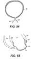
- FIG. 55illustrates a ventricular aneurysm A occurring in the apical region of left ventricle LV. As shown by the shaded region in FIG.
- aneurysm Aincludes infarcted tissue 24 that results in a reduced wall thickness when compared to adjacent non-infarcted wall regions, as shown by the unshaded regions in FIG. 55 .
- FIG. 55also shows the septal wall S partially infarcted, again shown by the shaded region.
- the ventricular aneurysmalso may be dyskinetic, meaning that when the ventricle contracts, the aneurysm further dilates, or bulges, outward.
- the infarcted region of the septal wall Salso may be particularly dyskinetic, especially in the case of the infarcted tissue having progressed to an aneurysm.
- the bulge resulting from an aneurysmcan have several serious effects on the heart and its performance that can lead to in both morbidity and mortality. For example, because the bulge creates a geometric abnormality as well as a region of non-contracting tissue, thrombosis is more likely to occur in that region. Thrombosis is the formation of a blood clot, or thrombus, that can cause other medical complications, such as a stroke.
- An ischemic strokeis a blockage of blood flow to the brain that occurs when the thrombus breaks free and is ejected out of the ventricle.
- the aneurysmal bulgecreates problems with pumping function in at least three ways.
- the infarcted tissuedoes not contribute to the pumping of the ventricle because it does not contract (akinesia). To account for this loss of pumping, remaining portions of the ventricle wall may contract more to maintain cardiac output. If the infarcted region thins and progresses to an aneurysm (dyskinesia), this effect is further exacerbated by the aneurysm expanding with a portion of the blood from the ventricular contraction. This further increases the contractile requirement of the remaining functional myocardium.
- the aneurysmal bulgealters the geometry of the entire ventricular chamber.
- the ventricledevelops a larger radius of curvature, which directly applies more tension to the heart wall, as characterized by LaPlace's law.
- Non-aneurysmal ischemic or infarcted myocardiumoccurs when a major coronary artery is occluded and results in infarction in the myocardial tissue, but without a bulging aneurysm. In a manner similar to an aneurysm, the akinetic ischemic or infarcted zone ceases to participate in the ventricular contraction. This results in the functioning, contractile myocardium needing to contract more to make up for the lack of contraction of the akinetic zone. Typically, the result is the entire ventricle increasing in size, which increases wall stress. Again, since the functioning myocardium must work harder, continuing progression of heart failure can occur.
- Mitral regurgitationalso may result from infarcted tissue, depending on the region of the ventricle that has become infarcted or aneurysmal and any subsequent overall ventricular dilation. Mitral regurgitation is a condition whereby blood leaks through the mitral valve due to an improper positioning of the valve structures that causes it not to close entirely. If the infarcted or aneurysmal region is located in the vicinity of the mitral valve, geometric abnormalities may cause the mitral valve to alter its normal position and dimension, and may lead to annular dilatation and the development of mitral regurgitation.
- Typical treatments of infarcted tissue, and ventricular aneurysms in particular,include a variety of open surgical procedures.
- a “linear” aneurysmectomyis performed. This procedure involves the removal of aneurysmal portions of the anterior wall along with any thrombus that may exist.
- FIG. 41illustrates the result of a conventional surgical method when an aneurysm occurs in the distal left ventricle.
- the region of aneurysmal scar tissue that extends through the entire thickness of the chamber wallis removed by incision and the remaining border zone regions 24 ′ (i.e., regions where infarcted tissue meets non-infarcted muscle) are sewn together with a suture 27 .
- the ventricular septal wall S that is infarctedis left untouched. Additionally, the septal wall generally remains untouched because simple excision and suturing does not involve excluding or cutting the septal wall.
- Newer surgical approachesinclude the “Dor” and “Jatene” procedures.
- the “Dor” procedurethe aneurysm is removed and an endocardial patch is placed to cover the dyskinetic septal wall portion of the aneurysm. In this manner, at least the portion of stroke volume “lost” to dyskinesia is restored.
- a purse string sutureis placed at the base of the aneurysm. The infarcted septal wall is circumferentially reduced by inbrication with sutures. The result is that most of the aneurysmal tissue is excluded from the ventricle.
- One aspect of the present inventionpertains to a non-pharmacological, passive apparatus and method for the treatment of a failing heart due to dilatation.
- the deviceis configured to reduce the tension in the heart wall. It is believed to reverse, stop or slow the disease process of a failing heart as it reduces the energy consumption of the failing heart, decreases isovolumetric contraction, increases isotonic contraction (sarcomere shortening), which in turn increases stroke volume.
- the devicereduces wall tension during diastole and systole.
- Splintscan be grouped as either “full cycle splints,” which engage the heart to produce a chamber shape change throughout the cardiac cycle, or “restrictive splints,” which do not engage the heart wall at end systole to produce a chamber shape change.
- the apparatusincludes a tension member for drawing at least two walls of the heart chamber toward each other to reduce the radius or area of the heart chamber in at least one cross sectional plane.
- the tension memberhas anchoring members disposed at opposite ends for engagement with the heart or chamber wall.
- the apparatusincludes a compression member for drawing at least two walls of a heart chamber toward each other.
- the compression memberincludes a balloon.
- a frameis provided for supporting the compression member.
- Yet another embodiment of the inventionincludes a clamp having two ends biased toward one another for drawing at least two walls of a heart chamber toward each other.
- the clampincludes at least two ends having atraumatic anchoring members disposed thereon for engagement with the heart or chamber wall.
- a heart wall tension reduction apparatuswhich includes a first tension member having two oppositely disposed ends and first and second elongate anchor members.
- a second tension membercan be provided.
- One of the elongate anchorsmay be substituted for by two smaller anchors.
- an elongate compression membercan be provided.
- First and second elongate lever memberspreferably extend from opposite ends of the compression member.
- a tension memberextends between the first and second lever members.
- the compression member of the above embodimentcan be disposed exterior to, or internally of the heart.
- the tension memberextends through the chamber or chambers to bias the lever members toward the heart.
- a rigid elongate frame memberin accordance with the present invention, can extend through one or more chambers of the heart.
- One or more cantilever memberscan be disposed at opposite ends of the frame member.
- Each cantilever memberincludes at least one atraumatic pad disposed thereon. The atraumatic pads disposed at opposite ends of the frame member can be biased toward each other to compress the heart chamber.
- One method of placing a heart wall tension apparatus or splint on a human heartincludes the step of extending a hollow needle through at least one chamber of the heart such that each end of the needle is external to the chamber.
- a flexible leaderis connected to a first end of a tension member.
- a second end of the tension memberis connected to an atraumatic pad.
- the leaderis advanced through the needle from one end of the needle to the other.
- the leaderis further advanced until the second end of the tension member is proximate the heart and the first end of the tension member is external to the heart.
- a second atraumatic padis connected to the first end of the tension member such that the first and second atraumatic pads engage the heart.
- Yet another method of placing a heart wall tension apparatus on a heartincludes the step of extending a needle having a flexible tension member releasably connected thereto through at least one chamber of the heart such that opposite ends of the tension member are external to the chamber and exposed on opposite sides of the chamber.
- the needleis removed from the tension member.
- first and second atraumatic padsare connected to the tension member at opposite ends of the tension member.
- another aspect of the inventioninvolves placing the splint relative to the infarcted or aneurysmal zone, and, in a preferred embodiment, diametrically across the infarcted or aneurysmal zone, to decrease the stress on the infarcted tissue and adjacent border zone tissue.
- An alternative to diametric placement of the splintincludes placing one atraumatic anchor member of the splint at the center of the infarcted or aneurysmal region, extending the splint across the entire heart chamber, and placing the second atraumatic anchor member on the opposite chamber wall.
- an aspect of the present inventionincludes a method of placing the splint adjacent but below the mitral valve to draw the papillary muscles together or the walls of the valve seat together. It is also envisioned to use the splint both as the sole device for treating infarcted tissue and aneurysms or in combination with the surgical techniques described earlier.
- An external splintusing a compression member, also may be used to treat a heart having infarcted or aneurysmal tissue.
- the compression memberis placed entirely exterior to the heart and positioned so as to result in similar effects as discussed above with reference to the splint.
- inventive methods and devices to treat infarcted tissue and aneurysmsinclude a variety of patching and suturing methods and related devices. Each of these methods and related devices reduces the radius of curvature of the infarcted wall region and adjacent regions and contains the infarcted region to stop further progression.
- a further aspect of the inventioninvolves the identification of aneurysmal and infarcted regions using any one or more of a variety of devices and methods.
- These devices and methodsinclude a bipolar electrode, liquid dye injection and tracing, fiber optics, MRI, and ultrasound. These devices can be used to distinguish between healthy and infarcted heart tissue.
- a method for treating a heart having a zone of infarcted tissue in its chamberincludes deforming a wall of the chamber that-includes the infarcted tissue such that a radius of curvature of the wall is reduced.
- the methodinvolves providing at least one tension member having two ends and an anchor on each end.
- the tension memberis positioned transverse to the chamber to reduce the radius of curvature of the wall of the chamber that includes the infarcted tissue and/or to draw the walls containing the infarcted tissue together.
- the present inventioninvolves positioning a tension member having anchors on each of its ends transverse to the heart chamber so that the infarcted tissue is drawn toward an interior of the heart chamber.
- the anchorsare placed exterior to the chamber.
- the present inventionincludes positioning a compression member having a first end and a second end, each having anchor members around an exterior of the heart.
- the compression memberis positioned so as to surround the chamber with infarcted tissue and to reduce the radius of curvature of the portion of the heart wall that has the infarcted tissue.
- a method of treating a heart having infarcted tissue in one of its chambersinvolves epicardial suturing around the perimeter of a region of infarcted tissue and pulling free ends of the suture to draw the infarcted tissue region together. The suture is then secured to hold the infarcted tissue together.
- This suturealso may be employed in combination with a myocardial patch or a substantially rigid enclosure member, both of which represent other preferred embodiments of the present invention.
- a method of treating a heart having infarcted tissue in one of its chambersinvolves positioning an enclosure member around a zone of infarcted tissue. During the positioning, the enclosure member has a first configuration. After positioning, the enclosure member is then secured to a wall of the heart and the enclosure member reconfigures to a second configuration. Upon reconfiguration to the second configuration, the radius of curvature of the portion of the heart wall including the infarcted tissue reduces.
- an apparatus for treating a heart having a zone of infarcted tissue in one of its chambersincludes an enclosure member adapted to assume a first configuration during placement of the enclosure member around an infarcted tissue zone.
- the enclosure memberfurther is adapted to assume a second configuration after securing the enclosure member to a heart wall surrounding the chamber. The second configuration draws the infarcted tissue toward a center of the enclosure member and reduces the radius of curvature of the heart wall.
- a device for treating a heart having infarcted tissue in one of its chambersincludes a patch adapted to be attached to the heart, with a substantially elongated member secured to the patch.
- the patchWhen the patch is placed over the infarcted or aneurysmal tissue region, the elongated member tends to push the infarcted tissue region toward an interior of the heart chamber.
- a plurality of suturesare attached at one end to points on a chamber wall proximate to an infarcted tissue region and the sutures are extended up through a space defined by an enclosure member to draw the infarcted tissue together and through the enclosure member.
- the other ends of the suturesare then attached to points on the wall of the chamber to hold the tissue in place.
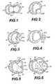
- FIG. 1is a transverse cross-section of the left and right ventricles of a human heart showing the placement of a splint in accordance with the present invention
- FIG. 2is a transverse cross-section of the left and right ventricles of a human heart showing the placement of a balloon device in accordance with the present invention
- FIG. 3is a transverse cross-section of the left and right ventricles of a human heart showing the placement of an external compression frame structure in accordance with the present invention
- FIG. 4is a transverse cross-section of the left and right ventricles of a human heart showing a clamp in accordance with the present invention
- FIG. 5is a transverse cross-section of the left and right ventricles of a human heart showing a three tension member version of the splint of FIG. 1 ;
- FIG. 6is a transverse cross-section of the left and right ventricles of a human heart showing a two tension member version of the splint shown in FIG. 1 ;
- FIG. 7is a vertical cross-sectional view of the left ventricle of a human heart showing an alternate version of the splint in accordance with the present invention
- FIG. 8is an end of the splint shown in FIG. 7 ;
- FIG. 9is a vertical cross-sectional view of a chamber of a human heart showing another alternative embodiment of the splint in accordance with the present invention.
- FIG. 10is a vertical cross-section of a chamber of a human heart showing another alternative configuration of splints in accordance with the present invention.
- FIG. 11is a vertical cross-sectional view of a chamber of a human heart showing another embodiment of a splint in accordance with the present invention.
- FIG. 12is a vertical cross-sectional view of a chamber of a human heart showing another embodiment of the splint in accordance with the present invention.
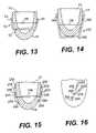
- FIG. 13is a vertical cross-sectional view of a chamber of a human heart showing a compression member version of the splint in accordance with the present invention
- FIG. 14is a vertical cross-sectional view of a chamber of a human heart showing another version of the splint shown in FIG. 13 ;
- FIG. 15is a vertical cross-sectional view of a chamber of a human heart showing a frame member version of the splint in accordance with the present invention
- FIG. 16is an end view of the splint of FIG. 15 ;
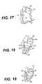
- FIG. 17is a vertical cross-section of the left ventricle and atrium, the left ventricle having aneurysmal scar tissue;
- FIG. 18is a vertical cross-section of the heart of FIG. 17 showing the splint of FIG. 1 drawing the aneurysmal scar tissue toward the opposite wall of the left ventricle;
- FIG. 19is a vertical cross-section of the left ventricle and atrium of a human heart showing a version of the splint of FIG. 1 having an elongate anchor bar;
- FIG. 20is a side view of an undeployed hinged anchor member
- FIG. 21is a side view of a deployed hinged anchor member of FIG. 10 ;
- FIG. 22is a cross-sectional view of an captured ball anchor member
- FIG. 23is a perspective view of a cross bar anchor member
- FIG. 24is a cross sectional view of an alternate anchor pad
- FIG. 25is a cross sectional view of an alternate anchor pad
- FIG. 26is a perspective view of yet another alternate embodiment of an anchor pad including an anchor pad loosening device
- FIG. 27is a perspective view of a tension member clip
- FIG. 28is a cross sectional view of an alternate embodiment of a tension member clip
- FIG. 29is a cross sectional view of a heart including a tension member having a heat set end;
- FIG. 30is a cross sectional view of the pad including an envelope
- FIG. 31shows the envelope of FIG. 30 ;
- FIG. 32is a side view of a multifilament twisted cable
- FIG. 33is a cross sectional of the cable of FIG. 32 ;
- FIG. 34is a side of a multifilament braided tension member
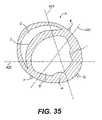
- FIG. 35is a schematic generally horizontal cross sectional view of the heart showing preferred tension member alignments
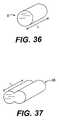
- FIG. 36is a idealized cylindrical model of a left ventricle of a human heart
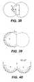
- FIG. 37is a splinted model of the left ventricle of FIG. 14 ;
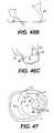
- FIG. 38is a transverse cross-sectional view of FIG. 15 showing various modeling parameters
- FIG. 39is a transverse cross-section of the splinted left ventricle of FIG. 15 showing a hypothetical force distribution
- FIG. 40is a second transverse cross-sectional view of the model left ventricle of FIG. 15 showing a hypothetical force distribution
- FIG. 41is a transverse, partial cross-section of left and right ventricles showing a traditional surgical method of treating infarcted tissue regions;
- FIG. 42is a transverse, partial cross-sectional view of left and right ventricles with an infarcted or aneurysmal region in the apical portion of the left ventricle and a splint according to an embodiment of the invention placed diametrically across the region;
- FIGS. 43 a – 43 bare long axis cross-sectional views of left and right ventricles showing a region of infarcted tissue in a portion of the basal left ventricle and a splint according to an embodiment of the invention placed across the infarcted region;
- FIGS. 43 c – 43 dare short axis cross-sectional views of the heart in FIGS. 43 a – 43 b shown from the perspective of lines c—c and d—d, respectively;
- FIGS. 44 a – 44 bare short axis cross-sectional views of the left ventricle having an aneurysm and a placement of a splint according to an embodiment of the present invention with respect to the aneurysm to treat the heart;
- FIG. 45is a short axis cross-sectional view of the right and left ventricles having an infarcted region like that shown in FIG. 43 c and an external splint device according to an embodiment of the present invention placed to treat the infarction;
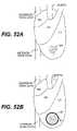
- FIGS. 46 a – 46 cshow transverse, partial cross-sectional views of a left ventricle having an infarcted region, illustrating the combined inventive method of surgical removal of the infarcted tissue and placement of a splint according to an embodiment of the present invention
- FIG. 47is a short axis cross-sectional view of the left and right ventricles including a view of the mitral valve with an infarcted or aneurysmal region in a portion of the basal left ventricle and a splint according to an embodiment of the invention placed in the vicinity of the mitral valve;
- FIGS. 48 a – 48 bare short axis cross-sectional views of the left and right ventricles and a view of the mitral valve with an aneurysmal region in a portion of the basal left ventricle and an external device according to an embodiment of the invention placed in the vicinity of the mitral valve and aneurysm;
- FIGS. 49 a – 49 bare transverse cross-sections of a left ventricle having an aneurysmal region and the placement of a staked patch according to an embodiment of the present invention
- FIGS. 50 a – 50 bare planar views of an infarcted or aneurysmal tissue region with a purse-string suture according to an embodiment of the present invention
- FIGS. 51 a – 51 care planar views of an infarcted or aneurysmal tissue region and a purse-string suture and enclosure member according to an embodiment of the present invention
- FIGS. 52 a – 52 care planar exterior views of the heart with an infarcted region of tissue in the left ventricle and an enclosure member according to an embodiment of the invention, showing one configuration during application of the member and a second configuration after application of the member;
- FIGS. 53 a – 53 bare planar views of an infarcted or aneurysmal tissue region and placement of a tie enclosure according to an embodiment of the present invention
- FIG. 54is a planar view of yet another embodiment of an enclosure member according to an embodiment of the present invention.
- FIG. 55is a transverse, partial cross-section of left and right ventricles with an aneurysmal region located at an apical portion of the left ventricle and infarcted tissue along the septal wall.
- the devices of the present inventionoperate passively in that, once placed in the heart, they do not require an active stimulus either mechanical, electrical, or otherwise, to function.
- the devicesalter the shape or geometry of the heart, both locally and globally, and increase the heart's efficiency by their placement with respect to the heart. That is, the heart experiences an increased pumping efficiency through an alteration in its shape or geometry and concomitant reduction in stress on the heart walls.
- the inventive devices and methodsoffer numerous advantages over the existing treatments for various heart conditions.
- the devicesare relatively easy to manufacture and use, and the related surgical techniques for their implementation do not require the invasive procedures of current surgical techniques.
- the surgical techniquedoes not necessarily require removing portions of the heart tissue, opening the heart chamber, or stopping the heart.
- the surgical techniques of the present inventionare also less risky to the patient than other techniques.
- the devices and methods of the present invention used to treat infarcted tissue and aneurysmsalso are likely to be more effective than prior devices.
- the inventive devicesalter the shape or geometry of the chamber, either globally or locally, and reduce the radius of curvature of the chamber wall, resulting in lower stresses in the heart wall.
- CABGcoronary artery bypass grafting
- the inventive methods and related devicesallow for quickly reducing stress on the myocardium, which may save “stunned” tissue, i.e., tissue that is being starved of nutrients carried with the blood flow, that otherwise may not be recoverable after a certain time period. Also, the inventive methods and device may hinder further progression or dilation of scarred, non-contractile tissue.
- the disclosed inventive methods and related devicesinvolve geometric reshaping of the heart.
- substantially the entire chamber geometryis altered to return the heart to a more normal configuration.
- FIGS. 36 through 40show a model of this geometric reshaping, which includes a reduction in radius of curvature of the chamber walls.
- the heart wallsPrior to reshaping the chamber geometry, the heart walls experience high stress due to a combination of both the relatively large increased diameter of the chamber and the thinning of the chamber wall.
- Geometric reshaping according to the present inventionreduces the stress in the walls of the heart chamber to increase the heart's pumping efficiency, as well as to stop further dilatation of the heart.
- inventive methods and devicesinvolve geometric reshaping a particular area of the chamber and/or reducing the radius of curvature of the chamber wall in that area.
- the radius of much of the heart chamberchanges. This increases stress on the heart walls.
- the healthy regions of the heartwork harder to pump in order to make up pumping volume due to lost contractility in the infarcted tissue region. Together, these effects limit the pumping effectiveness of the heart and can contribute to further degradation of the heart.
- Geometrically reshaping the area of the aneurysmby, for example, reducing the radius of curvature of the wall, lowers stress on the wall regions in that vicinity and improves pumping function.
- the geometric reshapingpermits the scar tissue to heal in a more organized fashion and reduces progression of the scar tissue into other areas and further aneurysmal bulging.
- left ventriclehas been selected for illustrative purposes because a large number of the disorders that the present invention treats occur in the left ventricle.
- FIG. 1shows a transverse cross-section of a left ventricle 10 and a right ventricle 12 of a human heart 14 .
- Extending through the left ventricleis a splint 16 including a tension member 18 and oppositely disposed anchors 20 .
- Splint 16as shown in FIG. 1 , has been positioned to draw opposite walls of left ventricle 10 toward each other to reduce the “radius” of the left ventricular cross-section or the cross-sectional area thereof to reduce left ventricular wall stresses.
- splint 16 and the alternative devices disclosed hereinare described in relation to the left ventricle of a human heart, these devices could also be used to reduce the radius or cross-sectional area of the other chambers of a human heart in transverse or vertical directions, or at an angle between the transverse and vertical.
- splintsThose apparatus of the present invention which reduce heart wall stress by changing chamber wall geometry can be referred to as “splints”. “Full cycle splints” engage the heart to produce a chamber shape change throughout the cardiac cycle. “Restrictive splints” do not engage the heart wall at end systole to 110 produce a chamber shape change.
- FIG. 2discloses an alternate embodiment of the present invention, wherein a balloon 200 is deployed adjacent the left ventricle.
- the size and degree of inflation of the ballooncan be varied to reduce the radius or cross-sectional area of left ventricle 10 of heart 14 .
- FIG. 3shows yet another alternative embodiment of the present invention deployed with respect to left ventricle 10 of human heart 14 .
- a compression frame structure 300is engaged with heart 14 at atraumatic anchor pads 310 .
- a compression member 312 having an atraumatic surface 314presses against a wall of left ventricle 10 to reduce the radius or cross-sectional area thereof.
- FIG. 4is a transverse cross-sectional view of human heart 14 showing yet another embodiment of the present invention.
- a clamp 400 having atraumatic anchor pads 410 biased toward each otheris shown disposed on a wall of left ventricle 10 .
- the radius or cross-sectional area of left ventricle 10is reduced by clamping off the portion of the wall between pads 410 .
- Pads 410can be biased toward each other and/or can be held together by a locking device.
- FIGS. 1–4can be made from materials which can remain implanted in the human body indefinitely. Such biocompatible materials are well-known to those skilled in the art of clinical medical devices.
- FIG. 5shows an alternate embodiment of the splint of FIG. 1 referred to in FIG. 5 by the numeral 116 .
- the embodiment 116 shown in FIG. 5includes three tension members 118 as opposed to a single tension member 18 as shown in FIG. 1 .
- FIG. 6shows yet another embodiment of the splint 216 having four tension members 218 . It is anticipated that in some patients, the disease process of the failing heart may be so advanced that three, four or more tension members may be desirable to reduce the heart wall stresses more substantially than possible with a single tension member as shown in FIG. 1 .
- FIG. 7is a partial vertical cross-section of human heart 14 showing left ventricle 10 .
- another splint embodiment 316is shown having a tension member 318 extending through left ventricle 10 .
- tension member 318On opposite ends of tension member 318 are disposed elongate anchors or pads 320 .
- FIG. 8is an end view of tension member 318 showing elongate anchor 320 .
- FIG. 9shows another embodiment of a splint 416 disposed in a partial vertical cross-section of human heart 14 .
- Splint 416includes two elongate anchors or pads 420 similar to those shown in FIGS. 7 and 8 .
- two tension members 418extend through left ventricle 10 to interconnect anchors 420 on opposite sides of heart 14 .
- FIG. 10is a vertical cross section of heart 14 showing left ventricle 10 .
- two splints 16are disposed through left ventricle 10 and vertically spaced from each other to resemble the configuration of FIG. 9 .
- FIG. 11is a vertical cross-sectional view of the left ventricle of heart 14 .
- Two alternate embodiment splints 516are shown extending through left ventricle 10 .
- Each splint 516includes two tension members 518 interconnecting two anchors or pads 520 .
- FIG. 12is yet another vertical cross-sectional view of left ventricle 10 of heart 14 .
- An alternate embodiment 616 of the splintis shown extending through left ventricle 10 ;
- Splint 616includes an elongate anchor pad 620 and two shorter anchors or pads 621 .
- Splint 616includes two tension members 618 . Each tension member 618 extends between anchors 620 and respective anchors 621 .
- FIG. 13is a vertical cross-sectional view of left ventricle 10 of heart 14 .
- a splint 50is shown disposed on heart 14 .
- Splint 50includes a compression member 52 shown extending through left ventricle 10 .
- Opposite ends of compression member 52are disposed exterior to left ventricle 10 .
- Lever members 54extend from each end of compression member 52 upwardly along the exterior surface of ventricle 10 .
- a tension member 56extends between lever members 54 to bias lever members 54 toward heart 14 to compress chamber 10 .
- Compression member 52should be substantially rigid, but lever members 54 and to some degree compression member 52 should be flexible enough to allow tension member 56 to bias lever members 54 toward heart 14 .
- lever members 54could be hinged to compression member 52 such that lever members 54 could pivot about the hinge when biased toward heart 14 by tension member 56 .
- FIG. 14shows an alternate embodiment 156 of the splint shown in FIG. 13 .
- lever members 154are longer than members 54 as compression member 152 of splint 150 has been disposed to the exterior of left ventricle 10 .
- FIG. 15is a vertical cross-sectional view of left ventricle 10 of heart 14 .
- An alternate embodiment 250 of the splintis shown on heart 14 .
- a preferably relatively rigid frame member 256extends through ventricle 10 .
- cantilever member 254Disposed on opposite ends of frame 256 are cantilever member 254 .
- cantilever members 254Disposed on cantilever members 254 are atraumatic pads 258 .
- Cantilever members 254can be positioned along frame member 256 such that atraumatic pads 258 press against heart 14 to compress chamber 10 .
- FIG. 16is an end view of frame member 256 showing cantilever members 254 and pads 258 .
- the tension memberscan be formed from flexible or relatively more rigid material.
- the compression members and frame membershould be formed from generally rigid material which may flex under load, but generally hold its shape.
- FIG. 17is a partial vertical cross-section of human heart 14 showing left ventricle 10 and left atrium 22 .
- heart 14includes a region of scar tissue 24 associated with an aneurysm or ischemia.
- the scar tissue 24increases the radius or cross-sectional area of left ventricle 10 in the region affected by the scar tissue. Such an increase in the radius or cross-sectional area of the left ventricle will result in greater wall stresses on the walls of the left ventricle.
- FIG. 18is a vertical cross-sectional view of the heart 14 as shown in FIG. 17 , wherein a splint 16 has been placed to draw the scar tissue 24 toward an opposite wall of left ventricle 10 .
- the radius or cross-sectional area of the left ventricle affected by the scar tissue 24is reduced. The reduction of this radius or cross-sectional area results in reduction in the wall stress in the left ventricular wall and thus improves heart pumping efficiency.
- FIG. 19is a vertical cross-sectional view of left ventricle 10 and left atrium 22 of heart 14 in which a splint 16 has been placed.
- splint 16includes an alternative anchor 26 .
- the anchor 20is preferably an elongate member having a length as shown in FIG. 19 substantially greater than its width (not shown).
- Anchor bar 26might be used to reduce the radius or cross-sectional area of the left ventricle in an instance where there is generalized enlargement of left ventricle 10 such as in idiopathic dilated cardiomyopathy. In such an instance, bar anchor 26 can distribute forces more widely than anchor 20 .
- FIGS. 20 and 21are side views of a hinged anchor 28 which could be substituted for anchors 20 in undeployed and deployed positions respectively.
- Anchor 28 as shown in FIG. 20includes two legs similar to bar anchor 26 .
- Hinged anchor 28could include additional legs and the length of those legs could be varied to distribute the force over the surface of the heart wall.
- the webbingwould be disposed on the surface of the legs which would be in contact with the heart wall.
- FIG. 22is a cross-sectional view of a capture ball anchor 30 .
- Capture ball anchor 30can be used in place of anchor 20 .
- Capture ball anchor 30includes a disk portion 32 to distribute the force of the anchor on the heart wall, and a recess 34 for receiving a ball 36 affixed to an end of tension member 18 .
- Disk 32 and recess 34include a side groove which allows tension member 38 to be passed from an outside edge of disk 32 into recess 34 .
- Ball 36can then be advanced into recess 34 by drawing tension member 18 through an opening 38 in recess 34 opposite disk 32 .
- FIG. 23is a perspective view of a cross bar anchor 40 .
- the cross bar anchor 40can be used in place of anchors 20 .
- the anchor 40preferably includes a disk or pad portion 42 having a cross bar 44 extending over an opening 46 in pad 42 .
- Tension member 18can be extended through opening 46 and tied to cross bar 42 as shown.
- FIG. 24is a cross sectional view of an alternate embodiment of anchor pad 340 in accordance with the present invention.
- Anchor pad 340preferably includes a disc shaped pad portion 342 .
- Disc-shaped pad portion 342includes side 343 , which in use is disposed toward the heart.
- a conical aperture 348 having sloping sides 346extends through pad 342 .
- Collet 344is disposed within orifice 348 .
- a threaded portion 350 of collet 344extends from orifice 348 opposite side 343 , nut 352 is threaded over threaded portion 350 .
- Lumen 345extends through collet 344 .
- a tension member 354is shown extending through lumen 345 .
- Lumen 345has a diameter such that when nut 352 is not tightened on threaded portion 350 , tension member 354 can slide freely through lumen 345 .
- tension member 354can slide freely through lumen 345 .
- nut 352When nut 352 is tightened, it draws collet 344 away from side 343 . Collet 344 is then pinched between walls 346 of orifice 348 .
- collet 344is pinched, the size of lumen 345 is reduced such that tension member 354 can no longer move freely within lumen 345 , fixing the position of pad 340 on tension member 354 .
- FIG. 25is a cross sectional view of an alternate embodiment of an anchor pad 360 in accordance with the present invention.
- Anchor pad 360includes a generally disc-shaped pad portion 362 .
- Pad 362includes a side 363 which when the pad is in use, is disposed toward the heart.
- a tension member lumen 364extends through pad 362 .
- Lumen 364preferably has a generally conical shaped portion 365 disposed toward side 363 .
- Tension member 370is shown disposed through lumen 364 in FIG. 25 .
- Pad 362includes a threaded passage 366 extending from an edge of pad 362 to lumen 364 .
- a set screw 368is threaded into passage 366 . Set screw 368 can be tightened to engage tension member 370 to fix the position of anchor pad 360 .
- the size of lumen 364is preferably large enough that anchor pad 360 can slide relatively freely over tension member 370 .
- FIG. 26is a perspective view of yet another embodiment of anchor pad 380 in accordance with the present invention.
- Anchor pad 380preferably includes a generally disc-shaped pad portion 382 having a first side 383 which in use would be disposed toward the heart and a second side 385 .
- Pad 382 as well as pads 342 and 362are preferably formed from a metal such as stainless steel alloys or titanium alloys.
- a tension member fastener 384is formed in pad 382 by cutting a series of grooves and apertures through pad 382 from side 385 to side 383 .
- a first groove 386has a generally horseshoe shape.
- Second groove 388extends between opposite portions of horseshoe shaped groove 386 to form two oppositely disposed cantilever members 387 .
- a relatively large aperture 394is formed between cantilever members 387 proximate their free ends.
- a second and smaller aperture 390is formed closer to the fixed ends of cantilever members 387 .
- Tension member 392is shown extending through aperture 390 .
- tension member 392is clamped between cantilever members 387 such that the location of pad 382 is fixed along tension member 392 .
- Pad 382can be released by using a spreading device 396 to spread cantilever members 387 apart.
- Spreading device 396includes handle 398 to spreading arms 400 each having a finger 402 . Fingers 402 can be placed within aperture 394 then aims 400 and fingers 402 can be spread apart by pivoting them around a pin 404 such that cantilevers 387 are spread apart and pad 382 can move freely along tension member 392 .
- spreader 396is shown extending transversely from tension member 392 , it could also be configured such that fingers 402 do not curve transversely from arms 400 and thus spreader 396 could be disposed parallel to tension member 392 . This would be particularly desirable in a situation where anchor pad 380 was being placed through a port or window during a less invasive splint implantation procedure. It can be appreciated that cantilever members 387 can be held apart such that pad 380 can be moved along tension member 392 by placement of a temporary wedge or pin in groove 388 .
- grooves 388may include an additional small aperture disposed between aperture 390 and aperture 394 into which a pin could be placed to hold open members 387 .
- Aperture 390 of pad 380can also include a conical portion disposed toward side 383 such as conical portion 365 of pad 360 .
- Cantilever arms 384are preferably configured such that they do not stress tension member 392 beyond its elastic limit. It can also be appreciated that the force developed by cantilever members 387 impinging on tension member 392 is operator independent and defined by the geometry and material characteristics of members 387 .
- FIG. 27is a perspective view of an anchor pad 360 having a tension member 370 extending therethrough. After pad 360 is secured to tension member 370 , that portion of tension member 370 which extends from the side of anchor pad 360 opposite side 363 is preferably removed. This can be accomplished by trimming tension member 370 with wire cutter 414 or scissors. Although anchor pad 360 is used here to illustrate trimming tension member 370 , it can be appreciated that in each of the embodiments disclosed herein there may be an excess portion of tension member extending from an anchor, which is preferably removed or trimmed.
- FIG. 28is a cross sectional view of an alternate embodiment 420 of a tension member cutter.
- Device 420includes an elongate outer tube 422 having a distal end 424 .
- Tube 424defines a lumen 423 through which extends a second tube 430 having a distal end 428 .
- Extending distally from distal end 428are two cutting arms 424 and 426 which are shown partially withdrawn into lumen 423 and transversely restrained by distal end 424 of outer tube 422 .
- arms 424 and 426When unrestrained by distal end 424 , arms 424 and 426 are biased apart.
- Each arm 424 and 426has a cutting element 425 and 427 , respectively. Elements 425 and 427 are shown in contact with each other in FIG. 28 .
- a tension member 370extends between arms 424 and through lumen 432 of inner tube 430 .
- a representative anchor pad 360is disposed adjacent elements 425 and 427 .
- Device 420 of FIG. 28is particularly useful when trimming excess tension member using less invasive techniques as it can be readily advanced over a tension member through a port or window trocar.
- FIG. 29is a vertical cross sectional view of left ventricle B of heart A.
- a transventricular splint 443 including a tension member 370 and anchor pads 360are shown disposed on heart A.
- To the left of heart A as shown in the figureis a coiled portion 442 of tension member 470 .
- tension member 370could be formed from a shape memory alloy such that portion 442 could be preset to assume a coil shape when warmed to near body temperature.
- the anchorsare secured in place along the tension member and the excess length of tension member removed if desired, the anchor or anchor pads are preferably secured in place on the heart.
- the anchor or anchor padsare secured such that relatively movement between the anchors or anchor pads and the heart is limited to reduce abrasion of the heart wall.
- a biocompatible adhesivecould be placed between the pad and the heart to adhere the pad to the heart.
- aperturescould be provided in the pad such that sutures could be extended through the apertures and into the heart to secure the pad.
- the padcould include threaded apertures into which anchor screws could be advanced through the pad and- into the heart wall to secure the pad to the heart.
- FIG. 30illustrates yet another alternative approach to securing the anchors or anchor pads to the heart surface.
- FIG. 30is a cross sectional view of an anchor pad 340 disposed on heart A.
- Anchor pad 340is disposed within an envelope 446 .
- Envelope 446includes a bottom layer 447 disposed between anchor pad 340 and heart A and a top layer 448 disposed on the opposite side of anchor pad 340 .
- Layers 347 and 340are held together by sutures 449 .
- Bottom layer 447is preferably a mesh or expanded PTFE which has a pore size or intranodial dimension sufficient to promote tissue ingrowth.
- the pore sizeis preferably between about 10 and about 100 microns and more preferably, between about 20 and about 40 microns.
- the intranodial dimensionis preferably between about 10 to about 100 microns and more preferably between about 20 to about 40 microns.
- the top materialcould also be expanded PTFE or the like having a pore size which preferably does not promote ingrowth and thus resists adhesion to surrounding tissue.
- the porescould be formed directly in the pad surface.
- Envelope 446would preferably be placed around pad 340 prior to placing pad 340 on tension member 354 .
- a window 450can be provided to provide access to nut 352 to secure pads to tension member 354 . After tightening nut 352 , window 450 can be closed by suture 452 .
- FIG. 31is a top view of pad 340 and envelope 446 of FIG. 30 . It can be appreciated that a similar envelope can be placed around the various anchor pads disclosed herein. The location of the window may have to vary, however, to provide access to the respective means for securing the anchor pads to the tension member.
- the splints of the present inventioncan be implanted acutely or chronically. When the splints are implanted chronically, it is particularly important that the tension member or members be highly fatigue resistant.
- Typical materials for the tension membercan include, among other biocompatible materials, stainless steel, titanium alloys, NiTi alloys such as Nitinol or elgiloy.
- the tension memberis a wire having a diameter of between 0.005 to 0.035 inches in diameter or, more preferably, between 0.01 and 0.02 inches in diameter and, most preferably, about 0.014 inches in diameter.
- the length of the tension member between the padsis preferably about 0.6 to 4 inches, and more preferably, between about 1 to 3 inches and, most preferably, about 2 inches.
- their surfacecan be electro-polished, buffed or shot peened. Drawing or annealing of the metal will also improve fatigue resistance.
- the tension memberin a preferred embodiment, articulates with respect to the anchor pad to reduce bending of the tension member at the pad. This can be accomplished by a ball and socket joint shown in FIG. 22 , for example.
- the tension memberitself can be made more flexible or bendable by providing a multi-filament tension member such as a braided or twisted wire cable tension member. A multifiber filament structure of numerous smaller wires can then easily, while reducing the stress level on any individual wire as compared to a solid wire of the same diameter as the multifilament bundle.
- Such a multi-filament tension membercan be made from biocompatible materials such as, but not limited to, stainless steel, Nitinol, titanium alloys, LCP (liquid crystal polymer), SpectraTM fiber, kevlar fiber, or carbon fiber.
- the multi-filament structureis coated or covered to substantially seal the multi-filament structure. Coatings such as silicone, urethane or PTFE are preferred.
- FIG. 32is a side view of multifilament twisted cable 400 .
- Cable 400includes a plurality of wires or filaments 402 twisted about the longitudinal axis of cable 400 .
- FIG. 33is a transverse cross sectional view of cable 400 .
- cable 400includes a surrounding coating 404 not shown in FIG. 32 .
- FIG. 34is a side view of a braided multifilament tension member 410 .
- Tension member 410includes a plurality of filaments or wires 412 . It can be appreciated that numerous braiding patterns are known to those skilled in the art of multifilament members. It is anticipated that in a preferred embodiment, braided member 410 can have an optional core of fibers running parallel to an elongate axis of tension member 410 . In yet another preferred embodiment, tension member 410 could have a solid wire core extending parallel to and along the longitudinal axis of tension member 410 .
- tension members and anchors or anchor padsare preferably bio-resistant, i.e., resistant to physiologic attack.
- tension member and/or anchors or anchor padscan be coated with carbon material such as glass, pyrolytic carbon, diamond or graphite, zirconium nitrate or oxide.
- Roughened or porous urethanes, silicone or polymer coatings or sheathscan be used to promote tissue ingrowth to create a biological seal.
- Hydrophilic and albumin coatingscan also be used. Drugs incorporated into a binder coating can also be used to reduce biological attack on the splint and irritation of tissue by the splint.
- Such drugsinclude heparin, coumadin, anti-inflammatory steroid or ASA-aspirin.
- the oxide layer of the underlying metalcould also be optimized to improve bio-resistance. This is particularly true for stainless steel, titanium, or nickel titanium on which an oxide layer can be formed by heating the component to improve biocompatibility.
- Further coatingsinclude calcium hydroxy appetite, beta tricalcium phosphate and aluminum oxide can be applied to the tension member.
- the tension member and/or pad or anchor padcan at least be, in part, formed from titanium to enhance electronegativity.
- the anchors or anchor pads and, particularly the tension membersare biocompatible, preferably antithrombogenic and made to prevent hemolysis.
- the coatings used to enhance bio-resistance described abovecan generally be used to improve biocompatibility. Since the tension member is exposed to significant blood flows through the left ventricle, in a preferred embodiment, the tension member has a generally small size and shape elliptical cross sectional shape to reduce turbulence or drag over the tension member. If such elliptical, transverse cross section tension member were used, it can be appreciated that the narrow end would be preferably oriented toward the direction of blood flow. It is also desirable to select a tension member material and shape which would not vibrate at resonant frequency under the influence of blood flow.
- the surface of the anchor or anchor pad and/or tension member in contact with the heart wallcan be coated or include an ingrowth inducing covering such as collagen, dacron, expanded PTFE or a roughened/porous surface.
- a clotting inducing substancemay also be bound to the tension member and/or anchor or anchor pads, such as avitene or collagen.
- the portion of the heart wall where the tension member passes throughcould be cauterized.
- the tissuecan be cauterized by heating the tension member.
- a gluesuch as cyanoacrylate can also be disposed between the tension member and the heart wall to reduce or prevent bleeding from the heart wall.
- Mechanical meanssuch as an O-ring or compression fitting could also be disposed between the heart wall and the tension member to reduce bleeding.
- a purse string suturecan be placed on the heart, around the tension member adjacent the pad as well.
- the tension memberis preferably flexible enough to allow for changing interface conditions between the heart and the splint, and alternating pad orientation throughout the cardiac cycle.
- the flexibilityshould be sufficient enough to avoid injury to the heart or bleeding. It is also preferable that if the heart were to contract sufficiently enough to put the tension member in compression that it would readily buckle. Buckling could be promoted by providing a ribbon shaped tension member, chain link tension member, thin wire tension member, bent tension member or multi-filament tension member.
- the tension memberis preferably radiopaque, echo cardiographic visible, or MRI compatible or includes a marker which is radiopaque, echo visible, or MRI compatible.
- the preferred locations for markerswould include the center of the tension member and at the ends of the tension member disposed at the heart walls.
- the radiopaque markerscould be gold or platinum or other biocompatible metal or heavy metal filled polymeric sleeves.
- the tension or markerare preferably non-interfering or visible. Having radiopaque echo compatible or MRI compatible tension members or markers is particularly desirable for follow-up, non-invasive monitoring of the tension member after implantation. The presence of the tension member can be visualized and the distance between two or more markers measured. Integrity of the tension member can be confirmed as well.
- the tension memberis not conductive to the action potential of muscle. This can be accomplished by insulating the tension member, anchor and/or anchor pad interface or fabricating the tension member anchor and/or anchor pad from a non-conductive metal such as titanium.
- sensorscan advantageously be incorporated into the splints.
- a strain gaugecan be disposed on a tension member to monitor the loading on the member in use. Strain can be related to load as known to those skilled in the art by developing a stress/strain relationship for a given tension member.
- the strain gaugecan be connected by a biocompatible lead to a conventional monitoring device.
- a pressure gauge formed from, for example, piezo electric materialcan also be disposed on the tension member to monitor filling pressures or muscle contractility.
- a tension membercan be slidably enclosed within a tube. If the tension member were to fail, the tube would contain the tension member therein.
- tension membercould be connected to a pacing lead.
- tension membercould be conductive, pacing signals could be conveyed along the tension member from one heart wall to another.
- the various embodiments of the present inventionare placed in or adjacent the human heart to reduce the radius or cross-section area of at least one chamber of the heart. This is done to reduce wall stress or tension in the heart or chamber wall to slow, stop or reverse failure of the heart.
- a cannulacan be used to pierce both walls of the heart and one end of the splint can be advanced through the cannula from one side of the heart to the opposite side where an anchor can be affixed or deployed.
- an anchoris affixed or deployed at the opposite end of splint 16 . Additional methods for splint placement are described in more detail in U.S. Pat. No. 6,260,552, issued on Jul. 17, 2001 and entitled “Transventricular Implant Tools and Devices” and incorporated herein by reference.
- the methods described above to advance the tension members through the ventriclescan be repeated to advance the desired number of tension members through the ventricle for a particular configuration.
- the length of the tension memberscan be determined-based upon the size and condition of the patient's heart. It should also be noted that although the left ventricle has been referred to here for illustrative purposes, that the apparatus and methods of this invention can also be used to splint multiple chambers of a patient's heart as well as the right ventricle or either atrium.
- FIG. 35is a schematic view of generally horizontal cross section of heart A including left ventricle B and right ventricle C. Also shown are left anterior descending artery E, posterior descending artery F, obtuse marginal artery G, postero-medial papillary muscle H and antero-lateral papillary muscle 1 . Shown in FIG. 35 are three generally horizontal preferred alignments for tension member placement for the splints of the present invention when used for the purpose of treating ventricular dilatation. These alignments generally met three goals of splint positioning including good bisection of the left ventricle, avoidance of major coronary vessels and avoidance of valve apparatus including chordae leaflets and papillary muscles.
- Alignment 420can be referred to as the anterior/posterior (AP) position.
- Alignment 422can be referred as the posterior septal/lateral wall (PSL) position.
- Alignment 424can be referred to as the anterior septal/lateral wall (ASL) position.
- APanterior/posterior
- the alignments shownare illustrative only and that the alignments may be shifted or rotated about a vertical axis generally disposed through the left ventricle and still avoid the major coronary vessels and papillary muscles.
- the alignmentpasses through a substantial portion of right ventricle C, it may be desirable to dispose not only two pads on the exterior of the heart at opposite ends of a tension member, but also a third pad within right ventricle C on septal J.
- the spacing between the third pad and the pad disposed outside the heart proximate left ventricle Bpreferably defines the shape change of left ventricle B.
- FIG. 36is a view of a cylinder or idealized heart chamber 48 which is used to illustrate the reduction of wall stress in a heart chamber as a result of deployment of the splint in accordance with the present invention.
- the model used herein and the calculations related to this modelare intended merely to illustrate the mechanism by which wall stress is reduced in the heart chamber. No effort is made herein to quantify the actual reduction which would be realized in any particular in vivo application.
- FIG. 37is a view of the idealized heart chamber 48 of FIG. 36 wherein the chamber has been splinted along its length L such that a “figure eight” cross-section has been formed along the length thereof. It should be noted that the perimeter of the circular transverse cross-section of the chamber in FIG. 36 is equal to the perimeter of the figure eight transverse cross-section of FIG. 37 . For purposes of this model, opposite lobes of the figure in cross-section are assumed to be mirror images.
- FIG. 38shows various parameters of the FIG. 1 cross-section of the splinted idealized heart chamber of FIG. 37 .
- lis the length of the splint between opposite walls of the chamber
- R 2is the radius of each lobe
- ⁇is the angle between the two radii of one lobe which extends to opposite ends of the portion of the splint within chamber 48
- his the height of the triangle formed by the two radii and the portion of the splint within the chamber 48 (R 1 is the radius of the cylinder of FIG. 36 ).
- the wall tension T in the walls of the cylinderis 104.4 newtons.
- FIGS. 39 and 40show a hypothetical distribution of wall tension T and pressure P for the figure eight cross-section. As ⁇ goes from 180° to 0°, tension T s , in the splint goes from 0 to a 2T load where the chamber walls carry a T load.
- the chamber length Lis a constant 10 cm
- the original radius R 1is 4 cm
- the length l between the two pads as measured along the tension memberis preferably 0.4 to about 0.8 and more preferably between about 0.5 to about 0.7 and most preferably about 0.6 times the distance along the length of the tension member at end diastole if the pads were not secured to the tension member and provided no resistance to expansion of the heart.
- tension member lengthcan be found in U.S. Pat. No. 6,260,552, issued on Jul. 17, 2001 and entitled “Transventricular Implant Tools and Devices” which is incorporated herein by reference.
- FIG. 17is a partial vertical cross-section of human heart 14 showing left ventricle 10 and left atrium 22 .
- heart 14includes a region of scar tissue 24 associated with an aneurysm.
- the aneurysmal scar tissue 24increases the radius or cross-sectional area of left ventricle 10 in the region affected by the scar tissue.
- Such an increase in the radius or cross-sectional area of the left ventriclewill result in greater wall stresses on the walls of the left ventricle, especially those walls adjacent to the aneurysm.
- the inventive splintalso can be used to treat infarcted tissue or aneurysms occurring on the heart wall, as illustrated by FIGS. 18 and 42 – 44 , 46 , and 47 .
- FIGS. 18 and 42 – 44 , 46 , and 47show various placements of a splint to treat infarcted tissue or aneurysms. It is to be understood that variations of these placements that have similar effects are within the scope of this invention.
- FIG. 42illustrates a method for placing a splint of the present invention to treat a heart with infarcted tissue, including an aneurysm.
- the particular aneurysm A shown in FIG. 42affects the ventricular septal wall.
- FIG. 42shows a partial transverse cross-section of a human heart having an aneurysm A (shown by shading) in the left ventricle wall. It is contemplated that the methods and devices of this invention also apply to treatment of hearts with akinetic scar tissue that has not progressed past an infarcted stage and into an aneurysmal stage, in which case there would be little or no bulging of the heart wall. Such a condition is shown in FIGS. 43 a – 43 d to be described shortly. In FIG.
- splint 16is placed diametrically across aneurysm A to lessen the load carried by the transmural infarcted tissue 24 forming aneurysm A, as well as any adjacent border zone tissue that may be present.
- the border zone(although not shown in FIG. 42 ) is the portion of the heart wall which has a mix of contractile tissue 24 ′′ and infarcted tissue 24 .
- Anchors 20 of splint 16are located on the outside of the chamber walls and are placed generally adjacent to the portions of the chamber wall that transition from infarcted myocardium 24 to regions of contractile myocardium 24 ′′.
- Tension member 18extends through the heart chamber with each of its ends connecting to opposing anchors 20 .
- Anchors 20especially when used to anchor splint 16 on septal wall S, can be of the self-deploying type disclosed in U.S. Pat. No. 6,260,552, issued on Jul. 17, 2001, entitled “Transventricular Implant Tools and Devices,” the complete disclosure of which is incorporated herein by reference.
- Splint 16reduces the radius of curvature of the aneurysmal region A and the adjacent regions of the chamber wall.
- contractile regions 24 ′′ of the myocardiumthat were under high stress due to geometric abnormalities associated with an aneurysmal region A are relieved from that high stress, thereby resulting in increased pumping ability upon contraction.
- reducing the radius of curvaturehelps to reduce some of the stress in the adjacent contractile myocardium.
- the heartcan more easily pump the required blood flow output, helping to offset the pumping lost by the infarcted muscle 24 .
- Those regions of the chamber wall that have only endocardial infarcted tissue, that is border zone regions 24 ′likely will experience an increase in their ability to contract and contribute to pumping.
- Another contemplated mode of the inventionincludes closing off the infarcted or aneurysmal region completely by shortening the splint so that the walls adjacent the anchors contact each other. Closing off the infarcted or aneurysmal tissue from the rest of the heart chamber in this way renders this tissue completely non-functional with respect to contributing to the pumping. Shortening the splint to achieve contact of the heart walls may also reduce the risk of embolic thrombus because no blood would be expected to flow into the excluded region.
- the need to remove any thrombus already adhered to the heart wallmay be unnecessary because the thrombus would have no way of escaping back into the heart chamber to cause stroke or other malfunctions. If the infarcted or aneurysmal tissue extends to the septal wall of the chamber, the splint would be placed across the chamber so as to exclude the non-contractile tissue of the septal wall as well.
- FIG. 18is a vertical cross-sectional view of heart 14 as shown in FIG. 17 .
- FIG. 18depicts another method of the present invention, wherein splint 16 is placed to draw aneurysm A toward an opposite wall of left ventricle 10 .
- An anchor 20 of splint 16is placed on the outside wall of heart 14 , approximately at the center of the infarcted tissue 24 forming aneurysm A.
- Tension member 18connected to this anchor, is then extended across the chamber of the heart to the opposite wall and connected to another anchor 20 placed on the outside chamber wall to secure splint 16 .
- the radius or cross-sectional area of the left ventricle affected by the infarcted tissue 24is thereby reduced.
- infarcted tissue 24is supported by anchor 20 of the splint to prevent any additional bulging of the wall or progression of the infarcted tissue to other areas of the myocardium.
- FIGS. 43 a – 43 ddepict the use of splint 16 to treat a heart chamber having a discrete zone of akinetic infarcted myocardium 24 that has not yet developed into an aneurysm.
- FIGS. 43 a and 43 bdepict a long axis (or essentially vertical) cross-sectional view of the heart, with FIG. 43 b showing the placement of splint 16 .
- FIGS. 43 c and 43 dshow the heart in short axis (or essentially horizontal) cross-section, with placement of splint 16 shown in FIG. 43 d .
- splint 16is shown treating a heart including infarcted tissue 24 that does not affect septal wall S.
- both anchors 20are placed on exterior wall portions of left ventricle LV.
- a splintcould be used to alter the geometry of the chamber in cases in which an infarcted region does affect septal wall S.
- the radius of curvatureis reduced. This reduction of curvature facilitates the pumping ability of any portions near the infarcted region 24 that have some contractile potential by reducing wall stress in the region.
- the heartimproves its ejection fraction, cardiac reserve, and muscle contractibility. Additionally, the remainder of the ventricle also experiences a reduced radius of curvature, as shown in FIG. 43 b and 43 d , further facilitating the contractile ability of the entire ventricle.
- the hearthas the ability to account for the lost pumping ability of the infarcted myocardium 24 . Left untreated, this condition causes the rest of the ventricle to attempt to contract more, ultimately over-working and weakening the heart to a greater degree.
- FIGS. 44 a – 44 bshow a cross-sectional view of the left ventricle with an aneurysmal region A.
- splint 16can also be placed such that one anchor 20 engages approximately the center of the bulge formed by aneurysm A.
- Tension member 18is then extended through the center of aneurysm A and across left ventricle LV to anchor splint 16 to a point on the surrounding heart wall substantially opposite to aneurysm A.
- This positioning of splint 16tends to bring the aneurysmal bulge in line with the normal curvature of the heart wall.
- FIG. 45shows the use of a completely external device to treat a heart having a zone of infarcted tissue 24 .
- the external splint deviceis an external frame generally in the form of a clamp 17 with anchor pads 21 on each end of the clamp.
- the clamp 17is configured to exert a compressive force on the heart wall to cause shape change of the heart chamber.
- Other external splint devices, in addition to clamp 17that are contemplated for use in treating a heart having infarcted myocardium are disclosed in U.S. Pat. No. 6,183,411, issued on Feb. 6, 2001, and entitled “External Stress Reduction Device and Method,” the complete disclosure of which is incorporated herein by reference.
- external splint devicessuch as that shown in FIG. 45 include the potential further advantage of significantly reducing the possibility of thrombus formation resulting from surfaces of devices that contact blood flowing through the chamber.
- FIGS. 46 a through 46 cAnother use for splint 16 in treating heart chambers having infarcted or aneurysmal tissue is shown with reference to FIGS. 46 a through 46 c .
- This methodcombines the use of splint 16 with the traditional surgical technique in which the infarcted tissue is removed.
- all of the infarcted myocardium 24 shown in FIG. 46 acan be excised from the chamber, as shown in FIG. 46 b .
- the separated portions of the chamber wallswill then be sutured back together.
- Splint 16is then placed diametrically transverse to the portion of heart chamber walls 24 ′′ that have been partially excised, as shown in FIG. 46 c .
- Anchors 20are placed between the portions of the chamber wall from which infarcted tissue 24 was removed and portions of the chamber wall that contained contractile tissue throughout its thickness. Because only contractile tissue regions 24 ′′ would remain after this procedure, as opposed to conventional surgery which leaves some of the infarcted tissue occurring in border zone regions in place, contractile function, and thus cardiac function, likely would improve. Splint 16 reduces the radius of curvature in those thinner regions of the walls that remain after the surgery, allowing them to produce stronger contractions and relieving stress in those wall portions.
- the border zonecannot contribute significantly to the pumping function, for the contractile muscle must contract against the stiffness of the infarcted muscle. But, with the infarcted tissue removed, the thin contractile section of myocardium shown in FIG. 46 c can be made to contract and contribute to pumping, particularly since splint 16 has reduced stress enough to allow the thin region of tissue to exert the required pressure to pump.
- an external devicelike clamp 17 shown in FIG. 45 , can also be combined with the surgical technique.
- Such an external devicewould be placed with respect to the heart chamber walls such that its anchors are disposed in the approximate locations as anchors 20 on splint 16 , and the device between the anchors could extend around the portions of the walls that have been sutured together. It is contemplated that the splint 16 could be used in hearts where the aneurysm involves the septal wall of the ventricle. Similar to the splinting shown in FIG. 42 , one or both anchor pads could be self-deploying.
- FIG. 47Another use of the splint to treat infarcted or aneurysmal tissue near the base of a mitral valve MV is shown in FIG. 47 .
- the splintis placed such that its anchors 20 are diametrically opposed to one another across an aneurysm, or generally dilated annular region, located in a portion of the basal left ventricle in the vicinity of mitral valve MV.
- Anchors 20are placed on the outer wall of left ventricle LV with tension member 18 of splint 16 drawn through the heart chamber and diametrically across the infarcted or aneurysmal region.
- the dotted line of tension member 18 shown in FIG. 47illustrates that splint 16 lies below mitral valve MV.
- FIG. 47shows the placement of splint 16 such that both anchors are disposed on external walls of the left ventricle. It should be noted that the splint also could be placed so that one anchor is disposed on septal wall S, in which case a self-deploying anchor preferably would be used. Also, tension member 16 may be made to curve between anchors 20 in order to avoid damaging internal structures of the ventricle.
- FIGS. 48 a – 48 bshow the use of an external splint device to treat a heart having an aneurysm A in the vicinity of mitral valve MV.
- the anterior leaflet AL and posterior leaflet PL of mitral valve MVhave separated causing mitral regurgitation, as shown in FIG. 48 a .
- FIG. 48 bshow the placement of an external splint device having a clamp portion 19 connecting two anchors 25 . It is contemplated that the external splint device used to treat this particular heart condition may also include a protrusion 19 ′ as part of clamp portion 19 .
- protrusion 19 ′engages aneurysm A so as to push aneurysm A toward a center of the ventricle.
- a stabilizing bar 23could be attached to the end of protrusion 19 ′.
- Stabilizing bar 23is configured to extend through the heart chamber wall and anchor to the edge of posterior leaflet PL of mitral valve MV to bring the leaflet in close proximity to anterior leaflet AL.
- Such a stabilizing barmay be made of a semi-rigid, or rigid, material such as implantable metals or other suitable material. While FIGS.
- FIGS. 49 a – 49 billustrate one preferred embodiment of the invention.
- FIG. 49 ashows left ventricle LV with an aneurysm A in a portion of the chamber wall.
- FIG. 49 billustrates placement of a staked patch 72 according to an embodiment of the present invention used to treat aneurysmal bulge A.
- Staked patch 72includes a patch 70 , made of Dacron or PTFE for example, and an elongated member 71 secured to the patch. When staked patch 72 is secured into position (by sutures or the like) over the surface of aneurysmal bulge A, elongated member 71 pushes on the aneurysmal tissue region.
- Staked patch 72pushes bulge A into the heart chamber. This pushing likely will result in an even further reduction in the stresses experienced by the adjacent heart wall due to the reduction in the radius of curvature resulting from drawing adjacent regions in toward each other when the patch is in place. Such further reduction in stress would tend to promote a more organized healing of the scar tissue region and prevent progression of the scar into other healthy myocardium. Additionally, by pushing the bulge into the chamber space, thrombosis is less likely to occur because of the active blood flow past the surface of the bulge.
- staked patch 72has a single stake 71 secured to the patch in a substantially perpendicular direction.
- several stakescould be secured to the patch depending on the size of the affected tissue area to be treated.
- stakescould be secured in various orientations, including skewed orientations, relative to the patch.
- the stakeshave different sizes in order to optimize the degree and direction in which the bulge is pushed in, especially when the bulge has a non-uniform surface configuration.
- the stakesmay be rigid or semi-rigid and may be manufactured from implantable metals and polymers, or other suitable materials of similar characteristics.
- a three-dimensional patchalso is contemplated by the present invention.
- a patchcould be inflatable or solid.
- the patchwould consist of an essentially flat surface configured to lie flush with the epicardial surface and a bulging surface that would engage with the aneurysmal region to push the aneurysm into the chamber in a manner similar to the stake described above.
- a suture ringmaybe placed around the perimeter of the flat surface to secure the patch into place on the heart.
- a further embodiment of the present inventionis a shrinkable patch for treating aneurysms.
- a patchmay be made of heat-shrinkable material and applied via sutures to the aneurysmal tissue region while the tissue is in a relaxed state.
- Gentle heatsuch as that produced by, for example, a hot air gun, an infrared heating lamp, or other similar heating mechanisms, would then be applied to shrink the patch.
- This shrinkingalso will cause the size of the affected area to be decreased by being pulled in tightly with the shrinking patch, thereby reducing the radius of curvature of the adjacent myocardium.
- the patchmay be made of any suitable material compatible with the human body and having heat-shrinking characteristics.
- Such a materialexamples include oriented polyethylene, oriented polypropylene, and a woven Dacron polyester of partially-oriented yarn.
- Partially-oriented yarnis capable of significant longitudinal shrinkage when heated. Additionally, targeting and heating certain areas yields a non-uniform shrinking that more precisely tailors the resulting configuration of the patch and the affected tissue region.
- FIGS. 50 a – 50 bAnother embodiment of the present invention, which may be used either alone or in combination with a patch, is a purse-string suture 50 , as shown in FIGS. 50 a – 50 b .
- a suture 50is placed to encircle the affected tissue area 24 , as shown in FIG. 50 a .
- Suture 50has free ends 51 that are pulled to draw in suture 50 and reduce the perimeter of the affected tissue, as shown in FIG. 50 b . Free ends 51 are then secured so as to keep the gathered tissue region in place. The securing can be accomplished by tying free ends 51 to one another or by some like means.
- Purse-string suture 50could be drawn in to such an extent that the outer walls of the perimeter of infarcted tissue 24 contact each other. Drawing the infarcted tissue 24 to this extent cuts off the tissue completely from the rest of the heart chamber and renders it non-functional.
- a patchmay be applied to support any bulging tissue area that remains after application of suture 50 , as well as provide a more secure means of maintaining the gathered portions of the myocardium.
- FIGS. 51 a – 51 cillustrate a further embodiment of the present invention, the combination of a purse-string suture and an enclosure member 60 .
- purse-string suture 50is applied to infarcted or aneurysmal tissue 24 as described above with reference to FIGS. 50 a – 50 b .
- an enclosure member 60is secured into place.
- enclosure member 60is placed around the gathered tissue 24 so that substantially all of the infarcted tissue is contained inside enclosure member 60 .
- suturesare used to secure the enclosure member 60 into place, however other securing means are also contemplated.
- Enclosure member 60preferably is made from a substantially rigid material, for example stainless steel, semi-rigid material, or any other material exhibiting like characteristics such as, for example, polyamide imide, titanium, or ultra high molecular weight polyethelene, in order to carry the load created by the gathered infarcted tissue. Because of its relative rigidity, enclosure it member 60 withstands the load of the gathered tissue better than sutures alone and contains the progression of the infarcted tissue. Although FIG. 51 c shows a ring as enclosure member 60 , other shapes may be used to serve the inventive purposes. Overall, regardless of the shape of enclosure member 60 , its perimeter should be selected to have approximately the same shape as the perimeter of the infarcted tissue region once it has been gathered together by purse-string suture 50 .
- FIG. 52 ashows a region 24 of infarcted myocardium on left ventricle LV.
- Other heart structure shown in these figuresincludes the right ventricle RV, the right atrium RA, the superior and inferior vena cavas, and the aorta.
- the inventive enclosure member 61 of FIGS. 52 a – 52 cis configured to change from a circular shape to an elliptical or oval shape.
- Such an enclosure membercan be fabricated from a shape memory alloy, such as nitinol, or other similar suitable material, and processed such that it has a circular shape at a temperature below its transformation temperature, which is approximately equal to body temperature.
- the ringUpon reaching the transformation temperature, the ring, comprised of the shape memory material, alters its configuration to the shape of an ellipse or oval, or other suitable configurations.
- the temperature of the heartis below body temperature and therefore ring 61 remains in the circular shape during application of ring 61 to the heart, as shown in FIG. 52 b .
- the heartAfter installation of the ring is complete, the heart reaches normal body temperature, transforming ring 61 into the elliptical or oval shape, as shown in FIG. 52 c .
- this shape transformationdeforms the infarcted region of tissue on the heart wall to change the shape and/or size of the heart chamber, relieve stress on the heart walls, and improve overall contractile function of the heart.
- infarcted region 24deforms in a short axis direction, as shown in FIG. 52 c.
- enclosure member 61can be formed of a spring metal, such as, for example, high tensile strength stainless steel.
- a spring metalsuch as, for example, high tensile strength stainless steel.
- the enclosure memberis initially processed into the elliptical or oval configuration.
- the enclosure memberis then attached around a circular polymer, or other suitable material, sheet to form and maintain a circular shape during attachment of the ring to the ventricle. Once attached, the circular polymer sheet is removed, causing the enclosure member to spring back to its original elliptical shape.
- enclosure membersthat deform non-uniformly, either by having a non-uniform configuration at the shape memory transformation temperature, or at the initial processing shape of the spring metal. It also should be noted that enclosure member 61 can take on any suitable shape both before application and after, depending on such factors. as, for example, the particular infarcted region to be treated and the desired final radius of curvature.
- FIGS. 53 a – 53 billustrate a tie enclosure, a further embodiment of the invention and a variation on the use of the enclosure member.
- the tie enclosureincludes a plurality of sutures 62 having free ends 64 .
- Sutures 62secure around the perimeter of the infarcted or aneurysmal tissue 24 , as shown in FIG. 53 a .
- Sutures 62are secured at points 63 using pledgets 65 .
- Enclosure member 60is then placed over the affected tissue region and free ends 64 of the plurality of sutures 62 are extended through enclosure member 60 to pull sutures 62 through enclosure member 60 as well. By pulling on free ends 64 of sutures 62 , the infarcted or aneurysmal tissue region 24 is again gathered and its perimeter reduced.
- the tissueis drawn so that pledgets 65 ultimately are adjacent enclosure member 60 .
- the tissue, attached to the suturesis drawn through enclosure member 60 , and once in place, enclosure member 60 can be sutured to the myocardium.
- Free ends 64 of sutures 62are drawn until pledgets 65 are brought close to enclosure member 60 .
- Sutures 62are then tied to the enclosure member 60 .
- enclosure member 60may be directly sutured to the myocardium, as shown in FIG. 53 b .
- drawing in the aneurysmal tissue regionreduces stress on the chamber walls, hinders the progression of the infarction, and results in a more uniform scar formation, and reduces the radius of curvature, and therefore the stress, in the region of the chamber adjacent the infarction, as well as more global reduction of radius of curvature.
- the tie enclosurealso allows for non-uniform drawing in of the affected tissue region by using a plurality of sutures of varying lengths.
- the tie enclosureallows for particular regions of the aneurysm or infarction to be targeted and drawn in while others are left intact or drawn in to a lesser degree. This allows for a more precise change in geometrical configuration that may be necessary due to non-uniformities existing in the initial geometry of the infarcted or aneurysmal region.
- Enclosure member 60also may be made of a flexible material so as to allow enclosure member 60 to take on a non-uniform configuration when sutures 62 are drawn and secured.
- enclosure member 60may be made of a rigid, semi-rigid, or flexible material, depending on the particular application for which the device will be used.
- the shape of enclosure member 60can be that of a ring as shown in FIGS. 53 a – 53 b or can be another shape, as long as the perimeter of enclosure member 60 is less than or equal to that of the drawn in tissue region.
- enclosure member 60 and sutures 62in the manner described with respect to FIGS. 53 a – 53 b , wider zones of infarcted or aneurysmal tissue preferentially may be drawn further toward enclosure member 60 , thereby maximizing the radius reduction in the direction of the widest dimension. This enables portions of the ventricle that are the most affected by the infarction to experience the greatest radius reduction and therefore the greatest stress relief.
- Enclosure member 65 in FIG. 54takes the form of a braided ring with a lumen through which tightening cords 66 extend.
- Enclosure member 65can be sutured into place surrounding an infarcted zone of tissue, with the sutures passing through enclosure member 65 only and not through tightening cords 66 .
- tightening cords 66can be pulled to draw in enclosure member 65 , causing the infarcted tissue region to be drawn together to thereby reduce the radius of curvature of the heart wall in that location.
- Enclosure member 65may be made of a relatively flexible material that is either atraumatic itself or is wrapped in an atraumatic material such as Dacron or PTFE for example. Other materials that encompass these characteristics are considered to be within the scope of this invention as well.
- a further aspect of the inventionconsists of the use of various devices for performing such identification in order to achieve precise placement of the inventive devices, including the splints, sutures, patches, and rings.
- the identification devicescan be used either endocardially, epicardially, or transcardially, depending on which treatment procedure is being performed.
- One such method of identifying infarcted tissue regions from contractile tissue regionsinvolves the use of a bipolar electrode. Using the electrode, differences in impedance sensed by the electrode will indicate regions of infarcted versus contractile tissue.
- Another methodinvolves the use of fiber optics to distinguish infarcted from contractile regions of tissue.
- the fiber opticssense either density or color differences in transmitted or reflected light. Such differences would indicate whether a region contained infarcted tissue or contractile tissue. For instance, a known intensity of light could be directed toward a tissue region, with a known nominal intensity transmitting through contractile tissue regions. Intensities of the transmitted light through the chamber wall could then be measured. Upon sensing a decrease in intensity from the nominal value, the infarcted region could be pinpointed. Border zones also could be sensed in this way by looking for a gradation in transmission differences from the nominal value to a lowest value.
- Another method to locate regions of infarcted versus contractile tissuecan be employed during the surgical procedure itself.
- This methodinvolves the injection of radioactive media, such as in a thallium scan, with “real-time” imaging of the radiation during the surgery through the use of a Geiger counter contact probe. Higher observed radiation densities indicate regions of perfused tissue.
- Yet another injection methoduses a visible dye injected into coronary vessels to identify contractile perfused tissue. In this identification technique, the dye travels only to contractile tissue, not to infarcted or scarred tissue regions.
- TEEtransesophageal echo ultrasounds may be used to detect regions of infarcted tissue.
- tissue regionsare contemplated to locate infarcted tissue regions.
- the surgeonmay use his fingers to probe the outer surface of the chamber wall and feel for differences in the tissue regions.
- All of the inventive passive devices to be implanted in the heartmay be made of biocompatible material that can remain in the human body indefinitely. Any surface engaging portions of the heart should be atraumatic in order to avoid tissue damage.
Landscapes
- Health & Medical Sciences (AREA)
- Life Sciences & Earth Sciences (AREA)
- Cardiology (AREA)
- Surgery (AREA)
- Veterinary Medicine (AREA)
- Public Health (AREA)
- Heart & Thoracic Surgery (AREA)
- Engineering & Computer Science (AREA)
- Biomedical Technology (AREA)
- Animal Behavior & Ethology (AREA)
- General Health & Medical Sciences (AREA)
- Molecular Biology (AREA)
- Medical Informatics (AREA)
- Nuclear Medicine, Radiotherapy & Molecular Imaging (AREA)
- Oral & Maxillofacial Surgery (AREA)
- Transplantation (AREA)
- Vascular Medicine (AREA)
- Surgical Instruments (AREA)
- Prostheses (AREA)
Abstract
Description
- “infarction”: or “infarcted”: refers to myocardium (also described as tissue or muscle) that has lost its ability to contract as a result of cellular necrosis, this term can include, for example, aneurysmal tissue and scar tissue that replaces the necrotic cellular muscle tissue;
- “aneurysm” or “aneurysmal”: refers to infarcted myocardium that is dyskinetic with respect to surrounding portions of the myocardium;
- “contractile”: refers to myocardium that is not infarcted and has generally retained contractile potential, though this muscle tissue may not be fully contracting given other conditions, e.g. too much wall stress; and
- “border zone”: refers to chamber wall that has a region of infarcted tissue and a region of contractile tissue through its thickness.
h=R2COS (θ/2)
l=2R2SIN (θ/2)
R2=R1π/(2π−θ)
A2=2π(R2)2(1−θ/2π)+hl
Claims (37)
Priority Applications (5)
| Application Number | Priority Date | Filing Date | Title |
|---|---|---|---|
| US10/136,446US7189199B2 (en) | 1997-01-02 | 2002-05-02 | Methods and devices for improving cardiac function in hearts |
| US10/734,309US20040133063A1 (en) | 1997-01-02 | 2003-12-12 | Methods and devices for improving cardiac function in hearts |
| US10/738,255US20040267083A1 (en) | 1997-01-02 | 2003-12-17 | Methods and devices for improving cardiac function in hearts |
| US11/316,341US20060161040A1 (en) | 1997-01-02 | 2005-12-23 | Methods and devices for improving cardiac function in hearts |
| US11/404,092US20070112244A1 (en) | 1997-01-02 | 2006-04-14 | Methods and devices for improving cardiac function in hearts |
Applications Claiming Priority (5)
| Application Number | Priority Date | Filing Date | Title |
|---|---|---|---|
| US08/778,277US6050936A (en) | 1997-01-02 | 1997-01-02 | Heart wall tension reduction apparatus |
| US08/933,456US5961440A (en) | 1997-01-02 | 1997-09-18 | Heart wall tension reduction apparatus and method |
| US09/124,286US6045497A (en) | 1997-01-02 | 1998-07-29 | Heart wall tension reduction apparatus and method |
| US09/422,328US6406420B1 (en) | 1997-01-02 | 1999-10-21 | Methods and devices for improving cardiac function in hearts |
| US10/136,446US7189199B2 (en) | 1997-01-02 | 2002-05-02 | Methods and devices for improving cardiac function in hearts |
Related Parent Applications (1)
| Application Number | Title | Priority Date | Filing Date |
|---|---|---|---|
| US09/422,328ContinuationUS6406420B1 (en) | 1997-01-02 | 1999-10-21 | Methods and devices for improving cardiac function in hearts |
Related Child Applications (3)
| Application Number | Title | Priority Date | Filing Date |
|---|---|---|---|
| US10/734,309ContinuationUS20040133063A1 (en) | 1997-01-02 | 2003-12-12 | Methods and devices for improving cardiac function in hearts |
| US10/738,255ContinuationUS20040267083A1 (en) | 1997-01-02 | 2003-12-17 | Methods and devices for improving cardiac function in hearts |
| US11/316,341ContinuationUS20060161040A1 (en) | 1997-01-02 | 2005-12-23 | Methods and devices for improving cardiac function in hearts |
Publications (2)
| Publication Number | Publication Date |
|---|---|
| US20020169359A1 US20020169359A1 (en) | 2002-11-14 |
| US7189199B2true US7189199B2 (en) | 2007-03-13 |
Family
ID=27383087
Family Applications (6)
| Application Number | Title | Priority Date | Filing Date |
|---|---|---|---|
| US09/422,328Expired - LifetimeUS6406420B1 (en) | 1997-01-02 | 1999-10-21 | Methods and devices for improving cardiac function in hearts |
| US10/136,446Expired - Fee RelatedUS7189199B2 (en) | 1997-01-02 | 2002-05-02 | Methods and devices for improving cardiac function in hearts |
| US10/734,309AbandonedUS20040133063A1 (en) | 1997-01-02 | 2003-12-12 | Methods and devices for improving cardiac function in hearts |
| US10/738,255AbandonedUS20040267083A1 (en) | 1997-01-02 | 2003-12-17 | Methods and devices for improving cardiac function in hearts |
| US11/316,341AbandonedUS20060161040A1 (en) | 1997-01-02 | 2005-12-23 | Methods and devices for improving cardiac function in hearts |
| US11/404,092AbandonedUS20070112244A1 (en) | 1997-01-02 | 2006-04-14 | Methods and devices for improving cardiac function in hearts |
Family Applications Before (1)
| Application Number | Title | Priority Date | Filing Date |
|---|---|---|---|
| US09/422,328Expired - LifetimeUS6406420B1 (en) | 1997-01-02 | 1999-10-21 | Methods and devices for improving cardiac function in hearts |
Family Applications After (4)
| Application Number | Title | Priority Date | Filing Date |
|---|---|---|---|
| US10/734,309AbandonedUS20040133063A1 (en) | 1997-01-02 | 2003-12-12 | Methods and devices for improving cardiac function in hearts |
| US10/738,255AbandonedUS20040267083A1 (en) | 1997-01-02 | 2003-12-17 | Methods and devices for improving cardiac function in hearts |
| US11/316,341AbandonedUS20060161040A1 (en) | 1997-01-02 | 2005-12-23 | Methods and devices for improving cardiac function in hearts |
| US11/404,092AbandonedUS20070112244A1 (en) | 1997-01-02 | 2006-04-14 | Methods and devices for improving cardiac function in hearts |
Country Status (1)
| Country | Link |
|---|---|
| US (6) | US6406420B1 (en) |
Cited By (187)
| Publication number | Priority date | Publication date | Assignee | Title |
|---|---|---|---|---|
| US20020077524A1 (en)* | 1997-01-02 | 2002-06-20 | Myocor, Inc. | Heart wall tension reduction apparatus |
| US20040193191A1 (en)* | 2003-02-06 | 2004-09-30 | Guided Delivery Systems, Inc. | Devices and methods for heart valve repair |
| US20040243227A1 (en)* | 2002-06-13 | 2004-12-02 | Guided Delivery Systems, Inc. | Delivery devices and methods for heart valve repair |
| US20050065396A1 (en)* | 1998-09-21 | 2005-03-24 | Myocor, Inc. | External stress reduction device and method |
| US20050096498A1 (en)* | 2001-04-24 | 2005-05-05 | Houser Russell A. | Sizing and shaping device for treating congestive heart failure |
| US20050107812A1 (en)* | 2002-06-13 | 2005-05-19 | Guided Delivery Systems, Inc. | Delivery devices and methods for heart valve repair |
| US20050107810A1 (en)* | 2002-06-13 | 2005-05-19 | Guided Delivery Systems, Inc. | Devices and methods for heart valve repair |
| US20050113811A1 (en)* | 2001-04-24 | 2005-05-26 | Houser Russell A. | Method and devices for treating ischemic congestive heart failure |
| US20050113810A1 (en)* | 2001-04-24 | 2005-05-26 | Houser Russell A. | Shaping suture for treating congestive heart failure |
| US20050273138A1 (en)* | 2003-12-19 | 2005-12-08 | Guided Delivery Systems, Inc. | Devices and methods for anchoring tissue |
| US20060030792A1 (en)* | 2001-04-24 | 2006-02-09 | Bioventrix, A Chf Technologies Company, Inc. A California Corporation | System and method for sizing a heart for treating congestive heart failure |
| US20060039756A1 (en)* | 2004-08-19 | 2006-02-23 | Bernd Lemke | Self-propelled device for milling road surfaces |
| US20060122633A1 (en)* | 2002-06-13 | 2006-06-08 | John To | Methods and devices for termination |
| US20060190030A1 (en)* | 2002-06-13 | 2006-08-24 | John To | Methods and devices for termination |
| US20060241656A1 (en)* | 2002-06-13 | 2006-10-26 | Starksen Niel F | Delivery devices and methods for heart valve repair |
| US20060247672A1 (en)* | 2005-04-27 | 2006-11-02 | Vidlund Robert M | Devices and methods for pericardial access |
| US20060247764A1 (en)* | 2005-04-27 | 2006-11-02 | Bioventrix, A Chf Technologies Company, Inc. A Carlifornia Corporation | System and method for sizing a heart for treating congestive heart failure |
| US20070055303A1 (en)* | 2000-10-06 | 2007-03-08 | Myocor, Inc. | Endovascular splinting devices and methods |
| US20070055206A1 (en)* | 2005-08-10 | 2007-03-08 | Guided Delivery Systems, Inc. | Methods and devices for deployment of tissue anchors |
| US20070093890A1 (en)* | 2005-10-26 | 2007-04-26 | Eliasen Kenneth A | Heart valve implant |
| US20070118213A1 (en)* | 2005-11-23 | 2007-05-24 | Didier Loulmet | Methods, devices, and kits for treating mitral valve prolapse |
| US20070129598A1 (en)* | 2001-09-07 | 2007-06-07 | Raman Jaishanker | Method and apparatus for external stabilization of the heart |
| US20070208357A1 (en)* | 1999-06-25 | 2007-09-06 | Houser Russell A | Apparatus and methods for treating tissue |
| US20080065046A1 (en)* | 2006-09-08 | 2008-03-13 | Sabbah Hani N | Intramyocardial patterning for global cardiac resizing and reshaping |
| US20080172035A1 (en)* | 2006-10-18 | 2008-07-17 | Starksen Niel F | Methods and devices for catheter advancement and delivery of substances therethrough |
| US20080177380A1 (en)* | 2007-01-19 | 2008-07-24 | Starksen Niel F | Methods and devices for heart tissue repair |
| US20080234728A1 (en)* | 2002-06-13 | 2008-09-25 | Guided Delivery Systems, Inc. | Devices and methods for heart valve repair |
| US20080234815A1 (en)* | 2003-09-04 | 2008-09-25 | Guided Delivery Systems, Inc. | Devices and methods for cardiac annulus stabilization and treatment |
| US20080269720A1 (en)* | 2007-04-11 | 2008-10-30 | Sabbah Hani N | Cardiac repair, resizing and reshaping using the venous system of the heart |
| US20080319256A1 (en)* | 1998-07-29 | 2008-12-25 | Myocor, Inc. | Transventricular implant tools and devices |
| US20090012413A1 (en)* | 2006-09-08 | 2009-01-08 | Sabbah Hani N | Cardiac patterning for improving diastolic function |
| US20090105729A1 (en)* | 2007-10-18 | 2009-04-23 | John Zentgraf | Minimally invasive repair of a valve leaflet in a beating heart |
| US20090222083A1 (en)* | 2008-02-06 | 2009-09-03 | Guided Delivery Systems Inc. | Multi-window guide tunnel |
| US20090234318A1 (en)* | 2007-10-19 | 2009-09-17 | Guided Delivery Systems, Inc. | Systems and methods for cardiac remodeling |
| US20090259210A1 (en)* | 2008-04-10 | 2009-10-15 | Sabbah Hani N | Method, apparatus and kits for forming structural members within the cardiac venous system |
| US20090259212A1 (en)* | 2008-04-10 | 2009-10-15 | Sabbah Hani N | Apparatus and method for controlled depth of injection into myocardial tissue |
| US20090281517A1 (en)* | 1999-08-18 | 2009-11-12 | Intrinsic Therapeutics, Inc. | System and method for repairing an intervertebral disc |
| US20090287187A1 (en)* | 2008-05-07 | 2009-11-19 | Guided Delivery Systems Inc. | Deflectable guide |
| US20100004664A1 (en)* | 2005-12-28 | 2010-01-07 | Intrinsic Therapeutics, Inc. | Anchoring system for disc repair |
| US7666224B2 (en) | 2002-11-12 | 2010-02-23 | Edwards Lifesciences Llc | Devices and methods for heart valve treatment |
| US20100049259A1 (en)* | 2007-09-07 | 2010-02-25 | Intrinsic Therapeutics, Inc. | Method for vertebral endplate reconstruction |
| US7678145B2 (en) | 2002-01-09 | 2010-03-16 | Edwards Lifesciences Llc | Devices and methods for heart valve treatment |
| US20100082098A1 (en)* | 2002-06-13 | 2010-04-01 | Starksen Niel F | Delivery devices and methods for heart valve repair |
| US20100094314A1 (en)* | 2008-10-10 | 2010-04-15 | Hernlund Jonathan D | Tether tensioning devices and related methods |
| US20100121349A1 (en)* | 2008-10-10 | 2010-05-13 | Meier Stephen C | Termination devices and related methods |
| US20100161047A1 (en)* | 2008-12-22 | 2010-06-24 | Valtech Cardio, Ltd. | Adjustable partial annuloplasty ring and mechanism therefor |
| US20100161042A1 (en)* | 2008-12-22 | 2010-06-24 | Valtech Cardio,Ltd. | Implantation of repair chords in the heart |
| US20100174297A1 (en)* | 2005-01-21 | 2010-07-08 | Giovanni Speziali | Thorascopic Heart Valve Repair Method and Apparatus |
| US20100185172A1 (en)* | 2009-01-20 | 2010-07-22 | Mariel Fabro | Diagnostic catheters, guide catheters, visualization devices and chord manipulation devices, and related kits and methods |
| US20100198192A1 (en)* | 2009-01-20 | 2010-08-05 | Eugene Serina | Anchor deployment devices and related methods |
| US20100211166A1 (en)* | 2009-02-17 | 2010-08-19 | Eran Miller | Actively-engageable movement-restriction mechanism for use with an annuloplasty structure |
| US7785366B2 (en) | 2005-10-26 | 2010-08-31 | Maurer Christopher W | Mitral spacer |
| US20100280605A1 (en)* | 2009-05-04 | 2010-11-04 | Valtech Cardio, Ltd. | Deployment techniques for annuloplasty ring |
| US20100286767A1 (en)* | 2009-05-07 | 2010-11-11 | Valtech Cardio, Ltd. | Annuloplasty ring with intra-ring anchoring |
| US20110011917A1 (en)* | 2008-12-31 | 2011-01-20 | Hansen Medical, Inc. | Methods, devices, and kits for treating valve prolapse |
| US7883539B2 (en) | 1997-01-02 | 2011-02-08 | Edwards Lifesciences Llc | Heart wall tension reduction apparatus and method |
| US20110106247A1 (en)* | 2009-10-29 | 2011-05-05 | Valtech Cardio, Ltd. | Tissue anchor for annuloplasty device |
| US20110125271A1 (en)* | 1999-08-18 | 2011-05-26 | Intrinsic Therapeutics, Inc. | Method of performing an anchor implantation procedure within a disc |
| US7976539B2 (en) | 2004-03-05 | 2011-07-12 | Hansen Medical, Inc. | System and method for denaturing and fixing collagenous tissue |
| US8025698B2 (en) | 1999-08-18 | 2011-09-27 | Intrinsic Therapeutics, Inc. | Method of rehabilitating an anulus fibrosus |
| US8147542B2 (en) | 2008-12-22 | 2012-04-03 | Valtech Cardio, Ltd. | Adjustable repair chords and spool mechanism therefor |
| US8216302B2 (en) | 2005-10-26 | 2012-07-10 | Cardiosolutions, Inc. | Implant delivery and deployment system and method |
| US8226711B2 (en) | 1997-12-17 | 2012-07-24 | Edwards Lifesciences, Llc | Valve to myocardium tension members device and method |
| US8231678B2 (en) | 1999-08-18 | 2012-07-31 | Intrinsic Therapeutics, Inc. | Method of treating a herniated disc |
| US8323341B2 (en) | 2007-09-07 | 2012-12-04 | Intrinsic Therapeutics, Inc. | Impaction grafting for vertebral fusion |
| US8449606B2 (en) | 2005-10-26 | 2013-05-28 | Cardiosolutions, Inc. | Balloon mitral spacer |
| US8480730B2 (en) | 2007-05-14 | 2013-07-09 | Cardiosolutions, Inc. | Solid construct mitral spacer |
| US8518063B2 (en) | 2001-04-24 | 2013-08-27 | Russell A. Houser | Arteriotomy closure devices and techniques |
| US8523881B2 (en) | 2010-07-26 | 2013-09-03 | Valtech Cardio, Ltd. | Multiple anchor delivery tool |
| US8591460B2 (en) | 2008-06-13 | 2013-11-26 | Cardiosolutions, Inc. | Steerable catheter and dilator and system and method for implanting a heart implant |
| US8597347B2 (en) | 2007-11-15 | 2013-12-03 | Cardiosolutions, Inc. | Heart regurgitation method and apparatus |
| US8690939B2 (en) | 2009-10-29 | 2014-04-08 | Valtech Cardio, Ltd. | Method for guide-wire based advancement of a rotation assembly |
| US8734467B2 (en) | 2009-12-02 | 2014-05-27 | Valtech Cardio, Ltd. | Delivery tool for implantation of spool assembly coupled to a helical anchor |
| US8778017B2 (en) | 2005-10-26 | 2014-07-15 | Cardiosolutions, Inc. | Safety for mitral valve implant |
| US8790394B2 (en) | 2010-05-24 | 2014-07-29 | Valtech Cardio, Ltd. | Adjustable artificial chordeae tendineae with suture loops |
| US8852270B2 (en) | 2007-11-15 | 2014-10-07 | Cardiosolutions, Inc. | Implant delivery system and method |
| US8858623B2 (en) | 2011-11-04 | 2014-10-14 | Valtech Cardio, Ltd. | Implant having multiple rotational assemblies |
| US8900295B2 (en) | 2011-09-26 | 2014-12-02 | Edwards Lifesciences Corporation | Prosthetic valve with ventricular tethers |
| US8926696B2 (en) | 2008-12-22 | 2015-01-06 | Valtech Cardio, Ltd. | Adjustable annuloplasty devices and adjustment mechanisms therefor |
| US8926697B2 (en) | 2011-06-23 | 2015-01-06 | Valtech Cardio, Ltd. | Closed band for percutaneous annuloplasty |
| US8926695B2 (en) | 2006-12-05 | 2015-01-06 | Valtech Cardio, Ltd. | Segmented ring placement |
| US8940044B2 (en) | 2011-06-23 | 2015-01-27 | Valtech Cardio, Ltd. | Closure element for use with an annuloplasty structure |
| US8961541B2 (en) | 2007-12-03 | 2015-02-24 | Cardio Vascular Technologies Inc. | Vascular closure devices, systems, and methods of use |
| US8992567B1 (en) | 2001-04-24 | 2015-03-31 | Cardiovascular Technologies Inc. | Compressible, deformable, or deflectable tissue closure devices and method of manufacture |
| US9011520B2 (en) | 2009-10-29 | 2015-04-21 | Valtech Cardio, Ltd. | Tissue anchor for annuloplasty device |
| US9011530B2 (en) | 2008-12-22 | 2015-04-21 | Valtech Cardio, Ltd. | Partially-adjustable annuloplasty structure |
| US9044221B2 (en) | 2010-12-29 | 2015-06-02 | Neochord, Inc. | Exchangeable system for minimally invasive beating heart repair of heart valve leaflets |
| US9180007B2 (en) | 2009-10-29 | 2015-11-10 | Valtech Cardio, Ltd. | Apparatus and method for guide-wire based advancement of an adjustable implant |
| US9192472B2 (en) | 2008-06-16 | 2015-11-24 | Valtec Cardio, Ltd. | Annuloplasty devices and methods of delivery therefor |
| US9198757B2 (en) | 2000-10-06 | 2015-12-01 | Edwards Lifesciences, Llc | Methods and devices for improving mitral valve function |
| US9232998B2 (en) | 2013-03-15 | 2016-01-12 | Cardiosolutions Inc. | Trans-apical implant systems, implants and methods |
| US9259317B2 (en) | 2008-06-13 | 2016-02-16 | Cardiosolutions, Inc. | System and method for implanting a heart implant |
| US9289297B2 (en) | 2013-03-15 | 2016-03-22 | Cardiosolutions, Inc. | Mitral valve spacer and system and method for implanting the same |
| US9307980B2 (en) | 2010-01-22 | 2016-04-12 | 4Tech Inc. | Tricuspid valve repair using tension |
| US9345460B2 (en) | 2001-04-24 | 2016-05-24 | Cardiovascular Technologies, Inc. | Tissue closure devices, device and systems for delivery, kits and methods therefor |
| US9364326B2 (en) | 2011-06-29 | 2016-06-14 | Mitralix Ltd. | Heart valve repair devices and methods |
| US9526613B2 (en) | 2005-03-17 | 2016-12-27 | Valtech Cardio Ltd. | Mitral valve treatment techniques |
| US9545305B2 (en) | 2013-06-14 | 2017-01-17 | Cardiosolutions, Inc. | Mitral valve spacer and system and method for implanting the same |
| US9610162B2 (en) | 2013-12-26 | 2017-04-04 | Valtech Cardio, Ltd. | Implantation of flexible implant |
| US9693865B2 (en) | 2013-01-09 | 2017-07-04 | 4 Tech Inc. | Soft tissue depth-finding tool |
| US9700412B2 (en) | 2014-06-26 | 2017-07-11 | Mitralix Ltd. | Heart valve repair devices for placement in ventricle and delivery systems for implanting heart valve repair devices |
| US9724192B2 (en) | 2011-11-08 | 2017-08-08 | Valtech Cardio, Ltd. | Controlled steering functionality for implant-delivery tool |
| US9730793B2 (en) | 2012-12-06 | 2017-08-15 | Valtech Cardio, Ltd. | Techniques for guide-wire based advancement of a tool |
| US9801720B2 (en) | 2014-06-19 | 2017-10-31 | 4Tech Inc. | Cardiac tissue cinching |
| US9861350B2 (en) | 2010-09-03 | 2018-01-09 | Ancora Heart, Inc. | Devices and methods for anchoring tissue |
| US9883943B2 (en) | 2006-12-05 | 2018-02-06 | Valtech Cardio, Ltd. | Implantation of repair devices in the heart |
| US9907547B2 (en) | 2014-12-02 | 2018-03-06 | 4Tech Inc. | Off-center tissue anchors |
| US9907681B2 (en) | 2013-03-14 | 2018-03-06 | 4Tech Inc. | Stent with tether interface |
| US9918840B2 (en) | 2011-06-23 | 2018-03-20 | Valtech Cardio, Ltd. | Closed band for percutaneous annuloplasty |
| US9949828B2 (en) | 2012-10-23 | 2018-04-24 | Valtech Cardio, Ltd. | Controlled steering functionality for implant-delivery tool |
| US9949829B2 (en) | 2002-06-13 | 2018-04-24 | Ancora Heart, Inc. | Delivery devices and methods for heart valve repair |
| US9968452B2 (en) | 2009-05-04 | 2018-05-15 | Valtech Cardio, Ltd. | Annuloplasty ring delivery cathethers |
| US10022114B2 (en) | 2013-10-30 | 2018-07-17 | 4Tech Inc. | Percutaneous tether locking |
| US10039643B2 (en) | 2013-10-30 | 2018-08-07 | 4Tech Inc. | Multiple anchoring-point tension system |
| US10052095B2 (en) | 2013-10-30 | 2018-08-21 | 4Tech Inc. | Multiple anchoring-point tension system |
| US10058323B2 (en) | 2010-01-22 | 2018-08-28 | 4 Tech Inc. | Tricuspid valve repair using tension |
| US10058321B2 (en) | 2015-03-05 | 2018-08-28 | Ancora Heart, Inc. | Devices and methods of visualizing and determining depth of penetration in cardiac tissue |
| US10098737B2 (en) | 2009-10-29 | 2018-10-16 | Valtech Cardio, Ltd. | Tissue anchor for annuloplasty device |
| US10111663B2 (en) | 2011-02-18 | 2018-10-30 | Ancora Heart, Inc. | Implant retrieval device |
| US10195030B2 (en) | 2014-10-14 | 2019-02-05 | Valtech Cardio, Ltd. | Leaflet-restraining techniques |
| US10206673B2 (en) | 2012-05-31 | 2019-02-19 | 4Tech, Inc. | Suture-securing for cardiac valve repair |
| US10226342B2 (en) | 2016-07-08 | 2019-03-12 | Valtech Cardio, Ltd. | Adjustable annuloplasty device with alternating peaks and troughs |
| US10231831B2 (en) | 2009-12-08 | 2019-03-19 | Cardiovalve Ltd. | Folding ring implant for heart valve |
| US10299793B2 (en) | 2013-10-23 | 2019-05-28 | Valtech Cardio, Ltd. | Anchor magazine |
| DE102018200974A1 (en)* | 2018-01-23 | 2019-07-25 | AdjuCor GmbH | Heart support device with structured surface |
| US10376266B2 (en) | 2012-10-23 | 2019-08-13 | Valtech Cardio, Ltd. | Percutaneous tissue anchor techniques |
| US10405978B2 (en) | 2010-01-22 | 2019-09-10 | 4Tech Inc. | Tricuspid valve repair using tension |
| US10420645B2 (en) | 2012-02-15 | 2019-09-24 | Children's Medical Center Corporation | Right ventricular papillary approximation |
| US10449333B2 (en) | 2013-03-14 | 2019-10-22 | Valtech Cardio, Ltd. | Guidewire feeder |
| US10517719B2 (en) | 2008-12-22 | 2019-12-31 | Valtech Cardio, Ltd. | Implantation of repair devices in the heart |
| US10588620B2 (en) | 2018-03-23 | 2020-03-17 | Neochord, Inc. | Device for suture attachment for minimally invasive heart valve repair |
| US10667914B2 (en) | 2016-11-18 | 2020-06-02 | Ancora Heart, Inc. | Myocardial implant load sharing device and methods to promote LV function |
| US10682232B2 (en) | 2013-03-15 | 2020-06-16 | Edwards Lifesciences Corporation | Translation catheters, systems, and methods of use thereof |
| US10695046B2 (en) | 2005-07-05 | 2020-06-30 | Edwards Lifesciences Corporation | Tissue anchor and anchoring system |
| US10695178B2 (en) | 2011-06-01 | 2020-06-30 | Neochord, Inc. | Minimally invasive repair of heart valve leaflets |
| US10702274B2 (en) | 2016-05-26 | 2020-07-07 | Edwards Lifesciences Corporation | Method and system for closing left atrial appendage |
| US10709449B2 (en) | 2011-02-18 | 2020-07-14 | Ancora Heart, Inc. | Systems and methods for variable stiffness tethers |
| US10751182B2 (en) | 2015-12-30 | 2020-08-25 | Edwards Lifesciences Corporation | System and method for reshaping right heart |
| US10765517B2 (en) | 2015-10-01 | 2020-09-08 | Neochord, Inc. | Ringless web for repair of heart valves |
| US10765514B2 (en) | 2015-04-30 | 2020-09-08 | Valtech Cardio, Ltd. | Annuloplasty technologies |
| US10792152B2 (en) | 2011-06-23 | 2020-10-06 | Valtech Cardio, Ltd. | Closed band for percutaneous annuloplasty |
| US10828160B2 (en) | 2015-12-30 | 2020-11-10 | Edwards Lifesciences Corporation | System and method for reducing tricuspid regurgitation |
| US10835221B2 (en) | 2017-11-02 | 2020-11-17 | Valtech Cardio, Ltd. | Implant-cinching devices and systems |
| US10918373B2 (en) | 2013-08-31 | 2021-02-16 | Edwards Lifesciences Corporation | Devices and methods for locating and implanting tissue anchors at mitral valve commissure |
| US10918374B2 (en) | 2013-02-26 | 2021-02-16 | Edwards Lifesciences Corporation | Devices and methods for percutaneous tricuspid valve repair |
| US10925610B2 (en) | 2015-03-05 | 2021-02-23 | Edwards Lifesciences Corporation | Devices for treating paravalvular leakage and methods use thereof |
| US10966709B2 (en) | 2018-09-07 | 2021-04-06 | Neochord, Inc. | Device for suture attachment for minimally invasive heart valve repair |
| US10980973B2 (en) | 2015-05-12 | 2021-04-20 | Ancora Heart, Inc. | Device and method for releasing catheters from cardiac structures |
| US11026791B2 (en) | 2018-03-20 | 2021-06-08 | Medtronic Vascular, Inc. | Flexible canopy valve repair systems and methods of use |
| US11045627B2 (en) | 2017-04-18 | 2021-06-29 | Edwards Lifesciences Corporation | Catheter system with linear actuation control mechanism |
| US11123191B2 (en) | 2018-07-12 | 2021-09-21 | Valtech Cardio Ltd. | Annuloplasty systems and locking tools therefor |
| US11135062B2 (en) | 2017-11-20 | 2021-10-05 | Valtech Cardio Ltd. | Cinching of dilated heart muscle |
| US11173030B2 (en) | 2018-05-09 | 2021-11-16 | Neochord, Inc. | Suture length adjustment for minimally invasive heart valve repair |
| US11253360B2 (en) | 2018-05-09 | 2022-02-22 | Neochord, Inc. | Low profile tissue anchor for minimally invasive heart valve repair |
| US11259924B2 (en) | 2006-12-05 | 2022-03-01 | Valtech Cardio Ltd. | Implantation of repair devices in the heart |
| US11285003B2 (en) | 2018-03-20 | 2022-03-29 | Medtronic Vascular, Inc. | Prolapse prevention device and methods of use thereof |
| US11376126B2 (en) | 2019-04-16 | 2022-07-05 | Neochord, Inc. | Transverse helical cardiac anchor for minimally invasive heart valve repair |
| US11395648B2 (en) | 2012-09-29 | 2022-07-26 | Edwards Lifesciences Corporation | Plication lock delivery system and method of use thereof |
| US11589989B2 (en) | 2017-03-31 | 2023-02-28 | Neochord, Inc. | Minimally invasive heart valve repair in a beating heart |
| US11653910B2 (en) | 2010-07-21 | 2023-05-23 | Cardiovalve Ltd. | Helical anchor implantation |
| US11660191B2 (en) | 2008-03-10 | 2023-05-30 | Edwards Lifesciences Corporation | Method to reduce mitral regurgitation |
| US11660190B2 (en) | 2007-03-13 | 2023-05-30 | Edwards Lifesciences Corporation | Tissue anchors, systems and methods, and devices |
| US11666442B2 (en) | 2018-01-26 | 2023-06-06 | Edwards Lifesciences Innovation (Israel) Ltd. | Techniques for facilitating heart valve tethering and chord replacement |
| US11672524B2 (en) | 2019-07-15 | 2023-06-13 | Ancora Heart, Inc. | Devices and methods for tether cutting |
| US11779463B2 (en) | 2018-01-24 | 2023-10-10 | Edwards Lifesciences Innovation (Israel) Ltd. | Contraction of an annuloplasty structure |
| US11779458B2 (en) | 2016-08-10 | 2023-10-10 | Cardiovalve Ltd. | Prosthetic valve with leaflet connectors |
| US11801135B2 (en) | 2015-02-05 | 2023-10-31 | Cardiovalve Ltd. | Techniques for deployment of a prosthetic valve |
| US11819411B2 (en) | 2019-10-29 | 2023-11-21 | Edwards Lifesciences Innovation (Israel) Ltd. | Annuloplasty and tissue anchor technologies |
| US11844691B2 (en) | 2013-01-24 | 2023-12-19 | Cardiovalve Ltd. | Partially-covered prosthetic valves |
| US11937795B2 (en) | 2016-02-16 | 2024-03-26 | Cardiovalve Ltd. | Techniques for providing a replacement valve and transseptal communication |
| US11969348B2 (en) | 2011-12-12 | 2024-04-30 | Edwards Lifesciences Corporation | Cardiac valve replacement |
| US12023247B2 (en) | 2020-05-20 | 2024-07-02 | Edwards Lifesciences Corporation | Reducing the diameter of a cardiac valve annulus with independent control over each of the anchors that are launched into the annulus |
| US12029646B2 (en) | 2017-08-03 | 2024-07-09 | Cardiovalve Ltd. | Prosthetic heart valve |
| US12035898B2 (en) | 2005-04-22 | 2024-07-16 | Edwards Lifesciences Corporation | Catheter-based tissue remodeling devices and methods |
| US12053380B2 (en) | 2014-07-30 | 2024-08-06 | Cardiovalve Ltd. | Anchoring of a prosthetic valve |
| US12053379B2 (en) | 2016-08-01 | 2024-08-06 | Cardiovalve Ltd. | Minimally-invasive delivery systems |
| US12090048B2 (en) | 2017-08-03 | 2024-09-17 | Cardiovalve Ltd. | Prosthetic heart valve |
| US12109110B2 (en) | 2022-04-29 | 2024-10-08 | Tangent Cardiovascular, Inc. | Percutaneous tricuspid valve repair devices and methods |
| US12138165B2 (en) | 2011-06-23 | 2024-11-12 | Edwards Lifesciences Innovation (Israel) Ltd. | Annuloplasty implants |
| US12208007B2 (en) | 2020-01-16 | 2025-01-28 | Neochord, Inc. | Helical cardiac anchors for minimally invasive heart valve repair |
| US12208006B2 (en) | 2019-09-25 | 2025-01-28 | Edwards Lifesciences Corporation | Constricting a cardiac valve annulus using a cord that has a loop portion and a single second portion |
| US12226096B2 (en) | 2019-05-29 | 2025-02-18 | Edwards Lifesciences Innovation (Israel) Ltd. | Tissue anchor handling systems and methods |
| US12357459B2 (en) | 2020-12-03 | 2025-07-15 | Cardiovalve Ltd. | Transluminal delivery system |
| US12364606B2 (en) | 2019-07-23 | 2025-07-22 | Edwards Lifesciences Innovation (Israel) Ltd. | Fluoroscopic visualization of heart valve anatomy |
| US12396718B2 (en) | 2020-06-19 | 2025-08-26 | Edwards Lifesciences Corporation | Self-stopping tissue anchors |
| US12419749B2 (en) | 2019-08-30 | 2025-09-23 | Edwards Lifesciences Innovation (Israel) Ltd. | Anchor channel tip |
| US12440648B2 (en) | 2022-02-25 | 2025-10-14 | Edwards Lifesciences Innovation (Israel) Ltd. | Low-profile steerable catheter |
Families Citing this family (323)
| Publication number | Priority date | Publication date | Assignee | Title |
|---|---|---|---|---|
| US20030045771A1 (en)* | 1997-01-02 | 2003-03-06 | Schweich Cyril J. | Heart wall tension reduction devices and methods |
| US6406420B1 (en)* | 1997-01-02 | 2002-06-18 | Myocor, Inc. | Methods and devices for improving cardiac function in hearts |
| US6077214A (en) | 1998-07-29 | 2000-06-20 | Myocor, Inc. | Stress reduction apparatus and method |
| EP2133030A1 (en)* | 1997-06-27 | 2009-12-16 | The Trustees of Columbia University of the City of New York | Method and apparatus for circulatory valve repair |
| FR2768324B1 (en) | 1997-09-12 | 1999-12-10 | Jacques Seguin | SURGICAL INSTRUMENT FOR PERCUTANEOUSLY FIXING TWO AREAS OF SOFT TISSUE, NORMALLY MUTUALLY REMOTE, TO ONE ANOTHER |
| US7491232B2 (en) | 1998-09-18 | 2009-02-17 | Aptus Endosystems, Inc. | Catheter-based fastener implantation apparatus and methods with implantation force resolution |
| US6695768B1 (en) | 1999-03-30 | 2004-02-24 | Robert A. Levine | Adjustable periventricular ring/ring like device/method for control of ischemic mitral regurgitation and congestive heart disease |
| US10327743B2 (en)* | 1999-04-09 | 2019-06-25 | Evalve, Inc. | Device and methods for endoscopic annuloplasty |
| US7811296B2 (en) | 1999-04-09 | 2010-10-12 | Evalve, Inc. | Fixation devices for variation in engagement of tissue |
| US8216256B2 (en) | 1999-04-09 | 2012-07-10 | Evalve, Inc. | Detachment mechanism for implantable fixation devices |
| US6752813B2 (en) | 1999-04-09 | 2004-06-22 | Evalve, Inc. | Methods and devices for capturing and fixing leaflets in valve repair |
| US20040044350A1 (en)* | 1999-04-09 | 2004-03-04 | Evalve, Inc. | Steerable access sheath and methods of use |
| AU770243B2 (en) | 1999-04-09 | 2004-02-19 | Evalve, Inc. | Methods and apparatus for cardiac valve repair |
| US7226467B2 (en) | 1999-04-09 | 2007-06-05 | Evalve, Inc. | Fixation device delivery catheter, systems and methods of use |
| US7618426B2 (en) | 2002-12-11 | 2009-11-17 | Usgi Medical, Inc. | Apparatus and methods for forming gastrointestinal tissue approximations |
| US7637905B2 (en) | 2003-01-15 | 2009-12-29 | Usgi Medical, Inc. | Endoluminal tool deployment system |
| US7744613B2 (en) | 1999-06-25 | 2010-06-29 | Usgi Medical, Inc. | Apparatus and methods for forming and securing gastrointestinal tissue folds |
| US7416554B2 (en) | 2002-12-11 | 2008-08-26 | Usgi Medical Inc | Apparatus and methods for forming and securing gastrointestinal tissue folds |
| US7582051B2 (en)* | 2005-06-10 | 2009-09-01 | Cardiokinetix, Inc. | Peripheral seal for a ventricular partitioning device |
| US7279007B2 (en)* | 1999-08-09 | 2007-10-09 | Cardioklnetix, Inc. | Method for improving cardiac function |
| US9694121B2 (en) | 1999-08-09 | 2017-07-04 | Cardiokinetix, Inc. | Systems and methods for improving cardiac function |
| US8257428B2 (en)* | 1999-08-09 | 2012-09-04 | Cardiokinetix, Inc. | System for improving cardiac function |
| US8388672B2 (en)* | 1999-08-09 | 2013-03-05 | Cardiokinetix, Inc. | System for improving cardiac function by sealing a partitioning membrane within a ventricle |
| US8529430B2 (en) | 2002-08-01 | 2013-09-10 | Cardiokinetix, Inc. | Therapeutic methods and devices following myocardial infarction |
| US8500795B2 (en)* | 1999-08-09 | 2013-08-06 | Cardiokinetix, Inc. | Retrievable devices for improving cardiac function |
| US20030109770A1 (en)* | 1999-08-09 | 2003-06-12 | Sharkey Hugh R. | Device with a porous membrane for improving cardiac function |
| US10307147B2 (en) | 1999-08-09 | 2019-06-04 | Edwards Lifesciences Corporation | System for improving cardiac function by sealing a partitioning membrane within a ventricle |
| US20060229491A1 (en)* | 2002-08-01 | 2006-10-12 | Cardiokinetix, Inc. | Method for treating myocardial rupture |
| US7674222B2 (en) | 1999-08-09 | 2010-03-09 | Cardiokinetix, Inc. | Cardiac device and methods of use thereof |
| US6702732B1 (en) | 1999-12-22 | 2004-03-09 | Paracor Surgical, Inc. | Expandable cardiac harness for treating congestive heart failure |
| EP1113497A3 (en)* | 1999-12-29 | 2006-01-25 | Texas Instruments Incorporated | Semiconductor package with conductor impedance selected during assembly |
| WO2001067985A1 (en) | 2000-03-10 | 2001-09-20 | Paracor Surgical, Inc. | Expandable cardiac harness for treating congestive heart failure |
| US6537198B1 (en)* | 2000-03-21 | 2003-03-25 | Myocor, Inc. | Splint assembly for improving cardiac function in hearts, and method for implanting the splint assembly |
| US9078660B2 (en) | 2000-08-09 | 2015-07-14 | Cardiokinetix, Inc. | Devices and methods for delivering an endocardial device |
| US20060030881A1 (en) | 2004-08-05 | 2006-02-09 | Cardiokinetix, Inc. | Ventricular partitioning device |
| US9332992B2 (en) | 2004-08-05 | 2016-05-10 | Cardiokinetix, Inc. | Method for making a laminar ventricular partitioning device |
| US8398537B2 (en)* | 2005-06-10 | 2013-03-19 | Cardiokinetix, Inc. | Peripheral seal for a ventricular partitioning device |
| US10064696B2 (en) | 2000-08-09 | 2018-09-04 | Edwards Lifesciences Corporation | Devices and methods for delivering an endocardial device |
| US7862500B2 (en)* | 2002-08-01 | 2011-01-04 | Cardiokinetix, Inc. | Multiple partitioning devices for heart treatment |
| US7399271B2 (en)* | 2004-01-09 | 2008-07-15 | Cardiokinetix, Inc. | Ventricular partitioning device |
| US9332993B2 (en) | 2004-08-05 | 2016-05-10 | Cardiokinetix, Inc. | Devices and methods for delivering an endocardial device |
| US7762943B2 (en)* | 2004-03-03 | 2010-07-27 | Cardiokinetix, Inc. | Inflatable ventricular partitioning device |
| US7070618B2 (en)* | 2000-10-25 | 2006-07-04 | Viacor, Inc. | Mitral shield |
| US6704604B2 (en) | 2000-12-28 | 2004-03-09 | Medtronic, Inc. | System and method for promoting selective tissue in-growth for an implantable medical device |
| US6994093B2 (en)* | 2001-02-28 | 2006-02-07 | Chase Medical, L.P. | Ventricular restoration shaping apparatus and method of use |
| US20020133227A1 (en)* | 2001-02-28 | 2002-09-19 | Gregory Murphy | Ventricular restoration patch apparatus and method of use |
| US20030181940A1 (en)* | 2001-02-28 | 2003-09-25 | Gregory Murphy | Ventricular restoration shaping apparatus and method of use |
| US6923646B2 (en)* | 2001-04-18 | 2005-08-02 | Air Techniques, Inc. | Process and apparatus for treating an exhaust stream from a dental operatory |
| US8202315B2 (en) | 2001-04-24 | 2012-06-19 | Mitralign, Inc. | Catheter-based annuloplasty using ventricularly positioned catheter |
| US20020188170A1 (en)* | 2001-04-27 | 2002-12-12 | Santamore William P. | Prevention of myocardial infarction induced ventricular expansion and remodeling |
| ITMI20011012A1 (en) | 2001-05-17 | 2002-11-17 | Ottavio Alfieri | ANNULAR PROSTHESIS FOR MITRAL VALVE |
| US7935145B2 (en) | 2001-05-17 | 2011-05-03 | Edwards Lifesciences Corporation | Annuloplasty ring for ischemic mitral valve insuffuciency |
| US6908482B2 (en) | 2001-08-28 | 2005-06-21 | Edwards Lifesciences Corporation | Three-dimensional annuloplasty ring and template |
| US7485088B2 (en)* | 2001-09-05 | 2009-02-03 | Chase Medical L.P. | Method and device for percutaneous surgical ventricular repair |
| EP1424958A2 (en) | 2001-09-10 | 2004-06-09 | Paracor Medical, Inc. | Cardiac harness |
| US6685620B2 (en)* | 2001-09-25 | 2004-02-03 | The Foundry Inc. | Ventricular infarct assist device and methods for using it |
| WO2003037217A1 (en) | 2001-10-31 | 2003-05-08 | Paracor Medical, Inc. | Heart failure treatment device |
| US6575971B2 (en)* | 2001-11-15 | 2003-06-10 | Quantum Cor, Inc. | Cardiac valve leaflet stapler device and methods thereof |
| US8231639B2 (en) | 2001-11-28 | 2012-07-31 | Aptus Endosystems, Inc. | Systems and methods for attaching a prosthesis within a body lumen or hollow organ |
| US20070073389A1 (en) | 2001-11-28 | 2007-03-29 | Aptus Endosystems, Inc. | Endovascular aneurysm devices, systems, and methods |
| US9320503B2 (en) | 2001-11-28 | 2016-04-26 | Medtronic Vascular, Inc. | Devices, system, and methods for guiding an operative tool into an interior body region |
| AU2002353807B2 (en) | 2001-11-28 | 2008-08-14 | Aptus Endosystems, Inc. | Endovascular aneurysm repair system |
| US20110087320A1 (en)* | 2001-11-28 | 2011-04-14 | Aptus Endosystems, Inc. | Devices, Systems, and Methods for Prosthesis Delivery and Implantation, Including a Prosthesis Assembly |
| US20050177180A1 (en)* | 2001-11-28 | 2005-08-11 | Aptus Endosystems, Inc. | Devices, systems, and methods for supporting tissue and/or structures within a hollow body organ |
| US20070112338A1 (en)* | 2005-11-01 | 2007-05-17 | Microfabrica Inc. | Microdevices for tissue approximation and retention, methods for using, and methods for making |
| EP1467661A4 (en)* | 2001-12-19 | 2008-11-05 | Nmt Medical Inc | Septal occluder and associated methods |
| US7318833B2 (en) | 2001-12-19 | 2008-01-15 | Nmt Medical, Inc. | PFO closure device with flexible thrombogenic joint and improved dislodgement resistance |
| US7022063B2 (en) | 2002-01-07 | 2006-04-04 | Paracor Medical, Inc. | Cardiac harness |
| US7174896B1 (en) | 2002-01-07 | 2007-02-13 | Paracor Medical, Inc. | Method and apparatus for supporting a heart |
| US20050125012A1 (en)* | 2002-06-28 | 2005-06-09 | Houser Russell A. | Hemostatic patch for treating congestive heart failure |
| EP1534173A2 (en) | 2002-09-05 | 2005-06-01 | Paracor Medical, Inc. | Cardiac harness |
| US20050119735A1 (en) | 2002-10-21 | 2005-06-02 | Spence Paul A. | Tissue fastening systems and methods utilizing magnetic guidance |
| EP1555949A4 (en)* | 2002-10-21 | 2009-07-01 | Mitralign Inc | Method and apparatus for performing catheter-based annuloplasty using local plications |
| US8454652B1 (en) | 2002-10-29 | 2013-06-04 | Adam L. Cohen | Releasable tissue anchoring device, methods for using, and methods for making |
| AU2003295380A1 (en)* | 2002-11-12 | 2004-06-03 | Myocor, Inc. | Devices and methods for heart valve treatment |
| US7247134B2 (en) | 2002-11-12 | 2007-07-24 | Myocor, Inc. | Devices and methods for heart valve treatment |
| US7736299B2 (en) | 2002-11-15 | 2010-06-15 | Paracor Medical, Inc. | Introducer for a cardiac harness delivery |
| US7229405B2 (en) | 2002-11-15 | 2007-06-12 | Paracor Medical, Inc. | Cardiac harness delivery device and method of use |
| JP2006506183A (en) | 2002-11-15 | 2006-02-23 | パラコー メディカル インコーポレイテッド | Cardiac harness feeder |
| US7942898B2 (en) | 2002-12-11 | 2011-05-17 | Usgi Medical, Inc. | Delivery systems and methods for gastric reduction |
| US7942884B2 (en) | 2002-12-11 | 2011-05-17 | Usgi Medical, Inc. | Methods for reduction of a gastric lumen |
| US8721515B2 (en)* | 2003-01-31 | 2014-05-13 | L-Vad Technology, Inc. | Rigid body aortic blood pump implant |
| US10631871B2 (en) | 2003-05-19 | 2020-04-28 | Evalve, Inc. | Fixation devices, systems and methods for engaging tissue |
| US7341584B1 (en) | 2003-05-30 | 2008-03-11 | Thomas David Starkey | Device and method to limit filling of the heart |
| US7316706B2 (en)* | 2003-06-20 | 2008-01-08 | Medtronic Vascular, Inc. | Tensioning device, system, and method for treating mitral valve regurgitation |
| WO2005007032A2 (en) | 2003-07-10 | 2005-01-27 | Paracor Medical, Inc. | Self-anchoring cardiac harness |
| US7513867B2 (en)* | 2003-07-16 | 2009-04-07 | Kardium, Inc. | Methods and devices for altering blood flow through the left ventricle |
| US20050033157A1 (en)* | 2003-07-25 | 2005-02-10 | Klein Dean A. | Multi-modality marking material and method |
| US8216252B2 (en) | 2004-05-07 | 2012-07-10 | Usgi Medical, Inc. | Tissue manipulation and securement system |
| US7155295B2 (en) | 2003-11-07 | 2006-12-26 | Paracor Medical, Inc. | Cardiac harness for treating congestive heart failure and for defibrillating and/or pacing/sensing |
| US7158839B2 (en) | 2003-11-07 | 2007-01-02 | Paracor Medical, Inc. | Cardiac harness for treating heart disease |
| US7361180B2 (en) | 2004-05-07 | 2008-04-22 | Usgi Medical, Inc. | Apparatus for manipulating and securing tissue |
| US7347863B2 (en) | 2004-05-07 | 2008-03-25 | Usgi Medical, Inc. | Apparatus and methods for manipulating and securing tissue |
| US20050251189A1 (en) | 2004-05-07 | 2005-11-10 | Usgi Medical Inc. | Multi-position tissue manipulation assembly |
| US7166127B2 (en) | 2003-12-23 | 2007-01-23 | Mitralign, Inc. | Tissue fastening systems and methods utilizing magnetic guidance |
| US8864822B2 (en) | 2003-12-23 | 2014-10-21 | Mitralign, Inc. | Devices and methods for introducing elements into tissue |
| EP1703854A1 (en) | 2004-01-12 | 2006-09-27 | Paracor Medical, Inc. | Cardiac harness having interconnected strands |
| US7703459B2 (en) | 2004-03-09 | 2010-04-27 | Usgi Medical, Inc. | Apparatus and methods for mapping out endoluminal gastrointestinal surgery |
| US8444657B2 (en) | 2004-05-07 | 2013-05-21 | Usgi Medical, Inc. | Apparatus and methods for rapid deployment of tissue anchors |
| US7736374B2 (en) | 2004-05-07 | 2010-06-15 | Usgi Medical, Inc. | Tissue manipulation and securement system |
| US7390329B2 (en) | 2004-05-07 | 2008-06-24 | Usgi Medical, Inc. | Methods for grasping and cinching tissue anchors |
| US8057511B2 (en) | 2004-05-07 | 2011-11-15 | Usgi Medical, Inc. | Apparatus and methods for positioning and securing anchors |
| US8257394B2 (en) | 2004-05-07 | 2012-09-04 | Usgi Medical, Inc. | Apparatus and methods for positioning and securing anchors |
| US7918869B2 (en) | 2004-05-07 | 2011-04-05 | Usgi Medical, Inc. | Methods and apparatus for performing endoluminal gastroplasty |
| JP4774048B2 (en) | 2004-05-14 | 2011-09-14 | エヴァルヴ インコーポレイテッド | Locking mechanism of fixing device engaged with tissue and tissue engaging method |
| US7695493B2 (en) | 2004-06-09 | 2010-04-13 | Usgi Medical, Inc. | System for optimizing anchoring force |
| US8206417B2 (en) | 2004-06-09 | 2012-06-26 | Usgi Medical Inc. | Apparatus and methods for optimizing anchoring force |
| US7736379B2 (en) | 2004-06-09 | 2010-06-15 | Usgi Medical, Inc. | Compressible tissue anchor assemblies |
| US7678135B2 (en) | 2004-06-09 | 2010-03-16 | Usgi Medical, Inc. | Compressible tissue anchor assemblies |
| US7640046B2 (en)* | 2004-06-18 | 2009-12-29 | Cardiac Pacemakers, Inc. | Methods and apparatuses for localizing myocardial infarction during catheterization |
| US7288104B2 (en)* | 2004-06-25 | 2007-10-30 | Cardiac Pacemakers, Inc. | Endocardial splint and method therefor |
| US7522786B2 (en)* | 2005-12-22 | 2009-04-21 | Palo Alto Research Center Incorporated | Transmitting light with photon energy information |
| WO2006037073A2 (en)* | 2004-09-27 | 2006-04-06 | Evalve, Inc. | Methods and devices for tissue grasping and assessment |
| US8052592B2 (en) | 2005-09-27 | 2011-11-08 | Evalve, Inc. | Methods and devices for tissue grasping and assessment |
| US20060079736A1 (en) | 2004-10-13 | 2006-04-13 | Sing-Fatt Chin | Method and device for percutaneous left ventricular reconstruction |
| US20060089711A1 (en)* | 2004-10-27 | 2006-04-27 | Medtronic Vascular, Inc. | Multifilament anchor for reducing a compass of a lumen or structure in mammalian body |
| US8121687B2 (en)* | 2004-11-01 | 2012-02-21 | Proteus Biomedical, Inc. | Cardiac motion characterization by strain measurement |
| US20060135966A1 (en)* | 2004-11-15 | 2006-06-22 | Laurent Schaller | Catheter-based tissue remodeling devices and methods |
| US7433739B1 (en)* | 2004-11-30 | 2008-10-07 | Pacesetter, Inc. | Passive fixation mechanism for epicardial sensing and stimulation lead placed through pericardial access |
| CA2597066C (en) | 2005-02-07 | 2014-04-15 | Evalve, Inc. | Methods, systems and devices for cardiac valve repair |
| WO2011034628A1 (en) | 2005-02-07 | 2011-03-24 | Evalve, Inc. | Methods, systems and devices for cardiac valve repair |
| US20100298929A1 (en)* | 2005-02-07 | 2010-11-25 | Thornton Troy L | Methods, systems and devices for cardiac valve repair |
| US7320665B2 (en) | 2005-03-02 | 2008-01-22 | Venkataramana Vijay | Cardiac Ventricular Geometry Restoration Device and Treatment for Heart Failure |
| US20060199995A1 (en)* | 2005-03-02 | 2006-09-07 | Venkataramana Vijay | Percutaneous cardiac ventricular geometry restoration device and treatment for heart failure |
| US8298291B2 (en) | 2005-05-26 | 2012-10-30 | Usgi Medical, Inc. | Methods and apparatus for securing and deploying tissue anchors |
| US9585651B2 (en) | 2005-05-26 | 2017-03-07 | Usgi Medical, Inc. | Methods and apparatus for securing and deploying tissue anchors |
| US7621866B2 (en)* | 2005-05-31 | 2009-11-24 | Ethicon, Inc. | Method and device for deployment of a sub-pericardial sack |
| US7947076B2 (en)* | 2005-06-03 | 2011-05-24 | Medtronic Xomed, Inc. | Nasal valve treatment method and apparatus |
| US7766816B2 (en)* | 2005-06-09 | 2010-08-03 | Chf Technologies, Inc. | Method and apparatus for closing off a portion of a heart ventricle |
| JP4987861B2 (en)* | 2005-06-09 | 2012-07-25 | ザユニバーシティー オブ マイアミ | Papillary muscle attachment for left ventricular reduction |
| US8685083B2 (en)* | 2005-06-27 | 2014-04-01 | Edwards Lifesciences Corporation | Apparatus, system, and method for treatment of posterior leaflet prolapse |
| JP4376836B2 (en)* | 2005-07-29 | 2009-12-02 | 富士フイルム株式会社 | Magnetic recording device |
| US7587247B2 (en) | 2005-08-01 | 2009-09-08 | Paracor Medical, Inc. | Cardiac harness having an optimal impedance range |
| US8506474B2 (en)* | 2005-08-19 | 2013-08-13 | Bioventrix, Inc. | Method and device for treating dysfunctional cardiac tissue |
| EP1933756B1 (en) | 2005-08-19 | 2016-07-20 | CHF Technologies Inc. | Steerable lesion excluding heart implants for congestive heart failure |
| DE102005041208A1 (en)* | 2005-08-31 | 2007-03-01 | Osypka, Peter, Dr.-Ing. | Biometric parameter e.g. blood pressure, measuring device for use on heart, has anchor connected with sensor in point of application, where string unit is engaged between both ends of anchor, which is moved or stuck using tool or stiletto |
| US20070078297A1 (en)* | 2005-08-31 | 2007-04-05 | Medtronic Vascular, Inc. | Device for Treating Mitral Valve Regurgitation |
| WO2007062239A2 (en)* | 2005-11-28 | 2007-05-31 | Myotech Llc | Method and apparatus for minimally invasive direct mechanical ventricular actuation |
| JP5371440B2 (en)* | 2005-12-15 | 2013-12-18 | ジョージア テック リサーチ コーポレイション | Papillary muscle position control device, system and method |
| JP5361392B2 (en) | 2005-12-15 | 2013-12-04 | ジョージア テック リサーチ コーポレイション | System and method enabling heart valve replacement |
| JP2009519784A (en) | 2005-12-15 | 2009-05-21 | ジョージア テック リサーチ コーポレイション | System and method for controlling heart valve dimensions |
| US8726909B2 (en) | 2006-01-27 | 2014-05-20 | Usgi Medical, Inc. | Methods and apparatus for revision of obesity procedures |
| JP4856194B2 (en)* | 2006-01-27 | 2012-01-18 | サーキュライト・インコーポレーテッド | Cardiac assist system |
| US7749249B2 (en) | 2006-02-21 | 2010-07-06 | Kardium Inc. | Method and device for closing holes in tissue |
| US20070203391A1 (en)* | 2006-02-24 | 2007-08-30 | Medtronic Vascular, Inc. | System for Treating Mitral Valve Regurgitation |
| US20070208217A1 (en) | 2006-03-03 | 2007-09-06 | Acorn Cardiovascular, Inc. | Self-adjusting attachment structure for a cardiac support device |
| US20070265658A1 (en)* | 2006-05-12 | 2007-11-15 | Aga Medical Corporation | Anchoring and tethering system |
| EP2029053B1 (en) | 2006-05-15 | 2011-02-23 | Edwards Lifesciences AG | A system for altering the geometry of the heart |
| US20080069801A1 (en)* | 2006-06-13 | 2008-03-20 | Lee Randall J | Methods and apparatus for using polymer-based beads and hydrogels for cardiac applications |
| US8449605B2 (en) | 2006-06-28 | 2013-05-28 | Kardium Inc. | Method for anchoring a mitral valve |
| US8870916B2 (en) | 2006-07-07 | 2014-10-28 | USGI Medical, Inc | Low profile tissue anchors, tissue anchor systems, and methods for their delivery and use |
| US7837610B2 (en) | 2006-08-02 | 2010-11-23 | Kardium Inc. | System for improving diastolic dysfunction |
| US7908017B1 (en) | 2006-08-28 | 2011-03-15 | Pacesetter, Inc. | Lead deployable myocardial infarction patch |
| EP2650029B1 (en) | 2006-08-30 | 2015-01-14 | CircuLite, Inc. | Devices and systems for establishing supplemental blood flow in the circulatory system |
| US7905823B2 (en)* | 2006-08-30 | 2011-03-15 | Circulite, Inc. | Devices, methods and systems for establishing supplemental blood flow in the circulatory system |
| US8333686B2 (en)* | 2006-08-30 | 2012-12-18 | Circulite, Inc. | Cannula insertion devices, systems, and methods including a compressible member |
| EP2061531B1 (en)* | 2006-09-14 | 2016-04-13 | CircuLite, Inc. | Intravascular blood pump and catheter |
| EP2068768A4 (en)* | 2006-09-25 | 2014-01-08 | Corassist Cardiovascular Ltd | Method and system for improving diastolic function of the heart |
| US9211115B2 (en) | 2006-09-28 | 2015-12-15 | Bioventrix, Inc. | Location, time, and/or pressure determining devices, systems, and methods for deployment of lesion-excluding heart implants for treatment of cardiac heart failure and other disease states |
| US8123668B2 (en) | 2006-09-28 | 2012-02-28 | Bioventrix (A Chf Technologies' Company) | Signal transmitting and lesion excluding heart implants for pacing defibrillating and/or sensing of heart beat |
| CA2677361C (en)* | 2007-02-09 | 2016-04-05 | Edwards Lifesciences Corporation | Progressively sized annuloplasty rings |
| ES2624595T3 (en) | 2007-03-05 | 2017-07-17 | Endospan Ltd | Bifurcated, supportive, expandable endoluminal grafts with multiple components and methods for use |
| US20080228266A1 (en)* | 2007-03-13 | 2008-09-18 | Mitralign, Inc. | Plication assistance devices and methods |
| US8911461B2 (en) | 2007-03-13 | 2014-12-16 | Mitralign, Inc. | Suture cutter and method of cutting suture |
| US20110034954A1 (en)* | 2007-04-13 | 2011-02-10 | Pacesetter, Inc. | Sutureless reinforcement for and method of treating a myocardial infarction |
| WO2009011993A1 (en)* | 2007-07-19 | 2009-01-22 | Circulite, Inc. | Cannula for heart chamber implantation and related systems and methods |
| US8192351B2 (en) | 2007-08-13 | 2012-06-05 | Paracor Medical, Inc. | Medical device delivery system having integrated introducer |
| JP2010536183A (en)* | 2007-08-23 | 2010-11-25 | サンーゴバン アブレイシブズ,インコーポレイティド | Optimized CMP conditioner design for next generation oxide / metal CMP |
| US8092363B2 (en)* | 2007-09-05 | 2012-01-10 | Mardil, Inc. | Heart band with fillable chambers to modify heart valve function |
| CA2698388C (en) | 2007-09-07 | 2015-11-24 | Edwards Lifesciences Corporation | Active holder for annuloplasty ring delivery |
| DE102007043830A1 (en) | 2007-09-13 | 2009-04-02 | Lozonschi, Lucian, Madison | Heart valve stent |
| US8491455B2 (en)* | 2007-10-03 | 2013-07-23 | Bioventrix, Inc. | Treating dysfunctional cardiac tissue |
| US8343029B2 (en)* | 2007-10-24 | 2013-01-01 | Circulite, Inc. | Transseptal cannula, tip, delivery system, and method |
| WO2009076209A1 (en)* | 2007-12-05 | 2009-06-18 | Surgical Energetics, Inc | A bolster for securing a septal splint to a cardiac wall, a method of use thereof, and a system including the same |
| CN101965162B (en)* | 2007-12-15 | 2014-12-10 | 恩多斯潘有限公司 | Extra-vascular wrapping for treating aneurysmatic aorta in conjunction with endovascular stent-graft and methods thereof |
| WO2009099644A1 (en) | 2008-02-08 | 2009-08-13 | Heartware, Inc. | Ventricular assist device for intraventricular placement |
| US8092416B2 (en) | 2008-03-28 | 2012-01-10 | Vitalmex Internacional S.A. De C.V. | Device and method for connecting a blood pump without trapping air bubbles |
| US20100131057A1 (en) | 2008-04-16 | 2010-05-27 | Cardiovascular Technologies, Llc | Transvalvular intraannular band for aortic valve repair |
| US20100121437A1 (en)* | 2008-04-16 | 2010-05-13 | Cardiovascular Technologies, Llc | Transvalvular intraannular band and chordae cutting for ischemic and dilated cardiomyopathy |
| US10456259B2 (en) | 2008-04-16 | 2019-10-29 | Heart Repair Technologies, Inc. | Transvalvular intraannular band for mitral valve repair |
| US20100121435A1 (en) | 2008-04-16 | 2010-05-13 | Cardiovascular Technologies, Llc | Percutaneous transvalvular intrannular band for mitral valve repair |
| US11083579B2 (en) | 2008-04-16 | 2021-08-10 | Heart Repair Technologies, Inc. | Transvalvular intraanular band and chordae cutting for ischemic and dilated cardiomyopathy |
| US11013599B2 (en) | 2008-04-16 | 2021-05-25 | Heart Repair Technologies, Inc. | Percutaneous transvalvular intraannular band for mitral valve repair |
| US8262725B2 (en)* | 2008-04-16 | 2012-09-11 | Cardiovascular Technologies, Llc | Transvalvular intraannular band for valve repair |
| US20090287304A1 (en) | 2008-05-13 | 2009-11-19 | Kardium Inc. | Medical Device for Constricting Tissue or a Bodily Orifice, for example a mitral valve |
| US8647254B2 (en) | 2008-07-01 | 2014-02-11 | Maquet Cardiovascular Llc | Epicardial clip |
| US8425402B2 (en)* | 2008-07-21 | 2013-04-23 | Bioventrix, Inc. | Cardiac anchor structures, methods, and systems for treatment of congestive heart failure and other conditions |
| US8945211B2 (en)* | 2008-09-12 | 2015-02-03 | Mitralign, Inc. | Tissue plication device and method for its use |
| CA2740867C (en) | 2008-10-16 | 2018-06-12 | Aptus Endosystems, Inc. | Devices, systems, and methods for endovascular staple and/or prosthesis delivery and implantation |
| EP2381852A4 (en)* | 2009-01-21 | 2014-06-11 | Tendyne Medical Inc | Apical papillary muscle attachment for left ventricular reduction |
| US8585578B2 (en)* | 2009-02-05 | 2013-11-19 | Coloplast A/S | Implantable devices, tools and methods for anatomical support |
| US8720446B2 (en)* | 2010-06-04 | 2014-05-13 | Coloplast A/S | Sacrocolpopexy support and method of implantation |
| US8696544B2 (en) | 2009-02-05 | 2014-04-15 | Coloplast A/S | Minimally invasive adjustable support |
| US20100274227A1 (en)* | 2009-02-13 | 2010-10-28 | Alexander Khairkhahan | Delivery catheter handle cover |
| US20110015476A1 (en)* | 2009-03-04 | 2011-01-20 | Jeff Franco | Devices and Methods for Treating Cardiomyopathy |
| US8460168B2 (en)* | 2009-03-27 | 2013-06-11 | Circulite, Inc. | Transseptal cannula device, coaxial balloon delivery device, and methods of using the same |
| US20100249491A1 (en)* | 2009-03-27 | 2010-09-30 | Circulite, Inc. | Two-piece transseptal cannula, delivery system, and method of delivery |
| CA3009244C (en) | 2009-06-23 | 2020-04-28 | Endospan Ltd. | Vascular prostheses for treating aneurysms |
| FR2947715B1 (en)* | 2009-07-09 | 2013-05-24 | Cousin Biotech | IMPLANTABLE DEVICE FOR THE RECONCILIATION OF ANATOMICAL STRUCTURES, IN PARTICULAR IN THE TREATMENT OF HIATAL HERNIA |
| EP2477555B1 (en) | 2009-09-15 | 2013-12-25 | Evalve, Inc. | Device for cardiac valve repair |
| WO2011041571A2 (en) | 2009-10-01 | 2011-04-07 | Kardium Inc. | Medical device, kit and method for constricting tissue or a bodily orifice, for example, a mitral valve |
| US8790242B2 (en) | 2009-10-26 | 2014-07-29 | Cardiokinetix, Inc. | Ventricular volume reduction |
| US20110112353A1 (en)* | 2009-11-09 | 2011-05-12 | Circulite, Inc. | Bifurcated outflow cannulae |
| CN102740807B (en) | 2009-11-30 | 2015-11-25 | 恩多斯潘有限公司 | Multi-component stent-graft system for implantation into vessels with multiple branches |
| US9427186B2 (en)* | 2009-12-04 | 2016-08-30 | Endomagnetics Ltd. | Magnetic probe apparatus |
| US10634741B2 (en) | 2009-12-04 | 2020-04-28 | Endomagnetics Ltd. | Magnetic probe apparatus |
| EP3300695B1 (en) | 2009-12-08 | 2023-05-24 | Avalon Medical Ltd. | Device and system for transcatheter mitral valve replacement |
| EP2509535B1 (en) | 2009-12-08 | 2016-12-07 | Endospan Ltd | Endovascular stent-graft system with fenestrated and crossing stent-grafts |
| US20110196189A1 (en)* | 2010-02-09 | 2011-08-11 | Myocardiocare, Inc. | Extra-cardiac differential ventricular actuation by inertial and baric partitioning |
| US9050066B2 (en) | 2010-06-07 | 2015-06-09 | Kardium Inc. | Closing openings in anatomical tissue |
| US20120037262A1 (en)* | 2010-08-12 | 2012-02-16 | Ykk Corporation Of America | Retractable energy absorbing webbing and method of manufacturing same |
| EP2608743B1 (en) | 2010-08-24 | 2018-04-04 | Edwards Lifesciences Corporation | Flexible annuloplasty ring with select control points |
| WO2012033847A1 (en)* | 2010-09-07 | 2012-03-15 | Spence Paul A | Cannula systems and methods |
| US8940002B2 (en) | 2010-09-30 | 2015-01-27 | Kardium Inc. | Tissue anchor system |
| US8652025B2 (en)* | 2010-10-19 | 2014-02-18 | Michael D. Laufer | Methods and devices for diastolic assist |
| US8932350B2 (en) | 2010-11-30 | 2015-01-13 | Edwards Lifesciences Corporation | Reduced dehiscence annuloplasty ring |
| WO2012111006A1 (en) | 2011-02-17 | 2012-08-23 | Endospan Ltd. | Vascular bands and delivery systems therefor |
| WO2012117395A1 (en) | 2011-03-02 | 2012-09-07 | Endospan Ltd. | Reduced-strain extra- vascular ring for treating aortic aneurysm |
| US9072511B2 (en) | 2011-03-25 | 2015-07-07 | Kardium Inc. | Medical kit for constricting tissue or a bodily orifice, for example, a mitral valve |
| US9254209B2 (en) | 2011-07-07 | 2016-02-09 | Endospan Ltd. | Stent fixation with reduced plastic deformation |
| EP4289398A3 (en) | 2011-08-11 | 2024-03-13 | Tendyne Holdings, Inc. | Improvements for prosthetic valves and related inventions |
| US9839510B2 (en) | 2011-08-28 | 2017-12-12 | Endospan Ltd. | Stent-grafts with post-deployment variable radial displacement |
| US8945177B2 (en) | 2011-09-13 | 2015-02-03 | Abbott Cardiovascular Systems Inc. | Gripper pusher mechanism for tissue apposition systems |
| EP2760328B1 (en) | 2011-09-30 | 2023-02-22 | Bioventrix, Inc. | Remote pericardial hemostasis for ventricular access and reconstruction or other organ therapies |
| US9788838B2 (en) | 2011-10-11 | 2017-10-17 | Zone 2 Surgical, Inc. | Tissue device |
| US9427339B2 (en) | 2011-10-30 | 2016-08-30 | Endospan Ltd. | Triple-collar stent-graft |
| US9597204B2 (en) | 2011-12-04 | 2017-03-21 | Endospan Ltd. | Branched stent-graft system |
| US9827092B2 (en) | 2011-12-16 | 2017-11-28 | Tendyne Holdings, Inc. | Tethers for prosthetic mitral valve |
| US10076414B2 (en) | 2012-02-13 | 2018-09-18 | Mitraspan, Inc. | Method and apparatus for repairing a mitral valve |
| CA2900930A1 (en) | 2012-02-13 | 2013-08-22 | Mitraspan, Inc. | Method and apparatus for repairing a mitral valve |
| US9265514B2 (en) | 2012-04-17 | 2016-02-23 | Miteas Ltd. | Manipulator for grasping tissue |
| WO2013171730A1 (en) | 2012-05-15 | 2013-11-21 | Endospan Ltd. | Stent-graft with fixation elements that are radially confined for delivery |
| WO2014022124A1 (en) | 2012-07-28 | 2014-02-06 | Tendyne Holdings, Inc. | Improved multi-component designs for heart valve retrieval device, sealing structures and stent assembly |
| WO2014021905A1 (en) | 2012-07-30 | 2014-02-06 | Tendyne Holdings, Inc. | Improved delivery systems and methods for transcatheter prosthetic valves |
| AU2013328871B2 (en) | 2012-10-12 | 2018-08-16 | Diaxamed, Llc | Cardiac treatment system and method |
| EP2908880B1 (en) | 2012-10-16 | 2018-12-05 | Paul A. Spence | Devices for facilitating flow from the heart to a blood pump |
| US9351719B2 (en) | 2012-11-01 | 2016-05-31 | Zone 2 Surgical, Inc. | Self locking knotless suture |
| US9539004B2 (en) | 2013-03-08 | 2017-01-10 | Zone 2 Surgical, Inc. | Collapsible locking suture |
| US9668892B2 (en) | 2013-03-11 | 2017-06-06 | Endospan Ltd. | Multi-component stent-graft system for aortic dissections |
| EP2968560B1 (en) | 2013-03-11 | 2020-04-29 | Endomagnetics Ltd. | Hypoosmotic solutions for lymph node detection |
| US9234877B2 (en) | 2013-03-13 | 2016-01-12 | Endomagnetics Ltd. | Magnetic detector |
| US9239314B2 (en) | 2013-03-13 | 2016-01-19 | Endomagnetics Ltd. | Magnetic detector |
| US9328436B2 (en) | 2013-03-14 | 2016-05-03 | Ykk Corporation Of America | Energy absorbing fabric and method of manufacturing same |
| US9687346B2 (en) | 2013-03-14 | 2017-06-27 | Edwards Lifesciences Corporation | Multi-stranded heat set annuloplasty rings |
| US11224510B2 (en) | 2013-04-02 | 2022-01-18 | Tendyne Holdings, Inc. | Prosthetic heart valve and systems and methods for delivering the same |
| US10463489B2 (en) | 2013-04-02 | 2019-11-05 | Tendyne Holdings, Inc. | Prosthetic heart valve and systems and methods for delivering the same |
| US9486306B2 (en) | 2013-04-02 | 2016-11-08 | Tendyne Holdings, Inc. | Inflatable annular sealing device for prosthetic mitral valve |
| US10478293B2 (en) | 2013-04-04 | 2019-11-19 | Tendyne Holdings, Inc. | Retrieval and repositioning system for prosthetic heart valve |
| AU2014268717A1 (en) | 2013-05-24 | 2015-12-03 | Bioventrix, Inc. | Cardiac tissue penetrating devices, methods, and systems for treatment of congestive heart failure and other conditions |
| US9610159B2 (en) | 2013-05-30 | 2017-04-04 | Tendyne Holdings, Inc. | Structural members for prosthetic mitral valves |
| US20140358224A1 (en)* | 2013-05-30 | 2014-12-04 | Tendyne Holdlings, Inc. | Six cell inner stent device for prosthetic mitral valves |
| CN105658178B (en) | 2013-06-25 | 2018-05-08 | 坦迪尼控股股份有限公司 | Thrombus management and structural compliance features for prosthetic heart valves |
| AU2014296087B2 (en)* | 2013-08-01 | 2019-08-01 | Tendyne Holdings, Inc. | Epicardial anchor devices and methods |
| US9480546B2 (en) | 2013-08-05 | 2016-11-01 | Coloplast A/S | Hysteropexy mesh apparatuses and methods |
| US10575953B2 (en) | 2013-08-30 | 2020-03-03 | Bioventrix, Inc. | Heart anchor positioning devices, methods, and systems for treatment of congestive heart failure and other conditions |
| CA2922126A1 (en) | 2013-08-30 | 2015-03-05 | Bioventrix, Inc. | Cardiac tissue anchoring devices, methods, and systems for treatment of congestive heart failure and other conditions |
| USD717954S1 (en) | 2013-10-14 | 2014-11-18 | Mardil, Inc. | Heart treatment device |
| US20150105611A1 (en)* | 2013-10-16 | 2015-04-16 | Edwards Lifesciences, Llc | Heart wall tension reduction apparatus |
| WO2015058039A1 (en) | 2013-10-17 | 2015-04-23 | Robert Vidlund | Apparatus and methods for alignment and deployment of intracardiac devices |
| EP3062744B1 (en) | 2013-10-28 | 2020-01-22 | Tendyne Holdings, Inc. | Prosthetic heart valve and systems for delivering the same |
| US9526611B2 (en) | 2013-10-29 | 2016-12-27 | Tendyne Holdings, Inc. | Apparatus and methods for delivery of transcatheter prosthetic valves |
| WO2015075708A1 (en) | 2013-11-19 | 2015-05-28 | Endospan Ltd. | Stent system with radial-expansion locking |
| US9848880B2 (en) | 2013-11-20 | 2017-12-26 | James E. Coleman | Adjustable heart valve implant |
| US9855048B2 (en)* | 2013-11-20 | 2018-01-02 | James E. Coleman | Controlling a size of a pylorus |
| EP3076884B1 (en) | 2013-12-04 | 2020-02-05 | Heartware, Inc. | Apparatus for cutting an atrial wall |
| WO2015120122A2 (en) | 2014-02-05 | 2015-08-13 | Robert Vidlund | Apparatus and methods for transfemoral delivery of prosthetic mitral valve |
| US9986993B2 (en) | 2014-02-11 | 2018-06-05 | Tendyne Holdings, Inc. | Adjustable tether and epicardial pad system for prosthetic heart valve |
| JP6865037B2 (en) | 2014-03-10 | 2021-04-28 | テンダイン ホールディングス,インコーポレイテッド | Devices and methods for positioning the artificial mitral valve and monitoring the tether load of the artificial mitral valve |
| US10390943B2 (en) | 2014-03-17 | 2019-08-27 | Evalve, Inc. | Double orifice device for transcatheter mitral valve replacement |
| US9572666B2 (en) | 2014-03-17 | 2017-02-21 | Evalve, Inc. | Mitral valve fixation device removal devices and methods |
| KR20170066470A (en) | 2014-09-28 | 2017-06-14 | 카디오키네틱스 인크. | Apparatuses for treating cardiac dysfunction |
| CN106029005B (en) | 2014-12-18 | 2018-01-19 | 恩都思潘有限公司 | The Endovascular stent-graft of horizontal conduit with tired resistance |
| US10188392B2 (en) | 2014-12-19 | 2019-01-29 | Abbott Cardiovascular Systems, Inc. | Grasping for tissue repair |
| AU2016205371B2 (en) | 2015-01-07 | 2019-10-10 | Tendyne Holdings, Inc. | Prosthetic mitral valves and apparatus and methods for delivery of same |
| AU2016215197B2 (en) | 2015-02-05 | 2020-01-02 | Tendyne Holdings Inc. | Expandable epicardial pads and devices and methods for their delivery |
| US10524912B2 (en) | 2015-04-02 | 2020-01-07 | Abbott Cardiovascular Systems, Inc. | Tissue fixation devices and methods |
| CA2983002C (en) | 2015-04-16 | 2023-07-04 | Tendyne Holdings, Inc. | Apparatus and methods for delivery, repositioning, and retrieval of transcatheter prosthetic valves |
| JP6821665B2 (en) | 2015-06-04 | 2021-01-27 | エンドマグネティクス リミテッド | Magnetic marker |
| US10314707B2 (en) | 2015-06-09 | 2019-06-11 | Edwards Lifesciences, Llc | Asymmetric mitral annuloplasty band |
| US10376673B2 (en) | 2015-06-19 | 2019-08-13 | Evalve, Inc. | Catheter guiding system and methods |
| US10238494B2 (en) | 2015-06-29 | 2019-03-26 | Evalve, Inc. | Self-aligning radiopaque ring |
| US10667815B2 (en) | 2015-07-21 | 2020-06-02 | Evalve, Inc. | Tissue grasping devices and related methods |
| US10413408B2 (en) | 2015-08-06 | 2019-09-17 | Evalve, Inc. | Delivery catheter systems, methods, and devices |
| US10206779B2 (en) | 2015-09-10 | 2019-02-19 | Bioventrix, Inc. | Systems and methods for deploying a cardiac anchor |
| US10327894B2 (en) | 2015-09-18 | 2019-06-25 | Tendyne Holdings, Inc. | Methods for delivery of prosthetic mitral valves |
| US10238495B2 (en) | 2015-10-09 | 2019-03-26 | Evalve, Inc. | Delivery catheter handle and methods of use |
| AU2016362474B2 (en) | 2015-12-03 | 2021-04-22 | Tendyne Holdings, Inc. | Frame features for prosthetic mitral valves |
| WO2017117109A1 (en) | 2015-12-28 | 2017-07-06 | Tendyne Holdings, Inc. | Atrial pocket closures for prosthetic heart valves |
| HK1257217A1 (en) | 2016-01-29 | 2019-10-18 | Bioventrix, Inc. | Percutaneous arterial access to position transmyocardial implant devices and methods |
| US10470877B2 (en) | 2016-05-03 | 2019-11-12 | Tendyne Holdings, Inc. | Apparatus and methods for anterior valve leaflet management |
| US11039921B2 (en) | 2016-06-13 | 2021-06-22 | Tendyne Holdings, Inc. | Sequential delivery of two-part prosthetic mitral valve |
| JP6968113B2 (en) | 2016-06-30 | 2021-11-17 | テンダイン ホールディングス,インコーポレイテッド | Transapical delivery device for artificial heart valves |
| US10736632B2 (en) | 2016-07-06 | 2020-08-11 | Evalve, Inc. | Methods and devices for valve clip excision |
| WO2018013515A1 (en) | 2016-07-12 | 2018-01-18 | Tendyne Holdings, Inc. | Apparatus and methods for trans-septal retrieval of prosthetic heart valves |
| US11071564B2 (en) | 2016-10-05 | 2021-07-27 | Evalve, Inc. | Cardiac valve cutting device |
| US10363138B2 (en) | 2016-11-09 | 2019-07-30 | Evalve, Inc. | Devices for adjusting the curvature of cardiac valve structures |
| US10398553B2 (en) | 2016-11-11 | 2019-09-03 | Evalve, Inc. | Opposing disk device for grasping cardiac valve tissue |
| US10426616B2 (en) | 2016-11-17 | 2019-10-01 | Evalve, Inc. | Cardiac implant delivery system |
| US10779837B2 (en) | 2016-12-08 | 2020-09-22 | Evalve, Inc. | Adjustable arm device for grasping tissues |
| US10314586B2 (en) | 2016-12-13 | 2019-06-11 | Evalve, Inc. | Rotatable device and method for fixing tricuspid valve tissue |
| AU2017382273A1 (en) | 2016-12-22 | 2019-08-08 | Heart Repair Technologies, Inc. | Percutaneous delivery systems for anchoring an implant in a cardiac valve annulus |
| ES3029211T3 (en) | 2017-01-16 | 2025-06-23 | Coloplast As | Sacrocolpopexy support |
| US10898330B2 (en) | 2017-03-28 | 2021-01-26 | Edwards Lifesciences Corporation | Positioning, deploying, and retrieving implantable devices |
| WO2018209313A1 (en) | 2017-05-12 | 2018-11-15 | Evalve, Inc. | Long arm valve repair clip |
| CN111050702B (en) | 2017-07-13 | 2022-07-05 | 坦迪尼控股股份有限公司 | Prosthetic heart valve and devices and methods for delivering the prosthetic heart valve |
| AU2018323900A1 (en) | 2017-08-28 | 2020-02-27 | Tendyne Holdings, Inc. | Prosthetic heart valves with tether coupling features |
| CA3104687A1 (en) | 2018-07-30 | 2020-02-06 | Edwards Lifesciences Corporation | Minimally-invasive low strain annuloplasty ring |
| US12102531B2 (en) | 2018-10-22 | 2024-10-01 | Evalve, Inc. | Tissue cutting systems, devices and methods |
| CN109602524B (en)* | 2018-10-25 | 2022-04-05 | 先健科技(深圳)有限公司 | Fixing device for interventional medical instrument |
| WO2020167672A1 (en) | 2019-02-11 | 2020-08-20 | Subramanian Valavanur A | Percutaneous delivery systems for anchoring an implant in a cardiac valve annulus |
| JP7543391B2 (en) | 2019-07-15 | 2024-09-02 | エバルブ,インコーポレイティド | Method of Actuating Individual Proximal Elements |
| EP3831343B1 (en) | 2019-12-05 | 2024-01-31 | Tendyne Holdings, Inc. | Braided anchor for mitral valve |
| US11648114B2 (en) | 2019-12-20 | 2023-05-16 | Tendyne Holdings, Inc. | Distally loaded sheath and loading funnel |
| US11951002B2 (en) | 2020-03-30 | 2024-04-09 | Tendyne Holdings, Inc. | Apparatus and methods for valve and tether fixation |
| US12178444B2 (en) | 2020-05-06 | 2024-12-31 | Evalve, Inc. | Clip removal systems and methods |
| US12048448B2 (en) | 2020-05-06 | 2024-07-30 | Evalve, Inc. | Leaflet grasping and cutting device |
| US12171485B2 (en) | 2020-05-06 | 2024-12-24 | Evalve, Inc. | Systems and methods for leaflet cutting using a hook catheter |
| US12414811B2 (en) | 2020-05-06 | 2025-09-16 | Evalve, Inc. | Devices and methods for leaflet cutting |
| US12171486B2 (en) | 2020-05-06 | 2024-12-24 | Evalve, Inc. | Devices and methods for clip separation |
| EP4199860A1 (en) | 2020-08-19 | 2023-06-28 | Tendyne Holdings, Inc. | Fully-transseptal apical pad with pulley for tensioning |
| WO2022236928A1 (en)* | 2021-05-08 | 2022-11-17 | 上海臻亿医疗科技有限公司 | Anchoring device for cardiac implant prosthesis and cardiac implant prosthesis comprising same |
| CN113262079B (en)* | 2021-06-10 | 2024-12-13 | 启晨(上海)医疗器械有限公司 | A fixing structure for an artificial heart valve |
| US20250302464A1 (en)* | 2024-04-02 | 2025-10-02 | inQB8 Medical Technologies, LLC | Systems for percutaneous ventriculoplasty using ventricular anchors |
Citations (274)
| Publication number | Priority date | Publication date | Assignee | Title |
|---|---|---|---|---|
| US963899A (en) | 1909-11-06 | 1910-07-12 | Kistler Samuel L | Surgical clamp. |
| US3019790A (en) | 1960-07-15 | 1962-02-06 | Robert J Militana | Combination hemostat and intravenous needle |
| US3656185A (en) | 1969-02-04 | 1972-04-18 | Rhone Poulenc Sa | Cardiac valvular support prosthesis |
| US3980068A (en) | 1975-03-25 | 1976-09-14 | Jacobsen Manufacturing Company | Gasoline powered machine having two electric circuit switches |
| US4035849A (en) | 1975-11-17 | 1977-07-19 | William W. Angell | Heart valve stent and process for preparing a stented heart valve prosthesis |
| US4055861A (en) | 1975-04-11 | 1977-11-01 | Rhone-Poulenc Industries | Support for a natural human heart valve |
| US4192293A (en) | 1978-09-05 | 1980-03-11 | Manfred Asrican | Cardiac assist device |
| US4217665A (en) | 1975-06-23 | 1980-08-19 | Societe D'utilisation Scientifique Et Industrielle Du Froid - Usifroid | Prosthesis for use in valvuloplasty |
| US4261342A (en) | 1978-10-26 | 1981-04-14 | Iker Aranguren Duo | Process for installing mitral valves in their anatomical space by attaching cords to an artificial stent |
| US4300564A (en) | 1978-11-09 | 1981-11-17 | Olympus Optical Co., Ltd. | Forceps for extracting stones in the pelvis of a kidney |
| US4306319A (en) | 1980-06-16 | 1981-12-22 | Robert L. Kaster | Heart valve with non-circular body |
| US4343048A (en) | 1979-08-06 | 1982-08-10 | Ross Donald N | Stent for a cardiac valve |
| US4372293A (en) | 1980-12-24 | 1983-02-08 | Vijil Rosales Cesar A | Apparatus and method for surgical correction of ptotic breasts |
| US4409974A (en) | 1981-06-29 | 1983-10-18 | Freedland Jeffrey A | Bone-fixating surgical implant device |
| US4536893A (en) | 1982-03-03 | 1985-08-27 | Roberto Parravicini | Implant device for substaining the activity of the myocardium |
| US4579120A (en) | 1982-09-30 | 1986-04-01 | Cordis Corporation | Strain relief for percutaneous lead |
| US4592342A (en) | 1983-05-02 | 1986-06-03 | Salmasian Samuel S | Method for appetite suppression and weight loss maintenance and device |
| US4629459A (en) | 1983-12-28 | 1986-12-16 | Shiley Inc. | Alternate stent covering for tissue valves |
| US4690134A (en) | 1985-07-01 | 1987-09-01 | Snyders Robert V | Ventricular assist device |
| US4705040A (en) | 1985-11-18 | 1987-11-10 | Medi-Tech, Incorporated | Percutaneous fixation of hollow organs |
| DE3614292C1 (en) | 1986-04-26 | 1987-11-19 | Alexander Prof Dr Bernhard | Holder for unframed biological mitral valve implant |
| US4936857A (en) | 1987-02-23 | 1990-06-26 | Kulik Yaroslav P | Prosthetic pericardium |
| US4944753A (en) | 1988-09-26 | 1990-07-31 | Burgess Frank M | Method for producing retro-sternal space |
| US4960424A (en) | 1988-06-30 | 1990-10-02 | Grooters Ronald K | Method of replacing a defective atrio-ventricular valve with a total atrio-ventricular valve bioprosthesis |
| US4997431A (en) | 1989-08-30 | 1991-03-05 | Angeion Corporation | Catheter |
| US5061277A (en) | 1986-08-06 | 1991-10-29 | Baxter International Inc. | Flexible cardiac valvular support prosthesis |
| WO1991019465A2 (en) | 1990-06-07 | 1991-12-26 | Mitral heart valve replacements | |
| US5104407A (en) | 1989-02-13 | 1992-04-14 | Baxter International Inc. | Selectively flexible annuloplasty ring |
| US5131905A (en) | 1990-07-16 | 1992-07-21 | Grooters Ronald K | External cardiac assist device |
| USRE34021E (en) | 1985-11-18 | 1992-08-04 | Abbott Laboratories | Percutaneous fixation of hollow organs |
| US5156621A (en) | 1988-03-22 | 1992-10-20 | Navia Jose A | Stentless bioprosthetic cardiac valve |
| US5169381A (en) | 1991-03-29 | 1992-12-08 | Snyders Robert V | Ventricular assist device |
| US5192314A (en) | 1991-12-12 | 1993-03-09 | Daskalakis Michael K | Synthetic intraventricular implants and method of inserting |
| US5250049A (en) | 1992-01-10 | 1993-10-05 | Michael Roger H | Bone and tissue connectors |
| US5258015A (en) | 1991-05-03 | 1993-11-02 | American Cyanamid Company | Locking filament caps |
| US5284488A (en) | 1992-12-23 | 1994-02-08 | Sideris Eleftherios B | Adjustable devices for the occlusion of cardiac defects |
| EP0583012A1 (en) | 1987-02-27 | 1994-02-16 | VASCOR, Inc. | Ventricular assist device |
| US5300087A (en) | 1991-03-22 | 1994-04-05 | Knoepfler Dennis J | Multiple purpose forceps |
| US5312642A (en) | 1991-02-08 | 1994-05-17 | United States Surgical Corporation | Method and apparatus for calendering and coating/filling sutures |
| US5360444A (en) | 1991-03-19 | 1994-11-01 | Kenji Kusuhara | Occluder supporter and a method of attachment thereof |
| US5376112A (en) | 1992-01-27 | 1994-12-27 | Duran; Carlos G. | Valveless conduit with sigmoid valve annuloplasty ring |
| US5385528A (en) | 1993-06-17 | 1995-01-31 | Wilk; Peter J. | Intrapericardial assist device and associated method |
| US5389096A (en) | 1990-12-18 | 1995-02-14 | Advanced Cardiovascular Systems | System and method for percutaneous myocardial revascularization |
| WO1995006447A1 (en) | 1993-08-31 | 1995-03-09 | W.L. Gore & Associates, Inc. | Improved device for tissue repair and method for employing same |
| US5417709A (en) | 1994-04-12 | 1995-05-23 | Symbiosis Corporation | Endoscopic instrument with end effectors forming suction and/or irrigation lumens |
| WO1995016407A1 (en) | 1993-12-13 | 1995-06-22 | Brigham And Women's Hospital | Aortic valve supporting device |
| WO1995016476A1 (en) | 1993-12-17 | 1995-06-22 | Heartport Inc. | System for cardiac procedures |
| US5433727A (en) | 1994-08-16 | 1995-07-18 | Sideris; Eleftherios B. | Centering buttoned device for the occlusion of large defects for occluding |
| US5445600A (en) | 1994-04-29 | 1995-08-29 | Abdulla; Ra-Id | Flow control systemic to pulmonary arterial shunt |
| US5452733A (en) | 1993-02-22 | 1995-09-26 | Stanford Surgical Technologies, Inc. | Methods for performing thoracoscopic coronary artery bypass |
| US5458574A (en) | 1994-03-16 | 1995-10-17 | Heartport, Inc. | System for performing a cardiac procedure |
| WO1996002197A1 (en) | 1994-07-13 | 1996-02-01 | Latif Sadek | Instrument for gynaecological surgery in women |
| DE4234127C2 (en) | 1992-10-09 | 1996-02-22 | Herbert Dr Vetter | Heart valve prosthesis |
| WO1996004852A1 (en) | 1994-08-10 | 1996-02-22 | Northrup William F Iii | Apparatus for reducing circumference of vascular structure |
| US5496305A (en) | 1985-03-22 | 1996-03-05 | Massachusetts Institue Of Technology | Catheter for laser angiosurgery |
| US5509428A (en) | 1994-05-31 | 1996-04-23 | Dunlop; Richard W. | Method and apparatus for the creation of tricuspid regurgitation |
| US5522884A (en) | 1993-02-19 | 1996-06-04 | Medtronic, Inc. | Holder for adjustable mitral & tricuspid annuloplasty rings |
| US5533958A (en) | 1993-06-17 | 1996-07-09 | Wilk; Peter J. | Intrapericardial assist device and associated method |
| US5571215A (en) | 1993-02-22 | 1996-11-05 | Heartport, Inc. | Devices and methods for intracardiac procedures |
| US5584803A (en) | 1991-07-16 | 1996-12-17 | Heartport, Inc. | System for cardiac procedures |
| WO1996040356A1 (en) | 1995-06-07 | 1996-12-19 | Ep Technologies, Inc. | Atrial appendage stasis reduction procedures and devices |
| US5607471A (en) | 1993-08-03 | 1997-03-04 | Jacques Seguin | Prosthetic ring for heart surgery |
| WO1997014286A3 (en) | 1995-10-18 | 1997-05-29 | Fraunhofer Ges Forschung | Device for supporting cardiac function with elastic filling chambers |
| WO1997024101A1 (en) | 1996-01-02 | 1997-07-10 | David Boyd Melvin | Activation device for the natural heart and method of doing the same |
| WO1997024082A1 (en) | 1993-11-01 | 1997-07-10 | Cox James L | Heart valve replacement using flexible tubes |
| WO1997024083A1 (en) | 1993-11-01 | 1997-07-10 | Cox James L | Method of replacing heart valves using flexible tubes |
| DE29619294U1 (en) | 1996-11-07 | 1997-07-17 | Caić, Pero, 63450 Hanau | Heart cuff |
| EP0792621A1 (en) | 1996-02-29 | 1997-09-03 | Munir Dr. Uwaydah | Cannulated clamp |
| US5665092A (en) | 1993-03-01 | 1997-09-09 | Mangiardi; John R. | Marker for surgical procedures |
| US5674279A (en) | 1992-01-27 | 1997-10-07 | Medtronic, Inc. | Annuloplasty and suture rings |
| US5682906A (en) | 1993-02-22 | 1997-11-04 | Heartport, Inc. | Methods of performing intracardiac procedures on an arrested heart |
| US5702343A (en) | 1996-10-02 | 1997-12-30 | Acorn Medical, Inc. | Cardiac reinforcement device |
| EP0820729A1 (en) | 1996-07-25 | 1998-01-28 | Target Therapeutics, Inc. | Intravascular occlusion removal device |
| WO1998003213A1 (en) | 1996-07-23 | 1998-01-29 | Heartport, Inc. | Minimally-invasive devices and methods for treatment of congestive heart failure |
| US5713954A (en) | 1995-06-13 | 1998-02-03 | Abiomed R&D, Inc. | Extra cardiac ventricular assist device |
| US5718725A (en) | 1992-12-03 | 1998-02-17 | Heartport, Inc. | Devices and methods for intracardiac procedures |
| WO1998017347A1 (en) | 1996-10-18 | 1998-04-30 | Cardio Technologies, Inc. | Method and apparatus for assisting a heart |
| WO1998018393A1 (en) | 1996-10-30 | 1998-05-07 | Hearten Medical, Inc. | Device for the treatment of patent ductus arteriosus and method of using the device |
| US5755783A (en) | 1996-07-29 | 1998-05-26 | Stobie; Robert | Suture rings for rotatable artificial heart valves |
| US5758663A (en) | 1992-04-10 | 1998-06-02 | Wilk; Peter J. | Coronary artery by-pass method |
| US5766234A (en) | 1996-03-07 | 1998-06-16 | Light Sciences Limited Partnership | Implanting and fixing a flexible probe for administering a medical therapy at a treatment site within a patient'body |
| WO1998026738A1 (en) | 1996-12-08 | 1998-06-25 | Hearten Medical, Inc. | Device for the treatment of infarcted tissue and method of using the device |
| US5776189A (en) | 1997-03-05 | 1998-07-07 | Khalid; Naqeeb | Cardiac valvular support prosthesis |
| WO1998029041A1 (en) | 1997-01-02 | 1998-07-09 | Myocor, Inc. | Heart wall tension reduction apparatus and method |
| WO1998032382A1 (en) | 1997-01-24 | 1998-07-30 | Hearten Medical, Inc. | Device for the treatment of damaged heart valve leaflets and method of using the device |
| US5800334A (en) | 1993-06-17 | 1998-09-01 | Wilk; Peter J. | Intrapericardial assist device and associated method |
| US5800531A (en) | 1996-09-30 | 1998-09-01 | Baxter International Inc. | Bioprosthetic heart valve implantation device |
| US5800528A (en) | 1995-06-13 | 1998-09-01 | Abiomed R & D, Inc. | Passive girdle for heart ventricle for therapeutic aid to patients having ventricular dilatation |
| US5807384A (en) | 1996-12-20 | 1998-09-15 | Eclipse Surgical Technologies, Inc. | Transmyocardial revascularization (TMR) enhanced treatment for coronary artery disease |
| US5814097A (en) | 1992-12-03 | 1998-09-29 | Heartport, Inc. | Devices and methods for intracardiac procedures |
| WO1998044969A1 (en) | 1997-04-04 | 1998-10-15 | Cardio Technologies, Inc. | Drive system for controlling cardiac compression |
| US5824066A (en) | 1995-12-01 | 1998-10-20 | Medtronic, Inc. | Annuloplasty prosthesis |
| US5824069A (en) | 1996-09-13 | 1998-10-20 | Medtronic, Inc. | Prosthetic heart valve with suturing member having non-uniform radial width |
| US5840059A (en) | 1995-06-07 | 1998-11-24 | Cardiogenesis Corporation | Therapeutic and diagnostic agent delivery |
| US5849005A (en) | 1995-06-07 | 1998-12-15 | Heartport, Inc. | Method and apparatus for minimizing the risk of air embolism when performing a procedure in a patient's thoracic cavity |
| WO1998058598A1 (en) | 1997-06-21 | 1998-12-30 | Hans Haindl | Bag for at least partially enveloping a heart |
| US5855614A (en) | 1993-02-22 | 1999-01-05 | Heartport, Inc. | Method and apparatus for thoracoscopic intracardiac procedures |
| US5855601A (en) | 1996-06-21 | 1999-01-05 | The Trustees Of Columbia University In The City Of New York | Artificial heart valve and method and device for implanting the same |
| WO1999000059A1 (en) | 1997-06-27 | 1999-01-07 | The Trustees Of Columbia University In The City Of New York | Method and apparatus for circulatory valve repair |
| US5876436A (en) | 1994-10-21 | 1999-03-02 | St. Jude Medical, Inc. | Rotatable cuff assembly for a heart valve prosthesis |
| WO1999013777A1 (en) | 1997-09-12 | 1999-03-25 | Jacques Seguin | Surgical instrument for fixing soft tissue |
| US5888240A (en) | 1994-07-29 | 1999-03-30 | Baxter International Inc. | Distensible annuloplasty ring for surgical remodelling of an atrioventricular valve and nonsurgical method for post-implantation distension thereof to accomodate patient growth |
| WO1999016350A1 (en) | 1997-09-26 | 1999-04-08 | Ep Technologies, Inc. | Systems for recording the use of cardiac devices |
| US5902229A (en) | 1998-03-30 | 1999-05-11 | Cardio Technologies, Inc. | Drive system for controlling cardiac compression |
| WO1999022784A1 (en) | 1997-11-03 | 1999-05-14 | Cardio Technologies, Inc. | Method and apparatus for assisting a heart to pump blood |
| WO1999011201A9 (en) | 1997-09-04 | 1999-05-20 | Endocore Inc | Artificial chordae replacement |
| WO1999030647A1 (en) | 1997-12-17 | 1999-06-24 | Myocor, Inc. | Valve to myocardium tension members device and method |
| US5928281A (en) | 1997-03-27 | 1999-07-27 | Baxter International Inc. | Tissue heart valves |
| US5944738A (en) | 1998-02-06 | 1999-08-31 | Aga Medical Corporation | Percutaneous catheter directed constricting occlusion device |
| WO1999044534A1 (en) | 1998-03-05 | 1999-09-10 | The University Of Cincinnati | Device and method for restructuring heart chamber geometry |
| WO1999044680A1 (en) | 1998-03-02 | 1999-09-10 | Abiomed, Inc. | Controlling congestive heart failure |
| WO1999044969A1 (en) | 1998-03-06 | 1999-09-10 | Nippon Soda Co., Ltd. | Methods for highly selectively o-alkylating amide compounds with the use of copper salts |
| US5961539A (en) | 1997-01-17 | 1999-10-05 | Segmed, Inc. | Method and apparatus for sizing, stabilizing and/or reducing the circumference of an anatomical structure |
| US5961440A (en) | 1997-01-02 | 1999-10-05 | Myocor, Inc. | Heart wall tension reduction apparatus and method |
| US5961549A (en) | 1997-04-03 | 1999-10-05 | Baxter International Inc. | Multi-leaflet bioprosthetic heart valve |
| US5967990A (en) | 1998-08-13 | 1999-10-19 | President And Fellows Of Harvard College | Surgical probe comprising visible markings on an elastic membrane |
| WO1999052470A1 (en) | 1998-04-10 | 1999-10-21 | Cardio Technologies, Inc. | Cardiac remodeling and reinforcement device |
| US5971911A (en) | 1993-06-17 | 1999-10-26 | Wilk; Peter J. | Intrapericardial assist device and associated method |
| US5972022A (en) | 1994-09-26 | 1999-10-26 | Ethicon, Inc. | Tissue attachment device having elastomeric section |
| WO1999053977A1 (en) | 1998-04-22 | 1999-10-28 | Scimed Life Systems, Inc. | System for stress relieving the heart muscle and for controlling heart function |
| WO1999056655A1 (en) | 1998-05-01 | 1999-11-11 | Correstore, Inc. | Anterior segment coronary restoration apparatus and method |
| US5984857A (en) | 1991-09-30 | 1999-11-16 | Thoratec Laboratories Corporation | Step-down skeletal muscle energy conversion system |
| US5999678A (en) | 1996-12-27 | 1999-12-07 | Eclipse Surgical Technologies, Inc. | Laser delivery means adapted for drug delivery |
| US6001126A (en) | 1997-12-24 | 1999-12-14 | Baxter International Inc. | Stentless bioprosthetic heart valve with coronary protuberances and related methods for surgical repair of defective heart valves |
| WO1999066969A1 (en) | 1998-06-24 | 1999-12-29 | Cardio Technologies, Inc. | High-pressure drive system |
| WO2000002500A1 (en) | 1998-07-13 | 2000-01-20 | Acorn Cardiovascular, Inc. | Cardiac disease treatment device and method |
| US6019722A (en) | 1997-09-17 | 2000-02-01 | Guidant Corporation | Device to permit offpump beating heart coronary bypass surgery |
| US6024756A (en) | 1996-03-22 | 2000-02-15 | Scimed Life Systems, Inc. | Method of reversibly closing a septal defect |
| WO2000013722A1 (en) | 1998-09-09 | 2000-03-16 | Scimed Life Systems, Inc. | Cardiac assist device using fluid |
| US6045497A (en) | 1997-01-02 | 2000-04-04 | Myocor, Inc. | Heart wall tension reduction apparatus and method |
| DE19947885A1 (en) | 1998-10-05 | 2000-04-06 | Cardiothoracic Sys Inc | Heart positioning and manipulating unit for use during operations comprises asymmetrically expandable section with primary and secondary sections that contact posterior and lateroposterior heart sections |
| WO2000018320A1 (en) | 1998-09-30 | 2000-04-06 | The University Of Cincinnati | Device and method for restructuring heart chamber geometry |
| WO2000003759A3 (en) | 1998-07-15 | 2000-04-20 | St Jude Medical | Mitral and tricuspid valve repair |
| WO2000025842A1 (en) | 1998-11-04 | 2000-05-11 | Cardio Technologies, Inc. | Ventricular assist device with pre-formed inflation bladder |
| WO2000027304A1 (en) | 1998-11-10 | 2000-05-18 | Sofradim Production | Suspension device for treating prolapsus and urinary incontinence |
| WO2000028912A1 (en) | 1998-11-18 | 2000-05-25 | Acorn Cardiovascular, Inc. | Cardiac constraint with prior venus occlusion |
| WO2000028918A1 (en) | 1998-11-18 | 2000-05-25 | Acorn Cardiovascular, Inc. | Defibrillating cardiac constraint |
| DE29824017U1 (en) | 1997-06-21 | 2000-05-25 | Haindl, Hans, Dr.med., 30974 Wennigsen | Pouch for at least partially enclosing a heart |
| US6077214A (en) | 1998-07-29 | 2000-06-20 | Myocor, Inc. | Stress reduction apparatus and method |
| US6085754A (en) | 1998-07-13 | 2000-07-11 | Acorn Cardiovascular, Inc. | Cardiac disease treatment method |
| WO2000042919A1 (en) | 1999-01-22 | 2000-07-27 | Buckberg Gerald D | Anterior and inferior segment cardiac restoration apparatus and method |
| WO2000025853A3 (en) | 1998-11-04 | 2000-07-27 | Cardio Tech Inc | Cardiac assist system and method thereof |
| WO2000042950A2 (en) | 1999-01-26 | 2000-07-27 | Edwards Lifesciences Corporation | Flexible heart valve |
| WO2000042951A1 (en) | 1999-01-26 | 2000-07-27 | Edwards Lifesciences Corporation | Anatomical orifice sizers and methods of orifice sizing |
| WO2000006028A9 (en) | 1998-07-29 | 2000-08-10 | Myocor Inc | Transventricular implant tools and devices |
| WO2000045735A1 (en) | 1999-02-02 | 2000-08-10 | Acorn Cardiovascular, Inc. | Cardiac constraint jacket construction |
| US6113536A (en) | 1998-09-30 | 2000-09-05 | A-Med Systems, Inc. | Device and method of attaching a blood pump and tubes to a surgical retractor |
| US6120520A (en) | 1997-05-27 | 2000-09-19 | Angiotrax, Inc. | Apparatus and methods for stimulating revascularization and/or tissue growth |
| US6123662A (en) | 1998-07-13 | 2000-09-26 | Acorn Cardiovascular, Inc. | Cardiac disease treatment and device |
| US6129758A (en) | 1995-10-06 | 2000-10-10 | Cardiomend Llc | Products and methods for circulatory system valve repair |
| WO2000061033A1 (en) | 1999-04-08 | 2000-10-19 | Ethicon | Flexible prosthesis in particular for curing hernias by the celioscopic path |
| WO2000062715A1 (en) | 1999-04-16 | 2000-10-26 | Edwards Lifesciences Corporation | Aortic annuloplasty ring |
| WO2000036995A3 (en) | 1998-12-21 | 2000-11-09 | Corset Inc | Method and apparatus for reinforcement of the heart ventricles |
| US6155968A (en) | 1998-07-23 | 2000-12-05 | Wilk; Peter J. | Method and device for improving cardiac function |
| WO2001000111A1 (en) | 1999-06-29 | 2001-01-04 | Jan Otto Solem | Device and method for treatment of mitral insufficiency |
| US6174279B1 (en) | 1999-09-21 | 2001-01-16 | Acorn Cardiovascular, Inc. | Cardiac constraint with tension indicator |
| US6174332B1 (en) | 1997-12-05 | 2001-01-16 | St. Jude Medical, Inc. | Annuloplasty ring with cut zone |
| WO2001003608A1 (en) | 1999-07-07 | 2001-01-18 | Acorn Cardiovascular, Inc. | Cardiac reinforcement devices and methods |
| US6179791B1 (en) | 1999-09-21 | 2001-01-30 | Acorn Cardiovascular, Inc. | Device for heart measurement |
| US6182664B1 (en) | 1996-02-19 | 2001-02-06 | Edwards Lifesciences Corporation | Minimally invasive cardiac valve surgery procedure |
| US6183411B1 (en) | 1998-09-21 | 2001-02-06 | Myocor, Inc. | External stress reduction device and method |
| US6183512B1 (en) | 1999-04-16 | 2001-02-06 | Edwards Lifesciences Corporation | Flexible annuloplasty system |
| US6193648B1 (en) | 1999-09-21 | 2001-02-27 | Acorn Cardiovascular, Inc. | Cardiac constraint with draw string tensioning |
| WO2001019291A1 (en) | 1999-09-14 | 2001-03-22 | Edwards Lifesciences Corporation | Methods and apparatus for measuring valve annuluses during heart valve-replacement surgery |
| WO2001019292A1 (en) | 1999-09-17 | 2001-03-22 | Cardiac Concepts, Inc. | Mitral valve annuloplasty ring and method |
| US6206004B1 (en) | 1996-12-06 | 2001-03-27 | Comedicus Incorporated | Treatment method via the pericardial space |
| WO2001021247A1 (en) | 1999-09-20 | 2001-03-29 | Appriva Medical, Inc. | Method and apparatus for closing a body lumen |
| US6217610B1 (en) | 1994-07-29 | 2001-04-17 | Edwards Lifesciences Corporation | Expandable annuloplasty ring |
| WO2001026557A1 (en) | 1999-10-13 | 2001-04-19 | The Hospital Corporation, Doing Business As Massachusetts General Hospital General | Devices and methods for percutaneous mitral valve repair |
| WO2001028432A1 (en) | 1999-10-21 | 2001-04-26 | Edwards Lifesciences Corporation | Minimally invasive mitral valve repair method and apparatus |
| US6231561B1 (en) | 1999-09-20 | 2001-05-15 | Appriva Medical, Inc. | Method and apparatus for closing a body lumen |
| US6245102B1 (en) | 1997-05-07 | 2001-06-12 | Iowa-India Investments Company Ltd. | Stent, stent graft and stent valve |
| US6258023B1 (en) | 1999-07-08 | 2001-07-10 | Chase Medical, Inc. | Device and method for isolating a surface of a beating heart during surgery |
| US6258021B1 (en) | 1993-06-17 | 2001-07-10 | Peter J. Wilk | Intrapericardial assist method |
| US6260820B1 (en) | 1999-05-21 | 2001-07-17 | Nordstrom Valves, Inc. | Valve with rotatable valve member and method for forming same |
| WO2001050981A1 (en) | 2000-01-14 | 2001-07-19 | Acorn Cardiovascular, Inc. | Delivery of cardiac constraint jacket |
| US20010014811A1 (en) | 1999-03-03 | 2001-08-16 | Hany Hussein | Novel device and method for treatment of congestive heart failure |
| US20010018611A1 (en) | 1999-06-30 | 2001-08-30 | Solem Jan Otto | Method and device for treatment of mitral insufficiency |
| US6283993B1 (en) | 1989-07-31 | 2001-09-04 | Edwards Lifesciences Corporation | Annuloplasty ring delivery system |
| US6309370B1 (en) | 1998-02-05 | 2001-10-30 | Biosense, Inc. | Intracardiac drug delivery |
| US20010037123A1 (en) | 1999-04-15 | 2001-11-01 | Jens E. Hoekendijk | Apparatus and methods for off-pump cardiac surgery |
| US20010041914A1 (en) | 1999-11-22 | 2001-11-15 | Frazier Andrew G.C. | Tissue patch deployment catheter |
| US6338712B2 (en) | 1997-09-17 | 2002-01-15 | Origin Medsystems, Inc. | Device to permit offpump beating heart coronary bypass surgery |
| US20020007216A1 (en) | 1996-01-02 | 2002-01-17 | Melvin David Boyd | Heart wall actuation device for the natural heart |
| US20020013571A1 (en) | 1999-04-09 | 2002-01-31 | Evalve, Inc. | Methods and devices for capturing and fixing leaflets in valve repair |
| US20020026092A1 (en) | 1998-05-01 | 2002-02-28 | Buckberg Gerald D. | Ventricular restoration patch |
| US20020029783A1 (en) | 1993-02-22 | 2002-03-14 | Stevens John H. | Minimally-invasive devices and methods for treatment of congestive heart failure |
| US6360749B1 (en) | 1998-10-09 | 2002-03-26 | Swaminathan Jayaraman | Modification of properties and geometry of heart tissue to influence heart function |
| US20020056461A1 (en) | 1998-10-09 | 2002-05-16 | Swaminathan Jayaraman | Modification of properties and geometry of heart tissue to influence heart function |
| WO2000060995A9 (en) | 1999-04-09 | 2002-06-13 | Evalve Inc | Methods and apparatus for cardiac valve repair |
| US6406420B1 (en) | 1997-01-02 | 2002-06-18 | Myocor, Inc. | Methods and devices for improving cardiac function in hearts |
| US20020077532A1 (en) | 1999-04-15 | 2002-06-20 | Gannoe James R. | Apparatus and methods for cardiac surgery |
| US6432059B2 (en) | 1997-06-12 | 2002-08-13 | The Research Foundation Of State University Of New York | Method and apparatus for more precisely determined mean left atrial pressure |
| WO2000062727A9 (en) | 1999-04-16 | 2002-08-29 | Hearten Medical | Device for shaping infarcted heart tissue and method of using the device |
| US6443949B2 (en) | 1997-03-13 | 2002-09-03 | Biocardia, Inc. | Method of drug delivery to interstitial regions of the myocardium |
| US20020161275A1 (en) | 1997-01-02 | 2002-10-31 | Schweich Cyril J. | Heart wall tension reduction apparatus and method |
| US6511426B1 (en) | 1998-06-02 | 2003-01-28 | Acuson Corporation | Medical diagnostic ultrasound system and method for versatile processing |
| US20030045771A1 (en) | 1997-01-02 | 2003-03-06 | Schweich Cyril J. | Heart wall tension reduction devices and methods |
| US6544180B1 (en) | 1996-11-07 | 2003-04-08 | Data Sciences International, Inc. | Blood flow meter apparatus and method of use |
| US6547821B1 (en) | 1998-07-16 | 2003-04-15 | Cardiothoracic Systems, Inc. | Surgical procedures and devices for increasing cardiac output of the heart |
| US6572529B2 (en) | 1993-06-17 | 2003-06-03 | Wilk Patent Development Corporation | Intrapericardial assist method |
| US6587734B2 (en) | 1998-11-04 | 2003-07-01 | Acorn Cardiovascular, Inc. | Cardio therapeutic heart sack |
| US6626821B1 (en) | 2001-05-22 | 2003-09-30 | Abiomed, Inc. | Flow-balanced cardiac wrap |
| US6651671B1 (en) | 1993-02-22 | 2003-11-25 | Heartport, Inc. | Lens-invasive devices and methods for cardiac valve surgery |
| US6673009B1 (en) | 2000-11-08 | 2004-01-06 | Acorn Cardiovascular, Inc. | Adjustment clamp |
| US6676702B2 (en) | 2001-05-14 | 2004-01-13 | Cardiac Dimensions, Inc. | Mitral valve therapy assembly and method |
| US20040010305A1 (en) | 2001-12-05 | 2004-01-15 | Cardiac Dimensions, Inc. | Device and method for modifying the shape of a body organ |
| US20040015039A1 (en) | 2002-07-16 | 2004-01-22 | The University Of Cincinnati | Modular power system and method for a heart wall actuation system for the natural heart |
| US20040015041A1 (en) | 2002-07-18 | 2004-01-22 | The University Of Cincinnati | Protective sheath apparatus and method for use with a heart wall actuation system for the natural heart |
| US20040015040A1 (en) | 2002-07-18 | 2004-01-22 | The University Of Cincinnati | Flexible, torsionable cardiac framework for heart wall actuation of the natural heart |
| US6682474B2 (en) | 2000-03-10 | 2004-01-27 | Paracor Surgical, Inc. | Expandable cardiac harness for treating congestive heart failure |
| US6681773B2 (en) | 2001-02-28 | 2004-01-27 | Chase Medical, Inc. | Kit and method for use during ventricular restoration |
| US6682476B2 (en) | 2000-06-13 | 2004-01-27 | Acorn Cardiovascular, Inc. | Cardiac disease treatment and device |
| US6682475B2 (en) | 2002-06-11 | 2004-01-27 | Acorn Cardiovascular, Inc. | Tension indicator for cardiac support device and method therefore |
| US20040019378A1 (en) | 2001-04-24 | 2004-01-29 | Hlavka Edwin J. | Method and apparatus for performing catheter-based annuloplasty |
| US20040019377A1 (en) | 2002-01-14 | 2004-01-29 | Taylor Daniel C. | Method and apparatus for reducing mitral regurgitation |
| US6685646B2 (en) | 1996-11-01 | 2004-02-03 | Jomed Inc. | Measurement of volumetric fluid flow and its velocity profile |
| US20040034271A1 (en) | 2002-08-19 | 2004-02-19 | The University Of Cincinnati | Heart wall actuation system for the natural heart with shape limiting elements |
| US6695768B1 (en) | 1999-03-30 | 2004-02-24 | Robert A. Levine | Adjustable periventricular ring/ring like device/method for control of ischemic mitral regurgitation and congestive heart disease |
| US20040039443A1 (en) | 1999-06-30 | 2004-02-26 | Solem Jan Otto | Method and device for treatment of mitral insufficiency |
| US20040044365A1 (en) | 2002-09-03 | 2004-03-04 | Bachman Alan B. | Single catheter mitral valve repair device and method for use |
| US6702732B1 (en) | 1999-12-22 | 2004-03-09 | Paracor Surgical, Inc. | Expandable cardiac harness for treating congestive heart failure |
| US6702826B2 (en) | 2000-06-23 | 2004-03-09 | Viacor, Inc. | Automated annular plication for mitral valve repair |
| US20040049115A1 (en) | 2001-04-30 | 2004-03-11 | Chase Medical, L.P. | System and method for facilitating cardiac intervention |
| US6706065B2 (en) | 2000-01-31 | 2004-03-16 | Ev3 Santa Rosa, Inc. | Endoluminal ventricular retention |
| US20040059180A1 (en) | 2002-09-23 | 2004-03-25 | The University Of Cincinnati | Basal mounting cushion frame component to facilitate extrinsic heart wall actuation |
| US20040059351A1 (en) | 2002-08-02 | 2004-03-25 | Eigler Neal L. | Methods and apparatus for atrioventricular valve repair |
| US20040064014A1 (en) | 2001-05-31 | 2004-04-01 | Melvin David B. | Devices and methods for assisting natural heart function |
| US6716158B2 (en) | 2001-09-07 | 2004-04-06 | Mardil, Inc. | Method and apparatus for external stabilization of the heart |
| US6719767B1 (en) | 2000-08-11 | 2004-04-13 | Edwards Lifesciences Corporation | Device and a method for treatment of atrioventricular regurgitation |
| US20040073302A1 (en) | 2002-02-05 | 2004-04-15 | Jonathan Rourke | Method and apparatus for improving mitral valve function |
| US6723038B1 (en)* | 2000-10-06 | 2004-04-20 | Myocor, Inc. | Methods and devices for improving mitral valve function |
| US6726716B2 (en) | 2001-08-24 | 2004-04-27 | Edwards Lifesciences Corporation | Self-molding annuloplasty ring |
| US6726717B2 (en) | 2001-05-17 | 2004-04-27 | Edwards Lifesciences Corporation | Annular prosthesis for mitral valve |
| US6730016B1 (en) | 2000-06-12 | 2004-05-04 | Acorn Cardiovascular, Inc. | Cardiac disease treatment and device |
| US6733525B2 (en) | 2001-03-23 | 2004-05-11 | Edwards Lifesciences Corporation | Rolled minimally-invasive heart valves and methods of use |
| US20040092962A1 (en) | 1999-04-09 | 2004-05-13 | Evalve, Inc., A Delaware Corporation | Multi-catheter steerable guiding system and methods of use |
| US20040098116A1 (en) | 2002-11-15 | 2004-05-20 | Callas Peter L. | Valve annulus constriction apparatus and method |
| US6740107B2 (en) | 2001-12-19 | 2004-05-25 | Trimedyne, Inc. | Device for treatment of atrioventricular valve regurgitation |
| US20040111101A1 (en) | 1999-08-10 | 2004-06-10 | Chin Albert K. | Endoscopic subxiphoid surgical procedures |
| US20040111095A1 (en) | 2002-12-05 | 2004-06-10 | Cardiac Dimensions, Inc. | Medical device delivery system |
| US20040122513A1 (en) | 2002-10-10 | 2004-06-24 | Navia Jose?Apos; Luis | Method and apparatus for replacing a mitral valve with a stentless bioprosthetic valve having chordae |
| US20040122448A1 (en) | 2002-08-13 | 2004-06-24 | The General Hospital Corporation | Cardiac devices and methods for percutaneous repair of atrioventricular valves |
| US6755779B2 (en) | 2000-12-01 | 2004-06-29 | Acorn Cardiovascular, Inc. | Apparatus and method for delivery of cardiac constraint jacket |
| US20040127980A1 (en) | 2002-12-26 | 2004-07-01 | Cardiac Dimensions, Inc. | System and method to effect the mitral valve annulus of a heart |
| US20040127982A1 (en) | 2002-10-01 | 2004-07-01 | Ample Medical, Inc. | Devices, systems, and methods for reshaping a heart valve annulus |
| US20040127981A1 (en) | 2000-09-20 | 2004-07-01 | Ample Medical, Inc. | Devices, systems, and methods for retaining a native heart valve leaflet |
| US20040133220A1 (en) | 2000-01-31 | 2004-07-08 | Randall Lashinski | Adjustable transluminal annuloplasty system |
| US20040133240A1 (en) | 2003-01-07 | 2004-07-08 | Cardiac Dimensions, Inc. | Electrotherapy system, device, and method for treatment of cardiac valve dysfunction |
| US20040133273A1 (en) | 2002-11-15 | 2004-07-08 | Cox Daniel L. | Apparatuses and methods for heart valve repair |
| US20040133062A1 (en) | 2002-10-11 | 2004-07-08 | Suresh Pai | Minimally invasive cardiac force transfer structures |
| US20040133069A1 (en) | 2000-05-10 | 2004-07-08 | Acorn Cardiovascular, Inc. | Cardiac disease treatment and device |
| US20040138526A1 (en) | 2003-01-15 | 2004-07-15 | Guenst Gary W. | Methods and tools for accessing an anatomic space |
| US20040143323A1 (en) | 2003-01-16 | 2004-07-22 | Chawla Surenda K. | Valve repair device |
| US6767362B2 (en) | 2000-04-06 | 2004-07-27 | Edwards Lifesciences Corporation | Minimally-invasive heart valves and methods of use |
| US20040148019A1 (en) | 2002-11-12 | 2004-07-29 | Vidlund Robert M. | Devices and methods for heart valve treatment |
| US20040148020A1 (en) | 2002-11-12 | 2004-07-29 | Vidlund Robert M. | Devices and methods for heart valve treatment |
| US6769434B2 (en) | 2000-06-30 | 2004-08-03 | Viacor, Inc. | Method and apparatus for performing a procedure on a cardiac valve |
| US20040158321A1 (en) | 2003-02-12 | 2004-08-12 | Cardiac Dimensions, Inc. | Method of implanting a mitral valve therapy device |
| US6776754B1 (en) | 2000-10-04 | 2004-08-17 | Wilk Patent Development Corporation | Method for closing off lower portion of heart ventricle |
| US20040162610A1 (en) | 2001-12-26 | 2004-08-19 | Tradinco Ab | Mitral and tricuspid vlave repair |
| US20040172046A1 (en) | 2002-10-21 | 2004-09-02 | Hlavka Edwin J. | Method and apparatus for performing catheter-based annuloplasty using local plications |
| US20040176679A1 (en) | 2001-04-30 | 2004-09-09 | Chase Medical, L.P. | System and method for facilitating cardiac intervention |
| US6790231B2 (en) | 2001-02-05 | 2004-09-14 | Viacor, Inc. | Apparatus and method for reducing mitral regurgitation |
| US20040181238A1 (en) | 2003-03-14 | 2004-09-16 | David Zarbatany | Mitral valve repair system and method for use |
| US20040186566A1 (en) | 2003-03-18 | 2004-09-23 | Hindrichs Paul J. | Body tissue remodeling methods and apparatus |
| US6797001B2 (en) | 2002-03-11 | 2004-09-28 | Cardiac Dimensions, Inc. | Device, assembly and method for mitral valve repair |
| US20040193191A1 (en) | 2003-02-06 | 2004-09-30 | Guided Delivery Systems, Inc. | Devices and methods for heart valve repair |
| US20040193260A1 (en) | 2001-12-05 | 2004-09-30 | Alferness Clifton A. | Anchor and pull mitral valve device and method |
| US6800090B2 (en) | 2001-05-14 | 2004-10-05 | Cardiac Dimensions, Inc. | Mitral valve therapy device, system and method |
| US6814700B1 (en) | 1996-03-04 | 2004-11-09 | Heartport, Inc. | Soft tissue retractor and method for providing surgical access |
| US20040243229A1 (en) | 2002-01-09 | 2004-12-02 | Myocor, Inc. | Devices and methods for heart valve treatment |
| US20050015723A1 (en) | 2003-07-14 | 2005-01-20 | Light John J. | Method, apparatus and system for enabling users to selectively greek documents |
| US20050049679A1 (en) | 2001-02-05 | 2005-03-03 | Taylor Daniel C. | Method and apparatus for improving mitral valve function |
Family Cites Families (29)
| Publication number | Priority date | Publication date | Assignee | Title |
|---|---|---|---|---|
| US617979A (en)* | 1899-01-17 | Method of manufacturing graphite articles | ||
| US3980086A (en)* | 1974-02-28 | 1976-09-14 | Bio-Medicus, Inc. | Fluid conveying surgical instrument |
| US4437475A (en)* | 1981-08-28 | 1984-03-20 | Medtronic, Inc. | Transvenous cardiovascular integrated lead anchoring sleeve, protector, and permanent lead introducer stop gap |
| US5383840A (en)* | 1992-07-28 | 1995-01-24 | Vascor, Inc. | Biocompatible ventricular assist and arrhythmia control device including cardiac compression band-stay-pad assembly |
| US5256132A (en)* | 1992-08-17 | 1993-10-26 | Snyders Robert V | Cardiac assist envelope for endoscopic application |
| US5655548A (en)* | 1996-09-16 | 1997-08-12 | Circulation, Inc. | Method for treatment of ischemic heart disease by providing transvenous myocardial perfusion |
| US6506149B2 (en)* | 1999-09-07 | 2003-01-14 | Origin Medsystems, Inc. | Organ manipulator having suction member supported with freedom to move relative to its support |
| US20030023132A1 (en)* | 2000-05-31 | 2003-01-30 | Melvin David B. | Cyclic device for restructuring heart chamber geometry |
| US6343605B1 (en)* | 2000-08-08 | 2002-02-05 | Scimed Life Systems, Inc. | Percutaneous transluminal myocardial implantation device and method |
| US6846296B1 (en)* | 2000-09-14 | 2005-01-25 | Abiomed, Inc. | Apparatus and method for detachably securing a device to a natural heart |
| US6913608B2 (en)* | 2000-10-23 | 2005-07-05 | Viacor, Inc. | Automated annular plication for mitral valve repair |
| US6786924B2 (en)* | 2001-03-15 | 2004-09-07 | Medtronic, Inc. | Annuloplasty band and method |
| US20020188170A1 (en)* | 2001-04-27 | 2002-12-12 | Santamore William P. | Prevention of myocardial infarction induced ventricular expansion and remodeling |
| US6858039B2 (en)* | 2002-07-08 | 2005-02-22 | Edwards Lifesciences Corporation | Mitral valve annuloplasty ring having a posterior bow |
| US20050010263A1 (en)* | 2001-07-27 | 2005-01-13 | Patrick Schauerte | Neurostimulation unit for immobilizing the heart during cardiosurgical operations |
| US6685620B2 (en)* | 2001-09-25 | 2004-02-03 | The Foundry Inc. | Ventricular infarct assist device and methods for using it |
| US7635387B2 (en)* | 2001-11-01 | 2009-12-22 | Cardiac Dimensions, Inc. | Adjustable height focal tissue deflector |
| US7011688B2 (en)* | 2002-05-10 | 2006-03-14 | C.R. Bard, Inc. | Prosthetic repair fabric |
| US7087064B1 (en)* | 2002-10-15 | 2006-08-08 | Advanced Cardiovascular Systems, Inc. | Apparatuses and methods for heart valve repair |
| US7351259B2 (en)* | 2003-06-05 | 2008-04-01 | Cardiac Dimensions, Inc. | Device, system and method to affect the mitral valve annulus of a heart |
| US7887582B2 (en)* | 2003-06-05 | 2011-02-15 | Cardiac Dimensions, Inc. | Device and method for modifying the shape of a body organ |
| US20050004665A1 (en)* | 2003-07-02 | 2005-01-06 | Lishan Aklog | Annuloplasty rings and methods for repairing cardiac valves |
| US8052751B2 (en)* | 2003-07-02 | 2011-11-08 | Flexcor, Inc. | Annuloplasty rings for repairing cardiac valves |
| US20050010283A1 (en)* | 2003-07-11 | 2005-01-13 | Vedic Biotechnology, Inc. | Heart failure mitral annuloplasty ring with multiple sets of suture placement indicia |
| US20050010286A1 (en)* | 2003-07-11 | 2005-01-13 | Vedic Biotechnology, Inc. | Heart failure mitral annuloplasty ring with removable central posterior portion |
| IES20030539A2 (en)* | 2003-07-22 | 2005-05-18 | Medtronic Vascular Connaught | Stents and stent delivery system |
| US20050038509A1 (en)* | 2003-08-14 | 2005-02-17 | Ashe Kassem Ali | Valve prosthesis including a prosthetic leaflet |
| WO2005099374A2 (en)* | 2004-04-05 | 2005-10-27 | Genesee Biomedical, Inc. | Method and apparatus for the surgical treatment of congestive heart failure |
| US7320665B2 (en)* | 2005-03-02 | 2008-01-22 | Venkataramana Vijay | Cardiac Ventricular Geometry Restoration Device and Treatment for Heart Failure |
- 1999
- 1999-10-21USUS09/422,328patent/US6406420B1/ennot_activeExpired - Lifetime
- 2002
- 2002-05-02USUS10/136,446patent/US7189199B2/ennot_activeExpired - Fee Related
- 2003
- 2003-12-12USUS10/734,309patent/US20040133063A1/ennot_activeAbandoned
- 2003-12-17USUS10/738,255patent/US20040267083A1/ennot_activeAbandoned
- 2005
- 2005-12-23USUS11/316,341patent/US20060161040A1/ennot_activeAbandoned
- 2006
- 2006-04-14USUS11/404,092patent/US20070112244A1/ennot_activeAbandoned
Patent Citations (449)
| Publication number | Priority date | Publication date | Assignee | Title |
|---|---|---|---|---|
| US963899A (en) | 1909-11-06 | 1910-07-12 | Kistler Samuel L | Surgical clamp. |
| US3019790A (en) | 1960-07-15 | 1962-02-06 | Robert J Militana | Combination hemostat and intravenous needle |
| US3656185A (en) | 1969-02-04 | 1972-04-18 | Rhone Poulenc Sa | Cardiac valvular support prosthesis |
| US3980068A (en) | 1975-03-25 | 1976-09-14 | Jacobsen Manufacturing Company | Gasoline powered machine having two electric circuit switches |
| US4055861A (en) | 1975-04-11 | 1977-11-01 | Rhone-Poulenc Industries | Support for a natural human heart valve |
| US4217665A (en) | 1975-06-23 | 1980-08-19 | Societe D'utilisation Scientifique Et Industrielle Du Froid - Usifroid | Prosthesis for use in valvuloplasty |
| US4035849A (en) | 1975-11-17 | 1977-07-19 | William W. Angell | Heart valve stent and process for preparing a stented heart valve prosthesis |
| US4192293A (en) | 1978-09-05 | 1980-03-11 | Manfred Asrican | Cardiac assist device |
| US4261342A (en) | 1978-10-26 | 1981-04-14 | Iker Aranguren Duo | Process for installing mitral valves in their anatomical space by attaching cords to an artificial stent |
| US4300564A (en) | 1978-11-09 | 1981-11-17 | Olympus Optical Co., Ltd. | Forceps for extracting stones in the pelvis of a kidney |
| US4343048A (en) | 1979-08-06 | 1982-08-10 | Ross Donald N | Stent for a cardiac valve |
| US4306319A (en) | 1980-06-16 | 1981-12-22 | Robert L. Kaster | Heart valve with non-circular body |
| US4372293A (en) | 1980-12-24 | 1983-02-08 | Vijil Rosales Cesar A | Apparatus and method for surgical correction of ptotic breasts |
| US4409974A (en) | 1981-06-29 | 1983-10-18 | Freedland Jeffrey A | Bone-fixating surgical implant device |
| US4536893A (en) | 1982-03-03 | 1985-08-27 | Roberto Parravicini | Implant device for substaining the activity of the myocardium |
| US4579120A (en) | 1982-09-30 | 1986-04-01 | Cordis Corporation | Strain relief for percutaneous lead |
| US4592342A (en) | 1983-05-02 | 1986-06-03 | Salmasian Samuel S | Method for appetite suppression and weight loss maintenance and device |
| US4629459A (en) | 1983-12-28 | 1986-12-16 | Shiley Inc. | Alternate stent covering for tissue valves |
| US5496305A (en) | 1985-03-22 | 1996-03-05 | Massachusetts Institue Of Technology | Catheter for laser angiosurgery |
| US4690134A (en) | 1985-07-01 | 1987-09-01 | Snyders Robert V | Ventricular assist device |
| US4705040A (en) | 1985-11-18 | 1987-11-10 | Medi-Tech, Incorporated | Percutaneous fixation of hollow organs |
| USRE34021E (en) | 1985-11-18 | 1992-08-04 | Abbott Laboratories | Percutaneous fixation of hollow organs |
| DE3614292C1 (en) | 1986-04-26 | 1987-11-19 | Alexander Prof Dr Bernhard | Holder for unframed biological mitral valve implant |
| US5061277B1 (en) | 1986-08-06 | 2000-02-29 | Baxter Travenol Lab | Flexible cardiac valvular support prosthesis |
| US5061277A (en) | 1986-08-06 | 1991-10-29 | Baxter International Inc. | Flexible cardiac valvular support prosthesis |
| US4936857A (en) | 1987-02-23 | 1990-06-26 | Kulik Yaroslav P | Prosthetic pericardium |
| EP0583012A1 (en) | 1987-02-27 | 1994-02-16 | VASCOR, Inc. | Ventricular assist device |
| EP0583012B1 (en) | 1987-02-27 | 1996-07-10 | VASCOR, Inc. | Ventricular assist device |
| US5156621A (en) | 1988-03-22 | 1992-10-20 | Navia Jose A | Stentless bioprosthetic cardiac valve |
| US4960424A (en) | 1988-06-30 | 1990-10-02 | Grooters Ronald K | Method of replacing a defective atrio-ventricular valve with a total atrio-ventricular valve bioprosthesis |
| US4944753A (en) | 1988-09-26 | 1990-07-31 | Burgess Frank M | Method for producing retro-sternal space |
| US5104407A (en) | 1989-02-13 | 1992-04-14 | Baxter International Inc. | Selectively flexible annuloplasty ring |
| US5104407B1 (en) | 1989-02-13 | 1999-09-21 | Baxter Int | Selectively flexible annuloplasty ring |
| US6283993B1 (en) | 1989-07-31 | 2001-09-04 | Edwards Lifesciences Corporation | Annuloplasty ring delivery system |
| US4997431A (en) | 1989-08-30 | 1991-03-05 | Angeion Corporation | Catheter |
| US5106386A (en) | 1989-08-30 | 1992-04-21 | Angelase, Inc. | Catheter |
| WO1991019465A2 (en) | 1990-06-07 | 1991-12-26 | Mitral heart valve replacements | |
| US5131905A (en) | 1990-07-16 | 1992-07-21 | Grooters Ronald K | External cardiac assist device |
| US5389096A (en) | 1990-12-18 | 1995-02-14 | Advanced Cardiovascular Systems | System and method for percutaneous myocardial revascularization |
| US5312642A (en) | 1991-02-08 | 1994-05-17 | United States Surgical Corporation | Method and apparatus for calendering and coating/filling sutures |
| US5360444A (en) | 1991-03-19 | 1994-11-01 | Kenji Kusuhara | Occluder supporter and a method of attachment thereof |
| US5300087A (en) | 1991-03-22 | 1994-04-05 | Knoepfler Dennis J | Multiple purpose forceps |
| US5169381A (en) | 1991-03-29 | 1992-12-08 | Snyders Robert V | Ventricular assist device |
| US5258015A (en) | 1991-05-03 | 1993-11-02 | American Cyanamid Company | Locking filament caps |
| US5584803A (en) | 1991-07-16 | 1996-12-17 | Heartport, Inc. | System for cardiac procedures |
| US5984857A (en) | 1991-09-30 | 1999-11-16 | Thoratec Laboratories Corporation | Step-down skeletal muscle energy conversion system |
| US5192314A (en) | 1991-12-12 | 1993-03-09 | Daskalakis Michael K | Synthetic intraventricular implants and method of inserting |
| US5250049A (en) | 1992-01-10 | 1993-10-05 | Michael Roger H | Bone and tissue connectors |
| US5376112A (en) | 1992-01-27 | 1994-12-27 | Duran; Carlos G. | Valveless conduit with sigmoid valve annuloplasty ring |
| US5674279A (en) | 1992-01-27 | 1997-10-07 | Medtronic, Inc. | Annuloplasty and suture rings |
| US5758663A (en) | 1992-04-10 | 1998-06-02 | Wilk; Peter J. | Coronary artery by-pass method |
| DE4234127C2 (en) | 1992-10-09 | 1996-02-22 | Herbert Dr Vetter | Heart valve prosthesis |
| US5814097A (en) | 1992-12-03 | 1998-09-29 | Heartport, Inc. | Devices and methods for intracardiac procedures |
| US5718725A (en) | 1992-12-03 | 1998-02-17 | Heartport, Inc. | Devices and methods for intracardiac procedures |
| US5284488A (en) | 1992-12-23 | 1994-02-08 | Sideris Eleftherios B | Adjustable devices for the occlusion of cardiac defects |
| US5522884A (en) | 1993-02-19 | 1996-06-04 | Medtronic, Inc. | Holder for adjustable mitral & tricuspid annuloplasty rings |
| US6651671B1 (en) | 1993-02-22 | 2003-11-25 | Heartport, Inc. | Lens-invasive devices and methods for cardiac valve surgery |
| US6125852A (en) | 1993-02-22 | 2000-10-03 | Heartport, Inc. | Minimally-invasive devices and methods for treatment of congestive heart failure |
| US6079414A (en) | 1993-02-22 | 2000-06-27 | Heartport, Inc. | Method for thoracoscopic intracardiac procedures including septal defect |
| US5452733A (en) | 1993-02-22 | 1995-09-26 | Stanford Surgical Technologies, Inc. | Methods for performing thoracoscopic coronary artery bypass |
| US5682906A (en) | 1993-02-22 | 1997-11-04 | Heartport, Inc. | Methods of performing intracardiac procedures on an arrested heart |
| US5571215A (en) | 1993-02-22 | 1996-11-05 | Heartport, Inc. | Devices and methods for intracardiac procedures |
| US20020029783A1 (en) | 1993-02-22 | 2002-03-14 | Stevens John H. | Minimally-invasive devices and methods for treatment of congestive heart failure |
| US5855614A (en) | 1993-02-22 | 1999-01-05 | Heartport, Inc. | Method and apparatus for thoracoscopic intracardiac procedures |
| US5665092A (en) | 1993-03-01 | 1997-09-09 | Mangiardi; John R. | Marker for surgical procedures |
| US5533958A (en) | 1993-06-17 | 1996-07-09 | Wilk; Peter J. | Intrapericardial assist device and associated method |
| US5385528A (en) | 1993-06-17 | 1995-01-31 | Wilk; Peter J. | Intrapericardial assist device and associated method |
| US5800334A (en) | 1993-06-17 | 1998-09-01 | Wilk; Peter J. | Intrapericardial assist device and associated method |
| US5971911A (en) | 1993-06-17 | 1999-10-26 | Wilk; Peter J. | Intrapericardial assist device and associated method |
| US6258021B1 (en) | 1993-06-17 | 2001-07-10 | Peter J. Wilk | Intrapericardial assist method |
| US6572529B2 (en) | 1993-06-17 | 2003-06-03 | Wilk Patent Development Corporation | Intrapericardial assist method |
| US5607471A (en) | 1993-08-03 | 1997-03-04 | Jacques Seguin | Prosthetic ring for heart surgery |
| WO1995006447A1 (en) | 1993-08-31 | 1995-03-09 | W.L. Gore & Associates, Inc. | Improved device for tissue repair and method for employing same |
| US5450860A (en) | 1993-08-31 | 1995-09-19 | W. L. Gore & Associates, Inc. | Device for tissue repair and method for employing same |
| WO1997024082A1 (en) | 1993-11-01 | 1997-07-10 | Cox James L | Heart valve replacement using flexible tubes |
| WO1997024083A1 (en) | 1993-11-01 | 1997-07-10 | Cox James L | Method of replacing heart valves using flexible tubes |
| WO1995016407A1 (en) | 1993-12-13 | 1995-06-22 | Brigham And Women's Hospital | Aortic valve supporting device |
| WO1995016476A1 (en) | 1993-12-17 | 1995-06-22 | Heartport Inc. | System for cardiac procedures |
| US5458574A (en) | 1994-03-16 | 1995-10-17 | Heartport, Inc. | System for performing a cardiac procedure |
| US5417709A (en) | 1994-04-12 | 1995-05-23 | Symbiosis Corporation | Endoscopic instrument with end effectors forming suction and/or irrigation lumens |
| US5445600A (en) | 1994-04-29 | 1995-08-29 | Abdulla; Ra-Id | Flow control systemic to pulmonary arterial shunt |
| US5509428A (en) | 1994-05-31 | 1996-04-23 | Dunlop; Richard W. | Method and apparatus for the creation of tricuspid regurgitation |
| WO1996002197A1 (en) | 1994-07-13 | 1996-02-01 | Latif Sadek | Instrument for gynaecological surgery in women |
| US5888240A (en) | 1994-07-29 | 1999-03-30 | Baxter International Inc. | Distensible annuloplasty ring for surgical remodelling of an atrioventricular valve and nonsurgical method for post-implantation distension thereof to accomodate patient growth |
| US20010021874A1 (en) | 1994-07-29 | 2001-09-13 | Alexandre Carpentier | Expandable annuloplasty ring |
| US6217610B1 (en) | 1994-07-29 | 2001-04-17 | Edwards Lifesciences Corporation | Expandable annuloplasty ring |
| US5593424A (en) | 1994-08-10 | 1997-01-14 | Segmed, Inc. | Apparatus and method for reducing and stabilizing the circumference of a vascular structure |
| WO1996004852A1 (en) | 1994-08-10 | 1996-02-22 | Northrup William F Iii | Apparatus for reducing circumference of vascular structure |
| US5433727A (en) | 1994-08-16 | 1995-07-18 | Sideris; Eleftherios B. | Centering buttoned device for the occlusion of large defects for occluding |
| US5972022A (en) | 1994-09-26 | 1999-10-26 | Ethicon, Inc. | Tissue attachment device having elastomeric section |
| US5876436A (en) | 1994-10-21 | 1999-03-02 | St. Jude Medical, Inc. | Rotatable cuff assembly for a heart valve prosthesis |
| US5849005A (en) | 1995-06-07 | 1998-12-15 | Heartport, Inc. | Method and apparatus for minimizing the risk of air embolism when performing a procedure in a patient's thoracic cavity |
| WO1996040356A1 (en) | 1995-06-07 | 1996-12-19 | Ep Technologies, Inc. | Atrial appendage stasis reduction procedures and devices |
| US5984917A (en) | 1995-06-07 | 1999-11-16 | Ep Technologies, Inc. | Device and method for remote insertion of a closed loop |
| US6379366B1 (en) | 1995-06-07 | 2002-04-30 | Ep Technologies, Inc. | Devices for installing stasis reducing means in body tissue |
| US6132438A (en) | 1995-06-07 | 2000-10-17 | Ep Technologies, Inc. | Devices for installing stasis reducing means in body tissue |
| US5840059A (en) | 1995-06-07 | 1998-11-24 | Cardiogenesis Corporation | Therapeutic and diagnostic agent delivery |
| US5865791A (en) | 1995-06-07 | 1999-02-02 | E.P. Technologies Inc. | Atrial appendage stasis reduction procedure and devices |
| US5800528A (en) | 1995-06-13 | 1998-09-01 | Abiomed R & D, Inc. | Passive girdle for heart ventricle for therapeutic aid to patients having ventricular dilatation |
| US5713954A (en) | 1995-06-13 | 1998-02-03 | Abiomed R&D, Inc. | Extra cardiac ventricular assist device |
| US6508756B1 (en) | 1995-06-13 | 2003-01-21 | Abiomed, Inc. | Passive cardiac assistance device |
| US6224540B1 (en) | 1995-06-13 | 2001-05-01 | Abiomed, Inc. | Passive girdle for heart ventricle for therapeutic aid to patients having ventricular dilatation |
| US6129758A (en) | 1995-10-06 | 2000-10-10 | Cardiomend Llc | Products and methods for circulatory system valve repair |
| WO1997014286A3 (en) | 1995-10-18 | 1997-05-29 | Fraunhofer Ges Forschung | Device for supporting cardiac function with elastic filling chambers |
| US6206820B1 (en) | 1995-10-18 | 2001-03-27 | Fraunhofer Gesellschaft Zur Foerderung Der Angewandten Forschung | Device for supporting cardiac function having elastic filling chambers |
| US5824066A (en) | 1995-12-01 | 1998-10-20 | Medtronic, Inc. | Annuloplasty prosthesis |
| US20040024286A1 (en) | 1996-01-02 | 2004-02-05 | The University Of Cincinnati | Heart wall actuation device for the natural heart |
| WO1997024101A1 (en) | 1996-01-02 | 1997-07-10 | David Boyd Melvin | Activation device for the natural heart and method of doing the same |
| US6221103B1 (en) | 1996-01-02 | 2001-04-24 | The University Of Cincinnati | Device and method for restructuring heart chamber geometry |
| US6592619B2 (en) | 1996-01-02 | 2003-07-15 | University Of Cincinnati | Heart wall actuation device for the natural heart |
| US20020022880A1 (en) | 1996-01-02 | 2002-02-21 | Melvin David B. | Device and method for restructuring heart chamber geometry |
| US20020007216A1 (en) | 1996-01-02 | 2002-01-17 | Melvin David Boyd | Heart wall actuation device for the natural heart |
| US5957977A (en) | 1996-01-02 | 1999-09-28 | University Of Cincinnati | Activation device for the natural heart including internal and external support structures |
| US20020111533A1 (en) | 1996-01-02 | 2002-08-15 | Melvin David Boyd | Device and method for restructuring heart chamber geometry |
| US6520904B1 (en) | 1996-01-02 | 2003-02-18 | The University Of Cincinnati | Device and method for restructuring heart chamber geometry |
| US20010003986A1 (en) | 1996-02-19 | 2001-06-21 | Cosgrove Delos M. | Minimally invasive cardiac surgery procedure |
| US6182664B1 (en) | 1996-02-19 | 2001-02-06 | Edwards Lifesciences Corporation | Minimally invasive cardiac valve surgery procedure |
| EP0792621A1 (en) | 1996-02-29 | 1997-09-03 | Munir Dr. Uwaydah | Cannulated clamp |
| US6814700B1 (en) | 1996-03-04 | 2004-11-09 | Heartport, Inc. | Soft tissue retractor and method for providing surgical access |
| US5766234A (en) | 1996-03-07 | 1998-06-16 | Light Sciences Limited Partnership | Implanting and fixing a flexible probe for administering a medical therapy at a treatment site within a patient'body |
| US6117159A (en) | 1996-03-22 | 2000-09-12 | Scimed Life Systems, Inc. | Apparatus and method for closing a septal defect |
| US6024756A (en) | 1996-03-22 | 2000-02-15 | Scimed Life Systems, Inc. | Method of reversibly closing a septal defect |
| US5855601A (en) | 1996-06-21 | 1999-01-05 | The Trustees Of Columbia University In The City Of New York | Artificial heart valve and method and device for implanting the same |
| WO1998003213A1 (en) | 1996-07-23 | 1998-01-29 | Heartport, Inc. | Minimally-invasive devices and methods for treatment of congestive heart failure |
| EP0820729A1 (en) | 1996-07-25 | 1998-01-28 | Target Therapeutics, Inc. | Intravascular occlusion removal device |
| US6143025A (en) | 1996-07-29 | 2000-11-07 | Edwards Lifesciences Corporation | Suture rings for rotatable artificial heart valves |
| US5755783A (en) | 1996-07-29 | 1998-05-26 | Stobie; Robert | Suture rings for rotatable artificial heart valves |
| US5824069A (en) | 1996-09-13 | 1998-10-20 | Medtronic, Inc. | Prosthetic heart valve with suturing member having non-uniform radial width |
| US5800531A (en) | 1996-09-30 | 1998-09-01 | Baxter International Inc. | Bioprosthetic heart valve implantation device |
| US6197053B1 (en) | 1996-09-30 | 2001-03-06 | Edwards Lifesciences Corporation | Bioprosthetic heart valve implantation device |
| US20040059188A1 (en) | 1996-10-02 | 2004-03-25 | Acorn Cardiovascula, Inc. | Cardiac reinforcement device |
| US5702343A (en) | 1996-10-02 | 1997-12-30 | Acorn Medical, Inc. | Cardiac reinforcement device |
| US20040059189A1 (en) | 1996-10-02 | 2004-03-25 | Acorn Cardiovascular, Inc. | Cardiac reinforcement device |
| US6126590A (en) | 1996-10-02 | 2000-10-03 | Acorn Cardiovascular, Inc. | Cardiac reinforcement device |
| US6165121A (en) | 1996-10-02 | 2000-12-26 | Acorn Cardiovascular, Inc. | Cardiac reinforcement device |
| US20040059187A1 (en) | 1996-10-02 | 2004-03-25 | Acorn Cardiovascular, Inc. | Cardiac reinforcement device |
| US20040059182A1 (en) | 1996-10-02 | 2004-03-25 | Acorn Cardiovascular, Inc. | Cardiac reinforcement device |
| US20040059181A1 (en) | 1996-10-02 | 2004-03-25 | Acorn Cardiovascular, Inc. | Cardiac reinforcement device |
| US6077218A (en) | 1996-10-02 | 2000-06-20 | Acorn Cardiovascular, Inc. | Cardiac reinforcement device |
| WO1998014136A1 (en) | 1996-10-02 | 1998-04-09 | Acorn Cardiovascular, Inc. | Heart volume limiting device |
| US6375608B1 (en) | 1996-10-02 | 2002-04-23 | Acorn Cardiovascular, Inc. | Cardiac reinforcement device |
| US6165122A (en) | 1996-10-02 | 2000-12-26 | Acorn Cardiovascular, Inc. | Cardiac reinforcement device |
| US6893392B2 (en) | 1996-10-02 | 2005-05-17 | Acorn Cardiovascular, Inc. | Cardiac reinforcement device |
| US20040171909A1 (en) | 1996-10-02 | 2004-09-02 | Acron Cardiovascular, Inc. | Cardiac reinforcement device |
| US20040181124A1 (en) | 1996-10-02 | 2004-09-16 | Acorn Cardiovascular, Inc. | Cardiac reinforcement device |
| US5971910A (en) | 1996-10-18 | 1999-10-26 | Cardio Technologies, Inc. | Method and apparatus for assisting a heart to pump blood by applying substantially uniform pressure to the ventricles |
| WO1998017347A1 (en) | 1996-10-18 | 1998-04-30 | Cardio Technologies, Inc. | Method and apparatus for assisting a heart |
| WO1998018393A1 (en) | 1996-10-30 | 1998-05-07 | Hearten Medical, Inc. | Device for the treatment of patent ductus arteriosus and method of using the device |
| US6685646B2 (en) | 1996-11-01 | 2004-02-03 | Jomed Inc. | Measurement of volumetric fluid flow and its velocity profile |
| DE29619294U1 (en) | 1996-11-07 | 1997-07-17 | Caić, Pero, 63450 Hanau | Heart cuff |
| US6544180B1 (en) | 1996-11-07 | 2003-04-08 | Data Sciences International, Inc. | Blood flow meter apparatus and method of use |
| US6206004B1 (en) | 1996-12-06 | 2001-03-27 | Comedicus Incorporated | Treatment method via the pericardial space |
| US6071303A (en) | 1996-12-08 | 2000-06-06 | Hearten Medical, Inc. | Device for the treatment of infarcted tissue and method of treating infarcted tissue |
| WO1998026738A1 (en) | 1996-12-08 | 1998-06-25 | Hearten Medical, Inc. | Device for the treatment of infarcted tissue and method of using the device |
| US5807384A (en) | 1996-12-20 | 1998-09-15 | Eclipse Surgical Technologies, Inc. | Transmyocardial revascularization (TMR) enhanced treatment for coronary artery disease |
| US5999678A (en) | 1996-12-27 | 1999-12-07 | Eclipse Surgical Technologies, Inc. | Laser delivery means adapted for drug delivery |
| WO1998029041A1 (en) | 1997-01-02 | 1998-07-09 | Myocor, Inc. | Heart wall tension reduction apparatus and method |
| US20030166992A1 (en) | 1997-01-02 | 2003-09-04 | Myocor, Inc. | Heart wall tension reduction apparatus |
| US6332864B1 (en) | 1997-01-02 | 2001-12-25 | Myocor, Inc. | Heart wall tension reduction apparatus |
| US6332863B1 (en) | 1997-01-02 | 2001-12-25 | Myocor, Inc. | Heart wall tension reduction kit |
| US6629921B1 (en) | 1997-01-02 | 2003-10-07 | Myocor, Inc. | Heart wall tension reduction apparatus and method |
| US6059715A (en) | 1997-01-02 | 2000-05-09 | Myocor, Inc. | Heart wall tension reduction apparatus |
| US20030045771A1 (en) | 1997-01-02 | 2003-03-06 | Schweich Cyril J. | Heart wall tension reduction devices and methods |
| US6050936A (en) | 1997-01-02 | 2000-04-18 | Myocor, Inc. | Heart wall tension reduction apparatus |
| US6514194B2 (en) | 1997-01-02 | 2003-02-04 | Myocor, Inc. | Heart wall tension reduction apparatus and method |
| US6045497A (en) | 1997-01-02 | 2000-04-04 | Myocor, Inc. | Heart wall tension reduction apparatus and method |
| US20020169359A1 (en) | 1997-01-02 | 2002-11-14 | Myocor, Inc. | Methods and devices for improving cardiac function in hearts |
| US20020161275A1 (en) | 1997-01-02 | 2002-10-31 | Schweich Cyril J. | Heart wall tension reduction apparatus and method |
| US6165120A (en) | 1997-01-02 | 2000-12-26 | Myocor, Inc. | Heart wall tension reduction apparatus and method |
| US6261222B1 (en) | 1997-01-02 | 2001-07-17 | Myocor, Inc. | Heart wall tension reduction apparatus and method |
| US6589160B2 (en) | 1997-01-02 | 2003-07-08 | Myocor Inc | Heart wall tension reduction apparatus |
| US20020058855A1 (en) | 1997-01-02 | 2002-05-16 | Myocor, Inc. | Heart wall tension reduction apparatus and method |
| US20030171641A1 (en) | 1997-01-02 | 2003-09-11 | Myocor, Inc | Heart wall tension reduction apparatus and method |
| US6165119A (en) | 1997-01-02 | 2000-12-26 | Myocor, Inc. | Heart wall tension reduction apparatus and method |
| US6162168A (en) | 1997-01-02 | 2000-12-19 | Myocor, Inc. | Heart wall tension reduction apparatus |
| US6755777B2 (en) | 1997-01-02 | 2004-06-29 | Myocor, Inc. | Heart wall tension reduction apparatus and method |
| US6406420B1 (en) | 1997-01-02 | 2002-06-18 | Myocor, Inc. | Methods and devices for improving cardiac function in hearts |
| US5961440A (en) | 1997-01-02 | 1999-10-05 | Myocor, Inc. | Heart wall tension reduction apparatus and method |
| US20040167374A1 (en) | 1997-01-02 | 2004-08-26 | Myocor, Inc. | Heart wall tension reduction apparatus and method |
| US5961539A (en) | 1997-01-17 | 1999-10-05 | Segmed, Inc. | Method and apparatus for sizing, stabilizing and/or reducing the circumference of an anatomical structure |
| WO1998032382A1 (en) | 1997-01-24 | 1998-07-30 | Hearten Medical, Inc. | Device for the treatment of damaged heart valve leaflets and method of using the device |
| US5776189A (en) | 1997-03-05 | 1998-07-07 | Khalid; Naqeeb | Cardiac valvular support prosthesis |
| US6443949B2 (en) | 1997-03-13 | 2002-09-03 | Biocardia, Inc. | Method of drug delivery to interstitial regions of the myocardium |
| US5928281A (en) | 1997-03-27 | 1999-07-27 | Baxter International Inc. | Tissue heart valves |
| US6102944A (en) | 1997-03-27 | 2000-08-15 | Edwards Lifesciences Corporation | Methods of tissue heart valve assembly |
| US5961549A (en) | 1997-04-03 | 1999-10-05 | Baxter International Inc. | Multi-leaflet bioprosthetic heart valve |
| US6245105B1 (en) | 1997-04-03 | 2001-06-12 | Edwards Lifesciences Corporation | Method of testing bioprosthetic heart valve leaflets |
| WO1998044969A1 (en) | 1997-04-04 | 1998-10-15 | Cardio Technologies, Inc. | Drive system for controlling cardiac compression |
| US6245102B1 (en) | 1997-05-07 | 2001-06-12 | Iowa-India Investments Company Ltd. | Stent, stent graft and stent valve |
| US6120520A (en) | 1997-05-27 | 2000-09-19 | Angiotrax, Inc. | Apparatus and methods for stimulating revascularization and/or tissue growth |
| US6432059B2 (en) | 1997-06-12 | 2002-08-13 | The Research Foundation Of State University Of New York | Method and apparatus for more precisely determined mean left atrial pressure |
| DE19826675A1 (en) | 1997-06-21 | 1999-03-04 | Haindl Hans | Bag for shrouding the heart |
| US20020133055A1 (en) | 1997-06-21 | 2002-09-19 | Acorn Cardiovascular Inc. | Bag for at least partially enveloping a heart |
| WO1998058598A1 (en) | 1997-06-21 | 1998-12-30 | Hans Haindl | Bag for at least partially enveloping a heart |
| US6416459B1 (en) | 1997-06-21 | 2002-07-09 | Acorn Cardiovascular Inc. | Bag for at least partially enveloping a heart |
| US20040102678A1 (en) | 1997-06-21 | 2004-05-27 | Acorn Cardiovascular Inc. | Bag for at least partially enveloping a heart |
| DE29824017U1 (en) | 1997-06-21 | 2000-05-25 | Haindl, Hans, Dr.med., 30974 Wennigsen | Pouch for at least partially enclosing a heart |
| US6645139B2 (en) | 1997-06-21 | 2003-11-11 | Acorn Cardiovascular Inc. | Bag for at least partially enveloping a heart |
| US6269819B1 (en) | 1997-06-27 | 2001-08-07 | The Trustees Of Columbia University In The City Of New York | Method and apparatus for circulatory valve repair |
| WO1999000059A1 (en) | 1997-06-27 | 1999-01-07 | The Trustees Of Columbia University In The City Of New York | Method and apparatus for circulatory valve repair |
| US20040199183A1 (en) | 1997-06-27 | 2004-10-07 | Oz Mehmet C. | Method and apparatus for circulatory valve repair |
| US20040002719A1 (en) | 1997-06-27 | 2004-01-01 | Oz Mehmet C. | Method and apparatus for circulatory valve repair |
| US20030105519A1 (en) | 1997-09-04 | 2003-06-05 | Roland Fasol | Artificial chordae replacement |
| WO1999011201A9 (en) | 1997-09-04 | 1999-05-20 | Endocore Inc | Artificial chordae replacement |
| FR2768324B1 (en) | 1997-09-12 | 1999-12-10 | Jacques Seguin | SURGICAL INSTRUMENT FOR PERCUTANEOUSLY FIXING TWO AREAS OF SOFT TISSUE, NORMALLY MUTUALLY REMOTE, TO ONE ANOTHER |
| US6461366B1 (en) | 1997-09-12 | 2002-10-08 | Evalve, Inc. | Surgical device for connecting soft tissue |
| WO1999013777A1 (en) | 1997-09-12 | 1999-03-25 | Jacques Seguin | Surgical instrument for fixing soft tissue |
| US6019722A (en) | 1997-09-17 | 2000-02-01 | Guidant Corporation | Device to permit offpump beating heart coronary bypass surgery |
| US6338712B2 (en) | 1997-09-17 | 2002-01-15 | Origin Medsystems, Inc. | Device to permit offpump beating heart coronary bypass surgery |
| US6086532A (en) | 1997-09-26 | 2000-07-11 | Ep Technologies, Inc. | Systems for recording use of structures deployed in association with heart tissue |
| WO1999016350A1 (en) | 1997-09-26 | 1999-04-08 | Ep Technologies, Inc. | Systems for recording the use of cardiac devices |
| US6221013B1 (en) | 1997-09-26 | 2001-04-24 | Ep Technologies, Inc. | Systems for recording use of structures deployed in association with heart tissue |
| US20020143250A1 (en) | 1997-09-26 | 2002-10-03 | Ep Technologies, Inc. | Systems for recording use of structures deployed in association with heart tissue |
| WO1999022784A1 (en) | 1997-11-03 | 1999-05-14 | Cardio Technologies, Inc. | Method and apparatus for assisting a heart to pump blood |
| US6238334B1 (en) | 1997-11-03 | 2001-05-29 | Cardio Technologies, Inc. | Method and apparatus for assisting a heart to pump blood |
| US6174332B1 (en) | 1997-12-05 | 2001-01-16 | St. Jude Medical, Inc. | Annuloplasty ring with cut zone |
| WO1999030647A1 (en) | 1997-12-17 | 1999-06-24 | Myocor, Inc. | Valve to myocardium tension members device and method |
| US6332893B1 (en) | 1997-12-17 | 2001-12-25 | Myocor, Inc. | Valve to myocardium tension members device and method |
| US6001126A (en) | 1997-12-24 | 1999-12-14 | Baxter International Inc. | Stentless bioprosthetic heart valve with coronary protuberances and related methods for surgical repair of defective heart valves |
| US6309370B1 (en) | 1998-02-05 | 2001-10-30 | Biosense, Inc. | Intracardiac drug delivery |
| US5944738A (en) | 1998-02-06 | 1999-08-31 | Aga Medical Corporation | Percutaneous catheter directed constricting occlusion device |
| US6314322B1 (en) | 1998-03-02 | 2001-11-06 | Abiomed, Inc. | System and method for treating dilated cardiomyopathy using end diastolic volume (EDV) sensing |
| WO1999044680A1 (en) | 1998-03-02 | 1999-09-10 | Abiomed, Inc. | Controlling congestive heart failure |
| US6409760B1 (en) | 1998-03-05 | 2002-06-25 | University Of Cincinnati | Device and method for restructuring heart chamber geometry |
| US6190408B1 (en)* | 1998-03-05 | 2001-02-20 | The University Of Cincinnati | Device and method for restructuring the heart chamber geometry |
| WO1999044534A1 (en) | 1998-03-05 | 1999-09-10 | The University Of Cincinnati | Device and method for restructuring heart chamber geometry |
| WO1999044969A1 (en) | 1998-03-06 | 1999-09-10 | Nippon Soda Co., Ltd. | Methods for highly selectively o-alkylating amide compounds with the use of copper salts |
| US5902229A (en) | 1998-03-30 | 1999-05-11 | Cardio Technologies, Inc. | Drive system for controlling cardiac compression |
| US6095968A (en) | 1998-04-10 | 2000-08-01 | Cardio Technologies, Inc. | Reinforcement device |
| WO1999052470A1 (en) | 1998-04-10 | 1999-10-21 | Cardio Technologies, Inc. | Cardiac remodeling and reinforcement device |
| WO1999053977A1 (en) | 1998-04-22 | 1999-10-28 | Scimed Life Systems, Inc. | System for stress relieving the heart muscle and for controlling heart function |
| US6494825B1 (en) | 1998-04-22 | 2002-12-17 | Scimed Life Systems, Inc. | System for stress relieving the heart muscle and for controlling heart function |
| US6110100A (en) | 1998-04-22 | 2000-08-29 | Scimed Life Systems, Inc. | System for stress relieving the heart muscle and for controlling heart function |
| WO1999056655A1 (en) | 1998-05-01 | 1999-11-11 | Correstore, Inc. | Anterior segment coronary restoration apparatus and method |
| US20030191538A1 (en) | 1998-05-01 | 2003-10-09 | Buckberg Gerald D. | Method of using ventricular restoration patch |
| US6024096A (en) | 1998-05-01 | 2000-02-15 | Correstore Inc | Anterior segment ventricular restoration apparatus and method |
| US6221104B1 (en) | 1998-05-01 | 2001-04-24 | Cor Restore, Inc. | Anterior and interior segment cardiac restoration apparatus and method |
| US20040181126A1 (en) | 1998-05-01 | 2004-09-16 | Buckberg Gerald D. | Anterior and inferior segment ventricular restoration apparatus and method |
| US6439237B1 (en) | 1998-05-01 | 2002-08-27 | Correstore, Inc. | Anterior segment coronary restoration apparatus and method |
| US6450171B1 (en) | 1998-05-01 | 2002-09-17 | Correstore, Inc. | Anterior and inferior segment ventricular restoration apparatus and method |
| US6837247B2 (en) | 1998-05-01 | 2005-01-04 | Correstore, Inc. | Method of using ventricular restoration patch |
| US20020026092A1 (en) | 1998-05-01 | 2002-02-28 | Buckberg Gerald D. | Ventricular restoration patch |
| US6544167B2 (en) | 1998-05-01 | 2003-04-08 | Correstore, Inc. | Ventricular restoration patch |
| US6511426B1 (en) | 1998-06-02 | 2003-01-28 | Acuson Corporation | Medical diagnostic ultrasound system and method for versatile processing |
| US20010034551A1 (en) | 1998-06-16 | 2001-10-25 | Cardiac Concepts, Inc. | Mitral valve annuloplasty ring and method of implanting |
| US6250308B1 (en) | 1998-06-16 | 2001-06-26 | Cardiac Concepts, Inc. | Mitral valve annuloplasty ring and method of implanting |
| WO1999066969A1 (en) | 1998-06-24 | 1999-12-29 | Cardio Technologies, Inc. | High-pressure drive system |
| US6085754A (en) | 1998-07-13 | 2000-07-11 | Acorn Cardiovascular, Inc. | Cardiac disease treatment method |
| US20040171908A1 (en) | 1998-07-13 | 2004-09-02 | Acorn Cardiovascular, Inc. | Cardiac disease treatment and device |
| US20040181123A1 (en) | 1998-07-13 | 2004-09-16 | Acorn Cardiovascular, Inc. | Cardiac disease treatment and device |
| WO2000002500A1 (en) | 1998-07-13 | 2000-01-20 | Acorn Cardiovascular, Inc. | Cardiac disease treatment device and method |
| US20040181122A1 (en) | 1998-07-13 | 2004-09-16 | Acorn Cardiovascular, Inc. | Cardiac disease treatment and device |
| US20040181125A1 (en) | 1998-07-13 | 2004-09-16 | Acorn Cardiovascular, Inc. | Cardiac disease treatment and device |
| US6896652B2 (en) | 1998-07-13 | 2005-05-24 | Acorn Cardiovascular, Inc. | Cardiac disease treatment and device |
| US6123662A (en) | 1998-07-13 | 2000-09-26 | Acorn Cardiovascular, Inc. | Cardiac disease treatment and device |
| US6582355B2 (en) | 1998-07-13 | 2003-06-24 | Acorn Cardiovascular, Inc. | Cardiac disease treatment method |
| US20040171907A1 (en) | 1998-07-13 | 2004-09-02 | Acorn Cardiovascular, Inc. | Cardiac disease treatment and device |
| US20040181121A1 (en) | 1998-07-13 | 2004-09-16 | Acorn Cardiovascular, Inc. | Cardiac disease treatment and device |
| US20030028077A1 (en) | 1998-07-13 | 2003-02-06 | Acorn Cardiovascular, Inc. | Cardiac disease treatment and device |
| US20010029314A1 (en) | 1998-07-13 | 2001-10-11 | Acorn Cardiovascular, Inc. | Cardiac disease treatment and device |
| US6537203B1 (en) | 1998-07-13 | 2003-03-25 | Acorn Cardiovascular, Inc. | Cardiac disease treatment and device |
| US20040167539A1 (en) | 1998-07-15 | 2004-08-26 | St. Jude Medical, Inc. | Mitral and tricuspid valve repair |
| WO2000003759A3 (en) | 1998-07-15 | 2000-04-20 | St Jude Medical | Mitral and tricuspid valve repair |
| US6695866B1 (en) | 1998-07-15 | 2004-02-24 | St. Jude Medical, Inc. | Mitral and tricuspid valve repair |
| US6165183A (en) | 1998-07-15 | 2000-12-26 | St. Jude Medical, Inc. | Mitral and tricuspid valve repair |
| US6547821B1 (en) | 1998-07-16 | 2003-04-15 | Cardiothoracic Systems, Inc. | Surgical procedures and devices for increasing cardiac output of the heart |
| US6155968A (en) | 1998-07-23 | 2000-12-05 | Wilk; Peter J. | Method and device for improving cardiac function |
| US20020173694A1 (en) | 1998-07-29 | 2002-11-21 | Myocor, Inc. | Stress reduction apparatus and method |
| WO2000006028A9 (en) | 1998-07-29 | 2000-08-10 | Myocor Inc | Transventricular implant tools and devices |
| US20030032979A1 (en) | 1998-07-29 | 2003-02-13 | Myocor, Inc. | Transventricular implant tools and devices |
| US6264602B1 (en) | 1998-07-29 | 2001-07-24 | Myocor, Inc. | Stress reduction apparatus and method |
| WO2000006026A9 (en) | 1998-07-29 | 2000-08-10 | Myocor Inc | Heart wall tension reduction apparatus and method |
| US6402680B2 (en) | 1998-07-29 | 2002-06-11 | Myocor, Inc. | Stress reduction apparatus and method |
| US6077214A (en) | 1998-07-29 | 2000-06-20 | Myocor, Inc. | Stress reduction apparatus and method |
| US6260552B1 (en) | 1998-07-29 | 2001-07-17 | Myocor, Inc. | Transventricular implant tools and devices |
| US5967990A (en) | 1998-08-13 | 1999-10-19 | President And Fellows Of Harvard College | Surgical probe comprising visible markings on an elastic membrane |
| WO2000013722A1 (en) | 1998-09-09 | 2000-03-16 | Scimed Life Systems, Inc. | Cardiac assist device using fluid |
| US6251061B1 (en) | 1998-09-09 | 2001-06-26 | Scimed Life Systems, Inc. | Cardiac assist device using field controlled fluid |
| US6183411B1 (en) | 1998-09-21 | 2001-02-06 | Myocor, Inc. | External stress reduction device and method |
| US6402679B1 (en) | 1998-09-21 | 2002-06-11 | Myocor, Inc. | External stress reduction device and method |
| WO2000018320A1 (en) | 1998-09-30 | 2000-04-06 | The University Of Cincinnati | Device and method for restructuring heart chamber geometry |
| US6113536A (en) | 1998-09-30 | 2000-09-05 | A-Med Systems, Inc. | Device and method of attaching a blood pump and tubes to a surgical retractor |
| DE19947885A1 (en) | 1998-10-05 | 2000-04-06 | Cardiothoracic Sys Inc | Heart positioning and manipulating unit for use during operations comprises asymmetrically expandable section with primary and secondary sections that contact posterior and lateroposterior heart sections |
| US20040158123A1 (en) | 1998-10-09 | 2004-08-12 | Swaminathan Jayaraman | Modification of properties and geometry of heart tissue to influence function |
| US20020056461A1 (en) | 1998-10-09 | 2002-05-16 | Swaminathan Jayaraman | Modification of properties and geometry of heart tissue to influence heart function |
| US6685627B2 (en) | 1998-10-09 | 2004-02-03 | Swaminathan Jayaraman | Modification of properties and geometry of heart tissue to influence heart function |
| US6360749B1 (en) | 1998-10-09 | 2002-03-26 | Swaminathan Jayaraman | Modification of properties and geometry of heart tissue to influence heart function |
| WO2000025853A3 (en) | 1998-11-04 | 2000-07-27 | Cardio Tech Inc | Cardiac assist system and method thereof |
| US6876887B2 (en) | 1998-11-04 | 2005-04-05 | Acorn Cardiovascular, Inc. | Cardio therapeutic heart sack |
| US6587734B2 (en) | 1998-11-04 | 2003-07-01 | Acorn Cardiovascular, Inc. | Cardio therapeutic heart sack |
| WO2000025842A1 (en) | 1998-11-04 | 2000-05-11 | Cardio Technologies, Inc. | Ventricular assist device with pre-formed inflation bladder |
| WO2000027304A1 (en) | 1998-11-10 | 2000-05-18 | Sofradim Production | Suspension device for treating prolapsus and urinary incontinence |
| US6370429B1 (en) | 1998-11-18 | 2002-04-09 | Acorn Cardiovascular, Inc. | Defibrillating cardiac constraint |
| US6169922B1 (en) | 1998-11-18 | 2001-01-02 | Acorn Cardiovascular, Inc. | Defibrillating cardiac jacket with interwoven electrode grids |
| WO2000028912A1 (en) | 1998-11-18 | 2000-05-25 | Acorn Cardiovascular, Inc. | Cardiac constraint with prior venus occlusion |
| WO2000028918A1 (en) | 1998-11-18 | 2000-05-25 | Acorn Cardiovascular, Inc. | Defibrillating cardiac constraint |
| US6230714B1 (en) | 1998-11-18 | 2001-05-15 | Acorn Cardiovascular, Inc. | Cardiac constraint with prior venus occlusion methods |
| US20020065449A1 (en) | 1998-12-21 | 2002-05-30 | Corset, Inc. | Cardiac reinforcement device |
| WO2000036995A3 (en) | 1998-12-21 | 2000-11-09 | Corset Inc | Method and apparatus for reinforcement of the heart ventricles |
| US6432039B1 (en) | 1998-12-21 | 2002-08-13 | Corset, Inc. | Methods and apparatus for reinforcement of the heart ventricles |
| WO2000042919A1 (en) | 1999-01-22 | 2000-07-27 | Buckberg Gerald D | Anterior and inferior segment cardiac restoration apparatus and method |
| WO2000042951A1 (en) | 1999-01-26 | 2000-07-27 | Edwards Lifesciences Corporation | Anatomical orifice sizers and methods of orifice sizing |
| WO2000042950A2 (en) | 1999-01-26 | 2000-07-27 | Edwards Lifesciences Corporation | Flexible heart valve |
| WO2000045735A1 (en) | 1999-02-02 | 2000-08-10 | Acorn Cardiovascular, Inc. | Cardiac constraint jacket construction |
| US6155972A (en) | 1999-02-02 | 2000-12-05 | Acorn Cardiovascular, Inc. | Cardiac constraint jacket construction |
| US20010014811A1 (en) | 1999-03-03 | 2001-08-16 | Hany Hussein | Novel device and method for treatment of congestive heart failure |
| US6701929B2 (en) | 1999-03-03 | 2004-03-09 | Hany Hussein | Device and method for treatment of congestive heart failure |
| US6695768B1 (en) | 1999-03-30 | 2004-02-24 | Robert A. Levine | Adjustable periventricular ring/ring like device/method for control of ischemic mitral regurgitation and congestive heart disease |
| WO2000061033A1 (en) | 1999-04-08 | 2000-10-19 | Ethicon | Flexible prosthesis in particular for curing hernias by the celioscopic path |
| US20020013571A1 (en) | 1999-04-09 | 2002-01-31 | Evalve, Inc. | Methods and devices for capturing and fixing leaflets in valve repair |
| US20040039442A1 (en) | 1999-04-09 | 2004-02-26 | Evalve, Inc. | Methods and apparatus for cardiac valve repair |
| US20040003819A1 (en) | 1999-04-09 | 2004-01-08 | Evalve, Inc. | Methods and apparatus for cardiac valve repair |
| US6752813B2 (en) | 1999-04-09 | 2004-06-22 | Evalve, Inc. | Methods and devices for capturing and fixing leaflets in valve repair |
| US20050021057A1 (en) | 1999-04-09 | 2005-01-27 | Evalve, Inc. | Leaflet structuring |
| US20050033446A1 (en) | 1999-04-09 | 2005-02-10 | Evalve, Inc. A California Corporation | Methods and apparatus for cardiac valve repair |
| US20050021056A1 (en) | 1999-04-09 | 2005-01-27 | Evalve, Inc. | Leaflet structuring |
| US20040030382A1 (en) | 1999-04-09 | 2004-02-12 | Evalve, Inc. | Methods and apparatus for cardiac valve repair |
| US20040092962A1 (en) | 1999-04-09 | 2004-05-13 | Evalve, Inc., A Delaware Corporation | Multi-catheter steerable guiding system and methods of use |
| US20040225300A1 (en) | 1999-04-09 | 2004-11-11 | Evalve, Inc. | Methods and devices for capturing and fixing leaflets in valve repair |
| WO2000060995A9 (en) | 1999-04-09 | 2002-06-13 | Evalve Inc | Methods and apparatus for cardiac valve repair |
| US6629534B1 (en) | 1999-04-09 | 2003-10-07 | Evalve, Inc. | Methods and apparatus for cardiac valve repair |
| US6331157B2 (en) | 1999-04-15 | 2001-12-18 | Heartport, Inc. | Apparatus and methods for off-pump cardiac surgery |
| US20020077532A1 (en) | 1999-04-15 | 2002-06-20 | Gannoe James R. | Apparatus and methods for cardiac surgery |
| US20010037123A1 (en) | 1999-04-15 | 2001-11-01 | Jens E. Hoekendijk | Apparatus and methods for off-pump cardiac surgery |
| US6183512B1 (en) | 1999-04-16 | 2001-02-06 | Edwards Lifesciences Corporation | Flexible annuloplasty system |
| WO2000062715A1 (en) | 1999-04-16 | 2000-10-26 | Edwards Lifesciences Corporation | Aortic annuloplasty ring |
| US6231602B1 (en) | 1999-04-16 | 2001-05-15 | Edwards Lifesciences Corporation | Aortic annuloplasty ring |
| WO2000062727A9 (en) | 1999-04-16 | 2002-08-29 | Hearten Medical | Device for shaping infarcted heart tissue and method of using the device |
| US6260820B1 (en) | 1999-05-21 | 2001-07-17 | Nordstrom Valves, Inc. | Valve with rotatable valve member and method for forming same |
| US6210432B1 (en) | 1999-06-29 | 2001-04-03 | Jan Otto Solem | Device and method for treatment of mitral insufficiency |
| WO2001000111A1 (en) | 1999-06-29 | 2001-01-04 | Jan Otto Solem | Device and method for treatment of mitral insufficiency |
| US20050043792A1 (en) | 1999-06-29 | 2005-02-24 | Edwards Lifesciences Ag | Device and method for treatment of mitral insufficiency |
| US20040102840A1 (en) | 1999-06-30 | 2004-05-27 | Solem Jan Otto | Method and device for treatment of mitral insufficiency |
| US20040039443A1 (en) | 1999-06-30 | 2004-02-26 | Solem Jan Otto | Method and device for treatment of mitral insufficiency |
| US20010018611A1 (en) | 1999-06-30 | 2001-08-30 | Solem Jan Otto | Method and device for treatment of mitral insufficiency |
| WO2001003608A1 (en) | 1999-07-07 | 2001-01-18 | Acorn Cardiovascular, Inc. | Cardiac reinforcement devices and methods |
| US6241654B1 (en) | 1999-07-07 | 2001-06-05 | Acorn Cardiovasculr, Inc. | Cardiac reinforcement devices and methods |
| US20030009081A1 (en) | 1999-07-08 | 2003-01-09 | Chase Medical, Lp | Device and method for isolating a surface of a beating heart during surgery |
| US6478729B1 (en) | 1999-07-08 | 2002-11-12 | Chase Medical, Lp | Device and method for isolating a surface of a beating heart during surgery |
| US6258023B1 (en) | 1999-07-08 | 2001-07-10 | Chase Medical, Inc. | Device and method for isolating a surface of a beating heart during surgery |
| US20040111101A1 (en) | 1999-08-10 | 2004-06-10 | Chin Albert K. | Endoscopic subxiphoid surgical procedures |
| WO2001019291A1 (en) | 1999-09-14 | 2001-03-22 | Edwards Lifesciences Corporation | Methods and apparatus for measuring valve annuluses during heart valve-replacement surgery |
| WO2001019292A1 (en) | 1999-09-17 | 2001-03-22 | Cardiac Concepts, Inc. | Mitral valve annuloplasty ring and method |
| US6712804B2 (en) | 1999-09-20 | 2004-03-30 | Ev3 Sunnyvale, Inc. | Method of closing an opening in a wall of the heart |
| US20010039435A1 (en) | 1999-09-20 | 2001-11-08 | Roue Chad C. | Method of closing an opening in a wall of the heart |
| US6290674B1 (en) | 1999-09-20 | 2001-09-18 | Appriva Medical, Inc. | Method and apparatus for closing intracardiac septal defects |
| US20010049492A1 (en) | 1999-09-20 | 2001-12-06 | Frazier Andrew G.C. | Anastomosis catheter |
| US20010039434A1 (en) | 1999-09-20 | 2001-11-08 | Frazier Andrew G.C. | Method and apparatus for closing a subcutaneous tissue opening |
| US6419669B1 (en) | 1999-09-20 | 2002-07-16 | Appriva Medical, Inc. | Method and apparatus for patching a tissue opening |
| US20010041915A1 (en) | 1999-09-20 | 2001-11-15 | Roue Chad C. | Atrial septal defect closure catheter |
| US20010014800A1 (en) | 1999-09-20 | 2001-08-16 | Frazier Andrew G.C. | Method of reducing the volume of the heart |
| US6328727B1 (en) | 1999-09-20 | 2001-12-11 | Appriva Medical, Inc. | Transluminal anastomosis method and apparatus |
| US20010039436A1 (en) | 1999-09-20 | 2001-11-08 | Frazier Andrew G.C. | Endoluminal anchor |
| US6231561B1 (en) | 1999-09-20 | 2001-05-15 | Appriva Medical, Inc. | Method and apparatus for closing a body lumen |
| US6436088B2 (en) | 1999-09-20 | 2002-08-20 | Appriva Medical, Inc. | Method and apparatus for closing a subcutaneous tissue opening |
| US6458100B2 (en) | 1999-09-20 | 2002-10-01 | Appriva Medical, Inc. | Atrial septal defect closure catheter |
| WO2001021247A1 (en) | 1999-09-20 | 2001-03-29 | Appriva Medical, Inc. | Method and apparatus for closing a body lumen |
| US6193648B1 (en) | 1999-09-21 | 2001-02-27 | Acorn Cardiovascular, Inc. | Cardiac constraint with draw string tensioning |
| WO2001021070A1 (en) | 1999-09-21 | 2001-03-29 | Acorn Cardiovascular, Inc. | Device for heart measurement |
| US6174279B1 (en) | 1999-09-21 | 2001-01-16 | Acorn Cardiovascular, Inc. | Cardiac constraint with tension indicator |
| WO2001021099A1 (en) | 1999-09-21 | 2001-03-29 | Acorn Cardiovascular, Inc. | Cardiac constraint with tension indicator |
| US6179791B1 (en) | 1999-09-21 | 2001-01-30 | Acorn Cardiovascular, Inc. | Device for heart measurement |
| WO2001021098A1 (en) | 1999-09-21 | 2001-03-29 | Acorn Cardiovascular, Inc. | Cardiac constraint with draw string tensioning |
| WO2001026557A1 (en) | 1999-10-13 | 2001-04-19 | The Hospital Corporation, Doing Business As Massachusetts General Hospital General | Devices and methods for percutaneous mitral valve repair |
| US6875224B2 (en) | 1999-10-13 | 2005-04-05 | Massachusetts General Hospital | Devices and methods for percutaneous mitral valve repair |
| US6312447B1 (en) | 1999-10-13 | 2001-11-06 | The General Hospital Corporation | Devices and methods for percutaneous mitral valve repair |
| WO2001028432A1 (en) | 1999-10-21 | 2001-04-26 | Edwards Lifesciences Corporation | Minimally invasive mitral valve repair method and apparatus |
| US20040093023A1 (en) | 1999-10-21 | 2004-05-13 | Allen William J. | Minimally invasive mitral valve repair method and apparatus |
| US6626930B1 (en) | 1999-10-21 | 2003-09-30 | Edwards Lifesciences Corporation | Minimally invasive mitral valve repair method and apparatus |
| US20010041914A1 (en) | 1999-11-22 | 2001-11-15 | Frazier Andrew G.C. | Tissue patch deployment catheter |
| US6702732B1 (en) | 1999-12-22 | 2004-03-09 | Paracor Surgical, Inc. | Expandable cardiac harness for treating congestive heart failure |
| WO2001050981A1 (en) | 2000-01-14 | 2001-07-19 | Acorn Cardiovascular, Inc. | Delivery of cardiac constraint jacket |
| US6689048B2 (en) | 2000-01-14 | 2004-02-10 | Acorn Cardiovascular, Inc. | Delivery of cardiac constraint jacket |
| US20040186342A1 (en) | 2000-01-14 | 2004-09-23 | Acorn Cardiovascular, Inc. | Delivery of cardiac constraint jacket |
| US20040176840A1 (en) | 2000-01-31 | 2004-09-09 | Langberg Jonathan J. | Percutaneous mitral annuloplasty with hemodynamic monitoring |
| US6706065B2 (en) | 2000-01-31 | 2004-03-16 | Ev3 Santa Rosa, Inc. | Endoluminal ventricular retention |
| US20040138744A1 (en) | 2000-01-31 | 2004-07-15 | Randall Lashinski | Transluminal mitral annuloplasty with active anchoring |
| US6709456B2 (en) | 2000-01-31 | 2004-03-23 | Ev3 Santa Rosa, Inc. | Percutaneous mitral annuloplasty with hemodynamic monitoring |
| US20040133220A1 (en) | 2000-01-31 | 2004-07-08 | Randall Lashinski | Adjustable transluminal annuloplasty system |
| US6682474B2 (en) | 2000-03-10 | 2004-01-27 | Paracor Surgical, Inc. | Expandable cardiac harness for treating congestive heart failure |
| US6767362B2 (en) | 2000-04-06 | 2004-07-27 | Edwards Lifesciences Corporation | Minimally-invasive heart valves and methods of use |
| US20040133069A1 (en) | 2000-05-10 | 2004-07-08 | Acorn Cardiovascular, Inc. | Cardiac disease treatment and device |
| US6730016B1 (en) | 2000-06-12 | 2004-05-04 | Acorn Cardiovascular, Inc. | Cardiac disease treatment and device |
| US20040102679A1 (en) | 2000-06-13 | 2004-05-27 | Acorn Cardiovascular, Inc. | Cardiac disease treatment and device |
| US6682476B2 (en) | 2000-06-13 | 2004-01-27 | Acorn Cardiovascular, Inc. | Cardiac disease treatment and device |
| US6702826B2 (en) | 2000-06-23 | 2004-03-09 | Viacor, Inc. | Automated annular plication for mitral valve repair |
| US6769434B2 (en) | 2000-06-30 | 2004-08-03 | Viacor, Inc. | Method and apparatus for performing a procedure on a cardiac valve |
| US6719767B1 (en) | 2000-08-11 | 2004-04-13 | Edwards Lifesciences Corporation | Device and a method for treatment of atrioventricular regurgitation |
| US20040127981A1 (en) | 2000-09-20 | 2004-07-01 | Ample Medical, Inc. | Devices, systems, and methods for retaining a native heart valve leaflet |
| US6776754B1 (en) | 2000-10-04 | 2004-08-17 | Wilk Patent Development Corporation | Method for closing off lower portion of heart ventricle |
| US20040152947A1 (en) | 2000-10-06 | 2004-08-05 | Schroeder Richard F. | Methods and devices for improving mitral valve function |
| US6723038B1 (en)* | 2000-10-06 | 2004-04-20 | Myocor, Inc. | Methods and devices for improving mitral valve function |
| US6673009B1 (en) | 2000-11-08 | 2004-01-06 | Acorn Cardiovascular, Inc. | Adjustment clamp |
| US6755779B2 (en) | 2000-12-01 | 2004-06-29 | Acorn Cardiovascular, Inc. | Apparatus and method for delivery of cardiac constraint jacket |
| US20050049679A1 (en) | 2001-02-05 | 2005-03-03 | Taylor Daniel C. | Method and apparatus for improving mitral valve function |
| US6790231B2 (en) | 2001-02-05 | 2004-09-14 | Viacor, Inc. | Apparatus and method for reducing mitral regurgitation |
| US6681773B2 (en) | 2001-02-28 | 2004-01-27 | Chase Medical, Inc. | Kit and method for use during ventricular restoration |
| US6702763B2 (en) | 2001-02-28 | 2004-03-09 | Chase Medical, L.P. | Sizing apparatus and method for use during ventricular restoration |
| US6733525B2 (en) | 2001-03-23 | 2004-05-11 | Edwards Lifesciences Corporation | Rolled minimally-invasive heart valves and methods of use |
| US20040019378A1 (en) | 2001-04-24 | 2004-01-29 | Hlavka Edwin J. | Method and apparatus for performing catheter-based annuloplasty |
| US20040176678A1 (en) | 2001-04-30 | 2004-09-09 | Chase Medical, L.P. | System and method for facilitating cardiac intervention |
| US20040176679A1 (en) | 2001-04-30 | 2004-09-09 | Chase Medical, L.P. | System and method for facilitating cardiac intervention |
| US20040049115A1 (en) | 2001-04-30 | 2004-03-11 | Chase Medical, L.P. | System and method for facilitating cardiac intervention |
| US20040049116A1 (en) | 2001-04-30 | 2004-03-11 | Chase Medical, L.P. | System and method for facilitating cardiac intervention |
| US6800090B2 (en) | 2001-05-14 | 2004-10-05 | Cardiac Dimensions, Inc. | Mitral valve therapy device, system and method |
| US6676702B2 (en) | 2001-05-14 | 2004-01-13 | Cardiac Dimensions, Inc. | Mitral valve therapy assembly and method |
| US6726717B2 (en) | 2001-05-17 | 2004-04-27 | Edwards Lifesciences Corporation | Annular prosthesis for mitral valve |
| US6626821B1 (en) | 2001-05-22 | 2003-09-30 | Abiomed, Inc. | Flow-balanced cardiac wrap |
| US20040064014A1 (en) | 2001-05-31 | 2004-04-01 | Melvin David B. | Devices and methods for assisting natural heart function |
| US6726716B2 (en) | 2001-08-24 | 2004-04-27 | Edwards Lifesciences Corporation | Self-molding annuloplasty ring |
| US6716158B2 (en) | 2001-09-07 | 2004-04-06 | Mardil, Inc. | Method and apparatus for external stabilization of the heart |
| US20040193260A1 (en) | 2001-12-05 | 2004-09-30 | Alferness Clifton A. | Anchor and pull mitral valve device and method |
| US20040010305A1 (en) | 2001-12-05 | 2004-01-15 | Cardiac Dimensions, Inc. | Device and method for modifying the shape of a body organ |
| US6740107B2 (en) | 2001-12-19 | 2004-05-25 | Trimedyne, Inc. | Device for treatment of atrioventricular valve regurgitation |
| US20040162610A1 (en) | 2001-12-26 | 2004-08-19 | Tradinco Ab | Mitral and tricuspid vlave repair |
| US20040243229A1 (en) | 2002-01-09 | 2004-12-02 | Myocor, Inc. | Devices and methods for heart valve treatment |
| US20040019377A1 (en) | 2002-01-14 | 2004-01-29 | Taylor Daniel C. | Method and apparatus for reducing mitral regurgitation |
| US20040073302A1 (en) | 2002-02-05 | 2004-04-15 | Jonathan Rourke | Method and apparatus for improving mitral valve function |
| US6797001B2 (en) | 2002-03-11 | 2004-09-28 | Cardiac Dimensions, Inc. | Device, assembly and method for mitral valve repair |
| US6682475B2 (en) | 2002-06-11 | 2004-01-27 | Acorn Cardiovascular, Inc. | Tension indicator for cardiac support device and method therefore |
| US20040102839A1 (en) | 2002-06-26 | 2004-05-27 | Cohn William E. | Method and apparatus for improving mitral valve function |
| US20040015039A1 (en) | 2002-07-16 | 2004-01-22 | The University Of Cincinnati | Modular power system and method for a heart wall actuation system for the natural heart |
| US20040015041A1 (en) | 2002-07-18 | 2004-01-22 | The University Of Cincinnati | Protective sheath apparatus and method for use with a heart wall actuation system for the natural heart |
| US20040015040A1 (en) | 2002-07-18 | 2004-01-22 | The University Of Cincinnati | Flexible, torsionable cardiac framework for heart wall actuation of the natural heart |
| US20040059351A1 (en) | 2002-08-02 | 2004-03-25 | Eigler Neal L. | Methods and apparatus for atrioventricular valve repair |
| US20040122448A1 (en) | 2002-08-13 | 2004-06-24 | The General Hospital Corporation | Cardiac devices and methods for percutaneous repair of atrioventricular valves |
| US20040034271A1 (en) | 2002-08-19 | 2004-02-19 | The University Of Cincinnati | Heart wall actuation system for the natural heart with shape limiting elements |
| US20040044365A1 (en) | 2002-09-03 | 2004-03-04 | Bachman Alan B. | Single catheter mitral valve repair device and method for use |
| US20040059180A1 (en) | 2002-09-23 | 2004-03-25 | The University Of Cincinnati | Basal mounting cushion frame component to facilitate extrinsic heart wall actuation |
| US20040127982A1 (en) | 2002-10-01 | 2004-07-01 | Ample Medical, Inc. | Devices, systems, and methods for reshaping a heart valve annulus |
| US20040122512A1 (en) | 2002-10-10 | 2004-06-24 | The Cleveland Clinic Foundation | Method and apparatus for replacing a mitral valve with a stentless bioprosthetic valve |
| US20040122513A1 (en) | 2002-10-10 | 2004-06-24 | Navia Jose?Apos; Luis | Method and apparatus for replacing a mitral valve with a stentless bioprosthetic valve having chordae |
| US20040133062A1 (en) | 2002-10-11 | 2004-07-08 | Suresh Pai | Minimally invasive cardiac force transfer structures |
| US20040172046A1 (en) | 2002-10-21 | 2004-09-02 | Hlavka Edwin J. | Method and apparatus for performing catheter-based annuloplasty using local plications |
| US20040148020A1 (en) | 2002-11-12 | 2004-07-29 | Vidlund Robert M. | Devices and methods for heart valve treatment |
| US20040148019A1 (en) | 2002-11-12 | 2004-07-29 | Vidlund Robert M. | Devices and methods for heart valve treatment |
| US20040133273A1 (en) | 2002-11-15 | 2004-07-08 | Cox Daniel L. | Apparatuses and methods for heart valve repair |
| US20040098116A1 (en) | 2002-11-15 | 2004-05-20 | Callas Peter L. | Valve annulus constriction apparatus and method |
| US20040111095A1 (en) | 2002-12-05 | 2004-06-10 | Cardiac Dimensions, Inc. | Medical device delivery system |
| US20040127980A1 (en) | 2002-12-26 | 2004-07-01 | Cardiac Dimensions, Inc. | System and method to effect the mitral valve annulus of a heart |
| US6793673B2 (en) | 2002-12-26 | 2004-09-21 | Cardiac Dimensions, Inc. | System and method to effect mitral valve annulus of a heart |
| US20040133240A1 (en) | 2003-01-07 | 2004-07-08 | Cardiac Dimensions, Inc. | Electrotherapy system, device, and method for treatment of cardiac valve dysfunction |
| US20040138526A1 (en) | 2003-01-15 | 2004-07-15 | Guenst Gary W. | Methods and tools for accessing an anatomic space |
| US20040143323A1 (en) | 2003-01-16 | 2004-07-22 | Chawla Surenda K. | Valve repair device |
| US20040193191A1 (en) | 2003-02-06 | 2004-09-30 | Guided Delivery Systems, Inc. | Devices and methods for heart valve repair |
| US20040158321A1 (en) | 2003-02-12 | 2004-08-12 | Cardiac Dimensions, Inc. | Method of implanting a mitral valve therapy device |
| US20040181238A1 (en) | 2003-03-14 | 2004-09-16 | David Zarbatany | Mitral valve repair system and method for use |
| US20040186566A1 (en) | 2003-03-18 | 2004-09-23 | Hindrichs Paul J. | Body tissue remodeling methods and apparatus |
| US20050015723A1 (en) | 2003-07-14 | 2005-01-20 | Light John J. | Method, apparatus and system for enabling users to selectively greek documents |
Non-Patent Citations (106)
| Title |
|---|
| "Congestive Heart Failure in the United States: A New Epidemic" Data Fact Sheet, National Heart, Lung, and Blood Institute, National Institutes of Health, Dec. 9, 1996, pp. 1-6. |
| "Heart "jacket" could help stop heart failure progression," Clinica 916, Jul. 2000. |
| "Nation's First 'Heart Jacket' Surgery to Treat Heart Failure Performed at HUP; Novel 'Cardiac Support Device' Comes to America After Promising Results in Europe," Jun. 26, 2000, 3 pages. |
| "Reversible Cardiomyopathy," Thoratec's Heartbeat, vol. 10.2, aug. 1996, 4 pages. |
| ABIOMED, Inc. Annual Report 1996, 32 pages. |
| Acorn Cardiovascular Company Overview, Jun. 2000, 6 pages. |
| Acorn Cardiovascular Company Overview, undated, 2 pages. |
| Acorn Cardiovascular Executive Summary, May 2000, 7 pages. |
| Acorn Cardiovascular Highlights, Abstracts, Apr. 19, 1999. |
| Acorn Cardiovascular Highlights, Abstracts, Mar. 10, 1999. |
| Acorn Cardiovascular Highlights, Abstracts, Nov. 9, 1999. |
| Acorn Cardiovascular Highlights, Abstracts, Oct. 1, 1999. |
| Acorn Cardiovascular Summary, undated, 1 page. |
| Acorn Cardiovascular, Inc. Abstracts, Nov. 13, 2000. |
| Alonso-Lej, M.D., "Adjustable Annuloplasty for Tricuspid Insufficiency," The Annals of Thoracic Surgery, vol. 46, No. 3, Sep. 1988, 2 pages. |
| Alonso-Lej, The Journal of Thoracic and Cardiovascular Surgery, vol. 68, No. 3, Sep. 1974, p. 349. |
| Bach et al., "Early Improvement in Congestive Heart Failure after Correction of Secondary Mitral Regurgitation in End-Stage Cardiomyopathy," American Heart Journal, Jun. 1995, pp. 1165-1170. |
| Bailey et al. "The Surgical Correction of Mitral Insufficiency By The Use of Pericardial Grafts", The Journal of Thoracic Surgery, 28:551-603, Dec. 1954. |
| Bailey et al., "Closed Intracardiac Tactile Surgery", Diseases of the Chest, 22:1-24, Jul. 1952. |
| Bailey, C.P. et al., Diseases of the Chest, vol. XXII, Closed Intracardiac Tactile Surgery, Jul. 1952, No. 1, pp. 1-24. |
| Bailey, Charles P., M.D., et al., The Surgical Corrction of Mitral Insufficiency by the Use of Pericardial Grafts, The Journal of Thoracic Surgery, vol. 28, No. 6, pp. 551-603, Dec. 1954. |
| Batista et al., "Partial Left Ventriculectomy to Improve Left Ventricular Function in End-Stage Heart Disease," J. Card. Surg., 1996:11:96-98. |
| Batista et al., "Partial Left Ventriculectomy to Improve Left Ventricular Function in End-Stage Heart Disease," J. Card. Surg, 1996, 11:96-98. |
| Batista, M.D. et al., "Partial Left Ventriculectomy to Treat End-Stage Heart Disease," The Society of Thoracic Surgeons, 1997, pp. 634-638. |
| Bearnson et al., "Development of a Prototype Magnetically Suspended Rotor Ventricular Assist Device," ASAIO Journal, 1996, pp. 275-280. |
| Bocchi et al., "Clinical Outcome after Surgical Remodeling of left Ventricle in Candidates to Heart Transplantation with Idiopathic Dilated Cardiomyopathy-Short Term Results," date even with or prior to Jan. 2, 1997, 1 page. |
| Bolling et al., "Surgery for Acquired Heart Disease/Early Outcome of Mitral Valve Reconstruction in Patients with End-Stage Cardiomyopathy," The Journal of Thoracic and Cardiovascular Surgery, vol. 109, No. 4, Apr. 1995, pp. 676-683. |
| Boyd et al., "Tricuspid Annuloplasty," The Journal of Thoracic Cardiovascular Surgery, vol. 68, No. 3, Sep. 1974, 8 pages. |
| Brochure entitled "Thoratec Ventricular Assist Device System-Because Heart Patients Come In All Sizes," date even with or prior to Jan. 2, 1997, 5 pages. |
| Burnett et al., "Improved Survival After Hemopump Insertion In Patients Experiencing Postcardiotomy Cardiogenic Shock During Cardiopulmonary Bypass," From the Section of Transplantation, Division of Cardiovascular Surgery, Texas Heart Institute and St. Luke's Episcopal Hospital, Houston, Texas, dated even with or prior to Jan. 2, 1997, pp. 626-628. |
| Carpentier et al., "Myocardial Substitution With A Stimulated Skeletal Muscle: First Successful Clinical Case, Dept. of Card Surg and Lab for the Study of Cardiac Grafts and Prostheses," p. 1267, 1996. |
| Carpentier et al., "Myocardial Substitution with a Stimulated Skeletal Muscle: First Successful Clinical Case," Letter to the Editor, p. 1267, Sep. 25, 1996. |
| Chachques et al., "Latissimus Dorsi Dynamic Cardiomyoplasty," Ann. Thorac. Surg., 1989:47:600-604. |
| Chachques et al., "Latissimus Dorsi Dynamic Cardiomyoplasty," Ann. Thorac. Surg. 1989; 47:600-604. |
| Correspondence, The Annals of Thoracic Surgery, vol. 46, No. 3, Sep. 1988. |
| Cox, "Left Ventricular Aneurysms: Pathophysiologic Observations and Standard Resection," Seminars in Thoracic and Cardiovascular Surgery, vol. 9, No. 2, Apr. 1997, pp. 113-122. |
| Data Fact Sheet, "Congestive Heart Failure in the United States: A New Epidemic," 6 pages, 1996, from http://www.nhlbi.nih.gov/nhlbi/cardio/other/gp/CHF.htm. |
| Deeb et al., "Clinical Experience with the Nimbus Pump," From the University of Michigan Medical Center Section of Thoracic Surgery and Division of Cardiology, Ann Arbor, Michigan, date even with or prior to Jan. 2, 1997, pp. 632-636. |
| Dickstein et al., "Heart Reduction Surgery: An Analysis of the Impact on Cardiac Function," The Journal of Thoracic and Cardiovascular Surgery, vol. 113, No. 6, Jun. 1997, 9 pages. |
| Doty, M.D., "Septation of the univentricular heart," The Journal of Thoracic and Cardiovascular Surgery, vol. 78, No. 3, Sep. 1979, pp. 423-430. |
| Dullum, Mercedes K.C., Update on Restraint Devices for Congestive Heart Failure, Abstract and copy of presentation slides given at Tech-Con 2005 for Society of Thoracic Surgeons, Jan. 23, 2005, 11 pages. |
| Edie, M.D. et al., "Surgical repair of single ventricle," The Journal of Thoracic and Cardiovascular Surgery, vol. 66, No. 3, Sep. 1973, pp. 350-360. |
| Farrar et al., "A New Skeletal Muscle Linear-Pull Energy Convertor as a Power Source for Prosthetic Support Devices," The Journal of Heart & Lung Transplantation, vol. 11, No. 5, Sep. 1992, pp. 341-349. |
| Feldt, M.D., "Current status of the septation procedure for univentricular heart," The Journal of Thoracic and Cardiovascular Surgery, vol. 82, No. 1, Jul. 1981, pp. 93-97. |
| Freeman, Leonard M., M.D., et al, Seminars in Nuclear Medicine, The Heart, vol. III, No. 2, Apr. 1973. |
| Glenn et al., "The Surgical Treatment of Mitral Insufficiency: The Fate of A Vascularized Transchamber Intracardiac Graft", Annals of Surgery, 141:510-518, Apr. 1955. |
| Glenn, William W. L., MD et al., New Haven Connecticut, Annals of Surgery, The Surgical Treatment of Mitral Insufficiency: The Fate of A vascularized Transchamber Intracardiac Graft, vol. 141, No. 4, pp. 510-518, Apr. 1955. |
| Harken et al., "The Surgical Correction of Mitral Insufficiency", Surgical Forum, 4:4-7, 1953. |
| Harken et al., "The Surgical Correction of Mitral Insufficiency", The Journal of Thoracic Surgery, 28:604-27, 1954. |
| Harken, D.E., et al., Surgical Forum, The Surgical Correction of Mitral Insufficiency, pp. 4-7, Oct. 1953. |
| Harken, D.E., et al., The Journal of Thoracic Surgery, The Surgical Correction of Mitral Insufficiency, vol. 28, pp. 604-627, Jul.-Dec. 1954. |
| Huikuri, "Effect of Mitral Valve Replacement on Left Ventricular Function in Mitral Regurgitation," Br. Heart J., vol. 49, 1983, pp. 328-333. |
| Hung, Judy MD et al., Reverse Ventricular Remodeling Reduces Ischemic Mitral Regurgitation: Echo-Guided Device Application in the Beating Heart, Circulation, www.circulationaha.org, Nov. 12, 2002. |
| Ianuzzo et al., "On Preconditioning of Skeletal Muscle: Application to Dynamic Cardiomyoplasty," Invited Commentary, J. Card. Surg., 1996:11:109-110. |
| Ianuzzo et al., "Preservation of the Latissimus Dorsi Muscle During Cardiomyoplasty Surgery," J. Card. Surg., 1996:11:99-108. |
| Ianuzzo et al., "Preservationof the Latissimus Dorsi Muscle During Cardiomyoplasty Sugery," J. Card Surg, 1996, 11:99-108. |
| Ianuzzo et al., "On Preconditioning of Skeletal Muscle: Application to Dynamic Cardiomyoplasty," J. Card. Surg., 1996, 11:109-110. |
| Kay et al., "Surgical Treatment of Mitral Insufficiency", Surgery, 37:697-706, May 1955. |
| Kay et al., "Surgical Treatment of Mitral Insufficiency", The Journal of Thoracic Surgery, 29: 618-620, 1955. |
| Kay, Earle B., M.D. et al., Cleveland, Ohio, Surgery, A Monthly Journal Devoted to the Art and Science of Surgery, Surgical Treatment of Mitral Insufficiency, vol. 37, No. 5, pp. 697-620, May 1955. |
| Kay, Earle et al., The Journal of Thoracic Surgery, Surgical Treatment of Mitral Insufficiency, Experimental Observations, Jan. vol. 29, pp. 618-620, Jan.-Jun. 1955. |
| Kormos et al., "Experience with Univentricular Support In Mortally Ill Cardiac Transplant Candidates," Ann. Thorac. Surg., 1990:49:261-71. |
| Kurlansky et al., "Adjustable Annuloplasty for Tricuspid Insufficiency," Ann. Thorac. Surg., 44:404-406, Oct. 1987. |
| Lev, M.D., et al., "Single (Primitive) Ventricle," Circulation, vol. 39, May 1969, pp. 577-591. |
| Lucas et al., "Long-Term Follow-Up (12 to 35 Weeks) After Dynamic Cardiomyoplasty," JACC, vol. 22, No. 3, Sep. 1993:758-67. |
| Lucas et al., "Long-Term Follow-Up (12 to 35 Weeks) After Dynamic Cardiomyoplasty," JACC, vol. 22 No. 3: 758-67, 1993. |
| Masahiro et al., "Surgery for Acquired Heart Disease/Effects of Preserving Mitral Apparatus on Ventricular Systolic Function in Mitral Valve Operations in Dogs," The Journal of Thoracic and Cardiovascular Surgery, vol. 106, No. 6, Dec. 1993, pp. 1138-1146. |
| McCarthy et al., "Clinical Experience with the Novacor Ventricular Assist System," J. Thorac. Cardiovasc. Surg., 1991:102-578-87. |
| McCarthy et al., "Device Based Left Ventricular Shape Change Immediately Reduces left Ventricular Volume and Increases Ejection Fraction in a pacing Induced Cardiomyopathy Model in Dogs: A Pilot Study," JACC, Feb. 2000, p. 183. |
| McCarthy et al., "Early Results with Partial Left Ventriculectomy," From the Departments of Thoracic and Cardiovascular Surgery, Cardiology, and Transplant Center, Cleveland Clinic Foundation, Presented at the 77<SUP>th </SUP>Annual Meeting of the American Association of Thoracic Surgeons, May 1997, 33 pages. |
| McCarthy et al., "Early Results with Partial Left Ventriculectomy," From the Departments of Thoracic and Cardiovascular Surgery, Cardiology, and Transplant Center, Cleveland Clinic Foundation, Presented at the 77th Annual Meeting of the American Association of Thoracic Surgeons, May 1997, 33 pages. |
| McGoon, M.D. et al., "Correction of the univentricular heart having two atrioventricular valves," The Journal of Thoracic and Cardiovascular Surgery, vol. 74, No. 2, Aug. 1977, pp. 218-226. |
| Medtronic, Inc. 1996 Annual Shareholders Report, 79 pages. |
| Melvin et al., "Reduction of Ventricular Wall Tensile Stress by Geometric Remodeling Device," 1 page, undated. |
| Melvin et al., "Reduction of Ventricular Wall Tensile Stress by Geometric Remodeling Device," 1999, 6 pages. |
| Melvin, "Ventricular Radius Reduction Without Restriction: A Computational Analysis," ASAIO Journal, 45:160-165, 1999. |
| Melvin, "Ventricular Radius-Reduction Without Resection, A Computational Assessment," 4 pages, undated. |
| Moreira et al., "Latissimus Dorsi Cardiomyoplasty in the Treatment of Patients with Dilated Cardiomyopathy," Supplement IV Circulation, Sep. 25, 1996. |
| Moreira et al., "Latissimus Dorsi Cardiomyoplasty in the Treatment of Patients With Dilated Cardiomyopathy," Cardiac Transplantation, Ventricular Support, and Cardiac Preservation, 1996, pp. VI 257-263. |
| Phillips et al., "Hemopump Support for the Failing Heart," From the Department of Cardiovascular Medicine and Surgery, Mercy Hospital Medical Center, Des Moines, Iowa, date even with or prior to Jan. 2, 1997, pp. 629-631. |
| Pitarys II et al., "Long-Term Effects of Excision of the Mitral Apparatus on Global and Regional Ventricular Function in Humans," JACC, vol. 15, No. 3, Mar. 1, 1990, pp. 557-563. |
| Press Release dated Apr. 27, 1995, "ABIOMED's Temporary Artificial Heart System Reaches 1,000 Patient Milestone; BVS-5000 in More Than 100 U.S. Medical Centers," 1 page. |
| Press Release dated Aug. 11, 1995, "ABIOMED Receives Grant from NIH to Develop Disposable Bearingless Centrifugal Blood Pump," 1 page. |
| Press Release dated Aug. 25, 1995, "ABIOMED Wins Research Grant from NIH to Develop Suturing Instrument for Abdominal surgery," 1 page. |
| Press Release dated Jun. 9, 1995, "ABIOMED Receives Grant from National Institutes of Health to Develop a Laser Welding Technique for Tissue Repair," 1 page. |
| Press Release dated May 17, 1996, "ABIOMED Receives FDA Approval to Expand Indications for Use of Cardiac Assist System," 1 page. |
| Press Release dated Oct. 3, 1994, "Heartmate System Becomes First Implantable Cardiac-Assist Device to be Approved for Commercial Safe in the U.S.," 2 pages. |
| Press Release dated Oct. 3, 1994, "Heartmate System Becomes First Implantable Cardiac-Assist To Be Approved For Commercial Sale in the U.S.," 2 pages, ICI Thermo Cardiosystems Inc. |
| Press Release dated Oct. 3, 1995, "ABIOMED Wins $4.35 Million Contract from the National Heart, Lung and Blood Institutes to Develop Implantable Heart Booster," 1 page. |
| Press Release dated Sep. 16, 1996, "ABIOMED Wins $8.5 Million Federal Contract to Qualify its Artificial Heart for Human Trials," 5 pages. |
| Press Release dated Sep. 26, 1996, ABIOMED's Temporary Artificial Heart System Reaches 200 U.S. Medical Center Milestone, 1 page. |
| Press Release dated Sep. 29, 1995, "ABIOMED wins NIH Grant to Develop Calcification-Resistant Plastic Heart Valve," 1 page. |
| Sakakibara et al., "A Muscle Powered Cardiac Assist Device for Right Ventricular Support: Total Assist or Partial Assist?," Trans. Am.Soc. Artif. Intern. Organs, vol. XXXVI, 1990, pp. 372-375. |
| Sakakibara, "A Surgical Approach to the Correction of Mitral Insufficiency", Annals of Surgery, 142:196-203, Aug. 1955. |
| Sakakibara, Shigeru , M.D., Tokyo, Japan, Annals of Surgery, A Surgical Approach to the Correction of Mitral Insufficiency, vol. 142, pp. 196-203, Jul.-Dec. 1955. |
| Savage, M.D., "Repair of left ventricular aneurysm," The Journal of Thoracic and Cardiovascular Surgery, vol. 103, No. 3, Sep. 1992, pp. 752-762. |
| Schuler et al., "Temporal Response of Left Ventricular Performance to Mitral Valve Surgery," vol. 59, No. 6, Jun. 1979, pp. 1218-1231. |
| Shumacker, "Cardiac Aneurysms," The Evolution of Cardiac Surgery, 1992, pp. 159-165. |
| Shumacker, Jr., "Attempts to Control Mitral Regurgitation", The Evolution of Cardiac Surgery, 203-210, 1992. |
| Shumacker, Jr., Harris, B., Indiana University Press, The Evolution of Cardiac Surgery, Attempts to Control Mitral Regurgitation, pp. 203-210, 1992. |
| Timek, Thomasz A. et al, Department of Cardiothoracic Surgery and Division of Cardiovascular Medicine, Stanford University School of Medicine, Stanford, CA, Septal-Lateral Annular Cinching ('SLAC') reduces Mitral Annular Size without Perturbing Normal Annular Dynamics, 2002. |
| Timek, Tomasz A., MD, et al, The Journal of Thoracic Surgery, Surgery for Acquired Cardiovascular Disease, Septal-lateral annular cinching abolishes acute ischemic mitral regurgitation, vol. 123, No. 5, pp. 881-888, May 2002. |
| Tsai et al., "Surface Modifying Additives for Improved Device-Blood Compatibility," ASAIO Journal, 1994, pp. 619-624. |
| US 6,197,052, 03/2002, Cosgrove et al. (withdrawn) |
| Wampler et al., "Treatment of Cardiogenic Shock with the Hemopump Left Ventricular Assist Device," Ann. Thorac. Surg., 1991:52:506-13. |
| Westaby with Bosher, "Landmarks in Cardiac Surgery," 1997, pp. 198-199. |
Cited By (422)
| Publication number | Priority date | Publication date | Assignee | Title |
|---|---|---|---|---|
| US8460173B2 (en) | 1997-01-02 | 2013-06-11 | Edwards Lifesciences, Llc | Heart wall tension reduction apparatus and method |
| US20040167374A1 (en)* | 1997-01-02 | 2004-08-26 | Myocor, Inc. | Heart wall tension reduction apparatus and method |
| US20090137863A1 (en)* | 1997-01-02 | 2009-05-28 | Myocor, Inc. | Heart wall tension reduction apparatus |
| US7695425B2 (en) | 1997-01-02 | 2010-04-13 | Edwards Lifesciences Llc | Heart wall tension reduction apparatus and method |
| US7883539B2 (en) | 1997-01-02 | 2011-02-08 | Edwards Lifesciences Llc | Heart wall tension reduction apparatus and method |
| US8267852B2 (en) | 1997-01-02 | 2012-09-18 | Edwards Lifesciences, Llc | Heart wall tension reduction apparatus and method |
| US20020077524A1 (en)* | 1997-01-02 | 2002-06-20 | Myocor, Inc. | Heart wall tension reduction apparatus |
| US8226711B2 (en) | 1997-12-17 | 2012-07-24 | Edwards Lifesciences, Llc | Valve to myocardium tension members device and method |
| US7722523B2 (en) | 1998-07-29 | 2010-05-25 | Edwards Lifesciences Llc | Transventricular implant tools and devices |
| US20080319256A1 (en)* | 1998-07-29 | 2008-12-25 | Myocor, Inc. | Transventricular implant tools and devices |
| US7981020B2 (en) | 1998-07-29 | 2011-07-19 | Edwards Lifesciences Llc | Transventricular implant tools and devices |
| US8579798B2 (en) | 1998-09-21 | 2013-11-12 | Edwards Lifesciences, Llc | External cardiac stress reduction method |
| US20050065396A1 (en)* | 1998-09-21 | 2005-03-24 | Myocor, Inc. | External stress reduction device and method |
| US20070208357A1 (en)* | 1999-06-25 | 2007-09-06 | Houser Russell A | Apparatus and methods for treating tissue |
| US20090264994A1 (en)* | 1999-06-25 | 2009-10-22 | Hansen Medical, Inc. | Apparatus and methods for treating tissue |
| US8333204B2 (en) | 1999-06-25 | 2012-12-18 | Hansen Medical, Inc. | Apparatus and methods for treating tissue |
| US8523883B2 (en) | 1999-06-25 | 2013-09-03 | Hansen Medical, Inc. | Apparatus and methods for treating tissue |
| US8231678B2 (en) | 1999-08-18 | 2012-07-31 | Intrinsic Therapeutics, Inc. | Method of treating a herniated disc |
| US8021425B2 (en)* | 1999-08-18 | 2011-09-20 | Intrinsic Therapeutics, Inc. | Versatile method of repairing an intervertebral disc |
| US9333087B2 (en) | 1999-08-18 | 2016-05-10 | Intrinsic Therapeutics, Inc. | Herniated disc repair |
| US20090281517A1 (en)* | 1999-08-18 | 2009-11-12 | Intrinsic Therapeutics, Inc. | System and method for repairing an intervertebral disc |
| US8409284B2 (en) | 1999-08-18 | 2013-04-02 | Intrinsic Therapeutics, Inc. | Methods of repairing herniated segments in the disc |
| US20110125271A1 (en)* | 1999-08-18 | 2011-05-26 | Intrinsic Therapeutics, Inc. | Method of performing an anchor implantation procedure within a disc |
| US8257437B2 (en) | 1999-08-18 | 2012-09-04 | Intrinsic Therapeutics, Inc. | Methods of intervertebral disc augmentation |
| US9706947B2 (en) | 1999-08-18 | 2017-07-18 | Intrinsic Therapeutics, Inc. | Method of performing an anchor implantation procedure within a disc |
| US8025698B2 (en) | 1999-08-18 | 2011-09-27 | Intrinsic Therapeutics, Inc. | Method of rehabilitating an anulus fibrosus |
| US20070055303A1 (en)* | 2000-10-06 | 2007-03-08 | Myocor, Inc. | Endovascular splinting devices and methods |
| US9198757B2 (en) | 2000-10-06 | 2015-12-01 | Edwards Lifesciences, Llc | Methods and devices for improving mitral valve function |
| US8518063B2 (en) | 2001-04-24 | 2013-08-27 | Russell A. Houser | Arteriotomy closure devices and techniques |
| US8992567B1 (en) | 2001-04-24 | 2015-03-31 | Cardiovascular Technologies Inc. | Compressible, deformable, or deflectable tissue closure devices and method of manufacture |
| US20050113811A1 (en)* | 2001-04-24 | 2005-05-26 | Houser Russell A. | Method and devices for treating ischemic congestive heart failure |
| US20050113810A1 (en)* | 2001-04-24 | 2005-05-26 | Houser Russell A. | Shaping suture for treating congestive heart failure |
| US20060030792A1 (en)* | 2001-04-24 | 2006-02-09 | Bioventrix, A Chf Technologies Company, Inc. A California Corporation | System and method for sizing a heart for treating congestive heart failure |
| US9345460B2 (en) | 2001-04-24 | 2016-05-24 | Cardiovascular Technologies, Inc. | Tissue closure devices, device and systems for delivery, kits and methods therefor |
| US20050096498A1 (en)* | 2001-04-24 | 2005-05-05 | Houser Russell A. | Sizing and shaping device for treating congestive heart failure |
| US7441342B2 (en) | 2001-04-24 | 2008-10-28 | Chf Technologies, Inc. | System and method for sizing a heart for treating congestive heart failure |
| US9289298B2 (en) | 2001-09-07 | 2016-03-22 | Mardil, Inc. | Method and apparatus for external stabilization of the heart |
| US8128553B2 (en) | 2001-09-07 | 2012-03-06 | Mardil, Inc. | Method and apparatus for external stabilization of the heart |
| US20080215074A1 (en)* | 2001-09-07 | 2008-09-04 | Raman Jaishankar | Method and apparatus for external stabilization of the heart |
| US8092367B2 (en) | 2001-09-07 | 2012-01-10 | Mardil, Inc. | Method for external stabilization of the base of the heart |
| US20070129598A1 (en)* | 2001-09-07 | 2007-06-07 | Raman Jaishanker | Method and apparatus for external stabilization of the heart |
| US8715160B2 (en) | 2001-09-07 | 2014-05-06 | Mardil, Inc. | Method and apparatus for external stabilization of the heart |
| US7678145B2 (en) | 2002-01-09 | 2010-03-16 | Edwards Lifesciences Llc | Devices and methods for heart valve treatment |
| US8506624B2 (en) | 2002-01-09 | 2013-08-13 | Edwards Lifesciences, Llc | Devices and methods for heart valve treatment |
| US8070805B2 (en) | 2002-01-09 | 2011-12-06 | Edwards Lifesciences Llc | Devices and methods for heart valve treatment |
| US20080234728A1 (en)* | 2002-06-13 | 2008-09-25 | Guided Delivery Systems, Inc. | Devices and methods for heart valve repair |
| US20080051837A1 (en)* | 2002-06-13 | 2008-02-28 | John To | Methods and devices for termination |
| US20080294177A1 (en)* | 2002-06-13 | 2008-11-27 | Guided Delivery Systems Inc. | Methods and devices for termination |
| US20060241656A1 (en)* | 2002-06-13 | 2006-10-26 | Starksen Niel F | Delivery devices and methods for heart valve repair |
| US9468528B2 (en) | 2002-06-13 | 2016-10-18 | Guided Delivery Systems, Inc. | Devices and methods for heart valve repair |
| US7883538B2 (en) | 2002-06-13 | 2011-02-08 | Guided Delivery Systems Inc. | Methods and devices for termination |
| US9072513B2 (en) | 2002-06-13 | 2015-07-07 | Guided Delivery Systems Inc. | Methods and devices for termination |
| US20080234701A1 (en)* | 2002-06-13 | 2008-09-25 | Guided Delivery Systems, Inc. | Devices and methods for heart valve repair |
| US9949829B2 (en) | 2002-06-13 | 2018-04-24 | Ancora Heart, Inc. | Delivery devices and methods for heart valve repair |
| US10092402B2 (en) | 2002-06-13 | 2018-10-09 | Ancora Heart, Inc. | Devices and methods for heart valve repair |
| US8641727B2 (en) | 2002-06-13 | 2014-02-04 | Guided Delivery Systems, Inc. | Devices and methods for heart valve repair |
| US9636107B2 (en) | 2002-06-13 | 2017-05-02 | Ancora Heart, Inc. | Devices and methods for heart valve repair |
| US20060190030A1 (en)* | 2002-06-13 | 2006-08-24 | John To | Methods and devices for termination |
| US20060122633A1 (en)* | 2002-06-13 | 2006-06-08 | John To | Methods and devices for termination |
| US20060025787A1 (en)* | 2002-06-13 | 2006-02-02 | Guided Delivery Systems, Inc. | Devices and methods for heart valve repair |
| US20100082098A1 (en)* | 2002-06-13 | 2010-04-01 | Starksen Niel F | Delivery devices and methods for heart valve repair |
| US20040243227A1 (en)* | 2002-06-13 | 2004-12-02 | Guided Delivery Systems, Inc. | Delivery devices and methods for heart valve repair |
| US20050107812A1 (en)* | 2002-06-13 | 2005-05-19 | Guided Delivery Systems, Inc. | Delivery devices and methods for heart valve repair |
| US9226825B2 (en) | 2002-06-13 | 2016-01-05 | Guided Delivery Systems, Inc. | Delivery devices and methods for heart valve repair |
| US10624741B2 (en) | 2002-06-13 | 2020-04-21 | Ancora Heart, Inc. | Delivery devices and methods for heart valve repair |
| US20050107810A1 (en)* | 2002-06-13 | 2005-05-19 | Guided Delivery Systems, Inc. | Devices and methods for heart valve repair |
| US10898328B2 (en) | 2002-06-13 | 2021-01-26 | Ancora Heart, Inc. | Devices and methods for heart valve repair |
| US20050107811A1 (en)* | 2002-06-13 | 2005-05-19 | Guided Delivery Systems, Inc. | Delivery devices and methods for heart valve repair |
| US8287557B2 (en) | 2002-06-13 | 2012-10-16 | Guided Delivery Systems, Inc. | Methods and devices for termination |
| US8066766B2 (en) | 2002-06-13 | 2011-11-29 | Guided Delivery Systems Inc. | Methods and devices for termination |
| US7666224B2 (en) | 2002-11-12 | 2010-02-23 | Edwards Lifesciences Llc | Devices and methods for heart valve treatment |
| US20080065047A1 (en)* | 2002-11-29 | 2008-03-13 | Sabbah Hani N | Intramyocardial patterning for treating localized anomalies of the heart |
| US8287555B2 (en) | 2003-02-06 | 2012-10-16 | Guided Delivery Systems, Inc. | Devices and methods for heart valve repair |
| US20040193191A1 (en)* | 2003-02-06 | 2004-09-30 | Guided Delivery Systems, Inc. | Devices and methods for heart valve repair |
| US20080234704A1 (en)* | 2003-09-04 | 2008-09-25 | Guided Delivery Systems, Inc. | Delivery devices and methods for heart valve repair |
| US20080234815A1 (en)* | 2003-09-04 | 2008-09-25 | Guided Delivery Systems, Inc. | Devices and methods for cardiac annulus stabilization and treatment |
| US8343173B2 (en) | 2003-09-04 | 2013-01-01 | Guided Delivery Systems Inc. | Delivery devices and methods for heart valve repair |
| US7922762B2 (en) | 2003-09-04 | 2011-04-12 | Guided Delivery Systems Inc. | Devices and methods for cardiac annulus stabilization and treatment |
| US20080051810A1 (en)* | 2003-12-19 | 2008-02-28 | John To | Devices and methods for anchoring tissue |
| US20080051832A1 (en)* | 2003-12-19 | 2008-02-28 | John To | Devices and methods for anchoring tissue |
| US20080058868A1 (en)* | 2003-12-19 | 2008-03-06 | John To | Devices and methods for anchoring tissue |
| US20050273138A1 (en)* | 2003-12-19 | 2005-12-08 | Guided Delivery Systems, Inc. | Devices and methods for anchoring tissue |
| US7976539B2 (en) | 2004-03-05 | 2011-07-12 | Hansen Medical, Inc. | System and method for denaturing and fixing collagenous tissue |
| US8926603B2 (en) | 2004-03-05 | 2015-01-06 | Hansen Medical, Inc. | System and method for denaturing and fixing collagenous tissue |
| US20060039756A1 (en)* | 2004-08-19 | 2006-02-23 | Bernd Lemke | Self-propelled device for milling road surfaces |
| US20100174297A1 (en)* | 2005-01-21 | 2010-07-08 | Giovanni Speziali | Thorascopic Heart Valve Repair Method and Apparatus |
| US9700300B2 (en) | 2005-01-21 | 2017-07-11 | Mayo Foundation For Medical Education And Research | Thorascopic heart valve repair apparatus |
| US8968338B2 (en) | 2005-01-21 | 2015-03-03 | Mayo Foundation For Medical Education And Research | Thorascopic heart valve repair method and apparatus |
| US8465500B2 (en) | 2005-01-21 | 2013-06-18 | Mayo Foundation For Medical Education And Research | Thorascopic heart valve repair method and apparatus |
| US9364213B2 (en) | 2005-01-21 | 2016-06-14 | Mayo Foundation For Medical Education And Research | Thorascopic heart valve repair method |
| US10582924B2 (en) | 2005-01-21 | 2020-03-10 | Mayo Foundation For Medical Education And Research | Thorascopic heart valve repair method |
| US11534156B2 (en) | 2005-01-21 | 2022-12-27 | Mayo Foundation For Medical Education And Research | Thorascopic heart valve repair method and apparatus |
| US9526613B2 (en) | 2005-03-17 | 2016-12-27 | Valtech Cardio Ltd. | Mitral valve treatment techniques |
| US11497605B2 (en) | 2005-03-17 | 2022-11-15 | Valtech Cardio Ltd. | Mitral valve treatment techniques |
| US10561498B2 (en) | 2005-03-17 | 2020-02-18 | Valtech Cardio, Ltd. | Mitral valve treatment techniques |
| US12035898B2 (en) | 2005-04-22 | 2024-07-16 | Edwards Lifesciences Corporation | Catheter-based tissue remodeling devices and methods |
| US20060247672A1 (en)* | 2005-04-27 | 2006-11-02 | Vidlund Robert M | Devices and methods for pericardial access |
| US20060247764A1 (en)* | 2005-04-27 | 2006-11-02 | Bioventrix, A Chf Technologies Company, Inc. A Carlifornia Corporation | System and method for sizing a heart for treating congestive heart failure |
| US10695046B2 (en) | 2005-07-05 | 2020-06-30 | Edwards Lifesciences Corporation | Tissue anchor and anchoring system |
| US20070055206A1 (en)* | 2005-08-10 | 2007-03-08 | Guided Delivery Systems, Inc. | Methods and devices for deployment of tissue anchors |
| US8092525B2 (en) | 2005-10-26 | 2012-01-10 | Cardiosolutions, Inc. | Heart valve implant |
| US9232999B2 (en) | 2005-10-26 | 2016-01-12 | Cardiosolutions Inc. | Mitral spacer |
| US8216302B2 (en) | 2005-10-26 | 2012-07-10 | Cardiosolutions, Inc. | Implant delivery and deployment system and method |
| US9517129B2 (en) | 2005-10-26 | 2016-12-13 | Cardio Solutions, Inc. | Implant delivery and deployment system and method |
| US7785366B2 (en) | 2005-10-26 | 2010-08-31 | Maurer Christopher W | Mitral spacer |
| US20070093890A1 (en)* | 2005-10-26 | 2007-04-26 | Eliasen Kenneth A | Heart valve implant |
| US8486136B2 (en) | 2005-10-26 | 2013-07-16 | Cardiosolutions, Inc. | Mitral spacer |
| US8449606B2 (en) | 2005-10-26 | 2013-05-28 | Cardiosolutions, Inc. | Balloon mitral spacer |
| US8888844B2 (en) | 2005-10-26 | 2014-11-18 | Cardiosolutions, Inc. | Heart valve implant |
| US8778017B2 (en) | 2005-10-26 | 2014-07-15 | Cardiosolutions, Inc. | Safety for mitral valve implant |
| US8506623B2 (en) | 2005-10-26 | 2013-08-13 | Cardiosolutions, Inc. | Implant delivery and deployment system and method |
| US8894705B2 (en) | 2005-10-26 | 2014-11-25 | Cardiosolutions, Inc. | Balloon mitral spacer |
| US20070118213A1 (en)* | 2005-11-23 | 2007-05-24 | Didier Loulmet | Methods, devices, and kits for treating mitral valve prolapse |
| US8545551B2 (en) | 2005-11-23 | 2013-10-01 | Hansen Medical, Inc. | Methods, devices, and kits for treating mitral valve prolapse |
| US20100049311A1 (en)* | 2005-11-23 | 2010-02-25 | Didier Loulmet | Methods, devices, and kits for treating mitral valve prolapse |
| US7632308B2 (en) | 2005-11-23 | 2009-12-15 | Didier Loulmet | Methods, devices, and kits for treating mitral valve prolapse |
| US10470804B2 (en) | 2005-12-28 | 2019-11-12 | Intrinsic Therapeutics, Inc. | Bone anchor delivery systems and methods |
| US11185354B2 (en) | 2005-12-28 | 2021-11-30 | Intrinsic Therapeutics, Inc. | Bone anchor delivery systems and methods |
| US9610106B2 (en) | 2005-12-28 | 2017-04-04 | Intrinsic Therapeutics, Inc. | Bone anchor systems |
| US8114082B2 (en) | 2005-12-28 | 2012-02-14 | Intrinsic Therapeutics, Inc. | Anchoring system for disc repair |
| US9039741B2 (en) | 2005-12-28 | 2015-05-26 | Intrinsic Therapeutics, Inc. | Bone anchor systems |
| US20100004664A1 (en)* | 2005-12-28 | 2010-01-07 | Intrinsic Therapeutics, Inc. | Anchoring system for disc repair |
| US8394146B2 (en) | 2005-12-28 | 2013-03-12 | Intrinsic Therapeutics, Inc. | Vertebral anchoring methods |
| US20080065048A1 (en)* | 2006-09-08 | 2008-03-13 | Sabbah Hani N | Intramyocardial patterning for global cardiac resizing and reshaping |
| US9375313B2 (en) | 2006-09-08 | 2016-06-28 | The Regents Of The University Of California | Intramyocardial patterning for global cardiac resizing and reshaping |
| US20090012413A1 (en)* | 2006-09-08 | 2009-01-08 | Sabbah Hani N | Cardiac patterning for improving diastolic function |
| US9782258B2 (en) | 2006-09-08 | 2017-10-10 | The Regents Of The University Of California | Intramyocardial patterning for global cardiac resizing and reshaping |
| US20080065046A1 (en)* | 2006-09-08 | 2008-03-13 | Sabbah Hani N | Intramyocardial patterning for global cardiac resizing and reshaping |
| US8388680B2 (en) | 2006-10-18 | 2013-03-05 | Guided Delivery Systems, Inc. | Methods and devices for catheter advancement and delivery of substances therethrough |
| US20080172035A1 (en)* | 2006-10-18 | 2008-07-17 | Starksen Niel F | Methods and devices for catheter advancement and delivery of substances therethrough |
| US8926695B2 (en) | 2006-12-05 | 2015-01-06 | Valtech Cardio, Ltd. | Segmented ring placement |
| US10357366B2 (en) | 2006-12-05 | 2019-07-23 | Valtech Cardio, Ltd. | Implantation of repair devices in the heart |
| US11259924B2 (en) | 2006-12-05 | 2022-03-01 | Valtech Cardio Ltd. | Implantation of repair devices in the heart |
| US9883943B2 (en) | 2006-12-05 | 2018-02-06 | Valtech Cardio, Ltd. | Implantation of repair devices in the heart |
| US10363137B2 (en) | 2006-12-05 | 2019-07-30 | Valtech Cardio, Ltd. | Implantation of repair devices in the heart |
| US11344414B2 (en) | 2006-12-05 | 2022-05-31 | Valtech Cardio Ltd. | Implantation of repair devices in the heart |
| US9351830B2 (en) | 2006-12-05 | 2016-05-31 | Valtech Cardio, Ltd. | Implant and anchor placement |
| US9872769B2 (en) | 2006-12-05 | 2018-01-23 | Valtech Cardio, Ltd. | Implantation of repair devices in the heart |
| US9974653B2 (en) | 2006-12-05 | 2018-05-22 | Valtech Cardio, Ltd. | Implantation of repair devices in the heart |
| US20080177380A1 (en)* | 2007-01-19 | 2008-07-24 | Starksen Niel F | Methods and devices for heart tissue repair |
| US11660190B2 (en) | 2007-03-13 | 2023-05-30 | Edwards Lifesciences Corporation | Tissue anchors, systems and methods, and devices |
| US20080269720A1 (en)* | 2007-04-11 | 2008-10-30 | Sabbah Hani N | Cardiac repair, resizing and reshaping using the venous system of the heart |
| US7875017B2 (en) | 2007-04-11 | 2011-01-25 | Henry Ford Health System | Cardiac repair, resizing and reshaping using the venous system of the heart |
| US20110087190A1 (en)* | 2007-04-11 | 2011-04-14 | Henry Ford Health System | Cardiac Repair, Resizing and Reshaping Using the Venous System of the Heart |
| US8419711B2 (en) | 2007-04-11 | 2013-04-16 | Henry Ford Health System | Cardiac repair, resizing and reshaping using the venous system of the heart |
| US8480730B2 (en) | 2007-05-14 | 2013-07-09 | Cardiosolutions, Inc. | Solid construct mitral spacer |
| US9226832B2 (en) | 2007-09-07 | 2016-01-05 | Intrinsic Therapeutics, Inc. | Interbody fusion material retention methods |
| US8323341B2 (en) | 2007-09-07 | 2012-12-04 | Intrinsic Therapeutics, Inc. | Impaction grafting for vertebral fusion |
| US20100049259A1 (en)* | 2007-09-07 | 2010-02-25 | Intrinsic Therapeutics, Inc. | Method for vertebral endplate reconstruction |
| US10716685B2 (en) | 2007-09-07 | 2020-07-21 | Intrinsic Therapeutics, Inc. | Bone anchor delivery systems |
| US20110196492A1 (en)* | 2007-09-07 | 2011-08-11 | Intrinsic Therapeutics, Inc. | Bone anchoring systems |
| US8454612B2 (en) | 2007-09-07 | 2013-06-04 | Intrinsic Therapeutics, Inc. | Method for vertebral endplate reconstruction |
| US8361155B2 (en) | 2007-09-07 | 2013-01-29 | Intrinsic Therapeutics, Inc. | Soft tissue impaction methods |
| US10076424B2 (en) | 2007-09-07 | 2018-09-18 | Intrinsic Therapeutics, Inc. | Impaction systems |
| US8758393B2 (en) | 2007-10-18 | 2014-06-24 | Neochord, Inc. | Minimally invasive repair of a valve leaflet in a beating heart |
| US11419602B2 (en) | 2007-10-18 | 2022-08-23 | Neochord, Inc. | Minimally invasive repair of a valve leaflet in a beating heart |
| US9192374B2 (en) | 2007-10-18 | 2015-11-24 | Neochord, Inc. | Minimally invasive repair of a valve leaflet in a beating heart |
| US12426874B2 (en) | 2007-10-18 | 2025-09-30 | Neochord, Inc. | Minimally invasive repair of a valve leaflet in a beating heart |
| US20090105729A1 (en)* | 2007-10-18 | 2009-04-23 | John Zentgraf | Minimally invasive repair of a valve leaflet in a beating heart |
| US10507018B2 (en) | 2007-10-18 | 2019-12-17 | Neochord, Inc. | Minimally invasive repair of a valve leaflet in a beating heart |
| US9125632B2 (en) | 2007-10-19 | 2015-09-08 | Guided Delivery Systems, Inc. | Systems and methods for cardiac remodeling |
| US20090234318A1 (en)* | 2007-10-19 | 2009-09-17 | Guided Delivery Systems, Inc. | Systems and methods for cardiac remodeling |
| US8597347B2 (en) | 2007-11-15 | 2013-12-03 | Cardiosolutions, Inc. | Heart regurgitation method and apparatus |
| US9770330B2 (en) | 2007-11-15 | 2017-09-26 | Cardiosolutions, Inc. | Implant delivery system and method |
| US8852270B2 (en) | 2007-11-15 | 2014-10-07 | Cardiosolutions, Inc. | Implant delivery system and method |
| US8961541B2 (en) | 2007-12-03 | 2015-02-24 | Cardio Vascular Technologies Inc. | Vascular closure devices, systems, and methods of use |
| US12082813B2 (en) | 2008-02-06 | 2024-09-10 | Ancora Heart, Inc. | Multi-window guide tunnel |
| US8790367B2 (en) | 2008-02-06 | 2014-07-29 | Guided Delivery Systems Inc. | Multi-window guide tunnel |
| US9706996B2 (en) | 2008-02-06 | 2017-07-18 | Ancora Heart, Inc. | Multi-window guide tunnel |
| US20090222083A1 (en)* | 2008-02-06 | 2009-09-03 | Guided Delivery Systems Inc. | Multi-window guide tunnel |
| US10542987B2 (en) | 2008-02-06 | 2020-01-28 | Ancora Heart, Inc. | Multi-window guide tunnel |
| US20100094248A1 (en)* | 2008-02-06 | 2010-04-15 | Guided Delivery Systems Inc. | Multi-window guide tunnel |
| US11660191B2 (en) | 2008-03-10 | 2023-05-30 | Edwards Lifesciences Corporation | Method to reduce mitral regurgitation |
| US8801665B2 (en) | 2008-04-10 | 2014-08-12 | Henry Ford Health System | Apparatus and method for controlled depth of injection into myocardial tissue |
| US20090259210A1 (en)* | 2008-04-10 | 2009-10-15 | Sabbah Hani N | Method, apparatus and kits for forming structural members within the cardiac venous system |
| US20090259212A1 (en)* | 2008-04-10 | 2009-10-15 | Sabbah Hani N | Apparatus and method for controlled depth of injection into myocardial tissue |
| US8096985B2 (en) | 2008-05-07 | 2012-01-17 | Guided Delivery Systems Inc. | Deflectable guide |
| US20090287187A1 (en)* | 2008-05-07 | 2009-11-19 | Guided Delivery Systems Inc. | Deflectable guide |
| US10363392B2 (en) | 2008-05-07 | 2019-07-30 | Ancora Heart, Inc. | Deflectable guide |
| US9259317B2 (en) | 2008-06-13 | 2016-02-16 | Cardiosolutions, Inc. | System and method for implanting a heart implant |
| US8591460B2 (en) | 2008-06-13 | 2013-11-26 | Cardiosolutions, Inc. | Steerable catheter and dilator and system and method for implanting a heart implant |
| US9192472B2 (en) | 2008-06-16 | 2015-11-24 | Valtec Cardio, Ltd. | Annuloplasty devices and methods of delivery therefor |
| US20100094314A1 (en)* | 2008-10-10 | 2010-04-15 | Hernlund Jonathan D | Tether tensioning devices and related methods |
| US20100121349A1 (en)* | 2008-10-10 | 2010-05-13 | Meier Stephen C | Termination devices and related methods |
| US8795298B2 (en) | 2008-10-10 | 2014-08-05 | Guided Delivery Systems Inc. | Tether tensioning devices and related methods |
| US9636106B2 (en) | 2008-10-10 | 2017-05-02 | Ancora Heart, Inc. | Termination devices and related methods |
| US8808368B2 (en) | 2008-12-22 | 2014-08-19 | Valtech Cardio, Ltd. | Implantation of repair chords in the heart |
| US8926696B2 (en) | 2008-12-22 | 2015-01-06 | Valtech Cardio, Ltd. | Adjustable annuloplasty devices and adjustment mechanisms therefor |
| US8252050B2 (en) | 2008-12-22 | 2012-08-28 | Valtech Cardio Ltd. | Implantation of repair chords in the heart |
| US20100161047A1 (en)* | 2008-12-22 | 2010-06-24 | Valtech Cardio, Ltd. | Adjustable partial annuloplasty ring and mechanism therefor |
| US8241351B2 (en) | 2008-12-22 | 2012-08-14 | Valtech Cardio, Ltd. | Adjustable partial annuloplasty ring and mechanism therefor |
| US8147542B2 (en) | 2008-12-22 | 2012-04-03 | Valtech Cardio, Ltd. | Adjustable repair chords and spool mechanism therefor |
| US9277994B2 (en) | 2008-12-22 | 2016-03-08 | Valtech Cardio, Ltd. | Implantation of repair chords in the heart |
| US11116634B2 (en) | 2008-12-22 | 2021-09-14 | Valtech Cardio Ltd. | Annuloplasty implants |
| US10856986B2 (en) | 2008-12-22 | 2020-12-08 | Valtech Cardio, Ltd. | Adjustable annuloplasty devices and adjustment mechanisms therefor |
| US9011530B2 (en) | 2008-12-22 | 2015-04-21 | Valtech Cardio, Ltd. | Partially-adjustable annuloplasty structure |
| US10470882B2 (en) | 2008-12-22 | 2019-11-12 | Valtech Cardio, Ltd. | Closure element for use with annuloplasty structure |
| US10517719B2 (en) | 2008-12-22 | 2019-12-31 | Valtech Cardio, Ltd. | Implantation of repair devices in the heart |
| US20100161042A1 (en)* | 2008-12-22 | 2010-06-24 | Valtech Cardio,Ltd. | Implantation of repair chords in the heart |
| US9662209B2 (en) | 2008-12-22 | 2017-05-30 | Valtech Cardio, Ltd. | Contractible annuloplasty structures |
| US12138168B2 (en) | 2008-12-22 | 2024-11-12 | Edwards Lifesciences Innovation (Israel) Ltd. | Adjustable annuloplasty devices and adjustment mechanisms therefor |
| US9713530B2 (en) | 2008-12-22 | 2017-07-25 | Valtech Cardio, Ltd. | Adjustable annuloplasty devices and adjustment mechanisms therefor |
| US20110011917A1 (en)* | 2008-12-31 | 2011-01-20 | Hansen Medical, Inc. | Methods, devices, and kits for treating valve prolapse |
| US11202883B2 (en) | 2009-01-20 | 2021-12-21 | Ancora Heart, Inc. | Diagnostic catheters, guide catheters, visualization devices and chord manipulation devices, and related kits and methods |
| US20100185172A1 (en)* | 2009-01-20 | 2010-07-22 | Mariel Fabro | Diagnostic catheters, guide catheters, visualization devices and chord manipulation devices, and related kits and methods |
| US20100198208A1 (en)* | 2009-01-20 | 2010-08-05 | Napp Malte I | Diagnostic catheters, guide catheters, visualization devices and chord manipulation devices, and related kits and methods |
| US10625046B2 (en) | 2009-01-20 | 2020-04-21 | Ancora Heart, Inc. | Diagnostic catheters, guide catheters, visualization devices and chord manipulation devices, and related kits and methods |
| US9616197B2 (en) | 2009-01-20 | 2017-04-11 | Ancora Heart, Inc. | Anchor deployment devices and related methods |
| US10625047B2 (en) | 2009-01-20 | 2020-04-21 | Ancora Heart, Inc. | Anchor deployment devices and related methods |
| US9173646B2 (en) | 2009-01-20 | 2015-11-03 | Guided Delivery Systems Inc. | Diagnostic catheters, guide catheters, visualization devices and chord manipulation devices, and related kits and methods |
| US11980722B2 (en) | 2009-01-20 | 2024-05-14 | Ancora Heart, Inc. | Diagnostic catheters, guide catheters, visualization devices and chord manipulation devices, and related kits and methods |
| US20100198056A1 (en)* | 2009-01-20 | 2010-08-05 | Mariel Fabro | Diagnostic catheters, guide catheters, visualization devices and chord manipulation devices, and related kits and methods |
| US20100198192A1 (en)* | 2009-01-20 | 2010-08-05 | Eugene Serina | Anchor deployment devices and related methods |
| US8353956B2 (en) | 2009-02-17 | 2013-01-15 | Valtech Cardio, Ltd. | Actively-engageable movement-restriction mechanism for use with an annuloplasty structure |
| US11202709B2 (en) | 2009-02-17 | 2021-12-21 | Valtech Cardio Ltd. | Actively-engageable movement-restriction mechanism for use with an annuloplasty structure |
| US20100211166A1 (en)* | 2009-02-17 | 2010-08-19 | Eran Miller | Actively-engageable movement-restriction mechanism for use with an annuloplasty structure |
| US9561104B2 (en) | 2009-02-17 | 2017-02-07 | Valtech Cardio, Ltd. | Actively-engageable movement-restriction mechanism for use with an annuloplasty structure |
| US10350068B2 (en) | 2009-02-17 | 2019-07-16 | Valtech Cardio, Ltd. | Actively-engageable movement-restriction mechanism for use with an annuloplasty structure |
| US9968452B2 (en) | 2009-05-04 | 2018-05-15 | Valtech Cardio, Ltd. | Annuloplasty ring delivery cathethers |
| US9474606B2 (en) | 2009-05-04 | 2016-10-25 | Valtech Cardio, Ltd. | Over-wire implant contraction methods |
| US8500800B2 (en) | 2009-05-04 | 2013-08-06 | Valtech Cardio Ltd. | Implantation of repair chords in the heart |
| US11766327B2 (en) | 2009-05-04 | 2023-09-26 | Edwards Lifesciences Innovation (Israel) Ltd. | Implantation of repair chords in the heart |
| US12350149B2 (en) | 2009-05-04 | 2025-07-08 | Edwards Lifesciences Innovation (Israel) Ltd. | Method and apparatus for repairing a heart valve |
| US20100280605A1 (en)* | 2009-05-04 | 2010-11-04 | Valtech Cardio, Ltd. | Deployment techniques for annuloplasty ring |
| US11185412B2 (en) | 2009-05-04 | 2021-11-30 | Valtech Cardio Ltd. | Deployment techniques for annuloplasty implants |
| US10548729B2 (en) | 2009-05-04 | 2020-02-04 | Valtech Cardio, Ltd. | Deployment techniques for annuloplasty ring and over-wire rotation tool |
| US12350158B2 (en) | 2009-05-04 | 2025-07-08 | Edwards Lifesciences Innovation (Israel) Ltd. | Annuloplasty ring delivery catheters |
| US8911494B2 (en) | 2009-05-04 | 2014-12-16 | Valtech Cardio, Ltd. | Deployment techniques for annuloplasty ring |
| US8545553B2 (en) | 2009-05-04 | 2013-10-01 | Valtech Cardio, Ltd. | Over-wire rotation tool |
| US11076958B2 (en) | 2009-05-04 | 2021-08-03 | Valtech Cardio, Ltd. | Annuloplasty ring delivery catheters |
| US11844665B2 (en) | 2009-05-04 | 2023-12-19 | Edwards Lifesciences Innovation (Israel) Ltd. | Deployment techniques for annuloplasty structure |
| US10856987B2 (en) | 2009-05-07 | 2020-12-08 | Valtech Cardio, Ltd. | Multiple anchor delivery tool |
| US9937042B2 (en) | 2009-05-07 | 2018-04-10 | Valtech Cardio, Ltd. | Multiple anchor delivery tool |
| US9592122B2 (en) | 2009-05-07 | 2017-03-14 | Valtech Cardio, Ltd | Annuloplasty ring with intra-ring anchoring |
| US11723774B2 (en) | 2009-05-07 | 2023-08-15 | Edwards Lifesciences Innovation (Israel) Ltd. | Multiple anchor delivery tool |
| US9119719B2 (en) | 2009-05-07 | 2015-09-01 | Valtech Cardio, Ltd. | Annuloplasty ring with intra-ring anchoring |
| US20100286767A1 (en)* | 2009-05-07 | 2010-11-11 | Valtech Cardio, Ltd. | Annuloplasty ring with intra-ring anchoring |
| US8715342B2 (en) | 2009-05-07 | 2014-05-06 | Valtech Cardio, Ltd. | Annuloplasty ring with intra-ring anchoring |
| US10098737B2 (en) | 2009-10-29 | 2018-10-16 | Valtech Cardio, Ltd. | Tissue anchor for annuloplasty device |
| US8277502B2 (en) | 2009-10-29 | 2012-10-02 | Valtech Cardio, Ltd. | Tissue anchor for annuloplasty device |
| US8940042B2 (en) | 2009-10-29 | 2015-01-27 | Valtech Cardio, Ltd. | Apparatus for guide-wire based advancement of a rotation assembly |
| US9414921B2 (en) | 2009-10-29 | 2016-08-16 | Valtech Cardio, Ltd. | Tissue anchor for annuloplasty device |
| US10751184B2 (en) | 2009-10-29 | 2020-08-25 | Valtech Cardio, Ltd. | Apparatus and method for guide-wire based advancement of an adjustable implant |
| US9180007B2 (en) | 2009-10-29 | 2015-11-10 | Valtech Cardio, Ltd. | Apparatus and method for guide-wire based advancement of an adjustable implant |
| US9011520B2 (en) | 2009-10-29 | 2015-04-21 | Valtech Cardio, Ltd. | Tissue anchor for annuloplasty device |
| US11617652B2 (en) | 2009-10-29 | 2023-04-04 | Edwards Lifesciences Innovation (Israel) Ltd. | Apparatus and method for guide-wire based advancement of an adjustable implant |
| US12097118B2 (en) | 2009-10-29 | 2024-09-24 | Edwards Lifesciences Innovation (Israel) Ltd. | Tissue anchor for heart implant |
| US8690939B2 (en) | 2009-10-29 | 2014-04-08 | Valtech Cardio, Ltd. | Method for guide-wire based advancement of a rotation assembly |
| US20110106247A1 (en)* | 2009-10-29 | 2011-05-05 | Valtech Cardio, Ltd. | Tissue anchor for annuloplasty device |
| US11141271B2 (en) | 2009-10-29 | 2021-10-12 | Valtech Cardio Ltd. | Tissue anchor for annuloplasty device |
| US9968454B2 (en) | 2009-10-29 | 2018-05-15 | Valtech Cardio, Ltd. | Techniques for guide-wire based advancement of artificial chordae |
| US11602434B2 (en) | 2009-12-02 | 2023-03-14 | Edwards Lifesciences Innovation (Israel) Ltd. | Systems and methods for tissue adjustment |
| US9622861B2 (en) | 2009-12-02 | 2017-04-18 | Valtech Cardio, Ltd. | Tool for actuating an adjusting mechanism |
| US8734467B2 (en) | 2009-12-02 | 2014-05-27 | Valtech Cardio, Ltd. | Delivery tool for implantation of spool assembly coupled to a helical anchor |
| US10492909B2 (en) | 2009-12-02 | 2019-12-03 | Valtech Cardio, Ltd. | Tool for actuating an adjusting mechanism |
| US10231831B2 (en) | 2009-12-08 | 2019-03-19 | Cardiovalve Ltd. | Folding ring implant for heart valve |
| US11839541B2 (en) | 2009-12-08 | 2023-12-12 | Cardiovalve Ltd. | Prosthetic heart valve with upper skirt |
| US11141268B2 (en) | 2009-12-08 | 2021-10-12 | Cardiovalve Ltd. | Prosthetic heart valve with upper and lower skirts |
| US11351026B2 (en) | 2009-12-08 | 2022-06-07 | Cardiovalve Ltd. | Rotation-based anchoring of an implant |
| US10058323B2 (en) | 2010-01-22 | 2018-08-28 | 4 Tech Inc. | Tricuspid valve repair using tension |
| US10405978B2 (en) | 2010-01-22 | 2019-09-10 | 4Tech Inc. | Tricuspid valve repair using tension |
| US9307980B2 (en) | 2010-01-22 | 2016-04-12 | 4Tech Inc. | Tricuspid valve repair using tension |
| US10433963B2 (en) | 2010-01-22 | 2019-10-08 | 4Tech Inc. | Tissue anchor and delivery tool |
| US10238491B2 (en) | 2010-01-22 | 2019-03-26 | 4Tech Inc. | Tricuspid valve repair using tension |
| US8790394B2 (en) | 2010-05-24 | 2014-07-29 | Valtech Cardio, Ltd. | Adjustable artificial chordeae tendineae with suture loops |
| US11653910B2 (en) | 2010-07-21 | 2023-05-23 | Cardiovalve Ltd. | Helical anchor implantation |
| US12310575B2 (en) | 2010-07-21 | 2025-05-27 | Cardiovalve Ltd. | Helical anchor implantation |
| US8523881B2 (en) | 2010-07-26 | 2013-09-03 | Valtech Cardio, Ltd. | Multiple anchor delivery tool |
| US9861350B2 (en) | 2010-09-03 | 2018-01-09 | Ancora Heart, Inc. | Devices and methods for anchoring tissue |
| US9044221B2 (en) | 2010-12-29 | 2015-06-02 | Neochord, Inc. | Exchangeable system for minimally invasive beating heart repair of heart valve leaflets |
| US10080659B1 (en) | 2010-12-29 | 2018-09-25 | Neochord, Inc. | Devices and methods for minimally invasive repair of heart valves |
| US10130474B2 (en) | 2010-12-29 | 2018-11-20 | Neochord, Inc. | Exchangeable system for minimally invasive beating heart repair of heart valve leaflets |
| US10111663B2 (en) | 2011-02-18 | 2018-10-30 | Ancora Heart, Inc. | Implant retrieval device |
| US10709449B2 (en) | 2011-02-18 | 2020-07-14 | Ancora Heart, Inc. | Systems and methods for variable stiffness tethers |
| US10695178B2 (en) | 2011-06-01 | 2020-06-30 | Neochord, Inc. | Minimally invasive repair of heart valve leaflets |
| US11974920B2 (en) | 2011-06-01 | 2024-05-07 | Neochord, Inc. | Minimally invasive repair of heart valve leaflets |
| US8940044B2 (en) | 2011-06-23 | 2015-01-27 | Valtech Cardio, Ltd. | Closure element for use with an annuloplasty structure |
| US12138165B2 (en) | 2011-06-23 | 2024-11-12 | Edwards Lifesciences Innovation (Israel) Ltd. | Annuloplasty implants |
| US10792152B2 (en) | 2011-06-23 | 2020-10-06 | Valtech Cardio, Ltd. | Closed band for percutaneous annuloplasty |
| US12409032B2 (en) | 2011-06-23 | 2025-09-09 | Edwards Lifesciences Innovation (Israel) Ltd. | Percutaneous implantation of an annuloplasty structure |
| US9918840B2 (en) | 2011-06-23 | 2018-03-20 | Valtech Cardio, Ltd. | Closed band for percutaneous annuloplasty |
| US8926697B2 (en) | 2011-06-23 | 2015-01-06 | Valtech Cardio, Ltd. | Closed band for percutaneous annuloplasty |
| US9956078B2 (en) | 2011-06-29 | 2018-05-01 | Mitralix Ltd. | Heart valve repair devices and methods |
| US11039924B2 (en) | 2011-06-29 | 2021-06-22 | Mitralix Ltd. | Heart valve repair devices and methods |
| US9364326B2 (en) | 2011-06-29 | 2016-06-14 | Mitralix Ltd. | Heart valve repair devices and methods |
| USRE47490E1 (en) | 2011-09-26 | 2019-07-09 | Edwards Lifesciences Corporation | Prosthetic valve with ventricular tethers |
| US8900295B2 (en) | 2011-09-26 | 2014-12-02 | Edwards Lifesciences Corporation | Prosthetic valve with ventricular tethers |
| US10363136B2 (en) | 2011-11-04 | 2019-07-30 | Valtech Cardio, Ltd. | Implant having multiple adjustment mechanisms |
| US12274620B2 (en) | 2011-11-04 | 2025-04-15 | Edwards Lifesciences Innovation (Israel) Ltd. | Implant having multiple adjusting mechanisms |
| US9265608B2 (en) | 2011-11-04 | 2016-02-23 | Valtech Cardio, Ltd. | Implant having multiple rotational assemblies |
| US8858623B2 (en) | 2011-11-04 | 2014-10-14 | Valtech Cardio, Ltd. | Implant having multiple rotational assemblies |
| US11197759B2 (en) | 2011-11-04 | 2021-12-14 | Valtech Cardio Ltd. | Implant having multiple adjusting mechanisms |
| US9775709B2 (en) | 2011-11-04 | 2017-10-03 | Valtech Cardio, Ltd. | Implant having multiple adjustable mechanisms |
| US9724192B2 (en) | 2011-11-08 | 2017-08-08 | Valtech Cardio, Ltd. | Controlled steering functionality for implant-delivery tool |
| US11857415B2 (en) | 2011-11-08 | 2024-01-02 | Edwards Lifesciences Innovation (Israel) Ltd. | Controlled steering functionality for implant-delivery tool |
| US10568738B2 (en) | 2011-11-08 | 2020-02-25 | Valtech Cardio, Ltd. | Controlled steering functionality for implant-delivery tool |
| US11969348B2 (en) | 2011-12-12 | 2024-04-30 | Edwards Lifesciences Corporation | Cardiac valve replacement |
| US10420645B2 (en) | 2012-02-15 | 2019-09-24 | Children's Medical Center Corporation | Right ventricular papillary approximation |
| US10206673B2 (en) | 2012-05-31 | 2019-02-19 | 4Tech, Inc. | Suture-securing for cardiac valve repair |
| US11395648B2 (en) | 2012-09-29 | 2022-07-26 | Edwards Lifesciences Corporation | Plication lock delivery system and method of use thereof |
| US10376266B2 (en) | 2012-10-23 | 2019-08-13 | Valtech Cardio, Ltd. | Percutaneous tissue anchor techniques |
| US11344310B2 (en) | 2012-10-23 | 2022-05-31 | Valtech Cardio Ltd. | Percutaneous tissue anchor techniques |
| US12414772B2 (en) | 2012-10-23 | 2025-09-16 | Edwards Lifesciences Innovation (Israel) Ltd. | Percutaneous tissue anchor techniques |
| US11890190B2 (en) | 2012-10-23 | 2024-02-06 | Edwards Lifesciences Innovation (Israel) Ltd. | Location indication system for implant-delivery tool |
| US12274618B2 (en) | 2012-10-23 | 2025-04-15 | Edwards Lifesciences Innovation (Israel) Ltd. | Location indication system for implant-delivery tool |
| US10893939B2 (en) | 2012-10-23 | 2021-01-19 | Valtech Cardio, Ltd. | Controlled steering functionality for implant delivery tool |
| US9949828B2 (en) | 2012-10-23 | 2018-04-24 | Valtech Cardio, Ltd. | Controlled steering functionality for implant-delivery tool |
| US10610360B2 (en) | 2012-12-06 | 2020-04-07 | Valtech Cardio, Ltd. | Techniques for guide-wire based advancement of a tool |
| US11583400B2 (en) | 2012-12-06 | 2023-02-21 | Edwards Lifesciences Innovation (Israel) Ltd. | Techniques for guided advancement of a tool |
| US9730793B2 (en) | 2012-12-06 | 2017-08-15 | Valtech Cardio, Ltd. | Techniques for guide-wire based advancement of a tool |
| US12251307B2 (en) | 2012-12-06 | 2025-03-18 | Edwards Lifesciences Innovation (Israel) Ltd. | Techniques for guide-wire based advancement of a tool |
| US9693865B2 (en) | 2013-01-09 | 2017-07-04 | 4 Tech Inc. | Soft tissue depth-finding tool |
| US10449050B2 (en) | 2013-01-09 | 2019-10-22 | 4 Tech Inc. | Soft tissue depth-finding tool |
| US9788948B2 (en) | 2013-01-09 | 2017-10-17 | 4 Tech Inc. | Soft tissue anchors and implantation techniques |
| US11844691B2 (en) | 2013-01-24 | 2023-12-19 | Cardiovalve Ltd. | Partially-covered prosthetic valves |
| US11793505B2 (en) | 2013-02-26 | 2023-10-24 | Edwards Lifesciences Corporation | Devices and methods for percutaneous tricuspid valve repair |
| US10918374B2 (en) | 2013-02-26 | 2021-02-16 | Edwards Lifesciences Corporation | Devices and methods for percutaneous tricuspid valve repair |
| US12419633B2 (en) | 2013-02-26 | 2025-09-23 | Edwards Lifesciences Corporation | Devices and methods for percutaneous tricuspid valve repair |
| US12156981B2 (en) | 2013-03-14 | 2024-12-03 | Edwards Lifesciences Innovation (Israel) Ltd. | Guidewire feeder |
| US9907681B2 (en) | 2013-03-14 | 2018-03-06 | 4Tech Inc. | Stent with tether interface |
| US10449333B2 (en) | 2013-03-14 | 2019-10-22 | Valtech Cardio, Ltd. | Guidewire feeder |
| US11534583B2 (en) | 2013-03-14 | 2022-12-27 | Valtech Cardio Ltd. | Guidewire feeder |
| US9289297B2 (en) | 2013-03-15 | 2016-03-22 | Cardiosolutions, Inc. | Mitral valve spacer and system and method for implanting the same |
| US9833316B2 (en) | 2013-03-15 | 2017-12-05 | Cardiosolutions, Inc. | Trans-apical implant systems, implants and methods |
| US11890194B2 (en) | 2013-03-15 | 2024-02-06 | Edwards Lifesciences Corporation | Translation catheters, systems, and methods of use thereof |
| US10682232B2 (en) | 2013-03-15 | 2020-06-16 | Edwards Lifesciences Corporation | Translation catheters, systems, and methods of use thereof |
| US9232998B2 (en) | 2013-03-15 | 2016-01-12 | Cardiosolutions Inc. | Trans-apical implant systems, implants and methods |
| US9545305B2 (en) | 2013-06-14 | 2017-01-17 | Cardiosolutions, Inc. | Mitral valve spacer and system and method for implanting the same |
| US9980812B2 (en) | 2013-06-14 | 2018-05-29 | Cardiosolutions, Inc. | Mitral valve spacer and system and method for implanting the same |
| US12396720B2 (en) | 2013-08-31 | 2025-08-26 | Edwards Lifesciences Corporation | Devices and methods for locating and implanting tissue anchors at mitral valve commissure |
| US10918373B2 (en) | 2013-08-31 | 2021-02-16 | Edwards Lifesciences Corporation | Devices and methods for locating and implanting tissue anchors at mitral valve commissure |
| US11744573B2 (en) | 2013-08-31 | 2023-09-05 | Edwards Lifesciences Corporation | Devices and methods for locating and implanting tissue anchors at mitral valve commissure |
| US10299793B2 (en) | 2013-10-23 | 2019-05-28 | Valtech Cardio, Ltd. | Anchor magazine |
| US12408918B2 (en) | 2013-10-23 | 2025-09-09 | Edwards Lifesciences Innovation (Israel) Ltd. | Anchor magazine |
| US11766263B2 (en) | 2013-10-23 | 2023-09-26 | Edwards Lifesciences Innovation (Israel) Ltd. | Anchor magazine |
| US11065001B2 (en) | 2013-10-23 | 2021-07-20 | Valtech Cardio, Ltd. | Anchor magazine |
| US10052095B2 (en) | 2013-10-30 | 2018-08-21 | 4Tech Inc. | Multiple anchoring-point tension system |
| US10039643B2 (en) | 2013-10-30 | 2018-08-07 | 4Tech Inc. | Multiple anchoring-point tension system |
| US10022114B2 (en) | 2013-10-30 | 2018-07-17 | 4Tech Inc. | Percutaneous tether locking |
| US10265170B2 (en) | 2013-12-26 | 2019-04-23 | Valtech Cardio, Ltd. | Implantation of flexible implant |
| US10973637B2 (en) | 2013-12-26 | 2021-04-13 | Valtech Cardio, Ltd. | Implantation of flexible implant |
| US9610162B2 (en) | 2013-12-26 | 2017-04-04 | Valtech Cardio, Ltd. | Implantation of flexible implant |
| US9801720B2 (en) | 2014-06-19 | 2017-10-31 | 4Tech Inc. | Cardiac tissue cinching |
| US10098738B2 (en) | 2014-06-26 | 2018-10-16 | Mitralix Ltd. | Heart valve repair devices for placement in ventricle and delivery systems for implanting heart valve repair devices |
| US9700412B2 (en) | 2014-06-26 | 2017-07-11 | Mitralix Ltd. | Heart valve repair devices for placement in ventricle and delivery systems for implanting heart valve repair devices |
| US10864079B2 (en) | 2014-06-26 | 2020-12-15 | Mitralix Ltd. | Heart valve repair devices for placement in ventricle and delivery systems for implanting heart valve repair devices |
| US12053380B2 (en) | 2014-07-30 | 2024-08-06 | Cardiovalve Ltd. | Anchoring of a prosthetic valve |
| US10195030B2 (en) | 2014-10-14 | 2019-02-05 | Valtech Cardio, Ltd. | Leaflet-restraining techniques |
| US11071628B2 (en) | 2014-10-14 | 2021-07-27 | Valtech Cardio, Ltd. | Leaflet-restraining techniques |
| US11389152B2 (en) | 2014-12-02 | 2022-07-19 | 4Tech Inc. | Off-center tissue anchors with tension members |
| US9907547B2 (en) | 2014-12-02 | 2018-03-06 | 4Tech Inc. | Off-center tissue anchors |
| US11801135B2 (en) | 2015-02-05 | 2023-10-31 | Cardiovalve Ltd. | Techniques for deployment of a prosthetic valve |
| US12396851B2 (en) | 2015-02-05 | 2025-08-26 | Cardiovalve Ltd. | Prosthetic valve with arms and flanges |
| US10980529B2 (en) | 2015-03-05 | 2021-04-20 | Ancora Heart, Inc. | Devices and methods of visualizing and determining depth of penetration in cardiac tissue |
| US10058321B2 (en) | 2015-03-05 | 2018-08-28 | Ancora Heart, Inc. | Devices and methods of visualizing and determining depth of penetration in cardiac tissue |
| US10925610B2 (en) | 2015-03-05 | 2021-02-23 | Edwards Lifesciences Corporation | Devices for treating paravalvular leakage and methods use thereof |
| US12102316B2 (en) | 2015-03-05 | 2024-10-01 | Ancora Heart, Inc. | Devices and methods of visualizing and determining depth of penetration in cardiac tissue |
| US12138164B2 (en) | 2015-04-30 | 2024-11-12 | Edwards Lifesciences Innovation (Israel) Ltd. | Annuloplasty technologies |
| US11020227B2 (en) | 2015-04-30 | 2021-06-01 | Valtech Cardio, Ltd. | Annuloplasty technologies |
| US10765514B2 (en) | 2015-04-30 | 2020-09-08 | Valtech Cardio, Ltd. | Annuloplasty technologies |
| US10980973B2 (en) | 2015-05-12 | 2021-04-20 | Ancora Heart, Inc. | Device and method for releasing catheters from cardiac structures |
| US11964112B2 (en) | 2015-05-12 | 2024-04-23 | Ancora Heart, Inc. | Device and method for releasing catheters from cardiac structures |
| US11484409B2 (en) | 2015-10-01 | 2022-11-01 | Neochord, Inc. | Ringless web for repair of heart valves |
| US10765517B2 (en) | 2015-10-01 | 2020-09-08 | Neochord, Inc. | Ringless web for repair of heart valves |
| US11660192B2 (en) | 2015-12-30 | 2023-05-30 | Edwards Lifesciences Corporation | System and method for reshaping heart |
| US10828160B2 (en) | 2015-12-30 | 2020-11-10 | Edwards Lifesciences Corporation | System and method for reducing tricuspid regurgitation |
| US10751182B2 (en) | 2015-12-30 | 2020-08-25 | Edwards Lifesciences Corporation | System and method for reshaping right heart |
| US11890193B2 (en) | 2015-12-30 | 2024-02-06 | Edwards Lifesciences Corporation | System and method for reducing tricuspid regurgitation |
| US11937795B2 (en) | 2016-02-16 | 2024-03-26 | Cardiovalve Ltd. | Techniques for providing a replacement valve and transseptal communication |
| US11540835B2 (en) | 2016-05-26 | 2023-01-03 | Edwards Lifesciences Corporation | Method and system for closing left atrial appendage |
| US10702274B2 (en) | 2016-05-26 | 2020-07-07 | Edwards Lifesciences Corporation | Method and system for closing left atrial appendage |
| US10226342B2 (en) | 2016-07-08 | 2019-03-12 | Valtech Cardio, Ltd. | Adjustable annuloplasty device with alternating peaks and troughs |
| US12102533B2 (en) | 2016-07-08 | 2024-10-01 | Edwards Lifesciences Innovation (Israel) Ltd. | Adjustable annuloplasty device with alternating peaks and troughs |
| US10959845B2 (en) | 2016-07-08 | 2021-03-30 | Valtech Cardio, Ltd. | Adjustable annuloplasty device with alternating peaks and troughs |
| US12053379B2 (en) | 2016-08-01 | 2024-08-06 | Cardiovalve Ltd. | Minimally-invasive delivery systems |
| US11779458B2 (en) | 2016-08-10 | 2023-10-10 | Cardiovalve Ltd. | Prosthetic valve with leaflet connectors |
| US12257151B2 (en) | 2016-11-18 | 2025-03-25 | Ancora Heart, Inc. | Myocardial implant load sharing device and methods to promote LV function |
| US10667914B2 (en) | 2016-11-18 | 2020-06-02 | Ancora Heart, Inc. | Myocardial implant load sharing device and methods to promote LV function |
| US11589989B2 (en) | 2017-03-31 | 2023-02-28 | Neochord, Inc. | Minimally invasive heart valve repair in a beating heart |
| US12208009B2 (en) | 2017-03-31 | 2025-01-28 | Neochord, Inc. | Minimally invasive heart valve repair in a beating heart |
| US11045627B2 (en) | 2017-04-18 | 2021-06-29 | Edwards Lifesciences Corporation | Catheter system with linear actuation control mechanism |
| US11883611B2 (en) | 2017-04-18 | 2024-01-30 | Edwards Lifesciences Corporation | Catheter system with linear actuation control mechanism |
| US12064347B2 (en) | 2017-08-03 | 2024-08-20 | Cardiovalve Ltd. | Prosthetic heart valve |
| US12029646B2 (en) | 2017-08-03 | 2024-07-09 | Cardiovalve Ltd. | Prosthetic heart valve |
| US12090048B2 (en) | 2017-08-03 | 2024-09-17 | Cardiovalve Ltd. | Prosthetic heart valve |
| US12232958B2 (en) | 2017-08-03 | 2025-02-25 | Cardiovalve Ltd. | Prosthetic heart valve |
| US10835221B2 (en) | 2017-11-02 | 2020-11-17 | Valtech Cardio, Ltd. | Implant-cinching devices and systems |
| US11832784B2 (en) | 2017-11-02 | 2023-12-05 | Edwards Lifesciences Innovation (Israel) Ltd. | Implant-cinching devices and systems |
| US11135062B2 (en) | 2017-11-20 | 2021-10-05 | Valtech Cardio Ltd. | Cinching of dilated heart muscle |
| US12364605B2 (en) | 2017-11-20 | 2025-07-22 | Edwards Lifesciences Innovation (Israel) Ltd. | Cinching of dilated heart muscle |
| DE102018200974A1 (en)* | 2018-01-23 | 2019-07-25 | AdjuCor GmbH | Heart support device with structured surface |
| US11779463B2 (en) | 2018-01-24 | 2023-10-10 | Edwards Lifesciences Innovation (Israel) Ltd. | Contraction of an annuloplasty structure |
| US11666442B2 (en) | 2018-01-26 | 2023-06-06 | Edwards Lifesciences Innovation (Israel) Ltd. | Techniques for facilitating heart valve tethering and chord replacement |
| US11701228B2 (en) | 2018-03-20 | 2023-07-18 | Medtronic Vascular, Inc. | Flexible canopy valve repair systems and methods of use |
| US11026791B2 (en) | 2018-03-20 | 2021-06-08 | Medtronic Vascular, Inc. | Flexible canopy valve repair systems and methods of use |
| US12343257B2 (en) | 2018-03-20 | 2025-07-01 | Medtronic Vascular, Inc. | Prolapse prevention device and methods of use thereof |
| US11285003B2 (en) | 2018-03-20 | 2022-03-29 | Medtronic Vascular, Inc. | Prolapse prevention device and methods of use thereof |
| US11931261B2 (en) | 2018-03-20 | 2024-03-19 | Medtronic Vascular, Inc. | Prolapse prevention device and methods of use thereof |
| US11612389B2 (en) | 2018-03-23 | 2023-03-28 | Neochord, Inc. | Device for suture attachment for minimally invasive heart valve repair |
| US10588620B2 (en) | 2018-03-23 | 2020-03-17 | Neochord, Inc. | Device for suture attachment for minimally invasive heart valve repair |
| US12310577B2 (en) | 2018-03-23 | 2025-05-27 | Neochord, Inc. | Device for suture attachment for minimally invasive heart valve repair |
| US11957584B2 (en) | 2018-05-09 | 2024-04-16 | Neochord, Inc. | Suture length adjustment for minimally invasive heart valve repair |
| US11173030B2 (en) | 2018-05-09 | 2021-11-16 | Neochord, Inc. | Suture length adjustment for minimally invasive heart valve repair |
| US11253360B2 (en) | 2018-05-09 | 2022-02-22 | Neochord, Inc. | Low profile tissue anchor for minimally invasive heart valve repair |
| US11890191B2 (en) | 2018-07-12 | 2024-02-06 | Edwards Lifesciences Innovation (Israel) Ltd. | Fastener and techniques therefor |
| US11123191B2 (en) | 2018-07-12 | 2021-09-21 | Valtech Cardio Ltd. | Annuloplasty systems and locking tools therefor |
| US10966709B2 (en) | 2018-09-07 | 2021-04-06 | Neochord, Inc. | Device for suture attachment for minimally invasive heart valve repair |
| US12137897B2 (en) | 2018-09-07 | 2024-11-12 | Neochord, Inc. | Device for suture attachment for minimally invasive heart valve repair |
| US11918468B2 (en) | 2019-04-16 | 2024-03-05 | Neochord, Inc. | Transverse helical cardiac anchor for minimally invasive heart valve repair |
| US11376126B2 (en) | 2019-04-16 | 2022-07-05 | Neochord, Inc. | Transverse helical cardiac anchor for minimally invasive heart valve repair |
| US12226096B2 (en) | 2019-05-29 | 2025-02-18 | Edwards Lifesciences Innovation (Israel) Ltd. | Tissue anchor handling systems and methods |
| US11672524B2 (en) | 2019-07-15 | 2023-06-13 | Ancora Heart, Inc. | Devices and methods for tether cutting |
| US12364606B2 (en) | 2019-07-23 | 2025-07-22 | Edwards Lifesciences Innovation (Israel) Ltd. | Fluoroscopic visualization of heart valve anatomy |
| US12419749B2 (en) | 2019-08-30 | 2025-09-23 | Edwards Lifesciences Innovation (Israel) Ltd. | Anchor channel tip |
| US12208006B2 (en) | 2019-09-25 | 2025-01-28 | Edwards Lifesciences Corporation | Constricting a cardiac valve annulus using a cord that has a loop portion and a single second portion |
| US11819411B2 (en) | 2019-10-29 | 2023-11-21 | Edwards Lifesciences Innovation (Israel) Ltd. | Annuloplasty and tissue anchor technologies |
| US12208007B2 (en) | 2020-01-16 | 2025-01-28 | Neochord, Inc. | Helical cardiac anchors for minimally invasive heart valve repair |
| US12023247B2 (en) | 2020-05-20 | 2024-07-02 | Edwards Lifesciences Corporation | Reducing the diameter of a cardiac valve annulus with independent control over each of the anchors that are launched into the annulus |
| US12396718B2 (en) | 2020-06-19 | 2025-08-26 | Edwards Lifesciences Corporation | Self-stopping tissue anchors |
| US12357459B2 (en) | 2020-12-03 | 2025-07-15 | Cardiovalve Ltd. | Transluminal delivery system |
| US12440648B2 (en) | 2022-02-25 | 2025-10-14 | Edwards Lifesciences Innovation (Israel) Ltd. | Low-profile steerable catheter |
| US12109110B2 (en) | 2022-04-29 | 2024-10-08 | Tangent Cardiovascular, Inc. | Percutaneous tricuspid valve repair devices and methods |
Also Published As
| Publication number | Publication date |
|---|---|
| US20040133063A1 (en) | 2004-07-08 |
| US6406420B1 (en) | 2002-06-18 |
| US20060161040A1 (en) | 2006-07-20 |
| US20040267083A1 (en) | 2004-12-30 |
| US20020169359A1 (en) | 2002-11-14 |
| US20070112244A1 (en) | 2007-05-17 |
Similar Documents
| Publication | Publication Date | Title |
|---|---|---|
| US7189199B2 (en) | Methods and devices for improving cardiac function in hearts | |
| WO2001028455A1 (en) | Methods and devices for improving cardiac function in hearts | |
| US6261222B1 (en) | Heart wall tension reduction apparatus and method | |
| US9131928B2 (en) | Elongated body for deployment in a heart | |
| US7766812B2 (en) | Methods and devices for improving mitral valve function | |
| US6616684B1 (en) | Endovascular splinting devices and methods | |
| US6572533B1 (en) | Cardiac disease treatment and device | |
| US7316706B2 (en) | Tensioning device, system, and method for treating mitral valve regurgitation | |
| US7044905B2 (en) | Splint assembly for improving cardiac function in hearts, and method for implanting the splint assembly | |
| US8470028B2 (en) | Methods, systems and devices for cardiac valve repair | |
| US20100298929A1 (en) | Methods, systems and devices for cardiac valve repair | |
| EP2231028A2 (en) | Methods and devices for treatment of a heart |
Legal Events
| Date | Code | Title | Description |
|---|---|---|---|
| STCF | Information on status: patent grant | Free format text:PATENTED CASE | |
| CC | Certificate of correction | ||
| AS | Assignment | Owner name:VENTURE LENDING & LEASING IV, INC., CALIFORNIA Free format text:SECURITY AGREEMMENT;ASSIGNOR:MYOCOR, INC.;REEL/FRAME:019805/0072 Effective date:20070820 Owner name:VENTURE LENDING & LEASING IV, INC.,CALIFORNIA Free format text:SECURITY AGREEMMENT;ASSIGNOR:MYOCOR, INC.;REEL/FRAME:019805/0072 Effective date:20070820 | |
| FEPP | Fee payment procedure | Free format text:PAT HOLDER NO LONGER CLAIMS SMALL ENTITY STATUS, ENTITY STATUS SET TO UNDISCOUNTED (ORIGINAL EVENT CODE: STOL); ENTITY STATUS OF PATENT OWNER: LARGE ENTITY | |
| FEPP | Fee payment procedure | Free format text:PAYOR NUMBER ASSIGNED (ORIGINAL EVENT CODE: ASPN); ENTITY STATUS OF PATENT OWNER: LARGE ENTITY | |
| AS | Assignment | Owner name:EDWARDS LIFESCIENCES LLC, CALIFORNIA Free format text:ASSIGNMENT OF ASSIGNORS INTEREST;ASSIGNOR:MYOCOR, INC.;REEL/FRAME:022277/0011 Effective date:20081029 Owner name:EDWARDS LIFESCIENCES LLC,CALIFORNIA Free format text:ASSIGNMENT OF ASSIGNORS INTEREST;ASSIGNOR:MYOCOR, INC.;REEL/FRAME:022277/0011 Effective date:20081029 | |
| FPAY | Fee payment | Year of fee payment:4 | |
| AS | Assignment | Owner name:MYOCOR, INC., MINNESOTA Free format text:ASSIGNMENT OF ASSIGNORS INTEREST;ASSIGNORS:MCCARTHY, PATRICK M.;SCHWEICH, CYRIL J., JR.;MORTIER, TODD J.;AND OTHERS;SIGNING DATES FROM 19991012 TO 19991019;REEL/FRAME:025853/0675 | |
| FPAY | Fee payment | Year of fee payment:8 | |
| FEPP | Fee payment procedure | Free format text:MAINTENANCE FEE REMINDER MAILED (ORIGINAL EVENT CODE: REM.); ENTITY STATUS OF PATENT OWNER: LARGE ENTITY | |
| LAPS | Lapse for failure to pay maintenance fees | Free format text:PATENT EXPIRED FOR FAILURE TO PAY MAINTENANCE FEES (ORIGINAL EVENT CODE: EXP.); ENTITY STATUS OF PATENT OWNER: LARGE ENTITY | |
| STCH | Information on status: patent discontinuation | Free format text:PATENT EXPIRED DUE TO NONPAYMENT OF MAINTENANCE FEES UNDER 37 CFR 1.362 | |
| FP | Lapsed due to failure to pay maintenance fee | Effective date:20190313 |