US7175646B2 - Apparatus and method for percutaneous sealing of blood vessel punctures - Google Patents
Apparatus and method for percutaneous sealing of blood vessel puncturesDownload PDFInfo
- Publication number
- US7175646B2 US7175646B2US10/107,539US10753902AUS7175646B2US 7175646 B2US7175646 B2US 7175646B2US 10753902 AUS10753902 AUS 10753902AUS 7175646 B2US7175646 B2US 7175646B2
- Authority
- US
- United States
- Prior art keywords
- catheter
- distal end
- puncture
- sheath
- introducer
- Prior art date
- Legal status (The legal status is an assumption and is not a legal conclusion. Google has not performed a legal analysis and makes no representation as to the accuracy of the status listed.)
- Expired - Fee Related
Links
Images


Classifications
- A—HUMAN NECESSITIES
- A61—MEDICAL OR VETERINARY SCIENCE; HYGIENE
- A61M—DEVICES FOR INTRODUCING MEDIA INTO, OR ONTO, THE BODY; DEVICES FOR TRANSDUCING BODY MEDIA OR FOR TAKING MEDIA FROM THE BODY; DEVICES FOR PRODUCING OR ENDING SLEEP OR STUPOR
- A61M25/00—Catheters; Hollow probes
- A61M25/01—Introducing, guiding, advancing, emplacing or holding catheters
- A61M25/02—Holding devices, e.g. on the body
- A61M25/04—Holding devices, e.g. on the body in the body, e.g. expansible
- A—HUMAN NECESSITIES
- A61—MEDICAL OR VETERINARY SCIENCE; HYGIENE
- A61B—DIAGNOSIS; SURGERY; IDENTIFICATION
- A61B17/00—Surgical instruments, devices or methods
- A61B17/0057—Implements for plugging an opening in the wall of a hollow or tubular organ, e.g. for sealing a vessel puncture or closing a cardiac septal defect
- A—HUMAN NECESSITIES
- A61—MEDICAL OR VETERINARY SCIENCE; HYGIENE
- A61M—DEVICES FOR INTRODUCING MEDIA INTO, OR ONTO, THE BODY; DEVICES FOR TRANSDUCING BODY MEDIA OR FOR TAKING MEDIA FROM THE BODY; DEVICES FOR PRODUCING OR ENDING SLEEP OR STUPOR
- A61M25/00—Catheters; Hollow probes
- A61M25/01—Introducing, guiding, advancing, emplacing or holding catheters
- A61M25/06—Body-piercing guide needles or the like
- A61M25/0662—Guide tubes
- A—HUMAN NECESSITIES
- A61—MEDICAL OR VETERINARY SCIENCE; HYGIENE
- A61M—DEVICES FOR INTRODUCING MEDIA INTO, OR ONTO, THE BODY; DEVICES FOR TRANSDUCING BODY MEDIA OR FOR TAKING MEDIA FROM THE BODY; DEVICES FOR PRODUCING OR ENDING SLEEP OR STUPOR
- A61M25/00—Catheters; Hollow probes
- A61M25/10—Balloon catheters
- A61M25/1011—Multiple balloon catheters
- A—HUMAN NECESSITIES
- A61—MEDICAL OR VETERINARY SCIENCE; HYGIENE
- A61B—DIAGNOSIS; SURGERY; IDENTIFICATION
- A61B17/00—Surgical instruments, devices or methods
- A61B17/08—Wound clamps or clips, i.e. not or only partly penetrating the tissue ; Devices for bringing together the edges of a wound
- A61B17/085—Wound clamps or clips, i.e. not or only partly penetrating the tissue ; Devices for bringing together the edges of a wound with adhesive layer
- A—HUMAN NECESSITIES
- A61—MEDICAL OR VETERINARY SCIENCE; HYGIENE
- A61B—DIAGNOSIS; SURGERY; IDENTIFICATION
- A61B17/00—Surgical instruments, devices or methods
- A61B2017/00535—Surgical instruments, devices or methods pneumatically or hydraulically operated
- A61B2017/00557—Surgical instruments, devices or methods pneumatically or hydraulically operated inflatable
- A—HUMAN NECESSITIES
- A61—MEDICAL OR VETERINARY SCIENCE; HYGIENE
- A61B—DIAGNOSIS; SURGERY; IDENTIFICATION
- A61B17/00—Surgical instruments, devices or methods
- A61B17/0057—Implements for plugging an opening in the wall of a hollow or tubular organ, e.g. for sealing a vessel puncture or closing a cardiac septal defect
- A61B2017/00672—Locating means therefor, e.g. bleed back lumen
- A—HUMAN NECESSITIES
- A61—MEDICAL OR VETERINARY SCIENCE; HYGIENE
- A61B—DIAGNOSIS; SURGERY; IDENTIFICATION
- A61B90/00—Instruments, implements or accessories specially adapted for surgery or diagnosis and not covered by any of the groups A61B1/00 - A61B50/00, e.g. for luxation treatment or for protecting wound edges
- A61B90/39—Markers, e.g. radio-opaque or breast lesions markers
Definitions
- the present inventionrelates generally to the field of apparatus and methods for sealing wounds in the blood vessels of humans or animals. More specifically, the invention relates to a guided vascular compression device for percutaneously sealing arterial or venous punctures subsequent to surgical procedures, by promoting in situ hemostasis.
- PTCApercutaneous transluminal coronary angioplasty
- percutaneous coronary angioplastymost often involving access to the femoral artery, is performed hundreds of thousands of times annually, and the number of other such vessel-piercing procedures performed, e.g., percutaneous coronary angiography and atherectomy, has exceeded two million per year.
- external pressure devicessuch as femoral compression systems
- femoral compression systemsmay be unsuitable for patients with substantial amounts of subcutaneous adipose tissue, since the skin surface may be a considerable distance from the vascular puncture site, thereby rendering skin compression inaccurate and thus less effective.
- One class of such puncture sealing devicesfeatures intraluminal plugs, as disclosed in U.S. Pat. Nos. 4,852,568 —Kensey; 4,890,612 —Kensey; 5,021,059 —Kensey et al.; and 5,061,774 —Kensey.
- This class of deviceis characterized by the placement of an object within the bloodstream of the vessel to close the puncture.
- the Janzen et al. plug materialgenerally resorbable, is not removed from the patient once installed. Such permanent placement of foreign material into the body may result in inflammation or scar formation in the long term.
- the present inventioninvolves a method for sealing a puncture site in a blood vessel, and apparatus for performing that method, wherein use is made of an introducer sheath (commonly referred to in the medical community as an “introducer”) which is usually already in place inside the puncture site when a medical practitioner has completed a procedure that requires intravascular access.
- Locatormeans, preferably either a locator tube (having an inflatable locating balloon), or a standard guidewire, is passed through the introducer and into the lumen of the vessel.
- a dissolvable locating tipcan be provided at the distal end of the catheter. The locating tip is inserted into the lumen of the vessel, using a guidewire, and maintains the distal end of the catheter in its proper position in the puncture site.
- a semi-rigid catheterincluding an expandable compression element at its distal end, and either two axial lumens (used in a compression balloon embodiment) or a single axial lumen (used in other embodiments), is inserted along the locator means fully into the introducer so that the expandable compression element at the distal end of the catheter is contained in an unexpanded state within the distal end of the introducer when the introducer is in a first or distal position relative to the catheter.
- the introducer and the catheterare partially withdrawn together (moved proximally) from the puncture site until a preferred location above the vessel is achieved, the relative axial positions of the introducer and the catheter remaining unchanged, so that the introducer remains in its first or distal position relative to the catheter.
- This locationis chosen to provide for a layer of fatty tissue above the puncture site between the compression element and the vessel.
- the extent of partial withdrawalis determined by the tactile sense of the practitioner, aided by a marker on a locator tube for the embodiment employing a locating balloon as the locator means, or by fluoroscopic viewing of a contrast medium, for the embodiments employing a guidewire (with or without a dissolvable locating tip) as the locating means.
- the introducerWhen the location is achieved, the introducer is moved to a second or proximal position relative to the catheter until the expandable compression element is revealed and expanded to bear on the fatty tissue layer.
- the expandable compression elementcomprises an expandable prong assembly including a resilient spanning sheet for compressing the fatty tissue layer.
- the expandable compression elementcomprises a foam pad element bearing directly on the fatty tissue layer upon expansion when deployed from the introducer.
- a lightweight holding arrangementis employed to maintain hemostatic pressure.
- the holding arrangementcomprises an adhesive skin patch and fastener strips or bands bringing downward pressure on a sheath cuff clamped to the introducer.
- the locator meanslocated balloon tube or guidewire
- the expandable distal end elementis collapsed, and the introducer and catheter are permanently removed from the patient.
- FIG. 1is an elevational view, partially in cross section, illustrating a first preferred embodiment of the present invention
- FIG. 1Ais an elevational view, partially in cross section, illustrating the initial position in a puncture site of the distal portion of the apparatus of FIG. 1 ;
- FIG. 1Bis an elevational view, partially in cross section, illustrating the apparatus of FIG. 1A in a preferred operational position
- FIG. 1Cis an elevational view, partially in cross section, illustrating the apparatus of FIG. 1A with the compression balloon revealed and not yet inflated;
- FIG. 1Dis a cross sectional view taken along lines 1 D— 1 D of FIG. 1 , illustrating the dual lumen configuration of a catheter element of the apparatus of FIG. 1 ;
- FIG. 2is an elevational view, partially in cross section, of a second preferred embodiment of the present invention, showing the compression mechanism of this embodiment in a retracted state near a vascular puncture site;
- FIG. 2Ais a perspective view of the embodiment of FIG. 2 , showing the compression mechanism in an expanded state;
- FIG. 2Bis a view similar to that of FIG. 2 , showing the compression mechanism deployed, in its expanded state, at a vascular puncture site;
- FIG. 3is an elevational view, partially in cross section, of a third preferred embodiment of the present invention, showing the compression mechanism of this embodiment in a retracted state near a vascular puncture site;
- FIG. 3Ais a view, similar to that of FIG. 3 , illustrating the compression mechanism in an expanded state
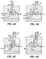
- FIG. 4is perspective view of a fourth preferred embodiment of the present invention.
- FIG. 4Ais an elevational view, partially in cross section, illustrating the initial position in a puncture site of the introducer and guidewire elements of the apparatus of FIG. 4 ;
- FIG. 4Bis a view similar to that of FIG. 4A , but showing a catheter contained within the introducer when the introducer is in a first axial position relative to the catheter;
- FIG. 4Cis an elevational view, partially in cross section, illustrating the apparatus of FIG. 4A in a preferred operational position
- FIG. 4Dis an elevational view, partially in cross section, illustrating the apparatus of FIG. 4A with the compression balloon revealed and not yet inflated, the introducer having been moved to a second axial position relative to the catheter;
- FIG. 4Eis a perspective view, partially in cross section, illustrating the compression balloon of the apparatus of FIG. 4D in an inflated state
- FIG. 4Fis an elevational view, partially in cross section, illustrating the apparatus of FIG. 4E with the guidewire element withdrawn;
- FIG. 5is an elevational view, partially in cross section, illustrating a modification of the embodiment of FIG. 1 ;
- FIG. 6is an elevational view of a modification of the fourth preferred embodiment of FIGS. 4 through 4F , having an optional dissolvable locating tip at the distal end of the catheter;
- FIG. 7is an elevational view of the modified fourth preferred embodiment of FIG. 6 , showing the present invention deployed at a vascular puncture site, with the locating tip inserted into the lumen of a blood vessel;
- FIG. 8is an elevational view of a modification of the third preferred embodiment of FIGS. 3 and 3A , having an optional dissolvable locating tip;
- FIG. 9is an elevational view of an alternative form of a dissolvable locating tip.
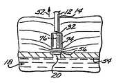
- FIG. 1A percutaneous blood vessel sealing device, or percutaneous hemostatic device 10 , which applies hemostatic sealing pressure directly to tissue adjacent a vascular puncture site, without employing implanted materials, is shown in FIG. 1 .
- an introducer sheath(“introducer”) 12 , well known in the art, is present in an incision 14 that extends from the skin surface 16 to a blood vessel (artery or vein) 18 of a patient at the site of a blood vessel puncture 20 .
- the introducer 12has normally been inserted previously to provide access to the vessel 18 for instrumentation (not shown) used in performing a vascular procedure immediately preceding the need to seal the puncture 20 .
- the initial position of an introducer 12 so insertedis most clearly illustrated in FIG. 4A , which shows a tapered distal end 22 of the introducer 12 at a puncture site 24 , inserted within a vascular puncture 20 .
- the introducer 12will have a size of approximately 7 French (2.3 mm in diameter), and a length of approximately 130 mm, although a size as large as 14 French (4.7mm in diameter) may be used for larger punctures.
- a working channel 26extends axially from the proximal end 28 of the introducer 12 through its tapered distal end 22 .
- a hollow locator tube 30extends coaxially through the introducer 12 and into the vessel 18 through the puncture 20 .
- Guided by the locator tube 30 into the introducer working channel 26is a semi-rigid catheter 32 having a catheter proximal end 33 , and a catheter distal end 34 ( FIG. 1A ).
- the introducer 12is movable axially with respect to the catheter 32 , and is disposed initially at a first axial position, or distal position, in which the catheter distal end 34 is enclosed or sheathed within the distal end 22 of the introducer 12 .
- the catheter 32is a dual-lumen device having a first axial lumen 36 ( FIG. 1D ) which encompasses the locator tube 30 when the catheter 32 is inserted into the working channel 26 of the introducer 12 .
- a second axial, lumen 38is provided with an inflation orifice 40 near its distal end, the inflation orifice communicating with the interior of a compression balloon 42 that concentrically surrounds a portion of the length of the catheter 32 extending proximally from its distal end 34 .
- the compression balloon 42is initially enclosed, in an uninflated state, within the distal end 22 of the introducer 12 , as illustrated in FIG. 1A .
- the opposite (proximal) end of the second axial lumen 38communicates with a compression balloon inflation port 44 through an inflation tube 45 , as shown in FIGS. 1 and 4 .
- the catheter 32has an outer diameter sufficiently small to be freely insertable into the introducer 12 , and a length that is greater than that of the introducer 12 , i.e., in the range of about 130 mm to about 750 mm.
- the locator tube 30has an inflatable intravascular locating balloon 50 at its distal end portion, shown in FIG. 1A in an uninflated state.
- the interior of the locating balloon 50is in fluid communication with the hollow interior of the locator tube 30 through a suitable inflation orifice (not shown), as is well known in conventional balloon catheters and the like.
- luer locks 46 , 48may be employed for both the locator balloon embodiment ( FIGS. 1 through 1D ) and for embodiments (described below) featuring expandable compression elements other than the compression balloon 42 , a version using no luer locks will be described below that is specifically adapted for use with the compression balloon 42 .
- Both the luer and non-luer versionsare suitable for embodiments employing either the inflatable locating balloon 50 or a guidewire locating means, to be described below.
- FIG. 1Ashows the locator tube 30 , having the uninflated locating balloon 50 near its distal end, inserted into the vessel 18 through the introducer 12 and the vascular puncture 20 . It is advantageous to construct the locator tube 30 so that a length of tube extends distally beyond the location of the locating balloon 50 into the vessel 18 to facilitate re-access through the vascular puncture 20 , if required.
- the entire apparatus 10(including the introducer 12 and the catheter 32 ) is in its initial position relative to the vessel; that is, the distal tip 22 of the introducer 12 is located adjacent to or within the puncture 20 , while the introducer 12 is in its above-described first axial position or distal position relative to the catheter 32 , in which the catheter distal end 34 and the uninflated compression balloon 42 are enclosed within the a distal end 22 of the introducer 12 .
- FIG. 1Billustrates the device 10 after the locating balloon 50 has been inflated by fluid introduced into it via the locator tube 30 .
- the entire device 10(including the introducer 12 and the catheter 32 ) has been partially withdrawn from the puncture site 24 in the direction of the arrow 52 (i.e., in the proximal direction), to a “preferred operational position”, in which the locating balloon 50 is lodged against an interior wall 54 of the vessel 18 .
- the introducer 12remains in its first or distal position, in which the portion of the catheter 32 carrying the uninflated compression balloon 42 is enclosed within the distal end 22 of the introducer 12 .
- the introducer 12has been moved axially, relative to the catheter 32 , in the direction of the arrow 52 (i.e., proximally), to its second axial position, or proximal position.
- the movement of the introducer 12 to this second or proximal positionuncovers the uninflated compression balloon 42 .
- the compression stage of the device 10is illustrated next in FIG. 1 .
- the compression balloon 42inflated via the second axial lumen 38 ( FIG. 1D ), rests in an optimal position to effect natural hemostasis, viz., above a laminar portion 56 of the fatty tissue adjacent the puncture site 24 .
- An optimal distance from the vessel 18 to the catheter distal end 34is in the range of 2 mm to 10 mm. This distance will dispose a layer of fatty tissue 56 between the vessel 18 and the catheter 32 , minimizing the potential for pseudo-aneurysm.
- the introducer luer lock 46is shown engaged with the catheter luer lock 48 , assuring that a holding force applied to the introducer 12 will be transmitted as well to the catheter 32 .
- a visible marker band 57 on the exterior of the locating tubing 30may advantageously be provided to align the proximal ends of the introducer 12 and the catheter 32 in correspondence with the location of the distal ends 22 , 34 thereof when the locator balloon 50 is lodged against the inner wall 54 of vessel 18 .
- FIGS. 1 and 4An adhesive skin patch 58 with a sheath cuff 60 clamped onto the external portion of the introducer 12 to apply downward force (in the direction of the arrow 62 , i.e., distally) on the introducer 12 is shown in FIGS. 1 and 4 .
- Fastener strips 64secure the adhesive patch 58 to the sheath cuff 60 .
- the fastener strips 64may be elastic bands with suitable adhesive areas, or hook and loop strips (such as the type marketed under the trademark VELCRO) that adhere to areas of complementary material on the patch 58 .
- Pressure maintained by the introducer sheath cuff 60 on the catheter 32provides hemostatic pressure on the compression balloon 42 to bear on the tissue layer 56 for a first period of time, whereupon the locating tube 30 is withdrawn (the locator balloon 50 having first been deflated), and a second period of time elapses, after which all instrumentation is removed from the patient as will be noted when the method for sealing the puncture 20 is described in detail below.
- FIGS. 2 , 2 A, and 2 Bshow a collapsible prong assembly compression element 66 attached to the catheter distal end 34 .
- the prong assembly 66is radially compressed or collapsed when enclosed within the introducer 12 , when the introducer is in its first or distal position.
- the prong assembly 66expands radially when the introducer 12 is partially withdrawn from the vessel 18 ( FIGS. 2A and 2B ), by moving the introducer 12 to its second or proximal position in a manner similar to the partial withdrawal of introducer 12 in the direction of arrow 52 as described previously in connection with the compression balloon embodiment.
- the prong assembly 66comprises a plurality of spaced-apart resilient prongs 68 , the proximal ends of which are attached to the catheter 32 , and the distal ends of which are attached to a collapsible spanning film sheet or dam 70 , shown expanded in FIGS. 2A and 2B .
- the sheet or dam 70allows the application of hemostatic pressure on the tissue 56 above the vessel 18 .
- a central aperture 72 in the sheet or dam 70permits the locator tube (not shown) to project through the catheter 32 into the vessel 18 as described previously. Since there is no compression balloon to be inflated, a catheter with a single axial lumen 36 is adequate for this application.
- Materials for the spanning sheet or dam 70may include polyurethane and polyethyleneterephthalate (PET).
- FIGS. 3 and 3Ashow a foam pad compression element 74 attached to the catheter distal end 34 .
- the foam pad element 74is compressed when enclosed within the introducer 12 when the introducer is in its first or distal position.
- the foam pad compression element 74then expands when the introducer 12 is partially withdrawn from the vessel 18 , as shown in FIG. 3A , by moving the introducer 12 to its second or proximal position, as described above with respect to the first and second embodiments. Hemostatic pressure is similarly exerted on the tissue 56 above the vessel 18 .
- An axial channel 76 in the foam pad 74permits the locator tube (not shown) to project through the catheter 32 into the vessel 18 , as described previously.
- Materials for the foam pad 74may include various polymeric foams, such as polyurethanes, as are well-known in the art.
- the foam pad 74may be impregnated with a coagulant such as thrombin or protamine to effect local hemostasis.
- a locator tube 30 with a locating balloon 50to determine the optimal operational location for the apparatus 10 .
- a guidewire 78may be utilized for the location determination of the apparatus 10 , as illustrated in FIGS. 4 through 4F .
- a standard guidewire 78typically 3 French (1 mm in diameter), shown coaxially located within the introducer 12 , has a distal end 82 extending out of the introducer distal end 22 into the puncture 20 of the vessel 18 .
- the catheter 32is shown in FIG. 4B having been inserted into the introducer 12 and guided to the distal end 22 of the introducer by the guidewire 78 .
- a radiopaque marker 84for viewing under fluoroscopy, as shown in FIG. 4D .
- FIG. 4Cshows an optimal location for catheter distal end 34 , radiopaque contrast medium (not shown) having been introduced into the catheter lumen 36 , and the apparatus 10 having been partially withdrawn from the vessel 18 in the direction of the arrow 52 (i.e., proximally).
- An extravasation 85 of the radiopaque contrast mediumis shown marking the desired distance between the vessel 18 and the catheter distal end 34 , as will be explained when the method for sealing the puncture is described below.
- FIG. 4DThe introducer 12 is shown in FIG. 4D having been moved, in the direction of the arrow 52 , to its second or proximal position to reveal the uninflated compression balloon 42 in position for inflating.
- FIG. 4Eillustrates the apparatus 10 with the compression balloon 42 inflated and in place above the fatty layer 56 to apply hemostatic pressure for a first period of time in order to effect initial closure of puncture site 24 .
- FIG. 4Fshows the apparatus 10 after the guidewire 78 has been removed from the apparatus 10 and pressure is applied for a second period of time to close the puncture 20 .
- the guidewire 78 and radiopaque positioning of an expandable compression element at the distal end 34 of the catheter 32may be employed with the prong assembly and foam pad embodiments described above in connection with the locator tube 30 .
- a standard hemostatic “Y” 86is used, as shown in FIG. 4 .
- the “Y” 86has a main leg 88 for receiving the guidewire 78 into the axial lumen 36 of the catheter 32 , while a side port 90 of the “Y” 86 is used for introducing the contrast medium into the same lumen.
- FIG. 5A modification of the first (compression balloon) embodiment of the present invention is shown in FIG. 5 , where an apparatus 110 has an introducer 112 having no luer connection with a catheter 132 . Since the cuff 60 applies downward force in the direction of the arrow 62 only to the introducer 112 , and not to the catheter 132 , the distal end 122 of the introducer 112 must bear directly on the compression balloon 42 to exert hemostatic pressure on the balloon 42 . Although this modification is suitable only for the compression balloon embodiment of this invention, both the locator tube 30 and the guidewire 78 may be utilized in this modification for optimal positioning of the catheter distal end 34 .
- FIGS. 6 and 7illustrate a modification of the fourth preferred embodiment (that of FIGS. 4 through 4F ).
- a dissolvable or resorbable locating tip element 140is fixed to the distal end 34 of the catheter 32 , distally from the compression balloon 42 .
- the locating tip element 140is an elongate, tubular member, having a generally cylindrical main portion 142 and a tapered distal end tip 144 .
- the diameter of the cylindrical main portion 142is preferably about 1.3 to 4.0 mm, and the overall length of the locating tip 140 element is preferably about 1 to 4 cm, the dimensions of a particular locating tip 140 being selected in accordance with such factors as the dimensions and location of the puncture site, its “dwell time” (see below), and the preferences of the physician.
- the locating tip 140 elementis hollow and open-ended to allow the guidewire 78 to pass through it.
- the locating tip element 140is preferably used in conjunction with locating means such as the guidewire 78 or the locator tube 30 . As shown in FIG. 7 , the locating tip element 140 is dimensioned so that it can be inserted into the blood vessel lumen 18 through the puncture site 20 , and maintains the location of the apparatus 10 during use, after the guidewire 78 has been withdrawn (see below).
- the locating tip element 140is made of a material that is biocompatibly (non-toxically) dissolved in the blood stream during a period of between about 10 and 60 minutes (the “dwell time”), during which time partial resorption of the material into the subcutaneous tissue results in the detachment of the locating tip element 140 from the catheter 32 . The detachment leaves a portion of the locating tip element 140 outside of the blood vessel, to be completely dissolved into the subcutaneous tissue.
- Suitable materials for the locating tip element 140may include, for example a number of well-known polymers, methyl cellulose, carboxymethyl cellulose, carbowaxes, and gelatin (particularly pigskin gelatin).
- suitable polymersare polylactic glycolic acids, a polyvinyl pyrrolidone, polyvinyl alcohol, polyproline, and polyethylene oxide.
- the dissolvable locating tip element 140may be employed with other embodiments of the invention, particularly the compressible foam pad embodiment of FIGS. 3 and 3A .
- a foam pad 74 ′is attached to the distal end 34 of the catheter 32 , and has a distal end 146 .
- a dissolvable locating tip element 140 ′is attached to, and extends distally from, the distal end 146 of the foam pad 74 ′.
- the dissolvable locating tip element 140 , 140 ′may be modified and adapted for use as a locating tip for catheters and other devices that may be used in a wide variety of applications.
- an alternative dissolvable locating tip element 150is shown in FIG. 9 .
- the dissolvable locating tip element 150comprises a cylindrical portion 152 attached to the distal end of a surgical instrument 154 (e.g., a catheter). Projecting distally and coaxially from the cylindrical portion 152 is a slender tubular portion 156 , terminating in a tapered distal end tip 158 .
- the distal end of the cylindrical portiondefines an annular shoulder 160 surrounding the juncture with the tubular portion 156 .
- the entire dissolvable locating tip 150may have an axial passage 162 through its length to receive a guidewire or the like (not shown).
- the dissolvable locating tip element 150 of FIG. 9may be inserted into a puncture site in a blood vessel, in the manner similar to that discussed above with respect to FIGS. 6 and 7 . In this embodiment, however, only the distal tip end 158 and an adjacent portion of the tubular portion 156 enter the blood vessel.
- the cylindrical portion 152remains outside the vessel, with the shoulder 160 functioning as a compression element, much as does the foam pad 74 , 74 ′, described above in connection with FIGS. 3 , 3 A, and 8 .
- the compression elementbeing integral with and of the same dissolvable material as the locating tip, likewise dissolves in the patient's tissue after hemostasis has occurred.
- a piercing cannulais inserted into the skin of a patient at an angle of from 25 to 45 degrees until it punctures a blood vessel, e.g., the femoral artery.
- the vesselmay be located one centimeter or more beneath the surface of the skin.
- a guidewireis inserted through the cannula into the vessel, the cannula is withdrawn, and a catheter introducer sheath is inserted over the guidewire into the puncture site.
- the practitionerthen uses the introducer to gain access to the vascular lumen for the instrumentation used to perform the particular procedure.
- the introduceris the last device remaining in the puncture, which must then be sealed.
- the method of the present inventionprovides a rapid, permanent, inexpensive sealing of a puncture in a blood vessel, with no foreign implants remaining in the patient.
- the methodcan be understood with reference to the drawing FIGS. and the previous description of the apparatus of this invention.
- an introducer sheath 12is shown in a puncture site 24 at the conclusion of a vascular procedure.
- a locator tube 30 having an inflatable locating balloon 50 adjacent its distal endis inserted anally through the introducer 12 , into a puncture 20 and extending the uninflated locating balloon 50 into the lumen of a vessel 18 .
- a dual lumen catheter 32is passed over the locator tube 30 so that a first lumen 36 ( FIG. 1D ) of the catheter 32 receives the locator tube 30 .
- the locator tube 30maintains alignment of the catheter 32 with the puncture 20 and allows repeated access into the vessel 18 , if necessary.
- the catheter 32having an inflatable compression balloon 42 at its distal end 34 , is inserted fully into the introducer 12 until its distal end 34 , including the uninflated compression balloon 42 , is at the distal end 22 of the introducer 12 .
- the locator tube 30is pushed or pulled until a marker band 57 (shown in FIG. 1 ) is aligned with the proximal end 33 of the catheter 32 .
- the marker band 57is preselected to establish a fixed relationship with the catheter 32 so that a preferred distance may be maintained between the vessel 18 and the distal end 34 of catheter 32 as will be explained below.
- the introducer 12being in its first or distal position, the uninflated compression balloon 42 is fully enclosed and contained within the working channel 26 of the introducer 12 , as described above.
- the practitionerthen inflates the locating balloon 50 via the locator tube 30 , partially withdrawing the introducer 12 , the catheter 32 and the locator tube 30 from the puncture 20 in the direction of the arrow 52 , until the locating balloon 50 lodges against the inner wall of the vessel 18 at the puncture 20 , as illustrated in FIG. 1B . Since the position of the catheter distal end 34 relative to the introducer distal end 22 remains unchanged, the distal end 34 of the catheter is now at the location predetermined by the placement of the marker band 57 , preferably about 5 mm to 15 mm from the puncture 20 . This distance will allow a layer of fatty subcutaneous tissue 56 to lie between the catheter distal end 34 and the puncture 20 .
- the introducer 12is further withdrawn in the direction of the arrow 52 , by moving it to its second or proximal position relative to the catheter 32 , as described above, to expose the uninflated compression balloon 42 , as shown in FIG. 1C .
- the luer fittings 46 , 48 at the proximal ends of the catheter 32 and the introducer 12 , respectively,are now connected to each other to lock the catheter 32 and the introducer 12 into a fixed position relative to one another, and the compression balloon 42 is then inflated, as illustrated in FIG. 1 , via a second catheter lumen 38 ( FIG. 1D ).
- the compression balloon 42is then pressed down against the fatty layer 56 above the puncture site 24 , while gentle traction is maintained on the locating balloon 50 , thus compressing the extravascular fatty tissue 56 between the balloons 42 , 50 .
- the fatty tissue 56advantageously minimizes the potential of pseudo-aneurysm formation and promotes efficient hemostasis.
- an introducer cuff 60is clamped onto the introducer 12 and secured to an adhesive patch 58 by means of elastic or hook and loop fastening strips 64 ( FIGS. 1 and 4 ).
- the introducer 12is locked with the catheter 32 by the luer fittings 46 , 48 , the downward force provided by the fastening strips 64 is transmitted from the introducer 12 through the semi-rigid catheter 32 to the compression balloon 42 , maintaining hemostatic pressure on the puncture site 24 through fatty tissue 56 .
- first period of timeapproximately 5 to 15 minutes
- initial clotting of the puncture 20will have occurred.
- the locating balloon 50is then deflated and the locator tube 30 withdrawn from the apparatus 10 , leaving only a small (e.g., approximately 1 mm in diameter) portion of the original puncture 20 to clot.
- the compression balloon 42remains in place for an additional (second) period of time (approximately 5 to 25 minutes), providing hemostasis to the puncture 20 , after which the compression balloon 42 is deflated and retracted proximally into the introducer 12 , the luer fittings 46 , 48 having first been disconnected.
- the sealing processhaving been completed, the apparatus 10 is completely removed from the patient.
- the foregoing methoduses an introducer 12 that is already positioned at the access site so that position is not lost in changing instruments, bleeding does not occur while devices are positioned, and the locator tube 30 maintains the access location for re-access if needed during the initial clotting of the puncture 20 .
- employment of the present inventionrequires minimal physician time and greatly reduces staff time and involvement previously devoted to maintaining supradermal pressure for long periods of hemostasis.
- the need for operating room timemay be reduced by the removal of the locator tube 30 , the introducer 12 and the catheter 32 after the patient is returned to the patient's room. Overall, patient discomfort is significantly lessened through the use of the foregoing method as compared with the traditional manual external compression techniques.
- the compression element at catheter distal end 34comprises the collapsible prong assembly 66 , as shown in FIGS. 2 , 2 A, and 2 B.
- the movement of the introducer 12 to its second or proximal positionreleases the prong assembly 66 from confinement within the introducer 12 , allowing the individual prongs 68 of the prong assembly 66 to expand, as illustrated in FIG. 2A .
- a resilient spanning sheet or dam 70supported by the ends of the prongs 68 , then allows the application of hemostatic pressure on the fatty tissue layer 56 , as described earlier in connection with the compression balloon embodiment.
- the locator tube(not shown) passes through and is withdrawn from the aperture 72 in the spanning film 70 .
- a third embodiment of the methodinvolves the use of the compressible foam pad 74 shown in FIGS. 3 and 3A as the compression element at the distal end 34 of the catheter 32 .
- the introducer 12when the catheter 32 is in the preferred location as shown in FIG. 3 , the introducer 12 is moved from its first or distal position to its second or proximal position (in the direction of the arrow 52 ) to uncover the foam pad 74 , allowing it to expand, as illustrated in FIG. 3A .
- the expanded foam pad 74exerts hemostatic pressure upon the fatty tissue layer 56 , as described previously.
- the locator tube(not shown) passes through and is withdrawn from the pad channel 76 formed axially in the foam pad 74 .
- a coagulant agentsuch as collagen, thrombin or protamine may be delivered to the vicinity of the puncture site through the pad channel 76 which communicates with the catheter axial lumen 36 .
- the foam pad 74may be saturated with the agent prior to deployment.
- the method employed with the apparatus described abovemay also use a guidewire 78 ( FIG. 4 ) to perform the locating functions provided by the locator tube 30 in the previous embodiments.
- All three of the compression elementsviz., the compression balloon 42 , the expandable prong element 66 and the foam pad 74 , may be utilized with the guidewire 78 .
- FIGS. 4 through 4Fshowing only the compression balloon 42 alternative, may be viewed with the understanding that the method to be described in conjunction therewith applies to all three guidewire 78 embodiments.
- the introducer 12is shown as it remains in the puncture 20 after a vascular access procedure.
- a conventional surgical guidewire 78is extended through the introducer 12 so that its distal end 82 extends into the lumen of the vessel 18 .
- the dual lumen catheter 32is passed over the guidewire 78 so that a first lumen 36 ( FIG. 1D ) of the catheter 32 receives the guidewire 78 .
- the guidewire 78maintains alignment of the catheter 32 with the puncture 20 and allows re-access into the vessel 18 if it becomes necessary.
- the catheter 32having an inflatable compression balloon 42 at its distal end 34 , is inserted fully into the introducer 12 until its distal end 34 , including the uninflated compression balloon 42 , is enclosed within the working channel 26 at the distal end 22 of the introducer 12 , as shown in FIG. 4B .
- a radiopaque contrast medium(not shown) is introduced into the catheter first lumen 36 , as illustrated in FIG. 4 .
- a main leg 88 of a conventional hemostasis “Y” 86may be passed over the guidewire 78 and attached to the proximal end 33 of the catheter lumen 36 .
- the contrast mediumis then introduced into the catheter lumen 36 via a side port 90 of the “Y” 86 , and viewed by the practitioner using conventional fluoroscopic techniques.
- a radiopaque marker 84may be provided at the tip of the catheter distal end 34 ( FIG. 4D ) .
- the introducer 12 with the catheter 32is partially withdrawn in the direction of the arrow 52 from the puncture 20 . Withdrawal is continued until contrast medium in the catheter lumen 36 escaping from around the guidewire 78 into the vessel 18 is observed to form an extravasation cloud 85 , signifying that the introducer 12 and the catheter 32 have exited the puncture 20 .
- the catheter distal end element 34is the preferred distance of about 5 to 15 mm from the vessel 18 , withdrawal of the catheter 32 is halted, as shown in FIG. 4C .
- the remainder of the closure procedureis essentially the same as described above after the preferred position of the catheter 32 was determined through the locator tube 30 method.
- the introducer 12is moved from its first or distal position relative to the catheter 32 to its second or proximal position, to expose the uninflated compression balloon 42 , as shown in FIG. 4D .
- the compression balloon 42is then inflated to bear on the fatty tissue layer 56 as shown in FIG. 4E .
- the locating meansin this embodiment guidewire 78
- the method employing the guidewire 78may be effectively adapted for use with the expandable prong element and foam tip embodiments of the present invention.
- FIG. 5shows the position of the catheter 132 aligned with a visible marker band 57 on the locator tube 30 , just as in the first embodiment described above. It will be readily understood that the method of this “luerless” apparatus 110 may be equally utilized with the guidewire 78 as with the locator tube 30 for the compression balloon embodiment of this invention.
Landscapes
- Health & Medical Sciences (AREA)
- Life Sciences & Earth Sciences (AREA)
- Heart & Thoracic Surgery (AREA)
- Animal Behavior & Ethology (AREA)
- Veterinary Medicine (AREA)
- Public Health (AREA)
- Engineering & Computer Science (AREA)
- Biomedical Technology (AREA)
- General Health & Medical Sciences (AREA)
- Anesthesiology (AREA)
- Hematology (AREA)
- Pulmonology (AREA)
- Biophysics (AREA)
- Surgery (AREA)
- Cardiology (AREA)
- Nuclear Medicine, Radiotherapy & Molecular Imaging (AREA)
- Medical Informatics (AREA)
- Molecular Biology (AREA)
- Child & Adolescent Psychology (AREA)
- Surgical Instruments (AREA)
- Media Introduction/Drainage Providing Device (AREA)
Abstract
Description
Claims (39)
Priority Applications (3)
| Application Number | Priority Date | Filing Date | Title |
|---|---|---|---|
| US10/107,539US7175646B2 (en) | 1995-09-15 | 2002-03-25 | Apparatus and method for percutaneous sealing of blood vessel punctures |
| US11/537,024US20070021770A1 (en) | 1995-09-15 | 2006-09-29 | Apparatus and Method for Percutaneous Sealing of Blood Vessel Punctures |
| US12/887,945US20110014290A1 (en) | 1995-09-15 | 2010-09-22 | System and method for facilitating hemostasis with an absorbable sponge |
Applications Claiming Priority (4)
| Application Number | Priority Date | Filing Date | Title |
|---|---|---|---|
| US08/528,892US5645566A (en) | 1995-09-15 | 1995-09-15 | Apparatus and method for percutaneous sealing of blood vessel punctures |
| US08/888,851US6071300A (en) | 1995-09-15 | 1997-07-07 | Apparatus and method for percutaneous sealing of blood vessel punctures |
| US09/365,674US6371974B1 (en) | 1995-09-15 | 1999-08-02 | Apparatus and method for percutaneous sealing of blood vessel punctures |
| US10/107,539US7175646B2 (en) | 1995-09-15 | 2002-03-25 | Apparatus and method for percutaneous sealing of blood vessel punctures |
Related Parent Applications (1)
| Application Number | Title | Priority Date | Filing Date |
|---|---|---|---|
| US09/365,674DivisionUS6371974B1 (en) | 1995-09-15 | 1999-08-02 | Apparatus and method for percutaneous sealing of blood vessel punctures |
Related Child Applications (1)
| Application Number | Title | Priority Date | Filing Date |
|---|---|---|---|
| US11/537,024ContinuationUS20070021770A1 (en) | 1995-09-15 | 2006-09-29 | Apparatus and Method for Percutaneous Sealing of Blood Vessel Punctures |
Publications (2)
| Publication Number | Publication Date |
|---|---|
| US20020156495A1 US20020156495A1 (en) | 2002-10-24 |
| US7175646B2true US7175646B2 (en) | 2007-02-13 |
Family
ID=25394032
Family Applications (4)
| Application Number | Title | Priority Date | Filing Date |
|---|---|---|---|
| US08/888,851Expired - LifetimeUS6071300A (en) | 1995-09-15 | 1997-07-07 | Apparatus and method for percutaneous sealing of blood vessel punctures |
| US09/365,674Expired - LifetimeUS6371974B1 (en) | 1995-09-15 | 1999-08-02 | Apparatus and method for percutaneous sealing of blood vessel punctures |
| US10/107,539Expired - Fee RelatedUS7175646B2 (en) | 1995-09-15 | 2002-03-25 | Apparatus and method for percutaneous sealing of blood vessel punctures |
| US11/537,024AbandonedUS20070021770A1 (en) | 1995-09-15 | 2006-09-29 | Apparatus and Method for Percutaneous Sealing of Blood Vessel Punctures |
Family Applications Before (2)
| Application Number | Title | Priority Date | Filing Date |
|---|---|---|---|
| US08/888,851Expired - LifetimeUS6071300A (en) | 1995-09-15 | 1997-07-07 | Apparatus and method for percutaneous sealing of blood vessel punctures |
| US09/365,674Expired - LifetimeUS6371974B1 (en) | 1995-09-15 | 1999-08-02 | Apparatus and method for percutaneous sealing of blood vessel punctures |
Family Applications After (1)
| Application Number | Title | Priority Date | Filing Date |
|---|---|---|---|
| US11/537,024AbandonedUS20070021770A1 (en) | 1995-09-15 | 2006-09-29 | Apparatus and Method for Percutaneous Sealing of Blood Vessel Punctures |
Country Status (5)
| Country | Link |
|---|---|
| US (4) | US6071300A (en) |
| EP (1) | EP0994673A4 (en) |
| AU (1) | AU742269B2 (en) |
| CA (1) | CA2295879C (en) |
| WO (1) | WO1999002091A1 (en) |
Cited By (88)
| Publication number | Priority date | Publication date | Assignee | Title |
|---|---|---|---|---|
| US20050075597A1 (en)* | 2003-09-12 | 2005-04-07 | Vournakis John N. | Vascular access preservation in hemodialysis patients |
| US20050216018A1 (en)* | 2004-03-29 | 2005-09-29 | Sennett Andrew R | Orthopedic surgery access devices |
| US20050267520A1 (en)* | 2004-05-12 | 2005-12-01 | Modesitt D B | Access and closure device and method |
| US20060271078A1 (en)* | 2005-05-12 | 2006-11-30 | Modesitt D B | Access and closure device and method |
| US20070055223A1 (en)* | 2003-02-04 | 2007-03-08 | Cardiodex, Ltd. | Methods and apparatus for hemostasis following arterial catheterization |
| US20080065150A1 (en)* | 2006-09-12 | 2008-03-13 | Boston Scientific Scimed, Inc. | Method and Apparatus for Promoting Hemostasis of a Blood Vessel Puncture |
| US20080167643A1 (en)* | 2004-11-22 | 2008-07-10 | Cardiodex Ltd. | Techniques for Heating-Treating Varicose Veins |
| US20080208320A1 (en)* | 2006-12-15 | 2008-08-28 | Francisca Tan-Malecki | Delivery Apparatus and Methods for Vertebrostenting |
| US20090105744A1 (en)* | 2007-10-17 | 2009-04-23 | Modesitt D Bruce | Methods for forming tracts in tissue |
| US20090125056A1 (en)* | 2007-08-15 | 2009-05-14 | Cardiodex Ltd. | Systems and methods for puncture closure |
| US20090248056A1 (en)* | 2008-03-28 | 2009-10-01 | Ethicon, Inc. | Applicator instruments for controlling bleeding at surgical sites and methods therefor |
| US20090326538A1 (en)* | 2006-12-15 | 2009-12-31 | Sennett Andrew R | Devices and methods for fracture reduction |
| US20100016786A1 (en)* | 2008-07-21 | 2010-01-21 | Arstasis, Inc. | Devices, methods, and kits for forming tracts in tissue |
| US20100016810A1 (en)* | 2008-07-21 | 2010-01-21 | Arstasis. Inc., | Devices and methods for forming tracts in tissue |
| US20100125296A1 (en)* | 2004-07-10 | 2010-05-20 | Modesitt D Bruce | Biological tissue closure device and method |
| US20100152748A1 (en)* | 2008-12-12 | 2010-06-17 | E-Pacing, Inc. | Devices, Systems, and Methods Providing Body Lumen Access |
| US20100211000A1 (en)* | 2008-08-26 | 2010-08-19 | Killion Douglas P | Method and system for sealing percutaneous punctures |
| US20100268175A1 (en)* | 2009-04-21 | 2010-10-21 | Xlumena, Inc. | System and method for delivering expanding trocar through a sheath |
| US20110125178A1 (en)* | 2009-05-15 | 2011-05-26 | Michael Drews | Devices, methods and kits for forming tracts in tissue |
| US20110208215A1 (en)* | 2009-09-22 | 2011-08-25 | Modesitt D Bruce | Devices, methods, and kits for forming tracts in tissue |
| US20110230829A1 (en)* | 2010-03-19 | 2011-09-22 | Fitzgerald Patrick J | Arterial Tamponade Device and Method |
| US20110230907A1 (en)* | 2010-03-19 | 2011-09-22 | Sinocclusive Llc | Arterial tamponade device and method |
| US8277483B2 (en) | 2011-01-25 | 2012-10-02 | Wound Care 360, LLC | Vascular wound closing apparatus and method |
| US20130018414A1 (en)* | 2004-01-30 | 2013-01-17 | W.L. Gore & Associates | Devices, Systems and Methods for Closure of Cardiac Openings |
| US8361100B2 (en) | 2008-03-17 | 2013-01-29 | Ethicon, Inc. | Applicator instruments for the delivery, deployment, and tamponade of hemostats and methods therefor |
| US8437833B2 (en) | 2008-10-07 | 2013-05-07 | Bard Access Systems, Inc. | Percutaneous magnetic gastrostomy |
| US8444671B2 (en) | 2006-12-21 | 2013-05-21 | Cardiva Medical, Inc. | Hemostasis-enhancing device and method for its use |
| US8478382B2 (en) | 2008-02-11 | 2013-07-02 | C. R. Bard, Inc. | Systems and methods for positioning a catheter |
| US8512256B2 (en) | 2006-10-23 | 2013-08-20 | Bard Access Systems, Inc. | Method of locating the tip of a central venous catheter |
| WO2014018280A1 (en)* | 2012-07-24 | 2014-01-30 | Wound Care 360, LLC | Vascular wound closing apparatus and method |
| US8774907B2 (en) | 2006-10-23 | 2014-07-08 | Bard Access Systems, Inc. | Method of locating the tip of a central venous catheter |
| US8781555B2 (en) | 2007-11-26 | 2014-07-15 | C. R. Bard, Inc. | System for placement of a catheter including a signal-generating stylet |
| US8784336B2 (en) | 2005-08-24 | 2014-07-22 | C. R. Bard, Inc. | Stylet apparatuses and methods of manufacture |
| US8801693B2 (en) | 2010-10-29 | 2014-08-12 | C. R. Bard, Inc. | Bioimpedance-assisted placement of a medical device |
| US8821918B2 (en) | 2001-03-12 | 2014-09-02 | Boston Scientific Scimed Inc. | Cross-linked gelatin composition comprising a wetting agent |
| US8845682B2 (en) | 2009-10-13 | 2014-09-30 | E-Pacing, Inc. | Vasculature closure devices and methods |
| US8849382B2 (en) | 2007-11-26 | 2014-09-30 | C. R. Bard, Inc. | Apparatus and display methods relating to intravascular placement of a catheter |
| USD724745S1 (en) | 2011-08-09 | 2015-03-17 | C. R. Bard, Inc. | Cap for an ultrasound probe |
| US9125578B2 (en) | 2009-06-12 | 2015-09-08 | Bard Access Systems, Inc. | Apparatus and method for catheter navigation and tip location |
| US9131931B2 (en) | 2013-01-21 | 2015-09-15 | Vi Bravoseal, Llc | Vessel sealing device with automatic deployment |
| US9138215B2 (en) | 2013-01-21 | 2015-09-22 | Vi Bravoseal, Llc | Vessel sealing device |
| US9211107B2 (en) | 2011-11-07 | 2015-12-15 | C. R. Bard, Inc. | Ruggedized ultrasound hydrogel insert |
| USD754357S1 (en) | 2011-08-09 | 2016-04-19 | C. R. Bard, Inc. | Ultrasound probe head |
| US9339206B2 (en) | 2009-06-12 | 2016-05-17 | Bard Access Systems, Inc. | Adaptor for endovascular electrocardiography |
| US9381041B2 (en) | 2009-04-21 | 2016-07-05 | Xlumena, Inc. | Methods and devices for access across adjacent tissue layers |
| US9445734B2 (en) | 2009-06-12 | 2016-09-20 | Bard Access Systems, Inc. | Devices and methods for endovascular electrography |
| US9456766B2 (en) | 2007-11-26 | 2016-10-04 | C. R. Bard, Inc. | Apparatus for use with needle insertion guidance system |
| US9480485B2 (en) | 2006-12-15 | 2016-11-01 | Globus Medical, Inc. | Devices and methods for vertebrostenting |
| US9492097B2 (en) | 2007-11-26 | 2016-11-15 | C. R. Bard, Inc. | Needle length determination and calibration for insertion guidance system |
| US9521961B2 (en) | 2007-11-26 | 2016-12-20 | C. R. Bard, Inc. | Systems and methods for guiding a medical instrument |
| US9532724B2 (en) | 2009-06-12 | 2017-01-03 | Bard Access Systems, Inc. | Apparatus and method for catheter navigation using endovascular energy mapping |
| US9554716B2 (en) | 2007-11-26 | 2017-01-31 | C. R. Bard, Inc. | Insertion guidance system for needles and medical components |
| US9636031B2 (en) | 2007-11-26 | 2017-05-02 | C.R. Bard, Inc. | Stylets for use with apparatus for intravascular placement of a catheter |
| US9649048B2 (en) | 2007-11-26 | 2017-05-16 | C. R. Bard, Inc. | Systems and methods for breaching a sterile field for intravascular placement of a catheter |
| US9681823B2 (en) | 2007-11-26 | 2017-06-20 | C. R. Bard, Inc. | Integrated system for intravascular placement of a catheter |
| US9839372B2 (en) | 2014-02-06 | 2017-12-12 | C. R. Bard, Inc. | Systems and methods for guidance and placement of an intravascular device |
| US9901714B2 (en) | 2008-08-22 | 2018-02-27 | C. R. Bard, Inc. | Catheter assembly including ECG sensor and magnetic assemblies |
| US10046139B2 (en) | 2010-08-20 | 2018-08-14 | C. R. Bard, Inc. | Reconfirmation of ECG-assisted catheter tip placement |
| US10076330B2 (en) | 2008-05-12 | 2018-09-18 | Xlumena, Inc. | Tissue anchor for securing tissue layers |
| US10085731B2 (en) | 2013-07-15 | 2018-10-02 | E-Pacing, Inc. | Vasculature closure devices and methods |
| US10085753B2 (en) | 2005-07-01 | 2018-10-02 | Abbott Laboratories | Clip applier and methods of use |
| US10154835B2 (en) | 2013-05-09 | 2018-12-18 | Essential Medical, Inc. | Vascular closure device with conforming plug member |
| US10201340B2 (en) | 2002-02-21 | 2019-02-12 | Integrated Vascular Systems, Inc. | Sheath apparatus and methods for delivering a closure device |
| US10245013B2 (en) | 2000-12-07 | 2019-04-02 | Integrated Vascular Systems, Inc. | Closure device and methods for making and using them |
| US10307145B2 (en) | 2013-01-21 | 2019-06-04 | Cyndrx, Llc | Vessel sealing device |
| US10349890B2 (en) | 2015-06-26 | 2019-07-16 | C. R. Bard, Inc. | Connector interface for ECG-based catheter positioning system |
| US10398418B2 (en) | 2003-01-30 | 2019-09-03 | Integrated Vascular Systems, Inc. | Clip applier and methods of use |
| US10413295B2 (en) | 2008-05-16 | 2019-09-17 | Abbott Laboratories | Engaging element for engaging tissue |
| US10441753B2 (en) | 2012-05-25 | 2019-10-15 | Arstasis, Inc. | Vascular access configuration |
| US10449330B2 (en) | 2007-11-26 | 2019-10-22 | C. R. Bard, Inc. | Magnetic element-equipped needle assemblies |
| US10524691B2 (en) | 2007-11-26 | 2020-01-07 | C. R. Bard, Inc. | Needle assembly including an aligned magnetic element |
| US10537312B2 (en) | 2012-12-21 | 2020-01-21 | Abbott Cardiovascular Systems, Inc. | Articulating suturing device |
| US10537313B2 (en) | 2009-01-09 | 2020-01-21 | Abbott Vascular, Inc. | Closure devices and methods |
| US10588637B2 (en) | 2016-05-17 | 2020-03-17 | Cook Medical Technologies Llc | Temporary occlusion of blood vessel using extra-luminal balloon |
| US10639008B2 (en) | 2009-10-08 | 2020-05-05 | C. R. Bard, Inc. | Support and cover structures for an ultrasound probe head |
| US10675447B2 (en) | 2012-05-25 | 2020-06-09 | Arstasis, Inc. | Vascular access configuration |
| US10751509B2 (en) | 2007-11-26 | 2020-08-25 | C. R. Bard, Inc. | Iconic representations for guidance of an indwelling medical device |
| US10820885B2 (en) | 2012-06-15 | 2020-11-03 | C. R. Bard, Inc. | Apparatus and methods for detection of a removable cap on an ultrasound probe |
| US10945735B2 (en) | 2004-04-12 | 2021-03-16 | Boston Scientific Scimed, Inc. | Luminal structure anchoring devices and methods |
| US10952732B2 (en) | 2013-02-21 | 2021-03-23 | Boston Scientific Scimed Inc. | Devices and methods for forming an anastomosis |
| US10973584B2 (en) | 2015-01-19 | 2021-04-13 | Bard Access Systems, Inc. | Device and method for vascular access |
| US10992079B2 (en) | 2018-10-16 | 2021-04-27 | Bard Access Systems, Inc. | Safety-equipped connection systems and methods thereof for establishing electrical connections |
| US11000207B2 (en) | 2016-01-29 | 2021-05-11 | C. R. Bard, Inc. | Multiple coil system for tracking a medical device |
| US11103213B2 (en) | 2009-10-08 | 2021-08-31 | C. R. Bard, Inc. | Spacers for use with an ultrasound probe |
| US11253242B2 (en) | 2013-01-21 | 2022-02-22 | Cyndrx, Llc | Vessel sealing device |
| US11918278B2 (en) | 2020-05-19 | 2024-03-05 | Boston Scientific Medical Device Limited | Medical delivery systems and methods of using the same |
| US12239302B2 (en) | 2013-01-21 | 2025-03-04 | Cyndrx Llc | Vessel sealing device |
| US12303105B2 (en) | 2004-04-12 | 2025-05-20 | Boston Scientific Scimed, Inc. | Luminal structure anchoring devices and methods |
Families Citing this family (222)
| Publication number | Priority date | Publication date | Assignee | Title |
|---|---|---|---|---|
| US6183497B1 (en)* | 1998-05-01 | 2001-02-06 | Sub-Q, Inc. | Absorbable sponge with contrasting agent |
| US6071300A (en)* | 1995-09-15 | 2000-06-06 | Sub-Q Inc. | Apparatus and method for percutaneous sealing of blood vessel punctures |
| US6071301A (en) | 1998-05-01 | 2000-06-06 | Sub Q., Inc. | Device and method for facilitating hemostasis of a biopsy tract |
| US6162192A (en) | 1998-05-01 | 2000-12-19 | Sub Q, Inc. | System and method for facilitating hemostasis of blood vessel punctures with absorbable sponge |
| EP0901393B1 (en)* | 1996-04-26 | 2003-12-10 | Medtronic, Inc. | Intravascular balloon occlusion device and vessel punch member |
| US8323305B2 (en)* | 1997-02-11 | 2012-12-04 | Cardiva Medical, Inc. | Expansile device for use in blood vessels and tracts in the body and method |
| US7625352B1 (en) | 1998-05-01 | 2009-12-01 | Sub-Q, Inc. | Depth and puncture control for system for hemostasis of blood vessel |
| US6315753B1 (en)* | 1998-05-01 | 2001-11-13 | Sub-Q, Inc. | System and method for facilitating hemostasis of blood vessel punctures with absorbable sponge |
| US20010045575A1 (en)* | 1998-05-01 | 2001-11-29 | Mark Ashby | Device and method for facilitating hemostasis of a biopsy tract |
| US7004962B2 (en)* | 1998-07-27 | 2006-02-28 | Schneider (Usa), Inc. | Neuroaneurysm occlusion and delivery device and method of using same |
| US6704431B1 (en)* | 1998-09-04 | 2004-03-09 | Nippon Telegraph And Telephone Corporation | Method and apparatus for digital watermarking |
| JP4271375B2 (en)* | 1999-02-10 | 2009-06-03 | サブ−キュー・インコーポレーテッド | Device and method for facilitating hemostasis in a biopsy duct |
| US6482179B1 (en)* | 1999-05-28 | 2002-11-19 | Cohesion Technologies, Inc. | Apparatuses, methods and compositions for closing tissue puncture openings |
| US7662161B2 (en)* | 1999-09-13 | 2010-02-16 | Rex Medical, L.P | Vascular hole closure device |
| US7942888B2 (en)* | 1999-09-13 | 2011-05-17 | Rex Medical, L.P. | Vascular hole closure device |
| US8083766B2 (en) | 1999-09-13 | 2011-12-27 | Rex Medical, Lp | Septal defect closure device |
| AU7373700A (en)* | 1999-09-13 | 2001-04-17 | Rex Medical, Lp | Vascular closure |
| US7341595B2 (en) | 1999-09-13 | 2008-03-11 | Rex Medical, L.P | Vascular hole closure device |
| US7267679B2 (en)* | 1999-09-13 | 2007-09-11 | Rex Medical, L.P | Vascular hole closure device |
| US7695492B1 (en) | 1999-09-23 | 2010-04-13 | Boston Scientific Scimed, Inc. | Enhanced bleed back system |
| US6984219B2 (en) | 1999-09-23 | 2006-01-10 | Mark Ashby | Depth and puncture control for blood vessel hemostasis system |
| US8758400B2 (en) | 2000-01-05 | 2014-06-24 | Integrated Vascular Systems, Inc. | Closure system and methods of use |
| US7842068B2 (en) | 2000-12-07 | 2010-11-30 | Integrated Vascular Systems, Inc. | Apparatus and methods for providing tactile feedback while delivering a closure device |
| US6942674B2 (en)* | 2000-01-05 | 2005-09-13 | Integrated Vascular Systems, Inc. | Apparatus and methods for delivering a closure device |
| US6391048B1 (en) | 2000-01-05 | 2002-05-21 | Integrated Vascular Systems, Inc. | Integrated vascular device with puncture site closure component and sealant and methods of use |
| US6461364B1 (en) | 2000-01-05 | 2002-10-08 | Integrated Vascular Systems, Inc. | Vascular sheath with bioabsorbable puncture site closure apparatus and methods of use |
| US9579091B2 (en) | 2000-01-05 | 2017-02-28 | Integrated Vascular Systems, Inc. | Closure system and methods of use |
| EP1259168B1 (en)* | 2000-02-24 | 2010-09-08 | Loma Linda University Medical Center | Patch and glue delivery system for closing tissue openings during surgery |
| US6540735B1 (en) | 2000-05-12 | 2003-04-01 | Sub-Q, Inc. | System and method for facilitating hemostasis of blood vessel punctures with absorbable sponge |
| US6890342B2 (en) | 2000-08-02 | 2005-05-10 | Loma Linda University | Method and apparatus for closing vascular puncture using hemostatic material |
| DE60144328D1 (en) | 2000-09-08 | 2011-05-12 | Abbott Vascular Inc | Surgical clamp |
| US7201725B1 (en) | 2000-09-25 | 2007-04-10 | Sub-Q, Inc. | Device and method for determining a depth of an incision |
| US6626918B1 (en) | 2000-10-06 | 2003-09-30 | Medical Technology Group | Apparatus and methods for positioning a vascular sheath |
| US7806904B2 (en) | 2000-12-07 | 2010-10-05 | Integrated Vascular Systems, Inc. | Closure device |
| US7211101B2 (en) | 2000-12-07 | 2007-05-01 | Abbott Vascular Devices | Methods for manufacturing a clip and clip |
| US6623510B2 (en) | 2000-12-07 | 2003-09-23 | Integrated Vascular Systems, Inc. | Closure device and methods for making and using them |
| US7905900B2 (en) | 2003-01-30 | 2011-03-15 | Integrated Vascular Systems, Inc. | Clip applier and methods of use |
| US6659981B2 (en)* | 2000-12-08 | 2003-12-09 | Medtronic, Inc. | Medical device delivery catheter with distal locator |
| WO2002087636A1 (en) | 2001-03-12 | 2002-11-07 | Sub-Q, Inc. | Methods for sterilizing cross-linked gelatin compositions |
| US6743195B2 (en)* | 2001-03-14 | 2004-06-01 | Cardiodex | Balloon method and apparatus for vascular closure following arterial catheterization |
| US8992567B1 (en) | 2001-04-24 | 2015-03-31 | Cardiovascular Technologies Inc. | Compressible, deformable, or deflectable tissue closure devices and method of manufacture |
| US8961541B2 (en) | 2007-12-03 | 2015-02-24 | Cardio Vascular Technologies Inc. | Vascular closure devices, systems, and methods of use |
| US20080109030A1 (en) | 2001-04-24 | 2008-05-08 | Houser Russell A | Arteriotomy closure devices and techniques |
| US7008440B2 (en) | 2001-11-08 | 2006-03-07 | Sub-Q, Inc. | System and method for delivering hemostasis promoting material to a blood vessel puncture site by fluid pressure |
| US6863680B2 (en)* | 2001-11-08 | 2005-03-08 | Sub-Q, Inc. | System and method for delivering hemostasis promoting material to a blood vessel puncture site by fluid pressure |
| US7029489B1 (en) | 2001-05-18 | 2006-04-18 | Sub-Q, Inc. | System and method for delivering hemostasis promoting material to a blood vessel puncture site |
| IES20010547A2 (en) | 2001-06-07 | 2002-12-11 | Christy Cummins | Surgical Staple |
| US7037323B2 (en)* | 2001-11-08 | 2006-05-02 | Sub-Q, Inc. | Pledget-handling system and method for delivering hemostasis promoting material to a blood vessel puncture site by fluid pressure |
| US7025748B2 (en) | 2001-11-08 | 2006-04-11 | Boston Scientific Scimed, Inc. | Sheath based blood vessel puncture locator and depth indicator |
| US7192436B2 (en)* | 2001-11-08 | 2007-03-20 | Sub-Q, Inc. | Pledget-handling system and method for delivering hemostasis promoting material to a blood vessel puncture site by fluid pressure |
| US7037322B1 (en) | 2001-11-08 | 2006-05-02 | Sub-Q, Inc. | System and method for delivering hemostasis promoting material to a blood vessel puncture with a staging tube |
| IES20030424A2 (en) | 2002-06-04 | 2003-12-10 | Robert Stevenson | Blood vessel closure clip and delivery device |
| ATE342000T1 (en)* | 2002-06-14 | 2006-11-15 | Univ Loma Linda Med | DEVICE FOR CLOSING VASCULAR WOUNDS |
| ATE460885T1 (en)* | 2002-08-01 | 2010-04-15 | Abbott Lab Vascular Entpr Ltd | DEVICE FOR SEALING PUNCTURE SITES IN BLOOD VESSELS |
| US20040122362A1 (en)* | 2002-09-10 | 2004-06-24 | Houser Russell A. | Pseudo aneurysm repair system |
| US7455680B1 (en) | 2002-11-04 | 2008-11-25 | Boston Scientific Scimed, Inc. | Apparatus and method for inhibiting blood loss |
| US7955353B1 (en) | 2002-11-04 | 2011-06-07 | Sub-Q, Inc. | Dissolvable closure device |
| US8317821B1 (en) | 2002-11-04 | 2012-11-27 | Boston Scientific Scimed, Inc. | Release mechanism |
| US20040122349A1 (en)* | 2002-12-20 | 2004-06-24 | Lafontaine Daniel M. | Closure device with textured surface |
| US8709038B2 (en)* | 2002-12-20 | 2014-04-29 | Boston Scientific Scimed, Inc. | Puncture hole sealing device |
| US8758398B2 (en) | 2006-09-08 | 2014-06-24 | Integrated Vascular Systems, Inc. | Apparatus and method for delivering a closure element |
| US7857828B2 (en) | 2003-01-30 | 2010-12-28 | Integrated Vascular Systems, Inc. | Clip applier and methods of use |
| US8905937B2 (en) | 2009-02-26 | 2014-12-09 | Integrated Vascular Systems, Inc. | Methods and apparatus for locating a surface of a body lumen |
| US8202293B2 (en) | 2003-01-30 | 2012-06-19 | Integrated Vascular Systems, Inc. | Clip applier and methods of use |
| US8821534B2 (en) | 2010-12-06 | 2014-09-02 | Integrated Vascular Systems, Inc. | Clip applier having improved hemostasis and methods of use |
| US7115127B2 (en) | 2003-02-04 | 2006-10-03 | Cardiodex, Ltd. | Methods and apparatus for hemostasis following arterial catheterization |
| US7322976B2 (en) | 2003-03-04 | 2008-01-29 | Cardiva Medical, Inc. | Apparatus and methods for closing vascular penetrations |
| US20050228430A1 (en)* | 2003-03-18 | 2005-10-13 | Medtronic, Inc. | Intravascular balloon occlusion device and method for using the same |
| GB0307826D0 (en)* | 2003-04-04 | 2003-05-07 | Univ London | A device for transfixing and joining tissue |
| WO2004093649A2 (en)* | 2003-04-22 | 2004-11-04 | Sub-Q, Inc. | Puncture closure systeme with pin and pull technique |
| US7942897B2 (en)* | 2003-07-10 | 2011-05-17 | Boston Scientific Scimed, Inc. | System for closing an opening in a body cavity |
| US9498366B2 (en)* | 2003-07-28 | 2016-11-22 | Baronova, Inc. | Devices and methods for pyloric anchoring |
| US20090259236A2 (en) | 2003-07-28 | 2009-10-15 | Baronova, Inc. | Gastric retaining devices and methods |
| US8821521B2 (en)* | 2003-07-28 | 2014-09-02 | Baronova, Inc. | Gastro-intestinal device and method for treating addiction |
| US8048169B2 (en)* | 2003-07-28 | 2011-11-01 | Baronova, Inc. | Pyloric valve obstructing devices and methods |
| US9700450B2 (en)* | 2003-07-28 | 2017-07-11 | Baronova, Inc. | Devices and methods for gastrointestinal stimulation |
| DE602004031953D1 (en) | 2003-08-14 | 2011-05-05 | Univ Loma Linda Med | |
| US8187627B2 (en) | 2003-09-05 | 2012-05-29 | Loma Linda University Medical Center | Dressing delivery system for internal wounds |
| US8007514B2 (en)* | 2003-10-17 | 2011-08-30 | St. Jude Medical Puerto Rico Llc | Automatic suture locking device |
| US20050107820A1 (en)* | 2003-11-13 | 2005-05-19 | Forsberg Andrew T. | Vascular puncture depth locator |
| US8128652B2 (en)* | 2003-11-13 | 2012-03-06 | St. Jude Medical Puerto Rico Llc | Method and apparatus for sealing an internal tissue puncture incorporating a block and tackle |
| US7621937B2 (en)* | 2003-12-03 | 2009-11-24 | St. Jude Medical Puerto Rico LC | Vascular sealing device with high surface area sealing plug |
| US7597705B2 (en)* | 2003-12-03 | 2009-10-06 | St. Jude Medical Puerto Rico Llc | Vascular puncture seal anchor nest |
| US7875043B1 (en) | 2003-12-09 | 2011-01-25 | Sub-Q, Inc. | Cinching loop |
| US7572274B2 (en)* | 2004-05-27 | 2009-08-11 | Cardiva Medical, Inc. | Self-tensioning vascular occlusion device and method for its use |
| US9017374B2 (en)* | 2004-04-09 | 2015-04-28 | Cardiva Medical, Inc. | Device and method for sealing blood vessels |
| US7993366B2 (en)* | 2004-05-27 | 2011-08-09 | Cardiva Medical, Inc. | Self-tensioning vascular occlusion device and method for its use |
| DE102004022780A1 (en)* | 2004-05-03 | 2005-12-01 | Michel DOARÉ | System for vascular occlusion, especially after arterial catheter intervention |
| US7842069B2 (en)* | 2004-05-07 | 2010-11-30 | Nmt Medical, Inc. | Inflatable occluder |
| US20050267521A1 (en)* | 2004-05-13 | 2005-12-01 | St. Jude Medical Puerto Rico B.V. | Collagen sponge for arterial sealing |
| US8992454B2 (en)* | 2004-06-09 | 2015-03-31 | Bard Access Systems, Inc. | Splitable tip catheter with bioresorbable adhesive |
| US20060058844A1 (en)* | 2004-09-13 | 2006-03-16 | St. Jude Medical Puerto Rico B.V. | Vascular sealing device with locking system |
| US20060116635A1 (en)* | 2004-11-29 | 2006-06-01 | Med Enclosure L.L.C. | Arterial closure device |
| US20060142798A1 (en)* | 2004-12-27 | 2006-06-29 | Holman Thomas J | Device and method for closing an opening in a body cavity or lumen |
| US7344544B2 (en) | 2005-03-28 | 2008-03-18 | Cardica, Inc. | Vascular closure system |
| US7458978B1 (en) | 2005-03-28 | 2008-12-02 | Cardica, Inc. | Vascular closure system utilizing a staple |
| US7618436B2 (en)* | 2005-04-12 | 2009-11-17 | St. Jude Medical Puerto Rico Llc | Tissue puncture closure device with scroll gear transmission tamping system |
| WO2006115689A1 (en)* | 2005-04-22 | 2006-11-02 | Rex Medical, L.P. | Closure device for left atrial appendage |
| US8926633B2 (en) | 2005-06-24 | 2015-01-06 | Abbott Laboratories | Apparatus and method for delivering a closure element |
| US20070060895A1 (en) | 2005-08-24 | 2007-03-15 | Sibbitt Wilmer L Jr | Vascular closure methods and apparatuses |
| US9456811B2 (en) | 2005-08-24 | 2016-10-04 | Abbott Vascular Inc. | Vascular closure methods and apparatuses |
| US8920442B2 (en) | 2005-08-24 | 2014-12-30 | Abbott Vascular Inc. | Vascular opening edge eversion methods and apparatuses |
| JP5253170B2 (en)* | 2005-10-05 | 2013-07-31 | ローマ リンダ ユニヴァーシティ メディカル センター | Vascular wound closure device and method |
| US9101742B2 (en)* | 2005-10-28 | 2015-08-11 | Baxter International Inc. | Gastrointestinal applicator and method of using same |
| US7941873B2 (en)* | 2005-11-23 | 2011-05-17 | Scott W. Nagely | Protective helmet with cervical spine protection and additional brain protection |
| US20070129757A1 (en)* | 2005-12-02 | 2007-06-07 | Cook Incorporated | Devices, systems, and methods for occluding a defect |
| US7691127B2 (en)* | 2005-12-13 | 2010-04-06 | Cardiva Medical, Inc. | Drug eluting vascular closure devices and methods |
| US20100168767A1 (en)* | 2008-06-30 | 2010-07-01 | Cardiva Medical, Inc. | Apparatus and methods for delivering hemostatic materials for blood vessel closure |
| US9179897B2 (en) | 2005-12-13 | 2015-11-10 | Cardiva Medical, Inc. | Vascular closure devices and methods providing hemostatic enhancement |
| US8911472B2 (en)* | 2005-12-13 | 2014-12-16 | Cardiva Medical, Inc. | Apparatus and methods for delivering hemostatic materials for blood vessel closure |
| US8382794B2 (en)* | 2006-01-04 | 2013-02-26 | St. Jude Medical Puerto Rico Llc | Balloon insertion apparatus and method of sealing a tissue puncture |
| US8808310B2 (en) | 2006-04-20 | 2014-08-19 | Integrated Vascular Systems, Inc. | Resettable clip applier and reset tools |
| US8556930B2 (en) | 2006-06-28 | 2013-10-15 | Abbott Laboratories | Vessel closure device |
| US7875053B2 (en)* | 2006-09-15 | 2011-01-25 | Cardica, Inc. | Apparatus and method for closure of patent foramen ovale |
| US7749248B2 (en)* | 2006-09-18 | 2010-07-06 | St. Jude Medical Puerto Rico Llc | Flexible tamping device |
| ES2624595T3 (en) | 2007-03-05 | 2017-07-17 | Endospan Ltd | Bifurcated, supportive, expandable endoluminal grafts with multiple components and methods for use |
| US7473258B2 (en)* | 2007-03-08 | 2009-01-06 | Cardica, Inc. | Surgical stapler |
| US7533790B1 (en) | 2007-03-08 | 2009-05-19 | Cardica, Inc. | Surgical stapler |
| US8226681B2 (en) | 2007-06-25 | 2012-07-24 | Abbott Laboratories | Methods, devices, and apparatus for managing access through tissue |
| EP2166954A1 (en) | 2007-07-13 | 2010-03-31 | Rex Medical, L.P. | Vascular hole closure device |
| US8333787B2 (en) | 2007-12-31 | 2012-12-18 | St. Jude Medical Puerto Rico Llc | Vascular closure device having a flowable sealing material |
| US8568445B2 (en)* | 2007-08-21 | 2013-10-29 | St. Jude Medical Puerto Rico Llc | Extra-vascular sealing device and method |
| DE102007040868A1 (en)* | 2007-08-29 | 2009-04-16 | Innora Gmbh | Balloon catheter with protection against unfolding |
| BRPI0815437A2 (en) | 2007-09-07 | 2015-02-18 | Baronova Inc | "DEVICE TO INTERMITTENTLY OBSERVE A GASTRIC OPENING" |
| US9456816B2 (en) | 2007-09-12 | 2016-10-04 | Transluminal Technologies, Llc | Closure device, deployment apparatus, and method of deploying a closure device |
| JP5426553B2 (en) | 2007-09-12 | 2014-02-26 | トランスルミナル テクノロジーズ リミテッド ライアビリティー カンパニー | Closure device, placement device, and method of placing a closure device |
| US8876861B2 (en)* | 2007-09-12 | 2014-11-04 | Transluminal Technologies, Inc. | Closure device, deployment apparatus, and method of deploying a closure device |
| US20090093826A1 (en)* | 2007-10-05 | 2009-04-09 | Cardica, Inc. | Patent Foramen Ovale Closure System |
| CN101965162B (en) | 2007-12-15 | 2014-12-10 | 恩多斯潘有限公司 | Extra-vascular wrapping for treating aneurysmatic aorta in conjunction with endovascular stent-graft and methods thereof |
| US8893947B2 (en) | 2007-12-17 | 2014-11-25 | Abbott Laboratories | Clip applier and methods of use |
| US20090157101A1 (en) | 2007-12-17 | 2009-06-18 | Abbott Laboratories | Tissue closure system and methods of use |
| US7841502B2 (en) | 2007-12-18 | 2010-11-30 | Abbott Laboratories | Modular clip applier |
| US9282953B2 (en)* | 2007-12-31 | 2016-03-15 | St. Jude Medical Puerto Rico Llc | Systems and methods for locating and closing a tissue puncture |
| US8840640B2 (en) | 2007-12-31 | 2014-09-23 | St. Jude Medical Puerto Rico Llc | Vascular closure device having an improved plug |
| US9226738B2 (en) | 2008-02-15 | 2016-01-05 | Rex Medical, L.P. | Vascular hole closure delivery device |
| US8920463B2 (en) | 2008-02-15 | 2014-12-30 | Rex Medical, L.P. | Vascular hole closure device |
| US8070772B2 (en) | 2008-02-15 | 2011-12-06 | Rex Medical, L.P. | Vascular hole closure device |
| US8491629B2 (en) | 2008-02-15 | 2013-07-23 | Rex Medical | Vascular hole closure delivery device |
| US20110029013A1 (en) | 2008-02-15 | 2011-02-03 | Mcguckin James F | Vascular Hole Closure Device |
| US8920462B2 (en) | 2008-02-15 | 2014-12-30 | Rex Medical, L.P. | Vascular hole closure device |
| US20090264920A1 (en)* | 2008-03-31 | 2009-10-22 | Alejandro Berenstein | Catheter-based septal occlusion device and adhesive delivery system |
| US20090254121A1 (en)* | 2008-04-02 | 2009-10-08 | Cardica, Inc. | Vascular Closure with Multi-Pronged Clip |
| US8398676B2 (en) | 2008-10-30 | 2013-03-19 | Abbott Vascular Inc. | Closure device |
| US8858594B2 (en) | 2008-12-22 | 2014-10-14 | Abbott Laboratories | Curved closure device |
| US8323312B2 (en) | 2008-12-22 | 2012-12-04 | Abbott Laboratories | Closure device |
| US9414820B2 (en) | 2009-01-09 | 2016-08-16 | Abbott Vascular Inc. | Closure devices, systems, and methods |
| US9173644B2 (en) | 2009-01-09 | 2015-11-03 | Abbott Vascular Inc. | Closure devices, systems, and methods |
| US20100179589A1 (en) | 2009-01-09 | 2010-07-15 | Abbott Vascular Inc. | Rapidly eroding anchor |
| US9089311B2 (en) | 2009-01-09 | 2015-07-28 | Abbott Vascular Inc. | Vessel closure devices and methods |
| US20100185234A1 (en) | 2009-01-16 | 2010-07-22 | Abbott Vascular Inc. | Closure devices, systems, and methods |
| EP2416711A4 (en) | 2009-04-09 | 2017-06-07 | Cardiovascular Technologies, Inc. | Tissue closure devices, device and systems for delivery, kits and methods therefor |
| CA3009244C (en) | 2009-06-23 | 2020-04-28 | Endospan Ltd. | Vascular prostheses for treating aneurysms |
| WO2011004374A1 (en)* | 2009-07-09 | 2011-01-13 | Endospan Ltd. | Apparatus for closure of a lumen and methods of using the same |
| US20110054492A1 (en) | 2009-08-26 | 2011-03-03 | Abbott Laboratories | Medical device for repairing a fistula |
| US8506525B2 (en)* | 2009-11-12 | 2013-08-13 | University Of South Alabama | Wound sealing fluid delivery apparatus and method |
| CN102740807B (en) | 2009-11-30 | 2015-11-25 | 恩多斯潘有限公司 | Multi-component stent-graft system for implantation into vessels with multiple branches |
| US20120330132A1 (en)* | 2009-12-07 | 2012-12-27 | Paul Sorajja | Device for the Delineation of Cardiovascular or Other Anatomical Structures |
| US9993236B2 (en) | 2009-12-08 | 2018-06-12 | Phillips Medical, LLC | Hemostatic device and its methods of use |
| EP2509535B1 (en) | 2009-12-08 | 2016-12-07 | Endospan Ltd | Endovascular stent-graft system with fenestrated and crossing stent-grafts |
| US9179900B2 (en)* | 2009-12-08 | 2015-11-10 | Phillips Medical Llc | Hemostatic device and its methods of use |
| WO2011080738A1 (en) | 2009-12-31 | 2011-07-07 | Endospan Ltd. | Endovascular flow direction indicator |
| AU2011203802B2 (en)* | 2010-01-06 | 2015-02-19 | St. Jude Medical, Inc. | Method and system for sealing percutaneous punctures |
| AU2011203850A1 (en)* | 2010-01-11 | 2012-08-02 | Arstasis, Inc. | Device for forming tracts in tissue |
| EP2351529B1 (en) | 2010-01-27 | 2019-06-05 | Aeeg Ab | Post operative wound support device |
| US9468517B2 (en) | 2010-02-08 | 2016-10-18 | Endospan Ltd. | Thermal energy application for prevention and management of endoleaks in stent-grafts |
| US20110208289A1 (en)* | 2010-02-25 | 2011-08-25 | Endospan Ltd. | Flexible Stent-Grafts |
| US8303624B2 (en) | 2010-03-15 | 2012-11-06 | Abbott Cardiovascular Systems, Inc. | Bioabsorbable plug |
| EP2560556B1 (en) | 2010-04-19 | 2016-10-12 | Phillips Medical LLC | A hemostatic device |
| US8758399B2 (en) | 2010-08-02 | 2014-06-24 | Abbott Cardiovascular Systems, Inc. | Expandable bioabsorbable plug apparatus and method |
| US8603116B2 (en) | 2010-08-04 | 2013-12-10 | Abbott Cardiovascular Systems, Inc. | Closure device with long tines |
| US9248262B2 (en)* | 2010-08-31 | 2016-02-02 | Vibha Agarwal | Vascular dilator for controlling blood flow in a blood vessel |
| AU2011326525B2 (en) | 2010-11-09 | 2015-06-18 | Transluminal Technologies, Llc | Specially designed magnesium-aluminum alloys and medical uses thereof in a hemodynamic environment |
| CA2826022A1 (en) | 2011-02-03 | 2012-08-09 | Endospan Ltd. | Implantable medical devices constructed of shape memory material |
| US8617184B2 (en) | 2011-02-15 | 2013-12-31 | Abbott Cardiovascular Systems, Inc. | Vessel closure system |
| WO2012111006A1 (en) | 2011-02-17 | 2012-08-23 | Endospan Ltd. | Vascular bands and delivery systems therefor |
| WO2012117395A1 (en) | 2011-03-02 | 2012-09-07 | Endospan Ltd. | Reduced-strain extra- vascular ring for treating aortic aneurysm |
| US9149276B2 (en) | 2011-03-21 | 2015-10-06 | Abbott Cardiovascular Systems, Inc. | Clip and deployment apparatus for tissue closure |
| US8574287B2 (en) | 2011-06-14 | 2013-11-05 | Endospan Ltd. | Stents incorporating a plurality of strain-distribution locations |
| US8951298B2 (en) | 2011-06-21 | 2015-02-10 | Endospan Ltd. | Endovascular system with circumferentially-overlapping stent-grafts |
| US9254209B2 (en) | 2011-07-07 | 2016-02-09 | Endospan Ltd. | Stent fixation with reduced plastic deformation |
| US9839510B2 (en) | 2011-08-28 | 2017-12-12 | Endospan Ltd. | Stent-grafts with post-deployment variable radial displacement |
| US9427339B2 (en) | 2011-10-30 | 2016-08-30 | Endospan Ltd. | Triple-collar stent-graft |
| US9332976B2 (en) | 2011-11-30 | 2016-05-10 | Abbott Cardiovascular Systems, Inc. | Tissue closure device |
| US9597204B2 (en) | 2011-12-04 | 2017-03-21 | Endospan Ltd. | Branched stent-graft system |
| EP2793990A4 (en)* | 2011-12-21 | 2016-03-02 | Walkill Concepts Inc | Self-sealing catheters |
| US9486193B2 (en)* | 2012-01-24 | 2016-11-08 | St. Jude Medical Puerto Rico Llc | Procedural sheath securement device and methods |
| US9642604B2 (en) | 2012-04-12 | 2017-05-09 | Phillips Medical Llc | Hemostatic system and its methods of use |
| WO2013171730A1 (en) | 2012-05-15 | 2013-11-21 | Endospan Ltd. | Stent-graft with fixation elements that are radially confined for delivery |
| US9468428B2 (en) | 2012-06-13 | 2016-10-18 | Phillips Medical Llc | Hemostatic device and its methods of use |
| US9993360B2 (en) | 2013-01-08 | 2018-06-12 | Endospan Ltd. | Minimization of stent-graft migration during implantation |
| US9668892B2 (en) | 2013-03-11 | 2017-06-06 | Endospan Ltd. | Multi-component stent-graft system for aortic dissections |
| AU2014235521B2 (en) | 2013-03-15 | 2018-01-04 | Baronova, Inc. | Locking gastric obstruction device and method of use |
| US10966697B2 (en) | 2013-05-06 | 2021-04-06 | Caveomed Gmbh | Vascular closure device and method of positioning vascular closure device |
| EP2801325B1 (en) | 2013-05-06 | 2016-03-16 | CaveoMed GmbH | Vascular closure device |
| US9067043B2 (en) | 2013-05-28 | 2015-06-30 | Smh Device Corp. | Tunneled catheter with hemostasis mechanism |
| US9308351B2 (en) | 2013-05-28 | 2016-04-12 | Smh Device Corp. | Tunneled catheter with hemostasis mechanism |
| US9724081B2 (en) | 2013-06-04 | 2017-08-08 | Phillips Medical Llc | Hemostatic system and its methods of use |
| US9839416B2 (en) | 2013-07-12 | 2017-12-12 | Phillips Medical, LLC | Hemostatic device and its methods of use |
| US10085730B2 (en) | 2013-07-12 | 2018-10-02 | Phillips Medical, LLC | Hemostatic device and its methods of use |
| WO2015075708A1 (en) | 2013-11-19 | 2015-05-28 | Endospan Ltd. | Stent system with radial-expansion locking |
| WO2015103423A1 (en)* | 2013-12-31 | 2015-07-09 | Confluence Llc | Medical devices, dressings, and methods for closing openings in tissue |
| US9364208B2 (en) | 2014-03-18 | 2016-06-14 | King Abdullah International Medical Research Center | Medical material delivery device |
| US10451643B2 (en)* | 2014-05-21 | 2019-10-22 | Hitachi High-Technologies Corporation | Sample dispensing device and nozzle tip for sample dispensing device |
| CN106029005B (en) | 2014-12-18 | 2018-01-19 | 恩都思潘有限公司 | The Endovascular stent-graft of horizontal conduit with tired resistance |
| EP4147649A1 (en) | 2015-02-10 | 2023-03-15 | Teleflex Life Sciences Limited | Closure device for sealing percutaneous opening in a vessel |
| EP3261572B1 (en)* | 2016-05-20 | 2018-08-01 | Brainlab AG | Tracking reference fixation support |
| DE112017002974T5 (en) | 2016-07-19 | 2019-03-07 | Cummins Filtration Ip, Inc. | KOALESZER WITH PERFORATED LAYER |
| CN109640842A (en)* | 2016-08-17 | 2019-04-16 | 波士顿科学有限公司 | Sleeve pipe with integrated closing device |
| US11458484B2 (en)* | 2016-12-05 | 2022-10-04 | Cummins Filtration Ip, Inc. | Separation assembly with a single-piece impulse turbine |
| WO2018129438A1 (en) | 2017-01-09 | 2018-07-12 | Cummins Filtration Ip, Inc. | Impulse turbine with non-wetting surface for improved hydraulic efficiency |
| US11446598B2 (en) | 2017-06-20 | 2022-09-20 | Cummins Filtration Ip, Inc. | Axial flow centrifugal separator |
| US10952710B2 (en)* | 2017-07-16 | 2021-03-23 | Steven Jay Blumenthal | Balloon closure device |
| US12213656B2 (en) | 2017-07-16 | 2025-02-04 | Steven Jay Blumenthal | Balloon closure device |
| CN111801167B (en) | 2018-02-02 | 2022-12-30 | 康明斯滤清系统知识产权公司 | Separation assembly with single-piece impulse turbine |
| WO2019204265A1 (en) | 2018-04-17 | 2019-10-24 | Cummins Filtration Ip, Inc. | Separation assembly with a two-piece impulse turbine |
| US12071873B2 (en) | 2018-05-24 | 2024-08-27 | Cummins Filtration Ip, Inc. | Anti-rotation features for crankcase ventilation filters |
| DE202018107159U1 (en)* | 2018-11-07 | 2020-02-12 | Halil Krasniqi | Medical device, auxiliary element for use as part of such a medical device and connecting element for use as part of such an auxiliary element |
| US11504105B2 (en) | 2019-01-25 | 2022-11-22 | Rex Medical L.P. | Vascular hole closure device |
| CN113491556B (en)* | 2021-06-25 | 2022-07-26 | 四川艾迈思生物医疗科技股份有限公司 | Embolism microcatheter assembly |
| US20230149042A1 (en)* | 2021-11-16 | 2023-05-18 | Mukesh Gupta | Needle and cannula assembly for cannulation and treatment of subcutaneous vessels |
| WO2024110850A1 (en)* | 2022-11-21 | 2024-05-30 | Revascardio Ltd. | Devices, systems, and methods for intraluminal puncturing |
| US20230389934A1 (en)* | 2023-08-14 | 2023-12-07 | Jesse Radford | Emergency Protocol Receptacle for Puncture Wounds |
Citations (187)
| Publication number | Priority date | Publication date | Assignee | Title |
|---|---|---|---|---|
| US581235A (en) | 1897-04-20 | Island | ||
| GB159023A (en) | 1919-11-20 | 1921-02-21 | Henry Kent Norris | Improvements in and connected with governing mechanism for internal combustion engines |
| US1578517A (en) | 1924-12-23 | 1926-03-30 | George N Hein | Valve piston and barrel construction for hypodermic syringes |
| US2086580A (en) | 1935-06-24 | 1937-07-13 | Myron C Shirley | Applicator |
| US2370319A (en) | 1944-11-07 | 1945-02-27 | Dohner & Lippincott | Paper perforator |
| US2465357A (en) | 1944-08-14 | 1949-03-29 | Upjohn Co | Therapeutic sponge and method of making |
| US2492458A (en) | 1944-12-08 | 1949-12-27 | Jr Edgar A Bering | Fibrin foam |
| US2507244A (en) | 1947-04-14 | 1950-05-09 | Upjohn Co | Surgical gelatin dusting powder and process for preparing same |
| US2558395A (en) | 1947-06-03 | 1951-06-26 | Hoffmann La Roche | Undenatured gelatin hemostatic sponge containing thrombin |
| US2597011A (en) | 1950-07-28 | 1952-05-20 | Us Agriculture | Preparation of starch sponge |
| US2680442A (en) | 1952-04-04 | 1954-06-08 | Frank L Linzmayer | Disposable suppository casing |
| US2761446A (en) | 1955-03-30 | 1956-09-04 | Chemical Specialties Co Inc | Implanter and cartridge |
| US2814294A (en) | 1953-04-17 | 1957-11-26 | Becton Dickinson Co | Unit for and method of inhibiting and controlling bleeding tendencies |
| US2824092A (en) | 1955-01-04 | 1958-02-18 | Robert E Thompson | Process of preparation of a gelatincarboxymethyl cellulose complex |
| US2874776A (en) | 1954-06-07 | 1959-02-24 | Royal Mcbee Corp | Punch and die mechanism |
| US2899362A (en) | 1959-08-11 | Hemostatic sponges and method of | ||
| US2997195A (en) | 1959-07-16 | 1961-08-22 | Yuen Yat Chuen | Drinking straws |
| US3157524A (en) | 1960-10-25 | 1964-11-17 | Ethicon Inc | Preparation of collagen sponge |
| US3358689A (en) | 1964-06-09 | 1967-12-19 | Roehr Products Company Inc | Integral lancet and package |
| US3411505A (en) | 1965-12-15 | 1968-11-19 | Paul D. Nobis | Device for interrupting arterial flow |
| US3703174A (en) | 1970-07-14 | 1972-11-21 | Medidyne Corp | Method and apparatus for catheter injection |
| US3724465A (en) | 1971-07-22 | 1973-04-03 | Kimberly Clark Co | Tampon coated with insertion aid and method for coating |
| US3736939A (en) | 1972-01-07 | 1973-06-05 | Kendall & Co | Balloon catheter with soluble tip |
| US4000741A (en) | 1975-11-03 | 1977-01-04 | The Kendall Company | Syringe assembly |
| US4098728A (en) | 1976-01-02 | 1978-07-04 | Solomon Rosenblatt | Medical surgical sponge and method of making same |
| GB1569660A (en) | 1976-07-30 | 1980-06-18 | Medline Ab | Occlusion of body channels |
| US4211323A (en) | 1978-12-01 | 1980-07-08 | California Medical Developments, Inc. | Disposable diagnostic swab having a stored culture medium |
| US4218155A (en) | 1978-02-10 | 1980-08-19 | Etablissements Armor, S.A. | Stick for applying a liquid |
| US4219026A (en) | 1978-09-15 | 1980-08-26 | The Kendall Company | Bladder hemostatic catheter |
| US4224945A (en) | 1978-08-30 | 1980-09-30 | Jonathan Cohen | Inflatable expansible surgical pressure dressing |
| SU782814A1 (en) | 1977-01-18 | 1980-11-30 | За витель | Prosthesis for closing defect in heart tissues |
| US4238480A (en) | 1978-05-19 | 1980-12-09 | Sawyer Philip Nicholas | Method for preparing an improved hemostatic agent and method of employing the same |
| US4292972A (en) | 1980-07-09 | 1981-10-06 | E. R. Squibb & Sons, Inc. | Lyophilized hydrocolloio foam |
| US4323072A (en) | 1980-01-18 | 1982-04-06 | Shiley, Incorporated | Cannula for a vein distention system |
| US4340066A (en) | 1980-02-01 | 1982-07-20 | Sherwood Medical Industries Inc. | Medical device for collecting a body sample |
| US4390018A (en) | 1980-09-15 | 1983-06-28 | Zukowski Henry J | Method for preventing loss of spinal fluid after spinal tap |
| US4404970A (en) | 1978-05-19 | 1983-09-20 | Sawyer Philip Nicholas | Hemostatic article and methods for preparing and employing the same |
| US4405314A (en) | 1982-04-19 | 1983-09-20 | Cook Incorporated | Apparatus and method for catheterization permitting use of a smaller gage needle |
| US4515637A (en) | 1983-11-16 | 1985-05-07 | Seton Company | Collagen-thrombin compositions |
| US4573576A (en) | 1983-10-27 | 1986-03-04 | Krol Thomas C | Percutaneous gastrostomy kit |
| US4588395A (en) | 1978-03-10 | 1986-05-13 | Lemelson Jerome H | Catheter and method |
| US4587969A (en) | 1985-01-28 | 1986-05-13 | Rolando Gillis | Support assembly for a blood vessel or like organ |
| US4591094A (en) | 1983-08-13 | 1986-05-27 | Arthur Morris | Fountain |
| US4619913A (en) | 1984-05-29 | 1986-10-28 | Matrix Pharmaceuticals, Inc. | Treatments employing drug-containing matrices for introduction into cellular lesion areas |
| US4619261A (en) | 1984-08-09 | 1986-10-28 | Frederico Guerriero | Hydrostatic pressure device for bleeding control through an inflatable, stitchable and retrievable balloon-net system |
| US4644649A (en) | 1985-09-26 | 1987-02-24 | Seaman Roy C | Apparatus for trimming reeds of musical instruments |
| US4645488A (en) | 1982-08-12 | 1987-02-24 | Board Of Trustees Of The University Of Alabama | Syringe for extrusion of wetted, particulate material |
| US4699616A (en) | 1986-06-13 | 1987-10-13 | Hollister Incorporated | Catheter retention device and method |
| US4708718A (en) | 1985-07-02 | 1987-11-24 | Target Therapeutics | Hyperthermic treatment of tumors |
| US4744364A (en) | 1987-02-17 | 1988-05-17 | Intravascular Surgical Instruments, Inc. | Device for sealing percutaneous puncture in a vessel |
| US4790819A (en) | 1987-08-24 | 1988-12-13 | American Cyanamid Company | Fibrin clot delivery device and method |
| US4829994A (en) | 1987-05-27 | 1989-05-16 | Kurth Paul A | Femoral compression device for post-catheterization hemostasis |
| US4832688A (en) | 1986-04-09 | 1989-05-23 | Terumo Kabushiki Kaisha | Catheter for repair of blood vessel |
| US4836204A (en) | 1987-07-06 | 1989-06-06 | Landymore Roderick W | Method for effecting closure of a perforation in the septum of the heart |
| US4839204A (en) | 1987-05-19 | 1989-06-13 | Yazaki Kakoh Co., Ltd. | Resin coated metal pipe having a plane surface for a lightweight structure |
| US4850960A (en) | 1987-07-08 | 1989-07-25 | Joseph Grayzel | Diagonally tapered, bevelled tip introducing catheter and sheath and method for insertion |
| US4852568A (en) | 1987-02-17 | 1989-08-01 | Kensey Nash Corporation | Method and apparatus for sealing an opening in tissue of a living being |
| US4869143A (en) | 1985-06-11 | 1989-09-26 | Merrick Industries, Inc. | Card file punch |
| US4890612A (en) | 1987-02-17 | 1990-01-02 | Kensey Nash Corporation | Device for sealing percutaneous puncture in a vessel |
| US4929246A (en) | 1988-10-27 | 1990-05-29 | C. R. Bard, Inc. | Method for closing and sealing an artery after removing a catheter |
| US4936835A (en) | 1988-05-26 | 1990-06-26 | Haaga John R | Medical needle with bioabsorbable tip |
| US4950234A (en) | 1987-05-26 | 1990-08-21 | Sumitomo Pharmaceuticals Company, Limited | Device for administering solid preparations |
| US5007895A (en) | 1989-04-05 | 1991-04-16 | Burnett George S | Wound packing instrument |
| US5021059A (en) | 1990-05-07 | 1991-06-04 | Kensey Nash Corporation | Plug device with pulley for sealing punctures in tissue and methods of use |
| US5049138A (en) | 1989-11-13 | 1991-09-17 | Boston Scientific Corporation | Catheter with dissolvable tip |
| US5053046A (en) | 1988-08-22 | 1991-10-01 | Woodrow W. Janese | Dural sealing needle and method of use |
| US5061274A (en) | 1989-12-04 | 1991-10-29 | Kensey Nash Corporation | Plug device for sealing openings and method of use |
| US5080655A (en) | 1988-05-26 | 1992-01-14 | Haaga John R | Medical biopsy needle |
| EP0476178A1 (en) | 1990-09-21 | 1992-03-25 | Bioplex Medical B.V. | Device for placing styptic material on perforated blood vessels |
| US5106376A (en) | 1989-07-07 | 1992-04-21 | B. Braun Melsungen Ag | Anaesthesia set |
| US5108421A (en) | 1990-10-01 | 1992-04-28 | Quinton Instrument Company | Insertion assembly and method of inserting a vessel plug into the body of a patient |
| US5129889A (en) | 1987-11-03 | 1992-07-14 | Hahn John L | Synthetic absorbable epidural catheter |
| US5160323A (en) | 1989-05-26 | 1992-11-03 | Andrew Daniel E | Method and system for inserting spinal catheters |
| US5163904A (en) | 1991-11-12 | 1992-11-17 | Merit Medical Systems, Inc. | Syringe apparatus with attached pressure gauge |
| US5167624A (en) | 1990-11-09 | 1992-12-01 | Catheter Research, Inc. | Embolus delivery system and method |
| US5192300A (en) | 1990-10-01 | 1993-03-09 | Quinton Instrument Company | Insertion assembly and method of inserting a vessel plug into the body of a patient |
| US5192301A (en) | 1989-01-17 | 1993-03-09 | Nippon Zeon Co., Ltd. | Closing plug of a defect for medical use and a closing plug device utilizing it |
| US5192290A (en) | 1990-08-29 | 1993-03-09 | Applied Medical Resources, Inc. | Embolectomy catheter |
| US5195988A (en) | 1988-05-26 | 1993-03-23 | Haaga John R | Medical needle with removable sheath |
| US5219899A (en) | 1989-04-22 | 1993-06-15 | Degussa Aktiengesellschaft | Pasty dental material which is an organopolysilane filler combined with a polymerizable bonding agent |
| US5221259A (en) | 1990-12-27 | 1993-06-22 | Novoste Corporation | Wound treating device and method of using same |
| US5220926A (en) | 1992-07-13 | 1993-06-22 | Jones George T | Finger mounted core biopsy guide |
| US5232453A (en) | 1989-07-14 | 1993-08-03 | E. R. Squibb & Sons, Inc. | Catheter holder |
| EP0557963A1 (en) | 1992-02-24 | 1993-09-01 | United States Surgical Corporation | Resilient arm mesh deployer |
| US5242683A (en) | 1989-07-21 | 1993-09-07 | Nycomed Imaging As | Contrast media comprising a paramagnetic agent and an iodinated agent for x-ray and mri |
| US5254105A (en) | 1988-05-26 | 1993-10-19 | Haaga John R | Sheath for wound closure caused by a medical tubular device |
| US5282827A (en) | 1991-11-08 | 1994-02-01 | Kensey Nash Corporation | Hemostatic puncture closure system and method of use |
| WO1994002072A1 (en) | 1992-07-16 | 1994-02-03 | Sherwood Medical Company | Device for sealing hemostatic incisions |
| US5299581A (en) | 1990-07-05 | 1994-04-05 | Donnell John T | Intravaginal device |
| US5310407A (en) | 1991-06-17 | 1994-05-10 | Datascope Investment Corp. | Laparoscopic hemostat delivery system and method for using said system |
| US5320639A (en) | 1993-03-12 | 1994-06-14 | Meadox Medicals, Inc. | Vascular plug delivery system |
| US5322515A (en) | 1993-03-15 | 1994-06-21 | Abbott Laboratories | Luer adapter assembly for emergency syringe |
| US5325857A (en) | 1993-07-09 | 1994-07-05 | Hossein Nabai | Skin biopsy device and method |
| US5334216A (en) | 1992-12-10 | 1994-08-02 | Howmedica Inc. | Hemostatic plug |
| US5342388A (en) | 1993-03-25 | 1994-08-30 | Sonia Toller | Method and apparatus for sealing luminal tissue |
| US5350399A (en) | 1991-09-23 | 1994-09-27 | Jay Erlebacher | Percutaneous arterial puncture seal device and insertion tool therefore |
| US5352211A (en) | 1993-07-11 | 1994-10-04 | Louisville Laboratories | External stability device |
| US5366480A (en) | 1990-12-24 | 1994-11-22 | American Cyanamid Company | Synthetic elastomeric buttressing pledget |
| US5370656A (en) | 1993-02-26 | 1994-12-06 | Merocel Corporation | Throat pack |
| WO1994028800A1 (en) | 1993-06-04 | 1994-12-22 | Kensey Nash Corporation | Hemostatic vessel puncture closure with filament lock |
| US5383896A (en) | 1993-05-25 | 1995-01-24 | Gershony; Gary | Vascular sealing device |
| US5383899A (en) | 1993-09-28 | 1995-01-24 | Hammerslag; Julius G. | Method of using a surface opening adhesive sealer |
| US5385550A (en) | 1994-03-29 | 1995-01-31 | Su; Chan-Ho | Needle protective means for prevention against stab and virus infection |
| EP0637431A1 (en) | 1993-08-05 | 1995-02-08 | VODA, Jan | Suture device |
| US5388588A (en) | 1993-05-04 | 1995-02-14 | Nabai; Hossein | Biopsy wound closure device and method |
| US5391183A (en) | 1990-09-21 | 1995-02-21 | Datascope Investment Corp | Device and method sealing puncture wounds |
| US5399361A (en) | 1992-05-01 | 1995-03-21 | Amgen Inc. | Collagen-containing sponges as drug delivery compositions for proteins |
| US5417699A (en) | 1992-12-10 | 1995-05-23 | Perclose Incorporated | Device and method for the percutaneous suturing of a vascular puncture site |
| US5419765A (en) | 1990-12-27 | 1995-05-30 | Novoste Corporation | Wound treating device and method for treating wounds |
| US5431639A (en) | 1993-08-12 | 1995-07-11 | Boston Scientific Corporation | Treating wounds caused by medical procedures |
| US5437292A (en) | 1993-11-19 | 1995-08-01 | Bioseal, Llc | Method for sealing blood vessel puncture sites |
| US5443481A (en) | 1992-07-27 | 1995-08-22 | Lee; Benjamin I. | Methods and device for percutaneous sealing of arterial puncture sites |
| US5447502A (en) | 1988-05-26 | 1995-09-05 | Haaga; John R. | Sheath for wound closure caused by a medical tubular device |
| US5458570A (en) | 1991-01-22 | 1995-10-17 | May, Jr.; James W. | Absorbable catheter and method of using the same |
| WO1995028124A2 (en) | 1994-04-08 | 1995-10-26 | Atrix Laboratories, Inc. | An adjunctive polymer system for use with medical device |
| US5462194A (en) | 1995-01-11 | 1995-10-31 | Candea Inc. | Self-venting straw tip |
| WO1995032679A1 (en) | 1994-05-31 | 1995-12-07 | Medical Laser Technology, Inc. | Dental laser apparatus and method |
| WO1995032669A1 (en) | 1994-06-01 | 1995-12-07 | Perclose, Inc. | Apparatus and method for advancing surgical knots |
| WO1995032671A1 (en) | 1994-06-01 | 1995-12-07 | Perclose, Inc. | Method and device for providing vascular hemostasis |
| US5486195A (en) | 1993-07-26 | 1996-01-23 | Myers; Gene | Method and apparatus for arteriotomy closure |
| US5490736A (en) | 1994-09-08 | 1996-02-13 | Habley Medical Technology Corporation | Stylus applicator for a rehydrated multi-constituent medication |
| WO1996008208A1 (en) | 1994-09-16 | 1996-03-21 | Biopsys Medical, Inc. | Methods and devices for defining and marking tissue |
| US5507279A (en) | 1993-11-30 | 1996-04-16 | Fortune; John B. | Retrograde endotracheal intubation kit |
| US5522840A (en) | 1992-11-23 | 1996-06-04 | Krajicek; Milan | Device for the non-surgical seal of the interstice in the wall of a vessel |
| US5522850A (en) | 1994-06-23 | 1996-06-04 | Incontrol, Inc. | Defibrillation and method for cardioverting a heart and storing related activity data |
| US5527332A (en) | 1994-11-02 | 1996-06-18 | Mectra Labs, Inc. | Tissue cutter for surgery |
| US5526822A (en) | 1994-03-24 | 1996-06-18 | Biopsys Medical, Inc. | Method and apparatus for automated biopsy and collection of soft tissue |
| US5542914A (en) | 1993-02-12 | 1996-08-06 | Kimberly-Clark Corporation | Encapsulated tampon with an applicator |
| US5545175A (en) | 1993-06-18 | 1996-08-13 | Leonard Bloom | Disposable quarded finger scalpel for inserting a line in a patent and lock off therefor |
| US5545178A (en) | 1994-04-29 | 1996-08-13 | Kensey Nash Corporation | System for closing a percutaneous puncture formed by a trocar to prevent tissue at the puncture from herniating |
| WO1996024290A1 (en) | 1995-02-10 | 1996-08-15 | Sherwood Medical Company | Assembly for sealing a puncture in a vessel |
| US5554108A (en) | 1991-11-06 | 1996-09-10 | Tambrands Inc. | Sanitary tampon |
| US5558853A (en) | 1993-01-25 | 1996-09-24 | Sonus Pharmaceuticals | Phase shift colloids as ultrasound contrast agents |
| US5571168A (en)* | 1995-04-05 | 1996-11-05 | Scimed Lifesystems Inc | Pull back stent delivery system |
| US5601603A (en) | 1993-06-16 | 1997-02-11 | White Spot Ag | Use of and process for the introduction of fibrin sealant into a puncture channel |
| US5601207A (en) | 1996-03-13 | 1997-02-11 | Paczonay; Joseph R. | Bite valve having a plurality of slits |
| US5601601A (en) | 1991-12-13 | 1997-02-11 | Unisurge Holdings, Inc. | Hand held surgical device |
| WO1997007934A1 (en) | 1995-08-28 | 1997-03-06 | Norton Company | Washable coated abrasives |
| WO1997009934A1 (en) | 1995-09-15 | 1997-03-20 | Sub-Q, Inc. | Apparatus and method for percutaneous sealing of blood vessel punctures |
| US5620461A (en) | 1989-05-29 | 1997-04-15 | Muijs Van De Moer; Wouter M. | Sealing device |
| US5624685A (en) | 1991-10-16 | 1997-04-29 | Terumo Kabushiki Kaisha | High polymer gel and vascular lesion embolizing material comprising the same |
| US5645849A (en) | 1993-11-03 | 1997-07-08 | Clarion Pharmaceuticals, Inc. | Hemostatic patch |
| US5649547A (en) | 1994-03-24 | 1997-07-22 | Biopsys Medical, Inc. | Methods and devices for automated biopsy and collection of soft tissue |
| US5653730A (en) | 1993-09-28 | 1997-08-05 | Hemodynamics, Inc. | Surface opening adhesive sealer |
| EP0637432B1 (en) | 1993-08-03 | 1997-10-01 | Aesculap Ag | Looping instrument |
| US5674346A (en) | 1992-12-15 | 1997-10-07 | Johnson & Johnson Consumer Products, Inc. | Hydrogel laminate, bandages and composites and methods for forming the same |
| US5676689A (en) | 1991-11-08 | 1997-10-14 | Kensey Nash Corporation | Hemostatic puncture closure system including vessel location device and method of use |
| US5681279A (en) | 1996-11-04 | 1997-10-28 | Roper; David H. | Pill dispensing syringe |
| WO1998006346A1 (en) | 1996-08-12 | 1998-02-19 | Biopsys Medical, Inc. | Apparatus and method for marking tissue |
| US5769086A (en) | 1995-12-06 | 1998-06-23 | Biopsys Medical, Inc. | Control system and method for automated biopsy device |
| US5782861A (en) | 1996-12-23 | 1998-07-21 | Sub Q Inc. | Percutaneous hemostasis device |
| US5800389A (en) | 1996-02-09 | 1998-09-01 | Emx, Inc. | Biopsy device |
| US5810806A (en) | 1996-08-29 | 1998-09-22 | Ethicon Endo-Surgery | Methods and devices for collection of soft tissue |
| US5827218A (en) | 1996-04-18 | 1998-10-27 | Stryker Corporation | Surgical suction pool tip |
| US5858008A (en) | 1997-04-22 | 1999-01-12 | Becton, Dickinson And Company | Cannula sealing shield assembly |
| US5868762A (en) | 1997-09-25 | 1999-02-09 | Sub-Q, Inc. | Percutaneous hemostatic suturing device and method |
| US5931165A (en) | 1994-09-06 | 1999-08-03 | Fusion Medical Technologies, Inc. | Films having improved characteristics and methods for their preparation and use |
| WO1999066834A1 (en) | 1998-06-22 | 1999-12-29 | Richard Eustis Fulton, Iii | Biopsy localization method and device |
| US6027471A (en) | 1995-01-18 | 2000-02-22 | Fallon; Timothy J. | Apparatus for applying a hemostatic agent onto a tissue |
| US6027482A (en) | 1994-12-12 | 2000-02-22 | Becton Dickinson And Company | Syringe tip cap |
| US6033427A (en) | 1998-01-07 | 2000-03-07 | Lee; Benjamin I. | Method and device for percutaneous sealing of internal puncture sites |
| US6056768A (en) | 1992-01-07 | 2000-05-02 | Cates; Christopher U. | Blood vessel sealing system |
| US6063061A (en) | 1996-08-27 | 2000-05-16 | Fusion Medical Technologies, Inc. | Fragmented polymeric compositions and methods for their use |
| US6066325A (en) | 1996-08-27 | 2000-05-23 | Fusion Medical Technologies, Inc. | Fragmented polymeric compositions and methods for their use |
| US6071300A (en) | 1995-09-15 | 2000-06-06 | Sub-Q Inc. | Apparatus and method for percutaneous sealing of blood vessel punctures |
| US6071301A (en) | 1998-05-01 | 2000-06-06 | Sub Q., Inc. | Device and method for facilitating hemostasis of a biopsy tract |
| US6126675A (en) | 1999-01-11 | 2000-10-03 | Ethicon, Inc. | Bioabsorbable device and method for sealing vascular punctures |
| US6161034A (en) | 1999-02-02 | 2000-12-12 | Senorx, Inc. | Methods and chemical preparations for time-limited marking of biopsy sites |
| US6162192A (en) | 1998-05-01 | 2000-12-19 | Sub Q, Inc. | System and method for facilitating hemostasis of blood vessel punctures with absorbable sponge |
| US6183497B1 (en) | 1998-05-01 | 2001-02-06 | Sub-Q, Inc. | Absorbable sponge with contrasting agent |
| US6197327B1 (en) | 1997-06-11 | 2001-03-06 | Umd, Inc. | Device and method for treatment of dysmenorrhea |
| US6200328B1 (en) | 1998-05-01 | 2001-03-13 | Sub Q, Incorporated | Device and method for facilitating hemostasis of a biopsy tract |
| US6315753B1 (en) | 1998-05-01 | 2001-11-13 | Sub-Q, Inc. | System and method for facilitating hemostasis of blood vessel punctures with absorbable sponge |
| US20020002889A1 (en) | 2000-04-28 | 2002-01-10 | Mark Ashby | Easy cutter |
| US20020016612A1 (en) | 1998-05-01 | 2002-02-07 | Mark Ashby | Device and method for facilitating hemostasis of a biopsy tract |
| US20020042378A1 (en) | 1999-06-10 | 2002-04-11 | Cary J. Reich | Hemoactive compositions and methods for their manufacture and use |
| US20020062104A1 (en) | 1999-09-23 | 2002-05-23 | Mark Ashby | Depth and puncture control for blood vessel hemostasis system |
| US6503222B2 (en) | 2000-09-28 | 2003-01-07 | Pfizer Inc | Oral dosage dispenser |
| US20030028140A1 (en) | 2001-03-12 | 2003-02-06 | Greff Richard J. | Cross-linked gelatin composition comprising a wetting agent |
| US6540735B1 (en) | 2000-05-12 | 2003-04-01 | Sub-Q, Inc. | System and method for facilitating hemostasis of blood vessel punctures with absorbable sponge |
| US6544236B1 (en) | 1999-02-10 | 2003-04-08 | Sub-Q, Incorporated | Device, system and method for improving delivery of hemostatic material |
| US6547806B1 (en) | 2000-02-04 | 2003-04-15 | Ni Ding | Vascular sealing device and method of use |
| US20030088269A1 (en) | 2001-11-08 | 2003-05-08 | Sub-Q, Inc. | System and method for delivering hemostasis promoting material to a blood vessel puncture site by fluid pressure |
| US6585680B2 (en) | 1999-11-29 | 2003-07-01 | Scan-Mark, Inc. | Suction tube for surgical purposes |
| US6610026B2 (en) | 1998-05-01 | 2003-08-26 | Sub-Q, Inc. | Method of hydrating a sponge material for delivery to a body |
| US20040019330A1 (en) | 2001-11-08 | 2004-01-29 | Sub-Q, Inc., A California Corporation | Sheath based blood vessel puncture locator and depth indicator |
| US20040019328A1 (en) | 2001-11-08 | 2004-01-29 | Sing Eduardo Chi | System and method for delivering hemostasis promoting material to a blood vessel puncture site by fluid pressure |
Family Cites Families (11)
| Publication number | Priority date | Publication date | Assignee | Title |
|---|---|---|---|---|
| US3358684A (en)* | 1965-03-12 | 1967-12-19 | Marshall Gerald | Parenteral injection devices |
| US4680171A (en)* | 1985-03-15 | 1987-07-14 | William Shell | Visualization of a bloodstream circulation with biodegradable microspheres |
| US5134991A (en)* | 1990-11-15 | 1992-08-04 | Hustead Anesthesiology, P.A. | Ocular pressure-reducing device |
| US5129882A (en)* | 1990-12-27 | 1992-07-14 | Novoste Corporation | Wound clotting device and method of using same |
| US5222974A (en)* | 1991-11-08 | 1993-06-29 | Kensey Nash Corporation | Hemostatic puncture closure system and method of use |
| US5411520A (en)* | 1991-11-08 | 1995-05-02 | Kensey Nash Corporation | Hemostatic vessel puncture closure system utilizing a plug located within the puncture tract spaced from the vessel, and method of use |
| US6699261B1 (en)* | 1992-01-07 | 2004-03-02 | Cch Associates, Inc. | Blood vessel sealing system |
| US5292332A (en)* | 1992-07-27 | 1994-03-08 | Lee Benjamin I | Methods and device for percutanceous sealing of arterial puncture sites |
| US5634936A (en)* | 1995-02-06 | 1997-06-03 | Scimed Life Systems, Inc. | Device for closing a septal defect |
| US5761446A (en)* | 1995-06-14 | 1998-06-02 | Unisys Corp | Livelock avoidance |
| US8430906B2 (en)* | 2006-09-29 | 2013-04-30 | St. Jude Medical, Cardiology Division, Inc. | Method and apparatus to promote hemostasis |
- 1997
- 1997-07-07USUS08/888,851patent/US6071300A/ennot_activeExpired - Lifetime
- 1998
- 1998-07-07EPEP98933237Apatent/EP0994673A4/ennot_activeWithdrawn
- 1998-07-07WOPCT/US1998/014080patent/WO1999002091A1/ennot_activeApplication Discontinuation
- 1998-07-07AUAU82933/98Apatent/AU742269B2/ennot_activeCeased
- 1998-07-07CACA002295879Apatent/CA2295879C/ennot_activeExpired - Fee Related
- 1999
- 1999-08-02USUS09/365,674patent/US6371974B1/ennot_activeExpired - Lifetime
- 2002
- 2002-03-25USUS10/107,539patent/US7175646B2/ennot_activeExpired - Fee Related
- 2006
- 2006-09-29USUS11/537,024patent/US20070021770A1/ennot_activeAbandoned
Patent Citations (223)
| Publication number | Priority date | Publication date | Assignee | Title |
|---|---|---|---|---|
| US2899362A (en) | 1959-08-11 | Hemostatic sponges and method of | ||
| US581235A (en) | 1897-04-20 | Island | ||
| GB159023A (en) | 1919-11-20 | 1921-02-21 | Henry Kent Norris | Improvements in and connected with governing mechanism for internal combustion engines |
| US1578517A (en) | 1924-12-23 | 1926-03-30 | George N Hein | Valve piston and barrel construction for hypodermic syringes |
| US2086580A (en) | 1935-06-24 | 1937-07-13 | Myron C Shirley | Applicator |
| US2465357A (en) | 1944-08-14 | 1949-03-29 | Upjohn Co | Therapeutic sponge and method of making |
| US2370319A (en) | 1944-11-07 | 1945-02-27 | Dohner & Lippincott | Paper perforator |
| US2492458A (en) | 1944-12-08 | 1949-12-27 | Jr Edgar A Bering | Fibrin foam |
| US2507244A (en) | 1947-04-14 | 1950-05-09 | Upjohn Co | Surgical gelatin dusting powder and process for preparing same |
| US2558395A (en) | 1947-06-03 | 1951-06-26 | Hoffmann La Roche | Undenatured gelatin hemostatic sponge containing thrombin |
| US2597011A (en) | 1950-07-28 | 1952-05-20 | Us Agriculture | Preparation of starch sponge |
| US2680442A (en) | 1952-04-04 | 1954-06-08 | Frank L Linzmayer | Disposable suppository casing |
| US2814294A (en) | 1953-04-17 | 1957-11-26 | Becton Dickinson Co | Unit for and method of inhibiting and controlling bleeding tendencies |
| US2874776A (en) | 1954-06-07 | 1959-02-24 | Royal Mcbee Corp | Punch and die mechanism |
| US2824092A (en) | 1955-01-04 | 1958-02-18 | Robert E Thompson | Process of preparation of a gelatincarboxymethyl cellulose complex |
| US2761446A (en) | 1955-03-30 | 1956-09-04 | Chemical Specialties Co Inc | Implanter and cartridge |
| US2997195A (en) | 1959-07-16 | 1961-08-22 | Yuen Yat Chuen | Drinking straws |
| US3157524A (en) | 1960-10-25 | 1964-11-17 | Ethicon Inc | Preparation of collagen sponge |
| US3358689A (en) | 1964-06-09 | 1967-12-19 | Roehr Products Company Inc | Integral lancet and package |
| US3411505A (en) | 1965-12-15 | 1968-11-19 | Paul D. Nobis | Device for interrupting arterial flow |
| US3703174A (en) | 1970-07-14 | 1972-11-21 | Medidyne Corp | Method and apparatus for catheter injection |
| US3724465A (en) | 1971-07-22 | 1973-04-03 | Kimberly Clark Co | Tampon coated with insertion aid and method for coating |
| US3736939A (en) | 1972-01-07 | 1973-06-05 | Kendall & Co | Balloon catheter with soluble tip |
| US4000741A (en) | 1975-11-03 | 1977-01-04 | The Kendall Company | Syringe assembly |
| US4098728A (en) | 1976-01-02 | 1978-07-04 | Solomon Rosenblatt | Medical surgical sponge and method of making same |
| GB1569660A (en) | 1976-07-30 | 1980-06-18 | Medline Ab | Occlusion of body channels |
| SU782814A1 (en) | 1977-01-18 | 1980-11-30 | За витель | Prosthesis for closing defect in heart tissues |
| US4218155A (en) | 1978-02-10 | 1980-08-19 | Etablissements Armor, S.A. | Stick for applying a liquid |
| US4588395A (en) | 1978-03-10 | 1986-05-13 | Lemelson Jerome H | Catheter and method |
| US4404970A (en) | 1978-05-19 | 1983-09-20 | Sawyer Philip Nicholas | Hemostatic article and methods for preparing and employing the same |
| US4238480A (en) | 1978-05-19 | 1980-12-09 | Sawyer Philip Nicholas | Method for preparing an improved hemostatic agent and method of employing the same |
| US4224945A (en) | 1978-08-30 | 1980-09-30 | Jonathan Cohen | Inflatable expansible surgical pressure dressing |
| US4219026A (en) | 1978-09-15 | 1980-08-26 | The Kendall Company | Bladder hemostatic catheter |
| US4211323A (en) | 1978-12-01 | 1980-07-08 | California Medical Developments, Inc. | Disposable diagnostic swab having a stored culture medium |
| US4323072A (en) | 1980-01-18 | 1982-04-06 | Shiley, Incorporated | Cannula for a vein distention system |
| EP0032826B1 (en) | 1980-01-18 | 1984-06-20 | Shiley Incorporated | Vein distention apparatus |
| US4340066A (en) | 1980-02-01 | 1982-07-20 | Sherwood Medical Industries Inc. | Medical device for collecting a body sample |
| US4292972A (en) | 1980-07-09 | 1981-10-06 | E. R. Squibb & Sons, Inc. | Lyophilized hydrocolloio foam |
| US4390018A (en) | 1980-09-15 | 1983-06-28 | Zukowski Henry J | Method for preventing loss of spinal fluid after spinal tap |
| US4405314A (en) | 1982-04-19 | 1983-09-20 | Cook Incorporated | Apparatus and method for catheterization permitting use of a smaller gage needle |
| US4645488A (en) | 1982-08-12 | 1987-02-24 | Board Of Trustees Of The University Of Alabama | Syringe for extrusion of wetted, particulate material |
| US4591094A (en) | 1983-08-13 | 1986-05-27 | Arthur Morris | Fountain |
| US4573576A (en) | 1983-10-27 | 1986-03-04 | Krol Thomas C | Percutaneous gastrostomy kit |
| US4515637A (en) | 1983-11-16 | 1985-05-07 | Seton Company | Collagen-thrombin compositions |
| US4619913A (en) | 1984-05-29 | 1986-10-28 | Matrix Pharmaceuticals, Inc. | Treatments employing drug-containing matrices for introduction into cellular lesion areas |
| US4619261A (en) | 1984-08-09 | 1986-10-28 | Frederico Guerriero | Hydrostatic pressure device for bleeding control through an inflatable, stitchable and retrievable balloon-net system |
| US4587969A (en) | 1985-01-28 | 1986-05-13 | Rolando Gillis | Support assembly for a blood vessel or like organ |
| US4869143A (en) | 1985-06-11 | 1989-09-26 | Merrick Industries, Inc. | Card file punch |
| US4708718A (en) | 1985-07-02 | 1987-11-24 | Target Therapeutics | Hyperthermic treatment of tumors |
| US4644649A (en) | 1985-09-26 | 1987-02-24 | Seaman Roy C | Apparatus for trimming reeds of musical instruments |
| US4832688A (en) | 1986-04-09 | 1989-05-23 | Terumo Kabushiki Kaisha | Catheter for repair of blood vessel |
| US4699616A (en) | 1986-06-13 | 1987-10-13 | Hollister Incorporated | Catheter retention device and method |
| US4744364A (en) | 1987-02-17 | 1988-05-17 | Intravascular Surgical Instruments, Inc. | Device for sealing percutaneous puncture in a vessel |
| US4852568A (en) | 1987-02-17 | 1989-08-01 | Kensey Nash Corporation | Method and apparatus for sealing an opening in tissue of a living being |
| US4890612A (en) | 1987-02-17 | 1990-01-02 | Kensey Nash Corporation | Device for sealing percutaneous puncture in a vessel |
| US4839204A (en) | 1987-05-19 | 1989-06-13 | Yazaki Kakoh Co., Ltd. | Resin coated metal pipe having a plane surface for a lightweight structure |
| US4950234A (en) | 1987-05-26 | 1990-08-21 | Sumitomo Pharmaceuticals Company, Limited | Device for administering solid preparations |
| US4829994A (en) | 1987-05-27 | 1989-05-16 | Kurth Paul A | Femoral compression device for post-catheterization hemostasis |
| US4836204A (en) | 1987-07-06 | 1989-06-06 | Landymore Roderick W | Method for effecting closure of a perforation in the septum of the heart |
| US4850960A (en) | 1987-07-08 | 1989-07-25 | Joseph Grayzel | Diagonally tapered, bevelled tip introducing catheter and sheath and method for insertion |
| US4790819A (en) | 1987-08-24 | 1988-12-13 | American Cyanamid Company | Fibrin clot delivery device and method |
| US5129889A (en) | 1987-11-03 | 1992-07-14 | Hahn John L | Synthetic absorbable epidural catheter |
| US5447502A (en) | 1988-05-26 | 1995-09-05 | Haaga; John R. | Sheath for wound closure caused by a medical tubular device |
| US4936835A (en) | 1988-05-26 | 1990-06-26 | Haaga John R | Medical needle with bioabsorbable tip |
| US5195988A (en) | 1988-05-26 | 1993-03-23 | Haaga John R | Medical needle with removable sheath |
| US5254105A (en) | 1988-05-26 | 1993-10-19 | Haaga John R | Sheath for wound closure caused by a medical tubular device |
| US5080655A (en) | 1988-05-26 | 1992-01-14 | Haaga John R | Medical biopsy needle |
| US5053046A (en) | 1988-08-22 | 1991-10-01 | Woodrow W. Janese | Dural sealing needle and method of use |
| US4929246A (en) | 1988-10-27 | 1990-05-29 | C. R. Bard, Inc. | Method for closing and sealing an artery after removing a catheter |
| FR2641692B1 (en) | 1989-01-17 | 1995-05-12 | Nippon Zeon Co | |
| US5192301A (en) | 1989-01-17 | 1993-03-09 | Nippon Zeon Co., Ltd. | Closing plug of a defect for medical use and a closing plug device utilizing it |
| US5007895A (en) | 1989-04-05 | 1991-04-16 | Burnett George S | Wound packing instrument |
| US5219899A (en) | 1989-04-22 | 1993-06-15 | Degussa Aktiengesellschaft | Pasty dental material which is an organopolysilane filler combined with a polymerizable bonding agent |
| US5160323A (en) | 1989-05-26 | 1992-11-03 | Andrew Daniel E | Method and system for inserting spinal catheters |
| US5620461A (en) | 1989-05-29 | 1997-04-15 | Muijs Van De Moer; Wouter M. | Sealing device |
| US5106376A (en) | 1989-07-07 | 1992-04-21 | B. Braun Melsungen Ag | Anaesthesia set |
| US5232453A (en) | 1989-07-14 | 1993-08-03 | E. R. Squibb & Sons, Inc. | Catheter holder |
| US5242683A (en) | 1989-07-21 | 1993-09-07 | Nycomed Imaging As | Contrast media comprising a paramagnetic agent and an iodinated agent for x-ray and mri |
| US5049138A (en) | 1989-11-13 | 1991-09-17 | Boston Scientific Corporation | Catheter with dissolvable tip |
| US5061274A (en) | 1989-12-04 | 1991-10-29 | Kensey Nash Corporation | Plug device for sealing openings and method of use |
| US5021059A (en) | 1990-05-07 | 1991-06-04 | Kensey Nash Corporation | Plug device with pulley for sealing punctures in tissue and methods of use |
| US5299581A (en) | 1990-07-05 | 1994-04-05 | Donnell John T | Intravaginal device |
| US5192290A (en) | 1990-08-29 | 1993-03-09 | Applied Medical Resources, Inc. | Embolectomy catheter |
| US5591204A (en) | 1990-09-21 | 1997-01-07 | Datascope Investment Corp. | Device and method for sealing puncture wounds |
| US5741223A (en) | 1990-09-21 | 1998-04-21 | Datascope Investment Corp. | Device and method for sealing puncture wounds |
| US5725498A (en) | 1990-09-21 | 1998-03-10 | Datascope Investment Corp. | Device and method for sealing puncture wounds |
| US5391183A (en) | 1990-09-21 | 1995-02-21 | Datascope Investment Corp | Device and method sealing puncture wounds |
| US5437631A (en) | 1990-09-21 | 1995-08-01 | Datascope Investment Corp. | Percutaneous introducer set and method for sealing puncture wounds |
| US5830130A (en) | 1990-09-21 | 1998-11-03 | Datascope Investment Corp. | Device and method for sealing puncture wounds |
| EP0482350B1 (en) | 1990-09-21 | 1996-12-27 | Datascope Investment Corp. | Device for sealing puncture wounds |
| EP0476178A1 (en) | 1990-09-21 | 1992-03-25 | Bioplex Medical B.V. | Device for placing styptic material on perforated blood vessels |
| US5601602A (en) | 1990-10-01 | 1997-02-11 | Quinton Instrument Company | Insertion assembly and method of inserting a vessel plug into the body of a patient |
| US5591205A (en) | 1990-10-01 | 1997-01-07 | Quinton Instrument Company | Insertion assembly and method of inserting a vessel plug into the body of a patient |
| US5275616A (en) | 1990-10-01 | 1994-01-04 | Quinton Instrument Company | Insertion assembly and method of inserting a vessel plug into the body of a patient |
| US5275616B1 (en) | 1990-10-01 | 1996-01-23 | Quinton Instr | Insertion assembly and method of inserting a vessel plug into the body of a patient |
| US5478352A (en) | 1990-10-01 | 1995-12-26 | Quinton Instrument Company | Insertion assembly and method of inserting a vessel plug into the body of a patient |
| US5108421A (en) | 1990-10-01 | 1992-04-28 | Quinton Instrument Company | Insertion assembly and method of inserting a vessel plug into the body of a patient |
| US5716375A (en) | 1990-10-01 | 1998-02-10 | Quinton Instrument Company | Insertion assembly and method of inserting a vessel plug into the body of a patient |
| US5192300A (en) | 1990-10-01 | 1993-03-09 | Quinton Instrument Company | Insertion assembly and method of inserting a vessel plug into the body of a patient |
| US5167624A (en) | 1990-11-09 | 1992-12-01 | Catheter Research, Inc. | Embolus delivery system and method |
| US5366480A (en) | 1990-12-24 | 1994-11-22 | American Cyanamid Company | Synthetic elastomeric buttressing pledget |
| US5419765A (en) | 1990-12-27 | 1995-05-30 | Novoste Corporation | Wound treating device and method for treating wounds |
| US5221259A (en) | 1990-12-27 | 1993-06-22 | Novoste Corporation | Wound treating device and method of using same |
| US5458570A (en) | 1991-01-22 | 1995-10-17 | May, Jr.; James W. | Absorbable catheter and method of using the same |
| US5310407A (en) | 1991-06-17 | 1994-05-10 | Datascope Investment Corp. | Laparoscopic hemostat delivery system and method for using said system |
| US5350399A (en) | 1991-09-23 | 1994-09-27 | Jay Erlebacher | Percutaneous arterial puncture seal device and insertion tool therefore |
| US5624685A (en) | 1991-10-16 | 1997-04-29 | Terumo Kabushiki Kaisha | High polymer gel and vascular lesion embolizing material comprising the same |
| US5554108A (en) | 1991-11-06 | 1996-09-10 | Tambrands Inc. | Sanitary tampon |
| US5676689A (en) | 1991-11-08 | 1997-10-14 | Kensey Nash Corporation | Hemostatic puncture closure system including vessel location device and method of use |
| US6090130A (en) | 1991-11-08 | 2000-07-18 | Kensey Nash Corporation | Hemostatic puncture closure system including blood vessel locator and method of use |
| US6007563A (en) | 1991-11-08 | 1999-12-28 | Kensey Nash Corporation | Method of deploying percutaneous puncture closure |
| US5707393A (en) | 1991-11-08 | 1998-01-13 | Kensey Nash Corporation | Hemostatic puncture closure system and method of use |
| US5282827A (en) | 1991-11-08 | 1994-02-01 | Kensey Nash Corporation | Hemostatic puncture closure system and method of use |
| US5163904A (en) | 1991-11-12 | 1992-11-17 | Merit Medical Systems, Inc. | Syringe apparatus with attached pressure gauge |
| US5601601A (en) | 1991-12-13 | 1997-02-11 | Unisurge Holdings, Inc. | Hand held surgical device |
| US6056768A (en) | 1992-01-07 | 2000-05-02 | Cates; Christopher U. | Blood vessel sealing system |
| EP0557963A1 (en) | 1992-02-24 | 1993-09-01 | United States Surgical Corporation | Resilient arm mesh deployer |
| US5399361A (en) | 1992-05-01 | 1995-03-21 | Amgen Inc. | Collagen-containing sponges as drug delivery compositions for proteins |
| US5220926A (en) | 1992-07-13 | 1993-06-22 | Jones George T | Finger mounted core biopsy guide |
| US5540715A (en) | 1992-07-16 | 1996-07-30 | Sherwood Medical Company | Device for sealing hemostatic incisions |
| WO1994002072A1 (en) | 1992-07-16 | 1994-02-03 | Sherwood Medical Company | Device for sealing hemostatic incisions |
| US5443481A (en) | 1992-07-27 | 1995-08-22 | Lee; Benjamin I. | Methods and device for percutaneous sealing of arterial puncture sites |
| US5522840A (en) | 1992-11-23 | 1996-06-04 | Krajicek; Milan | Device for the non-surgical seal of the interstice in the wall of a vessel |
| US5334216A (en) | 1992-12-10 | 1994-08-02 | Howmedica Inc. | Hemostatic plug |
| US5417699A (en) | 1992-12-10 | 1995-05-23 | Perclose Incorporated | Device and method for the percutaneous suturing of a vascular puncture site |
| US5674346A (en) | 1992-12-15 | 1997-10-07 | Johnson & Johnson Consumer Products, Inc. | Hydrogel laminate, bandages and composites and methods for forming the same |
| US5558853A (en) | 1993-01-25 | 1996-09-24 | Sonus Pharmaceuticals | Phase shift colloids as ultrasound contrast agents |
| US5542914A (en) | 1993-02-12 | 1996-08-06 | Kimberly-Clark Corporation | Encapsulated tampon with an applicator |
| US5370656A (en) | 1993-02-26 | 1994-12-06 | Merocel Corporation | Throat pack |
| US5320639A (en) | 1993-03-12 | 1994-06-14 | Meadox Medicals, Inc. | Vascular plug delivery system |
| US5322515A (en) | 1993-03-15 | 1994-06-21 | Abbott Laboratories | Luer adapter assembly for emergency syringe |
| US5342388A (en) | 1993-03-25 | 1994-08-30 | Sonia Toller | Method and apparatus for sealing luminal tissue |
| US5467780A (en) | 1993-05-04 | 1995-11-21 | Nabai; Hossein | Biopsy wound closure device and method |
| US5388588A (en) | 1993-05-04 | 1995-02-14 | Nabai; Hossein | Biopsy wound closure device and method |
| US5479936A (en) | 1993-05-04 | 1996-01-02 | Nabai; Hossein | Biopsy wound closure device and method |
| US5383896A (en) | 1993-05-25 | 1995-01-24 | Gershony; Gary | Vascular sealing device |
| WO1994028800A1 (en) | 1993-06-04 | 1994-12-22 | Kensey Nash Corporation | Hemostatic vessel puncture closure with filament lock |
| US5601603A (en) | 1993-06-16 | 1997-02-11 | White Spot Ag | Use of and process for the introduction of fibrin sealant into a puncture channel |
| US5545175A (en) | 1993-06-18 | 1996-08-13 | Leonard Bloom | Disposable quarded finger scalpel for inserting a line in a patent and lock off therefor |
| US5325857A (en) | 1993-07-09 | 1994-07-05 | Hossein Nabai | Skin biopsy device and method |
| US5352211A (en) | 1993-07-11 | 1994-10-04 | Louisville Laboratories | External stability device |
| US5486195A (en) | 1993-07-26 | 1996-01-23 | Myers; Gene | Method and apparatus for arteriotomy closure |
| EP0637432B1 (en) | 1993-08-03 | 1997-10-01 | Aesculap Ag | Looping instrument |
| EP0637431A1 (en) | 1993-08-05 | 1995-02-08 | VODA, Jan | Suture device |
| US5431639A (en) | 1993-08-12 | 1995-07-11 | Boston Scientific Corporation | Treating wounds caused by medical procedures |
| US5529577A (en) | 1993-09-28 | 1996-06-25 | Hemodynamics, Inc. | Surface opening adhesive sealer |
| US5665107A (en) | 1993-09-28 | 1997-09-09 | Hemodynamics, Inc. | Surface opening adhesive sealer |
| US5653730A (en) | 1993-09-28 | 1997-08-05 | Hemodynamics, Inc. | Surface opening adhesive sealer |
| US5383899A (en) | 1993-09-28 | 1995-01-24 | Hammerslag; Julius G. | Method of using a surface opening adhesive sealer |
| US5645849A (en) | 1993-11-03 | 1997-07-08 | Clarion Pharmaceuticals, Inc. | Hemostatic patch |
| US5437292A (en) | 1993-11-19 | 1995-08-01 | Bioseal, Llc | Method for sealing blood vessel puncture sites |
| US5507279A (en) | 1993-11-30 | 1996-04-16 | Fortune; John B. | Retrograde endotracheal intubation kit |
| US5775333A (en) | 1994-03-24 | 1998-07-07 | Ethicon Endo-Surgery, Inc. | Apparatus for automated biopsy and collection of soft tissue |
| US5649547A (en) | 1994-03-24 | 1997-07-22 | Biopsys Medical, Inc. | Methods and devices for automated biopsy and collection of soft tissue |
| US5526822A (en) | 1994-03-24 | 1996-06-18 | Biopsys Medical, Inc. | Method and apparatus for automated biopsy and collection of soft tissue |
| US5385550A (en) | 1994-03-29 | 1995-01-31 | Su; Chan-Ho | Needle protective means for prevention against stab and virus infection |
| WO1995028124A2 (en) | 1994-04-08 | 1995-10-26 | Atrix Laboratories, Inc. | An adjunctive polymer system for use with medical device |
| US5545178A (en) | 1994-04-29 | 1996-08-13 | Kensey Nash Corporation | System for closing a percutaneous puncture formed by a trocar to prevent tissue at the puncture from herniating |
| WO1995032679A1 (en) | 1994-05-31 | 1995-12-07 | Medical Laser Technology, Inc. | Dental laser apparatus and method |
| WO1995032669A1 (en) | 1994-06-01 | 1995-12-07 | Perclose, Inc. | Apparatus and method for advancing surgical knots |
| WO1995032671A1 (en) | 1994-06-01 | 1995-12-07 | Perclose, Inc. | Method and device for providing vascular hemostasis |
| US5522850A (en) | 1994-06-23 | 1996-06-04 | Incontrol, Inc. | Defibrillation and method for cardioverting a heart and storing related activity data |
| US5931165A (en) | 1994-09-06 | 1999-08-03 | Fusion Medical Technologies, Inc. | Films having improved characteristics and methods for their preparation and use |
| US5490736A (en) | 1994-09-08 | 1996-02-13 | Habley Medical Technology Corporation | Stylus applicator for a rehydrated multi-constituent medication |
| WO1996008208A1 (en) | 1994-09-16 | 1996-03-21 | Biopsys Medical, Inc. | Methods and devices for defining and marking tissue |
| US5527332A (en) | 1994-11-02 | 1996-06-18 | Mectra Labs, Inc. | Tissue cutter for surgery |
| US6027482A (en) | 1994-12-12 | 2000-02-22 | Becton Dickinson And Company | Syringe tip cap |
| US5462194A (en) | 1995-01-11 | 1995-10-31 | Candea Inc. | Self-venting straw tip |
| US6027471A (en) | 1995-01-18 | 2000-02-22 | Fallon; Timothy J. | Apparatus for applying a hemostatic agent onto a tissue |
| WO1996024290A1 (en) | 1995-02-10 | 1996-08-15 | Sherwood Medical Company | Assembly for sealing a puncture in a vessel |
| US5571168A (en)* | 1995-04-05 | 1996-11-05 | Scimed Lifesystems Inc | Pull back stent delivery system |
| WO1997007934A1 (en) | 1995-08-28 | 1997-03-06 | Norton Company | Washable coated abrasives |
| US6071300A (en) | 1995-09-15 | 2000-06-06 | Sub-Q Inc. | Apparatus and method for percutaneous sealing of blood vessel punctures |
| US6371974B1 (en) | 1995-09-15 | 2002-04-16 | Sub Q, Inc. | Apparatus and method for percutaneous sealing of blood vessel punctures |
| WO1997009934A1 (en) | 1995-09-15 | 1997-03-20 | Sub-Q, Inc. | Apparatus and method for percutaneous sealing of blood vessel punctures |
| US5645566A (en) | 1995-09-15 | 1997-07-08 | Sub Q Inc. | Apparatus and method for percutaneous sealing of blood vessel punctures |
| US5769086A (en) | 1995-12-06 | 1998-06-23 | Biopsys Medical, Inc. | Control system and method for automated biopsy device |
| US5800389A (en) | 1996-02-09 | 1998-09-01 | Emx, Inc. | Biopsy device |
| US5601207A (en) | 1996-03-13 | 1997-02-11 | Paczonay; Joseph R. | Bite valve having a plurality of slits |
| US5827218A (en) | 1996-04-18 | 1998-10-27 | Stryker Corporation | Surgical suction pool tip |
| WO1998006346A1 (en) | 1996-08-12 | 1998-02-19 | Biopsys Medical, Inc. | Apparatus and method for marking tissue |
| US5902310A (en) | 1996-08-12 | 1999-05-11 | Ethicon Endo-Surgery, Inc. | Apparatus and method for marking tissue |
| US6063061A (en) | 1996-08-27 | 2000-05-16 | Fusion Medical Technologies, Inc. | Fragmented polymeric compositions and methods for their use |
| US6066325A (en) | 1996-08-27 | 2000-05-23 | Fusion Medical Technologies, Inc. | Fragmented polymeric compositions and methods for their use |
| US5810806A (en) | 1996-08-29 | 1998-09-22 | Ethicon Endo-Surgery | Methods and devices for collection of soft tissue |
| US5681279A (en) | 1996-11-04 | 1997-10-28 | Roper; David H. | Pill dispensing syringe |
| US5782861A (en) | 1996-12-23 | 1998-07-21 | Sub Q Inc. | Percutaneous hemostasis device |
| US5984950A (en) | 1996-12-23 | 1999-11-16 | Sub-Q, Inc. | Percutaneous hemostasis device |
| US5858008A (en) | 1997-04-22 | 1999-01-12 | Becton, Dickinson And Company | Cannula sealing shield assembly |
| US6197327B1 (en) | 1997-06-11 | 2001-03-06 | Umd, Inc. | Device and method for treatment of dysmenorrhea |
| US5868762A (en) | 1997-09-25 | 1999-02-09 | Sub-Q, Inc. | Percutaneous hemostatic suturing device and method |
| US6033427A (en) | 1998-01-07 | 2000-03-07 | Lee; Benjamin I. | Method and device for percutaneous sealing of internal puncture sites |
| US6086607A (en) | 1998-05-01 | 2000-07-11 | Sub-Q, Inc. | Device and method for facilitating hemostasis of a biopsy tract |
| US6447534B2 (en) | 1998-05-01 | 2002-09-10 | Sub-Q, Inc. | Device and method for facilitating hemostasis of a biopsy tract |
| US20030088271A1 (en) | 1998-05-01 | 2003-05-08 | Cragg Andrew M. | System and method for facilitating hemostasis of blood vessel punctures with absorbable sponge |
| US6162192A (en) | 1998-05-01 | 2000-12-19 | Sub Q, Inc. | System and method for facilitating hemostasis of blood vessel punctures with absorbable sponge |
| US6183497B1 (en) | 1998-05-01 | 2001-02-06 | Sub-Q, Inc. | Absorbable sponge with contrasting agent |
| US6071301A (en) | 1998-05-01 | 2000-06-06 | Sub Q., Inc. | Device and method for facilitating hemostasis of a biopsy tract |
| US6200328B1 (en) | 1998-05-01 | 2001-03-13 | Sub Q, Incorporated | Device and method for facilitating hemostasis of a biopsy tract |
| US6315753B1 (en) | 1998-05-01 | 2001-11-13 | Sub-Q, Inc. | System and method for facilitating hemostasis of blood vessel punctures with absorbable sponge |
| US6610026B2 (en) | 1998-05-01 | 2003-08-26 | Sub-Q, Inc. | Method of hydrating a sponge material for delivery to a body |
| US20020016612A1 (en) | 1998-05-01 | 2002-02-07 | Mark Ashby | Device and method for facilitating hemostasis of a biopsy tract |
| US20020038133A1 (en) | 1998-05-01 | 2002-03-28 | Sing Eduardo Chi | Absorbable sponge with contrasting agent |
| US6527734B2 (en) | 1998-05-01 | 2003-03-04 | Sub-Q, Inc. | System and method for facilitating hemostasis of blood vessel punctures with absorbable sponge |
| US20030135237A1 (en) | 1998-05-01 | 2003-07-17 | Cragg Andrew H. | Device, system and method for improving delivery of hemostatic material |
| US6440151B1 (en) | 1998-05-01 | 2002-08-27 | Sub-Q, Inc. | Device and method for facilitating hemostasis of a biopsy tract |
| US6440153B2 (en) | 1998-05-01 | 2002-08-27 | Sub-Q, Inc. | Device and method for facilitating hemostasis of a biopsy tract |
| WO1999066834A1 (en) | 1998-06-22 | 1999-12-29 | Richard Eustis Fulton, Iii | Biopsy localization method and device |
| US6126675A (en) | 1999-01-11 | 2000-10-03 | Ethicon, Inc. | Bioabsorbable device and method for sealing vascular punctures |
| US6161034A (en) | 1999-02-02 | 2000-12-12 | Senorx, Inc. | Methods and chemical preparations for time-limited marking of biopsy sites |
| US6544236B1 (en) | 1999-02-10 | 2003-04-08 | Sub-Q, Incorporated | Device, system and method for improving delivery of hemostatic material |
| US20020042378A1 (en) | 1999-06-10 | 2002-04-11 | Cary J. Reich | Hemoactive compositions and methods for their manufacture and use |
| US20020062104A1 (en) | 1999-09-23 | 2002-05-23 | Mark Ashby | Depth and puncture control for blood vessel hemostasis system |
| US6585680B2 (en) | 1999-11-29 | 2003-07-01 | Scan-Mark, Inc. | Suction tube for surgical purposes |
| US6547806B1 (en) | 2000-02-04 | 2003-04-15 | Ni Ding | Vascular sealing device and method of use |
| US20020002889A1 (en) | 2000-04-28 | 2002-01-10 | Mark Ashby | Easy cutter |
| US6540735B1 (en) | 2000-05-12 | 2003-04-01 | Sub-Q, Inc. | System and method for facilitating hemostasis of blood vessel punctures with absorbable sponge |
| US20030120258A1 (en) | 2000-05-12 | 2003-06-26 | Mark Ashby | System and method for facilitating hemostasis of blood vessel punctures with absorbable sponge |
| US6503222B2 (en) | 2000-09-28 | 2003-01-07 | Pfizer Inc | Oral dosage dispenser |
| US20030028140A1 (en) | 2001-03-12 | 2003-02-06 | Greff Richard J. | Cross-linked gelatin composition comprising a wetting agent |
| US20030088269A1 (en) | 2001-11-08 | 2003-05-08 | Sub-Q, Inc. | System and method for delivering hemostasis promoting material to a blood vessel puncture site by fluid pressure |
| US20040019330A1 (en) | 2001-11-08 | 2004-01-29 | Sub-Q, Inc., A California Corporation | Sheath based blood vessel puncture locator and depth indicator |
| US20040019328A1 (en) | 2001-11-08 | 2004-01-29 | Sing Eduardo Chi | System and method for delivering hemostasis promoting material to a blood vessel puncture site by fluid pressure |
Non-Patent Citations (49)
| Title |
|---|
| (125) Ashby, Mark et al; U.S. Appl. No. 10/287,922; filed: Nov. 4, 2002; entitled: Apparatus And Method For Inhibiting Blood Loss. |
| (130) Ashby, Mark et al; U.S. Appl. No. 10/069,107; filed: Dec. 16, 2002; entitled: Device And Method For Determining A Depth Of An Incision. |
| (144) Ashby, Mark et al; U.S. Appl. No. 10/278,710; filed: Oct. 22, 2002; entitled: "System and Method for Facilitating Hemostasis of Blood Vessel Punctures With Absorbable Sponge". |
| (152) Ashby, Mark et al; U.S. Appl. No. 10/334,770; filed: Dec. 31, 2002; entitled: "Improved System and Method for Facilitating Hemostatis with Absorbable Sponge". |
| (154) Ashby, Mark et al; U.S. Appl. No. 10/421,680; filed Apr. 22, 2003; entitled: "Puncture Closure System With Pin And Pull Technique". |
| (159) Ashby, Mark et al; U.S. Appl. No. 10/462,065; filed: Jun. 12, 2003; entitled: "Enhanced Bleed Back System". |
| (160) Ashby, Mark et al, U.S. Appl. No. 10/462,064; filed: Jun. 12, 2003; entitled: "Release Mechanism". |
| (161) Ashby, Mark et al; U.S. Appl. No. 10/461,587; filed: Jun. 12, 2003; entitled: "Dissolvable Closure Device". |
| (162) Ashby, Mark et al; U.S. Appl. No. 10/461,035; filed: Jun. 13, 2003; entitled: "System And Method For Delivering Hemostasis Promoting Material To A Blood Vessel Puncture Site Using a Cannula". |
| (163) Ashby, Mark et al; U.S. Appl. No. 10/461,006; filed Jun. 13, 2003; entitled: "System and Method for Delivering Hemostasis Promoting Material to a Blood Vessel Puncture with a Staging Tube". |
| (164) Ashby, Mark et al; U.S. Appl. No. 10/460,859; filed: Jun. 12, 2003; entitled: "Hemostatic Device Including a Capsule". |
| (187) Ashby, Mark et al; U.S. Appl. No. 10/732,441; filed: Dec. 9, 2003; entitled: "Pledget-Handling System and Method for Delivering Hemostasis Promoting Material to a Blood Vessel Puncture Site By Fluid Pressure". |
| (190) Ashby, Mark et al; U.S. Appl. No. 10/754,824; filed: Jan. 9, 2004; entitled: "Sheath-Mounted Arterial Plug Delivery Device". |
| Allison, D., et al., "Percutaneous liver biopsy and track embolization with steel coils," Radiology, vol. 169, pp. 261-263, (1998). |
| Berman et al "Modification of the Cope Drainage Catheter to Facilitate Placement" AJR 146:169-170, Jan. 1986 0361-803X/86/1461-0169 (C) American Ray Society. |
| Berman et al "Modification of the Cope Drainage Catheter to Facilitate Placement" AJR 146:169-170, Jan. 1986 0361-803X/86/1461-0169 © American Ray Society. |
| Berman, et al "Guided Direct Antegrade Puncture of the Superficial Femoral Artery" AJR 147:632-634, Sep. 1986 0361-803X/86/1473-0632 (C) American Roentgen Ray Society. |
| Berman, et al "Guided Direct Antegrade Puncture of the Superficial Femoral Artery" AJR 147:632-634, Sep. 1986 0361-803X/86/1473-0632 © American Roentgen Ray Society. |
| Bryne, J. Review Article: Endovascular treatments for intracranial aneurysms, 1996 The British journal of radiology; 98,891-899. |
| Chuang, V., et al., "Sheath needle for liver biopsy in high-risk patience," Radiology, vol. 166, pp. 261-262 (1988). |
| Correll, John T. et al. Biologic investigations of new absorbable sponge; p. 585. |
| Correll, John T., et al., A new Physiologically absorbable sponge. |
| Di Seni, Ricardo, et al, "Part 1, Embolotherapy: Agents, Equipment, and Techniques," Vascular Embolotherapy, vol. 4, pp. 29 & 33. |
| Fandrich, C., et al. "Small gauge gelfoam plug liver biopsy in high risk patients," Australian Radiology, vol. 40, pp. 230-234 (1996). |
| J.P.M. Foran, et al. "Early Mobilisation After Percutaneous Cardiac Catheterisation Using Collagen Plug (VasoSeal)Haemostasis," Br Heart J., vol. 69 (1993), pp. 424-429. |
| J.S.R. Gibbs, et al. "Femoral Arterial Hemostasis Using a Collagen Plug Afer Coronary Artery Stent Implantation," Journal Of Interventional Cardiology, vol. 5, No. 2 (1992), pp. 85-88. |
| Journal of interventional cardiology vol. 5 No. 2 June. |
| Kassell, et al. Size of Intracanial aneurysm; vol. 12, No. 3, (1983). |
| Kiemeneiji, F, et al., "Improved anticoagulation management after Palmaz Schatz coronary stent implantation by sealing the arterial puncture site with vascular hemostasis device," Catherization and Cardiovascular diagnosis, vol. 30, pp. 1685-1692 (1995). |
| Pharmacia & Upjohn manufacture brochure gelfoam sterile sponge, sterile powder and sterile film, pp. 1-34 (May 1997). |
| Pharmacia & Upjohn manufacturer brochure "gelfoam sterile powder ," (Feb. 1996). |
| Pharmacia & Upjohn manufacturer brochure (Sep. 1996). |
| Pharmacia & Upjohn manufacturer brochure, "gelfoam sterile powder" (Mar. 1996). |
| Pharmacia & Upjohn manufacturer specification, "Gelfoam sterile sponge, sterile powder and sterile film" pp. 1-23 (Nov. 1996). |
| Riley, SA, Percutaneous liver biopsy with plugging of needle track: a safe method for use in patients with impaired coagulation, The lancet, p. 436 (1964). |
| Saddekni, MD et al "Antegrade Catherization of the Superficial Femoral Artery" Radiology 1985 157:561-532. |
| Schievink, et al. The New England Journal of Medicine; review articles; intracanial aneurysms; Jan. 2, 1997. |
| Schrader, R. "Percutaneous Collagen Application," Catherization and Cardiiovascular Diagnosis, vol. 2 (1992), pp. 298-302. |
| Sigmund Silber, "Rapid Hemostasis of Arterial Puncture Sites with Collagen in Patient Undergoing Diagno: and Interventional Cardiac Catheterization," Clin. Cardio., vol. 20 (Dec. 1997), pp. 981-992. |
| Smith, T., "Percutaneous transhepatic liver biopsy with tract embolization," Radiology, vol. 198, pp. 769-774 (1996). |
| Szikora, et al. Combined Use of stents and cells to treat experimental wide-necked carotid aneurysms: Preliminary results; AJNR AM newradiol 15: 1091-1102, Jun. 94. |
| Szikora, et al. Endovascular treatment of experimental aneurysms with liquid polymers; vol. 38, No. 2, Feb. 96. |
| Timothy A. Sanborn, et al. "Mulitcenter Randomized Trial Comparing a Percutaneous Collagen Hemostasis Device With Conventional Compression After Diagnostic Angiography and Angioplasty," JACC, vol. 22, No. 5 (Nov. 1, 1993), pp. 1273-1279. |
| Turjman, et al. Combined stent implantation & endovascular coil placement for treatment of experimental wide-necked aneurysms:AJNRAM J. Neuroradio 15: 1087-1090 Jun. 94. |
| Vogelzang "A Modified Cope Introducing Dilator to Allow Straight Guide Wire Introduction" AJR 146:381-382 Feb. 1986 0361-803X/86/1462-0381 (C) American Roentgen Ray Society. |
| Vogelzang "A Modified Cope Introducing Dilator to Allow Straight Guide Wire Introduction" AJR 146:381-382 Feb. 1986 0361-803X/86/1462-0381 © American Roentgen Ray Society. |
| William G. Kussmaul, et al. "Rapid Arterial Hemostasis and Decreased Access Site Complications After Cardiac Catherization and Angioplasty: Results of a Randomized Trial of a Novel Hemostatic Device," JACC, vol. 22, No. 7 (Jun. 1995). pp. 1685-1692. |
| Yoshimoto, et al cerebral aneurysms unrelated to arterial bifurcations; Acta neurochir (Wien) (96) 138: 958-964. |
| Zins, M., "US-guided percutaneous liver biopsy with plugging of the needle track" radiology, vol. 187, pp. 841-843, (1992). |
Cited By (164)
| Publication number | Priority date | Publication date | Assignee | Title |
|---|---|---|---|---|
| US10245013B2 (en) | 2000-12-07 | 2019-04-02 | Integrated Vascular Systems, Inc. | Closure device and methods for making and using them |
| US8821918B2 (en) | 2001-03-12 | 2014-09-02 | Boston Scientific Scimed Inc. | Cross-linked gelatin composition comprising a wetting agent |
| US10201340B2 (en) | 2002-02-21 | 2019-02-12 | Integrated Vascular Systems, Inc. | Sheath apparatus and methods for delivering a closure device |
| US10398418B2 (en) | 2003-01-30 | 2019-09-03 | Integrated Vascular Systems, Inc. | Clip applier and methods of use |
| US11589856B2 (en) | 2003-01-30 | 2023-02-28 | Integrated Vascular Systems, Inc. | Clip applier and methods of use |
| US20070055223A1 (en)* | 2003-02-04 | 2007-03-08 | Cardiodex, Ltd. | Methods and apparatus for hemostasis following arterial catheterization |
| US8372072B2 (en) | 2003-02-04 | 2013-02-12 | Cardiodex Ltd. | Methods and apparatus for hemostasis following arterial catheterization |
| US20070213710A1 (en)* | 2003-02-04 | 2007-09-13 | Hayim Lindenbaum | Methods and apparatus for hemostasis following arterial catheterization |
| US20100228241A1 (en)* | 2003-02-04 | 2010-09-09 | Cardiodex Ltd. | Methods and apparatus for hemostasis following arterial catheterization |
| US20050075597A1 (en)* | 2003-09-12 | 2005-04-07 | Vournakis John N. | Vascular access preservation in hemodialysis patients |
| US8152750B2 (en)* | 2003-09-12 | 2012-04-10 | Marine Polymer Technologies, Inc. | Vascular access preservation in hemodialysis patients |
| US8992453B2 (en) | 2003-09-12 | 2015-03-31 | Marine Polymer Technologies, Inc. | Vascular access preservation in hemodialysis patients |
| US20130018414A1 (en)* | 2004-01-30 | 2013-01-17 | W.L. Gore & Associates | Devices, Systems and Methods for Closure of Cardiac Openings |
| US20050216018A1 (en)* | 2004-03-29 | 2005-09-29 | Sennett Andrew R | Orthopedic surgery access devices |
| US7959634B2 (en) | 2004-03-29 | 2011-06-14 | Soteira Inc. | Orthopedic surgery access devices |
| US9427221B2 (en) | 2004-04-09 | 2016-08-30 | Cardiva Medical, Inc. | Hemostasis-enhancing device and method for its use |
| US10945735B2 (en) | 2004-04-12 | 2021-03-16 | Boston Scientific Scimed, Inc. | Luminal structure anchoring devices and methods |
| US11857160B2 (en) | 2004-04-12 | 2024-01-02 | Boston Scientific Scimed, Inc. | Luminal structure anchoring devices and methods |
| US12303105B2 (en) | 2004-04-12 | 2025-05-20 | Boston Scientific Scimed, Inc. | Luminal structure anchoring devices and methods |
| US20050267520A1 (en)* | 2004-05-12 | 2005-12-01 | Modesitt D B | Access and closure device and method |
| US20100125296A1 (en)* | 2004-07-10 | 2010-05-20 | Modesitt D Bruce | Biological tissue closure device and method |
| US8435236B2 (en) | 2004-11-22 | 2013-05-07 | Cardiodex, Ltd. | Techniques for heat-treating varicose veins |
| US20080167643A1 (en)* | 2004-11-22 | 2008-07-10 | Cardiodex Ltd. | Techniques for Heating-Treating Varicose Veins |
| US20060271078A1 (en)* | 2005-05-12 | 2006-11-30 | Modesitt D B | Access and closure device and method |
| US8241325B2 (en) | 2005-05-12 | 2012-08-14 | Arstasis, Inc. | Access and closure device and method |
| US8002794B2 (en) | 2005-05-12 | 2011-08-23 | Arstasis, Inc. | Access and closure device and method |
| US20090318889A1 (en)* | 2005-05-12 | 2009-12-24 | Arstasis, Inc. | Access and closure device and method |
| US12070214B2 (en) | 2005-07-01 | 2024-08-27 | Abbott Laboratories | Clip applier and methods of use |
| US10085753B2 (en) | 2005-07-01 | 2018-10-02 | Abbott Laboratories | Clip applier and methods of use |
| US11344304B2 (en) | 2005-07-01 | 2022-05-31 | Abbott Laboratories | Clip applier and methods of use |
| US11207496B2 (en) | 2005-08-24 | 2021-12-28 | C. R. Bard, Inc. | Stylet apparatuses and methods of manufacture |
| US8784336B2 (en) | 2005-08-24 | 2014-07-22 | C. R. Bard, Inc. | Stylet apparatuses and methods of manufacture |
| US10004875B2 (en) | 2005-08-24 | 2018-06-26 | C. R. Bard, Inc. | Stylet apparatuses and methods of manufacture |
| US20080065150A1 (en)* | 2006-09-12 | 2008-03-13 | Boston Scientific Scimed, Inc. | Method and Apparatus for Promoting Hemostasis of a Blood Vessel Puncture |
| US7789893B2 (en) | 2006-09-12 | 2010-09-07 | Boston Scientific Scimed, Inc. | Method and apparatus for promoting hemostasis of a blood vessel puncture |
| US8774907B2 (en) | 2006-10-23 | 2014-07-08 | Bard Access Systems, Inc. | Method of locating the tip of a central venous catheter |
| US8512256B2 (en) | 2006-10-23 | 2013-08-20 | Bard Access Systems, Inc. | Method of locating the tip of a central venous catheter |
| US8858455B2 (en) | 2006-10-23 | 2014-10-14 | Bard Access Systems, Inc. | Method of locating the tip of a central venous catheter |
| US9265443B2 (en) | 2006-10-23 | 2016-02-23 | Bard Access Systems, Inc. | Method of locating the tip of a central venous catheter |
| US9833169B2 (en) | 2006-10-23 | 2017-12-05 | Bard Access Systems, Inc. | Method of locating the tip of a central venous catheter |
| US9345422B2 (en) | 2006-10-23 | 2016-05-24 | Bard Acess Systems, Inc. | Method of locating the tip of a central venous catheter |
| US20090326538A1 (en)* | 2006-12-15 | 2009-12-31 | Sennett Andrew R | Devices and methods for fracture reduction |
| US9192397B2 (en) | 2006-12-15 | 2015-11-24 | Gmedelaware 2 Llc | Devices and methods for fracture reduction |
| US20080208320A1 (en)* | 2006-12-15 | 2008-08-28 | Francisca Tan-Malecki | Delivery Apparatus and Methods for Vertebrostenting |
| US7909873B2 (en) | 2006-12-15 | 2011-03-22 | Soteira, Inc. | Delivery apparatus and methods for vertebrostenting |
| US20100114111A1 (en)* | 2006-12-15 | 2010-05-06 | Francisca Tan-Malecki | Delivery apparatus and methods for vertebrostenting |
| US20080249481A1 (en)* | 2006-12-15 | 2008-10-09 | Lawrence Crainich | Devices and Methods for Vertebrostenting |
| US9480485B2 (en) | 2006-12-15 | 2016-11-01 | Globus Medical, Inc. | Devices and methods for vertebrostenting |
| US8623025B2 (en) | 2006-12-15 | 2014-01-07 | Gmedelaware 2 Llc | Delivery apparatus and methods for vertebrostenting |
| US9237916B2 (en) | 2006-12-15 | 2016-01-19 | Gmedeleware 2 Llc | Devices and methods for vertebrostenting |
| US10130347B2 (en) | 2006-12-21 | 2018-11-20 | Cardiva Medical, Inc. | Hemostasis-enhancing device and method for its use |
| US8444671B2 (en) | 2006-12-21 | 2013-05-21 | Cardiva Medical, Inc. | Hemostasis-enhancing device and method for its use |
| US20090125056A1 (en)* | 2007-08-15 | 2009-05-14 | Cardiodex Ltd. | Systems and methods for puncture closure |
| US8366706B2 (en) | 2007-08-15 | 2013-02-05 | Cardiodex, Ltd. | Systems and methods for puncture closure |
| US20090105744A1 (en)* | 2007-10-17 | 2009-04-23 | Modesitt D Bruce | Methods for forming tracts in tissue |
| US9521961B2 (en) | 2007-11-26 | 2016-12-20 | C. R. Bard, Inc. | Systems and methods for guiding a medical instrument |
| US11134915B2 (en) | 2007-11-26 | 2021-10-05 | C. R. Bard, Inc. | System for placement of a catheter including a signal-generating stylet |
| US10342575B2 (en) | 2007-11-26 | 2019-07-09 | C. R. Bard, Inc. | Apparatus for use with needle insertion guidance system |
| US10238418B2 (en) | 2007-11-26 | 2019-03-26 | C. R. Bard, Inc. | Apparatus for use with needle insertion guidance system |
| US10849695B2 (en) | 2007-11-26 | 2020-12-01 | C. R. Bard, Inc. | Systems and methods for breaching a sterile field for intravascular placement of a catheter |
| US10751509B2 (en) | 2007-11-26 | 2020-08-25 | C. R. Bard, Inc. | Iconic representations for guidance of an indwelling medical device |
| US9999371B2 (en) | 2007-11-26 | 2018-06-19 | C. R. Bard, Inc. | Integrated system for intravascular placement of a catheter |
| US10602958B2 (en) | 2007-11-26 | 2020-03-31 | C. R. Bard, Inc. | Systems and methods for guiding a medical instrument |
| US10966630B2 (en) | 2007-11-26 | 2021-04-06 | C. R. Bard, Inc. | Integrated system for intravascular placement of a catheter |
| US11779240B2 (en) | 2007-11-26 | 2023-10-10 | C. R. Bard, Inc. | Systems and methods for breaching a sterile field for intravascular placement of a catheter |
| US11707205B2 (en) | 2007-11-26 | 2023-07-25 | C. R. Bard, Inc. | Integrated system for intravascular placement of a catheter |
| US8781555B2 (en) | 2007-11-26 | 2014-07-15 | C. R. Bard, Inc. | System for placement of a catheter including a signal-generating stylet |
| US10231753B2 (en) | 2007-11-26 | 2019-03-19 | C. R. Bard, Inc. | Insertion guidance system for needles and medical components |
| US11529070B2 (en) | 2007-11-26 | 2022-12-20 | C. R. Bard, Inc. | System and methods for guiding a medical instrument |
| US9681823B2 (en) | 2007-11-26 | 2017-06-20 | C. R. Bard, Inc. | Integrated system for intravascular placement of a catheter |
| US8849382B2 (en) | 2007-11-26 | 2014-09-30 | C. R. Bard, Inc. | Apparatus and display methods relating to intravascular placement of a catheter |
| US10449330B2 (en) | 2007-11-26 | 2019-10-22 | C. R. Bard, Inc. | Magnetic element-equipped needle assemblies |
| US10524691B2 (en) | 2007-11-26 | 2020-01-07 | C. R. Bard, Inc. | Needle assembly including an aligned magnetic element |
| US9649048B2 (en) | 2007-11-26 | 2017-05-16 | C. R. Bard, Inc. | Systems and methods for breaching a sterile field for intravascular placement of a catheter |
| US10165962B2 (en) | 2007-11-26 | 2019-01-01 | C. R. Bard, Inc. | Integrated systems for intravascular placement of a catheter |
| US9636031B2 (en) | 2007-11-26 | 2017-05-02 | C.R. Bard, Inc. | Stylets for use with apparatus for intravascular placement of a catheter |
| US9554716B2 (en) | 2007-11-26 | 2017-01-31 | C. R. Bard, Inc. | Insertion guidance system for needles and medical components |
| US10105121B2 (en) | 2007-11-26 | 2018-10-23 | C. R. Bard, Inc. | System for placement of a catheter including a signal-generating stylet |
| US11123099B2 (en) | 2007-11-26 | 2021-09-21 | C. R. Bard, Inc. | Apparatus for use with needle insertion guidance system |
| US9456766B2 (en) | 2007-11-26 | 2016-10-04 | C. R. Bard, Inc. | Apparatus for use with needle insertion guidance system |
| US9549685B2 (en) | 2007-11-26 | 2017-01-24 | C. R. Bard, Inc. | Apparatus and display methods relating to intravascular placement of a catheter |
| US9492097B2 (en) | 2007-11-26 | 2016-11-15 | C. R. Bard, Inc. | Needle length determination and calibration for insertion guidance system |
| US9526440B2 (en) | 2007-11-26 | 2016-12-27 | C.R. Bard, Inc. | System for placement of a catheter including a signal-generating stylet |
| US8971994B2 (en) | 2008-02-11 | 2015-03-03 | C. R. Bard, Inc. | Systems and methods for positioning a catheter |
| US8478382B2 (en) | 2008-02-11 | 2013-07-02 | C. R. Bard, Inc. | Systems and methods for positioning a catheter |
| US8361100B2 (en) | 2008-03-17 | 2013-01-29 | Ethicon, Inc. | Applicator instruments for the delivery, deployment, and tamponade of hemostats and methods therefor |
| US8372092B2 (en) | 2008-03-17 | 2013-02-12 | Ethicon, Inc. | Applicator instruments having protective carriers for hemostats and methods therefor |
| US20090248056A1 (en)* | 2008-03-28 | 2009-10-01 | Ethicon, Inc. | Applicator instruments for controlling bleeding at surgical sites and methods therefor |
| US8366733B2 (en)* | 2008-03-28 | 2013-02-05 | Ethicon, Inc. | Applicator instruments for controlling bleeding at surgical sites and methods therefor |
| US10076330B2 (en) | 2008-05-12 | 2018-09-18 | Xlumena, Inc. | Tissue anchor for securing tissue layers |
| US10413295B2 (en) | 2008-05-16 | 2019-09-17 | Abbott Laboratories | Engaging element for engaging tissue |
| US9687255B2 (en) | 2008-06-17 | 2017-06-27 | Globus Medical, Inc. | Device and methods for fracture reduction |
| US10588646B2 (en) | 2008-06-17 | 2020-03-17 | Globus Medical, Inc. | Devices and methods for fracture reduction |
| US20100016810A1 (en)* | 2008-07-21 | 2010-01-21 | Arstasis. Inc., | Devices and methods for forming tracts in tissue |
| US20100016786A1 (en)* | 2008-07-21 | 2010-01-21 | Arstasis, Inc. | Devices, methods, and kits for forming tracts in tissue |
| US8979882B2 (en) | 2008-07-21 | 2015-03-17 | Arstasis, Inc. | Devices, methods, and kits for forming tracts in tissue |
| US9901714B2 (en) | 2008-08-22 | 2018-02-27 | C. R. Bard, Inc. | Catheter assembly including ECG sensor and magnetic assemblies |
| US11027101B2 (en) | 2008-08-22 | 2021-06-08 | C. R. Bard, Inc. | Catheter assembly including ECG sensor and magnetic assemblies |
| US8506592B2 (en) | 2008-08-26 | 2013-08-13 | St. Jude Medical, Inc. | Method and system for sealing percutaneous punctures |
| US20100211000A1 (en)* | 2008-08-26 | 2010-08-19 | Killion Douglas P | Method and system for sealing percutaneous punctures |
| US8845683B2 (en) | 2008-08-26 | 2014-09-30 | St. Jude Medical, Inc. | Method and system for sealing percutaneous punctures |
| US9907513B2 (en) | 2008-10-07 | 2018-03-06 | Bard Access Systems, Inc. | Percutaneous magnetic gastrostomy |
| US8437833B2 (en) | 2008-10-07 | 2013-05-07 | Bard Access Systems, Inc. | Percutaneous magnetic gastrostomy |
| US20100152748A1 (en)* | 2008-12-12 | 2010-06-17 | E-Pacing, Inc. | Devices, Systems, and Methods Providing Body Lumen Access |
| US10537313B2 (en) | 2009-01-09 | 2020-01-21 | Abbott Vascular, Inc. | Closure devices and methods |
| US11439378B2 (en) | 2009-01-09 | 2022-09-13 | Abbott Cardiovascular Systems, Inc. | Closure devices and methods |
| US12383247B2 (en) | 2009-01-09 | 2025-08-12 | Abbott Vascular, Inc. | Closure devices and methods |
| US9381041B2 (en) | 2009-04-21 | 2016-07-05 | Xlumena, Inc. | Methods and devices for access across adjacent tissue layers |
| US9364259B2 (en) | 2009-04-21 | 2016-06-14 | Xlumena, Inc. | System and method for delivering expanding trocar through a sheath |
| US20100268175A1 (en)* | 2009-04-21 | 2010-10-21 | Xlumena, Inc. | System and method for delivering expanding trocar through a sheath |
| US20110125178A1 (en)* | 2009-05-15 | 2011-05-26 | Michael Drews | Devices, methods and kits for forming tracts in tissue |
| US10271762B2 (en) | 2009-06-12 | 2019-04-30 | Bard Access Systems, Inc. | Apparatus and method for catheter navigation using endovascular energy mapping |
| US9125578B2 (en) | 2009-06-12 | 2015-09-08 | Bard Access Systems, Inc. | Apparatus and method for catheter navigation and tip location |
| US10912488B2 (en) | 2009-06-12 | 2021-02-09 | Bard Access Systems, Inc. | Apparatus and method for catheter navigation and tip location |
| US9339206B2 (en) | 2009-06-12 | 2016-05-17 | Bard Access Systems, Inc. | Adaptor for endovascular electrocardiography |
| US11419517B2 (en) | 2009-06-12 | 2022-08-23 | Bard Access Systems, Inc. | Apparatus and method for catheter navigation using endovascular energy mapping |
| US10231643B2 (en) | 2009-06-12 | 2019-03-19 | Bard Access Systems, Inc. | Apparatus and method for catheter navigation and tip location |
| US9532724B2 (en) | 2009-06-12 | 2017-01-03 | Bard Access Systems, Inc. | Apparatus and method for catheter navigation using endovascular energy mapping |
| US9445734B2 (en) | 2009-06-12 | 2016-09-20 | Bard Access Systems, Inc. | Devices and methods for endovascular electrography |
| US20110208215A1 (en)* | 2009-09-22 | 2011-08-25 | Modesitt D Bruce | Devices, methods, and kits for forming tracts in tissue |
| US11998386B2 (en) | 2009-10-08 | 2024-06-04 | C. R. Bard, Inc. | Support and cover structures for an ultrasound probe head |
| US11103213B2 (en) | 2009-10-08 | 2021-08-31 | C. R. Bard, Inc. | Spacers for use with an ultrasound probe |
| US10639008B2 (en) | 2009-10-08 | 2020-05-05 | C. R. Bard, Inc. | Support and cover structures for an ultrasound probe head |
| US8845682B2 (en) | 2009-10-13 | 2014-09-30 | E-Pacing, Inc. | Vasculature closure devices and methods |
| US20110230829A1 (en)* | 2010-03-19 | 2011-09-22 | Fitzgerald Patrick J | Arterial Tamponade Device and Method |
| US20110230907A1 (en)* | 2010-03-19 | 2011-09-22 | Sinocclusive Llc | Arterial tamponade device and method |
| US10046139B2 (en) | 2010-08-20 | 2018-08-14 | C. R. Bard, Inc. | Reconfirmation of ECG-assisted catheter tip placement |
| US8801693B2 (en) | 2010-10-29 | 2014-08-12 | C. R. Bard, Inc. | Bioimpedance-assisted placement of a medical device |
| US9415188B2 (en) | 2010-10-29 | 2016-08-16 | C. R. Bard, Inc. | Bioimpedance-assisted placement of a medical device |
| US8277483B2 (en) | 2011-01-25 | 2012-10-02 | Wound Care 360, LLC | Vascular wound closing apparatus and method |
| WO2012103172A3 (en)* | 2011-01-25 | 2012-10-04 | Wound Care 360, LLC | Vascular wound closing apparatus and method |
| US8353929B2 (en) | 2011-01-25 | 2013-01-15 | Wound Care 360, LLC | Vascular wound closing apparatus and method |
| USD724745S1 (en) | 2011-08-09 | 2015-03-17 | C. R. Bard, Inc. | Cap for an ultrasound probe |
| USD754357S1 (en) | 2011-08-09 | 2016-04-19 | C. R. Bard, Inc. | Ultrasound probe head |
| US9211107B2 (en) | 2011-11-07 | 2015-12-15 | C. R. Bard, Inc. | Ruggedized ultrasound hydrogel insert |
| US12343072B2 (en) | 2012-05-17 | 2025-07-01 | Boston Scientific Scimed, Inc. | Methods and devices for access across adjacent tissue layers |
| US10675447B2 (en) | 2012-05-25 | 2020-06-09 | Arstasis, Inc. | Vascular access configuration |
| US10441753B2 (en) | 2012-05-25 | 2019-10-15 | Arstasis, Inc. | Vascular access configuration |
| US10820885B2 (en) | 2012-06-15 | 2020-11-03 | C. R. Bard, Inc. | Apparatus and methods for detection of a removable cap on an ultrasound probe |
| US9259212B2 (en) | 2012-07-24 | 2016-02-16 | Wound Care 360, LLC | Vascular wound closing apparatus and method |
| WO2014018280A1 (en)* | 2012-07-24 | 2014-01-30 | Wound Care 360, LLC | Vascular wound closing apparatus and method |
| US11672518B2 (en) | 2012-12-21 | 2023-06-13 | Abbott Cardiovascular Systems, Inc. | Articulating suturing device |
| US10537312B2 (en) | 2012-12-21 | 2020-01-21 | Abbott Cardiovascular Systems, Inc. | Articulating suturing device |
| US9131931B2 (en) | 2013-01-21 | 2015-09-15 | Vi Bravoseal, Llc | Vessel sealing device with automatic deployment |
| US10182803B2 (en) | 2013-01-21 | 2019-01-22 | Cyndrx, Llc | Vessel sealing device with automatic deployment |
| US11253242B2 (en) | 2013-01-21 | 2022-02-22 | Cyndrx, Llc | Vessel sealing device |
| US12239302B2 (en) | 2013-01-21 | 2025-03-04 | Cyndrx Llc | Vessel sealing device |
| US10869656B2 (en) | 2013-01-21 | 2020-12-22 | Cyndrx, Llc | Vessel sealing device with automatic deployment |
| US10307145B2 (en) | 2013-01-21 | 2019-06-04 | Cyndrx, Llc | Vessel sealing device |
| US9138215B2 (en) | 2013-01-21 | 2015-09-22 | Vi Bravoseal, Llc | Vessel sealing device |
| US12201299B2 (en) | 2013-02-21 | 2025-01-21 | Boston Scientific Scimed, Inc. | Devices and methods for forming an anastomosis |
| US10952732B2 (en) | 2013-02-21 | 2021-03-23 | Boston Scientific Scimed Inc. | Devices and methods for forming an anastomosis |
| US10154835B2 (en) | 2013-05-09 | 2018-12-18 | Essential Medical, Inc. | Vascular closure device with conforming plug member |
| US10085731B2 (en) | 2013-07-15 | 2018-10-02 | E-Pacing, Inc. | Vasculature closure devices and methods |
| US9839372B2 (en) | 2014-02-06 | 2017-12-12 | C. R. Bard, Inc. | Systems and methods for guidance and placement of an intravascular device |
| US10863920B2 (en) | 2014-02-06 | 2020-12-15 | C. R. Bard, Inc. | Systems and methods for guidance and placement of an intravascular device |
| US10973584B2 (en) | 2015-01-19 | 2021-04-13 | Bard Access Systems, Inc. | Device and method for vascular access |
| US11026630B2 (en) | 2015-06-26 | 2021-06-08 | C. R. Bard, Inc. | Connector interface for ECG-based catheter positioning system |
| US10349890B2 (en) | 2015-06-26 | 2019-07-16 | C. R. Bard, Inc. | Connector interface for ECG-based catheter positioning system |
| US11000207B2 (en) | 2016-01-29 | 2021-05-11 | C. R. Bard, Inc. | Multiple coil system for tracking a medical device |
| US10588637B2 (en) | 2016-05-17 | 2020-03-17 | Cook Medical Technologies Llc | Temporary occlusion of blood vessel using extra-luminal balloon |
| US11621518B2 (en) | 2018-10-16 | 2023-04-04 | Bard Access Systems, Inc. | Safety-equipped connection systems and methods thereof for establishing electrical connections |
| US10992079B2 (en) | 2018-10-16 | 2021-04-27 | Bard Access Systems, Inc. | Safety-equipped connection systems and methods thereof for establishing electrical connections |
| US11918278B2 (en) | 2020-05-19 | 2024-03-05 | Boston Scientific Medical Device Limited | Medical delivery systems and methods of using the same |
Also Published As
| Publication number | Publication date |
|---|---|
| AU8293398A (en) | 1999-02-08 |
| AU742269B2 (en) | 2001-12-20 |
| US6371974B1 (en) | 2002-04-16 |
| US6071300A (en) | 2000-06-06 |
| WO1999002091A1 (en) | 1999-01-21 |
| US20020156495A1 (en) | 2002-10-24 |
| EP0994673A4 (en) | 2001-04-18 |
| CA2295879C (en) | 2008-10-14 |
| US20070021770A1 (en) | 2007-01-25 |
| EP0994673A1 (en) | 2000-04-26 |
| CA2295879A1 (en) | 1999-01-21 |
Similar Documents
| Publication | Publication Date | Title |
|---|---|---|
| US7175646B2 (en) | Apparatus and method for percutaneous sealing of blood vessel punctures | |
| US5645566A (en) | Apparatus and method for percutaneous sealing of blood vessel punctures | |
| JP3253892B2 (en) | Fixation device for sealing percutaneous holes in blood vessels | |
| US6048357A (en) | Anchoring device and method for sealing punctures in vessels | |
| US8597325B2 (en) | Apparatus and methods for providing tactile feedback while delivering a closure device | |
| EP0493810B1 (en) | Wound clotting device | |
| US6017359A (en) | Vascular sealing apparatus | |
| US7622628B2 (en) | Hemostatic wire guided bandage and method of use | |
| US7789893B2 (en) | Method and apparatus for promoting hemostasis of a blood vessel puncture | |
| US5626601A (en) | Vascular sealing apparatus and method | |
| US20070010853A1 (en) | Apparatus and methods for positioning a vascular sheath | |
| US20020077656A1 (en) | Devices for sealing openings through tissue and apparatus and methods for delivering them | |
| US20080015481A1 (en) | Hemostatic bandage and method of use | |
| JPH0570469B2 (en) | ||
| KR20080110741A (en) | Wound Closure Device | |
| JP2002543914A (en) | System and method for establishing access to vessels |
Legal Events
| Date | Code | Title | Description |
|---|---|---|---|
| AS | Assignment | Owner name:BOSTON SCIENTIFIC CORPORATION, MASSACHUSETTS Free format text:NOTE AND WARRANT PURCHASE AGREEMENT;ASSIGNOR:SUB-Q, INC.;REEL/FRAME:015156/0484 Effective date:20040831 Owner name:PACIFIC VENTURE GROUP II, L.P., CALIFORNIA Free format text:NOTE AND WARRANT PURCHASE AGREEMENT;ASSIGNOR:SUB-Q, INC.;REEL/FRAME:015156/0484 Effective date:20040831 Owner name:PVG ASSOCIATES II, L.P., CALIFORNIA Free format text:NOTE AND WARRANT PURCHASE AGREEMENT;ASSIGNOR:SUB-Q, INC.;REEL/FRAME:015156/0484 Effective date:20040831 Owner name:PEQUOT PRIVATE EQUITY FUND II, L.P., CONNECTICUT Free format text:NOTE AND WARRANT PURCHASE AGREEMENT;ASSIGNOR:SUB-Q, INC.;REEL/FRAME:015156/0484 Effective date:20040831 Owner name:KINGSBURY CAPITAL PARTNERS, L.P. III, CALIFORNIA Free format text:NOTE AND WARRANT PURCHASE AGREEMENT;ASSIGNOR:SUB-Q, INC.;REEL/FRAME:015156/0484 Effective date:20040831 Owner name:KINGSBURY CAPITAL PARTNERS, L.P. IV, CALIFORNIA Free format text:NOTE AND WARRANT PURCHASE AGREEMENT;ASSIGNOR:SUB-Q, INC.;REEL/FRAME:015156/0484 Effective date:20040831 Owner name:HARTZLER, GEOFFREY O., KANSAS Free format text:NOTE AND WARRANT PURCHASE AGREEMENT;ASSIGNOR:SUB-Q, INC.;REEL/FRAME:015156/0484 Effective date:20040831 Owner name:WALLACE, GEORGE, CALIFORNIA Free format text:NOTE AND WARRANT PURCHASE AGREEMENT;ASSIGNOR:SUB-Q, INC.;REEL/FRAME:015156/0484 Effective date:20040831 Owner name:CRAGG, ANDREW H., MINNESOTA Free format text:NOTE AND WARRANT PURCHASE AGREEMENT;ASSIGNOR:SUB-Q, INC.;REEL/FRAME:015156/0484 Effective date:20040831 Owner name:ALLEN FAMILY TRUST DATED 10/12/81, CALIFORNIA Free format text:NOTE AND WARRANT PURCHASE AGREEMENT;ASSIGNOR:SUB-Q, INC.;REEL/FRAME:015156/0484 Effective date:20040831 Owner name:FEUCHTER, BRUCE, CALIFORNIA Free format text:NOTE AND WARRANT PURCHASE AGREEMENT;ASSIGNOR:SUB-Q, INC.;REEL/FRAME:015156/0484 Effective date:20040831 Owner name:BOSTON SCIENTIFIC CORPORATION,MASSACHUSETTS Free format text:NOTE AND WARRANT PURCHASE AGREEMENT;ASSIGNOR:SUB-Q, INC.;REEL/FRAME:015156/0484 Effective date:20040831 Owner name:PACIFIC VENTURE GROUP II, L.P.,CALIFORNIA Free format text:NOTE AND WARRANT PURCHASE AGREEMENT;ASSIGNOR:SUB-Q, INC.;REEL/FRAME:015156/0484 Effective date:20040831 Owner name:PVG ASSOCIATES II, L.P.,CALIFORNIA Free format text:NOTE AND WARRANT PURCHASE AGREEMENT;ASSIGNOR:SUB-Q, INC.;REEL/FRAME:015156/0484 Effective date:20040831 Owner name:PEQUOT PRIVATE EQUITY FUND II, L.P.,CONNECTICUT Free format text:NOTE AND WARRANT PURCHASE AGREEMENT;ASSIGNOR:SUB-Q, INC.;REEL/FRAME:015156/0484 Effective date:20040831 Owner name:KINGSBURY CAPITAL PARTNERS, L.P. III,CALIFORNIA Free format text:NOTE AND WARRANT PURCHASE AGREEMENT;ASSIGNOR:SUB-Q, INC.;REEL/FRAME:015156/0484 Effective date:20040831 Owner name:KINGSBURY CAPITAL PARTNERS, L.P. IV,CALIFORNIA Free format text:NOTE AND WARRANT PURCHASE AGREEMENT;ASSIGNOR:SUB-Q, INC.;REEL/FRAME:015156/0484 Effective date:20040831 Owner name:HARTZLER, GEOFFREY O.,KANSAS Free format text:NOTE AND WARRANT PURCHASE AGREEMENT;ASSIGNOR:SUB-Q, INC.;REEL/FRAME:015156/0484 Effective date:20040831 Owner name:WALLACE, GEORGE,CALIFORNIA Free format text:NOTE AND WARRANT PURCHASE AGREEMENT;ASSIGNOR:SUB-Q, INC.;REEL/FRAME:015156/0484 Effective date:20040831 Owner name:CRAGG, ANDREW H.,MINNESOTA Free format text:NOTE AND WARRANT PURCHASE AGREEMENT;ASSIGNOR:SUB-Q, INC.;REEL/FRAME:015156/0484 Effective date:20040831 Owner name:ALLEN FAMILY TRUST DATED 10/12/81,CALIFORNIA Free format text:NOTE AND WARRANT PURCHASE AGREEMENT;ASSIGNOR:SUB-Q, INC.;REEL/FRAME:015156/0484 Effective date:20040831 Owner name:FEUCHTER, BRUCE,CALIFORNIA Free format text:NOTE AND WARRANT PURCHASE AGREEMENT;ASSIGNOR:SUB-Q, INC.;REEL/FRAME:015156/0484 Effective date:20040831 | |
| AS | Assignment | Owner name:BOSTON SCIENTIFIC SCIMED, INC.,MINNESOTA Free format text:ASSIGNMENT OF ASSIGNORS INTEREST;ASSIGNOR:SUB-Q, INC.;REEL/FRAME:018420/0029 Effective date:20050504 Owner name:BOSTON SCIENTIFIC SCIMED, INC., MINNESOTA Free format text:ASSIGNMENT OF ASSIGNORS INTEREST;ASSIGNOR:SUB-Q, INC.;REEL/FRAME:018420/0029 Effective date:20050504 | |
| FEPP | Fee payment procedure | Free format text:PAYOR NUMBER ASSIGNED (ORIGINAL EVENT CODE: ASPN); ENTITY STATUS OF PATENT OWNER: LARGE ENTITY | |
| FPAY | Fee payment | Year of fee payment:4 | |
| FPAY | Fee payment | Year of fee payment:8 | |
| FEPP | Fee payment procedure | Free format text:MAINTENANCE FEE REMINDER MAILED (ORIGINAL EVENT CODE: REM.); ENTITY STATUS OF PATENT OWNER: LARGE ENTITY | |
| LAPS | Lapse for failure to pay maintenance fees | Free format text:PATENT EXPIRED FOR FAILURE TO PAY MAINTENANCE FEES (ORIGINAL EVENT CODE: EXP.); ENTITY STATUS OF PATENT OWNER: LARGE ENTITY | |
| STCH | Information on status: patent discontinuation | Free format text:PATENT EXPIRED DUE TO NONPAYMENT OF MAINTENANCE FEES UNDER 37 CFR 1.362 | |
| FP | Lapsed due to failure to pay maintenance fee | Effective date:20190213 |