US7171255B2 - Virtual reality 3D visualization for surgical procedures - Google Patents
Virtual reality 3D visualization for surgical proceduresDownload PDFInfo
- Publication number
- US7171255B2 US7171255B2US09/897,326US89732601AUS7171255B2US 7171255 B2US7171255 B2US 7171255B2US 89732601 AUS89732601 AUS 89732601AUS 7171255 B2US7171255 B2US 7171255B2
- Authority
- US
- United States
- Prior art keywords
- image
- planned
- radiation source
- treatment region
- spatial
- Prior art date
- Legal status (The legal status is an assumption and is not a legal conclusion. Google has not performed a legal analysis and makes no representation as to the accuracy of the status listed.)
- Expired - Fee Related
Links
- 238000012800visualizationMethods0.000titledescription14
- 238000001356surgical procedureMethods0.000title1
- 238000011282treatmentMethods0.000claimsabstractdescription117
- 238000000034methodMethods0.000claimsabstractdescription45
- 238000002604ultrasonographyMethods0.000claimsabstractdescription45
- 238000003384imaging methodMethods0.000claimsabstractdescription37
- 210000000056organAnatomy0.000claimsabstractdescription18
- 230000005855radiationEffects0.000claimsdescription138
- 238000009826distributionMethods0.000claimsdescription87
- 230000001225therapeutic effectEffects0.000claimsdescription57
- 210000002307prostateAnatomy0.000claimsdescription52
- 239000000523sampleSubstances0.000claimsdescription35
- 238000009877renderingMethods0.000claimsdescription15
- 238000012545processingMethods0.000claimsdescription13
- 230000008569processEffects0.000claimsdescription10
- 238000002725brachytherapyMethods0.000claimsdescription8
- 239000000463materialSubstances0.000claimsdescription2
- 238000013160medical therapyMethods0.000claims6
- 238000002203pretreatmentMethods0.000claims2
- 238000002059diagnostic imagingMethods0.000claims1
- 238000011866long-term treatmentMethods0.000claims1
- 238000002560therapeutic procedureMethods0.000abstractdescription6
- 239000007943implantSubstances0.000description19
- 230000002285radioactive effectEffects0.000description18
- 210000001519tissueAnatomy0.000description15
- 238000001959radiotherapyMethods0.000description8
- 210000003128headAnatomy0.000description6
- 238000002513implantationMethods0.000description5
- 238000012285ultrasound imagingMethods0.000description5
- 238000005516engineering processMethods0.000description4
- 229910052740iodineInorganic materials0.000description4
- 210000000664rectumAnatomy0.000description4
- 238000002679ablationMethods0.000description3
- 238000013459approachMethods0.000description3
- 230000008021depositionEffects0.000description3
- 239000011630iodineSubstances0.000description3
- 239000013598vectorSubstances0.000description3
- 206010028980NeoplasmDiseases0.000description2
- 206010060862Prostate cancerDiseases0.000description2
- 208000000236Prostatic NeoplasmsDiseases0.000description2
- 210000003484anatomyAnatomy0.000description2
- 230000000694effectsEffects0.000description2
- 230000006870functionEffects0.000description2
- 210000004907glandAnatomy0.000description2
- PCHJSUWPFVWCPO-UHFFFAOYSA-NgoldChemical compound[Au]PCHJSUWPFVWCPO-UHFFFAOYSA-N0.000description2
- 229910052737goldInorganic materials0.000description2
- 239000010931goldSubstances0.000description2
- 238000003780insertionMethods0.000description2
- 230000037431insertionEffects0.000description2
- 238000012978minimally invasive surgical procedureMethods0.000description2
- 239000007787solidSubstances0.000description2
- 238000012360testing methodMethods0.000description2
- ZCYVEMRRCGMTRW-UHFFFAOYSA-N7553-56-2Chemical compound[I]ZCYVEMRRCGMTRW-UHFFFAOYSA-N0.000description1
- 241001270131Agaricus moelleriSpecies0.000description1
- 238000012935AveragingMethods0.000description1
- 208000004434CalcinosisDiseases0.000description1
- 230000005856abnormalityEffects0.000description1
- 208000009956adenocarcinomaDiseases0.000description1
- 238000003491arrayMethods0.000description1
- 230000003190augmentative effectEffects0.000description1
- 238000001574biopsyMethods0.000description1
- 210000004556brainAnatomy0.000description1
- 210000000481breastAnatomy0.000description1
- 230000002308calcificationEffects0.000description1
- 238000004364calculation methodMethods0.000description1
- 201000011510cancerDiseases0.000description1
- 230000008859changeEffects0.000description1
- 230000002596correlated effectEffects0.000description1
- 238000002681cryosurgeryMethods0.000description1
- 238000001514detection methodMethods0.000description1
- 238000011161developmentMethods0.000description1
- 238000010586diagramMethods0.000description1
- 201000010099diseaseDiseases0.000description1
- 208000037265diseases, disorders, signs and symptomsDiseases0.000description1
- 238000004980dosimetryMethods0.000description1
- 239000003814drugSubstances0.000description1
- 229940079593drugDrugs0.000description1
- 238000000802evaporation-induced self-assemblyMethods0.000description1
- 238000011347external beam therapyMethods0.000description1
- 238000009499grossingMethods0.000description1
- 230000036541healthEffects0.000description1
- 238000010348incorporationMethods0.000description1
- 230000003993interactionEffects0.000description1
- 238000002350laparotomyMethods0.000description1
- 210000005228liver tissueAnatomy0.000description1
- 238000013507mappingMethods0.000description1
- 239000011159matrix materialSubstances0.000description1
- 238000012986modificationMethods0.000description1
- 230000004048modificationEffects0.000description1
- 238000011275oncology therapyMethods0.000description1
- 230000003287optical effectEffects0.000description1
- 210000004197pelvisAnatomy0.000description1
- 210000002640perineumAnatomy0.000description1
- 238000000275quality assuranceMethods0.000description1
- 239000012857radioactive materialSubstances0.000description1
- 230000029058respiratory gaseous exchangeEffects0.000description1
- 230000004044responseEffects0.000description1
- 210000001525retinaAnatomy0.000description1
- 229910001220stainless steelInorganic materials0.000description1
- 239000010935stainless steelSubstances0.000description1
- 239000000126substanceSubstances0.000description1
- 230000004083survival effectEffects0.000description1
- 238000010408sweepingMethods0.000description1
- 230000001131transforming effectEffects0.000description1
- 210000003708urethraAnatomy0.000description1
Images


Classifications
- A—HUMAN NECESSITIES
- A61—MEDICAL OR VETERINARY SCIENCE; HYGIENE
- A61B—DIAGNOSIS; SURGERY; IDENTIFICATION
- A61B5/00—Measuring for diagnostic purposes; Identification of persons
- A61B5/05—Detecting, measuring or recording for diagnosis by means of electric currents or magnetic fields; Measuring using microwaves or radio waves
- A61B5/055—Detecting, measuring or recording for diagnosis by means of electric currents or magnetic fields; Measuring using microwaves or radio waves involving electronic [EMR] or nuclear [NMR] magnetic resonance, e.g. magnetic resonance imaging
- A—HUMAN NECESSITIES
- A61—MEDICAL OR VETERINARY SCIENCE; HYGIENE
- A61B—DIAGNOSIS; SURGERY; IDENTIFICATION
- A61B18/00—Surgical instruments, devices or methods for transferring non-mechanical forms of energy to or from the body
- A61B18/18—Surgical instruments, devices or methods for transferring non-mechanical forms of energy to or from the body by applying electromagnetic radiation, e.g. microwaves
- A—HUMAN NECESSITIES
- A61—MEDICAL OR VETERINARY SCIENCE; HYGIENE
- A61B—DIAGNOSIS; SURGERY; IDENTIFICATION
- A61B34/00—Computer-aided surgery; Manipulators or robots specially adapted for use in surgery
- A61B34/10—Computer-aided planning, simulation or modelling of surgical operations
- A—HUMAN NECESSITIES
- A61—MEDICAL OR VETERINARY SCIENCE; HYGIENE
- A61B—DIAGNOSIS; SURGERY; IDENTIFICATION
- A61B34/00—Computer-aided surgery; Manipulators or robots specially adapted for use in surgery
- A61B34/20—Surgical navigation systems; Devices for tracking or guiding surgical instruments, e.g. for frameless stereotaxis
- A—HUMAN NECESSITIES
- A61—MEDICAL OR VETERINARY SCIENCE; HYGIENE
- A61B—DIAGNOSIS; SURGERY; IDENTIFICATION
- A61B8/00—Diagnosis using ultrasonic, sonic or infrasonic waves
- A61B8/08—Clinical applications
- A—HUMAN NECESSITIES
- A61—MEDICAL OR VETERINARY SCIENCE; HYGIENE
- A61B—DIAGNOSIS; SURGERY; IDENTIFICATION
- A61B8/00—Diagnosis using ultrasonic, sonic or infrasonic waves
- A61B8/12—Diagnosis using ultrasonic, sonic or infrasonic waves in body cavities or body tracts, e.g. by using catheters
- A—HUMAN NECESSITIES
- A61—MEDICAL OR VETERINARY SCIENCE; HYGIENE
- A61B—DIAGNOSIS; SURGERY; IDENTIFICATION
- A61B8/00—Diagnosis using ultrasonic, sonic or infrasonic waves
- A61B8/42—Details of probe positioning or probe attachment to the patient
- A—HUMAN NECESSITIES
- A61—MEDICAL OR VETERINARY SCIENCE; HYGIENE
- A61B—DIAGNOSIS; SURGERY; IDENTIFICATION
- A61B8/00—Diagnosis using ultrasonic, sonic or infrasonic waves
- A61B8/42—Details of probe positioning or probe attachment to the patient
- A61B8/4209—Details of probe positioning or probe attachment to the patient by using holders, e.g. positioning frames
- A61B8/4218—Details of probe positioning or probe attachment to the patient by using holders, e.g. positioning frames characterised by articulated arms
- A—HUMAN NECESSITIES
- A61—MEDICAL OR VETERINARY SCIENCE; HYGIENE
- A61B—DIAGNOSIS; SURGERY; IDENTIFICATION
- A61B8/00—Diagnosis using ultrasonic, sonic or infrasonic waves
- A61B8/52—Devices using data or image processing specially adapted for diagnosis using ultrasonic, sonic or infrasonic waves
- A61B8/5215—Devices using data or image processing specially adapted for diagnosis using ultrasonic, sonic or infrasonic waves involving processing of medical diagnostic data
- A61B8/5238—Devices using data or image processing specially adapted for diagnosis using ultrasonic, sonic or infrasonic waves involving processing of medical diagnostic data for combining image data of patient, e.g. merging several images from different acquisition modes into one image
- A—HUMAN NECESSITIES
- A61—MEDICAL OR VETERINARY SCIENCE; HYGIENE
- A61B—DIAGNOSIS; SURGERY; IDENTIFICATION
- A61B90/00—Instruments, implements or accessories specially adapted for surgery or diagnosis and not covered by any of the groups A61B1/00 - A61B50/00, e.g. for luxation treatment or for protecting wound edges
- A61B90/36—Image-producing devices or illumination devices not otherwise provided for
- A61B90/37—Surgical systems with images on a monitor during operation
- A—HUMAN NECESSITIES
- A61—MEDICAL OR VETERINARY SCIENCE; HYGIENE
- A61M—DEVICES FOR INTRODUCING MEDIA INTO, OR ONTO, THE BODY; DEVICES FOR TRANSDUCING BODY MEDIA OR FOR TAKING MEDIA FROM THE BODY; DEVICES FOR PRODUCING OR ENDING SLEEP OR STUPOR
- A61M37/00—Other apparatus for introducing media into the body; Percutany, i.e. introducing medicines into the body by diffusion through the skin
- A61M37/0069—Devices for implanting pellets, e.g. markers or solid medicaments
- A—HUMAN NECESSITIES
- A61—MEDICAL OR VETERINARY SCIENCE; HYGIENE
- A61N—ELECTROTHERAPY; MAGNETOTHERAPY; RADIATION THERAPY; ULTRASOUND THERAPY
- A61N5/00—Radiation therapy
- A61N5/10—X-ray therapy; Gamma-ray therapy; Particle-irradiation therapy
- A61N5/1001—X-ray therapy; Gamma-ray therapy; Particle-irradiation therapy using radiation sources introduced into or applied onto the body; brachytherapy
- A61N5/1007—Arrangements or means for the introduction of sources into the body
- A—HUMAN NECESSITIES
- A61—MEDICAL OR VETERINARY SCIENCE; HYGIENE
- A61N—ELECTROTHERAPY; MAGNETOTHERAPY; RADIATION THERAPY; ULTRASOUND THERAPY
- A61N5/00—Radiation therapy
- A61N5/10—X-ray therapy; Gamma-ray therapy; Particle-irradiation therapy
- A61N5/1001—X-ray therapy; Gamma-ray therapy; Particle-irradiation therapy using radiation sources introduced into or applied onto the body; brachytherapy
- A61N5/1027—Interstitial radiation therapy
- A—HUMAN NECESSITIES
- A61—MEDICAL OR VETERINARY SCIENCE; HYGIENE
- A61N—ELECTROTHERAPY; MAGNETOTHERAPY; RADIATION THERAPY; ULTRASOUND THERAPY
- A61N5/00—Radiation therapy
- A61N5/10—X-ray therapy; Gamma-ray therapy; Particle-irradiation therapy
- A61N5/103—Treatment planning systems
- A—HUMAN NECESSITIES
- A61—MEDICAL OR VETERINARY SCIENCE; HYGIENE
- A61B—DIAGNOSIS; SURGERY; IDENTIFICATION
- A61B17/00—Surgical instruments, devices or methods
- A61B17/00234—Surgical instruments, devices or methods for minimally invasive surgery
- A61B2017/00238—Type of minimally invasive operation
- A61B2017/00274—Prostate operation, e.g. prostatectomy, turp, bhp treatment
- A—HUMAN NECESSITIES
- A61—MEDICAL OR VETERINARY SCIENCE; HYGIENE
- A61B—DIAGNOSIS; SURGERY; IDENTIFICATION
- A61B17/00—Surgical instruments, devices or methods
- A61B17/34—Trocars; Puncturing needles
- A61B17/3403—Needle locating or guiding means
- A61B2017/3405—Needle locating or guiding means using mechanical guide means
- A61B2017/3411—Needle locating or guiding means using mechanical guide means with a plurality of holes, e.g. holes in matrix arrangement
- A—HUMAN NECESSITIES
- A61—MEDICAL OR VETERINARY SCIENCE; HYGIENE
- A61B—DIAGNOSIS; SURGERY; IDENTIFICATION
- A61B18/00—Surgical instruments, devices or methods for transferring non-mechanical forms of energy to or from the body
- A61B2018/00315—Surgical instruments, devices or methods for transferring non-mechanical forms of energy to or from the body for treatment of particular body parts
- A61B2018/00547—Prostate
- A—HUMAN NECESSITIES
- A61—MEDICAL OR VETERINARY SCIENCE; HYGIENE
- A61B—DIAGNOSIS; SURGERY; IDENTIFICATION
- A61B34/00—Computer-aided surgery; Manipulators or robots specially adapted for use in surgery
- A61B34/25—User interfaces for surgical systems
- A61B2034/256—User interfaces for surgical systems having a database of accessory information, e.g. including context sensitive help or scientific articles
- A—HUMAN NECESSITIES
- A61—MEDICAL OR VETERINARY SCIENCE; HYGIENE
- A61B—DIAGNOSIS; SURGERY; IDENTIFICATION
- A61B90/00—Instruments, implements or accessories specially adapted for surgery or diagnosis and not covered by any of the groups A61B1/00 - A61B50/00, e.g. for luxation treatment or for protecting wound edges
- A61B90/36—Image-producing devices or illumination devices not otherwise provided for
- A61B2090/364—Correlation of different images or relation of image positions in respect to the body
- A61B2090/365—Correlation of different images or relation of image positions in respect to the body augmented reality, i.e. correlating a live optical image with another image
- A—HUMAN NECESSITIES
- A61—MEDICAL OR VETERINARY SCIENCE; HYGIENE
- A61B—DIAGNOSIS; SURGERY; IDENTIFICATION
- A61B90/00—Instruments, implements or accessories specially adapted for surgery or diagnosis and not covered by any of the groups A61B1/00 - A61B50/00, e.g. for luxation treatment or for protecting wound edges
- A61B90/36—Image-producing devices or illumination devices not otherwise provided for
- A61B2090/364—Correlation of different images or relation of image positions in respect to the body
- A61B2090/367—Correlation of different images or relation of image positions in respect to the body creating a 3D dataset from 2D images using position information
- A—HUMAN NECESSITIES
- A61—MEDICAL OR VETERINARY SCIENCE; HYGIENE
- A61B—DIAGNOSIS; SURGERY; IDENTIFICATION
- A61B90/00—Instruments, implements or accessories specially adapted for surgery or diagnosis and not covered by any of the groups A61B1/00 - A61B50/00, e.g. for luxation treatment or for protecting wound edges
- A61B90/36—Image-producing devices or illumination devices not otherwise provided for
- A61B90/37—Surgical systems with images on a monitor during operation
- A61B2090/378—Surgical systems with images on a monitor during operation using ultrasound
- A61B2090/3782—Surgical systems with images on a monitor during operation using ultrasound transmitter or receiver in catheter or minimal invasive instrument
- A—HUMAN NECESSITIES
- A61—MEDICAL OR VETERINARY SCIENCE; HYGIENE
- A61N—ELECTROTHERAPY; MAGNETOTHERAPY; RADIATION THERAPY; ULTRASOUND THERAPY
- A61N5/00—Radiation therapy
- A61N5/10—X-ray therapy; Gamma-ray therapy; Particle-irradiation therapy
- A61N5/1001—X-ray therapy; Gamma-ray therapy; Particle-irradiation therapy using radiation sources introduced into or applied onto the body; brachytherapy
- A61N5/1007—Arrangements or means for the introduction of sources into the body
- A61N2005/1011—Apparatus for permanent insertion of sources
- A—HUMAN NECESSITIES
- A61—MEDICAL OR VETERINARY SCIENCE; HYGIENE
- A61N—ELECTROTHERAPY; MAGNETOTHERAPY; RADIATION THERAPY; ULTRASOUND THERAPY
- A61N5/00—Radiation therapy
- A61N5/10—X-ray therapy; Gamma-ray therapy; Particle-irradiation therapy
- A61N5/1001—X-ray therapy; Gamma-ray therapy; Particle-irradiation therapy using radiation sources introduced into or applied onto the body; brachytherapy
- A61N5/1007—Arrangements or means for the introduction of sources into the body
- A61N2005/1012—Templates or grids for guiding the introduction of sources
- A—HUMAN NECESSITIES
- A61—MEDICAL OR VETERINARY SCIENCE; HYGIENE
- A61N—ELECTROTHERAPY; MAGNETOTHERAPY; RADIATION THERAPY; ULTRASOUND THERAPY
- A61N5/00—Radiation therapy
- A61N5/10—X-ray therapy; Gamma-ray therapy; Particle-irradiation therapy
- A61N5/1048—Monitoring, verifying, controlling systems and methods
- A61N5/1049—Monitoring, verifying, controlling systems and methods for verifying the position of the patient with respect to the radiation beam
Definitions
- the present inventionis directed in general to an improved method and apparatus for carrying out minimally invasive treatments of the human body by virtual reality visualization of the treatment area. More particularly the invention is concerned with use of a three-dimensional (“3D”) imaging probe apparatus and method for providing a real time, 3D, ultrasound, translucent rendering of a human anatomy undergoing treatment along with real time translucent rendering of treatment devices interacting with the organ.
- 3Dthree-dimensional
- Such a methodologywould be particularly useful as a system for guidance of minimally invasive surgical instruments within tissues to be treated as well as for deposition of radioactive seeds, or placement of other radioactive sources, and for radiotherapy of cancerous tissues in a human organ, such as the male prostate.
- New minimally invasive surgical proceduresare most often optically guided, but such optical guidance methods do not permit visualization and guidance of instruments or probes within (inside) the target tissue or organ. Incorporation of real-time 3D visualization inside diseased tissues would provide accurate guidance of therapy. Open-magnet MRI is used to visualize some procedures such as thermal therapy and brain biopsies. However, the method is expensive, not truly real-time, and is limited in application.
- Numerous conventional treatment methodsinvolve attempts to provide a targeted dosage of radiation or chemicals to the organ, and such treatments are often based on general anatomical assumptions of size and location. These methods suffer from inaccuracy of localizing the target for any one particular individual and potential real time changes of relative orientation and position of target tissue, normal tissue, and radiation therapy devices.
- adenocarcinoma of the male prostatewhich is the most commonly diagnosed cancer in the male population of the United States.
- 254,000 new cases of prostate cancerwere diagnosed in 1995 and 317,000 in 1996.
- a method of implanting radioactive gold or iodine seedswas developed. With this approach, the radioactive material is permanently placed into the prostate via a retropubic approach during laparotomy when diagnostic lymphadenectomy was also being performed. A high dose of radiation is delivered to the prostate as the radioactive seeds decay.
- the five year disease free survival (“local control”) obtained by this methodwas compared to similarly staged patients treated with an external radiation beam.
- TRUStransrectal ultrasonography
- the recent transrectal ultrasound guided transperineal implant techniquehas been developed which is in use. That procedure is described in three steps: (1) the initial volumetric assessment of the prostate gland performed using ultrasound, (2) development of a radiation therapy “pre-plan,” and (3) performing the actual intraoperative implant.
- the purpose of the initial volumetric assessment prior to the pre-plan or implantationis to obtain a quantitative understanding of the size of the prostate, which is then used to determine the total activity and distribution of radioactivity which is to be implanted into the prostate.
- an ultrasound probeis physically attached to a template.
- the templateis a plastic rectangle which contains an array of holes separated at predefined intervals, usually 5 mm.
- the template systemserves two purposes: (1) to fix the ultrasound probe, and hence the imaging plane to the reference frame of the catheter and seed positions, and (2) to guide the catheters into the prostate volume. More specifically, the template system serves as a reference frame for spatial quantities which are required for the description of the implant procedure.
- transrectal ultrasounda number of serial ultrasound images are obtained at 5-mm intervals, and the prostate is outlined on each image. The images are taken so that the entire prostate gland is covered. This results in a stack of two-dimensional (“2D”) outlines, or contours, which, taken together, outline the entire 3D prostate volume. From this volume, the quantitative volume of the prostate is calculated.
- a radiation therapy planwhich describes the positions of the radioactive seeds within the prostate is developed. This plan attempts to optimize the dose to the prostate, minimize the dose to surrounding healthy tissue, and minimize dose inhomogeneity.
- the positions of the radioactive seedsare constrained to fall within the catheter tracks, since the seeds are placed within the prostate transperineally via these catheters.
- the result of the pre-plandescribes the positions and strengths of the radioactive seeds within the catheter which optimizes the dose to the prostate.
- the TRUS probeIntraoperatively, the TRUS probe is inserted, and the template is mounted against the perineum.
- the templateis a plastic rectangle which contains an array of holes separated at fixed intervals. These holes act as guides for the catheters.
- the TRUS probeis inserted into the rectum and placed so that the image corresponds to the prostate base (the maximum depth).
- Two or three cathetersare inserted into the tissue surrounding the prostate or in the periphery of the prostate to immobilize the gland. These catheters contain no radioactive seeds. This image serves as a spatial reference for all further images and seed positions within the prostate. Subsequently, catheters are inserted into the gland based on the pre-plan through the template.
- the ultrasound probeis positioned each time so that the catheter, and hence seeds, which are inserted into the prostate are visible on the ultrasound image. If the placement of the catheter within the prostate is not according to the pre-plan, the catheter is then withdrawn and reinserted until the catheter is correctly placed. This is a time-consuming process; and it is very difficult to achieve optimal placement. Invariably, the catheters deflect angularly as they are inserted, and their positions are difficult to determine by 2D ultrasound. This is due to the fact that the visualization process is a 2D process while the actual implant procedure is 3D. Once all the seeds are in place, another series of 2D images are obtained to quantify the final, resultant dose distribution delivered to the patient. In some instances, a pair of orthogonal fluoroscopic images are also obtained to determine the final seed placements. This procedure is usually performed a few weeks post implant.
- FIG. 1Aillustrates a block diagram of an embodiment of the invention
- FIG. 1Bshows an alternate embodiment for a three-dimensional probe
- FIG. 2illustrates an ultrasound guided implant system
- FIG. 3Aillustrates patient setup for a radioactive implant procedure
- 3 Billustrates an anatomical prostate phantom used for testing and planning
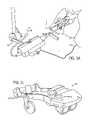
- FIG. 3Cillustrates in detail a probe holder/stepper assembly shown partly in FIG. 3A ;
- FIG. 4Aillustrates a front schematic view of a brachytherapy phantom
- FIG. 4Ba side schematic view of the brachytherapy phantom
- FIG. 5Aillustrates reconstruction of standard orthogonal image planes from a 3D image stack and FIG. 5B the reconstruction of oblique image planes from a 3D image stack;
- FIG. 6illustrates the viewing geometry for a 3D translucent reconstruction of an image
- FIG. 7Aillustrates translucent images of a human prostate for four different viewing angles and FIG. 7B illustrates translucent images of a phantom organ for six different viewing angles;
- FIG. 8illustrates a time sequenced image of the prostate organ in FIG. 7A showing approach of a catheter containing a radioactive seed, deposition of the seed and withdrawal of the catheter leaving the seed;
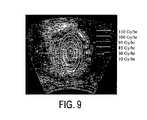
- FIG. 9illustrates isodose distributions of radiation from a single radioactive seed
- FIG. 10illustrates a flow chart of software routine for processing imaging data for visualization
- FIG. 11illustrates a virtual reality head mounted display.
- FIG. 12depicts another illustration of isodose distributions of radiation from a single radioactive seed.
- FIGS. 13( a ) and 13 ( b )depict the ability to view optimized seeds and needles in the same volume as the real time ultrasound data.
- a system 10 constructed in accordance with an example of the inventionis illustrated generally in FIG. 1 .
- a three-dimensional (3D) probe 12accumulates image data from a treatment region or organ of a patient, image data is processed using a 3D imaging card 14 .
- the probe 12preferably is an ultrasound device but can be any other rapid imaging technology, such as rapid CT or MR.
- a conventional personal computer 16 having a monitorcan be used to operate on the image data from the imaging card 14 using conventional software and hardware tools to be described in more detail hereinafter.
- Radioactive seeds 18are provided for insertion using any one of a variety of conventional means for inserting devices or articles into the human body, such as insertion devices 19 , which may be either needles or stiff catheters.
- the 3D ultrasound probe 12therefore, provides an image signal to the computer 16 and a virtual realty interface card 13 coupled to the imaging card 14 which enables a user to visualize a translucent image of the patient organ and real time interaction of any one of a variety of treatment devices, such as the implant needles 19 or a Foley catheter 20 , and one of the seeds 18 within the organ.
- Computer softwarecan be utilized in a conventional manner to visualize the 3D imaging data in various formats.
- the formatsinclude orthogonal two dimensional (2D) images, oblique 2D images, and translucent 3D rendering. All of these reconstructions can be directly displayed on the computer monitor; and 3D translucent, stereoscopic, rendering is also available in the VR (Virtual Realty) mode.
- the preferred ultrasound probe 12is a conventional Kretz ultrasound imaging system manufactured by Kretz Corporation, now available as Medison Combison 530 through Medison America Corporation, Pleasantown, Calif. This system and other such conventional systems are readily available and can provide real time ultrasound image data.
- the Medison Combison ultrasound systemincorporates an endorectal probe which acquires multiple image planes in real time and the software of the present invention reconstructs the translucent 3D volume.
- Alternate systemsinclude bi-plane 2D imaging systems with the probe mounted in a stepper motor driven holder for rapid automatic acauisition of multiple image planes. There is nothing that is application specific about the imaging system, thus any commercially available system will suffice.
- the diagnostic transrectal ultrasound probe 12(see FIG. 2 ) is inserted into the patient's rectum to obtain real time volumetric images of the prostate for use during the implant procedure.
- the diagnostic probe 12is preferably a phased array probe designed so that the array of transducers can rotate about the axis of the array sweeping out a 3D imaging volume. As the probe 12 rotates, images are captured and digitized by use of the imaging card 14 (see FIG. 1 ), so as to create a fixed number of images slices per rotation.
- An alternative methodutilizes a transverse oriented phased array form of the endorectal probe 12 which is moved longitudinally in an automated rapid sequence so as to create a series of transverse image slices automatically.
- Another embodiment of the probe 12can incorporate multiple transverse phased arrays (shown in phantom in FIG. 1B ) arranged parallel to each other orthogonal to the axis of an endorectal probe to produce multiple simultaneous image slices (see, for example, FIGS. 5A and 5B ).
- the 3D image datawill be represented as a three dimensional image raster.
- the ultrasound probe 12can be mounted into a probe holder 30 (see FIGS. 3A and 3C ) with FIG. 3B illustrating one example of an ultrasound image from an anatomical prostate phantom employed to carry out testing and planning.
- the probe holder 30includes a digital encoder 42 for providing information regarding the position of all of the desired ultrasound image planes in the prostate relative to each other. The image plane location will be automatically sent to the system computer and “tagged” to the acquired ultrasound image for that position. Thus, it will be possible to reproduce the longitudinal and lateral positions of the implant catheters for the ultrasound therapy applicators and for the temperature probes.
- each of the processing cardsis configured specifically for 3D.
- Several commercial cardscan be equipped with this amount of video RAM (VRAM), but the way the card's hardware interacts with the computer's video and software drivers does not utilize this data in 3D.
- the processing and memory architecturepreferably is designed to allow for simultaneous image acquisition and processing.
- the digitizing cardshould also preferably have standard imaging tools, such as real time window and leveling, zoom and pan of the ultrasound images.
- Some existing cardse.g., Matrox; Coreco) do provide standard imaging tools.
- the 3D image data arising from the ultrasound probe 12is preferably buffered on the imaging card 14 .
- the 3D imageis preferably represented as a series of 2D images. This is referred to as the image stack or 3D image raster.
- the 3D image rasteris represented in memory as a linear array of bytes of length N ⁇ M ⁇ P where N is the width of the 2D image in pixels, M is the height a 2D image in pixels, and P is the number of 2D images in the image stack.
- the usercan include defined formats. Entire 3D image stacks at specific times during the intraoperative session can be stored in the DICOM standard. The user will have the ability to select a 3D image volume for archiving as part of the system software. These image stacks can then be reviewed in any of the various visualization modes (standard orthogonal 2D views, oblique 2D views, or 3D translucent views) as described above. In addition, the user will have the ability to store any of the 2D views available at any time during the intraoperative session.
- the computational platformcan, for example, be any form of computing means, such as the personal computer 16 , which incorporates a PCI bus architecture.
- PCI busis preferable over the ISA or EISA bus because the PCI bus is much faster.
- a 200 Mhz (or greater speed) Pentium/Pentium-Pro computer supplied with 128 Mbytes of RAM and a 6.0 Gbyte hard diskshould be sufficient RAM and disk memory to run the software in a real-time fashion and to archive all patient data.
- a high resolution monitorcapable of displaying at least 1280 ⁇ 1024 ⁇ 64 bit resolutions is preferably used.
- the ultrasound images obtained from the ultrasound imaging system of the ultrasound probe 12can be of good diagnostic quality.
- this input image datainto a 3D representation, whether in the 3D perspective mode or the real time VR mode, the resultant volumes can, however, be noisy and hinder diagnostic and spatial accuracy.
- a number of conventional hardware and software filterscan be used which will filter the incoming image data stored on the imaging card 14 . Routines such as image pixel averaging, smoothing, and interpolation can improve the 3D rendering of the imaging volume. These sets of filters or routines are to be distinguished from the set of standard imaging tools running on the host CPU which are available within a conventional imaging software package.
- three of the perspective viewsare the standard transverse, coronal and sagittal 2D views. These three orthogonal views are taken from a user specified location within the imaging space. For example, the user can request that the three orthogonal views have their common centers at a spatial position of (5.0 cm, 15.0, 25.0 cm) relative to the origin of the template system. One also can select the reference point of either of the three orthogonal views independently, that is the three views do not have to have common center points.
- FIGS. 5A and 5Bshow examples of several example 2D views from a 3D ultrasound image volume.
- FIG. 6shows a number of possible viewing directions
- FIG. 7gives further examples of translucent 3D viewing from different angles.
- the 3D ultrasound image volumewas obtained from actual ultrasound images of a human prostate and of a prostate implant phantom.
- contourscan be given a unique name by the user, and then drawn by the user using the mouse of the computer 16 . All attributes of the contours such as name and color can, based on conventional imaging software, be user selectable.
- the usercan also edit the contours by selecting functions, such as adding a point to a contour, deleting a point from a contour or deleting the entire contour.
- the userhas the option to render them in 3D or view in conventional 2D mode on the 3D perspective mode or viewed in the VR mode.
- contour 3D attributessuch as color, lighting, and shading are user controlled.
- the contoursby default appear on the 2D images, however, the user can control the individual contour's 2D and 3D visibility.
- the 3D image rastercan be rendered as a real time, transparent, 3D volume.
- This transparent volumecan be viewed and displayed on the monitor of the computer 16 at any arbitrary viewing angle and is calculated using conventional 3D object reconstruction algorithms.
- 3D object reconstruction algorithmscan render a large imaging volume in fractions of a second, even on present day computing platforms.
- the transparent nature of the reconstructionthus allows the user to “see” inside any objects which appear in the imaging volume. For example, if the prostate is imaged in the imaging volume, then it will be reconstructed as a transparent volume, in which other anatomical landmarks such as the urethra, tissue abnormalities or calcifications can be seen.
- any other objectssuch as needles or catheters are inserted into the prostate, and if they are visible in the ultrasound images, they will be seen as they enter the prostate (see FIG. 8 showing introduction of the seed 18 with the catheter/needle 19 ). Since the volumes are rendered as transparent solids, the needles 19 (and other articles) can thus easily be seen as they move inside the prostate volume as well. Since the ultrasound images are obtained in real time, the 3D perspective reconstruction is also rendered in real time.
- the preferred algorithm for the perspective 3D reconstructionis the known Bresenham raytrace algorithm.
- the patientundergoes an initial volumetric ultrasound scan using the probe 12 . This scan is done before the radiation therapy planning or the actual implant.
- the ideal positions of the radioactive seeds 18(see FIG. 1 ) within the prostate are determined. This ideal seed distribution is optimized to deliver a dose distribution within the prostate that will deliver all the radiation dose to the target volume only, while sparing the surrounding healthy tissues such as the rectum and bladder.
- the optimal positions of the seeds 18 and the optimal position of the needles 19are recorded for later use in the operating room when the needles 19 are loaded into the patient.
- the seeds 18are then loaded into the needles 19 , and the physician then attempts to place the needles 19 inside the prostate according to the treatment dose plan positions (again, see example in FIG. 8 ).
- the dose as a function of position for a cylindrical .sup. 125I seed of a given activitycan be determined from a lookup table or calculated from a conventional analytic formula.
- the dose fieldcan be visualized as a set of isodose lines in 2D or isodose surface in 3D (see, for example, FIG. 9 and FIG. 12 ).
- the dose computation routineis based upon the TG43 standard adopted by the AAPM (American Association of Physicists in Medicine) entitled “Dosimetry of Interstitial Brachytherapy Sources”: Recommendations of the AAPM Radiation Therapy Committee Task Group No. 43 which specifies the dose model and the data used in the dose calculation.
- This particular implementationruns extremely fast on a conventional 233 MHz PC, computing the dose for a single seed in less than 0.5 seconds.
- the total 3D dose distribution within the prostate for a 100 seed implantrequires only 50 seconds, or less than one minute total computation time. Thus, this can be done “on line” in the operating room.
- the userhas the ability to view the optimized seeds 18 and the needles 19 in the same volume as the real time ultrasound data (see, for example. FIG. 13( a )).
- Thisallows the physician to see exactly where the needles 19 should go and hence make adjustments to position the needles 19 optimally.
- the pre-planned, optimal positioned needles 19 and the seeds 18can be rendered again as a transparent solid, the color of which is user selectable.
- the real needles 19are inserted into the prostate, their positions relative to the ideal needle placements based on the dose plan can be monitored in real time (see, for example. FIG. 13( b )).
- FIGS. 5A and 5Bdisplays perspective 3D views and the three orthogonal reconstructions of the image data along with the pre-planned catheter positions.
- the pre-planned needles 19can also be viewed in the VR mode as virtual objects overlaid onto the imaging volume.
- FIG. 10A flowchart description of the translucent volume visualization methodology is shown in FIG. 10 .
- the input image volumeis described by the vectors i, j, k of appropriate magnitude for the volume.
- the viewing angle parametersare the angles ⁇ , ⁇ described on FIG. 6 and FIG. 10 .
- the rotation matrix, Ris calculated using the formulae given in the flowchart of FIG. 10 .
- the entire imaging volumeis calculated by multiplying the rotation matrices in the x, y, z directions by the respective vectors i, j and k describing the incremental portions along the x, y, z directions.
- the multiplying vectoris (i–i o , j–j o , k–k o ) where i o , j o , k o are the starting points along x, y and z axes and the volume is determined by summing the component contributions shown in FIG. 10 .
- the 3D translucent imageis then created by computing the translucent 2D image over the entire image volume and summing the z-pixels.
- a virtual reality interface systemcan be composed of a conventional head mounted display (HMD) 50 shown in FIG. 11 and a 6D (x,y,z, roll, pitch, yaw) tracking system.
- the HMD 50consists of two color monitors which mount to a head set in the position directly in front of the eyes.
- the HMD 50is based on the principal that whatever is displayed on each monitor is directly incident on the retina for each eye, and hence true 3D images can be created by rendering objects as 3D perspective images for each eye. Given the distance between the eyes (the interoccular distance which is approximately 80 mm) and the distance and spherical angles of the distance of the center line between the eyes from the coordinate origin, the 2D images which appear in each of the two monitors can be determined exactly as described above.
- the transparent, 3D reconstruction of the real time imaging datawill preferably be reconstructed.
- the physicianwill see the 3D ultrasound volume mapped inside the patient's pelvis, spatially correlated to the position of the patient's real prostate (or other organ) and anatomy.
- the physiciancan “see” inside the patient to the extent of what is visible in the ultrasound imaging volume. Since the ultrasound probe 12 is locked down to the template, which is then secured to the floor, the exact positions of all voxels in the ultrasound imaging volume are known exactly relative to the template, and hence relative to the room.
- the needles 19As the needles 19 are inserted into the patient, they will appear in the image volume and hence are reconstructed in the VR reconstruction. All of this occurs in real time so that the physician also can see the needles 19 enter the prostate in real time. As mentioned above, if the pre-planned, optimized needles 19 are displayed, the physician can then see the position of the actual needles 19 as they are being inserted relative to the optimal placement. Hence, the physician has the ability to adjust the needles 19 to correspond to their optimal positions. In addition, since the needles 19 are automatically extracted, the computer software has the ability to calculate and render the 3D dose distribution in real time as the needles 19 are being inserted.
- a currently available, a fast and inexpensive HMDis made by Virtual-IO Corporation (Mountain View, Calif.).
- the HMDis full color with two 0.70 LCD displays with a resolution of 180,000 pixels per LCD panel.
- the video inputis NTSC with field sequential format.
- the LCD panelsare semitransparent, allowing the real outside world to be included in the virtual reconstruction.
- the field of viewis 30° for each eye.
- a six degree of freedom (6 DOF) tracking systemcan also be attached to the HMD.
- the 6 DOF tracking systemallows for the determination of the spatial position of the user's head and the yaw, pitch, and roll of the head.
- the conventional head setweighs only 8 ounces and comes with stereo sound. Stereo sound is an extremely valuable technology in the operating room. With this capability, the physician has the ability to monitor the patient's heart rate and respiration rate while performing the implant. Hence any fluctuation in the patient's vital signs can be instantly accessed and acted thereon if necessary.
- the radioactive seeds 18are made of high density material such as stainless steel, and hence have a very bright response in the ultrasound images. Therefore, automatic seed detection in the ultrasound images can readily be accomplished, for example, by a simple thresholding algorithm along with the requirement that the resultant objects which are removed by threshold have a certain maximum size determined by the actual size of the seeds.
- Near-real-time visualizationwill provide immediate feedback to the physician during the implant process itself.
- the nearly real time visualizationis of great importance to the effective use of a translucent overlay of the ideal seed pre-plan (from the therapy planning process) in the three-dimensional volume.
- the physiciancan “see” in nearly real time the relationship of the needles and seeds being implanted to the ideal pre-plan locations and quickly accommodate redirection required prior to leaving the radiation seeds.
- the need for this in three-dimensional representationis very important to overcome the greatest fundamental limitation in brachytherapy, which is knowing at the same time both the lateral placement and longitudinal placement of needles and seeds relative to the target volume and pre-plan.
- This real time 3D visualizationalso would speed the implant process in the case of brachytherapy as well as make it more accurate. It would also speed other minimally invasive surgical procedures and localized tissue ablation procedures (for example, cryosurgery or localized selected ablation of diseased liver tissue or local removal of breast tissue). These procedures could be accomplished with real time visualization inside the tissue being treated with greater accuracy in shorter time. This aspect would reduce operating room time and costs to the patient and health care system.
Landscapes
- Health & Medical Sciences (AREA)
- Life Sciences & Earth Sciences (AREA)
- Engineering & Computer Science (AREA)
- Biomedical Technology (AREA)
- Veterinary Medicine (AREA)
- Nuclear Medicine, Radiotherapy & Molecular Imaging (AREA)
- Public Health (AREA)
- General Health & Medical Sciences (AREA)
- Animal Behavior & Ethology (AREA)
- Surgery (AREA)
- Pathology (AREA)
- Heart & Thoracic Surgery (AREA)
- Medical Informatics (AREA)
- Radiology & Medical Imaging (AREA)
- Molecular Biology (AREA)
- Physics & Mathematics (AREA)
- Biophysics (AREA)
- Robotics (AREA)
- Oral & Maxillofacial Surgery (AREA)
- Computer Vision & Pattern Recognition (AREA)
- Dermatology (AREA)
- Anesthesiology (AREA)
- Hematology (AREA)
- Otolaryngology (AREA)
- Electromagnetism (AREA)
- Gynecology & Obstetrics (AREA)
- High Energy & Nuclear Physics (AREA)
- Ultra Sonic Daignosis Equipment (AREA)
- Radiation-Therapy Devices (AREA)
- Apparatus For Radiation Diagnosis (AREA)
Abstract
Description
Claims (45)
Priority Applications (2)
| Application Number | Priority Date | Filing Date | Title |
|---|---|---|---|
| US09/897,326US7171255B2 (en) | 1995-07-26 | 2001-07-02 | Virtual reality 3D visualization for surgical procedures |
| US10/310,565US20030135102A1 (en) | 2000-05-18 | 2002-12-05 | Method and system for registration and guidance of intravascular treatment |
Applications Claiming Priority (3)
| Application Number | Priority Date | Filing Date | Title |
|---|---|---|---|
| US08/507,199US5810007A (en) | 1995-07-26 | 1995-07-26 | Ultrasound localization and image fusion for the treatment of prostate cancer |
| US08/977,362US6256529B1 (en) | 1995-07-26 | 1997-11-24 | Virtual reality 3D visualization for surgical procedures |
| US09/897,326US7171255B2 (en) | 1995-07-26 | 2001-07-02 | Virtual reality 3D visualization for surgical procedures |
Related Parent Applications (1)
| Application Number | Title | Priority Date | Filing Date |
|---|---|---|---|
| US08/977,362ContinuationUS6256529B1 (en) | 1995-07-26 | 1997-11-24 | Virtual reality 3D visualization for surgical procedures |
Related Child Applications (1)
| Application Number | Title | Priority Date | Filing Date |
|---|---|---|---|
| US10/310,565Continuation-In-PartUS20030135102A1 (en) | 2000-05-18 | 2002-12-05 | Method and system for registration and guidance of intravascular treatment |
Publications (2)
| Publication Number | Publication Date |
|---|---|
| US20010041838A1 US20010041838A1 (en) | 2001-11-15 |
| US7171255B2true US7171255B2 (en) | 2007-01-30 |
Family
ID=37022875
Family Applications (2)
| Application Number | Title | Priority Date | Filing Date |
|---|---|---|---|
| US08/977,362Expired - LifetimeUS6256529B1 (en) | 1995-07-26 | 1997-11-24 | Virtual reality 3D visualization for surgical procedures |
| US09/897,326Expired - Fee RelatedUS7171255B2 (en) | 1995-07-26 | 2001-07-02 | Virtual reality 3D visualization for surgical procedures |
Family Applications Before (1)
| Application Number | Title | Priority Date | Filing Date |
|---|---|---|---|
| US08/977,362Expired - LifetimeUS6256529B1 (en) | 1995-07-26 | 1997-11-24 | Virtual reality 3D visualization for surgical procedures |
Country Status (5)
| Country | Link |
|---|---|
| US (2) | US6256529B1 (en) |
| EP (1) | EP1033934A4 (en) |
| AU (1) | AU1596999A (en) |
| CA (1) | CA2311319C (en) |
| WO (1) | WO1999026534A1 (en) |
Cited By (108)
| Publication number | Priority date | Publication date | Assignee | Title |
|---|---|---|---|---|
| US20040114718A1 (en)* | 2002-11-28 | 2004-06-17 | Elekta Ab | Radiotherapy apparatus and operating method |
| US20040143181A1 (en)* | 1999-05-26 | 2004-07-22 | Damasco Sanford D. | Computer guided ablation of tissue using integrated ablative/temperature sensing devices |
| US20050058326A1 (en)* | 2003-07-25 | 2005-03-17 | Karl Barth | System and method for the creation of a virtual observation and access channel in medical 3D images |
| US20050203413A1 (en)* | 2003-08-07 | 2005-09-15 | Gabor Fichtinger | Transcavital needle insertion device |
| US20050234326A1 (en)* | 2004-04-01 | 2005-10-20 | Olympus Corporation | Medical procedure support system and method |
| US20060020204A1 (en)* | 2004-07-01 | 2006-01-26 | Bracco Imaging, S.P.A. | System and method for three-dimensional space management and visualization of ultrasound data ("SonoDEX") |
| US20060074309A1 (en)* | 2002-11-06 | 2006-04-06 | Odile Bonnefous | Phased array acoustic system for 3d imaging of moving parts |
| US20070009564A1 (en)* | 2005-06-22 | 2007-01-11 | Mcclain James B | Drug/polymer composite materials and methods of making the same |
| US20070293787A1 (en)* | 2003-08-13 | 2007-12-20 | Taylor James D | Targeted biopsy delivery system |
| US20080058836A1 (en)* | 2006-08-03 | 2008-03-06 | Hansen Medical, Inc. | Systems and methods for performing minimally invasive procedures |
| US20080095919A1 (en)* | 2006-10-23 | 2008-04-24 | Mcclain James B | Holder For Electrically Charging A Substrate During Coating |
| US20090043209A1 (en)* | 2007-07-27 | 2009-02-12 | Makoto Hirama | Ultrasonic diagnostic apparatus |
| US20090099544A1 (en)* | 2007-10-12 | 2009-04-16 | Gynesonics, Inc. | Methods and systems for controlled deployment of needles in tissue |
| US20090186069A1 (en)* | 2006-04-26 | 2009-07-23 | Micell Technologies, Inc. | Coatings Containing Multiple Drugs |
| US20090292351A1 (en)* | 2008-04-17 | 2009-11-26 | Micell Technologies, Inc. | Stents having bioabsorbable layers |
| WO2010009335A1 (en) | 2008-07-17 | 2010-01-21 | Micell Technologies, Inc. | Drug delivery medical device |
| KR100942159B1 (en) | 2007-12-24 | 2010-02-12 | 가톨릭대학교 산학협력단 | Seed Phantom for Positional Accuracy Assessment of Radiation Seeds for Prostate Cancer Brachytherapy |
| US20100063580A1 (en)* | 2007-01-08 | 2010-03-11 | Mcclain James B | Stents having biodegradable layers |
| US20100211164A1 (en)* | 2007-04-17 | 2010-08-19 | Mcclain James B | Stents having biodegradable layers |
| US20100241220A1 (en)* | 2009-03-23 | 2010-09-23 | Mcclain James B | Peripheral Stents Having Layers |
| WO2010111232A2 (en) | 2009-03-23 | 2010-09-30 | Micell Technologies, Inc. | Drug delivery medical device |
| US20100256748A1 (en)* | 2009-04-01 | 2010-10-07 | Micell Technologies, Inc. | Coated stents |
| US20100272778A1 (en)* | 2007-04-17 | 2010-10-28 | Micell Technologies, Inc. | Stents having controlled elution |
| US20100298928A1 (en)* | 2007-10-19 | 2010-11-25 | Micell Technologies, Inc. | Drug Coated Stents |
| US20110040139A1 (en)* | 2007-03-15 | 2011-02-17 | Senorx, Inc. | Soft body catheter with low friction lumen |
| US20110046483A1 (en)* | 2008-01-24 | 2011-02-24 | Henry Fuchs | Methods, systems, and computer readable media for image guided ablation |
| US20110159069A1 (en)* | 2008-12-26 | 2011-06-30 | Shaw Wendy J | Medical Implants and Methods of Making Medical Implants |
| EP2341862A1 (en)* | 2008-09-16 | 2011-07-13 | Osyris Medical | Apparatus and method for treating a portion of a human or animal body using a means for delivering treatment doses and a dosimetry control means |
| US20110190864A1 (en)* | 2010-02-02 | 2011-08-04 | Micell Technologies, Inc. | Stent and stent delivery system with improved deliverability |
| US20110238161A1 (en)* | 2010-03-26 | 2011-09-29 | Battelle Memorial Institute | System and method for enhanced electrostatic deposition and surface coatings |
| US8057379B2 (en) | 2005-11-18 | 2011-11-15 | Senorx, Inc. | Treatment of a body cavity |
| US20110286653A1 (en)* | 2010-05-21 | 2011-11-24 | Gorges Sebastien | Method for processing radiological images to determine a 3d position of a needle |
| US20110288361A1 (en)* | 2002-06-17 | 2011-11-24 | Nucletron B.V. | Real time radiation treatment planning system |
| US8075469B2 (en) | 2005-11-18 | 2011-12-13 | Senorx, Inc. | Methods for asymmetrical irradiation of a body cavity |
| WO2012009684A2 (en) | 2010-07-16 | 2012-01-19 | Micell Technologies, Inc. | Drug delivery medical device |
| WO2012092504A2 (en) | 2010-12-30 | 2012-07-05 | Micell Technologies, Inc. | Nanoparticle and surface-modified particulate coatings, coated balloons, and methods therefore |
| US8273006B2 (en) | 2005-11-18 | 2012-09-25 | Senorx, Inc. | Tissue irradiation |
| US8277370B2 (en) | 2007-03-12 | 2012-10-02 | Senorx, Inc. | Radiation catheter with multilayered balloon |
| US8292794B2 (en) | 2002-11-06 | 2012-10-23 | Senorx, Inc. | Method for maintaining access to a biopsy site |
| US20120278711A1 (en)* | 2003-09-16 | 2012-11-01 | Labtest International, Inc. D/B/A Intertek Consumer Goods North America | Haptic response system and method of use |
| US20120294497A1 (en)* | 2011-05-20 | 2012-11-22 | Varian Medical Systems, Inc. | Method and Apparatus Pertaining to Images Used for Radiation-Treatment Planning |
| US8360950B2 (en) | 2008-01-24 | 2013-01-29 | Senorx, Inc. | Multilumen brachytherapy balloon catheter |
| US8398535B2 (en) | 2002-11-06 | 2013-03-19 | Senorx, Inc. | Catheter assembly for delivering a radiation source into a body cavity |
| WO2013059509A1 (en) | 2011-10-18 | 2013-04-25 | Micell Technologies, Inc. | Drug delivery medical device |
| US20140016759A1 (en)* | 2012-07-13 | 2014-01-16 | The Chinese University Of Hong Kong | Compound 6d-offset simulating phantom and quality assurance program for precision image-guided radiotherapy and radiosurgery |
| US8750568B2 (en) | 2012-05-22 | 2014-06-10 | Covidien Lp | System and method for conformal ablation planning |
| US8758256B2 (en) | 2010-07-12 | 2014-06-24 | Best Medical International, Inc. | Apparatus for brachytherapy that uses a scanning probe for treatment of malignant tissue |
| US8758429B2 (en) | 2005-07-15 | 2014-06-24 | Micell Technologies, Inc. | Polymer coatings containing drug powder of controlled morphology |
| US8880151B1 (en) | 2013-11-27 | 2014-11-04 | Clear Guide Medical, Llc | Surgical needle for a surgical system with optical recognition |
| US8900651B2 (en) | 2007-05-25 | 2014-12-02 | Micell Technologies, Inc. | Polymer films for medical device coating |
| US9044216B2 (en) | 2010-07-12 | 2015-06-02 | Best Medical International, Inc. | Biopsy needle assembly |
| US9107698B2 (en) | 2010-04-12 | 2015-08-18 | Inneroptic Technology, Inc. | Image annotation in image-guided medical procedures |
| US9248311B2 (en) | 2009-02-11 | 2016-02-02 | Hologic, Inc. | System and method for modifying a flexibility of a brachythereapy catheter |
| US9364294B2 (en) | 2009-02-17 | 2016-06-14 | Inneroptic Technology, Inc. | Systems, methods, apparatuses, and computer-readable media for image management in image-guided medical procedures |
| US9398936B2 (en) | 2009-02-17 | 2016-07-26 | Inneroptic Technology, Inc. | Systems, methods, apparatuses, and computer-readable media for image guided surgery |
| US9402601B1 (en)* | 1999-06-22 | 2016-08-02 | Teratech Corporation | Methods for controlling an ultrasound imaging procedure and providing ultrasound images to an external non-ultrasound application via a network |
| US9439623B2 (en) | 2012-05-22 | 2016-09-13 | Covidien Lp | Surgical planning system and navigation system |
| US9439627B2 (en) | 2012-05-22 | 2016-09-13 | Covidien Lp | Planning system and navigation system for an ablation procedure |
| US9439622B2 (en) | 2012-05-22 | 2016-09-13 | Covidien Lp | Surgical navigation system |
| US9498182B2 (en) | 2012-05-22 | 2016-11-22 | Covidien Lp | Systems and methods for planning and navigation |
| US9510856B2 (en) | 2008-07-17 | 2016-12-06 | Micell Technologies, Inc. | Drug delivery medical device |
| US9579524B2 (en) | 2009-02-11 | 2017-02-28 | Hologic, Inc. | Flexible multi-lumen brachytherapy device |
| US9623260B2 (en) | 2004-11-05 | 2017-04-18 | Theragenics Corporation | Expandable brachytherapy device |
| US9622720B2 (en) | 2013-11-27 | 2017-04-18 | Clear Guide Medical, Inc. | Ultrasound system with stereo image guidance or tracking |
| US9659345B2 (en) | 2006-08-02 | 2017-05-23 | Inneroptic Technology, Inc. | System and method of providing real-time dynamic imagery of a medical procedure site using multiple modalities |
| US9675319B1 (en) | 2016-02-17 | 2017-06-13 | Inneroptic Technology, Inc. | Loupe display |
| WO2017214172A1 (en)* | 2016-06-06 | 2017-12-14 | Edda Technology, Inc. | Method and system for interactive laparoscopic ultrasound guided ablation planning and surgical procedure simulation |
| US9901406B2 (en) | 2014-10-02 | 2018-02-27 | Inneroptic Technology, Inc. | Affected region display associated with a medical device |
| US9950194B2 (en) | 2014-09-09 | 2018-04-24 | Mevion Medical Systems, Inc. | Patient positioning system |
| US9949700B2 (en) | 2015-07-22 | 2018-04-24 | Inneroptic Technology, Inc. | Medical device approaches |
| US10022557B2 (en) | 2010-09-30 | 2018-07-17 | Hologic, Inc. | Using a guided member to facilitate brachytherapy device swap |
| US10117972B2 (en) | 2011-07-15 | 2018-11-06 | Micell Technologies, Inc. | Drug delivery medical device |
| US10188772B2 (en) | 2011-10-18 | 2019-01-29 | Micell Technologies, Inc. | Drug delivery medical device |
| US10188467B2 (en) | 2014-12-12 | 2019-01-29 | Inneroptic Technology, Inc. | Surgical guidance intersection display |
| US10207126B2 (en) | 2009-05-11 | 2019-02-19 | Cytyc Corporation | Lumen visualization and identification system for multi-lumen balloon catheter |
| US10232092B2 (en) | 2010-04-22 | 2019-03-19 | Micell Technologies, Inc. | Stents and other devices having extracellular matrix coating |
| US10272606B2 (en) | 2013-05-15 | 2019-04-30 | Micell Technologies, Inc. | Bioabsorbable biomedical implants |
| US10278778B2 (en) | 2016-10-27 | 2019-05-07 | Inneroptic Technology, Inc. | Medical device navigation using a virtual 3D space |
| US10314559B2 (en) | 2013-03-14 | 2019-06-11 | Inneroptic Technology, Inc. | Medical device guidance |
| US10342992B2 (en) | 2011-01-06 | 2019-07-09 | Hologic, Inc. | Orienting a brachytherapy applicator |
| US10464100B2 (en) | 2011-05-31 | 2019-11-05 | Micell Technologies, Inc. | System and process for formation of a time-released, drug-eluting transferable coating |
| US10643360B2 (en) | 2017-02-10 | 2020-05-05 | Arizona Board Of Regents On Behalf Of Arizona State University | Real-time medical image visualization systems and related methods |
| US10667855B1 (en) | 2019-05-10 | 2020-06-02 | Trod Medical Us, Llc | Dual coil ablation devices |
| US10737075B2 (en) | 2016-02-08 | 2020-08-11 | Orbusneich Medical Pte. Ltd. | Drug eluting balloon |
| US10835396B2 (en) | 2005-07-15 | 2020-11-17 | Micell Technologies, Inc. | Stent with polymer coating containing amorphous rapamycin |
| US10939977B2 (en) | 2018-11-26 | 2021-03-09 | Augmedics Ltd. | Positioning marker |
| US10993770B2 (en) | 2016-11-11 | 2021-05-04 | Gynesonics, Inc. | Controlled treatment of tissue and dynamic interaction with, and comparison of, tissue and/or treatment data |
| US11039943B2 (en) | 2013-03-12 | 2021-06-22 | Micell Technologies, Inc. | Bioabsorbable biomedical implants |
| EP3868434A1 (en) | 2016-02-08 | 2021-08-25 | Orbusneich Medical Pte. Ltd | Drug eluting balloon |
| US11259879B2 (en) | 2017-08-01 | 2022-03-01 | Inneroptic Technology, Inc. | Selective transparency to assist medical device navigation |
| US11389252B2 (en) | 2020-06-15 | 2022-07-19 | Augmedics Ltd. | Rotating marker for image guided surgery |
| US11426494B2 (en) | 2007-01-08 | 2022-08-30 | MT Acquisition Holdings LLC | Stents having biodegradable layers |
| US11464578B2 (en) | 2009-02-17 | 2022-10-11 | Inneroptic Technology, Inc. | Systems, methods, apparatuses, and computer-readable media for image management in image-guided medical procedures |
| US11484365B2 (en) | 2018-01-23 | 2022-11-01 | Inneroptic Technology, Inc. | Medical image guidance |
| US11547382B2 (en) | 1999-06-22 | 2023-01-10 | Teratech Corporation | Networked ultrasound system and method for imaging a medical procedure using an invasive probe |
| US11707329B2 (en) | 2018-08-10 | 2023-07-25 | Covidien Lp | Systems and methods for ablation visualization |
| US11750794B2 (en) | 2015-03-24 | 2023-09-05 | Augmedics Ltd. | Combining video-based and optic-based augmented reality in a near eye display |
| US11766296B2 (en) | 2018-11-26 | 2023-09-26 | Augmedics Ltd. | Tracking system for image-guided surgery |
| US11801115B2 (en) | 2019-12-22 | 2023-10-31 | Augmedics Ltd. | Mirroring in image guided surgery |
| US11896445B2 (en) | 2021-07-07 | 2024-02-13 | Augmedics Ltd. | Iliac pin and adapter |
| US11974887B2 (en) | 2018-05-02 | 2024-05-07 | Augmedics Ltd. | Registration marker for an augmented reality system |
| US11980506B2 (en) | 2019-07-29 | 2024-05-14 | Augmedics Ltd. | Fiducial marker |
| US12044856B2 (en) | 2022-09-13 | 2024-07-23 | Augmedics Ltd. | Configurable augmented reality eyewear for image-guided medical intervention |
| US12150821B2 (en) | 2021-07-29 | 2024-11-26 | Augmedics Ltd. | Rotating marker and adapter for image-guided surgery |
| US12178666B2 (en) | 2019-07-29 | 2024-12-31 | Augmedics Ltd. | Fiducial marker |
| US12239385B2 (en) | 2020-09-09 | 2025-03-04 | Augmedics Ltd. | Universal tool adapter |
| US12354227B2 (en) | 2022-04-21 | 2025-07-08 | Augmedics Ltd. | Systems for medical image visualization |
| US12417595B2 (en) | 2021-08-18 | 2025-09-16 | Augmedics Ltd. | Augmented-reality surgical system using depth sensing |
Families Citing this family (147)
| Publication number | Priority date | Publication date | Assignee | Title |
|---|---|---|---|---|
| US6256529B1 (en)* | 1995-07-26 | 2001-07-03 | Burdette Medical Systems, Inc. | Virtual reality 3D visualization for surgical procedures |
| US20030135115A1 (en)* | 1997-11-24 | 2003-07-17 | Burdette Everette C. | Method and apparatus for spatial registration and mapping of a biopsy needle during a tissue biopsy |
| US6129670A (en)* | 1997-11-24 | 2000-10-10 | Burdette Medical Systems | Real time brachytherapy spatial registration and visualization system |
| EP1079730B1 (en)* | 1997-11-24 | 2007-01-03 | Computerized Medical Systems, Inc. | Real time brachytherapy spatial registration and visualization system |
| ES2304794T3 (en) | 1998-06-22 | 2008-10-16 | Ao Technology Ag | PAREO OF LOCATION THROUGH LOCALIZATION SCREWS. |
| US6947584B1 (en)* | 1998-08-25 | 2005-09-20 | General Electric Company | Volume imaging system |
| AU5882599A (en)* | 1998-09-24 | 2000-04-10 | Super Dimension Ltd. | System and method for determining the location of a catheter during an intra-body medical procedure |
| US6689043B1 (en)* | 1998-11-06 | 2004-02-10 | Amersham Plc | Products and methods for brachytherapy |
| IL142166A0 (en)* | 1998-11-06 | 2002-03-10 | Nycomed Amersham Plc | Products and methods for brachytherapy |
| CA2367271C (en) | 1999-03-17 | 2008-12-16 | Synthes (U.S.A.) | System and method for ligament graft placement |
| US8944070B2 (en) | 1999-04-07 | 2015-02-03 | Intuitive Surgical Operations, Inc. | Non-force reflecting method for providing tool force information to a user of a telesurgical system |
| AU766981B2 (en) | 1999-04-20 | 2003-10-30 | Ao Technology Ag | Device for the percutaneous obtainment of 3D-coordinates on the surface of a human or animal organ |
| AU768975B2 (en) | 1999-05-03 | 2004-01-15 | Ao Technology Ag | Position detector with auxiliary means for detecting the direction of the gravity vector |
| US6694170B1 (en)* | 1999-05-26 | 2004-02-17 | Endocare, Inc. | Computer guided surgery for prostatic nerve sparing |
| US6464642B1 (en)* | 1999-08-20 | 2002-10-15 | Kabushiki Kaisha Toshiba | Ultrasonic diagnosis apparatus |
| US6610013B1 (en) | 1999-10-01 | 2003-08-26 | Life Imaging Systems, Inc. | 3D ultrasound-guided intraoperative prostate brachytherapy |
| EP1095628A3 (en)* | 1999-10-29 | 2001-05-16 | Marconi Medical Systems, Inc. | Planning minimally invasive procedures for in - vivo placement of objects |
| US6544178B1 (en)* | 1999-11-05 | 2003-04-08 | Volumetrics Medical Imaging | Methods and systems for volume rendering using ultrasound data |
| AU2001238032A1 (en)* | 2000-02-04 | 2001-08-14 | The Research Foundation Of State University Of New York | System and method for computer aided treatment planning |
| JP2001299747A (en)* | 2000-04-20 | 2001-10-30 | Nippon Koden Corp | Ultrasonic 3D scanning probe |
| US6438401B1 (en)* | 2000-04-28 | 2002-08-20 | Alpha Intervention Technology, Inc. | Indentification and quantification of needle displacement departures from treatment plan |
| US20030135102A1 (en)* | 2000-05-18 | 2003-07-17 | Burdette Everette C. | Method and system for registration and guidance of intravascular treatment |
| US6561980B1 (en)* | 2000-05-23 | 2003-05-13 | Alpha Intervention Technology, Inc | Automatic segmentation of prostate, rectum and urethra in ultrasound imaging |
| US6869390B2 (en) | 2000-06-05 | 2005-03-22 | Mentor Corporation | Automated implantation system for radioisotope seeds |
| US6537192B1 (en) | 2000-06-05 | 2003-03-25 | Mentor Corporation | Automated radioisotope seed loader system for implant needles |
| US6748259B1 (en)* | 2000-06-15 | 2004-06-08 | Spectros Corporation | Optical imaging of induced signals in vivo under ambient light conditions |
| WO2002009571A2 (en)* | 2000-07-31 | 2002-02-07 | Galil Medical Ltd. | Planning and facilitation systems and methods for cryosurgery |
| CA2314794A1 (en)* | 2000-08-01 | 2002-02-01 | Dimitre Hristov | Apparatus for lesion or organ localization |
| US6493574B1 (en)* | 2000-09-28 | 2002-12-10 | Koninklijke Philips Electronics, N.V. | Calibration phantom and recognition algorithm for automatic coordinate transformation in diagnostic imaging |
| US6544185B2 (en)* | 2000-10-23 | 2003-04-08 | Valentino Montegrande | Ultrasound imaging marker and method of use |
| AU2002232517A1 (en)* | 2000-11-10 | 2002-06-03 | Mentor Corporation | Automated implantation system for radioisotope seeds |
| US7776310B2 (en) | 2000-11-16 | 2010-08-17 | Microspherix Llc | Flexible and/or elastic brachytherapy seed or strand |
| US6514193B2 (en)* | 2000-11-16 | 2003-02-04 | Microspherix Llc | Method of administering a therapeutically active substance |
| US6607487B2 (en)* | 2001-01-23 | 2003-08-19 | The Regents Of The University Of California | Ultrasound image guided acetabular implant orientation during total hip replacement |
| US6572527B2 (en)* | 2001-02-23 | 2003-06-03 | Mentor Corporation | Radioactive seed-holding device |
| AU2002258923A1 (en)* | 2001-04-24 | 2002-11-05 | Edward J. Kaplan | Deflectable implantation device and method for use |
| US6549802B2 (en)* | 2001-06-07 | 2003-04-15 | Varian Medical Systems, Inc. | Seed localization system and method in ultrasound by fluoroscopy and ultrasound fusion |
| US7853312B2 (en)* | 2001-06-07 | 2010-12-14 | Varian Medical Systems, Inc. | Seed localization system for use in an ultrasound system and method of using the same |
| WO2003011390A2 (en)* | 2001-07-31 | 2003-02-13 | University Of Rochester | Method and device for optimization of preloaded brachytherapy needles |
| EP1460938A4 (en) | 2001-11-05 | 2006-07-26 | Computerized Med Syst Inc | Apparatus and method for registration, guidance, and targeting of external beam radiation therapy |
| AU2003232881A1 (en)* | 2002-01-16 | 2003-09-09 | Mayo Foundation For Medical Education And Research | Method and apparatus for image-guided therapy |
| US7371218B2 (en)* | 2002-01-17 | 2008-05-13 | Siemens Medical Solutions Usa, Inc. | Immersive portable ultrasound system and method |
| US11202676B2 (en) | 2002-03-06 | 2021-12-21 | Mako Surgical Corp. | Neural monitor-based dynamic haptics |
| US8010180B2 (en) | 2002-03-06 | 2011-08-30 | Mako Surgical Corp. | Haptic guidance system and method |
| TW200304608A (en) | 2002-03-06 | 2003-10-01 | Z Kat Inc | System and method for using a haptic device in combination with a computer-assisted surgery system |
| US8996169B2 (en) | 2011-12-29 | 2015-03-31 | Mako Surgical Corp. | Neural monitor-based dynamic haptics |
| DE10210647A1 (en)* | 2002-03-11 | 2003-10-02 | Siemens Ag | Method for displaying an image of an instrument inserted into an area of a patient under examination uses a C-arch fitted with a source of X-rays and a ray detector. |
| WO2003083781A1 (en)* | 2002-03-29 | 2003-10-09 | Koninklijke Philips Electronics N.V. | Method, system and computer program for stereoscopic viewing of 3d medical images |
| US7187800B2 (en)* | 2002-08-02 | 2007-03-06 | Computerized Medical Systems, Inc. | Method and apparatus for image segmentation using Jensen-Shannon divergence and Jensen-Renyi divergence |
| EP1542591A2 (en)* | 2002-08-29 | 2005-06-22 | Computerized Medical Systems, Inc. | Methods and systems for localizing a medical imaging probe and for spatial registration and mapping of a biopsy needle during a tissue biopsy |
| ATE499880T1 (en)* | 2002-09-27 | 2011-03-15 | Olympus Corp | ULTRASONIC DIAGNOSTIC DEVICE |
| US7289599B2 (en)* | 2002-10-04 | 2007-10-30 | Varian Medical Systems Technologies, Inc. | Radiation process and apparatus |
| JP2006501948A (en)* | 2002-10-07 | 2006-01-19 | ノモス・コーポレーシヨン | Method and apparatus for target position verification |
| US20040092786A1 (en)* | 2002-11-08 | 2004-05-13 | Memorial Sloan-Kettering Cancer Center | Intraoperative dynamic dosimetry for prostate implants |
| US6953426B2 (en)* | 2003-01-29 | 2005-10-11 | Mentor Corporation | Seed magazine |
| US20050074156A1 (en)* | 2003-01-30 | 2005-04-07 | Holupka Edward J. | Automatic detection of radioactive seeds for CT based post-planning for prostate seed implantation based on the hough transform |
| WO2004086082A1 (en)* | 2003-03-27 | 2004-10-07 | Koninklijke Philips Electronics N.V. | Guidance of invasive medical devices by wide view three dimensional ultrasonic imaging |
| JP2006523115A (en)* | 2003-03-27 | 2006-10-12 | コーニンクレッカ フィリップス エレクトロニクス エヌ ヴィ | Method for guiding an invasive medical device using a combined three-dimensional ultrasound imaging system |
| JP2006521147A (en)* | 2003-03-27 | 2006-09-21 | コーニンクレッカ フィリップス エレクトロニクス エヌ ヴィ | Method and apparatus for guiding an invasive medical device by three-dimensional ultrasound imaging |
| JP2007528743A (en)* | 2003-04-30 | 2007-10-18 | ディースリーディー,エル.ピー. | Intraoral imaging system |
| US20040228509A1 (en)* | 2003-05-15 | 2004-11-18 | Beth Israel Deaconess Medical Center, Inc. | Automatic spacial identification of tissue implanted linear sources using medical imaging |
| US9655676B2 (en)* | 2003-05-16 | 2017-05-23 | Trod Medical | Method of percutaneous localized or focal treatment of prostate lesions using radio frequency |
| US20050059879A1 (en)* | 2003-09-16 | 2005-03-17 | Robert Sutherland | Localization of a sensor device in a body |
| US20050059887A1 (en)* | 2003-09-16 | 2005-03-17 | Hassan Mostafavi | Localization of a target using in vivo markers |
| DE202004014857U1 (en)* | 2003-09-29 | 2005-04-21 | Fraunhofer-Gesellschaft zur Förderung der angewandten Forschung e.V. | Device for the virtual situation analysis of at least one intracorporeally introduced into a body medical instrument |
| US20050228281A1 (en)* | 2004-03-31 | 2005-10-13 | Nefos Thomas P | Handheld diagnostic ultrasound system with head mounted display |
| EP1774312B1 (en) | 2004-07-20 | 2017-04-12 | Elekta Ltd. | Calibrating ultrasound imaging devices |
| WO2006032134A2 (en) | 2004-09-20 | 2006-03-30 | Resonant Medical Inc. | Radiotherapy treatment monitoring using ultrasound |
| WO2006086223A2 (en)* | 2005-02-08 | 2006-08-17 | Blue Belt Technologies, Inc. | Augmented reality device and method |
| US9789608B2 (en) | 2006-06-29 | 2017-10-17 | Intuitive Surgical Operations, Inc. | Synthetic representation of a surgical robot |
| JP4999012B2 (en) | 2005-06-06 | 2012-08-15 | インチュイティブ サージカル,インコーポレイテッド | Laparoscopic ultrasonic robotic surgical system |
| US11259870B2 (en) | 2005-06-06 | 2022-03-01 | Intuitive Surgical Operations, Inc. | Interactive user interfaces for minimally invasive telesurgical systems |
| US8398541B2 (en) | 2006-06-06 | 2013-03-19 | Intuitive Surgical Operations, Inc. | Interactive user interfaces for robotic minimally invasive surgical systems |
| WO2007047782A2 (en) | 2005-10-20 | 2007-04-26 | Intuitive Surgical, Inc | Auxiliary image display and manipulation on a computer display in a medical robotic system |
| US8929621B2 (en) | 2005-12-20 | 2015-01-06 | Elekta, Ltd. | Methods and systems for segmentation and surface matching |
| WO2007129308A2 (en)* | 2006-05-02 | 2007-11-15 | Galil Medical Ltd. | Cryotherapy planning and control system |
| WO2007136769A2 (en) | 2006-05-19 | 2007-11-29 | Mako Surgical Corp. | Method and apparatus for controlling a haptic device |
| US20070279435A1 (en)* | 2006-06-02 | 2007-12-06 | Hern Ng | Method and system for selective visualization and interaction with 3D image data |
| US20070279436A1 (en)* | 2006-06-02 | 2007-12-06 | Hern Ng | Method and system for selective visualization and interaction with 3D image data, in a tunnel viewer |
| KR101477133B1 (en) | 2006-06-13 | 2014-12-29 | 인튜어티브 서지컬 인코포레이티드 | Minimally invasive surgical system |
| US20090192523A1 (en) | 2006-06-29 | 2009-07-30 | Intuitive Surgical, Inc. | Synthetic representation of a surgical instrument |
| US9718190B2 (en) | 2006-06-29 | 2017-08-01 | Intuitive Surgical Operations, Inc. | Tool position and identification indicator displayed in a boundary area of a computer display screen |
| US10008017B2 (en) | 2006-06-29 | 2018-06-26 | Intuitive Surgical Operations, Inc. | Rendering tool information as graphic overlays on displayed images of tools |
| US12357400B2 (en) | 2006-06-29 | 2025-07-15 | Intuitive Surgical Operations, Inc. | Synthetic representation of a surgical robot |
| US10258425B2 (en) | 2008-06-27 | 2019-04-16 | Intuitive Surgical Operations, Inc. | Medical robotic system providing an auxiliary view of articulatable instruments extending out of a distal end of an entry guide |
| US9451928B2 (en) | 2006-09-13 | 2016-09-27 | Elekta Ltd. | Incorporating internal anatomy in clinical radiotherapy setups |
| US20080228023A1 (en)* | 2007-03-15 | 2008-09-18 | Senorx, Inc. | Soft body catheter with low friction lumen |
| US9138129B2 (en) | 2007-06-13 | 2015-09-22 | Intuitive Surgical Operations, Inc. | Method and system for moving a plurality of articulated instruments in tandem back towards an entry guide |
| US9084623B2 (en) | 2009-08-15 | 2015-07-21 | Intuitive Surgical Operations, Inc. | Controller assisted reconfiguration of an articulated instrument during movement into and out of an entry guide |
| US9089256B2 (en) | 2008-06-27 | 2015-07-28 | Intuitive Surgical Operations, Inc. | Medical robotic system providing an auxiliary view including range of motion limitations for articulatable instruments extending out of a distal end of an entry guide |
| US9469034B2 (en) | 2007-06-13 | 2016-10-18 | Intuitive Surgical Operations, Inc. | Method and system for switching modes of a robotic system |
| US8620473B2 (en) | 2007-06-13 | 2013-12-31 | Intuitive Surgical Operations, Inc. | Medical robotic system with coupled control modes |
| US10531858B2 (en) | 2007-07-20 | 2020-01-14 | Elekta, LTD | Methods and systems for guiding the acquisition of ultrasound images |
| US20090112538A1 (en)* | 2007-10-26 | 2009-04-30 | Joel Anderson | Virtual reality simulations for health care customer management |
| US9168173B2 (en) | 2008-04-04 | 2015-10-27 | Truevision Systems, Inc. | Apparatus and methods for performing enhanced visually directed procedures under low ambient light conditions |
| JP5366612B2 (en)* | 2008-05-20 | 2013-12-11 | 株式会社東芝 | Image processing apparatus, image processing method, and image processing program |
| EP2309942A4 (en)* | 2008-06-18 | 2017-06-21 | Engineering Services Inc. | Mri compatible robot with calibration phantom and phantom |
| US12239396B2 (en) | 2008-06-27 | 2025-03-04 | Intuitive Surgical Operations, Inc. | Medical robotic system providing an auxiliary view including range of motion limitations for articulatable instruments extending out of a distal end of an entry guide |
| US8864652B2 (en) | 2008-06-27 | 2014-10-21 | Intuitive Surgical Operations, Inc. | Medical robotic system providing computer generated auxiliary views of a camera instrument for controlling the positioning and orienting of its tip |
| US9226798B2 (en)* | 2008-10-10 | 2016-01-05 | Truevision Systems, Inc. | Real-time surgical reference indicium apparatus and methods for surgical applications |
| US10117721B2 (en)* | 2008-10-10 | 2018-11-06 | Truevision Systems, Inc. | Real-time surgical reference guides and methods for surgical applications |
| US9173717B2 (en) | 2009-02-20 | 2015-11-03 | Truevision Systems, Inc. | Real-time surgical reference indicium apparatus and methods for intraocular lens implantation |
| US12266040B2 (en) | 2009-03-31 | 2025-04-01 | Intuitive Surgical Operations, Inc. | Rendering tool information as graphic overlays on displayed images of tools |
| WO2010144402A2 (en) | 2009-06-08 | 2010-12-16 | Surgivision, Inc. | Mri-guided surgical systems with preset scan planes |
| CN102625670B (en) | 2009-06-16 | 2015-07-15 | 核磁共振成像介入技术有限公司 | MRI-guided devices and MRI-guided interventional systems that can track and generate dynamic visualizations of the devices in near real time |
| US10542962B2 (en) | 2009-07-10 | 2020-01-28 | Elekta, LTD | Adaptive radiotherapy treatment using ultrasound |
| US9492927B2 (en) | 2009-08-15 | 2016-11-15 | Intuitive Surgical Operations, Inc. | Application of force feedback on an input device to urge its operator to command an articulated instrument to a preferred pose |
| US8918211B2 (en) | 2010-02-12 | 2014-12-23 | Intuitive Surgical Operations, Inc. | Medical robotic system providing sensory feedback indicating a difference between a commanded state and a preferred pose of an articulated instrument |
| US8784443B2 (en) | 2009-10-20 | 2014-07-22 | Truevision Systems, Inc. | Real-time surgical reference indicium apparatus and methods for astigmatism correction |
| US9248316B2 (en) | 2010-01-12 | 2016-02-02 | Elekta Ltd. | Feature tracking using ultrasound |
| US20110172526A1 (en) | 2010-01-12 | 2011-07-14 | Martin Lachaine | Feature Tracking Using Ultrasound |
| US20110213342A1 (en)* | 2010-02-26 | 2011-09-01 | Ashok Burton Tripathi | Real-time Virtual Indicium Apparatus and Methods for Guiding an Implant into an Eye |
| US8810598B2 (en) | 2011-04-08 | 2014-08-19 | Nant Holdings Ip, Llc | Interference based augmented reality hosting platforms |
| EP2783340A4 (en) | 2011-11-21 | 2015-03-25 | Nant Holdings Ip Llc | Subscription bill service, systems and methods |
| US8670816B2 (en)* | 2012-01-30 | 2014-03-11 | Inneroptic Technology, Inc. | Multiple medical device guidance |
| WO2014036499A1 (en) | 2012-08-30 | 2014-03-06 | Truevision Systems, Inc. | Imaging system and methods displaying a fused multidimensional reconstructed image |
| US10507066B2 (en) | 2013-02-15 | 2019-12-17 | Intuitive Surgical Operations, Inc. | Providing information of tools by filtering image areas adjacent to or on displayed images of the tools |
| EP2996587B1 (en)* | 2013-03-28 | 2022-08-24 | Koninklijke Philips N.V. | Instrument localization in guided high dose rate brachytherapy |
| CN104414745A (en)* | 2013-08-23 | 2015-03-18 | 冯勇 | Navigation-guided photodynamic treating device |
| CN105555211B (en)* | 2013-09-18 | 2018-08-21 | 皇家飞利浦有限公司 | Interventional Tool Stepper for Electromagnetic Tracking |
| DE102013219737B4 (en) | 2013-09-30 | 2019-05-09 | Siemens Healthcare Gmbh | Angiographic examination procedure of a vascular system |
| US9582516B2 (en) | 2013-10-17 | 2017-02-28 | Nant Holdings Ip, Llc | Wide area augmented reality location-based services |
| US9877697B2 (en)* | 2014-04-30 | 2018-01-30 | Emory University | Systems, methods and computer readable storage media storing instructions for generating planning images based on HDR applicators |
| CN106132484B (en)* | 2014-05-28 | 2019-09-06 | 核通业务有限公司 | Method and system for the plesioradiotherapy plan based on imaging data |
| JP6894839B2 (en)* | 2014-10-17 | 2021-06-30 | コーニンクレッカ フィリップス エヌ ヴェKoninklijke Philips N.V. | A system for real-time organ segmentation and instrument navigation during instrument insertion within interventional treatment, and how it operates |
| US10154239B2 (en) | 2014-12-30 | 2018-12-11 | Onpoint Medical, Inc. | Image-guided surgery with surface reconstruction and augmented reality visualization |
| US10013808B2 (en)* | 2015-02-03 | 2018-07-03 | Globus Medical, Inc. | Surgeon head-mounted display apparatuses |
| US9974619B2 (en) | 2015-02-11 | 2018-05-22 | Engineering Services Inc. | Surgical robot |
| GB201510959D0 (en)* | 2015-06-22 | 2015-08-05 | Ec Medica Ltd | Augmented reality imaging System, Apparatus and Method |
| CN111329553B (en) | 2016-03-12 | 2021-05-04 | P·K·朗 | Devices and methods for surgery |
| CN106667577A (en)* | 2016-12-13 | 2017-05-17 | 安徽乐年健康养老产业有限公司 | Medical device control system based on virtual reality |
| WO2018132804A1 (en) | 2017-01-16 | 2018-07-19 | Lang Philipp K | Optical guidance for surgical, medical, and dental procedures |
| CN106974730A (en)* | 2017-04-01 | 2017-07-25 | 上海术理智能科技有限公司 | Surgical simulation method, device and equipment based on virtual reality and medical image |
| US10299880B2 (en) | 2017-04-24 | 2019-05-28 | Truevision Systems, Inc. | Stereoscopic visualization camera and platform |
| US11083537B2 (en) | 2017-04-24 | 2021-08-10 | Alcon Inc. | Stereoscopic camera with fluorescence visualization |
| US10917543B2 (en) | 2017-04-24 | 2021-02-09 | Alcon Inc. | Stereoscopic visualization camera and integrated robotics platform |
| US11801114B2 (en) | 2017-09-11 | 2023-10-31 | Philipp K. Lang | Augmented reality display for vascular and other interventions, compensation for cardiac and respiratory motion |
| FR3071396B1 (en)* | 2017-09-22 | 2022-01-21 | Koelis | MEDICAL INSTRUMENT GUIDE DEVICE AND CORRESPONDING METHOD |
| US11348257B2 (en) | 2018-01-29 | 2022-05-31 | Philipp K. Lang | Augmented reality guidance for orthopedic and other surgical procedures |
| CA3105067A1 (en) | 2018-06-27 | 2020-01-02 | Colorado State University Research Foundation | Methods and apparatus for efficiently rendering, managing, recording, and replaying interactive, multiuser, virtual reality experiences |
| US11553969B1 (en) | 2019-02-14 | 2023-01-17 | Onpoint Medical, Inc. | System for computation of object coordinates accounting for movement of a surgical site for spinal and other procedures |
| US11857378B1 (en) | 2019-02-14 | 2024-01-02 | Onpoint Medical, Inc. | Systems for adjusting and tracking head mounted displays during surgery including with surgical helmets |
| US12211151B1 (en) | 2019-07-30 | 2025-01-28 | Onpoint Medical, Inc. | Systems for optimizing augmented reality displays for surgical procedures |
| US11903650B2 (en) | 2019-09-11 | 2024-02-20 | Ardeshir Rastinehad | Method for providing clinical support for surgical guidance during robotic surgery |
| US12053247B1 (en) | 2020-12-04 | 2024-08-06 | Onpoint Medical, Inc. | System for multi-directional tracking of head mounted displays for real-time augmented reality guidance of surgical procedures |
| WO2022192585A1 (en) | 2021-03-10 | 2022-09-15 | Onpoint Medical, Inc. | Augmented reality guidance for imaging systems and robotic surgery |
| US12433761B1 (en) | 2022-01-20 | 2025-10-07 | Onpoint Medical, Inc. | Systems and methods for determining the shape of spinal rods and spinal interbody devices for use with augmented reality displays, navigation systems and robots in minimally invasive spine procedures |
Citations (157)
| Publication number | Priority date | Publication date | Assignee | Title |
|---|---|---|---|---|
| US4125858A (en) | 1976-03-27 | 1978-11-14 | Emi Limited | Video display arrangements |
| US4567896A (en) | 1984-01-20 | 1986-02-04 | Elscint, Inc. | Method and apparatus for calibrating a biopsy attachment for ultrasonic imaging apparatus |
| US4586512A (en) | 1981-06-26 | 1986-05-06 | Thomson-Csf | Device for localized heating of biological tissues |
| US4751643A (en) | 1986-08-04 | 1988-06-14 | General Electric Company | Method and apparatus for determining connected substructures within a body |
| US4764971A (en) | 1985-11-25 | 1988-08-16 | Eastman Kodak Company | Image processing method including image segmentation |
| US4791567A (en) | 1986-09-15 | 1988-12-13 | General Electric Company | Three dimensional connectivity system employing an equivalence schema for determining connected substructures within a body |
| US4856074A (en) | 1987-03-20 | 1989-08-08 | Fuji Xerox Co., Ltd. | Region recognizing device |
| US4896673A (en) | 1988-07-15 | 1990-01-30 | Medstone International, Inc. | Method and apparatus for stone localization using ultrasound imaging |
| US4961425A (en) | 1987-08-14 | 1990-10-09 | Massachusetts Institute Of Technology | Morphometric analysis of anatomical tomographic data |
| US4991579A (en) | 1987-11-10 | 1991-02-12 | Allen George S | Method and apparatus for providing related images over time of a portion of the anatomy using fiducial implants |
| US4994013A (en)* | 1988-07-28 | 1991-02-19 | Best Industries, Inc. | Pellet for a radioactive seed |
| WO1991007726A1 (en) | 1989-11-21 | 1991-05-30 | I.S.G. Technologies Inc. | Probe-correlated viewing of anatomical image data |
| EP0455439A2 (en) | 1990-05-02 | 1991-11-06 | International Business Machines Corporation | Computer method and system for analysing real or simulated physical systems |
| US5072384A (en) | 1988-11-23 | 1991-12-10 | Arch Development Corp. | Method and system for automated computerized analysis of sizes of hearts and lungs in digital chest radiographs |
| US5133020A (en) | 1989-07-21 | 1992-07-21 | Arch Development Corporation | Automated method and system for the detection and classification of abnormal lesions and parenchymal distortions in digital medical images |
| EP0424912A3 (en) | 1989-10-27 | 1992-09-23 | Hitachi, Ltd. | Region extracting method and three-dimensional display method |
| US5166876A (en) | 1991-01-16 | 1992-11-24 | General Electric Company | System and method for detecting internal structures contained within the interior region of a solid object |
| US5170790A (en) | 1990-04-06 | 1992-12-15 | Technomed International | Arm having an end movable in translation, and therapeutic treatment apparatus constituting an application thereof |
| US5178148A (en) | 1990-04-06 | 1993-01-12 | Technomed International | Method of automatically measuring the volume of a tumor or of a gland, in particular the prostate, a measuring device, and a method and apparatus constituting and application thereof |
| US5185809A (en) | 1987-08-14 | 1993-02-09 | The General Hospital Corporation | Morphometric analysis of anatomical tomographic data |
| US5187658A (en) | 1990-01-17 | 1993-02-16 | General Electric Company | System and method for segmenting internal structures contained within the interior region of a solid object |
| US5204625A (en) | 1990-12-20 | 1993-04-20 | General Electric Company | Segmentation of stationary and vascular surfaces in magnetic resonance imaging |
| US5222499A (en) | 1989-11-15 | 1993-06-29 | Allen George S | Method and apparatus for imaging the anatomy |
| US5227969A (en) | 1988-08-01 | 1993-07-13 | W. L. Systems, Inc. | Manipulable three-dimensional projection imaging method |
| US5234004A (en) | 1988-11-21 | 1993-08-10 | Technomed International | Method and apparatus for the surgical treatment of tissues by thermal effect, and in particular the prostate, using a urethral microwave-emitting probe means |
| US5239591A (en) | 1991-07-03 | 1993-08-24 | U.S. Philips Corp. | Contour extraction in multi-phase, multi-slice cardiac mri studies by propagation of seed contours between images |
| US5242373A (en) | 1991-09-17 | 1993-09-07 | Scott Walter P | Medical seed implantation instrument |
| US5260871A (en) | 1991-07-31 | 1993-11-09 | Mayo Foundation For Medical Education And Research | Method and apparatus for diagnosis of breast tumors |
| US5289374A (en) | 1992-02-28 | 1994-02-22 | Arch Development Corporation | Method and system for analysis of false positives produced by an automated scheme for the detection of lung nodules in digital chest radiographs |
| US5299253A (en) | 1992-04-10 | 1994-03-29 | Akzo N.V. | Alignment system to overlay abdominal computer aided tomography and magnetic resonance anatomy with single photon emission tomography |
| US5319549A (en) | 1992-11-25 | 1994-06-07 | Arch Development Corporation | Method and system for determining geometric pattern features of interstitial infiltrates in chest images |
| US5339812A (en) | 1988-12-23 | 1994-08-23 | Medical Instrumentation And Diagnostic Corporation | Three-dimensional computer graphics simulation and computerized numerical optimization for dose delivery and treatment planning |
| US5371810A (en) | 1991-09-27 | 1994-12-06 | E. I. Du Pont De Nemours And Company | Method of determining the interior points of an object in a background |
| US5389101A (en) | 1992-04-21 | 1995-02-14 | University Of Utah | Apparatus and method for photogrammetric surgical localization |
| US5391139A (en)* | 1992-09-03 | 1995-02-21 | William Beaumont Hospital | Real time radiation treatment planning system |
| US5391199A (en) | 1993-07-20 | 1995-02-21 | Biosense, Inc. | Apparatus and method for treating cardiac arrhythmias |
| US5394457A (en) | 1992-10-08 | 1995-02-28 | Leibinger Gmbh | Device for marking body sites for medical examinations |
| US5398690A (en)* | 1994-08-03 | 1995-03-21 | Batten; Bobby G. | Slaved biopsy device, analysis apparatus, and process |
| US5410617A (en) | 1991-04-25 | 1995-04-25 | Unisys Corporation | Method for adaptively thresholding grayscale image data |
| US5409006A (en) | 1992-12-03 | 1995-04-25 | Siemens Aktiengesellschaft | System for the treatment of pathological tissue having a catheter with a marker for avoiding damage to healthy tissue |
| US5411026A (en) | 1993-10-08 | 1995-05-02 | Nomos Corporation | Method and apparatus for lesion position verification |
| US5412563A (en) | 1993-09-16 | 1995-05-02 | General Electric Company | Gradient image segmentation method |
| US5433199A (en) | 1994-02-24 | 1995-07-18 | General Electric Company | Cardiac functional analysis method using gradient image segmentation |
| US5447154A (en) | 1992-07-31 | 1995-09-05 | Universite Joseph Fourier | Method for determining the position of an organ |
| US5452367A (en) | 1993-11-29 | 1995-09-19 | Arch Development Corporation | Automated method and system for the segmentation of medical images |
| US5457754A (en) | 1990-08-02 | 1995-10-10 | University Of Cincinnati | Method for automatic contour extraction of a cardiac image |
| US5458126A (en) | 1994-02-24 | 1995-10-17 | General Electric Company | Cardiac functional analysis system employing gradient image segmentation |
| US5460592A (en) | 1994-01-24 | 1995-10-24 | Amersham Holdings, Inc. | Apparatus and method for making carrier assembly for radioactive seed carrier |
| US5491627A (en) | 1993-05-13 | 1996-02-13 | Arch Development Corporation | Method and system for the detection of microcalcifications in digital mammograms |
| US5494039A (en) | 1993-07-16 | 1996-02-27 | Cryomedical Sciences, Inc. | Biopsy needle insertion guide and method of use in prostate cryosurgery |
| WO1996010949A1 (en) | 1994-10-07 | 1996-04-18 | Medical Media Systems | Video-based surgical targeting system |
| US5517602A (en) | 1992-12-03 | 1996-05-14 | Hewlett-Packard Company | Method and apparatus for generating a topologically consistent visual representation of a three dimensional surface |
| US5526812A (en)* | 1993-06-21 | 1996-06-18 | General Electric Company | Display system for enhancing visualization of body structures during medical procedures |
| US5531223A (en) | 1990-02-15 | 1996-07-02 | Kabushiki Kaisha Toshiba | Method and apparatus of nuclear magnetic resonance imaging with nonlinearly processed image display |
| US5531227A (en) | 1994-01-28 | 1996-07-02 | Schneider Medical Technologies, Inc. | Imaging device and method |
| US5537485A (en) | 1992-07-21 | 1996-07-16 | Arch Development Corporation | Method for computer-aided detection of clustered microcalcifications from digital mammograms |
| WO1996011624A3 (en) | 1994-10-07 | 1996-07-18 | Univ St Louis | Surgical navigation systems including reference and localization frames |
| US5553207A (en) | 1991-05-27 | 1996-09-03 | Hitachi, Ltd. | Method of and apparatus for region extraction in three-dimensional voxel data |
| US5562095A (en)* | 1992-12-24 | 1996-10-08 | Victoria Hospital Corporation | Three dimensional ultrasound imaging system |
| US5566246A (en) | 1991-03-28 | 1996-10-15 | Texas Instruments Incorporated | System and method for ranking and extracting salient contours for target recognition |
| US5570430A (en) | 1994-05-31 | 1996-10-29 | University Of Washington | Method for determining the contour of an in vivo organ using multiple image frames of the organ |
| US5574799A (en) | 1992-06-12 | 1996-11-12 | The Johns Hopkins University | Method and system for automated detection of microcalcification clusters in mammograms |
| US5583659A (en) | 1994-11-10 | 1996-12-10 | Eastman Kodak Company | Multi-windowing technique for thresholding an image using local image properties |
| WO1996042070A1 (en) | 1995-06-13 | 1996-12-27 | British Technology Group Limited | Apparatus for image enhancement, and related method |
| US5590215A (en) | 1993-10-15 | 1996-12-31 | Allen; George S. | Method for providing medical images |
| US5603318A (en) | 1992-04-21 | 1997-02-18 | University Of Utah Research Foundation | Apparatus and method for photogrammetric surgical localization |
| US5622170A (en) | 1990-10-19 | 1997-04-22 | Image Guided Technologies, Inc. | Apparatus for determining the position and orientation of an invasive portion of a probe inside a three-dimensional body |
| US5624382A (en) | 1992-03-10 | 1997-04-29 | Siemens Aktiengesellschaft | Method and apparatus for ultrasound tissue therapy |
| US5638819A (en) | 1995-08-29 | 1997-06-17 | Manwaring; Kim H. | Method and apparatus for guiding an instrument to a target |
| US5640496A (en) | 1991-02-04 | 1997-06-17 | Medical Instrumentation And Diagnostics Corp. (Midco) | Method and apparatus for management of image data by linked lists of pixel values |
| US5660185A (en) | 1995-04-13 | 1997-08-26 | Neovision Corporation | Image-guided biopsy apparatus with enhanced imaging and methods |
| US5662111A (en) | 1991-01-28 | 1997-09-02 | Cosman; Eric R. | Process of stereotactic optical navigation |
| US5669382A (en) | 1996-11-19 | 1997-09-23 | General Electric Company | System for measuring myocardium in cardiac images |
| US5672172A (en)* | 1994-06-23 | 1997-09-30 | Vros Corporation | Surgical instrument with ultrasound pulse generator |
| US5682886A (en) | 1995-12-26 | 1997-11-04 | Musculographics Inc | Computer-assisted surgical system |
| US5715836A (en) | 1993-02-16 | 1998-02-10 | Kliegis; Ulrich | Method and apparatus for planning and monitoring a surgical operation |
| US5727538A (en) | 1996-04-05 | 1998-03-17 | Shawn Ellis | Electronically actuated marking pellet projector |
| US5734739A (en) | 1994-05-31 | 1998-03-31 | University Of Washington | Method for determining the contour of an in vivo organ using multiple image frames of the organ |
| US5740225A (en) | 1995-12-07 | 1998-04-14 | Kabushiki Kaisha Toshiba | Radiation therapy planning method and its system and apparatus |
| US5742263A (en) | 1995-12-18 | 1998-04-21 | Telxon Corporation | Head tracking system for a head mounted display system |
| US5749362A (en) | 1992-05-27 | 1998-05-12 | International Business Machines Corporation | Method of creating an image of an anatomical feature where the feature is within a patient's body |
| US5769074A (en) | 1994-10-13 | 1998-06-23 | Horus Therapeutics, Inc. | Computer assisted methods for diagnosing diseases |
| US5776063A (en) | 1996-09-30 | 1998-07-07 | Molecular Biosystems, Inc. | Analysis of ultrasound images in the presence of contrast agent |
| US5797849A (en) | 1995-03-28 | 1998-08-25 | Sonometrics Corporation | Method for carrying out a medical procedure using a three-dimensional tracking and imaging system |
| WO1998039736A1 (en) | 1997-03-07 | 1998-09-11 | Computerized Medical Systems, Inc. | Autosegmentation/autocontouring system and method for use with three-dimensional radiation therapy treatment planning |
| US5810007A (en) | 1995-07-26 | 1998-09-22 | Associates Of The Joint Center For Radiation Therapy, Inc. | Ultrasound localization and image fusion for the treatment of prostate cancer |
| US5817022A (en) | 1995-03-28 | 1998-10-06 | Sonometrics Corporation | System for displaying a 2-D ultrasound image within a 3-D viewing environment |
| US5830145A (en) | 1996-09-20 | 1998-11-03 | Cardiovascular Imaging Systems, Inc. | Enhanced accuracy of three-dimensional intraluminal ultrasound (ILUS) image reconstruction |
| US5833627A (en) | 1995-04-13 | 1998-11-10 | United States Surgical Corporation | Image-guided biopsy apparatus and methods of use |
| US5851183A (en) | 1990-10-19 | 1998-12-22 | St. Louis University | System for indicating the position of a surgical probe within a head on an image of the head |
| US5860909A (en) | 1996-10-18 | 1999-01-19 | Mick Radio Nuclear Instruments, Inc. | Seed applicator for use in radiation therapy |
| US5868757A (en) | 1994-11-16 | 1999-02-09 | Pgk, Enterprises, Inc. | Method and apparatus for interstitial radiation of the prostate gland |
| US5868673A (en) | 1995-03-28 | 1999-02-09 | Sonometrics Corporation | System for carrying out surgery, biopsy and ablation of a tumor or other physical anomaly |
| US5871448A (en)* | 1997-10-14 | 1999-02-16 | Real World Design And Development Co. | Stepper apparatus for use in the imaging/treatment of internal organs using an ultrasound probe |
| US5906574A (en) | 1995-10-06 | 1999-05-25 | Kan; William C. | Apparatus for vacuum-assisted handling and loading of radioactive seeds and spacers into implant needles within an enclosed visible radiation shield for use in therapeutic radioactive seed implantation |
| US5928130A (en) | 1998-03-16 | 1999-07-27 | Schmidt; Bruno | Apparatus and method for implanting radioactive seeds in tissue |
| US5931786A (en) | 1997-06-13 | 1999-08-03 | Barzell Whitmore Maroon Bells, Inc. | Ultrasound probe support and stepping device |
| US5938583A (en) | 1997-12-29 | 1999-08-17 | Grimm; Peter D. | Precision implant needle and method of using same in seed implant treatment of prostate cancer |
| US5951571A (en) | 1996-09-19 | 1999-09-14 | Surgical Navigation Specialist Inc. | Method and apparatus for correlating a body with an image of the body |
| US5961527A (en) | 1997-01-22 | 1999-10-05 | Barzell Whitmore Maroon Bells, Inc. | Omni-directional precision instrument platform |
| US5974165A (en) | 1993-11-30 | 1999-10-26 | Arch Development Corporation | Automated method and system for the alignment and correlation of images from two different modalities |
| US5984870A (en) | 1997-07-25 | 1999-11-16 | Arch Development Corporation | Method and system for the automated analysis of lesions in ultrasound images |
| US5989811A (en) | 1994-09-29 | 1999-11-23 | Urocor, Inc. | Sextant core biopsy predictive mechanism for non-organ confined disease status |
| US6004267A (en) | 1997-03-07 | 1999-12-21 | University Of Florida | Method for diagnosing and staging prostate cancer |
| US6006126A (en) | 1991-01-28 | 1999-12-21 | Cosman; Eric R. | System and method for stereotactic registration of image scan data |
| DE19825999A1 (en) | 1998-06-10 | 1999-12-23 | Siemens Ag | System for intracorporeal and intraluminal X-ray therapy |
| US6007474A (en) | 1997-10-20 | 1999-12-28 | Ablation Technologies, Inc. | Radioactive and/or thermal seed implantation device |
| US6025128A (en) | 1994-09-29 | 2000-02-15 | The University Of Tulsa | Prediction of prostate cancer progression by analysis of selected predictive parameters |
| US6027446A (en) | 1998-01-12 | 2000-02-22 | Washington Univ. Of Office Of Technology Transfer | Pubic arch detection and interference assessment in transrectal ultrasound guided prostate cancer therapy |
| US6033357A (en) | 1997-03-28 | 2000-03-07 | Navius Corporation | Intravascular radiation delivery device |
| US6038467A (en) | 1997-01-24 | 2000-03-14 | U.S. Philips Corporation | Image display system and image guided surgery system |
| US6036632A (en) | 1998-05-28 | 2000-03-14 | Barzell-Whitmore Maroon Bells, Inc. | Sterile disposable template grid system |
| WO2000014668A1 (en) | 1998-09-08 | 2000-03-16 | Catholic University Of America | Method and system for improved detection of prostate cancer |
| US6048312A (en) | 1998-04-23 | 2000-04-11 | Ishrak; Syed Omar | Method and apparatus for three-dimensional ultrasound imaging of biopsy needle |
| US6083167A (en) | 1998-02-10 | 2000-07-04 | Emory University | Systems and methods for providing radiation therapy and catheter guides |
| US6083166A (en) | 1997-12-02 | 2000-07-04 | Situs Corporation | Method and apparatus for determining a measure of tissue manipulation |
| US6095975A (en)* | 1997-05-27 | 2000-08-01 | Silvern; David A. | Apparatus and method for determining optimal locations to place radioactive seeds at a cancerous site |
| US6102844A (en) | 1995-12-18 | 2000-08-15 | Kerisma Medical Products, L.L.C. | Fiberoptic-guided interstitial seed manual applicator and seed cartridge |
| US6102867A (en) | 1997-02-11 | 2000-08-15 | Tetrad Corporation | Sheath and methods of ultrasonic guidance of biopsy and catheter insertion |
| US6129670A (en) | 1997-11-24 | 2000-10-10 | Burdette Medical Systems | Real time brachytherapy spatial registration and visualization system |
| US6140065A (en) | 1997-09-05 | 2000-10-31 | Dianon Systems, Inc. | Methods for diagnosing benign prostatic diseases and prostatic adenocarcinoma using an algorithm |
| US6167296A (en)* | 1996-06-28 | 2000-12-26 | The Board Of Trustees Of The Leland Stanford Junior University | Method for volumetric image navigation |
| WO2000063658A3 (en) | 1999-04-15 | 2001-01-04 | Ultraguide Ltd | Apparatus and method for detecting the bending of medical invasive tools in medical interventions |
| US6179768B1 (en) | 1996-07-08 | 2001-01-30 | Delft Instruments Intellectual Property B.V. | Capsule for use in brachytherapy and a combination of a capsule for brachytherapy and a guidewire |
| US6196964B1 (en) | 1996-07-05 | 2001-03-06 | Delft Instruments Intellectual Property B.V. | Combination of a capsule for brachytherapy and a guidewire |
| US6196963B1 (en) | 1999-03-02 | 2001-03-06 | Medtronic Ave, Inc. | Brachytherapy device assembly and method of use |
| US6206832B1 (en) | 1996-11-29 | 2001-03-27 | Life Imaging Systems | Apparatus for guiding medical instruments during ultrasonographic imaging |
| US6210315B1 (en) | 1998-07-20 | 2001-04-03 | Cook Urological Inc. | Brachytherapy device including an anti-static handle |
| US6213110B1 (en) | 1999-12-16 | 2001-04-10 | Odyssey Paintball Products, Inc. | Rapid feed paintball loader |
| US6213932B1 (en) | 1997-12-12 | 2001-04-10 | Bruno Schmidt | Interstitial brachytherapy device and method |
| US6238342B1 (en) | 1998-05-26 | 2001-05-29 | Riverside Research Institute | Ultrasonic tissue-type classification and imaging methods and apparatus |
| US6241670B1 (en) | 1997-07-02 | 2001-06-05 | Kabushiki Kaisha Toshiba | Radiotherapy system |
| US6256529B1 (en)* | 1995-07-26 | 2001-07-03 | Burdette Medical Systems, Inc. | Virtual reality 3D visualization for surgical procedures |
| US6261219B1 (en) | 1998-05-04 | 2001-07-17 | Novoste Corporation | Intraluminal radiation treatment system |
| US6266453B1 (en) | 1999-07-26 | 2001-07-24 | Computerized Medical Systems, Inc. | Automated image fusion/alignment system and method |
| US6270472B1 (en) | 1998-12-29 | 2001-08-07 | University Of Pittsburgh Of The Commonwealth System Of Higher Education | Apparatus and a method for automatically introducing implants into soft tissue with adjustable spacing |
| US6301495B1 (en) | 1999-04-27 | 2001-10-09 | International Business Machines Corporation | System and method for intra-operative, image-based, interactive verification of a pre-operative surgical plan |
| US20010029334A1 (en) | 1999-12-28 | 2001-10-11 | Rainer Graumann | Method and system for visualizing an object |
| US6311084B1 (en)* | 1998-05-04 | 2001-10-30 | Robert A. Cormack | Radiation seed implant method and apparatus |
| US6325758B1 (en) | 1997-10-27 | 2001-12-04 | Nomos Corporation | Method and apparatus for target position verification |
| US6354989B1 (en) | 1998-10-14 | 2002-03-12 | Terumo Kabushiki Kaisha | Radiation source delivery wire and catheter assembly for radiation therapy provided with the same |
| US6358195B1 (en) | 2000-03-09 | 2002-03-19 | Neoseed Technology Llc | Method and apparatus for loading radioactive seeds into brachytherapy needles |
| WO2001087164A3 (en) | 2000-05-18 | 2002-03-21 | Integrated Implant Systems L L | Targeting fixture for a grid template |
| US6361487B1 (en) | 2000-03-09 | 2002-03-26 | Neoseed Technology Llc | Method and apparatus for brachytherapy treatment of prostate disease |
| US6366796B1 (en) | 1998-10-23 | 2002-04-02 | Philips Medical Systems (Cleveland), Inc. | Method and apparatus for planning brachytherapy surgical procedures |
| US6387035B1 (en) | 1997-03-28 | 2002-05-14 | Jomed, Inc. | Catheter with swivel tip |
| US6387034B1 (en) | 1998-08-17 | 2002-05-14 | Georia Tech Research Corporation | Brachytherapy treatment planning method and apparatus |
| WO2002044749A1 (en) | 2000-11-28 | 2002-06-06 | Roke Manor Research Limited | Optical tracking systems |
| US20020077546A1 (en) | 2000-10-17 | 2002-06-20 | Bernd Aldefeld | Method for intravascular localization and imaging without X-rays |
| WO2001095795A3 (en) | 2000-06-15 | 2002-06-20 | Spectros Corp | Optical imaging of induced signals in vivo under ambient light conditions |
| US20020087080A1 (en) | 2000-12-28 | 2002-07-04 | Slayton Michael H. | Visual imaging system for ultrasonic probe |
| US6416492B1 (en) | 2000-09-28 | 2002-07-09 | Scimed Life Systems, Inc. | Radiation delivery system utilizing intravascular ultrasound |
| WO2001006924A9 (en) | 1999-07-23 | 2002-07-11 | Univ Florida | Ultrasonic guidance of target structures for medical procedures |
| US6454696B1 (en) | 1999-07-23 | 2002-09-24 | Nucletron B. V. | Device and method for implanting radioactive seeds |
| US6500109B2 (en) | 2000-07-21 | 2002-12-31 | Tayman Medical, Inc. | Prostate treatment template |
| US20030074011A1 (en) | 1998-09-24 | 2003-04-17 | Super Dimension Ltd. | System and method of recording and displaying in context of an image a location of at least one point-of-interest in a body during an intra-body medical procedure |
| US6572525B1 (en) | 2000-05-26 | 2003-06-03 | Lisa Yoshizumi | Needle having an aperture for detecting seeds or spacers loaded therein and colored seeds or spacers |
- 1997
- 1997-11-24USUS08/977,362patent/US6256529B1/ennot_activeExpired - Lifetime
- 1998
- 1998-11-23WOPCT/US1998/024901patent/WO1999026534A1/ennot_activeApplication Discontinuation
- 1998-11-23AUAU15969/99Apatent/AU1596999A/ennot_activeAbandoned
- 1998-11-23CACA002311319Apatent/CA2311319C/ennot_activeExpired - Lifetime
- 1998-11-23EPEP98960355Apatent/EP1033934A4/ennot_activeCeased
- 2001
- 2001-07-02USUS09/897,326patent/US7171255B2/ennot_activeExpired - Fee Related
Patent Citations (167)
| Publication number | Priority date | Publication date | Assignee | Title |
|---|---|---|---|---|
| US4125858A (en) | 1976-03-27 | 1978-11-14 | Emi Limited | Video display arrangements |
| US4586512A (en) | 1981-06-26 | 1986-05-06 | Thomson-Csf | Device for localized heating of biological tissues |
| US4567896A (en) | 1984-01-20 | 1986-02-04 | Elscint, Inc. | Method and apparatus for calibrating a biopsy attachment for ultrasonic imaging apparatus |
| US4764971A (en) | 1985-11-25 | 1988-08-16 | Eastman Kodak Company | Image processing method including image segmentation |
| US4751643A (en) | 1986-08-04 | 1988-06-14 | General Electric Company | Method and apparatus for determining connected substructures within a body |
| US4791567A (en) | 1986-09-15 | 1988-12-13 | General Electric Company | Three dimensional connectivity system employing an equivalence schema for determining connected substructures within a body |
| US4856074A (en) | 1987-03-20 | 1989-08-08 | Fuji Xerox Co., Ltd. | Region recognizing device |
| US4961425A (en) | 1987-08-14 | 1990-10-09 | Massachusetts Institute Of Technology | Morphometric analysis of anatomical tomographic data |
| US5185809A (en) | 1987-08-14 | 1993-02-09 | The General Hospital Corporation | Morphometric analysis of anatomical tomographic data |
| US4991579A (en) | 1987-11-10 | 1991-02-12 | Allen George S | Method and apparatus for providing related images over time of a portion of the anatomy using fiducial implants |
| US4896673A (en) | 1988-07-15 | 1990-01-30 | Medstone International, Inc. | Method and apparatus for stone localization using ultrasound imaging |
| US4994013A (en)* | 1988-07-28 | 1991-02-19 | Best Industries, Inc. | Pellet for a radioactive seed |
| US5227969A (en) | 1988-08-01 | 1993-07-13 | W. L. Systems, Inc. | Manipulable three-dimensional projection imaging method |
| US5234004A (en) | 1988-11-21 | 1993-08-10 | Technomed International | Method and apparatus for the surgical treatment of tissues by thermal effect, and in particular the prostate, using a urethral microwave-emitting probe means |
| US5072384A (en) | 1988-11-23 | 1991-12-10 | Arch Development Corp. | Method and system for automated computerized analysis of sizes of hearts and lungs in digital chest radiographs |
| US5339812A (en) | 1988-12-23 | 1994-08-23 | Medical Instrumentation And Diagnostic Corporation | Three-dimensional computer graphics simulation and computerized numerical optimization for dose delivery and treatment planning |
| US5133020A (en) | 1989-07-21 | 1992-07-21 | Arch Development Corporation | Automated method and system for the detection and classification of abnormal lesions and parenchymal distortions in digital medical images |
| US5319551A (en) | 1989-10-27 | 1994-06-07 | Hitachi, Ltd. | Region extracting method and three-dimensional display method |
| EP0424912A3 (en) | 1989-10-27 | 1992-09-23 | Hitachi, Ltd. | Region extracting method and three-dimensional display method |
| US5222499A (en) | 1989-11-15 | 1993-06-29 | Allen George S | Method and apparatus for imaging the anatomy |
| WO1991007726A1 (en) | 1989-11-21 | 1991-05-30 | I.S.G. Technologies Inc. | Probe-correlated viewing of anatomical image data |
| US5187658A (en) | 1990-01-17 | 1993-02-16 | General Electric Company | System and method for segmenting internal structures contained within the interior region of a solid object |
| US5531223A (en) | 1990-02-15 | 1996-07-02 | Kabushiki Kaisha Toshiba | Method and apparatus of nuclear magnetic resonance imaging with nonlinearly processed image display |
| US5178148A (en) | 1990-04-06 | 1993-01-12 | Technomed International | Method of automatically measuring the volume of a tumor or of a gland, in particular the prostate, a measuring device, and a method and apparatus constituting and application thereof |
| US5170790A (en) | 1990-04-06 | 1992-12-15 | Technomed International | Arm having an end movable in translation, and therapeutic treatment apparatus constituting an application thereof |
| EP0455439A2 (en) | 1990-05-02 | 1991-11-06 | International Business Machines Corporation | Computer method and system for analysing real or simulated physical systems |
| US5457754A (en) | 1990-08-02 | 1995-10-10 | University Of Cincinnati | Method for automatic contour extraction of a cardiac image |
| US5851183A (en) | 1990-10-19 | 1998-12-22 | St. Louis University | System for indicating the position of a surgical probe within a head on an image of the head |
| US5622170A (en) | 1990-10-19 | 1997-04-22 | Image Guided Technologies, Inc. | Apparatus for determining the position and orientation of an invasive portion of a probe inside a three-dimensional body |
| US5204625A (en) | 1990-12-20 | 1993-04-20 | General Electric Company | Segmentation of stationary and vascular surfaces in magnetic resonance imaging |
| US5166876A (en) | 1991-01-16 | 1992-11-24 | General Electric Company | System and method for detecting internal structures contained within the interior region of a solid object |
| US6006126A (en) | 1991-01-28 | 1999-12-21 | Cosman; Eric R. | System and method for stereotactic registration of image scan data |
| US5662111A (en) | 1991-01-28 | 1997-09-02 | Cosman; Eric R. | Process of stereotactic optical navigation |
| US5640496A (en) | 1991-02-04 | 1997-06-17 | Medical Instrumentation And Diagnostics Corp. (Midco) | Method and apparatus for management of image data by linked lists of pixel values |
| US5566246A (en) | 1991-03-28 | 1996-10-15 | Texas Instruments Incorporated | System and method for ranking and extracting salient contours for target recognition |
| US5410617A (en) | 1991-04-25 | 1995-04-25 | Unisys Corporation | Method for adaptively thresholding grayscale image data |
| US5553207A (en) | 1991-05-27 | 1996-09-03 | Hitachi, Ltd. | Method of and apparatus for region extraction in three-dimensional voxel data |
| US5239591A (en) | 1991-07-03 | 1993-08-24 | U.S. Philips Corp. | Contour extraction in multi-phase, multi-slice cardiac mri studies by propagation of seed contours between images |
| US5260871A (en) | 1991-07-31 | 1993-11-09 | Mayo Foundation For Medical Education And Research | Method and apparatus for diagnosis of breast tumors |
| US5242373A (en) | 1991-09-17 | 1993-09-07 | Scott Walter P | Medical seed implantation instrument |
| US5371810A (en) | 1991-09-27 | 1994-12-06 | E. I. Du Pont De Nemours And Company | Method of determining the interior points of an object in a background |
| US5289374A (en) | 1992-02-28 | 1994-02-22 | Arch Development Corporation | Method and system for analysis of false positives produced by an automated scheme for the detection of lung nodules in digital chest radiographs |
| US5624382A (en) | 1992-03-10 | 1997-04-29 | Siemens Aktiengesellschaft | Method and apparatus for ultrasound tissue therapy |
| US5299253A (en) | 1992-04-10 | 1994-03-29 | Akzo N.V. | Alignment system to overlay abdominal computer aided tomography and magnetic resonance anatomy with single photon emission tomography |
| US6146390A (en) | 1992-04-21 | 2000-11-14 | Sofamor Danek Holdings, Inc. | Apparatus and method for photogrammetric surgical localization |
| US5836954A (en) | 1992-04-21 | 1998-11-17 | University Of Utah Research Foundation | Apparatus and method for photogrammetric surgical localization |
| US5603318A (en) | 1992-04-21 | 1997-02-18 | University Of Utah Research Foundation | Apparatus and method for photogrammetric surgical localization |
| US6165181A (en) | 1992-04-21 | 2000-12-26 | Sofamor Danek Holdings, Inc. | Apparatus and method for photogrammetric surgical localization |
| US5389101A (en) | 1992-04-21 | 1995-02-14 | University Of Utah | Apparatus and method for photogrammetric surgical localization |
| US5749362A (en) | 1992-05-27 | 1998-05-12 | International Business Machines Corporation | Method of creating an image of an anatomical feature where the feature is within a patient's body |
| US5574799A (en) | 1992-06-12 | 1996-11-12 | The Johns Hopkins University | Method and system for automated detection of microcalcification clusters in mammograms |
| US5537485A (en) | 1992-07-21 | 1996-07-16 | Arch Development Corporation | Method for computer-aided detection of clustered microcalcifications from digital mammograms |
| US5447154A (en) | 1992-07-31 | 1995-09-05 | Universite Joseph Fourier | Method for determining the position of an organ |
| US5391139A (en)* | 1992-09-03 | 1995-02-21 | William Beaumont Hospital | Real time radiation treatment planning system |
| US5394457A (en) | 1992-10-08 | 1995-02-28 | Leibinger Gmbh | Device for marking body sites for medical examinations |
| US5319549A (en) | 1992-11-25 | 1994-06-07 | Arch Development Corporation | Method and system for determining geometric pattern features of interstitial infiltrates in chest images |
| US5517602A (en) | 1992-12-03 | 1996-05-14 | Hewlett-Packard Company | Method and apparatus for generating a topologically consistent visual representation of a three dimensional surface |
| US5409006A (en) | 1992-12-03 | 1995-04-25 | Siemens Aktiengesellschaft | System for the treatment of pathological tissue having a catheter with a marker for avoiding damage to healthy tissue |
| US5562095A (en)* | 1992-12-24 | 1996-10-08 | Victoria Hospital Corporation | Three dimensional ultrasound imaging system |
| US5715836A (en) | 1993-02-16 | 1998-02-10 | Kliegis; Ulrich | Method and apparatus for planning and monitoring a surgical operation |
| US5491627A (en) | 1993-05-13 | 1996-02-13 | Arch Development Corporation | Method and system for the detection of microcalcifications in digital mammograms |
| US5526812A (en)* | 1993-06-21 | 1996-06-18 | General Electric Company | Display system for enhancing visualization of body structures during medical procedures |
| US5494039A (en) | 1993-07-16 | 1996-02-27 | Cryomedical Sciences, Inc. | Biopsy needle insertion guide and method of use in prostate cryosurgery |
| US5391199A (en) | 1993-07-20 | 1995-02-21 | Biosense, Inc. | Apparatus and method for treating cardiac arrhythmias |
| US5412563A (en) | 1993-09-16 | 1995-05-02 | General Electric Company | Gradient image segmentation method |
| US5411026A (en) | 1993-10-08 | 1995-05-02 | Nomos Corporation | Method and apparatus for lesion position verification |
| US5590215A (en) | 1993-10-15 | 1996-12-31 | Allen; George S. | Method for providing medical images |
| US5452367A (en) | 1993-11-29 | 1995-09-19 | Arch Development Corporation | Automated method and system for the segmentation of medical images |
| US5974165A (en) | 1993-11-30 | 1999-10-26 | Arch Development Corporation | Automated method and system for the alignment and correlation of images from two different modalities |
| US5460592A (en) | 1994-01-24 | 1995-10-24 | Amersham Holdings, Inc. | Apparatus and method for making carrier assembly for radioactive seed carrier |
| US5531227A (en) | 1994-01-28 | 1996-07-02 | Schneider Medical Technologies, Inc. | Imaging device and method |
| US5433199A (en) | 1994-02-24 | 1995-07-18 | General Electric Company | Cardiac functional analysis method using gradient image segmentation |
| US5458126A (en) | 1994-02-24 | 1995-10-17 | General Electric Company | Cardiac functional analysis system employing gradient image segmentation |
| US5570430A (en) | 1994-05-31 | 1996-10-29 | University Of Washington | Method for determining the contour of an in vivo organ using multiple image frames of the organ |
| US5734739A (en) | 1994-05-31 | 1998-03-31 | University Of Washington | Method for determining the contour of an in vivo organ using multiple image frames of the organ |
| US5672172A (en)* | 1994-06-23 | 1997-09-30 | Vros Corporation | Surgical instrument with ultrasound pulse generator |
| US5398690A (en)* | 1994-08-03 | 1995-03-21 | Batten; Bobby G. | Slaved biopsy device, analysis apparatus, and process |
| US5989811A (en) | 1994-09-29 | 1999-11-23 | Urocor, Inc. | Sextant core biopsy predictive mechanism for non-organ confined disease status |
| US6025128A (en) | 1994-09-29 | 2000-02-15 | The University Of Tulsa | Prediction of prostate cancer progression by analysis of selected predictive parameters |
| US5765561A (en) | 1994-10-07 | 1998-06-16 | Medical Media Systems | Video-based surgical targeting system |
| WO1996011624A3 (en) | 1994-10-07 | 1996-07-18 | Univ St Louis | Surgical navigation systems including reference and localization frames |
| WO1996010949A1 (en) | 1994-10-07 | 1996-04-18 | Medical Media Systems | Video-based surgical targeting system |
| US6236875B1 (en) | 1994-10-07 | 2001-05-22 | Surgical Navigation Technologies | Surgical navigation systems including reference and localization frames |
| US5769074A (en) | 1994-10-13 | 1998-06-23 | Horus Therapeutics, Inc. | Computer assisted methods for diagnosing diseases |
| US5583659A (en) | 1994-11-10 | 1996-12-10 | Eastman Kodak Company | Multi-windowing technique for thresholding an image using local image properties |
| US5868757A (en) | 1994-11-16 | 1999-02-09 | Pgk, Enterprises, Inc. | Method and apparatus for interstitial radiation of the prostate gland |
| US5817022A (en) | 1995-03-28 | 1998-10-06 | Sonometrics Corporation | System for displaying a 2-D ultrasound image within a 3-D viewing environment |
| US5797849A (en) | 1995-03-28 | 1998-08-25 | Sonometrics Corporation | Method for carrying out a medical procedure using a three-dimensional tracking and imaging system |
| US5868673A (en) | 1995-03-28 | 1999-02-09 | Sonometrics Corporation | System for carrying out surgery, biopsy and ablation of a tumor or other physical anomaly |
| US5833627A (en) | 1995-04-13 | 1998-11-10 | United States Surgical Corporation | Image-guided biopsy apparatus and methods of use |
| US5660185A (en) | 1995-04-13 | 1997-08-26 | Neovision Corporation | Image-guided biopsy apparatus with enhanced imaging and methods |
| WO1996042070A1 (en) | 1995-06-13 | 1996-12-27 | British Technology Group Limited | Apparatus for image enhancement, and related method |
| US5810007A (en) | 1995-07-26 | 1998-09-22 | Associates Of The Joint Center For Radiation Therapy, Inc. | Ultrasound localization and image fusion for the treatment of prostate cancer |
| US6208883B1 (en) | 1995-07-26 | 2001-03-27 | Associates Of The Joint Center For Radiation Therapy, Inc. | Ultrasound localization and image fusion for the treatment of prostate cancer |
| US6256529B1 (en)* | 1995-07-26 | 2001-07-03 | Burdette Medical Systems, Inc. | Virtual reality 3D visualization for surgical procedures |
| US5638819A (en) | 1995-08-29 | 1997-06-17 | Manwaring; Kim H. | Method and apparatus for guiding an instrument to a target |
| US5906574A (en) | 1995-10-06 | 1999-05-25 | Kan; William C. | Apparatus for vacuum-assisted handling and loading of radioactive seeds and spacers into implant needles within an enclosed visible radiation shield for use in therapeutic radioactive seed implantation |
| US5740225A (en) | 1995-12-07 | 1998-04-14 | Kabushiki Kaisha Toshiba | Radiation therapy planning method and its system and apparatus |
| US5742263A (en) | 1995-12-18 | 1998-04-21 | Telxon Corporation | Head tracking system for a head mounted display system |
| US6102844A (en) | 1995-12-18 | 2000-08-15 | Kerisma Medical Products, L.L.C. | Fiberoptic-guided interstitial seed manual applicator and seed cartridge |
| US5682886A (en) | 1995-12-26 | 1997-11-04 | Musculographics Inc | Computer-assisted surgical system |
| US5727538A (en) | 1996-04-05 | 1998-03-17 | Shawn Ellis | Electronically actuated marking pellet projector |
| US6167296A (en)* | 1996-06-28 | 2000-12-26 | The Board Of Trustees Of The Leland Stanford Junior University | Method for volumetric image navigation |
| US6196964B1 (en) | 1996-07-05 | 2001-03-06 | Delft Instruments Intellectual Property B.V. | Combination of a capsule for brachytherapy and a guidewire |
| US6179768B1 (en) | 1996-07-08 | 2001-01-30 | Delft Instruments Intellectual Property B.V. | Capsule for use in brachytherapy and a combination of a capsule for brachytherapy and a guidewire |
| US5951571A (en) | 1996-09-19 | 1999-09-14 | Surgical Navigation Specialist Inc. | Method and apparatus for correlating a body with an image of the body |
| US5830145A (en) | 1996-09-20 | 1998-11-03 | Cardiovascular Imaging Systems, Inc. | Enhanced accuracy of three-dimensional intraluminal ultrasound (ILUS) image reconstruction |
| US5776063A (en) | 1996-09-30 | 1998-07-07 | Molecular Biosystems, Inc. | Analysis of ultrasound images in the presence of contrast agent |
| US5860909A (en) | 1996-10-18 | 1999-01-19 | Mick Radio Nuclear Instruments, Inc. | Seed applicator for use in radiation therapy |
| US5669382A (en) | 1996-11-19 | 1997-09-23 | General Electric Company | System for measuring myocardium in cardiac images |
| US6206832B1 (en) | 1996-11-29 | 2001-03-27 | Life Imaging Systems | Apparatus for guiding medical instruments during ultrasonographic imaging |
| US5961527A (en) | 1997-01-22 | 1999-10-05 | Barzell Whitmore Maroon Bells, Inc. | Omni-directional precision instrument platform |
| US6038467A (en) | 1997-01-24 | 2000-03-14 | U.S. Philips Corporation | Image display system and image guided surgery system |
| US6102867A (en) | 1997-02-11 | 2000-08-15 | Tetrad Corporation | Sheath and methods of ultrasonic guidance of biopsy and catheter insertion |
| US6004267A (en) | 1997-03-07 | 1999-12-21 | University Of Florida | Method for diagnosing and staging prostate cancer |
| WO1998039736A1 (en) | 1997-03-07 | 1998-09-11 | Computerized Medical Systems, Inc. | Autosegmentation/autocontouring system and method for use with three-dimensional radiation therapy treatment planning |
| US5859891A (en) | 1997-03-07 | 1999-01-12 | Hibbard; Lyn | Autosegmentation/autocontouring system and method for use with three-dimensional radiation therapy treatment planning |
| US6249594B1 (en) | 1997-03-07 | 2001-06-19 | Computerized Medical Systems, Inc. | Autosegmentation/autocontouring system and method |
| US6033357A (en) | 1997-03-28 | 2000-03-07 | Navius Corporation | Intravascular radiation delivery device |
| US6387035B1 (en) | 1997-03-28 | 2002-05-14 | Jomed, Inc. | Catheter with swivel tip |
| US6095975A (en)* | 1997-05-27 | 2000-08-01 | Silvern; David A. | Apparatus and method for determining optimal locations to place radioactive seeds at a cancerous site |
| US5931786A (en) | 1997-06-13 | 1999-08-03 | Barzell Whitmore Maroon Bells, Inc. | Ultrasound probe support and stepping device |
| US6241670B1 (en) | 1997-07-02 | 2001-06-05 | Kabushiki Kaisha Toshiba | Radiotherapy system |
| US5984870A (en) | 1997-07-25 | 1999-11-16 | Arch Development Corporation | Method and system for the automated analysis of lesions in ultrasound images |
| US6140065A (en) | 1997-09-05 | 2000-10-31 | Dianon Systems, Inc. | Methods for diagnosing benign prostatic diseases and prostatic adenocarcinoma using an algorithm |
| US5871448A (en)* | 1997-10-14 | 1999-02-16 | Real World Design And Development Co. | Stepper apparatus for use in the imaging/treatment of internal organs using an ultrasound probe |
| US6007474A (en) | 1997-10-20 | 1999-12-28 | Ablation Technologies, Inc. | Radioactive and/or thermal seed implantation device |
| US6325758B1 (en) | 1997-10-27 | 2001-12-04 | Nomos Corporation | Method and apparatus for target position verification |
| US6129670A (en) | 1997-11-24 | 2000-10-10 | Burdette Medical Systems | Real time brachytherapy spatial registration and visualization system |
| US6512942B1 (en) | 1997-11-24 | 2003-01-28 | Computerized Medical Systems, Inc. | Radiation therapy and real time imaging of a patient treatment region |
| US6083166A (en) | 1997-12-02 | 2000-07-04 | Situs Corporation | Method and apparatus for determining a measure of tissue manipulation |
| US6213932B1 (en) | 1997-12-12 | 2001-04-10 | Bruno Schmidt | Interstitial brachytherapy device and method |
| US5938583A (en) | 1997-12-29 | 1999-08-17 | Grimm; Peter D. | Precision implant needle and method of using same in seed implant treatment of prostate cancer |
| US6027446A (en) | 1998-01-12 | 2000-02-22 | Washington Univ. Of Office Of Technology Transfer | Pubic arch detection and interference assessment in transrectal ultrasound guided prostate cancer therapy |
| US6083167A (en) | 1998-02-10 | 2000-07-04 | Emory University | Systems and methods for providing radiation therapy and catheter guides |
| US5928130A (en) | 1998-03-16 | 1999-07-27 | Schmidt; Bruno | Apparatus and method for implanting radioactive seeds in tissue |
| US6048312A (en) | 1998-04-23 | 2000-04-11 | Ishrak; Syed Omar | Method and apparatus for three-dimensional ultrasound imaging of biopsy needle |
| US6311084B1 (en)* | 1998-05-04 | 2001-10-30 | Robert A. Cormack | Radiation seed implant method and apparatus |
| US6261219B1 (en) | 1998-05-04 | 2001-07-17 | Novoste Corporation | Intraluminal radiation treatment system |
| US6238342B1 (en) | 1998-05-26 | 2001-05-29 | Riverside Research Institute | Ultrasonic tissue-type classification and imaging methods and apparatus |
| US6036632A (en) | 1998-05-28 | 2000-03-14 | Barzell-Whitmore Maroon Bells, Inc. | Sterile disposable template grid system |
| DE19825999A1 (en) | 1998-06-10 | 1999-12-23 | Siemens Ag | System for intracorporeal and intraluminal X-ray therapy |
| US6210315B1 (en) | 1998-07-20 | 2001-04-03 | Cook Urological Inc. | Brachytherapy device including an anti-static handle |
| US6387034B1 (en) | 1998-08-17 | 2002-05-14 | Georia Tech Research Corporation | Brachytherapy treatment planning method and apparatus |
| WO2000014668A1 (en) | 1998-09-08 | 2000-03-16 | Catholic University Of America | Method and system for improved detection of prostate cancer |
| US20030074011A1 (en) | 1998-09-24 | 2003-04-17 | Super Dimension Ltd. | System and method of recording and displaying in context of an image a location of at least one point-of-interest in a body during an intra-body medical procedure |
| US6354989B1 (en) | 1998-10-14 | 2002-03-12 | Terumo Kabushiki Kaisha | Radiation source delivery wire and catheter assembly for radiation therapy provided with the same |
| US6366796B1 (en) | 1998-10-23 | 2002-04-02 | Philips Medical Systems (Cleveland), Inc. | Method and apparatus for planning brachytherapy surgical procedures |
| US6270472B1 (en) | 1998-12-29 | 2001-08-07 | University Of Pittsburgh Of The Commonwealth System Of Higher Education | Apparatus and a method for automatically introducing implants into soft tissue with adjustable spacing |
| US6196963B1 (en) | 1999-03-02 | 2001-03-06 | Medtronic Ave, Inc. | Brachytherapy device assembly and method of use |
| WO2000063658A3 (en) | 1999-04-15 | 2001-01-04 | Ultraguide Ltd | Apparatus and method for detecting the bending of medical invasive tools in medical interventions |
| US6301495B1 (en) | 1999-04-27 | 2001-10-09 | International Business Machines Corporation | System and method for intra-operative, image-based, interactive verification of a pre-operative surgical plan |
| US6454696B1 (en) | 1999-07-23 | 2002-09-24 | Nucletron B. V. | Device and method for implanting radioactive seeds |
| WO2001006924A9 (en) | 1999-07-23 | 2002-07-11 | Univ Florida | Ultrasonic guidance of target structures for medical procedures |
| US6266453B1 (en) | 1999-07-26 | 2001-07-24 | Computerized Medical Systems, Inc. | Automated image fusion/alignment system and method |
| US6213110B1 (en) | 1999-12-16 | 2001-04-10 | Odyssey Paintball Products, Inc. | Rapid feed paintball loader |
| US20010029334A1 (en) | 1999-12-28 | 2001-10-11 | Rainer Graumann | Method and system for visualizing an object |
| US6358195B1 (en) | 2000-03-09 | 2002-03-19 | Neoseed Technology Llc | Method and apparatus for loading radioactive seeds into brachytherapy needles |
| US6361487B1 (en) | 2000-03-09 | 2002-03-26 | Neoseed Technology Llc | Method and apparatus for brachytherapy treatment of prostate disease |
| WO2001087164A3 (en) | 2000-05-18 | 2002-03-21 | Integrated Implant Systems L L | Targeting fixture for a grid template |
| US6572525B1 (en) | 2000-05-26 | 2003-06-03 | Lisa Yoshizumi | Needle having an aperture for detecting seeds or spacers loaded therein and colored seeds or spacers |
| WO2001095795A3 (en) | 2000-06-15 | 2002-06-20 | Spectros Corp | Optical imaging of induced signals in vivo under ambient light conditions |
| US6500109B2 (en) | 2000-07-21 | 2002-12-31 | Tayman Medical, Inc. | Prostate treatment template |
| US6416492B1 (en) | 2000-09-28 | 2002-07-09 | Scimed Life Systems, Inc. | Radiation delivery system utilizing intravascular ultrasound |
| US20020077546A1 (en) | 2000-10-17 | 2002-06-20 | Bernd Aldefeld | Method for intravascular localization and imaging without X-rays |
| WO2002044749A1 (en) | 2000-11-28 | 2002-06-06 | Roke Manor Research Limited | Optical tracking systems |
| US20020087080A1 (en) | 2000-12-28 | 2002-07-04 | Slayton Michael H. | Visual imaging system for ultrasonic probe |
Non-Patent Citations (72)
| Title |
|---|
| Adams and Bischof; "Seeded Region Growing"; IEEE Transactions on Pattern Analysis and Machine Intelligence; Jun. 1994; pp. 641-647; vol. 16, No. 6. |
| Anuta, P.E., Oct. 1970 IEEE Transactions on Geoscience Electronics, vol. GE-8, No. 4, "Spatial Registration of Multispectrical and Multitemporal Digital Imagery Using Fast Fourier Transform Techniques", pp. 353-368. |
| Ballard and Brown; Computer Vision; 1982; pp. 119-165; Chapters 4-5; Prentice-Hall, Inc., Englewood Cliffs, New Jersey. |
| Barnea and Silverman, Feb. 1972 IEEE Transactions on Computers, vol. C-21, No. 2, "A Class of Algorithms for Fast Digital Image Registration", pp. 179-186. |
| Besag; "On the Statistical Analysis of Dirty Pictures"; Journal of the Royal Statistical Society; 1986; pp. 259-279; vol. 48, No. 3. |
| Beveridge, Griffith, Kohler, Hanson and Riseman; "Segmenting Images Using Localized Histograms adn Region Merging"; International Journal of Computer Vision; 1989; pp. 311-347; vol. 2; Kluwer Academic Publishers, Boston. |
| Bezdek et al.; "Review of MR image segmentation techniques using pattern recognition"; Medical Physics; vol. 20(4); pp. 1033-1048; 1993. |
| Brown, L.G., Dec. 1992 ACM Computing Surveys vol. 24 No. 4, "A Survey of Image Registration Techniques", pp. 325-378. |
| Chakraborty et al.; Deformable boundary finding in medical images by integrating gradient and region information; IEEE Transactions on Medical Imaging; vol. 15; pp. 859-870; 1996. |
| Cline et al. "3D reconstruction of the brain from magnetic resonance images using a connectivity algorithm"; Magnetic Resonance Imaging; vol. 5; pp. 345-352; 1987. |
| Cline et al.; "Three-dimensional segmentation of MR images of the head using probability and connectivity"; Journal of Computer Assisted Tomography; vol. 14(6); pp. 1037-1045; 1990. |
| Cline et al.; "Vascular morphology by three-dimensional magnetic resonance imaging"; Magnetic Resonance Imaging; vol. 7; pp. 45-54; 1989. |
| Cohen; "On Active Contour Models and Balloons"; CVGIP: Image Understanding; Mar. 1991; pp. 211-218; vol. 53, No. 2; Academic Press, Inc. |
| Collignon, A., et al., Apr. 1995 Proc. of the Xvth Int'l. Conf. On Computer Vision, Virtual Reality, and Robotics in Medicine (CVRMed '95), vol. 905, "Automated Multi-Modality Image Registration Based On Information Theory", pp. 263-274. |
| Cover and Thomas; Elements of Information Theory; 1991; pp. 12-42, 279-332; Chapters 2, 12; John Wiley & Sons, Inc., New York. |
| Decarli et al.; "Method for quantification of brain, ventricular, and subarachnoid CSF volumes from MR images"; Journal of Computer Assisted Tomography; vol. 16(2); pp. 274-284; 1992. |
| Delagnes et al.; "Active contours approach to object tracing in image sequences with complex background"; Pattern Recognition Letters; vol. 16(2); pp. 171-178; 1995. |
| Devroye, Györfi and Lugosi; A Probabilistic Theory Pattern Recognition; 1996; pp. 9-20, 61-90; Chapters 2, 5; Springer-Verlag New York, Inc. |
| Diallo, B.; "Conception, realisation et exploitation d'une base de donnees en neuroimagerie cognitive"; Universite de Caen/Basse-Normandi; Mar. 1992; pp. 148-180; Caen, France. |
| Duda, Hart and Stork; Pattern Classification; 2001; pp. 20-83, 161-214; Second Edition, Chapters 2, 4; John Wiley & Sons, Inc., New York. |
| Duda, Hart and Stork; Pattern Classification; 2001; pp. 517-599; Second Edition, Chapter 10; John Wiley & Sons, Inc., New York. |
| Fletcher et al.; A multispectral analysis of brain tissues; Magnetic Resonance in Medicine; vol. 29; pp. 623-630; 1993. |
| Fukunaga; Introduction to Statistical Pattern Recognition; 1990; pp. 51-123; Second Edition, Chapter 3; Academic Press, Inc., San Diego, California. |
| Geman and Geman; "Stochastic Relaxation, Gibbs Distributions, and the Bayesian Restoration of Images"; IEEE Transactions on Pattern Analysis and Machine Intelligence; Nov. 1984, pp. 721-741; vol. PAMI-6, No. 6. |
| Giardina and Kuhl; "Accuracy of Curve Approximation by Harmonically Related Vectors with Elliptical Loci"; Computer Graphics and Image Processing; 1977; pp. 277-285; vol. 6; Academic Press, Inc. |
| Gomez-Lopera J F et al.: "An analysis of edge detection by using the Jensen-Shannon divergence" Journal of Mathematical Imaging and Vision, vol. 13, No. 1, Aug. 2000, pp. 35-56, XP008033153 Kluwer Academic Publishers, Netherlands ISSN: 0924-9907 the whole document. |
| Grimmett and Stirzaker; Probability and Random Processses; 1992; pp. 1-21; Second Edition, Chapter 1; Oxford University Press, Oxford. |
| Haralick and Shapiro; "Image Segmentation Techniques"; Computer Vision, Graphics, and Image Processing; 1985; pp. 100-132; vol. 29. |
| He, Hamza and Krim; "An Information Divergence Measure for ISAR Image Registration"; IEEE Workshop on Statistical Signal Processing; Aug. 2001; Singapore. |
| Hibbard, et al., Jun. 26, 1987 Science vol. 236, "Three-Dimensional Representation and Analysis of Brain Energy Metabolism", pp. 1641-1646. |
| Hibbard; "Maximum a posteriori Segmentation for Medical Visualization"; IEEE Workshop on Biomedical Image Analysis (WBIA98), Santa Barbara, California; Jun. 1998; pp. 93-102. |
| Höhne et al.: "Interactive 3D segmentation of MRI and CT volumes using morphological operations"; Journal of Computer Assisted Tomography; vol. 16(2); pp. 285-294; 1992. |
| Holupka et al.; "Ultrasound image fusion for external beam radiotherapy for prostate cancer"; Int. J. Radiation Oncology Biol. Phys.; vol. 35, No. 5; pp. 975-984; 1996; Elsevier Science Inc., USA. |
| Kass, Witkin and Terzopoulos; "Snakes: Active Contour Models"; International Journal of Computer Vision; 1988; pp. 321-331; vol. 1, No. 4; Kluwer Academic Publishers, Boston. |
| Kohn et al.; "Analysis of brain and cerebrospinal fluid volumes with MR imaging"; Radiology; vol. 178; pp. 115-112; 1991. |
| Kuhl and Giardina; "Elliptic Fourier Features of a Closed Contour"; Computer Graphics and Image Processing; 1982; pp. 236-258; vol. 18; Academic Press, Inc. |
| Kullback and Leibler; "On Information and Sufficiency"; Annals of Mathematical Statistics; 1951; pp. 79-86; vol. 22. |
| Kullback; Information Theory and Statistics; 1959; pp. 1-35; Chapters 1, 2; Dover Publications, Inc., Mineola, New York. |
| Lin; "Divergence Measures Based on the Shannon Entropy"; IEEE Transactions on Information Theory; Jan. 1991; pp. 145-151; vol. 37, No. 1. |
| Maes, F., et al., Jun. 1996 IEEE Proceedings of MMBIA, "Multi-Modality Image Registration by Maximization of Mutual Information", pp. 14-22. |
| Malladi, Sethian and Vemuri; "Shape Modeling with Front Propagation: A Level Set Approach"; IEEE Transactions on Pattern Analysis and Machine Intelligence; Feb. 1995; pp. 158-175; vol. 17, No. 2. |
| McInerney et al.; Deformable models in medical image analysis; Proceedings of Mathematical Methods Biomedical Image Analysis; pp. 171-180; 1996. |
| Miller et al.; Matematical textbook of deformable neuroanatomies; Proceedings of the National Academy of Sciences USA; vol. 90; pp. 11944-11948; 1993. |
| Neal et al.; "Technical Note: Evaluation of a region growing algorithm for segmenting pelvic computed tomography images during radiotherapy planning"; The British Journal of Radiology; vol. 67; pp. 392-395; 1994. |
| Nocedal and Wright; Numerical Optimization; 1999; pp. 10-63; Chapters 2, 3; Springer-Verlag New York, Inc. |
| Pal and Pal; "A Review on Image Segmentation Techniques"; Pattern Recognition; 1993; pp. 1277-1294; vol. 26, No. 9; Great Britain. |
| Patent Cooperation Treaty; International Search Report; May 17, 2004. |
| Patent Cooperation Treaty; Supplementary European Search Report; Nov. 15, 2001. |
| Pelizzari, C.A., et al., Jan./Feb. 1989 Journal of Computer Assisted Tomography, vol. 13, No. 1, "Accurate Three-Dimensional Registration of CT, PET, and/or MR Images of the Brain", pp. 20-26. |
| Pietrzyk, U. et al.; "An Interactive technique for three-dimensional image registration: validation for PET, SPECT, MRI and CT brain studies"; The Journal of Nuclear Medicine; vol. 35, No. 12; Dec. 1994; pp. 2011-2018. |
| Pietrzyk, U. et al.; "Three-dimensional alignment of functional and morphological tomograms"; Journal of Computer Assisted Tomography; Jan.-Feb. 1990; vol. 14, No. 1; pp. 51-59; USA. |
| Pratt, W.K., May 1974 IEEE Transactions on Aerospace and Electronic Systems, vol. AES-10, No. 3, "Correlation Techniques of Image Registration", pp. 353-358. |
| Press, Teukolsky, Vetterling and Flannery; Numerical Recipes in C++, The Art of Scientific Computing; 2002; pp. 398-429; Second Edition; Chapter 10; Cambridge University Press, Cambridge, UK. |
| Principe; Xu and Fisher; "Information-Theoretic Learning" in Unsupervised Adaptive Filtering; 1999; pp. 265-319; Wiley, New York. |
| Rényi; "On Measures of Entropy and Information"; Proceedings of the Fourth Berkley Symposium on Mathematical Statistics and Probability; Berkeley, California, Jun.-Jul. 1960; Published 1961; pp. 547-561; vol. I; University of California Press. |
| Rényi; "Some Fundamental Questions of Information Theory"; Selected Papers of Alfred Rényi; 1976; pp. 526-552; vol. 2; Akademia Kiado, Budapest. |
| Rosenfeld and Kak; Digital Picture Processing; 1982; pp. 56-190; Second Edition, vol. 2, Chapter 10; Academic Press, New York. |
| Rosenman, J.G., et al., 1998 Int. J. Radiation Oncology Biol. Phys., vol. 40, No. 1, "Image Registration: An Essential Part of Radiation Therapy Treatment Planning", pp. 197-205. |
| Shannon; "A Mathematical Theory of Communication"; The Bell System Technical Journal; Jul., Oct. 1948; pp. 379-423, 623-656; vol. 27. |
| Staib and Duncan; "Boundary Finding with Parametrically Deformable Models"; IEEE Transactions on Pattern Analysis and Machine Intelligence; Nov. 1992; pp. 1061-1075; vol. 14, No. 11. |
| Studholme, C., et al., 1996 Medical Image Analysis, vol. 1, No. 2, "Automated 3-D Registration of MR and CT Images of the Head", pp. 163-175. |
| Tang and Ma; "General Scheme of Region Competition Based on Scale Space"; IEEE Transactions on Pattern Analysis and Machine Intelligence; Dec. 2001; pp. 1366-1378; vol. 23, No. 12. |
| Toennies, K. D. et al.; "Registration of 3D objects and surfaces"; IEEE Computer Graphics and Applications; vol. 10, No. 3; May 1, 1990; pp. 52-62; New York. |
| Unal G et al.: "Active polygons for object tracking" proceedings First International Symposium on 30 Data Processing Visualization and Transmission, Padova, Italy, Jun. 19-21, 2002, pp. 696-699, XP002290194 2002, Los Alamitos, CA, USA, IEEE Comput. Sox, USA ISBN: 0-7695-1521-5 sections 2, 3. |
| Vaidyanathan et al.; "Comparison of supervised MRI segmentation methods for tumor volume determination during therapy"; Magnetic Resonance Imaging: vol. 13(5): pp. 719-728; 1995. |
| Van Den Elsen, P.A., et al., Mar. 1993 IEEE Engineering in Medicine and Biology, "Medical Image Matching-A Review with Classification", pp. 26-39. |
| Van Herk, M., et al., Jul. 1994 Medical Physics, vol. 21, No. 7, "Automatic Three-Dimensional Correlation of CT-CT, CT-MRI, and CT-SPECT Using Chamfer Matching", pp. 1163-1178. |
| Van Trees; Detection, Estimation, and Modulation Theory, Part I Detection, Estimation, and Linear Modulation Theory; 1968; pp. 19-165; Chapter 2; John Wiley & Sons, Inc., New York. |
| Viola, P., et al. Jun. 1995 Proc of the Vth Int'l Conf. On Computer Vision, "Alignment by Maximization of Mutual Information", pp. 16-23. |
| Wells et al.; "Adaptive segmentation of MRI data"; IEEE Transactions on Medical Imaging; vol. 15, No. 4; pp. 429-442; 1996. |
| Witten and Frank; Data Mining, Practical Machine Learning Tools and Techniques with Java Implementations; 2000; pp. 157-227; Chapter 6; Morgan Kaufmann Publishers, San Francisco, California. |
| Yin et al.; "Comparison of bilateral-subtraction and single-image processing techniques in the computerized detection of mammographic masses"; Investigative Radiology; vol. 28(6); pp. 473-481; 1993. |
Cited By (216)
| Publication number | Priority date | Publication date | Assignee | Title |
|---|---|---|---|---|
| US7363071B2 (en)* | 1999-05-26 | 2008-04-22 | Endocare, Inc. | Computer guided ablation of tissue using integrated ablative/temperature sensing devices |
| US20040143181A1 (en)* | 1999-05-26 | 2004-07-22 | Damasco Sanford D. | Computer guided ablation of tissue using integrated ablative/temperature sensing devices |
| US9326808B2 (en) | 1999-05-26 | 2016-05-03 | ENOCARE, Inc. | System for providing computer guided ablation of tissue |
| US11547382B2 (en) | 1999-06-22 | 2023-01-10 | Teratech Corporation | Networked ultrasound system and method for imaging a medical procedure using an invasive probe |
| US12138128B2 (en) | 1999-06-22 | 2024-11-12 | Teratech Corporation | Ultrasound probe with integrated electronics |
| US9402601B1 (en)* | 1999-06-22 | 2016-08-02 | Teratech Corporation | Methods for controlling an ultrasound imaging procedure and providing ultrasound images to an external non-ultrasound application via a network |
| US8301228B2 (en)* | 2002-06-17 | 2012-10-30 | Nucletron Operations B.V. | Real time radiation treatment planning system |
| US8744553B2 (en) | 2002-06-17 | 2014-06-03 | Nucletron Operations B.V. | Real-time radiation treatment planning system |
| US20110288361A1 (en)* | 2002-06-17 | 2011-11-24 | Nucletron B.V. | Real time radiation treatment planning system |
| US9381377B2 (en) | 2002-06-17 | 2016-07-05 | Nucletron Operations B.V. | Real time radiation treatment planning system |
| US7347820B2 (en)* | 2002-11-06 | 2008-03-25 | Koninklijke Philips Electronics N.V. | Phased array acoustic system for 3D imaging of moving parts |
| US8292794B2 (en) | 2002-11-06 | 2012-10-23 | Senorx, Inc. | Method for maintaining access to a biopsy site |
| US8328710B2 (en) | 2002-11-06 | 2012-12-11 | Senorx, Inc. | Temporary catheter for biopsy site tissue fixation |
| US20060074309A1 (en)* | 2002-11-06 | 2006-04-06 | Odile Bonnefous | Phased array acoustic system for 3d imaging of moving parts |
| US8398535B2 (en) | 2002-11-06 | 2013-03-19 | Senorx, Inc. | Catheter assembly for delivering a radiation source into a body cavity |
| US8517906B2 (en) | 2002-11-06 | 2013-08-27 | Hologic, Inc. | Brachytherapy device |
| US20040114718A1 (en)* | 2002-11-28 | 2004-06-17 | Elekta Ab | Radiotherapy apparatus and operating method |
| US7844474B2 (en)* | 2003-07-25 | 2010-11-30 | Siemens Aktiengesellschaft | System and method for the creation of a virtual observation and access channel in medical 3D images |
| US20050058326A1 (en)* | 2003-07-25 | 2005-03-17 | Karl Barth | System and method for the creation of a virtual observation and access channel in medical 3D images |
| US20050203413A1 (en)* | 2003-08-07 | 2005-09-15 | Gabor Fichtinger | Transcavital needle insertion device |
| US20090054807A1 (en)* | 2003-08-13 | 2009-02-26 | Taylor James D | Targeted biopsy delivery system |
| US8317724B2 (en) | 2003-08-13 | 2012-11-27 | Envisioneering, Llc | Targeted treatment delivery system |
| US20070293787A1 (en)* | 2003-08-13 | 2007-12-20 | Taylor James D | Targeted biopsy delivery system |
| US7833168B2 (en) | 2003-08-13 | 2010-11-16 | Envisioneering Medical Technologies, Llc | Targeted biopsy delivery system |
| US20110144492A1 (en)* | 2003-08-13 | 2011-06-16 | Taylor James D | Targeted Treatment Delivery System |
| US20120278711A1 (en)* | 2003-09-16 | 2012-11-01 | Labtest International, Inc. D/B/A Intertek Consumer Goods North America | Haptic response system and method of use |
| US20050234326A1 (en)* | 2004-04-01 | 2005-10-20 | Olympus Corporation | Medical procedure support system and method |
| US20060020204A1 (en)* | 2004-07-01 | 2006-01-26 | Bracco Imaging, S.P.A. | System and method for three-dimensional space management and visualization of ultrasound data ("SonoDEX") |
| US9623260B2 (en) | 2004-11-05 | 2017-04-18 | Theragenics Corporation | Expandable brachytherapy device |
| US9808650B2 (en) | 2004-11-05 | 2017-11-07 | Theragenics Corporation | Expandable brachytherapy device |
| US20070009564A1 (en)* | 2005-06-22 | 2007-01-11 | Mcclain James B | Drug/polymer composite materials and methods of making the same |
| US11911301B2 (en) | 2005-07-15 | 2024-02-27 | Micell Medtech Inc. | Polymer coatings containing drug powder of controlled morphology |
| US8758429B2 (en) | 2005-07-15 | 2014-06-24 | Micell Technologies, Inc. | Polymer coatings containing drug powder of controlled morphology |
| US10835396B2 (en) | 2005-07-15 | 2020-11-17 | Micell Technologies, Inc. | Stent with polymer coating containing amorphous rapamycin |
| US9827117B2 (en) | 2005-07-15 | 2017-11-28 | Micell Technologies, Inc. | Polymer coatings containing drug powder of controlled morphology |
| US10898353B2 (en) | 2005-07-15 | 2021-01-26 | Micell Technologies, Inc. | Polymer coatings containing drug powder of controlled morphology |
| US8273006B2 (en) | 2005-11-18 | 2012-09-25 | Senorx, Inc. | Tissue irradiation |
| US8057379B2 (en) | 2005-11-18 | 2011-11-15 | Senorx, Inc. | Treatment of a body cavity |
| US9180312B2 (en) | 2005-11-18 | 2015-11-10 | Hologic, Inc. | Brachytherapy device for asymmetrical irradiation of a body cavity |
| US9415239B2 (en) | 2005-11-18 | 2016-08-16 | Hologic, Inc. | Brachytherapy device for facilitating asymmetrical irradiation of a body cavity |
| US8075469B2 (en) | 2005-11-18 | 2011-12-13 | Senorx, Inc. | Methods for asymmetrical irradiation of a body cavity |
| US8079946B2 (en) | 2005-11-18 | 2011-12-20 | Senorx, Inc. | Asymmetrical irradiation of a body cavity |
| US10413750B2 (en) | 2005-11-18 | 2019-09-17 | Hologic, Inc. | Brachytherapy device for facilitating asymmetrical irradiation of a body cavity |
| US8251884B2 (en) | 2005-11-18 | 2012-08-28 | Senorx, Inc. | Methods for asymmetrical irradiation of a body cavity |
| US8636637B2 (en) | 2005-11-18 | 2014-01-28 | Hologic, Inc | Methods for asymmetrical irradiation of a body cavity |
| US8192344B2 (en) | 2005-11-18 | 2012-06-05 | Senorx, Inc. | Methods for asymmetrical irradiation of a body cavity |
| US11007307B2 (en) | 2006-04-26 | 2021-05-18 | Micell Technologies, Inc. | Coatings containing multiple drugs |
| US9737645B2 (en) | 2006-04-26 | 2017-08-22 | Micell Technologies, Inc. | Coatings containing multiple drugs |
| US20090186069A1 (en)* | 2006-04-26 | 2009-07-23 | Micell Technologies, Inc. | Coatings Containing Multiple Drugs |
| US11850333B2 (en) | 2006-04-26 | 2023-12-26 | Micell Medtech Inc. | Coatings containing multiple drugs |
| US9415142B2 (en) | 2006-04-26 | 2016-08-16 | Micell Technologies, Inc. | Coatings containing multiple drugs |
| US8852625B2 (en) | 2006-04-26 | 2014-10-07 | Micell Technologies, Inc. | Coatings containing multiple drugs |
| US9659345B2 (en) | 2006-08-02 | 2017-05-23 | Inneroptic Technology, Inc. | System and method of providing real-time dynamic imagery of a medical procedure site using multiple modalities |
| US11481868B2 (en) | 2006-08-02 | 2022-10-25 | Inneroptic Technology, Inc. | System and method of providing real-time dynamic imagery of a medical procedure she using multiple modalities |
| US10127629B2 (en) | 2006-08-02 | 2018-11-13 | Inneroptic Technology, Inc. | System and method of providing real-time dynamic imagery of a medical procedure site using multiple modalities |
| US10733700B2 (en) | 2006-08-02 | 2020-08-04 | Inneroptic Technology, Inc. | System and method of providing real-time dynamic imagery of a medical procedure site using multiple modalities |
| US20080058836A1 (en)* | 2006-08-03 | 2008-03-06 | Hansen Medical, Inc. | Systems and methods for performing minimally invasive procedures |
| US8409172B2 (en)* | 2006-08-03 | 2013-04-02 | Hansen Medical, Inc. | Systems and methods for performing minimally invasive procedures |
| US9539593B2 (en) | 2006-10-23 | 2017-01-10 | Micell Technologies, Inc. | Holder for electrically charging a substrate during coating |
| US20080095919A1 (en)* | 2006-10-23 | 2008-04-24 | Mcclain James B | Holder For Electrically Charging A Substrate During Coating |
| US9737642B2 (en) | 2007-01-08 | 2017-08-22 | Micell Technologies, Inc. | Stents having biodegradable layers |
| US11426494B2 (en) | 2007-01-08 | 2022-08-30 | MT Acquisition Holdings LLC | Stents having biodegradable layers |
| US20100063580A1 (en)* | 2007-01-08 | 2010-03-11 | Mcclain James B | Stents having biodegradable layers |
| US10617795B2 (en) | 2007-01-08 | 2020-04-14 | Micell Technologies, Inc. | Stents having biodegradable layers |
| US8287442B2 (en) | 2007-03-12 | 2012-10-16 | Senorx, Inc. | Radiation catheter with multilayered balloon |
| US8277370B2 (en) | 2007-03-12 | 2012-10-02 | Senorx, Inc. | Radiation catheter with multilayered balloon |
| US8758214B2 (en) | 2007-03-12 | 2014-06-24 | Hologic, Inc. | Radiation catheter with multilayered balloon |
| US20110040139A1 (en)* | 2007-03-15 | 2011-02-17 | Senorx, Inc. | Soft body catheter with low friction lumen |
| US9486338B2 (en) | 2007-04-17 | 2016-11-08 | Micell Technologies, Inc. | Stents having controlled elution |
| US20100211164A1 (en)* | 2007-04-17 | 2010-08-19 | Mcclain James B | Stents having biodegradable layers |
| US9775729B2 (en) | 2007-04-17 | 2017-10-03 | Micell Technologies, Inc. | Stents having controlled elution |
| US9433516B2 (en) | 2007-04-17 | 2016-09-06 | Micell Technologies, Inc. | Stents having controlled elution |
| US20100272778A1 (en)* | 2007-04-17 | 2010-10-28 | Micell Technologies, Inc. | Stents having controlled elution |
| US8900651B2 (en) | 2007-05-25 | 2014-12-02 | Micell Technologies, Inc. | Polymer films for medical device coating |
| US20090043209A1 (en)* | 2007-07-27 | 2009-02-12 | Makoto Hirama | Ultrasonic diagnostic apparatus |
| US8241218B2 (en)* | 2007-07-27 | 2012-08-14 | Kabushiki Kaisha Toshiba | Ultrasonic diagnostic apparatus |
| US11096760B2 (en) | 2007-10-12 | 2021-08-24 | Gynesonics, Inc. | Methods and systems for controlled deployment of needles in tissue |
| US8088072B2 (en)* | 2007-10-12 | 2012-01-03 | Gynesonics, Inc. | Methods and systems for controlled deployment of needles in tissue |
| US20090099544A1 (en)* | 2007-10-12 | 2009-04-16 | Gynesonics, Inc. | Methods and systems for controlled deployment of needles in tissue |
| US11096761B2 (en) | 2007-10-12 | 2021-08-24 | Gynesonics, Inc. | Methods and systems for controlled deployment of needles in tissue |
| US11826207B2 (en) | 2007-10-12 | 2023-11-28 | Gynesonics, Inc | Methods and systems for controlled deployment of needles in tissue |
| US11925512B2 (en) | 2007-10-12 | 2024-03-12 | Gynesonics, Inc. | Methods and systems for controlled deployment of needles in tissue |
| US20120078134A1 (en)* | 2007-10-12 | 2012-03-29 | Gynesonics, Inc. | Methods and systems for controlled deployment of needles in tissue |
| US8262577B2 (en)* | 2007-10-12 | 2012-09-11 | Gynesonics, Inc. | Methods and systems for controlled deployment of needles in tissue |
| US20100298928A1 (en)* | 2007-10-19 | 2010-11-25 | Micell Technologies, Inc. | Drug Coated Stents |
| KR100942159B1 (en) | 2007-12-24 | 2010-02-12 | 가톨릭대학교 산학협력단 | Seed Phantom for Positional Accuracy Assessment of Radiation Seeds for Prostate Cancer Brachytherapy |
| US9265572B2 (en)* | 2008-01-24 | 2016-02-23 | The University Of North Carolina At Chapel Hill | Methods, systems, and computer readable media for image guided ablation |
| US20110046483A1 (en)* | 2008-01-24 | 2011-02-24 | Henry Fuchs | Methods, systems, and computer readable media for image guided ablation |
| US8740763B2 (en) | 2008-01-24 | 2014-06-03 | Hologic Inc. | Multilumen brachytherapy balloon catheter |
| US8360950B2 (en) | 2008-01-24 | 2013-01-29 | Senorx, Inc. | Multilumen brachytherapy balloon catheter |
| US9789233B2 (en) | 2008-04-17 | 2017-10-17 | Micell Technologies, Inc. | Stents having bioabsorbable layers |
| US20090292351A1 (en)* | 2008-04-17 | 2009-11-26 | Micell Technologies, Inc. | Stents having bioabsorbable layers |
| US10350333B2 (en) | 2008-04-17 | 2019-07-16 | Micell Technologies, Inc. | Stents having bioabsorable layers |
| US9486431B2 (en) | 2008-07-17 | 2016-11-08 | Micell Technologies, Inc. | Drug delivery medical device |
| WO2010009335A1 (en) | 2008-07-17 | 2010-01-21 | Micell Technologies, Inc. | Drug delivery medical device |
| US9981071B2 (en) | 2008-07-17 | 2018-05-29 | Micell Technologies, Inc. | Drug delivery medical device |
| US10350391B2 (en) | 2008-07-17 | 2019-07-16 | Micell Technologies, Inc. | Drug delivery medical device |
| US20100015200A1 (en)* | 2008-07-17 | 2010-01-21 | Micell Technologies, Inc. | Drug Delivery Medical Device |
| US9510856B2 (en) | 2008-07-17 | 2016-12-06 | Micell Technologies, Inc. | Drug delivery medical device |
| EP2341862A1 (en)* | 2008-09-16 | 2011-07-13 | Osyris Medical | Apparatus and method for treating a portion of a human or animal body using a means for delivering treatment doses and a dosimetry control means |
| US20110159069A1 (en)* | 2008-12-26 | 2011-06-30 | Shaw Wendy J | Medical Implants and Methods of Making Medical Implants |
| US8834913B2 (en) | 2008-12-26 | 2014-09-16 | Battelle Memorial Institute | Medical implants and methods of making medical implants |
| US9248311B2 (en) | 2009-02-11 | 2016-02-02 | Hologic, Inc. | System and method for modifying a flexibility of a brachythereapy catheter |
| US9579524B2 (en) | 2009-02-11 | 2017-02-28 | Hologic, Inc. | Flexible multi-lumen brachytherapy device |
| US9364294B2 (en) | 2009-02-17 | 2016-06-14 | Inneroptic Technology, Inc. | Systems, methods, apparatuses, and computer-readable media for image management in image-guided medical procedures |
| US11464578B2 (en) | 2009-02-17 | 2022-10-11 | Inneroptic Technology, Inc. | Systems, methods, apparatuses, and computer-readable media for image management in image-guided medical procedures |
| US9398936B2 (en) | 2009-02-17 | 2016-07-26 | Inneroptic Technology, Inc. | Systems, methods, apparatuses, and computer-readable media for image guided surgery |
| US10398513B2 (en) | 2009-02-17 | 2019-09-03 | Inneroptic Technology, Inc. | Systems, methods, apparatuses, and computer-readable media for image management in image-guided medical procedures |
| US11464575B2 (en) | 2009-02-17 | 2022-10-11 | Inneroptic Technology, Inc. | Systems, methods, apparatuses, and computer-readable media for image guided surgery |
| US12419695B2 (en) | 2009-02-17 | 2025-09-23 | Inneroptic Technology, Inc. | Systems, methods, apparatuses, and computer-readable media for image management in image-guided medical procedures |
| US10136951B2 (en) | 2009-02-17 | 2018-11-27 | Inneroptic Technology, Inc. | Systems, methods, apparatuses, and computer-readable media for image guided surgery |
| US20100241220A1 (en)* | 2009-03-23 | 2010-09-23 | Mcclain James B | Peripheral Stents Having Layers |
| WO2010111232A2 (en) | 2009-03-23 | 2010-09-30 | Micell Technologies, Inc. | Drug delivery medical device |
| US10653820B2 (en) | 2009-04-01 | 2020-05-19 | Micell Technologies, Inc. | Coated stents |
| US20100256748A1 (en)* | 2009-04-01 | 2010-10-07 | Micell Technologies, Inc. | Coated stents |
| US9981072B2 (en) | 2009-04-01 | 2018-05-29 | Micell Technologies, Inc. | Coated stents |
| US10207126B2 (en) | 2009-05-11 | 2019-02-19 | Cytyc Corporation | Lumen visualization and identification system for multi-lumen balloon catheter |
| US11369498B2 (en) | 2010-02-02 | 2022-06-28 | MT Acquisition Holdings LLC | Stent and stent delivery system with improved deliverability |
| US20110190864A1 (en)* | 2010-02-02 | 2011-08-04 | Micell Technologies, Inc. | Stent and stent delivery system with improved deliverability |
| US9687864B2 (en) | 2010-03-26 | 2017-06-27 | Battelle Memorial Institute | System and method for enhanced electrostatic deposition and surface coatings |
| US8795762B2 (en) | 2010-03-26 | 2014-08-05 | Battelle Memorial Institute | System and method for enhanced electrostatic deposition and surface coatings |
| US20110238161A1 (en)* | 2010-03-26 | 2011-09-29 | Battelle Memorial Institute | System and method for enhanced electrostatic deposition and surface coatings |
| US9107698B2 (en) | 2010-04-12 | 2015-08-18 | Inneroptic Technology, Inc. | Image annotation in image-guided medical procedures |
| US10232092B2 (en) | 2010-04-22 | 2019-03-19 | Micell Technologies, Inc. | Stents and other devices having extracellular matrix coating |
| US8600138B2 (en)* | 2010-05-21 | 2013-12-03 | General Electric Company | Method for processing radiological images to determine a 3D position of a needle |
| US20110286653A1 (en)* | 2010-05-21 | 2011-11-24 | Gorges Sebastien | Method for processing radiological images to determine a 3d position of a needle |
| US8758256B2 (en) | 2010-07-12 | 2014-06-24 | Best Medical International, Inc. | Apparatus for brachytherapy that uses a scanning probe for treatment of malignant tissue |
| US9044216B2 (en) | 2010-07-12 | 2015-06-02 | Best Medical International, Inc. | Biopsy needle assembly |
| US11904118B2 (en) | 2010-07-16 | 2024-02-20 | Micell Medtech Inc. | Drug delivery medical device |
| WO2012009684A2 (en) | 2010-07-16 | 2012-01-19 | Micell Technologies, Inc. | Drug delivery medical device |
| US10022557B2 (en) | 2010-09-30 | 2018-07-17 | Hologic, Inc. | Using a guided member to facilitate brachytherapy device swap |
| WO2012092504A2 (en) | 2010-12-30 | 2012-07-05 | Micell Technologies, Inc. | Nanoparticle and surface-modified particulate coatings, coated balloons, and methods therefore |
| US10342992B2 (en) | 2011-01-06 | 2019-07-09 | Hologic, Inc. | Orienting a brachytherapy applicator |
| US20120294497A1 (en)* | 2011-05-20 | 2012-11-22 | Varian Medical Systems, Inc. | Method and Apparatus Pertaining to Images Used for Radiation-Treatment Planning |
| US9014454B2 (en)* | 2011-05-20 | 2015-04-21 | Varian Medical Systems, Inc. | Method and apparatus pertaining to images used for radiation-treatment planning |
| US10464100B2 (en) | 2011-05-31 | 2019-11-05 | Micell Technologies, Inc. | System and process for formation of a time-released, drug-eluting transferable coating |
| US10729819B2 (en) | 2011-07-15 | 2020-08-04 | Micell Technologies, Inc. | Drug delivery medical device |
| US10117972B2 (en) | 2011-07-15 | 2018-11-06 | Micell Technologies, Inc. | Drug delivery medical device |
| US10188772B2 (en) | 2011-10-18 | 2019-01-29 | Micell Technologies, Inc. | Drug delivery medical device |
| WO2013059509A1 (en) | 2011-10-18 | 2013-04-25 | Micell Technologies, Inc. | Drug delivery medical device |
| US9439623B2 (en) | 2012-05-22 | 2016-09-13 | Covidien Lp | Surgical planning system and navigation system |
| US8750568B2 (en) | 2012-05-22 | 2014-06-10 | Covidien Lp | System and method for conformal ablation planning |
| US9439627B2 (en) | 2012-05-22 | 2016-09-13 | Covidien Lp | Planning system and navigation system for an ablation procedure |
| US9439622B2 (en) | 2012-05-22 | 2016-09-13 | Covidien Lp | Surgical navigation system |
| US9498182B2 (en) | 2012-05-22 | 2016-11-22 | Covidien Lp | Systems and methods for planning and navigation |
| US8845191B2 (en)* | 2012-07-13 | 2014-09-30 | The Chinese University Of Hong Kong | Compound 6D-offset simulating phantom and quality assurance program for precision image-guided radiotherapy and radiosurgery |
| US20140016759A1 (en)* | 2012-07-13 | 2014-01-16 | The Chinese University Of Hong Kong | Compound 6d-offset simulating phantom and quality assurance program for precision image-guided radiotherapy and radiosurgery |
| US11039943B2 (en) | 2013-03-12 | 2021-06-22 | Micell Technologies, Inc. | Bioabsorbable biomedical implants |
| US10314559B2 (en) | 2013-03-14 | 2019-06-11 | Inneroptic Technology, Inc. | Medical device guidance |
| US10272606B2 (en) | 2013-05-15 | 2019-04-30 | Micell Technologies, Inc. | Bioabsorbable biomedical implants |
| US9622720B2 (en) | 2013-11-27 | 2017-04-18 | Clear Guide Medical, Inc. | Ultrasound system with stereo image guidance or tracking |
| US8880151B1 (en) | 2013-11-27 | 2014-11-04 | Clear Guide Medical, Llc | Surgical needle for a surgical system with optical recognition |
| US9668819B2 (en) | 2013-11-27 | 2017-06-06 | Clear Guide Medical, Inc. | Surgical needle for a surgical system with optical recognition |
| US9950194B2 (en) | 2014-09-09 | 2018-04-24 | Mevion Medical Systems, Inc. | Patient positioning system |
| US11684429B2 (en) | 2014-10-02 | 2023-06-27 | Inneroptic Technology, Inc. | Affected region display associated with a medical device |
| US12262960B2 (en) | 2014-10-02 | 2025-04-01 | Inneroptic Technology, Inc. | Affected region display associated with a medical device |
| US9901406B2 (en) | 2014-10-02 | 2018-02-27 | Inneroptic Technology, Inc. | Affected region display associated with a medical device |
| US10820944B2 (en) | 2014-10-02 | 2020-11-03 | Inneroptic Technology, Inc. | Affected region display based on a variance parameter associated with a medical device |
| US10188467B2 (en) | 2014-12-12 | 2019-01-29 | Inneroptic Technology, Inc. | Surgical guidance intersection display |
| US11931117B2 (en) | 2014-12-12 | 2024-03-19 | Inneroptic Technology, Inc. | Surgical guidance intersection display |
| US10820946B2 (en) | 2014-12-12 | 2020-11-03 | Inneroptic Technology, Inc. | Surgical guidance intersection display |
| US11534245B2 (en) | 2014-12-12 | 2022-12-27 | Inneroptic Technology, Inc. | Surgical guidance intersection display |
| US12063345B2 (en) | 2015-03-24 | 2024-08-13 | Augmedics Ltd. | Systems for facilitating augmented reality-assisted medical procedures |
| US12206837B2 (en) | 2015-03-24 | 2025-01-21 | Augmedics Ltd. | Combining video-based and optic-based augmented reality in a near eye display |
| US11750794B2 (en) | 2015-03-24 | 2023-09-05 | Augmedics Ltd. | Combining video-based and optic-based augmented reality in a near eye display |
| US12069233B2 (en) | 2015-03-24 | 2024-08-20 | Augmedics Ltd. | Head-mounted augmented reality near eye display device |
| US9949700B2 (en) | 2015-07-22 | 2018-04-24 | Inneroptic Technology, Inc. | Medical device approaches |
| US11103200B2 (en) | 2015-07-22 | 2021-08-31 | Inneroptic Technology, Inc. | Medical device approaches |
| EP3868435A1 (en) | 2016-02-08 | 2021-08-25 | Orbusneich Medical Pte. Ltd | Drug eluting balloon |
| US10737075B2 (en) | 2016-02-08 | 2020-08-11 | Orbusneich Medical Pte. Ltd. | Drug eluting balloon |
| US10792477B2 (en) | 2016-02-08 | 2020-10-06 | Orbusneich Medical Pte. Ltd. | Drug eluting balloon |
| US11559671B2 (en) | 2016-02-08 | 2023-01-24 | Orbusneich Medical Pte. Ltd. | Drug eluting balloon |
| EP3868434A1 (en) | 2016-02-08 | 2021-08-25 | Orbusneich Medical Pte. Ltd | Drug eluting balloon |
| EP3871731A1 (en) | 2016-02-08 | 2021-09-01 | Orbusneich Medical Pte. Ltd | Drug eluting balloon |
| US9675319B1 (en) | 2016-02-17 | 2017-06-13 | Inneroptic Technology, Inc. | Loupe display |
| US11179136B2 (en) | 2016-02-17 | 2021-11-23 | Inneroptic Technology, Inc. | Loupe display |
| US10433814B2 (en) | 2016-02-17 | 2019-10-08 | Inneroptic Technology, Inc. | Loupe display |
| US11071589B2 (en) | 2016-06-06 | 2021-07-27 | Edda Technology, Inc. | Method and system for interactive laparoscopic ultrasound guided ablation planning and surgical procedure simulation |
| WO2017214172A1 (en)* | 2016-06-06 | 2017-12-14 | Edda Technology, Inc. | Method and system for interactive laparoscopic ultrasound guided ablation planning and surgical procedure simulation |
| US10278778B2 (en) | 2016-10-27 | 2019-05-07 | Inneroptic Technology, Inc. | Medical device navigation using a virtual 3D space |
| US10772686B2 (en) | 2016-10-27 | 2020-09-15 | Inneroptic Technology, Inc. | Medical device navigation using a virtual 3D space |
| US11369439B2 (en) | 2016-10-27 | 2022-06-28 | Inneroptic Technology, Inc. | Medical device navigation using a virtual 3D space |
| US11419682B2 (en) | 2016-11-11 | 2022-08-23 | Gynesonics, Inc. | Controlled treatment of tissue and dynamic interaction with, and comparison of, tissue and/or treatment data |
| US10993770B2 (en) | 2016-11-11 | 2021-05-04 | Gynesonics, Inc. | Controlled treatment of tissue and dynamic interaction with, and comparison of, tissue and/or treatment data |
| US12239382B2 (en) | 2016-11-11 | 2025-03-04 | Gynesonics, Inc. | Controlled treatment of tissue and dynamic interaction with, and comparison of, tissue and/or treatment data |
| US10643360B2 (en) | 2017-02-10 | 2020-05-05 | Arizona Board Of Regents On Behalf Of Arizona State University | Real-time medical image visualization systems and related methods |
| US11259879B2 (en) | 2017-08-01 | 2022-03-01 | Inneroptic Technology, Inc. | Selective transparency to assist medical device navigation |
| US11484365B2 (en) | 2018-01-23 | 2022-11-01 | Inneroptic Technology, Inc. | Medical image guidance |
| US11974887B2 (en) | 2018-05-02 | 2024-05-07 | Augmedics Ltd. | Registration marker for an augmented reality system |
| US11980508B2 (en) | 2018-05-02 | 2024-05-14 | Augmedics Ltd. | Registration of a fiducial marker for an augmented reality system |
| US11980507B2 (en) | 2018-05-02 | 2024-05-14 | Augmedics Ltd. | Registration of a fiducial marker for an augmented reality system |
| US12290416B2 (en) | 2018-05-02 | 2025-05-06 | Augmedics Ltd. | Registration of a fiducial marker for an augmented reality system |
| US12207891B2 (en) | 2018-08-10 | 2025-01-28 | Covidien Lp | Systems and methods for ablation visualization |
| US11707329B2 (en) | 2018-08-10 | 2023-07-25 | Covidien Lp | Systems and methods for ablation visualization |
| US10939977B2 (en) | 2018-11-26 | 2021-03-09 | Augmedics Ltd. | Positioning marker |
| US12201384B2 (en) | 2018-11-26 | 2025-01-21 | Augmedics Ltd. | Tracking systems and methods for image-guided surgery |
| US11766296B2 (en) | 2018-11-26 | 2023-09-26 | Augmedics Ltd. | Tracking system for image-guided surgery |
| US11980429B2 (en) | 2018-11-26 | 2024-05-14 | Augmedics Ltd. | Tracking methods for image-guided surgery |
| US10667855B1 (en) | 2019-05-10 | 2020-06-02 | Trod Medical Us, Llc | Dual coil ablation devices |
| US11707314B2 (en) | 2019-05-10 | 2023-07-25 | Ime Acquisition Sub Llc | Ablation system with impedance navigation |
| US10864036B2 (en) | 2019-05-10 | 2020-12-15 | Trod Medical Us, Llc | Guided ablation devices |
| US11980506B2 (en) | 2019-07-29 | 2024-05-14 | Augmedics Ltd. | Fiducial marker |
| US12178666B2 (en) | 2019-07-29 | 2024-12-31 | Augmedics Ltd. | Fiducial marker |
| US12383369B2 (en) | 2019-12-22 | 2025-08-12 | Augmedics Ltd. | Mirroring in image guided surgery |
| US11801115B2 (en) | 2019-12-22 | 2023-10-31 | Augmedics Ltd. | Mirroring in image guided surgery |
| US12076196B2 (en) | 2019-12-22 | 2024-09-03 | Augmedics Ltd. | Mirroring in image guided surgery |
| US12186028B2 (en) | 2020-06-15 | 2025-01-07 | Augmedics Ltd. | Rotating marker for image guided surgery |
| US11389252B2 (en) | 2020-06-15 | 2022-07-19 | Augmedics Ltd. | Rotating marker for image guided surgery |
| US12239385B2 (en) | 2020-09-09 | 2025-03-04 | Augmedics Ltd. | Universal tool adapter |
| US11896445B2 (en) | 2021-07-07 | 2024-02-13 | Augmedics Ltd. | Iliac pin and adapter |
| US12150821B2 (en) | 2021-07-29 | 2024-11-26 | Augmedics Ltd. | Rotating marker and adapter for image-guided surgery |
| US12417595B2 (en) | 2021-08-18 | 2025-09-16 | Augmedics Ltd. | Augmented-reality surgical system using depth sensing |
| US12354227B2 (en) | 2022-04-21 | 2025-07-08 | Augmedics Ltd. | Systems for medical image visualization |
| US12412346B2 (en) | 2022-04-21 | 2025-09-09 | Augmedics Ltd. | Methods for medical image visualization |
| US12044858B2 (en) | 2022-09-13 | 2024-07-23 | Augmedics Ltd. | Adjustable augmented reality eyewear for image-guided medical intervention |
| US12044856B2 (en) | 2022-09-13 | 2024-07-23 | Augmedics Ltd. | Configurable augmented reality eyewear for image-guided medical intervention |
Also Published As
| Publication number | Publication date |
|---|---|
| EP1033934A1 (en) | 2000-09-13 |
| WO1999026534A1 (en) | 1999-06-03 |
| CA2311319C (en) | 2005-07-05 |
| AU1596999A (en) | 1999-06-15 |
| EP1033934A4 (en) | 2002-01-02 |
| CA2311319A1 (en) | 1999-06-03 |
| US20010041838A1 (en) | 2001-11-15 |
| US6256529B1 (en) | 2001-07-03 |
Similar Documents
| Publication | Publication Date | Title |
|---|---|---|
| US7171255B2 (en) | Virtual reality 3D visualization for surgical procedures | |
| US6512942B1 (en) | Radiation therapy and real time imaging of a patient treatment region | |
| EP1079730B1 (en) | Real time brachytherapy spatial registration and visualization system | |
| US6961405B2 (en) | Method and apparatus for target position verification | |
| US10806369B2 (en) | Method and apparatus for determining the position of a surgical tool relative to a target volume inside an animal body | |
| EP1088524B1 (en) | System for 3D Ultrasound-guided intraoperative prostate brachytherapy | |
| US6325758B1 (en) | Method and apparatus for target position verification | |
| JP4411406B2 (en) | Species position confirmation apparatus and method using combined X-ray fluoroscopy and ultrasound | |
| Kessler et al. | Integration of multimodality imaging data for radiotherapy treatment planning | |
| US9451928B2 (en) | Incorporating internal anatomy in clinical radiotherapy setups | |
| US6366796B1 (en) | Method and apparatus for planning brachytherapy surgical procedures | |
| US8333685B2 (en) | System and method for image-guided therapy planning and procedure | |
| Ryken et al. | Ultrasonographic guidance for spinal extracranial radiosurgery: technique and application for metastatic spinal lesions | |
| French et al. | Computing intraoperative dosimetry for prostate brachytherapy using TRUS and fluoroscopy1 | |
| Giorgi | Imaging techniques and computers | |
| Cosman | Computer-assisted technologies for neurosurgical procedures | |
| Miller et al. | An implementation of a virtual reality interstitial brachytherapy system | |
| Zaim et al. | Two-dimensional ultrasound image matching system for photodynamic therapy | |
| Fischer et al. | Image telecommunication for radiation therapy planning | |
| Avizonis et al. | Three-dimensional viewing of and dosimetric calculations in Au-198 implants of the prostate. | |
| Zamorano et al. | Three-dimensional and multiplanar tomographic image processing for neurosurgical planning and automation | |
| Zaim | Three-dimensional image-guided system for cryosurgery and photodynamic therapy of the prostate | |
| Miller et al. | 2273 Virtual reality assisted brachytherapy |
Legal Events
| Date | Code | Title | Description |
|---|---|---|---|
| AS | Assignment | Owner name:U.S. BANK NATIONAL ASSOCIATION, MISSOURI Free format text:SECURITY AGREEMENT;ASSIGNOR:COMPUTERIZED MEDICAL SYSTEMS, INC.;REEL/FRAME:017400/0082 Effective date:20060328 Owner name:USB CAPITAL FUNDING CORP., MISSOURI Free format text:SECURITY AGREEMENT;ASSIGNOR:COMPUTERIZED MEDICAL SYSTEMS, INC.;REEL/FRAME:017400/0161 Effective date:20060328 | |
| AS | Assignment | Owner name:COMPUTERIZED MEDICAL SYSTEMS, INC., MISSOURI Free format text:RELEASE BY SECURED PARTY;ASSIGNOR:U.S. BANK, N.A.;REEL/FRAME:020617/0273 Effective date:20080304 Owner name:COMPUTERIZED MEDICAL SYSTEMS, INC., MISSOURI Free format text:RELEASE BY SECURED PARTY;ASSIGNOR:USB CAPITAL RESOURCES, INC., AS SUCCESSOR IN INTEREST TO USB CAPITAL FUNDING CORP., F/K/A WISCONSIN CAPITAL CORPORATION;REEL/FRAME:020617/0139 Effective date:20080304 | |
| FEPP | Fee payment procedure | Free format text:PAT HOLDER NO LONGER CLAIMS SMALL ENTITY STATUS, ENTITY STATUS SET TO UNDISCOUNTED (ORIGINAL EVENT CODE: STOL); ENTITY STATUS OF PATENT OWNER: LARGE ENTITY | |
| FPAY | Fee payment | Year of fee payment:4 | |
| AS | Assignment | Owner name:BURDETTE MEDICAL SYSTEMS, INC., ILLINOIS Free format text:ASSIGNMENT OF ASSIGNORS INTEREST;ASSIGNORS:HOLUPKA, EDWARD J.;BURDETTE, EVERETTE C.;KAPLAN, IRVING D.;SIGNING DATES FROM 19980531 TO 19980603;REEL/FRAME:029941/0104 Owner name:COMPUTERIZED MEDICAL SYSTEMS, INC., MISSOURI Free format text:ASSIGNMENT OF ASSIGNORS INTEREST;ASSIGNOR:BURDETTE MEDICAL SYSTEMS, INC.;REEL/FRAME:029944/0371 Effective date:20020715 | |
| AS | Assignment | Owner name:CMSI HOLDINGS CORP., MISSOURI Free format text:MERGER;ASSIGNOR:COMPUTERIZED MEDICAL SYSTEMS, INC.;REEL/FRAME:029974/0710 Effective date:20120430 Owner name:IMPAC MEDICAL SYSTEMS, INC., CALIFORNIA Free format text:MERGER;ASSIGNOR:CMSI HOLDINGS CORP.;REEL/FRAME:029977/0053 Effective date:20120430 | |
| FPAY | Fee payment | Year of fee payment:8 | |
| FEPP | Fee payment procedure | Free format text:MAINTENANCE FEE REMINDER MAILED (ORIGINAL EVENT CODE: REM.); ENTITY STATUS OF PATENT OWNER: LARGE ENTITY | |
| LAPS | Lapse for failure to pay maintenance fees | Free format text:PATENT EXPIRED FOR FAILURE TO PAY MAINTENANCE FEES (ORIGINAL EVENT CODE: EXP.); ENTITY STATUS OF PATENT OWNER: LARGE ENTITY | |
| STCH | Information on status: patent discontinuation | Free format text:PATENT EXPIRED DUE TO NONPAYMENT OF MAINTENANCE FEES UNDER 37 CFR 1.362 | |
| FP | Lapsed due to failure to pay maintenance fee | Effective date:20190130 |