US7167742B2 - Utilization of an infrared probe to discriminate between materials - Google Patents
Utilization of an infrared probe to discriminate between materialsDownload PDFInfo
- Publication number
- US7167742B2 US7167742B2US10/142,003US14200302AUS7167742B2US 7167742 B2US7167742 B2US 7167742B2US 14200302 AUS14200302 AUS 14200302AUS 7167742 B2US7167742 B2US 7167742B2
- Authority
- US
- United States
- Prior art keywords
- infrared
- region
- bone
- reflection
- host
- Prior art date
- Legal status (The legal status is an assumption and is not a legal conclusion. Google has not performed a legal analysis and makes no representation as to the accuracy of the status listed.)
- Expired - Fee Related, expires
Links
Images

Classifications
- G—PHYSICS
- G01—MEASURING; TESTING
- G01N—INVESTIGATING OR ANALYSING MATERIALS BY DETERMINING THEIR CHEMICAL OR PHYSICAL PROPERTIES
- G01N21/00—Investigating or analysing materials by the use of optical means, i.e. using sub-millimetre waves, infrared, visible or ultraviolet light
- G01N21/17—Systems in which incident light is modified in accordance with the properties of the material investigated
- G01N21/55—Specular reflectivity
- G01N21/552—Attenuated total reflection
- A—HUMAN NECESSITIES
- A61—MEDICAL OR VETERINARY SCIENCE; HYGIENE
- A61B—DIAGNOSIS; SURGERY; IDENTIFICATION
- A61B5/00—Measuring for diagnostic purposes; Identification of persons
- A61B5/0059—Measuring for diagnostic purposes; Identification of persons using light, e.g. diagnosis by transillumination, diascopy, fluorescence
- A—HUMAN NECESSITIES
- A61—MEDICAL OR VETERINARY SCIENCE; HYGIENE
- A61B—DIAGNOSIS; SURGERY; IDENTIFICATION
- A61B5/00—Measuring for diagnostic purposes; Identification of persons
- A61B5/45—For evaluating or diagnosing the musculoskeletal system or teeth
- A61B5/4504—Bones
- G—PHYSICS
- G01—MEASURING; TESTING
- G01N—INVESTIGATING OR ANALYSING MATERIALS BY DETERMINING THEIR CHEMICAL OR PHYSICAL PROPERTIES
- G01N21/00—Investigating or analysing materials by the use of optical means, i.e. using sub-millimetre waves, infrared, visible or ultraviolet light
- G01N21/17—Systems in which incident light is modified in accordance with the properties of the material investigated
- G01N21/55—Specular reflectivity
- G01N2021/559—Determining variation of specular reflection within diffusively reflecting sample
- G—PHYSICS
- G01—MEASURING; TESTING
- G01N—INVESTIGATING OR ANALYSING MATERIALS BY DETERMINING THEIR CHEMICAL OR PHYSICAL PROPERTIES
- G01N21/00—Investigating or analysing materials by the use of optical means, i.e. using sub-millimetre waves, infrared, visible or ultraviolet light
- G01N21/17—Systems in which incident light is modified in accordance with the properties of the material investigated
- G01N21/25—Colour; Spectral properties, i.e. comparison of effect of material on the light at two or more different wavelengths or wavelength bands
- G01N21/31—Investigating relative effect of material at wavelengths characteristic of specific elements or molecules, e.g. atomic absorption spectrometry
- G01N21/35—Investigating relative effect of material at wavelengths characteristic of specific elements or molecules, e.g. atomic absorption spectrometry using infrared light
- G01N21/359—Investigating relative effect of material at wavelengths characteristic of specific elements or molecules, e.g. atomic absorption spectrometry using infrared light using near infrared light
- G—PHYSICS
- G01—MEASURING; TESTING
- G01N—INVESTIGATING OR ANALYSING MATERIALS BY DETERMINING THEIR CHEMICAL OR PHYSICAL PROPERTIES
- G01N2201/00—Features of devices classified in G01N21/00
- G01N2201/08—Optical fibres; light guides
- G01N2201/0846—Fibre interface with sample, e.g. for spatial resolution
Definitions
- the present inventionconcerns the application of a probe operative in the mid- or near-infrared (“IR”) region of the electromagnetic spectrum (“EM”) for in situ sensing of the absorption of infrared energy or reflected infrared energy of a material that has a distinguishable infrared spectrum and removing such material with a combined ablator, cutter, carver or polisher.
- IRmid- or near-infrared
- EMelectromagnetic spectrum
- the present inventionconcerns the application of a probe operative in the mid- or near-IR region of the EM for in situ sensing of the absorption of infrared energy or reflected infrared energy for distinguishing between two materials that have different infrared spectra and separating or removing one material from the other with an ablator, cutter, carver or polisher.
- the present inventionrelates to the application of a probe operative in the mid- or near-IR region of the EM for in situ sensing of the absorption of infrared energy or reflected infrared energy for (i) discriminating between a host tissue and a non-host material in situ or in vivo, and (ii) in combination with an ablator, cutter, carver or polisher, separating the non-host material from the host tissue in situ or in vivo.
- PMMApolymethyl methacrylate
- Mid-infrared spectroscopyhas been utilized extensively in the biomedical field.
- Mid-infrared spectroscopyhas been utilized to study biological molecules (reviewed in “ Infrared and Raman Spectroscopy of Biological Materials ”, New York, Marcel Dekker, 2001), including bone (Boskey A. L., Gadaleta S., Gundberg C., Doty S. B., Ducy P., Karsenty G., (1998), “Fourier Transform Infrared Microspectroscopic Analysis of Bones of Osteocalcin-deficient Mice Provides Insight into the Function of Osteocalcin”, Bone. 23:187–196; Camacho N.
- Lovoi et al. U.S. Pat. No. 4,737,628concern a method and system for controlled selective removal of material such as a tumor or a substance causing a blood vessel blockage using a “high intensity beam of radiant energy” impinging from a distance to the material to be removed.
- the present inventionrelates to a method of evaluating the surface of a material that has a distinguishable infrared spectrum comprising:
- step (c)analyzing the infrared radiation from step (b) for at least one of peak height, peak area, frequency, and chemometric parameters, and
- actuating a removal devicesuch as an ablator, cutter, carver or polisher
- a removal devicesuch as an ablator, cutter, carver or polisher
- the present inventionis also directed to a method of distinguishing between two materials that have different infrared spectra and separating or removing one material from the other material comprising:
- step (c)analyzing the infrared radiation from step (b) for at least one peak height, peak area, frequency and chemometric parameters for one or both materials, and
- actuating a removal devicesuch as an ablator, cutter, carver or polisher
- a removal devicesuch as an ablator, cutter, carver or polisher
- a signal from the IFOPis between pre-selected values for at least one of peak height, peak area, frequency and chemometric parameters for one or both materials, or the ratio for at least one of such peak heights, peak areas, frequencies and chemometric parameters for one or both materials, to separate or remove one material from the other material.
- the present inventionconcerns a method for discriminating between a host tissue and a non-host material in vivo comprising:
- step (c)analyzing the infrared radiation from step (b) for at least one of peak height, peak area, frequency and chemometric parameters, comparing at least one of peak height, peak area, frequency and chemometric parameters to established values for at least one of peak height, peak area, frequency and chemometric parameters for the host tissue, the non-host material or both the host tissue and the non-host material, to detect the presence or absence of a non-host material.
- the present inventionalso relates to a method for separating a host tissue in intimate contact with a non-host material comprising:
- step (c)analyzing the infrared radiation from step (b) for at least one of peak height, peak area, frequency and chemometric parameters of the host tissue, the non-host material or both the host tissue and the non-host material, and
- the removal devicee.g., the ablator, carver or polisher
- a signal from the IFOPis between pre-selected values for (i) at least one of peak height, peak area, frequency and chemometric parameters for one or both of the host tissue and the non-host material, or (ii) the ratio for at least one of the peak heights, peak areas, frequencies and chemometric parameters for one or both of the host tissue and the non-host material.
- the present inventionalso concerns a method of detecting and measuring the thickness of a material in contact with a substrate comprising:
- step (c)analyzing the intensity of the infrared radiation from step (b) for at least one of peak height, peak area, frequency and chemometric parameters to determine and measure the thickness of the surface material.
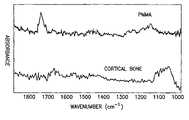
- the drawingshows mid-infrared spectra of PMMA and bone.
- PMMAhas a distinct absorbance band at approximately 1740 cm ⁇ 1 .
- Bonedisplays a distinct absorbance at approximately 1030 cm ⁇ 1 .
- the present inventioninvolves providing an IFOP which is operative in the mid-infrared region of the EM (approximately 400 cm ⁇ 1 to 5,000 cm ⁇ 1 ) or in the near infrared region of the EM (approximately 5,000 cm ⁇ 1 to 14,000 cm ⁇ 1 ).
- the probe for use in the present inventioncan detect infrared radiation by ATR or by reflection.
- the evaluation of the region of interest to determine whether it is a host tissue or a non-host materialcan be carried out by, for example, peak shift analysis, peak area analysis, a combination of peak shift analysis and peak area analysis or by chemometric parameters.
- An example of a probe for use in the present inventioncomprises an ATR element, or can receive reflected IR energy directly, and has an input end and an output end for receiving IR and transmitting attenuated IR radiation, respectively (see U.S. Pat. Nos. 5,170,056 and 5,923,808, the entire contents of both U.S. Pat. Nos. 5,170,056 and 5,923,808 are incorporated by reference herein).
- a probe operative in the infrared region of the EM spectrum for in situ real time sensing of the absorption of IR energy in a samplecomprising:
- an ATR crystal elementhaving an input end portion for receiving IR energy and an output end portion for transmitting attenuated IR energy, the ATR element having wall portions disposed along a central or long axis thereof, the IR energy being reflected along the element in a direction transverse to the central axis;
- a plurality of infrared transmitting fibersin the form of a cylindrical bundle at the input end portion and the output end portion of the ATR element, the bundle of fibers having end faces disposed in direct contacting abutment with the ATR element for transmitting and receiving IR energy into and out of the element, the bundle arranged in a cluster of individual fibers, the bundle centered on an axis common with the central axis of the ATR element, the fibers having relatively high numerical apertures for spreading transmitted energy so that a portion of the energy enters the ATR element at an angle less than the critical angle for facilitating total internal reflection by the wall portions of the element and for receiving a sensible amount of IR energy from the ATR element.
- the probeis configured with a crystal suitable for ATR such as ZnS.
- the crystal tipis positioned in optical contact with the surface of the sample. Infrared radiation passes through the surface of the sample, back through the fiber optic and into an infrared detector. This output signal is first measured as a function of the wavelength in the absence of the sample (the background spectrum); and then measured as a function of the wavelength in the presence of the sample. The signal measured with the sample present is then divided, point by point, by the background spectrum to give a percentage transmission, wherein the logarithm of the percentage transmission gives the absorbance spectrum.
- the probeis configured without a crystal tip. It is placed a distance from the tissue that is suitable for collection of reflectance data.
- a flexible fiber-optic cablecomprising a mid-infrared-transmitting glass chalcogenide equipped with a MCT detector is coupled to a spectrometer.
- the fiber opticis preferably 1 to 2 meters in length and is transmissive over the infrared region of at least 400 to 4,000 cm ⁇ 1 .
- a 5 mm diameter ZnS probe with a 1 mm region of surface contactcan be attached to the end of the cable, thereby permitting sampling of 1 mm diameter sample areas.
- the standard tipis a nominal two-bounce design, whereas the flat-ended design is a nominal three-bounce design. The optimum design will have the highest possible penetration depth on the flat face of the crystal, while avoiding the loss of light that will occur if an appreciable percentage of the rays exceeds the Brewster angle.
- a host tissue in the context of the present inventionincludes tissue that naturally occurs in the body of a human or an animal, such as soft connective tissue, such as articular cartilage, meniscal cartilage, ligament, tendon and capsule, or mineralized tissue such as bone or teeth.
- the non-host materialcan be another tissue (such as a bone graft or allograft) or a synthetic material, such as a polymer, a synthetic bone substitute, a bone cement such as polymethyl methacrylate (“PMMA”), a metal or a ceramic.
- the infrared radiationpenetrates the surface of the region of interest (for example, the region suspected of containing a host tissue in intimate contact with a non-host material) to a depth of approximately up to 10 microns.
- the probeis in contact (touches) the region of interest.
- the probeis within a sufficient distance from the surface of the region of interest for detecting reflection (such distance could be a few centimeters from the surface of the region of interest).
- Infrared radiationis analyzed in the present invention for at least one of peak height, peak area, frequency and chemometric parameters.
- chemometricshas been defined as follows:
- the analysis of the infrared radiationinvolves comparing at least one of peak height, peak area, frequency and chemometric parameters for one or both materials to established or pre-selected values, or the ratio for at least one of peak heights, peak area, frequencies and chemometrics for one or both materials.
- One method of the present inventionis to utilize an IFOP to discriminate between bone and at least one other compound or material (such as a compound or material in contact with a bone) whose signature IR spectrum are sufficiently different to be able to distinguish between them.
- the IFOPto discriminate between bone cement, usually comprised of polymethyl methacrylate (PMMA), and bone tissue in vivo by examination of the infrared spectrum of the material of interest.
- PMMApolymethyl methacrylate
- Prior to the present inventionother than visual examination, there was no method to discriminate between bone and other materials in contact with bone (such as bone cement) in vivo during surgical procedures. Although with direct observation one can often discriminate between such two materials, it can also at times be quite difficult to discriminate between the two materials. This is especially true in the femoral canal during revision total joint surgery, when visualization is limited. Specific IR signature absorbance bands arise from bone and PMMA (see the drawing), and differences in these bands can be used to identify these components. Discrimination of bone and PMMA would be carried out by the IFOP during surgical procedures such as revision of total joint replacements. The present invention thus provides a chemical basis by which it is possible to distinguish between these two materials in situ.
- PMMApolymethyl methacrylate
- Another use of the present inventionis to discriminate between bone and a metal or plastic implant surface in vivo.
- the infrared spectra of bone and a plastic, such as polyethylene, or a metal (no infrared absorbance)are sufficiently different such that these materials can be identified in vivo during surgical procedures.
- Another method of the present inventioninvolves a combination of a probe operative in the mid-infrared region or the near-infrared region of the EM for in situ sensing of the absorption of IR energy or of reflected TR energy, together with a removal system, such as an ablation, debridement or cutting system (e.g., an ultrasonic ablator or a laser).
- a removal systemsuch as an ablation, debridement or cutting system (e.g., an ultrasonic ablator or a laser).
- the ablatorsuch as sold by Misonix, Inc. of Farmingdale, N.Y., USA
- cutting instrumentreceives signals from the probe and would indicate with a LED when to ablate, or could be turned-off automatically by incoming IR signals.
- ablationwill be carried out only so long as the probe detects PMMA.
- the identification and removal of a non-host materialare carried out in one step.
- the removal device or ablatorit is preferred to use an ultrasonic device which by its nature is in physical contact with the surface of the region of interest (such as in detecting ATR).
- the ultrasonic deviceavoids the use of electromagnetic radiation such as in a laser.
- the ultrasonic deviceremoves undesired material by vaporizing the material.
- the present inventionis also useful for cleaning surfaces where at least one substance or tissue has a clearly distinct and detectable IR spectrum. Examples are as follows: teeth and fillings; and cement between two materials, such as metal and bone.
- resins or any other material that has a distinguishable infrared spectrum that are embedded in other materialscan be examined.
- pearlsare often glued onto hollow metal (or other) receptacles. The excess glue can be ablated without touching the pearl if the ablator functions only when the infrared signal from the glue is detected.
- impurities on surfaces of materialscan be removed if at least one of the materials has a distinguishable infrared spectrum by actuating an ablator, cutter, carver or polisher when the appropriate infrared IR signal is given. That is, when either the host material or the impurity have at least one IR peak that is distinguishable from the other material, and the height or area of this peak, or the chemometric parameters of the IR spectrum is pre-selected to actuate the ablator, cutter, carver or polisher.
Landscapes
- Health & Medical Sciences (AREA)
- Life Sciences & Earth Sciences (AREA)
- General Health & Medical Sciences (AREA)
- Physics & Mathematics (AREA)
- Pathology (AREA)
- Biophysics (AREA)
- Medical Informatics (AREA)
- General Physics & Mathematics (AREA)
- Immunology (AREA)
- Analytical Chemistry (AREA)
- Chemical & Material Sciences (AREA)
- Engineering & Computer Science (AREA)
- Biomedical Technology (AREA)
- Heart & Thoracic Surgery (AREA)
- Biochemistry (AREA)
- Molecular Biology (AREA)
- Surgery (AREA)
- Animal Behavior & Ethology (AREA)
- Public Health (AREA)
- Veterinary Medicine (AREA)
- Investigating Or Analysing Materials By Optical Means (AREA)
- Length Measuring Devices By Optical Means (AREA)
Abstract
Description
Claims (31)
Priority Applications (1)
| Application Number | Priority Date | Filing Date | Title |
|---|---|---|---|
| US10/142,003US7167742B2 (en) | 2001-05-10 | 2002-05-09 | Utilization of an infrared probe to discriminate between materials |
Applications Claiming Priority (2)
| Application Number | Priority Date | Filing Date | Title |
|---|---|---|---|
| US28998401P | 2001-05-10 | 2001-05-10 | |
| US10/142,003US7167742B2 (en) | 2001-05-10 | 2002-05-09 | Utilization of an infrared probe to discriminate between materials |
Publications (2)
| Publication Number | Publication Date |
|---|---|
| US20020169379A1 US20020169379A1 (en) | 2002-11-14 |
| US7167742B2true US7167742B2 (en) | 2007-01-23 |
Family
ID=23114026
Family Applications (1)
| Application Number | Title | Priority Date | Filing Date |
|---|---|---|---|
| US10/142,003Expired - Fee RelatedUS7167742B2 (en) | 2001-05-10 | 2002-05-09 | Utilization of an infrared probe to discriminate between materials |
Country Status (4)
| Country | Link |
|---|---|
| US (1) | US7167742B2 (en) |
| AU (1) | AU2002256509A1 (en) |
| GB (1) | GB2396408B (en) |
| WO (1) | WO2002090906A2 (en) |
Cited By (10)
| Publication number | Priority date | Publication date | Assignee | Title |
|---|---|---|---|---|
| US20060018525A1 (en)* | 2004-07-21 | 2006-01-26 | Barbour Randall L | Method and system for temporal spectral imaging |
| WO2009042644A2 (en) | 2007-09-25 | 2009-04-02 | Perception Raisonnement Action En Medecine | Methods and apparatus for assisting cartilage diagnostic and therapeutic procedures |
| US20090118601A1 (en)* | 2004-09-29 | 2009-05-07 | University Of Delaware | Ir spectrographic apparatus and method for diagnosis of disease |
| US20090268942A1 (en)* | 2008-04-23 | 2009-10-29 | Price John D | Methods and apparatus for detection of motion picture piracy for piracy prevention |
| US20110024629A1 (en)* | 2008-04-04 | 2011-02-03 | Colgate-Palmolive Company | Analysis of substrates having agents deposited thereon |
| US20110054360A1 (en)* | 2009-08-27 | 2011-03-03 | Electronics And Telecommunications Research Institute | Finger motion detecting apparatus and method |
| US20110059023A1 (en)* | 2008-03-19 | 2011-03-10 | Tunnell James W | Narrowband imaging using near-infrared absorbing nanoparticles |
| US20130273661A1 (en)* | 2012-04-13 | 2013-10-17 | Schlumberger Technology Corporation | Methods and appratus for simultaneous estimation of quantitative minerology, kerogen content and maturity in gas shale and oil-bearing shale |
| US8812084B1 (en)* | 2009-12-31 | 2014-08-19 | Albert Francis Messano, JR. | Systems and methods for multispectral scanning and detection for medical diagnosis |
| US8971998B2 (en) | 2009-12-31 | 2015-03-03 | Integral Electromagnetronic Technologies Llc | Systems and methods for multispectral scanning and detection for medical diagnosis |
Families Citing this family (12)
| Publication number | Priority date | Publication date | Assignee | Title |
|---|---|---|---|---|
| AU2001261445A1 (en) | 2000-05-12 | 2001-11-26 | Mathias P. B. Bostrom | Determination of the ultrastructure of connective tissue by an infrared fiber-optic spectroscopic probe |
| US20060189965A1 (en)* | 2003-04-01 | 2006-08-24 | Emil Litvak | System,apparatus and method for large area tissue ablation |
| US20050119587A1 (en)* | 2003-07-01 | 2005-06-02 | University Of Michigan | Method and apparatus for evaluating connective tissue conditions |
| JP2007524833A (en)* | 2003-07-01 | 2007-08-30 | ザ リージェンツ オブ ザ ユニバーシティ オブ ミシガン | Method and apparatus for diagnosing bone tissue condition |
| US7049596B2 (en) | 2003-07-02 | 2006-05-23 | Innoventive Technologies, Inc. | Method and apparatus for distinguishing materials |
| CA2577164A1 (en) | 2004-06-22 | 2005-12-29 | Crescent Diagnostics (Ireland) Limited | Diagnostic methods for osteoporosis |
| EP1898791A2 (en)* | 2005-06-30 | 2008-03-19 | The University Of Wyoming | Method for determination of bone fracture risk using raman spectroscopy |
| US7729749B2 (en)* | 2005-09-01 | 2010-06-01 | The Regents Of The University Of Michigan | Method and apparatus for evaluating connective tissue conditions |
| KR20140018183A (en)* | 2010-09-16 | 2014-02-12 | 레이디안스, 아이엔씨. | Laser based processing of layered materials |
| US9919380B2 (en) | 2013-02-23 | 2018-03-20 | Coherent, Inc. | Shaping of brittle materials with controlled surface and bulk properties |
| CN103808688B (en)* | 2014-01-22 | 2016-08-10 | 重庆医科大学 | Rapid and non-destructive detection of quality consistency of finished pharmaceuticals by near-infrared spectroscopy |
| US20190234869A1 (en)* | 2016-09-13 | 2019-08-01 | The General Hospital Corporation | Systems and methods for characterizing biological material using near-infrared spectroscopy |
Citations (42)
| Publication number | Priority date | Publication date | Assignee | Title |
|---|---|---|---|---|
| US3769963A (en) | 1972-03-31 | 1973-11-06 | L Goldman | Instrument for performing laser micro-surgery and diagnostic transillumination of living human tissue |
| US3941122A (en)* | 1974-04-08 | 1976-03-02 | Bolt Beranek And Newman, Inc. | High frequency ultrasonic process and apparatus for selectively dissolving and removing unwanted solid and semi-solid materials and the like |
| US4336809A (en) | 1980-03-17 | 1982-06-29 | Burleigh Instruments, Inc. | Human and animal tissue photoradiation system and method |
| US4588885A (en) | 1984-02-07 | 1986-05-13 | International Technical Associates | Method of and apparatus for the removal of paint and the like from a substrate |
| US4733660A (en) | 1984-08-07 | 1988-03-29 | Medical Laser Research And Development Corporation | Laser system for providing target specific energy deposition and damage |
| US4737628A (en) | 1984-02-07 | 1988-04-12 | International Technical Associates | Method and system for controlled and selective removal of material |
| US4973848A (en)* | 1989-07-28 | 1990-11-27 | J. Mccaughan | Laser apparatus for concurrent analysis and treatment |
| US5038039A (en) | 1990-01-29 | 1991-08-06 | Cornell Research Foundation, Inc. | Method of detecting the presence of anomalies in biological tissues and cells in natural and cultured form by infrared spectroscopy |
| US5170056A (en) | 1991-02-28 | 1992-12-08 | Galileo Electro-Optics Corporation | Optical fiber coupled devices for remote spectroscopy in the infrared |
| US5197470A (en) | 1990-07-16 | 1993-03-30 | Eastman Kodak Company | Near infrared diagnostic method and instrument |
| US5204517A (en)* | 1991-12-24 | 1993-04-20 | Maxwell Laboratories, Inc. | Method and system for control of a material removal process using spectral emission discrimination |
| US5275594A (en)* | 1990-11-09 | 1994-01-04 | C. R. Bard, Inc. | Angioplasty system having means for identification of atherosclerotic plaque |
| US5280788A (en) | 1991-02-26 | 1994-01-25 | Massachusetts Institute Of Technology | Devices and methods for optical diagnosis of tissue |
| US5281798A (en)* | 1991-12-24 | 1994-01-25 | Maxwell Laboratories, Inc. | Method and system for selective removal of material coating from a substrate using a flashlamp |
| US5286947A (en) | 1992-09-08 | 1994-02-15 | General Electric Company | Apparatus and method for monitoring material removal from a workpiece |
| US5304173A (en) | 1985-03-22 | 1994-04-19 | Massachusetts Institute Of Technology | Spectral diagonostic and treatment system |
| US5346488A (en) | 1985-04-08 | 1994-09-13 | The General Hospital Corporation | Laser-induced ablation of atherosclerotic plaque |
| US5452716A (en) | 1992-02-25 | 1995-09-26 | Novo Nordisk A/S | Method and device for in vivo measuring the concentration of a substance in the blood |
| US5460182A (en) | 1992-09-14 | 1995-10-24 | Sextant Medical Corporation | Tissue penetrating apparatus and methods |
| US5516043A (en) | 1994-06-30 | 1996-05-14 | Misonix Inc. | Ultrasonic atomizing device |
| US5527273A (en) | 1994-10-06 | 1996-06-18 | Misonix, Inc. | Ultrasonic lipectomy probe and method for manufacture |
| US5596992A (en) | 1993-06-30 | 1997-01-28 | Sandia Corporation | Multivariate classification of infrared spectra of cell and tissue samples |
| US5701913A (en) | 1995-02-03 | 1997-12-30 | University Technologies International Inc. | Tissue softness probe |
| US5733739A (en) | 1995-06-07 | 1998-03-31 | Inphocyte, Inc. | System and method for diagnosis of disease by infrared analysis of human tissues and cells |
| US5746736A (en) | 1995-08-09 | 1998-05-05 | Lumedics, Ltd. | Cryogenic laser lithotripsy with enhanced light absorption |
| US5762609A (en) | 1992-09-14 | 1998-06-09 | Sextant Medical Corporation | Device and method for analysis of surgical tissue interventions |
| US5769791A (en) | 1992-09-14 | 1998-06-23 | Sextant Medical Corporation | Tissue interrogating device and methods |
| USRE36044E (en) | 1991-12-24 | 1999-01-12 | Benaron; David A. | Path constrained spectrophotometer and method for determination of spatial distribution of light or other radiation scattering and absorbing substances in a radiation scattering medium |
| US5876397A (en) | 1984-01-24 | 1999-03-02 | Boston Scientific Corporation | Reduction of an arteriosclerotic lesion by selective absorption of electromagnetic energy in a component thereof |
| DE19841217A1 (en) | 1997-10-27 | 1999-04-29 | Acspect Corp | Decaying-wave Fourier transform infra red spectrometer analyzing tissues and fluids, including in vivo |
| US5923808A (en) | 1997-06-23 | 1999-07-13 | Melling; Peter J. | Mid-infrared fiber-optic spectroscopic probe for use at elevated temperatures |
| US5986770A (en) | 1994-07-30 | 1999-11-16 | Roche Diagnostics Gmbh | Apparatus and method for the optical characterization of the structure and composition of a light scattering sample |
| US5987346A (en) | 1993-02-26 | 1999-11-16 | Benaron; David A. | Device and method for classification of tissue |
| US6036667A (en) | 1996-10-04 | 2000-03-14 | United States Surgical Corporation | Ultrasonic dissection and coagulation system |
| US6068604A (en) | 1998-04-09 | 2000-05-30 | Smith & Nephew, Inc. | Cartilage indentor instrument |
| US6135774A (en) | 1997-04-03 | 2000-10-24 | Kaltenbach & Voigt Gmbh & Co. | Diagnosis and treatment device for teeth |
| US6200307B1 (en) | 1997-05-22 | 2001-03-13 | Illumenex Corporation | Treatment of in-stent restenosis using cytotoxic radiation |
| US6270471B1 (en) | 1997-12-23 | 2001-08-07 | Misonix Incorporated | Ultrasonic probe with isolated outer cannula |
| WO2001087040A2 (en) | 2000-05-12 | 2001-11-22 | Hospital For Special Surgery | Determination of the ultrastructure of connective tissue by an infrared fiber-optic spectroscopic probe |
| US6324419B1 (en) | 1998-10-27 | 2001-11-27 | Nejat Guzelsu | Apparatus and method for non-invasive measurement of stretch |
| US20010048077A1 (en) | 1997-10-27 | 2001-12-06 | Afanassieva Natalia I. | Apparatus and method for spectroscopic analysis of human or animal tissue or body fluids |
| US20020002336A1 (en) | 2000-01-21 | 2002-01-03 | Marchitto Kevin S. | Optical measurements of bone composition |
Family Cites Families (2)
| Publication number | Priority date | Publication date | Assignee | Title |
|---|---|---|---|---|
| US36044A (en)* | 1862-07-29 | Improvement in heaters for railroad-cars | ||
| US6374300B2 (en)* | 1999-07-15 | 2002-04-16 | F5 Networks, Inc. | Method and system for storing load balancing information with an HTTP cookie |
- 2002
- 2002-05-09AUAU2002256509Apatent/AU2002256509A1/ennot_activeAbandoned
- 2002-05-09USUS10/142,003patent/US7167742B2/ennot_activeExpired - Fee Related
- 2002-05-09GBGB0318142Apatent/GB2396408B/ennot_activeExpired - Fee Related
- 2002-05-09WOPCT/US2002/014639patent/WO2002090906A2/ennot_activeApplication Discontinuation
Patent Citations (48)
| Publication number | Priority date | Publication date | Assignee | Title |
|---|---|---|---|---|
| US3769963A (en) | 1972-03-31 | 1973-11-06 | L Goldman | Instrument for performing laser micro-surgery and diagnostic transillumination of living human tissue |
| US3941122A (en)* | 1974-04-08 | 1976-03-02 | Bolt Beranek And Newman, Inc. | High frequency ultrasonic process and apparatus for selectively dissolving and removing unwanted solid and semi-solid materials and the like |
| US4336809A (en) | 1980-03-17 | 1982-06-29 | Burleigh Instruments, Inc. | Human and animal tissue photoradiation system and method |
| US5876397A (en) | 1984-01-24 | 1999-03-02 | Boston Scientific Corporation | Reduction of an arteriosclerotic lesion by selective absorption of electromagnetic energy in a component thereof |
| US4588885A (en) | 1984-02-07 | 1986-05-13 | International Technical Associates | Method of and apparatus for the removal of paint and the like from a substrate |
| US4737628A (en) | 1984-02-07 | 1988-04-12 | International Technical Associates | Method and system for controlled and selective removal of material |
| US4733660A (en) | 1984-08-07 | 1988-03-29 | Medical Laser Research And Development Corporation | Laser system for providing target specific energy deposition and damage |
| US5304173A (en) | 1985-03-22 | 1994-04-19 | Massachusetts Institute Of Technology | Spectral diagonostic and treatment system |
| US5346488A (en) | 1985-04-08 | 1994-09-13 | The General Hospital Corporation | Laser-induced ablation of atherosclerotic plaque |
| US4973848A (en)* | 1989-07-28 | 1990-11-27 | J. Mccaughan | Laser apparatus for concurrent analysis and treatment |
| US5038039A (en) | 1990-01-29 | 1991-08-06 | Cornell Research Foundation, Inc. | Method of detecting the presence of anomalies in biological tissues and cells in natural and cultured form by infrared spectroscopy |
| US5197470A (en) | 1990-07-16 | 1993-03-30 | Eastman Kodak Company | Near infrared diagnostic method and instrument |
| US5275594A (en)* | 1990-11-09 | 1994-01-04 | C. R. Bard, Inc. | Angioplasty system having means for identification of atherosclerotic plaque |
| US5280788A (en) | 1991-02-26 | 1994-01-25 | Massachusetts Institute Of Technology | Devices and methods for optical diagnosis of tissue |
| US5170056A (en) | 1991-02-28 | 1992-12-08 | Galileo Electro-Optics Corporation | Optical fiber coupled devices for remote spectroscopy in the infrared |
| US5204517A (en)* | 1991-12-24 | 1993-04-20 | Maxwell Laboratories, Inc. | Method and system for control of a material removal process using spectral emission discrimination |
| US5281798A (en)* | 1991-12-24 | 1994-01-25 | Maxwell Laboratories, Inc. | Method and system for selective removal of material coating from a substrate using a flashlamp |
| USRE36044E (en) | 1991-12-24 | 1999-01-12 | Benaron; David A. | Path constrained spectrophotometer and method for determination of spatial distribution of light or other radiation scattering and absorbing substances in a radiation scattering medium |
| US5452716A (en) | 1992-02-25 | 1995-09-26 | Novo Nordisk A/S | Method and device for in vivo measuring the concentration of a substance in the blood |
| US5286947A (en) | 1992-09-08 | 1994-02-15 | General Electric Company | Apparatus and method for monitoring material removal from a workpiece |
| US5772597A (en) | 1992-09-14 | 1998-06-30 | Sextant Medical Corporation | Surgical tool end effector |
| US5762609A (en) | 1992-09-14 | 1998-06-09 | Sextant Medical Corporation | Device and method for analysis of surgical tissue interventions |
| US5769791A (en) | 1992-09-14 | 1998-06-23 | Sextant Medical Corporation | Tissue interrogating device and methods |
| US5785658A (en) | 1992-09-14 | 1998-07-28 | Sexant Medical Corporation | In vivo tissue analysis methods and apparatus |
| US5807261A (en) | 1992-09-14 | 1998-09-15 | Sextant Medical Corporation | Noninvasive system for characterizing tissue in vivo |
| US5460182A (en) | 1992-09-14 | 1995-10-24 | Sextant Medical Corporation | Tissue penetrating apparatus and methods |
| US5987346A (en) | 1993-02-26 | 1999-11-16 | Benaron; David A. | Device and method for classification of tissue |
| US5596992A (en) | 1993-06-30 | 1997-01-28 | Sandia Corporation | Multivariate classification of infrared spectra of cell and tissue samples |
| US5516043A (en) | 1994-06-30 | 1996-05-14 | Misonix Inc. | Ultrasonic atomizing device |
| US5986770A (en) | 1994-07-30 | 1999-11-16 | Roche Diagnostics Gmbh | Apparatus and method for the optical characterization of the structure and composition of a light scattering sample |
| US5527273A (en) | 1994-10-06 | 1996-06-18 | Misonix, Inc. | Ultrasonic lipectomy probe and method for manufacture |
| US5701913A (en) | 1995-02-03 | 1997-12-30 | University Technologies International Inc. | Tissue softness probe |
| US5733739A (en) | 1995-06-07 | 1998-03-31 | Inphocyte, Inc. | System and method for diagnosis of disease by infrared analysis of human tissues and cells |
| US5746736A (en) | 1995-08-09 | 1998-05-05 | Lumedics, Ltd. | Cryogenic laser lithotripsy with enhanced light absorption |
| US6063050A (en) | 1996-10-04 | 2000-05-16 | United States Surgical Corp. | Ultrasonic dissection and coagulation system |
| US6036667A (en) | 1996-10-04 | 2000-03-14 | United States Surgical Corporation | Ultrasonic dissection and coagulation system |
| US6135774A (en) | 1997-04-03 | 2000-10-24 | Kaltenbach & Voigt Gmbh & Co. | Diagnosis and treatment device for teeth |
| US6200307B1 (en) | 1997-05-22 | 2001-03-13 | Illumenex Corporation | Treatment of in-stent restenosis using cytotoxic radiation |
| US5923808A (en) | 1997-06-23 | 1999-07-13 | Melling; Peter J. | Mid-infrared fiber-optic spectroscopic probe for use at elevated temperatures |
| DE19841217A1 (en) | 1997-10-27 | 1999-04-29 | Acspect Corp | Decaying-wave Fourier transform infra red spectrometer analyzing tissues and fluids, including in vivo |
| US20010048077A1 (en) | 1997-10-27 | 2001-12-06 | Afanassieva Natalia I. | Apparatus and method for spectroscopic analysis of human or animal tissue or body fluids |
| US6270471B1 (en) | 1997-12-23 | 2001-08-07 | Misonix Incorporated | Ultrasonic probe with isolated outer cannula |
| US6068604A (en) | 1998-04-09 | 2000-05-30 | Smith & Nephew, Inc. | Cartilage indentor instrument |
| US6324419B1 (en) | 1998-10-27 | 2001-11-27 | Nejat Guzelsu | Apparatus and method for non-invasive measurement of stretch |
| US20020002336A1 (en) | 2000-01-21 | 2002-01-03 | Marchitto Kevin S. | Optical measurements of bone composition |
| WO2001087040A2 (en) | 2000-05-12 | 2001-11-22 | Hospital For Special Surgery | Determination of the ultrastructure of connective tissue by an infrared fiber-optic spectroscopic probe |
| US20020010400A1 (en) | 2000-05-12 | 2002-01-24 | Hospital For Special Surgery | Determination of the ultrastructure of connective tissue by an infrared fiber-optic spectroscopic probe |
| US6934576B2 (en)* | 2000-05-12 | 2005-08-23 | Hospital For Special Surgery | Determination of the ultrastructure of connective tissue by an infrared fiber-optic spectroscopic probe |
Non-Patent Citations (87)
| Title |
|---|
| Andersson M., Folestad S., Gottfries J., Johansson M.O., Josefson M., Wahlund K.G., (2000), "Quantitative Analysis of Film Coating in a Fluidized Bed Process by In-Line NIR Spectrometry and Multivariate Batch Calibration", Anal. Chem., 72:2099-2108. |
| Attin T., Opatowski A., Meyer C., Zingg-Meyer B., Monting J.S., (2000), "Class II Restorations With a Polyacid-Modified Composite Resin in Primary Molars Placed in a Dental Practice: Results of a Two-Year Clinical Evaluation", Oper. Dent., 25:259-264. |
| Behravesh E., Yasko A.W., Engel P.S., Mikos A.G., (1999), "Synthetic Biodegradable Polymers for Orthopaedic Applications", Clin. Orthop., S118-S129. |
| Blanco M., Coello J., Iturriaga H., Maspoch S., Rovira E., (1997), "Determination of Water in Ferrous Lactate by Near Infrared Reflectance Spectroscopy with a Fibre-Optic Probe", J. Pharm. Biomed. Anal., 16:255-262. |
| Boden S.D., (2000) , "Biology of Lumbar Spine Fusion and Use of Bone Graft Substitutes: Present, Future, and Next Generation", Tissue Eng., 6:383-399. |
| Boskey A.L., Gadaleta S., Gundberg C., Doty S.B., Ducy P. and Karsenty G., "Fourier Transform Infrared Microspectroscopic Analysis of Bones of Osteocalcin-Deficient Mice Provides Insight Into the Function of Osteocalcin", Bone , vol. 23, No. 3, (1998) , pp. 187-196. |
| Boskey A.L., Gadaleta S., Gundberg C., Doty S.B., Ducy P., Karsenty G., (1998), "Fourier Transform Infrared Microspectroscopic Analysis of Bones of Osteocalcin-deficient Mice Provides Insight into the Function of Osteocalcin", Bone. 23:187-196. |
| Boskey, A.L., Guidon, P., Doty, S.B., Stiner, D., Leboy, P., and Binderman, I., "The Mechanism of Beta-Glycerophosphate Action in Mineralizing Chick Limb-Bud Mesenchymal Cell Cultures", J. Bone Miner. Res.,11, pp. 1694-1702, (1996). |
| Bostman O., Pihlajamaki H., (2000), "Clinical Biocompatibility of Biodegradable Orthopaedic Implants for Internal Fixation: A Review", Biomaterials, 21:2615-2621. |
| Bostrom M.P.G., West P., Yang X., Camacho N.P., "Evaluation of Cartilage Degradation by an Infrared Fiber Optic Probe", presented at the 4<SUP>th </SUP>Combined Meeting of the Orthopaedic Research Society, 2001. |
| Bouveresse E., Casolino C., de la P.C., (1998), "Application of Standardisation Methods to Correct the Spectral Differences Induced by a Fibre Optic Probe Used For the Near-Infrared Analysis of Pharmaceutical Tablets", J. Pharm. Biomed. Anal., 18:35-42. |
| Boydston-White S., Gopen T., Houser S., Bargonetti J., Diem M., (1999), "Infrared Spectroscopy of Human Tissue. V. Infrared Spectroscopic Studies of Myeloid Leukemia (ML-1) Cells at Different Phases of the Cell Cycle", Biospectroscopy, 5:219-227. |
| Buckwalter, J.A., and Mow, V.C., "Injuries to Cartilage and Meniscus: Sports Injuries to Articular Cartilage", DeLee, J.C., and Drez, D., Jr., Orthopaedic-Sports Medicine Principles and Practice, Philadelphia: W.B. Saunders Company, pp. 82-107, (1994). |
| Bychkov, S.M., and Kuzmina, S.A., "Study of Tissue Proteoglycans by Means of Infrared Spectroscopy", Biull. Eksp. Biol. Med., 114, pp. 246-249, (1992). |
| Bychkov, S.M., Bogatov, V.N., and Kuzmina, S.A., "Infrared Spectra of Cartilage Proteoglycans", Bull. Eksp. Biol. Med., 90, pp. 561-563, (1980). |
| Bychkov, S.M., Bogatov, V.N., and Kuzmina, S.A., "Study of Different Proteoglycan Salts", Bull. Eksp. Biol. Med., 92, pp. 302-305, (1981). |
| Camacho N.P., Hou L., Toledano T.R., Ilg W.A., Brayton C.F., Raggio C.L., et al., (1999), "The Material Basis for Reduced Mechanical Properties in Oim Mice Bones", J. Bone Miner. Res., 14:264-272. |
| Camacho N.P., Lin J., Yang X., West P. and Bostrom M.P.G., "An Infrared Fiber Optic Probe for Detection of Degenerative Cartilage", Trans 48<SUP>th </SUP>ORS Meeting, 2002 (Abstract). |
| Camacho, N.P., Hou, L., Toledano, T.R., Ilg, W.A., Brayton, C.F., Raggio, C.L., Root, L., and Boskey, A.L., "The Material Basis for Reduced Mechanical Properties in Oim Mice Bones", J. Bone Miner, Res., 14, pp. 264-272, (1999). |
| Camacho, N.P., Landis, W.J., and Boskey, A.L., "Mineral Changes in a Mouse Model of Osteogenesis Imperfecta Detected by Fourier Transform Infrared Microscopy", Connect. Tissue Res., 35, pp. 259-265, (1996). |
| Camacho, N.P., Mendelsohn, R., Grigiene, R., Torzilla, P.A., "Polarized FI-IR Microscopic Determination of Collagen Orientation in Articular Cartilage", 42nd Annual Meeting, Orthopaedic Research Society, Feb. 19-22, 1996, Atlanta, Georgia. |
| Camacho, N.P., Rimnac, C.M., Meyer, R.A.J., Doty, S., and Boskey, A.L., "Effect of Abnormal Mineralization on the Mechanical Behavior of X-Linked Hypophosphatemic Mice Femora", Bone, 17, pp. 271-278, (1995). |
| Camacho, N.P., West, P., Torzilli, P.A., Mendelsohn, R., "FTIR Microscopic Imaging of Collagen and Proteoglycan in Bovine Cartilage", Biopolymers, 62:1-8 (2001). |
| Caplan A.I., (2000), "Tissue Engineering Designs for the Future: New Logics, Old Molecules", Tissue Eng., 6:1-8. |
| Collier J.H., Camp J.P., Hudson T.W., Schmidt C.E., (2000), "Synthesis and Characterization of PolypyrroleHyaluronic Acid Composite Biomaterials for Tissue Engineering Applications", J. Biomed. Mater. Res., 50:574-584. |
| Cordewener F.W., Schmitz J.P., (2000), "The Future of Biodegradable Osteosyntheses", Tissue Eng., 6:413-424. |
| Dadd M.R., Sharp D.C., Pettman A.J., Knowles C.J., (2000), "Real-Time Monitoring of Nitrile Biotransformations by Mid-Infrared Spectroscopy", J. Microbiol. Methods, 41:69-75. |
| Doak D.L., Phillips J.A., (1999), "In Situ Monitoring of an Escherichia Coli Fermentation Using A Diamond Composition ATR Probe and Mid-Infrared Spectroscopy", Biotechnol. Prog., 15:529-539. |
| Feng W., Haishu D., Fenghua T., Jun Z., Qing X., Xianwu T., (2001), "Influence of Overlying Tissue and Probe Geometry on the Sensitivity of a Near-Infrared Tissue Oximeter", Physiol. Meas., 22:201-208. |
| Fujikawa K., Kobayashi T., Sasazaki Y., Matsumoto H., Seedhom B.B., (2000) , "Anterior Cruciate Ligament Reconstruction with the Leeds-Keio Artificial Ligament", J. Long Term Eff. Med. Implants, 10:225-238. |
| Fukuyama Y., Yoshida S., Yanagisawa S., Shimizu M., (1999), "A Study on the Differences Between Oral Squamous Cell Carcinomas and Normal Oral Mucosas Measured by Fourier Transform Infrared Spectroscopy", Biospectroscopy, 5:117-126. |
| Gadaleta, S.J., Camacho, N.P., Mendelsohn, R., and Boskey, A.L., "Fourier Transform Infrared Microscopy of Calcified Turkey Leg Tendon", Calcif. Tissue Int., 58, pp. 17-23, (1996). |
| Gadaleta, S.J., Landis, W.J., Boskey, A.L., and Mendelsohn, R., "Polarized FT-IR Microscopy of Calcified Turkey Leg Tendon", Connect. Tissue Reg., 34, pp. 203-211, (1996). |
| George, A., and Veis, A., "FTIRS in H<SUB>2</SUB> O Demonstrates that Collagen Monomers Undergo a Conformational Transition Prior to Thermal Self-Assembly In Vitro", Biochemistry, 30, pp. 2372-2377, (1991). |
| Griffiths, P.R., and J.A. de Haseth, (1986) , "Fourier Transform Infrared Spectrometry", Wiley-Interscience, New York, 457, pp. 188-193. |
| Harris S.C., Walker D.S., (2000), "Quantitative Real-Time Monitoring of Dryer Effluent Using Fiber Optic Near-Infrared Spectroscopy", J. Pharm. Sci., 89:1180-1186. |
| Hollander, A.P., T.F. Heathfield, C. Webber, Y. Iwata, R. Bourne, C. Rorabeck, and A.R. Poole, (1994) , "Increased Damage to Type II Collagen in Osteoarthritic Articular Cartilage Detected by a New Immunoassay", Journal of Clinical Investigation, 93, pp. 1722-1732. |
| Hollinger J.O., Winn S., Bonadio J., (2000), "Options for Tissue Engineering to Address Challenges of the Aging Skeleton", Tissue Eng., 6:341-350. |
| Horecker, B., Kaplan, N.O., Marmur, J., and Scheraga, H.A., "Collagens", Conformation in Fibrous Proteins and Related Synthetic Polypeptides, New York: Academic Press, editors Fraser, R.D.B. and MacRae, T.P., pp. 344-402, (1973). |
| Hull E.L., Conover D.L., Foster T.H., (1999), "Carbogen-Induced Changes in Rat Mammary Tumour Oxygenation Reported by Near Infrared Spectroscopy", Br. J. Cancer, 79:1709-1716. |
| Infrared and Raman Spectroscopy of Biological Materials, Eds. Gremlich, H.U., Yar., 3., New York, Marcel-Dekker, pp. 323-377 (2001). |
| Khan S.N., Sandhu H.S., Parvataneni H.K., Girardi F.P., Cammisa F.P., (2000), "Bone Graft Substitutes in Spine Surgery", Bull. Hosp. Jt. Dis., 59:5-10. |
| Kidder, L.H., Kalasinsky, V.F., Luke, J.L., Levin, I.W., and Lewis, E.N., "Visualization of Silicone Gel in Human Breast Tissue Using New Infrared Imaging Spectroscopy", Nat. Med., 3, pp. 235-237, (1997). |
| Krejci I., Schupbach P., Balmelli F., Lutz F., (1999), "The Ultrastructure of a Compomer Adhesive Interface in Enamel and Dentin, and its Marginal Adaptation Under Dentinal Fluid as Compared to that of a Composite", Dent. Mater., 15:349-358. |
| Lazarev, Y.A., Grishkovsky, B.A., and Khromova, T.B., "Amide I Band of IR Spectrum and Structure of Collagen and Related Polypeptides", Biopolymers, 24, pp. 1449-1478. (1985). |
| Lazarev, Y.A., Grishkovsky, B.A., Khromova, T.B., Lazareva, A.V., and Grechishko, V.S., "Bound Water in Collagen-Like Triple Helical Structure", Biopolymers, 32, pp. 189-195, (1992). |
| Lewis, E.N., Kidder, L.H., Levin, I.W., Kalasinsky, V.F., Hanig, J.P., and Lester, D.S., "Applications of Fourier Transform Infrared Imaging Microscopy in Neurotoxicity", Ann. N.Y. Acad., Sci., 820, pp. 234-247, (1997). |
| Liu, K.Z., Dembinski, T.C., and Mantsch, H.H., "Rapid Determination of Fetal Lung Maturity from Infrared Spectra of Amniotic Fluid", Am. J. Obstet. Gynecol., 178, pp. 234-241, (1998). |
| Lutz F., Krejci I., (2000), "Amalgam Substitutes: A Critical Analysis", J. Esthet. Dent., 12:146-159. |
| M. Khan, M. Yamauchi, S. Srisawasdi, D. Stiner, S. Doty, E.P. Paschalis, A.L. Boskey, "Homocysteine Decreases Chondrocyte-Mediated Matrix Mineralization In Differentiating Chick Limb-bud Mesenchymal Cell Micro-Mass Cultures", Bone, 28, 387-398 (2001). |
| Marcott, C., Reeder, R.C., Paschalis, E.P., Tatakis, D.N., Boskey, A.L., and Mendelsohn, R., "Infrared Microspectroscopic Imaging of Biomineralized Tissues Using a Mercury-Cadmium-Telluride Focal-Plane Array Detector", Cell. Mol. Biol. (Noisy-le-grand), 44, pp. 109-115, (1998). |
| Marti A., (2000), "Cobalt-Base Alloys Used in Bone Surgery", Injury 31 Suppl., 4:18-21. |
| McIntosh L.M., Jackson M., Mantsch H.H., Stranc M.F., Pilavdzic D., Crowson A.N., (1999), "Infrared Spectra of Basal Cell Carcinomas are Distinct from Non-Tumor-Bearing Skin Components", J. Invest. Dermatol., 112:951-956. |
| Mendelsohn, R., and Moore, D.J., "Vibrational Spectroscopic Studies of Lipid Domains in Biomembranes and Model Systems", Chem. Phys. Lipids, 96, pp. 141-157, (1998). |
| Moore, D.J., Gioioso, S., Sills, R.H., and Mendelsohn, R., "Some Relationships Between Membrane Phospholipid Domains, Conformational Order, and Cell Shape in Intact Human Erythrocytes", Biochim. Biophys. Acta., 1415, pp. 342-348, (1999). |
| Moore, D.J., Rerek, M.E., and Mendelsohn, R., "Lipid Domains and Orthorhombic Phases in Model Stratum Corneum: Evidence from Fourier Transform Infrared Spectroscopy Studies", Biochem. Biophys. Res. Commun., 231, pp. 797-801, (1997). |
| Pachalis, E.P., F. Betts, E. DiCarlo, J.M. Lane, R. Mendelsohn, and A.L. Boskey, "Mineral and Organic Matrix Changes in Osteoporosis", J. Dent. Res., 76, p. 287 (1997). |
| Panula, H.E., Hyttinen, M.M., Arokoski, J.P., Langsjo, T.K., Pelettari, A., Kiviranta, I., and Helminen, H.J., "Articular Cartilage Superficial Zone Collagen Birefringence Reduced and Cartilage Thickness Increased before Surface Fibrillation in Experimental Osteoarthritis", Ann. Rheum. Dis., 57, pp. 237-245, (1998). |
| Paschalis E.P., DiCarlo E., Betts F., Sherman P., Mendelsohn R., Boskey A.L., (1996) , "FTIR Microspectroscopic Analysis of Human Osteonal Bone", Calcif. Tissue Int., 59:480-487. |
| Paschalis E.P., Jacenko O., Olsen B., deCrombrugghe B., Boskey A.L., (1996), "The Role of Type X Collagen in Endochondral Ossification as Deduced by Fourier Transform Infrared Microscopy Analysis", Connect. Tissue Res., 35:371-377. |
| Paschalis E.P., Jacenko O., Olsen B., Mendelsohn R. and Boskey A.L., "Fourier Transform Infrared Microspectroscopic Analysis Identifies Alterations in Mineral Properties in Bones from Mice Transgenic for Type X Collagen", Bone , vol. 19, No. 2, (1996), pp. 151-156. |
| Paschalis, E.P., Betts, F., DiCarlo, E., Mendelsohn, R., and Boskey, A. L., "FTIR Microspectroscopic Analysis of Human Iliac Crest Biopsies from Untreated Osteoporotic Bone", Calcif. Tissue Int., 61, pp. 487-492, (1997). |
| Paschalis, E.P., Betts, F., DiCarlo, E., Mendelsohn, R., and Boskey, A.L., "FTIR Microspectroscopic Analysis of Normal Human Cortical and Trabecular Bone", Calcif, Tissue Int., 61, pp. 480-486, (1997). |
| Pietrzak W.S., (2000), "Principles of Development and Use of Absorbable Internal Fixation", Tissue Eng., 6:425-433. |
| Potter, H.G., Linklater, J.M., Allen, A.A., Hannafin, J.A., and Haas, S.B., "Magnetic Resonance Imaging of Articular Cartilage in the Knee: An Evaluation With Use of Fast-Spin-Echo Imaging", J. Bone Joint Surg. Am., 80, pp. 1276-1284, (1998). |
| Potter, K., Kidder, L.H., Levin, I.W., Lewis E.N., Spencer R.G., "Imaging of Collagen and Proteoglycan in Cartilage Sections Using Fourier Transform Infrared Spectral Imaging", Arthritis & Rheum 44 (4) :846-855 (2001). |
| Quaresima V., Sacco S., Totaro R., Ferrari M., (2000) , "Non-invasive Measurement of Cerebral Hemoglobin Oxygen Saturation Using Two Near Infrared Spectroscopy Approaches", J. Biomed. Opt., 5:201-205. |
| Recht, M.P., and Resnick, D., "Magnetic Resonance Imaging of Articular Cartilage: An Overview" Top. Magn. Reson. Imaging, 9, pp. 328-336, (1998). |
| Reddi A.H., (2000) , "Morphogenesis and Tissue Engineering of Bone and Cartilage: Inductive Signals, Stem Cells, and Biomimetic Biomaterials", Tissue Eng., 6:351-359. |
| Rehman I., Karsh M., Hench L.L., Bonfield W., (2000), "Analysis of Apatite Layers on Glass-Ceramic Particulate Using FTIR and FT-Raman Spectroscopy", J. Biomed. Mater. Res., 50:97-100. |
| Rehman I., Knowles J.C., Bonfield W., (1998), "Analysis of In Vitro Reaction Layers Formed on Bioglass Using Thin-Film X-Ray Diffraction and ATR-FTIR Microspectroscopy", J. Biomed. Mater. Res., 41:162-166. |
| Santavirta S., Takagi M., Gomez-Barrena E., Nevalainen J., Lassus J., Salo J., et al., (1999), "Studies of Host Response to Orthopedic Implants and Biomaterials", J. Long Term Eff. Med. Implants., 9:67-76. |
| Schultz C.P., Liu K.Z., Kerr P.D., Mantsch H.H., (1998), "In Situ Infrared Histopathology of Keratinization in Human Oral/Oropharyngeal Squamous Cell Carcinoma", Oncol. Res., 10:277-286. |
| Schumacher, H.R., Klippel, J.H., and Koopman, W.J., "Articular Cartilage", Primer on the Rheumatic Diseases, 11<SUP>th </SUP>edition, Atlanta: The Arthritis Foundation, pp. 14-18, (1993). |
| Sedel L., (2000) , "Evolution of Alumina-on-Alumina Implants: A Review", Clin. Orthop., 48-54. |
| Shaw R.A., Eysel H.H., Liu K.Z., Mantsch H.H., (1998) , "Infrared Spectroscopic Analysis of Biomedical Specimens Using Glass Substrates", Anal. Biochem., 259:181-186. |
| Shaw R.A., Guijon F.B., Paraskevas M., Ying S.L., Mantsch H.H., (1999), "Infrared Spectroscopy of Exfoliated Cervical Cell Specimens, Proceed with Caution", Anal. Quant. Cytol. Histol., 21:292-302. |
| Speer, D.P., and Dahners, L., "The Collagenous Architecture of Articular Cartilage, Correlation of Scanning Electron Microscopy and Polarized Light Microscopy Observations", Clin. Orthop., 167, 99. 267-275, (1979). |
| Tate W.H., You C., Powers J.M., (1999), "Bond Strength of Copomers to Dentin Using Acidic Primers", Am. Jour. of Dent., vol. 12, No. 5, pp. 235-242. |
| Tate W.H., You C., Powers J.M., (2000), "Bond Strength of Compomers to Human Enamel", Oper. Dent., 25:283-291. |
| U.S. Appl. No. 09/853,298, Camacho et al. |
| Uemura T., Nishida K., Sakakida M., Ichinose K., Shimoda S., Shichiri M., (1999), "Non-Invasive Blood Glucose Measurement by Fourier Transform Infrared Spectroscopic Analysis Through the Mucous Membrane of the Lip: Application of a Chalcogenide Optical Fiber System", Front. Med. Biol. Eng., 9:137-153. |
| US 6,230,044, 05/2001, Afanassieva et al. (withdrawn) |
| Weng J., Liu Q., Wolke J.G., Zhang X., de Groot K., (1997), "Formation and Characteristics of the Apatite Layer on Plasma-Sprayed Hydroxyapatite Coatings in Simulated Body Fluid", Biomaterials, 18:1027-1035. |
| White J.G., (1994), "On-line Moisture Detection for a Microwave Vacuum Dryer", Pharm. Res., 11:728-732. |
| Willmann G., (2000), "Ceramic Femoral Head Retrieval Data", Clin. Orthop., 22-28. |
| Zhang, S.F., Rolfe P., Wright G., Lian W., Milling A.J., Tanaka S., et al., (1998) , "Physical and Biological Properties of Compound Membranes Incorporating a Copolymer with a Phosphorylcholine Head Group", Biomaterials, 19:691-700 |
Cited By (16)
| Publication number | Priority date | Publication date | Assignee | Title |
|---|---|---|---|---|
| US20060018525A1 (en)* | 2004-07-21 | 2006-01-26 | Barbour Randall L | Method and system for temporal spectral imaging |
| US20090118601A1 (en)* | 2004-09-29 | 2009-05-07 | University Of Delaware | Ir spectrographic apparatus and method for diagnosis of disease |
| WO2009042644A2 (en) | 2007-09-25 | 2009-04-02 | Perception Raisonnement Action En Medecine | Methods and apparatus for assisting cartilage diagnostic and therapeutic procedures |
| US10028722B2 (en)* | 2007-09-25 | 2018-07-24 | Hospital For Special Surgery | Methods and apparatus for assisting cartilage diagnostic and therapeutic procedures |
| US20110059023A1 (en)* | 2008-03-19 | 2011-03-10 | Tunnell James W | Narrowband imaging using near-infrared absorbing nanoparticles |
| US8895929B2 (en) | 2008-04-04 | 2014-11-25 | Colgate-Palmolive Company | Analysis of substrates having agents deposited thereon |
| CN102187200A (en)* | 2008-04-04 | 2011-09-14 | 高露洁-棕榄公司 | Analysis of the substrate on which the drug is deposited |
| US8803095B2 (en)* | 2008-04-04 | 2014-08-12 | Colgate-Palmolive Company | Analysis of substrates having agents deposited thereon |
| US20110024629A1 (en)* | 2008-04-04 | 2011-02-03 | Colgate-Palmolive Company | Analysis of substrates having agents deposited thereon |
| US20090268942A1 (en)* | 2008-04-23 | 2009-10-29 | Price John D | Methods and apparatus for detection of motion picture piracy for piracy prevention |
| US20110054360A1 (en)* | 2009-08-27 | 2011-03-03 | Electronics And Telecommunications Research Institute | Finger motion detecting apparatus and method |
| US8292833B2 (en)* | 2009-08-27 | 2012-10-23 | Electronics And Telecommunications Research Institute | Finger motion detecting apparatus and method |
| US8812084B1 (en)* | 2009-12-31 | 2014-08-19 | Albert Francis Messano, JR. | Systems and methods for multispectral scanning and detection for medical diagnosis |
| US8971998B2 (en) | 2009-12-31 | 2015-03-03 | Integral Electromagnetronic Technologies Llc | Systems and methods for multispectral scanning and detection for medical diagnosis |
| US20130273661A1 (en)* | 2012-04-13 | 2013-10-17 | Schlumberger Technology Corporation | Methods and appratus for simultaneous estimation of quantitative minerology, kerogen content and maturity in gas shale and oil-bearing shale |
| US8906690B2 (en)* | 2012-04-13 | 2014-12-09 | Schlumberger Technology Corporation | Methods for simultaneous estimation of quantitative minerology, kerogen content and maturity in gas shale and oil-bearing shale |
Also Published As
| Publication number | Publication date |
|---|---|
| AU2002256509A1 (en) | 2002-11-18 |
| WO2002090906A2 (en) | 2002-11-14 |
| WO2002090906A3 (en) | 2003-04-17 |
| GB2396408A (en) | 2004-06-23 |
| GB0318142D0 (en) | 2003-09-03 |
| US20020169379A1 (en) | 2002-11-14 |
| GB2396408B (en) | 2005-06-29 |
Similar Documents
| Publication | Publication Date | Title |
|---|---|---|
| US7167742B2 (en) | Utilization of an infrared probe to discriminate between materials | |
| Nickell et al. | Anisotropy of light propagation in human skin | |
| US6442408B1 (en) | Method for quantification of stratum corneum hydration using diffuse reflectance spectroscopy | |
| US6675029B2 (en) | Apparatus and method for quantification of tissue hydration using diffuse reflectance spectroscopy | |
| TWI324686B (en) | Noninvasive measurement of glucose through the optical properties of tissue | |
| AU707523B2 (en) | Non-invasive blood analyte sensor | |
| US6934576B2 (en) | Determination of the ultrastructure of connective tissue by an infrared fiber-optic spectroscopic probe | |
| US20050043597A1 (en) | Optical vivo probe of analyte concentration within the sterile matrix under the human nail | |
| GB2352512A (en) | A radiation probe and dectecting tooth decay | |
| JP2000506048A (en) | Calibration for subsequent monitoring of biological compounds | |
| CN102488564B (en) | Dental plaque detector capable of imaging by aid of fluorescent area arrays | |
| US8417322B2 (en) | Method and apparatus for diagnosing bone tissue conditions | |
| Taube et al. | Deviations of inorganic and organic carbon content in hypomineralised enamel | |
| AU2009247837A1 (en) | Tissue assessment | |
| US20020002336A1 (en) | Optical measurements of bone composition | |
| Sim et al. | Frequency-dependent characteristics of terahertz radiation on the enamel and dentin of human tooth | |
| Toledano et al. | Biochemical assessment of nanostructures in human trabecular bone: Proposal of a Raman microspectroscopy based measurements protocol | |
| Lee et al. | Raman microspectroscopy demonstrates reduced mineralization of subchondral bone marrow lesions in knee osteoarthritis patients | |
| JP2008197080A (en) | Caries detection method and apparatus | |
| Morris | Raman spectroscopy of bone and cartilage | |
| Okagbare et al. | Transcutaneous Raman spectroscopy for assessing progress of bone-graft incorporation in bone reconstruction and repair | |
| Khalil et al. | Terahertz pulsed spectroscopy for optical and dielectric properties of demineralized bone matrix, collagen and hydroxyapatite | |
| Garip et al. | Diagnosis of bone and cartilage diseases | |
| Belokonev et al. | Visualization and Spectral Analysis of Pubic Periosteum Atrophy | |
| Morris et al. | Picosecond time-gated Raman spectroscopy for transcutaneous evaluation of bone composition |
Legal Events
| Date | Code | Title | Description |
|---|---|---|---|
| AS | Assignment | Owner name:HOSPITAL FOR SPECIAL SURGERY, NEW YORK Free format text:ASSIGNMENT OF ASSIGNORS INTEREST;ASSIGNORS:CAMACHO, NANCY P.;BOSTROM, MATHIAS P.G.;BERTHA, STEVE L.;REEL/FRAME:012891/0590 Effective date:20020508 | |
| FPAY | Fee payment | Year of fee payment:4 | |
| FPAY | Fee payment | Year of fee payment:8 | |
| AS | Assignment | Owner name:BANK OF AMERICA, N.A., AS COLLATERAL AGENT, NORTH CAROLINA Free format text:SECURITY INTEREST;ASSIGNOR:QWEST COMMUNICATIONS INTERNATIONAL INC.;REEL/FRAME:044652/0829 Effective date:20171101 Owner name:BANK OF AMERICA, N.A., AS COLLATERAL AGENT, NORTH Free format text:SECURITY INTEREST;ASSIGNOR:QWEST COMMUNICATIONS INTERNATIONAL INC.;REEL/FRAME:044652/0829 Effective date:20171101 | |
| FEPP | Fee payment procedure | Free format text:MAINTENANCE FEE REMINDER MAILED (ORIGINAL EVENT CODE: REM.); ENTITY STATUS OF PATENT OWNER: SMALL ENTITY | |
| LAPS | Lapse for failure to pay maintenance fees | Free format text:PATENT EXPIRED FOR FAILURE TO PAY MAINTENANCE FEES (ORIGINAL EVENT CODE: EXP.); ENTITY STATUS OF PATENT OWNER: SMALL ENTITY | |
| STCH | Information on status: patent discontinuation | Free format text:PATENT EXPIRED DUE TO NONPAYMENT OF MAINTENANCE FEES UNDER 37 CFR 1.362 | |
| FP | Lapsed due to failure to pay maintenance fee | Effective date:20190123 |