US7160264B2 - Article and method for ocular aqueous drainage - Google Patents
Article and method for ocular aqueous drainageDownload PDFInfo
- Publication number
- US7160264B2 US7160264B2US10/737,707US73770703AUS7160264B2US 7160264 B2US7160264 B2US 7160264B2US 73770703 AUS73770703 AUS 73770703AUS 7160264 B2US7160264 B2US 7160264B2
- Authority
- US
- United States
- Prior art keywords
- implant
- anterior chamber
- sclera
- scleral
- feet
- Prior art date
- Legal status (The legal status is an assumption and is not a legal conclusion. Google has not performed a legal analysis and makes no representation as to the accuracy of the status listed.)
- Expired - Lifetime, expires
Links
- 238000000034methodMethods0.000titleclaimsabstractdescription22
- 239000007943implantSubstances0.000claimsabstractdescription198
- 210000002159anterior chamberAnatomy0.000claimsabstractdescription60
- 210000003786scleraAnatomy0.000claimsabstractdescription44
- 239000012530fluidSubstances0.000claimsabstractdescription30
- 230000004410intraocular pressureEffects0.000claimsdescription21
- 210000003739neckAnatomy0.000description31
- 208000010412GlaucomaDiseases0.000description17
- 210000001742aqueous humorAnatomy0.000description10
- 238000002513implantationMethods0.000description10
- 238000001356surgical procedureMethods0.000description9
- 210000004087corneaAnatomy0.000description8
- 239000000463materialSubstances0.000description7
- 241001465754MetazoaSpecies0.000description5
- 210000001232limbus corneaeAnatomy0.000description5
- 210000001365lymphatic vesselAnatomy0.000description4
- 238000004519manufacturing processMethods0.000description4
- 201000004569BlindnessDiseases0.000description3
- 230000001788irregularEffects0.000description3
- 238000011282treatmentMethods0.000description3
- 238000005520cutting processMethods0.000description2
- 239000003814drugSubstances0.000description2
- 229940079593drugDrugs0.000description2
- 230000035876healingEffects0.000description2
- 238000003780insertionMethods0.000description2
- 230000037431insertionEffects0.000description2
- 238000002483medicationMethods0.000description2
- 238000002360preparation methodMethods0.000description2
- 206010061307Neck deformityDiseases0.000description1
- 206010061323Optic neuropathyDiseases0.000description1
- 238000010521absorption reactionMethods0.000description1
- 238000007792additionMethods0.000description1
- 230000000340anti-metaboliteEffects0.000description1
- 229940100197antimetaboliteDrugs0.000description1
- 239000002256antimetaboliteSubstances0.000description1
- 230000015572biosynthetic processEffects0.000description1
- 229920002678cellulosePolymers0.000description1
- 239000001913celluloseSubstances0.000description1
- 230000006835compressionEffects0.000description1
- 238000007906compressionMethods0.000description1
- 210000000795conjunctivaAnatomy0.000description1
- 238000010276constructionMethods0.000description1
- 238000012217deletionMethods0.000description1
- 230000037430deletionEffects0.000description1
- 230000006866deteriorationEffects0.000description1
- 208000037265diseases, disorders, signs and symptomsDiseases0.000description1
- 230000000694effectsEffects0.000description1
- 208000030533eye diseaseDiseases0.000description1
- 238000001914filtrationMethods0.000description1
- 230000001771impaired effectEffects0.000description1
- 238000000608laser ablationMethods0.000description1
- 238000012986modificationMethods0.000description1
- 230000004048modificationEffects0.000description1
- 210000003205muscleAnatomy0.000description1
- 208000020911optic nerve diseaseDiseases0.000description1
- 239000011148porous materialSubstances0.000description1
- 239000004627regenerated celluloseSubstances0.000description1
- 230000000087stabilizing effectEffects0.000description1
- 230000007704transitionEffects0.000description1
- 230000004393visual impairmentEffects0.000description1
Images


Classifications
- A—HUMAN NECESSITIES
- A61—MEDICAL OR VETERINARY SCIENCE; HYGIENE
- A61F—FILTERS IMPLANTABLE INTO BLOOD VESSELS; PROSTHESES; DEVICES PROVIDING PATENCY TO, OR PREVENTING COLLAPSING OF, TUBULAR STRUCTURES OF THE BODY, e.g. STENTS; ORTHOPAEDIC, NURSING OR CONTRACEPTIVE DEVICES; FOMENTATION; TREATMENT OR PROTECTION OF EYES OR EARS; BANDAGES, DRESSINGS OR ABSORBENT PADS; FIRST-AID KITS
- A61F9/00—Methods or devices for treatment of the eyes; Devices for putting in contact-lenses; Devices to correct squinting; Apparatus to guide the blind; Protective devices for the eyes, carried on the body or in the hand
- A61F9/007—Methods or devices for eye surgery
- A61F9/00781—Apparatus for modifying intraocular pressure, e.g. for glaucoma treatment
Definitions
- This inventionrelates generally to ophthalmic implants and surgical techniques for lowering the intraocular pressure of an eye and, more particularly, for draining ocular aqueous fluid from the anterior chamber of the eye in the treatment of glaucoma.
- Glaucomais an eye disorder that afflicts many people and, if left untreated, can result impaired vision, and blindness.
- the disorderis characterized by progress optic neuropathy, often associated with high intraocular pressure (IOP) in the eye.
- IOPintraocular pressure
- the high IOPis caused by poor outflow of ocular fluid, the aqueous humor, from the anterior chamber behind the cornea.
- the high IOPis caused by insufficient outflow of the aqueous humor from the anterior and posterior chambers of the eye due to the deterioration or blockage of the outflow route.
- IOPThe focus of most treatments for glaucoma is in reducing the IOP.
- Conventional treatments for reducing IOPinclude medications, laser trabeculoplasty surgery, glaucoma filtration surgery and glaucoma shunt implantation surgeries. Many of the medications, including antimetabolites, reduce the formation of aqueous humor and have undesirable side-effects.
- an ophthalmic implant or shuntis implanted in the eye to facilitate drainage of the aqueous humor from the anterior chamber. Examples of such ophthalmic implants and a background discussion of glaucoma are disclosed by U.S. Pat. No. 5,520,631, U.S. Pat. No. 5,704,907, and U.S. Pat. No. 6,102,045, all granted to Nordquist et al., and all of which are hereby incorporated herein by reference.
- ophthalmic implantshave, in some cases, provided an improvement in the drainage of aqueous humor from the anterior chamber, thereby reducing the IOP in the eyes of glaucoma patients and reducing the risk of vision loss.
- the implantsare not as stable in the eye as would be ideal, so that they could migrate from their implanted position, resulting in the loss of efficacy and other complications.
- the implantsare typically made of a porous material for permitting drainage through them. But the amount of drainage is limited by the fluid transport characteristics of the porous implant material in the cited devices.
- a needremains in the art for a way to reduce IOP by implanting an ophthalmic implant that facilitates increased drainage of the aqueous humor fluid from the anterior chamber of the eye.
- an ophthalmic implant and techniques for implanting itthat result in the implant being more stable in the eye.
- such an implantthat is time and cost-effective to manufacture and implant. It is to the provision of such methods and articles that the present invention is primarily directed.
- the present inventionprovides an ophthalmic implant for implanting in the eye of persons or animals with glaucoma to reduce intraocular pressure (IOP).
- the implanthas a body, one or more feet, and a neck between the body and the feet.
- the bodycan be positioned under a flap and in a recess surgically created in the sclera, the feet can be positioned in the anterior chamber, and the neck can be positioned in an opening surgically created in the sclera between the scleral recess and the anterior chamber.
- the implantcan be implanted in the eye to permit ocular fluid, the aqueous humor, to drain out of the anterior chamber.
- the implant bodyhas outer portions that can be tucked into undercuts created at bottom corners of and extending outward from the scleral recess. With the outer portions of the body tucked into the undercuts, the implant is held more securely in place in the eye. Also, the body is manufactured with suture holes for receiving sutures to secure the implant to the sclera. This further increases the stability of the implant, and eliminates the need for surgeons to create suture holes during surgical implantation.
- the feetpreferably have a curvature that is approximately the same as the curvature of the anterior chamber at the sclera, which is near 11 millimeters diameter for adult humans. In this way, the curved feet seat nicely within the anterior chamber to provide increased implant stability. Also, the feet extend beyond the width of the body so that if the sutures fail the feet are still unlikely to migrate from the anterior chamber. This further increases the stability of the implant in the eye.
- the neckpreferably has a length that is greater than known implants so that during implantation, the scleral recess may be cut a safe distance from the anterior chamber. This reduces the need for precise surgical cuts and reduces the chance that a cut may penetrate into the anterior chamber, which would ruin that site for implantation. Also, the length of the neck may be less than the length of the scleral opening. In this way, the neck is under tension, which tends to increase the stability of the implant in the eye.
- the implanthas one or more drainage passageways for the ocular fluid to flow through out of the anterior chamber and into the sclera for dispersing by lymphatic vessels.
- the drainage passagewaysare formed in the outer surfaces of the implant, in the interior of the implant, or both. In this way, the drainage passageways facilitate increased ocular fluid flow from the anterior chamber, thereby increasing the aqueous humor outflow rate and reducing the intraocular pressure.
- the implanthas one or more longitudinal drainage passageways along the length of the implant.
- the implanthas one or more lateral drainage passageways across the width of the implant.
- the implanthas one or more surface drainage passageways provided by channels in both outer surfaces of the implant.
- the implantis made of at least two layers and has one or more interior drainage passageways provided by surface channels in inner-facing surfaces of the implant layers.
- the implantis made of at least three layers and has one or more interior drainage passageways provided by voids in an inner layer of the implant.
- An exemplary methodincludes the steps of creating a recess in the sclera, creating an opening in the sclera between the scleral recess and the anterior chamber, creating one or more undercuts in the sclera extending outward from the scleral recess, providing an ophthalmic implant having a body and one or more feet, inserting the feet of the ophthalmic implant through the scleral opening and into the anterior chamber, and inserting the body of the ophthalmic implant into the scleral recess with outer portions of the implant body extending into the scleral undercuts.
- the scleral undercutsare made at the bottom corners of the scleral recess.
- the outer portions of the implant bodyare secured in the scleral undercuts to stabilize the ophthalmic implant in the eye while ocular fluid drains out of the anterior chamber.
- the scleral openingis preferably made with a length that is greater than the length of the neck of the implant body. Therefore, the neck is under tension when implanted into the eye to stabilize the implant in the eye.
- the present inventionprovides an improved ophthalmic implant for treating glaucoma patients by lowering the IOP in their eyes.
- the ophthalmic implanthas one or more drainage passageways that promote increased drainage of the aqueous humor fluid from the anterior chamber of the eye.
- the inventionprovides an implant with a uniquely configured body, feet, and neck to increase the stability of the implant in the eye.
- the present inventionprovides methods for implanting an implant in the eye of a glaucoma patient to better stabilize the implant in the patient's eye.
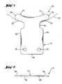
- FIG. 1is a plan view of an ophthalmic implant according to a first exemplary embodiment of the present invention, showing the implant having a body, feet, and a neck between the body and the feet.
- FIG. 2is a side view of the implant of FIG. 1 .
- FIG. 3is a cross-sectional view of a portion of an eye surgically prepared for implanting the ophthalmic implant of FIG. 1 according to an exemplary implantation method, showing a flap and recess created in the sclera of the eye.
- FIG. 4is a perspective view of the eye portion of FIG. 3 , showing undercuts being made at bottom corners of the scleral recess.
- FIG. 5is a plan view of the eye portion of FIG. 3 , after the undercuts have been made in the sclera.
- FIG. 6is a plan view of the eye portion of FIG. 3 , after the implant has been surgically implanted in the eye.
- FIG. 7is a plan view of the eye portion of FIG. 3 , showing the scleral opening made with a length that is greater than the length of the implant neck, according to another exemplary implantation method.
- FIG. 8is a plan view of an ophthalmic implant according to a second exemplary embodiment of the present invention, showing the implant having longitudinal drainage passageways.
- FIG. 9is a plan view of an ophthalmic implant according to a third exemplary embodiment of the present invention, showing the implant also having lateral drainage passageways.
- FIG. 10is a side view of an ophthalmic implant according to a fourth exemplary embodiment of the present invention, showing the implant having drainage passageways formed by channels in both outer surfaces of the implant.
- FIG. 11is a side view of an ophthalmic implant according to a fifth exemplary embodiment of the present invention, showing the implant made of two layers and having drainage passageways formed by channels in inner-facing surfaces of the implant.
- FIG. 12is a side view of an ophthalmic implant according to a sixth exemplary embodiment of the present invention, showing the implant made of three layers and having drainage passageways formed by voids in the inner layer of the implant.
- Rangesmay be expressed herein as from “about” or “approximately” one particular value and/or to “about” or “approximately” another particular value. When such a range is expressed, another embodiment includes from the one particular value and/or to the other particular value. Similarly, when values are expressed as approximations, by use of “about,” “approximately,” or the like, it will be understood that the particular value forms another embodiment.
- the present inventionprovides ophthalmic implants and surgical methods for implanting them in the eyes of people or animals suffering from glaucoma to reduce intraocular pressure (IOP).
- IOPintraocular pressure
- outflow of ocular fluid, the aqueous humor, from the anterior chamber of the patient's eyesis increased while better stabilizing the implant in the eye. This eliminates or at least significantly reduces the likelihood of glaucoma resulting in blindness.
- FIGS. 1 and 2show an ophthalmic implant according to a first exemplary embodiment of the present invention, generally referred to as the implant 10 .
- the implant 10has a body 12 , one or more feet 14 , and a neck 16 between the body and the feet.
- the body 12can be positioned under a flap and in a recess surgically created in the sclera, the feet 14 can be positioned in the anterior chamber, and the neck 16 can be positioned in an opening surgically created in the sclera between the scleral recess and the anterior chamber.
- the implant body 12is generally rectangular.
- the body 12can be triangular, polygonal, or it can have another shape.
- the body 12is about 4.0 mm long and about 3.5 mm wide.
- the implant body 12has outer portions 18 that can be tucked into undercuts created at bottom corners of and extending outward from the scleral recess. With the outer portions 18 of the body 12 tucked into the undercuts, the implant 10 is held more securely in place in the eye.
- the body 12has about 0.1 to 0.05 mm outer portions 18 at three sides. Alternatively, the body can have the outer portions 18 at only one or two sides.
- the outer portions 18are typically the same thickness as the rest of the body 12 .
- the implant body 12is manufactured with suture holes 20 for receiving sutures to secure the implant to the sclera.
- Conventional implantstypically do not have suture holes, so the surgeon has to pierce the implant body during the implantation surgery to suture the implant in place in the eye.
- the suture holes 20 in the body 12simplify the operation by eliminating the need for surgeons to create the suture holes during surgical implantation.
- the implant feet 14have a curvature 22 that is approximately the same as the curvature of the anterior chamber at the sclera of the patient.
- the curvature 22has a radius of about 5.5 mm.
- This junction of the sclera and the anterior chamber(formed by the space under the cornea) is known as the limbus corneae, or the anterior chamber angle, or simply, the angle. Because the feet 22 are so curved, they seat with a close fit against the limbus corneae. While the seating edge 24 of the feet 14 is so curved, the opposite edge need not be curved.
- the feet 14have outer portions 26 that extend beyond the width of the body 12 .
- the feet 14extend about 1.5 mm from the neck and the outer portions 26 extend about 1.0 mm beyond the body 12 . Because of the outer portions 26 , the feet 14 are long enough that they do not work their way out of the anterior chamber through the scleral opening. So if the sutures were to fail, the outer portions 26 of the feet 14 keep the implant 10 securely in place on the eye.
- the implant neck 16has a reduced width, relative to the body 12 and feet 16 . Additionally, the implant neck 16 has a greater length than previously cited implants. With the previously cited implants having very short necks provided by a slit or notch, the surgeon must cut the scleral recess close (within a small fraction of a millimeter) to the limbus corneae. If the cut is too deep, it will penetrate into the anterior chamber and the opening may allow the feet 14 to pass through it even when unfolded. Because the feet 14 could then migrate out of the anterior chamber and into the scleral, this site cannot then be used for the conventional implant. And because of the position of the rectus muscle, there are only four scleral sites where the implant can be readily implanted.
- the neck 16 of the present implant 10is sufficiently long that the body 12 is spaced apart from the anterior chamber so that the scleral recess does not need to be created immediately adjacent to the anterior chamber.
- the neck 16is about 0.8 mm long, about one-fifth of the length of the body 12 .
- the length of the neck 16is less than the length of the scleral opening created between the scleral recess and the anterior chamber.
- the neck 16can be about 0.8 mm long and the scleral opening can be made about 1.0 mm long, which is the approximate thickness of the limbus corneae transition between the sclera and the cornea. So when the implant 10 is positioned in the eye, the neck 16 is under tension. And the part of the sclera between the scleral recess and the anterior chamber is under compression. By placing the neck 16 under tension, the implant is less able to shift and migrate in the eye.
- the implant 10is made of regenerated cellulose, though other materials or a combination of materials with the desired strength, softness, flaccidness, and ocular biocompatibility may be selected.
- the materialis flexible for conforming the shape of the eye and so the feet can fold in for implanting.
- the implant 10can be manufactured by die-cutting or other fabrication techniques. In a typical commercial embodiment, the implant 10 has a generally uniform thickness of about 80 to 250 microns.
- the present inventionalso provides surgical techniques for implanting the ophthalmic implant 10 in the eye of a person or animal with glaucoma to reduce IOP. It will be understood that these exemplary methods can be used with other implants as long as they have the body outer portions 18 and/or the neck 16 of the implant 10 as described above.
- FIG. 3shows a portion of an eye 50 that has been surgically prepared for implanting the ophthalmic implant 10 .
- the eye 50has a sclera 52 , a cornea 54 , an angle 56 at the junction of the sclera and the cornea, an iris 58 , an anterior chamber 60 between the cornea and the iris, a posterior chamber 62 behind the iris, and a conjunctiva 64 covering the sclera.
- the surgical preparationincludes the steps of creating a flap 66 and thus a recess 68 under the flap in the sclera 52 , and creating an opening 70 in the sclera between the scleral recess 66 and the anterior chamber 60 .
- the scleral recess 68is made with a size and shape for receiving the implant body 12 (but not the outer portions 18 of the body), and the scleral opening 70 is made with a size and shape for passing through it the implant neck 16 and the implant feet 14 when folded in.
- the surgical preparationfurther includes the step of creating undercuts 72 in the sclera 52 extending outward from the scleral recess 68 .
- the undercuts 72are made with a size and shape for receiving the outer portions 18 of the implant body 12 .
- the undercuts 72can be made about 0.1 to 0.5 mm outward from the scleral recess 68 for receiving outer portions 18 of about the same size.
- the undercuts 72can be made at three sides of the scleral recess 68 , or at fewer or more sides if so desired.
- the undercuts 72are made at bottom corners 74 of the scleral recess 68 .
- the scleral recess 68 , scleral opening 70 , and scleral recess undercuts 72are preferably made by cutting with a scalpel 76 , but they could alternatively be made by a laser or by another surgical technique.
- FIG. 6shows the implant 10 inserted into the eye 10 .
- the insertion stepsinclude folding in the implant feet 14 and inserting the folded feet and the neck 16 through the scleral opening 70 so that the feet are inserted into the anterior chamber 60 and the neck is positioned in the scleral opening 70 .
- the elasticity of the implant materialcauses the feet 14 to unfold so that they do not migrate down out of the anterior chamber.
- the insertion stepsfurther include inserting the implant body 12 into the scleral recess 68 under the scleral flap 66 and inserting the body outer portions 18 into the undercuts 72 . With the implant body 12 nested within the scleral recess 68 and the body outer portions 18 tucked into the scleral undercuts 72 , the implant 10 is constrained from shifting around on the eye 50 .
- suturescan be sewn into the sclera 52 through the suture holes 20 in the implant 10 to further stabilize it in place. And the scleral flap 66 is sutured close to promote proper healing and help stabilize the implant 10 .
- the scleral opening 70is made with a length that is greater than the length of the implant neck 16 .
- the sceral flap 66 and recess 68are not made as close to the cornea 54 .
- the scleral opening 70may be made about 1.0 mm long, which is about the thickness of the limbus corneae, so the sceral flap 66 and recess 68 are made up to about 1.0 mm from the cornea 54 .
- the implanthas one or more drainage passageways for the ocular fluid to flow through out of the anterior chamber 60 and into the sclera 52 .
- the scleral flap 66tends to heal back into its original position.
- ocular fluidneed not flow out of the scleral through the scleral flap incisions because the lymphatic vessels in the sclera 52 absorb and disperse the ocular fluid.
- the implantis provided with the drainage passageways to facilitate ocular fluid drainage to the sclera 52 for dispersing by the lymphatic vessels.
- the drainage passagewaysare formed in the outer surfaces of the implant, in the interior of the implant, or in both.
- the drainage passagewaysmay be formed by die-stamping, laser ablation or by other fabrication techniques.
- FIG. 8shows a second exemplary embodiment of the invention with the implant 200 having drainage passageways provided by longitudinal drainage passageways 280 .
- the longitudinal drainage passageways 280provide a route for the ocular fluid to flow through, instead of just migrating through the cellulose material of the implant 200 .
- Any number of the longitudinal drainage passageways 280can be provided, depending on the amount of drainage desired and limitations imposed by the size of the implant 200 .
- FIG. 9shows a third exemplary embodiment of the invention with the implant 300 having drainage passageways provided by longitudinal drainage passageways 380 as well as lateral drainage passageways 382 .
- the lateral drainage passageways 382deliver the ocular fluid across the implant 300 to its sides for dispersing the fluid over a larger area into the sclera 52 for better absorption by the lymphatic vessels.
- main portions of the longitudinal drainage passageways 380 and/or the lateral drainage passageways 382may be thicker or deeper than branch portions, so as not to create a bottleneck in the fluid flow delivery system.
- Any number of the lateral drainage passageways 382may be provided, depending on the amount of drainage desired and limitations imposed by the size of the implant 200 .
- the implant 300may be provided with the lateral drainage passageways 382 but without the longitudinal drainage passageways 380 , if so desired.
- FIG. 10shows a fourth exemplary embodiment of the invention with the implant 400 having the drainage passageways formed by channels 484 in both outer surfaces 486 of the implant.
- the channels 484may be configured in alignment with each other (one over another), or they may be staggered in a regular or irregular pattern.
- FIG. 11shows a fifth exemplary embodiment of the invention with the implant 500 made of two layers 588 and 589 , and having the drainage passageways formed by channels 590 and 591 in inner-facing surfaces 592 and 593 of the implant layers.
- the layers 588 and 589may be laminated together or folded over each other, and more than two layers may be provided, if desired.
- the channels 590 and 591may be configured to align with each other (one over another), or they may be staggered in a regular or irregular pattern.
- FIG. 12shows a sixth exemplary embodiment of the invention with the implant 600 made of three layers, with two outer layers 694 and one inner layer 695 .
- the drainage passagewaysare formed by one or more voids 696 in the inner layer 695 of the implant 600 .
- the layers 694 and 695may be laminated together or folded over each other, and more than one inner layer may be provided, if desired. And the layers 694 and 695 may have also surface channels in alignment with the voids 696 or staggered in a regular or irregular pattern.
- the implant feetextend directly from the body instead of indirectly from the body with the neck in between.
- the bodydoes not migrate through the scleral opening into the anterior chamber, however, because the sutures hold it in place.
- the implanthas dimensions that are larger than or smaller than those of the typical commercial embodiments for an average-sized adult as described above. For example, smaller implants could be used for children and/or pets, and larger ones for large adults and/or animals.
- the present inventionprovides an improved ophthalmic implant for lowering the IOP in the eyes of glaucoma patients.
- the ophthalmic implanthas one or more drainage passageways formed in the outer surfaces and/or the interior of the implants to drain more ocular fluid out of the anterior chamber of the eye.
- the implanthas a uniquely configured body, feet, and/or neck to increase the stability of the implant in the eye.
- the present inventionprovides surgical implantation methods including providing undercuts and scleral openings sized to better stabilize the implants securely in place. And the implants are preferably of a simple construction using known materials such that they are time and cost-effective to manufacture and implant.
Landscapes
- Health & Medical Sciences (AREA)
- Ophthalmology & Optometry (AREA)
- Life Sciences & Earth Sciences (AREA)
- Animal Behavior & Ethology (AREA)
- Engineering & Computer Science (AREA)
- Biomedical Technology (AREA)
- Heart & Thoracic Surgery (AREA)
- Vascular Medicine (AREA)
- Nuclear Medicine, Radiotherapy & Molecular Imaging (AREA)
- Surgery (AREA)
- General Health & Medical Sciences (AREA)
- Public Health (AREA)
- Veterinary Medicine (AREA)
- Prostheses (AREA)
- Acyclic And Carbocyclic Compounds In Medicinal Compositions (AREA)
- Medicinal Preparation (AREA)
- Aeration Devices For Treatment Of Activated Polluted Sludge (AREA)
Abstract
Description
Claims (20)
Priority Applications (1)
| Application Number | Priority Date | Filing Date | Title |
|---|---|---|---|
| US10/737,707US7160264B2 (en) | 2002-12-19 | 2003-12-16 | Article and method for ocular aqueous drainage |
Applications Claiming Priority (2)
| Application Number | Priority Date | Filing Date | Title |
|---|---|---|---|
| US43494602P | 2002-12-19 | 2002-12-19 | |
| US10/737,707US7160264B2 (en) | 2002-12-19 | 2003-12-16 | Article and method for ocular aqueous drainage |
Publications (2)
| Publication Number | Publication Date |
|---|---|
| US20040260227A1 US20040260227A1 (en) | 2004-12-23 |
| US7160264B2true US7160264B2 (en) | 2007-01-09 |
Family
ID=32682127
Family Applications (1)
| Application Number | Title | Priority Date | Filing Date |
|---|---|---|---|
| US10/737,707Expired - LifetimeUS7160264B2 (en) | 2002-12-19 | 2003-12-16 | Article and method for ocular aqueous drainage |
Country Status (8)
| Country | Link |
|---|---|
| US (1) | US7160264B2 (en) |
| EP (1) | EP1578319B1 (en) |
| AT (1) | ATE471706T1 (en) |
| AU (1) | AU2003290089A1 (en) |
| CA (1) | CA2516976C (en) |
| DE (1) | DE60333124D1 (en) |
| ES (1) | ES2346430T3 (en) |
| WO (1) | WO2004056294A1 (en) |
Cited By (19)
| Publication number | Priority date | Publication date | Assignee | Title |
|---|---|---|---|---|
| US20050107734A1 (en)* | 2003-11-14 | 2005-05-19 | Coroneo Minas T. | Ocular pressure regulation |
| US20070149915A1 (en)* | 2003-05-05 | 2007-06-28 | Judith Yablonski | Internal shunt and method for treating glaucoma |
| US20070191863A1 (en)* | 2006-01-17 | 2007-08-16 | De Juan Eugene Jr | Glaucoma Treatment Device |
| US20070233037A1 (en)* | 2006-01-17 | 2007-10-04 | Gifford Hanson S Iii | Drug Delivery Treatment Device |
| US20100114006A1 (en)* | 2008-11-05 | 2010-05-06 | Advanced Medical Optics, Inc. | Glaucoma drainage shunts and methods of use |
| US20100137981A1 (en)* | 2008-06-25 | 2010-06-03 | Silvestrini Thomas A | Ocular implant with shape change capabilities |
| US20100249691A1 (en)* | 2009-03-26 | 2010-09-30 | Abbott Medical Optics Inc. | Glaucoma shunts with flow management and improved surgical performance |
| US20100274258A1 (en)* | 2009-01-28 | 2010-10-28 | Silvestrini Thomas A | Ocular implant with stiffness qualities, methods of implantation and system |
| US20110105990A1 (en)* | 2009-11-04 | 2011-05-05 | Silvestrini Thomas A | Zonal drug delivery device and method |
| US20110238075A1 (en)* | 2009-12-23 | 2011-09-29 | Luke Clauson | Drug delivery devices and methods |
| US8672870B2 (en) | 2007-07-17 | 2014-03-18 | Transcend Medical, Inc. | Ocular implant with hydrogel expansion capabilities |
| US8926510B2 (en) | 2011-04-27 | 2015-01-06 | Istar Medical Sa | Device and method for glaucoma management and treatment |
| US9155656B2 (en) | 2012-04-24 | 2015-10-13 | Transcend Medical, Inc. | Delivery system for ocular implant |
| US9480598B2 (en) | 2012-09-17 | 2016-11-01 | Novartis Ag | Expanding ocular implant devices and methods |
| US9763829B2 (en) | 2012-11-14 | 2017-09-19 | Novartis Ag | Flow promoting ocular implant |
| US9987163B2 (en) | 2013-04-16 | 2018-06-05 | Novartis Ag | Device for dispensing intraocular substances |
| US10085633B2 (en) | 2012-04-19 | 2018-10-02 | Novartis Ag | Direct visualization system for glaucoma treatment |
| US10842916B2 (en) | 2015-06-24 | 2020-11-24 | Healionics Corporation | Injectable porous device for treatment of dry and wet age-related macular degeneration or diabetic retinopathy |
| US11672701B2 (en) | 2018-10-25 | 2023-06-13 | Amo Groningen B.V. | Bleb control glaucoma shunts |
Families Citing this family (55)
| Publication number | Priority date | Publication date | Assignee | Title |
|---|---|---|---|---|
| KR20020035476A (en) | 1999-04-26 | 2002-05-11 | 지엠피 비젼 솔루션즈 인코포레이티드 | Shunt device and method for treating glaucoma |
| US6638239B1 (en) | 2000-04-14 | 2003-10-28 | Glaukos Corporation | Apparatus and method for treating glaucoma |
| US7867186B2 (en) | 2002-04-08 | 2011-01-11 | Glaukos Corporation | Devices and methods for treatment of ocular disorders |
| US7431710B2 (en) | 2002-04-08 | 2008-10-07 | Glaukos Corporation | Ocular implants with anchors and methods thereof |
| AU2002258754B2 (en) | 2001-04-07 | 2006-08-17 | Glaukos Corporation | Glaucoma stent and methods thereof for glaucoma treatment |
| US7331984B2 (en) | 2001-08-28 | 2008-02-19 | Glaukos Corporation | Glaucoma stent for treating glaucoma and methods of use |
| ES2523454T3 (en)* | 2003-06-16 | 2014-11-26 | Solx, Inc. | Referral for the treatment of glaucoma |
| US20060069340A1 (en)* | 2003-06-16 | 2006-03-30 | Solx, Inc. | Shunt for the treatment of glaucoma |
| US9095411B2 (en) | 2010-11-15 | 2015-08-04 | Aquesys, Inc. | Devices for deploying intraocular shunts |
| US10085884B2 (en) | 2006-06-30 | 2018-10-02 | Aquesys, Inc. | Intraocular devices |
| US8758290B2 (en) | 2010-11-15 | 2014-06-24 | Aquesys, Inc. | Devices and methods for implanting a shunt in the suprachoroidal space |
| US8721702B2 (en) | 2010-11-15 | 2014-05-13 | Aquesys, Inc. | Intraocular shunt deployment devices |
| JP5396272B2 (en)* | 2006-06-30 | 2014-01-22 | アクエシス インコーポレイテッド | Method, system and apparatus for reducing pressure in an organ |
| US8974511B2 (en) | 2010-11-15 | 2015-03-10 | Aquesys, Inc. | Methods for treating closed angle glaucoma |
| US20120123316A1 (en) | 2010-11-15 | 2012-05-17 | Aquesys, Inc. | Intraocular shunts for placement in the intra-tenon's space |
| US8828070B2 (en) | 2010-11-15 | 2014-09-09 | Aquesys, Inc. | Devices for deploying intraocular shunts |
| US8852137B2 (en) | 2010-11-15 | 2014-10-07 | Aquesys, Inc. | Methods for implanting a soft gel shunt in the suprachoroidal space |
| US8308701B2 (en) | 2010-11-15 | 2012-11-13 | Aquesys, Inc. | Methods for deploying intraocular shunts |
| US8852256B2 (en) | 2010-11-15 | 2014-10-07 | Aquesys, Inc. | Methods for intraocular shunt placement |
| US8663303B2 (en) | 2010-11-15 | 2014-03-04 | Aquesys, Inc. | Methods for deploying an intraocular shunt from a deployment device and into an eye |
| US8801766B2 (en) | 2010-11-15 | 2014-08-12 | Aquesys, Inc. | Devices for deploying intraocular shunts |
| EP2088976B1 (en) | 2006-11-10 | 2019-07-03 | Glaukos Corporation | Uveoscleral shunt |
| US8968396B2 (en) | 2007-07-23 | 2015-03-03 | Powervision, Inc. | Intraocular lens delivery systems and methods of use |
| WO2009031319A1 (en)* | 2007-09-07 | 2009-03-12 | Yugen Kaisha Conan | Drainage device for ophthalmic surgery |
| US8585629B2 (en) | 2010-11-15 | 2013-11-19 | Aquesys, Inc. | Systems for deploying intraocular shunts |
| US20160256320A1 (en) | 2010-11-15 | 2016-09-08 | Aquesys, Inc. | Intraocular shunt placement in the suprachoroidal space |
| US9610195B2 (en) | 2013-02-27 | 2017-04-04 | Aquesys, Inc. | Intraocular shunt implantation methods and devices |
| US9808373B2 (en) | 2013-06-28 | 2017-11-07 | Aquesys, Inc. | Intraocular shunt implantation |
| US8765210B2 (en) | 2011-12-08 | 2014-07-01 | Aquesys, Inc. | Systems and methods for making gelatin shunts |
| US8852136B2 (en) | 2011-12-08 | 2014-10-07 | Aquesys, Inc. | Methods for placing a shunt into the intra-scleral space |
| US10080682B2 (en) | 2011-12-08 | 2018-09-25 | Aquesys, Inc. | Intrascleral shunt placement |
| JP6008992B2 (en) | 2012-02-13 | 2016-10-19 | イリデックス・コーポレーション | Reduction of intraocular pressure using a tubular clip |
| CA2868341C (en) | 2012-03-26 | 2021-01-12 | Glaukos Corporation | System and method for delivering multiple ocular implants |
| US9125723B2 (en) | 2013-02-19 | 2015-09-08 | Aquesys, Inc. | Adjustable glaucoma implant |
| US10159600B2 (en) | 2013-02-19 | 2018-12-25 | Aquesys, Inc. | Adjustable intraocular flow regulation |
| US9592151B2 (en) | 2013-03-15 | 2017-03-14 | Glaukos Corporation | Systems and methods for delivering an ocular implant to the suprachoroidal space within an eye |
| EP3785668A1 (en) | 2013-03-15 | 2021-03-03 | Alcon Inc. | Intraocular lens storage and loading devices and methods of use |
| US10517759B2 (en) | 2013-03-15 | 2019-12-31 | Glaukos Corporation | Glaucoma stent and methods thereof for glaucoma treatment |
| EP3068354B1 (en) | 2013-11-14 | 2023-06-28 | Aquesys, Inc. | Intraocular shunt inserter |
| EP3677229A1 (en) | 2014-05-29 | 2020-07-08 | Glaukos Corporation | Implants with controlled drug delivery features |
| PE20151266A1 (en)* | 2014-07-01 | 2015-09-10 | Velasquez Mario Eduardo Miranda | DRAINAGE DEVICE FOR THE CONTROL OF INTRAOCULAR PRESSURE IN GLAUCOMA |
| JP7030516B2 (en) | 2014-12-31 | 2022-03-07 | マイクロオプティクス インコーポレイテッド | Glaucoma treatment equipment and methods |
| MA42406A (en) | 2015-06-03 | 2018-05-16 | Aquesys Inc | IMPLEMENTATION OF INTRAOCULAR AB EXTERNO SHUNT |
| US11925578B2 (en) | 2015-09-02 | 2024-03-12 | Glaukos Corporation | Drug delivery implants with bi-directional delivery capacity |
| CN108778398B (en) | 2015-09-30 | 2021-12-14 | 迈克罗欧普提克斯股份有限公司 | Dry eye treatment device and method |
| MX2018014763A (en) | 2016-06-02 | 2019-04-29 | Aquesys Inc | Intraocular drug delivery. |
| US20200078215A1 (en)* | 2016-07-06 | 2020-03-12 | MicroOptx Inc. | Glaucoma treatment devices and methods |
| US11116625B2 (en) | 2017-09-28 | 2021-09-14 | Glaukos Corporation | Apparatus and method for controlling placement of intraocular implants |
| US11246753B2 (en) | 2017-11-08 | 2022-02-15 | Aquesys, Inc. | Manually adjustable intraocular flow regulation |
| US11717440B2 (en)* | 2018-01-23 | 2023-08-08 | Avisi Technologies, Inc. | Method and device for treating eye disease |
| US10952898B2 (en) | 2018-03-09 | 2021-03-23 | Aquesys, Inc. | Intraocular shunt inserter |
| US11135089B2 (en) | 2018-03-09 | 2021-10-05 | Aquesys, Inc. | Intraocular shunt inserter |
| WO2019195419A1 (en) | 2018-04-03 | 2019-10-10 | Jack Chu | A new ocular device and method for glaucoma treatment |
| CN117159273A (en)* | 2018-08-31 | 2023-12-05 | 新世界医学有限公司 | Inserter device for ocular implantation procedure |
| FR3119763B1 (en)* | 2022-04-15 | 2024-04-12 | Ciliatech | OPHTHALMOLOGICAL INTERPOSITION IMPLANT WITH CONTACT OUTPUT |
Citations (52)
| Publication number | Priority date | Publication date | Assignee | Title |
|---|---|---|---|---|
| US4037604A (en) | 1976-01-05 | 1977-07-26 | Newkirk John B | Artifical biological drainage device |
| US4402681A (en) | 1980-08-23 | 1983-09-06 | Haas Joseph S | Artificial implant valve for the regulation of intraocular pressure |
| US4428746A (en) | 1981-07-29 | 1984-01-31 | Antonio Mendez | Glaucoma treatment device |
| US4521210A (en) | 1982-12-27 | 1985-06-04 | Wong Vernon G | Eye implant for relieving glaucoma, and device and method for use therewith |
| US4722724A (en) | 1986-06-23 | 1988-02-02 | Stanley Schocket | Anterior chamber tube shunt to an encircling band, and related surgical procedure |
| SU1316114A1 (en) | 1985-02-12 | 1988-03-15 | Научно-исследовательский институт резиновых и латексных изделий | Implant for forming support stump after evisceration |
| US4750901A (en) | 1986-03-07 | 1988-06-14 | Molteno Anthony C B | Implant for drainage of aqueous humour |
| US4787885A (en) | 1984-04-06 | 1988-11-29 | Binder Perry S | Hydrogel seton |
| US4826478A (en) | 1986-06-23 | 1989-05-02 | Stanley Schocket | Anterior chamber tube shunt to an encircling band, and related surgical procedure |
| SU1535542A1 (en) | 1987-11-18 | 1990-01-15 | Всесоюзный Научно-Исследовательский Институт Глазных Болезней | Method of treating secondary graucoma |
| US4936825A (en)* | 1988-04-11 | 1990-06-26 | Ungerleider Bruce A | Method for reducing intraocular pressure caused by glaucoma |
| US4946436A (en) | 1989-11-17 | 1990-08-07 | Smith Stewart G | Pressure-relieving device and process for implanting |
| US5041081A (en) | 1990-05-18 | 1991-08-20 | Odrich Ronald B | Ocular implant for controlling glaucoma |
| US5092837A (en) | 1989-12-20 | 1992-03-03 | Robert Ritch | Method for the treatment of glaucoma |
| WO1994013234A1 (en) | 1992-12-17 | 1994-06-23 | Michael Andrew Coote | Implant device and method for treatment of glaucoma |
| US5342370A (en) | 1993-03-19 | 1994-08-30 | University Of Miami | Method and apparatus for implanting an artifical meshwork in glaucoma surgery |
| EP0214853B1 (en) | 1985-09-06 | 1994-11-30 | Minnesota Mining And Manufacturing Company | Viscoelastic collagen solution for ophthalmic use and method of preparation |
| US5397300A (en) | 1990-05-31 | 1995-03-14 | Iovision, Inc. | Glaucoma implant |
| US5433701A (en)* | 1994-12-21 | 1995-07-18 | Rubinstein; Mark H. | Apparatus for reducing ocular pressure |
| US5454796A (en) | 1991-04-09 | 1995-10-03 | Hood Laboratories | Device and method for controlling intraocular fluid pressure |
| US5476445A (en) | 1990-05-31 | 1995-12-19 | Iovision, Inc. | Glaucoma implant with a temporary flow restricting seal |
| WO1995035078A1 (en) | 1994-06-22 | 1995-12-28 | Chauvin Opsia | Sclerotomy implant |
| US5520631A (en) | 1994-07-22 | 1996-05-28 | Wound Healing Of Oklahoma | Method and apparatus for lowering the intraocular pressure of an eye |
| US5558630A (en) | 1994-12-30 | 1996-09-24 | Fisher; Bret L. | Intrascleral implant and method for the regulation of intraocular pressure |
| USRE35390E (en) | 1989-11-17 | 1996-12-03 | Smith; Stewart G. | Pressure relieving device and process for implanting |
| US5626558A (en) | 1995-05-05 | 1997-05-06 | Suson; John | Adjustable flow rate glaucoma shunt and method of using same |
| US5626559A (en) | 1994-05-02 | 1997-05-06 | Ramot University Authority For Applied Research And Industrial Development Ltd. | Ophthalmic device for draining excess intraocular fluid |
| US5681275A (en) | 1988-10-07 | 1997-10-28 | Ahmed; Abdul Mateen | Ophthalmological device with adaptable multiple distribution plates |
| US5702414A (en) | 1995-05-14 | 1997-12-30 | Optonol Ltd | Method of implanting an intraocular implant |
| US5704907A (en) | 1994-07-22 | 1998-01-06 | Wound Healing Of Oklahoma | Method and apparatus for lowering the intraocular pressure of an eye |
| US5713844A (en) | 1997-01-10 | 1998-02-03 | Peyman; Gholam A. | Device and method for regulating intraocular pressure |
| US5743868A (en) | 1994-02-14 | 1998-04-28 | Brown; Reay H. | Corneal pressure-regulating implant device |
| NL1005694C2 (en) | 1997-04-01 | 1998-10-12 | Amc Amsterdam | Control of fluid pressure within the eye ball for glaucoma sufferers |
| US5882327A (en) | 1997-04-17 | 1999-03-16 | Jacob; Jean T. | Long-term glaucoma drainage implant |
| US5893837A (en) | 1997-02-28 | 1999-04-13 | Staar Surgical Company, Inc. | Glaucoma drain implanting device and method |
| US5968058A (en) | 1996-03-27 | 1999-10-19 | Optonol Ltd. | Device for and method of implanting an intraocular implant |
| US6001128A (en) | 1997-05-29 | 1999-12-14 | Alcon Laboratories, Inc. | Materials for use in glaucoma filtration devices |
| US6050970A (en) | 1997-05-08 | 2000-04-18 | Pharmacia & Upjohn Company | Method and apparatus for inserting a glaucoma implant in an anterior and posterior segment of the eye |
| US6102045A (en) | 1994-07-22 | 2000-08-15 | Premier Laser Systems, Inc. | Method and apparatus for lowering the intraocular pressure of an eye |
| US6186974B1 (en) | 1997-01-10 | 2001-02-13 | University College London And Moorfields Eye Hospital Nhs Trust | Device for use in the eye |
| US6203513B1 (en) | 1997-11-20 | 2001-03-20 | Optonol Ltd. | Flow regulating implant, method of manufacture, and delivery device |
| DE19947711A1 (en) | 1999-10-04 | 2001-05-03 | Norbert Schrage | Implant especially for the treatment of glaucoma comprises removable peduncular body for insertion in the eye |
| WO2001079656A1 (en) | 2000-04-07 | 2001-10-25 | Vladimir Ivanovich Ivannikov | Device for flow and liftgas production of oil-wells (versions) |
| WO2002017832A1 (en) | 2000-09-01 | 2002-03-07 | Ioltechnologie-Production | Glaucoma drain |
| WO2002032343A2 (en) | 2000-10-18 | 2002-04-25 | Wilcox Michael J | C-shaped cross section tubular ophthalmic implant for reduction of intraocular pressure and method of use |
| US6383218B1 (en) | 1997-02-17 | 2002-05-07 | Corneal Industrie | Sclero-ceratectomy implant for descemet's membrane |
| US6383219B1 (en) | 1997-02-17 | 2002-05-07 | Corneal Industrie | Implant for deep sclerectomy |
| US6450984B1 (en) | 1999-04-26 | 2002-09-17 | Gmp Vision Solutions, Inc. | Shunt device and method for treating glaucoma |
| AT409586B (en) | 2001-04-26 | 2002-09-25 | Clemens Dr Vass | Implant draining aqueous humor from anterior chamber of eye into Schlemm's channel, includes fixation plate for stabilization on sclera |
| WO2002080811A2 (en) | 2001-04-07 | 2002-10-17 | Glaukos Corporation | Glaucoma stent and methods thereof for glaucoma treatment |
| US6736791B1 (en)* | 2000-04-14 | 2004-05-18 | Glaukos Corporation | Glaucoma treatment device |
| US20040098123A1 (en)* | 2002-11-19 | 2004-05-20 | Freeman Jerre M. | Bulbous scleral implants for the treatment of eye disorders such as presbyopia and glaucoma |
Family Cites Families (1)
| Publication number | Priority date | Publication date | Assignee | Title |
|---|---|---|---|---|
| US35390A (en)* | 1862-05-27 | Improved guide and support for scroll-saws |
- 2003
- 2003-12-16USUS10/737,707patent/US7160264B2/ennot_activeExpired - Lifetime
- 2003-12-18ESES03782448Tpatent/ES2346430T3/ennot_activeExpired - Lifetime
- 2003-12-18EPEP03782448Apatent/EP1578319B1/ennot_activeExpired - Lifetime
- 2003-12-18DEDE60333124Tpatent/DE60333124D1/ennot_activeExpired - Lifetime
- 2003-12-18WOPCT/EP2003/014532patent/WO2004056294A1/ennot_activeApplication Discontinuation
- 2003-12-18CACA2516976Apatent/CA2516976C/ennot_activeExpired - Lifetime
- 2003-12-18ATAT03782448Tpatent/ATE471706T1/ennot_activeIP Right Cessation
- 2003-12-18AUAU2003290089Apatent/AU2003290089A1/ennot_activeAbandoned
Patent Citations (55)
| Publication number | Priority date | Publication date | Assignee | Title |
|---|---|---|---|---|
| US4037604A (en) | 1976-01-05 | 1977-07-26 | Newkirk John B | Artifical biological drainage device |
| US4402681A (en) | 1980-08-23 | 1983-09-06 | Haas Joseph S | Artificial implant valve for the regulation of intraocular pressure |
| US4428746A (en) | 1981-07-29 | 1984-01-31 | Antonio Mendez | Glaucoma treatment device |
| US4521210A (en) | 1982-12-27 | 1985-06-04 | Wong Vernon G | Eye implant for relieving glaucoma, and device and method for use therewith |
| US4787885A (en) | 1984-04-06 | 1988-11-29 | Binder Perry S | Hydrogel seton |
| SU1316114A1 (en) | 1985-02-12 | 1988-03-15 | Научно-исследовательский институт резиновых и латексных изделий | Implant for forming support stump after evisceration |
| EP0214853B1 (en) | 1985-09-06 | 1994-11-30 | Minnesota Mining And Manufacturing Company | Viscoelastic collagen solution for ophthalmic use and method of preparation |
| US4750901A (en) | 1986-03-07 | 1988-06-14 | Molteno Anthony C B | Implant for drainage of aqueous humour |
| US4826478A (en) | 1986-06-23 | 1989-05-02 | Stanley Schocket | Anterior chamber tube shunt to an encircling band, and related surgical procedure |
| US4722724A (en) | 1986-06-23 | 1988-02-02 | Stanley Schocket | Anterior chamber tube shunt to an encircling band, and related surgical procedure |
| SU1535542A1 (en) | 1987-11-18 | 1990-01-15 | Всесоюзный Научно-Исследовательский Институт Глазных Болезней | Method of treating secondary graucoma |
| US4936825A (en)* | 1988-04-11 | 1990-06-26 | Ungerleider Bruce A | Method for reducing intraocular pressure caused by glaucoma |
| US5372577A (en) | 1988-04-11 | 1994-12-13 | Ungerleider; Bruce A. | Apparatus for reducing intraocular pressure |
| US5681275A (en) | 1988-10-07 | 1997-10-28 | Ahmed; Abdul Mateen | Ophthalmological device with adaptable multiple distribution plates |
| US4946436A (en) | 1989-11-17 | 1990-08-07 | Smith Stewart G | Pressure-relieving device and process for implanting |
| USRE35390E (en) | 1989-11-17 | 1996-12-03 | Smith; Stewart G. | Pressure relieving device and process for implanting |
| US5092837A (en) | 1989-12-20 | 1992-03-03 | Robert Ritch | Method for the treatment of glaucoma |
| US5041081A (en) | 1990-05-18 | 1991-08-20 | Odrich Ronald B | Ocular implant for controlling glaucoma |
| US5397300A (en) | 1990-05-31 | 1995-03-14 | Iovision, Inc. | Glaucoma implant |
| US5476445A (en) | 1990-05-31 | 1995-12-19 | Iovision, Inc. | Glaucoma implant with a temporary flow restricting seal |
| US5454796A (en) | 1991-04-09 | 1995-10-03 | Hood Laboratories | Device and method for controlling intraocular fluid pressure |
| WO1994013234A1 (en) | 1992-12-17 | 1994-06-23 | Michael Andrew Coote | Implant device and method for treatment of glaucoma |
| US5342370A (en) | 1993-03-19 | 1994-08-30 | University Of Miami | Method and apparatus for implanting an artifical meshwork in glaucoma surgery |
| US5651782A (en) | 1993-03-19 | 1997-07-29 | University Of Miami | Method and apparatus for implanting an artificial meshwork in glaucoma surgery |
| US5743868A (en) | 1994-02-14 | 1998-04-28 | Brown; Reay H. | Corneal pressure-regulating implant device |
| US5626559A (en) | 1994-05-02 | 1997-05-06 | Ramot University Authority For Applied Research And Industrial Development Ltd. | Ophthalmic device for draining excess intraocular fluid |
| WO1995035078A1 (en) | 1994-06-22 | 1995-12-28 | Chauvin Opsia | Sclerotomy implant |
| US5704907A (en) | 1994-07-22 | 1998-01-06 | Wound Healing Of Oklahoma | Method and apparatus for lowering the intraocular pressure of an eye |
| US6102045A (en) | 1994-07-22 | 2000-08-15 | Premier Laser Systems, Inc. | Method and apparatus for lowering the intraocular pressure of an eye |
| US5520631A (en) | 1994-07-22 | 1996-05-28 | Wound Healing Of Oklahoma | Method and apparatus for lowering the intraocular pressure of an eye |
| US5433701A (en)* | 1994-12-21 | 1995-07-18 | Rubinstein; Mark H. | Apparatus for reducing ocular pressure |
| US5558630A (en) | 1994-12-30 | 1996-09-24 | Fisher; Bret L. | Intrascleral implant and method for the regulation of intraocular pressure |
| US5626558A (en) | 1995-05-05 | 1997-05-06 | Suson; John | Adjustable flow rate glaucoma shunt and method of using same |
| US5702414A (en) | 1995-05-14 | 1997-12-30 | Optonol Ltd | Method of implanting an intraocular implant |
| US5968058A (en) | 1996-03-27 | 1999-10-19 | Optonol Ltd. | Device for and method of implanting an intraocular implant |
| US6186974B1 (en) | 1997-01-10 | 2001-02-13 | University College London And Moorfields Eye Hospital Nhs Trust | Device for use in the eye |
| US5713844A (en) | 1997-01-10 | 1998-02-03 | Peyman; Gholam A. | Device and method for regulating intraocular pressure |
| US6383218B1 (en) | 1997-02-17 | 2002-05-07 | Corneal Industrie | Sclero-ceratectomy implant for descemet's membrane |
| US6383219B1 (en) | 1997-02-17 | 2002-05-07 | Corneal Industrie | Implant for deep sclerectomy |
| US5893837A (en) | 1997-02-28 | 1999-04-13 | Staar Surgical Company, Inc. | Glaucoma drain implanting device and method |
| NL1005694C2 (en) | 1997-04-01 | 1998-10-12 | Amc Amsterdam | Control of fluid pressure within the eye ball for glaucoma sufferers |
| US5882327A (en) | 1997-04-17 | 1999-03-16 | Jacob; Jean T. | Long-term glaucoma drainage implant |
| US6050970A (en) | 1997-05-08 | 2000-04-18 | Pharmacia & Upjohn Company | Method and apparatus for inserting a glaucoma implant in an anterior and posterior segment of the eye |
| US6001128A (en) | 1997-05-29 | 1999-12-14 | Alcon Laboratories, Inc. | Materials for use in glaucoma filtration devices |
| US6203513B1 (en) | 1997-11-20 | 2001-03-20 | Optonol Ltd. | Flow regulating implant, method of manufacture, and delivery device |
| US6450984B1 (en) | 1999-04-26 | 2002-09-17 | Gmp Vision Solutions, Inc. | Shunt device and method for treating glaucoma |
| DE19947711A1 (en) | 1999-10-04 | 2001-05-03 | Norbert Schrage | Implant especially for the treatment of glaucoma comprises removable peduncular body for insertion in the eye |
| WO2001079656A1 (en) | 2000-04-07 | 2001-10-25 | Vladimir Ivanovich Ivannikov | Device for flow and liftgas production of oil-wells (versions) |
| US6736791B1 (en)* | 2000-04-14 | 2004-05-18 | Glaukos Corporation | Glaucoma treatment device |
| WO2002017832A1 (en) | 2000-09-01 | 2002-03-07 | Ioltechnologie-Production | Glaucoma drain |
| US20040092856A1 (en)* | 2000-09-01 | 2004-05-13 | Elie Dahan | Glaucoma drain |
| WO2002032343A2 (en) | 2000-10-18 | 2002-04-25 | Wilcox Michael J | C-shaped cross section tubular ophthalmic implant for reduction of intraocular pressure and method of use |
| WO2002080811A2 (en) | 2001-04-07 | 2002-10-17 | Glaukos Corporation | Glaucoma stent and methods thereof for glaucoma treatment |
| AT409586B (en) | 2001-04-26 | 2002-09-25 | Clemens Dr Vass | Implant draining aqueous humor from anterior chamber of eye into Schlemm's channel, includes fixation plate for stabilization on sclera |
| US20040098123A1 (en)* | 2002-11-19 | 2004-05-20 | Freeman Jerre M. | Bulbous scleral implants for the treatment of eye disorders such as presbyopia and glaucoma |
Non-Patent Citations (1)
| Title |
|---|
| 'Ex-PRESS Miniature Glaucoma Implant,' Optonol-Product Information, http://www.optonal.com/info.html, (date unknown), pp. 1-6. |
Cited By (78)
| Publication number | Priority date | Publication date | Assignee | Title |
|---|---|---|---|---|
| US20070149915A1 (en)* | 2003-05-05 | 2007-06-28 | Judith Yablonski | Internal shunt and method for treating glaucoma |
| US8945038B2 (en) | 2003-05-05 | 2015-02-03 | Transcend Medical, Inc. | Internal shunt and method for treating glaucoma |
| US9844462B2 (en) | 2003-05-05 | 2017-12-19 | Novartis Ag | Internal shunt and method for treating glaucoma |
| US8444588B2 (en) | 2003-05-05 | 2013-05-21 | Transcend Medical, Inc. | Internal shunt and method for treating glaucoma |
| US8486000B2 (en) | 2003-11-14 | 2013-07-16 | Transcend Medical, Inc. | Ocular pressure regulation |
| US7850638B2 (en) | 2003-11-14 | 2010-12-14 | Transcend Medical, Inc. | Ocular pressure regulation |
| US8758289B2 (en) | 2003-11-14 | 2014-06-24 | Transcend Medical, Inc. | Ocular pressure regulation |
| US7291125B2 (en) | 2003-11-14 | 2007-11-06 | Transcend Medical, Inc. | Ocular pressure regulation |
| US8771218B2 (en) | 2003-11-14 | 2014-07-08 | Transcend Medical, Inc. | Ocular pressure regulation |
| US8808220B2 (en) | 2003-11-14 | 2014-08-19 | Transcend Medical, Inc. | Ocular pressure regulation |
| US8128588B2 (en) | 2003-11-14 | 2012-03-06 | Transcend Medical, Inc. | Ocular pressure regulation |
| US7815592B2 (en) | 2003-11-14 | 2010-10-19 | Transcend Medical, Inc. | Ocular pressure regulation |
| US10226380B2 (en) | 2003-11-14 | 2019-03-12 | Novartis Ag | Ocular pressure regulation |
| US8728021B2 (en) | 2003-11-14 | 2014-05-20 | Transcend Medical, Inc. | Ocular pressure regulation |
| US20070106236A1 (en)* | 2003-11-14 | 2007-05-10 | Coroneo Minas T | Ocular Pressure Regulation |
| US20110028884A1 (en)* | 2003-11-14 | 2011-02-03 | Minas Theodore Coroneo | Ocular pressure regulation |
| US20070106235A1 (en)* | 2003-11-14 | 2007-05-10 | Coroneo Minas T | Ocular Pressure Regulation |
| US20110087149A1 (en)* | 2003-11-14 | 2011-04-14 | Minas Theodore Coroneo | Ocular pressure regulation |
| US20070088242A1 (en)* | 2003-11-14 | 2007-04-19 | Coroneo Minas T | Ocular pressure regulation |
| US20110087151A1 (en)* | 2003-11-14 | 2011-04-14 | Minas Theodore Coroneo | Ocular pressure regulation |
| US9351873B2 (en) | 2003-11-14 | 2016-05-31 | Transcend Medical, Inc. | Ocular pressure regulation |
| US20050107734A1 (en)* | 2003-11-14 | 2005-05-19 | Coroneo Minas T. | Ocular pressure regulation |
| US11786402B2 (en) | 2006-01-17 | 2023-10-17 | Alcon Inc. | Glaucoma treatment device |
| US9421130B2 (en) | 2006-01-17 | 2016-08-23 | Novartis Ag. | Glaucoma treatment device |
| US9398977B2 (en) | 2006-01-17 | 2016-07-26 | Transcend Medical, Inc. | Glaucoma treatment device |
| US9668917B2 (en) | 2006-01-17 | 2017-06-06 | Novartis Ag | Drug delivery treatment device |
| US9084662B2 (en) | 2006-01-17 | 2015-07-21 | Transcend Medical, Inc. | Drug delivery treatment device |
| US9789000B2 (en) | 2006-01-17 | 2017-10-17 | Novartis Ag | Glaucoma treatment device |
| US20110028883A1 (en)* | 2006-01-17 | 2011-02-03 | Juan Jr Eugene De | Glaucoma treatment device |
| US10905590B2 (en) | 2006-01-17 | 2021-02-02 | Alcon Inc. | Glaucoma treatment device |
| US8814819B2 (en) | 2006-01-17 | 2014-08-26 | Transcend Medical, Inc. | Glaucoma treatment device |
| US8734378B2 (en) | 2006-01-17 | 2014-05-27 | Transcend Medical, Inc. | Glaucoma treatment device |
| US8801649B2 (en) | 2006-01-17 | 2014-08-12 | Transcend Medical, Inc. | Glaucoma treatment device |
| US12303430B2 (en) | 2006-01-17 | 2025-05-20 | Alcon Inc. | Glaucoma treatment device |
| US20070233037A1 (en)* | 2006-01-17 | 2007-10-04 | Gifford Hanson S Iii | Drug Delivery Treatment Device |
| US8721656B2 (en) | 2006-01-17 | 2014-05-13 | Transcend Medical, Inc. | Glaucoma treatment device |
| US20070191863A1 (en)* | 2006-01-17 | 2007-08-16 | De Juan Eugene Jr | Glaucoma Treatment Device |
| US9585789B2 (en) | 2007-07-17 | 2017-03-07 | Novartis Ag | Ocular implant with hydrogel expansion capabilities |
| US8672870B2 (en) | 2007-07-17 | 2014-03-18 | Transcend Medical, Inc. | Ocular implant with hydrogel expansion capabilities |
| US8617139B2 (en) | 2008-06-25 | 2013-12-31 | Transcend Medical, Inc. | Ocular implant with shape change capabilities |
| US20100137981A1 (en)* | 2008-06-25 | 2010-06-03 | Silvestrini Thomas A | Ocular implant with shape change capabilities |
| US10016301B2 (en) | 2008-06-25 | 2018-07-10 | Novartis Ag | Ocular implant with shape change capabilities |
| US9468558B2 (en) | 2008-11-05 | 2016-10-18 | Abbott Medical Optics Inc. | Glaucoma drainage shunts and methods of use |
| US20100114006A1 (en)* | 2008-11-05 | 2010-05-06 | Advanced Medical Optics, Inc. | Glaucoma drainage shunts and methods of use |
| US8920357B2 (en) | 2008-11-05 | 2014-12-30 | Abbott Medical Optics Inc. | Glaucoma drainage shunts and methods of use |
| US10492948B2 (en) | 2008-11-05 | 2019-12-03 | Johnson & Johnson Surgical Vision, Inc. | Glaucoma drainage shunts and methods of use |
| US8353856B2 (en) | 2008-11-05 | 2013-01-15 | Abbott Medical Optics Inc. | Glaucoma drainage shunts and methods of use |
| US10531983B2 (en) | 2009-01-28 | 2020-01-14 | Novartis Ag | Ocular implant with stiffness qualities, methods of implantation and system |
| US11344448B2 (en) | 2009-01-28 | 2022-05-31 | Alcon Inc. | Ocular implant with stiffness qualities, methods of implantation and system |
| US8574294B2 (en) | 2009-01-28 | 2013-11-05 | Transcend Medical, Inc. | Ocular implant with stiffness qualities, methods of implantation and system |
| US8262726B2 (en) | 2009-01-28 | 2012-09-11 | Transcend Medical, Inc. | Ocular implant with stiffness qualities, methods of implantation and system |
| US8172899B2 (en) | 2009-01-28 | 2012-05-08 | Transcend Medical, Inc. | Ocular implant with stiffness qualities, methods of implantation and system |
| US8167939B2 (en) | 2009-01-28 | 2012-05-01 | Transcend Medical, Inc. | Ocular implant with stiffness qualities, methods of implantation and system |
| US20100274258A1 (en)* | 2009-01-28 | 2010-10-28 | Silvestrini Thomas A | Ocular implant with stiffness qualities, methods of implantation and system |
| US12233004B2 (en) | 2009-01-28 | 2025-02-25 | Alcon Inc. | Ocular implant with stiffness qualities, methods of implantation and system |
| US8377122B2 (en) | 2009-01-28 | 2013-02-19 | Transcend Medical, Inc. | Ocular implant with stiffness qualities, methods of implantation and system |
| US20110028983A1 (en)* | 2009-01-28 | 2011-02-03 | Silvestrini Thomas A | Ocular implant with stiffness qualities, methods of implantation and system |
| US20110087148A1 (en)* | 2009-01-28 | 2011-04-14 | Silvestrini Thomas A | Ocular implant with stiffness qualities, methods of implantation and system |
| US9763828B2 (en) | 2009-01-28 | 2017-09-19 | Novartis Ag | Ocular implant with stiffness qualities, methods of implantation and system |
| US11839571B2 (en) | 2009-01-28 | 2023-12-12 | Alcon Inc. | Ocular implant with stiffness qualities, methods of implantation and system |
| US20100249691A1 (en)* | 2009-03-26 | 2010-09-30 | Abbott Medical Optics Inc. | Glaucoma shunts with flow management and improved surgical performance |
| US8702639B2 (en) | 2009-03-26 | 2014-04-22 | Abbott Medical Optics Inc. | Glaucoma shunts with flow management and improved surgical performance |
| US20110105990A1 (en)* | 2009-11-04 | 2011-05-05 | Silvestrini Thomas A | Zonal drug delivery device and method |
| US20110238075A1 (en)* | 2009-12-23 | 2011-09-29 | Luke Clauson | Drug delivery devices and methods |
| US8529492B2 (en) | 2009-12-23 | 2013-09-10 | Trascend Medical, Inc. | Drug delivery devices and methods |
| US9549846B2 (en) | 2009-12-23 | 2017-01-24 | Novartis Ag | Drug delivery devices and methods |
| US9089392B2 (en) | 2009-12-23 | 2015-07-28 | Transcend Medical, Inc. | Drug delivery devices and methods |
| US8926510B2 (en) | 2011-04-27 | 2015-01-06 | Istar Medical Sa | Device and method for glaucoma management and treatment |
| US10085633B2 (en) | 2012-04-19 | 2018-10-02 | Novartis Ag | Direct visualization system for glaucoma treatment |
| US9907697B2 (en) | 2012-04-24 | 2018-03-06 | Novartis Ag | Delivery system for ocular implant |
| US10912676B2 (en) | 2012-04-24 | 2021-02-09 | Alcon Inc. | Delivery system for ocular implant |
| US9155656B2 (en) | 2012-04-24 | 2015-10-13 | Transcend Medical, Inc. | Delivery system for ocular implant |
| US9241832B2 (en) | 2012-04-24 | 2016-01-26 | Transcend Medical, Inc. | Delivery system for ocular implant |
| US9480598B2 (en) | 2012-09-17 | 2016-11-01 | Novartis Ag | Expanding ocular implant devices and methods |
| US9763829B2 (en) | 2012-11-14 | 2017-09-19 | Novartis Ag | Flow promoting ocular implant |
| US9987163B2 (en) | 2013-04-16 | 2018-06-05 | Novartis Ag | Device for dispensing intraocular substances |
| US10842916B2 (en) | 2015-06-24 | 2020-11-24 | Healionics Corporation | Injectable porous device for treatment of dry and wet age-related macular degeneration or diabetic retinopathy |
| US11672701B2 (en) | 2018-10-25 | 2023-06-13 | Amo Groningen B.V. | Bleb control glaucoma shunts |
Also Published As
| Publication number | Publication date |
|---|---|
| EP1578319B1 (en) | 2010-06-23 |
| ATE471706T1 (en) | 2010-07-15 |
| EP1578319A1 (en) | 2005-09-28 |
| ES2346430T3 (en) | 2010-10-15 |
| CA2516976A1 (en) | 2004-07-08 |
| WO2004056294A1 (en) | 2004-07-08 |
| CA2516976C (en) | 2013-04-30 |
| AU2003290089A1 (en) | 2004-07-14 |
| DE60333124D1 (en) | 2010-08-05 |
| US20040260227A1 (en) | 2004-12-23 |
Similar Documents
| Publication | Publication Date | Title |
|---|---|---|
| US7160264B2 (en) | Article and method for ocular aqueous drainage | |
| US4787885A (en) | Hydrogel seton | |
| US20200330272A1 (en) | Glaucoma treatment devices and methods | |
| US4634418A (en) | Hydrogel seton | |
| JP3044238B2 (en) | Tools for lowering intraocular pressure | |
| EP2039380B1 (en) | Uveoscleral drainage device | |
| US5722948A (en) | Covering for an ocular device | |
| US4604087A (en) | Aqueous humor drainage device | |
| US4428746A (en) | Glaucoma treatment device | |
| US12350193B2 (en) | Dry eye treatment devices and methods | |
| US20080306429A1 (en) | Uveoscleral drainage device | |
| JP2002521145A (en) | Sutureless implantable devices and methods for treating glaucoma | |
| US20110105986A1 (en) | Uveoscleral drainage device | |
| US20220125638A1 (en) | Glaucoma treatment devices and methods | |
| US20180078416A1 (en) | Inlet tube protector for glaucoma shunts | |
| US20250248846A1 (en) | Ophtalmological interposition implant with engagement protusion | |
| KR20200026150A (en) | Modifiable-transformable and material-transferrable ocular disease implant apparatus | |
| KR102828111B1 (en) | Shunt for fibrous encapsulation in glaucoma patients | |
| US20230255826A1 (en) | Ocular drainage implant | |
| Dada | Astigmatic Considerations |
Legal Events
| Date | Code | Title | Description |
|---|---|---|---|
| AS | Assignment | Owner name:NOVARTIS AG, SWITZERLAND Free format text:ASSIGNMENT OF ASSIGNORS INTEREST;ASSIGNORS:LISK, JAMES R., JR.;MEMMEN, JAMES E.;HAMPTON, SCOTT, M.;AND OTHERS;REEL/FRAME:015034/0078;SIGNING DATES FROM 20040112 TO 20040329 | |
| AS | Assignment | Owner name:WOUND HEALING OF OKLAHOMA, INC., OKLAHOMA Free format text:ASSIGNMENT OF ASSIGNORS INTEREST;ASSIGNOR:NOVARTIS AG;REEL/FRAME:015611/0798 Effective date:20040915 | |
| AS | Assignment | Owner name:MEDTRONIC XOMED, INC., FLORIDA Free format text:ASSIGNMENT OF ASSIGNORS INTEREST;ASSIGNOR:WOUND HEALING OF OKLAHOMA, INC.;REEL/FRAME:015618/0622 Effective date:20040917 | |
| STCF | Information on status: patent grant | Free format text:PATENTED CASE | |
| FEPP | Fee payment procedure | Free format text:PAT HOLDER NO LONGER CLAIMS SMALL ENTITY STATUS, ENTITY STATUS SET TO UNDISCOUNTED (ORIGINAL EVENT CODE: STOL); ENTITY STATUS OF PATENT OWNER: LARGE ENTITY | |
| REFU | Refund | Free format text:REFUND - SURCHARGE, PETITION TO ACCEPT PYMT AFTER EXP, UNINTENTIONAL (ORIGINAL EVENT CODE: R2551); ENTITY STATUS OF PATENT OWNER: LARGE ENTITY | |
| AS | Assignment | Owner name:WOUND HEALING OF OKLAHOMA, INC.,OKLAHOMA Free format text:ASSIGNMENT OF ASSIGNORS INTEREST;ASSIGNOR:MEDTRONIC XOMED, INC.;REEL/FRAME:024066/0455 Effective date:20090513 | |
| FPAY | Fee payment | Year of fee payment:4 | |
| FEPP | Fee payment procedure | Free format text:PAT HOLDER CLAIMS SMALL ENTITY STATUS, ENTITY STATUS SET TO SMALL (ORIGINAL EVENT CODE: LTOS); ENTITY STATUS OF PATENT OWNER: LARGE ENTITY | |
| FPAY | Fee payment | Year of fee payment:8 | |
| FEPP | Fee payment procedure | Free format text:ENTITY STATUS SET TO UNDISCOUNTED (ORIGINAL EVENT CODE: BIG.) | |
| MAFP | Maintenance fee payment | Free format text:PAYMENT OF MAINTENANCE FEE, 12TH YEAR, LARGE ENTITY (ORIGINAL EVENT CODE: M1553) Year of fee payment:12 |