US7068874B2 - Microfluidic sorting device - Google Patents
Microfluidic sorting deviceDownload PDFInfo
- Publication number
- US7068874B2 US7068874B2US10/848,972US84897204AUS7068874B2US 7068874 B2US7068874 B2US 7068874B2US 84897204 AUS84897204 AUS 84897204AUS 7068874 B2US7068874 B2US 7068874B2
- Authority
- US
- United States
- Prior art keywords
- microfluidic
- particles
- optical
- channels
- branch
- Prior art date
- Legal status (The legal status is an assumption and is not a legal conclusion. Google has not performed a legal analysis and makes no representation as to the accuracy of the status listed.)
- Expired - Lifetime, expires
Links
Images


Classifications
- H—ELECTRICITY
- H05—ELECTRIC TECHNIQUES NOT OTHERWISE PROVIDED FOR
- H05H—PLASMA TECHNIQUE; PRODUCTION OF ACCELERATED ELECTRICALLY-CHARGED PARTICLES OR OF NEUTRONS; PRODUCTION OR ACCELERATION OF NEUTRAL MOLECULAR OR ATOMIC BEAMS
- H05H3/00—Production or acceleration of neutral particle beams, e.g. molecular or atomic beams
- H05H3/04—Acceleration by electromagnetic wave pressure
- Y—GENERAL TAGGING OF NEW TECHNOLOGICAL DEVELOPMENTS; GENERAL TAGGING OF CROSS-SECTIONAL TECHNOLOGIES SPANNING OVER SEVERAL SECTIONS OF THE IPC; TECHNICAL SUBJECTS COVERED BY FORMER USPC CROSS-REFERENCE ART COLLECTIONS [XRACs] AND DIGESTS
- Y10—TECHNICAL SUBJECTS COVERED BY FORMER USPC
- Y10T—TECHNICAL SUBJECTS COVERED BY FORMER US CLASSIFICATION
- Y10T428/00—Stock material or miscellaneous articles
- Y10T428/24—Structurally defined web or sheet [e.g., overall dimension, etc.]
- Y10T428/24744—Longitudinal or transverse tubular cavity or cell
Definitions
- the present inventiongenerally concerns optical tweezers, microfluidics, flow cytometry, biological Micro Optical Electro Mechanical Systems (Bio-MOEMS), Laguerre-Gaussian mode emissions from Vertical Cavity Surface Emitting Lasers (VCSELs), cell cytometry and microfluidic switches and switching.
- Bio-MOEMSMicro Optical Electro Mechanical Systems
- VCSELsVertical Cavity Surface Emitting Lasers
- the present inventionparticularly concerns the sorting of microparticles in fluid, thus a “microfluidic sorting device”; and also the directed movement, particularly for purposes of switching, of microparticles based on the transference of momentum from photons impinging on the microparticles, ergo “photonic momentum transfer”.
- the present inventionwill be seen to employ optical tweezers. See A. Ashkin, J. M. Dziedzic, J. E. Bjorkholm, and S. Chu, “Observation of a single-beam gradient force optical trap for dielectric particles;” Opt. Lett. 11, 288–291) (1986).
- the present inventionwill also be seen to employ micro-fabricated fluidic channels. See H.-P. Chou, C. Spence. A. Scherer. and S. Quake, “A microfabricated device for sizing and sorting DNA molecules,” Proc. Natl. Acad. Sci. USA 96 11–13 (1999).
- bio-chipla-on-a-chip
- microfluidic technologiesThere are many existing (i) bio-chip (lab-on-a-chip) technologies, and (ii) microfluidic technologies. Most of these technologies use electrical or mechanical force to perform switching within the channels.
- opticsas generate photonic pressure, or radiation pressure
- a first laserdefines an optical path having an intensity gradient which is effective to propel the particles along the path but which is sufficiently weak that the particles are not trapped in an axial direction.
- a probe laser beaminterrogates the particles to identify predetermined phenotypical characteristics of the particles.
- a second laser beamintersects the driving first laser beam, wherein the second laser beam is activated by an output signal indicative of a predetermined characteristic.
- the second laser beamis switchable between a first intensity and a second intensity, where the first intensity is effective to displace selected particles from the driving laser beam and the second intensity is effective to propel selected particles along the deflection laser beam.
- the selected particlesmay then be propelled by the deflection beam to a location effective for further analysis.
- the described particle propulsion means of Martin, et al.concerns (i) the suspension of particles by fluidics and (ii) the use of an optical pushing beam to move particles around in a cavity.
- sortingas is performed by certain apparatus of the present invention—is also described.
- the present inventionis distinguished over U.S. Pat. No. 4,887,721 for SORTING IN MICROFLUIDICS to Martin, et al. because this patent teaches the use of optical beams to do all particle transport, while the present invention uses optical beams only for switching, with transport accomplished by microfluidic flow.
- a single beampushes a particle along from one chamber to the next. It will soon be seen that in the various apparatus of the present invention continuous water flow serves to move the particles around, and optics is only used as the switch. This is a much more efficient use of photons and makes for a faster throughput device.
- the Martin, et al. patentalso describes (i) sensing particles by optical means, and (ii) act on the results of the sensing so as to (iii) manipulate the particles with laser light.
- Such optical sensingis fully compatible with the present invention.
- the method of the inventionincludes the steps of: a) using a processive exonuclease to cleave from a single DNA strand the next available single nucleotide on the strand; b) transporting the single nucleotide away from the DNA strand; c) incorporating the single nucleotide in a fluorescence-enhancing matrix; d) irradiating the single nucleotide to cause it to fluoresce; e) detecting the fluorescence; f) identifying the single nucleotide by its fluorescence; and g) repeating steps a) to f) indefinitely (e.g., until the DNA strand is fully cleaved or until a desired length of the DNA is sequenced).
- the apparatus of the inventionincludes a cleaving station for the extraction of DNA from cells and the separation of single nucleotides from the DNA; a transport system to separate the single nucleotide from the DNA and incorporate the single nucleotide in a fluorescence-enhancing matrix; and a detection station for the irradiation, detection and identification of the single nucleotides.
- the nucleotidesare advantageously detected by irradiating the nucleotides with a laser to stimulate their natural fluorescence, detecting the fluorescence spectrum and matching the detected spectrum with that previously recorded for the four nucleotides in order to identify the specific nucleotide.
- an electric field applied(about 0.1–10 V/cm) via suitably incorporated electrodes to induce the chromosomes to migrate into a microchannel single-file, much as is done in an initial step of cell sorting.
- the individual chromosomesare visualized by the microscope system as they proceed along the microchannel.
- This stepcan also be automated by using computer image analysis for the identification of chromosomes (see Zeidler, 1988, Nature 334: 635). Bifurcations in the channel are similarly used in conjunction with selectively applied electric fields to divert the individual chromosomes into small isolation chambers.
- the sister chromatidsare separated by either a focused laser microbeam and optical tweezers, or mechanical microdissection to provide two “identical” copies for sequencing.
- the present inventionwill be seen to use optical tweezers not only on chromosomes and the like once delivered to “chambers” by use of microchannels, but also to divert the particles within the microchannels themselves—a process that Ulmer contemplates to do only by electric fields.
- U.S. Pat. No. 5,495,105 to Nishimura, et al. for a METHOD AND APPARATUS FOR PARTICLE MANIPULATION, AND MEASURING APPARATUS UTILIZING THE SAMEconcerns a flow of liquid containing floating fine particles formed in a flow path, thereby causing successive movement of the particles.
- a light beam having intensity distribution from a laseris focused on the liquid flow, whereby the particle is optically trapped at the irradiating position, thus being stopped against the liquid flow or being slowed by a braking force. This phenomenon is utilized in controlling the spacing of the particles in the flow or in separating the particles.
- the present inventionwill be seen not to be concerned with retarding (breaking) or trapping the flow of particles in a fluid, but rather in changing the path(s) of particle flow.
- the Shivashankar, et al., patentconcerns an apparatus and method for immobilizing molecules, particularly biomolecules such as DNA, RNA, proteins, lipids, carbohydrates, or hormones onto a substrate such as glass or silica. Patterns of immobilization can be made resulting in addressable, discrete arrays of molecules on a substrate, having applications in bioelectronics, DNA hybridization assays, drug assays, etc.
- the Shivashankar, et al., inventionreportedly readily permits grafting arrays of genomic DNA and proteins for real-time process monitoring based on DNA-DNA, DNA-protein or receptor-ligand interactions.
- an optical tweezeris usable as a non-invasive tool, permitting a particle coated with a molecule, such as a bio-molecule, to be selected and grafted onto spatially localized positions of a semiconductor substrate. It is recognized that this non-invasive optical method, in addition to biochip fabrication, has applications in grafting arrays of specific biomolecules within microfluidic chambers, and it is forecast by Shivashankar, et al., that optical separation methods may work for molecules as well as cells.
- U.S. Pat. No. 6,159,749 to Liuassigned to Beckman Coulter, Inc. (Fullerton, Calif.), for a HIGHLY SENSITIVE BEAD-BASED MULTI-ANALYTE ASSAY SYSTEM USING OPTICAL TWEEZERS concerns an apparatus and method for chemical and biological analysis, the apparatus having an optical trapping means to manipulate the reaction substrate, and a measurement means.
- the optical trapping meansis essentially a laser source capable of emitting a beam of suitable wavelength (e.g., Nd:YAG laser).
- the laser beamimpinges upon a dielectric microparticle (e.g., a 5 micron polystyrene bead which serves as a reaction substrate), and the bead is thus confined at the focus of the laser beam by a radial component of the gradient force.
- a dielectric microparticlee.g., a 5 micron polystyrene bead which serves as a reaction substrate
- the beadcan be moved, either by moving the beam focus, or by moving the reaction chamber. In this manner, the bead can be transferred among separate reaction wells connected by microchannels to permit reactions with the reagent affixed to the bead, and the reagents contained in the individual wells.
- U.S. Pat. No. 6,294,063 to Becker, et al., assigned to the Board of Regents, The University of Texas System (Austin, Tex.), for a METHOD AND APPARATUS FOR PROGRAMMABLE FLUIDIC PROCESSINGconcerns a method and apparatus for microfluidic processing by programmably manipulating a packet.
- a materialis introduced onto a reaction surface and compartmentalized to form a packet.
- a position of the packetis sensed with a position sensor.
- a programmable manipulation forceis applied to the packet at the position.
- the programmable manipulation forceis adjustable according to packet position by a controller.
- the packetis programmably moved according to the programmable manipulation force along arbitrarily chosen paths.
- the “packets”may be moved along the “paths” by many different types of forces including optical forces.
- the forcesare described to be any of dielectrophoretic, electrophoretic, optical (as may arise, for example, through the use of optical tweezers), mechanical (as may arise, for example, from elastic traveling waves or from acoustic waves), or any other suitable type of force (or combination thereof).
- these forcesare programmable. Using such programmable forces, packets may be manipulated along arbitrarily chosen paths.
- the method and apparatus of Becker, et al.does not contemplate moving with one force—microfluidics—while manipulating with another force—an optical force.
- the present inventioncontemplates the use of optical beams (as generate photonic pressure, or radiation pressure) to perform switching of small particles that are flowing in microfluidic channels.
- the inventionis particularly beneficial of use in bio-chip technologies where one wishes to both transport and sort cells (or other biological samples).
- the present inventioncontemplates the optical, or radiation, manipulation of microparticles within a continuous fluid, normally water, flowing through small, microfluidic, channels.
- a continuous fluidnormally water, flowing through small, microfluidic, channels.
- the water flowmay be induced by electro-osmosis, pressure, pumping, or whatever.
- Photonic forcesserve to controllably direct a particle appearing at the junction from one of the n input channels into (i.e., “down to”) one of the m output channels.
- the photonic forcesmay be in the nature of pulling forces, or, more preferably, photonic pressure forces, or both pulling and pushing forces to controllably force the particle in the desired direction and into the desired output channel.
- Two or more lasersmay be directionally opposed so that a particle appearing at one of the n input channels may be pushed (or pulled) in either direction to one of the m output channels.
- the size range of the microfluidic channelsis preferably from 2 ⁇ m to 200 ⁇ m in diameter, respectively switching and sorting microparticles, including living cells, in a size range from 1 ⁇ m to 100 ⁇ m in diameter.
- This microfluidic switching aspect of the present inventionhas two major embodiments, which embodiments are more completely expounded in the DESCRIPTION OF THE PREFERRED EMBODIMENT of this specification as section 1 entitled “All-Optical Switching of Biological Samples in a Microfluidic Device”, and as section 2 entitled “Integration of Optoelectronic Array Devices for Cell Transport and Sorting”.
- the “optoelectronic array devices” of the second embodimentare most preferably implemented as the VCSEL tweezers, and these tweezers are more completely expounded in the section 3 entitled “VCSEL Optical Tweezers, Including as Are Implemented as Arrays”.
- an optical tweezer trapis used to trap a particle as it enters the junction and to “PULL” it to one side or the other.
- the scattering force of an optical beamis used to “PUSH” a particle towards one output or the other.
- Microfluidic particle switches in accordance with the present inventioncan be made both (i) parallel and (ii) cascadable—which is a great advantage.
- a specific advantage of using optics for switchingis that there is no physical contact with the particle, therefore concerns of cross-contamination are reduced.
- the optical switching beampreferably enters the switching region of a microfluidic chip orthogonally to the flat face of the chip.
- the several microfluidic channels at the junctionare at varying depths, or levels, in the chip, and the switching beams serve to force a particle transversely to the flat face of the chip—“up” or “down” within the chip—to realize switching.
- Each optical beamis typically focused in a microfluidic junction by an external lens. This is very convenient, and eases optical design considerably. However, it will also be understood that optical beams could alternatively be entered by wave guides and/or microlenses fabricated directly within the microfluidic chip.
- the present inventioncontemplates a new form of optical tweezer that is implemented from both (i) a Vertical Cavity Surface Emitting Laser (VCSEL) (or tweezer arrays that are implemented from arrayed VCSELs) and (ii) a VCSEL-light-transparent substrate in which are present microfluidic channels flowing fluid containing microparticles.
- VCSELVertical Cavity Surface Emitting Laser
- the relatively low output power, and consequent relatively low optical trapping strength of a VCSELis in particular compensated for in the “microfluidic optical tweezers” of the present invention by changing the lasing, and laser light emission, mode of the VCSEL from Hermite-Gaussian to Laguerre Gaussian. This change is realized in accordance with the VCSEL post-fabrication annealing process taught within the related U.S. patent application, the contents of which are incorporated herein by reference.
- the preferred VCSELs so annealed and so converted from a Hermite-Gaussian to a Laguerre-Gaussian emission modeemit light that is characterized by rotational symmetry and, in higher modal orders, close resembles the so-called “donut” mode.
- Light of this characteristicis optimal for tweezing; the “tweezed” object is held within the center of a single laser beam.
- the ability to construct and to control arrayed VCSELs at low costpresents new opportunities for the sequenced control of tweezing and, in accordance with the present invention, the controlled transport and switching of microparticles traveling within microfluidic channels.
- the present inventionis embodied in a method of moving, and also manipulating, small particles, including for purposes of switching and sorting.
- the method of both physically (i) moving and (ii) manipulating a small particleconsists of (i) placing the particle in fluid flowing in a microfluidic channel; and (ii) manipulating the particle under force of radiation as it moves in the microfluidic channel.
- the methodmay be extended and adapted to physically spatially switching the small particle to a selected one of plural alternative destination locations.
- the placing of the particle in fluid flowing in a microfluidic channelconsists of suspending the particle in fluid flowing in a compound microfluidic channel from (i) an upstream location through (ii) a junction branching to (iii) each of plural alternative downstream destination locations.
- the manipulating of the particle under force of radiation as it moves in the compound microfluidic channelthen consists of controlling the particle at the branching junction to move under force of radiation into a selected path leading to a selected one of the plural alternative downstream destination locations.
- the controllingis preferably with a single radiation beam, the particle being suspended within the flowing fluid passing straight through the junction into a path leading to a first downstream destination location in absence of the radiation beam. However, in the presence of the radiation beam the particle deflects into an alternative, second, downstream destination location.
- the controllingmay alternatively be with a selected one of two radiation beams impinging on the junction from different directions.
- the particle suspended within the flowing fluiddeflects in one direction under radiation force of one radiation beam into a first path leading to a first downstream destination location.
- the particledeflects under radiation force of the other, different direction, radiation beam into a second path leading to a second downstream destination location.
- the particlewill enter the junction from any number of n input paths that are normally spaced parallel, and will be deflected to a varying distance in either directions so as to enter a selected one of the m output paths.
- the particular radiation (laser) source that is energized, and the duration of the energization,will influence how far, and in what direction, the particle moves.
- n or m or bothare large numbers >4
- the controllingis preferably with a laser beam, and more preferably with a Vertical Cavity Surface Emitting (VCSEL) laser beam, and still more preferably with a VCSEL laser beam having Laguerre-Gaussian spatial energy distribution.
- VCSELVertical Cavity Surface Emitting
- the present inventionis embodied in a mechanism for moving, and also manipulating, small particles, including for purposes of switching and sorting.
- the preferred small particle moving and manipulating mechanismincludes (i) a substrate in which is present at least one microfluidic channel, the substrate being radiation transparent at at least one region along the microfluidic channel; (ii) a flow inducer inducing a flow of fluid bearing small particles in the microfluidic channel; and (iii) at least one radiation beam selectively enabled to pass through at least one radiation-transparent region of the substrate and into the microfluidic channel so as to there produce a manipulating radiation force on the small particles as they flow by.
- This small particles moving and manipulating mechanismcan be configured and adapted as a switching mechanism for sorting the small particles.
- the substrate'sat least one microfluidic channel branches at the at least one junction.
- the flow induceris inducing the flow of fluid bearing small particles in the at least one microfluidic channel including through the channel's at least one junction and into all the channel's branches.
- the at least one radiation beamselectively passes through the radiation-transparent region of substrate and into a junction of the microfluidic channel so as to there selectively produce a radiation force on each small particle at such time as the particle should pass through the junction, which selective force will cause each small particle to enter into an associated desired one of the channel's branches.
- the small particlesare controllably sorted into the channel branches.
- the substrate of the switch mechanismhas plural levels differing in distance of separation from a major surface of the substrate
- the at least one microfluidic channelbranches at the at least one junction between (i) at least one, first, path continuing on the same level and (ii) another, alternative second path continuing on a different level.
- one only radiation beamselectively acts on a small particle at the junction so as to (i) produce when ON a radiation force on the small particle at the junction that will cause the small particle to flow into the alternative second path.
- this one radiation beamis OFF, the small particle will continue flowing upon the same level and into the first path.
- the present inventionmay simply be considered to be embodied in a small particle switch, or, more precisely, a switch mechanism for controllably spatially moving and switching a small particle arising from a particle source into a selected one of a plurality of particle sinks.
- the switchincludes a radiation-transparent microfluidic device defining a branched microfluidic channel, in which channel fluid containing a small particle can flow, proceeding from (i) particle source to (ii) a junction where the channel then branches into (iii) a plurality of paths respectively leading to the plurality of particle sinks.
- the switchalso includes a flow inducer for inducing a flow of fluid, suitable to contain the small particle, in the microfluidic channel from the particle source through the junction to all the plurality of particle sinks.
- the switchincludes at least one radiation beam selectively enabled to pass through the radiation-transparent microfluidic device and into the junction so as to there produce a radiation force on a small particle as it passes through the junction within the flow of fluid, therein by this selectively enabled and produced radiation force selectively directing the small particle that is within the fluid flow into a selected one of the plurality of paths, and to a selected one of the plurality of particle sinks.
- the small particleIn operation of the switch the small particle is physically transported in the microfluidic channel from the particle source to that particular particle sink where it ultimately goes by action of the flow of fluid within the microfluidic channel.
- the small particleis physically switched to a selected one of the plurality of microfluidic channel paths, and to a selected one of the plurality of particle sinks, by action of radiation force from the radiation beam.
- the branched microfluidic channel of the radiation-transparent microfluidic deviceis typically bifurcated at the junction into two paths respectively leading to two particle sinks.
- the flow inducerthus induces the flow of fluid suitable to contain the small particle from the particle source through the junction to both particle sinks, while the at least one radiation beam is selectively enabled to produce a radiation force on a small particle as it passes through the junction within the flow of fluid so as to selectively direct the small particle into a selected one of the two paths, and to a selected one of the two particle sinks.
- two radiation beamsare selectively enabled to produce a radiation force on a small particle as it passes through the junction within the flow of fluid so as to selectively direct the small particle into a selected one of the two paths, and to a selected one of the two particle sinks, one of the two radiation beams being enabled to push the particle into one of the two paths and the other of the two radiation beams being enabled to push the particle into the other one of the two paths.
- the preferred bifurcated junctionsplits into two paths one of which paths proceeds straight ahead and another of which paths veers away, the two paths respectively leading to two particle sinks.
- one radiation beamis selectively enabled to produce a radiation force on a small particle as it passes through the junction within the flow of fluid so as to push when enabled the small particle into the path that veers away, and so as to permit when not enabled that the particle will proceed in the path straight ahead.
- the one radiation beamis preferably substantially in the geometric plane at the junction.
- the present inventionmay simply be considered to be embodied in a new form of optical tweezers.
- the optical tweezershave a body defining a microfluidic channel in which fluid transporting small particles flows, the body being transparent to radiation at at least some region of the microfluidic channel.
- a radiation sourceselectively acts, through the body at a radiation-transparent region thereof, on the transported small particles within the microfluidic channels. By this action the small particles (i) are transported by the fluid to a point of manipulation by the radiation source, and (ii) are there manipulated by the radiation source.
- the radiation sourcepreferably consists of one or more Vertical Cavity Surface Emitting Lasers (VCSELs), which may be arrayed in one, or in two dimensions as the number, and positions, of manipulating locations dictates.
- VCSELsVertical Cavity Surface Emitting Lasers
- the VCSEL radiation sourcesare preferably conditioned so as to emit laser light in the Laguerre-Gaussian mode, with a Laguerre-Gaussian spatial intensity distribution.
- the one or more VCSELsare preferably disposed orthogonally to a surface, normally a major surface, of the body, normally a planar substrate, in which is present the microfluidic channel, laser light from at least one VCSEL, and normally all VCSELs, impinging substantially orthogonally on both the body and its microfluidic channel.
- the microfluidic channelnormally has a junction where an upstream, input, fluidic pathway bifurcates into at least two alternative, downstream, fluidic pathways. The presence or absence of the radiation at this junction then determines whether a particle contained within fluid flowing from the upstream fluidic pathway through the junction is induced to enter a one, or another, of the two alternative, downstream, fluidic pathways.
- the two alternative, downstream, fluidic pathways of the microfluidic channelmay be, and preferably are, separated in a “Z” axis direction orthogonal to the plane of the substrate.
- the presence or absence of the laser light from the VCSEL at the junctionthus selectively forces the particle in a “Z” axis direction, orthogonal to the plane of the substrate, in order to determine which one of the two alternative, downstream, fluidic pathways the particle will enter.
- the two alternative, downstream, fluidic pathways of the microfluidic channelmay be separated in different directions in the plane of the substrate, the at least two alternative downstream, fluidic pathways then being of the topology of the arms of an inverted capital letter “Y”, or, topologically equivalently, of the two opposing crossbar segments of an inverted capital letter “T”.
- the presence or absence of the laser light from the VCSEL at the junctionthen selectively forces the particle to deviate in direction of motion in the plane of the substrate, therein to determine which branch one of the two alternative, downstream, fluidic pathways the particle will enter.
- the present inventionmay simply be considered to be embodied in a new method of optically tweezing a small particle.
- the methodconsists of transporting the small particle in fluid flowing within a microfluidic channel, and then manipulating the small particle with laser light as it is transported by the flowing fluid within the channel.
- the manipulating laser lightis preferably from a Vertical Cavity Surface Emitting Laser (VCSEL), and still more preferably has a substantial Laguerre-Gaussian spatial energy distribution.
- VCSELVertical Cavity Surface Emitting Laser
- a number of particles each in an associated microfluidic channelmay each be illuminated in and by the laser light of an associated single Vertical Cavity Surface Emitting Lasers (VCSELs), all at the same time.
- VCSELsVertical Cavity Surface Emitting Lasers
- multiple particlesmay be illuminated at multiple locations all within the same channel, and all at the same time.
- the laser light illumination of the particle moving in the microfluidic channel under force of fluid flowis preferably substantially orthogonal to a local direction of the channel, and of the particle movement.
- the present inventionmay be considered to be embodied in a microfluidic device for sorting a small particle within, and moving with, fluid flowing within microfluidic channels within the device.
- the microfluidic devicehas a housing defining one or more microfluidic channels, in which channels fluid containing at least one small particle can flow, at least one microfluidic channel having at least one junction, said junction being a place where a small particle that is within a fluid flow proceeding from (i) a location within a microfluidic channel upstream of the junction, through (ii) the junction to (iii) a one of at least two different, alternative, microfluidic channels downstream of the junction, may be induced to enter into a selected one of the two downstream channels.
- the devicefurther has a flow inducer for inducing an upstream-to-downstream flow of fluid containing the at least one small particle in the microfluidic channels of the housing and through the junction.
- the devicehas a source of optical, or photonic, forces for selectively producing photonic forces on the at least one small particle as it flows through the junction so as to controllably direct this at least one small particle that is within the fluid flow into a selected one of at the least two downstream microfluidic channels.
- the at least one small particleis transported from upstream to downstream in microfluidic channels by the flow of fluid within these channels, while the same small particle is sorted to a selected downstream microfluidic channel under a photonic force.
- a junction where sorting is realizedmay be in the topological shape of an inverted “Y” or, topologically equivalently, a “T”, where a stem of the “Y”, or of the “T”, is the upstream microfluidic channel, and where two legs of the “Y”, or, topologically equivalently, two segments of the crossbar of the “T”, are two downstream microfluidic channels.
- a junction where sorting is realizedmay be in the shape of an “X”, where two legs of the “X” are upstream microfluidic channels, and where a remaining two legs of the “X” are two downstream microfluidic channels.
- the photonic pressure forcepushes the at least one small particle in a selected direction.
- FIG. 1is a diagrammatic representation showing VCSEL array optical tweezers in accordance with the present invention for the parallel transport of samples on a chip.
- FIG. 2consisting of FIGS. 2 a and 2 b , are pictures of the energy distribution of typical Hermite-Gaussian and Laguerre-Gaussian spatial energy distribution emission modes each from an associated VCSEL.
- FIG. 3is a sequence of images showing the capture (1 and 2, FIGS. 3 a and 3 b ), horizontal translation (3, FIG. 3 c ) and placement (4, FIG. 3 d ) of a 5 ⁇ m microsphere by a VCSEL-driven optical trap.
- FIG. 4is a diagram respectively showing in perspective view ( FIG. 4 a ) and two side views with the optical beam respectively “off” ( FIG. 4 b ) and “on” ( FIG. 4 c ), the scattering force from an optical beam acting as an “elevator” between two fluidic channels at different levels in a three-dimensional PDMS structure; when the optical beam is “off” ( FIG. 4 b ) a particle will flow straight through the junction; however when the optical beam is “on” ( FIG. 4 c ), a particle will be pushed into the intersecting channel.
- FIG. 5consisting of FIGS. 5 a through 5 c , are diagrams of particle switching using optical scattering force; fluid is drawn through two overlapping channels at a constant rate; at the intersection of the two channels a 5 ⁇ m microsphere will either remain in the its original channel or be pushed by an incipient optical beam into the opposite channel.
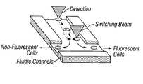
- FIG. 6is a diagrammatic illustration of the concept of the present invention for an all optical microfluidic flow cytometer for the separation of different cell species; samples are injected into the input port sequentially and directed to one of two output parts by the attractive trapping force of an optical tweezer beam.
- FIG. 7consisting of FIGS. 7 a through 7 d , respectively show microfluidic “T”, “Y”, 1-to-N and M-to-N channels fabricated in PDMS in accordance with the present invention; a typical channel width being 40 ⁇ m.
- FIG. 8shows a photonic sorting device in accordance with the present invention where (i) microfluidic channels are mounted into an optical tweezers and microscope setup; (ii) an optical beam is focused to a point at the junction of the channels; (iii) a voltage is applied to the channels to induce fluid flow; and (iv) sorting progress is monitored on a CCD camera.
- FIG. 9is a sequence of images demonstrating the photonic switching mechanism of the present invention where (i) microspheres flow in to a channel junction from an input port at the top; (ii) the microspheres are first captured (a) by an optical tweezer trap; (iii) the position of the microsphere is translated laterally to either the left or the right (B); and (iv) the microsphere is then released from the trap (C) and allowed to follow the fluid flow into either the left or right output parts.
- the dotted circleindicates the position of the optical trap. Where each of the two exit channels is equal, the microsphere will flow to its nearest exit channel (C).
- the present inventionuses photonic pressure to implement directed switching and sorting of microparticles.
- a photonic switching mechanism in accordance with the present inventionuses an optical tweezers trap.
- Channelsmost typically formed by molding a silicone elastomer, are used to guide a fluid, such as, by way of example, water, flowing, typically continuously, in a path having the shape of an inverted letter “Y” between, by way of example, one input reservoir and two output reservoirs.
- microspheres dispersed in the water continuously flowing through the input micro-channel that forms the central leg of the “Y”are selectively directed by optical radiation pressure to a determined output channel, or a selected branch leg of the “Y”. All-optical sorting is advantageous in that it can provide precise and individual manipulation of single cells or other biological samples regardless of their electrical charge or lack thereof.
- Optical tweezershave been combined with micro-fabricated fluidic channels to demonstrate tile photonic sorter.
- optical tweezersthe scattering of photons off of a small particle provides a net attractive or repulsive force depending on the index of refraction of the particle and the surrounding fluid.
- Previous demonstrations of the optical manipulation of objects through defined fluidic channelsused photonic pressure to transport cells over the length of the channels.
- the device described in this paperemploys photonic pressure only at the switching junction, while long distance transport of the cells is achieved by continuous fluid flow.
- FIG. 1cells or functionalized microspheres are entered into a T-shaped fluidic channel.
- each sampleshould be sequentially identified (either by fluorescence or some other means) and then directed into one of the two branches of the “T” depending on its type. Sorting is achieved at the junction of the channel by capturing the sample in an optical trap and then drawing it to either the left or right side of the main channel. Provided that the fluidic flow is non-turbulent, when the sample is released it will naturally flow out the closest branch of the junction. The sorted samples may be collected or sent into further iterations of sorting.
- Optical sorting in this mannermay have a number of advantages over electrical sorting depending on the test and the type of cell.
- Optical switchingcan provide precise, individual control over each particle. Additionally, while large arrays of sorting devices are envisioned on a single bio-chip to increase throughput, it may be difficult to address such large arrays electrically. Optical addressing may allow greater flexibility in this respect as device size scales.
- VCSEL arrayscan serve as optical tweezer arrays.
- Tweezer arrays that are independently addressablecan beneficially be used to both (i) transport and (ii) separate samples of microparticles, including in a bio-chip device integrating both the microchannels and the VCSEL arrays.
- photonic momentum from the VCSEL laser light(from each of arrayed VCSELs) is used as to realize multiple parallel optical switches operating in parallel in multiple microfabricated microfluidic fluidic channels, and/or, in multiple locations in each microfluidic channel.
- an optical tweezermay be implemented with one single vertical cavity surface emitting laser (VCSEL) device.
- VCSELvertical cavity surface emitting laser
- An array of VCSELsmay be used as a parallel array of optical tweezers that, as selectively controlled both individually and in concert, increase both the flexibility, and the parallelism, in the manipulation of microparticles.
- the VCSELsare normally arrayed on a single chip, and, with their vertically-emitted laser beams, serve to manipulate microparticles on the surface of the chip, or on a facing chip including as may have and present channels, including channels as may also contain and/or flow fluids.
- VCSEL arraysare made from VCSELs modified (by a post-fabrication annealing process) to emit laser light most pronouncedly in a high-order Laguerre-Gaussian mode (as opposed to a Hermite-Gaussian mode), optical pressure forces from various still higher-power light sources can be used, particularly for the fast switching of particles within microfluidic channels.
- each VCSEL in an array of VCSELs(i) emits in the Laguerre-Gaussian mode, (ii) with the emitted laser beam being focused, so as to individually act as a single trap. In this manner, precise uniformity or selective control over each trap can be achieved by appropriately modulating the current to each VCSEL.
- the VCSEL arraysare (i) compact (ii) reliable and long-lived, and (iii) inexpensive of construction on (iv) substrates that are compatible with other optoelectronic functions that may be desired in a bio-chip—such as arrayed detectors.
- Both polystyrene microspheres and live cells both wet and dryare suitably tweezed and manipulated in diverse manners by both individual and arrayed VCSEL laser beams.
- both (i) the attractive gradient force and (ii) the scattering force of a focused VCSEL optical beamhave variously been used to direct, or to “switch”, small particles flowing through junctions molded in PDMS.
- the VCSEL based tweezers, and still other VCSEL arrays, of the present inventionare suitably integrated as optical array devices performing, permissively simultaneously, both detection and manipulation.
- one side of a transparent die defining and presenting microfluidic channels and switching junctionsmay be pressed flat against a combination stimulating and sensing chip that can, by way of example, both (i) stimulate the emission of, by way of example, fluorescent light from (only) those ones of suitably positioned sample particles or cells that appropriately emit such light as an indication of some characteristic or state, and, also, (ii) sense the fluorescent light so stimulated to be selectively emitted, including so as to ultimately provide an indicating signal to digital computer or the like.
- This (i) stimulating and (ii) sensingis done in one or more “upstream” locations, including in parallel.
- the other side of the same transparent die having the microfluidic channels and switching junctionsmay be set flat against an array of VCSELs, each VCSEL “addressing”, and associated switching junction most commonly downstream of some sensing location. As each particle moves by it may be selectively “switched” into one or another channel, including under computer control. In this manner highly parallel and cost effective cell analysis and sorting may he achieved.
- Optical tweezers and tweezer arrayshave historically been generated in a number of ways including through the use of a rapid scan device, diffractive gratings or a spatial light modulator. Typical implementations of these techniques use the beam from a single high powered laser that is temporally or spatially divided among the various optical spots that are generated.
- VCSELsVertical Cavity Surface Emitting Lasers
- VCSEL arraysprovide a compact package, they are potentially very cheap, and the substrate is compatible with other optoelectronic functions that may be desired in a bio-chip such as array detectors.
- FIG. 2Shown in FIG. 2 is a comparison of the fundamental R Gaussian mode emitted from a VCSEL of FIG. 2 a to the high-order LaGuerre mode of FIG. 2 b .
- the energy of the emitted beamis moved to the outer edge of the ii aperture where, in an optical trap, photons have the greatest axial restoring force. Energy has been removed from the center of the beam, thereby decreasing the detrimental scattering force that acts to push particles out of the trap.
- FIG. 3shows a sequence of images captured by a CCD camera in which a single 5 ⁇ m diameter microsphere has been trapped, horizontally translated, and released. The full three-dimensionality of the trap was verified by translating along all axes, and also by observing that when stationary Brownian motion alone was insufficient to remove the particle from the trap.
- the strength of this trapwas measured by translating the beads at increasingly higher speeds through water and observing the point at which fluidic drag exceeded the optical trapping force. For a 10 ⁇ m diameter microsphere and a VCSEL driving current of 18 mA, a maximum drag speed of 6.4 ⁇ m/sec was observed, corresponding to a lateral trapping force of 0.6 picoNewtons. Smaller live cells ( ⁇ 5 ⁇ m) obtained from a mouse were also shown to be trapped by the VCSEL tweezers. However the strength of the trap was considerably less due to the lower dielectric constant and irregular structure of cells.
- a VCSEL array in accordance with the present inventionfor the simultaneous transport of multiple particles, also in accordance with the present invention, has been demonstrated.
- Optical beams from three VCSELs in a 1 ⁇ 3 linear arraywere similarly focused as in FIG. 3 through a microscope objective to the sample plate.
- the device spacing on the optoelectronic chipwas 250 um. After demagnification the trap spacing at the image plane was 13 um.
- Three 5 gm microsphereswere captured and translated simultaneously. This small scale demonstration indicates that much larger two-dimensional tweezers arrays with VCSEL devices are possible.
- microfluidic channelswere fabricated in a PDMS-based silicone elastomer (Dow Corning Sylgard 184). The channels were molded by a lithographically-defined relief master. Samples were cured at room temperature over a period of 24 hours. After curing, the channels were treated in a 45 C 1-ICI bath (0.02%, in water) for 40 minutes to increase their hydrophilicity. As shown in FIGS. 7 a and 7 b , both T-shaped and Y-shaped channels were fabricated. Similar results were obtained with each.
- Channels widths of 20 Am and 40 Am with depths ranging from 10 to 20 Am and lengths from 2 to 4 mmwere shown.
- the molded elastomerwas capped by a microscope slide cover slip. Reservoirs at the end of each channel were left open to permit the injection of fluid. Additionally, a gold electrode was inserted into each reservoir to permit an electro-osmotic flow to be induced within the channels. A combination of electro-osmosis and pressure was used to draw the fluids down the main channel, while sorting was performed purely by photonic pressure. Electro-osmotic fluid flow is a convenient tool for microchannels of this size, however mechanical pumping can also be used. Microspheres ranging in diameter from 0.8 Um to 10 Am were dispersed in water and shown to flow through the channels.
- the setup for the optical sorteris shown in FIG. 8 .
- the beam from a 70 mW, 850 nm diode laseris focused through the microscope slide cover slip onto the channels.
- the 100 ⁇ , 1.25 numerical aperture microscope objectivemakes a highly focused spot, therefore allowing three-dimensional optical trapping.
- the position of the optical trapis moved by translating the mounted channels over the beam.
- Prior calibration of the optical trap strength at this power and for 5 ⁇ m diameter microspheresdemonstrated a holding force of 2.8 picoNewtons. For this force the optical trap should be able to overcome the fluidic drag force of water for linear flow rates of up to 60 ⁇ m/sec.
- FIGS. 9 a – 9 eA demonstration of the switching process is depicted in the sequence of images in FIGS. 9 a – 9 e .
- the images shown hereare magnified to the junction of the “T”.
- the fluidic channels in this casewere 40 ⁇ m wide and 20 ⁇ m deep.
- the optical trapping beamis not visible in these pictures due to the IR-blocking filter in front of the CCD camera.
- Microspheres with a diameter of 5 ⁇ mwere drawn from the entry port with a linear fluidic velocity of approximately 30 ⁇ m/sec. The linear velocity is halved at the exit ports since each exit channel has the same cross-sectional area as the input channel.
- the potential difference between the entry and exit portswas 16 V.
- the optical trapAs a sphere enters the viewing area it is first captured by the optical trap (A). It is then manually translated laterally to either the left or right side of the channel (B) and then released. Because the fluid flow into each of the two channels is equal, the microsphere will flow to its nearest exit channel (C).
- the trapping and translating motionshould be automated, preferably by an actuating micro-mirror device or similar method.
- the laser power used in this applicationis high because the trapping force must overcome the drag force of the fluid.
- Implementing the optical trap from the top of the fluidic channelsis inherently inefficient since most of the photonic momentum is directed downwards instead of sideways.
- the laser beamis input from either side of the channel, either by focused beams or through integrated waveguides. By bringing the photons in from the sides of the channel, a much stronger “push” force can be achieved with much lower laser powers.
- the present specificationhas shown and described an all-optical switching device for particles flowing through microfluidic channels, and methods of positionally translating, and switching, the particles. Important applications of such a device and such methods include sorting of cells and other biological samples both for biotech research as well as therapeutic medicine.
- Photonic implementations of sample interrogation as well as manipulationhave some advantages over purely electrical implementations, particularly in terms of reducing the chance of external influences.
- Preliminary viability tests performed on living fibroblast cells exposed to the optical trap beamshowed that the cells continue to grow and reproduce normally.
- the use of vertical cavity surface emitting laser (VCSEL) arrays in multiple, independently-addressable optical trapsis currently under active development.
- An integrated combination of both photonic and electronic devicesshould permit greater complexity and capability to be achieved in bio-chip technology.
- the VCSELs that preferably serve as optical tweezerscan be arrayed in one, two and three dimensional arrays for controlling particulate movement and switching in one, two or three dimensions.
- the VCSELscan be, for example, colored—meaning centered upon a certain emission wavelength—as will make their radiation emission to act more, or less, strongly on various species, and states, of particles—thus potentially making that sensing can be dispensed with, and that switching will be both automatic and continuous dependent only upon particle coloration.
Landscapes
- Physics & Mathematics (AREA)
- Spectroscopy & Molecular Physics (AREA)
- Electromagnetism (AREA)
- Engineering & Computer Science (AREA)
- Plasma & Fusion (AREA)
- Apparatus Associated With Microorganisms And Enzymes (AREA)
- Physical Or Chemical Processes And Apparatus (AREA)
Abstract
Description
Claims (50)
Priority Applications (1)
| Application Number | Priority Date | Filing Date | Title |
|---|---|---|---|
| US10/848,972US7068874B2 (en) | 2000-11-28 | 2004-05-18 | Microfluidic sorting device |
Applications Claiming Priority (3)
| Application Number | Priority Date | Filing Date | Title |
|---|---|---|---|
| US25364400P | 2000-11-28 | 2000-11-28 | |
| US09/998,012US6778724B2 (en) | 2000-11-28 | 2001-11-28 | Optical switching and sorting of biological samples and microparticles transported in a micro-fluidic device, including integrated bio-chip devices |
| US10/848,972US7068874B2 (en) | 2000-11-28 | 2004-05-18 | Microfluidic sorting device |
Related Parent Applications (1)
| Application Number | Title | Priority Date | Filing Date |
|---|---|---|---|
| US09/998,012ContinuationUS6778724B2 (en) | 2000-11-28 | 2001-11-28 | Optical switching and sorting of biological samples and microparticles transported in a micro-fluidic device, including integrated bio-chip devices |
Publications (2)
| Publication Number | Publication Date |
|---|---|
| US20050164158A1 US20050164158A1 (en) | 2005-07-28 |
| US7068874B2true US7068874B2 (en) | 2006-06-27 |
Family
ID=26943443
Family Applications (2)
| Application Number | Title | Priority Date | Filing Date |
|---|---|---|---|
| US09/998,012Expired - LifetimeUS6778724B2 (en) | 2000-11-28 | 2001-11-28 | Optical switching and sorting of biological samples and microparticles transported in a micro-fluidic device, including integrated bio-chip devices |
| US10/848,972Expired - LifetimeUS7068874B2 (en) | 2000-11-28 | 2004-05-18 | Microfluidic sorting device |
Family Applications Before (1)
| Application Number | Title | Priority Date | Filing Date |
|---|---|---|---|
| US09/998,012Expired - LifetimeUS6778724B2 (en) | 2000-11-28 | 2001-11-28 | Optical switching and sorting of biological samples and microparticles transported in a micro-fluidic device, including integrated bio-chip devices |
Country Status (1)
| Country | Link |
|---|---|
| US (2) | US6778724B2 (en) |
Cited By (111)
| Publication number | Priority date | Publication date | Assignee | Title |
|---|---|---|---|---|
| US20040054722A1 (en)* | 2002-09-18 | 2004-03-18 | Alcatel | Meta service selector, meta service selector protocol, method, client, service, network access server, distributed system, and a computer software product for deploying services over a plurality of networks |
| US20050172476A1 (en)* | 2002-06-28 | 2005-08-11 | President And Fellows Of Havard College | Method and apparatus for fluid dispersion |
| US20050184230A1 (en)* | 2003-10-14 | 2005-08-25 | Commissariat A L'energie Atomique | Particle movement device |
| US20060163119A1 (en)* | 2002-11-01 | 2006-07-27 | Techno Network Shikoku Co., Ltd., | Method for sorting and recovering fine particle and apparatus for recovery |
| US20060163385A1 (en)* | 2003-04-10 | 2006-07-27 | Link Darren R | Formation and control of fluidic species |
| US20060169642A1 (en)* | 2002-02-04 | 2006-08-03 | John Oakey | Laminar flow-based separations of colloidal and cellular particles |
| US20060171846A1 (en)* | 2005-01-10 | 2006-08-03 | Marr David W M | Microfluidic systems incorporating integrated optical waveguides |
| US20060246575A1 (en)* | 2005-01-13 | 2006-11-02 | Micronics, Inc. | Microfluidic rare cell detection device |
| US20060257089A1 (en)* | 2005-04-08 | 2006-11-16 | Arryx, Inc. | Apparatus for optically-based sorting within liquid core waveguides |
| US20070003442A1 (en)* | 2003-08-27 | 2007-01-04 | President And Fellows Of Harvard College | Electronic control of fluidic species |
| US20070054119A1 (en)* | 2005-03-04 | 2007-03-08 | Piotr Garstecki | Systems and methods of forming particles |
| US20070086701A1 (en)* | 2003-12-04 | 2007-04-19 | Commissariat A L'energie Atomique | Particle concentration method |
| US20070195127A1 (en)* | 2006-01-27 | 2007-08-23 | President And Fellows Of Harvard College | Fluidic droplet coalescence |
| US20080003142A1 (en)* | 2006-05-11 | 2008-01-03 | Link Darren R | Microfluidic devices |
| US7385460B1 (en)* | 2004-11-17 | 2008-06-10 | California Institute Of Technology | Combined electrostatic and optical waveguide based microfluidic chip systems and methods |
| US20080138010A1 (en)* | 2006-12-11 | 2008-06-12 | Jiahua James Dou | Apparatus, System and Method for Particle Manipulation Using Wavesguides |
| US20080261295A1 (en)* | 2007-04-20 | 2008-10-23 | William Frank Butler | Cell Sorting System and Methods |
| WO2008137997A1 (en)* | 2007-05-08 | 2008-11-13 | The Regents Of The University Of California | Microfluidic device having regulated fluid transfer between elements located therein |
| US20090012187A1 (en)* | 2007-03-28 | 2009-01-08 | President And Fellows Of Harvard College | Emulsions and Techniques for Formation |
| US20090026387A1 (en)* | 2007-07-03 | 2009-01-29 | Colorado School Of Mines | Optical-based cell deformability |
| US20090042737A1 (en)* | 2007-08-09 | 2009-02-12 | Katz Andrew S | Methods and Devices for Correlated, Multi-Parameter Single Cell Measurements and Recovery of Remnant Biological Material |
| US20090062828A1 (en)* | 2007-09-04 | 2009-03-05 | Colorado School Of Mines | Magnetic field-based colloidal atherectomy |
| US20090110010A1 (en)* | 2007-09-26 | 2009-04-30 | Colorado School Of Mines | Fiber-focused diode-bar optical trapping for microfluidic manipulation |
| US20090131543A1 (en)* | 2005-03-04 | 2009-05-21 | Weitz David A | Method and Apparatus for Forming Multiple Emulsions |
| WO2010004516A1 (en)* | 2008-07-08 | 2010-01-14 | Ipgrip, Inc. | System and methods for in-line monitoring of particles in opaque flows and selective object manipulation in multi-component flow |
| US20100137163A1 (en)* | 2006-01-11 | 2010-06-03 | Link Darren R | Microfluidic Devices and Methods of Use in The Formation and Control of Nanoreactors |
| US20100304429A1 (en)* | 2003-08-28 | 2010-12-02 | William Frank Butler | Methods and apparatus for sorting cells using an optical switch in a microfluidic channel network |
| US20110026009A1 (en)* | 2006-09-15 | 2011-02-03 | Haemonetics Corporation | Surface Mapping by Optical Manipulation of Particles in Relation to a Functionalized Surface |
| US20110207207A1 (en)* | 2008-10-28 | 2011-08-25 | Gibson Emily A | Microfluidic cell sorter utilizing broadband coherent anti-stokes raman scattering |
| US20110229545A1 (en)* | 2010-03-17 | 2011-09-22 | President And Fellows Of Harvard College | Melt emulsification |
| US8162149B1 (en) | 2009-01-21 | 2012-04-24 | Sandia Corporation | Particle sorter comprising a fluid displacer in a closed-loop fluid circuit |
| WO2013090469A1 (en) | 2011-12-13 | 2013-06-20 | Sequenta, Inc. | Detection and measurement of tissue-infiltrating lymphocytes |
| US8528589B2 (en) | 2009-03-23 | 2013-09-10 | Raindance Technologies, Inc. | Manipulation of microfluidic droplets |
| US8535889B2 (en) | 2010-02-12 | 2013-09-17 | Raindance Technologies, Inc. | Digital analyte analysis |
| US8592221B2 (en) | 2007-04-19 | 2013-11-26 | Brandeis University | Manipulation of fluids, fluid components and reactions in microfluidic systems |
| WO2014017929A1 (en)* | 2012-07-27 | 2014-01-30 | Simpson Miriam Cather | Method and system for microfluidic particle orientation and/or sorting |
| US8658430B2 (en) | 2011-07-20 | 2014-02-25 | Raindance Technologies, Inc. | Manipulating droplet size |
| US20140069850A1 (en)* | 2011-09-16 | 2014-03-13 | University Of North Carolina At Charlotte | Methods and devices for optical sorting of microspheres based on their resonant optical properties |
| US8723104B2 (en) | 2012-09-13 | 2014-05-13 | City University Of Hong Kong | Methods and means for manipulating particles |
| US8772046B2 (en) | 2007-02-06 | 2014-07-08 | Brandeis University | Manipulation of fluids and reactions in microfluidic systems |
| US8841071B2 (en) | 2011-06-02 | 2014-09-23 | Raindance Technologies, Inc. | Sample multiplexing |
| US8871444B2 (en) | 2004-10-08 | 2014-10-28 | Medical Research Council | In vitro evolution in microfluidic systems |
| US8961764B2 (en) | 2010-10-15 | 2015-02-24 | Lockheed Martin Corporation | Micro fluidic optic design |
| US9012390B2 (en) | 2006-08-07 | 2015-04-21 | Raindance Technologies, Inc. | Fluorocarbon emulsion stabilizing surfactants |
| US9067207B2 (en) | 2009-06-04 | 2015-06-30 | University Of Virginia Patent Foundation | Optical approach for microfluidic DNA electrophoresis detection |
| US9150905B2 (en) | 2012-05-08 | 2015-10-06 | Adaptive Biotechnologies Corporation | Compositions and method for measuring and calibrating amplification bias in multiplexed PCR reactions |
| US9150852B2 (en) | 2011-02-18 | 2015-10-06 | Raindance Technologies, Inc. | Compositions and methods for molecular labeling |
| US9181590B2 (en) | 2011-10-21 | 2015-11-10 | Adaptive Biotechnologies Corporation | Quantification of adaptive immune cell genomes in a complex mixture of cells |
| US9238206B2 (en) | 2011-05-23 | 2016-01-19 | President And Fellows Of Harvard College | Control of emulsions, including multiple emulsions |
| US9322054B2 (en) | 2012-02-22 | 2016-04-26 | Lockheed Martin Corporation | Microfluidic cartridge |
| US9347099B2 (en) | 2008-11-07 | 2016-05-24 | Adaptive Biotechnologies Corp. | Single cell analysis by polymerase cycling assembly |
| US9364803B2 (en) | 2011-02-11 | 2016-06-14 | Raindance Technologies, Inc. | Methods for forming mixed droplets |
| US9365901B2 (en) | 2008-11-07 | 2016-06-14 | Adaptive Biotechnologies Corp. | Monitoring immunoglobulin heavy chain evolution in B-cell acute lymphoblastic leukemia |
| US9366632B2 (en) | 2010-02-12 | 2016-06-14 | Raindance Technologies, Inc. | Digital analyte analysis |
| US9399797B2 (en) | 2010-02-12 | 2016-07-26 | Raindance Technologies, Inc. | Digital analyte analysis |
| US9416420B2 (en) | 2008-11-07 | 2016-08-16 | Adaptive Biotechnologies Corp. | Monitoring health and disease status using clonotype profiles |
| US9448172B2 (en) | 2003-03-31 | 2016-09-20 | Medical Research Council | Selection by compartmentalised screening |
| US9487812B2 (en) | 2012-02-17 | 2016-11-08 | Colorado School Of Mines | Optical alignment deformation spectroscopy |
| US9498759B2 (en) | 2004-10-12 | 2016-11-22 | President And Fellows Of Harvard College | Compartmentalized screening by microfluidic control |
| US9506119B2 (en) | 2008-11-07 | 2016-11-29 | Adaptive Biotechnologies Corp. | Method of sequence determination using sequence tags |
| US9512487B2 (en) | 2008-11-07 | 2016-12-06 | Adaptive Biotechnologies Corp. | Monitoring health and disease status using clonotype profiles |
| US9528160B2 (en) | 2008-11-07 | 2016-12-27 | Adaptive Biotechnolgies Corp. | Rare clonotypes and uses thereof |
| US9557316B2 (en) | 2013-10-15 | 2017-01-31 | Samsung Electronics Co., Ltd. | Sample analysis methods and apparatuses and dynamic valve operation methods |
| US9562837B2 (en) | 2006-05-11 | 2017-02-07 | Raindance Technologies, Inc. | Systems for handling microfludic droplets |
| US9562897B2 (en) | 2010-09-30 | 2017-02-07 | Raindance Technologies, Inc. | Sandwich assays in droplets |
| US9645010B2 (en) | 2009-03-10 | 2017-05-09 | The Regents Of The University Of California | Fluidic flow cytometry devices and methods |
| US9708657B2 (en) | 2013-07-01 | 2017-07-18 | Adaptive Biotechnologies Corp. | Method for generating clonotype profiles using sequence tags |
| US9778164B2 (en) | 2009-03-10 | 2017-10-03 | The Regents Of The University Of California | Fluidic flow cytometry devices and particle sensing based on signal-encoding |
| US9809813B2 (en) | 2009-06-25 | 2017-11-07 | Fred Hutchinson Cancer Research Center | Method of measuring adaptive immunity |
| US9824179B2 (en) | 2011-12-09 | 2017-11-21 | Adaptive Biotechnologies Corp. | Diagnosis of lymphoid malignancies and minimal residual disease detection |
| US9841367B2 (en)* | 2011-09-16 | 2017-12-12 | The University Of North Carolina At Charlotte | Methods and devices for optical sorting of microspheres based on their resonant optical properties |
| US9839890B2 (en) | 2004-03-31 | 2017-12-12 | National Science Foundation | Compartmentalised combinatorial chemistry by microfluidic control |
| CN107603940A (en)* | 2017-09-07 | 2018-01-19 | 中国科学技术大学 | Utilize the method for wedge-shaped optical tweezer light field sorting particulate |
| US9885644B2 (en) | 2006-01-10 | 2018-02-06 | Colorado School Of Mines | Dynamic viscoelasticity as a rapid single-cell biomarker |
| US20180081347A1 (en)* | 2016-09-19 | 2018-03-22 | Palo Alto Research Center Incorporated | System and Method for Scalable Real-Time Micro-Object Position Control with the Aid of a Digital Computer |
| US10024819B2 (en) | 2010-10-21 | 2018-07-17 | The Regents Of The University Of California | Microfluidics with wirelessly powered electronic circuits |
| US10052605B2 (en) | 2003-03-31 | 2018-08-21 | Medical Research Council | Method of synthesis and testing of combinatorial libraries using microcapsules |
| US10066265B2 (en) | 2014-04-01 | 2018-09-04 | Adaptive Biotechnologies Corp. | Determining antigen-specific t-cells |
| US10077478B2 (en) | 2012-03-05 | 2018-09-18 | Adaptive Biotechnologies Corp. | Determining paired immune receptor chains from frequency matched subunits |
| US10150996B2 (en) | 2012-10-19 | 2018-12-11 | Adaptive Biotechnologies Corp. | Quantification of adaptive immune cell genomes in a complex mixture of cells |
| US10195571B2 (en) | 2011-07-06 | 2019-02-05 | President And Fellows Of Harvard College | Multiple emulsions and techniques for the formation of multiple emulsions |
| US10221461B2 (en) | 2012-10-01 | 2019-03-05 | Adaptive Biotechnologies Corp. | Immunocompetence assessment by adaptive immune receptor diversity and clonality characterization |
| US10246701B2 (en) | 2014-11-14 | 2019-04-02 | Adaptive Biotechnologies Corp. | Multiplexed digital quantitation of rearranged lymphoid receptors in a complex mixture |
| US10323276B2 (en) | 2009-01-15 | 2019-06-18 | Adaptive Biotechnologies Corporation | Adaptive immunity profiling and methods for generation of monoclonal antibodies |
| WO2019125502A1 (en)* | 2017-12-23 | 2019-06-27 | Lumacyte LLC | Microfluidic chip device for optical force measurements and cell imaging using microfluidic chip configuration and dynamics |
| US10351905B2 (en) | 2010-02-12 | 2019-07-16 | Bio-Rad Laboratories, Inc. | Digital analyte analysis |
| US10385475B2 (en) | 2011-09-12 | 2019-08-20 | Adaptive Biotechnologies Corp. | Random array sequencing of low-complexity libraries |
| US10392663B2 (en) | 2014-10-29 | 2019-08-27 | Adaptive Biotechnologies Corp. | Highly-multiplexed simultaneous detection of nucleic acids encoding paired adaptive immune receptor heterodimers from a large number of samples |
| US10428325B1 (en) | 2016-09-21 | 2019-10-01 | Adaptive Biotechnologies Corporation | Identification of antigen-specific B cell receptors |
| US10520500B2 (en) | 2009-10-09 | 2019-12-31 | Abdeslam El Harrak | Labelled silica-based nanomaterial with enhanced properties and uses thereof |
| US10533998B2 (en) | 2008-07-18 | 2020-01-14 | Bio-Rad Laboratories, Inc. | Enzyme quantification |
| US10647981B1 (en) | 2015-09-08 | 2020-05-12 | Bio-Rad Laboratories, Inc. | Nucleic acid library generation methods and compositions |
| US10722250B2 (en) | 2007-09-04 | 2020-07-28 | Colorado School Of Mines | Magnetic-field driven colloidal microbots, methods for forming and using the same |
| US10732649B2 (en) | 2004-07-02 | 2020-08-04 | The University Of Chicago | Microfluidic system |
| US10816550B2 (en) | 2012-10-15 | 2020-10-27 | Nanocellect Biomedical, Inc. | Systems, apparatus, and methods for sorting particles |
| US10837883B2 (en) | 2009-12-23 | 2020-11-17 | Bio-Rad Laboratories, Inc. | Microfluidic systems and methods for reducing the exchange of molecules between droplets |
| US10874997B2 (en) | 2009-09-02 | 2020-12-29 | President And Fellows Of Harvard College | Multiple emulsions created using jetting and other techniques |
| US11041202B2 (en) | 2015-04-01 | 2021-06-22 | Adaptive Biotechnologies Corporation | Method of identifying human compatible T cell receptors specific for an antigenic target |
| US11047008B2 (en) | 2015-02-24 | 2021-06-29 | Adaptive Biotechnologies Corporation | Methods for diagnosing infectious disease and determining HLA status using immune repertoire sequencing |
| US11066705B2 (en) | 2014-11-25 | 2021-07-20 | Adaptive Biotechnologies Corporation | Characterization of adaptive immune response to vaccination or infection using immune repertoire sequencing |
| US11174509B2 (en) | 2013-12-12 | 2021-11-16 | Bio-Rad Laboratories, Inc. | Distinguishing rare variations in a nucleic acid sequence from a sample |
| US11193176B2 (en) | 2013-12-31 | 2021-12-07 | Bio-Rad Laboratories, Inc. | Method for detecting and quantifying latent retroviral RNA species |
| US11248253B2 (en) | 2014-03-05 | 2022-02-15 | Adaptive Biotechnologies Corporation | Methods using randomer-containing synthetic molecules |
| US11254980B1 (en) | 2017-11-29 | 2022-02-22 | Adaptive Biotechnologies Corporation | Methods of profiling targeted polynucleotides while mitigating sequencing depth requirements |
| US11511242B2 (en) | 2008-07-18 | 2022-11-29 | Bio-Rad Laboratories, Inc. | Droplet libraries |
| US11561164B2 (en) | 2017-12-23 | 2023-01-24 | Lumactye, Inc. | Microfluidic chip device for optical force measurements and cell imaging using microfluidic chip configuration and dynamics |
| US11901041B2 (en) | 2013-10-04 | 2024-02-13 | Bio-Rad Laboratories, Inc. | Digital analysis of nucleic acid modification |
| US11913870B2 (en) | 2017-12-23 | 2024-02-27 | Lumacyte, Inc. | Microfluidic chip device for optical force measurements and cell imaging using microfluidic chip configuration and dynamics |
| US12038438B2 (en) | 2008-07-18 | 2024-07-16 | Bio-Rad Laboratories, Inc. | Enzyme quantification |
| US12097475B2 (en) | 2004-07-02 | 2024-09-24 | The University Of Chicago | Microfluidic system |
| US12209282B2 (en) | 2008-11-07 | 2025-01-28 | Adaptive Biotechnologies Corporation | Monitoring health and disease status using clonotype profiles |
Families Citing this family (79)
| Publication number | Priority date | Publication date | Assignee | Title |
|---|---|---|---|---|
| US6797942B2 (en) | 2001-09-13 | 2004-09-28 | University Of Chicago | Apparatus and process for the lateral deflection and separation of flowing particles by a static array of optical tweezers |
| US6936811B2 (en) | 2000-11-13 | 2005-08-30 | Genoptix, Inc. | Method for separating micro-particles |
| US20020121443A1 (en)* | 2000-11-13 | 2002-09-05 | Genoptix | Methods for the combined electrical and optical identification, characterization and/or sorting of particles |
| US20030007894A1 (en)* | 2001-04-27 | 2003-01-09 | Genoptix | Methods and apparatus for use of optical forces for identification, characterization and/or sorting of particles |
| US6913697B2 (en) | 2001-02-14 | 2005-07-05 | Science & Technology Corporation @ Unm | Nanostructured separation and analysis devices for biological membranes |
| US20030124516A1 (en)* | 2001-04-27 | 2003-07-03 | Genoptix, Inc. | Method of using optical interrogation to determine a biological property of a cell or population of cells |
| US20040023310A1 (en)* | 2001-04-27 | 2004-02-05 | Genoptix, Inc | Quantitative determination of protein kinase C activation using optophoretic analysis |
| US20030156991A1 (en)* | 2001-10-23 | 2003-08-21 | William Marsh Rice University | Optomechanically-responsive materials for use as light-activated actuators and valves |
| US7179420B2 (en)* | 2001-10-25 | 2007-02-20 | Techelan, Llc | Apparatus comprising a particle sorter/dispenser and method therefor |
| US20040001371A1 (en)* | 2002-06-26 | 2004-01-01 | The Arizona Board Of Regents On Behalf Of The University Of Arizona | Information storage and retrieval device using macromolecules as storage media |
| JP3898103B2 (en)* | 2002-08-26 | 2007-03-28 | 独立行政法人科学技術振興機構 | Cell analysis separator |
| US20040115830A1 (en)* | 2002-09-25 | 2004-06-17 | Igor Touzov | Components for nano-scale Reactor |
| CA2500392C (en) | 2002-09-27 | 2012-11-27 | The General Hospital Corporation | Microfluidic device for cell separation and uses thereof |
| US20040067167A1 (en)* | 2002-10-08 | 2004-04-08 | Genoptix, Inc. | Methods and apparatus for optophoretic diagnosis of cells and particles |
| US7160730B2 (en)* | 2002-10-21 | 2007-01-09 | Bach David T | Method and apparatus for cell sorting |
| DE10320869A1 (en)* | 2003-05-09 | 2004-12-16 | Evotec Technologies Gmbh | Methods and devices for liquid treatment of suspended particles |
| US7435391B2 (en)* | 2003-05-23 | 2008-10-14 | Lucent Technologies Inc. | Light-mediated micro-chemical reactors |
| EP1671105A4 (en)* | 2003-09-19 | 2010-06-23 | Univ California | LIGHT FORCE DETECTOR AND METHOD FOR MEASURING OPTICAL TRAPPING FORCES IN THE AXIS FROM LIGHT IMPULSE FORCE VARIATIONS ALONG AN OPTICAL AXIS |
| US7800750B2 (en)* | 2003-09-19 | 2010-09-21 | The Regents Of The University Of California | Optical trap utilizing a reflecting mirror for alignment |
| US20050067337A1 (en)* | 2003-09-30 | 2005-03-31 | Hart Sean J. | Laser optical separator and method for separating colloidal suspensions |
| US20060115379A1 (en)* | 2003-10-17 | 2006-06-01 | Bach David T | Magnetostrictive pump |
| US7177492B2 (en)* | 2004-03-11 | 2007-02-13 | Nomadics, Inc. | System, probe and methods for colorimetric testing |
| US7442339B2 (en)* | 2004-03-31 | 2008-10-28 | Intel Corporation | Microfluidic apparatus, Raman spectroscopy systems, and methods for performing molecular reactions |
| FR2873171B1 (en)* | 2004-07-19 | 2007-12-07 | Centre Nat Rech Scient Cnrse | ACTIVE COMPONENT MICROFLUIDIC CIRCUIT |
| US7391936B2 (en)* | 2005-01-21 | 2008-06-24 | Lucent Technologies, Inc. | Microfluidic sensors and methods for making the same |
| US20070196820A1 (en) | 2005-04-05 | 2007-08-23 | Ravi Kapur | Devices and methods for enrichment and alteration of cells and other particles |
| JP2008538282A (en)* | 2005-04-05 | 2008-10-23 | セルポイント ダイアグノスティクス, インコーポレイテッド | Device and method for enrichment and modification of circulating tumor cells and other particles |
| HU0500406D0 (en)* | 2005-04-22 | 2005-06-28 | Mta Szegedi Biolog Koezpont | Microfluidic device and method for manipulating electroosmotically moved fluid |
| KR20060111143A (en)* | 2005-04-22 | 2006-10-26 | 한국표준과학연구원 | Particle Separation Device Using Light Capture |
| US8921102B2 (en) | 2005-07-29 | 2014-12-30 | Gpb Scientific, Llc | Devices and methods for enrichment and alteration of circulating tumor cells and other particles |
| US7745788B2 (en) | 2005-09-23 | 2010-06-29 | Massachusetts Institute Of Technology | Optical trapping with a semiconductor |
| US7417788B2 (en)* | 2005-11-21 | 2008-08-26 | Aditya Narendra Joshi | Optical logic device |
| US7569789B2 (en)* | 2006-03-16 | 2009-08-04 | Visiongate, Inc. | Cantilevered coaxial flow injector apparatus and method for sorting particles |
| JP5032792B2 (en)* | 2006-05-22 | 2012-09-26 | 浜松ホトニクス株式会社 | Cell sorter |
| FR2901717A1 (en)* | 2006-05-30 | 2007-12-07 | Centre Nat Rech Scient | METHOD FOR TREATING DROPS IN A MICROFLUIDIC CIRCUIT |
| CA2580589C (en) | 2006-12-19 | 2016-08-09 | Fio Corporation | Microfluidic detection system |
| US8360321B2 (en) | 2007-04-02 | 2013-01-29 | Fio Corporation | System and method of deconvolving multiplexed fluorescence spectral signals generated by quantum dot optical coding technology |
| KR101258434B1 (en) | 2007-05-03 | 2013-05-02 | 삼성전자주식회사 | Microfluidic system and fabricating method of the same |
| CA2692259C (en) | 2007-06-22 | 2012-07-31 | Fio Corporation | Systems and methods for manufacturing quantum dot-doped polymer microbeads |
| EP2174115B1 (en) | 2007-07-09 | 2013-08-28 | Fio Corporation | Systems and methods for enhancing fluorescent detection of target molecules in a test sample |
| EP2209549A4 (en) | 2007-10-12 | 2014-03-05 | Fio Corp | Flow focusing method and system for forming concentrated volumes of microbeads, and microbeads formed further thereto |
| JP4389991B2 (en)* | 2007-10-26 | 2009-12-24 | ソニー株式会社 | Method and apparatus for optical measurement of fine particles |
| US9594071B2 (en)* | 2007-12-21 | 2017-03-14 | Colin G. Hebert | Device and method for laser analysis and separation (LAS) of particles |
| US10281385B2 (en)* | 2007-12-21 | 2019-05-07 | The United States Of America, As Represented By The Secretary Of The Navy | Device for laser analysis and separation (LAS) of particles |
| KR100938927B1 (en) | 2007-12-31 | 2010-01-27 | 재단법인서울대학교산학협력재단 | Microfluidic cell sorting apparatus using laser ablation |
| US7915577B2 (en)* | 2008-05-01 | 2011-03-29 | The United States Of America As Represented By The Secretary Of The Navy | Single-shot spatially-resolved imaging magnetometry using ultracold atoms |
| EP2321810A4 (en) | 2008-06-25 | 2014-09-17 | Fio Corp | Bio-threat alert system |
| BRPI0917839A2 (en) | 2008-08-29 | 2015-11-24 | Fio Corp | single-use portable diagnostic test device, and associated system and method for testing environmental and biological test samples |
| CN102348986B (en) | 2009-01-13 | 2015-05-06 | Fio公司 | Handheld diagnostic test device and method for use in combination with electronic devices and test cartridges in rapid diagnostic tests |
| US9068921B2 (en)* | 2009-03-07 | 2015-06-30 | Hewlett-Packard Development Company, L.P. | Analyzer and method for sensing using the same |
| US20100273681A1 (en)* | 2009-04-27 | 2010-10-28 | Wisconsin Alumni Research Foundation | Combinatorial chemistry reaction cell with optical tweezers |
| ITTO20100068U1 (en)* | 2010-04-20 | 2011-10-21 | Eltek Spa | MICROFLUID AND / OR EQUIPMENT DEVICES FOR MICROFLUID DEVICES |
| WO2012050556A1 (en)* | 2010-10-11 | 2012-04-19 | Hewlett-Packard Development Company L.P. | Microfluidic chip assembly |
| WO2012149185A2 (en)* | 2011-04-28 | 2012-11-01 | The Government Of The United States Of America As Represented By The Secretary Of The Navy | Method of changing fluid flow by using an optical beam |
| US9259741B2 (en) | 2011-12-29 | 2016-02-16 | Danmarks Tekniske Universitet | System for sorting microscopic objects using electromagnetic radiation |
| US9433941B2 (en)* | 2012-12-21 | 2016-09-06 | Cornell University | Microfluidic chip having on-chip electrically tunable high-throughput nanophotonic trap |
| WO2014117784A1 (en)* | 2013-02-04 | 2014-08-07 | Danmarks Tekniske Universitet | System for optical sorting of microscopic objects |
| CN110186835B (en) | 2013-03-15 | 2022-05-31 | Gpb科学有限公司 | On-chip microfluidic processing of particles |
| CN105247042B (en) | 2013-03-15 | 2021-06-11 | 普林斯顿大学理事会 | Method and apparatus for high throughput purification |
| US20150064153A1 (en) | 2013-03-15 | 2015-03-05 | The Trustees Of Princeton University | High efficiency microfluidic purification of stem cells to improve transplants |
| US8820538B1 (en)* | 2014-03-17 | 2014-09-02 | Namocell LLC | Method and apparatus for particle sorting |
| CA2959256A1 (en)* | 2014-08-29 | 2016-03-03 | Synaptive Medical (Barbados) Inc. | Molecular cell imaging using optical spectroscopy |
| US10539554B2 (en) | 2014-10-27 | 2020-01-21 | Board Of Regents, The University Of Texas System | High-throughput imaging platform |
| US10976232B2 (en) | 2015-08-24 | 2021-04-13 | Gpb Scientific, Inc. | Methods and devices for multi-step cell purification and concentration |
| US10228317B1 (en)* | 2016-08-25 | 2019-03-12 | Verily Life Sciences Llc | Multiplexed microfluidic cell sorting using laser induced cavitation bubbles |
| US10544413B2 (en) | 2017-05-18 | 2020-01-28 | 10X Genomics, Inc. | Methods and systems for sorting droplets and beads |
| EP4435113A1 (en) | 2017-05-18 | 2024-09-25 | 10x Genomics, Inc. | Methods and systems for sorting droplets and beads |
| US10610865B2 (en) | 2017-08-22 | 2020-04-07 | 10X Genomics, Inc. | Droplet forming devices and system with differential surface properties |
| WO2019083852A1 (en) | 2017-10-26 | 2019-05-02 | 10X Genomics, Inc. | Microfluidic channel networks for partitioning |
| WO2019156711A1 (en) | 2018-02-12 | 2019-08-15 | Hewlett-Packard Development Company, L.P. | Devices to measure flow rates with movable elements |
| US11680957B2 (en) | 2018-02-12 | 2023-06-20 | Hewlett-Packard Development Company, L.P. | Microfluidic flow sensor |
| WO2020011193A1 (en)* | 2018-07-11 | 2020-01-16 | The University Of Hong Kong | Automatic microfluidic system for continuous and quantitive collection of droplets |
| EP3698871A1 (en)* | 2019-02-19 | 2020-08-26 | Gottfried Wilhelm Leibniz Universität Hannover | Laser based sorting of droplets in microfluidic streams |
| EP3930900A1 (en) | 2019-02-28 | 2022-01-05 | 10X Genomics, Inc. | Devices, systems, and methods for increasing droplet formation efficiency |
| CA3141069A1 (en) | 2019-05-31 | 2020-12-03 | Amberstone Biosciences, Inc. | Microfluidic determination of low abundance events |
| US12186751B2 (en) | 2019-06-28 | 2025-01-07 | 10X Genomics, Inc. | Devices and systems incorporating acoustic ordering and methods of use thereof |
| US12059679B2 (en) | 2019-11-19 | 2024-08-13 | 10X Genomics, Inc. | Methods and devices for sorting droplets and particles |
| CN111454832B (en)* | 2020-04-27 | 2023-12-15 | 深圳大学 | A cell sorting system and method based on microfluidics |
| WO2024026018A2 (en)* | 2022-07-27 | 2024-02-01 | The Trustees Of The University Of Columbia In The City Of New York | Systems and methods for identifying and recovering rare biological cells from a sample |
Citations (161)
| Publication number | Priority date | Publication date | Assignee | Title |
|---|---|---|---|---|
| US3558877A (en) | 1966-12-19 | 1971-01-26 | Gca Corp | Method and apparatus for mass separation by selective light absorption |
| US3628182A (en) | 1969-03-20 | 1971-12-14 | Bell Telephone Labor Inc | Ring-type parametric oscillator |
| US3638139A (en) | 1964-09-29 | 1972-01-25 | Bell Telephone Labor Inc | Frequency-selective laser devices |
| US3662183A (en) | 1970-12-28 | 1972-05-09 | Bell Telephone Labor Inc | Continuously tunable optical parametric oscillator |
| US3710279A (en) | 1969-12-15 | 1973-01-09 | Bell Telephone Labor Inc | Apparatuses for trapping and accelerating neutral particles |
| US3725810A (en) | 1971-04-23 | 1973-04-03 | Bell Telephone Labor Inc | Optical stimulated emission devices employing split optical guides |
| US3761721A (en) | 1972-07-06 | 1973-09-25 | Trw Inc | Matter wave interferometric apparatus |
| US3778612A (en) | 1969-12-15 | 1973-12-11 | A Ashkin | Neutral particle beam separator and velocity analyzer using radiation pressure |
| US3793541A (en) | 1970-01-26 | 1974-02-19 | Bell Telephone Labor Inc | Optical stimulated emission devices employing optical guiding |
| US3808432A (en) | 1970-06-04 | 1974-04-30 | Bell Telephone Labor Inc | Neutral particle accelerator utilizing radiation pressure |
| US3808550A (en) | 1969-12-15 | 1974-04-30 | Bell Telephone Labor Inc | Apparatuses for trapping and accelerating neutral particles |
| US4063106A (en) | 1977-04-25 | 1977-12-13 | Bell Telephone Laboratories, Incorporated | Optical fiber Raman oscillator |
| US4092535A (en) | 1977-04-22 | 1978-05-30 | Bell Telephone Laboratories, Incorporated | Damping of optically levitated particles by feedback and beam shaping |
| US4127329A (en) | 1976-12-21 | 1978-11-28 | Northeast Utilities Service Company | Raman scattering system and method for aerosol monitoring |
| US4247815A (en) | 1979-05-22 | 1981-01-27 | The United States Of America As Represented By The Secretary Of The Army | Method and apparatus for physiologic facsimile imaging of biologic targets based on complex permittivity measurements using remote microwave interrogation |
| US4253846A (en) | 1979-11-21 | 1981-03-03 | Technicon Instruments Corporation | Method and apparatus for automated analysis of fluid samples |
| US4327288A (en) | 1980-09-29 | 1982-04-27 | Bell Telephone Laboratories, Incorporated | Method for focusing neutral atoms, molecules and ions |
| US4386274A (en) | 1980-11-10 | 1983-05-31 | Saul Altshuler | Isotope separation by standing waves |
| US4390403A (en) | 1981-07-24 | 1983-06-28 | Batchelder J Samuel | Method and apparatus for dielectrophoretic manipulation of chemical species |
| US4440638A (en) | 1982-02-16 | 1984-04-03 | U.T. Board Of Regents | Surface field-effect device for manipulation of charged species |
| US4451412A (en) | 1982-01-12 | 1984-05-29 | Thomson-Csf | Process for producing diffracting phase structures |
| US4453805A (en) | 1981-02-19 | 1984-06-12 | Bell Telephone Laboratories, Incorporated | Optical grating using a liquid suspension of dielectric particles |
| US4520484A (en) | 1981-05-22 | 1985-05-28 | Thomson-Csf | Coherent radiation source generating a beam with a regulatable propagation direction |
| US4536657A (en) | 1982-12-08 | 1985-08-20 | Commissariat A L'energie Atomique | Process and apparatus for obtaining beams of particles with a spatially modulated density |
| US4627689A (en) | 1983-12-08 | 1986-12-09 | University Of Pittsburgh | Crystalline colloidal narrow band radiation filter |
| US4632517A (en) | 1983-12-08 | 1986-12-30 | University Of Pittsburgh | Crystalline colloidal narrow band radiation filter |
| US4756427A (en) | 1984-09-11 | 1988-07-12 | Partec Ag | Method and apparatus for sorting particles |
| US4827125A (en) | 1987-04-29 | 1989-05-02 | The United States Of America As Represented By The Secretary Of The Department Of Health And Human Services | Confocal scanning laser microscope having no moving parts |
| US4886360A (en) | 1986-09-25 | 1989-12-12 | Amersham International Plc | Method and apparatus for particle analysis |
| US4887721A (en) | 1987-11-30 | 1989-12-19 | The United States Of America As Represented By The United States Department Of Energy | Laser particle sorter |
| US4893886A (en) | 1987-09-17 | 1990-01-16 | American Telephone And Telegraph Company | Non-destructive optical trap for biological particles and method of doing same |
| US4908112A (en) | 1988-06-16 | 1990-03-13 | E. I. Du Pont De Nemours & Co. | Silicon semiconductor wafer for analyzing micronic biological samples |
| JPH03101419A (en) | 1989-09-14 | 1991-04-26 | Nippon Telegr & Teleph Corp <Ntt> | Optical trap system atomic oscillator |
| US5029791A (en) | 1990-03-08 | 1991-07-09 | Candela Laser Corporation | Optics X-Y positioner |
| US5079169A (en) | 1990-05-22 | 1992-01-07 | The Regents Of The Stanford Leland Junior University | Method for optically manipulating polymer filaments |
| US5100627A (en) | 1989-11-30 | 1992-03-31 | The Regents Of The University Of California | Chamber for the optical manipulation of microscopic particles |
| US5113286A (en) | 1990-09-27 | 1992-05-12 | At&T Bell Laboratories | Diffraction grating apparatus and method of forming a surface relief pattern in diffraction grating apparatus |
| US5121400A (en) | 1989-12-01 | 1992-06-09 | Thomson-Csf | Device for coherent addition of laser beams |
| US5170890A (en) | 1990-12-05 | 1992-12-15 | Wilson Steven D | Particle trap |
| US5189294A (en) | 1992-07-08 | 1993-02-23 | The United States Of America As Represented By The Secretary Of The Air Force | Transform lens with a plurality of sliced lens segments |
| US5198369A (en) | 1990-04-25 | 1993-03-30 | Canon Kabushiki Kaisha | Sample measuring method using agglomeration reaction of microcarriers |
| JPH0588107A (en) | 1991-09-27 | 1993-04-09 | Olympus Optical Co Ltd | Optical tweezers device |
| US5206504A (en) | 1991-11-01 | 1993-04-27 | The United States Of America As Represented By The Administrator, National Aeronautics And Space Administration | Sample positioning in microgravity |
| US5212382A (en) | 1990-12-13 | 1993-05-18 | Keiji Sasaki | Laser trapping and method for applications thereof |
| JPH05232398A (en) | 1992-02-24 | 1993-09-10 | Canon Inc | Optical trap method |
| US5245466A (en) | 1990-08-15 | 1993-09-14 | President And Fellows Of Harvard University And Rowland Institute | Optical matter |
| US5274231A (en) | 1992-04-14 | 1993-12-28 | Board Of Trustees, Leland Stanford Jr. University | Method and apparatus for manipulating atoms, ions or molecules and for measuring physical quantities using stimulated Raman transitions |
| US5283417A (en) | 1989-12-07 | 1994-02-01 | Research Development Corporation Of Japan | Laser microprocessing and the device therefor |
| WO1994008221A1 (en) | 1992-09-29 | 1994-04-14 | The School Of Pharmacy, University Of London | Method of rheological investigation |
| US5308976A (en) | 1991-06-01 | 1994-05-03 | Research Development Corp. Of Japan | Method for multi-beam manipulation of microparticles |
| JPH06123886A (en) | 1992-10-12 | 1994-05-06 | Nippon Telegr & Teleph Corp <Ntt> | Fine particle array control method |
| JPH06132000A (en) | 1992-10-15 | 1994-05-13 | Jeol Ltd | Method and device for supporting sample of electron microscope etc. |
| US5327515A (en) | 1993-01-14 | 1994-07-05 | At&T Laboratories | Method for forming a Bragg grating in an optical medium |
| US5337324A (en) | 1992-06-11 | 1994-08-09 | Tokyo Institute Of Technology | Method for controlling movement of neutral atom and apparatus for carrying out the same |
| US5338930A (en) | 1990-06-01 | 1994-08-16 | Research Corporation Technologies | Frequency standard using an atomic fountain of optically trapped atoms |
| US5343038A (en) | 1991-12-12 | 1994-08-30 | Matsushita Electric Industrial Co., Ltd. | Scanning laser microscope with photo coupling and detecting unit |
| US5355252A (en) | 1992-01-27 | 1994-10-11 | Jeol Ltd. | Scanning laser microscope |
| US5360764A (en) | 1993-02-16 | 1994-11-01 | The United States Of America, As Represented By The Secretary Of Commerce | Method of fabricating laser controlled nanolithography |
| US5363190A (en) | 1992-09-07 | 1994-11-08 | Olympus Optical Co., Ltd. | Method and apparatus for optical micro manipulation |
| US5364744A (en) | 1992-07-23 | 1994-11-15 | Cell Robotics, Inc. | Method for the manufacture of an optical manipulation chamber |
| US5374566A (en) | 1993-01-27 | 1994-12-20 | National Semiconductor Corporation | Method of fabricating a BiCMOS structure |
| DE4326181A1 (en) | 1993-08-04 | 1995-02-09 | Europ Lab Molekularbiolog | Method and device for luminescence spectroscopy and material microprocessing of fixed and moving molecules, particles and objects |
| US5445011A (en) | 1993-09-21 | 1995-08-29 | Ghislain; Lucien P. | Scanning force microscope using an optical trap |
| US5452123A (en) | 1992-12-30 | 1995-09-19 | University Of Pittsburgh Of The Commonwealth System Of Higher Education | Method of making an optically nonlinear switched optical device and related devices |
| US5473471A (en) | 1993-04-16 | 1995-12-05 | Matsushita Electric Industrial Co., Ltd. | Complex lens with diffraction grating |
| US5495105A (en) | 1992-02-20 | 1996-02-27 | Canon Kabushiki Kaisha | Method and apparatus for particle manipulation, and measuring apparatus utilizing the same |
| US5512745A (en) | 1994-03-09 | 1996-04-30 | Board Of Trustees Of The Leland Stanford Jr. University | Optical trap system and method |
| JPH08234110A (en) | 1995-02-23 | 1996-09-13 | Nikon Corp | Epi-fluorescence microscope |
| JPH0943434A (en) | 1995-08-03 | 1997-02-14 | Masahiro Ikeda | Optical tweezers |
| US5608519A (en) | 1995-03-20 | 1997-03-04 | Gourley; Paul L. | Laser apparatus and method for microscopic and spectroscopic analysis and processing of biological cells |
| US5620857A (en) | 1995-06-07 | 1997-04-15 | United States Of America, As Represented By The Secretary Of Commerce | Optical trap for detection and quantitation of subzeptomolar quantities of analytes |
| US5625484A (en) | 1992-10-28 | 1997-04-29 | European Economic Community (Cee) | Optical modulator |
| US5629802A (en) | 1995-01-05 | 1997-05-13 | The United States Of America As Represented By The Secretary Of The Air Force | Spatially multiplexed optical signal processor |
| US5631141A (en) | 1995-05-05 | 1997-05-20 | The Regents Of The University Of California | High resolution biosensor for in-situ microthermometry |
| US5637458A (en) | 1994-07-20 | 1997-06-10 | Sios, Inc. | Apparatus and method for the detection and assay of organic molecules |
| WO1997021832A1 (en) | 1995-12-08 | 1997-06-19 | Evotec Biosystems Gmbh | Process for determination of low concentration of nucleic acid molecules |
| US5644588A (en) | 1994-03-26 | 1997-07-01 | Research Development Corporation Of Japan | Microfine light source |
| US5653859A (en) | 1993-01-21 | 1997-08-05 | Parton; Adrian | Methods of analysis/separation |
| US5659561A (en) | 1995-06-06 | 1997-08-19 | University Of Central Florida | Spatial solitary waves in bulk quadratic nonlinear materials and their applications |
| US5674743A (en) | 1993-02-01 | 1997-10-07 | Seq, Ltd. | Methods and apparatus for DNA sequencing |
| US5689109A (en) | 1993-01-13 | 1997-11-18 | Schuetze; Raimund | Apparatus and method for the manipulation, processing and observation of small particles, in particular biological particles |
| US5694216A (en) | 1996-04-25 | 1997-12-02 | University Of Central Florida | Scanning heterodyne acousto-optical interferometers |
| JPH1048102A (en) | 1996-07-31 | 1998-02-20 | Hitachi Ltd | Optical tweezers |
| JPH1062332A (en) | 1996-08-26 | 1998-03-06 | Moritex Corp | Laser trapping device and micromanipulator using the same |
| US5760395A (en) | 1996-04-18 | 1998-06-02 | Universities Research Assoc., Inc. | Method and apparatus for laser-controlled proton beam radiology |
| US5770856A (en) | 1993-07-22 | 1998-06-23 | British Technology Group Ltd | Near field sensor with cantilever and tip containing optical path for an evanescent wave |
| US5773298A (en) | 1994-03-31 | 1998-06-30 | Danfoss A/S | Successive samples analysis method and analysis apparatus |
| US5776674A (en) | 1995-06-05 | 1998-07-07 | Seq, Ltd | Chemical biochemical and biological processing in thin films |
| US5793485A (en) | 1995-03-20 | 1998-08-11 | Sandia Corporation | Resonant-cavity apparatus for cytometry or particle analysis |
| US5795457A (en) | 1990-01-30 | 1998-08-18 | British Technology Group Ltd. | Manipulation of solid, semi-solid or liquid materials |
| US5804436A (en) | 1996-08-02 | 1998-09-08 | Axiom Biotechnologies, Inc. | Apparatus and method for real-time measurement of cellular response |
| EP0635994B1 (en) | 1993-07-08 | 1998-09-23 | Canon Kabushiki Kaisha | Method and apparatus for separating particles |
| US5814200A (en) | 1993-03-31 | 1998-09-29 | British Technology Group Limited | Apparatus for separating by dielectrophoresis |
| EP0556748B1 (en) | 1992-02-20 | 1998-10-28 | Canon Kabushiki Kaisha | Method and apparatus for particle manipulation, and measuring apparatus utilizing the same |
| US5858192A (en) | 1996-10-18 | 1999-01-12 | Board Of Regents, The University Of Texas System | Method and apparatus for manipulation using spiral electrodes |
| US5888370A (en) | 1996-02-23 | 1999-03-30 | Board Of Regents, The University Of Texas System | Method and apparatus for fractionation using generalized dielectrophoresis and field flow fractionation |
| US5900160A (en) | 1993-10-04 | 1999-05-04 | President And Fellows Of Harvard College | Methods of etching articles via microcontact printing |
| WO1999039190A1 (en) | 1998-02-02 | 1999-08-05 | Signature Bioscience Inc. | Method and apparatus for detecting molecular binding events |
| US5935507A (en) | 1994-11-11 | 1999-08-10 | Moritex Corporation | Multi-point laser trapping device and the method thereof |
| JPH11218691A (en) | 1998-02-04 | 1999-08-10 | Hitachi Ltd | Droplet operation method and apparatus |
| US5939716A (en) | 1997-04-02 | 1999-08-17 | Sandia Corporation | Three-dimensional light trap for reflective particles |
| US5942443A (en) | 1996-06-28 | 1999-08-24 | Caliper Technologies Corporation | High throughput screening assay systems in microscale fluidic devices |
| US5950071A (en) | 1995-11-17 | 1999-09-07 | Lightforce Technology, Inc. | Detachment and removal of microscopic surface contaminants using a pulsed detach light |
| US5953166A (en) | 1995-03-22 | 1999-09-14 | Moritex Corporation | Laser trapping apparatus |
| US5952651A (en) | 1996-06-10 | 1999-09-14 | Moritex Corporation | Laser manipulation apparatus and cell plate used therefor |
| US5956106A (en) | 1993-07-27 | 1999-09-21 | Physical Optics Corporation | Illuminated display with light source destructuring and shaping device |
| US5993630A (en) | 1996-01-31 | 1999-11-30 | Board Of Regents The University Of Texas System | Method and apparatus for fractionation using conventional dielectrophoresis and field flow fractionation |
| WO1999061888A2 (en) | 1998-05-22 | 1999-12-02 | California Institute Of Technology | Microfabricated cell sorter |
| US6015714A (en) | 1995-03-17 | 2000-01-18 | The United States Of America As Represented By The Secretary Of Commerce | Characterization of individual polymer molecules based on monomer-interface interactions |
| EP0898493B1 (en) | 1996-03-18 | 2000-01-19 | University Of Wales, Bangor | Apparatus with electrode arrays for carrying out chemical, physical or physico-chemical reactions |
| US6033546A (en) | 1994-08-01 | 2000-03-07 | Lockheed Martin Energy Research Corporation | Apparatus and method for performing microfluidic manipulations for chemical analysis and synthesis |
| US6055106A (en) | 1998-02-03 | 2000-04-25 | Arch Development Corporation | Apparatus for applying optical gradient forces |
| WO2000023825A2 (en) | 1998-09-30 | 2000-04-27 | Board Of Control Of Michigan Technological University | Laser-guided manipulation of non-atomic particles |
| US6067859A (en) | 1999-03-04 | 2000-05-30 | The Board Of Regents, The University Of Texas System | Optical stretcher |
| US6071394A (en) | 1996-09-06 | 2000-06-06 | Nanogen, Inc. | Channel-less separation of bioparticles on a bioelectronic chip by dielectrophoresis |
| US6074725A (en) | 1997-12-10 | 2000-06-13 | Caliper Technologies Corp. | Fabrication of microfluidic circuits by printing techniques |
| US6078681A (en) | 1996-03-18 | 2000-06-20 | Marine Biological Laboratory | Analytical imaging system and process |
| US6082205A (en) | 1998-02-06 | 2000-07-04 | Ohio State University | System and device for determining particle characteristics |
| US6088097A (en) | 1998-01-14 | 2000-07-11 | Uhl; Rainer | Point-scanning luminescent microscope |
| US6088376A (en) | 1998-03-16 | 2000-07-11 | California Institute Of Technology | Vertical-cavity-surface-emitting semiconductor devices with fiber-coupled optical cavity |
| WO2000045179A2 (en) | 1999-01-27 | 2000-08-03 | The Regents Of The University Of California | Screening for modulators of g-protein subunit beta 3 |
| WO2000045170A2 (en) | 1999-02-01 | 2000-08-03 | Signature Bioscience Inc. | Method and apparatus for detecting molecular binding events |
| WO2000045160A1 (en) | 1999-02-01 | 2000-08-03 | Signature Bioscience Inc. | Method and apparatus for detecting molecular binding events |
| US6111398A (en) | 1997-07-03 | 2000-08-29 | Coulter International Corp. | Method and apparatus for sensing and characterizing particles |
| US6121603A (en) | 1997-12-01 | 2000-09-19 | Hang; Zhijiang | Optical confocal device having a common light directing means |
| WO2000054882A1 (en) | 1999-03-15 | 2000-09-21 | Aviva Biosciences Corporation | Individually addressable micro-electromagnetic unit array chips |
| US6139831A (en) | 1998-05-28 | 2000-10-31 | The Rockfeller University | Apparatus and method for immobilizing molecules onto a substrate |
| US6143558A (en) | 1997-07-08 | 2000-11-07 | The Regents Of The University Of Michigan | Optical fiberless sensors for analyzing cellular analytes |
| US6142025A (en) | 1998-02-06 | 2000-11-07 | The Cleveland Clinic Foundation | Method for determining particle characteristics |
| US6149789A (en) | 1990-10-31 | 2000-11-21 | Fraunhofer Gesellschaft Zur Forderung Der Angewandten Forschung E.V. | Process for manipulating microscopic, dielectric particles and a device therefor |
| US6159749A (en) | 1998-07-21 | 2000-12-12 | Beckman Coulter, Inc. | Highly sensitive bead-based multi-analyte assay system using optical tweezers |
| WO2001005514A1 (en) | 1999-07-20 | 2001-01-25 | University Of Wales, Bangor | Travelling wave dielectrophoretic apparatus and method |
| WO2001009606A1 (en) | 1999-08-02 | 2001-02-08 | Signature Bioscience, Inc. | Methods for analyzing protein binding events |
| WO2001011333A2 (en) | 1999-08-05 | 2001-02-15 | Axiom Biotechnologies, Inc. | Apparatus for real-time measurement of cellular response |
| WO2001014870A1 (en) | 1999-08-23 | 2001-03-01 | The Board Of Regents, The University Of Texas System | Method and apparatus for fractionation using conventional dielectrophoresis and field flow fractionation |
| WO2001020329A2 (en) | 1999-08-02 | 2001-03-22 | Signature Bioscience, Inc. | Test systems and sensors for detecting molecular binding events |
| US6208815B1 (en) | 1996-11-27 | 2001-03-27 | Evotec Biosystems Ag | Method for differentiating or detecting particles in a sample by identifying signal segments of time-resolved, optical raw signals from the sample on the basis of single photon detection |
| US6215134B1 (en) | 1997-05-09 | 2001-04-10 | California Institute Of Technology | Semiconductor surface lenses and shaped structures |
| US6221654B1 (en) | 1996-09-25 | 2001-04-24 | California Institute Of Technology | Method and apparatus for analysis and sorting of polynucleotides based on size |
| WO2001032930A1 (en) | 1999-11-04 | 2001-05-10 | California Institute Of Technology | Methods and apparatuses for analyzing polynucleotide sequences |
| WO2001040454A1 (en) | 1999-11-30 | 2001-06-07 | Oncosis | Method and apparatus for selectively targeting specific cells within a cell population |
| WO2001040769A2 (en) | 1999-12-02 | 2001-06-07 | Evotec Oai Ag | High rate screening method and device for optically detecting samples |
| WO2001044852A2 (en) | 1999-12-15 | 2001-06-21 | Evotec Oai Ag | Microscopy method and device |
| US6280960B1 (en) | 1997-06-13 | 2001-08-28 | Robert Carr | Optical detection and analysis of sub-micron particles |
| US6287776B1 (en) | 1998-02-02 | 2001-09-11 | Signature Bioscience, Inc. | Method for detecting and classifying nucleic acid hybridization |
| US6287758B1 (en) | 2000-03-23 | 2001-09-11 | Axiom Biotechnologies, Inc. | Methods of registering trans-membrane electric potentials |
| WO2001068110A1 (en) | 2000-03-13 | 2001-09-20 | Oncosis | Method and apparatus for inducing a cellular response |
| US6294063B1 (en) | 1999-02-12 | 2001-09-25 | Board Of Regents, The University Of Texas System | Method and apparatus for programmable fluidic processing |
| WO2002022774A1 (en) | 2000-09-12 | 2002-03-21 | Oncosis Llc | Chamber for laser-based processing |
| US20020045272A1 (en) | 2000-01-31 | 2002-04-18 | Mcdevitt John T. | Method and apparatus for the delivery of samples to a chemical sensor array |
| US20020058332A1 (en)* | 2000-09-15 | 2002-05-16 | California Institute Of Technology | Microfabricated crossflow devices and methods |
| US6399397B1 (en) | 1992-09-14 | 2002-06-04 | Sri International | Up-converting reporters for biological and other assays using laser excitation techniques |
| US20020094533A1 (en) | 2000-10-10 | 2002-07-18 | Hess Robert A. | Apparatus for assay, synthesis and storage, and methods of manufacture, use, and manipulation thereof |
| US6488895B1 (en) | 1998-10-29 | 2002-12-03 | Caliper Technologies Corp. | Multiplexed microfluidic devices, systems, and methods |
| US20030032204A1 (en) | 2001-07-19 | 2003-02-13 | Walt David R. | Optical array device and methods of use thereof for screening, analysis and manipulation of particles |
| US20030047676A1 (en) | 2001-09-13 | 2003-03-13 | Grier David G. | Apparatus and process for the lateral deflection and separation of flowing particles by a static array of optical tweezers |
| US6734436B2 (en)* | 2001-08-07 | 2004-05-11 | Sri International | Optical microfluidic devices and methods |
| US6744038B2 (en)* | 2000-11-13 | 2004-06-01 | Genoptix, Inc. | Methods of separating particles using an optical gradient |
| US6815664B2 (en)* | 2001-04-27 | 2004-11-09 | Genoptix, Inc. | Method for separation of particles |
| US6833542B2 (en)* | 2000-11-13 | 2004-12-21 | Genoptix, Inc. | Method for sorting particles |
| US20050121604A1 (en)* | 2003-09-04 | 2005-06-09 | Arryx, Inc. | Multiple laminar flow-based particle and cellular separation with laser steering |
Family Cites Families (28)
| Publication number | Priority date | Publication date | Assignee | Title |
|---|---|---|---|---|
| US4092636A (en)* | 1976-07-14 | 1978-05-30 | Texas Instruments Incorporated | Protective alarm system for window using reflected microwave energy |
| US4083106A (en)* | 1976-11-15 | 1978-04-11 | Mcelroy Arthur H | Adjustable polyethylene pipe outside bead remover |
| NL8201057A (en)* | 1982-03-15 | 1983-10-03 | Philips Nv | Apparatus for the serial merging of two ordered lists into a single ordered list. |
| JPH01105220A (en)* | 1987-07-20 | 1989-04-21 | Fuji Photo Film Co Ltd | Optical wavelength converting element |
| US5113288A (en)* | 1990-04-04 | 1992-05-12 | Nikon Corporation | Behind stop wide angle lens system |
| JP2690630B2 (en)* | 1991-05-17 | 1997-12-10 | 株式会社日立製作所 | Electrophotographic fixing device and electrophotographic device |
| JPH05196892A (en)* | 1992-01-21 | 1993-08-06 | Canon Inc | Polarized light illumination device and projection type display device using the same |
| FR2709132B1 (en)* | 1993-08-17 | 1995-11-17 | Sanofi Elf | DNA fragment carrying the gene coding for the fragmentation enzyme N-acetylheparosan and the adjacent sequences allowing its expression, recombinant enzyme and its use. |
| CA2131674A1 (en)* | 1993-09-10 | 1995-03-11 | Kalyan Ganesan | High performance error control coding in channel encoders and decoders |
| DE69307343T2 (en)* | 1993-09-24 | 1997-04-24 | Nokia Telecommunications Oy, Espoo | METHOD AND DEVICE FOR CONTROLLING SIGNAL QUALITY IN A CDMA CELLULAR COMMUNICATION SYSTEM |
| DE1258262T1 (en)* | 1993-10-28 | 2003-04-10 | Medrad, Inc. | Contrast delivery system |
| US5495103A (en)* | 1994-03-16 | 1996-02-27 | Ascom Hasler Mailing Systems Ag | Optical mail piece sensor for postage meter |
| US5958106A (en)* | 1994-08-01 | 1999-09-28 | International Titanium Powder, L.L.C. | Method of making metals and other elements from the halide vapor of the metal |
| US5953167A (en)* | 1995-02-08 | 1999-09-14 | Valentino; Joseph A. | Automatically adjustable passenger mirror assembly for a trailered vehicle having a mirror position feedback device |
| US5719467A (en) | 1995-07-27 | 1998-02-17 | Hewlett-Packard Company | Organic electroluminescent device |
| US5900180A (en)* | 1995-08-03 | 1999-05-04 | Scott; Samuel C. | Disposable layout form liner for structures |
| JPH1097838A (en)* | 1996-07-30 | 1998-04-14 | Yokogawa Analytical Syst Kk | Mass-spectrometer for inductively coupled plasma |
| JP3713921B2 (en)* | 1996-10-24 | 2005-11-09 | セイコーエプソン株式会社 | Method for manufacturing ink jet recording head |
| US5770858A (en)* | 1997-02-28 | 1998-06-23 | Galileo Corporation | Microchannel plate-based detector for time-of-flight mass spectrometer |
| US5960071A (en)* | 1997-03-27 | 1999-09-28 | Lucent Technologies Inc. | Inhibiting completion of defrauding calls |
| US5859581A (en)* | 1997-06-20 | 1999-01-12 | International Resistive Company, Inc. | Thick film resistor assembly for fan controller |
| US6033548A (en)* | 1997-07-28 | 2000-03-07 | Micron Technology, Inc. | Rotating system and method for electrodepositing materials on semiconductor wafers |
| AUPO903197A0 (en)* | 1997-09-09 | 1997-10-02 | Sola International Holdings Ltd | Improved progressive lens |
| US5952851A (en)* | 1997-09-16 | 1999-09-14 | Programmable Microelectronics Corporation | Boosted voltage driver |
| US7214298B2 (en) | 1997-09-23 | 2007-05-08 | California Institute Of Technology | Microfabricated cell sorter |
| DE19839725C1 (en)* | 1998-09-01 | 2000-03-30 | Daimler Chrysler Ag | Electrical contacting device for an electrocoating system for vehicle bodies |
| US6287778B1 (en)* | 1999-10-19 | 2001-09-11 | Affymetrix, Inc. | Allele detection using primer extension with sequence-coded identity tags |
| US6204048B1 (en)* | 1999-11-30 | 2001-03-20 | Petroleo Brasileiro S.A-Petrobras | Microorganisms useful for cleavage of organic C-N bonds |
- 2001
- 2001-11-28USUS09/998,012patent/US6778724B2/ennot_activeExpired - Lifetime
- 2004
- 2004-05-18USUS10/848,972patent/US7068874B2/ennot_activeExpired - Lifetime
Patent Citations (174)
| Publication number | Priority date | Publication date | Assignee | Title |
|---|---|---|---|---|
| US3638139A (en) | 1964-09-29 | 1972-01-25 | Bell Telephone Labor Inc | Frequency-selective laser devices |
| US3558877A (en) | 1966-12-19 | 1971-01-26 | Gca Corp | Method and apparatus for mass separation by selective light absorption |
| US3628182A (en) | 1969-03-20 | 1971-12-14 | Bell Telephone Labor Inc | Ring-type parametric oscillator |
| US3710279A (en) | 1969-12-15 | 1973-01-09 | Bell Telephone Labor Inc | Apparatuses for trapping and accelerating neutral particles |
| US3778612A (en) | 1969-12-15 | 1973-12-11 | A Ashkin | Neutral particle beam separator and velocity analyzer using radiation pressure |
| US3808550A (en) | 1969-12-15 | 1974-04-30 | Bell Telephone Labor Inc | Apparatuses for trapping and accelerating neutral particles |
| US3793541A (en) | 1970-01-26 | 1974-02-19 | Bell Telephone Labor Inc | Optical stimulated emission devices employing optical guiding |
| US3808432A (en) | 1970-06-04 | 1974-04-30 | Bell Telephone Labor Inc | Neutral particle accelerator utilizing radiation pressure |
| US3662183A (en) | 1970-12-28 | 1972-05-09 | Bell Telephone Labor Inc | Continuously tunable optical parametric oscillator |
| US3725810A (en) | 1971-04-23 | 1973-04-03 | Bell Telephone Labor Inc | Optical stimulated emission devices employing split optical guides |
| US3761721A (en) | 1972-07-06 | 1973-09-25 | Trw Inc | Matter wave interferometric apparatus |
| US4127329A (en) | 1976-12-21 | 1978-11-28 | Northeast Utilities Service Company | Raman scattering system and method for aerosol monitoring |
| US4092535A (en) | 1977-04-22 | 1978-05-30 | Bell Telephone Laboratories, Incorporated | Damping of optically levitated particles by feedback and beam shaping |
| US4063106A (en) | 1977-04-25 | 1977-12-13 | Bell Telephone Laboratories, Incorporated | Optical fiber Raman oscillator |
| US4247815A (en) | 1979-05-22 | 1981-01-27 | The United States Of America As Represented By The Secretary Of The Army | Method and apparatus for physiologic facsimile imaging of biologic targets based on complex permittivity measurements using remote microwave interrogation |
| US4253846A (en) | 1979-11-21 | 1981-03-03 | Technicon Instruments Corporation | Method and apparatus for automated analysis of fluid samples |
| US4327288A (en) | 1980-09-29 | 1982-04-27 | Bell Telephone Laboratories, Incorporated | Method for focusing neutral atoms, molecules and ions |
| US4386274A (en) | 1980-11-10 | 1983-05-31 | Saul Altshuler | Isotope separation by standing waves |
| US4453805A (en) | 1981-02-19 | 1984-06-12 | Bell Telephone Laboratories, Incorporated | Optical grating using a liquid suspension of dielectric particles |
| US4520484A (en) | 1981-05-22 | 1985-05-28 | Thomson-Csf | Coherent radiation source generating a beam with a regulatable propagation direction |
| US4390403A (en) | 1981-07-24 | 1983-06-28 | Batchelder J Samuel | Method and apparatus for dielectrophoretic manipulation of chemical species |
| US4451412A (en) | 1982-01-12 | 1984-05-29 | Thomson-Csf | Process for producing diffracting phase structures |
| US4440638A (en) | 1982-02-16 | 1984-04-03 | U.T. Board Of Regents | Surface field-effect device for manipulation of charged species |
| US4536657A (en) | 1982-12-08 | 1985-08-20 | Commissariat A L'energie Atomique | Process and apparatus for obtaining beams of particles with a spatially modulated density |
| US4627689A (en) | 1983-12-08 | 1986-12-09 | University Of Pittsburgh | Crystalline colloidal narrow band radiation filter |
| US4632517A (en) | 1983-12-08 | 1986-12-30 | University Of Pittsburgh | Crystalline colloidal narrow band radiation filter |
| US4756427A (en) | 1984-09-11 | 1988-07-12 | Partec Ag | Method and apparatus for sorting particles |
| US4886360A (en) | 1986-09-25 | 1989-12-12 | Amersham International Plc | Method and apparatus for particle analysis |
| US4827125A (en) | 1987-04-29 | 1989-05-02 | The United States Of America As Represented By The Secretary Of The Department Of Health And Human Services | Confocal scanning laser microscope having no moving parts |
| US4893886A (en) | 1987-09-17 | 1990-01-16 | American Telephone And Telegraph Company | Non-destructive optical trap for biological particles and method of doing same |
| US4887721A (en) | 1987-11-30 | 1989-12-19 | The United States Of America As Represented By The United States Department Of Energy | Laser particle sorter |
| US4908112A (en) | 1988-06-16 | 1990-03-13 | E. I. Du Pont De Nemours & Co. | Silicon semiconductor wafer for analyzing micronic biological samples |
| JPH03101419A (en) | 1989-09-14 | 1991-04-26 | Nippon Telegr & Teleph Corp <Ntt> | Optical trap system atomic oscillator |
| US5100627A (en) | 1989-11-30 | 1992-03-31 | The Regents Of The University Of California | Chamber for the optical manipulation of microscopic particles |
| US5121400A (en) | 1989-12-01 | 1992-06-09 | Thomson-Csf | Device for coherent addition of laser beams |
| US5283417A (en) | 1989-12-07 | 1994-02-01 | Research Development Corporation Of Japan | Laser microprocessing and the device therefor |
| US6197176B1 (en) | 1990-01-03 | 2001-03-06 | Btg International Limited | Manipulation of solid, semi-solid or liquid materials |
| US5795457A (en) | 1990-01-30 | 1998-08-18 | British Technology Group Ltd. | Manipulation of solid, semi-solid or liquid materials |
| US5029791A (en) | 1990-03-08 | 1991-07-09 | Candela Laser Corporation | Optics X-Y positioner |
| US5198369A (en) | 1990-04-25 | 1993-03-30 | Canon Kabushiki Kaisha | Sample measuring method using agglomeration reaction of microcarriers |
| US5079169A (en) | 1990-05-22 | 1992-01-07 | The Regents Of The Stanford Leland Junior University | Method for optically manipulating polymer filaments |
| US5338930A (en) | 1990-06-01 | 1994-08-16 | Research Corporation Technologies | Frequency standard using an atomic fountain of optically trapped atoms |
| US5245466A (en) | 1990-08-15 | 1993-09-14 | President And Fellows Of Harvard University And Rowland Institute | Optical matter |
| US5113286A (en) | 1990-09-27 | 1992-05-12 | At&T Bell Laboratories | Diffraction grating apparatus and method of forming a surface relief pattern in diffraction grating apparatus |
| US6149789A (en) | 1990-10-31 | 2000-11-21 | Fraunhofer Gesellschaft Zur Forderung Der Angewandten Forschung E.V. | Process for manipulating microscopic, dielectric particles and a device therefor |
| US5170890A (en) | 1990-12-05 | 1992-12-15 | Wilson Steven D | Particle trap |
| US5212382A (en) | 1990-12-13 | 1993-05-18 | Keiji Sasaki | Laser trapping and method for applications thereof |
| US5308976A (en) | 1991-06-01 | 1994-05-03 | Research Development Corp. Of Japan | Method for multi-beam manipulation of microparticles |
| JPH0588107A (en) | 1991-09-27 | 1993-04-09 | Olympus Optical Co Ltd | Optical tweezers device |
| US5206504A (en) | 1991-11-01 | 1993-04-27 | The United States Of America As Represented By The Administrator, National Aeronautics And Space Administration | Sample positioning in microgravity |
| US5343038A (en) | 1991-12-12 | 1994-08-30 | Matsushita Electric Industrial Co., Ltd. | Scanning laser microscope with photo coupling and detecting unit |
| US5355252A (en) | 1992-01-27 | 1994-10-11 | Jeol Ltd. | Scanning laser microscope |
| EP0556748B1 (en) | 1992-02-20 | 1998-10-28 | Canon Kabushiki Kaisha | Method and apparatus for particle manipulation, and measuring apparatus utilizing the same |
| US5495105A (en) | 1992-02-20 | 1996-02-27 | Canon Kabushiki Kaisha | Method and apparatus for particle manipulation, and measuring apparatus utilizing the same |
| JPH05232398A (en) | 1992-02-24 | 1993-09-10 | Canon Inc | Optical trap method |
| US5274231A (en) | 1992-04-14 | 1993-12-28 | Board Of Trustees, Leland Stanford Jr. University | Method and apparatus for manipulating atoms, ions or molecules and for measuring physical quantities using stimulated Raman transitions |
| US5337324A (en) | 1992-06-11 | 1994-08-09 | Tokyo Institute Of Technology | Method for controlling movement of neutral atom and apparatus for carrying out the same |
| US5189294A (en) | 1992-07-08 | 1993-02-23 | The United States Of America As Represented By The Secretary Of The Air Force | Transform lens with a plurality of sliced lens segments |
| US5364744A (en) | 1992-07-23 | 1994-11-15 | Cell Robotics, Inc. | Method for the manufacture of an optical manipulation chamber |
| US5363190A (en) | 1992-09-07 | 1994-11-08 | Olympus Optical Co., Ltd. | Method and apparatus for optical micro manipulation |
| US6399397B1 (en) | 1992-09-14 | 2002-06-04 | Sri International | Up-converting reporters for biological and other assays using laser excitation techniques |
| WO1994008221A1 (en) | 1992-09-29 | 1994-04-14 | The School Of Pharmacy, University Of London | Method of rheological investigation |
| JPH06123886A (en) | 1992-10-12 | 1994-05-06 | Nippon Telegr & Teleph Corp <Ntt> | Fine particle array control method |
| JPH06132000A (en) | 1992-10-15 | 1994-05-13 | Jeol Ltd | Method and device for supporting sample of electron microscope etc. |
| US5625484A (en) | 1992-10-28 | 1997-04-29 | European Economic Community (Cee) | Optical modulator |
| US5452123A (en) | 1992-12-30 | 1995-09-19 | University Of Pittsburgh Of The Commonwealth System Of Higher Education | Method of making an optically nonlinear switched optical device and related devices |
| US5689109A (en) | 1993-01-13 | 1997-11-18 | Schuetze; Raimund | Apparatus and method for the manipulation, processing and observation of small particles, in particular biological particles |
| US5327515A (en) | 1993-01-14 | 1994-07-05 | At&T Laboratories | Method for forming a Bragg grating in an optical medium |
| US5653859A (en) | 1993-01-21 | 1997-08-05 | Parton; Adrian | Methods of analysis/separation |
| US5993631A (en) | 1993-01-21 | 1999-11-30 | Scientific Generics Limited | Methods of analysis/separation |
| US5374566A (en) | 1993-01-27 | 1994-12-20 | National Semiconductor Corporation | Method of fabricating a BiCMOS structure |
| US5674743A (en) | 1993-02-01 | 1997-10-07 | Seq, Ltd. | Methods and apparatus for DNA sequencing |
| US5360764A (en) | 1993-02-16 | 1994-11-01 | The United States Of America, As Represented By The Secretary Of Commerce | Method of fabricating laser controlled nanolithography |
| US5814200A (en) | 1993-03-31 | 1998-09-29 | British Technology Group Limited | Apparatus for separating by dielectrophoresis |
| US5473471A (en) | 1993-04-16 | 1995-12-05 | Matsushita Electric Industrial Co., Ltd. | Complex lens with diffraction grating |
| US6224732B1 (en) | 1993-07-08 | 2001-05-01 | Canon Kabushiki Kaisha | Method and apparatus for separating particles |
| EP0635994B1 (en) | 1993-07-08 | 1998-09-23 | Canon Kabushiki Kaisha | Method and apparatus for separating particles |
| US5770856A (en) | 1993-07-22 | 1998-06-23 | British Technology Group Ltd | Near field sensor with cantilever and tip containing optical path for an evanescent wave |
| US5956106A (en) | 1993-07-27 | 1999-09-21 | Physical Optics Corporation | Illuminated display with light source destructuring and shaping device |
| DE4326181A1 (en) | 1993-08-04 | 1995-02-09 | Europ Lab Molekularbiolog | Method and device for luminescence spectroscopy and material microprocessing of fixed and moving molecules, particles and objects |
| US5445011A (en) | 1993-09-21 | 1995-08-29 | Ghislain; Lucien P. | Scanning force microscope using an optical trap |
| US5900160A (en) | 1993-10-04 | 1999-05-04 | President And Fellows Of Harvard College | Methods of etching articles via microcontact printing |
| US5512745A (en) | 1994-03-09 | 1996-04-30 | Board Of Trustees Of The Leland Stanford Jr. University | Optical trap system and method |
| US5644588A (en) | 1994-03-26 | 1997-07-01 | Research Development Corporation Of Japan | Microfine light source |
| US5773298A (en) | 1994-03-31 | 1998-06-30 | Danfoss A/S | Successive samples analysis method and analysis apparatus |
| US5637458A (en) | 1994-07-20 | 1997-06-10 | Sios, Inc. | Apparatus and method for the detection and assay of organic molecules |
| US6033546A (en) | 1994-08-01 | 2000-03-07 | Lockheed Martin Energy Research Corporation | Apparatus and method for performing microfluidic manipulations for chemical analysis and synthesis |
| US5935507A (en) | 1994-11-11 | 1999-08-10 | Moritex Corporation | Multi-point laser trapping device and the method thereof |
| US5629802A (en) | 1995-01-05 | 1997-05-13 | The United States Of America As Represented By The Secretary Of The Air Force | Spatially multiplexed optical signal processor |
| JPH08234110A (en) | 1995-02-23 | 1996-09-13 | Nikon Corp | Epi-fluorescence microscope |
| US6015714A (en) | 1995-03-17 | 2000-01-18 | The United States Of America As Represented By The Secretary Of Commerce | Characterization of individual polymer molecules based on monomer-interface interactions |
| US5793485A (en) | 1995-03-20 | 1998-08-11 | Sandia Corporation | Resonant-cavity apparatus for cytometry or particle analysis |
| US5608519A (en) | 1995-03-20 | 1997-03-04 | Gourley; Paul L. | Laser apparatus and method for microscopic and spectroscopic analysis and processing of biological cells |
| US5953166A (en) | 1995-03-22 | 1999-09-14 | Moritex Corporation | Laser trapping apparatus |
| US5631141A (en) | 1995-05-05 | 1997-05-20 | The Regents Of The University Of California | High resolution biosensor for in-situ microthermometry |
| US5776674A (en) | 1995-06-05 | 1998-07-07 | Seq, Ltd | Chemical biochemical and biological processing in thin films |
| US5659561A (en) | 1995-06-06 | 1997-08-19 | University Of Central Florida | Spatial solitary waves in bulk quadratic nonlinear materials and their applications |
| US5620857A (en) | 1995-06-07 | 1997-04-15 | United States Of America, As Represented By The Secretary Of Commerce | Optical trap for detection and quantitation of subzeptomolar quantities of analytes |
| JPH0943434A (en) | 1995-08-03 | 1997-02-14 | Masahiro Ikeda | Optical tweezers |
| US5950071A (en) | 1995-11-17 | 1999-09-07 | Lightforce Technology, Inc. | Detachment and removal of microscopic surface contaminants using a pulsed detach light |
| WO1997021832A1 (en) | 1995-12-08 | 1997-06-19 | Evotec Biosystems Gmbh | Process for determination of low concentration of nucleic acid molecules |
| US5993630A (en) | 1996-01-31 | 1999-11-30 | Board Of Regents The University Of Texas System | Method and apparatus for fractionation using conventional dielectrophoresis and field flow fractionation |
| US5888370A (en) | 1996-02-23 | 1999-03-30 | Board Of Regents, The University Of Texas System | Method and apparatus for fractionation using generalized dielectrophoresis and field flow fractionation |
| US5993632A (en) | 1996-02-23 | 1999-11-30 | Board Of Regents The University Of Texas System | Method and apparatus for fractionation using generalized dielectrophoresis and field flow fractionation |
| US6287832B1 (en) | 1996-02-23 | 2001-09-11 | Board Of Regents, The University Of Texas System | Method and apparatus for fractionation using generalized dielectrophoresis and field flow fractionation |
| US6078681A (en) | 1996-03-18 | 2000-06-20 | Marine Biological Laboratory | Analytical imaging system and process |
| EP0898493B1 (en) | 1996-03-18 | 2000-01-19 | University Of Wales, Bangor | Apparatus with electrode arrays for carrying out chemical, physical or physico-chemical reactions |
| US5760395A (en) | 1996-04-18 | 1998-06-02 | Universities Research Assoc., Inc. | Method and apparatus for laser-controlled proton beam radiology |
| US5694216A (en) | 1996-04-25 | 1997-12-02 | University Of Central Florida | Scanning heterodyne acousto-optical interferometers |
| US5952651A (en) | 1996-06-10 | 1999-09-14 | Moritex Corporation | Laser manipulation apparatus and cell plate used therefor |
| US5942443A (en) | 1996-06-28 | 1999-08-24 | Caliper Technologies Corporation | High throughput screening assay systems in microscale fluidic devices |
| JPH1048102A (en) | 1996-07-31 | 1998-02-20 | Hitachi Ltd | Optical tweezers |
| US6280967B1 (en) | 1996-08-02 | 2001-08-28 | Axiom Biotechnologies, Inc. | Cell flow apparatus and method for real-time of cellular responses |
| US6242209B1 (en) | 1996-08-02 | 2001-06-05 | Axiom Biotechnologies, Inc. | Cell flow apparatus and method for real-time measurements of cellular responses |
| US5919646A (en) | 1996-08-02 | 1999-07-06 | Axiom Biotechnologies, Inc. | Apparatus and method for real-time measurement of cellular response |
| US6096509A (en) | 1996-08-02 | 2000-08-01 | Axiom Biotechnologies, Inc. | Apparatus and method for compound profiling of living cells |
| US5804436A (en) | 1996-08-02 | 1998-09-08 | Axiom Biotechnologies, Inc. | Apparatus and method for real-time measurement of cellular response |
| JPH1062332A (en) | 1996-08-26 | 1998-03-06 | Moritex Corp | Laser trapping device and micromanipulator using the same |
| US6071394A (en) | 1996-09-06 | 2000-06-06 | Nanogen, Inc. | Channel-less separation of bioparticles on a bioelectronic chip by dielectrophoresis |
| US6221654B1 (en) | 1996-09-25 | 2001-04-24 | California Institute Of Technology | Method and apparatus for analysis and sorting of polynucleotides based on size |
| US6344325B1 (en) | 1996-09-25 | 2002-02-05 | California Institute Of Technology | Methods for analysis and sorting of polynucleotides |
| US5858192A (en) | 1996-10-18 | 1999-01-12 | Board Of Regents, The University Of Texas System | Method and apparatus for manipulation using spiral electrodes |
| US6208815B1 (en) | 1996-11-27 | 2001-03-27 | Evotec Biosystems Ag | Method for differentiating or detecting particles in a sample by identifying signal segments of time-resolved, optical raw signals from the sample on the basis of single photon detection |
| US6514722B2 (en) | 1997-03-27 | 2003-02-04 | Oncosis | Method and apparatus for selectively targeting specific cells within a cell population |
| US5939716A (en) | 1997-04-02 | 1999-08-17 | Sandia Corporation | Three-dimensional light trap for reflective particles |
| US6215134B1 (en) | 1997-05-09 | 2001-04-10 | California Institute Of Technology | Semiconductor surface lenses and shaped structures |
| US6280960B1 (en) | 1997-06-13 | 2001-08-28 | Robert Carr | Optical detection and analysis of sub-micron particles |
| US6111398A (en) | 1997-07-03 | 2000-08-29 | Coulter International Corp. | Method and apparatus for sensing and characterizing particles |
| US6143558A (en) | 1997-07-08 | 2000-11-07 | The Regents Of The University Of Michigan | Optical fiberless sensors for analyzing cellular analytes |
| US6121603A (en) | 1997-12-01 | 2000-09-19 | Hang; Zhijiang | Optical confocal device having a common light directing means |
| US6509085B1 (en) | 1997-12-10 | 2003-01-21 | Caliper Technologies Corp. | Fabrication of microfluidic circuits by printing techniques |
| US6074725A (en) | 1997-12-10 | 2000-06-13 | Caliper Technologies Corp. | Fabrication of microfluidic circuits by printing techniques |
| US6088097A (en) | 1998-01-14 | 2000-07-11 | Uhl; Rainer | Point-scanning luminescent microscope |
| US6287776B1 (en) | 1998-02-02 | 2001-09-11 | Signature Bioscience, Inc. | Method for detecting and classifying nucleic acid hybridization |
| US6287874B1 (en) | 1998-02-02 | 2001-09-11 | Signature Bioscience, Inc. | Methods for analyzing protein binding events |
| WO1999039190A1 (en) | 1998-02-02 | 1999-08-05 | Signature Bioscience Inc. | Method and apparatus for detecting molecular binding events |
| US6055106A (en) | 1998-02-03 | 2000-04-25 | Arch Development Corporation | Apparatus for applying optical gradient forces |
| JPH11218691A (en) | 1998-02-04 | 1999-08-10 | Hitachi Ltd | Droplet operation method and apparatus |
| US6142025A (en) | 1998-02-06 | 2000-11-07 | The Cleveland Clinic Foundation | Method for determining particle characteristics |
| US6082205A (en) | 1998-02-06 | 2000-07-04 | Ohio State University | System and device for determining particle characteristics |
| US6088376A (en) | 1998-03-16 | 2000-07-11 | California Institute Of Technology | Vertical-cavity-surface-emitting semiconductor devices with fiber-coupled optical cavity |
| WO1999061888A2 (en) | 1998-05-22 | 1999-12-02 | California Institute Of Technology | Microfabricated cell sorter |
| US6139831A (en) | 1998-05-28 | 2000-10-31 | The Rockfeller University | Apparatus and method for immobilizing molecules onto a substrate |
| US6159749A (en) | 1998-07-21 | 2000-12-12 | Beckman Coulter, Inc. | Highly sensitive bead-based multi-analyte assay system using optical tweezers |
| WO2000023825A2 (en) | 1998-09-30 | 2000-04-27 | Board Of Control Of Michigan Technological University | Laser-guided manipulation of non-atomic particles |
| US6488895B1 (en) | 1998-10-29 | 2002-12-03 | Caliper Technologies Corp. | Multiplexed microfluidic devices, systems, and methods |
| WO2000045179A2 (en) | 1999-01-27 | 2000-08-03 | The Regents Of The University Of California | Screening for modulators of g-protein subunit beta 3 |
| WO2000045170A2 (en) | 1999-02-01 | 2000-08-03 | Signature Bioscience Inc. | Method and apparatus for detecting molecular binding events |
| WO2000045160A1 (en) | 1999-02-01 | 2000-08-03 | Signature Bioscience Inc. | Method and apparatus for detecting molecular binding events |
| US6294063B1 (en) | 1999-02-12 | 2001-09-25 | Board Of Regents, The University Of Texas System | Method and apparatus for programmable fluidic processing |
| US6067859A (en) | 1999-03-04 | 2000-05-30 | The Board Of Regents, The University Of Texas System | Optical stretcher |
| WO2000054882A1 (en) | 1999-03-15 | 2000-09-21 | Aviva Biosciences Corporation | Individually addressable micro-electromagnetic unit array chips |
| WO2001005514A1 (en) | 1999-07-20 | 2001-01-25 | University Of Wales, Bangor | Travelling wave dielectrophoretic apparatus and method |
| WO2001009606A1 (en) | 1999-08-02 | 2001-02-08 | Signature Bioscience, Inc. | Methods for analyzing protein binding events |
| WO2001020329A2 (en) | 1999-08-02 | 2001-03-22 | Signature Bioscience, Inc. | Test systems and sensors for detecting molecular binding events |
| WO2001011333A2 (en) | 1999-08-05 | 2001-02-15 | Axiom Biotechnologies, Inc. | Apparatus for real-time measurement of cellular response |
| WO2001014870A1 (en) | 1999-08-23 | 2001-03-01 | The Board Of Regents, The University Of Texas System | Method and apparatus for fractionation using conventional dielectrophoresis and field flow fractionation |
| WO2001032930A1 (en) | 1999-11-04 | 2001-05-10 | California Institute Of Technology | Methods and apparatuses for analyzing polynucleotide sequences |
| WO2001040454A1 (en) | 1999-11-30 | 2001-06-07 | Oncosis | Method and apparatus for selectively targeting specific cells within a cell population |
| WO2001040769A2 (en) | 1999-12-02 | 2001-06-07 | Evotec Oai Ag | High rate screening method and device for optically detecting samples |
| WO2001044852A2 (en) | 1999-12-15 | 2001-06-21 | Evotec Oai Ag | Microscopy method and device |
| US20020045272A1 (en) | 2000-01-31 | 2002-04-18 | Mcdevitt John T. | Method and apparatus for the delivery of samples to a chemical sensor array |
| WO2001068110A1 (en) | 2000-03-13 | 2001-09-20 | Oncosis | Method and apparatus for inducing a cellular response |
| US6287758B1 (en) | 2000-03-23 | 2001-09-11 | Axiom Biotechnologies, Inc. | Methods of registering trans-membrane electric potentials |
| WO2002022774A1 (en) | 2000-09-12 | 2002-03-21 | Oncosis Llc | Chamber for laser-based processing |
| US20020058332A1 (en)* | 2000-09-15 | 2002-05-16 | California Institute Of Technology | Microfabricated crossflow devices and methods |
| US20020094533A1 (en) | 2000-10-10 | 2002-07-18 | Hess Robert A. | Apparatus for assay, synthesis and storage, and methods of manufacture, use, and manipulation thereof |
| US6744038B2 (en)* | 2000-11-13 | 2004-06-01 | Genoptix, Inc. | Methods of separating particles using an optical gradient |
| US6833542B2 (en)* | 2000-11-13 | 2004-12-21 | Genoptix, Inc. | Method for sorting particles |
| US6815664B2 (en)* | 2001-04-27 | 2004-11-09 | Genoptix, Inc. | Method for separation of particles |
| US20030032204A1 (en) | 2001-07-19 | 2003-02-13 | Walt David R. | Optical array device and methods of use thereof for screening, analysis and manipulation of particles |
| US6734436B2 (en)* | 2001-08-07 | 2004-05-11 | Sri International | Optical microfluidic devices and methods |
| US20030047676A1 (en) | 2001-09-13 | 2003-03-13 | Grier David G. | Apparatus and process for the lateral deflection and separation of flowing particles by a static array of optical tweezers |
| US20050121604A1 (en)* | 2003-09-04 | 2005-06-09 | Arryx, Inc. | Multiple laminar flow-based particle and cellular separation with laser steering |
Non-Patent Citations (119)
| Title |
|---|
| Ackerson et al, "Radiation Pressure As A Technique For Manipulation The Particle Order In Colloidal Suspensions", Faraday Discuss. Chem. Soc., 83, 1987, 309-316. |
| Afzal et al, "Optical Tweezers Using A Diode Laser", Rev. Sci. Instrum., 63, 4, Apr. 1992, 2157-2163. |
| Amato, "Optical Matter Emerges Under Laser", Science News, 136, 1989, 212. |
| Asher et al, "Crystalline Colloidal Bragg Diffraction Devices: The Basis For A New Generation Of Raman Instrumentation", Spectroscopy, 1, 12, 1988, 26-31. |
| Ashkin , "Acceleration & Trapping Of Particles by Radiation Pressure", Physical Review Letters, 24, 4, Jan. 26, 1970, 156-159. |
| Ashkin et al, "Force Generation Of Organelle Transport Measured In Vivo By An Infrared Laser Trap", Nature, 348, Nov. 22, 1990. 346-348. |
| Ashkin et al, "Internal Cell Manipulation Using Infrared Laser Traps", Proc. Natl. Acad. Sci. USA, 86, 20, Oct. 1989, 7914-7918. |
| Ashkin et al, "Observation Of A Single Beam Gradient Force Optical Trap For Dielectric Particles", Optics Letters, 11, 5, May 1986, 288-290. |
| Ashkin et al, "Optical Trapping & Manipulation Of Single Cells Using Infrared Laser Beams", Nature, 330, 6150, Dec. 24-31,1987, 769-771. |
| Ashkin et al, "Optical Trapping & Manipulation Of Viruses & Bacteria", Science, 235, 4795, Mar. 20, 1987, 1517-1520. |
| Ashkin et al., "Optical Levitation By Radiation Pressure", Appl.Phys.Lett., 19, 8, Oct. 15, 1971, 283-285. |
| Ashkin, "Applications Of Laser Radiation Pressure", Science, 210, 4474, Dec. 5, 1980, 1081-1088. |
| Ashkin, "Forces Of A Single Beam Gradient Laser Trap On A Dielectric Sphere In The Ray Optics Regime", Biophys.J., 61, Feb. 1992, 569-582. |
| Ashkin, "Optical Trapping & Manipulation Of Neutral Particles Using Lasers", Proc.Natl.Acad.Sci.USA, 94, 10, May 13, 1997, 4853-4860. |
| Ashkin, "Trapping Of Atoms By Resonance Radiation Pressure", Physical Review Letters, 40, 12, Mar. 20, 1978, 729-732. |
| Aviva website printout, www.avivabio.com. |
| Bagnato et al, "Continuous Stopping & Trapping Of Neutral Atoms", Physical Review Letters, 58, 21, May 25, 1987, 2194-2197 |
| Becker et al, "Separation Of Human Breast Cancer Cells From Blood By Differential Dielectric Arrinity", Proc. Natl. Acad. Sci. USA, 92, Jan. 1995, 860-864. |
| Berns et al, "Laser Microbeam As A Tool In Cell Biology: A Survey Of Cell Biology", International Review Of Cytology, 129, 1991,1-44 (Academic Press: San Diego). |
| Berns et al, "Use Of A Laser Induced Optical Force Trap To Study Chromosome Movement On the Mitotic Spindle", Proc. Natl. Acad. Sci. USA, 86, 12 Jun. 1989, 4539-4543. |
| Biegelow et al, "Observation Of Channeling Of Atoms In The Three Dimensional Interference Pattern Of Optical Standing Waves", Physical Review Letters, 65, 1 Jul. 2, 1990, 29-32. |
| Block et al., "Compliance Of Bacterial Flagella Measurement Without Optical Tweezers", Nature, 338, 6215, Apr. 6, 1989, 514-518. |
| Block, "Optical Tweezers: A New Tool For Biophysics", NonInvasive Techniques In Cell Biology, chap 15, 1990, 375-402. (Wiley-Liss Inc.: New York). |
| Bronkhorst et al, "A New Method To Study Shape Recovery Of Red Blood Cells Using Multiple Optical Trapping", Biophys. J., 69, 5, Nov. 1995, 1666-1673. |
| Buican et al., "Automated Single Cell Manipulation & Sorting By Light Trapping", Applied Optics, 26, 24, Dec. 15, 1987, 5311-5316. |
| Burns et al, "Optical Binding", Physical Review Letters, 63, 12, Sep. 18, 1989, 1233-1236. |
| Burns et al., "Optical Matter: Crystallization & Binding In Intense Optical Fields", Science, 249, 4970, Aug. 17, 1990, 749-754. |
| Business Week, "Is There Anything A Laser Can'T Do ?", Business Week, Oct. 30, 1989, 157. |
| Bustamante et al., "Manipulation OF Single DNA Molecules & Measurement Of Their Presistence, Length & Charge Density Under A Fluorescence Microscope", Abst. Of the 19<SUP>th </SUP>Mtg. Of Annual Mtg. OF Amer. Soc. For Photobiology, 53, Jun. 22, 1991, 46S (Pergamon Press: Oxford). |
| Bustamante et al., "Towards A Molecular Description Of Pulsed Field Gel Electrophoresis", Trends In Biotechnology, 11, 1993. 23-30. |
| Bustamante et al., "Manipulation OF Single DNA Molecules & Measurement Of Their Presistence, Length & Charge Density Under A Fluorescence Microscope", Abst. Of the 19th Mtg. Of Annual Mtg. OF Amer. Soc. For Photobiology, 53, Jun. 22, 1991, 46S (Pergamon Press: Oxford). |
| Bustamante, "Direct Observation & Manipulation Of Single DNA Molecules Using Fluorescence Microscopy", Annu. Rev. Biophys. Biophys. Chem., 20, 1991, 415-446. |
| Caldwell, "Field-Flow Fractionation", Analytical Chemistry, 60, 17, Sep. 1, 1988, 959-971. |
| Chiou et al., "Interferometric Optical Tweezer", Optics Communications, 133, Jan. 1, 1997, 7-10. |
| Chou et al, "A Microfabricated Device for Sizing & Sorting DNA Molecules", Proc. Natl. Acad. Sci. USA, 96, Jan. 1999, 11-13. |
| Chowdhury et al., "All Optical Logic Gates Using Colloids", Microwave & Optical Technology Letters, 1, 5, Jul. 1988, 175-178. |
| Chowdhury et al., "Laser Induced Freezing", Physical Review Letters, 55, 8, Aug. 19, 1985, 833-836. |
| Chowdhury et al., Exchange of Letters, Science, 252, May 24, 1991. |
| Chu et al., "Experimental Observation Of Optically Trapped Atoms", Physical Review Letters, 57, 3, Jul. 21, 1986, 314-317. |
| Clark et al., "Single Colloidal Crystals", Nature, 281, 5726, Sep. 6, 1979, 57-50. |
| Crocker et al., Microscopic Measurement Of The Pair Interaction Potential Of Charge Stabilized Colloid, Physical Review Letters, 73, 2, Jul. 11, 1994, 352-355. |
| Cromie, "Scientists Bind Matter With Light", Harvard University Gazette, Oct. 13, 1989, 1, 4-5. |
| Davies et al, "Optically Controlled Collisions Of Biological Objects", SPIE, 3260, Jan. 25-28, 1998, 15-22. |
| Dholakia et al, "Optical Tweezers: The Next Generation", Physics World, Oct. 2002, 31-35. |
| Dufresne et al, "Optical Tweezer Arrays & Optical Substrates Created With Diffractive Optics", Review Of Scientific Instruments 69, 5, May 1998, 1974-1977. |
| Esener, Center For Chips With Heterogeneously Intergrated Photonics (CHIPS), DARPA Opto Centers Kickoff, Nov. 8, 2000, Dana Point, CA. |
| Fallman et al., "Design For Fully Steerable Dual Trap Optical Tweezers", Applied Optics, 36, 10, Apr. 1, 1997, 2107-2113. |
| Fisher, "The Light That Binds", Popular Science, Jan. 1990, 24-25. |
| Flynn et al, "Parallel Transport Of Biological Cells Using Individually Addressable VCSEL Arrays As OPtical Tweezers", Sensor & Actuators B, 87, 2002, 239-243. |
| Fournier et al., "Writing Diffractive Structures By Optical Trapping", SPIE, 2406, Feb. 6-8, 1995, 101-112. |
| Fu et al, "A Microfabricated Fluorescence Activated Cell Sorter", Nature Biotechnology, 17, Nov. 1999, 1109-1111. |
| Gascoyne, website printout, Dec. 1, 2000. |
| Gorre-Talini et al, "Sorting Of Brownian Particles By The Pulsed Application Of A Asymmetric Potential", Physical Review E, 56, 2 Aug. 1997, 2025-2034. |
| Greulich et al, "The Light Microscope On Its Way From An Analytical To A Preparative Tool", Journal Of Microscopy, 167, Pt. 2, Aug. 1, 1992, 127-151. |
| Grier, "New Age Crystals", Nature, 389, 6653, Oct. 23, 1997, 784-785. |
| Gurrieri et al, "Imaging Of Kinked Configurations Of DNA Molecules Undergoing Orthogonal Field Alternating Gel Electophoresis By Fluorescemce Microscope", Biochemistry, 29, 13, Apr. 3, 1990, 3396-3401. |
| Gurrieri et al. Trapping Of Megabase Sized DNA Molecules During Agarose Gel Electrophoresis, Proc. Natl. Acad. Sci. USA, 96, Jan. 1999, 453-458. |
| Holtz et al, "Polymerized Colloidal Crystal Hydrogel Films As Intelligent Chemical Sensing Materials", Nature, 389, Oct. 23, 1997, 829-832. |
| Houseal et al, "Imaging Of The Motions & Conformational Transitions Of Single DNA Molecules Using Flourescence Microscopy", Biophys.J., 55, 324, Feb. 12-16, 1989, 373a. |
| Houseal et al., "Real Time Imaging Of Single DNA Molecules With Fluorescence Microscopy", Biophys.J., 56, Sep. 1989, 507-516. |
| Huber et al., "Isolation Of A Hyperthermophilic Archaeum Predicted By in situ RNA Analysis", Mature, 376, 6535, Jul. 6, 1995, 57-58. |
| Imasaka et al., "Optical Chromatography", Analytical Chemistry, 67, 11, Jun. 1, 1995, 1763-1765. |
| Inside R&D, "Matter Bound By Light", Inside R&D, 18, 43, Oct. 25, 1989, 2. |
| Kuo et al., "Optical Tweezers In Cell Biology", Trends In Cell Biology, 2 Apr. 1992, 116-118. |
| Lai, Detemination Of Spring Constant Of Laser Trapped Particle By Self-Mining Interferometry. Proc. Of SPIE, 3921, 2000, 197-204. |
| Law, "Matter Rides On Ripples Of Light", New Scientist, 1691, Nov. 16, 1989, 1691. |
| Leger et al, "Coherent Laser Addition Using Binary Phase Gratings", Applied Optics, 26, 20, Oct. 15, 1987, 4391-4393. |
| Li, et al.; Transport, Manipulations, and Reaction of Biological Cells On-Chip Using Electrokinetic Effects; Apr. 15, 1997; Analytical Chemistry, vol. 69, No. 8, pp. 1564-1568. |
| Mammen et al, "Optically Controlled Collisions Of Biological Objects To Evaluate Potent Polyvalent Inhibitors Of Virus-Cell Adhesion", Chemistry & Biology, 3, 9, Sep. 1996, 757-763. |
| Mason et al, "Optical Measurements Of Frequency Dependent Linear Viscoelastic Modull Of Complex Fluids", Physical Review Letters, 74, 7, Feb. 13, 1995, 1250-1523. |
| McClelland et al, "Low Frequency Peculiarities Of the Photorefractive Response In Sillenites", Optics Communications, 113, Jan. 1, 1995, 371-377. |
| Mihrimah et al.; Heterogeneous Integration through Electrokinetic Migration; Nov./Dec. 2001; IEEE Engineering in Medicine and Biology; pp. 144-151. |
| Misawa et al, "Multibeam Laser Manipulation & Fixation of Microparticles", Appl.Phys.Lett, 60, 3, Jan. 20, 1992, 310-312. |
| Misawa et al, "Spatial Pattern Formation, Size Selection, & Directional Flow Of Polymer Latex Particles By Laser Trapping Technique", Chemistry Letters, 3, Mar. 1991, 469-472. |
| Mitchell et al, "A Parctical Optical Trap For Manipulating & Isolating Bacterial from Complex Microbial Communities", Microb.Ecol., 25, 2, 1993, 113-119. |
| Murray et al, "Colloidal Crystals", American Scientist, 83, 3, May-Jun. 1995, 238-245. |
| Murray et al, "Experimental Observation Of Two Stage Melting In A Classical Two Dimensional Screened Coulomb System", Physical Review Letters, 58, 12, Mar. 23, 1987, 1200-1203. |
| MYCOMETRIX, website printout, www.mycometix.com, Dec. 1, 2000. |
| New York Times, "Atoms Bound Together By Light", New York Times, Oct. 31, 1989, C17. |
| Paterson et al, "Controlled Rotation Of optically Trapped Microscopic Particles", Science, 292, May 4, 2001, 912-914. |
| Pritchard et al., "Light Traps Using Spontaneous Forces", Physical Review Letters, 57, 3, Jul. 21, 1986, 310-313. |
| Quake et al, "From Micro- to Nanofabrication With Soft Materials", Science, 290, Nov. 24, 2000, 1536-1540. |
| Raab et al, "Trapping Of Neutral Sodium Atoms With Radiation Pressure", Physical Review Letters, 59, 23, Dec. 7, 1987, 2631-2634. |
| Rogovin et al, "Bifurcation In Degenerate Four-Wave Mixing In Liquid Suspensions Of Microspheres", Physical Review Letters, 54, 20, May 20, 1985, 2222-2225. |
| Roosen, "A Theoretical & Experimental Study Of The Stable Equilibrium Positions Of Spheres Levitated By Two Horizontal Laser Beams", Optics Communications, 21, 1, Apr. 1977, 189-194. |
| Sasaki et al, "Laser Scanning Micromanipulation & Spatial Patterning Of Fine Particles", Japanese Journal Of Applied Physics, 31, 5B, May 1991, L907-L909. |
| Sasaki et al, "Optical Trapping Of A Metal Particle & A Water Droplet By A Scanning Laser Beam", Appl. Phys. Lett., 60, 7, Feb. 17, 1992, 807-809. |
| Sasaki et al, "Pattern Formation & Flow Control Of Fine Particles By Laser Scanning Micromanipulation", Optics Letters, 16, 19, Oct. 1, 1991, 1463-1465. |
| Sasaki et al., "Optical Micromanipulation Of A Lasing Polymer Particle In Water", Japanese Journal Of Applied Physics, Pt. 2, 32, 8B, Aug. 15, 1993, L1144-L1147. |
| Shikano et al, "Separation Of A Single Cell By Red-Laser Manipulation", Applied Physics Letters, 75, 17, Oct. 25, 1999, 2671-2673. |
| Smith et al, "Direct Mechnical Measurements Of The Elasticity Of Single DNA Molecules By Using Magnetic Beads", Science, 258, 5085, Nov. 13, 1992, 1122-1126. |
| Smith et al, "Four Wave Mixing In An Artificial Kerr Medium", Optics Letters, 6, 6, June. 1981, 284-286. |
| Smith et al, "Model & Computer Simulations Of The Motion Of DNA Molecules During Pulsed Field Gel Electrophoresis", Biochemistry, 30, 21, May 28, 1991, 5264-5274. |
| Sonek et al, "Micromanipulation & Physical Monitoring Of Cells Using Two-Photon Excited Fluorescence In CW Laser Tweezers", SPIE, 2678, Jan. 28-Feb. 1, 1996, 62-68. |
| Suzuki et al, "Hysteric Behavior & Irreversibility Of Polymer Gels By pH Change", J. Chem. Phys., 103, 11, Sep. 15, 1995, 4706-4710. |
| Suzuki et al., "Optical Switching In Polymer Gels", J. Appl. Phys., 80, 1, Jul. 1, 1996, 131-136. |
| Svobada et al, Conformation & Elasticity Of The Isolated Red Blood Cell Membrane Skeleton, Biophys. J., 63, 3, Sep. 1, 1992, 784-793. |
| Svoboda et al, "Biological Applications In Optical Forces", Annu. Rev. Biophys. Biomol. Struct., 23, 1994, 247-285. |
| Swanson et al, "Diffractive Optical Elements For Use In Infrared Systems", Optical Engineering, 28, 6, Jun. 1989, 605-608. |
| Swanson, et al.; A fully multiplexed CMOS biochip for DNA Analysis; 2000; Sensors and Actuators B 64; pp. 22-30. |
| Takashima et al., "Dielectric Dispersion Of DNA", J. Mol. Biol., 7, 5, Nov. 1963, 455-467. |
| Thirunamachandran, "Intramolecular Interactions In The Pressence Of An Intense Radiation Field", Molecular Physics, 40, 2, 1980, 393-399. |
| Unger et al, "Monolithic Microfabricated Valves & Pumps By Multilayer Soft Lithography", Science, 288, Apr. 7, 2000, 113-116. |
| VanBlaaderen et al, "Template Directed Colloidal Crystallization", Nature, 385, 6614, Jan. 23, 1997, 321-324. |
| Visscher et al, "Construction Of Multiple Beam Optical Traps With Nanometer Resolution Position Screening", IEEE Jnl Of Selected Topics In Quantum Electronics, 2, 4, Dec. 1996, 1066-1075. |
| Wang et al, "All Optical Switching Of Biological Samples In A Microfluidic Device", International Phonics Conference 2000, Dec. 12-15, 2000, Hisinchu, Taiwan. |
| Wang et al, "Integration Of Optoelectronic Array Devices For Cell Transport & Sorting", Photonics West 2001, Jan. 20-26, 2001, San Jose, CA. |
| Weber et al, "Manipulation Of Cells, Organelles & Genomes By Laser Microbeam & Optical Trap", Intl. Rev. Of Cytology, 133, 1992, 1-41, (Academic Press: San Diego). |
| Wei et al, Laser Trapping Microscopy As A Diagnostic Technique For The Study Of Cellular Response & Laser-Cell Interactions, SPIE, 2983, Feb. 10-11, 1997, 22-28. |
| Westbrook et al, "Locatization Of Atoms In A Tree Dimensional Standing Wave", Physical Review Letters, 65, 1, Jul. 2, 1990, 33-36. |
| Wheeler, "Force Fields Of Laser Light Bind Molecules In A Remarkable Discovery At Harvard", The Chronicle Of Higher Education, Oct. 25, 1989, A4. |
| Wright et al, "Radiation Trapping Forces On Microspheres With Optical Tweezers", Appl. Phys. Lett., 63, 6 Aug. 9, 1993. |
| Wuite et al, "An Integrated Laser Trap/Flow Control Video Microscope For The Study Of Single Biomolecules", Biophys. Jnl., 79, 2, Aug. 2000, 1155-1167. |
| Xiang et al, "A Combinatorial Approach To Materials Discovery", Science, 368, 5218, Jun. 23, 1995, 1738-1740. |
| Yablonovitch et al, "Photonic Band Structure: The Face Centered Cubic Face", Physical Review Letters, 63, 18, Oct. 30, 1989, 1950-1953. |
| Yablonovitch, "Inhibited Spontaneous Emission In Solid State Physics & Electronics", Physical Review Letters, 58, 20, May 18, 1987, 2059-2062. |
| Yuqiu, "Mechanical, Electrical, & Chemical Manipulation Of Single DNA Molecules", Nanotechnology, 3, 1992, 16-20. |
| Zahn et al, "Fluorimetric Multiparameter Cell Assay At The Single Cell Level Fabricated By Optical Tweezers", FEBS Letters, 443, 1999, 337-340. |
| Zeidler; Automated chromosome analysis; Aug. 1988; Nature, vol. 334, No. 6183; pp. 635. |
Cited By (240)
| Publication number | Priority date | Publication date | Assignee | Title |
|---|---|---|---|---|
| US7276170B2 (en) | 2002-02-04 | 2007-10-02 | Colorado School Of Mines | Laminar flow-based separations of colloidal and cellular particles |
| US20070131622A1 (en)* | 2002-02-04 | 2007-06-14 | Colorado School Of Mines | Cell sorting device and method of manufacturing the same |
| US20090188795A1 (en)* | 2002-02-04 | 2009-07-30 | Colorado School Of Mines | Cell sorting device and method of manufacturing the same |
| US20080093306A1 (en)* | 2002-02-04 | 2008-04-24 | Colorado School Of Mines | Cell sorting device and method of manufacturing the same |
| US7472794B2 (en) | 2002-02-04 | 2009-01-06 | Colorado School Of Mines | Cell sorting device and method of manufacturing the same |
| US20060169642A1 (en)* | 2002-02-04 | 2006-08-03 | John Oakey | Laminar flow-based separations of colloidal and cellular particles |
| US8986628B2 (en) | 2002-06-28 | 2015-03-24 | President And Fellows Of Harvard College | Method and apparatus for fluid dispersion |
| US20100172803A1 (en)* | 2002-06-28 | 2010-07-08 | President And Fellows Of Harvard College | Method and apparatus for fluid dispersion |
| US20050172476A1 (en)* | 2002-06-28 | 2005-08-11 | President And Fellows Of Havard College | Method and apparatus for fluid dispersion |
| US7708949B2 (en) | 2002-06-28 | 2010-05-04 | President And Fellows Of Harvard College | Method and apparatus for fluid dispersion |
| US8337778B2 (en) | 2002-06-28 | 2012-12-25 | President And Fellows Of Harvard College | Method and apparatus for fluid dispersion |
| US20040054722A1 (en)* | 2002-09-18 | 2004-03-18 | Alcatel | Meta service selector, meta service selector protocol, method, client, service, network access server, distributed system, and a computer software product for deploying services over a plurality of networks |
| US7428971B2 (en)* | 2002-11-01 | 2008-09-30 | Techno Network Shikoku Co., Ltd. | Method for sorting and recovering fine particle and apparatus for recovery |
| US20060163119A1 (en)* | 2002-11-01 | 2006-07-27 | Techno Network Shikoku Co., Ltd., | Method for sorting and recovering fine particle and apparatus for recovery |
| US11187702B2 (en) | 2003-03-14 | 2021-11-30 | Bio-Rad Laboratories, Inc. | Enzyme quantification |
| US9448172B2 (en) | 2003-03-31 | 2016-09-20 | Medical Research Council | Selection by compartmentalised screening |
| US10052605B2 (en) | 2003-03-31 | 2018-08-21 | Medical Research Council | Method of synthesis and testing of combinatorial libraries using microcapsules |
| US9857303B2 (en) | 2003-03-31 | 2018-01-02 | Medical Research Council | Selection by compartmentalised screening |
| US20150283546A1 (en) | 2003-04-10 | 2015-10-08 | President And Fellows Of Harvard College | Formation and control of fluidic species |
| US10293341B2 (en) | 2003-04-10 | 2019-05-21 | President And Fellows Of Harvard College | Formation and control of fluidic species |
| US9038919B2 (en) | 2003-04-10 | 2015-05-26 | President And Fellows Of Harvard College | Formation and control of fluidic species |
| US11141731B2 (en) | 2003-04-10 | 2021-10-12 | President And Fellows Of Harvard College | Formation and control of fluidic species |
| US20060163385A1 (en)* | 2003-04-10 | 2006-07-27 | Link Darren R | Formation and control of fluidic species |
| US9878325B2 (en) | 2003-08-27 | 2018-01-30 | President And Fellows Of Harvard College | Electronic control of fluidic species |
| US8765485B2 (en) | 2003-08-27 | 2014-07-01 | President And Fellows Of Harvard College | Electronic control of fluidic species |
| US9789482B2 (en) | 2003-08-27 | 2017-10-17 | President And Fellows Of Harvard College | Methods of introducing a fluid into droplets |
| US10625256B2 (en) | 2003-08-27 | 2020-04-21 | President And Fellows Of Harvard College | Electronic control of fluidic species |
| US20070003442A1 (en)* | 2003-08-27 | 2007-01-04 | President And Fellows Of Harvard College | Electronic control of fluidic species |
| US11383234B2 (en) | 2003-08-27 | 2022-07-12 | President And Fellows Of Harvard College | Electronic control of fluidic species |
| US8426209B2 (en) | 2003-08-28 | 2013-04-23 | Celula, Inc. | Methods and apparatus for sorting cells using an optical switch in a microfluidic channel network |
| US20100304429A1 (en)* | 2003-08-28 | 2010-12-02 | William Frank Butler | Methods and apparatus for sorting cells using an optical switch in a microfluidic channel network |
| US7211787B2 (en)* | 2003-10-14 | 2007-05-01 | Commissariat A L'energie Atomique | Particle movement device |
| US20050184230A1 (en)* | 2003-10-14 | 2005-08-25 | Commissariat A L'energie Atomique | Particle movement device |
| US7366377B2 (en)* | 2003-12-04 | 2008-04-29 | Commissariat A L'energie Atomique | Particle concentration method |
| US20070086701A1 (en)* | 2003-12-04 | 2007-04-19 | Commissariat A L'energie Atomique | Particle concentration method |
| US9839890B2 (en) | 2004-03-31 | 2017-12-12 | National Science Foundation | Compartmentalised combinatorial chemistry by microfluidic control |
| US9925504B2 (en) | 2004-03-31 | 2018-03-27 | President And Fellows Of Harvard College | Compartmentalised combinatorial chemistry by microfluidic control |
| US11821109B2 (en) | 2004-03-31 | 2023-11-21 | President And Fellows Of Harvard College | Compartmentalised combinatorial chemistry by microfluidic control |
| US12097475B2 (en) | 2004-07-02 | 2024-09-24 | The University Of Chicago | Microfluidic system |
| US10732649B2 (en) | 2004-07-02 | 2020-08-04 | The University Of Chicago | Microfluidic system |
| US9029083B2 (en) | 2004-10-08 | 2015-05-12 | Medical Research Council | Vitro evolution in microfluidic systems |
| US9186643B2 (en) | 2004-10-08 | 2015-11-17 | Medical Research Council | In vitro evolution in microfluidic systems |
| US11786872B2 (en) | 2004-10-08 | 2023-10-17 | United Kingdom Research And Innovation | Vitro evolution in microfluidic systems |
| US8871444B2 (en) | 2004-10-08 | 2014-10-28 | Medical Research Council | In vitro evolution in microfluidic systems |
| US9498759B2 (en) | 2004-10-12 | 2016-11-22 | President And Fellows Of Harvard College | Compartmentalized screening by microfluidic control |
| US7385460B1 (en)* | 2004-11-17 | 2008-06-10 | California Institute Of Technology | Combined electrostatic and optical waveguide based microfluidic chip systems and methods |
| US20060171846A1 (en)* | 2005-01-10 | 2006-08-03 | Marr David W M | Microfluidic systems incorporating integrated optical waveguides |
| US20060246575A1 (en)* | 2005-01-13 | 2006-11-02 | Micronics, Inc. | Microfluidic rare cell detection device |
| US9039273B2 (en) | 2005-03-04 | 2015-05-26 | President And Fellows Of Harvard College | Method and apparatus for forming multiple emulsions |
| US20070054119A1 (en)* | 2005-03-04 | 2007-03-08 | Piotr Garstecki | Systems and methods of forming particles |
| US20090131543A1 (en)* | 2005-03-04 | 2009-05-21 | Weitz David A | Method and Apparatus for Forming Multiple Emulsions |
| US10316873B2 (en) | 2005-03-04 | 2019-06-11 | President And Fellows Of Harvard College | Method and apparatus for forming multiple emulsions |
| US7574076B2 (en)* | 2005-04-08 | 2009-08-11 | Arryx, Inc. | Apparatus for optically-based sorting within liquid core waveguides |
| US20060257089A1 (en)* | 2005-04-08 | 2006-11-16 | Arryx, Inc. | Apparatus for optically-based sorting within liquid core waveguides |
| US9885644B2 (en) | 2006-01-10 | 2018-02-06 | Colorado School Of Mines | Dynamic viscoelasticity as a rapid single-cell biomarker |
| US20100137163A1 (en)* | 2006-01-11 | 2010-06-03 | Link Darren R | Microfluidic Devices and Methods of Use in The Formation and Control of Nanoreactors |
| US12146134B2 (en) | 2006-01-11 | 2024-11-19 | Bio-Rad Laboratories, Inc. | Microfluidic devices and methods of use in the formation and control of nanoreactors |
| US9534216B2 (en) | 2006-01-11 | 2017-01-03 | Raindance Technologies, Inc. | Microfluidic devices and methods of use in the formation and control of nanoreactors |
| US9410151B2 (en) | 2006-01-11 | 2016-08-09 | Raindance Technologies, Inc. | Microfluidic devices and methods of use in the formation and control of nanoreactors |
| US9328344B2 (en) | 2006-01-11 | 2016-05-03 | Raindance Technologies, Inc. | Microfluidic devices and methods of use in the formation and control of nanoreactors |
| US20070195127A1 (en)* | 2006-01-27 | 2007-08-23 | President And Fellows Of Harvard College | Fluidic droplet coalescence |
| US12091710B2 (en) | 2006-05-11 | 2024-09-17 | Bio-Rad Laboratories, Inc. | Systems and methods for handling microfluidic droplets |
| US12337287B2 (en) | 2006-05-11 | 2025-06-24 | Bio-Rad Laboratories, Inc. | Microfluidic devices |
| US11351510B2 (en) | 2006-05-11 | 2022-06-07 | Bio-Rad Laboratories, Inc. | Microfluidic devices |
| US20080003142A1 (en)* | 2006-05-11 | 2008-01-03 | Link Darren R | Microfluidic devices |
| US9562837B2 (en) | 2006-05-11 | 2017-02-07 | Raindance Technologies, Inc. | Systems for handling microfludic droplets |
| US9273308B2 (en) | 2006-05-11 | 2016-03-01 | Raindance Technologies, Inc. | Selection of compartmentalized screening method |
| US20080014589A1 (en)* | 2006-05-11 | 2008-01-17 | Link Darren R | Microfluidic devices and methods of use thereof |
| US9498761B2 (en) | 2006-08-07 | 2016-11-22 | Raindance Technologies, Inc. | Fluorocarbon emulsion stabilizing surfactants |
| US9012390B2 (en) | 2006-08-07 | 2015-04-21 | Raindance Technologies, Inc. | Fluorocarbon emulsion stabilizing surfactants |
| US20110026009A1 (en)* | 2006-09-15 | 2011-02-03 | Haemonetics Corporation | Surface Mapping by Optical Manipulation of Particles in Relation to a Functionalized Surface |
| US8169600B2 (en) | 2006-09-15 | 2012-05-01 | Arryx, Inc. | Surface mapping by optical manipulation of particles in relation to a functionalized surface |
| US20080138010A1 (en)* | 2006-12-11 | 2008-06-12 | Jiahua James Dou | Apparatus, System and Method for Particle Manipulation Using Wavesguides |
| US7676122B2 (en)* | 2006-12-11 | 2010-03-09 | Jiahua James Dou | Apparatus, system and method for particle manipulation using waveguides |
| US9017623B2 (en) | 2007-02-06 | 2015-04-28 | Raindance Technologies, Inc. | Manipulation of fluids and reactions in microfluidic systems |
| US9440232B2 (en) | 2007-02-06 | 2016-09-13 | Raindance Technologies, Inc. | Manipulation of fluids and reactions in microfluidic systems |
| US11819849B2 (en) | 2007-02-06 | 2023-11-21 | Brandeis University | Manipulation of fluids and reactions in microfluidic systems |
| US8772046B2 (en) | 2007-02-06 | 2014-07-08 | Brandeis University | Manipulation of fluids and reactions in microfluidic systems |
| US10603662B2 (en) | 2007-02-06 | 2020-03-31 | Brandeis University | Manipulation of fluids and reactions in microfluidic systems |
| US7776927B2 (en) | 2007-03-28 | 2010-08-17 | President And Fellows Of Harvard College | Emulsions and techniques for formation |
| US20090012187A1 (en)* | 2007-03-28 | 2009-01-08 | President And Fellows Of Harvard College | Emulsions and Techniques for Formation |
| US10960397B2 (en) | 2007-04-19 | 2021-03-30 | President And Fellows Of Harvard College | Manipulation of fluids, fluid components and reactions in microfluidic systems |
| US9068699B2 (en) | 2007-04-19 | 2015-06-30 | Brandeis University | Manipulation of fluids, fluid components and reactions in microfluidic systems |
| US11224876B2 (en) | 2007-04-19 | 2022-01-18 | Brandeis University | Manipulation of fluids, fluid components and reactions in microfluidic systems |
| US10357772B2 (en) | 2007-04-19 | 2019-07-23 | President And Fellows Of Harvard College | Manipulation of fluids, fluid components and reactions in microfluidic systems |
| US11618024B2 (en) | 2007-04-19 | 2023-04-04 | President And Fellows Of Harvard College | Manipulation of fluids, fluid components and reactions in microfluidic systems |
| US10675626B2 (en) | 2007-04-19 | 2020-06-09 | President And Fellows Of Harvard College | Manipulation of fluids, fluid components and reactions in microfluidic systems |
| US8592221B2 (en) | 2007-04-19 | 2013-11-26 | Brandeis University | Manipulation of fluids, fluid components and reactions in microfluidic systems |
| US8691164B2 (en) | 2007-04-20 | 2014-04-08 | Celula, Inc. | Cell sorting system and methods |
| US20080261295A1 (en)* | 2007-04-20 | 2008-10-23 | William Frank Butler | Cell Sorting System and Methods |
| US20100135859A1 (en)* | 2007-05-08 | 2010-06-03 | The Regents Of The University Of California | Microfluidic device having regulated fluid transfer between elements located therein |
| WO2008137997A1 (en)* | 2007-05-08 | 2008-11-13 | The Regents Of The University Of California | Microfluidic device having regulated fluid transfer between elements located therein |
| US8124030B2 (en) | 2007-05-08 | 2012-02-28 | The Regents Of The University Of California | Microfluidic device having regulated fluid transfer between elements located therein |
| US20090026387A1 (en)* | 2007-07-03 | 2009-01-29 | Colorado School Of Mines | Optical-based cell deformability |
| US8119976B2 (en) | 2007-07-03 | 2012-02-21 | Colorado School Of Mines | Optical-based cell deformability |
| US20090042737A1 (en)* | 2007-08-09 | 2009-02-12 | Katz Andrew S | Methods and Devices for Correlated, Multi-Parameter Single Cell Measurements and Recovery of Remnant Biological Material |
| US10722250B2 (en) | 2007-09-04 | 2020-07-28 | Colorado School Of Mines | Magnetic-field driven colloidal microbots, methods for forming and using the same |
| US20090062828A1 (en)* | 2007-09-04 | 2009-03-05 | Colorado School Of Mines | Magnetic field-based colloidal atherectomy |
| US20090110010A1 (en)* | 2007-09-26 | 2009-04-30 | Colorado School Of Mines | Fiber-focused diode-bar optical trapping for microfluidic manipulation |
| US9878326B2 (en) | 2007-09-26 | 2018-01-30 | Colorado School Of Mines | Fiber-focused diode-bar optical trapping for microfluidic manipulation |
| WO2010004516A1 (en)* | 2008-07-08 | 2010-01-14 | Ipgrip, Inc. | System and methods for in-line monitoring of particles in opaque flows and selective object manipulation in multi-component flow |
| US10533998B2 (en) | 2008-07-18 | 2020-01-14 | Bio-Rad Laboratories, Inc. | Enzyme quantification |
| US12038438B2 (en) | 2008-07-18 | 2024-07-16 | Bio-Rad Laboratories, Inc. | Enzyme quantification |
| US11596908B2 (en) | 2008-07-18 | 2023-03-07 | Bio-Rad Laboratories, Inc. | Droplet libraries |
| US11534727B2 (en) | 2008-07-18 | 2022-12-27 | Bio-Rad Laboratories, Inc. | Droplet libraries |
| US11511242B2 (en) | 2008-07-18 | 2022-11-29 | Bio-Rad Laboratories, Inc. | Droplet libraries |
| US20110207207A1 (en)* | 2008-10-28 | 2011-08-25 | Gibson Emily A | Microfluidic cell sorter utilizing broadband coherent anti-stokes raman scattering |
| US10865453B2 (en) | 2008-11-07 | 2020-12-15 | Adaptive Biotechnologies Corporation | Monitoring health and disease status using clonotype profiles |
| US10760133B2 (en) | 2008-11-07 | 2020-09-01 | Adaptive Biotechnologies Corporation | Monitoring health and disease status using clonotype profiles |
| US9523129B2 (en) | 2008-11-07 | 2016-12-20 | Adaptive Biotechnologies Corp. | Sequence analysis of complex amplicons |
| US9512487B2 (en) | 2008-11-07 | 2016-12-06 | Adaptive Biotechnologies Corp. | Monitoring health and disease status using clonotype profiles |
| US9506119B2 (en) | 2008-11-07 | 2016-11-29 | Adaptive Biotechnologies Corp. | Method of sequence determination using sequence tags |
| US10266901B2 (en) | 2008-11-07 | 2019-04-23 | Adaptive Biotechnologies Corp. | Methods of monitoring conditions by sequence analysis |
| US10155992B2 (en) | 2008-11-07 | 2018-12-18 | Adaptive Biotechnologies Corp. | Monitoring health and disease status using clonotype profiles |
| US10246752B2 (en) | 2008-11-07 | 2019-04-02 | Adaptive Biotechnologies Corp. | Methods of monitoring conditions by sequence analysis |
| US9416420B2 (en) | 2008-11-07 | 2016-08-16 | Adaptive Biotechnologies Corp. | Monitoring health and disease status using clonotype profiles |
| US10519511B2 (en) | 2008-11-07 | 2019-12-31 | Adaptive Biotechnologies Corporation | Monitoring health and disease status using clonotype profiles |
| US9528160B2 (en) | 2008-11-07 | 2016-12-27 | Adaptive Biotechnolgies Corp. | Rare clonotypes and uses thereof |
| US11021757B2 (en) | 2008-11-07 | 2021-06-01 | Adaptive Biotechnologies Corporation | Monitoring health and disease status using clonotype profiles |
| US12209282B2 (en) | 2008-11-07 | 2025-01-28 | Adaptive Biotechnologies Corporation | Monitoring health and disease status using clonotype profiles |
| US9365901B2 (en) | 2008-11-07 | 2016-06-14 | Adaptive Biotechnologies Corp. | Monitoring immunoglobulin heavy chain evolution in B-cell acute lymphoblastic leukemia |
| US9347099B2 (en) | 2008-11-07 | 2016-05-24 | Adaptive Biotechnologies Corp. | Single cell analysis by polymerase cycling assembly |
| US11001895B2 (en) | 2008-11-07 | 2021-05-11 | Adaptive Biotechnologies Corporation | Methods of monitoring conditions by sequence analysis |
| US10323276B2 (en) | 2009-01-15 | 2019-06-18 | Adaptive Biotechnologies Corporation | Adaptive immunity profiling and methods for generation of monoclonal antibodies |
| US8162149B1 (en) | 2009-01-21 | 2012-04-24 | Sandia Corporation | Particle sorter comprising a fluid displacer in a closed-loop fluid circuit |
| US10324018B2 (en) | 2009-03-10 | 2019-06-18 | The Regents Of The University Of California | Fluidic flow cytometry devices and particle sensing based on signal-encoding |
| US9778164B2 (en) | 2009-03-10 | 2017-10-03 | The Regents Of The University Of California | Fluidic flow cytometry devices and particle sensing based on signal-encoding |
| US9645010B2 (en) | 2009-03-10 | 2017-05-09 | The Regents Of The University Of California | Fluidic flow cytometry devices and methods |
| US11268887B2 (en) | 2009-03-23 | 2022-03-08 | Bio-Rad Laboratories, Inc. | Manipulation of microfluidic droplets |
| US12352673B2 (en) | 2009-03-23 | 2025-07-08 | Bio-Rad Laboratories, Inc. | Manipulation of microfluidic droplets |
| US8528589B2 (en) | 2009-03-23 | 2013-09-10 | Raindance Technologies, Inc. | Manipulation of microfluidic droplets |
| US9067207B2 (en) | 2009-06-04 | 2015-06-30 | University Of Virginia Patent Foundation | Optical approach for microfluidic DNA electrophoresis detection |
| US9656261B2 (en) | 2009-06-04 | 2017-05-23 | Leidos Innovations Technology, Inc. | DNA analyzer |
| US9649631B2 (en) | 2009-06-04 | 2017-05-16 | Leidos Innovations Technology, Inc. | Multiple-sample microfluidic chip for DNA analysis |
| US9809813B2 (en) | 2009-06-25 | 2017-11-07 | Fred Hutchinson Cancer Research Center | Method of measuring adaptive immunity |
| US11214793B2 (en) | 2009-06-25 | 2022-01-04 | Fred Hutchinson Cancer Research Center | Method of measuring adaptive immunity |
| US11905511B2 (en) | 2009-06-25 | 2024-02-20 | Fred Hutchinson Cancer Center | Method of measuring adaptive immunity |
| US10874997B2 (en) | 2009-09-02 | 2020-12-29 | President And Fellows Of Harvard College | Multiple emulsions created using jetting and other techniques |
| US10520500B2 (en) | 2009-10-09 | 2019-12-31 | Abdeslam El Harrak | Labelled silica-based nanomaterial with enhanced properties and uses thereof |
| US10837883B2 (en) | 2009-12-23 | 2020-11-17 | Bio-Rad Laboratories, Inc. | Microfluidic systems and methods for reducing the exchange of molecules between droplets |
| US11390917B2 (en) | 2010-02-12 | 2022-07-19 | Bio-Rad Laboratories, Inc. | Digital analyte analysis |
| US8535889B2 (en) | 2010-02-12 | 2013-09-17 | Raindance Technologies, Inc. | Digital analyte analysis |
| US9074242B2 (en) | 2010-02-12 | 2015-07-07 | Raindance Technologies, Inc. | Digital analyte analysis |
| US10351905B2 (en) | 2010-02-12 | 2019-07-16 | Bio-Rad Laboratories, Inc. | Digital analyte analysis |
| US11254968B2 (en) | 2010-02-12 | 2022-02-22 | Bio-Rad Laboratories, Inc. | Digital analyte analysis |
| US10808279B2 (en) | 2010-02-12 | 2020-10-20 | Bio-Rad Laboratories, Inc. | Digital analyte analysis |
| US9399797B2 (en) | 2010-02-12 | 2016-07-26 | Raindance Technologies, Inc. | Digital analyte analysis |
| US9228229B2 (en) | 2010-02-12 | 2016-01-05 | Raindance Technologies, Inc. | Digital analyte analysis |
| US9366632B2 (en) | 2010-02-12 | 2016-06-14 | Raindance Technologies, Inc. | Digital analyte analysis |
| US20110229545A1 (en)* | 2010-03-17 | 2011-09-22 | President And Fellows Of Harvard College | Melt emulsification |
| US9562897B2 (en) | 2010-09-30 | 2017-02-07 | Raindance Technologies, Inc. | Sandwich assays in droplets |
| US11635427B2 (en) | 2010-09-30 | 2023-04-25 | Bio-Rad Laboratories, Inc. | Sandwich assays in droplets |
| US8961764B2 (en) | 2010-10-15 | 2015-02-24 | Lockheed Martin Corporation | Micro fluidic optic design |
| US10024819B2 (en) | 2010-10-21 | 2018-07-17 | The Regents Of The University Of California | Microfluidics with wirelessly powered electronic circuits |
| US9364803B2 (en) | 2011-02-11 | 2016-06-14 | Raindance Technologies, Inc. | Methods for forming mixed droplets |
| US11077415B2 (en) | 2011-02-11 | 2021-08-03 | Bio-Rad Laboratories, Inc. | Methods for forming mixed droplets |
| US11747327B2 (en) | 2011-02-18 | 2023-09-05 | Bio-Rad Laboratories, Inc. | Compositions and methods for molecular labeling |
| US11768198B2 (en) | 2011-02-18 | 2023-09-26 | Bio-Rad Laboratories, Inc. | Compositions and methods for molecular labeling |
| US12140591B2 (en) | 2011-02-18 | 2024-11-12 | Bio-Rad Laboratories, Inc. | Compositions and methods for molecular labeling |
| US12140590B2 (en) | 2011-02-18 | 2024-11-12 | Bio-Rad Laboratories, Inc. | Compositions and methods for molecular labeling |
| US11168353B2 (en) | 2011-02-18 | 2021-11-09 | Bio-Rad Laboratories, Inc. | Compositions and methods for molecular labeling |
| US9150852B2 (en) | 2011-02-18 | 2015-10-06 | Raindance Technologies, Inc. | Compositions and methods for molecular labeling |
| US11965877B2 (en) | 2011-02-18 | 2024-04-23 | Bio-Rad Laboratories, Inc. | Compositions and methods for molecular labeling |
| US9573099B2 (en) | 2011-05-23 | 2017-02-21 | President And Fellows Of Harvard College | Control of emulsions, including multiple emulsions |
| US9238206B2 (en) | 2011-05-23 | 2016-01-19 | President And Fellows Of Harvard College | Control of emulsions, including multiple emulsions |
| US8841071B2 (en) | 2011-06-02 | 2014-09-23 | Raindance Technologies, Inc. | Sample multiplexing |
| US11754499B2 (en) | 2011-06-02 | 2023-09-12 | Bio-Rad Laboratories, Inc. | Enzyme quantification |
| US10195571B2 (en) | 2011-07-06 | 2019-02-05 | President And Fellows Of Harvard College | Multiple emulsions and techniques for the formation of multiple emulsions |
| US8658430B2 (en) | 2011-07-20 | 2014-02-25 | Raindance Technologies, Inc. | Manipulating droplet size |
| US11898193B2 (en) | 2011-07-20 | 2024-02-13 | Bio-Rad Laboratories, Inc. | Manipulating droplet size |
| US10385475B2 (en) | 2011-09-12 | 2019-08-20 | Adaptive Biotechnologies Corp. | Random array sequencing of low-complexity libraries |
| US20140069850A1 (en)* | 2011-09-16 | 2014-03-13 | University Of North Carolina At Charlotte | Methods and devices for optical sorting of microspheres based on their resonant optical properties |
| US9841367B2 (en)* | 2011-09-16 | 2017-12-12 | The University Of North Carolina At Charlotte | Methods and devices for optical sorting of microspheres based on their resonant optical properties |
| US9242248B2 (en)* | 2011-09-16 | 2016-01-26 | The University Of North Carolina At Charlotte | Methods and devices for optical sorting of microspheres based on their resonant optical properties |
| US9181590B2 (en) | 2011-10-21 | 2015-11-10 | Adaptive Biotechnologies Corporation | Quantification of adaptive immune cell genomes in a complex mixture of cells |
| US9279159B2 (en) | 2011-10-21 | 2016-03-08 | Adaptive Biotechnologies Corporation | Quantification of adaptive immune cell genomes in a complex mixture of cells |
| US9824179B2 (en) | 2011-12-09 | 2017-11-21 | Adaptive Biotechnologies Corp. | Diagnosis of lymphoid malignancies and minimal residual disease detection |
| EP3460076A1 (en) | 2011-12-13 | 2019-03-27 | Adaptive Biotechnologies Corporation | Detection and measurement of tissue-infiltrating lymphocytes |
| US9499865B2 (en) | 2011-12-13 | 2016-11-22 | Adaptive Biotechnologies Corp. | Detection and measurement of tissue-infiltrating lymphocytes |
| WO2013090469A1 (en) | 2011-12-13 | 2013-06-20 | Sequenta, Inc. | Detection and measurement of tissue-infiltrating lymphocytes |
| US9487812B2 (en) | 2012-02-17 | 2016-11-08 | Colorado School Of Mines | Optical alignment deformation spectroscopy |
| US9988676B2 (en) | 2012-02-22 | 2018-06-05 | Leidos Innovations Technology, Inc. | Microfluidic cartridge |
| US9322054B2 (en) | 2012-02-22 | 2016-04-26 | Lockheed Martin Corporation | Microfluidic cartridge |
| US10077478B2 (en) | 2012-03-05 | 2018-09-18 | Adaptive Biotechnologies Corp. | Determining paired immune receptor chains from frequency matched subunits |
| US9150905B2 (en) | 2012-05-08 | 2015-10-06 | Adaptive Biotechnologies Corporation | Compositions and method for measuring and calibrating amplification bias in multiplexed PCR reactions |
| US10894977B2 (en) | 2012-05-08 | 2021-01-19 | Adaptive Biotechnologies Corporation | Compositions and methods for measuring and calibrating amplification bias in multiplexed PCR reactions |
| US9371558B2 (en) | 2012-05-08 | 2016-06-21 | Adaptive Biotechnologies Corp. | Compositions and method for measuring and calibrating amplification bias in multiplexed PCR reactions |
| US10214770B2 (en) | 2012-05-08 | 2019-02-26 | Adaptive Biotechnologies Corp. | Compositions and method for measuring and calibrating amplification bias in multiplexed PCR reactions |
| CN104884934A (en)* | 2012-07-27 | 2015-09-02 | 产生技术有限公司 | Method and system for microfluidic particle orientation and/or sorting |
| US9784663B2 (en)* | 2012-07-27 | 2017-10-10 | Engender Technologies Limited | Method and system for microfluidic particle orientation and/or sorting |
| EP3845885A1 (en)* | 2012-07-27 | 2021-07-07 | Engender Technologies Limited | Method and system for microfluidic particle orientation and/or sorting |
| EP2877832A4 (en)* | 2012-07-27 | 2016-04-20 | Engender Technologies Ltd | METHOD AND SYSTEM FOR ORIENTATION AND / OR SORTING MICROFLUIDIC PARTICLES |
| AU2013293647B2 (en)* | 2012-07-27 | 2017-08-31 | Engender Technologies Limited | Method and system for microfluidic particle orientation and/or sorting |
| US20150198517A1 (en)* | 2012-07-27 | 2015-07-16 | Auckland Uniservices Limited | Method and System for Microfluidic Particle Orientation and/or Sorting |
| EA030193B1 (en)* | 2012-07-27 | 2018-07-31 | Инджендер Текнолоджиз Лимитед | Method and system for microfluidic particle orientation and/or sorting |
| CN104884934B (en)* | 2012-07-27 | 2018-11-02 | 产生技术有限公司 | The method and system for orienting and/or sorting for microfluid particle |
| US10393644B2 (en)* | 2012-07-27 | 2019-08-27 | Engender Technologies Limited | Method and system for microfluidic particle orientation and/or sorting |
| WO2014017929A1 (en)* | 2012-07-27 | 2014-01-30 | Simpson Miriam Cather | Method and system for microfluidic particle orientation and/or sorting |
| US11480516B2 (en)* | 2012-07-27 | 2022-10-25 | Engender Technologies Limited | Method and system for microfluidic particle sorting |
| US20190331588A1 (en)* | 2012-07-27 | 2019-10-31 | Engender Technologies Limited | Method and system for microfluidic particle orientation and/or sorting |
| US10712255B2 (en)* | 2012-07-27 | 2020-07-14 | Engender Technologies Limited | Method and system for microfluidic particle orientation and/or sorting |
| US20170370822A1 (en)* | 2012-07-27 | 2017-12-28 | Engender Technologies Limited | Method and system for microfluidic particle orientation and/or sorting |
| US8723104B2 (en) | 2012-09-13 | 2014-05-13 | City University Of Hong Kong | Methods and means for manipulating particles |
| US11180813B2 (en) | 2012-10-01 | 2021-11-23 | Adaptive Biotechnologies Corporation | Immunocompetence assessment by adaptive immune receptor diversity and clonality characterization |
| US12104211B2 (en) | 2012-10-01 | 2024-10-01 | Adaptive Biotechnologies Corporation | Immunocompetence assessment by adaptive immune receptor diversity and clonality characterization |
| US10221461B2 (en) | 2012-10-01 | 2019-03-05 | Adaptive Biotechnologies Corp. | Immunocompetence assessment by adaptive immune receptor diversity and clonality characterization |
| US10816550B2 (en) | 2012-10-15 | 2020-10-27 | Nanocellect Biomedical, Inc. | Systems, apparatus, and methods for sorting particles |
| US10150996B2 (en) | 2012-10-19 | 2018-12-11 | Adaptive Biotechnologies Corp. | Quantification of adaptive immune cell genomes in a complex mixture of cells |
| US9708657B2 (en) | 2013-07-01 | 2017-07-18 | Adaptive Biotechnologies Corp. | Method for generating clonotype profiles using sequence tags |
| US10526650B2 (en) | 2013-07-01 | 2020-01-07 | Adaptive Biotechnologies Corporation | Method for genotyping clonotype profiles using sequence tags |
| US10077473B2 (en) | 2013-07-01 | 2018-09-18 | Adaptive Biotechnologies Corp. | Method for genotyping clonotype profiles using sequence tags |
| US11901041B2 (en) | 2013-10-04 | 2024-02-13 | Bio-Rad Laboratories, Inc. | Digital analysis of nucleic acid modification |
| US9557316B2 (en) | 2013-10-15 | 2017-01-31 | Samsung Electronics Co., Ltd. | Sample analysis methods and apparatuses and dynamic valve operation methods |
| US11174509B2 (en) | 2013-12-12 | 2021-11-16 | Bio-Rad Laboratories, Inc. | Distinguishing rare variations in a nucleic acid sequence from a sample |
| US11193176B2 (en) | 2013-12-31 | 2021-12-07 | Bio-Rad Laboratories, Inc. | Method for detecting and quantifying latent retroviral RNA species |
| US11248253B2 (en) | 2014-03-05 | 2022-02-15 | Adaptive Biotechnologies Corporation | Methods using randomer-containing synthetic molecules |
| US12351872B2 (en) | 2014-04-01 | 2025-07-08 | Adaptive Biotechnologies Corporation | Determining antigen-specific T-cells |
| US10066265B2 (en) | 2014-04-01 | 2018-09-04 | Adaptive Biotechnologies Corp. | Determining antigen-specific t-cells |
| US10435745B2 (en) | 2014-04-01 | 2019-10-08 | Adaptive Biotechnologies Corp. | Determining antigen-specific T-cells |
| US11261490B2 (en) | 2014-04-01 | 2022-03-01 | Adaptive Biotechnologies Corporation | Determining antigen-specific T-cells |
| US10392663B2 (en) | 2014-10-29 | 2019-08-27 | Adaptive Biotechnologies Corp. | Highly-multiplexed simultaneous detection of nucleic acids encoding paired adaptive immune receptor heterodimers from a large number of samples |
| US10246701B2 (en) | 2014-11-14 | 2019-04-02 | Adaptive Biotechnologies Corp. | Multiplexed digital quantitation of rearranged lymphoid receptors in a complex mixture |
| US11066705B2 (en) | 2014-11-25 | 2021-07-20 | Adaptive Biotechnologies Corporation | Characterization of adaptive immune response to vaccination or infection using immune repertoire sequencing |
| US12428682B2 (en) | 2015-02-24 | 2025-09-30 | Adaptive Biotechnologies Corporation | Methods for diagnosing infectious disease and determining HLA status using immune repertoire sequencing |
| US11047008B2 (en) | 2015-02-24 | 2021-06-29 | Adaptive Biotechnologies Corporation | Methods for diagnosing infectious disease and determining HLA status using immune repertoire sequencing |
| US11041202B2 (en) | 2015-04-01 | 2021-06-22 | Adaptive Biotechnologies Corporation | Method of identifying human compatible T cell receptors specific for an antigenic target |
| US10647981B1 (en) | 2015-09-08 | 2020-05-12 | Bio-Rad Laboratories, Inc. | Nucleic acid library generation methods and compositions |
| US20180081347A1 (en)* | 2016-09-19 | 2018-03-22 | Palo Alto Research Center Incorporated | System and Method for Scalable Real-Time Micro-Object Position Control with the Aid of a Digital Computer |
| US10558204B2 (en)* | 2016-09-19 | 2020-02-11 | Palo Alto Research Center Incorporated | System and method for scalable real-time micro-object position control with the aid of a digital computer |
| US10428325B1 (en) | 2016-09-21 | 2019-10-01 | Adaptive Biotechnologies Corporation | Identification of antigen-specific B cell receptors |
| CN107603940A (en)* | 2017-09-07 | 2018-01-19 | 中国科学技术大学 | Utilize the method for wedge-shaped optical tweezer light field sorting particulate |
| CN107603940B (en)* | 2017-09-07 | 2020-10-27 | 中国科学技术大学 | A method for sorting particles by optical field of wedge-shaped optical tweezers |
| US11254980B1 (en) | 2017-11-29 | 2022-02-22 | Adaptive Biotechnologies Corporation | Methods of profiling targeted polynucleotides while mitigating sequencing depth requirements |
| GB2584218A (en)* | 2017-12-23 | 2020-11-25 | Lumacyte LLC | Microfluidic chip device for optical force measurements and cell imaging using microfluidic chip configuration and dynamics |
| WO2019125502A1 (en)* | 2017-12-23 | 2019-06-27 | Lumacyte LLC | Microfluidic chip device for optical force measurements and cell imaging using microfluidic chip configuration and dynamics |
| RU2764676C1 (en)* | 2017-12-23 | 2022-01-19 | ЛУМАСАЙТ ЭлЭлСи | Apparatus with a microfluid chip for measuring the optical strength and imaging cells using the microfluid chip configuration and dynamics |
| US11561164B2 (en) | 2017-12-23 | 2023-01-24 | Lumactye, Inc. | Microfluidic chip device for optical force measurements and cell imaging using microfluidic chip configuration and dynamics |
| AU2017443695B2 (en)* | 2017-12-23 | 2023-09-28 | Lumacyte LLC | Microfluidic chip device for optical force measurements and cell imaging using microfluidic chip configuration and dynamics |
| AU2023286012B2 (en)* | 2017-12-23 | 2025-09-25 | Lumacyte LLC | Microfluidic chip device for optical force measurements and cell imaging using microfluidic chip configuration and dynamics |
| US11913870B2 (en) | 2017-12-23 | 2024-02-27 | Lumacyte, Inc. | Microfluidic chip device for optical force measurements and cell imaging using microfluidic chip configuration and dynamics |
Also Published As
| Publication number | Publication date |
|---|---|
| US6778724B2 (en) | 2004-08-17 |
| US20050164158A1 (en) | 2005-07-28 |
| US20020181837A1 (en) | 2002-12-05 |
Similar Documents
| Publication | Publication Date | Title |
|---|---|---|
| US7068874B2 (en) | Microfluidic sorting device | |
| WO2002044689A2 (en) | Storing microparticles in optical switch which is transported by micro-fluidic device | |
| Fuchs et al. | Electronic sorting and recovery of single live cells from microlitre sized samples | |
| Ozkan et al. | Optical manipulation of objects and biological cells in microfluidic devices | |
| Tsutsui et al. | Cell separation by non-inertial force fields in microfluidic systems | |
| US10226768B2 (en) | Pulsed laser triggered high speed microfluidic switch and applications in fluorescent activated cell sorting | |
| JP5241678B2 (en) | Microfluidic particle analysis system | |
| US7214298B2 (en) | Microfabricated cell sorter | |
| US6540895B1 (en) | Microfabricated cell sorter for chemical and biological materials | |
| US7723029B2 (en) | Biochips including ion transport detecting structures and methods of use | |
| EP1190229B1 (en) | Microfabricated cell sorter | |
| US7294503B2 (en) | Microfabricated crossflow devices and methods | |
| Chen et al. | Microfluidic chips for cell sorting | |
| US20050009004A1 (en) | Apparatus including ion transport detecting structures and methods of use | |
| AU2002230530B2 (en) | Storing microparticles in optical switch which is transported by micro-fluidic device | |
| CA2536360A1 (en) | Methods and apparatus for sorting cells using an optical switch in a microfluidic channel network | |
| US20050051429A1 (en) | Arbitrary and simultaneous control of multiple objects in microfluidic systems | |
| AU2002230530A1 (en) | Storing microparticles in optical switch which is transported by micro-fluidic device | |
| Medoro et al. | Lab on a chip for live-cell manipulation | |
| US20060177940A1 (en) | Optical trap separations in microfluidic flows | |
| Wang et al. | Integration of optoelectronic array devices for cell transport and sorting | |
| Patel et al. | Miniaturized sorters: Optical micro fluorescence activated cell sorter | |
| Lin et al. | Single Cell Manipulation Technology. | |
| Sugino et al. | Integration in a multilayer microfluidic chip of 8 parallel cell sorters with flow control by sol–gel transition of thermoreversible gelation polymer | |
| Lin et al. | Image-driven cell manipulation |
Legal Events
| Date | Code | Title | Description |
|---|---|---|---|
| AS | Assignment | Owner name:THE REGENTS OF THE UNIVERSITY OF CALIFORNIA, CALIF Free format text:ASSIGNMENT OF ASSIGNORS INTEREST;ASSIGNORS:WANG, MARK;ATA, ERHAN PILATKAN;ESENER, SADIK C.;REEL/FRAME:016828/0749;SIGNING DATES FROM 20030113 TO 20031222 | |
| STCF | Information on status: patent grant | Free format text:PATENTED CASE | |
| AS | Assignment | Owner name:COMERICA BANK, CALIFORNIA Free format text:SECURITY AGREEMENT;ASSIGNOR:CELULA, INC.;REEL/FRAME:020897/0832 Effective date:20080225 Owner name:COMERICA BANK,CALIFORNIA Free format text:SECURITY AGREEMENT;ASSIGNOR:CELULA, INC.;REEL/FRAME:020897/0832 Effective date:20080225 | |
| AS | Assignment | Owner name:ENTERPRISE PARTNERS VENTURE CAPITAL, CALIFORNIA Free format text:SECURITY AGREEMENT;ASSIGNOR:CELULA, INC.;REEL/FRAME:022824/0147 Effective date:20090522 Owner name:VERSANT VENTURES, CALIFORNIA Free format text:SECURITY AGREEMENT;ASSIGNOR:CELULA, INC.;REEL/FRAME:022824/0147 Effective date:20090522 Owner name:ARCH VENTURE FUND VI, L.P., ILLINOIS Free format text:SECURITY AGREEMENT;ASSIGNOR:CELULA, INC.;REEL/FRAME:022824/0147 Effective date:20090522 Owner name:ENTERPRISE PARTNERS VENTURE CAPITAL,CALIFORNIA Free format text:SECURITY AGREEMENT;ASSIGNOR:CELULA, INC.;REEL/FRAME:022824/0147 Effective date:20090522 Owner name:VERSANT VENTURES,CALIFORNIA Free format text:SECURITY AGREEMENT;ASSIGNOR:CELULA, INC.;REEL/FRAME:022824/0147 Effective date:20090522 Owner name:ARCH VENTURE FUND VI, L.P.,ILLINOIS Free format text:SECURITY AGREEMENT;ASSIGNOR:CELULA, INC.;REEL/FRAME:022824/0147 Effective date:20090522 | |
| AS | Assignment | Owner name:CELULA, INC., CALIFORNIA Free format text:RELEASE BY SECURED PARTY;ASSIGNORS:ENTERPRISE PARTNERS VI, L.P.;VERSANT VENTURE CAPITAL II, L.P.;VERSANT SIDE FUND II, L.P.;AND OTHERS;REEL/FRAME:023639/0554 Effective date:20091118 Owner name:CELULA, INC.,CALIFORNIA Free format text:RELEASE BY SECURED PARTY;ASSIGNORS:ENTERPRISE PARTNERS VI, L.P.;VERSANT VENTURE CAPITAL II, L.P.;VERSANT SIDE FUND II, L.P.;AND OTHERS;REEL/FRAME:023639/0554 Effective date:20091118 | |
| FPAY | Fee payment | Year of fee payment:4 | |
| AS | Assignment | Owner name:CELULA, INC.,CALIFORNIA Free format text:RELEASE BY SECURED PARTY;ASSIGNOR:COMERICA BANK;REEL/FRAME:024358/0482 Effective date:20100510 Owner name:CELULA, INC., CALIFORNIA Free format text:RELEASE BY SECURED PARTY;ASSIGNOR:COMERICA BANK;REEL/FRAME:024358/0482 Effective date:20100510 | |
| AS | Assignment | Owner name:COMERICA BANK, MICHIGAN Free format text:SECURITY AGREEMENT;ASSIGNOR:CELULA, INC.;REEL/FRAME:027114/0563 Effective date:20111021 | |
| FPAY | Fee payment | Year of fee payment:8 | |
| AS | Assignment | Owner name:CELULA, INC., CALIFORNIA Free format text:RELEASE BY SECURED PARTY;ASSIGNOR:COMERICA BANK;REEL/FRAME:035291/0215 Effective date:20150330 | |
| MAFP | Maintenance fee payment | Free format text:PAYMENT OF MAINTENANCE FEE, 12TH YR, SMALL ENTITY (ORIGINAL EVENT CODE: M2553) Year of fee payment:12 |