US7056294B2 - Method and apparatus for accessing the left atrial appendage - Google Patents
Method and apparatus for accessing the left atrial appendageDownload PDFInfo
- Publication number
- US7056294B2 US7056294B2US10/100,270US10027002AUS7056294B2US 7056294 B2US7056294 B2US 7056294B2US 10027002 AUS10027002 AUS 10027002AUS 7056294 B2US7056294 B2US 7056294B2
- Authority
- US
- United States
- Prior art keywords
- degrees
- curved section
- angle
- edge point
- range
- Prior art date
- Legal status (The legal status is an assumption and is not a legal conclusion. Google has not performed a legal analysis and makes no representation as to the accuracy of the status listed.)
- Expired - Lifetime, expires
Links
Images


Classifications
- A—HUMAN NECESSITIES
- A61—MEDICAL OR VETERINARY SCIENCE; HYGIENE
- A61B—DIAGNOSIS; SURGERY; IDENTIFICATION
- A61B17/00—Surgical instruments, devices or methods
- A61B17/32—Surgical cutting instruments
- A61B17/3203—Fluid jet cutting instruments
- A61B17/32037—Fluid jet cutting instruments for removing obstructions from inner organs or blood vessels, e.g. for atherectomy
- A—HUMAN NECESSITIES
- A61—MEDICAL OR VETERINARY SCIENCE; HYGIENE
- A61B—DIAGNOSIS; SURGERY; IDENTIFICATION
- A61B17/00—Surgical instruments, devices or methods
- A61B17/34—Trocars; Puncturing needles
- A61B17/3417—Details of tips or shafts, e.g. grooves, expandable, bendable; Multiple coaxial sliding cannulas, e.g. for dilating
- A61B17/3421—Cannulas
- A61B17/3423—Access ports, e.g. toroid shape introducers for instruments or hands
- A—HUMAN NECESSITIES
- A61—MEDICAL OR VETERINARY SCIENCE; HYGIENE
- A61B—DIAGNOSIS; SURGERY; IDENTIFICATION
- A61B17/00—Surgical instruments, devices or methods
- A61B17/32—Surgical cutting instruments
- A61B17/3205—Excision instruments
- A61B17/3207—Atherectomy devices working by cutting or abrading; Similar devices specially adapted for non-vascular obstructions
- A61B17/320725—Atherectomy devices working by cutting or abrading; Similar devices specially adapted for non-vascular obstructions with radially expandable cutting or abrading elements
- A—HUMAN NECESSITIES
- A61—MEDICAL OR VETERINARY SCIENCE; HYGIENE
- A61B—DIAGNOSIS; SURGERY; IDENTIFICATION
- A61B17/00—Surgical instruments, devices or methods
- A61B17/32—Surgical cutting instruments
- A61B17/3205—Excision instruments
- A61B17/3207—Atherectomy devices working by cutting or abrading; Similar devices specially adapted for non-vascular obstructions
- A61B17/32075—Pullback cutting; combined forward and pullback cutting, e.g. with cutters at both sides of the plaque
- A—HUMAN NECESSITIES
- A61—MEDICAL OR VETERINARY SCIENCE; HYGIENE
- A61B—DIAGNOSIS; SURGERY; IDENTIFICATION
- A61B17/00—Surgical instruments, devices or methods
- A61B17/34—Trocars; Puncturing needles
- A61B17/3478—Endoscopic needles, e.g. for infusion
- A—HUMAN NECESSITIES
- A61—MEDICAL OR VETERINARY SCIENCE; HYGIENE
- A61B—DIAGNOSIS; SURGERY; IDENTIFICATION
- A61B5/00—Measuring for diagnostic purposes; Identification of persons
- A61B5/0059—Measuring for diagnostic purposes; Identification of persons using light, e.g. diagnosis by transillumination, diascopy, fluorescence
- A61B5/0082—Measuring for diagnostic purposes; Identification of persons using light, e.g. diagnosis by transillumination, diascopy, fluorescence adapted for particular medical purposes
- A61B5/0084—Measuring for diagnostic purposes; Identification of persons using light, e.g. diagnosis by transillumination, diascopy, fluorescence adapted for particular medical purposes for introduction into the body, e.g. by catheters
- A—HUMAN NECESSITIES
- A61—MEDICAL OR VETERINARY SCIENCE; HYGIENE
- A61B—DIAGNOSIS; SURGERY; IDENTIFICATION
- A61B17/00—Surgical instruments, devices or methods
- A61B17/32—Surgical cutting instruments
- A61B17/320016—Endoscopic cutting instruments, e.g. arthroscopes, resectoscopes
- A—HUMAN NECESSITIES
- A61—MEDICAL OR VETERINARY SCIENCE; HYGIENE
- A61B—DIAGNOSIS; SURGERY; IDENTIFICATION
- A61B17/00—Surgical instruments, devices or methods
- A61B17/00234—Surgical instruments, devices or methods for minimally invasive surgery
- A61B2017/00238—Type of minimally invasive operation
- A61B2017/00243—Type of minimally invasive operation cardiac
- A61B2017/00247—Making holes in the wall of the heart, e.g. laser Myocardial revascularization
- A—HUMAN NECESSITIES
- A61—MEDICAL OR VETERINARY SCIENCE; HYGIENE
- A61B—DIAGNOSIS; SURGERY; IDENTIFICATION
- A61B17/00—Surgical instruments, devices or methods
- A61B2017/00367—Details of actuation of instruments, e.g. relations between pushing buttons, or the like, and activation of the tool, working tip, or the like
- A61B2017/00398—Details of actuation of instruments, e.g. relations between pushing buttons, or the like, and activation of the tool, working tip, or the like using powered actuators, e.g. stepper motors, solenoids
- A—HUMAN NECESSITIES
- A61—MEDICAL OR VETERINARY SCIENCE; HYGIENE
- A61B—DIAGNOSIS; SURGERY; IDENTIFICATION
- A61B17/00—Surgical instruments, devices or methods
- A61B2017/00743—Type of operation; Specification of treatment sites
- A61B2017/00778—Operations on blood vessels
- A61B2017/00783—Valvuloplasty
- A—HUMAN NECESSITIES
- A61—MEDICAL OR VETERINARY SCIENCE; HYGIENE
- A61B—DIAGNOSIS; SURGERY; IDENTIFICATION
- A61B17/00—Surgical instruments, devices or methods
- A61B17/34—Trocars; Puncturing needles
- A61B17/3417—Details of tips or shafts, e.g. grooves, expandable, bendable; Multiple coaxial sliding cannulas, e.g. for dilating
- A61B17/3421—Cannulas
- A61B17/3423—Access ports, e.g. toroid shape introducers for instruments or hands
- A61B2017/3425—Access ports, e.g. toroid shape introducers for instruments or hands for internal organs, e.g. heart ports
- A—HUMAN NECESSITIES
- A61—MEDICAL OR VETERINARY SCIENCE; HYGIENE
- A61B—DIAGNOSIS; SURGERY; IDENTIFICATION
- A61B18/00—Surgical instruments, devices or methods for transferring non-mechanical forms of energy to or from the body
- A61B2018/00315—Surgical instruments, devices or methods for transferring non-mechanical forms of energy to or from the body for treatment of particular body parts
- A61B2018/00345—Vascular system
- A61B2018/00351—Heart
- A61B2018/00392—Transmyocardial revascularisation
- A—HUMAN NECESSITIES
- A61—MEDICAL OR VETERINARY SCIENCE; HYGIENE
- A61B—DIAGNOSIS; SURGERY; IDENTIFICATION
- A61B5/00—Measuring for diagnostic purposes; Identification of persons
- A61B5/0059—Measuring for diagnostic purposes; Identification of persons using light, e.g. diagnosis by transillumination, diascopy, fluorescence
- A61B5/0075—Measuring for diagnostic purposes; Identification of persons using light, e.g. diagnosis by transillumination, diascopy, fluorescence by spectroscopy, i.e. measuring spectra, e.g. Raman spectroscopy, infrared absorption spectroscopy
- A—HUMAN NECESSITIES
- A61—MEDICAL OR VETERINARY SCIENCE; HYGIENE
- A61B—DIAGNOSIS; SURGERY; IDENTIFICATION
- A61B5/00—Measuring for diagnostic purposes; Identification of persons
- A61B5/0059—Measuring for diagnostic purposes; Identification of persons using light, e.g. diagnosis by transillumination, diascopy, fluorescence
- A61B5/0082—Measuring for diagnostic purposes; Identification of persons using light, e.g. diagnosis by transillumination, diascopy, fluorescence adapted for particular medical purposes
- A61B5/0084—Measuring for diagnostic purposes; Identification of persons using light, e.g. diagnosis by transillumination, diascopy, fluorescence adapted for particular medical purposes for introduction into the body, e.g. by catheters
- A61B5/0086—Measuring for diagnostic purposes; Identification of persons using light, e.g. diagnosis by transillumination, diascopy, fluorescence adapted for particular medical purposes for introduction into the body, e.g. by catheters using infrared radiation
- A—HUMAN NECESSITIES
- A61—MEDICAL OR VETERINARY SCIENCE; HYGIENE
- A61M—DEVICES FOR INTRODUCING MEDIA INTO, OR ONTO, THE BODY; DEVICES FOR TRANSDUCING BODY MEDIA OR FOR TAKING MEDIA FROM THE BODY; DEVICES FOR PRODUCING OR ENDING SLEEP OR STUPOR
- A61M25/00—Catheters; Hollow probes
- A61M25/0067—Catheters; Hollow probes characterised by the distal end, e.g. tips
- A61M25/0082—Catheter tip comprising a tool
- A61M25/0084—Catheter tip comprising a tool being one or more injection needles
Definitions
- the present inventionrelates to transseptal access systems for accessing the left atrium from the right atrium such as by crossing the fossa ovalis.
- the typical human heartincludes a right ventricle, a right atrium, left ventricle and left atrium.
- the right atriumis in fluid communication with the superior vena cava and the inferior vena cava.
- the tricuspid valveseparates the right atrium from the right ventricle.
- On the inner wall of the right atrium where it is separated from the left atriumis a thin walled, recessed portion, the fossa ovalis.
- the fossa ovalisis open (patent foramen), permitting fetal blood to flow between the right and left atria, bypassing the fetal lungs in favor of the placental blood flow. In most individuals, this opening closes after birth. In as many as about 20 percent of adults an opening (the patent foramen) still remains in place of the fossa ovalis between the right and left atria.
- a wide variety of diagnostic and therapeutic procedureshave been developed in which a catheter is transluminally advanced into various chambers and across valves of the heart.
- the most difficult chamber of the heart to access with a catheteris the left atrium. Access to the left atrium through the pulmonary artery is not possible. Approaches from the left ventricle are difficult, may cause arrhythmias and may present difficulty in obtaining stable catheter positioning. Accordingly, the presently preferred method of accessing the left atrium is through a transseptal approach, achieved by catheterization of the right atrium with subsequent penetration of the interatrial septum.
- the reduced wall thickness and location of the fossa ovalismakes it a useful access point for a transseptal access puncture.
- left atrial accesscan be either diagnostic or therapeutic.
- One diagnostic useis pressure measurement in the left atrium.
- left atrial accessallows a determination of the pressure difference between the left atrium and left ventricle.
- Left atrial accessalso allows entry into the left ventricle through the mitral valve. This is desirable when an artificial aortic valve is in place.
- Diagnostic measurement of the left ventricular pressuresare, therefore, desirable to allow evaluation of mechanical artificial aortic valves post-replacement. It may be unsafe to cross these mechanical artificial valves retrograde from the aorta; therefore, access to the left ventricle by the antegrade route using a transseptal puncture is the preferred approach. Once a catheter has been placed in the left atrium using the transseptal approach, access to the left ventricle can be gained by advancing catheters across the mitral valve.
- diagnostic indicationsexist for left atrial pressure measurements in addition to evaluating the functionality of artificial mitral valves.
- Other diagnostic indications for accessing the left ventricle via the antegrade transseptal approachinclude aortic stenosis, when a cardiologist is unable to pass a catheter retrograde into the left ventricle, and some disease states where the antegrade approach is considered preferable, such as subaortic obstruction.
- the therapeutic objectives of left atrial accessare primarily two-fold.
- the firstis mitral valvuloplasty which represents an alternative to surgical procedures to relieve obstruction of the mitral valve.
- the second therapeutic objectiveis for electrophysiological intervention in the left atrium.
- Catheter ablationinvolves the placement of energy (typically RF) through a catheter, into various locations of the heart to eradicate inappropriate electrical pathways affecting the heart function. When these locations are in the left atrium, the catheter through which the radio frequency generator is placed typically is itself placed with transseptal catheterization. More recently, therapeutic treatment of the left atrial appendage (LAA) to reduce the risk of embolic stroke has also been proposed.
- LAAleft atrial appendage
- a transseptal access sheathcomprising; a first curved section, and a second curved section; the first curved section having an outer edge that defines a first proximal edge point, and a first distal edge point, wherein the intersection between a tangent drawn to the first curved section at the first proximal edge point and a tangent drawn to the first curved section at the first distal edge point, define a first angle and a first plane; the second curved section having an outer edge that defines a second proximal edge point, and a second distal edge point; wherein the intersection between a tangent drawn to the second curved section at the second proximal edge point and a tangent drawn to the second curved section at the second distal edge point, define a second angle and a second plane; wherein the first angle is within the range of about 60 to about 120 degrees; wherein the second angle is within the range of about 0 and about 180 degrees, more
- a transseptal access sheathcomprising; a first curved section, and a second curved section; wherein the first curved section is shaped to abut the interior wall of the right atrium substantially opposite the fossa ovalis while directing the distal tip toward the fossa ovalis; and, wherein the second curved section is shaped to facilitate location and access of a desired region of the LAA.
- a transseptal access systemcomprising a sheath, a dilator and a needle; wherein the sheath comprises; a first curved section, and a second curved section; the first curved section having an outer edge that defines a first proximal edge point, and a first distal edge point, wherein the intersection between a tangent drawn to the first curved section at the first proximal edge point and a tangent drawn to the first curved section at the first distal edge point, define a first angle and a first plane; the second curved section having an outer edge that defines a second proximal edge point, and a second distal edge point wherein the intersection between a tangent drawn to the second curved section at the second proximal edge point and a tangent drawn to the second curved section at the second distal edge point, define a second angle and a second plane; wherein the first angle is within the range of about 90 degrees; wherein
- FIG. 1is a side elevational schematic view of a transseptal access system in accordance with the present invention.
- FIG. 2is a cross-sectional view taken along the line 2 — 2 in FIG. 1 .
- FIG. 3is an enlarged perspective view of the distal end of the transseptal access system of FIG. 1 .
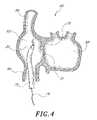
- FIG. 4is a schematic cross-sectional view of a portion of the heart, showing a transseptal access catheter of the present invention within the right atrium.
- FIG. 5is a cross-sectional view as in FIG. 4 , with the guidewire positioned in the superior vena cava.
- FIG. 6is a cross-sectional view as in FIG. 4 , with the transseptal access catheter positioned against the wall of the superior vena cava.
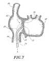
- FIG. 7is a cross-sectional view as in FIG. 4 , with the access catheter positioned against the fossa ovalis.
- FIG. 8is a cross-sectional view as in FIG. 4 , showing tissue distention or “tenting” as the needle punctures the fossa ovalis.
- FIG. 9is a cross-sectional view as in FIG. 8 , showing tissue distention as the dilator is advanced through the fossa ovalis.
- FIG. 10is a cross-sectional view as in FIG. 9 , illustrating the sheath, which has been advanced over the dilator and through the septum.
- FIG. 11is a cross-sectional view as in FIG. 10 , with the dilator removed, leaving the sheath in place across the fossa ovalis.
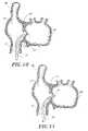
- FIGS. 12 and 13are perspective schematic views of a transseptal access sheath in accordance with the present invention.
- FIGS. 14A–Care perspective views of vents in the transseptal access sheath.
- FIGS. 15A–Care side views of a dilator in accordance with one embodiment of the present invention.
- FIGS. 16A–Care side views of various dilators in accordance with further embodiments of the present invention.
- FIGS. 17A–Care side views of the jacket in accordance with one embodiment of the present invention.
- FIG. 18is a side view of the transseptal access sheath in accordance with one embodiment of the present invention.
- FIG. 19is a side view of the transseptal access sheath in accordance with one embodiment of the present invention.
- FIGS. 20A and 20Bare side views of the needle in accordance with one embodiment of the present invention.
- FIG. 21is a side view of the needle in accordance with one embodiment of the present invention.
- FIGS. 22A–22Hare side views of various needles in accordance with further embodiments of the present invention.
- Dilator 20in accordance with the present invention.
- Dilator 20has a proximal end 22 , a distal end 24 and an elongate flexible tubular body 26 .
- the overall length of the dilator 20depends upon the percutaneous access point and the desired application. For example, lengths in the area of from about 80 cm to about 100 cm are typical for use in percutaneous transluminal access at the femoral vein for locating and puncturing a site on the atrial septum in the heart.
- Tubular body 26may be manufactured in accordance with any of a variety of known techniques, for manufacturing catheters adapted to reach the coronary arteries or chambers of the heart.
- tubular body 26may be manufactured as an extrusion of appropriate biocompatible polymeric materials such as high density polyethylene, polytetrafluoroethylene (PTFE), nylons, and a variety of others which are known in the art.
- Blended materialsmay also be used, such as HDPE (e.g., HDPE/LDPE ratios such as 50%:50%, 60%:40% and others) with from about 5% to about 25%, and, in one embodiment, about 20% BaSO.sub.4 for lubricity and radiopacity.
- tubular body 26may comprise a spring coil, solid walled hypodermic needle tubing (e.g., stainless steel, NiTi alloys) or braided reinforced wall as is understood in the catheter and guidewire arts.
- solid walled hypodermic needle tubinge.g., stainless steel, NiTi alloys
- braided reinforced wallas is understood in the catheter and guidewire arts.
- the tubular body 26is provided with an approximately circular cross sectional configuration having an outside diameter within the range of from about 0.020 inches to about 0.200 inches. In accordance with one embodiment of the invention, the tubular body 26 has an outside diameter of about 0.160 inches throughout its length. Other lengths and diameters may be readily utilized, depending upon the desired profile and performance characteristics.
- the proximal end 22is provided with a manifold 28 , having one or more access ports as in known in the art.
- manifold 28is provided with a core wire port 32 which may also or alternatively function as a guidewire port in an over the wire embodiment.
- An injection port 30may also be provided, for injecting a contrast media, such as to confirm that the distal end 24 has traversed the intraatrial septum. Additional access ports may be provided as needed, depending upon the functional capabilities of the catheter.
- Manifold 28may be injection molded from any of a variety of medical grade plastics or formed in accordance with other techniques known in the art.
- the proximal end 22is also provided with a communication line 34 such as a fiber optic bundle 35 in accordance with one aspect of the present invention.
- fiber optic bundle or signal transmitting line 35communicates with a signal e.g. sound, light, ultrasonic or other vibration, etc.) generator and detector 37 .
- the detector 37enables the catheter to distinguish among solid tissue or a thick membrane, a thin membrane such as at the fossa ovalis, and right atrial or left atrial chamber blood beyond the distal end 24 of dilator 20 as will be discussed.
- the flexible body 26is provided with a preset bend 25 , for assisting in biasing the distal end 24 against the intraatrial septum as is understood in the art.
- Bend 25preferably has a radius within the range of from about 0.5 cm to about 5 cm and, in one embodiment, about 2.5 cm. Bend 25 is centered on a point which is within the range of from about 1 cm to about 10 cm proximally from distal end 24 . In one embodiment, the bend 25 is centered at approximately 6 cm proximally from distal end 24 .
- the bend 25is defined by a proximal transition where it meets the substantially linear proximal portion of the dilator 20 , and a distal transition where it meets the substantially linear distal portion of the dilator 20 .
- the angular deflection of the bend 25is generally within the range of from about 30° to about 80° and, in one embodiment, is about 50°.
- Bend 25may be provided in accordance with any of a variety of techniques.
- the tubular body 26which includes a hypotube or other metal tubing
- the tubular body 26may be bent such as around a forming mandrel in excess of the elastic limit of the hypotube.
- an injection molded catheter bodymay be heat set in a predetermined bend, such as with removable flexible mandrels extending through any interior lumen to maintain patency of the lumen around the bend.
- Other techniqueswill be known to those of skill in the art.
- the bend 25may be formed during or after placement of the catheter in the heart.
- one or more axially moveable pull wiresmay extend throughout the length of the catheter. Proximal traction on a pull wire which is secured at the distal end of the catheter will cause a lateral defection of the catheter.
- distal end 24is provided with at least one signal transmitting surface 47 and at least one signal receiving surface 49 .
- Transmitting surface 47is adapted to transmit a signal from the distal end 24 of dilator 20 and generally in the distal direction with respect to the dilator.
- Receiving surface 49is adapted for receiving a reflected return signal traveling in a generally proximal direction with respect to the distal end 24 of dilator 20 .
- the transmitting surface 47comprises the distal end of a fiber optic or fiber optic bundle, or a transparent window positioned at the distal end of a fiber optic or fiber optic bundle.
- the receiving surface 49comprises a distal end of a receiving fiber optic or a transparent window positioned distally of the receiving fiber optic.
- two transmitting surfaces 47 and two receiving surfaces 49are provided each communicating with the spectrometer 37 via a unique communication line 34 .
- Transmission and reception of, for example, visible lightcan alternatively be accomplished though a single transparent window, and embodiments in which the transmission and reception signals are propagated through the same fiber optic or through closely adjacent fiber optics are also contemplated. Propagation of transmission and reception signals through the same fiber optic can be accomplished such as by the provision of a coupler at the proximal end to split the transmission and reception signals for processing at detector 37 as will be understood in, among others, the blood oximetry detector arts. Alternatively, one or more separate transmit surfaces 47 and receiving surfaces 49 may be provided, and anywhere within the range of from about 1 to about 12 of each transmit surface 47 and receiving surface 49 may be provided as desired.
- Signal transmitting bundle 35thus provides communication between the transmit surface 47 and receiving surface 49 , and a detector 37 such as a spectrometer which remains outside of the patient.
- a detector 37such as a spectrometer which remains outside of the patient.
- transmitter/detector 37is able to transmit multiple wavelengths of light, which propagate beyond the transmit surface 47 and into a target beyond the distal end 24 of the dilator 20 . Some of the transmitted light is absorbed in the target, while other transmitted light is reflected back and received at receiving surface 49 . The reflected light is thereafter propagated to the light detector 37 for processing.
- the present inventorshave determined that the light detector 37 in combination with the dilator of the present invention can identify when the distal end 24 of the dilator 20 is positioned against the fossa ovalis of the intraatrial septum, as opposed to other portions of the septum or muscle wall, due to the unique characteristics of light observed at the fossa ovalis.
- reflected light at the fossa ovaliswill exhibit unique characteristics imparted by (1) light reflected at the surface of or within the fossa ovalis, (2) light reflected through the fossa ovalis by blood in the left atrium, or (3) a combination of the foregoing.
- the ability of an optical detector to locate the fossa based upon light propagated through the fossais based upon several circumstances.
- the blood in the right atriumis relatively poorly oxygenated, and therefore more blue than red.
- the left atriumcontains well oxygenated blood which tends to be relatively red.
- the fossais thin enough to allow light to be transmitted across the fossa and into and from the left atrium while the fossa locator is still on the right atrial side.
- the septumcontains oxygenated blood and therefore has a certain level of red transmission.
- the fossais a thin translucent membrane which is almost yellow. Non-oxygenated blood within the right atrium is relatively blue, while oxygenated blood within the left atrium is red. Location of the fossa may thus alternatively be accomplished by identifying the presence of a translucent, near yellow membrane.
- the use of multiple wavelengths, transmission, and detectorswill allow assessment of both the near yellow color of the fossa, as well as the red color identified through the fossa as will be apparent to those of skill in the art in view of the disclosure herein.
- the method of the present inventionmay additionally be accomplished by providing a light source within the left atrium.
- the left atrium light sourcemay be provided on any of a variety of left atrium access catheters, as will be apparent to those of skill in the art. Light generated in the left atrium, will be detectable in the right atrium either exclusively at the fossa, or with a greatest intensity appearing at the fossa.
- the left atrium dilator 20need only be provided with light detector optics and electronics, to identify the fossa based upon the characteristics of light received from the right atrium light source.
- the dilator 20is additionally provided with a tissue piercing structure 42 such as a needle 44 .
- Needle 44preferably comprises a tubular structure such as a stainless steel hypotube having a sharpened distal end 50 .
- the sharpened distal end 50 of needle 44is axially moveable advanceable through an aperture 45 in the distal end 24 of the tubular body 26 .
- the needle 44has an axial length of from about 1 cm to about 5 cm, an inside diameter of about 0.022 inches and an outside diameter of about 0.032 inches. Any of a variety of other dimensions for needle 44 may also be used depending upon the desired performance and overall catheter dimensions. Needle 44 is connected to the distal end 40 of a control element such as core wire 36 which axially moveably extends throughout the length of tubular body 26 . The proximal end 38 of the core wire 36 in the illustrated embodiment extends proximally from the core wire port 32 .
- a control elementsuch as core wire 36 which axially moveably extends throughout the length of tubular body 26 .
- the proximal end 38 of the core wire 36 in the illustrated embodimentextends proximally from the core wire port 32 .
- the needle 44is preferably axially moveable between a first position in which the tip 50 is contained within the distal end 24 of the tubular body 26 and a distal position in which the distal tip 50 of the needle 44 is exposed beyond the distal end of the body 26 such as for piercing the fossa ovalis. Distal advancement of the proximal end 38 of core wire 36 will advance the needle 44 from the first position to the second position as will be appreciated in view of the disclosure herein. Alternatively, the needle 44 and core wire 36 may be removed entirely from the dilator 20 except when desired to pierce the septum.
- the proximal end 38 of the core wiremay be exposed beyond the proximal end of core wire port 32 as in the illustrated embodiment, such that the physician can grasp the core wire 36 and advance it distally with optimum tactile feedback.
- the proximal end 38 of core wire 36may be connected to any of a wide variety of controls such as a slider switch, rotatable knob or other control attached to or adjacent the manifold 28 . Manipulation of the control can controllably reciprocally move the needle 44 between the first and second position.
- the needle 44removably extends throughout the entire length of the dilator 20 .
- needle 44may have an axial length of from about 100 cm to about 120 cm or longer, and, in one embodiment, about 110 cm.
- radiopaque dyecan be injected through the central lumen 39 , and through the hollow needle 44 (if present) for assessing the position of the distal end 24 of the dilator 20 .
- bloodmay be withdrawn and analyzed for O 2 content by well known methods. Left atrial blood will have an O 2 saturation of greater than 90%, whereas right atrial blood has an O 2 saturation of less than 80%.
- a separate injection lumen(not illustrated) can be readily provided if desired for a particular application.
- the needle 44may be removable from the dilator 20 . In this construction, the dilator 20 retains its greatest flexibility such as for advancement to the intraatrial access site.
- the piercing structure 42such as needle 44 can be loaded into the proximal end 22 of the dilator 20 and advance distally throughout the length of the dilator 20 and out a distal aperture 45 .
- the piercing structure 42may be proximally retracted and removed from the dilator, thereby leaving the central lumen fully available for subsequent therapeutic or diagnostic devices or materials.
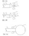
- the needle 44may be provided with a corkscrew thread or other helical structure 84 , such as those having substantially circular or triangular cross-sections, thereby permitting the needle 44 to penetrate the fossa ovalis 72 by rotating while moving transversely through the opening.
- a corkscrew thread or other helical structure 84such as those having substantially circular or triangular cross-sections, thereby permitting the needle 44 to penetrate the fossa ovalis 72 by rotating while moving transversely through the opening.
- the needlemay be configured to create an opening when advanced in the distal direction, and to enlarge the opening when advanced back through the fossa ovalis in the proximal direction. This may be accomplished by providing additional cutting means, such as edges, on the needle 44 .
- the needlehas a primary cutting edge 92 that is rotatably attached to the distal end of the needle 96 . The primary cutting edge 92 punctures the fossa ovalis 72 when the needle 44 is advanced in the distal direction.
- the distal end of the needle 44is advanced into the left atrium and the primary cutting edge 92 is rotated in the left atrium to expose one or more additional cutting surface 94 , such as by means of an axially moveable pull-wire as disclosed elsewhere herein, or by other appropriate remote control or biasing means as are well known to those ordinary skill in the art.
- the needle 44has two or more primary cutting edges 92 that are attached to the needle 44 in a scissors-like configuration.
- the primary cutting edges 92puncture the fossa ovalis when the needle 44 is advanced in the distal direction.
- the primary cutting edges 92can be opened in the left atrium to expose one or more additional cutting surfaces 94 , such as by means of an axially moveable pull-wire as disclosed elsewhere herein, or by other appropriate remote control or biasing means as are well known to those ordinary skill in the art.
- Retracting the needle back through the fossa ovalis 72 in the proximal direction with the additional cutting surfaces 94 in the exposed configurationresults in a larger opening in the fossa ovalis 72 .
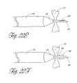
- the needle 44has two or more wings 90 that are contained within the needle 44 when the needle 44 is advanced in the distal direction.
- the wings 90are optionally biased to expand when the needle 44 has crossed the fossa ovalis.
- the wings 90are expanded from the proximal end of the needle 44 such as by means of an axially moveable pull-wire as disclosed elsewhere herein, or by other appropriate remote control or biasing means as are well known to those ordinary skill in the art.
- expanding the wings 90exposes secondary cutting surfaces 94 . Retracting the needle back through the fossa ovalis 72 in the proximal direction with the additional cutting surfaces 94 in the exposed configuration results in a larger opening in the fossa ovalis 72 .
- the wings 90may be configured with secondary cutting surfaces 94 inclined at an angle extending toward the proximal end of the needle.
- the wings 90may be configured with rounded secondary cutting surfaces 94 .
- the wings 90may also be inclined at an angle extending toward the distal end of the needle or at an angle perpendicular to the axis of the needle.
- the wings 90may be withdrawn back into the needle 44 by advancing the needle 44 distally into the dilator 20 .
- secondary cutting surfacescan be used with, or without, a dilator 20 .
- the optimal type of dilator, if any, to use with a needle having one or more secondary cutting surfaceswill depend on the particular application as well as the design of the primary and secondary cutting surfaces, and can be determined though routine experimentation.
- the cutting surfaces or other dilatation surfacesare advanceable from a first, reduced crossing profile for advancement in a first direction (e.g., distally through the fossa ovalis), and a second, enlarged crossing profile for movement in a second direction (e.g., proximal retraction back through the fossa ovalis).
- a first directione.g., distally through the fossa ovalis
- a second, enlarged crossing profile for movement in a second directione.g., proximal retraction back through the fossa ovalis.
- Any of a variety of structurescan be utilized, in which the cutting surface or tissue engagement surface is moveable from a first orientation in which it extend generally parallel to the longitudinal axis of the needle or other catheter component for penetration through a tissue surface, and a second, inclined orientation with respect to the longitudinal axis to present a larger cross sectional area.
- Such structuresmay be biased in the direction of the second, enlarged crossing profile and restrained by a movable sheath, pull wire or other retention structure.
- the tissue engagement surfacemay be neutrally biased, and moved between the reduced crossing profile and enlarged crossing profile orientations by axial movement of a pull wire.
- such structuresmay be advanced into the enlarged crossing profile by inflation of an underlying balloon in communication with the proximal end of the catheter by an axially extending inflation lumen.
- the distal end 24 of dilator 20is provided with a tapered frustro conical surface 27 .
- Thisallows the tubular body 26 to function as a dilator, thereby permitting the tapered surface 27 to enlarge the opening formed by needle 44 while minimizing “tenting” of the fossa ovalis during the transseptal access procedure.
- the distal end 24 of dilator 20may be provided with a corkscrew or other helical structure 84 , such as those having threads with semi-circular or triangular cross-sections, thereby permitting the dilator to enlarge the opening formed by needle 44 by rotating while moving transversely through the opening.
- the distal end 24 of dilator 20may be provided with one or more nozzles 86 capable of generating a high-pressure stream 88 of fluid, such as saline solution, thereby enlarging the opening formed by needle 44 .
- the dilator 20may be transversely advanced while the dilator 20 is spinning or rotating with respect to the fossa ovalis.
- the right atriummay be initially accessed with a transseptal access system through either the inferior or superior vena cava, which initially requires cannulation with an introducer sheath such as through the well known “Seldinger” technique.
- the transseptal access system of the present inventionincludes a transseptal sheath, a piercing dilator catheter 20 as discussed above, and an appropriately sized guidewire.
- the sheath 74comprises an elongate, flexible tubular body having preset bends as described below.
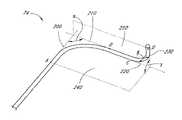
- the sheath 74comprises a first substantially linear section 100 , a first curved section 110 and a second curved section 130 .
- the term substantially linearalso encompasses structures that are actually linear.
- the first curved section 110is distal to the first substantially linear section 100
- the second curved section 130is distal to the first curved section 110 .
- the first curved section 110transitions directly into the second curved section 130 so there is no second substantially linear section 120 , and in other embodiments there is no third substantially linear section 140 distal to the second curved section.
- the sheathis designed to facilitate access to the left atrium. In other embodiments, the sheath is designed to facilitate access to other physiological structures, such as the LAA, the orifice of the LAA, the distal aspect of the LAA, or pulmonary vein such as the left superior pulmonary vein.
- the first curved sectionis shaped to abut the interior wall of the right atrium substantially opposite the fossa ovalis while directing the distal tip toward the fossa ovalis.
- the second curved sectionis shaped to facilitate location and access of the desired region of the LAA after penetration of the fossa ovalis.
- One of ordinary skill in the artwill recognize that the precise shape of these curves will depend on the patient's physiology and can be determined through routine experimentation.
- the first and second curved sectionsmay be provided in accordance with any of a variety of techniques.
- the first and second curved sectionsmay be introduced prior to insertion of the sheath into the body by bending the sheath, such as around a forming mandrel under heat or in excess of the elastic limit of the sheath.
- an injection molded sheath bodymay be provided with a predetermined bend, or by heat setting such as with removable flexible mandrels extending through any interior lumen to maintain patency of the lumen around the bend.
- the first and second curved sectionsmay be formed during or after placement of the sheath in the patient.
- Thismay be accomplished by providing the sheath with any of a variety of steering mechanisms, which allow a distal portion of the sheath to be inclined away from the axis of the normal bias of the catheter.
- one or more axially moveable pull wiresmay extend through the length of the sheath. Proximal traction on a pull wire that is secured at a distal location of the catheter will cause a lateral defection of the catheter.
- Other techniqueswill be known to those of skill in the art.
- the point where the first substantially linear section 100 begins to transition into the first curved section 110defines a first proximal edge point A on the outside edge of the exterior of the sheath.
- a tangent 200 to the outer edge of the first curved section 110 at the first proximal edge point Ais substantially parallel to the first substantially linear section 100 .
- the point where the first curved section 110 begins to transition into the second substantially linear section 120defines a first distal edge point B on the outside edge of the exterior of the sheath.
- a tangent 210 to the outer edge of the first curved section 110 at the first distal edge point Bis substantially parallel to the second substantially linear section 120 .
- the first distal edge point Bis defined as the first inflection point distal to the first curved section 110 on the outside edge of the exterior of the sheath.
- a tangent 210 to the outer edge of the first curved section 110 at the first distal edge point Bis substantially parallel to the outside edge of the exterior of the sheath at point B.
- the point where the second substantially linear section 120 begins to transition into the second curved section 130defines a second proximal edge point C on the outside edge of the exterior of the sheath.
- a tangent 220 to the outer edge of the second curved section 130 at the second proximal edge point Cis substantially parallel to the second substantially linear section 120 .
- the second proximal edge point Cis defined as the first inflection point proximal to the second curved section 130 .
- a tangent 220 to the outer edge of the second curved section 130 at the second proximal edge point Cis substantially parallel to the outside edge of the exterior of the sheath at point C.
- the point where the second curved section 130 begins to transition into the third substantially linear section 140defines a second distal edge point D on the outside edge of the exterior of the sheath.
- a tangent 230 to the outer edge of the second curved section 130 at the second distal edge point Dis substantially parallel to the third substantially linear section 140 .
- the second distal edge point Dis defined as the distal end point of the outside edge of the exterior of the sheath.
- the second distal edge point Dis defined as the first inflection point distal to the second curved section 130 .
- a tangent 220 to the outer edge of the second curved section 130 at the first distal edge point Bis substantially parallel to the outside edge of the exterior of the sheath at point B.
- the tangent 200 and the tangent 210intersect to form an angle ⁇ .
- the angle ⁇is preferably about 90 degrees, generally between about 75 degrees and about 105 degrees, and in other embodiments may be in the range of about 80 degrees to about 100 degrees.
- the tangent 220 and the tangent 230intersect to form an angle ⁇ .
- the angle ⁇is preferably about 90 degrees, generally between about 75 degrees and about 105 degrees, and in other embodiments may be in the range of about 80 degrees to about 100 degrees.
- the tangent 200 and the tangent 210also define a plane, 240 .
- the tangent 220 and the tangent 230also define a plane, 250 .
- the plane 240 and the plane 250define an angle ⁇ .
- the angle ⁇is preferably about 90 degrees, generally between about 75 degrees and about 105 degrees and in other embodiments may be in the range of about 80 degrees to about 100 degrees.
- the first curved section 110has an arc length along the outside edge of the exterior of the sheath between point A and point B in the range of about 7.5 cm to about 9.5 cm.
- the second curved section 120has an arc length along the outside edge of the exterior of the sheath between point C and point D in the range of about 5 cm to about 7.5 cm.
- the sheathalso defines a Cartesian coordinate system.
- the origin of this coordinate systemis defined as point A.
- the X-axisis defined as parallel to tangent 200 .
- the Y-axisis defined as the normal to tangent 200 in plane 240 .
- the Z-axisis defined as the normal to plane 240 at point A.
- the distance along the Y-axis between point A and point Dis about 9 cm. In other embodiments, one of ordinary skill in the art will appreciate that the distance may vary depending upon the patient's physiology, for example within the range of about 6 cm to about 15 cm. In one embodiment, the distance along the Z-axis between point C and point D is about 3.5 cm. In other embodiments, one of ordinary skill in the art will appreciate that the distance may vary depending upon the patient's physiology, for example within the range of about 1 cm to about 6 cm.
- the first curved section 110has a radius of curvature of about 5 cm. In other embodiments, one of ordinary skill in the art will appreciate that the radius of curvature may vary depending upon the patient's physiology, for example within the range of about 3 cm to about 10 cm. In one embodiment, the second curved section 130 has a radius of curvature of about 3 cm. In other embodiments, one of ordinary skill in the art will appreciate that the radius of curvature may vary depending upon the patient's physiology, for example within the range of about 1 cm to about 6 cm.
- the sheathis sufficiently tourqueable that the location of the distal tip of the sheath can be controlled from the proximal end of the sheath.
- additional torquabilityis supplied by re-enforcing the shaft with stainless steel or high strength thin polymeric braid. It is desirable to maximize the lubricity of the inner surface of the sheath to facilitate insertion and removal of various diagnostic and treatment devices within the sheath.
- the inner surface of the sheathis coated with lubricity enhancing material, such as PTFE.
- Manageability and atraumaticityare facilitated by varying the stiffness of the sheath along its length. It is desirable for the proximal section of the sheath to be relatively more stiff for torquability and kink resistance, and for the distal section of the sheath to be relatively less stiff. Generally, it is advantageous to soften the material comprising the sheath, and/or to make the wall of the sheath thinner toward the distal end.
- the proximal section of the sheathcomprises 70D Pebax and has a wall thickness of about 0.015 inches
- the intermediate section of the sheathcomprises 40D Pebax and has a wall thickness of about 0.015 inches
- the distal section of the sheathcomprises 40D Pebax and has a wall thickness of about 0.011 inches.
- the proximal section of the sheathextends for about 55 cm
- the intermediate section of the sheathextends for about 13 cm
- the distal section of the sheathextends for about 2 cm.
- the proximal section of the sheathmay extend from about 40 cm to about 65 cm
- the intermediate section of the sheathmay extend from about 5 cm to about 15 cm
- the distal section of the sheathmay extend from about 2 mm to about 50 mm.
- the shaftmay be made of a greater or lesser number of sections having varying compositions and durometer hardnesses, that there may be gradual or abrupt transition between the sections, that the various sections may comprise a variety of other materials, and that the thickness of the sheath wall may also be adjusted.
- the desired combination of these parameterswill depend on which parameters are selected and particularly useful combinations of these properties will be apparent to one of ordinary skill in the art through routine testing.
- the atraumacity of the transition between the needle, dilator, transition catheter or the likemay be enhanced by tapering the outer diameter of the distal tip of the sheath and/or tapering the thickness of the wall of the distal tip of the sheath.
- Visualization of the sheathcan be enhanced by marking the distal tip of the sheath a radiopaque marker.
- the radiopaque markermay be applied as a band, or in other shapes and at varying distances from the distal tip.
- vents 80are slits in the distal end of the sheath.
- the vents 80are holes which extend transversely through the wall near the distal end of the sheath.
- the vent holes 80may also be located within the radiopaque marker band to reduce or prevent collapsing or clogging of the vent holes.
- the vents 80have a diameter or cross sectional area equivalent to a round vent diameter within the range of from about 0.020 inches to about 0.100 inches. Anywhere from about one to about ten or more vents may be provided. In one embodiment, approximately 4 vents, each having a diameter of about 0.040 inches are provided.
- the ventsmay be positioned in a single plane transverse to the longitudinal axis of the catheter as illustrated. The vents reside in a plane which is generally no more than about 5 mm from the distal end. Preferably, the vents are positioned within about 3 mm of the distal end of the catheter. Alternatively, the vents may be staggered in two or more transverse planes, depending upon the desired performance characteristics.
- the preferred access pointis along the right femoral vein, although access from the left femoral vein is also possible. Access may also be achieved through a puncture in any of a variety of other veins of suitable internal diameter and the present invention is not limited in this regard.
- a conventional spring tipped guide wireis thereafter advanced through the needle into the vein and the needle is subsequently removed.
- the dilator 20 of the present inventionis positioned within a sheath of the type described herein, or other well-known sheaths, such as a 14 French introducer sheath. Subsequently, the sheath and inner dilator 20 , in combination with the guide wire, are advanced through the femoral vein to the right atrium.
- FIG. 4there is illustrated a schematic cross-section of a portion of the heart 60 .
- the right atrium 62is communication with the inferior vena cava 64 and the superior vena cava 66 .
- the right atrium 62is separated from the left atrium 68 by the intraatrial septum 70 .
- the fossa ovalis 72is located on the intraatrial septum 70 .
- the sheath 74 having the dilator 20 therein and a guidewire 76have been positioned within the right atrium 62 .
- the guidewire 76is thereafter distally advanced to access the superior vena cava 66 . See FIG. 5 .
- the dilator 20 and sheath 74are thereafter advanced into the superior vena cava as illustrated schematically in FIG. 6 .
- the guidewire 76is proximally retracted.
- a transseptal needle 44is advanced through the central lumen 39 of the dilator 20 and sheath 74 .
- the transseptal needle 44is advanced (possibly with a stylet in place) to a point that the stylet tip is just inside the distal tip of the sheath 74 and dilator 20 , a position previously noted by the operator, and the stylet is withdrawn from the transseptal needle.
- the remaining combination of the sheath 74 with the dilator 20 having the transseptal needle thereinis then drawn proximally from the superior vena cava while the first curved section 110 of the sheath, alone or in combination with the preset curve 25 at the distal region of dilator 20 , causes the tip of the sheath-dilator-transseptal needle combination to “drag” along the wall of the right atrium and the septum 70 .
- some differences in the access methodwill occur at this point.
- the light source and detector 37will likely need to be calibrated once the dilator 20 has been placed inside the right atrium 62 but before the tip has been placed against the septum 70 .
- the tip of the dilator 20is then positioned against the septum 70 by distal advancement through the sheath 74 .
- the tipis then dragged along the septum by proximal traction on the dilator 20 until the tip pops onto the fossa 72 .
- the characteristic color at the fossais detected by the detector 37 .
- a responsive audio or visual signalis generated, confirming that the catheter 20 is now properly positioned at the fossa ovalis 72 .
- the physicianis normally assisted during placement, as in the entire procedure, by fluoroscopy or other visualization techniques.
- the distal tip of sheath 74 and the distal tip of dilator 20may be provided with a radiopaque marker.
- some physiciansfind it desirable to infuse a radiopaque dye through the transseptal needle at various stages of the procedure to assist in visualization, particularly following the transseptal puncture.
- the transseptal needle 44is abruptly advanced to accomplish a quick puncture. See FIG. 8 .
- the needleis advanced by applying a force to the proximal end of the needle.
- the needleoften comprises a stiff proximal section and a flexible distal section.
- the distal sectionpreferably comprises ribbon coil.
- the needle 44is loaded into needle cavity 89 against a distal advancement biasing means 85 , such as a spring or coil. The needle 44 is held against the biasing means 85 by a release lock 87 .
- the release lock 87may comprise any commonly used retaining means, such as corresponding inward and outward projections, grooves or flanges on the needle cavity 89 and the needle 44 .
- the distal end of the sheath 74is placed in the desired location against the fossa ovalis 72 .
- the release lock 87is opened and the biasing means rapidly advances the needle 44 through the fossa ovalis 72 .
- one medical techniqueis to confirm the presence of the tip 50 of the transseptal needle 44 within the left atrium 68 .
- Confirmation of such location of the tip 50 of the transseptal needle 44may be accomplished by monitoring the pressure sensed through the transseptal needle lumen to ensure that the measured pressure is within the expected range and has a waveform configuration typical of left atrial pressure.
- proper position within the left atrium 68may be confirmed by analysis of oxygen saturation level of the blood drawn through the transseptal needle 44 ; i.e., aspirating fully oxygenated blood.
- visualization through fluoroscopy alone, or in combination with the use of dyemay also serve to confirm the presence of the tip 50 of the transseptal needle 44 in the left atrium 68 .
- the tip 27 of the dilator 20is advanced through the septum 70 and into the left atrium 68 .
- careis taken to ensure that, at the same time of advancing the dilator and sheath tip into the left atrium, the tip of the transseptal needle is not advanced a sufficient distance that the needle 44 can damage the opposing wall of the left atrium 68 .
- the transseptal needle 44is withdrawn.
- the sheath 74is then advanced into the left atrium 68 , either by advancing the sheath 74 alone over the dilator 20 or by advancing the sheath 74 and dilator 20 in combination. See FIG. 10 .
- the dilator 20is then withdrawn from sheath 74 when the latter has been advanced into the left atrium, thus leaving the main lumen of sheath 74 as a clear pathway to advancing further diagnostic or therapeutic instruments into the left atrium.
- the needle 44is optionally coated with a lubricous material, often a material such as PTFE, hydrophilic material or parylene, preferably PTFE.
- the dilator 20is also optionally coated with a lubricous material, often a material such as PTFE, hydrophilic material or parylene, preferably PTFE.
- the sheath 74contains an inflatable balloon 77 at the distal end and means for inflating the balloon 75 , such as an inflation lumen.
- the balloon 77is in a deflated position as the needle 44 crosses the fossa ovalis.
- the balloon 77is disposed around the needle 44 , and the balloon 77 is inflated as it crosses the fossa ovalis, thereby enlarging the puncture made by the needle 44 .
- This embodimentmay be used with or without an additional dilator 20 as described herein.
- the balloon 77may be deflated and withdrawn after the sheath 74 has accessed the left atrium.
- the balloonmay be left inflated, or further inflated after the sheath 74 accesses the left atrium.
- the balloon 77may be inflated after the balloon crosses the fossa ovalis.
- the inflated balloon 77creates an atraumatic distal tip that can be navigated to access a location of interest, such as the LAA.
- the balloonprevents the sheath from unintentionally passing back through the fossa ovalis during subsequent procedures.
- At least a portion of the needle 44 , the dilator 20 and at least a distal portion of the sheath 74are encompassed by a thin-walled jacket 82 that is made of lubricous material, preferably PTFE.
- a thin-walled jacket 82that is made of lubricous material, preferably PTFE.
- the distal end of the jacket 82advances across the fossa ovalis 72 with the needle 44 .
- the dilator 20 and sheath 74then cross the fossa ovalis 72 within the jacket 82 .
- the jacketis radially enlargeable and may have a thickness in the range of about 0.05 mm to about 0.75 mm depending upon the construction material.
- the optimum material or combination of materials for coating the needle 44 , coating the dilator 20 , or forming the jacket 82can be determined for any particular application through routine experimentation by one or ordinary skill in the art based on the disclosures herein.
- the sheath 74is itself tapered at its distal end to encompass at least a portion of the dilator. In this embodiment the sheath 74 is preferably tapered to encompass the dilator and at least a portion of the needle 44 . In another embodiment, illustrated in FIG. 19 , the sheath 74 includes means, such as a vacuum lumen, for applying a vacuum 83 at the distal end.
- the vacuum lumenmay be concentrically disposed within the needle 44 and the dilator 20 , concentrically disposed within the dilator 20 , but side by side with the needle 44 , or side by side with the dilator 20 .
- the vacuumis applied to the fossa ovalis before the needle 44 punctures the fossa ovalis.
- the vacuumis applied to the fossa ovalis after the needle punctures the fossa ovalis and before or as the dilator 20 crosses the fossa ovalis.
- the vacuummaintains the position of the fossa ovalis and reduces “tenting” during the transseptal access procedure.
- the vacuummay draw the fossa ovalis into the sheath 74 .
- the needle 44is oscillated at ultrasonic frequencies to further reduce friction between the needle 44 and the fossa ovalis 72 .
Landscapes
- Health & Medical Sciences (AREA)
- Life Sciences & Earth Sciences (AREA)
- Surgery (AREA)
- Medical Informatics (AREA)
- Animal Behavior & Ethology (AREA)
- Engineering & Computer Science (AREA)
- Biomedical Technology (AREA)
- Heart & Thoracic Surgery (AREA)
- Veterinary Medicine (AREA)
- Molecular Biology (AREA)
- Public Health (AREA)
- General Health & Medical Sciences (AREA)
- Nuclear Medicine, Radiotherapy & Molecular Imaging (AREA)
- Vascular Medicine (AREA)
- Pathology (AREA)
- Physics & Mathematics (AREA)
- Biophysics (AREA)
- Surgical Instruments (AREA)
Abstract
Description
Claims (27)
Priority Applications (5)
| Application Number | Priority Date | Filing Date | Title |
|---|---|---|---|
| US10/100,270US7056294B2 (en) | 2000-04-13 | 2002-03-15 | Method and apparatus for accessing the left atrial appendage |
| AU2003225822AAU2003225822A1 (en) | 2002-03-15 | 2003-03-14 | Method and apparatus for accessing the left atrial appendage |
| PCT/US2003/008044WO2003077733A2 (en) | 2002-03-15 | 2003-03-14 | Method and apparatus for accessing the left atrial appendage |
| US11/228,988US9131849B2 (en) | 2000-04-13 | 2005-09-16 | Method and apparatus for accessing the left atrial appendage |
| US14/834,499US20150359563A1 (en) | 2000-04-13 | 2015-08-25 | Method and apparatus for accessing the left atrial appendage |
Applications Claiming Priority (2)
| Application Number | Priority Date | Filing Date | Title |
|---|---|---|---|
| US09/549,218US6650923B1 (en) | 2000-04-13 | 2000-04-13 | Method for accessing the left atrium of the heart by locating the fossa ovalis |
| US10/100,270US7056294B2 (en) | 2000-04-13 | 2002-03-15 | Method and apparatus for accessing the left atrial appendage |
Related Parent Applications (1)
| Application Number | Title | Priority Date | Filing Date |
|---|---|---|---|
| US09/549,218Continuation-In-PartUS6650923B1 (en) | 2000-04-13 | 2000-04-13 | Method for accessing the left atrium of the heart by locating the fossa ovalis |
Related Child Applications (1)
| Application Number | Title | Priority Date | Filing Date |
|---|---|---|---|
| US11/228,988ContinuationUS9131849B2 (en) | 2000-04-13 | 2005-09-16 | Method and apparatus for accessing the left atrial appendage |
Publications (2)
| Publication Number | Publication Date |
|---|---|
| US20020169377A1 US20020169377A1 (en) | 2002-11-14 |
| US7056294B2true US7056294B2 (en) | 2006-06-06 |
Family
ID=28039764
Family Applications (3)
| Application Number | Title | Priority Date | Filing Date |
|---|---|---|---|
| US10/100,270Expired - LifetimeUS7056294B2 (en) | 2000-04-13 | 2002-03-15 | Method and apparatus for accessing the left atrial appendage |
| US11/228,988Expired - LifetimeUS9131849B2 (en) | 2000-04-13 | 2005-09-16 | Method and apparatus for accessing the left atrial appendage |
| US14/834,499AbandonedUS20150359563A1 (en) | 2000-04-13 | 2015-08-25 | Method and apparatus for accessing the left atrial appendage |
Family Applications After (2)
| Application Number | Title | Priority Date | Filing Date |
|---|---|---|---|
| US11/228,988Expired - LifetimeUS9131849B2 (en) | 2000-04-13 | 2005-09-16 | Method and apparatus for accessing the left atrial appendage |
| US14/834,499AbandonedUS20150359563A1 (en) | 2000-04-13 | 2015-08-25 | Method and apparatus for accessing the left atrial appendage |
Country Status (3)
| Country | Link |
|---|---|
| US (3) | US7056294B2 (en) |
| AU (1) | AU2003225822A1 (en) |
| WO (1) | WO2003077733A2 (en) |
Cited By (184)
| Publication number | Priority date | Publication date | Assignee | Title |
|---|---|---|---|---|
| US20050113719A1 (en)* | 2003-09-26 | 2005-05-26 | Vahid Saadat | Left atrial access apparatus and methods |
| US20050288622A1 (en)* | 2004-06-29 | 2005-12-29 | Applied Medical Resources Corporation | Insufflating optical surgical instrument |
| US20060079787A1 (en)* | 2004-09-30 | 2006-04-13 | Whiting James S | Transmembrane access systems and methods |
| US20060253129A1 (en)* | 2005-04-07 | 2006-11-09 | Liddicoat John R | Apparatus and method for the ligation of tissue |
| US20070083168A1 (en)* | 2004-09-30 | 2007-04-12 | Whiting James S | Transmembrane access systems and methods |
| US20070129753A1 (en)* | 2005-12-01 | 2007-06-07 | Chris Quinn | Method and apparatus for recapturing an implant from the left atrial appendage |
| US20070149995A1 (en)* | 2005-12-01 | 2007-06-28 | Chris Quinn | Method for accessing the left atrial appendage with a balloon-tipped transeptal sheath |
| US20080147097A1 (en)* | 2003-10-09 | 2008-06-19 | Sentreheart, Inc. | Apparatus and method for the ligation of tissue |
| US20080243183A1 (en)* | 2007-03-30 | 2008-10-02 | Miller Gary H | Devices, systems, and methods for closing the left atrial appendage |
| US20090105654A1 (en)* | 2007-10-19 | 2009-04-23 | Paul Kurth | Transseptal guidewire |
| US20090112249A1 (en)* | 2007-10-19 | 2009-04-30 | Coherex Medical, Inc. | Medical device for modification of left atrial appendage and related systems and methods |
| US20090182281A1 (en)* | 2008-01-16 | 2009-07-16 | Pressure Products Medical Supplies Inc. | Apparatus, system, and method of shielding the sharp tip of a transseptal guidewire |
| US20100004662A1 (en)* | 2008-06-20 | 2010-01-07 | Ela Medical S.A.S. | Preformed stylet for guiding a lead to contact the septum |
| USD611601S1 (en)* | 2007-05-15 | 2010-03-09 | Terumo Kabushiki Kaisha | Guide wire for catheter |
| US20100081988A1 (en)* | 2008-09-29 | 2010-04-01 | Applies Medical Resources Corporation | First-entry trocar system |
| US20100324588A1 (en)* | 2009-06-17 | 2010-12-23 | Coherex Medical, Inc. | Medical device for modification of left atrial appendage and related systems and methods |
| US20110022004A1 (en)* | 2009-07-27 | 2011-01-27 | Robert Kipperman | Patent foramen ovale catheter and method of using the same |
| US7955293B2 (en) | 2002-08-26 | 2011-06-07 | Flowcardia, Inc. | Ultrasound catheter for disrupting blood vessel obstructions |
| US20110152741A1 (en)* | 2009-12-21 | 2011-06-23 | Michael Banchieri | Cannula system |
| US20110190624A1 (en)* | 2010-01-29 | 2011-08-04 | Medtronic Ablation Frontiers Llc | Optical system and method for localizing the fossa ovalis during trans-septal procedures |
| US20110213316A1 (en)* | 2009-12-21 | 2011-09-01 | Tamer Ibrahim | Self-dilating cannula |
| US20110224666A1 (en)* | 2003-01-21 | 2011-09-15 | Gareth Davies | Method of surgical perforation via the delivery of energy |
| US8029470B2 (en) | 2004-09-30 | 2011-10-04 | Pacesetter, Inc. | Transmembrane access systems and methods |
| US8062566B2 (en) | 2003-04-08 | 2011-11-22 | Flowcardia, Inc. | Method of manufacturing an ultrasound transmission member for use in an ultrasound catheter device |
| US8133236B2 (en) | 2006-11-07 | 2012-03-13 | Flowcardia, Inc. | Ultrasound catheter having protective feature against breakage |
| US20120065597A1 (en)* | 2010-05-11 | 2012-03-15 | Cohen Todd J | Apparatus for safe performance of transseptal technique and placement and positioning of an ablation catheter |
| US20120065591A1 (en)* | 2006-07-26 | 2012-03-15 | Johan Willem Pieter Marsman | Facilitation Of Antegrade Insertion Of A Guidewire Into The Superficial Femoral Artery |
| US8221343B2 (en) | 2005-01-20 | 2012-07-17 | Flowcardia, Inc. | Vibrational catheter devices and methods for making same |
| US8226566B2 (en) | 2009-06-12 | 2012-07-24 | Flowcardia, Inc. | Device and method for vascular re-entry |
| US8235942B2 (en) | 2005-05-04 | 2012-08-07 | Olympus Endo Technology America Inc. | Rotate-to-advance catheterization system |
| US8246643B2 (en) | 2006-11-07 | 2012-08-21 | Flowcardia, Inc. | Ultrasound catheter having improved distal end |
| US20120239069A1 (en)* | 2011-03-17 | 2012-09-20 | Medtronic, Inc. | Transseptal Puncturing Device |
| US20120271248A1 (en)* | 2007-12-21 | 2012-10-25 | Innovatech, Llc | Marked precoated medical device and method of manufacturing same |
| US8317678B2 (en) | 2005-05-04 | 2012-11-27 | Olympus Endo Technology America Inc. | Rotate-to-advance catheterization system |
| US8343040B2 (en) | 2005-05-04 | 2013-01-01 | Olympus Endo Technology America Inc. | Rotate-to-advance catheterization system |
| US8369930B2 (en) | 2009-06-16 | 2013-02-05 | MRI Interventions, Inc. | MRI-guided devices and MRI-guided interventional systems that can track and generate dynamic visualizations of the devices in near real time |
| US8366674B2 (en) | 2005-05-04 | 2013-02-05 | Olympus Endo Technology America Inc. | Rotate-to-advance catheterization system |
| US8377041B2 (en) | 2005-02-28 | 2013-02-19 | Olympus Endo Technology America Inc. | Rotate-to-advance catheterization system |
| US8377090B2 (en) | 2002-05-16 | 2013-02-19 | Applied Medical Resources Corporation | Blunt tip obturator |
| US8414477B2 (en) | 2005-05-04 | 2013-04-09 | Olympus Endo Technology America Inc. | Rotate-to-advance catheterization system |
| US8435229B2 (en) | 2006-02-28 | 2013-05-07 | Olympus Endo Technology America Inc. | Rotate-to-advance catheterization system |
| US8454584B2 (en) | 2010-04-13 | 2013-06-04 | Cook Medical Technologies Llc | Medical anchor device |
| US8469983B2 (en) | 2007-09-20 | 2013-06-25 | Sentreheart, Inc. | Devices and methods for remote suture management |
| US8506519B2 (en) | 1999-02-16 | 2013-08-13 | Flowcardia, Inc. | Pre-shaped therapeutic catheter |
| US8518063B2 (en) | 2001-04-24 | 2013-08-27 | Russell A. Houser | Arteriotomy closure devices and techniques |
| US8517977B2 (en) | 2006-10-06 | 2013-08-27 | Applied Medical Resources Corporation | Visual insufflation port |
| US8574220B2 (en) | 2006-02-28 | 2013-11-05 | Olympus Endo Technology America Inc. | Rotate-to-advance catheterization system |
| US20130317542A1 (en)* | 2012-05-25 | 2013-11-28 | Boston Scientific Scimed, Inc. | Steerable delivery system |
| US8608769B2 (en) | 2001-09-24 | 2013-12-17 | Applied Medical Resources Corporation | Bladeless optical obturator |
| US8613751B2 (en) | 2003-11-24 | 2013-12-24 | Flowcardia, Inc. | Steerable ultrasound catheter |
| US8617096B2 (en) | 2004-08-26 | 2013-12-31 | Flowcardia, Inc. | Ultrasound catheter devices and methods |
| US8636759B2 (en) | 2001-09-24 | 2014-01-28 | Applied Medical Resources Corporation | Bladeless obturator |
| US8641630B2 (en) | 2003-09-19 | 2014-02-04 | Flowcardia, Inc. | Connector for securing ultrasound catheter to transducer |
| US8690911B2 (en) | 2009-01-08 | 2014-04-08 | Coherex Medical, Inc. | Medical device for modification of left atrial appendage and related systems and methods |
| US8721663B2 (en) | 1999-05-20 | 2014-05-13 | Sentreheart, Inc. | Methods and apparatus for transpericardial left atrial appendage closure |
| US8764631B2 (en) | 1997-02-10 | 2014-07-01 | Olympus Endo Technology America Inc. | Rotate to advance catheterization system |
| US8777841B2 (en) | 2007-05-18 | 2014-07-15 | Olympus Endo Technology America Inc. | Rotate-to-advance catheterization system |
| US8795253B2 (en) | 2011-04-05 | 2014-08-05 | Sorin Group Italia S.R.L. | Bi-directional perfusion cannula |
| US8834519B2 (en) | 1998-11-06 | 2014-09-16 | Artritech, Inc. | Method and device for left atrial appendage occlusion |
| US8940008B2 (en) | 2010-04-23 | 2015-01-27 | Assist Medical Llc | Transseptal access device and method of use |
| US8961423B2 (en) | 2003-02-26 | 2015-02-24 | Flowcardia, Inc. | Ultrasound catheter apparatus |
| US8961541B2 (en) | 2007-12-03 | 2015-02-24 | Cardio Vascular Technologies Inc. | Vascular closure devices, systems, and methods of use |
| US8992567B1 (en) | 2001-04-24 | 2015-03-31 | Cardiovascular Technologies Inc. | Compressible, deformable, or deflectable tissue closure devices and method of manufacture |
| US9072872B2 (en) | 2010-10-29 | 2015-07-07 | Medtronic, Inc. | Telescoping catheter delivery system for left heart endocardial device placement |
| US9198664B2 (en) | 2009-04-01 | 2015-12-01 | Sentreheart, Inc. | Tissue ligation devices and controls therefor |
| US9220395B2 (en) | 1999-09-27 | 2015-12-29 | James J. Frassica | Rotate-to-advance catheterization system |
| US20160001045A1 (en)* | 2013-09-19 | 2016-01-07 | W. L. Gore & Associates, Inc. | Dilator systems and methods |
| US9254148B2 (en) | 2011-05-02 | 2016-02-09 | Applied Medical Resources Corporation | Low-profile surgical universal access port |
| US9259290B2 (en) | 2009-06-08 | 2016-02-16 | MRI Interventions, Inc. | MRI-guided surgical systems with proximity alerts |
| US9265899B2 (en) | 2008-01-25 | 2016-02-23 | Applied Medical Resources Corporation | Insufflating access system |
| US9265520B2 (en) | 2002-08-02 | 2016-02-23 | Flowcardia, Inc. | Therapeutic ultrasound system |
| US9282984B2 (en) | 2006-04-05 | 2016-03-15 | Flowcardia, Inc. | Therapeutic ultrasound system |
| US9345460B2 (en) | 2001-04-24 | 2016-05-24 | Cardiovascular Technologies, Inc. | Tissue closure devices, device and systems for delivery, kits and methods therefor |
| US9351716B2 (en) | 2009-06-17 | 2016-05-31 | Coherex Medical, Inc. | Medical device and delivery system for modification of left atrial appendage and methods thereof |
| US9393023B2 (en) | 2009-01-13 | 2016-07-19 | Atricure, Inc. | Apparatus and methods for deploying a clip to occlude an anatomical structure |
| US9408608B2 (en) | 2013-03-12 | 2016-08-09 | Sentreheart, Inc. | Tissue ligation devices and methods therefor |
| US20160270810A1 (en)* | 2014-06-13 | 2016-09-22 | InterShunt Technologies, Inc. | Method and catheter for creating an interatrial aperture |
| US9463268B2 (en) | 2010-09-07 | 2016-10-11 | Paul A. Spence | Cannula systems and methods |
| US9486281B2 (en) | 2010-04-13 | 2016-11-08 | Sentreheart, Inc. | Methods and devices for accessing and delivering devices to a heart |
| US9498206B2 (en) | 2011-06-08 | 2016-11-22 | Sentreheart, Inc. | Tissue ligation devices and tensioning devices therefor |
| US9585991B2 (en) | 2012-10-16 | 2017-03-07 | Heartware, Inc. | Devices, systems, and methods for facilitating flow from the heart to a blood pump |
| US9649115B2 (en) | 2009-06-17 | 2017-05-16 | Coherex Medical, Inc. | Medical device for modification of left atrial appendage and related systems and methods |
| US9693781B2 (en) | 2009-06-17 | 2017-07-04 | Coherex Medical, Inc. | Medical device for modification of left atrial appendage and related systems and methods |
| US20170245885A1 (en)* | 2016-02-25 | 2017-08-31 | Indian Wells Medical, Inc. | Steerable endoluminal punch |
| US9808283B2 (en) | 2013-12-04 | 2017-11-07 | Heartware, Inc. | Apparatus and methods for cutting an atrial wall |
| US9936956B2 (en) | 2015-03-24 | 2018-04-10 | Sentreheart, Inc. | Devices and methods for left atrial appendage closure |
| US10064628B2 (en) | 2009-06-17 | 2018-09-04 | Coherex Medical, Inc. | Medical device for modification of left atrial appendage and related systems and methods |
| US20180289388A1 (en)* | 2016-02-25 | 2018-10-11 | Indian Wells Medical, Inc. | Steerable endoluminal punch with cutting stylet |
| US10130369B2 (en) | 2015-03-24 | 2018-11-20 | Sentreheart, Inc. | Tissue ligation devices and methods therefor |
| US10179009B2 (en) | 2012-08-07 | 2019-01-15 | Ahmad Abdul-Karim | Needleless transseptal access device and methods |
| US10182824B2 (en) | 2010-11-11 | 2019-01-22 | Atricure, Inc. | Clip applicator |
| US10220134B2 (en) | 2010-04-23 | 2019-03-05 | Mark D. Wieczorek | Transseptal access device and method of use |
| US10258408B2 (en) | 2013-10-31 | 2019-04-16 | Sentreheart, Inc. | Devices and methods for left atrial appendage closure |
| US10292710B2 (en) | 2016-02-26 | 2019-05-21 | Sentreheart, Inc. | Devices and methods for left atrial appendage closure |
| US10357263B2 (en) | 2012-01-18 | 2019-07-23 | C. R. Bard, Inc. | Vascular re-entry device |
| US10368911B2 (en) | 2013-08-07 | 2019-08-06 | Baylis Medical Company Inc. | Methods and devices for puncturing tissue |
| US10433854B2 (en) | 2010-10-27 | 2019-10-08 | Atricure, Inc. | Appendage clamp deployment assist device |
| US10463492B2 (en) | 2015-11-17 | 2019-11-05 | Edwards Lifesciences Corporation | Systems and devices for setting an anchor |
| USD876619S1 (en)* | 2018-03-06 | 2020-02-25 | Jasperate, Inc. | Hollow needle |
| US10582983B2 (en) | 2017-02-06 | 2020-03-10 | C. R. Bard, Inc. | Ultrasonic endovascular catheter with a controllable sheath |
| US10617425B2 (en) | 2014-03-10 | 2020-04-14 | Conformal Medical, Inc. | Devices and methods for excluding the left atrial appendage |
| US10631969B2 (en) | 2009-06-17 | 2020-04-28 | Coherex Medical, Inc. | Medical device for modification of left atrial appendage and related systems and methods |
| US10639060B2 (en) | 2014-06-13 | 2020-05-05 | InterShunt Technologies, Inc. | Method and catheter for creating an interatrial aperture |
| US10722240B1 (en) | 2019-02-08 | 2020-07-28 | Conformal Medical, Inc. | Devices and methods for excluding the left atrial appendage |
| US10758256B2 (en) | 2016-12-22 | 2020-09-01 | C. R. Bard, Inc. | Ultrasonic endovascular catheter |
| US10773087B1 (en) | 2016-09-16 | 2020-09-15 | Terrell M. Williams | Lead delivery for His-bundle pacing |
| US10835267B2 (en) | 2002-08-02 | 2020-11-17 | Flowcardia, Inc. | Ultrasound catheter having protective feature against breakage |
| US10864354B2 (en) | 2016-11-17 | 2020-12-15 | Boston Scientific Scimed Inc | Hydraulic auto crossing balloon/catheter |
| US10993735B2 (en) | 2014-06-13 | 2021-05-04 | InterShunt Technologies, Inc. | Method and catheter for creating an interatrial aperture |
| US11026695B2 (en) | 2016-10-27 | 2021-06-08 | Conformal Medical, Inc. | Devices and methods for excluding the left atrial appendage |
| US11135062B2 (en) | 2017-11-20 | 2021-10-05 | Valtech Cardio Ltd. | Cinching of dilated heart muscle |
| US20210346081A1 (en)* | 2020-05-06 | 2021-11-11 | Evalve, Inc. | Devices and methods for leaflet cutting |
| WO2021263145A1 (en) | 2020-06-25 | 2021-12-30 | Squadra Lifesciences, Inc. | Methods and apparatus for occluding the left atrial appendage |
| US11253699B1 (en) | 2019-03-21 | 2022-02-22 | Terrell M. Williams | Cardiac pacing lead |
| US20220079736A1 (en)* | 2016-12-09 | 2022-03-17 | Zenflow, Inc. | Systems, devices, and methods for the accurate deployment of an implant in the prostatic urethra |
| US11304753B2 (en) | 2019-09-13 | 2022-04-19 | Alleviant Medical, Inc. | Systems, devices, and methods for forming an anastomosis |
| US11344750B2 (en) | 2012-08-02 | 2022-05-31 | Flowcardia, Inc. | Ultrasound catheter system |
| US11369405B2 (en) | 2014-06-13 | 2022-06-28 | InterShunt Technologies, Inc. | Method and septostomy device for creating an interatrial aperture |
| US11369355B2 (en) | 2019-06-17 | 2022-06-28 | Coherex Medical, Inc. | Medical device and system for occluding a tissue opening and method thereof |
| US11382654B2 (en)* | 2016-02-25 | 2022-07-12 | Indian Wells Medical, Inc. | Steerable endoluminal punch with cutting stylet |
| US11399842B2 (en) | 2013-03-13 | 2022-08-02 | Conformal Medical, Inc. | Devices and methods for excluding the left atrial appendage |
| US11419632B2 (en) | 2010-04-23 | 2022-08-23 | Mark D. Wieczorek, P.C. | Transseptal access device and method of use |
| US11426172B2 (en) | 2016-10-27 | 2022-08-30 | Conformal Medical, Inc. | Devices and methods for excluding the left atrial appendage |
| US11446486B1 (en) | 2021-06-03 | 2022-09-20 | Gopi Dandamudi | Multielectrode medical lead |
| US11517319B2 (en) | 2017-09-23 | 2022-12-06 | Universität Zürich | Medical occluder device |
| US11596726B2 (en) | 2016-12-17 | 2023-03-07 | C.R. Bard, Inc. | Ultrasound devices for removing clots from catheters and related methods |
| US11633206B2 (en) | 2016-11-23 | 2023-04-25 | C.R. Bard, Inc. | Catheter with retractable sheath and methods thereof |
| US11660137B2 (en) | 2006-09-29 | 2023-05-30 | Boston Scientific Medical Device Limited | Connector system for electrosurgical device |
| US11684447B2 (en) | 2012-05-31 | 2023-06-27 | Boston Scientific Medical Device Limited | Radiofrequency perforation apparatus |
| US11717303B2 (en) | 2013-03-13 | 2023-08-08 | Conformal Medical, Inc. | Devices and methods for excluding the left atrial appendage |
| US11724070B2 (en) | 2019-12-19 | 2023-08-15 | Boston Scientific Medical Device Limited | Methods for determining a position of a first medical device with respect to a second medical device, and related systems and medical devices |
| US11744638B2 (en) | 2006-09-29 | 2023-09-05 | Boston Scientific Medical Device Limited | Electrosurgical device |
| US11759190B2 (en) | 2019-10-18 | 2023-09-19 | Boston Scientific Medical Device Limited | Lock for medical devices, and related systems and methods |
| US11766290B2 (en) | 2015-09-09 | 2023-09-26 | Boston Scientific Medical Device Limited | Epicardial access system and methods |
| US11793446B2 (en) | 2020-06-17 | 2023-10-24 | Boston Scientific Medical Device Limited | Electroanatomical mapping system with visualization of energy-delivery and elongated needle assemblies |
| US11801087B2 (en) | 2019-11-13 | 2023-10-31 | Boston Scientific Medical Device Limited | Apparatus and methods for puncturing tissue |
| US11801369B2 (en) | 2020-08-25 | 2023-10-31 | Shifamed Holdings, Llc | Adjustable interatrial shunts and associated systems and methods |
| US11812969B2 (en) | 2020-12-03 | 2023-11-14 | Coherex Medical, Inc. | Medical device and system for occluding a tissue opening and method thereof |
| US11819243B2 (en) | 2020-03-19 | 2023-11-21 | Boston Scientific Medical Device Limited | Medical sheath and related systems and methods |
| US11826075B2 (en) | 2020-04-07 | 2023-11-28 | Boston Scientific Medical Device Limited | Elongated medical assembly |
| US11857197B2 (en) | 2020-11-12 | 2024-01-02 | Shifamed Holdings, Llc | Adjustable implantable devices and associated methods |
| US11878131B2 (en) | 2017-12-05 | 2024-01-23 | Boston Scientific Medical Device Limited | Transseptal guide wire puncture system |
| US11931098B2 (en) | 2020-02-19 | 2024-03-19 | Boston Scientific Medical Device Limited | System and method for carrying out a medical procedure |
| US11937873B2 (en) | 2013-03-12 | 2024-03-26 | Boston Scientific Medical Device Limited | Electrosurgical device having a lumen |
| US11937796B2 (en) | 2020-06-18 | 2024-03-26 | Boston Scientific Medical Device Limited | Tissue-spreader assembly |
| US11938285B2 (en) | 2020-06-17 | 2024-03-26 | Boston Scientific Medical Device Limited | Stop-movement device for elongated medical assembly |
| US11944315B2 (en) | 2019-09-26 | 2024-04-02 | Universität Zürich | Left atrial appendage occlusion devices |
| US11980412B2 (en) | 2020-09-15 | 2024-05-14 | Boston Scientific Medical Device Limited | Elongated medical sheath |
| US11986209B2 (en) | 2020-02-25 | 2024-05-21 | Boston Scientific Medical Device Limited | Methods and devices for creation of communication between aorta and left atrium |
| US11998211B2 (en) | 2013-11-21 | 2024-06-04 | Atricure, Inc. | Occlusion clip |
| US12005202B2 (en) | 2020-08-07 | 2024-06-11 | Boston Scientific Medical Device Limited | Catheter having tissue-engaging device |
| US12004752B2 (en) | 2012-11-21 | 2024-06-11 | Atricure, Inc. | Occlusion clip |
| US12011279B2 (en) | 2020-04-07 | 2024-06-18 | Boston Scientific Medical Device Limited | Electro-anatomic mapping system |
| US12011210B2 (en) | 2013-03-15 | 2024-06-18 | Boston Scientific Medical Device Limited | Electrosurgical device having a distal aperture |
| US12042178B2 (en) | 2020-07-21 | 2024-07-23 | Boston Scientific Medical Device Limited | System of medical devices and method for pericardial puncture |
| US12048448B2 (en) | 2020-05-06 | 2024-07-30 | Evalve, Inc. | Leaflet grasping and cutting device |
| US12082792B2 (en) | 2020-02-25 | 2024-09-10 | Boston Scientific Medical Device Limited | Systems and methods for creating a puncture between aorta and the left atrium |
| US12090290B2 (en) | 2021-03-09 | 2024-09-17 | Shifamed Holdings, Llc | Shape memory actuators for adjustable shunting systems, and associated systems and methods |
| US12102531B2 (en) | 2018-10-22 | 2024-10-01 | Evalve, Inc. | Tissue cutting systems, devices and methods |
| US12128199B2 (en) | 2016-01-07 | 2024-10-29 | Boston Scientific Medical Device Limited | Hybrid transseptal dilator and methods of using the same |
| US12144508B2 (en) | 2019-02-08 | 2024-11-19 | Conformal Medical, Inc. | Devices and methods for excluding the left atrial appendage |
| US12151071B2 (en) | 2019-09-09 | 2024-11-26 | Shifamed Holdings, Llc | Adjustable shunts and associated systems and methods |
| US12156642B2 (en) | 2019-04-29 | 2024-12-03 | Boston Scientific Medical Device Limited | Transseptal systems, devices and methods |
| US12171486B2 (en) | 2020-05-06 | 2024-12-24 | Evalve, Inc. | Devices and methods for clip separation |
| US12171622B2 (en) | 2017-08-10 | 2024-12-24 | Boston Scientific Medical Device Limited | Heat exchange and temperature sensing device and method of use |
| US12171485B2 (en) | 2020-05-06 | 2024-12-24 | Evalve, Inc. | Systems and methods for leaflet cutting using a hook catheter |
| US12178444B2 (en) | 2020-05-06 | 2024-12-31 | Evalve, Inc. | Clip removal systems and methods |
| US12207836B2 (en) | 2016-11-01 | 2025-01-28 | Boston Scientific Medical Device Limited | Methods and devices for puncturing tissue |
| US12213817B2 (en) | 2020-04-23 | 2025-02-04 | Shifamed Holdings, Llc | Systems and methods for radiographic monitoring of shunts |
| US12213661B2 (en) | 2014-03-17 | 2025-02-04 | Evalve, Inc. | Mitral valve fixation device removal devices and methods |
| US12220543B2 (en) | 2020-09-10 | 2025-02-11 | Boston Scientific Medical Device Limited | Elongated medical catheter including marker band |
| US12251159B2 (en) | 2013-03-12 | 2025-03-18 | Boston Scientific Medical Device Limited | Medical device having a support structure |
| US12257401B2 (en) | 2013-12-20 | 2025-03-25 | Boston Scientific Medical Device Limited | Steerable medical device handle |
| US12274876B1 (en) | 2023-11-02 | 2025-04-15 | The Future Of Pacing L.L.C. | Multielectrode medical lead with fixation |
| US12343042B2 (en) | 2020-07-16 | 2025-07-01 | Boston Scientific Medical Device Limited | Pericardial puncture device and method |
| US12357792B2 (en) | 2019-01-04 | 2025-07-15 | Shifamed Holdings, Llc | Internal recharging systems and methods of use |
| US12370354B2 (en) | 2018-05-08 | 2025-07-29 | Boston Scientific Medical Device Limited | Coupling mechanisms for medical devices |
| US12396785B2 (en) | 2020-08-12 | 2025-08-26 | Boston Scientific Medical Device Limited | System of medical devices and method for pericardial puncture |
| US12402885B2 (en) | 2017-09-23 | 2025-09-02 | Universität Zürich | Medical occlusion device |
| US12408907B1 (en) | 2019-11-14 | 2025-09-09 | Edwards Lifesciences Corporation | Method of reducing left atrial pressure |
| US12408917B2 (en) | 2016-07-06 | 2025-09-09 | Evalve, Inc. | Methods and devices for valve clip excision |
| US12414797B2 (en) | 2019-08-22 | 2025-09-16 | Edwards Lifesciences Corporation | Puncture needles |
| US12420067B2 (en) | 2020-05-12 | 2025-09-23 | Boston Scientific Medical Device Limited | Guidewire assembly |
| US12440656B2 (en) | 2021-04-23 | 2025-10-14 | Shifamed Holdings, Llc | Power management for interatrial shunts and associated systems and methods |
Families Citing this family (270)
| Publication number | Priority date | Publication date | Assignee | Title |
|---|---|---|---|---|
| US6939361B1 (en) | 1999-09-22 | 2005-09-06 | Nmt Medical, Inc. | Guidewire for a free standing intervascular device having an integral stop mechanism |
| US8091556B2 (en) | 2001-04-20 | 2012-01-10 | V-Wave Ltd. | Methods and apparatus for reducing localized circulatory system pressure |
| US20090143808A1 (en)* | 2001-04-24 | 2009-06-04 | Houser Russell A | Guided Tissue Cutting Device, Method of Use and Kits Therefor |
| AU2002323634A1 (en) | 2001-09-06 | 2003-03-24 | Nmt Medical, Inc. | Flexible delivery system |
| US6702835B2 (en) | 2001-09-07 | 2004-03-09 | Core Medical, Inc. | Needle apparatus for closing septal defects and methods for using such apparatus |
| US20060052821A1 (en) | 2001-09-06 | 2006-03-09 | Ovalis, Inc. | Systems and methods for treating septal defects |
| US6776784B2 (en) | 2001-09-06 | 2004-08-17 | Core Medical, Inc. | Clip apparatus for closing septal defects and methods of use |
| US10098640B2 (en) | 2001-12-04 | 2018-10-16 | Atricure, Inc. | Left atrial appendage devices and methods |
| EP1467661A4 (en) | 2001-12-19 | 2008-11-05 | Nmt Medical Inc | Septal occluder and associated methods |
| US7318833B2 (en) | 2001-12-19 | 2008-01-15 | Nmt Medical, Inc. | PFO closure device with flexible thrombogenic joint and improved dislodgement resistance |
| EP1471835A4 (en) | 2002-01-14 | 2008-03-19 | Nmt Medical Inc | Patent foramen ovale (pfo) closure method and device |
| JP2005521447A (en) | 2002-03-25 | 2005-07-21 | エヌエムティー メディカル インコーポレイテッド | Closure clip of patent foramen ovale (PFO) |
| WO2003101312A1 (en) | 2002-06-03 | 2003-12-11 | Nmt Medical, Inc. | Device with biological tissue scaffold for intracardiac defect closure |
| AU2003240549A1 (en) | 2002-06-05 | 2003-12-22 | Nmt Medical, Inc. | Patent foramen ovale (pfo) closure device with radial and circumferential support |
| US7070582B2 (en)* | 2002-08-09 | 2006-07-04 | Boston Scientific Scimed, Inc. | Injection devices that provide reduced outflow of therapeutic agents and methods of delivering therapeutic agents |
| EP1542587B1 (en) | 2002-08-24 | 2011-04-27 | St. Jude Medical, Atrial Fibrillation Division, Inc. | Method and apparatus for locating the fossa ovalis and performing transseptal puncture |
| US7604608B2 (en)* | 2003-01-14 | 2009-10-20 | Flowcardia, Inc. | Ultrasound catheter and methods for making and using same |
| JP4532275B2 (en)* | 2002-09-23 | 2010-08-25 | エヌエムティー メディカル, インコーポレイティッド | Loader and method for prosthetic occluder |
| EP1556117A1 (en) | 2002-10-25 | 2005-07-27 | NMT Medical, Inc. | Expandable sheath tubing |
| WO2004052213A1 (en) | 2002-12-09 | 2004-06-24 | Nmt Medical, Inc. | Septal closure devices |
| US7048733B2 (en)* | 2003-09-19 | 2006-05-23 | Baylis Medical Company Inc. | Surgical perforation device with curve |
| US11039880B2 (en)* | 2003-01-21 | 2021-06-22 | Baylis Medical Company Inc. | Method of surgical perforation via the delivery of energy |
| US20040143262A1 (en)* | 2003-01-21 | 2004-07-22 | Baylis Medical Company Inc. | Surgical perforation device and method with pressure monitoring and staining abilities |
| US7947040B2 (en)* | 2003-01-21 | 2011-05-24 | Baylis Medical Company Inc | Method of surgical perforation via the delivery of energy |
| US7270662B2 (en)* | 2004-01-21 | 2007-09-18 | Naheed Visram | Surgical perforation device with electrocardiogram (ECG) monitoring ability and method of using ECG to position a surgical perforation device |
| US7112197B2 (en)* | 2003-01-21 | 2006-09-26 | Baylis Medical Company Inc. | Surgical device with pressure monitoring ability |
| US8021359B2 (en) | 2003-02-13 | 2011-09-20 | Coaptus Medical Corporation | Transseptal closure of a patent foramen ovale and other cardiac defects |
| US7257450B2 (en) | 2003-02-13 | 2007-08-14 | Coaptus Medical Corporation | Systems and methods for securing cardiovascular tissue |
| US7658747B2 (en) | 2003-03-12 | 2010-02-09 | Nmt Medical, Inc. | Medical device for manipulation of a medical implant |
| US7473266B2 (en) | 2003-03-14 | 2009-01-06 | Nmt Medical, Inc. | Collet-based delivery system |
| US7497857B2 (en) | 2003-04-29 | 2009-03-03 | Medtronic, Inc. | Endocardial dispersive electrode for use with a monopolar RF ablation pen |
| US8021387B2 (en)* | 2003-07-11 | 2011-09-20 | Biosense Webster, Inc. | Trans-septal sheath with splitting dilating needle and method for its use |
| US9861346B2 (en) | 2003-07-14 | 2018-01-09 | W. L. Gore & Associates, Inc. | Patent foramen ovale (PFO) closure device with linearly elongating petals |
| EP2481356B1 (en) | 2003-07-14 | 2013-09-11 | W.L. Gore & Associates, Inc. | Tubular patent foramen ovale (PFO) closure device with catch system |
| US8480706B2 (en) | 2003-07-14 | 2013-07-09 | W.L. Gore & Associates, Inc. | Tubular patent foramen ovale (PFO) closure device with catch system |
| CA2536368A1 (en) | 2003-08-19 | 2005-03-03 | Nmt Medical, Inc. | Expandable sheath tubing |
| JP2007504885A (en) | 2003-09-11 | 2007-03-08 | エヌエムティー メディカル, インコーポレイティッド | Devices, systems and methods for suturing tissue |
| CA2538707A1 (en) | 2003-09-11 | 2005-04-21 | Nmt Medical, Inc. | Suture sever tube |
| US8292910B2 (en) | 2003-11-06 | 2012-10-23 | Pressure Products Medical Supplies, Inc. | Transseptal puncture apparatus |
| US7666203B2 (en) | 2003-11-06 | 2010-02-23 | Nmt Medical, Inc. | Transseptal puncture apparatus |
| US20050273119A1 (en) | 2003-12-09 | 2005-12-08 | Nmt Medical, Inc. | Double spiral patent foramen ovale closure clamp |
| US20050149097A1 (en)* | 2003-12-30 | 2005-07-07 | Regnell Sandra J. | Transseptal needle |
| US20050192626A1 (en) | 2004-01-30 | 2005-09-01 | Nmt Medical, Inc. | Devices, systems, and methods for closure of cardiac openings |
| EP1713401A2 (en) | 2004-01-30 | 2006-10-25 | NMT Medical, Inc. | Devices, systems, and methods for closure of cardiac openings |
| JP2007527742A (en) | 2004-02-03 | 2007-10-04 | アトリア メディカル インク | Apparatus and method for controlling pressure in a living body |
| EP1737349A1 (en) | 2004-03-03 | 2007-01-03 | NMT Medical, Inc. | Delivery/recovery system for septal occluder |
| US20050267524A1 (en) | 2004-04-09 | 2005-12-01 | Nmt Medical, Inc. | Split ends closure device |
| US8361110B2 (en) | 2004-04-26 | 2013-01-29 | W.L. Gore & Associates, Inc. | Heart-shaped PFO closure device |
| US8308760B2 (en) | 2004-05-06 | 2012-11-13 | W.L. Gore & Associates, Inc. | Delivery systems and methods for PFO closure device with two anchors |
| US7842053B2 (en) | 2004-05-06 | 2010-11-30 | Nmt Medical, Inc. | Double coil occluder |
| EP1748732A1 (en) | 2004-05-07 | 2007-02-07 | NMT Medical, Inc. | Catching mechanisms for tubular septal occluder |
| US7704268B2 (en) | 2004-05-07 | 2010-04-27 | Nmt Medical, Inc. | Closure device with hinges |
| US7645285B2 (en) | 2004-05-26 | 2010-01-12 | Idx Medical, Ltd | Apparatus and methods for occluding a hollow anatomical structure |
| EP1765451B1 (en)* | 2004-06-14 | 2021-11-17 | Edwards Lifesciences Corporation | Devices for arterio-venous fistula creation |
| US8409219B2 (en) | 2004-06-18 | 2013-04-02 | Medtronic, Inc. | Method and system for placement of electrical lead inside heart |
| US7678081B2 (en)* | 2004-07-12 | 2010-03-16 | Pacesetter, Inc. | Methods and devices for transseptal access |
| US20060135962A1 (en)* | 2004-09-09 | 2006-06-22 | Kick George F | Expandable trans-septal sheath |
| EP1827247B8 (en) | 2004-09-24 | 2020-05-06 | W.L. Gore & Associates, Inc. | Occluder device double securement system for delivery/recovery of such occluder device |
| US7473252B2 (en) | 2004-10-07 | 2009-01-06 | Coaptus Medical Corporation | Systems and methods for shrinking and/or securing cardiovascular tissue |
| US20060095100A1 (en)* | 2004-10-29 | 2006-05-04 | Kian Shin Lee | Method and apparatus for regulating light administered at a patient treatment site |
| JP4709584B2 (en)* | 2004-12-24 | 2011-06-22 | 富士フイルム株式会社 | Ultrasonic diagnostic apparatus, ultrasonic tomographic image generation method, and ultrasonic tomographic image generation program |
| US8078266B2 (en) | 2005-10-25 | 2011-12-13 | Voyage Medical, Inc. | Flow reduction hood systems |
| US7918787B2 (en)* | 2005-02-02 | 2011-04-05 | Voyage Medical, Inc. | Tissue visualization and manipulation systems |
| US20080015569A1 (en) | 2005-02-02 | 2008-01-17 | Voyage Medical, Inc. | Methods and apparatus for treatment of atrial fibrillation |
| US7860555B2 (en) | 2005-02-02 | 2010-12-28 | Voyage Medical, Inc. | Tissue visualization and manipulation system |
| US10064540B2 (en) | 2005-02-02 | 2018-09-04 | Intuitive Surgical Operations, Inc. | Visualization apparatus for transseptal access |
| US8137333B2 (en) | 2005-10-25 | 2012-03-20 | Voyage Medical, Inc. | Delivery of biological compounds to ischemic and/or infarcted tissue |
| US7930016B1 (en) | 2005-02-02 | 2011-04-19 | Voyage Medical, Inc. | Tissue closure system |
| US11478152B2 (en) | 2005-02-02 | 2022-10-25 | Intuitive Surgical Operations, Inc. | Electrophysiology mapping and visualization system |
| US9510732B2 (en) | 2005-10-25 | 2016-12-06 | Intuitive Surgical Operations, Inc. | Methods and apparatus for efficient purging |
| US8050746B2 (en) | 2005-02-02 | 2011-11-01 | Voyage Medical, Inc. | Tissue visualization device and method variations |
| US7860556B2 (en)* | 2005-02-02 | 2010-12-28 | Voyage Medical, Inc. | Tissue imaging and extraction systems |
| JP4904293B2 (en) | 2005-03-03 | 2012-03-28 | セント・ジュード・メディカル・エイトリアル・フィブリレーション・ディヴィジョン・インコーポレーテッド | Method and apparatus for determining the location of the foveal fossa, creating a virtual foveal fossa and performing a transseptal puncture |
| WO2006102213A1 (en) | 2005-03-18 | 2006-09-28 | Nmt Medical, Inc. | Catch member for pfo occluder |
| US8579936B2 (en) | 2005-07-05 | 2013-11-12 | ProMed, Inc. | Centering of delivery devices with respect to a septal defect |
| JP2009501570A (en) | 2005-07-14 | 2009-01-22 | アイディエックス・メディカル・エルティーディー | Apparatus and method for occluding a hollow anatomical structure |
| USD540468S1 (en)* | 2005-08-30 | 2007-04-10 | Nipro Corporation | Catheter |
| US7846179B2 (en) | 2005-09-01 | 2010-12-07 | Ovalis, Inc. | Suture-based systems and methods for treating septal defects |
| US9259267B2 (en) | 2005-09-06 | 2016-02-16 | W.L. Gore & Associates, Inc. | Devices and methods for treating cardiac tissue |
| US20070093804A1 (en)* | 2005-10-17 | 2007-04-26 | Coaptus Medical Corporation | Control systems for patient devices, including devices for securing cardiovascular tissue, and associated methods |
| US8221310B2 (en) | 2005-10-25 | 2012-07-17 | Voyage Medical, Inc. | Tissue visualization device and method variations |
| US9259317B2 (en)* | 2008-06-13 | 2016-02-16 | Cardiosolutions, Inc. | System and method for implanting a heart implant |
| US8778017B2 (en)* | 2005-10-26 | 2014-07-15 | Cardiosolutions, Inc. | Safety for mitral valve implant |
| US8092525B2 (en) | 2005-10-26 | 2012-01-10 | Cardiosolutions, Inc. | Heart valve implant |
| US8852270B2 (en)* | 2007-11-15 | 2014-10-07 | Cardiosolutions, Inc. | Implant delivery system and method |
| US8449606B2 (en) | 2005-10-26 | 2013-05-28 | Cardiosolutions, Inc. | Balloon mitral spacer |
| US8216302B2 (en) | 2005-10-26 | 2012-07-10 | Cardiosolutions, Inc. | Implant delivery and deployment system and method |
| US7785366B2 (en) | 2005-10-26 | 2010-08-31 | Maurer Christopher W | Mitral spacer |
| US20070167981A1 (en) | 2005-12-22 | 2007-07-19 | Nmt Medical, Inc. | Catch members for occluder devices |
| DE102005062657A1 (en)* | 2005-12-28 | 2007-07-12 | Osypka, Peter, Dr.-Ing. | Device for closing an opening located in a heart separation wall |
| WO2007083288A2 (en) | 2006-01-23 | 2007-07-26 | Atria Medical Inc. | Heart anchor device |
| US8551135B2 (en) | 2006-03-31 | 2013-10-08 | W.L. Gore & Associates, Inc. | Screw catch mechanism for PFO occluder and method of use |
| US8814947B2 (en) | 2006-03-31 | 2014-08-26 | W.L. Gore & Associates, Inc. | Deformable flap catch mechanism for occluder device |
| US8870913B2 (en) | 2006-03-31 | 2014-10-28 | W.L. Gore & Associates, Inc. | Catch system with locking cap for patent foramen ovale (PFO) occluder |
| JP2009534148A (en)* | 2006-04-26 | 2009-09-24 | ノボ・ノルデイスク・エー/エス | Cannula for infusion device having tapered end and method for manufacturing the cannula |
| US9511214B2 (en) | 2006-05-02 | 2016-12-06 | Vascular Access Technologies, Inc. | Methods of transvascular retrograde access placement and devices for facilitating therein |
| EP2015825B1 (en)* | 2006-05-08 | 2014-04-23 | Cathrx Ltd | Shape imparting mechanism insertion |
| US20070282254A1 (en)* | 2006-05-16 | 2007-12-06 | Mina Chow | Needle devices and methods |
| US20070270741A1 (en)* | 2006-05-17 | 2007-11-22 | Hassett James A | Transseptal needle assembly and methods |
| US9055906B2 (en) | 2006-06-14 | 2015-06-16 | Intuitive Surgical Operations, Inc. | In-vivo visualization systems |
| US20080033241A1 (en)* | 2006-08-01 | 2008-02-07 | Ruey-Feng Peh | Left atrial appendage closure |
| WO2008028149A2 (en) | 2006-09-01 | 2008-03-06 | Voyage Medical, Inc. | Electrophysiology mapping and visualization system |
| US20080097476A1 (en) | 2006-09-01 | 2008-04-24 | Voyage Medical, Inc. | Precision control systems for tissue visualization and manipulation assemblies |
| US10004388B2 (en) | 2006-09-01 | 2018-06-26 | Intuitive Surgical Operations, Inc. | Coronary sinus cannulation |
| US10335131B2 (en) | 2006-10-23 | 2019-07-02 | Intuitive Surgical Operations, Inc. | Methods for preventing tissue migration |
| US10413284B2 (en) | 2006-11-07 | 2019-09-17 | Corvia Medical, Inc. | Atrial pressure regulation with control, sensing, monitoring and therapy delivery |
| US8460372B2 (en) | 2006-11-07 | 2013-06-11 | Dc Devices, Inc. | Prosthesis for reducing intra-cardiac pressure having an embolic filter |
| US20110257723A1 (en) | 2006-11-07 | 2011-10-20 | Dc Devices, Inc. | Devices and methods for coronary sinus pressure relief |
| EP2097012A4 (en) | 2006-11-07 | 2012-08-15 | David Stephen Celermajer | Devices and methods for the treatment of heart failure |
| US9232997B2 (en) | 2006-11-07 | 2016-01-12 | Corvia Medical, Inc. | Devices and methods for retrievable intra-atrial implants |
| US20080183036A1 (en) | 2006-12-18 | 2008-07-31 | Voyage Medical, Inc. | Systems and methods for unobstructed visualization and ablation |
| US8337518B2 (en)* | 2006-12-20 | 2012-12-25 | Onset Medical Corporation | Expandable trans-septal sheath |
| US8131350B2 (en) | 2006-12-21 | 2012-03-06 | Voyage Medical, Inc. | Stabilization of visualization catheters |
| US9226648B2 (en) | 2006-12-21 | 2016-01-05 | Intuitive Surgical Operations, Inc. | Off-axis visualization systems |
| US8473030B2 (en)* | 2007-01-12 | 2013-06-25 | Medtronic Vascular, Inc. | Vessel position and configuration imaging apparatus and methods |
| US20080243141A1 (en) | 2007-04-02 | 2008-10-02 | Salvatore Privitera | Surgical instrument with separate tool head and method of use |
| US9005242B2 (en) | 2007-04-05 | 2015-04-14 | W.L. Gore & Associates, Inc. | Septal closure device with centering mechanism |
| US9138562B2 (en) | 2007-04-18 | 2015-09-22 | W.L. Gore & Associates, Inc. | Flexible catheter system |
| US9050064B2 (en)* | 2007-04-27 | 2015-06-09 | Cvdevices, Llc | Systems for engaging a bodily tissue and methods of using the same |
| EP2148608A4 (en) | 2007-04-27 | 2010-04-28 | Voyage Medical Inc | Complex shape steerable tissue visualization and manipulation catheter |
| US8657805B2 (en) | 2007-05-08 | 2014-02-25 | Intuitive Surgical Operations, Inc. | Complex shape steerable tissue visualization and manipulation catheter |
| WO2008141238A1 (en) | 2007-05-11 | 2008-11-20 | Voyage Medical, Inc. | Visual electrode ablation systems |
| US9610070B2 (en) | 2007-06-15 | 2017-04-04 | Vivasure Medical Limited | Closure device |
| US20090030276A1 (en)* | 2007-07-27 | 2009-01-29 | Voyage Medical, Inc. | Tissue visualization catheter with imaging systems integration |
| US8235985B2 (en) | 2007-08-31 | 2012-08-07 | Voyage Medical, Inc. | Visualization and ablation system variations |
| US20090062790A1 (en)* | 2007-08-31 | 2009-03-05 | Voyage Medical, Inc. | Direct visualization bipolar ablation systems |
| US20090125022A1 (en)* | 2007-11-12 | 2009-05-14 | Voyage Medical, Inc. | Tissue visualization and ablation systems |
| US20090143640A1 (en)* | 2007-11-26 | 2009-06-04 | Voyage Medical, Inc. | Combination imaging and treatment assemblies |
| US20090156916A1 (en)* | 2007-12-18 | 2009-06-18 | Huisun Wang | Catheter systems with blood measurement device and methods |
| US20090156921A1 (en)* | 2007-12-18 | 2009-06-18 | Huisun Wang | Cardiac ablation catheter with oxygen saturation sensor |
| US11992238B2 (en) | 2008-02-05 | 2024-05-28 | Boston Scientific Scimed, Inc. | Endovascular device with a tissue piercing distal probe and associated methods |
| US8337425B2 (en)* | 2008-02-05 | 2012-12-25 | Bridgepoint Medical, Inc. | Endovascular device with a tissue piercing distal probe and associated methods |
| US8858609B2 (en) | 2008-02-07 | 2014-10-14 | Intuitive Surgical Operations, Inc. | Stent delivery under direct visualization |
| US20130165967A1 (en) | 2008-03-07 | 2013-06-27 | W.L. Gore & Associates, Inc. | Heart occlusion devices |
| US20110087261A1 (en)* | 2008-03-11 | 2011-04-14 | Umc Utrecht Holding B.V. | Device and Method for Transseptal Puncturing |
| US9833149B2 (en) | 2008-03-18 | 2017-12-05 | Circa Scientific, Llc | Methods, apparatus and systems for facilitating introduction of shaped medical instruments into the body of a subject |
| WO2009117523A2 (en) | 2008-03-18 | 2009-09-24 | Circa Medical, Llc | Large surface area temperature sensing device |
| US8591460B2 (en)* | 2008-06-13 | 2013-11-26 | Cardiosolutions, Inc. | Steerable catheter and dilator and system and method for implanting a heart implant |
| US20090326572A1 (en)* | 2008-06-27 | 2009-12-31 | Ruey-Feng Peh | Apparatus and methods for rapid tissue crossing |
| US9101735B2 (en) | 2008-07-07 | 2015-08-11 | Intuitive Surgical Operations, Inc. | Catheter control systems |
| US8894643B2 (en) | 2008-10-10 | 2014-11-25 | Intuitive Surgical Operations, Inc. | Integral electrode placement and connection systems |
| US8333012B2 (en) | 2008-10-10 | 2012-12-18 | Voyage Medical, Inc. | Method of forming electrode placement and connection systems |
| US9468364B2 (en) | 2008-11-14 | 2016-10-18 | Intuitive Surgical Operations, Inc. | Intravascular catheter with hood and image processing systems |
| US8388549B2 (en) | 2008-12-29 | 2013-03-05 | St. Jude Medical, Atrial Fibrillation Division, Inc. | Anatomical thermal sensing device and method |
| US8317810B2 (en)* | 2008-12-29 | 2012-11-27 | St. Jude Medical, Atrial Fibrillation Division, Inc. | Tissue puncture assemblies and methods for puncturing tissue |
| US10046141B2 (en)* | 2008-12-30 | 2018-08-14 | Biosense Webster, Inc. | Deflectable sheath introducer |
| US8332641B2 (en)* | 2009-01-30 | 2012-12-11 | Freescale Semiconductor, Inc. | Authenticated debug access for field returns |
| WO2010104795A1 (en)* | 2009-03-10 | 2010-09-16 | Boston Scientific Scimed, Inc. | Apparatus for recapturing an ablation balloon |
| US20100256629A1 (en)* | 2009-04-06 | 2010-10-07 | Voyage Medical, Inc. | Methods and devices for treatment of the ostium |
| US10076403B1 (en) | 2009-05-04 | 2018-09-18 | V-Wave Ltd. | Shunt for redistributing atrial blood volume |
| US12186176B2 (en) | 2009-05-04 | 2025-01-07 | V-Wave Ltd. | Shunt for redistributing atrial blood volume |
| WO2010128501A1 (en) | 2009-05-04 | 2010-11-11 | V-Wave Ltd. | Device and method for regulating pressure in a heart chamber |
| US9034034B2 (en) | 2010-12-22 | 2015-05-19 | V-Wave Ltd. | Devices for reducing left atrial pressure, and methods of making and using same |
| US20210161637A1 (en) | 2009-05-04 | 2021-06-03 | V-Wave Ltd. | Shunt for redistributing atrial blood volume |
| US9636094B2 (en) | 2009-06-22 | 2017-05-02 | W. L. Gore & Associates, Inc. | Sealing device and delivery system |
| US20120029556A1 (en) | 2009-06-22 | 2012-02-02 | Masters Steven J | Sealing device and delivery system |
| EP2275170B1 (en)* | 2009-07-15 | 2012-08-29 | Sorin CRM SAS | Assembly suitable for implantation in the coronary sinus, including a stimulation probe with anchoring screw |
| US8409236B2 (en)* | 2009-08-21 | 2013-04-02 | Vascular Access Technologies, Inc. | Methods of transvascular retrograde access placement and devices for facilitating the placement |
| US9757107B2 (en) | 2009-09-04 | 2017-09-12 | Corvia Medical, Inc. | Methods and devices for intra-atrial shunts having adjustable sizes |
| US20110144576A1 (en)* | 2009-12-14 | 2011-06-16 | Voyage Medical, Inc. | Catheter orientation control system mechanisms |
| WO2011080588A2 (en) | 2009-12-30 | 2011-07-07 | Vivasure Medical Limited | Closure system and uses thereof |
| EP2528646A4 (en) | 2010-01-29 | 2017-06-28 | DC Devices, Inc. | Devices and systems for treating heart failure |
| WO2011094521A2 (en) | 2010-01-29 | 2011-08-04 | Dc Devices, Inc. | Devices and methods for reducing venous pressure |
| US8694071B2 (en) | 2010-02-12 | 2014-04-08 | Intuitive Surgical Operations, Inc. | Image stabilization techniques and methods |
| US9814522B2 (en) | 2010-04-06 | 2017-11-14 | Intuitive Surgical Operations, Inc. | Apparatus and methods for ablation efficacy |
| US20190282749A1 (en) | 2010-04-23 | 2019-09-19 | Mark D. Wieczorek | Transseptal access device and method of uses |
| US9744339B2 (en) | 2010-05-12 | 2017-08-29 | Circa Scientific, Llc | Apparatus for manually manipulating hollow organs |
| USD901009S1 (en)* | 2010-05-12 | 2020-11-03 | Circa Scientific, Inc. | Apparatus for manipulating hollow organs |
| US9066741B2 (en) | 2010-11-01 | 2015-06-30 | Atricure, Inc. | Robotic toolkit |
| US12303119B2 (en) | 2011-02-10 | 2025-05-20 | Corvia Medical, Inc. | Apparatus and methods to create and maintain an intra-atrial pressure relief opening |
| WO2012109557A2 (en)* | 2011-02-10 | 2012-08-16 | Dc Devices, Inc. | Apparatus and methods to create and maintain an intra-atrial pressure relief opening |
| US8591495B2 (en) | 2011-02-23 | 2013-11-26 | Fischell Innovations, Llc | Introducer sheath with thin-walled shaft |
| US20120296275A1 (en) | 2011-05-16 | 2012-11-22 | Vivasure Medical Limited | Sheath-dilator system and uses thereof |
| US8535294B2 (en) | 2011-06-01 | 2013-09-17 | Fischell Innovations Llc | Carotid sheath with flexible distal section |
| US9629715B2 (en) | 2011-07-28 | 2017-04-25 | V-Wave Ltd. | Devices for reducing left atrial pressure having biodegradable constriction, and methods of making and using same |
| US11135054B2 (en) | 2011-07-28 | 2021-10-05 | V-Wave Ltd. | Interatrial shunts having biodegradable material, and methods of making and using same |
| US9770232B2 (en) | 2011-08-12 | 2017-09-26 | W. L. Gore & Associates, Inc. | Heart occlusion devices |
| RU2559922C1 (en) | 2011-08-15 | 2015-08-20 | Этрикьюэ Инк. | Surgical device |
| CA2854593C (en)* | 2011-11-04 | 2019-07-16 | Caymus Medical, Inc. | Systems and methods for percutaneous intravascular access and guidewire placement |
| US8951223B2 (en) | 2011-12-22 | 2015-02-10 | Dc Devices, Inc. | Methods and devices for intra-atrial shunts having adjustable sizes |
| US8747428B2 (en) | 2012-01-12 | 2014-06-10 | Fischell Innovations, Llc | Carotid sheath with entry and tracking rapid exchange dilators and method of use |
| US9282973B2 (en) | 2012-01-20 | 2016-03-15 | Atricure, Inc. | Clip deployment tool and associated methods |
| US9005155B2 (en) | 2012-02-03 | 2015-04-14 | Dc Devices, Inc. | Devices and methods for treating heart failure |
| US9572558B2 (en) | 2012-02-29 | 2017-02-21 | Vivasure Medical Limited | Devices and methods for delivering implants for percutaneous perforation closure |
| US9821145B2 (en)* | 2012-03-23 | 2017-11-21 | Pressure Products Medical Supplies Inc. | Transseptal puncture apparatus and method for using the same |
| US10588611B2 (en) | 2012-04-19 | 2020-03-17 | Corvia Medical Inc. | Implant retention attachment and method of use |
| US9623217B2 (en) | 2012-05-30 | 2017-04-18 | Vascular Access Techonlogies, Inc. | Transvascular access methods |
| US9220874B2 (en) | 2012-05-30 | 2015-12-29 | Vascular Access Technologies, Inc. | Transvascular access device and method |
| US9233015B2 (en) | 2012-06-15 | 2016-01-12 | Trivascular, Inc. | Endovascular delivery system with an improved radiopaque marker scheme |
| GB2503668B (en)* | 2012-07-03 | 2018-02-07 | Univ Hospitals Of Leicester Nhs Trust | Delivery apparatus |
| US9649480B2 (en) | 2012-07-06 | 2017-05-16 | Corvia Medical, Inc. | Devices and methods of treating or ameliorating diastolic heart failure through pulmonary valve intervention |
| US10828019B2 (en) | 2013-01-18 | 2020-11-10 | W.L. Gore & Associates, Inc. | Sealing device and delivery system |
| US9775636B2 (en) | 2013-03-12 | 2017-10-03 | Corvia Medical, Inc. | Devices, systems, and methods for treating heart failure |
| US20140277054A1 (en)* | 2013-03-15 | 2014-09-18 | Dc Devices, Inc. | Devices, systems, and methods for percutaneous trans-septal puncture |
| US9289297B2 (en) | 2013-03-15 | 2016-03-22 | Cardiosolutions, Inc. | Mitral valve spacer and system and method for implanting the same |
| US9850013B2 (en) | 2013-03-15 | 2017-12-26 | Vivasure Medical Limited | Loading devices and methods for percutaneous perforation closure systems |
| US9232998B2 (en) | 2013-03-15 | 2016-01-12 | Cardiosolutions Inc. | Trans-apical implant systems, implants and methods |
| US12426922B2 (en) | 2013-04-15 | 2025-09-30 | Transseptal Solutions Ltd. | Fossa ovalis penetration catheter |
| US9788858B2 (en) | 2013-04-15 | 2017-10-17 | Transseptal Solutions Ltd. | Fossa ovalis penetration using probing elements |
| US9700351B2 (en) | 2013-04-15 | 2017-07-11 | Transseptal Solutions Ltd. | Fossa ovalis penetration |
| US9545265B2 (en) | 2013-04-15 | 2017-01-17 | Transseptal Solutions Ltd. | Fossa ovalis penetration using balloons |
| EP2994185B1 (en) | 2013-05-07 | 2017-10-18 | St. Jude Medical Atrial Fibrillation Division Inc. | Guiding medical devices and associated methods of manufacturing |
| WO2014182797A1 (en)* | 2013-05-07 | 2014-11-13 | St. Jude Medical, Atrial Fibrillation Division, Inc. | Steerable medical device having multiple curve profiles |
| EP2999412B1 (en) | 2013-05-21 | 2020-05-06 | V-Wave Ltd. | Apparatus for delivering devices for reducing left atrial pressure |
| AU2014268717A1 (en) | 2013-05-24 | 2015-12-03 | Bioventrix, Inc. | Cardiac tissue penetrating devices, methods, and systems for treatment of congestive heart failure and other conditions |
| WO2014201452A1 (en) | 2013-06-14 | 2014-12-18 | Cardiosolutions, Inc. | Mitral valve spacer and system and method for implanting the same |
| FR3006884B1 (en)* | 2013-06-18 | 2016-06-24 | Laboratoires Invalv | ATRAUMATIC DEVICE FOR INTRODUCING A HOLLOW TUBULAR ELEMENT IN A BIOLOGICAL ORGAN |
| CA2920641C (en) | 2013-08-26 | 2022-08-30 | Merit Medical Systems, Inc. | Sheathless guide, rapid exchange dilator and associated methods |
| US10675450B2 (en) | 2014-03-12 | 2020-06-09 | Corvia Medical, Inc. | Devices and methods for treating heart failure |
| WO2015139031A1 (en)* | 2014-03-14 | 2015-09-17 | Cardiac Assist, Inc. | Image-guided transseptal puncture device |
| US9808230B2 (en) | 2014-06-06 | 2017-11-07 | W. L. Gore & Associates, Inc. | Sealing device and delivery system |
| JP6799526B2 (en) | 2014-07-23 | 2020-12-16 | コルヴィア メディカル インコーポレイテッド | Equipment and methods for the treatment of heart failure |
| US10500371B2 (en) | 2014-10-14 | 2019-12-10 | Transseptal Solutions Ltd. | Fossa ovalis penetration |
| US10433826B2 (en) | 2014-12-15 | 2019-10-08 | Vivasure Medical Limited | Closure apparatus with flexible sealable member and flexible support member |
| US11141142B2 (en) | 2014-12-15 | 2021-10-12 | Vivasure Medical Limited | Implantable sealable member with mesh layer |
| EP4241820B1 (en)* | 2015-01-23 | 2025-07-09 | Terumo Kabushiki Kaisha | Guide wire |
| US9706982B2 (en) | 2015-03-03 | 2017-07-18 | Transseptal Solutions Ltd. | Treatment of appendage openings |
| US9668674B2 (en) | 2015-03-03 | 2017-06-06 | Transseptal Solutions Ltd. | Measurement of appendage openings |
| EP3291773A4 (en) | 2015-05-07 | 2019-05-01 | The Medical Research, Infrastructure, And Health Services Fund Of The Tel Aviv Medical Center | TEMPORARY INTERAURICULAR SHUNTS |
| US10376673B2 (en) | 2015-06-19 | 2019-08-13 | Evalve, Inc. | Catheter guiding system and methods |
| US10238494B2 (en) | 2015-06-29 | 2019-03-26 | Evalve, Inc. | Self-aligning radiopaque ring |
| US10413408B2 (en) | 2015-08-06 | 2019-09-17 | Evalve, Inc. | Delivery catheter systems, methods, and devices |
| US10238495B2 (en) | 2015-10-09 | 2019-03-26 | Evalve, Inc. | Delivery catheter handle and methods of use |
| US10398503B2 (en) | 2015-10-14 | 2019-09-03 | Transseptal Soulutions Ltd. | Fossa ovalis penetration |
| WO2017102941A1 (en) | 2015-12-15 | 2017-06-22 | Vivasure Medical Limited | Arteriotomy closure apparatus with slotted shoe for advantageous pressure distribution |
| US11045218B2 (en) | 2016-01-21 | 2021-06-29 | University of Pittsburgh—of the Commonwealth System of Higher Education | Transatrial access for intracardiac therapy |
| JP7076735B2 (en) | 2016-05-03 | 2022-05-30 | テキサス メディカル センター | Tactile sensing device for lumbar puncture |
| US10835394B2 (en) | 2016-05-31 | 2020-11-17 | V-Wave, Ltd. | Systems and methods for making encapsulated hourglass shaped stents |
| US20170340460A1 (en) | 2016-05-31 | 2017-11-30 | V-Wave Ltd. | Systems and methods for making encapsulated hourglass shaped stents |
| JP2019193681A (en)* | 2016-09-09 | 2019-11-07 | テルモ株式会社 | Medical device and treatment method |
| WO2018062387A1 (en)* | 2016-09-30 | 2018-04-05 | テルモ株式会社 | Medical device and treatment method |
| US11660121B2 (en) | 2016-10-18 | 2023-05-30 | East End Medical Llc | Transseptal insertion device |
| US20210275246A1 (en)* | 2016-11-01 | 2021-09-09 | Baylis Medical Company Inc. | Methods and Devices for Puncturing Tissue |
| JP2019533539A (en) | 2016-11-11 | 2019-11-21 | アベニュ メディカル インコーポレイテッド | System and method for percutaneous endovascular access and wire placement |
| US12053602B2 (en) | 2016-12-09 | 2024-08-06 | Vascular Access Technologies, Inc. | Methods and devices for vascular access |
| US10617854B2 (en) | 2016-12-09 | 2020-04-14 | Vascular Access Technologies, Inc. | Trans-jugular carotid artery access methods |
| US11654224B2 (en) | 2016-12-30 | 2023-05-23 | Vascular Access Technologies, Inc. | Methods and devices for percutaneous implantation of arterio-venous grafts |
| CN108261216B (en)* | 2016-12-30 | 2021-03-19 | 先健科技(深圳)有限公司 | Delivery system for left atrial appendage occluder |
| US11291807B2 (en) | 2017-03-03 | 2022-04-05 | V-Wave Ltd. | Asymmetric shunt for redistributing atrial blood volume |
| WO2018158747A1 (en) | 2017-03-03 | 2018-09-07 | V-Wave Ltd. | Shunt for redistributing atrial blood volume |
| CN110678219A (en) | 2017-04-28 | 2020-01-10 | 美国医疗设备有限公司 | Interposer with partially annealed reinforcement element and related systems and methods |
| WO2019035993A1 (en)* | 2017-08-16 | 2019-02-21 | Indian Wells Medical, Inc. | Steerable endoluminal punch with cutting stylet |
| US10383610B2 (en) | 2017-10-27 | 2019-08-20 | Intuitap Medical, Inc. | Tactile sensing and needle guidance device |
| US10898698B1 (en) | 2020-05-04 | 2021-01-26 | V-Wave Ltd. | Devices with dimensions that can be reduced and increased in vivo, and methods of making and using the same |
| WO2019142152A1 (en) | 2018-01-20 | 2019-07-25 | V-Wave Ltd. | Devices and methods for providing passage between heart chambers |
| US11458287B2 (en) | 2018-01-20 | 2022-10-04 | V-Wave Ltd. | Devices with dimensions that can be reduced and increased in vivo, and methods of making and using the same |
| US10470797B1 (en) | 2018-07-17 | 2019-11-12 | SlipStream, LLC | Systems and methods for vascular access |
| MX2021011279A (en)* | 2019-03-20 | 2021-10-26 | East End Medical Llc | Directional balloon transseptal insertion device for medical procedures. |
| US12226602B2 (en) | 2019-04-03 | 2025-02-18 | V-Wave Ltd. | Systems for delivering implantable devices across an atrial septum |
| US11612385B2 (en) | 2019-04-03 | 2023-03-28 | V-Wave Ltd. | Systems and methods for delivering implantable devices across an atrial septum |
| US11559669B2 (en)* | 2019-04-04 | 2023-01-24 | Medtronic, Inc. | Cannula delivery catheter and procedure method |
| EP3972499A1 (en) | 2019-05-20 | 2022-03-30 | V-Wave Ltd. | Systems and methods for creating an interatrial shunt |
| CA3141251A1 (en) | 2019-06-11 | 2020-12-17 | East End Medical, Llc | Directional balloon transseptal insertion device for medical procedures with improved transseptal puncture system with puncture member balloon seal |
| AU2020349508A1 (en) | 2019-09-20 | 2022-04-14 | East End Medical, Llc | Directional balloon transseptal insertion device for medical procedures with improved transseptal puncture system with puncture member balloon seal |
| AU2020357991A1 (en) | 2019-10-04 | 2022-04-21 | East End Medical, Llc | Directional balloon transseptal insertion device for medical procedures with improved handle |
| JP7640544B2 (en) | 2019-11-08 | 2025-03-05 | エーエムエックス テクノロジーズ,エルエルシー | Methods and devices for modifying heart valve leaflets |
| AU2021221979A1 (en) | 2020-02-18 | 2022-09-15 | East End Medical, Llc | Deflectable anchor balloon catheter for vascular procedures |
| JP2023524042A (en)* | 2020-04-28 | 2023-06-08 | マヨ ファウンデーション フォー メディカル エデュケーション アンド リサーチ | Devices and methods for accessing the intradural compartment and treating intracranial hematomas |
| US20210346659A1 (en)* | 2020-05-08 | 2021-11-11 | Yale University | Systems and methods for bi-directional endovascular access for diagnostic and therapeutic interventions |
| AU2020315591A1 (en)* | 2020-06-08 | 2022-12-22 | Amx Technologies, Llc | Device allowing large bore transseptal access with subsequent atrial re-access and method thereof |
| JP2023530482A (en)* | 2020-06-19 | 2023-07-18 | デインデ・メディカル・コープ | Steerable catheters and related methods |
| WO2022035890A1 (en)* | 2020-08-11 | 2022-02-17 | Silk Road Medical,Inc. | Balloon catheter for transcarotid procedures |
| EP3960102A1 (en)* | 2020-08-28 | 2022-03-02 | atHeart Medical AG | Transseptal catheter |
| US11234702B1 (en) | 2020-11-13 | 2022-02-01 | V-Wave Ltd. | Interatrial shunt having physiologic sensor |
| EP4358872A1 (en) | 2021-06-22 | 2024-05-01 | Boston Scientific Scimed, Inc. | Left atrial appendage implant |
| AU2023252664A1 (en) | 2022-04-14 | 2024-10-17 | V-Wave Ltd. | Interatrial shunt with expanded neck region |
| US12151058B2 (en) | 2022-11-04 | 2024-11-26 | Mayo Foundation For Medical Education And Research | Systems, devices, and methods for accessing a subdural space |
| WO2024238801A1 (en)* | 2023-05-17 | 2024-11-21 | Merit Medical Systems, Inc. | Pigtail dilator system |
| CN116687556B (en)* | 2023-08-02 | 2023-11-03 | 上海微创电生理医疗科技股份有限公司 | Puncture assembly, positioning method and three-dimensional display method |
| US12296122B2 (en) | 2023-10-18 | 2025-05-13 | V-Wave Ltd. | Hybrid devices with dimensions that can be adjusted in vivo and methods of manufacturing thereof |
Citations (43)
| Publication number | Priority date | Publication date | Assignee | Title |
|---|---|---|---|---|
| US3811449A (en) | 1972-03-08 | 1974-05-21 | Becton Dickinson Co | Dilating apparatus and method |
| US4175545A (en) | 1977-03-10 | 1979-11-27 | Zafmedico Corp. | Method and apparatus for fiber-optic cardiovascular endoscopy |
| FR2481917A1 (en) | 1979-08-14 | 1981-11-13 | Inst Nat Sante Rech Med | Arterial pulse measuring appts. - uses two IR signals reflected through artery to determine blood flow speed |
| SU1297782A1 (en) | 1984-08-16 | 1987-03-23 | Пярнуская Межколхозная Свиноферма | Ultrasonic meter of thickness of fat interlayer |
| US4718417A (en) | 1985-03-22 | 1988-01-12 | Massachusetts Institute Of Technology | Visible fluorescence spectral diagnostic for laser angiosurgery |
| US4759348A (en) | 1981-09-28 | 1988-07-26 | Cawood Charles David | Endoscope assembly and surgical instrument for use therewith |
| US4827907A (en) | 1986-11-28 | 1989-05-09 | Teac Optical Co., Ltd. | Intra-observation apparatus |
| US4960412A (en) | 1988-04-15 | 1990-10-02 | Universal Medical Instrument Corp. | Catheter introducing system |
| US4966150A (en) | 1988-04-14 | 1990-10-30 | Asulab S.A. | Echo tracking system for apparatus for ultrasonic measurement of the position of a mobile wall |
| US4998972A (en) | 1988-04-28 | 1991-03-12 | Thomas J. Fogarty | Real time angioscopy imaging system |
| US5108413A (en)* | 1990-12-20 | 1992-04-28 | Moyers Robert E | Dilator for opening the lumen of a tubular organ |
| US5312341A (en) | 1992-08-14 | 1994-05-17 | Wayne State University | Retaining apparatus and procedure for transseptal catheterization |
| US5427119A (en) | 1993-11-03 | 1995-06-27 | Daig Corporation | Guiding introducer for right atrium |
| US5497774A (en) | 1993-11-03 | 1996-03-12 | Daig Corporation | Guiding introducer for left atrium |
| US5499975A (en) | 1989-01-31 | 1996-03-19 | Cook Incorporated | Smooth transitioned dilator-sheath assembly and method |
| US5514236A (en)* | 1992-09-18 | 1996-05-07 | Cordis Corporation | Method of making fiber-reinforced catheter introducer |
| US5558093A (en) | 1990-05-18 | 1996-09-24 | Cardiovascular Imaging Systems, Inc. | Guidewire with imaging capability |
| US5558652A (en) | 1994-10-06 | 1996-09-24 | B. Braun Medical, Inc. | Introducer with radiopaque marked tip and method of manufacture therefor |
| US5643231A (en) | 1993-08-13 | 1997-07-01 | Daig Corporation | Coronary sinus catheter |
| US5690611A (en)* | 1994-07-08 | 1997-11-25 | Daig Corporation | Process for the treatment of atrial arrhythima using a catheter guided by shaped giding introducers |
| US5722400A (en) | 1995-02-16 | 1998-03-03 | Daig Corporation | Guiding introducers for use in the treatment of left ventricular tachycardia |
| US5725512A (en) | 1993-11-03 | 1998-03-10 | Daig Corporation | Guilding introducer system for use in the left atrium |
| US5724975A (en) | 1996-12-12 | 1998-03-10 | Plc Medical Systems, Inc. | Ultrasonic detection system for transmyocardial revascularization |
| US5772678A (en)* | 1995-10-20 | 1998-06-30 | Inlet Medical, Inc. | Retractable disposable tip reusable trocar obturator |
| US5807261A (en) | 1992-09-14 | 1998-09-15 | Sextant Medical Corporation | Noninvasive system for characterizing tissue in vivo |
| US5814028A (en) | 1993-11-03 | 1998-09-29 | Daig Corporation | Curved guiding introducers for cardiac access |
| US5814029A (en) | 1994-11-03 | 1998-09-29 | Daig Corporation | Guiding introducer system for use in ablation and mapping procedures in the left ventricle |
| US5820591A (en) | 1990-02-02 | 1998-10-13 | E. P. Technologies, Inc. | Assemblies for creating compound curves in distal catheter regions |
| US5833673A (en) | 1994-11-02 | 1998-11-10 | Daig Corporation | Guiding introducer system for use in the treatment of left ventricular tachycardia |
| US5840027A (en) | 1993-11-03 | 1998-11-24 | Daig Corporation | Guiding introducer system for use in the right atrium |
| US5848969A (en) | 1996-10-28 | 1998-12-15 | Ep Technologies, Inc. | Systems and methods for visualizing interior tissue regions using expandable imaging structures |
| US5865791A (en) | 1995-06-07 | 1999-02-02 | E.P. Technologies Inc. | Atrial appendage stasis reduction procedure and devices |
| US6004280A (en) | 1997-08-05 | 1999-12-21 | Cordis Corporation | Guiding sheath having three-dimensional distal end |
| WO2000010452A1 (en)* | 1998-08-21 | 2000-03-02 | Aga Medical Corp. | Sizing catheter for measuring septal defects |
| WO2000027292A1 (en) | 1998-11-06 | 2000-05-18 | Appriva Medical, Inc. | Method and device for left atrial appendage occlusion |
| US6066126A (en) | 1997-12-18 | 2000-05-23 | Medtronic, Inc. | Precurved, dual curve cardiac introducer sheath |
| US6090084A (en) | 1994-07-08 | 2000-07-18 | Daig Corporation | Shaped guiding introducers for use with a catheter for the treatment of atrial arrhythmia |
| US6203531B1 (en) | 1993-11-03 | 2001-03-20 | Daig Corporation | Guiding introducers for use in the treatment of accessory pathways around the mitral valve using a retrograde approach |
| US6231561B1 (en) | 1999-09-20 | 2001-05-15 | Appriva Medical, Inc. | Method and apparatus for closing a body lumen |
| US6285898B1 (en) | 1993-07-20 | 2001-09-04 | Biosense, Inc. | Cardiac electromechanics |
| US6551303B1 (en) | 1999-10-27 | 2003-04-22 | Atritech, Inc. | Barrier device for ostium of left atrial appendage |
| US20030144657A1 (en)* | 2002-01-28 | 2003-07-31 | Cardiac Pacemakers, Inc. | Inner and outer telescoping catheter delivery system |
| US6650923B1 (en) | 2000-04-13 | 2003-11-18 | Ev3 Sunnyvale, Inc. | Method for accessing the left atrium of the heart by locating the fossa ovalis |
Family Cites Families (9)
| Publication number | Priority date | Publication date | Assignee | Title |
|---|---|---|---|---|
| US4938220A (en)* | 1986-08-01 | 1990-07-03 | Advanced Cardiovascular Systems, Inc. | Catheter with split tip marker and method of manufacture |
| US5066285A (en)* | 1990-01-26 | 1991-11-19 | Cordis Corporation | Catheter introducer sheath made of expanded polytetrafluoroethylene |
| US5180364A (en)* | 1991-07-03 | 1993-01-19 | Robert Ginsburg | Valved self-perfusing catheter guide |
| US5722403A (en)* | 1996-10-28 | 1998-03-03 | Ep Technologies, Inc. | Systems and methods using a porous electrode for ablating and visualizing interior tissue regions |
| AU1927399A (en)* | 1998-01-16 | 1999-08-02 | Lumend, Inc. | Catheter apparatus for treating arterial occlusions |
| US6245054B1 (en)* | 1998-11-09 | 2001-06-12 | Kristine B. Fuimaono | Guiding sheath exchange system |
| US6562049B1 (en)* | 2000-03-01 | 2003-05-13 | Cook Vascular Incorporated | Medical introducer apparatus |
| DE60128663T2 (en)* | 2000-03-23 | 2008-01-31 | Cook Inc., Bloomington | CATHETER INTRODUCTION SLEEVE |
| US6632231B2 (en)* | 2001-08-23 | 2003-10-14 | Scimed Life Systems, Inc. | Segmented balloon catheter blade |
- 2002
- 2002-03-15USUS10/100,270patent/US7056294B2/ennot_activeExpired - Lifetime
- 2003
- 2003-03-14WOPCT/US2003/008044patent/WO2003077733A2/ennot_activeApplication Discontinuation
- 2003-03-14AUAU2003225822Apatent/AU2003225822A1/ennot_activeAbandoned
- 2005
- 2005-09-16USUS11/228,988patent/US9131849B2/ennot_activeExpired - Lifetime
- 2015
- 2015-08-25USUS14/834,499patent/US20150359563A1/ennot_activeAbandoned
Patent Citations (50)
| Publication number | Priority date | Publication date | Assignee | Title |
|---|---|---|---|---|
| US3811449A (en) | 1972-03-08 | 1974-05-21 | Becton Dickinson Co | Dilating apparatus and method |
| US4175545A (en) | 1977-03-10 | 1979-11-27 | Zafmedico Corp. | Method and apparatus for fiber-optic cardiovascular endoscopy |
| FR2481917A1 (en) | 1979-08-14 | 1981-11-13 | Inst Nat Sante Rech Med | Arterial pulse measuring appts. - uses two IR signals reflected through artery to determine blood flow speed |
| US4759348A (en) | 1981-09-28 | 1988-07-26 | Cawood Charles David | Endoscope assembly and surgical instrument for use therewith |
| SU1297782A1 (en) | 1984-08-16 | 1987-03-23 | Пярнуская Межколхозная Свиноферма | Ultrasonic meter of thickness of fat interlayer |
| US4718417A (en) | 1985-03-22 | 1988-01-12 | Massachusetts Institute Of Technology | Visible fluorescence spectral diagnostic for laser angiosurgery |
| US4827907A (en) | 1986-11-28 | 1989-05-09 | Teac Optical Co., Ltd. | Intra-observation apparatus |
| US4966150A (en) | 1988-04-14 | 1990-10-30 | Asulab S.A. | Echo tracking system for apparatus for ultrasonic measurement of the position of a mobile wall |
| US4960412A (en) | 1988-04-15 | 1990-10-02 | Universal Medical Instrument Corp. | Catheter introducing system |
| US4998972A (en) | 1988-04-28 | 1991-03-12 | Thomas J. Fogarty | Real time angioscopy imaging system |
| US5499975A (en) | 1989-01-31 | 1996-03-19 | Cook Incorporated | Smooth transitioned dilator-sheath assembly and method |
| US5820591A (en) | 1990-02-02 | 1998-10-13 | E. P. Technologies, Inc. | Assemblies for creating compound curves in distal catheter regions |
| US5558093A (en) | 1990-05-18 | 1996-09-24 | Cardiovascular Imaging Systems, Inc. | Guidewire with imaging capability |
| US5108413A (en)* | 1990-12-20 | 1992-04-28 | Moyers Robert E | Dilator for opening the lumen of a tubular organ |
| US5312341A (en) | 1992-08-14 | 1994-05-17 | Wayne State University | Retaining apparatus and procedure for transseptal catheterization |
| US5807261A (en) | 1992-09-14 | 1998-09-15 | Sextant Medical Corporation | Noninvasive system for characterizing tissue in vivo |
| US5514236A (en)* | 1992-09-18 | 1996-05-07 | Cordis Corporation | Method of making fiber-reinforced catheter introducer |
| US6285898B1 (en) | 1993-07-20 | 2001-09-04 | Biosense, Inc. | Cardiac electromechanics |
| US5643231A (en) | 1993-08-13 | 1997-07-01 | Daig Corporation | Coronary sinus catheter |
| US5715818A (en)* | 1993-11-03 | 1998-02-10 | Daig Corporation | Method of using a guiding introducer for left atrium |
| US5427119A (en) | 1993-11-03 | 1995-06-27 | Daig Corporation | Guiding introducer for right atrium |
| US5879296A (en) | 1993-11-03 | 1999-03-09 | Daig Corporation | Guiding introducers for use in the treatment of left ventricular tachycardia |
| US5725512A (en) | 1993-11-03 | 1998-03-10 | Daig Corporation | Guilding introducer system for use in the left atrium |
| US6203531B1 (en) | 1993-11-03 | 2001-03-20 | Daig Corporation | Guiding introducers for use in the treatment of accessory pathways around the mitral valve using a retrograde approach |
| US5840027A (en) | 1993-11-03 | 1998-11-24 | Daig Corporation | Guiding introducer system for use in the right atrium |
| US5497774A (en) | 1993-11-03 | 1996-03-12 | Daig Corporation | Guiding introducer for left atrium |
| US5814028A (en) | 1993-11-03 | 1998-09-29 | Daig Corporation | Curved guiding introducers for cardiac access |
| US5690611A (en)* | 1994-07-08 | 1997-11-25 | Daig Corporation | Process for the treatment of atrial arrhythima using a catheter guided by shaped giding introducers |
| US6090084A (en) | 1994-07-08 | 2000-07-18 | Daig Corporation | Shaped guiding introducers for use with a catheter for the treatment of atrial arrhythmia |
| US5558652A (en) | 1994-10-06 | 1996-09-24 | B. Braun Medical, Inc. | Introducer with radiopaque marked tip and method of manufacture therefor |
| US5833673A (en) | 1994-11-02 | 1998-11-10 | Daig Corporation | Guiding introducer system for use in the treatment of left ventricular tachycardia |
| US5814029A (en) | 1994-11-03 | 1998-09-29 | Daig Corporation | Guiding introducer system for use in ablation and mapping procedures in the left ventricle |
| US6156018A (en) | 1994-11-03 | 2000-12-05 | Daig Corporation | Guiding introducer system for use in medical procedures in the left ventricle |
| US5722400A (en) | 1995-02-16 | 1998-03-03 | Daig Corporation | Guiding introducers for use in the treatment of left ventricular tachycardia |
| US5865791A (en) | 1995-06-07 | 1999-02-02 | E.P. Technologies Inc. | Atrial appendage stasis reduction procedure and devices |
| US5772678A (en)* | 1995-10-20 | 1998-06-30 | Inlet Medical, Inc. | Retractable disposable tip reusable trocar obturator |
| US5848969A (en) | 1996-10-28 | 1998-12-15 | Ep Technologies, Inc. | Systems and methods for visualizing interior tissue regions using expandable imaging structures |
| US5724975A (en) | 1996-12-12 | 1998-03-10 | Plc Medical Systems, Inc. | Ultrasonic detection system for transmyocardial revascularization |
| US6004280A (en) | 1997-08-05 | 1999-12-21 | Cordis Corporation | Guiding sheath having three-dimensional distal end |
| US6066126A (en) | 1997-12-18 | 2000-05-23 | Medtronic, Inc. | Precurved, dual curve cardiac introducer sheath |
| WO2000010452A1 (en)* | 1998-08-21 | 2000-03-02 | Aga Medical Corp. | Sizing catheter for measuring septal defects |
| US6152144A (en) | 1998-11-06 | 2000-11-28 | Appriva Medical, Inc. | Method and device for left atrial appendage occlusion |
| WO2000027292A1 (en) | 1998-11-06 | 2000-05-18 | Appriva Medical, Inc. | Method and device for left atrial appendage occlusion |
| US6231561B1 (en) | 1999-09-20 | 2001-05-15 | Appriva Medical, Inc. | Method and apparatus for closing a body lumen |
| US6290674B1 (en) | 1999-09-20 | 2001-09-18 | Appriva Medical, Inc. | Method and apparatus for closing intracardiac septal defects |
| US6328727B1 (en) | 1999-09-20 | 2001-12-11 | Appriva Medical, Inc. | Transluminal anastomosis method and apparatus |
| US6419669B1 (en) | 1999-09-20 | 2002-07-16 | Appriva Medical, Inc. | Method and apparatus for patching a tissue opening |
| US6551303B1 (en) | 1999-10-27 | 2003-04-22 | Atritech, Inc. | Barrier device for ostium of left atrial appendage |
| US6650923B1 (en) | 2000-04-13 | 2003-11-18 | Ev3 Sunnyvale, Inc. | Method for accessing the left atrium of the heart by locating the fossa ovalis |
| US20030144657A1 (en)* | 2002-01-28 | 2003-07-31 | Cardiac Pacemakers, Inc. | Inner and outer telescoping catheter delivery system |
Non-Patent Citations (2)
| Title |
|---|
| International Search Report from PCT/US03/08044 dated Dec. 11, 2003. |
| Transeptal Left Heart Catheterization: Usefulness of the Intracavity Electrocardiogram in the Localization of the Fossa Ovalis, Hector Bidoggia, MD, Juan P. Maciel, MD, and Jose A. Alvarez, MD, Catheterization and Cardiovascular Diagnosis 24-221-225 (1991). |
Cited By (372)
| Publication number | Priority date | Publication date | Assignee | Title |
|---|---|---|---|---|
| US8764631B2 (en) | 1997-02-10 | 2014-07-01 | Olympus Endo Technology America Inc. | Rotate to advance catheterization system |
| US9795387B2 (en) | 1997-05-19 | 2017-10-24 | Coherex Medical, Inc. | Medical device for modification of left atrial appendage and related systems and methods |
| US8834519B2 (en) | 1998-11-06 | 2014-09-16 | Artritech, Inc. | Method and device for left atrial appendage occlusion |
| US8506519B2 (en) | 1999-02-16 | 2013-08-13 | Flowcardia, Inc. | Pre-shaped therapeutic catheter |
| US8974473B2 (en) | 1999-05-20 | 2015-03-10 | Sentreheart, Inc. | Methods and apparatus for transpericardial left atrial appendage closure |
| US8721663B2 (en) | 1999-05-20 | 2014-05-13 | Sentreheart, Inc. | Methods and apparatus for transpericardial left atrial appendage closure |
| US9724105B2 (en) | 1999-05-20 | 2017-08-08 | Sentreheart, Inc. | Methods and apparatus for transpericardial left atrial appendage closure |
| US9220395B2 (en) | 1999-09-27 | 2015-12-29 | James J. Frassica | Rotate-to-advance catheterization system |
| US8992567B1 (en) | 2001-04-24 | 2015-03-31 | Cardiovascular Technologies Inc. | Compressible, deformable, or deflectable tissue closure devices and method of manufacture |
| US9345460B2 (en) | 2001-04-24 | 2016-05-24 | Cardiovascular Technologies, Inc. | Tissue closure devices, device and systems for delivery, kits and methods therefor |
| US8518063B2 (en) | 2001-04-24 | 2013-08-27 | Russell A. Houser | Arteriotomy closure devices and techniques |
| US8940009B2 (en) | 2001-09-24 | 2015-01-27 | Applied Medical Resources Corporation | Bladeless optical obturator |
| US8608769B2 (en) | 2001-09-24 | 2013-12-17 | Applied Medical Resources Corporation | Bladeless optical obturator |
| US9655643B2 (en) | 2001-09-24 | 2017-05-23 | Applied Medical Resources Corporation | Bladeless optical obturator |
| US10568658B2 (en) | 2001-09-24 | 2020-02-25 | Applied Medical Resources Corporation | Bladeless optical obturator |
| US9254125B2 (en) | 2001-09-24 | 2016-02-09 | Applied Medical Resources Corporation | Bladeless obturator |
| US8636759B2 (en) | 2001-09-24 | 2014-01-28 | Applied Medical Resources Corporation | Bladeless obturator |
| US8608768B2 (en) | 2002-05-16 | 2013-12-17 | Applied Medical Resources Corporation | Blunt tip obturator |
| US8377090B2 (en) | 2002-05-16 | 2013-02-19 | Applied Medical Resources Corporation | Blunt tip obturator |
| US11207098B2 (en) | 2002-05-16 | 2021-12-28 | Applied Medical Resources Corporation | Blunt tip obturator |
| US9545248B2 (en) | 2002-05-16 | 2017-01-17 | Applied Medical Resources Corporation | Blunt tip obturator |
| US10368906B2 (en) | 2002-05-16 | 2019-08-06 | Applied Medical Resources Corporation | Blunt tip obturator |
| US9265520B2 (en) | 2002-08-02 | 2016-02-23 | Flowcardia, Inc. | Therapeutic ultrasound system |
| US10722262B2 (en) | 2002-08-02 | 2020-07-28 | Flowcardia, Inc. | Therapeutic ultrasound system |
| US10835267B2 (en) | 2002-08-02 | 2020-11-17 | Flowcardia, Inc. | Ultrasound catheter having protective feature against breakage |
| US10111680B2 (en) | 2002-08-02 | 2018-10-30 | Flowcardia, Inc. | Therapeutic ultrasound system |
| US9381027B2 (en) | 2002-08-26 | 2016-07-05 | Flowcardia, Inc. | Steerable ultrasound catheter |
| US7955293B2 (en) | 2002-08-26 | 2011-06-07 | Flowcardia, Inc. | Ultrasound catheter for disrupting blood vessel obstructions |
| US10376272B2 (en) | 2002-08-26 | 2019-08-13 | Flowcardia, Inc. | Ultrasound catheter for disrupting blood vessel obstructions |
| US8690819B2 (en) | 2002-08-26 | 2014-04-08 | Flowcardia, Inc. | Ultrasound catheter for disrupting blood vessel obstructions |
| US8308677B2 (en) | 2002-08-26 | 2012-11-13 | Flowcardia, Inc. | Ultrasound catheter for disrupting blood vessel obstructions |
| US10285727B2 (en) | 2002-08-26 | 2019-05-14 | Flowcardia, Inc. | Steerable ultrasound catheter |
| US8956375B2 (en) | 2002-08-26 | 2015-02-17 | Flowcardia, Inc. | Ultrasound catheter devices and methods |
| US9421024B2 (en) | 2002-08-26 | 2016-08-23 | Flowcardia, Inc. | Steerable ultrasound catheter |
| US9597146B2 (en) | 2003-01-21 | 2017-03-21 | Baylis Medical Company Inc. | Method of surgical perforation via the delivery of energy |
| US20110224666A1 (en)* | 2003-01-21 | 2011-09-15 | Gareth Davies | Method of surgical perforation via the delivery of energy |
| US11103261B2 (en) | 2003-02-26 | 2021-08-31 | C.R. Bard, Inc. | Ultrasound catheter apparatus |
| US10130380B2 (en) | 2003-02-26 | 2018-11-20 | Flowcardia, Inc. | Ultrasound catheter apparatus |
| US8961423B2 (en) | 2003-02-26 | 2015-02-24 | Flowcardia, Inc. | Ultrasound catheter apparatus |
| US8062566B2 (en) | 2003-04-08 | 2011-11-22 | Flowcardia, Inc. | Method of manufacturing an ultrasound transmission member for use in an ultrasound catheter device |
| US9433433B2 (en) | 2003-09-19 | 2016-09-06 | Flowcardia, Inc. | Connector for securing ultrasound catheter to transducer |
| US11426189B2 (en) | 2003-09-19 | 2022-08-30 | Flowcardia, Inc. | Connector for securing ultrasound catheter to transducer |
| US8641630B2 (en) | 2003-09-19 | 2014-02-04 | Flowcardia, Inc. | Connector for securing ultrasound catheter to transducer |
| US10349964B2 (en) | 2003-09-19 | 2019-07-16 | Flowcardia, Inc. | Connector for securing ultrasound catheter to transducer |
| US20080200821A1 (en)* | 2003-09-26 | 2008-08-21 | Hansen Medical, Inc. | Left atrial access apparatus and methods |
| US20050113719A1 (en)* | 2003-09-26 | 2005-05-26 | Vahid Saadat | Left atrial access apparatus and methods |
| US7753853B2 (en) | 2003-09-26 | 2010-07-13 | Hansen Medical, Inc. | Left atrial access apparatus and methods |
| US10226589B2 (en) | 2003-10-03 | 2019-03-12 | Applied Medical Resources Corporation | Insufflating optical surgical instrument |
| US10918814B2 (en) | 2003-10-03 | 2021-02-16 | Applied Medical Resources Corporation | Insufflating optical surgical instrument |
| US7828810B2 (en) | 2003-10-09 | 2010-11-09 | Sentreheart, Inc. | Apparatus and method for the ligation of tissue |
| US9271819B2 (en) | 2003-10-09 | 2016-03-01 | Sentreheart, Inc. | Apparatus and method for the ligation of tissue |
| US10327780B2 (en) | 2003-10-09 | 2019-06-25 | Sentreheart, Inc. | Apparatus and method for the ligation of tissue |
| US8795297B2 (en) | 2003-10-09 | 2014-08-05 | Sentreheart, Inc. | Apparatus and method for the ligation of tissue |
| US11350944B2 (en) | 2003-10-09 | 2022-06-07 | Sentreheart Llc | Apparatus and method for the ligation of tissue |
| US7846168B2 (en) | 2003-10-09 | 2010-12-07 | Sentreheart, Inc. | Apparatus and method for the ligation of tissue |
| US20080147097A1 (en)* | 2003-10-09 | 2008-06-19 | Sentreheart, Inc. | Apparatus and method for the ligation of tissue |
| US10806460B2 (en) | 2003-10-09 | 2020-10-20 | Sentreheart Llc | Apparatus and method for the ligation of tissue |
| US8613751B2 (en) | 2003-11-24 | 2013-12-24 | Flowcardia, Inc. | Steerable ultrasound catheter |
| US11109884B2 (en) | 2003-11-24 | 2021-09-07 | Flowcardia, Inc. | Steerable ultrasound catheter |
| US8668709B2 (en) | 2003-11-24 | 2014-03-11 | Flowcardia, Inc. | Steerable ultrasound catheter |
| US9155558B2 (en) | 2004-06-29 | 2015-10-13 | Applied Medical Resources Corporation | Insufflating optical surgical instrument |
| US20100210998A1 (en)* | 2004-06-29 | 2010-08-19 | Applied Medical Resources Corporation | Insufflating optical surgical instruments |
| US7708713B2 (en)* | 2004-06-29 | 2010-05-04 | Applied Medical Resources Corporation | Insufflating optical surgical instrument |
| US8128590B2 (en) | 2004-06-29 | 2012-03-06 | Applied Medical Resources Corporation | Insufflating optical surgical instruments |
| US20050288622A1 (en)* | 2004-06-29 | 2005-12-29 | Applied Medical Resources Corporation | Insufflating optical surgical instrument |
| US10004520B2 (en) | 2004-08-26 | 2018-06-26 | Flowcardia, Inc. | Ultrasound catheter devices and methods |
| US10682151B2 (en) | 2004-08-26 | 2020-06-16 | Flowcardia, Inc. | Ultrasound catheter devices and methods |
| US8790291B2 (en) | 2004-08-26 | 2014-07-29 | Flowcardia, Inc. | Ultrasound catheter devices and methods |
| US8617096B2 (en) | 2004-08-26 | 2013-12-31 | Flowcardia, Inc. | Ultrasound catheter devices and methods |
| US20070083168A1 (en)* | 2004-09-30 | 2007-04-12 | Whiting James S | Transmembrane access systems and methods |
| US8029470B2 (en) | 2004-09-30 | 2011-10-04 | Pacesetter, Inc. | Transmembrane access systems and methods |
| US20060079787A1 (en)* | 2004-09-30 | 2006-04-13 | Whiting James S | Transmembrane access systems and methods |
| US20060079769A1 (en)* | 2004-09-30 | 2006-04-13 | Whiting James S | Transmembrane access systems and methods |
| US11510690B2 (en) | 2005-01-20 | 2022-11-29 | Flowcardia, Inc. | Vibrational catheter devices and methods for making same |
| US10285719B2 (en) | 2005-01-20 | 2019-05-14 | Flowcardia, Inc. | Vibrational catheter devices and methods for making same |
| US8221343B2 (en) | 2005-01-20 | 2012-07-17 | Flowcardia, Inc. | Vibrational catheter devices and methods for making same |
| US8377041B2 (en) | 2005-02-28 | 2013-02-19 | Olympus Endo Technology America Inc. | Rotate-to-advance catheterization system |
| US7918865B2 (en) | 2005-04-07 | 2011-04-05 | Sentreheart, Inc. | Apparatus and method for the ligation of tissue |
| US20060253129A1 (en)* | 2005-04-07 | 2006-11-09 | Liddicoat John R | Apparatus and method for the ligation of tissue |
| US9522006B2 (en) | 2005-04-07 | 2016-12-20 | Sentreheart, Inc. | Apparatus and method for the ligation of tissue |
| US8343040B2 (en) | 2005-05-04 | 2013-01-01 | Olympus Endo Technology America Inc. | Rotate-to-advance catheterization system |
| US8366674B2 (en) | 2005-05-04 | 2013-02-05 | Olympus Endo Technology America Inc. | Rotate-to-advance catheterization system |
| US8414477B2 (en) | 2005-05-04 | 2013-04-09 | Olympus Endo Technology America Inc. | Rotate-to-advance catheterization system |
| US8317678B2 (en) | 2005-05-04 | 2012-11-27 | Olympus Endo Technology America Inc. | Rotate-to-advance catheterization system |
| US8235942B2 (en) | 2005-05-04 | 2012-08-07 | Olympus Endo Technology America Inc. | Rotate-to-advance catheterization system |
| US8747300B2 (en) | 2005-05-04 | 2014-06-10 | Olympus Endo Technology America Inc. | Rotate-to-advance catheterization system |
| US8663268B2 (en) | 2005-12-01 | 2014-03-04 | Atritech, Inc. | Method for accessing the left atrial appendage with a balloon-tipped transseptal sheath |
| US8052715B2 (en) | 2005-12-01 | 2011-11-08 | Atritech, Inc. | Method and apparatus for recapturing an implant from the left atrial appendage |
| US20070149995A1 (en)* | 2005-12-01 | 2007-06-28 | Chris Quinn | Method for accessing the left atrial appendage with a balloon-tipped transeptal sheath |
| US8142470B2 (en) | 2005-12-01 | 2012-03-27 | Atritech, Inc. | Method for accessing the left atrial appendage with a balloon-tipped transeptal sheath |
| US20070129753A1 (en)* | 2005-12-01 | 2007-06-07 | Chris Quinn | Method and apparatus for recapturing an implant from the left atrial appendage |
| US8435229B2 (en) | 2006-02-28 | 2013-05-07 | Olympus Endo Technology America Inc. | Rotate-to-advance catheterization system |
| US8574220B2 (en) | 2006-02-28 | 2013-11-05 | Olympus Endo Technology America Inc. | Rotate-to-advance catheterization system |
| US9282984B2 (en) | 2006-04-05 | 2016-03-15 | Flowcardia, Inc. | Therapeutic ultrasound system |
| US20120065591A1 (en)* | 2006-07-26 | 2012-03-15 | Johan Willem Pieter Marsman | Facilitation Of Antegrade Insertion Of A Guidewire Into The Superficial Femoral Artery |
| US8900216B2 (en)* | 2006-07-26 | 2014-12-02 | Johan Willem Pieter Marsman | Facilitation of antegrade insertion of a guidewire into the superficial femoral artery |
| US11666377B2 (en) | 2006-09-29 | 2023-06-06 | Boston Scientific Medical Device Limited | Electrosurgical device |
| US11660137B2 (en) | 2006-09-29 | 2023-05-30 | Boston Scientific Medical Device Limited | Connector system for electrosurgical device |
| US12161390B2 (en) | 2006-09-29 | 2024-12-10 | Boston Scientific Medical Device Limited | Connector system for electrosurgical device |
| US11744638B2 (en) | 2006-09-29 | 2023-09-05 | Boston Scientific Medical Device Limited | Electrosurgical device |
| US8517977B2 (en) | 2006-10-06 | 2013-08-27 | Applied Medical Resources Corporation | Visual insufflation port |
| US11123105B2 (en) | 2006-10-06 | 2021-09-21 | Applied Medical Resources Corporation | Visual insufflation port |
| US9492062B2 (en) | 2006-10-06 | 2016-11-15 | Applied Medical Resources Corporation | Visual insufflation port |
| US9629643B2 (en) | 2006-11-07 | 2017-04-25 | Flowcardia, Inc. | Ultrasound catheter having improved distal end |
| US8246643B2 (en) | 2006-11-07 | 2012-08-21 | Flowcardia, Inc. | Ultrasound catheter having improved distal end |
| US8133236B2 (en) | 2006-11-07 | 2012-03-13 | Flowcardia, Inc. | Ultrasound catheter having protective feature against breakage |
| US10537712B2 (en) | 2006-11-07 | 2020-01-21 | Flowcardia, Inc. | Ultrasound catheter having improved distal end |
| US11229772B2 (en) | 2006-11-07 | 2022-01-25 | Flowcardia, Inc. | Ultrasound catheter having improved distal end |
| US8496669B2 (en) | 2006-11-07 | 2013-07-30 | Flowcardia, Inc. | Ultrasound catheter having protective feature against breakage |
| US9498223B2 (en) | 2007-03-30 | 2016-11-22 | Sentreheart, Inc. | Devices for closing the left atrial appendage |
| US20080243183A1 (en)* | 2007-03-30 | 2008-10-02 | Miller Gary H | Devices, systems, and methods for closing the left atrial appendage |
| US10966725B2 (en) | 2007-03-30 | 2021-04-06 | Sentreheart Llc | Devices and systems for closing the left atrial appendage |
| US8986325B2 (en) | 2007-03-30 | 2015-03-24 | Sentreheart, Inc. | Devices, systems, and methods for closing the left atrial appendage |
| US11020122B2 (en) | 2007-03-30 | 2021-06-01 | Sentreheart Llc | Methods for closing the left atrial appendage |
| US8771297B2 (en) | 2007-03-30 | 2014-07-08 | Sentreheart, Inc. | Devices, systems, and methods for closing the left atrial appendage |
| US11826050B2 (en) | 2007-03-30 | 2023-11-28 | Atricure, Inc. | Devices, systems, and methods for closing the left atrial appendage |
| USD611601S1 (en)* | 2007-05-15 | 2010-03-09 | Terumo Kabushiki Kaisha | Guide wire for catheter |
| US8870755B2 (en) | 2007-05-18 | 2014-10-28 | Olympus Endo Technology America Inc. | Rotate-to-advance catheterization system |
| US8777841B2 (en) | 2007-05-18 | 2014-07-15 | Olympus Endo Technology America Inc. | Rotate-to-advance catheterization system |
| US8469983B2 (en) | 2007-09-20 | 2013-06-25 | Sentreheart, Inc. | Devices and methods for remote suture management |
| US8500697B2 (en) | 2007-10-19 | 2013-08-06 | Pressure Products Medical Supplies, Inc. | Transseptal guidewire |
| US20090112249A1 (en)* | 2007-10-19 | 2009-04-30 | Coherex Medical, Inc. | Medical device for modification of left atrial appendage and related systems and methods |
| US8845711B2 (en) | 2007-10-19 | 2014-09-30 | Coherex Medical, Inc. | Medical device for modification of left atrial appendage and related systems and methods |
| US20090105654A1 (en)* | 2007-10-19 | 2009-04-23 | Paul Kurth | Transseptal guidewire |
| US11154303B2 (en) | 2007-10-19 | 2021-10-26 | Coherex Medical, Inc. | Medical device for modification of left atrial appendage and related systems and methods |
| US9585692B2 (en) | 2007-10-19 | 2017-03-07 | Pressure Products Medical Supplies Inc. | Transseptal guidewire |
| US8961541B2 (en) | 2007-12-03 | 2015-02-24 | Cardio Vascular Technologies Inc. | Vascular closure devices, systems, and methods of use |
| US8574171B2 (en)* | 2007-12-21 | 2013-11-05 | Innovatech, Llc | Marked precoated medical device and method of manufacturing same |
| US20120271248A1 (en)* | 2007-12-21 | 2012-10-25 | Innovatech, Llc | Marked precoated medical device and method of manufacturing same |
| US20090182281A1 (en)* | 2008-01-16 | 2009-07-16 | Pressure Products Medical Supplies Inc. | Apparatus, system, and method of shielding the sharp tip of a transseptal guidewire |
| US7963947B2 (en) | 2008-01-16 | 2011-06-21 | Pressure Products Medical Supplies, Inc. | Apparatus, system, and method of shielding the sharp tip of a transseptal guidewire |
| US9265899B2 (en) | 2008-01-25 | 2016-02-23 | Applied Medical Resources Corporation | Insufflating access system |
| US10071228B2 (en)* | 2008-06-20 | 2018-09-11 | Sorin Crm Sas | Preformed stylet for guiding a lead to contact the septum |
| US20100004662A1 (en)* | 2008-06-20 | 2010-01-07 | Ela Medical S.A.S. | Preformed stylet for guiding a lead to contact the septum |
| US11723689B2 (en) | 2008-09-29 | 2023-08-15 | Applied Medical Resources Corporation | First-entry trocar system |
| US20100081988A1 (en)* | 2008-09-29 | 2010-04-01 | Applies Medical Resources Corporation | First-entry trocar system |
| US10022150B2 (en) | 2008-09-29 | 2018-07-17 | Applied Medical Resources Corporation | First-entry trocar system |
| US9358040B2 (en) | 2008-09-29 | 2016-06-07 | Applied Medical Resources Corporation | First-entry trocar system |
| US10856906B2 (en) | 2008-09-29 | 2020-12-08 | Applied Medical Resources Corporation | First-entry trocar system |
| US9314266B2 (en) | 2008-09-29 | 2016-04-19 | Applied Medical Resources Corporation | First-entry trocar system |
| US8506520B2 (en) | 2008-09-29 | 2013-08-13 | Applied Medical Resources Corporation | Trocar system with laparoscope gas channel |
| US8840641B2 (en) | 2009-01-08 | 2014-09-23 | Coherex Medical, Inc. | Medical device for modification of left atrial appendage and related systems and methods |
| US10420564B2 (en) | 2009-01-08 | 2019-09-24 | Coherex Medical, Inc. | Medical device for modification of left atrial appendage and related systems and methods |
| US9572584B2 (en) | 2009-01-08 | 2017-02-21 | Coherex Medical, Inc. | Medical device for modification of left atrial appendage and related systems and methods |
| US10695070B2 (en) | 2009-01-08 | 2020-06-30 | Coherex Medical, Inc. | Medical device for modification of left atrial appendage and related systems and methods |
| US8795328B2 (en) | 2009-01-08 | 2014-08-05 | Coherex Medical, Inc. | Medical device for modification of left atrial appendage and related systems and methods |
| US8690911B2 (en) | 2009-01-08 | 2014-04-08 | Coherex Medical, Inc. | Medical device for modification of left atrial appendage and related systems and methods |
| US9750505B2 (en) | 2009-01-08 | 2017-09-05 | Coherex Medical, Inc. | Medical device for modification of left atrial appendage and related systems and methods |
| US9393023B2 (en) | 2009-01-13 | 2016-07-19 | Atricure, Inc. | Apparatus and methods for deploying a clip to occlude an anatomical structure |
| US9198664B2 (en) | 2009-04-01 | 2015-12-01 | Sentreheart, Inc. | Tissue ligation devices and controls therefor |
| US10799241B2 (en) | 2009-04-01 | 2020-10-13 | Sentreheart Llc | Tissue ligation devices and controls therefor |
| US11950784B2 (en) | 2009-04-01 | 2024-04-09 | Atricure, Inc. | Tissue ligation devices and controls therefor |
| US9259290B2 (en) | 2009-06-08 | 2016-02-16 | MRI Interventions, Inc. | MRI-guided surgical systems with proximity alerts |
| US9439735B2 (en) | 2009-06-08 | 2016-09-13 | MRI Interventions, Inc. | MRI-guided interventional systems that can track and generate dynamic visualizations of flexible intrabody devices in near real time |
| US9402646B2 (en) | 2009-06-12 | 2016-08-02 | Flowcardia, Inc. | Device and method for vascular re-entry |
| US8679049B2 (en) | 2009-06-12 | 2014-03-25 | Flowcardia, Inc. | Device and method for vascular re-entry |
| US8226566B2 (en) | 2009-06-12 | 2012-07-24 | Flowcardia, Inc. | Device and method for vascular re-entry |
| US8825133B2 (en) | 2009-06-16 | 2014-09-02 | MRI Interventions, Inc. | MRI-guided catheters |
| US8369930B2 (en) | 2009-06-16 | 2013-02-05 | MRI Interventions, Inc. | MRI-guided devices and MRI-guided interventional systems that can track and generate dynamic visualizations of the devices in near real time |
| US8886288B2 (en) | 2009-06-16 | 2014-11-11 | MRI Interventions, Inc. | MRI-guided devices and MRI-guided interventional systems that can track and generate dynamic visualizations of the devices in near real time |
| US8768433B2 (en) | 2009-06-16 | 2014-07-01 | MRI Interventions, Inc. | MRI-guided devices and MRI-guided interventional systems that can track and generate dynamic visualizations of the devices in near real time |
| US8396532B2 (en) | 2009-06-16 | 2013-03-12 | MRI Interventions, Inc. | MRI-guided devices and MRI-guided interventional systems that can track and generate dynamic visualizations of the devices in near real time |
| US9351716B2 (en) | 2009-06-17 | 2016-05-31 | Coherex Medical, Inc. | Medical device and delivery system for modification of left atrial appendage and methods thereof |
| US9693781B2 (en) | 2009-06-17 | 2017-07-04 | Coherex Medical, Inc. | Medical device for modification of left atrial appendage and related systems and methods |
| US10076337B2 (en) | 2009-06-17 | 2018-09-18 | Coherex Medical, Inc. | Medical device for modification of left atrial appendage and related systems and methods |
| US11540837B2 (en) | 2009-06-17 | 2023-01-03 | Coherex Medical, Inc. | Medical device for modification of left atrial appendage and related systems and methods |
| US12419643B2 (en) | 2009-06-17 | 2025-09-23 | Coherex Medical, Inc. | Medical device for modification of left atrial appendage and related systems and methods |
| US11918227B2 (en) | 2009-06-17 | 2024-03-05 | Coherex Medical, Inc. | Medical device for modification of left atrial appendage and related systems and methods |
| US9693780B2 (en) | 2009-06-17 | 2017-07-04 | Coherex Medical, Inc. | Medical device for modification of left atrial appendage and related systems and methods |
| US12213677B2 (en) | 2009-06-17 | 2025-02-04 | Coherex Medical, Inc. | Medical device for modification of left atrial appendage and related systems and methods |
| US9883864B2 (en) | 2009-06-17 | 2018-02-06 | Coherex Medical, Inc. | Medical device for modification of left atrial appendage and related systems and methods |
| US10064628B2 (en) | 2009-06-17 | 2018-09-04 | Coherex Medical, Inc. | Medical device for modification of left atrial appendage and related systems and methods |
| US10758240B2 (en) | 2009-06-17 | 2020-09-01 | Coherex Medical, Inc. | Medical device for modification of left atrial appendage and related systems and methods |
| US20100324588A1 (en)* | 2009-06-17 | 2010-12-23 | Coherex Medical, Inc. | Medical device for modification of left atrial appendage and related systems and methods |
| US8715318B2 (en) | 2009-06-17 | 2014-05-06 | Coherex Medical, Inc. | Medical device for modification of left atrial appendage and related systems and methods |
| US10631969B2 (en) | 2009-06-17 | 2020-04-28 | Coherex Medical, Inc. | Medical device for modification of left atrial appendage and related systems and methods |
| US9649115B2 (en) | 2009-06-17 | 2017-05-16 | Coherex Medical, Inc. | Medical device for modification of left atrial appendage and related systems and methods |
| US10772637B2 (en) | 2009-06-17 | 2020-09-15 | Coherex Medical, Inc. | Medical device and delivery system for modification of left atrial appendage and methods thereof |
| US11253262B2 (en) | 2009-06-17 | 2022-02-22 | Coherex Medical, Inc. | Delivery device, system, and method thereof |
| US10582929B2 (en) | 2009-06-17 | 2020-03-10 | Coherex Medical, Inc. | Medical device for modification of left atrial appendage and related systems and methods |
| US10582930B2 (en) | 2009-06-17 | 2020-03-10 | Coherex Medical, Inc. | Medical device for modification of left atrial appendage and related systems and methods |
| US11000289B2 (en) | 2009-06-17 | 2021-05-11 | Coherex Medical, Inc. | Medical device for modification of left atrial appendage and related systems and methods |
| US8636764B2 (en) | 2009-06-17 | 2014-01-28 | Coherex Medical, Inc. | Medical device for modification of left atrial appendage and related systems and methods |
| US10537332B2 (en) | 2009-06-17 | 2020-01-21 | Coherex Medical, Inc. | Medical device for modification of left atrial appendage and related systems and methods |
| US8465470B2 (en) | 2009-07-27 | 2013-06-18 | Robert Kipperman | Patent foramen ovale catheter and method of using the same |
| US20110022004A1 (en)* | 2009-07-27 | 2011-01-27 | Robert Kipperman | Patent foramen ovale catheter and method of using the same |
| US20110213316A1 (en)* | 2009-12-21 | 2011-09-01 | Tamer Ibrahim | Self-dilating cannula |
| US20110152741A1 (en)* | 2009-12-21 | 2011-06-23 | Michael Banchieri | Cannula system |
| US10426514B2 (en) | 2009-12-21 | 2019-10-01 | Livanova Usa, Inc. | Self-dilating cannula |
| US9339599B2 (en) | 2009-12-21 | 2016-05-17 | Sorin Group Usa, Inc. | Self-dilating cannula |
| US9211091B2 (en) | 2010-01-29 | 2015-12-15 | Medtronic Ablation Frontiers Llc | System for identifying the location of a device within a patients body in order to locate the fossa ovalis for trans-septal procedures |
| US20110190624A1 (en)* | 2010-01-29 | 2011-08-04 | Medtronic Ablation Frontiers Llc | Optical system and method for localizing the fossa ovalis during trans-septal procedures |
| US8923950B2 (en) | 2010-01-29 | 2014-12-30 | Medtronic Ablation Frontiers Llc | System for identifying the location of a device within a patient's body in order to locate the fossa ovalis for trans-septal procedures |
| US8369932B2 (en) | 2010-01-29 | 2013-02-05 | Medtronic Ablation Frontiers Llc | Optical methods of identifying the location of a medical device within a patient's body in order to locate the fossa ovalis for trans-septal procedures |
| US8454584B2 (en) | 2010-04-13 | 2013-06-04 | Cook Medical Technologies Llc | Medical anchor device |
| US9486281B2 (en) | 2010-04-13 | 2016-11-08 | Sentreheart, Inc. | Methods and devices for accessing and delivering devices to a heart |
| US10405919B2 (en) | 2010-04-13 | 2019-09-10 | Sentreheart, Inc. | Methods and devices for treating atrial fibrillation |
| US8940008B2 (en) | 2010-04-23 | 2015-01-27 | Assist Medical Llc | Transseptal access device and method of use |
| US10220134B2 (en) | 2010-04-23 | 2019-03-05 | Mark D. Wieczorek | Transseptal access device and method of use |
| US10307569B2 (en) | 2010-04-23 | 2019-06-04 | Mark D. Wieczorek | Transseptal access device and method of use |
| US11419632B2 (en) | 2010-04-23 | 2022-08-23 | Mark D. Wieczorek, P.C. | Transseptal access device and method of use |
| US20120065597A1 (en)* | 2010-05-11 | 2012-03-15 | Cohen Todd J | Apparatus for safe performance of transseptal technique and placement and positioning of an ablation catheter |
| US8500768B2 (en)* | 2010-05-11 | 2013-08-06 | Cardiac Inventions Unlimited Inc. | Apparatus for safe performance of transseptal technique and placement and positioning of an ablation catheter |
| US9463268B2 (en) | 2010-09-07 | 2016-10-11 | Paul A. Spence | Cannula systems and methods |
| US10433854B2 (en) | 2010-10-27 | 2019-10-08 | Atricure, Inc. | Appendage clamp deployment assist device |
| US11883035B2 (en) | 2010-10-27 | 2024-01-30 | Atricure, Inc. | Appendage clamp deployment assist device |
| US9072872B2 (en) | 2010-10-29 | 2015-07-07 | Medtronic, Inc. | Telescoping catheter delivery system for left heart endocardial device placement |
| US10391305B2 (en) | 2010-10-29 | 2019-08-27 | Medtronic, Inc. | Telescoping catheter delivery system for left heart endocardial device placement |
| US11318302B2 (en) | 2010-10-29 | 2022-05-03 | Medtronic, Inc. | Telescoping catheter delivery system for left heart endocardial device placement |
| US10182824B2 (en) | 2010-11-11 | 2019-01-22 | Atricure, Inc. | Clip applicator |
| CN103429179A (en)* | 2011-03-17 | 2013-12-04 | 麦德托尼克消融前沿有限公司 | Transseptal puncturing device |
| US20120239069A1 (en)* | 2011-03-17 | 2012-09-20 | Medtronic, Inc. | Transseptal Puncturing Device |
| US8795253B2 (en) | 2011-04-05 | 2014-08-05 | Sorin Group Italia S.R.L. | Bi-directional perfusion cannula |
| US12168104B2 (en) | 2011-04-05 | 2024-12-17 | Sorin Group Italia S.R.L. | Bi-directional perfusion cannula |
| US10183148B2 (en) | 2011-04-05 | 2019-01-22 | Sorin Group Italia S.R.L. | Bi-directional perfusion cannula |
| US11464942B2 (en) | 2011-04-05 | 2022-10-11 | Sorin Group Italia S.R.L. | Bi-directional perfusion cannula |
| US9254148B2 (en) | 2011-05-02 | 2016-02-09 | Applied Medical Resources Corporation | Low-profile surgical universal access port |
| US9498206B2 (en) | 2011-06-08 | 2016-11-22 | Sentreheart, Inc. | Tissue ligation devices and tensioning devices therefor |
| US11026690B2 (en) | 2011-06-08 | 2021-06-08 | Sentreheart Llc | Tissue ligation devices and tensioning devices therefor |
| US11191554B2 (en) | 2012-01-18 | 2021-12-07 | C.R. Bard, Inc. | Vascular re-entry device |
| US10357263B2 (en) | 2012-01-18 | 2019-07-23 | C. R. Bard, Inc. | Vascular re-entry device |
| US20130317542A1 (en)* | 2012-05-25 | 2013-11-28 | Boston Scientific Scimed, Inc. | Steerable delivery system |
| US11684447B2 (en) | 2012-05-31 | 2023-06-27 | Boston Scientific Medical Device Limited | Radiofrequency perforation apparatus |
| US11344750B2 (en) | 2012-08-02 | 2022-05-31 | Flowcardia, Inc. | Ultrasound catheter system |
| US10179009B2 (en) | 2012-08-07 | 2019-01-15 | Ahmad Abdul-Karim | Needleless transseptal access device and methods |
| US11317946B2 (en) | 2012-08-07 | 2022-05-03 | Ahmad Abdul-Karim | Needleless transseptal access device and methods |
| US10322217B2 (en) | 2012-10-16 | 2019-06-18 | Heartware, Inc. | Devices, systems, and methods for facilitating flow from the heart to a blood pump |
| US9585991B2 (en) | 2012-10-16 | 2017-03-07 | Heartware, Inc. | Devices, systems, and methods for facilitating flow from the heart to a blood pump |
| US12004752B2 (en) | 2012-11-21 | 2024-06-11 | Atricure, Inc. | Occlusion clip |
| US12193680B2 (en) | 2012-11-21 | 2025-01-14 | Atricure, Inc. | Occlusion clip |
| US10251650B2 (en) | 2013-03-12 | 2019-04-09 | Sentreheart, Inc. | Tissue litigation devices and methods therefor |
| US12251159B2 (en) | 2013-03-12 | 2025-03-18 | Boston Scientific Medical Device Limited | Medical device having a support structure |
| US11937873B2 (en) | 2013-03-12 | 2024-03-26 | Boston Scientific Medical Device Limited | Electrosurgical device having a lumen |
| US12262942B2 (en) | 2013-03-12 | 2025-04-01 | Boston Scientific Medical Device Limited | Electrosurgical device having a lumen |
| US9408608B2 (en) | 2013-03-12 | 2016-08-09 | Sentreheart, Inc. | Tissue ligation devices and methods therefor |
| US11207073B2 (en) | 2013-03-12 | 2021-12-28 | Sentreheart Llc | Tissue ligation devices and methods therefor |
| US12376861B2 (en) | 2013-03-13 | 2025-08-05 | Conformal Medical, Inc. | Devices and methods for excluding the left atrial appendage |
| US11717303B2 (en) | 2013-03-13 | 2023-08-08 | Conformal Medical, Inc. | Devices and methods for excluding the left atrial appendage |
| US11399842B2 (en) | 2013-03-13 | 2022-08-02 | Conformal Medical, Inc. | Devices and methods for excluding the left atrial appendage |
| US12082820B2 (en) | 2013-03-13 | 2024-09-10 | Conformal Medical, Inc. | Devices and methods for excluding the left atrial appendage |
| US12011210B2 (en) | 2013-03-15 | 2024-06-18 | Boston Scientific Medical Device Limited | Electrosurgical device having a distal aperture |
| US11998238B2 (en) | 2013-08-07 | 2024-06-04 | Boston Scientific Medical Device Limited | Methods and devices for puncturing tissue |
| US12433639B2 (en) | 2013-08-07 | 2025-10-07 | Boston Scientific Medical Device Limited | Methods and devices for puncturing tissue |
| US10368911B2 (en) | 2013-08-07 | 2019-08-06 | Baylis Medical Company Inc. | Methods and devices for puncturing tissue |
| US9757541B2 (en)* | 2013-09-19 | 2017-09-12 | W. L. Gore & Associates, Inc. | Dilator systems and methods |
| US20160001045A1 (en)* | 2013-09-19 | 2016-01-07 | W. L. Gore & Associates, Inc. | Dilator systems and methods |
| AU2014321368B2 (en)* | 2013-09-19 | 2017-10-26 | W.L. Gore & Associates, Inc. | Dilator systems and methods |
| US10799288B2 (en) | 2013-10-31 | 2020-10-13 | Sentreheart Llc | Devices and methods for left atrial appendage closure |
| US10258408B2 (en) | 2013-10-31 | 2019-04-16 | Sentreheart, Inc. | Devices and methods for left atrial appendage closure |
| US11844566B2 (en) | 2013-10-31 | 2023-12-19 | Atricure, Inc. | Devices and methods for left atrial appendage closure |
| US12076019B2 (en) | 2013-11-21 | 2024-09-03 | Atricure, Inc. | Occlusion clip |
| US11998211B2 (en) | 2013-11-21 | 2024-06-04 | Atricure, Inc. | Occlusion clip |
| US11998212B2 (en) | 2013-11-21 | 2024-06-04 | Atricure, Inc. | Occlusion clip |
| US10660669B2 (en) | 2013-12-04 | 2020-05-26 | Heartware, Inc. | Apparatus and methods for cutting an atrial wall |
| US9808283B2 (en) | 2013-12-04 | 2017-11-07 | Heartware, Inc. | Apparatus and methods for cutting an atrial wall |
| US12257401B2 (en) | 2013-12-20 | 2025-03-25 | Boston Scientific Medical Device Limited | Steerable medical device handle |
| US10617425B2 (en) | 2014-03-10 | 2020-04-14 | Conformal Medical, Inc. | Devices and methods for excluding the left atrial appendage |
| US12213661B2 (en) | 2014-03-17 | 2025-02-04 | Evalve, Inc. | Mitral valve fixation device removal devices and methods |
| US10993735B2 (en) | 2014-06-13 | 2021-05-04 | InterShunt Technologies, Inc. | Method and catheter for creating an interatrial aperture |
| US11369405B2 (en) | 2014-06-13 | 2022-06-28 | InterShunt Technologies, Inc. | Method and septostomy device for creating an interatrial aperture |
| US10639060B2 (en) | 2014-06-13 | 2020-05-05 | InterShunt Technologies, Inc. | Method and catheter for creating an interatrial aperture |
| US20240122619A1 (en)* | 2014-06-13 | 2024-04-18 | InterShunt Technologies, Inc. | Method and catheter for creating an interatrial aperture |
| US20160270810A1 (en)* | 2014-06-13 | 2016-09-22 | InterShunt Technologies, Inc. | Method and catheter for creating an interatrial aperture |
| US10993736B2 (en) | 2014-06-13 | 2021-05-04 | InterShunt Technologies, Inc. | Method and catheter for creating an interatrial aperture |
| US11957374B2 (en) | 2014-06-13 | 2024-04-16 | InterShunt Technologies, Inc. | Method and catheter for creating an interatrial aperture |
| US10959734B2 (en) | 2015-03-24 | 2021-03-30 | Sentreheart Llc | Tissue ligation devices and methods therefor |
| US9936956B2 (en) | 2015-03-24 | 2018-04-10 | Sentreheart, Inc. | Devices and methods for left atrial appendage closure |
| US10130369B2 (en) | 2015-03-24 | 2018-11-20 | Sentreheart, Inc. | Tissue ligation devices and methods therefor |
| US10716571B2 (en) | 2015-03-24 | 2020-07-21 | Sentreheart Llc | Devices and methods for left atrial appendage closure |
| US11766290B2 (en) | 2015-09-09 | 2023-09-26 | Boston Scientific Medical Device Limited | Epicardial access system and methods |
| US12369970B2 (en) | 2015-09-09 | 2025-07-29 | Boston Scientific Medical Device Limited | Epicardial access system and methods |
| US11331189B2 (en) | 2015-11-17 | 2022-05-17 | Edwards Lifesciences Corporation | Systems and devices for setting an anchor |
| US10463492B2 (en) | 2015-11-17 | 2019-11-05 | Edwards Lifesciences Corporation | Systems and devices for setting an anchor |
| US11446146B2 (en) | 2015-11-17 | 2022-09-20 | Edwards Lifesciences Corporation | Heart reshaping system |
| US11883294B2 (en) | 2015-11-17 | 2024-01-30 | Edwards Lifesciences Corporation | Systems and devices for setting an anchor |
| US10555814B2 (en) | 2015-11-17 | 2020-02-11 | Edwards Lifesciences Corporation | Ultrasound probe for cardiac treatment |
| US12128199B2 (en) | 2016-01-07 | 2024-10-29 | Boston Scientific Medical Device Limited | Hybrid transseptal dilator and methods of using the same |
| US20220354533A1 (en)* | 2016-02-25 | 2022-11-10 | Indian Wells Medical, Inc. | Steerable Endoluminal Punch With Cutting Stylet |
| US10485579B2 (en)* | 2016-02-25 | 2019-11-26 | Indian Wells Medical, Inc. | Steerable endoluminal punch |
| US20180289388A1 (en)* | 2016-02-25 | 2018-10-11 | Indian Wells Medical, Inc. | Steerable endoluminal punch with cutting stylet |
| US20170245885A1 (en)* | 2016-02-25 | 2017-08-31 | Indian Wells Medical, Inc. | Steerable endoluminal punch |
| US10729457B2 (en)* | 2016-02-25 | 2020-08-04 | Indian Wells Medical, Inc. | Steerable endoluminal punch with cutting stylet |
| US12329407B2 (en)* | 2016-02-25 | 2025-06-17 | Indian Wells Medical, Inc. | Steerable endoluminal punch with cutting stylet |
| US11839401B2 (en)* | 2016-02-25 | 2023-12-12 | Indian Wells Medical, Inc. | Steerable endoluminal punch with cutting stylet |
| US11382654B2 (en)* | 2016-02-25 | 2022-07-12 | Indian Wells Medical, Inc. | Steerable endoluminal punch with cutting stylet |
| US11389167B2 (en) | 2016-02-26 | 2022-07-19 | Atricure, Inc. | Devices and methods for left atrial appendage closure |
| US10292710B2 (en) | 2016-02-26 | 2019-05-21 | Sentreheart, Inc. | Devices and methods for left atrial appendage closure |
| US12408917B2 (en) | 2016-07-06 | 2025-09-09 | Evalve, Inc. | Methods and devices for valve clip excision |
| US10773087B1 (en) | 2016-09-16 | 2020-09-15 | Terrell M. Williams | Lead delivery for His-bundle pacing |
| US12167856B2 (en) | 2016-10-27 | 2024-12-17 | Conformal Medical, Inc. | Devices and methods for excluding the left atrial appendage |
| US11026695B2 (en) | 2016-10-27 | 2021-06-08 | Conformal Medical, Inc. | Devices and methods for excluding the left atrial appendage |
| US11426172B2 (en) | 2016-10-27 | 2022-08-30 | Conformal Medical, Inc. | Devices and methods for excluding the left atrial appendage |
| US11786256B2 (en) | 2016-10-27 | 2023-10-17 | Conformal Medical, Inc. | Devices and methods for excluding the left atrial appendage |
| US12256938B2 (en) | 2016-10-27 | 2025-03-25 | Conformal Medical, Inc. | Devices and methods for excluding the left atrial appendage |
| US12207836B2 (en) | 2016-11-01 | 2025-01-28 | Boston Scientific Medical Device Limited | Methods and devices for puncturing tissue |
| US10864354B2 (en) | 2016-11-17 | 2020-12-15 | Boston Scientific Scimed Inc | Hydraulic auto crossing balloon/catheter |
| US11633206B2 (en) | 2016-11-23 | 2023-04-25 | C.R. Bard, Inc. | Catheter with retractable sheath and methods thereof |
| US11998438B2 (en)* | 2016-12-09 | 2024-06-04 | Zenflow, Inc. | Systems, devices, and methods for the accurate deployment of an implant in the prostatic urethra |
| US12090040B2 (en) | 2016-12-09 | 2024-09-17 | Zenflow, Inc. | Methods for deployment of an implant |
| US20220079736A1 (en)* | 2016-12-09 | 2022-03-17 | Zenflow, Inc. | Systems, devices, and methods for the accurate deployment of an implant in the prostatic urethra |
| US11596726B2 (en) | 2016-12-17 | 2023-03-07 | C.R. Bard, Inc. | Ultrasound devices for removing clots from catheters and related methods |
| US10758256B2 (en) | 2016-12-22 | 2020-09-01 | C. R. Bard, Inc. | Ultrasonic endovascular catheter |
| US10582983B2 (en) | 2017-02-06 | 2020-03-10 | C. R. Bard, Inc. | Ultrasonic endovascular catheter with a controllable sheath |
| US11638624B2 (en) | 2017-02-06 | 2023-05-02 | C.R. Bard, Inc. | Ultrasonic endovascular catheter with a controllable sheath |
| US12396820B2 (en) | 2017-08-10 | 2025-08-26 | Boston Scientific Medical Device Limited | Heat exchange and temperature sensing device and method of use |
| US12171622B2 (en) | 2017-08-10 | 2024-12-24 | Boston Scientific Medical Device Limited | Heat exchange and temperature sensing device and method of use |
| US12402885B2 (en) | 2017-09-23 | 2025-09-02 | Universität Zürich | Medical occlusion device |
| US11517319B2 (en) | 2017-09-23 | 2022-12-06 | Universität Zürich | Medical occluder device |
| US11135062B2 (en) | 2017-11-20 | 2021-10-05 | Valtech Cardio Ltd. | Cinching of dilated heart muscle |
| US12364605B2 (en) | 2017-11-20 | 2025-07-22 | Edwards Lifesciences Innovation (Israel) Ltd. | Cinching of dilated heart muscle |
| US11878131B2 (en) | 2017-12-05 | 2024-01-23 | Boston Scientific Medical Device Limited | Transseptal guide wire puncture system |
| USD876619S1 (en)* | 2018-03-06 | 2020-02-25 | Jasperate, Inc. | Hollow needle |
| US12370354B2 (en) | 2018-05-08 | 2025-07-29 | Boston Scientific Medical Device Limited | Coupling mechanisms for medical devices |
| US12102531B2 (en) | 2018-10-22 | 2024-10-01 | Evalve, Inc. | Tissue cutting systems, devices and methods |
| US12357792B2 (en) | 2019-01-04 | 2025-07-15 | Shifamed Holdings, Llc | Internal recharging systems and methods of use |
| US12268394B2 (en) | 2019-02-08 | 2025-04-08 | Conformal Medical, Inc. | Devices and methods for excluding the left atrial appendage |
| US10722240B1 (en) | 2019-02-08 | 2020-07-28 | Conformal Medical, Inc. | Devices and methods for excluding the left atrial appendage |
| US12144508B2 (en) | 2019-02-08 | 2024-11-19 | Conformal Medical, Inc. | Devices and methods for excluding the left atrial appendage |
| US11116510B2 (en) | 2019-02-08 | 2021-09-14 | Conformal Medical, Inc. | Devices and methods for excluding the left atrial appendage |
| US11253699B1 (en) | 2019-03-21 | 2022-02-22 | Terrell M. Williams | Cardiac pacing lead |
| US11654278B1 (en) | 2019-03-21 | 2023-05-23 | Terrell M. Williams | Cardiac pacing lead |
| US11986651B1 (en) | 2019-03-21 | 2024-05-21 | The Future Of Pacing L.L.C. | Cardiac pacing lead |
| US12156642B2 (en) | 2019-04-29 | 2024-12-03 | Boston Scientific Medical Device Limited | Transseptal systems, devices and methods |
| US12102309B2 (en) | 2019-06-17 | 2024-10-01 | Coherex Medical, Inc. | Medical device and system for occluding a tissue opening and method thereof |
| US11369355B2 (en) | 2019-06-17 | 2022-06-28 | Coherex Medical, Inc. | Medical device and system for occluding a tissue opening and method thereof |
| US12414797B2 (en) | 2019-08-22 | 2025-09-16 | Edwards Lifesciences Corporation | Puncture needles |
| US12343487B2 (en) | 2019-09-09 | 2025-07-01 | Shifamed Holdings, Llc | Adjustable shunts and associated systems and methods |
| US12151071B2 (en) | 2019-09-09 | 2024-11-26 | Shifamed Holdings, Llc | Adjustable shunts and associated systems and methods |
| US11871987B2 (en) | 2019-09-13 | 2024-01-16 | Alleviant Medical, Inc. | Systems, devices, and methods for forming an anastomosis |
| US11612432B2 (en) | 2019-09-13 | 2023-03-28 | Alleviant Medical, Inc. | Systems, devices, and methods for forming an anastomosis |
| US11304753B2 (en) | 2019-09-13 | 2022-04-19 | Alleviant Medical, Inc. | Systems, devices, and methods for forming an anastomosis |
| US11944315B2 (en) | 2019-09-26 | 2024-04-02 | Universität Zürich | Left atrial appendage occlusion devices |
| US12426887B2 (en) | 2019-09-26 | 2025-09-30 | Universität Zürich | Left atrial appendage occlusion devices |
| US11759190B2 (en) | 2019-10-18 | 2023-09-19 | Boston Scientific Medical Device Limited | Lock for medical devices, and related systems and methods |
| US11801087B2 (en) | 2019-11-13 | 2023-10-31 | Boston Scientific Medical Device Limited | Apparatus and methods for puncturing tissue |
| US12408907B1 (en) | 2019-11-14 | 2025-09-09 | Edwards Lifesciences Corporation | Method of reducing left atrial pressure |
| US11724070B2 (en) | 2019-12-19 | 2023-08-15 | Boston Scientific Medical Device Limited | Methods for determining a position of a first medical device with respect to a second medical device, and related systems and medical devices |
| US12246148B2 (en) | 2019-12-19 | 2025-03-11 | Boston Scientific Medical Device Limited | Methods for determining a position of a first medical device with respect to a second medical device, and related systems and medical devices |
| US11931098B2 (en) | 2020-02-19 | 2024-03-19 | Boston Scientific Medical Device Limited | System and method for carrying out a medical procedure |
| US12369969B2 (en) | 2020-02-19 | 2025-07-29 | Boston Scientific Medical Device Limited | System and method for carrying out a medical procedure |
| US11986209B2 (en) | 2020-02-25 | 2024-05-21 | Boston Scientific Medical Device Limited | Methods and devices for creation of communication between aorta and left atrium |
| US12082792B2 (en) | 2020-02-25 | 2024-09-10 | Boston Scientific Medical Device Limited | Systems and methods for creating a puncture between aorta and the left atrium |
| US11819243B2 (en) | 2020-03-19 | 2023-11-21 | Boston Scientific Medical Device Limited | Medical sheath and related systems and methods |
| US11826075B2 (en) | 2020-04-07 | 2023-11-28 | Boston Scientific Medical Device Limited | Elongated medical assembly |
| US12011279B2 (en) | 2020-04-07 | 2024-06-18 | Boston Scientific Medical Device Limited | Electro-anatomic mapping system |
| US12213817B2 (en) | 2020-04-23 | 2025-02-04 | Shifamed Holdings, Llc | Systems and methods for radiographic monitoring of shunts |
| US12048448B2 (en) | 2020-05-06 | 2024-07-30 | Evalve, Inc. | Leaflet grasping and cutting device |
| US12414811B2 (en)* | 2020-05-06 | 2025-09-16 | Evalve, Inc. | Devices and methods for leaflet cutting |
| US12171485B2 (en) | 2020-05-06 | 2024-12-24 | Evalve, Inc. | Systems and methods for leaflet cutting using a hook catheter |
| US20210346081A1 (en)* | 2020-05-06 | 2021-11-11 | Evalve, Inc. | Devices and methods for leaflet cutting |
| US12171486B2 (en) | 2020-05-06 | 2024-12-24 | Evalve, Inc. | Devices and methods for clip separation |
| US12178444B2 (en) | 2020-05-06 | 2024-12-31 | Evalve, Inc. | Clip removal systems and methods |
| US12420067B2 (en) | 2020-05-12 | 2025-09-23 | Boston Scientific Medical Device Limited | Guidewire assembly |
| US11938285B2 (en) | 2020-06-17 | 2024-03-26 | Boston Scientific Medical Device Limited | Stop-movement device for elongated medical assembly |
| US11793446B2 (en) | 2020-06-17 | 2023-10-24 | Boston Scientific Medical Device Limited | Electroanatomical mapping system with visualization of energy-delivery and elongated needle assemblies |
| US11937796B2 (en) | 2020-06-18 | 2024-03-26 | Boston Scientific Medical Device Limited | Tissue-spreader assembly |
| WO2021263145A1 (en) | 2020-06-25 | 2021-12-30 | Squadra Lifesciences, Inc. | Methods and apparatus for occluding the left atrial appendage |
| US12343042B2 (en) | 2020-07-16 | 2025-07-01 | Boston Scientific Medical Device Limited | Pericardial puncture device and method |
| US12042178B2 (en) | 2020-07-21 | 2024-07-23 | Boston Scientific Medical Device Limited | System of medical devices and method for pericardial puncture |
| US12005202B2 (en) | 2020-08-07 | 2024-06-11 | Boston Scientific Medical Device Limited | Catheter having tissue-engaging device |
| US12396785B2 (en) | 2020-08-12 | 2025-08-26 | Boston Scientific Medical Device Limited | System of medical devices and method for pericardial puncture |
| US11801369B2 (en) | 2020-08-25 | 2023-10-31 | Shifamed Holdings, Llc | Adjustable interatrial shunts and associated systems and methods |
| US12220543B2 (en) | 2020-09-10 | 2025-02-11 | Boston Scientific Medical Device Limited | Elongated medical catheter including marker band |
| US11980412B2 (en) | 2020-09-15 | 2024-05-14 | Boston Scientific Medical Device Limited | Elongated medical sheath |
| US11857197B2 (en) | 2020-11-12 | 2024-01-02 | Shifamed Holdings, Llc | Adjustable implantable devices and associated methods |
| US12329388B2 (en) | 2020-12-03 | 2025-06-17 | Coherex Medical, Inc. | Medical device and system for occluding a tissue opening and method thereof |
| US11812969B2 (en) | 2020-12-03 | 2023-11-14 | Coherex Medical, Inc. | Medical device and system for occluding a tissue opening and method thereof |
| US12090290B2 (en) | 2021-03-09 | 2024-09-17 | Shifamed Holdings, Llc | Shape memory actuators for adjustable shunting systems, and associated systems and methods |
| US12440656B2 (en) | 2021-04-23 | 2025-10-14 | Shifamed Holdings, Llc | Power management for interatrial shunts and associated systems and methods |
| US11446486B1 (en) | 2021-06-03 | 2022-09-20 | Gopi Dandamudi | Multielectrode medical lead |
| US12440266B2 (en) | 2022-04-08 | 2025-10-14 | Boston Scientific Medical Device Limited | Transvascular electrosurgical devices and systems and methods of using the same |
| US12274876B1 (en) | 2023-11-02 | 2025-04-15 | The Future Of Pacing L.L.C. | Multielectrode medical lead with fixation |
Also Published As
| Publication number | Publication date |
|---|---|
| AU2003225822A8 (en) | 2003-09-29 |
| AU2003225822A1 (en) | 2003-09-29 |
| US20020169377A1 (en) | 2002-11-14 |
| US20060009715A1 (en) | 2006-01-12 |
| WO2003077733A2 (en) | 2003-09-25 |
| US9131849B2 (en) | 2015-09-15 |
| US20150359563A1 (en) | 2015-12-17 |
| WO2003077733A3 (en) | 2004-04-08 |
Similar Documents
| Publication | Publication Date | Title |
|---|---|---|
| US7056294B2 (en) | Method and apparatus for accessing the left atrial appendage | |
| US6650923B1 (en) | Method for accessing the left atrium of the heart by locating the fossa ovalis | |
| US11819228B2 (en) | Methods and systems for treating a pulmonary embolism | |
| US11154325B2 (en) | Apparatus and method for septal punch | |
| US8005529B2 (en) | Systems and methods for internal tissue penetration | |
| US11986209B2 (en) | Methods and devices for creation of communication between aorta and left atrium | |
| US11986202B2 (en) | Steerable endoluminal punch with introducer and guidewire | |
| JPH03501089A (en) | balloon angioplasty catheter | |
| US20220347394A1 (en) | Sheaths for needle delivery | |
| CA3138314A1 (en) | Transseptal systems, devices and methods | |
| CN113507895B (en) | Device and method for septum puncture | |
| US20220160389A1 (en) | Endoluminal punch system | |
| US20220249126A1 (en) | Apparatus and method for septal punch | |
| CN113208708A (en) | Percutaneous intervention kit and use method | |
| US11660119B2 (en) | Catheter system for left heart access | |
| US20230371982A1 (en) | Transseptal access device and method of use | |
| WO2025054191A1 (en) | Apparatus and method for septal penetration | |
| JP2024502267A (en) | Apparatus and method for septal punch and delivery and maneuvering of therapeutic devices |
Legal Events
| Date | Code | Title | Description |
|---|---|---|---|
| AS | Assignment | Owner name:APPRIVA MEDICAL, INC., CALIFORNIA Free format text:ASSIGNMENT OF ASSIGNORS INTEREST;ASSIGNORS:KHAIRKHAHAN, ALEXANDER K.;FRAZIER, ANDREW G.C.;KLENK, ALAN R.;AND OTHERS;REEL/FRAME:012957/0318;SIGNING DATES FROM 20020429 TO 20020603 | |
| AS | Assignment | Owner name:EV3 SUNNYVALE, INC, CALIFORNIA Free format text:CHANGE OF NAME;ASSIGNOR:APPRIVA MEDICAL, INC.;REEL/FRAME:013403/0477 Effective date:20020913 | |
| STCF | Information on status: patent grant | Free format text:PATENTED CASE | |
| AS | Assignment | Owner name:EV3 ENDOVASCULAR, INC.,MINNESOTA Free format text:ASSIGNMENT OF ASSIGNORS INTEREST;ASSIGNOR:EV3 SUNNYVALE, INC.;REEL/FRAME:018026/0660 Effective date:20060712 Owner name:EV3 ENDOVASCULAR, INC., MINNESOTA Free format text:ASSIGNMENT OF ASSIGNORS INTEREST;ASSIGNOR:EV3 SUNNYVALE, INC.;REEL/FRAME:018026/0660 Effective date:20060712 | |
| FEPP | Fee payment procedure | Free format text:PAYER NUMBER DE-ASSIGNED (ORIGINAL EVENT CODE: RMPN); ENTITY STATUS OF PATENT OWNER: LARGE ENTITY Free format text:PAYOR NUMBER ASSIGNED (ORIGINAL EVENT CODE: ASPN); ENTITY STATUS OF PATENT OWNER: LARGE ENTITY | |
| AS | Assignment | Owner name:ATRITECH, INC., MINNESOTA Free format text:ASSIGNMENT OF ASSIGNORS INTEREST;ASSIGNOR:EV3 ENDOVASCULAR, INC.;REEL/FRAME:019714/0631 Effective date:20070615 Owner name:ATRITECH, INC.,MINNESOTA Free format text:ASSIGNMENT OF ASSIGNORS INTEREST;ASSIGNOR:EV3 ENDOVASCULAR, INC.;REEL/FRAME:019714/0631 Effective date:20070615 | |
| FEPP | Fee payment procedure | Free format text:PAT HOLDER CLAIMS SMALL ENTITY STATUS, ENTITY STATUS SET TO SMALL (ORIGINAL EVENT CODE: LTOS); ENTITY STATUS OF PATENT OWNER: LARGE ENTITY | |
| FPAY | Fee payment | Year of fee payment:4 | |
| FEPP | Fee payment procedure | Free format text:PAYER NUMBER DE-ASSIGNED (ORIGINAL EVENT CODE: RMPN); ENTITY STATUS OF PATENT OWNER: LARGE ENTITY Free format text:PAYOR NUMBER ASSIGNED (ORIGINAL EVENT CODE: ASPN); ENTITY STATUS OF PATENT OWNER: LARGE ENTITY | |
| FEPP | Fee payment procedure | Free format text:PAT HOLDER NO LONGER CLAIMS SMALL ENTITY STATUS, ENTITY STATUS SET TO UNDISCOUNTED (ORIGINAL EVENT CODE: STOL); ENTITY STATUS OF PATENT OWNER: LARGE ENTITY | |
| FPAY | Fee payment | Year of fee payment:8 | |
| AS | Assignment | Owner name:BOSTON SCIENTIFIC SCIMED, INC., MINNESOTA Free format text:ASSIGNMENT OF ASSIGNORS INTEREST;ASSIGNOR:ATRITECH, INC.;REEL/FRAME:042285/0428 Effective date:20110302 | |
| MAFP | Maintenance fee payment | Free format text:PAYMENT OF MAINTENANCE FEE, 12TH YEAR, LARGE ENTITY (ORIGINAL EVENT CODE: M1553) Year of fee payment:12 |