US7054002B1 - Characterization of luminescence in a scattering medium - Google Patents
Characterization of luminescence in a scattering mediumDownload PDFInfo
- Publication number
- US7054002B1 US7054002B1US10/473,303US47330303AUS7054002B1US 7054002 B1US7054002 B1US 7054002B1US 47330303 AUS47330303 AUS 47330303AUS 7054002 B1US7054002 B1US 7054002B1
- Authority
- US
- United States
- Prior art keywords
- light
- wavelength
- medium
- luminophore
- emission
- Prior art date
- Legal status (The legal status is an assumption and is not a legal conclusion. Google has not performed a legal analysis and makes no representation as to the accuracy of the status listed.)
- Expired - Lifetime
Links
Images


Classifications
- G—PHYSICS
- G01—MEASURING; TESTING
- G01J—MEASUREMENT OF INTENSITY, VELOCITY, SPECTRAL CONTENT, POLARISATION, PHASE OR PULSE CHARACTERISTICS OF INFRARED, VISIBLE OR ULTRAVIOLET LIGHT; COLORIMETRY; RADIATION PYROMETRY
- G01J3/00—Spectrometry; Spectrophotometry; Monochromators; Measuring colours
- G01J3/28—Investigating the spectrum
- G01J3/44—Raman spectrometry; Scattering spectrometry ; Fluorescence spectrometry
- G01J3/4406—Fluorescence spectrometry
- G—PHYSICS
- G01—MEASURING; TESTING
- G01N—INVESTIGATING OR ANALYSING MATERIALS BY DETERMINING THEIR CHEMICAL OR PHYSICAL PROPERTIES
- G01N21/00—Investigating or analysing materials by the use of optical means, i.e. using sub-millimetre waves, infrared, visible or ultraviolet light
- G01N21/62—Systems in which the material investigated is excited whereby it emits light or causes a change in wavelength of the incident light
- G01N21/63—Systems in which the material investigated is excited whereby it emits light or causes a change in wavelength of the incident light optically excited
- G01N21/64—Fluorescence; Phosphorescence
- G01N21/6408—Fluorescence; Phosphorescence with measurement of decay time, time resolved fluorescence
- G01N2021/6415—Fluorescence; Phosphorescence with measurement of decay time, time resolved fluorescence with two excitations, e.g. strong pump/probe flash
- G—PHYSICS
- G01—MEASURING; TESTING
- G01N—INVESTIGATING OR ANALYSING MATERIALS BY DETERMINING THEIR CHEMICAL OR PHYSICAL PROPERTIES
- G01N21/00—Investigating or analysing materials by the use of optical means, i.e. using sub-millimetre waves, infrared, visible or ultraviolet light
- G01N21/17—Systems in which incident light is modified in accordance with the properties of the material investigated
- G01N21/47—Scattering, i.e. diffuse reflection
- G01N21/49—Scattering, i.e. diffuse reflection within a body or fluid
- G01N21/51—Scattering, i.e. diffuse reflection within a body or fluid inside a container, e.g. in an ampoule
Definitions
- the present inventionrelates to spectroscopic techniques involving luminescence, and more particularly, but not exclusively relates to the determination of lifetime of a luminophore in a light scattering medium.
- Fluorescent probes in the near-infrared rangeappear particularly promising for in vivo biomedical diagnostic techniques that involve external, noninvasive measurements or minimally invasive, endoscopic measurements of emitted light.
- one form of the present inventionis a unique technique to evaluate a medium including a luminophore.
- Other formsinclude unique systems and methods to measure optical properties of a luminophore in a light scattering medium.
- a light scattering mediumin another form, includes a luminiphore that is exposed to a number of different wavelengths. Multiply scattered light from exposure to these wavelengths is measured, and one or more optical characteristics of the medium are determined relative to the different wavelengths.
- an optical characteristic of a luminophore in a light scattering mediumis determined.
- the mediumis illuminated by light at a first wavelength corresponding to an emission wavelength of the luminophore and at a second wavelength corresponding to an excitation wavelength of the luminophore. Multiply scattered light in response to illumination by the different wavelengths is detected to optically characterize the medium and/or luminophore.
- a further form of the present inventionis a technique to determine the lifetime of a fluorophore.
- This techniqueincludes both a method and instrumentation directed to fluorescence lifetime measurements.
- this techniquedoes not utilize a reference fluorophore and is not adversely impacted by any light scattering and absorption that might occur in a scattering medium.
- Still a further form of the present inventionincludes a frequency-domain approach to measuring fluorescence lifetime of one or more fluorophores.
- This approachmay include exciting a fluorophore in a light scattering sample with a modulated excitation light and detecting phase shift information.
- the fluorescence lifetimemay be determined from the phase shift information through relationships characterizing light scattering by the sample.
- a light scattering sample containing a fluorophoreis exposed to an intensity modulated light at a first wavelength selected to cause the fluorophore to fluoresce at a second wavelength of light. Scattered light at the first wavelength is detected and scattered light at the second wavelength is detected. Optical properties are determined from the detected scattered light to provide fluorescence lifetime based on relationships that characterize photon migration in a light scattering medium. This form may include characterizing the detected scattered light in terms of phase shift in the frequency domain.
- a system of the present inventionincludes an intensity modulated excitation light source configured to deliver an excitation light of a first wavelength to a light scattering substance containing a fluorophore of interest.
- the fluorophoreresponds to the excitation light to provide a light emission at a second wavelength.
- Scattered lightis detected at two locations spaced apart from one another.
- a processorgathers information corresponding to the detected scattered light and processes this information to determine fluorescence lifetime by applying relationships that characterize photon migration of scattered light.
- a first detectormay be used for detection of light at a first one of the locations that includes a first optical fiber coupled to a first sensor.
- a second detectormay be used for detection of light at a second one of the locations that includes a second optical fiber coupled to a second sensor.
- the first and second detectorsmay also include corresponding first and second optical filters to selectively detect the first wavelength of light with the first sensor and the second wavelength of light with the second sensor.
- the coupling arrangement of the first and second fibers to the first and second sensorsmay be interchanged to obtain comparative measurements for the minimization of equipment inaccuracies.
- FIG. 1is a schematic view of a system according to one embodiment of the present invention.
- FIGS. 2A and 2Bdepict a flow chart illustrating one process that may be performed with the system shown in FIG. 1 .
- FIG. 3is a schematic view of a system of another embodiment of the present invention.
- FIG. 4depicts normalized excitation and emission spectra for DTTCI (top) and ICG (bottom) in a comparative format pertinent to experimental examples.
- FIG. 5plots phase shift versus modulation frequency for two different concentrations of DTTCI in a comparative format pertinent to experimental examples.
- FIG. 6plots phase shift versus modulation frequency for two different concentrations of ICG in a comparative format pertinent to experimental examples.
- FIG. 7plots lifetime versus modulation frequency for DTTCI and ICG corresponding to the plots of FIGS. 5 and 6 .
- FIG. 8plots relative modulation attenuation versus modulation frequency for two different concentrations of DTTCI in a comparative format pertinent to experimental examples.
- FIG. 9plots relative modulation attenuation versus modulation frequency for two different concentrations of ICG in a comparative format pertinent to experimental examples.
- FIG. 10plots lifetime versus modulation frequency for DTTCI and ICG corresponding to the plots of FIGS. 8 and 9 .
- lifetimerefers to the mean survival time of an activated luminophore or the mean time between the absorption of an excitation photon and emission of a photon.
- multiply scattered lightrefers to light that travels at least five (5) times the mean isotropic scattering length [(1 ⁇ g) ⁇ s ] ⁇ 1 ; where g is the mean cosine of angular scatter and ⁇ s is the scattering coefficient of the medium.
- FIG. 1illustrates evaluation system 20 of one embodiment of the present invention.
- System 20includes light source instrumentation 30 , container 40 , detection instrumentation 50 , and processor 70 .
- Light source instrumentation 30includes modulation signal generator 22 having a range of selectable output Radio Frequencies (RF).
- Generator 22dives two monochromatic light sources in the form of laser diodes 24 , 26 .
- Laser diodes 24 , 26ae intensity modulated at a selected RF frequency ( ⁇ ) input from generator 22 to provide light at an excitation wavelength ( ⁇ x ) and emission wavelength ( ⁇ m ), respectively.
- ⁇Radio Frequencies
- Instrumentation 30also includes kinematic mirror 32 that is operable to successively select between the two laser beams at wavelengths ⁇ x , ⁇ m output by the respective laser diodes 24 , 26 .
- Light from mirror 32encounters continuously variable neutral density filter wheel 34 of instrumentation 30 to selectively adjust intensity.
- Lens assembly 36 of instrumentation 30collects the light output by neutral density filter wheel 34 for input to optical source fiber 38 .
- Source fiber 38enters container 40 .
- Interchangeable connectors 46 a , 46 bcouple fibers 44 a , 44 b to detector 48 of detection instrumentation 50 .
- Detector 48includes two optic channels 50 a , 50 b coupled by connectors 46 a , 46 b to receive light from detector fibers 44 a , 44 b , respectively.
- Each channel 50 a , 50 bhas a corresponding interchangeable/removable interface filter (IF) 54 a , 54 b ; adjustable neutral density filter (NDF) 56 a , 56 b ; and light sensor 58 a , 58 b.
- IFinterchangeable/removable interface filter
- NDFadjustable neutral density filter
- Light sensors 58 a , 58 bare coupled to processor 70 to provide one or more output signals corresponding to light detected from medium M.
- Sensors 58 a , 58 bare arranged in a standard heterodyne configuration and may be of any form such as Photomultiplier Tubes (PMTs), photodiodes, or image intensified Charge Coupled Devices (CCDs) to name just a few.
- PMTsPhotomultiplier Tubes
- CCDsimage intensified Charge Coupled Devices
- RF signal generator 60is phase looked to generator 22 at a slightly different frequency ⁇ + ⁇ as represented by coupling 62 ; where ⁇ is the frequency difference.
- generator 60The output of generator 60 is amplified by RF amplifier 64 and mixed at sensors 58 a , 58 b to provide a corresponding differential output signal from which information corresponding to phase and modulation magnitude of the detected light can be determined with processor 70 . Accordingly, processor 70 is also operatively coupled to generators 22 , 60 .
- Processor 70includes a port for insertion and removal of a portable memory device such as an electromagnetically or optically encoded disk, cartridge, or tape.
- processor 70is also operatively coupled to visual display 76 and one or more Input/Output (I/O) devices 78 , including for example a keyboard, mouse, light pen, acoustic loudspeakers, microphone, and/or printer just to name a few.
- I/OInput/Output
- Processor 70may be comprised of one or more components configured as a single unit, or when of a multi-component form, processor 70 may have one or more components remotely located relative to the others, or otherwise have its components distributed throughout system 20 .
- Processor 70may be programmable, a state logic machine or other type of dedicated hardware, or a hybrid combination of programmable and dedicated hardware.
- One or more components of processor 70may be of the electronic variety including digital circuitry, analog circuitry, or both.
- processor 70may include one or more optical elements.
- Processor 70includes an integrated and/or remote storage capability in the form of one or more types of memory.
- this memorymay include one or more of the solid-state, magnetic, and/or optical memory types.
- Such memory typesmay include Random Access Memory (RAM), Sequential Accessible Memory (SAM) (such as the First-In, First-Out (FIFO) variety, or the Last-In, First-In LIFO variety), Programmable Read Only Memory (PROM), Electrically Programmable Read Only Memory (EPROM), flash memory or Electrically Erasable Programmable Read Only Memory (EEPROM); an optical disc memory (such as a CD ROM); a magnetically encoded hard disc, floppy disc, tape, or cartridge; another variety of storage device as would occur to those skilled in the art, or a combination of any of these types.
- the memorymay be volatile, nonvolatile, or a hybrid combination of volatile and nonvolatile varieties.
- memorymay be permanently installed, in a portable form that may be readily removed and reinstalled, or a combination of these
- processor 70is of a standard personal computer configuration with a common solid-state digital integrated processing unit operatively coupled to solid-state memory.
- appropriate interfacesare installed to facilitate control of generators 22 , 60 and receipt of data from detector 48 .
- the memory of this embodimentcontains programming to be executed by the processing unit, and is arranged for reading and writing of data in accordance with one or more routines executed by processor 70 .
- processor 70may include any oscillators, control clocks, interfaces, signal compensators/conditioners, filters, limiters, Analog-to-Digital (A/D) converters, Digital-to-Analog (D/A) converters, communication ports, or other types of circuits as would occur to those skilled in the art to implement the present invention.
- A/DAnalog-to-Digital
- D/ADigital-to-Analog
- Processor 70is configured to execute one or more routines to perform selected calculations with data received from detector 48 for lifetime evaluation process 120 .
- evaluation process 120utilizes Frequency Domain Photon Migration (FDPM) techniques to extract the characteristic optical properties of medium M. These properties are obtained for photons of two wavelengths: the wavelength used to optically excite a selected luminophore and the wavelength of the luminescence resulting from this excitation. Lifetime measurements are determined from these wavelength-dependent characterizations of the medium and comparative luminescence measurements, as described hereinafter.
- FDPMFrequency Domain Photon Migration
- a luminiphore probe with known excitation wavelength ⁇ x and emission wavelength ⁇ mis selected for evaluation and placed in light scattering medium M.
- This light scattering medium Mmay include, for example, living biologic tissue for which metabolites/analytes are being interrogated in terms of lifetime of the selected luminophore probe.
- the light scattering medium Mmay be a cell culture, flow cytometry stream, a chemical reaction medium or other light scattering environment as would occur to those skilled in the art.
- endogenous luminophores in medium Mare interrogated without introduction of an exogenous probe in stage 122 .
- stage 130intensity modulated light at frequency ⁇ with the excitation wavelength ⁇ x of the designated luminophore is provided by light source instrumentation 30 to source site 39 of medium M. Accordingly, light from laser diode 24 is provided from light source instrumentation 30 through fiber 38 to site 39 . The illuminating light is subsequently scattered and/or absorbed by medium M. As multiply scattered light sourced from site 39 reaches sites 42 a , 42 b ; it can be sensed with detection instrumentation 50 and quantitized in terms of frequency domain parameters of relative phase shift and/or modulation magnitude. Filters of detector 48 are removed and/or adjusted to facilitate detection of the excitation light wavelength with sensors 58 a , 58 b for this stage.
- Photon transport in a light scattering medium Mmay be modeled as a diffusive process.
- R( ⁇ , ⁇ )represents gain and conversion efficiency of the photon transport and the angle represents the phase delay of the modulated light.
- the absorption coefficient ⁇ s and the isoptropic scattering coefficient ⁇ ′ scharacterize pertinent light scattering properties of the medium at a given ght wavelength.
- the subscripts “x” and “m”are used to designate various optical parameters specific to the excitation and emission wavelengths, respectively.
- lifetime measurements in a multiply scattering mediummay be performed by accounting for the optical characteristics of the medium at two light wavelengths commonly associated with the luminophore: (a) the excitation wavelength ⁇ x and (b) the emission wavelength ⁇ m .
- This characterizationcan be in terms of the absorption and isotropic scattering coefficients for each of the two wavelengths: (a) ⁇ ax , ⁇ ′ sx for ⁇ x and (b) ⁇ am , ⁇ ′ sm for ⁇ m .
- stage 130medium M is characterized at ⁇ x beginning with operation 132 .
- medium Mis exposed to modulated light of wavelength ⁇ x at site 39 as sourced from laser diode 24 of light source instrumentation 30 .
- the solution to the diffusion equationtakes the form of spherical photon density waves described by the following expression (2):
- U x ⁇ ( r , ⁇ )P x ⁇ ( ⁇ ) 4 ⁇ ⁇ ⁇ ⁇ cD x ⁇ r ⁇ e - k z ⁇ ( ⁇ ) ⁇ r ( 2 )
- U x (r, ⁇ )is the frequency domain excitation photon density in the medium M
- the complex wave vector k x ( ⁇ )is given by expression (3) as follows:
- the photon densityis related to the observed modulation phase ⁇ (r, ⁇ ) by expression (5a) as follows:
- M x ⁇ ( r , ⁇ )P x ⁇ ( ⁇ ) P x ⁇ ( 0 ) ⁇ e r ⁇ [ ⁇ x ⁇ ( 0 ) - ⁇ x ⁇ ( ⁇ ) ] . ( 6b )
- This ratiois generally independent of source modulation.
- operation 132data is collected and stored in processor 70 corresponding to detected multiply scattered light output from medium M. It should be appreciated that only one of relative phase and modulation attenuation information needs to be observed to provide the desired characterization of the medium M at the excitation wavelength.
- a regression or other iterative estimationcan be employed. To facilitate this calculation, operation 132 detects multiply scattered light outputs for a number of different RF modulation frequencies.
- the effective response function of the detection instrumentationis designated ⁇ instr .
- the instrument effectscan be removed by taking one half of the difference between these two relative measurements.
- Measurements gathered with detection instrumentation 50 in operations 132 , 134 of stage 130 for a desired number of different modulation frequenciesare recorded with processor 70 .
- Stage 140is next encountered to obtain an optical characterization of medium M at the emission wavelength ⁇ m in terms of absorption and isotropic scattering coefficients ⁇ am , ⁇ ′ sm .
- emission wavelength lightis provided to source site 39 with laser diode 26 of light source instrumentation 30 , instead of excitation wavelength light from laser diode 24 .
- Filtering of detector 48is removed/adjusted to facilitate detection of multiply scattered light output from medium M at the emission wavelength ⁇ m and measurement of relative modulation phase or magnitude over a selected range of modulation frequencies in the manner described for stage 130 .
- the emission photon densityis characterized in stage 140 in accordance with expressions (2)–(9) substituting the emission wavelength for the excitation wavelength (and correspondingly substituting subscript “x” with “m” in these expressions).
- operation 144the measurements are repeated with coupling of site 42 a being switched to sensor 58 b of channel 50 b and coupling of site 42 b being switched to sensor 58 a of channel 50 a .
- the measurements of operations 142 , 144are recorded with processor 70 to determine absorption and isotropic scattering coefficients ⁇ am , ⁇ ′ sm .
- stage 150luminescence data is gathered.
- Excitation wavelength lightis provided to site 39 of medium M from laser diode 24 of light source instrumentation 30 in operation 152 of stage 150 and frequency domain measurements are gathered.
- detection instrumentation 50is configured with filtering of channel 50 a adjusted/removed to detect the excitation wavelength with sensor 58 a and filtering of channel 50 b adjusted/removed to detect the excitation wavelength with sensor 58 b .
- fluorescence photon densitymay be modeled in accordance with expression (10) as follows (where the subscript “f” denotes fluorescence optical parameters):
- ⁇ (r, ⁇ )⁇ (r, ⁇ )/ ⁇ (r, ⁇ ) and determinations of ⁇ (r, ⁇ ) and ⁇ (r, ⁇ ) are based on measured phase shifts ⁇ x ( ⁇ r, ⁇ ) and ⁇ f ( ⁇ r, ⁇ ).
- the fluorescence photon densityis related to fluorescence modulation M f (r, ⁇ ) by expression (5b).
- ⁇ ⁇ ( r 2 , ⁇ )⁇ 2 ⁇ ( r 2 , ⁇ ) + ⁇ 2 ⁇ ( r 2 , ⁇ ) ⁇ 2 ⁇ ( r 2 , 0 ) ( 15 ) is based upon the optical coefficients of the sample. Comparable expressions can be derived for other types of luminescence and different boundary conditions.
- the measurementsare repeated in operation 154 with sites 42 a , 42 b being switched relative to channels 50 a , 50 b as described in connection with operation 134 and 144 .
- the filtering of channels 50 a , 50 bis also adjusted to facilitate detection of the excitation wavelength with sensor 58 b and the emission wavelength with sensor 58 a .
- Measurements of operations 152 , 154are stored with processor 70 .
- processor 70performs calculations with the data collected during stages 130 , 140 , 150 .
- processor 70performs regression analysis of measurements from excitation characterization stage 130 to determine the absorption and isotropic scattering coefficients ⁇ ax , ⁇ ′ sx at the excitation wavelength.
- processor 70performs regression analysis of measurements from emission characterization stage 140 to determine the absorption and isotropic scattering coefficients ⁇ am , ⁇ ′ sm at the emission wavelength.
- processor 70calculates lifetime based on the measurements obtained from luminescence characterization stage 150 and the coefficients obtained from operations 162 , 164 .
- This phase value ⁇ xo (r 1 , ⁇ )can be calculated from expression (6a) and the optical characteristics determined in operation 162 from measurements of excitation wavelength characterization stage 130 .
- the modulation magnitude at radial distance r 2is referenced relative to the excitation modulation magnitude at radial distance r 1 .
- the ratio of epression (14) to expression (6b)yields expression (16) and (17) as follows:
- r1 ⁇ ⁇ ⁇ ⁇ ( r 2 , ⁇ ) M f 2 ⁇ ( ⁇ ⁇ ⁇ r , ⁇ ) ⁇ e 2 ⁇ r 1 ⁇ [ a z ⁇ ( 0 ) - ⁇ z ⁇ ( ⁇ ) ] - 1 .
- the ratio of expression (18) and the excitation modulation of expression (8)is provided by expression (19) as follows:
- M f ⁇ ( ⁇ ⁇ ⁇ r , ⁇ )m f ⁇ ( ⁇ ⁇ ⁇ r , ⁇ ) ⁇ m dx ⁇ ( r 1 , ⁇ ) m dm ( r 2 , ⁇ ( 20 ) is found to depend upon the ratio of the detection instrumentation 50 response functions at excitation and emission wavelengths. This information may be expressed as a single ratio of modulations of the sources; where given the optical coefficients of the medium M from operations 162 and 164 , excitation M xc (r, ⁇ ) and emission M mc (r, ⁇ ) can be determined from expression (7).
- the product of source modulation and detection instrument response functioncan be determined from expression (8) at the excitation:
- m dx ⁇ ( r 1 , ⁇ ) m dm ⁇ ( r 2 , ⁇ )m am m ax ⁇ m x ⁇ ( r 1 , ⁇ ) m m ⁇ ( r 2 , ⁇ ) ⁇ M ma ⁇ ( r 2 , ⁇ ) M xa ⁇ ( r 1 , ⁇ ) ( 23 ) which depends upon a comparison between measured and calculated modulation information and a constant ratio of modulations of the sources at emission and excitation wavelengths. Varying the source modulation ratio, the lifetime measured at multiple modulation frequencies can be regressed to obtain a unique source modulation ratio associated with a vanishing slope, i.e. minimized ⁇ 2 of the lifetime distribution, and to obtain the resulting lifetime ⁇ . After lifetime is determined in stage 160 from relative phase or modulation measurements, it is output in stage 170 , concluding process 120 .
- stage 160may be performed any time by processor 70 in relation to the measurement stages 130 , 140 , 150 to the extent measurement data has been provided to processor 70 .
- the arrangement of system 20may differ.
- light source instrumentationmay include a source of excitation and/or emission wavelength light such as a different type of laser, lamp, or other device as would occur to those skilled in the art.
- detection instrumentation 50may include an optical multiplexer controlled by processor 70 to provide for the switching of optical coupling relative to detector 48 .
- the measurementsmay be based on two spaced apart light source sites 39 in medium M with just one detection site 42 a or 42 b and corresponding sensor 58 a or 58 b .
- light source instrumentationmay be configured with another set of the components described for instrumentation 30 to provide two light source channels; while detection instrumentation 50 could be modified to include only a single channel 50 a or 50 b .
- the lifetime calculationsmay be readily adapted to this dual source using techniques known to those skilled in the art.
- instrument functionthat might lead to calculation discrepancies relative to the two source sites can be addressed by switching the equipment supplying light to each source; thereby providing the first and second measurement configurations described in connection with stages 130 , 140 , 150 .
- FIG. 3depicts medical diagnostic system 220 of another embodiment of the present invention.
- System 220includes diagnostic instrument 225 to evaluate a patient's medical condition based on lifetime readings of an endogenous, exogenous, or immobilized exogenous fluorophore in a patient's tissue 280 .
- Instrument 225includes light source apparatus 230 , detection apparatus 250 , and processor 270 that are operationally configured like light source instrumentation 30 , detection instrumentation 50 , and processor 70 , respectively, but are packaged in a manner convenient for in vivo interrogation of tissue 280 .
- the fluorophore in tissue 280is typically an exogenous probe introduced into tissue 280 that has a known excitation and emission wavelength to which the optics of instrument 225 are matched.
- exogenous probesmay be immobilized in a subcutaneous implant, by oral ingestion, or any other means that would occur to those skilled in the art.
- the present applicationmay be applied to monitor endogenous fluorophores for which excitation/emission wavelengths are known.
- Light source apparatus 230includes one or more sources to provide light to tissue 280 at excitation and emission wavelengths for the selected fluorophore in accordance with evaluation process 120 .
- the source lightis provided to site 239 of tissue 280 as represented by arrow IL.
- the source lightis multiply scattered by tissue 280 and received by detection apparatus at sites 242 a , 242 b as represented by arrows MS 1 , MS 2 .
- Instrument 225is configured to maintain a generally constant relative spacing between sites 239 , 242 a , 242 b , corresponding to r 0 , r 1 , and r 2 , respectively.
- Processor 270may be configured to control selection of the appropriate source light wavelength and modulation frequencies for light source apparatus 230 and the corresponding filter configuration for detection apparatus 250 to automatically perform the stages and operations of process 120 with instrument 225 .
- instrument 225may include a moveable hand-held wand or optical probe coupled to a base unit; where the wand is arranged for placement proximate to surface 282 of tissue 280 .
- surface 282may be the patients skin, with instrument 225 being arranged to make percutaneous measurements.
- instrument 225may include an endoscope coupled to a base unit that is to perform interrogations through a body lumen, cavity, or small surgical incision.
- surface 282can be the boundary of the body lumen or an organ selected for interrogation, to name just a few examples.
- the calculations performed in stage 160may be adjusted to account for finite boundary conditions to improve performance.
- instrument 225is arranged to place sites 239 , 242 a , 242 b in the tissue, more closely approximating infinite boundary conditions.
- instrument 225may be completely contained in a portable device that is battery powered.
- Yet another embodimentincludes interrogating a light scattering medium including an amount of a selected luminophore with light at an emission wavelength of the luminophore and sensing multiply scattered light at the emission light wavelength in response to this interrogation to provide a first optical characterization of the medium.
- the mediumis also exposed to light at an excitation wavelength of the luminophore and a multiply scattered luminescence at the emission wavelength is detected in response to this exposure.
- a value corresponding to lifetime of the luminophoreis determined from the first optical characterization and the luminescence.
- a further embodimentincludes exposing a light scattering medium including an amount of a luminophore to a number of different light wavelengths selected relative to the luminophore.
- a plurality of optical characteristics of the mediumare established by sensing multiply scattered light at each of the different wavelengths.
- a value corresponding to lifetime of the luminophoreis determined from the optical characteristics and a multiply scattered emission at a first one of the light wavelengths caused by exposure of the medium to a second one of the light wavelengths.
- Still a further embodimentprovides for an evaluation of a light scattering medium including an amount of a luminophore with a light interrogation system.
- This systemincludes a first device optically coupled to a first site of the medium and a second device optically coupled to a second site of the medium that is spaced apart from the first site. Also included are subjecting the medium to a first light wavelength selected relative to the luminophore, optically coupling the first device to the second site and the second device to the first site, and exposing the medium to the first light wavelength after coupling.
- a value corresponding to lifetime of the luminophoreis determined from a first light output and a second light output.
- Another embodimentincludes means for illuminating a light scattering medium including a luminophore; means for characterizing light scattering behavior of the medium for an excitation wavelength of the luminophore and an emission wavelength of the luminophore; and means for determining lifetime of the luminophore from the characterizing means and a multiply scattered light emission from the medium at the emission wavelength in response to illumination by light at the excitation wavelength.
- luminophoresmay have different activated states, they may have multiple lifetimes corresponding to these states. While the examples described herein are directed to the lifetime of luminophores that are excited to a single activated state to preserve clarity, in other embodiments the principles of the present invention can be applied to luminophores with multiple activated states and lifetimes using techniques known to those skilled in the art. Such multiple lifetime probes are also within the scope of the present invention.
- FDPM measurements according to process 120were conducted in a light scattering tissue-simulating phantom from of medium M.
- DTTCI3,3′-Diethylthiatricarbocyanine Iodide
- ICG or IR-125Indocyanine Green
- Container 40 for the phantomwas a cylindrical acrylic vessel 11.3 cm in diameter and 15 cm in height.
- the phantomwas prepared as an aqueous (DUIF water; Fisher Scientific, Fair Lawn, N.J.) intralipid (20%; Pharmacia & Upjohn Company, Clayton, N.C.) solution to mimic tissue-like scattering properties.
- a DTTCI stock solutionwas prepared with ethanol; and ICG was dissolved directly in water. Appropriate quantities of stock solutions were suspended in 1.0% Intralipid to yield the final fluorophore concentrations in the phantoms as listed in Table 1 that follows:
- FIG. 4shows the concentrations of the analyzed dyes and presets the relevant excitation and an emission spectra measured with a spectrofluorometer (Fluorolog-2; SPEX Industries, Inc., Edison, N.J.)
- the lifetime of DTTCI dissolved in ethanolwas previously reported as 1.33 ⁇ 0.02 ns at 790-nm excitation and 820-nm emission wavelengths.
- the lifetime of ICGwas reported to be 0.57 ⁇ 0.02 ns at 780-nm excitation and 830-nm emission wavelengths measured without scattering by use of time-domain techniques as well as by conventional frequency-domain fluorimetry referenced against the known lifetime of a well-characterized fluorophore.
- excitation wavelengthswere provided by laser diodes of the 56 DFS series; Melles Griot, Boulder, Colorado with power/wavelengths of 3 mW at 749 nm for ICG excitation and 25 mW at 718 nm for DTTCI excitation.
- a 30 mW 830 nm laser diode from Melles Griotwas used for emission wavelengths.
- Kinematic mirror 32was a model 9891 from New Focus, Santa Clara and neutral density filter wheel 34 as from Newport of Irvine, Calif.
- Source fiber 38was 1000- ⁇ m-core source fiber (3M FT SILICA/0.39-NA TECS multimode fiber type).
- fibers 44 a , 44 bwere provided by two additional 1000- ⁇ m-core fibers of identical length placed in medium M at equal depth and parallel to source fiber 38 .
- the light sensed by the fiberswas delivered to sensors 58 a , 58 b in the form of grain-modulated photomultiplier tube (PMT) detectors Model H6573; Hammamatsu, Bridgewater, N.J.).
- the detector fibers 44 a , 44 bwere terminated with connectors 46 a , 46 b in the form of SMA fiber-optic connectors that could be interchanged conveniently to perform the reconfigurations of stages 130 , 140 , 150 .
- PMTgrain-modulated photomultiplier tube
- Detector 48included neutral-density filters 56 a , 56 b from CVI Laser Corporation, Albuquerque, N.M. and interference filters 54 a , 54 b of a narrow-bandpass form (10 nm FWHM; CVI Laser Corporation). Also included were lens assemblies that focused the collected light onto the PMTs. The gain settings of the PMTs remained unchanged during all measurements.
- the three neutral-density filters in the setupaided in maintaining constant dc levels of the detected PMT signals.
- the filters 34 , 54 a , 54 bwere used to adapt the setup to different output power levels of the two source laser diodes 24 , 26 and to facilitate acquisition of light intensity signals over a large dynamic range.
- the PMTswere gain modulated at the modulation frequency of the laser diodes plus an offset frequency of 100 Hz ( ⁇ ).
- the heterodyned PMT signalswere then digitized, Fourier transformed, and analyzed for phase shift and amplitude attenuation by use of Lab VIEW software (National Instruments Corporation, Austin, Tex.) executed by a personal computer form of processor 70 .
- the experimental results illustrated in FIGS. 5–7are based on relative phase measurements in stages 130 , 140 , 150 .
- the stageswere performed in a different order than illustrated in the flow chart of FIGS. 2A and 2B . Namely, Stage 150 was performed first, followed by stage 130 , and then stage 140 .
- the measurement protocol for the lifetime determinationcomprises the three stages 130 , 140 , 150 for each concentration; where each stage includes measurements for two different configurations over a range of modulation frequencies from about 60 to about 140 MegaHertz (MHz).
- the kinematic mirror placed in the experimental setupexpedited execution of the three stages of each measurement.
- Optical parameters of the sample at excitation and emission wavelengthswere obtained from two-parameter least-squares fits of expression (5) to the excitation and the emission data, respectively. These optical coefficients together with the fluorescence phase shift then permitted the deduction of the lifetime of the fluorescent dye from expression (13).
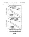
- the top graph (a) of FIG. 5provides experimentally determined plots of phase shift versus modulation frequency for a 0.5 ⁇ M concentration of DTTCI with the excitation characterization measurements of stage 130 , the emission characterization measurements of stage 140 , and the fluorescence characterization measurements of stage 150 shown in ascending order.
- the bottom graph (b) of FIG. 5provides plots of the same type and in the same order as top graph (a) for a 1.0 ⁇ M concentration of DTTCI.
- the top graph (a) and the bottom graph (b) of FIG. 6provide excitation, emission, and fluorescence characterization plots in the same order as the graphs of FIG. 5 for 0.0625 ⁇ M and 0.125 ⁇ M concentrations of ICG, respectively, FIG.
- FIG. 8provides relative modulation magnitude versus modulation frequency plots of fluorescence, emission, and excitation characterization for the two DTTCI concentrations as illustrated by different curve-fitted symbols.
- FIG. 9provides relative modulation magnitude versus modulation frequency plots of fluorescence, emission, and excitation characterizations for the two ICG concentrations as illustrated by different curve-fitted symbols.
- FIG. 10provides the lifetime calculations for the two concentrations of DTTCI and ICG based on the measurements plotted in FIGS. 8 and 9 .
Landscapes
- Physics & Mathematics (AREA)
- Spectroscopy & Molecular Physics (AREA)
- General Physics & Mathematics (AREA)
- Health & Medical Sciences (AREA)
- Nuclear Medicine, Radiotherapy & Molecular Imaging (AREA)
- Life Sciences & Earth Sciences (AREA)
- Chemical & Material Sciences (AREA)
- Analytical Chemistry (AREA)
- Biochemistry (AREA)
- General Health & Medical Sciences (AREA)
- Immunology (AREA)
- Pathology (AREA)
- Investigating, Analyzing Materials By Fluorescence Or Luminescence (AREA)
Abstract
Description
−cD∇2U(r,ω)+(cμa+iω)U(r,ω)−q(r,ω); (1)
where: D=[3(μ′s+μa)]−1is the diffusion coefficient of the medium, c is the speed of light in the medium, q(r,ω) describes properties of the light source, and ω is the modulation angular frequency of the light source (generator22). The output signal from
where: Ux(r,ω) is the frequency domain excitation photon density in the medium M and the complex wave vector kx(ω) is given by expression (3) as follows:
On substitution of expression (3) into expression (2), the excitation photon density can be modeled as follows in expression (4):
where:
The modulation M(r,ω) of the photon density waves a distance r away from and normalized to unity at the source (r=0) is related to photon density expression (5b) as follows:
For injection of source light at the excitation wavelength into the medium M in
θx(r,ω)=γx(ω)r (6a)
where the actual modulation information observed with
mx(r,ω)=Mx(r,ω)maxmdx(r,ω) (8)
and is related to Mx(r,ω) by the modulation of source msxand the modulation response mdx(r,ω) of
This ratio is generally independent of source modulation.
The quantum efficiency of the fluorophore is denoted by φ and μefdescribes absorption of excitation light due to fluorescence. Fluorescence decay is assumed to be of the monoexponential type with lifetime τ; however, the principles of the present invention can be applied to multiexponential decays using techniques known to these skilled in the art. Using expression (3), the fluorescence photon density of expression (10) can be written as presented in the following expression (11):
where:
The fluorescence photon density is related to the observed fluorescence modulation phase θf(r,ω) by expression (5a). Substituting expression (11) into expression (5a), expression (12) results as follows:
and the fluorescence decay lifetime can be written per expression (13) as follows:
where η(r,ω)=κ(r, ω)/ψ(r,ω) and determinations of ψ(r,ω) and κ(r, ω) are based on measured phase shifts Δθx(Δr,ω) and Δθf(Δr,ω).
where:
is based upon the optical coefficients of the sample. Comparable expressions can be derived for other types of luminescence and different boundary conditions.
from lifetime τ is derived:
The modulation magnitude detected with
mf(r2,ω)=Mf(r2,ω)mscmdm(r2,ω) (18)
which is related to modulation Mf(r2,ω) by modulation msxof the source at the excitation wavelength, and the modulation mdm(r2,ω) of the
From the ratio of expression (19), the following expression (20):
is found to depend upon the ratio of the
and emission:
wavelengths. The resulting ratio of detection instrument response functions is given by expression (23) as follows:
which depends upon a comparison between measured and calculated modulation information and a constant ratio of modulations of the sources at emission and excitation wavelengths. Varying the source modulation ratio, the lifetime measured at multiple modulation frequencies can be regressed to obtain a unique source modulation ratio associated with a vanishing slope, i.e. minimized χ2of the lifetime distribution, and to obtain the resulting lifetime τ. After lifetime is determined in
| Conc. | λx | μax | μax′ | λm | μam | μam′ | τ | |
| Dye | [μM] | [nm] | [1/cm] | [1/cm] | [nm] | [1/cm] | [1/cm] | [ns] |
| DTTCI | 0.5 | 749 | 0.054(8) | 9.2(12) | 828 | 0.032(3) | 10.2(8) | 1.34(3) |
| 1.0 | 749 | 0.07(2) | 6.8(15) | 828 | 0.031(3) | 8.4(7) | 1.34(4) | |
| ICG | 0.0625 | 778 | 0.039(8) | 9.5(13) | 828 | 0.033(4) | 8.0(8) | 0.54(3) |
| 0.125 | 778 | 0.05(1) | 7.9(20) | 828 | 0.051(9) | 9.2(15) | 0.56(4) | |
Additional samples were prepared in water for spectral analysis.
Claims (39)
Priority Applications (1)
| Application Number | Priority Date | Filing Date | Title |
|---|---|---|---|
| US10/473,303US7054002B1 (en) | 1999-10-08 | 1999-10-08 | Characterization of luminescence in a scattering medium |
Applications Claiming Priority (2)
| Application Number | Priority Date | Filing Date | Title |
|---|---|---|---|
| US10/473,303US7054002B1 (en) | 1999-10-08 | 1999-10-08 | Characterization of luminescence in a scattering medium |
| PCT/US1999/023709WO2000022414A1 (en) | 1998-10-09 | 1999-10-08 | Characterization of luminescence in a scattering medium |
Publications (1)
| Publication Number | Publication Date |
|---|---|
| US7054002B1true US7054002B1 (en) | 2006-05-30 |
Family
ID=36462672
Family Applications (1)
| Application Number | Title | Priority Date | Filing Date |
|---|---|---|---|
| US10/473,303Expired - LifetimeUS7054002B1 (en) | 1999-10-08 | 1999-10-08 | Characterization of luminescence in a scattering medium |
Country Status (1)
| Country | Link |
|---|---|
| US (1) | US7054002B1 (en) |
Cited By (21)
| Publication number | Priority date | Publication date | Assignee | Title |
|---|---|---|---|---|
| US20020072677A1 (en)* | 1996-08-23 | 2002-06-13 | Eva Sevick-Muraca | Imaging of light scattering tissues with fluorescent contrast agents |
| US20050085732A1 (en)* | 2003-06-20 | 2005-04-21 | Sevick-Muraca Eva M. | Method and system for near-infrared fluorescence contrast-enhanced imaging with area illumination and area detection |
| US20060241349A1 (en)* | 2002-12-12 | 2006-10-26 | Kazuhiro Gono | Imaging apparatus |
| US20060280508A1 (en)* | 2005-06-13 | 2006-12-14 | Sven Hadrich | Optimization of mode selection in a laser resonator |
| US20080050316A1 (en)* | 2006-08-24 | 2008-02-28 | Baylor College Of Medicine | Molecular imaging of epithelial cells in lymph |
| US20080056999A1 (en)* | 2006-08-24 | 2008-03-06 | Baylor College Of Medicine | Imaging agents for functional imaging of lymphatic structures |
| US20080230716A1 (en)* | 2000-10-23 | 2008-09-25 | General Electric Company | Dna biosensor and methods for making and using the same |
| US20090075248A1 (en)* | 2007-07-28 | 2009-03-19 | Buglab Llc | Particle sensor with wide linear range |
| EP2040050A1 (en) | 2007-09-24 | 2009-03-25 | Universität Potsdam | Measuring assembly for an optical spectrometer |
| US20090236541A1 (en)* | 2008-03-24 | 2009-09-24 | General Electric Company | System and Methods for Optical Imaging |
| US20090243436A1 (en)* | 2008-03-28 | 2009-10-01 | General Electric Company | Silicone rubber compositions comprising bismuth oxide and articles made therefrom |
| US20090252682A1 (en)* | 2006-06-01 | 2009-10-08 | The General Hospital Corporation | In-vivo optical imaging method including analysis of dynamic images |
| US20100027018A1 (en)* | 2007-02-05 | 2010-02-04 | Koninklijke Philips Electronics N.V. | Device and method for acquiring image data from a turbid medium |
| US7865230B1 (en) | 1997-02-07 | 2011-01-04 | Texas A&M University System | Method and system for detecting sentinel lymph nodes |
| US20110071403A1 (en)* | 2009-09-21 | 2011-03-24 | Board Of Regents Of The University Of Texas System | Functional near-infrared fluorescence lymphatic mapping for diagnosing, accessing, monitoring and directing therapy of lymphatic disorders |
| US20110108739A1 (en)* | 2008-07-15 | 2011-05-12 | Endress + Hauser Conducta Gesellschaft für Mess- und Regeltechnik mbH + Co. KG | Method for the optical determining of a measured variable of a medium |
| US20120162638A1 (en)* | 2009-03-02 | 2012-06-28 | Alain Villeneuve | Method for assessing an interaction of a sample with light beams having different wavelengths and apparatus for performing same |
| CN103954354A (en)* | 2014-04-03 | 2014-07-30 | 北京大学 | Quantum standard laser power meter and laser power measuring method |
| US20140303496A1 (en)* | 2011-11-02 | 2014-10-09 | Hamamatsu Photonics K.K. | Fluorescent light phantom device and fluorescent light imaging method |
| US9632030B1 (en) | 2012-11-05 | 2017-04-25 | Arrowhead Center, Inc. | Methods of measuring fluorescence lifetime using a flow cytometer |
| US20220099575A1 (en)* | 2014-08-08 | 2022-03-31 | Quantum-Si Incorporated | Integrated device with external light source for probing detecting and analyzing molecules |
Citations (59)
| Publication number | Priority date | Publication date | Assignee | Title |
|---|---|---|---|---|
| US4245909A (en) | 1978-06-26 | 1981-01-20 | Loos Hendricus G | Optical instrument for measurement of particle size distributions |
| US4541438A (en) | 1983-06-02 | 1985-09-17 | The Johns Hopkins University | Localization of cancerous tissue by monitoring infrared fluorescence emitted by intravenously injected porphyrin tumor-specific markers excited by long wavelength light |
| US4641969A (en) | 1982-12-15 | 1987-02-10 | Ab Bonnierforetagen | Method and apparatus for measuring the content of suspended substances in a flowing medium |
| US4781460A (en) | 1986-01-08 | 1988-11-01 | Coulter Electronics Of New England, Inc. | System for measuring the size distribution of particles dispersed in a fluid |
| US4871251A (en) | 1987-04-27 | 1989-10-03 | Preikschat F K | Apparatus and method for particle analysis |
| US4890920A (en) | 1986-02-12 | 1990-01-02 | Combustion Engineering, Inc. | In situ particle size measuring device |
| JPH02268256A (en) | 1989-04-07 | 1990-11-01 | Hamamatsu Photonics Kk | Apparatus for inspecting fluorescence characteristic |
| US5022757A (en) | 1989-01-23 | 1991-06-11 | Modell Mark D | Heterodyne system and method for sensing a target substance |
| US5119815A (en) | 1988-12-21 | 1992-06-09 | Nim, Incorporated | Apparatus for determining the concentration of a tissue pigment of known absorbance, in vivo, using the decay characteristics of scintered electromagnetic radiation |
| US5142372A (en) | 1990-03-08 | 1992-08-25 | Alfano Robert R | Three-dimensional optical imaging of semi-transparent and opaque objects using ultrashort light pulses, a streak camera and a coherent fiber bundle |
| US5164787A (en) | 1990-11-17 | 1992-11-17 | Horiba, Ltd. | Apparatus for measuring particle size distribution |
| US5190729A (en) | 1986-09-08 | 1993-03-02 | C. R. Bard, Inc. | Luminescent oxygen sensor based on a lanthanide complex |
| US5208651A (en) | 1991-07-16 | 1993-05-04 | The Regents Of The University Of California | Apparatus and method for measuring fluorescence intensities at a plurality of wavelengths and lifetimes |
| US5213105A (en) | 1990-12-04 | 1993-05-25 | Research Corporation Technologies, Inc. | Frequency domain optical imaging using diffusion of intensity modulated radiation |
| US5229839A (en) | 1989-10-06 | 1993-07-20 | National Aerospace Laboratory Of Science & Technology Agency | Method and apparatus for measuring the size of a single fine particle and the size distribution of fine particles |
| US5340991A (en) | 1993-05-21 | 1994-08-23 | The Board Of Regents Of The University Of Oklahoma | Fluorokinetic analysis of diffusion from a vessel |
| US5353799A (en) | 1991-01-22 | 1994-10-11 | Non Invasive Technology, Inc. | Examination of subjects using photon migration with high directionality techniques |
| WO1995012132A1 (en) | 1993-10-29 | 1995-05-04 | The Trustees Of The University Of Pennsylvania | Object imaging using diffuse light |
| US5413098A (en) | 1991-12-24 | 1995-05-09 | Sextant Medical Corporation | Path constrained spectrophotometer and method for determination of spatial distribution of light or other radiation scattering and absorbing substances in a radiation scattering medium |
| US5416580A (en) | 1993-07-07 | 1995-05-16 | General Signal Corporation | Methods and apparatus for determining small particle size distribution utilizing multiple light beams |
| US5421337A (en) | 1989-04-14 | 1995-06-06 | Massachusetts Institute Of Technology | Spectral diagnosis of diseased tissue |
| US5421339A (en) | 1993-05-12 | 1995-06-06 | Board Of Regents, The University Of Texas System | Diagnosis of dysplasia using laser induced fluoroescence |
| US5424843A (en) | 1992-12-23 | 1995-06-13 | The Regents Of The University Of California | Apparatus and method for qualitative and quantitative measurements of optical properties of turbid media using frequency-domain photon migration |
| US5438408A (en) | 1991-06-07 | 1995-08-01 | Sympatec Gmbh System-Partikel-Technik | Measuring device and method for the determination of particle size distributions by scattered light measurements |
| US5441054A (en) | 1992-07-20 | 1995-08-15 | Hamamatsu Photonics K.K. | Apparatus for measuring absorption information in scattering medium and method therefor |
| US5452723A (en) | 1992-07-24 | 1995-09-26 | Massachusetts Institute Of Technology | Calibrated spectrographic imaging |
| US5455675A (en) | 1992-08-26 | 1995-10-03 | Sympatec Gmbh System-Partikel-Technik | Apparatus for determination of particle sizes and/or distributions of particle sizes |
| US5485530A (en) | 1991-01-24 | 1996-01-16 | Joseph R. Lakowicz | Method and apparatus for multi-dimensional phase fluorescence lifetime imaging |
| US5502561A (en) | 1988-09-15 | 1996-03-26 | Board Of Trustees Of The University Of Arkansas | Characterization of particles by modulated dynamic light scattering |
| US5504337A (en) | 1990-10-10 | 1996-04-02 | Joseph R. Lakowicz | Method and apparatus for performing phase fluorescence lifetime measurements in flow cytometry |
| US5507287A (en) | 1991-05-08 | 1996-04-16 | Xillix Technologies Corporation | Endoscopic imaging system for diseased tissue |
| US5579773A (en) | 1994-09-30 | 1996-12-03 | Martin Marietta Energy Systems, Inc. | Laser-induced differential normalized fluorescence method for cancer diagnosis |
| US5582168A (en) | 1991-07-17 | 1996-12-10 | Georgia Tech Research Corp. | Apparatus and methods for measuring characteristics of biological tissues and similar materials |
| US5590660A (en) | 1994-03-28 | 1997-01-07 | Xillix Technologies Corp. | Apparatus and method for imaging diseased tissue using integrated autofluorescence |
| US5619324A (en) | 1995-12-29 | 1997-04-08 | Insitec Measurement Systems | Method for measuring particle size in the presence of multiple scattering |
| US5624847A (en) | 1991-05-03 | 1997-04-29 | Joseph R. Lakowicz | Method for optically measuring chemical analytes |
| US5628310A (en) | 1995-05-19 | 1997-05-13 | Joseph R. Lakowicz | Method and apparatus to perform trans-cutaneous analyte monitoring |
| US5647368A (en) | 1996-02-28 | 1997-07-15 | Xillix Technologies Corp. | Imaging system for detecting diseased tissue using native fluorsecence in the gastrointestinal and respiratory tract |
| GB2311366A (en) | 1996-03-19 | 1997-09-24 | Univ London | Determining absorption coefficients or modified scattering ceofficients |
| US5692504A (en) | 1993-11-04 | 1997-12-02 | Boehringer Mannheim Gmbh | Method and apparatus for the analysis of glucose in a biological matrix |
| US5699798A (en) | 1990-08-10 | 1997-12-23 | University Of Washington | Method for optically imaging solid tumor tissue |
| US5736410A (en) | 1992-09-14 | 1998-04-07 | Sri International | Up-converting reporters for biological and other assays using laser excitation techniques |
| US5759767A (en) | 1996-10-11 | 1998-06-02 | Joseph R. Lakowicz | Two-photon and multi-photon measurement of analytes in animal and human tissues and fluids |
| US5792049A (en) | 1996-01-17 | 1998-08-11 | Spectrx, Inc. | Spectroscopic system with disposable calibration device |
| US5818583A (en) | 1996-11-08 | 1998-10-06 | Purdue Research Foundation | Particle analysis system and method |
| US5860421A (en) | 1996-01-17 | 1999-01-19 | Spectrx, Inc. | Apparatus and method for calibrating measurement systems |
| US5865754A (en) | 1995-08-24 | 1999-02-02 | Purdue Research Foundation Office Of Technology Transfer | Fluorescence imaging system and method |
| US5891656A (en) | 1992-09-14 | 1999-04-06 | Sri International | Up-converting reporters for biological and other assays using laser excitation techniques |
| US5949077A (en) | 1992-08-10 | 1999-09-07 | Alfano; Robert R. | Technique for imaging an object in or behind a scattering medium |
| WO1999049312A1 (en) | 1998-03-23 | 1999-09-30 | Astrazeneca Ab | Method and device for analysing the three-dimensional distribution of a component in a sample |
| EP0959341A1 (en) | 1998-05-16 | 1999-11-24 | Deutsches Zentrum für Luft- und Raumfahrt e.V. | Method for the quantitative analysis of volumina of gases, especially exhaust gases from combustion apparatus, and device for implementing the method |
| WO2000022414A1 (en) | 1998-10-09 | 2000-04-20 | The Texas A & M University System | Characterization of luminescence in a scattering medium |
| WO2001022063A1 (en) | 1999-09-22 | 2001-03-29 | Astrazeneca Ab | Method and apparatus for spectrometric analysis of turbid, pharmaceutical samples |
| US6216540B1 (en) | 1995-06-06 | 2001-04-17 | Robert S. Nelson | High resolution device and method for imaging concealed objects within an obscuring medium |
| WO2002041760A2 (en) | 2000-11-27 | 2002-05-30 | The General Hospital | Fluorescence-mediated molecular tomography |
| US20020072677A1 (en) | 1996-08-23 | 2002-06-13 | Eva Sevick-Muraca | Imaging of light scattering tissues with fluorescent contrast agents |
| US6480276B1 (en) | 1999-07-13 | 2002-11-12 | Clemson University | Enhanced photon-migration methods for particle sizing in concentrated suspensions |
| US20030117622A1 (en) | 2001-10-22 | 2003-06-26 | Sevick-Muraca Eva M. | Characterizing powders using frequency-domain photon migration |
| US20050073681A1 (en) | 2001-04-03 | 2005-04-07 | Sevick-Muraca Eva M. | Method for characterising particles in supension from frequency domain photon migration measurements |
- 1999
- 1999-10-08USUS10/473,303patent/US7054002B1/ennot_activeExpired - Lifetime
Patent Citations (61)
| Publication number | Priority date | Publication date | Assignee | Title |
|---|---|---|---|---|
| US4245909A (en) | 1978-06-26 | 1981-01-20 | Loos Hendricus G | Optical instrument for measurement of particle size distributions |
| US4641969A (en) | 1982-12-15 | 1987-02-10 | Ab Bonnierforetagen | Method and apparatus for measuring the content of suspended substances in a flowing medium |
| US4541438A (en) | 1983-06-02 | 1985-09-17 | The Johns Hopkins University | Localization of cancerous tissue by monitoring infrared fluorescence emitted by intravenously injected porphyrin tumor-specific markers excited by long wavelength light |
| US4781460A (en) | 1986-01-08 | 1988-11-01 | Coulter Electronics Of New England, Inc. | System for measuring the size distribution of particles dispersed in a fluid |
| US4890920A (en) | 1986-02-12 | 1990-01-02 | Combustion Engineering, Inc. | In situ particle size measuring device |
| US5190729A (en) | 1986-09-08 | 1993-03-02 | C. R. Bard, Inc. | Luminescent oxygen sensor based on a lanthanide complex |
| US4871251A (en) | 1987-04-27 | 1989-10-03 | Preikschat F K | Apparatus and method for particle analysis |
| US5502561A (en) | 1988-09-15 | 1996-03-26 | Board Of Trustees Of The University Of Arkansas | Characterization of particles by modulated dynamic light scattering |
| US5119815A (en) | 1988-12-21 | 1992-06-09 | Nim, Incorporated | Apparatus for determining the concentration of a tissue pigment of known absorbance, in vivo, using the decay characteristics of scintered electromagnetic radiation |
| US5022757A (en) | 1989-01-23 | 1991-06-11 | Modell Mark D | Heterodyne system and method for sensing a target substance |
| JPH02268256A (en) | 1989-04-07 | 1990-11-01 | Hamamatsu Photonics Kk | Apparatus for inspecting fluorescence characteristic |
| US5421337A (en) | 1989-04-14 | 1995-06-06 | Massachusetts Institute Of Technology | Spectral diagnosis of diseased tissue |
| US5229839A (en) | 1989-10-06 | 1993-07-20 | National Aerospace Laboratory Of Science & Technology Agency | Method and apparatus for measuring the size of a single fine particle and the size distribution of fine particles |
| US5142372A (en) | 1990-03-08 | 1992-08-25 | Alfano Robert R | Three-dimensional optical imaging of semi-transparent and opaque objects using ultrashort light pulses, a streak camera and a coherent fiber bundle |
| US5699798A (en) | 1990-08-10 | 1997-12-23 | University Of Washington | Method for optically imaging solid tumor tissue |
| US5504337A (en) | 1990-10-10 | 1996-04-02 | Joseph R. Lakowicz | Method and apparatus for performing phase fluorescence lifetime measurements in flow cytometry |
| US5164787A (en) | 1990-11-17 | 1992-11-17 | Horiba, Ltd. | Apparatus for measuring particle size distribution |
| US5213105A (en) | 1990-12-04 | 1993-05-25 | Research Corporation Technologies, Inc. | Frequency domain optical imaging using diffusion of intensity modulated radiation |
| US5353799A (en) | 1991-01-22 | 1994-10-11 | Non Invasive Technology, Inc. | Examination of subjects using photon migration with high directionality techniques |
| US5485530A (en) | 1991-01-24 | 1996-01-16 | Joseph R. Lakowicz | Method and apparatus for multi-dimensional phase fluorescence lifetime imaging |
| US5624847A (en) | 1991-05-03 | 1997-04-29 | Joseph R. Lakowicz | Method for optically measuring chemical analytes |
| US5507287A (en) | 1991-05-08 | 1996-04-16 | Xillix Technologies Corporation | Endoscopic imaging system for diseased tissue |
| US5438408A (en) | 1991-06-07 | 1995-08-01 | Sympatec Gmbh System-Partikel-Technik | Measuring device and method for the determination of particle size distributions by scattered light measurements |
| US5208651A (en) | 1991-07-16 | 1993-05-04 | The Regents Of The University Of California | Apparatus and method for measuring fluorescence intensities at a plurality of wavelengths and lifetimes |
| US5582168A (en) | 1991-07-17 | 1996-12-10 | Georgia Tech Research Corp. | Apparatus and methods for measuring characteristics of biological tissues and similar materials |
| US5413098A (en) | 1991-12-24 | 1995-05-09 | Sextant Medical Corporation | Path constrained spectrophotometer and method for determination of spatial distribution of light or other radiation scattering and absorbing substances in a radiation scattering medium |
| US5441054A (en) | 1992-07-20 | 1995-08-15 | Hamamatsu Photonics K.K. | Apparatus for measuring absorption information in scattering medium and method therefor |
| US5452723A (en) | 1992-07-24 | 1995-09-26 | Massachusetts Institute Of Technology | Calibrated spectrographic imaging |
| US5949077A (en) | 1992-08-10 | 1999-09-07 | Alfano; Robert R. | Technique for imaging an object in or behind a scattering medium |
| US5455675A (en) | 1992-08-26 | 1995-10-03 | Sympatec Gmbh System-Partikel-Technik | Apparatus for determination of particle sizes and/or distributions of particle sizes |
| US5736410A (en) | 1992-09-14 | 1998-04-07 | Sri International | Up-converting reporters for biological and other assays using laser excitation techniques |
| US5891656A (en) | 1992-09-14 | 1999-04-06 | Sri International | Up-converting reporters for biological and other assays using laser excitation techniques |
| US5424843A (en) | 1992-12-23 | 1995-06-13 | The Regents Of The University Of California | Apparatus and method for qualitative and quantitative measurements of optical properties of turbid media using frequency-domain photon migration |
| US5421339A (en) | 1993-05-12 | 1995-06-06 | Board Of Regents, The University Of Texas System | Diagnosis of dysplasia using laser induced fluoroescence |
| US5340991A (en) | 1993-05-21 | 1994-08-23 | The Board Of Regents Of The University Of Oklahoma | Fluorokinetic analysis of diffusion from a vessel |
| US5416580A (en) | 1993-07-07 | 1995-05-16 | General Signal Corporation | Methods and apparatus for determining small particle size distribution utilizing multiple light beams |
| WO1995012132A1 (en) | 1993-10-29 | 1995-05-04 | The Trustees Of The University Of Pennsylvania | Object imaging using diffuse light |
| US5917190A (en) | 1993-10-29 | 1999-06-29 | Trustees Of The University Of Pennsylvania | Object imaging using diffuse light |
| US5692504A (en) | 1993-11-04 | 1997-12-02 | Boehringer Mannheim Gmbh | Method and apparatus for the analysis of glucose in a biological matrix |
| US5590660A (en) | 1994-03-28 | 1997-01-07 | Xillix Technologies Corp. | Apparatus and method for imaging diseased tissue using integrated autofluorescence |
| US5579773A (en) | 1994-09-30 | 1996-12-03 | Martin Marietta Energy Systems, Inc. | Laser-induced differential normalized fluorescence method for cancer diagnosis |
| US5628310A (en) | 1995-05-19 | 1997-05-13 | Joseph R. Lakowicz | Method and apparatus to perform trans-cutaneous analyte monitoring |
| US6216540B1 (en) | 1995-06-06 | 2001-04-17 | Robert S. Nelson | High resolution device and method for imaging concealed objects within an obscuring medium |
| US5865754A (en) | 1995-08-24 | 1999-02-02 | Purdue Research Foundation Office Of Technology Transfer | Fluorescence imaging system and method |
| US5619324A (en) | 1995-12-29 | 1997-04-08 | Insitec Measurement Systems | Method for measuring particle size in the presence of multiple scattering |
| US5792049A (en) | 1996-01-17 | 1998-08-11 | Spectrx, Inc. | Spectroscopic system with disposable calibration device |
| US5860421A (en) | 1996-01-17 | 1999-01-19 | Spectrx, Inc. | Apparatus and method for calibrating measurement systems |
| US5647368A (en) | 1996-02-28 | 1997-07-15 | Xillix Technologies Corp. | Imaging system for detecting diseased tissue using native fluorsecence in the gastrointestinal and respiratory tract |
| GB2311366A (en) | 1996-03-19 | 1997-09-24 | Univ London | Determining absorption coefficients or modified scattering ceofficients |
| US20020072677A1 (en) | 1996-08-23 | 2002-06-13 | Eva Sevick-Muraca | Imaging of light scattering tissues with fluorescent contrast agents |
| US5759767A (en) | 1996-10-11 | 1998-06-02 | Joseph R. Lakowicz | Two-photon and multi-photon measurement of analytes in animal and human tissues and fluids |
| US5818583A (en) | 1996-11-08 | 1998-10-06 | Purdue Research Foundation | Particle analysis system and method |
| WO1999049312A1 (en) | 1998-03-23 | 1999-09-30 | Astrazeneca Ab | Method and device for analysing the three-dimensional distribution of a component in a sample |
| EP0959341A1 (en) | 1998-05-16 | 1999-11-24 | Deutsches Zentrum für Luft- und Raumfahrt e.V. | Method for the quantitative analysis of volumina of gases, especially exhaust gases from combustion apparatus, and device for implementing the method |
| US6271522B1 (en) | 1998-05-16 | 2001-08-07 | Deutsches Zentrum Fur Luft-Und Raumfahrt E.V. | Process for the quantitative analysis of gas volumes, specifically exhaust and waste gases from combustion systems or incineration plants, as well as systems for performing these processes |
| WO2000022414A1 (en) | 1998-10-09 | 2000-04-20 | The Texas A & M University System | Characterization of luminescence in a scattering medium |
| US6480276B1 (en) | 1999-07-13 | 2002-11-12 | Clemson University | Enhanced photon-migration methods for particle sizing in concentrated suspensions |
| WO2001022063A1 (en) | 1999-09-22 | 2001-03-29 | Astrazeneca Ab | Method and apparatus for spectrometric analysis of turbid, pharmaceutical samples |
| WO2002041760A2 (en) | 2000-11-27 | 2002-05-30 | The General Hospital | Fluorescence-mediated molecular tomography |
| US20050073681A1 (en) | 2001-04-03 | 2005-04-07 | Sevick-Muraca Eva M. | Method for characterising particles in supension from frequency domain photon migration measurements |
| US20030117622A1 (en) | 2001-10-22 | 2003-06-26 | Sevick-Muraca Eva M. | Characterizing powders using frequency-domain photon migration |
Non-Patent Citations (94)
| Title |
|---|
| A. Knuittel et al., "Acoust-optic scanning and interfering photon density waves for precise localization of an absorbing (or fluorescence) body in a turbid medium", Rev. Sci. Instrum. vol. 64, No. 3, Mar. 1993, pp. 638-644. |
| Akira Ishimaru, Robert J. Marks, II, Leung Tsang, Chi M. Lam, Doug C. Park, Optical Sensing of Particle Size Distribution by Neural Network Technique (date unknown). |
| Alwin Kienle, Lothar Lilge, Michel S. Patterson, Raimund Hibst, Rudolf Steiner, and Brian C. Wilson, Spatially Resolved Absolute Diffuse Reflectance Measurements for Noninvasive Determination of the Optical Scattering and Absorption Coefficients of Biological Tissue, Applied Optics, vol. 35, No. 13 at 2304, May 1996. |
| B. W. Pogue et al., "Initial Assessment of a simple system for frequency domain diffuse optical tomography", Phys. Med. Biol. 40, (1995) 1709-1729. |
| Banerjee, et al., Assessment, of S(0, Ø) from multiply scattered light, Journal of Chemical Physics, vol. 111, No. 20, (C) 1999 American Institute of Physics, pp. 9133-9136, Nov. 22, 1999. |
| Banerjee, et al., Probing Static Structure of Colloid-Polymer Suspensions with Multiply Scattered Light, Journal of Colloid and Interface Science 209, (C) 1999 by Academic Press, pp. 142-153. |
| Brian C. Wilson et al., "Time-dependent optical spectroscopy and imaging for biomedical applications", Proceedings of the IEEE, vol. 80, No. 6, Jun. 1992 pp. 918-930. |
| Cerussi, Albert E., et al., "Experimental Verification of a Theory for the Time-Resolved Fluorescence Spectroscopy of Thick Tissues", Applied Optics, vol. 36, No. 1, Jan. 1, 1997, pp. 116-124. |
| Chance, et al., Review Article, Phase measurement of light absorption and scatter in human tissue, Review of Scientific Instruments, vol. 69, No. 10, (C) 1998 American Institute of Physics, pp. 3457-3481, Oct. 1998. |
| D. Jeffrey Lischer and Michel Y. Louge, Optical Fiber Measurements of Particle Concentration in Dense Suspensions: Calibration and Simulation, Applied Optics, Aug. 1992, vol. 31, No. 24, at 5106. |
| David A. Russel et al., "Continuous noninvasive measurement of In Vivo pH in conscious mice", Photochemistry and Photobiology, vol. 59, No. 3 (1994) pp. 309-313. |
| Dilip Paithankar, Jeff Kao, and Eva Sevick-Muraca, Particle Size Distribution Estimation Via Solution of the Inverse Problem of Multi-Wavelength Scattering Coefficient Measurements, Chem. Eng. Prog., Aug. 1995. |
| E. M. Sevick et al., "Frequency domain imaging absorbers obscured by scattering", J. Photochem, Photobiol. B:Biol, 16 (1992) pp. 169-185. |
| Eva M. Sevick-Muraca and Dilip Paithankar, Process Monitoring: Photon Migration Measurements in Particulate Systems, Fine Particle Society Meeting, Aug., 1995. |
| Eva M. Sevick-Muraca and Kavi Sharma, Measurements of Photon Migration for Particle Sizing in Optically Dense Suspensions, AlChE Journal, Nov. 1994. |
| Fantini, et al., Quantitative determination of the absorption spectra of chromophores in strongly scattering media: a light-emitting-diode based technique, Applied Optics, vol. 33, No. 22, pp. 5204-5213, Aug. 1, 1994. |
| Fishkin, et al., Frequency-domain method for measuring spectral properties in multiple-scattering media: methemoglobin absorption spectrum in a tissuelike phantom, Applied Optics, vol. 34, No. 7, pp. 1143-1155, Mar. 1, 1995. |
| Fishkin, et al., Frequency-domain photon migration measurements of normal and malignant tissue optical properties in a human subject, Applied Optics, vol. 36, No. 1, pp. 10-20, Jan. 1, 1997. |
| Fishkin, et al., Propagation of photon-density waves in strongly scattering media containing an absorbing semi-infinite plane bounded by a straight edge, vol. 10, No. 1, (C) 1993 Optical Society of America, pp. 127-140, Jan. 1993. |
| Gerken, et al., High-precision-frequency-domain measurements of the optical properties of turbid media, Optics Letters, vol. 24, No. 14, (C) 1999 Optical Society of America, pp. 930-932, Jul. 15, 1999. |
| Gratton, et al., A Continuously Variable Frequency Cross-Correlation Phase Fluorometer with Picosecond Resolution, (C) Biophysical Society, Biophysical Journal, vol. 44, pp. 315-324, Dec. 1983. |
| Gratton, et al., The possibility of a near-infrared optical imaging system using frequency domain methods, Mind Brain Imaging Program, Hamamatsu, Japan, pp. 183-189, Aug. 5-Oct. 1990. |
| Guillermo E. Elicabe and Luis H. Garcia-Rubio, Latex Particle Size Distribution from Turbidimetric Measurements, Polymer Characterization, 1990, at 84. |
| Gurfinkel, et al., "Pharmocokinetcs of ICG and HPPH-car for the Detection of Normal and Tumor Tissue Using Fluorescence, Near-infrared Reflectance Imaging: A Case Study", Photochemistry and Photobiology, 2000: 72(1): 94-102 (XP-001030699), Apr. 28, 2000. |
| Hawrysz, et al., Developments Toward Diagnostic Breast Cancer Imaging Using Near-Infrared Optical Measurements and Fluorescent Contrast Agents<SUP>1</SUP>, Review Article, Neoplasia, vol. 2, No. 5, (C) 2000 Nature America, Inc., pp. 388-417, Sep.-Oct. 2000. |
| Heimo Schnablegger and Otto Glatter, Sizing of Colloidal Particles with Light Scattering: Corrections for Beginning Multiple Scattering, Applied Optics, vol. 34, No. 18, at 3489, Jun. 1995. |
| Houston, et al., "Sensitivity and Depth Penetration of Continuous Wave Versus Frequency-domain Photon Migration Near-infrared Fluorecence Contrast-enhancing Imaging," Photochemistry and Photobiology, 2003, vol. 77(4), pp. 420-430. |
| Huabei Jiang, Keith D. Paulsen, Ulf L. Osterberg, Brian W. Pogue and Michael S. Patterson, Optical Image Reconstruction Using Frequency-Domain Data: Simulations and Experiments, Journal of the Optical Society of America, at 253, Sep. 1995. |
| Hutchinson, Christina L., et al., "Fluorescence-Lifetime Determination in Tissues or Other Scattering Media from Measurement of Excitation and Emission Kinetics", Applied Optics, vol. 35, No. 13, May 1, 1996, pp. 2325-2332. |
| Isayev, K, et al., "Study of Thermophysical Properties of a Metal-Hydrogen System," International Journal of Hydrogen Energy, vol. 21, No. 11-12, Nov. 12, 1996, pp. 1129-1132, Dec. 12, 1996. |
| J. Jager, H.J.M. Kramer, E.J. De Jong, On-Line Particle Size Measurement in Denise Slurries, Powder Technology, 1990. at 155-162. |
| James R. Rawlings, Stephen M. Miller, and Walter R. Witkowski, Model Identification and Control of Solution Crystallization Process: A Review, Ind. Eng. Chem. Res. 1993, vol. 32, No. 7, at 1276. |
| Jianhong Wang and F. Ross Hallett, Spherical Particle Size Determination by Analytical Inversion of the UV-Visible-NIR Extinction Spectrum, Applied Optics, vol. 35, No. 1, at 193, Jan. 1996. |
| John C. Thomas and Victoria Dimonie, Fiber Optic Dynamic Light Scattering from Concentrated Dispersion, 3: Particle Sizing in Concentrates, Applied Optics, Dec. 1990, vol. 29, No. 36, at 5332. |
| John Dimitratos, Guillermo Elicabe, and Christos Geogakis, Control of Emulsion Polymerization Reactors, AlChE Journal, Dec. 1994, vol. 40, No. 12, at 1993. |
| Joseph Pierce, Dilip Paithankar, Christina Hutchinson, David Taylor and Eva Sevick-Muraca, Particle Size Measurement in Suspensions through Frequency-Domain Photon Migration Measurements, Presentation to Fine Particle Society Meeting of Aug. 25, 1995. |
| Joshua B. Fishkin, Peter T.C. So, Albert E. Cerussi, Sergio Fantini, Maria Angela Franceschini, and Enrico Gratton, Frequency-Domain Method for Measuring Spectral Properties in Multiple-Scattering Media: Methemoglobin Absorption Spectrum in a Tissuelike Phantom, Applied Optics, vol. 34 No. 7, at 1143, Mar. 1995. |
| Jozef Vavra, Jozef Antalik and Marek Liska, Application of Regression Analysis in Spectroturbidity Size-Characterization Methods, Part. Syst. Charact. 12, 38-41, 1995. |
| Jun Wu et al., "Three-dimensional imaging of objects embedded in turbid media with fluorescence and raman spectroscopy", Applied Optics, vol. 34, No. 18, Jun. 1995 pp. 3425-3430. |
| Jun Wu et al., "Time-resolved multichannel imaging of fluorescent objects embedded in turbid media", Optic Letters, vol. 20, No. 5, Mar. 1995 pp. 489-491. |
| K. M. Yoo et al., "Imaging objects hidden in scattering media using a fluorescence-absorption technique", Optics Letters, vol. 16, No. 16, 1991, pp. 1252-1254. |
| Kusiel S. Shifrin and Ilja G. Zolotov, Spectral Attenuation and Aerosol Particle Size Distribution, Applied Optics, vol. 35, No. 12 at 2114, Apr. 1996. |
| L. H. Garcia-Rubio, Refractive Index Effects on the Absorption Spectra of Macromolecules, Macromolecules, 1992, at 2608. |
| M. A. O'Leary et al., "Reradiation and imaging of diffuse photon density waves using fluorescent inhomogeneities", Journal of Luminescence, (1994) pp. 281-286. |
| M.A. O'Leary, D.A. Boas, B. Chance, and A.G. Yodh, Experimental Images of Heterogeneous Turbid Media by Frequency-Domain Diffusing-Photon Tomography, Optics Letters, vol. 20, No. 5, at 426, Mar. 1995. |
| M.A. O'Leary, E.A. Boas, X.D. Li, B. Chance, and A.G. Yodh., "Fluorescence Lifetime Imaging in Turbid Media," Optics Letters, vol. 21, No. 2, pp. 158-160, Jan. 15, 1966. |
| Madsen, et al., Determination of the optical properties of the human uterus using frequency-domain photon migration and steady-state techniques, Phys. Med. Biol. 39, (C) 1994 IOP Publishing Ltd., pp. 1191-1202. |
| Mayer, Ralf H., et al., "Measurement of the Fluorescence Lifetime in Scattering Media by Frequency-Domain Photon Migration", Applied Optics, vol. 38, No. 22, Aug. 1, 1999, pp. 4930-4938. |
| Michael Patterson et al., "Applications of time-resolved light scattering measurements to photodynamictherapy dosimetry", Applied Optics 1203-1208. |
| Michael S. Patterson et al., "Mathematical model for time-resolved and frequency-domain fluorescence spectroscopy in biological tissues", Aplied Optics, vol. 33, No. 10, Apr. 1994, pp. 1963-1974. |
| Michael S. Patterson et al., "Mathematical model for time-resolved and frequency-domain fluorescence spectroscopy in biological tissues", Applied Optics, vol. 33, No. 10, Apr. 1994, pp. 1963-1974. |
| Michael S. Patterson, Steen J. Madsen, J. David Moulton and Brian C. Wilson, Diffusion Equation Representation of Photon Migration in Tissue (date unknown.). |
| Muzzio, et al., Sampling practices in powder blending, Research papers, International Journal of Pharmaceutics 155, (C) 1997 Elsevier Science B.V., pp. 153-178. |
| Nai-ning Wang, Gang Zheng, and Xiao-shu Cai, A Theoretical and Experimental Study of the Total Light Scattering Technique for Particle Size Analysis, Part. Part. Syst. Charact. 11, Feb. 1994, at 309-314. |
| Notification of Transmital of the International Search Report and the Written Opinion of the International Searching Authority for International Application No. PCT/US2004/019792, filed Jun. 18, 2004 (14 pages), Nov. 8, 2004. |
| Ntziachristos, et al. "In Vivo Tomographic Imaging of Near-Infrared Fluorescent Probes," Molecular Imaging, vol. 1(2), pp. 82-88, Apr. 2002. |
| Pan, et al., Volume of Pharmaceutical Probed by Frequency-Domain Photon Migration Measurements of Multiply Scattered Light, Analytical Chemistry 2002, vol. 74, No. 16, (C) 2002 American Chemical Society, pp. 4228-4234, Aug. 15, 2002. |
| Panda, et al., "Generalized B-Spline Signal Processing," European Journal to the Methods and Applications of Signal Processing, Elsevier Science Publishers, B.V. Amsterdam, NL, vol. 55, No. 1, Nov. 1, 1996 XP004016005, pp. 1-14. |
| PCT International Search Report in International Application No. 02/10433, dated Jun. 16, 2003, 10 pages. |
| PCT invitation to Pay Additional Fees (PCT Article 17(3)(a) and Rule 40.1), Annex to Form PCT/ISA/206 Communication Regarding to the Results of the Partial International Search Authority, regarding PCT/US02/10433, Applicant's reference 017575.0748, 6 pages, Nov. 29, 2002. |
| Pham, et al., Broad bandwidth frequency domain instrument for quantitive tissue optical spectroscopy, Review of Scientific Instruments, vol. 71, No. 6, (C) 2000 American Institute of Physics, pp. 2500-2513, Jun. 2000. |
| Pi-Huan Wang, Geoffrey S. Kent, M. Patrick McCormick, Larry W. Thomason, and Glenn K. Yue, Retrieval Analysis of Aerosol-Size Distribution with Simulated Extinction Measurements at SAGE III Wavelenghts, Applied Optics, vol. 35, No. 3 at 433, Jan. 1996. |
| R. C. Straight et al., "Application of Charge-coupled device technology for measurement of laser light and fluorescence distribution in tumors for photodynamic therapy", Photochemistry and Photobiology, vol. 53, No. 6, pp. 787-796. |
| R. Cubeddu et al., "Time-gated Fluorescence imaging for the diagnosis of tumors in a murine model", Photochemistry and Photobiology, vol. 57, No. 3, pp. 480-485. |
| R. Graaff, J.G. Aarnoudse, Jr. Zijp, P.M.A. Sloot, F.F.M. Mul, J. Greve, and M.H. Koelink, Reduced Light-Scattering Properties for Mixtures of Spherical Properties: A Simple Approximation Derived from Mie Calculations, Applied Optics, Apr. 1992, vol. 31, No. 10 at 1370. |
| Ramanujam, et al., Sources of phase noise in homodyne and heterodyne phase modulation devices used for tissue oximetry studies, Review of Scientific Instruments, vol. 69, No. 8, (C) 1998 American Institute of Physics, pp. 3042-3054, Aug. 1998. |
| Randall Barbour et al., "A perturbation approach for optical diffusion tomography using continuous-wave and time-resolved data", Medical Optical Tomography, pp. 87-121. |
| Reynolds, et al., "Imaging of Spontaneous Canine Mammary Tumors Using Fluorescent Contrast Agents", Photochemistry and Photobiology, 1999: 70(1): 87-94 (XP-001063376), Apr. 14, 1999. |
| Richard Haskel et al., "Boundary conditions for the diffusion equation in radiative transfer", J. Opt. Soc. Am., A. vol. 11, No. 10. Oct. 1994, pp. 2727-2741. |
| Richter, et al., Characterization of concentrated colloidal suspensions using time-dependent photon migration measurements, Reprinted from Colloids And Surfaces An International Journal, A: Physciochemical and Engineering Aspects, (C) 2000 Elsevier Science B.V., pp. 163-173, plus cover. |
| Richter, et al., Particle Sizing Using Frequency Domain Photon Migration, Part. Part. Syst. Charact. 15, (C) Wiley-VCH Verlag GmbH, D-69469 Weinheim, pp. 9-15, 1998. |
| Robert J. Farrell and Yen-Cheng Tsai, Nonlinear Controller for Batch Crystallization: Development and Experimental Demonstration, AIChE Journal, vol. 41, No. 10, at 2318, Oct. 1995. |
| Ronald G. Sparks and Charles L. Dobbs, The Use of Laser Backscatter Instrumentation for the On-Line Measurement of the Particle Size Distribution of Emulsions, Part. Part. Syst. Charact. 10, Jul. 1993, at 279-289. |
| Scott R. Fulton, et al., "Time-resolved laser-induced fluorescence spectroscopy for enhanced demarcation of human atherosclerotic plaques", Journal of Photochemistry and Photobiology, (1990) pp. 363-369. |
| Serge Mordon et al., "In Vivo pH measurement and imaging of tumor tissue using a pH-sensitive fluorescent probe (5,6-carboxyfluorescein): Instrumental and Experimental studies", Photochemistry and Photobiology, vol. 60, No. 3, pp. 274-279. |
| Sergei A. Vinogradov, Leu-Wei Lo, William T. Jenkins, Sydney M. Evans, Cameron Koch, and David F. Wilson, Noninvasive Imaging of the Distribution in Oxygen in Tissue in Vivo Using Near-Infrared Phosphors, Biophysical Journal, vol. 70, at 1609-1617, Apr. 1996. |
| Seth Fraden and Georg Maret, Multiple Light Scattering from Concentrated, Interacting Suspensions, Physical Review Letters, Jul. 1990, vol. 65, No. 4, at 512. |
| Seth Fraden et al., "Multiple light scattering from concentrated, interacting suspensions", Physical Review letters, vol. 65, No. 4, pp. 512-515. |
| Sevick, et al., Quantitation of Time-and Frequency-Resolved Optical Spectra for the Determination of Tissue Oxygenation, Analytical Biochemistry 195, (C) 1991 Academic Press Inc., pp. 330-351. |
| Sevick-Muraca, et al., Photon-Migration Measurement of Latex Size Distribution in Concentrated Suspensions, Particle Technology and Fluidization, AlChE Journal, vol. 43, No. 3, pp. 655-664, Mar. 1997. |
| Sevick-Muraca, et al.; "Method and System for Detecting Sentinel Lymph Nodes;" U.S. Appl. No. 10/618,194; 28 pgs, Jul. 11, 2003. |
| Sevick-Muraca, et al.; "Method for Characterizing Particles in Suspension from Frequency Domain Phton Migration Measurements" U.S. Appl. No. 10/115,271; 59 pgs, Apr. 3, 2002. |
| Sevick-Muraca, et al.; Method for Characterizing Particles in Suspension from Frequency Domain Photon Migration Measurements; U.S. Appl. No. 11/204,844; 59 pgs, Aug. 16, 2005. |
| Shinde, et al., Frequency-Domain Photon Migration Measurements for Quantitative Assessment of Powder Absorbance: A Novel Sensor of Blend Homogeneity, Research Articles, (C) 1999 American Chemical Society and American Pharmaceutical Association, Journal of Pharmaceutical Sciences, vol. 88, No. 10, pp. 959-966, Oct. 1999. |
| Shinde, et al., Investigation of static structure factor in dense suspensions by use of multiply scattered light, Applied Optics, vol. 38, No. 1, (C) 1999 Optical Society of America, pp. 197-204, Jan. 1, 1999. |
| Sun, et al., "Approach for Particle Sizing in Dense Polydisperse Colloidal Suspension Using Multiple Scattered Light," XP-001126299, Langmuir 2001, 17, 2001 American Chemical Society, pp. 6142-6147, Sep. 8, 2001. |
| Sun, et al., "Inversion Algorithms for Particle Sizing with Photon Migration Measurements," Fluid Mechanics and Transport Phenomena, AlChE Journal, vol. 47, No. 7, pp. 1487-1498, Jul. 2001. |
| Sun, et al., "Particle Characterization of Colloidal Suspension at High Volume Fractions Using Frequency Domain Photon Migration," 6th World Congress of Chemical Engineering, Melbourne 2001, pp. 4/15-12/15. |
| Thompson, et al., "Near-infrared fluorescence contrast-enhanced imaging with area illumination and area detection: the forward imaging problem", Applied Optics, 2003: 42(19): 4125-4136 (XP-002301883), Jul. 1, 2003. |
| Thompson, et al., "Near-infrared fluorescence contrast-enhanced imaging with intensified charge-coupled device homodyne detection: measurement precision and accuracy", Journal of Biomedical Optics, 2003: 8(1): 111-120 (XP-002301882)klj, Jan. 2003. |
| Tromberg, et al., Non-invasive measurements of breast tissue optical properties using frequency-domain photon migration, Phil. Trans. R. Soc. Lond. B, (C) 1997 The Royal Society, pp. 661-668. |
| Tromberg, et al., Properties of photon density waves in multiple-scattering media, vol. 32, No. 4, Applied Optics, pp. 607-616, Feb. 1, 1993. |
| Wai S. Poon et al., "Laser-induced Fluorescence: Experimental intraoperative delineation of tumor resection margins", J. Neurosurg, vol. 76, Apr. 1992, pp. 679-686. |
| X. D. Li et al., "Fluorescent diffuse photon density waves in homogenous and heterogeneous turbid media: analytic solutions and applications", Applied Optics, vol. 35, No. 19, Jul. 1996, pp. 3746-3758. |
Cited By (44)
| Publication number | Priority date | Publication date | Assignee | Title |
|---|---|---|---|---|
| US20100305453A1 (en)* | 1995-08-24 | 2010-12-02 | Sevick-Muraca Eva M | Imaging of light scattering tissues with fluorescent contrast agents |
| US20020072677A1 (en)* | 1996-08-23 | 2002-06-13 | Eva Sevick-Muraca | Imaging of light scattering tissues with fluorescent contrast agents |
| US7328059B2 (en)* | 1996-08-23 | 2008-02-05 | The Texas A & M University System | Imaging of light scattering tissues with fluorescent contrast agents |
| US20080175790A1 (en)* | 1997-02-07 | 2008-07-24 | The Texas A&M University System | Imaging of Light Scattering Tissues with Fluorescent Contrast Agents |
| US7865230B1 (en) | 1997-02-07 | 2011-01-04 | Texas A&M University System | Method and system for detecting sentinel lymph nodes |
| US20080230716A1 (en)* | 2000-10-23 | 2008-09-25 | General Electric Company | Dna biosensor and methods for making and using the same |
| US7615780B2 (en)* | 2000-10-23 | 2009-11-10 | General Electric Company | DNA biosensor and methods for making and using the same |
| US20060241349A1 (en)* | 2002-12-12 | 2006-10-26 | Kazuhiro Gono | Imaging apparatus |
| US8000776B2 (en)* | 2002-12-12 | 2011-08-16 | Olympus Corporation | Imaging apparatus |
| US7599732B2 (en) | 2003-06-20 | 2009-10-06 | The Texas A&M University System | Method and system for near-infrared fluorescence contrast-enhanced imaging with area illumination and area detection |
| US20050085732A1 (en)* | 2003-06-20 | 2005-04-21 | Sevick-Muraca Eva M. | Method and system for near-infrared fluorescence contrast-enhanced imaging with area illumination and area detection |
| US20060280508A1 (en)* | 2005-06-13 | 2006-12-14 | Sven Hadrich | Optimization of mode selection in a laser resonator |
| US7489715B2 (en)* | 2005-06-13 | 2009-02-10 | Sirah Laser-Und Plasmatechnick Gmbh | Optimization of mode selection in a laser resonator |
| US9220411B2 (en)* | 2006-06-01 | 2015-12-29 | The General Hospital Corporation | In-vivo optical imaging method including analysis of dynamic images |
| US20090252682A1 (en)* | 2006-06-01 | 2009-10-08 | The General Hospital Corporation | In-vivo optical imaging method including analysis of dynamic images |
| US20080050316A1 (en)* | 2006-08-24 | 2008-02-28 | Baylor College Of Medicine | Molecular imaging of epithelial cells in lymph |
| US7875260B2 (en) | 2006-08-24 | 2011-01-25 | Baylor College Of Medicine | Imaging agents for functional imaging of lymphatic structures |
| US20080064954A1 (en)* | 2006-08-24 | 2008-03-13 | Baylor College Of Medicine | Method of measuring propulsion in lymphatic structures |
| US20080056999A1 (en)* | 2006-08-24 | 2008-03-06 | Baylor College Of Medicine | Imaging agents for functional imaging of lymphatic structures |
| US8355131B2 (en)* | 2007-02-05 | 2013-01-15 | Koninklijke Philips Electronics N.V. | Device and method for acquiring image data from a turbid medium |
| US20100027018A1 (en)* | 2007-02-05 | 2010-02-04 | Koninklijke Philips Electronics N.V. | Device and method for acquiring image data from a turbid medium |
| US20090075248A1 (en)* | 2007-07-28 | 2009-03-19 | Buglab Llc | Particle sensor with wide linear range |
| US8603772B2 (en) | 2007-07-28 | 2013-12-10 | Bug Lab LLC | Particle sensor with wide linear range |
| EP2040050A1 (en) | 2007-09-24 | 2009-03-25 | Universität Potsdam | Measuring assembly for an optical spectrometer |
| US20100284004A1 (en)* | 2007-09-24 | 2010-11-11 | Universtat Potsdam | Measuring arrangement for an optical spectrometer |
| US8339599B2 (en) | 2007-09-24 | 2012-12-25 | Universitaet Potsdam | Measuring arrangement for an optical spectrometer |
| WO2009040055A1 (en)* | 2007-09-24 | 2009-04-02 | Universität Potsdam | Measuring arrangement for an optical spectrometer |
| US20090236541A1 (en)* | 2008-03-24 | 2009-09-24 | General Electric Company | System and Methods for Optical Imaging |
| US8389627B2 (en) | 2008-03-28 | 2013-03-05 | General Electric Company | Silicone rubber compositions comprising bismuth oxide and articles made therefrom |
| US20090243436A1 (en)* | 2008-03-28 | 2009-10-01 | General Electric Company | Silicone rubber compositions comprising bismuth oxide and articles made therefrom |
| US7902294B2 (en) | 2008-03-28 | 2011-03-08 | General Electric Company | Silicone rubber compositions comprising bismuth oxide and articles made therefrom |
| US20110112406A1 (en)* | 2008-03-28 | 2011-05-12 | General Electric Company | Silicone rubber compositions comprising bismuth oxide and articles made therefrom |
| US20110108739A1 (en)* | 2008-07-15 | 2011-05-12 | Endress + Hauser Conducta Gesellschaft für Mess- und Regeltechnik mbH + Co. KG | Method for the optical determining of a measured variable of a medium |
| US8625100B2 (en)* | 2008-07-15 | 2014-01-07 | Endress + Hauser Conducta Gesellschaft für Mess- und Regeltechnik mbH + Co. KG | Method for the optical determining of a measured variable of a medium |
| US20120162638A1 (en)* | 2009-03-02 | 2012-06-28 | Alain Villeneuve | Method for assessing an interaction of a sample with light beams having different wavelengths and apparatus for performing same |
| US8873059B2 (en)* | 2009-03-02 | 2014-10-28 | Genia Photononics Inc. | Method for assessing an interaction of a sample with light beams having different wavelengths and a plurality of intensity modulating functions |
| US20110071403A1 (en)* | 2009-09-21 | 2011-03-24 | Board Of Regents Of The University Of Texas System | Functional near-infrared fluorescence lymphatic mapping for diagnosing, accessing, monitoring and directing therapy of lymphatic disorders |
| US20140303496A1 (en)* | 2011-11-02 | 2014-10-09 | Hamamatsu Photonics K.K. | Fluorescent light phantom device and fluorescent light imaging method |
| US10041831B2 (en)* | 2011-11-02 | 2018-08-07 | Hamamatsu Photonics K.K. | Fluorescent light phantom device and fluorescent light imaging method |
| US9632030B1 (en) | 2012-11-05 | 2017-04-25 | Arrowhead Center, Inc. | Methods of measuring fluorescence lifetime using a flow cytometer |
| CN103954354A (en)* | 2014-04-03 | 2014-07-30 | 北京大学 | Quantum standard laser power meter and laser power measuring method |
| CN103954354B (en)* | 2014-04-03 | 2016-02-10 | 北京大学 | Quantum standard laser power takes into account laser power measurement method |
| US20220099575A1 (en)* | 2014-08-08 | 2022-03-31 | Quantum-Si Incorporated | Integrated device with external light source for probing detecting and analyzing molecules |
| US11959853B2 (en)* | 2014-08-08 | 2024-04-16 | Quantum-Si Incorporated | Integrated device with external light source for probing, detecting and analyzing molecules |
Similar Documents
| Publication | Publication Date | Title |
|---|---|---|
| US7054002B1 (en) | Characterization of luminescence in a scattering medium | |
| CA2230228C (en) | Fluorescence lifetime-based imaging and spectroscopy in tissues and other random media | |
| US6219566B1 (en) | Method of measuring concentration of luminescent materials in turbid media | |
| US6503478B2 (en) | Chemically specific imaging of tissue | |
| US4930516A (en) | Method for detecting cancerous tissue using visible native luminescence | |
| Fantini et al. | Quantitative determination of the absorption spectra of chromophores in strongly scattering media: a light-emitting-diode based technique | |
| KR100415850B1 (en) | Cancer Diagnosis Device Using Laser-Induced Differential Normalized Fluorescence Spectrum | |
| US7890157B2 (en) | Method for fluorescence lifetime imaging microscopy and spectroscopy | |
| JP4369616B2 (en) | Method and apparatus for non-invasively measuring a specimen | |
| US5628310A (en) | Method and apparatus to perform trans-cutaneous analyte monitoring | |
| US7818154B2 (en) | Monte Carlo based model of fluorescence in turbid media and methods and systems for using same to determine intrinsic fluorescence of turbid media | |
| IL111525A (en) | Method for analyzing glucose in a biological matrix | |
| WO2008115521A1 (en) | Photoacoustic indicators | |
| US7809431B2 (en) | Method of optically imaging biological tissues by using fluorescence, in particular for defining regions of interest in tissues to be analyzed by tomography | |
| Vo-Dinh et al. | Laser-induced differential fluorescence for cancer diagnosis without biopsy | |
| US6701168B1 (en) | Apparatus for measuring an oxygen concentration gradient and method of use thereof | |
| CA2346728C (en) | Characterization of luminescence in a scattering medium | |
| CN116337841B (en) | Depth positioning method for focus in heterogeneous tissue | |
| Tunnell et al. | Diagnostic tissue spectroscopy and its applications to gastrointestinal endoscopy | |
| US6615068B1 (en) | Technique for examining biological materials using diffuse reflectance spectroscopy and the kubelka-munk function | |
| US6215554B1 (en) | Laser diagnostic unit for detecting carcinosis | |
| Hattery et al. | Fluorescence measurement of localized deeply embedded physiological processes | |
| Lo et al. | A micro-light guide system for measuring oxygen by phosphorescence quenching | |
| Pogue et al. | Photosensitizer quantitation in vivo by flourescence microsampling | |
| Vo-Dinh et al. | Detection of colon malignancy using differential normalized fluorescence |
Legal Events
| Date | Code | Title | Description |
|---|---|---|---|
| AS | Assignment | Owner name:THE TEXAS A&M UNIVERSITY SYSTEM, TEXAS Free format text:ASSIGNMENT OF ASSIGNORS INTEREST;ASSIGNOR:PURDUE RESEARCH FOUNDATION;REEL/FRAME:017050/0463 Effective date:20001107 Owner name:PURDUE RESEARCH FOUNDATION, INDIANA Free format text:ASSIGNMENT OF ASSIGNORS INTEREST;ASSIGNOR:MAYER, RALF H.;REEL/FRAME:017050/0441 Effective date:20020523 Owner name:PURDUE RESEARCH FOUNDATION, INDIANA Free format text:ASSIGNMENT OF ASSIGNORS INTEREST;ASSIGNORS:SEVICK-MURACA, EVA M.;REYNOLDS, JEFFERY S.;REEL/FRAME:017050/0339;SIGNING DATES FROM 20020103 TO 20020125 | |
| STCF | Information on status: patent grant | Free format text:PATENTED CASE | |
| FPAY | Fee payment | Year of fee payment:4 | |
| FEPP | Fee payment procedure | Free format text:PAYOR NUMBER ASSIGNED (ORIGINAL EVENT CODE: ASPN); ENTITY STATUS OF PATENT OWNER: SMALL ENTITY | |
| FPAY | Fee payment | Year of fee payment:8 | |
| FEPP | Fee payment procedure | Free format text:11.5 YR SURCHARGE- LATE PMT W/IN 6 MO, SMALL ENTITY (ORIGINAL EVENT CODE: M2556) | |
| MAFP | Maintenance fee payment | Free format text:PAYMENT OF MAINTENANCE FEE, 12TH YR, SMALL ENTITY (ORIGINAL EVENT CODE: M2553) Year of fee payment:12 |