US7047063B2 - Tissue site markers for in vivo imaging - Google Patents
Tissue site markers for in vivo imagingDownload PDFInfo
- Publication number
- US7047063B2 US7047063B2US10/658,911US65891103AUS7047063B2US 7047063 B2US7047063 B2US 7047063B2US 65891103 AUS65891103 AUS 65891103AUS 7047063 B2US7047063 B2US 7047063B2
- Authority
- US
- United States
- Prior art keywords
- marker
- intracorporeal
- site
- biopsy
- tissue
- Prior art date
- Legal status (The legal status is an assumption and is not a legal conclusion. Google has not performed a legal analysis and makes no representation as to the accuracy of the status listed.)
- Expired - Lifetime, expires
Links
- 238000011503in vivo imagingMethods0.000title1
- 239000003550markerSubstances0.000claimsabstractdescription169
- 238000001574biopsyMethods0.000claimsabstractdescription126
- 238000002604ultrasonographyMethods0.000claimsabstractdescription39
- 239000010935stainless steelSubstances0.000claimsdescription12
- 229910001220stainless steelInorganic materials0.000claimsdescription12
- RTAQQCXQSZGOHL-UHFFFAOYSA-NTitaniumChemical compound[Ti]RTAQQCXQSZGOHL-UHFFFAOYSA-N0.000claimsdescription11
- 239000010936titaniumSubstances0.000claimsdescription11
- 229910052719titaniumInorganic materials0.000claimsdescription11
- 239000007769metal materialSubstances0.000claimsdescription10
- BASFCYQUMIYNBI-UHFFFAOYSA-NplatinumChemical compound[Pt]BASFCYQUMIYNBI-UHFFFAOYSA-N0.000claimsdescription10
- KDLHZDBZIXYQEI-UHFFFAOYSA-NPalladiumChemical compound[Pd]KDLHZDBZIXYQEI-UHFFFAOYSA-N0.000claimsdescription8
- 230000037237body shapeEffects0.000claimsdescription7
- 238000001514detection methodMethods0.000claimsdescription6
- 229910052697platinumInorganic materials0.000claimsdescription5
- 239000012530fluidSubstances0.000claimsdescription4
- 229910052763palladiumInorganic materials0.000claimsdescription4
- 229910045601alloyInorganic materials0.000claimsdescription3
- 239000000956alloySubstances0.000claimsdescription3
- 229910000619316 stainless steelInorganic materials0.000claims2
- 238000000034methodMethods0.000abstractdescription34
- 238000003384imaging methodMethods0.000abstractdescription27
- 239000011148porous materialSubstances0.000abstractdescription25
- 238000013508migrationMethods0.000abstractdescription11
- 230000005012migrationEffects0.000abstractdescription11
- 238000002310reflectometryMethods0.000abstractdescription9
- 238000002059diagnostic imagingMethods0.000abstractdescription5
- 210000001519tissueAnatomy0.000description66
- 239000000463materialSubstances0.000description26
- 210000000481breastAnatomy0.000description24
- 239000000203mixtureSubstances0.000description20
- 230000003902lesionEffects0.000description16
- 239000011159matrix materialSubstances0.000description15
- 238000009607mammographyMethods0.000description12
- 239000000853adhesiveSubstances0.000description9
- 230000001070adhesive effectEffects0.000description9
- 229910052751metalInorganic materials0.000description9
- 239000002184metalSubstances0.000description9
- 238000011282treatmentMethods0.000description9
- 230000001788irregularEffects0.000description6
- 238000001356surgical procedureMethods0.000description6
- 238000012800visualizationMethods0.000description6
- 239000002131composite materialSubstances0.000description5
- 239000007789gasSubstances0.000description5
- 239000002245particleSubstances0.000description5
- -1polyethylenePolymers0.000description5
- 229920000642polymerPolymers0.000description5
- 206010028980NeoplasmDiseases0.000description4
- 239000000499gelSubstances0.000description4
- 230000002209hydrophobic effectEffects0.000description4
- 150000002739metalsChemical class0.000description4
- 239000007787solidSubstances0.000description4
- 108010010803GelatinProteins0.000description3
- 229920002614Polyether block amidePolymers0.000description3
- 239000000560biocompatible materialSubstances0.000description3
- 238000007796conventional methodMethods0.000description3
- 230000010102embolizationEffects0.000description3
- 239000008273gelatinSubstances0.000description3
- 229920000159gelatinPolymers0.000description3
- 235000019322gelatineNutrition0.000description3
- 235000011852gelatine dessertsNutrition0.000description3
- 238000013188needle biopsyMethods0.000description3
- 239000008188pelletSubstances0.000description3
- 238000005070samplingMethods0.000description3
- 238000007794visualization techniqueMethods0.000description3
- 206010006187Breast cancerDiseases0.000description2
- 208000026310Breast neoplasmDiseases0.000description2
- 102000008186CollagenHuman genes0.000description2
- 108010035532CollagenProteins0.000description2
- 239000004698PolyethyleneSubstances0.000description2
- 239000011324beadSubstances0.000description2
- 230000008901benefitEffects0.000description2
- 238000005422blastingMethods0.000description2
- 201000011510cancerDiseases0.000description2
- 239000000919ceramicSubstances0.000description2
- 230000008859changeEffects0.000description2
- 229920001436collagenPolymers0.000description2
- 238000003745diagnosisMethods0.000description2
- 238000006073displacement reactionMethods0.000description2
- 238000003780insertionMethods0.000description2
- 230000037431insertionEffects0.000description2
- 230000000873masking effectEffects0.000description2
- 229910044991metal oxideInorganic materials0.000description2
- 150000004706metal oxidesChemical class0.000description2
- 230000002085persistent effectEffects0.000description2
- 229920000573polyethylenePolymers0.000description2
- 239000004810polytetrafluoroethyleneSubstances0.000description2
- 229920001343polytetrafluoroethylenePolymers0.000description2
- 239000000843powderSubstances0.000description2
- 230000005855radiationEffects0.000description2
- 238000011084recoveryMethods0.000description2
- 210000004872soft tissueAnatomy0.000description2
- 239000000126substanceSubstances0.000description2
- 238000012285ultrasound imagingMethods0.000description2
- 208000004434CalcinosisDiseases0.000description1
- 206010011732CystDiseases0.000description1
- 229920004934Dacron®Polymers0.000description1
- 206010014513Embolism arterialDiseases0.000description1
- 108010080379Fibrin Tissue AdhesiveProteins0.000description1
- 229920003266Leaf®Polymers0.000description1
- 208000027418Wounds and injuryDiseases0.000description1
- 230000002159abnormal effectEffects0.000description1
- 239000012790adhesive layerSubstances0.000description1
- 238000004458analytical methodMethods0.000description1
- 210000000988bone and boneAnatomy0.000description1
- 230000002308calcificationEffects0.000description1
- 238000002512chemotherapyMethods0.000description1
- 239000011248coating agentSubstances0.000description1
- 238000000576coating methodMethods0.000description1
- 150000001875compoundsChemical class0.000description1
- 238000009770conventional sinteringMethods0.000description1
- 208000031513cystDiseases0.000description1
- 230000000881depressing effectEffects0.000description1
- 238000002405diagnostic procedureMethods0.000description1
- 238000001125extrusionMethods0.000description1
- 239000000835fiberSubstances0.000description1
- 238000002594fluoroscopyMethods0.000description1
- 239000011521glassSubstances0.000description1
- 229920001903high density polyethylenePolymers0.000description1
- 239000004700high-density polyethyleneSubstances0.000description1
- 230000002962histologic effectEffects0.000description1
- 230000005661hydrophobic surfaceEffects0.000description1
- 238000011065in-situ storageMethods0.000description1
- 208000014674injuryDiseases0.000description1
- 229910052500inorganic mineralInorganic materials0.000description1
- 210000004072lungAnatomy0.000description1
- 230000036210malignancyEffects0.000description1
- 230000003211malignant effectEffects0.000description1
- 238000004519manufacturing processMethods0.000description1
- 239000011325microbeadSubstances0.000description1
- 239000011707mineralSubstances0.000description1
- 238000012986modificationMethods0.000description1
- 230000004048modificationEffects0.000description1
- TWNQGVIAIRXVLR-UHFFFAOYSA-Noxo(oxoalumanyloxy)alumaneChemical compoundO=[Al]O[Al]=OTWNQGVIAIRXVLR-UHFFFAOYSA-N0.000description1
- 230000001575pathological effectEffects0.000description1
- 230000000737periodic effectEffects0.000description1
- 239000004033plasticSubstances0.000description1
- 229920003023plasticPolymers0.000description1
- 239000005020polyethylene terephthalateSubstances0.000description1
- 229920002635polyurethanePolymers0.000description1
- 239000004814polyurethaneSubstances0.000description1
- 238000003825pressingMethods0.000description1
- 238000001959radiotherapyMethods0.000description1
- 230000008439repair processEffects0.000description1
- 238000005245sinteringMethods0.000description1
- 238000007920subcutaneous administrationMethods0.000description1
- 238000012360testing methodMethods0.000description1
- 230000001225therapeutic effectEffects0.000description1
- 230000008733traumaEffects0.000description1
Images


Classifications
- A—HUMAN NECESSITIES
- A61—MEDICAL OR VETERINARY SCIENCE; HYGIENE
- A61M—DEVICES FOR INTRODUCING MEDIA INTO, OR ONTO, THE BODY; DEVICES FOR TRANSDUCING BODY MEDIA OR FOR TAKING MEDIA FROM THE BODY; DEVICES FOR PRODUCING OR ENDING SLEEP OR STUPOR
- A61M37/00—Other apparatus for introducing media into the body; Percutany, i.e. introducing medicines into the body by diffusion through the skin
- A61M37/0069—Devices for implanting pellets, e.g. markers or solid medicaments
- A—HUMAN NECESSITIES
- A61—MEDICAL OR VETERINARY SCIENCE; HYGIENE
- A61B—DIAGNOSIS; SURGERY; IDENTIFICATION
- A61B90/00—Instruments, implements or accessories specially adapted for surgery or diagnosis and not covered by any of the groups A61B1/00 - A61B50/00, e.g. for luxation treatment or for protecting wound edges
- A61B90/39—Markers, e.g. radio-opaque or breast lesions markers
- A—HUMAN NECESSITIES
- A61—MEDICAL OR VETERINARY SCIENCE; HYGIENE
- A61K—PREPARATIONS FOR MEDICAL, DENTAL OR TOILETRY PURPOSES
- A61K49/00—Preparations for testing in vivo
- A61K49/001—Preparation for luminescence or biological staining
- A61K49/006—Biological staining of tissues in vivo, e.g. methylene blue or toluidine blue O administered in the buccal area to detect epithelial cancer cells, dyes used for delineating tissues during surgery
- A—HUMAN NECESSITIES
- A61—MEDICAL OR VETERINARY SCIENCE; HYGIENE
- A61K—PREPARATIONS FOR MEDICAL, DENTAL OR TOILETRY PURPOSES
- A61K49/00—Preparations for testing in vivo
- A61K49/22—Echographic preparations; Ultrasound imaging preparations ; Optoacoustic imaging preparations
- A61K49/222—Echographic preparations; Ultrasound imaging preparations ; Optoacoustic imaging preparations characterised by a special physical form, e.g. emulsions, liposomes
- A—HUMAN NECESSITIES
- A61—MEDICAL OR VETERINARY SCIENCE; HYGIENE
- A61B—DIAGNOSIS; SURGERY; IDENTIFICATION
- A61B10/00—Instruments for taking body samples for diagnostic purposes; Other methods or instruments for diagnosis, e.g. for vaccination diagnosis, sex determination or ovulation-period determination; Throat striking implements
- A61B10/02—Instruments for taking cell samples or for biopsy
- A—HUMAN NECESSITIES
- A61—MEDICAL OR VETERINARY SCIENCE; HYGIENE
- A61B—DIAGNOSIS; SURGERY; IDENTIFICATION
- A61B17/00—Surgical instruments, devices or methods
- A61B2017/00004—(bio)absorbable, (bio)resorbable or resorptive
- A—HUMAN NECESSITIES
- A61—MEDICAL OR VETERINARY SCIENCE; HYGIENE
- A61B—DIAGNOSIS; SURGERY; IDENTIFICATION
- A61B90/00—Instruments, implements or accessories specially adapted for surgery or diagnosis and not covered by any of the groups A61B1/00 - A61B50/00, e.g. for luxation treatment or for protecting wound edges
- A61B90/08—Accessories or related features not otherwise provided for
- A61B2090/0801—Prevention of accidental cutting or pricking
- A61B2090/08021—Prevention of accidental cutting or pricking of the patient or his organs
- A—HUMAN NECESSITIES
- A61—MEDICAL OR VETERINARY SCIENCE; HYGIENE
- A61B—DIAGNOSIS; SURGERY; IDENTIFICATION
- A61B90/00—Instruments, implements or accessories specially adapted for surgery or diagnosis and not covered by any of the groups A61B1/00 - A61B50/00, e.g. for luxation treatment or for protecting wound edges
- A61B90/39—Markers, e.g. radio-opaque or breast lesions markers
- A61B2090/3904—Markers, e.g. radio-opaque or breast lesions markers specially adapted for marking specified tissue
- A61B2090/3908—Soft tissue, e.g. breast tissue
- A—HUMAN NECESSITIES
- A61—MEDICAL OR VETERINARY SCIENCE; HYGIENE
- A61B—DIAGNOSIS; SURGERY; IDENTIFICATION
- A61B90/00—Instruments, implements or accessories specially adapted for surgery or diagnosis and not covered by any of the groups A61B1/00 - A61B50/00, e.g. for luxation treatment or for protecting wound edges
- A61B90/39—Markers, e.g. radio-opaque or breast lesions markers
- A61B2090/3925—Markers, e.g. radio-opaque or breast lesions markers ultrasonic
- A—HUMAN NECESSITIES
- A61—MEDICAL OR VETERINARY SCIENCE; HYGIENE
- A61B—DIAGNOSIS; SURGERY; IDENTIFICATION
- A61B90/00—Instruments, implements or accessories specially adapted for surgery or diagnosis and not covered by any of the groups A61B1/00 - A61B50/00, e.g. for luxation treatment or for protecting wound edges
- A61B90/39—Markers, e.g. radio-opaque or breast lesions markers
- A61B2090/3987—Applicators for implanting markers
- A—HUMAN NECESSITIES
- A61—MEDICAL OR VETERINARY SCIENCE; HYGIENE
- A61B—DIAGNOSIS; SURGERY; IDENTIFICATION
- A61B6/00—Apparatus or devices for radiation diagnosis; Apparatus or devices for radiation diagnosis combined with radiation therapy equipment
- A61B6/12—Arrangements for detecting or locating foreign bodies
- A—HUMAN NECESSITIES
- A61—MEDICAL OR VETERINARY SCIENCE; HYGIENE
- A61B—DIAGNOSIS; SURGERY; IDENTIFICATION
- A61B8/00—Diagnosis using ultrasonic, sonic or infrasonic waves
- A61B8/08—Clinical applications
- A61B8/0833—Clinical applications involving detecting or locating foreign bodies or organic structures
Definitions
- a biopsyIn diagnosing and treating certain medical conditions, it is often desirable to perform a biopsy, in which a specimen or sample of the suspicious tissue is removed for pathological examination, tests and analysis.
- obtaining a tissue sample by biopsy and the subsequent examinationare typically employed in the diagnosis of cancers and other malignant tumors, or to confirm that a suspected lesion or tumor is not malignant.
- the information obtained from these diagnostic tests and/or examinationsis frequently used to devise a therapeutic plan for the appropriate surgical procedure or other course of treatment.
- the suspicious tissue to be sampledis located in a subcutaneous site, such as inside a human breast.
- a subcutaneous sitesuch as inside a human breast.
- tissue samplesmay be accomplished by open surgical technique, or through the use of a specialized biopsy instrument and techniques.
- a small instrumentsuch as a biopsy needle
- examination of tissue samples taken by biopsyis of particular significance in the diagnosis and treatment of breast cancer.
- the biopsy and treatment site describedwill generally be the human breast, although the invention is suitable for marking biopsy sites in other parts of the human and other mammalian body as well.
- a tissue specimencan be removed from the mass by a variety of techniques, including but not limited to open surgical biopsy, a technique known as Fine Needle Aspiration Biopsy (FNAB) and instruments characterized as “vacuum assisted large core biopsy devices”.
- open surgical biopsya technique known as Fine Needle Aspiration Biopsy (FNAB) and instruments characterized as “vacuum assisted large core biopsy devices”.
- FNABFine Needle Aspiration Biopsy
- stereotactic needle biopsymay be used.
- the patientlies on a special biopsy table with her breast compressed between the plates of a mammography apparatus and two separate x-rays or digital video views are taken from two different points of view.
- a computercalculates the exact position of the lesion as well as depth of the lesion within the breast.
- a mechanical stereotactic apparatusis programmed with the coordinates and depth information calculated by the computer, and such apparatus is used to precisely advance the biopsy needle into the small lesion.
- this stereotactic techniquemay be used to obtain cytologic specimens, e.g., obtained through FNAB or it may be used to obtain histologic specimens e.g., obtained through coring needle biopsy.
- cytologic specimense.g., obtained through FNAB
- histologic specimense.g., obtained through coring needle biopsy.
- at least five separate biopsy specimensare obtained from locations around the small lesion as well as one from the center of the lesion.
- the available treatment options for cancerous lesions of the breastinclude various degrees of mastectomy or lumpectomy and radiation therapy, as well as chemotherapy and combinations of these treatments.
- radiographically visible tissue featuresoriginally observed in a mammogram, may be removed, altered or obscured by the biopsy procedure.
- a biopsy site markerbe placed in or on the patient's body to serve as a landmark for subsequent location of the lesion site. While current radiographic type markers may persist at the biopsy site, an additional mammography generally must be performed at the time of follow up treatment or surgery in order to locate the site of the previous surgery or biopsy.
- USIultrasonic imaging and visualization techniques
- USIcan be used to image the tissue of interest at the site of interest during a surgical or biopsy procedure or follow-up procedure.
- USIis capable of providing precise location and imaging of suspicious tissue, surrounding tissue and biopsy instruments within the patient's body during a procedure. Such imaging facilitates accurate and controllable removal or sampling of the suspicious tissue so as to minimize trauma to surrounding healthy tissue.
- the inventionis directed generally to devices and methods of marking a biopsy site, so that the location of the biopsy cavity is readily visible by ultrasonic imaging, as well as by conventional imaging methods, such as x-rays.
- the biopsy site marker of the inventionis a persistent marker which may be identified and located by ultrasound visualization.
- the biopsy site markers of the inventionhave a body conformation to enhance acoustical reflective signature or signal.
- the body conformationmay include boundaries of high contrast of acoustic impedance to enhance ultrasound reflection.
- the markersare readily detected by USI and present a substantial acoustic signature from a marker with small physical dimensions or size. Because of the high acoustic reflectivity of the markers of the invention, the marker size may be reduced to dimensions determined by the physical limits of the imaging system itself, e.g., the ultrasound (US) beam width, without requiring a larger or excessive marker size to reflect sufficient US energy to be noticeable.
- USultrasound
- the biopsy site markers of the inventionhave a characteristic body shape which is recognizably artificial during medical imaging, so as to be readily distinguishable from biological features within the marked tissue.
- the markersare readily distinguishable in the various imaging procedures from diagnostically important tissue features, such as lines of calcifications which frequently are signs for a developing malignancy.
- the marker body shapemay have one or more distinct features which may be visualized in different marker orientations.
- the shapemay correspond to a generally known symbol, so a to enhance recognition.
- tissuehas a wide range of acoustical impedance, for example lung tissue with high air content having low acoustical impedance as compared to bone tissue having high mineral content.
- lung tissuewith high air content having low acoustical impedance as compared to bone tissue having high mineral content.
- typical range of tissue acoustical impedanceis intermediate these extremes.
- the composition and body conformation of the markers of the inventionmay be selected so as to provide boundaries of high contrast of acoustic impedance with respect to the particular tissue site of use.
- a suitable marker body composition with acoustic impedance substantially higher than the tissue at the marked body siteis 316L stainless steel.
- Other alternative compositions, such as compositions of bio-compatible metals, ceramics, metal oxides or polymers, or composites or mixtures of these materials,may be suitable.
- the marker bodymay also be radio-opaque.
- the markermay have a composition in which marker body includes one or more (preferably a large plurality) of internal bounded spaces, such as voids, pores, discontinuities, inclusions, bubbles and the like. These internal spaces preferably contain or entrain air or other gases.
- Airhas an extremely low acoustic impedance relative to the marker body base or matrix substance. This is true even for matrix materials which themselves have acoustic impedance close to that of the surrounding tissue (e.g., some bio-compatible polymers).
- the marker bodypresents internal boundaries of high contrast in acoustic impedance, i.e., at the boundary between the matrix and each internal air-filled space. The marker body thus presents plurality of reflective interfaces to an incident US beam.
- a marker body with internal voids or air spacesmay, if desired, comprise a matrix or base composition which has an acoustic impedance close to that of the tissue at the marked body site, since the air or other gas within the internal spaces provides a dramatic contrast to the matrix material.
- Suitable bio-compatible materialsinclude polyethylene, polytetrafluoroethylene, PEBAX (made by Autochem Corp.), and the like.
- the body matrix materialcan have a hydrophobic composition or be treated to be hydrophobic.
- the surface area bounding internal open-cell poresshould be hydrophobic so as to resist the displacement of air or other gases in the pores by aqueous fluid from the surrounding tissue, particularly in the case of relatively large pore or space size.
- the markerscan include surface characteristics which enhance the acoustic signature and improve visibility under US imaging, as opposed to a smooth, rounded body surface.
- the biopsy markercan have a plurality of reflective external surfaces. By making the surface of an object lobulate, multifaceted or otherwise irregular, more reflective surfaces are created, and a brighter acoustic signature is achieved.
- a smooth solid sphereprovides at least some reflective surface oriented in each direction, but the reflection is achieved over a small portion to the area of the sphere, thus producing an unremarkable acoustic signature.
- the markersappears brighter in US imaging.
- the signal-enhancing body conformationmay include non-smooth surface texture, such as a porous, frosted, matte, pitted, peened, or scratched surface texture, and the like.
- the body conformationmay also include a multi-element surface contour, such as a faceted, multi-planar, lobulate, coiled, grooved, folded, or inlet surface contour, and the like.
- Such external body conformationsmay be used in combination with one another and in combination with the internal discontinuities or air spaces described above.
- the body length, diameter or other characteristic scale dimensions of some embodiments of the biopsy marker of the inventionmay be of a range of sizes.
- the optimum dimensions of the bodywill depend upon the specific selected factors which influence acoustic signature as described herein, such as material impedance, surface contours, surface texture, and internal conformation.
- the optimum sizemay depend upon such factors as the type of ultrasound imaging/visualization system used, its imaging resolution, the operating ultrasound frequency, and the biophysical nature of the tissue of interest.
- the body dimensionsmay be selected so as to be large enough to provide a distinct, recognizable marker image within the tissue biopsy site, when visualized under the particular imaging system and operating conditions of use.
- the body dimensionsmay also be selected to be small enough to avoid masking or obscuring diagnostically important tissue features.
- different marker dimensionsmay be selected to suit particular biopsy site tissue types, and to suit particular known and future medical imaging equipment.
- the markerIn terms of over-all size, it is desirable that the marker have at least one dimension which is about as large as or greater than the beam width of the USI system with which it is to be visualized. Typically, for current USI systems, the marker will have at least one dimension of about 1 mm or greater, and preferably of at least about 1.5 mm.
- the specific marker dimensions and shapemay be selected so as to accommodate the dimensions of a particular known or novel biopsy needle device or sampling apparatus, while still achieving a distinct and recognizable marker image under medical imaging as placed at the tissue site.
- the markermay be implanted or applied to the biopsy cavity during the course of the biopsy procedure, following sample recovery but prior to removal of the biopsy device.
- the marker of the inventionmay have a size and shape selected to permit application of the marker through the hollow interior space of a vacuum assisted large core biopsy device, such as is commercially available from Johnson and Johnson, Ethicon Endosurgery Division.
- the small physical size of the markers of the invention relative to their acoustic reflectivitypermits fitting the markers to a wide variety of biopsy devices.
- the pore sizeis typically from about 1 micrometer to 100 micrometers and preferably from about 5 micrometers to 40 micrometers, to provide high reflectivity of the incident US energy.
- some embodiments of the biopsy site marker of the inventionmay have elements which assist in accurately fixing the marker to the biopsy site so as to resist migration from the biopsy cavity. Such migration can occur when a placement instrument is withdrawn, and when the marked tissue is subsequently moved or manipulated, as for example when a breast is decompressed and removed from the mammography apparatus.
- one or more tissue engaging structures or haptic elementsare mounted or affixed to the main marker body, so as to resist movement or migration of the marker from the biopsy site in which it has been implanted during use.
- the biopsy site markermay comprise a pellet-shaped element which encapsulates the high impedance marker body, and assists in resisting migration.
- the encapsulating pelletmay be of a composition, such as gelatin, which is absorbed or dissipated over time, leaving the persistent marker body at the tissue site.
- the marker body (and/or the optional encapsulating element)may include an adhesive component to cause the marker body (or encapsulating element) to adhere to adjacent tissue within the biopsy site.
- a method of the invention for marking a tissue site of interestcan include implanting one or more of the markers of the invention, such as one of the exemplary marker embodiments described herein, in or adjacent to a tissue site of interest, e.g., within a biopsy cavity.
- the markermay then be visualized in situ, such as for purposes of subsequent medical and surgical procedures.
- the visualizationmay be by various known medical imaging systems and methods, and in particular may be visualized by known USI systems.
- Biopsy markers of the inventioncan be deposited in accordance with the various methods and techniques utilized in the state of the art.
- One technique of applying the biopsy markers of the inventionis to place or deposit them in a biopsy cavity that is created with a vacuum assisted large core biopsy device.
- An applicator particularly suitable for insertion of the biopsy site markers of the inventionis described below.
- the biopsy marker applicator disclosed in co-pending application Ser. No. 09/343,975 filed Jun. 30, 1999may be used to apply the markers of the current invention to a biopsy site.
- the dimensional size of the applicator device(particularly the inside diameter) may be adjusted to correspond to a selected diameter or characteristic dimension of the biopsy site marker embodiment of the present invention.
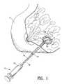
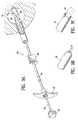
- FIG. 1is a perspective view of a human breast partially cut away having a lesion from which a biopsy specimen has been removed, and showing a marker applicator syringe and introduction cannula operatively positioned for introduction of a biopsy site marker embodying features of the present invention into the cavity created by removal of the biopsy specimen;
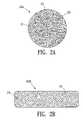
- FIGS. 2A–2Eshow exemplary conformations and shapes of sintered or porous metal site marker embodiments of the invention, FIG. 2A showing a sintered body having irregular pores, FIG. 2B showing a bubble-filled marker body, FIG. 2C showing a cylindrical-shaped marker, FIG. 2D showing a cruciform-shaped marker and FIG. 2E showing a polyhedral shaped marker.
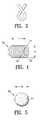
- FIG. 3shows a marker having the shape of a Greek letter.
- FIG. 4shows an example of the alternative coil-shaped embodiment of the marker of the invention
- FIG. 5shows an example of the alternative spheriod embodiment of the marker of the invention
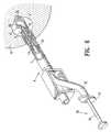
- FIG. 6is a schematic view (scale exaggerated for clarity) of an exemplary biopsy tissue site, in this case a human breast, showing a biopsy cavity of the type obtained by a known type of vacuum assisted large core biopsy sampler device, into which a biopsy marker or markers embodying features of the invention are deposited by a marker applicator device inserted through the outer cannula of the large core biopsy sampler.
- FIG. 7shows schematically an embodiment of the invention including one or more haptic elements and/or an adhesive component, for resisting migration of the marker within the tissue.
- FIG. 8shows schematically an embodiment of the invention including an encapsulating element and optional adhesive component, for resisting migration of the marker within the tissue.
- FIG. 9Ais a schematic view of a biopsy sampler device at a tissue site with an alternative marker delivery system.
- FIG. 9Bis a perspective view of the petalled distal end of the delivery device shown in FIG. 9A .
- FIG. 9Cis a perspective view of the distal end of the delivery device shown in FIG. 9A with a marker exiting the petalled distal end.
- FIG. 10is a perspective view of an alternative marker having a gel body with a radiopaque collar disposed about the gel body.
- FIG. 1shows the use and insertion into a biopsy site of any one of the biopsy site marker embodiments of the invention described herein.
- FIG. 1is a perspective view of a human breast 2 having a lesion 3 from which a biopsy specimen has been removed, thereby forming a biopsy cavity 4 within the lesion 3 , into which a biopsy site marker 10 of the of the present invention is implanted.
- the figureshows an outer cannula 12 with the distal end thereof operatively positioned within the biopsy site 4 .
- the outer cannula 12has been inserted percutaneously into the lesion 3 and a biopsy needle (not shown) has been passed through the outer cannula 12 and used to remove a biopsy specimen from the center of the lesion.
- Syringe-like marker application device 13includes a marker introduction tube or inner cannula 14 .
- the marker introduction cannula 14After removal of the biopsy needle (not shown), the marker introduction cannula 14 has been passed through the outer cannula 12 such that inner cannula distal end 14 d is located within the biopsy cavity 4 , the marker 10 being housed within cannula 14 .
- Piston 15 of marker applicator 13has an extension 16 which passes through the interior of inner cannula 14 . Upon depressing piston 15 , extenuation 16 pushes marker 10 outward through an opening 17 in the tip 14 d of inner cannula 14 into the cavity 4 .
- FIGS. 2A , 2 B, 2 C, 2 D and 2 Eshow exemplary internal conformations and shapes of the sintered or porous site marker embodiments of the invention 20 a – 20 e respectively.
- FIGS. 2A and 2Bshow schematic cross sections of a alternative porous or sintered marker body embodiments.
- FIG. 2Ais a cross section of a sintered site marker embodiment 20 a .
- the matrix or base material 21encloses a plurality of irregular shaped pores 22 distributed within the body 20 a , preferably throughout the body volume.
- the term “sintered”will be used to describe the porous body conformation, it being noted that conventional methods of production other than sintering may be employed to produce a material containing internal voids, pores, discontinuities, inclusions, bubbles and the like.
- the pores 22may be open celled, in which the pores 22 generally intersect or communicate with one another and the marker body exterior, which may give the body surface 23 a pitted texture on the scale of the pore size.
- the poresmay be closed celled, in which the pores 22 generally do not intersect one another or the exterior.
- the matrix material 21is preferably hydrophobic (or treated to have hydrophobic surfaces) to resist displacement of air entrained in pores 22 .
- the marker body 20 amay comprise a matrix or base composition 21 which has an acoustic impedance close to that of the tissue at the marked body site, since the air or other gas within the pores or internal spaces 22 provides a dramatic contrast to the matrix material 21 .
- Suitable bio-compatible materialsinclude polyethylene, polytetrafluoroethylene, PEBAX (made by Autochem Corp.), and the like.
- Such porous materialsmay be formed by conventional methods, such as heat bonding of polymer powders, extrusion and the like.
- FIG. 2Bis a schematic cross section of an alternative site marker embodiment 20 b .
- the matrix or base material 24encloses a plurality of inclusions, suspended particles or bubbles 25 distributed within the body 20 b , preferably throughout the body volume.
- the inclusions 25may be low-density or gas-filled particles, such as foamed-in-place bubbles, micro-beads, expanded beads, and the like, which have an acoustic impedance substantially lower than the matrix material 24 .
- the matrix material 24may as in the example of FIG. 2A .
- FIGS. 2C and 2Dshow exemplary shapes of the sintered or porous site marker embodiments of the invention 20 c and 20 d respectively.
- FIG. 2Cshows schematically a cylindrical sintered marker 20 c .
- the marker 20 ccomprises a generally cylindrical body having a diameter d and length l.
- the bodymay have diameter d of from 0.5 to 5 mm, and preferably about 1.5 mm.
- the length lmay be from about 1 diameters to about 10 diameters, and preferably from about 5 to 7 diameters.
- This biopsy site markerproduces a distinct, recognizable, marker image of artificial appearance when implanted at a depth of about 2 to 4 cm in human breast tissue, and visualized by a commercially available Accuson 128 US imaging system with an L7 transducer.
- FIG. 2Dillustrates a marker body 20 d having a polyhedral form of multiple intersecting flat surfaces 26 , 27 and 28 .
- FIG. 2Eshows a cruciform shaped marker 20 e having cruciform cross-section having four longitudinal fin-like portions 29 , which may be aligned at right angles to one another and joined at the longitudinal central axis 30 providing a selectable number of side facets (e.g., hexagonal cross-section).
- medial web portions 31may span laterally and join between adjacent fins 29 , the webs 31 preferably being aligned perpendicularly to the fins 29 .
- the planes of the intersecting fins and websform a pattern of eight mutually-perpendicular “corner reflectors” 32 .
- the length l and characteristic cross-section dimension cmay be as described with respect to the embodiments of FIGS. 2C and 2D .
- FIG. 3illustrates yet another alternative where the marker body is shaped to have the form, under ultrasound or radiological visualization, preferably both, of a familiar symbol or letter, to by easily recognizable as an artificial shape which is the lower-case Greek letter Gamma ( ⁇ ), which when visualized in a biopsy site bears a resemblance to a familiar breast-cancer-awareness symbol.
- ⁇Greek letter Gamma
- FIG. 4shows schematically an alternative coil marker 30 of the invention.
- the marker 30comprises a generally helical coil-like body formed from one or more lengths of fine wire and/or fiber 31 .
- the coil 30has a generally cylindrical overall form.
- the coil length l and diameter dmay be of a range of sizes, selected so as to be large enough to provide a distinct, recognizable ultrasound marker image within the tissue biopsy site, and small enough to avoid masking or obscuring diagnostically important tissue features.
- the coil diameter dmay be from 0.5 to 5 mm, and preferably about 1.5 mm.
- the coil length lis typically from about 1 coil diameters to about 10 coil diameters, and preferably from about 5 to 7 coil diameters.
- the helical turns of the coilprovide a body surface contour including a outer helical groove 32 and inner helical groove 33 on the coil surfaces (more than one such groove for a multiple helix).
- the grooved coil body surfaceincludes a plurality of lobes and crevices on the exterior of the coil which enhance acoustic reflectivity.
- the similarly lobed internal surfaces of the coilprovide additional reflectivity.
- the coilmay be given a “frosted” or textured surface, such as by particle blasting in the manner of the spheroid marker described above.
- a uniform coil embodimenthas a shape which is markedly artificial in appearance under conventional visualization methods, and is not easily confused tissue features of biological origin.
- the coilmay comprise a fine wire 31 of a material of high acoustic impedance relative to the tissue of the site, and may optionally be radio-opaque. Suitable materials are biologically compatible metals, such as stainless steel, titanium, platinum, palladium, alloys thereof and the like.
- the coilmay alternatively comprise a composite of different materials, such as a composite of metal and polymeric materials.
- the coilmay be wound about a central core of the same or different composition. Coil stock of suitable material, helical form and diameter is available commercially, and may be cut to a selected length by conventional means.
- a suitable materialis 316 L stainless steel surgical embolization coil currently used in arterial embolism repair procedures, e.g., Cook 4 mm diameter embolization coil MWCE-25-2.5-4 of 316L stainless steel and Dacron.
- Other suitable embolization coil stockis available in a range of coil diameters.
- This biopsy site markerproduces a distinct, recognizable marker image as implanted at a depth of about 2 to 4 cm in human breast tissue, when visualized by a commercially available Accuson 128 US imaging system with an L7 transducer.
- FIG. 5shows schematically the alternative spheroid marker 40 of the invention having a generally spherical body 40 .
- the porous or sintered marker embodiments of FIGS. 2A–2Dmay be spherical also.
- the embodiment of FIG. 5is a non-porous example, and the biopsy site marker 40 comprises a high acoustic impedance, biologically compatible material, such as 316 L stainless steel and titanium, or radiopaque metals such as platinum, palladium, or the like.
- Non-spherical shaped bodiesmay be used, however, metallic spheres of suitable materials are readily commercially available, and have a shape which is markedly artificial in appearance under conventional visualization methods, i.e., not easily confused tissue features of biological origin.
- the generally spherical bodymay have a diameter d selected so as to be large enough to provide a distinct, recognizable ultrasound marker image within the tissue biopsy site, and small enough to avoid obscuring tissue features.
- the optimum size of the spherewill depend on such factors as the type of visualization system used, its imaging resolution, and the physical nature of the biopsy tissue region.
- the sphere diameter dis typically be from about 1 mm to about 4 mm, and preferably from about 1.5 mm.
- the spherical body 40may include a pitted, matte, peened or frosted surface texture 41 , which may be produced by conventional particle blasting or peening techniques.
- the spheremay be blasted with glass beads of about 100 micrometer diameter to produce a frosted surface.
- the spheremay be blasted with aluminum oxide abrasive particles of about 25 micrometer diameter to produce a frosted surface.
- the frosted surface 41thus produced provides enhanced acoustic reflectivity in comparison to the untreated, smooth sphere.
- Other conventional texturing, pitting or faceting methodsmay alternatively be used to produce a frosted or irregular surface texture.
- This biopsy site markerproduces a distinct, recognizable marker image of artificial appearance when implanted at a depth of about 2 to 4 cm in human breast tissue, and visualized by a commercially available Acuson 128 US imaging system with an L7 transducer.
- FIG. 6shows schematically in cut-away section an exemplary marker applicator device 50 configured to be operated in association with a conventional vacuum assisted large core biopsy device 6 .
- the dimensional size of the applicator device(particularly the inside diameter) may be adjusted to correspond to the selected diameter or characteristic dimension of the biopsy site marker to be deposited.
- the biopsy markers of the inventioncan be used without this applicator, and can be deposited in accordance with the various methods and techniques utilized in the state of the art.
- the cylindrical body 52has an interior cavity and a piston 54 that fits and slides back and forth in the elongated cylindrical body 52 .
- the proximal end of the outer cannula 7may be provided with rectangularly shaped handle 56 the orientation of which indicates to the operator the orientation of the opening 9 provided in the distal end of the cannula 7 .
- the cylindrical body 52may have an enlarged finger disk or handle 57 at its outer (exterior to the patient) end which permits a user (not shown) to operate or move the piston 54 within the cylinder 52 of applicator 50 .
- the orientation of the elongated finger disk 57indicates the orientation of the opening 58 of body 53 adjacent its other, closed end 59 (internal within biopsy cavity).
- the opening 58is configured to form a ramp in the side of the tube 52 .
- the selected dimensions of the tube 52are coordinated with the dimensions of the piston 54 and with the cannula 7 of the vacuum assisted large core biopsy device 6 , thus permitting the tube 52 to both fit within cannula 7 and to contain one or more markers of the invention 10 within the inside diameter of cylinder 52 .
- the cylinder or tube 52 and the piston 54may be made from any appropriate medical grade plastic material, such as high density polyethylene or PEBAX, made by the Autochem Corporation.
- the tube 52is loaded with one or more of markers 10 .
- the markers 10may be any of the embodiments of the invention described above, and is shown schematically as a cylindrical object.
- pellets composed of various other materialsmay be inserted along with one of the embodiments of the biopsy markers of the present invention described herein.
- gelatin pellets of the type disclosed in our above referenced co-pending application Ser. No. 09/343,975may be inserted in conjunction with the biopsy markers 10 of the present invention.
- the opening 58 in the cylinder 52is moved into alignment with the opening or port 9 of the in the internal end of cannula 7 of biopsy sampler 6 .
- the piston 54is pressed inward by the operator so that the marker or markers 10 are expelled from the tube 52 through the ramp shaped opening 58 as the piston 54 is pushed into the cylinder or tube 52 .
- the markers 10are thereby extruded through opening 59 and port 9 into the biopsy cavity 4 .
- the applicator 50 and biopsy device 6are subsequently withdrawn.
- FIG. 7shows schematically an alternative marker 60 of the invention including one or more optional tissue-engaging or haptic elements 62 for resisting migration of the marker from the biopsy site.
- An exemplary cylindrical marker body 10is shown, although each embodiment of the biopsy site marker of the invention described above may optionally comprises one or more such tissue engaging structures.
- the haptic elements 62may comprise an wire-like material fixed to the marker body 10 at the proximal haptic end 64 and extending outward from the marker body 10 .
- the haptic 62may be looped back at its hook-like terminal end 66 .
- the haptic 62assists in resisting migration of the marker from the biopsy cavity, during initial placement, i.e., it engages the adjacent tissue to resist being sucked back towards the applicator when the applicator is withdrawn.
- the hapticalso resists migration during later movement, flexure or manipulation of the tissue surrounding the biopsy site, such as when a patient's breast is decompressed upon removal from a mammography device.
- the marker body 10may include an adhesive component 68 coated onto its surface to cause the marker body to adhere to adjacent tissue within the biopsy site.
- FIG. 8shows schematically the alternative marker 70 of the invention including an encapsulating element 72 and optional adhesive layer or component 74 , for resisting migration of the marker within the tissue.
- An exemplary cylindrical marker body 10is shown, although each of the biopsy site marker of the invention described above may optionally comprise a pellet-shaped encapsulating element.
- the pellet-shaped encapsulating element 72is disposed surrounding the marker body 10 and may fully or partially enclose the marker body.
- the encapsulating element 72may be of lower impedance than the metallic marker body 10 .
- Suitable materialsare gelatin or reconstituted collagen material, polymers, or mixtures or composites thereof.
- An optional adhesive component 74is shown coating the external surface of the encapsulating element, but may be included within the composition the encapsulating element 72 .
- FIG. 10illustrates an alternative marker 90 which has an elongated cylindrically shaped body of gel 91 surrounded with a metallic band 92 which is preferably formed of radiopaque material.
- the band 92may conpletely or only partially surround the body of gel 91 .
- the marker bodymay include an adhesive component to cause the marker body (or encapsulating element) to adhere to adjacent tissue within the biopsy site.
- the adhesive componentmay comprise a biocompatible adhesive, such as a polyurethane, polyacrylic compound, polyhydroxymethacrylate, fibrin glue (e.g., TissealTM), collagen adhesive, or mixtures thereof.
Landscapes
- Health & Medical Sciences (AREA)
- Life Sciences & Earth Sciences (AREA)
- Engineering & Computer Science (AREA)
- Animal Behavior & Ethology (AREA)
- Veterinary Medicine (AREA)
- Public Health (AREA)
- Biomedical Technology (AREA)
- General Health & Medical Sciences (AREA)
- Nuclear Medicine, Radiotherapy & Molecular Imaging (AREA)
- Heart & Thoracic Surgery (AREA)
- Surgery (AREA)
- Epidemiology (AREA)
- Medical Informatics (AREA)
- Physics & Mathematics (AREA)
- Oral & Maxillofacial Surgery (AREA)
- Pathology (AREA)
- Hematology (AREA)
- Molecular Biology (AREA)
- Dermatology (AREA)
- Acoustics & Sound (AREA)
- Radiology & Medical Imaging (AREA)
- Anesthesiology (AREA)
- Biodiversity & Conservation Biology (AREA)
- Oncology (AREA)
- Optics & Photonics (AREA)
- Apparatus For Radiation Diagnosis (AREA)
Abstract
Description
The present application is a Continuation of U.S. Patent application Ser. No. 09/717,909 filed Nov. 20, 2000 now U.S. Pat. No. 6,725,083 which is a Continuation-In-Part of U.S. patent application Ser. No. 09/241,936, filed Feb. 2, 1999, now U.S. Pat. No. 6,161,034, and a Continuation-In-Part of U.S. patent application Ser. No. 09/343,975, filed Jun. 30, 1999 now U.S. Pat. No. 6,347,214. Each of the above referenced patent applications is incorporated by reference herein and the benefit of the filing date of each application is hereby claimed under 35 U.S.C. § 1.20.
In diagnosing and treating certain medical conditions, it is often desirable to perform a biopsy, in which a specimen or sample of the suspicious tissue is removed for pathological examination, tests and analysis. As is known, obtaining a tissue sample by biopsy and the subsequent examination are typically employed in the diagnosis of cancers and other malignant tumors, or to confirm that a suspected lesion or tumor is not malignant. The information obtained from these diagnostic tests and/or examinations is frequently used to devise a therapeutic plan for the appropriate surgical procedure or other course of treatment.
In many instances, the suspicious tissue to be sampled is located in a subcutaneous site, such as inside a human breast. Such removal of tissue samples may be accomplished by open surgical technique, or through the use of a specialized biopsy instrument and techniques. To minimize surgical intrusion into patient's body, it is often desirable to insert a small instrument, such as a biopsy needle, into the body for extracting the biopsy specimen while imaging the procedure using fluoroscopy, ultrasonic imaging, x-rays, MRI or any other suitable form of imaging technique. Examination of tissue samples taken by biopsy is of particular significance in the diagnosis and treatment of breast cancer. In the ensuing discussion, the biopsy and treatment site described will generally be the human breast, although the invention is suitable for marking biopsy sites in other parts of the human and other mammalian body as well.
Periodic physical examination of the breasts and mammography are important for early detection of potentially cancerous lesions. In mammography, the breast is compressed between two plates while specialized x-ray images are taken. If an abnormal mass in the breast is found by physical examination or mammography, ultrasound may be used to determine whether the mass is a solid tumor or a fluid-filled cyst. Solid masses are usually subjected to some type of tissue biopsy to determine if the mass is cancerous.
If a solid mass or lesion is large enough to be palpable, a tissue specimen can be removed from the mass by a variety of techniques, including but not limited to open surgical biopsy, a technique known as Fine Needle Aspiration Biopsy (FNAB) and instruments characterized as “vacuum assisted large core biopsy devices”.
If a solid mass of the breast is small and non palpable (e.g., the type typically discovered through mammography), a relatively new biopsy procedure known as stereotactic needle biopsy may be used. In performing a stereotactic needle biopsy of a breast, the patient lies on a special biopsy table with her breast compressed between the plates of a mammography apparatus and two separate x-rays or digital video views are taken from two different points of view. A computer calculates the exact position of the lesion as well as depth of the lesion within the breast. Thereafter, a mechanical stereotactic apparatus is programmed with the coordinates and depth information calculated by the computer, and such apparatus is used to precisely advance the biopsy needle into the small lesion. Depending on the type of biopsy needle(s) used, this stereotactic technique may be used to obtain cytologic specimens, e.g., obtained through FNAB or it may be used to obtain histologic specimens e.g., obtained through coring needle biopsy. Usually at least five separate biopsy specimens are obtained from locations around the small lesion as well as one from the center of the lesion.
The available treatment options for cancerous lesions of the breast include various degrees of mastectomy or lumpectomy and radiation therapy, as well as chemotherapy and combinations of these treatments. However, radiographically visible tissue features, originally observed in a mammogram, may be removed, altered or obscured by the biopsy procedure. In order for the surgeon or radiation oncologist to direct surgical or radiation treatment to the precise location of the breast lesion several days or weeks after the biopsy procedure was performed, it is desirable that a biopsy site marker be placed in or on the patient's body to serve as a landmark for subsequent location of the lesion site. While current radiographic type markers may persist at the biopsy site, an additional mammography generally must be performed at the time of follow up treatment or surgery in order to locate the site of the previous surgery or biopsy. In addition, once the site of the previous procedure is located using mammography, the site must usually be marked with a location wire which has a barb on the end which is advanced into site of the previous procedure. The barb is meant to fix the tip of the location wire with respect to the site of the previous procedure so that the patient can then be removed from the confinement of the mammography apparatus and the follow-up procedure performed. However, as the patient is removed from the mammography apparatus, or otherwise transported the position of the location wire can change or shift in relation to the site of the previous procedure. This, in turn, can result in follow-up treatments being misdirected to an undesired portion of the patient's tissue.
As an alternative or adjunct to radiographic imaging, ultrasonic imaging and visualization techniques (herein abbreviated as “USI”) can be used to image the tissue of interest at the site of interest during a surgical or biopsy procedure or follow-up procedure. USI is capable of providing precise location and imaging of suspicious tissue, surrounding tissue and biopsy instruments within the patient's body during a procedure. Such imaging facilitates accurate and controllable removal or sampling of the suspicious tissue so as to minimize trauma to surrounding healthy tissue.
For example, during a breast biopsy procedure, the biopsy device is often imaged with USI while the device is being inserted into the patient's breast and activated to remove a sample of suspicious breast tissue. As USI is often used to image tissue during follow-up treatment, it may be desirable to have a marker, similar to the radiographic markers discussed above, which can be placed in a patient's body at the site of a surgical procedure and which are visible using USI. Such a marker enables a follow-up procedure to be performed without the need for traditional radiographic mammography imaging which, as discussed above, can be subject to inaccuracies as a result of shifting of the location wire as well as being tedious and uncomfortable for the patient.
The invention is directed generally to devices and methods of marking a biopsy site, so that the location of the biopsy cavity is readily visible by ultrasonic imaging, as well as by conventional imaging methods, such as x-rays. The biopsy site marker of the invention is a persistent marker which may be identified and located by ultrasound visualization.
The biopsy site markers of the invention have a body conformation to enhance acoustical reflective signature or signal. The body conformation may include boundaries of high contrast of acoustic impedance to enhance ultrasound reflection. The markers are readily detected by USI and present a substantial acoustic signature from a marker with small physical dimensions or size. Because of the high acoustic reflectivity of the markers of the invention, the marker size may be reduced to dimensions determined by the physical limits of the imaging system itself, e.g., the ultrasound (US) beam width, without requiring a larger or excessive marker size to reflect sufficient US energy to be noticeable.
In one embodiment, the biopsy site markers of the invention have a characteristic body shape which is recognizably artificial during medical imaging, so as to be readily distinguishable from biological features within the marked tissue. In particular, the markers are readily distinguishable in the various imaging procedures from diagnostically important tissue features, such as lines of calcifications which frequently are signs for a developing malignancy. The marker body shape may have one or more distinct features which may be visualized in different marker orientations. The shape may correspond to a generally known symbol, so a to enhance recognition.
In another embodiment, the markers of the invention have a body conformation to enhance the acoustic signature or signal, so that the body has high acoustic reflectivity when situated in tissue. The acoustic reflective signature of the markers depends on a number of factors. The marker may comprise a composition which presents at least one boundary of high contrast in acoustic impedance to incident US energy, effectively reflecting the US energy to be received by the imaging system. Acoustic impedance (AI) of a material is equal to the product of the characteristic density (ρ) of the material and the acoustic velocity (c) in the material, (i.e., AI=ρ×c). As an incident US beam encounters a boundary with a large change in acoustic impedance (e.g., at the marker surface or internal to the marker), much of the US energy is effectively reflected.
Different types of tissue have a wide range of acoustical impedance, for example lung tissue with high air content having low acoustical impedance as compared to bone tissue having high mineral content. However, for uses such as biopsy site marking in typical mammalian soft tissue of high aqueous content, the typical range of tissue acoustical impedance is intermediate these extremes. The composition and body conformation of the markers of the invention may be selected so as to provide boundaries of high contrast of acoustic impedance with respect to the particular tissue site of use.
In an embodiment of the invention, the marker may have a composition in which a base or matrix substance of the marker body (e.g., stainless steel) has an acoustic impedance substantially higher than the tissue at the marked body site. For example, typical bio-compatible metal materials, such as stainless steel, titanium, platinum and the like, generally have acoustic impedance values in the range of 15 to more than 30 times that of typical soft tissue of high aqueous or fatty content. The high acoustic impedance of the marker body base material relative to the surrounding tissue presents a reflective interface to an incident US beam.
A suitable marker body composition with acoustic impedance substantially higher than the tissue at the marked body site is 316L stainless steel. Other alternative compositions, such as compositions of bio-compatible metals, ceramics, metal oxides or polymers, or composites or mixtures of these materials, may be suitable. The marker body may also be radio-opaque.
In another embodiment of the invention, the marker may have a composition in which marker body includes one or more (preferably a large plurality) of internal bounded spaces, such as voids, pores, discontinuities, inclusions, bubbles and the like. These internal spaces preferably contain or entrain air or other gases.
Air has an extremely low acoustic impedance relative to the marker body base or matrix substance. This is true even for matrix materials which themselves have acoustic impedance close to that of the surrounding tissue (e.g., some bio-compatible polymers). The marker body presents internal boundaries of high contrast in acoustic impedance, i.e., at the boundary between the matrix and each internal air-filled space. The marker body thus presents plurality of reflective interfaces to an incident US beam.
Alternatively or in combination with to the materials of high acoustic impedance described above, a marker body with internal voids or air spaces may, if desired, comprise a matrix or base composition which has an acoustic impedance close to that of the tissue at the marked body site, since the air or other gas within the internal spaces provides a dramatic contrast to the matrix material. Suitable bio-compatible materials include polyethylene, polytetrafluoroethylene, PEBAX (made by Autochem Corp.), and the like.
The body matrix material can have a hydrophobic composition or be treated to be hydrophobic. The surface area bounding internal open-cell pores should be hydrophobic so as to resist the displacement of air or other gases in the pores by aqueous fluid from the surrounding tissue, particularly in the case of relatively large pore or space size.
In some embodiments of the invention, the markers can include surface characteristics which enhance the acoustic signature and improve visibility under US imaging, as opposed to a smooth, rounded body surface. In order to provide enhanced ultrasound imaging visibility from all directions of US impingement, the biopsy marker can have a plurality of reflective external surfaces. By making the surface of an object lobulate, multifaceted or otherwise irregular, more reflective surfaces are created, and a brighter acoustic signature is achieved.
For example, a smooth solid sphere provides at least some reflective surface oriented in each direction, but the reflection is achieved over a small portion to the area of the sphere, thus producing an unremarkable acoustic signature. In contrast, an object of the same composition and average diameter as the sphere, but with a highly irregular surface texture, a much brighter acoustic signature or signal is achieved. Thus, the by providing more reflective surfaces of differing or random orientation, the markers appears brighter in US imaging.
The signal-enhancing body conformation may include non-smooth surface texture, such as a porous, frosted, matte, pitted, peened, or scratched surface texture, and the like. The body conformation may also include a multi-element surface contour, such as a faceted, multi-planar, lobulate, coiled, grooved, folded, or inlet surface contour, and the like. Such external body conformations may be used in combination with one another and in combination with the internal discontinuities or air spaces described above.
The body length, diameter or other characteristic scale dimensions of some embodiments of the biopsy marker of the invention may be of a range of sizes. The optimum dimensions of the body will depend upon the specific selected factors which influence acoustic signature as described herein, such as material impedance, surface contours, surface texture, and internal conformation. In addition, the optimum size may depend upon such factors as the type of ultrasound imaging/visualization system used, its imaging resolution, the operating ultrasound frequency, and the biophysical nature of the tissue of interest.
The body dimensions may be selected so as to be large enough to provide a distinct, recognizable marker image within the tissue biopsy site, when visualized under the particular imaging system and operating conditions of use. The body dimensions may also be selected to be small enough to avoid masking or obscuring diagnostically important tissue features. Thus different marker dimensions may be selected to suit particular biopsy site tissue types, and to suit particular known and future medical imaging equipment.
In terms of over-all size, it is desirable that the marker have at least one dimension which is about as large as or greater than the beam width of the USI system with which it is to be visualized. Typically, for current USI systems, the marker will have at least one dimension of about 1 mm or greater, and preferably of at least about 1.5 mm.
In addition, for convenience in applying the marker to the tissue site, the specific marker dimensions and shape may be selected so as to accommodate the dimensions of a particular known or novel biopsy needle device or sampling apparatus, while still achieving a distinct and recognizable marker image under medical imaging as placed at the tissue site. By selecting a marker size and shape to fit within the internal diameter of a biopsy needle or sampling device, the marker may be implanted or applied to the biopsy cavity during the course of the biopsy procedure, following sample recovery but prior to removal of the biopsy device. For example, the marker of the invention may have a size and shape selected to permit application of the marker through the hollow interior space of a vacuum assisted large core biopsy device, such as is commercially available from Johnson and Johnson, Ethicon Endosurgery Division. The small physical size of the markers of the invention relative to their acoustic reflectivity permits fitting the markers to a wide variety of biopsy devices.
In terms of the size of features, including external or internal pores, texture features, facets and the like, it is preferable that these features have a characteristic dimension approximately equal to or exceeding the wavelength of the US beam of the imaging system. For example, with current imaging systems, for a marker with internal air-filled pores, the pore size is typically from about 1 micrometer to 100 micrometers and preferably from about 5 micrometers to 40 micrometers, to provide high reflectivity of the incident US energy.
Optionally, some embodiments of the biopsy site marker of the invention may have elements which assist in accurately fixing the marker to the biopsy site so as to resist migration from the biopsy cavity. Such migration can occur when a placement instrument is withdrawn, and when the marked tissue is subsequently moved or manipulated, as for example when a breast is decompressed and removed from the mammography apparatus. In one embodiment, one or more tissue engaging structures or haptic elements are mounted or affixed to the main marker body, so as to resist movement or migration of the marker from the biopsy site in which it has been implanted during use.
In another embodiment, the biopsy site marker may comprise a pellet-shaped element which encapsulates the high impedance marker body, and assists in resisting migration. The encapsulating pellet may be of a composition, such as gelatin, which is absorbed or dissipated over time, leaving the persistent marker body at the tissue site. In yet another embodiment, the marker body (and/or the optional encapsulating element) may include an adhesive component to cause the marker body (or encapsulating element) to adhere to adjacent tissue within the biopsy site.
A method of the invention for marking a tissue site of interest can include implanting one or more of the markers of the invention, such as one of the exemplary marker embodiments described herein, in or adjacent to a tissue site of interest, e.g., within a biopsy cavity. The marker may then be visualized in situ, such as for purposes of subsequent medical and surgical procedures. The visualization may be by various known medical imaging systems and methods, and in particular may be visualized by known USI systems.
Biopsy markers of the invention can be deposited in accordance with the various methods and techniques utilized in the state of the art. One technique of applying the biopsy markers of the invention is to place or deposit them in a biopsy cavity that is created with a vacuum assisted large core biopsy device. An applicator particularly suitable for insertion of the biopsy site markers of the invention is described below. However, it should be understood that the biopsy markers of the invention can be used without the exemplary applicator device described herein. The biopsy marker applicator disclosed in co-pending application Ser. No. 09/343,975 filed Jun. 30, 1999, may be used to apply the markers of the current invention to a biopsy site. The dimensional size of the applicator device (particularly the inside diameter) may be adjusted to correspond to a selected diameter or characteristic dimension of the biopsy site marker embodiment of the present invention.
These and other advantages of the invention will become more apparent from the following description when taken in conjunction with the accompanying drawings.
The following detailed description, and the accompanying drawings to which it refers are provided for purposes of exemplifying and illustrating representative examples and embodiments of the invention only, and are not intended to limit the scope of the invention in any way, and do not exhaustively illustrate and describe all possible embodiments and configurations in which one or more features of the present invention may take physical form.
All patents and patent applications cited in this specification are herein incorporated by reference as if each individual patent or patent application were specifically and individually indicated to be incorporated by reference.
Syringe-likemarker application device 13 includes a marker introduction tube orinner cannula 14. After removal of the biopsy needle (not shown), themarker introduction cannula 14 has been passed through theouter cannula 12 such that inner cannuladistal end 14d is located within thebiopsy cavity 4, themarker 10 being housed withincannula 14.Piston 15 ofmarker applicator 13 has anextension 16 which passes through the interior ofinner cannula 14. Upon depressingpiston 15,extenuation 16pushes marker 10 outward through anopening 17 in thetip 14dofinner cannula 14 into thecavity 4.
Theouter cannula 12 may be an outer tube element of a conventional vacuum assisted large core biopsy device, which has been left in place to assist in site marker application following biopsy sample recovery. One example of aapplicator syringe device 13 is described in further detail below with respect toFIG. 5 .
Thepores 22 may be open celled, in which thepores 22 generally intersect or communicate with one another and the marker body exterior, which may give the body surface23 a pitted texture on the scale of the pore size. Alternatively, the pores may be closed celled, in which thepores 22 generally do not intersect one another or the exterior. In the event that thepores 22 communicate with themarker exterior 23, thematrix material 21 is preferably hydrophobic (or treated to have hydrophobic surfaces) to resist displacement of air entrained inpores 22.
The base ormatrix composition 21 has may be of high acoustic impedance relative to the surrounding tissue (not shown). Sintered metal material may be shaped and sintered from commercially available metallic powders comprising a metal or mixtures of metals, using conventional sintering and forming techniques to produce body of selected shaped, and selected pore size and surface texture, so as to enhance acoustic reflectivity. The porosity of the sintered metal provides an irregular surface texture as well as internal voids. A suitable bio-compatible material is sintered 316L stainless steel, and suitable sintered stainless steel stock is commercially available in various forms, for example from the Mott Corporation. The sintered stock may be economically cut and shaped by conventional methods. Sintered stainless steel stock is commercially available with controlled pore size, selectable over a range of pore sizes. Thepores 22 of thesintered body 20amay vary over a range of pore sizes, and is typically from about 1 micrometer to 100 micrometers and preferably from about 5 micrometers to 40 micrometers.
In addition to sintered metal, alternative bio-compatible, impedance materials may be included or substituted, such as ceramics, metal oxides, polymers or composites/mixtures of these materials, which may be configured to have a generally distributed internal porosity and porous surface texture. Thus, themarker body 20amay comprise a matrix orbase composition 21 which has an acoustic impedance close to that of the tissue at the marked body site, since the air or other gas within the pores orinternal spaces 22 provides a dramatic contrast to thematrix material 21. Suitable bio-compatible materials include polyethylene, polytetrafluoroethylene, PEBAX (made by Autochem Corp.), and the like. Such porous materials may be formed by conventional methods, such as heat bonding of polymer powders, extrusion and the like.
The helical turns of the coil provide a body surface contour including a outerhelical groove 32 and inner helical groove33 on the coil surfaces (more than one such groove for a multiple helix). The grooved coil body surface includes a plurality of lobes and crevices on the exterior of the coil which enhance acoustic reflectivity. In addition the similarly lobed internal surfaces of the coil provide additional reflectivity. Optionally, the coil may be given a “frosted” or textured surface, such as by particle blasting in the manner of the spheroid marker described above. A uniform coil embodiment has a shape which is markedly artificial in appearance under conventional visualization methods, and is not easily confused tissue features of biological origin.
The coil may comprise afine wire 31 of a material of high acoustic impedance relative to the tissue of the site, and may optionally be radio-opaque. Suitable materials are biologically compatible metals, such as stainless steel, titanium, platinum, palladium, alloys thereof and the like. The coil may alternatively comprise a composite of different materials, such as a composite of metal and polymeric materials. The coil may be wound about a central core of the same or different composition. Coil stock of suitable material, helical form and diameter is available commercially, and may be cut to a selected length by conventional means. A suitable material is 316 L stainless steel surgical embolization coil currently used in arterial embolism repair procedures, e.g.,Cook 4 mm diameter embolization coil MWCE-25-2.5-4 of 316L stainless steel and Dacron. Other suitable embolization coil stock is available in a range of coil diameters. This biopsy site marker produces a distinct, recognizable marker image as implanted at a depth of about 2 to 4 cm in human breast tissue, when visualized by a commercially available Accuson 128 US imaging system with an L7 transducer.
The generally spherical body may have a diameter d selected so as to be large enough to provide a distinct, recognizable ultrasound marker image within the tissue biopsy site, and small enough to avoid obscuring tissue features. As with the other biopsy site marker embodiments of the invention, the optimum size of the sphere will depend on such factors as the type of visualization system used, its imaging resolution, and the physical nature of the biopsy tissue region. For example, the sphere diameter d is typically be from about 1 mm to about 4 mm, and preferably from about 1.5 mm.
Thespherical body 40 may include a pitted, matte, peened orfrosted surface texture 41, which may be produced by conventional particle blasting or peening techniques. For example, the sphere may be blasted with glass beads of about 100 micrometer diameter to produce a frosted surface. In another example, the sphere may be blasted with aluminum oxide abrasive particles of about 25 micrometer diameter to produce a frosted surface. Thefrosted surface 41 thus produced provides enhanced acoustic reflectivity in comparison to the untreated, smooth sphere. Other conventional texturing, pitting or faceting methods may alternatively be used to produce a frosted or irregular surface texture.
This biopsy site marker produces a distinct, recognizable marker image of artificial appearance when implanted at a depth of about 2 to 4 cm in human breast tissue, and visualized by a commercially available Acuson 128 US imaging system with an L7 transducer.
Theapplicator 50 comprises an elongatedcylindrical body 52 which has an outer diameter selected so that it fits, and may be inserted through, theouter cannula 7 of vacuum assisted largecore biopsy device 6. As shown inFIG. 6 , theouter cannula 7 is inserted through the biopsy incision into thebiopsy cavity 4 previously formed in the patient'stissue site 8, e.g., a human breast in the case of a breast biopsy.
Thecylindrical body 52 has an interior cavity and apiston 54 that fits and slides back and forth in the elongatedcylindrical body 52. The proximal end of theouter cannula 7 may be provided with rectangularly shaped handle56 the orientation of which indicates to the operator the orientation of the opening9 provided in the distal end of thecannula 7. Thecylindrical body 52 may have an enlarged finger disk or handle57 at its outer (exterior to the patient) end which permits a user (not shown) to operate or move thepiston 54 within thecylinder 52 ofapplicator 50. the orientation of theelongated finger disk 57 indicates the orientation of theopening 58 of body53 adjacent its other, closed end59 (internal within biopsy cavity). Theopening 58 is configured to form a ramp in the side of thetube 52.
In this connection it should be understood that the selected dimensions of thetube 52 are coordinated with the dimensions of thepiston 54 and with thecannula 7 of the vacuum assisted largecore biopsy device 6, thus permitting thetube 52 to both fit withincannula 7 and to contain one or more markers of theinvention 10 within the inside diameter ofcylinder 52. The cylinder ortube 52 and thepiston 54 may be made from any appropriate medical grade plastic material, such as high density polyethylene or PEBAX, made by the Autochem Corporation.
In one method of implanting thebiopsy markers 10 of the present invention, thetube 52 is loaded with one or more ofmarkers 10. Themarkers 10 may be any of the embodiments of the invention described above, and is shown schematically as a cylindrical object. Optionally, in addition to themarkers 10, pellets composed of various other materials may be inserted along with one of the embodiments of the biopsy markers of the present invention described herein. For example, gelatin pellets of the type disclosed in our above referenced co-pending application Ser. No. 09/343,975 may be inserted in conjunction with thebiopsy markers 10 of the present invention.
With themarkers 10 in thetube 52 and thetube 52 andcannula 7 inserted into thebiopsy site 4, theopening 58 in thecylinder 52 is moved into alignment with the opening or port9 of the in the internal end ofcannula 7 ofbiopsy sampler 6. Thepiston 54 is pressed inward by the operator so that the marker ormarkers 10 are expelled from thetube 52 through the ramp shapedopening 58 as thepiston 54 is pushed into the cylinder ortube 52. Themarkers 10 are thereby extruded throughopening 59 and port9 into thebiopsy cavity 4. Theapplicator 50 andbiopsy device 6 are subsequently withdrawn.
The haptic62 assists in resisting migration of the marker from the biopsy cavity, during initial placement, i.e., it engages the adjacent tissue to resist being sucked back towards the applicator when the applicator is withdrawn. The haptic also resists migration during later movement, flexure or manipulation of the tissue surrounding the biopsy site, such as when a patient's breast is decompressed upon removal from a mammography device. Optionally, themarker body 10 may include anadhesive component 68 coated onto its surface to cause the marker body to adhere to adjacent tissue within the biopsy site.
The pellet-shapedencapsulating element 72 is disposed surrounding themarker body 10 and may fully or partially enclose the marker body. The encapsulatingelement 72 may be of lower impedance than themetallic marker body 10. Suitable materials are gelatin or reconstituted collagen material, polymers, or mixtures or composites thereof. Anoptional adhesive component 74 is shown coating the external surface of the encapsulating element, but may be included within the composition the encapsulatingelement 72.
In any of the above-described embodiments of the invention, the marker body (and/or the optional encapsulating element) may include an adhesive component to cause the marker body (or encapsulating element) to adhere to adjacent tissue within the biopsy site. The adhesive component may comprise a biocompatible adhesive, such as a polyurethane, polyacrylic compound, polyhydroxymethacrylate, fibrin glue (e.g., Tisseal™), collagen adhesive, or mixtures thereof.
While particular forms of the invention have been illustrated and described, it will be apparent that various modifications can be made without departing from the spirit and scope of the invention. Accordingly, it is not intended that the invention be limited to the specific embodiments illustrated. It is therefore intended that this invention to be defined by the scope of the appended claims as broadly as the prior art will permit, and in view of the specification if need be.
Claims (29)
1. A remotely detectable marker for marking a selected intracorporeal site within a patient, comprising a sintered, ultrasound detectable body which is formed at least in part of metallic material, which has boundaries with a high contrast in acoustical impedance when the marker is placed at an intracorporeal site to facilitate ultrasound detection; and which has a body shape which is recognizable as artificial.
2. The intracorporeal site marker ofclaim 1 wherein the body is remotely detectable by ultrasound or X-ray.
3. The intracorporeal site marker ofclaim 1 wherein the metallic material is selected from the group consisting of stainless steel, titanium, platinum, palladium and alloys thereof.
4. The intracorporeal site marker ofclaim 1 wherein the metallic material is titanium.
5. The intracorporeal site marker ofclaim 1 wherein the metallic material is 316 stainless steel.
6. The intracorporeal site marker ofclaim 1 wherein the body is cylindrical in shape.
7. The intracorporeal site marker ofclaim 4 wherein the cylindrical shape has a diameter of about 0.5 to about 5 mm and a length of at least one diameter.
8. The intracorporeal site marker ofclaim 5 wherein the cylindrical shape has a length of up to 10 diameters.
9. The intracorporeal site marker ofclaim 5 wherein the cylindrical shape has a length of about 5 to about 7 diameters.
10. The intracorporeal site marker ofclaim 5 wherein the cylindrically shaped body is a helically shaped coil.
11. The intracorporeal site marker ofclaim 1 wherein the body has a spherical shape.
12. The intracorporeal site marker ofclaim 11 wherein the spherically shaped body has a diameter of about 1 to about 4 mm.
13. A remotely detectable marker for marking a selected intracorporeal site within a patient, comprising an ultrasound detectable body which is formed of sintered titanium, which has boundaries with a high contrast in acoustical impedance when the marker is placed at an intracorporeal site to facilitate ultrasound detection; and which has a body shape which is recognizable as artificial.
14. The intracorporeal site marker ofclaim 13 wherein the body is remotely detectable by ultrasound or X-ray.
15. The intracorporeal site marker ofclaim 13 wherein the ultrasound detectable, sintered titanium body is porous.
16. A remotely detectable marker for marking a selected intracorporeal site within a patient, comprising an ultrasound detectable body which is formed at least in part of porous sintered titanium, which has boundaries with a high contrast in acoustical impedance when the marker is placed at an intracorporeal site to facilitate ultrasound detection; and which has a body shape which is recognizable as artificial.
17. An intracorporeal marker delivery system, comprising:
a. an elongated delivery tube which has a discharge opening in a distal portion thereof, which has an inner lumen extending to and in fluid communication with the discharge opening; and
b. at least one biopsy site marker slidably disposed within the inner lumen of the delivery tube comprising a sintered, ultrasound detectable body which is formed at least in part of metallic material, which has boundaries with a high contrast in acoustical impedance when the marker is placed at an intracorporeal site to facilitate ultrasound detection; and which has a body shape which is recognizable as artificial.
18. The intracorporeal marker delivery system ofclaim 17 wherein the ultrasound detectable body is remotely detectable by X-ray.
19. The intracorporeal marker delivery system ofclaim 17 wherein the metallic material is selected from the group consisting of stainless steel, titanium, platinum, palladium and alloys thereof.
20. The intracorporeal marker delivery system ofclaim 17 wherein the metallic material is titanium.
21. The intracorporeal marker delivery system ofclaim 17 wherein the metallic material is 316 stainless steel.
22. The intracorporeal marker delivery system ofclaim 22 wherein the body is cylindrical in shape.
23. The intracorporeal marker delivery system ofclaim 22 wherein the cylindrical shape has a diameter of about 0.5 to about 5 mm and a length of at least one diameter.
24. The intracorporeal marker delivery system ofclaim 22 wherein the cylindrical shape has a length of up to 10 diameters.
25. The intracorporeal marker delivery system ofclaim 22 wherein the cylindrical shape has a length of about 5 to about 7 diameters.
26. The intracorporeal marker delivery system ofclaim 22 wherein the cylindrically shaped body is a helically shaped coil.
27. The intracorporeal marker delivery system ofclaim 17 wherein the body has a spherical shape.
28. The intracorporeal site marker ofclaim 27 wherein the spherically shaped body has a diameter of about 1 to about 4 mm.
29. An intracorporeal marker delivery system, comprising:
a. an elongated delivery tube which has a discharge opening in a distal portion thereof, which has an inner lumen extending to and in fluid communication with the discharge opening; and
b. at least one biopsy site marker slidably disposed within the inner lumen of the delivery tube comprising a sintered, porous, ultrasound detectable body which is formed at least in part of titanium, which has boundaries with a high contrast in acoustical impedance when the marker is placed at an intracorporeal site to facilitate ultrasound detection; and which has a body shape which is recognizable as artificial.
Priority Applications (1)
| Application Number | Priority Date | Filing Date | Title |
|---|---|---|---|
| US10/658,911US7047063B2 (en) | 1999-02-02 | 2003-09-10 | Tissue site markers for in vivo imaging |
Applications Claiming Priority (4)
| Application Number | Priority Date | Filing Date | Title |
|---|---|---|---|
| US09/241,936US6161034A (en) | 1999-02-02 | 1999-02-02 | Methods and chemical preparations for time-limited marking of biopsy sites |
| US09/343,975US6347241B2 (en) | 1999-02-02 | 1999-06-30 | Ultrasonic and x-ray detectable biopsy site marker and apparatus for applying it |
| US09/717,909US6725083B1 (en) | 1999-02-02 | 2000-11-20 | Tissue site markers for in VIVO imaging |
| US10/658,911US7047063B2 (en) | 1999-02-02 | 2003-09-10 | Tissue site markers for in vivo imaging |
Related Parent Applications (1)
| Application Number | Title | Priority Date | Filing Date |
|---|---|---|---|
| US09/717,909ContinuationUS6725083B1 (en) | 1999-02-02 | 2000-11-20 | Tissue site markers for in VIVO imaging |
Publications (2)
| Publication Number | Publication Date |
|---|---|
| US20050063908A1 US20050063908A1 (en) | 2005-03-24 |
| US7047063B2true US7047063B2 (en) | 2006-05-16 |
Family
ID=27399533
Family Applications (5)
| Application Number | Title | Priority Date | Filing Date |
|---|---|---|---|
| US09/717,909Expired - LifetimeUS6725083B1 (en) | 1999-02-02 | 2000-11-20 | Tissue site markers for in VIVO imaging |
| US10/658,911Expired - LifetimeUS7047063B2 (en) | 1999-02-02 | 2003-09-10 | Tissue site markers for in vivo imaging |
| US10/823,053Expired - LifetimeUS6993375B2 (en) | 1999-02-02 | 2004-04-12 | Tissue site markers for in vivo imaging |
| US11/238,201Expired - Fee RelatedUS7565191B2 (en) | 1999-02-02 | 2005-09-29 | Tissue site markers for in vivo imaging |
| US12/460,052Expired - Fee RelatedUS8224424B2 (en) | 1999-02-02 | 2009-07-13 | Tissue site markers for in vivo imaging |
Family Applications Before (1)
| Application Number | Title | Priority Date | Filing Date |
|---|---|---|---|
| US09/717,909Expired - LifetimeUS6725083B1 (en) | 1999-02-02 | 2000-11-20 | Tissue site markers for in VIVO imaging |
Family Applications After (3)
| Application Number | Title | Priority Date | Filing Date |
|---|---|---|---|
| US10/823,053Expired - LifetimeUS6993375B2 (en) | 1999-02-02 | 2004-04-12 | Tissue site markers for in vivo imaging |
| US11/238,201Expired - Fee RelatedUS7565191B2 (en) | 1999-02-02 | 2005-09-29 | Tissue site markers for in vivo imaging |
| US12/460,052Expired - Fee RelatedUS8224424B2 (en) | 1999-02-02 | 2009-07-13 | Tissue site markers for in vivo imaging |
Country Status (1)
| Country | Link |
|---|---|
| US (5) | US6725083B1 (en) |
Cited By (115)
| Publication number | Priority date | Publication date | Assignee | Title |
|---|---|---|---|---|
| US20060247516A1 (en)* | 2005-04-08 | 2006-11-02 | Hess Christopher J | Tissue marker and method for use |
| US20070055144A1 (en)* | 2004-08-12 | 2007-03-08 | Navotek Medical Ltd. | Medical Treatment System and Method |
| US20070110665A1 (en)* | 2005-11-17 | 2007-05-17 | Bolan Patrick J | Tissue marker for multimodality radiographic imaging |
| US20080132789A1 (en)* | 2006-06-05 | 2008-06-05 | Senorx, Inc. | Biopsy system with integrated imaging |
| US20080195066A1 (en)* | 2006-12-13 | 2008-08-14 | Speeg Trevor W V | Revolving Tissue Sample Holder For Biopsy Device |
| US20080200836A1 (en)* | 2006-12-13 | 2008-08-21 | Speeg Trevor W V | Biopsy Device With Motorized Needle Cocking |
| US20080214955A1 (en)* | 2006-12-13 | 2008-09-04 | Speeg Trevor W V | Presentation of Biopsy Sample By Biopsy Device |
| US20080228103A1 (en)* | 2006-12-13 | 2008-09-18 | Ritchie Paul G | Vacuum Timing Algorithm For Biopsy Device |
| US20080249398A1 (en)* | 2007-04-09 | 2008-10-09 | Harder George Fred | Hybrid Source Containing Multi-Radionuclides for Use in Radiation Therapy |
| US20080262473A1 (en)* | 2004-10-19 | 2008-10-23 | Navotek Medical Ltd. | Locating a Catheter Tip Using a Tracked Guide |
| US20090024225A1 (en)* | 2007-07-16 | 2009-01-22 | Stubbs James B | Implant for Targeting Therapeutic Procedure |
| US20090131819A1 (en)* | 2007-11-20 | 2009-05-21 | Ritchie Paul G | User Interface On Biopsy Device |
| US20090131734A1 (en)* | 2006-02-16 | 2009-05-21 | Navotek Medical Ltd. | Implantable medical marker and methods of preparation thereof |
| US20090131820A1 (en)* | 2007-11-20 | 2009-05-21 | Speeg Trevor W V | Icon-Based User Interface On Biopsy System Control Module |
| US20090131822A1 (en)* | 2007-11-20 | 2009-05-21 | Hibner John A | Biopsy Device With Sharps Reduction Feature |
| US20090127459A1 (en)* | 2004-08-12 | 2009-05-21 | Navotek Medical Ltd. | Localization of a Radioactive Source |
| US20090131824A1 (en)* | 2007-11-20 | 2009-05-21 | Andrisek John R | Biopsy Device With Fine Pitch Drive Train |
| US20090131818A1 (en)* | 2007-11-20 | 2009-05-21 | Speeg Trevor W V | Biopsy Device Tissue Sample Holder Rotation Control |
| US20090131821A1 (en)* | 2007-11-20 | 2009-05-21 | Speeg Trevor W V | Graphical User Interface For Biopsy System Control Module |
| US20090131817A1 (en)* | 2007-11-20 | 2009-05-21 | Speeg Trevor W V | Deployment device interface for biopsy device |
| US20090131816A1 (en)* | 2006-12-13 | 2009-05-21 | Ritchie Paul G | Engagement Interface For Biopsy System Vacuum Module |
| US20090209854A1 (en)* | 2008-02-19 | 2009-08-20 | Parihar Shailendra K | Biopsy method |
| EP2092906A2 (en) | 2008-02-25 | 2009-08-26 | Ethicon Endo-Surgery, Inc. | Biopsy site marker deployment instrument |
| US20090216115A1 (en)* | 2004-07-23 | 2009-08-27 | Calypso Medical Technologies, Inc. | Anchoring wirless markers within a human body |
| US20090216150A1 (en)* | 2008-02-25 | 2009-08-27 | Lee Reichel | Method and Apparatus For Inserting Biopsy Site Marker In Marker Body |
| US20090270760A1 (en)* | 2008-04-23 | 2009-10-29 | Leimbach Jessica P | Biopsy Devices |
| US20090270725A1 (en)* | 2008-04-23 | 2009-10-29 | Leimbach Jessica P | Devices Useful In Imaging |
| US20090270726A1 (en)* | 2008-04-23 | 2009-10-29 | Leimbach Jessica P | Methods For Imaging |
| EP2113204A2 (en) | 2008-04-23 | 2009-11-04 | Ethicon Endo-Surgery, Inc. | PEM and BSGI biopsy devices and methods |
| US20100030106A1 (en)* | 2008-07-30 | 2010-02-04 | Patrick Weizman | Biopsy device |
| US20100042041A1 (en)* | 2008-08-18 | 2010-02-18 | Navotek Medical Ltd. | Implantation device for soft tissue markers and other implants |
| EP2156807A2 (en) | 2008-08-22 | 2010-02-24 | Ethicon Endo-Surgery, Inc. | Biopsy marker delivery device |
| US20100049085A1 (en)* | 2008-08-22 | 2010-02-25 | Nock Andrew P | Method of making a biopsy marker delivery device |
| US20100099982A1 (en)* | 2004-03-11 | 2010-04-22 | Jose Luis Moctezuma De La Barrera | System, Device, and Method for Determining a Position of an Object |
| WO2010071747A1 (en) | 2008-12-18 | 2010-06-24 | Ethicon Endo-Surgery, Inc. | Tissue biopsy device with rotatably linked thumbwheel and tissue sample holder |
| US20100160819A1 (en)* | 2008-12-18 | 2010-06-24 | Parihar Shailendra K | Biopsy Device with Central Thumbwheel |
| US20100228096A1 (en)* | 2009-03-06 | 2010-09-09 | Ethicon Endo-Surgery, Inc. | Methods and devices for providing access into a body cavity |
| US20100249520A1 (en)* | 2009-03-31 | 2010-09-30 | Shelton Iv Frederick E | Method Of Surgical Access |
| US7819820B2 (en) | 2003-11-17 | 2010-10-26 | Bard Peripheral Vascular, Inc. | Self contained, self piercing, side-expelling marking apparatus |
| US7858038B2 (en) | 2007-11-20 | 2010-12-28 | Devicor Medical Products, Inc. | Biopsy device with illuminated tissue holder |
| US20110004094A1 (en)* | 2009-06-01 | 2011-01-06 | Focal Therapeutics | Bioabsorbable Target for Diagnostic or Therapeutic Procedure |
| US20110028831A1 (en)* | 2009-07-30 | 2011-02-03 | Kent James P | Permanently visible implantable fiduciary tissue marker |
| US20110028793A1 (en)* | 2009-07-30 | 2011-02-03 | Ethicon Endo-Surgery, Inc. | Methods and devices for providing access into a body cavity |
| US20110066001A1 (en)* | 2009-03-31 | 2011-03-17 | Shelton Iv Frederick E | Access Method With Insert |
| US20110071423A1 (en)* | 2009-09-21 | 2011-03-24 | Speeg Trevor W V | Flexible biopsy marker delivery device |
| US20110071424A1 (en)* | 2009-09-22 | 2011-03-24 | Nock Andrew P | Biopsy marker delivery device |
| US20110071391A1 (en)* | 2009-09-24 | 2011-03-24 | Speeg Trevor W V | Biopsy marker delivery device with positioning component |
| US20110071431A1 (en)* | 2009-09-24 | 2011-03-24 | Speeg Trevor W V | Biopsy marker delivery devices and methods |
| US20110098595A1 (en)* | 2009-10-22 | 2011-04-28 | Hibner John A | Biopsy Marker Delivery Configured to Retain Marker Prior to Intended Deployment. |
| US20110186058A1 (en)* | 2005-06-03 | 2011-08-04 | Medtronic Xomed, Inc. | Nasal valve treatment method & apparatus |
| US20110218433A1 (en)* | 2010-03-02 | 2011-09-08 | Speeg Trevor W V | Biopsy Marker Delivery Device |
| US8052708B2 (en) | 1999-06-17 | 2011-11-08 | Bard Peripheral Vascular, Inc. | Apparatus for the percutaneous marking of a lesion |
| US8064987B2 (en) | 2006-10-23 | 2011-11-22 | C. R. Bard, Inc. | Breast marker |
| WO2012033796A2 (en) | 2010-09-10 | 2012-03-15 | Devicor Medical Products, Inc. | Biopsy device tissue sample holder with removable tray |
| US8164064B2 (en) | 2004-08-12 | 2012-04-24 | Navotek Medical Ltd. | Localization of a radioactive source within a body of a subject |
| US8311610B2 (en) | 2008-01-31 | 2012-11-13 | C. R. Bard, Inc. | Biopsy tissue marker |
| US8357085B2 (en) | 2009-03-31 | 2013-01-22 | Ethicon Endo-Surgery, Inc. | Devices and methods for providing access into a body cavity |
| WO2013022614A2 (en) | 2011-08-08 | 2013-02-14 | Devicor Medical Products, Inc. | Access chamber and markers for biopsy device |
| US8401622B2 (en) | 2006-12-18 | 2013-03-19 | C. R. Bard, Inc. | Biopsy marker with in situ-generated imaging properties |
| US8419656B2 (en) | 2004-11-22 | 2013-04-16 | Bard Peripheral Vascular, Inc. | Post decompression marker introducer system |
| US8460337B2 (en) | 2010-06-09 | 2013-06-11 | Ethicon Endo-Surgery, Inc. | Selectable handle biasing |
| US8562592B2 (en) | 2010-05-07 | 2013-10-22 | Ethicon Endo-Surgery, Inc. | Compound angle laparoscopic methods and devices |
| USD695404S1 (en) | 2011-09-07 | 2013-12-10 | Devicor Medical Products, Inc. | Biopsy device |
| US8634899B2 (en) | 2003-11-17 | 2014-01-21 | Bard Peripheral Vascular, Inc. | Multi mode imaging marker |
| US8639315B2 (en) | 2003-05-23 | 2014-01-28 | Senorx, Inc. | Marker or filler forming fluid |
| US8670818B2 (en) | 2008-12-30 | 2014-03-11 | C. R. Bard, Inc. | Marker delivery device for tissue marker placement |
| US8702623B2 (en) | 2008-12-18 | 2014-04-22 | Devicor Medical Products, Inc. | Biopsy device with discrete tissue chambers |
| US20140259641A1 (en)* | 2013-03-15 | 2014-09-18 | Covidien Lp | Microwave energy-delivery device and system |
| WO2014150882A2 (en) | 2013-03-15 | 2014-09-25 | Devicor Medical Products, Inc. | Biopsy site marker applier |
| USD715442S1 (en) | 2013-09-24 | 2014-10-14 | C. R. Bard, Inc. | Tissue marker for intracorporeal site identification |
| USD715942S1 (en) | 2013-09-24 | 2014-10-21 | C. R. Bard, Inc. | Tissue marker for intracorporeal site identification |
| USD716451S1 (en) | 2013-09-24 | 2014-10-28 | C. R. Bard, Inc. | Tissue marker for intracorporeal site identification |
| USD716450S1 (en) | 2013-09-24 | 2014-10-28 | C. R. Bard, Inc. | Tissue marker for intracorporeal site identification |
| US8880154B2 (en) | 2003-05-23 | 2014-11-04 | Senorx, Inc. | Fibrous marker and intracorporeal delivery thereof |
| US8965486B2 (en) | 1999-02-02 | 2015-02-24 | Senorx, Inc. | Cavity filling biopsy site markers |
| WO2015031498A1 (en) | 2013-08-28 | 2015-03-05 | Devicor Medical Products, Inc. | Tissue collection assembly for biopsy device |
| US9039763B2 (en) | 1997-10-10 | 2015-05-26 | Senorx, Inc. | Tissue marking implant |
| US9119650B2 (en) | 2013-03-15 | 2015-09-01 | Covidien Lp | Microwave energy-delivery device and system |
| US9226760B2 (en) | 2010-05-07 | 2016-01-05 | Ethicon Endo-Surgery, Inc. | Laparoscopic devices with flexible actuation mechanisms |
| EP2989994A1 (en) | 2011-06-23 | 2016-03-02 | Devicor Medical Products, Inc. | Introducer for biopsy device |
| US9301723B2 (en) | 2013-03-15 | 2016-04-05 | Covidien Lp | Microwave energy-delivery device and system |
| US9333001B2 (en) | 2009-10-08 | 2016-05-10 | Ethicon Endo-Surgery, Inc. | Articulable laparoscopic instrument |
| WO2016073912A1 (en) | 2014-11-06 | 2016-05-12 | Devicor Medical Products, Inc. | Spring-ejected biopsy marker |
| US9579077B2 (en) | 2006-12-12 | 2017-02-28 | C.R. Bard, Inc. | Multiple imaging mode tissue marker |
| US9615915B2 (en) | 2014-07-25 | 2017-04-11 | Focal Therapeutics, Inc. | Implantable devices and techniques for oncoplastic surgery |
| US9636082B2 (en) | 2001-08-24 | 2017-05-02 | The Cooper Companies Global Holdings Lp | Medical-surgical devices |
| WO2017083417A1 (en) | 2015-11-12 | 2017-05-18 | Devicor Medical Products, Inc. | Marker delivery device and method of deploying a marker |
| WO2017083412A1 (en) | 2015-11-11 | 2017-05-18 | Devicor Medical Products, Inc. | Marker delivery device and method of deploying a marker |
| US9737334B2 (en) | 2009-03-06 | 2017-08-22 | Ethicon Llc | Methods and devices for accessing a body cavity |
| US9788819B2 (en) | 2014-05-01 | 2017-10-17 | Devicor Medical Products, Inc. | Introducer for biopsy device |
| US9795455B2 (en) | 2014-08-22 | 2017-10-24 | Breast-Med, Inc. | Tissue marker for multimodality radiographic imaging |
| US9820824B2 (en) | 1999-02-02 | 2017-11-21 | Senorx, Inc. | Deployment of polysaccharide markers for treating a site within a patent |
| US9861294B2 (en) | 1999-02-02 | 2018-01-09 | Senorx, Inc. | Marker delivery device with releasable plug |
| US9877706B2 (en) | 2013-03-15 | 2018-01-30 | Devicor Medical Products, Inc. | Biopsy device |
| US9936892B1 (en) | 2009-05-04 | 2018-04-10 | Cortex Manufacturing Inc. | Systems and methods for providing a fiducial marker |
| US9943704B1 (en) | 2009-01-21 | 2018-04-17 | Varian Medical Systems, Inc. | Method and system for fiducials contained in removable device for radiation therapy |
| US10045756B2 (en) | 2003-03-29 | 2018-08-14 | The Cooper Companies Global Holdings Lp | Medical devices |
| US10172674B2 (en) | 1999-02-02 | 2019-01-08 | Senorx, Inc. | Intracorporeal marker and marker delivery device |
| US10182868B2 (en) | 2005-11-17 | 2019-01-22 | Varian Medical Systems, Inc. | Apparatus and methods for using an electromagnetic transponder in orthopedic procedures |
| US10314563B2 (en) | 2014-11-26 | 2019-06-11 | Devicor Medical Products, Inc. | Graphical user interface for biopsy device |
| US10335124B1 (en) | 2016-02-29 | 2019-07-02 | Devicor Medical Products, Inc. | Marker delivery device with adaptor for biopsy site marking and method of use thereof |
| US10342635B2 (en) | 2005-04-20 | 2019-07-09 | Bard Peripheral Vascular, Inc. | Marking device with retractable cannula |
| US10413381B2 (en) | 2012-04-26 | 2019-09-17 | Focal Therapeutics, Inc. | Surgical implant for marking soft tissue |
| US10595831B2 (en) | 2012-05-30 | 2020-03-24 | Devicor Medical Products, Inc. | Control for biopsy device |
| US10610205B2 (en) | 2014-05-15 | 2020-04-07 | Devicor Medical Products, Inc. | Biopsy device |
| US10624697B2 (en) | 2014-08-26 | 2020-04-21 | Covidien Lp | Microwave ablation system |
| US10683119B2 (en) | 2014-05-23 | 2020-06-16 | Merit Medical Systems, Inc. | Marker element, device for making a marker element, and method for making a marker element |
| US10786604B2 (en) | 2008-09-23 | 2020-09-29 | Senorx, Inc. | Porous bioabsorbable implant |
| US10813692B2 (en) | 2016-02-29 | 2020-10-27 | Covidien Lp | 90-degree interlocking geometry for introducer for facilitating deployment of microwave radiating catheter |
| US10869653B2 (en) | 2015-10-30 | 2020-12-22 | Devicor Medical Products, Inc. | Tissue sample holder with bulk tissue collection feature |
| US11179141B2 (en) | 2006-12-13 | 2021-11-23 | Devicor Medical Products, Inc. | Biopsy system |
| US11219502B2 (en) | 2017-09-11 | 2022-01-11 | Medtronic Advanced Energy, Llc | Transformative shape-memory polymer tissue cavity marker devices, systems and deployment methods |
| US11241296B2 (en) | 2005-11-17 | 2022-02-08 | Breast-Med, Inc. | Imaging fiducial markers and methods |
| US11324567B2 (en) | 2018-02-01 | 2022-05-10 | Medtronic Advanced Energy, Llc | Expandable tissue cavity marker devices, systems and deployment methods |
| US11382714B2 (en) | 2013-01-18 | 2022-07-12 | The Johns Hopkins University | Ultrasound-detectable markers, ultrasound system, and methods for monitoring vascular flow and patency |
Families Citing this family (146)
| Publication number | Priority date | Publication date | Assignee | Title |
|---|---|---|---|---|
| US7637948B2 (en) | 1997-10-10 | 2009-12-29 | Senorx, Inc. | Tissue marking implant |
| US6161034A (en)* | 1999-02-02 | 2000-12-12 | Senorx, Inc. | Methods and chemical preparations for time-limited marking of biopsy sites |
| US6347241B2 (en)* | 1999-02-02 | 2002-02-12 | Senorx, Inc. | Ultrasonic and x-ray detectable biopsy site marker and apparatus for applying it |
| US6356782B1 (en) | 1998-12-24 | 2002-03-12 | Vivant Medical, Inc. | Subcutaneous cavity marking device and method |
| US6371904B1 (en) | 1998-12-24 | 2002-04-16 | Vivant Medical, Inc. | Subcutaneous cavity marking device and method |
| US9669113B1 (en) | 1998-12-24 | 2017-06-06 | Devicor Medical Products, Inc. | Device and method for safe location and marking of a biopsy cavity |
| US6725083B1 (en)* | 1999-02-02 | 2004-04-20 | Senorx, Inc. | Tissue site markers for in VIVO imaging |
| US20080039819A1 (en)* | 2006-08-04 | 2008-02-14 | Senorx, Inc. | Marker formed of starch or other suitable polysaccharide |
| US7651505B2 (en) | 2002-06-17 | 2010-01-26 | Senorx, Inc. | Plugged tip delivery for marker placement |
| US20090030309A1 (en)* | 2007-07-26 | 2009-01-29 | Senorx, Inc. | Deployment of polysaccharide markers |
| CA2659518A1 (en) | 2000-11-20 | 2002-05-30 | Senorx, Inc. | Tissue site markers for in vivo imaging |
| JP4179495B2 (en)* | 2002-06-12 | 2008-11-12 | 松崎 浩巳 | Bone filling material |
| US20080161720A1 (en)* | 2002-10-07 | 2008-07-03 | Nicoson Zachary R | Registration system |
| US20070260267A1 (en)* | 2002-10-07 | 2007-11-08 | Nicoson Zachary R | Localizing obturator |
| US7347829B2 (en)* | 2002-10-07 | 2008-03-25 | Suros Surgical Systems, Inc. | Introduction system for minimally invasive surgical instruments |
| JP3960904B2 (en)* | 2002-11-08 | 2007-08-15 | オリンパスメディカルシステムズ株式会社 | Transendoscopic medical device |
| US20050119562A1 (en)* | 2003-05-23 | 2005-06-02 | Senorx, Inc. | Fibrous marker formed of synthetic polymer strands |
| US8308708B2 (en)* | 2003-07-15 | 2012-11-13 | Abbott Cardiovascular Systems Inc. | Deployment system for myocardial cellular material |
| US20050033157A1 (en)* | 2003-07-25 | 2005-02-10 | Klein Dean A. | Multi-modality marking material and method |
| US7744852B2 (en)* | 2003-07-25 | 2010-06-29 | Rubicor Medical, Llc | Methods and systems for marking post biopsy cavity sites |
| US8357103B2 (en) | 2003-10-14 | 2013-01-22 | Suros Surgical Systems, Inc. | Vacuum assisted biopsy needle set |
| US8048003B2 (en) | 2003-10-14 | 2011-11-01 | Suros Surgical Systems, Inc. | Vacuum assisted biopsy device |
| US7988642B2 (en)* | 2003-10-14 | 2011-08-02 | Suros Surgical Systems, Inc. | Vacuum assisted biopsy device |
| JP4500315B2 (en)* | 2003-10-14 | 2010-07-14 | シュロス・サージカル・システムズ・インコーポレーテッド | Vacuum assisted biopsy needle set |
| WO2005053560A1 (en)* | 2003-11-26 | 2005-06-16 | Invivo Germany Gmbh | Tissue marker and method and apparatus for deploying the marker |
| JP4199653B2 (en)* | 2003-12-09 | 2008-12-17 | 松崎 浩巳 | Bone filling material |
| JP4578817B2 (en)* | 2004-02-06 | 2010-11-10 | オリンパス株式会社 | Surgical lesion identification system |
| US7465279B2 (en)* | 2004-03-31 | 2008-12-16 | Ethicon Endo-Surgery, Inc. | Marker device and method of deploying a cavity marker using a surgical biopsy device |
| US9638770B2 (en) | 2004-05-21 | 2017-05-02 | Devicor Medical Products, Inc. | MRI biopsy apparatus incorporating an imageable penetrating portion |
| US7708751B2 (en) | 2004-05-21 | 2010-05-04 | Ethicon Endo-Surgery, Inc. | MRI biopsy device |
| US8932233B2 (en) | 2004-05-21 | 2015-01-13 | Devicor Medical Products, Inc. | MRI biopsy device |
| WO2006014844A2 (en)* | 2004-07-25 | 2006-02-09 | Aricoga Creative Development, Llc | Container with integral compartments |
| US8433391B2 (en)* | 2004-10-13 | 2013-04-30 | Suros Surgical Systems, Inc. | Site marker |
| US8280486B2 (en) | 2004-10-13 | 2012-10-02 | Suros Surgical Systems, Inc. | Site marker visable under multiple modalities |
| US20060079805A1 (en)* | 2004-10-13 | 2006-04-13 | Miller Michael E | Site marker visable under multiple modalities |
| US8442623B2 (en)* | 2004-10-13 | 2013-05-14 | Suros Surgical Systems, Inc. | Site marker visible under multiple modalities |
| US8060183B2 (en) | 2004-10-13 | 2011-11-15 | Suros Surgical Systems, Inc. | Site marker visible under multiple modalities |
| WO2006044374A1 (en)* | 2004-10-14 | 2006-04-27 | Cook Incorporated | Echogenic medical device and method of forming echogenic surface |
| WO2006056739A2 (en)* | 2004-11-23 | 2006-06-01 | Quantum Medical Technology Limited | Surgical tag, magnetometer, and associated system |
| NL1027562C2 (en)* | 2004-11-23 | 2006-05-24 | Isodose Control Intellectual P | Catheter needle. |
| EP1874215A2 (en)* | 2005-03-31 | 2008-01-09 | Cytyc Corporation | Internal biopsy marking |
| US20060293581A1 (en)* | 2005-05-12 | 2006-12-28 | Sunnybrook And Women's College Health Sciences Centre | Marker device for X-ray, ultrasound and MR imaging |
| EP1741469A1 (en)* | 2005-07-08 | 2007-01-10 | Engineers & Doctors Wallstén Medical A/S | Method of guiding an irradiation equipment |
| US8052658B2 (en) | 2005-10-07 | 2011-11-08 | Bard Peripheral Vascular, Inc. | Drug-eluting tissue marker |
| US20070093709A1 (en)* | 2005-10-26 | 2007-04-26 | Abernathie Dennis L | Surgical navigation markers |
| US20070167739A1 (en)* | 2005-12-07 | 2007-07-19 | Salo Rodney W | Internally directed imaging and tracking system |
| WO2007070544A2 (en)* | 2005-12-13 | 2007-06-21 | Cook Incorporated | Implantable medical device using palladium |
| US8170647B2 (en)* | 2006-01-20 | 2012-05-01 | Best Medical International, Inc | Fiduciary markers and method of use thereof |
| US8349443B2 (en)* | 2006-02-23 | 2013-01-08 | Meadwestvaco Corporation | Method for treating a substrate |
| US20170066162A9 (en)* | 2006-03-28 | 2017-03-09 | Devicor Medical Products, Inc. | Method of Enhancing Ultrasound Visibility of Hyperechoic Materials |
| US8939910B2 (en) | 2006-03-28 | 2015-01-27 | Devicor Medical Products, Inc. | Method for enhancing ultrasound visibility of hyperechoic materials |
| US11129690B2 (en) | 2006-03-28 | 2021-09-28 | Devicor Medical Products, Inc. | Method for making hydrogel markers |
| US7945307B2 (en) | 2006-08-04 | 2011-05-17 | Senorx, Inc. | Marker delivery system with obturator |
| US20080125766A1 (en)* | 2006-11-24 | 2008-05-29 | Senorx, Inc. | MRI imageable assembly |
| EP2097022B1 (en) | 2006-11-24 | 2014-07-23 | Senorx, Inc. | Mri detectable obturator |
| US20080139925A1 (en)* | 2006-11-24 | 2008-06-12 | Senorx, Inc. | MRI detectable obturator |
| US8326401B2 (en)* | 2006-11-24 | 2012-12-04 | Senorx, Inc. | MRI detectable obturator |
| US20080234572A1 (en)* | 2007-03-23 | 2008-09-25 | Civco Medical Instruments Co., Inc. | Fiducial marker with absorbable connecting sleeve and absorbable spacer for imaging localization |
| US8137320B2 (en) | 2007-05-01 | 2012-03-20 | Suros Surgical Systems, Inc. | Securement for a surgical site marker and deployment device for same |
| US8906334B2 (en) | 2007-05-14 | 2014-12-09 | Invista North America S.A R.L. | High efficiency reactor and process |
| US8808200B2 (en) | 2007-10-01 | 2014-08-19 | Suros Surgical Systems, Inc. | Surgical device and method of using same |
| US8202229B2 (en)* | 2007-10-01 | 2012-06-19 | Suros Surgical Systems, Inc. | Surgical device |
| US20090092542A1 (en)* | 2007-10-09 | 2009-04-09 | Smyth Stuart K J | Bioinert tissue marker |
| US20090105584A1 (en)* | 2007-10-18 | 2009-04-23 | Civco Medical Instruments Co., Inc. | Fiducial marker deployment system using single stick neeedle and method of use |
| US9375287B2 (en)* | 2008-01-29 | 2016-06-28 | Covidien Lp | Target identification tool for intra-body localization |
| US20090247901A1 (en)* | 2008-03-25 | 2009-10-01 | Brian Zimmer | Latching side removal spacer |
| US20090247900A1 (en)* | 2008-03-25 | 2009-10-01 | Brian Zimmer | Push button adjustable spacer |
| US8043316B2 (en)* | 2008-05-02 | 2011-10-25 | Suros Surgical Systems, Inc. | Adjustable spacer |
| US20100063392A1 (en)* | 2008-09-08 | 2010-03-11 | Olympus Medical Systems Corp. | Ultrasound-guided ablation method and ultrasound-guided ablation system |
| US8968210B2 (en) | 2008-10-01 | 2015-03-03 | Covidien LLP | Device for needle biopsy with integrated needle protection |
| US9782565B2 (en) | 2008-10-01 | 2017-10-10 | Covidien Lp | Endoscopic ultrasound-guided biliary access system |
| US9186128B2 (en) | 2008-10-01 | 2015-11-17 | Covidien Lp | Needle biopsy device |
| US11298113B2 (en) | 2008-10-01 | 2022-04-12 | Covidien Lp | Device for needle biopsy with integrated needle protection |
| US9332973B2 (en) | 2008-10-01 | 2016-05-10 | Covidien Lp | Needle biopsy device with exchangeable needle and integrated needle protection |
| US8702677B2 (en)* | 2008-10-31 | 2014-04-22 | Warsaw Orthopedic, Inc. | Device and method for directional delivery of a drug depot |
| US20110275891A1 (en)* | 2009-01-27 | 2011-11-10 | Silenseed Ltd. | Method and system for implant delivery |
| US20100204570A1 (en)* | 2009-02-06 | 2010-08-12 | Paul Lubock | Anchor markers |
| US20100228090A1 (en)* | 2009-03-06 | 2010-09-09 | Ethicon Endo-Surgery, Inc. | Methods and devices for providing access into a body cavity |
| USD624182S1 (en)* | 2009-03-23 | 2010-09-21 | Karl Storz Gmbh & Co. Kg | Kidney clamp |
| CA2757870C (en)* | 2009-04-30 | 2016-02-02 | Cook Medical Technologies Llc | System and method for fiducial deployment |
| EP3106089B1 (en) | 2009-06-26 | 2020-12-02 | Cianna Medical, Inc. | System for localizing markers or tissue structures within a body |
| US9386942B2 (en) | 2009-06-26 | 2016-07-12 | Cianna Medical, Inc. | Apparatus, systems, and methods for localizing markers or tissue structures within a body |
| USD634010S1 (en) | 2009-08-05 | 2011-03-08 | Vivant Medical, Inc. | Medical device indicator guide |
| USD625005S1 (en)* | 2009-09-21 | 2010-10-05 | Hologic, Inc. | Biopsy site marker |
| US10634741B2 (en) | 2009-12-04 | 2020-04-28 | Endomagnetics Ltd. | Magnetic probe apparatus |
| US9427186B2 (en)* | 2009-12-04 | 2016-08-30 | Endomagnetics Ltd. | Magnetic probe apparatus |
| DE102010010821A1 (en)* | 2010-03-10 | 2011-09-15 | Siemens Aktiengesellschaft | In a bloodstream fixable element that is provided with biomarkers |
| US8956346B2 (en) | 2010-05-14 | 2015-02-17 | Rainbow Medical, Ltd. | Reflectance-facilitated ultrasound treatment and monitoring |
| US9242122B2 (en) | 2010-05-14 | 2016-01-26 | Liat Tsoref | Reflectance-facilitated ultrasound treatment and monitoring |
| US8617150B2 (en) | 2010-05-14 | 2013-12-31 | Liat Tsoref | Reflectance-facilitated ultrasound treatment |
| USD643118S1 (en)* | 2010-11-12 | 2011-08-09 | Hologic, Inc. | Biopsy site marker |
| US20120165659A1 (en)* | 2010-12-22 | 2012-06-28 | Boston Scientific Scimed, Inc. | Radiopaque implant |
| JP2012205676A (en)* | 2011-03-29 | 2012-10-25 | Terumo Corp | Sensor |
| JP2014511752A (en)* | 2011-04-20 | 2014-05-19 | カーディアック ペースメイカーズ, インコーポレイテッド | Ultrasound monitoring of implantable medical devices |
| US8838208B2 (en) | 2011-06-28 | 2014-09-16 | Cook Medical Technologies Llc | Fiducial deployment needle system |
| WO2013055568A1 (en) | 2011-10-13 | 2013-04-18 | Cook Medical Technologies Llc | Engaged fiducials and system for deployment |
| US9707414B2 (en) | 2012-02-14 | 2017-07-18 | Rainbow Medical Ltd. | Reflectance-facilitated ultrasound treatment and monitoring |
| DE202012002296U1 (en)* | 2012-03-10 | 2013-03-11 | Isys Medizintechnik Gmbh | Device for needle feed |
| US9770593B2 (en) | 2012-11-05 | 2017-09-26 | Pythagoras Medical Ltd. | Patient selection using a transluminally-applied electric current |
| CN104902836B (en) | 2012-11-05 | 2017-08-08 | 毕达哥拉斯医疗有限公司 | controlled tissue ablation |
| WO2014081940A1 (en) | 2012-11-21 | 2014-05-30 | Trustees Of Boston University | Tissue markers and uses thereof |
| US10660542B2 (en) | 2013-01-26 | 2020-05-26 | Cianna Medical, Inc. | RFID markers and systems and methods for identifying and locating them |
| US9713437B2 (en) | 2013-01-26 | 2017-07-25 | Cianna Medical, Inc. | Microwave antenna apparatus, systems, and methods for localizing markers or tissue structures within a body |
| US9522264B2 (en) | 2013-02-26 | 2016-12-20 | Cook Medical Technologies Llc | Ratchet-slide handle and system for fiducial deployment |
| EP2968560B1 (en) | 2013-03-11 | 2020-04-29 | Endomagnetics Ltd. | Hypoosmotic solutions for lymph node detection |
| US9234877B2 (en) | 2013-03-13 | 2016-01-12 | Endomagnetics Ltd. | Magnetic detector |
| US9239314B2 (en) | 2013-03-13 | 2016-01-19 | Endomagnetics Ltd. | Magnetic detector |
| EP2829301A1 (en)* | 2013-07-25 | 2015-01-28 | Bruno Escarguel | Medical device for radiotherapy treatment |
| CN106604751A (en)* | 2014-04-11 | 2017-04-26 | 柯惠Lp公司 | Tagged surgical instruments and methods therefor |
| US10478249B2 (en) | 2014-05-07 | 2019-11-19 | Pythagoras Medical Ltd. | Controlled tissue ablation techniques |
| EP3151764B1 (en) | 2014-06-09 | 2023-02-01 | Cook Medical Technologies LLC | Screw-driven handles and systems for fiducial deployment |
| JP6302573B2 (en) | 2014-06-16 | 2018-03-28 | クック・メディカル・テクノロジーズ・リミテッド・ライアビリティ・カンパニーCook Medical Technologies Llc | Plunger-driven collet handle and fiducial deployment system |
| US10080877B2 (en) | 2014-07-25 | 2018-09-25 | Warsaw Orthopedic, Inc. | Drug delivery device and methods having a drug cartridge |
| US9775978B2 (en) | 2014-07-25 | 2017-10-03 | Warsaw Orthopedic, Inc. | Drug delivery device and methods having a retaining member |
| US11227427B2 (en) | 2014-08-11 | 2022-01-18 | Covidien Lp | Treatment procedure planning system and method |
| AU2015341696B2 (en)* | 2014-11-07 | 2020-01-30 | Implantcast Gmbh | A method and apparatus for joint reconstruction |
| AU2015355303B2 (en) | 2014-12-03 | 2018-11-01 | Cook Medical Technologies Llc | Endoscopic ultrasound fiducial needle stylet handle assembly |
| KR20160081840A (en)* | 2014-12-30 | 2016-07-08 | (주) 더스탠다드 | Endoscopy marker for tissue strain |
| CN104856759B (en)* | 2015-03-26 | 2017-11-28 | 张海 | Multimode mammary gland development target and special device for releasing target |
| US10383685B2 (en) | 2015-05-07 | 2019-08-20 | Pythagoras Medical Ltd. | Techniques for use with nerve tissue |
| JP6821665B2 (en) | 2015-06-04 | 2021-01-27 | エンドマグネティクス リミテッド | Magnetic marker |
| US10610326B2 (en) | 2015-06-05 | 2020-04-07 | Cianna Medical, Inc. | Passive tags, and systems and methods for using them |
| KR101815738B1 (en)* | 2015-10-02 | 2018-01-05 | 가톨릭대학교 산학협력단 | Syringe with an affected part indicator |
| US10076650B2 (en) | 2015-11-23 | 2018-09-18 | Warsaw Orthopedic, Inc. | Enhanced stylet for drug depot injector |
| USD791326S1 (en)* | 2016-02-10 | 2017-07-04 | Carbon Medical Technologies, Inc. | Implantable fiducial marker |
| USD791327S1 (en)* | 2016-02-10 | 2017-07-04 | Carbon Medical Technologies, Inc. | Implantable fiducial marker |
| AU2017226261A1 (en) | 2016-03-03 | 2018-10-04 | Cianna Medical, Inc. | Implantable markers, and systems and methods for using them |
| JP7030714B2 (en) | 2016-04-06 | 2022-03-07 | シアナ メディカル,インク. | Reflector markers, and systems and methods for recognizing and locating them |
| WO2017199240A2 (en) | 2016-05-18 | 2017-11-23 | Pythagoras Medical Ltd. | Helical catheter |
| US11191611B2 (en) | 2016-06-03 | 2021-12-07 | Somatex Medical Technologies Gmbh | Marking device and implantation system |
| USD802756S1 (en) | 2016-06-23 | 2017-11-14 | Warsaw Orthopedic, Inc. | Drug pellet cartridge |
| US10610841B1 (en) | 2016-06-30 | 2020-04-07 | Devicor Medical Products, Inc. | Marker having enhanced ultrasound visibility and method of manufacturing the same |
| US10434261B2 (en) | 2016-11-08 | 2019-10-08 | Warsaw Orthopedic, Inc. | Drug pellet delivery system and method |
| WO2018097891A1 (en) | 2016-11-23 | 2018-05-31 | Hologic, Inc. | Biopsy site marker |
| WO2018175667A1 (en) | 2017-03-21 | 2018-09-27 | Cianna Medical, Inc. | Reflector markers and systems and methods for identifying and locating them |
| WO2018187594A2 (en) | 2017-04-07 | 2018-10-11 | View Point Medical, Inc. | Multi-mode imaging markers |
| DE102017211835A1 (en)* | 2017-07-11 | 2019-01-17 | Bip Biomedizinische Instrumente Und Produkte Gmbh | Medical, implantable marker and device for implanting the marker |
| RU2648004C1 (en)* | 2017-08-03 | 2018-03-21 | Общество с ограниченной ответственностью "Онколинн" | Method of marking a tumor lesion to visualize the necessary zone of breast resection (variants) |
| WO2019067441A1 (en) | 2017-09-26 | 2019-04-04 | Devicor Medical Products, Inc. | Biopsy site marker with microsphere coating |
| WO2020043278A1 (en)* | 2018-08-29 | 2020-03-05 | Brainlab Ag | Engraved retro-reflective tracking marker |
| US11883150B2 (en) | 2018-09-06 | 2024-01-30 | Cianna Medical, Inc. | Systems for identifying and locating reflectors using orthogonal sequences of reflector switching |
| CN114929152B (en) | 2019-08-28 | 2025-03-25 | 观点医疗有限公司 | Ultrasonic marker detection, markers, and associated systems, methods, and articles |
| CA3156428A1 (en) | 2019-11-05 | 2021-05-14 | Cianna Medical, Inc. | Systems and methods for imaging a body region using implanted markers |
| US11882992B2 (en) | 2019-11-27 | 2024-01-30 | View Point Medical, Inc. | Composite tissue markers detectable via multiple detection modalities including radiopaque element |
| US11903767B2 (en)* | 2019-11-27 | 2024-02-20 | View Point Medical, Inc. | Composite tissue markers detectable via multiple detection modalities |
| CN110974373A (en)* | 2020-01-17 | 2020-04-10 | 钱良 | Medical puncture needle |
Citations (76)
| Publication number | Priority date | Publication date | Assignee | Title |
|---|---|---|---|---|
| US2192270A (en) | 1938-05-25 | 1940-03-05 | American Brake Co | Brake rigging |
| US3341417A (en)* | 1965-07-14 | 1967-09-12 | Edwin S Sinaiko | Method of and means for diagnosis of ingested drugs with radio-opaque and other indicators |
| US3818894A (en) | 1971-01-22 | 1974-06-25 | Ceskoslovenska Akademie Ved | Laryngeal implant |
| US3823212A (en) | 1968-11-27 | 1974-07-09 | Freudenberg C Fa | Process for the production of collagen fiber fabrics in the form of felt-like membranes or sponge-like layers |
| US4007732A (en) | 1975-09-02 | 1977-02-15 | Robert Carl Kvavle | Method for location and removal of soft tissue in human biopsy operations |
| US4172449A (en) | 1978-05-01 | 1979-10-30 | New Research And Development Laboratories, Inc. | Body fluid pressure monitor |
| US4197846A (en) | 1974-10-09 | 1980-04-15 | Louis Bucalo | Method for structure for situating in a living body agents for treating the body |
| US4276885A (en) | 1979-05-04 | 1981-07-07 | Rasor Associates, Inc | Ultrasonic image enhancement |
| US4294241A (en) | 1977-06-09 | 1981-10-13 | Teruo Miyata | Collagen skin dressing |
| US4331654A (en) | 1980-06-13 | 1982-05-25 | Eli Lilly And Company | Magnetically-localizable, biodegradable lipid microspheres |
| EP0146699A1 (en) | 1983-12-22 | 1985-07-03 | GebràDer Sulzer Aktiengesellschaft | Implanted marker |
| US4545367A (en) | 1982-07-16 | 1985-10-08 | Cordis Corporation | Detachable balloon catheter and method of use |
| US4647480A (en) | 1983-07-25 | 1987-03-03 | Amchem Products, Inc. | Use of additive in aqueous cure of autodeposited coatings |
| US4693237A (en) | 1986-01-21 | 1987-09-15 | Hoffman Richard B | Radiopaque coded ring markers for use in identifying surgical grafts |
| EP0255123A2 (en) | 1986-07-30 | 1988-02-03 | Sumitomo Pharmaceuticals Company, Limited | Solid preparation administering instrument |
| EP0292936A2 (en) | 1987-05-26 | 1988-11-30 | Sumitomo Pharmaceuticals Company, Limited | Device for administering solid preparations |
| US4813062A (en) | 1986-08-13 | 1989-03-14 | Milliken Research Corporation | Radio-opaque marker and method |
| US4847049A (en) | 1985-12-18 | 1989-07-11 | Vitaphore Corporation | Method of forming chelated collagen having bactericidal properties |
| WO1989006978A1 (en) | 1988-02-05 | 1989-08-10 | Schering Aktiengesellschaft Berlin Und Bergkamen | Ultrasonic contrast agents, process for producing them and their use as diagnostic and therapeutic agents |
| US4863470A (en)* | 1985-03-19 | 1989-09-05 | Medical Engineering Corporation | Identification marker for a breast prosthesis |
| US4909250A (en) | 1988-11-14 | 1990-03-20 | Smith Joseph R | Implant system for animal identification |
| EP0386936A1 (en) | 1989-03-09 | 1990-09-12 | Vance Products Incorporated | Echogenic devices |
| WO1991012823A1 (en) | 1990-02-20 | 1991-09-05 | Delta Biotechnology Limited | Diagnostic aid |
| EP0458745A1 (en) | 1990-05-18 | 1991-11-27 | BRACCO International B.V. | Polymeric gas or air filled microballoons usable as suspensions in liquid carriers for ultrasonic echography |
| EP0481685A1 (en) | 1990-10-15 | 1992-04-22 | Cook Incorporated | Medical device for localizing a lesion |
| US5137928A (en) | 1990-04-26 | 1992-08-11 | Hoechst Aktiengesellschaft | Ultrasonic contrast agents, processes for their preparation and the use thereof as diagnostic and therapeutic agents |
| US5147307A (en) | 1991-06-17 | 1992-09-15 | Gluck Seymour M | Anatomical marker device and method |
| EP0552924A1 (en) | 1992-01-21 | 1993-07-28 | Vance Products Incorporated | Echogenic devices, material and method |
| WO1993014712A1 (en) | 1992-01-28 | 1993-08-05 | Technomed International | Apparatus for treating a target such as a lesion with a tracer element |
| US5236410A (en) | 1990-08-02 | 1993-08-17 | Ferrotherm International, Inc. | Tumor treatment method |
| WO1993017718A1 (en) | 1992-03-06 | 1993-09-16 | Nycomed Imaging A/S | Improvements in or relating to contrast agents |
| US5281197A (en)* | 1992-07-27 | 1994-01-25 | Symbiosis Corporation | Endoscopic hemostatic agent delivery system |
| US5281408A (en) | 1991-04-05 | 1994-01-25 | Unger Evan C | Low density microspheres and their use as contrast agents for computed tomography |
| US5282781A (en) | 1990-10-25 | 1994-02-01 | Omnitron International Inc. | Source wire for localized radiation treatment of tumors |
| US5289831A (en)* | 1989-03-09 | 1994-03-01 | Vance Products Incorporated | Surface-treated stent, catheter, cannula, and the like |
| US5334381A (en) | 1989-12-22 | 1994-08-02 | Unger Evan C | Liposomes as contrast agents for ultrasonic imaging and methods for preparing the same |
| US5368030A (en) | 1992-09-09 | 1994-11-29 | Izi Corporation | Non-invasive multi-modality radiographic surface markers |
| US5394875A (en) | 1993-10-21 | 1995-03-07 | Lewis; Judith T. | Automatic ultrasonic localization of targets implanted in a portion of the anatomy |
| US5395319A (en) | 1991-03-06 | 1995-03-07 | Suddeutsche Feinmechanik Gmbh | Needle for inserting an object into the body |
| US5422730A (en) | 1994-03-25 | 1995-06-06 | Barlow; Clyde H. | Automated optical detection of tissue perfusion by microspheres |
| US5425366A (en) | 1988-02-05 | 1995-06-20 | Schering Aktiengesellschaft | Ultrasonic contrast agents for color Doppler imaging |
| US5433204A (en) | 1993-11-16 | 1995-07-18 | Camilla Olson | Method of assessing placentation |
| US5469847A (en) | 1992-09-09 | 1995-11-28 | Izi Corporation | Radiographic multi-modality skin markers |
| US5494030A (en) | 1993-08-12 | 1996-02-27 | Trustees Of Dartmouth College | Apparatus and methodology for determining oxygen in biological systems |
| WO1996008208A1 (en) | 1994-09-16 | 1996-03-21 | Biopsys Medical, Inc. | Methods and devices for defining and marking tissue |
| US5508021A (en) | 1993-03-26 | 1996-04-16 | Vivorx Pharmaceuticals, Inc. | Non-fluorinated polymeric shells for medical imaging |
| US5549560A (en) | 1992-05-13 | 1996-08-27 | Wijdeven Gijsbertus G P Van De | Apparatus and method for injecting a pharmaceutical preparation in solid form |
| US5580568A (en) | 1995-07-27 | 1996-12-03 | Micro Therapeutics, Inc. | Cellulose diacetate compositions for use in embolizing blood vessels |
| US5636255A (en) | 1996-03-05 | 1997-06-03 | Queen's University At Kingston | Method and apparatus for CT image registration |
| US5643246A (en) | 1995-02-24 | 1997-07-01 | Gel Sciences, Inc. | Electromagnetically triggered, responsive gel based drug delivery device |
| US5646146A (en) | 1993-02-02 | 1997-07-08 | Novo Nordisk A/S | Heterocyclic compounds and their preparation and use |
| US5667767A (en) | 1995-07-27 | 1997-09-16 | Micro Therapeutics, Inc. | Compositions for use in embolizing blood vessels |
| US5676925A (en) | 1992-03-06 | 1997-10-14 | Nycomed Imaging As | Contrast agents comprising gas-containing or gas-generating polymer microparticles or microballoons |
| US5688490A (en) | 1991-02-15 | 1997-11-18 | Bracco International B.V. | Mucoadhesive compositions for increasing the ultrasonic image contrast of the digestive tract |
| US5695480A (en) | 1996-07-29 | 1997-12-09 | Micro Therapeutics, Inc. | Embolizing compositions |
| WO1998006346A1 (en) | 1996-08-12 | 1998-02-19 | Biopsys Medical, Inc. | Apparatus and method for marking tissue |
| US5782775A (en) | 1995-10-20 | 1998-07-21 | United States Surgical Corporation | Apparatus and method for localizing and removing tissue |
| US5823198A (en) | 1996-07-31 | 1998-10-20 | Micro Therapeutics, Inc. | Method and apparatus for intravasculer embolization |
| US5853366A (en) | 1996-07-08 | 1998-12-29 | Kelsey, Inc. | Marker element for interstitial treatment and localizing device and method using same |
| WO1999030764A1 (en) | 1997-12-18 | 1999-06-24 | C.R. Bard, Inc. | Systems and methods for local delivery of an agent |
| US6030333A (en) | 1997-10-24 | 2000-02-29 | Radiomed Corporation | Implantable radiotherapy device |
| US6056700A (en) | 1998-10-13 | 2000-05-02 | Emx, Inc. | Biopsy marker assembly and method of use |
| US6071301A (en) | 1998-05-01 | 2000-06-06 | Sub Q., Inc. | Device and method for facilitating hemostasis of a biopsy tract |
| US6161034A (en) | 1999-02-02 | 2000-12-12 | Senorx, Inc. | Methods and chemical preparations for time-limited marking of biopsy sites |
| US6162192A (en) | 1998-05-01 | 2000-12-19 | Sub Q, Inc. | System and method for facilitating hemostasis of blood vessel punctures with absorbable sponge |
| US6183497B1 (en) | 1998-05-01 | 2001-02-06 | Sub-Q, Inc. | Absorbable sponge with contrasting agent |
| US6220248B1 (en) | 1998-10-21 | 2001-04-24 | Ethicon Endo-Surgery, Inc. | Method for implanting a biopsy marker |
| US6234177B1 (en) | 1999-08-12 | 2001-05-22 | Thomas Barsch | Apparatus and method for deploying an expandable biopsy marker |
| US6270464B1 (en) | 1998-06-22 | 2001-08-07 | Artemis Medical, Inc. | Biopsy localization method and device |
| US6316522B1 (en) | 1997-08-18 | 2001-11-13 | Scimed Life Systems, Inc. | Bioresorbable hydrogel compositions for implantable prostheses |
| US6347241B2 (en) | 1999-02-02 | 2002-02-12 | Senorx, Inc. | Ultrasonic and x-ray detectable biopsy site marker and apparatus for applying it |
| US6356782B1 (en) | 1998-12-24 | 2002-03-12 | Vivant Medical, Inc. | Subcutaneous cavity marking device and method |
| US6371904B1 (en)* | 1998-12-24 | 2002-04-16 | Vivant Medical, Inc. | Subcutaneous cavity marking device and method |
| US6575991B1 (en) | 1999-06-17 | 2003-06-10 | Inrad, Inc. | Apparatus for the percutaneous marking of a lesion |
| US6656192B2 (en)* | 1998-09-25 | 2003-12-02 | United States Surgical Corporatioin | Site marker device |
| US6862470B2 (en) | 1999-02-02 | 2005-03-01 | Senorx, Inc. | Cavity-filling biopsy site markers |
Family Cites Families (351)
| Publication number | Priority date | Publication date | Assignee | Title |
|---|---|---|---|---|
| US292936A (en)* | 1884-02-05 | Fruit-picker | ||
| US552924A (en)* | 1896-01-14 | Electric releasing device | ||
| US146699A (en)* | 1874-01-20 | Improvement in elevators | ||
| US255123A (en)* | 1882-03-21 | Surface-gage | ||
| US2899362A (en) | 1959-08-11 | Hemostatic sponges and method of | ||
| US458745A (en)* | 1891-09-01 | Half to john hardy | ||
| US481685A (en)* | 1892-08-30 | Office | ||
| US386936A (en)* | 1888-07-31 | Marmaduke m | ||
| US2481408A (en) | 1946-08-13 | 1949-09-06 | Olin Mathieson | Regeneration of caustic soda-sodium sulfide cooking liquor |
| GB708148A (en) | 1950-12-18 | 1954-04-28 | Hoechst Ag | Hemostatic preparations and process for the manufacture thereof |
| DE1029528B (en) | 1954-01-30 | 1958-05-08 | Med H C Ernst Pohl Dr | Device for marking a drill hole to be made, especially for bone nails and bone screws |
| US2832888A (en) | 1956-05-17 | 1958-04-29 | David R Houston | Box car detector |
| US2907327A (en) | 1957-02-08 | 1959-10-06 | Pfizer & Co C | Pellet implanter |
| NL286151A (en) | 1962-11-29 | |||
| US3516412A (en) | 1965-08-16 | 1970-06-23 | Electro Catheter Corp | Bipolar electrode having irregularity at inserting end thereof and method of insertion |
| US3402712A (en) | 1966-07-19 | 1968-09-24 | American Home Prod | Pellet implanter |
| US3593343A (en)* | 1968-07-19 | 1971-07-20 | Robert F Viggers | Prosthetic ball-check heart valve |
| US3757781A (en) | 1971-09-17 | 1973-09-11 | R Smart | Tool for administering pills to animals |
| US3921632A (en) | 1974-08-16 | 1975-11-25 | Frank M Bardani | Implant device |
| US4005699A (en) | 1974-10-09 | 1977-02-01 | Louis Bucalo | Methods and apparatus for use in magnetic treatment of the body |
| US4041931A (en) | 1976-05-17 | 1977-08-16 | Elliott Donald P | Radiopaque anastomosis marker |
| US4217889A (en) | 1976-09-15 | 1980-08-19 | Heyer-Schulte Corporation | Flap development device and method of progressively increasing skin area |
| US4105030A (en) | 1977-01-03 | 1978-08-08 | Syntex (U.S.A.) Inc. | Implant apparatus |
| US4103690A (en) | 1977-03-21 | 1978-08-01 | Cordis Corporation | Self-suturing cardiac pacer lead |
| US4588395A (en) | 1978-03-10 | 1986-05-13 | Lemelson Jerome H | Catheter and method |
| US4390018A (en) | 1980-09-15 | 1983-06-28 | Zukowski Henry J | Method for preventing loss of spinal fluid after spinal tap |
| US4442843A (en) | 1980-11-17 | 1984-04-17 | Schering, Ag | Microbubble precursors and methods for their production and use |
| US4740208A (en) | 1980-11-21 | 1988-04-26 | Cavon Joseph F | Cast gel implantable prosthesis |
| US4470160A (en) | 1980-11-21 | 1984-09-11 | Cavon Joseph F | Cast gel implantable prosthesis |
| US4428082A (en) | 1980-12-08 | 1984-01-31 | Naficy Sadeque S | Breast prosthesis with filling valve |
| US4298998A (en) | 1980-12-08 | 1981-11-10 | Naficy Sadeque S | Breast prosthesis with biologically absorbable outer container |
| US4487209A (en) | 1981-03-16 | 1984-12-11 | Creative Research And Manufacturing Inc. | Biopsy needle |
| US4401124A (en) | 1981-08-13 | 1983-08-30 | Technicare Corporation | Reflection enhancement of a biopsy needle |
| US4400170A (en) | 1981-09-29 | 1983-08-23 | Syntex (U.S.A.) Inc. | Implanting device and implant magazine |
| US4582061A (en) | 1981-11-18 | 1986-04-15 | Indianapolis Center For Advanced Research, Inc. | Needle with ultrasonically reflective displacement scale |
| US4582640A (en) | 1982-03-08 | 1986-04-15 | Collagen Corporation | Injectable cross-linked collagen implant material |
| US4405314A (en) | 1982-04-19 | 1983-09-20 | Cook Incorporated | Apparatus and method for catheterization permitting use of a smaller gage needle |
| US5542915A (en) | 1992-08-12 | 1996-08-06 | Vidamed, Inc. | Thermal mapping catheter with ultrasound probe |
| US4438253A (en) | 1982-11-12 | 1984-03-20 | American Cyanamid Company | Poly(glycolic acid)/poly(alkylene glycol) block copolymers and method of manufacturing the same |
| GB2138298B (en) | 1983-04-21 | 1986-11-05 | Hundon Forge Ltd | Pellet implanter |
| US4655226A (en) | 1983-12-16 | 1987-04-07 | Southland Instruments, Inc. | Disposable biopsy needle unit |
| US4549560A (en) | 1984-03-19 | 1985-10-29 | Andis Company | Hair curling appliance with elastomer material covering heating element |
| MX163953B (en) | 1984-03-27 | 1992-07-03 | Univ New Jersey Med | PROCEDURE FOR PREPARING A BIODEGRADABLE COLLAGEN MATRIX |
| US4648880A (en) | 1984-08-30 | 1987-03-10 | Daniel Brauman | Implantable prosthetic devices |
| US4963150B1 (en) | 1984-08-30 | 1994-10-04 | Daniel Brauman | Implantable prosthetic device |
| US4820267A (en) | 1985-02-19 | 1989-04-11 | Endocon, Inc. | Cartridge injector for pellet medicaments |
| US5628781A (en) | 1985-06-06 | 1997-05-13 | Thomas Jefferson University | Implant materials, methods of treating the surface of implants with microvascular endothelial cells, and the treated implants themselves |
| US4682606A (en) | 1986-02-03 | 1987-07-28 | Decaprio Vincent H | Localizing biopsy apparatus |
| US4661103A (en) | 1986-03-03 | 1987-04-28 | Engineering Development Associates, Ltd. | Multiple implant injector |
| US4832686A (en)* | 1986-06-24 | 1989-05-23 | Anderson Mark E | Method for administering interleukin-2 |
| US4832680A (en) | 1986-07-03 | 1989-05-23 | C.R. Bard, Inc. | Apparatus for hypodermically implanting a genitourinary prosthesis |
| US4712449A (en)* | 1986-09-18 | 1987-12-15 | Sundstrand Corporation | Apparatus for limiting differential rotation |
| US4994028A (en) | 1987-03-18 | 1991-02-19 | Endocon, Inc. | Injector for inplanting multiple pellet medicaments |
| US4874376A (en) | 1987-04-13 | 1989-10-17 | Hawkins Jr Irvin F | Needle guide assembly |
| US4989608A (en) | 1987-07-02 | 1991-02-05 | Ratner Adam V | Device construction and method facilitating magnetic resonance imaging of foreign objects in a body |
| US5120802A (en) | 1987-12-17 | 1992-06-09 | Allied-Signal Inc. | Polycarbonate-based block copolymers and devices |
| US4889707A (en) | 1988-01-29 | 1989-12-26 | The Curators Of The University Of Missouri | Composition and method for radiation synovectomy of arthritic joints |
| US4870966A (en) | 1988-02-01 | 1989-10-03 | American Cyanamid Company | Bioabsorbable surgical device for treating nerve defects |
| US5374261A (en) | 1990-07-24 | 1994-12-20 | Yoon; Inbae | Multifunctional devices for use in endoscopic surgical procedures and methods-therefor |
| US4994013A (en) | 1988-07-28 | 1991-02-19 | Best Industries, Inc. | Pellet for a radioactive seed |
| US4938763B1 (en) | 1988-10-03 | 1995-07-04 | Atrix Lab Inc | Biodegradable in-situ forming implants and method of producing the same |
| US5702716A (en) | 1988-10-03 | 1997-12-30 | Atrix Laboratories, Inc. | Polymeric compositions useful as controlled release implants |
| US4950665A (en) | 1988-10-28 | 1990-08-21 | Oklahoma Medical Research Foundation | Phototherapy using methylene blue |
| JPH02131757A (en) | 1988-11-11 | 1990-05-21 | Olympus Optical Co Ltd | Cutting implement for surgery |
| US5800541A (en) | 1988-11-21 | 1998-09-01 | Collagen Corporation | Collagen-synthetic polymer matrices prepared using a multiple step reaction |
| US5475052A (en) | 1988-11-21 | 1995-12-12 | Collagen Corporation | Collagen-synthetic polymer matrices prepared using a multiple step reaction |
| US5162430A (en) | 1988-11-21 | 1992-11-10 | Collagen Corporation | Collagen-polymer conjugates |
| WO1990009152A1 (en) | 1989-02-15 | 1990-08-23 | Microtek Medical, Inc. | Biocompatible material and prosthesis |
| US5141748A (en) | 1989-02-17 | 1992-08-25 | Hoffmann-La Roche, Inc. | Implant drug delivery device |
| US5025797A (en) | 1989-03-29 | 1991-06-25 | Baran Gregory W | Automated biopsy instrument |
| GB2230191B (en) | 1989-04-15 | 1992-04-22 | Robert Graham Urie | Lesion location device |
| DE8905585U1 (en) | 1989-05-03 | 1989-06-29 | Bosch-Siemens Hausgeräte GmbH, 8000 München | Storage and dosing device for storing pumpable detergent or dishwashing detergent ingredients |
| US5012818A (en) | 1989-05-04 | 1991-05-07 | Joishy Suresh K | Two in one bone marrow surgical needle |
| US5284479A (en) | 1989-08-30 | 1994-02-08 | N.V. Nederlandsche Apparatenfabriek Nedap | Implanter |
| US5271961A (en) | 1989-11-06 | 1993-12-21 | Alkermes Controlled Therapeutics, Inc. | Method for producing protein microspheres |
| JPH03158171A (en) | 1989-11-17 | 1991-07-08 | Masataka Saito | Cap integral type disposal injection needle |
| US5197846A (en)* | 1989-12-22 | 1993-03-30 | Hitachi, Ltd. | Six-degree-of-freedom articulated robot mechanism and assembling and working apparatus using same |
| US5585112A (en) | 1989-12-22 | 1996-12-17 | Imarx Pharmaceutical Corp. | Method of preparing gas and gaseous precursor-filled microspheres |
| JPH042372U (en) | 1990-04-23 | 1992-01-09 | ||
| EP0475077B1 (en) | 1990-09-10 | 1996-06-12 | Synthes AG, Chur | Bone regeneration membrane |
| US5353804A (en) | 1990-09-18 | 1994-10-11 | Peb Biopsy Corporation | Method and device for percutaneous exisional breast biopsy |
| US5391183A (en) | 1990-09-21 | 1995-02-21 | Datascope Investment Corp | Device and method sealing puncture wounds |
| US6347240B1 (en) | 1990-10-19 | 2002-02-12 | St. Louis University | System and method for use in displaying images of a body part |
| US5280788A (en) | 1991-02-26 | 1994-01-25 | Massachusetts Institute Of Technology | Devices and methods for optical diagnosis of tissue |
| US5147631A (en) | 1991-04-30 | 1992-09-15 | Du Pont Merck Pharmaceutical Company | Porous inorganic ultrasound contrast agents |
| US5354623A (en) | 1991-05-21 | 1994-10-11 | Cook Incorporated | Joint, a laminate, and a method of preparing a nickel-titanium alloy member surface for bonding to another layer of metal |
| US5242759A (en) | 1991-05-21 | 1993-09-07 | Cook Incorporated | Joint, a laminate, and a method of preparing a nickel-titanium alloy member surface for bonding to another layer of metal |
| US5449560A (en) | 1991-07-05 | 1995-09-12 | Dow Corning S.A. | Composition suitable for glass laminate interlayer and laminate made therefrom |
| FI940028A7 (en) | 1991-07-05 | 1994-02-25 | Univ Rochester | Ultra-small, non-aggregated porous particles enclosing gas bubbles |
| US5195540A (en) | 1991-08-12 | 1993-03-23 | Samuel Shiber | Lesion marking process |
| US5199441A (en) | 1991-08-20 | 1993-04-06 | Hogle Hugh H | Fine needle aspiration biopsy apparatus and method |
| NL9101489A (en) | 1991-09-03 | 1993-04-01 | Texas Instruments Holland | INJECTOR FOR IMMEDIATELY IMPLANTING AN OBJECT IN A LIVING BEING. |
| GB9200391D0 (en) | 1992-01-09 | 1992-02-26 | Nycomed As | Improvements in or relating to contrast agents |
| EP0627941A1 (en) | 1992-02-28 | 1994-12-14 | ORDER, Stanley E. | Use of aggregated proteins to prolong retention time of a therapeutic agent adjacent a targeted site such as a tumor |
| EP0634927B1 (en) | 1992-03-06 | 2001-07-04 | Lica Pharmaceuticals A/S | Treatment and prophylaxis of diseases caused by parasites, or bacteria |
| US6350274B1 (en) | 1992-05-11 | 2002-02-26 | Regen Biologics, Inc. | Soft tissue closure systems |
| US5250026A (en) | 1992-05-27 | 1993-10-05 | Destron/Idi, Inc. | Adjustable precision transponder injector |
| US5629008A (en)* | 1992-06-02 | 1997-05-13 | C.R. Bard, Inc. | Method and device for long-term delivery of drugs |
| US5366756A (en) | 1992-06-15 | 1994-11-22 | United States Surgical Corporation | Method for treating bioabsorbable implant material |
| US5460182A (en) | 1992-09-14 | 1995-10-24 | Sextant Medical Corporation | Tissue penetrating apparatus and methods |
| US5320613A (en) | 1993-01-06 | 1994-06-14 | Scimed Life Systems, Inc. | Medical lumen flushing and guide wire loading device and method |
| JPH08500274A (en) | 1993-01-19 | 1996-01-16 | リー ハン−フー, | Device and method for implant prosthesis |
| US5388588A (en) | 1993-05-04 | 1995-02-14 | Nabai; Hossein | Biopsy wound closure device and method |
| US5409004A (en) | 1993-06-11 | 1995-04-25 | Cook Incorporated | Localization device with radiopaque markings |
| US5490521A (en) | 1993-08-31 | 1996-02-13 | Medtronic, Inc. | Ultrasound biopsy needle |
| US5676698A (en) | 1993-09-07 | 1997-10-14 | Datascope Investment Corp. | Soft tissue implant |
| US6241734B1 (en) | 1998-08-14 | 2001-06-05 | Kyphon, Inc. | Systems and methods for placing materials into bone |
| US5626611A (en) | 1994-02-10 | 1997-05-06 | United States Surgical Corporation | Composite bioabsorbable materials and surgical articles made therefrom |
| US5928773A (en) | 1994-02-15 | 1999-07-27 | Vitric Corporation | Foamed glass articles and methods of making same and methods of controlling the pH of same within specific limits |
| US5507807A (en) | 1994-03-01 | 1996-04-16 | Shippert; Ronald D. | Apparatus for the release of a substance within a patient |
| US5417708A (en) | 1994-03-09 | 1995-05-23 | Cook Incorporated | Intravascular treatment system and percutaneous release mechanism therefor |
| US5451406A (en) | 1994-07-14 | 1995-09-19 | Advanced Uroscience, Inc. | Tissue injectable composition and method of use |
| US6159445A (en) | 1994-07-20 | 2000-12-12 | Nycomed Imaging As | Light imaging contrast agents |
| US5954670A (en) | 1994-10-05 | 1999-09-21 | Baker; Gary H. | Mandrel-guided tandem multiple channel biopsy guide device and method of use |
| US5891558A (en) | 1994-11-22 | 1999-04-06 | Tissue Engineering, Inc. | Biopolymer foams for use in tissue repair and reconstruction |
| US5632432A (en) | 1994-12-19 | 1997-05-27 | Ethicon Endo-Surgery, Inc. | Surgical instrument |
| US5499989A (en) | 1994-12-22 | 1996-03-19 | Labash; Stephen S. | Breast biopsy apparatus and method of use |
| US6540981B2 (en) | 1997-12-04 | 2003-04-01 | Amersham Health As | Light imaging contrast agents |
| FR2731343B1 (en) | 1995-03-08 | 1997-08-22 | De La Joliniere Jean H Bouquet | DEVICE FOR LOCATING SUSPECTED BREAST INJURIES AND APPARATUS FOR PLACING SAME |
| US5795308A (en) | 1995-03-09 | 1998-08-18 | Russin; Lincoln D. | Apparatus for coaxial breast biopsy |
| US5762903A (en) | 1995-03-10 | 1998-06-09 | Korea Atomic Energy Research Institute | Radioactive chitosan complex for radiation therapy |
| US5817022A (en) | 1995-03-28 | 1998-10-06 | Sonometrics Corporation | System for displaying a 2-D ultrasound image within a 3-D viewing environment |
| US6120536A (en) | 1995-04-19 | 2000-09-19 | Schneider (Usa) Inc. | Medical devices with long term non-thrombogenic coatings |
| GB2301362B (en) | 1995-05-30 | 1999-01-06 | Johnson & Johnson Medical | Absorbable implant materials having controlled porosity |
| US5779647A (en) | 1995-06-07 | 1998-07-14 | Chau; Sonny | Automated biopsy instruments |
| US6774278B1 (en) | 1995-06-07 | 2004-08-10 | Cook Incorporated | Coated implantable medical device |
| US6521211B1 (en) | 1995-06-07 | 2003-02-18 | Bristol-Myers Squibb Medical Imaging, Inc. | Methods of imaging and treatment with targeted compositions |
| US5609629A (en) | 1995-06-07 | 1997-03-11 | Med Institute, Inc. | Coated implantable medical device |
| US5817034A (en) | 1995-09-08 | 1998-10-06 | United States Surgical Corporation | Apparatus and method for removing tissue |
| US5611352A (en) | 1995-09-14 | 1997-03-18 | Kobren; Myles S. | Cervical biopsy device |
| US5800445A (en) | 1995-10-20 | 1998-09-01 | United States Surgical Corporation | Tissue tagging device |
| CA2187975C (en) | 1995-10-20 | 2001-05-01 | Lisa W. Heaton | Surgical apparatus and method for marking tissue location |
| US5782764A (en) | 1995-11-07 | 1998-07-21 | Iti Medical Technologies, Inc. | Fiber composite invasive medical instruments and methods for use in interventional imaging procedures |
| WO1997017351A1 (en)* | 1995-11-08 | 1997-05-15 | Smithkline Beecham Plc | Spiro piperidine derivatives as 5mt receptor antagonists |
| US5769086A (en) | 1995-12-06 | 1998-06-23 | Biopsys Medical, Inc. | Control system and method for automated biopsy device |
| US5800389A (en) | 1996-02-09 | 1998-09-01 | Emx, Inc. | Biopsy device |
| US6203524B1 (en) | 1997-02-10 | 2001-03-20 | Emx, Inc. | Surgical and pharmaceutical site access guide and methods |
| US5842477A (en) | 1996-02-21 | 1998-12-01 | Advanced Tissue Sciences, Inc. | Method for repairing cartilage |
| BR9708046A (en) | 1996-03-11 | 2000-01-04 | Focal Inc | Method for local radiotherapy in a patient and its composition. |
| US5747060A (en) | 1996-03-26 | 1998-05-05 | Euro-Celtique, S.A. | Prolonged local anesthesia with colchicine |
| US5821184A (en) | 1996-03-29 | 1998-10-13 | Andrew Ungerleider | Foamed glass article for preparing surfaces, use therefor, and method of making same |
| US5865806A (en) | 1996-04-04 | 1999-02-02 | Becton Dickinson And Company | One step catheter advancement automatic needle retraction system |
| US5824042A (en) | 1996-04-05 | 1998-10-20 | Medtronic, Inc. | Endoluminal prostheses having position indicating markers |
| US7236816B2 (en) | 1996-04-25 | 2007-06-26 | Johns Hopkins University | Biopsy and sampling needle antennas for magnetic resonance imaging-guided biopsies |
| US5846220A (en) | 1996-04-30 | 1998-12-08 | Medtronic, Inc. | Therapeutic method for treatment of Alzheimer's disease |
| US6530896B1 (en) | 1996-05-13 | 2003-03-11 | James B. Elliott | Apparatus and method for introducing an implant |
| US5690120A (en) | 1996-05-24 | 1997-11-25 | Sarcos, Inc. | Hybrid catheter guide wire apparatus |
| US5820918A (en) | 1996-07-11 | 1998-10-13 | Hercules Incorporated | Medical devices containing in-situ generated medical compounds |
| US5702128A (en) | 1996-07-18 | 1997-12-30 | Beekley Corporation | Radiographic marker system and method of making same |
| US5830178A (en) | 1996-10-11 | 1998-11-03 | Micro Therapeutics, Inc. | Methods for embolizing vascular sites with an emboilizing composition comprising dimethylsulfoxide |
| US5842999A (en) | 1996-07-31 | 1998-12-01 | C.R. Bard, Inc. | Automated tissue sampling device |
| US6066325A (en) | 1996-08-27 | 2000-05-23 | Fusion Medical Technologies, Inc. | Fragmented polymeric compositions and methods for their use |
| US5824081A (en) | 1996-09-13 | 1998-10-20 | Lipomatrix Incorporated | Hydraulic foam tissue implant |
| US5676146B1 (en) | 1996-09-13 | 2000-04-18 | Osteotech Inc | Surgical implant containing a resorbable radiopaque marker and method of locating such within a body |
| CN1234742A (en) | 1996-10-28 | 1999-11-10 | 奈科姆成像有限公司 | Improvements in or related to diagnostic and/or therapeutic agents |
| US5845646A (en) | 1996-11-05 | 1998-12-08 | Lemelson; Jerome | System and method for treating select tissue in a living being |
| US6106473A (en) | 1996-11-06 | 2000-08-22 | Sts Biopolymers, Inc. | Echogenic coatings |
| US6119031A (en) | 1996-11-21 | 2000-09-12 | Boston Scientific Corporation | Miniature spectrometer |
| US5897507A (en) | 1996-11-25 | 1999-04-27 | Symbiosis Corporation | Biopsy forceps instrument having irrigation and aspiration capabilities |
| US5876340A (en) | 1997-04-17 | 1999-03-02 | Irvine Biomedical, Inc. | Ablation apparatus with ultrasonic imaging capabilities |
| US5876457A (en)* | 1997-05-20 | 1999-03-02 | George J. Picha | Spinal implant |
| AU7953698A (en) | 1997-06-13 | 1998-12-30 | Micro Therapeutics, Inc. | Contoured syringe and novel luer hub and methods for embolizing blood ve ssels |
| GB9712525D0 (en) | 1997-06-16 | 1997-08-20 | Nycomed Imaging As | Method |
| US6096037A (en) | 1997-07-29 | 2000-08-01 | Medtronic, Inc. | Tissue sealing electrosurgery device and methods of sealing tissue |
| DE19731021A1 (en) | 1997-07-18 | 1999-01-21 | Meyer Joerg | In vivo degradable metallic implant |
| US6096065A (en) | 1997-09-29 | 2000-08-01 | Boston Scientific Corporation | Sheath for tissue spectroscopy |
| US6340367B1 (en) | 1997-08-01 | 2002-01-22 | Boston Scientific Scimed, Inc. | Radiopaque markers and methods of using the same |
| US6174330B1 (en) | 1997-08-01 | 2001-01-16 | Schneider (Usa) Inc | Bioabsorbable marker having radiopaque constituents |
| WO1999008607A1 (en) | 1997-08-05 | 1999-02-25 | Boston Scientific Limited | Detachable aneurysm neck bridge |
| US5854382A (en) | 1997-08-18 | 1998-12-29 | Meadox Medicals, Inc. | Bioresorbable compositions for implantable prostheses |
| DE20103363U1 (en) | 2001-02-26 | 2001-05-17 | Braun Melsungen Ag | Protection device for an injection needle |
| US7125397B2 (en) | 1997-08-20 | 2006-10-24 | B. Braun Melsungen Ag | Protective device for an injection needle |
| US6616630B1 (en) | 1997-08-20 | 2003-09-09 | B. Braun Melsungen A.G. | Spring clip safety IV catheter |
| US6117108A (en) | 1997-08-20 | 2000-09-12 | Braun Melsungen Ag | Spring clip safety IV catheter |
| US6551253B2 (en) | 1997-09-12 | 2003-04-22 | Imagyn Medical Technologies | Incisional breast biopsy device |
| US6142955A (en) | 1997-09-19 | 2000-11-07 | United States Surgical Corporation | Biopsy apparatus and method |
| US6638308B2 (en) | 1997-10-10 | 2003-10-28 | John D. Corbitt, Jr. | Bioabsorbable breast implant |
| WO1999018886A1 (en) | 1997-10-10 | 1999-04-22 | Corbitt John D Jr | Breast implant |
| US7637948B2 (en) | 1997-10-10 | 2009-12-29 | Senorx, Inc. | Tissue marking implant |
| US6309420B1 (en) | 1997-10-14 | 2001-10-30 | Parallax Medical, Inc. | Enhanced visibility materials for implantation in hard tissue |
| US6511468B1 (en) | 1997-10-17 | 2003-01-28 | Micro Therapeutics, Inc. | Device and method for controlling injection of liquid embolic composition |
| CA2318488A1 (en) | 1997-10-24 | 1999-05-06 | Children's Medical Center Corporation | Methods for promoting cell transfection in vivo |
| US6015541A (en) | 1997-11-03 | 2000-01-18 | Micro Therapeutics, Inc. | Radioactive embolizing compositions |
| PT1300129E (en) | 1997-12-29 | 2005-07-29 | Alza Corp | IMPLANT TUBE |
| US6451871B1 (en) | 1998-11-25 | 2002-09-17 | Novartis Ag | Methods of modifying surface characteristics |
| WO1999035966A1 (en) | 1998-01-14 | 1999-07-22 | Leonard Reiffel | System to stabilize an irradiated internal target |
| US6181960B1 (en) | 1998-01-15 | 2001-01-30 | University Of Virginia Patent Foundation | Biopsy marker device |
| US6289229B1 (en) | 1998-01-20 | 2001-09-11 | Scimed Life Systems, Inc. | Readable probe array for in vivo use |
| US6660010B2 (en) | 1998-01-27 | 2003-12-09 | Scimed Life Systems, Inc. | Bone anchor placement device with recessed anchor mount |
| US6053925A (en) | 1998-02-27 | 2000-04-25 | Barnhart; William H. | Lesion localization device and method |
| US6312429B1 (en) | 1998-09-01 | 2001-11-06 | Senorx, Inc. | Electrosurgical lesion location device |
| US6471700B1 (en) | 1998-04-08 | 2002-10-29 | Senorx, Inc. | Apparatus and method for accessing biopsy site |
| US6638234B2 (en) | 1998-03-03 | 2003-10-28 | Senorx, Inc. | Sentinel node location and biopsy |
| WO1999051143A1 (en) | 1998-04-07 | 1999-10-14 | Windy Hill Technology, Inc. | Methods and devices for the localization of lesions in solid tissue |
| US20010045575A1 (en) | 1998-05-01 | 2001-11-29 | Mark Ashby | Device and method for facilitating hemostasis of a biopsy tract |
| US6610026B2 (en) | 1998-05-01 | 2003-08-26 | Sub-Q, Inc. | Method of hydrating a sponge material for delivery to a body |
| US6363940B1 (en) | 1998-05-14 | 2002-04-02 | Calypso Medical Technologies, Inc. | System and method for bracketing and removing tissue |
| US6224630B1 (en) | 1998-05-29 | 2001-05-01 | Advanced Bio Surfaces, Inc. | Implantable tissue repair device |
| US6159143A (en) | 1998-06-17 | 2000-12-12 | Scimed Life Systems, Inc. | Method and device for delivery of therapeutic agents in conjunction with isotope seed placement |
| US20020058882A1 (en) | 1998-06-22 | 2002-05-16 | Artemis Medical, Incorporated | Biopsy localization method and device |
| DE59913189D1 (en) | 1998-06-25 | 2006-05-04 | Biotronik Ag | Implantable, bioabsorbable vessel wall support, in particular coronary stent |
| US5941890A (en) | 1998-06-26 | 1999-08-24 | Ethicon Endo-Surgery, Inc. | Implantable surgical marker |
| US6261302B1 (en)* | 1998-06-26 | 2001-07-17 | Ethicon Endo-Surgery, Inc. | Applier for implantable surgical marker |
| US5921933A (en) | 1998-08-17 | 1999-07-13 | Medtronic, Inc. | Medical devices with echogenic coatings |
| US6179860B1 (en) | 1998-08-19 | 2001-01-30 | Artemis Medical, Inc. | Target tissue localization device and method |
| US6335029B1 (en) | 1998-08-28 | 2002-01-01 | Scimed Life Systems, Inc. | Polymeric coatings for controlled delivery of active agents |
| US6936014B2 (en) | 2002-10-16 | 2005-08-30 | Rubicor Medical, Inc. | Devices and methods for performing procedures on a breast |
| EP0993843B1 (en) | 1998-10-14 | 2006-04-26 | Terumo Kabushiki Kaisha | Radiation source delivery wire and catheter assembly for radiation therapy |
| WO2000023124A1 (en) | 1998-10-22 | 2000-04-27 | Edwards Lifesciences Corporation | Multi-functional coatings for medical devices |
| WO2000024332A1 (en) | 1998-10-23 | 2000-05-04 | Cortese Armand F | Marker for indicating the location of identified tissue |
| US6951564B2 (en) | 1998-10-23 | 2005-10-04 | United States Surgical Corporation | Site marker device |
| IL142166A0 (en) | 1998-11-06 | 2002-03-10 | Nycomed Amersham Plc | Products and methods for brachytherapy |
| US6395194B1 (en)* | 1998-12-18 | 2002-05-28 | Intersurface Dynamics Inc. | Chemical mechanical polishing compositions, and process for the CMP removal of iridium thin using same |
| US6270472B1 (en) | 1998-12-29 | 2001-08-07 | University Of Pittsburgh Of The Commonwealth System Of Higher Education | Apparatus and a method for automatically introducing implants into soft tissue with adjustable spacing |
| US7651505B2 (en)* | 2002-06-17 | 2010-01-26 | Senorx, Inc. | Plugged tip delivery for marker placement |
| US7983734B2 (en)* | 2003-05-23 | 2011-07-19 | Senorx, Inc. | Fibrous marker and intracorporeal delivery thereof |
| US8282573B2 (en)* | 2003-02-24 | 2012-10-09 | Senorx, Inc. | Biopsy device with selectable tissue receiving aperture orientation and site illumination |
| US6725083B1 (en)* | 1999-02-02 | 2004-04-20 | Senorx, Inc. | Tissue site markers for in VIVO imaging |
| JP3720999B2 (en) | 1999-02-18 | 2005-11-30 | 沖電気工業株式会社 | Input protection circuit |
| US6482143B1 (en)* | 1999-02-28 | 2002-11-19 | Syntheon, Llc | Raidoactive therapeutic seed having selective marker configuration |
| US6200258B1 (en) | 1999-08-10 | 2001-03-13 | Syntheon, Llc | Radioactive therapeutic seed having selective marker configuration |
| US6144875A (en) | 1999-03-16 | 2000-11-07 | Accuray Incorporated | Apparatus and method for compensating for respiratory and patient motion during treatment |
| US6712836B1 (en) | 1999-05-13 | 2004-03-30 | St. Jude Medical Atg, Inc. | Apparatus and methods for closing septal defects and occluding blood flow |
| EP1185200B1 (en) | 1999-06-05 | 2008-01-09 | Wilson-Cook Medical Inc. | Indicia for an endoscopic medical device |
| US6066122A (en) | 1999-06-09 | 2000-05-23 | Fisher; John | Needle apparatus and method for marking lesions |
| US6766186B1 (en)* | 1999-06-16 | 2004-07-20 | C. R. Bard, Inc. | Post biospy tissue marker and method of use |
| WO2001008578A1 (en)* | 1999-07-30 | 2001-02-08 | Vivant Medical, Inc. | Device and method for safe location and marking of a cavity and sentinel lymph nodes |
| US6250307B1 (en)* | 1999-09-17 | 2001-06-26 | Pi Medical, Inc. | Snoring treatment |
| US6450937B1 (en)* | 1999-12-17 | 2002-09-17 | C. R. Bard, Inc. | Needle for implanting brachytherapy seeds |
| US6635528B2 (en)* | 1999-12-22 | 2003-10-21 | Texas Instruments Incorporated | Method of planarizing a conductive plug situated under a ferroelectric capacitor |
| AU2623201A (en) | 1999-12-30 | 2001-07-16 | Kam W Leong | Controlled delivery of therapeutic agents by insertable medical devices |
| EP1114618A3 (en) | 2000-01-04 | 2001-08-22 | Ethicon Endo-Surgery, Inc. | Surgical instrument for applying beads to tissue |
| US6506156B1 (en) | 2000-01-19 | 2003-01-14 | Vascular Control Systems, Inc | Echogenic coating |
| US6575888B2 (en)* | 2000-01-25 | 2003-06-10 | Biosurface Engineering Technologies, Inc. | Bioabsorbable brachytherapy device |
| US6358217B1 (en) | 2000-01-31 | 2002-03-19 | Hugh Bourassa | Automatic and semi-automatic disposable biopsy needle device |
| US6564806B1 (en) | 2000-02-18 | 2003-05-20 | Thomas J. Fogarty | Device for accurately marking tissue |
| US6241687B1 (en) | 2000-02-18 | 2001-06-05 | Ethicon Endo-Surgery, Inc. | Method of use for a biopsy instrument with breakable sample segments |
| WO2001060235A2 (en) | 2000-02-18 | 2001-08-23 | Fogarty Thomas J M D | Improved device for accurately marking tissue |
| US6350244B1 (en)* | 2000-02-21 | 2002-02-26 | Biopsy Sciences, Llc | Bioabsorable markers for use in biopsy procedures |
| US6770070B1 (en) | 2000-03-17 | 2004-08-03 | Rita Medical Systems, Inc. | Lung treatment apparatus and method |
| US6450938B1 (en) | 2000-03-21 | 2002-09-17 | Promex, Llc | Brachytherapy device |
| US6356112B1 (en) | 2000-03-28 | 2002-03-12 | Translogic Technology, Inc. | Exclusive or/nor circuit |
| US6628982B1 (en) | 2000-03-30 | 2003-09-30 | The Regents Of The University Of Michigan | Internal marker device for identification of biological substances |
| JP4723156B2 (en) | 2000-03-31 | 2011-07-13 | アンジオ ダイナミクス インコーポレイテッド | Tissue biopsy and treatment equipment |
| US6425903B1 (en) | 2000-05-09 | 2002-07-30 | James W. Voegele | Implantable surgical marker |
| US7419678B2 (en) | 2000-05-12 | 2008-09-02 | Cordis Corporation | Coated medical devices for the prevention and treatment of vascular disease |
| US6776796B2 (en) | 2000-05-12 | 2004-08-17 | Cordis Corportation | Antiinflammatory drug and delivery device |
| US6961608B2 (en) | 2000-06-05 | 2005-11-01 | Kabushiki Kaisha Toshiba | Interventional MR imaging with detection and display of device position |
| US6991652B2 (en) | 2000-06-13 | 2006-01-31 | Burg Karen J L | Tissue engineering composite |
| US20020077693A1 (en) | 2000-12-19 | 2002-06-20 | Barclay Bruce J. | Covered, coiled drug delivery stent and method |
| US6394965B1 (en) | 2000-08-15 | 2002-05-28 | Carbon Medical Technologies, Inc. | Tissue marking using biocompatible microparticles |
| NZ525519A (en) | 2000-09-25 | 2005-01-28 | Cohesion Tech Inc | Resorbable anastomosis stents for insertion into an opening in a lumen of a vessel or tissue of a patient |
| US6716444B1 (en) | 2000-09-28 | 2004-04-06 | Advanced Cardiovascular Systems, Inc. | Barriers for polymer-coated implantable medical devices and methods for making the same |
| US7261735B2 (en) | 2001-05-07 | 2007-08-28 | Cordis Corporation | Local drug delivery devices and methods for maintaining the drug coatings thereon |
| US6746773B2 (en) | 2000-09-29 | 2004-06-08 | Ethicon, Inc. | Coatings for medical devices |
| US6863685B2 (en) | 2001-03-29 | 2005-03-08 | Cordis Corporation | Radiopacity intraluminal medical device |
| US6730044B2 (en) | 2000-10-13 | 2004-05-04 | Ethicon Endo-Surgery, Inc. | Firing mechanism for use in a surgical biopsy device |
| US6712774B2 (en) | 2000-10-13 | 2004-03-30 | James W. Voegele | Lockout for a surgical biopsy device |
| US6540694B1 (en) | 2000-10-16 | 2003-04-01 | Sanarus Medical, Inc. | Device for biopsy tumors |
| US6544185B2 (en) | 2000-10-23 | 2003-04-08 | Valentino Montegrande | Ultrasound imaging marker and method of use |
| EP1377342A4 (en) | 2000-10-25 | 2006-11-29 | Gary A Lamoureux | Pre-loaded needle assembly |
| WO2003026718A1 (en) | 2000-10-31 | 2003-04-03 | Cook Incorporated | Coated implantable medical device |
| US6758824B1 (en) | 2000-11-06 | 2004-07-06 | Suros Surgical Systems, Inc. | Biopsy apparatus |
| US6514193B2 (en)* | 2000-11-16 | 2003-02-04 | Microspherix Llc | Method of administering a therapeutically active substance |
| CA2659518A1 (en) | 2000-11-20 | 2002-05-30 | Senorx, Inc. | Tissue site markers for in vivo imaging |
| US20020077687A1 (en) | 2000-12-14 | 2002-06-20 | Ahn Samuel S. | Catheter assembly for treating ischemic tissue |
| US6478790B2 (en) | 2000-12-22 | 2002-11-12 | Frank M. Bardani | Implant device and dosage form employable therein |
| EP1363700A4 (en) | 2001-01-11 | 2005-11-09 | Rita Medical Systems Inc | Bone-treatment instrument and method |
| US20020095205A1 (en) | 2001-01-12 | 2002-07-18 | Edwin Tarun J. | Encapsulated radiopaque markers |
| US6749627B2 (en) | 2001-01-18 | 2004-06-15 | Ev3 Peripheral, Inc. | Grip for stent delivery system |
| US7014610B2 (en) | 2001-02-09 | 2006-03-21 | Medtronic, Inc. | Echogenic devices and methods of making and using such devices |
| US6636758B2 (en) | 2001-05-01 | 2003-10-21 | Concentric Medical, Inc. | Marker wire and process for using it |
| US6824527B2 (en) | 2001-06-07 | 2004-11-30 | Peter Gollobin | Protective sheath for winged needles and sheath and needle assembly |
| US8197535B2 (en) | 2001-06-19 | 2012-06-12 | Cordis Corporation | Low profile improved radiopacity intraluminal medical device |
| US6702744B2 (en) | 2001-06-20 | 2004-03-09 | Advanced Cardiovascular Systems, Inc. | Agents that stimulate therapeutic angiogenesis and techniques and devices that enable their delivery |
| US6648849B2 (en) | 2001-06-27 | 2003-11-18 | Ethicon, Inc. | Medicinal implant and device and method for loading and delivering implants containing drugs and cells |
| US9861517B2 (en) | 2001-07-26 | 2018-01-09 | Cook Medical Technologies Llc | Vessel closure member, delivery apparatus, and method of inserting the member |
| CA2453822C (en) | 2001-08-03 | 2011-02-22 | Tyco Healthcare Group Lp | Tissue marking apparatus and method |
| US6913626B2 (en) | 2001-08-14 | 2005-07-05 | Mcghan Jim J. | Medical implant having bioabsorbable textured surface |
| US6605047B2 (en) | 2001-09-10 | 2003-08-12 | Vivant Medical, Inc. | Biopsy marker delivery system |
| US7294118B2 (en) | 2001-10-24 | 2007-11-13 | Becton, Dickinson And Company | Retractable needle assembly |
| US6939318B2 (en) | 2002-05-03 | 2005-09-06 | Boston Scientific Scimed, Inc. | Method, tool, and system for deploying an implant into the body |
| US8088388B2 (en) | 2002-02-14 | 2012-01-03 | United Biomedical, Inc. | Stabilized synthetic immunogen delivery system |
| WO2003077730A2 (en) | 2002-03-11 | 2003-09-25 | Wardle John L | Surgical coils and methods of deploying |
| US7527610B2 (en) | 2002-03-29 | 2009-05-05 | Bioform Medical, Inc. | Connection indicator for a medical delivery/extraction system |
| US20030191355A1 (en) | 2002-04-04 | 2003-10-09 | Ferguson Patrick J. | Hollow bioabsorbable elements for positioning material in living tissue |
| US6818668B2 (en) | 2002-04-12 | 2004-11-16 | Biotest Laboratories, Llc | 5-alkyl-7-alkylcarbonate-isoflavone ester and related method |
| US20030199887A1 (en) | 2002-04-23 | 2003-10-23 | David Ferrera | Filamentous embolization device and method of use |
| US6652442B2 (en) | 2002-04-23 | 2003-11-25 | Acueity, Inc. | Micro-endoscope assembly for intraductal brachytherapy of a mammary duct and method of using same |
| US6780179B2 (en) | 2002-05-22 | 2004-08-24 | Rubicor Medical, Inc. | Methods and systems for in situ tissue marking and orientation stabilization |
| US7166133B2 (en) | 2002-06-13 | 2007-01-23 | Kensey Nash Corporation | Devices and methods for treating defects in the tissue of a living being |
| DK1521603T3 (en) | 2002-07-12 | 2011-04-18 | Cook Inc | Coated medical device |
| US20040016195A1 (en) | 2002-07-24 | 2004-01-29 | Archuleta John Paul | Foamed glass article for use as thermal energy control media |
| ATE383111T1 (en) | 2002-08-01 | 2008-01-15 | James E Selis | BIOPSY DEVICES |
| WO2004020024A1 (en) | 2002-08-30 | 2004-03-11 | Inrad, Inc. | Localizing needle with fluid delivery |
| US20040116802A1 (en) | 2002-10-05 | 2004-06-17 | Jessop Precision Products, Inc. | Medical imaging marker |
| US8027712B2 (en) | 2002-10-11 | 2011-09-27 | Ion Beam Applications S.A. | Elongated markers for soft tissue volume identification |
| US6994712B1 (en) | 2002-11-12 | 2006-02-07 | Biopsy Sciences, Llc | Bioabsorbable marker having external anchoring means |
| US20060036158A1 (en) | 2003-11-17 | 2006-02-16 | Inrad, Inc. | Self-contained, self-piercing, side-expelling marking apparatus |
| EP2221019B1 (en) | 2002-11-18 | 2012-01-04 | Bard Peripheral Vascular, Inc. | Percutaneous imaging marker |
| US7008382B2 (en) | 2002-11-20 | 2006-03-07 | Scimed Life Systems, Inc. | Medical instrument |
| US8137288B2 (en) | 2002-11-20 | 2012-03-20 | Boston Scientific Scimed, Inc. | Medical instrument |
| US6889833B2 (en) | 2002-12-30 | 2005-05-10 | Calypso Medical Technologies, Inc. | Packaged systems for implanting markers in a patient and methods for manufacturing and using such systems |
| US7247160B2 (en) | 2002-12-30 | 2007-07-24 | Calypso Medical Technologies, Inc. | Apparatuses and methods for percutaneously implanting objects in patients |
| WO2004075789A2 (en) | 2003-02-26 | 2004-09-10 | Cook Incorporated | PROTHESIS ADAPTED FOR PLACEDd UNDER EXTERNAL IMAGING |
| US8372112B2 (en) | 2003-04-11 | 2013-02-12 | St. Jude Medical, Cardiology Division, Inc. | Closure devices, related delivery methods, and related methods of use |
| US7641643B2 (en) | 2003-04-15 | 2010-01-05 | Abbott Cardiovascular Systems Inc. | Methods and compositions to treat myocardial conditions |
| US7306580B2 (en) | 2003-04-16 | 2007-12-11 | Cook Incorporated | Medical device with therapeutic agents |
| US6945973B2 (en) | 2003-05-01 | 2005-09-20 | Nuvasive, Inc. | Slidable bone plate system |
| US7877133B2 (en)* | 2003-05-23 | 2011-01-25 | Senorx, Inc. | Marker or filler forming fluid |
| US20050119562A1 (en)* | 2003-05-23 | 2005-06-02 | Senorx, Inc. | Fibrous marker formed of synthetic polymer strands |
| US7783336B2 (en) | 2003-06-06 | 2010-08-24 | Ethicon Endo-Surgery, Inc. | Subcutaneous biopsy cavity marker device |
| US20040265371A1 (en) | 2003-06-25 | 2004-12-30 | Looney Dwayne Lee | Hemostatic devices and methods of making same |
| US7537788B2 (en) | 2003-07-25 | 2009-05-26 | Rubicor Medical, Inc. | Post-biopsy cavity treatment implants and methods |
| US20050033157A1 (en) | 2003-07-25 | 2005-02-10 | Klein Dean A. | Multi-modality marking material and method |
| US7744852B2 (en) | 2003-07-25 | 2010-06-29 | Rubicor Medical, Llc | Methods and systems for marking post biopsy cavity sites |
| US20050049634A1 (en) | 2003-08-07 | 2005-03-03 | Scimed Life Systems, Inc. | Medical closure device |
| US7790141B2 (en) | 2003-08-11 | 2010-09-07 | Pathak Holdings, Llc | Radio-opaque compounds, compositions containing same and methods of their synthesis and use |
| US7001341B2 (en) | 2003-08-13 | 2006-02-21 | Scimed Life Systems, Inc. | Marking biopsy sites |
| US20050059887A1 (en) | 2003-09-16 | 2005-03-17 | Hassan Mostafavi | Localization of a target using in vivo markers |
| WO2005037338A1 (en) | 2003-10-14 | 2005-04-28 | Cook Incorporated | Hydrophilic coated medical device |
| US20050148512A1 (en) | 2003-11-10 | 2005-07-07 | Angiotech International Ag | Medical implants and fibrosis-inducing agents |
| US20050273002A1 (en) | 2004-06-04 | 2005-12-08 | Goosen Ryan L | Multi-mode imaging marker |
| AU2004293075A1 (en) | 2003-11-20 | 2005-06-09 | Angiotech International Ag | Soft tissue implants and anti-scarring agents |
| US20050114205A1 (en) | 2003-11-21 | 2005-05-26 | Kenneth Nelson | Multi-media digital cartridge storage and playback units |
| US20050113659A1 (en) | 2003-11-26 | 2005-05-26 | Albert Pothier | Device for data input for surgical navigation system |
| WO2005053560A1 (en) | 2003-11-26 | 2005-06-16 | Invivo Germany Gmbh | Tissue marker and method and apparatus for deploying the marker |
| US7761138B2 (en) | 2004-03-12 | 2010-07-20 | Boston Scientific Scimed, Inc. | MRI and X-ray visualization |
| US20050234336A1 (en) | 2004-03-26 | 2005-10-20 | Beckman Andrew T | Apparatus and method for marking tissue |
| US9638770B2 (en) | 2004-05-21 | 2017-05-02 | Devicor Medical Products, Inc. | MRI biopsy apparatus incorporating an imageable penetrating portion |
| US8075568B2 (en) | 2004-06-11 | 2011-12-13 | Selis James E | Biopsy devices and methods |
| DE102004030391A1 (en) | 2004-06-23 | 2006-01-26 | Somatex Medical Technologies Gmbh | marker |
| US20080097199A1 (en) | 2004-08-20 | 2008-04-24 | David Mullen | Tissue Marking Devices and Systems |
| US8280486B2 (en) | 2004-10-13 | 2012-10-02 | Suros Surgical Systems, Inc. | Site marker visable under multiple modalities |
| US20060079805A1 (en)* | 2004-10-13 | 2006-04-13 | Miller Michael E | Site marker visable under multiple modalities |
| WO2006043582A1 (en) | 2004-10-22 | 2006-04-27 | Kaneka Corporation | Catheter for medical treatment |
| WO2006056739A2 (en) | 2004-11-23 | 2006-06-01 | Quantum Medical Technology Limited | Surgical tag, magnetometer, and associated system |
| EA011594B1 (en) | 2004-12-30 | 2009-04-28 | Синвеншен Аг | Combination comprising an agent providing a signal, an implant material and a drug |
| DE102005012574A1 (en) | 2005-03-18 | 2006-09-21 | Bkh - Technotransfer Gmbh | Markers for animal and human tissue, in particular soft tissue |
| US20060217635A1 (en) | 2005-03-24 | 2006-09-28 | Mccombs Elizabeth S | Biopsy device marker deployment |
| EP1874215A2 (en) | 2005-03-31 | 2008-01-09 | Cytyc Corporation | Internal biopsy marking |
| US20060241385A1 (en) | 2005-04-12 | 2006-10-26 | Ethicon Endo-Surgery, Inc. | Guided disposable fiducial for breast biopsy localization fixture |
| CN101243332A (en) | 2005-06-21 | 2008-08-13 | 纳维斯堪Pet系统有限公司 | Tissue interventions using nuclear-emission image guidance |
| US7736293B2 (en) | 2005-07-22 | 2010-06-15 | Biocompatibles Uk Limited | Implants for use in brachytherapy and other radiation therapy that resist migration and rotation |
| ES2366102T3 (en) | 2005-09-26 | 2011-10-17 | Bard Peripheral Vascular, Inc. | INTRODUCTION SYSTEM OF A MARKER AFTER DECOMPRESSION. |
| US20070083132A1 (en) | 2005-10-11 | 2007-04-12 | Sharrow James S | Medical device coil |
| US7761137B2 (en) | 2005-12-16 | 2010-07-20 | Suros Surgical Systems, Inc. | Biopsy site marker deployment device |
| US8118786B2 (en) | 2006-03-29 | 2012-02-21 | Kawasumi Laboratories, Inc. | Guarded medical winged needle assembly |
| US20070287933A1 (en) | 2006-06-08 | 2007-12-13 | Chris Phan | Tissue debulking device and method of using the same |
| US20080177179A1 (en) | 2006-12-19 | 2008-07-24 | Cytyc Corporation | Target Tissue Locator for Image Guided Radiotherapy |
| US20090024225A1 (en) | 2007-07-16 | 2009-01-22 | Stubbs James B | Implant for Targeting Therapeutic Procedure |
- 2000
- 2000-11-20USUS09/717,909patent/US6725083B1/ennot_activeExpired - Lifetime
- 2003
- 2003-09-10USUS10/658,911patent/US7047063B2/ennot_activeExpired - Lifetime
- 2004
- 2004-04-12USUS10/823,053patent/US6993375B2/ennot_activeExpired - Lifetime
- 2005
- 2005-09-29USUS11/238,201patent/US7565191B2/ennot_activeExpired - Fee Related
- 2009
- 2009-07-13USUS12/460,052patent/US8224424B2/ennot_activeExpired - Fee Related
Patent Citations (84)
| Publication number | Priority date | Publication date | Assignee | Title |
|---|---|---|---|---|
| US2192270A (en) | 1938-05-25 | 1940-03-05 | American Brake Co | Brake rigging |
| US3341417A (en)* | 1965-07-14 | 1967-09-12 | Edwin S Sinaiko | Method of and means for diagnosis of ingested drugs with radio-opaque and other indicators |
| US3823212A (en) | 1968-11-27 | 1974-07-09 | Freudenberg C Fa | Process for the production of collagen fiber fabrics in the form of felt-like membranes or sponge-like layers |
| US3818894A (en) | 1971-01-22 | 1974-06-25 | Ceskoslovenska Akademie Ved | Laryngeal implant |
| US4197846A (en) | 1974-10-09 | 1980-04-15 | Louis Bucalo | Method for structure for situating in a living body agents for treating the body |
| US4007732A (en) | 1975-09-02 | 1977-02-15 | Robert Carl Kvavle | Method for location and removal of soft tissue in human biopsy operations |
| US4294241A (en) | 1977-06-09 | 1981-10-13 | Teruo Miyata | Collagen skin dressing |
| US4172449A (en) | 1978-05-01 | 1979-10-30 | New Research And Development Laboratories, Inc. | Body fluid pressure monitor |
| US4276885A (en) | 1979-05-04 | 1981-07-07 | Rasor Associates, Inc | Ultrasonic image enhancement |
| US4331654A (en) | 1980-06-13 | 1982-05-25 | Eli Lilly And Company | Magnetically-localizable, biodegradable lipid microspheres |
| US4545367A (en) | 1982-07-16 | 1985-10-08 | Cordis Corporation | Detachable balloon catheter and method of use |
| US4647480A (en) | 1983-07-25 | 1987-03-03 | Amchem Products, Inc. | Use of additive in aqueous cure of autodeposited coatings |
| EP0146699A1 (en) | 1983-12-22 | 1985-07-03 | GebràDer Sulzer Aktiengesellschaft | Implanted marker |
| US4863470A (en)* | 1985-03-19 | 1989-09-05 | Medical Engineering Corporation | Identification marker for a breast prosthesis |
| US4847049A (en) | 1985-12-18 | 1989-07-11 | Vitaphore Corporation | Method of forming chelated collagen having bactericidal properties |
| US4693237A (en) | 1986-01-21 | 1987-09-15 | Hoffman Richard B | Radiopaque coded ring markers for use in identifying surgical grafts |
| EP0255123A2 (en) | 1986-07-30 | 1988-02-03 | Sumitomo Pharmaceuticals Company, Limited | Solid preparation administering instrument |
| US4813062A (en) | 1986-08-13 | 1989-03-14 | Milliken Research Corporation | Radio-opaque marker and method |
| EP0292936A2 (en) | 1987-05-26 | 1988-11-30 | Sumitomo Pharmaceuticals Company, Limited | Device for administering solid preparations |
| WO1989006978A1 (en) | 1988-02-05 | 1989-08-10 | Schering Aktiengesellschaft Berlin Und Bergkamen | Ultrasonic contrast agents, process for producing them and their use as diagnostic and therapeutic agents |
| US5425366A (en) | 1988-02-05 | 1995-06-20 | Schering Aktiengesellschaft | Ultrasonic contrast agents for color Doppler imaging |
| US4909250A (en) | 1988-11-14 | 1990-03-20 | Smith Joseph R | Implant system for animal identification |
| EP0386936A1 (en) | 1989-03-09 | 1990-09-12 | Vance Products Incorporated | Echogenic devices |
| US5289831A (en)* | 1989-03-09 | 1994-03-01 | Vance Products Incorporated | Surface-treated stent, catheter, cannula, and the like |
| US5334381A (en) | 1989-12-22 | 1994-08-02 | Unger Evan C | Liposomes as contrast agents for ultrasonic imaging and methods for preparing the same |
| WO1991012823A1 (en) | 1990-02-20 | 1991-09-05 | Delta Biotechnology Limited | Diagnostic aid |
| US5137928A (en) | 1990-04-26 | 1992-08-11 | Hoechst Aktiengesellschaft | Ultrasonic contrast agents, processes for their preparation and the use thereof as diagnostic and therapeutic agents |
| EP0458745A1 (en) | 1990-05-18 | 1991-11-27 | BRACCO International B.V. | Polymeric gas or air filled microballoons usable as suspensions in liquid carriers for ultrasonic echography |
| US5236410A (en) | 1990-08-02 | 1993-08-17 | Ferrotherm International, Inc. | Tumor treatment method |
| EP0481685A1 (en) | 1990-10-15 | 1992-04-22 | Cook Incorporated | Medical device for localizing a lesion |
| US5221269A (en) | 1990-10-15 | 1993-06-22 | Cook Incorporated | Guide for localizing a nonpalpable breast lesion |
| US5282781A (en) | 1990-10-25 | 1994-02-01 | Omnitron International Inc. | Source wire for localized radiation treatment of tumors |
| US5688490A (en) | 1991-02-15 | 1997-11-18 | Bracco International B.V. | Mucoadhesive compositions for increasing the ultrasonic image contrast of the digestive tract |
| US5395319A (en) | 1991-03-06 | 1995-03-07 | Suddeutsche Feinmechanik Gmbh | Needle for inserting an object into the body |
| US5281408A (en) | 1991-04-05 | 1994-01-25 | Unger Evan C | Low density microspheres and their use as contrast agents for computed tomography |
| US5147307A (en) | 1991-06-17 | 1992-09-15 | Gluck Seymour M | Anatomical marker device and method |
| EP0552924A1 (en) | 1992-01-21 | 1993-07-28 | Vance Products Incorporated | Echogenic devices, material and method |
| WO1993014712A1 (en) | 1992-01-28 | 1993-08-05 | Technomed International | Apparatus for treating a target such as a lesion with a tracer element |
| WO1993017718A1 (en) | 1992-03-06 | 1993-09-16 | Nycomed Imaging A/S | Improvements in or relating to contrast agents |
| US5676925A (en) | 1992-03-06 | 1997-10-14 | Nycomed Imaging As | Contrast agents comprising gas-containing or gas-generating polymer microparticles or microballoons |
| US5549560A (en) | 1992-05-13 | 1996-08-27 | Wijdeven Gijsbertus G P Van De | Apparatus and method for injecting a pharmaceutical preparation in solid form |
| US5281197A (en)* | 1992-07-27 | 1994-01-25 | Symbiosis Corporation | Endoscopic hemostatic agent delivery system |
| US5469847A (en) | 1992-09-09 | 1995-11-28 | Izi Corporation | Radiographic multi-modality skin markers |
| US5368030A (en) | 1992-09-09 | 1994-11-29 | Izi Corporation | Non-invasive multi-modality radiographic surface markers |
| US5646146A (en) | 1993-02-02 | 1997-07-08 | Novo Nordisk A/S | Heterocyclic compounds and their preparation and use |
| US5508021A (en) | 1993-03-26 | 1996-04-16 | Vivorx Pharmaceuticals, Inc. | Non-fluorinated polymeric shells for medical imaging |
| US5494030A (en) | 1993-08-12 | 1996-02-27 | Trustees Of Dartmouth College | Apparatus and methodology for determining oxygen in biological systems |
| US5394875A (en) | 1993-10-21 | 1995-03-07 | Lewis; Judith T. | Automatic ultrasonic localization of targets implanted in a portion of the anatomy |
| US5433204A (en) | 1993-11-16 | 1995-07-18 | Camilla Olson | Method of assessing placentation |
| US5422730A (en) | 1994-03-25 | 1995-06-06 | Barlow; Clyde H. | Automated optical detection of tissue perfusion by microspheres |
| WO1996008208A1 (en) | 1994-09-16 | 1996-03-21 | Biopsys Medical, Inc. | Methods and devices for defining and marking tissue |
| US6228055B1 (en) | 1994-09-16 | 2001-05-08 | Ethicon Endo-Surgery, Inc. | Devices for marking and defining particular locations in body tissue |
| US5643246A (en) | 1995-02-24 | 1997-07-01 | Gel Sciences, Inc. | Electromagnetically triggered, responsive gel based drug delivery device |
| US5580568A (en) | 1995-07-27 | 1996-12-03 | Micro Therapeutics, Inc. | Cellulose diacetate compositions for use in embolizing blood vessels |
| US5667767A (en) | 1995-07-27 | 1997-09-16 | Micro Therapeutics, Inc. | Compositions for use in embolizing blood vessels |
| US5851508A (en) | 1995-07-27 | 1998-12-22 | Microtherapeutics, Inc. | Compositions for use in embolizing blood vessels |
| US5782775A (en) | 1995-10-20 | 1998-07-21 | United States Surgical Corporation | Apparatus and method for localizing and removing tissue |
| US5636255A (en) | 1996-03-05 | 1997-06-03 | Queen's University At Kingston | Method and apparatus for CT image registration |
| US5853366A (en) | 1996-07-08 | 1998-12-29 | Kelsey, Inc. | Marker element for interstitial treatment and localizing device and method using same |
| US5695480A (en) | 1996-07-29 | 1997-12-09 | Micro Therapeutics, Inc. | Embolizing compositions |
| US5823198A (en) | 1996-07-31 | 1998-10-20 | Micro Therapeutics, Inc. | Method and apparatus for intravasculer embolization |
| WO1998006346A1 (en) | 1996-08-12 | 1998-02-19 | Biopsys Medical, Inc. | Apparatus and method for marking tissue |
| US5902310A (en) | 1996-08-12 | 1999-05-11 | Ethicon Endo-Surgery, Inc. | Apparatus and method for marking tissue |
| US6316522B1 (en) | 1997-08-18 | 2001-11-13 | Scimed Life Systems, Inc. | Bioresorbable hydrogel compositions for implantable prostheses |
| US6030333A (en) | 1997-10-24 | 2000-02-29 | Radiomed Corporation | Implantable radiotherapy device |
| US6251418B1 (en) | 1997-12-18 | 2001-06-26 | C.R. Bard, Inc. | Systems and methods for local delivery of an agent |
| WO1999030764A1 (en) | 1997-12-18 | 1999-06-24 | C.R. Bard, Inc. | Systems and methods for local delivery of an agent |
| US6162192A (en) | 1998-05-01 | 2000-12-19 | Sub Q, Inc. | System and method for facilitating hemostasis of blood vessel punctures with absorbable sponge |
| US6183497B1 (en) | 1998-05-01 | 2001-02-06 | Sub-Q, Inc. | Absorbable sponge with contrasting agent |
| US6071301A (en) | 1998-05-01 | 2000-06-06 | Sub Q., Inc. | Device and method for facilitating hemostasis of a biopsy tract |
| US20010049481A1 (en) | 1998-06-22 | 2001-12-06 | Artemis Medical, Inc. | Biopsy localization method and device |
| US6699205B2 (en) | 1998-06-22 | 2004-03-02 | Artemis Medical, Inc. | Biopsy localization method and device |
| US6270464B1 (en) | 1998-06-22 | 2001-08-07 | Artemis Medical, Inc. | Biopsy localization method and device |
| US6656192B2 (en)* | 1998-09-25 | 2003-12-02 | United States Surgical Corporatioin | Site marker device |
| US6056700A (en) | 1998-10-13 | 2000-05-02 | Emx, Inc. | Biopsy marker assembly and method of use |
| US6220248B1 (en) | 1998-10-21 | 2001-04-24 | Ethicon Endo-Surgery, Inc. | Method for implanting a biopsy marker |
| US6356782B1 (en) | 1998-12-24 | 2002-03-12 | Vivant Medical, Inc. | Subcutaneous cavity marking device and method |
| US6371904B1 (en)* | 1998-12-24 | 2002-04-16 | Vivant Medical, Inc. | Subcutaneous cavity marking device and method |
| US6347241B2 (en) | 1999-02-02 | 2002-02-12 | Senorx, Inc. | Ultrasonic and x-ray detectable biopsy site marker and apparatus for applying it |
| US6567689B2 (en) | 1999-02-02 | 2003-05-20 | Senorx, Inc. | Methods and chemical preparations for time-limited marking of biopsy sites |
| US6161034A (en) | 1999-02-02 | 2000-12-12 | Senorx, Inc. | Methods and chemical preparations for time-limited marking of biopsy sites |
| US6862470B2 (en) | 1999-02-02 | 2005-03-01 | Senorx, Inc. | Cavity-filling biopsy site markers |
| US6575991B1 (en) | 1999-06-17 | 2003-06-10 | Inrad, Inc. | Apparatus for the percutaneous marking of a lesion |
| US6234177B1 (en) | 1999-08-12 | 2001-05-22 | Thomas Barsch | Apparatus and method for deploying an expandable biopsy marker |
Non-Patent Citations (4)
| Title |
|---|
| Armstrong, J. S., et al., "Differential marking of Excision Planes in Screened Breast lesions By Organically Coloured Gelatins", Journal of Clinical Pathology, Jul. 1990, No. 43 (7) pp. 604-607, XP000971447 abstract; tables 1,2. |
| Fucci, V., et al., "Large Bowel Transit Times Using Radioopaque Markers in Normal Cats", J. of Am. Animal Hospital Assn., Nov.-Dec. 1995 31 (6) 473-477. |
| Schindlbeck, N.E., et al., "Measurement of Colon Transit Time", J. of Gastroenterology, No. 28, pp. 399-404, 1990. |
| Shiga et al., Preparation of Poly(D, L-lactide) and Copoly(lactide-glycolide) Microspheres of Uniform Size, J. Pharm. Pharmacol. 1996 48:891-895. |
Cited By (251)
| Publication number | Priority date | Publication date | Assignee | Title |
|---|---|---|---|---|
| US9039763B2 (en) | 1997-10-10 | 2015-05-26 | Senorx, Inc. | Tissue marking implant |
| US9480554B2 (en) | 1997-10-10 | 2016-11-01 | Senorx, Inc. | Tissue marking implant |
| US10058416B2 (en) | 1997-10-10 | 2018-08-28 | Senorx, Inc. | Tissue marking implant |
| US9820824B2 (en) | 1999-02-02 | 2017-11-21 | Senorx, Inc. | Deployment of polysaccharide markers for treating a site within a patent |
| US9237937B2 (en) | 1999-02-02 | 2016-01-19 | Senorx, Inc. | Cavity-filling biopsy site markers |
| US8965486B2 (en) | 1999-02-02 | 2015-02-24 | Senorx, Inc. | Cavity filling biopsy site markers |
| US10172674B2 (en) | 1999-02-02 | 2019-01-08 | Senorx, Inc. | Intracorporeal marker and marker delivery device |
| US9649093B2 (en) | 1999-02-02 | 2017-05-16 | Senorx, Inc. | Cavity-filling biopsy site markers |
| US9861294B2 (en) | 1999-02-02 | 2018-01-09 | Senorx, Inc. | Marker delivery device with releasable plug |
| US10463446B2 (en) | 1999-06-17 | 2019-11-05 | Bard Peripheral Vascular, Inc. | Apparatus for the percutaneous marking of a lesion |
| US8052708B2 (en) | 1999-06-17 | 2011-11-08 | Bard Peripheral Vascular, Inc. | Apparatus for the percutaneous marking of a lesion |
| US9579159B2 (en) | 1999-06-17 | 2017-02-28 | Bard Peripheral Vascular, Inc. | Apparatus for the percutaneous marking of a lesion |
| US9636082B2 (en) | 2001-08-24 | 2017-05-02 | The Cooper Companies Global Holdings Lp | Medical-surgical devices |
| US9743904B2 (en) | 2001-08-24 | 2017-08-29 | The Cooper Companies Global Holdings Lp | Medico-surgical devices |
| US10478150B2 (en) | 2001-08-24 | 2019-11-19 | The Cooper Companies Global Holdings Lp | Medico—surgical devices |
| US9642591B2 (en) | 2001-08-24 | 2017-05-09 | The Cooper Companies Global Holdings Lp | Medical-surgical devices |
| US9848956B2 (en) | 2002-11-18 | 2017-12-26 | Bard Peripheral Vascular, Inc. | Self-contained, self-piercing, side-expelling marking apparatus |
| US10813716B2 (en) | 2002-11-18 | 2020-10-27 | Bard Peripheral Vascular, Inc. | Self-contained, self-piercing, side-expelling marking apparatus |
| US10639002B2 (en) | 2003-03-29 | 2020-05-05 | The Cooper Companies Global Holdings Lp | Medical devices |
| US10045756B2 (en) | 2003-03-29 | 2018-08-14 | The Cooper Companies Global Holdings Lp | Medical devices |
| US10045832B2 (en) | 2003-05-23 | 2018-08-14 | Senorx, Inc. | Marker or filler forming fluid |
| US9801688B2 (en) | 2003-05-23 | 2017-10-31 | Senorx, Inc. | Fibrous marker and intracorporeal delivery thereof |
| US8880154B2 (en) | 2003-05-23 | 2014-11-04 | Senorx, Inc. | Fibrous marker and intracorporeal delivery thereof |
| US8639315B2 (en) | 2003-05-23 | 2014-01-28 | Senorx, Inc. | Marker or filler forming fluid |
| US8634899B2 (en) | 2003-11-17 | 2014-01-21 | Bard Peripheral Vascular, Inc. | Multi mode imaging marker |
| US7819820B2 (en) | 2003-11-17 | 2010-10-26 | Bard Peripheral Vascular, Inc. | Self contained, self piercing, side-expelling marking apparatus |
| US8109877B2 (en)* | 2004-03-11 | 2012-02-07 | Stryker Leibinger Gmbh & Co. Kg | Method for determining a position of an object utilizing and ultrasonic imaging device |
| US11253224B2 (en) | 2004-03-11 | 2022-02-22 | Stryker European Operations Holdings Llc | System and method for determining a position of an object |
| US9211164B2 (en) | 2004-03-11 | 2015-12-15 | Stryker Leibinger Gmbh & Co. Kg | System and method for determining a position of an object |
| US20100099982A1 (en)* | 2004-03-11 | 2010-04-22 | Jose Luis Moctezuma De La Barrera | System, Device, and Method for Determining a Position of an Object |
| US20090216115A1 (en)* | 2004-07-23 | 2009-08-27 | Calypso Medical Technologies, Inc. | Anchoring wirless markers within a human body |
| US7952079B2 (en) | 2004-08-12 | 2011-05-31 | Navotek Medical Ltd. | Localization of a radioactive source |
| US8239002B2 (en) | 2004-08-12 | 2012-08-07 | Novatek Medical Ltd. | Guiding a tool for medical treatment by detecting a source of radioactivity |
| US8164064B2 (en) | 2004-08-12 | 2012-04-24 | Navotek Medical Ltd. | Localization of a radioactive source within a body of a subject |
| US8198588B2 (en) | 2004-08-12 | 2012-06-12 | Navotek Medical Ltd. | Localization of a radioactive source within a body of a subject |
| US8198600B2 (en) | 2004-08-12 | 2012-06-12 | Navotek Medical Ltd. | Localization of a radioactive source |
| US20090127459A1 (en)* | 2004-08-12 | 2009-05-21 | Navotek Medical Ltd. | Localization of a Radioactive Source |
| US20110198510A1 (en)* | 2004-08-12 | 2011-08-18 | Navotek Medical Ltd. | Localization of a radioactive source |
| US20070055144A1 (en)* | 2004-08-12 | 2007-03-08 | Navotek Medical Ltd. | Medical Treatment System and Method |
| US20070055090A1 (en)* | 2004-08-12 | 2007-03-08 | Navotek Medical Ltd. | Medical Treatment System and Method |
| US20080262473A1 (en)* | 2004-10-19 | 2008-10-23 | Navotek Medical Ltd. | Locating a Catheter Tip Using a Tracked Guide |
| US8419656B2 (en) | 2004-11-22 | 2013-04-16 | Bard Peripheral Vascular, Inc. | Post decompression marker introducer system |
| US20060258899A1 (en)* | 2005-04-08 | 2006-11-16 | Gill Robert P | Tissue suspension device |
| US7837612B2 (en) | 2005-04-08 | 2010-11-23 | Ethicon Endo-Surgery, Inc. | Tissue suspension device |
| US20060247516A1 (en)* | 2005-04-08 | 2006-11-02 | Hess Christopher J | Tissue marker and method for use |
| US11278370B2 (en) | 2005-04-20 | 2022-03-22 | Bard Peripheral Vascular, Inc. | Marking device with retractable cannula |
| US10357328B2 (en) | 2005-04-20 | 2019-07-23 | Bard Peripheral Vascular, Inc. and Bard Shannon Limited | Marking device with retractable cannula |
| US10342635B2 (en) | 2005-04-20 | 2019-07-09 | Bard Peripheral Vascular, Inc. | Marking device with retractable cannula |
| US9358104B2 (en) | 2005-06-03 | 2016-06-07 | Medtronic Xomed, Inc. | Nasal valve treatment method and apparatus |
| US9925042B2 (en) | 2005-06-03 | 2018-03-27 | Medtronic Xomed, Inc. | Nasal valve treatment method and apparatus |
| US20110186058A1 (en)* | 2005-06-03 | 2011-08-04 | Medtronic Xomed, Inc. | Nasal valve treatment method & apparatus |
| US20110190883A1 (en)* | 2005-06-03 | 2011-08-04 | Medtronic Xomed, Inc. | Nasal valve treatment method & apparatus |
| US10292812B2 (en) | 2005-06-03 | 2019-05-21 | Medtronic Xomed, Inc. | Nasal valve treatment method and apparatus |
| US20110190884A1 (en)* | 2005-06-03 | 2011-08-04 | Medtronic Xomed, Inc. | Nasal valve treatment method & apparatus |
| US8241356B2 (en) | 2005-06-03 | 2012-08-14 | Medtronic Xomed, Inc. | Nasal valve treatment method and apparatus |
| US8444691B2 (en)* | 2005-06-03 | 2013-05-21 | Medtronic Xomed, Inc. | Nasal valve treatment method and apparatus |
| US20100287887A1 (en)* | 2005-11-17 | 2010-11-18 | Breast-Med, Inc. | Tissue marker for multimodality radiographic imaging |
| US11241296B2 (en) | 2005-11-17 | 2022-02-08 | Breast-Med, Inc. | Imaging fiducial markers and methods |
| US20070110665A1 (en)* | 2005-11-17 | 2007-05-17 | Bolan Patrick J | Tissue marker for multimodality radiographic imaging |
| US8544162B2 (en) | 2005-11-17 | 2013-10-01 | Breast-Med, Inc. | Tissue marker for multimodality radiographic imaging |
| US10182868B2 (en) | 2005-11-17 | 2019-01-22 | Varian Medical Systems, Inc. | Apparatus and methods for using an electromagnetic transponder in orthopedic procedures |
| US7702378B2 (en) | 2005-11-17 | 2010-04-20 | Breast-Med, Inc. | Tissue marker for multimodality radiographic imaging |
| US9241773B2 (en) | 2005-11-17 | 2016-01-26 | Breast-Med, Inc. | Imaging fiducial markers and methods |
| US8966735B2 (en) | 2005-11-17 | 2015-03-03 | Breast-Med, Inc. | Tissue marker for multimodality radiographic imaging |
| US9861450B2 (en) | 2005-11-17 | 2018-01-09 | Breast-Med, Inc. | Imaging fiducial markers and methods |
| US20090131734A1 (en)* | 2006-02-16 | 2009-05-21 | Navotek Medical Ltd. | Implantable medical marker and methods of preparation thereof |
| US9005116B2 (en) | 2006-04-05 | 2015-04-14 | Ethicon Endo-Surgery, Inc. | Access device |
| US9375204B2 (en) | 2006-06-05 | 2016-06-28 | Senorx, Inc. | Biopsy system with integrated imaging |
| US20080132789A1 (en)* | 2006-06-05 | 2008-06-05 | Senorx, Inc. | Biopsy system with integrated imaging |
| US8064987B2 (en) | 2006-10-23 | 2011-11-22 | C. R. Bard, Inc. | Breast marker |
| US9901415B2 (en) | 2006-12-12 | 2018-02-27 | C. R. Bard, Inc. | Multiple imaging mode tissue marker |
| US10682200B2 (en) | 2006-12-12 | 2020-06-16 | C. R. Bard, Inc. | Multiple imaging mode tissue marker |
| US11471244B2 (en) | 2006-12-12 | 2022-10-18 | C.R. Bard, Inc. | Multiple imaging mode tissue marker |
| US9579077B2 (en) | 2006-12-12 | 2017-02-28 | C.R. Bard, Inc. | Multiple imaging mode tissue marker |
| US20080195066A1 (en)* | 2006-12-13 | 2008-08-14 | Speeg Trevor W V | Revolving Tissue Sample Holder For Biopsy Device |
| US20080214955A1 (en)* | 2006-12-13 | 2008-09-04 | Speeg Trevor W V | Presentation of Biopsy Sample By Biopsy Device |
| US7981049B2 (en) | 2006-12-13 | 2011-07-19 | Devicor Medical Products, Inc. | Engagement interface for biopsy system vacuum module |
| US7938786B2 (en) | 2006-12-13 | 2011-05-10 | Devicor Medical Products, Inc. | Vacuum timing algorithm for biopsy device |
| US20080228103A1 (en)* | 2006-12-13 | 2008-09-18 | Ritchie Paul G | Vacuum Timing Algorithm For Biopsy Device |
| US8480595B2 (en) | 2006-12-13 | 2013-07-09 | Devicor Medical Products, Inc. | Biopsy device with motorized needle cocking |
| US20080200836A1 (en)* | 2006-12-13 | 2008-08-21 | Speeg Trevor W V | Biopsy Device With Motorized Needle Cocking |
| US20090131816A1 (en)* | 2006-12-13 | 2009-05-21 | Ritchie Paul G | Engagement Interface For Biopsy System Vacuum Module |
| US9345457B2 (en) | 2006-12-13 | 2016-05-24 | Devicor Medical Products, Inc. | Presentation of biopsy sample by biopsy device |
| US11179141B2 (en) | 2006-12-13 | 2021-11-23 | Devicor Medical Products, Inc. | Biopsy system |
| US10905403B2 (en) | 2006-12-13 | 2021-02-02 | Devicor Medical Products, Inc. | Presentation of biopsy sample by biopsy device |
| US10517577B2 (en) | 2006-12-13 | 2019-12-31 | Devicor Medical Products, Inc. | Presentation of biopsy sample by biopsy device |
| US8251916B2 (en) | 2006-12-13 | 2012-08-28 | Devicor Medical Products, Inc. | Revolving tissue sample holder for biopsy device |
| US8968212B2 (en) | 2006-12-13 | 2015-03-03 | Devicor Medical Products, Inc. | Biopsy device with motorized needle cocking |
| US8401622B2 (en) | 2006-12-18 | 2013-03-19 | C. R. Bard, Inc. | Biopsy marker with in situ-generated imaging properties |
| US9042965B2 (en) | 2006-12-18 | 2015-05-26 | C. R. Bard, Inc. | Biopsy marker with in situ-generated imaging properties |
| US20080249398A1 (en)* | 2007-04-09 | 2008-10-09 | Harder George Fred | Hybrid Source Containing Multi-Radionuclides for Use in Radiation Therapy |
| US20090024225A1 (en)* | 2007-07-16 | 2009-01-22 | Stubbs James B | Implant for Targeting Therapeutic Procedure |
| EP2062537A1 (en) | 2007-11-20 | 2009-05-27 | Ethicon Endo-Surgery, Inc. | User interface on biopsy device |
| EP2062536A1 (en) | 2007-11-20 | 2009-05-27 | Ethicon Endo-Surgery, Inc. | Revolving tissue sample holder for biopsy device with instrument passage |
| US7806835B2 (en) | 2007-11-20 | 2010-10-05 | Devicor Medical Products, Inc. | Biopsy device with sharps reduction feature |
| US20090131824A1 (en)* | 2007-11-20 | 2009-05-21 | Andrisek John R | Biopsy Device With Fine Pitch Drive Train |
| US20090131822A1 (en)* | 2007-11-20 | 2009-05-21 | Hibner John A | Biopsy Device With Sharps Reduction Feature |
| US9433403B2 (en) | 2007-11-20 | 2016-09-06 | Devicor Medical Products, Inc. | Icon-based user interface on biopsy system control module |
| US20090131820A1 (en)* | 2007-11-20 | 2009-05-21 | Speeg Trevor W V | Icon-Based User Interface On Biopsy System Control Module |
| EP3434195A1 (en) | 2007-11-20 | 2019-01-30 | Devicor Medical Products, Inc. | Biopsy system |
| US20090131819A1 (en)* | 2007-11-20 | 2009-05-21 | Ritchie Paul G | User Interface On Biopsy Device |
| EP2062535A1 (en) | 2007-11-20 | 2009-05-27 | Ethicon Endo-Surgery, Inc. | Engagement interface for biopsy system vacuum module |
| US8052616B2 (en) | 2007-11-20 | 2011-11-08 | Devicor Medical Products, Inc. | Biopsy device with fine pitch drive train |
| US9039634B2 (en) | 2007-11-20 | 2015-05-26 | Devicor Medical Products, Inc. | Biopsy device tissue sample holder rotation control |
| EP2062538A1 (en) | 2007-11-20 | 2009-05-27 | Ethicon Endo-Surgery, Inc. | Biopsy device with motorised needle cocking and firing mechansim |
| EP2062539A1 (en) | 2007-11-20 | 2009-05-27 | Ethicon Endo-Surgery, Inc. | Biopsy device with motorized needle cocking and firing mechanism |
| US20090131818A1 (en)* | 2007-11-20 | 2009-05-21 | Speeg Trevor W V | Biopsy Device Tissue Sample Holder Rotation Control |
| US8454531B2 (en) | 2007-11-20 | 2013-06-04 | Devicor Medical Products, Inc. | Icon-based user interface on biopsy system control module |
| US7575556B2 (en) | 2007-11-20 | 2009-08-18 | Ethicon Endo-Surgery, Inc. | Deployment device interface for biopsy device |
| US20090131817A1 (en)* | 2007-11-20 | 2009-05-21 | Speeg Trevor W V | Deployment device interface for biopsy device |
| US20090131821A1 (en)* | 2007-11-20 | 2009-05-21 | Speeg Trevor W V | Graphical User Interface For Biopsy System Control Module |
| EP2245989A1 (en) | 2007-11-20 | 2010-11-03 | Ethicon Endo-Surgery, Inc. | Revolving tissue sample holder for biopsy device |
| US7858038B2 (en) | 2007-11-20 | 2010-12-28 | Devicor Medical Products, Inc. | Biopsy device with illuminated tissue holder |
| US8311610B2 (en) | 2008-01-31 | 2012-11-13 | C. R. Bard, Inc. | Biopsy tissue marker |
| US20090209853A1 (en)* | 2008-02-19 | 2009-08-20 | Parihar Shailendra K | Biopsy site marker applier |
| EP2092893A1 (en) | 2008-02-19 | 2009-08-26 | Ethicon Endo-Surgery, Inc. | Biopsy site marker applier |
| US20090209854A1 (en)* | 2008-02-19 | 2009-08-20 | Parihar Shailendra K | Biopsy method |
| EP2476377A1 (en) | 2008-02-19 | 2012-07-18 | Devicor Medical Products, Inc. | Biopsy site marker applier |
| EP2092906A2 (en) | 2008-02-25 | 2009-08-26 | Ethicon Endo-Surgery, Inc. | Biopsy site marker deployment instrument |
| US20090216181A1 (en)* | 2008-02-25 | 2009-08-27 | Speeg Trevor W V | Biopsy Site Marker Deployment Instrument |
| US20090216150A1 (en)* | 2008-02-25 | 2009-08-27 | Lee Reichel | Method and Apparatus For Inserting Biopsy Site Marker In Marker Body |
| US8079964B2 (en) | 2008-02-25 | 2011-12-20 | Devicor Medical Products, Inc. | Method and apparatus for inserting biopsy site marker in marker body |
| US8068895B2 (en) | 2008-02-25 | 2011-11-29 | Devicor Medical Products, Inc. | Biopsy site marker deployment instrument |
| US8864681B2 (en) | 2008-04-23 | 2014-10-21 | Devicor Medical Products, Inc. | Biopsy devices |
| EP2786710A2 (en) | 2008-04-23 | 2014-10-08 | Devicor Medical Products, Inc. | PEM and BSGI biopsy devices |
| US8532748B2 (en) | 2008-04-23 | 2013-09-10 | Devicor Medical Products, Inc. | Devices useful in imaging |
| US20090270760A1 (en)* | 2008-04-23 | 2009-10-29 | Leimbach Jessica P | Biopsy Devices |
| US20090270725A1 (en)* | 2008-04-23 | 2009-10-29 | Leimbach Jessica P | Devices Useful In Imaging |
| US20090270726A1 (en)* | 2008-04-23 | 2009-10-29 | Leimbach Jessica P | Methods For Imaging |
| EP2113204A2 (en) | 2008-04-23 | 2009-11-04 | Ethicon Endo-Surgery, Inc. | PEM and BSGI biopsy devices and methods |
| US20100030106A1 (en)* | 2008-07-30 | 2010-02-04 | Patrick Weizman | Biopsy device |
| US8050742B2 (en) | 2008-07-30 | 2011-11-01 | Devicor Medical Products, Inc. | Biopsy device |
| US7942843B2 (en) | 2008-08-18 | 2011-05-17 | Navotek Medical Ltd. | Implantation device for soft tissue markers and other implants |
| US20100042041A1 (en)* | 2008-08-18 | 2010-02-18 | Navotek Medical Ltd. | Implantation device for soft tissue markers and other implants |
| EP2156806A1 (en) | 2008-08-18 | 2010-02-24 | Navotek Medical Ltd. | Implantation device for soft tissue markers and other implants |
| US20100049084A1 (en)* | 2008-08-22 | 2010-02-25 | Nock Andrew P | Biopsy marker delivery device |
| EP2156807A2 (en) | 2008-08-22 | 2010-02-24 | Ethicon Endo-Surgery, Inc. | Biopsy marker delivery device |
| US20100049085A1 (en)* | 2008-08-22 | 2010-02-25 | Nock Andrew P | Method of making a biopsy marker delivery device |
| US8532747B2 (en) | 2008-08-22 | 2013-09-10 | Devicor Medical Products, Inc. | Biopsy marker delivery device |
| US10786604B2 (en) | 2008-09-23 | 2020-09-29 | Senorx, Inc. | Porous bioabsorbable implant |
| US11833275B2 (en) | 2008-09-23 | 2023-12-05 | Senorx, Inc. | Porous bioabsorbable implant |
| US8702623B2 (en) | 2008-12-18 | 2014-04-22 | Devicor Medical Products, Inc. | Biopsy device with discrete tissue chambers |
| US8083687B2 (en) | 2008-12-18 | 2011-12-27 | Devicor Medical Products, Inc. | Tissue biopsy device with rotatably linked thumbwheel and tissue sample holder |
| US20100160826A1 (en)* | 2008-12-18 | 2010-06-24 | Parihar Shailendra K | Tissue Biopsy Device with Rotatably Linked Thumbwheel and Tissue Sample Holder |
| WO2010071747A1 (en) | 2008-12-18 | 2010-06-24 | Ethicon Endo-Surgery, Inc. | Tissue biopsy device with rotatably linked thumbwheel and tissue sample holder |
| WO2010071752A1 (en) | 2008-12-18 | 2010-06-24 | Ethicon Endo-Surgery, Inc. | Biopsy device with central thumbwheel |
| US20100160819A1 (en)* | 2008-12-18 | 2010-06-24 | Parihar Shailendra K | Biopsy Device with Central Thumbwheel |
| US8670818B2 (en) | 2008-12-30 | 2014-03-11 | C. R. Bard, Inc. | Marker delivery device for tissue marker placement |
| US10258428B2 (en) | 2008-12-30 | 2019-04-16 | C. R. Bard, Inc. | Marker delivery device for tissue marker placement |
| US11779431B2 (en) | 2008-12-30 | 2023-10-10 | C. R. Bard, Inc. | Marker delivery device for tissue marker placement |
| US9943704B1 (en) | 2009-01-21 | 2018-04-17 | Varian Medical Systems, Inc. | Method and system for fiducials contained in removable device for radiation therapy |
| US9737334B2 (en) | 2009-03-06 | 2017-08-22 | Ethicon Llc | Methods and devices for accessing a body cavity |
| US20100228096A1 (en)* | 2009-03-06 | 2010-09-09 | Ethicon Endo-Surgery, Inc. | Methods and devices for providing access into a body cavity |
| EP2229891A1 (en) | 2009-03-18 | 2010-09-22 | Ethicon Endo-Surgery, Inc. | Biopsy devices |
| EP2229908A1 (en) | 2009-03-18 | 2010-09-22 | Ethicon Endo-Surgery, Inc. | Devices useful in imaging |
| EP2229890A1 (en) | 2009-03-18 | 2010-09-22 | Ethicon Endo-Surgery, Inc. | Methods for imaging |
| US8353824B2 (en) | 2009-03-31 | 2013-01-15 | Ethicon Endo-Surgery, Inc. | Access method with insert |
| US8226553B2 (en) | 2009-03-31 | 2012-07-24 | Ethicon Endo-Surgery, Inc. | Access device with insert |
| US8357085B2 (en) | 2009-03-31 | 2013-01-22 | Ethicon Endo-Surgery, Inc. | Devices and methods for providing access into a body cavity |
| US20100249521A1 (en)* | 2009-03-31 | 2010-09-30 | Shelton Iv Frederick E | Access Device Including Retractor And Insert |
| US20110066001A1 (en)* | 2009-03-31 | 2011-03-17 | Shelton Iv Frederick E | Access Method With Insert |
| US20100249520A1 (en)* | 2009-03-31 | 2010-09-30 | Shelton Iv Frederick E | Method Of Surgical Access |
| US9936892B1 (en) | 2009-05-04 | 2018-04-10 | Cortex Manufacturing Inc. | Systems and methods for providing a fiducial marker |
| US10952632B2 (en) | 2009-05-04 | 2021-03-23 | Cortex Manufacturing Inc. | Imaging fiducial markers and methods |
| US9014787B2 (en) | 2009-06-01 | 2015-04-21 | Focal Therapeutics, Inc. | Bioabsorbable target for diagnostic or therapeutic procedure |
| US20110004094A1 (en)* | 2009-06-01 | 2011-01-06 | Focal Therapeutics | Bioabsorbable Target for Diagnostic or Therapeutic Procedure |
| US11752361B2 (en) | 2009-06-01 | 2023-09-12 | Hologic, Inc. | Diagnostic or therapeutic procedure using implantable targets |
| US20110028831A1 (en)* | 2009-07-30 | 2011-02-03 | Kent James P | Permanently visible implantable fiduciary tissue marker |
| US20110028793A1 (en)* | 2009-07-30 | 2011-02-03 | Ethicon Endo-Surgery, Inc. | Methods and devices for providing access into a body cavity |
| US20110071423A1 (en)* | 2009-09-21 | 2011-03-24 | Speeg Trevor W V | Flexible biopsy marker delivery device |
| US8371443B2 (en) | 2009-09-22 | 2013-02-12 | Devicor Medical Products, Inc. | Biopsy marker delivery device |
| US20110071424A1 (en)* | 2009-09-22 | 2011-03-24 | Nock Andrew P | Biopsy marker delivery device |
| US8529465B2 (en) | 2009-09-24 | 2013-09-10 | Devicor Medical Products, Inc. | Biopsy marker delivery devices and methods |
| US20110071431A1 (en)* | 2009-09-24 | 2011-03-24 | Speeg Trevor W V | Biopsy marker delivery devices and methods |
| US20110071391A1 (en)* | 2009-09-24 | 2011-03-24 | Speeg Trevor W V | Biopsy marker delivery device with positioning component |
| US9474540B2 (en) | 2009-10-08 | 2016-10-25 | Ethicon-Endo-Surgery, Inc. | Laparoscopic device with compound angulation |
| US9333001B2 (en) | 2009-10-08 | 2016-05-10 | Ethicon Endo-Surgery, Inc. | Articulable laparoscopic instrument |
| US20110098595A1 (en)* | 2009-10-22 | 2011-04-28 | Hibner John A | Biopsy Marker Delivery Configured to Retain Marker Prior to Intended Deployment. |
| US8241299B2 (en) | 2009-10-22 | 2012-08-14 | Devicor Medical Products, Inc. | Biopsy marker delivery configured to retain marker prior to intended deployment |
| US20110218433A1 (en)* | 2010-03-02 | 2011-09-08 | Speeg Trevor W V | Biopsy Marker Delivery Device |
| US10206701B2 (en) | 2010-05-07 | 2019-02-19 | Ethicon Llc | Compound angle laparoscopic methods and devices |
| US9226760B2 (en) | 2010-05-07 | 2016-01-05 | Ethicon Endo-Surgery, Inc. | Laparoscopic devices with flexible actuation mechanisms |
| US8562592B2 (en) | 2010-05-07 | 2013-10-22 | Ethicon Endo-Surgery, Inc. | Compound angle laparoscopic methods and devices |
| US9468426B2 (en) | 2010-05-07 | 2016-10-18 | Ethicon Endo-Surgery, Inc. | Compound angle laparoscopic methods and devices |
| US8460337B2 (en) | 2010-06-09 | 2013-06-11 | Ethicon Endo-Surgery, Inc. | Selectable handle biasing |
| US9999406B2 (en) | 2010-09-10 | 2018-06-19 | Devicor Medical Products, Inc. | Biopsy device tissue sample holder with removable tray |
| WO2012033796A2 (en) | 2010-09-10 | 2012-03-15 | Devicor Medical Products, Inc. | Biopsy device tissue sample holder with removable tray |
| US11324490B2 (en) | 2010-09-10 | 2022-05-10 | Devicor Medical Products, Inc. | Biopsy device tissue sample holder with removable tray |
| US10258316B2 (en) | 2011-06-23 | 2019-04-16 | Devicor Medical Products, Inc. | Introducer for biopsy device |
| US9414816B2 (en) | 2011-06-23 | 2016-08-16 | Devicor Medical Products, Inc. | Introducer for biopsy device |
| EP2989994A1 (en) | 2011-06-23 | 2016-03-02 | Devicor Medical Products, Inc. | Introducer for biopsy device |
| WO2013022614A2 (en) | 2011-08-08 | 2013-02-14 | Devicor Medical Products, Inc. | Access chamber and markers for biopsy device |
| US9370402B2 (en) | 2011-08-08 | 2016-06-21 | Devicor Medical Products, Inc. | Access chamber and markers for biopsy device |
| US8938285B2 (en) | 2011-08-08 | 2015-01-20 | Devicor Medical Products, Inc. | Access chamber and markers for biopsy device |
| US9737285B2 (en) | 2011-08-08 | 2017-08-22 | Devicor Medical Products Inc. | Access chamber and markers for biopsy device |
| US9717482B2 (en) | 2011-08-08 | 2017-08-01 | Devicor Medical Products, Inc. | Access chamber and markers for biopsy device |
| US9717483B2 (en) | 2011-08-08 | 2017-08-01 | Devicor Medical Products, Inc. | Access chamber and markers for biopsy device |
| USD695404S1 (en) | 2011-09-07 | 2013-12-10 | Devicor Medical Products, Inc. | Biopsy device |
| US12357418B2 (en) | 2012-04-26 | 2025-07-15 | Hologic, Inc. | Surgical implant for marking soft tissue |
| US10500014B2 (en) | 2012-04-26 | 2019-12-10 | Focal Therapeutics, Inc. | Surgical implant for marking soft tissue |
| US11529211B2 (en) | 2012-04-26 | 2022-12-20 | Hologic, Inc. | Surgical implant for marking soft tissue |
| US10413381B2 (en) | 2012-04-26 | 2019-09-17 | Focal Therapeutics, Inc. | Surgical implant for marking soft tissue |
| US11944509B2 (en) | 2012-04-26 | 2024-04-02 | Hologic, Inc. | Surgical implant for marking soft tissue |
| US10595831B2 (en) | 2012-05-30 | 2020-03-24 | Devicor Medical Products, Inc. | Control for biopsy device |
| US11382714B2 (en) | 2013-01-18 | 2022-07-12 | The Johns Hopkins University | Ultrasound-detectable markers, ultrasound system, and methods for monitoring vascular flow and patency |
| US11490987B2 (en) | 2013-01-18 | 2022-11-08 | The Johns Hopkins University | Ultrasound-detectable markers, ultrasound system, and methods for monitoring vascular flow and patency |
| US11337757B2 (en) | 2013-03-15 | 2022-05-24 | Covidien Lp | Microwave energy-delivery device and system |
| US9913687B2 (en) | 2013-03-15 | 2018-03-13 | Covidien Lp | Microwave energy-delivery device and system |
| US9498286B2 (en) | 2013-03-15 | 2016-11-22 | Covidien Lp | Microwave energy-delivery device and system |
| US10363098B2 (en) | 2013-03-15 | 2019-07-30 | Covidien Lp | Microwave energy-delivery device and system |
| US12150704B2 (en) | 2013-03-15 | 2024-11-26 | Covidien Lp | Microwave energy-delivery device and system |
| US10499988B2 (en) | 2013-03-15 | 2019-12-10 | Covidien Lp | Microwave energy-delivery device and system |
| EP3581115A1 (en) | 2013-03-15 | 2019-12-18 | Devicor Medical Products, Inc. | Tissue management system |
| US10016236B2 (en) | 2013-03-15 | 2018-07-10 | Covidien Lp | Microwave energy-delivery device and system |
| US10413280B2 (en) | 2013-03-15 | 2019-09-17 | Devicor Medical Products, Inc. | Biopsy device |
| US9877706B2 (en) | 2013-03-15 | 2018-01-30 | Devicor Medical Products, Inc. | Biopsy device |
| WO2014150882A2 (en) | 2013-03-15 | 2014-09-25 | Devicor Medical Products, Inc. | Biopsy site marker applier |
| US10105181B2 (en) | 2013-03-15 | 2018-10-23 | Covidien Lp | Microwave energy-delivery device and system |
| US10675092B2 (en) | 2013-03-15 | 2020-06-09 | Covidien Lp | Microwave energy-delivery device and system |
| US9301723B2 (en) | 2013-03-15 | 2016-04-05 | Covidien Lp | Microwave energy-delivery device and system |
| US20140259641A1 (en)* | 2013-03-15 | 2014-09-18 | Covidien Lp | Microwave energy-delivery device and system |
| US9119650B2 (en) | 2013-03-15 | 2015-09-01 | Covidien Lp | Microwave energy-delivery device and system |
| US9161814B2 (en)* | 2013-03-15 | 2015-10-20 | Covidien Lp | Microwave energy-delivery device and system |
| US10874841B2 (en) | 2013-03-15 | 2020-12-29 | Devicor Medical Products, Inc. | Biopsy site marker applier |
| US10064607B2 (en) | 2013-08-28 | 2018-09-04 | Devicor Medical Products, Inc. | Tissue collection assembly for biopsy device |
| WO2015031498A1 (en) | 2013-08-28 | 2015-03-05 | Devicor Medical Products, Inc. | Tissue collection assembly for biopsy device |
| USD715442S1 (en) | 2013-09-24 | 2014-10-14 | C. R. Bard, Inc. | Tissue marker for intracorporeal site identification |
| USD715942S1 (en) | 2013-09-24 | 2014-10-21 | C. R. Bard, Inc. | Tissue marker for intracorporeal site identification |
| USD716451S1 (en) | 2013-09-24 | 2014-10-28 | C. R. Bard, Inc. | Tissue marker for intracorporeal site identification |
| USD716450S1 (en) | 2013-09-24 | 2014-10-28 | C. R. Bard, Inc. | Tissue marker for intracorporeal site identification |
| US9788819B2 (en) | 2014-05-01 | 2017-10-17 | Devicor Medical Products, Inc. | Introducer for biopsy device |
| US11564668B2 (en) | 2014-05-15 | 2023-01-31 | Devicor Medical Products, Inc. | Biopsy device |
| US10610205B2 (en) | 2014-05-15 | 2020-04-07 | Devicor Medical Products, Inc. | Biopsy device |
| US10683119B2 (en) | 2014-05-23 | 2020-06-16 | Merit Medical Systems, Inc. | Marker element, device for making a marker element, and method for making a marker element |
| US11534289B2 (en) | 2014-07-25 | 2022-12-27 | Hologic, Inc. | Implantable devices and techniques for oncoplastic surgery |
| US9980809B2 (en) | 2014-07-25 | 2018-05-29 | Focal Therapeutics, Inc. | Implantable devices and techniques for oncoplastic surgery |
| US9615915B2 (en) | 2014-07-25 | 2017-04-11 | Focal Therapeutics, Inc. | Implantable devices and techniques for oncoplastic surgery |
| US9795455B2 (en) | 2014-08-22 | 2017-10-24 | Breast-Med, Inc. | Tissue marker for multimodality radiographic imaging |
| US10624697B2 (en) | 2014-08-26 | 2020-04-21 | Covidien Lp | Microwave ablation system |
| US11974805B2 (en) | 2014-08-26 | 2024-05-07 | Covidien Lp | Microwave ablation system |
| WO2016073912A1 (en) | 2014-11-06 | 2016-05-12 | Devicor Medical Products, Inc. | Spring-ejected biopsy marker |
| US10314563B2 (en) | 2014-11-26 | 2019-06-11 | Devicor Medical Products, Inc. | Graphical user interface for biopsy device |
| US10869653B2 (en) | 2015-10-30 | 2020-12-22 | Devicor Medical Products, Inc. | Tissue sample holder with bulk tissue collection feature |
| WO2017083412A1 (en) | 2015-11-11 | 2017-05-18 | Devicor Medical Products, Inc. | Marker delivery device and method of deploying a marker |
| US11571273B2 (en) | 2015-11-11 | 2023-02-07 | Devicor Medical Products, Inc. | Marker delivery device and method of deploying a marker |
| US11364089B2 (en) | 2015-11-12 | 2022-06-21 | Devicor Medical Products, Inc. | Marker delivery device and method of deploying a marker |
| WO2017083417A1 (en) | 2015-11-12 | 2017-05-18 | Devicor Medical Products, Inc. | Marker delivery device and method of deploying a marker |
| US10813692B2 (en) | 2016-02-29 | 2020-10-27 | Covidien Lp | 90-degree interlocking geometry for introducer for facilitating deployment of microwave radiating catheter |
| US10335124B1 (en) | 2016-02-29 | 2019-07-02 | Devicor Medical Products, Inc. | Marker delivery device with adaptor for biopsy site marking and method of use thereof |
| US11219502B2 (en) | 2017-09-11 | 2022-01-11 | Medtronic Advanced Energy, Llc | Transformative shape-memory polymer tissue cavity marker devices, systems and deployment methods |
| US11324567B2 (en) | 2018-02-01 | 2022-05-10 | Medtronic Advanced Energy, Llc | Expandable tissue cavity marker devices, systems and deployment methods |
Also Published As
| Publication number | Publication date |
|---|---|
| US6993375B2 (en) | 2006-01-31 |
| US7565191B2 (en) | 2009-07-21 |
| US8224424B2 (en) | 2012-07-17 |
| US6725083B1 (en) | 2004-04-20 |
| US20100010342A1 (en) | 2010-01-14 |
| US20060036165A1 (en) | 2006-02-16 |
| US20040193044A1 (en) | 2004-09-30 |
| US20050063908A1 (en) | 2005-03-24 |
Similar Documents
| Publication | Publication Date | Title |
|---|---|---|
| US20180344425A1 (en) | Tissue site markers for in vivo imaging | |
| US7047063B2 (en) | Tissue site markers for in vivo imaging | |
| US11471244B2 (en) | Multiple imaging mode tissue marker | |
| US20060079805A1 (en) | Site marker visable under multiple modalities | |
| US9149341B2 (en) | Deployment of polysaccharide markers for treating a site within a patient | |
| US20080039819A1 (en) | Marker formed of starch or other suitable polysaccharide | |
| US20090171198A1 (en) | Powdered marker | |
| US9820824B2 (en) | Deployment of polysaccharide markers for treating a site within a patent | |
| US20080294039A1 (en) | Assembly with hemostatic and radiographically detectable pellets |
Legal Events
| Date | Code | Title | Description |
|---|---|---|---|
| STCF | Information on status: patent grant | Free format text:PATENTED CASE | |
| FPAY | Fee payment | Year of fee payment:4 | |
| FEPP | Fee payment procedure | Free format text:PAT HOLDER NO LONGER CLAIMS SMALL ENTITY STATUS, ENTITY STATUS SET TO UNDISCOUNTED (ORIGINAL EVENT CODE: STOL); ENTITY STATUS OF PATENT OWNER: LARGE ENTITY | |
| REFU | Refund | Free format text:REFUND - PAYMENT OF MAINTENANCE FEE, 8TH YR, SMALL ENTITY (ORIGINAL EVENT CODE: R2552); ENTITY STATUS OF PATENT OWNER: LARGE ENTITY | |
| AS | Assignment | Owner name:SENORX, INC., CALIFORNIA Free format text:ASSIGNMENT OF ASSIGNORS INTEREST;ASSIGNORS:BURBANK, FRED H.;LUBOCK, PAUL;JONES, MICHAEL L.;AND OTHERS;SIGNING DATES FROM 20101210 TO 20120123;REEL/FRAME:029315/0894 | |
| FPAY | Fee payment | Year of fee payment:8 | |
| MAFP | Maintenance fee payment | Free format text:PAYMENT OF MAINTENANCE FEE, 12TH YEAR, LARGE ENTITY (ORIGINAL EVENT CODE: M1553) Year of fee payment:12 |