US7046831B2 - System and method for fusion-aligned reprojection of incomplete data - Google Patents
System and method for fusion-aligned reprojection of incomplete dataDownload PDFInfo
- Publication number
- US7046831B2 US7046831B2US09/802,468US80246801AUS7046831B2US 7046831 B2US7046831 B2US 7046831B2US 80246801 AUS80246801 AUS 80246801AUS 7046831 B2US7046831 B2US 7046831B2
- Authority
- US
- United States
- Prior art keywords
- sinogram
- data set
- images
- image
- sinogram data
- Prior art date
- Legal status (The legal status is an assumption and is not a legal conclusion. Google has not performed a legal analysis and makes no representation as to the accuracy of the status listed.)
- Expired - Lifetime, expires
Links
Images


Classifications
- G—PHYSICS
- G06—COMPUTING OR CALCULATING; COUNTING
- G06T—IMAGE DATA PROCESSING OR GENERATION, IN GENERAL
- G06T11/00—2D [Two Dimensional] image generation
- G06T11/003—Reconstruction from projections, e.g. tomography
- G06T11/005—Specific pre-processing for tomographic reconstruction, e.g. calibration, source positioning, rebinning, scatter correction, retrospective gating
- A—HUMAN NECESSITIES
- A61—MEDICAL OR VETERINARY SCIENCE; HYGIENE
- A61B—DIAGNOSIS; SURGERY; IDENTIFICATION
- A61B6/00—Apparatus or devices for radiation diagnosis; Apparatus or devices for radiation diagnosis combined with radiation therapy equipment
- A61B6/08—Auxiliary means for directing the radiation beam to a particular spot, e.g. using light beams
- A—HUMAN NECESSITIES
- A61—MEDICAL OR VETERINARY SCIENCE; HYGIENE
- A61B—DIAGNOSIS; SURGERY; IDENTIFICATION
- A61B6/00—Apparatus or devices for radiation diagnosis; Apparatus or devices for radiation diagnosis combined with radiation therapy equipment
- A61B6/52—Devices using data or image processing specially adapted for radiation diagnosis
- A61B6/5211—Devices using data or image processing specially adapted for radiation diagnosis involving processing of medical diagnostic data
- A61B6/5229—Devices using data or image processing specially adapted for radiation diagnosis involving processing of medical diagnostic data combining image data of a patient, e.g. combining a functional image with an anatomical image
- A61B6/5235—Devices using data or image processing specially adapted for radiation diagnosis involving processing of medical diagnostic data combining image data of a patient, e.g. combining a functional image with an anatomical image combining images from the same or different ionising radiation imaging techniques, e.g. PET and CT
- A—HUMAN NECESSITIES
- A61—MEDICAL OR VETERINARY SCIENCE; HYGIENE
- A61B—DIAGNOSIS; SURGERY; IDENTIFICATION
- A61B6/00—Apparatus or devices for radiation diagnosis; Apparatus or devices for radiation diagnosis combined with radiation therapy equipment
- A61B6/50—Apparatus or devices for radiation diagnosis; Apparatus or devices for radiation diagnosis combined with radiation therapy equipment specially adapted for specific body parts; specially adapted for specific clinical applications
- A61B6/508—Apparatus or devices for radiation diagnosis; Apparatus or devices for radiation diagnosis combined with radiation therapy equipment specially adapted for specific body parts; specially adapted for specific clinical applications for non-human patients
- A—HUMAN NECESSITIES
- A61—MEDICAL OR VETERINARY SCIENCE; HYGIENE
- A61N—ELECTROTHERAPY; MAGNETOTHERAPY; RADIATION THERAPY; ULTRASOUND THERAPY
- A61N5/00—Radiation therapy
- A61N5/10—X-ray therapy; Gamma-ray therapy; Particle-irradiation therapy
- A61N5/1048—Monitoring, verifying, controlling systems and methods
- A61N5/1049—Monitoring, verifying, controlling systems and methods for verifying the position of the patient with respect to the radiation beam
Definitions
- This inventionrelates generally to radiation therapy and radiology, and more particularly to a method for reconstructing incomplete patient data for radiation therapy set-up and treatment verification.
- Medical equipment for radiation therapytreats tumorous tissue with high energy radiation.
- the amount of radiation and its placementmust be accurately controlled to ensure both that the tumor receives sufficient radiation to be destroyed, and that the damage to the surrounding and adjacent non-tumorous tissue is minimized.
- a radiation source external to the patienttreats internal tumors.
- the external sourceis normally collimated to direct a beam only to the tumorous site.
- the source of high energy radiationmay be x-rays, or electrons from linear accelerators in the range of 2–25 MeV, or gamma rays from highly focused radioisotopes such as a Co.sup.60 source having an energy of 1.25 MeV.
- CTcomputed tomography
- This therapy techniqueuses intensity modulated beams that enter the patient's body at a greater number of angles and positions than conventional therapies, thereby lessening the amount of radiation that healthy tissues are subjected to and concentrating the radiation where it is needed most, at the cancer site(s).
- the radiation fieldis “sculpted” to match the shape of the cancerous tissue to keep the dose of radiation to healthy tissue near the cancer low.
- a radiation treatment planmay be based on a computed tomography (“CT”) image of the patient.
- CTcomputed tomography
- a CT imageis produced by a mathematical reconstruction of many projection images obtained at different angles about the patient.
- the projectionsare one-dimensional line images indicating the attenuation of the beam by a “slice” of the patient.
- the actual CT datais held in a matrix wherein each row represents an angle and each column represents a distance.
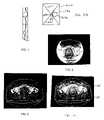
- the matrix of data obtained in a CT scancan be displayed as a sinogram as shown in FIG. 1 , or reconstructed into a two-dimensional image, as shown in FIG. 2 .
- the oncologistviews the cancerous areas on the CT image and determines the beam angles and intensities (identified with respect to the tumor image) which will be used to treat the tumor.
- a computer programselects the beam angles and intensities after the physician identifies the tumorous region and upper and lower dose limits for the treatment.
- the planning imagesare used to create a 3-D treatment plan of a region of interest.
- This region of interestis broken down into units called voxels, which are defined as volumetric pixels.
- Each voxelis then assigned a particular radiation dose depending on what type of tissue or other matter it contains, e.g. cancerous tissue, air, etc.
- the CT image of the patientis acquired substantially before the radiation treatment to allow time for the treatment plan to be prepared.
- the position of organs or other tissue to be treatedcan change from day-to-day because of a variety of factors.
- patientsmove during treatment because of breathing, muscle twitching or the like. Uncertainty in the positioning of the patient with respect to the original CT image can undermine the conformality of the radiation delivery.
- the verification processcan be done by techniques that compare the planning image to an image of the patient at the time of treatment.
- the data sets obtained on the day of treatment to be used for preparing the patient modelare often incomplete. Patients that are large in size may not fit within the field-of-view (FOV) of the CT machine attached to the therapeutic equipment applying the radiation dose, and may yield an image such as that shown in FIG. 3 , which shows only a portion of the image shown in FIG. 1 . Not only is there a limited field of view, the data around the edges contains significant artifacts so that the image has an irregular white border and internal values are distorted. Alternatively, only a limited sample size of slices may have been obtained. There may be other limitations that result in the collection of incomplete data sets.
- FOVfield-of-view
- the present inventionrelates to a method by which an incomplete CT patient data set can be combined with an existing CT patient data set to create an image of a patient that is complete and without significant artifacts.
- the methodincludes the steps of obtaining a first sinogram data set from a patient and a second sinogram data set or image from a patient. Both data sets are converted to images, and aligned together so that statistically, there is optimal registration between the two images.
- the aligned or “fused” imageis reprojected as a sinogram. This reprojected sinogram is compared to either the first or second sinogram to determine what data exists beyond the scope of the first or second sinogram. This additional data is added to the sinogram to which the fused sinogram was compared to obtain an augmented sinogram
- the augmented sinogramis converted to an image, referred to as a fusion-aligned reprojection image.
- the method of the present inventionis advantageous in that the availability of only one limited data sinogram/image will not affect the ability to perform accurate delivery verification, dose reconstruction, patient set-up or the like.
- the limited data image or “first image”is fused to a previously taken complete image or “second image.”
- the sinogram representing the fused imageis compared to the limited data sinogram, and the augmented limited data sinogram is prepared therefrom.
- From the augmented limited data sinogramthe fusion-aligned reprojected (FAR) image is obtained.
- the FAR imageis used to accurately apply radiation to the treatment area, which may be positioned differently than as shown in the previously obtained complete image.
- FIG. 1an example of a sinogram obtained from the CT scan of a patient
- FIG. 2is an example of a planning CT image obtained from a CT-scan sinogram similar to that shown in FIG. 1 ;
- FIG. 3is an example CT image with a limited field of view
- FIG. 4is a flowchart showing the process steps of the present invention.
- FIG. 5is a schematic example of a patient CT scan
- FIG. 6is a limited schematic view of FIG. 6 , showing the limited scan portion in the center of the object, and the remaining nonscanned portion in phantom;
- FIG. 7demonstrates how the limited image of FIG. 6 is aligned with the full image of FIG. 5 through the process of fusion
- FIG. 7Ashow the actual alignment or “fusion” of the images from FIGS. 5 and 6 ;
- FIG. 8is a schematic view of a fusion aligned reprojection image
- FIG. 9is a schematic view of a full image corresponding to that in FIG. 6 ;
- FIG. 10is a reconstructed image of FIGS. 2 and 3 fused and aligned in accordance with the method of the present invention.
- a preferred method in accordance with the present inventionis shown in the flowchart of FIG. 4 .
- a limited data sinogram 50 representing the treatment areais obtained from a patient.
- the limited data sinogram 50is prepared near the time that the patient is receiving his or her radiation treatment.
- the limited data sinogram 50may be obtained at any time.
- the limited data sinogram 50is reconstructed to a limited data image 52 , as seen in the example of FIG. 3 , and represented schematically in FIG. 6 as limited object 156 .
- FIG. 3contains a significant amount of artifacts such as the white irregular border 53 , and some distortion of image values.
- the treatment area targeted in FIG. 3is a prostate gland.
- the methodcan be applied to images of any part of the body, or be used in veterinary or radiological applications.
- a complete image 54 of the same patient and same treatment areais seen in FIG. 2 , and represented schematically in FIG. 5 as object 154 .
- this complete image 54will have been made prior to obtaining the limited data image 52 for the purpose of treatment planning.
- Even if limited image 52 were taken only minutes after the complete data image 54there are almost always inherent differences between the location of certain organs or tissue due to patient motion or other bodily functions. If enough time has elapsed between images, weight loss or growth of certain tissue can occur.
- complete image 54 or limited image 52need not be from CT scans, and that this technique can be generally applied to matching images from different projection imaging modalities such as magnetic resonance imaging, positron emission tomography, and single photon emission tomography. Thus, there may be misalignment or disagreement between the two images because of differing methods of data collection.
- FIGS. 2 and 3The two images shown in FIGS. 2 and 3 and represented schematically by objects 154 and 156 , in FIGS. 5 and 6 have differences between them.
- intestinal gasis shown in FIG. 3 , thereby displacing the treatment target.
- object 154is composed of diagonals 158 a and 160 a and an inclusion 161 a , within a frame 162 a .
- Limited object 156shows only corresponding diagonals 160 b and 158 b , and part of the inclusion designated as 161 b .
- FIG. 7“fusion” or image registration techniques are used to align limited data image 52 with complete image 54 .
- limited object 156is fused with complete object 154 so that statistically, there is optimal registration between the objects 154 and 156 .
- FIG. 7shows how the orientation of object 154 is aligned to closely match that of object 156 .
- FIG. 7Ashows diagonal 160 c as the perfect registration between diagonals 160 a and 160 b .
- Image registration or fusionmay be achieved by several techniques.
- One such techniqueis known as mutual information (MI), for which a well-known algorithm has been developed.
- MImutual information
- One such example of this algorithm being used to register multi-modal imagesis described in the following publication, incorporated herein by reference: Frederik Maes, Andre Collignon, Dirk Vendermeulen, Guy Marchal, and Paul Suetens, Multimodality Image Registration by Maximization of Mutual Information , Vol. 16, No. 2, IEEE Transactions on Medical Imaging, 187 (April 1997).
- EFFExtracted Feature Fusion
- a patient's bone structureusually stays the same even when a patient loses a substantial amount of weight. Therefore, the bones can in effect be extracted from each image subject to alignment, and then registered using statistical methods.
- diagonal 160 a and frame 162may represent bone or tissue that remains relatively unchanged over time. Therefore, only these relatively static features might be selected for fusion, while other features that are more dynamic, perhaps diagonals 158 a,b and inclusion 161 a,b, need not be included in the registration calculations.
- the benefits of registering only an extracted portion of an imageare reduced calculation times, improved accuracy, and more clearly defined goals for alignment in cases where the patient has significantly changed in shape.
- the benefitsarise from the registration of fewer data points, which in this case are voxels.
- the total processing timeis generally proportional to the number of points selected, so reducing that number from the size of the entire three-dimensional image set to a subset of points meeting certain criteria (e.g. voxels that represent bone or do not represent air) will typically reduce calculation times. This reduction of voxels can provide more accurate results than other methods of reducing the number of voxels for MI techniques, such as regular down-sampling.
- image registration techniquesinclude manual fusion, alignment using geometric features (e.g. surfaces), gradient methods, and voxel-similarity techniques.
- the aligned or transformed complete image 56is reprojected as a sinogram 58 .
- the data for sinogram 58is once again in a matrix wherein each row represents an angle, and each column represents distance.
- the data matrix of the reprojected sinogramis compared to the data matrix for limited data sinogram 50 to determine what data is missing from the limited sinogram. This is now possible because the complete sinogram is in alignment with the limited sinogram.
- the approximation of the missing sinogram data from the reprojected, fusion aligned version of image 154is added to the limited sinogram 50 to create an augmented limited data sinogram, or augmented sinogram 60 .
- the augmented sinogram 60is reconstructed to a fusion aligned reprojection image (FAR image) 62 that is an approximation of what the complete image would have looked like at the time the limited data image was obtained.
- the FAR image 62is represented schematically in FIG. 8 .
- Frame 162is the same as in FIG. 5 , and diagonals 158 c , 160 c and inclusion 161 c are now complete. This can compared to the object 168 in FIG.
- FIG. 10represents a reconstructed image obtained by combining FIGS. 2 and 3 in accordance with the method of the present invention. It can be seen that slight artifacts such as the faint ring 180 can result. However, such artifacts are insignificant because they do not impair the conspicuity of the important structures in the field of view, nor do they noticeably detriment dose calculations or other processes that utilize these images.
- the reconstructed image obtained from method of the present inventioncan then be used for patient setup (positioning the patient prior to delivery), dose registration (changing delivery patterns to compensate for patient position or tumor shape changes), delivery verification (using a signal measured at an exit detector to compute energy fluence directed toward a patient), deformable patient registration and deformable dose registration (using anatomical, biomechanical and region of interest data to map changes in the patient's anatomy between each fraction, a reconstructed dose is mapped to a reference image to obtain a cumulative dose).
Landscapes
- Health & Medical Sciences (AREA)
- Life Sciences & Earth Sciences (AREA)
- Engineering & Computer Science (AREA)
- Medical Informatics (AREA)
- Physics & Mathematics (AREA)
- Heart & Thoracic Surgery (AREA)
- General Health & Medical Sciences (AREA)
- Nuclear Medicine, Radiotherapy & Molecular Imaging (AREA)
- Optics & Photonics (AREA)
- Pathology (AREA)
- Radiology & Medical Imaging (AREA)
- Biomedical Technology (AREA)
- Biophysics (AREA)
- Molecular Biology (AREA)
- Surgery (AREA)
- Animal Behavior & Ethology (AREA)
- High Energy & Nuclear Physics (AREA)
- Public Health (AREA)
- Veterinary Medicine (AREA)
- General Physics & Mathematics (AREA)
- Theoretical Computer Science (AREA)
- Computer Vision & Pattern Recognition (AREA)
- Apparatus For Radiation Diagnosis (AREA)
- Magnetic Resonance Imaging Apparatus (AREA)
- Image Processing (AREA)
- Selective Calling Equipment (AREA)
Abstract
Description
Claims (189)
Priority Applications (9)
| Application Number | Priority Date | Filing Date | Title |
|---|---|---|---|
| US09/802,468US7046831B2 (en) | 2001-03-09 | 2001-03-09 | System and method for fusion-aligned reprojection of incomplete data |
| JP2002572101AJP4271941B2 (en) | 2001-03-09 | 2002-03-11 | Method for enhancing a tomographic projection image of a patient |
| DE60223346TDE60223346T2 (en) | 2001-03-09 | 2002-03-11 | SYSTEM AND METHOD FOR FUSION-ORIENTED RE-PROJECTION OF INCOMPLETE DATA |
| AT02723390TATE377813T1 (en) | 2001-03-09 | 2002-03-11 | SYSTEM AND METHOD FOR FUSION-ORIENTED REPROJECTION OF INCOMPLETE DATA |
| EP02723390AEP1374151B1 (en) | 2001-03-09 | 2002-03-11 | System and method for fusion-aligned reprojection of incomplete data |
| PCT/US2002/007346WO2002073519A1 (en) | 2001-03-09 | 2002-03-11 | System and method for fusion-aligned reprojection of incomplete data |
| CA002440244ACA2440244A1 (en) | 2001-03-09 | 2002-03-11 | System and method for fusion-aligned reprojection of incomplete data |
| ES02723390TES2296918T3 (en) | 2001-03-09 | 2002-03-11 | SYSTEM AND METHOD FOR ALIENATED REPROJECTION BY FUSION OF INCOMPLETE DATA. |
| US10/170,252US6915005B1 (en) | 2001-03-09 | 2002-06-11 | Method for reconstruction of limited data images using fusion-aligned reprojection and normal-error-aligned reprojection |
Applications Claiming Priority (1)
| Application Number | Priority Date | Filing Date | Title |
|---|---|---|---|
| US09/802,468US7046831B2 (en) | 2001-03-09 | 2001-03-09 | System and method for fusion-aligned reprojection of incomplete data |
Related Child Applications (1)
| Application Number | Title | Priority Date | Filing Date |
|---|---|---|---|
| US10/170,252Continuation-In-PartUS6915005B1 (en) | 2001-03-09 | 2002-06-11 | Method for reconstruction of limited data images using fusion-aligned reprojection and normal-error-aligned reprojection |
Publications (2)
| Publication Number | Publication Date |
|---|---|
| US20020136439A1 US20020136439A1 (en) | 2002-09-26 |
| US7046831B2true US7046831B2 (en) | 2006-05-16 |
Family
ID=25183781
Family Applications (2)
| Application Number | Title | Priority Date | Filing Date |
|---|---|---|---|
| US09/802,468Expired - LifetimeUS7046831B2 (en) | 2001-03-09 | 2001-03-09 | System and method for fusion-aligned reprojection of incomplete data |
| US10/170,252Expired - LifetimeUS6915005B1 (en) | 2001-03-09 | 2002-06-11 | Method for reconstruction of limited data images using fusion-aligned reprojection and normal-error-aligned reprojection |
Family Applications After (1)
| Application Number | Title | Priority Date | Filing Date |
|---|---|---|---|
| US10/170,252Expired - LifetimeUS6915005B1 (en) | 2001-03-09 | 2002-06-11 | Method for reconstruction of limited data images using fusion-aligned reprojection and normal-error-aligned reprojection |
Country Status (8)
| Country | Link |
|---|---|
| US (2) | US7046831B2 (en) |
| EP (1) | EP1374151B1 (en) |
| JP (1) | JP4271941B2 (en) |
| AT (1) | ATE377813T1 (en) |
| CA (1) | CA2440244A1 (en) |
| DE (1) | DE60223346T2 (en) |
| ES (1) | ES2296918T3 (en) |
| WO (1) | WO2002073519A1 (en) |
Cited By (35)
| Publication number | Priority date | Publication date | Assignee | Title |
|---|---|---|---|---|
| US20050129295A1 (en)* | 2003-12-16 | 2005-06-16 | Ge Medical Systems Global Technology Co., Llc | System and method for image processing |
| US20050197564A1 (en)* | 2004-02-20 | 2005-09-08 | University Of Florida Research Foundation, Inc. | System for delivering conformal radiation therapy while simultaneously imaging soft tissue |
| US20080031528A1 (en)* | 2006-04-03 | 2008-02-07 | Astrium Sas | Method of restoring movements of the line of sight of an optical instrument |
| US20080217561A1 (en)* | 2007-02-27 | 2008-09-11 | Mackie Thomas R | Phantom for ion range detection |
| US20090080594A1 (en)* | 2006-08-03 | 2009-03-26 | Kenneth Brooks | Dedicated breast radiation imaging/therapy system |
| US20090189095A1 (en)* | 2007-02-27 | 2009-07-30 | Ryan Thomas Flynn | Ion radiation therapy system with variable beam resolution |
| US20090212231A1 (en)* | 2007-02-27 | 2009-08-27 | Hill Patrick M | Heavy ion radiation therapy system with stair-step modulation |
| US20090289192A1 (en)* | 2007-02-27 | 2009-11-26 | Westerly David C | Scanning aperture ion beam modulator |
| US20100006778A1 (en)* | 2007-02-27 | 2010-01-14 | Flynn Ryan T | Ion radiation therapy system with distal gradient tracking |
| US20100019167A1 (en)* | 2007-02-27 | 2010-01-28 | Al-Sadah Jihad H | Fan beam modulator for ion beams providing continuous intensity modulation |
| US20100040273A1 (en)* | 2007-02-22 | 2010-02-18 | Hutchins Gary D | Imaging resolution recovery techniques |
| US20100176309A1 (en)* | 2007-02-27 | 2010-07-15 | Mackie Thomas R | Ion radiation therapy system with rocking gantry motion |
| US20100189220A1 (en)* | 2007-02-27 | 2010-07-29 | Flynn Ryan T | System and method for optimization of a radiation therapy plan in the presence of motion |
| US8076657B2 (en) | 2007-02-27 | 2011-12-13 | Wisconsin Alumni Research Foundation | Ion radiation therapy system having magnetic fan beam former |
| US8129701B2 (en) | 2007-02-27 | 2012-03-06 | Al-Sadah Jihad H | Areal modulator for intensity modulated radiation therapy |
| US8222616B2 (en) | 2007-10-25 | 2012-07-17 | Tomotherapy Incorporated | Method for adapting fractionation of a radiation therapy dose |
| US8232535B2 (en) | 2005-05-10 | 2012-07-31 | Tomotherapy Incorporated | System and method of treating a patient with radiation therapy |
| US20120294497A1 (en)* | 2011-05-20 | 2012-11-22 | Varian Medical Systems, Inc. | Method and Apparatus Pertaining to Images Used for Radiation-Treatment Planning |
| US8442287B2 (en) | 2005-07-22 | 2013-05-14 | Tomotherapy Incorporated | Method and system for evaluating quality assurance criteria in delivery of a treatment plan |
| US8565377B2 (en) | 2011-03-07 | 2013-10-22 | Dalhousie University | Methods and apparatus for imaging in conjunction with radiotherapy |
| US9443633B2 (en) | 2013-02-26 | 2016-09-13 | Accuray Incorporated | Electromagnetically actuated multi-leaf collimator |
| US9966160B2 (en) | 2015-11-24 | 2018-05-08 | Viewray Technologies, Inc. | Radiation beam collimating systems and methods |
| US10102682B1 (en) | 2017-04-17 | 2018-10-16 | Raytheon Company | System and method for combining 3D images in color |
| US10413751B2 (en) | 2016-03-02 | 2019-09-17 | Viewray Technologies, Inc. | Particle therapy with magnetic resonance imaging |
| US10463884B2 (en) | 2013-03-15 | 2019-11-05 | Viewray Technologies, Inc. | Systems and methods for linear accelerator radiotherapy with magnetic resonance imaging |
| US10561861B2 (en) | 2012-05-02 | 2020-02-18 | Viewray Technologies, Inc. | Videographic display of real-time medical treatment |
| US10821303B2 (en) | 2012-10-26 | 2020-11-03 | Viewray Technologies, Inc. | Assessment and improvement of treatment using imaging of physiological responses to radiation therapy |
| US11000706B2 (en) | 2016-12-13 | 2021-05-11 | Viewray Technologies, Inc. | Radiation therapy systems and methods |
| US11033758B2 (en) | 2017-12-06 | 2021-06-15 | Viewray Technologies, Inc. | Radiotherapy systems, methods and software |
| US11209509B2 (en) | 2018-05-16 | 2021-12-28 | Viewray Technologies, Inc. | Resistive electromagnet systems and methods |
| US11282209B2 (en) | 2020-01-10 | 2022-03-22 | Raytheon Company | System and method for generating contours |
| US11378629B2 (en) | 2016-06-22 | 2022-07-05 | Viewray Technologies, Inc. | Magnetic resonance imaging |
| US11475558B2 (en) | 2019-11-13 | 2022-10-18 | Raytheon Company | Organ isolation in scan data |
| US11562512B2 (en) | 2020-12-09 | 2023-01-24 | Raytheon Company | System and method for generating and displaying contours |
| US11893745B2 (en) | 2020-12-09 | 2024-02-06 | Raytheon Company | System and method for generating and displaying contours |
Families Citing this family (59)
| Publication number | Priority date | Publication date | Assignee | Title |
|---|---|---|---|---|
| US7046831B2 (en)* | 2001-03-09 | 2006-05-16 | Tomotherapy Incorporated | System and method for fusion-aligned reprojection of incomplete data |
| EP1514228A4 (en)* | 2002-06-11 | 2006-09-27 | Tomotherapy Inc | Method for reconstruction of limited data images using fusion-aligned reprojection and normal-error-aligned reprojection |
| US20080091388A1 (en)* | 2003-03-14 | 2008-04-17 | Failla Gregory A | Method for calculation radiation doses from acquired image data |
| US7317192B2 (en)* | 2003-06-02 | 2008-01-08 | Fox Chase Cancer Center | High energy polyenergetic ion selection systems, ion beam therapy systems, and ion beam treatment centers |
| US7778691B2 (en)* | 2003-06-13 | 2010-08-17 | Wisconsin Alumni Research Foundation | Apparatus and method using synchronized breathing to treat tissue subject to respiratory motion |
| US7957507B2 (en) | 2005-02-28 | 2011-06-07 | Cadman Patrick F | Method and apparatus for modulating a radiation beam |
| DE102005016256B3 (en)* | 2005-04-08 | 2006-06-08 | Siemens Ag | Displaying preoperative three-dimensional images with two-dimensional x-ray image acquisition involves repeatedly generating two-dimensional representations with varying parameters and displaying them on a screen |
| WO2007014104A2 (en) | 2005-07-22 | 2007-02-01 | Tomotherapy Incorporated | System and method of evaluating dose delivered by a radiation therapy system |
| AU2006272746A1 (en) | 2005-07-22 | 2007-02-01 | Tomotherapy Incorporated | Method and system for evaluating delivered dose |
| CN101512547A (en) | 2005-07-22 | 2009-08-19 | 断层放疗公司 | Method of and system for predicting dose delivery |
| CA2616296A1 (en) | 2005-07-22 | 2007-02-01 | Tomotherapy Incorporated | System and method of generating contour structures using a dose volume histogram |
| WO2007014105A2 (en)* | 2005-07-22 | 2007-02-01 | Tomotherapy Incorporated | Method and system for adapting a radiation therapy treatment plan based on a biological model |
| CN101267857A (en) | 2005-07-22 | 2008-09-17 | 断层放疗公司 | System and method of delivering radiation therapy to a moving region of interest |
| KR20080049716A (en)* | 2005-07-22 | 2008-06-04 | 토모테라피 인코포레이티드 | Methods and systems for evaluating quality assurance criteria associated with delivery of treatment plans |
| KR20080044251A (en) | 2005-07-22 | 2008-05-20 | 토모테라피 인코포레이티드 | How to place a constraint on a deformation map and system implementing the method |
| JP5060476B2 (en) | 2005-07-22 | 2012-10-31 | トモセラピー・インコーポレーテッド | System and method for detecting respiratory phase of a patient undergoing radiation therapy |
| JP2009502250A (en) | 2005-07-22 | 2009-01-29 | トモセラピー・インコーポレーテッド | Method and system for processing data associated with radiation therapy treatment planning |
| US9731148B2 (en)* | 2005-07-23 | 2017-08-15 | Tomotherapy Incorporated | Radiation therapy imaging and delivery utilizing coordinated motion of gantry and couch |
| US7831097B2 (en)* | 2006-03-16 | 2010-11-09 | Siemens Medical Solutions Usa, Inc. | System and method for image reconstruction |
| WO2007120744A2 (en)* | 2006-04-14 | 2007-10-25 | William Beaumont Hospital | Scanning slot cone-beam computed tomography and scanning focus spot cone-beam computed tomography |
| US9339243B2 (en) | 2006-04-14 | 2016-05-17 | William Beaumont Hospital | Image guided radiotherapy with dual source and dual detector arrays tetrahedron beam computed tomography |
| US8983024B2 (en) | 2006-04-14 | 2015-03-17 | William Beaumont Hospital | Tetrahedron beam computed tomography with multiple detectors and/or source arrays |
| US8073104B2 (en)* | 2006-05-25 | 2011-12-06 | William Beaumont Hospital | Portal and real time imaging for treatment verification |
| EP2026698A4 (en)* | 2006-05-25 | 2016-10-05 | Beaumont Hospital William | REAL TIME, ONLINE AND ONLINE TRACKING OF A TREATMENT DOSE AND FEEDBACK METHOD FOR ADAPTIVE RADIOTHERAPY GUIDED BY VOLUMETRIC IMAGE |
| CN101678210B (en)* | 2007-04-18 | 2012-05-23 | 伊利克塔股份有限公司 | radiotherapy device |
| EP2153406B1 (en)* | 2007-05-31 | 2012-08-01 | Elekta AB (PUBL) | Motion artefact reduction in ct scanning |
| CN101820948A (en)* | 2007-10-25 | 2010-09-01 | 断层放疗公司 | System and method for motion adaptive optimization of radiotherapy delivery |
| US8467497B2 (en)* | 2007-10-25 | 2013-06-18 | Tomotherapy Incorporated | System and method for motion adaptive optimization for radiation therapy delivery |
| ATE512627T1 (en) | 2009-04-15 | 2011-07-15 | Brainlab Ag | METHOD FOR COMPLETING A MEDICAL IMAGE DATA SET |
| US9466135B2 (en)* | 2009-08-20 | 2016-10-11 | Koninklijke Philips N.V. | Reconstruction of a region-of-interest image |
| US8666137B2 (en) | 2009-09-07 | 2014-03-04 | Koninklijke Philips N.V. | Apparatus and method for processing projection data |
| DE102009047867B4 (en)* | 2009-09-30 | 2016-10-06 | Siemens Healthcare Gmbh | Method and device for correcting truncated projection data |
| JP2013516278A (en) | 2010-01-05 | 2013-05-13 | ウィリアム・ボーモント・ホスピタル | Intensity-modulated rotating radiation therapy using continuous treatment table rotation / movement and simultaneous cone beam imaging |
| US20110216180A1 (en)* | 2010-03-05 | 2011-09-08 | Alessandro Pasini | Method and system for obtaining improved computed tomographic reconstructions |
| CN104854963B (en) | 2012-09-26 | 2020-02-21 | 株式会社尼康 | X-ray apparatus, and method for manufacturing the structure |
| CN103083022B (en)* | 2013-02-01 | 2015-09-16 | 徐州医学院 | A kind of binary position autoregistration and fusion scanning board |
| CN103070684B (en)* | 2013-02-01 | 2015-06-03 | 徐州医学院 | MRI (Magnetic Resonance Imaging) double body position scanning automatic registration and fusion device |
| CN103055424B (en)* | 2013-02-01 | 2015-08-19 | 徐州医学院 | A kind of based on CT/MRI binary bit scan autoregistration and fusion Radiotherapy evaluating apparatus |
| US20140343344A1 (en)* | 2013-04-08 | 2014-11-20 | Cubresa Inc. | Radiation Therapy Guided Using Gamma Imaging |
| JP6482934B2 (en)* | 2014-06-03 | 2019-03-13 | キヤノンメディカルシステムズ株式会社 | Image processing apparatus, radiation detection apparatus, and image processing method |
| US20180360406A1 (en)* | 2015-12-17 | 2018-12-20 | The University Of Tokyo | Image Processing Device and Image Processing Method |
| EP3181049B1 (en)* | 2015-12-18 | 2018-02-14 | RaySearch Laboratories AB | Radiotherapy method, computer program and computer system |
| AU2017278241B2 (en) | 2016-06-06 | 2021-10-21 | iTomography Corp. | Systems and methods for automated sinogram completion, combination, and completion by combination |
| IL310828A (en) | 2017-03-31 | 2024-04-01 | Empyrean Medical Systems Inc | A three-dimensional beam that creates an x-ray radiation source |
| EP3655103A1 (en) | 2017-07-18 | 2020-05-27 | Sensus Healthcare, Inc. | Real-time x-ray dosimetry in intraoperative radiation therapy |
| US11672491B2 (en) | 2018-03-30 | 2023-06-13 | Empyrean Medical Systems, Inc. | Validation of therapeutic radiation treatment |
| JP7258474B2 (en)* | 2018-05-10 | 2023-04-17 | 株式会社日立製作所 | X-ray CT device and radiotherapy system |
| CN109523507B (en)* | 2018-09-26 | 2023-09-19 | 苏州六莲科技有限公司 | Method and device for generating lesion image and computer readable storage medium |
| US10940334B2 (en)* | 2018-10-19 | 2021-03-09 | Sensus Healthcare, Inc. | Systems and methods for real time beam sculpting intra-operative-radiation-therapy treatment planning |
| US11080895B2 (en)* | 2018-10-30 | 2021-08-03 | International Business Machines Corporation | Generating simulated body parts for images |
| EP3886707A1 (en)* | 2018-11-30 | 2021-10-06 | Accuray, Inc. | Helical cone-beam computed tomography imaging with an off-centered detector |
| CN109646035B (en)* | 2019-01-04 | 2022-04-22 | 北京永新医疗设备有限公司 | Bone tomographic image reconstruction method and system |
| FI129810B (en)* | 2020-06-29 | 2022-09-15 | Oulun Yliopisto | Apparatus, method and computer program for processing computed tomography (ct) scan data |
| US11794039B2 (en) | 2021-07-13 | 2023-10-24 | Accuray, Inc. | Multimodal radiation apparatus and methods |
| US11854123B2 (en) | 2021-07-23 | 2023-12-26 | Accuray, Inc. | Sparse background measurement and correction for improving imaging |
| CN113724177B (en)* | 2021-09-07 | 2023-12-15 | 北京大学深圳医院 | Pulmonary nodule information fusion method, device, equipment and storage medium |
| US12257083B2 (en) | 2022-02-07 | 2025-03-25 | Accuray Inc. | Methods for saturation correction and dynamic gain configuration and apparatuses for performing the same |
| US20240095977A1 (en)* | 2022-09-16 | 2024-03-21 | Canon Medical Systems Corporation | Reconstruction device, x-ray ct apparatus, and image processing device |
| WO2024214017A1 (en)* | 2023-04-11 | 2024-10-17 | Medtronic Navigation, Inc | Method and apparatus for reconstucting image acquisitions for extended fields-of-view |
Citations (17)
| Publication number | Priority date | Publication date | Assignee | Title |
|---|---|---|---|---|
| US5317616A (en) | 1992-03-19 | 1994-05-31 | Wisconsin Alumni Research Foundation | Method and apparatus for radiation therapy |
| US5351280A (en) | 1992-03-19 | 1994-09-27 | Wisconsin Alumni Research Foundation | Multi-leaf radiation attenuator for radiation therapy |
| US5552605A (en)* | 1994-11-18 | 1996-09-03 | Picker International, Inc. | Motion correction based on reprojection data |
| US5579358A (en)* | 1995-05-26 | 1996-11-26 | General Electric Company | Compensation for movement in computed tomography equipment |
| US5625190A (en)* | 1995-07-11 | 1997-04-29 | General Electric Company | Forward projection algorithm |
| US5625663A (en) | 1992-03-19 | 1997-04-29 | Wisconsin Alumni Research Foundation | Dynamic beam flattening apparatus for radiation therapy |
| US5647663A (en) | 1996-01-05 | 1997-07-15 | Wisconsin Alumni Research Foundation | Radiation treatment planning method and apparatus |
| US5661773A (en) | 1992-03-19 | 1997-08-26 | Wisconsin Alumni Research Foundation | Interface for radiation therapy machine |
| US5673300A (en) | 1996-06-11 | 1997-09-30 | Wisconsin Alumni Research Foundation | Method of registering a radiation treatment plan to a patient |
| US5724400A (en) | 1992-03-19 | 1998-03-03 | Wisconsin Alumni Research Foundation | Radiation therapy system with constrained rotational freedom |
| US5761331A (en)* | 1995-06-05 | 1998-06-02 | Intellectual Property Group Of Pillsbury Madison & Sutro Llp | Method and apparatus for tomographic imaging and image reconstruction using recombinant transverse phase differentials |
| US5907594A (en)* | 1997-07-01 | 1999-05-25 | Analogic Corporation | Reconstruction of volumetric images by successive approximation in cone-beam computed tomography systems |
| US5963664A (en)* | 1995-06-22 | 1999-10-05 | Sarnoff Corporation | Method and system for image combination using a parallax-based technique |
| US6266453B1 (en)* | 1999-07-26 | 2001-07-24 | Computerized Medical Systems, Inc. | Automated image fusion/alignment system and method |
| US6282257B1 (en)* | 1999-06-23 | 2001-08-28 | The Board Of Trustees Of The University Of Illinois | Fast hierarchical backprojection method for imaging |
| US6324243B1 (en)* | 2000-02-23 | 2001-11-27 | General Electric Company | Method and apparatus for reconstructing images from projection data acquired by a computed tomography system |
| US6915005B1 (en)* | 2001-03-09 | 2005-07-05 | Tomo Therapy Incorporated | Method for reconstruction of limited data images using fusion-aligned reprojection and normal-error-aligned reprojection |
Family Cites Families (9)
| Publication number | Priority date | Publication date | Assignee | Title |
|---|---|---|---|---|
| US5961454A (en)* | 1993-08-13 | 1999-10-05 | The Brigham And Women's Hospital | Fusion of anatomical data sets into stereotactic coordinates |
| US5708422A (en)* | 1995-05-31 | 1998-01-13 | At&T | Transaction authorization and alert system |
| US5800353A (en)* | 1996-02-12 | 1998-09-01 | Mclaurin, Jr.; Robert L. | Automatic image registration of magnetic resonance imaging scans for localization, 3-dimensional treatment planning, and radiation treatment of abnormal lesions |
| US6167296A (en)* | 1996-06-28 | 2000-12-26 | The Board Of Trustees Of The Leland Stanford Junior University | Method for volumetric image navigation |
| US6009212A (en)* | 1996-07-10 | 1999-12-28 | Washington University | Method and apparatus for image registration |
| JP3878259B2 (en)* | 1996-11-13 | 2007-02-07 | 東芝医用システムエンジニアリング株式会社 | Medical image processing device |
| US6163604A (en)* | 1998-04-03 | 2000-12-19 | Lucent Technologies | Automated fraud management in transaction-based networks |
| US6167517A (en)* | 1998-04-09 | 2000-12-26 | Oracle Corporation | Trusted biometric client authentication |
| AU4180200A (en)* | 1999-04-02 | 2000-10-23 | Wisconsin Alumni Research Foundation | Megavoltage computed tomography during radiotherapy |
- 2001
- 2001-03-09USUS09/802,468patent/US7046831B2/ennot_activeExpired - Lifetime
- 2002
- 2002-03-11EPEP02723390Apatent/EP1374151B1/ennot_activeExpired - Lifetime
- 2002-03-11ATAT02723390Tpatent/ATE377813T1/ennot_activeIP Right Cessation
- 2002-03-11CACA002440244Apatent/CA2440244A1/ennot_activeAbandoned
- 2002-03-11WOPCT/US2002/007346patent/WO2002073519A1/enactiveIP Right Grant
- 2002-03-11ESES02723390Tpatent/ES2296918T3/ennot_activeExpired - Lifetime
- 2002-03-11DEDE60223346Tpatent/DE60223346T2/ennot_activeExpired - Lifetime
- 2002-03-11JPJP2002572101Apatent/JP4271941B2/ennot_activeExpired - Lifetime
- 2002-06-11USUS10/170,252patent/US6915005B1/ennot_activeExpired - Lifetime
Patent Citations (21)
| Publication number | Priority date | Publication date | Assignee | Title |
|---|---|---|---|---|
| US5625663A (en) | 1992-03-19 | 1997-04-29 | Wisconsin Alumni Research Foundation | Dynamic beam flattening apparatus for radiation therapy |
| US5317616A (en) | 1992-03-19 | 1994-05-31 | Wisconsin Alumni Research Foundation | Method and apparatus for radiation therapy |
| US5394452A (en) | 1992-03-19 | 1995-02-28 | Wisconsin Alumni Research Foundation | Verification system for radiation therapy |
| US5442675A (en) | 1992-03-19 | 1995-08-15 | Wisconsin Alumni Research Foundation | Dynamic collimator for radiation therapy |
| US5528650A (en) | 1992-03-19 | 1996-06-18 | Swerdloff; Stuart | Method and apparatus for radiation therapy |
| US5548627A (en) | 1992-03-19 | 1996-08-20 | Wisconsin Alumni Research Foundation | Radiation therapy system with constrained rotational freedom |
| US5724400A (en) | 1992-03-19 | 1998-03-03 | Wisconsin Alumni Research Foundation | Radiation therapy system with constrained rotational freedom |
| US5661773A (en) | 1992-03-19 | 1997-08-26 | Wisconsin Alumni Research Foundation | Interface for radiation therapy machine |
| US5351280A (en) | 1992-03-19 | 1994-09-27 | Wisconsin Alumni Research Foundation | Multi-leaf radiation attenuator for radiation therapy |
| US5552605A (en)* | 1994-11-18 | 1996-09-03 | Picker International, Inc. | Motion correction based on reprojection data |
| US5579358A (en)* | 1995-05-26 | 1996-11-26 | General Electric Company | Compensation for movement in computed tomography equipment |
| US5761331A (en)* | 1995-06-05 | 1998-06-02 | Intellectual Property Group Of Pillsbury Madison & Sutro Llp | Method and apparatus for tomographic imaging and image reconstruction using recombinant transverse phase differentials |
| US5963664A (en)* | 1995-06-22 | 1999-10-05 | Sarnoff Corporation | Method and system for image combination using a parallax-based technique |
| US5625190A (en)* | 1995-07-11 | 1997-04-29 | General Electric Company | Forward projection algorithm |
| US5647663A (en) | 1996-01-05 | 1997-07-15 | Wisconsin Alumni Research Foundation | Radiation treatment planning method and apparatus |
| US5673300A (en) | 1996-06-11 | 1997-09-30 | Wisconsin Alumni Research Foundation | Method of registering a radiation treatment plan to a patient |
| US5907594A (en)* | 1997-07-01 | 1999-05-25 | Analogic Corporation | Reconstruction of volumetric images by successive approximation in cone-beam computed tomography systems |
| US6282257B1 (en)* | 1999-06-23 | 2001-08-28 | The Board Of Trustees Of The University Of Illinois | Fast hierarchical backprojection method for imaging |
| US6266453B1 (en)* | 1999-07-26 | 2001-07-24 | Computerized Medical Systems, Inc. | Automated image fusion/alignment system and method |
| US6324243B1 (en)* | 2000-02-23 | 2001-11-27 | General Electric Company | Method and apparatus for reconstructing images from projection data acquired by a computed tomography system |
| US6915005B1 (en)* | 2001-03-09 | 2005-07-05 | Tomo Therapy Incorporated | Method for reconstruction of limited data images using fusion-aligned reprojection and normal-error-aligned reprojection |
Non-Patent Citations (9)
| Title |
|---|
| 3D Treatment Planning and Intensity-Modulated Radiation Therapy; www.intouchlive.com/journals/oncology/o9910sup5s.htm; James A. Purdy, PhD. |
| Automatic registration of CT and MR brain images using correlation of geometrical features, Van den Elsen, P. et al.; Medical Imaging, IEEE Transactions on , vol.: 14, Jun. 1995.* |
| Intensity Modulated Radiation Therapy: An Introduction for Patients and Clinicians; www.ocolink.upenn.edu/specialty/rad<SUB>-</SUB>onc/treat/imrt/imrt1.html; Jason Lee MD, Indra J. Das PhD, Shioa Y. Woo MD, Walter Grant PhD, Bin S. The MD, J. Kam Chiu MD, and E. Brian Butler MD. |
| Kudo et al., Helical-scan Computed Tomography Using Cone-Beam Projections, IEEE Conference 1991, ISBN: 0-7803-0513, vol. 3, pp. 1958-1962.* |
| Multi-Modal Volume Registration by Maximization of Mutual Information; Medical Image Analysis, Oxford University Press; William M. Wells III; Paul Viola, Hideki Atsumi, Shin Nakajima, and Rod Kikinis. |
| Multimodality Image Registration By Maximization of Mutual Information; IEEE Transactions on Medical Imaging, vol. 16, No. 2, Apr. 1997; Frederik Maes, André Collignon, Dirk Vandermeulen, Guy Marchal, and Paul Suetens. |
| Registration of Tomotheraphy Patients Using CT Projection Files; www.madrad.radiology.wise.edu/tomo/registration/reg<SUB>-</SUB>iccr2/reg<SUB>-</SUB>iccr2.html; E.E.Fitchard, J.S. Aldridge, P.J. Reckwerdt, T.R. Mackie. |
| Three-Dimensional Conformal Radiation Therapy (3DCRT) for Prostate Cancer; www.comed.com/Prostate/michalski; Jeff M. Michalski, MD, Carlos A. Perez, MD, and James A. Purdy, PhD. |
| Website information; http://splweb.bwh.harvard.edu:8000/pages/papers/kikinis/hpc/hpcfinal.html; High Performance Computing (HPC) in Medical Image Analysis (MIA) at the Surgical Planning Laboratory (SPL); Ron Kikinis, M.D., Simon Warfield, Ph.D., Carl-Fredrik Westin, Ph.D. |
Cited By (72)
| Publication number | Priority date | Publication date | Assignee | Title |
|---|---|---|---|---|
| US7447345B2 (en)* | 2003-12-16 | 2008-11-04 | General Electric Company | System and method for generating PET-CT images |
| US20050129295A1 (en)* | 2003-12-16 | 2005-06-16 | Ge Medical Systems Global Technology Co., Llc | System and method for image processing |
| US20100113911A1 (en)* | 2004-02-20 | 2010-05-06 | University Of Florida Research Foundation, Inc. | System for Delivering Conformal Radiation Therapy While Simultaneously Imaging Soft Tissue |
| US20050197564A1 (en)* | 2004-02-20 | 2005-09-08 | University Of Florida Research Foundation, Inc. | System for delivering conformal radiation therapy while simultaneously imaging soft tissue |
| US11497937B2 (en) | 2004-02-20 | 2022-11-15 | University Of Florida Research Foundation, Inc. | System for delivering conformal radiation therapy while simultaneously imaging soft tissue |
| US7907987B2 (en) | 2004-02-20 | 2011-03-15 | University Of Florida Research Foundation, Inc. | System for delivering conformal radiation therapy while simultaneously imaging soft tissue |
| US10688319B2 (en) | 2004-02-20 | 2020-06-23 | University Of Florida Research Foundation, Inc. | System for delivering conformal radiation therapy while simultaneously imaging soft tissue |
| US8190233B2 (en) | 2004-02-20 | 2012-05-29 | University Of Florida Research Foundation, Inc. | System for delivering conformal radiation therapy while simultaneously imaging soft tissue |
| US8232535B2 (en) | 2005-05-10 | 2012-07-31 | Tomotherapy Incorporated | System and method of treating a patient with radiation therapy |
| US8442287B2 (en) | 2005-07-22 | 2013-05-14 | Tomotherapy Incorporated | Method and system for evaluating quality assurance criteria in delivery of a treatment plan |
| US20080031528A1 (en)* | 2006-04-03 | 2008-02-07 | Astrium Sas | Method of restoring movements of the line of sight of an optical instrument |
| US8068696B2 (en)* | 2006-04-03 | 2011-11-29 | Astrium Sas | Method of restoring movements of the line of sight of an optical instrument |
| US8964936B2 (en) | 2006-08-03 | 2015-02-24 | Hologic, Inc. | Dedicated breast radiation imaging/therapy system |
| US20090080602A1 (en)* | 2006-08-03 | 2009-03-26 | Kenneth Brooks | Dedicated breast radiation imaging/therapy system |
| US7957508B2 (en) | 2006-08-03 | 2011-06-07 | Hologic, Inc. | Dedicated breast radiation imaging/therapy system |
| US20090080594A1 (en)* | 2006-08-03 | 2009-03-26 | Kenneth Brooks | Dedicated breast radiation imaging/therapy system |
| US20100040273A1 (en)* | 2007-02-22 | 2010-02-18 | Hutchins Gary D | Imaging resolution recovery techniques |
| US8553960B2 (en) | 2007-02-22 | 2013-10-08 | Indiana University Research & Technology Corporation | Imaging resolution recovery techniques |
| US20100243921A1 (en)* | 2007-02-27 | 2010-09-30 | Ryan Thomas Flynn | Ion radiation therapy system with variable beam resolution |
| US8269196B2 (en) | 2007-02-27 | 2012-09-18 | Wisconsin Alumni Research Foundation | Heavy ion radiation therapy system with stair-step modulation |
| US20100189220A1 (en)* | 2007-02-27 | 2010-07-29 | Flynn Ryan T | System and method for optimization of a radiation therapy plan in the presence of motion |
| US7763873B2 (en) | 2007-02-27 | 2010-07-27 | Wisconsin Alumni Research Foundation | Ion radiation therapy system with variable beam resolution |
| US7977657B2 (en) | 2007-02-27 | 2011-07-12 | Wisconsin Alumni Research Foundation | Ion radiation therapy system with distal gradient tracking |
| US7977648B2 (en) | 2007-02-27 | 2011-07-12 | Wisconsin Alumni Research Foundation | Scanning aperture ion beam modulator |
| US20090212231A1 (en)* | 2007-02-27 | 2009-08-27 | Hill Patrick M | Heavy ion radiation therapy system with stair-step modulation |
| US20100176309A1 (en)* | 2007-02-27 | 2010-07-15 | Mackie Thomas R | Ion radiation therapy system with rocking gantry motion |
| US8076657B2 (en) | 2007-02-27 | 2011-12-13 | Wisconsin Alumni Research Foundation | Ion radiation therapy system having magnetic fan beam former |
| US8093568B2 (en) | 2007-02-27 | 2012-01-10 | Wisconsin Alumni Research Foundation | Ion radiation therapy system with rocking gantry motion |
| US8129701B2 (en) | 2007-02-27 | 2012-03-06 | Al-Sadah Jihad H | Areal modulator for intensity modulated radiation therapy |
| US8154001B2 (en) | 2007-02-27 | 2012-04-10 | Wisconsin Alumni Research Foundation | Ion radiation therapy system with variable beam resolution |
| US7714309B2 (en) | 2007-02-27 | 2010-05-11 | Wisconsin Alumni Research Foundation | Phantom for ion range detection |
| US20080217561A1 (en)* | 2007-02-27 | 2008-09-11 | Mackie Thomas R | Phantom for ion range detection |
| US20100019167A1 (en)* | 2007-02-27 | 2010-01-28 | Al-Sadah Jihad H | Fan beam modulator for ion beams providing continuous intensity modulation |
| US7856082B2 (en) | 2007-02-27 | 2010-12-21 | Wisconsin Alumni Research Foundation | System and method for optimization of a radiation therapy plan in the presence of motion |
| US9006677B2 (en) | 2007-02-27 | 2015-04-14 | Wisconsin Alumni Research Foundation | Fan beam modulator for ion beams providing continuous intensity modulation |
| US20090189095A1 (en)* | 2007-02-27 | 2009-07-30 | Ryan Thomas Flynn | Ion radiation therapy system with variable beam resolution |
| US20100006778A1 (en)* | 2007-02-27 | 2010-01-14 | Flynn Ryan T | Ion radiation therapy system with distal gradient tracking |
| US20090289192A1 (en)* | 2007-02-27 | 2009-11-26 | Westerly David C | Scanning aperture ion beam modulator |
| US8254521B2 (en) | 2007-08-03 | 2012-08-28 | Hologic, Inc. | Dedicated breast radiation imaging/therapy system |
| US20110237927A1 (en)* | 2007-08-03 | 2011-09-29 | Hologic, Inc. | Dedicated breast radiation imaging/therapy system |
| US8222616B2 (en) | 2007-10-25 | 2012-07-17 | Tomotherapy Incorporated | Method for adapting fractionation of a radiation therapy dose |
| US8712011B2 (en) | 2011-03-07 | 2014-04-29 | Dalhousie University | Methods and apparatus for imaging in conjunction with radiotherapy |
| US8565377B2 (en) | 2011-03-07 | 2013-10-22 | Dalhousie University | Methods and apparatus for imaging in conjunction with radiotherapy |
| US20120294497A1 (en)* | 2011-05-20 | 2012-11-22 | Varian Medical Systems, Inc. | Method and Apparatus Pertaining to Images Used for Radiation-Treatment Planning |
| US9014454B2 (en)* | 2011-05-20 | 2015-04-21 | Varian Medical Systems, Inc. | Method and apparatus pertaining to images used for radiation-treatment planning |
| US10561861B2 (en) | 2012-05-02 | 2020-02-18 | Viewray Technologies, Inc. | Videographic display of real-time medical treatment |
| US11040222B2 (en) | 2012-10-26 | 2021-06-22 | Viewray Technologies, Inc. | Assessment and improvement of treatment using imaging of physiological responses to radiation therapy |
| US10835763B2 (en) | 2012-10-26 | 2020-11-17 | Viewray Technologies, Inc. | Assessment and improvement of treatment using imaging of physiological responses to radiation therapy |
| US10821303B2 (en) | 2012-10-26 | 2020-11-03 | Viewray Technologies, Inc. | Assessment and improvement of treatment using imaging of physiological responses to radiation therapy |
| US9443633B2 (en) | 2013-02-26 | 2016-09-13 | Accuray Incorporated | Electromagnetically actuated multi-leaf collimator |
| US11083912B2 (en) | 2013-03-15 | 2021-08-10 | Viewray Technologies, Inc. | Systems and methods for linear accelerator radiotherapy with magnetic resonance imaging |
| US10463884B2 (en) | 2013-03-15 | 2019-11-05 | Viewray Technologies, Inc. | Systems and methods for linear accelerator radiotherapy with magnetic resonance imaging |
| US11612764B2 (en) | 2013-03-15 | 2023-03-28 | Viewray Technologies, Inc. | Systems and methods for linear accelerator radiotherapy with magnetic resonance imaging |
| US9966160B2 (en) | 2015-11-24 | 2018-05-08 | Viewray Technologies, Inc. | Radiation beam collimating systems and methods |
| US10413751B2 (en) | 2016-03-02 | 2019-09-17 | Viewray Technologies, Inc. | Particle therapy with magnetic resonance imaging |
| US12017090B2 (en) | 2016-03-02 | 2024-06-25 | Viewray Systems, Inc. | Particle therapy with magnetic resonance imaging |
| US11351398B2 (en) | 2016-03-02 | 2022-06-07 | Viewray Technologies, Inc. | Particle therapy with magnetic resonance imaging |
| US11892523B2 (en) | 2016-06-22 | 2024-02-06 | Viewray Technologies, Inc. | Magnetic resonance imaging |
| US11768257B2 (en) | 2016-06-22 | 2023-09-26 | Viewray Technologies, Inc. | Magnetic resonance imaging |
| US11378629B2 (en) | 2016-06-22 | 2022-07-05 | Viewray Technologies, Inc. | Magnetic resonance imaging |
| US11931602B2 (en) | 2016-12-13 | 2024-03-19 | Viewray Technologies, Inc. | Radiation therapy systems and methods |
| US11000706B2 (en) | 2016-12-13 | 2021-05-11 | Viewray Technologies, Inc. | Radiation therapy systems and methods |
| US10102682B1 (en) | 2017-04-17 | 2018-10-16 | Raytheon Company | System and method for combining 3D images in color |
| US10650612B2 (en) | 2017-04-17 | 2020-05-12 | Raytheon Company | System and method for combining 3D images in color |
| US10366545B2 (en) | 2017-04-17 | 2019-07-30 | Raytheon Company | System and method for combining 3D images in color |
| US11033758B2 (en) | 2017-12-06 | 2021-06-15 | Viewray Technologies, Inc. | Radiotherapy systems, methods and software |
| US11209509B2 (en) | 2018-05-16 | 2021-12-28 | Viewray Technologies, Inc. | Resistive electromagnet systems and methods |
| US12000914B2 (en) | 2018-05-16 | 2024-06-04 | Viewray Systems, Inc. | Resistive electromagnet systems and methods |
| US11475558B2 (en) | 2019-11-13 | 2022-10-18 | Raytheon Company | Organ isolation in scan data |
| US11282209B2 (en) | 2020-01-10 | 2022-03-22 | Raytheon Company | System and method for generating contours |
| US11562512B2 (en) | 2020-12-09 | 2023-01-24 | Raytheon Company | System and method for generating and displaying contours |
| US11893745B2 (en) | 2020-12-09 | 2024-02-06 | Raytheon Company | System and method for generating and displaying contours |
Also Published As
| Publication number | Publication date |
|---|---|
| JP2004530467A (en) | 2004-10-07 |
| DE60223346T2 (en) | 2008-08-28 |
| US20020136439A1 (en) | 2002-09-26 |
| WO2002073519A1 (en) | 2002-09-19 |
| DE60223346D1 (en) | 2007-12-20 |
| EP1374151B1 (en) | 2007-11-07 |
| EP1374151A1 (en) | 2004-01-02 |
| ES2296918T3 (en) | 2008-05-01 |
| ATE377813T1 (en) | 2007-11-15 |
| EP1374151A4 (en) | 2006-09-13 |
| US6915005B1 (en) | 2005-07-05 |
| JP4271941B2 (en) | 2009-06-03 |
| CA2440244A1 (en) | 2002-09-19 |
Similar Documents
| Publication | Publication Date | Title |
|---|---|---|
| US7046831B2 (en) | System and method for fusion-aligned reprojection of incomplete data | |
| US9014446B2 (en) | Efficient user interaction with polygonal meshes for medical image segmentation | |
| US8306185B2 (en) | Radiotherapeutic treatment plan adaptation | |
| Webb | The physics of three dimensional radiation therapy: Conformal radiotherapy, radiosurgery and treatment planning | |
| CN101061520B (en) | Systems and methods for diagnostic imaging | |
| Mori et al. | Development of fast patient position verification software using 2D-3D image registration and its clinical experience | |
| Chen et al. | Treatment planning | |
| Zhang et al. | Comparing digital tomosynthesis to cone-beam CT for position verification in patients undergoing partial breast irradiation | |
| CA2489157A1 (en) | Method for reconstruction of limited data images using fusion-aligned reprojection and normal-error-aligned reprojection | |
| Driver et al. | Improvements in radiotherapy practice: the impact of new imaging technologies | |
| WO2004030761A1 (en) | Improvements in or relating to radiation treatment planning | |
| Lin et al. | Development of a novel post-processing treatment planning platform for 4D radiotherapy | |
| Baydush et al. | Initial application of digital tomosynthesis with on-board imaging in radiation oncology | |
| Thomas et al. | Validation of image registration and fusion of MV CBCT and planning CT for radiotherapy treatment planning | |
| Santos et al. | Multimodality image integration for radiotherapy treatment: an easy approach | |
| Schandorf | Patient Setup Quality Assurance In External Beam Radiotherapy Using A Third-Party Treatment Setup Verification Software | |
| Bellan et al. | A new methodological approach for PET implementation in radiotherapy treatment planning | |
| Yue et al. | A technique to re-establish dose distributions for previously treated brain cancer patients in external beam radiotherapy | |
| Buhle et al. | Imaging in three dimensional radiotherapy | |
| Chan et al. | Image-Guided Adaptive Radiation Therapy with anatomic imaging and dose verification in head and neck cancer using kV CBCT | |
| Guerreiro | Calibration of MR-Images for accurate dose calculations | |
| Antunes et al. | Study of CT/MRI mutual information based registration applied in brachytherapy | |
| Letourneau | Development of an On-line Planning and Delivery Technique for Radiotherapy of Spinal Metastases | |
| Bichan | Is it only a matter of time until the Conventional Treatment Simulator becomes obsolete? Critical evaluation of the CT-simulator | |
| Gonzalez et al. | Clinical implementation of megavoltage cone beam CT as part of an IGRT program |
Legal Events
| Date | Code | Title | Description |
|---|---|---|---|
| AS | Assignment | Owner name:TOMOTHERAPY, INC., WISCONSIN Free format text:ASSIGNMENT OF ASSIGNORS INTEREST;ASSIGNORS:RUCHALA, KENNETH J.;OLIVERA, GUSTAVO H.;MACKIE, THOMAS R.;AND OTHERS;REEL/FRAME:012247/0076 Effective date:20010713 | |
| STCF | Information on status: patent grant | Free format text:PATENTED CASE | |
| FPAY | Fee payment | Year of fee payment:4 | |
| FPAY | Fee payment | Year of fee payment:8 | |
| AS | Assignment | Owner name:CERBERUS BUSINESS FINANCE, LLC, AS COLLATERAL AGENT, NEW YORK Free format text:ASSIGNMENT FOR SECURITY - PATENTS;ASSIGNORS:ACCURAY INCORPORATED;TOMOTHERAPY INCORPORATED;REEL/FRAME:037513/0170 Effective date:20160111 Owner name:CERBERUS BUSINESS FINANCE, LLC, AS COLLATERAL AGEN Free format text:ASSIGNMENT FOR SECURITY - PATENTS;ASSIGNORS:ACCURAY INCORPORATED;TOMOTHERAPY INCORPORATED;REEL/FRAME:037513/0170 Effective date:20160111 | |
| AS | Assignment | Owner name:MIDCAP FUNDING IV TRUST (AS SUCCESSOR BY ASSIGNMENT FROM MIDCAP FINANCIAL TRUST), MARYLAND Free format text:SECURITY INTEREST;ASSIGNORS:ACCURAY INCORPORATED;TOMOTHERAPY INCORPORATED;REEL/FRAME:042826/0358 Effective date:20170614 Owner name:ACCURAY INCORPORATED, CALIFORNIA Free format text:RELEASE BY SECURED PARTY;ASSIGNOR:CERBERUS BUSINESS FINANCE, LLC. AS COLLATERAL AGENT;REEL/FRAME:042821/0580 Effective date:20170614 Owner name:TOMOTHERAPY INCORPORATED, CALIFORNIA Free format text:RELEASE BY SECURED PARTY;ASSIGNOR:CERBERUS BUSINESS FINANCE, LLC. AS COLLATERAL AGENT;REEL/FRAME:042821/0580 Effective date:20170614 Owner name:MIDCAP FUNDING IV TRUST (AS SUCCESSOR BY ASSIGNMEN Free format text:SECURITY INTEREST;ASSIGNORS:ACCURAY INCORPORATED;TOMOTHERAPY INCORPORATED;REEL/FRAME:042826/0358 Effective date:20170614 | |
| MAFP | Maintenance fee payment | Free format text:PAYMENT OF MAINTENANCE FEE, 12TH YEAR, LARGE ENTITY (ORIGINAL EVENT CODE: M1553) Year of fee payment:12 | |
| AS | Assignment | Owner name:MIDCAP FINANCIAL TRUST, MARYLAND Free format text:SECURITY INTEREST;ASSIGNORS:ACCURAY INCORPORATED;TOMOTHERAPY INCORPORATED;REEL/FRAME:044910/0685 Effective date:20171215 | |
| AS | Assignment | Owner name:MIDCAP FUNDING IV TRUST, AS SUCCESSOR TO EXISTING Free format text:ASSIGNMENT OF SECURITY AGREEMENTS;ASSIGNOR:MIDCAP FUNDING X TRUST (AS SUCCESSOR BY ASSIGNMENT FROM MIDCAP FUNDING IV TRUST, AS SUCCESSOR BY ASSIGNMENT FROM MIDCAP FINANCIAL TRUST), AS EXISTING ADMINISTRATIVE AGENT;REEL/FRAME:048481/0804 Effective date:20190221 Owner name:MIDCAP FUNDING IV TRUST, AS SUCCESSOR TO EXISTING ADMINISTRATIVE AGENT, MARYLAND Free format text:ASSIGNMENT OF SECURITY AGREEMENTS;ASSIGNOR:MIDCAP FUNDING X TRUST (AS SUCCESSOR BY ASSIGNMENT FROM MIDCAP FUNDING IV TRUST, AS SUCCESSOR BY ASSIGNMENT FROM MIDCAP FINANCIAL TRUST), AS EXISTING ADMINISTRATIVE AGENT;REEL/FRAME:048481/0804 Effective date:20190221 | |
| AS | Assignment | Owner name:SILICON VALLEY BANK, AS ADMINISTRATIVE AND COLLATERAL AGENT, CALIFORNIA Free format text:SECURITY INTEREST;ASSIGNORS:ACCURAY INCORPORATED;TOMOTHERAPY INCORPORATED;REEL/FRAME:056247/0001 Effective date:20210514 | |
| AS | Assignment | Owner name:ACCURAY INCORPORATED, CALIFORNIA Free format text:RELEASE BY SECURED PARTY;ASSIGNOR:MIDCAP FUNDING IV TRUST (AS SUCCESSOR BY ASSIGNMENT FROM MIDCAP FUNDING X TRUST, AS SUCCESSOR BY ASSIGNMENT FROM MIDCAP FUNDING IV TRUST, AS SUCCESSOR BY ASSIGNMENT FROM MIDCAP FINANCIAL TRUST);REEL/FRAME:056318/0559 Effective date:20210514 Owner name:TOMOTHERAPY INCORPORATED, CALIFORNIA Free format text:RELEASE BY SECURED PARTY;ASSIGNOR:MIDCAP FUNDING IV TRUST (AS SUCCESSOR BY ASSIGNMENT FROM MIDCAP FUNDING X TRUST, AS SUCCESSOR BY ASSIGNMENT FROM MIDCAP FUNDING IV TRUST, AS SUCCESSOR BY ASSIGNMENT FROM MIDCAP FINANCIAL TRUST);REEL/FRAME:056318/0559 Effective date:20210514 Owner name:ACCURAY INCORPORATED, CALIFORNIA Free format text:RELEASE BY SECURED PARTY;ASSIGNOR:MIDCAP FINANCIAL TRUST;REEL/FRAME:056318/0751 Effective date:20210514 Owner name:TOMOTHERAPY INCORPORATED, CALIFORNIA Free format text:RELEASE BY SECURED PARTY;ASSIGNOR:MIDCAP FINANCIAL TRUST;REEL/FRAME:056318/0751 Effective date:20210514 | |
| AS | Assignment | Owner name:ACCURAY INCORPORATED, WISCONSIN Free format text:RELEASE BY SECURED PARTY;ASSIGNOR:FIRST-CITIZENS BANK & TRUST COMPANY;REEL/FRAME:071638/0034 Effective date:20250606 |