US7036516B1 - Treatment of pigmented tissues using optical energy - Google Patents
Treatment of pigmented tissues using optical energyDownload PDFInfo
- Publication number
- US7036516B1 US7036516B1US09/130,213US13021398AUS7036516B1US 7036516 B1US7036516 B1US 7036516B1US 13021398 AUS13021398 AUS 13021398AUS 7036516 B1US7036516 B1US 7036516B1
- Authority
- US
- United States
- Prior art keywords
- tissue
- light
- particular volume
- pigment
- photoactivation
- Prior art date
- Legal status (The legal status is an assumption and is not a legal conclusion. Google has not performed a legal analysis and makes no representation as to the accuracy of the status listed.)
- Expired - Fee Related, expires
Links
- 238000011282treatmentMethods0.000titleclaimsdescription29
- 230000003287optical effectEffects0.000titleabstractdescription28
- 238000000034methodMethods0.000claimsabstractdescription110
- 239000000049pigmentSubstances0.000claimsabstractdescription106
- 230000005284excitationEffects0.000claimsabstractdescription81
- 239000003795chemical substances by applicationSubstances0.000claimsabstractdescription36
- 231100000760phototoxicToxicity0.000claimsabstractdescription29
- 210000001519tissueAnatomy0.000claimsdescription187
- XUMBMVFBXHLACL-UHFFFAOYSA-NMelaninChemical compoundO=C1C(=O)C(C2=CNC3=C(C(C(=O)C4=C32)=O)C)=C2C4=CNC2=C1CXUMBMVFBXHLACL-UHFFFAOYSA-N0.000claimsdescription148
- 230000002186photoactivationEffects0.000claimsdescription58
- 239000002243precursorSubstances0.000claimsdescription43
- 239000000463materialSubstances0.000claimsdescription27
- SXISMOAILJWTID-UHFFFAOYSA-N5-S-cysteinyldopaChemical compoundOC(=O)C(N)CSC1=CC(CC(N)C(O)=O)=CC(O)=C1OSXISMOAILJWTID-UHFFFAOYSA-N0.000claimsdescription13
- 210000003780hair follicleAnatomy0.000claimsdescription13
- 230000004913activationEffects0.000claimsdescription11
- 238000006243chemical reactionMethods0.000claimsdescription11
- 238000010438heat treatmentMethods0.000claimsdescription10
- 206010014970EphelidesDiseases0.000claimsdescription9
- 208000003351MelanosisDiseases0.000claimsdescription9
- WTDRDQBEARUVNC-LURJTMIESA-NL-DOPAChemical compoundOC(=O)[C@@H](N)CC1=CC=C(O)C(O)=C1WTDRDQBEARUVNC-LURJTMIESA-N0.000claimsdescription8
- 150000004032porphyrinsChemical class0.000claimsdescription7
- 102000001554HemoglobinsHuman genes0.000claimsdescription6
- 108010054147HemoglobinsProteins0.000claimsdescription6
- 239000000975dyeSubstances0.000claimsdescription6
- YFTGOBNOJKXZJC-UHFFFAOYSA-N5,6-dihydroxyindole-2-carboxylic acidChemical compoundOC1=C(O)C=C2NC(C(=O)O)=CC2=C1YFTGOBNOJKXZJC-UHFFFAOYSA-N0.000claimsdescription4
- 241000238370SepiaSpecies0.000claimsdescription4
- 150000001746carotenesChemical class0.000claimsdescription4
- 235000005473carotenesNutrition0.000claimsdescription4
- VJNCICVKUHKIIV-UHFFFAOYSA-NdopachromeChemical compoundO=C1C(=O)C=C2NC(C(=O)O)CC2=C1VJNCICVKUHKIIV-UHFFFAOYSA-N0.000claimsdescription4
- JDWYRSDDJVCWPB-LURJTMIESA-NleucodopachromeChemical compoundOC1=C(O)C=C2N[C@H](C(=O)O)CC2=C1JDWYRSDDJVCWPB-LURJTMIESA-N0.000claimsdescription4
- 238000004519manufacturing processMethods0.000claimsdescription4
- 239000000906photoactive agentSubstances0.000claimsdescription4
- MHIITNFQDPFSES-UHFFFAOYSA-N25,26,27,28-tetrazahexacyclo[16.6.1.13,6.18,11.113,16.019,24]octacosa-1(25),2,4,6,8(27),9,11,13,15,17,19,21,23-tridecaeneChemical classN1C(C=C2C3=CC=CC=C3C(C=C3NC(=C4)C=C3)=N2)=CC=C1C=C1C=CC4=N1MHIITNFQDPFSES-UHFFFAOYSA-N0.000claimsdescription2
- UZFPOOOQHWICKY-UHFFFAOYSA-N3-[13-[1-[1-[8,12-bis(2-carboxyethyl)-17-(1-hydroxyethyl)-3,7,13,18-tetramethyl-21,24-dihydroporphyrin-2-yl]ethoxy]ethyl]-18-(2-carboxyethyl)-8-(1-hydroxyethyl)-3,7,12,17-tetramethyl-22,23-dihydroporphyrin-2-yl]propanoic acidChemical compoundN1C(C=C2C(=C(CCC(O)=O)C(C=C3C(=C(C)C(C=C4N5)=N3)CCC(O)=O)=N2)C)=C(C)C(C(C)O)=C1C=C5C(C)=C4C(C)OC(C)C1=C(N2)C=C(N3)C(C)=C(C(O)C)C3=CC(C(C)=C3CCC(O)=O)=NC3=CC(C(CCC(O)=O)=C3C)=NC3=CC2=C1CUZFPOOOQHWICKY-UHFFFAOYSA-N0.000claimsdescription2
- RJKFOVLPORLFTN-LEKSSAKUSA-NProgesteroneChemical compoundC1CC2=CC(=O)CC[C@]2(C)[C@@H]2[C@@H]1[C@@H]1CC[C@H](C(=O)C)[C@@]1(C)CC2RJKFOVLPORLFTN-LEKSSAKUSA-N0.000claimsdescription2
- 229960004293porfimer sodiumDrugs0.000claimsdescription2
- 150000004033porphyrin derivativesChemical class0.000claimsdescription2
- ZCCUUQDIBDJBTK-UHFFFAOYSA-NpsoralenChemical classC1=C2OC(=O)C=CC2=CC2=C1OC=C2ZCCUUQDIBDJBTK-UHFFFAOYSA-N0.000claimsdescription2
- 230000002977hyperthermial effectEffects0.000claims4
- 230000006378damageEffects0.000abstractdescription23
- 206010020843HyperthermiaDiseases0.000abstractdescription8
- 230000036031hyperthermiaEffects0.000abstractdescription8
- 206010028980NeoplasmDiseases0.000description50
- 239000000047productSubstances0.000description23
- 210000004881tumor cellAnatomy0.000description20
- 201000001441melanomaDiseases0.000description19
- 210000004027cellAnatomy0.000description18
- 230000001225therapeutic effectEffects0.000description18
- 238000010521absorption reactionMethods0.000description17
- 238000005286illuminationMethods0.000description16
- 230000009021linear effectEffects0.000description14
- 230000008569processEffects0.000description14
- 230000000694effectsEffects0.000description10
- 230000035515penetrationEffects0.000description10
- 210000003491skinAnatomy0.000description9
- 208000012641Pigmentation diseaseDiseases0.000description8
- 230000003902lesionEffects0.000description8
- 230000007246mechanismEffects0.000description7
- RTAQQCXQSZGOHL-UHFFFAOYSA-NTitaniumChemical compound[Ti]RTAQQCXQSZGOHL-UHFFFAOYSA-N0.000description6
- 230000003993interactionEffects0.000description6
- 230000019612pigmentationEffects0.000description6
- 239000010936titaniumSubstances0.000description6
- 229910052719titaniumInorganic materials0.000description6
- 108020004414DNAProteins0.000description5
- 210000004207dermisAnatomy0.000description5
- 230000008030eliminationEffects0.000description5
- 238000003379elimination reactionMethods0.000description5
- 230000000699topical effectEffects0.000description5
- 238000002835absorbanceMethods0.000description4
- 230000001413cellular effectEffects0.000description4
- 230000017074necrotic cell deathEffects0.000description4
- 102000004169proteins and genesHuman genes0.000description4
- 108090000623proteins and genesProteins0.000description4
- 230000005855radiationEffects0.000description4
- 230000009467reductionEffects0.000description4
- 238000002560therapeutic procedureMethods0.000description4
- -15-SCDChemical compound0.000description3
- 241001465754MetazoaSpecies0.000description3
- 210000004209hairAnatomy0.000description3
- 230000006698inductionEffects0.000description3
- 230000004807localizationEffects0.000description3
- 238000002428photodynamic therapyMethods0.000description3
- 229910052594sapphireInorganic materials0.000description3
- 239000010980sapphireSubstances0.000description3
- IJGRMHOSHXDMSA-UHFFFAOYSA-NAtomic nitrogenChemical compoundN#NIJGRMHOSHXDMSA-UHFFFAOYSA-N0.000description2
- 208000000453Skin NeoplasmsDiseases0.000description2
- 230000009471actionEffects0.000description2
- 230000009286beneficial effectEffects0.000description2
- 201000011510cancerDiseases0.000description2
- 239000000470constituentSubstances0.000description2
- 230000002939deleterious effectEffects0.000description2
- 210000002615epidermisAnatomy0.000description2
- 230000005281excited stateEffects0.000description2
- 230000005283ground stateEffects0.000description2
- 230000007794irritationEffects0.000description2
- 230000004048modificationEffects0.000description2
- 238000012986modificationMethods0.000description2
- 230000009022nonlinear effectEffects0.000description2
- 230000000149penetrating effectEffects0.000description2
- 238000006552photochemical reactionMethods0.000description2
- 230000003595spectral effectEffects0.000description2
- 230000000451tissue damageEffects0.000description2
- 231100000827tissue damageToxicity0.000description2
- 230000001052transient effectEffects0.000description2
- SGNZYJXNUURYCH-UHFFFAOYSA-N5,6-dihydroxyindoleChemical compoundC1=C(O)C(O)=CC2=C1NC=C2SGNZYJXNUURYCH-UHFFFAOYSA-N0.000description1
- 208000032544CicatrixDiseases0.000description1
- RYGMFSIKBFXOCR-UHFFFAOYSA-NCopperChemical compound[Cu]RYGMFSIKBFXOCR-UHFFFAOYSA-N0.000description1
- HNDVDQJCIGZPNO-RXMQYKEDSA-ND-histidineChemical compoundOC(=O)[C@H](N)CC1=CN=CN1HNDVDQJCIGZPNO-RXMQYKEDSA-N0.000description1
- 230000005778DNA damageEffects0.000description1
- 231100000277DNA damageToxicity0.000description1
- 241000282412HomoSpecies0.000description1
- 229910012526LiSFInorganic materials0.000description1
- 206010027480Metastatic malignant melanomaDiseases0.000description1
- 229910017502Nd:YVO4Inorganic materials0.000description1
- 206010029098Neoplasm skinDiseases0.000description1
- 241000208125NicotianaSpecies0.000description1
- 235000002637Nicotiana tabacumNutrition0.000description1
- 208000006787Port-Wine StainDiseases0.000description1
- IQFYYKKMVGJFEH-XLPZGREQSA-NThymidineChemical classO=C1NC(=O)C(C)=CN1[C@@H]1O[C@H](CO)[C@@H](O)C1IQFYYKKMVGJFEH-XLPZGREQSA-N0.000description1
- 239000006096absorbing agentSubstances0.000description1
- 230000032912absorption of UV lightEffects0.000description1
- 238000009825accumulationMethods0.000description1
- 230000003213activating effectEffects0.000description1
- 230000001154acute effectEffects0.000description1
- 238000013459approachMethods0.000description1
- XKRFYHLGVUSROY-UHFFFAOYSA-NargonSubstances[Ar]XKRFYHLGVUSROY-UHFFFAOYSA-N0.000description1
- 229910052786argonInorganic materials0.000description1
- QVGXLLKOCUKJST-UHFFFAOYSA-Natomic oxygenChemical compound[O]QVGXLLKOCUKJST-UHFFFAOYSA-N0.000description1
- 230000003190augmentative effectEffects0.000description1
- 230000008901benefitEffects0.000description1
- 230000031018biological processes and functionsEffects0.000description1
- 239000008280bloodSubstances0.000description1
- 210000004369bloodAnatomy0.000description1
- UIZLQMLDSWKZGC-UHFFFAOYSA-Ncadmium heliumChemical compound[He].[Cd]UIZLQMLDSWKZGC-UHFFFAOYSA-N0.000description1
- 108091092356cellular DNAProteins0.000description1
- 230000000973chemotherapeutic effectEffects0.000description1
- 238000002512chemotherapyMethods0.000description1
- 238000007796conventional methodMethods0.000description1
- 229910052802copperInorganic materials0.000description1
- 239000010949copperSubstances0.000description1
- 239000002537cosmeticSubstances0.000description1
- 238000003745diagnosisMethods0.000description1
- 238000010586diagramMethods0.000description1
- 230000005670electromagnetic radiationEffects0.000description1
- 230000003090exacerbative effectEffects0.000description1
- 230000002349favourable effectEffects0.000description1
- 239000012530fluidSubstances0.000description1
- 239000007789gasSubstances0.000description1
- 238000007429general methodMethods0.000description1
- 231100000722genetic damageToxicity0.000description1
- 230000002068genetic effectEffects0.000description1
- 239000011521glassSubstances0.000description1
- CPBQJMYROZQQJC-UHFFFAOYSA-Nhelium neonChemical compound[He].[Ne]CPBQJMYROZQQJC-UHFFFAOYSA-N0.000description1
- 230000006872improvementEffects0.000description1
- 230000005865ionizing radiationEffects0.000description1
- 230000002427irreversible effectEffects0.000description1
- 229910052743kryptonInorganic materials0.000description1
- 231100000518lethalToxicity0.000description1
- 230000001665lethal effectEffects0.000description1
- 210000004072lungAnatomy0.000description1
- 239000011159matrix materialSubstances0.000description1
- 230000001404mediated effectEffects0.000description1
- 230000002503metabolic effectEffects0.000description1
- 230000001394metastastic effectEffects0.000description1
- 208000021039metastatic melanomaDiseases0.000description1
- 206010061289metastatic neoplasmDiseases0.000description1
- 230000035772mutationEffects0.000description1
- 229910052757nitrogenInorganic materials0.000description1
- 239000001301oxygenSubstances0.000description1
- 229910052760oxygenInorganic materials0.000description1
- 230000037361pathwayEffects0.000description1
- 230000001699photocatalysisEffects0.000description1
- 230000008832photodamageEffects0.000description1
- 230000003711photoprotective effectEffects0.000description1
- 238000010672photosynthesisMethods0.000description1
- 238000004393prognosisMethods0.000description1
- 239000010979rubySubstances0.000description1
- 229910001750rubyInorganic materials0.000description1
- 231100000241scarToxicity0.000description1
- 230000037390scarringEffects0.000description1
- 230000037387scarsEffects0.000description1
- 230000009291secondary effectEffects0.000description1
- 239000004065semiconductorSubstances0.000description1
- 230000005783single-strand breakEffects0.000description1
- 231100000444skin lesionToxicity0.000description1
- 206010040882skin lesionDiseases0.000description1
- 230000000638stimulationEffects0.000description1
- 238000007920subcutaneous administrationMethods0.000description1
- 238000006467substitution reactionMethods0.000description1
- 239000013589supplementSubstances0.000description1
- 230000004083survival effectEffects0.000description1
- 230000001018virulenceEffects0.000description1
Images


Classifications
- A—HUMAN NECESSITIES
- A61—MEDICAL OR VETERINARY SCIENCE; HYGIENE
- A61B—DIAGNOSIS; SURGERY; IDENTIFICATION
- A61B18/00—Surgical instruments, devices or methods for transferring non-mechanical forms of energy to or from the body
- A61B18/18—Surgical instruments, devices or methods for transferring non-mechanical forms of energy to or from the body by applying electromagnetic radiation, e.g. microwaves
- A61B18/20—Surgical instruments, devices or methods for transferring non-mechanical forms of energy to or from the body by applying electromagnetic radiation, e.g. microwaves using laser
- A61B18/203—Surgical instruments, devices or methods for transferring non-mechanical forms of energy to or from the body by applying electromagnetic radiation, e.g. microwaves using laser applying laser energy to the outside of the body
- A—HUMAN NECESSITIES
- A61—MEDICAL OR VETERINARY SCIENCE; HYGIENE
- A61B—DIAGNOSIS; SURGERY; IDENTIFICATION
- A61B17/00—Surgical instruments, devices or methods
- A61B2017/00743—Type of operation; Specification of treatment sites
- A61B2017/00747—Dermatology
- A61B2017/00769—Tattoo removal
- A—HUMAN NECESSITIES
- A61—MEDICAL OR VETERINARY SCIENCE; HYGIENE
- A61B—DIAGNOSIS; SURGERY; IDENTIFICATION
- A61B18/00—Surgical instruments, devices or methods for transferring non-mechanical forms of energy to or from the body
- A61B2018/00315—Surgical instruments, devices or methods for transferring non-mechanical forms of energy to or from the body for treatment of particular body parts
- A61B2018/00452—Skin
- A61B2018/00458—Deeper parts of the skin, e.g. treatment of vascular disorders or port wine stains
- A—HUMAN NECESSITIES
- A61—MEDICAL OR VETERINARY SCIENCE; HYGIENE
- A61B—DIAGNOSIS; SURGERY; IDENTIFICATION
- A61B18/00—Surgical instruments, devices or methods for transferring non-mechanical forms of energy to or from the body
- A61B2018/00315—Surgical instruments, devices or methods for transferring non-mechanical forms of energy to or from the body for treatment of particular body parts
- A61B2018/00452—Skin
- A61B2018/0047—Upper parts of the skin, e.g. skin peeling or treatment of wrinkles
- A—HUMAN NECESSITIES
- A61—MEDICAL OR VETERINARY SCIENCE; HYGIENE
- A61B—DIAGNOSIS; SURGERY; IDENTIFICATION
- A61B18/00—Surgical instruments, devices or methods for transferring non-mechanical forms of energy to or from the body
- A61B2018/00315—Surgical instruments, devices or methods for transferring non-mechanical forms of energy to or from the body for treatment of particular body parts
- A61B2018/00452—Skin
- A61B2018/00476—Hair follicles
- A—HUMAN NECESSITIES
- A61—MEDICAL OR VETERINARY SCIENCE; HYGIENE
- A61N—ELECTROTHERAPY; MAGNETOTHERAPY; RADIATION THERAPY; ULTRASOUND THERAPY
- A61N5/00—Radiation therapy
- A61N5/06—Radiation therapy using light
- A61N5/0613—Apparatus adapted for a specific treatment
- A61N5/062—Photodynamic therapy, i.e. excitation of an agent
Definitions
- the present inventionis directed to a method and apparatus for treating pigmented tissues by selective photoactivation of pigments in such tissues using optical energy and more specifically two-photon excitation.
- This selective photoactivationmay be used to effect photobleaching of such pigments or to effect photochemical conversion of such pigments into phototoxic products.
- Photobleachingreduces or eliminates undesirable pigmentation, for example that caused by pigments present in moles, freckles, hair follicles and tattoos.
- Photochemical conversionproduces phototoxic products that destroy pigmented tissues, such as those pigmented tissues in pigmented tumors.
- the present inventionis also directed to selective thermal destruction of pigmented tissues using related optical means.
- Photobleachingis the transient or permanent reduction of pigmentation in pigmented tissues upon optical illumination, typically occurring during intense illumination with visible or ultraviolet light. Photobleaching occurs when photoactive pigments are photochemically transformed from a highly colored state to a less highly colored state (de-pigmentation). For example, photobleaching may be used to reduce or eliminate undesirable pigmentation present in moles and hair follicles or to destroy dyes present in tattoos. It is desired that treated tissues will exhibit localized de-pigmentation without side effects, such as irritation or cell necrosis. However, previous methods for photobleaching tissues using visible or ultraviolet light have produced undesirable collateral effects, including irritation of surrounding tissues and possible scarring at the treatment site.
- photochemical conversion of pigments into phototoxic productsinvolves stimulation of localized cell necrosis in treated tissues. This is also effected by optical illumination, typically occurring when intense visible or ultraviolet light is used to illuminated susceptible pigmented tissues. Such localized necrosis may be useful for selective destruction of diseased tissues, such as those present in tumors or benign skin lesions.
- pigmented tumorssuch as melanomas
- melanomasare life threatening and highly difficult to treat. While melanomas can be treated if detected early using standard surgical, radiation or chemotherapeutic methods, these methods still do not have acceptable levels of effectiveness and produce high levels of collateral damage to normal tissue. Hence, even if detected relatively early, the prognosis is usually poor.
- a melanomahas metastasized beyond the primary tumor site, less than 20% of patients will survive beyond five years. For such melanomas, there are no effective therapies. Patients diagnosed with such a metastatic melanoma will survive on average only 3–6 months after the diagnosis even with therapeutic intervention.
- pigmented tissuesOne possible approach for treating pigmented tissues involves the use of melanins, their precursors, and other endogenous or exogenous pigments.
- melaninsthere are several pigments in humans that are collectively known as melanins.
- the function of melaninsare to protect tissues from the deleterious effects of electromagnetic radiation (e.g. light).
- melanins and their precursorscan also be converted to phototoxic products.
- a melanin precursor(5-SCD) has been shown to photobind to DNA after exposure to 300 nm (ultraviolet light) illumination.
- 5-SCDhas been shown to be chemically unstable in the presence of ultraviolet (UV) illumination and oxygen, thereby suggesting that phototoxic products of the (1) Type I variety (phototoxic) or the (2) Type II variety (photocatalytic) may be produced.
- melanoma cellsare amelanotic. These cells produce melanin precursors but only small quantities of melanin. Phototoxic damage (induction of single strand breaks) to DNA by at least two precursors to melanin (5-SCD and DIHCA) has been demonstrated upon exposure to UV light. Amelanotic cells will be killed by photodynamic therapy (PDT) performed on such precursors to melanin (e.g., 5-SCD, DIHEA). Thus, melanomas can be killed by delivering energy via light.
- PDTphotodynamic therapy
- UV/Near UV light required for photoactivationis unable to penetrate into normal or cancerous skin (i.e. beyond 2–3 mm.) More specifically, the poor penetration of such light has produced little effect on patients whose skin tumors are larger than or at a depth greater than 3 mm. As a result, only 40–50% of patients whose tumors exceed 3 mm will survive. Accordingly, the survival rate of melanoma patients with tumors whose depth is less than 1 mm is drastically better than those who have tumors which are either located at a depth of greater than 3 mm or extend to a depth greater than 3 mm.
- UV lightcan create thymidine dimers which damage genetic material. DNA damage is a major and possibly the sole cause of skin cancers like melanomas. Melanin's absorbance of UV light is designed to prevent this from happening.
- UV light, chemotherapy, and ionizing radiationhave recently been shown to increase the virulence of tumor cells. As a result, tumor cells when treated with UV light will have a greater mutation and error rate because the UV light can inactivate mechanisms designed to identify and correct genetic errors (in addition to creating new errors). Therefore, prior techniques were not only unable to effectively kill pigmented tissues by accessing endogenous pigments but also created side effects that could be lethal.
- the effectiveness of various photodynamic processeshave been found to be markedly increased by simultaneous photoactivation and localized heating (hyperthermia).
- localized heatingTypically, by heating the treatment zone 2–10° C. above normal temperatures, the effectiveness of PDT is increased many fold. Such heating alone, however, has not been shown to produce a significant therapeutic effect.
- the inventors of the present inventionhave conceived that more acute localized heating (i.e., >2–10° C. temperature rise) of tissues and tissue components within the treatment zone may produce a therapeutic effect by causing thermal overload in the treated tissues.
- the present inventionis directed to a method and apparatus for treatment of a particular volume of tissue or material containing an endogenous pigment.
- the present inventionuses the unique properties of simultaneous two-photon excitation with endogenous pigment in a particular volume of tissue, such as a tumor, to selectively photoactivate the pigment.
- This photoactivated pigmentmay thereby be photobleached or photochemically converted into a phototoxic product.
- Such photoactivationresults from the simultaneous two-photon excitation of the pigment.
- the photons responsible for photoactivationare provided by a laser which produces a beam of light comprising a train of one or more ultrashort pulses.
- This beam of lightcan be a focused beam of light if the location and extent of the particular volume of tissue to be treated is precisely known. The focused beam of light can then be scanned throughout the volume of the tissue to treat the entirety of the pigmented tissue.
- a non-focused light beamcan be used.
- an exogenous photodynamic agentcan be added to the particular volume of tissue.
- the exogenous agentcan be photoactivated by the simultaneous two-photon excitation. Activation of the exogenous photodynamic agent augments the effectiveness of the endogenous pigment.
- the effectiveness of such photoactivationis augmented through the localized application of hyperthermia in the pigmented tissues.
- the particular volume of tissueis treated with light to promote thermal overload of the pigmented tissues. Thermal overload heats and kills the pigmented tissues.
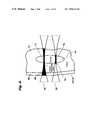
- FIG. 1illustrates an example energy level diagram for simultaneous two-photon excitation
- FIG. 2illustrates an example of absorption and scattering properties for animal tissue covering the ultraviolet to infrared spectral region
- FIG. 3shows the general trends in optical absorption properties of animal tissue for short wavelength and long wavelength light
- FIG. 4illustrates a comparison of optical activation in tissue when single-photon and two-photon excitation methods are used
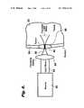
- FIG. 5illustrates an embodiment of the present invention for selective two-photon photoactivation of melanin, melanin-precursors or endogenous pigments using focused light
- FIG. 6illustrates an another embodiment for selective two-photon photoactivation of melanin, melanin-precursors, or endogenous pigments using focused light
- FIG. 7illustrates a further embodiment for selective two-photon photoactivation of melanin, melanin-precursors, or endogenous pigments using non-focused light
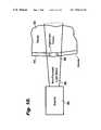
- FIG. 8illustrate still another embodiment for selective two-photon photoactivation of melanin, melanin-precursors, or endogenous pigments in a subsurface tissue using non-focused light
- FIG. 9illustrates an alternate embodiment for the present invention wherein a focused light beam is used to thermally overload and kill pigmented tumor cells.
- FIG. 10illustrates another alternate embodiment for the present invention wherein a non-focused light beam is used to thermally overload and kill pigmented tumor cells.
- the present inventionis directed to a method and apparatus for treating pigmented tissues using light.
- Such treatmentincludes the following photochemical outcomes of therapeutic value: (1) the elimination of undesirable pigmentation in pigmented tissues through photobleaching; and (2) the permanent destruction of pigmented tissues through photochemical conversion of pigments into phototoxic products. More specifically, simultaneous two-photon excitation is used to photochemically convert endogenous or exogenous pigments into desired photoactive products, resulting in the desired photobleaching or tissue destruction.
- Photobleachingis used to reduce or eliminate undesirable coloration of tissue, such as that in moles, freckles, hair follicles and tattoos.
- the production of phototoxic productsmay be used to preferentially kill pigmented tumor cells or other undesirable tissues while sparing normal cells.
- the methods and apparatus in the present invention used for photobleaching and production of phototoxic productsutilize equivalent photoactivation mechanisms, differing substantially only in the intended treatment target.
- the present inventionuses simultaneous two-photon excitation to photoactivate pigments in the pigmented tissues, yielding photobleached or phototoxic products.
- the present inventionuses related optical means to selectively destroy pigmented tissues via photothermal means.
- “Simultaneous two-photon excitation”is the non-linear optical excitation occurring as a result of the essentially simultaneous interaction of two photons originating from a single ultrashort laser pulse with one or more agents or pigments to produce one or more photoactivated agents or pigments.
- “Non-linear optical excitation”means those excitation processes involving the essentially simultaneous interaction of two photons with one or more agents or pigments.
- “Essentially simultaneous interaction”means those excitation processes occurring as a result of the interaction of one or more agents or pigments with photons provided by a single ultrashort laser pulse. Ultrashort means less than approximately 10 ns.
- simultaneous two-photon excitation to an allowed energy level 10occurs when a photoactive agent is excited from a first allowed electronic energy level 16 upon absorption of a certain energy E 1 that is provided by the simultaneous, combined interaction of two photons 12 and 14 with the agent. If the energies of both photons 12 and 14 are identical, the excitation process is termed “degenerate”.
- the simultaneous interaction of the two photonsis frequently described as being mediated by a transient virtual state 20 with a lifetime on the order of 10 femtoseconds (fs) or less. If both photons do not interact with the agent during this lifetime, excitation does not occur and the agent fails to reach the excited state S n (18).
- intersystem crossing, IXsubsequently occurs to bring the excited agent to a long-lived activated state T m from which a photochemical reaction R can occur.
- Simultaneous two-photon excitationmay thereby be used to excite processes that normally occur upon absorption of a single UV or visible photon through the simultaneous absorption of two near-infrared photons.
- An example of the simultaneous two-photon excitation processis the promotion of melanin precursors from a ground electronic state to an excited electronic state through the simultaneous absorption of two photons at 600 nm, followed by binding of the excited melanin precursor to DNA (this is conventionally excited using a single photon at 300 nm).
- the probability of excitationis related to the product of the instantaneous or peak powers of the first of two photons 12 and the second of two photons 14 .
- FIG. 2shows the absorption and scattering properties for various components of animal tissue, such as human dermis, covering the ultraviolet (UV) to near infrared (NIR) spectral region.
- UVultraviolet
- NIRnear infrared
- FIG. 2demonstrates how higher-energy photons 32 may experience considerably greater tissue absorption than lower-energy photons 34 .
- human skinstrongly absorbs higher-energy photons 32 at 400 nm, but is relatively transparent to lower-energy photons 34 at 800 nm. This is a consequence of the natural absorbance of higher-energy photons 32 by blood, pigments, proteins, and genetic materials, among other natural components, of skin.
- FIG. 2further demonstrates how higher-energy photons 42 may experience considerably greater tissue scatter than lower-energy photons 44 .
- Any optically dense mediumsuch as human skin, will strongly scatter higher-energy photons 42 , for example at 400 nm, but will exhibit much lower scatter for lower-energy photons 44 at 800 nm.
- UV light 50for example light at 400 nm
- the majority of the optical energyis immediately absorbed and scattered in the outermost layers 54 , such as the epidermis and dermis. Absorption may occur due to excitation of certain molecules in the cells of these outermost layers 54 , such as those composing the genetic material in the cellular nucleus.
- This absorption of higher-energy light by cellular constituentscan thereby initiate a variety of collateral photochemical changes 56 in these cells.
- These collateral photochemical changes 56 resulting from absorption of UV light 50can include irreversible genetic damage and induction of cancer.
- FIG. 4compares the extent of optically-induced damage in tissue when single-photon excitation 60 and simultaneous two-photon NIR excitation 62 methods are used to illuminate a subcutaneous tumor 64 .
- Single-photon excitation 60produces a photoactivation zone 66 that extends substantially along the entire optical path and has no significant biospecificity.
- collateral damagecan occur throughout surrounding tissues, such as the dermis 68 and surrounding healthy tissue 70 .
- the photoactivation zone 66will be slightly enhanced at the focus 72 .
- This photoactivation zone 66might not even extend into the tumor 64 if the UV or visible light is absorbed by the epidermis, dermis 68 or surrounding healthy tissue 70 prior to reaching the tumor 64 . This can occur as a consequence of the inherently high absorptivity of tissue at short wavelengths.
- NIR lightfor simultaneous two-photon excitation 62 produces a sharply defined remote photoactivation zone 74 that is spatially localized at the focus 76 as a consequence of the non-linear properties of this excitation method.
- Such localization of activation in such a focal zoneis a unique property of non-linear excitation processes, such as two-photon excitation.
- tissuedoes not appreciably absorb NIR light, collateral damage to the surrounding dermis 68 and healthy tissue 70 is minimized.
- non-focussed NIR lightmay be used to stimulate simultaneous two-photon photoactivation of agents present in a relatively large illuminated area.
- the extent of agent photoactivationis controlled by varying the location, intensity and duration of exposure of such agents to the NIR beam.
- focussed NIR lightmay be used to stimulate the simultaneous two-photon photoactivation process.
- beam irradiance, exposure duration, and degree of focussingare used to control the extent of agent photoactivation.
- NIR lightmay be used to achieve maximum efficacy.
- the high penetration depths achievable with NIR lightcombined with the inherent localization of photoactivation that is possible with focused simultaneous two-photon excitation provide a means for photoactivating agents in subsurface tissues without damaging overlying or underlying healthy tissues.
- the method of the present inventionimproves on the above-described advantages through the use of simultaneous two-photon excitation to produce a therapeutic outcome based on photoactivation of endogenous pigments in order to treat pigmented tissues.
- Endogenousmeans pre-existing in a patient or target.
- Pigmentsmeans naturally occurring agents that absorb optical energy. Examples of such pigments include melanin, melanin precursors, carotenes, porphyrins (such as hemoglobin), various tattoo dyes and other optically active species.
- “Therapeutic outcome”means photobleaching or photodynamic destruction of treated pigmented tissues resulting from the natural biological action of a photoactivated endogenous pigment.
- Photobleachingis the reduction or elimination of undesirable pigmentation, for example that caused by endogenous pigments present in moles, freckles, hair follicles and tattoos.
- Photodynamic destructionis localized tissue necrosis resulting from photochemical production of phototoxic products that destroy pigmented tissues, such as those pigmented tissues in pigmented tumors.
- Tissues suitable for treatmentinclude pigmented tissues in which a specific therapeutic outcome is desired, such as moles, freckles, pigmented tumors, benign lesions, hair follicles and tattoos.
- a precursor to the endogenous pigmentsmay be used.
- precursors to pigmentsinclude 5-S-cysteinyldopa (5-SCD) and 5,6-dihydroxyindole (DHI), dopa, dopa semiquinone, leucodopachrome, dopachrome, eumalanins, pheomelanins, sepia melanins, and 5,6-dihydroxyindole-2-carboxylic acid.
- Such precursorshave both photoprotective and phototoxic abilities.
- a metabolic precursor to melaninis a biochemical (e.g. 5-SCD, DHI) that is produced by the cell as part of the synthetic pathway that produces melanin.
- Melanin precursorswhen activated by light, can generate photoxic products that damage cellular materials (e.g., DNA) killing the target cells. Melanin precursors can be activated by two-photon excitation, as explained supra.
- melanin, melanin precursors, and other endogenous pigmentsare naturally occurring in human tissue, including in tumors. Such melanins, melanin precursors, or other endogenous pigments can be converted to phototoxic products after exposure to light.
- the present inventionuses the above-described simultaneous two-photon excitation to specifically target melanin, melanin precursors, or other endogenous pigments in pigmented tissues (such as melanomas and other tumors).
- the pigmentis converted to a phototoxic product by NIR light upon simultaneous two-photon excitation.
- the phototoxic productthen causes damage to the pigmented tissues (by for example photobinding to cellular DNA or causing breaks in this DNA). This kills the cells in the pigmented tissues and, therefore, destroys it.
- any melanin, melanin precursors, or other endogenous pigments in the tissue surrounding the targeted tissueare not converted to phototoxic products.
- focal zonethat is substantially localized in depth and cross-section.
- This focal zonecan be localized to the targeted tissue (such as a tumor) to be killed or a small zone within or surrounding this tissue.
- photoactivationwill only occur in the focal zone (i.e. in the tumor).
- any melanin, melanin precursors, or other endogenous pigment not in the targeted tissuesuch as for example, in tissue surrounding a tumor, will not be photoactivated because it is outside the focal zone.
- the simultaneous two-photon excitationis able to penetrate deep into normal or cancerous tissue and photoactivate melanin or other endogenous pigments located deep within the tissue.
- melanin or other endogenous pigments located deep within the tissueAs a result, tumors located deep within the body or large, deep tumors can be reached and destroyed. Destruction of these tumors can be done without activating melanin or other endogenous pigments along the path of the light or surrounding the tumor.
- the above-described unique features of simultaneous two-photon excitationmay be used to achieve improved safety and specificity in the photobleaching of pigmented tissues, such as in moles, freckles, hair follicles and tattoos.
- the pigments present in such tissuescan be activated by simultaneous two-photon activation, as explained supra, and upon activation may become photobleached.
- the present inventionalso uses simultaneous two-photon excitation to specifically target endogenous pigments in such pigmented tissues, thereby causing photobleaching and a desired reduction or elimination of apparent pigmentation.
- NIR sourcesuch as the mode-locked titanium:sapphire laser
- simultaneous two-photon photoactivationso as to photoactivate melanin, melanin precursors, or other endogenous pigments using light at a wavelength approximately twice that necessary for such conversion using conventional single-photon photoactivation.
- NIR lightexhibits improved penetration into tissue relative to that used for conventional single-photon photoactivation, and is less likely to produce collateral damage in tissues adjacent to the desired treatment target.
- the source 80produces a beam of light 82 consisting of a rapid series of high peak power pulses of NIR light.
- standard commercially available mode-locked titanium-sapphire lasersare capable of outputting mode-locked pulses with durations ⁇ 200 fs and pulse energies of about 1–20 nJ at pulse repetition frequencies in excess of 75 MHz.
- This sourceproduces a quasi-continuous beam of light having a relatively low average power (up to several Watts) but high peak power (on the order of 100 kW) that is continuously tunable over a NIR wavelength band from approximately 690–1080 nm.
- the pulse train from the source 80constitutes a beam of light 82 that is easily focussed using standard optical means, such as reflective or refractive optics 84 .
- the focused beam 86can then be directed into a tumor 88 or other localized treatment target.
- Simultaneous two-photon photoactivation of the melanin, melanin precursors, or other endogenous pigmentswill be substantially limited to the focal zone 90 of the focused light beam 86 due to the high instantaneous irradiance level that is only present at the focus. Furthermore, regardless of whether melanin, melanin precursors, or another endogenous pigment is present in surrounding healthy tissue 92 or skin 94 , insignificant collateral photoactivation, photodamage or conversion into a phototoxic product will occur outside the focal zone 90 . This is a consequence of the non-linear relationship between instantaneous optical power and simultaneous two-photon excitation, which limits significant excitation to the focal zone 90 . Even if melanin, melanin precursors, or another endogenous pigment is present outside of the focal zone 90 , excitation intensities are below that necessary to produce significant photoactivation.
- the apparatus of the present inventioncan also include, for example, a focusing apparatus for focusing the light throughout a range of focal lengths extending from a surface of the tissue to a depth substantially beyond the surface.
- the source of light and focusing apparatuscooperate to promote simultaneous two-photon excitation of the pigment at controllable locations throughout the volume of tissue.
- This scanningcan be done, for example, by positioning a focus of a beam of light over a range of positions so that a focal plane of the light beam occurs at a site located between a surface of the tissue and a point substantially beyond the tissue surface.
- treating the particular volume of tissuemay extend to penetrate deep within the tissue.
- This scanningcan further include varying, while the beam of light is extant, the radial position of the focal plane within the tissue, thereby to photoactivate the endogenous pigment at a multiplicity of positions between the tissue surface and a position located substantially beyond the tissue surface.
- the simultaneous two-photon photoactivation embodiment of the present inventionhas several variations for the treatment of topical tissues, as shown in FIG. 6 and in FIG. 7 .
- the non-damaging nature of focused NIR light, shown in FIG. 6or of non-focused NIR light, shown in FIG. 7 , allows photoactivation of melanin or other endogenous pigments at topical locations without risk to underlying or surrounding tissues.
- Focused simultaneous two-photon photoactivation of melanin or other endogenous pigments for topical therapyis effected when a beam of light 82 from a source 80 is focused 86 onto a tumor 88 or other localized treatment target using standard optical means, such as reflective or refractive optics 84 .
- photoactivation of the melanin, melanin precursors, or other endogenous pigments into a phototoxic productoccurs only at the focal zone 90 .
- the surrounding healthy tissue 92 and skin 94are unaffected in this process, even if they also contain melanin, melanin precursors, or another endogenous pigment, since photoactivation is substantially limited to the focal zone 90 .
- a scanning actioncan be used to effect photoactivation of the melanin, melanin precursor, or other endogenous pigment into a phototoxic product throughout the volume of the tumor 88 .
- Non-focused simultaneous two-photon photoactivation of melanin, melanin precursors, or other endogenous pigments for topical therapyis effected when a non-focused or expanded beam of light 96 from a source 80 is directed onto a topical tumor 88 or other localized treatment target.
- This beam of light 96may have a cross sectional area smaller than, equal to, or larger than that of the tumor 88 . Since melanin, melanin precursors, or other endogenous pigments are present in substantially higher levels in the tumor 88 , the therapeutic action will be substantially limited to the volume of the tumor 88 .
- the beam of light 96is non-damaging to tissues that do not contain a significant concentration of pigment, damage to surrounding healthy tissue 92 and skin 94 is avoided.
- This embodimentmay be particularly useful when the exact location, size and shape of the tumor 88 are not known, or when it is otherwise undesirable to carefully control the location of application of the beam of light 96 , since careful control of the location of the beam of light 96 is not critical for successful administration of this therapeutic regime.
- employment of extremely high peak power excitation sourcessuch as Q-switched lasers or regeneratively amplified mode-locked lasers, may be beneficial due to their exceptionally high peak radiant power (which is in the GW range) that will thereby afford a high instantaneous irradiance over a large area.
- FIG. 8A final related variation of this preferred embodiment for simultaneous two-photon photoactivation is shown in FIG. 8 , where a non-focused or expanded beam of light 96 from a source 80 is directed onto a tumor 88 or other localized treatment target located below the skin's surface.
- This beam of light 96may have a cross sectional area smaller than, equal to, or larger than that of the tumor 88 . Since melanin, melanin precursors, or other endogenous pigments are present in substantially higher levels in a tumor 88 , the therapeutic action will be substantially limited to the volume of the tumor 88 . Since the beam of light 96 is non-damaging to tissues that do not contain a significant concentration of pigment, damage to surrounding healthy tissue 92 and skin 94 is avoided.
- This embodimentmay also be particularly useful when the exact location, size and shape of the tumor 88 are not known, or when it is otherwise undesirable to carefully control the location of application of the beam of light 96 , since careful control of the location of the beam of light 96 is not critical for successful administration of this therapeutic regime.
- employment of extremely high peak power excitation sourcesmay be beneficial due to their exceptionally high peak radiant power and potential high instantaneous irradiance over a large area.
- the simultaneous two-photon excitationwill be produced by an ultrashort pulsed NIR laser light having a wavelength of from approximately 450 nm to 1400 nm with a pulse width of from approximately 25 fs to 10 ns and a greater than approximately 1 kHz pulse repetition frequency.
- Such laser lightcan be produced by a mode-locked titanium:sapphire laser or related laser sources.
- the extent and duration of excitation affected with such sourceswill be controlled by varying the location, irradiance and duration of application of the light.
- the effectiveness of the therapeutic outcomemay be markedly increased by simultaneous photoactivation and localized heating (hyperthermia) of the treatment site.
- Such heatingoccurs as a secondary effect of illumination with laser light, and may also be controlled by varying the location, irradiance and duration of application of the light, so as to yield heating in the treatment zone of 2–10° C. above normal temperatures.
- application of light at intensities of 150–3000 mW/cm 2may be used to produce such desirable hyperthermia.
- secondary thermal sourcessuch as infrared lamps or warm fluid baths, may be used to effect such desirable hyperthermia at the treatment site.
- various other optical sourcesare applicable, alone or in combination, such as continuous wave and pulsed lamps, diode light sources, semiconductor lasers; other types of gas, dye, and solid-state continuous, pulsed, or mode-locked lasers, including: argon ion lasers; krypton ion lasers; helium-neon lasers; helium-cadmium lasers; ruby lasers; Nd:YAG, Nd:YLF, Nd:YAP, Nd:YVO4, Nd:Glass, and Nd:CrGsGG lasers; Cr:LiSF lasers; Er:YAG lasers; F-center lasers; Ho:YAG and Ho:YLF lasers; copper vapor lasers; nitrogen lasers; optical parametric oscillators, amplifiers and generators; regeneratively amplified lasers; chirped-pulse amplified lasers; and sunlight.
- argon ion lasersincluding: argon ion lasers; krypton ion lasers
- an exogenous photodynamic agentcan be added to the patient to be activated in conjunction with the endogenous pigments.
- “Exogenous” agentsare photoactive materials not pre-existent in a patient or other target which are for example administered for the purpose of increasing efficiency of conversion of optical energy into a therapeutic process. Examples of such exogenous agents include Rose Bengal, psoralen derivatives, indocyanine, Lutex, Sn(ET 2 ) and various porphyrin derivatives, including porfimer sodium and benzoporphyrin derivative.
- the targeted tissueis pretreated with the exogenous agent so that it retains a therapeutic concentration of the agent when the tissue is treated with light so as to promote simultaneous two-photon activation of the agent.
- the agentcan be added at other times during the process.
- such agentscan be used to efficiently interact with NIR light so as to kill tissue by Type I or Type II PDT mechanisms. Such killing can be used to augment or supplement killing of pigmented tissues using endogenous photoactive agents as described supra.
- Another alternate embodiment of the present inventionis directed to the thermal destruction of melanomas and other pigmented lesions.
- Melanomasare usually dramatically darker than surrounding healthy tissue.
- the dark color associated with melanomasis caused by increased production of melanin by tumor cells.
- Melaninis a strong absorber of ultraviolet (UV) and visible light, and normally protects cells from the deleterious effects of solar UV radiation.
- FIG. 2shows that melanin is highly absorptive at wavelengths shorter than approximately 1000 nm.
- hemoglobinhas minimal absorbance above 450 nm.
- the high concentration of melanin in most melanoma cellsmakes them capable of strongly and selectively absorbing light at wavelengths longer than 450 nm and shorter than 1000 nm.
- illumination of melanoma cells with light at such wavelengthswill produce much more heat in those cells as compared to cells in less pigmented tissue.
- laser illuminationis used in cosmetic applications to remove unwanted hair.
- Laser hair removalis accomplished because there is more pigment in the hair follicles than in surrounding tissue. Therefore, when a laser illuminates the pigmented hair follicle, it absorbs much more of the light, causing localized heating. The localized hyperthermia thereby created in the bulb of the hair follicle kills the hair follicle while sparing surrounding tissue (which is not heated to a significant extent by the laser illumination).
- FIGS. 9 and 10illustrate such an alternate embodiment for the present invention wherein a focused light beam 86 ( FIG. 9 ) and a non-focused light beam 96 ( FIG. 10 ), respectively, are used to kill pigmented tumor cells 98 .
- Such pigmented tumor cells 98may be located at the surface of tissue 92 to be treated, or may be located significantly below the surface. Illumination of pigmented tumor cells 98 may be effected using a continuous wave or pulsed laser source operating in either of two wavelength bands between approximately 450 and 800 nm and between approximately 800 and 1400 nm.
- direct linear excitation of melaninis used to selectively promote thermal overload of pigmented tumor cells 98 .
- Light in this bandis preferred when pigmented tumor cells 98 are located at the surface of tissue or at depths of approximately 2 mm or less below the surface since such light is not capable of penetrating tissue to significantly greater depths.
- illuminationbe effected via application of one or more short pulses of light having a pulse duration of 10 ns (nanoseconds) or less, and more preferably of 10 ps (picoseconds) or less. Use of such short duration pulses reduces thermal loss to surrounding tissues, thereby improving efficiency in selective thermal overload of the pigmented tumor cells 98 .
- the wavelength of this lightbe between approximately 600 and 800 nm to afford improved specificity for excitation of melanin relative to hemoglobin.
- such lightbe produced by a light source such as a mode-locked titanium:sapphire laser, which is readily able to deliver such light pulses at such wavelengths.
- a focused light beam 86is preferable where the location and extent of the lesion is precisely known, since improved control over the extent of the treatment zone is thereby possible. By scanning this focused light beam 86 throughout the volume of the tumor, it is possible to treat the entirety of the pigmented tumor cells 98 .
- use of a non-focused light beam 96is preferred to assure that treatment is effected in all of the pigmented tumor cells 98 .
- excitation of melanin via linear mechanisms and non-linear two-photon mechanismsis used to selectively promote thermal overload of pigmented tumor cells 98 .
- Light in this bandis preferred when pigmented tumor cells 98 are located below the surface of tissue at depths of approximately 2 mm or greater since such light is capable of penetrating tissue to such depths.
- illuminationbe effected via application of one or more short pulses of light having a pulse duration of 10 ps or less, and more preferably of 1 ps or less.
- a focused light beam 86is preferable where the location and extent of the lesion is precisely known, since improved control over the extent of the treatment zone is thereby possible. Use of such a focused light beam 86 improves efficiency of non-linear excitation mechanisms, allowing relatively low energy light sources 80 , such as mode-locked titanium:sapphire lasers, to be successfully used. By scanning this focused light beam 86 throughout the volume of the tumor it is possible to treat the entirety of the pigmented tumor cells 98 .
- a non-focused light beam 96is preferred to assure that treatment is effected in all of the pigmented tumor cells 98 .
- amplified or other higher energy light sources 80such as the regeneratively amplified mode-locked titanium:sapphire laser, are preferred so as to increase illumination intensities to levels sufficient to achieve efficient non-linear excitation.
Landscapes
- Health & Medical Sciences (AREA)
- Physics & Mathematics (AREA)
- Life Sciences & Earth Sciences (AREA)
- Optics & Photonics (AREA)
- Surgery (AREA)
- Engineering & Computer Science (AREA)
- Biomedical Technology (AREA)
- Veterinary Medicine (AREA)
- Nuclear Medicine, Radiotherapy & Molecular Imaging (AREA)
- Public Health (AREA)
- General Health & Medical Sciences (AREA)
- Animal Behavior & Ethology (AREA)
- Molecular Biology (AREA)
- Medical Informatics (AREA)
- Heart & Thoracic Surgery (AREA)
- Otolaryngology (AREA)
- Electromagnetism (AREA)
- Biophysics (AREA)
- Pathology (AREA)
- Radiology & Medical Imaging (AREA)
- Radiation-Therapy Devices (AREA)
- Laser Surgery Devices (AREA)
- Thermotherapy And Cooling Therapy Devices (AREA)
- Medicines That Contain Protein Lipid Enzymes And Other Medicines (AREA)
- Medicines Containing Material From Animals Or Micro-Organisms (AREA)
Abstract
Description
MoleculeGROUND STATE+2hν600 nm→MoleculeEXCITED STATE (1)
which shows that a molecule in the ground state is promoted to an excited state following simultaneous absorption of two photons at 600 nm, hν600 nm. The reaction rate R, is given by R=k[MoleculeGROUND STATE] [hν600 nm]2, where k is a rate constant and where [MoleculeGROUND STATE] and [hν600 nm] symbolize concentrations of ground state molecules and excitation photons, respectively. Hence, due to the well known quadratic dependence on instantaneous photon irradiance, simultaneous two-photon excitation to an allowed
Claims (68)
Priority Applications (15)
| Application Number | Priority Date | Filing Date | Title |
|---|---|---|---|
| US09/130,213US7036516B1 (en) | 1996-10-30 | 1998-08-06 | Treatment of pigmented tissues using optical energy |
| CN99811013ACN1317951A (en) | 1998-08-06 | 1999-07-29 | Treatment of pigmented tissues using optical energy |
| BR9912776-8ABR9912776A (en) | 1998-08-06 | 1999-07-29 | Process and apparatus for treating a particular volume of tissue and processes for producing a photoactivated product on a particular volume of a material and for treating tissue |
| HK02101440.2AHK1040175A1 (en) | 1998-08-06 | 1999-07-29 | Treatment of pigmented tissues using optical energy |
| PCT/US1999/017176WO2000007514A1 (en) | 1998-08-06 | 1999-07-29 | Treatment of pigmented tissues using optical energy |
| EP99940828AEP1100394A4 (en) | 1998-08-06 | 1999-07-29 | Treatment of pigmented tissues using optical energy |
| IL14124399AIL141243A0 (en) | 1998-08-06 | 1999-07-29 | Treatment of pigmented tissues using optical energy |
| JP2000563201AJP4662631B2 (en) | 1998-08-06 | 1999-07-29 | Apparatus for treating pigmented tissue using light energy |
| CA002339252ACA2339252C (en) | 1998-08-06 | 1999-07-29 | Treatment of pigmented tissues using optical energy |
| AU54607/99AAU760418B2 (en) | 1998-08-06 | 1999-07-29 | Treatment of pigmented tissues using optical energy |
| KR1020017001601AKR20010072306A (en) | 1998-08-06 | 1999-07-29 | Treatment of pigmented tissues using optical energy |
| ARP990103907AAR020337A1 (en) | 1998-08-06 | 1999-08-05 | TREATMENT OF PIGMENTED FABRICS WITH THE USE OF OPTICAL ENERGY |
| IL141243AIL141243A (en) | 1998-08-06 | 2001-02-01 | Treatment of pigmented tissues using optical energy |
| US11/270,782US20060095101A1 (en) | 1996-10-30 | 2005-11-09 | Treatment of pigmented tissues using optical energy |
| US11/270,003US20060095097A1 (en) | 1996-10-30 | 2005-11-09 | Treatment of pigmented tissue using optical energy |
Applications Claiming Priority (2)
| Application Number | Priority Date | Filing Date | Title |
|---|---|---|---|
| US08/739,801US5829448A (en) | 1996-10-30 | 1996-10-30 | Method for improved selectivity in photo-activation of molecular agents |
| US09/130,213US7036516B1 (en) | 1996-10-30 | 1998-08-06 | Treatment of pigmented tissues using optical energy |
Related Parent Applications (1)
| Application Number | Title | Priority Date | Filing Date |
|---|---|---|---|
| US08/739,801Continuation-In-PartUS5829448A (en) | 1996-10-30 | 1996-10-30 | Method for improved selectivity in photo-activation of molecular agents |
Related Child Applications (2)
| Application Number | Title | Priority Date | Filing Date |
|---|---|---|---|
| US57170500ADivision | 1996-10-30 | 2000-05-15 | |
| US11/270,782Continuation-In-PartUS20060095101A1 (en) | 1996-10-30 | 2005-11-09 | Treatment of pigmented tissues using optical energy |
Publications (1)
| Publication Number | Publication Date |
|---|---|
| US7036516B1true US7036516B1 (en) | 2006-05-02 |
Family
ID=22443604
Family Applications (2)
| Application Number | Title | Priority Date | Filing Date |
|---|---|---|---|
| US09/130,213Expired - Fee RelatedUS7036516B1 (en) | 1996-10-30 | 1998-08-06 | Treatment of pigmented tissues using optical energy |
| US11/270,782AbandonedUS20060095101A1 (en) | 1996-10-30 | 2005-11-09 | Treatment of pigmented tissues using optical energy |
Family Applications After (1)
| Application Number | Title | Priority Date | Filing Date |
|---|---|---|---|
| US11/270,782AbandonedUS20060095101A1 (en) | 1996-10-30 | 2005-11-09 | Treatment of pigmented tissues using optical energy |
Country Status (12)
| Country | Link |
|---|---|
| US (2) | US7036516B1 (en) |
| EP (1) | EP1100394A4 (en) |
| JP (1) | JP4662631B2 (en) |
| KR (1) | KR20010072306A (en) |
| CN (1) | CN1317951A (en) |
| AR (1) | AR020337A1 (en) |
| AU (1) | AU760418B2 (en) |
| BR (1) | BR9912776A (en) |
| CA (1) | CA2339252C (en) |
| HK (1) | HK1040175A1 (en) |
| IL (2) | IL141243A0 (en) |
| WO (1) | WO2000007514A1 (en) |
Cited By (16)
| Publication number | Priority date | Publication date | Assignee | Title |
|---|---|---|---|---|
| US20040143181A1 (en)* | 1999-05-26 | 2004-07-22 | Damasco Sanford D. | Computer guided ablation of tissue using integrated ablative/temperature sensing devices |
| US20050148567A1 (en)* | 2003-08-20 | 2005-07-07 | Kjellbotn Charles R. | Treatment of tattoos by photodynamic therapy |
| US20050256514A1 (en)* | 2000-09-12 | 2005-11-17 | Khomchenko Vladimir V | Laser epilation method |
| US20080208294A1 (en)* | 2003-02-28 | 2008-08-28 | Advanced Light Technology, Llc | Disinfection, destruction of neoplastic growth, and sterilization by differential absorption of electromagnetic energy |
| US20080233051A1 (en)* | 2006-09-08 | 2008-09-25 | Prasad Paras N | Nanoparticles for two-photon activated photodynamic therapy and imaging |
| US20090035576A1 (en)* | 2006-09-08 | 2009-02-05 | Prasad Paras N | Nanoparticles for two-photon activated photodynamic therapy and imaging |
| WO2009092112A3 (en)* | 2008-01-18 | 2009-11-05 | The General Hospital Corporation | Selective photostimulation to induce cell proliferation |
| US20090297455A1 (en)* | 2006-08-09 | 2009-12-03 | Koninklijke Philips Electronics N.V. | Device for and a method of activating a physiologically effective substance by ultrasonic waves, and a capsule |
| US20110040295A1 (en)* | 2003-02-28 | 2011-02-17 | Photometics, Inc. | Cancer treatment using selective photo-apoptosis |
| WO2014055960A1 (en) | 2012-10-05 | 2014-04-10 | Genelux Corporation | Energy absorbing-based diagnostic and therapeutic methods employing nucleic acid molecules encoding chromophore-producing enzymes |
| US8968280B2 (en) | 2009-01-23 | 2015-03-03 | The General Hospital Corporation | Dose determination for inducing microcavitation in retinal pigment epithelium (RPE) |
| USRE46493E1 (en) | 2000-06-01 | 2017-08-01 | The General Hospital Corporation | Selective photocoagulation |
| US10799292B2 (en) | 2018-05-04 | 2020-10-13 | Bin Rao | High power tunable optical parametric oscillator for selective photothermolysis laser surgeries |
| US10966785B2 (en)* | 2006-08-02 | 2021-04-06 | Cynosure, Llc | Picosecond laser apparatus and methods for its operation and use |
| US11160685B1 (en)* | 2021-03-24 | 2021-11-02 | Stroma Medical Corporation | Laser systems and methods for alteration of eye color |
| US11418000B2 (en) | 2018-02-26 | 2022-08-16 | Cynosure, Llc | Q-switched cavity dumped sub-nanosecond laser |
Families Citing this family (29)
| Publication number | Priority date | Publication date | Assignee | Title |
|---|---|---|---|---|
| US6517532B1 (en) | 1997-05-15 | 2003-02-11 | Palomar Medical Technologies, Inc. | Light energy delivery head |
| ES2226133T3 (en) | 1997-05-15 | 2005-03-16 | Palomar Medical Technologies, Inc. | DERMATOLOGICAL TREATMENT DEVICE. |
| ES2245506T3 (en) | 1998-03-12 | 2006-01-01 | Palomar Medical Technologies, Inc. | ELECTROMAGNETIC RADIATION APPLICATION SYSTEM ON SKIN. |
| US20060212025A1 (en) | 1998-11-30 | 2006-09-21 | Light Bioscience, Llc | Method and apparatus for acne treatment |
| US9192780B2 (en) | 1998-11-30 | 2015-11-24 | L'oreal | Low intensity light therapy for treatment of retinal, macular, and visual pathway disorders |
| US6887260B1 (en) | 1998-11-30 | 2005-05-03 | Light Bioscience, Llc | Method and apparatus for acne treatment |
| US6283956B1 (en) | 1998-11-30 | 2001-09-04 | David H. McDaniels | Reduction, elimination, or stimulation of hair growth |
| EP1700573A3 (en)* | 2000-12-28 | 2010-12-01 | Palomar Medical Technologies, Inc. | Apparatus for therapeutic EMR treatment of the skin |
| WO2004000098A2 (en) | 2002-06-19 | 2003-12-31 | Palomar Medical Technologies, Inc. | Method and apparatus for treatment of cutaneous and subcutaneous conditions |
| EP2522294A2 (en) | 2002-10-23 | 2012-11-14 | Palomar Medical Technologies, Inc. | Phototreatment device for use with coolants and topical substances |
| CA2531099A1 (en) | 2003-04-10 | 2004-10-28 | Light Bioscience, Llc | Photomodulation methods and devices for regulating cell proliferation and gene expression |
| AU2003292591A1 (en)* | 2003-06-20 | 2005-01-04 | Keio University | Photodynamic therapy apparatus, method for controlling photodynamic therapy apparatus, and photodynamic therapy method |
| CA2533129A1 (en) | 2003-07-31 | 2005-02-10 | Light Bioscience, Llc | System and method for the photodynamic treatment of burns, wounds, and related skin disorders |
| US20050053895A1 (en) | 2003-09-09 | 2005-03-10 | The Procter & Gamble Company Attention: Chief Patent Counsel | Illuminated electric toothbrushes emitting high luminous intensity toothbrush |
| US7856985B2 (en) | 2005-04-22 | 2010-12-28 | Cynosure, Inc. | Method of treatment body tissue using a non-uniform laser beam |
| US20100049055A1 (en)* | 2005-05-31 | 2010-02-25 | W.O.M. World Of Medicine Ag | Method and apparatus for visual characterization of tissue |
| US8033284B2 (en)* | 2006-01-11 | 2011-10-11 | Curaelase, Inc. | Therapeutic laser treatment |
| US20080082149A1 (en)* | 2006-09-13 | 2008-04-03 | Bernstein Eric F | Laser treatment of pigmented lesions on the skin |
| US9919168B2 (en) | 2009-07-23 | 2018-03-20 | Palomar Medical Technologies, Inc. | Method for improvement of cellulite appearance |
| US20110172746A1 (en)* | 2010-01-12 | 2011-07-14 | Roger Porter | High Level Laser Therapy Apparatus and Methods |
| US9095414B2 (en) | 2011-06-24 | 2015-08-04 | The Regents Of The University Of California | Nonlinear optical photodynamic therapy (NLO-PDT) of the cornea |
| WO2013047261A1 (en)* | 2011-09-27 | 2013-04-04 | テルモ株式会社 | Abrasion device |
| EP2839552A4 (en) | 2012-04-18 | 2015-12-30 | Cynosure Inc | PICOSECOND LASER APPARATUS AND METHOD OF PROCESSING TARGET TISSUES USING THE SAME |
| WO2014145179A1 (en) | 2013-03-15 | 2014-09-18 | The General Hospital Corporation | Apparatus for tissue irradiation and methods and kits utilizing the same |
| US10589120B1 (en) | 2012-12-31 | 2020-03-17 | Gary John Bellinger | High-intensity laser therapy method and apparatus |
| US10285757B2 (en) | 2013-03-15 | 2019-05-14 | Cynosure, Llc | Picosecond optical radiation systems and methods of use |
| EP3498211B1 (en)* | 2013-08-09 | 2024-12-25 | The General Hospital Corporation | Apparatus for treating dermal melasma |
| US9907975B1 (en) | 2014-11-19 | 2018-03-06 | Roger D. Porter | Therapeutic laser treatment and transdermal stimulation of stem cell differentiation |
| FR3143312A1 (en)* | 2022-12-16 | 2024-06-21 | L'oréal | Process using light and a cosmetic composition |
Citations (46)
| Publication number | Priority date | Publication date | Assignee | Title |
|---|---|---|---|---|
| US4378806A (en) | 1980-08-12 | 1983-04-05 | Henley Cohn Julian L | Gapped resonant microwave apparatus for producing hyperthermia therapy of tumors |
| US4490543A (en) | 1982-11-12 | 1984-12-25 | University Of Northern Iowa Foundation | Low toxicity radiation sensitizer |
| US4601037A (en) | 1984-06-13 | 1986-07-15 | Britt Corporation | Pulsed laser system |
| US4647578A (en) | 1983-12-02 | 1987-03-03 | Sterling Drug Inc. | Phototoxic insecticidal compositions and method of use thereof |
| US4822335A (en)* | 1986-10-29 | 1989-04-18 | Kureha Kagaku Kogyo Kabushiki Kaisha | Apparatus for treatment of cancer with photodiode |
| US4846789A (en) | 1982-07-19 | 1989-07-11 | L. S. Van Landingham, Jr. | Combatting internal parasites in warm blooded animals |
| US4891043A (en) | 1987-05-28 | 1990-01-02 | Board Of Trustees Of The University Of Illinois | System for selective release of liposome encapsulated material via laser radiation |
| US4973848A (en)* | 1989-07-28 | 1990-11-27 | J. Mccaughan | Laser apparatus for concurrent analysis and treatment |
| US5034613A (en) | 1989-11-14 | 1991-07-23 | Cornell Research Foundation, Inc. | Two-photon laser microscopy |
| US5050597A (en)* | 1987-03-05 | 1991-09-24 | S.L.T. Japan Co., Ltd. | Laser irradiation system for thermotherapy |
| US5066291A (en) | 1990-04-25 | 1991-11-19 | Cincinnati Sub-Zero Products, Inc. | Solid-state laser frequency conversion system |
| US5089384A (en) | 1988-11-04 | 1992-02-18 | Amoco Corporation | Method and apparatus for selective cell destruction using amplified immunofluorescence |
| US5099756A (en) | 1989-06-01 | 1992-03-31 | Harry H. Leveen | Radio frequency thermotherapy |
| US5150712A (en) | 1983-12-14 | 1992-09-29 | Edap International, S.A. | Apparatus for examining and localizing tumors using ultra sounds, comprising a device for localized hyperthermia treatment |
| US5158536A (en) | 1989-08-28 | 1992-10-27 | Biopulmonics, Inc. | Lung cancer hyperthermia via ultrasound and/or convection with perfiuorochemical liquids |
| US5193526A (en)* | 1989-09-05 | 1993-03-16 | S.L.T. Japan Co., Ltd. | Laser light irradiation apparatus |
| US5209748A (en)* | 1989-08-24 | 1993-05-11 | S.L.T. Japan Co., Ltd. | Laser light irradiation apparatus |
| US5217455A (en)* | 1991-08-12 | 1993-06-08 | Tan Oon T | Laser treatment method for removing pigmentations, lesions, and abnormalities from the skin of a living human |
| US5222953A (en) | 1991-10-02 | 1993-06-29 | Kambiz Dowlatshahi | Apparatus for interstitial laser therapy having an improved temperature sensor for tissue being treated |
| US5226907A (en) | 1991-10-29 | 1993-07-13 | Tankovich Nikolai I | Hair removal device and method |
| EP0649667A2 (en)* | 1993-10-20 | 1995-04-26 | Antonella Aprile Carpenter | Quantum energy therapeutic biostimulation method and apparatus |
| US5429582A (en) | 1991-06-14 | 1995-07-04 | Williams; Jeffery A. | Tumor treatment |
| US5445608A (en)* | 1993-08-16 | 1995-08-29 | James C. Chen | Method and apparatus for providing light-activated therapy |
| WO1996007431A1 (en) | 1994-09-02 | 1996-03-14 | Universite De Montreal | Novel rhodamine derivatives for photodynamic therapy of cancer and in vitro purging of the leukemias |
| US5541947A (en) | 1995-05-10 | 1996-07-30 | The Regents Of The University Of Michigan | Selectively triggered, high contrast laser |
| US5540737A (en) | 1991-06-26 | 1996-07-30 | Massachusetts Institute Of Technology | Minimally invasive monopole phased array hyperthermia applicators and method for treating breast carcinomas |
| US5549596A (en)* | 1993-07-07 | 1996-08-27 | The General Hospital Corporation | Selective laser targeting of pigmented ocular cells |
| US5571152A (en)* | 1995-05-26 | 1996-11-05 | Light Sciences Limited Partnership | Microminiature illuminator for administering photodynamic therapy |
| US5586981A (en)* | 1994-08-25 | 1996-12-24 | Xin-Hua Hu | Treatment of cutaneous vascular and pigmented lesions |
| WO1997003697A2 (en) | 1995-07-19 | 1997-02-06 | Consiglio Nazionale Delle Ricerche | Fluorogenic substrates for diagnosis and photodynamic treatment of tumours |
| US5620479A (en) | 1992-11-13 | 1997-04-15 | The Regents Of The University Of California | Method and apparatus for thermal therapy of tumors |
| US5647866A (en) | 1993-11-09 | 1997-07-15 | Zaias; Nardo | Method of hair depilation |
| WO1997026920A2 (en) | 1996-01-23 | 1997-07-31 | Deutsches Krebsforschungszentrum | Conjugate for differentiating between diseased and healthy tissues |
| US5656186A (en)* | 1994-04-08 | 1997-08-12 | The Regents Of The University Of Michigan | Method for controlling configuration of laser induced breakdown and ablation |
| US5669916A (en) | 1994-09-28 | 1997-09-23 | The General Hospital Corporation | Method of hair removal |
| US5707401A (en)* | 1994-03-10 | 1998-01-13 | Esc Medical Systems, Ltd. | Apparatus for an efficient photodynamic treatment |
| US5720894A (en) | 1996-01-11 | 1998-02-24 | The Regents Of The University Of California | Ultrashort pulse high repetition rate laser system for biological tissue processing |
| US5735844A (en) | 1995-02-01 | 1998-04-07 | The General Hospital Corporation | Hair removal using optical pulses |
| WO1998018399A1 (en) | 1996-10-30 | 1998-05-07 | Photogen, Inc. | Method for improved selectivity in photo-activation of molecular agents |
| US5775339A (en)* | 1996-03-26 | 1998-07-07 | Pharmacyclics, Inc. | Photodynamic therapy of pigment-related lesions |
| US5860967A (en) | 1993-07-21 | 1999-01-19 | Lucid, Inc. | Dermatological laser treatment system with electronic visualization of the area being treated |
| US5952818A (en) | 1996-05-31 | 1999-09-14 | Rensselaer Polytechnic Institute | Electro-optical sensing apparatus and method for characterizing free-space electromagnetic radiation |
| US5957960A (en)* | 1997-05-05 | 1999-09-28 | Light Sciences Limited Partnership | Internal two photon excitation device for delivery of PDT to diffuse abnormal cells |
| US6099522A (en) | 1989-02-06 | 2000-08-08 | Visx Inc. | Automated laser workstation for high precision surgical and industrial interventions |
| EP0649677B1 (en) | 1993-10-22 | 2000-08-09 | Nippon Shokubai Co., Ltd. | Catalyst for production of pyromellitic anhydride and process for production of pyromellitic anhydride |
| US6331286B1 (en) | 1998-12-21 | 2001-12-18 | Photogen, Inc. | Methods for high energy phototherapeutics |
Family Cites Families (9)
| Publication number | Priority date | Publication date | Assignee | Title |
|---|---|---|---|---|
| US5590141A (en)* | 1992-04-24 | 1996-12-31 | Electro Scientific Industries, Inc. | Method and apparatus for generating and employing a high density of excited ions in a lasant |
| US5329398A (en)* | 1992-11-05 | 1994-07-12 | Novatec Laser Systems, Inc. | Single grating laser pulse stretcher and compressor |
| US5707403A (en)* | 1993-02-24 | 1998-01-13 | Star Medical Technologies, Inc. | Method for the laser treatment of subsurface blood vessels |
| EP0627643B1 (en)* | 1993-06-03 | 1999-05-06 | Hamamatsu Photonics K.K. | Laser scanning optical system using axicon |
| US5469454A (en)* | 1994-05-02 | 1995-11-21 | University Of Central Florida | Mode locked laser diode in a high power solid state regenerative amplifier and mount mechanism |
| US5658323A (en)* | 1995-07-12 | 1997-08-19 | Miller; Iain D. | Method and apparatus for dermatology treatment |
| US6272156B1 (en)* | 1998-01-28 | 2001-08-07 | Coherent, Inc. | Apparatus for ultrashort pulse transportation and delivery |
| US6676655B2 (en)* | 1998-11-30 | 2004-01-13 | Light Bioscience L.L.C. | Low intensity light therapy for the manipulation of fibroblast, and fibroblast-derived mammalian cells and collagen |
| US6554825B1 (en)* | 2000-05-09 | 2003-04-29 | Laserscope | Variable pulse duration, adjustable wavelength medical laser system |
- 1998
- 1998-08-06USUS09/130,213patent/US7036516B1/ennot_activeExpired - Fee Related
- 1999
- 1999-07-29AUAU54607/99Apatent/AU760418B2/ennot_activeExpired
- 1999-07-29CNCN99811013Apatent/CN1317951A/enactivePending
- 1999-07-29BRBR9912776-8Apatent/BR9912776A/ennot_activeIP Right Cessation
- 1999-07-29EPEP99940828Apatent/EP1100394A4/ennot_activeCeased
- 1999-07-29WOPCT/US1999/017176patent/WO2000007514A1/ennot_activeApplication Discontinuation
- 1999-07-29KRKR1020017001601Apatent/KR20010072306A/ennot_activeWithdrawn
- 1999-07-29JPJP2000563201Apatent/JP4662631B2/ennot_activeExpired - Lifetime
- 1999-07-29CACA002339252Apatent/CA2339252C/ennot_activeExpired - Lifetime
- 1999-07-29ILIL14124399Apatent/IL141243A0/enactiveIP Right Grant
- 1999-07-29HKHK02101440.2Apatent/HK1040175A1/enunknown
- 1999-08-05ARARP990103907Apatent/AR020337A1/ennot_activeApplication Discontinuation
- 2001
- 2001-02-01ILIL141243Apatent/IL141243A/ennot_activeIP Right Cessation
- 2005
- 2005-11-09USUS11/270,782patent/US20060095101A1/ennot_activeAbandoned
Patent Citations (48)
| Publication number | Priority date | Publication date | Assignee | Title |
|---|---|---|---|---|
| US4378806A (en) | 1980-08-12 | 1983-04-05 | Henley Cohn Julian L | Gapped resonant microwave apparatus for producing hyperthermia therapy of tumors |
| US4846789A (en) | 1982-07-19 | 1989-07-11 | L. S. Van Landingham, Jr. | Combatting internal parasites in warm blooded animals |
| US4490543A (en) | 1982-11-12 | 1984-12-25 | University Of Northern Iowa Foundation | Low toxicity radiation sensitizer |
| US4647578A (en) | 1983-12-02 | 1987-03-03 | Sterling Drug Inc. | Phototoxic insecticidal compositions and method of use thereof |
| US5150712A (en) | 1983-12-14 | 1992-09-29 | Edap International, S.A. | Apparatus for examining and localizing tumors using ultra sounds, comprising a device for localized hyperthermia treatment |
| US4601037A (en) | 1984-06-13 | 1986-07-15 | Britt Corporation | Pulsed laser system |
| US4822335A (en)* | 1986-10-29 | 1989-04-18 | Kureha Kagaku Kogyo Kabushiki Kaisha | Apparatus for treatment of cancer with photodiode |
| US5050597A (en)* | 1987-03-05 | 1991-09-24 | S.L.T. Japan Co., Ltd. | Laser irradiation system for thermotherapy |
| US4891043A (en) | 1987-05-28 | 1990-01-02 | Board Of Trustees Of The University Of Illinois | System for selective release of liposome encapsulated material via laser radiation |
| US5089384A (en) | 1988-11-04 | 1992-02-18 | Amoco Corporation | Method and apparatus for selective cell destruction using amplified immunofluorescence |
| US6099522A (en) | 1989-02-06 | 2000-08-08 | Visx Inc. | Automated laser workstation for high precision surgical and industrial interventions |
| US5099756A (en) | 1989-06-01 | 1992-03-31 | Harry H. Leveen | Radio frequency thermotherapy |
| US4973848A (en)* | 1989-07-28 | 1990-11-27 | J. Mccaughan | Laser apparatus for concurrent analysis and treatment |
| US5209748A (en)* | 1989-08-24 | 1993-05-11 | S.L.T. Japan Co., Ltd. | Laser light irradiation apparatus |
| US5158536A (en) | 1989-08-28 | 1992-10-27 | Biopulmonics, Inc. | Lung cancer hyperthermia via ultrasound and/or convection with perfiuorochemical liquids |
| US5193526A (en)* | 1989-09-05 | 1993-03-16 | S.L.T. Japan Co., Ltd. | Laser light irradiation apparatus |
| US5034613A (en) | 1989-11-14 | 1991-07-23 | Cornell Research Foundation, Inc. | Two-photon laser microscopy |
| US5066291A (en) | 1990-04-25 | 1991-11-19 | Cincinnati Sub-Zero Products, Inc. | Solid-state laser frequency conversion system |
| US5429582A (en) | 1991-06-14 | 1995-07-04 | Williams; Jeffery A. | Tumor treatment |
| US5540737A (en) | 1991-06-26 | 1996-07-30 | Massachusetts Institute Of Technology | Minimally invasive monopole phased array hyperthermia applicators and method for treating breast carcinomas |
| US5217455A (en)* | 1991-08-12 | 1993-06-08 | Tan Oon T | Laser treatment method for removing pigmentations, lesions, and abnormalities from the skin of a living human |
| US5290273A (en)* | 1991-08-12 | 1994-03-01 | Tan Oon T | Laser treatment method for removing pigement containing lesions from the skin of a living human |
| US5222953A (en) | 1991-10-02 | 1993-06-29 | Kambiz Dowlatshahi | Apparatus for interstitial laser therapy having an improved temperature sensor for tissue being treated |
| US5226907A (en) | 1991-10-29 | 1993-07-13 | Tankovich Nikolai I | Hair removal device and method |
| US5620479A (en) | 1992-11-13 | 1997-04-15 | The Regents Of The University Of California | Method and apparatus for thermal therapy of tumors |
| US5549596A (en)* | 1993-07-07 | 1996-08-27 | The General Hospital Corporation | Selective laser targeting of pigmented ocular cells |
| US5860967A (en) | 1993-07-21 | 1999-01-19 | Lucid, Inc. | Dermatological laser treatment system with electronic visualization of the area being treated |
| US5445608A (en)* | 1993-08-16 | 1995-08-29 | James C. Chen | Method and apparatus for providing light-activated therapy |
| EP0649667A2 (en)* | 1993-10-20 | 1995-04-26 | Antonella Aprile Carpenter | Quantum energy therapeutic biostimulation method and apparatus |
| EP0649677B1 (en) | 1993-10-22 | 2000-08-09 | Nippon Shokubai Co., Ltd. | Catalyst for production of pyromellitic anhydride and process for production of pyromellitic anhydride |
| US5647866A (en) | 1993-11-09 | 1997-07-15 | Zaias; Nardo | Method of hair depilation |
| US5707401A (en)* | 1994-03-10 | 1998-01-13 | Esc Medical Systems, Ltd. | Apparatus for an efficient photodynamic treatment |
| US5656186A (en)* | 1994-04-08 | 1997-08-12 | The Regents Of The University Of Michigan | Method for controlling configuration of laser induced breakdown and ablation |
| US5586981A (en)* | 1994-08-25 | 1996-12-24 | Xin-Hua Hu | Treatment of cutaneous vascular and pigmented lesions |
| WO1996007431A1 (en) | 1994-09-02 | 1996-03-14 | Universite De Montreal | Novel rhodamine derivatives for photodynamic therapy of cancer and in vitro purging of the leukemias |
| US5669916A (en) | 1994-09-28 | 1997-09-23 | The General Hospital Corporation | Method of hair removal |
| US5735844A (en) | 1995-02-01 | 1998-04-07 | The General Hospital Corporation | Hair removal using optical pulses |
| US5541947A (en) | 1995-05-10 | 1996-07-30 | The Regents Of The University Of Michigan | Selectively triggered, high contrast laser |
| US5571152A (en)* | 1995-05-26 | 1996-11-05 | Light Sciences Limited Partnership | Microminiature illuminator for administering photodynamic therapy |
| WO1997003697A2 (en) | 1995-07-19 | 1997-02-06 | Consiglio Nazionale Delle Ricerche | Fluorogenic substrates for diagnosis and photodynamic treatment of tumours |
| US5720894A (en) | 1996-01-11 | 1998-02-24 | The Regents Of The University Of California | Ultrashort pulse high repetition rate laser system for biological tissue processing |
| WO1997026920A2 (en) | 1996-01-23 | 1997-07-31 | Deutsches Krebsforschungszentrum | Conjugate for differentiating between diseased and healthy tissues |
| US5775339A (en)* | 1996-03-26 | 1998-07-07 | Pharmacyclics, Inc. | Photodynamic therapy of pigment-related lesions |
| US5952818A (en) | 1996-05-31 | 1999-09-14 | Rensselaer Polytechnic Institute | Electro-optical sensing apparatus and method for characterizing free-space electromagnetic radiation |
| US5829448A (en) | 1996-10-30 | 1998-11-03 | Photogen, Inc. | Method for improved selectivity in photo-activation of molecular agents |
| WO1998018399A1 (en) | 1996-10-30 | 1998-05-07 | Photogen, Inc. | Method for improved selectivity in photo-activation of molecular agents |
| US5957960A (en)* | 1997-05-05 | 1999-09-28 | Light Sciences Limited Partnership | Internal two photon excitation device for delivery of PDT to diffuse abnormal cells |
| US6331286B1 (en) | 1998-12-21 | 2001-12-18 | Photogen, Inc. | Methods for high energy phototherapeutics |
Non-Patent Citations (45)
| Title |
|---|
| (Svensk Farmaceutisk Tidskrift (1973) 77 (13): 641-647 (Abstract). |
| Ahmed, I., "Malignant melanoma," Mayo Clinic Proced., 72; 356-361, 1997. |
| Anderson et al, "Selective photothermolysis of cutaneous pigmentation by Q-switched Nd:YAG laser pulses at 1064, 532 and 355 nm," J. Invest. Dermatol., 93; 28-32, 1989. |
| Bodaness, R.S. and D.S. King (1985) The two-photon induced flourescence of the tumor localizing photo-sensitizer hematoporphyrin derivative via 1064 nm . . . Biochem. Biophys. Res. Comm. 1 2 6, 346-351. |
| Bodaness, R.S., et al., (1986) Two-photon laser-induced fluorescence of the tumor-localizing photosensitive hematoporphyrin derivative. J. Biol. Chem. 2 6 1, 12098-12101. |
| Cheong, W-F. et al., "A Review of the Optical Properties of Biological Tissues" IEEE 5. Quant. Election 2 6, 2166-2185 (1990). |
| Communication from EPO regarding European Patent Application No. 99940828, Mailed Feb. 2, 2005. Includes European Search Report. |
| de Vries et al, "Increased susceptability to ultraviolet-B and carcinogens of mice lacking the DNA excision repair gene XPA," Nature, 377; 169-173; 1995. |
| de Vries et al, "Increased susceptability to ultraviolet-B and carcinogens of mice lacking the DNA excision repair gene XPA," Nature,377; 169-173; 1995. |
| Dees et al. "Treatment of Murine Cutaneous Melanoma with Near Infrared Light" Photochemistry and Photobiology, 2002, pp. 296-301. |
| Draumer, N.H., et al., (1997) Femtosecond dynamics of excited-state evolution in [Ru(bpy)<SUP>3</SUP>]<SUP>2+</SUP>. Science 2 7 5, 54-57. |
| Favilla et al., "Photodynamic therapy: a 5 year study of its effectiveness in the treatment of posterior uveal melanoma and evaluation of haematoprophyrin uptake and phototoxicity of melanoma cells in tissue culture," Melanoma Res., 5; 355-364, 1995. |
| Göpert-Mayer, M., (1931) Elementary process with two quantum jumps. Ann. Physik 9, 273-294. |
| Hadjur et al, "Photodynamic effects of hypericin on lipid peroxidation and antioxidant status in melanoma cells," Photochem. Photobiol., 64; 375-381, 1996. |
| Inaba, H., et al., (1985) Nd:YAG laser-induced hematoporphyrin visible flourescence and two-photon-excited photochemical effect on malignant tumor cells. J. Opt. Soc. Am. A:Opt. Inage Science 2, P72 (mtg abstr). |
| International Search Report for application No. PCT/US99/17176, dated Oct. 25, 1999. |
| Kaiser, W. and C.G.B. Garrett, (1961) Two photon excitation in CaF<SUB>2</SUB>:Eu<SUP>2+</SUP>. Phys. Rev. Lett. 7, 229-231. |
| Katsumi, T.A. et al, "Photodynamic Therapy with a Diode Laser for Implanted Fibrosarcoma in Mice Employing Mono-L-Aspartyl Chlorin E<SUB>6</SUB>," Photochemistry and Photobiology, vol. 64, No. 4, pp. 671-675, 1996. |
| Lenz, P., (1995) In vivo excitation of photosensitizers by infrared light. Photochem. Photobiol. 6 2, 333-338. |
| Lenz, P., (1995) In vivo excitation photosensitizers by infrared light. Photochem. Photobiol. 6 2, 333-338. |
| M.J. Wirth, et al., "Two-Photon Excited Molecular Fluorescence in Optically Dense Media," Analytical Chemistry, 49(1977) 2054-2057. |
| Marchesini, R., et al., (1986) A study on the possible involvement of nonlinear mechanism of light absorption by HpD with Nd:YAG laser. Lasers Surg. Med. 6, 323-327. |
| Mashiko, S., et al., (1986) Two-photon excited visible flourescence of hematoporphyrin and phiophorbide a and in vitro experiments of the photodynamic . . . J. Opt. Soc. Am. B:Opt. Phys 3, P72-P73 (mtg. abstr). |
| McClain, W.M., (1974) Two-photon molecular spectroscopy. Acc. Chem. Res. 7, 129-135. |
| Monson, P.R. and W.M. McClain (1970) Polarization dependence of the two-photon absorption of tumbling molecules with application of liquid 1-choronaphthalene and benzene. J. Chem. Phys. 5 3, 29-37. |
| Nakane et al, "High incidence of ultraviolet-B- or chemical-carcinogen-induced skin tumors in mice lacking the xenoderma pigmentosum group A gene," Nature, 377; 165-168, 1995. |
| Oh, D.H., et al., (1997) Two-photon excitation of 4'-hydroxymethyl-4,5', 8-trimethylpsoralen. Photochem. Photobiol. 6 5, 91-95. |
| Patrice, T., et al., (1983) Neodymium-yttrium aluminum gamet laser destruction of nonsensitized and hematoporphyrin derivative-sensitized tumors. Canc. Res. 4 3, 2876-2879. |
| Rigel, D.S., "Malignant melanoma: incidence issues and their effect on diagnosis and treatment," Mayo Clinic Proced, 72; 367-371, 1997. |
| Rigel, D.S., "Malignant melanoma: incidence issues and their effect on diagnosis and treatment," Mayo Clinic Proced., 72; 367-371, 1997. |
| Riley, P.A., "Melanin," Int. J. Biochem., Cell Biol., 29; 1235-1239, 1997. |
| Roger et al, "Mucosal, genital and unusual variants of melanoma," Mayo Clinic Proced., 72; 362-366, 1977. |
| Sands et al, "High susceptibility to ultraviolet-induced carcingenesis in mice lacking XPC," Nature, 377; 162-165, 1995. |
| Sands et al, "High susceptibility to ultraviolet-induced carcinogenesis in mice lacking XPC," Nature, 377; 162-165, 1995. |
| Schmitz et al, "Dual role of melanins and melanin precursors as photoprotective and phototoxic agents: inhibition of ultraviolet radiation-induced lipid peroxidation," Photochem. Photobiol., 61; 650-655, 1995. |
| Shea C.R., et al., (1990) Mechanistic investigation of doxycyckine photosensitization by picosecond-pulsed and continuous wave laser irradiation of cells in culture. J. Biol. Chem. 2 6 5, 5977-5982. |
| Sitnik, Theresa M. et al. (1998) The Effect of Fluence Rate on Tumor and Normal Tissue Responses to Photodynamic Therapy. Photochemistry and Photobiology, pp. 462-466. |
| Stables, G.I. et al, "Photodynamic Therapy, Antitumour Treatment," Cancer Treatment Reviews, vol. 21, pp. 311-323, 1995. |
| Steil, H., et al., (1993) Photophysical properties of the photosensitizer phophorbide a studied at high photon flux densities. J. Photochem. Photobiol. B: Biology 1 7, 181-186. |
| Steil, H., et al., (1993) Photophysical properties of the photosensitizer phophorbide a studied at high photon flux densities. J. Photochem. Photobiol. B: Biology 1 7,181-186. |
| Swofford, R.L. and W.M. McClaim, (1975) The effect of saptial and temporal laser beam characteristics on two-photon absorption. Chem. Phys. Lett. 3 4, 455-459. |
| Watanabe et al, "Comparative studies of femtosecond to microsecond laser pulses on selective pigmented cell injury in skin," Photochem. Photobiol., 53, 757-762, 1991. |
| Xanthenes: Fluorone Derivatives, The Journal Of Organic Chemistry 57(1992) Jul. 31, No. 16, Washington, DC pps 4418-4421. |
| Young, A.R., "Chromophores in human skin," Phys. Med. Biol., 42; 789-802, 1997. |
| Zhu, Ning Wen, et al., (1997) Sub-Lethal Effects of Exposing The Human Melanoma Cell Line SKmel-23 to 532 nm Laser Light. Int. J. Cancer: 72, 1104-1112. |
Cited By (23)
| Publication number | Priority date | Publication date | Assignee | Title |
|---|---|---|---|---|
| US7363071B2 (en)* | 1999-05-26 | 2008-04-22 | Endocare, Inc. | Computer guided ablation of tissue using integrated ablative/temperature sensing devices |
| US9326808B2 (en) | 1999-05-26 | 2016-05-03 | ENOCARE, Inc. | System for providing computer guided ablation of tissue |
| US20040143181A1 (en)* | 1999-05-26 | 2004-07-22 | Damasco Sanford D. | Computer guided ablation of tissue using integrated ablative/temperature sensing devices |
| USRE46493E1 (en) | 2000-06-01 | 2017-08-01 | The General Hospital Corporation | Selective photocoagulation |
| US20050256514A1 (en)* | 2000-09-12 | 2005-11-17 | Khomchenko Vladimir V | Laser epilation method |
| US8790381B2 (en) | 2003-02-28 | 2014-07-29 | Photometics, Inc. | Disinfection, destruction of neoplastic growth, and sterilization by differential absorption of electromagnetic energy |
| US20080208294A1 (en)* | 2003-02-28 | 2008-08-28 | Advanced Light Technology, Llc | Disinfection, destruction of neoplastic growth, and sterilization by differential absorption of electromagnetic energy |
| US20110040295A1 (en)* | 2003-02-28 | 2011-02-17 | Photometics, Inc. | Cancer treatment using selective photo-apoptosis |
| US20050148567A1 (en)* | 2003-08-20 | 2005-07-07 | Kjellbotn Charles R. | Treatment of tattoos by photodynamic therapy |
| US11712299B2 (en) | 2006-08-02 | 2023-08-01 | Cynosure, LLC. | Picosecond laser apparatus and methods for its operation and use |
| US10966785B2 (en)* | 2006-08-02 | 2021-04-06 | Cynosure, Llc | Picosecond laser apparatus and methods for its operation and use |
| US20090297455A1 (en)* | 2006-08-09 | 2009-12-03 | Koninklijke Philips Electronics N.V. | Device for and a method of activating a physiologically effective substance by ultrasonic waves, and a capsule |
| US20090035576A1 (en)* | 2006-09-08 | 2009-02-05 | Prasad Paras N | Nanoparticles for two-photon activated photodynamic therapy and imaging |
| US20080233051A1 (en)* | 2006-09-08 | 2008-09-25 | Prasad Paras N | Nanoparticles for two-photon activated photodynamic therapy and imaging |
| WO2009092112A3 (en)* | 2008-01-18 | 2009-11-05 | The General Hospital Corporation | Selective photostimulation to induce cell proliferation |
| US8968280B2 (en) | 2009-01-23 | 2015-03-03 | The General Hospital Corporation | Dose determination for inducing microcavitation in retinal pigment epithelium (RPE) |
| WO2014055960A1 (en) | 2012-10-05 | 2014-04-10 | Genelux Corporation | Energy absorbing-based diagnostic and therapeutic methods employing nucleic acid molecules encoding chromophore-producing enzymes |
| US11418000B2 (en) | 2018-02-26 | 2022-08-16 | Cynosure, Llc | Q-switched cavity dumped sub-nanosecond laser |
| US11791603B2 (en) | 2018-02-26 | 2023-10-17 | Cynosure, LLC. | Q-switched cavity dumped sub-nanosecond laser |
| US10799292B2 (en) | 2018-05-04 | 2020-10-13 | Bin Rao | High power tunable optical parametric oscillator for selective photothermolysis laser surgeries |
| US11160685B1 (en)* | 2021-03-24 | 2021-11-02 | Stroma Medical Corporation | Laser systems and methods for alteration of eye color |
| US20220304857A1 (en)* | 2021-03-24 | 2022-09-29 | Stroma Medical Corporation | Laser systems and methods for alteration of eye color |
| US11819458B2 (en)* | 2021-03-24 | 2023-11-21 | Stroma Medical Corporation | Laser systems and methods for alteration of eye color |
Also Published As
| Publication number | Publication date |
|---|---|
| EP1100394A4 (en) | 2005-03-16 |
| IL141243A (en) | 2006-07-05 |
| WO2000007514A9 (en) | 2001-04-19 |
| CN1317951A (en) | 2001-10-17 |
| CA2339252A1 (en) | 2000-02-17 |
| JP4662631B2 (en) | 2011-03-30 |
| AU760418B2 (en) | 2003-05-15 |
| EP1100394A1 (en) | 2001-05-23 |
| BR9912776A (en) | 2001-05-08 |
| AU5460799A (en) | 2000-02-28 |
| KR20010072306A (en) | 2001-07-31 |
| AR020337A1 (en) | 2002-05-08 |
| IL141243A0 (en) | 2002-03-10 |
| US20060095101A1 (en) | 2006-05-04 |
| HK1040175A1 (en) | 2002-05-31 |
| CA2339252C (en) | 2008-02-12 |
| JP2002522110A (en) | 2002-07-23 |
| WO2000007514A1 (en) | 2000-02-17 |
Similar Documents
| Publication | Publication Date | Title |
|---|---|---|
| US7036516B1 (en) | Treatment of pigmented tissues using optical energy | |
| Chen et al. | Chromophore-enhanced laser-tumor tissue photothermal interaction using an 808-nm diode laser | |
| US7353829B1 (en) | Methods and apparatus for multi-photon photo-activation of therapeutic agents | |
| US6358242B1 (en) | Post laser treatment for permanent hair removal | |
| US5527350A (en) | Pulsed infrared laser treatment of psoriasis | |
| Herd et al. | Basic laser principles | |
| Patil | Overview of lasers | |
| Van Gemert et al. | Clinical use of laser-tissue interactions | |
| Parrish et al. | Laser photomedicine | |
| Mariwalla et al. | Use of lasers and light‐based therapies for treatment of acne vulgaris | |
| Karrer et al. | Role of lasers and photodynamic therapy in the treatment of cutaneous malignancy | |
| Spencer et al. | The excimer lasers. | |
| Park et al. | 1,213 Cases of treatment of facial acne using indocyanine green and intense pulsed light in Asian skin | |
| Goldberg | Laser removal of pigmented and vascular lesions | |
| Weir et al. | Photo-assisted epilation—review and personal observations | |
| US20060095097A1 (en) | Treatment of pigmented tissue using optical energy | |
| Wachter et al. | Simultaneous two-photon excitation of photodynamic therapy agents | |
| Walsh et al. | Laser-Tissue Interactions and Their Clinical Applications¹ | |
| Wachter et al. | Photodynamic therapy with ultrafast lasers | |
| MXPA01001258A (en) | Treatment of pigmented tissues using optical energy | |
| Goldberg | Facial rejuvenation: a total approach | |
| Parrish | Laser medicine and laser dermatology | |
| Mioc et al. | Selected laser-based therapies in otolaryngology | |
| Seoighe et al. | The use of lasers in the treatment of cutaneous lesions | |
| Hernandez-Jimenez | Lasers in Cosmetic Dermatology & Skincare Practice |
Legal Events
| Date | Code | Title | Description |
|---|---|---|---|
| AS | Assignment | Owner name:PHOTOGEN, INC., TENNESSEE Free format text:ASSIGNMENT OF ASSIGNORS INTEREST;ASSIGNORS:DEES, H. CRAIG;WACHTER, ERIC A.;REEL/FRAME:009382/0076 Effective date:19980804 | |
| AS | Assignment | Owner name:XANTECH PHARMACEUTICALS, INC., TENNESSEE Free format text:MERGER;ASSIGNOR:VALLEY PHARMACEUTICALS, INC.;REEL/FRAME:013782/0211 Effective date:20021121 | |
| AS | Assignment | Owner name:VALEY PHARMACEUTICALS, INC., TENNESSEE Free format text:CHANGE OF NAME;ASSIGNOR:PHOTOGEN, INC.;REEL/FRAME:013782/0476 Effective date:20021112 | |
| AS | Assignment | Owner name:PROVECTUS DEVICETECH, INC., TENNESSEE Free format text:ASSIGNMENT OF ASSIGNORS INTEREST;ASSIGNOR:XANTECH PHARMACEUTICALS, INC.;REEL/FRAME:014763/0824 Effective date:20040621 | |
| AS | Assignment | Owner name:DCOFI MASTER LDC, AS AGENT, NEW YORK Free format text:SECURITY AGREEMENT;ASSIGNOR:PROVECTUS PHARMACEUTICALS, INC.;REEL/FRAME:016127/0746 Effective date:20050330 Owner name:DCOFI MASTER LDC, AS AGENT, NEW YORK Free format text:SECURITY AGREEMENT;ASSIGNOR:PROVECTUS DEVICETECH, INC.;REEL/FRAME:016127/0660 Effective date:20050330 | |
| AS | Assignment | Owner name:PROVECTUS DEVICETECH, INC., TENNESSEE Free format text:RELEASE BY SECURED PARTY;ASSIGNOR:DCOFI MASTER LDC;REEL/FRAME:019215/0608 Effective date:20070306 Owner name:PROVECTUS PHARMACEUTICALS, INC., TENNESSEE Free format text:RELEASE BY SECURED PARTY;ASSIGNOR:DCOFI MASTER LDC;REEL/FRAME:019215/0669 Effective date:20070306 | |
| FPAY | Fee payment | Year of fee payment:4 | |
| FPAY | Fee payment | Year of fee payment:8 | |
| FEPP | Fee payment procedure | Free format text:MAINTENANCE FEE REMINDER MAILED (ORIGINAL EVENT CODE: REM.) | |
| LAPS | Lapse for failure to pay maintenance fees | Free format text:PATENT EXPIRED FOR FAILURE TO PAY MAINTENANCE FEES (ORIGINAL EVENT CODE: EXP.) | |
| STCH | Information on status: patent discontinuation | Free format text:PATENT EXPIRED DUE TO NONPAYMENT OF MAINTENANCE FEES UNDER 37 CFR 1.362 | |
| FP | Lapsed due to failure to pay maintenance fee | Effective date:20180502 |