US7025776B1 - Arteriotomy closure devices and techniques - Google Patents
Arteriotomy closure devices and techniquesDownload PDFInfo
- Publication number
- US7025776B1 US7025776B1US10/224,659US22465902AUS7025776B1US 7025776 B1US7025776 B1US 7025776B1US 22465902 AUS22465902 AUS 22465902AUS 7025776 B1US7025776 B1US 7025776B1
- Authority
- US
- United States
- Prior art keywords
- closure device
- arterial closure
- vessel
- connecting member
- arterial
- Prior art date
- Legal status (The legal status is an assumption and is not a legal conclusion. Google has not performed a legal analysis and makes no representation as to the accuracy of the status listed.)
- Expired - Fee Related, expires
Links
- 238000000034methodMethods0.000titleclaimsdescription44
- 239000000463materialSubstances0.000claimsdescription76
- 239000000853adhesiveSubstances0.000claimsdescription35
- 230000001070adhesive effectEffects0.000claimsdescription35
- 239000012790adhesive layerSubstances0.000claimsdescription8
- 239000003814drugSubstances0.000claimsdescription6
- 239000012530fluidSubstances0.000claimsdescription6
- 229940124597therapeutic agentDrugs0.000claimsdescription6
- 230000035876healingEffects0.000claimsdescription5
- 239000000560biocompatible materialSubstances0.000claimsdescription4
- 238000002513implantationMethods0.000claimsdescription4
- HERSSAVMHCMYSQ-UHFFFAOYSA-N1,8-diazacyclotetradecane-2,9-dioneChemical compoundO=C1CCCCCNC(=O)CCCCCN1HERSSAVMHCMYSQ-UHFFFAOYSA-N0.000description50
- 229910052751metalInorganic materials0.000description42
- 239000002184metalSubstances0.000description42
- 210000001519tissueAnatomy0.000description20
- 229910001000nickel titaniumInorganic materials0.000description18
- HLXZNVUGXRDIFK-UHFFFAOYSA-Nnickel titaniumChemical compound[Ti].[Ti].[Ti].[Ti].[Ti].[Ti].[Ti].[Ti].[Ti].[Ti].[Ti].[Ni].[Ni].[Ni].[Ni].[Ni].[Ni].[Ni].[Ni].[Ni].[Ni].[Ni].[Ni].[Ni].[Ni]HLXZNVUGXRDIFK-UHFFFAOYSA-N0.000description18
- 229910000734martensiteInorganic materials0.000description12
- 238000003780insertionMethods0.000description11
- 239000008280bloodSubstances0.000description10
- 210000004369bloodAnatomy0.000description10
- 230000037431insertionEffects0.000description10
- -1poly(hydroxybutyrate)Polymers0.000description10
- PXHVJJICTQNCMI-UHFFFAOYSA-NNickelChemical compound[Ni]PXHVJJICTQNCMI-UHFFFAOYSA-N0.000description9
- 150000002739metalsChemical class0.000description9
- 230000002792vascularEffects0.000description9
- 230000008859changeEffects0.000description8
- 238000010438heat treatmentMethods0.000description8
- 238000007789sealingMethods0.000description8
- 238000005452bendingMethods0.000description7
- 239000002131composite materialSubstances0.000description7
- 230000002787reinforcementEffects0.000description7
- 239000012781shape memory materialSubstances0.000description7
- 229910045601alloyInorganic materials0.000description6
- 239000000956alloySubstances0.000description6
- 229910001566austeniteInorganic materials0.000description6
- 230000002439hemostatic effectEffects0.000description6
- 238000004519manufacturing processMethods0.000description6
- 229920000642polymerPolymers0.000description6
- 229910001285shape-memory alloyInorganic materials0.000description6
- 239000010936titaniumSubstances0.000description6
- 238000000576coating methodMethods0.000description5
- 239000004020conductorSubstances0.000description5
- 238000013461designMethods0.000description5
- 239000000499gelSubstances0.000description5
- 239000000203mixtureSubstances0.000description5
- 238000012986modificationMethods0.000description5
- 230000004048modificationEffects0.000description5
- 230000001225therapeutic effectEffects0.000description5
- 230000009466transformationEffects0.000description5
- 102000008186CollagenHuman genes0.000description4
- 108010035532CollagenProteins0.000description4
- 239000004821Contact adhesiveSubstances0.000description4
- RTAQQCXQSZGOHL-UHFFFAOYSA-NTitaniumChemical compound[Ti]RTAQQCXQSZGOHL-UHFFFAOYSA-N0.000description4
- 239000011248coating agentSubstances0.000description4
- 229920001436collagenPolymers0.000description4
- 230000006835compressionEffects0.000description4
- 238000007906compressionMethods0.000description4
- 230000023597hemostasisEffects0.000description4
- 229910001092metal group alloyInorganic materials0.000description4
- 229910052759nickelInorganic materials0.000description4
- BASFCYQUMIYNBI-UHFFFAOYSA-NplatinumChemical compound[Pt]BASFCYQUMIYNBI-UHFFFAOYSA-N0.000description4
- 230000008569processEffects0.000description4
- 230000002441reversible effectEffects0.000description4
- 239000000243solutionSubstances0.000description4
- 229910052719titaniumInorganic materials0.000description4
- 229920004934Dacron®Polymers0.000description3
- 239000003146anticoagulant agentSubstances0.000description3
- 230000008901benefitEffects0.000description3
- 150000001875compoundsChemical class0.000description3
- 230000007797corrosionEffects0.000description3
- 238000005260corrosionMethods0.000description3
- 239000013013elastic materialSubstances0.000description3
- 239000004744fabricSubstances0.000description3
- 239000006260foamSubstances0.000description3
- 230000006870functionEffects0.000description3
- 238000000465mouldingMethods0.000description3
- 239000005020polyethylene terephthalateSubstances0.000description3
- 238000011084recoveryMethods0.000description3
- 230000002829reductive effectEffects0.000description3
- 230000000284resting effectEffects0.000description3
- 239000007787solidSubstances0.000description3
- 238000005507sprayingMethods0.000description3
- 239000010935stainless steelSubstances0.000description3
- 229910001220stainless steelInorganic materials0.000description3
- RYGMFSIKBFXOCR-UHFFFAOYSA-NCopperChemical compound[Cu]RYGMFSIKBFXOCR-UHFFFAOYSA-N0.000description2
- 102000009123FibrinHuman genes0.000description2
- 108010073385FibrinProteins0.000description2
- BWGVNKXGVNDBDI-UHFFFAOYSA-NFibrin monomerChemical compoundCNC(=O)CNC(=O)CNBWGVNKXGVNDBDI-UHFFFAOYSA-N0.000description2
- HTTJABKRGRZYRN-UHFFFAOYSA-NHeparinChemical compoundOC1C(NC(=O)C)C(O)OC(COS(O)(=O)=O)C1OC1C(OS(O)(=O)=O)C(O)C(OC2C(C(OS(O)(=O)=O)C(OC3C(C(O)C(O)C(O3)C(O)=O)OS(O)(=O)=O)C(CO)O2)NS(O)(=O)=O)C(C(O)=O)O1HTTJABKRGRZYRN-UHFFFAOYSA-N0.000description2
- MWUXSHHQAYIFBG-UHFFFAOYSA-NNitric oxideChemical compoundO=[N]MWUXSHHQAYIFBG-UHFFFAOYSA-N0.000description2
- 230000009471actionEffects0.000description2
- 238000007792additionMethods0.000description2
- 238000004026adhesive bondingMethods0.000description2
- 229910052782aluminiumInorganic materials0.000description2
- XAGFODPZIPBFFR-UHFFFAOYSA-NaluminiumChemical compound[Al]XAGFODPZIPBFFR-UHFFFAOYSA-N0.000description2
- 210000001367arteryAnatomy0.000description2
- TZCXTZWJZNENPQ-UHFFFAOYSA-Lbarium sulfateChemical compound[Ba+2].[O-]S([O-])(=O)=OTZCXTZWJZNENPQ-UHFFFAOYSA-L0.000description2
- 230000006399behaviorEffects0.000description2
- 230000009286beneficial effectEffects0.000description2
- 210000004204blood vesselAnatomy0.000description2
- 230000036760body temperatureEffects0.000description2
- 238000005266castingMethods0.000description2
- 238000010276constructionMethods0.000description2
- 230000008602contractionEffects0.000description2
- 229920001577copolymerPolymers0.000description2
- 229910052802copperInorganic materials0.000description2
- 239000010949copperSubstances0.000description2
- 238000005520cutting processMethods0.000description2
- 238000003618dip coatingMethods0.000description2
- 230000002526effect on cardiovascular systemEffects0.000description2
- 230000000694effectsEffects0.000description2
- 229920000295expanded polytetrafluoroethylenePolymers0.000description2
- 238000001125extrusionMethods0.000description2
- 229950003499fibrinDrugs0.000description2
- 208000025339heart septal defectDiseases0.000description2
- 229960002897heparinDrugs0.000description2
- 229920000669heparinPolymers0.000description2
- 239000007943implantSubstances0.000description2
- 230000013011matingEffects0.000description2
- 210000000056organAnatomy0.000description2
- 229910052697platinumInorganic materials0.000description2
- 229920001432poly(L-lactide)Polymers0.000description2
- 229920013745polyesteretherketonePolymers0.000description2
- 229920001343polytetrafluoroethylenePolymers0.000description2
- 239000004810polytetrafluoroethyleneSubstances0.000description2
- 229920002635polyurethanePolymers0.000description2
- 239000004814polyurethaneSubstances0.000description2
- 230000001954sterilising effectEffects0.000description2
- 238000004659sterilization and disinfectionMethods0.000description2
- PJVWKTKQMONHTI-UHFFFAOYSA-NwarfarinChemical compoundOC=1C2=CC=CC=C2OC(=O)C=1C(CC(=O)C)C1=CC=CC=C1PJVWKTKQMONHTI-UHFFFAOYSA-N0.000description2
- 238000003466weldingMethods0.000description2
- MGVRBUNKWISLAM-DQWUKECYSA-N(4s)-5-[[(2s)-1-[[(2s)-2-amino-3-(4-hydroxyphenyl)propanoyl]-[(2s)-4-methyl-1-oxo-1-sulfooxypentan-2-yl]amino]-4-carboxy-1-oxobutan-2-yl]amino]-4-[[(2s)-1-[(2s,3s)-2-[[(2s)-4-carboxy-2-[[(2s)-4-carboxy-2-[[(2s)-2-[[(2s)-3-carboxy-2-[[2-[[(2s)-2,4-diamino-Chemical compoundC([C@@H](C(=O)N[C@@H](CCC(O)=O)C(=O)N[C@@H](CCC(O)=O)C(=O)N[C@@H]([C@@H](C)CC)C(=O)N1[C@@H](CCC1)C(=O)N[C@@H](CCC(O)=O)C(=O)N[C@@H](CCC(O)=O)C(=O)N([C@@H](CC(C)C)C(=O)OS(O)(=O)=O)C(=O)[C@@H](N)CC=1C=CC(O)=CC=1)NC(=O)[C@H](CC(O)=O)NC(=O)CNC(=O)[C@@H](N)CC(N)=O)C1=CC=CC=C1MGVRBUNKWISLAM-DQWUKECYSA-N0.000description1
- 108020000948Antisense OligonucleotidesProteins0.000description1
- BSYNRYMUTXBXSQ-UHFFFAOYSA-NAspirinChemical compoundCC(=O)OC1=CC=CC=C1C(O)=OBSYNRYMUTXBXSQ-UHFFFAOYSA-N0.000description1
- 229920001651CyanoacrylatePolymers0.000description1
- 108010092160DactinomycinProteins0.000description1
- 229940123900Direct thrombin inhibitorDrugs0.000description1
- 102000016942ElastinHuman genes0.000description1
- 108010014258ElastinProteins0.000description1
- 239000004593EpoxySubstances0.000description1
- JOYRKODLDBILNP-UHFFFAOYSA-NEthyl urethaneChemical compoundCCOC(N)=OJOYRKODLDBILNP-UHFFFAOYSA-N0.000description1
- 108010049003FibrinogenProteins0.000description1
- 102000008946FibrinogenHuman genes0.000description1
- AEMRFAOFKBGASW-UHFFFAOYSA-NGlycolic acidPolymersOCC(O)=OAEMRFAOFKBGASW-UHFFFAOYSA-N0.000description1
- 102000003886GlycoproteinsHuman genes0.000description1
- 108090000288GlycoproteinsProteins0.000description1
- 108010007267HirudinsProteins0.000description1
- 102000007625HirudinsHuman genes0.000description1
- 239000004677NylonSubstances0.000description1
- 229920003171Poly (ethylene oxide)Polymers0.000description1
- 239000004952PolyamideSubstances0.000description1
- 229920002732PolyanhydridePolymers0.000description1
- 239000004698PolyethyleneSubstances0.000description1
- 229920000954PolyglycolidePolymers0.000description1
- 229920000331PolyhydroxybutyratePolymers0.000description1
- 239000004642PolyimideSubstances0.000description1
- 102000007327ProtaminesHuman genes0.000description1
- 108010007568ProtaminesProteins0.000description1
- 229920002472StarchPolymers0.000description1
- 229910000831SteelInorganic materials0.000description1
- 108010023197StreptokinaseProteins0.000description1
- QAOWNCQODCNURD-UHFFFAOYSA-LSulfateChemical compound[O-]S([O-])(=O)=OQAOWNCQODCNURD-UHFFFAOYSA-L0.000description1
- 208000007536ThrombosisDiseases0.000description1
- 229910001069Ti alloyInorganic materials0.000description1
- 229910000883Ti6Al4VInorganic materials0.000description1
- 108090000435Urokinase-type plasminogen activatorProteins0.000description1
- 102000003990Urokinase-type plasminogen activatorHuman genes0.000description1
- HCHKCACWOHOZIP-UHFFFAOYSA-NZincChemical compound[Zn]HCHKCACWOHOZIP-UHFFFAOYSA-N0.000description1
- 229960001138acetylsalicylic acidDrugs0.000description1
- 150000001252acrylic acid derivativesChemical class0.000description1
- 229930183665actinomycinNatural products0.000description1
- 230000003213activating effectEffects0.000description1
- 230000001464adherent effectEffects0.000description1
- 230000003872anastomosisEffects0.000description1
- 239000005557antagonistSubstances0.000description1
- 239000003242anti bacterial agentSubstances0.000description1
- 230000002785anti-thrombosisEffects0.000description1
- 229940088710antibiotic agentDrugs0.000description1
- 229940127090anticoagulant agentDrugs0.000description1
- 229940127219anticoagulant drugDrugs0.000description1
- 239000000074antisense oligonucleotideSubstances0.000description1
- 238000012230antisense oligonucleotidesMethods0.000description1
- 229960004676antithrombotic agentDrugs0.000description1
- 230000015572biosynthetic processEffects0.000description1
- WMWLMWRWZQELOS-UHFFFAOYSA-Nbismuth(III) oxideInorganic materialsO=[Bi]O[Bi]=OWMWLMWRWZQELOS-UHFFFAOYSA-N0.000description1
- 108010055460bivalirudinProteins0.000description1
- 229960001500bivalirudinDrugs0.000description1
- OIRCOABEOLEUMC-GEJPAHFPSA-NbivalirudinChemical compoundC([C@@H](C(=O)N[C@@H](CCC(O)=O)C(=O)N[C@@H](CCC(O)=O)C(=O)N[C@@H]([C@@H](C)CC)C(=O)N1[C@@H](CCC1)C(=O)N[C@@H](CCC(O)=O)C(=O)N[C@@H](CCC(O)=O)C(=O)N[C@@H](CC=1C=CC(O)=CC=1)C(=O)N[C@@H](CC(C)C)C(O)=O)NC(=O)[C@H](CC(O)=O)NC(=O)CNC(=O)[C@H](CC(N)=O)NC(=O)CNC(=O)CNC(=O)CNC(=O)CNC(=O)[C@H]1N(CCC1)C(=O)[C@H](CCCNC(N)=N)NC(=O)[C@H]1N(CCC1)C(=O)[C@H](N)CC=1C=CC=CC=1)C1=CC=CC=C1OIRCOABEOLEUMC-GEJPAHFPSA-N0.000description1
- 230000017531blood circulationEffects0.000description1
- 210000000988bone and boneAnatomy0.000description1
- 230000001680brushing effectEffects0.000description1
- 229910052793cadmiumInorganic materials0.000description1
- BDOSMKKIYDKNTQ-UHFFFAOYSA-Ncadmium atomChemical compound[Cd]BDOSMKKIYDKNTQ-UHFFFAOYSA-N0.000description1
- AXCZMVOFGPJBDE-UHFFFAOYSA-Lcalcium dihydroxideChemical compound[OH-].[OH-].[Ca+2]AXCZMVOFGPJBDE-UHFFFAOYSA-L0.000description1
- 229910001861calcium hydroxideInorganic materials0.000description1
- 239000000920calcium hydroxideSubstances0.000description1
- 210000000748cardiovascular systemAnatomy0.000description1
- 229920002678cellulosePolymers0.000description1
- 239000001913celluloseSubstances0.000description1
- 239000003795chemical substances by applicationSubstances0.000description1
- 229960005188collagenDrugs0.000description1
- 238000009734composite fabricationMethods0.000description1
- 238000001816coolingMethods0.000description1
- 229940072645coumadinDrugs0.000description1
- NLCKLZIHJQEMCU-UHFFFAOYSA-Ncyano prop-2-enoateChemical classC=CC(=O)OC#NNLCKLZIHJQEMCU-UHFFFAOYSA-N0.000description1
- GVJHHUAWPYXKBD-UHFFFAOYSA-Nd-alpha-tocopherolNatural productsOC1=C(C)C(C)=C2OC(CCCC(C)CCCC(C)CCCC(C)C)(C)CCC2=C1CGVJHHUAWPYXKBD-UHFFFAOYSA-N0.000description1
- 230000001934delayEffects0.000description1
- 238000000151depositionMethods0.000description1
- 230000008021depositionEffects0.000description1
- 238000001514detection methodMethods0.000description1
- 229960003957dexamethasoneDrugs0.000description1
- UREBDLICKHMUKA-CXSFZGCWSA-NdexamethasoneChemical compoundC1CC2=CC(=O)C=C[C@]2(C)[C@]2(F)[C@@H]1[C@@H]1C[C@@H](C)[C@@](C(=O)CO)(O)[C@@]1(C)C[C@@H]2OUREBDLICKHMUKA-CXSFZGCWSA-N0.000description1
- 229960004833dexamethasone phosphateDrugs0.000description1
- VQODGRNSFPNSQE-CXSFZGCWSA-Ndexamethasone phosphateChemical compoundC1CC2=CC(=O)C=C[C@]2(C)[C@]2(F)[C@@H]1[C@@H]1C[C@@H](C)[C@@](C(=O)COP(O)(O)=O)(O)[C@@]1(C)C[C@@H]2OVQODGRNSFPNSQE-CXSFZGCWSA-N0.000description1
- 238000007598dipping methodMethods0.000description1
- 229920002549elastinPolymers0.000description1
- 239000007772electrode materialSubstances0.000description1
- 230000008030eliminationEffects0.000description1
- 238000003379elimination reactionMethods0.000description1
- 238000010828elutionMethods0.000description1
- 125000003700epoxy groupChemical group0.000description1
- 210000001105femoral arteryAnatomy0.000description1
- 229940012952fibrinogenDrugs0.000description1
- 230000002496gastric effectEffects0.000description1
- 238000001415gene therapyMethods0.000description1
- 239000011521glassSubstances0.000description1
- PCHJSUWPFVWCPO-UHFFFAOYSA-NgoldChemical compound[Au]PCHJSUWPFVWCPO-UHFFFAOYSA-N0.000description1
- 229910052737goldInorganic materials0.000description1
- 239000010931goldSubstances0.000description1
- 239000003102growth factorSubstances0.000description1
- 239000003966growth inhibitorSubstances0.000description1
- 229940006607hirudinDrugs0.000description1
- WQPDUTSPKFMPDP-OUMQNGNKSA-NhirudinChemical compoundC([C@@H](C(=O)N[C@@H](CCC(O)=O)C(=O)N[C@@H](CCC(O)=O)C(=O)N[C@@H]([C@@H](C)CC)C(=O)N1[C@@H](CCC1)C(=O)N[C@@H](CCC(O)=O)C(=O)N[C@@H](CCC(O)=O)C(=O)N[C@@H](CC=1C=CC(OS(O)(=O)=O)=CC=1)C(=O)N[C@@H](CC(C)C)C(=O)N[C@@H](CCC(N)=O)C(O)=O)NC(=O)[C@H](CC(O)=O)NC(=O)CNC(=O)[C@H](CC(O)=O)NC(=O)[C@H](CC(N)=O)NC(=O)[C@H](CC=1NC=NC=1)NC(=O)[C@H](CO)NC(=O)[C@H](CCC(N)=O)NC(=O)[C@H]1N(CCC1)C(=O)[C@H](CCCCN)NC(=O)[C@H]1N(CCC1)C(=O)[C@@H](NC(=O)CNC(=O)[C@H](CCC(O)=O)NC(=O)CNC(=O)[C@@H](NC(=O)[C@@H](NC(=O)[C@H]1NC(=O)[C@H](CCC(N)=O)NC(=O)[C@H](CC(N)=O)NC(=O)[C@H](CCCCN)NC(=O)[C@H](CCC(O)=O)NC(=O)CNC(=O)[C@H](CC(O)=O)NC(=O)[C@H](CO)NC(=O)CNC(=O)[C@H](CC(C)C)NC(=O)[C@H]([C@@H](C)CC)NC(=O)[C@@H]2CSSC[C@@H](C(=O)N[C@@H](CCC(O)=O)C(=O)NCC(=O)N[C@@H](CO)C(=O)N[C@@H](CC(N)=O)C(=O)N[C@H](C(=O)N[C@H](C(NCC(=O)N[C@@H](CCC(N)=O)C(=O)NCC(=O)N[C@@H](CC(N)=O)C(=O)N[C@@H](CCCCN)C(=O)N2)=O)CSSC1)C(C)C)NC(=O)[C@H](CC(C)C)NC(=O)[C@H]1NC(=O)[C@H](CC(C)C)NC(=O)[C@H](CC(N)=O)NC(=O)[C@H](CCC(N)=O)NC(=O)CNC(=O)[C@H](CO)NC(=O)[C@H](CCC(O)=O)NC(=O)[C@H]([C@@H](C)O)NC(=O)[C@@H](NC(=O)[C@H](CC(O)=O)NC(=O)[C@@H](NC(=O)[C@H](CC=2C=CC(O)=CC=2)NC(=O)[C@@H](NC(=O)[C@@H](N)C(C)C)C(C)C)[C@@H](C)O)CSSC1)C(C)C)[C@@H](C)O)[C@@H](C)O)C1=CC=CC=C1WQPDUTSPKFMPDP-OUMQNGNKSA-N0.000description1
- 108010059239hirugenProteins0.000description1
- 239000000017hydrogelSubstances0.000description1
- 238000007373indentationMethods0.000description1
- 238000002347injectionMethods0.000description1
- 239000007924injectionSubstances0.000description1
- 238000001746injection mouldingMethods0.000description1
- 150000002500ionsChemical class0.000description1
- 239000010410layerSubstances0.000description1
- 230000000670limiting effectEffects0.000description1
- 239000007788liquidSubstances0.000description1
- 239000007769metal materialSubstances0.000description1
- 229920001778nylonPolymers0.000description1
- 210000003101oviductAnatomy0.000description1
- 230000036961partial effectEffects0.000description1
- 239000012466permeateSubstances0.000description1
- 238000001259photo etchingMethods0.000description1
- 229920001308poly(aminoacid)Polymers0.000description1
- 239000005015poly(hydroxybutyrate)Substances0.000description1
- 229920000747poly(lactic acid)Polymers0.000description1
- 229920002463poly(p-dioxanone) polymerPolymers0.000description1
- 229920002647polyamidePolymers0.000description1
- 229920001610polycaprolactonePolymers0.000description1
- 239000004632polycaprolactoneSubstances0.000description1
- 229920000515polycarbonatePolymers0.000description1
- 239000004417polycarbonateSubstances0.000description1
- 239000000622polydioxanoneSubstances0.000description1
- 229920000647polyepoxidePolymers0.000description1
- 229920000728polyesterPolymers0.000description1
- 229920000573polyethylenePolymers0.000description1
- 229920001721polyimidePolymers0.000description1
- 239000004626polylactic acidSubstances0.000description1
- 239000002861polymer materialSubstances0.000description1
- 229920001296polysiloxanePolymers0.000description1
- 239000000843powderSubstances0.000description1
- 238000012545processingMethods0.000description1
- 229940048914protamineDrugs0.000description1
- 108090000623proteins and genesProteins0.000description1
- 102000004169proteins and genesHuman genes0.000description1
- ZAHRKKWIAAJSAO-UHFFFAOYSA-NrapamycinNatural productsCOCC(O)C(=C/C(C)C(=O)CC(OC(=O)C1CCCCN1C(=O)C(=O)C2(O)OC(CC(OC)C(=CC=CC=CC(C)CC(C)C(=O)C)C)CCC2C)C(C)CC3CCC(O)C(C3)OC)CZAHRKKWIAAJSAO-UHFFFAOYSA-N0.000description1
- 230000009467reductionEffects0.000description1
- 239000004627regenerated celluloseSubstances0.000description1
- 230000003014reinforcing effectEffects0.000description1
- 230000035939shockEffects0.000description1
- 229910052710siliconInorganic materials0.000description1
- 239000010703siliconSubstances0.000description1
- 229960002930sirolimusDrugs0.000description1
- QFJCIRLUMZQUOT-HPLJOQBZSA-NsirolimusChemical compoundC1C[C@@H](O)[C@H](OC)C[C@@H]1C[C@@H](C)[C@H]1OC(=O)[C@@H]2CCCCN2C(=O)C(=O)[C@](O)(O2)[C@H](C)CC[C@H]2C[C@H](OC)/C(C)=C/C=C/C=C/[C@@H](C)C[C@@H](C)C(=O)[C@H](OC)[C@H](O)/C(C)=C/[C@@H](C)C(=O)C1QFJCIRLUMZQUOT-HPLJOQBZSA-N0.000description1
- 238000004544sputter depositionMethods0.000description1
- 238000010561standard procedureMethods0.000description1
- 239000008107starchSubstances0.000description1
- 235000019698starchNutrition0.000description1
- 239000010959steelSubstances0.000description1
- 229960005202streptokinaseDrugs0.000description1
- 239000000126substanceSubstances0.000description1
- 229910021653sulphate ionInorganic materials0.000description1
- RCINICONZNJXQF-MZXODVADSA-NtaxolChemical compoundO([C@@H]1[C@@]2(C[C@@H](C(C)=C(C2(C)C)[C@H](C([C@]2(C)[C@@H](O)C[C@H]3OC[C@]3([C@H]21)OC(C)=O)=O)OC(=O)C)OC(=O)[C@H](O)[C@@H](NC(=O)C=1C=CC=CC=1)C=1C=CC=CC=1)O)C(=O)C1=CC=CC=C1RCINICONZNJXQF-MZXODVADSA-N0.000description1
- 239000003868thrombin inhibitorSubstances0.000description1
- 229960001295tocopherolDrugs0.000description1
- 229930003799tocopherolNatural products0.000description1
- 235000010384tocopherolNutrition0.000description1
- 239000011732tocopherolSubstances0.000description1
- 229960005356urokinaseDrugs0.000description1
- 210000005166vasculatureAnatomy0.000description1
- 230000000007visual effectEffects0.000description1
- 229960005080warfarinDrugs0.000description1
- XLYOFNOQVPJJNP-UHFFFAOYSA-NwaterSubstancesOXLYOFNOQVPJJNP-UHFFFAOYSA-N0.000description1
- 229910052725zincInorganic materials0.000description1
- 239000011701zincSubstances0.000description1
- GVJHHUAWPYXKBD-IEOSBIPESA-Nα-tocopherolChemical compoundOC1=C(C)C(C)=C2O[C@@](CCC[C@H](C)CCC[C@H](C)CCCC(C)C)(C)CCC2=C1CGVJHHUAWPYXKBD-IEOSBIPESA-N0.000description1
Images


Classifications
- A—HUMAN NECESSITIES
- A61—MEDICAL OR VETERINARY SCIENCE; HYGIENE
- A61B—DIAGNOSIS; SURGERY; IDENTIFICATION
- A61B17/00—Surgical instruments, devices or methods
- A61B17/0057—Implements for plugging an opening in the wall of a hollow or tubular organ, e.g. for sealing a vessel puncture or closing a cardiac septal defect
- A—HUMAN NECESSITIES
- A61—MEDICAL OR VETERINARY SCIENCE; HYGIENE
- A61B—DIAGNOSIS; SURGERY; IDENTIFICATION
- A61B17/00—Surgical instruments, devices or methods
- A61B17/00491—Surgical glue applicators
- A—HUMAN NECESSITIES
- A61—MEDICAL OR VETERINARY SCIENCE; HYGIENE
- A61B—DIAGNOSIS; SURGERY; IDENTIFICATION
- A61B17/00—Surgical instruments, devices or methods
- A61B17/0057—Implements for plugging an opening in the wall of a hollow or tubular organ, e.g. for sealing a vessel puncture or closing a cardiac septal defect
- A61B2017/00637—Implements for plugging an opening in the wall of a hollow or tubular organ, e.g. for sealing a vessel puncture or closing a cardiac septal defect for sealing trocar wounds through abdominal wall
- A—HUMAN NECESSITIES
- A61—MEDICAL OR VETERINARY SCIENCE; HYGIENE
- A61B—DIAGNOSIS; SURGERY; IDENTIFICATION
- A61B17/00—Surgical instruments, devices or methods
- A61B17/0057—Implements for plugging an opening in the wall of a hollow or tubular organ, e.g. for sealing a vessel puncture or closing a cardiac septal defect
- A61B2017/00646—Type of implements
- A61B2017/00659—Type of implements located only on one side of the opening
- A—HUMAN NECESSITIES
- A61—MEDICAL OR VETERINARY SCIENCE; HYGIENE
- A61B—DIAGNOSIS; SURGERY; IDENTIFICATION
- A61B17/00—Surgical instruments, devices or methods
- A61B2017/00831—Material properties
- A61B2017/00862—Material properties elastic or resilient
- A—HUMAN NECESSITIES
- A61—MEDICAL OR VETERINARY SCIENCE; HYGIENE
- A61B—DIAGNOSIS; SURGERY; IDENTIFICATION
- A61B17/00—Surgical instruments, devices or methods
- A61B2017/00831—Material properties
- A61B2017/00867—Material properties shape memory effect
- A—HUMAN NECESSITIES
- A61—MEDICAL OR VETERINARY SCIENCE; HYGIENE
- A61B—DIAGNOSIS; SURGERY; IDENTIFICATION
- A61B17/00—Surgical instruments, devices or methods
- A61B17/28—Surgical forceps
- A61B17/29—Forceps for use in minimally invasive surgery
- A61B17/2909—Handles
- A61B2017/2925—Pistol grips
Definitions
- the field of the inventionsgenerally relates to cardiovascular and arterial closure devices, and, more particularly, to arterial closure devices and techniques.
- a catheterIn most cardiology and radiology procedures, a catheter is inserted into an artery, such as the femoral artery, through a vascular introducer. When the procedure is complete, the physician removes the catheter from the introducer and then removes the introducer from the arteriotomy into the vessel. The physician then must prevent or limit the amount of blood that leaks through the arteriotomy so that the patient can be discharged. Physicians currently use a number of methods to close the arteriotomy, such as localized compression, sutures, collagen plugs, and adhesives, gels, foams, and similar materials. To use localized compression, the physician presses down against the vessel to allow the arteriotomy to naturally clot.
- localized compressionthe physician presses down against the vessel to allow the arteriotomy to naturally clot.
- This methodcan take half an hour or more, and requires the patient to remain immobilized for at least that period of time and be kept in the hospital for observation. There are potentials for clots at puncture site to be dislodged. Moreover, the amount of time necessary for the compression can be significantly increased depending upon how much heparin, glycoprotein IIb/IIA antagonists, or other anti-clotting agents were used during the procedure. Sutures and collagen plugs may have procedure variability, may require time to close the vessel, may have negative cost factors, and may necessitate a separate deployment device. Adhesives, gels, and foams may have negative cost factors, may necessitate a possibly complicated deployment process, and may have procedure variability.
- an arterial closure deviceis deliverable over a tube for placement within and against an arteriotomy.
- the arterial closure deviceincludes a first member forming an enlargement around the circumference of the arterial closure device and being configured to be received against an outer surface of a vessel; a connecting member having a smaller outer diameter than the first member, extending from the first member, and being configured to be positioned within an arteriotomy of a vessel; and a longitudinal channel configured to receive a tube and passing between the first member and the connecting member.
- Embodiments of the arterial closure devicemay include one or more of the following features.
- the connecting membermay include slits.
- the connecting membermay be expandable from a first narrow diameter to a second expanded diameter.
- the arterial closure devicemay further include a second member extending from the connecting member, forming an enlargement around the circumference of the arterial closure device and being configured to be received against an inner surface of the vessel when the first member is received against the outer surface of the vessel.
- the arterial closure devicemay still further include an adhesive layer on at least one of the first member, the second member, and the connecting member.
- the first membermay extend at an angle from the arterial closure device, the second member may extend at an angle from the arterial closure device, and the first member may be generally oriented in the direction of the second member.
- the first membermay include at least one superelastic/shape memory element configured to move between a first extended position and a second extended position and the second member may include at least one superelastic/shape memory element configured to move between a first extended position and a second extended position.
- the arterial closure devicemay further include an adhesive layer on at least one of the first member and the connecting member.
- the arterial closure devicemay further include an adhesive within the longitudinal channel.
- the arterial closure devicemay further include longitudinal slots along the longitudinal channel.
- the arterial closure devicemay further include an extending member extending from the first member in a generally opposite direction away from the connecting member and the longitudinal channel continues from the first member through the extending member.
- the extending membermay include a closable opening of the longitudinal channel.
- the arterial closure devicemay further include a slot along at least a part of the length of the arterial closure device.
- the arterial closure devicemay further include a deployment tool, the deployment tool including a handle, a contacting section, and an extension that extends between the handle and the contacting section.
- the contacting sectionis configured to mate with the arterial closure device to advance the arterial closure device over the tube and deploy the arterial closure device within the vessel.
- an arterial closure systemin another general aspect, includes an arterial closure device and a deployment tool.
- the arterial closure deviceincludes a first member forming an enlargement around the circumference of the arterial closure device and being configured to be received against an outer surface of a vessel, a connecting member having a smaller outer diameter than the first member, extending from the first member, and being configured to be positioned within an arteriotomy of a vessel, and a longitudinal channel configured to receive a tube and passing between the first member and the connecting member.
- the deployment toolincludes a handle, a contacting section, and an extension that extends between the handle and the contacting section, the contacting section being configured to mate with the arterial closure device to advance the arterial closure device over the tube and deploy the arterial closure device within the vessel.
- Embodiments of the arterial closure systemmay include any of the features described above or herein.
- the arterial closure systemmay further include a second member extending from the connecting member, forming an enlargement around the circumference of the arterial closure device and being configured to be received against an inner surface of the vessel when the first member is received against the outer surface of the vessel.
- the first membermay include at least one superelastic/shape memory element configured to move between a first extended position and a second extended position; and the second member may include at least one superelastic/shape memory element configured to move between a first extended position and a second extended position.
- the arterial closure devicemay include a slot along at least a portion of the length of the arterial closure device and the contacting section of the deployment tool may include a longitudinal slot.
- a method of closing an opening in a vesselincludes providing an arterial closure device that includes a first member forming an enlargement around the circumference of the arterial closure device and being configured to be received against an outer surface of a vessel, a connecting member having a smaller outer diameter than the first member, extending from the first member, and being configured to be positioned within an arteriotomy of a vessel, and a longitudinal channel configured to receive a tube and passing between the first member and the connecting member.
- the methodfurther includes providing a deployment tool comprising a handle, a contacting section, and an extension that extends between the handle and the contacting section, the contacting section being configured to mate with the arterial closure device to advance the arterial closure device over the tube and deploy the arterial closure device within the vessel.
- the methodstill further includes slidably mounting the arterial closure device to a tube; inserting the tube through an opening into the vessel; using the deployment tool to advance and deploy the arterial closure device by advancing the arterial closure device along the tube until the connecting member is deployed within the vessel and the first member is received against the outer surface of the vessel; and removing the tube from the vessel and from the arterial closure device.
- Embodiments of the method of closing an opening in a vesselmay include any of the features described above or herein.
- the arterial closure devicemay further include a second member extending from the connecting member, forming an enlargement around the circumference of the arterial closure device and being configured to be received against an inner surface of the vessel when the first member is received against the outer surface of the vessel and an adhesive layer is positioned on at least one of the first member, the second member, and the connecting member, and deploying the arterial closure device further comprises positioning the second member against the inner surface of the vessel.
- the arterial closure device, the arterial closure system, and the arterial closure methodprovides considerable advantages, as described herein.
- the ACDs and methods described hereincan provide: (1) the ability to deploy an ACD without the removal and re-insertion of a second device; (2) the ability to be used on most commercial vascular introducers, catheters, tubes, etc.; (3) the ability to use tactile feedback to correctly and properly deploy an ACD without direct or indirect visual assistance; (4) the ability to use adhesives to secure the device to the vessel; (5) the ability to use adhesives to close off the device to prevent blood leaking or seepage; and (6) the ability to provide eluting therapeutic agents incorporated within or on the device.
- the device, system and methodare advantageously simple to use, inexpensive, and effective as a percutaneous vessel access closure device and method.
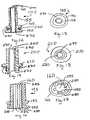
- FIG. 1is a side view of a arterial closure device positioned around a tubular section of a vascular introducer.
- FIG. 2is a side view of the arterial closure device of FIG. 1 advanced through a percutaneous opening by a deployment instrument.
- FIG. 3is a side view of the arterial closure device of FIG. 1 deployed through a vessel wall.
- FIG. 4is a cross-sectional side view of the arterial closure device of FIG. 1 deployed through a vessel wall.
- FIG. 5is a top view of the arterial closure device of FIG. 1 .
- FIGS. 6 and 7are side and cross-sectional side views, respectively, of a second implementation of a arterial closure device deployed within an arteriotomy of a vessel wall.
- FIG. 8is a bottom end view of the arterial closure device of FIG. 6 showing the flared end opened.
- FIG. 9is a bottom end view of the arterial closure device of FIG. 6 showing the flared end closed.
- FIG. 10is a top end view of the arterial closure device of FIG. 6 showing the flared end partially closed.
- FIG. 11is a side view of the arterial closure device of FIG. 6 showing the flared end.
- FIGS. 12 and 13are a cross-sectional side view and a top view, respectively, of the arterial closure device of FIG. 1 having an adhesive on the inner diameter and tissue engagement areas.
- FIGS. 14 and 15are a cross-sectional side view and a top view, respectively, of the arterial closure device of FIG. 6 having an adhesive on the inner diameter and tissue engagement areas.
- FIGS. 16 and 17are a cross-sectional side view and a top view, respectively, of the arterial closure device of FIG. 1 having grooves on the inner diameter to form a thinned or weakened wall.
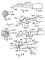
- FIG. 18is a side view of an angled arterial closure device.
- FIG. 19is a side view of an angled arterial closure device having a foldable extending member.
- FIG. 20is a side view of a deployment tool.
- FIG. 21is a side view of the deployment tool of FIG. 20 used to deploy a arterial closure device.
- FIG. 22is an end view of the deployment tool FIG. 20 .
- FIG. 23is a side view of the deployment tool of FIG. 20 having an extended contacting member.
- FIG. 24is a side view of the deployment tool of FIG. 23 used to deploy a arterial closure device.
- FIG. 25is an end view of the deployment tool FIG. 23
- FIG. 26is a cross-sectional side view of a arterial closure device having angled closure edges for compressing a vessel wall.
- FIG. 27is a top view of the arterial closure device of FIG. 26 .
- FIG. 28is a side view of the arterial closure device of FIG. 26 being advanced through the skin into a vessel with the closure edges deflected.
- FIG. 29is a side view of the arterial closure device of FIG. 26 deployed and secured onto vessel wall with the closure edges occluding the arteriotomy.
- FIG. 30is a side view of a arterial closure device.
- FIG. 31is an end view of the arterial closure device of FIG. 30 .
- FIG. 32is a perspective side view of a vascular connector having a closable end.
- FIG. 33is an end view of the arterial closure device of FIG. 32 .
- FIG. 34is a side view of a liner having a longitudinal slot for a arterial closure device.
- FIG. 35is an end view of the liner of FIG. 34 .
- FIG. 36is a side view of a liner having a radial slot.
- FIG. 37is an end view of the liner of FIG. 36 .
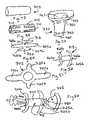
- FIG. 38is a side view of a plug-style arterial closure device that includes an adhesive layer on the vessel contact areas.
- FIG. 39is a side view of a plug style arterial closure device that has limited vessel protrusion and includes an adhesive on the vessel contacting areas.
- FIGS. 40 and 41are side views of the plug style arterial closure device of FIG. 39 being deployed and deployed within a vessel.
- FIG. 42is an end view showing the distal end of the introducer inside a vessel.
- FIGS. 43 and 44are end views showing a flared arterial closure device deployed along the introducer.
- FIG. 45is an end view showing the flared arterial closure device of FIG. 43 deployed against the vessel to close the arteriotomy.
- FIGS. 46 and 47are electrical schematics for a direct resistive element heating circuit and an ohmic tissue heating circuit.
- FIG. 48is a perspective view of a tube used to fabricate a arterial closure device.
- FIG. 49is a perspective view of the tube of FIG. 48 showing material being removed.
- FIG. 50is a side view of the tube of FIG. 48 with the material removed.
- FIG. 51is a top view of the curved configuration.
- FIG. 52is a side view of the configuration of FIG. 51 .
- FIG. 53is a perspective view of a fabric covering.
- FIGS. 54–58are side views showing the fabric covering of FIG. 53 being mounted within the curved configuration of FIG. 51 to form a arterial closure device.
- FIG. 59is a side view of a configuration having side arms that fold over each other.
- FIGS. 60 and 61are front and cross-sectional side views of a deployment tool for deploying the arterial closure device.
- FIG. 62is a cross-sectional side view of the deployment tool of FIG. 60 having the arterial closure device within.
- a vascular closure system 100generally includes two components: a arterial closure device (“ACD”) 105 and a deployment instrument 110 .
- the ACD 105is slidably mounted to a vascular introducer 115 or other tubular device, such as a catheter, advanced over a tube section 120 of the introducer 115 using the deployment instrument 110 , passed through a percutaneous opening 125 , and placed through an arteriotomy 130 in a vessel wall 133 into a blood vessel 135 .
- the deployment tool 110 and the introducer 115then are removed from the blood vessel 135 and out of the percutaneous opening 125 .
- the ACD 105is generally compliant, tubular, and includes a first member 140 , a second member 145 , a connecting member 150 between the first member and the second member, and an optional extending member 155 that extends from the second member.
- a longitudinal channel 160passes between a first opening 165 in the extending member (or second member if the extending member is not present) and a second opening 170 in the first member 140 .
- the ACD 105is formed of a tubular structure of sufficient length and thickness (e.g., a single wall thickness of between 0.005′′ and 0.05′′, and more particularly between 0.01′′ and 0.02′′) that can be advanced over the introducer 115 , and through the puncture site 125 .
- the ACD 105has sufficient rigidity to be advanced through the puncture site 125 yet is compliant enough to be compressed onto itself by the natural elasticity of the vessel wall 133 after the introducer 115 is removed.
- the connecting member 150can be configured to have a natural elasticity such that when it is no longer mounted over the introducer tube 120 , it will return to its original smaller diameter state.
- the ACD 105may include, for example, longitudinal sections of the tube where the wall thickness is thinner (e.g., connecting member 150 ) thereby creating creases or weakened areas that receive the vessel wall 133 .
- the creaseswould reduce the amount of compressive force required to collapse the tube onto itself.
- a design allowing tactile feedbackmay be used to determine the proper insertion position (depth). The tactile feedback could be accomplished by the ACD 105 having one or more rings of increased wall thickness, an “hour glass” geometry, a thin, narrow, then wide geometry, combination, or other means to provide an abrupt change in the advancing force resistance during deployment.
- the ACD 105may be manufactured in many different French sizes, to match the outer diameter of any commercial vascular introducers 115 .
- the ACD 105is placed around the outside of any commercially available introducer 115 , or other device that is inserted into the cardiovascular system (e.g., catheter, etc.), and positioned adjacent to the proximal end of the introducer (i.e., near the valve or luer fitting of the introducer).
- the introducer 115then is inserted into the vasculature using standard techniques.
- the tubular ACD 115Prior to removing the introducer 115 , the tubular ACD 115 is advanced to the skin, for example, by the physician manually advancing the ACD along the tube 120 .
- the deployment instrument 110then is positioned against or clipped onto the tuber 120 , advanced to be in contact with the proximal end (i.e., second member 145 ) of the ACD 105 , and advanced through the skin such that at least the distal most portion (e.g., first member 140 ) of the ACD is inside the vessel 135 .
- the ACD 105is prevented from deforming or collapsing during insertion by the rigidity of the tube 120 .
- the tube 120also acts as a guide to position the ACD 105 through the puncture site 125 during its advancement and deployment.
- the introducer 115is removed, the deployment instrument 110 is held in position and still in contact with the ACD 105 preventing the ACD from coming out of the vessel 135 along with the introducer. Once the introducer 115 is completely removed, the ACD 105 is compressed together due to the elastic recovery of the vessel wall 133 , achieving hemostasis and effectively sealing the arteriotomy 150 and puncture site 125 .
- the ACD 105can be partially or completely fabricated from a biocompatible material, such as expanded polytetrafluoroethylene (ePTFE), polyester, polyurethane, silicone, Dacron, urethane, and/or a composite or combination of these or other suitable materials.
- a biocompatible materialsuch as expanded polytetrafluoroethylene (ePTFE), polyester, polyurethane, silicone, Dacron, urethane, and/or a composite or combination of these or other suitable materials.
- the ACD 105also can be partially or completely fabricated from a biodegradable/bioabsorbable material, including modified cellulose, collagen, fibrin, fibrinogen, elastin or other connective proteins or natural materials, polymers or copolymers such as polylactide [poly-L-lactide (PLLA), poly-D-lactide (PDLA)], polyglycolide, polydioxanone, polycaprolactone, polygluconate, polylactic acid (PLA), polylactic acid-polyethylene oxide copolymers, poly(hydroxybutyrate), polyanhydride, polyphosphoester, poly(amino acids), poly(alpha-hydroxy acid) or related copolymers of these materials as well as composites and combinations thereof and combinations of other biodegradable/bioabsorbable materials.
- PLLApoly-L-lactide
- PDLApoly-D-lactide
- polyglycolidepolydioxanone
- polycaprolactonepolygluconate
- the ACD 105also can be partially or completely fabricated from materials that swell, or expand when they are exposed to a fluid, such as blood, or another fluid, for example, that can be added by the physician to cause the material to swell.
- materialsinclude hydrophilic gels (hydrogels), regenerated cellulose, polyethylene vinyl acetate (PEVA), as well as composites and combinations thereof and combinations of other biocompatible swellable or expandable materials.
- the ACD 105can be made using several methods and processes including extrusion, molding (i.e., injection molding or other known molding techniques), casting, dip coating, spraying, adhesive bonding, ultra-sonic welding, composite fabrication techniques, and combinations of these and/or other similar methods and processes.
- the ACD 105also can have a biocompatible contact adhesive or other material within the longitudinal channel 160 so that when the longitudinal channel is compressed within the arteriotomy 130 , the adhesive bonds the inside surfaces of the longitudinal channel together. This assists or expedites the sealing of the arteriotomy.
- bonding materialscan be used on the outside of the ACD 105 , for example, on the outer surface of the first member 140 , the second member 145 , the connecting member 150 , and or the optional extending member 155 . In particular, the bonding material is especially useful where the ACD contacts the vessel wall 133 defining the arteriotomy 130 .
- the biocompatible contact adhesive adhesive/bonding compounds/solutionscould be added during the manufacturing process, just prior to deployment, or after the device has been deployed.
- the bonding materialscould be in the form of a liquid, semi solid, or solid. Suitable bonding materials include gels, foams and microporous mesh. Suitable adhesives include acrylates, cyanoacrylates, epoxies, fibrin-based adhesives, other biological based adhesives, UV light and/or heat activated or other specialized adhesives.
- the adhesivecould bond on initial contact, or longer, to allow repositioning if desired.
- the preferred adhesivemay be a crystalline polymer that changes from a non-tacky crystalline state to an adhesive gel state when the temperature is raised from room temperature to body temperature.
- Such materialis available under the trade name IntillemerTM adhesive, available from Landec Corp. as well as composites and combinations thereof and combinations of other materials.
- Suppliers of biocompatible adhesivesinclude, but are not limited to, Plasto (Dijon, France), Haemacure (Montreal, Canada), Cohesion (Palo Alto, Calif.), Cryolife (Kennesaw, Ga.), TissueLink (Dover, N.H.), and others.
- the adhesivecan be blended with a material, such as a starch or other material, that retards or delays bonding to allow repositioning of the coupler after it has been deployed.
- a degradable coatingcan be placed over the adhesive coating so that it degrades and exposes the adhesive.
- Other adhesivesare understood to include composites-based adherents and combinations of the above materials and other suitable materials as are known in the art.
- the ACD 105can be fabricated from materials that include one or more radiopaque materials, such as barium sulfate, bismuth trioxide, or other any other radiopaque material.
- the radiopaque materialis added to the materials from which the ACD 105 is fabricated or to the bonding materials that are placed in, on, or around the ACD.
- ACD 200a second implementation of a arterial closure device is shown as a arterial closure device (“ACD”) 200 .
- the ACD 200includes a first member 205 , a second member 210 , and an optional extending member 215 that extends from the second member.
- a longitudinal channel 220passes between a first opening 225 in the extending member (or second member if the extending member is not present) and a second opening 230 in the first member 205 .
- the ACD 200is implanted within an arteriotomy 130 in a manner similar to the implantation of the ACD 105 . However, the ACD 200 does not include a member that is substantially in contact with the inner wall of the vessel 135 .
- the ACDhas a flare, or two or more short slits 235 in the side wall of the first member 205 .
- the flare or slits 235are designed to open or flare around the catheter or introducer 120 when advanced to the top of the vessel puncture site ( FIG. 8 ).
- the materials from which the ACD 200 or the second member 205 are fabricatedmay be a very elastic material such that when around the introducer it expands and when advanced beyond the end of the introducer, it contracts such that the individual flares pinch or otherwise catch the edges of the arteriotomy or punctured vessel and pull them together while contracting ( FIG. 9 ). This action is intended to close the arteriotomy 130 and create hemostasis.
- the inside of the flared section 235 of the ACD 200may have a biocompatible contact adhesive or other bonding material, as described above, that further secures the ACD within the arteriotomy and to the vessel 135 , and, in particular the second member 210 to the top or outer surface of the vessel.
- the adhesive or bonding materialscan be implemented on any of the above ACDs.
- the ACD 105has an adhesive or bonding material 270 on the inner diameter and tissue engagement areas.
- the ACD 200has the adhesive or bonding material 270 on the inner diameter and tissue engagement areas.
- adhesive 270will close the respective longitudinal channel 160 , 200 of the ACD 105 , 200 to reduce or eliminate seepage blood.
- the adhesive 270 around the tissue contacting areaswill bond the ACD to the vessel wall to reduce or eliminate seepage of blood through those regions.
- the ACD 105can have the inner diameter of the longitudinal channel 160 modified to include ridges 280 and channels 285 that weaken or thin the wall section of the ACD.
- the inner diameter of the longitudinal channel 160can be expanded or reduced depending upon the circumferential pressure exerted against the ACD. For example, when passing the introducer through the longitudinal channel the inner diameter will be expanded. When the introducer is subsequently removed, the inner diameter is reduced because of the natural elastic recoil properties of the ACD. In this manner, the seepage of blood through the longitudinal channel is reduced or eliminated.
- the surfaces of the inner diameter of the longitudinal channelcan be coated with an adhesive, as described above, to further ensure that the inner diameter is closed.
- the ACDs described hereinalso can include one or more therapeutic agents that affect healing at the site where the device is deployed.
- the agent(s)can be incorporated into the structure forming the device and/or incorporated into a coating.
- therapeutic agentsmay include, but are not limited to, antithrombotics (such as anticoagulants), antimitogens, antimitotoxins, antisense oligonucleotides, gene therapy solutions, nitric oxide, and growth factors and inhibitors.
- Direct thrombin inhibitorsthat may be beneficial include Hirudin, Hirugen, Hirulog, PPACK (D-phenylalanyl-L-propyl-L-arginine chloromethyl ketone), Argatreban, and D-FPRCH.sub.2 Cl (D-phenylalanyl-L-propyl-L-arginyl chloromethyl ketone); indirect thrombin inhibitors include Heparin and Warfarin (coumadin). Alternatively, a clot promoter may be used, such as protamine sulphate or calcium hydroxide.
- Additional therapeutic materialsinclude, aspirin, dexamethasone, dexamethasone phosphate, streptokinase, tocopherol, TPA, urokinase, paclitaxel (Taxol), actinomycin, rapamyacin, or other. Sirolimus, or other antibiotics may also be used.
- the therapeutic compounds/solutionsmay be blended with the device base materials during fabrication, applied just prior to deployment, or after the device has been deployed. Additionally, the therapeutic materials may be located on, through, inside, or combination of the device in holes, grooves, slots or other indentation to allow elution of the therapeutic compound(s).
- Post device fabrication coating methodsinclude, but are not limited to, dipping, spraying, brushing, submerging the devices into a beaker containing a therapeutic solution while inside a vacuum chamber to permeate the device material, etc.
- the geometry of the ACDs described hereinis shown for illustration purposes as being generally round. However, they can be of any other geometry, such as oval, elliptical, rectangular, square, ridged, or a combination of shapes.
- the ACDhas been illustrated as forming a generally perpendicular angle with the vessel wall once deployed. Nonetheless, the inventors intend the configuration to be at any suitable angle, such as between 30° and 60°, or, for example, 45° or as otherwise desired.
- a range of angles of the ACDcan be available and the physician can choose the appropriate ACD based on the angle at which the introducer is introduced into the vessel. For example, referring to FIG.
- a ACD 290is formed to have the extending member 155 extending at an angle of approximately 45° from the second member 145 .
- the first member 140 and the second member 145are longitudinally offset. This configuration is designed to cause the extending member 155 to follow the path created by the introducer.
- the ACDhas a second member 292 , a foldable extending member 294 , and a groove 296 positioned between the second member 292 and the folding extending member 294 . In this manner, the extending member 294 can be folded or bent over to be less obtrusive and to close off the flow of blood through the ACD.
- a deployment tool 300is designed to engage or otherwise contact the proximal edge, or other edge, of the ACD.
- the tool 300is generally handheld and includes a handle 305 , an extension 310 , and a contacting section 315 that clips onto, or otherwise contacts the outside of the introducer and mates with the ACD.
- the contacting section 315has sufficient length to advance the ACD through the tissue to the desired position on the vessel.
- the handle 305 or grasping sectioncan be, for example, round, rectangular, elliptical, or a combination of shapes or other shape that fit comfortably in the hand.
- the contacting section 315can have a cross-sectional geometry of a partially open tube having more than 50% diameter coverage, so that it can clip onto, and slide over the outer diameter of the introducer.
- the deployment toolcan include an additional extension 320 that is configured to fit around the extending member 155 and mate with the second member 145 .
- the extension 320can be attached to the introducer after the introducer is positioned within the artery.
- the deployment tool 300can be made partially or completely from several different polymer materials including polycarbonate, nylon, polyethylene, polytetrafluoroethylene (PTFE), fluoroethylene-propylene (FEP) or polyfluoroacrylate (PFA), polyester ether ketone (PEEK), polyamide, polyimide, polyethyleneteraphthalate (PET), combination or other material able to withstand sterilization processing.
- the toolcan also be made partially or completely from several different types of metals including stainless steel; spring metal alloys such as elgiloyTM, inconelTM; superelastic/shape memory alloys such as Nitinol (NiTi) as well as composites and combinations thereof and combinations of other materials.
- the deployment tool 300can be made using several methods and processes including extrusion, molding (injection and other), casting, adhesive bonding, ultrasonic welding as well as combinations thereof and combinations of other methods and processes.
- the proximal edge of the ACDi.e., of the extending member 155 or the second member 145
- the distal edge or other portion of the advancement tool 300may have interlocking geometries to aid and/or control the position of the ACD during advancement along the introducer.
- the engagement/contact section 315 , 320 of the tool 300may have a cross-sectional geometry of a complete circle that is designed to split away from the introducer once the ACD has been advanced and deployed. Splitting can be accomplished by having weakened areas in the wall of the tubing, such as linear perforations, or linear scores. This version would require that the deployment tool be back loaded onto the introducer before the ACD is placed onto the introducer and prior to insertion into the vessel.
- the inside, concave section of the contact section 315 , 320may be coated with a hydrophilic or other lubricious material to reduce the friction during advancement and deployment of the ACD.
- the contacting section 315 , 320 of the toolcan be lengthened and designed to further attach to and compress the distal edge of the ACD, thereby providing additional support during insertion and deployment into the vessel.
- a ACD 350includes a first angled closure edge 355 , a second angled closure edge 360 , an extending member 365 , and a connection member 370 between the first and second angled closure members.
- the first angled closure edge 355 and the second angled closure edgeare generally directed at each other such that they define a narrow opening 375 through which the vessel wall 133 is received.
- the ACD 350is deployed over the introducer tube section 120 using, for example, the deployment tool 300 .
- the second angled closure edge 360is deflected away from the first angled closure edge 355 . The deflection can be caused, for example, by the contacting section 320 surrounding the second angled closure edge 360 .
- the angled closure edges 355 and 360are formed, for example, from a flexible member, such as a polymer, superelastic/shape memory material, or a combination of the two.
- the superelastic/shape memory membercan be coated with a polymer.
- a ACD 400includes a threaded section 405 and an extending section 410 .
- the threaded section 405includes threads 415 mounted on and between a first member 420 and a second member 425 .
- the extending section 410includes a longitudinal channel 430 that includes a distal shaped channel 435 .
- a deployment tool having a mating shaped distal endis inserted into the longitudinal channel 430 such that it mates with the distal shaped channel 435 . By rotating the deployment tool, the ACD can be threadably inserted into the arteriotomy.
- the distal edge of the ACD 400is designed to engage the opening of the arteriotomy or puncture site and protrude to a specific depth based on how many times the ACD was advanced, twisted or turned.
- the ACD 400may have a stop 437 to limit how far the device protrudes into the vessel.
- the same “screw” type distal edgecould be used on a hemostatic plug, made from a solid piece of material, rather than a tube structure.
- a deployment toolwould be needed that has, for example, a grasping distal end for insertion into the vessel.
- the ACD 400can be modified to include a longitudinal channel that pass through the entire length of the device and deployed over a introducer.
- the deployment tool and the proximal edge of the ACDwould have a mating geometry such that the deployment tool is rotated to threadably insert the ACD through the arteriotomy.
- a ACD 450includes a tissue contacting member 455 and an extending member 460 .
- a longitudinal channel 463passes through the ACD.
- the extending member 460includes a longitudinal slot 465 and a circumferential channel 470 in which a contracting member 475 is received.
- the contracting member 475tends to close the longitudinal channel 463 unless kept open, for example, by an introducer 115 within the channel. In this manner, when the ACD 450 is deployed within the arteriotomy and the introducer is removed, the longitudinal channel is closed, which prevents or limits blood flow or seepage through the channel.
- the ACDcan be formed from any of the materials described above.
- the ACDcan be formed from a polymer and the extending member can be formed from a flexible material such as a polyurethane/Dacron composite that easily collapses as a consequence of contraction property of the contracting member 475 .
- a ACD inner liner 480is formed as a simple slotted tube 485 that includes a slot 490 along its length that functions a means for side access onto the introducer, after the introducer has been inserted into the vessel.
- the slot 490can be formed as a longitudinal or radial slit, illustrated below.
- the ACD inner linercan be opened sufficiently to attach onto the introducer from the side. Any configuration of the ACDs described herein is built around the ACD liner 480 with a slot formed within the ACD.
- the tube 485optionally can extend from the ACD and then be clamped at the proximal end once the ACD liner 480 and ACD are deployed.
- a ACD liner 500includes a tube 505 that includes a radial slot 510 along an extending member 515 and through a first member 520 and a second member 525 .
- the ACD inner liner 500is sufficiently openable to be threaded onto the introducer from its side. Any configuration of the ACDs described herein can be built around the ACD liner 500 with a slot formed within the ACD.
- the tube 505optionally can extend from the ACD and then be clamped at the proximal end once the ACD liner 500 and ACD are deployed.
- a plug style ACD 550that is similar to ACD 105 includes a channel 555 into which a deployment tool 552 is inserted to deploy the ACD through an arteriotomy to close the arteriotomy.
- the ACDincludes an adhesive layer 560 for bonding to the tissue.
- the ACD 550differs from the ACD 105 in that the channel 555 does not extend the entire length of the ACD.
- a ACD 570( FIG. 39 ) is similar to the ACD 550 except that it has limited vessel protrusion, similar to the ACD 200 above.
- the ACD 550 , 570is placed into the arteriotomy and held briefly for an adhesive bond to form.
- the deployment device 552then can be removed.
- the distal end of the deployment tool 552also can have a grasping feature to grasp the proximal end of the plug ACD during deployment and to release after the plug ACD has been seated in or is on the vessel, and able to release when the tool is being withdrawn.
- a ACDcan have a distal end geometry, which once positioned at the puncture site, is designed to compress the vessel wall for increased securement and sealing.
- a ACD 600may have a flare 605 , or two or more longitudinal slits in the side of the tube, that are designed to open, or flare apart when advanced and in contact with the top of the vessel puncture site (i.e., arteriotomy).
- the ACD 600can be made from a very elastic material and/or a superelastic/shape memory material such that when the introducer is removed, the flares or slits will pinch, or otherwise bring the edges of the punctured vessel together, effectively creating hemostasis.
- the inside of the flared section of the closure devicecould have biocompatible contact adhesive, other bonding material, and/or small barbs or protrusions that may assist in securing the device to the top of the vessel wall.
- heatcan be used to assist with, or as an adjunct to, the process by recovering the ACD, activating (e.g., causing to flow, etc.) a hemostatic material to the puncture site that assists in sealing (e.g., through vessel contraction including the denaturing and reformation of collagen at the site) or accelerate healing, or a combination of these or other beneficial effects.
- Direct resistive element heatingFIG. 46
- ohmic tissue heatingFIG. 47
- Biocompatible electrode materialscan be mixed with the base material of the ACD as a powder during manufacturing, or as a wire, strip, or other geometry, added onto any surface of the device, and connected to a suitable (i.e., electrical and biocompatible) conductor.
- a suitable conductori.e., electrical and biocompatible
- one conductor 620is connected to an RF power source.
- Another conductoris connected to a ground pad 630 placed on the patient's body, and also connected to the power source.
- both conductors from the power source 625are connected to an electrode 635 .
- a twisting, cutting, or other manipulative actionremoves the conductor previously attached to the closure device.
- a special tipis placed over a standard electro surgical tool (e.g., Bovie) to insert through the skin and make contact with the closure device, tissue or both.
- Bovieelectro surgical tool
- Alternative versions of the closure devicemay utilize an electrode that is formed by ion deposition, sputter coating, spraying, dip coating, adhesive, combination or other method or design.
- a superelastic/shape memory ACD 700is made from a superelastic/shape memory sheet or tube 705 .
- the sheet or tube 705is etched, cut, or otherwise machined to remove material 710 ( FIG. 49 ) to leave a starting configuration 715 ( FIG. 50 ).
- the method of removing the materialmay be, for example, photo-etching and/or laser or chemical cutting.
- the starting configurationincludes first extending members 720 , second extending members 725 , and a connecting member 730 between the first and second extending members.
- the first and second extending members 720 and 725then are bent and curved ( FIGS. 51 and 52 ).
- the first and second extending membersare curved to mate with the inner and outer surface, respectively, of a vessel.
- longer first and second extending members 720 a and 725 aare bent to be generally perpendicular to the connecting member 730 and have a curvature that is similar to that of the length dimension of a vessel wall.
- the shorter first and second extending members 720 b and 725 bare bent to have a radius of curvature that is similar to that of the radius of curvature of the circumference of a vessel wall.
- the shapes of the first and second extending members 720 , 725are set using known techniques of imparting shapes in superelastic/shape memory materials, as described in further detail below.
- a fabric covering 740( FIG. 53 ), such as Dacron, then is mounted to the curved configuration 715 .
- the covering 740includes distal side openings 745 and proximal side openings 750 .
- a longitudinal channel 755passes between a distal opening 760 and a proximal opening 765 .
- the covering 740is pulled distal end through the curved configuration 715 and the extending members 720 are straightened from their retracted state and passed through the distal side openings 745 ( FIG. 54 ).
- the covering 740then is pulled back such that the distal side openings 745 are tight against the first extending members 720 ( FIG. 55 ).
- the first extending members 720then are allowed to expand back to their retracted state.
- the second extending members 725then are straightened from their retracted state and passed through the proximal side openings 750 ( FIG. 56 ).
- the second extending members 725then are allowed to expand back to their retracted state, thereby trapping a proximal end 760 of the covering against the connecting member 730 between the first and second extending members 720 , 725 ( FIGS. 57 and 58 ).
- the longitudinal channel 755passes through the covering 740 and the shaped configuration 715 .
- the second extending members 725 bcan be configured to curve back over and under the opposite second extending member 725 b .
- the second extending members 725 binstead of curving against the outer circumference of the vessel in which the device is implanted, the second extending members 725 b function to close the longitudinal channel 755 when they are in their retracted position.
- the covering 740is mounted to the curved configuration 715 as described above.
- the second extending members 725 bare kept in a straightened position because of the introducer or catheter that passes through the longitudinal channel 755 .
- the second extending members 725 breturn to their retracted position, thereby closing or partially closing the longitudinal channel 755 .
- the covering 740also contributes to the closure of the longitudinal channel 755 and reduction or elimination of blood leakage or seepage through the longitudinal channel.
- the deployment tubeincludes a handle 780 , an extension 783 , a guide 786 , and a pusher tuber 789 .
- the guide 786extends from the extension 783 and includes a first longitudinal channel 791 and a longitudinal ridge 792 that passes along the inner surface of the first longitudinal channel 791 .
- the pusher tube 789is slidably mounted within the first longitudinal channel 791 and includes a second longitudinal channel 793 , a pusher surface 794 , and a groove 796 that is configured to slide over the longitudinal ridge 792 .
- the guide 786 and the pusher tube 789include longitudinal slots 797 , 798 so that the deployment tool 775 can be placed around the catheter or introducer.
- the physicianpushes the ACD 700 along the introducer 120 into the vessel using the pusher tube 789 .
- the ACDcan be placed within an arteriotomy using other deployment tools or even by hand.
- the ACDs hereinmay contain a metallic braid, coil, sheet, strip, wire, rod, or other configuration on the inner diameter, outer diameter, within, and/or a combination of these.
- the metallic materialcould be made from superelastic/shape memory alloys such as Nitinol.
- the metallic braid or coilcould be annealed in one configuration during manufacture and processed and packaged in another configuration. When the material is exposed to normal body temperature (i.e., 37° C.), it will be set to either expand apart or contract inward depending on the design and annealed geometry (diameter). This characteristic may assist with the closure of the ACD.
- Elasticityis the ability of the metal, under a bending load, for example, to deflect (i.e., strain) and not take a permanent “set” when the load (i.e., stress) is removed.
- Common elastic metalscan strain to about two percent before they set.
- Superelastic metalsare unique in that they can withstand up to about ten percent strain before taking a set. This is attributed to a “stress-induced” phase change within the metal to allow it to withstand such dramatic levels of strain. Depending on the composition of the metal, this temperature that allows such a phase change can vary.
- the metalcan return to an “unset” shape. Then, upon returning to the previous “set” temperature, the shape changes back. This is a “shape-memory” effect due to the change in temperature changing the phase within the metal.
- Elasticityis a key feature of superelastic materials. When a metal is loaded (i.e., stressed) and undergoes, for example, bending, it may deflect (i.e., strain) in a “springy” fashion and tend to return to its original shape when the load is removed, or it may tend to “set” and stay in a bent condition. This ability to return to the original shape is a measure of the elasticity or “resilience” of the metal. This ability for a metal to be resilient is desirable for such things as springs, shock absorbing devices, and even wire for orthodontic braces where the ability to deflect, but not deform (i.e., set) is important to maintain an applied force.
- the metaltakes a set, it is said to have plastically (versus elastically) deformed. This is because the imposed stress, produced by the bending load, has exceeded the “yield strength” (stress) of the metal.
- yield strengthstress
- this level of stress that produces a setis referred to as the “elastic limit”, but is about the same as the yield strength. If the applied load increases past the yield strength of the metal, it will produce more plasticity and can eventually break. The higher the yield strength of the metal, the more elastic it is. “Good” elastic metals can accommodate up to about two percent strain prior to taking a set. But this is not the only factor governing “elasticity”.
- the modulus of the metalis an inherent property. Steels, for example, have a relatively high modulus (30 msi) while the more flexible aluminum has a lower modulus of about 10 msi.
- the modulus for titanium alloysis generally between 12 and 15 msi.
- Resilienceis the overall measure of elasticity or “spring-back ability” of a metal.
- the ratio of the yield strength divided by the modulus of the metalis the resilience. Although it is one thing for a metal to be resilient, it must also have sufficient strength for the intended service conditions.
- each increment of loadproduces a given increment of deflection (strain) within the metal. And the metal remains elastic if the applied is below the yield stress.
- the metal alloysthat behave in an even more elastic manner. These are the “superelastic” metals, where, for a given applied stress (load) increment, the strain in the metal can reach 5 or 6 percent or more without taking a set. In these types of metals, the overall strain required to produce a set can reach an impressive 10 percent. This phenomenon is related to a phase change within the metal, and which is induced by the applied stress. This “stress-induced” phase change can also allow the metal to be set at one temperature and return to another shape at another temperature. This is a “shape-memory” effect, discussed below.
- the most common superelastic metalused in many commercial applications, is an alloy comprised of about equal parts of nickel (Ni) and titanium (Ti), and has a trade name of “Nitinol”. It is also referred to as “NiTi”.
- NiTinickel
- the stability of the internal phases in the metalcan be changed. Basically, there are two phases: (1) an “austenite” phase and (2) a lower-temperature, “martensite” phase. When the metal is in an austenitic phase condition and is stressed, then a stress-induced martensite forms, resulting in the super-elasticity. This is reversible, and the original shape returns upon release of the applied stress.
- the Ni-to-Ti ratio in the Nitinolis selected so that the stress-induced martensite forms at ambient temperatures for the case of super-elastic brace and support devices, which are used in ambient conditions.
- the specific compositioncan be selected to result in the desired temperature for the formation of the martensite phase (Ms) and the lower temperature (Mf) at which this transformation finishes. Both the Ms and Mf temperatures are below the temperature at which the austenite phase is stable (As and Af).
- the performance of an ACDcan be further enhanced with the use of superelastic materials such as Nitinol.
- the superelasticityallows for greatly improved collapsibility, which will return to its intended original shape when the introducer (or catheter) is removed from the inside of the ACD.
- the high degree of flexibilityis also more compatible with the stiffness of the engaged vessel.
- NitinolBy manipulating the composition of Nitinol, a variety of stress-induced superelastic properties can result, and over a desired, predetermined service temperature range. This allows the metal to behave in a “shape-memory” or “shape recovery” fashion.
- the metalis “set” to a predetermined, desired shape at one temperature when in a martensitic condition, and which returns to the original shape when the temperature is returned to the austenitic temperature.
- the shape memory phenomenonoccurs from a reversible crystalline phase change between austenite and the lower-temperature martensite.
- This transformationoccurs from an induced stress as described previously, it can, of course, also change with temperature variations.
- This transformationis reversible, but the temperatures at which these phase changes start and finish differs depending on whether it is heated or cooled. This difference is referred to as a hysteresis cycle.
- This cycleis characterized by the four temperatures mentioned previously, As, Af, Ms, and Mf.
- the transformation to austenitebegins at the As, and will be fully austenite at Af.
- austenitewill begin to transform back to martensite at the Ms temperature, and become fully martensitic at the Mf.
- the specific composition of the alloycan result in a desired combination of these four transformation temperatures.
- the alloyIn the malleable martensitic state, the alloy can be easily deformed (set). Then upon heating back to the austenitic temperature, the alloy will freely recover back to its original shape. Then if cooled back to the martensitic state, the deformed shape reforms.
- the typical sequence of utilizing this shape memory propertyis to set the shape of, for example, a stent or anastomosis connector, while in the higher-temperature austenitic state. Then, when cooled, deform the martensite material, and then heat to recover the original shape.
- Nitinol materialrequires an exceptionally tight bend, and one that would normally exceed the elastic limit of the material and thus permanently deform it
- a bendcan be placed in the device and the device annealed to relieve bending stresses within the device.
- the devicecan be bent further to produce an even sharper bend, and then re-annealed to alleviate the stress from this additional bending.
- This processcan be repeated to attain the desired, sharp bend or radii that would otherwise permanently deform the device if the bend were attempted in a single bending event.
- the process for recovery from the position of the most recent bendis then performed as described above.
- Nitinolis, by far the most popular of the superelastic metals, there are other alloys that can also exhibit superelastic or shape-memory behavior. These include the following:
- Nitinolbecause of the large amount of titanium in the composition, has been the only FDA approved superelastic/shape memory alloy for medical implant devices.
- the corrosion resistance of Nitinolis superior to that of commonly used 3161 stainless steel, and, if surface oxidized or passivated carefully, can reach corrosion resistance comparable to the most popular titanium implant alloy, Ti6Al4V.
- the metal piececan be electropolished to improve its biocompatibility and blood compatibility. Biocompatibility studies have routinely showed Nitinol as a metal with suitable biocompatibility for medical device applications.
- Nitinolis also biocompatible and corrosion resistant. This unique combination of properties allows a device made of Nitinol, such as an arterial closure device, to be fully collapsed within a deployment tool and be subsequently released, at a particular site within the vessel, to form its intended service shape.
- Materials other than superelastic/shape memory alloysmay be used as reinforcements provided they can be elastically deformed within the temperature, stress, and strain parameters required to maximize the elastic restoring force thereby enabling the tubular closure device to recover to a specific diameter and/or geometry once deployed inside, over, or on top of the vessel or other location.
- Such materialsinclude other shape memory alloys, spring stainless steel 17-7, other spring metal alloys such as elgiloyTM, inconelTM, superelastic polymers, etc.
- the superelastic/shape memory material(s), previously cut into the desired pattern and/or length,are stressed into the desired resting configuration over a mandrel or other forming fixture having the desired resting shape of the tubular plug, depending on the vessel size or other location where the ACD or plug is intended to be used, and the material is heated to between 300 and 650° Celsius for a period of time, typically between 30 seconds and 30 minutes.
- the superelastic materialis quenched by inserting into chilled water or other fluid, or otherwise allowed to return to ambient temperature.
- the superelastic reinforcementsare fabricated into their resting configuration.
- the superelastic/shape memory reinforcementsmay be full or partial length or width of the ACD or tubular plug.
- Electropolishingmay reduce platelet adhesion causing thrombosis, and encourage endothelization of the exposed metallic areas. Electropolishing also beneficially removes or reduces flash and other artifacts from the fabrication of the device.
- Superelastic/shape memory materialssuch as tubular, rectangular, wire, braid, flat, round, combination or other structures also can be used in the design of the closure device, to assist with grasping, contacting, bringing tissue together, sealing, or other desired function.
- the superelastic/shape memory materialscould be used to resist compressive closure and act as a flexible reinforcing strain relief to prevent kinking and to prevent the conduit from closing.
- the engagement/contact section of the deployment toolcan have a cross sectional geometry of a complete circle that may be designed to split away from the introducer once the closure device has been advanced/deployed. Splitting could be accomplished by having thinned or weakened areas in the wall of the deployment device tubing, such as linear perforations, or linear scores, combination, or other perforation configuration. This version would require that the deployment tool be back loaded onto the introducer before the closure device is placed onto the introducer and prior to insertion into the vessel.
- the deployment toolcan be a clip-on tool, can compress the device to reduce the cross sectional profile prior to insertion and/or may include a constraining sheath to reduce a section, or sections of the device during insertion to the target site.
- This versionwould be particularly useful for bringing two tissue walls together while yet providing a conduit between the tissues.
- the proximal end of the ACDs described hereinmay be closed using hemostats, or other tools, by pinching the end together until the inner diameter bonds, or compresses together. Adhesive may be used to assist in the closure of the device.
- the proximal edge of the closure device and the distal (or other) edge of the advancement/deployment toolcan have interlocking geometries to aid control during advancement (particularly when inserting by twisting or turning while advancing into the vessel).
- the proximal edge or end of the closure devicemay have a collar made of a superelastic/shape memory material, an elastic combination of materials or suitable elastic materials that would compress the end of the device together once the introducer is removed from the inner diameter of the closure device.
- the closing and sealing of the devicemay be enhanced with an adhesive, swellable material, or other coating or layer.
- the closure meanscan be other than a tubular structure, such as a plug.
- a special introducerhaving multi-lumens, one for the catheter or other device, and at least one for the hemostatic plug material.
- the hemostatic material, and matching geometry plungerwould be inserted into the proximal end of the special introducer. As the plunger is advanced, the hemostatic material is advanced into position and the introducer is withdrawn from the vessel.
- the basic device, system and methodcan be sized and configured for medical devices other than vascular introducers, such as guide wires, catheters, laparoscope, endoscope, trocar, cannula, electrode wire, or other.
- medical devicesother than vascular introducers, such as guide wires, catheters, laparoscope, endoscope, trocar, cannula, electrode wire, or other.
- tubular closure deviceespecially when made from swellable material, as a reversible sterilization method for women by occluding the fallopian tubes, and men by occluding the vas ducts or tubes.
- a modified version of the device and systemcan be used for the closure of septal defects in the heart, as well as anywhere else in the body.
- the clip on section of the deployment toolwould be modified to fit onto the catheter, and be long enough (such as, e.g., full catheter length) to be remotely advanced from the proximal end of the catheter.
- the deployment toolalso may be modified and used to compress the device during insertion into the body to thereby reduce the cross-sectional profile during insertion.
- the deployment methodmay be enabled by longitudinal movement, manipulation, or retraction of the deployment tool away from the closure device, which removes the compression of the device and allows the device to expand and fill in the opening, such as a septal defect.
- the ACDs described hereincan be used for cardiovascular applications where hemostasis (temporary or permanent) is desired. Additionally, the ACDs can be used with simple modifications for any tubular, duct, organ, hollow body cavity, or other structures or tissues, where temporary or permanent sealing or plugging is needed, or alternatively, where a conduit or conduit reinforcement is desired. For conduit or conduit reinforcement applications, the material and design used thereby would be sufficiently resistant to compressive closure while still remaining flexible, e.g., longitudinally and/or radially flexible.
- the ACDs described hereinalso can be used for gastric bypass procedures, general tissue bunching or bringing tissues together, and on or in other vessels, organs, tissues, bones, and/or other body tissues than those specifically described.
Landscapes
- Health & Medical Sciences (AREA)
- Surgery (AREA)
- Life Sciences & Earth Sciences (AREA)
- Biomedical Technology (AREA)
- Nuclear Medicine, Radiotherapy & Molecular Imaging (AREA)
- Engineering & Computer Science (AREA)
- Cardiology (AREA)
- Heart & Thoracic Surgery (AREA)
- Medical Informatics (AREA)
- Molecular Biology (AREA)
- Animal Behavior & Ethology (AREA)
- General Health & Medical Sciences (AREA)
- Public Health (AREA)
- Veterinary Medicine (AREA)
- Surgical Instruments (AREA)
Abstract
Description
- Copper—40 at % Zinc
- Copper—14 wt % Aluminum-4 wt % Nickel
- Iron—32 wt % Manganese-6 wt % Silicon
- Gold—5 to 50 at % Cadmium
- Nickel—36 to 38 at % Aluminum
- Iron—25 at % Platinum
- Titanium—40 at % Nickel-1 Oat % Copper
- Manganese—5 to 35 at % Copper
- Titanium—49 to 51 at % Nickel (Nitinol)
Claims (21)
Priority Applications (10)
| Application Number | Priority Date | Filing Date | Title |
|---|---|---|---|
| US10/224,659US7025776B1 (en) | 2001-04-24 | 2002-08-21 | Arteriotomy closure devices and techniques |
| US10/825,785US20050113810A1 (en) | 2001-04-24 | 2004-04-16 | Shaping suture for treating congestive heart failure |
| US10/825,783US20050125012A1 (en) | 2002-06-28 | 2004-04-16 | Hemostatic patch for treating congestive heart failure |
| US10/825,786US20050113811A1 (en) | 2001-04-24 | 2004-04-16 | Method and devices for treating ischemic congestive heart failure |
| US10/826,028US20050096498A1 (en) | 2001-04-24 | 2004-04-16 | Sizing and shaping device for treating congestive heart failure |
| US11/116,600US7441342B2 (en) | 2001-04-24 | 2005-04-27 | System and method for sizing a heart for treating congestive heart failure |
| US11/933,129US20080114394A1 (en) | 2001-04-24 | 2007-10-31 | Arteriotomy Closure Devices and Techniques |
| US11/930,111US20080109030A1 (en) | 2001-04-24 | 2007-10-31 | Arteriotomy closure devices and techniques |
| US12/167,212US8518063B2 (en) | 2001-04-24 | 2008-07-02 | Arteriotomy closure devices and techniques |
| US12/563,297US8992567B1 (en) | 2001-04-24 | 2009-09-21 | Compressible, deformable, or deflectable tissue closure devices and method of manufacture |
Applications Claiming Priority (6)
| Application Number | Priority Date | Filing Date | Title |
|---|---|---|---|
| US28626901P | 2001-04-24 | 2001-04-24 | |
| US30089201P | 2001-06-25 | 2001-06-25 | |
| US30225501P | 2001-06-28 | 2001-06-28 | |
| US12771402A | 2002-04-23 | 2002-04-23 | |
| US10/224,659US7025776B1 (en) | 2001-04-24 | 2002-08-21 | Arteriotomy closure devices and techniques |
| US27924206A | 2006-04-10 | 2006-04-10 |
Related Parent Applications (2)
| Application Number | Title | Priority Date | Filing Date |
|---|---|---|---|
| US12771402AContinuation | 2001-04-24 | 2002-04-23 | |
| US10/183,396Continuation-In-PartUS6726696B1 (en) | 2001-04-24 | 2002-06-28 | Patches and collars for medical applications and methods of use |
Related Child Applications (3)
| Application Number | Title | Priority Date | Filing Date |
|---|---|---|---|
| US78548604AContinuation | 2001-04-24 | 2004-02-24 | |
| US10/825,785ContinuationUS20050113810A1 (en) | 2001-04-24 | 2004-04-16 | Shaping suture for treating congestive heart failure |
| US27924206AContinuation | 2001-04-24 | 2006-04-10 |
Publications (1)
| Publication Number | Publication Date |
|---|---|
| US7025776B1true US7025776B1 (en) | 2006-04-11 |
Family
ID=39370188
Family Applications (1)
| Application Number | Title | Priority Date | Filing Date |
|---|---|---|---|
| US10/224,659Expired - Fee RelatedUS7025776B1 (en) | 2001-04-24 | 2002-08-21 | Arteriotomy closure devices and techniques |
Country Status (1)
| Country | Link |
|---|---|
| US (1) | US7025776B1 (en) |
Cited By (155)
| Publication number | Priority date | Publication date | Assignee | Title |
|---|---|---|---|---|
| US20030009180A1 (en)* | 1999-09-13 | 2003-01-09 | Hinchliffe Peter W.J. | Vascular hole closure device |
| US20030144695A1 (en)* | 1999-09-13 | 2003-07-31 | Mcguckin James F. | Vascular hole closure device |
| US20040176798A1 (en)* | 1997-02-11 | 2004-09-09 | Cardiva Medical, Inc. | Expansile device for use in blood vessels and tracts in the body and method |
| US20050033326A1 (en)* | 1999-09-13 | 2005-02-10 | Briganti Richard T. | Vascular hole closure device |
| US20050267520A1 (en)* | 2004-05-12 | 2005-12-01 | Modesitt D B | Access and closure device and method |
| US20050288706A1 (en)* | 2004-05-07 | 2005-12-29 | Nmt Medical, Inc. | Inflatable occluder |
| US20060155327A1 (en)* | 1999-09-13 | 2006-07-13 | Briganti Richard T | Vascular hole closure device |
| US20060212063A1 (en)* | 2005-03-18 | 2006-09-21 | Wilk Patent, Llc | Surgical device and associated trans-organ surgical method |
| US20060247680A1 (en)* | 2004-04-08 | 2006-11-02 | Aga Medical Corporation | Flanged occlusion devices and methods |
| US20060276839A1 (en)* | 1999-09-13 | 2006-12-07 | Rex Medical | Septal defect closure device |
| US20070010853A1 (en)* | 2000-10-06 | 2007-01-11 | Integrated Vascular Systems, Inc. | Apparatus and methods for positioning a vascular sheath |
| US20070106246A1 (en)* | 2005-05-12 | 2007-05-10 | Modesitt D B | Access and closure device and method |
| US20070118162A1 (en)* | 2005-11-22 | 2007-05-24 | Michael Abi-Kheirs | Shape memory hemostasis band |
| US20070135837A1 (en)* | 2005-12-13 | 2007-06-14 | Cardiva Medical, Inc. | Drug eluting vascular closure devices and methods |
| US20070198058A1 (en)* | 2006-02-21 | 2007-08-23 | Daniel Gelbart | Method and device for closing holes in tissue |
| US20070255402A1 (en)* | 2006-05-01 | 2007-11-01 | Moore Jacob J | Wound closure method |
| US20070270891A1 (en)* | 2005-04-22 | 2007-11-22 | Mcguckin James F Jr | Closure device for left atrial appendage |
| US20070270688A1 (en)* | 2006-05-19 | 2007-11-22 | Daniel Gelbart | Automatic atherectomy system |
| US20080004697A1 (en)* | 2006-06-28 | 2008-01-03 | Samuel Victor Lichtenstein | Method for anchoring a mitral valve |
| US20080004534A1 (en)* | 2006-06-28 | 2008-01-03 | Daniel Gelbart | Intra-cardiac mapping and ablation method |
| US20080004640A1 (en)* | 2006-06-28 | 2008-01-03 | Abbott Laboratories | Vessel closure device |
| US20080045778A1 (en)* | 2006-08-02 | 2008-02-21 | Samuel Victor Lichtenstein | System for improving diastolic dysfunction |
| US20080103593A1 (en)* | 2006-11-01 | 2008-05-01 | Ortiz Mark S | Use of Biosurgical Adhesive on Inflatable Device for Gastric Restriction |
| US20080154303A1 (en)* | 2006-12-21 | 2008-06-26 | Cardiva Medical, Inc. | Hemostasis-enhancing device and method for its use |
| US20080217017A1 (en)* | 2007-03-06 | 2008-09-11 | James Michael Brown | Method of treating well with foamed composition |
| US20090043314A1 (en)* | 2007-07-16 | 2009-02-12 | The Cleveland Clinic Foundation | Access device for body tissue and method of using same |
| US20090076597A1 (en)* | 2007-09-19 | 2009-03-19 | Jonathan Micheal Dahlgren | System for mechanical adjustment of medical implants |
| US20090105744A1 (en)* | 2007-10-17 | 2009-04-23 | Modesitt D Bruce | Methods for forming tracts in tissue |
| US20090131930A1 (en)* | 2007-11-16 | 2009-05-21 | Daniel Gelbart | Medical device for use in bodily lumens, for example an atrium |
| US20090143789A1 (en)* | 2007-12-03 | 2009-06-04 | Houser Russell A | Vascular closure devices, systems, and methods of use |
| US20090187213A1 (en)* | 2007-07-13 | 2009-07-23 | Mcguckin Jr James F | Vascular hole closure device |
| US20090192441A1 (en)* | 2008-01-25 | 2009-07-30 | Daniel Gelbart | Liposuction system |
| US7572274B2 (en) | 2004-05-27 | 2009-08-11 | Cardiva Medical, Inc. | Self-tensioning vascular occlusion device and method for its use |
| WO2009099437A1 (en)* | 2008-02-05 | 2009-08-13 | Boston Scientific Limited | Apparatus and method for closing an opening in a blood vessel using memory metal and collagen |
| US20090227938A1 (en)* | 2008-03-05 | 2009-09-10 | Insitu Therapeutics, Inc. | Wound Closure Devices, Methods of Use, and Kits |
| US20090287304A1 (en)* | 2008-05-13 | 2009-11-19 | Kardium Inc. | Medical Device for Constricting Tissue or a Bodily Orifice, for example a mitral valve |
| WO2010002918A1 (en) | 2008-06-30 | 2010-01-07 | Cardiva Medical, Inc. | Apparatus and methods for delivering hemostatic materials for blood vessel closure |
| US20100016786A1 (en)* | 2008-07-21 | 2010-01-21 | Arstasis, Inc. | Devices, methods, and kits for forming tracts in tissue |
| US7678133B2 (en) | 2004-07-10 | 2010-03-16 | Arstasis, Inc. | Biological tissue closure device and method |
| US20100115407A1 (en)* | 2008-11-05 | 2010-05-06 | Lg Electronics Inc. | Mobile terminal and displaying method thereof |
| US20100114156A1 (en)* | 2008-10-30 | 2010-05-06 | Abbott Vascular Inc. | Closure device |
| US20100179571A1 (en)* | 2009-01-09 | 2010-07-15 | Abbott Vascular Inc. | Closure devices, systems, and methods |
| US20100217308A1 (en)* | 2009-02-20 | 2010-08-26 | Boston Scientific Scimed, Inc. | Locking element for vascular closure device |
| US20100217311A1 (en)* | 2009-02-20 | 2010-08-26 | Boston Scientific Scimed, Inc. | Tissue puncture closure device |
| US20100217309A1 (en)* | 2009-02-20 | 2010-08-26 | Boston Scientific Scimed, Inc. | Plug for arteriotomy closure and method of use |
| US20100217310A1 (en)* | 2009-02-20 | 2010-08-26 | Boston Scientific Scimed, Inc. | Modified plug for arteriotomy closure |
| US7815640B2 (en) | 2003-03-04 | 2010-10-19 | Cardiva Medical, Inc. | Apparatus and methods for closing vascular penetrations |
| US20110087272A1 (en)* | 2009-10-08 | 2011-04-14 | Timothy Sargeant | Wound Closure Device |
| US20110087274A1 (en)* | 2009-10-08 | 2011-04-14 | Tyco Healtcare Group LP, New Haven, Ct | Wound Closure Device |
| US20110087271A1 (en)* | 2009-10-08 | 2011-04-14 | Timothy Sargeant | Wound Closure Device |
| US20110087273A1 (en)* | 2009-10-08 | 2011-04-14 | Tyco Healthcare Group Lp | Wound Closure Device |
| US20110125178A1 (en)* | 2009-05-15 | 2011-05-26 | Michael Drews | Devices, methods and kits for forming tracts in tissue |
| US20110159458A1 (en)* | 2009-11-20 | 2011-06-30 | Heath Derek E | Endodontic Instrument With Modified Memory and Flexibility Properties and Method |
| US7993366B2 (en) | 2004-05-27 | 2011-08-09 | Cardiva Medical, Inc. | Self-tensioning vascular occlusion device and method for its use |
| US20110207994A1 (en)* | 2010-02-25 | 2011-08-25 | Burrell Janna M | Methods and Devices for Treating Morbid Obesity Using Hydrogel |
| US20110208215A1 (en)* | 2009-09-22 | 2011-08-25 | Modesitt D Bruce | Devices, methods, and kits for forming tracts in tissue |
| US20110218568A1 (en)* | 2009-01-09 | 2011-09-08 | Voss Laveille K | Vessel closure devices, systems, and methods |
| US20110238089A1 (en)* | 2007-12-17 | 2011-09-29 | Abbott Laboratories | Tissue closure system and methods of use |
| US8070772B2 (en) | 2008-02-15 | 2011-12-06 | Rex Medical, L.P. | Vascular hole closure device |
| US8088060B2 (en) | 2000-03-15 | 2012-01-03 | Orbusneich Medical, Inc. | Progenitor endothelial cell capturing with a drug eluting implantable medical device |
| US20120109190A1 (en)* | 2010-05-06 | 2012-05-03 | Cvdevices, Llc (A California Limited Liability Company) | Devices, systems, and methods for closing an aperture in a bodily tissue |
| US8292918B2 (en) | 2009-02-20 | 2012-10-23 | Boston Scientific Scimed, Inc. | Composite plug for arteriotomy closure and method of use |
| US20120296372A1 (en)* | 2011-05-19 | 2012-11-22 | Abbott Cardiovascular Systems, Inc. | Collapsible plug for tissue closure |
| US8323312B2 (en) | 2008-12-22 | 2012-12-04 | Abbott Laboratories | Closure device |
| US20130018414A1 (en)* | 2004-01-30 | 2013-01-17 | W.L. Gore & Associates | Devices, Systems and Methods for Closure of Cardiac Openings |
| US8375553B2 (en) | 2009-02-20 | 2013-02-19 | Boston Scientific Scimed, Inc. | Locking element for vascular closure device |
| US8398656B2 (en) | 2003-01-30 | 2013-03-19 | Integrated Vascular Systems, Inc. | Clip applier and methods of use |
| US8444673B2 (en) | 2010-02-11 | 2013-05-21 | Boston Scientific Scimed, Inc. | Automatic vascular closure deployment devices and methods |
| US8469995B2 (en) | 2002-06-04 | 2013-06-25 | Abbott Vascular Inc. | Blood vessel closure clip and delivery device |
| US8486092B2 (en) | 2000-12-07 | 2013-07-16 | Integrated Vascular Systems, Inc. | Closure device and methods for making and using them |
| US8491629B2 (en) | 2008-02-15 | 2013-07-23 | Rex Medical | Vascular hole closure delivery device |
| US8518063B2 (en) | 2001-04-24 | 2013-08-27 | Russell A. Houser | Arteriotomy closure devices and techniques |
| US8518057B2 (en) | 2005-07-01 | 2013-08-27 | Abbott Laboratories | Clip applier and methods of use |
| US8529587B2 (en) | 2003-01-30 | 2013-09-10 | Integrated Vascular Systems, Inc. | Methods of use of a clip applier |
| US8529598B2 (en) | 2009-02-20 | 2013-09-10 | Boston Scientific Scimed, Inc. | Tissue puncture closure device |
| US8579932B2 (en) | 2002-02-21 | 2013-11-12 | Integrated Vascular Systems, Inc. | Sheath apparatus and methods for delivering a closure device |
| US8585836B2 (en) | 2002-12-31 | 2013-11-19 | Integrated Vascular Systems, Inc. | Methods for manufacturing a clip and clip |
| US8590760B2 (en) | 2004-05-25 | 2013-11-26 | Abbott Vascular Inc. | Surgical stapler |
| US8597325B2 (en) | 2000-12-07 | 2013-12-03 | Integrated Vascular Systems, Inc. | Apparatus and methods for providing tactile feedback while delivering a closure device |
| US8597340B2 (en) | 2010-09-17 | 2013-12-03 | Boston Scientific Scimed, Inc. | Torque mechanism actuated bioabsorbable vascular closure device |
| US8603116B2 (en) | 2010-08-04 | 2013-12-10 | Abbott Cardiovascular Systems, Inc. | Closure device with long tines |
| US8617184B2 (en) | 2011-02-15 | 2013-12-31 | Abbott Cardiovascular Systems, Inc. | Vessel closure system |
| US8690910B2 (en) | 2000-12-07 | 2014-04-08 | Integrated Vascular Systems, Inc. | Closure device and methods for making and using them |
| US8728119B2 (en) | 2001-06-07 | 2014-05-20 | Abbott Vascular Inc. | Surgical staple |
| US8758399B2 (en) | 2010-08-02 | 2014-06-24 | Abbott Cardiovascular Systems, Inc. | Expandable bioabsorbable plug apparatus and method |
| US8758400B2 (en) | 2000-01-05 | 2014-06-24 | Integrated Vascular Systems, Inc. | Closure system and methods of use |
| US8758402B2 (en) | 2010-12-17 | 2014-06-24 | Boston Scientific Scimed, Inc. | Tissue puncture closure device |
| US8758398B2 (en) | 2006-09-08 | 2014-06-24 | Integrated Vascular Systems, Inc. | Apparatus and method for delivering a closure element |
| US8758396B2 (en) | 2000-01-05 | 2014-06-24 | Integrated Vascular Systems, Inc. | Vascular sheath with bioabsorbable puncture site closure apparatus and methods of use |
| US20140194925A1 (en)* | 2011-05-18 | 2014-07-10 | St. Jude Medical Puerto Rico Llc | Method and system for closing a vascular hole |
| US8784447B2 (en) | 2000-09-08 | 2014-07-22 | Abbott Vascular Inc. | Surgical stapler |
| US8808310B2 (en) | 2006-04-20 | 2014-08-19 | Integrated Vascular Systems, Inc. | Resettable clip applier and reset tools |
| US8821534B2 (en) | 2010-12-06 | 2014-09-02 | Integrated Vascular Systems, Inc. | Clip applier having improved hemostasis and methods of use |
| US8820602B2 (en) | 2007-12-18 | 2014-09-02 | Abbott Laboratories | Modular clip applier |
| US8858594B2 (en) | 2008-12-22 | 2014-10-14 | Abbott Laboratories | Curved closure device |
| US8893947B2 (en) | 2007-12-17 | 2014-11-25 | Abbott Laboratories | Clip applier and methods of use |
| US8905937B2 (en) | 2009-02-26 | 2014-12-09 | Integrated Vascular Systems, Inc. | Methods and apparatus for locating a surface of a body lumen |
| US8911472B2 (en) | 2005-12-13 | 2014-12-16 | Cardiva Medical, Inc. | Apparatus and methods for delivering hemostatic materials for blood vessel closure |
| US8920463B2 (en) | 2008-02-15 | 2014-12-30 | Rex Medical, L.P. | Vascular hole closure device |
| US8920462B2 (en) | 2008-02-15 | 2014-12-30 | Rex Medical, L.P. | Vascular hole closure device |
| US8926633B2 (en) | 2005-06-24 | 2015-01-06 | Abbott Laboratories | Apparatus and method for delivering a closure element |
| US8926656B2 (en) | 2003-01-30 | 2015-01-06 | Integated Vascular Systems, Inc. | Clip applier and methods of use |
| US8940002B2 (en) | 2010-09-30 | 2015-01-27 | Kardium Inc. | Tissue anchor system |
| US8956388B2 (en) | 2000-01-05 | 2015-02-17 | Integrated Vascular Systems, Inc. | Integrated vascular device with puncture site closure component and sealant |
| US8992567B1 (en) | 2001-04-24 | 2015-03-31 | Cardiovascular Technologies Inc. | Compressible, deformable, or deflectable tissue closure devices and method of manufacture |
| US9005377B2 (en) | 2009-11-20 | 2015-04-14 | D & S Dental, Llc | Method of modifying a physical property of an endodontic instrument |
| US9011423B2 (en) | 2012-05-21 | 2015-04-21 | Kardium, Inc. | Systems and methods for selecting, activating, or selecting and activating transducers |
| US9017374B2 (en) | 2004-04-09 | 2015-04-28 | Cardiva Medical, Inc. | Device and method for sealing blood vessels |
| US9050066B2 (en) | 2010-06-07 | 2015-06-09 | Kardium Inc. | Closing openings in anatomical tissue |
| US9072511B2 (en) | 2011-03-25 | 2015-07-07 | Kardium Inc. | Medical kit for constricting tissue or a bodily orifice, for example, a mitral valve |
| US9089311B2 (en) | 2009-01-09 | 2015-07-28 | Abbott Vascular Inc. | Vessel closure devices and methods |
| US9119633B2 (en) | 2006-06-28 | 2015-09-01 | Kardium Inc. | Apparatus and method for intra-cardiac mapping and ablation |
| US9149276B2 (en) | 2011-03-21 | 2015-10-06 | Abbott Cardiovascular Systems, Inc. | Clip and deployment apparatus for tissue closure |
| US9179897B2 (en) | 2005-12-13 | 2015-11-10 | Cardiva Medical, Inc. | Vascular closure devices and methods providing hemostatic enhancement |
| US9198592B2 (en) | 2012-05-21 | 2015-12-01 | Kardium Inc. | Systems and methods for activating transducers |
| US9204964B2 (en) | 2009-10-01 | 2015-12-08 | Kardium Inc. | Medical device, kit and method for constricting tissue or a bodily orifice, for example, a mitral valve |
| US9226738B2 (en) | 2008-02-15 | 2016-01-05 | Rex Medical, L.P. | Vascular hole closure delivery device |
| US9282965B2 (en) | 2008-05-16 | 2016-03-15 | Abbott Laboratories | Apparatus and methods for engaging tissue |
| US9314230B2 (en) | 2009-01-09 | 2016-04-19 | Abbott Vascular Inc. | Closure device with rapidly eroding anchor |
| US9332976B2 (en) | 2011-11-30 | 2016-05-10 | Abbott Cardiovascular Systems, Inc. | Tissue closure device |
| US9345460B2 (en) | 2001-04-24 | 2016-05-24 | Cardiovascular Technologies, Inc. | Tissue closure devices, device and systems for delivery, kits and methods therefor |
| US9364209B2 (en) | 2012-12-21 | 2016-06-14 | Abbott Cardiovascular Systems, Inc. | Articulating suturing device |
| US9414824B2 (en) | 2009-01-16 | 2016-08-16 | Abbott Vascular Inc. | Closure devices, systems, and methods |
| US9414820B2 (en) | 2009-01-09 | 2016-08-16 | Abbott Vascular Inc. | Closure devices, systems, and methods |
| US9452016B2 (en) | 2011-01-21 | 2016-09-27 | Kardium Inc. | Catheter system |
| US9463005B2 (en) | 2008-02-15 | 2016-10-11 | Rex Medical, L.P. | Vascular hole closure device |
| US9480525B2 (en) | 2011-01-21 | 2016-11-01 | Kardium, Inc. | High-density electrode-based medical device system |
| US9486191B2 (en) | 2009-01-09 | 2016-11-08 | Abbott Vascular, Inc. | Closure devices |
| US9492228B2 (en) | 2011-01-21 | 2016-11-15 | Kardium Inc. | Enhanced medical device for use in bodily cavities, for example an atrium |
| US9522217B2 (en) | 2000-03-15 | 2016-12-20 | Orbusneich Medical, Inc. | Medical device with coating for capturing genetically-altered cells and methods for using same |
| USD777926S1 (en) | 2012-01-20 | 2017-01-31 | Kardium Inc. | Intra-cardiac procedure device |
| USD777925S1 (en) | 2012-01-20 | 2017-01-31 | Kardium Inc. | Intra-cardiac procedure device |
| US9579091B2 (en) | 2000-01-05 | 2017-02-28 | Integrated Vascular Systems, Inc. | Closure system and methods of use |
| US9585647B2 (en) | 2009-08-26 | 2017-03-07 | Abbott Laboratories | Medical device for repairing a fistula |
| US9795459B2 (en) | 2009-11-20 | 2017-10-24 | D & S Dental, Llc | Medical instrument with modified memory and flexibility properties and method |
| US20170319189A1 (en)* | 2012-02-29 | 2017-11-09 | Vivasure Medical Limited | Implants and methods for percutaneous perforation closure |
| EP3278740A1 (en)* | 2007-09-12 | 2018-02-07 | Transluminal Technologies, LLC | Closure device |
| US10028783B2 (en) | 2006-06-28 | 2018-07-24 | Kardium Inc. | Apparatus and method for intra-cardiac mapping and ablation |
| IT201700038786A1 (en)* | 2017-04-07 | 2018-10-07 | Mascia Brunelli S P A | DEVICE FOR CLOSING A TROCAR SITE |
| US10154835B2 (en) | 2013-05-09 | 2018-12-18 | Essential Medical, Inc. | Vascular closure device with conforming plug member |
| US20190015086A1 (en)* | 2017-07-16 | 2019-01-17 | Steven Jay Blumenthal | Balloon Closure Device |
| US10196713B2 (en) | 2009-11-20 | 2019-02-05 | Dentsply Sirona Inc. | Medical instrument with modified memory and flexibility properties and method |
| US10368936B2 (en) | 2014-11-17 | 2019-08-06 | Kardium Inc. | Systems and methods for selecting, activating, or selecting and activating transducers |
| US10441753B2 (en) | 2012-05-25 | 2019-10-15 | Arstasis, Inc. | Vascular access configuration |
| US10531868B2 (en) | 2017-12-01 | 2020-01-14 | Cardiva Medical, Inc. | Apparatus and methods for accessing and closing multiple penetrations on a blood vessel |
| US10675447B2 (en) | 2012-05-25 | 2020-06-09 | Arstasis, Inc. | Vascular access configuration |
| US10722184B2 (en) | 2014-11-17 | 2020-07-28 | Kardium Inc. | Systems and methods for selecting, activating, or selecting and activating transducers |
| US10779808B2 (en) | 2011-09-02 | 2020-09-22 | Cardiva Medical, Inc. | Catheter with sealed hydratable hemostatic occlusion element |
| US10827977B2 (en) | 2012-05-21 | 2020-11-10 | Kardium Inc. | Systems and methods for activating transducers |
| US11259867B2 (en) | 2011-01-21 | 2022-03-01 | Kardium Inc. | High-density electrode-based medical device system |
| US11389232B2 (en) | 2006-06-28 | 2022-07-19 | Kardium Inc. | Apparatus and method for intra-cardiac mapping and ablation |
| US11504105B2 (en) | 2019-01-25 | 2022-11-22 | Rex Medical L.P. | Vascular hole closure device |
| US11612730B1 (en) | 2019-03-26 | 2023-03-28 | Li-Mei Lin, M.D. Medical Management Corporation | All-in-one arterial access and closure system (ACS) |
| US12213656B2 (en) | 2017-07-16 | 2025-02-04 | Steven Jay Blumenthal | Balloon closure device |
| US12383246B2 (en) | 2020-10-12 | 2025-08-12 | Abbott Cardiovascular Systems, Inc. | Vessel closure device with improved safety and tract hemostasis |
Citations (47)
| Publication number | Priority date | Publication date | Assignee | Title |
|---|---|---|---|---|
| US5053008A (en) | 1990-11-21 | 1991-10-01 | Sandeep Bajaj | Intracardiac catheter |
| US5092841A (en) | 1990-05-17 | 1992-03-03 | Wayne State University | Method for treating an arterial wall injured during angioplasty |
| US5127626A (en) | 1989-10-31 | 1992-07-07 | Applied Vascular Devices, Inc. | Apparatus for sealing around members extending therethrough |
| US5176653A (en) | 1990-02-15 | 1993-01-05 | Joel Metals | Improvements to implantable vascular access devices |
| US5192301A (en)* | 1989-01-17 | 1993-03-09 | Nippon Zeon Co., Ltd. | Closing plug of a defect for medical use and a closing plug device utilizing it |
| US5209776A (en) | 1990-07-27 | 1993-05-11 | The Trustees Of Columbia University In The City Of New York | Tissue bonding and sealing composition and method of using the same |
| US5290310A (en) | 1991-10-30 | 1994-03-01 | Howmedica, Inc. | Hemostatic implant introducer |
| US5292362A (en) | 1990-07-27 | 1994-03-08 | The Trustees Of Columbia University In The City Of New York | Tissue bonding and sealing composition and method of using the same |
| US5292332A (en)* | 1992-07-27 | 1994-03-08 | Lee Benjamin I | Methods and device for percutanceous sealing of arterial puncture sites |
| US5330445A (en) | 1988-05-26 | 1994-07-19 | Haaga John R | Sheath for wound closure caused by a medical tubular device |
| US5334216A (en) | 1992-12-10 | 1994-08-02 | Howmedica Inc. | Hemostatic plug |
| US5383896A (en) | 1993-05-25 | 1995-01-24 | Gershony; Gary | Vascular sealing device |
| US5431639A (en) | 1993-08-12 | 1995-07-11 | Boston Scientific Corporation | Treating wounds caused by medical procedures |
| US5443481A (en) | 1992-07-27 | 1995-08-22 | Lee; Benjamin I. | Methods and device for percutaneous sealing of arterial puncture sites |
| US5486195A (en) | 1993-07-26 | 1996-01-23 | Myers; Gene | Method and apparatus for arteriotomy closure |
| US5556385A (en) | 1994-12-06 | 1996-09-17 | Corpak, Inc. | Improved percutaneous access device |
| US5562617A (en) | 1994-01-18 | 1996-10-08 | Finch, Jr.; Charles D. | Implantable vascular device |
| US5591204A (en) | 1990-09-21 | 1997-01-07 | Datascope Investment Corp. | Device and method for sealing puncture wounds |
| US5613974A (en) | 1992-12-10 | 1997-03-25 | Perclose, Inc. | Apparatus and method for vascular closure |
| US5626601A (en) | 1995-10-27 | 1997-05-06 | Gary Gershony | Vascular sealing apparatus and method |
| US5643318A (en) | 1994-03-31 | 1997-07-01 | Boston Scientific Corporation | Vascular plug with vessel locator |
| US5645566A (en) | 1995-09-15 | 1997-07-08 | Sub Q Inc. | Apparatus and method for percutaneous sealing of blood vessel punctures |
| US5665107A (en) | 1993-09-28 | 1997-09-09 | Hemodynamics, Inc. | Surface opening adhesive sealer |
| US5665106A (en) | 1993-09-28 | 1997-09-09 | Hemodynamics, Inc. | Vascular patch applicator |
| US5718725A (en) | 1992-12-03 | 1998-02-17 | Heartport, Inc. | Devices and methods for intracardiac procedures |
| US5725551A (en) | 1993-07-26 | 1998-03-10 | Myers; Gene | Method and apparatus for arteriotomy closure |
| US5759194A (en) | 1993-09-28 | 1998-06-02 | Hemodynamics, Inc. | Vascular patch applicator |
| US5782860A (en) | 1997-02-11 | 1998-07-21 | Biointerventional Corporation | Closure device for percutaneous occlusion of puncture sites and tracts in the human body and method |
| US5782861A (en) | 1996-12-23 | 1998-07-21 | Sub Q Inc. | Percutaneous hemostasis device |
| US5810810A (en) | 1992-04-23 | 1998-09-22 | Scimed Life Systems, Inc. | Apparatus and method for sealing vascular punctures |
| US5814097A (en) | 1992-12-03 | 1998-09-29 | Heartport, Inc. | Devices and methods for intracardiac procedures |
| US5830222A (en) | 1995-10-13 | 1998-11-03 | Transvascular, Inc. | Device, system and method for intersititial transvascular intervention |
| US5868778A (en) | 1995-10-27 | 1999-02-09 | Vascular Solutions, Inc. | Vascular sealing apparatus and method |
| US5906631A (en) | 1997-12-05 | 1999-05-25 | Surface Genesis, Inc. | Method and device for sealing vascular puncture wounds |
| US5941897A (en) | 1997-05-09 | 1999-08-24 | Myers; Gene E. | Energy activated fibrin plug |
| US5976174A (en)* | 1997-12-15 | 1999-11-02 | Ruiz; Carlos E. | Medical hole closure device and methods of use |
| US6017359A (en) | 1993-05-25 | 2000-01-25 | Vascular Solutions, Inc. | Vascular sealing apparatus |
| US6045570A (en) | 1997-02-11 | 2000-04-04 | Biointerventional Corporation | Biological sealant mixture and system for use in percutaneous occlusion of puncture sites and tracts in the human body and method |
| US6056770A (en) | 1997-02-11 | 2000-05-02 | Biointerventional Corporation | Expansile device for use in blood vessels and tracts in the body and method |
| US6099506A (en) | 1997-09-26 | 2000-08-08 | Macoviak; John A. | Introducer and perfusion cannula |
| US6126675A (en) | 1999-01-11 | 2000-10-03 | Ethicon, Inc. | Bioabsorbable device and method for sealing vascular punctures |
| US6156016A (en) | 1998-01-06 | 2000-12-05 | Maginot Vascular Systems | Catheter systems and associated methods utilizing removable inner catheter or catheters |
| US6183496B1 (en) | 1998-11-02 | 2001-02-06 | Datascope Investment Corp. | Collapsible hemostatic plug |
| US6197042B1 (en) | 2000-01-05 | 2001-03-06 | Medical Technology Group, Inc. | Vascular sheath with puncture site closure apparatus and methods of use |
| US6210366B1 (en) | 1996-10-10 | 2001-04-03 | Sanfilippo, Ii Dominic Joseph | Vascular access kit |
| US6645225B1 (en)* | 2000-11-01 | 2003-11-11 | Alvan W. Atkinson | Method and apparatus for plugging a patent foramen ovale formed in the heart |
| US6663655B2 (en)* | 2000-12-14 | 2003-12-16 | Core Medical, Inc. | Apparatus and methods for sealing vascular punctures |
- 2002
- 2002-08-21USUS10/224,659patent/US7025776B1/ennot_activeExpired - Fee Related
Patent Citations (57)
| Publication number | Priority date | Publication date | Assignee | Title |
|---|---|---|---|---|
| US5330445A (en) | 1988-05-26 | 1994-07-19 | Haaga John R | Sheath for wound closure caused by a medical tubular device |
| US5573518A (en) | 1988-05-26 | 1996-11-12 | Haaga; John R. | Sheath for wound closure caused by a medical tubular device |
| US5447502A (en) | 1988-05-26 | 1995-09-05 | Haaga; John R. | Sheath for wound closure caused by a medical tubular device |
| US5192301A (en)* | 1989-01-17 | 1993-03-09 | Nippon Zeon Co., Ltd. | Closing plug of a defect for medical use and a closing plug device utilizing it |
| US5127626A (en) | 1989-10-31 | 1992-07-07 | Applied Vascular Devices, Inc. | Apparatus for sealing around members extending therethrough |
| US5176653A (en) | 1990-02-15 | 1993-01-05 | Joel Metals | Improvements to implantable vascular access devices |
| US5092841A (en) | 1990-05-17 | 1992-03-03 | Wayne State University | Method for treating an arterial wall injured during angioplasty |
| US5209776A (en) | 1990-07-27 | 1993-05-11 | The Trustees Of Columbia University In The City Of New York | Tissue bonding and sealing composition and method of using the same |
| US5292362A (en) | 1990-07-27 | 1994-03-08 | The Trustees Of Columbia University In The City Of New York | Tissue bonding and sealing composition and method of using the same |
| US5591204A (en) | 1990-09-21 | 1997-01-07 | Datascope Investment Corp. | Device and method for sealing puncture wounds |
| US5725498A (en) | 1990-09-21 | 1998-03-10 | Datascope Investment Corp. | Device and method for sealing puncture wounds |
| US5741223A (en) | 1990-09-21 | 1998-04-21 | Datascope Investment Corp. | Device and method for sealing puncture wounds |
| US5830130A (en) | 1990-09-21 | 1998-11-03 | Datascope Investment Corp. | Device and method for sealing puncture wounds |
| US5053008A (en) | 1990-11-21 | 1991-10-01 | Sandeep Bajaj | Intracardiac catheter |
| US5290310A (en) | 1991-10-30 | 1994-03-01 | Howmedica, Inc. | Hemostatic implant introducer |
| US5324306A (en) | 1991-10-30 | 1994-06-28 | Howmedica, Inc. | Hemostatic implant introducer |
| US5810810A (en) | 1992-04-23 | 1998-09-22 | Scimed Life Systems, Inc. | Apparatus and method for sealing vascular punctures |
| US5292332A (en)* | 1992-07-27 | 1994-03-08 | Lee Benjamin I | Methods and device for percutanceous sealing of arterial puncture sites |
| US5443481A (en) | 1992-07-27 | 1995-08-22 | Lee; Benjamin I. | Methods and device for percutaneous sealing of arterial puncture sites |
| US5718725A (en) | 1992-12-03 | 1998-02-17 | Heartport, Inc. | Devices and methods for intracardiac procedures |
| US5814097A (en) | 1992-12-03 | 1998-09-29 | Heartport, Inc. | Devices and methods for intracardiac procedures |
| US5449375A (en) | 1992-12-10 | 1995-09-12 | Howmedica Inc. | Method of making a hemostatic plug |
| US5613974A (en) | 1992-12-10 | 1997-03-25 | Perclose, Inc. | Apparatus and method for vascular closure |
| US5334216A (en) | 1992-12-10 | 1994-08-02 | Howmedica Inc. | Hemostatic plug |
| US5957952A (en) | 1993-05-25 | 1999-09-28 | Vascular Solutions, Inc. | Vascular sealing device |
| US5383896A (en) | 1993-05-25 | 1995-01-24 | Gershony; Gary | Vascular sealing device |
| US6017359A (en) | 1993-05-25 | 2000-01-25 | Vascular Solutions, Inc. | Vascular sealing apparatus |
| US5725551A (en) | 1993-07-26 | 1998-03-10 | Myers; Gene | Method and apparatus for arteriotomy closure |
| US5486195A (en) | 1993-07-26 | 1996-01-23 | Myers; Gene | Method and apparatus for arteriotomy closure |
| US5431639A (en) | 1993-08-12 | 1995-07-11 | Boston Scientific Corporation | Treating wounds caused by medical procedures |
| US5665106A (en) | 1993-09-28 | 1997-09-09 | Hemodynamics, Inc. | Vascular patch applicator |
| US5665107A (en) | 1993-09-28 | 1997-09-09 | Hemodynamics, Inc. | Surface opening adhesive sealer |
| US5759194A (en) | 1993-09-28 | 1998-06-02 | Hemodynamics, Inc. | Vascular patch applicator |
| US5713859A (en) | 1994-01-18 | 1998-02-03 | Vasca, Inc. | Implantable vascular device |
| US5562617A (en) | 1994-01-18 | 1996-10-08 | Finch, Jr.; Charles D. | Implantable vascular device |
| US5643318A (en) | 1994-03-31 | 1997-07-01 | Boston Scientific Corporation | Vascular plug with vessel locator |
| US5556385A (en) | 1994-12-06 | 1996-09-17 | Corpak, Inc. | Improved percutaneous access device |
| US5645566A (en) | 1995-09-15 | 1997-07-08 | Sub Q Inc. | Apparatus and method for percutaneous sealing of blood vessel punctures |
| US5830222A (en) | 1995-10-13 | 1998-11-03 | Transvascular, Inc. | Device, system and method for intersititial transvascular intervention |
| US5626601A (en) | 1995-10-27 | 1997-05-06 | Gary Gershony | Vascular sealing apparatus and method |
| US5868778A (en) | 1995-10-27 | 1999-02-09 | Vascular Solutions, Inc. | Vascular sealing apparatus and method |
| US6210366B1 (en) | 1996-10-10 | 2001-04-03 | Sanfilippo, Ii Dominic Joseph | Vascular access kit |
| US5984950A (en) | 1996-12-23 | 1999-11-16 | Sub-Q, Inc. | Percutaneous hemostasis device |
| US5782861A (en) | 1996-12-23 | 1998-07-21 | Sub Q Inc. | Percutaneous hemostasis device |
| US5782860A (en) | 1997-02-11 | 1998-07-21 | Biointerventional Corporation | Closure device for percutaneous occlusion of puncture sites and tracts in the human body and method |
| US6045570A (en) | 1997-02-11 | 2000-04-04 | Biointerventional Corporation | Biological sealant mixture and system for use in percutaneous occlusion of puncture sites and tracts in the human body and method |
| US6056770A (en) | 1997-02-11 | 2000-05-02 | Biointerventional Corporation | Expansile device for use in blood vessels and tracts in the body and method |
| US5941897A (en) | 1997-05-09 | 1999-08-24 | Myers; Gene E. | Energy activated fibrin plug |
| US6099506A (en) | 1997-09-26 | 2000-08-08 | Macoviak; John A. | Introducer and perfusion cannula |
| US5906631A (en) | 1997-12-05 | 1999-05-25 | Surface Genesis, Inc. | Method and device for sealing vascular puncture wounds |
| US5976174A (en)* | 1997-12-15 | 1999-11-02 | Ruiz; Carlos E. | Medical hole closure device and methods of use |
| US6156016A (en) | 1998-01-06 | 2000-12-05 | Maginot Vascular Systems | Catheter systems and associated methods utilizing removable inner catheter or catheters |
| US6183496B1 (en) | 1998-11-02 | 2001-02-06 | Datascope Investment Corp. | Collapsible hemostatic plug |
| US6126675A (en) | 1999-01-11 | 2000-10-03 | Ethicon, Inc. | Bioabsorbable device and method for sealing vascular punctures |
| US6197042B1 (en) | 2000-01-05 | 2001-03-06 | Medical Technology Group, Inc. | Vascular sheath with puncture site closure apparatus and methods of use |
| US6645225B1 (en)* | 2000-11-01 | 2003-11-11 | Alvan W. Atkinson | Method and apparatus for plugging a patent foramen ovale formed in the heart |
| US6663655B2 (en)* | 2000-12-14 | 2003-12-16 | Core Medical, Inc. | Apparatus and methods for sealing vascular punctures |
Cited By (372)
| Publication number | Priority date | Publication date | Assignee | Title |
|---|---|---|---|---|
| US20040176798A1 (en)* | 1997-02-11 | 2004-09-09 | Cardiva Medical, Inc. | Expansile device for use in blood vessels and tracts in the body and method |
| US8323305B2 (en) | 1997-02-11 | 2012-12-04 | Cardiva Medical, Inc. | Expansile device for use in blood vessels and tracts in the body and method |
| US20060276839A1 (en)* | 1999-09-13 | 2006-12-07 | Rex Medical | Septal defect closure device |
| US20070293880A1 (en)* | 1999-09-13 | 2007-12-20 | Rex Medical | Vascular hole closure device |
| US8597324B2 (en) | 1999-09-13 | 2013-12-03 | Rex Medical L.P. | Vascular hole closure device |
| US7662161B2 (en) | 1999-09-13 | 2010-02-16 | Rex Medical, L.P | Vascular hole closure device |
| US20060155327A1 (en)* | 1999-09-13 | 2006-07-13 | Briganti Richard T | Vascular hole closure device |
| US20100305588A1 (en)* | 1999-09-13 | 2010-12-02 | Briganti Richard T | Vascular hole closure device |
| US7942888B2 (en) | 1999-09-13 | 2011-05-17 | Rex Medical, L.P. | Vascular hole closure device |
| US7341595B2 (en) | 1999-09-13 | 2008-03-11 | Rex Medical, L.P | Vascular hole closure device |
| US20030009180A1 (en)* | 1999-09-13 | 2003-01-09 | Hinchliffe Peter W.J. | Vascular hole closure device |
| US20050033326A1 (en)* | 1999-09-13 | 2005-02-10 | Briganti Richard T. | Vascular hole closure device |
| US8083766B2 (en) | 1999-09-13 | 2011-12-27 | Rex Medical, Lp | Septal defect closure device |
| US20030144695A1 (en)* | 1999-09-13 | 2003-07-31 | Mcguckin James F. | Vascular hole closure device |
| US9968345B2 (en) | 1999-09-13 | 2018-05-15 | Rex Medical, L.P. | Vascular hole closure device |
| US8128653B2 (en) | 1999-09-13 | 2012-03-06 | Rex Medical, L.P. | Vascular hole closure device |
| US7267679B2 (en) | 1999-09-13 | 2007-09-11 | Rex Medical, L.P | Vascular hole closure device |
| US10111664B2 (en) | 2000-01-05 | 2018-10-30 | Integrated Vascular Systems, Inc. | Closure system and methods of use |
| US9579091B2 (en) | 2000-01-05 | 2017-02-28 | Integrated Vascular Systems, Inc. | Closure system and methods of use |
| US8758400B2 (en) | 2000-01-05 | 2014-06-24 | Integrated Vascular Systems, Inc. | Closure system and methods of use |
| US8758396B2 (en) | 2000-01-05 | 2014-06-24 | Integrated Vascular Systems, Inc. | Vascular sheath with bioabsorbable puncture site closure apparatus and methods of use |
| US8956388B2 (en) | 2000-01-05 | 2015-02-17 | Integrated Vascular Systems, Inc. | Integrated vascular device with puncture site closure component and sealant |
| US9050087B2 (en) | 2000-01-05 | 2015-06-09 | Integrated Vascular Systems, Inc. | Integrated vascular device with puncture site closure component and sealant and methods of use |
| US9522217B2 (en) | 2000-03-15 | 2016-12-20 | Orbusneich Medical, Inc. | Medical device with coating for capturing genetically-altered cells and methods for using same |
| US8088060B2 (en) | 2000-03-15 | 2012-01-03 | Orbusneich Medical, Inc. | Progenitor endothelial cell capturing with a drug eluting implantable medical device |
| US9060769B2 (en) | 2000-09-08 | 2015-06-23 | Abbott Vascular Inc. | Surgical stapler |
| US9402625B2 (en) | 2000-09-08 | 2016-08-02 | Abbott Vascular Inc. | Surgical stapler |
| US8784447B2 (en) | 2000-09-08 | 2014-07-22 | Abbott Vascular Inc. | Surgical stapler |
| US20070010853A1 (en)* | 2000-10-06 | 2007-01-11 | Integrated Vascular Systems, Inc. | Apparatus and methods for positioning a vascular sheath |
| US9089674B2 (en) | 2000-10-06 | 2015-07-28 | Integrated Vascular Systems, Inc. | Apparatus and methods for positioning a vascular sheath |
| US8486108B2 (en) | 2000-12-07 | 2013-07-16 | Integrated Vascular Systems, Inc. | Closure device and methods for making and using them |
| US10245013B2 (en) | 2000-12-07 | 2019-04-02 | Integrated Vascular Systems, Inc. | Closure device and methods for making and using them |
| US9585646B2 (en) | 2000-12-07 | 2017-03-07 | Integrated Vascular Systems, Inc. | Closure device and methods for making and using them |
| US8603136B2 (en) | 2000-12-07 | 2013-12-10 | Integrated Vascular Systems, Inc. | Apparatus and methods for providing tactile feedback while delivering a closure device |
| US9320522B2 (en) | 2000-12-07 | 2016-04-26 | Integrated Vascular Systems, Inc. | Closure device and methods for making and using them |
| US8486092B2 (en) | 2000-12-07 | 2013-07-16 | Integrated Vascular Systems, Inc. | Closure device and methods for making and using them |
| US8597325B2 (en) | 2000-12-07 | 2013-12-03 | Integrated Vascular Systems, Inc. | Apparatus and methods for providing tactile feedback while delivering a closure device |
| US9554786B2 (en) | 2000-12-07 | 2017-01-31 | Integrated Vascular Systems, Inc. | Closure device and methods for making and using them |
| US8690910B2 (en) | 2000-12-07 | 2014-04-08 | Integrated Vascular Systems, Inc. | Closure device and methods for making and using them |
| US8992567B1 (en) | 2001-04-24 | 2015-03-31 | Cardiovascular Technologies Inc. | Compressible, deformable, or deflectable tissue closure devices and method of manufacture |
| US9345460B2 (en) | 2001-04-24 | 2016-05-24 | Cardiovascular Technologies, Inc. | Tissue closure devices, device and systems for delivery, kits and methods therefor |
| US8518063B2 (en) | 2001-04-24 | 2013-08-27 | Russell A. Houser | Arteriotomy closure devices and techniques |
| US8728119B2 (en) | 2001-06-07 | 2014-05-20 | Abbott Vascular Inc. | Surgical staple |
| US9498196B2 (en) | 2002-02-21 | 2016-11-22 | Integrated Vascular Systems, Inc. | Sheath apparatus and methods for delivering a closure device |
| US8579932B2 (en) | 2002-02-21 | 2013-11-12 | Integrated Vascular Systems, Inc. | Sheath apparatus and methods for delivering a closure device |
| US10201340B2 (en) | 2002-02-21 | 2019-02-12 | Integrated Vascular Systems, Inc. | Sheath apparatus and methods for delivering a closure device |
| US9295469B2 (en) | 2002-06-04 | 2016-03-29 | Abbott Vascular Inc. | Blood vessel closure clip and delivery device |
| US9980728B2 (en) | 2002-06-04 | 2018-05-29 | Abbott Vascular Inc | Blood vessel closure clip and delivery device |
| US8469995B2 (en) | 2002-06-04 | 2013-06-25 | Abbott Vascular Inc. | Blood vessel closure clip and delivery device |
| US8585836B2 (en) | 2002-12-31 | 2013-11-19 | Integrated Vascular Systems, Inc. | Methods for manufacturing a clip and clip |
| US8926656B2 (en) | 2003-01-30 | 2015-01-06 | Integated Vascular Systems, Inc. | Clip applier and methods of use |
| US9271707B2 (en) | 2003-01-30 | 2016-03-01 | Integrated Vascular Systems, Inc. | Clip applier and methods of use |
| US8398656B2 (en) | 2003-01-30 | 2013-03-19 | Integrated Vascular Systems, Inc. | Clip applier and methods of use |
| US8529587B2 (en) | 2003-01-30 | 2013-09-10 | Integrated Vascular Systems, Inc. | Methods of use of a clip applier |
| US9398914B2 (en) | 2003-01-30 | 2016-07-26 | Integrated Vascular Systems, Inc. | Methods of use of a clip applier |
| US10398418B2 (en) | 2003-01-30 | 2019-09-03 | Integrated Vascular Systems, Inc. | Clip applier and methods of use |
| US11589856B2 (en) | 2003-01-30 | 2023-02-28 | Integrated Vascular Systems, Inc. | Clip applier and methods of use |
| US7815640B2 (en) | 2003-03-04 | 2010-10-19 | Cardiva Medical, Inc. | Apparatus and methods for closing vascular penetrations |
| US20130018414A1 (en)* | 2004-01-30 | 2013-01-17 | W.L. Gore & Associates | Devices, Systems and Methods for Closure of Cardiac Openings |
| US10231737B2 (en) | 2004-04-08 | 2019-03-19 | St. Jude Medical, Cardiology Division, Inc. | Flanged occlusion devices and methods |
| US9743932B2 (en)* | 2004-04-08 | 2017-08-29 | St. Jude Medical, Cardiology Division, Inc. | Flanged occlusion devices and methods |
| US11839379B2 (en) | 2004-04-08 | 2023-12-12 | St. Jude Medical, Cardiology Division, Inc. | Flanged occlusion devices and methods |
| US11045202B2 (en) | 2004-04-08 | 2021-06-29 | St. Jude Medical, Cardiology Division, Inc. | Flanged occlusion devices and methods |
| US12290263B2 (en) | 2004-04-08 | 2025-05-06 | St. Jude Medical, Cardiology Division, Inc. | Flanged occlusion devices and methods |
| US20060247680A1 (en)* | 2004-04-08 | 2006-11-02 | Aga Medical Corporation | Flanged occlusion devices and methods |
| US9427221B2 (en) | 2004-04-09 | 2016-08-30 | Cardiva Medical, Inc. | Hemostasis-enhancing device and method for its use |
| US9017374B2 (en) | 2004-04-09 | 2015-04-28 | Cardiva Medical, Inc. | Device and method for sealing blood vessels |
| US7842069B2 (en)* | 2004-05-07 | 2010-11-30 | Nmt Medical, Inc. | Inflatable occluder |
| US20050288706A1 (en)* | 2004-05-07 | 2005-12-29 | Nmt Medical, Inc. | Inflatable occluder |
| US8002791B2 (en) | 2004-05-12 | 2011-08-23 | Arstasis, Inc. | Access and closure device and method |
| US20070032802A1 (en)* | 2004-05-12 | 2007-02-08 | Modesitt D B | Access and closure device and method |
| US7998169B2 (en) | 2004-05-12 | 2011-08-16 | Arstasis, Inc. | Access and closure device and method |
| US8002793B2 (en) | 2004-05-12 | 2011-08-23 | Arstasis, Inc. | Access and closure device and method |
| US8002792B2 (en) | 2004-05-12 | 2011-08-23 | Arstasis, Inc. | Access and closure device and method |
| US8012168B2 (en) | 2004-05-12 | 2011-09-06 | Arstasis, Inc. | Access and closure device and method |
| US20050267520A1 (en)* | 2004-05-12 | 2005-12-01 | Modesitt D B | Access and closure device and method |
| US8590760B2 (en) | 2004-05-25 | 2013-11-26 | Abbott Vascular Inc. | Surgical stapler |
| US7993366B2 (en) | 2004-05-27 | 2011-08-09 | Cardiva Medical, Inc. | Self-tensioning vascular occlusion device and method for its use |
| US20090275978A1 (en)* | 2004-05-27 | 2009-11-05 | Cardiva Medical, Inc. | Self-tensioning vascular occlusion device and method for its use |
| US8439944B2 (en) | 2004-05-27 | 2013-05-14 | Cardiva Medical, Inc. | Self-tensioning vascular occlusion device and method for its use |
| US7572274B2 (en) | 2004-05-27 | 2009-08-11 | Cardiva Medical, Inc. | Self-tensioning vascular occlusion device and method for its use |
| US7678133B2 (en) | 2004-07-10 | 2010-03-16 | Arstasis, Inc. | Biological tissue closure device and method |
| US20060212063A1 (en)* | 2005-03-18 | 2006-09-21 | Wilk Patent, Llc | Surgical device and associated trans-organ surgical method |
| US8740934B2 (en) | 2005-04-22 | 2014-06-03 | Rex Medical, L.P. | Closure device for left atrial appendage |
| US20070270891A1 (en)* | 2005-04-22 | 2007-11-22 | Mcguckin James F Jr | Closure device for left atrial appendage |
| US9901350B2 (en) | 2005-04-22 | 2018-02-27 | Rex Medical, L.P. | Closure device for left atrial appendage |
| US20070106246A1 (en)* | 2005-05-12 | 2007-05-10 | Modesitt D B | Access and closure device and method |
| US20070255313A1 (en)* | 2005-05-12 | 2007-11-01 | Arstasis, Inc. | Access and closure device and method |
| US8002794B2 (en) | 2005-05-12 | 2011-08-23 | Arstasis, Inc. | Access and closure device and method |
| US8241325B2 (en) | 2005-05-12 | 2012-08-14 | Arstasis, Inc. | Access and closure device and method |
| US8083767B2 (en) | 2005-05-12 | 2011-12-27 | Arstasis, Inc. | Access and closure device and method |
| US8926633B2 (en) | 2005-06-24 | 2015-01-06 | Abbott Laboratories | Apparatus and method for delivering a closure element |
| US8518057B2 (en) | 2005-07-01 | 2013-08-27 | Abbott Laboratories | Clip applier and methods of use |
| US11344304B2 (en) | 2005-07-01 | 2022-05-31 | Abbott Laboratories | Clip applier and methods of use |
| US12070214B2 (en) | 2005-07-01 | 2024-08-27 | Abbott Laboratories | Clip applier and methods of use |
| US9050068B2 (en) | 2005-07-01 | 2015-06-09 | Abbott Laboratories | Clip applier and methods of use |
| US10085753B2 (en) | 2005-07-01 | 2018-10-02 | Abbott Laboratories | Clip applier and methods of use |
| US20070118162A1 (en)* | 2005-11-22 | 2007-05-24 | Michael Abi-Kheirs | Shape memory hemostasis band |
| US9370370B2 (en)* | 2005-11-22 | 2016-06-21 | Boston Scientific Scimed, Inc. | Shape memory hemostasis band |
| US20070135837A1 (en)* | 2005-12-13 | 2007-06-14 | Cardiva Medical, Inc. | Drug eluting vascular closure devices and methods |
| US8911472B2 (en) | 2005-12-13 | 2014-12-16 | Cardiva Medical, Inc. | Apparatus and methods for delivering hemostatic materials for blood vessel closure |
| US9439637B2 (en) | 2005-12-13 | 2016-09-13 | Cardiva Medical, Inc. | Apparatus and methods for delivering hemostatic materials for blood vessel closure |
| US9179897B2 (en) | 2005-12-13 | 2015-11-10 | Cardiva Medical, Inc. | Vascular closure devices and methods providing hemostatic enhancement |
| US10363021B2 (en) | 2005-12-13 | 2019-07-30 | Cardiva Medical, Inc. | Vascular closure devices and methods providing hemostatic enhancement |
| US20160345946A1 (en)* | 2005-12-13 | 2016-12-01 | Cardiva Medical, Inc. | Apparatus and methods for delivering hemostatic materials for blood vessel closure |
| US8747435B2 (en) | 2005-12-13 | 2014-06-10 | Cardiva Medical, Inc. | Drug eluting vascular closure devices and methods |
| US20100145383A1 (en)* | 2005-12-13 | 2010-06-10 | Cardiva Medical, Inc. | Drug eluting vascular closure devices and methods |
| US11399815B2 (en) | 2005-12-13 | 2022-08-02 | Cardiva Medical, Inc. | Vascular closure devices and methods providing hemostatic enhancement |
| US7691127B2 (en) | 2005-12-13 | 2010-04-06 | Cardiva Medical, Inc. | Drug eluting vascular closure devices and methods |
| US10327747B2 (en) | 2005-12-13 | 2019-06-25 | Cardiva Medical, Inc. | Apparatus and methods for delivering hemostatic materials for blood vessel closure |
| US20070198058A1 (en)* | 2006-02-21 | 2007-08-23 | Daniel Gelbart | Method and device for closing holes in tissue |
| US9572557B2 (en) | 2006-02-21 | 2017-02-21 | Kardium Inc. | Method and device for closing holes in tissue |
| US8337524B2 (en) | 2006-02-21 | 2012-12-25 | Kardium Inc. | Method and device for closing holes in tissue |
| US7749249B2 (en) | 2006-02-21 | 2010-07-06 | Kardium Inc. | Method and device for closing holes in tissue |
| US20100222789A1 (en)* | 2006-02-21 | 2010-09-02 | Kardium Inc. | Method and device for closing holes in tissue |
| US8808310B2 (en) | 2006-04-20 | 2014-08-19 | Integrated Vascular Systems, Inc. | Resettable clip applier and reset tools |
| US20070255402A1 (en)* | 2006-05-01 | 2007-11-01 | Moore Jacob J | Wound closure method |
| US8532746B2 (en) | 2006-05-19 | 2013-09-10 | Kardium Inc. | Automatic atherectomy system |
| US20110125172A1 (en)* | 2006-05-19 | 2011-05-26 | Kardium Inc. | Automatic atherectomy system |
| US8150499B2 (en) | 2006-05-19 | 2012-04-03 | Kardium Inc. | Automatic atherectomy system |
| US20070270688A1 (en)* | 2006-05-19 | 2007-11-22 | Daniel Gelbart | Automatic atherectomy system |
| US8556930B2 (en) | 2006-06-28 | 2013-10-15 | Abbott Laboratories | Vessel closure device |
| US9987084B2 (en) | 2006-06-28 | 2018-06-05 | Kardium Inc. | Apparatus and method for intra-cardiac mapping and ablation |
| US10028783B2 (en) | 2006-06-28 | 2018-07-24 | Kardium Inc. | Apparatus and method for intra-cardiac mapping and ablation |
| US10828094B2 (en) | 2006-06-28 | 2020-11-10 | Kardium Inc. | Apparatus and method for intra-cardiac mapping and ablation |
| US9119634B2 (en) | 2006-06-28 | 2015-09-01 | Kardium Inc. | Apparatus and method for intra-cardiac mapping and ablation |
| US10828093B2 (en) | 2006-06-28 | 2020-11-10 | Kardium Inc. | Apparatus and method for intra-cardiac mapping and ablation |
| US10820941B2 (en) | 2006-06-28 | 2020-11-03 | Kardium Inc. | Apparatus and method for intra-cardiac mapping and ablation |
| US9119633B2 (en) | 2006-06-28 | 2015-09-01 | Kardium Inc. | Apparatus and method for intra-cardiac mapping and ablation |
| US20080004697A1 (en)* | 2006-06-28 | 2008-01-03 | Samuel Victor Lichtenstein | Method for anchoring a mitral valve |
| US8672998B2 (en) | 2006-06-28 | 2014-03-18 | Kardium Inc. | Method for anchoring a mitral valve |
| US11389231B2 (en) | 2006-06-28 | 2022-07-19 | Kardium Inc. | Apparatus and method for intra-cardiac mapping and ablation |
| US9192468B2 (en) | 2006-06-28 | 2015-11-24 | Kardium Inc. | Method for anchoring a mitral valve |
| US20080004534A1 (en)* | 2006-06-28 | 2008-01-03 | Daniel Gelbart | Intra-cardiac mapping and ablation method |
| US11389232B2 (en) | 2006-06-28 | 2022-07-19 | Kardium Inc. | Apparatus and method for intra-cardiac mapping and ablation |
| US8449605B2 (en) | 2006-06-28 | 2013-05-28 | Kardium Inc. | Method for anchoring a mitral valve |
| US8920411B2 (en) | 2006-06-28 | 2014-12-30 | Kardium Inc. | Apparatus and method for intra-cardiac mapping and ablation |
| US20080004640A1 (en)* | 2006-06-28 | 2008-01-03 | Abbott Laboratories | Vessel closure device |
| US11399890B2 (en) | 2006-06-28 | 2022-08-02 | Kardium Inc. | Apparatus and method for intra-cardiac mapping and ablation |
| US9987083B2 (en) | 2006-06-28 | 2018-06-05 | Kardium Inc. | Apparatus and method for intra-cardiac mapping and ablation |
| US9962144B2 (en) | 2006-06-28 | 2018-05-08 | Abbott Laboratories | Vessel closure device |
| US11033392B2 (en) | 2006-08-02 | 2021-06-15 | Kardium Inc. | System for improving diastolic dysfunction |
| US20080045778A1 (en)* | 2006-08-02 | 2008-02-21 | Samuel Victor Lichtenstein | System for improving diastolic dysfunction |
| US7837610B2 (en) | 2006-08-02 | 2010-11-23 | Kardium Inc. | System for improving diastolic dysfunction |
| US8758398B2 (en) | 2006-09-08 | 2014-06-24 | Integrated Vascular Systems, Inc. | Apparatus and method for delivering a closure element |
| US20080103593A1 (en)* | 2006-11-01 | 2008-05-01 | Ortiz Mark S | Use of Biosurgical Adhesive on Inflatable Device for Gastric Restriction |
| US7892250B2 (en)* | 2006-11-01 | 2011-02-22 | Ethicon Endo-Surgery, Inc. | Use of biosurgical adhesive on inflatable device for gastric restriction |
| US10130347B2 (en) | 2006-12-21 | 2018-11-20 | Cardiva Medical, Inc. | Hemostasis-enhancing device and method for its use |
| US20080154303A1 (en)* | 2006-12-21 | 2008-06-26 | Cardiva Medical, Inc. | Hemostasis-enhancing device and method for its use |
| US8444671B2 (en) | 2006-12-21 | 2013-05-21 | Cardiva Medical, Inc. | Hemostasis-enhancing device and method for its use |
| US20080217017A1 (en)* | 2007-03-06 | 2008-09-11 | James Michael Brown | Method of treating well with foamed composition |
| US20090187213A1 (en)* | 2007-07-13 | 2009-07-23 | Mcguckin Jr James F | Vascular hole closure device |
| US8906059B2 (en) | 2007-07-13 | 2014-12-09 | Rex Medical, L.P. | Vascular hole closure device |
| US20090043314A1 (en)* | 2007-07-16 | 2009-02-12 | The Cleveland Clinic Foundation | Access device for body tissue and method of using same |
| EP3278740A1 (en)* | 2007-09-12 | 2018-02-07 | Transluminal Technologies, LLC | Closure device |
| US20090076597A1 (en)* | 2007-09-19 | 2009-03-19 | Jonathan Micheal Dahlgren | System for mechanical adjustment of medical implants |
| US20090105744A1 (en)* | 2007-10-17 | 2009-04-23 | Modesitt D Bruce | Methods for forming tracts in tissue |
| US11076913B2 (en) | 2007-11-16 | 2021-08-03 | Kardium Inc. | Medical device for use in bodily lumens, for example an atrium |
| US9603661B2 (en) | 2007-11-16 | 2017-03-28 | Kardium Inc. | Medical device for use in bodily lumens, for example an atrium |
| US8906011B2 (en) | 2007-11-16 | 2014-12-09 | Kardium Inc. | Medical device for use in bodily lumens, for example an atrium |
| US11413091B2 (en) | 2007-11-16 | 2022-08-16 | Kardium Inc. | Medical device for use in bodily lumens, for example an atrium |
| US11432874B2 (en) | 2007-11-16 | 2022-09-06 | Kardium Inc. | Medical device for use in bodily lumens, for example an atrium |
| US11331141B2 (en) | 2007-11-16 | 2022-05-17 | Kardium Inc. | Medical device for use in bodily lumens, for example an atrium |
| US11304751B2 (en) | 2007-11-16 | 2022-04-19 | Kardium Inc. | Medical device for use in bodily lumens, for example an atrium |
| US11751940B2 (en) | 2007-11-16 | 2023-09-12 | Kardium Inc. | Medical device for use in bodily lumens, for example an atrium |
| US20090131930A1 (en)* | 2007-11-16 | 2009-05-21 | Daniel Gelbart | Medical device for use in bodily lumens, for example an atrium |
| US9585717B2 (en) | 2007-11-16 | 2017-03-07 | Kardium Inc. | Medical device for use in bodily lumens, for example an atrium |
| US11633231B2 (en) | 2007-11-16 | 2023-04-25 | Kardium Inc. | Medical device for use in bodily lumens, for example an atrium |
| US8932287B2 (en) | 2007-11-16 | 2015-01-13 | Kardium Inc. | Medical device for use in bodily lumens, for example an atrium |
| US10828096B2 (en) | 2007-11-16 | 2020-11-10 | Kardium Inc. | Medical device for use in bodily lumens, for example an atrium |
| US10828097B2 (en) | 2007-11-16 | 2020-11-10 | Kardium Inc. | Medical device for use in bodily lumens, for example an atrium |
| US9750569B2 (en) | 2007-11-16 | 2017-09-05 | Kardium Inc. | Medical device for use in bodily lumens, for example an atrium |
| US9820810B2 (en) | 2007-11-16 | 2017-11-21 | Kardium Inc. | Medical device for use in bodily lumens, for example an atrium |
| US10828098B2 (en) | 2007-11-16 | 2020-11-10 | Kardium Inc. | Medical device for use in bodily lumens, for example an atrium |
| US10828095B2 (en) | 2007-11-16 | 2020-11-10 | Kardium Inc. | Medical device for use in bodily lumens, for example an atrium |
| US11801091B2 (en) | 2007-11-16 | 2023-10-31 | Kardium Inc. | Medical device for use in bodily lumens, for example an atrium |
| US10499986B2 (en) | 2007-11-16 | 2019-12-10 | Kardium Inc. | Medical device for use in bodily lumens, for example an atrium |
| US9877779B2 (en) | 2007-11-16 | 2018-01-30 | Kardium Inc. | Medical device for use in bodily lumens, for example an atrium |
| US9839474B2 (en) | 2007-11-16 | 2017-12-12 | Kardium Inc. | Medical device for use in bodily lumens, for example an atrium |
| US20090143789A1 (en)* | 2007-12-03 | 2009-06-04 | Houser Russell A | Vascular closure devices, systems, and methods of use |
| US8961541B2 (en)* | 2007-12-03 | 2015-02-24 | Cardio Vascular Technologies Inc. | Vascular closure devices, systems, and methods of use |
| US8893947B2 (en) | 2007-12-17 | 2014-11-25 | Abbott Laboratories | Clip applier and methods of use |
| US20110238089A1 (en)* | 2007-12-17 | 2011-09-29 | Abbott Laboratories | Tissue closure system and methods of use |
| US8672953B2 (en) | 2007-12-17 | 2014-03-18 | Abbott Laboratories | Tissue closure system and methods of use |
| US8820602B2 (en) | 2007-12-18 | 2014-09-02 | Abbott Laboratories | Modular clip applier |
| US8489172B2 (en) | 2008-01-25 | 2013-07-16 | Kardium Inc. | Liposuction system |
| US20090192441A1 (en)* | 2008-01-25 | 2009-07-30 | Daniel Gelbart | Liposuction system |
| WO2009099437A1 (en)* | 2008-02-05 | 2009-08-13 | Boston Scientific Limited | Apparatus and method for closing an opening in a blood vessel using memory metal and collagen |
| US9295458B2 (en) | 2008-02-15 | 2016-03-29 | Rex Medical, L.P. | Vascular hole closure delivery device |
| US11020104B2 (en) | 2008-02-15 | 2021-06-01 | Rex Medical L.P. | Vascular hole closure delivery device |
| US10004486B2 (en) | 2008-02-15 | 2018-06-26 | Rex Medical, L.P. | Vascular hole closure delivery device |
| US8920463B2 (en) | 2008-02-15 | 2014-12-30 | Rex Medical, L.P. | Vascular hole closure device |
| US10342524B2 (en) | 2008-02-15 | 2019-07-09 | Rex Medical, L.P. | Vascular hole closure device |
| US9943300B2 (en) | 2008-02-15 | 2018-04-17 | Rex Medical, L.P. | Vascular hole closure device |
| US8920462B2 (en) | 2008-02-15 | 2014-12-30 | Rex Medical, L.P. | Vascular hole closure device |
| US9924930B2 (en) | 2008-02-15 | 2018-03-27 | Rex Medical, L.P. | Vascular hole closure device |
| US9339261B2 (en) | 2008-02-15 | 2016-05-17 | Rex Medical, L.P. | Vascular hole closure delivery device |
| US10390808B2 (en) | 2008-02-15 | 2019-08-27 | Rex Medical, L.P | Vascular hole closure device |
| US10390807B2 (en) | 2008-02-15 | 2019-08-27 | Rex Medical, L.P. | Vascular hole closure device |
| US8968361B2 (en) | 2008-02-15 | 2015-03-03 | Rex Medical, L.P. | Vascular hole closure device |
| US9226738B2 (en) | 2008-02-15 | 2016-01-05 | Rex Medical, L.P. | Vascular hole closure delivery device |
| US11369354B2 (en) | 2008-02-15 | 2022-06-28 | Rex Medical L.P. | Vascular hole closure delivery device |
| US10098621B2 (en) | 2008-02-15 | 2018-10-16 | Rex Medical, Lp. | Vascular hole closure delivery device |
| US8070772B2 (en) | 2008-02-15 | 2011-12-06 | Rex Medical, L.P. | Vascular hole closure device |
| US11123059B2 (en) | 2008-02-15 | 2021-09-21 | Rex Medical, L.P. | Vascular hole closure delivery device |
| US11064986B2 (en) | 2008-02-15 | 2021-07-20 | Rex Medical, L.P. | Vascular hole closure device |
| US10108646B2 (en) | 2008-02-15 | 2018-10-23 | Rex Medical, L.P. | Vascular hole closure delivery device |
| US8491629B2 (en) | 2008-02-15 | 2013-07-23 | Rex Medical | Vascular hole closure delivery device |
| US9782155B2 (en) | 2008-02-15 | 2017-10-10 | Rex Medical, L.P. | Vascular hole closure device |
| US9463005B2 (en) | 2008-02-15 | 2016-10-11 | Rex Medical, L.P. | Vascular hole closure device |
| US12349883B2 (en) | 2008-02-15 | 2025-07-08 | Rex Medical, L.P. | Vascular hole closure device |
| US20090227938A1 (en)* | 2008-03-05 | 2009-09-10 | Insitu Therapeutics, Inc. | Wound Closure Devices, Methods of Use, and Kits |
| US9744038B2 (en) | 2008-05-13 | 2017-08-29 | Kardium Inc. | Medical device for constricting tissue or a bodily orifice, for example a mitral valve |
| US20090287304A1 (en)* | 2008-05-13 | 2009-11-19 | Kardium Inc. | Medical Device for Constricting Tissue or a Bodily Orifice, for example a mitral valve |
| US9282965B2 (en) | 2008-05-16 | 2016-03-15 | Abbott Laboratories | Apparatus and methods for engaging tissue |
| US10413295B2 (en) | 2008-05-16 | 2019-09-17 | Abbott Laboratories | Engaging element for engaging tissue |
| US20100168767A1 (en)* | 2008-06-30 | 2010-07-01 | Cardiva Medical, Inc. | Apparatus and methods for delivering hemostatic materials for blood vessel closure |
| WO2010002918A1 (en) | 2008-06-30 | 2010-01-07 | Cardiva Medical, Inc. | Apparatus and methods for delivering hemostatic materials for blood vessel closure |
| US11717278B2 (en) | 2008-06-30 | 2023-08-08 | Cardiva Medical, Inc. | Apparatus and methods for delivering hemostatic materials for blood vessel closure |
| US20100016786A1 (en)* | 2008-07-21 | 2010-01-21 | Arstasis, Inc. | Devices, methods, and kits for forming tracts in tissue |
| US8979882B2 (en) | 2008-07-21 | 2015-03-17 | Arstasis, Inc. | Devices, methods, and kits for forming tracts in tissue |
| US9241696B2 (en)* | 2008-10-30 | 2016-01-26 | Abbott Vascular Inc. | Closure device |
| US8398676B2 (en) | 2008-10-30 | 2013-03-19 | Abbott Vascular Inc. | Closure device |
| US8657852B2 (en) | 2008-10-30 | 2014-02-25 | Abbott Vascular Inc. | Closure device |
| US20100114156A1 (en)* | 2008-10-30 | 2010-05-06 | Abbott Vascular Inc. | Closure device |
| US20100115407A1 (en)* | 2008-11-05 | 2010-05-06 | Lg Electronics Inc. | Mobile terminal and displaying method thereof |
| US8323312B2 (en) | 2008-12-22 | 2012-12-04 | Abbott Laboratories | Closure device |
| US8858594B2 (en) | 2008-12-22 | 2014-10-14 | Abbott Laboratories | Curved closure device |
| US9314230B2 (en) | 2009-01-09 | 2016-04-19 | Abbott Vascular Inc. | Closure device with rapidly eroding anchor |
| US20110218568A1 (en)* | 2009-01-09 | 2011-09-08 | Voss Laveille K | Vessel closure devices, systems, and methods |
| US9089311B2 (en) | 2009-01-09 | 2015-07-28 | Abbott Vascular Inc. | Vessel closure devices and methods |
| US11439378B2 (en) | 2009-01-09 | 2022-09-13 | Abbott Cardiovascular Systems, Inc. | Closure devices and methods |
| US9486191B2 (en) | 2009-01-09 | 2016-11-08 | Abbott Vascular, Inc. | Closure devices |
| US9173644B2 (en) | 2009-01-09 | 2015-11-03 | Abbott Vascular Inc. | Closure devices, systems, and methods |
| US12383247B2 (en) | 2009-01-09 | 2025-08-12 | Abbott Vascular, Inc. | Closure devices and methods |
| US9414820B2 (en) | 2009-01-09 | 2016-08-16 | Abbott Vascular Inc. | Closure devices, systems, and methods |
| US10537313B2 (en) | 2009-01-09 | 2020-01-21 | Abbott Vascular, Inc. | Closure devices and methods |
| US20100179571A1 (en)* | 2009-01-09 | 2010-07-15 | Abbott Vascular Inc. | Closure devices, systems, and methods |
| US9414824B2 (en) | 2009-01-16 | 2016-08-16 | Abbott Vascular Inc. | Closure devices, systems, and methods |
| US10363020B2 (en) | 2009-02-20 | 2019-07-30 | Boston Scientific Scimed Inc. | Composite plug for arteriotomy closure and method of use |
| US8529598B2 (en) | 2009-02-20 | 2013-09-10 | Boston Scientific Scimed, Inc. | Tissue puncture closure device |
| US8317824B2 (en) | 2009-02-20 | 2012-11-27 | Boston Scientific Scimed, Inc. | Tissue puncture closure device |
| US8052914B2 (en) | 2009-02-20 | 2011-11-08 | Boston Scientific Scimed, Inc. | Modified plug for arteriotomy closure |
| US8292918B2 (en) | 2009-02-20 | 2012-10-23 | Boston Scientific Scimed, Inc. | Composite plug for arteriotomy closure and method of use |
| US20100217309A1 (en)* | 2009-02-20 | 2010-08-26 | Boston Scientific Scimed, Inc. | Plug for arteriotomy closure and method of use |
| US8375553B2 (en) | 2009-02-20 | 2013-02-19 | Boston Scientific Scimed, Inc. | Locking element for vascular closure device |
| US20120035653A1 (en)* | 2009-02-20 | 2012-02-09 | Boston Scientific Scimed, Inc. | Modified plug for arteriotomy closure |
| US20100217310A1 (en)* | 2009-02-20 | 2010-08-26 | Boston Scientific Scimed, Inc. | Modified plug for arteriotomy closure |
| US20100217308A1 (en)* | 2009-02-20 | 2010-08-26 | Boston Scientific Scimed, Inc. | Locking element for vascular closure device |
| US9913634B2 (en) | 2009-02-20 | 2018-03-13 | Boston Scientific Scimed, Inc. | Locking element for vascular closure device |
| US20100217311A1 (en)* | 2009-02-20 | 2010-08-26 | Boston Scientific Scimed, Inc. | Tissue puncture closure device |
| US9282955B2 (en) | 2009-02-20 | 2016-03-15 | Boston Scientific Scimed, Inc. | Tissue puncture closure device |
| US8905937B2 (en) | 2009-02-26 | 2014-12-09 | Integrated Vascular Systems, Inc. | Methods and apparatus for locating a surface of a body lumen |
| US20110125178A1 (en)* | 2009-05-15 | 2011-05-26 | Michael Drews | Devices, methods and kits for forming tracts in tissue |
| US9585647B2 (en) | 2009-08-26 | 2017-03-07 | Abbott Laboratories | Medical device for repairing a fistula |
| US20110208215A1 (en)* | 2009-09-22 | 2011-08-25 | Modesitt D Bruce | Devices, methods, and kits for forming tracts in tissue |
| US9204964B2 (en) | 2009-10-01 | 2015-12-08 | Kardium Inc. | Medical device, kit and method for constricting tissue or a bodily orifice, for example, a mitral valve |
| US10687941B2 (en) | 2009-10-01 | 2020-06-23 | Kardium Inc. | Medical device, kit and method for constricting tissue or a bodily orifice, for example, a mitral valve |
| US10813758B2 (en) | 2009-10-01 | 2020-10-27 | Kardium Inc. | Medical device, kit and method for constricting tissue or a bodily orifice, for example, a mitral valve |
| US9867703B2 (en) | 2009-10-01 | 2018-01-16 | Kardium Inc. | Medical device, kit and method for constricting tissue or a bodily orifice, for example, a mitral valve |
| US20110087274A1 (en)* | 2009-10-08 | 2011-04-14 | Tyco Healtcare Group LP, New Haven, Ct | Wound Closure Device |
| US20110087273A1 (en)* | 2009-10-08 | 2011-04-14 | Tyco Healthcare Group Lp | Wound Closure Device |
| US8617206B2 (en) | 2009-10-08 | 2013-12-31 | Covidien Lp | Wound closure device |
| US9833225B2 (en) | 2009-10-08 | 2017-12-05 | Covidien Lp | Wound closure device |
| US20110087272A1 (en)* | 2009-10-08 | 2011-04-14 | Timothy Sargeant | Wound Closure Device |
| US20110087271A1 (en)* | 2009-10-08 | 2011-04-14 | Timothy Sargeant | Wound Closure Device |
| US10182882B1 (en) | 2009-11-20 | 2019-01-22 | Dentsply Sirona Inc. | Method of modifying a physical property of an endodontic instrument |
| US20110159458A1 (en)* | 2009-11-20 | 2011-06-30 | Heath Derek E | Endodontic Instrument With Modified Memory and Flexibility Properties and Method |
| US10196713B2 (en) | 2009-11-20 | 2019-02-05 | Dentsply Sirona Inc. | Medical instrument with modified memory and flexibility properties and method |
| US9005377B2 (en) | 2009-11-20 | 2015-04-14 | D & S Dental, Llc | Method of modifying a physical property of an endodontic instrument |
| US9795459B2 (en) | 2009-11-20 | 2017-10-24 | D & S Dental, Llc | Medical instrument with modified memory and flexibility properties and method |
| US8444673B2 (en) | 2010-02-11 | 2013-05-21 | Boston Scientific Scimed, Inc. | Automatic vascular closure deployment devices and methods |
| US9301740B2 (en) | 2010-02-11 | 2016-04-05 | Boston Scientific Scimed, Inc. | Automatic vascular closure deployment devices and methods |
| US20110207994A1 (en)* | 2010-02-25 | 2011-08-25 | Burrell Janna M | Methods and Devices for Treating Morbid Obesity Using Hydrogel |
| US8608642B2 (en) | 2010-02-25 | 2013-12-17 | Ethicon Endo-Surgery, Inc. | Methods and devices for treating morbid obesity using hydrogel |
| US20120109190A1 (en)* | 2010-05-06 | 2012-05-03 | Cvdevices, Llc (A California Limited Liability Company) | Devices, systems, and methods for closing an aperture in a bodily tissue |
| US9918706B2 (en) | 2010-06-07 | 2018-03-20 | Kardium Inc. | Closing openings in anatomical tissue |
| US10603022B2 (en) | 2010-06-07 | 2020-03-31 | Kardium Inc. | Closing openings in anatomical tissue |
| US9050066B2 (en) | 2010-06-07 | 2015-06-09 | Kardium Inc. | Closing openings in anatomical tissue |
| US8758399B2 (en) | 2010-08-02 | 2014-06-24 | Abbott Cardiovascular Systems, Inc. | Expandable bioabsorbable plug apparatus and method |
| US8603116B2 (en) | 2010-08-04 | 2013-12-10 | Abbott Cardiovascular Systems, Inc. | Closure device with long tines |
| US8597340B2 (en) | 2010-09-17 | 2013-12-03 | Boston Scientific Scimed, Inc. | Torque mechanism actuated bioabsorbable vascular closure device |
| US8940002B2 (en) | 2010-09-30 | 2015-01-27 | Kardium Inc. | Tissue anchor system |
| US8821534B2 (en) | 2010-12-06 | 2014-09-02 | Integrated Vascular Systems, Inc. | Clip applier having improved hemostasis and methods of use |
| US8758402B2 (en) | 2010-12-17 | 2014-06-24 | Boston Scientific Scimed, Inc. | Tissue puncture closure device |
| US10485608B2 (en) | 2011-01-21 | 2019-11-26 | Kardium Inc. | Catheter system |
| US9675401B2 (en) | 2011-01-21 | 2017-06-13 | Kardium Inc. | Enhanced medical device for use in bodily cavities, for example an atrium |
| US12178490B2 (en) | 2011-01-21 | 2024-12-31 | Kardium Inc. | Enhanced medical device for use in bodily cavities, for example an atrium |
| US9452016B2 (en) | 2011-01-21 | 2016-09-27 | Kardium Inc. | Catheter system |
| US11596463B2 (en) | 2011-01-21 | 2023-03-07 | Kardium Inc. | Enhanced medical device for use in bodily cavities, for example an atrium |
| US9480525B2 (en) | 2011-01-21 | 2016-11-01 | Kardium, Inc. | High-density electrode-based medical device system |
| US12059202B2 (en) | 2011-01-21 | 2024-08-13 | Kardium Inc. | Catheter system |
| US9486273B2 (en) | 2011-01-21 | 2016-11-08 | Kardium Inc. | High-density electrode-based medical device system |
| US11298173B2 (en) | 2011-01-21 | 2022-04-12 | Kardium Inc. | Enhanced medical device for use in bodily cavities, for example an atrium |
| US11259867B2 (en) | 2011-01-21 | 2022-03-01 | Kardium Inc. | High-density electrode-based medical device system |
| US12349955B2 (en) | 2011-01-21 | 2025-07-08 | Kardium Inc. | Enhanced medical device for use in bodily cavities, for example an atrium |
| US11350989B2 (en) | 2011-01-21 | 2022-06-07 | Kardium Inc. | Catheter system |
| US9492228B2 (en) | 2011-01-21 | 2016-11-15 | Kardium Inc. | Enhanced medical device for use in bodily cavities, for example an atrium |
| US11607261B2 (en) | 2011-01-21 | 2023-03-21 | Kardium Inc. | Enhanced medical device for use in bodily cavities, for example an atrium |
| US9492227B2 (en) | 2011-01-21 | 2016-11-15 | Kardium Inc. | Enhanced medical device for use in bodily cavities, for example an atrium |
| US11399881B2 (en) | 2011-01-21 | 2022-08-02 | Kardium Inc. | Enhanced medical device for use in bodily cavities, for example an atrium |
| US11896295B2 (en) | 2011-01-21 | 2024-02-13 | Kardium Inc. | High-density electrode-based medical device system |
| US12383325B2 (en) | 2011-01-21 | 2025-08-12 | Kardium Inc. | Enhanced medical device for use in bodily cavities, for example an atrium |
| US9526573B2 (en) | 2011-01-21 | 2016-12-27 | Kardium Inc. | Enhanced medical device for use in bodily cavities, for example an atrium |
| US8617184B2 (en) | 2011-02-15 | 2013-12-31 | Abbott Cardiovascular Systems, Inc. | Vessel closure system |
| US9149276B2 (en) | 2011-03-21 | 2015-10-06 | Abbott Cardiovascular Systems, Inc. | Clip and deployment apparatus for tissue closure |
| US10058318B2 (en) | 2011-03-25 | 2018-08-28 | Kardium Inc. | Medical kit for constricting tissue or a bodily orifice, for example, a mitral valve |
| US9072511B2 (en) | 2011-03-25 | 2015-07-07 | Kardium Inc. | Medical kit for constricting tissue or a bodily orifice, for example, a mitral valve |
| US20140194925A1 (en)* | 2011-05-18 | 2014-07-10 | St. Jude Medical Puerto Rico Llc | Method and system for closing a vascular hole |
| US8556932B2 (en)* | 2011-05-19 | 2013-10-15 | Abbott Cardiovascular Systems, Inc. | Collapsible plug for tissue closure |
| US20120296372A1 (en)* | 2011-05-19 | 2012-11-22 | Abbott Cardiovascular Systems, Inc. | Collapsible plug for tissue closure |
| US11690608B2 (en) | 2011-09-02 | 2023-07-04 | Cardiva Medical, Inc. | Catheter with sealed hydratable hemostatic occlusion element |
| US10779808B2 (en) | 2011-09-02 | 2020-09-22 | Cardiva Medical, Inc. | Catheter with sealed hydratable hemostatic occlusion element |
| US9332976B2 (en) | 2011-11-30 | 2016-05-10 | Abbott Cardiovascular Systems, Inc. | Tissue closure device |
| USD777925S1 (en) | 2012-01-20 | 2017-01-31 | Kardium Inc. | Intra-cardiac procedure device |
| USD777926S1 (en) | 2012-01-20 | 2017-01-31 | Kardium Inc. | Intra-cardiac procedure device |
| US20170319189A1 (en)* | 2012-02-29 | 2017-11-09 | Vivasure Medical Limited | Implants and methods for percutaneous perforation closure |
| US10966698B2 (en)* | 2012-02-29 | 2021-04-06 | Vivasure Medical Limited | Implants and methods for percutaneous perforation closure |
| US11957328B2 (en) | 2012-02-29 | 2024-04-16 | Vivasure Medical Limited | Implants and methods for percutaneous perforation closure |
| US9532831B2 (en) | 2012-05-21 | 2017-01-03 | Kardium Inc. | Systems and methods for activating transducers |
| US9888972B2 (en) | 2012-05-21 | 2018-02-13 | Kardium Inc. | Systems and methods for selecting, activating, or selecting and activating transducers |
| US9198592B2 (en) | 2012-05-21 | 2015-12-01 | Kardium Inc. | Systems and methods for activating transducers |
| US9693832B2 (en) | 2012-05-21 | 2017-07-04 | Kardium Inc. | Systems and methods for selecting, activating, or selecting and activating transducers |
| US10568576B2 (en) | 2012-05-21 | 2020-02-25 | Kardium Inc. | Systems and methods for activating transducers |
| US10918446B2 (en) | 2012-05-21 | 2021-02-16 | Kardium Inc. | Systems and methods for selecting, activating, or selecting and activating transducers |
| US12226172B2 (en) | 2012-05-21 | 2025-02-18 | Kardium Inc. | Systems and methods for selecting, activating, or selecting and activating transducers |
| US10470826B2 (en) | 2012-05-21 | 2019-11-12 | Kardium Inc. | Systems and methods for selecting, activating, or selecting and activating transducers |
| US11690684B2 (en) | 2012-05-21 | 2023-07-04 | Kardium Inc. | Systems and methods for selecting, activating, or selecting and activating transducers |
| US11154248B2 (en) | 2012-05-21 | 2021-10-26 | Kardium Inc. | Systems and methods for activating transducers |
| US9445862B2 (en) | 2012-05-21 | 2016-09-20 | Kardium Inc. | Systems and methods for selecting, activating, or selecting and activating transducers |
| US9011423B2 (en) | 2012-05-21 | 2015-04-21 | Kardium, Inc. | Systems and methods for selecting, activating, or selecting and activating transducers |
| US9439713B2 (en) | 2012-05-21 | 2016-09-13 | Kardium Inc. | Systems and methods for activating transducers |
| US12324636B2 (en) | 2012-05-21 | 2025-06-10 | Kardium Inc. | Systems and methods for selecting, activating, or selecting and activating transducers |
| US9017320B2 (en) | 2012-05-21 | 2015-04-28 | Kardium, Inc. | Systems and methods for activating transducers |
| US9259264B2 (en) | 2012-05-21 | 2016-02-16 | Kardium Inc. | Systems and methods for activating transducers |
| US11589821B2 (en) | 2012-05-21 | 2023-02-28 | Kardium Inc. | Systems and methods for activating transducers |
| US9017321B2 (en) | 2012-05-21 | 2015-04-28 | Kardium, Inc. | Systems and methods for activating transducers |
| US12376795B2 (en) | 2012-05-21 | 2025-08-05 | Kardium Inc. | Systems and methods for activating transducers |
| US12376796B2 (en) | 2012-05-21 | 2025-08-05 | Kardium Inc. | Systems and methods for activating transducers |
| US11805974B2 (en) | 2012-05-21 | 2023-11-07 | Kardium Inc. | Systems and methods for selecting, activating, or selecting and activating transducers |
| US10827977B2 (en) | 2012-05-21 | 2020-11-10 | Kardium Inc. | Systems and methods for activating transducers |
| US11633238B2 (en) | 2012-05-21 | 2023-04-25 | Kardium Inc. | Systems and methods for selecting, activating, or selecting and activating transducers |
| US9980679B2 (en) | 2012-05-21 | 2018-05-29 | Kardium Inc. | Systems and methods for activating transducers |
| US11672485B2 (en) | 2012-05-21 | 2023-06-13 | Kardium Inc. | Systems and methods for activating transducers |
| US9572509B2 (en) | 2012-05-21 | 2017-02-21 | Kardium Inc. | Systems and methods for activating transducers |
| US10441753B2 (en) | 2012-05-25 | 2019-10-15 | Arstasis, Inc. | Vascular access configuration |
| US10675447B2 (en) | 2012-05-25 | 2020-06-09 | Arstasis, Inc. | Vascular access configuration |
| US10537312B2 (en) | 2012-12-21 | 2020-01-21 | Abbott Cardiovascular Systems, Inc. | Articulating suturing device |
| US9364209B2 (en) | 2012-12-21 | 2016-06-14 | Abbott Cardiovascular Systems, Inc. | Articulating suturing device |
| US11672518B2 (en) | 2012-12-21 | 2023-06-13 | Abbott Cardiovascular Systems, Inc. | Articulating suturing device |
| US10154835B2 (en) | 2013-05-09 | 2018-12-18 | Essential Medical, Inc. | Vascular closure device with conforming plug member |
| US12133745B2 (en) | 2014-11-17 | 2024-11-05 | Kardium Inc. | Systems and methods for selecting, activating, or selecting and activating transducers |
| US11026638B2 (en) | 2014-11-17 | 2021-06-08 | Kardium Inc. | Systems and methods for selecting, activating, or selecting and activating transducers |
| US12383208B2 (en) | 2014-11-17 | 2025-08-12 | Kardium Inc. | Systems and methods for selecting, activating, or selecting and activating transducers |
| US10751006B2 (en) | 2014-11-17 | 2020-08-25 | Kardium Inc. | Systems and methods for selecting, activating, or selecting and activating transducers |
| US10368936B2 (en) | 2014-11-17 | 2019-08-06 | Kardium Inc. | Systems and methods for selecting, activating, or selecting and activating transducers |
| US11026637B2 (en) | 2014-11-17 | 2021-06-08 | Kardium Inc. | Systems and methods for selecting, activating, or selecting and activating transducers |
| US10758191B2 (en) | 2014-11-17 | 2020-09-01 | Kardium Inc. | Systems and methods for selecting, activating, or selecting and activating transducers |
| US10722184B2 (en) | 2014-11-17 | 2020-07-28 | Kardium Inc. | Systems and methods for selecting, activating, or selecting and activating transducers |
| IT201700038786A1 (en)* | 2017-04-07 | 2018-10-07 | Mascia Brunelli S P A | DEVICE FOR CLOSING A TROCAR SITE |
| US10952710B2 (en)* | 2017-07-16 | 2021-03-23 | Steven Jay Blumenthal | Balloon closure device |
| US20190015086A1 (en)* | 2017-07-16 | 2019-01-17 | Steven Jay Blumenthal | Balloon Closure Device |
| US12213656B2 (en) | 2017-07-16 | 2025-02-04 | Steven Jay Blumenthal | Balloon closure device |
| US20200345336A1 (en)* | 2017-12-01 | 2020-11-05 | Cardiva Medical, Inc. | Apparatus and methods for accessing and closing multiple penetrations on a blood vessel |
| US12349885B2 (en)* | 2017-12-01 | 2025-07-08 | Cardiva Medical, Inc. | Apparatus and methods for accessing and closing multiple penetrations on a blood vessel |
| US10660627B2 (en)* | 2017-12-01 | 2020-05-26 | Cardiva Medical, Inc. | Apparatus and methods for accessing and closing multiple penetrations on a blood vessel |
| US11723640B2 (en)* | 2017-12-01 | 2023-08-15 | Cardiva Medical, Inc. | Apparatus and methods for accessing and closing multiple penetrations on a blood vessel |
| US10531868B2 (en) | 2017-12-01 | 2020-01-14 | Cardiva Medical, Inc. | Apparatus and methods for accessing and closing multiple penetrations on a blood vessel |
| US20230346361A1 (en)* | 2017-12-01 | 2023-11-02 | Cardiva Medical, Inc. | Apparatus and Methods for Accessing and Closing Multiple Penetrations on a Blood Vessel |
| US11504105B2 (en) | 2019-01-25 | 2022-11-22 | Rex Medical L.P. | Vascular hole closure device |
| US11612730B1 (en) | 2019-03-26 | 2023-03-28 | Li-Mei Lin, M.D. Medical Management Corporation | All-in-one arterial access and closure system (ACS) |
| US12383246B2 (en) | 2020-10-12 | 2025-08-12 | Abbott Cardiovascular Systems, Inc. | Vessel closure device with improved safety and tract hemostasis |
Similar Documents
| Publication | Publication Date | Title |
|---|---|---|
| US7025776B1 (en) | Arteriotomy closure devices and techniques | |
| US8518063B2 (en) | Arteriotomy closure devices and techniques | |
| US6726696B1 (en) | Patches and collars for medical applications and methods of use | |
| US8992567B1 (en) | Compressible, deformable, or deflectable tissue closure devices and method of manufacture | |
| US7182771B1 (en) | Vascular couplers, techniques, methods, and accessories | |
| US12383247B2 (en) | Closure devices and methods | |
| US10245013B2 (en) | Closure device and methods for making and using them | |
| US7326230B2 (en) | Vascular sealing device and method of use | |
| AU2005216893B2 (en) | Closure device | |
| US9320522B2 (en) | Closure device and methods for making and using them | |
| JP5626738B2 (en) | Tissue suturing device, transfer device and system, kit and method therefor | |
| US20020082641A1 (en) | Closure device and methods for making and using them | |
| JP2013255826A (en) | Access and closure device and method | |
| EP2416711A2 (en) | Tissue closure devices, device and systems for delivery, kits and methods therefor | |
| HK1097720B (en) | Closure device |
Legal Events
| Date | Code | Title | Description |
|---|---|---|---|
| AS | Assignment | Owner name:KNOBBE, MARTENS, OLSON & BEAR LLP, CALIFORNIA Free format text:SECURITY INTEREST;ASSIGNOR:VASCULAR CLOSURE SYSTEMS, INC.;REEL/FRAME:022878/0934 Effective date:20090619 | |
| AS | Assignment | Owner name:ADVANCED CATHETER ENGINEERING, INC., CALIFORNIA Free format text:ASSIGNMENT OF ASSIGNORS INTEREST;ASSIGNORS:HOUSER, RUSSELL A.;HARE, WILLIAM D.;REEL/FRAME:023029/0101;SIGNING DATES FROM 20030623 TO 20030624 Owner name:CARDIOVASCULAR TECHNOLOGIES, INC., CALIFORNIA Free format text:ASSIGNMENT OF ASSIGNORS INTEREST;ASSIGNOR:ADVANCED CATHETER ENGINEERING, INC.;REEL/FRAME:023029/0141 Effective date:20060728 Owner name:ADVANCED CATHETER ENGINEERING, INC., CALIFORNIA Free format text:ASSIGNMENT OF ASSIGNORS INTEREST;ASSIGNORS:HOUSER, RUSSELL A.;HARE, WILLIAM D.;SIGNING DATES FROM 20030623 TO 20030624;REEL/FRAME:023029/0101 | |
| REMI | Maintenance fee reminder mailed | ||
| FEPP | Fee payment procedure | Free format text:PAT HOLDER NO LONGER CLAIMS SMALL ENTITY STATUS, ENTITY STATUS SET TO UNDISCOUNTED (ORIGINAL EVENT CODE: STOL); ENTITY STATUS OF PATENT OWNER: SMALL ENTITY | |
| FPAY | Fee payment | Year of fee payment:4 | |
| SULP | Surcharge for late payment | ||
| AS | Assignment | Owner name:BIOVENTRIX, INC., CALIFORNIA Free format text:MERGER;ASSIGNOR:CHF TECHNOLOGIES, INC.;REEL/FRAME:028857/0413 Effective date:20120615 | |
| FEPP | Fee payment procedure | Free format text:PAT HOLDER CLAIMS SMALL ENTITY STATUS, ENTITY STATUS SET TO SMALL (ORIGINAL EVENT CODE: LTOS); ENTITY STATUS OF PATENT OWNER: SMALL ENTITY | |
| REFU | Refund | Free format text:REFUND - PAYMENT OF MAINTENANCE FEE, 8TH YEAR, LARGE ENTITY (ORIGINAL EVENT CODE: R1552); ENTITY STATUS OF PATENT OWNER: SMALL ENTITY | |
| FPAY | Fee payment | Year of fee payment:8 | |
| AS | Assignment | Owner name:VASCULAR CLOSURE SYSTEMS, INC., CALIFORNIA Free format text:SECURITY INTEREST TERMINATION;ASSIGNOR:KNOBBE, MARTENS, OLSON & BEAR, LLP;REEL/FRAME:033483/0548 Effective date:20100802 | |
| AS | Assignment | Owner name:BIOVENTRIX, INC., CALIFORNIA Free format text:CORRECTIVE ASSIGNMENT TO CORRECT THE REMOVE PATENT NUMBERS 6,726,696 AND 7,025,776 PREVIOUSLY RECORDED AT REEL: 028857 FRAME: 0413. ASSIGNOR(S) HEREBY CONFIRMS THE ASSIGNMENT;ASSIGNOR:CHF TECHNOLOGIES, INC.;REEL/FRAME:037101/0679 Effective date:20120615 | |
| FEPP | Fee payment procedure | Free format text:MAINTENANCE FEE REMINDER MAILED (ORIGINAL EVENT CODE: REM.) | |
| LAPS | Lapse for failure to pay maintenance fees | Free format text:PATENT EXPIRED FOR FAILURE TO PAY MAINTENANCE FEES (ORIGINAL EVENT CODE: EXP.) | |
| STCH | Information on status: patent discontinuation | Free format text:PATENT EXPIRED DUE TO NONPAYMENT OF MAINTENANCE FEES UNDER 37 CFR 1.362 | |
| FP | Lapsed due to failure to pay maintenance fee | Effective date:20180411 |