US7023547B2 - Apparatus including a biochip for imaging of biological samples and method - Google Patents
Apparatus including a biochip for imaging of biological samples and methodDownload PDFInfo
- Publication number
- US7023547B2 US7023547B2US09/838,700US83870001AUS7023547B2US 7023547 B2US7023547 B2US 7023547B2US 83870001 AUS83870001 AUS 83870001AUS 7023547 B2US7023547 B2US 7023547B2
- Authority
- US
- United States
- Prior art keywords
- spots
- biochip
- height
- tir
- array
- Prior art date
- Legal status (The legal status is an assumption and is not a legal conclusion. Google has not performed a legal analysis and makes no representation as to the accuracy of the status listed.)
- Expired - Lifetime, expires
Links
Images


Classifications
- G—PHYSICS
- G01—MEASURING; TESTING
- G01N—INVESTIGATING OR ANALYSING MATERIALS BY DETERMINING THEIR CHEMICAL OR PHYSICAL PROPERTIES
- G01N21/00—Investigating or analysing materials by the use of optical means, i.e. using sub-millimetre waves, infrared, visible or ultraviolet light
- G01N21/17—Systems in which incident light is modified in accordance with the properties of the material investigated
- G01N21/21—Polarisation-affecting properties
- G—PHYSICS
- G01—MEASURING; TESTING
- G01N—INVESTIGATING OR ANALYSING MATERIALS BY DETERMINING THEIR CHEMICAL OR PHYSICAL PROPERTIES
- G01N21/00—Investigating or analysing materials by the use of optical means, i.e. using sub-millimetre waves, infrared, visible or ultraviolet light
- G01N21/17—Systems in which incident light is modified in accordance with the properties of the material investigated
- G01N21/25—Colour; Spectral properties, i.e. comparison of effect of material on the light at two or more different wavelengths or wavelength bands
- G01N21/251—Colorimeters; Construction thereof
- G01N21/253—Colorimeters; Construction thereof for batch operation, i.e. multisample apparatus
- G—PHYSICS
- G01—MEASURING; TESTING
- G01N—INVESTIGATING OR ANALYSING MATERIALS BY DETERMINING THEIR CHEMICAL OR PHYSICAL PROPERTIES
- G01N21/00—Investigating or analysing materials by the use of optical means, i.e. using sub-millimetre waves, infrared, visible or ultraviolet light
- G01N21/17—Systems in which incident light is modified in accordance with the properties of the material investigated
- G01N21/55—Specular reflectivity
- G01N21/552—Attenuated total reflection
- Y—GENERAL TAGGING OF NEW TECHNOLOGICAL DEVELOPMENTS; GENERAL TAGGING OF CROSS-SECTIONAL TECHNOLOGIES SPANNING OVER SEVERAL SECTIONS OF THE IPC; TECHNICAL SUBJECTS COVERED BY FORMER USPC CROSS-REFERENCE ART COLLECTIONS [XRACs] AND DIGESTS
- Y10—TECHNICAL SUBJECTS COVERED BY FORMER USPC
- Y10S—TECHNICAL SUBJECTS COVERED BY FORMER USPC CROSS-REFERENCE ART COLLECTIONS [XRACs] AND DIGESTS
- Y10S436/00—Chemistry: analytical and immunological testing
- Y10S436/805—Optical property
Definitions
- This inventionrelates to imaging techniques in conjunction with total internal reflection at the boundary of an optically transparent material and more particularly to the use of such techniques for detecting the presence, composition, quantity, and spatial distribution of substances on optically transparent substrates.
- This inventionrelates to a biochip (also referred to as gene chip, protein chip, microarray and others) useful in applications of the ans caused the local polarization change detected in the respective parts of the emerging light beam.
- U.S. Pat. No. 5,633,724 to Ring, et al.(1997) describes the readout of a biochemical array using the evanescent field.
- This patentfocuses on fluorescent assays, using the evanescent field to excite fluorescent markers attached to the substances to be detected and analyzed.
- the attachment of fluorescent markers or other molecular tags to the substances to be detected on the surfacerequires an additional step in performing the measurement, which is not required in the current invention.
- the patentfurther describes use of a resonant cavity to provide on an evanescent field for exciting analytes.
- an array of biologically active spots on the surface of a substrate for identifying constituents in test material brought into contact with the arrayis well known.
- Such processesrequire spots of, for example, oligonucleotides, DNA clones, antibodies, peptides, receptors, enzymes, inhibitors, etc. which are processed to exhibit fluorescence, electroluminescence, current change, voltage change—etc. for providing a detectable signature for the presence of constituents in the material being tested.
- light from a light source member providing an extended, polarized light beamis directed through a transparent substrate and undergoes total internal reflection at the surface of the substrate by a single reflection within the TIR member.
- the reflected lightis detected by a polarization-sensitive, two-dimensional array detector.
- the changes of the local polarization state in the beam's cross-section caused by the total internal reflectionare employed to obtain information about the presence and composition in an array of substances on the substrate surface for each point of the surface.
- Total internal reflectionis described in; M. Born, et al., “Principles of Optics”, 6th ed., pp 47–51, Pergamon Press, Oxford, 1991.
- the light generating element within the light source memberis a quasi-monochromatic light source of moderate bandwidth.
- the light-generating element within the light source memberis an LED of moderate bandwidth.
- the light from the light source memberis directed through an internal reflection member to reflect off a specimen.
- the total internal reflection at any point within the cross-section of the light beamcauses a phase shift between the light component polarized in the plane of incidence and the component polarized perpendicular to the plane of incidence.
- the reflected lightis detected by a polarization-sensitive, two dimensional array detector and the signal from this detector is then processed in a computer to provide two-dimensional information about substances on the surface of the specimen.
- Spatially distributed changes in polarization state in the cross-section of the reflected beamare indicative of the substances in the specimen in the location in the specimen array corresponding to a position in the detector.
- the apparatus and methodis especially adapted for imaging material in an aqueous solution. It is furthermore particularly suited for detecting attachment and detachment of analytes to a two-dimensional biomolecular array positioned on the total internal reflection member as part of a biosensor system.
- a plurality of discrete specimen spotsare presented in an array, where the method and apparatus will image the array so as to distinguish each of the discrete specimen spots by an image which represents the change in polarization state within each of the discrete specimen spots. Fluorescence or molecular tagging is not necessary nor practical for use in this invention.
- the apparatus disclosed in the above-identified parent applicationprovides an image of an entire array on a biochip or if desired a portion of the entire array.
- the biochip slides used in accordance with the principles of this inventionmust have a roughness low enough to permit separate resolution of all of the spots on the array being imaged.
- the apparatus disclosedallows imaging at a level of greater precision such that, a lower degree of roughness on the surface of the biochip slide is required in order to make available the more precise information.
- the image informationcomprises height and surface coverage information of the interaction which occurs, for example, between DNA or mRNA, the oligonucleotides deposited at each location in the biochip array and the constituent in the test material flowed over the biochip array.
- the height of such molecules as are found in bio-arrayscan be as much as 300 ⁇ –500 ⁇ . Some biomolecules will have a height of lesser dimension. Consequently, the surface of the substrate on which the spots are located has to have a roughness of less than about 300 ⁇ in the measured area in order to avoid obscuration of the height information.
- Such precisionis about equal to that of polished silicon wafers which cannot be used in the apparatus disclosed herein because silicon is not transparent. Roughness is a known unit of measure which is defined in Optical Society of America Handbook of Optics, Second Edition, volume 1, section 41.10.
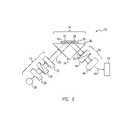
- FIG. 1is a block diagram of the invention.
- FIG. 2is a block diagram of an embodiment of the invention.
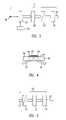
- FIG. 3is a block diagram of alternative portions of the invention.
- FIG. 4is a block diagram of alternative portions of the invention.
- FIG. 5is a block diagram of alternative portions of the invention.
- FIG. 6is a schematic projection view of a biochip useful in the apparatus of FIG. 1 .
- FIG. 7is a schematic view of the slide of FIG. 2 showing a representative spot.
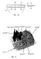
- FIG. 8is an hypochetrial enlarged view of a biochip of the prior art representing the surface roughness of the slide and biomolecules.
- FIG. 9is an enlarged schematic side view of a biochip slide having reduced roughness in accordance with the principles of the invention.
- FIG. 10is a schematic representation of a jig for holding the slide of FIG. 2 .
- FIG. 11is a computer printout of an area plot of spots of a microarray.
- the inventioncomprises a method and apparatus for analyzing a two-dimensional arrangement of chemical substances with an imaging technique.
- a polarized light source of known polarization stateis directed into a total internal reflection member (TIR member) configured for a single reflection at a total internal reflection surface (TIR surface) and then exiting the TIR member.
- TIR membertotal internal reflection member
- TIR surfacetotal internal reflection surface
- superposition of reflections as encountered at a layered optical structure where the layer thicknesses are smaller than the coherence length of the illuminating lightis referred to as a single reflection.
- the chemical specimenis in place above the TIR surface in the evanescent field of the reflected light beam. After reflection, the beam is passed to a polarization-sensitive two-dimensional detector such as a polarizer and a camera.
- the beam's contentcan then be processed to determine the change in polarization state, locally in the two-dimensional cross-section of the beam.
- Thisprovides a spatially distributed map of change of polarization state in the specimen.
- a variety of techniquesare available to determine the change in polarization such as measuring the deviation from a null condition or by comparing the input polarization state to the output polarization state.
- the refractive index composition of the materials within the evanescent fielddetermines the change in the polarization state of the beam due to the reflection at the TIR surface.
- a two-dimensional variation of this composition within the TIR surfaceis associated with a respective variation of the polarization state spatially distributed across the cross-section of the reflected light beam.
- the chemical specimenforms a two-dimensional array of molecules (here referred to as receptors) with specific affinities towards respective other molecules (here referred to as ligands).
- the inventionis utilized to indicate the presence or absence of binding between ligands and receptors on the array.
- arrayscommonly consist of a plurality of discrete specimen spots.
- the present method and apparatuswill image the array so as to distinguish each of the discrete specimen spots represented by the local change in polarization state in the cross-section of the reflected beam.
- the inventionpermits measurement of thickness and/or refractive index composition of the specimen under investigation with a very high resolution, in the sub angstrom range, spatially resolved over an entire area.
- the inventionis particularly useful in applications where the specimen is in an aqueous solution.
- the present inventionis used to determine the presence of biological agents in a solution such as in immunosensor applications by measuring their attachment to antibodies on the TIR surface in the evanescent field.
- the present inventionis used to determine the presence and structure of nucleic acid sequences in a solution by measuring their attachment to other nucleic acid sequences on the TIR surface in the evanescent field. Described in more detail below are different embodiments of the invention.
- FIGS. 1 and 2an apparatus and method is illustrated which implements one embodiment of the invention.
- the apparatus 10can be described conveniently as consisting of three general portions.
- Portion 12is a polarized light source assembly
- portion 14is a total internal reflection assembly providing a single reflection
- portion 16is a polarization-sensitive two-dimensional array detector assembly.
- Data from the detector assembly 16is sent by an electrical signal 24 to processor 18 such as a specially programmed computer and user access system such as a print-out or image display.
- Datacan be presented as an image, a data table, or in other forms.
- the polarized light source assembly 12passes polarized light of known polarization state (which may be varied or varying) 20 to the total internal reflection assembly 14 where a single reflection occurs and the reflected light 22 having a changed polarization state passes to the detector assembly 16 , where it is recorded spatially over the cross-section of the beam.
- the recorded datais sent to the processor 18 where the change of polarization state is determined to provide a spatially resolved map of changes in polarization state.
- each spotwill be imaged for its change in polarization state within the spot area.
- FIG. 2shows a more detailed preferred embodiment.
- the polarized light source assembly 12has a light source 26 , a beam forming member 28 (if the nature of the light source is such as to make beam forming useful or necessary) a polarizer 30 and an optical retarder 32 .
- the total internal light reflection assembly 14has an optical element 34 which has an optical surface 36 . Also shown is a specimen slide 38 on the optical surface 36 , and between them an index matching substance 40 . Because of the index matching a total internal reflection surface (TIR surface) is defined as the upper surface 39 of the specimen slide 38 .
- a specimen 42is on the total internal reflection surface 39 of the slide 38 .
- the optical element 34is a prism configured along with the index-matched slide 38 in relationship to the incoming light beam 20 , and the exiting light beam 22 such that the beam reflects only a single time at the TIR surface 39 and then exits the prism. If the specimen is placed directly on the optical surface 36 , then the optical surface 36 would be the TIR surface. But this is not the usual application as the specimen (such as a biochip) is usually prepared more conveniently on a specimen slide 38 and placed in the apparatus. In any event, however constructed, there is an optical structure having a TIR surface and the beam reflects only a single time at the TIR surface between entering and leaving the optical structure. In other words, there is a TIR surface in optical contact with the specimen, such that the evanescent field associated with the total internal reflection interacts with the specimen, and there is only a single reflection at that TIR surface.
- the post reflection detector assembly 16has a polarizer 44 , and a two-dimensional array detector 46 , preferably a camera of the CCD type.
- the processor 18is a specially programmed computer and output means for processing the imagery into a representation of film thickness variations spatially resolved over the cross-section of the area imaged.
- the imagingis acquired by detecting changes spatially distributed in the local polarization state in the beam's cross-section caused by the total internal reflection. This provides information about the presence and composition in the array of substances on the substrate surface for each resolvable point on the surface. Different polarization state changes are included in the cross-section of the reflected beam indicative of the substances on the specimen in the location in the specimen array corresponding to a position in the detector.
- the processor 18receives the data as an electrical signal 24 and characterizes the change of polarization state spatially over the two-dimensional array.
- the analysis and processingis done in one embodiment by comparing the known polarization state of the incoming light from the light processing assembly 12 with the changed polarization state of the reflected light 22 , spatially resolved two-dimensionally within the beam which provides a map of spatially distributed points or spots in the specimen array.
- the polarization shiftis then analyzed by the processor 18 to provide information of the presence and properties of elements in the chemical specimen.
- Other known techniques, such as null processingcan be used to determine the change in polarization state.
- the light source member 26may be an LED, an SLD (Super Luminescent Diode), an incandescent light source, or a laser. If an LED or SLD is used, the set-up shown in FIG. 2 is appropriate, where the beam-forming member 28 is a collimator. If an incandescent light source is used, an optical filter is also used.
- the light source 26 for the apparatusis a quasi-monochromatic light source of moderate bandwidth.
- the light source 26is preferably an LED of moderate bandwidth.
- the bandwidthis a full width half maximum wavelength in the range of about 10 nm–50 nm, and more preferably a full width half maximum wavelength in the range of about 30nm–50nm.
- the optical retarder 32could be placed instead in the exiting beam path 22 before the polarizer 44 .
- a moving diffuser 52is adapted to produce speckle-offsetting fluctuation of the minima and maxima in the speckle pattern caused by the laser.
- the moving diffuser 52is attached to a mechanical actuator 54 which is preferably a motor and servo-apparatus for providing the speckle offsetting fluctuations.
- the beam 20then proceeds through the beam-forming element 28 , the polarizer 30 and the optical retarder 32 , exiting the light source assembly 20 .
- the polarizer 30employs a polarizer of selected known polarization state.
- the polarizer 30may be of the type having a mechanical actuator driven by a motor control signal so as to enable varying and selecting the polarization state of the light beam 20 .
- the total internal reflection optical element 34either alone or in combination with an index matched slide may be arranged for use with a specimen in various ways to define a total internal reflection assembly so long as the specimen is in the evanescent field of the reflected beam 20 , 22 .
- the specimen 42could be set directly on the optical surface 36 in which case the optical surface 36 would be the TIR surface but this is inconvenient and repeated use is likely to degrade the optical quality of the optical surface 36 , and therefore, consistent with common practice in which a biochip or other chemical assay specimen is provided, a specimen slide 38 or other supporting apparatus is employed. It is common in a biochip to provide an array of discrete specimen spots supported on a structure for obtaining analysis of each spot.
- the term total internal reflection optical elementrefers to known optical elements alone or in combination with other elements which provide the phenomenon known as total internal reflection.
- FIG. 2shows use of a prism combined with a slide 38 , being index matched so that there is a TIR surface 39 .
- FIG. 4shows an alternative optical arrangement in which a flat optical member 56 having an upper surface 58 is surmounted by a specimen slide 60 and an index matching substance 62 on which is a specimen 64 .
- the TIR surface 66is the top of the slide 60 .
- the beam 20enters the assembly, is refracted as it enters, and leaves the optical member 56 after a single reflection at the TIR surface 66 as beam 22 .
- Other mechanisms for providing total internal reflection and an evanescent fieldcan be employed in practicing this invention as long as only a single reflection occurs at the TIR surface upon which the specimen is placed so as to be in the evanescent field associated with the reflection.
- the post-reflection processing arrangement 16 through which the beam 22 passescan alternatively, consist of a polarizer member 70 , a beam forming member 72 and a two-dimensional array detector 74 .
- the method and apparatuscan be used in combination with biochips of the type having discrete specimen spots or a micro-titer plate containing an array of discrete spots or locations for analysis, where the detected change in polarization state is spatially related to the discrete locations in the reflected beam. Therefore, as used herein the slide and specimen refers to any type of chemical or biological array which is desired to be examined.
- the invention as described aboveprovides an extremely sensitive optical imaging system for real-time imaging of the binding status of biochip array elements on the surface of a glass or plastic chip.
- An exemplary monitored array spotis approximately 25 mm in diameter, with a lateral resolution better than 10 microns, resulting in fully parallel, continuous real-time readout of up to 5 million sensor fields.
- Sensor sensitivity to surface attachmentis in the femtogram/mm 2 range (e.g. one DNA per square micron) and spatial resolution of 20 microns.
- the apparatus of FIG. 1operates by imaging the pattern of reaction results on the biochip. Those reactions produce changes in the height of the resulting material which react at each spot, imaging an area of array containing numerous spots.
- the area imagedcould be a portion or the entire biochip array.
- different constituents in test material flowed over the spotsbind in a manner which identifies those constituents.
- the image produced by the apparatus of FIG. 1not only identifies the constituents in the test material, but also can determine the rate at which the reactions occur and the height of the resulting molecules at each spot. With the apparatus described height differences can be imaged dynamically over such short periods of time that intermediate height change readings can be recorded and therefore height change rate can be determined as well as allowing comparison of the rate of height change or intermediate amount of height change among the for spots on the biochip array.
- FIG. 6is a schematic projection view of a slide suitable for use in the apparatus of FIG. 1-5 .
- the slide 110has a top surface 112 on which an array of spots 114 is formed.
- FIG. 7is a side view of the slide of FIG. 6 illustrating a representative spot 116 .
- FIG. 8is an enlarged portion of a slide of FIG. 6 illustrating a plurality of spots after interaction in the context of an exaggerated surface roughness for the slide as in the prior art.
- the horizontal broken line 120represents the maximum surface roughness as indicated by the opposing arrows 121 and 122 . It can be seen from the figure that the height of a reacted spot at 123 is exaggerated as compared to a reacted spot at 124 and at 125 . Similarly, unreacted spots at 126 and 127 distort the resulting image where they should provide an equal result.
- FIG. 9illustrates the point by showing the same interaction pattern where the surface roughness is less than a small portion of the height of the molecule produced by a reaction at each spot. Like numerals are used in FIGS. 8 and 9 to facilitate comparisons between the two figures.
- the slide of FIG. 6is positioned in a fixture 200 as shown in FIG. 10 .
- the slideis inserted into the fixture by movement as indicated by the arrow 201 , into a position represented by horizontal dashed lines 202 and 204 .
- the slideis spaced apart from surface 36 of optical element 34 of FIG. 1 .
- the spacing between surface 36 and the opposed bottom surface of the slidecontains a fluid 206 of a matching index of refraction which, for all practical purposes, renders those surfaces invisible and providing for TIR only from the top surface 112 of the slide 10 .
- the biochip of FIG. 6can include an array, for example, of 100,000 spots and those spots can be imaged simultaneously or in selected sub-arrays.
- Each spothas a diameter of from 50 microns to 500 microns where each spot comprises a biological element (i.e. oligonucleotides, cDNA clone, protein, antibody, antigen, bacteria, enzyme, inhibitor, receptor and others).
- Each spothas a known designation for, for example, genes for breast cancer, prostate cancer, aging and others,—(up to 80,000 genes).
- the biological elementsmust be substances that can be attached by various chemical and physical techniques to create a biochip. A full discussion of biochip fabrication is included in “Microarray Biochip Technology” by Mark Schena, a Biotechniques Books Publication, 2000. “DNA Microarrays: A Practical Approach” by Mark Schena, a Practical Approach Series, 2000.
- the binding status of biological array elements (spots) on the surface of a (glass) slide or chipis obtained by the apparatus of FIG. 1 .
- the slide monitoring areais approximately 25 mm in diameter with a lateral resolution better than 10 microns, resulting in fully parallel, continuous, real-time readout of up to five million sensor fields.
- System sensitivity to surface attachmentis in the femtogram/mm 2 range.
- the apparatus and chiphave application to DNA and protein chip scanning, high throughput screening, ligand fishing, immunosensors, clinical diagnostics and research, toxicology profiling, binding kinetics research, genomics and protemics.
- Particular featuresinclude; real-time imaging, no labels necessary, sensitivity of 1 fg/mm 2 (one DNA per square micron), partial resolution of 20 microns, and a sensor field of about 24 mm diameter.
- the above described imaging apparatuswas used to measure the thickness of protein layers deposited directly on the prism surface, which has a surface roughness of ⁇ /10. Tests were performed by depositing small droplets ( ⁇ 0.5 mm in area, 200 nl in volume) of bovine serum albumin (BSA) solutions directly on the sensor surface (the TIR surface). After the water was evaporated, only the protein remained on the surface leaving a well-defined quantity of protein at the test location. Table 1 lists the number of molecules per drop for each concentration, as well as an estimate of the effective layer thickness based on the area the droplet covered on the surface. The intensities as measured by a CCD camera detector for the surface was computer plotted as an area plot and is shown in FIG. 11 .
- BSAbovine serum albumin
- one aspect of inventionis in a biochip having a slide whose surface, which is the TIR surface, is of sufficiently low roughness specification that an array of spots imaged by the apparatus can be individually resolved using the apparatus described above.
- a process using the apparatus and a biochip of such sufficiently low roughnessis:
- every region where the chip (slide) surface deviates from the initial statestands out as a bright spot whose intensity is directly related to the thickness (height) differences induced by the deviation.
- target moleculeswill be flowed over the biochip surface.
- the intensity of the biochipmay be continuously monitored across the array to study thickness changes that occur on the biochip.
- the relative intensity measured at the detectorwill be related to the sample parameters and the setting of the polarizing elements using computer programs based on a detailed “Jones calculus” sensor system description. With these programs theoretical plots will be fitted to the acquired measurement data sets, and the outputs' dependence on parameter variations can be visualized. At the start of the procedure, the entire biochip is homogeneously dark.
- the intensity along the surfaceincreases.
- the intensityis expected to be the highest for high affinity interactions and mild changes are expected for low affinity interactions.
- the rate of intensity changecan be related to the affinity constraints of the system.
- affinity measurements for multiple peptidescan be performed.
- the effect of binding density(because different concentrations of peptides are used in spotting) can also be measured.
Landscapes
- Physics & Mathematics (AREA)
- Health & Medical Sciences (AREA)
- Life Sciences & Earth Sciences (AREA)
- Chemical & Material Sciences (AREA)
- Analytical Chemistry (AREA)
- Biochemistry (AREA)
- General Health & Medical Sciences (AREA)
- General Physics & Mathematics (AREA)
- Immunology (AREA)
- Pathology (AREA)
- Spectroscopy & Molecular Physics (AREA)
- Investigating Or Analysing Materials By Optical Means (AREA)
Abstract
Description
| TABLE 1 |
| BSA dilutions and molecular quantities in sample droplets |
| BSA | BSA | Number of | |||||
| mass per | Molecules/ | Moles | Molecular | Effective | Protein | ||
| Concentration | drop | drop | per drop | Layers | coverage | ||
| 10 mg/ | 2 μg | 1.8 *1013 | 30 pmol | 1400 | 10 | 4 μg/mm2 |
| 1 mg/ | 200 ng | 1.8 *1012 | 3 pmol | 140 | 1 | 400 ng/mm2 |
| 100 μg/ | 20 ng | 1.8 *1011 | 300 | 14 | 100 | 40 ng/mm2 |
| 10 μg/ | 2 ng | 1.8 *1010 | 30 fmol | 1.4 | 10 | 4 ng/mm2 |
| 1 μg/ | 200 pg | 1.8 *109 | 3 fmol | 0.14 | 1 | 400 pg/mm2 |
| 100 ng/ | 20 pg | 1.8 *108 | 300 amol | 0.014 | 100 | 40 pg/ |
| 10 ng/ | 2 pg | 1.8 *107 | 30 amol | 1.4*10−3 | 10 | 4 pg/mm2 |
| 1 ng/ | 200 fg | 1.8 *106 | 3 amol | 1.4*10−4 | 1 | 400 fg/mm2 |
| 100 pg/ | 20 fg | 1.8 *105 | 0.3 amol | 1.4*10−5 | 100 | 40 fg/mm2 |
| 10 pg/ | 2 fg | 1.8 *104 | 0.03 amol | 1.4*10−6 | 10 | 4 fg/mm2 |
- placing the biochip surface in a flow cell in combination with the apparatus such that the surface of the slide which has the spots is the TIR surface;
- initially calibrating the apparatus such that light reflected from the biochip yields a fully-linear polarization;
- adjusting the analyzer to a null position to fully block the linearly polarized light. flowing target molecules over the biochip surface.
Claims (22)
Priority Applications (15)
| Application Number | Priority Date | Filing Date | Title |
|---|---|---|---|
| US09/838,700US7023547B2 (en) | 2000-07-11 | 2001-04-19 | Apparatus including a biochip for imaging of biological samples and method |
| US10/046,620US20020093654A1 (en) | 2000-07-11 | 2001-11-12 | Apparatus and method for imaging |
| US10/046,620US6833920B2 (en) | 2000-07-11 | 2002-01-12 | Apparatus and method for imaging |
| EP02718883AEP1390719A4 (en) | 2001-04-19 | 2002-01-27 | Imaging apparatus and method |
| IL15847302AIL158473A0 (en) | 2001-04-19 | 2002-01-27 | Imaging apparatus and method |
| PCT/US2002/002662WO2002086468A1 (en) | 2001-04-19 | 2002-01-27 | Imaging apparatus and method |
| AU2002249994AAU2002249994A1 (en) | 2001-04-19 | 2002-01-27 | Imaging apparatus and method |
| JP2002583949AJP4219689B2 (en) | 2001-04-19 | 2002-01-27 | Imaging apparatus and method |
| US10/841,988US6882420B2 (en) | 2000-07-11 | 2004-05-07 | Apparatus and method for imaging |
| US10/847,736US7193711B2 (en) | 2000-07-11 | 2004-05-17 | Imaging method and apparatus |
| US10/847,754US7126688B2 (en) | 2000-07-11 | 2004-05-17 | Microarray scanning |
| US11/021,204US7002686B2 (en) | 2000-07-11 | 2004-12-22 | Apparatus and method for imaging |
| US11/321,168US7518724B2 (en) | 2000-07-11 | 2005-12-29 | Image acquisition, processing, and display |
| JP2007048283AJP2007178442A (en) | 2001-04-19 | 2007-02-28 | Imaging apparatus and imaging method |
| US12/117,245US20080204750A1 (en) | 2000-07-11 | 2008-05-08 | Image acquisition, processing, and display |
Applications Claiming Priority (2)
| Application Number | Priority Date | Filing Date | Title |
|---|---|---|---|
| US09/614,503US6594011B1 (en) | 2000-07-11 | 2000-07-11 | Imaging apparatus and method |
| US09/838,700US7023547B2 (en) | 2000-07-11 | 2001-04-19 | Apparatus including a biochip for imaging of biological samples and method |
Related Parent Applications (1)
| Application Number | Title | Priority Date | Filing Date |
|---|---|---|---|
| US09/614,503Continuation-In-PartUS6594011B1 (en) | 2000-07-11 | 2000-07-11 | Imaging apparatus and method |
Related Child Applications (6)
| Application Number | Title | Priority Date | Filing Date |
|---|---|---|---|
| US09/614,503Continuation-In-PartUS6594011B1 (en) | 2000-07-11 | 2000-07-11 | Imaging apparatus and method |
| US10/046,620Continuation-In-PartUS20020093654A1 (en) | 2000-07-11 | 2001-11-12 | Apparatus and method for imaging |
| US10/046,620Continuation-In-PartUS6833920B2 (en) | 2000-07-11 | 2002-01-12 | Apparatus and method for imaging |
| US10/847,754Continuation-In-PartUS7126688B2 (en) | 2000-07-11 | 2004-05-17 | Microarray scanning |
| US10/847,736Continuation-In-PartUS7193711B2 (en) | 2000-07-11 | 2004-05-17 | Imaging method and apparatus |
| US11/321,168Continuation-In-PartUS7518724B2 (en) | 2000-07-11 | 2005-12-29 | Image acquisition, processing, and display |
Publications (2)
| Publication Number | Publication Date |
|---|---|
| US20020021443A1 US20020021443A1 (en) | 2002-02-21 |
| US7023547B2true US7023547B2 (en) | 2006-04-04 |
Family
ID=46277527
Family Applications (1)
| Application Number | Title | Priority Date | Filing Date |
|---|---|---|---|
| US09/838,700Expired - LifetimeUS7023547B2 (en) | 2000-07-11 | 2001-04-19 | Apparatus including a biochip for imaging of biological samples and method |
Country Status (1)
| Country | Link |
|---|---|
| US (1) | US7023547B2 (en) |
Cited By (21)
| Publication number | Priority date | Publication date | Assignee | Title |
|---|---|---|---|---|
| US20060250612A1 (en)* | 1997-09-22 | 2006-11-09 | Meeks Steven W | Detecting and classifying surface features or defects by controlling the angle of the illumination plane of incidence with respect to the feature or defect |
| US20070115483A1 (en)* | 1997-09-22 | 2007-05-24 | Oak Dave S | Surface finish roughness measurement |
| US20070153273A1 (en)* | 1997-09-22 | 2007-07-05 | Meeks Steven W | Material independent profiler |
| US7397553B1 (en) | 2005-10-24 | 2008-07-08 | Kla-Tencor Technologies Corporation | Surface scanning |
| US7532318B2 (en) | 2005-05-06 | 2009-05-12 | Kla-Tencor Corporation | Wafer edge inspection |
| US7554654B2 (en) | 2007-01-26 | 2009-06-30 | Kla-Tencor Corporation | Surface characteristic analysis |
| US20090269742A1 (en)* | 2005-09-05 | 2009-10-29 | Shigeyoshi Horiike | Substrate for immobilizing biopolymer and method of immobilizing biopolymer by using the same |
| US8004676B1 (en)* | 2007-02-09 | 2011-08-23 | The Research Foundation Of State University Of New York | Method for detecting analytes using surface plasmon resonance |
| US20110284769A1 (en)* | 2009-02-03 | 2011-11-24 | Takuya Matsui | Total internal reflection microscope apparatus and method for analyzing fluorescent sample |
| US9063072B1 (en) | 2012-06-12 | 2015-06-23 | Maven Technologies, Llc | Birefringence correction for imaging ellipsometric bioassay system and method |
| US9383361B2 (en) | 2012-04-25 | 2016-07-05 | Biodesy, Inc. | Methods for detecting allosteric modulators of protein |
| US9395358B2 (en) | 2012-02-05 | 2016-07-19 | Biodesy, Inc. | Methods for detecting allosteric modulators of protein |
| US9428789B2 (en) | 2011-03-21 | 2016-08-30 | Biodesy, Inc. | Classification of kinase inhibitors using nonlinear optical techniques |
| CN106066303A (en)* | 2016-05-24 | 2016-11-02 | 中国科学院重庆绿色智能技术研究院 | A kind of strengthen biomolecule Terahertz characteristic signal in solution receive runner prisms waveguide and preparation method thereof |
| US9880172B2 (en) | 2008-08-04 | 2018-01-30 | Biodesy, Inc. | Nonlinear optical detection of molecules comprising an unnatural amino acid possessing a hyperpolarizability |
| US9938560B2 (en) | 2012-02-05 | 2018-04-10 | Biodesy, Inc. | Methods for identifying modulators of Ras using nonlinear techniques |
| US9989534B2 (en) | 2014-06-30 | 2018-06-05 | Biodesy, Inc. | Systems and methods for high throughput analysis of conformation in biological entities |
| US10314549B1 (en) | 2013-07-16 | 2019-06-11 | Alacrity Patient Services, Inc. | Method and apparatus for monitoring development of medication induced febrile neutropenia |
| US10672502B2 (en) | 2015-04-02 | 2020-06-02 | Biodesy, Inc. | Methods for determining protein structure using a surface-selective nonlinear optical technique |
| US10768174B2 (en) | 2014-12-23 | 2020-09-08 | Bluelight Therapeutics, Inc. | Attachment of proteins to interfaces for use in nonlinear optical detection |
| US12318228B2 (en) | 2013-07-16 | 2025-06-03 | Alacrity Patient Services, Inc. | Method and apparatus for monitoring development of medication induced side effects |
Families Citing this family (16)
| Publication number | Priority date | Publication date | Assignee | Title |
|---|---|---|---|---|
| US7193711B2 (en)* | 2000-07-11 | 2007-03-20 | Maven Technologies, Llc | Imaging method and apparatus |
| US6833920B2 (en)* | 2000-07-11 | 2004-12-21 | Maven Technologies Llc | Apparatus and method for imaging |
| US7126688B2 (en)* | 2000-07-11 | 2006-10-24 | Maven Technologies, Llc | Microarray scanning |
| US7154598B2 (en)* | 2002-07-12 | 2006-12-26 | Decision Biomarkers, Inc. | Excitation and imaging of fluorescent arrays |
| US20060127946A1 (en)* | 2002-08-16 | 2006-06-15 | Montagu Jean I | Reading of fluorescent arrays |
| US7384742B2 (en)* | 2002-08-16 | 2008-06-10 | Decision Biomarkers, Inc. | Substrates for isolating reacting and microscopically analyzing materials |
| JP2007528692A (en)* | 2003-05-28 | 2007-10-18 | メイバン・テクノロジーズ・エルエルシー | Method and apparatus for recognizing molecular compounds |
| DE102005041584B4 (en)* | 2005-09-01 | 2007-08-16 | Universität Karlsruhe (Th) | Differential measurement method for determining concentration differences for supersaturation determination |
| US7233396B1 (en)* | 2006-04-17 | 2007-06-19 | Alphasniffer Llc | Polarization based interferometric detector |
| CN101688835B (en)* | 2007-06-28 | 2014-01-29 | 皇家飞利浦电子股份有限公司 | Microelectronic sensor device for optical inspection of wetted surfaces |
| US8039270B2 (en)* | 2008-05-22 | 2011-10-18 | Maven Technologies, Llc | Apparatus and method for performing ligand binding assays on microarrays in multiwell plates |
| WO2010029471A1 (en)* | 2008-09-09 | 2010-03-18 | Koninklijke Philips Electronics N.V. | Method for determining the presence of a sample on a sample receiving surface |
| CN102590141A (en)* | 2012-03-12 | 2012-07-18 | 南开大学 | Mixture refractive index measurement method based on total internal reflection |
| CN104568838A (en)* | 2013-10-28 | 2015-04-29 | 南开大学 | Total internal reflection method-based automatic wide-spectrum range substance dispersion measurement device |
| DE102017102582A1 (en)* | 2016-02-10 | 2017-08-10 | Franz Schmidt & Haensch Gmbh & Co. | Refraktometeranordnung |
| JP7693187B2 (en)* | 2020-10-30 | 2025-06-17 | D-テック合同会社 | Inspection Equipment |
Citations (58)
| Publication number | Priority date | Publication date | Assignee | Title |
|---|---|---|---|---|
| US4238565A (en) | 1978-06-22 | 1980-12-09 | Miles Laboratories, Inc. | Specific binding assay with a prosthetic group as a label component |
| US4256834A (en) | 1979-04-09 | 1981-03-17 | Syva Company | Fluorescent scavenger particle immunoassay |
| US4508832A (en) | 1981-06-22 | 1985-04-02 | Battelle Memorial Institute | Ellipsometrically measuring rate of optical change in immunoassay |
| US5164589A (en) | 1988-11-10 | 1992-11-17 | Pharmacia Biosensor Ab | Reusable optical interface for non-permanent passive light coupling |
| US5229833A (en) | 1990-09-26 | 1993-07-20 | Gec-Marconi Limited | Optical sensor |
| US5234769A (en)* | 1992-04-16 | 1993-08-10 | Deposition Sciences, Inc. | Wear resistant transparent dielectric coatings |
| US5255075A (en) | 1991-03-22 | 1993-10-19 | Gec-Marconi Limited | Optical sensor |
| US5437840A (en) | 1994-04-15 | 1995-08-01 | Hewlett-Packard Company | Apparatus for intracavity sensing of macroscopic properties of chemicals |
| US5483346A (en) | 1994-04-11 | 1996-01-09 | Butzer; Dane C. | Polarization based optical sensor utilizing total internal reflection |
| US5485277A (en) | 1994-07-26 | 1996-01-16 | Physical Optics Corporation | Surface plasmon resonance sensor and methods for the utilization thereof |
| US5491556A (en) | 1992-01-11 | 1996-02-13 | Fisons, Plc | Analytical device with variable angle of incidence |
| WO1996008720A1 (en) | 1994-09-15 | 1996-03-21 | Biacore Ab | Milk assay |
| US5573956A (en) | 1991-08-20 | 1996-11-12 | Hanning; Anders | Assay method based upon refractive index changes at a solid optical surface |
| WO1996038729A1 (en) | 1995-06-02 | 1996-12-05 | Biacore Ab | Pathogen assay method |
| US5593130A (en) | 1993-06-09 | 1997-01-14 | Pharmacia Biosensor Ab | Valve, especially for fluid handling bodies with microflowchannels |
| US5633724A (en) | 1995-08-29 | 1997-05-27 | Hewlett-Packard Company | Evanescent scanning of biochemical array |
| WO1997019375A1 (en) | 1995-11-24 | 1997-05-29 | Biacore Ab | Optical coupling device and method for its production |
| US5641640A (en) | 1992-06-29 | 1997-06-24 | Biacore Ab | Method of assaying for an analyte using surface plasmon resonance |
| USRE35716E (en) | 1988-08-02 | 1998-01-20 | Gene Tec Corporation | Temperature control apparatus and method |
| US5753518A (en) | 1993-05-24 | 1998-05-19 | Pharmacia Ab | Method of determining affinity and kinetic properties |
| WO1998032002A1 (en) | 1997-01-22 | 1998-07-23 | Biacore Ab | Pipette and carrier assembly for a sensor |
| US5856873A (en) | 1996-04-30 | 1999-01-05 | Fuji Photo Film Co., Ltd. | Ellipso sensor using a prism |
| US5922604A (en) | 1997-06-05 | 1999-07-13 | Gene Tec Corporation | Thin reaction chambers for containing and handling liquid microvolumes |
| US5922594A (en) | 1994-09-26 | 1999-07-13 | Biacore Ab | Method of producing bilayer lipid membranes |
| US5955729A (en) | 1995-09-08 | 1999-09-21 | Biacore Ab | Surface plasmon resonance-mass spectrometry |
| US5965456A (en) | 1992-06-11 | 1999-10-12 | Biacore Ab | Analyte detection |
| US5972612A (en) | 1995-07-14 | 1999-10-26 | Biacore Ab | Surface-sensitive detection of hybridization at equilibrium |
| US6008893A (en) | 1999-03-22 | 1999-12-28 | Biacore Ab | Reversible-flow conduit system |
| US6008010A (en) | 1996-11-01 | 1999-12-28 | University Of Pittsburgh | Method and apparatus for holding cells |
| US6045996A (en) | 1993-10-26 | 2000-04-04 | Affymetrix, Inc. | Hybridization assays on oligonucleotide arrays |
| US6127183A (en) | 1995-09-01 | 2000-10-03 | Biacore Ab | Monitoring of refractive index of optical sensor for determination of surface structure changes |
| US6140044A (en) | 1994-06-08 | 2000-10-31 | Affymetrix, Inc. | Method and apparatus for packaging a probe array |
| US6143513A (en) | 1999-06-23 | 2000-11-07 | Biacore Ab | Method and kit for detecting betalactam-containing compounds |
| US6143574A (en) | 1995-11-14 | 2000-11-07 | Biacore Ab | Method of determining affinity or kinetic properties in solution |
| US6197595B1 (en) | 1995-06-29 | 2001-03-06 | Affymetrix, Inc. | Integrated nucleic acid diagnostic device |
| US6200814B1 (en) | 1998-01-20 | 2001-03-13 | Biacore Ab | Method and device for laminar flow on a sensing surface |
| US6207381B1 (en) | 1996-04-04 | 2001-03-27 | Biacore Ab | Method for nucleic acid analysis |
| US6277330B1 (en) | 1996-09-30 | 2001-08-21 | Aventis Research & Technologies Gmbh & Co K.G. | Optical sensor for detecting chemical substances dissolved or dispersed in water |
| US6289286B1 (en) | 1998-05-29 | 2001-09-11 | Biacore Ab | Surface regeneration of biosensors and characterization of biomolecules associated therewith |
| US20020019019A1 (en) | 1999-06-18 | 2002-02-14 | Markku Hamalainen | Method and apparatus for assaying a drug candidate to estimate a pharmacokinetic parameter associated therewith |
| US20020154311A1 (en) | 2001-03-14 | 2002-10-24 | Biacore Ab | Apparatus and method for total internal reflection spectroscopy |
| US6475809B1 (en)* | 1998-07-14 | 2002-11-05 | Zyomyx, Incorporated | Protein arrays for high-throughput screening |
| US20020182717A1 (en) | 2001-03-14 | 2002-12-05 | Biacore Ab | Method of preparing supported lipid film membranes and use thereof |
| US6493097B1 (en) | 1997-02-04 | 2002-12-10 | Biacore Ab | Analytical method and apparatus |
| US6503760B2 (en) | 2000-03-16 | 2003-01-07 | Biacore Ab | Method for capturing analytes eluted from surface-bound ligands |
| US20030022388A1 (en) | 2001-06-29 | 2003-01-30 | Biacore Ab | Flow cell method |
| USD472644S1 (en) | 2001-05-16 | 2003-04-01 | Biacore Ab | Chip carrier for biosensor apparatus |
| US6589798B1 (en) | 1996-12-12 | 2003-07-08 | Biacore Ab | Method and system for analyte determination |
| WO2003056337A1 (en) | 2001-12-21 | 2003-07-10 | Biacore Ab | Immobilization of binding agents |
| US6594011B1 (en) | 2000-07-11 | 2003-07-15 | Maven Technologies, Llc | Imaging apparatus and method |
| WO2003102580A1 (en) | 2002-05-31 | 2003-12-11 | Biacore Ab | Method of coupling binding agents to a substrate surface |
| US20040002167A1 (en) | 2002-03-27 | 2004-01-01 | Biacore Ab | Method and system for curve quality control |
| US20040012676A1 (en) | 2002-03-15 | 2004-01-22 | Affymetrix, Inc., A Corporation Organized Under The Laws Of Delaware | System, method, and product for scanning of biological materials |
| US20040023247A1 (en) | 2002-07-31 | 2004-02-05 | Affymetrix, Inc. | Quality control methods for microarray production |
| US20040030504A1 (en) | 2002-04-26 | 2004-02-12 | Affymetrix, Inc. A Corporation Organized Under The Laws Of Delaware | System, method, and computer program product for the representation of biological sequence data |
| US20040038268A1 (en) | 1989-06-07 | 2004-02-26 | Affymetrix, Inc. | Support bound probes and methods of analysis using the same |
| US6698454B2 (en) | 2000-11-02 | 2004-03-02 | Biacore Ab | Valve integrally associated with microfluidic liquid transport assembly |
| US6806051B2 (en)* | 2000-09-25 | 2004-10-19 | Picoliter Inc. | Arrays of partially nonhybridizing oligonucleotides and preparation thereof using focused acoustic energy |
- 2001
- 2001-04-19USUS09/838,700patent/US7023547B2/ennot_activeExpired - Lifetime
Patent Citations (61)
| Publication number | Priority date | Publication date | Assignee | Title |
|---|---|---|---|---|
| US4238565A (en) | 1978-06-22 | 1980-12-09 | Miles Laboratories, Inc. | Specific binding assay with a prosthetic group as a label component |
| US4256834A (en) | 1979-04-09 | 1981-03-17 | Syva Company | Fluorescent scavenger particle immunoassay |
| US4508832A (en) | 1981-06-22 | 1985-04-02 | Battelle Memorial Institute | Ellipsometrically measuring rate of optical change in immunoassay |
| USRE35716E (en) | 1988-08-02 | 1998-01-20 | Gene Tec Corporation | Temperature control apparatus and method |
| US5164589A (en) | 1988-11-10 | 1992-11-17 | Pharmacia Biosensor Ab | Reusable optical interface for non-permanent passive light coupling |
| US5313264A (en) | 1988-11-10 | 1994-05-17 | Pharmacia Biosensor Ab | Optical biosensor system |
| US20040038268A1 (en) | 1989-06-07 | 2004-02-26 | Affymetrix, Inc. | Support bound probes and methods of analysis using the same |
| US5229833A (en) | 1990-09-26 | 1993-07-20 | Gec-Marconi Limited | Optical sensor |
| US5255075A (en) | 1991-03-22 | 1993-10-19 | Gec-Marconi Limited | Optical sensor |
| US5573956A (en) | 1991-08-20 | 1996-11-12 | Hanning; Anders | Assay method based upon refractive index changes at a solid optical surface |
| US5491556A (en) | 1992-01-11 | 1996-02-13 | Fisons, Plc | Analytical device with variable angle of incidence |
| US5234769A (en)* | 1992-04-16 | 1993-08-10 | Deposition Sciences, Inc. | Wear resistant transparent dielectric coatings |
| US5965456A (en) | 1992-06-11 | 1999-10-12 | Biacore Ab | Analyte detection |
| US5641640A (en) | 1992-06-29 | 1997-06-24 | Biacore Ab | Method of assaying for an analyte using surface plasmon resonance |
| US5753518A (en) | 1993-05-24 | 1998-05-19 | Pharmacia Ab | Method of determining affinity and kinetic properties |
| US5593130A (en) | 1993-06-09 | 1997-01-14 | Pharmacia Biosensor Ab | Valve, especially for fluid handling bodies with microflowchannels |
| US6045996A (en) | 1993-10-26 | 2000-04-04 | Affymetrix, Inc. | Hybridization assays on oligonucleotide arrays |
| US5483346A (en) | 1994-04-11 | 1996-01-09 | Butzer; Dane C. | Polarization based optical sensor utilizing total internal reflection |
| US5437840A (en) | 1994-04-15 | 1995-08-01 | Hewlett-Packard Company | Apparatus for intracavity sensing of macroscopic properties of chemicals |
| US6140044A (en) | 1994-06-08 | 2000-10-31 | Affymetrix, Inc. | Method and apparatus for packaging a probe array |
| US5485277A (en) | 1994-07-26 | 1996-01-16 | Physical Optics Corporation | Surface plasmon resonance sensor and methods for the utilization thereof |
| WO1996008720A1 (en) | 1994-09-15 | 1996-03-21 | Biacore Ab | Milk assay |
| US5922594A (en) | 1994-09-26 | 1999-07-13 | Biacore Ab | Method of producing bilayer lipid membranes |
| WO1996038729A1 (en) | 1995-06-02 | 1996-12-05 | Biacore Ab | Pathogen assay method |
| US6197595B1 (en) | 1995-06-29 | 2001-03-06 | Affymetrix, Inc. | Integrated nucleic acid diagnostic device |
| US5972612A (en) | 1995-07-14 | 1999-10-26 | Biacore Ab | Surface-sensitive detection of hybridization at equilibrium |
| US5633724A (en) | 1995-08-29 | 1997-05-27 | Hewlett-Packard Company | Evanescent scanning of biochemical array |
| US6127183A (en) | 1995-09-01 | 2000-10-03 | Biacore Ab | Monitoring of refractive index of optical sensor for determination of surface structure changes |
| US5955729A (en) | 1995-09-08 | 1999-09-21 | Biacore Ab | Surface plasmon resonance-mass spectrometry |
| US6143574A (en) | 1995-11-14 | 2000-11-07 | Biacore Ab | Method of determining affinity or kinetic properties in solution |
| WO1997019375A1 (en) | 1995-11-24 | 1997-05-29 | Biacore Ab | Optical coupling device and method for its production |
| US6207381B1 (en) | 1996-04-04 | 2001-03-27 | Biacore Ab | Method for nucleic acid analysis |
| US5856873A (en) | 1996-04-30 | 1999-01-05 | Fuji Photo Film Co., Ltd. | Ellipso sensor using a prism |
| US6277330B1 (en) | 1996-09-30 | 2001-08-21 | Aventis Research & Technologies Gmbh & Co K.G. | Optical sensor for detecting chemical substances dissolved or dispersed in water |
| US6008010A (en) | 1996-11-01 | 1999-12-28 | University Of Pittsburgh | Method and apparatus for holding cells |
| US6589798B1 (en) | 1996-12-12 | 2003-07-08 | Biacore Ab | Method and system for analyte determination |
| WO1998032002A1 (en) | 1997-01-22 | 1998-07-23 | Biacore Ab | Pipette and carrier assembly for a sensor |
| US6493097B1 (en) | 1997-02-04 | 2002-12-10 | Biacore Ab | Analytical method and apparatus |
| US20030067612A1 (en) | 1997-02-04 | 2003-04-10 | Biacore Ab | Analytical method and apparatus |
| US5922604A (en) | 1997-06-05 | 1999-07-13 | Gene Tec Corporation | Thin reaction chambers for containing and handling liquid microvolumes |
| US6200814B1 (en) | 1998-01-20 | 2001-03-13 | Biacore Ab | Method and device for laminar flow on a sensing surface |
| US6289286B1 (en) | 1998-05-29 | 2001-09-11 | Biacore Ab | Surface regeneration of biosensors and characterization of biomolecules associated therewith |
| US6475809B1 (en)* | 1998-07-14 | 2002-11-05 | Zyomyx, Incorporated | Protein arrays for high-throughput screening |
| US6008893A (en) | 1999-03-22 | 1999-12-28 | Biacore Ab | Reversible-flow conduit system |
| US20020019019A1 (en) | 1999-06-18 | 2002-02-14 | Markku Hamalainen | Method and apparatus for assaying a drug candidate to estimate a pharmacokinetic parameter associated therewith |
| US6143513A (en) | 1999-06-23 | 2000-11-07 | Biacore Ab | Method and kit for detecting betalactam-containing compounds |
| US6503760B2 (en) | 2000-03-16 | 2003-01-07 | Biacore Ab | Method for capturing analytes eluted from surface-bound ligands |
| US6594011B1 (en) | 2000-07-11 | 2003-07-15 | Maven Technologies, Llc | Imaging apparatus and method |
| US6806051B2 (en)* | 2000-09-25 | 2004-10-19 | Picoliter Inc. | Arrays of partially nonhybridizing oligonucleotides and preparation thereof using focused acoustic energy |
| US6698454B2 (en) | 2000-11-02 | 2004-03-02 | Biacore Ab | Valve integrally associated with microfluidic liquid transport assembly |
| US20020154311A1 (en) | 2001-03-14 | 2002-10-24 | Biacore Ab | Apparatus and method for total internal reflection spectroscopy |
| US20020182717A1 (en) | 2001-03-14 | 2002-12-05 | Biacore Ab | Method of preparing supported lipid film membranes and use thereof |
| USD472644S1 (en) | 2001-05-16 | 2003-04-01 | Biacore Ab | Chip carrier for biosensor apparatus |
| USD480149S1 (en) | 2001-05-16 | 2003-09-30 | Biacore Ab | Cover for a measuring cassette for biosensor apparatus |
| US20030022388A1 (en) | 2001-06-29 | 2003-01-30 | Biacore Ab | Flow cell method |
| WO2003056337A1 (en) | 2001-12-21 | 2003-07-10 | Biacore Ab | Immobilization of binding agents |
| US20040012676A1 (en) | 2002-03-15 | 2004-01-22 | Affymetrix, Inc., A Corporation Organized Under The Laws Of Delaware | System, method, and product for scanning of biological materials |
| US20040002167A1 (en) | 2002-03-27 | 2004-01-01 | Biacore Ab | Method and system for curve quality control |
| US20040030504A1 (en) | 2002-04-26 | 2004-02-12 | Affymetrix, Inc. A Corporation Organized Under The Laws Of Delaware | System, method, and computer program product for the representation of biological sequence data |
| WO2003102580A1 (en) | 2002-05-31 | 2003-12-11 | Biacore Ab | Method of coupling binding agents to a substrate surface |
| US20040023247A1 (en) | 2002-07-31 | 2004-02-05 | Affymetrix, Inc. | Quality control methods for microarray production |
Non-Patent Citations (61)
| Title |
|---|
| "[34] Surface Immobilization Techniques in Combination With Ellipsometry", by Ulf Jonsson, et al., Methods in Enzymology vol. 137, pp. 381-1351, 1988. |
| "A Biosensor Concept Based on Imagiing Ellipsometry for Visualization of Biomolecular Interactions", by Gang Jin, et al., Analytical Biochemistry 232, pp. 69-72, 1995. |
| "A Comparative Study of Protein Immobilization Techniques for Optical Immunosensors", by A. Ahluwalia, et al., Biosensors and Bioelectronics 7, pp. 207-214, 1991. |
| "A New View on Polarization Microscopy", by Rudolf Oldenbourg, Nature vol. 381, pp. 811-812, Jun. 27, 1996. |
| "Assembly of Antibodies in Lipid Membranes for Biosensor Development", by Huaiyou Wang et al., Applied Biochemistry and Biotechnology, pp. 163-181, 1994. |
| "Biosensors Based on Surface Concentration Measuring Devices-The Concept of Surface Concentration", by Ulf Jonsson, et al., Progress in Colloid and Polymer Sci. 70, pp. 96-100, 1985. |
| "Biosensors: An Introduction", by Brian R. Eggins, pp. 112-113, 1987. |
| "Characterization of Biomembranes by Spectral Ellipsometry, Surface Plasmon Resonance and Interferometry With Regard to Biosensor Application", by Ch. Striebel, et al., Biosensors & Bioelectronics 9, pp. 139-146, 1994. |
| "Complement Activation by 3-Mercapto-1,2-Propanediol Immobilized on Gold Surfaces", by Pentti Tengvall, et al., Biomaterials 17, pp. 1001-1007, 1995. |
| "Coupling of Biomolecules to Silicon Surfaces for Use in Ellipsometry and Other Relate; Techniques", by Carl Fredrik Mandenius, et al., Methods in Enzymology, vol. 137, pp. 388-394, 1988. |
| "Determination by Ellipsometry of the Affinity of Monoclonal Antibodies", by Haken Nygren, et al., Journal of Immunological Methods, 92, pp. 219-221, 1986. |
| "Direct Visualization of Monolayers at the Air-Water Interface by Brewster Angle Microscopy", by Dirk Honig, et al., J. Phys. Chem., pp. 4590 & 4592, 1991. |
| "DNA Microarrays A Practical Approach" Edited by Mark Schena, Department of Biochemistry, Beckman Center, Standord University Medical Center, Standord, USA, Oxford University Press, 1999. |
| "Effects of Hydrophilization and Immobilization on the Interfacial Behavior of Immunoglobulins", by Martin Malmsten, et al., Journal of Colloid and Interface Science 177, pp. 70-78, 1994. |
| "Ellipsometric Immunosensors for the Determination of Y-Interferon and Human Serum Albumin", by T.A. Ruzgas, et al., Biosensors & Bioelectronics 7, pp. 305-308, 1992. |
| "Flow-Injection Ellipsometry-An In Situ Method for the Study of Biomolecular Adsorption and Interaction at Solid Surfaces", by Ulf Jonsson, et al., Colloids and Surfaces, pp. 333-339, 1985. |
| "Handbook of Optics", by The Optical Society of America; vol. 1; pp. 4.23, 4.24; 1995. |
| "Handbook of Optics", Michael Bass Editor in Chief, by The Optical Society of America; vol. 1; pp. 4.23, 4.24; 1995 McGraw-Hill, Inc. |
| "Handbook of Optics, vol. 1, Section 41.10". |
| "Imaging Ellipsometry for Biosensor Applications", by Gang Jin, et al., Transducers '95.Eurosensors IX, pp. 509-774, 1995. |
| "Imaging Ellipsometry Revisited: Developments for Visualization of Thin Transparent Layers on Silicon Substrates", by Gang Jin, et al., Rev. Sci. Instrum., pp. 2930-2936, 1996. |
| "Kinetics of Antibody-Binding to Surface-Immobilized Antigen: Influence of Mass Transport on the Enzyme-Linked Immunosorbent Assay (ELISA)", by Nygren & Stenberg, Jour of Colloid and Interface Science, vol. 107, pp. 560-566, 1985. |
| "Microarray Biochip Technology" by Mark Schena, PhD, TeleChem Internation, Inc., Sunnyvale, California, USA, A BioTechniques Books Publication, Eaton Publishing, 2000. |
| "Microscope at the Brewster Angle: Direct Observation of First-Order Phase Transitions in Monolayers", by S. Henon, et al., Rev. Sci. Instrum. 62, pp. 936-939, 1990. |
| "Monitoring Specific Interaction of Low Molecular Weight Biomolecules on Oxidized Porous Silicon Usiing Ellipsometry", byD. Van Noort; S. Welin-Klintstrom, et al., Biosensors & Bioelectronics vol. 13, pp. 439-449, 1997. |
| "Monitoring Specific Interaction of Low Molecular Weight Biomolecules on Oxidized Porous Silicon Using Ellipsometry", byD. Van Noort; S. Welin-Klintstrom, et al., Biosensors & Bioelectronics vol. 13, pp. 439-449, 1997. |
| "Optical Characterization of Very Thin Hydrogenated Amorphous Silicon Films Using Spectroscopic Ellipsometry"; by Saitoh; Hori; Suzuki; & Iida; Japanese Journal of Applied Physics; 1991. |
| "Opto-Electronic Immunosensors: A Review of Optical Immunoassay at Continuous Surfaces", by John F. Place, et al., Biosensors 1, pp. 321-353, 1985. |
| "Patterning of Immobilized Antibody Layers Via Photolithography and Oxygen Plasma Exposure", by A.W. Flounders, et al., Biosensors and Bioelectronics vol. 12, pp. 447-456, 1997. |
| "Principles of Optics-Electromagnetic Theory of Propagation, Interference and Diffraction of Light", by Max Born & Emil Wolf, Sixth Edition, pp. 47-51. |
| "Spectroscopic Ellipsometry and Biology: Recent Developments and Challenges", by H. Arwin, Thin Solid Films 313-314, pp. 764-774, 1998. |
| "Spectroscopic Ellipsometry and Reflectometry A User's Guide" by Harland G. Tompkins, Motorola Inc., and William A. McGahan, Nanometrics, Inc., A Wiley-Interscience Publication, John Wiley & Sons, inc., 1999. |
| "Structural Analysis With Quantitative Birefringence Imaging", by Clifford C. Hoyt, et al., American Laboratory, pp. 34-42, Jul. 1999. |
| "Temporal Studies on the Deposition of Complement on Human Colostrum IgA and Serum I; Immobilized on Methylated Silicon", by Pentti Tengvall, et al., Journal of Biomedical Materials Research, vol. 35, pp. 81-91, 1997. |
| "Universal Imaging Corporation-Metapolscope"; "Metamorph Imaging System", by Dr. Rudolf Oldenbourg, pp. 1-2. |
| "Waveguide Ellipsometry Biosensors: Concept and Preliminary Analysis", SPIE vol. 1648, by Jinyu Wang, pp. 44-50, 1992. |
| "Wetting and Dewetting of Si/SiO2-Wafers by Free and Lipid-Monolayer Covered Aqueous Solutions Under Controlled Humidity", by G. Elender, et al., Journal de Physique II, pp. 455-479, 1994. |
| A. Brecht et al. "Biosensors: Fundamentals, Technologies and Applications" GBF Monographs, vol. 17, pp. 174-178, 1991 Germany. |
| Biosensors: Fundamental, Technologies and Applications, by A. Brecht, et al., edited by F. Sche et al., GBF Monographs vol. 17, pp. 174-178, 1991. |
| Ch Striebel et al. "Characterization of Biomembranes by Spectral Ellipsometry, Surface Plasmon Resonance and Interferometry with Regard to Biosensor Application", Biosensors & Bioelectronics 9, pp. 139-146, 1994 Elsevier Science Publishers Ltd. |
| Christopher Palmer "Diffraction Grating Handbook", pp. 35-44, 2000 Richardson Grating Laboratory, Rochester, New York. |
| Clifford C. Hoyt et al. "Structural analysis with quantitative birefringence imaging", American Laboratory, pp. 34-42, Jul. 1999. |
| Danny Van Noort et al. "Monitoring Specific Interaction of Low Molecular Weight Biomolecules on Oxidized Porous Silicon Using Ellipsometry", Biosensors & Bioelectronics vol. 13, No. 3-4 pp. 439-449, 1998 Elsevier Science, S.A. Great Britain. |
| Dirk Honig et al. "Direct visualization of monolayers at the air-water interface by Brewster angle microscopy", J. Phys. Chem., pp. 4590 & 4592, 1991 American Chemical Society. |
| Eggins, "Biosensors: An Introduction", pp. 112-113, 1987 John Wiley & Sons. |
| Erwin G. Loewen "Diffraction Gratings, Ruled and Holographic", Applied Optics and Optical Engineering, vol. IX, pp. 33-71, Bausch and Lomb, Inc., Rochester, New York 1983 Academic Press, Inc. |
| Gang Jin et al. "A biosensor concept based on imaging ellipsometry for visualization of biomolecular interactions", Analytical Biochemistry 232, pp. 69-72, 1995. |
| Gang Jin et al. "Imaging Ellipsometry for Biosensor Applications" Transducers '95. Eurosensors IX, Digest of Technical Papers vol. 2, Sessions A7-D13, Papers No. 232-496 pp. 509-512, Stockholm, Sweden, Jun. 1995. |
| Gang Jin et al. "Imaging Ellipsometry Revisited: Developments for Visualization of Thin Transparent Layers on Silicon Substrates", American Institute of Physics, Rev. Sci. Instrum., pp. 2930-2936, Aug. 1996. |
| H. Arwin "Spectroscopic ellipsometry and biology: recent developments and challenges", Thin Solid Films 313-314, pp. 7640774, 1998 Elsevier Science S.A. |
| Haken Nygren et al. "Determination by Ellipsometry of the Affinity of Monoclonal Antibodies", Journal of Immunological Methods, 92, pp. 219-225, 1986 Elsevier Science Publishers Ltd. |
| Jinyu Wang "Waveguide Ellipsometry Biosensors: Concept and Preliminary Analysis", SPIE vol. 1648, Fiber Optical Medical and Fluorescent Sensors and Applications pp. 44-50. 1992. |
| John F. Place et al. "Opto-electronic Immunosensors: A Review of Optical Immunoassay At Continuous Surfaces", Biosensors 1, pp. 321-353. 1985 Elsevier Applied Science Publishers Ltd., England. |
| Jonsson, Ulf et al. "Biosensors Based on Surface Concentration Measuring Devices-The Concept of Surface Concentration" Progress in Colloid and Polymer Sci. vol. 70, pp. 96-100, 1985. |
| Max Born et al. "Principles of Optics-Electromagnetic Theory of Propagation, Interference and Diffraction of Light", Sixth Edition, pp. 47-51 Pergamon Press. |
| Pentti Tengvall et al. "Complement activation by 3-mercapto-1,2-propanediol immobilized on gold surfaces", Biomaterials vol. 17, No. 10 pp. 1001-1007, 1995 Elseviar Science Ltd., Great Britain. |
| S. Henon et al. "Microscope at the Brewster angle: direct observation of first-order phase transitions in monolayers", Rev. Sci. Instrum. 62, (4) pp. 936-939, Apr. 1991 American Institute of Physics. |
| T.A. Ruzgas et al. Ellipsometric Immunosensors for the Determination of gammaInterferon and □ Human Serum Albumin{hacek over (Z Biosensors & Bioelectronics 7, pp. 305-308, 1992 Elsevier Science □ Publishers Ltd. |
| Tadashi Saitoh, et al."Optical Characterization of Very Thin Hydrogenated Amorphous Silicon Films Using Spectroscopic Ellipsometry"; Japanese Journal of Applied Physics; vol. 30, No. 11B, Nov. 1991. pp. L1914-L1916. |
| Ulf Jonsson et al. "Flow-Injection Ellipsometry-An in Situ Method for the Study of Biomolecular Adsorption and Interaction at Solid Surfaces," Colloids and Surfaces. 13 (1985) pp. 333-339, 1985 Elsevier Science Publishers BV, Amsterdam, The Netherlands. |
| Ulf Jonsson et al. "Surface Immobilization Techniques in Combination with Ellipsometry" Methods in Enzymology vol. 137, Immobilized Enzymes and Cells Part D pp. 381-1351, 1988 Academic Press, Inc. Harcourt Brace Jovanovich, Publishers. |
Cited By (29)
| Publication number | Priority date | Publication date | Assignee | Title |
|---|---|---|---|---|
| US7630086B2 (en) | 1997-09-22 | 2009-12-08 | Kla-Tencor Corporation | Surface finish roughness measurement |
| US20070115483A1 (en)* | 1997-09-22 | 2007-05-24 | Oak Dave S | Surface finish roughness measurement |
| US20070153273A1 (en)* | 1997-09-22 | 2007-07-05 | Meeks Steven W | Material independent profiler |
| US20060250612A1 (en)* | 1997-09-22 | 2006-11-09 | Meeks Steven W | Detecting and classifying surface features or defects by controlling the angle of the illumination plane of incidence with respect to the feature or defect |
| US7714995B2 (en) | 1997-09-22 | 2010-05-11 | Kla-Tencor Corporation | Material independent profiler |
| US7688435B2 (en) | 1997-09-22 | 2010-03-30 | Kla-Tencor Corporation | Detecting and classifying surface features or defects by controlling the angle of the illumination plane of incidence with respect to the feature or defect |
| US7532318B2 (en) | 2005-05-06 | 2009-05-12 | Kla-Tencor Corporation | Wafer edge inspection |
| US20090269742A1 (en)* | 2005-09-05 | 2009-10-29 | Shigeyoshi Horiike | Substrate for immobilizing biopolymer and method of immobilizing biopolymer by using the same |
| US8551760B2 (en)* | 2005-09-05 | 2013-10-08 | Shimadzu Corporation | Substrate for immobilizing biopolymer and method of immobilizing biopolymer by using the same |
| US7397553B1 (en) | 2005-10-24 | 2008-07-08 | Kla-Tencor Technologies Corporation | Surface scanning |
| US7554654B2 (en) | 2007-01-26 | 2009-06-30 | Kla-Tencor Corporation | Surface characteristic analysis |
| US8004676B1 (en)* | 2007-02-09 | 2011-08-23 | The Research Foundation Of State University Of New York | Method for detecting analytes using surface plasmon resonance |
| US9880172B2 (en) | 2008-08-04 | 2018-01-30 | Biodesy, Inc. | Nonlinear optical detection of molecules comprising an unnatural amino acid possessing a hyperpolarizability |
| US20110284769A1 (en)* | 2009-02-03 | 2011-11-24 | Takuya Matsui | Total internal reflection microscope apparatus and method for analyzing fluorescent sample |
| US8362449B2 (en)* | 2009-02-03 | 2013-01-29 | Hitachi High-Technologies Corporation | Total internal reflection microscope apparatus and method for analyzing fluorescent sample |
| US9428789B2 (en) | 2011-03-21 | 2016-08-30 | Biodesy, Inc. | Classification of kinase inhibitors using nonlinear optical techniques |
| US9395358B2 (en) | 2012-02-05 | 2016-07-19 | Biodesy, Inc. | Methods for detecting allosteric modulators of protein |
| US9938560B2 (en) | 2012-02-05 | 2018-04-10 | Biodesy, Inc. | Methods for identifying modulators of Ras using nonlinear techniques |
| US9383361B2 (en) | 2012-04-25 | 2016-07-05 | Biodesy, Inc. | Methods for detecting allosteric modulators of protein |
| US9063072B1 (en) | 2012-06-12 | 2015-06-23 | Maven Technologies, Llc | Birefringence correction for imaging ellipsometric bioassay system and method |
| US10314549B1 (en) | 2013-07-16 | 2019-06-11 | Alacrity Patient Services, Inc. | Method and apparatus for monitoring development of medication induced febrile neutropenia |
| US11045149B2 (en) | 2013-07-16 | 2021-06-29 | Alacrity Patient Services, Inc. | Method and apparatus for monitoring development of medication induced febrile neutropenia |
| US12318228B2 (en) | 2013-07-16 | 2025-06-03 | Alacrity Patient Services, Inc. | Method and apparatus for monitoring development of medication induced side effects |
| US9989534B2 (en) | 2014-06-30 | 2018-06-05 | Biodesy, Inc. | Systems and methods for high throughput analysis of conformation in biological entities |
| US10451630B2 (en) | 2014-06-30 | 2019-10-22 | Biodesy, Inc. | Systems and methods for high throughput analysis of conformation in biological entities |
| US10768174B2 (en) | 2014-12-23 | 2020-09-08 | Bluelight Therapeutics, Inc. | Attachment of proteins to interfaces for use in nonlinear optical detection |
| US10672502B2 (en) | 2015-04-02 | 2020-06-02 | Biodesy, Inc. | Methods for determining protein structure using a surface-selective nonlinear optical technique |
| CN106066303A (en)* | 2016-05-24 | 2016-11-02 | 中国科学院重庆绿色智能技术研究院 | A kind of strengthen biomolecule Terahertz characteristic signal in solution receive runner prisms waveguide and preparation method thereof |
| CN106066303B (en)* | 2016-05-24 | 2019-04-19 | 中国科学院重庆绿色智能技术研究院 | Nano-channel prism waveguide for enhancing terahertz characteristic signal of biomolecules in solution and preparation method thereof |
Also Published As
| Publication number | Publication date |
|---|---|
| US20020021443A1 (en) | 2002-02-21 |
Similar Documents
| Publication | Publication Date | Title |
|---|---|---|
| US7023547B2 (en) | Apparatus including a biochip for imaging of biological samples and method | |
| US6594011B1 (en) | Imaging apparatus and method | |
| US7002686B2 (en) | Apparatus and method for imaging | |
| US7193711B2 (en) | Imaging method and apparatus | |
| EP2158474B1 (en) | Method using a grating-based sensor combining label-free binding detection and fluorescence amplification | |
| US20040091862A1 (en) | Method and device for detecting temperature-dependent parameters, such as the association/dissociation parameters and/or the equilibrium constant of complexes comprising at least two components | |
| US20100329933A1 (en) | Grating-based sensor combining label-free binding detection and fluorescence amplification and readout system for sensor | |
| US7126688B2 (en) | Microarray scanning | |
| US20040142482A1 (en) | High-resolution ellipsometry method for quantitative or qualitative analysis of sample variations, biochip and measuring device | |
| US6756014B2 (en) | Biochemical sensor and biochemical testing system using the same | |
| JP2007178442A6 (en) | Imaging apparatus and method | |
| JP2007178442A (en) | Imaging apparatus and imaging method | |
| WO2003060446A9 (en) | Apparatus and method for imaging | |
| US7867783B2 (en) | Apparatus and method for performing ligand binding assays on microarrays in multiwell plates | |
| US20020093654A1 (en) | Apparatus and method for imaging | |
| Gauglitz | Optical sensor arrays based on microtiterplate dimensions |
Legal Events
| Date | Code | Title | Description |
|---|---|---|---|
| AS | Assignment | Owner name:INTELLIGENT OPTICAL SYSTEMS, INC., CALIFORNIA Free format text:ASSIGNMENT OF ASSIGNORS INTEREST;ASSIGNORS:VENKATASUBBARAO, SRIVATSA;KEMPEN, LOTHAR U.;REEL/FRAME:012261/0253 Effective date:20010302 | |
| AS | Assignment | Owner name:MAVEN TECHNOLOGIES, LLC, CALIFORNIA Free format text:ASSIGNMENT OF ASSIGNORS INTEREST;ASSIGNOR:INTELLIGENT OPTICAL SYSTEMS, INC.;REEL/FRAME:014292/0527 Effective date:20030515 | |
| STCF | Information on status: patent grant | Free format text:PATENTED CASE | |
| FPAY | Fee payment | Year of fee payment:4 | |
| AS | Assignment | Owner name:RASSMAN, WILLIAM, CALIFORNIA Free format text:SECURITY AGREEMENT;ASSIGNOR:MAVEN TECHNOLOGIES, LLC;REEL/FRAME:029922/0633 Effective date:20130201 Owner name:FINNE, RONALD, CALIFORNIA Free format text:SECURITY AGREEMENT;ASSIGNOR:MAVEN TECHNOLOGIES, LLC;REEL/FRAME:029922/0633 Effective date:20130201 Owner name:SHAPIRO, HERB, CALIFORNIA Free format text:SECURITY AGREEMENT;ASSIGNOR:MAVEN TECHNOLOGIES, LLC;REEL/FRAME:029922/0633 Effective date:20130201 Owner name:SCHWARTZBARD, JANET, NEW JERSEY Free format text:SECURITY AGREEMENT;ASSIGNOR:MAVEN TECHNOLOGIES, LLC;REEL/FRAME:029922/0633 Effective date:20130201 Owner name:RASSMAN, SEAN, CALIFORNIA Free format text:SECURITY AGREEMENT;ASSIGNOR:MAVEN TECHNOLOGIES, LLC;REEL/FRAME:029922/0633 Effective date:20130201 Owner name:SCHWARTZBARD, MICHAEL, NEW JERSEY Free format text:SECURITY AGREEMENT;ASSIGNOR:MAVEN TECHNOLOGIES, LLC;REEL/FRAME:029922/0633 Effective date:20130201 Owner name:O'HARA, MAUREEN, WASHINGTON Free format text:SECURITY AGREEMENT;ASSIGNOR:MAVEN TECHNOLOGIES, LLC;REEL/FRAME:029922/0633 Effective date:20130201 | |
| AS | Assignment | Owner name:RASSMAN, WILLIAM RICHARD, CALIFORNIA Free format text:SECURITY AGREEMENT;ASSIGNOR:MAVEN TECHNOLOGIES, LLC;REEL/FRAME:031498/0530 Effective date:20131001 | |
| REMI | Maintenance fee reminder mailed | ||
| AS | Assignment | Owner name:STRATEC BIOTECHNOLOGIES USA, INC., CALIFORNIA Free format text:SECURITY INTEREST;ASSIGNOR:MAVEN TECHNOLOGIES, LLC, A NEVADA LIMITED LIABILILTY COMPANY DBA MAVEN BIOTECHNOLOGIES;REEL/FRAME:032462/0029 Effective date:20140228 | |
| FPAY | Fee payment | Year of fee payment:8 | |
| SULP | Surcharge for late payment | Year of fee payment:7 | |
| AS | Assignment | Owner name:MAVEN TECHNOLOGIES LLC, CALIFORNIA Free format text:RELEASE BY SECURED PARTY;ASSIGNOR:STRATEC BIOMEDICAL USA, INC;REEL/FRAME:039808/0990 Effective date:20160824 | |
| MAFP | Maintenance fee payment | Free format text:PAYMENT OF MAINTENANCE FEE, 12TH YR, SMALL ENTITY (ORIGINAL EVENT CODE: M2553) Year of fee payment:12 | |
| AS | Assignment | Owner name:MAVEN TECHNOLOGIES, LLC, CALIFORNIA Free format text:RELEASE BY SECURED PARTY;ASSIGNORS:RASSMAN, WILLIAM;RASSMAN, SEAN;SCHWARTZBARD, MICHAEL;AND OTHERS;REEL/FRAME:044854/0370 Effective date:20180123 Owner name:MAVEN TECHNOLOGIES, LLC, CALIFORNIA Free format text:RELEASE BY SECURED PARTY;ASSIGNOR:RASSMAN, WILLIAM;REEL/FRAME:044854/0531 Effective date:20180123 Owner name:RASSMAN, WILLIAM, CALIFORNIA Free format text:ASSIGNMENT OF ASSIGNORS INTEREST;ASSIGNOR:MAVEN TECHNOLOGIES, LLC;REEL/FRAME:044855/0142 Effective date:20180207 | |
| AS | Assignment | Owner name:MAVEN BIOTECHNOLOGIES, LLC, CALIFORNIA Free format text:ASSIGNMENT OF ASSIGNORS INTEREST;ASSIGNOR:RASSMAN, WILLIAM;REEL/FRAME:046862/0501 Effective date:20180712 |