US7022286B2 - Fluidic device for medical diagnostics - Google Patents
Fluidic device for medical diagnosticsDownload PDFInfo
- Publication number
- US7022286B2 US7022286B2US10/330,790US33079002AUS7022286B2US 7022286 B2US7022286 B2US 7022286B2US 33079002 AUS33079002 AUS 33079002AUS 7022286 B2US7022286 B2US 7022286B2
- Authority
- US
- United States
- Prior art keywords
- sample
- bladder
- measurement area
- measurement
- meter
- Prior art date
- Legal status (The legal status is an assumption and is not a legal conclusion. Google has not performed a legal analysis and makes no representation as to the accuracy of the status listed.)
- Expired - Lifetime, expires
Links
- 230000003287optical effectEffects0.000claimsabstractdescription16
- 238000012360testing methodMethods0.000claimsdescription14
- 230000000881depressing effectEffects0.000claims1
- 230000001678irradiating effectEffects0.000claims1
- 238000005259measurementMethods0.000abstractdescription81
- 210000004369bloodAnatomy0.000abstractdescription28
- 239000008280bloodSubstances0.000abstractdescription28
- 239000003153chemical reaction reagentSubstances0.000abstractdescription23
- 239000013060biological fluidSubstances0.000abstractdescription12
- 239000012491analyteSubstances0.000abstractdescription9
- 230000015271coagulationEffects0.000abstractdescription2
- 238000005345coagulationMethods0.000abstractdescription2
- 230000000704physical effectEffects0.000abstract1
- 239000012530fluidSubstances0.000description27
- 238000003556assayMethods0.000description12
- PGOHTUIFYSHAQG-LJSDBVFPSA-N(2S)-6-amino-2-[[(2S)-5-amino-2-[[(2S)-2-[[(2S)-2-[[(2S)-2-[[(2S)-4-amino-2-[[(2S)-2-[[(2S)-2-[[(2S)-2-[[(2S)-2-[[(2S)-5-amino-2-[[(2S)-5-amino-2-[[(2S)-2-[[(2S)-2-[[(2S)-2-[[(2S,3R)-2-[[(2S)-5-amino-2-[[(2S)-2-[[(2S)-2-[[(2S,3R)-2-[[(2S)-2-[[(2S)-2-[[(2S)-2-[[(2S)-2-[[(2S)-5-amino-2-[[(2S)-1-[(2S,3R)-2-[[(2S)-2-[[(2S)-2-[[(2R)-2-[[(2S)-2-[[(2S)-2-[[2-[[(2S)-2-[[(2S)-2-[[(2S)-2-[[(2S)-1-[(2S)-2-[[(2S)-2-[[(2S)-2-[[(2S)-2-amino-4-methylsulfanylbutanoyl]amino]-3-(1H-indol-3-yl)propanoyl]amino]-5-carbamimidamidopentanoyl]amino]propanoyl]pyrrolidine-2-carbonyl]amino]-3-methylbutanoyl]amino]-4-methylpentanoyl]amino]-4-methylpentanoyl]amino]acetyl]amino]-3-hydroxypropanoyl]amino]-4-methylpentanoyl]amino]-3-sulfanylpropanoyl]amino]-4-methylsulfanylbutanoyl]amino]-5-carbamimidamidopentanoyl]amino]-3-hydroxybutanoyl]pyrrolidine-2-carbonyl]amino]-5-oxopentanoyl]amino]-3-hydroxypropanoyl]amino]-3-hydroxypropanoyl]amino]-3-(1H-imidazol-5-yl)propanoyl]amino]-4-methylpentanoyl]amino]-3-hydroxybutanoyl]amino]-3-(1H-indol-3-yl)propanoyl]amino]-5-carbamimidamidopentanoyl]amino]-5-oxopentanoyl]amino]-3-hydroxybutanoyl]amino]-3-hydroxypropanoyl]amino]-3-carboxypropanoyl]amino]-3-hydroxypropanoyl]amino]-5-oxopentanoyl]amino]-5-oxopentanoyl]amino]-3-phenylpropanoyl]amino]-5-carbamimidamidopentanoyl]amino]-3-methylbutanoyl]amino]-4-methylpentanoyl]amino]-4-oxobutanoyl]amino]-5-carbamimidamidopentanoyl]amino]-3-(1H-indol-3-yl)propanoyl]amino]-4-carboxybutanoyl]amino]-5-oxopentanoyl]amino]hexanoic acidChemical compoundCSCC[C@H](N)C(=O)N[C@@H](Cc1c[nH]c2ccccc12)C(=O)N[C@@H](CCCNC(N)=N)C(=O)N[C@@H](C)C(=O)N1CCC[C@H]1C(=O)N[C@@H](C(C)C)C(=O)N[C@@H](CC(C)C)C(=O)N[C@@H](CC(C)C)C(=O)NCC(=O)N[C@@H](CO)C(=O)N[C@@H](CC(C)C)C(=O)N[C@@H](CS)C(=O)N[C@@H](CCSC)C(=O)N[C@@H](CCCNC(N)=N)C(=O)N[C@@H]([C@@H](C)O)C(=O)N1CCC[C@H]1C(=O)N[C@@H](CCC(N)=O)C(=O)N[C@@H](CO)C(=O)N[C@@H](CO)C(=O)N[C@@H](Cc1cnc[nH]1)C(=O)N[C@@H](CC(C)C)C(=O)N[C@@H]([C@@H](C)O)C(=O)N[C@@H](Cc1c[nH]c2ccccc12)C(=O)N[C@@H](CCCNC(N)=N)C(=O)N[C@@H](CCC(N)=O)C(=O)N[C@@H]([C@@H](C)O)C(=O)N[C@@H](CO)C(=O)N[C@@H](CC(O)=O)C(=O)N[C@@H](CO)C(=O)N[C@@H](CCC(N)=O)C(=O)N[C@@H](CCC(N)=O)C(=O)N[C@@H](Cc1ccccc1)C(=O)N[C@@H](CCCNC(N)=N)C(=O)N[C@@H](C(C)C)C(=O)N[C@@H](CC(C)C)C(=O)N[C@@H](CC(N)=O)C(=O)N[C@@H](CCCNC(N)=N)C(=O)N[C@@H](Cc1c[nH]c2ccccc12)C(=O)N[C@@H](CCC(O)=O)C(=O)N[C@@H](CCC(N)=O)C(=O)N[C@@H](CCCCN)C(O)=OPGOHTUIFYSHAQG-LJSDBVFPSA-N0.000description10
- 108010000499ThromboplastinProteins0.000description10
- 102000002262ThromboplastinHuman genes0.000description10
- 230000008859changeEffects0.000description10
- 239000007788liquidSubstances0.000description9
- 241000283690Bos taurusSpecies0.000description6
- 206010039238Rouleaux formationDiseases0.000description6
- 238000000034methodMethods0.000description6
- 230000009467reductionEffects0.000description6
- 230000005499meniscusEffects0.000description5
- 238000004458analytical methodMethods0.000description4
- 230000035602clottingEffects0.000description4
- 206010053567CoagulopathiesDiseases0.000description3
- 229920004142LEXAN™Polymers0.000description3
- 239000004418LexanSubstances0.000description3
- 239000002390adhesive tapeSubstances0.000description3
- 230000005540biological transmissionEffects0.000description3
- 230000023555blood coagulationEffects0.000description3
- 238000004891communicationMethods0.000description3
- 238000013461designMethods0.000description3
- 230000000694effectsEffects0.000description3
- 239000007789gasSubstances0.000description3
- 239000000463materialSubstances0.000description3
- 229920000728polyesterPolymers0.000description3
- 229920006267polyester filmPolymers0.000description3
- 108010013773recombinant FVIIaProteins0.000description3
- 230000002829reductive effectEffects0.000description3
- 229920001169thermoplasticPolymers0.000description3
- 239000004416thermosoftening plasticSubstances0.000description3
- MWUXSHHQAYIFBG-UHFFFAOYSA-NNitric oxideChemical compoundO=[N]MWUXSHHQAYIFBG-UHFFFAOYSA-N0.000description2
- 108010094028ProthrombinProteins0.000description2
- 102100027378ProthrombinHuman genes0.000description2
- 238000010521absorption reactionMethods0.000description2
- 239000000853adhesiveSubstances0.000description2
- 230000001070adhesive effectEffects0.000description2
- 210000004027cellAnatomy0.000description2
- 230000002706hydrostatic effectEffects0.000description2
- 238000002347injectionMethods0.000description2
- 239000007924injectionSubstances0.000description2
- 238000010030laminatingMethods0.000description2
- 230000033001locomotionEffects0.000description2
- 239000012528membraneSubstances0.000description2
- 239000000203mixtureSubstances0.000description2
- 238000012986modificationMethods0.000description2
- 230000004048modificationEffects0.000description2
- 238000007639printingMethods0.000description2
- 102000004169proteins and genesHuman genes0.000description2
- 108090000623proteins and genesProteins0.000description2
- 229940039716prothrombinDrugs0.000description2
- 238000007789sealingMethods0.000description2
- 210000002966serumAnatomy0.000description2
- DWHCYDWXLJOFFO-UHFFFAOYSA-N4-(5-phenylthiophen-2-yl)anilineChemical compoundC1=CC(N)=CC=C1C1=CC=C(C=2C=CC=CC=2)S1DWHCYDWXLJOFFO-UHFFFAOYSA-N0.000description1
- 102000015081Blood Coagulation FactorsHuman genes0.000description1
- 108010039209Blood Coagulation FactorsProteins0.000description1
- 108010014173Factor XProteins0.000description1
- 108010071241Factor XIIaProteins0.000description1
- 108010074860Factor XaProteins0.000description1
- 108010049003FibrinogenProteins0.000description1
- 102000008946FibrinogenHuman genes0.000description1
- 102000017011Glycated Hemoglobin AHuman genes0.000description1
- 108010014663Glycated Hemoglobin AProteins0.000description1
- 102000001554HemoglobinsHuman genes0.000description1
- 108010054147HemoglobinsProteins0.000description1
- HTTJABKRGRZYRN-UHFFFAOYSA-NHeparinChemical compoundOC1C(NC(=O)C)C(O)OC(COS(O)(=O)=O)C1OC1C(OS(O)(=O)=O)C(O)C(OC2C(C(OS(O)(=O)=O)C(OC3C(C(O)C(O)C(O3)C(O)=O)OS(O)(=O)=O)C(CO)O2)NS(O)(=O)=O)C(C(O)=O)O1HTTJABKRGRZYRN-UHFFFAOYSA-N0.000description1
- 208000007536ThrombosisDiseases0.000description1
- 239000003146anticoagulant agentSubstances0.000description1
- 229940127219anticoagulant drugDrugs0.000description1
- 238000013459approachMethods0.000description1
- 238000000149argon plasma sinteringMethods0.000description1
- 230000003190augmentative effectEffects0.000description1
- 229940006612barium citrateDrugs0.000description1
- PAVWOHWZXOQYDB-UHFFFAOYSA-Hbarium(2+);2-hydroxypropane-1,2,3-tricarboxylateChemical compound[Ba+2].[Ba+2].[Ba+2].[O-]C(=O)CC(O)(CC([O-])=O)C([O-])=O.[O-]C(=O)CC(O)(CC([O-])=O)C([O-])=OPAVWOHWZXOQYDB-UHFFFAOYSA-H0.000description1
- 230000008901benefitEffects0.000description1
- 239000003114blood coagulation factorSubstances0.000description1
- 210000001124body fluidAnatomy0.000description1
- 239000010839body fluidSubstances0.000description1
- 238000004364calculation methodMethods0.000description1
- 150000001720carbohydratesChemical class0.000description1
- 235000014633carbohydratesNutrition0.000description1
- 238000007398colorimetric assayMethods0.000description1
- 239000003283colorimetric indicatorSubstances0.000description1
- 238000011109contaminationMethods0.000description1
- 230000007423decreaseEffects0.000description1
- 230000003247decreasing effectEffects0.000description1
- 230000000994depressogenic effectEffects0.000description1
- 238000002405diagnostic procedureMethods0.000description1
- 238000007865dilutingMethods0.000description1
- 208000037265diseases, disorders, signs and symptomsDiseases0.000description1
- 229940079593drugDrugs0.000description1
- 239000003814drugSubstances0.000description1
- 239000003792electrolyteSubstances0.000description1
- 238000005516engineering processMethods0.000description1
- 210000003743erythrocyteAnatomy0.000description1
- 229940012952fibrinogenDrugs0.000description1
- 230000005484gravityEffects0.000description1
- 238000005534hematocritMethods0.000description1
- 230000002489hematologic effectEffects0.000description1
- 230000023597hemostasisEffects0.000description1
- 229960002897heparinDrugs0.000description1
- 229920000669heparinPolymers0.000description1
- 229940088597hormoneDrugs0.000description1
- 239000005556hormoneSubstances0.000description1
- 238000005286illuminationMethods0.000description1
- 230000006872improvementEffects0.000description1
- 238000002329infrared spectrumMethods0.000description1
- 230000002452interceptive effectEffects0.000description1
- 230000031700light absorptionEffects0.000description1
- 230000000670limiting effectEffects0.000description1
- 150000002632lipidsChemical class0.000description1
- 239000002991molded plasticSubstances0.000description1
- 238000012544monitoring processMethods0.000description1
- 230000036961partial effectEffects0.000description1
- 229920002492poly(sulfone)Polymers0.000description1
- 229920000515polycarbonatePolymers0.000description1
- 239000004417polycarbonateSubstances0.000description1
- 238000013102re-testMethods0.000description1
- 238000011160researchMethods0.000description1
- 210000003296salivaAnatomy0.000description1
- 239000007787solidSubstances0.000description1
- 230000003595spectral effectEffects0.000description1
- 239000003053toxinSubstances0.000description1
- 231100000765toxinToxicity0.000description1
- 108700012359toxinsProteins0.000description1
- 238000002211ultraviolet spectrumMethods0.000description1
- 210000002700urineAnatomy0.000description1
- 239000002821viper venomSubstances0.000description1
- PJVWKTKQMONHTI-UHFFFAOYSA-NwarfarinChemical compoundOC=1C2=CC=CC=C2OC(=O)C=1C(CC(=O)C)C1=CC=CC=C1PJVWKTKQMONHTI-UHFFFAOYSA-N0.000description1
- 229960005080warfarinDrugs0.000description1
- 239000002699waste materialSubstances0.000description1
Images


Classifications
- A—HUMAN NECESSITIES
- A61—MEDICAL OR VETERINARY SCIENCE; HYGIENE
- A61B—DIAGNOSIS; SURGERY; IDENTIFICATION
- A61B5/00—Measuring for diagnostic purposes; Identification of persons
- B—PERFORMING OPERATIONS; TRANSPORTING
- B01—PHYSICAL OR CHEMICAL PROCESSES OR APPARATUS IN GENERAL
- B01L—CHEMICAL OR PHYSICAL LABORATORY APPARATUS FOR GENERAL USE
- B01L3/00—Containers or dishes for laboratory use, e.g. laboratory glassware; Droppers
- B01L3/50—Containers for the purpose of retaining a material to be analysed, e.g. test tubes
- B01L3/502—Containers for the purpose of retaining a material to be analysed, e.g. test tubes with fluid transport, e.g. in multi-compartment structures
- B01L3/5027—Containers for the purpose of retaining a material to be analysed, e.g. test tubes with fluid transport, e.g. in multi-compartment structures by integrated microfluidic structures, i.e. dimensions of channels and chambers are such that surface tension forces are important, e.g. lab-on-a-chip
- B01L3/502738—Containers for the purpose of retaining a material to be analysed, e.g. test tubes with fluid transport, e.g. in multi-compartment structures by integrated microfluidic structures, i.e. dimensions of channels and chambers are such that surface tension forces are important, e.g. lab-on-a-chip characterised by integrated valves
- G—PHYSICS
- G01—MEASURING; TESTING
- G01N—INVESTIGATING OR ANALYSING MATERIALS BY DETERMINING THEIR CHEMICAL OR PHYSICAL PROPERTIES
- G01N33/00—Investigating or analysing materials by specific methods not covered by groups G01N1/00 - G01N31/00
- G01N33/48—Biological material, e.g. blood, urine; Haemocytometers
- G01N33/483—Physical analysis of biological material
- G01N33/487—Physical analysis of biological material of liquid biological material
- G01N33/49—Blood
- G01N33/4905—Determining clotting time of blood
- G—PHYSICS
- G01—MEASURING; TESTING
- G01N—INVESTIGATING OR ANALYSING MATERIALS BY DETERMINING THEIR CHEMICAL OR PHYSICAL PROPERTIES
- G01N33/00—Investigating or analysing materials by specific methods not covered by groups G01N1/00 - G01N31/00
- G01N33/48—Biological material, e.g. blood, urine; Haemocytometers
- G01N33/50—Chemical analysis of biological material, e.g. blood, urine; Testing involving biospecific ligand binding methods; Immunological testing
- G01N33/52—Use of compounds or compositions for colorimetric, spectrophotometric or fluorometric investigation, e.g. use of reagent paper and including single- and multilayer analytical elements
- G01N33/525—Multi-layer analytical elements
- G—PHYSICS
- G01—MEASURING; TESTING
- G01N—INVESTIGATING OR ANALYSING MATERIALS BY DETERMINING THEIR CHEMICAL OR PHYSICAL PROPERTIES
- G01N33/00—Investigating or analysing materials by specific methods not covered by groups G01N1/00 - G01N31/00
- G01N33/48—Biological material, e.g. blood, urine; Haemocytometers
- G01N33/50—Chemical analysis of biological material, e.g. blood, urine; Testing involving biospecific ligand binding methods; Immunological testing
- G01N33/53—Immunoassay; Biospecific binding assay; Materials therefor
- G01N33/5302—Apparatus specially adapted for immunological test procedures
- G01N33/5304—Reaction vessels, e.g. agglutination plates
- G—PHYSICS
- G01—MEASURING; TESTING
- G01N—INVESTIGATING OR ANALYSING MATERIALS BY DETERMINING THEIR CHEMICAL OR PHYSICAL PROPERTIES
- G01N33/00—Investigating or analysing materials by specific methods not covered by groups G01N1/00 - G01N31/00
- G01N33/48—Biological material, e.g. blood, urine; Haemocytometers
- G01N33/50—Chemical analysis of biological material, e.g. blood, urine; Testing involving biospecific ligand binding methods; Immunological testing
- G01N33/53—Immunoassay; Biospecific binding assay; Materials therefor
- G01N33/543—Immunoassay; Biospecific binding assay; Materials therefor with an insoluble carrier for immobilising immunochemicals
- G01N33/54366—Apparatus specially adapted for solid-phase testing
- G01N33/54386—Analytical elements
- G01N33/54387—Immunochromatographic test strips
- G01N33/54388—Immunochromatographic test strips based on lateral flow
- B—PERFORMING OPERATIONS; TRANSPORTING
- B01—PHYSICAL OR CHEMICAL PROCESSES OR APPARATUS IN GENERAL
- B01L—CHEMICAL OR PHYSICAL LABORATORY APPARATUS FOR GENERAL USE
- B01L2200/00—Solutions for specific problems relating to chemical or physical laboratory apparatus
- B01L2200/06—Fluid handling related problems
- B01L2200/0621—Control of the sequence of chambers filled or emptied
- B—PERFORMING OPERATIONS; TRANSPORTING
- B01—PHYSICAL OR CHEMICAL PROCESSES OR APPARATUS IN GENERAL
- B01L—CHEMICAL OR PHYSICAL LABORATORY APPARATUS FOR GENERAL USE
- B01L2200/00—Solutions for specific problems relating to chemical or physical laboratory apparatus
- B01L2200/12—Specific details about manufacturing devices
- B—PERFORMING OPERATIONS; TRANSPORTING
- B01—PHYSICAL OR CHEMICAL PROCESSES OR APPARATUS IN GENERAL
- B01L—CHEMICAL OR PHYSICAL LABORATORY APPARATUS FOR GENERAL USE
- B01L2300/00—Additional constructional details
- B01L2300/06—Auxiliary integrated devices, integrated components
- B01L2300/0681—Filter
- B—PERFORMING OPERATIONS; TRANSPORTING
- B01—PHYSICAL OR CHEMICAL PROCESSES OR APPARATUS IN GENERAL
- B01L—CHEMICAL OR PHYSICAL LABORATORY APPARATUS FOR GENERAL USE
- B01L2300/00—Additional constructional details
- B01L2300/08—Geometry, shape and general structure
- B01L2300/0809—Geometry, shape and general structure rectangular shaped
- B01L2300/0822—Slides
- B—PERFORMING OPERATIONS; TRANSPORTING
- B01—PHYSICAL OR CHEMICAL PROCESSES OR APPARATUS IN GENERAL
- B01L—CHEMICAL OR PHYSICAL LABORATORY APPARATUS FOR GENERAL USE
- B01L2300/00—Additional constructional details
- B01L2300/08—Geometry, shape and general structure
- B01L2300/0809—Geometry, shape and general structure rectangular shaped
- B01L2300/0825—Test strips
- B—PERFORMING OPERATIONS; TRANSPORTING
- B01—PHYSICAL OR CHEMICAL PROCESSES OR APPARATUS IN GENERAL
- B01L—CHEMICAL OR PHYSICAL LABORATORY APPARATUS FOR GENERAL USE
- B01L2300/00—Additional constructional details
- B01L2300/08—Geometry, shape and general structure
- B01L2300/0861—Configuration of multiple channels and/or chambers in a single devices
- B01L2300/0864—Configuration of multiple channels and/or chambers in a single devices comprising only one inlet and multiple receiving wells, e.g. for separation, splitting
- B—PERFORMING OPERATIONS; TRANSPORTING
- B01—PHYSICAL OR CHEMICAL PROCESSES OR APPARATUS IN GENERAL
- B01L—CHEMICAL OR PHYSICAL LABORATORY APPARATUS FOR GENERAL USE
- B01L2300/00—Additional constructional details
- B01L2300/08—Geometry, shape and general structure
- B01L2300/0861—Configuration of multiple channels and/or chambers in a single devices
- B01L2300/087—Multiple sequential chambers
- B—PERFORMING OPERATIONS; TRANSPORTING
- B01—PHYSICAL OR CHEMICAL PROCESSES OR APPARATUS IN GENERAL
- B01L—CHEMICAL OR PHYSICAL LABORATORY APPARATUS FOR GENERAL USE
- B01L2300/00—Additional constructional details
- B01L2300/08—Geometry, shape and general structure
- B01L2300/0887—Laminated structure
- B—PERFORMING OPERATIONS; TRANSPORTING
- B01—PHYSICAL OR CHEMICAL PROCESSES OR APPARATUS IN GENERAL
- B01L—CHEMICAL OR PHYSICAL LABORATORY APPARATUS FOR GENERAL USE
- B01L2400/00—Moving or stopping fluids
- B01L2400/04—Moving fluids with specific forces or mechanical means
- B01L2400/0403—Moving fluids with specific forces or mechanical means specific forces
- B01L2400/0406—Moving fluids with specific forces or mechanical means specific forces capillary forces
- B—PERFORMING OPERATIONS; TRANSPORTING
- B01—PHYSICAL OR CHEMICAL PROCESSES OR APPARATUS IN GENERAL
- B01L—CHEMICAL OR PHYSICAL LABORATORY APPARATUS FOR GENERAL USE
- B01L2400/00—Moving or stopping fluids
- B01L2400/04—Moving fluids with specific forces or mechanical means
- B01L2400/0475—Moving fluids with specific forces or mechanical means specific mechanical means and fluid pressure
- B01L2400/0481—Moving fluids with specific forces or mechanical means specific mechanical means and fluid pressure squeezing of channels or chambers
- B—PERFORMING OPERATIONS; TRANSPORTING
- B01—PHYSICAL OR CHEMICAL PROCESSES OR APPARATUS IN GENERAL
- B01L—CHEMICAL OR PHYSICAL LABORATORY APPARATUS FOR GENERAL USE
- B01L2400/00—Moving or stopping fluids
- B01L2400/06—Valves, specific forms thereof
- B01L2400/0688—Valves, specific forms thereof surface tension valves, capillary stop, capillary break
- B—PERFORMING OPERATIONS; TRANSPORTING
- B01—PHYSICAL OR CHEMICAL PROCESSES OR APPARATUS IN GENERAL
- B01L—CHEMICAL OR PHYSICAL LABORATORY APPARATUS FOR GENERAL USE
- B01L3/00—Containers or dishes for laboratory use, e.g. laboratory glassware; Droppers
- B01L3/50—Containers for the purpose of retaining a material to be analysed, e.g. test tubes
- B01L3/502—Containers for the purpose of retaining a material to be analysed, e.g. test tubes with fluid transport, e.g. in multi-compartment structures
- B01L3/5027—Containers for the purpose of retaining a material to be analysed, e.g. test tubes with fluid transport, e.g. in multi-compartment structures by integrated microfluidic structures, i.e. dimensions of channels and chambers are such that surface tension forces are important, e.g. lab-on-a-chip
- B01L3/502723—Containers for the purpose of retaining a material to be analysed, e.g. test tubes with fluid transport, e.g. in multi-compartment structures by integrated microfluidic structures, i.e. dimensions of channels and chambers are such that surface tension forces are important, e.g. lab-on-a-chip characterised by venting arrangements
- G—PHYSICS
- G01—MEASURING; TESTING
- G01N—INVESTIGATING OR ANALYSING MATERIALS BY DETERMINING THEIR CHEMICAL OR PHYSICAL PROPERTIES
- G01N21/00—Investigating or analysing materials by the use of optical means, i.e. using sub-millimetre waves, infrared, visible or ultraviolet light
- G01N21/84—Systems specially adapted for particular applications
- G01N21/8483—Investigating reagent band
- G—PHYSICS
- G01—MEASURING; TESTING
- G01N—INVESTIGATING OR ANALYSING MATERIALS BY DETERMINING THEIR CHEMICAL OR PHYSICAL PROPERTIES
- G01N2333/00—Assays involving biological materials from specific organisms or of a specific nature
- G01N2333/435—Assays involving biological materials from specific organisms or of a specific nature from animals; from humans
- G01N2333/745—Assays involving non-enzymic blood coagulation factors
- G01N2333/7454—Tissue factor (tissue thromboplastin, Factor III)
- G—PHYSICS
- G01—MEASURING; TESTING
- G01N—INVESTIGATING OR ANALYSING MATERIALS BY DETERMINING THEIR CHEMICAL OR PHYSICAL PROPERTIES
- G01N2333/00—Assays involving biological materials from specific organisms or of a specific nature
- G01N2333/90—Enzymes; Proenzymes
- G01N2333/914—Hydrolases (3)
- G01N2333/948—Hydrolases (3) acting on peptide bonds (3.4)
- G01N2333/95—Proteinases, i.e. endopeptidases (3.4.21-3.4.99)
- G01N2333/964—Proteinases, i.e. endopeptidases (3.4.21-3.4.99) derived from animal tissue
- G01N2333/96425—Proteinases, i.e. endopeptidases (3.4.21-3.4.99) derived from animal tissue from mammals
- G01N2333/96427—Proteinases, i.e. endopeptidases (3.4.21-3.4.99) derived from animal tissue from mammals in general
- G01N2333/9643—Proteinases, i.e. endopeptidases (3.4.21-3.4.99) derived from animal tissue from mammals in general with EC number
- G01N2333/96433—Serine endopeptidases (3.4.21)
- G01N2333/96441—Serine endopeptidases (3.4.21) with definite EC number
- G01N2333/96447—Factor VII (3.4.21.21)
- Y—GENERAL TAGGING OF NEW TECHNOLOGICAL DEVELOPMENTS; GENERAL TAGGING OF CROSS-SECTIONAL TECHNOLOGIES SPANNING OVER SEVERAL SECTIONS OF THE IPC; TECHNICAL SUBJECTS COVERED BY FORMER USPC CROSS-REFERENCE ART COLLECTIONS [XRACs] AND DIGESTS
- Y10—TECHNICAL SUBJECTS COVERED BY FORMER USPC
- Y10T—TECHNICAL SUBJECTS COVERED BY FORMER US CLASSIFICATION
- Y10T436/00—Chemistry: analytical and immunological testing
- Y10T436/11—Automated chemical analysis
- Y10T436/112499—Automated chemical analysis with sample on test slide
- Y—GENERAL TAGGING OF NEW TECHNOLOGICAL DEVELOPMENTS; GENERAL TAGGING OF CROSS-SECTIONAL TECHNOLOGIES SPANNING OVER SEVERAL SECTIONS OF THE IPC; TECHNICAL SUBJECTS COVERED BY FORMER USPC CROSS-REFERENCE ART COLLECTIONS [XRACs] AND DIGESTS
- Y10—TECHNICAL SUBJECTS COVERED BY FORMER USPC
- Y10T—TECHNICAL SUBJECTS COVERED BY FORMER US CLASSIFICATION
- Y10T436/00—Chemistry: analytical and immunological testing
- Y10T436/25—Chemistry: analytical and immunological testing including sample preparation
- Y10T436/25375—Liberation or purification of sample or separation of material from a sample [e.g., filtering, centrifuging, etc.]
Definitions
- This inventionrelates to a fluidic medical diagnostic device for measuring the concentration of an analyte in or a property of a biological fluid.
- a variety of medical diagnostic proceduresinvolve tests on biological fluids, such as blood, urine, or saliva, and are based on a change in a physical characteristic of such a fluid or an element of the fluid, such as blood serum.
- the characteristiccan be an electrical, magnetic, fluidic, or optical property.
- optical propertyWhen an optical property is monitored, these procedures may make use of a transparent or translucent device to contain the biological fluid and a reagent.
- a change in light absorption of the fluidcan be related to an analyte concentration in, or property of, the fluid.
- a light sourceis located adjacent to one surface of the device and a detector is adjacent to the opposite surface. The detector measures light transmitted through a fluid sample.
- the light source and detectorcan be on the same side of the device, in which case the detector measures light scattered and/or reflected by the sample.
- a reflectormay be located at or adjacent to the opposite surface.
- References to “light” throughout this specification and the appended claimsshould be understood to include the infrared and ultraviolet spectra, as well as the visible. References to “absorption” are meant to refer to the reduction in intensity as a light beam passes through a medium; thus, it encompasses both “true” absorption and scattering.
- a transparent test deviceis described in Wells et al. W094/02850, published on Feb. 3, 1994.
- Their devicecomprises a sealed housing, which is transparent or translucent, impervious, and rigid or semi-rigid.
- An assay materialis contained within the housing, together with one or more assay reagents at predetermined sites.
- the housingis opened and the sample introduced just before conducting the assay.
- the combination of assay reagents and analyte in the sampleresults in a change in optical properties, such as color, of selected reagents at the end of the assay.
- the resultscan be read visually or with an optical instrument.
- the indicatorincludes a “half-bulb cavity”, which is compressible.
- the bulbis compressed and released to form a suction that draws fluid from a source, through a half-tubular cavity that has an indicator imprinted on its wall.
- the only controls on fluid flow into the indicatorare how much the bulb is compressed and how long the indicator inlet is immersed in the source, while the bulb is released.
- U.S. Pat. No. 3,640,267issued on Feb. 8, 1972 to Hurtig et al., discloses a container for collecting is samples of body fluid that includes a chamber that has resilient, collapsible walls. The walls are squeezed before the container inlet is placed into the fluid being collected. When released, the walls are restored to their uncollapsed condition, drawing fluid into and through the inlet. As with the Davis device, discussed above, control of fluid flow into the indicator is very limited.
- U.S. Pat. No. 4,088,448, issued on May 9, 1978 to Lilja et al.discloses a cuvette, which permits optical analysis of a sample mixed with a reagent.
- the reagentis coated on the walls of a cavity, which is then filled with a liquid sample.
- the samplemixes with the reagent to cause an optically-detectable change.
- a number of patents, discussed below,disclose devices for diluting and/or analyzing biological fluid samples. These devices include valve-like designs to control the flow of the sample.
- U.S. Pat. No. 4,426,451issued on Jan. 17, 1984 to Columbus, discloses a multi-zone fluidic device that has pressure-actuatable means for controlling the flow of fluid between the zones. His device makes use of pressure balances on a liquid meniscus at the interface between a first zone and a second zone that has a different cross section. When both the first and second zones are at atmospheric pressure, surface tension creates a back pressure that stops the liquid meniscus from proceeding from the first zone to the second.
- the configuration of this interface or “stop junction”is such that the liquid flows into the second zone only upon application of an externally generated pressure to the liquid in the first zone that is sufficient to push the meniscus into the second zone.
- U.S. Pat. No. 5,230,866issued on Jul. 27, 1993 to Shartle et al., discloses a fluidic device with multiple stop junctions in which the surface tension-induced back pressure at the stop junction is augmented; for example, by trapping and compressing gas in the second zone. The compressed gas can then be vented before applying additional hydrostatic pressure to the first zone to cause fluid to flow into the second zone.
- rupture junctionsBy varying the back pressure of multiple stop junctions in parallel, “rupture junctions” can be formed, having lower maximum back pressure.
- U.S. Pat. No. 5,472,603, issued on Dec. 5, 1995 to Schembridiscloses using centrifugal force to overcome the back pressure in a stop junction.
- the first zoneis at atmospheric pressure plus a centrifugally generated pressure that is less than the pressure required to overcome the back pressure.
- the second zoneis at atmospheric pressure.
- additional centrifugal pressureis applied to the first zone, overcoming the meniscus back pressure.
- the second zoneremains at atmospheric pressure.
- U.S. Pat. No. 5,700,695issued on Dec. 23, 1997 to Yassinzadeh et al., discloses an apparatus for collecting and manipulating a biological fluid that uses a “thermal pressure chamber” to provide the driving force for moving the sample through the apparatus.
- U.S. Pat. No. 5,736,404issued on Apr. 7, 1998, to Yassinzadeh et al., discloses a method for determining the coagulation time of a blood sample that involves causing an end of the sample to oscillate within a passageway. The oscillating motion is caused by alternately increasing and decreasing the pressure on the sample.
- the present inventionprovides a fluidic diagnostic device for measuring an analyte concentration or property of a biological fluid.
- the devicecomprises
- the devicecomprises
- the deviceis particularly well adapted for measuring prothrombin time (PT time), with the biological fluid being whole blood and the measurement area having a composition that catalyzes the blood clotting cascade.
- PT timeprothrombin time
- FIG. 1is a plan view of a device of the present invention.
- FIG. 2is an exploded view of the device of FIG. 1 .
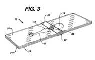
- FIG. 3is a perspective view of the device of FIG. 1 .
- FIG. 4is a schematic of a meter for use with a device of this invention.
- FIG. 4Adepicts an alternative embodiment of an element of the meter of FIG. 4 .
- FIG. 5is a graph of data that is used to determine PT time.
- FIG. 6is a plan view of an alternative embodiment of a device of this invention.
- FIGS. 6A , 6 B, and 6 Cdepict a time sequence during which a sample is admitted to the device of FIG. 6 .
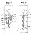
- FIG. 7is a schematic of a device having multiple measurement areas in parallel, multiple stop junctions in parallel, and a single bladder.
- FIG. 8is a schematic of a device having multiple measurement areas in series, with a single stop junction, a single bladder, and a filter over the sample port.
- FIG. 9is a schematic of a device having multiple measurement areas and multiple stop junctions arranged in an alternating series, as well as multiple bladders.
- FIG. 10is a schematic of a device that includes multiple measurement areas in parallel, a single bladder, and a single bypass channel.
- FIG. 11is a schematic of a device having multiple measurement areas in series, multiple stop junctions in series, multiple bladders in series, and multiple bypass channels.
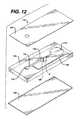
- FIG. 12is an exploded view of an injection-molded device of this invention.
- FIG. 13is a perspective view of the device of FIG. 12 .
- the deviceis of the type that relates a physical parameter of the fluid, or an element of the fluid, to an analyte concentration in the fluid or to a property of the fluid.
- a physical parametere.g., electrical, magnetic, fluidic, or optical
- the deviceincludes a sample application area; a bladder, to create a suction force to draw the sample into the device; a measurement area, in which the sample may undergo a change in an optical parameter, such as light scattering; and a stop junction to precisely stop flow after filling the measurement area.
- the deviceis substantially transparent over the measurement area, so that the area can be illuminated by a light source on one side and the transmitted light measured on the opposite side.
- the measurement on the samplemay be of a parameter that is not changing, but typically the sample undergoes a change in the measurement area, and the change in transmitted light is a measure of the analyte or fluid property of interest.
- light that is scattered from a fluid sample or light that passes through the sample and is reflected back through a second time (by a reflector on that opposite side)can be detected by a detector on the same side as the light source.
- This type of deviceis suitable for a variety of analytical tests of biological fluids, such as determining biochemical or hematological characteristics, or measuring the concentration in such fluids of proteins, hormones, carbohydrates, lipids, drugs, toxins, gases, electrolytes, etc.
- analytical testsof biological fluids, such as determining biochemical or hematological characteristics, or measuring the concentration in such fluids of proteins, hormones, carbohydrates, lipids, drugs, toxins, gases, electrolytes, etc.
- the procedures for performing these testshave been described in the literature. Among the tests, and where they are described, are the following:
- the present deviceis particularly well suited for measuring blood-clotting time—“prothrombin time” or “PT time”—and details regarding such a device appear below.
- the modifications needed to adapt the device for applications such as those listed aboverequire no more than routine experimentation.
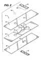
- FIG. 1is a plan view of a device 10 of the present invention.
- FIG. 2is an exploded view and FIG. 3 a perspective view of the device.
- Sampleis applied to sample port 12 after bladder 14 has been compressed.
- the region of layer 26 and/or layer 28 that adjoins the cutout for bladder 14must be resilient, to permit bladder 14 to be compressed.
- Polyester of about 0.1 mm thicknesshas suitable resilience and springiness.
- top layer 26has a thickness of about 0.125 mm, bottom layer 28 about 0.100 mm.
- the volume of bladder 14is preferably at least about equal to the combined volume of channel 16 and measurement area 18 . If measurement area 18 is to be illuminated from below, layer 28 must be transparent where it adjoins measurement area 18 .
- reagent 20contains thromboplastin that is free of bulking reagents normally found in lyophilized reagents.
- stop junction 22adjoins bladder 14 and measurement area 18 ; however, a continuation of channel 16 may be on either or both sides of stop junction 22 , separating the stop junction from measurement area 18 and/or bladder 14 .
- sample flow stopsWhen the sample reaches stop junction 22 , sample flow stops.
- the principle of operation of stop junctionsis described in U.S. Pat. No. 5,230,866, incorporated herein by reference.
- all the above elementsare formed by cutouts in intermediate layer 24 , sandwiched between top layer 26 and bottom layer 28 .
- layer 24is double-sided adhesive tape.
- Stop junction 22is formed by an additional cutout in layer 26 and/or 28 , aligned with the cutout in layer 24 and sealed with sealing layer 30 and/or 32 .
- the stop junctioncomprises cutouts in both layers 26 and 28 , with sealing layers 30 and 32 .
- Each cutout for stop junction 22is at least as wide as channel 16 .
- an optional filter 12 Ato cover sample port 12 .
- the filtermay separate out red blood cells from a whole blood sample and/or may contain a reagent to interact with the blood to provide additional information.
- a suitable filtercomprises an anisotropic membrane, preferably a polysulfone membrane of the type available from Spectral Diagnostics, Inc., Toronto, Canada.
- Optional reflector 18 Amay be on, or adjacent to, a surface of layer 26 and positioned over measurement area 18 . If the reflector is present, the device becomes a transflectance device.
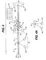
- the method of using the strip of FIGS. 1 , 2 , and 3can be understood with reference to a schematic of the elements of a meter shown in FIG. 4 , which contemplates an automated meter. Alternatively, manual operation is also possible. (In that case, bladder 14 is manually depressed before sample is applied to sample port 12 , is then released.)
- the first step the user performsis to turn on the meter, thereby energizing strip detector 40 , sample detector 42 , measurement system 44 , and optional heater 46 .
- the second stepis to insert the strip.
- the stripis not transparent over at least a part of its area, so that an inserted strip will block the illumination by LED 40 a of detector 40 b .
- Detector 40 bthereby senses that a strip has been inserted and triggers bladder actuator 48 to compress bladder 14 .
- a meter display 50then directs the user to apply a sample to sample port 12 as the third and last step the user must perform to initiate the measurement sequence.
- the empty sample portis reflective. When a sample is introduced into the sample port, it absorbs light from LED 42 a and thereby reduces the light that is reflected to detector 42 b . That reduction in light, in turn, signals actuator 48 to release bladder 14 .
- the resultant suction in channel 16draws sample through measurement area 18 to stop junction 22 .
- Measurement system 44includes an LED/detector pair (like 44 a and 44 b ) for each measurement area. Analysis of the transmitted light as a function of time (as described below) permits a calculation of the PT time, which is displayed on the meter display 50 .
- sample temperatureis maintained at about 37° C. by heater 46 .
- the detectorsenses a sample in sample port 12 , simply by detecting a reduction in (specular) reflection of a light signal that is emitted by 42 a and detected by 42 b .
- a simple systemcannot easily distinguish between a whole blood sample and some other liquid (e.g., blood serum) placed in the sample port in error or, even, an object (e.g., a finger) that can approach sample port 12 and cause the system to erroneously conclude that a proper sample has been applied.
- another embodimentmeasures diffuse reflection from the sample port. This embodiment appears in FIG. 4A , which shows detector 42 b positioned normal to the plane of strip 10 . With the arrangement shown in FIG.
- FIG. 5depicts a typical “clot signature” curve in which the current from detector 44 b is plotted as a function of time. Blood is first detected in the measurement area by 44 b at time 1 . In the time interval A, between points 1 and 2 , the blood fills the measurement area.
- the reduction in current during that time intervalis due to light scattered by red cells and is thus an approximate measure of the hematocrit.
- samplehas filled the measurement area and is at rest, its movement having been stopped by the stop junction.
- the red cellsbegin to stack up like coins (rouleaux formation).
- the rouleaux effectallows increasing light transmission through the sample (and less scattering) in the time interval between points 2 and 3 .
- clot formationends rouleaux formation and transmission through the sample reaches a maximum
- the PT timecan be calculated from the interval B between points 1 and 3 or between 2 and 3 .
- bloodchanges state from liquid to a semi-solid gel, with a corresponding reduction in light transmission.
- the reduction in current C between the maximum 3 and endpoint 4correlates with fibrinogen in the sample.
- the device pictured in FIG. 2 and described aboveis preferably formed by laminating thermoplastic sheets 26 and 28 to a thermoplastic intermediate layer 24 that has adhesive on both of its surfaces.
- the cutouts that form the elements shown in FIG. 1may be formed, for example, by laser- or die-cutting of layers 24 , 26 , and 28 .
- the devicecan be formed of molded plastic.
- the surface of sheet 28is hydrophilic. (Film 9962, available from 3M, St. Paul, Minn.) However, the surfaces do not need to be hydrophilic, because the sample fluid will fill the device without capillary forces.
- sheets 26 and 28may be untreated polyester or other thermoplastic sheet, well known in the art.
- the devicecan be used in any orientation. Unlike capillary fill devices that have vent holes through which sample could leak, the present device vents through the sample port before sample is applied, which means that the part of the strip that is first inserted into the meter is without an opening, reducing the risk of contamination.
- FIG. 6is a plan view of another embodiment of the device of the present invention, in which the device includes a bypass channel 52 that connects channel 16 with bladder 14 .
- the function and operation of the bypass channelcan be understood by referring to FIGS. 6 A, 6 B, and 6 C which depict a time sequence during which a sample is drawn into device 10 for the measurement.
- FIG. 6Adepicts the situation after a user has applied a sample to the strip, while bladder 14 is compressed. This can be accomplished by applying one or more drops of blood.
- FIG. 6Bdepicts the situation after the bladder is decompressed.
- the resulting reduced pressure in the inlet channel 16draws the sample initially into the measurement area 18 .
- stop junction 22the sample encounters a back pressure that causes it to stop and causes additional sample to be drawn into the bypass channel.
- FIG. 6Cdepicts the situation when a reading is taken. Sample is isolated and at rest in measurement area 18 . Excess sample and/or air has been drawn into bypass channel 52 .
- the bypass channel of FIG. 6provides an important improvement over the operation of the “basic” strip of FIGS. 1-3 .
- stop junction 22stops the flow of sample after it fills measurement area 18 .
- the stop junctionaccomplishes the flow stoppage as a result of surface tension acting on the meniscus at the leading edge of the fluid at an abrupt change in cross section of the flow channel.
- the pressure on the bladder side of the stop junctionremains below atmospheric pressure while the pressure on the sample side remains open to atmosphere.
- there is an ambient pressure imbalance on the two sidesThe greater the imbalance, the greater the risk that the stop junction will leak and that sample will flow through the stop junction, interfering with rouleaux formation, and, consequently, providing inaccurate values of PT.
- Bypass channel 52minimizes that risk.
- the reduced pressure on the bladder side of the stop junctiondraws sample into the bypass channel ( FIGS. 6B , 6 C) until the ambient pressure is equalized at atmospheric pressure on both sides of the stop junction. Note that the (reduced) pressure on the bladder side is relatively uncontrolled.
- the bypass channel 52by enabling the pressures on the two sides of the stop junction to equilibrate, permits the use of larger bladders that have greater suction. Larger bladders, in turn, provide more reliable operation of the system.
- FIG. 7depicts an embodiment of the present invention in which there are multiple (three are shown) measurement areas “in parallel”. That is to say that the channels 116 P, 216 P, and 316 P fill substantially simultaneously (assuming they have the same dimensions).
- the situation depicted in FIG. 7is achieved, as discussed above, by applying sample to sample port 112 while bladder 114 is compressed, then releasing bladder 114 .
- the first stepis to apply sample to sample well 112 while bladder 114 is compressed.
- the second stepis to release the bladder. Sample flows to measurement areas 118 P, 218 P, and 318 P, and flow stops when sample reaches stop junctions, 122 P, 222 P, and 322 P, respectively.
- the optional second and third measurement areasmay contain, for example, reagents that neutralize the presence of interferents (such as heparin) in the blood, or that provide a built-in control on the PT measurement, or that measure another blood parameter (such as APPT).
- interferentssuch as heparin
- APPTanother blood parameter
- FIG. 8is a schematic illustration of an embodiment in which multiple measurement areas are “in series”, meaning that they fill sequentially.
- measurement areas 118 S, 218 S, and 318 Sfill sequentially, through a single channel 116 S, until the sample reaches stop junction 122 S.
- a potential drawback of this designis that sample passing from one measurement area to the next may carry over reagent.
- FIG. 9is a schematic of another embodiment of a device that is adapted for multiple sequential tests.
- stop junctions 122 T, 222 T, and 322 Tpermit a user to control the timing of sequential filling of measurement areas 118 T, 218 T, and 318 T.
- bladders 114 , 214 , and 314are all compressed before a blood sample is applied to sample well 112 .
- Bladder 114is then released to draw blood into measurement area 118 T to stop junction 122 T.
- bladder 214is released to permit blood to break through stop junction 122 T and enter measurement area 218 T to stop junction 222 T.
- bladder 314is decompressed, permitting sample to break through stop function 222 T and flow to stop junction 322 T.
- the device of FIG. 9must be carefully formed, since the force drawing sample into the device—caused by decompressing a bladder—must be balanced against the opposing force—exerted by a stop junction. If the drawing force is too great, a stop junction may prematurely permit sample to pass; if it's too small, it will not draw the sample through a stop junction, when that is intended.
- FIG. 10depicts a preferred embodiment of the present device. It is a parallel multi-channel device that includes bypass channel 152 P Bypass channel 152 P serves a purpose in this device that is analogous to that served by bypass channel 52 in the device of FIG. 6 , which was described above.
- Measurement area 118 Pcontains thromboplastin.
- measurement areas 218 P and 318 Pcontain controls, more preferably, the controls described below.
- Area 218 Pcontains thromboplastin, bovine eluate, and recombinant Factor VIIa.
- the compositionis selected to normalize the clotting time of a blood sample by counteracting the effect of an anticoagulant, such as warfarin.
- Measurement area 318 Pcontains thromboplastin and bovine eluate alone, to partially overcome the effect of an anticoagulent. Thus, 3 measurements are made on the strip. PT time of the sample, the measurement of primary interest, is measured on area 118 P. However, that measurement is validated only when measurements on areas 218 P and 318 P yield results within a predetermined range.
- Extended stop junction 422stops flow in all three measurement areas.
- FIG. 11depicts a device that includes bypass channels 152 S and 252 S to permit timed filling of measurement areas 118 T and 218 T. Operation of the device of FIG. 11 is analogous to that of the device of FIG. 9 , described above, with the following exception.
- First bypass channel 152 Shas a region in which a reagent that causes clotting, such as thromboplastin, is coated. As a first measurement is made in reagent area 118 T, a clot forms in blood that had been drawn into bypass channel 152 S. Thus, when the second bladder is decompressed, blood is blocked from being drawn through bypass 152 S and instead is drawn though stop junction 122 T to measurement area 218 T and bypass channel 252 S.
- FIG. 12is an exploded view of an injection-molded device 110 , including top layer 126 and bottom layer 128 sandwiching intermediate layer 124 .
- the intermediate layerhas depressions in its top surface that form sample port 112 , channel 116 , measurement area 118 , and optional bypass channel 152 .
- Stop junction 122passes through the thickness of intermediate layer 124 . Sample flow stops at the interface between stop junction 122 and channel A, which is formed by a depression in the bottom surface. Thus, the sample flows from sample port 112 through channel 116 to measurement area 118 into stop junction 122 .
- the principle of operation of the injection molded deviceis the same as described above. It provides greater flexibility in the design of the stop junction, as well as the other elements of the device, because a wide range of channel cross sections are feasible.
- the molded structurealso provides more rigidity, although it is substantially more costly.
- a strip of this inventionis made by first passing a double-sided adhesive tape (RX 675SLT, available from Scapa Tapes, Windsor, Conn.) sandwiched between two release liners into a laminating and rotary die-cutting converting system.
- RX 675SLTdouble-sided adhesive tape
- the pattern shown in FIG. 6is cut through the top release liner and tape, but not through the bottom release liner, which is then removed as waste, along with the cutouts from the tape.
- Polyester film treated to be hydrophilic(3M9962, available from 3M, St. Paul, Minn.) is laminated to the exposed bottom side of the tape.
- Reagentthromboplastin, available from Ortho Clinical Diagnostics, Raritan, N.J.
- Reagentis then printed onto the reagent area ( 18 ) of the polyester film by bubble jet printing, using printing heads 51612A, from Hewlett Packard, Corvallis, Oreg.
- a sample portis cut in untreated polyester film (AR1235, available from Adhesives Research, Glen Rock, Pa.) and then laminated, in register, to the top of the double-sided tape (after removing the release layer).
- a diethen cuts the stop junction through the three layers of the sandwich.
- strips of single-sided adhesive tape(MSX4841, available from 3M, St. Paul, Minn.) are applied to the outside of the polyester layers to seal the stop junction.
- Reagent that is bubble-jet printed onto areas 118 P, 218 P, and 318 Pis, respectively, thromboplastin; thromboplastin, bovine eluate, and recombinant Factor VIIa; and thromboplastin and bovine eluate alone.
- the bovine eluate(plasma barium citrate bovine eluate) is available from Haemotologic Technologies, Burlington, Vt.; and recombinant Factor VIIa from American Diagnostica, Greenwich, Conn.
- Measurements made on a whole blood sample using the strip of this Exampleyield a curve of the type shown in FIG. 5 for each of the measurement areas.
- the data from the curves for the controlsare used to qualify the data from the curve for measurement area 118 P.
- the PT timecan be determined more reliably than can be done with a strip having a single measurement area.
- the device of FIGS. 12 and 13is formed by sandwiching middle layer 124 between top layer 126 and bottom layer 128 .
- the middle and bottom layersare injection molded polycarbonate (Lexan*121) and have thicknesses of 6.3 mm and 1.5 mm, respectively.
- Top layer 126is made by die cutting 0.18 mm Lexan*8010 sheet. The elements are ultrasonically welded after the reagent of Example 1 is applied to reagent area 118 .
- the Lexan*materialis available from General Electric, Pittsfield, Mass.
Landscapes
- Health & Medical Sciences (AREA)
- Life Sciences & Earth Sciences (AREA)
- Chemical & Material Sciences (AREA)
- Immunology (AREA)
- Engineering & Computer Science (AREA)
- Hematology (AREA)
- Biomedical Technology (AREA)
- Molecular Biology (AREA)
- Urology & Nephrology (AREA)
- General Health & Medical Sciences (AREA)
- Analytical Chemistry (AREA)
- Physics & Mathematics (AREA)
- Pathology (AREA)
- Food Science & Technology (AREA)
- Medicinal Chemistry (AREA)
- Biochemistry (AREA)
- General Physics & Mathematics (AREA)
- Microbiology (AREA)
- Cell Biology (AREA)
- Biotechnology (AREA)
- Chemical Kinetics & Catalysis (AREA)
- Dispersion Chemistry (AREA)
- Clinical Laboratory Science (AREA)
- Biophysics (AREA)
- Ecology (AREA)
- Public Health (AREA)
- Medical Informatics (AREA)
- Surgery (AREA)
- Animal Behavior & Ethology (AREA)
- Heart & Thoracic Surgery (AREA)
- Veterinary Medicine (AREA)
- Investigating Or Analysing Biological Materials (AREA)
- Infusion, Injection, And Reservoir Apparatuses (AREA)
- Optical Measuring Cells (AREA)
- Examining Or Testing Airtightness (AREA)
- Fluid-Pressure Circuits (AREA)
- Endoscopes (AREA)
- Investigating Or Analysing Materials By Optical Means (AREA)
- External Artificial Organs (AREA)
- Investigating Or Analysing Materials By The Use Of Chemical Reactions (AREA)
Abstract
Description
- a first layer and second layer at least one of which has a resilient region over at least part of its area, separated by an intermediate layer, in which cutouts in the intermediate layer form, with the first and second layers,
- a) a sample port for introducing a sample of the biological fluid into the device;
- b) a first measurement area, in which a physical parameter of the sample is measured and related to the analyte concentration or property of the fluid;
- c) a first channel, having a first end and a second end, to provide a fluidic path from the sample port at the first end through the first measurement area;
- d) a first bladder at the second end of the first channel, comprising at least a part of the resilient region in at least the first or second layer and having a volume that is at least about equal to the combined volume of the first measurement area and first channel; and
- e) a first stop junction in the first channel between the first measurement area and first bladder that comprises a co-aligned through hole in at least the first or second layer, the through hole being overlaid with a third layer.
- a first layer, which has a resilient region over at least a part of its area, and a second layer, separated by an intermediate layer, in which recesses in a first surface of the intermediate layer form, with the first layer,
- a) a sample port for introducing a sample of the biological fluid into the device;
- b) a measurement area, in which the sample undergoes a change in a physical parameter that is measured and related to the analyte concentration or property of the fluid;
- c) a channel, having a first end and a second end, to provide a fluidic path from the sample port at the first end through the measurement area; and
- d) a bladder, at the second end of the channel, comprising at least a part of the resilient region in the first layer and having a volume that is at least about equal to the combined volume of the measurement area and channel; and
- a stop junction in the channel between the measurement area and bladder that comprises two passages substantially normal to the first surface of the intermediate layer, each passage having a first end in fluid communication with the channel and a second end in fluid communication with a recess in a second surface of the intermediate layer, which recess provides fluid communication between the second ends of the passages.
- (1) Chromogenic Factor XIIa Assay (and other clotting factors as well): Rand, M. D. et al., Blood, 88, 3432 (1996).
- (2) Factor X Assay: Bick, R. L. Disorders of Thrombosis and Hemostasis: Clinical and Laboratory Practice. Chicago, ASCP Press, 1992.
- (3) DRVVT (Dilute Russells Viper Venom Test): Exner, T. et al., Blood Coag. Fibrinol., 1, 259 (1990).
- (4) Immunonephelometric and Immunoturbidimetric Assays for Proteins: Whicher, J. T., CRC Crit. Rev. Clin Lab Sci. 18:213 (1983).
- (5) TPA Assay: Mann, K. G., et al., Blood, 76, 755, (1990).; and Hartshorn, J. N. et al., Blood, 78, 833 (1991).
- (6) APTT (Activated Partial Thromboplastin Time Assay): Proctor, R. R. and Rapaport, S. I. Amer. J. Clin. Path, 36, 212 (1961); Brandt, J. T. and Triplett, D. A. Amer. J. Clin. Path., 76, 530 (1981); and Kelsey, P. R. Thromb. Haemost. 52, 172 (1984).
- (7) HbA1c Assay (Glycosylated Hemoglobin Assay): Nicol, D. J. et al., Clin. Chem. 29, 1694 (1983).
- (8) Total Hemoglobin: Schneck et al., Clinical Chem., 32/33, 526 (1986); and U.S. Pat. No. 4,088,448.
- (9) Factor Xa: Vinazzer, H., Proc. Symp. Dtsch. Ges. Klin. Chem., 203 (1977), ed. By Witt, I
- (10) Colorimetric Assay for Nitric Oxide: Schmidt, H. H., et al., Biochemica, 2, 22 (1995).
Claims (6)
Priority Applications (1)
| Application Number | Priority Date | Filing Date | Title |
|---|---|---|---|
| US10/330,790US7022286B2 (en) | 1998-07-20 | 2002-12-26 | Fluidic device for medical diagnostics |
Applications Claiming Priority (3)
| Application Number | Priority Date | Filing Date | Title |
|---|---|---|---|
| US9342198P | 1998-07-20 | 1998-07-20 | |
| US09/333,765US6521182B1 (en) | 1998-07-20 | 1999-06-15 | Fluidic device for medical diagnostics |
| US10/330,790US7022286B2 (en) | 1998-07-20 | 2002-12-26 | Fluidic device for medical diagnostics |
Related Parent Applications (1)
| Application Number | Title | Priority Date | Filing Date |
|---|---|---|---|
| US09/333,765ContinuationUS6521182B1 (en) | 1998-07-20 | 1999-06-15 | Fluidic device for medical diagnostics |
Publications (2)
| Publication Number | Publication Date |
|---|---|
| US20030156984A1 US20030156984A1 (en) | 2003-08-21 |
| US7022286B2true US7022286B2 (en) | 2006-04-04 |
Family
ID=26787520
Family Applications (8)
| Application Number | Title | Priority Date | Filing Date |
|---|---|---|---|
| US09/333,765Expired - LifetimeUS6521182B1 (en) | 1998-07-20 | 1999-06-15 | Fluidic device for medical diagnostics |
| US10/052,447AbandonedUS20020064480A1 (en) | 1998-07-20 | 2002-01-17 | Fluidic device for medical diagnostics |
| US10/121,636AbandonedUS20020110486A1 (en) | 1998-07-20 | 2002-04-11 | Analyte test strip with two controls |
| US10/121,425AbandonedUS20020110922A1 (en) | 1998-07-20 | 2002-04-11 | Vacuum loaded test strip and method of use |
| US10/264,662AbandonedUS20030031594A1 (en) | 1998-07-20 | 2002-10-03 | Vacuum loaded test strip with stop junction and bypass channel |
| US10/330,456AbandonedUS20030156983A1 (en) | 1998-07-20 | 2002-12-26 | Fluidic device for medical diagnostics |
| US10/330,790Expired - LifetimeUS7022286B2 (en) | 1998-07-20 | 2002-12-26 | Fluidic device for medical diagnostics |
| US10/666,846AbandonedUS20040109790A1 (en) | 1998-07-20 | 2003-09-18 | Vacuum loaded test strip with stop junction and bypass channel |
Family Applications Before (6)
| Application Number | Title | Priority Date | Filing Date |
|---|---|---|---|
| US09/333,765Expired - LifetimeUS6521182B1 (en) | 1998-07-20 | 1999-06-15 | Fluidic device for medical diagnostics |
| US10/052,447AbandonedUS20020064480A1 (en) | 1998-07-20 | 2002-01-17 | Fluidic device for medical diagnostics |
| US10/121,636AbandonedUS20020110486A1 (en) | 1998-07-20 | 2002-04-11 | Analyte test strip with two controls |
| US10/121,425AbandonedUS20020110922A1 (en) | 1998-07-20 | 2002-04-11 | Vacuum loaded test strip and method of use |
| US10/264,662AbandonedUS20030031594A1 (en) | 1998-07-20 | 2002-10-03 | Vacuum loaded test strip with stop junction and bypass channel |
| US10/330,456AbandonedUS20030156983A1 (en) | 1998-07-20 | 2002-12-26 | Fluidic device for medical diagnostics |
Family Applications After (1)
| Application Number | Title | Priority Date | Filing Date |
|---|---|---|---|
| US10/666,846AbandonedUS20040109790A1 (en) | 1998-07-20 | 2003-09-18 | Vacuum loaded test strip with stop junction and bypass channel |
Country Status (13)
| Country | Link |
|---|---|
| US (8) | US6521182B1 (en) |
| EP (1) | EP0974840B1 (en) |
| JP (1) | JP2000055911A (en) |
| KR (1) | KR100634714B1 (en) |
| CN (1) | CN1199038C (en) |
| AT (1) | ATE229649T1 (en) |
| CA (1) | CA2277639A1 (en) |
| DE (1) | DE69904403T2 (en) |
| DK (1) | DK0974840T3 (en) |
| ES (1) | ES2189353T3 (en) |
| IL (1) | IL130807A (en) |
| NO (1) | NO993536L (en) |
| TW (1) | TW411268B (en) |
Cited By (77)
| Publication number | Priority date | Publication date | Assignee | Title |
|---|---|---|---|---|
| US20020110922A1 (en)* | 1998-07-20 | 2002-08-15 | Shartle Robert Justice | Vacuum loaded test strip and method of use |
| US20020192833A1 (en)* | 2000-07-31 | 2002-12-19 | Victor Pan | Method and apparatus for detecting the presence of a fluid on a test strip |
| US20070081155A1 (en)* | 2005-08-30 | 2007-04-12 | Schembri Carol T | Lab in a cuvette |
| US20070092407A1 (en)* | 2005-10-26 | 2007-04-26 | General Electric Company | Optical sensor array system and method for parallel processing of chemical and biochemical information |
| US7297151B2 (en) | 2002-04-19 | 2007-11-20 | Elikan Technologies, Inc. | Method and apparatus for body fluid sampling with improved sensing |
| US7316700B2 (en) | 2001-06-12 | 2008-01-08 | Pelikan Technologies, Inc. | Self optimizing lancing device with adaptation means to temporal variations in cutaneous properties |
| US7344507B2 (en) | 2002-04-19 | 2008-03-18 | Pelikan Technologies, Inc. | Method and apparatus for lancet actuation |
| US7344894B2 (en) | 2001-10-16 | 2008-03-18 | Agilent Technologies, Inc. | Thermal regulation of fluidic samples within a diagnostic cartridge |
| US7374544B2 (en) | 2002-04-19 | 2008-05-20 | Pelikan Technologies, Inc. | Method and apparatus for penetrating tissue |
| US7410468B2 (en) | 2002-04-19 | 2008-08-12 | Pelikan Technologies, Inc. | Method and apparatus for penetrating tissue |
| US7481776B2 (en) | 2002-04-19 | 2009-01-27 | Pelikan Technologies, Inc. | Method and apparatus for penetrating tissue |
| US7491178B2 (en) | 2002-04-19 | 2009-02-17 | Pelikan Technologies, Inc. | Method and apparatus for penetrating tissue |
| US7524293B2 (en) | 2002-04-19 | 2009-04-28 | Pelikan Technologies, Inc. | Method and apparatus for penetrating tissue |
| US20090114608A1 (en)* | 2007-11-02 | 2009-05-07 | Industrial Technology Research Institute | Fluid analytical device |
| US7537571B2 (en) | 2001-06-12 | 2009-05-26 | Pelikan Technologies, Inc. | Integrated blood sampling analysis system with multi-use sampling module |
| US7547287B2 (en) | 2002-04-19 | 2009-06-16 | Pelikan Technologies, Inc. | Method and apparatus for penetrating tissue |
| US7563232B2 (en) | 2002-04-19 | 2009-07-21 | Pelikan Technologies, Inc. | Method and apparatus for penetrating tissue |
| US7582063B2 (en) | 2000-11-21 | 2009-09-01 | Pelikan Technologies, Inc. | Blood testing apparatus having a rotatable cartridge with multiple lancing elements and testing means |
| US7582099B2 (en) | 2002-04-19 | 2009-09-01 | Pelikan Technologies, Inc | Method and apparatus for penetrating tissue |
| US7604592B2 (en) | 2003-06-13 | 2009-10-20 | Pelikan Technologies, Inc. | Method and apparatus for a point of care device |
| US7648468B2 (en) | 2002-04-19 | 2010-01-19 | Pelikon Technologies, Inc. | Method and apparatus for penetrating tissue |
| US7666149B2 (en) | 1997-12-04 | 2010-02-23 | Peliken Technologies, Inc. | Cassette of lancet cartridges for sampling blood |
| US7674232B2 (en) | 2002-04-19 | 2010-03-09 | Pelikan Technologies, Inc. | Method and apparatus for penetrating tissue |
| US7682318B2 (en) | 2001-06-12 | 2010-03-23 | Pelikan Technologies, Inc. | Blood sampling apparatus and method |
| US7699791B2 (en) | 2001-06-12 | 2010-04-20 | Pelikan Technologies, Inc. | Method and apparatus for improving success rate of blood yield from a fingerstick |
| US7713214B2 (en) | 2002-04-19 | 2010-05-11 | Pelikan Technologies, Inc. | Method and apparatus for a multi-use body fluid sampling device with optical analyte sensing |
| US7717863B2 (en) | 2002-04-19 | 2010-05-18 | Pelikan Technologies, Inc. | Method and apparatus for penetrating tissue |
| US7731729B2 (en) | 2002-04-19 | 2010-06-08 | Pelikan Technologies, Inc. | Method and apparatus for penetrating tissue |
| US7822454B1 (en) | 2005-01-03 | 2010-10-26 | Pelikan Technologies, Inc. | Fluid sampling device with improved analyte detecting member configuration |
| US7833171B2 (en) | 2002-04-19 | 2010-11-16 | Pelikan Technologies, Inc. | Method and apparatus for penetrating tissue |
| US7841992B2 (en) | 2001-06-12 | 2010-11-30 | Pelikan Technologies, Inc. | Tissue penetration device |
| US7850621B2 (en) | 2003-06-06 | 2010-12-14 | Pelikan Technologies, Inc. | Method and apparatus for body fluid sampling and analyte sensing |
| US20100328654A1 (en)* | 2007-06-28 | 2010-12-30 | Koninklijke Philips Electronics N.V. | Microelectronic sensor device for optical examinations on a wetted surface |
| US7862520B2 (en) | 2002-04-19 | 2011-01-04 | Pelikan Technologies, Inc. | Body fluid sampling module with a continuous compression tissue interface surface |
| US20110005341A1 (en)* | 2008-03-11 | 2011-01-13 | Koninklijke Philips Electronics N.V. | Filtering apparatus for filtering a fluid |
| US7874994B2 (en) | 2002-04-19 | 2011-01-25 | Pelikan Technologies, Inc. | Method and apparatus for penetrating tissue |
| US7892183B2 (en) | 2002-04-19 | 2011-02-22 | Pelikan Technologies, Inc. | Method and apparatus for body fluid sampling and analyte sensing |
| US7901362B2 (en) | 2002-04-19 | 2011-03-08 | Pelikan Technologies, Inc. | Method and apparatus for penetrating tissue |
| US7909778B2 (en) | 2002-04-19 | 2011-03-22 | Pelikan Technologies, Inc. | Method and apparatus for penetrating tissue |
| US7909775B2 (en) | 2001-06-12 | 2011-03-22 | Pelikan Technologies, Inc. | Method and apparatus for lancet launching device integrated onto a blood-sampling cartridge |
| US7914465B2 (en) | 2002-04-19 | 2011-03-29 | Pelikan Technologies, Inc. | Method and apparatus for penetrating tissue |
| US7959582B2 (en) | 2002-04-19 | 2011-06-14 | Pelikan Technologies, Inc. | Method and apparatus for penetrating tissue |
| US7976476B2 (en) | 2002-04-19 | 2011-07-12 | Pelikan Technologies, Inc. | Device and method for variable speed lancet |
| US8197421B2 (en) | 2002-04-19 | 2012-06-12 | Pelikan Technologies, Inc. | Method and apparatus for penetrating tissue |
| US8221334B2 (en) | 2002-04-19 | 2012-07-17 | Sanofi-Aventis Deutschland Gmbh | Method and apparatus for penetrating tissue |
| US8267870B2 (en) | 2002-04-19 | 2012-09-18 | Sanofi-Aventis Deutschland Gmbh | Method and apparatus for body fluid sampling with hybrid actuation |
| US8282576B2 (en) | 2003-09-29 | 2012-10-09 | Sanofi-Aventis Deutschland Gmbh | Method and apparatus for an improved sample capture device |
| US8333710B2 (en) | 2002-04-19 | 2012-12-18 | Sanofi-Aventis Deutschland Gmbh | Tissue penetration device |
| US8420025B2 (en) | 2005-10-26 | 2013-04-16 | General Electric Company | Methods and systems for delivery of fluidic samples to sensor arrays |
| US8435190B2 (en) | 2002-04-19 | 2013-05-07 | Sanofi-Aventis Deutschland Gmbh | Method and apparatus for penetrating tissue |
| US20130114076A1 (en)* | 2010-07-09 | 2013-05-09 | Koninklijke Philips Electronics N.V. | Cartridge with large-scale manufacturing design |
| US8439872B2 (en) | 1998-03-30 | 2013-05-14 | Sanofi-Aventis Deutschland Gmbh | Apparatus and method for penetration with shaft having a sensor for sensing penetration depth |
| WO2013190073A1 (en) | 2012-06-21 | 2013-12-27 | Lifescan Scotland Limited | Analytical test strip with capillary sample-receiving chambers separated by stop junctions |
| US8652831B2 (en) | 2004-12-30 | 2014-02-18 | Sanofi-Aventis Deutschland Gmbh | Method and apparatus for analyte measurement test time |
| US8668656B2 (en) | 2003-12-31 | 2014-03-11 | Sanofi-Aventis Deutschland Gmbh | Method and apparatus for improving fluidic flow and sample capture |
| US8702624B2 (en) | 2006-09-29 | 2014-04-22 | Sanofi-Aventis Deutschland Gmbh | Analyte measurement device with a single shot actuator |
| US8721671B2 (en) | 2001-06-12 | 2014-05-13 | Sanofi-Aventis Deutschland Gmbh | Electric lancet actuator |
| US8828203B2 (en) | 2004-05-20 | 2014-09-09 | Sanofi-Aventis Deutschland Gmbh | Printable hydrogels for biosensors |
| US8877023B2 (en) | 2012-06-21 | 2014-11-04 | Lifescan Scotland Limited | Electrochemical-based analytical test strip with intersecting sample-receiving chambers |
| US8965476B2 (en) | 2010-04-16 | 2015-02-24 | Sanofi-Aventis Deutschland Gmbh | Tissue penetration device |
| WO2015048225A1 (en)* | 2013-09-26 | 2015-04-02 | Quick Llc | Sample collection device for optical analysis |
| US9034639B2 (en) | 2002-12-30 | 2015-05-19 | Sanofi-Aventis Deutschland Gmbh | Method and apparatus using optical techniques to measure analyte levels |
| US9072842B2 (en) | 2002-04-19 | 2015-07-07 | Sanofi-Aventis Deutschland Gmbh | Method and apparatus for penetrating tissue |
| US9128038B2 (en) | 2012-06-21 | 2015-09-08 | Lifescan Scotland Limited | Analytical test strip with capillary sample-receiving chambers separated by a physical barrier island |
| US9144401B2 (en) | 2003-06-11 | 2015-09-29 | Sanofi-Aventis Deutschland Gmbh | Low pain penetrating member |
| US9226699B2 (en) | 2002-04-19 | 2016-01-05 | Sanofi-Aventis Deutschland Gmbh | Body fluid sampling module with a continuous compression tissue interface surface |
| US9248267B2 (en) | 2002-04-19 | 2016-02-02 | Sanofi-Aventis Deustchland Gmbh | Tissue penetration device |
| US9314194B2 (en) | 2002-04-19 | 2016-04-19 | Sanofi-Aventis Deutschland Gmbh | Tissue penetration device |
| US9351680B2 (en) | 2003-10-14 | 2016-05-31 | Sanofi-Aventis Deutschland Gmbh | Method and apparatus for a variable user interface |
| US9375169B2 (en) | 2009-01-30 | 2016-06-28 | Sanofi-Aventis Deutschland Gmbh | Cam drive for managing disposable penetrating member actions with a single motor and motor and control system |
| US9386944B2 (en) | 2008-04-11 | 2016-07-12 | Sanofi-Aventis Deutschland Gmbh | Method and apparatus for analyte detecting device |
| US9427532B2 (en) | 2001-06-12 | 2016-08-30 | Sanofi-Aventis Deutschland Gmbh | Tissue penetration device |
| US9560993B2 (en) | 2001-11-21 | 2017-02-07 | Sanofi-Aventis Deutschland Gmbh | Blood testing apparatus having a rotatable cartridge with multiple lancing elements and testing means |
| US9795747B2 (en) | 2010-06-02 | 2017-10-24 | Sanofi-Aventis Deutschland Gmbh | Methods and apparatus for lancet actuation |
| US9820684B2 (en) | 2004-06-03 | 2017-11-21 | Sanofi-Aventis Deutschland Gmbh | Method and apparatus for a fluid sampling device |
| US9839386B2 (en) | 2002-04-19 | 2017-12-12 | Sanofi-Aventis Deustschland Gmbh | Body fluid sampling device with capacitive sensor |
| US10928289B2 (en)* | 2017-05-04 | 2021-02-23 | University Of Connecticut | Assembly for measuring the viscosity of fluids using microchannels |
Families Citing this family (142)
| Publication number | Priority date | Publication date | Assignee | Title |
|---|---|---|---|---|
| US7494816B2 (en) | 1997-12-22 | 2009-02-24 | Roche Diagnostic Operations, Inc. | System and method for determining a temperature during analyte measurement |
| US7390667B2 (en)* | 1997-12-22 | 2008-06-24 | Roche Diagnostics Operations, Inc. | System and method for analyte measurement using AC phase angle measurements |
| US8071384B2 (en) | 1997-12-22 | 2011-12-06 | Roche Diagnostics Operations, Inc. | Control and calibration solutions and methods for their use |
| US7407811B2 (en)* | 1997-12-22 | 2008-08-05 | Roche Diagnostics Operations, Inc. | System and method for analyte measurement using AC excitation |
| US6830934B1 (en) | 1999-06-15 | 2004-12-14 | Lifescan, Inc. | Microdroplet dispensing for a medical diagnostic device |
| US6084660A (en)* | 1998-07-20 | 2000-07-04 | Lifescan, Inc. | Initiation of an analytical measurement in blood |
| US20050103624A1 (en) | 1999-10-04 | 2005-05-19 | Bhullar Raghbir S. | Biosensor and method of making |
| US6458326B1 (en) | 1999-11-24 | 2002-10-01 | Home Diagnostics, Inc. | Protective test strip platform |
| DE60122517T2 (en)* | 2000-03-31 | 2007-03-08 | Lifescan, Inc., Milpitas | ELECTRICALLY CONDUCTIVE PATTERN FOR MONITORING THE FILLING OF MEDICAL DEVICES |
| US6908593B1 (en)* | 2000-03-31 | 2005-06-21 | Lifescan, Inc. | Capillary flow control in a fluidic diagnostic device |
| JP4606543B2 (en)* | 2000-04-13 | 2011-01-05 | パナソニック株式会社 | Method for confirming amount of solution to be measured and measuring system control method in optical property measuring apparatus |
| US6726818B2 (en)* | 2000-07-21 | 2004-04-27 | I-Sens, Inc. | Biosensors with porous chromatographic membranes |
| JP4384344B2 (en)* | 2000-08-09 | 2009-12-16 | 拓之 今野 | Blood coagulation time measurement method and apparatus using granular spot pattern by laser reflected light |
| US7144495B2 (en) | 2000-12-13 | 2006-12-05 | Lifescan, Inc. | Electrochemical test strip with an integrated micro-needle and associated methods |
| US6620310B1 (en) | 2000-12-13 | 2003-09-16 | Lifescan, Inc. | Electrochemical coagulation assay and device |
| US20040099310A1 (en)* | 2001-01-05 | 2004-05-27 | Per Andersson | Microfluidic device |
| US6525330B2 (en) | 2001-02-28 | 2003-02-25 | Home Diagnostics, Inc. | Method of strip insertion detection |
| US6562625B2 (en) | 2001-02-28 | 2003-05-13 | Home Diagnostics, Inc. | Distinguishing test types through spectral analysis |
| US6541266B2 (en) | 2001-02-28 | 2003-04-01 | Home Diagnostics, Inc. | Method for determining concentration of an analyte in a test strip |
| CA2441206A1 (en) | 2001-03-19 | 2002-09-26 | Gyros Ab | Characterization of reaction variables |
| US7776608B2 (en) | 2001-07-09 | 2010-08-17 | Bayer Healthcare Llc | Volume meter testing device and method of use |
| US6884592B2 (en) | 2001-09-05 | 2005-04-26 | Lifescan, Inc. | Devices for analyte concentration determination and methods of manufacturing and using the same |
| US20030044318A1 (en)* | 2001-09-05 | 2003-03-06 | Lorin Olson | Devices for analyte concentration determination and methods of using the same |
| JP2003091787A (en)* | 2001-09-17 | 2003-03-28 | Riken Keiki Co Ltd | Portable gas alarm |
| EP2332651A3 (en)* | 2001-10-25 | 2011-08-31 | Bar Ilan University | Interactive transparent individual cells biochip processor |
| US6989891B2 (en) | 2001-11-08 | 2006-01-24 | Optiscan Biomedical Corporation | Device and method for in vitro determination of analyte concentrations within body fluids |
| ATE479089T1 (en)* | 2001-11-16 | 2010-09-15 | Stefan Ufer | FLEXIBLE SENSOR AND MANUFACTURING METHOD |
| US6746872B2 (en) | 2002-01-16 | 2004-06-08 | Lifescan, Inc. | Control compositions and methods of use for coagulation tests |
| US6673617B2 (en)* | 2002-03-14 | 2004-01-06 | Lifescan, Inc. | Test strip qualification system |
| US6682933B2 (en)* | 2002-03-14 | 2004-01-27 | Lifescan, Inc. | Test strip qualification system |
| US6660527B2 (en)* | 2002-03-28 | 2003-12-09 | David Karl Stroup | Fluid-transfer collection assembly and method of using the same |
| US8372016B2 (en) | 2002-04-19 | 2013-02-12 | Sanofi-Aventis Deutschland Gmbh | Method and apparatus for body fluid sampling and analyte sensing |
| US8360992B2 (en) | 2002-04-19 | 2013-01-29 | Sanofi-Aventis Deutschland Gmbh | Method and apparatus for penetrating tissue |
| DE10234819A1 (en)* | 2002-07-31 | 2004-02-19 | Roche Diagnostics Gmbh | Test apparatus for blood, comprising compound body with test strip levels and transport channels to give complex tests in compact structure |
| US7604775B2 (en) | 2002-08-12 | 2009-10-20 | Bayer Healthcare Llc | Fluid collecting and monitoring device |
| US7010432B2 (en) | 2002-08-30 | 2006-03-07 | Lifescan, Inc. | Method and system for determining the acceptability of signal data collected from a prothrombin time test strip |
| US7049087B2 (en)* | 2002-11-05 | 2006-05-23 | Lifescan, Inc. | Method for manufacturing a tissue factor-based prothrombin time reagent |
| US7291310B2 (en)* | 2002-12-17 | 2007-11-06 | The Regents Of The University Of Michigan | Microsystem for determining clotting time of blood and low-cost, single-use device for use therein |
| GB0300820D0 (en)* | 2003-01-14 | 2003-02-12 | Diagnoswiss Sa | Membrane-microchannel strip |
| DE10305050A1 (en)* | 2003-02-07 | 2004-08-19 | Roche Diagnostics Gmbh | Analytical test element and method for blood tests |
| IL154677A0 (en)* | 2003-02-27 | 2003-09-17 | Univ Bar Ilan | A method and apparatus for manipulating an individual cell |
| US8153081B2 (en) | 2003-05-29 | 2012-04-10 | Bayer Healthcare Llc | Test sensor and method for manufacturing the same |
| DE602004028463D1 (en) | 2003-05-30 | 2010-09-16 | Pelikan Technologies Inc | METHOD AND DEVICE FOR INJECTING LIQUID |
| US7718439B2 (en) | 2003-06-20 | 2010-05-18 | Roche Diagnostics Operations, Inc. | System and method for coding information on a biosensor test strip |
| US7645421B2 (en) | 2003-06-20 | 2010-01-12 | Roche Diagnostics Operations, Inc. | System and method for coding information on a biosensor test strip |
| JP4447009B2 (en)* | 2003-06-20 | 2010-04-07 | エフ ホフマン−ラ ロッシュ アクチェン ゲゼルシャフト | Test strip with slot vent opening |
| US7645373B2 (en)* | 2003-06-20 | 2010-01-12 | Roche Diagnostic Operations, Inc. | System and method for coding information on a biosensor test strip |
| US8148164B2 (en) | 2003-06-20 | 2012-04-03 | Roche Diagnostics Operations, Inc. | System and method for determining the concentration of an analyte in a sample fluid |
| US7488601B2 (en) | 2003-06-20 | 2009-02-10 | Roche Diagnostic Operations, Inc. | System and method for determining an abused sensor during analyte measurement |
| US7604721B2 (en) | 2003-06-20 | 2009-10-20 | Roche Diagnostics Operations, Inc. | System and method for coding information on a biosensor test strip |
| US8679853B2 (en)* | 2003-06-20 | 2014-03-25 | Roche Diagnostics Operations, Inc. | Biosensor with laser-sealed capillary space and method of making |
| US8071030B2 (en)* | 2003-06-20 | 2011-12-06 | Roche Diagnostics Operations, Inc. | Test strip with flared sample receiving chamber |
| US7597793B2 (en)* | 2003-06-20 | 2009-10-06 | Roche Operations Ltd. | System and method for analyte measurement employing maximum dosing time delay |
| US7452457B2 (en)* | 2003-06-20 | 2008-11-18 | Roche Diagnostics Operations, Inc. | System and method for analyte measurement using dose sufficiency electrodes |
| US8206565B2 (en) | 2003-06-20 | 2012-06-26 | Roche Diagnostics Operation, Inc. | System and method for coding information on a biosensor test strip |
| US8058077B2 (en)* | 2003-06-20 | 2011-11-15 | Roche Diagnostics Operations, Inc. | Method for coding information on a biosensor test strip |
| US8597597B2 (en)* | 2003-06-26 | 2013-12-03 | Seng Enterprises Ltd. | Picoliter well holding device and method of making the same |
| US9200245B2 (en)* | 2003-06-26 | 2015-12-01 | Seng Enterprises Ltd. | Multiwell plate |
| US20060240548A1 (en)* | 2003-06-26 | 2006-10-26 | Mordechai Deutsch | Materials for constructing cell-chips, cell-chip covers, cell-chips coats, processed cell-chips and uses thereof |
| US6927745B2 (en)* | 2003-08-25 | 2005-08-09 | Harris Corporation | Frequency selective surfaces and phased array antennas using fluidic dielectrics |
| US7582472B2 (en)* | 2003-08-26 | 2009-09-01 | Smith Kenneth E | Apparatus and method for liquid sample testing |
| US7147362B2 (en)* | 2003-10-15 | 2006-12-12 | Agilent Technologies, Inc. | Method of mixing by intermittent centrifugal force |
| EP1713926B1 (en) | 2004-02-06 | 2012-08-01 | Bayer HealthCare, LLC | Oxidizable species as an internal reference for biosensors and method of use |
| US7588724B2 (en) | 2004-03-05 | 2009-09-15 | Bayer Healthcare Llc | Mechanical device for mixing a fluid sample with a treatment solution |
| US20050220630A1 (en)* | 2004-03-31 | 2005-10-06 | Sebastian Bohm | Method of using triggerable passive valves to control the flow of fluid |
| US20050217742A1 (en)* | 2004-03-31 | 2005-10-06 | Sebastian Bohm | Microfluidic circuit including an array of triggerable passive valves |
| US20050220644A1 (en)* | 2004-03-31 | 2005-10-06 | Sebastian Bohm | Pneumatic actuator for bolus generation in a fluid handling circuit |
| US7156117B2 (en)* | 2004-03-31 | 2007-01-02 | Lifescan Scotland Limited | Method of controlling the movement of fluid through a microfluidic circuit using an array of triggerable passive valves |
| US7665303B2 (en)* | 2004-03-31 | 2010-02-23 | Lifescan Scotland, Ltd. | Method of segregating a bolus of fluid using a pneumatic actuator in a fluid handling circuit |
| US7059352B2 (en)* | 2004-03-31 | 2006-06-13 | Lifescan Scotland | Triggerable passive valve for use in controlling the flow of fluid |
| US7850916B2 (en) | 2004-04-07 | 2010-12-14 | Abbott Laboratories | Disposable chamber for analyzing biologic fluids |
| WO2005108991A2 (en) | 2004-05-04 | 2005-11-17 | Metrika, Inc | Mechanical cartridge with test strip fluid control features for use in a fluid analyte meter |
| DE102004024432A1 (en)* | 2004-05-14 | 2005-12-08 | Tesa Ag | Use of a hydrophilic surface film in medical diagnostic strips |
| US9775553B2 (en) | 2004-06-03 | 2017-10-03 | Sanofi-Aventis Deutschland Gmbh | Method and apparatus for a fluid sampling device |
| US7556723B2 (en)* | 2004-06-18 | 2009-07-07 | Roche Diagnostics Operations, Inc. | Electrode design for biosensor |
| US7569126B2 (en) | 2004-06-18 | 2009-08-04 | Roche Diagnostics Operations, Inc. | System and method for quality assurance of a biosensor test strip |
| US20060002817A1 (en)* | 2004-06-30 | 2006-01-05 | Sebastian Bohm | Flow modulation devices |
| US8343074B2 (en)* | 2004-06-30 | 2013-01-01 | Lifescan Scotland Limited | Fluid handling devices |
| US20060001551A1 (en)* | 2004-06-30 | 2006-01-05 | Ulrich Kraft | Analyte monitoring system with wireless alarm |
| US20060000709A1 (en)* | 2004-06-30 | 2006-01-05 | Sebastian Bohm | Methods for modulation of flow in a flow pathway |
| US20090054811A1 (en)* | 2004-12-30 | 2009-02-26 | Dirk Boecker | Method and apparatus for analyte measurement test time |
| EP1866075B1 (en) | 2005-01-25 | 2010-09-08 | Seng Enterprises Limited | Microfluidic device for studying cells |
| US20060189925A1 (en) | 2005-02-14 | 2006-08-24 | Gable Jennifer H | Methods and apparatus for extracting and analyzing a component of a bodily fluid |
| US7364562B2 (en)* | 2005-10-06 | 2008-04-29 | Optiscan Biomedical Corp. | Anti-clotting apparatus and methods for fluid handling system |
| NZ561676A (en)* | 2005-03-16 | 2009-06-26 | Attogenix Biosystems Pte Ltd | Methods and device for transmitting, enclosing and analysing fluid samples |
| JP5385607B2 (en) | 2005-07-20 | 2014-01-08 | バイエル・ヘルスケア・エルエルシー | Gated current measuring instrument |
| KR101577176B1 (en) | 2005-09-30 | 2015-12-14 | 바이엘 헬스케어 엘엘씨 | Gated voltammetry analyte determination |
| US9561001B2 (en) | 2005-10-06 | 2017-02-07 | Optiscan Biomedical Corporation | Fluid handling cassette system for body fluid analyzer |
| WO2007043619A1 (en)* | 2005-10-13 | 2007-04-19 | Nissui Pharmaceutical Co., Ltd. | Testing device |
| CN101292161B (en)* | 2005-10-18 | 2012-11-28 | 藤森工业株式会社 | Apparatus for monitoring thrombus formation and method of monitoring thrombus formation |
| US7731901B2 (en)* | 2005-10-19 | 2010-06-08 | Abbott Laboratories | Apparatus and method for performing counts within a biologic fluid sample |
| EP2016402A2 (en)* | 2006-04-11 | 2009-01-21 | Optiscan Biomedical Corporation | Anti-clotting apparatus and methods for fluid handling system |
| SE530244C2 (en)* | 2006-05-05 | 2008-04-08 | Hemocue Ab | Method and system for quantitative hemoglobin determination |
| SE531948C2 (en) | 2006-06-20 | 2009-09-15 | Aamic Ab | Liquid sample analyzer including filters in direct contact with projections |
| US7771655B2 (en)* | 2006-07-12 | 2010-08-10 | Bayer Healthcare Llc | Mechanical device for mixing a fluid sample with a treatment solution |
| DE102006032667A1 (en)* | 2006-07-13 | 2008-01-17 | Tesa Ag | Web-shaped material with a coating that allows a very fast spreading or a very fast transport of liquids |
| GB0617035D0 (en) | 2006-08-30 | 2006-10-11 | Inverness Medical Switzerland | Fluidic indicator device |
| WO2008079731A1 (en)* | 2006-12-22 | 2008-07-03 | Home Diagnostics, Inc. | Gel formation to reduce hematocrit sensitivity in electrochemical test |
| US8877484B2 (en) | 2007-01-10 | 2014-11-04 | Scandinavian Micro Biodevices Aps | Microfluidic device and a microfluidic system and a method of performing a test |
| DE102007003755A1 (en) | 2007-01-19 | 2008-07-31 | Tesa Ag | Web-shaped material with a coating that enables a permanent fast spreading or a permanent, very fast transport of liquids |
| KR100834286B1 (en) | 2007-01-23 | 2008-05-30 | 엘지전자 주식회사 | Multi-layer strip and biomaterial measuring device for biomaterial measurement |
| DE102007018383A1 (en) | 2007-04-17 | 2008-10-23 | Tesa Ag | Sheet-like material with hydrophilic and hydrophobic areas and their production |
| WO2008144575A2 (en) | 2007-05-18 | 2008-11-27 | Optiscan Biomedical Corporation | Fluid injection and safety system |
| DE102007026998A1 (en) | 2007-06-07 | 2008-12-11 | Tesa Ag | Hydrophilic coating varnish |
| JP2010530979A (en)* | 2007-06-20 | 2010-09-16 | エムイーシー ダイナミクス コーポレイション | Method and apparatus for measuring blood coagulation |
| WO2009031313A1 (en)* | 2007-09-04 | 2009-03-12 | Panasonic Corporation | Blood analysis device and blood analysis system using the same |
| EP2055384A1 (en)* | 2007-10-31 | 2009-05-06 | Leukocare AG | Device for identifying constituents in a fluid |
| US8951395B2 (en)* | 2007-11-05 | 2015-02-10 | Nippon Kayaku Kabushiki Kaisha | Biosensor |
| US9145540B1 (en) | 2007-11-15 | 2015-09-29 | Seng Enterprises Ltd. | Device for the study of living cells |
| WO2009076302A1 (en) | 2007-12-10 | 2009-06-18 | Bayer Healthcare Llc | Control markers for auto-detection of control solution and methods of use |
| EP2237887A2 (en)* | 2007-12-26 | 2010-10-13 | Seng Enterprises Ltd. | Device for the study of living cells |
| DE102008006225A1 (en) | 2008-01-25 | 2009-07-30 | Tesa Ag | Biosensor and its production |
| WO2009112038A2 (en) | 2008-03-14 | 2009-09-17 | Scandinavian Micro Biodevices Aps | A microfluidic system and a method of performing a test |
| EP2313767B1 (en) | 2008-07-16 | 2014-02-26 | International Technidyne Corporation | Cuvette-based apparatus for blood coagulation measurement and testing |
| DE102008050092A1 (en) | 2008-10-06 | 2010-04-08 | Hach Lange Gmbh | Mobile water analysis arrangement |
| DE102008051008A1 (en) | 2008-10-13 | 2010-04-15 | Tesa Se | Pressure-sensitive adhesive tape with functionalized adhesive and its use |
| US20100092768A1 (en)* | 2008-10-13 | 2010-04-15 | Tesa Ag | Pressure-sensitive adhesive tape with functionalized adhesive and use thereof |
| JP5665864B2 (en)* | 2009-07-07 | 2015-02-04 | ベーリンガー インゲルハイム マイクロパーツ ゲゼルシャフト ミットベシュレンクテル ハフツングBoehringer Ingelheim microParts GmbH | Plasma separation reservoir |
| US9554742B2 (en) | 2009-07-20 | 2017-01-31 | Optiscan Biomedical Corporation | Fluid analysis system |
| EP2456355B1 (en) | 2009-07-20 | 2016-09-14 | Optiscan Biomedical Corporation | Adjustable connector and dead space reduction |
| JP5709894B2 (en) | 2009-12-18 | 2015-04-30 | アボット ポイント オブ ケア インコーポレイテッド | Biological fluid analysis cartridge |
| WO2011156522A1 (en) | 2010-06-09 | 2011-12-15 | Optiscan Biomedical Corporation | Measuring analytes in a fluid sample drawn from a patient |
| GB2499147B (en)* | 2010-10-28 | 2015-06-03 | Ibm | Microfluidic device with auxiliary and bypass channels |
| EP2658653B1 (en) | 2010-12-30 | 2015-03-04 | Abbott Point Of Care, Inc. | Biologic fluid analysis cartridge with sample handling portion and analysis chamber portion |
| US8956518B2 (en) | 2011-04-20 | 2015-02-17 | Lifescan, Inc. | Electrochemical sensors with carrier field |
| ES2886583T3 (en)* | 2011-05-26 | 2021-12-20 | Massachusetts Gen Hospital | Optical thromboelastography system and method for the evaluation of blood coagulation metrics |
| WO2013006716A1 (en) | 2011-07-06 | 2013-01-10 | Optiscan Biomedical Corporation | Sample cell for fluid analysis system |
| WO2013028980A1 (en) | 2011-08-24 | 2013-02-28 | Abbott Point Of Care, Inc. | Biologic fluid sample analysis cartridge |
| BR112014017854A8 (en)* | 2012-01-24 | 2017-07-11 | Koninklijke Philips Nv | CARTRIDGE TO PROCESS A FLUID |
| ES2848673T3 (en) | 2012-12-19 | 2021-08-11 | Massachusetts Gen Hospital | Optical blood coagulation sensor |
| JP6273107B2 (en)* | 2013-08-02 | 2018-01-31 | デンカ生研株式会社 | Method for enhancing detection light using light reflector in immunochromatography |
| CN105583014B (en)* | 2015-12-18 | 2019-01-22 | 中国电子科技集团公司第五十四研究所 | Photonic microfluidic detection chip based on LTCC integration |
| EP3493908B1 (en)* | 2016-08-05 | 2023-11-29 | Vital Signs Solutions Limited | Device and method for liquid analysis to detect biomarkers |
| KR20180099964A (en)* | 2017-02-27 | 2018-09-06 | (주)오상헬스케어 | Strip for blood analysis |
| US11067526B2 (en) | 2017-08-17 | 2021-07-20 | Abbott Point Of Care Inc. | Devices, systems, and methods for performing optical and electrochemical assays |
| CN108152517A (en)* | 2017-12-27 | 2018-06-12 | 北京乐普医疗科技有限责任公司 | A kind of device and method for testing activated partial thromboplastin time |
| KR102428879B1 (en)* | 2017-12-29 | 2022-08-04 | 주식회사 앱솔로지 | Diagnostic kit and control method thereof |
| EP3948249A4 (en)* | 2019-03-24 | 2023-11-22 | Baldwa, Mehul | Biosensor for detection of analytes in a fluid |
| WO2020218439A1 (en)* | 2019-04-25 | 2020-10-29 | 京セラ株式会社 | Flow path device, cartridge, and measurement system |
| TWI737135B (en)* | 2020-01-21 | 2021-08-21 | 微采視像科技股份有限公司 | Slide sets, machine and method for optical blood coagulation test |
| CN111856042A (en)* | 2020-07-31 | 2020-10-30 | 深圳微点生物技术股份有限公司 | Device for detecting blood coagulation properties and its application |
| US20240328957A1 (en)* | 2023-04-03 | 2024-10-03 | Burst Diagnostics Llc | Chemiluminescence microfluidic immunoassay device and methods of use thereof |
Citations (51)
| Publication number | Priority date | Publication date | Assignee | Title |
|---|---|---|---|---|
| US3620676A (en) | 1969-02-20 | 1971-11-16 | Sterilizer Control Royalties A | Disposable colorimetric indicator and sampling device for liquids |
| US3640267A (en)* | 1969-12-15 | 1972-02-08 | Damon Corp | Clinical sample container |
| US4088448A (en) | 1975-09-29 | 1978-05-09 | Lilja Jan Evert | Apparatus for sampling, mixing the sample with a reagent and making particularly optical analyses |
| GB1526708A (en) | 1976-06-18 | 1978-09-27 | Alfa Laval Ab | Method of making reagent test device and reagent test device made according to this method |
| US4420566A (en) | 1982-06-10 | 1983-12-13 | Eastman Kodak Company | Method and apparatus for detecting sample fluid on an analysis slide |
| US4426451A (en) | 1981-01-28 | 1984-01-17 | Eastman Kodak Company | Multi-zoned reaction vessel having pressure-actuatable control means between zones |
| US4756884A (en) | 1985-08-05 | 1988-07-12 | Biotrack, Inc. | Capillary flow device |
| US4761381A (en) | 1985-09-18 | 1988-08-02 | Miles Inc. | Volume metering capillary gap device for applying a liquid sample onto a reactive surface |
| US4822568A (en) | 1986-03-28 | 1989-04-18 | Minoru Tomita | Apparatus for measuring aggregation rate of whole blood red blood cells |
| US4847209A (en) | 1987-11-09 | 1989-07-11 | Miles Inc. | Latex agglutination immunoassay in the presence of hemoglobin |
| US4849340A (en) | 1987-04-03 | 1989-07-18 | Cardiovascular Diagnostics, Inc. | Reaction system element and method for performing prothrombin time assay |
| US4868129A (en) | 1987-08-27 | 1989-09-19 | Biotrack Inc. | Apparatus and method for dilution and mixing of liquid samples |
| US4877745A (en) | 1986-11-17 | 1989-10-31 | Abbott Laboratories | Apparatus and process for reagent fluid dispensing and printing |
| US4935346A (en) | 1986-08-13 | 1990-06-19 | Lifescan, Inc. | Minimum procedure system for the determination of analytes |
| US5049487A (en) | 1986-08-13 | 1991-09-17 | Lifescan, Inc. | Automated initiation of timing of reflectance readings |
| US5068181A (en) | 1989-12-01 | 1991-11-26 | Akzo N.V. | Method of monitoring reagent delivery in a scanning spectrophotometer |
| US5100620A (en) | 1989-05-15 | 1992-03-31 | Miles, Inc. | Capillary tube/gap reagent format |
| US5104813A (en) | 1989-04-13 | 1992-04-14 | Biotrack, Inc. | Dilution and mixing cartridge |
| US5108926A (en) | 1987-09-08 | 1992-04-28 | Board Of Regents, The University Of Texas System | Apparatus for the precise positioning of cells |
| US5208163A (en) | 1990-08-06 | 1993-05-04 | Miles Inc. | Self-metering fluid analysis device |
| US5230866A (en) | 1991-03-01 | 1993-07-27 | Biotrack, Inc. | Capillary stop-flow junction having improved stability against accidental fluid flow |
| US5242606A (en) | 1990-06-04 | 1993-09-07 | Abaxis, Incorporated | Sample metering port for analytical rotor having overflow chamber |
| WO1993022452A1 (en) | 1992-05-01 | 1993-11-11 | Cormac Gerard Kilty | Measurement of an enzyme marker as an aid to diagnosis |
| WO1994002850A1 (en) | 1992-07-21 | 1994-02-03 | Medix Biotech, Inc. | Transparent assay test devices and methods |
| US5338688A (en) | 1990-08-02 | 1994-08-16 | Boehringer Mannheim Gmbh | Method for the metered application of a biochemical analytical liquid to a target |
| US5366902A (en) | 1990-10-30 | 1994-11-22 | Hypoguard (Uk) Limited | Collection and display device |
| US5378638A (en) | 1990-08-02 | 1995-01-03 | Boehringer Mannheim Gmbh | Analysis element and process for its manufacture |
| WO1995012117A1 (en) | 1993-10-28 | 1995-05-04 | I-Stat Corporation | Apparatus for assaying viscosity changes in fluid samples and method of conducting same |
| US5472603A (en) | 1992-04-02 | 1995-12-05 | Abaxis, Inc. | Analytical rotor with dye mixing chamber |
| US5504011A (en) | 1994-10-21 | 1996-04-02 | International Technidyne Corporation | Portable test apparatus and associated method of performing a blood coagulation test |
| US5508521A (en) | 1994-12-05 | 1996-04-16 | Cardiovascular Diagnostics Inc. | Method and apparatus for detecting liquid presence on a reflecting surface using modulated light |
| US5610287A (en) | 1993-12-06 | 1997-03-11 | Molecular Tool, Inc. | Method for immobilizing nucleic acid molecules |
| US5627041A (en) | 1994-09-02 | 1997-05-06 | Biometric Imaging, Inc. | Disposable cartridge for an assay of a biological sample |
| US5674699A (en) | 1993-06-08 | 1997-10-07 | Chronomed, Inc. | Two-phase optical assay |
| US5677195A (en) | 1991-11-22 | 1997-10-14 | Affymax Technologies N.V. | Combinatorial strategies for polymer synthesis |
| EP0803288A2 (en) | 1996-04-26 | 1997-10-29 | Kyoto Daiichi Kagaku Co., Ltd. | Device and method for analyzing a sample |
| US5700695A (en) | 1994-06-30 | 1997-12-23 | Zia Yassinzadeh | Sample collection and manipulation method |
| US5708278A (en) | 1996-05-13 | 1998-01-13 | Johnson & Johnson Clinical Diagnostics, Inc. | Reflective wetness detector |
| US5728352A (en) | 1994-11-14 | 1998-03-17 | Advanced Care Products | Disposable electronic diagnostic instrument |
| US5736404A (en) | 1995-12-27 | 1998-04-07 | Zia Yassinzadeh | Flow detection appartus and method |
| US5827681A (en) | 1996-12-20 | 1998-10-27 | University Technology Corporation | Rapid detection and drug sensitivity of malaria |
| WO1999018426A1 (en) | 1997-10-03 | 1999-04-15 | 77 Elektronika Mu^'szeripari Kft. | Method for determining the glucose content of a blood sample |
| EP0922954A2 (en) | 1997-12-10 | 1999-06-16 | UMM Electronics, Inc. | Method for recognizing the presence of sample fluid on a test strip by evaluating first and second derivatives |
| EP0974840A2 (en) | 1998-07-20 | 2000-01-26 | Lifescan, Inc. | Fluidic device for medical diagnostics |
| US6033866A (en) | 1997-12-08 | 2000-03-07 | Biomedix, Inc. | Highly sensitive amperometric bi-mediator-based glucose biosensor |
| US6066504A (en) | 1997-06-27 | 2000-05-23 | Hemosense, Inc. | Coagulation or lysis assays using an electroactive species |
| US6066448A (en) | 1995-03-10 | 2000-05-23 | Meso Sclae Technologies, Llc. | Multi-array, multi-specific electrochemiluminescence testing |
| US6084660A (en) | 1998-07-20 | 2000-07-04 | Lifescan, Inc. | Initiation of an analytical measurement in blood |
| US6261519B1 (en) | 1998-07-20 | 2001-07-17 | Lifescan, Inc. | Medical diagnostic device with enough-sample indicator |
| US6362890B1 (en) | 1999-06-14 | 2002-03-26 | Roche Diagnostics Gmbh | Method and device for checking the liquid take up of a test layer of an analysis element |
| US20020098114A1 (en) | 1998-07-20 | 2002-07-25 | Harding Ian A. | Microdroplet dispensing for a medical diagnostic device |
Family Cites Families (15)
| Publication number | Priority date | Publication date | Assignee | Title |
|---|---|---|---|---|
| DE3113953A1 (en)* | 1981-04-07 | 1982-10-21 | Basf Ag, 6700 Ludwigshafen | IMPACT THERMOPLASTIC MOLDS |
| JPS59138432A (en)* | 1983-01-27 | 1984-08-08 | Kobe Steel Ltd | Mold clamping apparatus of tire vulcanizer |
| US4894340A (en)* | 1987-12-21 | 1990-01-16 | Suomen Sokeri Oy | Microbial sulfhydryl oxidase and method |
| US5039617A (en)* | 1989-04-20 | 1991-08-13 | Biotrack, Inc. | Capillary flow device and method for measuring activated partial thromboplastin time |
| KR920010809B1 (en)* | 1990-05-19 | 1992-12-17 | 주식회사 금성사 | Lcd projector |
| FI87166C (en)* | 1991-07-05 | 1992-12-10 | Leo Longlife Ltd Oy | PENNASK |
| CA2156226C (en)* | 1994-08-25 | 1999-02-23 | Takayuki Taguchi | Biological fluid analyzing device and method |
| US6207369B1 (en)* | 1995-03-10 | 2001-03-27 | Meso Scale Technologies, Llc | Multi-array, multi-specific electrochemiluminescence testing |
| JP3213566B2 (en)* | 1996-04-26 | 2001-10-02 | アークレイ株式会社 | Sample analysis tool, sample analysis method and sample analyzer using the same |
| US6991762B1 (en)* | 1996-04-26 | 2006-01-31 | Arkray, Inc. | Device for analyzing a sample |
| JP3498201B2 (en)* | 1997-08-27 | 2004-02-16 | アークレイ株式会社 | Vacuum generator and sample analyzer using the same |
| US5847209A (en)* | 1997-12-03 | 1998-12-08 | Gupta; Anurag Ateet | Process for recovery of solid and reusable urea from the urea adduction process |
| US6056448A (en)* | 1998-04-16 | 2000-05-02 | Lockheed Martin Corporation | Vertical cavity surface emitting laser array packaging |
| US6640267B1 (en)* | 1999-09-27 | 2003-10-28 | Cypress Semiconductor Corp. | Architecture for multi-queue storage element |
| US6866822B1 (en)* | 2000-08-11 | 2005-03-15 | Lifescan, Inc. | Gimbaled bladder actuator for use with test strips |
- 1999
- 1999-06-15USUS09/333,765patent/US6521182B1/ennot_activeExpired - Lifetime
- 1999-07-05ILIL13080799Apatent/IL130807A/ennot_activeIP Right Cessation
- 1999-07-19CACA002277639Apatent/CA2277639A1/ennot_activeAbandoned
- 1999-07-19JPJP11205185Apatent/JP2000055911A/enactivePending
- 1999-07-19NONO993536Apatent/NO993536L/ennot_activeApplication Discontinuation
- 1999-07-19EPEP99305691Apatent/EP0974840B1/ennot_activeExpired - Lifetime
- 1999-07-19ESES99305691Tpatent/ES2189353T3/ennot_activeExpired - Lifetime
- 1999-07-19DKDK99305691Tpatent/DK0974840T3/enactive
- 1999-07-19DEDE69904403Tpatent/DE69904403T2/ennot_activeExpired - Lifetime
- 1999-07-19ATAT99305691Tpatent/ATE229649T1/enactive
- 1999-07-20TWTW088112255Apatent/TW411268B/ennot_activeIP Right Cessation
- 1999-07-20KRKR1019990029228Apatent/KR100634714B1/ennot_activeExpired - Lifetime
- 1999-07-20CNCNB991106091Apatent/CN1199038C/ennot_activeExpired - Lifetime
- 2002
- 2002-01-17USUS10/052,447patent/US20020064480A1/ennot_activeAbandoned
- 2002-04-11USUS10/121,636patent/US20020110486A1/ennot_activeAbandoned
- 2002-04-11USUS10/121,425patent/US20020110922A1/ennot_activeAbandoned
- 2002-10-03USUS10/264,662patent/US20030031594A1/ennot_activeAbandoned
- 2002-12-26USUS10/330,456patent/US20030156983A1/ennot_activeAbandoned
- 2002-12-26USUS10/330,790patent/US7022286B2/ennot_activeExpired - Lifetime
- 2003
- 2003-09-18USUS10/666,846patent/US20040109790A1/ennot_activeAbandoned
Patent Citations (64)
| Publication number | Priority date | Publication date | Assignee | Title |
|---|---|---|---|---|
| US3620676A (en) | 1969-02-20 | 1971-11-16 | Sterilizer Control Royalties A | Disposable colorimetric indicator and sampling device for liquids |
| US3640267A (en)* | 1969-12-15 | 1972-02-08 | Damon Corp | Clinical sample container |
| US4088448A (en) | 1975-09-29 | 1978-05-09 | Lilja Jan Evert | Apparatus for sampling, mixing the sample with a reagent and making particularly optical analyses |
| GB1526708A (en) | 1976-06-18 | 1978-09-27 | Alfa Laval Ab | Method of making reagent test device and reagent test device made according to this method |
| US4426451A (en) | 1981-01-28 | 1984-01-17 | Eastman Kodak Company | Multi-zoned reaction vessel having pressure-actuatable control means between zones |
| US4420566A (en) | 1982-06-10 | 1983-12-13 | Eastman Kodak Company | Method and apparatus for detecting sample fluid on an analysis slide |
| US4756884A (en) | 1985-08-05 | 1988-07-12 | Biotrack, Inc. | Capillary flow device |
| US4761381A (en) | 1985-09-18 | 1988-08-02 | Miles Inc. | Volume metering capillary gap device for applying a liquid sample onto a reactive surface |
| US4822568A (en) | 1986-03-28 | 1989-04-18 | Minoru Tomita | Apparatus for measuring aggregation rate of whole blood red blood cells |
| US4935346A (en) | 1986-08-13 | 1990-06-19 | Lifescan, Inc. | Minimum procedure system for the determination of analytes |
| US5049487A (en) | 1986-08-13 | 1991-09-17 | Lifescan, Inc. | Automated initiation of timing of reflectance readings |
| US4877745A (en) | 1986-11-17 | 1989-10-31 | Abbott Laboratories | Apparatus and process for reagent fluid dispensing and printing |
| US4849340A (en) | 1987-04-03 | 1989-07-18 | Cardiovascular Diagnostics, Inc. | Reaction system element and method for performing prothrombin time assay |
| US4868129A (en) | 1987-08-27 | 1989-09-19 | Biotrack Inc. | Apparatus and method for dilution and mixing of liquid samples |
| US5108926A (en) | 1987-09-08 | 1992-04-28 | Board Of Regents, The University Of Texas System | Apparatus for the precise positioning of cells |
| US4847209A (en) | 1987-11-09 | 1989-07-11 | Miles Inc. | Latex agglutination immunoassay in the presence of hemoglobin |
| US5104813A (en) | 1989-04-13 | 1992-04-14 | Biotrack, Inc. | Dilution and mixing cartridge |
| US5100620A (en) | 1989-05-15 | 1992-03-31 | Miles, Inc. | Capillary tube/gap reagent format |
| US5068181A (en) | 1989-12-01 | 1991-11-26 | Akzo N.V. | Method of monitoring reagent delivery in a scanning spectrophotometer |
| US5242606A (en) | 1990-06-04 | 1993-09-07 | Abaxis, Incorporated | Sample metering port for analytical rotor having overflow chamber |
| US5378638A (en) | 1990-08-02 | 1995-01-03 | Boehringer Mannheim Gmbh | Analysis element and process for its manufacture |
| US5338688A (en) | 1990-08-02 | 1994-08-16 | Boehringer Mannheim Gmbh | Method for the metered application of a biochemical analytical liquid to a target |
| US5208163A (en) | 1990-08-06 | 1993-05-04 | Miles Inc. | Self-metering fluid analysis device |
| US5366902A (en) | 1990-10-30 | 1994-11-22 | Hypoguard (Uk) Limited | Collection and display device |
| US5230866A (en) | 1991-03-01 | 1993-07-27 | Biotrack, Inc. | Capillary stop-flow junction having improved stability against accidental fluid flow |
| US5677195A (en) | 1991-11-22 | 1997-10-14 | Affymax Technologies N.V. | Combinatorial strategies for polymer synthesis |
| US5472603A (en) | 1992-04-02 | 1995-12-05 | Abaxis, Inc. | Analytical rotor with dye mixing chamber |
| WO1993022452A1 (en) | 1992-05-01 | 1993-11-11 | Cormac Gerard Kilty | Measurement of an enzyme marker as an aid to diagnosis |
| WO1994002850A1 (en) | 1992-07-21 | 1994-02-03 | Medix Biotech, Inc. | Transparent assay test devices and methods |
| US5674699A (en) | 1993-06-08 | 1997-10-07 | Chronomed, Inc. | Two-phase optical assay |
| WO1995012117A1 (en) | 1993-10-28 | 1995-05-04 | I-Stat Corporation | Apparatus for assaying viscosity changes in fluid samples and method of conducting same |
| US5628961A (en) | 1993-10-28 | 1997-05-13 | I-Stat Corporation | Apparatus for assaying viscosity changes in fluid samples and method of conducting same |
| US5610287A (en) | 1993-12-06 | 1997-03-11 | Molecular Tool, Inc. | Method for immobilizing nucleic acid molecules |
| US5700695A (en) | 1994-06-30 | 1997-12-23 | Zia Yassinzadeh | Sample collection and manipulation method |
| US5627041A (en) | 1994-09-02 | 1997-05-06 | Biometric Imaging, Inc. | Disposable cartridge for an assay of a biological sample |
| US5504011A (en) | 1994-10-21 | 1996-04-02 | International Technidyne Corporation | Portable test apparatus and associated method of performing a blood coagulation test |
| US5591403A (en) | 1994-10-21 | 1997-01-07 | International Technidyne Corporation | Portable prothrombin time test apparatus and associated method of performing a prothrombin time test |
| US5728352A (en) | 1994-11-14 | 1998-03-17 | Advanced Care Products | Disposable electronic diagnostic instrument |
| US5508521A (en) | 1994-12-05 | 1996-04-16 | Cardiovascular Diagnostics Inc. | Method and apparatus for detecting liquid presence on a reflecting surface using modulated light |
| US6066448A (en) | 1995-03-10 | 2000-05-23 | Meso Sclae Technologies, Llc. | Multi-array, multi-specific electrochemiluminescence testing |
| US5736404A (en) | 1995-12-27 | 1998-04-07 | Zia Yassinzadeh | Flow detection appartus and method |
| US6103196A (en) | 1995-12-27 | 2000-08-15 | Yassinzadeh; Zia | Flow detection apparatus and method |
| EP0803288A2 (en) | 1996-04-26 | 1997-10-29 | Kyoto Daiichi Kagaku Co., Ltd. | Device and method for analyzing a sample |
| US6180062B1 (en) | 1996-04-26 | 2001-01-30 | Kyoto Daiichi Kagaku Co., Ltd. | Device for analyzing a sample |
| US6001307A (en) | 1996-04-26 | 1999-12-14 | Kyoto Daiichi Kagaku Co., Ltd. | Device for analyzing a sample |
| US5708278A (en) | 1996-05-13 | 1998-01-13 | Johnson & Johnson Clinical Diagnostics, Inc. | Reflective wetness detector |
| US5827681A (en) | 1996-12-20 | 1998-10-27 | University Technology Corporation | Rapid detection and drug sensitivity of malaria |
| US6066504A (en) | 1997-06-27 | 2000-05-23 | Hemosense, Inc. | Coagulation or lysis assays using an electroactive species |
| WO1999018426A1 (en) | 1997-10-03 | 1999-04-15 | 77 Elektronika Mu^'szeripari Kft. | Method for determining the glucose content of a blood sample |
| US6033866A (en) | 1997-12-08 | 2000-03-07 | Biomedix, Inc. | Highly sensitive amperometric bi-mediator-based glucose biosensor |
| EP0922954A2 (en) | 1997-12-10 | 1999-06-16 | UMM Electronics, Inc. | Method for recognizing the presence of sample fluid on a test strip by evaluating first and second derivatives |
| US20030031594A1 (en) | 1998-07-20 | 2003-02-13 | Shartle Robert Justice | Vacuum loaded test strip with stop junction and bypass channel |
| US6084660A (en) | 1998-07-20 | 2000-07-04 | Lifescan, Inc. | Initiation of an analytical measurement in blood |
| US6261519B1 (en) | 1998-07-20 | 2001-07-17 | Lifescan, Inc. | Medical diagnostic device with enough-sample indicator |
| US20020064480A1 (en) | 1998-07-20 | 2002-05-30 | Shartle Robert Justice | Fluidic device for medical diagnostics |
| US20020098114A1 (en) | 1998-07-20 | 2002-07-25 | Harding Ian A. | Microdroplet dispensing for a medical diagnostic device |
| US20020110486A1 (en) | 1998-07-20 | 2002-08-15 | Shartle Robert Justice | Analyte test strip with two controls |
| EP0974840A2 (en) | 1998-07-20 | 2000-01-26 | Lifescan, Inc. | Fluidic device for medical diagnostics |
| US6521182B1 (en) | 1998-07-20 | 2003-02-18 | Lifescan, Inc. | Fluidic device for medical diagnostics |
| US20030156983A1 (en) | 1998-07-20 | 2003-08-21 | Shartle Robert Justice | Fluidic device for medical diagnostics |
| US20030156984A1 (en) | 1998-07-20 | 2003-08-21 | John Lemke | Fluidic device for medical diagnostics |
| US20030210287A1 (en) | 1998-07-20 | 2003-11-13 | Harding Ian A. | Microdroplet dispensing methods for a medical diagnostic device |
| US6362890B1 (en) | 1999-06-14 | 2002-03-26 | Roche Diagnostics Gmbh | Method and device for checking the liquid take up of a test layer of an analysis element |
| EP1069427A2 (en) | 1999-07-16 | 2001-01-17 | Lifescan, Inc. | Initiation of an analytical measurement procedure for blood |
Non-Patent Citations (2)
| Title |
|---|
| Stimpson, Donald et al. Biotechniques, vol. 25, No. 25, 1998, pp. 886-890. |
| Stimpson, Donald et al., "Parallel production of oligonucleotide arrays using membranes and reagent jet printing" Biotechniques, vol. 25, No. 5, Nov. 1998, pp. 886-890. |
Cited By (135)
| Publication number | Priority date | Publication date | Assignee | Title |
|---|---|---|---|---|
| US7666149B2 (en) | 1997-12-04 | 2010-02-23 | Peliken Technologies, Inc. | Cassette of lancet cartridges for sampling blood |
| US8439872B2 (en) | 1998-03-30 | 2013-05-14 | Sanofi-Aventis Deutschland Gmbh | Apparatus and method for penetration with shaft having a sensor for sensing penetration depth |
| US20020110922A1 (en)* | 1998-07-20 | 2002-08-15 | Shartle Robert Justice | Vacuum loaded test strip and method of use |
| US20020192833A1 (en)* | 2000-07-31 | 2002-12-19 | Victor Pan | Method and apparatus for detecting the presence of a fluid on a test strip |
| US7582063B2 (en) | 2000-11-21 | 2009-09-01 | Pelikan Technologies, Inc. | Blood testing apparatus having a rotatable cartridge with multiple lancing elements and testing means |
| US8216154B2 (en) | 2001-06-12 | 2012-07-10 | Sanofi-Aventis Deutschland Gmbh | Tissue penetration device |
| US8360991B2 (en) | 2001-06-12 | 2013-01-29 | Sanofi-Aventis Deutschland Gmbh | Tissue penetration device |
| US9802007B2 (en) | 2001-06-12 | 2017-10-31 | Sanofi-Aventis Deutschland Gmbh | Methods and apparatus for lancet actuation |
| US7909775B2 (en) | 2001-06-12 | 2011-03-22 | Pelikan Technologies, Inc. | Method and apparatus for lancet launching device integrated onto a blood-sampling cartridge |
| US9694144B2 (en) | 2001-06-12 | 2017-07-04 | Sanofi-Aventis Deutschland Gmbh | Sampling module device and method |
| US7850622B2 (en) | 2001-06-12 | 2010-12-14 | Pelikan Technologies, Inc. | Tissue penetration device |
| US9427532B2 (en) | 2001-06-12 | 2016-08-30 | Sanofi-Aventis Deutschland Gmbh | Tissue penetration device |
| US7316700B2 (en) | 2001-06-12 | 2008-01-08 | Pelikan Technologies, Inc. | Self optimizing lancing device with adaptation means to temporal variations in cutaneous properties |
| US8845550B2 (en) | 2001-06-12 | 2014-09-30 | Sanofi-Aventis Deutschland Gmbh | Tissue penetration device |
| US8721671B2 (en) | 2001-06-12 | 2014-05-13 | Sanofi-Aventis Deutschland Gmbh | Electric lancet actuator |
| US7537571B2 (en) | 2001-06-12 | 2009-05-26 | Pelikan Technologies, Inc. | Integrated blood sampling analysis system with multi-use sampling module |
| US8679033B2 (en) | 2001-06-12 | 2014-03-25 | Sanofi-Aventis Deutschland Gmbh | Tissue penetration device |
| US8641643B2 (en) | 2001-06-12 | 2014-02-04 | Sanofi-Aventis Deutschland Gmbh | Sampling module device and method |
| US8622930B2 (en) | 2001-06-12 | 2014-01-07 | Sanofi-Aventis Deutschland Gmbh | Tissue penetration device |
| US8382683B2 (en) | 2001-06-12 | 2013-02-26 | Sanofi-Aventis Deutschland Gmbh | Tissue penetration device |
| US8282577B2 (en) | 2001-06-12 | 2012-10-09 | Sanofi-Aventis Deutschland Gmbh | Method and apparatus for lancet launching device integrated onto a blood-sampling cartridge |
| US8343075B2 (en) | 2001-06-12 | 2013-01-01 | Sanofi-Aventis Deutschland Gmbh | Tissue penetration device |
| US8211037B2 (en) | 2001-06-12 | 2012-07-03 | Pelikan Technologies, Inc. | Tissue penetration device |
| US8206319B2 (en) | 2001-06-12 | 2012-06-26 | Sanofi-Aventis Deutschland Gmbh | Tissue penetration device |
| US7682318B2 (en) | 2001-06-12 | 2010-03-23 | Pelikan Technologies, Inc. | Blood sampling apparatus and method |
| US7699791B2 (en) | 2001-06-12 | 2010-04-20 | Pelikan Technologies, Inc. | Method and apparatus for improving success rate of blood yield from a fingerstick |
| US8206317B2 (en) | 2001-06-12 | 2012-06-26 | Sanofi-Aventis Deutschland Gmbh | Tissue penetration device |
| US8162853B2 (en) | 2001-06-12 | 2012-04-24 | Pelikan Technologies, Inc. | Tissue penetration device |
| US8123700B2 (en) | 2001-06-12 | 2012-02-28 | Pelikan Technologies, Inc. | Method and apparatus for lancet launching device integrated onto a blood-sampling cartridge |
| US8016774B2 (en) | 2001-06-12 | 2011-09-13 | Pelikan Technologies, Inc. | Tissue penetration device |
| US7988645B2 (en) | 2001-06-12 | 2011-08-02 | Pelikan Technologies, Inc. | Self optimizing lancing device with adaptation means to temporal variations in cutaneous properties |
| US7981055B2 (en) | 2001-06-12 | 2011-07-19 | Pelikan Technologies, Inc. | Tissue penetration device |
| US7841992B2 (en) | 2001-06-12 | 2010-11-30 | Pelikan Technologies, Inc. | Tissue penetration device |
| US7344894B2 (en) | 2001-10-16 | 2008-03-18 | Agilent Technologies, Inc. | Thermal regulation of fluidic samples within a diagnostic cartridge |
| US9560993B2 (en) | 2001-11-21 | 2017-02-07 | Sanofi-Aventis Deutschland Gmbh | Blood testing apparatus having a rotatable cartridge with multiple lancing elements and testing means |
| US8905945B2 (en) | 2002-04-19 | 2014-12-09 | Dominique M. Freeman | Method and apparatus for penetrating tissue |
| US8430828B2 (en) | 2002-04-19 | 2013-04-30 | Sanofi-Aventis Deutschland Gmbh | Method and apparatus for a multi-use body fluid sampling device with sterility barrier release |
| US9839386B2 (en) | 2002-04-19 | 2017-12-12 | Sanofi-Aventis Deustschland Gmbh | Body fluid sampling device with capacitive sensor |
| US7874994B2 (en) | 2002-04-19 | 2011-01-25 | Pelikan Technologies, Inc. | Method and apparatus for penetrating tissue |
| US7892183B2 (en) | 2002-04-19 | 2011-02-22 | Pelikan Technologies, Inc. | Method and apparatus for body fluid sampling and analyte sensing |
| US7901362B2 (en) | 2002-04-19 | 2011-03-08 | Pelikan Technologies, Inc. | Method and apparatus for penetrating tissue |
| US7909777B2 (en) | 2002-04-19 | 2011-03-22 | Pelikan Technologies, Inc | Method and apparatus for penetrating tissue |
| US7909774B2 (en) | 2002-04-19 | 2011-03-22 | Pelikan Technologies, Inc. | Method and apparatus for penetrating tissue |
| US7909778B2 (en) | 2002-04-19 | 2011-03-22 | Pelikan Technologies, Inc. | Method and apparatus for penetrating tissue |
| US7344507B2 (en) | 2002-04-19 | 2008-03-18 | Pelikan Technologies, Inc. | Method and apparatus for lancet actuation |
| US7914465B2 (en) | 2002-04-19 | 2011-03-29 | Pelikan Technologies, Inc. | Method and apparatus for penetrating tissue |
| US7938787B2 (en) | 2002-04-19 | 2011-05-10 | Pelikan Technologies, Inc. | Method and apparatus for penetrating tissue |
| US7959582B2 (en) | 2002-04-19 | 2011-06-14 | Pelikan Technologies, Inc. | Method and apparatus for penetrating tissue |
| US7976476B2 (en) | 2002-04-19 | 2011-07-12 | Pelikan Technologies, Inc. | Device and method for variable speed lancet |
| US7833171B2 (en) | 2002-04-19 | 2010-11-16 | Pelikan Technologies, Inc. | Method and apparatus for penetrating tissue |
| US7981056B2 (en) | 2002-04-19 | 2011-07-19 | Pelikan Technologies, Inc. | Methods and apparatus for lancet actuation |
| US9795334B2 (en) | 2002-04-19 | 2017-10-24 | Sanofi-Aventis Deutschland Gmbh | Method and apparatus for penetrating tissue |
| US7988644B2 (en) | 2002-04-19 | 2011-08-02 | Pelikan Technologies, Inc. | Method and apparatus for a multi-use body fluid sampling device with sterility barrier release |
| US9724021B2 (en) | 2002-04-19 | 2017-08-08 | Sanofi-Aventis Deutschland Gmbh | Method and apparatus for penetrating tissue |
| US8007446B2 (en) | 2002-04-19 | 2011-08-30 | Pelikan Technologies, Inc. | Method and apparatus for penetrating tissue |
| US7731729B2 (en) | 2002-04-19 | 2010-06-08 | Pelikan Technologies, Inc. | Method and apparatus for penetrating tissue |
| US8062231B2 (en) | 2002-04-19 | 2011-11-22 | Pelikan Technologies, Inc. | Method and apparatus for penetrating tissue |
| US8079960B2 (en) | 2002-04-19 | 2011-12-20 | Pelikan Technologies, Inc. | Methods and apparatus for lancet actuation |
| US7374544B2 (en) | 2002-04-19 | 2008-05-20 | Pelikan Technologies, Inc. | Method and apparatus for penetrating tissue |
| US7717863B2 (en) | 2002-04-19 | 2010-05-18 | Pelikan Technologies, Inc. | Method and apparatus for penetrating tissue |
| US8197421B2 (en) | 2002-04-19 | 2012-06-12 | Pelikan Technologies, Inc. | Method and apparatus for penetrating tissue |
| US8197423B2 (en) | 2002-04-19 | 2012-06-12 | Pelikan Technologies, Inc. | Method and apparatus for penetrating tissue |
| US8202231B2 (en) | 2002-04-19 | 2012-06-19 | Sanofi-Aventis Deutschland Gmbh | Method and apparatus for penetrating tissue |
| US7713214B2 (en) | 2002-04-19 | 2010-05-11 | Pelikan Technologies, Inc. | Method and apparatus for a multi-use body fluid sampling device with optical analyte sensing |
| US7674232B2 (en) | 2002-04-19 | 2010-03-09 | Pelikan Technologies, Inc. | Method and apparatus for penetrating tissue |
| US7410468B2 (en) | 2002-04-19 | 2008-08-12 | Pelikan Technologies, Inc. | Method and apparatus for penetrating tissue |
| US9498160B2 (en) | 2002-04-19 | 2016-11-22 | Sanofi-Aventis Deutschland Gmbh | Method for penetrating tissue |
| US8221334B2 (en) | 2002-04-19 | 2012-07-17 | Sanofi-Aventis Deutschland Gmbh | Method and apparatus for penetrating tissue |
| US7481776B2 (en) | 2002-04-19 | 2009-01-27 | Pelikan Technologies, Inc. | Method and apparatus for penetrating tissue |
| US8267870B2 (en) | 2002-04-19 | 2012-09-18 | Sanofi-Aventis Deutschland Gmbh | Method and apparatus for body fluid sampling with hybrid actuation |
| US9314194B2 (en) | 2002-04-19 | 2016-04-19 | Sanofi-Aventis Deutschland Gmbh | Tissue penetration device |
| US9248267B2 (en) | 2002-04-19 | 2016-02-02 | Sanofi-Aventis Deustchland Gmbh | Tissue penetration device |
| US9226699B2 (en) | 2002-04-19 | 2016-01-05 | Sanofi-Aventis Deutschland Gmbh | Body fluid sampling module with a continuous compression tissue interface surface |
| US8333710B2 (en) | 2002-04-19 | 2012-12-18 | Sanofi-Aventis Deutschland Gmbh | Tissue penetration device |
| US8337419B2 (en) | 2002-04-19 | 2012-12-25 | Sanofi-Aventis Deutschland Gmbh | Tissue penetration device |
| US8337420B2 (en) | 2002-04-19 | 2012-12-25 | Sanofi-Aventis Deutschland Gmbh | Tissue penetration device |
| US7648468B2 (en) | 2002-04-19 | 2010-01-19 | Pelikon Technologies, Inc. | Method and apparatus for penetrating tissue |
| US9186468B2 (en) | 2002-04-19 | 2015-11-17 | Sanofi-Aventis Deutschland Gmbh | Method and apparatus for penetrating tissue |
| US7582099B2 (en) | 2002-04-19 | 2009-09-01 | Pelikan Technologies, Inc | Method and apparatus for penetrating tissue |
| US8382682B2 (en) | 2002-04-19 | 2013-02-26 | Sanofi-Aventis Deutschland Gmbh | Method and apparatus for penetrating tissue |
| US8388551B2 (en) | 2002-04-19 | 2013-03-05 | Sanofi-Aventis Deutschland Gmbh | Method and apparatus for multi-use body fluid sampling device with sterility barrier release |
| US8403864B2 (en) | 2002-04-19 | 2013-03-26 | Sanofi-Aventis Deutschland Gmbh | Method and apparatus for penetrating tissue |
| US9089678B2 (en) | 2002-04-19 | 2015-07-28 | Sanofi-Aventis Deutschland Gmbh | Method and apparatus for penetrating tissue |
| US8414503B2 (en) | 2002-04-19 | 2013-04-09 | Sanofi-Aventis Deutschland Gmbh | Methods and apparatus for lancet actuation |
| US9089294B2 (en) | 2002-04-19 | 2015-07-28 | Sanofi-Aventis Deutschland Gmbh | Analyte measurement device with a single shot actuator |
| US7862520B2 (en) | 2002-04-19 | 2011-01-04 | Pelikan Technologies, Inc. | Body fluid sampling module with a continuous compression tissue interface surface |
| US8435190B2 (en) | 2002-04-19 | 2013-05-07 | Sanofi-Aventis Deutschland Gmbh | Method and apparatus for penetrating tissue |
| US9072842B2 (en) | 2002-04-19 | 2015-07-07 | Sanofi-Aventis Deutschland Gmbh | Method and apparatus for penetrating tissue |
| US7491178B2 (en) | 2002-04-19 | 2009-02-17 | Pelikan Technologies, Inc. | Method and apparatus for penetrating tissue |
| US7524293B2 (en) | 2002-04-19 | 2009-04-28 | Pelikan Technologies, Inc. | Method and apparatus for penetrating tissue |
| US8579831B2 (en) | 2002-04-19 | 2013-11-12 | Sanofi-Aventis Deutschland Gmbh | Method and apparatus for penetrating tissue |
| US8690796B2 (en) | 2002-04-19 | 2014-04-08 | Sanofi-Aventis Deutschland Gmbh | Method and apparatus for penetrating tissue |
| US7297151B2 (en) | 2002-04-19 | 2007-11-20 | Elikan Technologies, Inc. | Method and apparatus for body fluid sampling with improved sensing |
| US7563232B2 (en) | 2002-04-19 | 2009-07-21 | Pelikan Technologies, Inc. | Method and apparatus for penetrating tissue |
| US7547287B2 (en) | 2002-04-19 | 2009-06-16 | Pelikan Technologies, Inc. | Method and apparatus for penetrating tissue |
| US9034639B2 (en) | 2002-12-30 | 2015-05-19 | Sanofi-Aventis Deutschland Gmbh | Method and apparatus using optical techniques to measure analyte levels |
| US7850621B2 (en) | 2003-06-06 | 2010-12-14 | Pelikan Technologies, Inc. | Method and apparatus for body fluid sampling and analyte sensing |
| US8251921B2 (en) | 2003-06-06 | 2012-08-28 | Sanofi-Aventis Deutschland Gmbh | Method and apparatus for body fluid sampling and analyte sensing |
| US10034628B2 (en) | 2003-06-11 | 2018-07-31 | Sanofi-Aventis Deutschland Gmbh | Low pain penetrating member |
| US9144401B2 (en) | 2003-06-11 | 2015-09-29 | Sanofi-Aventis Deutschland Gmbh | Low pain penetrating member |
| US7604592B2 (en) | 2003-06-13 | 2009-10-20 | Pelikan Technologies, Inc. | Method and apparatus for a point of care device |
| US8945910B2 (en) | 2003-09-29 | 2015-02-03 | Sanofi-Aventis Deutschland Gmbh | Method and apparatus for an improved sample capture device |
| US8282576B2 (en) | 2003-09-29 | 2012-10-09 | Sanofi-Aventis Deutschland Gmbh | Method and apparatus for an improved sample capture device |
| US9351680B2 (en) | 2003-10-14 | 2016-05-31 | Sanofi-Aventis Deutschland Gmbh | Method and apparatus for a variable user interface |
| US8296918B2 (en) | 2003-12-31 | 2012-10-30 | Sanofi-Aventis Deutschland Gmbh | Method of manufacturing a fluid sampling device with improved analyte detecting member configuration |
| US9561000B2 (en) | 2003-12-31 | 2017-02-07 | Sanofi-Aventis Deutschland Gmbh | Method and apparatus for improving fluidic flow and sample capture |
| US8668656B2 (en) | 2003-12-31 | 2014-03-11 | Sanofi-Aventis Deutschland Gmbh | Method and apparatus for improving fluidic flow and sample capture |
| US8828203B2 (en) | 2004-05-20 | 2014-09-09 | Sanofi-Aventis Deutschland Gmbh | Printable hydrogels for biosensors |
| US9261476B2 (en) | 2004-05-20 | 2016-02-16 | Sanofi Sa | Printable hydrogel for biosensors |
| US9820684B2 (en) | 2004-06-03 | 2017-11-21 | Sanofi-Aventis Deutschland Gmbh | Method and apparatus for a fluid sampling device |
| US8652831B2 (en) | 2004-12-30 | 2014-02-18 | Sanofi-Aventis Deutschland Gmbh | Method and apparatus for analyte measurement test time |
| US7822454B1 (en) | 2005-01-03 | 2010-10-26 | Pelikan Technologies, Inc. | Fluid sampling device with improved analyte detecting member configuration |
| US7259846B2 (en)* | 2005-08-30 | 2007-08-21 | Agilent Technologies, Inc. | Lab in a cuvette |
| US20070081155A1 (en)* | 2005-08-30 | 2007-04-12 | Schembri Carol T | Lab in a cuvette |
| US20070092407A1 (en)* | 2005-10-26 | 2007-04-26 | General Electric Company | Optical sensor array system and method for parallel processing of chemical and biochemical information |
| US7723120B2 (en)* | 2005-10-26 | 2010-05-25 | General Electric Company | Optical sensor array system and method for parallel processing of chemical and biochemical information |
| US8420025B2 (en) | 2005-10-26 | 2013-04-16 | General Electric Company | Methods and systems for delivery of fluidic samples to sensor arrays |
| US8702624B2 (en) | 2006-09-29 | 2014-04-22 | Sanofi-Aventis Deutschland Gmbh | Analyte measurement device with a single shot actuator |
| US8411274B2 (en) | 2007-06-28 | 2013-04-02 | Koninklijke Philips Electronics N.V. | Microelectronic sensor device for optical examinations on a wetted surface |
| US20100328654A1 (en)* | 2007-06-28 | 2010-12-30 | Koninklijke Philips Electronics N.V. | Microelectronic sensor device for optical examinations on a wetted surface |
| US8002995B2 (en)* | 2007-11-02 | 2011-08-23 | Industrial Technology Research Institute | Fluid analytical device |
| US20090114608A1 (en)* | 2007-11-02 | 2009-05-07 | Industrial Technology Research Institute | Fluid analytical device |
| US20110005341A1 (en)* | 2008-03-11 | 2011-01-13 | Koninklijke Philips Electronics N.V. | Filtering apparatus for filtering a fluid |
| US8475734B2 (en)* | 2008-03-11 | 2013-07-02 | Koninklijke Philips Electronics N.V. | Filtering apparatus for filtering a fluid |
| US9386944B2 (en) | 2008-04-11 | 2016-07-12 | Sanofi-Aventis Deutschland Gmbh | Method and apparatus for analyte detecting device |
| US9375169B2 (en) | 2009-01-30 | 2016-06-28 | Sanofi-Aventis Deutschland Gmbh | Cam drive for managing disposable penetrating member actions with a single motor and motor and control system |
| US8965476B2 (en) | 2010-04-16 | 2015-02-24 | Sanofi-Aventis Deutschland Gmbh | Tissue penetration device |
| US9795747B2 (en) | 2010-06-02 | 2017-10-24 | Sanofi-Aventis Deutschland Gmbh | Methods and apparatus for lancet actuation |
| US20130114076A1 (en)* | 2010-07-09 | 2013-05-09 | Koninklijke Philips Electronics N.V. | Cartridge with large-scale manufacturing design |
| US9528958B2 (en) | 2012-06-21 | 2016-12-27 | Lifescan Scotland Limited | Analytical test strip with capillary sample-receiving chambers separated by a physical barrier island |
| US9128038B2 (en) | 2012-06-21 | 2015-09-08 | Lifescan Scotland Limited | Analytical test strip with capillary sample-receiving chambers separated by a physical barrier island |
| US8877023B2 (en) | 2012-06-21 | 2014-11-04 | Lifescan Scotland Limited | Electrochemical-based analytical test strip with intersecting sample-receiving chambers |
| WO2013190073A1 (en) | 2012-06-21 | 2013-12-27 | Lifescan Scotland Limited | Analytical test strip with capillary sample-receiving chambers separated by stop junctions |
| WO2015048225A1 (en)* | 2013-09-26 | 2015-04-02 | Quick Llc | Sample collection device for optical analysis |
| US10928289B2 (en)* | 2017-05-04 | 2021-02-23 | University Of Connecticut | Assembly for measuring the viscosity of fluids using microchannels |
Also Published As
| Publication number | Publication date |
|---|---|
| US20020064480A1 (en) | 2002-05-30 |
| US20030031594A1 (en) | 2003-02-13 |
| JP2000055911A (en) | 2000-02-25 |
| CN1250160A (en) | 2000-04-12 |
| EP0974840B1 (en) | 2002-12-11 |
| DE69904403T2 (en) | 2003-10-30 |
| US20030156983A1 (en) | 2003-08-21 |
| DK0974840T3 (en) | 2003-03-31 |
| IL130807A (en) | 2003-11-23 |
| NO993536L (en) | 2000-01-21 |
| NO993536D0 (en) | 1999-07-19 |
| DE69904403D1 (en) | 2003-01-23 |
| ATE229649T1 (en) | 2002-12-15 |
| CN1199038C (en) | 2005-04-27 |
| US20020110486A1 (en) | 2002-08-15 |
| EP0974840A2 (en) | 2000-01-26 |
| IL130807A0 (en) | 2001-01-28 |
| US20020110922A1 (en) | 2002-08-15 |
| ES2189353T3 (en) | 2003-07-01 |
| KR20000011826A (en) | 2000-02-25 |
| TW411268B (en) | 2000-11-11 |
| CA2277639A1 (en) | 2000-01-20 |
| US20040109790A1 (en) | 2004-06-10 |
| KR100634714B1 (en) | 2006-10-17 |
| US20030156984A1 (en) | 2003-08-21 |
| US6521182B1 (en) | 2003-02-18 |
| EP0974840A3 (en) | 2000-03-08 |
Similar Documents
| Publication | Publication Date | Title |
|---|---|---|
| US7022286B2 (en) | Fluidic device for medical diagnostics | |
| AU752645B2 (en) | Fluidic device for medical diagnostics | |
| US6084660A (en) | Initiation of an analytical measurement in blood | |
| US6652814B1 (en) | Strip holder for use in a test strip meter | |
| US6866822B1 (en) | Gimbaled bladder actuator for use with test strips | |
| AU2001280844A1 (en) | Strip holder for use in a test strip meter | |
| AU2001282985A1 (en) | Gimbaled bladder actuator for use with test strips | |
| HK1032821B (en) | A method for the initiation of an analytical measurement in blood and a method for the validation of a measurement |
Legal Events
| Date | Code | Title | Description |
|---|---|---|---|
| AS | Assignment | Owner name:LIFESCAN, INC., CALIFORNIA Free format text:ASSIGNMENT OF ASSIGNORS INTEREST;ASSIGNORS:SHARTLE, ROBERT JUSTICE;PAN, VICTOR;LEMKE, JOHN;AND OTHERS;REEL/FRAME:014034/0241;SIGNING DATES FROM 20030321 TO 20030404 | |
| STCF | Information on status: patent grant | Free format text:PATENTED CASE | |
| FPAY | Fee payment | Year of fee payment:4 | |
| FPAY | Fee payment | Year of fee payment:8 | |
| MAFP | Maintenance fee payment | Free format text:PAYMENT OF MAINTENANCE FEE, 12TH YEAR, LARGE ENTITY (ORIGINAL EVENT CODE: M1553) Year of fee payment:12 | |
| AS | Assignment | Owner name:BANK OF AMERICA, N.A., AS COLLATERAL AGENT, NORTH CAROLINA Free format text:SECURITY AGREEMENT;ASSIGNOR:LIFESCAN IP HOLDINGS, LLC;REEL/FRAME:047179/0150 Effective date:20181001 Owner name:BANK OF AMERICA, N.A., AS COLLATERAL AGENT, NORTH Free format text:SECURITY AGREEMENT;ASSIGNOR:LIFESCAN IP HOLDINGS, LLC;REEL/FRAME:047179/0150 Effective date:20181001 | |
| AS | Assignment | Owner name:BANK OF AMERICA, N.A., AS COLLATERAL AGENT, NORTH CAROLINA Free format text:SECURITY AGREEMENT;ASSIGNOR:LIFESCAN IP HOLDINGS, LLC;REEL/FRAME:047186/0836 Effective date:20181001 Owner name:BANK OF AMERICA, N.A., AS COLLATERAL AGENT, NORTH Free format text:SECURITY AGREEMENT;ASSIGNOR:LIFESCAN IP HOLDINGS, LLC;REEL/FRAME:047186/0836 Effective date:20181001 | |
| AS | Assignment | Owner name:LIFESCAN IP HOLDINGS, LLC, CALIFORNIA Free format text:ASSIGNMENT OF ASSIGNORS INTEREST;ASSIGNOR:CILAG GMBH INTERNATIONAL;REEL/FRAME:050837/0001 Effective date:20181001 Owner name:CILAG GMBH INTERNATIONAL, SWITZERLAND Free format text:ASSIGNMENT OF ASSIGNORS INTEREST;ASSIGNOR:LIFESCAN INC.;REEL/FRAME:050836/0737 Effective date:20181001 | |
| AS | Assignment | Owner name:JOHNSON & JOHNSON CONSUMER INC., NEW JERSEY Free format text:RELEASE OF SECOND LIEN PATENT SECURITY AGREEMENT RECORDED OCT. 3, 2018, REEL/FRAME 047186/0836;ASSIGNOR:BANK OF AMERICA, N.A.;REEL/FRAME:064206/0176 Effective date:20230627 Owner name:JANSSEN BIOTECH, INC., PENNSYLVANIA Free format text:RELEASE OF SECOND LIEN PATENT SECURITY AGREEMENT RECORDED OCT. 3, 2018, REEL/FRAME 047186/0836;ASSIGNOR:BANK OF AMERICA, N.A.;REEL/FRAME:064206/0176 Effective date:20230627 Owner name:LIFESCAN IP HOLDINGS, LLC, CALIFORNIA Free format text:RELEASE OF SECOND LIEN PATENT SECURITY AGREEMENT RECORDED OCT. 3, 2018, REEL/FRAME 047186/0836;ASSIGNOR:BANK OF AMERICA, N.A.;REEL/FRAME:064206/0176 Effective date:20230627 | |
| AS | Assignment | Owner name:CILAG GMBH INTERNATIONAL, SWITZERLAND Free format text:CORRECTIVE ASSIGNMENT TO CORRECT THE PROPERTY LIST BY ADDING PATENTS 6990849;7169116; 7351770;7462265;7468125; 7572356;8093903; 8486245;8066866;AND DELETE 10881560. PREVIOUSLY RECORDED ON REEL 050836 FRAME 0737. ASSIGNOR(S) HEREBY CONFIRMS THE ASSIGNMENT;ASSIGNOR:LIFESCAN INC.;REEL/FRAME:064782/0443 Effective date:20181001 |