US6990370B1 - Method for mapping heart electrophysiology - Google Patents
Method for mapping heart electrophysiologyDownload PDFInfo
- Publication number
- US6990370B1 US6990370B1US09/547,543US54754300AUS6990370B1US 6990370 B1US6990370 B1US 6990370B1US 54754300 AUS54754300 AUS 54754300AUS 6990370 B1US6990370 B1US 6990370B1
- Authority
- US
- United States
- Prior art keywords
- electrodes
- electrode
- catheter
- heart
- location
- Prior art date
- Legal status (The legal status is an assumption and is not a legal conclusion. Google has not performed a legal analysis and makes no representation as to the accuracy of the status listed.)
- Expired - Fee Related
Links
- 238000002001electrophysiologyMethods0.000titleclaimsabstractdescription12
- 230000007831electrophysiologyEffects0.000titleclaimsabstractdescription12
- 238000000034methodMethods0.000titleclaimsdescription71
- 238000013507mappingMethods0.000titleabstractdescription9
- 238000005259measurementMethods0.000claimsabstractdescription37
- 210000005242cardiac chamberAnatomy0.000claimsabstractdescription27
- 230000000694effectsEffects0.000claimsabstractdescription23
- 230000005684electric fieldEffects0.000claimsabstractdescription14
- 238000002560therapeutic procedureMethods0.000claimsdescription75
- 230000008569processEffects0.000claimsdescription40
- 239000008280bloodSubstances0.000abstractdescription16
- 210000004369bloodAnatomy0.000abstractdescription16
- 238000012544monitoring processMethods0.000description16
- 238000010586diagramMethods0.000description13
- 239000000523sampleSubstances0.000description12
- 239000013598vectorSubstances0.000description10
- 239000000126substanceSubstances0.000description9
- 210000001174endocardiumAnatomy0.000description8
- 238000002399angioplastyMethods0.000description6
- 238000002679ablationMethods0.000description5
- 206010003119arrhythmiaDiseases0.000description5
- 230000006793arrhythmiaEffects0.000description4
- 230000000747cardiac effectEffects0.000description4
- 230000005284excitationEffects0.000description4
- WSFSSNUMVMOOMR-UHFFFAOYSA-NFormaldehydeChemical compoundO=CWSFSSNUMVMOOMR-UHFFFAOYSA-N0.000description3
- 230000002159abnormal effectEffects0.000description3
- 238000004364calculation methodMethods0.000description3
- 230000008859changeEffects0.000description3
- 238000000354decomposition reactionMethods0.000description3
- 230000001225therapeutic effectEffects0.000description3
- 208000001871TachycardiaDiseases0.000description2
- 230000009471actionEffects0.000description2
- 238000013459approachMethods0.000description2
- 238000010009beatingMethods0.000description2
- 230000001143conditioned effectEffects0.000description2
- 230000001351cycling effectEffects0.000description2
- 230000007547defectEffects0.000description2
- 238000003745diagnosisMethods0.000description2
- 239000000284extractSubstances0.000description2
- 239000000835fiberSubstances0.000description2
- 238000003780insertionMethods0.000description2
- 230000037431insertionEffects0.000description2
- 238000000638solvent extractionMethods0.000description2
- QWLVMLNROVBUHH-UHFFFAOYSA-NCCC(CC1)CCC1C(C)CChemical compoundCCC(CC1)CCC1C(C)CQWLVMLNROVBUHH-UHFFFAOYSA-N0.000description1
- LFQSCWFLJHTTHZ-UHFFFAOYSA-NEthanolChemical compoundCCOLFQSCWFLJHTTHZ-UHFFFAOYSA-N0.000description1
- 206010073310Occupational exposuresDiseases0.000description1
- 206010049447TachyarrhythmiaDiseases0.000description1
- 238000010317ablation therapyMethods0.000description1
- 210000001367arteryAnatomy0.000description1
- 230000008901benefitEffects0.000description1
- 230000036770blood supplyEffects0.000description1
- 238000004422calculation algorithmMethods0.000description1
- 239000002131composite materialSubstances0.000description1
- 239000007933dermal patchSubstances0.000description1
- 238000002405diagnostic procedureMethods0.000description1
- 230000008030eliminationEffects0.000description1
- 238000003379elimination reactionMethods0.000description1
- 238000005290field theoryMethods0.000description1
- 238000001914filtrationMethods0.000description1
- 230000000004hemodynamic effectEffects0.000description1
- 230000010354integrationEffects0.000description1
- 230000005865ionizing radiationEffects0.000description1
- 208000028867ischemiaDiseases0.000description1
- 238000002372labellingMethods0.000description1
- 238000012986modificationMethods0.000description1
- 230000004048modificationEffects0.000description1
- 231100000675occupational exposureToxicity0.000description1
- 230000000644propagated effectEffects0.000description1
- 238000005086pumpingMethods0.000description1
- 238000000611regression analysisMethods0.000description1
- 230000011218segmentationEffects0.000description1
- 230000003068static effectEffects0.000description1
- 230000002038tachyarrhythmic effectEffects0.000description1
- 238000012360testing methodMethods0.000description1
- 230000000007visual effectEffects0.000description1
- 238000012800visualizationMethods0.000description1
Images


Classifications
- A—HUMAN NECESSITIES
- A61—MEDICAL OR VETERINARY SCIENCE; HYGIENE
- A61B—DIAGNOSIS; SURGERY; IDENTIFICATION
- A61B5/00—Measuring for diagnostic purposes; Identification of persons
- A61B5/68—Arrangements of detecting, measuring or recording means, e.g. sensors, in relation to patient
- A61B5/6846—Arrangements of detecting, measuring or recording means, e.g. sensors, in relation to patient specially adapted to be brought in contact with an internal body part, i.e. invasive
- A61B5/6847—Arrangements of detecting, measuring or recording means, e.g. sensors, in relation to patient specially adapted to be brought in contact with an internal body part, i.e. invasive mounted on an invasive device
- A61B5/6852—Catheters
- A61B5/6853—Catheters with a balloon
- A—HUMAN NECESSITIES
- A61—MEDICAL OR VETERINARY SCIENCE; HYGIENE
- A61B—DIAGNOSIS; SURGERY; IDENTIFICATION
- A61B18/00—Surgical instruments, devices or methods for transferring non-mechanical forms of energy to or from the body
- A61B18/04—Surgical instruments, devices or methods for transferring non-mechanical forms of energy to or from the body by heating
- A61B18/12—Surgical instruments, devices or methods for transferring non-mechanical forms of energy to or from the body by heating by passing a current through the tissue to be heated, e.g. high-frequency current
- A61B18/14—Probes or electrodes therefor
- A61B18/1492—Probes or electrodes therefor having a flexible, catheter-like structure, e.g. for heart ablation
- A—HUMAN NECESSITIES
- A61—MEDICAL OR VETERINARY SCIENCE; HYGIENE
- A61B—DIAGNOSIS; SURGERY; IDENTIFICATION
- A61B5/00—Measuring for diagnostic purposes; Identification of persons
- A61B5/05—Detecting, measuring or recording for diagnosis by means of electric currents or magnetic fields; Measuring using microwaves or radio waves
- A61B5/053—Measuring electrical impedance or conductance of a portion of the body
- A61B5/0538—Measuring electrical impedance or conductance of a portion of the body invasively, e.g. using a catheter
- A—HUMAN NECESSITIES
- A61—MEDICAL OR VETERINARY SCIENCE; HYGIENE
- A61B—DIAGNOSIS; SURGERY; IDENTIFICATION
- A61B5/00—Measuring for diagnostic purposes; Identification of persons
- A61B5/103—Measuring devices for testing the shape, pattern, colour, size or movement of the body or parts thereof, for diagnostic purposes
- A61B5/107—Measuring physical dimensions, e.g. size of the entire body or parts thereof
- A61B5/1076—Measuring physical dimensions, e.g. size of the entire body or parts thereof for measuring dimensions inside body cavities, e.g. using catheters
- A—HUMAN NECESSITIES
- A61—MEDICAL OR VETERINARY SCIENCE; HYGIENE
- A61B—DIAGNOSIS; SURGERY; IDENTIFICATION
- A61B5/00—Measuring for diagnostic purposes; Identification of persons
- A61B5/24—Detecting, measuring or recording bioelectric or biomagnetic signals of the body or parts thereof
- A61B5/25—Bioelectric electrodes therefor
- A61B5/279—Bioelectric electrodes therefor specially adapted for particular uses
- A61B5/28—Bioelectric electrodes therefor specially adapted for particular uses for electrocardiography [ECG]
- A61B5/282—Holders for multiple electrodes
- A—HUMAN NECESSITIES
- A61—MEDICAL OR VETERINARY SCIENCE; HYGIENE
- A61B—DIAGNOSIS; SURGERY; IDENTIFICATION
- A61B5/00—Measuring for diagnostic purposes; Identification of persons
- A61B5/24—Detecting, measuring or recording bioelectric or biomagnetic signals of the body or parts thereof
- A61B5/25—Bioelectric electrodes therefor
- A61B5/279—Bioelectric electrodes therefor specially adapted for particular uses
- A61B5/28—Bioelectric electrodes therefor specially adapted for particular uses for electrocardiography [ECG]
- A61B5/283—Invasive
- A61B5/287—Holders for multiple electrodes, e.g. electrode catheters for electrophysiological study [EPS]
- A—HUMAN NECESSITIES
- A61—MEDICAL OR VETERINARY SCIENCE; HYGIENE
- A61B—DIAGNOSIS; SURGERY; IDENTIFICATION
- A61B5/00—Measuring for diagnostic purposes; Identification of persons
- A61B5/68—Arrangements of detecting, measuring or recording means, e.g. sensors, in relation to patient
- A61B5/6846—Arrangements of detecting, measuring or recording means, e.g. sensors, in relation to patient specially adapted to be brought in contact with an internal body part, i.e. invasive
- A61B5/6847—Arrangements of detecting, measuring or recording means, e.g. sensors, in relation to patient specially adapted to be brought in contact with an internal body part, i.e. invasive mounted on an invasive device
- A61B5/6852—Catheters
- A61B5/6858—Catheters with a distal basket, e.g. expandable basket
- A—HUMAN NECESSITIES
- A61—MEDICAL OR VETERINARY SCIENCE; HYGIENE
- A61N—ELECTROTHERAPY; MAGNETOTHERAPY; RADIATION THERAPY; ULTRASOUND THERAPY
- A61N1/00—Electrotherapy; Circuits therefor
- A61N1/18—Applying electric currents by contact electrodes
- A61N1/32—Applying electric currents by contact electrodes alternating or intermittent currents
- A61N1/36—Applying electric currents by contact electrodes alternating or intermittent currents for stimulation
- A61N1/362—Heart stimulators
- A61N1/3625—External stimulators
- A—HUMAN NECESSITIES
- A61—MEDICAL OR VETERINARY SCIENCE; HYGIENE
- A61N—ELECTROTHERAPY; MAGNETOTHERAPY; RADIATION THERAPY; ULTRASOUND THERAPY
- A61N1/00—Electrotherapy; Circuits therefor
- A61N1/18—Applying electric currents by contact electrodes
- A61N1/32—Applying electric currents by contact electrodes alternating or intermittent currents
- A61N1/36—Applying electric currents by contact electrodes alternating or intermittent currents for stimulation
- A61N1/362—Heart stimulators
- A61N1/37—Monitoring; Protecting
- A61N1/3702—Physiological parameters
- A—HUMAN NECESSITIES
- A61—MEDICAL OR VETERINARY SCIENCE; HYGIENE
- A61B—DIAGNOSIS; SURGERY; IDENTIFICATION
- A61B18/00—Surgical instruments, devices or methods for transferring non-mechanical forms of energy to or from the body
- A61B18/18—Surgical instruments, devices or methods for transferring non-mechanical forms of energy to or from the body by applying electromagnetic radiation, e.g. microwaves
- A61B18/1815—Surgical instruments, devices or methods for transferring non-mechanical forms of energy to or from the body by applying electromagnetic radiation, e.g. microwaves using microwaves
- A—HUMAN NECESSITIES
- A61—MEDICAL OR VETERINARY SCIENCE; HYGIENE
- A61B—DIAGNOSIS; SURGERY; IDENTIFICATION
- A61B18/00—Surgical instruments, devices or methods for transferring non-mechanical forms of energy to or from the body
- A61B18/18—Surgical instruments, devices or methods for transferring non-mechanical forms of energy to or from the body by applying electromagnetic radiation, e.g. microwaves
- A61B18/20—Surgical instruments, devices or methods for transferring non-mechanical forms of energy to or from the body by applying electromagnetic radiation, e.g. microwaves using laser
- A61B18/22—Surgical instruments, devices or methods for transferring non-mechanical forms of energy to or from the body by applying electromagnetic radiation, e.g. microwaves using laser the beam being directed along or through a flexible conduit, e.g. an optical fibre; Couplings or hand-pieces therefor
- A61B18/24—Surgical instruments, devices or methods for transferring non-mechanical forms of energy to or from the body by applying electromagnetic radiation, e.g. microwaves using laser the beam being directed along or through a flexible conduit, e.g. an optical fibre; Couplings or hand-pieces therefor with a catheter
- A—HUMAN NECESSITIES
- A61—MEDICAL OR VETERINARY SCIENCE; HYGIENE
- A61B—DIAGNOSIS; SURGERY; IDENTIFICATION
- A61B18/00—Surgical instruments, devices or methods for transferring non-mechanical forms of energy to or from the body
- A61B2018/00053—Mechanical features of the instrument of device
- A61B2018/00214—Expandable means emitting energy, e.g. by elements carried thereon
- A61B2018/0022—Balloons
- A—HUMAN NECESSITIES
- A61—MEDICAL OR VETERINARY SCIENCE; HYGIENE
- A61B—DIAGNOSIS; SURGERY; IDENTIFICATION
- A61B18/00—Surgical instruments, devices or methods for transferring non-mechanical forms of energy to or from the body
- A61B2018/00053—Mechanical features of the instrument of device
- A61B2018/00214—Expandable means emitting energy, e.g. by elements carried thereon
- A61B2018/00267—Expandable means emitting energy, e.g. by elements carried thereon having a basket shaped structure
- A—HUMAN NECESSITIES
- A61—MEDICAL OR VETERINARY SCIENCE; HYGIENE
- A61B—DIAGNOSIS; SURGERY; IDENTIFICATION
- A61B18/00—Surgical instruments, devices or methods for transferring non-mechanical forms of energy to or from the body
- A61B2018/00315—Surgical instruments, devices or methods for transferring non-mechanical forms of energy to or from the body for treatment of particular body parts
- A61B2018/00345—Vascular system
- A61B2018/00351—Heart
- A—HUMAN NECESSITIES
- A61—MEDICAL OR VETERINARY SCIENCE; HYGIENE
- A61B—DIAGNOSIS; SURGERY; IDENTIFICATION
- A61B18/00—Surgical instruments, devices or methods for transferring non-mechanical forms of energy to or from the body
- A61B2018/00571—Surgical instruments, devices or methods for transferring non-mechanical forms of energy to or from the body for achieving a particular surgical effect
- A61B2018/00577—Ablation
- A—HUMAN NECESSITIES
- A61—MEDICAL OR VETERINARY SCIENCE; HYGIENE
- A61B—DIAGNOSIS; SURGERY; IDENTIFICATION
- A61B18/00—Surgical instruments, devices or methods for transferring non-mechanical forms of energy to or from the body
- A61B2018/00636—Sensing and controlling the application of energy
- A61B2018/00773—Sensed parameters
- A61B2018/00839—Bioelectrical parameters, e.g. ECG, EEG
- A—HUMAN NECESSITIES
- A61—MEDICAL OR VETERINARY SCIENCE; HYGIENE
- A61B—DIAGNOSIS; SURGERY; IDENTIFICATION
- A61B18/00—Surgical instruments, devices or methods for transferring non-mechanical forms of energy to or from the body
- A61B2018/00636—Sensing and controlling the application of energy
- A61B2018/00773—Sensed parameters
- A61B2018/00875—Resistance or impedance
- A—HUMAN NECESSITIES
- A61—MEDICAL OR VETERINARY SCIENCE; HYGIENE
- A61B—DIAGNOSIS; SURGERY; IDENTIFICATION
- A61B18/00—Surgical instruments, devices or methods for transferring non-mechanical forms of energy to or from the body
- A61B2018/00636—Sensing and controlling the application of energy
- A61B2018/00773—Sensed parameters
- A61B2018/00892—Voltage
- A—HUMAN NECESSITIES
- A61—MEDICAL OR VETERINARY SCIENCE; HYGIENE
- A61B—DIAGNOSIS; SURGERY; IDENTIFICATION
- A61B90/00—Instruments, implements or accessories specially adapted for surgery or diagnosis and not covered by any of the groups A61B1/00 - A61B50/00, e.g. for luxation treatment or for protecting wound edges
- A61B90/06—Measuring instruments not otherwise provided for
- A61B2090/061—Measuring instruments not otherwise provided for for measuring dimensions, e.g. length
- A—HUMAN NECESSITIES
- A61—MEDICAL OR VETERINARY SCIENCE; HYGIENE
- A61B—DIAGNOSIS; SURGERY; IDENTIFICATION
- A61B2560/00—Constructional details of operational features of apparatus; Accessories for medical measuring apparatus
- A61B2560/04—Constructional details of apparatus
- A61B2560/0443—Modular apparatus
- A61B2560/045—Modular apparatus with a separable interface unit, e.g. for communication
- A—HUMAN NECESSITIES
- A61—MEDICAL OR VETERINARY SCIENCE; HYGIENE
- A61B—DIAGNOSIS; SURGERY; IDENTIFICATION
- A61B2562/00—Details of sensors; Constructional details of sensor housings or probes; Accessories for sensors
- A61B2562/04—Arrangements of multiple sensors of the same type
- A61B2562/043—Arrangements of multiple sensors of the same type in a linear array
- A—HUMAN NECESSITIES
- A61—MEDICAL OR VETERINARY SCIENCE; HYGIENE
- A61B—DIAGNOSIS; SURGERY; IDENTIFICATION
- A61B2562/00—Details of sensors; Constructional details of sensor housings or probes; Accessories for sensors
- A61B2562/04—Arrangements of multiple sensors of the same type
- A61B2562/046—Arrangements of multiple sensors of the same type in a matrix array
- A—HUMAN NECESSITIES
- A61—MEDICAL OR VETERINARY SCIENCE; HYGIENE
- A61B—DIAGNOSIS; SURGERY; IDENTIFICATION
- A61B5/00—Measuring for diagnostic purposes; Identification of persons
- A61B5/24—Detecting, measuring or recording bioelectric or biomagnetic signals of the body or parts thereof
- A61B5/30—Input circuits therefor
- Y—GENERAL TAGGING OF NEW TECHNOLOGICAL DEVELOPMENTS; GENERAL TAGGING OF CROSS-SECTIONAL TECHNOLOGIES SPANNING OVER SEVERAL SECTIONS OF THE IPC; TECHNICAL SUBJECTS COVERED BY FORMER USPC CROSS-REFERENCE ART COLLECTIONS [XRACs] AND DIGESTS
- Y10—TECHNICAL SUBJECTS COVERED BY FORMER USPC
- Y10T—TECHNICAL SUBJECTS COVERED BY FORMER US CLASSIFICATION
- Y10T29/00—Metal working
- Y10T29/49—Method of mechanical manufacture
- Y10T29/49002—Electrical device making
- Y10T29/49117—Conductor or circuit manufacturing
Definitions
- the parent inventionrelates to electrophysiology apparatus which is used to measure and to visualize electrical activity occurring in a patient's heart.
- the systemcan display both a visual map of the underlying electrical activity originating in a chamber of a patient's heart and the location of a therapy catheter located within a heart chamber.
- the electrophysiology apparatusincludes several subsystems including: a therapy catheter system, a measurement catheter system and a computer based signal acquisition, control and display system.
- cardiac tachyarrhythmiasare caused by conduction defects which interfere with the normal propagation of electrical signals in a patient's heart. These arrhythmias may be treated electrically, pharmacologically or surgically.
- the optimal therapeutic approach to treat a particular tachyarrhythmiadepends upon the nature and location of the underlying conduction defect. For this reason electrophysiologic mapping is used to explore the electrical activity of the heart during a tachyarrhythmic episode.
- the typical electrophysiologic mapping procedureinvolves positioning an electrode system within the heart. Electrical measurements are made which reveal the electrical propagation of activity in the heart. If ablation is the indicated therapy then a therapy catheter is positioned at the desired location within the heart and energy is delivered to the therapy catheter to ablate the tissue.
- the electrophysiology apparatus of the inventionis partitioned into several interconnected subsystems.
- the measurement catheter systemintroduces a modulated electric field into the heart chamber.
- the blood volume and the moving heart wall surfacemodify the applied electric field.
- Electrode sites within the heart chamberpassively monitor the modifications to the field and a dynamic representation of the location of the interior wall of the heart is developed for display to the physician.
- Electrophysiologic signals generated by the heart itselfare also measured at electrode sites within the heart and these signals are low pass filtered and displayed along with the dynamic wall representation. This composite dynamic electrophysiologic map may be displayed and used to diagnose the underlying arrhythmia.
- a therapy catheter systemcan also be introduced into the heart chamber.
- a modulated electrical field delivered to an electrode on this therapy cathetercan be used to show the location of the therapy catheter within the heart.
- the therapy catheter locationcan be displayed on the dynamic electrophysiologic map in real time along with the other diagnostic information.
- the therapy catheter locationcan be displayed along with the intrinsic or provoked electrical activity of the heart to show the relative position of the therapy catheter tip to the electrical activity originating within the heart itself. Consequently the dynamic electrophysiology map can be used by the physician to guide the therapy catheter to any desired location within the heart.
- the dynamic electrophysiologic mapis produced in a step-wise process.
- the interior shape of the heartis determined. This information is derived from a sequence of geometric measurements related to the modulation of the applied electric field. Knowledge of the dynamic shape of the heart is used to generate a representation of the interior surface of the heart.
- the intrinsic electrical activity of the heartis measured.
- the signals of physiologic originare passively detected and processed such that the magnitude of the potentials on the wall surface may be displayed on the wall surface representation.
- the measured electrical activitymay be displayed on the wall surface representation in any of a variety of formats.
- a location currentmay be delivered to a therapy catheter within the same chamber. The potential sensed from this current may be processed to determine the relative or absolute location of the therapy catheter within the chamber.
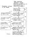
- FIG. 1is a schematic block diagram of the electrophysiology apparatus
- FIG. 2is a block diagram representing the partitioning of the electrophysiology apparatus
- FIG. 3is a diagram of an illustrative balloon electrode set implementation of the measurement catheter and a therapy catheter;
- FIG. 4is a schematic diagram of an illustrative basket electrode set implementation of the measurement catheter
- FIG. 5is a flow chart showing the wall surface generation process
- FIG. 6is a schematic diagram of a row of electrodes of the balloon catheter and their use in measuring distance to the heart chamber wall;
- FIG. 7is a screen display representing the motion of the cardiac wall surface
- FIG. 8is a schematic block diagram of the portion of the electrophysiology apparatus which implements the body orientation generation process
- FIG. 9is a flow charting showing the body orientation generation process
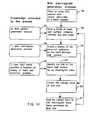
- FIG. 10is a flow chart showing the wall electrogram generation process
- FIG. 11is a representative screen display showing wall electrogram information
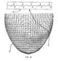
- FIG. 12is a representative screen display showing wall electrogram information
- FIG. 13is a representative screen display showing wall electrogram information
- FIG. 14is a flow chart showing the site electrogram generation process.
- FIG. 15is a flow chart showing the movable electrode location process.
- FIG. 16is a schematic block diagram of the therapy catheter system
- FIG. 17is a schematic diagram of the laser delivery embodiment of the therapy catheter.
- FIG. 18is a schematic diagram of a microwave delivery embodiment of the therapy catheter
- FIG. 19is a schematic diagram of a chemical delivery embodiment of the therapy catheter.
- FIG. 20is a schematic diagram of the angioplasty catheter embodiment of the therapy catheter.
- FIG. 1shows the electrophysiologic apparatus 10 connected to a patient 12 .
- a monitoring catheter system 14is placed in the heart 16 to generate a display of the electrical activity of the heart 16 .
- a therapy catheter 18may be inserted into the heart to perform ablation or other corrective treatment.
- the monitoring catheter 14has a proximal end 20 which may be manipulated by the attending physician, and a distal end 22 which carries a monitoring catheter electrode set 44 . In general the distal end 22 of the monitoring catheter 14 will be relatively small and will float freely in the heart chamber.
- the therapy catheter 18has a distal end 24 which carries a therapy catheter electrode set 46 .
- the therapy catheteralso has proximal end 26 which can be manipulated by the attending physician.
- the electrode sets located on the cathetersare coupled to an interface system 28 , through appropriate cables.
- the cable 30connects the monitoring catheter electrode set 44 to the interface system 28 while cable 32 connects the therapy catheter electrode set 46 to the interface system 28 .
- the interface system 28contains a number of subsystems which are controlled by a computer 34 .
- the data collected by the interface system 28is manipulated by the computer 34 and displayed on a display device 36 .
- Surface electrodes represented by electrode 40may also be coupled to the electrophysiology apparatus 10 for several purposes via an appropriate cable 42 .
- a therapy generator 38is connected to the therapy catheter electrode 60 and to the therapy surface ground 70 , through the interface system 28 .
- the skin surface electrode cable 42couples the ECG surface electrodes 74 to the ECG system 39 , which may be a subsystem of interface system 28 .
- FIG. 2is a schematic diagram showing an illustrative segmentation of the electrode sets and their electrical connections to subsystems in the electrophysiology apparatus 10 .
- the monitoring electrode set 44contains a subset of passive electrodes 48 which are connected to a signal conditioner 50 .
- the monitoring electrode set 44also contains a subset of active electrodes 52 which are connected to a signal generator 54 through a switch 59 .
- the signal generator 54is controlled by the computer 34 . In operation, the signal generator 54 generates a burst of (4800 Hz for example) signals which are supplied to the active electrode set 52 . This energy sets up an electric field within the heart 16 chamber.
- the electrical potentials present on the passive electrode set 48represent the summation of the underlying electrophysiological signals generated by the heart and the field induced by the burst.
- the signal conditioner 50separates these two components. The preferred technique is to separate the signals based upon their frequency.
- the high pass section 56 of the signal conditionerextracts the induced field signals as modulated by the blood volume and the changing position of the chamber walls 125 .
- the signalsare amplified with a gain of approximately 500 from passive electrodes 48 with amplifier 151 .
- the signalsare high pass filtered at roughly 1200 Hz by filter 153 .
- the 4800 Hz signalis extracted by demodulator 155 .
- the individual signalsare converted to digital format by the analog to digital converter 157 before being sent to the computer 34 .
- the low pass section 58 of the signal conditioner 50extracts physiologic signals. First, signal drift is reduced with a 0.01 Hz high pass filter 143 . Next, a programmable gain amplifier 145 amplifies the signals. Then a low pass filter 147 removes extraneous high frequency noise and the signal from the induced field. Finally, the physiologic signals are converted to digital format by the analog to digital converter 149 before being sent to the computer 34 .
- the therapy catheter electrode set 46includes at least one therapy delivery electrode 60 , and preferably one or more monitoring electrodes 62 , and one or more locator electrodes 68 .
- the therapy delivery electrode 60cooperates with the ground electrode 70 , which is generally a skin patch electrode, to deliver ablation energy to the heart. These electrodes are coupled to the ablation energy generator 38 which is shown as an RF current source.
- a locator electrode 68is provided which is preferably proximate the delivery electrode 60 , but can be a separate electrode site located near the distal end 24 of the therapy catheter 18 . This electrode site is coupled with an active electrode 52 through a switch 59 to the signal generator 54 .
- the electric field coupled to the therapy catheter 18permits the physician to track and visualize the location of the locator electrode 68 on the display device 36 .
- the therapy catheter electrode set 46can also be used to monitor the physiologic signals generated at the chamber wall 125 by a low pass signal conditioner 141 which is similar to the low pass section 58 of the signal conditioner 50 . These digitized signals are then sent to the computer 34 .
- At least one electrode pair 119 of surface electrodes 40are also coupled to the signal generator 54 through switch 59 .
- Each electrode 89 and 115are placed opposite each other on the body surface with the heart 16 in-between them.
- the induced fieldis sensed by passive electrodes 48 and conditioned by the high pass section 56 of the signal conditioner 50 . This field helps the computer 34 align or orient the passive electrodes 48 to the body for better visualization of the heart on the monitor 36 .
- the ECG subsystem 39accepts signals from standard ECG skin electrodes 74 . It also contains a low pass section similar to the low pass section 58 of signal conditioner 50 . In general, the passive electrode set 48 and active electrode set 52 will reside on a single catheter, however it should be recognized that other locations and geometries are suitable as well. Both basket and balloon devices are particularly well suited to this application.
- FIG. 3shows an electrode configuration on a balloon catheter 94 which has an inflatable balloon 96 which underlies an array or set of passive electrodes 48 typified by passive electrode 72 .
- passive electrodes 48can be organized into rows, typified by row 123 , and columns, typified by column 121 .
- a pair of active excitation electrodes 52are typified by proximal electrode 92 and distal electrode 98 .
- the balloon catheter 94 configurationcan be quite small in comparison with the basket catheter 80 configuration. This small size is desirable both for insertion into and for use in a beating heart 16 .
- FIG. 3also shows a movable, reference or therapy catheter system 18 .
- This catheteris shown lying along the interior surface 125 of the heart 16 .
- a pair of electrodes shown as delivery electrode 60 and reference electrode 62are located a fixed distance apart on the catheter body 64 .
- This auxiliary cathetermay be used to supply ablation energy to the tissue during therapy.
- This therapy catheter 18may be used with either the basket catheter 60 configuration or the balloon catheter 94 configuration.
- FIG. 4shows an electrode configuration on a basket catheter 80 .
- the limbs of the basket 80typified by limb 82 carry multiple passive electrode sites typified by electrode 84 .
- a pair of active excitation electrodesare shown on the central shaft 86 of the basket 80 as indicated by excitation electrode 88 .
- the basket catheter 80 electrodeslie gently against the interior surface 125 of the heart 16 urged into position by the resilience of the limbs.
- the basket catheter 80permits unimpeded flow of blood through the heart during the mapping procedure which is very desirable. This form of catheter also places the electrodes into contact with the heart chamber wall 125 for in-contact mapping of the physiologic potentials of the heart 16 .
- the signal generator 54can generate a 4800 Hz sinusoidal signal burst on the active electrode set 52 which creates an electric field in the heart.
- the changing position of the chamber walls 125 and the amount of blood within the heartdetermines the signal strength present at the passive electrode sites 48 .
- the chamber geometryis derived from the electric field as measured at the passive electrode sites 48 which may, or may not be in contact with the walls 125 of the heart.
- the field strengthis inversely proportional to the instantaneous physical wall location and the distance from the active electrodes 52 to these walls.
- the potentials on the passive set of electrodes 72are related to the wall location, but a set of computationally intensive field equations must be solved to ascertain the position of the wall.
- both the basket and balloon approachcan be used to generate the dynamic representation of the wall surface.
- the computer 34operates under the control of a stored program which implements several control functions and further displays data on a display device 36 .
- the principal software processesare the wall surface generation process (WSGP); the body orientation generation process (BOGP); the wall electrogram generation process (WEGP); the site electrogram generation process (SEGP); and the movable electrode location process (MELP).
- FIG. 5is a flow chart describing the method used to generate the “wall surface” of the interior of the heart 16 .
- the step-wise processesare presented with certain physical parameters which are either known in advance by computation or are measured. This knowledge or information is shown in block 53 , block 55 and block 57 .
- the WSGP processbegins at block 41 with the insertion of the monitoring catheter 14 in the heart 16 .
- This catheter 14places an array of electrodes 44 in a heart 16 chamber. This array must have both passive measurement electrode sites 48 and active interrogation electrode sites 52 located in a known position.
- the processenters a measurement and display loop at block 43 where an interrogation pulse burst is generated by the signal generator 54 seen in FIG. 2 .
- These pulsesare generated first with the current source at site 92 and the current sink at site 98 and second with the current source at site 98 and the sink at site 92 as seen in FIG. 3 .
- the signal conditioner 50uses information on the frequency and timing of the interrogation current from block 53 to demodulate the signals and analog to digital convert the signals received at the passive measurement electrodes 48 .
- the information from block 55in used. This information includes both the current strength of the interrogation pulse and the location of the interrogation source and sink electrodes. Impedance is voltage divided by current. The voltage offset caused by the location of the current source can be reduced by the two measurements of opposite polarity.
- Block 49determines how the heart chamber tissue, which has roughly three times the impedance of blood, in combination with the type of electrode array affects the field generated by the interrogation electrodes.
- the bloodeffects the impedance directly as the field is propagated from the interrogation electrodes to the measurement electrodes.
- the inverse of the measured voltageis proportional to the square root of the distance from the source.
- the impedance of the field generated within the blood volumeis modulated by the position of the walls 125 , with their higher impedance, with respect to the location relative to the measurement electrodes.
- the distance from the interrogation electrodes to the heart chamber wall 125is determined at a point normal to the field generated by the active interrogation electrodes 52 .
- the passive electrodes 48 on the balloon catheter 94can be positioned in rows 123 and columns 121 with the columns in a line from the top of the balloon 96 near active electrode 92 to the bottom of the balloon 96 near active electrode 98 .
- three configurationsare possible: 8 rows and 8 columns, 7 rows and 9 columns, and 6 rows and 10 columns.
- the measurements from any row 123are treated independently.
- 8 measurements of distanceare taken for any selected row of electrodes, giving a total of 64 measurements.
- FIG. 6is a schematic drawing of the embodiment required to measure the distance 129 from the centroid 127 of the balloon 96 through the passive electrode 131 to the heart chamber wall 125 .
- the passive electrode 131is one of eight electrodes on a row of electrodes 123 . Starting with electrode 131 and labeling it as electrode A, the other electrodes on the row 123 are labeled B, C, D, E, F, G and H by proceeding around the balloon 96 in a clockwise direction. The measurements of impedance “I” at these electrodes are thus labeled I A , I B , I C , I D , I E , I F , I G and I H .
- the computationcan be redone by shifting this direction clockwise one electrode, relabeling electrodes A through H and solving the above equation again. Once the distances for this row of electrodes 123 are determined then the next row distances are determined in the same way until the distances at all 64 electrodes are determined.
- a model of the chamber wall 125 shapecan be created in block 51 .
- Various techniques for creating a shapeare possible, including cubic spline fits, and best fit of an ellipsoid.
- the positions of the active electrodes 52 and the passive electrodes 48 relative to the heart 16 chamber wallsare also determined at this point.
- the loopcontinues as the method moves back to block 43 . This loop continues at a rate fast enough to visualize the real-time wall motion of the heart chamber, at least at twenty times per second.
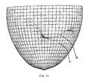
- FIG. 7shows two separate frames of the dynamic representation of the heart wall.
- Wire frame 71shows the heart at systole while wire frame 73 shows the heart at diastole.
- Path arrow 75 and path arrow 77represent the dynamic cycling through several intermediate shapes between the systole and diastole representation. These views are useful as they indicate the mechanical pumping motion of the heart to the physician.
- FIG. 8is a schematic drawing of the apparatus required to perform the body orientation generation process. It shows a patient 12 with at least one pair 119 of skin electrodes 40 attached to the body surface in a stationary position on the body and in a known configuration. These electrodes are typified by example surface electrodes 89 and 115 each of which could be an ECG electrode 74 , an RF generation current sink electrode 70 , or another electrode specifically dedicated to the BOGP. Ideally, electrode 89 and 115 are opposite one another on the body with the heart 16 directly in between them. This pair of electrodes is attached to the signal generator 54 through the switch 59 via an appropriate cable 117 . The distal end 22 of monitoring catheter 14 is situated in the heart 16 where the passive electrodes 48 can measure the signals generated across the electrode 89 and electrode 115 .
- FIG. 9is a flow chart describing the method used to align the wall surface representation of the WSGP to the body orientation.
- the processbegins at step 101 where the monitoring catheter 14 with a set of passive electrodes 48 is inserted into heart 16 chamber and a pair of surface electrodes 119 are attached at a known position on the body 12 .
- the processbegins cycling at step 102 where the signal generator 54 generates a signal across the skin electrode 89 and skin electrode 115 .
- the voltage created by the signal generator 54is measured from passive electrode 48 by the high pass section 56 of the signal conditioner 50 by using the information from block 110 which includes the frequency and timing of the field generated by the signal generator 54 . This voltage information is stored in an array “Y”.
- a regression analysisis performed which creates a vector which lines up with the field generated in step 103 .
- This regression methodis the same whether a basket catheter as shown in FIG. 4 or a balloon catheter as shown in FIG. 3 is used.
- the location of each passive electrode 48is provided to the method by block 110 . This information comes from different sources in each case however. In the case of a basket catheter 80 these three dimensional electrode locations come from the WSGP. In the case of the balloon catheter 94 these three dimensional electrode locations are known a priori. In each case they are saved in an array “X”.
- step 105the information on the location of the chamber walls 125 from the WSGP 109 can be used to create a three dimensional model of the heart 16 chamber as seen in FIG. 7 .
- this representationcan be shown in a known orientation relative to the body in step 106 .
- a specific orientationsuch as typical radiological orientations RAO (right anterior oblique), LAO (left anterior oblique), or AP (anterior/posterior) can be presented.
- RAOright anterior oblique
- LAOleft anterior oblique
- APanterior/posterior
- FIG. 10is a flow chart describing the wall electrogram generation process (WEGP). This process begins at block 61 when a monitoring catheter 14 with an array of passive measurement electrodes 48 is placed in a heart chamber 16 and deployed. The process enters a loop at block 63 .
- the frequency of the interrogation pulses generated by the signal generator 54is provided by block 85 .
- the low pass filter section 58 of the signal conditioner 50measures the voltage at frequencies lower than the generated interrogation pulses. Typically the highest frequency of the biopotentials is 100 Hz but can be as high as 250 Hz.
- the measurementsare contact voltages from the chamber wall 125 tissue contacting the electrodes 84 .
- the measurementsare measurements of the field generated throughout the blood volume by the tissue on the chamber wall 125 .
- a model of the array boundary and the chamber wall 125 boundaryis created from the information in block 87 .
- This informationincludes the location of the passive electrodes 48 on the array and the chamber wall 125 locations from the WSGP.
- the array boundary and the chamber wall 125 boundaryare the same since they are in contact.
- the locationsare determined in three-dimensional space of the sites on the chamber wall where potentials are measured.
- step 65locations are generated in three-dimensional space of the sites on the chamber wall where potentials are to be determined.
- the potentialsare projected on to the sites on the chamber wall specified in step 65 .
- the measured potentialsare assigned to these sites.
- a three dimensional techniquesuch as those typically used in field theory is used to generate a representation of the three dimensional field gradients in the blood volume of the heart chamber.
- Two examples of appropriate techniquesare a spherical harmonics solution to Laplace's equation, and boundary element analysis. A more detailed description of spherical harmonics is given in the parent disclosure which is incorporated by reference herein.
- the voltageis measured at the passive electrodes 48 on the probe or balloon catheter 94 . From the voltage at the electrodes on the probe and the knowledge that the probe is nonconducting, the voltage and normal current at a previously selected set of nodes on the endocardial surface 125 are determined by the boundary element method in the following manner.
- Laplace's equationis solved by the boundary element method, a specialized finite element method that permits one to restrict the calculations to the two-dimensional probe and endocardial surfaces (and not have to deal with calculations over the blood pool between these two surfaces).
- the systemuses a higher-order version of the boundary element method. This system currently uses bicubic splines to represent the probe and endocardial surfaces and bilinear elements and bicubic splines to represent the voltage and the normal current on these surfaces.
- the boundary element methodconsists of creating and solving a set of linear equations for the voltage and the normal current on the endocardium based on the voltage measurements at the electrodes on the probe.
- Each of the elements in the matrices that are involved in this setconsists of two-dimensional integrals, which are calculated by numerical and analytical integration.
- a large number of pointsare calculated on the three-dimensional chamber surface 125 .
- thisis done through interpolation using bilinear or bicubic splines.
- thiscan be done either by using the model, such as the boundary element method or spherical harmonics to generate more points.
- bilinear or bicubic splinescan be used to interpolate between a smaller number of points.
- a representation of the electrical potentials on the surface 125are used to display the patterns.
- These types of displaysinclude color maps, maps of iso-potential lines, maps of potential gradient lines and others.
- the electro-physiologic informationis reconstructed on the dynamic wall surface 125 .
- the measured electrical activityis positioned by the WSGP at the exact location which gives rise to the activity.
- the high resolution of the systemcreates an enormous amount of information to display.
- Several techniquesmay be used to display this information to the physician.
- the electrogram datacan be shown in false color gray-scale on a two dimensional wall surface representation. In this instance areas of equal potential areas are shown in the same color.
- a vectorized display of datacan be shown on a wire grid as shown in FIG.
- FIG. 14is a flow chart of the site electrogram generation process (SEGP). This process is used to extract and display a time series representation of the electrical activity at a physician selected site.
- FIG. 13shows a site 97 that has been selected and a time series electrogram 99 is shown on the display device 36 along with the dynamic wall representation.
- SEGPsite electrogram generation process
- the userwill use the display 36 to determine a location of interest in block 33 which will then be marked on the display device 36 at step 81 .
- the voltage from this locationwill be collected at block 83 .
- This voltagewill be plotted in a wave-form representation 99 in block 31 .
- the loopcontinues at this point at a rate sufficient to display all of the frequencies of such a time series electrogram 99 , at least 300 points per second.
- the false color and vectorized display imagesmay direct the physician to specific sites on the endocardial surface for further exploration.
- the systemmay allow the physician to “zoom” in on an area to show the electrical activity in greater detail. Also the physician may select a site on the endocardial wall 125 and display a traditional time series electrogram 99 originating at that site.
- FIG. 15is a flow-chart of the movable electrode location process (MELP). It begins at block 11 when a catheter with an array of passive measurement electrodes 48 and active electrodes 52 is placed in a heart 16 chamber and deployed. At block 13 a second catheter 18 with at least one electrode is introduced into the same chamber. The process enters a loop at block 15 where the signal generator 54 generates a carrier current between the movable location electrode 68 and an active electrode 52 . At block 17 the high pass section 56 of signal conditioner 50 , using the frequency and timing information of the location signal from block 29 , produces measured voltages from the passive measurement electrodes 48 . At block 19 the information from block 27 is used to determine the location of the electrode 68 where the location current is generated.
- MELPmovable electrode location process
- This informationincludes the strength of the generated location current, the impedances of blood and tissue, the location of the active electrode 52 in use and the location of all the passive measurement electrodes 48 .
- One method for using this informationwould entail performing a three dimensional triangulation of the point source location signal using four orthogonal passive electrode 48 sites.
- the implementation of step 19is the same both for the case of a basket system as seen in FIG. 3 and for the case of a balloon system as seen in FIG. 4 .
- two data setsare acquired closely spaced in time such that they are effectively instantaneous relative to the speed of cardiac mechanical activity.
- the data setscould be acquired simultaneously, by driving signals at two different frequencies, and separating them electronically by well known filtering means.
- the first data setis acquired by driving the current carrier from the location electrode 68 to a first sink or active electrode as typified by electrode 98 .
- This electrodeis at a known location on the body of the monitoring catheter 14 relative to the array of passive electrodes 48 .
- the location of this first sink electrodeis ideally displaced distally from the centroid 127 of the array of electrodes by at least 25 millimeters.
- a second data setis then acquired by driving the current from the location electrode 68 to a second active electrode 92 , located ideally at least 25 millimeters proximally from the centroid 127 of the array of electrodes.
- nis the number of array electrodes
- V piare the potentials measured from each i th electrode 72
- R iis a vector from the origin (centroid of the array of electrodes 96 ) to the i th probe electrode 72
- R Lis the “location vector”, or three dimensional location to be solved for in the minimization
- R s1 , R s2are the location vectors of the active sink electrodes (eg.
- Each additional squared parenthetical termrequires the probe data set Vpi, another ‘b’ fitting term, and the particular active sink electrode 52 vector R s used during the acquisition of that data set. If the sink electrode 52 is far enough away, for example using a right leg patch electrode, the fourth term in the squared expression for that data set may be deleted as R s becomes very large.
- the methoddoes not require two data sets.
- the first squared expression in the above expression(requiring only data set V pi1 ) may be sufficiently accurate.
- the non-linear least squares minimizationmay be performed on the above summation by any of several well-known methods.
- the Levenberg-Marquardt methodhas been used in practice to accomplish this with efficient and robust results.
- Nominal values for k and bare 70 and 0 respectively, when normalizing the potential values obtained as if the current source were 1 ampere.
- the number of parameters in the minimization for the above expressionare six: k, b 1 , b 2 , and the x, y, and z coordinates of vector R L cassuming a Cartesian coordinate system with origin at the center of the array of electrodes 96 ).
- a model of the heart 16 chamber wallis generated from the information provided from the WSGP 25 .
- Such a modelcan be represented on a display 36 in a manner typified in FIG. 6 .
- a second figure representing the distal end of the monitoring catheter 14can be presented. In this way, the full three dimensional geometry of the chamber and the array catheter can be presented.
- step 23this geometry is updated repeatedly to provide a dynamic view of the chamber, the monitoring catheter 18 , along with a representation of the distal end 24 of the therapy catheter 18 . If this is then combined with the electrical potentials generated by the WEGP, the therapy catheter can be moved to an electrical site of interest represented by a point in three dimensional space.
- Calibration of the system to insure that physical dimensions are accurately scaledis not a necessity for use of the system in a diagnostic or therapeutic setting.
- the availability of heart geometry in real timecan permit various hemodynamic measurements to be made and displayed to the physician as well. These measurements include systolic time intervals, stroke volume and cardiac output.
- Calibrationwhere desired, requires at least two electrodes 60 and 62 a known distance apart placed along the inner-surface of the heart chamber 16 , as shown in FIG. 3 . In general the two electrode sites will each be coupled to the location signal generator 54 .
- the MELP of FIG. 15can be calibrated by scaling the calculations 50 the distance between computed locations match the known distance apart of the two electrodes 60 and 62 .
- the WSGP of FIG. 5can be calibrated by scaling the distance measured by the WSGP in the direction of electrodes 60 and 62 to the calibrated distances measured by MELP.
- the WEGP of FIG. 10 and SEGP of FIG. 14can be calibrated to those measurements by computing the voltages at the same locations on the chamber wall 125 where electrodes 60 and 62 are located. These computed voltages can then be scaled to match the physically measured voltages from electrodes 60 and 62 .
- FIG. 16is a schematic diagram of the therapy catheter system.
- the therapy catheter 18has both a distal end 24 and a proximal end 26 .
- a handle 163is on the proximal end 26 which allows the user to manipulate the distal end 24 and position it in the heart 16 . Referring to FIG. 1 , this handle also permits the therapy catheter 18 to connect to the interface system 28 of the electrophysiologic apparatus 10 through the cable 32 .
- the location currentis generated by the signal generator 54 through the switch 59 and subsequently through the wire 177 of cable 32 which is connected directly to the locator electrode 68 .
- the therapy catheter systemalso includes a therapy generator 38 which is connected to the therapy catheter handle 163 via therapy supply line 161 .
- the therapy supply line 161extends through the handle 163 , through the catheter body 64 , to the therapy deployment apparatus 60 at the distal end 24 of the catheter.
- the locator electrode 68is in close proximity to the therapy deployment apparatus 60 in order to determine its location within the heart 16 .
- FIG. 17shows an embodiment of the therapy catheter 18 using laser energy to supply the therapy.
- This laser catheter 165includes the location wire 177 which connects the interface system 28 to the locator electrode 68 at the catheter's distal end 24 .
- the therapy supply line 161is a fiber optic cable 167 and the therapy deployment apparatus 60 is a fiber optic terminator 169 which directs the laser energy to the site of therapy delivery.
- FIG. 18shows an embodiment of the therapy catheter 18 using microwave energy to supply the therapy.
- This microwave catheter 171includes the location wire 177 which connects the interface system 28 to the locator electrode 68 at the catheter's distal end 24 .
- the therapy supply line 161is a microwave wave guide 173 and the therapy deployment apparatus 60 is a microwave emitter 175 which directs the microwave energy to the site of therapy delivery.
- FIG. 19shows an embodiment of the therapy catheter 18 using a chemical to supply the therapy.
- This chemical deliver catheter 181includes the location wire 177 which connects the interface system 28 to the locator electrode 68 at the catheter's distal end 24 .
- the therapy supply line 161is a chemical filled lumen 183 .
- This lumenextends to the distal end 24 of the chemical delivery catheter 181 where a needle 185 is used to infuse the chemical into the heart chamber wall 125 .
- the needle 185is withdrawn into the catheter body through withdrawal action 187 .
- the chemical delivery needle 165can be deployed through the reverse of withdrawal action 187 .
- Potential chemicals to be used in the therapeutic delivery processinclude formaldehyde and alcohol.
- Each of the therapy catheters 18 shown in FIG. 17 through FIG. 19 as well as the radio frequency catheter shown in FIG. 2can be miniaturized and inserted into the coronary arterial tree.
- the location signal generated at locator electrode 68can still be sensed by the passive electrodes 48 even though the signal is coming from the epicardium of the heart 16 rather than from within the heart chamber.
- the movable electrode location process of FIG. 15can be used in this instance to help determine the location of the distal end 24 of the therapy catheter 18 in the coronary arterial tree and whether it is close to a site of abnormal electrical activity. Assuming that a site of ischemia will commonly be a site of abnormal electrical activity, the MELP will also enable more rapid location of potential sites for angioplasty.
- FIG. 20shows an embodiment of the therapy catheter 18 using balloon inflation to supply the therapy.
- This angioplasty catheter 191includes the location wire 177 which connects the interface system 28 to the locator electrode 68 at the catheter's distal end 24 .
- the therapy supply line 161is an inflation media supply lumen 193 and the therapy deployment apparatus 60 is an angioplasty balloon 195 .
- a site of interestwould be determined after viewing the wall electrogram generated by the WEGP of FIG. 10 .
- the angioplasty therapy catheter 191would be positioned in the coronary arterial tree and its position determined relative to the site of interest.
- the balloon 195would be deployed to open the artery.
- the electrical activity of the sitewould be reviewed to determine whether the underlying tissue 125 was now receiving a proper blood supply and thus was no longer electrically abnormal.
Landscapes
- Health & Medical Sciences (AREA)
- Life Sciences & Earth Sciences (AREA)
- Engineering & Computer Science (AREA)
- Animal Behavior & Ethology (AREA)
- Veterinary Medicine (AREA)
- Public Health (AREA)
- Biomedical Technology (AREA)
- Heart & Thoracic Surgery (AREA)
- General Health & Medical Sciences (AREA)
- Surgery (AREA)
- Medical Informatics (AREA)
- Molecular Biology (AREA)
- Biophysics (AREA)
- Physics & Mathematics (AREA)
- Pathology (AREA)
- Cardiology (AREA)
- Nuclear Medicine, Radiotherapy & Molecular Imaging (AREA)
- Radiology & Medical Imaging (AREA)
- Physiology (AREA)
- Plasma & Fusion (AREA)
- Otolaryngology (AREA)
- Dentistry (AREA)
- Oral & Maxillofacial Surgery (AREA)
- Media Introduction/Drainage Providing Device (AREA)
- Measurement And Recording Of Electrical Phenomena And Electrical Characteristics Of The Living Body (AREA)
- Surgical Instruments (AREA)
Abstract
Description
ln(DA)=
where DAis the desired
b=Σxy/Σx2
where “X” is the array of electrode locations, “Y” is the array of measured voltages and “b” is the orientation vector. If more than one pair of skin electrodes are used then an orthogonal set of orientation vectors can be created and any rotation of the
Where n is the number of array electrodes, where k, b1and b2are fitting parameters, Vpiare the potentials measured from each ithelectrode72, Riis a vector from the origin (centroid of the array of electrodes96) to the ithprobe electrode72, RLis the “location vector”, or three dimensional location to be solved for in the minimization, and Rs1, Rs2are the location vectors of the active sink electrodes (eg.92 and98) which are known at locations on the axis of the array of
Claims (2)
Priority Applications (1)
| Application Number | Priority Date | Filing Date | Title |
|---|---|---|---|
| US09/547,543US6990370B1 (en) | 1992-09-23 | 2000-04-12 | Method for mapping heart electrophysiology |
Applications Claiming Priority (6)
| Application Number | Priority Date | Filing Date | Title |
|---|---|---|---|
| US07/949,690US5311866A (en) | 1992-09-23 | 1992-09-23 | Heart mapping catheter |
| US07/950,448US5297549A (en) | 1992-09-23 | 1992-09-23 | Endocardial mapping system |
| PCT/US1993/009015WO1994006349A1 (en) | 1992-09-23 | 1993-09-23 | Endocardial mapping system |
| US38783295A | 1995-05-26 | 1995-05-26 | |
| US510598A | 1998-01-09 | 1998-01-09 | |
| US09/547,543US6990370B1 (en) | 1992-09-23 | 2000-04-12 | Method for mapping heart electrophysiology |
Related Parent Applications (1)
| Application Number | Title | Priority Date | Filing Date |
|---|---|---|---|
| US510598AContinuation | 1992-09-23 | 1998-01-09 |
Publications (1)
| Publication Number | Publication Date |
|---|---|
| US6990370B1true US6990370B1 (en) | 2006-01-24 |
Family
ID=73446239
Family Applications (4)
| Application Number | Title | Priority Date | Filing Date |
|---|---|---|---|
| US08/420,698Expired - LifetimeUS5662108A (en) | 1992-09-23 | 1995-04-12 | Electrophysiology mapping system |
| US09/547,464Expired - Fee RelatedUS6728562B1 (en) | 1992-09-23 | 2000-04-12 | Method for creating a virtual electrogram |
| US09/547,543Expired - Fee RelatedUS6990370B1 (en) | 1992-09-23 | 2000-04-12 | Method for mapping heart electrophysiology |
| US09/547,690Expired - LifetimeUS6640119B1 (en) | 1992-09-23 | 2000-04-12 | Method for orienting an electrode array |
Family Applications Before (2)
| Application Number | Title | Priority Date | Filing Date |
|---|---|---|---|
| US08/420,698Expired - LifetimeUS5662108A (en) | 1992-09-23 | 1995-04-12 | Electrophysiology mapping system |
| US09/547,464Expired - Fee RelatedUS6728562B1 (en) | 1992-09-23 | 2000-04-12 | Method for creating a virtual electrogram |
Family Applications After (1)
| Application Number | Title | Priority Date | Filing Date |
|---|---|---|---|
| US09/547,690Expired - LifetimeUS6640119B1 (en) | 1992-09-23 | 2000-04-12 | Method for orienting an electrode array |
Country Status (1)
| Country | Link |
|---|---|
| US (4) | US5662108A (en) |
Cited By (185)
| Publication number | Priority date | Publication date | Assignee | Title |
|---|---|---|---|---|
| US20040254437A1 (en)* | 1998-06-30 | 2004-12-16 | Hauck John A. | Method and apparatus for catheter navigation and location and mapping in the heart |
| US20050288613A1 (en)* | 2004-06-25 | 2005-12-29 | Cardiac Pacemakers, Inc. | Endocardial splint and method therefor |
| US20050288599A1 (en)* | 2004-05-17 | 2005-12-29 | C.R. Bard, Inc. | High density atrial fibrillation cycle length (AFCL) detection and mapping system |
| US20070073179A1 (en)* | 2005-09-15 | 2007-03-29 | St. Jude Medical, Atrial Fibrillation Division, Inc. | System and Method for Three Dimensional Mapping of Electrophysiology Information |
| US20070185486A1 (en)* | 2004-05-28 | 2007-08-09 | Hauck John A | Robotic surgical system |
| US20070198008A1 (en)* | 2004-05-28 | 2007-08-23 | Hauck John A | Robotic surgical system and method for automated therapy delivery |
| WO2007111542A1 (en)* | 2006-03-27 | 2007-10-04 | St. Jude Medical Ab | Medical system for monitoring and localisation of electrode leads in the heart |
| US20070270705A1 (en)* | 2006-05-17 | 2007-11-22 | Starks Daniel R | System and method for complex geometry modeling of anatomy using multiple surface models |
| US20070299352A1 (en)* | 2006-06-13 | 2007-12-27 | Doron Harlev | Non-contact cardiac mapping, including moving catheter and multi-beat integration |
| US20070299353A1 (en)* | 2006-06-13 | 2007-12-27 | Doron Harlev | Non-contact cardiac mapping, including preprocessing |
| US20070299351A1 (en)* | 2006-06-13 | 2007-12-27 | Doron Harlev | Non-contact cardiac mapping, including resolution map |
| US20080009758A1 (en)* | 2006-05-17 | 2008-01-10 | Voth Eric J | System and method for mapping electrophysiology information onto complex geometry |
| US20080033284A1 (en)* | 2005-05-27 | 2008-02-07 | Hauck John A | Robotically controlled catheter and method of its calibration |
| US20080161671A1 (en)* | 2006-12-29 | 2008-07-03 | Voth Eric J | Body surface mapping system |
| US20080161669A1 (en)* | 2006-12-29 | 2008-07-03 | Hauck John A | Cardiac Navigation System Including Electrode Array for Use Therewith |
| US20080161681A1 (en)* | 2006-12-29 | 2008-07-03 | Hauck John A | Navigational reference dislodgement detection method & system |
| US20080221643A1 (en)* | 2007-03-09 | 2008-09-11 | Olson Eric S | System and method for correction of inhomogeneous fields |
| US20080221425A1 (en)* | 2007-03-09 | 2008-09-11 | Olson Eric S | System and method for local deformable registration of a catheter navigation system to image data or a model |
| US20090058806A1 (en)* | 2005-04-16 | 2009-03-05 | Mitchell Scott Middler | Processing cursor movements in a graphical user interface of a multimedia application |
| US20090253976A1 (en)* | 2008-04-02 | 2009-10-08 | Rhythmia Medical, Inc. | Intracardiac Tracking System |
| US20090264743A1 (en)* | 2008-04-18 | 2009-10-22 | Markowitz H Toby | Interference Blocking and Frequency Selection |
| US20090264739A1 (en)* | 2008-04-18 | 2009-10-22 | Markowitz H Toby | Determining a position of a member within a sheath |
| US20090264750A1 (en)* | 2008-04-18 | 2009-10-22 | Markowitz H Toby | Locating a member in a structure |
| US20090262980A1 (en)* | 2008-04-18 | 2009-10-22 | Markowitz H Toby | Method and Apparatus for Determining Tracking a Virtual Point Defined Relative to a Tracked Member |
| US20090264752A1 (en)* | 2008-04-18 | 2009-10-22 | Markowitz H Toby | Method And Apparatus For Mapping A Structure |
| US20090264781A1 (en)* | 2006-08-03 | 2009-10-22 | Christoph Scharf | Method and device for determining and presenting surface charge and dipole densities on cardiac walls |
| US20090276020A1 (en)* | 2008-05-02 | 2009-11-05 | Pacesetter, Inc. | Tools for delivering implantable medical leads and methods of using and manufacturing such tools |
| US20090297001A1 (en)* | 2008-04-18 | 2009-12-03 | Markowitz H Toby | Method And Apparatus For Mapping A Structure |
| US20100106154A1 (en)* | 2008-10-27 | 2010-04-29 | Rhythmia Medical, Inc. | Tracking System Using Field Mapping |
| US20100152571A1 (en)* | 2008-12-16 | 2010-06-17 | Medtronic Navigation, Inc | Combination of electromagnetic and electropotential localization |
| US20100168560A1 (en)* | 2008-12-31 | 2010-07-01 | Hauck John A | Devices and Methods for Catheter Localization |
| US20100179424A1 (en)* | 2009-01-09 | 2010-07-15 | Reinhard Warnking | Methods and apparatus for treatment of mitral valve insufficiency |
| US20100274150A1 (en)* | 2009-04-23 | 2010-10-28 | Rhythmia Medical, Inc. | Multi-Electrode Mapping System |
| US7825925B2 (en) | 2007-03-09 | 2010-11-02 | St. Jude Medical, Atrial Fibrillation Division, Inc. | Method and system for repairing triangulated surface meshes |
| US20100286550A1 (en)* | 2009-05-08 | 2010-11-11 | Rhythmia Medical, Inc. | Impedance Based Anatomy Generation |
| US20100286551A1 (en)* | 2009-05-08 | 2010-11-11 | Rhythmia Medical, Inc. | Impedance Based Anatomy Generation |
| US20100298690A1 (en)* | 2008-01-17 | 2010-11-25 | Christoph Scharf | Device and method for the geometric determination of electrical dipole densities on the cardiac wall |
| US20100305423A1 (en)* | 2009-06-02 | 2010-12-02 | Huisun Wang | Catheter having distal sealing member |
| US20100324414A1 (en)* | 2007-02-08 | 2010-12-23 | Rhythmia Medical, Inc., A Delaware Corporation | Catheter tracking and endocardium representation generation |
| EP2266459A1 (en)* | 2009-06-24 | 2010-12-29 | Cortius B.V. i.o. | Inverse imaging of electrical activity of a heart muscle |
| WO2010129095A3 (en)* | 2009-05-08 | 2011-01-13 | Rhythmia Medical, Inc. | Impedance based anatomy generation |
| US20110054304A1 (en)* | 2009-08-31 | 2011-03-03 | Medtronic, Inc. | Combination Localization System |
| US20110106203A1 (en)* | 2009-10-30 | 2011-05-05 | Medtronic, Inc. | System and method to evaluate electrode position and spacing |
| US20110118725A1 (en)* | 2009-11-11 | 2011-05-19 | Mayse Martin L | Non-invasive and minimally invasive denervation methods and systems for performing the same |
| US20110118803A1 (en)* | 2009-11-18 | 2011-05-19 | Pacesetter, Inc. | Cardiac Resynchronization Therapy Optimization Using Vector Measurements Obtained From Realtime Electrode Position Tracking |
| EP2407118A2 (en) | 2010-07-13 | 2012-01-18 | St. Jude Medical, Atrial Fibrillation Division, Inc. | Methods and systems for filtering respiration noise from localization data |
| US8135467B2 (en) | 2007-04-18 | 2012-03-13 | Medtronic, Inc. | Chronically-implantable active fixation medical electrical leads and related methods for non-fluoroscopic implantation |
| WO2012082200A1 (en) | 2010-12-17 | 2012-06-21 | St. Jude Medical, Atrial Fibrillation Division, Inc. | Navigation reference dislodgement detection method and system |
| US20120172713A1 (en)* | 2010-12-29 | 2012-07-05 | Carlos Carbonera | System and method for rendering an image of an elongate medical device |
| US8346339B2 (en) | 2011-04-22 | 2013-01-01 | Topera, Inc. | Basket style cardiac mapping catheter having a flexible electrode assembly for detection of cardiac rhythm disorders |
| US8489192B1 (en) | 2008-02-15 | 2013-07-16 | Holaira, Inc. | System and method for bronchial dilation |
| US8494614B2 (en) | 2009-08-31 | 2013-07-23 | Regents Of The University Of Minnesota | Combination localization system |
| US8647284B2 (en) | 2005-09-15 | 2014-02-11 | St. Jude Medical, Atrial Fibrillation Division, Inc. | System and method for mapping complex fractionated electrogram information |
| US8694074B2 (en) | 2010-05-11 | 2014-04-08 | Rhythmia Medical, Inc. | Electrode displacement determination |
| US8740895B2 (en) | 2009-10-27 | 2014-06-03 | Holaira, Inc. | Delivery devices with coolable energy emitting assemblies |
| US8755864B2 (en) | 2004-05-28 | 2014-06-17 | St. Jude Medical, Atrial Fibrillation Division, Inc. | Robotic surgical system and method for diagnostic data mapping |
| US8808280B2 (en) | 2008-05-09 | 2014-08-19 | Holaira, Inc. | Systems, assemblies, and methods for treating a bronchial tree |
| WO2014137897A1 (en) | 2013-03-05 | 2014-09-12 | St. Jude Medical, Atrial Fibrillation Division, Inc. | System and method for detecting sheathing and unsheathing of localization elements |
| US20150057507A1 (en)* | 2013-08-20 | 2015-02-26 | St. Jude Medical, Atrial Fibrillation Division, Inc. | System and Method for Generating Electrophysiology Maps |
| US9002442B2 (en) | 2011-01-13 | 2015-04-07 | Rhythmia Medical, Inc. | Beat alignment and selection for cardiac mapping |
| WO2015054048A1 (en) | 2013-10-09 | 2015-04-16 | St. Jude Medical, Cardiology Division, Inc. | System and method for generating electrophysiology maps |
| WO2015065648A1 (en) | 2013-10-31 | 2015-05-07 | St. Jude Medical, Cardiology Division, Inc. | System and method for generating electrophysiology maps |
| US9149328B2 (en) | 2009-11-11 | 2015-10-06 | Holaira, Inc. | Systems, apparatuses, and methods for treating tissue and controlling stenosis |
| US9198601B2 (en) | 2006-12-29 | 2015-12-01 | St. Jude Medical, Atrial Fibrillation Division, Inc. | Contact sensor and sheath exit sensor |
| US9277872B2 (en) | 2011-01-13 | 2016-03-08 | Rhythmia Medical, Inc. | Electroanatomical mapping |
| WO2016061384A1 (en) | 2014-10-15 | 2016-04-21 | St. Jude Medical, Cardiology Division, Inc. | Methods and systems for mapping local conduction velocity |
| WO2016061387A1 (en) | 2014-10-15 | 2016-04-21 | St. Jude Medical, Cardiology Division, Inc. | Methods and systems for generating integrated substrate maps for cardiac arrhythmias |
| US9339618B2 (en) | 2003-05-13 | 2016-05-17 | Holaira, Inc. | Method and apparatus for controlling narrowing of at least one airway |
| WO2016111804A1 (en) | 2015-01-07 | 2016-07-14 | St. Jude Medical, Cardiology Division, Inc. | Imaging device |
| US9398933B2 (en) | 2012-12-27 | 2016-07-26 | Holaira, Inc. | Methods for improving drug efficacy including a combination of drug administration and nerve modulation |
| US9560980B2 (en) | 2012-01-31 | 2017-02-07 | Medtronic, Inc. | Automatic selection of electrode vectors for assessing risk of heart failure decompensation events |
| US9585586B2 (en) | 2006-12-29 | 2017-03-07 | St. Jude Medical, Atrial Fibrillation Division, Inc. | Navigational reference dislodgement detection method and system |
| US9585588B2 (en) | 2014-06-03 | 2017-03-07 | Boston Scientific Scimed, Inc. | Electrode assembly having an atraumatic distal tip |
| WO2017040581A1 (en) | 2015-09-02 | 2017-03-09 | St. Jude Medical, Cardiology Division, Inc. | Methods and systems for identifying and mapping cardiac activation wavefronts |
| USD782686S1 (en) | 2012-08-31 | 2017-03-28 | Acutus Medical, Inc. | Transducer-electrode pair for a catheter |
| WO2017062248A1 (en) | 2015-10-07 | 2017-04-13 | St. Jude Medical, Cardiology Division, Inc. | Methods and systems for mapping cardiac restitution |
| US9636032B2 (en) | 2013-05-06 | 2017-05-02 | Boston Scientific Scimed Inc. | Persistent display of nearest beat characteristics during real-time or play-back electrophysiology data visualization |
| US9687166B2 (en) | 2013-10-14 | 2017-06-27 | Boston Scientific Scimed, Inc. | High resolution cardiac mapping electrode array catheter |
| US9700372B2 (en) | 2002-07-01 | 2017-07-11 | Recor Medical, Inc. | Intraluminal methods of ablating nerve tissue |
| WO2017142850A1 (en) | 2016-02-16 | 2017-08-24 | St. Jude Medical, Cardiology Division, Inc. | Methods and systems for electrophysiology mapping using medical images |
| US9757044B2 (en) | 2011-03-10 | 2017-09-12 | Acutus Medical, Inc. | Device and method for the geometric determination of electrical dipole densities on the cardiac wall |
| US9848795B2 (en) | 2014-06-04 | 2017-12-26 | Boston Scientific Scimed Inc. | Electrode assembly |
| US9901303B2 (en) | 2011-04-14 | 2018-02-27 | St. Jude Medical, Atrial Fibrillation Division, Inc. | System and method for registration of multiple navigation systems to a common coordinate frame |
| US9918649B2 (en) | 2013-05-14 | 2018-03-20 | Boston Scientific Scimed Inc. | Representation and identification of activity patterns during electro-physiology mapping using vector fields |
| WO2018089172A1 (en) | 2016-11-11 | 2018-05-17 | St. Jude Medical, Cardiology Division, Inc. | System and method for generating electrophysiology maps |
| WO2018094063A1 (en) | 2016-11-21 | 2018-05-24 | St. Jude Medical, Cardiology Division, Inc. | System and method for generating electrophysiology maps |
| WO2018132543A1 (en) | 2017-01-13 | 2018-07-19 | St. Jude Medical, Cardiology Division, Inc. | System and method for generating premature ventricular contraction electrophysiology maps |
| US10034637B2 (en) | 2007-12-28 | 2018-07-31 | Boston Scientific Scimed, Inc. | Cardiac mapping catheter |
| WO2018160631A1 (en) | 2017-03-02 | 2018-09-07 | St. Jude Medical, Cardiology Division, Inc. | System and method for differentiation of adipose tissue from scar tissue during electrophysiological mapping |
| WO2018204375A1 (en) | 2017-05-04 | 2018-11-08 | St. Jude Medical, Cardiology Division, Inc. | System and method for determining ablation parameters |
| WO2018212996A1 (en) | 2017-05-17 | 2018-11-22 | St. Jude Medical, Cardiology Division, Inc. | System and method for mapping local activation times |
| WO2019009967A1 (en) | 2017-07-07 | 2019-01-10 | St. Jude Medical, Cardiology Division, Inc. | System and method for electrophysiological mapping |
| WO2019018182A1 (en) | 2017-07-19 | 2019-01-24 | St. Jude Medical, Cardiology Division, Inc. | System and method for electrophysiological mapping |
| US10188314B2 (en) | 2013-03-05 | 2019-01-29 | St. Jude Medical, Cardiology Division, Inc. | System and method for detecting sheathing and unsheathing of localization elements |
| US10201311B2 (en) | 2013-02-08 | 2019-02-12 | Acutus Medical, Inc. | Expandable catheter assembly with flexible printed circuit board (PCB) electrical pathways |
| US10206652B2 (en) | 2008-12-30 | 2019-02-19 | St. Jude Medical, Atrial Fibrillation Division, Inc. | Intracardiac imaging system utilizing a multipurpose catheter |
| WO2019046250A1 (en) | 2017-09-01 | 2019-03-07 | St. Jude Medical, Cardiology Division, Inc. | System and method for visualizing a proximity of a catheter electrode to a 3d geometry of biological tissue |
| WO2019055115A1 (en) | 2017-09-18 | 2019-03-21 | St. Jude Medical, Cardiology Division, Inc. | System and method for sorting electrophysiological signals from multi-dimensional catheters |
| US10238350B2 (en) | 2015-05-08 | 2019-03-26 | St. Jude Medical, Cardiology Division, Inc. | System and method for real-time electrophysiological mapping |
| US10258285B2 (en) | 2004-05-28 | 2019-04-16 | St. Jude Medical, Atrial Fibrillation Division, Inc. | Robotic surgical system and method for automated creation of ablation lesions |
| US10271757B2 (en) | 2015-09-26 | 2019-04-30 | Boston Scientific Scimed Inc. | Multiple rhythm template monitoring |
| US10271758B2 (en) | 2015-09-26 | 2019-04-30 | Boston Scientific Scimed, Inc. | Intracardiac EGM signals for beat matching and acceptance |
| US10278616B2 (en) | 2015-05-12 | 2019-05-07 | Navix International Limited | Systems and methods for tracking an intrabody catheter |
| EP3498156A1 (en) | 2013-12-20 | 2019-06-19 | St. Jude Medical, Cardiology Division, Inc. | Coaxial electrode catheters for extracting electrophysiologic parameters |
| WO2019139884A1 (en) | 2018-01-09 | 2019-07-18 | St. Jude Medical, Cardiology Division, Inc. | System and method for sorting electrophysiological signals on virtual catheters |
| US10362963B2 (en) | 2011-04-14 | 2019-07-30 | St. Jude Medical, Atrial Fibrillation Division, Inc. | Correction of shift and drift in impedance-based medical device navigation using magnetic field information |
| WO2019156755A1 (en) | 2018-02-12 | 2019-08-15 | St. Jude Medical, Cardiology Division, Inc. | System and method for mapping cardiac muscle fiber orientation |
| US10398331B2 (en) | 2015-12-04 | 2019-09-03 | St. Jude Medical, Cardiology Division, Inc. | Methods and systems for statistically analyzing electrograms for local abnormal ventricular activities and mapping the same |
| US10405766B2 (en) | 2015-09-26 | 2019-09-10 | Boston Scientific Scimed, Inc. | Method of exploring or mapping internal cardiac structures |
| WO2019209626A1 (en) | 2018-04-26 | 2019-10-31 | St. Jude Medical, Cardiology Division, Inc. | System and method for mapping arrhythmic driver sites |
| WO2019241079A1 (en) | 2018-06-14 | 2019-12-19 | St. Jude Medical, Cardiology Division, Inc. | System and method for mapping cardiac activity |
| US10593234B2 (en) | 2015-05-12 | 2020-03-17 | Acutus Medical, Inc. | Cardiac virtualization test tank and testing system and method |
| WO2020055506A1 (en) | 2018-09-12 | 2020-03-19 | St. Jude Medical, Cardiology Division, Inc. | System and method for generating three dimensional geometric models of anatomical regions |
| US10610288B2 (en) | 2007-11-16 | 2020-04-07 | St. Jude Medical, Atrial Fibrillation Division, Inc. | Device and method for real-time lesion estimation during ablation |
| US10621790B2 (en) | 2015-09-26 | 2020-04-14 | Boston Scientific Scimed Inc. | Systems and methods for anatomical shell editing |
| WO2020096689A1 (en) | 2018-09-10 | 2020-05-14 | St. Jude Medical, Cardiology Division, Inc. | System and method for displaying electrophysiological signals from multi-dimensional catheters |
| US10653318B2 (en) | 2015-05-13 | 2020-05-19 | Acutus Medical, Inc. | Localization system and method useful in the acquisition and analysis of cardiac information |
| WO2020142165A1 (en) | 2019-01-03 | 2020-07-09 | St. Jude Medical, Cardiology Division, Inc. | System and method for mapping cardiac activation wavefronts |
| US10709507B2 (en) | 2016-11-16 | 2020-07-14 | Navix International Limited | Real-time display of treatment-related tissue changes using virtual material |
| US10758144B2 (en) | 2015-08-20 | 2020-09-01 | Boston Scientific Scimed Inc. | Flexible electrode for cardiac sensing and method for making |
| US10758147B2 (en) | 2016-03-01 | 2020-09-01 | St. Jude Medical, Cardiology Division, Inc. | Methods and systems for mapping cardiac activity |
| US10758238B2 (en) | 2007-11-21 | 2020-09-01 | St. Jude Medical, Atrial Fibrillation Division, Inc. | Method and systems for occluding vessels during cardiac ablation including optional electroanatomical guidance |
| WO2020185503A1 (en) | 2019-03-08 | 2020-09-17 | St Jude Medical, Cardiology Division, Inc. | High density electrode catheters |
| WO2020185339A1 (en) | 2019-03-12 | 2020-09-17 | St. Jude Medical, Cardiology Division, Inc. | System and method for cardiac mapping |
| WO2020205128A1 (en) | 2019-04-04 | 2020-10-08 | St. Jude Medical Cardiology Division, Inc. | System and method for cardiac mapping |
| US10799148B2 (en) | 2015-05-07 | 2020-10-13 | St. Jude Medical, Cardiology Division, Inc. | System and method for detecting sheathing and unsheathing of localization elements |
| WO2020214439A1 (en) | 2019-04-18 | 2020-10-22 | St. Jude Medical, Cardiology Division, Inc. | System and method for cardiac mapping |
| WO2020219513A1 (en) | 2019-04-24 | 2020-10-29 | St. Jude Medical, Cardiology Division, Inc. | System, method, and apparatus for visualizing cardiac activation |
| US10828106B2 (en) | 2015-05-12 | 2020-11-10 | Navix International Limited | Fiducial marking for image-electromagnetic field registration |
| US10828011B2 (en) | 2013-09-13 | 2020-11-10 | Acutus Medical, Inc. | Devices and methods for determination of electrical dipole densities on a cardiac surface |
| WO2020227469A1 (en) | 2019-05-09 | 2020-11-12 | St. Jude Medical, Cardiology Division, Inc. | System and method for detection and mapping of near field conduction in scar tissue |
| WO2020242940A1 (en) | 2019-05-24 | 2020-12-03 | St. Jude Medical, Cardiology Division, Inc. | System and method for cardiac mapping |
| US10863945B2 (en) | 2004-05-28 | 2020-12-15 | St. Jude Medical, Atrial Fibrillation Division, Inc. | Robotic surgical system with contact sensing feature |
| US10881455B2 (en) | 2015-05-12 | 2021-01-05 | Navix International Limited | Lesion assessment by dielectric property analysis |
| US10888235B2 (en) | 2015-01-07 | 2021-01-12 | St. Jude Medical, Cardiology Division, Inc. | System, method, and apparatus for visualizing cardiac timing information using animations |
| US10918307B2 (en) | 2011-09-13 | 2021-02-16 | St. Jude Medical, Atrial Fibrillation Division, Inc. | Catheter navigation using impedance and magnetic field measurements |
| US10925684B2 (en) | 2015-05-12 | 2021-02-23 | Navix International Limited | Contact quality assessment by dielectric property analysis |
| US11010983B2 (en) | 2016-11-16 | 2021-05-18 | Navix International Limited | Tissue model dynamic visual rendering |
| US11071486B2 (en) | 2018-06-01 | 2021-07-27 | St. Jude Medical, Cardiology Division, Inc. | System and method for generating activation timing maps |
| US11071491B2 (en) | 2015-10-07 | 2021-07-27 | St. Jude Medical, Cardiology Division, Inc. | Methods and systems for mapping cardiac repolarization |
| WO2021150421A1 (en) | 2020-01-24 | 2021-07-29 | St. Jude Medical, Cardiology Division, Inc. | System and method for generating three dimensional geometric models of anatomical regions |
| US11103177B2 (en) | 2018-04-18 | 2021-08-31 | St, Jude Medical, Cardiology Division, Inc. | System and method for mapping cardiac activity |
| WO2021188182A1 (en) | 2020-03-16 | 2021-09-23 | St. Jude Medical, Cardiology Division, Inc. | System, method, and apparatus for mapping local activation times |
| WO2021216589A1 (en) | 2020-04-21 | 2021-10-28 | St. Jude Medical, Cardiology Division, Inc. | System and method for mapping cardiac activity |
| WO2021236310A1 (en) | 2020-05-19 | 2021-11-25 | St. Jude Medical, Cardiology Division, Inc. | System and method for mapping electrophysiological activation |
| US11266467B2 (en) | 2016-10-25 | 2022-03-08 | Navix International Limited | Systems and methods for registration of intra-body electrical readings with a pre-acquired three dimensional image |
| US11278231B2 (en) | 2014-03-25 | 2022-03-22 | Acutus Medical, Inc. | Cardiac analysis user interface system and method |
| US11284813B2 (en) | 2016-11-16 | 2022-03-29 | Navix International Limited | Real-time display of tissue deformation by interactions with an intra-body probe |
| US11311204B2 (en) | 2017-01-12 | 2022-04-26 | Navix International Limited | Systems and methods for reconstruction of intrabody electrical readings to anatomical structure |
| US11331029B2 (en) | 2016-11-16 | 2022-05-17 | Navix International Limited | Esophagus position detection by electrical mapping |
| US11344366B2 (en) | 2015-05-12 | 2022-05-31 | Acutus Medical, Inc. | Ultrasound sequencing system and method |
| US11350996B2 (en) | 2016-07-14 | 2022-06-07 | Navix International Limited | Characteristic track catheter navigation |
| US11399759B2 (en) | 2016-05-03 | 2022-08-02 | Acutus Medical, Inc. | Cardiac mapping system with efficiency algorithm |
| US11471067B2 (en) | 2017-01-12 | 2022-10-18 | Navix International Limited | Intrabody probe navigation by electrical self-sensing |
| US11517713B2 (en) | 2019-06-26 | 2022-12-06 | Boston Scientific Scimed, Inc. | Light guide protection structures for plasma system to disrupt vascular lesions |
| US11564607B2 (en) | 2015-04-30 | 2023-01-31 | The Regents Of The University Of Michigan | Method and system for mapping and analyzing cardiac electrical activity |
| US11583339B2 (en) | 2019-10-31 | 2023-02-21 | Bolt Medical, Inc. | Asymmetrical balloon for intravascular lithotripsy device and method |
| US11583202B2 (en) | 2017-08-17 | 2023-02-21 | Navix International Limited | Field gradient-based remote imaging |
| WO2023028133A1 (en) | 2021-08-26 | 2023-03-02 | St. Jude Medical, Cardiology Division, Inc. | Method and system for generating respiration signals for use in electrophysiology procedures |
| US11622713B2 (en) | 2016-11-16 | 2023-04-11 | Navix International Limited | Estimators for ablation effectiveness |
| US11648057B2 (en) | 2021-05-10 | 2023-05-16 | Bolt Medical, Inc. | Optical analyzer assembly with safety shutdown system for intravascular lithotripsy device |
| US11660427B2 (en) | 2019-06-24 | 2023-05-30 | Boston Scientific Scimed, Inc. | Superheating system for inertial impulse generation to disrupt vascular lesions |
| US11672585B2 (en) | 2021-01-12 | 2023-06-13 | Bolt Medical, Inc. | Balloon assembly for valvuloplasty catheter system |
| US11672599B2 (en) | 2020-03-09 | 2023-06-13 | Bolt Medical, Inc. | Acoustic performance monitoring system and method within intravascular lithotripsy device |
| WO2023114588A1 (en) | 2021-12-17 | 2023-06-22 | St. Jude Medical, Cardiology Division, Inc. | Method and system for visualizing ablation procedure data |
| US11707323B2 (en) | 2020-04-03 | 2023-07-25 | Bolt Medical, Inc. | Electrical analyzer assembly for intravascular lithotripsy device |
| US11717139B2 (en) | 2019-06-19 | 2023-08-08 | Bolt Medical, Inc. | Plasma creation via nonaqueous optical breakdown of laser pulse energy for breakup of vascular calcium |
| US11730395B2 (en) | 2017-01-12 | 2023-08-22 | Navix International Limited | Reconstruction of an anatomical structure from intrabody measurements |
| EP4233762A2 (en) | 2022-02-23 | 2023-08-30 | St. Jude Medical, Cardiology Division, Inc. | Method and system for tracking and visualizing medival devices |
| US11806075B2 (en) | 2021-06-07 | 2023-11-07 | Bolt Medical, Inc. | Active alignment system and method for laser optical coupling |
| US11819229B2 (en) | 2019-06-19 | 2023-11-21 | Boston Scientific Scimed, Inc. | Balloon surface photoacoustic pressure wave generation to disrupt vascular lesions |
| US11839391B2 (en) | 2021-12-14 | 2023-12-12 | Bolt Medical, Inc. | Optical emitter housing assembly for intravascular lithotripsy device |
| US11903642B2 (en) | 2020-03-18 | 2024-02-20 | Bolt Medical, Inc. | Optical analyzer assembly and method for intravascular lithotripsy device |
| US12016610B2 (en) | 2020-12-11 | 2024-06-25 | Bolt Medical, Inc. | Catheter system for valvuloplasty procedure |
| US12102384B2 (en) | 2019-11-13 | 2024-10-01 | Bolt Medical, Inc. | Dynamic intravascular lithotripsy device with movable energy guide |
| US12161396B2 (en) | 2021-10-19 | 2024-12-10 | Riccardo Cappato | System, device, and method for determining location of arrhythmogenic foci |
| US12178582B2 (en) | 2018-11-09 | 2024-12-31 | Acutus Medical, Inc. | Systems and methods for calculating patient information |
| US12207870B2 (en) | 2020-06-15 | 2025-01-28 | Boston Scientific Scimed, Inc. | Spectroscopic tissue identification for balloon intravascular lithotripsy guidance |
| WO2025024540A1 (en) | 2023-07-25 | 2025-01-30 | St. Jude Medical, Cardiology Division, Inc. | System and method for denoising electrophysiology signals |
| US12274485B2 (en) | 2021-01-12 | 2025-04-15 | Bolt Medical, Inc. | Balloon assembly for valvuloplasty catheter system |
| US12274497B2 (en) | 2019-12-18 | 2025-04-15 | Bolt Medical, Inc. | Multiplexer for laser-driven intravascular lithotripsy device |
| US12295654B2 (en) | 2020-06-03 | 2025-05-13 | Boston Scientific Scimed, Inc. | System and method for maintaining balloon integrity within intravascular lithotripsy device with plasma generator |
| US12295669B2 (en) | 2019-06-04 | 2025-05-13 | Acutus Medical, Inc. | Systems and methods for performing localization within a body |
| WO2025128826A1 (en) | 2023-12-13 | 2025-06-19 | St. Jude Medical, Cardiology Division, Inc. | Flexible electrical components and medical devices incorporating the same |
| US12402946B2 (en) | 2019-06-19 | 2025-09-02 | Boston Scientific Scimed, Inc. | Breakdown of laser pulse energy for breakup of vascular calcium |
Families Citing this family (267)
| Publication number | Priority date | Publication date | Assignee | Title |
|---|---|---|---|---|
| USRE41334E1 (en) | 1992-09-23 | 2010-05-11 | St. Jude Medical, Atrial Fibrillation Division, Inc. | Endocardial mapping system |
| US7930012B2 (en)* | 1992-09-23 | 2011-04-19 | St. Jude Medical, Atrial Fibrillation Division, Inc. | Chamber location method |
| CA2678625A1 (en)* | 1992-09-23 | 1994-03-31 | St. Jude Medical, Atrial Fibrillation Division, Inc. | Endocardial mapping system |
| US5662108A (en)* | 1992-09-23 | 1997-09-02 | Endocardial Solutions, Inc. | Electrophysiology mapping system |
| US7189208B1 (en)* | 1992-09-23 | 2007-03-13 | Endocardial Solutions, Inc. | Method for measuring heart electrophysiology |
| US6947785B1 (en)* | 1993-09-23 | 2005-09-20 | Endocardial Solutions, Inc. | Interface system for endocardial mapping catheter |
| SE9702678D0 (en)* | 1997-07-11 | 1997-07-11 | Siemens Elema Ab | Device for mapping electrical activity in the heart |
| US6161047A (en)* | 1998-04-30 | 2000-12-12 | Medtronic Inc. | Apparatus and method for expanding a stimulation lead body in situ |
| US6319241B1 (en) | 1998-04-30 | 2001-11-20 | Medtronic, Inc. | Techniques for positioning therapy delivery elements within a spinal cord or a brain |
| US7806829B2 (en) | 1998-06-30 | 2010-10-05 | St. Jude Medical, Atrial Fibrillation Division, Inc. | System and method for navigating an ultrasound catheter to image a beating heart |
| US7187973B2 (en)* | 1998-06-30 | 2007-03-06 | Endocardial Solutions, Inc. | Congestive heart failure pacing optimization method and device |
| US7670297B1 (en) | 1998-06-30 | 2010-03-02 | St. Jude Medical, Atrial Fibrillation Division, Inc. | Chamber mapping system |
| US6950689B1 (en)* | 1998-08-03 | 2005-09-27 | Boston Scientific Scimed, Inc. | Dynamically alterable three-dimensional graphical model of a body region |
| US20040006268A1 (en)* | 1998-09-24 | 2004-01-08 | Super Dimension Ltd Was Filed In Parent Case | System and method of recording and displaying in context of an image a location of at least one point-of-interest in a body during an intra-body medical procedure |
| US20030074011A1 (en)* | 1998-09-24 | 2003-04-17 | Super Dimension Ltd. | System and method of recording and displaying in context of an image a location of at least one point-of-interest in a body during an intra-body medical procedure |
| AU5882599A (en) | 1998-09-24 | 2000-04-10 | Super Dimension Ltd. | System and method for determining the location of a catheter during an intra-body medical procedure |
| IL126333A0 (en) | 1998-09-24 | 1999-05-09 | Super Dimension Ltd | System and method of recording and displaying in context of an image a location of at least one point-of-interest in body during an intra-body medical procedure |
| US8244370B2 (en) | 2001-04-13 | 2012-08-14 | Greatbatch Ltd. | Band stop filter employing a capacitor and an inductor tank circuit to enhance MRI compatibility of active medical devices |
| US7844319B2 (en)* | 1998-11-04 | 2010-11-30 | Susil Robert C | Systems and methods for magnetic-resonance-guided interventional procedures |
| US6701176B1 (en) | 1998-11-04 | 2004-03-02 | Johns Hopkins University School Of Medicine | Magnetic-resonance-guided imaging, electrophysiology, and ablation |
| DE69920395T2 (en)* | 1999-01-28 | 2005-10-06 | Ministèro dell' Universita' e della Ricerca Scientifica e Tecnologica | Device for localization of endocardial electrodes |
| US6556695B1 (en) | 1999-02-05 | 2003-04-29 | Mayo Foundation For Medical Education And Research | Method for producing high resolution real-time images, of structure and function during medical procedures |
| US6308093B1 (en) | 1999-10-07 | 2001-10-23 | Massachusetts Institute Of Technology | Method and apparatus for guiding ablative therapy of abnormal biological electrical excitation |
| US6370412B1 (en) | 1999-10-07 | 2002-04-09 | Massachusetts Institute Of Technology | Method and apparatus for guiding ablative therapy of abnormal biological electrical excitation |
| US6892091B1 (en) | 2000-02-18 | 2005-05-10 | Biosense, Inc. | Catheter, method and apparatus for generating an electrical map of a chamber of the heart |
| US6400981B1 (en) | 2000-06-21 | 2002-06-04 | Biosense, Inc. | Rapid mapping of electrical activity in the heart |
| US6569160B1 (en) | 2000-07-07 | 2003-05-27 | Biosense, Inc. | System and method for detecting electrode-tissue contact |
| US6546270B1 (en) | 2000-07-07 | 2003-04-08 | Biosense, Inc. | Multi-electrode catheter, system and method |
| US6408199B1 (en) | 2000-07-07 | 2002-06-18 | Biosense, Inc. | Bipolar mapping of intracardiac potentials with electrode having blood permeable covering |
| US6575901B2 (en) | 2000-12-29 | 2003-06-10 | Ge Medical Systems Information Technologies | Distributed real time replication-based annotation and documentation system for cardiology procedures |
| US6594519B2 (en) | 2000-12-29 | 2003-07-15 | Ge Medical Systems Information Technologies | Distributed real time catalog-based annotation and documentation system for cardiology procedures |
| US7069071B2 (en)* | 2000-12-29 | 2006-06-27 | Ge Medical Systems, Inc. | Distributed multi-user replication-based system for real time data access during cardiology procedures |
| US6572556B2 (en) | 2000-12-29 | 2003-06-03 | Ge Medical Systems Information Technologies | Distributed multi-user catalog-based system for real time data access during cardiology procedures |
| US6584345B2 (en) | 2001-03-13 | 2003-06-24 | Biosense, Inc. | Apparatus and method for measuring a plurality of electrical signals from the body of a patient |
| WO2002083016A1 (en) | 2001-04-13 | 2002-10-24 | Surgi-Vision, Inc. | Systems and methods for magnetic-resonance-guided interventional procedures |
| US9295828B2 (en) | 2001-04-13 | 2016-03-29 | Greatbatch Ltd. | Self-resonant inductor wound portion of an implantable lead for enhanced MRI compatibility of active implantable medical devices |
| US8600519B2 (en)* | 2001-04-13 | 2013-12-03 | Greatbatch Ltd. | Transient voltage/current protection system for electronic circuits associated with implanted leads |
| US8989870B2 (en) | 2001-04-13 | 2015-03-24 | Greatbatch Ltd. | Tuned energy balanced system for minimizing heating and/or to provide EMI protection of implanted leads in a high power electromagnetic field environment |
| US20070088416A1 (en) | 2001-04-13 | 2007-04-19 | Surgi-Vision, Inc. | Mri compatible medical leads |
| US8219208B2 (en) | 2001-04-13 | 2012-07-10 | Greatbatch Ltd. | Frequency selective passive component networks for active implantable medical devices utilizing an energy dissipating surface |
| US8509913B2 (en) | 2001-04-13 | 2013-08-13 | Greatbatch Ltd. | Switched diverter circuits for minimizing heating of an implanted lead and/or providing EMI protection in a high power electromagnetic field environment |
| US8457760B2 (en) | 2001-04-13 | 2013-06-04 | Greatbatch Ltd. | Switched diverter circuits for minimizing heating of an implanted lead and/or providing EMI protection in a high power electromagnetic field environment |
| US8977355B2 (en)* | 2001-04-13 | 2015-03-10 | Greatbatch Ltd. | EMI filter employing a capacitor and an inductor tank circuit having optimum component values |
| US6895267B2 (en) | 2001-10-24 | 2005-05-17 | Scimed Life Systems, Inc. | Systems and methods for guiding and locating functional elements on medical devices positioned in a body |
| US6993384B2 (en)* | 2001-12-04 | 2006-01-31 | Advanced Bionics Corporation | Apparatus and method for determining the relative position and orientation of neurostimulation leads |
| US7853330B2 (en)* | 2001-12-04 | 2010-12-14 | Boston Scientific Neuromodulation Corporation | Apparatus and method for determining the relative position and orientation of neurostimulation leads |
| US6904307B2 (en)* | 2002-05-29 | 2005-06-07 | Surgi-Vision, Inc. | Magnetic resonance probes |
| US7640053B2 (en)* | 2002-06-26 | 2009-12-29 | Endosense S.A. | Catheterization method and system for controlling tip displacement |
| US6957101B2 (en) | 2002-08-21 | 2005-10-18 | Joshua Porath | Transient event mapping in the heart |
| CA2508800A1 (en)* | 2002-12-11 | 2004-06-24 | Proteus Biomedical, Inc. | Method and system for monitoring and treating hemodynamic parameters |
| JP4465349B2 (en)* | 2003-01-24 | 2010-05-19 | プロテウス バイオメディカル インコーポレイテッド | Method and system for measuring cardiac parameters |
| US7613513B1 (en)* | 2003-07-01 | 2009-11-03 | Pacesetter, Inc. | System and method for determining cardiac geometry |
| US6973339B2 (en)* | 2003-07-29 | 2005-12-06 | Biosense, Inc | Lasso for pulmonary vein mapping and ablation |
| US7966058B2 (en)* | 2003-12-31 | 2011-06-21 | General Electric Company | System and method for registering an image with a representation of a probe |
| US20050154279A1 (en)* | 2003-12-31 | 2005-07-14 | Wenguang Li | System and method for registering an image with a representation of a probe |
| US20050154282A1 (en)* | 2003-12-31 | 2005-07-14 | Wenguang Li | System and method for registering an image with a representation of a probe |
| US20050154286A1 (en)* | 2004-01-02 | 2005-07-14 | Neason Curtis G. | System and method for receiving and displaying information pertaining to a patient |
| US20050154285A1 (en)* | 2004-01-02 | 2005-07-14 | Neason Curtis G. | System and method for receiving and displaying information pertaining to a patient |
| GB0400687D0 (en)* | 2004-01-13 | 2004-02-18 | Invivo Technology Ltd | Bladder mapping |
| EP1718194A4 (en)* | 2004-01-20 | 2010-05-12 | Topspin Medical Israel Ltd | Mri probe for prostate imaging |
| WO2005087128A1 (en) | 2004-03-05 | 2005-09-22 | Hansen Medical, Inc. | Robotic catheter system |
| US20060100610A1 (en)* | 2004-03-05 | 2006-05-11 | Wallace Daniel T | Methods using a robotic catheter system |
| US20050209524A1 (en)* | 2004-03-10 | 2005-09-22 | General Electric Company | System and method for receiving and storing information pertaining to a patient |
| US20050228251A1 (en)* | 2004-03-30 | 2005-10-13 | General Electric Company | System and method for displaying a three-dimensional image of an organ or structure inside the body |
| US20050228252A1 (en)* | 2004-04-02 | 2005-10-13 | General Electric Company | Electrophysiology system and method |
| US20050222509A1 (en)* | 2004-04-02 | 2005-10-06 | General Electric Company | Electrophysiology system and method |
| US8010175B2 (en)* | 2004-05-05 | 2011-08-30 | Siemens Medical Solutions Usa, Inc. | Patient-specific coronary territory mapping |
| US7632265B2 (en)* | 2004-05-28 | 2009-12-15 | St. Jude Medical, Atrial Fibrillation Division, Inc. | Radio frequency ablation servo catheter and method |
| US7974674B2 (en)* | 2004-05-28 | 2011-07-05 | St. Jude Medical, Atrial Fibrillation Division, Inc. | Robotic surgical system and method for surface modeling |
| EP1768557A1 (en)* | 2004-06-24 | 2007-04-04 | KSN Energies, LLC | Electrical impedance tomography to characterize tissue |
| WO2006029090A2 (en)* | 2004-09-02 | 2006-03-16 | Proteus Biomedical, Inc. | Methods and apparatus for tissue activation and monitoring |
| US7877149B2 (en)* | 2004-09-02 | 2011-01-25 | Proteus Biomedical Inc. | Electrical angle gauge |
| US7925329B2 (en)* | 2004-10-08 | 2011-04-12 | Proteus Biomedical, Inc. | Implantable doppler tomography system |
| US20080058656A1 (en)* | 2004-10-08 | 2008-03-06 | Costello Benedict J | Electric tomography |
| EP1799113A4 (en)* | 2004-10-08 | 2010-07-07 | Proteus Biomedical Inc | Continuous field tomography |
| US20060084866A1 (en)* | 2004-10-18 | 2006-04-20 | Gadi Lewkonya | Expanding imaging probe |
| US20060084861A1 (en)* | 2004-10-18 | 2006-04-20 | Topspin Medical (Isreal) Ltd. | Magnet and coil configurations for MRI probes |
| US20080077186A1 (en)* | 2006-04-18 | 2008-03-27 | Proteus Biomedical, Inc. | High phrenic, low capture threshold pacing devices and methods |
| JP5112879B2 (en)* | 2004-12-22 | 2013-01-09 | プロテウス デジタル ヘルス, インコーポレイテッド | Segmented electrodes that are implantable and addressable |
| DE102005005037B4 (en)* | 2005-02-03 | 2012-02-02 | Siemens Ag | Device for recording electrophysiological signals and for displaying the signals or signal information determined therefrom |
| US8075498B2 (en) | 2005-03-04 | 2011-12-13 | Endosense Sa | Medical apparatus system having optical fiber load sensing capability |
| US8182433B2 (en) | 2005-03-04 | 2012-05-22 | Endosense Sa | Medical apparatus system having optical fiber load sensing capability |
| JP2009500086A (en) | 2005-07-01 | 2009-01-08 | ハンセン メディカル,インク. | Robotic guide catheter system |
| US20090299447A1 (en)* | 2005-07-01 | 2009-12-03 | Marc Jensen | Deployable epicardial electrode and sensor array |
| US7569015B2 (en)* | 2005-07-15 | 2009-08-04 | General Electric Company | Integrated physiology and imaging workstation |
| US20070016029A1 (en)* | 2005-07-15 | 2007-01-18 | General Electric Company | Physiology workstation with real-time fluoroscopy and ultrasound imaging |
| EP2363073B1 (en) | 2005-08-01 | 2015-10-07 | St. Jude Medical Luxembourg Holding S.à.r.l. | Medical apparatus system having optical fiber load sensing capability |
| US20070043596A1 (en)* | 2005-08-16 | 2007-02-22 | General Electric Company | Physiology network and workstation for use therewith |
| US7740584B2 (en) | 2005-08-16 | 2010-06-22 | The General Electric Company | Method and system for mapping physiology information onto ultrasound-based anatomic structure |
| US20070055167A1 (en)* | 2005-09-02 | 2007-03-08 | Bullinga John R | System and method for analysis of cardiac arrhythmia using timing and variability of relationships between elctrogram features |
| US7365745B2 (en)* | 2005-09-15 | 2008-04-29 | St. Jude Medical, Atrial Fibrillation Division, Inc. | Method of rendering a surface from a solid graphical image |
| US7885707B2 (en)* | 2005-09-15 | 2011-02-08 | St. Jude Medical, Atrial Fibrillation Division, Inc. | Method of scaling navigation signals to account for impedance drift in tissue |
| US20110066057A1 (en)* | 2005-10-31 | 2011-03-17 | Zdeblick Mark J | Electrical Angle Gauge |
| WO2007120290A2 (en)* | 2005-11-22 | 2007-10-25 | Proteus Biomedical, Inc. | External continuous field tomography |
| US20070167758A1 (en)* | 2005-11-23 | 2007-07-19 | Costello Benedict J | Automated detection of cardiac motion using contrast markers |
| US8190238B2 (en) | 2005-12-09 | 2012-05-29 | Hansen Medical, Inc. | Robotic catheter system and methods |
| JP2009521276A (en)* | 2005-12-22 | 2009-06-04 | プロテウス バイオメディカル インコーポレイテッド | Implantable integrated circuit |
| US20070161894A1 (en)* | 2005-12-23 | 2007-07-12 | Mark Zdeblick | Ultrasound synchrony measurement |
| US7879029B2 (en) | 2005-12-30 | 2011-02-01 | Biosense Webster, Inc. | System and method for selectively energizing catheter electrodes |
| US20100191306A1 (en)* | 2006-01-25 | 2010-07-29 | Greatbatch Ltd. | Transient voltage suppression circuit for an implanted rfid chip |
| US8903505B2 (en) | 2006-06-08 | 2014-12-02 | Greatbatch Ltd. | Implantable lead bandstop filter employing an inductive coil with parasitic capacitance to enhance MRI compatibility of active medical devices |
| US8567265B2 (en) | 2006-06-09 | 2013-10-29 | Endosense, SA | Triaxial fiber optic force sensing catheter |
| US8048063B2 (en) | 2006-06-09 | 2011-11-01 | Endosense Sa | Catheter having tri-axial force sensor |
| US10028783B2 (en) | 2006-06-28 | 2018-07-24 | Kardium Inc. | Apparatus and method for intra-cardiac mapping and ablation |
| US11389232B2 (en) | 2006-06-28 | 2022-07-19 | Kardium Inc. | Apparatus and method for intra-cardiac mapping and ablation |
| US9119633B2 (en) | 2006-06-28 | 2015-09-01 | Kardium Inc. | Apparatus and method for intra-cardiac mapping and ablation |
| US8920411B2 (en) | 2006-06-28 | 2014-12-30 | Kardium Inc. | Apparatus and method for intra-cardiac mapping and ablation |
| US20080097566A1 (en)* | 2006-07-13 | 2008-04-24 | Olivier Colliou | Focused segmented electrode |
| US20080039916A1 (en)* | 2006-08-08 | 2008-02-14 | Olivier Colliou | Distally distributed multi-electrode lead |
| US20080114230A1 (en)* | 2006-11-14 | 2008-05-15 | Bruce Addis | Electrode support |
| US7894871B2 (en)* | 2006-12-29 | 2011-02-22 | St. Jude Medical, Atrial Fibrillation Division, Inc. | Filtering method for surface modeling |
| US20080208068A1 (en)* | 2007-02-26 | 2008-08-28 | Timothy Robertson | Dynamic positional information constrained heart model |
| US20080242976A1 (en)* | 2007-03-30 | 2008-10-02 | Proteus Biomedical, Inc. | Electric field tomography |
| DE102007021035A1 (en)* | 2007-05-04 | 2008-11-13 | Siemens Ag | Image processing, image visualization and image archiving system for the contra-merging and visualization of coregistered image data |
| US20080287783A1 (en)* | 2007-05-16 | 2008-11-20 | General Electric Company | System and method of tracking delivery of an imaging probe |
| US8989842B2 (en)* | 2007-05-16 | 2015-03-24 | General Electric Company | System and method to register a tracking system with intracardiac echocardiography (ICE) imaging system |
| US8527032B2 (en)* | 2007-05-16 | 2013-09-03 | General Electric Company | Imaging system and method of delivery of an instrument to an imaged subject |
| US8428690B2 (en)* | 2007-05-16 | 2013-04-23 | General Electric Company | Intracardiac echocardiography image reconstruction in combination with position tracking system |
| US8364242B2 (en)* | 2007-05-17 | 2013-01-29 | General Electric Company | System and method of combining ultrasound image acquisition with fluoroscopic image acquisition |
| US8622935B1 (en) | 2007-05-25 | 2014-01-07 | Endosense Sa | Elongated surgical manipulator with body position and distal force sensing |
| WO2009009746A1 (en)* | 2007-07-11 | 2009-01-15 | Proteus Biomedical, Inc. | Spread spectrum electric tomography |
| US9108052B2 (en)* | 2007-07-24 | 2015-08-18 | Asthmatx, Inc. | System and method for controlling power based on impedance detection, such as controlling power to tissue treatment devices |
| US20090076476A1 (en)* | 2007-08-15 | 2009-03-19 | Hansen Medical, Inc. | Systems and methods employing force sensing for mapping intra-body tissue |
| US8906011B2 (en) | 2007-11-16 | 2014-12-09 | Kardium Inc. | Medical device for use in bodily lumens, for example an atrium |
| EP2197347B1 (en) | 2007-12-17 | 2015-06-24 | St. Jude Medical, Atrial Fibrillation Division, Inc. | Systems and methods for modeling both unobstructed and obstructed portions of a catheter |
| US20090163801A1 (en)* | 2007-12-19 | 2009-06-25 | St. Jude Medical, Atrial Fibrillation Division, Inc. | System for displaying data relating to energy emitting treatment devices together with electrophysiological mapping data |
| US8000941B2 (en)* | 2007-12-30 | 2011-08-16 | St. Jude Medical, Atrial Fibrillation Division, Inc. | System and method for surface reconstruction from an unstructured point set |
| EP2259740A2 (en) | 2008-02-20 | 2010-12-15 | Guided Delivery Systems, Inc. | Electrophysiology catheter system |
| US8473069B2 (en)* | 2008-02-28 | 2013-06-25 | Proteus Digital Health, Inc. | Integrated circuit implementation and fault control system, device, and method |
| US10080889B2 (en) | 2009-03-19 | 2018-09-25 | Greatbatch Ltd. | Low inductance and low resistance hermetically sealed filtered feedthrough for an AIMD |
| US9108066B2 (en) | 2008-03-20 | 2015-08-18 | Greatbatch Ltd. | Low impedance oxide resistant grounded capacitor for an AIMD |
| WO2009122273A2 (en) | 2008-04-03 | 2009-10-08 | Superdimension, Ltd. | Magnetic interference detection system and method |
| US8676303B2 (en) | 2008-05-13 | 2014-03-18 | The Regents Of The University Of California | Methods and systems for treating heart instability |
| US20100280366A1 (en)* | 2008-05-13 | 2010-11-04 | Lawrence Arne | Continuous field tomography systems and methods of using the same |
| US8298227B2 (en) | 2008-05-14 | 2012-10-30 | Endosense Sa | Temperature compensated strain sensing catheter |
| EP2297673B1 (en) | 2008-06-03 | 2020-04-22 | Covidien LP | Feature-based registration method |
| US8218847B2 (en) | 2008-06-06 | 2012-07-10 | Superdimension, Ltd. | Hybrid registration method |
| US20090326397A1 (en)* | 2008-06-27 | 2009-12-31 | Yashar Behzadi | Clinical applications for electrical tomography derived metrics |
| US20100063400A1 (en)* | 2008-09-05 | 2010-03-11 | Anne Lindsay Hall | Method and apparatus for catheter guidance using a combination of ultrasound and x-ray imaging |
| CN104873191A (en) | 2008-10-09 | 2015-09-02 | 加利福尼亚大学董事会 | Device and process for automatically positioning biological rhythm disorder source |
| US8725241B2 (en)* | 2008-11-07 | 2014-05-13 | Cardioinsight Technologies, Inc. | Visualization of physiological data for virtual electrodes |
| WO2010065539A2 (en)* | 2008-12-02 | 2010-06-10 | Proteus Biomedical, Inc. | Optimial drive frequency selection in electrical tomography |
| US8447414B2 (en) | 2008-12-17 | 2013-05-21 | Greatbatch Ltd. | Switched safety protection circuit for an AIMD system during exposure to high power electromagnetic fields |
| US8948476B2 (en) | 2010-12-20 | 2015-02-03 | St. Jude Medical, Atrial Fibrillation Division, Inc. | Determination of cardiac geometry responsive to doppler based imaging of blood flow characteristics |
| US20100168557A1 (en)* | 2008-12-30 | 2010-07-01 | Deno D Curtis | Multi-electrode ablation sensing catheter and system |
| US8620978B2 (en)* | 2008-12-31 | 2013-12-31 | St. Jude Medical, Atrial Fibrillation Division, Inc. | System and method for filtering electrophysiological signals |
| US9307931B2 (en)* | 2008-12-31 | 2016-04-12 | St. Jude Medical, Atrial Fibrillation Division, Inc. | Multiple shell construction to emulate chamber contraction with a mapping system |
| US9610118B2 (en)* | 2008-12-31 | 2017-04-04 | St. Jude Medical, Atrial Fibrillation Division, Inc. | Method and apparatus for the cancellation of motion artifacts in medical interventional navigation |
| US8095224B2 (en)* | 2009-03-19 | 2012-01-10 | Greatbatch Ltd. | EMI shielded conduit assembly for an active implantable medical device |
| WO2010126503A1 (en) | 2009-04-29 | 2010-11-04 | Proteus Biomedical, Inc. | Methods and apparatus for leads for implantable devices |
| US9254123B2 (en) | 2009-04-29 | 2016-02-09 | Hansen Medical, Inc. | Flexible and steerable elongate instruments with shape control and support elements |
| GB0908506D0 (en)* | 2009-05-18 | 2009-06-24 | Imagination Tech Ltd | Method and apparatus for drawing polygons |
| WO2010144402A2 (en) | 2009-06-08 | 2010-12-16 | Surgivision, Inc. | Mri-guided surgical systems with preset scan planes |
| CN102625670B (en) | 2009-06-16 | 2015-07-15 | 核磁共振成像介入技术有限公司 | MRI-guided devices and MRI-guided interventional systems that can track and generate dynamic visualizations of the devices in near real time |
| WO2011011736A2 (en) | 2009-07-23 | 2011-01-27 | Proteus Biomedical, Inc. | Solid-state thin film capacitor |
| US9392948B2 (en) | 2011-12-09 | 2016-07-19 | The Regents Of The University Of California | System and method of identifying sources for biological rhythms |
| US10398326B2 (en) | 2013-03-15 | 2019-09-03 | The Regents Of The University Of California | System and method of identifying sources associated with biological rhythm disorders |
| US9332915B2 (en) | 2013-03-15 | 2016-05-10 | The Regents Of The University Of California | System and method to identify sources associated with biological rhythm disorders |
| US10434319B2 (en) | 2009-10-09 | 2019-10-08 | The Regents Of The University Of California | System and method of identifying sources associated with biological rhythm disorders |
| US8882763B2 (en) | 2010-01-12 | 2014-11-11 | Greatbatch Ltd. | Patient attached bonding strap for energy dissipation from a probe or a catheter during magnetic resonance imaging |
| US20110213260A1 (en)* | 2010-02-26 | 2011-09-01 | Pacesetter, Inc. | Crt lead placement based on optimal branch selection and optimal site selection |
| WO2011127211A2 (en) | 2010-04-08 | 2011-10-13 | The Regents Of The University Of California | Methods, system and apparatus for the detection, diagnosis and treatment of biological rhythm disorders |
| EP2593029A4 (en)* | 2010-07-13 | 2015-03-04 | Sandhill Scient Inc | APPARATUS AND METHOD FOR DETECTING AND MEASURING THE STATUS OF THE OESOPHAGIAN MUCOUS AND INDICATIONS OF PATHOLOGICAL GASTROESOPHAGEAL REFLUX |
| US20120071752A1 (en) | 2010-09-17 | 2012-03-22 | Sewell Christopher M | User interface and method for operating a robotic medical system |
| US9113807B2 (en)* | 2010-12-29 | 2015-08-25 | St. Jude Medical, Atrial Fibrillation Division, Inc. | Dynamic adaptive respiration compensation with automatic gain control |
| US20120191079A1 (en) | 2011-01-20 | 2012-07-26 | Hansen Medical, Inc. | System and method for endoluminal and translumenal therapy |
| US9452016B2 (en) | 2011-01-21 | 2016-09-27 | Kardium Inc. | Catheter system |
| US9480525B2 (en) | 2011-01-21 | 2016-11-01 | Kardium, Inc. | High-density electrode-based medical device system |
| CA2764494A1 (en) | 2011-01-21 | 2012-07-21 | Kardium Inc. | Enhanced medical device for use in bodily cavities, for example an atrium |
| US11259867B2 (en) | 2011-01-21 | 2022-03-01 | Kardium Inc. | High-density electrode-based medical device system |
| US9931514B2 (en) | 2013-06-30 | 2018-04-03 | Greatbatch Ltd. | Low impedance oxide resistant grounded capacitor for an AIMD |
| US10272252B2 (en) | 2016-11-08 | 2019-04-30 | Greatbatch Ltd. | Hermetic terminal for an AIMD having a composite brazed conductive lead |
| US10350421B2 (en) | 2013-06-30 | 2019-07-16 | Greatbatch Ltd. | Metallurgically bonded gold pocket pad for grounding an EMI filter to a hermetic terminal for an active implantable medical device |
| US10596369B2 (en) | 2011-03-01 | 2020-03-24 | Greatbatch Ltd. | Low equivalent series resistance RF filter for an active implantable medical device |
| US9427596B2 (en) | 2013-01-16 | 2016-08-30 | Greatbatch Ltd. | Low impedance oxide resistant grounded capacitor for an AIMD |
| US11198014B2 (en) | 2011-03-01 | 2021-12-14 | Greatbatch Ltd. | Hermetically sealed filtered feedthrough assembly having a capacitor with an oxide resistant electrical connection to an active implantable medical device housing |
| CN103718191B (en) | 2011-05-02 | 2018-02-02 | 加利福尼亚大学董事会 | Systems and methods for targeting cardiac rhythm disorders using shaping ablation |
| US9107600B2 (en) | 2011-05-02 | 2015-08-18 | The Regents Of The University Of California | System and method for reconstructing cardiac activation information |
| US9050006B2 (en) | 2011-05-02 | 2015-06-09 | The Regents Of The University Of California | System and method for reconstructing cardiac activation information |
| US8165666B1 (en) | 2011-05-02 | 2012-04-24 | Topera, Inc. | System and method for reconstructing cardiac activation information |
| EP2729214B1 (en) | 2011-07-05 | 2019-09-04 | CardioInsight Technologies, Inc. | System to facilitate providing therapy to a patient |
| US10506948B2 (en) | 2011-07-05 | 2019-12-17 | Cardioinsight Technologies, Inc. | Localization for electrocardiographic mapping |
| US9492113B2 (en) | 2011-07-15 | 2016-11-15 | Boston Scientific Scimed, Inc. | Systems and methods for monitoring organ activity |
| US20130030363A1 (en) | 2011-07-29 | 2013-01-31 | Hansen Medical, Inc. | Systems and methods utilizing shape sensing fibers |
| US8498686B2 (en) | 2011-10-04 | 2013-07-30 | Biosense Webster (Israel), Ltd. | Mapping catheter with spiral electrode assembly |
| WO2013052848A1 (en)* | 2011-10-07 | 2013-04-11 | Boston Scientific Scimed, Inc. | Methods for detection and thermal treatment of lower urinary tract conditions |
| WO2013052852A1 (en) | 2011-10-07 | 2013-04-11 | Boston Scientific Scimed, Inc. | Methods and systems for detection and thermal treatment of lower urinary tract conditions |
| US20130155079A1 (en)* | 2011-12-14 | 2013-06-20 | Robert Scott Hartog | Saving and Restoring Shader Context State |
| USD777926S1 (en) | 2012-01-20 | 2017-01-31 | Kardium Inc. | Intra-cardiac procedure device |
| USD777925S1 (en) | 2012-01-20 | 2017-01-31 | Kardium Inc. | Intra-cardiac procedure device |
| US9017321B2 (en) | 2012-05-21 | 2015-04-28 | Kardium, Inc. | Systems and methods for activating transducers |
| US10827977B2 (en) | 2012-05-21 | 2020-11-10 | Kardium Inc. | Systems and methods for activating transducers |
| US9198592B2 (en) | 2012-05-21 | 2015-12-01 | Kardium Inc. | Systems and methods for activating transducers |
| CA2881462C (en) | 2012-08-09 | 2020-07-14 | University Of Iowa Research Foundation | Catheters, catheter systems, and methods for puncturing through a tissue structure |
| US20140148673A1 (en) | 2012-11-28 | 2014-05-29 | Hansen Medical, Inc. | Method of anchoring pullwire directly articulatable region in catheter |
| USRE46699E1 (en) | 2013-01-16 | 2018-02-06 | Greatbatch Ltd. | Low impedance oxide resistant grounded capacitor for an AIMD |
| US10448860B2 (en)* | 2013-03-13 | 2019-10-22 | The Johns Hopkins University | System and method for bioelectric localization and navigation of interventional medical devices |
| US20140277334A1 (en) | 2013-03-14 | 2014-09-18 | Hansen Medical, Inc. | Active drives for robotic catheter manipulators |
| US9326822B2 (en) | 2013-03-14 | 2016-05-03 | Hansen Medical, Inc. | Active drives for robotic catheter manipulators |
| US9408669B2 (en) | 2013-03-15 | 2016-08-09 | Hansen Medical, Inc. | Active drive mechanism with finite range of motion |
| US8715199B1 (en) | 2013-03-15 | 2014-05-06 | Topera, Inc. | System and method to define a rotational source associated with a biological rhythm disorder |
| US20140276936A1 (en) | 2013-03-15 | 2014-09-18 | Hansen Medical, Inc. | Active drive mechanism for simultaneous rotation and translation |
| US9204929B2 (en) | 2013-09-16 | 2015-12-08 | Biosense Webster (Israel) Ltd. | Basket catheter with deflectable spine |
| CN105578957B (en) | 2013-10-30 | 2018-06-29 | 圣犹达医疗用品心脏病学部门有限公司 | For the cardiac mapping system and method for the assessment based on voltage of electrogram |
| EP3021746B1 (en) | 2013-10-30 | 2018-01-31 | St. Jude Medical, Cardiology Division, Inc. | Cardiac mapping system and method for bi-directional activation detection of electrograms |
| US9301713B2 (en) | 2013-11-19 | 2016-04-05 | Pacesetter, Inc. | Method and system to assess mechanical dyssynchrony based on motion data collected by a navigation system |
| US9314191B2 (en) | 2013-11-19 | 2016-04-19 | Pacesetter, Inc. | Method and system to measure cardiac motion using a cardiovascular navigation system |
| US9814406B2 (en) | 2013-11-19 | 2017-11-14 | Pacesetter, Inc. | Method and system to identify motion data associated with consistent electrical and mechanical behavior for a region of interest |
| US9867556B2 (en) | 2014-02-07 | 2018-01-16 | St. Jude Medical, Cardiology Division, Inc. | System and method for assessing dimensions and eccentricity of valve annulus for trans-catheter valve implantation |
| US9192759B2 (en) | 2014-03-31 | 2015-11-24 | Dennison Hamilton | System and method for stabilizing implanted spinal cord stimulators |
| US10046140B2 (en) | 2014-04-21 | 2018-08-14 | Hansen Medical, Inc. | Devices, systems, and methods for controlling active drive systems |
| US10105077B2 (en) | 2014-05-05 | 2018-10-23 | Pacesetter, Inc. | Method and system for calculating strain from characterization data of a cardiac chamber |
| US9895076B2 (en) | 2014-05-05 | 2018-02-20 | Pacesetter, Inc. | Method and system to determine cardiac cycle length in connection with cardiac mapping |
| US9302099B2 (en) | 2014-05-05 | 2016-04-05 | Pacesetter, Inc. | System and method for evaluating lead stability of an implantable medical device |
| US9364170B2 (en) | 2014-05-05 | 2016-06-14 | Pacesetter, Inc. | Method and system to characterize motion data based on neighboring map points |
| US9861823B2 (en) | 2014-05-05 | 2018-01-09 | Pacesetter, Inc. | Cardiac resynchronization system and method |
| US9700233B2 (en) | 2014-05-05 | 2017-07-11 | Pacesetter, Inc. | Method and system to equalizing cardiac cycle length between map points |
| US9763591B2 (en) | 2014-05-05 | 2017-09-19 | Pacesetter, Inc. | Method and system to subdivide a mapping area for mechanical activation analysis |
| US10285647B2 (en) | 2014-05-05 | 2019-05-14 | Pacesetter Inc. | Method and system to automatically assign map points to anatomical segments and determine mechanical activation time |
| US9380940B2 (en) | 2014-05-05 | 2016-07-05 | Pacesetter, Inc. | Method and system for displaying a three dimensional visualization of cardiac motion |
| EP3495018B1 (en) | 2014-05-07 | 2023-09-06 | Farapulse, Inc. | Apparatus for selective tissue ablation |
| WO2015175944A1 (en) | 2014-05-16 | 2015-11-19 | Gary Long | Methods and apparatus for multi-catheter tissue ablation |
| EP3206613B1 (en) | 2014-10-14 | 2019-07-03 | Farapulse, Inc. | Apparatus for rapid and safe pulmonary vein cardiac ablation |
| US10722184B2 (en) | 2014-11-17 | 2020-07-28 | Kardium Inc. | Systems and methods for selecting, activating, or selecting and activating transducers |
| US10368936B2 (en) | 2014-11-17 | 2019-08-06 | Kardium Inc. | Systems and methods for selecting, activating, or selecting and activating transducers |
| US10716672B2 (en) | 2015-04-07 | 2020-07-21 | St. Jude Medical, Cardiology Division, Inc. | System and method for intraprocedural assessment of geometry and compliance of valve annulus for trans-catheter valve implantation |
| US10201277B2 (en) | 2015-09-07 | 2019-02-12 | Ablacon Inc. | Systems, devices, components and methods for detecting the locations of sources of cardiac rhythm disorders in a patient's heart |
| JP6785301B2 (en) | 2015-11-04 | 2020-11-18 | ボストン サイエンティフィック サイムド,インコーポレイテッドBoston Scientific Scimed,Inc. | Medical devices and related methods |
| US20170189097A1 (en) | 2016-01-05 | 2017-07-06 | Iowa Approach Inc. | Systems, apparatuses and methods for delivery of ablative energy to tissue |
| US12144541B2 (en) | 2016-01-05 | 2024-11-19 | Boston Scientific Scimed, Inc. | Systems, apparatuses and methods for delivery of ablative energy to tissue |
| CN114903591A (en) | 2016-03-21 | 2022-08-16 | 华盛顿大学 | Virtual reality or augmented reality visualization of 3D medical images |
| US10463439B2 (en) | 2016-08-26 | 2019-11-05 | Auris Health, Inc. | Steerable catheter with shaft load distributions |
| US11241559B2 (en) | 2016-08-29 | 2022-02-08 | Auris Health, Inc. | Active drive for guidewire manipulation |
| US10418705B2 (en) | 2016-10-28 | 2019-09-17 | Covidien Lp | Electromagnetic navigation antenna assembly and electromagnetic navigation system including the same |
| US10446931B2 (en) | 2016-10-28 | 2019-10-15 | Covidien Lp | Electromagnetic navigation antenna assembly and electromagnetic navigation system including the same |
| US10517505B2 (en) | 2016-10-28 | 2019-12-31 | Covidien Lp | Systems, methods, and computer-readable media for optimizing an electromagnetic navigation system |
| US10722311B2 (en) | 2016-10-28 | 2020-07-28 | Covidien Lp | System and method for identifying a location and/or an orientation of an electromagnetic sensor based on a map |
| US10751126B2 (en) | 2016-10-28 | 2020-08-25 | Covidien Lp | System and method for generating a map for electromagnetic navigation |
| US10792106B2 (en) | 2016-10-28 | 2020-10-06 | Covidien Lp | System for calibrating an electromagnetic navigation system |
| US10638952B2 (en) | 2016-10-28 | 2020-05-05 | Covidien Lp | Methods, systems, and computer-readable media for calibrating an electromagnetic navigation system |
| US10615500B2 (en) | 2016-10-28 | 2020-04-07 | Covidien Lp | System and method for designing electromagnetic navigation antenna assemblies |
| US10842410B2 (en)* | 2016-11-16 | 2020-11-24 | Walter Kusumoto | Electrophysiology mapping with echo probe data |
| US10249415B2 (en) | 2017-01-06 | 2019-04-02 | Greatbatch Ltd. | Process for manufacturing a leadless feedthrough for an active implantable medical device |
| US9987081B1 (en) | 2017-04-27 | 2018-06-05 | Iowa Approach, Inc. | Systems, devices, and methods for signal generation |
| EP3749238B1 (en) | 2018-02-08 | 2023-08-16 | Farapulse, Inc. | Apparatus for controlled delivery of pulsed electric field ablative energy to tissue |
| US10905888B2 (en) | 2018-03-22 | 2021-02-02 | Greatbatch Ltd. | Electrical connection for an AIMD EMI filter utilizing an anisotropic conductive layer |
| US10912945B2 (en) | 2018-03-22 | 2021-02-09 | Greatbatch Ltd. | Hermetic terminal for an active implantable medical device having a feedthrough capacitor partially overhanging a ferrule for high effective capacitance area |
| CN119074196A (en) | 2018-05-07 | 2024-12-06 | 波士顿科学医学有限公司 | Systems, devices and methods for filtering high voltage noise induced by pulsed electric field ablation |
| WO2019217433A1 (en) | 2018-05-07 | 2019-11-14 | Farapulse, Inc. | Systems, apparatuses and methods for delivery of ablative energy to tissue |
| US11291382B2 (en) | 2018-06-01 | 2022-04-05 | Diversatek Healthcare, Inc. | System and method for detecting and measuring the condition of intraluminal esophageal mucosa |
| US11596324B2 (en) | 2018-10-25 | 2023-03-07 | Biosense Webster (Israel) Ltd. | Combined active current location (ACL) and tissue proximity indication (TPI) system |
| US11452484B2 (en) | 2018-10-25 | 2022-09-27 | Biosense Webster (Israel) Ltd. | Electrodes on double-sided printed circuit board (PCB) to cancel far-held signal |
| US11426126B2 (en) | 2019-05-23 | 2022-08-30 | Biosense Webster (Israel) Ltd. | Indicating electrode contact |
| US12089902B2 (en) | 2019-07-30 | 2024-09-17 | Coviden Lp | Cone beam and 3D fluoroscope lung navigation |
| US12369975B2 (en) | 2019-09-12 | 2025-07-29 | Biosense Webster (Israel) Ltd. | Balloon catheter with force sensor |
| US10625080B1 (en) | 2019-09-17 | 2020-04-21 | Farapulse, Inc. | Systems, apparatuses, and methods for detecting ectopic electrocardiogram signals during pulsed electric field ablation |
| US11065047B2 (en) | 2019-11-20 | 2021-07-20 | Farapulse, Inc. | Systems, apparatuses, and methods for protecting electronic components from high power noise induced by high voltage pulses |
| US11497541B2 (en) | 2019-11-20 | 2022-11-15 | Boston Scientific Scimed, Inc. | Systems, apparatuses, and methods for protecting electronic components from high power noise induced by high voltage pulses |
| US10842572B1 (en) | 2019-11-25 | 2020-11-24 | Farapulse, Inc. | Methods, systems, and apparatuses for tracking ablation devices and generating lesion lines |
| WO2022072385A2 (en) | 2020-09-30 | 2022-04-07 | Boston Scientific Scimed Inc | Pretreatment waveform for irreversible electroporation |
| US20220183748A1 (en) | 2020-12-16 | 2022-06-16 | Biosense Webster (Israel) Ltd. | Accurate tissue proximity |
| US11864844B2 (en) | 2020-12-22 | 2024-01-09 | Biosense Webster (Israel) Ltd. | Distal end assembly guidance |
| US12251167B2 (en)* | 2021-05-24 | 2025-03-18 | Biosense Webster (Israel) Ltd. | Gesture based selection of portion of catheter |
| US20230008606A1 (en) | 2021-07-06 | 2023-01-12 | Biosense Webster (Israel) Ltd. | Contact assessment for balloon catheter |
| US12070264B2 (en) | 2021-07-23 | 2024-08-27 | Biosense Webster (Israel) Ltd. | Accurate tissue proximity |
| US20230157569A1 (en)* | 2021-11-22 | 2023-05-25 | Biosense Webster (Israel) Ltd. | Mapping System with Real Time Electrogram Overlay |
| US20230210437A1 (en) | 2021-12-30 | 2023-07-06 | Biosense Webster (Israel) Ltd. | Intuitive Mapping System |
Citations (3)
| Publication number | Priority date | Publication date | Assignee | Title |
|---|---|---|---|---|
| US5391199A (en)* | 1993-07-20 | 1995-02-21 | Biosense, Inc. | Apparatus and method for treating cardiac arrhythmias |
| US5553611A (en)* | 1994-01-06 | 1996-09-10 | Endocardial Solutions, Inc. | Endocardial measurement method |
| US5662108A (en)* | 1992-09-23 | 1997-09-02 | Endocardial Solutions, Inc. | Electrophysiology mapping system |
Family Cites Families (7)
| Publication number | Priority date | Publication date | Assignee | Title |
|---|---|---|---|---|
| US4821731A (en)* | 1986-04-25 | 1989-04-18 | Intra-Sonix, Inc. | Acoustic image system and method |
| US5345936A (en)* | 1991-02-15 | 1994-09-13 | Cardiac Pathways Corporation | Apparatus with basket assembly for endocardial mapping |
| US5255678A (en)* | 1991-06-21 | 1993-10-26 | Ecole Polytechnique | Mapping electrode balloon |
| US5411025A (en)* | 1992-06-30 | 1995-05-02 | Cordis Webster, Inc. | Cardiovascular catheter with laterally stable basket-shaped electrode array |
| US5409000A (en)* | 1993-09-14 | 1995-04-25 | Cardiac Pathways Corporation | Endocardial mapping and ablation system utilizing separately controlled steerable ablation catheter with ultrasonic imaging capabilities and method |
| US5697377A (en)* | 1995-11-22 | 1997-12-16 | Medtronic, Inc. | Catheter mapping system and method |
| DE19622078A1 (en)* | 1996-05-31 | 1997-12-04 | Siemens Ag | Active current localising appts. for heart |
- 1995
- 1995-04-12USUS08/420,698patent/US5662108A/ennot_activeExpired - Lifetime
- 2000
- 2000-04-12USUS09/547,464patent/US6728562B1/ennot_activeExpired - Fee Related
- 2000-04-12USUS09/547,543patent/US6990370B1/ennot_activeExpired - Fee Related
- 2000-04-12USUS09/547,690patent/US6640119B1/ennot_activeExpired - Lifetime
Patent Citations (3)
| Publication number | Priority date | Publication date | Assignee | Title |
|---|---|---|---|---|
| US5662108A (en)* | 1992-09-23 | 1997-09-02 | Endocardial Solutions, Inc. | Electrophysiology mapping system |
| US5391199A (en)* | 1993-07-20 | 1995-02-21 | Biosense, Inc. | Apparatus and method for treating cardiac arrhythmias |
| US5553611A (en)* | 1994-01-06 | 1996-09-10 | Endocardial Solutions, Inc. | Endocardial measurement method |
Cited By (449)
| Publication number | Priority date | Publication date | Assignee | Title |
|---|---|---|---|---|
| US20040254437A1 (en)* | 1998-06-30 | 2004-12-16 | Hauck John A. | Method and apparatus for catheter navigation and location and mapping in the heart |
| US7263397B2 (en)* | 1998-06-30 | 2007-08-28 | St. Jude Medical, Atrial Fibrillation Division, Inc. | Method and apparatus for catheter navigation and location and mapping in the heart |
| US9707034B2 (en) | 2002-07-01 | 2017-07-18 | Recor Medical, Inc. | Intraluminal method and apparatus for ablating nerve tissue |
| US9700372B2 (en) | 2002-07-01 | 2017-07-11 | Recor Medical, Inc. | Intraluminal methods of ablating nerve tissue |
| US10953170B2 (en) | 2003-05-13 | 2021-03-23 | Nuvaira, Inc. | Apparatus for treating asthma using neurotoxin |
| US9339618B2 (en) | 2003-05-13 | 2016-05-17 | Holaira, Inc. | Method and apparatus for controlling narrowing of at least one airway |
| US8903478B2 (en) | 2004-05-17 | 2014-12-02 | Boston Scientific Scimed, Inc. | High density atrial fibrillation cycle length (AFCL) detection and mapping system |
| US20050288599A1 (en)* | 2004-05-17 | 2005-12-29 | C.R. Bard, Inc. | High density atrial fibrillation cycle length (AFCL) detection and mapping system |
| US10863945B2 (en) | 2004-05-28 | 2020-12-15 | St. Jude Medical, Atrial Fibrillation Division, Inc. | Robotic surgical system with contact sensing feature |
| US10258285B2 (en) | 2004-05-28 | 2019-04-16 | St. Jude Medical, Atrial Fibrillation Division, Inc. | Robotic surgical system and method for automated creation of ablation lesions |
| US9204935B2 (en) | 2004-05-28 | 2015-12-08 | St. Jude Medical, Atrial Fibrillation Division, Inc. | Robotic surgical system and method for diagnostic data mapping |
| US8528565B2 (en) | 2004-05-28 | 2013-09-10 | St. Jude Medical, Atrial Fibrillation Division, Inc. | Robotic surgical system and method for automated therapy delivery |
| US20070198008A1 (en)* | 2004-05-28 | 2007-08-23 | Hauck John A | Robotic surgical system and method for automated therapy delivery |
| US9782130B2 (en) | 2004-05-28 | 2017-10-10 | St. Jude Medical, Atrial Fibrillation Division, Inc. | Robotic surgical system |
| US9566119B2 (en) | 2004-05-28 | 2017-02-14 | St. Jude Medical, Atrial Fibrillation Division, Inc. | Robotic surgical system and method for automated therapy delivery |
| US20070185486A1 (en)* | 2004-05-28 | 2007-08-09 | Hauck John A | Robotic surgical system |
| US8755864B2 (en) | 2004-05-28 | 2014-06-17 | St. Jude Medical, Atrial Fibrillation Division, Inc. | Robotic surgical system and method for diagnostic data mapping |
| US7288104B2 (en)* | 2004-06-25 | 2007-10-30 | Cardiac Pacemakers, Inc. | Endocardial splint and method therefor |
| US20050288613A1 (en)* | 2004-06-25 | 2005-12-29 | Cardiac Pacemakers, Inc. | Endocardial splint and method therefor |
| US20080033236A1 (en)* | 2004-06-25 | 2008-02-07 | Cardiac Pacemakers, Inc. | Endocardial splint and method therefor |
| US7530989B2 (en) | 2004-06-25 | 2009-05-12 | Cardiac Pacemakers, Inc. | Endocardial splint and method therefor |
| US20090058806A1 (en)* | 2005-04-16 | 2009-03-05 | Mitchell Scott Middler | Processing cursor movements in a graphical user interface of a multimedia application |
| US8407023B2 (en) | 2005-05-27 | 2013-03-26 | St. Jude Medical, Atrial Fibrillation Division, Inc. | Robotically controlled catheter and method of its calibration |
| US20080033284A1 (en)* | 2005-05-27 | 2008-02-07 | Hauck John A | Robotically controlled catheter and method of its calibration |
| US8155910B2 (en) | 2005-05-27 | 2012-04-10 | St. Jude Medical, Atrial Fibrillation Divison, Inc. | Robotically controlled catheter and method of its calibration |
| US9237930B2 (en) | 2005-05-27 | 2016-01-19 | St. Jude Medical, Atrial Fibrillation Division, Inc. | Robotically controlled catheter and method of its calibration |
| US8038625B2 (en)* | 2005-09-15 | 2011-10-18 | St. Jude Medical, Atrial Fibrillation Division, Inc. | System and method for three-dimensional mapping of electrophysiology information |
| US8647284B2 (en) | 2005-09-15 | 2014-02-11 | St. Jude Medical, Atrial Fibrillation Division, Inc. | System and method for mapping complex fractionated electrogram information |
| US20070073179A1 (en)* | 2005-09-15 | 2007-03-29 | St. Jude Medical, Atrial Fibrillation Division, Inc. | System and Method for Three Dimensional Mapping of Electrophysiology Information |
| US7908005B2 (en) | 2006-03-27 | 2011-03-15 | St. Jude Medical Ab | Medical system for monitoring and localization of electrode leads in the heart |
| WO2007111542A1 (en)* | 2006-03-27 | 2007-10-04 | St. Jude Medical Ab | Medical system for monitoring and localisation of electrode leads in the heart |
| US7988639B2 (en)* | 2006-05-17 | 2011-08-02 | St. Jude Medical, Atrial Fibrillation Division, Inc. | System and method for complex geometry modeling of anatomy using multiple surface models |
| US7774051B2 (en) | 2006-05-17 | 2010-08-10 | St. Jude Medical, Atrial Fibrillation Division, Inc. | System and method for mapping electrophysiology information onto complex geometry |
| US20080009758A1 (en)* | 2006-05-17 | 2008-01-10 | Voth Eric J | System and method for mapping electrophysiology information onto complex geometry |
| US8364253B2 (en) | 2006-05-17 | 2013-01-29 | St. Jude Medical, Atrial Fibrillation Division, Inc. | System and method for mapping electrophysiology information onto complex geometry |
| US20070270705A1 (en)* | 2006-05-17 | 2007-11-22 | Starks Daniel R | System and method for complex geometry modeling of anatomy using multiple surface models |
| US7729752B2 (en) | 2006-06-13 | 2010-06-01 | Rhythmia Medical, Inc. | Non-contact cardiac mapping, including resolution map |
| US20090177072A1 (en)* | 2006-06-13 | 2009-07-09 | Rhythmia Medical, Inc. | Non-Contact Cardiac Mapping, Including Moving Catheter and Multi-Beat Integration |
| US8433394B2 (en) | 2006-06-13 | 2013-04-30 | Rhythmia Medical, Inc. | Cardiac mapping |
| US20070299352A1 (en)* | 2006-06-13 | 2007-12-27 | Doron Harlev | Non-contact cardiac mapping, including moving catheter and multi-beat integration |
| US20070299353A1 (en)* | 2006-06-13 | 2007-12-27 | Doron Harlev | Non-contact cardiac mapping, including preprocessing |
| US20070299351A1 (en)* | 2006-06-13 | 2007-12-27 | Doron Harlev | Non-contact cardiac mapping, including resolution map |
| US20110190625A1 (en)* | 2006-06-13 | 2011-08-04 | Rhythmia Medical, Inc. | Cardiac mapping |
| US20110160574A1 (en)* | 2006-06-13 | 2011-06-30 | Rhythmia Medical, Inc. | Cardiac mapping with catheter shape information |
| US7957791B2 (en) | 2006-06-13 | 2011-06-07 | Rhythmin Medical, Inc. | Multi-beat integration for cardiac mapping |
| US9526434B2 (en) | 2006-06-13 | 2016-12-27 | Rhythmia Medical, Inc. | Cardiac mapping with catheter shape information |
| US7957792B2 (en) | 2006-06-13 | 2011-06-07 | Rhythmia Medical, Inc. | Spatial resolution determination for cardiac mapping |
| US7953475B2 (en) | 2006-06-13 | 2011-05-31 | Rhythmia Medical, Inc. | Preprocessing for cardiac mapping |
| US8948853B2 (en) | 2006-06-13 | 2015-02-03 | Rhythmia Medical, Inc. | Cardiac mapping with catheter shape information |
| US8989851B2 (en) | 2006-06-13 | 2015-03-24 | Rhythmia Medical, Inc. | Cardiac mapping |
| US7937136B2 (en) | 2006-06-13 | 2011-05-03 | Rhythmia Medical, Inc. | Cardiac mapping, including resolution map |
| US7930018B2 (en) | 2006-06-13 | 2011-04-19 | Rhythmia Medical, Inc. | Cardiac mapping, including moving catheter and multi-beat integration |
| US20080249424A1 (en)* | 2006-06-13 | 2008-10-09 | Rhythmia Medical, Inc. A Delaware Corporation | Non-Contact Cardiac Mapping, Including Moving Catheter and Multi-Beat Integration |
| US20100305433A1 (en)* | 2006-06-13 | 2010-12-02 | Rhythmia Medical, Inc. | Non-contact cardiac mapping, including resolution map |
| US9730602B2 (en) | 2006-06-13 | 2017-08-15 | Boston Scientific Scimed Inc. | Cardiac mapping |
| US7505810B2 (en) | 2006-06-13 | 2009-03-17 | Rhythmia Medical, Inc. | Non-contact cardiac mapping, including preprocessing |
| US7515954B2 (en) | 2006-06-13 | 2009-04-07 | Rhythmia Medical, Inc. | Non-contact cardiac mapping, including moving catheter and multi-beat integration |
| US11013444B2 (en) | 2006-08-03 | 2021-05-25 | Christoph Scharf | Method and device for determining and presenting surface charge and dipole densities on cardiac walls |
| US10376171B2 (en) | 2006-08-03 | 2019-08-13 | Christoph Scharf | Method and device for determining and presenting surface charge and dipole densities on cardiac walls |
| US8417313B2 (en) | 2006-08-03 | 2013-04-09 | Christoph Scharf | Method and device for determining and presenting surface charge and dipole densities on cardiac walls |
| US9610024B2 (en) | 2006-08-03 | 2017-04-04 | Christoph Scharf | Method and device for determining and presenting surface charge and dipole densities on cardiac walls |
| US8918158B2 (en) | 2006-08-03 | 2014-12-23 | Christoph Scharf | Method and device for determining and presenting surface charge and dipole densities on cardiac walls |
| US20090264781A1 (en)* | 2006-08-03 | 2009-10-22 | Christoph Scharf | Method and device for determining and presenting surface charge and dipole densities on cardiac walls |
| US10413206B2 (en) | 2006-08-03 | 2019-09-17 | Christoph Scharf | Method and device for determining and presenting surface charge and dipole densities on cardiac walls |
| US8700119B2 (en) | 2006-08-03 | 2014-04-15 | Christoph Scharf | Method and device for determining and presenting surface charge and dipole densities on cardiac walls |
| AU2007281009B2 (en)* | 2006-08-03 | 2013-07-11 | Christoph Scharf | Method and device for determining and presenting surface charge and dipole densities on cardiac walls |
| US9167982B2 (en) | 2006-08-03 | 2015-10-27 | Christoph Scharf | Method and device for determining and presenting surface charge and dipole densities on cardiac walls |
| US20080161681A1 (en)* | 2006-12-29 | 2008-07-03 | Hauck John A | Navigational reference dislodgement detection method & system |
| US9585586B2 (en) | 2006-12-29 | 2017-03-07 | St. Jude Medical, Atrial Fibrillation Division, Inc. | Navigational reference dislodgement detection method and system |
| US8788011B2 (en) | 2006-12-29 | 2014-07-22 | St. Jude Medical, Atrial Fibrillation Division, Inc. | Cardiac navigation system including electrode array for use therewith |
| US7957784B2 (en) | 2006-12-29 | 2011-06-07 | St. Jude Medical, Atrial Fibrillation Division, Inc. | Body surface mapping system |
| US8433387B2 (en) | 2006-12-29 | 2013-04-30 | St. Jude Medical, Atrial Fibrillation Division, Inc. | Body-surface mapping system |
| US20080161669A1 (en)* | 2006-12-29 | 2008-07-03 | Hauck John A | Cardiac Navigation System Including Electrode Array for Use Therewith |
| US11766205B2 (en) | 2006-12-29 | 2023-09-26 | St. Jude Medical, Atrial Fibrillation Division, Inc. | Navigational reference dislodgement detection method and system |
| US10945632B2 (en) | 2006-12-29 | 2021-03-16 | St. Jude Medical, Atrial Fibrillation Division, Inc. | Navigational reference dislodgement detection method and system |
| US20080161671A1 (en)* | 2006-12-29 | 2008-07-03 | Voth Eric J | Body surface mapping system |
| US8489171B2 (en) | 2006-12-29 | 2013-07-16 | St. Jude Medical, Atrial Fibrillation Division, Inc. | Cardiac navigation system including electrode array for use therewith |
| US9198601B2 (en) | 2006-12-29 | 2015-12-01 | St. Jude Medical, Atrial Fibrillation Division, Inc. | Contact sensor and sheath exit sensor |
| US9220439B2 (en) | 2006-12-29 | 2015-12-29 | St. Jude Medical, Atrial Fibrillation Division, Inc. | Navigational reference dislodgement detection method and system |
| US20110224540A1 (en)* | 2006-12-29 | 2011-09-15 | Hauck John A | Cardiac navigation system including electrode array for use therewith |
| US20110213259A1 (en)* | 2006-12-29 | 2011-09-01 | Voth Eric J | Body-surface mapping system |
| US10687725B2 (en) | 2006-12-29 | 2020-06-23 | St. Jude Medical, Atrial Fibrillation Division, Inc. | Navigational reference dislodgement detection method and system |
| US7996055B2 (en) | 2006-12-29 | 2011-08-09 | St. Jude Medical, Atrial Fibrillation Division, Inc. | Cardiac navigation system including electrode array for use therewith |
| US8615287B2 (en)* | 2007-02-08 | 2013-12-24 | Rhythmia Medical, Inc. | Catheter tracking and endocardium representation generation |
| US20100324414A1 (en)* | 2007-02-08 | 2010-12-23 | Rhythmia Medical, Inc., A Delaware Corporation | Catheter tracking and endocardium representation generation |
| US8130221B2 (en) | 2007-03-09 | 2012-03-06 | St. Jude Medical, Atrial Fibrillation Division, Inc. | Method and system for repairing triangulated surface meshes |
| US9591990B2 (en) | 2007-03-09 | 2017-03-14 | St. Jude Medical, Atrial Fibrillation Division, Inc. | Automated catalog and system for correction of inhomogeneous fields |
| US9549689B2 (en) | 2007-03-09 | 2017-01-24 | St. Jude Medical, Atrial Fibrillation Division, Inc. | System and method for correction of inhomogeneous fields |
| US10433929B2 (en) | 2007-03-09 | 2019-10-08 | St. Jude Medical, Atrial Fibrillation Division, Inc. | System and method for local deformable registration of a catheter navigation system to image data or a model |
| US7825925B2 (en) | 2007-03-09 | 2010-11-02 | St. Jude Medical, Atrial Fibrillation Division, Inc. | Method and system for repairing triangulated surface meshes |
| US10945633B2 (en) | 2007-03-09 | 2021-03-16 | St. Jude Medical, Atrial Fibrillation Division, Inc. | Automated catalog and system for correction of inhomogeneous fields |
| US20080221643A1 (en)* | 2007-03-09 | 2008-09-11 | Olson Eric S | System and method for correction of inhomogeneous fields |
| US20080221425A1 (en)* | 2007-03-09 | 2008-09-11 | Olson Eric S | System and method for local deformable registration of a catheter navigation system to image data or a model |
| US20110074779A1 (en)* | 2007-03-09 | 2011-03-31 | Voth Eric J | Method and System For Repairing Triangulated Surface Meshes |
| US8135467B2 (en) | 2007-04-18 | 2012-03-13 | Medtronic, Inc. | Chronically-implantable active fixation medical electrical leads and related methods for non-fluoroscopic implantation |
| US10610288B2 (en) | 2007-11-16 | 2020-04-07 | St. Jude Medical, Atrial Fibrillation Division, Inc. | Device and method for real-time lesion estimation during ablation |
| US10758238B2 (en) | 2007-11-21 | 2020-09-01 | St. Jude Medical, Atrial Fibrillation Division, Inc. | Method and systems for occluding vessels during cardiac ablation including optional electroanatomical guidance |
| US11272886B2 (en) | 2007-12-28 | 2022-03-15 | Boston Scientific Scimed, Inc. | Cardiac mapping catheter |
| US10034637B2 (en) | 2007-12-28 | 2018-07-31 | Boston Scientific Scimed, Inc. | Cardiac mapping catheter |
| US11116438B2 (en) | 2008-01-17 | 2021-09-14 | Christoph Scharf | Device and method for the geometric determination of electrical dipole densities on the cardiac wall |
| US20100298690A1 (en)* | 2008-01-17 | 2010-11-25 | Christoph Scharf | Device and method for the geometric determination of electrical dipole densities on the cardiac wall |
| US9192318B2 (en) | 2008-01-17 | 2015-11-24 | Christoph Scharf | Device and method for the geometric determination of electrical dipole densities on the cardiac wall |
| US10463267B2 (en) | 2008-01-17 | 2019-11-05 | Christoph Scharf | Device and method for the geometric determination of electrical dipole densities on the cardiac wall |
| US9913589B2 (en) | 2008-01-17 | 2018-03-13 | Christoph Scharf | Device and method for the geometric determination of electrical dipole densities on the cardiac wall |
| US9504395B2 (en) | 2008-01-17 | 2016-11-29 | Christoph Scharf | Device and method for the geometric determination of electrical dipole densities on the cardiac wall |
| US8512255B2 (en) | 2008-01-17 | 2013-08-20 | Christoph Scharf | Device and method for the geometric determination of electrical dipole densities on the cardiac wall |
| US9125643B2 (en) | 2008-02-15 | 2015-09-08 | Holaira, Inc. | System and method for bronchial dilation |
| US8489192B1 (en) | 2008-02-15 | 2013-07-16 | Holaira, Inc. | System and method for bronchial dilation |
| US12357827B2 (en) | 2008-02-15 | 2025-07-15 | Nuvaira, Inc. | System and method for bronchial dilation |
| US8731672B2 (en) | 2008-02-15 | 2014-05-20 | Holaira, Inc. | System and method for bronchial dilation |
| US11058879B2 (en) | 2008-02-15 | 2021-07-13 | Nuvaira, Inc. | System and method for bronchial dilation |
| US8538509B2 (en) | 2008-04-02 | 2013-09-17 | Rhythmia Medical, Inc. | Intracardiac tracking system |
| US8725240B2 (en) | 2008-04-02 | 2014-05-13 | Rhythmia Medical, Inc. | Intracardiac tracking system |
| US9474467B2 (en) | 2008-04-02 | 2016-10-25 | Rhythmia Medical, Inc. | Intracardiac tracking system |
| US8463368B2 (en) | 2008-04-02 | 2013-06-11 | Rhythmia Medical, Inc. | Intra-cardiac tracking system |
| US9014793B2 (en) | 2008-04-02 | 2015-04-21 | Rhythmia Medical, Inc. | Intracardiac tracking system |
| US20090253976A1 (en)* | 2008-04-02 | 2009-10-08 | Rhythmia Medical, Inc. | Intracardiac Tracking System |
| US9131872B2 (en) | 2008-04-18 | 2015-09-15 | Medtronic, Inc. | Multiple sensor input for structure identification |
| US8214018B2 (en) | 2008-04-18 | 2012-07-03 | Medtronic, Inc. | Determining a flow characteristic of a material in a structure |
| US9179860B2 (en) | 2008-04-18 | 2015-11-10 | Medtronic, Inc. | Determining a location of a member |
| US8391965B2 (en) | 2008-04-18 | 2013-03-05 | Regents Of The University Of Minnesota | Determining the position of an electrode relative to an insulative cover |
| US8421799B2 (en) | 2008-04-18 | 2013-04-16 | Regents Of The University Of Minnesota | Illustrating a three-dimensional nature of a data set on a two-dimensional display |
| US8424536B2 (en) | 2008-04-18 | 2013-04-23 | Regents Of The University Of Minnesota | Locating a member in a structure |
| US8364252B2 (en) | 2008-04-18 | 2013-01-29 | Medtronic, Inc. | Identifying a structure for cannulation |
| US20090264743A1 (en)* | 2008-04-18 | 2009-10-22 | Markowitz H Toby | Interference Blocking and Frequency Selection |
| US8442625B2 (en) | 2008-04-18 | 2013-05-14 | Regents Of The University Of Minnesota | Determining and illustrating tracking system members |
| US8457371B2 (en) | 2008-04-18 | 2013-06-04 | Regents Of The University Of Minnesota | Method and apparatus for mapping a structure |
| US9332928B2 (en) | 2008-04-18 | 2016-05-10 | Medtronic, Inc. | Method and apparatus to synchronize a location determination in a structure with a characteristic of the structure |
| US20090297001A1 (en)* | 2008-04-18 | 2009-12-03 | Markowitz H Toby | Method And Apparatus For Mapping A Structure |
| US8345067B2 (en) | 2008-04-18 | 2013-01-01 | Regents Of The University Of Minnesota | Volumetrically illustrating a structure |
| US8340751B2 (en) | 2008-04-18 | 2012-12-25 | Medtronic, Inc. | Method and apparatus for determining tracking a virtual point defined relative to a tracked member |
| US20090264740A1 (en)* | 2008-04-18 | 2009-10-22 | Markowitz H Toby | Locating an Introducer |
| US9101285B2 (en) | 2008-04-18 | 2015-08-11 | Medtronic, Inc. | Reference structure for a tracking system |
| US8494608B2 (en) | 2008-04-18 | 2013-07-23 | Medtronic, Inc. | Method and apparatus for mapping a structure |
| US8843189B2 (en) | 2008-04-18 | 2014-09-23 | Medtronic, Inc. | Interference blocking and frequency selection |
| US8260395B2 (en) | 2008-04-18 | 2012-09-04 | Medtronic, Inc. | Method and apparatus for mapping a structure |
| US8532734B2 (en) | 2008-04-18 | 2013-09-10 | Regents Of The University Of Minnesota | Method and apparatus for mapping a structure |
| US20090264739A1 (en)* | 2008-04-18 | 2009-10-22 | Markowitz H Toby | Determining a position of a member within a sheath |
| US20090264778A1 (en)* | 2008-04-18 | 2009-10-22 | Markowitz H Toby | Uni-Polar and Bi-Polar Switchable Tracking System between |
| US20090267773A1 (en)* | 2008-04-18 | 2009-10-29 | Markowitz H Toby | Multiple Sensor for Structure Identification |
| US8560042B2 (en) | 2008-04-18 | 2013-10-15 | Medtronic, Inc. | Locating an indicator |
| US20090264750A1 (en)* | 2008-04-18 | 2009-10-22 | Markowitz H Toby | Locating a member in a structure |
| US20090262992A1 (en)* | 2008-04-18 | 2009-10-22 | Markowitz H Toby | Method And Apparatus For Mapping A Structure |
| US20090262979A1 (en)* | 2008-04-18 | 2009-10-22 | Markowitz H Toby | Determining a Material Flow Characteristic in a Structure |
| US8887736B2 (en) | 2008-04-18 | 2014-11-18 | Medtronic, Inc. | Tracking a guide member |
| US20090264752A1 (en)* | 2008-04-18 | 2009-10-22 | Markowitz H Toby | Method And Apparatus For Mapping A Structure |
| US8208991B2 (en) | 2008-04-18 | 2012-06-26 | Medtronic, Inc. | Determining a material flow characteristic in a structure |
| US8660640B2 (en) | 2008-04-18 | 2014-02-25 | Medtronic, Inc. | Determining a size of a representation of a tracked member |
| US8663120B2 (en)* | 2008-04-18 | 2014-03-04 | Regents Of The University Of Minnesota | Method and apparatus for mapping a structure |
| US20090264727A1 (en)* | 2008-04-18 | 2009-10-22 | Markowitz H Toby | Method and apparatus for mapping a structure |
| US20090264741A1 (en)* | 2008-04-18 | 2009-10-22 | Markowitz H Toby | Determining a Size of A Representation of A Tracked Member |
| US20090264738A1 (en)* | 2008-04-18 | 2009-10-22 | Markowitz H Toby | Method and apparatus for mapping a structure |
| US9662041B2 (en) | 2008-04-18 | 2017-05-30 | Medtronic, Inc. | Method and apparatus for mapping a structure |
| US8185192B2 (en) | 2008-04-18 | 2012-05-22 | Regents Of The University Of Minnesota | Correcting for distortion in a tracking system |
| US20090262980A1 (en)* | 2008-04-18 | 2009-10-22 | Markowitz H Toby | Method and Apparatus for Determining Tracking a Virtual Point Defined Relative to a Tracked Member |
| US20090265128A1 (en)* | 2008-04-18 | 2009-10-22 | Markowitz H Toby | Correcting for distortion in a tracking system |
| US20090264777A1 (en)* | 2008-04-18 | 2009-10-22 | Markowitz H Toby | Determining a Flow Characteristic of a Material in a Structure |
| US10426377B2 (en) | 2008-04-18 | 2019-10-01 | Medtronic, Inc. | Determining a location of a member |
| US20090264746A1 (en)* | 2008-04-18 | 2009-10-22 | Markowitz H Toby | Tracking a guide member |
| US20090264751A1 (en)* | 2008-04-18 | 2009-10-22 | Markowitz H Toby | Determining the position of an electrode relative to an insulative cover |
| US20090264747A1 (en)* | 2008-04-18 | 2009-10-22 | Markowitz H Toby | Determining and illustrating tracking system members |
| US20090264745A1 (en)* | 2008-04-18 | 2009-10-22 | Markowitz H Toby | Method and Apparatus To Synchronize a Location Determination in a Structure With a Characteristic of the Structure |
| US20090264744A1 (en)* | 2008-04-18 | 2009-10-22 | Markowitz H Toby | Reference Structure for a Tracking System |
| US20090264742A1 (en)* | 2008-04-18 | 2009-10-22 | Markowitz H Toby | Determining and Illustrating a Structure |
| US20090264748A1 (en)* | 2008-04-18 | 2009-10-22 | Markowitz H Toby | Volumetrically illustrating a structure |
| US8831701B2 (en) | 2008-04-18 | 2014-09-09 | Medtronic, Inc. | Uni-polar and bi-polar switchable tracking system between |
| US20090264749A1 (en)* | 2008-04-18 | 2009-10-22 | Markowitz H Toby | Identifying a structure for cannulation |
| US20090262982A1 (en)* | 2008-04-18 | 2009-10-22 | Markowitz H Toby | Determining a Location of a Member |
| US8839798B2 (en) | 2008-04-18 | 2014-09-23 | Medtronic, Inc. | System and method for determining sheath location |
| US20090276020A1 (en)* | 2008-05-02 | 2009-11-05 | Pacesetter, Inc. | Tools for delivering implantable medical leads and methods of using and manufacturing such tools |
| US8961507B2 (en) | 2008-05-09 | 2015-02-24 | Holaira, Inc. | Systems, assemblies, and methods for treating a bronchial tree |
| US8961508B2 (en) | 2008-05-09 | 2015-02-24 | Holaira, Inc. | Systems, assemblies, and methods for treating a bronchial tree |
| US8808280B2 (en) | 2008-05-09 | 2014-08-19 | Holaira, Inc. | Systems, assemblies, and methods for treating a bronchial tree |
| US10149714B2 (en) | 2008-05-09 | 2018-12-11 | Nuvaira, Inc. | Systems, assemblies, and methods for treating a bronchial tree |
| US8821489B2 (en) | 2008-05-09 | 2014-09-02 | Holaira, Inc. | Systems, assemblies, and methods for treating a bronchial tree |
| US11937868B2 (en) | 2008-05-09 | 2024-03-26 | Nuvaira, Inc. | Systems, assemblies, and methods for treating a bronchial tree |
| US9668809B2 (en) | 2008-05-09 | 2017-06-06 | Holaira, Inc. | Systems, assemblies, and methods for treating a bronchial tree |
| US20100106154A1 (en)* | 2008-10-27 | 2010-04-29 | Rhythmia Medical, Inc. | Tracking System Using Field Mapping |
| US8137343B2 (en) | 2008-10-27 | 2012-03-20 | Rhythmia Medical, Inc. | Tracking system using field mapping |
| US9808178B2 (en) | 2008-10-27 | 2017-11-07 | Boston Scientific Scimed Inc. | Tracking system using field mapping |
| US20100106009A1 (en)* | 2008-10-27 | 2010-04-29 | Rhythmia Medical, Inc. | Tracking System Using Field Mapping |
| US8167876B2 (en) | 2008-10-27 | 2012-05-01 | Rhythmia Medical, Inc. | Tracking system using field mapping |
| US8568406B2 (en) | 2008-10-27 | 2013-10-29 | Rhythmia Medical, Inc. | Tracking system using field mapping |
| US8175681B2 (en) | 2008-12-16 | 2012-05-08 | Medtronic Navigation Inc. | Combination of electromagnetic and electropotential localization |
| US8731641B2 (en) | 2008-12-16 | 2014-05-20 | Medtronic Navigation, Inc. | Combination of electromagnetic and electropotential localization |
| US20100152571A1 (en)* | 2008-12-16 | 2010-06-17 | Medtronic Navigation, Inc | Combination of electromagnetic and electropotential localization |
| US10206652B2 (en) | 2008-12-30 | 2019-02-19 | St. Jude Medical, Atrial Fibrillation Division, Inc. | Intracardiac imaging system utilizing a multipurpose catheter |
| US9808180B2 (en) | 2008-12-31 | 2017-11-07 | St. Jude Medical, Atrial Fibrillation Divison, Inc. | Devices and methods for catheter localization |
| US11234612B2 (en) | 2008-12-31 | 2022-02-01 | St Jude Medical, Atrial Fibrillation Division, Inc. | Devices and methods for catheter localization |
| US20100168560A1 (en)* | 2008-12-31 | 2010-07-01 | Hauck John A | Devices and Methods for Catheter Localization |
| US8700129B2 (en) | 2008-12-31 | 2014-04-15 | St. Jude Medical, Atrial Fibrillation Division, Inc. | Devices and methods for catheter localization |
| US20100179424A1 (en)* | 2009-01-09 | 2010-07-15 | Reinhard Warnking | Methods and apparatus for treatment of mitral valve insufficiency |
| US8974445B2 (en) | 2009-01-09 | 2015-03-10 | Recor Medical, Inc. | Methods and apparatus for treatment of cardiac valve insufficiency |
| CN102548605A (en)* | 2009-04-09 | 2012-07-04 | 美敦力公司 | System and method for determining sheath location |
| WO2010118314A1 (en)* | 2009-04-09 | 2010-10-14 | Medtronic, Inc. | System and method for determining sheath location |
| CN102548605B (en)* | 2009-04-09 | 2014-07-09 | 美敦力公司 | System and method for determining sheath location |
| US9398862B2 (en) | 2009-04-23 | 2016-07-26 | Rhythmia Medical, Inc. | Multi-electrode mapping system |
| US10201288B2 (en) | 2009-04-23 | 2019-02-12 | Boston Scientific Scimed, Inc. | Multi-electrode mapping system |
| US8401625B2 (en) | 2009-04-23 | 2013-03-19 | Rhythmia Medical, Inc. | Multi-electrode mapping system |
| US20100274150A1 (en)* | 2009-04-23 | 2010-10-28 | Rhythmia Medical, Inc. | Multi-Electrode Mapping System |
| WO2010129095A3 (en)* | 2009-05-08 | 2011-01-13 | Rhythmia Medical, Inc. | Impedance based anatomy generation |
| US8571647B2 (en) | 2009-05-08 | 2013-10-29 | Rhythmia Medical, Inc. | Impedance based anatomy generation |
| US20100286551A1 (en)* | 2009-05-08 | 2010-11-11 | Rhythmia Medical, Inc. | Impedance Based Anatomy Generation |
| US8744566B2 (en) | 2009-05-08 | 2014-06-03 | Rhythmia Medical, Inc. | Impedance based anatomy generation |
| US9936922B2 (en) | 2009-05-08 | 2018-04-10 | Boston Scientific Scimed, Inc. | Impedance based anatomy generation |
| US9510769B2 (en) | 2009-05-08 | 2016-12-06 | Rhythmia Medical, Inc. | Impedance based anatomy generation |
| US20100286550A1 (en)* | 2009-05-08 | 2010-11-11 | Rhythmia Medical, Inc. | Impedance Based Anatomy Generation |
| US8103338B2 (en) | 2009-05-08 | 2012-01-24 | Rhythmia Medical, Inc. | Impedance based anatomy generation |
| US10405771B2 (en) | 2009-05-08 | 2019-09-10 | Rhythmia Medical Inc. | Impedance based anatomy generation |
| US9113809B2 (en) | 2009-05-08 | 2015-08-25 | Rhythmia Medical, Inc. | Impedance based anatomy generation |
| US8535272B2 (en) | 2009-06-02 | 2013-09-17 | St. Jude Medical, Atrial Fibrillation Division, Inc. | Sealing member for a catheter |
| US8167845B2 (en) | 2009-06-02 | 2012-05-01 | St. Jude Medical, Atrial Fibrillation Division, Inc. | Catheter having distal sealing member |
| US20100305423A1 (en)* | 2009-06-02 | 2010-12-02 | Huisun Wang | Catheter having distal sealing member |
| US8838203B2 (en) | 2009-06-24 | 2014-09-16 | Cortius Holding B.V. | Inverse imaging of electrical activity of a heart muscle |
| EP2266459A1 (en)* | 2009-06-24 | 2010-12-29 | Cortius B.V. i.o. | Inverse imaging of electrical activity of a heart muscle |
| US8494613B2 (en) | 2009-08-31 | 2013-07-23 | Medtronic, Inc. | Combination localization system |
| US8494614B2 (en) | 2009-08-31 | 2013-07-23 | Regents Of The University Of Minnesota | Combination localization system |
| US20110054304A1 (en)* | 2009-08-31 | 2011-03-03 | Medtronic, Inc. | Combination Localization System |
| US9675412B2 (en) | 2009-10-27 | 2017-06-13 | Holaira, Inc. | Delivery devices with coolable energy emitting assemblies |
| US9931162B2 (en) | 2009-10-27 | 2018-04-03 | Nuvaira, Inc. | Delivery devices with coolable energy emitting assemblies |
| US8740895B2 (en) | 2009-10-27 | 2014-06-03 | Holaira, Inc. | Delivery devices with coolable energy emitting assemblies |
| US9649153B2 (en) | 2009-10-27 | 2017-05-16 | Holaira, Inc. | Delivery devices with coolable energy emitting assemblies |
| US8777943B2 (en) | 2009-10-27 | 2014-07-15 | Holaira, Inc. | Delivery devices with coolable energy emitting assemblies |
| US9017324B2 (en) | 2009-10-27 | 2015-04-28 | Holaira, Inc. | Delivery devices with coolable energy emitting assemblies |
| US8932289B2 (en) | 2009-10-27 | 2015-01-13 | Holaira, Inc. | Delivery devices with coolable energy emitting assemblies |
| US9005195B2 (en) | 2009-10-27 | 2015-04-14 | Holaira, Inc. | Delivery devices with coolable energy emitting assemblies |
| US20110106203A1 (en)* | 2009-10-30 | 2011-05-05 | Medtronic, Inc. | System and method to evaluate electrode position and spacing |
| US8355774B2 (en) | 2009-10-30 | 2013-01-15 | Medtronic, Inc. | System and method to evaluate electrode position and spacing |
| US20110118725A1 (en)* | 2009-11-11 | 2011-05-19 | Mayse Martin L | Non-invasive and minimally invasive denervation methods and systems for performing the same |
| US9149328B2 (en) | 2009-11-11 | 2015-10-06 | Holaira, Inc. | Systems, apparatuses, and methods for treating tissue and controlling stenosis |
| US11712283B2 (en) | 2009-11-11 | 2023-08-01 | Nuvaira, Inc. | Non-invasive and minimally invasive denervation methods and systems for performing the same |
| US10610283B2 (en) | 2009-11-11 | 2020-04-07 | Nuvaira, Inc. | Non-invasive and minimally invasive denervation methods and systems for performing the same |
| US8911439B2 (en) | 2009-11-11 | 2014-12-16 | Holaira, Inc. | Non-invasive and minimally invasive denervation methods and systems for performing the same |
| US11389233B2 (en) | 2009-11-11 | 2022-07-19 | Nuvaira, Inc. | Systems, apparatuses, and methods for treating tissue and controlling stenosis |
| US9649154B2 (en) | 2009-11-11 | 2017-05-16 | Holaira, Inc. | Non-invasive and minimally invasive denervation methods and systems for performing the same |
| US12343060B2 (en) | 2009-11-11 | 2025-07-01 | Nuvaira, Inc. | Non-invasive and minimally invasive denervation methods and systems for performing the same |
| US12290309B2 (en) | 2009-11-11 | 2025-05-06 | Nuvaira, Inc. | Systems, apparatuses, and methods for treating tissue and controlling stenosis |
| US20110118803A1 (en)* | 2009-11-18 | 2011-05-19 | Pacesetter, Inc. | Cardiac Resynchronization Therapy Optimization Using Vector Measurements Obtained From Realtime Electrode Position Tracking |
| US8412327B2 (en)* | 2009-11-18 | 2013-04-02 | Pacesetter, Inc. | Cardiac resynchronization therapy optimization using vector measurements obtained from realtime electrode position tracking |
| US8942786B2 (en) | 2010-05-11 | 2015-01-27 | Rhythmia Medical, Inc. | Tracking using field mapping |
| US8694074B2 (en) | 2010-05-11 | 2014-04-08 | Rhythmia Medical, Inc. | Electrode displacement determination |
| US9131869B2 (en) | 2010-05-11 | 2015-09-15 | Rhythmia Medical, Inc. | Tracking using field mapping |
| US20140107508A1 (en)* | 2010-05-11 | 2014-04-17 | Rhythmia Medical, Inc. | Electrode displacement determination |
| US8603004B2 (en) | 2010-07-13 | 2013-12-10 | St. Jude Medical, Atrial Fibrillation Division, Inc. | Methods and systems for filtering respiration noise from localization data |
| EP2407118A2 (en) | 2010-07-13 | 2012-01-18 | St. Jude Medical, Atrial Fibrillation Division, Inc. | Methods and systems for filtering respiration noise from localization data |
| WO2012082200A1 (en) | 2010-12-17 | 2012-06-21 | St. Jude Medical, Atrial Fibrillation Division, Inc. | Navigation reference dislodgement detection method and system |
| US9560988B2 (en)* | 2010-12-29 | 2017-02-07 | St. Jude Medical, Atrial Fibrillation Division, Inc. | System and method for rendering an image of an elongate medical device |
| US20120172713A1 (en)* | 2010-12-29 | 2012-07-05 | Carlos Carbonera | System and method for rendering an image of an elongate medical device |
| US9498146B2 (en) | 2011-01-13 | 2016-11-22 | Rhythmia Medical, Inc. | Electroanatomical mapping |
| US10335051B2 (en) | 2011-01-13 | 2019-07-02 | Rhythmia Medical, Inc. | Beat alignment and selection for cardiac mapping |
| US9888862B2 (en) | 2011-01-13 | 2018-02-13 | Boston Scientific Scimed, Inc. | Electroanatomical mapping |
| US9002442B2 (en) | 2011-01-13 | 2015-04-07 | Rhythmia Medical, Inc. | Beat alignment and selection for cardiac mapping |
| US9289148B2 (en) | 2011-01-13 | 2016-03-22 | Rhythmia Medical, Inc. | Electroanatomical mapping |
| US9277872B2 (en) | 2011-01-13 | 2016-03-08 | Rhythmia Medical, Inc. | Electroanatomical mapping |
| US12102417B2 (en) | 2011-03-10 | 2024-10-01 | Acutus Medical, Inc. | Device and method for the geometric determination of electrical dipole densities on the cardiac wall |
| US10314497B2 (en) | 2011-03-10 | 2019-06-11 | Acutus Medical Inc. | Device and method for the geometric determination of electrical dipole densities on the cardiac wall |
| US9757044B2 (en) | 2011-03-10 | 2017-09-12 | Acutus Medical, Inc. | Device and method for the geometric determination of electrical dipole densities on the cardiac wall |
| US11278209B2 (en) | 2011-03-10 | 2022-03-22 | Acutus Medical, Inc. | Device and method for the geometric determination of electrical dipole densities on the cardiac wall |
| US9968268B2 (en) | 2011-03-10 | 2018-05-15 | Acutus Medical, Inc. | Device and method for the geometric determination of electrical dipole densities on the cardiac wall |
| US10362963B2 (en) | 2011-04-14 | 2019-07-30 | St. Jude Medical, Atrial Fibrillation Division, Inc. | Correction of shift and drift in impedance-based medical device navigation using magnetic field information |
| US9901303B2 (en) | 2011-04-14 | 2018-02-27 | St. Jude Medical, Atrial Fibrillation Division, Inc. | System and method for registration of multiple navigation systems to a common coordinate frame |
| US9560982B2 (en) | 2011-04-22 | 2017-02-07 | Topera, Inc. | Methods for detection of cardiac rhythm disorders using basket style cardiac mapping catheter |
| US8504133B2 (en) | 2011-04-22 | 2013-08-06 | Topera, Inc. | Basket style cardiac mapping catheter having a flexible electrode assembly and an atraumatic tip for detection of cardiac rhythm disorders |
| US8812074B2 (en) | 2011-04-22 | 2014-08-19 | Topera, Inc. | Methods for detection of cardiac rhythm disorders using basket style cardiac mapping catheter |
| US8346339B2 (en) | 2011-04-22 | 2013-01-01 | Topera, Inc. | Basket style cardiac mapping catheter having a flexible electrode assembly for detection of cardiac rhythm disorders |
| US8364234B2 (en) | 2011-04-22 | 2013-01-29 | Topera, Inc. | Basket style cardiac mapping catheter having spline bends for detection of cardiac rhythm disorders |
| US12336825B2 (en) | 2011-04-22 | 2025-06-24 | Topera Inc. | Basket style cardiac mapping catheter having a flexible electrode assembly for detection of cardiac rhythm disorders |
| US9504399B2 (en) | 2011-04-22 | 2016-11-29 | Topera, Inc. | Basket style cardiac mapping catheter having a flexible electrode assembly for sensing monophasic action potentials |
| US8644902B2 (en) | 2011-04-22 | 2014-02-04 | Topera, Inc. | Methods for detection of cardiac rhythm disorders using basket style cardiac mapping catheter |
| US8364236B2 (en) | 2011-04-22 | 2013-01-29 | Topera, Inc. | Flexible electrode assembly for insertion into body lumen or organ |
| US10244960B2 (en) | 2011-04-22 | 2019-04-02 | Topera, Inc. | Basket style cardiac mapping catheter having spline bends for detection of cardiac rhythm disorders |
| US10918307B2 (en) | 2011-09-13 | 2021-02-16 | St. Jude Medical, Atrial Fibrillation Division, Inc. | Catheter navigation using impedance and magnetic field measurements |
| US9560980B2 (en) | 2012-01-31 | 2017-02-07 | Medtronic, Inc. | Automatic selection of electrode vectors for assessing risk of heart failure decompensation events |
| USD851774S1 (en) | 2012-08-31 | 2019-06-18 | Acutus Medical, Inc. | Set of transducer-electrode pairs for a catheter |
| US10667753B2 (en) | 2012-08-31 | 2020-06-02 | Acutus Medical, Inc. | Catheter system and methods of medical uses of same, including diagnostic and treatment uses for the heart |
| US10004459B2 (en) | 2012-08-31 | 2018-06-26 | Acutus Medical, Inc. | Catheter system and methods of medical uses of same, including diagnostic and treatment uses for the heart |
| USD782686S1 (en) | 2012-08-31 | 2017-03-28 | Acutus Medical, Inc. | Transducer-electrode pair for a catheter |
| US12318225B2 (en) | 2012-08-31 | 2025-06-03 | Acutus Medical, Inc. | Catheter system and methods of medical uses of same, including diagnostic and treatment uses for the heart |
| USD954970S1 (en) | 2012-08-31 | 2022-06-14 | Acutus Medical, Inc. | Set of transducer-electrode pairs for a catheter |
| US9398933B2 (en) | 2012-12-27 | 2016-07-26 | Holaira, Inc. | Methods for improving drug efficacy including a combination of drug administration and nerve modulation |
| US10201311B2 (en) | 2013-02-08 | 2019-02-12 | Acutus Medical, Inc. | Expandable catheter assembly with flexible printed circuit board (PCB) electrical pathways |
| US10278614B2 (en) | 2013-03-05 | 2019-05-07 | St. Jude Medical, Cardiology Division, Inc. | System and method for detecting sheathing and unsheathing of localization elements |
| US10188314B2 (en) | 2013-03-05 | 2019-01-29 | St. Jude Medical, Cardiology Division, Inc. | System and method for detecting sheathing and unsheathing of localization elements |
| US9026196B2 (en) | 2013-03-05 | 2015-05-05 | St. Jude Medical, Atrial Fibrillation Division, Inc. | System and method for detecting sheathing and unsheathing of localization elements |
| WO2014137897A1 (en) | 2013-03-05 | 2014-09-12 | St. Jude Medical, Atrial Fibrillation Division, Inc. | System and method for detecting sheathing and unsheathing of localization elements |
| US9392973B2 (en) | 2013-03-05 | 2016-07-19 | St. Jude Medical, Atrial Fibrillation Division, Inc. | System and method for detecting sheathing and unsheathing of localization elements |
| US9636032B2 (en) | 2013-05-06 | 2017-05-02 | Boston Scientific Scimed Inc. | Persistent display of nearest beat characteristics during real-time or play-back electrophysiology data visualization |
| US10555680B2 (en) | 2013-05-14 | 2020-02-11 | Boston Scientific Scimed Inc. | Representation and identification of activity patterns during electro-physiology mapping using vector fields |
| US9918649B2 (en) | 2013-05-14 | 2018-03-20 | Boston Scientific Scimed Inc. | Representation and identification of activity patterns during electro-physiology mapping using vector fields |
| WO2015026733A1 (en) | 2013-08-20 | 2015-02-26 | St. Jude Medical, Atrial Fibrillation Division, Inc. | System and method for generating electrophysiology maps |
| US20150057507A1 (en)* | 2013-08-20 | 2015-02-26 | St. Jude Medical, Atrial Fibrillation Division, Inc. | System and Method for Generating Electrophysiology Maps |
| US10828011B2 (en) | 2013-09-13 | 2020-11-10 | Acutus Medical, Inc. | Devices and methods for determination of electrical dipole densities on a cardiac surface |
| US12178604B2 (en) | 2013-09-13 | 2024-12-31 | Acutus Medical, Inc. | Devices and methods for determination of electrical dipole densities on a cardiac surface |
| US9220435B2 (en) | 2013-10-09 | 2015-12-29 | St. Jude Medical, Cardiology Division, Inc. | System and method for generating electrophysiology maps |
| WO2015054048A1 (en) | 2013-10-09 | 2015-04-16 | St. Jude Medical, Cardiology Division, Inc. | System and method for generating electrophysiology maps |
| US9687166B2 (en) | 2013-10-14 | 2017-06-27 | Boston Scientific Scimed, Inc. | High resolution cardiac mapping electrode array catheter |
| WO2015065648A1 (en) | 2013-10-31 | 2015-05-07 | St. Jude Medical, Cardiology Division, Inc. | System and method for generating electrophysiology maps |
| US9717429B2 (en) | 2013-10-31 | 2017-08-01 | St. Jude Medical, Cardiology Division, Inc. | System and method for analyzing biological signals and generating electrophyisology maps |
| US9901271B2 (en) | 2013-10-31 | 2018-02-27 | St. Jude Medical, Cardiology Division, Inc. | System and method for analyzing biological signals and generating electrophysiology maps |
| EP3498156A1 (en) | 2013-12-20 | 2019-06-19 | St. Jude Medical, Cardiology Division, Inc. | Coaxial electrode catheters for extracting electrophysiologic parameters |
| US11931157B2 (en) | 2014-03-25 | 2024-03-19 | Acutus Medical, Inc. | Cardiac analysis user interface system and method |
| US11278231B2 (en) | 2014-03-25 | 2022-03-22 | Acutus Medical, Inc. | Cardiac analysis user interface system and method |
| US12402825B2 (en) | 2014-03-25 | 2025-09-02 | Enchannel Medical, Ltd. | Cardiac analysis user interface system and method |
| US9585588B2 (en) | 2014-06-03 | 2017-03-07 | Boston Scientific Scimed, Inc. | Electrode assembly having an atraumatic distal tip |
| US9848795B2 (en) | 2014-06-04 | 2017-12-26 | Boston Scientific Scimed Inc. | Electrode assembly |
| WO2016061387A1 (en) | 2014-10-15 | 2016-04-21 | St. Jude Medical, Cardiology Division, Inc. | Methods and systems for generating integrated substrate maps for cardiac arrhythmias |
| US11375916B2 (en) | 2014-10-15 | 2022-07-05 | St. Jude Medical, Cardiology Division, Inc. | Methods and systems for generating integrated substrate maps for cardiac arrhythmias |
| US9788751B2 (en) | 2014-10-15 | 2017-10-17 | St. Jude Medical, Cardiology Division, Inc. | Methods and systems for generating integrated substrate maps for cardiac arrhythmias |
| WO2016061384A1 (en) | 2014-10-15 | 2016-04-21 | St. Jude Medical, Cardiology Division, Inc. | Methods and systems for mapping local conduction velocity |
| US10512435B2 (en) | 2014-10-15 | 2019-12-24 | St. Jude Medical, Cardiology Division, Inc. | Systems for mapping local conduction velocity |
| US9474491B2 (en) | 2014-10-15 | 2016-10-25 | St. Jude Medical, Cardiology Division, Inc. | Methods and systems for mapping local conduction velocity |
| US9872653B2 (en) | 2014-10-15 | 2018-01-23 | St. Jude Medical, Cardiology Division, Inc. | Methods and systems for mapping local conduction velocity |
| US10624557B2 (en) | 2014-10-15 | 2020-04-21 | St. Jude Medical, Cardiology Division, Inc. | Methods and systems for generating integrated substrate maps for cardiac arrhythmias |
| US11179112B2 (en) | 2014-10-15 | 2021-11-23 | St. Jude Medical, Cardiology Division, Inc. | Methods and systems for mapping local conduction velocity |
| US11564611B2 (en) | 2015-01-07 | 2023-01-31 | St. Jude Medical, Cardiology Division, Inc. | System, method, and apparatus for visualizing cardiac timing information using animations |
| US11950917B2 (en) | 2015-01-07 | 2024-04-09 | St. Jude Medical, Cardiology Division, Inc. | System, method, and apparatus for visualizing cardiac timing information using animations |
| US10888235B2 (en) | 2015-01-07 | 2021-01-12 | St. Jude Medical, Cardiology Division, Inc. | System, method, and apparatus for visualizing cardiac timing information using animations |
| WO2016111804A1 (en) | 2015-01-07 | 2016-07-14 | St. Jude Medical, Cardiology Division, Inc. | Imaging device |
| US11564607B2 (en) | 2015-04-30 | 2023-01-31 | The Regents Of The University Of Michigan | Method and system for mapping and analyzing cardiac electrical activity |
| US10799148B2 (en) | 2015-05-07 | 2020-10-13 | St. Jude Medical, Cardiology Division, Inc. | System and method for detecting sheathing and unsheathing of localization elements |
| US10238350B2 (en) | 2015-05-08 | 2019-03-26 | St. Jude Medical, Cardiology Division, Inc. | System and method for real-time electrophysiological mapping |
| US10799188B2 (en) | 2015-05-08 | 2020-10-13 | St. Jude Medical, Cardiology Division, Inc. | System and method for real-time electrophysiological mapping |
| US11039888B2 (en) | 2015-05-12 | 2021-06-22 | Navix International Limited | Calculation of an ablation plan |
| US10593234B2 (en) | 2015-05-12 | 2020-03-17 | Acutus Medical, Inc. | Cardiac virtualization test tank and testing system and method |
| US11793576B2 (en) | 2015-05-12 | 2023-10-24 | Navix International Limited | Calculation of an ablation plan |
| US11344366B2 (en) | 2015-05-12 | 2022-05-31 | Acutus Medical, Inc. | Ultrasound sequencing system and method |
| US10278616B2 (en) | 2015-05-12 | 2019-05-07 | Navix International Limited | Systems and methods for tracking an intrabody catheter |
| US10881455B2 (en) | 2015-05-12 | 2021-01-05 | Navix International Limited | Lesion assessment by dielectric property analysis |
| US12161398B2 (en) | 2015-05-12 | 2024-12-10 | Acutus Medical, Inc. | Ultrasound sequencing method |
| US10828106B2 (en) | 2015-05-12 | 2020-11-10 | Navix International Limited | Fiducial marking for image-electromagnetic field registration |
| US10925684B2 (en) | 2015-05-12 | 2021-02-23 | Navix International Limited | Contact quality assessment by dielectric property analysis |
| US10653318B2 (en) | 2015-05-13 | 2020-05-19 | Acutus Medical, Inc. | Localization system and method useful in the acquisition and analysis of cardiac information |
| US12053258B2 (en) | 2015-05-13 | 2024-08-06 | Acutus Medical, Inc. | Localization system and method useful in the acquisition and analysis of cardiac information |
| US10758144B2 (en) | 2015-08-20 | 2020-09-01 | Boston Scientific Scimed Inc. | Flexible electrode for cardiac sensing and method for making |
| US9888860B2 (en) | 2015-09-02 | 2018-02-13 | St. Jude Medical, Cardiology Division, Inc. | Methods and systems for identifying and mapping cardiac activation wavefronts |
| US11672460B2 (en) | 2015-09-02 | 2023-06-13 | St. Jude Medical, Cardiology Division, Inc. | Methods and systems for identifying and mapping cardiac activation wavefronts |
| WO2017040581A1 (en) | 2015-09-02 | 2017-03-09 | St. Jude Medical, Cardiology Division, Inc. | Methods and systems for identifying and mapping cardiac activation wavefronts |
| US10687721B2 (en) | 2015-09-02 | 2020-06-23 | St. Jude Medical, Cardiology Division, Inc. | Methods and systems for identifying and mapping cardiac activation wavefronts |
| US11026618B2 (en) | 2015-09-26 | 2021-06-08 | Boston Scientific Scimed Inc. | Intracardiac EGM signals for beat matching and acceptance |
| US10621790B2 (en) | 2015-09-26 | 2020-04-14 | Boston Scientific Scimed Inc. | Systems and methods for anatomical shell editing |
| US10271757B2 (en) | 2015-09-26 | 2019-04-30 | Boston Scientific Scimed Inc. | Multiple rhythm template monitoring |
| US10271758B2 (en) | 2015-09-26 | 2019-04-30 | Boston Scientific Scimed, Inc. | Intracardiac EGM signals for beat matching and acceptance |
| US10405766B2 (en) | 2015-09-26 | 2019-09-10 | Boston Scientific Scimed, Inc. | Method of exploring or mapping internal cardiac structures |
| US11071491B2 (en) | 2015-10-07 | 2021-07-27 | St. Jude Medical, Cardiology Division, Inc. | Methods and systems for mapping cardiac repolarization |
| WO2017062248A1 (en) | 2015-10-07 | 2017-04-13 | St. Jude Medical, Cardiology Division, Inc. | Methods and systems for mapping cardiac restitution |
| US10349856B2 (en) | 2015-10-07 | 2019-07-16 | St. Jude Medical, Cardiology Division, Inc. | Methods and systems for mapping cardiac restitution |
| US10398331B2 (en) | 2015-12-04 | 2019-09-03 | St. Jude Medical, Cardiology Division, Inc. | Methods and systems for statistically analyzing electrograms for local abnormal ventricular activities and mapping the same |
| US11229393B2 (en) | 2015-12-04 | 2022-01-25 | St. Jude Medical, Cardiology Division, Inc. | Methods and systems for statistically analyzing electrograms for local abnormal ventricular activities and mapping the same |
| WO2017142850A1 (en) | 2016-02-16 | 2017-08-24 | St. Jude Medical, Cardiology Division, Inc. | Methods and systems for electrophysiology mapping using medical images |
| US10758147B2 (en) | 2016-03-01 | 2020-09-01 | St. Jude Medical, Cardiology Division, Inc. | Methods and systems for mapping cardiac activity |
| US11931158B2 (en) | 2016-03-01 | 2024-03-19 | St. Jude Medical, Cardiology Division, Inc. St. | Methods and systems for mapping cardiac activity |
| US11399759B2 (en) | 2016-05-03 | 2022-08-02 | Acutus Medical, Inc. | Cardiac mapping system with efficiency algorithm |
| US11350996B2 (en) | 2016-07-14 | 2022-06-07 | Navix International Limited | Characteristic track catheter navigation |
| US11819293B2 (en) | 2016-10-25 | 2023-11-21 | Navix International Limited | Systems and methods for registration of intra-body electrical readings with a pre-acquired three dimensional image |
| US11266467B2 (en) | 2016-10-25 | 2022-03-08 | Navix International Limited | Systems and methods for registration of intra-body electrical readings with a pre-acquired three dimensional image |
| WO2018089172A1 (en) | 2016-11-11 | 2018-05-17 | St. Jude Medical, Cardiology Division, Inc. | System and method for generating electrophysiology maps |
| US10561325B2 (en) | 2016-11-11 | 2020-02-18 | St. Jude Medical, Cardiology Division, Inc. | System and method for generating electrophysiology maps |
| US11331029B2 (en) | 2016-11-16 | 2022-05-17 | Navix International Limited | Esophagus position detection by electrical mapping |
| US11284813B2 (en) | 2016-11-16 | 2022-03-29 | Navix International Limited | Real-time display of tissue deformation by interactions with an intra-body probe |
| US11010983B2 (en) | 2016-11-16 | 2021-05-18 | Navix International Limited | Tissue model dynamic visual rendering |
| US10709507B2 (en) | 2016-11-16 | 2020-07-14 | Navix International Limited | Real-time display of treatment-related tissue changes using virtual material |
| US11622713B2 (en) | 2016-11-16 | 2023-04-11 | Navix International Limited | Estimators for ablation effectiveness |
| WO2018094063A1 (en) | 2016-11-21 | 2018-05-24 | St. Jude Medical, Cardiology Division, Inc. | System and method for generating electrophysiology maps |
| US11344236B2 (en) | 2016-11-21 | 2022-05-31 | St Jude Medical Cardiology Division, Inc. | System and method for generating electrophysiology maps |
| US11471067B2 (en) | 2017-01-12 | 2022-10-18 | Navix International Limited | Intrabody probe navigation by electrical self-sensing |
| US11730395B2 (en) | 2017-01-12 | 2023-08-22 | Navix International Limited | Reconstruction of an anatomical structure from intrabody measurements |
| US11311204B2 (en) | 2017-01-12 | 2022-04-26 | Navix International Limited | Systems and methods for reconstruction of intrabody electrical readings to anatomical structure |
| US12070299B2 (en) | 2017-01-12 | 2024-08-27 | Navix International Limited | Systems and methods for reconstruction of intra-body electrical readings to anatomical structure |
| US10610120B2 (en) | 2017-01-13 | 2020-04-07 | St. Jude Medical, Cardiology Division, Inc. | System and method for generating premature ventricular contraction electrophysiology maps |
| WO2018132543A1 (en) | 2017-01-13 | 2018-07-19 | St. Jude Medical, Cardiology Division, Inc. | System and method for generating premature ventricular contraction electrophysiology maps |
| WO2018160631A1 (en) | 2017-03-02 | 2018-09-07 | St. Jude Medical, Cardiology Division, Inc. | System and method for differentiation of adipose tissue from scar tissue during electrophysiological mapping |
| WO2018204375A1 (en) | 2017-05-04 | 2018-11-08 | St. Jude Medical, Cardiology Division, Inc. | System and method for determining ablation parameters |
| WO2018212996A1 (en) | 2017-05-17 | 2018-11-22 | St. Jude Medical, Cardiology Division, Inc. | System and method for mapping local activation times |
| US11380029B2 (en) | 2017-05-17 | 2022-07-05 | St. Jude Medical, Cardiology Division, Inc. | System and method for mapping local activation times |
| US12369835B2 (en) | 2017-07-07 | 2025-07-29 | St. Jude Medical, Cardiology Division, Inc. | System and method for electrophysiological mapping |
| US11298066B2 (en) | 2017-07-07 | 2022-04-12 | St. Jude Medical, Cardiology Division, Inc. | System and method for electrophysiological mapping |
| WO2019009967A1 (en) | 2017-07-07 | 2019-01-10 | St. Jude Medical, Cardiology Division, Inc. | System and method for electrophysiological mapping |
| US11564606B2 (en) | 2017-07-19 | 2023-01-31 | St. Jude Medical, Cardiology Division, Inc. | System and method for electrophysiological mapping |
| WO2019018182A1 (en) | 2017-07-19 | 2019-01-24 | St. Jude Medical, Cardiology Division, Inc. | System and method for electrophysiological mapping |
| US11583202B2 (en) | 2017-08-17 | 2023-02-21 | Navix International Limited | Field gradient-based remote imaging |
| WO2019046250A1 (en) | 2017-09-01 | 2019-03-07 | St. Jude Medical, Cardiology Division, Inc. | System and method for visualizing a proximity of a catheter electrode to a 3d geometry of biological tissue |
| US11464417B2 (en) | 2017-09-18 | 2022-10-11 | St. Jude Medical, Cardiology Division, Inc. | System and method for sorting electro-physiological signals from multi-dimensional catheters |
| WO2019055115A1 (en) | 2017-09-18 | 2019-03-21 | St. Jude Medical, Cardiology Division, Inc. | System and method for sorting electrophysiological signals from multi-dimensional catheters |
| WO2019139884A1 (en) | 2018-01-09 | 2019-07-18 | St. Jude Medical, Cardiology Division, Inc. | System and method for sorting electrophysiological signals on virtual catheters |
| US11291398B2 (en) | 2018-01-09 | 2022-04-05 | St Jude Medical, Cardiology Division, Inc. | System and method for sorting electrophysiological signals on virtual catheters |
| US11399763B2 (en) | 2018-02-12 | 2022-08-02 | St. Jude Medical, Cardiology Division, Inc. | System and method for mapping cardiac muscle fiber orientation |
| WO2019156755A1 (en) | 2018-02-12 | 2019-08-15 | St. Jude Medical, Cardiology Division, Inc. | System and method for mapping cardiac muscle fiber orientation |
| US11103177B2 (en) | 2018-04-18 | 2021-08-31 | St, Jude Medical, Cardiology Division, Inc. | System and method for mapping cardiac activity |
| US11793442B2 (en) | 2018-04-26 | 2023-10-24 | St. Jude Medical, Cardiology Division, Inc. | System and method for mapping arrhythmic driver sites |
| WO2019209626A1 (en) | 2018-04-26 | 2019-10-31 | St. Jude Medical, Cardiology Division, Inc. | System and method for mapping arrhythmic driver sites |
| US11134879B2 (en) | 2018-04-26 | 2021-10-05 | St. Jude Medical, Cardiology Division, Inc. | System and method for mapping arrhythmic driver sites |
| US11071486B2 (en) | 2018-06-01 | 2021-07-27 | St. Jude Medical, Cardiology Division, Inc. | System and method for generating activation timing maps |
| WO2019241079A1 (en) | 2018-06-14 | 2019-12-19 | St. Jude Medical, Cardiology Division, Inc. | System and method for mapping cardiac activity |
| WO2020096689A1 (en) | 2018-09-10 | 2020-05-14 | St. Jude Medical, Cardiology Division, Inc. | System and method for displaying electrophysiological signals from multi-dimensional catheters |
| US11369306B2 (en) | 2018-09-10 | 2022-06-28 | St. Jude Medical, Cardiology Division, Inc. | System and method for displaying electrophysiological signals from multi-dimensional catheters |
| WO2020055506A1 (en) | 2018-09-12 | 2020-03-19 | St. Jude Medical, Cardiology Division, Inc. | System and method for generating three dimensional geometric models of anatomical regions |
| US11941754B2 (en) | 2018-09-12 | 2024-03-26 | St. Jude Medical, Cardiology Division, Inc. | System and method for generating three dimensional geometric models of anatomical regions |
| US12178582B2 (en) | 2018-11-09 | 2024-12-31 | Acutus Medical, Inc. | Systems and methods for calculating patient information |
| WO2020142165A1 (en) | 2019-01-03 | 2020-07-09 | St. Jude Medical, Cardiology Division, Inc. | System and method for mapping cardiac activation wavefronts |
| WO2020185503A1 (en) | 2019-03-08 | 2020-09-17 | St Jude Medical, Cardiology Division, Inc. | High density electrode catheters |
| WO2020185339A1 (en) | 2019-03-12 | 2020-09-17 | St. Jude Medical, Cardiology Division, Inc. | System and method for cardiac mapping |
| US11969254B2 (en) | 2019-03-12 | 2024-04-30 | St. Jude Medical, Cardiology Division, Inc. | System and method for cardiac mapping |
| WO2020205128A1 (en) | 2019-04-04 | 2020-10-08 | St. Jude Medical Cardiology Division, Inc. | System and method for cardiac mapping |
| US12138060B2 (en) | 2019-04-04 | 2024-11-12 | St. Jude Medical, Cardiology Division, Inc. | System and method for cardiac mapping |
| WO2020214439A1 (en) | 2019-04-18 | 2020-10-22 | St. Jude Medical, Cardiology Division, Inc. | System and method for cardiac mapping |
| US12064252B2 (en) | 2019-04-18 | 2024-08-20 | St. Jude Medical, Cardiology Division, Inc. | System and method for cardiac mapping |
| WO2020219513A1 (en) | 2019-04-24 | 2020-10-29 | St. Jude Medical, Cardiology Division, Inc. | System, method, and apparatus for visualizing cardiac activation |
| US12167917B2 (en) | 2019-05-09 | 2024-12-17 | St. Jude Medical, Cardiology Division, Inc. | System and method for detection and mapping of near field conduction in scar tissue |
| WO2020227469A1 (en) | 2019-05-09 | 2020-11-12 | St. Jude Medical, Cardiology Division, Inc. | System and method for detection and mapping of near field conduction in scar tissue |
| WO2020242940A1 (en) | 2019-05-24 | 2020-12-03 | St. Jude Medical, Cardiology Division, Inc. | System and method for cardiac mapping |
| US12369840B2 (en) | 2019-05-24 | 2025-07-29 | St. Jude Medical, Cardiology Division, Inc. | System and method for cardiac mapping |
| US12295669B2 (en) | 2019-06-04 | 2025-05-13 | Acutus Medical, Inc. | Systems and methods for performing localization within a body |
| US12402946B2 (en) | 2019-06-19 | 2025-09-02 | Boston Scientific Scimed, Inc. | Breakdown of laser pulse energy for breakup of vascular calcium |
| US11717139B2 (en) | 2019-06-19 | 2023-08-08 | Bolt Medical, Inc. | Plasma creation via nonaqueous optical breakdown of laser pulse energy for breakup of vascular calcium |
| US11819229B2 (en) | 2019-06-19 | 2023-11-21 | Boston Scientific Scimed, Inc. | Balloon surface photoacoustic pressure wave generation to disrupt vascular lesions |
| US11660427B2 (en) | 2019-06-24 | 2023-05-30 | Boston Scientific Scimed, Inc. | Superheating system for inertial impulse generation to disrupt vascular lesions |
| US11517713B2 (en) | 2019-06-26 | 2022-12-06 | Boston Scientific Scimed, Inc. | Light guide protection structures for plasma system to disrupt vascular lesions |
| US12186499B2 (en) | 2019-06-26 | 2025-01-07 | Boston Scientific Scimed, Inc. | Light guide protection structures for plasma system to disrupt vascular lesions |
| US11911574B2 (en) | 2019-06-26 | 2024-02-27 | Boston Scientific Scimed, Inc. | Fortified balloon inflation fluid for plasma system to disrupt vascular lesions |
| US12311124B2 (en) | 2019-06-26 | 2025-05-27 | Boston Scientific Scimed, Inc. | Fortified balloon inflation fluid for plasma system to disrupt vascular lesions |
| US12280223B2 (en) | 2019-06-26 | 2025-04-22 | Boston Scientific Scimed, Inc. | Focusing element for plasma system to disrupt vascular lesions |
| US11583339B2 (en) | 2019-10-31 | 2023-02-21 | Bolt Medical, Inc. | Asymmetrical balloon for intravascular lithotripsy device and method |
| US12102384B2 (en) | 2019-11-13 | 2024-10-01 | Bolt Medical, Inc. | Dynamic intravascular lithotripsy device with movable energy guide |
| US12274497B2 (en) | 2019-12-18 | 2025-04-15 | Bolt Medical, Inc. | Multiplexer for laser-driven intravascular lithotripsy device |
| US12045929B2 (en) | 2020-01-24 | 2024-07-23 | St. Jude Medical, Cardiology Division, Inc. | System and method for generating three dimensional geometric models of anatomical regions |
| WO2021150421A1 (en) | 2020-01-24 | 2021-07-29 | St. Jude Medical, Cardiology Division, Inc. | System and method for generating three dimensional geometric models of anatomical regions |
| US11672599B2 (en) | 2020-03-09 | 2023-06-13 | Bolt Medical, Inc. | Acoustic performance monitoring system and method within intravascular lithotripsy device |
| WO2021188182A1 (en) | 2020-03-16 | 2021-09-23 | St. Jude Medical, Cardiology Division, Inc. | System, method, and apparatus for mapping local activation times |
| US12324671B2 (en) | 2020-03-16 | 2025-06-10 | St. Jude Medical, Cardiology Division, Inc. | System, method, and apparatus for mapping local activation times |
| US11903642B2 (en) | 2020-03-18 | 2024-02-20 | Bolt Medical, Inc. | Optical analyzer assembly and method for intravascular lithotripsy device |
| US11707323B2 (en) | 2020-04-03 | 2023-07-25 | Bolt Medical, Inc. | Electrical analyzer assembly for intravascular lithotripsy device |
| WO2021216589A1 (en) | 2020-04-21 | 2021-10-28 | St. Jude Medical, Cardiology Division, Inc. | System and method for mapping cardiac activity |
| US11751794B2 (en) | 2020-05-19 | 2023-09-12 | St. Jude Medical, Cardiology Division, Inc. | System and method for mapping electrophysiological activation |
| WO2021236310A1 (en) | 2020-05-19 | 2021-11-25 | St. Jude Medical, Cardiology Division, Inc. | System and method for mapping electrophysiological activation |
| US12295654B2 (en) | 2020-06-03 | 2025-05-13 | Boston Scientific Scimed, Inc. | System and method for maintaining balloon integrity within intravascular lithotripsy device with plasma generator |
| US12207870B2 (en) | 2020-06-15 | 2025-01-28 | Boston Scientific Scimed, Inc. | Spectroscopic tissue identification for balloon intravascular lithotripsy guidance |
| US12016610B2 (en) | 2020-12-11 | 2024-06-25 | Bolt Medical, Inc. | Catheter system for valvuloplasty procedure |
| US12274485B2 (en) | 2021-01-12 | 2025-04-15 | Bolt Medical, Inc. | Balloon assembly for valvuloplasty catheter system |
| US11672585B2 (en) | 2021-01-12 | 2023-06-13 | Bolt Medical, Inc. | Balloon assembly for valvuloplasty catheter system |
| US11648057B2 (en) | 2021-05-10 | 2023-05-16 | Bolt Medical, Inc. | Optical analyzer assembly with safety shutdown system for intravascular lithotripsy device |
| US11806075B2 (en) | 2021-06-07 | 2023-11-07 | Bolt Medical, Inc. | Active alignment system and method for laser optical coupling |
| WO2023028133A1 (en) | 2021-08-26 | 2023-03-02 | St. Jude Medical, Cardiology Division, Inc. | Method and system for generating respiration signals for use in electrophysiology procedures |
| US12161396B2 (en) | 2021-10-19 | 2024-12-10 | Riccardo Cappato | System, device, and method for determining location of arrhythmogenic foci |
| US12232753B2 (en) | 2021-12-14 | 2025-02-25 | Bolt Medical, Inc. | Optical emitter housing assembly for intravascular lithotripsy device |
| US11839391B2 (en) | 2021-12-14 | 2023-12-12 | Bolt Medical, Inc. | Optical emitter housing assembly for intravascular lithotripsy device |
| WO2023114588A1 (en) | 2021-12-17 | 2023-06-22 | St. Jude Medical, Cardiology Division, Inc. | Method and system for visualizing ablation procedure data |
| EP4233762A2 (en) | 2022-02-23 | 2023-08-30 | St. Jude Medical, Cardiology Division, Inc. | Method and system for tracking and visualizing medival devices |
| WO2025024540A1 (en) | 2023-07-25 | 2025-01-30 | St. Jude Medical, Cardiology Division, Inc. | System and method for denoising electrophysiology signals |
| WO2025128826A1 (en) | 2023-12-13 | 2025-06-19 | St. Jude Medical, Cardiology Division, Inc. | Flexible electrical components and medical devices incorporating the same |
Also Published As
| Publication number | Publication date |
|---|---|
| US6640119B1 (en) | 2003-10-28 |
| US5662108A (en) | 1997-09-02 |
| US6728562B1 (en) | 2004-04-27 |
Similar Documents
| Publication | Publication Date | Title |
|---|---|---|
| US6990370B1 (en) | Method for mapping heart electrophysiology | |
| US6947785B1 (en) | Interface system for endocardial mapping catheter | |
| US7189208B1 (en) | Method for measuring heart electrophysiology | |
| US11013444B2 (en) | Method and device for determining and presenting surface charge and dipole densities on cardiac walls | |
| KR100789117B1 (en) | Catheter, method and apparatus for generating an electrical map of a chamber of the heart | |
| EP1086649B1 (en) | Apparatus for intracardially surveying a condition of a chamber of a heart | |
| EP1088515B1 (en) | Vascular reconstruction | |
| CA2357729C (en) | Method and apparatus for characterizing cardiac tissue from local electrograms | |
| US7937136B2 (en) | Cardiac mapping, including resolution map | |
| KR100686477B1 (en) | Vector mapping and display method of three-dimensional reorganized organs | |
| US7957791B2 (en) | Multi-beat integration for cardiac mapping | |
| US20070299353A1 (en) | Non-contact cardiac mapping, including preprocessing | |
| US7123952B2 (en) | Cardiac magnetic field diagnozer for atrial flutter and atrial fibrillation and method for identifying electric turning path of atrial flutter and atrial fibrillation | |
| US11559240B2 (en) | Methods and tools to merge mapping data acquired from multiple catheters | |
| US20190000339A1 (en) | Systems, apparatus, and methods for electro-anatomical mapping of a catheter with electrode contact assessment and rotor projection |
Legal Events
| Date | Code | Title | Description |
|---|---|---|---|
| AS | Assignment | Owner name:ENDOCARDIAL SOLUTIONS, INC., MINNESOTA Free format text:ASSIGNMENT OF ASSIGNORS INTEREST;ASSIGNORS:BEATTY, GRAYDON;BUDD, JEFFREY;HAUCK, JOHN;REEL/FRAME:017011/0911;SIGNING DATES FROM 20051025 TO 20051204 | |
| AS | Assignment | Owner name:ST. JUDE MEDICAL, DAIG DIVISION, INC., MINNESOTA Free format text:CHANGE OF NAME;ASSIGNOR:ENDOCARDIAL SOLUTIONS, INC.;REEL/FRAME:018490/0226 Effective date:20051221 | |
| AS | Assignment | Owner name:ST. JUDE MEDICAL, ATRIAL FIBRILLATION DIVISION, IN Free format text:CHANGE OF NAME;ASSIGNOR:ST. JUDE MEDICAL, DAIG DIVISION, INC.;REEL/FRAME:018498/0607 Effective date:20051221 | |
| FEPP | Fee payment procedure | Free format text:PAT HOLDER NO LONGER CLAIMS SMALL ENTITY STATUS, ENTITY STATUS SET TO UNDISCOUNTED (ORIGINAL EVENT CODE: STOL); ENTITY STATUS OF PATENT OWNER: LARGE ENTITY | |
| REFU | Refund | Free format text:REFUND - SURCHARGE, PETITION TO ACCEPT PYMT AFTER EXP, UNINTENTIONAL (ORIGINAL EVENT CODE: R2551); ENTITY STATUS OF PATENT OWNER: LARGE ENTITY | |
| FPAY | Fee payment | Year of fee payment:4 | |
| FPAY | Fee payment | Year of fee payment:8 | |
| FEPP | Fee payment procedure | Free format text:MAINTENANCE FEE REMINDER MAILED (ORIGINAL EVENT CODE: REM.) | |
| LAPS | Lapse for failure to pay maintenance fees | Free format text:PATENT EXPIRED FOR FAILURE TO PAY MAINTENANCE FEES (ORIGINAL EVENT CODE: EXP.) | |
| STCH | Information on status: patent discontinuation | Free format text:PATENT EXPIRED DUE TO NONPAYMENT OF MAINTENANCE FEES UNDER 37 CFR 1.362 | |
| FP | Lapsed due to failure to pay maintenance fee | Effective date:20180124 |