US6990364B2 - Noninvasive measurement of glucose through the optical properties of tissue - Google Patents
Noninvasive measurement of glucose through the optical properties of tissueDownload PDFInfo
- Publication number
- US6990364B2 US6990364B2US10/297,736US29773603AUS6990364B2US 6990364 B2US6990364 B2US 6990364B2US 29773603 AUS29773603 AUS 29773603AUS 6990364 B2US6990364 B2US 6990364B2
- Authority
- US
- United States
- Prior art keywords
- measurement
- tissue
- glucose
- analyte
- features
- Prior art date
- Legal status (The legal status is an assumption and is not a legal conclusion. Google has not performed a legal analysis and makes no representation as to the accuracy of the status listed.)
- Expired - Lifetime, expires
Links
- 238000005259measurementMethods0.000titleclaimsabstractdescription200
- WQZGKKKJIJFFOK-GASJEMHNSA-NGlucoseNatural productsOC[C@H]1OC(O)[C@H](O)[C@@H](O)[C@@H]1OWQZGKKKJIJFFOK-GASJEMHNSA-N0.000titleclaimsabstractdescription188
- 239000008103glucoseSubstances0.000titleclaimsabstractdescription188
- 230000003287optical effectEffects0.000titleclaimsabstractdescription74
- 238000000034methodMethods0.000claimsabstractdescription98
- XLYOFNOQVPJJNP-UHFFFAOYSA-NwaterSubstancesOXLYOFNOQVPJJNP-UHFFFAOYSA-N0.000claimsabstractdescription67
- 210000004369bloodAnatomy0.000claimsabstractdescription45
- 239000008280bloodSubstances0.000claimsabstractdescription45
- 210000001519tissueAnatomy0.000claimsdescription184
- 230000003595spectral effectEffects0.000claimsdescription72
- 239000012491analyteSubstances0.000claimsdescription57
- 238000001228spectrumMethods0.000claimsdescription49
- 239000012530fluidSubstances0.000claimsdescription36
- 230000009102absorptionEffects0.000claimsdescription26
- 238000010521absorption reactionMethods0.000claimsdescription26
- 230000008859changeEffects0.000claimsdescription21
- 238000002835absorbanceMethods0.000claimsdescription20
- 230000005855radiationEffects0.000claimsdescription20
- 102000004169proteins and genesHuman genes0.000claimsdescription18
- 108090000623proteins and genesProteins0.000claimsdescription18
- 238000007781pre-processingMethods0.000claimsdescription14
- 238000004458analytical methodMethods0.000claimsdescription13
- 238000012937correctionMethods0.000claimsdescription13
- 238000000605extractionMethods0.000claimsdescription12
- 230000035790physiological processes and functionsEffects0.000claimsdescription10
- 230000009466transformationEffects0.000claimsdescription9
- 230000000694effectsEffects0.000claimsdescription8
- 210000003722extracellular fluidAnatomy0.000claimsdescription7
- 238000012544monitoring processMethods0.000claimsdescription7
- 238000010606normalizationMethods0.000claimsdescription7
- 238000010238partial least squares regressionMethods0.000claimsdescription7
- 238000005286illuminationMethods0.000claimsdescription6
- 230000002596correlated effectEffects0.000claimsdescription5
- 229910000530Gallium indium arsenideInorganic materials0.000claimsdescription4
- 238000013528artificial neural networkMethods0.000claimsdescription4
- 230000000875corresponding effectEffects0.000claimsdescription4
- 239000000835fiberSubstances0.000claimsdescription4
- 230000035479physiological effects, processes and functionsEffects0.000claimsdescription4
- 210000002381plasmaAnatomy0.000claimsdescription4
- 238000000513principal component analysisMethods0.000claimsdescription4
- 230000001502supplementing effectEffects0.000claimsdescription4
- 238000004422calculation algorithmMethods0.000claimsdescription3
- 230000008878couplingEffects0.000claimsdescription3
- 238000010168coupling processMethods0.000claimsdescription3
- 238000005859coupling reactionMethods0.000claimsdescription3
- 230000001627detrimental effectEffects0.000claimsdescription3
- 238000001914filtrationMethods0.000claimsdescription3
- 230000002068genetic effectEffects0.000claimsdescription3
- 238000009499grossingMethods0.000claimsdescription3
- 229910052736halogenInorganic materials0.000claimsdescription3
- 150000002367halogensChemical class0.000claimsdescription3
- 239000000463materialSubstances0.000claimsdescription3
- 238000012628principal component regressionMethods0.000claimsdescription3
- 238000012545processingMethods0.000claimsdescription3
- 210000002966serumAnatomy0.000claimsdescription3
- 238000011068loading methodMethods0.000claimsdescription2
- 210000000707wristAnatomy0.000claimsdescription2
- 230000001960triggered effectEffects0.000claims2
- 230000007613environmental effectEffects0.000claims1
- 238000001727in vivoMethods0.000claims1
- 238000000862absorption spectrumMethods0.000abstractdescription15
- 238000004497NIR spectroscopyMethods0.000abstractdescription12
- 230000006461physiological responseEffects0.000abstractdescription7
- 230000004075alterationEffects0.000abstractdescription4
- 230000004962physiological conditionEffects0.000abstractdescription3
- 230000004048modificationEffects0.000abstractdescription2
- 238000012986modificationMethods0.000abstractdescription2
- 210000003491skinAnatomy0.000description37
- 239000000523sampleSubstances0.000description22
- 239000000470constituentSubstances0.000description18
- 210000004207dermisAnatomy0.000description18
- 230000008569processEffects0.000description13
- 238000001514detection methodMethods0.000description10
- 206010012601diabetes mellitusDiseases0.000description8
- 230000007774longtermEffects0.000description8
- 230000033001locomotionEffects0.000description7
- 238000013459approachMethods0.000description6
- 210000000245forearmAnatomy0.000description6
- 239000000126substanceSubstances0.000description6
- 230000007423decreaseEffects0.000description5
- 230000007246mechanismEffects0.000description5
- 238000002834transmittanceMethods0.000description5
- 102000008186CollagenHuman genes0.000description4
- 108010035532CollagenProteins0.000description4
- 210000000577adipose tissueAnatomy0.000description4
- 230000001413cellular effectEffects0.000description4
- 229920001436collagenPolymers0.000description4
- 238000009792diffusion processMethods0.000description4
- 230000004907fluxEffects0.000description4
- 125000002791glucosyl groupChemical groupC1([C@H](O)[C@@H](O)[C@H](O)[C@H](O1)CO)*0.000description4
- 230000036571hydrationEffects0.000description4
- 238000006703hydration reactionMethods0.000description4
- 230000003993interactionEffects0.000description4
- 210000004379membraneAnatomy0.000description4
- 239000012528membraneSubstances0.000description4
- 230000004044responseEffects0.000description4
- 230000035945sensitivityEffects0.000description4
- 210000000476body waterAnatomy0.000description3
- 238000004364calculation methodMethods0.000description3
- 210000000170cell membraneAnatomy0.000description3
- 238000005516engineering processMethods0.000description3
- 239000000203mixtureSubstances0.000description3
- 230000003204osmotic effectEffects0.000description3
- 238000013450outlier detectionMethods0.000description3
- 206010033675panniculitisDiseases0.000description3
- 230000002829reductive effectEffects0.000description3
- 210000004304subcutaneous tissueAnatomy0.000description3
- 230000000153supplemental effectEffects0.000description3
- 230000002792vascularEffects0.000description3
- 238000003491arrayMethods0.000description2
- 235000013405beerNutrition0.000description2
- 230000008901benefitEffects0.000description2
- 210000004027cellAnatomy0.000description2
- 238000000354decomposition reactionMethods0.000description2
- 238000010586diagramMethods0.000description2
- 230000037213dietEffects0.000description2
- 235000005911dietNutrition0.000description2
- 230000004069differentiationEffects0.000description2
- 238000002651drug therapyMethods0.000description2
- 230000005670electromagnetic radiationEffects0.000description2
- 210000002615epidermisAnatomy0.000description2
- 239000000284extractSubstances0.000description2
- 230000006870functionEffects0.000description2
- 238000000491multivariate analysisMethods0.000description2
- 238000005457optimizationMethods0.000description2
- 239000002245particleSubstances0.000description2
- 230000010412perfusionEffects0.000description2
- 238000000926separation methodMethods0.000description2
- 238000004611spectroscopical analysisMethods0.000description2
- 238000010561standard procedureMethods0.000description2
- 230000003068static effectEffects0.000description2
- 210000000434stratum corneumAnatomy0.000description2
- 238000007920subcutaneous administrationMethods0.000description2
- 238000012360testing methodMethods0.000description2
- 230000036962time dependentEffects0.000description2
- 230000001052transient effectEffects0.000description2
- 0*CC1C#CCCC1Chemical compound*CC1C#CCCC10.000description1
- 238000012935AveragingMethods0.000description1
- 201000004569BlindnessDiseases0.000description1
- JQERCIYDRLOUEQ-UITAMQMPSA-NC(C1CC1)/C=C1\C#CC=C1Chemical compoundC(C1CC1)/C=C1\C#CC=C1JQERCIYDRLOUEQ-UITAMQMPSA-N0.000description1
- 102000016942ElastinHuman genes0.000description1
- 108010014258ElastinProteins0.000description1
- 102000010834Extracellular Matrix ProteinsHuman genes0.000description1
- 108010037362Extracellular Matrix ProteinsProteins0.000description1
- 241000282412HomoSpecies0.000description1
- 206010020772HypertensionDiseases0.000description1
- 241000428199MustelinaeSpecies0.000description1
- 208000028389Nerve injuryDiseases0.000description1
- 239000004793PolystyreneSubstances0.000description1
- 206010067584Type 1 diabetes mellitusDiseases0.000description1
- 210000001015abdomenAnatomy0.000description1
- 239000006096absorbing agentSubstances0.000description1
- 230000002238attenuated effectEffects0.000description1
- 230000004888barrier functionEffects0.000description1
- 230000009286beneficial effectEffects0.000description1
- 230000004071biological effectEffects0.000description1
- 230000008033biological extinctionEffects0.000description1
- 210000004204blood vesselAnatomy0.000description1
- 210000001601blood-air barrierAnatomy0.000description1
- 239000013065commercial productSubstances0.000description1
- 210000002808connective tissueAnatomy0.000description1
- 238000002790cross-validationMethods0.000description1
- 230000002939deleterious effectEffects0.000description1
- 238000011161developmentMethods0.000description1
- 210000000624ear auricleAnatomy0.000description1
- 230000002500effect on skinEffects0.000description1
- 229920002549elastinPolymers0.000description1
- 238000011156evaluationMethods0.000description1
- 238000013401experimental designMethods0.000description1
- 238000002474experimental methodMethods0.000description1
- 210000002744extracellular matrixAnatomy0.000description1
- 210000000744eyelidAnatomy0.000description1
- 102000034240fibrous proteinsHuman genes0.000description1
- 108091005899fibrous proteinsProteins0.000description1
- 210000003780hair follicleAnatomy0.000description1
- 230000036541healthEffects0.000description1
- 208000019622heart diseaseDiseases0.000description1
- 238000010438heat treatmentMethods0.000description1
- 230000003054hormonal effectEffects0.000description1
- 201000001421hyperglycemiaDiseases0.000description1
- 238000003384imaging methodMethods0.000description1
- 238000010874in vitro modelMethods0.000description1
- 208000015181infectious diseaseDiseases0.000description1
- 238000002329infrared spectrumMethods0.000description1
- 230000002452interceptive effectEffects0.000description1
- 210000002977intracellular fluidAnatomy0.000description1
- 208000017169kidney diseaseDiseases0.000description1
- 230000031700light absorptionEffects0.000description1
- 238000012417linear regressionMethods0.000description1
- 150000002632lipidsChemical class0.000description1
- 238000010801machine learningMethods0.000description1
- 238000013178mathematical modelMethods0.000description1
- 235000012054mealsNutrition0.000description1
- 238000001690micro-dialysisMethods0.000description1
- 210000003205muscleAnatomy0.000description1
- 238000001320near-infrared absorption spectroscopyMethods0.000description1
- 230000008764nerve damageEffects0.000description1
- 230000037311normal skinEffects0.000description1
- 230000035764nutritionEffects0.000description1
- 235000016709nutritionNutrition0.000description1
- 238000007410oral glucose tolerance testMethods0.000description1
- 238000003909pattern recognitionMethods0.000description1
- 230000001766physiological effectEffects0.000description1
- 229920002223polystyrenePolymers0.000description1
- 230000010349pulsationEffects0.000description1
- 239000012925reference materialSubstances0.000description1
- 238000000985reflectance spectrumMethods0.000description1
- 238000000611regression analysisMethods0.000description1
- 238000011160researchMethods0.000description1
- 238000012552reviewMethods0.000description1
- 238000005070samplingMethods0.000description1
- 230000037067skin hydrationEffects0.000description1
- 241000894007speciesSpecies0.000description1
- 238000002798spectrophotometry methodMethods0.000description1
- 230000000638stimulationEffects0.000description1
- 239000013589supplementSubstances0.000description1
- 210000000106sweat glandAnatomy0.000description1
- 230000000699topical effectEffects0.000description1
- 238000000844transformationMethods0.000description1
- 150000003626triacylglycerolsChemical class0.000description1
- 208000035408type 1 diabetes mellitus 1Diseases0.000description1
- 210000000689upper legAnatomy0.000description1
Images


Classifications
- A—HUMAN NECESSITIES
- A61—MEDICAL OR VETERINARY SCIENCE; HYGIENE
- A61B—DIAGNOSIS; SURGERY; IDENTIFICATION
- A61B5/00—Measuring for diagnostic purposes; Identification of persons
- A—HUMAN NECESSITIES
- A61—MEDICAL OR VETERINARY SCIENCE; HYGIENE
- A61B—DIAGNOSIS; SURGERY; IDENTIFICATION
- A61B5/00—Measuring for diagnostic purposes; Identification of persons
- A61B5/145—Measuring characteristics of blood in vivo, e.g. gas concentration or pH-value ; Measuring characteristics of body fluids or tissues, e.g. interstitial fluid or cerebral tissue
- A61B5/1455—Measuring characteristics of blood in vivo, e.g. gas concentration or pH-value ; Measuring characteristics of body fluids or tissues, e.g. interstitial fluid or cerebral tissue using optical sensors, e.g. spectral photometrical oximeters
- A—HUMAN NECESSITIES
- A61—MEDICAL OR VETERINARY SCIENCE; HYGIENE
- A61B—DIAGNOSIS; SURGERY; IDENTIFICATION
- A61B5/00—Measuring for diagnostic purposes; Identification of persons
- A61B5/145—Measuring characteristics of blood in vivo, e.g. gas concentration or pH-value ; Measuring characteristics of body fluids or tissues, e.g. interstitial fluid or cerebral tissue
- A61B5/14532—Measuring characteristics of blood in vivo, e.g. gas concentration or pH-value ; Measuring characteristics of body fluids or tissues, e.g. interstitial fluid or cerebral tissue for measuring glucose, e.g. by tissue impedance measurement
- A—HUMAN NECESSITIES
- A61—MEDICAL OR VETERINARY SCIENCE; HYGIENE
- A61B—DIAGNOSIS; SURGERY; IDENTIFICATION
- A61B5/00—Measuring for diagnostic purposes; Identification of persons
- A61B5/145—Measuring characteristics of blood in vivo, e.g. gas concentration or pH-value ; Measuring characteristics of body fluids or tissues, e.g. interstitial fluid or cerebral tissue
- A61B5/1495—Calibrating or testing of in-vivo probes
- G—PHYSICS
- G01—MEASURING; TESTING
- G01N—INVESTIGATING OR ANALYSING MATERIALS BY DETERMINING THEIR CHEMICAL OR PHYSICAL PROPERTIES
- G01N21/00—Investigating or analysing materials by the use of optical means, i.e. using sub-millimetre waves, infrared, visible or ultraviolet light
- G01N21/17—Systems in which incident light is modified in accordance with the properties of the material investigated
- G01N21/25—Colour; Spectral properties, i.e. comparison of effect of material on the light at two or more different wavelengths or wavelength bands
- G01N21/31—Investigating relative effect of material at wavelengths characteristic of specific elements or molecules, e.g. atomic absorption spectrometry
- G01N21/35—Investigating relative effect of material at wavelengths characteristic of specific elements or molecules, e.g. atomic absorption spectrometry using infrared light
- G01N21/359—Investigating relative effect of material at wavelengths characteristic of specific elements or molecules, e.g. atomic absorption spectrometry using infrared light using near infrared light
- A—HUMAN NECESSITIES
- A61—MEDICAL OR VETERINARY SCIENCE; HYGIENE
- A61B—DIAGNOSIS; SURGERY; IDENTIFICATION
- A61B2560/00—Constructional details of operational features of apparatus; Accessories for medical measuring apparatus
- A61B2560/02—Operational features
- A61B2560/0223—Operational features of calibration, e.g. protocols for calibrating sensors
- G—PHYSICS
- G01—MEASURING; TESTING
- G01N—INVESTIGATING OR ANALYSING MATERIALS BY DETERMINING THEIR CHEMICAL OR PHYSICAL PROPERTIES
- G01N21/00—Investigating or analysing materials by the use of optical means, i.e. using sub-millimetre waves, infrared, visible or ultraviolet light
- G01N21/17—Systems in which incident light is modified in accordance with the properties of the material investigated
- G01N21/25—Colour; Spectral properties, i.e. comparison of effect of material on the light at two or more different wavelengths or wavelength bands
- G01N21/31—Investigating relative effect of material at wavelengths characteristic of specific elements or molecules, e.g. atomic absorption spectrometry
- G01N21/35—Investigating relative effect of material at wavelengths characteristic of specific elements or molecules, e.g. atomic absorption spectrometry using infrared light
- G01N2021/3595—Investigating relative effect of material at wavelengths characteristic of specific elements or molecules, e.g. atomic absorption spectrometry using infrared light using FTIR
- G—PHYSICS
- G01—MEASURING; TESTING
- G01N—INVESTIGATING OR ANALYSING MATERIALS BY DETERMINING THEIR CHEMICAL OR PHYSICAL PROPERTIES
- G01N21/00—Investigating or analysing materials by the use of optical means, i.e. using sub-millimetre waves, infrared, visible or ultraviolet light
- G01N21/17—Systems in which incident light is modified in accordance with the properties of the material investigated
- G01N21/25—Colour; Spectral properties, i.e. comparison of effect of material on the light at two or more different wavelengths or wavelength bands
- G01N21/31—Investigating relative effect of material at wavelengths characteristic of specific elements or molecules, e.g. atomic absorption spectrometry
- G01N21/33—Investigating relative effect of material at wavelengths characteristic of specific elements or molecules, e.g. atomic absorption spectrometry using ultraviolet light
Definitions
- the present inventionrelates generally to noninvasive tissue analyte determination. More particularly, the invention relates to methods and apparatus for characterizing physiological and chemical properties of an irradiated tissue sample by extracting spectral features reflecting optical properties of key tissue constituents. Subsequently, based on such spectral features, noninvasive glucose measurements that are biased by physiological changes in tissue are compensated. Alternatively, glucose is measured indirectly based on natural physiological response of tissue to shifts in glucose concentration.
- Diabetesis a leading cause of death and disability worldwide and afflicts an estimated 16 million Americans. Complications of diabetes include heart and kidney disease, blindness, nerve damage and high blood pressure with the estimated total cost to United States economy alone exceeding $90 billion per year [ Diabetes Statistics , Publication No. 98-3926, National Institutes of Health, Bethesda Md. (November 1997)]. Long-term clinical studies show that the onset of complications can be significantly reduced through proper control of blood glucose levels [The Diabetes Control and Complications Trial Research Group, The effect of intensive treatment of diabetes on the development and progression of long - term complications in insulin - dependent diabetes mellitus . N Eng J of Med. 329:977-86 (1993)].
- a vital element of diabetes managementis the self-monitoring of blood glucose levels by diabetics in the home environment.
- a significant disadvantage of current monitoring techniquesis that they discourage regular use due to the inconvenient and painful nature of drawing blood through the skin prior to analysis. Therefore, new methods for self-monitoring of blood glucose levels are required to improve the prospects for more rigorous control of blood glucose in diabetic patients.
- near-infrared spectroscopyinvolves the illumination of a spot on the body with near-infrared electromagnetic radiation (light in the wavelength range 750-2500 nm). The light is partially absorbed and scattered, according to its interaction with the constituents of the tissue prior to being reflected back to a detector. The detected light contains quantitative information that is based on the known interaction of the incident light with components of the body tissue including water, fat, protein and glucose.
- Previously reported methods for the noninvasive measurement of glucose through near-infrared spectroscopyrely on the detection of the magnitude of light attenuation caused by the absorption signature of blood glucose as represented in the targeted tissue volume.
- the tissue volumeis the portion of irradiated tissue from which light is reflected or transmitted to the spectrometer detection system.
- the signal due to the absorption of glucoseis extracted from the spectral measurement through various methods of signal processing and one or more mathematical models.
- the modelsare developed through the process of calibration on the basis of an exemplary set of spectral measurements and associated reference blood glucose values (the calibration set) based on an analysis of capillary (fingertip) or venous blood.
- BodwellA new approach for the estimation of body composition: infrared interactance , The American Journal of Clinical Nutrition, 40, pp.1123-1140 (December 1984), S. Homma, T. Fukunaga, A. Kagaya, Influence of adipose tissue thickness in near infrared spectroscopic signals in the measurement of human muscle , Journal of Biomedical Optics, 1:4, pp.418-424 (October 1996), A. Profio, Light transport in tissue , Applied Optics, 28:12), pp. 2216-2222, (June 1989), M. Van Gemert, S. Jacques, H. Sterenborg, W.
- the measurementis further complicated by the heterogeneity of the sample, the multi-layered structure of the skin and the rapid variation related to hydration levels, changes in the volume fraction of blood in the tissue, hormonal stimulation, temperature fluctuations and blood analyte levels. This can be further considered through a discussion of the scattering properties of skin.
- Skinconsists of a superficial layer known as the stratum corneum, a stratified cellular epidermis, and an underlying dermis of connective tissue. Below the dermis is the subcutaneous fatty layer or adipose tissue.
- the epidermiswith a thickness of 10-150 ⁇ m, together with the stratum corneum provides a barrier to infection and loss of moisture, while the dermis is the thick inner layer that provides mechanical strength and elasticity [F. Ebling, The Normal Skin, Textbook of Dermatology, 2 nd ed.; A. Rook; D. Wilkinson, F.
- the thickness of the dermisranges from 0.5 mm over the eyelid to 4 mm on the back and averages approximately 1.2 mm over most of the body [S. Wilson, V. Spence, Phys. Med. Biol., 33:894-897 (1988)].
- the dermisIn the dermis, water accounts for approximately 70% percent of the volume. The next most abundant constituent is collagen, a fibrous protein comprising 70-75% of the dry weight of the dermis. Elastin fibers, also a protein, are plentiful though they constitute only a small proportion of the bulk. In addition, the dermis contains a wide variety of structures (e.g., sweat glands, hair follicles and blood vessels) and other cellular constituents [see F. Ebling, supra]. Conversely, the subcutaneous layer (adipose tissue) is by volume approximately 10% water and consists primarily of cells rich in triglycerides (fat).
- the concentration of glucosevaries in each layer according to the water content, the relative sizes of the fluid compartments, the distribution of capillaries and the perfusion of blood. Due to the high concentration of fat, the average concentration of glucose in subcutaneous tissue is significantly lower than that of the dermis.
- Diffuse reflectance or remittanceis defined as that fraction of incident optical radiation that is returned from a turbid sample.
- diffuse transmittanceis the fraction of incident optical radiation that is transmitted through a turbid sample.
- Absorption by the various skin constituents mentioned aboveaccounts for the spectral extinction of the light within each layer. Scattering is the only process by which the beam may be returned to contribute to the diffuse reflectance of the skin. Scattering also has a strong influence on the light that is diffusely transmitted through a portion of the skin.
- the scattering in tissuesis due to discontinuities in the refractive index on the microscopic level, such as the aqueous-lipid membrane interfaces between each tissue compartment or the collagen fibrils within the extracellular matrix [B. Wilson, S. Jacques, Optical reflectance and transmittance of tissues: principles and applications , IEEE Journal of Quantum Electronics, 26:12 (December 1990)].
- the spatial distribution and intensity of scattered lightdepends upon the size and shape of the particles relative to the wavelength, and upon the difference in refractive index between the medium and the constituent particles.
- the scattering of the dermisis dominated by the scattering from collagen fiber bundles in the 2.8 ⁇ m diameter range occupying twenty-one percent of the dermal volume, and the refractive index mismatch is 1.38/1.35 [S.
- the absorption of light in tissueis primarily due to three fundamental constituents: water, protein and fat.
- waterdominates the near-infrared absorbance above 1100 nm and is observed through pronounced absorbance bands (for example, see FIG. 3 ).
- Protein in its various forms, and in particular collagen,is a strong absorber of light that irradiates the dermis.
- Near-infrared light that penetrates to subcutaneous tissueis absorbed primarily by fat.
- the overall absorbance at a particular wavelengthis the sum of the individual absorbances of each particular analyte given by Beer's Law.
- the concentration of a particular analyte, such as glucosecan be determined through a multivariate analysis of the absorbance over a multiplicity of wavelengths because ⁇ is unique for each analyte.
- the concentration of glucoseis at least three orders of magnitude less than that of water.
- the signal targeted for detection by reported approaches to near-infrared measurement of glucose(the absorbance due to glucose in the tissue) is expected to be at most three orders of magnitude less than other interfering tissue constituents. Therefore, the near-infrared measurement of glucose requires a high level of sensitivity over a broad wavelength range, and the application of methods of multivariate analysis.
- Total body wateraccounts for over 60% of the weight of the average person and is distributed between two major compartments: the extracellular fluid (one-third of total body water) and the intracellular fluid (two-thirds of total body water) [see A. Guyton, J. Hall, Textbook of Medical of Physiology, 9 th ed., Philadelphia, W. B. Saunders Company (1996)].
- the extracellular fluidin turn is divided into the interstitial fluid (extravascular) and the blood plasma (intravascular). Water permeable lipid membranes separate the compartments and water is transferred rapidly between them through the process of diffusion, in order to equalize the concentrations of water and other analytes across the membrane.
- the net water flux from one compartment to anotherconstitutes the process of osmosis and the amount of pressure required to prevent osmosis is termed the osmotic pressure.
- the fluid compartmentsUnder static physiological conditions the fluid compartments are at equilibrium. However, during a net fluid gain or loss as a result of water intake or loss, all compartments gain or lose water proportionally and maintain a constant relative volume.
- the inventionrecognizes that Fick's law of diffusion drives the short-term intra-/extra vascular fluid compartment balance.
- the movement of water and other analytes from intravascular to extravascular compartmentsoccurs rapidly as tremendous numbers of molecules of water and other constituents, in constant thermal motion, diffuse back and forth through the capillary wall.
- the rate at which water molecules diffuse through the capillary membraneis about eighty times greater than the rate at which the plasma itself flows linearly along the capillary.
- the cell membraneis relatively impermeable to most solutes but highly permeable to water, whenever there is a higher concentration of a solute on one side of the cell membrane, water diffuses across the membrane toward the region of higher solute concentration. Large osmotic pressures can develop across the cell membrane with relatively small changes in the concentration of solutes in the extracellular fluid. As a result, relatively small changes in concentration of impermeable solutes in the extracellular fluid, such as glucose, can cause tremendous changes in cell volume.
- Malin and Ruchti, suprareport a method for compensating for variation related to the structure and state of the tissue through an intelligent pattern recognition system capable of determining calibration models that are most appropriate for the patient at the time of measurement.
- the calibration modelsare developed from the spectral absorbance of a representative population of patients that have been segregated into groups.
- the groups or classesare defined on the basis of structural and state similarity such that the variation within a class is small compared to the variation between classes. Classification occurs through extracted features of the tissue absorbance spectrum related to the current patient state and structure.
- the described inventiondoes not use features for directly compensating for physiological changes in the tissue. Further, the direct use of features representing the physiological state of the subject (or subject's measurement site) for noninvasive measurement of glucose was not described.
- No reported methodprovides a method and apparatus for detecting features that reflect changes in the optical properties of tissue related to physiological properties of the tissue such as the shifting of water between fluid compartments.
- Second, no reported methodutilizes features that reflect the dynamic nature of the tissue to detect conditions unsuitable for near-infrared measurement of blood glucose.
- Third, no reported methodutilizes fluid compartment shifts as reflected in spectral features related to the optical properties of tissue to indirectly measure glucose. As a result, noninvasive measurement of glucose is limited by the dynamic nature of tissue related to the tissue's physiological response to various conditions and the re-distribution of water among tissue fluid compartments.
- This disclosureprovides a method and apparatus for utilizing the optical properties of tissues as reflected in key spectroscopic features to improve the accuracy and precision of the noninvasive measurement of glucose through near-infrared spectroscopy. Specifically, a method is provided for detecting changes in the optical properties of tissue through the identification of key features that are responsive to and reflect physiological variations. Secondly, a process is given for detecting conditions that are not conducive to noninvasive measurement of glucose.
- a meansis provided for compensating noninvasive glucose measurements that are biased by physiological changes in the tissue.
- a methodis disclosed for measuring glucose indirectly on the basis of the natural physiological response of the tissue to the concentration of glucose.
- the apparatus required to make such a measurementis detailed.
- FIG. 1provides a block diagram of a system for measuring glucose noninvasively through near-infrared spectroscopy according to the invention
- FIG. 2provides a block diagram of a spectrometer from the system of FIG. 1 according to the invention
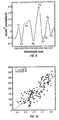
- FIG. 3shows a typical absorbance spectrum measurement from the forearm of a human subject
- FIG. 4shows a normalized second derivative of an absorbance spectrum versus wavelength
- FIG. 5shows the second derivative of an absorbance spectrum in the first overtone with features identified according to the invention
- FIG. 6shows the second derivative of the absorbance spectrum of FIG. 5 in the vicinity of the 1910 nm water band
- FIG. 7shows the second derivative of the absorbance spectrum of FIG. 4 in the vicinity of the 1450 nm water band
- FIG. 8shows the second derivative of the absorbance spectrum of FIG. 5 in the second overtone region with exemplar features according to the invention
- FIG. 9shows the second derivative of the absorbance spectrum of FIG. 5 in the combination band with key features marked according to the invention.
- FIG. 10shows a plot of a normalized fat band versus glucose concentration according to the invention.
- a major difficulty in noninvasively measuring glucose through near-infrared spectroscopyarises from the fact that glucose is present in very small amounts (2-20 mM). Calibrating a sensor to this small glucose signal requires extraction of the signal from a massive background due to variations in tissue characteristics and hydration. These background variations result in changes in the optical properties of the sampled tissue leading to confounding effects due to the resulting pathlength changes causing large uncertainties in the extracted signal. Long-term variations (over a few days) in tissue characteristics are especially bothersome since their effects could be large enough to swamp the small glucose signal. While the glucose signal is small, changes in blood glucose triggers physiological responses that are large.
- a major physiological response that can be detected spectrallyis the fluid shift that occurs due to changes in blood glucose, which causes water to move in and out of the vascular and cellular compartments. This redistribution of water causes changes in the scattering and absorption properties of skin leading to significant changes in the observed spectra of skin. This change in spectra due to changes in fluid distribution as a response to changes in blood glucose has proven extremely useful for building robust calibration models for glucose measurement.
- changes in blood glucoseleads to changes in the distribution and content of water in skin tissue.
- This variationcauses a change in the refractive index (and thus the scattering coefficient) and a change in the absorption coefficient of tissue.
- the depth to which light penetrates the tissueis changed.
- the changes in the absorption and scattering propertiesaffect the amount of light from a certain depth in the tissue that reaches the detector.
- changes in the water content in the dermiswill dictate the amount of light reaching the detector that has probed the subcutaneous tissue and thereby changes the total amount of light that is absorbed by fat.
- changes in the fluid distributionwill change the magnitude of the fat absorbance signal detected.
- the inventionprovides a method for detecting changes in the optical properties of tissue related to physiological changes, such as the water distribution among various compartments, for determining conditions that are not conducive to noninvasive measurement of glucose through near-infrared spectroscopy, and for:
- An apparatus for detecting and correcting fluid compartment changes and indirectly measuring glucoseincludes, but is not limited to:
- the spectrometer systemdetects near-infrared light within a specified range that is diffusely transmitted or reflected from the targeted tissue, and the analyzer measures glucose through data processing operations and the application of a model.
- the patient interface moduleFundamental to the system is the patient interface module, which precisely couples the apparatus to the tissue measurement site physically and optically, with minimal disturbance.
- a meanssuch as an optical coupling medium, is provided for preparing the sample site for spectroscopic measurement prior to contact with the device for the purpose of reducing specular reflectance and the effects of skin temperature and skin hydration changes.
- FIG. 1An overview of the system is shown in FIG. 1 and generally consists of two elements, a spectrometer 101 or instrument and an analyzer 208 embodying the process for obtaining the glucose measurement.
- the spectrometermeasures the near-infrared spectrum of a subject's tissue.
- the analyzerprocesses the spectral measurement, extracts features relevant to outlier detection and glucose measurement and applies a model to the processed spectral measurement and/or the extracted features to obtain a glucose measurement.
- a detailed description of the spectrometer by system and the components of the analyzerfollow.
- the spectroscopic measurement system 101consists of a source of near-infrared radiation 200 , a wavelength selection system 201 , an interface to the patient, a means for directing the near-infrared radiation to the skin 203 and a means for directing radiation reflected or transmitted from the skin 205 , a means for detecting near-infrared radiation that is reflected or transmitted from the skin 206 , a means for analyzing the detected near-infrared radiation 208 and a means for displaying the measured analyte, property or constituent 209 .
- the wavelength selection 201can occur between the subject interface 203 and the detector optics 205 .
- the source 200radiates near-infrared energy in the wavelength range 700-2500 nm and may consist of, for example, an array of LED's or a halogen lamp.
- One or more bandpass filtersare (optionally) required to minimize the effect of wavelengths from outside of the spectral range of interest, but which are still emitted by the source of near-infrared energy.
- halogen lampswhile having peak energy at approximately 1600 nm, still give off electromagnetic radiation above 2500 nm. This has detrimental effects on the detection of glucose since wavelengths above 2500 nm have deleterious effects at the measurement site due to heating of the tissue and its respective components.
- the method of wavelength separation 201 before and or after illumination of the skincan be performed through the use of a dispersive element (e.g., a plane or concave, ruled or holographic grating), an interferometer, or successive illumination of the elements of an LED array without an additional dispersive element.
- a dispersive elemente.g., a plane or concave, ruled or holographic grating
- an interferometere.g., a plane or concave, ruled or holographic grating
- successive illumination of the elements of an LED array without an additional dispersive elemente.g., a plane or concave, ruled or holographic grating
- a reference wavelength standard 202for example a polystyrene standard
- the sensing element(s) 206are detectors that are responsive to the targeted wavelengths and may constitute either an array or a single element.
- the material junction(s)occurs at a wavelength not required for the measurement.
- the junctiontypically occurs at 1750 nm for the purpose of reducing the cost of the array due to the high cost of extended InGaAs.
- this wavelength regionoccurs in the middle of the absorptions associated with fat, protein and glucose; thus, it is much preferable for the junction to occur at approximately 1480 nm ⁇ 20 nm.
- the electronics used to sense the individual elements of the arrayhave their junction occurring at the same wavelength.
- the tissue sample interfaceincludes a subject 204 interface module 203 by which near-infrared radiation is directed to and from 205 the tissue, either directly or through a light pipe, fiber optics, a lens system or a light directing mirror system.
- the specularly reflected radiation from the irradiated siteis of such a magnitude that it would greatly interfere with detection of the returned radiation.
- the patient interface modulefurther includes an elbow rest, a wrist rest, and a guide to assist in interfacing the illumination mechanism of choice and the tissue of interest.
- the patient interface moduleincludes a guide or positioning mechanism to assist in interfacing the tissue of interest.
- an optical coupling fluidis placed between the illumination mechanism and the tissue of interest to minimize specular reflectance from the surface of the skin.
- the collected near-infrared radiationis converted to a voltage and sampled through an analog-to-digital 207 converter for analysis on a microprocessor-based system 208 and the result of such analysis displayed 209 .
- the sample siteincludes the specific tissue irradiated by the spectrometer system.
- the ideal qualities of the sample siteinclude homogeneity, immutability and accessibility to the target analyte. While several measurement sites can be used, including the abdomen, thigh, hand (palm or back of the hand), ear lobe or finger, in the preferred embodiment, the volar part of the forearm is used.
- the measurementcan be made in either diffuse reflectance or diffuse transmittance mode, the preferred method is diffuse reflectance.
- the scanning of the tissuecan be done continuously, in the case of an area not subject to pulsation effects, or the scanning can be done intermittently between pulses.
- the spectrometer systemprovides a spectral measurement 104 or “spectrum” to the analyzer 208 for determination or measurement of the concentration of glucose.
- the spectrumis denoted by the vector m ⁇ 1 ⁇ N of absorbance values pertaining to a set of N wavelengths ⁇ N that span the near-infrared portion (700-2500 nm) of the spectrum.
- the measurementcan consist of a specific set of wavelengths in the near infrared region that have been optimized for the extraction of features and for the measurement requirements.
- the measurement of glucoseis optimally performed in the wavelength range 1100-1935 nm, or a selected subset thereof.
- the measurement mcan be defined as the measured intensity, I.
- mmay consist of either a single spectrum collected with an instrument or a combination of several (optimally) selected spectra collected over a defined measurement period and averaged. Methods for selecting the spectra, used to produce the lowest noise measurement, include similarity or distance measures (i.e., select the most similar) and clustering operations. Preprocessing and Feature Extraction
- Feature extraction 106is any mathematical transformation that enhances a quality or aspect of the sample measurement for interpretation [R. Duda, P. Hart, Pattern Classification and Scene Analysis , John Wiley and Sons, New York (1973)].
- the general purpose of feature extractionis to concisely represent or enhance the chemical concentration, structural properties and physiological state of the tissue measurement site.
- a set of featuresis developed that represents or reflects the optical properties of the tissue based on:
- the featuresare then applied either to identify conditions unsuitable for glucose measurement or to perform an actual measurement of glucose.
- a resolved estimate of the magnitude of the fat band absorbancecan be used to infer specific information about the dermis.
- fatis absent from the dermis, near infrared radiation must propagate through the dermis to penetrate into the adipose tissue beneath.
- physiological changes, and the corresponding changes in the optical properties of the dermisinfluence the magnitude of the fat band absorbance.
- first or second derivative calculation[A. Savitzky, M. Golay, Smoothing and Differentiation of Data by Simplified Least Squares Procedures , Anal. Chem., 36: 8, pp. 1627-1639 (1964)] or scatter correction:
- the set of abstract featuresdo not necessarily have a specific interpretation related to the physical system.
- the scores of a principal component analysisare used as features although their physical interpretation is not always known.
- the utility of the principal component analysisis related to the nature of the tissue absorbance spectrum. The most significant variation in the tissue spectral absorbance is not caused by the absorption of glucose but is related to the state, structure and composition of the measurement site. This variation is modeled by the primary principal components. Therefore, the leading principal components tend to represent variation related to the structural properties and physiological state of the tissue measurement site and consequently reflect the optical properties of tissue.
- the featuresare determined from the second derivative of the absorbance spectrum shown in FIG. 4 .
- Each critical pointis identified according to its wavelength.
- the value of the second derivative spectrum at each critical pointis used as a feature to represent a key property of the tissue sample associated with the measurement spectrum.
- FIGS. 5 through 9many key features are identified as exemplary measurements. These include:
- Normalization pointsare generally used to determine derived features and points designated as “fat” (f1-f4), “protein” p1-p9 and “water” w2-w6 are generally located in the vicinity of an absorption band due to fat, protein or water respectively. Due to the bandwidth (lower resolution) of the second derivative spectrum, several of the bands associated with one constituent include absorbance due to another and a few of the critical points are associated with a constituent because their location is in the vicinity of the respective constituent. In addition, the wavelengths are reported for the features shown in the example second derivative spectrum and can change substantially as a result of variation in the reduced scattering coefficient and the inner filter effect related to the multiple layers of the skin.
- preprocessingincludes on or more steps of filtering, differentiation, scatter correction and normalization.
- Wavelength selectionlimits the spectrum to regions pertaining specifically to glucose including 1450-1700 nm, 1700-1900 nm, 2050-2200 nm, and 2250-2400 nm.
- the tissue templateis determined through one or more spectral measurements and a data selection criterion, for example, by selecting only spectral measurements that resemble each other closely and averaging them.
- x tincludes features extracted from a (spectral) measurement collected on tissue at the beginning of the measurement period.
- This processis referred to as “re-calibration” and involves both the collection of one or more spectral measurements that are processed to form a tissue template as well as an associated set of reference glucose values.
- the glucose valuesare combined, according to the same strategy as that used to create the tissue template to form a measurement bias adjustment 103 , described in greater detail below.
- the measurement periodis defined as a time period during which the state of the tissue sample is uniform (optical properties within a preset bound) and the tissue measurement site is constant.
- the tissue templatecan also be any set of features from a given patient or calibration set that future spectral measurements will be compared with.
- the variables c and dare determined through a least-squares fit (to minimize the Euclidean norm of z) of the tissue template over a particular wavelength range to the measured spectrum.
- the measurement of glucoseis accomplished through the application of a calibration model 108 to the processed spectral measurement and/or the extracted features.
- the modelis determined from a calibration set of exemplary paired data points each consisting of a pre-processed spectral measurement (x) and an associated reference glucose value (y) determined from an analysis of a sample of blood or interstitial fluid.
- the reference glucose measurementscan be determined from a blood draw at the fingertip or site of the spectral measurement.
- the reference glucose measurementscan be determined from interstitial glucose concentrations taken at or near the site of spectroscopic measurement or alternate representative site, for example the forearm.
- blood, serum, plasma or interstitial drawsare taken from a tissue site that is either near the sensor sample site or has been designed/determined to reflect the sample site.
- tissue sitethat is either near the sensor sample site or has been designed/determined to reflect the sample site.
- non-invasive near-infrared measurementsare taken for calibration on the forearm, it is possible in some individuals to collect a capillary blood draw from the same forearm or the opposite forearm.
- the calibration setis based on one or more subjects and generally contains glucose concentrations that span the expected range of glucose variation and that include spectral variation representative of that likely to be encountered in future spectral measurements.
- the calibration model 108includes an equation, a set of parameters and corresponding computer code that is implemented to measure the subject's glucose level on the basis of the preprocessed spectral measurement.
- the preprocessing and feature extraction, together with the modelefficiently extract the net analyte signal of glucose where net analyte signal is the portion of the spectral signal related to the target analyte that is orthogonal to the interference [A. Lorber, K. Faber, B. Kowalski, Net Analyte Signal Calculation in Multivariate Calibration , Anal. Chem, 69, pp. 1620-1626 (1997)].
- the net analyte signalis then scaled and bias corrected 103 to match the desired units of glucose measurement (e.g. mg/dL).
- the extracted featuresare supplemental and are applied to compensate another model for variation in the optical properties related to a change in the effective pathlength of detected light and sample tissue volume but which changes are unrelated to absorption due to glucose. This is accomplished by using the absorption features that reflect the changes in tissue optical properties related to a water shift between compartments (or other physiological transient condition) to supplement a calibration that is based on the near-infrared absorption of glucose.
- the extracted features related to the physiological and chemical response of the bodyare primary and used to indirectly measure the subject's glucose level.
- the methodis based on the natural response to changes in blood glucose, which result in the alteration of fluid distribution in the interstitial, vascular and cellular compartments.
- Such alteration of fluid distributioncauses changes in the scattering and absorption properties of tissue that are detectable through near-infrared spectroscopy and which serve as a basis for an indirect blood glucose measurement.
- the near-infrared signalreflects the changes in the scattering properties from different layers in skin that coincide with changes in glucose concentration.
- the changes in fluid distributionlead to changes in the apparent absorption of key constituents, such as fat, protein and water that provide a signal that is substantially higher than that of glucose and can be used as markers for measuring glucose noninvasively.
- key constituentssuch as fat, protein and water that provide a signal that is substantially higher than that of glucose and can be used as markers for measuring glucose noninvasively.
- long-term fluid compartment balancesare influenced by fluid intake, exercise, diet, drug therapy and other physiological factors.
- ⁇is the estimated glucose concentration
- x p ⁇ Nis a processed spectral measurement
- z ⁇ Mis the set of features representative of the physiological state or optical properties of the tissue
- f: N,M ⁇ 1is a model used to measure glucose on the basis of the preprocessed spectrum and extracted features
- bis a baseline adjustment for the glucose measurement associated with both the tissue template and calibration model.
- the model, f( ⁇ )is determined through a calibration set including spectral measurements, extracted features and reference glucose values (from blood or interstitial measurements).
- the method for designing the structure of f( ⁇ )is through the process of system of identification [L. Ljung, Systems Identification: Theory for the User, 2d.ed., Prentice Hall (1999)].
- the model parametersare calculated using known methods including multivariate regression or weighted multivariate regression [N. Draper, H. Smith, Applied Regression Analysis, 2d.ed., John Wiley and Sons, New York (1981)], principal component regression [H. Martens, T. Naes, Multivariate Calibration , John Wiley and Sons, New York (1989)], partial least squares regression [P. Geladi, B. Kowalski, Partial least-squares regression: a tutorial, Analytica Chimica Acta, 185, pp. 1-17, (1986)], or artificial neural networks [S. Haykin, Neural Networks: A Comprehensive Foundation , Prentice Hall, Upper Saddle River N.J. (1994)].
- f( ⁇ )is found through an experiment in which the tissue optical properties are stable or constant while the glucose is manipulated.
- Second, the optical properties of tissue are allowed to fluctuate and g( ⁇ ), m s and m i are determined on the basis of the error in glucose measurement where the target value for g( ⁇ ) is given by: ry ⁇ f ( x p ) ⁇ b (3) where y is the reference glucose concentration.
- F and Gare determined separately as described above using linear methods of calibration. This final realization of the supplemental use of features for glucose measurement is the preferred method.
- gM ⁇ 1 is a model used to map the features to a variable correlated to the reference glucose level and m s and m i are slope and intercepts used to convert g(z) to the correct units.
- the method for determining g( ⁇ )is through an exemplary set (calibration set) of spectral measurements, extracted features and reference glucose concentrations (from blood or interstitial measurements). A sub-set of features is selected based on their combined correlation to the reference glucose concentration.
- variable selectionWhile a priori knowledge and trial-and-error can be employed for variable selection, standard methods also exist for variable selection including stepwise regression [Draper, et al., supra] random search techniques, genetic algorithms [D. Goldberg, Genetic Algorithm in Search, Optimization and Machine Learning , Addison Wesley Publishing Company (1989)] or evolutionary programming [D. Fogel, An Introduction to Simulated Evolutionay Optimization , IEEE Trans. On Neural Networks, 5:1 (January 1994)].
- the features, zare selected to include at least the normalized second derivative fat band (d2) or the normalized second derivative protein band (d1).
- the parameters of the modelare determined through multivariate regression, weighted multivariate regression or locally weighted regression. For example, a calibration set was collected on a particular subject whose glucose concentration spanned the range 70-350 mg/dl. A plot of the normalized fat band, d2, versus glucose concentration is given in FIG. 10 . The high degree of correlation between the feature and reference glucose concentration indicates that glucose measurement is feasible through this extracted feature. A simple linear regression is performed to determine the model parameters of the equation above.
- the inventionis not limited to the normalized fat and protein bands.
- the wavelength chosen for normalizationis not restricted to 1665 nm.
- a multiplicity of modelsexists for various subjects and categories of subjects depending on the optical properties of their respective tissue sample, baseline level of perfusion and physiological response to changes in glucose concentration.
- an alternative embodimentconsists of using a combination of features related to all of the major types of absorption bands.
- abstract features that reflect the changes in the optical properties of skin tissuecan be used as the independent variables for noninvasive calibration and measurement of glucose.
- the spectral measurement, mis preprocessed and is followed by wavelength selection to create the preprocessed vector, x.
- the calibration modelcan be determined through multivariate regression, weighted multivariate regression, locally weighted regression or other standard approach. While principal component regression has been described as the method for spectral decomposition, partial least squares regression can also be applied.
- the preferred methodinvolves preprocessing through a first derivative with a wide smoothing window (e.g., 31 nm), scatter correction through multiplicative scatter correction or standard normal variate transformation [R. Barnes, M. Dhanoa, S. Lister, Applied Spectroscopy, 43:772-777 (1989], and wavelength selection in the range 1450-1850 nm or a subset thereof.
- a wide smoothing windowe.g. 31 nm
- scatter correctionthrough multiplicative scatter correction or standard normal variate transformation [R. Barnes, M. Dhanoa, S. Lister, Applied Spectroscopy, 43:772-777 (1989]
- wavelength selectionin the range 1450-1850 nm or a subset thereof.
- information from a water bandsuch as 1180-1450 nm may also be included.
- the preprocessed datais corrected to the tissue template and partial-least squares is applied to develop the calibration model.
- Glucoseis then measured through the application of the identical preprocessing steps to a spectral measurement (first derivative, scatter correction, wavelength selection and tissue template correction) to obtain the processed spectral measurement, x.
Landscapes
- Health & Medical Sciences (AREA)
- Life Sciences & Earth Sciences (AREA)
- Physics & Mathematics (AREA)
- Pathology (AREA)
- General Health & Medical Sciences (AREA)
- Surgery (AREA)
- Engineering & Computer Science (AREA)
- Biomedical Technology (AREA)
- Heart & Thoracic Surgery (AREA)
- Medical Informatics (AREA)
- Molecular Biology (AREA)
- Animal Behavior & Ethology (AREA)
- Biophysics (AREA)
- Public Health (AREA)
- Veterinary Medicine (AREA)
- Spectroscopy & Molecular Physics (AREA)
- Optics & Photonics (AREA)
- Emergency Medicine (AREA)
- Chemical & Material Sciences (AREA)
- Analytical Chemistry (AREA)
- Biochemistry (AREA)
- General Physics & Mathematics (AREA)
- Immunology (AREA)
- Measurement Of The Respiration, Hearing Ability, Form, And Blood Characteristics Of Living Organisms (AREA)
- Investigating Or Analysing Materials By Optical Means (AREA)
Abstract
Description
A=εcl (1)
where ε is the analyte specific absorption coefficient, c is the concentration and/is the pathlength. The overall absorbance at a particular wavelength is the sum of the individual absorbances of each particular analyte given by Beer's Law. The concentration of a particular analyte, such as glucose, can be determined through a multivariate analysis of the absorbance over a multiplicity of wavelengths because ε is unique for each analyte. However, in tissue compartments expected to contain glucose, the concentration of glucose is at least three orders of magnitude less than that of water. Consequently, the signal targeted for detection by reported approaches to near-infrared measurement of glucose (the absorbance due to glucose in the tissue) is expected to be at most three orders of magnitude less than other interfering tissue constituents. Therefore, the near-infrared measurement of glucose requires a high level of sensitivity over a broad wavelength range, and the application of methods of multivariate analysis.
- correcting the glucose measurement on the basis of detected changes in tissue optical properties; or
- measuring glucose indirectly on the basis of features reflecting the detected optical properties.
- a spectrometer system;
- a patient interface module; and
- an analyzer.

where m is the reflectance spectrum of the skin and is analogous to an absorbance spectrum containing quantitative information that is based on the known interaction of the incident light with components of the body tissue. A plot of m versus λ is shown in
where Ir∈
Preprocessing and Feature Extraction
- identification of distinct absorption bands that change in various ways with respect to changes in pathlength; and
- the scattering and absorption properties (or coefficients) of the measurement site.
outlier detection 107;- compensation for changes in the optical properties of
tissue 102; and glucose measurement 109.
- “simple” features are the values of the spectral measurement or the processed spectral measurement at the critical points (the points at which the slope is zero);
- additional (derived) features are determined from the base features through mathematical transformation such as addition, subtraction, division and multiplication; and
- abstract features are derived through linear and nonlinear transformations of the pre-processed spectrum.
- Normalization points (n) 1-8 near 1665, 1708, 1746, 1868, 1380, 1133, 2020 and 2232 nm respectively;
- Fat bands points (f) 1-4 near 1727, 1765, 1214, 1165 nm;
- Protein band points (p) 1-9 near 1687, 1715, 1190, 2050, 2150, 2175, 2275, 2292, and 2355 nm; and
- Water band points (w) 2-6 near 1789, 1896, 1410, 1465 and 1150 nm.
z=x−(cxt+d) (5)
where x is the preprocessed spectrum or the selected set of features, xtis the estimated background or tissue template associated with the measurement period, and c and d are slope and intercept adjustments to the tissue template. During each measurement period, defined by a measurement position on the tissue and a level of physiological stability of the measurement site, the tissue template is determined through one or more spectral measurements and a data selection criterion, for example, by selecting only spectral measurements that resemble each other closely and averaging them. In the preferred embodiment, xtincludes features extracted from a (spectral) measurement collected on tissue at the beginning of the measurement period. This process is referred to as “re-calibration” and involves both the collection of one or more spectral measurements that are processed to form a tissue template as well as an associated set of reference glucose values. The glucose values are combined, according to the same strategy as that used to create the tissue template to form a
Detection of Tissue States
ŷ=f(xp,z)+b (1)
where ŷ is the estimated glucose concentration, xp∈
ÿ=f(xp)−(msg(z)+mi)+b (2)
where f:
r=y−f(xp)−b (3)
where y is the reference glucose concentration. In the third embodiment, when f(·) and g(·) are determined to be linear over the range of measurement, equation #8 reduces to:
ŷ=xpF−(mszG+mi)+b (4)
where F∈
ÿ=(msg(z)+b (5)
where g:
ŷ=(mszG+mi)+b, (6)
where G∈
ŷ=g1z1+g2z2+g3z3+g4+b (12)
where z1, z2and z3are the normalized second derivative values of the fat band and two water bands respectively. This equation can then be used to measure glucose values from spectra taken in the future after preprocessing and feature extraction.
z=xP (13)
where x∈
ŷ=xG+b (13)
where G∈
Claims (66)
z=x−(cxt+d):
ŷ=f(xp,z)+b,
ŷ=f(xp)−(msg(z)+mi)+b,
r=y−f(xp)−b
ŷ=xpF−(mszG+mi)+b,
ŷ=(msg(z)+mi)+b
ŷ=(mszG+mi)+b,
ŷ=g1z1+g2z2+g3z3+g4+b,
z=xP
ŷ=mG+b
Priority Applications (2)
| Application Number | Priority Date | Filing Date | Title |
|---|---|---|---|
| US10/297,736US6990364B2 (en) | 2001-01-26 | 2002-01-25 | Noninvasive measurement of glucose through the optical properties of tissue |
| US10/917,802US7233816B2 (en) | 1997-08-14 | 2004-08-12 | Optical sampling interface system for in vivo measurement of tissue |
Applications Claiming Priority (3)
| Application Number | Priority Date | Filing Date | Title |
|---|---|---|---|
| US26443101P | 2001-01-26 | 2001-01-26 | |
| PCT/US2002/002288WO2002065090A2 (en) | 2001-01-26 | 2002-01-25 | Noninvasive measurement of glucose through the optical properties of tissue |
| US10/297,736US6990364B2 (en) | 2001-01-26 | 2002-01-25 | Noninvasive measurement of glucose through the optical properties of tissue |
Related Parent Applications (1)
| Application Number | Title | Priority Date | Filing Date |
|---|---|---|---|
| PCT/US2002/002288A-371-Of-InternationalWO2002065090A2 (en) | 1997-08-14 | 2002-01-25 | Noninvasive measurement of glucose through the optical properties of tissue |
Related Child Applications (1)
| Application Number | Title | Priority Date | Filing Date |
|---|---|---|---|
| US10/349,573Continuation-In-PartUS7039446B2 (en) | 1997-08-14 | 2003-01-22 | Indirect measurement of tissue analytes through tissue properties |
Publications (2)
| Publication Number | Publication Date |
|---|---|
| US20040068163A1 US20040068163A1 (en) | 2004-04-08 |
| US6990364B2true US6990364B2 (en) | 2006-01-24 |
Family
ID=23006047
Family Applications (1)
| Application Number | Title | Priority Date | Filing Date |
|---|---|---|---|
| US10/297,736Expired - LifetimeUS6990364B2 (en) | 1997-08-14 | 2002-01-25 | Noninvasive measurement of glucose through the optical properties of tissue |
Country Status (12)
| Country | Link |
|---|---|
| US (1) | US6990364B2 (en) |
| EP (1) | EP1364196A4 (en) |
| JP (3) | JP4071113B2 (en) |
| KR (1) | KR100893432B1 (en) |
| CN (1) | CN1325015C (en) |
| AU (1) | AU2002249985B2 (en) |
| CA (1) | CA2441078A1 (en) |
| IL (1) | IL156844A0 (en) |
| MX (1) | MXPA03006726A (en) |
| NZ (1) | NZ527164A (en) |
| TW (1) | TWI324686B (en) |
| WO (1) | WO2002065090A2 (en) |
Cited By (275)
| Publication number | Priority date | Publication date | Assignee | Title |
|---|---|---|---|---|
| US20040220459A1 (en)* | 2003-05-02 | 2004-11-04 | Schlegel Robert P. | Zero corrected optical blood analyte detector |
| US20050186648A1 (en)* | 2004-01-29 | 2005-08-25 | Schurman Matthew J. | OCT based method for diagnosis and therapy |
| US20060063988A1 (en)* | 2004-08-11 | 2006-03-23 | Schurman Matthew J | Method and apparatus for monitoring glucose levels in a biological tissue |
| US20060167347A1 (en)* | 2002-11-04 | 2006-07-27 | Kexin Xu | Composite spectral measurement method and its spectral detection instrument |
| US20060264719A1 (en)* | 2004-08-11 | 2006-11-23 | Schurman Matthew J | Method for data reduction and calibration of an OCT-based blood glucose monitor |
| US20060276696A1 (en)* | 2004-08-11 | 2006-12-07 | Glucolight Corporation | Methods for noninvasively measuring analyte levels in a subject |
| US20070203405A1 (en)* | 2004-10-15 | 2007-08-30 | Yoshiaki Shimomura | Instrument For Noninvasively Measuring Blood Sugar Level |
| US20090198113A1 (en)* | 2006-06-07 | 2009-08-06 | Koninklijke Philips Electronics N.V. | Dedicated spectral illumination spectroscopy |
| US20090275812A1 (en)* | 2008-03-04 | 2009-11-05 | Glucolight Corporation | Flowometry in Optical Coherence Tomography for Analyte Level Estimation |
| US20100113899A1 (en)* | 2008-11-06 | 2010-05-06 | Mark Ries Robinson | Alignment System for Optically Sampling a Hand |
| US20100160747A1 (en)* | 2008-12-12 | 2010-06-24 | Mark Ries Robinson | Selection of preferred sampling location on hand via minimization of sampling error, and optical alignment for repeatably sampling tissue |
| US7884933B1 (en) | 2010-05-05 | 2011-02-08 | Revolutionary Business Concepts, Inc. | Apparatus and method for determining analyte concentrations |
| US8571618B1 (en) | 2009-09-28 | 2013-10-29 | Cercacor Laboratories, Inc. | Adaptive calibration system for spectrophotometric measurements |
| US8670642B2 (en) | 2005-11-18 | 2014-03-11 | Omni Medsci, Inc. | Broadband or mid-infrared fiber light sources |
| WO2012087319A3 (en)* | 2010-12-22 | 2014-03-20 | Verifica-Bg Inc. | Method and apparatus for in vivo optical measurement of blood glucose concentration |
| US8848282B2 (en) | 2002-09-03 | 2014-09-30 | Omni Medsci, Inc. | System and method for voice control of medical devices |
| US9164032B2 (en) | 2012-12-31 | 2015-10-20 | Omni Medsci, Inc. | Short-wave infrared super-continuum lasers for detecting counterfeit or illicit drugs and pharmaceutical process control |
| US9351672B2 (en) | 2012-07-16 | 2016-05-31 | Timothy Ruchti | Multiplexed pathlength resolved noninvasive analyzer apparatus with stacked filters and method of use thereof |
| US9351671B2 (en) | 2012-07-16 | 2016-05-31 | Timothy Ruchti | Multiplexed pathlength resolved noninvasive analyzer apparatus and method of use thereof |
| US9442065B2 (en) | 2014-09-29 | 2016-09-13 | Zyomed Corp. | Systems and methods for synthesis of zyotons for use in collision computing for noninvasive blood glucose and other measurements |
| US9554738B1 (en) | 2016-03-30 | 2017-01-31 | Zyomed Corp. | Spectroscopic tomography systems and methods for noninvasive detection and measurement of analytes using collision computing |
| US9585604B2 (en) | 2012-07-16 | 2017-03-07 | Zyomed Corp. | Multiplexed pathlength resolved noninvasive analyzer apparatus with dynamic optical paths and method of use thereof |
| CN106525729A (en)* | 2015-09-12 | 2017-03-22 | 南京理工大学 | Substance element content information detection method based on spectral analysis technology |
| US20170148164A1 (en)* | 2014-07-11 | 2017-05-25 | Nikon Corporation | Image analysis apparatus, imaging system, surgery support system, image analysis method, storage medium, and detection system |
| US9766126B2 (en) | 2013-07-12 | 2017-09-19 | Zyomed Corp. | Dynamic radially controlled light input to a noninvasive analyzer apparatus and method of use thereof |
| US20170323437A1 (en)* | 2014-12-12 | 2017-11-09 | Canon Kabushiki Kaisha | Information processing apparatus, information processing method, and program |
| US9897584B2 (en) | 2012-12-31 | 2018-02-20 | Omni Medsci, Inc. | Short-wave infrared super-continuum lasers for natural gas leak detection, exploration, and other active remote sensing applications |
| US9993159B2 (en) | 2012-12-31 | 2018-06-12 | Omni Medsci, Inc. | Near-infrared super-continuum lasers for early detection of breast and other cancers |
| US10136819B2 (en) | 2012-12-31 | 2018-11-27 | Omni Medsci, Inc. | Short-wave infrared super-continuum lasers and similar light sources for imaging applications |
| US10159412B2 (en) | 2010-12-01 | 2018-12-25 | Cercacor Laboratories, Inc. | Handheld processing device including medical applications for minimally and non invasive glucose measurements |
| US10206581B2 (en) | 2014-10-29 | 2019-02-19 | Zoll Medical Corporation | Transesophageal or transtracheal cardiac monitoring by optical spectroscopy |
| US10213113B2 (en) | 2012-12-31 | 2019-02-26 | Omni Medsci, Inc. | Physiological measurement device using light emitting diodes |
| US10365160B2 (en) | 2017-08-02 | 2019-07-30 | Samsung Electronics Co., Ltd. | Spectrum measurement apparatus and method, and calibration method of spectrum measurement apparatus |
| US10485461B2 (en) | 2016-07-29 | 2019-11-26 | Samsung Electronics Co., Ltd. | Apparatus and method for estimating substance in blood |
| US10660526B2 (en) | 2012-12-31 | 2020-05-26 | Omni Medsci, Inc. | Near-infrared time-of-flight imaging using laser diodes with Bragg reflectors |
| US10716499B1 (en) | 2015-03-24 | 2020-07-21 | Zoll Medical Corporation | Physiological monitoring by optical spectroscopy |
| US10736518B2 (en) | 2015-08-31 | 2020-08-11 | Masimo Corporation | Systems and methods to monitor repositioning of a patient |
| US10765367B2 (en) | 2014-10-07 | 2020-09-08 | Masimo Corporation | Modular physiological sensors |
| US10779098B2 (en) | 2018-07-10 | 2020-09-15 | Masimo Corporation | Patient monitor alarm speaker analyzer |
| US10784634B2 (en) | 2015-02-06 | 2020-09-22 | Masimo Corporation | Pogo pin connector |
| USD897098S1 (en) | 2018-10-12 | 2020-09-29 | Masimo Corporation | Card holder set |
| US10799163B2 (en) | 2006-10-12 | 2020-10-13 | Masimo Corporation | Perfusion index smoother |
| US10799160B2 (en) | 2013-10-07 | 2020-10-13 | Masimo Corporation | Regional oximetry pod |
| US10825568B2 (en) | 2013-10-11 | 2020-11-03 | Masimo Corporation | Alarm notification system |
| US10835130B2 (en) | 2014-12-19 | 2020-11-17 | Samsung Electronics Co., Ltd. | Noninvasive blood glucose measurement method and apparatus |
| US10849554B2 (en) | 2017-04-18 | 2020-12-01 | Masimo Corporation | Nose sensor |
| US10856750B2 (en) | 2017-04-28 | 2020-12-08 | Masimo Corporation | Spot check measurement system |
| US10856788B2 (en) | 2005-03-01 | 2020-12-08 | Cercacor Laboratories, Inc. | Noninvasive multi-parameter patient monitor |
| US10863938B2 (en) | 2006-10-12 | 2020-12-15 | Masimo Corporation | System and method for monitoring the life of a physiological sensor |
| US10869602B2 (en) | 2002-03-25 | 2020-12-22 | Masimo Corporation | Physiological measurement communications adapter |
| US10874333B2 (en) | 2015-09-15 | 2020-12-29 | Massachusetts Institute Of Technology | Systems and methods for diagnosis of middle ear conditions and detection of analytes in the tympanic membrane |
| US10912502B2 (en) | 2008-07-03 | 2021-02-09 | Masimo Corporation | User-worn device for noninvasively measuring a physiological parameter of a user |
| US10912524B2 (en) | 2006-09-22 | 2021-02-09 | Masimo Corporation | Modular patient monitor |
| US10918281B2 (en) | 2017-04-26 | 2021-02-16 | Masimo Corporation | Medical monitoring device having multiple configurations |
| US10925550B2 (en) | 2011-10-13 | 2021-02-23 | Masimo Corporation | Medical monitoring hub |
| US10932729B2 (en) | 2018-06-06 | 2021-03-02 | Masimo Corporation | Opioid overdose monitoring |
| US10932705B2 (en) | 2017-05-08 | 2021-03-02 | Masimo Corporation | System for displaying and controlling medical monitoring data |
| US10939877B2 (en) | 2005-10-14 | 2021-03-09 | Masimo Corporation | Robust alarm system |
| US10943450B2 (en) | 2009-12-21 | 2021-03-09 | Masimo Corporation | Modular patient monitor |
| US10952641B2 (en) | 2008-09-15 | 2021-03-23 | Masimo Corporation | Gas sampling line |
| US10956950B2 (en) | 2017-02-24 | 2021-03-23 | Masimo Corporation | Managing dynamic licenses for physiological parameters in a patient monitoring environment |
| US10959652B2 (en) | 2001-07-02 | 2021-03-30 | Masimo Corporation | Low power pulse oximeter |
| US10973447B2 (en) | 2003-01-24 | 2021-04-13 | Masimo Corporation | Noninvasive oximetry optical sensor including disposable and reusable elements |
| USD916135S1 (en) | 2018-10-11 | 2021-04-13 | Masimo Corporation | Display screen or portion thereof with a graphical user interface |
| US10980432B2 (en) | 2013-08-05 | 2021-04-20 | Masimo Corporation | Systems and methods for measuring blood pressure |
| US10980484B2 (en) | 2017-03-27 | 2021-04-20 | Samsung Electronics Co., Ltd. | Method of enabling feature extraction for glucose monitoring using near-infrared (NIR) spectroscopy |
| US10980457B2 (en) | 2007-04-21 | 2021-04-20 | Masimo Corporation | Tissue profile wellness monitor |
| USD917564S1 (en) | 2018-10-11 | 2021-04-27 | Masimo Corporation | Display screen or portion thereof with graphical user interface |
| USD917704S1 (en) | 2019-08-16 | 2021-04-27 | Masimo Corporation | Patient monitor |
| US10987066B2 (en) | 2017-10-31 | 2021-04-27 | Masimo Corporation | System for displaying oxygen state indications |
| US10991135B2 (en) | 2015-08-11 | 2021-04-27 | Masimo Corporation | Medical monitoring analysis and replay including indicia responsive to light attenuated by body tissue |
| USD917550S1 (en) | 2018-10-11 | 2021-04-27 | Masimo Corporation | Display screen or portion thereof with a graphical user interface |
| US10993643B2 (en) | 2006-10-12 | 2021-05-04 | Masimo Corporation | Patient monitor capable of monitoring the quality of attached probes and accessories |
| US10993662B2 (en) | 2016-03-04 | 2021-05-04 | Masimo Corporation | Nose sensor |
| USD919100S1 (en) | 2019-08-16 | 2021-05-11 | Masimo Corporation | Holder for a patient monitor |
| US11000232B2 (en) | 2014-06-19 | 2021-05-11 | Masimo Corporation | Proximity sensor in pulse oximeter |
| USD919094S1 (en) | 2019-08-16 | 2021-05-11 | Masimo Corporation | Blood pressure device |
| US11022466B2 (en) | 2013-07-17 | 2021-06-01 | Masimo Corporation | Pulser with double-bearing position encoder for non-invasive physiological monitoring |
| US11020084B2 (en) | 2012-09-20 | 2021-06-01 | Masimo Corporation | Acoustic patient sensor coupler |
| US11020029B2 (en) | 2003-07-25 | 2021-06-01 | Masimo Corporation | Multipurpose sensor port |
| USD921202S1 (en) | 2019-08-16 | 2021-06-01 | Masimo Corporation | Holder for a blood pressure device |
| US11026604B2 (en) | 2017-07-13 | 2021-06-08 | Cercacor Laboratories, Inc. | Medical monitoring device for harmonizing physiological measurements |
| US11069461B2 (en) | 2012-08-01 | 2021-07-20 | Masimo Corporation | Automated assembly sensor cable |
| USD925597S1 (en) | 2017-10-31 | 2021-07-20 | Masimo Corporation | Display screen or portion thereof with graphical user interface |
| US11071480B2 (en) | 2012-04-17 | 2021-07-27 | Masimo Corporation | Hypersaturation index |
| US11076777B2 (en) | 2016-10-13 | 2021-08-03 | Masimo Corporation | Systems and methods for monitoring orientation to reduce pressure ulcer formation |
| US11087875B2 (en) | 2009-03-04 | 2021-08-10 | Masimo Corporation | Medical monitoring system |
| US11083397B2 (en) | 2012-02-09 | 2021-08-10 | Masimo Corporation | Wireless patient monitoring device |
| USD927699S1 (en) | 2019-10-18 | 2021-08-10 | Masimo Corporation | Electrode pad |
| US11086609B2 (en) | 2017-02-24 | 2021-08-10 | Masimo Corporation | Medical monitoring hub |
| US11095068B2 (en) | 2017-08-15 | 2021-08-17 | Masimo Corporation | Water resistant connector for noninvasive patient monitor |
| US11089982B2 (en) | 2011-10-13 | 2021-08-17 | Masimo Corporation | Robust fractional saturation determination |
| US11096631B2 (en) | 2017-02-24 | 2021-08-24 | Masimo Corporation | Modular multi-parameter patient monitoring device |
| US11103134B2 (en) | 2014-09-18 | 2021-08-31 | Masimo Semiconductor, Inc. | Enhanced visible near-infrared photodiode and non-invasive physiological sensor |
| US11114188B2 (en) | 2009-10-06 | 2021-09-07 | Cercacor Laboratories, Inc. | System for monitoring a physiological parameter of a user |
| US11109818B2 (en) | 2018-04-19 | 2021-09-07 | Masimo Corporation | Mobile patient alarm display |
| US11109770B2 (en) | 2011-06-21 | 2021-09-07 | Masimo Corporation | Patient monitoring system |
| US11133105B2 (en) | 2009-03-04 | 2021-09-28 | Masimo Corporation | Medical monitoring system |
| US11132117B2 (en) | 2012-03-25 | 2021-09-28 | Masimo Corporation | Physiological monitor touchscreen interface |
| USD933232S1 (en) | 2020-05-11 | 2021-10-12 | Masimo Corporation | Blood pressure monitor |
| US11145408B2 (en) | 2009-03-04 | 2021-10-12 | Masimo Corporation | Medical communication protocol translator |
| US11147521B2 (en) | 2018-09-11 | 2021-10-19 | Samsung Electronics Co., Ltd. | Apparatus and method for health care |
| US11153089B2 (en) | 2016-07-06 | 2021-10-19 | Masimo Corporation | Secure and zero knowledge data sharing for cloud applications |
| US11147518B1 (en) | 2013-10-07 | 2021-10-19 | Masimo Corporation | Regional oximetry signal processor |
| US11172890B2 (en) | 2012-01-04 | 2021-11-16 | Masimo Corporation | Automated condition screening and detection |
| US11178776B2 (en) | 2015-02-06 | 2021-11-16 | Masimo Corporation | Fold flex circuit for LNOP |
| US11176801B2 (en) | 2011-08-19 | 2021-11-16 | Masimo Corporation | Health care sanitation monitoring system |
| US11179111B2 (en) | 2012-01-04 | 2021-11-23 | Masimo Corporation | Automated CCHD screening and detection |
| US11185262B2 (en) | 2017-03-10 | 2021-11-30 | Masimo Corporation | Pneumonia screener |
| US11191484B2 (en) | 2016-04-29 | 2021-12-07 | Masimo Corporation | Optical sensor tape |
| US11191485B2 (en) | 2006-06-05 | 2021-12-07 | Masimo Corporation | Parameter upgrade system |
| US11202571B2 (en) | 2016-07-07 | 2021-12-21 | Masimo Corporation | Wearable pulse oximeter and respiration monitor |
| US11224363B2 (en) | 2013-01-16 | 2022-01-18 | Masimo Corporation | Active-pulse blood analysis system |
| US11229374B2 (en) | 2006-12-09 | 2022-01-25 | Masimo Corporation | Plethysmograph variability processor |
| US11234655B2 (en) | 2007-01-20 | 2022-02-01 | Masimo Corporation | Perfusion trend indicator |
| US11241199B2 (en) | 2011-10-13 | 2022-02-08 | Masimo Corporation | System for displaying medical monitoring data |
| US11259745B2 (en) | 2014-01-28 | 2022-03-01 | Masimo Corporation | Autonomous drug delivery system |
| US11272883B2 (en) | 2016-03-04 | 2022-03-15 | Masimo Corporation | Physiological sensor |
| US11272839B2 (en) | 2018-10-12 | 2022-03-15 | Ma Simo Corporation | System for transmission of sensor data using dual communication protocol |
| US11272852B2 (en) | 2011-06-21 | 2022-03-15 | Masimo Corporation | Patient monitoring system |
| US11289199B2 (en) | 2010-01-19 | 2022-03-29 | Masimo Corporation | Wellness analysis system |
| US11291061B2 (en) | 2017-01-18 | 2022-03-29 | Masimo Corporation | Patient-worn wireless physiological sensor with pairing functionality |
| USRE49007E1 (en) | 2010-03-01 | 2022-04-05 | Masimo Corporation | Adaptive alarm system |
| US11291415B2 (en) | 2015-05-04 | 2022-04-05 | Cercacor Laboratories, Inc. | Noninvasive sensor system with visual infographic display |
| US11298021B2 (en) | 2017-10-19 | 2022-04-12 | Masimo Corporation | Medical monitoring system |
| USRE49034E1 (en) | 2002-01-24 | 2022-04-19 | Masimo Corporation | Physiological trend monitor |
| US11331044B2 (en) | 2017-12-01 | 2022-05-17 | Samsung Electronics Co., Ltd. | Apparatus and method for measuring bio-information |
| US11331013B2 (en) | 2014-09-04 | 2022-05-17 | Masimo Corporation | Total hemoglobin screening sensor |
| US11330996B2 (en) | 2010-05-06 | 2022-05-17 | Masimo Corporation | Patient monitor for determining microcirculation state |
| US11363960B2 (en) | 2011-02-25 | 2022-06-21 | Masimo Corporation | Patient monitor for monitoring microcirculation |
| US11367529B2 (en) | 2012-11-05 | 2022-06-21 | Cercacor Laboratories, Inc. | Physiological test credit method |
| US11389093B2 (en) | 2018-10-11 | 2022-07-19 | Masimo Corporation | Low noise oximetry cable |
| US11399722B2 (en) | 2010-03-30 | 2022-08-02 | Masimo Corporation | Plethysmographic respiration rate detection |
| US11399774B2 (en) | 2010-10-13 | 2022-08-02 | Masimo Corporation | Physiological measurement logic engine |
| US11410507B2 (en) | 2017-02-24 | 2022-08-09 | Masimo Corporation | Localized projection of audible noises in medical settings |
| US11406286B2 (en) | 2018-10-11 | 2022-08-09 | Masimo Corporation | Patient monitoring device with improved user interface |
| US11417426B2 (en) | 2017-02-24 | 2022-08-16 | Masimo Corporation | System for displaying medical monitoring data |
| US11412964B2 (en) | 2008-05-05 | 2022-08-16 | Masimo Corporation | Pulse oximetry system with electrical decoupling circuitry |
| US11426125B2 (en) | 2009-02-16 | 2022-08-30 | Masimo Corporation | Physiological measurement device |
| US11439329B2 (en) | 2011-07-13 | 2022-09-13 | Masimo Corporation | Multiple measurement mode in a physiological sensor |
| US11445948B2 (en) | 2018-10-11 | 2022-09-20 | Masimo Corporation | Patient connector assembly with vertical detents |
| US11452449B2 (en) | 2012-10-30 | 2022-09-27 | Masimo Corporation | Universal medical system |
| US11464410B2 (en) | 2018-10-12 | 2022-10-11 | Masimo Corporation | Medical systems and methods |
| US11484231B2 (en) | 2010-03-08 | 2022-11-01 | Masimo Corporation | Reprocessing of a physiological sensor |
| US11488715B2 (en) | 2011-02-13 | 2022-11-01 | Masimo Corporation | Medical characterization system |
| US11490863B2 (en) | 2018-10-19 | 2022-11-08 | Samsung Electronics Co., Ltd. | Apparatus and method for estimating bio-information, and apparatus for supporting estimation of bio-information |
| US11504058B1 (en) | 2016-12-02 | 2022-11-22 | Masimo Corporation | Multi-site noninvasive measurement of a physiological parameter |
| US11504066B1 (en) | 2015-09-04 | 2022-11-22 | Cercacor Laboratories, Inc. | Low-noise sensor system |
| US11504062B2 (en) | 2013-03-14 | 2022-11-22 | Masimo Corporation | Patient monitor placement indicator |
| US11504002B2 (en) | 2012-09-20 | 2022-11-22 | Masimo Corporation | Physiological monitoring system |
| US11515664B2 (en) | 2009-03-11 | 2022-11-29 | Masimo Corporation | Magnetic connector |
| US11517223B2 (en) | 2018-12-13 | 2022-12-06 | Samsung Electronics Co., Ltd. | Apparatus and method for estimating blood glucose |
| USD973072S1 (en) | 2020-09-30 | 2022-12-20 | Masimo Corporation | Display screen or portion thereof with graphical user interface |
| US11534087B2 (en) | 2009-11-24 | 2022-12-27 | Cercacor Laboratories, Inc. | Physiological measurement system with automatic wavelength adjustment |
| USD973685S1 (en) | 2020-09-30 | 2022-12-27 | Masimo Corporation | Display screen or portion thereof with graphical user interface |
| USD973686S1 (en) | 2020-09-30 | 2022-12-27 | Masimo Corporation | Display screen or portion thereof with graphical user interface |
| USD974193S1 (en) | 2020-07-27 | 2023-01-03 | Masimo Corporation | Wearable temperature measurement device |
| US11559275B2 (en) | 2008-12-30 | 2023-01-24 | Masimo Corporation | Acoustic sensor assembly |
| US11564599B2 (en) | 2017-10-19 | 2023-01-31 | Samsung Electronics Co., Ltd. | Apparatus and method for obtaining individualized unit spectrum, and apparatus and method for estimating biological component |
| US11571152B2 (en) | 2009-12-04 | 2023-02-07 | Masimo Corporation | Calibration for multi-stage physiological monitors |
| US11581091B2 (en) | 2014-08-26 | 2023-02-14 | Vccb Holdings, Inc. | Real-time monitoring systems and methods in a healthcare environment |
| USD979516S1 (en) | 2020-05-11 | 2023-02-28 | Masimo Corporation | Connector |
| US11596363B2 (en) | 2013-09-12 | 2023-03-07 | Cercacor Laboratories, Inc. | Medical device management system |
| USD980091S1 (en) | 2020-07-27 | 2023-03-07 | Masimo Corporation | Wearable temperature measurement device |
| US11602289B2 (en) | 2015-02-06 | 2023-03-14 | Masimo Corporation | Soft boot pulse oximetry sensor |
| US11607139B2 (en) | 2006-09-20 | 2023-03-21 | Masimo Corporation | Congenital heart disease monitor |
| US11622733B2 (en) | 2008-05-02 | 2023-04-11 | Masimo Corporation | Monitor configuration system |
| US11637437B2 (en) | 2019-04-17 | 2023-04-25 | Masimo Corporation | Charging station for physiological monitoring device |
| US11638532B2 (en) | 2008-07-03 | 2023-05-02 | Masimo Corporation | User-worn device for noninvasively measuring a physiological parameter of a user |
| USD985498S1 (en) | 2019-08-16 | 2023-05-09 | Masimo Corporation | Connector |
| US11645905B2 (en) | 2013-03-13 | 2023-05-09 | Masimo Corporation | Systems and methods for monitoring a patient health network |
| US11653862B2 (en) | 2015-05-22 | 2023-05-23 | Cercacor Laboratories, Inc. | Non-invasive optical physiological differential pathlength sensor |
| US11673041B2 (en) | 2013-12-13 | 2023-06-13 | Masimo Corporation | Avatar-incentive healthcare therapy |
| US11672447B2 (en) | 2006-10-12 | 2023-06-13 | Masimo Corporation | Method and apparatus for calibration to reduce coupling between signals in a measurement system |
| US11679579B2 (en) | 2015-12-17 | 2023-06-20 | Masimo Corporation | Varnish-coated release liner |
| US11684296B2 (en) | 2018-12-21 | 2023-06-27 | Cercacor Laboratories, Inc. | Noninvasive physiological sensor |
| US11690574B2 (en) | 2003-11-05 | 2023-07-04 | Masimo Corporation | Pulse oximeter access apparatus and method |
| US11696712B2 (en) | 2014-06-13 | 2023-07-11 | Vccb Holdings, Inc. | Alarm fatigue management systems and methods |
| US11717210B2 (en) | 2010-09-28 | 2023-08-08 | Masimo Corporation | Depth of consciousness monitor including oximeter |
| US11721105B2 (en) | 2020-02-13 | 2023-08-08 | Masimo Corporation | System and method for monitoring clinical activities |
| US11724031B2 (en) | 2006-01-17 | 2023-08-15 | Masimo Corporation | Drug administration controller |
| US11730379B2 (en) | 2020-03-20 | 2023-08-22 | Masimo Corporation | Remote patient management and monitoring systems and methods |
| USD997365S1 (en) | 2021-06-24 | 2023-08-29 | Masimo Corporation | Physiological nose sensor |
| US11744471B2 (en) | 2009-09-17 | 2023-09-05 | Masimo Corporation | Optical-based physiological monitoring system |
| US11747178B2 (en) | 2011-10-27 | 2023-09-05 | Masimo Corporation | Physiological monitor gauge panel |
| USD998631S1 (en) | 2018-10-11 | 2023-09-12 | Masimo Corporation | Display screen or portion thereof with a graphical user interface |
| US11751769B2 (en) | 2017-03-22 | 2023-09-12 | Modulated Imaging, Inc. | Systems and methods for assessing diabetic circulatory complications |
| USD998630S1 (en) | 2018-10-11 | 2023-09-12 | Masimo Corporation | Display screen or portion thereof with a graphical user interface |
| US11752262B2 (en) | 2009-05-20 | 2023-09-12 | Masimo Corporation | Hemoglobin display and patient treatment |
| USD999246S1 (en) | 2018-10-11 | 2023-09-19 | Masimo Corporation | Display screen or portion thereof with a graphical user interface |
| US11766198B2 (en) | 2018-02-02 | 2023-09-26 | Cercacor Laboratories, Inc. | Limb-worn patient monitoring device |
| USD1000975S1 (en) | 2021-09-22 | 2023-10-10 | Masimo Corporation | Wearable temperature measurement device |
| US11779247B2 (en) | 2009-07-29 | 2023-10-10 | Masimo Corporation | Non-invasive physiological sensor cover |
| US11803623B2 (en) | 2019-10-18 | 2023-10-31 | Masimo Corporation | Display layout and interactive objects for patient monitoring |
| US11816771B2 (en) | 2017-02-24 | 2023-11-14 | Masimo Corporation | Augmented reality system for displaying patient data |
| US11832940B2 (en) | 2019-08-27 | 2023-12-05 | Cercacor Laboratories, Inc. | Non-invasive medical monitoring device for blood analyte measurements |
| US11864890B2 (en) | 2016-12-22 | 2024-01-09 | Cercacor Laboratories, Inc. | Methods and devices for detecting intensity of light with translucent detector |
| US11872156B2 (en) | 2018-08-22 | 2024-01-16 | Masimo Corporation | Core body temperature measurement |
| US11877824B2 (en) | 2011-08-17 | 2024-01-23 | Masimo Corporation | Modulated physiological sensor |
| US11879960B2 (en) | 2020-02-13 | 2024-01-23 | Masimo Corporation | System and method for monitoring clinical activities |
| US11887728B2 (en) | 2012-09-20 | 2024-01-30 | Masimo Corporation | Intelligent medical escalation process |
| US11883129B2 (en) | 2018-04-24 | 2024-01-30 | Cercacor Laboratories, Inc. | Easy insert finger sensor for transmission based spectroscopy sensor |
| US11911157B2 (en) | 2021-03-03 | 2024-02-27 | Samsung Electronics Co., Ltd. | Apparatus and method for estimating target component |
| US11937949B2 (en) | 2004-03-08 | 2024-03-26 | Masimo Corporation | Physiological parameter system |
| US11944431B2 (en) | 2006-03-17 | 2024-04-02 | Masimo Corportation | Apparatus and method for creating a stable optical interface |
| US11951186B2 (en) | 2019-10-25 | 2024-04-09 | Willow Laboratories, Inc. | Indicator compounds, devices comprising indicator compounds, and methods of making and using the same |
| US11963736B2 (en) | 2009-07-20 | 2024-04-23 | Masimo Corporation | Wireless patient monitoring system |
| US11963749B2 (en) | 2013-03-13 | 2024-04-23 | Masimo Corporation | Acoustic physiological monitoring system |
| US11974841B2 (en) | 2009-10-16 | 2024-05-07 | Masimo Corporation | Respiration processor |
| US11990706B2 (en) | 2012-02-08 | 2024-05-21 | Masimo Corporation | Cable tether system |
| US11986067B2 (en) | 2020-08-19 | 2024-05-21 | Masimo Corporation | Strap for a wearable device |
| US11986289B2 (en) | 2018-11-27 | 2024-05-21 | Willow Laboratories, Inc. | Assembly for medical monitoring device with multiple physiological sensors |
| US11992342B2 (en) | 2013-01-02 | 2024-05-28 | Masimo Corporation | Acoustic respiratory monitoring sensor with probe-off detection |
| US11998362B2 (en) | 2009-10-15 | 2024-06-04 | Masimo Corporation | Acoustic respiratory monitoring sensor having multiple sensing elements |
| US12004869B2 (en) | 2018-11-05 | 2024-06-11 | Masimo Corporation | System to monitor and manage patient hydration via plethysmograph variablity index in response to the passive leg raising |
| US12004881B2 (en) | 2012-01-04 | 2024-06-11 | Masimo Corporation | Automated condition screening and detection |
| USD1031729S1 (en) | 2017-08-15 | 2024-06-18 | Masimo Corporation | Connector |
| US12014328B2 (en) | 2005-07-13 | 2024-06-18 | Vccb Holdings, Inc. | Medicine bottle cap with electronic embedded curved display |
| US12016661B2 (en) | 2011-01-10 | 2024-06-25 | Masimo Corporation | Non-invasive intravascular volume index monitor |
| US12016721B2 (en) | 2013-10-11 | 2024-06-25 | Masimo Corporation | Acoustic sensor with attachment portion |
| US12029586B2 (en) | 2006-10-12 | 2024-07-09 | Masimo Corporation | Oximeter probe off indicator defining probe off space |
| US12029844B2 (en) | 2020-06-25 | 2024-07-09 | Willow Laboratories, Inc. | Combination spirometer-inhaler |
| US12036014B2 (en) | 2015-01-23 | 2024-07-16 | Masimo Corporation | Nasal/oral cannula system and manufacturing |
| US12042285B1 (en) | 2012-08-29 | 2024-07-23 | Masimo Corporation | Physiological measurement calibration |
| USD1036293S1 (en) | 2021-08-17 | 2024-07-23 | Masimo Corporation | Straps for a wearable device |
| US12048534B2 (en) | 2020-03-04 | 2024-07-30 | Willow Laboratories, Inc. | Systems and methods for securing a tissue site to a sensor |
| US12066426B1 (en) | 2019-01-16 | 2024-08-20 | Masimo Corporation | Pulsed micro-chip laser for malaria detection |
| US12076159B2 (en) | 2019-02-07 | 2024-09-03 | Masimo Corporation | Combining multiple QEEG features to estimate drug-independent sedation level using machine learning |
| US12082926B2 (en) | 2020-08-04 | 2024-09-10 | Masimo Corporation | Optical sensor with multiple detectors or multiple emitters |
| USD1041511S1 (en) | 2018-10-11 | 2024-09-10 | Masimo Corporation | Display screen or portion thereof with a graphical user interface |
| US12089968B2 (en) | 2006-12-22 | 2024-09-17 | Masimo Corporation | Optical patient monitor |
| USD1042596S1 (en) | 2022-12-12 | 2024-09-17 | Masimo Corporation | Monitoring camera |
| US12097043B2 (en) | 2018-06-06 | 2024-09-24 | Masimo Corporation | Locating a locally stored medication |
| US12114974B2 (en) | 2020-01-13 | 2024-10-15 | Masimo Corporation | Wearable device with physiological parameters monitoring |
| USD1048571S1 (en) | 2021-10-07 | 2024-10-22 | Masimo Corporation | Bite block |
| US12126683B2 (en) | 2021-08-31 | 2024-10-22 | Masimo Corporation | Privacy switch for mobile communications device |
| US12127838B2 (en) | 2020-04-22 | 2024-10-29 | Willow Laboratories, Inc. | Self-contained minimal action invasive blood constituent system |
| USD1048908S1 (en) | 2022-10-04 | 2024-10-29 | Masimo Corporation | Wearable sensor |
| US12131661B2 (en) | 2019-10-03 | 2024-10-29 | Willow Laboratories, Inc. | Personalized health coaching system |
| US12128213B2 (en) | 2020-01-30 | 2024-10-29 | Willow Laboratories, Inc. | Method of operating redundant staggered disease management systems |
| US12138079B2 (en) | 2016-11-30 | 2024-11-12 | Masimo Corporation | Haemodynamic monitor with improved filtering |
| US12178572B1 (en) | 2013-06-11 | 2024-12-31 | Masimo Corporation | Blood glucose sensing system |
| US12178852B2 (en) | 2020-09-30 | 2024-12-31 | Willow Laboratories, Inc. | Insulin formulations and uses in infusion devices |
| USD1057159S1 (en) | 2022-03-29 | 2025-01-07 | Masimo Corporation | Electronic measurement device |
| USD1057160S1 (en) | 2022-03-29 | 2025-01-07 | Masimo Corporation | Electronic measurement device |
| US12198790B1 (en) | 2010-10-07 | 2025-01-14 | Masimo Corporation | Physiological monitor sensor systems and methods |
| US12201702B1 (en) | 2016-02-12 | 2025-01-21 | Masimo Corporation | Diagnosis, removal, or mechanical damaging of tumor using plasmonic nanobubbles |
| US12207901B1 (en) | 2019-08-16 | 2025-01-28 | Masimo Corporation | Optical detection of transient vapor nanobubbles in a microfluidic device |
| US12220207B2 (en) | 2019-02-26 | 2025-02-11 | Masimo Corporation | Non-contact core body temperature measurement systems and methods |
| USD1061585S1 (en) | 2020-10-16 | 2025-02-11 | Masimo Corporation | Display screen or portion thereof with graphical user interface |
| US12236767B2 (en) | 2022-01-11 | 2025-02-25 | Masimo Corporation | Machine learning based monitoring system |
| USD1063893S1 (en) | 2022-03-11 | 2025-02-25 | Masimo Corporation | Electronic device |
| USD1066244S1 (en) | 2023-05-11 | 2025-03-11 | Masimo Corporation | Charger |
| US12257081B2 (en) | 2009-10-15 | 2025-03-25 | Masimo Corporation | Bidirectional physiological information display |
| USD1068656S1 (en) | 2023-05-11 | 2025-04-01 | Masimo Corporation | Charger |
| US12268475B2 (en) | 2012-12-31 | 2025-04-08 | Omni Medsci, Inc. | Wearable device for differential measurement on pulse rate and blood flow |
| US12272445B1 (en) | 2019-12-05 | 2025-04-08 | Masimo Corporation | Automated medical coding |
| USD1071195S1 (en) | 2022-10-06 | 2025-04-15 | Masimo Corporation | Mounting device for a medical transducer |
| USD1072837S1 (en) | 2020-10-27 | 2025-04-29 | Masimo Corporation | Display screen or portion thereof with graphical user interface |
| USD1072836S1 (en) | 2020-10-16 | 2025-04-29 | Masimo Corporation | Display screen or portion thereof with graphical user interface |
| USD1078689S1 (en) | 2022-12-12 | 2025-06-10 | Masimo Corporation | Electronic device |
| US12336796B2 (en) | 2021-07-13 | 2025-06-24 | Masimo Corporation | Wearable device with physiological parameters monitoring |
| US12343108B2 (en) | 2013-03-15 | 2025-07-01 | Willow Laboratories, Inc. | Cloud-based physiological monitoring system |
| USD1083653S1 (en) | 2022-09-09 | 2025-07-15 | Masimo Corporation | Band |
| US12362596B2 (en) | 2021-08-19 | 2025-07-15 | Masimo Corporation | Wearable physiological monitoring devices |
| USD1085102S1 (en) | 2021-03-19 | 2025-07-22 | Masimo Corporation | Display screen or portion thereof with graphical user interface |
| US12367973B2 (en) | 2013-09-12 | 2025-07-22 | Willow Laboratories, Inc. | Medical device calibration |
| US12383194B2 (en) | 2012-06-07 | 2025-08-12 | Masimo Corporation | Depth of consciousness monitor |
| USD1092244S1 (en) | 2023-07-03 | 2025-09-09 | Masimo Corporation | Band for an electronic device |
| US12408869B2 (en) | 2009-05-19 | 2025-09-09 | Masimo Corporation | Disposable components for reusable physiological sensor |
| US12419588B2 (en) | 2009-10-15 | 2025-09-23 | Masimo Corporation | System and method for monitoring respiratory rate measurements |
| USD1094735S1 (en) | 2023-05-25 | 2025-09-23 | Masimo Corporation | Wearable device for physiological monitoring |
| USD1095483S1 (en) | 2022-09-23 | 2025-09-30 | Masimo Corporation | Caregiver notification device |
| USD1095288S1 (en) | 2022-07-20 | 2025-09-30 | Masimo Corporation | Set of straps for a wearable device |
| US12440128B2 (en) | 2022-12-23 | 2025-10-14 | Masimo Corporation | Wrist and finger worn pulse oximetry system |
Families Citing this family (85)
| Publication number | Priority date | Publication date | Assignee | Title |
|---|---|---|---|---|
| US7383069B2 (en)* | 1997-08-14 | 2008-06-03 | Sensys Medical, Inc. | Method of sample control and calibration adjustment for use with a noninvasive analyzer |
| US7206623B2 (en)* | 2000-05-02 | 2007-04-17 | Sensys Medical, Inc. | Optical sampling interface system for in vivo measurement of tissue |
| US7098037B2 (en) | 1998-10-13 | 2006-08-29 | Inlight Solutions, Inc. | Accommodating subject and instrument variations in spectroscopic determinations |
| US7519406B2 (en)* | 2004-04-28 | 2009-04-14 | Sensys Medical, Inc. | Noninvasive analyzer sample probe interface method and apparatus |
| US20060211931A1 (en)* | 2000-05-02 | 2006-09-21 | Blank Thomas B | Noninvasive analyzer sample probe interface method and apparatus |
| US7606608B2 (en)* | 2000-05-02 | 2009-10-20 | Sensys Medical, Inc. | Optical sampling interface system for in-vivo measurement of tissue |
| US20070179367A1 (en)* | 2000-05-02 | 2007-08-02 | Ruchti Timothy L | Method and Apparatus for Noninvasively Estimating a Property of an Animal Body Analyte from Spectral Data |
| US7043288B2 (en) | 2002-04-04 | 2006-05-09 | Inlight Solutions, Inc. | Apparatus and method for spectroscopic analysis of tissue to detect diabetes in an individual |
| US20030135547A1 (en)* | 2001-07-23 | 2003-07-17 | Kent J. Thomas | Extensible modular communication executive with active message queue and intelligent message pre-validation |
| US20070149868A1 (en)* | 2002-03-08 | 2007-06-28 | Blank Thomas B | Method and Apparatus for Photostimulation Enhanced Analyte Property Estimation |
| US20050187439A1 (en)* | 2003-03-07 | 2005-08-25 | Blank Thomas B. | Sampling interface system for in-vivo estimation of tissue analyte concentration |
| JP4189322B2 (en)* | 2002-03-08 | 2008-12-03 | センシス メディカル インク | Compact instrument for non-invasive measurement of glucose by near infrared spectroscopy |
| US8504128B2 (en) | 2002-03-08 | 2013-08-06 | Glt Acquisition Corp. | Method and apparatus for coupling a channeled sample probe to tissue |
| US8718738B2 (en)* | 2002-03-08 | 2014-05-06 | Glt Acquisition Corp. | Method and apparatus for coupling a sample probe with a sample site |
| US20050054908A1 (en)* | 2003-03-07 | 2005-03-10 | Blank Thomas B. | Photostimulation method and apparatus in combination with glucose determination |
| US7697966B2 (en)* | 2002-03-08 | 2010-04-13 | Sensys Medical, Inc. | Noninvasive targeting system method and apparatus |
| US7027848B2 (en) | 2002-04-04 | 2006-04-11 | Inlight Solutions, Inc. | Apparatus and method for non-invasive spectroscopic measurement of analytes in tissue using a matched reference analyte |
| US20050159656A1 (en)* | 2003-03-07 | 2005-07-21 | Hockersmith Linda J. | Method and apparatus for presentation of noninvasive glucose concentration information |
| EP1464273B1 (en) | 2003-04-03 | 2006-11-29 | Matsushita Electric Industrial Co., Ltd. | Method and device for measuring concentration of specific component |
| US6968222B2 (en) | 2003-05-02 | 2005-11-22 | Oculir, Inc. | Methods and device for non-invasive analyte measurement |
| US6975892B2 (en) | 2003-10-21 | 2005-12-13 | Oculir, Inc. | Methods for non-invasive analyte measurement from the conjunctiva |
| US6958039B2 (en) | 2003-05-02 | 2005-10-25 | Oculir, Inc. | Method and instruments for non-invasive analyte measurement |
| US20070234300A1 (en)* | 2003-09-18 | 2007-10-04 | Leake David W | Method and Apparatus for Performing State-Table Driven Regression Testing |
| US20050073690A1 (en)* | 2003-10-03 | 2005-04-07 | Abbink Russell E. | Optical spectroscopy incorporating a vertical cavity surface emitting laser (VCSEL) |
| US20080033275A1 (en)* | 2004-04-28 | 2008-02-07 | Blank Thomas B | Method and Apparatus for Sample Probe Movement Control |
| US8868147B2 (en) | 2004-04-28 | 2014-10-21 | Glt Acquisition Corp. | Method and apparatus for controlling positioning of a noninvasive analyzer sample probe |
| US20060217602A1 (en)* | 2005-03-04 | 2006-09-28 | Alan Abul-Haj | Method and apparatus for noninvasive targeting |
| US8140139B2 (en)* | 2005-06-14 | 2012-03-20 | Dominion Assets, Llc | Method and apparatus for the non-invasive sensing of glucose in a human subject |
| US20060281982A1 (en)* | 2005-06-14 | 2006-12-14 | Diasense, Inc. | Method and apparatus for the non-invasive sensing of glucose in a human subject |
| JP4744976B2 (en)* | 2005-08-09 | 2011-08-10 | 株式会社東芝 | Biological information measuring apparatus and method |
| JP2009521972A (en)* | 2005-12-28 | 2009-06-11 | コーニンクレッカ フィリップス エレクトロニクス エヌ ヴィ | Non-invasive system and method for measuring skin hydration of a subject |
| KR101047968B1 (en)* | 2006-02-20 | 2011-07-12 | (주)하모닉스 | Apparatus and method for analyzing blood components using energy absorption correction |
| DE102006024459A1 (en)* | 2006-05-24 | 2007-11-29 | Fraunhofer-Gesellschaft zur Förderung der angewandten Forschung e.V. | A sensor, processing device, method and computer program for providing information about a vital parameter of a living being |
| JP4963921B2 (en)* | 2006-09-29 | 2012-06-27 | シスメックス株式会社 | Non-invasive living body measurement device |
| CN101523203B (en)* | 2006-09-29 | 2012-09-05 | 皇家飞利浦电子股份有限公司 | Determination of optical absorption coefficients |
| KR100883153B1 (en)* | 2007-04-20 | 2009-02-10 | 나가사끼-켄 | Non-invasive measuring device of blood sugar level |
| WO2009004541A1 (en)* | 2007-07-03 | 2009-01-08 | Koninklijke Philips Electronics N.V. | Spectroscopy measurements of the concentration of a substance in a scattering tissue |
| BRPI0909825B8 (en)* | 2008-03-25 | 2021-06-22 | Univ Missouri | method and systems for non-invasive blood glucose detection using spectral data from one or more non-glucose components |
| CN101292875B (en)* | 2008-06-06 | 2010-07-14 | 天津市先石光学技术有限公司 | Method for measuring component concentration with reference wavelength |
| WO2010105197A2 (en)* | 2009-03-12 | 2010-09-16 | The General Hospital Corporation | Non-contact optical system, computer-accessible medium and method for measuring at least one mechanical property of tissue using coherent speckle techniques(s) |
| EP2241246A1 (en)* | 2009-04-14 | 2010-10-20 | Koninklijke Philips Electronics N.V. | Using muscle tension sensing to locate an analyte measurement site on the skin |
| JP5427951B2 (en)* | 2009-08-10 | 2014-02-26 | ディアベテス トールス スウェーデン アーべー | Apparatus and method for generating status indication |
| JP5394501B2 (en) | 2009-10-02 | 2014-01-22 | シャープ株式会社 | Blood vessel condition monitoring method |
| JP5360718B2 (en)* | 2009-11-02 | 2013-12-04 | 独立行政法人国立高等専門学校機構 | Method for obtaining blood glucose concentration information |
| JP5386634B2 (en) | 2010-03-19 | 2014-01-15 | シャープ株式会社 | Measurement result processing apparatus, measurement system, measurement result processing method, control program, and recording medium |
| JP5982364B2 (en)* | 2010-06-22 | 2016-08-31 | センスペック・ゲゼルシャフト・ミット・ベシュレンクテル・ハフツングSenspec Gmbh | Apparatus and method for identifying and monitoring components or characteristics of a measurement medium, in particular physiological blood values |
| JP5448224B1 (en)* | 2013-03-12 | 2014-03-19 | 日本分光株式会社 | Chromatography system, signal processing apparatus, chromatography data processing terminal and program |
| KR102290280B1 (en)* | 2014-08-25 | 2021-08-17 | 삼성전자주식회사 | Non-invasive measuring device and method for bio-analyte and diagnosing device and method for diagnosing metabolic syndrome |
| KR102290279B1 (en)* | 2014-08-25 | 2021-08-17 | 삼성전자주식회사 | Stress measurement device and Stress measurement method |
| JP2016075522A (en)* | 2014-10-03 | 2016-05-12 | セイコーエプソン株式会社 | Target component weighing device and electronic apparatus |
| KR102360075B1 (en)* | 2015-02-13 | 2022-02-08 | 삼성전자주식회사 | The method and apparatus for measuring bio signal |
| TWI580398B (en)* | 2015-04-01 | 2017-05-01 | 長庚大學 | Skin detection probe and device |
| KR102443262B1 (en) | 2015-09-23 | 2022-09-13 | 삼성전자주식회사 | Method and apparatus for predicting analyte concentration |
| CN106680236A (en)* | 2015-11-06 | 2017-05-17 | 深圳市芭田生态工程股份有限公司 | Method for mapping spectral data and chemical detection data |
| KR102444674B1 (en) | 2015-11-20 | 2022-09-16 | 삼성전자주식회사 | Optical measuring device and electronic device having same |
| US11129556B2 (en)* | 2015-12-31 | 2021-09-28 | Wear2B Ltd. | Device, system and method for non-invasive monitoring of physiological measurements |
| CN109348727B (en) | 2016-01-26 | 2022-11-29 | 耐克创新有限合伙公司 | Near infrared spectroscopy techniques for sensing glycogen in muscle tissue |
| US11209358B2 (en) | 2016-03-14 | 2021-12-28 | Analog Devices, Inc. | Blocking specular reflections |
| US20170261427A1 (en)* | 2016-03-14 | 2017-09-14 | Analog Devices, Inc. | Optical measurements of chemical content |
| KR102497849B1 (en)* | 2016-05-09 | 2023-02-07 | 삼성전자주식회사 | Method and apparatus for predicting analyte concentration |
| KR102655736B1 (en)* | 2016-07-19 | 2024-04-05 | 삼성전자주식회사 | Heterogeneous spectrum-based blood sugar estimation apparatus and method |
| KR102655737B1 (en) | 2016-11-30 | 2024-04-05 | 삼성전자주식회사 | Apparatus and method for estimating substance in body |
| CN107192690B (en)* | 2017-05-19 | 2019-04-23 | 重庆大学 | Near-infrared spectroscopy non-invasive blood glucose detection method and its detection network model training method |
| KR102051811B1 (en)* | 2017-07-27 | 2019-12-04 | 고려대학교산학협력단 | Biosensor for measuring glucose comprising cytoplasmic filter |
| US10687739B2 (en) | 2017-09-05 | 2020-06-23 | Cnoga Medical Ltd. | Method and apparatus for non-invasive glucose measurement |
| CN108303406B (en)* | 2018-02-07 | 2021-04-27 | 武汉轻工大学 | Camellia seed oil adulteration detection method based on Raman spectrum |
| KR102715921B1 (en) | 2018-02-23 | 2024-10-10 | 삼성전자주식회사 | Apparatus and method for estimation concentration of blood compound |
| CN108464837B (en)* | 2018-05-04 | 2020-10-23 | 重庆大学 | An integrated instrument and method for blood viscosity optical diagnosis and treatment for family medicine |
| JP6839400B2 (en) | 2018-05-09 | 2021-03-10 | 国立研究開発法人量子科学技術研究開発機構 | Tissue identification device, organization identification system, organization identification method, organization identification program, and recording medium |
| KR102765474B1 (en)* | 2018-07-23 | 2025-02-07 | 삼성전자주식회사 | Apparatus and method for determining blood glucose concentration |
| KR102669993B1 (en)* | 2018-10-17 | 2024-05-27 | 삼성전자주식회사 | Apparatus and method for processing spectrum according to change of object temperature |
| TWI696451B (en)* | 2019-01-15 | 2020-06-21 | 邱裕中 | Glucose detection system with band-pass infrared light |
| CN110118767B (en)* | 2019-04-16 | 2020-07-14 | 华中科技大学 | Method and system for analyzing optical transition of material |
| CN114449942A (en)* | 2019-08-16 | 2022-05-06 | 格鲁可玛特有限公司 | Active miniaturized sensing systems and methods |
| KR20210022319A (en) | 2019-08-20 | 2021-03-03 | 삼성전자주식회사 | Apparatus and method for estimating bio-information |
| CN115281667B (en)* | 2020-02-26 | 2024-12-03 | 先阳科技有限公司 | Noninvasive detection method, noninvasive detection device, noninvasive detection system and noninvasive detection wearable equipment for tissue components |
| PE20230272A1 (en)* | 2020-04-03 | 2023-02-08 | Decision Tree Llc | SYSTEMS AND METHODS FOR INTERPRETING HIGH ENERGY INTERACTIONS |
| CA3196697A1 (en)* | 2020-10-28 | 2022-05-05 | Yaakov WENGARTEN | Biometric parameter measurement |
| US20240099586A1 (en)* | 2021-01-18 | 2024-03-28 | Hamamatsu Photonics K.K. | Analysis device and analysis method |
| JP2023092286A (en)* | 2021-12-21 | 2023-07-03 | アズビル株式会社 | Concentration measuring device, concentration measuring system and concentration measuring method |
| KR102717386B1 (en)* | 2022-01-27 | 2024-10-15 | 스페클립스 주식회사 | Medical diagnosis apparatus and controlling method thereof |
| CN114886421B (en)* | 2022-05-13 | 2024-10-18 | 四川启睿克科技有限公司 | Near infrared-based high-precision noninvasive blood glucose concentration detection system and method |
| CN116959742B (en)* | 2023-08-16 | 2024-05-14 | 迈德医疗科技(深圳)有限公司 | Blood glucose data processing method and system based on spherical coordinate kernel principal component analysis |
| CN119770039B (en)* | 2025-03-11 | 2025-05-09 | 湖南安瑜健康科技有限公司 | Noninvasive blood glucose test method based on population big data model |
| CN119969973B (en)* | 2025-04-15 | 2025-06-20 | 山西禹创天璇生物科技有限公司 | Multi-physics field data fusion system, method, product, and medium for biological probe |
Citations (9)
| Publication number | Priority date | Publication date | Assignee | Title |
|---|---|---|---|---|
| US5487384A (en)* | 1993-02-25 | 1996-01-30 | Blue Marble Research, Inc. | Kinematic assay of plasma glucose concentration without blood sampling |
| US5551422A (en)* | 1992-11-09 | 1996-09-03 | Boehringer Mannheim Gmbh | Method and apparatus for analytical determination of glucose in a biological matrix |
| US5729333A (en)* | 1989-09-18 | 1998-03-17 | Minnesota Mining And Manufacturing Company | Characterizing biological matter in a dynamic condition using near infrared spectroscopy spectrum |
| US5743262A (en)* | 1995-06-07 | 1998-04-28 | Masimo Corporation | Blood glucose monitoring system |
| US5830132A (en)* | 1993-08-24 | 1998-11-03 | Robinson; Mark R. | Robust accurate non-invasive analyte monitor |
| US6044285A (en) | 1997-11-12 | 2000-03-28 | Lightouch Medical, Inc. | Method for non-invasive measurement of an analyte |
| US6119026A (en) | 1997-12-04 | 2000-09-12 | Hewlett-Packard Company | Radiation apparatus and method for analysis of analytes in sample |
| US6212424B1 (en)* | 1998-10-29 | 2001-04-03 | Rio Grande Medical Technologies, Inc. | Apparatus and method for determination of the adequacy of dialysis by non-invasive near-infrared spectroscopy |
| US6615061B1 (en)* | 1998-11-23 | 2003-09-02 | Abbott Laboratories | Optical sensor having a selectable sampling distance for determination of analytes |
Family Cites Families (11)
| Publication number | Priority date | Publication date | Assignee | Title |
|---|---|---|---|---|
| US5433197A (en)* | 1992-09-04 | 1995-07-18 | Stark; Edward W. | Non-invasive glucose measurement method and apparatus |
| US6240306B1 (en)* | 1995-08-09 | 2001-05-29 | Rio Grande Medical Technologies, Inc. | Method and apparatus for non-invasive blood analyte measurement with fluid compartment equilibration |
| JPH1010034A (en)* | 1996-06-25 | 1998-01-16 | Ngk Insulators Ltd | Particle sensor |
| US5879294A (en)* | 1996-06-28 | 1999-03-09 | Hutchinson Technology Inc. | Tissue chromophore measurement system |
| JP4212007B2 (en)* | 1996-11-26 | 2009-01-21 | パナソニック電工株式会社 | Blood component concentration analyzer |
| US6115673A (en)* | 1997-08-14 | 2000-09-05 | Instrumentation Metrics, Inc. | Method and apparatus for generating basis sets for use in spectroscopic analysis |
| JPH11342123A (en)* | 1998-06-03 | 1999-12-14 | Omron Corp | Non-invasion organic component measuring device |
| US6157041A (en)* | 1998-10-13 | 2000-12-05 | Rio Grande Medical Technologies, Inc. | Methods and apparatus for tailoring spectroscopic calibration models |
| US6353226B1 (en)* | 1998-11-23 | 2002-03-05 | Abbott Laboratories | Non-invasive sensor capable of determining optical parameters in a sample having multiple layers |
| JP2000186998A (en)* | 1998-12-22 | 2000-07-04 | Matsushita Electric Works Ltd | Living body spectrum measuring device |
| IL144401A0 (en) | 1999-01-22 | 2002-05-23 | Instrumentation Metrics Inc | System and method for noninvasive blood analyte measurements |
- 2002
- 2002-01-25EPEP02718874Apatent/EP1364196A4/ennot_activeWithdrawn
- 2002-01-25WOPCT/US2002/002288patent/WO2002065090A2/enactiveIP Right Grant
- 2002-01-25MXMXPA03006726Apatent/MXPA03006726A/enactiveIP Right Grant
- 2002-01-25CACA002441078Apatent/CA2441078A1/ennot_activeAbandoned
- 2002-01-25ILIL15684402Apatent/IL156844A0/ennot_activeIP Right Cessation
- 2002-01-25NZNZ527164Apatent/NZ527164A/enunknown
- 2002-01-25USUS10/297,736patent/US6990364B2/ennot_activeExpired - Lifetime
- 2002-01-25KRKR1020037009614Apatent/KR100893432B1/ennot_activeExpired - Fee Related
- 2002-01-25JPJP2002564560Apatent/JP4071113B2/ennot_activeExpired - Fee Related
- 2002-01-25AUAU2002249985Apatent/AU2002249985B2/ennot_activeCeased
- 2002-01-25CNCNB028041852Apatent/CN1325015C/ennot_activeExpired - Fee Related
- 2002-01-28TWTW091101414Apatent/TWI324686B/ennot_activeIP Right Cessation
- 2007
- 2007-06-16JPJP2007159608Apatent/JP2007296372A/enactivePending
- 2007-11-26JPJP2007303986Apatent/JP2008132335A/enactivePending
Patent Citations (9)
| Publication number | Priority date | Publication date | Assignee | Title |
|---|---|---|---|---|
| US5729333A (en)* | 1989-09-18 | 1998-03-17 | Minnesota Mining And Manufacturing Company | Characterizing biological matter in a dynamic condition using near infrared spectroscopy spectrum |
| US5551422A (en)* | 1992-11-09 | 1996-09-03 | Boehringer Mannheim Gmbh | Method and apparatus for analytical determination of glucose in a biological matrix |
| US5487384A (en)* | 1993-02-25 | 1996-01-30 | Blue Marble Research, Inc. | Kinematic assay of plasma glucose concentration without blood sampling |
| US5830132A (en)* | 1993-08-24 | 1998-11-03 | Robinson; Mark R. | Robust accurate non-invasive analyte monitor |
| US5743262A (en)* | 1995-06-07 | 1998-04-28 | Masimo Corporation | Blood glucose monitoring system |
| US6044285A (en) | 1997-11-12 | 2000-03-28 | Lightouch Medical, Inc. | Method for non-invasive measurement of an analyte |
| US6119026A (en) | 1997-12-04 | 2000-09-12 | Hewlett-Packard Company | Radiation apparatus and method for analysis of analytes in sample |
| US6212424B1 (en)* | 1998-10-29 | 2001-04-03 | Rio Grande Medical Technologies, Inc. | Apparatus and method for determination of the adequacy of dialysis by non-invasive near-infrared spectroscopy |
| US6615061B1 (en)* | 1998-11-23 | 2003-09-02 | Abbott Laboratories | Optical sensor having a selectable sampling distance for determination of analytes |
Cited By (550)
| Publication number | Priority date | Publication date | Assignee | Title |
|---|---|---|---|---|
| US10959652B2 (en) | 2001-07-02 | 2021-03-30 | Masimo Corporation | Low power pulse oximeter |
| US10980455B2 (en) | 2001-07-02 | 2021-04-20 | Masimo Corporation | Low power pulse oximeter |
| US11219391B2 (en) | 2001-07-02 | 2022-01-11 | Masimo Corporation | Low power pulse oximeter |
| USRE49034E1 (en) | 2002-01-24 | 2022-04-19 | Masimo Corporation | Physiological trend monitor |
| US10869602B2 (en) | 2002-03-25 | 2020-12-22 | Masimo Corporation | Physiological measurement communications adapter |
| US11484205B2 (en) | 2002-03-25 | 2022-11-01 | Masimo Corporation | Physiological measurement device |
| US9456750B2 (en) | 2002-09-03 | 2016-10-04 | Omni Medsci, Inc. | System and method for voice control of medical devices |
| US9055868B2 (en) | 2002-09-03 | 2015-06-16 | Omni Medsci, Inc. | System and method for voice control of medical devices |
| US9456751B2 (en) | 2002-09-03 | 2016-10-04 | Omni Medsci, Inc. | System and method for voice control of medical devices |
| US8848282B2 (en) | 2002-09-03 | 2014-09-30 | Omni Medsci, Inc. | System and method for voice control of medical devices |
| US9770174B2 (en) | 2002-09-03 | 2017-09-26 | Omni Medsci, Inc. | System and method for voice control of measurement apparatus |
| US10004402B2 (en) | 2002-09-03 | 2018-06-26 | Omni Medsci, Inc. | Measurement apparatus for physiological parameters |
| US7899506B2 (en)* | 2002-11-04 | 2011-03-01 | Tianjin Sunshine Optics Technolies Co. Ltd. | Composite spectral measurement method and its spectral detection instrument |
| US20060167347A1 (en)* | 2002-11-04 | 2006-07-27 | Kexin Xu | Composite spectral measurement method and its spectral detection instrument |
| US10973447B2 (en) | 2003-01-24 | 2021-04-13 | Masimo Corporation | Noninvasive oximetry optical sensor including disposable and reusable elements |
| US7039448B2 (en)* | 2003-05-02 | 2006-05-02 | Diramed, Llc | Zero corrected optical blood analyte detector |
| US20040220459A1 (en)* | 2003-05-02 | 2004-11-04 | Schlegel Robert P. | Zero corrected optical blood analyte detector |
| US11020029B2 (en) | 2003-07-25 | 2021-06-01 | Masimo Corporation | Multipurpose sensor port |
| US11690574B2 (en) | 2003-11-05 | 2023-07-04 | Masimo Corporation | Pulse oximeter access apparatus and method |
| US7510849B2 (en) | 2004-01-29 | 2009-03-31 | Glucolight Corporation | OCT based method for diagnosis and therapy |
| US20050186648A1 (en)* | 2004-01-29 | 2005-08-25 | Schurman Matthew J. | OCT based method for diagnosis and therapy |
| US11937949B2 (en) | 2004-03-08 | 2024-03-26 | Masimo Corporation | Physiological parameter system |
| US11426104B2 (en) | 2004-08-11 | 2022-08-30 | Masimo Corporation | Method for data reduction and calibration of an OCT-based physiological monitor |
| US7822452B2 (en) | 2004-08-11 | 2010-10-26 | Glt Acquisition Corp. | Method for data reduction and calibration of an OCT-based blood glucose monitor |
| US8548549B2 (en) | 2004-08-11 | 2013-10-01 | Glt Acquisition Corp. | Methods for noninvasively measuring analyte levels in a subject |
| US8204566B2 (en) | 2004-08-11 | 2012-06-19 | Glt Acquisition Corp. | Method and apparatus for monitoring blood constituent levels in biological tissue |
| US8036727B2 (en) | 2004-08-11 | 2011-10-11 | Glt Acquisition Corp. | Methods for noninvasively measuring analyte levels in a subject |
| US9668679B2 (en) | 2004-08-11 | 2017-06-06 | Masimo Corporation | Method for data reduction and calibration of an OCT-based physiological monitor |
| US10791971B2 (en) | 2004-08-11 | 2020-10-06 | Masimo Corporation | Method for data reduction and calibration of an OCT-based physiological monitor |
| US20110015505A1 (en)* | 2004-08-11 | 2011-01-20 | GLT Acquistition Corp. | Method for data reduction and calibration of an oct-based physiological monitor |
| US8788003B2 (en) | 2004-08-11 | 2014-07-22 | Glt Acquisition Corp. | Monitoring blood constituent levels in biological tissue |
| US8306596B2 (en) | 2004-08-11 | 2012-11-06 | Glt Acquisition Corp. | Method for data reduction and calibration of an OCT-based physiological monitor |
| US10130291B2 (en) | 2004-08-11 | 2018-11-20 | Masimo Corporation | Method for data reduction and calibration of an OCT-based physiological monitor |
| US20060264719A1 (en)* | 2004-08-11 | 2006-11-23 | Schurman Matthew J | Method for data reduction and calibration of an OCT-based blood glucose monitor |
| US20060276696A1 (en)* | 2004-08-11 | 2006-12-07 | Glucolight Corporation | Methods for noninvasively measuring analyte levels in a subject |
| US20060063988A1 (en)* | 2004-08-11 | 2006-03-23 | Schurman Matthew J | Method and apparatus for monitoring glucose levels in a biological tissue |
| US7254429B2 (en)* | 2004-08-11 | 2007-08-07 | Glucolight Corporation | Method and apparatus for monitoring glucose levels in a biological tissue |
| US9078560B2 (en) | 2004-08-11 | 2015-07-14 | Glt Acquisition Corp. | Method for data reduction and calibration of an OCT-based physiological monitor |
| US9554737B2 (en) | 2004-08-11 | 2017-01-31 | Masimo Corporation | Noninvasively measuring analyte levels in a subject |
| US7613487B2 (en) | 2004-10-15 | 2009-11-03 | Nagasaki Prefectural Government | Instrument for noninvasively measuring blood sugar level |
| US20070203405A1 (en)* | 2004-10-15 | 2007-08-30 | Yoshiaki Shimomura | Instrument For Noninvasively Measuring Blood Sugar Level |
| US10984911B2 (en) | 2005-03-01 | 2021-04-20 | Cercacor Laboratories, Inc. | Multiple wavelength sensor emitters |
| US12283374B2 (en) | 2005-03-01 | 2025-04-22 | Willow Laboratories, Inc. | Noninvasive multi-parameter patient monitor |
| US12230393B2 (en) | 2005-03-01 | 2025-02-18 | Willow Laboratories, Inc. | Multiple wavelength sensor emitters |
| US10856788B2 (en) | 2005-03-01 | 2020-12-08 | Cercacor Laboratories, Inc. | Noninvasive multi-parameter patient monitor |
| US11545263B2 (en) | 2005-03-01 | 2023-01-03 | Cercacor Laboratories, Inc. | Multiple wavelength sensor emitters |
| US11430572B2 (en) | 2005-03-01 | 2022-08-30 | Cercacor Laboratories, Inc. | Multiple wavelength sensor emitters |
| US12014328B2 (en) | 2005-07-13 | 2024-06-18 | Vccb Holdings, Inc. | Medicine bottle cap with electronic embedded curved display |
| US10939877B2 (en) | 2005-10-14 | 2021-03-09 | Masimo Corporation | Robust alarm system |
| US12178620B2 (en) | 2005-10-14 | 2024-12-31 | Masimo Corporation | Robust alarm system |
| US11839498B2 (en) | 2005-10-14 | 2023-12-12 | Masimo Corporation | Robust alarm system |
| US9400215B2 (en) | 2005-11-18 | 2016-07-26 | Omni Medsci, Inc. | Broadband or mid-infrared fiber light sources |
| US8670642B2 (en) | 2005-11-18 | 2014-03-11 | Omni Medsci, Inc. | Broadband or mid-infrared fiber light sources |
| US10466102B2 (en) | 2005-11-18 | 2019-11-05 | Omni Medsci, Inc. | Spectroscopy system with laser and pulsed output beam |
| US10041832B2 (en) | 2005-11-18 | 2018-08-07 | Omni Medsci, Inc. | Mid-infrared super-continuum laser |
| US9476769B2 (en) | 2005-11-18 | 2016-10-25 | Omni Medsci, Inc. | Broadband or mid-infrared fiber light sources |
| US10942064B2 (en) | 2005-11-18 | 2021-03-09 | Omni Medsci, Inc. | Diagnostic system with broadband light source |
| US9077146B2 (en) | 2005-11-18 | 2015-07-07 | Omni Medsci, Inc. | Broadband or mid-infrared fiber light sources |
| US8971681B2 (en) | 2005-11-18 | 2015-03-03 | Omni Medsci, Inc. | Broadband or mid-infrared fiber light sources |
| US9726539B2 (en) | 2005-11-18 | 2017-08-08 | Omni Medsci, Inc. | Broadband or mid-infrared fiber light sources |
| US11724031B2 (en) | 2006-01-17 | 2023-08-15 | Masimo Corporation | Drug administration controller |
| US11944431B2 (en) | 2006-03-17 | 2024-04-02 | Masimo Corportation | Apparatus and method for creating a stable optical interface |
| US12109048B2 (en) | 2006-06-05 | 2024-10-08 | Masimo Corporation | Parameter upgrade system |
| US11191485B2 (en) | 2006-06-05 | 2021-12-07 | Masimo Corporation | Parameter upgrade system |
| US20090198113A1 (en)* | 2006-06-07 | 2009-08-06 | Koninklijke Philips Electronics N.V. | Dedicated spectral illumination spectroscopy |
| US11607139B2 (en) | 2006-09-20 | 2023-03-21 | Masimo Corporation | Congenital heart disease monitor |
| US10912524B2 (en) | 2006-09-22 | 2021-02-09 | Masimo Corporation | Modular patient monitor |
| US11857319B2 (en) | 2006-10-12 | 2024-01-02 | Masimo Corporation | System and method for monitoring the life of a physiological sensor |
| US12029586B2 (en) | 2006-10-12 | 2024-07-09 | Masimo Corporation | Oximeter probe off indicator defining probe off space |
| US12127835B2 (en) | 2006-10-12 | 2024-10-29 | Masimo Corporation | System and method for monitoring the life of a physiological sensor |
| US10799163B2 (en) | 2006-10-12 | 2020-10-13 | Masimo Corporation | Perfusion index smoother |
| US11006867B2 (en) | 2006-10-12 | 2021-05-18 | Masimo Corporation | Perfusion index smoother |
| US10993643B2 (en) | 2006-10-12 | 2021-05-04 | Masimo Corporation | Patient monitor capable of monitoring the quality of attached probes and accessories |
| US11759130B2 (en) | 2006-10-12 | 2023-09-19 | Masimo Corporation | Perfusion index smoother |
| US11857315B2 (en) | 2006-10-12 | 2024-01-02 | Masimo Corporation | Patient monitor capable of monitoring the quality of attached probes and accessories |
| US10863938B2 (en) | 2006-10-12 | 2020-12-15 | Masimo Corporation | System and method for monitoring the life of a physiological sensor |
| US11672447B2 (en) | 2006-10-12 | 2023-06-13 | Masimo Corporation | Method and apparatus for calibration to reduce coupling between signals in a measurement system |
| US11317837B2 (en) | 2006-10-12 | 2022-05-03 | Masimo Corporation | System and method for monitoring the life of a physiological sensor |
| US12171552B2 (en) | 2006-10-12 | 2024-12-24 | Masimo Corporation | Method and apparatus for calibration to reduce coupling between signals in a measurement system |
| US12109012B2 (en) | 2006-12-09 | 2024-10-08 | Masimo Corporation | Plethysmograph variability processor |
| US11229374B2 (en) | 2006-12-09 | 2022-01-25 | Masimo Corporation | Plethysmograph variability processor |
| US12089968B2 (en) | 2006-12-22 | 2024-09-17 | Masimo Corporation | Optical patient monitor |
| US12357243B2 (en) | 2006-12-22 | 2025-07-15 | Masimo Corporation | Physiological parameter system |
| US11234655B2 (en) | 2007-01-20 | 2022-02-01 | Masimo Corporation | Perfusion trend indicator |
| US12156733B2 (en) | 2007-04-21 | 2024-12-03 | Masimo Corporation | Tissue profile wellness monitor |
| US11647923B2 (en) | 2007-04-21 | 2023-05-16 | Masimo Corporation | Tissue profile wellness monitor |
| US10980457B2 (en) | 2007-04-21 | 2021-04-20 | Masimo Corporation | Tissue profile wellness monitor |
| US20090275812A1 (en)* | 2008-03-04 | 2009-11-05 | Glucolight Corporation | Flowometry in Optical Coherence Tomography for Analyte Level Estimation |
| US11660028B2 (en) | 2008-03-04 | 2023-05-30 | Masimo Corporation | Multispot monitoring for use in optical coherence tomography |
| US10368787B2 (en) | 2008-03-04 | 2019-08-06 | Masimo Corporation | Flowometry in optical coherence tomography for analyte level estimation |
| US11033210B2 (en) | 2008-03-04 | 2021-06-15 | Masimo Corporation | Multispot monitoring for use in optical coherence tomography |
| US20100113900A1 (en)* | 2008-03-04 | 2010-05-06 | Glucolight Corporation | Multispot Monitoring for Use in Optical Coherence Tomography |
| US9060721B2 (en) | 2008-03-04 | 2015-06-23 | Glt Acquisition Corp. | Flowometry in optical coherence tomography for analyte level estimation |
| US8768423B2 (en) | 2008-03-04 | 2014-07-01 | Glt Acquisition Corp. | Multispot monitoring for use in optical coherence tomography |
| US11426105B2 (en) | 2008-03-04 | 2022-08-30 | Masimo Corporation | Flowometry in optical coherence tomography for analyte level estimation |
| US8571617B2 (en) | 2008-03-04 | 2013-10-29 | Glt Acquisition Corp. | Flowometry in optical coherence tomography for analyte level estimation |
| US9833180B2 (en) | 2008-03-04 | 2017-12-05 | Masimo Corporation | Multispot monitoring for use in optical coherence tomography |
| US11622733B2 (en) | 2008-05-02 | 2023-04-11 | Masimo Corporation | Monitor configuration system |
| US11412964B2 (en) | 2008-05-05 | 2022-08-16 | Masimo Corporation | Pulse oximetry system with electrical decoupling circuitry |
| US11751773B2 (en) | 2008-07-03 | 2023-09-12 | Masimo Corporation | Emitter arrangement for physiological measurements |
| US10912502B2 (en) | 2008-07-03 | 2021-02-09 | Masimo Corporation | User-worn device for noninvasively measuring a physiological parameter of a user |
| US10945648B2 (en) | 2008-07-03 | 2021-03-16 | Masimo Corporation | User-worn device for noninvasively measuring a physiological parameter of a user |
| US12023139B1 (en) | 2008-07-03 | 2024-07-02 | Masimo Corporation | User-worn device for noninvasively measuring a physiological parameter of a user |
| US11647914B2 (en) | 2008-07-03 | 2023-05-16 | Masimo Corporation | User-worn device for noninvasively measuring a physiological parameter of a user |
| US10912500B2 (en) | 2008-07-03 | 2021-02-09 | Masimo Corporation | Multi-stream data collection system for noninvasive measurement of blood constituents |
| US10912501B2 (en) | 2008-07-03 | 2021-02-09 | Masimo Corporation | User-worn device for noninvasively measuring a physiological parameter of a user |
| US12036009B1 (en) | 2008-07-03 | 2024-07-16 | Masimo Corporation | User-worn device for noninvasively measuring a physiological parameter of a user |
| US11484229B2 (en) | 2008-07-03 | 2022-11-01 | Masimo Corporation | User-worn device for noninvasively measuring a physiological parameter of a user |
| US11484230B2 (en) | 2008-07-03 | 2022-11-01 | Masimo Corporation | User-worn device for noninvasively measuring a physiological parameter of a user |
| US11642037B2 (en) | 2008-07-03 | 2023-05-09 | Masimo Corporation | User-worn device for noninvasively measuring a physiological parameter of a user |
| US11642036B2 (en) | 2008-07-03 | 2023-05-09 | Masimo Corporation | User-worn device for noninvasively measuring a physiological parameter of a user |
| US11426103B2 (en) | 2008-07-03 | 2022-08-30 | Masimo Corporation | Multi-stream data collection system for noninvasive measurement of blood constituents |
| US11638532B2 (en) | 2008-07-03 | 2023-05-02 | Masimo Corporation | User-worn device for noninvasively measuring a physiological parameter of a user |
| US12414711B2 (en) | 2008-09-15 | 2025-09-16 | Masimo Corporation | Gas sampling line |
| US10952641B2 (en) | 2008-09-15 | 2021-03-23 | Masimo Corporation | Gas sampling line |
| US11564593B2 (en) | 2008-09-15 | 2023-01-31 | Masimo Corporation | Gas sampling line |
| US20100113899A1 (en)* | 2008-11-06 | 2010-05-06 | Mark Ries Robinson | Alignment System for Optically Sampling a Hand |
| US20100160747A1 (en)* | 2008-12-12 | 2010-06-24 | Mark Ries Robinson | Selection of preferred sampling location on hand via minimization of sampling error, and optical alignment for repeatably sampling tissue |
| US12232905B2 (en) | 2008-12-30 | 2025-02-25 | Masimo Corporation | Acoustic sensor assembly |
| US11559275B2 (en) | 2008-12-30 | 2023-01-24 | Masimo Corporation | Acoustic sensor assembly |
| US11426125B2 (en) | 2009-02-16 | 2022-08-30 | Masimo Corporation | Physiological measurement device |
| US11432771B2 (en) | 2009-02-16 | 2022-09-06 | Masimo Corporation | Physiological measurement device |
| US11877867B2 (en) | 2009-02-16 | 2024-01-23 | Masimo Corporation | Physiological measurement device |
| US12057222B2 (en) | 2009-03-04 | 2024-08-06 | Masimo Corporation | Physiological alarm threshold determination |
| US11158421B2 (en) | 2009-03-04 | 2021-10-26 | Masimo Corporation | Physiological parameter alarm delay |
| US11087875B2 (en) | 2009-03-04 | 2021-08-10 | Masimo Corporation | Medical monitoring system |
| US11923080B2 (en) | 2009-03-04 | 2024-03-05 | Masimo Corporation | Medical monitoring system |
| US11133105B2 (en) | 2009-03-04 | 2021-09-28 | Masimo Corporation | Medical monitoring system |
| US11145408B2 (en) | 2009-03-04 | 2021-10-12 | Masimo Corporation | Medical communication protocol translator |
| US11515664B2 (en) | 2009-03-11 | 2022-11-29 | Masimo Corporation | Magnetic connector |
| US11848515B1 (en) | 2009-03-11 | 2023-12-19 | Masimo Corporation | Magnetic connector |
| US12408869B2 (en) | 2009-05-19 | 2025-09-09 | Masimo Corporation | Disposable components for reusable physiological sensor |
| US11752262B2 (en) | 2009-05-20 | 2023-09-12 | Masimo Corporation | Hemoglobin display and patient treatment |
| US12318580B2 (en) | 2009-05-20 | 2025-06-03 | Masimo Corporation | Hemoglobin display and patient treatment |
| US11963736B2 (en) | 2009-07-20 | 2024-04-23 | Masimo Corporation | Wireless patient monitoring system |
| US11779247B2 (en) | 2009-07-29 | 2023-10-10 | Masimo Corporation | Non-invasive physiological sensor cover |
| US12042283B2 (en) | 2009-07-29 | 2024-07-23 | Masimo Corporation | Non-invasive physiological sensor cover |
| US11744471B2 (en) | 2009-09-17 | 2023-09-05 | Masimo Corporation | Optical-based physiological monitoring system |
| US8571618B1 (en) | 2009-09-28 | 2013-10-29 | Cercacor Laboratories, Inc. | Adaptive calibration system for spectrophotometric measurements |
| US11342072B2 (en) | 2009-10-06 | 2022-05-24 | Cercacor Laboratories, Inc. | Optical sensing systems and methods for detecting a physiological condition of a patient |
| US11114188B2 (en) | 2009-10-06 | 2021-09-07 | Cercacor Laboratories, Inc. | System for monitoring a physiological parameter of a user |
| US12257081B2 (en) | 2009-10-15 | 2025-03-25 | Masimo Corporation | Bidirectional physiological information display |
| US12419588B2 (en) | 2009-10-15 | 2025-09-23 | Masimo Corporation | System and method for monitoring respiratory rate measurements |
| US11998362B2 (en) | 2009-10-15 | 2024-06-04 | Masimo Corporation | Acoustic respiratory monitoring sensor having multiple sensing elements |
| US11974841B2 (en) | 2009-10-16 | 2024-05-07 | Masimo Corporation | Respiration processor |
| US11534087B2 (en) | 2009-11-24 | 2022-12-27 | Cercacor Laboratories, Inc. | Physiological measurement system with automatic wavelength adjustment |
| US12127833B2 (en) | 2009-11-24 | 2024-10-29 | Willow Laboratories, Inc. | Physiological measurement system with automatic wavelength adjustment |
| US12186079B2 (en) | 2009-12-04 | 2025-01-07 | Masimo Corporation | Calibration for multi-stage physiological monitors |
| US11571152B2 (en) | 2009-12-04 | 2023-02-07 | Masimo Corporation | Calibration for multi-stage physiological monitors |
| US10943450B2 (en) | 2009-12-21 | 2021-03-09 | Masimo Corporation | Modular patient monitor |
| US11900775B2 (en) | 2009-12-21 | 2024-02-13 | Masimo Corporation | Modular patient monitor |
| US11289199B2 (en) | 2010-01-19 | 2022-03-29 | Masimo Corporation | Wellness analysis system |
| USRE49007E1 (en) | 2010-03-01 | 2022-04-05 | Masimo Corporation | Adaptive alarm system |
| US11484231B2 (en) | 2010-03-08 | 2022-11-01 | Masimo Corporation | Reprocessing of a physiological sensor |
| US12109021B2 (en) | 2010-03-08 | 2024-10-08 | Masimo Corporation | Reprocessing of a physiological sensor |
| US11399722B2 (en) | 2010-03-30 | 2022-08-02 | Masimo Corporation | Plethysmographic respiration rate detection |
| US8199322B2 (en) | 2010-05-05 | 2012-06-12 | Revolutionary Business Concepts, Inc. | Apparatus and method for determining analyte concentrations |
| US7884933B1 (en) | 2010-05-05 | 2011-02-08 | Revolutionary Business Concepts, Inc. | Apparatus and method for determining analyte concentrations |
| US11330996B2 (en) | 2010-05-06 | 2022-05-17 | Masimo Corporation | Patient monitor for determining microcirculation state |
| US12178559B2 (en) | 2010-05-06 | 2024-12-31 | Masimo Corporation | Patient monitor for determining microcirculation state |
| US11717210B2 (en) | 2010-09-28 | 2023-08-08 | Masimo Corporation | Depth of consciousness monitor including oximeter |
| US12198790B1 (en) | 2010-10-07 | 2025-01-14 | Masimo Corporation | Physiological monitor sensor systems and methods |
| US11399774B2 (en) | 2010-10-13 | 2022-08-02 | Masimo Corporation | Physiological measurement logic engine |
| US10159412B2 (en) | 2010-12-01 | 2018-12-25 | Cercacor Laboratories, Inc. | Handheld processing device including medical applications for minimally and non invasive glucose measurements |
| US10729335B2 (en) | 2010-12-01 | 2020-08-04 | Cercacor Laboratories, Inc. | Handheld processing device including medical applications for minimally and non invasive glucose measurements |
| US12121333B2 (en) | 2010-12-01 | 2024-10-22 | Willow Laboratories, Inc. | Handheld processing device including medical applications for minimally and non invasive glucose measurements |
| WO2012087319A3 (en)* | 2010-12-22 | 2014-03-20 | Verifica-Bg Inc. | Method and apparatus for in vivo optical measurement of blood glucose concentration |
| US12016661B2 (en) | 2011-01-10 | 2024-06-25 | Masimo Corporation | Non-invasive intravascular volume index monitor |
| US11488715B2 (en) | 2011-02-13 | 2022-11-01 | Masimo Corporation | Medical characterization system |
| US11363960B2 (en) | 2011-02-25 | 2022-06-21 | Masimo Corporation | Patient monitor for monitoring microcirculation |
| US12318175B2 (en) | 2011-06-21 | 2025-06-03 | Masimo Corporation | Patient monitoring system |
| US11925445B2 (en) | 2011-06-21 | 2024-03-12 | Masimo Corporation | Patient monitoring system |
| US11272852B2 (en) | 2011-06-21 | 2022-03-15 | Masimo Corporation | Patient monitoring system |
| US11109770B2 (en) | 2011-06-21 | 2021-09-07 | Masimo Corporation | Patient monitoring system |
| US11439329B2 (en) | 2011-07-13 | 2022-09-13 | Masimo Corporation | Multiple measurement mode in a physiological sensor |
| US12357181B2 (en) | 2011-08-17 | 2025-07-15 | Masimo Coporation | Modulated physiological sensor |
| US11877824B2 (en) | 2011-08-17 | 2024-01-23 | Masimo Corporation | Modulated physiological sensor |
| US11816973B2 (en) | 2011-08-19 | 2023-11-14 | Masimo Corporation | Health care sanitation monitoring system |
| US11176801B2 (en) | 2011-08-19 | 2021-11-16 | Masimo Corporation | Health care sanitation monitoring system |
| US10925550B2 (en) | 2011-10-13 | 2021-02-23 | Masimo Corporation | Medical monitoring hub |
| US11241199B2 (en) | 2011-10-13 | 2022-02-08 | Masimo Corporation | System for displaying medical monitoring data |
| US11179114B2 (en) | 2011-10-13 | 2021-11-23 | Masimo Corporation | Medical monitoring hub |
| US12402843B2 (en) | 2011-10-13 | 2025-09-02 | Masimo Corporation | System for displaying medical monitoring data |
| US12329548B2 (en) | 2011-10-13 | 2025-06-17 | Masimo Corporation | Medical monitoring hub |
| US11089982B2 (en) | 2011-10-13 | 2021-08-17 | Masimo Corporation | Robust fractional saturation determination |
| US11786183B2 (en) | 2011-10-13 | 2023-10-17 | Masimo Corporation | Medical monitoring hub |
| US12226206B2 (en) | 2011-10-13 | 2025-02-18 | Masimo Corporation | Robust fractional saturation determination |
| US11747178B2 (en) | 2011-10-27 | 2023-09-05 | Masimo Corporation | Physiological monitor gauge panel |
| US12004881B2 (en) | 2012-01-04 | 2024-06-11 | Masimo Corporation | Automated condition screening and detection |
| US11179111B2 (en) | 2012-01-04 | 2021-11-23 | Masimo Corporation | Automated CCHD screening and detection |
| US12011300B2 (en) | 2012-01-04 | 2024-06-18 | Masimo Corporation | Automated condition screening and detection |
| US11172890B2 (en) | 2012-01-04 | 2021-11-16 | Masimo Corporation | Automated condition screening and detection |
| US11990706B2 (en) | 2012-02-08 | 2024-05-21 | Masimo Corporation | Cable tether system |
| US11083397B2 (en) | 2012-02-09 | 2021-08-10 | Masimo Corporation | Wireless patient monitoring device |
| US11918353B2 (en) | 2012-02-09 | 2024-03-05 | Masimo Corporation | Wireless patient monitoring device |
| US12109022B2 (en) | 2012-02-09 | 2024-10-08 | Masimo Corporation | Wireless patient monitoring device |
| US11132117B2 (en) | 2012-03-25 | 2021-09-28 | Masimo Corporation | Physiological monitor touchscreen interface |
| US12167913B2 (en) | 2012-04-17 | 2024-12-17 | Masimo Corporation | Hypersaturation index |
| US11071480B2 (en) | 2012-04-17 | 2021-07-27 | Masimo Corporation | Hypersaturation index |
| US12383194B2 (en) | 2012-06-07 | 2025-08-12 | Masimo Corporation | Depth of consciousness monitor |
| US9585604B2 (en) | 2012-07-16 | 2017-03-07 | Zyomed Corp. | Multiplexed pathlength resolved noninvasive analyzer apparatus with dynamic optical paths and method of use thereof |
| US9351672B2 (en) | 2012-07-16 | 2016-05-31 | Timothy Ruchti | Multiplexed pathlength resolved noninvasive analyzer apparatus with stacked filters and method of use thereof |
| US9351671B2 (en) | 2012-07-16 | 2016-05-31 | Timothy Ruchti | Multiplexed pathlength resolved noninvasive analyzer apparatus and method of use thereof |
| US9375170B2 (en) | 2012-07-16 | 2016-06-28 | Timothy Ruchti | Multiplexed pathlength resolved noninvasive analyzer apparatus with stacked filters and method of use thereof |
| US11557407B2 (en) | 2012-08-01 | 2023-01-17 | Masimo Corporation | Automated assembly sensor cable |
| US11069461B2 (en) | 2012-08-01 | 2021-07-20 | Masimo Corporation | Automated assembly sensor cable |
| US12042285B1 (en) | 2012-08-29 | 2024-07-23 | Masimo Corporation | Physiological measurement calibration |
| US11887728B2 (en) | 2012-09-20 | 2024-01-30 | Masimo Corporation | Intelligent medical escalation process |
| US11020084B2 (en) | 2012-09-20 | 2021-06-01 | Masimo Corporation | Acoustic patient sensor coupler |
| USD989112S1 (en) | 2012-09-20 | 2023-06-13 | Masimo Corporation | Display screen or portion thereof with a graphical user interface for physiological monitoring |
| US11504002B2 (en) | 2012-09-20 | 2022-11-22 | Masimo Corporation | Physiological monitoring system |
| US11992361B2 (en) | 2012-09-20 | 2024-05-28 | Masimo Corporation | Acoustic patient sensor coupler |
| US12310695B2 (en) | 2012-10-30 | 2025-05-27 | Masimo Corporation | Universal medical system |
| US11452449B2 (en) | 2012-10-30 | 2022-09-27 | Masimo Corporation | Universal medical system |
| US12230391B2 (en) | 2012-11-05 | 2025-02-18 | Willow Laboratories, Inc. | Physiological test credit method |
| US11367529B2 (en) | 2012-11-05 | 2022-06-21 | Cercacor Laboratories, Inc. | Physiological test credit method |
| US10188299B2 (en) | 2012-12-31 | 2019-01-29 | Omni Medsci, Inc. | System configured for measuring physiological parameters |
| US10213113B2 (en) | 2012-12-31 | 2019-02-26 | Omni Medsci, Inc. | Physiological measurement device using light emitting diodes |
| US9500634B2 (en) | 2012-12-31 | 2016-11-22 | Omni Medsci, Inc. | Short-wave infrared super-continuum lasers for natural gas leak detection, exploration, and other active remote sensing applications |
| US9651533B2 (en) | 2012-12-31 | 2017-05-16 | Omni Medsci, Inc. | Short-wave infrared super-continuum lasers for detecting counterfeit or illicit drugs and pharmaceutical process control |
| US9757040B2 (en) | 2012-12-31 | 2017-09-12 | Omni Medsci, Inc. | Short-wave infrared super-continuum lasers for early detection of dental caries |
| US12426788B2 (en) | 2012-12-31 | 2025-09-30 | Omni Medsci, Inc. | Active remote sensing of atmospheric gases or smoke using a time-of-flight sensor |
| US11353440B2 (en) | 2012-12-31 | 2022-06-07 | Omni Medsci, Inc. | Time-of-flight physiological measurements and cloud services |
| US9797876B2 (en) | 2012-12-31 | 2017-10-24 | Omni Medsci, Inc. | Short-wave infrared super-continuum lasers for natural gas leak detection, exploration, and other active remote sensing applications |
| US9164032B2 (en) | 2012-12-31 | 2015-10-20 | Omni Medsci, Inc. | Short-wave infrared super-continuum lasers for detecting counterfeit or illicit drugs and pharmaceutical process control |
| US9861286B1 (en) | 2012-12-31 | 2018-01-09 | Omni Medsci, Inc. | Short-wave infrared super-continuum lasers for early detection of dental caries |
| US11160455B2 (en) | 2012-12-31 | 2021-11-02 | Omni Medsci, Inc. | Multi-wavelength wearable device for non-invasive blood measurements in tissue |
| US10928374B2 (en) | 2012-12-31 | 2021-02-23 | Omni Medsci, Inc. | Non-invasive measurement of blood within the skin using array of laser diodes with Bragg reflectors and a camera system |
| US9885698B2 (en) | 2012-12-31 | 2018-02-06 | Omni Medsci, Inc. | Near-infrared lasers for non-invasive monitoring of glucose, ketones, HbA1C, and other blood constituents |
| US9897584B2 (en) | 2012-12-31 | 2018-02-20 | Omni Medsci, Inc. | Short-wave infrared super-continuum lasers for natural gas leak detection, exploration, and other active remote sensing applications |
| US9993159B2 (en) | 2012-12-31 | 2018-06-12 | Omni Medsci, Inc. | Near-infrared super-continuum lasers for early detection of breast and other cancers |
| US9995722B2 (en) | 2012-12-31 | 2018-06-12 | Omni Medsci, Inc. | Short-wave infrared super-continuum lasers for natural gas leak detection, exploration, and other active remote sensing applications |
| US10098546B2 (en) | 2012-12-31 | 2018-10-16 | Omni Medsci, Inc. | Wearable devices using near-infrared light sources |
| US10126283B2 (en) | 2012-12-31 | 2018-11-13 | Omni Medsci, Inc. | Near-infrared time-of-flight imaging |
| US10136819B2 (en) | 2012-12-31 | 2018-11-27 | Omni Medsci, Inc. | Short-wave infrared super-continuum lasers and similar light sources for imaging applications |
| US10172523B2 (en) | 2012-12-31 | 2019-01-08 | Omni Medsci, Inc. | Light-based spectroscopy with improved signal-to-noise ratio |
| US11109761B2 (en) | 2012-12-31 | 2021-09-07 | Omni Medsci, Inc. | High signal-to-noise ratio light spectroscopy of tissue |
| US10918287B2 (en) | 2012-12-31 | 2021-02-16 | Omni Medsci, Inc. | System for non-invasive measurement using cameras and time of flight detection |
| US10201283B2 (en) | 2012-12-31 | 2019-02-12 | Omni Medsci, Inc. | Near-infrared laser diodes used in imaging applications |
| US9494567B2 (en) | 2012-12-31 | 2016-11-15 | Omni Medsci, Inc. | Near-infrared lasers for non-invasive monitoring of glucose, ketones, HBA1C, and other blood constituents |
| US10874304B2 (en) | 2012-12-31 | 2020-12-29 | Omni Medsci, Inc. | Semiconductor source based near infrared measurement device with improved signal-to-noise ratio |
| US10820807B2 (en) | 2012-12-31 | 2020-11-03 | Omni Medsci, Inc. | Time-of-flight measurement of skin or blood using array of laser diodes with Bragg reflectors |
| US10677774B2 (en) | 2012-12-31 | 2020-06-09 | Omni Medsci, Inc. | Near-infrared time-of-flight cameras and imaging |
| US12268475B2 (en) | 2012-12-31 | 2025-04-08 | Omni Medsci, Inc. | Wearable device for differential measurement on pulse rate and blood flow |
| US9500635B2 (en) | 2012-12-31 | 2016-11-22 | Omni Medsci, Inc. | Short-wave infrared super-continuum lasers for early detection of dental caries |
| US10660526B2 (en) | 2012-12-31 | 2020-05-26 | Omni Medsci, Inc. | Near-infrared time-of-flight imaging using laser diodes with Bragg reflectors |
| US11241156B2 (en) | 2012-12-31 | 2022-02-08 | Omni Medsci, Inc. | Time-of-flight imaging and physiological measurements |
| US10386230B1 (en) | 2012-12-31 | 2019-08-20 | Omni Medsci, Inc. | Near-infrared time-of-flight remote sensing |
| US10517484B2 (en) | 2012-12-31 | 2019-12-31 | Omni Medsci, Inc. | Semiconductor diodes-based physiological measurement device with improved signal-to-noise ratio |
| US10441176B2 (en) | 2012-12-31 | 2019-10-15 | Omni Medsci, Inc. | Imaging using near-infrared laser diodes with distributed bragg reflectors |
| US11992342B2 (en) | 2013-01-02 | 2024-05-28 | Masimo Corporation | Acoustic respiratory monitoring sensor with probe-off detection |
| US12193813B2 (en) | 2013-01-16 | 2025-01-14 | Masimo Corporation | Active-pulse blood analysis system |
| US11224363B2 (en) | 2013-01-16 | 2022-01-18 | Masimo Corporation | Active-pulse blood analysis system |
| US11839470B2 (en) | 2013-01-16 | 2023-12-12 | Masimo Corporation | Active-pulse blood analysis system |
| US12142136B2 (en) | 2013-03-13 | 2024-11-12 | Masimo Corporation | Systems and methods for monitoring a patient health network |
| US11963749B2 (en) | 2013-03-13 | 2024-04-23 | Masimo Corporation | Acoustic physiological monitoring system |
| US11645905B2 (en) | 2013-03-13 | 2023-05-09 | Masimo Corporation | Systems and methods for monitoring a patient health network |
| US11504062B2 (en) | 2013-03-14 | 2022-11-22 | Masimo Corporation | Patient monitor placement indicator |
| US12042300B2 (en) | 2013-03-14 | 2024-07-23 | Masimo Corporation | Patient monitor placement indicator |
| US12343108B2 (en) | 2013-03-15 | 2025-07-01 | Willow Laboratories, Inc. | Cloud-based physiological monitoring system |
| US12178572B1 (en) | 2013-06-11 | 2024-12-31 | Masimo Corporation | Blood glucose sensing system |
| US9766126B2 (en) | 2013-07-12 | 2017-09-19 | Zyomed Corp. | Dynamic radially controlled light input to a noninvasive analyzer apparatus and method of use thereof |
| US11022466B2 (en) | 2013-07-17 | 2021-06-01 | Masimo Corporation | Pulser with double-bearing position encoder for non-invasive physiological monitoring |
| US11988532B2 (en) | 2013-07-17 | 2024-05-21 | Masimo Corporation | Pulser with double-bearing position encoder for non-invasive physiological monitoring |
| US11944415B2 (en) | 2013-08-05 | 2024-04-02 | Masimo Corporation | Systems and methods for measuring blood pressure |
| US12318176B2 (en) | 2013-08-05 | 2025-06-03 | Masimo Corporation | Systems and methods for measuring blood pressure |
| US10980432B2 (en) | 2013-08-05 | 2021-04-20 | Masimo Corporation | Systems and methods for measuring blood pressure |
| US11596363B2 (en) | 2013-09-12 | 2023-03-07 | Cercacor Laboratories, Inc. | Medical device management system |
| US12367973B2 (en) | 2013-09-12 | 2025-07-22 | Willow Laboratories, Inc. | Medical device calibration |
| US12357203B2 (en) | 2013-10-07 | 2025-07-15 | Masimo Corporation | Regional oximetry pod |
| US11751780B2 (en) | 2013-10-07 | 2023-09-12 | Masimo Corporation | Regional oximetry sensor |
| US11076782B2 (en) | 2013-10-07 | 2021-08-03 | Masimo Corporation | Regional oximetry user interface |
| US11147518B1 (en) | 2013-10-07 | 2021-10-19 | Masimo Corporation | Regional oximetry signal processor |
| US11717194B2 (en) | 2013-10-07 | 2023-08-08 | Masimo Corporation | Regional oximetry pod |
| US12357237B1 (en) | 2013-10-07 | 2025-07-15 | Masimo Corporation | Regional oximetry signal processor |
| US10799160B2 (en) | 2013-10-07 | 2020-10-13 | Masimo Corporation | Regional oximetry pod |
| US12318196B2 (en) | 2013-10-07 | 2025-06-03 | Masimo Corporation | Regional oximetry user interface |
| US12009098B2 (en) | 2013-10-11 | 2024-06-11 | Masimo Corporation | Alarm notification system |
| US12230396B2 (en) | 2013-10-11 | 2025-02-18 | Masimo Corporation | Alarm notification system |
| US11488711B2 (en) | 2013-10-11 | 2022-11-01 | Masimo Corporation | Alarm notification system |
| US10832818B2 (en) | 2013-10-11 | 2020-11-10 | Masimo Corporation | Alarm notification system |
| US11699526B2 (en) | 2013-10-11 | 2023-07-11 | Masimo Corporation | Alarm notification system |
| US12016721B2 (en) | 2013-10-11 | 2024-06-25 | Masimo Corporation | Acoustic sensor with attachment portion |
| US10825568B2 (en) | 2013-10-11 | 2020-11-03 | Masimo Corporation | Alarm notification system |
| US12214274B2 (en) | 2013-12-13 | 2025-02-04 | Masimo Corporation | Avatar-incentive healthcare therapy |
| US11673041B2 (en) | 2013-12-13 | 2023-06-13 | Masimo Corporation | Avatar-incentive healthcare therapy |
| US11969645B2 (en) | 2013-12-13 | 2024-04-30 | Masimo Corporation | Avatar-incentive healthcare therapy |
| US11883190B2 (en) | 2014-01-28 | 2024-01-30 | Masimo Corporation | Autonomous drug delivery system |
| US12402832B2 (en) | 2014-01-28 | 2025-09-02 | Masimo Corporation | Autonomous drug delivery system |
| US11259745B2 (en) | 2014-01-28 | 2022-03-01 | Masimo Corporation | Autonomous drug delivery system |
| US11696712B2 (en) | 2014-06-13 | 2023-07-11 | Vccb Holdings, Inc. | Alarm fatigue management systems and methods |
| US12396667B2 (en) | 2014-06-13 | 2025-08-26 | Vccb Holdings, Inc. | Alarm fatigue management systems and methods |
| US12011292B2 (en) | 2014-06-19 | 2024-06-18 | Masimo Corporation | Proximity sensor in pulse oximeter |
| US11000232B2 (en) | 2014-06-19 | 2021-05-11 | Masimo Corporation | Proximity sensor in pulse oximeter |
| US10467747B2 (en)* | 2014-07-11 | 2019-11-05 | Nikon Corporation | Image analysis apparatus, imaging system, surgery support system, image analysis method, storage medium, and detection system |
| US20170148164A1 (en)* | 2014-07-11 | 2017-05-25 | Nikon Corporation | Image analysis apparatus, imaging system, surgery support system, image analysis method, storage medium, and detection system |
| US11581091B2 (en) | 2014-08-26 | 2023-02-14 | Vccb Holdings, Inc. | Real-time monitoring systems and methods in a healthcare environment |
| US12237081B2 (en) | 2014-08-26 | 2025-02-25 | Vccb Holdings, Inc. | Real-time monitoring systems and methods in a healthcare environment |
| US11961616B2 (en) | 2014-08-26 | 2024-04-16 | Vccb Holdings, Inc. | Real-time monitoring systems and methods in a healthcare environment |
| US12201420B2 (en) | 2014-09-04 | 2025-01-21 | Masimo Corporation | Total hemoglobin screening sensor |
| US11331013B2 (en) | 2014-09-04 | 2022-05-17 | Masimo Corporation | Total hemoglobin screening sensor |
| US11850024B2 (en) | 2014-09-18 | 2023-12-26 | Masimo Semiconductor, Inc. | Enhanced visible near-infrared photodiode and non-invasive physiological sensor |
| US11103134B2 (en) | 2014-09-18 | 2021-08-31 | Masimo Semiconductor, Inc. | Enhanced visible near-infrared photodiode and non-invasive physiological sensor |
| US12220205B2 (en) | 2014-09-18 | 2025-02-11 | Masimo Semiconductor, Inc. | Enhanced visible near-infrared photodiode and non-invasive physiological sensor |
| US9448164B2 (en) | 2014-09-29 | 2016-09-20 | Zyomed Corp. | Systems and methods for noninvasive blood glucose and other analyte detection and measurement using collision computing |
| US9459202B2 (en) | 2014-09-29 | 2016-10-04 | Zyomed Corp. | Systems and methods for collision computing for detection and noninvasive measurement of blood glucose and other substances and events |
| US9448165B2 (en) | 2014-09-29 | 2016-09-20 | Zyomed Corp. | Systems and methods for control of illumination or radiation collection for blood glucose and other analyte detection and measurement using collision computing |
| US9459203B2 (en) | 2014-09-29 | 2016-10-04 | Zyomed, Corp. | Systems and methods for generating and using projector curve sets for universal calibration for noninvasive blood glucose and other measurements |
| US9442065B2 (en) | 2014-09-29 | 2016-09-13 | Zyomed Corp. | Systems and methods for synthesis of zyotons for use in collision computing for noninvasive blood glucose and other measurements |
| US9453794B2 (en) | 2014-09-29 | 2016-09-27 | Zyomed Corp. | Systems and methods for blood glucose and other analyte detection and measurement using collision computing |
| US9459201B2 (en) | 2014-09-29 | 2016-10-04 | Zyomed Corp. | Systems and methods for noninvasive blood glucose and other analyte detection and measurement using collision computing |
| US9610018B2 (en) | 2014-09-29 | 2017-04-04 | Zyomed Corp. | Systems and methods for measurement of heart rate and other heart-related characteristics from photoplethysmographic (PPG) signals using collision computing |
| US11717218B2 (en) | 2014-10-07 | 2023-08-08 | Masimo Corporation | Modular physiological sensor |
| US10765367B2 (en) | 2014-10-07 | 2020-09-08 | Masimo Corporation | Modular physiological sensors |
| US10206581B2 (en) | 2014-10-29 | 2019-02-19 | Zoll Medical Corporation | Transesophageal or transtracheal cardiac monitoring by optical spectroscopy |
| US11612325B2 (en) | 2014-10-29 | 2023-03-28 | Zoll Medical Corporation | Transesophageal or transtracheal cardiac monitoring by optical spectroscopy |
| US10311559B2 (en)* | 2014-12-12 | 2019-06-04 | Canon Kabushiki Kaisha | Information processing apparatus, information processing method, and storage medium |
| US20170323437A1 (en)* | 2014-12-12 | 2017-11-09 | Canon Kabushiki Kaisha | Information processing apparatus, information processing method, and program |
| US10835130B2 (en) | 2014-12-19 | 2020-11-17 | Samsung Electronics Co., Ltd. | Noninvasive blood glucose measurement method and apparatus |
| US12036014B2 (en) | 2015-01-23 | 2024-07-16 | Masimo Corporation | Nasal/oral cannula system and manufacturing |
| US11894640B2 (en) | 2015-02-06 | 2024-02-06 | Masimo Corporation | Pogo pin connector |
| US12015226B2 (en) | 2015-02-06 | 2024-06-18 | Masimo Corporation | Pogo pin connector |
| US11602289B2 (en) | 2015-02-06 | 2023-03-14 | Masimo Corporation | Soft boot pulse oximetry sensor |
| US12127834B2 (en) | 2015-02-06 | 2024-10-29 | Masimo Corporation | Soft boot pulse oximetry sensor |
| US12374843B2 (en) | 2015-02-06 | 2025-07-29 | Masimo Corporation | Pogo pin connector |
| US11903140B2 (en) | 2015-02-06 | 2024-02-13 | Masimo Corporation | Fold flex circuit for LNOP |
| US11437768B2 (en) | 2015-02-06 | 2022-09-06 | Masimo Corporation | Pogo pin connector |
| US12207419B2 (en) | 2015-02-06 | 2025-01-21 | Masimo Corporation | Fold flex circuit for LNOP |
| US10784634B2 (en) | 2015-02-06 | 2020-09-22 | Masimo Corporation | Pogo pin connector |
| US11178776B2 (en) | 2015-02-06 | 2021-11-16 | Masimo Corporation | Fold flex circuit for LNOP |
| US10716499B1 (en) | 2015-03-24 | 2020-07-21 | Zoll Medical Corporation | Physiological monitoring by optical spectroscopy |
| US11291415B2 (en) | 2015-05-04 | 2022-04-05 | Cercacor Laboratories, Inc. | Noninvasive sensor system with visual infographic display |
| US12004883B2 (en) | 2015-05-04 | 2024-06-11 | Willow Laboratories, Inc. | Noninvasive sensor system with visual infographic display |
| US12150760B2 (en) | 2015-05-22 | 2024-11-26 | Willow Laboratories, Inc. | Non-invasive optical physiological differential pathlength sensor |
| US11653862B2 (en) | 2015-05-22 | 2023-05-23 | Cercacor Laboratories, Inc. | Non-invasive optical physiological differential pathlength sensor |
| US11605188B2 (en) | 2015-08-11 | 2023-03-14 | Masimo Corporation | Medical monitoring analysis and replay including indicia responsive to light attenuated by body tissue |
| US11967009B2 (en) | 2015-08-11 | 2024-04-23 | Masimo Corporation | Medical monitoring analysis and replay including indicia responsive to light attenuated by body tissue |
| US10991135B2 (en) | 2015-08-11 | 2021-04-27 | Masimo Corporation | Medical monitoring analysis and replay including indicia responsive to light attenuated by body tissue |
| US11089963B2 (en) | 2015-08-31 | 2021-08-17 | Masimo Corporation | Systems and methods for patient fall detection |
| US11576582B2 (en) | 2015-08-31 | 2023-02-14 | Masimo Corporation | Patient-worn wireless physiological sensor |
| US12150739B2 (en) | 2015-08-31 | 2024-11-26 | Masimo Corporation | Systems and methods for patient fall detection |
| US12133717B2 (en) | 2015-08-31 | 2024-11-05 | Masimo Corporation | Systems and methods for patient fall detection |
| US10736518B2 (en) | 2015-08-31 | 2020-08-11 | Masimo Corporation | Systems and methods to monitor repositioning of a patient |
| US11864922B2 (en) | 2015-09-04 | 2024-01-09 | Cercacor Laboratories, Inc. | Low-noise sensor system |
| US11504066B1 (en) | 2015-09-04 | 2022-11-22 | Cercacor Laboratories, Inc. | Low-noise sensor system |
| CN106525729A (en)* | 2015-09-12 | 2017-03-22 | 南京理工大学 | Substance element content information detection method based on spectral analysis technology |
| US10874333B2 (en) | 2015-09-15 | 2020-12-29 | Massachusetts Institute Of Technology | Systems and methods for diagnosis of middle ear conditions and detection of analytes in the tympanic membrane |
| US11679579B2 (en) | 2015-12-17 | 2023-06-20 | Masimo Corporation | Varnish-coated release liner |
| US12201702B1 (en) | 2016-02-12 | 2025-01-21 | Masimo Corporation | Diagnosis, removal, or mechanical damaging of tumor using plasmonic nanobubbles |
| US11931176B2 (en) | 2016-03-04 | 2024-03-19 | Masimo Corporation | Nose sensor |
| US10993662B2 (en) | 2016-03-04 | 2021-05-04 | Masimo Corporation | Nose sensor |
| US11272883B2 (en) | 2016-03-04 | 2022-03-15 | Masimo Corporation | Physiological sensor |
| US9554738B1 (en) | 2016-03-30 | 2017-01-31 | Zyomed Corp. | Spectroscopic tomography systems and methods for noninvasive detection and measurement of analytes using collision computing |
| US11191484B2 (en) | 2016-04-29 | 2021-12-07 | Masimo Corporation | Optical sensor tape |
| US12004877B2 (en) | 2016-04-29 | 2024-06-11 | Masimo Corporation | Optical sensor tape |
| US11153089B2 (en) | 2016-07-06 | 2021-10-19 | Masimo Corporation | Secure and zero knowledge data sharing for cloud applications |
| US11706029B2 (en) | 2016-07-06 | 2023-07-18 | Masimo Corporation | Secure and zero knowledge data sharing for cloud applications |
| US12107960B2 (en) | 2016-07-06 | 2024-10-01 | Masimo Corporation | Secure and zero knowledge data sharing for cloud applications |
| US11202571B2 (en) | 2016-07-07 | 2021-12-21 | Masimo Corporation | Wearable pulse oximeter and respiration monitor |
| US12070293B2 (en) | 2016-07-07 | 2024-08-27 | Masimo Corporation | Wearable pulse oximeter and respiration monitor |
| US10485461B2 (en) | 2016-07-29 | 2019-11-26 | Samsung Electronics Co., Ltd. | Apparatus and method for estimating substance in blood |
| US11076777B2 (en) | 2016-10-13 | 2021-08-03 | Masimo Corporation | Systems and methods for monitoring orientation to reduce pressure ulcer formation |
| US12138079B2 (en) | 2016-11-30 | 2024-11-12 | Masimo Corporation | Haemodynamic monitor with improved filtering |
| US12232888B2 (en) | 2016-12-02 | 2025-02-25 | Masimo Corporation | Multi-site noninvasive measurement of a physiological parameter |
| US11504058B1 (en) | 2016-12-02 | 2022-11-22 | Masimo Corporation | Multi-site noninvasive measurement of a physiological parameter |
| US11864890B2 (en) | 2016-12-22 | 2024-01-09 | Cercacor Laboratories, Inc. | Methods and devices for detecting intensity of light with translucent detector |
| US12302426B2 (en) | 2017-01-18 | 2025-05-13 | Masimo Corporation | Patient-worn wireless physiological sensor with pairing functionality |
| US11825536B2 (en) | 2017-01-18 | 2023-11-21 | Masimo Corporation | Patient-worn wireless physiological sensor with pairing functionality |
| US11291061B2 (en) | 2017-01-18 | 2022-03-29 | Masimo Corporation | Patient-worn wireless physiological sensor with pairing functionality |
| US11969269B2 (en) | 2017-02-24 | 2024-04-30 | Masimo Corporation | Modular multi-parameter patient monitoring device |
| US12205208B2 (en) | 2017-02-24 | 2025-01-21 | Masimo Corporation | Augmented reality system for displaying patient data |
| US11886858B2 (en) | 2017-02-24 | 2024-01-30 | Masimo Corporation | Medical monitoring hub |
| US11830349B2 (en) | 2017-02-24 | 2023-11-28 | Masimo Corporation | Localized projection of audible noises in medical settings |
| US10956950B2 (en) | 2017-02-24 | 2021-03-23 | Masimo Corporation | Managing dynamic licenses for physiological parameters in a patient monitoring environment |
| US11417426B2 (en) | 2017-02-24 | 2022-08-16 | Masimo Corporation | System for displaying medical monitoring data |
| US11086609B2 (en) | 2017-02-24 | 2021-08-10 | Masimo Corporation | Medical monitoring hub |
| US11096631B2 (en) | 2017-02-24 | 2021-08-24 | Masimo Corporation | Modular multi-parameter patient monitoring device |
| US12211617B2 (en) | 2017-02-24 | 2025-01-28 | Masimo Corporation | System for displaying medical monitoring data |
| US11901070B2 (en) | 2017-02-24 | 2024-02-13 | Masimo Corporation | System for displaying medical monitoring data |
| US11410507B2 (en) | 2017-02-24 | 2022-08-09 | Masimo Corporation | Localized projection of audible noises in medical settings |
| US11816771B2 (en) | 2017-02-24 | 2023-11-14 | Masimo Corporation | Augmented reality system for displaying patient data |
| US12394285B2 (en) | 2017-02-24 | 2025-08-19 | Masimo Corporation | Localized projection of audible noises in medical settings |
| US11596365B2 (en) | 2017-02-24 | 2023-03-07 | Masimo Corporation | Modular multi-parameter patient monitoring device |
| US12318229B2 (en) | 2017-02-24 | 2025-06-03 | Masimo Corporation | Modular multi-parameter patient monitoring device |
| US11185262B2 (en) | 2017-03-10 | 2021-11-30 | Masimo Corporation | Pneumonia screener |
| US11751769B2 (en) | 2017-03-22 | 2023-09-12 | Modulated Imaging, Inc. | Systems and methods for assessing diabetic circulatory complications |
| US10980484B2 (en) | 2017-03-27 | 2021-04-20 | Samsung Electronics Co., Ltd. | Method of enabling feature extraction for glucose monitoring using near-infrared (NIR) spectroscopy |
| US10849554B2 (en) | 2017-04-18 | 2020-12-01 | Masimo Corporation | Nose sensor |
| US11534110B2 (en) | 2017-04-18 | 2022-12-27 | Masimo Corporation | Nose sensor |
| US12220257B2 (en) | 2017-04-18 | 2025-02-11 | Masimo Corporation | Nose sensor |
| US12004875B2 (en) | 2017-04-18 | 2024-06-11 | Masimo Corporation | Nose sensor |
| US11813036B2 (en) | 2017-04-26 | 2023-11-14 | Masimo Corporation | Medical monitoring device having multiple configurations |
| US10918281B2 (en) | 2017-04-26 | 2021-02-16 | Masimo Corporation | Medical monitoring device having multiple configurations |
| US10856750B2 (en) | 2017-04-28 | 2020-12-08 | Masimo Corporation | Spot check measurement system |
| US12263018B2 (en) | 2017-04-28 | 2025-04-01 | Masimo Corporation | Spot check measurement system |
| US12343142B2 (en) | 2017-05-08 | 2025-07-01 | Masimo Corporation | System for displaying and controlling medical monitoring data |
| US12011264B2 (en) | 2017-05-08 | 2024-06-18 | Masimo Corporation | System for displaying and controlling medical monitoring data |
| US10932705B2 (en) | 2017-05-08 | 2021-03-02 | Masimo Corporation | System for displaying and controlling medical monitoring data |
| US11992311B2 (en) | 2017-07-13 | 2024-05-28 | Willow Laboratories, Inc. | Medical monitoring device for harmonizing physiological measurements |
| US11026604B2 (en) | 2017-07-13 | 2021-06-08 | Cercacor Laboratories, Inc. | Medical monitoring device for harmonizing physiological measurements |
| US12402816B2 (en) | 2017-07-13 | 2025-09-02 | Willow Laboratories, Inc. | Medical monitoring device for harmonizing physiological measurements |
| US10365160B2 (en) | 2017-08-02 | 2019-07-30 | Samsung Electronics Co., Ltd. | Spectrum measurement apparatus and method, and calibration method of spectrum measurement apparatus |
| USD1031729S1 (en) | 2017-08-15 | 2024-06-18 | Masimo Corporation | Connector |
| US11705666B2 (en) | 2017-08-15 | 2023-07-18 | Masimo Corporation | Water resistant connector for noninvasive patient monitor |
| US11095068B2 (en) | 2017-08-15 | 2021-08-17 | Masimo Corporation | Water resistant connector for noninvasive patient monitor |
| US12142875B2 (en) | 2017-08-15 | 2024-11-12 | Masimo Corporation | Water resistant connector for noninvasive patient monitor |
| US11564599B2 (en) | 2017-10-19 | 2023-01-31 | Samsung Electronics Co., Ltd. | Apparatus and method for obtaining individualized unit spectrum, and apparatus and method for estimating biological component |
| US11298021B2 (en) | 2017-10-19 | 2022-04-12 | Masimo Corporation | Medical monitoring system |
| USD1044828S1 (en) | 2017-10-31 | 2024-10-01 | Masimo Corporation | Display screen or portion thereof with graphical user interface |
| US10987066B2 (en) | 2017-10-31 | 2021-04-27 | Masimo Corporation | System for displaying oxygen state indications |
| USD925597S1 (en) | 2017-10-31 | 2021-07-20 | Masimo Corporation | Display screen or portion thereof with graphical user interface |
| US12059274B2 (en) | 2017-10-31 | 2024-08-13 | Masimo Corporation | System for displaying oxygen state indications |
| US11331044B2 (en) | 2017-12-01 | 2022-05-17 | Samsung Electronics Co., Ltd. | Apparatus and method for measuring bio-information |
| US11766198B2 (en) | 2018-02-02 | 2023-09-26 | Cercacor Laboratories, Inc. | Limb-worn patient monitoring device |
| US11109818B2 (en) | 2018-04-19 | 2021-09-07 | Masimo Corporation | Mobile patient alarm display |
| US11844634B2 (en) | 2018-04-19 | 2023-12-19 | Masimo Corporation | Mobile patient alarm display |
| US12193849B2 (en) | 2018-04-19 | 2025-01-14 | Masimo Corporation | Mobile patient alarm display |
| US11883129B2 (en) | 2018-04-24 | 2024-01-30 | Cercacor Laboratories, Inc. | Easy insert finger sensor for transmission based spectroscopy sensor |
| US10932729B2 (en) | 2018-06-06 | 2021-03-02 | Masimo Corporation | Opioid overdose monitoring |
| US11627919B2 (en) | 2018-06-06 | 2023-04-18 | Masimo Corporation | Opioid overdose monitoring |
| US11564642B2 (en) | 2018-06-06 | 2023-01-31 | Masimo Corporation | Opioid overdose monitoring |
| US10939878B2 (en) | 2018-06-06 | 2021-03-09 | Masimo Corporation | Opioid overdose monitoring |
| US12097043B2 (en) | 2018-06-06 | 2024-09-24 | Masimo Corporation | Locating a locally stored medication |
| US10779098B2 (en) | 2018-07-10 | 2020-09-15 | Masimo Corporation | Patient monitor alarm speaker analyzer |
| US12238489B2 (en) | 2018-07-10 | 2025-02-25 | Masimo Corporation | Patient monitor alarm speaker analyzer |
| US11812229B2 (en) | 2018-07-10 | 2023-11-07 | Masimo Corporation | Patient monitor alarm speaker analyzer |
| US11082786B2 (en) | 2018-07-10 | 2021-08-03 | Masimo Corporation | Patient monitor alarm speaker analyzer |
| US12257183B2 (en) | 2018-08-22 | 2025-03-25 | Masimo Corporation | Core body temperature measurement |
| US11872156B2 (en) | 2018-08-22 | 2024-01-16 | Masimo Corporation | Core body temperature measurement |
| US11147521B2 (en) | 2018-09-11 | 2021-10-19 | Samsung Electronics Co., Ltd. | Apparatus and method for health care |
| USD999245S1 (en) | 2018-10-11 | 2023-09-19 | Masimo Corporation | Display screen or portion thereof with graphical user interface |
| USD998631S1 (en) | 2018-10-11 | 2023-09-12 | Masimo Corporation | Display screen or portion thereof with a graphical user interface |
| USD999244S1 (en) | 2018-10-11 | 2023-09-19 | Masimo Corporation | Display screen or portion thereof with a graphical user interface |
| USD998630S1 (en) | 2018-10-11 | 2023-09-12 | Masimo Corporation | Display screen or portion thereof with a graphical user interface |
| USD916135S1 (en) | 2018-10-11 | 2021-04-13 | Masimo Corporation | Display screen or portion thereof with a graphical user interface |
| USD998625S1 (en) | 2018-10-11 | 2023-09-12 | Masimo Corporation | Display screen or portion thereof with a graphical user interface |
| USD999246S1 (en) | 2018-10-11 | 2023-09-19 | Masimo Corporation | Display screen or portion thereof with a graphical user interface |
| US11992308B2 (en) | 2018-10-11 | 2024-05-28 | Masimo Corporation | Patient monitoring device with improved user interface |
| USD917564S1 (en) | 2018-10-11 | 2021-04-27 | Masimo Corporation | Display screen or portion thereof with graphical user interface |
| US12053280B2 (en) | 2018-10-11 | 2024-08-06 | Masimo Corporation | Low noise oximetry cable |
| US11445948B2 (en) | 2018-10-11 | 2022-09-20 | Masimo Corporation | Patient connector assembly with vertical detents |
| US11389093B2 (en) | 2018-10-11 | 2022-07-19 | Masimo Corporation | Low noise oximetry cable |
| US12156732B2 (en) | 2018-10-11 | 2024-12-03 | Masimo Corporation | Patient connector assembly with vertical detents |
| US11406286B2 (en) | 2018-10-11 | 2022-08-09 | Masimo Corporation | Patient monitoring device with improved user interface |
| USD917550S1 (en) | 2018-10-11 | 2021-04-27 | Masimo Corporation | Display screen or portion thereof with a graphical user interface |
| USD1041511S1 (en) | 2018-10-11 | 2024-09-10 | Masimo Corporation | Display screen or portion thereof with a graphical user interface |
| US12042245B2 (en) | 2018-10-12 | 2024-07-23 | Masimo Corporation | Medical systems and methods |
| US11272839B2 (en) | 2018-10-12 | 2022-03-15 | Ma Simo Corporation | System for transmission of sensor data using dual communication protocol |
| USD989327S1 (en) | 2018-10-12 | 2023-06-13 | Masimo Corporation | Holder |
| US11464410B2 (en) | 2018-10-12 | 2022-10-11 | Masimo Corporation | Medical systems and methods |
| US12257022B2 (en) | 2018-10-12 | 2025-03-25 | Masimo Corporation | System for transmission of sensor data using dual communication protocol |
| USD897098S1 (en) | 2018-10-12 | 2020-09-29 | Masimo Corporation | Card holder set |
| US11490863B2 (en) | 2018-10-19 | 2022-11-08 | Samsung Electronics Co., Ltd. | Apparatus and method for estimating bio-information, and apparatus for supporting estimation of bio-information |
| US12004869B2 (en) | 2018-11-05 | 2024-06-11 | Masimo Corporation | System to monitor and manage patient hydration via plethysmograph variablity index in response to the passive leg raising |
| US11986289B2 (en) | 2018-11-27 | 2024-05-21 | Willow Laboratories, Inc. | Assembly for medical monitoring device with multiple physiological sensors |
| US11517223B2 (en) | 2018-12-13 | 2022-12-06 | Samsung Electronics Co., Ltd. | Apparatus and method for estimating blood glucose |
| US11684296B2 (en) | 2018-12-21 | 2023-06-27 | Cercacor Laboratories, Inc. | Noninvasive physiological sensor |
| US12064240B2 (en) | 2018-12-21 | 2024-08-20 | Willow Laboratories, Inc. | Noninvasive physiological sensor |
| US12066426B1 (en) | 2019-01-16 | 2024-08-20 | Masimo Corporation | Pulsed micro-chip laser for malaria detection |
| US12076159B2 (en) | 2019-02-07 | 2024-09-03 | Masimo Corporation | Combining multiple QEEG features to estimate drug-independent sedation level using machine learning |
| US12220207B2 (en) | 2019-02-26 | 2025-02-11 | Masimo Corporation | Non-contact core body temperature measurement systems and methods |
| US12433524B2 (en) | 2019-04-17 | 2025-10-07 | Masimo Corporation | Electrocardiogram device |
| US11637437B2 (en) | 2019-04-17 | 2023-04-25 | Masimo Corporation | Charging station for physiological monitoring device |
| US12178581B2 (en) | 2019-04-17 | 2024-12-31 | Masimo Corporation | Patient monitoring systems, devices, and methods |
| US11678829B2 (en) | 2019-04-17 | 2023-06-20 | Masimo Corporation | Physiological monitoring device attachment assembly |
| US11986305B2 (en) | 2019-04-17 | 2024-05-21 | Masimo Corporation | Liquid inhibiting air intake for blood pressure monitor |
| US12390140B2 (en) | 2019-04-17 | 2025-08-19 | Masimo Corporation | Blood pressure cuff |
| US11701043B2 (en) | 2019-04-17 | 2023-07-18 | Masimo Corporation | Blood pressure monitor attachment assembly |
| USD1037462S1 (en) | 2019-08-16 | 2024-07-30 | Masimo Corporation | Holder for a patient monitor |
| USD967433S1 (en) | 2019-08-16 | 2022-10-18 | Masimo Corporation | Patient monitor |
| USD917704S1 (en) | 2019-08-16 | 2021-04-27 | Masimo Corporation | Patient monitor |
| USD919100S1 (en) | 2019-08-16 | 2021-05-11 | Masimo Corporation | Holder for a patient monitor |
| USD919094S1 (en) | 2019-08-16 | 2021-05-11 | Masimo Corporation | Blood pressure device |
| US12207901B1 (en) | 2019-08-16 | 2025-01-28 | Masimo Corporation | Optical detection of transient vapor nanobubbles in a microfluidic device |
| USD1066672S1 (en) | 2019-08-16 | 2025-03-11 | Masimo Corporation | Patient monitor and holder |
| USD921202S1 (en) | 2019-08-16 | 2021-06-01 | Masimo Corporation | Holder for a blood pressure device |
| USD985498S1 (en) | 2019-08-16 | 2023-05-09 | Masimo Corporation | Connector |
| USD933234S1 (en) | 2019-08-16 | 2021-10-12 | Masimo Corporation | Patient monitor |
| USD933233S1 (en) | 2019-08-16 | 2021-10-12 | Masimo Corporation | Blood pressure device |
| US11832940B2 (en) | 2019-08-27 | 2023-12-05 | Cercacor Laboratories, Inc. | Non-invasive medical monitoring device for blood analyte measurements |
| US12131661B2 (en) | 2019-10-03 | 2024-10-29 | Willow Laboratories, Inc. | Personalized health coaching system |
| US12235941B2 (en) | 2019-10-18 | 2025-02-25 | Masimo Corporation | Display layout and interactive objects for patient monitoring |
| USD927699S1 (en) | 2019-10-18 | 2021-08-10 | Masimo Corporation | Electrode pad |
| USD950738S1 (en) | 2019-10-18 | 2022-05-03 | Masimo Corporation | Electrode pad |
| US12235947B2 (en) | 2019-10-18 | 2025-02-25 | Masimo Corporation | Display layout and interactive objects for patient monitoring |
| US11803623B2 (en) | 2019-10-18 | 2023-10-31 | Masimo Corporation | Display layout and interactive objects for patient monitoring |
| US11951186B2 (en) | 2019-10-25 | 2024-04-09 | Willow Laboratories, Inc. | Indicator compounds, devices comprising indicator compounds, and methods of making and using the same |
| US12272445B1 (en) | 2019-12-05 | 2025-04-08 | Masimo Corporation | Automated medical coding |
| US12114974B2 (en) | 2020-01-13 | 2024-10-15 | Masimo Corporation | Wearable device with physiological parameters monitoring |
| US12128213B2 (en) | 2020-01-30 | 2024-10-29 | Willow Laboratories, Inc. | Method of operating redundant staggered disease management systems |
| US11721105B2 (en) | 2020-02-13 | 2023-08-08 | Masimo Corporation | System and method for monitoring clinical activities |
| US12067783B2 (en) | 2020-02-13 | 2024-08-20 | Masimo Corporation | System and method for monitoring clinical activities |
| US12347202B2 (en) | 2020-02-13 | 2025-07-01 | Masimo Corporation | System and method for monitoring clinical activities |
| US11879960B2 (en) | 2020-02-13 | 2024-01-23 | Masimo Corporation | System and method for monitoring clinical activities |
| US12322185B2 (en) | 2020-02-13 | 2025-06-03 | Masimo Corporation | System and method for monitoring clinical activities |
| US12048534B2 (en) | 2020-03-04 | 2024-07-30 | Willow Laboratories, Inc. | Systems and methods for securing a tissue site to a sensor |
| US12064217B2 (en) | 2020-03-20 | 2024-08-20 | Masimo Corporation | Remote patient management and monitoring systems and methods |
| US12042252B2 (en) | 2020-03-20 | 2024-07-23 | Masimo Corporation | Remote patient management and monitoring systems and methods |
| US12364403B2 (en) | 2020-03-20 | 2025-07-22 | Masimo Corporation | Wearable device for noninvasive body temperature measurement |
| US12390114B2 (en) | 2020-03-20 | 2025-08-19 | Masimo Corporation | Wearable device for monitoring health status |
| US11730379B2 (en) | 2020-03-20 | 2023-08-22 | Masimo Corporation | Remote patient management and monitoring systems and methods |
| US12295708B2 (en) | 2020-03-20 | 2025-05-13 | Masimo Corporation | Remote patient management and monitoring systems and methods |
| US11974833B2 (en) | 2020-03-20 | 2024-05-07 | Masimo Corporation | Wearable device for noninvasive body temperature measurement |
| US12127838B2 (en) | 2020-04-22 | 2024-10-29 | Willow Laboratories, Inc. | Self-contained minimal action invasive blood constituent system |
| USD933232S1 (en) | 2020-05-11 | 2021-10-12 | Masimo Corporation | Blood pressure monitor |
| USD1060680S1 (en) | 2020-05-11 | 2025-02-04 | Masimo Corporation | Blood pressure monitor |
| USD965789S1 (en) | 2020-05-11 | 2022-10-04 | Masimo Corporation | Blood pressure monitor |
| USD979516S1 (en) | 2020-05-11 | 2023-02-28 | Masimo Corporation | Connector |
| US12029844B2 (en) | 2020-06-25 | 2024-07-09 | Willow Laboratories, Inc. | Combination spirometer-inhaler |
| USD1022729S1 (en) | 2020-07-27 | 2024-04-16 | Masimo Corporation | Wearable temperature measurement device |
| USD980091S1 (en) | 2020-07-27 | 2023-03-07 | Masimo Corporation | Wearable temperature measurement device |
| USD974193S1 (en) | 2020-07-27 | 2023-01-03 | Masimo Corporation | Wearable temperature measurement device |
| US12082926B2 (en) | 2020-08-04 | 2024-09-10 | Masimo Corporation | Optical sensor with multiple detectors or multiple emitters |
| US11986067B2 (en) | 2020-08-19 | 2024-05-21 | Masimo Corporation | Strap for a wearable device |
| USD973686S1 (en) | 2020-09-30 | 2022-12-27 | Masimo Corporation | Display screen or portion thereof with graphical user interface |
| USD973685S1 (en) | 2020-09-30 | 2022-12-27 | Masimo Corporation | Display screen or portion thereof with graphical user interface |
| USD973072S1 (en) | 2020-09-30 | 2022-12-20 | Masimo Corporation | Display screen or portion thereof with graphical user interface |
| US12178852B2 (en) | 2020-09-30 | 2024-12-31 | Willow Laboratories, Inc. | Insulin formulations and uses in infusion devices |
| USD1061585S1 (en) | 2020-10-16 | 2025-02-11 | Masimo Corporation | Display screen or portion thereof with graphical user interface |
| USD1072836S1 (en) | 2020-10-16 | 2025-04-29 | Masimo Corporation | Display screen or portion thereof with graphical user interface |
| USD1072837S1 (en) | 2020-10-27 | 2025-04-29 | Masimo Corporation | Display screen or portion thereof with graphical user interface |
| US12440171B2 (en) | 2021-01-05 | 2025-10-14 | Masimo Corporation | Modular patient monitor |
| US11911157B2 (en) | 2021-03-03 | 2024-02-27 | Samsung Electronics Co., Ltd. | Apparatus and method for estimating target component |
| USD1085102S1 (en) | 2021-03-19 | 2025-07-22 | Masimo Corporation | Display screen or portion thereof with graphical user interface |
| USD1042852S1 (en) | 2021-06-24 | 2024-09-17 | Masimo Corporation | Physiological nose sensor |
| USD997365S1 (en) | 2021-06-24 | 2023-08-29 | Masimo Corporation | Physiological nose sensor |
| USD1079020S1 (en) | 2021-06-24 | 2025-06-10 | Masimo Corporation | Physiological nose sensor |
| US12336796B2 (en) | 2021-07-13 | 2025-06-24 | Masimo Corporation | Wearable device with physiological parameters monitoring |
| USD1036293S1 (en) | 2021-08-17 | 2024-07-23 | Masimo Corporation | Straps for a wearable device |
| US12362596B2 (en) | 2021-08-19 | 2025-07-15 | Masimo Corporation | Wearable physiological monitoring devices |
| US12126683B2 (en) | 2021-08-31 | 2024-10-22 | Masimo Corporation | Privacy switch for mobile communications device |
| USD1000975S1 (en) | 2021-09-22 | 2023-10-10 | Masimo Corporation | Wearable temperature measurement device |
| USD1050910S1 (en) | 2021-09-22 | 2024-11-12 | Masimo Corporation | Portion of a wearable temperature measurement device |
| USD1048571S1 (en) | 2021-10-07 | 2024-10-22 | Masimo Corporation | Bite block |
| US12236767B2 (en) | 2022-01-11 | 2025-02-25 | Masimo Corporation | Machine learning based monitoring system |
| USD1063893S1 (en) | 2022-03-11 | 2025-02-25 | Masimo Corporation | Electronic device |
| USD1057160S1 (en) | 2022-03-29 | 2025-01-07 | Masimo Corporation | Electronic measurement device |
| USD1057159S1 (en) | 2022-03-29 | 2025-01-07 | Masimo Corporation | Electronic measurement device |
| USD1095288S1 (en) | 2022-07-20 | 2025-09-30 | Masimo Corporation | Set of straps for a wearable device |
| USD1083653S1 (en) | 2022-09-09 | 2025-07-15 | Masimo Corporation | Band |
| USD1095483S1 (en) | 2022-09-23 | 2025-09-30 | Masimo Corporation | Caregiver notification device |
| USD1048908S1 (en) | 2022-10-04 | 2024-10-29 | Masimo Corporation | Wearable sensor |
| USD1071195S1 (en) | 2022-10-06 | 2025-04-15 | Masimo Corporation | Mounting device for a medical transducer |
| USD1078689S1 (en) | 2022-12-12 | 2025-06-10 | Masimo Corporation | Electronic device |
| USD1042596S1 (en) | 2022-12-12 | 2024-09-17 | Masimo Corporation | Monitoring camera |
| US12440128B2 (en) | 2022-12-23 | 2025-10-14 | Masimo Corporation | Wrist and finger worn pulse oximetry system |
| USD1066244S1 (en) | 2023-05-11 | 2025-03-11 | Masimo Corporation | Charger |
| USD1068656S1 (en) | 2023-05-11 | 2025-04-01 | Masimo Corporation | Charger |
| USD1094735S1 (en) | 2023-05-25 | 2025-09-23 | Masimo Corporation | Wearable device for physiological monitoring |
| USD1092244S1 (en) | 2023-07-03 | 2025-09-09 | Masimo Corporation | Band for an electronic device |
Also Published As
| Publication number | Publication date |
|---|---|
| CN1620263A (en) | 2005-05-25 |
| JP2008132335A (en) | 2008-06-12 |
| MXPA03006726A (en) | 2004-10-15 |
| JP2005506517A (en) | 2005-03-03 |
| CA2441078A1 (en) | 2002-08-22 |
| AU2002249985B2 (en) | 2005-11-17 |
| CN1325015C (en) | 2007-07-11 |
| KR20040020878A (en) | 2004-03-09 |
| EP1364196A4 (en) | 2008-09-03 |
| TWI324686B (en) | 2010-05-11 |
| JP2007296372A (en) | 2007-11-15 |
| EP1364196A2 (en) | 2003-11-26 |
| US20040068163A1 (en) | 2004-04-08 |
| WO2002065090A3 (en) | 2003-04-10 |
| WO2002065090A2 (en) | 2002-08-22 |
| NZ527164A (en) | 2005-07-29 |
| KR100893432B1 (en) | 2009-04-17 |
| JP4071113B2 (en) | 2008-04-02 |
| IL156844A0 (en) | 2004-02-08 |
Similar Documents
| Publication | Publication Date | Title |
|---|---|---|
| US6990364B2 (en) | Noninvasive measurement of glucose through the optical properties of tissue | |
| AU2002249985A1 (en) | Noninvasive measurement of glucose through the optical properties of tissue | |
| US7039446B2 (en) | Indirect measurement of tissue analytes through tissue properties | |
| US6788965B2 (en) | Intelligent system for detecting errors and determining failure modes in noninvasive measurement of blood and tissue analytes | |
| US7640140B2 (en) | Method of processing noninvasive spectra | |
| US7010336B2 (en) | Measurement site dependent data preprocessing method for robust calibration and prediction | |
| US7233816B2 (en) | Optical sampling interface system for in vivo measurement of tissue | |
| US7343185B2 (en) | Measurement of body compounds | |
| US8406839B2 (en) | Method and apparatus for determining blood analytes | |
| US20060200017A1 (en) | Noninvasive targeting system method and apparatus | |
| HK1071044A1 (en) | Indirect measurement of tissue analytes through tissue properties | |
| HK1071044B (en) | Indirect measurement of tissue analytes through tissue properties |
Legal Events
| Date | Code | Title | Description |
|---|---|---|---|
| AS | Assignment | Owner name:SENSYS MEDICAL, INC., ARIZONA Free format text:ASSIGNMENT OF ASSIGNORS INTEREST;ASSIGNORS:RUCHTI, TIMOTHY L.;BLANK, THOMAS B.;MONFRE, STEPHEN L.;AND OTHERS;REEL/FRAME:015581/0920;SIGNING DATES FROM 20030106 TO 20040630 | |
| STCF | Information on status: patent grant | Free format text:PATENTED CASE | |
| AS | Assignment | Owner name:SILICON VALLEY BANK, CALIFORNIA Free format text:SECURITY AGREEMENT;ASSIGNOR:SENSYS MEDICAL, INC.;REEL/FRAME:017344/0643 Effective date:20060310 | |
| AS | Assignment | Owner name:SILICON VALLEY BANK, CALIFORNIA Free format text:SECURITY AGREEMENT;ASSIGNOR:SENSYS MEDICAL INC.;REEL/FRAME:017921/0274 Effective date:20060310 | |
| AS | Assignment | Owner name:SENSYS MEDICAL INC., ARIZONA Free format text:RELEASE;ASSIGNOR:SILICON VALLEY BANK;REEL/FRAME:021603/0397 Effective date:20080929 Owner name:SENSYS MEDICAL INC., ARIZONA Free format text:RELEASE;ASSIGNOR:SILICON VALLEY BANK;REEL/FRAME:021603/0415 Effective date:20080929 | |
| AS | Assignment | Owner name:GLENN PATENT GROUP, CALIFORNIA Free format text:LIEN;ASSIGNOR:SENSYS MEDICAL, INC.;REEL/FRAME:022117/0887 Effective date:20090120 Owner name:GLENN PATENT GROUP,CALIFORNIA Free format text:LIEN;ASSIGNOR:SENSYS MEDICAL, INC.;REEL/FRAME:022117/0887 Effective date:20090120 | |
| AS | Assignment | Owner name:SENSYS MEDICAL, INC., ARIZONA Free format text:LIEN RELEASE;ASSIGNOR:GLENN PATENT GROUP;REEL/FRAME:022542/0360 Effective date:20090414 Owner name:SENSYS MEDICAL, INC.,ARIZONA Free format text:LIEN RELEASE;ASSIGNOR:GLENN PATENT GROUP;REEL/FRAME:022542/0360 Effective date:20090414 | |
| REMI | Maintenance fee reminder mailed | ||
| FPAY | Fee payment | Year of fee payment:4 | |
| SULP | Surcharge for late payment | ||
| FEPP | Fee payment procedure | Free format text:PAYOR NUMBER ASSIGNED (ORIGINAL EVENT CODE: ASPN); ENTITY STATUS OF PATENT OWNER: LARGE ENTITY | |
| FEPP | Fee payment procedure | Free format text:PAT HOLDER NO LONGER CLAIMS SMALL ENTITY STATUS, ENTITY STATUS SET TO UNDISCOUNTED (ORIGINAL EVENT CODE: STOL); ENTITY STATUS OF PATENT OWNER: LARGE ENTITY | |
| AS | Assignment | Owner name:SENSYS MEDICAL, LTD, MALTA Free format text:ASSIGNMENT OF ASSIGNORS INTEREST;ASSIGNOR:SENSYS MEDICAL, INC.;REEL/FRAME:028714/0623 Effective date:20120428 | |
| AS | Assignment | Owner name:GLT ACQUISITION CORP., CALIFORNIA Free format text:ASSIGNMENT OF ASSIGNORS INTEREST;ASSIGNOR:SENSYS MEDICAL, LIMITED;REEL/FRAME:028912/0036 Effective date:20120829 | |
| FPAY | Fee payment | Year of fee payment:8 | |
| FEPP | Fee payment procedure | Free format text:MAINTENANCE FEE REMINDER MAILED (ORIGINAL EVENT CODE: REM.) | |
| FEPP | Fee payment procedure | Free format text:11.5 YR SURCHARGE- LATE PMT W/IN 6 MO, LARGE ENTITY (ORIGINAL EVENT CODE: M1556) | |
| MAFP | Maintenance fee payment | Free format text:PAYMENT OF MAINTENANCE FEE, 12TH YEAR, LARGE ENTITY (ORIGINAL EVENT CODE: M1553) Year of fee payment:12 |