US6989016B2 - Vascular suction cannula, dilator and surgical stapler - Google Patents
Vascular suction cannula, dilator and surgical staplerDownload PDFInfo
- Publication number
- US6989016B2 US6989016B2US10/689,358US68935803AUS6989016B2US 6989016 B2US6989016 B2US 6989016B2US 68935803 AUS68935803 AUS 68935803AUS 6989016 B2US6989016 B2US 6989016B2
- Authority
- US
- United States
- Prior art keywords
- staple
- stapler
- artery
- dilator
- cannula
- Prior art date
- Legal status (The legal status is an assumption and is not a legal conclusion. Google has not performed a legal analysis and makes no representation as to the accuracy of the status listed.)
- Expired - Fee Related, expires
Links
- 230000002792vascularEffects0.000titleabstractdescription18
- 238000002788crimpingMethods0.000claimsabstractdescription31
- 210000001367arteryAnatomy0.000abstractdescription35
- 238000000034methodMethods0.000abstractdescription19
- 230000007246mechanismEffects0.000abstractdescription15
- 210000003462veinAnatomy0.000abstractdescription10
- 239000008280bloodSubstances0.000abstractdescription7
- 210000004369bloodAnatomy0.000abstractdescription7
- 238000010276constructionMethods0.000abstractdescription7
- 230000000007visual effectEffects0.000abstractdescription5
- 238000013459approachMethods0.000abstractdescription3
- 238000007789sealingMethods0.000abstractdescription3
- 210000001519tissueAnatomy0.000description11
- 238000003780insertionMethods0.000description5
- 230000037431insertionEffects0.000description5
- 239000000463materialSubstances0.000description5
- 239000012528membraneSubstances0.000description5
- 238000012986modificationMethods0.000description4
- 230000004048modificationEffects0.000description4
- 239000012530fluidSubstances0.000description3
- 238000004519manufacturing processMethods0.000description3
- 239000004033plasticSubstances0.000description3
- 241001631457CannulaSpecies0.000description2
- 230000003872anastomosisEffects0.000description2
- 210000004204blood vesselAnatomy0.000description2
- 238000004891communicationMethods0.000description2
- 210000004351coronary vesselAnatomy0.000description2
- 229920001971elastomerPolymers0.000description2
- 230000008439repair processEffects0.000description2
- 210000004872soft tissueAnatomy0.000description2
- 230000001225therapeutic effectEffects0.000description2
- RTAQQCXQSZGOHL-UHFFFAOYSA-NTitaniumChemical compound[Ti]RTAQQCXQSZGOHL-UHFFFAOYSA-N0.000description1
- 230000009471actionEffects0.000description1
- 238000004873anchoringMethods0.000description1
- 210000000709aortaAnatomy0.000description1
- 230000000747cardiac effectEffects0.000description1
- 238000013461designMethods0.000description1
- 239000000806elastomerSubstances0.000description1
- 210000001105femoral arteryAnatomy0.000description1
- 238000002357laparoscopic surgeryMethods0.000description1
- 210000004072lungAnatomy0.000description1
- 238000005259measurementMethods0.000description1
- 229920001296polysiloxanePolymers0.000description1
- 230000008569processEffects0.000description1
- 230000000717retained effectEffects0.000description1
- 229910001220stainless steelInorganic materials0.000description1
- 239000010935stainless steelSubstances0.000description1
- 238000001356surgical procedureMethods0.000description1
- 210000000115thoracic cavityAnatomy0.000description1
- 229910052719titaniumInorganic materials0.000description1
- 239000010936titaniumSubstances0.000description1
Images


Classifications
- A—HUMAN NECESSITIES
- A61—MEDICAL OR VETERINARY SCIENCE; HYGIENE
- A61B—DIAGNOSIS; SURGERY; IDENTIFICATION
- A61B17/00—Surgical instruments, devices or methods
- A61B17/0057—Implements for plugging an opening in the wall of a hollow or tubular organ, e.g. for sealing a vessel puncture or closing a cardiac septal defect
- A—HUMAN NECESSITIES
- A61—MEDICAL OR VETERINARY SCIENCE; HYGIENE
- A61B—DIAGNOSIS; SURGERY; IDENTIFICATION
- A61B17/00—Surgical instruments, devices or methods
- A61B17/068—Surgical staplers, e.g. containing multiple staples or clamps
- A61B17/0682—Surgical staplers, e.g. containing multiple staples or clamps for applying U-shaped staples or clamps, e.g. without a forming anvil
- A61B17/0684—Surgical staplers, e.g. containing multiple staples or clamps for applying U-shaped staples or clamps, e.g. without a forming anvil having a forming anvil staying above the tissue during stapling
- A—HUMAN NECESSITIES
- A61—MEDICAL OR VETERINARY SCIENCE; HYGIENE
- A61B—DIAGNOSIS; SURGERY; IDENTIFICATION
- A61B17/00—Surgical instruments, devices or methods
- A61B17/34—Trocars; Puncturing needles
- A61B17/3417—Details of tips or shafts, e.g. grooves, expandable, bendable; Multiple coaxial sliding cannulas, e.g. for dilating
- A61B17/3421—Cannulas
- A61B17/3439—Cannulas with means for changing the inner diameter of the cannula, e.g. expandable
- A—HUMAN NECESSITIES
- A61—MEDICAL OR VETERINARY SCIENCE; HYGIENE
- A61B—DIAGNOSIS; SURGERY; IDENTIFICATION
- A61B17/00—Surgical instruments, devices or methods
- A61B17/02—Surgical instruments, devices or methods for holding wounds open, e.g. retractors; Tractors
- A61B17/0218—Surgical instruments, devices or methods for holding wounds open, e.g. retractors; Tractors for minimally invasive surgery
- A—HUMAN NECESSITIES
- A61—MEDICAL OR VETERINARY SCIENCE; HYGIENE
- A61B—DIAGNOSIS; SURGERY; IDENTIFICATION
- A61B17/00—Surgical instruments, devices or methods
- A61B17/34—Trocars; Puncturing needles
- A61B17/3417—Details of tips or shafts, e.g. grooves, expandable, bendable; Multiple coaxial sliding cannulas, e.g. for dilating
- A—HUMAN NECESSITIES
- A61—MEDICAL OR VETERINARY SCIENCE; HYGIENE
- A61B—DIAGNOSIS; SURGERY; IDENTIFICATION
- A61B17/00—Surgical instruments, devices or methods
- A61B17/0057—Implements for plugging an opening in the wall of a hollow or tubular organ, e.g. for sealing a vessel puncture or closing a cardiac septal defect
- A61B2017/00637—Implements for plugging an opening in the wall of a hollow or tubular organ, e.g. for sealing a vessel puncture or closing a cardiac septal defect for sealing trocar wounds through abdominal wall
- A—HUMAN NECESSITIES
- A61—MEDICAL OR VETERINARY SCIENCE; HYGIENE
- A61B—DIAGNOSIS; SURGERY; IDENTIFICATION
- A61B17/00—Surgical instruments, devices or methods
- A61B17/0057—Implements for plugging an opening in the wall of a hollow or tubular organ, e.g. for sealing a vessel puncture or closing a cardiac septal defect
- A61B2017/00646—Type of implements
- A61B2017/00668—Type of implements the implement being a tack or a staple
- A—HUMAN NECESSITIES
- A61—MEDICAL OR VETERINARY SCIENCE; HYGIENE
- A61B—DIAGNOSIS; SURGERY; IDENTIFICATION
- A61B17/00—Surgical instruments, devices or methods
- A61B17/30—Surgical pincettes, i.e. surgical tweezers without pivotal connections
- A61B2017/306—Surgical pincettes, i.e. surgical tweezers without pivotal connections holding by means of suction
- A—HUMAN NECESSITIES
- A61—MEDICAL OR VETERINARY SCIENCE; HYGIENE
- A61B—DIAGNOSIS; SURGERY; IDENTIFICATION
- A61B17/00—Surgical instruments, devices or methods
- A61B17/34—Trocars; Puncturing needles
- A61B2017/348—Means for supporting the trocar against the body or retaining the trocar inside the body
- A61B2017/3482—Means for supporting the trocar against the body or retaining the trocar inside the body inside
- A61B2017/3484—Anchoring means, e.g. spreading-out umbrella-like structure
- A61B2017/3488—Fixation to inner organ or inner body tissue
Definitions
- the present inventionrelates to a vascular suction device, a dilator and a stapler for the closure of a puncture made in the wall of an artery or vein during a medical procedure.
- the present inventionhas particular utility for use in and around the femoral artery during and after coronary/cardiac procedures.
- Other utilitiesinclude soft-tissue anchoring, meniscal repair, thoracic lung closure, endoscopic procedures, esophageal repair, laparoscopy, skin/epidermal wound closure and general tissue closure.
- Surgical stapling instruments, dilators and cannulas for diagnostic, interventional and/or therapeutic medical proceduresare known.
- U.S. Pat. No. 5,709,335 issued to Heckdiscloses a wholly distal surgical stapling instrument for stapling a tubular tissue structure to a luminal structure, such as a vascular lumen.
- This devicecan be used for anastomotic stapling of a tubular vessel having two untethered ends, and is especially useful for making the primary anastomotic connection of a bypass vein to a coronary artery or to the aorta.
- the deviceessentially includes a rod that is placed within the tubular vessel and an anvil that forces staples (associated with the rod) to bend outwardly against the vessel and a target (such as a coronary artery).
- a targetsuch as a coronary artery.
- this devicerequires that the stapler device be placed within the tubular vessel (e.g., vein or artery) for operation. While this device is useful when stapling a graft vein or the like, unfortunately, this device would be inappropriate when the entirety of the tubular tissue is not accessible, such as following percutaneous catheterization procedures.
- U.S. Pat. No. 5,695,504 issued to Gifford, III et al.discloses an end-to-side vascular anastomosis device to perform end-to-side anastomosis between a graft vessel and the wall of a target vessel.
- This deviceinvolves a procedure in which the end of a graft vessel is passed through an inner sleeve of the device until the end of the vessel extends from the distal end of the device. The distal end of the graft is then affixed to the wall of the target, using a staple and stapler which forces a staple into both tissues.
- this deviceis useful for the attachment of one tubular tissue onto another, however, is inadequate in sealing a puncture in an artery, vein or other tissue left by certain medical procedures.
- the present inventionsolves the aforementioned drawbacks by providing a suction cannula, dilator, stapler and staple that are simple to use and manufacture.
- the present inventionprovides a suction cannula that is concentrically aligned with a puncture site (e.g., puncture in an artery or vein) and provides vacuum about the periphery of the puncture site so that the puncture hole is always located during a medical procedure, and to thereby permit a surgeon to quickly and efficiently close the puncture using, for example, a stapling device.
- the suction cannulahas a tube-in-tube construction having an inner tube and an outer tube where a vacuum can be applied between the tubes.
- the present inventionprovides a dilator, which can be placed within the inner tube of the suction cannula during insertion into the body.
- the dilator (and suction cannula)centers around a guide wire (that is already in place within the venous structure) and follows the path of the guide wire to the puncture site.
- the dilatorhas a tapered tip on the distal end that follows the guide wire though the puncture hole made in the vein or artery.
- a blood indicatoris provided on the proximal end to provide visual feedback when the surgeon is in the artery (i.e., pulsating blood indicates that the tip of the dilator is in the artery).
- the dilatorincludes a tapered tip on the distal end that is radially collapsible so that the dilator can be withdrawn from the artery and the suction cannula is thereby permitted to advance over the dilator to the artery wall.
- indicators on the external, proximal end of the dilatorprovide the user with a visual measurement as to the distance to the artery wall.
- a staplerwhich holds a multi-pronged staple on a shaft at the distal end.
- the distal portion of the stapleris constructed to fit within the suction cannula (i.e., the inner tube of the cannula) to approach the puncture in the wall of the artery (or other soft tissue), to permit the stapling of the artery.
- the distal end of the staplerincludes a T-flange that retains a staple, and a deploying mechanism that deploys the staple into the artery, thereby sealing the puncture.
- Deployment of the staplecan include crimping of the staple through the vascular wall and/or partial insertion of the staple into the tissue.
- the T-flangepermits the staple to be retained on the distal end of the stapler and deployed into the artery wall.
- An oval hub on the T-flangeis provided that mates with an oval hole in the center of the staple.
- a stapleis placed on the hub and rotated 90 degrees, thereby affixing the staple to the stapler.
- the shaftcan be rotated 90 degrees, thereby aligning the oval hub and the oval hole, so that the stapler can be removed.
- the stapleincludes a plurality of prongs that are inserted into the vascular wall.
- the suction cannula of the present inventionpermits the surgeon to remain centrally located about a puncture site throughout the entire procedure, from incision to closing.
- the suction cannulapermits a surgeon to enter an incision, and using a dilator as an artery indicator, secure the cannula to the artery wall, via vacuum force, about the puncture site.
- thispermits the surgeon to view and approach the puncture site (using a catheter, for example) throughout the entire procedure, without obstruction.
- a stapler and stapleare provided which can be guided down the shaft of the cannula to quickly seal the puncture site.
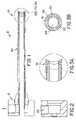
- FIG. 1is a longitudinal cross-sectional view of one embodiment of the suction cannula of the present invention
- FIG. 2is a cross-sectional view of the hub portion of the suction cannula of FIG. 1 ;
- FIG. 3Ais an enlarged cross-sectional view of the distal end of the suction cannula of FIG. 1 ;
- FIG. 3Bis an end-on cross sectional view of the distal end of the suction cannula of FIG. 3A ;
- FIG. 4is a detailed view of the distal end of one embodiment of the dilator of the present invention.
- FIG. 4Ais a detailed view of an alternative embodiment of the tip section of the dilator of FIG. 4 ;
- FIG. 5is a perspective view of the preferred staple of the present invention.
- FIG. 6Ais a view of the distal end of one embodiment of the stapler of the present invention.
- FIG. 6Bis a detailed view of the stapler of FIG. 6A in cooperation with the preferred staple of the present invention.
- FIG. 6Cis another detailed view of the stapler of FIG. 6A in cooperation with the preferred staple of the present invention.
- FIG. 6Dis another detailed view of the stapler of FIG. 6A in cooperation with the preferred staple of the present invention.
- FIG. 6Eis an end-on view of the flange portion of the distal end of FIG. 6A ;
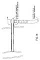
- FIG. 6Fis a side view of another preferred staple of the present invention in cooperation with the crimping member
- FIGS. 7–19show the operation of a preferred sequence of the present invention
- FIG. 20depicts another embodiment of the cannula and dilator of the present invention.
- FIG. 21depicts the outer sheath of the cannula embodiment of FIG. 20 ;
- FIG. 22shows the cannula of the embodiment of FIG. 20 ;
- FIG. 23depicts a detailed view of the tip section of the dilator of FIG. 20 ;
- FIGS. 24A and 24Bdepict a cross sectional view and a side view, respectively, of an alternative tip portion of the stapler of the present invention
- FIG. 25Adepicts a cross-sectional view of another preferred cannula of the present invention.
- FIG. 25Bdepicts a cross-sectional view of the cannula of FIG. 25A , in cooperation with the stapler of FIGS. 24A and 24B .
- FIGS. 1–3Bdepict various views of one embodiment of the suction cannula 10 of the present invention.
- cannula 10comprises a tubular member 30 , a proximal end 12 and a distal end 14 .
- the distal end 14is adapted to permit vacuum affixation of the cannula 10 to a vascular wall, or other tissue as will be described below.
- the tubular member 30is preferably constructed with a tube 20 within a tube 18 .
- the chamber 22 between the tubes 18 and 20is used as a vacuum chamber.
- Passage 24permits a dilator and/or stapler device (each discussed below) and/or other surgical devices to pass therethrough.
- Support members 26are provided to concentrically affix tubes 18 and 20 .
- the proximal end 12as shown in FIG. 2 includes a vacuum port 28 that can be attached to an external vacuum (not shown). Vacuum port 28 communicates with chamber 22 (between inner tube 20 and outer tube 18 ) to provide a vacuum therein.
- a flexible tip sectionis provided on the distal and 14 of the cannula to provide a secure vacuum interface between cannula 10 and a vascular wall.
- the flexible tip sectionis formed of pliable rubber or other equivalent materials.
- FIG. 4depicts one preferred embodiment of a dilator 40 , used in conjunction with the suction cannula 10 , described above.
- the dilatorincludes a tubular structure 50 , a distal end 52 and a proximal end 54 (not shown in FIG. 4 ).
- the tubular structure 50is intended to pass within the inner tube 20 of the suction cannula 10 .
- the diameter of tubular structure 50is preferably manufactured to the tolerance of the inner tube 20 , to permit unobstructed ingress and egress of the dilator 40 within the cannula 10 .
- the distal end 52preferably includes a dilator tip 44 , a passage 46 for a guide wire 48 , and a collapsible section 42 that can be hand-manipulated to expand and contract (described below). Additionally, another tube 45 is provided within tube 50 in fluid communication with opening 47 and hub (described below) to allow blood to flow within tube 45 . Tube 45 can be eccentrically disposed within tube 50 (as shown), or, tube 45 can be concentrically disposed within tube 50 . Referring to FIG. 8 , the proximal end 54 of the dilator includes a movable hub 56 .
- a cam mechanism 62connected between movable hub 56 and collapsible section 42 (via one or more connecting members, not shown) that engages an O-ring 60 to collapse and/or expand section 42 .
- the diameter of section 42is larger than the diameter of tube 20 , thereby locking the dilator 40 against the cannula tube 30 (described herein).
- section 42is collapsed so that the dilator can pass within tube 20 of the cannula 30 .
- FIG. 4Adepicts an alternative embodiment for the tip section 44 ′ of the dilator depicted in FIG. 4 .
- tip section 44 ′has an elongated shape, as compared with the embodiment in FIG. 4 .
- opening 47 ′permits fluid to flow within region 49 , which is disposed within tube 50 ′ around tube 45 ′.
- tube 50 ′ and tip 44 ′are not fixed within the cannula 30 . Rather, tube 50 ′ and tip 44 ′ can be inserted into and withdrawn from the cannula with relatively little obstruction.
- FIG. 5depicts the preferred embodiment of the surgical staple 70 of the present invention.
- Staple 70includes an oval member 76 with a plurality of prongs 72 around the circumference of oval member 76 .
- Oval member 76defines an oval opening or hole 74 , which cooperates with a stapler (described below).
- a staplerdescribed below.
- prongs 72crimp onto the vascular walls (or other tissue) to effectively seal a puncture.
- FIG. 6Ashows a view of the distal end of the stapler 80 of the preferred embodiment. The distal end includes a slidable crimping member 82 and a flange member 84 . As shown in FIG.
- flange member 84is shaped to match the inner diameter of oval member 76 of the staple 70 .
- staple 70is inserted over flange member 84 so that staple 70 abuts shaft member 86 adjacent flange member 84 .
- Staple 70is rotated approximately 90 degrees, as depicted in FIG. 6D , thereby locking the staple between flange 84 and shaft 86 .
- Flange member 84is connected to connecting rod 100 (as shown in FIGS. 11 and 15 ) passing through the stapler device to the proximal end.
- key hub 98which is also connected to connecting rod 100 (and thus, flange member 84 ) can be rotated approximately 90 degrees, thereby releasing the staple 70 from the staple device.
- key hubis hand rotatable.
- key hub, connecting rod and flange membercan also be automatically rotated through the action of the driving mechanism and handle, 96 and 94 .
- FIGS. 6B and 6Cdepict insertion of the staple into the vascular wall (or other tissue) and crimping of the staple, respectively, using the stapler 80 , described above.
- Crimping member 82is first slid toward the vascular wall so that the staple 72 pierces the wall ( FIG. 6B ). It will be understood that the members 72 can include a sharp or pointed edge 88 to aid the insertion of staple 70 into the vascular wall. Crimping member 82 is then further advanced toward the vascular wall to force the staple to crimp, due to the force direction exerted by the conforming portion 90 onto the staple ( FIG. 6C ).
- conforming portion 90includes a generally parabolic shape.
- the flange member 84can be rotated (e.g., rotated 90 degrees, via connecting rod 100 and key hub 98 ) so that hole 74 and flange 84 are aligned, and the stapler can be withdrawn from the vascular wall.
- FIG. 6Fanother embodiment of a staple 76 ′ of the present invention.
- the staple of this embodimentcooperates with the flange member 84 ′, crimping member 82 ′, conforming portion 90 ′ and shaft 86 ′ as in the previous embodiment.
- membrane 130is included in this embodiment.
- Membrane 130is formed on the staple between members 72 ′ such that the opening 74 (not shown) is covered.
- the membrane 130is preferably formed to permit unobstructed ingress and egress of flange 84 ′ within the opening 74 , as shown in the drawing.
- Membrane 130is formed of silicone, elastomer, or bioabsorbable material. Essentially, membrane 130 is provided to seal the puncture hole in the vascular wall that may remain unsealed due to the opening 74 of the staple 76 ′.
- FIGS. 7–19depict detailed functionality of the cannula 10 , dilator 40 , staple 70 and stapler 80 (as described above with reference to FIGS. 1–6E ) of the present invention.
- the suction cannula 10 and the dilator 40are inserted into the incision in the skin (facia), following the previously-inserted guide wire 48 , toward the arterial puncture site.
- the guide wirecan be removed at any stage of the proceeding process, or may be left within the cannula as a reference point. It should be noted that with reference to the stapling procedure described herein, it is preferable that the guide wire be removed.
- the dilatoris removed and vacuum source 92 is applied to the cannula to secure the cannula to the artery wall.
- vacuum source 92is applied to the cannula to secure the cannula to the artery wall.
- the puncture siteis to be closed.
- the stapler 80(with a staple 70 secured on the distal end, as described above) is inserted down the cannula to the puncture site.
- the staple 70is pushed into the vascular wall sufficiently to allow the staple to at least partially pierce the wall, as shown in the close-up view of FIG. 12 .
- the surgeonactivates a lever 94 , which, in turn activates driving mechanism 96 to drive crimping member 82 distally, to thereby crimp the staple and seal the puncture site (as described above).
- driving mechanism 96is contained within handle 108 . More specifically, mechanism 96 preferably includes a spring 102 housed in housing 104 . Spring 102 is connected to lever 94 (via connecting hub 110 ) and crimping member 82 , so that movement of lever 94 provides distal and proximal movement to crimping member 82 . Spring member preferably keeps lever 94 and crimping member 82 in the relative positions shown in FIG. 11 and 6A , respectively. Thus, movement of the lever 94 as indicated by the arrow in FIG. 11 causes crimping member 82 to be forced against the staple for closure (crimping), as described above.
- a key hub 98 on the stapleris rotated to turn the shaft 86 approximately ninety degrees to align opening 74 of staple 70 with flange 84 , as shown in FIG. 15 .
- Thispermits disengagement of the staple 70 from the stapler 80 , so that the stapler can be removed from the cannula, as shown in FIGS. 16 and 17 .
- the stapled puncture sitecan be inspected (down the cannula) to ensure that the puncture site is correctly sealed ( FIG. 19 ).
- the guide wireif not previously removed, can be removed at this point.
- the vacuumis disengaged to permit the cannula to be removed from the incision in the skin, as shown in FIG. 18 . It should be noted that other geometric configurations of the flange member and staple will necessitate an alternative rotation, which may be other than approximately 90 degrees.
- the preferred material used for the construction of the devices shown in all the figurescan include plastic, stainless steel, titanium, and bioabsorbable material (where appropriate).
- an appropriate suturing mechanism, laser suturing mechanism, or other closure systemcan be used to seal the puncture site.
- the suction cannula 10provides unobstructed access to the puncture site during medical procedures, including closure of the wound.
- the driving mechanism 96 of the staplercould be appropriately modified with a push-button activated gear mechanism to slide the crimping member distally.
- the shape of the staple 70 /flange 84can also be modified.
- the member 76can modified and shaped as a rectangle, triangle, square, etc.
- the member 76can include a circular shape which is friction fit over the flange member.
- the flange 84would be appropriately modified to match the opening 74 defined by the member 76 to permit engagement and disengagement of the staple 70 and flange 84 , as described above.
- the staple 70can be further modified with barbs on the prongs 72 , to provide a more secure fastening of the staple to the artery wall.
- the crimping member 82can be modified to include a conforming portion 90 having a variety of shapes, provided that the overall functionality of the crimping member, as described herein, is not hindered.
- the vacuum source applied to the cannula 10can be any conventionally known automated vacuum supply.
- the cannulacan be appropriately modified to include a manually activated vacuum using, for example, a bulb mechanism, when a vacuum supply is otherwise unavailable.
- FIGS. 20–23an alternative embodiment for the cannula and dilator are shown.
- an outer sheath 110preferably formed of plastic, is placed over the cannula 112 with the dilator 114 inserted into the cannula.
- the plastic sheath 110is slidably engaged the over cannula using hub 120 .
- the sheath 110locks the distal tip 116 of the dilator 114 at juncture 118 .
- Retracting the sheath 110is accomplished by pulling proximally on hub 120 , thereby opening the wing members 122 of the sheath 110 .
- a latch 132can be provided that holds the hub 120 in place.
- latch 132can be manually removed from the hub 120 to permit movement of the hub.
- snap-fit interference locks 134 a and 134 bcan be provided as shown to fix the hub (and sheath) in the proximal position, as indicated by the arrow.
- the cannula 112may be of the type described above.
- the cannulacan be modified so that only the distal tip 124 has a tube-in-tube construction. In other words, referring to FIGS.
- the tube-in-tube constructionneed not span the entire length of the device, but may rather only be provided at the tip section 124 , recognizing that the stapler, dilator or other instruments will be inserted therein.
- the distal tip 116 of the dilator 114is preferably constructed as shown in FIG. 23 .
- the distal tipcan include a passage 126 in fluid communication with the dilator, to provide visual indication within the artery by the presence of blood (shown at the dilator hub section).
- the dilator tipcan be elongated (more so than shown in the drawings) thereby reducing the angle of insertion into a vein or artery (as shown in FIG. 4A ).
- the cannula 112can modified to include only a single tube.
- the sheath 118can replace the outer tube 18 of the cannula ( FIG. 1 ) and a vacuum can be created within the space between the sheath and the cannula.
- Anther embodiment of the cannula 140 of the present inventionis depicted in FIG. 25A .
- an outer tube 142is provided, similar to the embodiment of FIGS. 1–3A .
- the inner tubeis provided as a plurality of arcuate segments 144 a – 144 d, connected to the outer tube by connecting members 148 a – 148 d .
- the space between the segments 144 a–d and the outer tube 142shown as 146 a–d ,is preferably used for the vacuum, as described above.
- the connecting members 148 a–d ⁇ caffalso be constructed so as to provide a keyway space 150 , which can be keyed to a variety of instruments, as will be described below.
- FIG. 25Acan be extruded the entire length of the cannula 140 , or provided at the distal tip thereof It should also be noted that the length of the arcuate segments and the positioning of the connecting members is a matter of design choice for a desired cross-sectional profile.
- FIGS. 24A and 24Bdepict the tip section 160 of another preferred stapler of the present invention.
- the tip section 160includes a conforming portion 162 having a plurality of fingers 164 a–d , which are located about the periphery of the section 160 , and provided to urge the staple against the flange member (described above).
- FIG. 25BThe cooperation of the cannula of FIG. 25A and the stapler tip of FIGS. 24A and 24B is depicted in FIG. 25B .
- the space 150permits passage therethrough of the stapler tip 160 .
- the staple 168 and flange 170which operate as described herein.
- the present inventionis not so limited.
- the cannula of the present inventioncan also be used in other tissue environments, as may be required.
Landscapes
- Health & Medical Sciences (AREA)
- Surgery (AREA)
- Life Sciences & Earth Sciences (AREA)
- Medical Informatics (AREA)
- Animal Behavior & Ethology (AREA)
- Engineering & Computer Science (AREA)
- Biomedical Technology (AREA)
- Heart & Thoracic Surgery (AREA)
- Veterinary Medicine (AREA)
- Molecular Biology (AREA)
- Nuclear Medicine, Radiotherapy & Molecular Imaging (AREA)
- General Health & Medical Sciences (AREA)
- Public Health (AREA)
- Pathology (AREA)
- Cardiology (AREA)
- Surgical Instruments (AREA)
- Media Introduction/Drainage Providing Device (AREA)
Abstract
Description
This application is a continuation application under 37 CFR §1.53(b) of application Ser. No. 09/486,185 filed Feb. 18, 2000 now abandoned, which claims priority to U.S. Provisional application No. 60/093,701 filed Jul. 22, 1998, and PCT application No. PCT/US99/16476 filed Jul. 21, 1999, all of which are hereby incorporated by reference in their entirety.
1. Field of the Invention
The present invention relates to a vascular suction device, a dilator and a stapler for the closure of a puncture made in the wall of an artery or vein during a medical procedure. The present invention has particular utility for use in and around the femoral artery during and after coronary/cardiac procedures. Other utilities include soft-tissue anchoring, meniscal repair, thoracic lung closure, endoscopic procedures, esophageal repair, laparoscopy, skin/epidermal wound closure and general tissue closure.
2. Description of Related Art
Surgical stapling instruments, dilators and cannulas for diagnostic, interventional and/or therapeutic medical procedures are known. For example, U.S. Pat. No. 5,709,335 issued to Heck discloses a wholly distal surgical stapling instrument for stapling a tubular tissue structure to a luminal structure, such as a vascular lumen. This device can be used for anastomotic stapling of a tubular vessel having two untethered ends, and is especially useful for making the primary anastomotic connection of a bypass vein to a coronary artery or to the aorta. The device essentially includes a rod that is placed within the tubular vessel and an anvil that forces staples (associated with the rod) to bend outwardly against the vessel and a target (such as a coronary artery). Thus, this device requires that the stapler device be placed within the tubular vessel (e.g., vein or artery) for operation. While this device is useful when stapling a graft vein or the like, unfortunately, this device would be inappropriate when the entirety of the tubular tissue is not accessible, such as following percutaneous catheterization procedures.
Another example can be found in U.S. Pat. No. 5,403,333 issued to Kaster et al. This patent discloses a side-to-end anastomotic staple apparatus for use where the end of a blood vessel becomes connected to the side or wall of a second blood vessel or other structure, such as the heart. Similar to the previous discussion, this device requires that at least one end of the vessel be open, so that a stapling mechanism can be inserted therethrough. As noted above, many surgical procedures only access a portion of the vessel. Thus, this device would not be useful in these circumstances.
Yet another example, U.S. Pat. No. 5,695,504 issued to Gifford, III et al., discloses an end-to-side vascular anastomosis device to perform end-to-side anastomosis between a graft vessel and the wall of a target vessel. This device involves a procedure in which the end of a graft vessel is passed through an inner sleeve of the device until the end of the vessel extends from the distal end of the device. The distal end of the graft is then affixed to the wall of the target, using a staple and stapler which forces a staple into both tissues. Similar to the previous disclosures, this device is useful for the attachment of one tubular tissue onto another, however, is inadequate in sealing a puncture in an artery, vein or other tissue left by certain medical procedures.
Other examples can be found in the art. However, these devices are often complicated to manufacture and use, requiring expensive tooling and materials. It is often the case that staplers, cannulas and dilators are single application or procedure devices, which must be discarded after use. Thus, there is a need to provide an efficient stapler mechanism that is simple to use and relatively easy to manufacture, since the device is likely to be discarded after only one use. Moreover, the prior art has failed to provide a device that permits a doctor or clinician to gain access to a puncture site and remain centered on that site throughout the entire procedure, including closure of the puncture, or to ensure that the closure mechanism is delivered over and/or around the puncture site.
Thus, the present invention solves the aforementioned drawbacks by providing a suction cannula, dilator, stapler and staple that are simple to use and manufacture. In one aspect, the present invention provides a suction cannula that is concentrically aligned with a puncture site (e.g., puncture in an artery or vein) and provides vacuum about the periphery of the puncture site so that the puncture hole is always located during a medical procedure, and to thereby permit a surgeon to quickly and efficiently close the puncture using, for example, a stapling device. In the preferred embodiment the suction cannula has a tube-in-tube construction having an inner tube and an outer tube where a vacuum can be applied between the tubes.
In another aspect, the present invention provides a dilator, which can be placed within the inner tube of the suction cannula during insertion into the body. The dilator (and suction cannula) centers around a guide wire (that is already in place within the venous structure) and follows the path of the guide wire to the puncture site. Preferably, the dilator has a tapered tip on the distal end that follows the guide wire though the puncture hole made in the vein or artery. A blood indicator is provided on the proximal end to provide visual feedback when the surgeon is in the artery (i.e., pulsating blood indicates that the tip of the dilator is in the artery). In one preferred embodiment, the dilator includes a tapered tip on the distal end that is radially collapsible so that the dilator can be withdrawn from the artery and the suction cannula is thereby permitted to advance over the dilator to the artery wall. To that end, indicators on the external, proximal end of the dilator provide the user with a visual measurement as to the distance to the artery wall. Once the suction cannula makes contact with the vascular wall, and vacuum can be applied to the cannula so that the cannula remains concentrically aligned with the puncture in the vessel, and the dilator can be removed.
In yet another aspect of the present invention, a stapler is provided which holds a multi-pronged staple on a shaft at the distal end. The distal portion of the stapler is constructed to fit within the suction cannula (i.e., the inner tube of the cannula) to approach the puncture in the wall of the artery (or other soft tissue), to permit the stapling of the artery. Preferably, the distal end of the stapler includes a T-flange that retains a staple, and a deploying mechanism that deploys the staple into the artery, thereby sealing the puncture. Deployment of the staple can include crimping of the staple through the vascular wall and/or partial insertion of the staple into the tissue. The T-flange permits the staple to be retained on the distal end of the stapler and deployed into the artery wall. An oval hub on the T-flange is provided that mates with an oval hole in the center of the staple. To hold a staple, a staple is placed on the hub and rotated 90 degrees, thereby affixing the staple to the stapler. Once the staple is crimped onto the artery wall, the shaft can be rotated 90 degrees, thereby aligning the oval hub and the oval hole, so that the stapler can be removed. Preferably, the staple includes a plurality of prongs that are inserted into the vascular wall.
Advantageously, the suction cannula of the present invention permits the surgeon to remain centrally located about a puncture site throughout the entire procedure, from incision to closing. The suction cannula permits a surgeon to enter an incision, and using a dilator as an artery indicator, secure the cannula to the artery wall, via vacuum force, about the puncture site. Also advantageously, this permits the surgeon to view and approach the puncture site (using a catheter, for example) throughout the entire procedure, without obstruction. In addition, a stapler and staple are provided which can be guided down the shaft of the cannula to quickly seal the puncture site.
It will be appreciated by those skilled in the art that although the following Detailed Description will proceed with reference being made to preferred embodiments, the present invention is not intended to be limited to these preferred embodiments. Other features and advantages of the present invention will become apparent as the following Detailed Description proceeds, and upon reference to the Drawings, wherein like numerals depict like parts, and wherein:
Referring toFIG. 6F , another embodiment of a staple76′ of the present invention. The staple of this embodiment cooperates with theflange member 84′, crimpingmember 82′, conformingportion 90′ andshaft 86′ as in the previous embodiment. Included in this embodiment is membrane130. Membrane130 is formed on the staple betweenmembers 72′ such that the opening74 (not shown) is covered. The membrane130 is preferably formed to permit unobstructed ingress and egress offlange 84′ within theopening 74, as shown in the drawing. Membrane130 is formed of silicone, elastomer, or bioabsorbable material. Essentially, membrane130 is provided to seal the puncture hole in the vascular wall that may remain unsealed due to theopening 74 of the staple76′.
Once the diagnostic, interventional, therapeutic, or other procedure (following the cannula to the puncture site) is complete, the puncture site is to be closed. As shown inFIG. 11 , the stapler80 (with a staple70 secured on the distal end, as described above) is inserted down the cannula to the puncture site. The staple70 is pushed into the vascular wall sufficiently to allow the staple to at least partially pierce the wall, as shown in the close-up view ofFIG. 12 . As shown inFIGS. 13 and 14 , the surgeon activates alever 94, which, in turn activates drivingmechanism 96 to drive crimpingmember 82 distally, to thereby crimp the staple and seal the puncture site (as described above). As shown in the figures, drivingmechanism 96 is contained withinhandle 108. More specifically,mechanism 96 preferably includes aspring 102 housed inhousing 104.Spring 102 is connected to lever94 (via connecting hub110) and crimpingmember 82, so that movement oflever 94 provides distal and proximal movement to crimpingmember 82. Spring member preferably keepslever 94 and crimpingmember 82 in the relative positions shown inFIG. 11 and 6A , respectively. Thus, movement of thelever 94 as indicated by the arrow inFIG. 11 causes crimping member 82 to be forced against the staple for closure (crimping), as described above. Once crimped, akey hub 98 on the stapler is rotated to turn theshaft 86 approximately ninety degrees to align opening74 ofstaple 70 withflange 84, as shown inFIG. 15 . This permits disengagement of the staple70 from thestapler 80, so that the stapler can be removed from the cannula, as shown inFIGS. 16 and 17 . After the stapler is removed the stapled puncture site can be inspected (down the cannula) to ensure that the puncture site is correctly sealed (FIG. 19 ). In addition, the guide wire, if not previously removed, can be removed at this point. The vacuum is disengaged to permit the cannula to be removed from the incision in the skin, as shown inFIG. 18 . It should be noted that other geometric configurations of the flange member and staple will necessitate an alternative rotation, which may be other than approximately 90 degrees.
The preferred material used for the construction of the devices shown in all the figures can include plastic, stainless steel, titanium, and bioabsorbable material (where appropriate).
Modifications to the present invention are also possible. For example, instead of astapling device 80, as described above, an appropriate suturing mechanism, laser suturing mechanism, or other closure system can be used to seal the puncture site. In any event, thesuction cannula 10 provides unobstructed access to the puncture site during medical procedures, including closure of the wound. Thedriving mechanism 96 of the stapler could be appropriately modified with a push-button activated gear mechanism to slide the crimping member distally. Those skilled in the art will recognize that many modifications are possible to drive the crimping member, and all such modifications are deemed within the scope of the present invention.
The shape of the staple70/flange 84 can also be modified. For example, themember 76 can modified and shaped as a rectangle, triangle, square, etc. Alternatively, themember 76 can include a circular shape which is friction fit over the flange member. Accordingly, theflange 84 would be appropriately modified to match theopening 74 defined by themember 76 to permit engagement and disengagement of the staple70 andflange 84, as described above. The staple70 can be further modified with barbs on theprongs 72, to provide a more secure fastening of the staple to the artery wall. The crimpingmember 82 can be modified to include a conformingportion 90 having a variety of shapes, provided that the overall functionality of the crimping member, as described herein, is not hindered.
The vacuum source applied to thecannula 10 can be any conventionally known automated vacuum supply. Of course, the cannula can be appropriately modified to include a manually activated vacuum using, for example, a bulb mechanism, when a vacuum supply is otherwise unavailable.
Additional modifications are also possible. Referring toFIGS. 20–23 , an alternative embodiment for the cannula and dilator are shown. In this embodiment, anouter sheath 110, preferably formed of plastic, is placed over thecannula 112 with thedilator 114 inserted into the cannula. Theplastic sheath 110 is slidably engaged the overcannula using hub 120. As shown inFIGS. 21 and 23 , thesheath 110 locks thedistal tip 116 of thedilator 114 atjuncture 118. Retracting thesheath 110 is accomplished by pulling proximally onhub 120, thereby opening thewing members 122 of thesheath 110. To that end, alatch 132 can be provided that holds thehub 120 in place. Preferably, latch132 can be manually removed from thehub 120 to permit movement of the hub. Additionally, snap-fit interference locks134aand134bcan be provided as shown to fix the hub (and sheath) in the proximal position, as indicated by the arrow. Thecannula 112 may be of the type described above. Alternatively, instead of the tube-in-tube suction cannula set forth herein, the cannula can be modified so that only thedistal tip 124 has a tube-in-tube construction. In other words, referring toFIGS. 1–3A , the tube-in-tube construction need not span the entire length of the device, but may rather only be provided at thetip section 124, recognizing that the stapler, dilator or other instruments will be inserted therein. Thedistal tip 116 of thedilator 114 is preferably constructed as shown inFIG. 23 . Preferably, the distal tip can include apassage 126 in fluid communication with the dilator, to provide visual indication within the artery by the presence of blood (shown at the dilator hub section). It should be noted that the dilator tip can be elongated (more so than shown in the drawings) thereby reducing the angle of insertion into a vein or artery (as shown inFIG. 4A ). Also alternatively, instead of a cannula having a tube-in-tube construction as described herein, thecannula 112 can modified to include only a single tube. In this case, thesheath 118 can replace theouter tube 18 of the cannula (FIG. 1 ) and a vacuum can be created within the space between the sheath and the cannula.
Anther embodiment of thecannula 140 of the present invention is depicted inFIG. 25A . In this embodiment, anouter tube 142 is provided, similar to the embodiment ofFIGS. 1–3A . The inner tube, however, is provided as a plurality ofarcuate segments 144a–144d,connected to the outer tube by connectingmembers 148a–148d. The space between thesegments 144a–dand theouter tube 142, shown as146a–d,is preferably used for the vacuum, as described above. The connectingmembers 148a–d˜caff also be constructed so as to provide akeyway space 150, which can be keyed to a variety of instruments, as will be described below. It should be noted that the construction shown inFIG. 25A can be extruded the entire length of thecannula 140, or provided at the distal tip thereof It should also be noted that the length of the arcuate segments and the positioning of the connecting members is a matter of design choice for a desired cross-sectional profile.
Although the detailed description provided herein has largely been in reference to arterial procedures, the present invention is not so limited. The cannula of the present invention can also be used in other tissue environments, as may be required.
Claims (10)
1. A tissue stapler for deploying a staple into tissue, comprising a tubular member having a tip section, a trigger, and a connecting rod located within said tubular member and extending between said tip section and said trigger, a crimping member located on said tubular member and disposed within said tip section, said connecting rod having a flange member adapted to hold said staple between said flange member and said crimping member, said connecting rod and said tubular member being slidably operable by said trigger to slide said flange member toward said crimping member thereby deploying said staple.
2. A stapler as claimed inclaim 1 , wherein said connecting rod is rotatable within said tubular member, and said flange member is mated with an opening in said staple in one dimension, wherein said staple has an opening sized and shaped to be placed over said flange member and wherein, upon rotation of said connecting rod, said staple can be held against said crimping member by said flange member.
3. A stapler as claimed inclaim 2 , wherein said flange member has a generally oval shape and said opening in said staple has a mated oval shape.
4. A stapler as claimed inclaim 1 , wherein said trigger comprises a lever for moving said connecting rod within said tubular member.
5. A stapler as claimed inclaim 1 , wherein said staple comprises a plurality of tissue engaging prongs that are crimped together at least partially through said tissue by said crimping member.
6. A stapler as claimed inclaim 1 , wherein said crimping member has a generally parabolic shape.
7. A stapler as claimed inclaim 1 , wherein said crimping member has a plurality of slidable fingers, said crimping member being operable by said trigger to slide axially over said connecting rod and flange member to crimp said staple.
8. A staple and a stapler for stapling tissue, said stapler comprising a tubular member having a tip section, a trigger, and a connecting rod located within said tubular member and extending between said tip section and said trigger a crimping member located on said tubular member and disposed within said tip section, said connecting rod having a flange member adapted to hold said staple between said flange member and said crimping member, said connecting rod and said tubular member being slidably operable by said trigger to slide said flange member toward said crimping member thereby deploying said staple; and said staple comprising a ring member defining an opening therein, said opening being mated to fit over said flange member in one dimension and a plurality of tissue engaging members located on said ring member to pierce into tissue upon crimping by said stapler.
9. A staple and a stapler as claimed inclaim 8 , wherein said flange member and said opening have mated shapes.
10. A staple and a stapler as claimed inclaim 8 , wherein said connecting rod is rotatable within said tubular member, and said flange member is mated with an opening in said staple in one dimension, wherein said staple is adapted to be placed over said flange member and wherein, upon rotation of said connecting rod, said staple can be held against said crimping member by said flange member.
Priority Applications (1)
| Application Number | Priority Date | Filing Date | Title |
|---|---|---|---|
| US10/689,358US6989016B2 (en) | 1998-07-22 | 2003-10-20 | Vascular suction cannula, dilator and surgical stapler |
Applications Claiming Priority (4)
| Application Number | Priority Date | Filing Date | Title |
|---|---|---|---|
| US9370198P | 1998-07-22 | 1998-07-22 | |
| PCT/US1999/016476WO2000007640A2 (en) | 1998-07-22 | 1999-07-21 | Vascular suction cannula, dilator and surgical stapler |
| US48618500A | 2000-02-18 | 2000-02-18 | |
| US10/689,358US6989016B2 (en) | 1998-07-22 | 2003-10-20 | Vascular suction cannula, dilator and surgical stapler |
Related Parent Applications (2)
| Application Number | Title | Priority Date | Filing Date |
|---|---|---|---|
| US09486185Continuation | 1999-07-21 | ||
| PCT/US1999/016476ContinuationWO2000007640A2 (en) | 1998-07-22 | 1999-07-21 | Vascular suction cannula, dilator and surgical stapler |
Publications (2)
| Publication Number | Publication Date |
|---|---|
| US20040082906A1 US20040082906A1 (en) | 2004-04-29 |
| US6989016B2true US6989016B2 (en) | 2006-01-24 |
Family
ID=22240293
Family Applications (1)
| Application Number | Title | Priority Date | Filing Date |
|---|---|---|---|
| US10/689,358Expired - Fee RelatedUS6989016B2 (en) | 1998-07-22 | 2003-10-20 | Vascular suction cannula, dilator and surgical stapler |
Country Status (6)
| Country | Link |
|---|---|
| US (1) | US6989016B2 (en) |
| EP (1) | EP1105049A4 (en) |
| AU (1) | AU758665B2 (en) |
| CA (1) | CA2339060A1 (en) |
| IL (1) | IL141014A0 (en) |
| WO (1) | WO2000007640A2 (en) |
Cited By (97)
| Publication number | Priority date | Publication date | Assignee | Title |
|---|---|---|---|---|
| US20020072768A1 (en)* | 2000-12-07 | 2002-06-13 | Ginn Richard S. | Apparatus and methods for providing tactile feedback while delivering a closure device |
| US20030078598A1 (en)* | 2000-01-05 | 2003-04-24 | Integrated Vascular Systems, Inc. | Vascular sheath with bioabsorbable puncture site closure apparatus and methods of use |
| US20030195561A1 (en)* | 2000-12-07 | 2003-10-16 | Carley Michael T. | Closure device and methods for making and using them |
| US20040015170A1 (en)* | 2000-05-01 | 2004-01-22 | Tallarida Steven J. | System and method for joint resurface repair |
| US20040073255A1 (en)* | 2002-02-21 | 2004-04-15 | Ginn Richard S | Plunger apparatus and methods for delivering a closure device |
| US20040153123A1 (en)* | 2003-01-30 | 2004-08-05 | Integrated Vascular Systems, Inc. | Clip applier and methods of use |
| US20050107820A1 (en)* | 2003-11-13 | 2005-05-19 | Forsberg Andrew T. | Vascular puncture depth locator |
| US20050234508A1 (en)* | 2002-06-04 | 2005-10-20 | Christy Cummins | Blood vessel closure clip and delivery device |
| US20050267530A1 (en)* | 2001-06-07 | 2005-12-01 | Christy Cummins | Surgical staple |
| US20050274768A1 (en)* | 2004-05-25 | 2005-12-15 | Christy Cummins | Surgical stapler |
| US20050283188A1 (en)* | 1998-05-29 | 2005-12-22 | By-Pass, Inc. | Vascular closure device |
| US20060020270A1 (en)* | 2003-01-30 | 2006-01-26 | Ronald Jabba | Clip applier and methods of use |
| US20060122575A1 (en)* | 2002-06-11 | 2006-06-08 | Akio Wakabayashi | System and efficient drainage of body cavity |
| US20060135989A1 (en)* | 2000-12-07 | 2006-06-22 | Carley Michael T | Closure device |
| US20060144479A1 (en)* | 2002-12-31 | 2006-07-06 | Integrated Vascular Systems, Inc. | Methods for manufacturing a clip and clip |
| US20060195112A1 (en)* | 2002-12-03 | 2006-08-31 | Ek Steven W | System and method for retrograde procedure |
| US20060229726A1 (en)* | 2000-05-01 | 2006-10-12 | Ek Steven W | Articular surface implant |
| US20060265012A1 (en)* | 2002-11-26 | 2006-11-23 | Abbott Laboratories | Multi-Element Biased Suture Clip |
| US20060287674A1 (en)* | 2000-01-05 | 2006-12-21 | Ginn Richard S | Closure system and methods of use |
| US20070010853A1 (en)* | 2000-10-06 | 2007-01-11 | Integrated Vascular Systems, Inc. | Apparatus and methods for positioning a vascular sheath |
| US20070021778A1 (en)* | 2005-06-24 | 2007-01-25 | Abbott Laboratories Abbott Vascular Devices | Apparatus and method for delivering a closure element |
| US20070049968A1 (en)* | 2005-08-24 | 2007-03-01 | Sibbitt Wilmer L Jr | Vascular opening edge eversion methods and apparatuses |
| US20070123921A1 (en)* | 2003-11-20 | 2007-05-31 | Arthrosurface, Inc. | Retrograde excision system and apparatus |
| US20070179608A1 (en)* | 2005-07-29 | 2007-08-02 | Arthrosurface, Inc. | System and method for articular surface repair |
| US20070203506A1 (en)* | 2005-08-24 | 2007-08-30 | Sibbitt Wilmer L Jr | Vascular closure methods and apparatuses |
| US20070250080A1 (en)* | 2006-04-20 | 2007-10-25 | Integrated Vascular Systems, Inc. | Resettable clip applier and reset tools |
| US20070282352A1 (en)* | 2000-12-07 | 2007-12-06 | Carley Michael T | Closure device and methods for making and using them |
| US20080004636A1 (en)* | 2005-07-01 | 2008-01-03 | Abbott Laboratories | Clip applier and methods of use |
| US20080065152A1 (en)* | 2006-09-08 | 2008-03-13 | Abbott Laboratories | Apparatus and method for delivering a closure element |
| US20080147114A1 (en)* | 1998-05-29 | 2008-06-19 | Bypass, Inc. | Vascular port device |
| WO2008033558A3 (en)* | 2006-09-15 | 2008-07-31 | Vascular Prec | Tissue closure, delivery device and method of use |
| US20080195113A1 (en)* | 2007-02-14 | 2008-08-14 | Arthrosurface Incorporated | Bone Cement Delivery Device |
| US20080210737A1 (en)* | 2000-01-05 | 2008-09-04 | Integrated Vascular Systems, Inc. | Integrated vascular device with puncture site closure component and sealant and methods of use |
| US20080269802A1 (en)* | 2000-09-08 | 2008-10-30 | Abbott Vascular Inc. | Surgical stapler |
| US20080312686A1 (en)* | 2005-07-01 | 2008-12-18 | Abbott Laboratories | Antimicrobial closure element and closure element applier |
| US20080319475A1 (en)* | 2007-06-25 | 2008-12-25 | Abbott Laboratories | Methods, Devices, and Apparatus for Managing Access Through Tissue |
| US20090082802A1 (en)* | 2007-09-26 | 2009-03-26 | Medtronic Vascular, Inc. | Mechanism and Method for Closing an Arteriotomy |
| US20090149771A1 (en)* | 2007-12-05 | 2009-06-11 | Laerdal Medical As | Vessel locator |
| US20090157102A1 (en)* | 2007-12-17 | 2009-06-18 | Abbott Laboratories | Clip applier and methods of use |
| US20090157101A1 (en)* | 2007-12-17 | 2009-06-18 | Abbott Laboratories | Tissue closure system and methods of use |
| US20090157103A1 (en)* | 2007-12-18 | 2009-06-18 | Abbott Laboratories | Modular clip applier |
| US20090187215A1 (en)* | 2007-12-19 | 2009-07-23 | Abbott Laboratories | Methods and apparatus to reduce a dimension of an implantable device in a smaller state |
| US20090228040A1 (en)* | 2008-03-04 | 2009-09-10 | Medtronic Vascular, Inc. | Mechanism and Method for Closing an Arteriotomy |
| US7604641B2 (en) | 2000-05-01 | 2009-10-20 | Arthrosurface Incorporated | System and method for joint resurface repair |
| US7618462B2 (en) | 2000-05-01 | 2009-11-17 | Arthrosurface Incorporated | System and method for joint resurface repair |
| US20090287244A1 (en)* | 2008-05-16 | 2009-11-19 | Abbott Laboratories Vascular Enterprises Limited | Apparatus and methods for engaging tissue |
| US7678151B2 (en) | 2000-05-01 | 2010-03-16 | Ek Steven W | System and method for joint resurface repair |
| US20100114159A1 (en)* | 2008-10-30 | 2010-05-06 | Abbott Vascular Inc. | Closure device |
| US20100160958A1 (en)* | 2008-12-22 | 2010-06-24 | Abbott Laboratories | Closure Device |
| US20100168790A1 (en)* | 2008-12-22 | 2010-07-01 | Abbott Laboratories | Curved closure device |
| US20100179590A1 (en)* | 2009-01-09 | 2010-07-15 | Abbott Vascular Inc. | Vessel closure devices and methods |
| US20100179572A1 (en)* | 2009-01-09 | 2010-07-15 | Abbott Vascular Inc. | Closure devices, systems, and methods |
| US20100179589A1 (en)* | 2009-01-09 | 2010-07-15 | Abbott Vascular Inc. | Rapidly eroding anchor |
| US20100185234A1 (en)* | 2009-01-16 | 2010-07-22 | Abbott Vascular Inc. | Closure devices, systems, and methods |
| US20100217132A1 (en)* | 2009-02-26 | 2010-08-26 | Abbott Laboratories | Methods and apparatus for locating a surface of a body lumen |
| US7828817B2 (en) | 2000-01-05 | 2010-11-09 | Integrated Vascular Systems, Inc. | Apparatus and methods for delivering a closure device |
| US7828853B2 (en) | 2004-11-22 | 2010-11-09 | Arthrosurface, Inc. | Articular surface implant and delivery system |
| US7896883B2 (en) | 2000-05-01 | 2011-03-01 | Arthrosurface, Inc. | Bone resurfacing system and method |
| US7896885B2 (en) | 2002-12-03 | 2011-03-01 | Arthrosurface Inc. | Retrograde delivery of resurfacing devices |
| US20110054492A1 (en)* | 2009-08-26 | 2011-03-03 | Abbott Laboratories | Medical device for repairing a fistula |
| US7901408B2 (en) | 2002-12-03 | 2011-03-08 | Arthrosurface, Inc. | System and method for retrograde procedure |
| US20110106148A1 (en)* | 2000-01-05 | 2011-05-05 | Integrated Vascular Systems, Inc. | Closure system and methods of use |
| US20110224719A1 (en)* | 2010-03-15 | 2011-09-15 | Abbott Cardiovascular Systems, Inc. | Bioabsorbable plug |
| US20110230897A1 (en)* | 2003-01-30 | 2011-09-22 | Integrated Vascular Systems, Inc. | Clip applier and methods of use |
| US8048108B2 (en) | 2005-08-24 | 2011-11-01 | Abbott Vascular Inc. | Vascular closure methods and apparatuses |
| US8177841B2 (en) | 2000-05-01 | 2012-05-15 | Arthrosurface Inc. | System and method for joint resurface repair |
| US8202293B2 (en) | 2003-01-30 | 2012-06-19 | Integrated Vascular Systems, Inc. | Clip applier and methods of use |
| US8361159B2 (en) | 2002-12-03 | 2013-01-29 | Arthrosurface, Inc. | System for articular surface replacement |
| US8388624B2 (en) | 2003-02-24 | 2013-03-05 | Arthrosurface Incorporated | Trochlear resurfacing system and method |
| US8523872B2 (en) | 2002-12-03 | 2013-09-03 | Arthrosurface Incorporated | Tibial resurfacing system |
| US8556930B2 (en) | 2006-06-28 | 2013-10-15 | Abbott Laboratories | Vessel closure device |
| US8603116B2 (en) | 2010-08-04 | 2013-12-10 | Abbott Cardiovascular Systems, Inc. | Closure device with long tines |
| US8758399B2 (en) | 2010-08-02 | 2014-06-24 | Abbott Cardiovascular Systems, Inc. | Expandable bioabsorbable plug apparatus and method |
| US8821534B2 (en) | 2010-12-06 | 2014-09-02 | Integrated Vascular Systems, Inc. | Clip applier having improved hemostasis and methods of use |
| US9066716B2 (en) | 2011-03-30 | 2015-06-30 | Arthrosurface Incorporated | Suture coil and suture sheath for tissue repair |
| US9149276B2 (en) | 2011-03-21 | 2015-10-06 | Abbott Cardiovascular Systems, Inc. | Clip and deployment apparatus for tissue closure |
| US9149271B2 (en) | 2010-11-22 | 2015-10-06 | The Board Of Trustees Of The Leland Stanford, Jr. University | Device and method for treatment of hemorrhoids |
| WO2015154170A1 (en)* | 2014-04-10 | 2015-10-15 | University Health Network | Cannula for connecting medical devices to biological systems |
| US9173644B2 (en) | 2009-01-09 | 2015-11-03 | Abbott Vascular Inc. | Closure devices, systems, and methods |
| US9283076B2 (en) | 2009-04-17 | 2016-03-15 | Arthrosurface Incorporated | Glenoid resurfacing system and method |
| US9332976B2 (en) | 2011-11-30 | 2016-05-10 | Abbott Cardiovascular Systems, Inc. | Tissue closure device |
| US9358029B2 (en) | 2006-12-11 | 2016-06-07 | Arthrosurface Incorporated | Retrograde resection apparatus and method |
| US9364209B2 (en) | 2012-12-21 | 2016-06-14 | Abbott Cardiovascular Systems, Inc. | Articulating suturing device |
| US9468448B2 (en) | 2012-07-03 | 2016-10-18 | Arthrosurface Incorporated | System and method for joint resurfacing and repair |
| US9486191B2 (en) | 2009-01-09 | 2016-11-08 | Abbott Vascular, Inc. | Closure devices |
| US9492200B2 (en) | 2013-04-16 | 2016-11-15 | Arthrosurface Incorporated | Suture system and method |
| US9662126B2 (en) | 2009-04-17 | 2017-05-30 | Arthrosurface Incorporated | Glenoid resurfacing system and method |
| US9861492B2 (en) | 2014-03-07 | 2018-01-09 | Arthrosurface Incorporated | Anchor for an implant assembly |
| US10624748B2 (en) | 2014-03-07 | 2020-04-21 | Arthrosurface Incorporated | System and method for repairing articular surfaces |
| US10624752B2 (en) | 2006-07-17 | 2020-04-21 | Arthrosurface Incorporated | Tibial resurfacing system and method |
| US10945743B2 (en) | 2009-04-17 | 2021-03-16 | Arthrosurface Incorporated | Glenoid repair system and methods of use thereof |
| US11134946B2 (en) | 2018-02-27 | 2021-10-05 | Bolder Surgical, Llc | Staple cartridge and methods for surgical staplers |
| US11160663B2 (en) | 2017-08-04 | 2021-11-02 | Arthrosurface Incorporated | Multicomponent articular surface implant |
| US11478358B2 (en) | 2019-03-12 | 2022-10-25 | Arthrosurface Incorporated | Humeral and glenoid articular surface implant systems and methods |
| US11607319B2 (en) | 2014-03-07 | 2023-03-21 | Arthrosurface Incorporated | System and method for repairing articular surfaces |
| US11712276B2 (en) | 2011-12-22 | 2023-08-01 | Arthrosurface Incorporated | System and method for bone fixation |
| US20240398408A1 (en)* | 2023-06-05 | 2024-12-05 | Cannuflow, Inc. | Staple and driver for tendon stapling apparatus |
Families Citing this family (50)
| Publication number | Priority date | Publication date | Assignee | Title |
|---|---|---|---|---|
| US6287322B1 (en) | 1995-12-07 | 2001-09-11 | Loma Linda University Medical Center | Tissue opening locator and everter and method |
| ATE341345T1 (en)* | 1999-06-11 | 2006-10-15 | Cytyc Corp | LIQUID GEL FORMULATION FOR DETECTING MILK DUCTS IN THE BREAST PRIOR TO SURGICAL ABLATION OF THE BREAST TISSUE |
| US6780197B2 (en)* | 2000-01-05 | 2004-08-24 | Integrated Vascular Systems, Inc. | Apparatus and methods for delivering a vascular closure device to a body lumen |
| US6197042B1 (en) | 2000-01-05 | 2001-03-06 | Medical Technology Group, Inc. | Vascular sheath with puncture site closure apparatus and methods of use |
| EP1259168B1 (en) | 2000-02-24 | 2010-09-08 | Loma Linda University Medical Center | Patch and glue delivery system for closing tissue openings during surgery |
| US6890342B2 (en) | 2000-08-02 | 2005-05-10 | Loma Linda University | Method and apparatus for closing vascular puncture using hemostatic material |
| US6322580B1 (en)* | 2000-09-01 | 2001-11-27 | Angiolink Corporation | Wound site management and wound closure device |
| US6558400B2 (en) | 2001-05-30 | 2003-05-06 | Satiety, Inc. | Obesity treatment tools and methods |
| US7083629B2 (en)* | 2001-05-30 | 2006-08-01 | Satiety, Inc. | Overtube apparatus for insertion into a body |
| ATE342000T1 (en) | 2002-06-14 | 2006-11-15 | Univ Loma Linda Med | DEVICE FOR CLOSING VASCULAR WOUNDS |
| US7070582B2 (en)* | 2002-08-09 | 2006-07-04 | Boston Scientific Scimed, Inc. | Injection devices that provide reduced outflow of therapeutic agents and methods of delivering therapeutic agents |
| DE10261575A1 (en)* | 2002-12-23 | 2004-07-08 | Nova Lung Gmbh | Device for cannulating a blood-carrying vessel and its use for cannulating blood-carrying vessels |
| DE602004031953D1 (en) | 2003-08-14 | 2011-05-05 | Univ Loma Linda Med | |
| US8187627B2 (en) | 2003-09-05 | 2012-05-29 | Loma Linda University Medical Center | Dressing delivery system for internal wounds |
| US20050137499A1 (en)* | 2003-12-23 | 2005-06-23 | Sheets Ellen E. | Ductal lavage catheter |
| CA2556228C (en) | 2004-02-13 | 2014-05-13 | Satiety, Inc. | Methods for reducing hollow organ volume |
| MXPA06009971A (en) | 2004-02-27 | 2007-08-08 | Satiety Inc | Methods and devices for reducing hollow organ volume. |
| CA2465301C (en) | 2004-04-28 | 2012-02-07 | John Scott Parent | Process to produce silica-filled elastomeric compounds |
| EP1611850A1 (en)* | 2004-06-28 | 2006-01-04 | Cardio Life Research S.A. | Occlusion and tight punction device for an anatomical structure |
| US20060106288A1 (en) | 2004-11-17 | 2006-05-18 | Roth Alex T | Remote tissue retraction device |
| US7442187B2 (en)* | 2005-01-27 | 2008-10-28 | Boston Scientific Scimed, Inc. | Multiple needle injection catheter |
| US7458978B1 (en) | 2005-03-28 | 2008-12-02 | Cardica, Inc. | Vascular closure system utilizing a staple |
| US7344544B2 (en) | 2005-03-28 | 2008-03-18 | Cardica, Inc. | Vascular closure system |
| JP5253170B2 (en) | 2005-10-05 | 2013-07-31 | ローマ リンダ ユニヴァーシティ メディカル センター | Vascular wound closure device and method |
| US20070239206A1 (en)* | 2006-03-31 | 2007-10-11 | Shelton Frederick E Iv | Suture with adhesive/sealant delivery mechanism |
| USD611144S1 (en) | 2006-06-28 | 2010-03-02 | Abbott Laboratories | Apparatus for delivering a closure element |
| DE102006042639A1 (en)* | 2006-09-01 | 2008-03-20 | Novalung Gmbh | Cannula introducing device for use in extracorporeal circulation system, has expandable structure that is transferred from non-expandable condition into expandable condition by dilator, where structure has opening in expandable condition |
| US7875053B2 (en) | 2006-09-15 | 2011-01-25 | Cardica, Inc. | Apparatus and method for closure of patent foramen ovale |
| US7533790B1 (en) | 2007-03-08 | 2009-05-19 | Cardica, Inc. | Surgical stapler |
| US7473258B2 (en) | 2007-03-08 | 2009-01-06 | Cardica, Inc. | Surgical stapler |
| US9387308B2 (en)* | 2007-04-23 | 2016-07-12 | Cardioguidance Biomedical, Llc | Guidewire with adjustable stiffness |
| US20080269800A1 (en)* | 2007-04-24 | 2008-10-30 | Medtronic Vascular, Inc. | Arteriotomy closure system with dual lumens sheath |
| US20090024089A1 (en)* | 2007-04-25 | 2009-01-22 | Levine Jonathan A | Long tapered dilator |
| US9610070B2 (en) | 2007-06-15 | 2017-04-04 | Vivasure Medical Limited | Closure device |
| WO2009151970A2 (en)* | 2008-05-28 | 2009-12-17 | Vibrynt, Inc. | Devices, system and methods for minimally invasive abdominal surgical procedures |
| US9358328B2 (en) | 2009-12-15 | 2016-06-07 | Prabhat K. Ahluwalia | Suction device |
| WO2011080588A2 (en) | 2009-12-30 | 2011-07-07 | Vivasure Medical Limited | Closure system and uses thereof |
| US8617184B2 (en) | 2011-02-15 | 2013-12-31 | Abbott Cardiovascular Systems, Inc. | Vessel closure system |
| US20120296275A1 (en) | 2011-05-16 | 2012-11-22 | Vivasure Medical Limited | Sheath-dilator system and uses thereof |
| US9572558B2 (en) | 2012-02-29 | 2017-02-21 | Vivasure Medical Limited | Devices and methods for delivering implants for percutaneous perforation closure |
| US9744276B2 (en) | 2012-03-20 | 2017-08-29 | Prabhat Kumar Ahluwalia | Suction device |
| US8945093B2 (en) | 2012-03-20 | 2015-02-03 | Minimally Invasive Surgical Technologies, Inc. | Suction device |
| US9850013B2 (en) | 2013-03-15 | 2017-12-26 | Vivasure Medical Limited | Loading devices and methods for percutaneous perforation closure systems |
| US10433826B2 (en) | 2014-12-15 | 2019-10-08 | Vivasure Medical Limited | Closure apparatus with flexible sealable member and flexible support member |
| US11141142B2 (en) | 2014-12-15 | 2021-10-12 | Vivasure Medical Limited | Implantable sealable member with mesh layer |
| WO2017102941A1 (en) | 2015-12-15 | 2017-06-22 | Vivasure Medical Limited | Arteriotomy closure apparatus with slotted shoe for advantageous pressure distribution |
| EP3595538A4 (en)* | 2017-03-14 | 2020-12-23 | PRC Cardio-Optic | SYSTEMS AND PROCEDURES FOR NAVIGATING, OPENING AND CLEANING PLAQUE OR COMPLETE OCCLUSION IN ARTERIES |
| EP4018946A1 (en) | 2017-05-03 | 2022-06-29 | Medtronic Vascular, Inc. | Tissue-removing catheter |
| US11690645B2 (en) | 2017-05-03 | 2023-07-04 | Medtronic Vascular, Inc. | Tissue-removing catheter |
| US11819236B2 (en) | 2019-05-17 | 2023-11-21 | Medtronic Vascular, Inc. | Tissue-removing catheter |
Citations (58)
| Publication number | Priority date | Publication date | Assignee | Title |
|---|---|---|---|---|
| US2568566A (en) | 1946-05-06 | 1951-09-18 | Sokolik Edward | Surgical therapeutic appliance |
| US4441497A (en) | 1982-10-21 | 1984-04-10 | Paudler Franklin T | Universal suture passer |
| US4469101A (en) | 1980-10-23 | 1984-09-04 | Battelle Memorial Institute | Suture device |
| US4648871A (en) | 1983-06-14 | 1987-03-10 | Mediplast Ab | Suction catheter |
| US4836205A (en) | 1988-03-21 | 1989-06-06 | Barrett Gene R | Grasper-stitcher device for arthroscopic anterior cruciate ligament repair |
| US4932962A (en) | 1989-05-16 | 1990-06-12 | Inbae Yoon | Suture devices particularly useful in endoscopic surgery and methods of suturing |
| US4950285A (en) | 1989-11-27 | 1990-08-21 | Wilk Peter J | Suture device |
| US4990153A (en) | 1988-10-07 | 1991-02-05 | Ophthalmic Ventures Limited Partnership | Surgical suturing system and probe assembly |
| US5029580A (en) | 1990-07-18 | 1991-07-09 | Ballard Medical Products | Medical aspirating apparatus with multi-lumen catheter tube and methods |
| US5037433A (en) | 1990-05-17 | 1991-08-06 | Wilk Peter J | Endoscopic suturing device and related method and suture |
| US5053047A (en) | 1989-05-16 | 1991-10-01 | Inbae Yoon | Suture devices particularly useful in endoscopic surgery and methods of suturing |
| US5074874A (en) | 1989-05-16 | 1991-12-24 | Inbae Yoon | Suture devices particularly useful in endoscopic surgery |
| US5104394A (en) | 1990-11-05 | 1992-04-14 | Knoepfler Dennis J | Automatic stapler for laparoscopic procedure with selective cutter and suction irrigator |
| US5123913A (en) | 1989-11-27 | 1992-06-23 | Wilk Peter J | Suture device |
| US5222976A (en) | 1989-05-16 | 1993-06-29 | Inbae Yoon | Suture devices particularly useful in endoscopic surgery |
| US5234445A (en) | 1992-09-18 | 1993-08-10 | Ethicon, Inc. | Endoscopic suturing device |
| US5234447A (en) | 1990-08-28 | 1993-08-10 | Robert L. Kaster | Side-to-end vascular anastomotic staple apparatus |
| US5250053A (en) | 1992-05-29 | 1993-10-05 | Linvatec Corporation | Suture shuttle device |
| US5281237A (en) | 1992-09-25 | 1994-01-25 | Gimpelson Richard J | Surgical stitching device and method of use |
| US5282809A (en) | 1992-11-16 | 1994-02-01 | Ethicon, Inc. | Endoscopic suturing device |
| US5320629A (en) | 1991-01-07 | 1994-06-14 | Laparomed Corporation | Device and method for applying suture |
| US5330491A (en) | 1992-09-18 | 1994-07-19 | Ethicon, Inc. | Endoscopic suturing device |
| US5334198A (en) | 1992-10-09 | 1994-08-02 | Innovasive Devices, Inc. | Surgical instrument |
| US5342374A (en) | 1992-12-17 | 1994-08-30 | Wan Shaw P | Suture guiding device and method of use |
| US5364408A (en) | 1992-09-04 | 1994-11-15 | Laurus Medical Corporation | Endoscopic suture system |
| US5364407A (en) | 1994-03-21 | 1994-11-15 | Poll Wayne L | Laparoscopic suturing system |
| US5370610A (en) | 1993-02-09 | 1994-12-06 | Reynolds; James R. | Surgical drainage tube system |
| US5374275A (en) | 1993-03-25 | 1994-12-20 | Synvasive Technology, Inc. | Surgical suturing device and method of use |
| US5391173A (en) | 1994-02-10 | 1995-02-21 | Wilk; Peter J. | Laparoscopic suturing technique and associated device |
| EP0455626B1 (en) | 1990-05-03 | 1995-03-08 | IMMUNO Aktiengesellschaft | Biopsy device |
| US5403333A (en) | 1990-08-28 | 1995-04-04 | Robert L. Kaster | Side-to-end vascular anastomotic staple apparatus |
| US5405354A (en) | 1993-08-06 | 1995-04-11 | Vance Products Inc. | Suture driver |
| US5417699A (en) | 1992-12-10 | 1995-05-23 | Perclose Incorporated | Device and method for the percutaneous suturing of a vascular puncture site |
| US5423833A (en) | 1992-11-24 | 1995-06-13 | Ethicon Endo-Surgery | Surgical suturing instrument |
| US5439467A (en) | 1991-12-03 | 1995-08-08 | Vesica Medical, Inc. | Suture passer |
| US5460613A (en) | 1992-10-28 | 1995-10-24 | Smiths Industries Medical Systems, Inc. | Suction catheter assemblies |
| US5462561A (en) | 1993-08-05 | 1995-10-31 | Voda; Jan K. | Suture device |
| US5462562A (en) | 1993-05-17 | 1995-10-31 | Henry Ford Hospital | Suture passer and method of using |
| US5468251A (en) | 1993-10-12 | 1995-11-21 | Advanced Surgical, Inc. | Surgical suturing device |
| US5490503A (en) | 1994-04-29 | 1996-02-13 | Smiths Industries Medical Systems, Inc. | Suction catheter having multiple valves and collet assembly |
| US5496335A (en) | 1993-08-25 | 1996-03-05 | Inlet Medical, Inc. | Insertable suture passing grasping probe and methodology for using same |
| US5501690A (en) | 1994-09-02 | 1996-03-26 | Ethicon Endo-Surgery | Suturing device |
| US5507758A (en) | 1993-08-25 | 1996-04-16 | Inlet Medical, Inc. | Insertable suture grasping probe guide, and methodology for using same |
| US5522821A (en) | 1995-04-06 | 1996-06-04 | Brown; Randall L. | Apparatus for grasping a suturing device to ease withdrawal |
| US5545170A (en) | 1992-10-09 | 1996-08-13 | Innovasive Devices, Inc. | Surgical instrument |
| US5544664A (en) | 1991-12-03 | 1996-08-13 | Benderev; Theodore V. | Method of advancing a suture through tissue |
| US5554162A (en) | 1994-12-02 | 1996-09-10 | Delange; Gregory S. | Method and device for surgically joining luminal structures |
| US5569271A (en) | 1993-10-04 | 1996-10-29 | Hoel; Steven B. | Surgical instrument for suturing |
| US5569269A (en) | 1993-07-26 | 1996-10-29 | Innovasive Devices, Inc. | Surgical grasping and suturing device and method |
| US5571119A (en) | 1993-10-25 | 1996-11-05 | Children's Medical Center Corporation | Retractable suture needle with self-contained driver |
| US5573540A (en) | 1994-07-18 | 1996-11-12 | Yoon; Inbae | Apparatus and method for suturing an opening in anatomical tissue |
| US5618290A (en) | 1993-10-19 | 1997-04-08 | W.L. Gore & Associates, Inc. | Endoscopic suture passer and method |
| US5695504A (en)* | 1995-02-24 | 1997-12-09 | Heartport, Inc. | Devices and methods for performing a vascular anastomosis |
| US5709335A (en) | 1994-06-17 | 1998-01-20 | Heartport, Inc. | Surgical stapling instrument and method thereof |
| US5713849A (en) | 1994-07-19 | 1998-02-03 | Cordis Corporation | Suction catheter and method |
| US5715815A (en) | 1995-03-28 | 1998-02-10 | Ballard Medical Products, Inc. | Sheath sterility preservation filter and seal for suction catheters |
| US6001078A (en) | 1996-05-29 | 1999-12-14 | Cordis Corporation | Selective positioning drainage catheter |
| US6428498B2 (en) | 1998-04-14 | 2002-08-06 | Renan Uflacker | Suction catheter for rapidly debriding abscesses |
Family Cites Families (3)
| Publication number | Priority date | Publication date | Assignee | Title |
|---|---|---|---|---|
| US4692140A (en)* | 1985-07-01 | 1987-09-08 | Snyder Laboratories, Inc. | Lavage/suction tip with dual splash shield |
| DE3936811A1 (en)* | 1989-03-25 | 1990-09-27 | Storz Karl | Endoscope for removal of gall stones - has dilator formed from several telescopic tubes |
| GB9027408D0 (en)* | 1990-12-18 | 1991-02-06 | Univ Sheffield | Surgical device |
- 1999
- 1999-07-21AUAU51179/99Apatent/AU758665B2/ennot_activeCeased
- 1999-07-21WOPCT/US1999/016476patent/WO2000007640A2/enactiveIP Right Grant
- 1999-07-21ILIL14101499Apatent/IL141014A0/enunknown
- 1999-07-21EPEP99935772Apatent/EP1105049A4/ennot_activeWithdrawn
- 1999-07-21CACA002339060Apatent/CA2339060A1/ennot_activeAbandoned
- 2003
- 2003-10-20USUS10/689,358patent/US6989016B2/ennot_activeExpired - Fee Related
Patent Citations (60)
| Publication number | Priority date | Publication date | Assignee | Title |
|---|---|---|---|---|
| US2568566A (en) | 1946-05-06 | 1951-09-18 | Sokolik Edward | Surgical therapeutic appliance |
| US4469101A (en) | 1980-10-23 | 1984-09-04 | Battelle Memorial Institute | Suture device |
| US4441497A (en) | 1982-10-21 | 1984-04-10 | Paudler Franklin T | Universal suture passer |
| US4648871A (en) | 1983-06-14 | 1987-03-10 | Mediplast Ab | Suction catheter |
| US4836205A (en) | 1988-03-21 | 1989-06-06 | Barrett Gene R | Grasper-stitcher device for arthroscopic anterior cruciate ligament repair |
| US4990153A (en) | 1988-10-07 | 1991-02-05 | Ophthalmic Ventures Limited Partnership | Surgical suturing system and probe assembly |
| US5053047A (en) | 1989-05-16 | 1991-10-01 | Inbae Yoon | Suture devices particularly useful in endoscopic surgery and methods of suturing |
| US4932962A (en) | 1989-05-16 | 1990-06-12 | Inbae Yoon | Suture devices particularly useful in endoscopic surgery and methods of suturing |
| US5222976A (en) | 1989-05-16 | 1993-06-29 | Inbae Yoon | Suture devices particularly useful in endoscopic surgery |
| US5074874A (en) | 1989-05-16 | 1991-12-24 | Inbae Yoon | Suture devices particularly useful in endoscopic surgery |
| US4950285A (en) | 1989-11-27 | 1990-08-21 | Wilk Peter J | Suture device |
| US5123913A (en) | 1989-11-27 | 1992-06-23 | Wilk Peter J | Suture device |
| EP0455626B1 (en) | 1990-05-03 | 1995-03-08 | IMMUNO Aktiengesellschaft | Biopsy device |
| US5037433A (en) | 1990-05-17 | 1991-08-06 | Wilk Peter J | Endoscopic suturing device and related method and suture |
| US5029580A (en) | 1990-07-18 | 1991-07-09 | Ballard Medical Products | Medical aspirating apparatus with multi-lumen catheter tube and methods |
| US5403333A (en) | 1990-08-28 | 1995-04-04 | Robert L. Kaster | Side-to-end vascular anastomotic staple apparatus |
| US5234447A (en) | 1990-08-28 | 1993-08-10 | Robert L. Kaster | Side-to-end vascular anastomotic staple apparatus |
| US5104394A (en) | 1990-11-05 | 1992-04-14 | Knoepfler Dennis J | Automatic stapler for laparoscopic procedure with selective cutter and suction irrigator |
| US5320629A (en) | 1991-01-07 | 1994-06-14 | Laparomed Corporation | Device and method for applying suture |
| US5320629B1 (en) | 1991-01-07 | 2000-05-02 | Advanced Surgical Inc | Device and method for applying suture |
| US5544664A (en) | 1991-12-03 | 1996-08-13 | Benderev; Theodore V. | Method of advancing a suture through tissue |
| US5439467A (en) | 1991-12-03 | 1995-08-08 | Vesica Medical, Inc. | Suture passer |
| US5279311A (en) | 1992-05-29 | 1994-01-18 | Linvatec Corporation | Suture shuttle device |
| US5250053A (en) | 1992-05-29 | 1993-10-05 | Linvatec Corporation | Suture shuttle device |
| US5364408A (en) | 1992-09-04 | 1994-11-15 | Laurus Medical Corporation | Endoscopic suture system |
| US5330491A (en) | 1992-09-18 | 1994-07-19 | Ethicon, Inc. | Endoscopic suturing device |
| US5234445A (en) | 1992-09-18 | 1993-08-10 | Ethicon, Inc. | Endoscopic suturing device |
| US5281237A (en) | 1992-09-25 | 1994-01-25 | Gimpelson Richard J | Surgical stitching device and method of use |
| US5334198A (en) | 1992-10-09 | 1994-08-02 | Innovasive Devices, Inc. | Surgical instrument |
| US5545170A (en) | 1992-10-09 | 1996-08-13 | Innovasive Devices, Inc. | Surgical instrument |
| US5460613A (en) | 1992-10-28 | 1995-10-24 | Smiths Industries Medical Systems, Inc. | Suction catheter assemblies |
| US5282809A (en) | 1992-11-16 | 1994-02-01 | Ethicon, Inc. | Endoscopic suturing device |
| US5423833A (en) | 1992-11-24 | 1995-06-13 | Ethicon Endo-Surgery | Surgical suturing instrument |
| US5417699A (en) | 1992-12-10 | 1995-05-23 | Perclose Incorporated | Device and method for the percutaneous suturing of a vascular puncture site |
| US5342374A (en) | 1992-12-17 | 1994-08-30 | Wan Shaw P | Suture guiding device and method of use |
| US5370610A (en) | 1993-02-09 | 1994-12-06 | Reynolds; James R. | Surgical drainage tube system |
| US5374275A (en) | 1993-03-25 | 1994-12-20 | Synvasive Technology, Inc. | Surgical suturing device and method of use |
| US5462562A (en) | 1993-05-17 | 1995-10-31 | Henry Ford Hospital | Suture passer and method of using |
| US5569269A (en) | 1993-07-26 | 1996-10-29 | Innovasive Devices, Inc. | Surgical grasping and suturing device and method |
| US5462561A (en) | 1993-08-05 | 1995-10-31 | Voda; Jan K. | Suture device |
| US5405354A (en) | 1993-08-06 | 1995-04-11 | Vance Products Inc. | Suture driver |
| US5507758A (en) | 1993-08-25 | 1996-04-16 | Inlet Medical, Inc. | Insertable suture grasping probe guide, and methodology for using same |
| US5496335A (en) | 1993-08-25 | 1996-03-05 | Inlet Medical, Inc. | Insertable suture passing grasping probe and methodology for using same |
| US5569271A (en) | 1993-10-04 | 1996-10-29 | Hoel; Steven B. | Surgical instrument for suturing |
| US5468251A (en) | 1993-10-12 | 1995-11-21 | Advanced Surgical, Inc. | Surgical suturing device |
| US5618290A (en) | 1993-10-19 | 1997-04-08 | W.L. Gore & Associates, Inc. | Endoscopic suture passer and method |
| US5571119A (en) | 1993-10-25 | 1996-11-05 | Children's Medical Center Corporation | Retractable suture needle with self-contained driver |
| US5391173A (en) | 1994-02-10 | 1995-02-21 | Wilk; Peter J. | Laparoscopic suturing technique and associated device |
| US5364407A (en) | 1994-03-21 | 1994-11-15 | Poll Wayne L | Laparoscopic suturing system |
| US5490503A (en) | 1994-04-29 | 1996-02-13 | Smiths Industries Medical Systems, Inc. | Suction catheter having multiple valves and collet assembly |
| US5709335A (en) | 1994-06-17 | 1998-01-20 | Heartport, Inc. | Surgical stapling instrument and method thereof |
| US5573540A (en) | 1994-07-18 | 1996-11-12 | Yoon; Inbae | Apparatus and method for suturing an opening in anatomical tissue |
| US5713849A (en) | 1994-07-19 | 1998-02-03 | Cordis Corporation | Suction catheter and method |
| US5501690A (en) | 1994-09-02 | 1996-03-26 | Ethicon Endo-Surgery | Suturing device |
| US5554162A (en) | 1994-12-02 | 1996-09-10 | Delange; Gregory S. | Method and device for surgically joining luminal structures |
| US5695504A (en)* | 1995-02-24 | 1997-12-09 | Heartport, Inc. | Devices and methods for performing a vascular anastomosis |
| US5715815A (en) | 1995-03-28 | 1998-02-10 | Ballard Medical Products, Inc. | Sheath sterility preservation filter and seal for suction catheters |
| US5522821A (en) | 1995-04-06 | 1996-06-04 | Brown; Randall L. | Apparatus for grasping a suturing device to ease withdrawal |
| US6001078A (en) | 1996-05-29 | 1999-12-14 | Cordis Corporation | Selective positioning drainage catheter |
| US6428498B2 (en) | 1998-04-14 | 2002-08-06 | Renan Uflacker | Suction catheter for rapidly debriding abscesses |
Non-Patent Citations (1)
| Title |
|---|
| Canadian Office Action for Canadian Application No. 2,339,060 mailed on Jul. 26, 2004, 3 pgs. |
Cited By (258)
| Publication number | Priority date | Publication date | Assignee | Title |
|---|---|---|---|---|
| US20050283188A1 (en)* | 1998-05-29 | 2005-12-22 | By-Pass, Inc. | Vascular closure device |
| US20080147114A1 (en)* | 1998-05-29 | 2008-06-19 | Bypass, Inc. | Vascular port device |
| US20060195123A1 (en)* | 2000-01-05 | 2006-08-31 | Ginn Richard S | Vascular sheath with bioabsorbable puncture site closure apparatus and methods of use |
| US20060195124A1 (en)* | 2000-01-05 | 2006-08-31 | Ginn Richard S | Vascular sheath with bioabsorbable puncture site closure apparatus and methods of use |
| US20080221616A1 (en)* | 2000-01-05 | 2008-09-11 | Integrated Vascular Systems, Inc. | Integrated vascular device with puncture site closure component and sealant and methods of use |
| US8758400B2 (en) | 2000-01-05 | 2014-06-24 | Integrated Vascular Systems, Inc. | Closure system and methods of use |
| US20080210737A1 (en)* | 2000-01-05 | 2008-09-04 | Integrated Vascular Systems, Inc. | Integrated vascular device with puncture site closure component and sealant and methods of use |
| US10111664B2 (en) | 2000-01-05 | 2018-10-30 | Integrated Vascular Systems, Inc. | Closure system and methods of use |
| US20030078598A1 (en)* | 2000-01-05 | 2003-04-24 | Integrated Vascular Systems, Inc. | Vascular sheath with bioabsorbable puncture site closure apparatus and methods of use |
| US9579091B2 (en) | 2000-01-05 | 2017-02-28 | Integrated Vascular Systems, Inc. | Closure system and methods of use |
| US20060287674A1 (en)* | 2000-01-05 | 2006-12-21 | Ginn Richard S | Closure system and methods of use |
| US20110106148A1 (en)* | 2000-01-05 | 2011-05-05 | Integrated Vascular Systems, Inc. | Closure system and methods of use |
| US7931669B2 (en) | 2000-01-05 | 2011-04-26 | Integrated Vascular Systems, Inc. | Integrated vascular device with puncture site closure component and sealant and methods of use |
| US7819895B2 (en) | 2000-01-05 | 2010-10-26 | Integrated Vascular Systems, Inc. | Vascular sheath with bioabsorbable puncture site closure apparatus and methods of use |
| US7901428B2 (en) | 2000-01-05 | 2011-03-08 | Integrated Vascular Systems, Inc. | Vascular sheath with bioabsorbable puncture site closure apparatus and methods of use |
| US8956388B2 (en) | 2000-01-05 | 2015-02-17 | Integrated Vascular Systems, Inc. | Integrated vascular device with puncture site closure component and sealant |
| US8758396B2 (en) | 2000-01-05 | 2014-06-24 | Integrated Vascular Systems, Inc. | Vascular sheath with bioabsorbable puncture site closure apparatus and methods of use |
| US9050087B2 (en) | 2000-01-05 | 2015-06-09 | Integrated Vascular Systems, Inc. | Integrated vascular device with puncture site closure component and sealant and methods of use |
| US7828817B2 (en) | 2000-01-05 | 2010-11-09 | Integrated Vascular Systems, Inc. | Apparatus and methods for delivering a closure device |
| US9055955B2 (en) | 2000-05-01 | 2015-06-16 | Arthrosurface Inc. | Bone resurfacing system and method |
| US7510558B2 (en) | 2000-05-01 | 2009-03-31 | Arthrosurface, Inc. | System and method for joint resurface repair |
| US7857817B2 (en) | 2000-05-01 | 2010-12-28 | Arthrosurface Inc. | System and method for joint resurface repair |
| US7896883B2 (en) | 2000-05-01 | 2011-03-01 | Arthrosurface, Inc. | Bone resurfacing system and method |
| US20040015170A1 (en)* | 2000-05-01 | 2004-01-22 | Tallarida Steven J. | System and method for joint resurface repair |
| US8540717B2 (en) | 2000-05-01 | 2013-09-24 | Arthrosurface Incorporated | System and method for joint resurface repair |
| US20110152869A1 (en)* | 2000-05-01 | 2011-06-23 | Arthrosurface, Inc. | Bone Resurfacing System and Method |
| US8864827B2 (en) | 2000-05-01 | 2014-10-21 | Arthrosurface Inc. | System and method for joint resurface repair |
| US8177841B2 (en) | 2000-05-01 | 2012-05-15 | Arthrosurface Inc. | System and method for joint resurface repair |
| US20060229726A1 (en)* | 2000-05-01 | 2006-10-12 | Ek Steven W | Articular surface implant |
| US9204873B2 (en) | 2000-05-01 | 2015-12-08 | Arthrosurface Incorporated | System and method for joint resurface repair |
| US7713305B2 (en) | 2000-05-01 | 2010-05-11 | Arthrosurface, Inc. | Articular surface implant |
| US20100070045A1 (en)* | 2000-05-01 | 2010-03-18 | Arthrosurface Incorparated | System and Method for Joint Resurface Repair |
| US7678151B2 (en) | 2000-05-01 | 2010-03-16 | Ek Steven W | System and method for joint resurface repair |
| US7618462B2 (en) | 2000-05-01 | 2009-11-17 | Arthrosurface Incorporated | System and method for joint resurface repair |
| US7604641B2 (en) | 2000-05-01 | 2009-10-20 | Arthrosurface Incorporated | System and method for joint resurface repair |
| US9357989B2 (en) | 2000-05-01 | 2016-06-07 | Arthrosurface Incorporated | System and method for joint resurface repair |
| US8147559B2 (en) | 2000-05-01 | 2012-04-03 | Arthrosurface Incorporated | System and method for joint resurface repair |
| US9402625B2 (en) | 2000-09-08 | 2016-08-02 | Abbott Vascular Inc. | Surgical stapler |
| US9060769B2 (en) | 2000-09-08 | 2015-06-23 | Abbott Vascular Inc. | Surgical stapler |
| US20080272173A1 (en)* | 2000-09-08 | 2008-11-06 | Abbott Vascular Inc. | Surgical stapler |
| US8784447B2 (en) | 2000-09-08 | 2014-07-22 | Abbott Vascular Inc. | Surgical stapler |
| US20080269801A1 (en)* | 2000-09-08 | 2008-10-30 | Abbott Vascular Inc. | Surgical stapler |
| US20080269802A1 (en)* | 2000-09-08 | 2008-10-30 | Abbott Vascular Inc. | Surgical stapler |
| US9089674B2 (en) | 2000-10-06 | 2015-07-28 | Integrated Vascular Systems, Inc. | Apparatus and methods for positioning a vascular sheath |
| US20070010853A1 (en)* | 2000-10-06 | 2007-01-11 | Integrated Vascular Systems, Inc. | Apparatus and methods for positioning a vascular sheath |
| US7806904B2 (en) | 2000-12-07 | 2010-10-05 | Integrated Vascular Systems, Inc. | Closure device |
| US20110144668A1 (en)* | 2000-12-07 | 2011-06-16 | Integrated Vascular Systems, Inc. | Closure device |
| US8128644B2 (en) | 2000-12-07 | 2012-03-06 | Integrated Vascular Systems, Inc. | Closure device and methods for making and using them |
| US7879071B2 (en) | 2000-12-07 | 2011-02-01 | Integrated Vascular Systems, Inc. | Closure device and methods for making and using them |
| US9585646B2 (en) | 2000-12-07 | 2017-03-07 | Integrated Vascular Systems, Inc. | Closure device and methods for making and using them |
| US20040009289A1 (en)* | 2000-12-07 | 2004-01-15 | Carley Michael T. | Closure device and methods for making and using them |
| US10245013B2 (en) | 2000-12-07 | 2019-04-02 | Integrated Vascular Systems, Inc. | Closure device and methods for making and using them |
| US9554786B2 (en) | 2000-12-07 | 2017-01-31 | Integrated Vascular Systems, Inc. | Closure device and methods for making and using them |
| US20060135989A1 (en)* | 2000-12-07 | 2006-06-22 | Carley Michael T | Closure device |
| US20070282352A1 (en)* | 2000-12-07 | 2007-12-06 | Carley Michael T | Closure device and methods for making and using them |
| US20090177212A1 (en)* | 2000-12-07 | 2009-07-09 | Integrated Vascular Systems, Inc. | Closure device and methods for making and using them |
| US8597325B2 (en) | 2000-12-07 | 2013-12-03 | Integrated Vascular Systems, Inc. | Apparatus and methods for providing tactile feedback while delivering a closure device |
| US20070276416A1 (en)* | 2000-12-07 | 2007-11-29 | Integrated Vascular Systems, Inc. | Closure device and methods for making and using them |
| US20070270904A1 (en)* | 2000-12-07 | 2007-11-22 | Integrated Vascular Systems, Inc. | Apparatus and Methods for Providing Tactile Feedback while Delivering a Closure Device |
| US8236026B2 (en) | 2000-12-07 | 2012-08-07 | Integrated Vascular Systems, Inc. | Closure device and methods for making and using them |
| US20040073236A1 (en)* | 2000-12-07 | 2004-04-15 | Integrated Vascular Systems, Inc. | Closure device and methods for making and using them |
| US8690910B2 (en) | 2000-12-07 | 2014-04-08 | Integrated Vascular Systems, Inc. | Closure device and methods for making and using them |
| US20110071565A1 (en)* | 2000-12-07 | 2011-03-24 | Integrated Vascular Systems, Inc. | Apparatus and methods for providing tactile feedback while delivering a closure device |
| US9320522B2 (en) | 2000-12-07 | 2016-04-26 | Integrated Vascular Systems, Inc. | Closure device and methods for making and using them |
| US8182497B2 (en) | 2000-12-07 | 2012-05-22 | Integrated Vascular Systems, Inc. | Closure device |
| US20030195561A1 (en)* | 2000-12-07 | 2003-10-16 | Carley Michael T. | Closure device and methods for making and using them |
| US7887555B2 (en) | 2000-12-07 | 2011-02-15 | Integrated Vascular Systems, Inc. | Closure device and methods for making and using them |
| US7842068B2 (en) | 2000-12-07 | 2010-11-30 | Integrated Vascular Systems, Inc. | Apparatus and methods for providing tactile feedback while delivering a closure device |
| US8486108B2 (en) | 2000-12-07 | 2013-07-16 | Integrated Vascular Systems, Inc. | Closure device and methods for making and using them |
| US20020072768A1 (en)* | 2000-12-07 | 2002-06-13 | Ginn Richard S. | Apparatus and methods for providing tactile feedback while delivering a closure device |
| US8257390B2 (en) | 2000-12-07 | 2012-09-04 | Integrated Vascular Systems, Inc. | Closure device and methods for making and using them |
| US8486092B2 (en) | 2000-12-07 | 2013-07-16 | Integrated Vascular Systems, Inc. | Closure device and methods for making and using them |
| US20060167484A1 (en)* | 2000-12-07 | 2006-07-27 | Carley Michael T | Closure device and methods for making and using them |
| US20060190038A1 (en)* | 2000-12-07 | 2006-08-24 | Carley Michael T | Closure device and methods for making and using them |
| US8603136B2 (en) | 2000-12-07 | 2013-12-10 | Integrated Vascular Systems, Inc. | Apparatus and methods for providing tactile feedback while delivering a closure device |
| US20050267530A1 (en)* | 2001-06-07 | 2005-12-01 | Christy Cummins | Surgical staple |
| US20110137340A1 (en)* | 2001-06-07 | 2011-06-09 | Abbott Vascular Inc. | Surgical staple |
| US20070010854A1 (en)* | 2001-06-07 | 2007-01-11 | Christy Cummins | Surgical Staple |
| US8728119B2 (en) | 2001-06-07 | 2014-05-20 | Abbott Vascular Inc. | Surgical staple |
| US7918873B2 (en) | 2001-06-07 | 2011-04-05 | Abbott Vascular Inc. | Surgical staple |
| US20110144691A1 (en)* | 2001-06-07 | 2011-06-16 | Abbott Vascular Inc. | Surgical staple |
| US7887563B2 (en) | 2001-06-07 | 2011-02-15 | Abbott Vascular Inc. | Surgical staple |
| US20040073255A1 (en)* | 2002-02-21 | 2004-04-15 | Ginn Richard S | Plunger apparatus and methods for delivering a closure device |
| US8579932B2 (en) | 2002-02-21 | 2013-11-12 | Integrated Vascular Systems, Inc. | Sheath apparatus and methods for delivering a closure device |
| US9498196B2 (en) | 2002-02-21 | 2016-11-22 | Integrated Vascular Systems, Inc. | Sheath apparatus and methods for delivering a closure device |
| US10201340B2 (en) | 2002-02-21 | 2019-02-12 | Integrated Vascular Systems, Inc. | Sheath apparatus and methods for delivering a closure device |
| US8007512B2 (en) | 2002-02-21 | 2011-08-30 | Integrated Vascular Systems, Inc. | Plunger apparatus and methods for delivering a closure device |
| US9295469B2 (en) | 2002-06-04 | 2016-03-29 | Abbott Vascular Inc. | Blood vessel closure clip and delivery device |
| US8469995B2 (en) | 2002-06-04 | 2013-06-25 | Abbott Vascular Inc. | Blood vessel closure clip and delivery device |
| US9980728B2 (en) | 2002-06-04 | 2018-05-29 | Abbott Vascular Inc | Blood vessel closure clip and delivery device |
| US8192459B2 (en) | 2002-06-04 | 2012-06-05 | Abbott Vascular Inc. | Blood vessel closure clip and delivery device |
| US20050234508A1 (en)* | 2002-06-04 | 2005-10-20 | Christy Cummins | Blood vessel closure clip and delivery device |
| US20110144663A1 (en)* | 2002-06-04 | 2011-06-16 | Abbott Vascular Inc. | Blood Vessel Closure Clip and Delivery Device |
| US7850709B2 (en) | 2002-06-04 | 2010-12-14 | Abbott Vascular Inc. | Blood vessel closure clip and delivery device |
| US20060122575A1 (en)* | 2002-06-11 | 2006-06-08 | Akio Wakabayashi | System and efficient drainage of body cavity |
| US8439893B2 (en)* | 2002-06-11 | 2013-05-14 | Medela Holding Ag | System and method for efficient drainage of body cavity |
| US20060265012A1 (en)* | 2002-11-26 | 2006-11-23 | Abbott Laboratories | Multi-Element Biased Suture Clip |
| US7806910B2 (en) | 2002-11-26 | 2010-10-05 | Abbott Laboratories | Multi-element biased suture clip |
| US9044343B2 (en) | 2002-12-03 | 2015-06-02 | Arthrosurface Incorporated | System for articular surface replacement |
| US8556902B2 (en) | 2002-12-03 | 2013-10-15 | Arthrosurface Incorporated | System and method for retrograde procedure |
| US7914545B2 (en) | 2002-12-03 | 2011-03-29 | Arthrosurface, Inc | System and method for retrograde procedure |
| US8926615B2 (en) | 2002-12-03 | 2015-01-06 | Arthrosurface, Inc. | System and method for retrograde procedure |
| US10076343B2 (en) | 2002-12-03 | 2018-09-18 | Arthrosurface Incorporated | System for articular surface replacement |
| US20060195112A1 (en)* | 2002-12-03 | 2006-08-31 | Ek Steven W | System and method for retrograde procedure |
| US7896885B2 (en) | 2002-12-03 | 2011-03-01 | Arthrosurface Inc. | Retrograde delivery of resurfacing devices |
| US7901408B2 (en) | 2002-12-03 | 2011-03-08 | Arthrosurface, Inc. | System and method for retrograde procedure |
| US8523872B2 (en) | 2002-12-03 | 2013-09-03 | Arthrosurface Incorporated | Tibial resurfacing system |
| US8663230B2 (en) | 2002-12-03 | 2014-03-04 | Arthrosurface Incorporated | Retrograde delivery of resurfacing devices |
| US8361159B2 (en) | 2002-12-03 | 2013-01-29 | Arthrosurface, Inc. | System for articular surface replacement |
| US20110196434A1 (en)* | 2002-12-03 | 2011-08-11 | Arthrosurface Incorporated | System and method for retrograde procedure |
| US8585836B2 (en) | 2002-12-31 | 2013-11-19 | Integrated Vascular Systems, Inc. | Methods for manufacturing a clip and clip |
| US7850797B2 (en) | 2002-12-31 | 2010-12-14 | Integrated Vascular Systems, Inc. | Methods for manufacturing a clip and clip |
| US8202283B2 (en) | 2002-12-31 | 2012-06-19 | Integrated Vascular Systems, Inc. | Methods for manufacturing a clip and clip |
| US20110060355A1 (en)* | 2002-12-31 | 2011-03-10 | Integrated Vacular Systems, Inc. | Methods for manufacturing a clip and clip |
| US7854810B2 (en) | 2002-12-31 | 2010-12-21 | Integrated Vascular Systems, Inc. | Methods for manufacturing a clip and clip |
| US20060144479A1 (en)* | 2002-12-31 | 2006-07-06 | Integrated Vascular Systems, Inc. | Methods for manufacturing a clip and clip |
| US7905900B2 (en) | 2003-01-30 | 2011-03-15 | Integrated Vascular Systems, Inc. | Clip applier and methods of use |
| US8398656B2 (en) | 2003-01-30 | 2013-03-19 | Integrated Vascular Systems, Inc. | Clip applier and methods of use |
| US9398914B2 (en) | 2003-01-30 | 2016-07-26 | Integrated Vascular Systems, Inc. | Methods of use of a clip applier |
| US20110230897A1 (en)* | 2003-01-30 | 2011-09-22 | Integrated Vascular Systems, Inc. | Clip applier and methods of use |
| US8202294B2 (en) | 2003-01-30 | 2012-06-19 | Integrated Vascular Systems, Inc. | Clip applier and methods of use |
| US8202293B2 (en) | 2003-01-30 | 2012-06-19 | Integrated Vascular Systems, Inc. | Clip applier and methods of use |
| US10398418B2 (en) | 2003-01-30 | 2019-09-03 | Integrated Vascular Systems, Inc. | Clip applier and methods of use |
| US20040153123A1 (en)* | 2003-01-30 | 2004-08-05 | Integrated Vascular Systems, Inc. | Clip applier and methods of use |
| US20110166584A1 (en)* | 2003-01-30 | 2011-07-07 | Integrated Vascular Systems, Inc. | Clip applier and methods of use |
| US20110144664A1 (en)* | 2003-01-30 | 2011-06-16 | Integrated Vascular Systems, Inc. | Clip applier and methods of use |
| US8529587B2 (en) | 2003-01-30 | 2013-09-10 | Integrated Vascular Systems, Inc. | Methods of use of a clip applier |
| US9271707B2 (en) | 2003-01-30 | 2016-03-01 | Integrated Vascular Systems, Inc. | Clip applier and methods of use |
| US8926656B2 (en) | 2003-01-30 | 2015-01-06 | Integated Vascular Systems, Inc. | Clip applier and methods of use |
| US20060020270A1 (en)* | 2003-01-30 | 2006-01-26 | Ronald Jabba | Clip applier and methods of use |
| US11589856B2 (en) | 2003-01-30 | 2023-02-28 | Integrated Vascular Systems, Inc. | Clip applier and methods of use |
| US20040153122A1 (en)* | 2003-01-30 | 2004-08-05 | Integrated Vascular Systems, Inc. | Clip applier and methods of use |
| US7867249B2 (en) | 2003-01-30 | 2011-01-11 | Integrated Vascular Systems, Inc. | Clip applier and methods of use |
| US7857828B2 (en) | 2003-01-30 | 2010-12-28 | Integrated Vascular Systems, Inc. | Clip applier and methods of use |
| US9351745B2 (en) | 2003-02-24 | 2016-05-31 | Arthrosurface Incorporated | Trochlear resurfacing system and method |
| US11337819B2 (en) | 2003-02-24 | 2022-05-24 | Arthrosurface Incorporated | Trochlear resurfacing system and method |
| US9931211B2 (en) | 2003-02-24 | 2018-04-03 | Arthrosurface Incorporated | Trochlear resurfacing system and method |
| US8388624B2 (en) | 2003-02-24 | 2013-03-05 | Arthrosurface Incorporated | Trochlear resurfacing system and method |
| US10624749B2 (en) | 2003-02-24 | 2020-04-21 | Arthrosurface Incorporated | Trochlear resurfacing system and method |
| US20050107820A1 (en)* | 2003-11-13 | 2005-05-19 | Forsberg Andrew T. | Vascular puncture depth locator |
| US20070123921A1 (en)* | 2003-11-20 | 2007-05-31 | Arthrosurface, Inc. | Retrograde excision system and apparatus |
| US7951163B2 (en) | 2003-11-20 | 2011-05-31 | Arthrosurface, Inc. | Retrograde excision system and apparatus |
| US20050274768A1 (en)* | 2004-05-25 | 2005-12-15 | Christy Cummins | Surgical stapler |
| US8590760B2 (en) | 2004-05-25 | 2013-11-26 | Abbott Vascular Inc. | Surgical stapler |
| US8961614B2 (en) | 2004-11-22 | 2015-02-24 | Arthrosurface, Inc. | Articular surface implant and delivery system |
| US7828853B2 (en) | 2004-11-22 | 2010-11-09 | Arthrosurface, Inc. | Articular surface implant and delivery system |
| US8926633B2 (en) | 2005-06-24 | 2015-01-06 | Abbott Laboratories | Apparatus and method for delivering a closure element |
| US20070021778A1 (en)* | 2005-06-24 | 2007-01-25 | Abbott Laboratories Abbott Vascular Devices | Apparatus and method for delivering a closure element |
| US8313497B2 (en) | 2005-07-01 | 2012-11-20 | Abbott Laboratories | Clip applier and methods of use |
| US20080312686A1 (en)* | 2005-07-01 | 2008-12-18 | Abbott Laboratories | Antimicrobial closure element and closure element applier |
| US11344304B2 (en) | 2005-07-01 | 2022-05-31 | Abbott Laboratories | Clip applier and methods of use |
| US10085753B2 (en) | 2005-07-01 | 2018-10-02 | Abbott Laboratories | Clip applier and methods of use |
| US9050068B2 (en) | 2005-07-01 | 2015-06-09 | Abbott Laboratories | Clip applier and methods of use |
| US8518057B2 (en) | 2005-07-01 | 2013-08-27 | Abbott Laboratories | Clip applier and methods of use |
| US12070214B2 (en) | 2005-07-01 | 2024-08-27 | Abbott Laboratories | Clip applier and methods of use |
| US20080004636A1 (en)* | 2005-07-01 | 2008-01-03 | Abbott Laboratories | Clip applier and methods of use |
| US20070179608A1 (en)* | 2005-07-29 | 2007-08-02 | Arthrosurface, Inc. | System and method for articular surface repair |
| US9456811B2 (en) | 2005-08-24 | 2016-10-04 | Abbott Vascular Inc. | Vascular closure methods and apparatuses |
| US8920442B2 (en) | 2005-08-24 | 2014-12-30 | Abbott Vascular Inc. | Vascular opening edge eversion methods and apparatuses |
| US8048108B2 (en) | 2005-08-24 | 2011-11-01 | Abbott Vascular Inc. | Vascular closure methods and apparatuses |
| US20070049968A1 (en)* | 2005-08-24 | 2007-03-01 | Sibbitt Wilmer L Jr | Vascular opening edge eversion methods and apparatuses |
| US20070203506A1 (en)* | 2005-08-24 | 2007-08-30 | Sibbitt Wilmer L Jr | Vascular closure methods and apparatuses |
| US20070250080A1 (en)* | 2006-04-20 | 2007-10-25 | Integrated Vascular Systems, Inc. | Resettable clip applier and reset tools |
| US8808310B2 (en) | 2006-04-20 | 2014-08-19 | Integrated Vascular Systems, Inc. | Resettable clip applier and reset tools |
| US9962144B2 (en) | 2006-06-28 | 2018-05-08 | Abbott Laboratories | Vessel closure device |
| US8556930B2 (en) | 2006-06-28 | 2013-10-15 | Abbott Laboratories | Vessel closure device |
| US11471289B2 (en) | 2006-07-17 | 2022-10-18 | Arthrosurface Incorporated | Tibial resurfacing system and method |
| US10624752B2 (en) | 2006-07-17 | 2020-04-21 | Arthrosurface Incorporated | Tibial resurfacing system and method |
| US8758398B2 (en) | 2006-09-08 | 2014-06-24 | Integrated Vascular Systems, Inc. | Apparatus and method for delivering a closure element |
| US20080065152A1 (en)* | 2006-09-08 | 2008-03-13 | Abbott Laboratories | Apparatus and method for delivering a closure element |
| WO2008033558A3 (en)* | 2006-09-15 | 2008-07-31 | Vascular Prec | Tissue closure, delivery device and method of use |
| US10959740B2 (en) | 2006-12-11 | 2021-03-30 | Arthrosurface Incorporated | Retrograde resection apparatus and method |
| US10045788B2 (en) | 2006-12-11 | 2018-08-14 | Arthrosurface Incorporated | Retrograde resection apparatus and method |
| US9358029B2 (en) | 2006-12-11 | 2016-06-07 | Arthrosurface Incorporated | Retrograde resection apparatus and method |
| US20080195113A1 (en)* | 2007-02-14 | 2008-08-14 | Arthrosurface Incorporated | Bone Cement Delivery Device |
| US20080319475A1 (en)* | 2007-06-25 | 2008-12-25 | Abbott Laboratories | Methods, Devices, and Apparatus for Managing Access Through Tissue |
| US8226681B2 (en) | 2007-06-25 | 2012-07-24 | Abbott Laboratories | Methods, devices, and apparatus for managing access through tissue |
| US20090082802A1 (en)* | 2007-09-26 | 2009-03-26 | Medtronic Vascular, Inc. | Mechanism and Method for Closing an Arteriotomy |
| US20090149771A1 (en)* | 2007-12-05 | 2009-06-11 | Laerdal Medical As | Vessel locator |
| US8893947B2 (en) | 2007-12-17 | 2014-11-25 | Abbott Laboratories | Clip applier and methods of use |
| US8672953B2 (en) | 2007-12-17 | 2014-03-18 | Abbott Laboratories | Tissue closure system and methods of use |
| US20090157102A1 (en)* | 2007-12-17 | 2009-06-18 | Abbott Laboratories | Clip applier and methods of use |
| US20090157101A1 (en)* | 2007-12-17 | 2009-06-18 | Abbott Laboratories | Tissue closure system and methods of use |
| US8820602B2 (en) | 2007-12-18 | 2014-09-02 | Abbott Laboratories | Modular clip applier |
| US20110066164A1 (en)* | 2007-12-18 | 2011-03-17 | Abbott Laboratories | Modular clip applier |
| US7841502B2 (en) | 2007-12-18 | 2010-11-30 | Abbott Laboratories | Modular clip applier |
| US20090157103A1 (en)* | 2007-12-18 | 2009-06-18 | Abbott Laboratories | Modular clip applier |
| US20090187215A1 (en)* | 2007-12-19 | 2009-07-23 | Abbott Laboratories | Methods and apparatus to reduce a dimension of an implantable device in a smaller state |
| US20090228040A1 (en)* | 2008-03-04 | 2009-09-10 | Medtronic Vascular, Inc. | Mechanism and Method for Closing an Arteriotomy |
| US9282965B2 (en) | 2008-05-16 | 2016-03-15 | Abbott Laboratories | Apparatus and methods for engaging tissue |
| US10413295B2 (en) | 2008-05-16 | 2019-09-17 | Abbott Laboratories | Engaging element for engaging tissue |
| US20090287244A1 (en)* | 2008-05-16 | 2009-11-19 | Abbott Laboratories Vascular Enterprises Limited | Apparatus and methods for engaging tissue |
| US8398676B2 (en) | 2008-10-30 | 2013-03-19 | Abbott Vascular Inc. | Closure device |
| US20100114159A1 (en)* | 2008-10-30 | 2010-05-06 | Abbott Vascular Inc. | Closure device |
| US20100114156A1 (en)* | 2008-10-30 | 2010-05-06 | Abbott Vascular Inc. | Closure device |
| US9241696B2 (en) | 2008-10-30 | 2016-01-26 | Abbott Vascular Inc. | Closure device |
| US8657852B2 (en) | 2008-10-30 | 2014-02-25 | Abbott Vascular Inc. | Closure device |
| US20100160958A1 (en)* | 2008-12-22 | 2010-06-24 | Abbott Laboratories | Closure Device |
| US20100168790A1 (en)* | 2008-12-22 | 2010-07-01 | Abbott Laboratories | Curved closure device |
| US8858594B2 (en) | 2008-12-22 | 2014-10-14 | Abbott Laboratories | Curved closure device |
| US8323312B2 (en) | 2008-12-22 | 2012-12-04 | Abbott Laboratories | Closure device |
| US9414820B2 (en) | 2009-01-09 | 2016-08-16 | Abbott Vascular Inc. | Closure devices, systems, and methods |
| US9314230B2 (en) | 2009-01-09 | 2016-04-19 | Abbott Vascular Inc. | Closure device with rapidly eroding anchor |
| US9486191B2 (en) | 2009-01-09 | 2016-11-08 | Abbott Vascular, Inc. | Closure devices |
| US20100179572A1 (en)* | 2009-01-09 | 2010-07-15 | Abbott Vascular Inc. | Closure devices, systems, and methods |
| US10537313B2 (en) | 2009-01-09 | 2020-01-21 | Abbott Vascular, Inc. | Closure devices and methods |
| US9173644B2 (en) | 2009-01-09 | 2015-11-03 | Abbott Vascular Inc. | Closure devices, systems, and methods |
| US20100179589A1 (en)* | 2009-01-09 | 2010-07-15 | Abbott Vascular Inc. | Rapidly eroding anchor |
| US20100179590A1 (en)* | 2009-01-09 | 2010-07-15 | Abbott Vascular Inc. | Vessel closure devices and methods |
| US9089311B2 (en) | 2009-01-09 | 2015-07-28 | Abbott Vascular Inc. | Vessel closure devices and methods |
| US12383247B2 (en) | 2009-01-09 | 2025-08-12 | Abbott Vascular, Inc. | Closure devices and methods |
| US9414824B2 (en) | 2009-01-16 | 2016-08-16 | Abbott Vascular Inc. | Closure devices, systems, and methods |
| US20100185234A1 (en)* | 2009-01-16 | 2010-07-22 | Abbott Vascular Inc. | Closure devices, systems, and methods |
| US20100217132A1 (en)* | 2009-02-26 | 2010-08-26 | Abbott Laboratories | Methods and apparatus for locating a surface of a body lumen |
| US8905937B2 (en) | 2009-02-26 | 2014-12-09 | Integrated Vascular Systems, Inc. | Methods and apparatus for locating a surface of a body lumen |
| US9662126B2 (en) | 2009-04-17 | 2017-05-30 | Arthrosurface Incorporated | Glenoid resurfacing system and method |
| US9283076B2 (en) | 2009-04-17 | 2016-03-15 | Arthrosurface Incorporated | Glenoid resurfacing system and method |
| US10945743B2 (en) | 2009-04-17 | 2021-03-16 | Arthrosurface Incorporated | Glenoid repair system and methods of use thereof |
| US10478200B2 (en) | 2009-04-17 | 2019-11-19 | Arthrosurface Incorporated | Glenoid resurfacing system and method |
| US11478259B2 (en) | 2009-04-17 | 2022-10-25 | Arthrosurface, Incorporated | Glenoid resurfacing system and method |
| US20110054492A1 (en)* | 2009-08-26 | 2011-03-03 | Abbott Laboratories | Medical device for repairing a fistula |
| US9585647B2 (en) | 2009-08-26 | 2017-03-07 | Abbott Laboratories | Medical device for repairing a fistula |
| US20110224719A1 (en)* | 2010-03-15 | 2011-09-15 | Abbott Cardiovascular Systems, Inc. | Bioabsorbable plug |
| US8303624B2 (en) | 2010-03-15 | 2012-11-06 | Abbott Cardiovascular Systems, Inc. | Bioabsorbable plug |
| US8758399B2 (en) | 2010-08-02 | 2014-06-24 | Abbott Cardiovascular Systems, Inc. | Expandable bioabsorbable plug apparatus and method |
| US8603116B2 (en) | 2010-08-04 | 2013-12-10 | Abbott Cardiovascular Systems, Inc. | Closure device with long tines |
| US9149271B2 (en) | 2010-11-22 | 2015-10-06 | The Board Of Trustees Of The Leland Stanford, Jr. University | Device and method for treatment of hemorrhoids |
| US8821534B2 (en) | 2010-12-06 | 2014-09-02 | Integrated Vascular Systems, Inc. | Clip applier having improved hemostasis and methods of use |
| US9149276B2 (en) | 2011-03-21 | 2015-10-06 | Abbott Cardiovascular Systems, Inc. | Clip and deployment apparatus for tissue closure |
| US9066716B2 (en) | 2011-03-30 | 2015-06-30 | Arthrosurface Incorporated | Suture coil and suture sheath for tissue repair |
| US9332976B2 (en) | 2011-11-30 | 2016-05-10 | Abbott Cardiovascular Systems, Inc. | Tissue closure device |
| US11712276B2 (en) | 2011-12-22 | 2023-08-01 | Arthrosurface Incorporated | System and method for bone fixation |
| US9468448B2 (en) | 2012-07-03 | 2016-10-18 | Arthrosurface Incorporated | System and method for joint resurfacing and repair |
| US11191552B2 (en) | 2012-07-03 | 2021-12-07 | Arthrosurface, Incorporated | System and method for joint resurfacing and repair |
| US10307172B2 (en) | 2012-07-03 | 2019-06-04 | Arthrosurface Incorporated | System and method for joint resurfacing and repair |
| US9364209B2 (en) | 2012-12-21 | 2016-06-14 | Abbott Cardiovascular Systems, Inc. | Articulating suturing device |
| US11672518B2 (en) | 2012-12-21 | 2023-06-13 | Abbott Cardiovascular Systems, Inc. | Articulating suturing device |
| US10537312B2 (en) | 2012-12-21 | 2020-01-21 | Abbott Cardiovascular Systems, Inc. | Articulating suturing device |
| US10695096B2 (en) | 2013-04-16 | 2020-06-30 | Arthrosurface Incorporated | Suture system and method |
| US9492200B2 (en) | 2013-04-16 | 2016-11-15 | Arthrosurface Incorporated | Suture system and method |
| US11648036B2 (en) | 2013-04-16 | 2023-05-16 | Arthrosurface Incorporated | Suture system and method |
| US9931219B2 (en) | 2014-03-07 | 2018-04-03 | Arthrosurface Incorporated | Implant and anchor assembly |
| US10575957B2 (en) | 2014-03-07 | 2020-03-03 | Arthrosurface Incoporated | Anchor for an implant assembly |
| US10624748B2 (en) | 2014-03-07 | 2020-04-21 | Arthrosurface Incorporated | System and method for repairing articular surfaces |
| US9962265B2 (en) | 2014-03-07 | 2018-05-08 | Arthrosurface Incorporated | System and method for repairing articular surfaces |
| US10624754B2 (en) | 2014-03-07 | 2020-04-21 | Arthrosurface Incorporated | System and method for repairing articular surfaces |
| US11766334B2 (en) | 2014-03-07 | 2023-09-26 | Arthrosurface Incorporated | System and method for repairing articular surfaces |
| US9861492B2 (en) | 2014-03-07 | 2018-01-09 | Arthrosurface Incorporated | Anchor for an implant assembly |
| US11607319B2 (en) | 2014-03-07 | 2023-03-21 | Arthrosurface Incorporated | System and method for repairing articular surfaces |
| US11083587B2 (en) | 2014-03-07 | 2021-08-10 | Arthrosurface Incorporated | Implant and anchor assembly |
| EP3128932B1 (en)* | 2014-04-10 | 2024-09-04 | University Health Network | Cannula for connecting medical devices to biological systems |
| US11045633B2 (en) | 2014-04-10 | 2021-06-29 | University Health Network | Cannula for connecting medical devices to biological systems |
| WO2015154170A1 (en)* | 2014-04-10 | 2015-10-15 | University Health Network | Cannula for connecting medical devices to biological systems |
| US11160663B2 (en) | 2017-08-04 | 2021-11-02 | Arthrosurface Incorporated | Multicomponent articular surface implant |
| US11134946B2 (en) | 2018-02-27 | 2021-10-05 | Bolder Surgical, Llc | Staple cartridge and methods for surgical staplers |
| US12096936B2 (en) | 2018-02-27 | 2024-09-24 | Bolder Surgical, Llc | Staple cartridge and methods for surgical staplers |
| US11478358B2 (en) | 2019-03-12 | 2022-10-25 | Arthrosurface Incorporated | Humeral and glenoid articular surface implant systems and methods |
| US20240398408A1 (en)* | 2023-06-05 | 2024-12-05 | Cannuflow, Inc. | Staple and driver for tendon stapling apparatus |
Also Published As
| Publication number | Publication date |
|---|---|
| WO2000007640A9 (en) | 2001-11-15 |
| US20040082906A1 (en) | 2004-04-29 |
| WO2000007640A3 (en) | 2000-11-16 |
| EP1105049A4 (en) | 2007-10-17 |
| EP1105049A2 (en) | 2001-06-13 |
| AU758665B2 (en) | 2003-03-27 |
| AU5117999A (en) | 2000-02-28 |
| IL141014A0 (en) | 2002-02-10 |
| WO2000007640A2 (en) | 2000-02-17 |
| CA2339060A1 (en) | 2000-02-17 |
Similar Documents
| Publication | Publication Date | Title |
|---|---|---|
| US6989016B2 (en) | Vascular suction cannula, dilator and surgical stapler | |
| US20250049467A1 (en) | Method of suturing a trocar path incision | |
| EP1357840B1 (en) | Apparatus and methods for delivering a closure device | |
| US7901428B2 (en) | Vascular sheath with bioabsorbable puncture site closure apparatus and methods of use | |
| US7510561B2 (en) | Apparatus and method for connecting a conduit to a hollow organ | |
| US10111664B2 (en) | Closure system and methods of use | |
| US9579091B2 (en) | Closure system and methods of use | |
| JP4399352B2 (en) | Plug with collet and removable guidewire element for placing a vascular closure device and method for use | |
| EP2011441B1 (en) | Suturing device for sealing an opening in a blood vessel or other biological structure | |
| EP1453420B1 (en) | Apparatus for providing tactile feedback while delivering a closure device | |
| EP1593347B1 (en) | Apparatus for closing an opening in tissue | |
| US20050021060A1 (en) | Device, tools and methods for performing anastomosis | |
| JP2004511275A (en) | Wound site management and wound closure devices | |
| US20150290370A1 (en) | Apparatus and method for forming a hole in a hollow organ, connecting a conduit to the hollow organ and connecting a left ventricular assist device (lvad) to the hollow organ | |
| BR112015013062B1 (en) | APPLIANCE FOR FABRIC STAPLING | |
| WO2002000122A1 (en) | Device and method for performing vascular anastomosis | |
| WO2005030096A9 (en) | Connector assembly for joining a graft vessel to a side of a target vessel | |
| AU2003212025A1 (en) | Suturing device for sealing an opening in a blood vessel |
Legal Events
| Date | Code | Title | Description |
|---|---|---|---|
| AS | Assignment | Owner name:MEDTRONIC ANGIOLINK, INC., CALIFORNIA Free format text:MERGER;ASSIGNORS:ANGIOLINK CORPORATION;SWINGLINE ACQUISITION CORP.;REEL/FRAME:016016/0151 Effective date:20041101 | |
| FPAY | Fee payment | Year of fee payment:4 | |
| FPAY | Fee payment | Year of fee payment:8 | |
| FEPP | Fee payment procedure | Free format text:MAINTENANCE FEE REMINDER MAILED (ORIGINAL EVENT CODE: REM.) | |
| LAPS | Lapse for failure to pay maintenance fees | Free format text:PATENT EXPIRED FOR FAILURE TO PAY MAINTENANCE FEES (ORIGINAL EVENT CODE: EXP.) | |
| STCH | Information on status: patent discontinuation | Free format text:PATENT EXPIRED DUE TO NONPAYMENT OF MAINTENANCE FEES UNDER 37 CFR 1.362 | |
| FP | Lapsed due to failure to pay maintenance fee | Effective date:20180124 |