US6986784B1 - Implant anchor systems - Google Patents
Implant anchor systemsDownload PDFInfo
- Publication number
- US6986784B1 US6986784B1US09/743,695US74369501AUS6986784B1US 6986784 B1US6986784 B1US 6986784B1US 74369501 AUS74369501 AUS 74369501AUS 6986784 B1US6986784 B1US 6986784B1
- Authority
- US
- United States
- Prior art keywords
- tissue
- implant
- implant device
- tail
- proximal
- Prior art date
- Legal status (The legal status is an assumption and is not a legal conclusion. Google has not performed a legal analysis and makes no representation as to the accuracy of the status listed.)
- Expired - Fee Related
Links
Images


Classifications
- A—HUMAN NECESSITIES
- A61—MEDICAL OR VETERINARY SCIENCE; HYGIENE
- A61F—FILTERS IMPLANTABLE INTO BLOOD VESSELS; PROSTHESES; DEVICES PROVIDING PATENCY TO, OR PREVENTING COLLAPSING OF, TUBULAR STRUCTURES OF THE BODY, e.g. STENTS; ORTHOPAEDIC, NURSING OR CONTRACEPTIVE DEVICES; FOMENTATION; TREATMENT OR PROTECTION OF EYES OR EARS; BANDAGES, DRESSINGS OR ABSORBENT PADS; FIRST-AID KITS
- A61F2/00—Filters implantable into blood vessels; Prostheses, i.e. artificial substitutes or replacements for parts of the body; Appliances for connecting them with the body; Devices providing patency to, or preventing collapsing of, tubular structures of the body, e.g. stents
- A61F2/02—Prostheses implantable into the body
- A61F2/24—Heart valves ; Vascular valves, e.g. venous valves; Heart implants, e.g. passive devices for improving the function of the native valve or the heart muscle; Transmyocardial revascularisation [TMR] devices; Valves implantable in the body
- A61F2/2493—Transmyocardial revascularisation [TMR] devices
- A—HUMAN NECESSITIES
- A61—MEDICAL OR VETERINARY SCIENCE; HYGIENE
- A61F—FILTERS IMPLANTABLE INTO BLOOD VESSELS; PROSTHESES; DEVICES PROVIDING PATENCY TO, OR PREVENTING COLLAPSING OF, TUBULAR STRUCTURES OF THE BODY, e.g. STENTS; ORTHOPAEDIC, NURSING OR CONTRACEPTIVE DEVICES; FOMENTATION; TREATMENT OR PROTECTION OF EYES OR EARS; BANDAGES, DRESSINGS OR ABSORBENT PADS; FIRST-AID KITS
- A61F2/00—Filters implantable into blood vessels; Prostheses, i.e. artificial substitutes or replacements for parts of the body; Appliances for connecting them with the body; Devices providing patency to, or preventing collapsing of, tubular structures of the body, e.g. stents
- A61F2/82—Devices providing patency to, or preventing collapsing of, tubular structures of the body, e.g. stents
- A61F2/86—Stents in a form characterised by the wire-like elements; Stents in the form characterised by a net-like or mesh-like structure
- A61F2/88—Stents in a form characterised by the wire-like elements; Stents in the form characterised by a net-like or mesh-like structure the wire-like elements formed as helical or spiral coils
- A—HUMAN NECESSITIES
- A61—MEDICAL OR VETERINARY SCIENCE; HYGIENE
- A61F—FILTERS IMPLANTABLE INTO BLOOD VESSELS; PROSTHESES; DEVICES PROVIDING PATENCY TO, OR PREVENTING COLLAPSING OF, TUBULAR STRUCTURES OF THE BODY, e.g. STENTS; ORTHOPAEDIC, NURSING OR CONTRACEPTIVE DEVICES; FOMENTATION; TREATMENT OR PROTECTION OF EYES OR EARS; BANDAGES, DRESSINGS OR ABSORBENT PADS; FIRST-AID KITS
- A61F2/00—Filters implantable into blood vessels; Prostheses, i.e. artificial substitutes or replacements for parts of the body; Appliances for connecting them with the body; Devices providing patency to, or preventing collapsing of, tubular structures of the body, e.g. stents
- A61F2/82—Devices providing patency to, or preventing collapsing of, tubular structures of the body, e.g. stents
- A61F2/848—Devices providing patency to, or preventing collapsing of, tubular structures of the body, e.g. stents having means for fixation to the vessel wall, e.g. barbs
- A61F2002/8486—Devices providing patency to, or preventing collapsing of, tubular structures of the body, e.g. stents having means for fixation to the vessel wall, e.g. barbs provided on at least one of the ends
- A—HUMAN NECESSITIES
- A61—MEDICAL OR VETERINARY SCIENCE; HYGIENE
- A61F—FILTERS IMPLANTABLE INTO BLOOD VESSELS; PROSTHESES; DEVICES PROVIDING PATENCY TO, OR PREVENTING COLLAPSING OF, TUBULAR STRUCTURES OF THE BODY, e.g. STENTS; ORTHOPAEDIC, NURSING OR CONTRACEPTIVE DEVICES; FOMENTATION; TREATMENT OR PROTECTION OF EYES OR EARS; BANDAGES, DRESSINGS OR ABSORBENT PADS; FIRST-AID KITS
- A61F2220/00—Fixations or connections for prostheses classified in groups A61F2/00 - A61F2/26 or A61F2/82 or A61F9/00 or A61F11/00 or subgroups thereof
- A61F2220/0008—Fixation appliances for connecting prostheses to the body
- A61F2220/0016—Fixation appliances for connecting prostheses to the body with sharp anchoring protrusions, e.g. barbs, pins, spikes
- A—HUMAN NECESSITIES
- A61—MEDICAL OR VETERINARY SCIENCE; HYGIENE
- A61F—FILTERS IMPLANTABLE INTO BLOOD VESSELS; PROSTHESES; DEVICES PROVIDING PATENCY TO, OR PREVENTING COLLAPSING OF, TUBULAR STRUCTURES OF THE BODY, e.g. STENTS; ORTHOPAEDIC, NURSING OR CONTRACEPTIVE DEVICES; FOMENTATION; TREATMENT OR PROTECTION OF EYES OR EARS; BANDAGES, DRESSINGS OR ABSORBENT PADS; FIRST-AID KITS
- A61F2230/00—Geometry of prostheses classified in groups A61F2/00 - A61F2/26 or A61F2/82 or A61F9/00 or A61F11/00 or subgroups thereof
- A61F2230/0002—Two-dimensional shapes, e.g. cross-sections
- A61F2230/0028—Shapes in the form of latin or greek characters
- A61F2230/005—Rosette-shaped, e.g. star-shaped
- A—HUMAN NECESSITIES
- A61—MEDICAL OR VETERINARY SCIENCE; HYGIENE
- A61F—FILTERS IMPLANTABLE INTO BLOOD VESSELS; PROSTHESES; DEVICES PROVIDING PATENCY TO, OR PREVENTING COLLAPSING OF, TUBULAR STRUCTURES OF THE BODY, e.g. STENTS; ORTHOPAEDIC, NURSING OR CONTRACEPTIVE DEVICES; FOMENTATION; TREATMENT OR PROTECTION OF EYES OR EARS; BANDAGES, DRESSINGS OR ABSORBENT PADS; FIRST-AID KITS
- A61F2250/00—Special features of prostheses classified in groups A61F2/00 - A61F2/26 or A61F2/82 or A61F9/00 or A61F11/00 or subgroups thereof
- A61F2250/0014—Special features of prostheses classified in groups A61F2/00 - A61F2/26 or A61F2/82 or A61F9/00 or A61F11/00 or subgroups thereof having different values of a given property or geometrical feature, e.g. mechanical property or material property, at different locations within the same prosthesis
- A61F2250/0029—Special features of prostheses classified in groups A61F2/00 - A61F2/26 or A61F2/82 or A61F9/00 or A61F11/00 or subgroups thereof having different values of a given property or geometrical feature, e.g. mechanical property or material property, at different locations within the same prosthesis differing in bending or flexure capacity
- A—HUMAN NECESSITIES
- A61—MEDICAL OR VETERINARY SCIENCE; HYGIENE
- A61F—FILTERS IMPLANTABLE INTO BLOOD VESSELS; PROSTHESES; DEVICES PROVIDING PATENCY TO, OR PREVENTING COLLAPSING OF, TUBULAR STRUCTURES OF THE BODY, e.g. STENTS; ORTHOPAEDIC, NURSING OR CONTRACEPTIVE DEVICES; FOMENTATION; TREATMENT OR PROTECTION OF EYES OR EARS; BANDAGES, DRESSINGS OR ABSORBENT PADS; FIRST-AID KITS
- A61F2250/00—Special features of prostheses classified in groups A61F2/00 - A61F2/26 or A61F2/82 or A61F9/00 or A61F11/00 or subgroups thereof
- A61F2250/0014—Special features of prostheses classified in groups A61F2/00 - A61F2/26 or A61F2/82 or A61F9/00 or A61F11/00 or subgroups thereof having different values of a given property or geometrical feature, e.g. mechanical property or material property, at different locations within the same prosthesis
- A61F2250/0036—Special features of prostheses classified in groups A61F2/00 - A61F2/26 or A61F2/82 or A61F9/00 or A61F11/00 or subgroups thereof having different values of a given property or geometrical feature, e.g. mechanical property or material property, at different locations within the same prosthesis differing in thickness
Definitions
- This inventionrelates to tissue implant devices and methods of their use.
- the devices and methodsconcern systems for anchoring the implants in tissue so that they do not migrate after implantation.
- tissue implant devicesin the human body. Such applications include electrical pacing leads or other tissue monitoring devices or tissue support structures such as endoluminal stents.
- a device implanted in tissuemay experience migratory forces applied by movement of the surrounding tissue into which the device has been implanted. Migration is especially a problem in muscle tissue that regularly contracts and relaxes around the device. Because the device is static and is relatively inflexible, rather than absorbing the forces applied by the tissue, those forces act on the device to move it in the tissue. Migration of the device ultimately may lead to ejection of the device from the tissue. An ejected device could prove harmful to a patient if it enters the blood stream and blocks blood flow to a critical organ such as the brain.
- the most regular aggressive migratory forces created by muscle tissuemay be experienced by implant devices which are placed in heart tissue. Because the heart muscle regularly contracts and relaxes in an exaggerated fashion to pump blood through the ventricle, implant devices located within that tissue have significant forces applied upon them. For example, the myocardial tissue comprising the exterior wall of the heart at the left ventricle may increase in thickness by forty to sixty percent with each contraction. Conventional methods of anchoring a device to tissue such as by stapling or suturing prove difficult in applications where there is exaggerated and constant movement of the subject tissue because it is difficult to accurately apply a suture or staple to the intended location.
- Implant devices for the hearthave been disclosed in U.S. Pat. No. 5,429,144 (Wilk) and in U.S. Pat. No. 5,810,836 (Hussein et al.) for the purpose of restoring blood flow to the tissue of the heart.
- Conventional treatments of restoring blood flow to heart tissuesuch as coronary artery bypass grafting have been supplanted in recent years by various methods of transmyocardial revascularization (TMR).
- TMR methodsinclude creating channels into tissue of the heart either by needle acupuncture or coring with a hypodermic tube or by laser or mechanical ablative methods.
- Hussein and Wilkattempt to maintain the patency of such channels by a placement of a mechanical implant device to the heart tissue to support an open pathway through which blood may flow.
- the Hussein patentdiscloses several stent embodiments that are delivered through the epicardium of the heart into the myocardium and positioned to be open to the left ventricle.
- the present inventionprovides implant devices configured to become anchored within tissue so that they do not migrate despite experiencing aggressive migration forces applied by the highly dynamic movement of muscle tissue that surrounds them. Additionally, methods for placing the devices so that they remain securely anchored within the tissue are provided.
- the devicesare comprised of a flexible body, preferably formed from a helical wound spring.
- the devices of the present inventionnot only exhibit improved anchoring effectiveness, but also are configured to provide better durability and resistance to possible fracture in a highly dynamic tissue environment.
- the system for anchoringalso controls injury to the tissue in which the device is implanted. Although tissue injury and its associated injury response may be a mechanism of initiating beneficial angiogenesis in tissue, it is preferable to have control over the amount of injury created by implanting a device.
- the present anchoring systemsprovide the ability to reduce the trauma, tearing and injury to tissue that can be caused by anchoring a device to the tissue.
- the devices of the present inventionmay be delivered to the intended tissue location percutaneously, through a catheter based system, transthoracically or surgically.
- inventive devices and methodscan be applied to implants intended for use in any region of the body, it is believed that the anchor systems are especially useful as applied to implant devices for the heart configured to treat ischemia.
- Flexible implant devicesmay be configured to promote angiogenesis through a variety of mechanisms examples of which are described in detail in pending U.S. patent application Ser. Nos. 09/164,173, 09/211,332 and 09/299,795.
- the spring implant devicesmay be considered to have a body having proximal and distal portions.
- proximalis understood to mean the direction leading external to the patient and distal is understood to mean any direction leading internally to the patient.
- the implant devices discussed hereinare delivered into the tissue in a distal direction so that the body is implanted within the tissue and the proximal end of the device is approximately flush with the tissue surface or slightly submerged under the surface.
- the anchoring mechanismcomprises an area of the device at its proximal portion or end having a profile that is larger than the profile of the distal portion of the device.
- the anchoring tail at the distal portion of the deviceis configured to reside beneath the tissue surface after the device is implanted.
- the tailmay comprise the proximal most coil of the device being flared outward to define an arm, perhaps curved, extending tangentially or spirally from the cylindrical profile of the implant device defined by the more distal coils of the device.
- the armmay be bent distally to provide a projecting edge for engaging tissue.
- the devicecan be implanted in tissue over an appropriate delivery device discussed in detail below by applying an insertion force while rotating the device so that it “screws” into the tissue. The device is advanced until the most proximal coil becomes submerged slightly under the tissue.
- the configuration of the armappears to resist migration of the device either distally through the tissue or proximally back out of the tissue. Additionally, the arm appears to resist rotational movement of the device so that it does not “unscrew” out of the tissue.
- a bulbous tip at the proximal end of the deviceaids in resisting rotational movement of the device.
- the anchor systemcomprises an increasing taper of the overall diameter of the device in the proximal direction.
- the most proximal coils of the deviceincrease in diameter size in the proximal direction to form a somewhat conical shape.
- the increasing tapermay be present along the full length of the device or the most distal coils may be a constant diameter with the increasing taper in the proximal direction beginning at some point along the length of the device.
- the proximal portion of the devicethus defines a profile that is greater than the profile of the distal portion of the device.
- the most proximal coil of the tapered deviceis submerged below the surface of the tissue when the device is implanted.
- Both the arm and the tapered embodimentsdo not require an abrupt transition in the filament shape to define a tail.
- An abrupt transition or bend in the filamentmay weaken the filament material causing it to break prematurely under the stresses created by dynamic loading of the surrounding tissue. Therefore, the absence of such a transition may be a distinct advantage and durability of the device.
- no segment of the devicewill lie across a transition region which may develop between the highly dynamic muscle tissue into which the device is implanted and a more static tissue surface which may be created if fibrous tissue develops at the surface of the tissue where the device is implanted.
- the submerged proximal portion of the deviceserves to resist rotational movement of the device in the proximal direction.
- a flexible coil spring bodyis configured to have a broadly wound proximal coil to serve as an anchoring tail.
- the broadly wound coildefines a larger profile than the more distal coils of the implant device.
- the broadly wound coildoes not become submerged below the tissue surface when the device is implanted, but rather, remains flush with the surface to resist migration of the device in the distal direction.
- the broadly wound coilmay be a continuation of the helical coil winding that forms the body of the device or it may be extended proximally from the body of the coil by a neck region.
- the broad wound coilhas a profile that is larger than the body of the implant device, it tends to distribute migratory forces acting in the distal direction over a broader surface area of the tissue, preventing the tail from penetrating the tissue and allowing migration.
- the broad wound coil that serves as a tailmay be circular or a variety of non-circular shapes. It may be provided with or without barbs to lock into the tissue.
- the tailmay be concentric with or off center from the longitudinal axis defined by the coils that make up the body of the implant device. Additionally, the proximal end of the coil may be secured to the broad loop by wrapping welding or by crimping a malleable sleeve so that a closed loop configuration is maintained.
- the integrity of the proximal portion of the device that defines the tailmay be enhanced by increasing the flexibility of the neck which joins the tail to the body with a coiled loop formed in the filament.
- the tailmay be fortified by utilizing several coils abutting each other to form the broad loop.
- a flexible implant device formed from a helical spring bodymay be formed from a filament having a non-circular cross-section.
- a filament having a rectangular cross-sectionmay serve to prevent migration through the tissue in the axle direction by several mechanisms.
- the helical coilis wound such that the major axis of the rectangular cross-section is substantially perpendicular to the longitudinal axis of the body of the device greater axial flexibility is imparted to the spring, while maintaining sufficient radial stiffness to resist crushing by the tissue, then would be possible with a round cross-sectional filament material.
- Increased axial flexibility of the devicepermits it to move with surrounding tissue, absorbing forces that would otherwise tend to push the device out of position in the tissue.
- the orientation of the major axis of the rectangular cross-section of the filament to be perpendicular to the longitudinal axis of the devicepresents a larger surface area engaging the tissue to resist axial migration.
- the major axis of the rectangular cross-section filamentmay be oriented at an angle that is acute to the longitudinal axis of the device, so that the filament is canted in the proximal direction, to facilitate insertion of the device in the distal direction during implantation into the tissue.
- the canted orientation of the rectangular cross-sectional filamentstill provides the flexibility benefits of the perpendicular orientation discussed above and may enhance anchoring capability by presenting a leading proximal facing edge that serves to grip into tissue.
- tissue implant devicesare anchored to the tissue with surgical adhesive.
- the surgical adhesivemay be applied to the exterior device, similar to a coating prior to delivery, or may be applied to the device in tissue location after delivery.
- the delivery devicemay be configured to have a bore configured to deliver surgical adhesive to the interior device of the device after it has been implanted into tissue.
- the surgical adhesivemay be applied directly to the proximal end of the device at the tissue surface interface manually, after delivery of the device.
- Tissue implant devices having a wide variety of configurationsmay be anchored using a surgical adhesive such as that disclosed in U.S. Pat. No. 4,057,535. Such an adhesive is biocompatible and absorbable. Ultraviolet curing adhesives may also be used, which form a structure after curing that is flexible and ultimately, absorbable.
- the deviceis configured to resist migration in the tissue by exhibiting adequate flexibility in the axial direction to absorb migrational forces applied by surrounding tissue so that the device does not become displaced.
- the devicecan be configured to be adequately flexible in the axial direction while still providing sufficient radial stiffness to resist collapse of the device under force of the surrounding tissue.
- variablessuch as filament diameter, spacing between individual coils and filament material may be altered to provide adequate axial flexibility in a device to reduce migration.
- a variety of filament materialsmay be used such as surgical grade stainless steels. Other materials may be used to vary the modulus of elasticity of the filament.
- flexibility of the coil implantmay be varied along the length of the coil, not only by varying spacing between coils and diameter of the filament along its length, but also by using two or more different filament materials along the length of the filament that have different modulii of elasticity.
- FIG. 1is a highly diagrammatic illustration of a tissue implant device of the present invention implanted in tissue;
- FIG. 2is an end view of the tissue implant device shown in FIG. 1 ;
- FIG. 3is an end view of a modified version of the tissue implant device shown in FIG. 1 ;
- FIG. 4is a side view of a modified version of the tissue implant device as shown in FIG. 1 ;
- FIG. 5is a highly diagrammatic illustration of a preferred embodiment of the tissue implant device of the present invention implanted in tissue;
- FIG. 6is an end view of a preferred embodiment of the tissue implant device shown in FIG. 5 ;
- FIG. 7is a side view of another preferred embodiment of the tissue implant device.
- FIG. 8is an isometric view of an alternate embodiment of the tissue implant device
- FIG. 9is a isometric view of a variation of the alternate embodiment of the tissue implant device.
- FIG. 10is a isometric view of a variation of the alternate embodiment of the tissue implant device.
- FIG. 11is a isometric view of a variation of the alternate embodiment of the tissue implant device.
- FIG. 12is a isometric view of a variation of the alternate embodiment of the tissue implant device.
- FIG. 13is a side view of an alternate embodiment of the tissue implant device
- FIG. 14is a partial sectional view of the tissue implant device shown in FIG. 13 ;
- FIG. 15is a partial sectional view of a variation of the tissue implant device shown in FIG. 13 ;
- FIG. 16Ais a side view of a tissue implant device delivery system
- FIG. 16Bis a detailed side view of the distal end of the tissue implant device delivery system.
- FIG. 16Cis a detailed side view of the distal end of the tissue implant device delivery system carrying an implant.
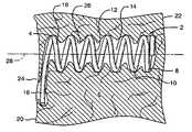
- FIG. 1shows a partial sectional side view of a preferred embodiment of an implant device configured for improved anchoring.
- the implant device 4comprises a helical spring body 8 formed of a plurality of individual coils 10 .
- the helical spring body 8defines an interior 12 which maintains an open cavity 14 within the tissue 6 when the device is implanted.
- the anchor mechanismcomprises a laterally extending arm, which extends from the most proximal coil 18 of the spring body 8 to define a lateral extent away from the longitudinal axis 28 of the device that is greater than the overall diameter of the coil body 8 .
- the implant deviceis particularly useful in treating ischemic tissue such as that often occurs in a myocardium of the heart.
- the implant devicemay be inserted into the tissue 6 , such as that of the myocardium, through the epicardial surface 20 at entry site 24 such that the device extends the majority of the thickness of the myocardium towards endocardial surface 22 .
- the deviceis fully implanted within the tissue such that the proximal laterally extending arm 16 is submerged within the tissue.
- FIG. 2shows an end view of the device 2 and in particular the laterally extending arm 16 , which is configured to prevent migration of the device.
- the armextends from the most proximal coil 18 in a tangential direction from the round coil.
- the arm 16then curves slightly in the direction of the curvature of the coil.
- the lateral extent of the arm beyond the outside diameter of the deviceis approximately 1–3 mm.
- the diameter of the body 8 of the coilis preferably on the order of 2–3 mm.
- the armserves to provide increased surface area engaged with the tissue to prevent migration in an axial direction through the tissue.
- the implantation of the arm into the tissuecauses it to prevent rotation of the device so that the device cannot back out of its tissue implant site.
- FIG. 3shows a variation of the laterally extending arm 16 having a bend 30 and second lateral extent 32 .
- the alternate embodiment of the laterally extending arm 16 shown in FIG. 3provides additional surface area and may serve additionally help to prevent rotation of the device. Variations of the shape of the laterally extending arm are possible and are considered within the scope of the invention.
- FIG. 4Another alternative design of the lateral extending arm is shown in FIG. 4 .
- the lateral extending arm 16is shown to have a distally projecting bend 34 at the proximal end 36 of the arm.
- the distally projecting bend 34may serve to further resist axial movement in the distal direction when the device is implanted in tissue because it serves more as a barb to claw into the tissue to resist movement.
- the penetration of the bendalso serves to resist rotation of the device.
- the bend 34need not be as extreme as shown in FIG. 4 , but rather the bend may be subtle, only to the extend that the arm 16 extends horizontally rather than following the acute angle of the helical pattern of the most distal coil 18 .
- FIG. 5shows another preferred embodiment of the implant device.
- a semi-tapered coil spring implant device 40may also provide adequate anchoring in dynamic tissue such as the tissue of the myocardium while meeting the objectives of the invention.
- the implant device 40comprises a helical coil spring 42 having a proximal portion 44 and a distal portion 46 .
- the individual coils 48 of the spring 42increase in diameter through the proximal portion 44 .
- Each coilincreases in size in the proximal direction.
- the coils of the distal portion 46are a constant diameter that is somewhat smaller than the diameter of the coils of the proximal portion.
- the most proximal coil 50does not extend laterally outward as with the previous embodiment, rather it terminates in its position as part of the helical coil arrangement.
- the proximal end of the coilmay be formed to have a bulbous shape 52 to further resist penetration of the tissue after the device has been implanted. As with the previous embodiment, the tissue tends to herniate at points 26 along the length of the implant.
- the implant device 40has shown to resist migration and rotation by virtue of the partial increase in taper at the proximal portion 44 of the device. This configuration may also serve to resist migration due to the enhanced flexibility of the proximal coils by virtue of their increased diameter. Increasing the overall diameter of the proximal coils (while maintaining the same filament thickness) serves to increase the flexibility of those coils.
- FIG. 6shows an end view of the implant device 40 having a partial taper at the proximal portion 44 .
- the most proximal coil 50is submerged within the tissue 6 when the device is implanted.
- the submersion of the most proximal coilprovides the advantages detailed above and additionally avoids placing a section of the coil across the transition between the tissue and tissue surface, which may tend to move differently placing an increased stress on the device and possibly leading to premature failure.
- FIG. 7shows a variation of the preferred embodiments discussed above wherein the increasing taper is present throughout the length of the device 80 such that each coil increases in diameter in a direction from the distal end 82 to the proximal end 84 .
- the full taper embodiment 80is believed to offer the same benefits as described in connection with the device shown in FIG. 5 .
- the smaller distal coils 46may define a diameter on the order of approximately 2.2 millimeters measured to the outside diameter of the coils and the larger diameter, maximum extent of the taper may be on the order of 4,5 to 5 millimeters.
- the devicesare preferably on the order of 7–8 mm in length.
- FIG. 8shows an alternate embodiment of an implant device having an anchoring mechanism.
- the coil devicehas an interior 98 , which is defined by the individual turns 120 of the coil.
- the helical coil 96defines a frame, which holds back surrounding tissue so that blood may pool in the interior chamber, coagulate and become fibrin.
- Spaces 122 between individual turns of the coilpermit communication between the interior chamber 98 , where fibrin will grow and the blood and tissue that surround the device.
- Open ends 124also permit communication between the interior chamber 98 and surrounding tissue.
- the coil 96has a tail 128 configured to resist excessive penetration of the device into the subject tissue so that the overall depth that the device is implanted in the tissue is controlled.
- the tail 128may be configured in a variety of forms. The example of a tail shown in FIG.
- the broad coil of the tailWhen the device is implanted in tissue, the broad coil of the tail is positioned to be flush with the surface of the tissue.
- the broad coil taildistributes the migratory forces experienced by the device over a broad area of tissue surface. The tail resists penetration of tissue surface thereby preventing migration of the device further into the tissue.
- filament 126 from which the coil is formedmay be a solid material or may, itself, be a coil spring structure having a plurality of openings between turns of the coil, which serve to permit herniation of surrounding tissue into the coil for anchoring capability.
- the broad coil tail 128has a proximal end 130 which is preferably joined to the broad coil to maintain the coil circular shape.
- the proximal end 130can be joined to the broad coil 128 by a variety of means such as welding as is shown by weld 132 .
- FIG. 9shows an alternative embodiment of joining the proximal end 130 to the broad coil tail 128 .
- the alternative embodimentcomprises wrapping the portion of the filament adjacent the proximal end 130 around the broad coil tail 128 in several turns
- FIG. 10shows another alternative embodiment useful for joining the proximal end 130 of the coil 96 to the broad coil tail 128 .
- the alternative embodimentutilizes a malleable sleeve 136 to encompass both a portion of the broad coil tail 128 and the distal end of the coil 130 . The malleable sleeve is then crimped to mechanically grasp the distal end 130 and broad coil and join them so that the circular shape of the broad coil tail 128 is maintained.
- the broad coil tail 128need not be a circular shape but may have a variety of broad shapes capable of serving to disperse migratory forces over a broad surface area of tissue when the device is implanted.
- FIG. 11shows a possible non-circular shape for the broad coil comprising a star shaped 137 .
- FIG. 12shows yet another alternative embodiment for the shape of the broad coil tail 128 . In FIG. 12 a somewhat oval broad coil 138 is shown. Additionally, the broad coil 138 has plurality of distally projecting protrusions 139 , which may increase the grasp of the coil into the tissue to prevent migration.
- FIG. 13shows yet another alternate embodiment of a tubular frame device.
- the canted coil device 140is formed from a filament 142 of rectangular cross-section such as a strand of flat wire. As shown in FIG. 14 , the coil is formed so that the major cross-sectional axis 147 of the rectangular wire is oriented at an acute angle to the longitudinal axis 150 of the coil 140 . The orientation gives each turn 146 of the coil a projecting edge 144 , which tends to claw into tissue to serve as an anchoring mechanism for the device.
- FIG. 15shows a wrapped ribbon implant embodiment.
- the implant 90is formed by a filament of a rectangular cross-sectional filament around a ribbed mandrel.
- the major axis 147 of the rectangular cross-section ribbonis oriented substantially perpendicular to the longitudinal axis 150 of the implant, as is shown in FIG. 15 .
- the major axis 147 of the coils 142 of the rectangular ribbondo not extend radically from the longitudinal axis 150 of the implant 140 at an acute angle. With greater coil surface area extending away from the longitudinal axis of the implant, the implant is believed to be more stable and less likely to migrate once implanted within the myocardium.
- the implantis preferably formed from 316 stainless steel rectangular cross-section forming wire. Preferred dimensions for the rectangular cross-section filament are on the order of 0.003 inches to 0.005 inches for the minor axis width and 0.015 to 0.018 inches for the major axis.
- FIGS. 16A–16Cshow an example of a surgical delivery device that may be used to deliver the implants into tissue such as that of the myocardium of the heart.
- the delivery deviceshown in FIG. 16A , comprises an obturator 180 that includes a main shaft 182 , by which it can be gripped and manipulated.
- the distal end 181 of the shaft 182is shown in detail in FIG. 16B and includes a reduced diameter device support section 184 having a sharp distal tip 186 adapted to pierce tissue.
- the diameter of the shaft segment 184is such as to fit closely within the interior of the devices.
- the proximal end of the segment 184terminates in a shoulder 188 formed at the junction of a proximally adjacent, slightly enlarged diameter portion 190 of the shaft.
- the distal end of the device support segment 184may include a radially projecting pin 192 dimensioned to project and fit between adjacent turns of the coils of a device.
- the pin 192engages the coils in a thread-like fashion so that after the assembly has been inserted into the tissue, the obturator 180 can be removed simply by unscrewing the obturator to free it from the implanted coil.
- the obturatormay be configured without the projecting pin 192 so that the device can be slipped on and off the obturator, without screwing.
- the proximal end of the devicemay bear against the shoulder 188 , and the tail 28 , if so equipped may extend along the segment 190 of the obturator.
- the intended tissue locationis first accessed surgically, such as by a cut-down method.
- the obturatorwith an implant device loaded on to segment 184 , then may be advanced into the tissue to deliver the implant.
- the sharp tippierces the tissue permitting the obturator and implant to be pushed inward into the tissue.
- the epicardial surface of the heartis accessed and penetrated by the obturator to deliver the implant.
- the shoulder 188prevents proximal movement of the implant along segment 184 during delivery.
- the distal end of the obturatoris projected to, and slightly beyond, the endocardium to place the implant device.
- the obturatorthen may be unscrewed and separated from the implant device. If the obturator is configured without the pin 192 , the obturator may be withdrawn directly from the device and the tissue. Simply applying light closure pressure to the epicardial puncture will cause the puncture hole to clot at the epicardium.
- An alternative method of anchoring the devicecomprises applying a surgical adhesive to the site of the implant such that adhesive is joined to the implant device and to surrounding tissue so that it is adhered.
- one method of applying the surgical adhesivemay comprise applying it directly at the surgical site or manually or delivering a quantity of surgical adhesive through the obturator delivery device directly to the cavity 14 created in the tissue by the device.
- each of the above described implant embodimentsmay exhibit improved resistance to migration by being configured to have enhanced axial flexibility.
- the flexibilitymay be controlled by a number of factors.
- One factor in controlling the flexibility of a helical coil deviceis to decrease the spacing between adjacent coils.
- Wire diametermay vary between 0.006 inches to 0.010 inches for the above described devices with 0.007 to 0.008 being preferred.
- Preferred pitch with such diametersmay be on the order of 0.25 mm–1 mm.
- the flexibility of the spring implantmay also be increased by increasing the overall diameter of the coils.
- implant devicesdiscussed above, but other materials having different modulii of elasticty such as nickel titanium alloys can be used. Additionally, implants formed from 2 or more different materials can be formed to provide varying flexibility. Different material filaments can be joined by welding or crimping of a malleable sleeve.
- the inventionprovides a novel approach to providing an anchoring system for implant devices.
- the devices and methods of the present inventionare simple and easy to apply to a wide range of implant designs.
Landscapes
- Health & Medical Sciences (AREA)
- Engineering & Computer Science (AREA)
- Cardiology (AREA)
- Biomedical Technology (AREA)
- Heart & Thoracic Surgery (AREA)
- Transplantation (AREA)
- Oral & Maxillofacial Surgery (AREA)
- Vascular Medicine (AREA)
- Life Sciences & Earth Sciences (AREA)
- Animal Behavior & Ethology (AREA)
- General Health & Medical Sciences (AREA)
- Public Health (AREA)
- Veterinary Medicine (AREA)
- Prostheses (AREA)
Abstract
Description
Claims (47)
Priority Applications (1)
| Application Number | Priority Date | Filing Date | Title |
|---|---|---|---|
| US09/743,695US6986784B1 (en) | 1999-05-14 | 2000-05-12 | Implant anchor systems |
Applications Claiming Priority (4)
| Application Number | Priority Date | Filing Date | Title |
|---|---|---|---|
| US13433199P | 1999-05-14 | 1999-05-14 | |
| US13457299P | 1999-05-17 | 1999-05-17 | |
| PCT/US2000/013118WO2000069345A1 (en) | 1999-05-14 | 2000-05-12 | Implant anchor systems |
| US09/743,695US6986784B1 (en) | 1999-05-14 | 2000-05-12 | Implant anchor systems |
Publications (1)
| Publication Number | Publication Date |
|---|---|
| US6986784B1true US6986784B1 (en) | 2006-01-17 |
Family
ID=35550725
Family Applications (1)
| Application Number | Title | Priority Date | Filing Date |
|---|---|---|---|
| US09/743,695Expired - Fee RelatedUS6986784B1 (en) | 1999-05-14 | 2000-05-12 | Implant anchor systems |
Country Status (1)
| Country | Link |
|---|---|
| US (1) | US6986784B1 (en) |
Cited By (103)
| Publication number | Priority date | Publication date | Assignee | Title |
|---|---|---|---|---|
| US20020082621A1 (en)* | 2000-09-22 | 2002-06-27 | Schurr Marc O. | Methods and devices for folding and securing tissue |
| US20020128701A1 (en)* | 2000-04-28 | 2002-09-12 | Winters R. Edward | Low profile expandable hoop support device for flexible tubes |
| US20040254518A1 (en)* | 2003-03-27 | 2004-12-16 | Sun Lee | Device to promote blood flow into the myocardium |
| US20050222672A1 (en)* | 2004-04-01 | 2005-10-06 | Cappella, Inc. | Ostial stent |
| US20060259132A1 (en)* | 2005-05-02 | 2006-11-16 | Cook Incorporated | Vascular stent for embolic protection |
| US20070032863A1 (en)* | 2001-11-28 | 2007-02-08 | Aptus Endosystems, Inc. | Catheter-based fastener implantation methods |
| US20070038241A1 (en)* | 2005-08-04 | 2007-02-15 | Cook Incorporated | Embolic protection device having inflatable frame |
| US20070066991A1 (en)* | 2005-09-16 | 2007-03-22 | Cook Incorporated | Embolic protection device |
| US20070073389A1 (en)* | 2001-11-28 | 2007-03-29 | Aptus Endosystems, Inc. | Endovascular aneurysm devices, systems, and methods |
| US20070078481A1 (en)* | 2005-10-04 | 2007-04-05 | Cook Incorporated | Embolic protection device |
| US20070088383A1 (en)* | 2005-10-03 | 2007-04-19 | Cook Incorporated | Embolic protection device |
| US20070112374A1 (en)* | 2005-10-18 | 2007-05-17 | Cook Incorporated | Invertible filter for embolic protection |
| US20070118173A1 (en)* | 2005-11-17 | 2007-05-24 | Cook Incorporated | Foam embolic protection device |
| US20080071307A1 (en)* | 2006-09-19 | 2008-03-20 | Cook Incorporated | Apparatus and methods for in situ embolic protection |
| US20090069822A1 (en)* | 2007-09-10 | 2009-03-12 | Olympus Medical Systems Corp. | Tissue fastening tool, stent, applicator for placing the same, and tissue fastening method through natural orifice |
| US20090076538A1 (en)* | 2007-09-14 | 2009-03-19 | Cook Incorporated | Helical embolic protection device |
| US20090076539A1 (en)* | 2007-09-14 | 2009-03-19 | Cook Incorporated | Helical thrombus removal device |
| US20090076593A1 (en)* | 2007-09-14 | 2009-03-19 | Cook Incorporated | Expandable device for treatment of a stricture in a body vessel |
| US20090082852A1 (en)* | 2001-06-04 | 2009-03-26 | Aptus Endosystems, Inc. | Catheter-based fastener implantation apparatus and methods |
| US20090099650A1 (en)* | 2001-11-28 | 2009-04-16 | Lee Bolduc | Devices, systems, and methods for endovascular staple and/or prosthesis delivery and implantation |
| US20090182355A1 (en)* | 2007-12-20 | 2009-07-16 | Levine Andy H | Porous barbs for long-term anchoring in the gastrointestinal tract |
| US20100010509A1 (en)* | 2008-07-11 | 2010-01-14 | Olympus Medical Systems Corp. | Tissue fastening apparatus |
| US20100010520A1 (en)* | 2008-07-11 | 2010-01-14 | Olympus Medical Systems Corp. | Tissue fastener |
| US20100010508A1 (en)* | 2008-07-11 | 2010-01-14 | Olympus Medical Systems Corp. | Tissue fastening tool and applicator for indwelling the same within body, and tissue fastening method through natural orifice |
| US20100030328A1 (en)* | 2008-04-18 | 2010-02-04 | Medtronic, Inc. | Apparatus for Treating a Heart Valve, in Particular a Mitral Valve |
| US7704222B2 (en) | 1998-09-10 | 2010-04-27 | Jenavalve Technology, Inc. | Methods and conduits for flowing blood from a heart chamber to a blood vessel |
| US20100168785A1 (en)* | 2008-12-29 | 2010-07-01 | Cook Incorporated | Embolic protection device and method of use |
| US20100198333A1 (en)* | 2009-01-31 | 2010-08-05 | Macatangay Edwin E | Preform for and an endoluminal prosthesis |
| US20100211087A1 (en)* | 2009-02-17 | 2010-08-19 | Cook Incorporated | Loop thrombectomy device |
| US20100211094A1 (en)* | 2009-02-18 | 2010-08-19 | Cook Incorporated | Umbrella distal embolic protection device |
| US20100268314A1 (en)* | 2009-04-17 | 2010-10-21 | Medtronic Vascular, Inc. | Hollow Helical Stent System |
| US20100274277A1 (en)* | 2009-04-27 | 2010-10-28 | Cook Incorporated | Embolic protection device with maximized flow-through |
| US20100292785A1 (en)* | 2007-04-20 | 2010-11-18 | Medtronic Corevalve Llc | Implant for treatment of a heart valve, in particular a mitral valve, material including such an implant, and material for insertion thereof |
| US20110004237A1 (en)* | 2007-12-12 | 2011-01-06 | Peter Schneider | Minimal surface area contact device for holding plaque to blood vessel wall |
| US20110040326A1 (en)* | 2005-05-20 | 2011-02-17 | Neotract, Inc. | Coiled anchor device |
| US20110087320A1 (en)* | 2001-11-28 | 2011-04-14 | Aptus Endosystems, Inc. | Devices, Systems, and Methods for Prosthesis Delivery and Implantation, Including a Prosthesis Assembly |
| US20110098738A1 (en)* | 2004-10-06 | 2011-04-28 | James B Hunt | Emboli capturing device having a coil and method for capturing emboli |
| US20110152886A1 (en)* | 2007-02-26 | 2011-06-23 | Olympus Medical Systems Corp. | Applicator and tissue fastening method through natural orifice |
| US20110238088A1 (en)* | 2001-11-28 | 2011-09-29 | Aptus Endosystems, Inc. | Devices, systems, and methods for supporting tissue and/or structures within a hollow body organ |
| US20110257581A1 (en)* | 2010-04-19 | 2011-10-20 | Gold Thread Llc | Filament Implant System and Method |
| US20120083822A1 (en)* | 2008-12-17 | 2012-04-05 | Boris Anukhin | Methods and apparatus for filtering a body lumen |
| US8216269B2 (en) | 2005-11-02 | 2012-07-10 | Cook Medical Technologies Llc | Embolic protection device having reduced profile |
| US8221446B2 (en) | 2005-03-15 | 2012-07-17 | Cook Medical Technologies | Embolic protection device |
| US20130204311A1 (en)* | 2010-05-12 | 2013-08-08 | Helical Solutions, Inc. | Implants and methods for treating cardiac arrhythmias |
| US8685044B2 (en) | 2001-11-28 | 2014-04-01 | Aptus Endosystems, Inc. | Systems and methods for attaching a prosthesis with a body lumen or hollow organ |
| US8690897B2 (en) | 2001-11-28 | 2014-04-08 | Aptus Endosystems, Inc. | Devices, systems, and methods for prosthesis delivery and implantation, including the use of a fastener tool |
| US20140148786A1 (en)* | 2011-08-23 | 2014-05-29 | Simcha Milo | Device for creating temporary access and then closure |
| US8900252B2 (en) | 2005-05-20 | 2014-12-02 | Neotract, Inc. | Devices, systems and methods for treating benign prostatic hyperplasia and other conditions |
| US8940001B2 (en) | 2005-05-20 | 2015-01-27 | Neotract, Inc. | Devices, systems and methods for retracting, lifting, compressing, supporting or repositioning tissues or anatomical structures |
| US8945152B2 (en) | 2005-05-20 | 2015-02-03 | Neotract, Inc. | Multi-actuating trigger anchor delivery system |
| US8945169B2 (en) | 2005-03-15 | 2015-02-03 | Cook Medical Technologies Llc | Embolic protection device |
| US9056005B2 (en) | 2009-03-25 | 2015-06-16 | University Of Iowa Research Foundation | Methods and devices for arytenoid repositioning |
| US9161749B2 (en) | 2011-04-14 | 2015-10-20 | Neotract, Inc. | Method and apparatus for treating sexual dysfunction |
| US9204842B2 (en) | 2010-10-29 | 2015-12-08 | Medtronic, Inc. | Medical device fixation attachment mechanism |
| EP2987526A1 (en)* | 2014-08-21 | 2016-02-24 | BIOTRONIK SE & Co. KG | Medical implant comprising a fixing device |
| US9320589B2 (en) | 2001-11-28 | 2016-04-26 | Medtronic Vascular, Inc. | Endovascular aneurysm repair system |
| US9320511B2 (en) | 2005-05-20 | 2016-04-26 | Neotract, Inc. | Multi-actuating trigger anchor delivery system |
| US9320503B2 (en) | 2001-11-28 | 2016-04-26 | Medtronic Vascular, Inc. | Devices, system, and methods for guiding an operative tool into an interior body region |
| US9351648B2 (en) | 2012-08-24 | 2016-05-31 | Medtronic, Inc. | Implantable medical device electrode assembly |
| US9504461B2 (en) | 2005-05-20 | 2016-11-29 | Neotract, Inc. | Anchor delivery system |
| US9526572B2 (en) | 2011-04-26 | 2016-12-27 | Aperiam Medical, Inc. | Method and device for treatment of hypertension and other maladies |
| US9545322B2 (en) | 2007-12-12 | 2017-01-17 | Intact Vascular, Inc. | Device and method for tacking plaque to blood vessel wall |
| US9603730B2 (en) | 2007-12-12 | 2017-03-28 | Intact Vascular, Inc. | Endoluminal device and method |
| US9730818B2 (en) | 2007-12-12 | 2017-08-15 | Intact Vascular, Inc. | Endoluminal device and method |
| US9901434B2 (en) | 2007-02-27 | 2018-02-27 | Cook Medical Technologies Llc | Embolic protection device including a Z-stent waist band |
| US9925031B2 (en) | 2009-12-28 | 2018-03-27 | Cook Medical Technologies Llc | Endoluminal device with kink-resistant regions |
| US10022250B2 (en) | 2007-12-12 | 2018-07-17 | Intact Vascular, Inc. | Deployment device for placement of multiple intraluminal surgical staples |
| US10130353B2 (en) | 2012-06-29 | 2018-11-20 | Neotract, Inc. | Flexible system for delivering an anchor |
| US10166127B2 (en) | 2007-12-12 | 2019-01-01 | Intact Vascular, Inc. | Endoluminal device and method |
| US10195014B2 (en) | 2005-05-20 | 2019-02-05 | Neotract, Inc. | Devices, systems and methods for treating benign prostatic hyperplasia and other conditions |
| US10194905B2 (en) | 2001-11-28 | 2019-02-05 | Medtronic Vascular, Inc. | Devices, systems, and methods for endovascular staple and/or prosthesis delivery and implantation |
| US10245167B2 (en) | 2015-01-29 | 2019-04-02 | Intact Vascular, Inc. | Delivery device and method of delivery |
| US10265061B2 (en) | 2005-05-20 | 2019-04-23 | Neotract, Inc. | Latching anchor device |
| US10271973B2 (en) | 2011-06-03 | 2019-04-30 | Intact Vascular, Inc. | Endovascular implant |
| US10271974B2 (en) | 2011-06-24 | 2019-04-30 | Cook Medical Technologies Llc | Helical stent |
| US10278839B2 (en) | 2007-12-12 | 2019-05-07 | Intact Vascular, Inc. | Endovascular impant |
| US10292801B2 (en) | 2012-03-29 | 2019-05-21 | Neotract, Inc. | System for delivering anchors for treating incontinence |
| US10299780B2 (en) | 2005-05-20 | 2019-05-28 | Neotract, Inc. | Apparatus and method for manipulating or retracting tissue and anatomical structure |
| GB2568999A (en)* | 2017-09-27 | 2019-06-05 | Spiration Inc D/B/A Olympus Respiratory America | Tissue fastening tool |
| US10426509B2 (en) | 2005-05-20 | 2019-10-01 | Neotract, Inc. | Median lobe destruction apparatus and method |
| US10492792B2 (en) | 2005-05-20 | 2019-12-03 | Neotract, Inc. | Devices, systems and methods for treating benign prostatic hyperplasia and other conditions |
| CN110740692A (en)* | 2017-06-09 | 2020-01-31 | 希格纳姆外科有限公司 | Implant for closing an opening in tissue |
| US20200129170A1 (en)* | 2014-12-02 | 2020-04-30 | 4Tech Inc. | Tissue Anchors |
| US10898356B2 (en) | 2015-01-29 | 2021-01-26 | Intact Vascular, Inc. | Delivery device and method of delivery |
| US10925587B2 (en) | 2005-05-20 | 2021-02-23 | Neotract, Inc. | Anchor delivery system |
| US10993805B2 (en) | 2008-02-26 | 2021-05-04 | Jenavalve Technology, Inc. | Stent for the positioning and anchoring of a valvular prosthesis in an implantation site in the heart of a patient |
| US10993824B2 (en) | 2016-01-01 | 2021-05-04 | Intact Vascular, Inc. | Delivery device and method of delivery |
| US11065138B2 (en) | 2016-05-13 | 2021-07-20 | Jenavalve Technology, Inc. | Heart valve prosthesis delivery system and method for delivery of heart valve prosthesis with introducer sheath and loading system |
| US11185405B2 (en) | 2013-08-30 | 2021-11-30 | Jenavalve Technology, Inc. | Radially collapsible frame for a prosthetic valve and method for manufacturing such a frame |
| US11197754B2 (en) | 2017-01-27 | 2021-12-14 | Jenavalve Technology, Inc. | Heart valve mimicry |
| US11298115B2 (en) | 2020-08-03 | 2022-04-12 | Teleflex Life Sciences Limited | Handle and cartridge system for medical interventions |
| US11337800B2 (en) | 2015-05-01 | 2022-05-24 | Jenavalve Technology, Inc. | Device and method with reduced pacemaker rate in heart valve replacement |
| US11357624B2 (en) | 2007-04-13 | 2022-06-14 | Jenavalve Technology, Inc. | Medical device for treating a heart valve insufficiency |
| US11517431B2 (en) | 2005-01-20 | 2022-12-06 | Jenavalve Technology, Inc. | Catheter system for implantation of prosthetic heart valves |
| US11564794B2 (en) | 2008-02-26 | 2023-01-31 | Jenavalve Technology, Inc. | Stent for the positioning and anchoring of a valvular prosthesis in an implantation site in the heart of a patient |
| US11589981B2 (en) | 2010-05-25 | 2023-02-28 | Jenavalve Technology, Inc. | Prosthetic heart valve and transcatheter delivered endoprosthesis comprising a prosthetic heart valve and a stent |
| US11660218B2 (en) | 2017-07-26 | 2023-05-30 | Intact Vascular, Inc. | Delivery device and method of delivery |
| US11672520B2 (en) | 2017-12-23 | 2023-06-13 | Teleflex Life Sciences Limited | Expandable tissue engagement apparatus and method |
| US12121461B2 (en) | 2015-03-20 | 2024-10-22 | Jenavalve Technology, Inc. | Heart valve prosthesis delivery system and method for delivery of heart valve prosthesis with introducer sheath |
| US12171658B2 (en) | 2022-11-09 | 2024-12-24 | Jenavalve Technology, Inc. | Catheter system for sequential deployment of an expandable implant |
| US12245758B2 (en) | 2018-12-24 | 2025-03-11 | 4Tech Inc. | Self-locking tissue anchors |
| US12414854B2 (en) | 2010-05-20 | 2025-09-16 | Jenavalve Technology, Inc. | Catheter system for introducing an expandable stent into the body of a patient |
| US12440301B2 (en) | 2019-10-30 | 2025-10-14 | Teleflex Life Sciences Llc | System for delivery of a fiducial marker |
Citations (156)
| Publication number | Priority date | Publication date | Assignee | Title |
|---|---|---|---|---|
| FR1514319A (en) | 1967-01-11 | 1968-02-23 | Device for implantation in the apical region of the heart of an artificial ventricle | |
| US3991750A (en) | 1975-04-28 | 1976-11-16 | Syntex Corporation | Dromostanolone propionate implant pellet useful for producing weight gains in animals and suppressing estrus in female animals |
| US3995617A (en) | 1972-05-31 | 1976-12-07 | Watkins David H | Heart assist method and catheter |
| US4130904A (en)* | 1977-06-06 | 1978-12-26 | Thermo Electron Corporation | Prosthetic blood conduit |
| US4307722A (en) | 1979-08-14 | 1981-12-29 | Evans Joseph M | Dilators for arterial dilation |
| US4503569A (en) | 1983-03-03 | 1985-03-12 | Dotter Charles T | Transluminally placed expandable graft prosthesis |
| US4546499A (en) | 1982-12-13 | 1985-10-15 | Possis Medical, Inc. | Method of supplying blood to blood receiving vessels |
| US4580568A (en) | 1984-10-01 | 1986-04-08 | Cook, Incorporated | Percutaneous endovascular stent and method for insertion thereof |
| US4582181A (en) | 1983-08-12 | 1986-04-15 | Advanced Cardiovascular Systems, Inc. | Steerable dilatation catheter |
| US4641653A (en) | 1978-06-02 | 1987-02-10 | Rockey Arthur G | Medical sleeve |
| US4649922A (en) | 1986-01-23 | 1987-03-17 | Wiktor Donimik M | Catheter arrangement having a variable diameter tip and spring prosthesis |
| US4655771A (en) | 1982-04-30 | 1987-04-07 | Shepherd Patents S.A. | Prosthesis comprising an expansible or contractile tubular body |
| US4658817A (en) | 1985-04-01 | 1987-04-21 | Children's Hospital Medical Center | Method and apparatus for transmyocardial revascularization using a laser |
| US4665918A (en) | 1986-01-06 | 1987-05-19 | Garza Gilbert A | Prosthesis system and method |
| US4681110A (en) | 1985-12-02 | 1987-07-21 | Wiktor Dominik M | Catheter arrangement having a blood vessel liner, and method of using it |
| US4733665A (en) | 1985-11-07 | 1988-03-29 | Expandable Grafts Partnership | Expandable intraluminal graft, and method and apparatus for implanting an expandable intraluminal graft |
| US4768507A (en) | 1986-02-24 | 1988-09-06 | Medinnovations, Inc. | Intravascular stent and percutaneous insertion catheter system for the dilation of an arterial stenosis and the prevention of arterial restenosis |
| US4774949A (en) | 1983-06-14 | 1988-10-04 | Fogarty Thomas J | Deflector guiding catheter |
| WO1989001798A1 (en) | 1987-09-02 | 1989-03-09 | Engineers & Doctors A/S | Device for the placing of a partial catheter in a body cavity |
| US4813925A (en) | 1987-04-21 | 1989-03-21 | Medical Engineering Corporation | Spiral ureteral stent |
| US4861330A (en) | 1987-03-12 | 1989-08-29 | Gene Voss | Cardiac assist device and method |
| US4889137A (en) | 1988-05-05 | 1989-12-26 | The United States Of America As Reprsented By The Department Of Health And Human Services | Method for improved use of heart/lung machine |
| US4904264A (en) | 1984-05-08 | 1990-02-27 | Fried. Krupp Gmbh | Artifical joint system |
| US4917666A (en) | 1988-11-14 | 1990-04-17 | Medtronic Versaflex, Inc. | Steerable thru-lumen catheter |
| US4920980A (en) | 1987-09-14 | 1990-05-01 | Cordis Corporation | Catheter with controllable tip |
| US4950227A (en) | 1988-11-07 | 1990-08-21 | Boston Scientific Corporation | Stent delivery system |
| US4995857A (en) | 1989-04-07 | 1991-02-26 | Arnold John R | Left ventricular assist device and method for temporary and permanent procedures |
| US4997431A (en) | 1989-08-30 | 1991-03-05 | Angeion Corporation | Catheter |
| US5040543A (en) | 1990-07-25 | 1991-08-20 | C. R. Bard, Inc. | Movable core guidewire |
| US5042486A (en) | 1989-09-29 | 1991-08-27 | Siemens Aktiengesellschaft | Catheter locatable with non-ionizing field and method for locating same |
| US5047028A (en) | 1989-05-12 | 1991-09-10 | Quinghua Qian | Method for inducing thrombosis in blood vessels |
| US5049138A (en) | 1989-11-13 | 1991-09-17 | Boston Scientific Corporation | Catheter with dissolvable tip |
| US5056517A (en) | 1989-07-24 | 1991-10-15 | Consiglio Nazionale Delle Ricerche | Biomagnetically localizable multipurpose catheter and method for magnetocardiographic guided intracardiac mapping, biopsy and ablation of cardiac arrhythmias |
| WO1991015254A1 (en) | 1990-04-04 | 1991-10-17 | Zimmon David S | Indwelling stent and method of use |
| US5087243A (en) | 1990-06-18 | 1992-02-11 | Boaz Avitall | Myocardial iontophoresis |
| US5114414A (en) | 1984-09-18 | 1992-05-19 | Medtronic, Inc. | Low profile steerable catheter |
| EP0490459A1 (en) | 1990-12-14 | 1992-06-17 | The Kendall Company | Multilumen catheter |
| US5158548A (en) | 1990-04-25 | 1992-10-27 | Advanced Cardiovascular Systems, Inc. | Method and system for stent delivery |
| US5167614A (en) | 1991-10-29 | 1992-12-01 | Medical Engineering Corporation | Prostatic stent |
| EP0515867A2 (en) | 1991-05-01 | 1992-12-02 | The Trustees Of Columbia University In The City Of New York | Myocardial revascularization through the endocardial surface using a laser |
| US5176626A (en) | 1992-01-15 | 1993-01-05 | Wilson-Cook Medical, Inc. | Indwelling stent |
| US5180366A (en) | 1990-10-10 | 1993-01-19 | Woods W T | Apparatus and method for angioplasty and for preventing re-stenosis |
| US5190058A (en) | 1991-05-22 | 1993-03-02 | Medtronic, Inc. | Method of using a temporary stent catheter |
| US5256146A (en) | 1991-10-11 | 1993-10-26 | W. D. Ensminger | Vascular catheterization system with catheter anchoring feature |
| US5266073A (en) | 1987-12-08 | 1993-11-30 | Wall W Henry | Angioplasty stent |
| US5287861A (en) | 1992-10-30 | 1994-02-22 | Wilk Peter J | Coronary artery by-pass method and associated catheter |
| US5290295A (en) | 1992-07-15 | 1994-03-01 | Querals & Fine, Inc. | Insertion tool for an intraluminal graft procedure |
| WO1994005265A1 (en) | 1992-09-10 | 1994-03-17 | Children's Medical Center Corporation | Biodegradable polymer matrices for sustained delivery of local anesthetic agents |
| US5312456A (en) | 1991-01-31 | 1994-05-17 | Carnegie Mellon University | Micromechanical barb and method for making the same |
| US5324325A (en) | 1991-06-27 | 1994-06-28 | Siemens Pacesetter, Inc. | Myocardial steroid releasing lead |
| US5366493A (en) | 1991-02-04 | 1994-11-22 | Case Western Reserve University | Double helix functional stimulation electrode |
| WO1994027612A1 (en) | 1993-05-20 | 1994-12-08 | Baylor College Of Medicine | Genetic therapy for cardiovascular disease |
| US5372600A (en) | 1991-10-31 | 1994-12-13 | Instent Inc. | Stent delivery systems |
| US5380316A (en) | 1990-12-18 | 1995-01-10 | Advanced Cardiovascular Systems, Inc. | Method for intra-operative myocardial device revascularization |
| RU2026640C1 (en) | 1984-09-13 | 1995-01-20 | Адолий Яковлевич Кононов | Method for surgical treatment of ischemia |
| US5389096A (en) | 1990-12-18 | 1995-02-14 | Advanced Cardiovascular Systems | System and method for percutaneous myocardial revascularization |
| US5391199A (en) | 1993-07-20 | 1995-02-21 | Biosense, Inc. | Apparatus and method for treating cardiac arrhythmias |
| US5409004A (en) | 1993-06-11 | 1995-04-25 | Cook Incorporated | Localization device with radiopaque markings |
| US5409019A (en) | 1992-10-30 | 1995-04-25 | Wilk; Peter J. | Coronary artery by-pass method |
| US5423885A (en) | 1992-01-31 | 1995-06-13 | Advanced Cardiovascular Systems, Inc. | Stent capable of attachment within a body lumen |
| US5425757A (en) | 1993-05-21 | 1995-06-20 | Tiefenbrun; Jonathan | Aortic surgical procedure |
| US5429144A (en) | 1992-10-30 | 1995-07-04 | Wilk; Peter J. | Coronary artery by-pass method |
| US5441516A (en) | 1994-03-03 | 1995-08-15 | Scimed Lifesystems Inc. | Temporary stent |
| US5453090A (en) | 1994-03-01 | 1995-09-26 | Cordis Corporation | Method of stent delivery through an elongate softenable sheath |
| US5452733A (en) | 1993-02-22 | 1995-09-26 | Stanford Surgical Technologies, Inc. | Methods for performing thoracoscopic coronary artery bypass |
| US5458615A (en) | 1993-07-06 | 1995-10-17 | Advanced Cardiovascular Systems, Inc. | Stent delivery system |
| US5464404A (en) | 1993-09-20 | 1995-11-07 | Abela Laser Systems, Inc. | Cardiac ablation catheters and method |
| US5464650A (en) | 1993-04-26 | 1995-11-07 | Medtronic, Inc. | Intravascular stent and method |
| US5466242A (en) | 1994-02-02 | 1995-11-14 | Mori; Katsushi | Stent for biliary, urinary or vascular system |
| US5476471A (en)* | 1993-08-19 | 1995-12-19 | Mind - E.M.S.G. Ltd | Device and method for external correction of insufficient valves in venous junctions |
| US5476505A (en) | 1993-11-18 | 1995-12-19 | Advanced Cardiovascular Systems, Inc. | Coiled stent and delivery system |
| US5484424A (en)* | 1992-11-19 | 1996-01-16 | Celsa L.G. (Societe Anonyme) | Blood filtering device having a catheter with longitudinally variable rigidity |
| US5487739A (en) | 1987-11-17 | 1996-01-30 | Brown University Research Foundation | Implantable therapy systems and methods |
| US5514176A (en) | 1995-01-20 | 1996-05-07 | Vance Products Inc. | Pull apart coil stent |
| EP0714640A1 (en) | 1994-11-28 | 1996-06-05 | Advanced Cardiovascular Systems, Inc. | System and method for delivering multiple stents |
| EP0717969A2 (en) | 1994-12-22 | 1996-06-26 | Target Therapeutics, Inc. | Implant delivery assembly with expandable coupling/decoupling mechanism |
| RU2063179C1 (en) | 1993-10-06 | 1996-07-10 | Новосибирский медицинский институт | Multifunctional scalpel-tunnelizer |
| US5536274A (en)* | 1991-02-15 | 1996-07-16 | pfm Produkterfur Die Medizin | Spiral implant for organ pathways |
| US5551954A (en) | 1991-10-04 | 1996-09-03 | Scimed Life Systems, Inc. | Biodegradable drug delivery vascular stent |
| US5551427A (en) | 1995-02-13 | 1996-09-03 | Altman; Peter A. | Implantable device for the effective elimination of cardiac arrhythmogenic sites |
| EP0732089A2 (en) | 1995-03-17 | 1996-09-18 | Advanced Cardiovascular Systems, Inc. | Multi-anchor stent |
| US5562922A (en) | 1993-03-18 | 1996-10-08 | Cedars-Sinai Medical Center | Drug incorporating and release polymeric coating for bioprosthesis |
| US5562619A (en) | 1993-08-19 | 1996-10-08 | Boston Scientific Corporation | Deflectable catheter |
| US5562622A (en) | 1995-03-20 | 1996-10-08 | Contimed, Inc. | Self-cleansing bladder drainage device |
| US5571168A (en) | 1995-04-05 | 1996-11-05 | Scimed Lifesystems Inc | Pull back stent delivery system |
| US5602301A (en) | 1993-11-16 | 1997-02-11 | Indiana University Foundation | Non-human mammal having a graft and methods of delivering protein to myocardial tissue |
| US5613981A (en)* | 1995-04-21 | 1997-03-25 | Medtronic, Inc. | Bidirectional dual sinusoidal helix stent |
| US5614206A (en) | 1995-03-07 | 1997-03-25 | Wright Medical Technology, Inc. | Controlled dissolution pellet containing calcium sulfate |
| DE29619029U1 (en) | 1996-11-02 | 1997-04-10 | Kletke, Georg, Dr.med., 24534 Neumünster | Miocard puncture needle |
| FR2725615B1 (en) | 1994-10-17 | 1997-06-13 | Caffiniere Jean Yves De | BONE ANCHORING DEVICE FOR FIXATION THREADS USED IN ORTHOPEDIC SURGERY |
| US5643308A (en) | 1995-02-28 | 1997-07-01 | Markman; Barry Stephen | Method and apparatus for forming multiple cavities for placement of hair grafts |
| US5653759A (en)* | 1995-07-18 | 1997-08-05 | Beth Israel Hospital Assoc. Inc. | In-vivo method for repairing a ruptured segment of a therapeutic appliance surgically positioned previously within the body of a living human |
| US5653756A (en) | 1990-10-31 | 1997-08-05 | Baxter International Inc. | Closed porous chambers for implanting tissue in a host |
| US5655548A (en) | 1996-09-16 | 1997-08-12 | Circulation, Inc. | Method for treatment of ischemic heart disease by providing transvenous myocardial perfusion |
| US5662124A (en) | 1996-06-19 | 1997-09-02 | Wilk Patent Development Corp. | Coronary artery by-pass method |
| WO1997032551A1 (en) | 1996-03-04 | 1997-09-12 | Energy Life Systems Corporation | Device and method for trans myocardial revascularization |
| WO1997042910A1 (en) | 1996-05-14 | 1997-11-20 | Cardia Catheter Co. | Tubular stent for use in medical applications |
| US5690643A (en) | 1996-02-20 | 1997-11-25 | Leocor, Incorporated | Stent delivery system |
| WO1997044071A1 (en) | 1996-05-21 | 1997-11-27 | Amnon Sudai | Apparatus and methods for revascularization and perfusion |
| WO1997045105A1 (en) | 1996-05-24 | 1997-12-04 | Angiotech Pharmaceuticals, Inc. | Compositions and methods for treating or preventing diseases of body passageways |
| WO1997038730A3 (en) | 1996-04-17 | 1997-12-11 | Olivier Bertrand | Radioactivity local delivery system |
| EP0812574A2 (en) | 1996-06-13 | 1997-12-17 | Eclipse Surgical Technologies, Inc. | Intraoperative myocardial device and stimulation procedure |
| WO1997047253A1 (en) | 1996-06-14 | 1997-12-18 | Kriton Medical, Inc. | Methods and devices for reducing angina, enhancing myocardial perfusion and increasing cardiac function |
| WO1996020698A3 (en) | 1995-01-05 | 1998-01-22 | Univ Michigan | Surface-modified nanoparticles and method of making and using same |
| WO1998005307A1 (en) | 1996-08-08 | 1998-02-12 | Localmed, Inc. | Transmural drug delivery method and apparatus |
| WO1998008456A1 (en) | 1996-08-26 | 1998-03-05 | Transvascular, Inc. | Methods and apparatus for transmyocardial direct coronary revascularization |
| EP0830873A2 (en) | 1996-09-20 | 1998-03-25 | Kaneka Medix Corporation | Medical wire connected to an implanted device and method for using the same |
| US5735897A (en) | 1993-10-19 | 1998-04-07 | Scimed Life Systems, Inc. | Intravascular stent pump |
| US5738654A (en) | 1995-03-20 | 1998-04-14 | Contimed, Inc. | Self cleansing bladder drainage device |
| US5741330A (en) | 1990-10-31 | 1998-04-21 | Baxter International, Inc. | Close vascularization implant material |
| WO1998016644A1 (en) | 1996-10-16 | 1998-04-23 | Zymogenetics, Inc. | Fibroblast growth factor homologs |
| US5744515A (en) | 1995-05-26 | 1998-04-28 | Bsi Corporation | Method and implantable article for promoting endothelialization |
| US5756127A (en) | 1996-10-29 | 1998-05-26 | Wright Medical Technology, Inc. | Implantable bioresorbable string of calcium sulfate beads |
| US5755682A (en) | 1996-08-13 | 1998-05-26 | Heartstent Corporation | Method and apparatus for performing coronary artery bypass surgery |
| WO1998023228A1 (en) | 1996-11-25 | 1998-06-04 | Alza Corporation | Directional drug delivery stent |
| US5762600A (en) | 1994-04-29 | 1998-06-09 | W. L. Gore & Associates, Inc. | Blood contact surfaces employing natural subendothelial matrix and methods for making and using the same |
| WO1998025533A1 (en) | 1996-12-09 | 1998-06-18 | Scimed Life Systems, Inc. | Radio frequency transmyocardial revascularization corer |
| US5769843A (en) | 1996-02-20 | 1998-06-23 | Cormedica | Percutaneous endomyocardial revascularization |
| WO1998029148A1 (en) | 1996-12-31 | 1998-07-09 | Scimed Life Systems, Inc. | Multilayer liquid absorption and deformation devices |
| US5782823A (en) | 1996-04-05 | 1998-07-21 | Eclipse Surgical Technologies, Inc. | Laser device for transmyocardial revascularization procedures including means for enabling a formation of a pilot hole in the epicardium |
| EP0853921A2 (en) | 1996-12-27 | 1998-07-22 | Eclipse Surgical Technologies, Inc. | Laser assisted drug delivery |
| US5785702A (en) | 1996-10-15 | 1998-07-28 | Eclipse Surgical Technologies, Inc. | Method for non-synchronous laser-assisted transmyocardial revascularization |
| WO1998032859A1 (en) | 1997-01-29 | 1998-07-30 | Cornell Research Foundation, Inc. | Multiple site delivery of adenoviral vector for the induction of angiogenesis |
| DE19703482A1 (en) | 1997-01-31 | 1998-08-06 | Ernst Peter Prof Dr M Strecker | Stent |
| US5792453A (en) | 1995-02-28 | 1998-08-11 | The Regents Of The University Of California | Gene transfer-mediated angiogenesis therapy |
| US5797870A (en) | 1995-06-07 | 1998-08-25 | Indiana University Foundation | Pericardial delivery of therapeutic and diagnostic agents |
| US5807384A (en) | 1996-12-20 | 1998-09-15 | Eclipse Surgical Technologies, Inc. | Transmyocardial revascularization (TMR) enhanced treatment for coronary artery disease |
| US5824049A (en) | 1995-06-07 | 1998-10-20 | Med Institute, Inc. | Coated implantable medical device |
| US5824059A (en)* | 1997-08-05 | 1998-10-20 | Wijay; Bandula | Flexible stent |
| US5830502A (en) | 1994-10-28 | 1998-11-03 | Alza Corporation | Injection-molded dosage form |
| US5840059A (en) | 1995-06-07 | 1998-11-24 | Cardiogenesis Corporation | Therapeutic and diagnostic agent delivery |
| US5861032A (en) | 1996-01-31 | 1999-01-19 | Surface Genesis, Inc. | Medical device having a biocompatible coating and oxidation method of coupling therefor |
| WO1998046115A3 (en) | 1997-04-11 | 1999-01-21 | Transvascular Inc | Methods and apparatus for transmyocardial direct coronary revascularization |
| US5879383A (en) | 1994-04-29 | 1999-03-09 | W. L. Gore & Associates, Inc. | Blood contact surfaces using endothelium on a subendothelial matrix |
| US5891133A (en)* | 1996-03-29 | 1999-04-06 | Eclipse Surgical Technologies, Inc. | Apparatus for laser-assisted intra-coronary transmyocardial revascularization and other applications |
| US5893869A (en)* | 1997-02-19 | 1999-04-13 | University Of Iowa Research Foundation | Retrievable inferior vena cava filter system and method for use thereof |
| US5899915A (en) | 1996-12-02 | 1999-05-04 | Angiotrax, Inc. | Apparatus and method for intraoperatively performing surgery |
| WO1999021510A1 (en) | 1997-10-29 | 1999-05-06 | Kensey Nash Corporation | Transmyocardial revascularization system |
| US5911717A (en)* | 1997-03-17 | 1999-06-15 | Precision Vascular Systems, Inc. | Catheter deliverable thrombogenic apparatus and method |
| WO1999038459A3 (en) | 1998-01-30 | 1999-10-07 | Wilk Patent Dev Corp | Transmyocardial coronary artery bypass and revascularization |
| US5971993A (en) | 1996-11-07 | 1999-10-26 | Myocardial Stents, Inc. | System for delivery of a trans myocardial device to a heart wall |
| WO1999053863A1 (en) | 1998-04-20 | 1999-10-28 | Heartstent Corporation | Transmyocardial implant procedure and tools |
| EP0953320A2 (en) | 1998-04-30 | 1999-11-03 | Medtronic, Inc. | Medical device |
| US5980514A (en) | 1996-07-26 | 1999-11-09 | Target Therapeutics, Inc. | Aneurysm closure device assembly |
| US6007544A (en)* | 1996-06-14 | 1999-12-28 | Beth Israel Deaconess Medical Center | Catheter apparatus having an improved shape-memory alloy cuff and inflatable on-demand balloon for creating a bypass graft in-vivo |
| US6045565A (en) | 1997-11-04 | 2000-04-04 | Scimed Life Systems, Inc. | Percutaneous myocardial revascularization growth factor mediums and method |
| US6053924A (en) | 1996-11-07 | 2000-04-25 | Hussein; Hany | Device and method for trans myocardial revascularization |
| US6059825A (en)* | 1992-03-05 | 2000-05-09 | Angiodynamics, Inc. | Clot filter |
| EP1062920A1 (en) | 1999-06-22 | 2000-12-27 | ContiCare Medical, Inc. | Self-cleansing bladder drainage device |
| US6197324B1 (en) | 1997-12-18 | 2001-03-06 | C. R. Bard, Inc. | System and methods for local delivery of an agent |
| US6248112B1 (en) | 1998-09-30 | 2001-06-19 | C. R. Bard, Inc. | Implant delivery system |
| US6251418B1 (en) | 1997-12-18 | 2001-06-26 | C.R. Bard, Inc. | Systems and methods for local delivery of an agent |
| US6263880B1 (en) | 1998-06-22 | 2001-07-24 | Neovasys, Inc. | Method of enhancing blood flow in tissue |
| US6277082B1 (en) | 1999-07-22 | 2001-08-21 | C. R. Bard, Inc. | Ischemia detection system |
| US6432126B1 (en) | 1998-09-30 | 2002-08-13 | C.R. Bard, Inc. | Flexible vascular inducing implants |
| US6458092B1 (en) | 1998-09-30 | 2002-10-01 | C. R. Bard, Inc. | Vascular inducing implants |
- 2000
- 2000-05-12USUS09/743,695patent/US6986784B1/ennot_activeExpired - Fee Related
Patent Citations (171)
| Publication number | Priority date | Publication date | Assignee | Title |
|---|---|---|---|---|
| FR1514319A (en) | 1967-01-11 | 1968-02-23 | Device for implantation in the apical region of the heart of an artificial ventricle | |
| US3995617A (en) | 1972-05-31 | 1976-12-07 | Watkins David H | Heart assist method and catheter |
| US3991750A (en) | 1975-04-28 | 1976-11-16 | Syntex Corporation | Dromostanolone propionate implant pellet useful for producing weight gains in animals and suppressing estrus in female animals |
| US4130904A (en)* | 1977-06-06 | 1978-12-26 | Thermo Electron Corporation | Prosthetic blood conduit |
| US4641653A (en) | 1978-06-02 | 1987-02-10 | Rockey Arthur G | Medical sleeve |
| US4307722A (en) | 1979-08-14 | 1981-12-29 | Evans Joseph M | Dilators for arterial dilation |
| US4655771A (en) | 1982-04-30 | 1987-04-07 | Shepherd Patents S.A. | Prosthesis comprising an expansible or contractile tubular body |
| US4655771B1 (en) | 1982-04-30 | 1996-09-10 | Medinvent Ams Sa | Prosthesis comprising an expansible or contractile tubular body |
| US4546499A (en) | 1982-12-13 | 1985-10-15 | Possis Medical, Inc. | Method of supplying blood to blood receiving vessels |
| US4562597A (en) | 1982-12-13 | 1986-01-07 | Possis Medical, Inc. | Method of supplying blood to blood receiving vessels |
| US4503569A (en) | 1983-03-03 | 1985-03-12 | Dotter Charles T | Transluminally placed expandable graft prosthesis |
| US4774949A (en) | 1983-06-14 | 1988-10-04 | Fogarty Thomas J | Deflector guiding catheter |
| US4582181A (en) | 1983-08-12 | 1986-04-15 | Advanced Cardiovascular Systems, Inc. | Steerable dilatation catheter |
| US4904264A (en) | 1984-05-08 | 1990-02-27 | Fried. Krupp Gmbh | Artifical joint system |
| RU2026640C1 (en) | 1984-09-13 | 1995-01-20 | Адолий Яковлевич Кононов | Method for surgical treatment of ischemia |
| US5114414A (en) | 1984-09-18 | 1992-05-19 | Medtronic, Inc. | Low profile steerable catheter |
| US4580568A (en) | 1984-10-01 | 1986-04-08 | Cook, Incorporated | Percutaneous endovascular stent and method for insertion thereof |
| US4658817A (en) | 1985-04-01 | 1987-04-21 | Children's Hospital Medical Center | Method and apparatus for transmyocardial revascularization using a laser |
| US4733665B1 (en) | 1985-11-07 | 1994-01-11 | Expandable Grafts Partnership | Expandable intraluminal graft,and method and apparatus for implanting an expandable intraluminal graft |
| US4733665A (en) | 1985-11-07 | 1988-03-29 | Expandable Grafts Partnership | Expandable intraluminal graft, and method and apparatus for implanting an expandable intraluminal graft |
| US4733665C2 (en) | 1985-11-07 | 2002-01-29 | Expandable Grafts Partnership | Expandable intraluminal graft and method and apparatus for implanting an expandable intraluminal graft |
| US4681110A (en) | 1985-12-02 | 1987-07-21 | Wiktor Dominik M | Catheter arrangement having a blood vessel liner, and method of using it |
| US4665918A (en) | 1986-01-06 | 1987-05-19 | Garza Gilbert A | Prosthesis system and method |
| US4649922A (en) | 1986-01-23 | 1987-03-17 | Wiktor Donimik M | Catheter arrangement having a variable diameter tip and spring prosthesis |
| US4768507A (en) | 1986-02-24 | 1988-09-06 | Medinnovations, Inc. | Intravascular stent and percutaneous insertion catheter system for the dilation of an arterial stenosis and the prevention of arterial restenosis |
| US4861330A (en) | 1987-03-12 | 1989-08-29 | Gene Voss | Cardiac assist device and method |
| US4813925A (en) | 1987-04-21 | 1989-03-21 | Medical Engineering Corporation | Spiral ureteral stent |
| US5098374A (en) | 1987-09-02 | 1992-03-24 | Engineers & Doctors A/A | Device for the placing of a partial catheter in a body cavity |
| WO1989001798A1 (en) | 1987-09-02 | 1989-03-09 | Engineers & Doctors A/S | Device for the placing of a partial catheter in a body cavity |
| US4920980A (en) | 1987-09-14 | 1990-05-01 | Cordis Corporation | Catheter with controllable tip |
| US5487739A (en) | 1987-11-17 | 1996-01-30 | Brown University Research Foundation | Implantable therapy systems and methods |
| US5266073A (en) | 1987-12-08 | 1993-11-30 | Wall W Henry | Angioplasty stent |
| US4889137A (en) | 1988-05-05 | 1989-12-26 | The United States Of America As Reprsented By The Department Of Health And Human Services | Method for improved use of heart/lung machine |
| US4950227A (en) | 1988-11-07 | 1990-08-21 | Boston Scientific Corporation | Stent delivery system |
| US4917666A (en) | 1988-11-14 | 1990-04-17 | Medtronic Versaflex, Inc. | Steerable thru-lumen catheter |
| US4995857A (en) | 1989-04-07 | 1991-02-26 | Arnold John R | Left ventricular assist device and method for temporary and permanent procedures |
| US5047028A (en) | 1989-05-12 | 1991-09-10 | Quinghua Qian | Method for inducing thrombosis in blood vessels |
| US5056517A (en) | 1989-07-24 | 1991-10-15 | Consiglio Nazionale Delle Ricerche | Biomagnetically localizable multipurpose catheter and method for magnetocardiographic guided intracardiac mapping, biopsy and ablation of cardiac arrhythmias |
| US4997431A (en) | 1989-08-30 | 1991-03-05 | Angeion Corporation | Catheter |
| US5042486A (en) | 1989-09-29 | 1991-08-27 | Siemens Aktiengesellschaft | Catheter locatable with non-ionizing field and method for locating same |
| US5049138A (en) | 1989-11-13 | 1991-09-17 | Boston Scientific Corporation | Catheter with dissolvable tip |
| WO1991015254A1 (en) | 1990-04-04 | 1991-10-17 | Zimmon David S | Indwelling stent and method of use |
| US5158548A (en) | 1990-04-25 | 1992-10-27 | Advanced Cardiovascular Systems, Inc. | Method and system for stent delivery |
| US5087243A (en) | 1990-06-18 | 1992-02-11 | Boaz Avitall | Myocardial iontophoresis |
| US5040543A (en) | 1990-07-25 | 1991-08-20 | C. R. Bard, Inc. | Movable core guidewire |
| US5180366A (en) | 1990-10-10 | 1993-01-19 | Woods W T | Apparatus and method for angioplasty and for preventing re-stenosis |
| US5653756A (en) | 1990-10-31 | 1997-08-05 | Baxter International Inc. | Closed porous chambers for implanting tissue in a host |
| US5741330A (en) | 1990-10-31 | 1998-04-21 | Baxter International, Inc. | Close vascularization implant material |
| EP0490459A1 (en) | 1990-12-14 | 1992-06-17 | The Kendall Company | Multilumen catheter |
| US5389096A (en) | 1990-12-18 | 1995-02-14 | Advanced Cardiovascular Systems | System and method for percutaneous myocardial revascularization |
| US5380316A (en) | 1990-12-18 | 1995-01-10 | Advanced Cardiovascular Systems, Inc. | Method for intra-operative myocardial device revascularization |
| US5676850A (en) | 1991-01-31 | 1997-10-14 | Carnegie Mellon University | Micromechanical barb and method for making the same |
| US5312456A (en) | 1991-01-31 | 1994-05-17 | Carnegie Mellon University | Micromechanical barb and method for making the same |
| US5569272A (en) | 1991-01-31 | 1996-10-29 | Carnegie Mellon University | Tissue-connective devices with micromechanical barbs |
| US5366493A (en) | 1991-02-04 | 1994-11-22 | Case Western Reserve University | Double helix functional stimulation electrode |
| US5536274A (en)* | 1991-02-15 | 1996-07-16 | pfm Produkterfur Die Medizin | Spiral implant for organ pathways |
| EP0515867A2 (en) | 1991-05-01 | 1992-12-02 | The Trustees Of Columbia University In The City Of New York | Myocardial revascularization through the endocardial surface using a laser |
| US5190058A (en) | 1991-05-22 | 1993-03-02 | Medtronic, Inc. | Method of using a temporary stent catheter |
| US5324325A (en) | 1991-06-27 | 1994-06-28 | Siemens Pacesetter, Inc. | Myocardial steroid releasing lead |
| US5551954A (en) | 1991-10-04 | 1996-09-03 | Scimed Life Systems, Inc. | Biodegradable drug delivery vascular stent |
| US5256146A (en) | 1991-10-11 | 1993-10-26 | W. D. Ensminger | Vascular catheterization system with catheter anchoring feature |
| US5167614A (en) | 1991-10-29 | 1992-12-01 | Medical Engineering Corporation | Prostatic stent |
| US5372600A (en) | 1991-10-31 | 1994-12-13 | Instent Inc. | Stent delivery systems |
| US5176626A (en) | 1992-01-15 | 1993-01-05 | Wilson-Cook Medical, Inc. | Indwelling stent |
| US5593434A (en) | 1992-01-31 | 1997-01-14 | Advanced Cardiovascular Systems, Inc. | Stent capable of attachment within a body lumen |
| US5423885A (en) | 1992-01-31 | 1995-06-13 | Advanced Cardiovascular Systems, Inc. | Stent capable of attachment within a body lumen |
| US6059825A (en)* | 1992-03-05 | 2000-05-09 | Angiodynamics, Inc. | Clot filter |
| US5290295A (en) | 1992-07-15 | 1994-03-01 | Querals & Fine, Inc. | Insertion tool for an intraluminal graft procedure |
| WO1994005265A1 (en) | 1992-09-10 | 1994-03-17 | Children's Medical Center Corporation | Biodegradable polymer matrices for sustained delivery of local anesthetic agents |
| US5429144A (en) | 1992-10-30 | 1995-07-04 | Wilk; Peter J. | Coronary artery by-pass method |
| US5287861A (en) | 1992-10-30 | 1994-02-22 | Wilk Peter J | Coronary artery by-pass method and associated catheter |
| US5409019A (en) | 1992-10-30 | 1995-04-25 | Wilk; Peter J. | Coronary artery by-pass method |
| US5484424A (en)* | 1992-11-19 | 1996-01-16 | Celsa L.G. (Societe Anonyme) | Blood filtering device having a catheter with longitudinally variable rigidity |
| US5452733A (en) | 1993-02-22 | 1995-09-26 | Stanford Surgical Technologies, Inc. | Methods for performing thoracoscopic coronary artery bypass |
| US5562922A (en) | 1993-03-18 | 1996-10-08 | Cedars-Sinai Medical Center | Drug incorporating and release polymeric coating for bioprosthesis |
| US5464650A (en) | 1993-04-26 | 1995-11-07 | Medtronic, Inc. | Intravascular stent and method |
| WO1994027612A1 (en) | 1993-05-20 | 1994-12-08 | Baylor College Of Medicine | Genetic therapy for cardiovascular disease |
| US5425757A (en) | 1993-05-21 | 1995-06-20 | Tiefenbrun; Jonathan | Aortic surgical procedure |
| US5409004A (en) | 1993-06-11 | 1995-04-25 | Cook Incorporated | Localization device with radiopaque markings |
| US5458615A (en) | 1993-07-06 | 1995-10-17 | Advanced Cardiovascular Systems, Inc. | Stent delivery system |
| US5391199A (en) | 1993-07-20 | 1995-02-21 | Biosense, Inc. | Apparatus and method for treating cardiac arrhythmias |
| US5480422A (en) | 1993-07-20 | 1996-01-02 | Biosense, Inc. | Apparatus for treating cardiac arrhythmias |
| US5562619A (en) | 1993-08-19 | 1996-10-08 | Boston Scientific Corporation | Deflectable catheter |
| US5476471A (en)* | 1993-08-19 | 1995-12-19 | Mind - E.M.S.G. Ltd | Device and method for external correction of insufficient valves in venous junctions |
| US5464404A (en) | 1993-09-20 | 1995-11-07 | Abela Laser Systems, Inc. | Cardiac ablation catheters and method |
| RU2063179C1 (en) | 1993-10-06 | 1996-07-10 | Новосибирский медицинский институт | Multifunctional scalpel-tunnelizer |
| US5735897A (en) | 1993-10-19 | 1998-04-07 | Scimed Life Systems, Inc. | Intravascular stent pump |
| US5602301A (en) | 1993-11-16 | 1997-02-11 | Indiana University Foundation | Non-human mammal having a graft and methods of delivering protein to myocardial tissue |
| US5476505A (en) | 1993-11-18 | 1995-12-19 | Advanced Cardiovascular Systems, Inc. | Coiled stent and delivery system |
| US5466242A (en) | 1994-02-02 | 1995-11-14 | Mori; Katsushi | Stent for biliary, urinary or vascular system |
| US5453090A (en) | 1994-03-01 | 1995-09-26 | Cordis Corporation | Method of stent delivery through an elongate softenable sheath |
| US5593412A (en) | 1994-03-01 | 1997-01-14 | Cordis Corporation | Stent delivery method and apparatus |
| US5441516A (en) | 1994-03-03 | 1995-08-15 | Scimed Lifesystems Inc. | Temporary stent |
| US5762600A (en) | 1994-04-29 | 1998-06-09 | W. L. Gore & Associates, Inc. | Blood contact surfaces employing natural subendothelial matrix and methods for making and using the same |
| US5879383A (en) | 1994-04-29 | 1999-03-09 | W. L. Gore & Associates, Inc. | Blood contact surfaces using endothelium on a subendothelial matrix |
| FR2725615B1 (en) | 1994-10-17 | 1997-06-13 | Caffiniere Jean Yves De | BONE ANCHORING DEVICE FOR FIXATION THREADS USED IN ORTHOPEDIC SURGERY |
| US5830502A (en) | 1994-10-28 | 1998-11-03 | Alza Corporation | Injection-molded dosage form |
| EP0714640A1 (en) | 1994-11-28 | 1996-06-05 | Advanced Cardiovascular Systems, Inc. | System and method for delivering multiple stents |
| EP0717969A2 (en) | 1994-12-22 | 1996-06-26 | Target Therapeutics, Inc. | Implant delivery assembly with expandable coupling/decoupling mechanism |
| WO1996020698A3 (en) | 1995-01-05 | 1998-01-22 | Univ Michigan | Surface-modified nanoparticles and method of making and using same |
| US5514176A (en) | 1995-01-20 | 1996-05-07 | Vance Products Inc. | Pull apart coil stent |
| US5551427A (en) | 1995-02-13 | 1996-09-03 | Altman; Peter A. | Implantable device for the effective elimination of cardiac arrhythmogenic sites |
| US5792453A (en) | 1995-02-28 | 1998-08-11 | The Regents Of The University Of California | Gene transfer-mediated angiogenesis therapy |
| US5643308A (en) | 1995-02-28 | 1997-07-01 | Markman; Barry Stephen | Method and apparatus for forming multiple cavities for placement of hair grafts |
| US5614206A (en) | 1995-03-07 | 1997-03-25 | Wright Medical Technology, Inc. | Controlled dissolution pellet containing calcium sulfate |
| EP0732089A2 (en) | 1995-03-17 | 1996-09-18 | Advanced Cardiovascular Systems, Inc. | Multi-anchor stent |
| US5562622A (en) | 1995-03-20 | 1996-10-08 | Contimed, Inc. | Self-cleansing bladder drainage device |
| EP1321111A2 (en) | 1995-03-20 | 2003-06-25 | ContiCare Medical, Inc. | Self-cleansing bladder drainage device |
| US5738654A (en) | 1995-03-20 | 1998-04-14 | Contimed, Inc. | Self cleansing bladder drainage device |
| US5571168A (en) | 1995-04-05 | 1996-11-05 | Scimed Lifesystems Inc | Pull back stent delivery system |
| US5613981A (en)* | 1995-04-21 | 1997-03-25 | Medtronic, Inc. | Bidirectional dual sinusoidal helix stent |
| US5744515A (en) | 1995-05-26 | 1998-04-28 | Bsi Corporation | Method and implantable article for promoting endothelialization |
| US5797870A (en) | 1995-06-07 | 1998-08-25 | Indiana University Foundation | Pericardial delivery of therapeutic and diagnostic agents |
| US5840059A (en) | 1995-06-07 | 1998-11-24 | Cardiogenesis Corporation | Therapeutic and diagnostic agent delivery |
| US5824049A (en) | 1995-06-07 | 1998-10-20 | Med Institute, Inc. | Coated implantable medical device |
| US5653759A (en)* | 1995-07-18 | 1997-08-05 | Beth Israel Hospital Assoc. Inc. | In-vivo method for repairing a ruptured segment of a therapeutic appliance surgically positioned previously within the body of a living human |
| US5861032A (en) | 1996-01-31 | 1999-01-19 | Surface Genesis, Inc. | Medical device having a biocompatible coating and oxidation method of coupling therefor |
| US5690643A (en) | 1996-02-20 | 1997-11-25 | Leocor, Incorporated | Stent delivery system |
| US5769843A (en) | 1996-02-20 | 1998-06-23 | Cormedica | Percutaneous endomyocardial revascularization |
| US5810836A (en)* | 1996-03-04 | 1998-09-22 | Myocardial Stents, Inc. | Device and method for trans myocardial revascularization (TMR) |
| WO1997032551A1 (en) | 1996-03-04 | 1997-09-12 | Energy Life Systems Corporation | Device and method for trans myocardial revascularization |
| US5891133A (en)* | 1996-03-29 | 1999-04-06 | Eclipse Surgical Technologies, Inc. | Apparatus for laser-assisted intra-coronary transmyocardial revascularization and other applications |
| US5782823A (en) | 1996-04-05 | 1998-07-21 | Eclipse Surgical Technologies, Inc. | Laser device for transmyocardial revascularization procedures including means for enabling a formation of a pilot hole in the epicardium |
| WO1997038730A3 (en) | 1996-04-17 | 1997-12-11 | Olivier Bertrand | Radioactivity local delivery system |
| WO1997042910A1 (en) | 1996-05-14 | 1997-11-20 | Cardia Catheter Co. | Tubular stent for use in medical applications |
| WO1997044071A1 (en) | 1996-05-21 | 1997-11-27 | Amnon Sudai | Apparatus and methods for revascularization and perfusion |
| WO1997045105A1 (en) | 1996-05-24 | 1997-12-04 | Angiotech Pharmaceuticals, Inc. | Compositions and methods for treating or preventing diseases of body passageways |
| EP0812574A2 (en) | 1996-06-13 | 1997-12-17 | Eclipse Surgical Technologies, Inc. | Intraoperative myocardial device and stimulation procedure |
| US6007544A (en)* | 1996-06-14 | 1999-12-28 | Beth Israel Deaconess Medical Center | Catheter apparatus having an improved shape-memory alloy cuff and inflatable on-demand balloon for creating a bypass graft in-vivo |
| WO1997047253A1 (en) | 1996-06-14 | 1997-12-18 | Kriton Medical, Inc. | Methods and devices for reducing angina, enhancing myocardial perfusion and increasing cardiac function |
| US5662124A (en) | 1996-06-19 | 1997-09-02 | Wilk Patent Development Corp. | Coronary artery by-pass method |
| US5980514A (en) | 1996-07-26 | 1999-11-09 | Target Therapeutics, Inc. | Aneurysm closure device assembly |
| WO1998005307A1 (en) | 1996-08-08 | 1998-02-12 | Localmed, Inc. | Transmural drug delivery method and apparatus |
| US5755682A (en) | 1996-08-13 | 1998-05-26 | Heartstent Corporation | Method and apparatus for performing coronary artery bypass surgery |
| WO1998008456A1 (en) | 1996-08-26 | 1998-03-05 | Transvascular, Inc. | Methods and apparatus for transmyocardial direct coronary revascularization |
| US5824071A (en) | 1996-09-16 | 1998-10-20 | Circulation, Inc. | Apparatus for treatment of ischemic heart disease by providing transvenous myocardial perfusion |
| US5655548A (en) | 1996-09-16 | 1997-08-12 | Circulation, Inc. | Method for treatment of ischemic heart disease by providing transvenous myocardial perfusion |
| EP0830873A2 (en) | 1996-09-20 | 1998-03-25 | Kaneka Medix Corporation | Medical wire connected to an implanted device and method for using the same |
| US5785702A (en) | 1996-10-15 | 1998-07-28 | Eclipse Surgical Technologies, Inc. | Method for non-synchronous laser-assisted transmyocardial revascularization |
| WO1998016644A1 (en) | 1996-10-16 | 1998-04-23 | Zymogenetics, Inc. | Fibroblast growth factor homologs |
| US5756127A (en) | 1996-10-29 | 1998-05-26 | Wright Medical Technology, Inc. | Implantable bioresorbable string of calcium sulfate beads |
| DE29619029U1 (en) | 1996-11-02 | 1997-04-10 | Kletke, Georg, Dr.med., 24534 Neumünster | Miocard puncture needle |
| US5971993A (en) | 1996-11-07 | 1999-10-26 | Myocardial Stents, Inc. | System for delivery of a trans myocardial device to a heart wall |
| US6053924A (en) | 1996-11-07 | 2000-04-25 | Hussein; Hany | Device and method for trans myocardial revascularization |
| WO1998023228A1 (en) | 1996-11-25 | 1998-06-04 | Alza Corporation | Directional drug delivery stent |
| US5899915A (en) | 1996-12-02 | 1999-05-04 | Angiotrax, Inc. | Apparatus and method for intraoperatively performing surgery |
| WO1998025533A1 (en) | 1996-12-09 | 1998-06-18 | Scimed Life Systems, Inc. | Radio frequency transmyocardial revascularization corer |
| US5807384A (en) | 1996-12-20 | 1998-09-15 | Eclipse Surgical Technologies, Inc. | Transmyocardial revascularization (TMR) enhanced treatment for coronary artery disease |
| EP0853921A2 (en) | 1996-12-27 | 1998-07-22 | Eclipse Surgical Technologies, Inc. | Laser assisted drug delivery |
| WO1998029148A1 (en) | 1996-12-31 | 1998-07-09 | Scimed Life Systems, Inc. | Multilayer liquid absorption and deformation devices |
| WO1998032859A1 (en) | 1997-01-29 | 1998-07-30 | Cornell Research Foundation, Inc. | Multiple site delivery of adenoviral vector for the induction of angiogenesis |
| DE19703482A1 (en) | 1997-01-31 | 1998-08-06 | Ernst Peter Prof Dr M Strecker | Stent |
| US5893869A (en)* | 1997-02-19 | 1999-04-13 | University Of Iowa Research Foundation | Retrievable inferior vena cava filter system and method for use thereof |
| US5911717A (en)* | 1997-03-17 | 1999-06-15 | Precision Vascular Systems, Inc. | Catheter deliverable thrombogenic apparatus and method |
| WO1998046115A3 (en) | 1997-04-11 | 1999-01-21 | Transvascular Inc | Methods and apparatus for transmyocardial direct coronary revascularization |
| US5824059A (en)* | 1997-08-05 | 1998-10-20 | Wijay; Bandula | Flexible stent |
| US5980548A (en) | 1997-10-29 | 1999-11-09 | Kensey Nash Corporation | Transmyocardial revascularization system |
| WO1999021510A1 (en) | 1997-10-29 | 1999-05-06 | Kensey Nash Corporation | Transmyocardial revascularization system |
| US6045565A (en) | 1997-11-04 | 2000-04-04 | Scimed Life Systems, Inc. | Percutaneous myocardial revascularization growth factor mediums and method |
| US6197324B1 (en) | 1997-12-18 | 2001-03-06 | C. R. Bard, Inc. | System and methods for local delivery of an agent |
| US6251418B1 (en) | 1997-12-18 | 2001-06-26 | C.R. Bard, Inc. | Systems and methods for local delivery of an agent |
| WO1999038459A3 (en) | 1998-01-30 | 1999-10-07 | Wilk Patent Dev Corp | Transmyocardial coronary artery bypass and revascularization |
| WO1999053863A1 (en) | 1998-04-20 | 1999-10-28 | Heartstent Corporation | Transmyocardial implant procedure and tools |
| EP0953320A2 (en) | 1998-04-30 | 1999-11-03 | Medtronic, Inc. | Medical device |
| US6263880B1 (en) | 1998-06-22 | 2001-07-24 | Neovasys, Inc. | Method of enhancing blood flow in tissue |
| US6458092B1 (en) | 1998-09-30 | 2002-10-01 | C. R. Bard, Inc. | Vascular inducing implants |
| US6248112B1 (en) | 1998-09-30 | 2001-06-19 | C. R. Bard, Inc. | Implant delivery system |
| US6432126B1 (en) | 1998-09-30 | 2002-08-13 | C.R. Bard, Inc. | Flexible vascular inducing implants |
| US6447522B2 (en) | 1998-09-30 | 2002-09-10 | C. R. Bard, Inc. | Implant delivery system |
| EP1062920A1 (en) | 1999-06-22 | 2000-12-27 | ContiCare Medical, Inc. | Self-cleansing bladder drainage device |
| US6277082B1 (en) | 1999-07-22 | 2001-08-21 | C. R. Bard, Inc. | Ischemia detection system |
Non-Patent Citations (42)
| Title |
|---|
| A. Hassan Khazei et al., "Myocardial Canalization, A new Method of Myocardial Revascularization", The Annals of Thoracic Surgery, vol. 6, No. 2, pp. 163-171, Aug. 1968. |
| A. Sachinopoulou et al., "Invited Review Transmyocardial Revascularization", Lasers in Medical Science, 1995, vol. 10, pp. 83-91, Sep. 1995. |
| Alfred Goldman et al., "Experimental Methods for Producing a Collateral Circulation to the Heart Directly from the Left Ventricle", Journals of Thoracic Surgery, vol. 31, No. 3, pp. 364-374, Mar. 1956. |
| B. Schumacher et al., Induction of Neoangiogenesis in Ischemic Myocardium by Human Growth Factors, First Clinical Results of a New Treatment of Coronary Heart Disease, Clinical Investigation and Reports, pp. 645-650, Dec. 1997. |
| C. Massimo, et al., Myocardial Revascularization By a New Method of Carrying Blood Directly from the Left Ventricular Cavity into the Coronary Circulation, Journals of Thoracic Surgery, vol. 34, No. 2, pp. 257-264, Aug. 1957. |
| Charles T. Dotter, Transluminally-placed Coilspring Endarterial Tube Grafts: Long-term Patency in Canine Popliteal Artery, Investigative Radiology, pp. 329-332, Sep.-Oct. 1969. |
| Garrett Lee et al., Feasibility of Intravascular Laser Irradiation for In vivo Visualization and therapy of Cardiocirculatory Diseases, American Heart Journal., vol. 103 No. 6, pp. 1076-1077. |
| Garrett Lee et al., Laser-Dissolution of Coronary Atherosclerotic Obstruction, American Heart Journal, vol. 102, No. 6, part 1, pp. 1074-1075, Dec. 1981. |
| George S. Abela et al., Laser Revascularization: What Are Its Prospects?, Journal of Cardiovascular Medicine, pp. 977-984, Sep. 1983. |
| George S. Abela et al., Use of Laser Radiation to Recanalize Totally Obstructed Coronary Arteries (Abstract), Journal American College Cardiology 1983: 1(2):691. |
| Isam N. Anabtawi et al., Experimental Evaluation of Myocardial Tunnelization as a Method of Myocardial Revascularization, Journal of Thoracic and Cardiovascular Surgery, vol. 58, No. 5, pp. 638-646, Nov. 1969. |
| John E. Hershey et al., Transmyocardial Puncture Revascularization, Geriatrics, pp. 101-108, Mar. 1969. |
| Ladislav Kuzela et al. Experimental Evaluation of Direct Transventricular Revascularization, Journal of Thoracic Cardivascular Surgery, vol. 57, No. 6, pp. 770-773, Jun. 1969. |
| Mahmood Mirhoseini et al. Revascularization of the Heart by Laser; Journal of Microsurgery, pp. 253-260, Jun. 1981. |
| Mahmood Mirhoseini et al., Clinical Report: Laser Myocardial Revascularization, Lasers in Surgery and Medicne, vol., 6, pp. 459-461, 1986. |
| Mahmood Mirhoseini et al., Myocardial Revascularization by Laser: A Clinical Report; Lasers in Surgery and Medicine 3:241-245 (1983). |
| Mahmood Mirhoseini et al., New Concepts in Revascularziation of the Myocardium, The Annals of Thoracic Surgery, vol. 45, No. 4, pp. 415-420, Apr. 1988. |
| Mahmood Mirhoseini et al., Transventricular Revascularization by Laser, Lasers in Surgery and Medicine, vol., 2, pp. 1987-1998, 1982. |
| Neil B. Ingels, et al., Measurement of Midwall Myocardial Dynamics in Intact Man by Radiography of Surgically Implanted Markers, Circulation, vol. 52, pp. 859-867 (Nov. 1975). |
| P. Walter et al., Treatment of Acute Myocardial Infarction by Transmural Blood Supply from the Ventricular Cavity, Department of Surgery and Department of Radiology of the Hanover Medical School, Hanover, pp. 130-138, (1971). |
| P.K. Sen, et al, Transmyocardial Acupuncture, A New Approach to Myocardial Revascularization; Journal of Thoracic and Cardiovascular Surgery, vol. 50, No. 2, pp. 181-189, Aug. 1965. |
| P.K. Sen. et al., Further Studies in Multiple Transmyocardial Acupuncture as a Method of Myocardial Revascularization, Surgery, vol., 64, No. 5, pp. 861-870, No. 1968. |
| Peter Whittaker, et al., Transmural Channels Can Protect Ischemic Tissue, Assessment of Long-term Myocardial Response to Laser and Needle-Made Channels, Circulation, vol. 93, No. 1, pp. 143-152, Jan. 1996. |
| R.I. Hardy et al., Regional Myocardial Blood Flow and Cardiac Mechanics in Dog Hearts with CO<SUB>2 </SUB>Laser-Induced Intramyocardial Revascularization, Basic Research Cardiology, 85:179-197 (1990). |
| R.I. Hardy et al., Regional Myocardial Blood Flow and Cardiac Mechanics in Dog Hearts with CO2 Laser-Induced Intramyocardial Revascularization, Basic Research Cardiology, 85:179-197 (1990). |
| Roque Pifarre et al., Myocardial Revascularization by Transmyocardial Acupuncture: A Physiologic Impossibility; Journal of Thoracic and Cardiovascular Surgery; vol. 58, No. 3, pp. 424-429, Sep. 1969. |
| U.S. Appl. No. 09/073,118, filed May 5, 1998, Gambale. |
| U.S. Appl. No. 09/159,834, filed Sep. 24, 1998, Cafferata. |
| U.S. Appl. No. 09/162,547, filed Sep. 29, 1998, Gambale. |
| U.S. Appl. No. 09/211,332, filed Dec. 15, 1998, Gambale et al. |
| U.S. Appl. No. 09/299,795, filed Apr. 26, 1999, Ahern. |
| U.S. Appl. No. 09/328,808, filed Jun. 9, 1999, Ahern. |
| U.S. Appl. No. 09/368,119, filed Aug. 4, 1999, Tedeschi et al. |
| U.S. Appl. No. 09/743,726, filed Apr. 12, 2001, Gambale et al. |
| U.S. Appl. No. 09/774,319, filed Jan. 31, 2001, Gambale et al. |
| U.S. Appl. No. 09/774,320, filed Jan. 31, 2001, Gambale et al. |
| U.S. Appl. No. 09/888,757, filed Jun. 25, 2001, Ahern et al. |
| U.S. Appl. No. 09/990,644, filed Nov. 21, 2001, Gambale et al. |
| U.S. Appl. No. 10/048,205, filed May 2, 2002, Gambale. |
| U.S. Appl. No. 10/048,694, filed Jun. 10, 2002, Gambale et al. |
| Valluvan Jevanandam et al., Myocardial Revascularization by Laser-Induced Channels, Surgical Forum vol. VL, American College of Surgeons 76<SUP>th </SUP>Clinical Congress, vol. 4, pp. 225-227, Oct. 1990. |
| Valluvan Jevanandam et al., Myocardial Revascularization by Laser-Induced Channels, Surgical Forum vol. VL, American College of Surgeons 76th Clinical Congress, vol. 4, pp. 225-227, Oct. 1990. |
Cited By (198)
| Publication number | Priority date | Publication date | Assignee | Title |
|---|---|---|---|---|
| US7736327B2 (en) | 1998-09-10 | 2010-06-15 | Jenavalve Technology, Inc. | Methods and conduits for flowing blood from a heart chamber to a blood vessel |
| US7704222B2 (en) | 1998-09-10 | 2010-04-27 | Jenavalve Technology, Inc. | Methods and conduits for flowing blood from a heart chamber to a blood vessel |
| US8216174B2 (en) | 1998-09-10 | 2012-07-10 | Jenavalve Technology, Inc. | Methods and conduits for flowing blood from a heart chamber to a blood vessel |
| US8597226B2 (en) | 1998-09-10 | 2013-12-03 | Jenavalve Technology, Inc. | Methods and conduits for flowing blood from a heart chamber to a blood vessel |
| US20020128701A1 (en)* | 2000-04-28 | 2002-09-12 | Winters R. Edward | Low profile expandable hoop support device for flexible tubes |
| US20020082621A1 (en)* | 2000-09-22 | 2002-06-27 | Schurr Marc O. | Methods and devices for folding and securing tissue |
| US10245026B2 (en) | 2000-09-22 | 2019-04-02 | Boston Scientific Scimed, Inc. | Methods and devices for folding and securing tissue |
| US9173656B2 (en) | 2000-09-22 | 2015-11-03 | Boston Scientific Scimed, Inc. | Methods and devices for folding and securing tissue |
| US20090082852A1 (en)* | 2001-06-04 | 2009-03-26 | Aptus Endosystems, Inc. | Catheter-based fastener implantation apparatus and methods |
| US9968353B2 (en) | 2001-06-04 | 2018-05-15 | Medtronic Vascular, Inc. | Catheter based fastener implantation apparatus and methods |
| US8685044B2 (en) | 2001-11-28 | 2014-04-01 | Aptus Endosystems, Inc. | Systems and methods for attaching a prosthesis with a body lumen or hollow organ |
| US20110238088A1 (en)* | 2001-11-28 | 2011-09-29 | Aptus Endosystems, Inc. | Devices, systems, and methods for supporting tissue and/or structures within a hollow body organ |
| US20070073389A1 (en)* | 2001-11-28 | 2007-03-29 | Aptus Endosystems, Inc. | Endovascular aneurysm devices, systems, and methods |
| US10357230B2 (en) | 2001-11-28 | 2019-07-23 | Medtronic Vascular, Inc. | Devices, system, and methods for guiding an operative tool into an interior body region |
| US10098770B2 (en) | 2001-11-28 | 2018-10-16 | Medtronic Vascular, Inc. | Endovascular aneurysm devices, systems, and methods |
| US8690897B2 (en) | 2001-11-28 | 2014-04-08 | Aptus Endosystems, Inc. | Devices, systems, and methods for prosthesis delivery and implantation, including the use of a fastener tool |
| US9320503B2 (en) | 2001-11-28 | 2016-04-26 | Medtronic Vascular, Inc. | Devices, system, and methods for guiding an operative tool into an interior body region |
| US9808250B2 (en) | 2001-11-28 | 2017-11-07 | Medtronic Vascular, Inc. | Systems and methods for attaching a prosthesis within a body lumen or hollow organ |
| US20110087320A1 (en)* | 2001-11-28 | 2011-04-14 | Aptus Endosystems, Inc. | Devices, Systems, and Methods for Prosthesis Delivery and Implantation, Including a Prosthesis Assembly |
| US20090099650A1 (en)* | 2001-11-28 | 2009-04-16 | Lee Bolduc | Devices, systems, and methods for endovascular staple and/or prosthesis delivery and implantation |
| US20090112303A1 (en)* | 2001-11-28 | 2009-04-30 | Lee Bolduc | Devices, systems, and methods for endovascular staple and/or prosthesis delivery and implantation |
| US9320589B2 (en) | 2001-11-28 | 2016-04-26 | Medtronic Vascular, Inc. | Endovascular aneurysm repair system |
| US10299791B2 (en) | 2001-11-28 | 2019-05-28 | Medtronic Vascular, Inc. | Endovascular aneurysm repair system |
| US7959670B2 (en)* | 2001-11-28 | 2011-06-14 | Aptus Endosystems, Inc. | Catheter-based fastener implantation methods |
| US9744021B2 (en) | 2001-11-28 | 2017-08-29 | Medtronic Vascular, Inc. | Devices, systems, and methods for prosthesis delivery and implantation, including the use of a fastener tool |
| US9023065B2 (en) | 2001-11-28 | 2015-05-05 | Aptus Endosystems, Inc. | Devices, systems, and methods for supporting tissue and/or structures within a hollow body organ |
| US20070032863A1 (en)* | 2001-11-28 | 2007-02-08 | Aptus Endosystems, Inc. | Catheter-based fastener implantation methods |
| US9320591B2 (en) | 2001-11-28 | 2016-04-26 | Medtronic Vascular, Inc. | Devices, systems, and methods for prosthesis delivery and implantation, including the use of a fastener tool |
| US10595867B2 (en) | 2001-11-28 | 2020-03-24 | Medtronic Vascular, Inc. | Systems and methods for attaching a prosthesis within a body lumen or hollow organ |
| US10194905B2 (en) | 2001-11-28 | 2019-02-05 | Medtronic Vascular, Inc. | Devices, systems, and methods for endovascular staple and/or prosthesis delivery and implantation |
| US20040254518A1 (en)* | 2003-03-27 | 2004-12-16 | Sun Lee | Device to promote blood flow into the myocardium |
| US20050222672A1 (en)* | 2004-04-01 | 2005-10-06 | Cappella, Inc. | Ostial stent |
| US8795315B2 (en) | 2004-10-06 | 2014-08-05 | Cook Medical Technologies Llc | Emboli capturing device having a coil and method for capturing emboli |
| US20110098738A1 (en)* | 2004-10-06 | 2011-04-28 | James B Hunt | Emboli capturing device having a coil and method for capturing emboli |
| US11517431B2 (en) | 2005-01-20 | 2022-12-06 | Jenavalve Technology, Inc. | Catheter system for implantation of prosthetic heart valves |
| US8221446B2 (en) | 2005-03-15 | 2012-07-17 | Cook Medical Technologies | Embolic protection device |
| US8945169B2 (en) | 2005-03-15 | 2015-02-03 | Cook Medical Technologies Llc | Embolic protection device |
| US20060259132A1 (en)* | 2005-05-02 | 2006-11-16 | Cook Incorporated | Vascular stent for embolic protection |
| US10299780B2 (en) | 2005-05-20 | 2019-05-28 | Neotract, Inc. | Apparatus and method for manipulating or retracting tissue and anatomical structure |
| US11471148B2 (en) | 2005-05-20 | 2022-10-18 | Teleflex Life Sciences Limited | Devices, systems and methods for treating benign prostatic hyperplasia and other conditions |
| US9320511B2 (en) | 2005-05-20 | 2016-04-26 | Neotract, Inc. | Multi-actuating trigger anchor delivery system |
| US10426509B2 (en) | 2005-05-20 | 2019-10-01 | Neotract, Inc. | Median lobe destruction apparatus and method |
| US9504461B2 (en) | 2005-05-20 | 2016-11-29 | Neotract, Inc. | Anchor delivery system |
| US10492792B2 (en) | 2005-05-20 | 2019-12-03 | Neotract, Inc. | Devices, systems and methods for treating benign prostatic hyperplasia and other conditions |
| US10575844B2 (en) | 2005-05-20 | 2020-03-03 | Neotract, Inc. | Devices, systems and methods for treating benign prostatic hyperplasia and other conditions |
| US10265061B2 (en) | 2005-05-20 | 2019-04-23 | Neotract, Inc. | Latching anchor device |
| US10925587B2 (en) | 2005-05-20 | 2021-02-23 | Neotract, Inc. | Anchor delivery system |
| US8945152B2 (en) | 2005-05-20 | 2015-02-03 | Neotract, Inc. | Multi-actuating trigger anchor delivery system |
| US10945719B2 (en) | 2005-05-20 | 2021-03-16 | Neotract, Inc. | Devices, systems and methods for retracting, lifting, compressing, supporting or repositioning tissues or anatomical structures |
| US8940001B2 (en) | 2005-05-20 | 2015-01-27 | Neotract, Inc. | Devices, systems and methods for retracting, lifting, compressing, supporting or repositioning tissues or anatomical structures |
| US20110040326A1 (en)* | 2005-05-20 | 2011-02-17 | Neotract, Inc. | Coiled anchor device |
| US10195014B2 (en) | 2005-05-20 | 2019-02-05 | Neotract, Inc. | Devices, systems and methods for treating benign prostatic hyperplasia and other conditions |
| US8900252B2 (en) | 2005-05-20 | 2014-12-02 | Neotract, Inc. | Devices, systems and methods for treating benign prostatic hyperplasia and other conditions |
| US11090036B2 (en) | 2005-05-20 | 2021-08-17 | Neotract, Inc. | Devices, systems and methods for treating benign prostatic hyperplasia and other conditions |
| US12201283B2 (en) | 2005-05-20 | 2025-01-21 | Teleflex Life Sciences Llc | Devices, systems and methods for treating benign prostatic hyperplasia and other conditions |
| US11504149B2 (en) | 2005-05-20 | 2022-11-22 | Teleflex Life Sciences Limited | Median lobe destruction apparatus and method |
| US10143461B2 (en) | 2005-05-20 | 2018-12-04 | Neotract, Inc. | Devices, systems and methods for retracting, lifting, compressing, supporting or repositioning tissues or anatomical structures |
| US10105132B2 (en) | 2005-05-20 | 2018-10-23 | Neotract, Inc. | Devices, systems and methods for treating benign prostatic hyperplasia and other conditions |
| US8394113B2 (en)* | 2005-05-20 | 2013-03-12 | Neotract, Inc. | Coiled anchor device |
| US8187298B2 (en) | 2005-08-04 | 2012-05-29 | Cook Medical Technologies Llc | Embolic protection device having inflatable frame |
| US20070038241A1 (en)* | 2005-08-04 | 2007-02-15 | Cook Incorporated | Embolic protection device having inflatable frame |
| US8377092B2 (en) | 2005-09-16 | 2013-02-19 | Cook Medical Technologies Llc | Embolic protection device |
| US20070066991A1 (en)* | 2005-09-16 | 2007-03-22 | Cook Incorporated | Embolic protection device |
| US8632562B2 (en) | 2005-10-03 | 2014-01-21 | Cook Medical Technologies Llc | Embolic protection device |
| US20070088383A1 (en)* | 2005-10-03 | 2007-04-19 | Cook Incorporated | Embolic protection device |
| US20070078481A1 (en)* | 2005-10-04 | 2007-04-05 | Cook Incorporated | Embolic protection device |
| US8182508B2 (en) | 2005-10-04 | 2012-05-22 | Cook Medical Technologies Llc | Embolic protection device |
| US8252017B2 (en) | 2005-10-18 | 2012-08-28 | Cook Medical Technologies Llc | Invertible filter for embolic protection |
| US20070112374A1 (en)* | 2005-10-18 | 2007-05-17 | Cook Incorporated | Invertible filter for embolic protection |
| US8216269B2 (en) | 2005-11-02 | 2012-07-10 | Cook Medical Technologies Llc | Embolic protection device having reduced profile |
| US8152831B2 (en) | 2005-11-17 | 2012-04-10 | Cook Medical Technologies Llc | Foam embolic protection device |
| US20070118173A1 (en)* | 2005-11-17 | 2007-05-24 | Cook Incorporated | Foam embolic protection device |
| US20080071307A1 (en)* | 2006-09-19 | 2008-03-20 | Cook Incorporated | Apparatus and methods for in situ embolic protection |
| US9907639B2 (en) | 2006-09-19 | 2018-03-06 | Cook Medical Technologies Llc | Apparatus and methods for in situ embolic protection |
| US20110152886A1 (en)* | 2007-02-26 | 2011-06-23 | Olympus Medical Systems Corp. | Applicator and tissue fastening method through natural orifice |
| US9510833B2 (en)* | 2007-02-26 | 2016-12-06 | Olympus Corporation | Applicator and tissue fastening method through natural orifice |
| US9901434B2 (en) | 2007-02-27 | 2018-02-27 | Cook Medical Technologies Llc | Embolic protection device including a Z-stent waist band |
| US11357624B2 (en) | 2007-04-13 | 2022-06-14 | Jenavalve Technology, Inc. | Medical device for treating a heart valve insufficiency |
| US20100292785A1 (en)* | 2007-04-20 | 2010-11-18 | Medtronic Corevalve Llc | Implant for treatment of a heart valve, in particular a mitral valve, material including such an implant, and material for insertion thereof |
| US9237886B2 (en)* | 2007-04-20 | 2016-01-19 | Medtronic, Inc. | Implant for treatment of a heart valve, in particular a mitral valve, material including such an implant, and material for insertion thereof |
| US9585754B2 (en) | 2007-04-20 | 2017-03-07 | Medtronic, Inc. | Implant for treatment of a heart valve, in particular a mitral valve, material including such an implant, and material for insertion thereof |
| US20090069822A1 (en)* | 2007-09-10 | 2009-03-12 | Olympus Medical Systems Corp. | Tissue fastening tool, stent, applicator for placing the same, and tissue fastening method through natural orifice |
| US8858576B2 (en)* | 2007-09-10 | 2014-10-14 | Olympus Medical Systems Corp. | Tissue fastening tool, stent, applicator for placing the same, and tissue fastening method through natural orifice |
| US20090076538A1 (en)* | 2007-09-14 | 2009-03-19 | Cook Incorporated | Helical embolic protection device |
| US9138307B2 (en) | 2007-09-14 | 2015-09-22 | Cook Medical Technologies Llc | Expandable device for treatment of a stricture in a body vessel |
| US9398946B2 (en) | 2007-09-14 | 2016-07-26 | Cook Medical Technologies Llc | Expandable device for treatment of a stricture in a body vessel |
| US20090076593A1 (en)* | 2007-09-14 | 2009-03-19 | Cook Incorporated | Expandable device for treatment of a stricture in a body vessel |
| US20090076539A1 (en)* | 2007-09-14 | 2009-03-19 | Cook Incorporated | Helical thrombus removal device |
| US8419748B2 (en) | 2007-09-14 | 2013-04-16 | Cook Medical Technologies Llc | Helical thrombus removal device |
| US9545322B2 (en) | 2007-12-12 | 2017-01-17 | Intact Vascular, Inc. | Device and method for tacking plaque to blood vessel wall |
| US10166127B2 (en) | 2007-12-12 | 2019-01-01 | Intact Vascular, Inc. | Endoluminal device and method |
| US9730818B2 (en) | 2007-12-12 | 2017-08-15 | Intact Vascular, Inc. | Endoluminal device and method |
| US9974670B2 (en) | 2007-12-12 | 2018-05-22 | Intact Vascular, Inc. | Method of treating atherosclerotic occlusive disease |
| US10022250B2 (en) | 2007-12-12 | 2018-07-17 | Intact Vascular, Inc. | Deployment device for placement of multiple intraluminal surgical staples |
| US10299945B2 (en) | 2007-12-12 | 2019-05-28 | Intact Vascular, Inc. | Method of treating atherosclerotic occlusive disease |
| US20110004237A1 (en)* | 2007-12-12 | 2011-01-06 | Peter Schneider | Minimal surface area contact device for holding plaque to blood vessel wall |
| US10660771B2 (en) | 2007-12-12 | 2020-05-26 | Intact Vacsular, Inc. | Deployment device for placement of multiple intraluminal surgical staples |
| US10188533B2 (en)* | 2007-12-12 | 2019-01-29 | Intact Vascular, Inc. | Minimal surface area contact device for holding plaque to blood vessel wall |
| US10278839B2 (en) | 2007-12-12 | 2019-05-07 | Intact Vascular, Inc. | Endovascular impant |
| US10799374B2 (en) | 2007-12-12 | 2020-10-13 | Intact Vascular, Inc. | Device and method for tacking plaque to blood vessel wall |
| US10835395B2 (en) | 2007-12-12 | 2020-11-17 | Intact Vascular, Inc. | Method of treating atherosclerotic occlusive disease |
| US9603730B2 (en) | 2007-12-12 | 2017-03-28 | Intact Vascular, Inc. | Endoluminal device and method |
| US10117762B2 (en) | 2007-12-12 | 2018-11-06 | Intact Vascular, Inc. | Endoluminal device and method |
| US20090182355A1 (en)* | 2007-12-20 | 2009-07-16 | Levine Andy H | Porous barbs for long-term anchoring in the gastrointestinal tract |
| US11564794B2 (en) | 2008-02-26 | 2023-01-31 | Jenavalve Technology, Inc. | Stent for the positioning and anchoring of a valvular prosthesis in an implantation site in the heart of a patient |
| US12232957B2 (en) | 2008-02-26 | 2025-02-25 | Jenavalve Technology, Inc. | Stent for the positioning and anchoring of a valvular prosthesis in an implantation site in the heart of a patient |
| US11154398B2 (en) | 2008-02-26 | 2021-10-26 | JenaValve Technology. Inc. | Stent for the positioning and anchoring of a valvular prosthesis in an implantation site in the heart of a patient |
| US10993805B2 (en) | 2008-02-26 | 2021-05-04 | Jenavalve Technology, Inc. | Stent for the positioning and anchoring of a valvular prosthesis in an implantation site in the heart of a patient |
| US20100030328A1 (en)* | 2008-04-18 | 2010-02-04 | Medtronic, Inc. | Apparatus for Treating a Heart Valve, in Particular a Mitral Valve |
| US8262724B2 (en)* | 2008-04-18 | 2012-09-11 | Medtronic Corevalve, Inc. | Apparatus for treating a heart valve, in particular a mitral valve |
| US8632585B2 (en) | 2008-04-18 | 2014-01-21 | Medtronic Corevalve, Inc. | Apparatus for treating a heart valve, in particular a mitral valve |
| US8162958B2 (en)* | 2008-07-11 | 2012-04-24 | Olympus Medical Systems Corp. | Tissue fastening tool and applicator for indwelling the same within body, and tissue fastening method through natural orifice |
| US20100010514A1 (en)* | 2008-07-11 | 2010-01-14 | Olympus Medical Systems Corp. | Tissue fastening tool |
| US20100010508A1 (en)* | 2008-07-11 | 2010-01-14 | Olympus Medical Systems Corp. | Tissue fastening tool and applicator for indwelling the same within body, and tissue fastening method through natural orifice |
| US9662114B2 (en) | 2008-07-11 | 2017-05-30 | Olympus Corporation | Method of forming through hole |
| US20100010509A1 (en)* | 2008-07-11 | 2010-01-14 | Olympus Medical Systems Corp. | Tissue fastening apparatus |
| US20120172896A1 (en)* | 2008-07-11 | 2012-07-05 | Olympus Medical Systems Corp. | Tissue fastening method |
| US20100010520A1 (en)* | 2008-07-11 | 2010-01-14 | Olympus Medical Systems Corp. | Tissue fastener |
| US8828026B2 (en)* | 2008-07-11 | 2014-09-09 | Olympus Medical Systems Corp. | Tissue fastening apparatus |
| US8932305B2 (en)* | 2008-07-11 | 2015-01-13 | Olympus Medical Systems Corp. | Tissue fastening method |
| US8998945B2 (en)* | 2008-12-17 | 2015-04-07 | Abbott Laboratories Vascular Enterprises Limited | Methods and apparatus for filtering a body lumen |
| US9375304B2 (en) | 2008-12-17 | 2016-06-28 | Abbott Laboratories Vascular Enterprises Limited | Methods and apparatus for filtering a body lumen |
| US20120083822A1 (en)* | 2008-12-17 | 2012-04-05 | Boris Anukhin | Methods and apparatus for filtering a body lumen |
| US8657849B2 (en) | 2008-12-29 | 2014-02-25 | Cook Medical Technologies Llc | Embolic protection device and method of use |
| US20100168785A1 (en)* | 2008-12-29 | 2010-07-01 | Cook Incorporated | Embolic protection device and method of use |
| US8388644B2 (en) | 2008-12-29 | 2013-03-05 | Cook Medical Technologies Llc | Embolic protection device and method of use |
| US20100198333A1 (en)* | 2009-01-31 | 2010-08-05 | Macatangay Edwin E | Preform for and an endoluminal prosthesis |
| US8641753B2 (en) | 2009-01-31 | 2014-02-04 | Cook Medical Technologies Llc | Preform for and an endoluminal prosthesis |
| US8926687B2 (en) | 2009-01-31 | 2015-01-06 | Cook Medical Technologies Llc | Preform for and an endoluminal prosthesis |
| US8361095B2 (en) | 2009-02-17 | 2013-01-29 | Cook Medical Technologies Llc | Loop thrombectomy device |
| US20100211087A1 (en)* | 2009-02-17 | 2010-08-19 | Cook Incorporated | Loop thrombectomy device |
| US20100211094A1 (en)* | 2009-02-18 | 2010-08-19 | Cook Incorporated | Umbrella distal embolic protection device |
| US9056005B2 (en) | 2009-03-25 | 2015-06-16 | University Of Iowa Research Foundation | Methods and devices for arytenoid repositioning |
| US20100268314A1 (en)* | 2009-04-17 | 2010-10-21 | Medtronic Vascular, Inc. | Hollow Helical Stent System |
| US7942917B2 (en)* | 2009-04-17 | 2011-05-17 | Medtronic Vascular, Inc. | Hollow helical stent system |
| US20100274277A1 (en)* | 2009-04-27 | 2010-10-28 | Cook Incorporated | Embolic protection device with maximized flow-through |
| US10888443B2 (en) | 2009-06-11 | 2021-01-12 | Intact Vascular, Inc. | Device for holding plaque to blood vessel wall |
| US10779971B2 (en) | 2009-06-11 | 2020-09-22 | Intact Vascular, Inc. | Endovascular implant |
| US9925031B2 (en) | 2009-12-28 | 2018-03-27 | Cook Medical Technologies Llc | Endoluminal device with kink-resistant regions |
| US20110257581A1 (en)* | 2010-04-19 | 2011-10-20 | Gold Thread Llc | Filament Implant System and Method |
| US9700304B2 (en)* | 2010-04-19 | 2017-07-11 | Gold Thread Llc | Filament implant system and method |
| WO2011133367A1 (en)* | 2010-04-19 | 2011-10-27 | Gold Thread Llc | Filament implant system and method |
| US20130204311A1 (en)* | 2010-05-12 | 2013-08-08 | Helical Solutions, Inc. | Implants and methods for treating cardiac arrhythmias |
| US12414854B2 (en) | 2010-05-20 | 2025-09-16 | Jenavalve Technology, Inc. | Catheter system for introducing an expandable stent into the body of a patient |
| US11589981B2 (en) | 2010-05-25 | 2023-02-28 | Jenavalve Technology, Inc. | Prosthetic heart valve and transcatheter delivered endoprosthesis comprising a prosthetic heart valve and a stent |
| US10137013B2 (en) | 2010-05-29 | 2018-11-27 | Intact Vascular, Inc. | Endoluminal device and method |
| US10779968B2 (en) | 2010-05-29 | 2020-09-22 | Intact Vascular, Inc. | Endoluminal device and method |
| US9204842B2 (en) | 2010-10-29 | 2015-12-08 | Medtronic, Inc. | Medical device fixation attachment mechanism |
| US10307601B2 (en) | 2010-10-29 | 2019-06-04 | Medtronic, Inc. | Attachment mechanism for a fixation member of an implantable device |
| US9161749B2 (en) | 2011-04-14 | 2015-10-20 | Neotract, Inc. | Method and apparatus for treating sexual dysfunction |
| US9526572B2 (en) | 2011-04-26 | 2016-12-27 | Aperiam Medical, Inc. | Method and device for treatment of hypertension and other maladies |
| US10779969B2 (en) | 2011-06-03 | 2020-09-22 | Intact Vascular, Inc. | Endovascular implant and deployment devices |
| US10271973B2 (en) | 2011-06-03 | 2019-04-30 | Intact Vascular, Inc. | Endovascular implant |
| US10285831B2 (en) | 2011-06-03 | 2019-05-14 | Intact Vascular, Inc. | Endovascular implant |
| US10390977B2 (en) | 2011-06-03 | 2019-08-27 | Intact Vascular, Inc. | Endovascular implant |
| US10271974B2 (en) | 2011-06-24 | 2019-04-30 | Cook Medical Technologies Llc | Helical stent |
| US20140148786A1 (en)* | 2011-08-23 | 2014-05-29 | Simcha Milo | Device for creating temporary access and then closure |
| US9610097B2 (en)* | 2011-08-23 | 2017-04-04 | Simcha Milo | Device for creating temporary access and then closure |
| US10292801B2 (en) | 2012-03-29 | 2019-05-21 | Neotract, Inc. | System for delivering anchors for treating incontinence |
| US10130353B2 (en) | 2012-06-29 | 2018-11-20 | Neotract, Inc. | Flexible system for delivering an anchor |
| US12324576B2 (en) | 2012-06-29 | 2025-06-10 | Teleflex Life Sciences Llc | Flexible system for delivering an anchor |
| US11331093B2 (en) | 2012-06-29 | 2022-05-17 | Teleflex Life Sciences Limited | Flexible system for delivering an anchor |
| US12376842B2 (en) | 2012-06-29 | 2025-08-05 | Teleflex Life Sciences Llc | Flexible system for delivering an anchor |
| US9351648B2 (en) | 2012-08-24 | 2016-05-31 | Medtronic, Inc. | Implantable medical device electrode assembly |
| US11850140B2 (en) | 2013-03-14 | 2023-12-26 | Teleflex Life Sciences Limited | Devices, systems and methods for treating benign prostatic hyperplasia and other conditions |
| US12042372B2 (en) | 2013-03-14 | 2024-07-23 | Teleflex Life Sciences Llc | Devices, systems and methods for treating benign prostatic hyperplasia and other conditions |
| US10912637B2 (en) | 2013-03-14 | 2021-02-09 | Neotract, Inc. | Devices, systems and methods for treating benign prostatic hyperplasia and other conditions |
| US11129608B2 (en) | 2013-03-15 | 2021-09-28 | Neotract, Inc. | Anchor delivery system |
| US10349932B2 (en) | 2013-03-15 | 2019-07-16 | Neotract, Inc. | Anchor delivery system |
| US11185405B2 (en) | 2013-08-30 | 2021-11-30 | Jenavalve Technology, Inc. | Radially collapsible frame for a prosthetic valve and method for manufacturing such a frame |
| US12318281B2 (en) | 2013-08-30 | 2025-06-03 | Jenavalve Technology, Inc. | Radially collapsible frame for a prosthetic valve and method for manufacturing such a frame |
| EP2987526A1 (en)* | 2014-08-21 | 2016-02-24 | BIOTRONIK SE & Co. KG | Medical implant comprising a fixing device |
| US9827414B2 (en) | 2014-08-21 | 2017-11-28 | Biotronik Se & Co. Kg | Medical implant with a fixing device |
| US20200129170A1 (en)* | 2014-12-02 | 2020-04-30 | 4Tech Inc. | Tissue Anchors |
| US12226093B2 (en) | 2014-12-02 | 2025-02-18 | 4Tech Inc. | Off-center tissue anchors |
| US11304836B2 (en) | 2015-01-29 | 2022-04-19 | Intact Vascular, Inc. | Delivery device and method of delivery |
| US10245167B2 (en) | 2015-01-29 | 2019-04-02 | Intact Vascular, Inc. | Delivery device and method of delivery |
| US10898356B2 (en) | 2015-01-29 | 2021-01-26 | Intact Vascular, Inc. | Delivery device and method of delivery |
| US12121461B2 (en) | 2015-03-20 | 2024-10-22 | Jenavalve Technology, Inc. | Heart valve prosthesis delivery system and method for delivery of heart valve prosthesis with introducer sheath |
| US11337800B2 (en) | 2015-05-01 | 2022-05-24 | Jenavalve Technology, Inc. | Device and method with reduced pacemaker rate in heart valve replacement |
| US12343255B2 (en) | 2015-05-01 | 2025-07-01 | Jenavalve Technology, Inc. | Device and method with reduced pacemaker rate in heart valve replacement |
| US10993824B2 (en) | 2016-01-01 | 2021-05-04 | Intact Vascular, Inc. | Delivery device and method of delivery |
| US11065138B2 (en) | 2016-05-13 | 2021-07-20 | Jenavalve Technology, Inc. | Heart valve prosthesis delivery system and method for delivery of heart valve prosthesis with introducer sheath and loading system |
| US12433745B2 (en) | 2017-01-27 | 2025-10-07 | Jenavalve Technology, Inc. | Heart valve mimicry |
| US11197754B2 (en) | 2017-01-27 | 2021-12-14 | Jenavalve Technology, Inc. | Heart valve mimicry |
| CN110740692B (en)* | 2017-06-09 | 2023-07-14 | 希格纳姆外科有限公司 | implants used to close openings in tissue |
| CN110740692A (en)* | 2017-06-09 | 2020-01-31 | 希格纳姆外科有限公司 | Implant for closing an opening in tissue |
| US11660218B2 (en) | 2017-07-26 | 2023-05-30 | Intact Vascular, Inc. | Delivery device and method of delivery |
| GB2568999B (en)* | 2017-09-27 | 2022-07-13 | Gyrus Acmi Inc | Tissue fastening tool |
| GB2568999A (en)* | 2017-09-27 | 2019-06-05 | Spiration Inc D/B/A Olympus Respiratory America | Tissue fastening tool |
| US12121228B2 (en) | 2017-12-23 | 2024-10-22 | Teleflex Life Sciences Llc | Expandable tissue engagement apparatus and method |
| US11672520B2 (en) | 2017-12-23 | 2023-06-13 | Teleflex Life Sciences Limited | Expandable tissue engagement apparatus and method |
| US12245758B2 (en) | 2018-12-24 | 2025-03-11 | 4Tech Inc. | Self-locking tissue anchors |
| US12440301B2 (en) | 2019-10-30 | 2025-10-14 | Teleflex Life Sciences Llc | System for delivery of a fiducial marker |
| US11298115B2 (en) | 2020-08-03 | 2022-04-12 | Teleflex Life Sciences Limited | Handle and cartridge system for medical interventions |
| US12167842B2 (en) | 2020-08-03 | 2024-12-17 | Teleflex Life Sciences Llc | Handle and cartridge system for medical interventions |
| US11801041B2 (en) | 2020-08-03 | 2023-10-31 | Teleflex Life Sciences Limited | Handle and cartridge system for medical interventions |
| US12171658B2 (en) | 2022-11-09 | 2024-12-24 | Jenavalve Technology, Inc. | Catheter system for sequential deployment of an expandable implant |
Similar Documents
| Publication | Publication Date | Title |
|---|---|---|
| US6986784B1 (en) | Implant anchor systems | |
| EP1098597A1 (en) | Implant anchor systems | |
| US7204847B1 (en) | Implant anchor systems | |
| EP0876803B1 (en) | TMR stent and delivery system | |
| EP1200016A1 (en) | Improved implant anchor systems | |
| US6458092B1 (en) | Vascular inducing implants | |
| US8712553B2 (en) | Means to securely fixate pacing leads and/or sensors in vessels | |
| US7651503B1 (en) | Endocardial lead cutting apparatus | |
| US9237886B2 (en) | Implant for treatment of a heart valve, in particular a mitral valve, material including such an implant, and material for insertion thereof | |
| US4796643A (en) | Medical electrode leads | |
| US7187982B2 (en) | Medical electrical lead anchoring | |
| US12138449B2 (en) | Temporary pacing lead | |
| EP1173111B1 (en) | Devices for treating ischemia by creating a fibrin plug | |
| US20070282412A1 (en) | Medical electrical lead with deployable fixation features | |
| JP2009504331A (en) | Fixing and removing leads | |
| CA2426474A1 (en) | Absorbable fastener and applying apparatus | |
| WO2010059648A2 (en) | Device and method for tendon, ligament or soft tissue repair | |
| US20050060019A1 (en) | Implant and agent delivery device | |
| US20220354646A1 (en) | Anchor designs configured for anchor migration/backout control | |
| US20200237493A1 (en) | Devices, systems, and methods for tightening, fastening, or anchoring in tissue | |
| CN111148477A (en) | Tissue anchor with hemostatic features | |
| US12245758B2 (en) | Self-locking tissue anchors |
Legal Events
| Date | Code | Title | Description |
|---|---|---|---|
| AS | Assignment | Owner name:C.R. BARD, INC., NEW JERSEY Free format text:ASSIGNMENT OF ASSIGNORS INTEREST;ASSIGNOR:FORCUCCI, STEPHEN J.;REEL/FRAME:011800/0738 Effective date:20010327 | |
| AS | Assignment | Owner name:C.R. BARD, INC., NEW JERSEY Free format text:ASSIGNMENT OF ASSIGNORS INTEREST;ASSIGNOR:FORDE, SEAN;REEL/FRAME:011800/0762 Effective date:20010504 Owner name:C.R. BARD, INC., NEW JERSEY Free format text:ASSIGNMENT OF ASSIGNORS INTEREST;ASSIGNORS:WEISER, MICHAEL F.;GAMBALE, RICHARD A.;KAN, KELLYWAN;REEL/FRAME:011800/0741;SIGNING DATES FROM 20010328 TO 20010329 | |
| FEPP | Fee payment procedure | Free format text:PAYOR NUMBER ASSIGNED (ORIGINAL EVENT CODE: ASPN); ENTITY STATUS OF PATENT OWNER: LARGE ENTITY | |
| FPAY | Fee payment | Year of fee payment:4 | |
| REMI | Maintenance fee reminder mailed | ||
| LAPS | Lapse for failure to pay maintenance fees | ||
| STCH | Information on status: patent discontinuation | Free format text:PATENT EXPIRED DUE TO NONPAYMENT OF MAINTENANCE FEES UNDER 37 CFR 1.362 | |
| FP | Lapsed due to failure to pay maintenance fee | Effective date:20140117 |