US6945943B2 - Analyte concentration determination devices and methods of using the same - Google Patents
Analyte concentration determination devices and methods of using the sameDownload PDFInfo
- Publication number
- US6945943B2 US6945943B2US10/137,598US13759802AUS6945943B2US 6945943 B2US6945943 B2US 6945943B2US 13759802 AUS13759802 AUS 13759802AUS 6945943 B2US6945943 B2US 6945943B2
- Authority
- US
- United States
- Prior art keywords
- sample
- tester
- skin
- present
- analyte
- Prior art date
- Legal status (The legal status is an assumption and is not a legal conclusion. Google has not performed a legal analysis and makes no representation as to the accuracy of the status listed.)
- Expired - Lifetime, expires
Links
- 239000012491analyteSubstances0.000titleclaimsabstractdescription98
- 238000000034methodMethods0.000titleclaimsabstractdescription38
- 239000012530fluidSubstances0.000claimsdescription11
- 230000003213activating effectEffects0.000abstractdescription3
- 238000012360testing methodMethods0.000description37
- 239000011159matrix materialSubstances0.000description33
- 230000033001locomotionEffects0.000description21
- MHAJPDPJQMAIIY-UHFFFAOYSA-NHydrogen peroxideChemical compoundOOMHAJPDPJQMAIIY-UHFFFAOYSA-N0.000description20
- 239000000463materialSubstances0.000description14
- -1interstitial fluidSubstances0.000description13
- 239000000047productSubstances0.000description13
- 230000007246mechanismEffects0.000description11
- 102000004190EnzymesHuman genes0.000description9
- 108090000790EnzymesProteins0.000description9
- 229940088598enzymeDrugs0.000description9
- WQZGKKKJIJFFOK-GASJEMHNSA-NGlucoseNatural productsOC[C@H]1OC(O)[C@H](O)[C@@H](O)[C@@H]1OWQZGKKKJIJFFOK-GASJEMHNSA-N0.000description8
- 239000003153chemical reaction reagentSubstances0.000description8
- 239000008103glucoseSubstances0.000description8
- 239000004793PolystyreneSubstances0.000description7
- 238000005259measurementMethods0.000description7
- 230000001737promoting effectEffects0.000description7
- 102000003992PeroxidasesHuman genes0.000description6
- 238000003556assayMethods0.000description6
- 210000004369bloodAnatomy0.000description6
- 239000008280bloodSubstances0.000description6
- 238000006243chemical reactionMethods0.000description6
- HVYWMOMLDIMFJA-DPAQBDIFSA-NcholesterolChemical compoundC1C=C2C[C@@H](O)CC[C@]2(C)[C@@H]2[C@@H]1[C@@H]1CC[C@H]([C@H](C)CCCC(C)C)[C@@]1(C)CC2HVYWMOMLDIMFJA-DPAQBDIFSA-N0.000description6
- 238000001514detection methodMethods0.000description6
- 230000006870functionEffects0.000description6
- 230000009471actionEffects0.000description5
- 150000001875compoundsChemical class0.000description5
- 238000006073displacement reactionMethods0.000description5
- OEZPVSPULCMUQB-VRTOBVRTSA-Nhydron;(e)-(3-methyl-1,3-benzothiazol-2-ylidene)hydrazine;chlorideChemical groupCl.C1=CC=C2S\C(=N\N)N(C)C2=C1OEZPVSPULCMUQB-VRTOBVRTSA-N0.000description5
- 108040007629peroxidase activity proteinsProteins0.000description5
- 238000012546transferMethods0.000description5
- 239000012528membraneSubstances0.000description4
- 230000003287optical effectEffects0.000description4
- 230000003647oxidationEffects0.000description4
- 238000007254oxidation reactionMethods0.000description4
- 230000008569processEffects0.000description4
- 230000000717retained effectEffects0.000description4
- 238000012552reviewMethods0.000description4
- 108010015776Glucose oxidaseProteins0.000description3
- 239000004366Glucose oxidaseSubstances0.000description3
- 102000001554HemoglobinsHuman genes0.000description3
- 108010054147HemoglobinsProteins0.000description3
- 108010001336Horseradish PeroxidaseProteins0.000description3
- 239000000853adhesiveSubstances0.000description3
- 230000001070adhesive effectEffects0.000description3
- 235000012000cholesterolNutrition0.000description3
- 206010012601diabetes mellitusDiseases0.000description3
- 239000003814drugSubstances0.000description3
- 239000000975dyeSubstances0.000description3
- 229940116332glucose oxidaseDrugs0.000description3
- 235000019420glucose oxidaseNutrition0.000description3
- 238000003384imaging methodMethods0.000description3
- 239000011148porous materialSubstances0.000description3
- LWKJNIMGNUTZOO-UHFFFAOYSA-N3,5-dichloro-2-hydroxybenzenesulfonic acidChemical compoundOC1=C(Cl)C=C(Cl)C=C1S(O)(=O)=OLWKJNIMGNUTZOO-UHFFFAOYSA-N0.000description2
- NEGFNJRAUMCZMY-UHFFFAOYSA-N3-(dimethylamino)benzoic acidChemical compoundCN(C)C1=CC=CC(C(O)=O)=C1NEGFNJRAUMCZMY-UHFFFAOYSA-N0.000description2
- RLFWWDJHLFCNIJ-UHFFFAOYSA-N4-aminoantipyrineChemical compoundCN1C(C)=C(N)C(=O)N1C1=CC=CC=C1RLFWWDJHLFCNIJ-UHFFFAOYSA-N0.000description2
- LFQSCWFLJHTTHZ-UHFFFAOYSA-NEthanolChemical compoundCCOLFQSCWFLJHTTHZ-UHFFFAOYSA-N0.000description2
- JVTAAEKCZFNVCJ-UHFFFAOYSA-MLactateChemical compoundCC(O)C([O-])=OJVTAAEKCZFNVCJ-UHFFFAOYSA-M0.000description2
- 239000004698PolyethyleneSubstances0.000description2
- 239000004743PolypropyleneSubstances0.000description2
- 239000002250absorbentSubstances0.000description2
- 230000002745absorbentEffects0.000description2
- 150000004982aromatic aminesChemical class0.000description2
- 238000004891communicationMethods0.000description2
- 238000011161developmentMethods0.000description2
- 201000010099diseaseDiseases0.000description2
- 208000037265diseases, disorders, signs and symptomsDiseases0.000description2
- 229940079593drugDrugs0.000description2
- 238000010304firingMethods0.000description2
- 238000004519manufacturing processMethods0.000description2
- 239000000203mixtureSubstances0.000description2
- 238000012986modificationMethods0.000description2
- 230000004048modificationEffects0.000description2
- 238000012544monitoring processMethods0.000description2
- 238000004806packaging method and processMethods0.000description2
- 229920000573polyethylenePolymers0.000description2
- 229920001155polypropylenePolymers0.000description2
- 229920002223polystyrenePolymers0.000description2
- 230000004044responseEffects0.000description2
- 230000035807sensationEffects0.000description2
- 238000003860storageMethods0.000description2
- 239000000758substrateSubstances0.000description2
- 230000004304visual acuityEffects0.000description2
- PHOLIFLKGONSGY-CSKARUKUSA-N(e)-(3-methyl-1,3-benzothiazol-2-ylidene)hydrazineChemical compoundC1=CC=C2S\C(=N\N)N(C)C2=C1PHOLIFLKGONSGY-CSKARUKUSA-N0.000description1
- HRDVWDQQYABHGH-UHFFFAOYSA-N4-[4-(dimethylamino)phenyl]-N,N-dimethylaniline hydrochlorideChemical compoundCl.CN(C)c1ccc(cc1)-c1ccc(cc1)N(C)CHRDVWDQQYABHGH-UHFFFAOYSA-N0.000description1
- IPBNQYLKHUNLQE-UHFFFAOYSA-N8-anilinonaphthalene-1-sulfonic acid;azaneChemical compound[NH4+].C=12C(S(=O)(=O)[O-])=CC=CC2=CC=CC=1NC1=CC=CC=C1IPBNQYLKHUNLQE-UHFFFAOYSA-N0.000description1
- 108010025188Alcohol oxidaseProteins0.000description1
- 241000228245Aspergillus nigerSpecies0.000description1
- WVDDGKGOMKODPV-UHFFFAOYSA-NBenzyl alcoholChemical compoundOCC1=CC=CC=C1WVDDGKGOMKODPV-UHFFFAOYSA-N0.000description1
- 108010089254Cholesterol oxidaseProteins0.000description1
- 108010073450Lactate 2-monooxygenaseProteins0.000description1
- 108090000854OxidoreductasesProteins0.000description1
- 102000004316OxidoreductasesHuman genes0.000description1
- 108700020962PeroxidaseProteins0.000description1
- 239000004952PolyamideSubstances0.000description1
- 208000027418Wounds and injuryDiseases0.000description1
- 125000003158alcohol groupChemical group0.000description1
- 239000000956alloySubstances0.000description1
- 229910045601alloyInorganic materials0.000description1
- 229910052782aluminiumInorganic materials0.000description1
- XAGFODPZIPBFFR-UHFFFAOYSA-NaluminiumChemical compound[Al]XAGFODPZIPBFFR-UHFFFAOYSA-N0.000description1
- 150000001412aminesChemical class0.000description1
- 238000004458analytical methodMethods0.000description1
- 125000003118aryl groupChemical group0.000description1
- 238000005452bendingMethods0.000description1
- HFACYLZERDEVSX-UHFFFAOYSA-NbenzidineChemical classC1=CC(N)=CC=C1C1=CC=C(N)C=C1HFACYLZERDEVSX-UHFFFAOYSA-N0.000description1
- WQZGKKKJIJFFOK-VFUOTHLCSA-Nbeta-D-glucoseChemical compoundOC[C@H]1O[C@@H](O)[C@H](O)[C@@H](O)[C@@H]1OWQZGKKKJIJFFOK-VFUOTHLCSA-N0.000description1
- 239000001913celluloseSubstances0.000description1
- 229920002678cellulosePolymers0.000description1
- 230000008859changeEffects0.000description1
- 239000003593chromogenic compoundSubstances0.000description1
- 238000004140cleaningMethods0.000description1
- 230000006835compressionEffects0.000description1
- 238000007906compressionMethods0.000description1
- 239000000470constituentSubstances0.000description1
- 238000011109contaminationMethods0.000description1
- 230000006378damageEffects0.000description1
- 230000001419dependent effectEffects0.000description1
- 230000000881depressing effectEffects0.000description1
- 238000013461designMethods0.000description1
- 230000006866deteriorationEffects0.000description1
- 238000003745diagnosisMethods0.000description1
- 230000003292diminished effectEffects0.000description1
- 230000002526effect on cardiovascular systemEffects0.000description1
- 238000011156evaluationMethods0.000description1
- 210000003722extracellular fluidAnatomy0.000description1
- 239000012467final productSubstances0.000description1
- 239000007850fluorescent dyeSubstances0.000description1
- 210000000245forearmAnatomy0.000description1
- 239000011521glassSubstances0.000description1
- 125000002791glucosyl groupChemical groupC1([C@H](O)[C@@H](O)[C@H](O)[C@H](O1)CO)*0.000description1
- 208000014674injuryDiseases0.000description1
- 230000010354integrationEffects0.000description1
- 230000001788irregularEffects0.000description1
- 238000002372labellingMethods0.000description1
- 230000000670limiting effectEffects0.000description1
- 239000004973liquid crystal related substanceSubstances0.000description1
- 229910052751metalInorganic materials0.000description1
- 239000002184metalSubstances0.000description1
- 150000002739metalsChemical class0.000description1
- 235000013336milkNutrition0.000description1
- 239000008267milkSubstances0.000description1
- 210000004080milkAnatomy0.000description1
- 230000001590oxidative effectEffects0.000description1
- 230000036961partial effectEffects0.000description1
- 230000037368penetrate the skinEffects0.000description1
- 230000003617peroxidasic effectEffects0.000description1
- 239000004033plasticSubstances0.000description1
- 229920003023plasticPolymers0.000description1
- 229920002492poly(sulfone)Polymers0.000description1
- 229920002647polyamidePolymers0.000description1
- 239000004417polycarbonateSubstances0.000description1
- 229920000515polycarbonatePolymers0.000description1
- 229920000728polyesterPolymers0.000description1
- 229920000642polymerPolymers0.000description1
- 229920000098polyolefinPolymers0.000description1
- 239000004810polytetrafluoroethyleneSubstances0.000description1
- 229920001343polytetrafluoroethylenePolymers0.000description1
- 230000001681protective effectEffects0.000description1
- 230000002829reductive effectEffects0.000description1
- 230000000284resting effectEffects0.000description1
- 238000005070samplingMethods0.000description1
- 239000004065semiconductorSubstances0.000description1
- 239000010935stainless steelSubstances0.000description1
- 229910001220stainless steelInorganic materials0.000description1
- 230000004936stimulating effectEffects0.000description1
- 239000000126substanceSubstances0.000description1
- 238000012956testing procedureMethods0.000description1
- 229940124597therapeutic agentDrugs0.000description1
- 230000000007visual effectEffects0.000description1
- 239000011800void materialSubstances0.000description1
- 239000002699waste materialSubstances0.000description1
- 230000003245working effectEffects0.000description1
Images


Classifications
- A—HUMAN NECESSITIES
- A61—MEDICAL OR VETERINARY SCIENCE; HYGIENE
- A61B—DIAGNOSIS; SURGERY; IDENTIFICATION
- A61B5/00—Measuring for diagnostic purposes; Identification of persons
- A61B5/15—Devices for taking samples of blood
- A61B5/157—Devices characterised by integrated means for measuring characteristics of blood
- A—HUMAN NECESSITIES
- A61—MEDICAL OR VETERINARY SCIENCE; HYGIENE
- A61B—DIAGNOSIS; SURGERY; IDENTIFICATION
- A61B5/00—Measuring for diagnostic purposes; Identification of persons
- A61B5/15—Devices for taking samples of blood
- A61B5/150007—Details
- A61B5/150015—Source of blood
- A61B5/150022—Source of blood for capillary blood or interstitial fluid
- A—HUMAN NECESSITIES
- A61—MEDICAL OR VETERINARY SCIENCE; HYGIENE
- A61B—DIAGNOSIS; SURGERY; IDENTIFICATION
- A61B5/00—Measuring for diagnostic purposes; Identification of persons
- A61B5/15—Devices for taking samples of blood
- A61B5/150007—Details
- A61B5/150206—Construction or design features not otherwise provided for; manufacturing or production; packages; sterilisation of piercing element, piercing device or sampling device
- A61B5/150305—Packages specially adapted for piercing devices or blood sampling devices
- A—HUMAN NECESSITIES
- A61—MEDICAL OR VETERINARY SCIENCE; HYGIENE
- A61B—DIAGNOSIS; SURGERY; IDENTIFICATION
- A61B5/00—Measuring for diagnostic purposes; Identification of persons
- A61B5/15—Devices for taking samples of blood
- A61B5/150007—Details
- A61B5/150358—Strips for collecting blood, e.g. absorbent
- A—HUMAN NECESSITIES
- A61—MEDICAL OR VETERINARY SCIENCE; HYGIENE
- A61B—DIAGNOSIS; SURGERY; IDENTIFICATION
- A61B5/00—Measuring for diagnostic purposes; Identification of persons
- A61B5/15—Devices for taking samples of blood
- A61B5/150007—Details
- A61B5/150801—Means for facilitating use, e.g. by people with impaired vision; means for indicating when used correctly or incorrectly; means for alarming
- A61B5/150809—Means for facilitating use, e.g. by people with impaired vision; means for indicating when used correctly or incorrectly; means for alarming by audible feedback
- A—HUMAN NECESSITIES
- A61—MEDICAL OR VETERINARY SCIENCE; HYGIENE
- A61B—DIAGNOSIS; SURGERY; IDENTIFICATION
- A61B5/00—Measuring for diagnostic purposes; Identification of persons
- A61B5/15—Devices for taking samples of blood
- A61B5/150007—Details
- A61B5/150801—Means for facilitating use, e.g. by people with impaired vision; means for indicating when used correctly or incorrectly; means for alarming
- A61B5/150824—Means for facilitating use, e.g. by people with impaired vision; means for indicating when used correctly or incorrectly; means for alarming by visual feedback
- A—HUMAN NECESSITIES
- A61—MEDICAL OR VETERINARY SCIENCE; HYGIENE
- A61B—DIAGNOSIS; SURGERY; IDENTIFICATION
- A61B5/00—Measuring for diagnostic purposes; Identification of persons
- A61B5/15—Devices for taking samples of blood
- A61B5/151—Devices specially adapted for taking samples of capillary blood, e.g. by lancets, needles or blades
- A61B5/15101—Details
- A61B5/15126—Means for controlling the lancing movement, e.g. 2D- or 3D-shaped elements, tooth-shaped elements or sliding guides
- A61B5/15128—Means for controlling the lancing movement, e.g. 2D- or 3D-shaped elements, tooth-shaped elements or sliding guides comprising 2D- or 3D-shaped elements, e.g. cams, curved guide rails or threads
- A—HUMAN NECESSITIES
- A61—MEDICAL OR VETERINARY SCIENCE; HYGIENE
- A61B—DIAGNOSIS; SURGERY; IDENTIFICATION
- A61B5/00—Measuring for diagnostic purposes; Identification of persons
- A61B5/15—Devices for taking samples of blood
- A61B5/151—Devices specially adapted for taking samples of capillary blood, e.g. by lancets, needles or blades
- A61B5/15186—Devices loaded with a single lancet, i.e. a single lancet with or without a casing is loaded into a reusable drive device and then discarded after use; drive devices reloadable for multiple use
- A61B5/15188—Constructional features of reusable driving devices
- A61B5/1519—Constructional features of reusable driving devices comprising driving means, e.g. a spring, for propelling the piercing unit
- A—HUMAN NECESSITIES
- A61—MEDICAL OR VETERINARY SCIENCE; HYGIENE
- A61B—DIAGNOSIS; SURGERY; IDENTIFICATION
- A61B10/00—Instruments for taking body samples for diagnostic purposes; Other methods or instruments for diagnosis, e.g. for vaccination diagnosis, sex determination or ovulation-period determination; Throat striking implements
- A61B10/0045—Devices for taking samples of body liquids
- A61B2010/008—Interstitial fluid
- A—HUMAN NECESSITIES
- A61—MEDICAL OR VETERINARY SCIENCE; HYGIENE
- A61B—DIAGNOSIS; SURGERY; IDENTIFICATION
- A61B5/00—Measuring for diagnostic purposes; Identification of persons
- A61B5/15—Devices for taking samples of blood
- A61B5/151—Devices specially adapted for taking samples of capillary blood, e.g. by lancets, needles or blades
- A61B5/15101—Details
- A61B5/15115—Driving means for propelling the piercing element to pierce the skin, e.g. comprising mechanisms based on shape memory alloys, magnetism, solenoids, piezoelectric effect, biased elements, resilient elements, vacuum or compressed fluids
- A61B5/15117—Driving means for propelling the piercing element to pierce the skin, e.g. comprising mechanisms based on shape memory alloys, magnetism, solenoids, piezoelectric effect, biased elements, resilient elements, vacuum or compressed fluids comprising biased elements, resilient elements or a spring, e.g. a helical spring, leaf spring, or elastic strap
Definitions
- the field of this inventionis analyte concentration determination devices.
- Analyte concentration determination in physiological samplesis of ever increasing importance to today's society. Such assays find use in a variety of application settings, including clinical laboratory testing, home testing, etc., where the results of such testing play a prominent role in the diagnosis and management of a variety of disease conditions. Analytes of interest include glucose for diabetes management, cholesterol for monitoring cardiovascular conditions, drugs for monitoring levels of therapeutic agents, identifying illegal levels of drugs, and the like. In response to this growing importance of analyte concentration determination, a variety of analyte concentration determination protocols and devices for both clinical and home testing have been developed.
- a physiological sampleIn determining the concentration of an analyte in a physiological sample, a physiological sample must first be obtained for testing. However, obtaining and testing the sample often involves cumbersome and complicated procedures. Unfortunately, successful manipulation and handling of multiple test elements, such as an analyte tester, e.g., a test strip, lancing members, meters and the like is to a great extent dependent on the visual acuity and manual dexterity of the user, which, in the case of people with diabetes for example, is subject to deterioration over the course of the disease state. In extreme cases, for people who have significant loss of sight, hand-eye coordination and fingertip sensation, testing procedures can become significantly difficult and require additional assistance from ancillary devices or personnel.
- an analyte testere.g., a test strip, lancing members, meters and the like is to a great extent dependent on the visual acuity and manual dexterity of the user, which, in the case of people with diabetes for example, is subject to deterioration over
- a typical procedure for making an analyte concentration measurement with the use of an analyte testersuch as a tester configured as a test strip or the like involves the following actions or steps (but not necessarily in the order given): (1) removing testing supplies from a carrying case, (2) grasping the lancing device and removing a lancing device loading cap or door, (3) removing and disposing of a used lancet from the lancing device, (4) inserting a new lancet in the lancing device, (5) twisting off a protective cap from the lancet, (6) replacing the lancing device cap, (7) cocking the lancing device, (8) opening a tester vial/container, (9) removing a tester from the container and inserting or interfacing it with a meter, (10) holding the lancing device to the skin, (11) firing the lancing device, (12) removing the lancing device from the skin, (13) extracting a sample from the incised area of skin, (14) applying sample to the tester and obtaining results of the measurement; (15)
- test strip dispensersare configured to both store and advance successive testers upon actuation. Examples of such devices for dispensing test strips are presented in U.S. Pat. Nos. 5,510,266; 5,660,791; 5,575,403; 5,736,103; 5,757,666; 5,797,693; 5,856,195 and PCT Publication WO 99/44508. Some of these test strip dispenser devices also include meter functionality for testing physiological fluid.
- U.S. Pat. No. 6,228,100discloses a structure configured for sequential firing of a number of lancets, one at a time, in order to eliminate the requirement that a user remove and replace each lancet individually before and after use.
- this devicedoes not include any tester components or functions.
- U.S. Pat. No. 6,099,484discloses a sampling device which includes a single needle associated with a spring mechanism, a capillary tube associated with a pusher, and a test strip. An analyzer may also be mounted in the device for analyzing the sample. Accordingly, the single needle is displaced toward the skin surface by un-cocking a spring and then retracting it by another spring. A pusher is then displaced to push the capillary tube in communication with a sample and the pusher is then released and the fluid is transferred to a test strip through the capillary tube.
- U.S. Pat. No. 5,820,570discloses an apparatus which includes a base having a hollow needle and a cover having a membrane, whereby the base and cover are connected together at a hinge point. When in a closed position, the needle is in communication with the membrane and fluid can be drawn up through the needle and placed on the membrane of the cover.
- the devices disclosed in the aforementioned patentsare configured to test the sample at a site distant from the lanced site, thereby requiring the sample to be moved from the lanced site to another area for testing.
- sampleis moved through a capillary tube to a test strip and in the case of the '570 patent sample is moved through the needle to a membrane. While effective at moving the sample to the site of testing, a significant amount of sample may be lost during the transport process using such methods and devices, e.g., sample may adhere to the sides of the capillary tube, needle or the like.
- such devicesrequire a greater amount of sample from the incision area in order to perform an accurate test at the testing area, such that oftentimes the user needs to “milk” the initial lanced site to extract the required amount of sample therefrom or may need to lance yet another site. Both options are difficult for a user suffering from diabetes and have significant pain associated with them as well.
- the subject devicesare meters characterized by having a housing having an aperture, a lancing element having a lancet held therein disposed within the housing, means for activating the lancing element to displace the lancet through the aperture of the housing to provide an incision in an area of skin to provide physiological sample at the surface of the incised area of skin, and means for determining whether a sufficient amount of the physiological sample is present at the surface of the incised area of skin for analyte concentration determination.
- the subject methodsinclude (1) lancing an area of skin to provide an incision in the area of skin, whereby physiological sample is provided at the surface of the area of skin, (2) illuminating the physiological sample present at the surface of skin, (3) detecting light reflected from the physiological sample, and (4) determining whether the physiological sample is present at the surface of the skin in an amount sufficient for analyte concentration determination based upon the detected reflected light. Once a sufficient amount of sample is determined to be present, a tester is contacted with the sample and the concentration of an analyte in the sample is determined.
- the subject inventionalso includes kits for use in practicing the subject methods.
- FIG. 1shows an exemplary embodiment of a representative tester suitable for use in the subject invention configured as a test strip.
- FIG. 2shows an exterior view of an exemplary embodiment of a subject device.
- FIG. 3is a schematic illustration of a subject device.
- FIG. 4shows an exemplary embodiment of a tester suitable for use with the subject invention.
- FIG. 5shows a plurality of testers of FIG. 4 stacked together.
- FIG. 6shows the testers of FIG. 5 retained in a cartridge or casing.
- FIGS. 7A-7Fshows the steps of an exemplary embodiment of a tester movement element moving a tester in contact with physiological sample present on the surface of skin.
- the subject devicesare meters characterized by having a housing having an aperture, a lancing element having a lancet held therein disposed within the housing, means for activating the lancing element to displace the lancet through the aperture of the housing to provide an incision in an area of skin to provide physiological sample at the surface of the incised area of skin, and means for determining whether a sufficient amount of the physiological sample is present at the surface of the incised area of skin for analyte concentration determination.
- the subject methodsinclude (1) lancing an area of skin to provide an incision in the area of skin, whereby physiological sample is provided at the surface of the area of skin, (2) illuminating the physiological sample present at the surface of skin, (3) detecting light reflected from the physiological sample, and (4) determining whether the physiological sample is present at the surface of the skin in an amount sufficient for analyte concentration determination based upon the detected reflected light. Once a sufficient amount of sample is determined to be present, a tester is contacted with the ample and the concentration of an analyte in the sample is determined.
- the subject inventionalso includes kits for use in practicing the subject methods.
- analyte tester meter devicesare provided that are capable of creating an incision in an area of skin and determining whether a sufficient amount of sample is present at the surface of the skin at the incised area to provide an accurate analyte concentration determination.
- Using such a deviceadvantageously enables a user to perform an analyte concentration determination test only in those instances where a sufficient amount of sample is present, thereby avoiding contacting a tester with an insufficient amount of sample. Accordingly, using the subject devices prevents the need to discard or waste a tester due to an insufficient amount of sample applied thereto, thereby reducing the cost of analyte concentration determination.
- the subject inventionis suitable for determining analyte concentration using a wide variety of testers.
- the testers used with the subject metersmay be correctly characterized as optical, colorimetric or photometric (used herein interchangeably) type testers as are known in the art.
- Such testersfind use in the determination of a wide variety of different analyte concentrations, where representative analytes include, but are not limited to, glucose, cholesterol, lactate, alcohol, and the like.
- the testers used with the subject inventionare used to determine the glucose concentration in a physiological sample, e.g., interstitial fluid, blood, blood fractions, constituents thereof, and the like.
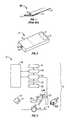
- FIG. 1shows an exemplary embodiment of a representative calorimetric reagent tester 80 employed in these embodiments of the subject invention.
- Tester 80is generally made up of at least the following components: a matrix 11 for receiving a sample, a reagent composition (not shown as a structural component) that typically includes one or more members of an analyte oxidation signal producing system and a support element 12 .
- FIG. 1shows tester 80 having matrix 11 positioned at one end of support element 12 with an adhesive 13 such that it is configured as a test strip.
- a hole 14is present in the support element 12 in the area of matrix 11 in which a sample can be applied to one side of the matrix 11 and a reaction can be detected on an opposite side of matrix 11 .
- Matrix 11that is employed in the testers is an inert matrix which provides a support for the various members of the signal producing system, described below, as well as the light absorbing or chromogenic product produced by the signal producing system, i.e., the indicator.
- Matrix 11is configured to provide a location for the physiological sample, e.g., blood, application and a location for the detection of the light-absorbing product produced by the indicator of the signal producing system.
- matrix 11is one that is permissive of aqueous fluid flow through it and provides sufficient void space for the chemical reactions of the signal producing system to take place.
- matriceshave been developed for use in various analyte detection assays, which matrices may differ in terms of materials, dimensions and the like, where representative matrices include, but are not limited to, those described in U.S. Pat. Nos.
- matrix 11is not critical to the tester and therefore is chosen with respect to other factors, including the nature of the instrument which is used to read the tester, convenience and the like.
- the dimensions and porosity of the matrixmay vary greatly, where matrix 11 may or may not have pores and/or a porosity gradient, e.g. with larger pores near or at the sample application region and smaller pores at the detection region.
- Materials from which matrix 11 may be fabricatedvary, and include polymers, e.g. polysulfone, polyamides, cellulose or absorbent paper, and the like, where the material may or may not be functionalized to provide for covalent or non-covalent attachment of the various members of the signal producing system.
- the testersfurther include one or more members of a signal producing system which produces a detectable product in response to the presence of analyte, which detectable product can be used to derive the amount of analyte present in the assayed sample.
- the one or more members of the signal producing systemare associated, e.g., covalently or non-covalently attached to, at least a portion of (i.e., the detection region) the matrix, and in many embodiments to substantially all of the matrix.
- the signal producing systemis an analyte oxidation signal producing system.
- analyte oxidation signal producing systemis meant that in generating the detectable signal from which the analyte concentration in the sample is derived, the analyte is oxidized by one or more suitable enzymes to produce an oxidized form of the analyte and a corresponding or proportional amount of hydrogen peroxide.
- the hydrogen peroxideis then employed, in turn, to generate the detectable product from one or more indicator compounds, where the amount of detectable product generated by the signal measuring system, i.e. the signal, is then related to the amount of analyte in the initial sample.
- the analyte oxidation signal producing systems present in the testersare also correctly characterized as hydrogen peroxide based signal producing systems.
- the hydrogen peroxide based signal producing systemsinclude an enzyme that oxidizes the analyte and produces a corresponding amount of hydrogen peroxide, whereby corresponding amount is meant that the amount of hydrogen peroxide that is produced is proportional to the amount of analyte present in the sample.
- This first enzymenecessarily depends on the nature of the analyte being assayed but is generally an oxidase.
- the first enzymemay be: glucose oxidase (where the analyte is glucose); cholesterol oxidase (where the analyte is cholesterol); alcohol oxidase (where the analyte is alcohol); lactate oxidase (where the analyte is lactate) and the like.
- Other oxidizing enzymes for use with these and other analytes of interestare known to those of skill in the art and may also be employed.
- the first enzymeis glucose oxidase.
- the glucose oxidasemay be obtained from any convenient source, e.g. a naturally occurring source such as Aspergillus niger or Penicillum, or recombinantly produced.
- a second enzyme of the signal producing systemmay be an enzyme that catalyzes the conversion of one or more indicator compounds into a detectable product in the presence of hydrogen peroxide, where the amount of detectable product that is produced by this reaction is proportional to the amount of hydrogen peroxide that is present.
- This second enzymeis generally a peroxidase, where suitable peroxidases include: horseradish peroxidase (HRP), soy peroxidase, recombinantly produced peroxidase and synthetic analogs having peroxidative activity and the like. See e.g., Y. Ci, F. Wang; Analytica Chimica Acta, 233 (1990), 299-302.
- the indicator compound or compounds, e.g., substratesare ones that are either formed or decomposed by the hydrogen peroxide in the presence of the peroxidase to produce an indicator dye that absorbs light in a predetermined wavelength range.
- the indicator dyeabsorbs strongly at a wavelength different from that at which the sample or the testing reagent absorbs strongly.
- the oxidized form of the indicatormay be a colored, faintly-colored, or colorless final product that evidences a change in color of the matrix. That is to say, the testing reagent can indicate the presence of glucose in a sample by a colored area being bleached or, alternatively, by a colorless area developing color.
- Indicator compounds that are useful in the present inventioninclude both one- and two-component chromogenic substrates.
- One-component systemsinclude aromatic amines, aromatic alcohols, azines, and benzidines, such as tetramethyl benzidine-HCl.
- Suitable two-component systemsinclude those in which one component is MBTH, an MBTH derivative (see for example those disclosed in U.S. patent application Ser. No. 08/302,575, incorporated herein by reference), or 4-aminoantipyrine and the other component is an aromatic amine, aromatic alcohol, conjugated amine, conjugated alcohol or aromatic or aliphatic aldehyde.
- Exemplary two-component systemsare 3-methyl-2-benzothiazolinone hydrazone hydrochloride (MBTH) combined with 3-dimethylaminobenzoic acid (DMAB); MBTH combined with 3,5-dichloro-2-hydroxybenzene-sulfonic acid (DCHBS); and 3-methyl-2-benzothiazolinonehydrazone N-sulfonyl benzenesulfonate monosodium (MBTHSB) combined with 8-anilino-1 naphthalene sulfonic acid ammonium (ANS).
- the dye couple MBTHSB-ANSis preferred.
- signal producing systemsthat produce a fluorescent detectable product (or detectable non-fluorescent substance, e.g. in a fluorescent background) may be employed, such as those described in: Kiyoshi Zaitsu, Yosuke Ohkura: New fluorogenic substrates for Horseradish Peroxidase: rapid and sensitive assay for hydrogen peroxide and the Peroxidase. Analytical Biochemistry (1980) 109, 109-113.
- Matrix 11is usually attached to a support element 12 .
- Support element 12may be of a material that is sufficiently rigid to be inserted into an automated device such as a meter without undue bending or kinking.
- Matrix 11may be attached to support element 12 by any convenient mechanisms, e.g., clamps, adhesive, etc., herein shown attached using an adhesive 13 .
- support member 12is made of material such as polyolefins, e.g., polyethylene or polypropylene, polystyrene or polyesters. The length of the support element 12 typically dictates or corresponds to the length of the tester.
- support element 12is usually configured to enable a tester to be used with or inserted into a meter.
- support element 12and thus tester may assume a variety of shapes and sizes, where the exact size and shape are dictated in part by the device with which the tester is used.
- sampleis allowed to react with the members of the signal producing system to produce a detectable product that is present in an amount proportional to the initial amount present in the sample.
- the amount of sample that is introduced to matrix 11 of the test stripmay vary, but generally ranges from 5.0 to about 10.0 ⁇ l.
- the samplemay be introduced to matrix 11 using any convenient protocol, where the sample may be injected, allowed to wick, or be otherwise introduced.
- the amount of detectable product, i.e., signal produced by the signal producing systemis then determined and related to the amount of analyte in the initial sample. See U.S. Pat. Nos.
- colorimetric reagent testerssuitable for use with the subject invention include, but are not limited to, those described in U.S. Pat. Nos. 5,049,487; 5,563,042; 5,753,452; 5,789,255, the disclosures of which are herein incorporated by reference.
- the subject inventionincludes analyte concentration determination devices, i.e., optical meter devices, that automatically determine the concentration of an analyte in a physiological sample applied to a tester, such as the type of tester described above or the like.
- analyte concentration determination devicesi.e., optical meter devices
- a feature of the subject devicesis that they are capable of determining whether a sufficient amount of sample is present at the incision site, i.e., at the surface if the skin where an incision has been made, to perform an accurate analyte concentration determination.
- the subject metersdetermine the sufficiency of sample size before any sample is contacted with a tester, thereby conserving testers for use only in instances where a sufficient amount of sample is present.
- the deviceis capable of bringing a tester into contact with the sufficient amount of sample at the site of the incision, thereby eliminating the need to move or transfer the sample to the site of the tester, which oftentimes results in significant loss of sample to the transfer element.
- FIG. 2shows a perspective view of the exterior of a subject device.
- device 2includes housing 4 having reporting element 6 positioned on the exterior thereof for communicating information to the user such as results of sample amount determination and analyte concentration.
- reporting element 6may take various hard copy and soft copy forms.
- a visual displaysuch as a liquid crystal display (LCD) or light emitting diode (LED) display, but it may also be a tape printer, audible signal, or the like.
- Housing 4also has aperture 8 positioned through a wall or side thereof to provide an opening from the interior to the exterior of the housing, for example to enable a lancet to protrude therethrough to make an incision in an area of skin and for sample collection and also for light to pass through.
- Housing 8may also include a physiological sample promoting element 10 that is typically positioned adjacent aperture 8 , and usually is configured as a ring or the like positioned around at least a part of the perimeter of aperture 8 , as will be described in greater detail below.
- Housing 4also has panel or cover 5 through which the interior of housing 4 may be accessed by the user, for example to load and/or remove testers and/or a disposable lancet therein. It will be apparent that other access means may be employed as well.
- Panel 5is constructed to be moveable from a closed to an opened position by any convenient means. For example, panel 5 may be slideably moved, hingedly affixed to housing 4 , etc.
- housing 4will necessarily vary depending on a variety of factors, where such factors include, but are not limited to, the type and size of the tester used therewith and the number of such testers that are stored in the meter, for example in a cartridge or casing or the like. Usually, housing 4 is shaped to be easily and comfortably, e.g., ergonomically, held in a user's hand.
- FIG. 2shows housing 4 having a rectangular shape, but other shapes are possible as well.
- housing 4may be of a square, cylindrical, circular, disc, or elliptical shape, etc., or substantially so.
- the shape of housing 4may be more complex such as a substantially irregular shape or the like.
- housing 4may also vary depending on a variety of factors such as the type and size and shape of the testers to be used therewith, and the number of testers held or accommodated in housing 4 , and the like. Usually, housing 4 is sized to be easily and comfortably held in a user's hand and easily transportable.
- Housing 4may be manufactured from a variety of materials, where such materials will not substantially interfere with the analyte concentration determination, e.g., will not substantially interfere with the reagents of the tester(s) held therein.
- Representative materials that may be used in the manufacture of the subject housinginclude, but are not limited to, polymeric materials such as polytetrafluoroethylene, polypropylene, polyethylene, polystyrene, polycarbonate and blends thereof, metals such as stainless steel, aluminum and alloys thereof, TeflonTM, siliceous material, e.g., glass materials, and the like.
- FIG. 3shows a schematic illustration of device 2 , and more specifically the internal components of housing 4 .
- housing 4is positioned on an area of skin S such that the area to be lanced is encompassed by aperture 8 .
- physiological sample promoting element 10Surrounding aperture 8 , as mentioned above, is optional physiological sample promoting element 10 that is configured to increase the amount of the physiological sample at the area of skin to be incised.
- Sample promoting element 10is usually configured as a ring or a partial ring that is capable of surrounding or substantially surrounding the area of skin to be incised to provide pressure to the surrounding area, thereby providing a bulged area to be incised, as shown.
- physiological fluidis displaced from the pressured area to areas adjacent the pressured areas, e.g., an area inside sample promoting ring 10 , i.e., the area to be incised, and the area outside sample promoting ring 10 , thereby engorging the area to be incised with physiological fluid.
- Pressuremay be applied by the user, for example the user may push down on the device when contacted with skin or may be actuated automatically.
- Housing 2also includes lancing element 23 configured to hold a disposable lancet 21 for making an incision in an area of skin to provide physiological sample for testing, where disposable lancets are known in the art and will not be described further herein.
- disposable lance 21is capable of being actuated, either manually for example by depressing a button on housing 4 or automatically for example once the meter is positioned substantially close to an area of skin. As such, lancet 21 is moved from a first, resting position away from the skin to a second displaced position through aperture 8 and in contact with the skin, where it is caused to penetrate the skin to provide an incision therein to provide physiological sample from the incised site for testing.
- lancing element 23includes lancet holder 23 a that retains disposable lancet 21 in a fixed position for lancing and lancet displacement mechanism 23 b for displacing disposable lancet 21 towards the skin.
- Lancet holder 23 aretains disposable lancet 21 using any suitable means such as friction, snap fit and the like, such that disposable lancet 21 is easily removable or ejectable for replacement with a new lancet, yet held firmly enough to avoid unintentional movement or ejection from lancet holder 23 a .
- Lancet displacement mechanism 23 bmay use any convenient mechanism for displacing a lancet towards the skin, where such mechanisms are well known in the art.
- Lancet displacement mechanism 23 bmay be actuated automatically or manually, for example by some simple user action. For example, the motions could occur when user pushes a button on the meter or simply presses the meter against the test site. As for the workings of a meter able to produce the desired action, the design and production of certain actuators is well within the level of skill in the art.
- lancet displacement mechanism 23 bis a spring mechanism such as a compression spring mechanism or the like (see for example U.S. Pat. No. 6,099,484, the disclosure of which is herein incorporated by reference). However, other suitable lancet displacement mechanisms may be employed and are well known in the art.
- lancing element 23 with disposable lancet 21is operatively positioned adjacent aperture 8 such that disposable lancet 21 is positioned in a first position away from the skin S, whereby upon actuation disposable lancet 21 is displaced to a second position through aperture 8 to make contact with and incise the skin.
- lancing element 23 and thus disposable lancet 21are positioned at an angle relative to aperture 8 , however it will be apparent that lancing element 23 and disposable lancet 21 may be positioned in any appropriate orientation relative to aperture 8 .
- Housing 4includes at least one light source 16 and typically also includes condensing lens 17 capable of focusing light from light source 16 to the area of aperture 8 .
- Light source 16projects light onto the area of the skin that has been incised by disposable lancet 12 , that is it projects light onto the area of skin encompassed by aperture 8 .
- Light source 16also projects light onto a tester, e.g., the matrix of a tester, having sample applied thereto and which has reagents for reacting with certain analytes in the sample, as described above.
- the same or different light sourcemay project light onto the skin as is used to project light onto a tester, where typically the same light source is used.
- light source 16typically includes a light emitting diode (LED) or any other convenient light source such as a laser diode, a filtered lamp, a phototransistor, and the like.
- the light source 16contains two or more LED sources, e.g., three LED sources, or a single diode capable of emitting two or more distinct wavelengths of light.
- the light source 16is usually capable of emitting light at wavelengths ranging from about 400 nm to about 1000 nm, usually from about 500 nm to about 940 nm.
- light source 16typically projects light at a wavelength of about 400 nm to about 1000 nm, more usually at about 480 nm to about 600 nm, where the hemoglobin with the blood sample absorbs light.
- the light source 16is capable of emitting light at about 635 nm and about 700 nm and in many embodiments the light source is capable of emitting light at about 660 nm and 940 nm, and in certain embodiments the light source is capable of emitting light at about 525 nm, 630 nm and 940 nm. It will be apparent that the wavelengths described herein are for exemplary purposes only and are in no way intended to limit the scope of the invention as many other combinations of wavelengths are possible as well.
- LYS A676 light sourcecapable of emitting light of 635 nm and 700 nm available from ASRAM Opto Semiconductor, Inc.
- Housing 4also includes at least one detector 20 for detecting light reflected from, i.e., intercepting reflected light, e.g., diffusely reflected light, the area of incised skin, for determining whether a sufficient amount of sample is present at the surface of the skin and for detecting light reflected, i.e., intercepting reflected light from, e.g., diffusely reflected light, a tester such as the matrix of a tester, for determining analyte concentration in a sample applied to the tester.
- the same or different detectormay detect light from the above-described areas.
- Housing 4may also include optional imaging optics 25 or an aperture (not shown) for imaging reflected light onto at least one detector 20 .
- the subject metersalso include means for determining whether a sufficient amount or volume of sample is present at the surface of the skin that has had an incision made therein, where such determination is based upon the amount of reflected light detected from each area.
- This meansis generally a digital integrated circuit 24 , where such a digital integrated circuit 24 is under the control of a software program and thus is suitably programmed to execute all of the steps or functions required of it to determine whether reflected light indicates a sufficient amount of sample, or any hardware or software combination that will perform such required functions.
- sample amount determination means 24is capable of executing or following an algorithm stored in the meter to determine, based on reflected light detected from an area of skin and more specifically an area of skin having physiological sample thereon, whether a sufficient amount of sample is present to perform an accurate analyte concentration determination test.
- Sample amount determination means 24usually reads the output of a signal conversion element such as analog/digital converter 22 which converts an analog signal from at least one detector 20 to a digital signal. Accordingly, sample amount determination means 24 is capable of carrying out all the steps necessary to determine whether reflected light detected from an area of skin indicates a sufficient amount of sample present in that area.
- the subject metersalso include means for determining the concentration of an analyte in the sample 26 , where such sample is contacted with a tester for analyte concentration determination. That is, if a sufficient amount of sample is determined to be present on the surface of the skin, the sample is contacted with a tester for analyte concentration determination, as will be described in greater detail below.
- This meansis generally a digital integrated circuit 26 , where such a digital integrated circuit 26 is under the control of a software program and thus is suitably programmed to execute all of the steps or functions required of it, or any hardware or software combination that will perform such required functions.
- analyte concentration determination means 26is capable of executing or following an algorithm stored in the meter to determine analyte concentration in a physiological; sample.
- Analyte concentration determination means 26is shown in FIG. 3 as a separate component from sample evaluation means 24 , but in certain embodiments means for determining whether a sufficient amount of sample is present at the surface of the skin and means for determining the concentration of an analyte may be the same integrated circuit.
- digital integrated circuit 26is capable of carrying out all the steps necessary to determine analyte concentration in a physiological sample.
- the subject metersalso include program and data memory 28 , which may be a digital integrated circuit, that stores data and the operating program of one or more of the digital integrated circuits of the meter.
- the subject metersalso include reporting device 6 , as described above, for communicating results of sample size sufficiency, analyte concentration, error messages, etc., to the user.
- the sampleis contacted with a tester so that the concentration of an analyte in the sample may be determined.
- a testersuch as the type of tester described above or any appropriate tester such as the type of tester described below, is placed in contact with the sufficient amount of sample.
- a testermay be manually placed in contact with the sample or automatically moved into contact with sample.
- the subject metersusually include means for retaining at least one tester within housing 4 , e.g., in an area or recess.
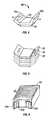
- FIG. 3shows an exemplary embodiment of tester cartridge or casing 29 having a plurality of testers 90 held therein, where tester 90 a is positioned to be grasped so that it may be moved into an appropriate position.
- FIG. 4shows an enlarged view of an exemplary embodiment of tester 90 suitable for use with the subject invention.
- tester 90includes matrix 99 having members of a signal producing system (not shown as a structural component), where matrix 99 is attached to support 92 .
- Support 92has upwardly biased ends 92 a and 92 b , where in many embodiment ends 92 a and 92 b have grasping holes 9 therein or other convenient means to enable easy grasping.
- support 92includes a window or transparent area or the like (not shown) positioned over matrix 99 to enable light to illuminate matrix 99 and to enable light to be detected from matrix 99 , through such a window or transparent area.
- tester 90may be placed over a physiological sample and the matrix may be “read” at the opposite side of the matrix through the window or transparent area.
- Such testersalso can be advantageously stacked for containment in a cartridge or casing, as shown in FIG. 5 , and shown stacked in cartridge 29 in FIG. 6 .
- a plurality of testers 90are retained within cartridge 29 and tester 90 a is positioned to be accessible so that it may be grasped and moved to contact a sample.
- Such a tester configuration and cartridgeare exemplary and in no way intended to limit the scope of the invention as other tester configurations and cartridges or containers housing such testers may be used with the present invention, as will be apparent to one of skill in the art.
- a testeris moved into contact with a sample determined to be present in a sufficient amount.
- the subject metersmay also include a tester movement element 27 .
- a testermay be moved in any convenient manner, where the following embodiments are provided by way of example and are in no way intended to limit the scope of the invention. In all such embodiments, a tester is moved in contact with sample (i.e., sample is not moved to contact the tester), thus sample is not lost in the transfer process, as is the case with many prior art devices. When contacted with the sample, the absorbent matrix of the tester absorbs essentially all of the sample from the site. In such a manner, a smaller amount of sample present at the surface of skin is required for an accurate analyte concentration measurement than in many prior art devices.
- FIGS. 7A-7FAn exemplary embodiment of a tester movement element suitable for use in the present invention is shown in FIGS. 7A-7F .
- Movement element 47is associated with a wall W of housing 4 such that cam member 42 is slideably engaged with groove 40 of wall W.
- Cam member 42is associated with one side of slideable movement member 43 by pin 45 , where tester grasping arm 46 is associated with the other side of slideable movement member 43 by pin 48 .
- slideable movement member 43slideably moves along bar 41 , in many embodiments which may be a groove or the like.
- tester movement element 43is actuated either manually or automatically, for example when a sufficient amount of sample is detected.
- tester movement element 43is moved automatically by way of a motor or the like, but may also be moved manually such as by the action of a user slideably moving a button or knob on an exterior surface of housing 4 which is operatively associated with tester movement element 43 .
- the steps of movement of such a testerare shown in FIGS. 7A-7F for moving tester 90 a from cartridge 29 , for example, to an area of skin S having physiological sample PS thereon.
- grasping arm 46is not shown physically associated with tester 90 a for the sake of showing an unobstructed view of the movement of tester 90 a . It will be apparent that grasping arm 46 is associated with tester 90 a , for example by holes 9 in tester 90 a in order to move tester 90 a to a site having physiological sample.
- cam member 42is positioned in a first position at the top of groove 40 and grasping arm 46 is operatively engaged with tester 90 a which is in cartridge 29 , but which is accessible to grasping arm 46 .
- Cam member 42slideably moves along groove 40 as slideable movement member 43 moves along bar 41 .
- grasping arm 46is caused to move tester 90 a in a direction towards physiological sample PS, as shown in FIGS.
- Grasping arm 46is configured to hold tester 90 a in contact with physiological sample PS and not obstruct the view of detector 20 or the path of the at least one light source 16 so that analyte concentration may be determined while the tester remains in the position, as shown in FIG. 7 F.
- Also provided by the subject inventionare methods for determining the concentration of an analyte in a physiological sample applied to a tester. More specifically, methods are provided that enable the determination of whether a sufficient amount of physiological sample is present at the surface of an incised area of skin to perform an analyte concentration determination assay and, if such a sufficient amount of sample is present, a tester is contacted with the sample and the concentration of an analyte in the sample is determined.
- the first stepis to lance an area of skin to provide physiological sample at the surface of the skin for testing.
- Any appropriate area of skinmay be lanced, where typically a finger, forearm, toe, or the like are used.
- a subject deviceas described above, having a disposable lancet 21 therein may be used to lance the skin by actuating lancing element 23 , thereby displacing lancet 21 towards the skin such that lancet 21 protrudes through aperture 8 of housing 4 to provide an opening or incision in the skin.
- sampleis caused to be present at the incised site by promoting the expression thereof.
- forcemay be applied to the area of skin surrounding the area of interest from which physiological fluid is desired, either before or after lancing. As such, the area of interest becomes engorged with physiological fluid. In such a manner, a greater amount of sample may be provided from the incision than would be provided without the application of force to the perimeter of the area of interest.
- sample promoting element 10may be used to promote sample at the site.
- the sampleis illuminated with light.
- the samplemay be illuminated with light at about 400 nm to about 1000 nm, usually about 480 nm to about 600 nm and more usually about 525 nm; however other wavelengths are possible as well as will be apparent to one of skill in the art.
- An important feature of the subject methodsis that the size of the sample is determined to be sufficient or insufficient before the sample is contacted with a tester, based on the amount of reflected light from the sample. In this manner, a tester is not wasted due to insufficiency of sample applied thereto.
- lightis reflected from the sample at the surface of the skin, where such light is detected and related to the amount of physiological sample present.
- skinreflects light at 525 nm
- hemoglobin present in a blood sampleabsorbs light at 525 nm.
- the reflectance at 525 nmis sufficiently low or below a predetermined value or the like or is sufficiently reduced from an initial measurement reflectance value, for example taken at a time prior when sample is present, i.e., a measurement of light reflected from skin without blood present, it is determined that a sufficient amount of sample is present.
- imaging optics or an aperturemay be used to image the reflected light onto a detector, as described above.
- the amount of sample determined to be sufficientwill vary depending on a variety of factors such as the analyte of interest, the tester, etc. Typically, an amount of sample at the surface of the skin ranging from about 0.5 ⁇ l to about 10 ⁇ l is determined to be sufficient for obtaining accurate analyte measurements.
- analyte concentrationis not performed with the present amount of sample.
- a usermay attempt to provide additional sample at the site, e.g., by milking the present incision or otherwise stimulating the site. If it is determined that a sufficient amount of sample is present at the surface of the skin, either initially or if additional sample has been provided after an initial determination of insufficiency as mentioned above, an analyte tester is contacted with the sufficient amount of sample at the surface of the skin. Accordingly, an important feature of the subject invention is that the sample is not moved to the site of the tester, rather the tester is moved to the site of the sample.
- sampleis not lost in the transfer process, for example to the sides of a capillary tube or needle or the like.
- a minimal amount of sampleis required at the surface of the skin for accurate analyte concentration determination, where the amount may be as little as about 0.5 ⁇ l, as described above.
- a testeris brought into contact with the sample such that sample is absorbed directly onto the tester, without the aid of a transfer tube or the like.
- the testeris usually moved into an operative position relative to the sample using tester movement element 27 .
- at least one testerusually a plurality of testers, is retained inside housing 4 , typically in a cartridge 29 or the like.
- Tester movement element 27engages a topmost or first tester 90 a positioned in cartridge 29 such that tester 90 a is accessible to tester movement element 27 and any remaining testers 90 are positioned or stacked inside cartridge 29 for use at a later time, where once tester 90 a is removed from cartridge 29 , the next tester positioned behind tester 90 a will move into position to be grasped and moved.
- test strip movement element 27grasps tester 90 a and moves it into contact with the physiological sample, (see FIGS. 7 A- 7 F).
- the sampleis allowed to react with the members of the signal producing system to produce a detectable product that is present in an amount proportional to the initial amount of analyte present in the sample.
- the amount of detectable producti.e., signal produced by the signal producing system, is then determined and related to the amount of analyte in the initial sample.
- means for determining the concentration of an analyte in the sample 26determines analyte concentration, as described above, where the results of the analyte concentration determination are communicated to a user by reporting element 6 .
- kits for practicing the subject methodsinclude a device according to the subject invention, i.e., a subject optical meter.
- the subject kitsmay also include one or more testers, usually a plurality of testers retained in a cartridge or the like, such as the type of tester described above.
- the subject kitsmay further include one or more disposable lancets.
- the subject kitsmay include a control solution or standard, e.g., a control solution that has a known analyte concentration such as a known glucose concentration.
- the kitsmay further include instructions for using the apparatus for determining the presence and/or concentration of an analyte in a physiological sample applied to a tester.
- the instructionsmay be printed on a substrate, such as paper or plastic, etc.
- the instructionsmay be present in the kits as a package insert, in the labeling of the container of the kit or components thereof (i.e., associated with the packaging or sub-packaging) etc.
- the instructionsare present as an electronic storage data file present on a suitable computer readable storage medium, e.g., CD-ROM, diskette, etc.
- the above described inventionprovides a simple, quick and convenient way to determine whether a sufficient amount of sample is present for analyte concentration determination and to determine analyte concentration in a sample determined to be present in a sufficient amount.
- the above described inventionprovides a number of advantages, including, but not limited to, integration of several testing components in a single, hand-held device, ease of use, determination of whether a sufficient amount of sample is present before contacting the sample with a tester and accurate analyte concentration determination using minimal sample amounts.
- the subject inventionrepresents a significant contribution to the art.
Landscapes
- Health & Medical Sciences (AREA)
- Life Sciences & Earth Sciences (AREA)
- Engineering & Computer Science (AREA)
- Molecular Biology (AREA)
- Animal Behavior & Ethology (AREA)
- Pathology (AREA)
- Physics & Mathematics (AREA)
- Biomedical Technology (AREA)
- Heart & Thoracic Surgery (AREA)
- Medical Informatics (AREA)
- Hematology (AREA)
- Surgery (AREA)
- Biophysics (AREA)
- General Health & Medical Sciences (AREA)
- Public Health (AREA)
- Veterinary Medicine (AREA)
- Diabetes (AREA)
- Manufacturing & Machinery (AREA)
- Measurement Of The Respiration, Hearing Ability, Form, And Blood Characteristics Of Living Organisms (AREA)
- Investigating Or Analysing Biological Materials (AREA)
- Investigating Or Analysing Materials By Optical Means (AREA)
- Investigating, Analyzing Materials By Fluorescence Or Luminescence (AREA)
Abstract
Description
Claims (4)
Priority Applications (11)
| Application Number | Priority Date | Filing Date | Title |
|---|---|---|---|
| US10/137,598US6945943B2 (en) | 2002-05-01 | 2002-05-01 | Analyte concentration determination devices and methods of using the same |
| IL15534903AIL155349A0 (en) | 2002-05-01 | 2003-04-10 | Analyte concentration determination devices and methods of using the same |
| SG200302278ASG108916A1 (en) | 2002-05-01 | 2003-04-22 | Analyte concentration determination devices and methods of using the same |
| CA002427092ACA2427092A1 (en) | 2002-05-01 | 2003-04-28 | Analyte concentration determination devices and methods of using the same |
| CN03128423ACN1455258A (en) | 2002-05-01 | 2003-04-29 | Apparatus for detecting concentration of matter to be analysed and using method thereof |
| AT03252734TATE369560T1 (en) | 2002-05-01 | 2003-04-30 | DEVICE AND METHOD FOR DETERMINING THE CONCENTRATION OF ANALYTES |
| EP03252734AEP1359418B1 (en) | 2002-05-01 | 2003-04-30 | Analyte concentration determination devices and methods of using the same |
| TW092110085ATW200403040A (en) | 2002-05-01 | 2003-04-30 | Analyte concentration determination devices and methods of using the same |
| JP2003125642AJP2004113770A (en) | 2002-05-01 | 2003-04-30 | Analyte concentration determination apparatus and using method therefor |
| DE60315373TDE60315373T2 (en) | 2002-05-01 | 2003-04-30 | Apparatus and method for determining the concentration of analytes |
| HK04100105.8AHK1057254B (en) | 2002-05-01 | 2004-01-07 | Analyte concentration determination devices and methods of using the same |
Applications Claiming Priority (1)
| Application Number | Priority Date | Filing Date | Title |
|---|---|---|---|
| US10/137,598US6945943B2 (en) | 2002-05-01 | 2002-05-01 | Analyte concentration determination devices and methods of using the same |
Publications (2)
| Publication Number | Publication Date |
|---|---|
| US20030208140A1 US20030208140A1 (en) | 2003-11-06 |
| US6945943B2true US6945943B2 (en) | 2005-09-20 |
Family
ID=29215701
Family Applications (1)
| Application Number | Title | Priority Date | Filing Date |
|---|---|---|---|
| US10/137,598Expired - LifetimeUS6945943B2 (en) | 2002-05-01 | 2002-05-01 | Analyte concentration determination devices and methods of using the same |
Country Status (10)
| Country | Link |
|---|---|
| US (1) | US6945943B2 (en) |
| EP (1) | EP1359418B1 (en) |
| JP (1) | JP2004113770A (en) |
| CN (1) | CN1455258A (en) |
| AT (1) | ATE369560T1 (en) |
| CA (1) | CA2427092A1 (en) |
| DE (1) | DE60315373T2 (en) |
| IL (1) | IL155349A0 (en) |
| SG (1) | SG108916A1 (en) |
| TW (1) | TW200403040A (en) |
Cited By (65)
| Publication number | Priority date | Publication date | Assignee | Title |
|---|---|---|---|---|
| US7297151B2 (en) | 2002-04-19 | 2007-11-20 | Elikan Technologies, Inc. | Method and apparatus for body fluid sampling with improved sensing |
| US7316700B2 (en) | 2001-06-12 | 2008-01-08 | Pelikan Technologies, Inc. | Self optimizing lancing device with adaptation means to temporal variations in cutaneous properties |
| US7344894B2 (en) | 2001-10-16 | 2008-03-18 | Agilent Technologies, Inc. | Thermal regulation of fluidic samples within a diagnostic cartridge |
| US7344507B2 (en) | 2002-04-19 | 2008-03-18 | Pelikan Technologies, Inc. | Method and apparatus for lancet actuation |
| US7374544B2 (en) | 2002-04-19 | 2008-05-20 | Pelikan Technologies, Inc. | Method and apparatus for penetrating tissue |
| US7410468B2 (en) | 2002-04-19 | 2008-08-12 | Pelikan Technologies, Inc. | Method and apparatus for penetrating tissue |
| US7481776B2 (en) | 2002-04-19 | 2009-01-27 | Pelikan Technologies, Inc. | Method and apparatus for penetrating tissue |
| US7491178B2 (en) | 2002-04-19 | 2009-02-17 | Pelikan Technologies, Inc. | Method and apparatus for penetrating tissue |
| US7524293B2 (en) | 2002-04-19 | 2009-04-28 | Pelikan Technologies, Inc. | Method and apparatus for penetrating tissue |
| US7537571B2 (en) | 2001-06-12 | 2009-05-26 | Pelikan Technologies, Inc. | Integrated blood sampling analysis system with multi-use sampling module |
| US7547287B2 (en) | 2002-04-19 | 2009-06-16 | Pelikan Technologies, Inc. | Method and apparatus for penetrating tissue |
| US7563232B2 (en) | 2002-04-19 | 2009-07-21 | Pelikan Technologies, Inc. | Method and apparatus for penetrating tissue |
| US7582099B2 (en) | 2002-04-19 | 2009-09-01 | Pelikan Technologies, Inc | Method and apparatus for penetrating tissue |
| US7582063B2 (en) | 2000-11-21 | 2009-09-01 | Pelikan Technologies, Inc. | Blood testing apparatus having a rotatable cartridge with multiple lancing elements and testing means |
| US7604592B2 (en) | 2003-06-13 | 2009-10-20 | Pelikan Technologies, Inc. | Method and apparatus for a point of care device |
| US7648468B2 (en) | 2002-04-19 | 2010-01-19 | Pelikon Technologies, Inc. | Method and apparatus for penetrating tissue |
| US7666149B2 (en) | 1997-12-04 | 2010-02-23 | Peliken Technologies, Inc. | Cassette of lancet cartridges for sampling blood |
| US7674232B2 (en) | 2002-04-19 | 2010-03-09 | Pelikan Technologies, Inc. | Method and apparatus for penetrating tissue |
| US7682318B2 (en) | 2001-06-12 | 2010-03-23 | Pelikan Technologies, Inc. | Blood sampling apparatus and method |
| US7699791B2 (en) | 2001-06-12 | 2010-04-20 | Pelikan Technologies, Inc. | Method and apparatus for improving success rate of blood yield from a fingerstick |
| US7713214B2 (en) | 2002-04-19 | 2010-05-11 | Pelikan Technologies, Inc. | Method and apparatus for a multi-use body fluid sampling device with optical analyte sensing |
| US7717863B2 (en) | 2002-04-19 | 2010-05-18 | Pelikan Technologies, Inc. | Method and apparatus for penetrating tissue |
| US7731729B2 (en) | 2002-04-19 | 2010-06-08 | Pelikan Technologies, Inc. | Method and apparatus for penetrating tissue |
| US7822454B1 (en) | 2005-01-03 | 2010-10-26 | Pelikan Technologies, Inc. | Fluid sampling device with improved analyte detecting member configuration |
| US7833171B2 (en) | 2002-04-19 | 2010-11-16 | Pelikan Technologies, Inc. | Method and apparatus for penetrating tissue |
| US7841992B2 (en) | 2001-06-12 | 2010-11-30 | Pelikan Technologies, Inc. | Tissue penetration device |
| US7850621B2 (en) | 2003-06-06 | 2010-12-14 | Pelikan Technologies, Inc. | Method and apparatus for body fluid sampling and analyte sensing |
| US7862520B2 (en) | 2002-04-19 | 2011-01-04 | Pelikan Technologies, Inc. | Body fluid sampling module with a continuous compression tissue interface surface |
| US7874994B2 (en) | 2002-04-19 | 2011-01-25 | Pelikan Technologies, Inc. | Method and apparatus for penetrating tissue |
| US20110019196A1 (en)* | 2002-12-31 | 2011-01-27 | Yin-Chun Huang | Apparatus for testing component concentration of a test sample |
| US7892183B2 (en) | 2002-04-19 | 2011-02-22 | Pelikan Technologies, Inc. | Method and apparatus for body fluid sampling and analyte sensing |
| US7901362B2 (en) | 2002-04-19 | 2011-03-08 | Pelikan Technologies, Inc. | Method and apparatus for penetrating tissue |
| US7909778B2 (en) | 2002-04-19 | 2011-03-22 | Pelikan Technologies, Inc. | Method and apparatus for penetrating tissue |
| US7909775B2 (en) | 2001-06-12 | 2011-03-22 | Pelikan Technologies, Inc. | Method and apparatus for lancet launching device integrated onto a blood-sampling cartridge |
| US7914465B2 (en) | 2002-04-19 | 2011-03-29 | Pelikan Technologies, Inc. | Method and apparatus for penetrating tissue |
| US7959582B2 (en) | 2002-04-19 | 2011-06-14 | Pelikan Technologies, Inc. | Method and apparatus for penetrating tissue |
| US7976476B2 (en) | 2002-04-19 | 2011-07-12 | Pelikan Technologies, Inc. | Device and method for variable speed lancet |
| US8197421B2 (en) | 2002-04-19 | 2012-06-12 | Pelikan Technologies, Inc. | Method and apparatus for penetrating tissue |
| US8221334B2 (en) | 2002-04-19 | 2012-07-17 | Sanofi-Aventis Deutschland Gmbh | Method and apparatus for penetrating tissue |
| US8267870B2 (en) | 2002-04-19 | 2012-09-18 | Sanofi-Aventis Deutschland Gmbh | Method and apparatus for body fluid sampling with hybrid actuation |
| US8282576B2 (en) | 2003-09-29 | 2012-10-09 | Sanofi-Aventis Deutschland Gmbh | Method and apparatus for an improved sample capture device |
| US8333710B2 (en) | 2002-04-19 | 2012-12-18 | Sanofi-Aventis Deutschland Gmbh | Tissue penetration device |
| US8435190B2 (en) | 2002-04-19 | 2013-05-07 | Sanofi-Aventis Deutschland Gmbh | Method and apparatus for penetrating tissue |
| US8439872B2 (en) | 1998-03-30 | 2013-05-14 | Sanofi-Aventis Deutschland Gmbh | Apparatus and method for penetration with shaft having a sensor for sensing penetration depth |
| US8652831B2 (en) | 2004-12-30 | 2014-02-18 | Sanofi-Aventis Deutschland Gmbh | Method and apparatus for analyte measurement test time |
| US8668656B2 (en) | 2003-12-31 | 2014-03-11 | Sanofi-Aventis Deutschland Gmbh | Method and apparatus for improving fluidic flow and sample capture |
| US8702624B2 (en) | 2006-09-29 | 2014-04-22 | Sanofi-Aventis Deutschland Gmbh | Analyte measurement device with a single shot actuator |
| US8721671B2 (en) | 2001-06-12 | 2014-05-13 | Sanofi-Aventis Deutschland Gmbh | Electric lancet actuator |
| US8828203B2 (en) | 2004-05-20 | 2014-09-09 | Sanofi-Aventis Deutschland Gmbh | Printable hydrogels for biosensors |
| US8965476B2 (en) | 2010-04-16 | 2015-02-24 | Sanofi-Aventis Deutschland Gmbh | Tissue penetration device |
| US9034639B2 (en) | 2002-12-30 | 2015-05-19 | Sanofi-Aventis Deutschland Gmbh | Method and apparatus using optical techniques to measure analyte levels |
| US9072842B2 (en) | 2002-04-19 | 2015-07-07 | Sanofi-Aventis Deutschland Gmbh | Method and apparatus for penetrating tissue |
| US9144401B2 (en) | 2003-06-11 | 2015-09-29 | Sanofi-Aventis Deutschland Gmbh | Low pain penetrating member |
| US9226699B2 (en) | 2002-04-19 | 2016-01-05 | Sanofi-Aventis Deutschland Gmbh | Body fluid sampling module with a continuous compression tissue interface surface |
| US9248267B2 (en) | 2002-04-19 | 2016-02-02 | Sanofi-Aventis Deustchland Gmbh | Tissue penetration device |
| US9314194B2 (en) | 2002-04-19 | 2016-04-19 | Sanofi-Aventis Deutschland Gmbh | Tissue penetration device |
| US9351680B2 (en) | 2003-10-14 | 2016-05-31 | Sanofi-Aventis Deutschland Gmbh | Method and apparatus for a variable user interface |
| US9375169B2 (en) | 2009-01-30 | 2016-06-28 | Sanofi-Aventis Deutschland Gmbh | Cam drive for managing disposable penetrating member actions with a single motor and motor and control system |
| US9386944B2 (en) | 2008-04-11 | 2016-07-12 | Sanofi-Aventis Deutschland Gmbh | Method and apparatus for analyte detecting device |
| US9427532B2 (en) | 2001-06-12 | 2016-08-30 | Sanofi-Aventis Deutschland Gmbh | Tissue penetration device |
| US9560993B2 (en) | 2001-11-21 | 2017-02-07 | Sanofi-Aventis Deutschland Gmbh | Blood testing apparatus having a rotatable cartridge with multiple lancing elements and testing means |
| US9795747B2 (en) | 2010-06-02 | 2017-10-24 | Sanofi-Aventis Deutschland Gmbh | Methods and apparatus for lancet actuation |
| US9820684B2 (en) | 2004-06-03 | 2017-11-21 | Sanofi-Aventis Deutschland Gmbh | Method and apparatus for a fluid sampling device |
| US9839386B2 (en) | 2002-04-19 | 2017-12-12 | Sanofi-Aventis Deustschland Gmbh | Body fluid sampling device with capacitive sensor |
| US10371663B2 (en) | 2010-12-31 | 2019-08-06 | Lifescan Ip Holdings, Llc | Systems and methods for high accuracy analyte measurement |
Families Citing this family (41)
| Publication number | Priority date | Publication date | Assignee | Title |
|---|---|---|---|---|
| US20020010406A1 (en) | 1996-05-17 | 2002-01-24 | Douglas Joel S. | Methods and apparatus for expressing body fluid from an incision |
| EP1579814A3 (en) | 1996-05-17 | 2006-06-14 | Roche Diagnostics Operations, Inc. | Methods and apparatus for sampling and analyzing body fluid |
| US7828749B2 (en) | 1996-05-17 | 2010-11-09 | Roche Diagnostics Operations, Inc. | Blood and interstitial fluid sampling device |
| US7235056B2 (en) | 1996-05-17 | 2007-06-26 | Amira Medical | Body fluid sampling device and methods of use |
| US8372016B2 (en) | 2002-04-19 | 2013-02-12 | Sanofi-Aventis Deutschland Gmbh | Method and apparatus for body fluid sampling and analyte sensing |
| WO2003088824A2 (en) | 2002-04-19 | 2003-10-30 | Pelikan Technologies, Inc. | Device and method for variable speed lancet |
| US7485128B2 (en) | 2002-04-19 | 2009-02-03 | Pelikan Technologies, Inc. | Method and apparatus for penetrating tissue |
| US8360992B2 (en) | 2002-04-19 | 2013-01-29 | Sanofi-Aventis Deutschland Gmbh | Method and apparatus for penetrating tissue |
| US7244265B2 (en) | 2002-04-19 | 2007-07-17 | Pelikan Technologies, Inc. | Method and apparatus for penetrating tissue |
| US7815579B2 (en)* | 2005-03-02 | 2010-10-19 | Roche Diagnostics Operations, Inc. | Dynamic integrated lancing test strip with sterility cover |
| DE602004028463D1 (en) | 2003-05-30 | 2010-09-16 | Pelikan Technologies Inc | METHOD AND DEVICE FOR INJECTING LIQUID |
| US8394328B2 (en) | 2003-12-31 | 2013-03-12 | Nipro Diagnostics, Inc. | Test strip container with integrated meter having strip coding capability |
| US8147426B2 (en) | 2003-12-31 | 2012-04-03 | Nipro Diagnostics, Inc. | Integrated diagnostic test system |
| US9012232B2 (en) | 2005-07-15 | 2015-04-21 | Nipro Diagnostics, Inc. | Diagnostic strip coding system and related methods of use |
| US8394337B2 (en) | 2003-12-31 | 2013-03-12 | Nipro Diagnostics, Inc. | Test strip container with integrated meter |
| EP1737345A1 (en)* | 2004-04-15 | 2007-01-03 | Roche Diagnostics GmbH | Integrated spot monitoring device with fluid sensor |
| USD506832S1 (en) | 2004-06-02 | 2005-06-28 | Home Diagnostics, Inc. | Meter for an integrated diagnostic test system |
| USD507657S1 (en) | 2004-06-02 | 2005-07-19 | Home Diagnostics, Inc. | Meter for an integrated diagnostic test system |
| US9775553B2 (en) | 2004-06-03 | 2017-10-03 | Sanofi-Aventis Deutschland Gmbh | Method and apparatus for a fluid sampling device |
| AU2005202624A1 (en)* | 2004-06-28 | 2006-01-12 | Lifescan Scotland Limited | Resealable vial and dispensing mechanism for test sensor |
| US20060100655A1 (en)* | 2004-10-28 | 2006-05-11 | Koon-Wah Leong | Combined lancing and auxiliary device |
| USD561904S1 (en) | 2005-01-31 | 2008-02-12 | Home Diagnostics, Inc. | Meter for an integrated diagnostic test system |
| US20060275890A1 (en)* | 2005-06-06 | 2006-12-07 | Home Diagnostics, Inc. | Method of manufacturing a disposable diagnostic meter |
| US8999125B2 (en) | 2005-07-15 | 2015-04-07 | Nipro Diagnostics, Inc. | Embedded strip lot autocalibration |
| US7955856B2 (en) | 2005-07-15 | 2011-06-07 | Nipro Diagnostics, Inc. | Method of making a diagnostic test strip having a coding system |
| EP1815790A1 (en)* | 2006-02-04 | 2007-08-08 | Roche Diagnostics GmbH | Lancet device with impedance measuring unit |
| EP1982653A1 (en)* | 2007-04-18 | 2008-10-22 | Roche Diagnostics GmbH | Pricking device and analysis device |
| EP3984454A1 (en) | 2008-06-06 | 2022-04-20 | Intuity Medical, Inc. | Medical diagnostic devices and methods |
| WO2009148624A1 (en)* | 2008-06-06 | 2009-12-10 | Intuity Medical, Inc. | Detection meter and mode of operation |
| EP2506768B1 (en) | 2009-11-30 | 2016-07-06 | Intuity Medical, Inc. | Calibration material delivery devices and methods |
| WO2013041704A1 (en)* | 2011-09-22 | 2013-03-28 | Sanofi-Aventis Deutschland Gmbh | Detecting a blood sample |
| WO2014205412A1 (en) | 2013-06-21 | 2014-12-24 | Intuity Medical, Inc. | Analyte monitoring system with audible feedback |
| US10831013B2 (en) | 2013-08-26 | 2020-11-10 | S.D. Sight Diagnostics Ltd. | Digital microscopy systems, methods and computer program products |
| CA3018536A1 (en) | 2016-03-30 | 2017-10-05 | S.D. Sight Diagnostics Ltd | Distinguishing between blood sample components |
| GB2590813B (en)* | 2017-01-10 | 2021-10-27 | Drawbridge Health Inc | Devices, systems, and methods for sample collection |
| CA3053301A1 (en)* | 2017-02-16 | 2018-08-23 | Essenlix Corporation | Assay with textured surface |
| AU2018369859B2 (en)* | 2017-11-14 | 2024-01-25 | S.D. Sight Diagnostics Ltd | Sample carrier for optical measurements |
| JP2020051800A (en)* | 2018-09-25 | 2020-04-02 | 株式会社日立ハイテクノロジーズ | Automatic analyzer |
| EP3656303B1 (en)* | 2018-11-22 | 2021-06-30 | Roche Diabetes Care GmbH | Blood withdrawal system with lancet ejection mechanism |
| WO2021116959A1 (en) | 2019-12-12 | 2021-06-17 | S.D. Sight Diagnostics Ltd | Analyzing an analyte disposed within a medium |
| EP4073566B1 (en) | 2019-12-12 | 2025-04-23 | S.D. Sight Diagnostics Ltd. | Artificial generation of color blood smear image |
Citations (26)
| Publication number | Priority date | Publication date | Assignee | Title |
|---|---|---|---|---|
| US720906A (en) | 1902-11-17 | 1903-02-17 | Charles Eilrich | Trench-filler. |
| US4553541A (en) | 1981-03-23 | 1985-11-19 | Becton, Dickinson And Co. | Automatic retractable lancet assembly |
| US4637403A (en) | 1985-04-08 | 1987-01-20 | Garid, Inc. | Glucose medical monitoring system |
| US4648408A (en) | 1984-05-11 | 1987-03-10 | Medscan B.V. | Blood sampling unit |
| US4787398A (en) | 1985-04-08 | 1988-11-29 | Garid, Inc. | Glucose medical monitoring system |
| US5212879A (en) | 1991-05-15 | 1993-05-25 | International Technidyne Corp. | Method for manufacturing a disposable-retractable finger stick device |
| US5231993A (en) | 1991-11-20 | 1993-08-03 | Habley Medical Technology Corporation | Blood sampler and component tester with guide member |
| US5314441A (en) | 1992-10-16 | 1994-05-24 | International Technidyne Corporation | Disposable slicing lancet assembly |
| US5395387A (en) | 1993-02-26 | 1995-03-07 | Becton Dickinson And Company | Lancet blade designed for reduced pain |
| US5439473A (en) | 1993-12-13 | 1995-08-08 | Modulohm A/S | Safety lancet |
| US5476474A (en) | 1994-07-27 | 1995-12-19 | Ryder International Corporation | Rotary lancet |
| US5529581A (en) | 1994-05-17 | 1996-06-25 | International Technidyne Corporation | Lancet device for creating a skin incision |
| US5575403A (en) | 1995-01-13 | 1996-11-19 | Bayer Corporation | Dispensing instrument for fluid monitoring sensors |
| US5757666A (en) | 1993-04-23 | 1998-05-26 | Boehringer Mannheim Gmbh | System for analyzing compounds contained liquid samples |
| US5820570A (en) | 1993-10-13 | 1998-10-13 | Integ Incorporated | Interstitial fluid collection and constituent measurement |
| US5879367A (en) | 1995-09-08 | 1999-03-09 | Integ, Inc. | Enhanced interstitial fluid collection |
| WO1999044508A1 (en) | 1998-03-06 | 1999-09-10 | Spectrx, Inc. | Integrated poration, harvesting and analysis device, and method therefor |
| US5951493A (en) | 1997-05-16 | 1999-09-14 | Mercury Diagnostics, Inc. | Methods and apparatus for expressing body fluid from an incision |
| US5951492A (en) | 1996-05-17 | 1999-09-14 | Mercury Diagnostics, Inc. | Methods and apparatus for sampling and analyzing body fluid |
| US5971941A (en) | 1997-12-04 | 1999-10-26 | Hewlett-Packard Company | Integrated system and method for sampling blood and analysis |
| US6027459A (en) | 1996-12-06 | 2000-02-22 | Abbott Laboratories | Method and apparatus for obtaining blood for diagnostic tests |
| US6099484A (en) | 1996-05-17 | 2000-08-08 | Amira Medical | Methods and apparatus for sampling and analyzing body fluid |
| US6228100B1 (en) | 1999-10-25 | 2001-05-08 | Steven Schraga | Multi-use lancet device |
| WO2002008753A2 (en) | 2000-07-20 | 2002-01-31 | Hypoguard Limited | Device for measuring analyte concentration in a fluid |
| US20030069509A1 (en)* | 2001-10-10 | 2003-04-10 | David Matzinger | Devices for physiological fluid sampling and methods of using the same |
| US20030191415A1 (en)* | 2001-03-29 | 2003-10-09 | Piet Moerman | Integrated sample testing meter |
- 2002
- 2002-05-01USUS10/137,598patent/US6945943B2/ennot_activeExpired - Lifetime
- 2003
- 2003-04-10ILIL15534903Apatent/IL155349A0/enunknown
- 2003-04-22SGSG200302278Apatent/SG108916A1/enunknown
- 2003-04-28CACA002427092Apatent/CA2427092A1/ennot_activeAbandoned
- 2003-04-29CNCN03128423Apatent/CN1455258A/enactivePending
- 2003-04-30JPJP2003125642Apatent/JP2004113770A/ennot_activeAbandoned
- 2003-04-30EPEP03252734Apatent/EP1359418B1/ennot_activeExpired - Lifetime
- 2003-04-30TWTW092110085Apatent/TW200403040A/enunknown
- 2003-04-30ATAT03252734Tpatent/ATE369560T1/ennot_activeIP Right Cessation
- 2003-04-30DEDE60315373Tpatent/DE60315373T2/ennot_activeExpired - Lifetime
Patent Citations (28)
| Publication number | Priority date | Publication date | Assignee | Title |
|---|---|---|---|---|
| US720906A (en) | 1902-11-17 | 1903-02-17 | Charles Eilrich | Trench-filler. |
| US4553541A (en) | 1981-03-23 | 1985-11-19 | Becton, Dickinson And Co. | Automatic retractable lancet assembly |
| US4648408A (en) | 1984-05-11 | 1987-03-10 | Medscan B.V. | Blood sampling unit |
| US4637403A (en) | 1985-04-08 | 1987-01-20 | Garid, Inc. | Glucose medical monitoring system |
| US4787398A (en) | 1985-04-08 | 1988-11-29 | Garid, Inc. | Glucose medical monitoring system |
| US5212879A (en) | 1991-05-15 | 1993-05-25 | International Technidyne Corp. | Method for manufacturing a disposable-retractable finger stick device |
| US5231993A (en) | 1991-11-20 | 1993-08-03 | Habley Medical Technology Corporation | Blood sampler and component tester with guide member |
| US5314441A (en) | 1992-10-16 | 1994-05-24 | International Technidyne Corporation | Disposable slicing lancet assembly |
| US5395387A (en) | 1993-02-26 | 1995-03-07 | Becton Dickinson And Company | Lancet blade designed for reduced pain |
| US5757666A (en) | 1993-04-23 | 1998-05-26 | Boehringer Mannheim Gmbh | System for analyzing compounds contained liquid samples |
| US5820570A (en) | 1993-10-13 | 1998-10-13 | Integ Incorporated | Interstitial fluid collection and constituent measurement |
| US5439473A (en) | 1993-12-13 | 1995-08-08 | Modulohm A/S | Safety lancet |
| US5529581A (en) | 1994-05-17 | 1996-06-25 | International Technidyne Corporation | Lancet device for creating a skin incision |
| US5476474A (en) | 1994-07-27 | 1995-12-19 | Ryder International Corporation | Rotary lancet |
| US5575403A (en) | 1995-01-13 | 1996-11-19 | Bayer Corporation | Dispensing instrument for fluid monitoring sensors |
| US5879367A (en) | 1995-09-08 | 1999-03-09 | Integ, Inc. | Enhanced interstitial fluid collection |
| US6099484A (en) | 1996-05-17 | 2000-08-08 | Amira Medical | Methods and apparatus for sampling and analyzing body fluid |
| US5951492A (en) | 1996-05-17 | 1999-09-14 | Mercury Diagnostics, Inc. | Methods and apparatus for sampling and analyzing body fluid |
| EP1112717A1 (en) | 1996-12-06 | 2001-07-04 | Abbott Laboratories | Apparatus suitable for obtaining blood samples in a diagnostic test |
| US6027459A (en) | 1996-12-06 | 2000-02-22 | Abbott Laboratories | Method and apparatus for obtaining blood for diagnostic tests |
| US5951493A (en) | 1997-05-16 | 1999-09-14 | Mercury Diagnostics, Inc. | Methods and apparatus for expressing body fluid from an incision |
| US6071250A (en) | 1997-05-16 | 2000-06-06 | Amira Medical | Methods and apparatus for expressing body fluid from an incision |
| US5971941A (en) | 1997-12-04 | 1999-10-26 | Hewlett-Packard Company | Integrated system and method for sampling blood and analysis |
| WO1999044508A1 (en) | 1998-03-06 | 1999-09-10 | Spectrx, Inc. | Integrated poration, harvesting and analysis device, and method therefor |
| US6228100B1 (en) | 1999-10-25 | 2001-05-08 | Steven Schraga | Multi-use lancet device |
| WO2002008753A2 (en) | 2000-07-20 | 2002-01-31 | Hypoguard Limited | Device for measuring analyte concentration in a fluid |
| US20030191415A1 (en)* | 2001-03-29 | 2003-10-09 | Piet Moerman | Integrated sample testing meter |
| US20030069509A1 (en)* | 2001-10-10 | 2003-04-10 | David Matzinger | Devices for physiological fluid sampling and methods of using the same |
Cited By (118)
| Publication number | Priority date | Publication date | Assignee | Title |
|---|---|---|---|---|
| US7666149B2 (en) | 1997-12-04 | 2010-02-23 | Peliken Technologies, Inc. | Cassette of lancet cartridges for sampling blood |
| US8439872B2 (en) | 1998-03-30 | 2013-05-14 | Sanofi-Aventis Deutschland Gmbh | Apparatus and method for penetration with shaft having a sensor for sensing penetration depth |
| US7582063B2 (en) | 2000-11-21 | 2009-09-01 | Pelikan Technologies, Inc. | Blood testing apparatus having a rotatable cartridge with multiple lancing elements and testing means |
| US8845550B2 (en) | 2001-06-12 | 2014-09-30 | Sanofi-Aventis Deutschland Gmbh | Tissue penetration device |
| US7682318B2 (en) | 2001-06-12 | 2010-03-23 | Pelikan Technologies, Inc. | Blood sampling apparatus and method |
| US9802007B2 (en) | 2001-06-12 | 2017-10-31 | Sanofi-Aventis Deutschland Gmbh | Methods and apparatus for lancet actuation |
| US9694144B2 (en) | 2001-06-12 | 2017-07-04 | Sanofi-Aventis Deutschland Gmbh | Sampling module device and method |
| US7981055B2 (en) | 2001-06-12 | 2011-07-19 | Pelikan Technologies, Inc. | Tissue penetration device |
| US9427532B2 (en) | 2001-06-12 | 2016-08-30 | Sanofi-Aventis Deutschland Gmbh | Tissue penetration device |
| US7537571B2 (en) | 2001-06-12 | 2009-05-26 | Pelikan Technologies, Inc. | Integrated blood sampling analysis system with multi-use sampling module |
| US8721671B2 (en) | 2001-06-12 | 2014-05-13 | Sanofi-Aventis Deutschland Gmbh | Electric lancet actuator |
| US8679033B2 (en) | 2001-06-12 | 2014-03-25 | Sanofi-Aventis Deutschland Gmbh | Tissue penetration device |
| US8641643B2 (en) | 2001-06-12 | 2014-02-04 | Sanofi-Aventis Deutschland Gmbh | Sampling module device and method |
| US7316700B2 (en) | 2001-06-12 | 2008-01-08 | Pelikan Technologies, Inc. | Self optimizing lancing device with adaptation means to temporal variations in cutaneous properties |
| US8382683B2 (en) | 2001-06-12 | 2013-02-26 | Sanofi-Aventis Deutschland Gmbh | Tissue penetration device |
| US8360991B2 (en) | 2001-06-12 | 2013-01-29 | Sanofi-Aventis Deutschland Gmbh | Tissue penetration device |
| US7988645B2 (en) | 2001-06-12 | 2011-08-02 | Pelikan Technologies, Inc. | Self optimizing lancing device with adaptation means to temporal variations in cutaneous properties |
| US8343075B2 (en) | 2001-06-12 | 2013-01-01 | Sanofi-Aventis Deutschland Gmbh | Tissue penetration device |
| US7909775B2 (en) | 2001-06-12 | 2011-03-22 | Pelikan Technologies, Inc. | Method and apparatus for lancet launching device integrated onto a blood-sampling cartridge |
| US7699791B2 (en) | 2001-06-12 | 2010-04-20 | Pelikan Technologies, Inc. | Method and apparatus for improving success rate of blood yield from a fingerstick |
| US8282577B2 (en) | 2001-06-12 | 2012-10-09 | Sanofi-Aventis Deutschland Gmbh | Method and apparatus for lancet launching device integrated onto a blood-sampling cartridge |
| US8622930B2 (en) | 2001-06-12 | 2014-01-07 | Sanofi-Aventis Deutschland Gmbh | Tissue penetration device |
| US8216154B2 (en) | 2001-06-12 | 2012-07-10 | Sanofi-Aventis Deutschland Gmbh | Tissue penetration device |
| US8211037B2 (en) | 2001-06-12 | 2012-07-03 | Pelikan Technologies, Inc. | Tissue penetration device |
| US8206319B2 (en) | 2001-06-12 | 2012-06-26 | Sanofi-Aventis Deutschland Gmbh | Tissue penetration device |
| US7841992B2 (en) | 2001-06-12 | 2010-11-30 | Pelikan Technologies, Inc. | Tissue penetration device |
| US8206317B2 (en) | 2001-06-12 | 2012-06-26 | Sanofi-Aventis Deutschland Gmbh | Tissue penetration device |
| US7850622B2 (en) | 2001-06-12 | 2010-12-14 | Pelikan Technologies, Inc. | Tissue penetration device |
| US8162853B2 (en) | 2001-06-12 | 2012-04-24 | Pelikan Technologies, Inc. | Tissue penetration device |
| US8123700B2 (en) | 2001-06-12 | 2012-02-28 | Pelikan Technologies, Inc. | Method and apparatus for lancet launching device integrated onto a blood-sampling cartridge |
| US8016774B2 (en) | 2001-06-12 | 2011-09-13 | Pelikan Technologies, Inc. | Tissue penetration device |
| US7344894B2 (en) | 2001-10-16 | 2008-03-18 | Agilent Technologies, Inc. | Thermal regulation of fluidic samples within a diagnostic cartridge |
| US9560993B2 (en) | 2001-11-21 | 2017-02-07 | Sanofi-Aventis Deutschland Gmbh | Blood testing apparatus having a rotatable cartridge with multiple lancing elements and testing means |
| US7374544B2 (en) | 2002-04-19 | 2008-05-20 | Pelikan Technologies, Inc. | Method and apparatus for penetrating tissue |
| US8435190B2 (en) | 2002-04-19 | 2013-05-07 | Sanofi-Aventis Deutschland Gmbh | Method and apparatus for penetrating tissue |
| US7909774B2 (en) | 2002-04-19 | 2011-03-22 | Pelikan Technologies, Inc. | Method and apparatus for penetrating tissue |
| US7909777B2 (en) | 2002-04-19 | 2011-03-22 | Pelikan Technologies, Inc | Method and apparatus for penetrating tissue |
| US7914465B2 (en) | 2002-04-19 | 2011-03-29 | Pelikan Technologies, Inc. | Method and apparatus for penetrating tissue |
| US7938787B2 (en) | 2002-04-19 | 2011-05-10 | Pelikan Technologies, Inc. | Method and apparatus for penetrating tissue |
| US7959582B2 (en) | 2002-04-19 | 2011-06-14 | Pelikan Technologies, Inc. | Method and apparatus for penetrating tissue |
| US7976476B2 (en) | 2002-04-19 | 2011-07-12 | Pelikan Technologies, Inc. | Device and method for variable speed lancet |
| US7901362B2 (en) | 2002-04-19 | 2011-03-08 | Pelikan Technologies, Inc. | Method and apparatus for penetrating tissue |
| US7981056B2 (en) | 2002-04-19 | 2011-07-19 | Pelikan Technologies, Inc. | Methods and apparatus for lancet actuation |
| US7892183B2 (en) | 2002-04-19 | 2011-02-22 | Pelikan Technologies, Inc. | Method and apparatus for body fluid sampling and analyte sensing |
| US7988644B2 (en) | 2002-04-19 | 2011-08-02 | Pelikan Technologies, Inc. | Method and apparatus for a multi-use body fluid sampling device with sterility barrier release |
| US8007446B2 (en) | 2002-04-19 | 2011-08-30 | Pelikan Technologies, Inc. | Method and apparatus for penetrating tissue |
| US9839386B2 (en) | 2002-04-19 | 2017-12-12 | Sanofi-Aventis Deustschland Gmbh | Body fluid sampling device with capacitive sensor |
| US8062231B2 (en) | 2002-04-19 | 2011-11-22 | Pelikan Technologies, Inc. | Method and apparatus for penetrating tissue |
| US7410468B2 (en) | 2002-04-19 | 2008-08-12 | Pelikan Technologies, Inc. | Method and apparatus for penetrating tissue |
| US8079960B2 (en) | 2002-04-19 | 2011-12-20 | Pelikan Technologies, Inc. | Methods and apparatus for lancet actuation |
| US7874994B2 (en) | 2002-04-19 | 2011-01-25 | Pelikan Technologies, Inc. | Method and apparatus for penetrating tissue |
| US7862520B2 (en) | 2002-04-19 | 2011-01-04 | Pelikan Technologies, Inc. | Body fluid sampling module with a continuous compression tissue interface surface |
| US8197421B2 (en) | 2002-04-19 | 2012-06-12 | Pelikan Technologies, Inc. | Method and apparatus for penetrating tissue |
| US8197423B2 (en) | 2002-04-19 | 2012-06-12 | Pelikan Technologies, Inc. | Method and apparatus for penetrating tissue |
| US8202231B2 (en) | 2002-04-19 | 2012-06-19 | Sanofi-Aventis Deutschland Gmbh | Method and apparatus for penetrating tissue |
| US9795334B2 (en) | 2002-04-19 | 2017-10-24 | Sanofi-Aventis Deutschland Gmbh | Method and apparatus for penetrating tissue |
| US7833171B2 (en) | 2002-04-19 | 2010-11-16 | Pelikan Technologies, Inc. | Method and apparatus for penetrating tissue |
| US9724021B2 (en) | 2002-04-19 | 2017-08-08 | Sanofi-Aventis Deutschland Gmbh | Method and apparatus for penetrating tissue |
| US7731729B2 (en) | 2002-04-19 | 2010-06-08 | Pelikan Technologies, Inc. | Method and apparatus for penetrating tissue |
| US8221334B2 (en) | 2002-04-19 | 2012-07-17 | Sanofi-Aventis Deutschland Gmbh | Method and apparatus for penetrating tissue |
| US7717863B2 (en) | 2002-04-19 | 2010-05-18 | Pelikan Technologies, Inc. | Method and apparatus for penetrating tissue |
| US7481776B2 (en) | 2002-04-19 | 2009-01-27 | Pelikan Technologies, Inc. | Method and apparatus for penetrating tissue |
| US7491178B2 (en) | 2002-04-19 | 2009-02-17 | Pelikan Technologies, Inc. | Method and apparatus for penetrating tissue |
| US7713214B2 (en) | 2002-04-19 | 2010-05-11 | Pelikan Technologies, Inc. | Method and apparatus for a multi-use body fluid sampling device with optical analyte sensing |
| US9498160B2 (en) | 2002-04-19 | 2016-11-22 | Sanofi-Aventis Deutschland Gmbh | Method for penetrating tissue |
| US8333710B2 (en) | 2002-04-19 | 2012-12-18 | Sanofi-Aventis Deutschland Gmbh | Tissue penetration device |
| US8337419B2 (en) | 2002-04-19 | 2012-12-25 | Sanofi-Aventis Deutschland Gmbh | Tissue penetration device |
| US8337420B2 (en) | 2002-04-19 | 2012-12-25 | Sanofi-Aventis Deutschland Gmbh | Tissue penetration device |
| US7674232B2 (en) | 2002-04-19 | 2010-03-09 | Pelikan Technologies, Inc. | Method and apparatus for penetrating tissue |
| US7648468B2 (en) | 2002-04-19 | 2010-01-19 | Pelikon Technologies, Inc. | Method and apparatus for penetrating tissue |
| US7524293B2 (en) | 2002-04-19 | 2009-04-28 | Pelikan Technologies, Inc. | Method and apparatus for penetrating tissue |
| US8382682B2 (en) | 2002-04-19 | 2013-02-26 | Sanofi-Aventis Deutschland Gmbh | Method and apparatus for penetrating tissue |
| US8388551B2 (en) | 2002-04-19 | 2013-03-05 | Sanofi-Aventis Deutschland Gmbh | Method and apparatus for multi-use body fluid sampling device with sterility barrier release |
| US8403864B2 (en) | 2002-04-19 | 2013-03-26 | Sanofi-Aventis Deutschland Gmbh | Method and apparatus for penetrating tissue |
| US8414503B2 (en) | 2002-04-19 | 2013-04-09 | Sanofi-Aventis Deutschland Gmbh | Methods and apparatus for lancet actuation |
| US8430828B2 (en) | 2002-04-19 | 2013-04-30 | Sanofi-Aventis Deutschland Gmbh | Method and apparatus for a multi-use body fluid sampling device with sterility barrier release |
| US7909778B2 (en) | 2002-04-19 | 2011-03-22 | Pelikan Technologies, Inc. | Method and apparatus for penetrating tissue |
| US7344507B2 (en) | 2002-04-19 | 2008-03-18 | Pelikan Technologies, Inc. | Method and apparatus for lancet actuation |
| US8579831B2 (en) | 2002-04-19 | 2013-11-12 | Sanofi-Aventis Deutschland Gmbh | Method and apparatus for penetrating tissue |
| US8267870B2 (en) | 2002-04-19 | 2012-09-18 | Sanofi-Aventis Deutschland Gmbh | Method and apparatus for body fluid sampling with hybrid actuation |
| US7582099B2 (en) | 2002-04-19 | 2009-09-01 | Pelikan Technologies, Inc | Method and apparatus for penetrating tissue |
| US9314194B2 (en) | 2002-04-19 | 2016-04-19 | Sanofi-Aventis Deutschland Gmbh | Tissue penetration device |
| US9248267B2 (en) | 2002-04-19 | 2016-02-02 | Sanofi-Aventis Deustchland Gmbh | Tissue penetration device |
| US7563232B2 (en) | 2002-04-19 | 2009-07-21 | Pelikan Technologies, Inc. | Method and apparatus for penetrating tissue |
| US8690796B2 (en) | 2002-04-19 | 2014-04-08 | Sanofi-Aventis Deutschland Gmbh | Method and apparatus for penetrating tissue |
| US9226699B2 (en) | 2002-04-19 | 2016-01-05 | Sanofi-Aventis Deutschland Gmbh | Body fluid sampling module with a continuous compression tissue interface surface |
| US7547287B2 (en) | 2002-04-19 | 2009-06-16 | Pelikan Technologies, Inc. | Method and apparatus for penetrating tissue |
| US9186468B2 (en) | 2002-04-19 | 2015-11-17 | Sanofi-Aventis Deutschland Gmbh | Method and apparatus for penetrating tissue |
| US7297151B2 (en) | 2002-04-19 | 2007-11-20 | Elikan Technologies, Inc. | Method and apparatus for body fluid sampling with improved sensing |
| US8905945B2 (en) | 2002-04-19 | 2014-12-09 | Dominique M. Freeman | Method and apparatus for penetrating tissue |
| US9089678B2 (en) | 2002-04-19 | 2015-07-28 | Sanofi-Aventis Deutschland Gmbh | Method and apparatus for penetrating tissue |
| US9089294B2 (en) | 2002-04-19 | 2015-07-28 | Sanofi-Aventis Deutschland Gmbh | Analyte measurement device with a single shot actuator |
| US9072842B2 (en) | 2002-04-19 | 2015-07-07 | Sanofi-Aventis Deutschland Gmbh | Method and apparatus for penetrating tissue |
| US9034639B2 (en) | 2002-12-30 | 2015-05-19 | Sanofi-Aventis Deutschland Gmbh | Method and apparatus using optical techniques to measure analyte levels |
| US20110019196A1 (en)* | 2002-12-31 | 2011-01-27 | Yin-Chun Huang | Apparatus for testing component concentration of a test sample |
| US8068217B2 (en) | 2002-12-31 | 2011-11-29 | Transpacific Systems, Llc | Apparatus for testing component concentration of a test sample |
| US8251921B2 (en) | 2003-06-06 | 2012-08-28 | Sanofi-Aventis Deutschland Gmbh | Method and apparatus for body fluid sampling and analyte sensing |
| US7850621B2 (en) | 2003-06-06 | 2010-12-14 | Pelikan Technologies, Inc. | Method and apparatus for body fluid sampling and analyte sensing |
| US10034628B2 (en) | 2003-06-11 | 2018-07-31 | Sanofi-Aventis Deutschland Gmbh | Low pain penetrating member |
| US9144401B2 (en) | 2003-06-11 | 2015-09-29 | Sanofi-Aventis Deutschland Gmbh | Low pain penetrating member |
| US7604592B2 (en) | 2003-06-13 | 2009-10-20 | Pelikan Technologies, Inc. | Method and apparatus for a point of care device |
| US8282576B2 (en) | 2003-09-29 | 2012-10-09 | Sanofi-Aventis Deutschland Gmbh | Method and apparatus for an improved sample capture device |
| US8945910B2 (en) | 2003-09-29 | 2015-02-03 | Sanofi-Aventis Deutschland Gmbh | Method and apparatus for an improved sample capture device |
| US9351680B2 (en) | 2003-10-14 | 2016-05-31 | Sanofi-Aventis Deutschland Gmbh | Method and apparatus for a variable user interface |
| US8668656B2 (en) | 2003-12-31 | 2014-03-11 | Sanofi-Aventis Deutschland Gmbh | Method and apparatus for improving fluidic flow and sample capture |
| US8296918B2 (en) | 2003-12-31 | 2012-10-30 | Sanofi-Aventis Deutschland Gmbh | Method of manufacturing a fluid sampling device with improved analyte detecting member configuration |
| US9561000B2 (en) | 2003-12-31 | 2017-02-07 | Sanofi-Aventis Deutschland Gmbh | Method and apparatus for improving fluidic flow and sample capture |
| US8828203B2 (en) | 2004-05-20 | 2014-09-09 | Sanofi-Aventis Deutschland Gmbh | Printable hydrogels for biosensors |
| US9261476B2 (en) | 2004-05-20 | 2016-02-16 | Sanofi Sa | Printable hydrogel for biosensors |
| US9820684B2 (en) | 2004-06-03 | 2017-11-21 | Sanofi-Aventis Deutschland Gmbh | Method and apparatus for a fluid sampling device |
| US8652831B2 (en) | 2004-12-30 | 2014-02-18 | Sanofi-Aventis Deutschland Gmbh | Method and apparatus for analyte measurement test time |
| US7822454B1 (en) | 2005-01-03 | 2010-10-26 | Pelikan Technologies, Inc. | Fluid sampling device with improved analyte detecting member configuration |
| US8702624B2 (en) | 2006-09-29 | 2014-04-22 | Sanofi-Aventis Deutschland Gmbh | Analyte measurement device with a single shot actuator |
| US9386944B2 (en) | 2008-04-11 | 2016-07-12 | Sanofi-Aventis Deutschland Gmbh | Method and apparatus for analyte detecting device |
| US9375169B2 (en) | 2009-01-30 | 2016-06-28 | Sanofi-Aventis Deutschland Gmbh | Cam drive for managing disposable penetrating member actions with a single motor and motor and control system |
| US8965476B2 (en) | 2010-04-16 | 2015-02-24 | Sanofi-Aventis Deutschland Gmbh | Tissue penetration device |
| US9795747B2 (en) | 2010-06-02 | 2017-10-24 | Sanofi-Aventis Deutschland Gmbh | Methods and apparatus for lancet actuation |
| US10371663B2 (en) | 2010-12-31 | 2019-08-06 | Lifescan Ip Holdings, Llc | Systems and methods for high accuracy analyte measurement |
Also Published As
| Publication number | Publication date |
|---|---|
| CN1455258A (en) | 2003-11-12 |
| CA2427092A1 (en) | 2003-11-01 |
| JP2004113770A (en) | 2004-04-15 |
| EP1359418A1 (en) | 2003-11-05 |
| TW200403040A (en) | 2004-03-01 |
| DE60315373D1 (en) | 2007-09-20 |
| EP1359418B1 (en) | 2007-08-08 |
| ATE369560T1 (en) | 2007-08-15 |
| SG108916A1 (en) | 2005-02-28 |
| DE60315373T2 (en) | 2008-05-08 |
| IL155349A0 (en) | 2003-11-23 |
| HK1057254A1 (en) | 2004-03-19 |
| US20030208140A1 (en) | 2003-11-06 |
Similar Documents
| Publication | Publication Date | Title |
|---|---|---|
| US6945943B2 (en) | Analyte concentration determination devices and methods of using the same | |
| US6847451B2 (en) | Apparatuses and methods for analyte concentration determination | |
| US6872358B2 (en) | Test strip dispenser | |
| RU2291667C2 (en) | Tester, testing strap, device and method of measuring concentration | |
| US7172728B2 (en) | Test strip containers and methods of using the same | |
| JP3964009B2 (en) | Remote dose type analyte concentration meter | |
| US5846486A (en) | Hollow frustum reagent test device | |
| EP1359419A1 (en) | Device and method for analyte concentration determination | |
| US20050118062A1 (en) | Analyte concentration determination meters and methods of using the same | |
| JP2000146961A (en) | Test piece for analyzing humor sample and reading by detecting device | |
| JPH10132813A (en) | Measuring method for concentration of substance to be analyzed | |
| HK1057254B (en) | Analyte concentration determination devices and methods of using the same | |
| EP0371503B1 (en) | Device for assay of liquid sample | |
| JP2001245872A (en) | Auxiliary tool | |
| US8501111B2 (en) | Small volume and fast acting optical analyte sensor | |
| HK1057398B (en) | Apparatuses and methods for analyte concentration determination | |
| HK1057397A (en) | Device and method for analyte concentration determination | |
| HK1056395A (en) | Test strip containers and methods of using the same | |
| HK1054725B (en) | Test strip dispenser |
Legal Events
| Date | Code | Title | Description |
|---|---|---|---|
| AS | Assignment | Owner name:LIFESCAN, INC., CALIFORNIA Free format text:ASSIGNMENT OF ASSIGNORS INTEREST;ASSIGNOR:PUGH, JERRY T.;REEL/FRAME:012877/0246 Effective date:20020426 | |
| STCF | Information on status: patent grant | Free format text:PATENTED CASE | |
| FPAY | Fee payment | Year of fee payment:4 | |
| FPAY | Fee payment | Year of fee payment:8 | |
| FPAY | Fee payment | Year of fee payment:12 | |
| AS | Assignment | Owner name:BANK OF AMERICA, N.A., AS COLLATERAL AGENT, NORTH CAROLINA Free format text:SECURITY AGREEMENT;ASSIGNOR:LIFESCAN IP HOLDINGS, LLC;REEL/FRAME:047179/0150 Effective date:20181001 Owner name:BANK OF AMERICA, N.A., AS COLLATERAL AGENT, NORTH Free format text:SECURITY AGREEMENT;ASSIGNOR:LIFESCAN IP HOLDINGS, LLC;REEL/FRAME:047179/0150 Effective date:20181001 | |
| AS | Assignment | Owner name:BANK OF AMERICA, N.A., AS COLLATERAL AGENT, NORTH CAROLINA Free format text:SECURITY AGREEMENT;ASSIGNOR:LIFESCAN IP HOLDINGS, LLC;REEL/FRAME:047186/0836 Effective date:20181001 Owner name:BANK OF AMERICA, N.A., AS COLLATERAL AGENT, NORTH Free format text:SECURITY AGREEMENT;ASSIGNOR:LIFESCAN IP HOLDINGS, LLC;REEL/FRAME:047186/0836 Effective date:20181001 | |
| AS | Assignment | Owner name:LIFESCAN IP HOLDINGS, LLC, CALIFORNIA Free format text:ASSIGNMENT OF ASSIGNORS INTEREST;ASSIGNOR:CILAG GMBH INTERNATIONAL;REEL/FRAME:050837/0001 Effective date:20181001 Owner name:CILAG GMBH INTERNATIONAL, SWITZERLAND Free format text:ASSIGNMENT OF ASSIGNORS INTEREST;ASSIGNOR:LIFESCAN INC.;REEL/FRAME:050836/0737 Effective date:20181001 | |
| AS | Assignment | Owner name:JOHNSON & JOHNSON CONSUMER INC., NEW JERSEY Free format text:RELEASE OF SECOND LIEN PATENT SECURITY AGREEMENT RECORDED OCT. 3, 2018, REEL/FRAME 047186/0836;ASSIGNOR:BANK OF AMERICA, N.A.;REEL/FRAME:064206/0176 Effective date:20230627 Owner name:JANSSEN BIOTECH, INC., PENNSYLVANIA Free format text:RELEASE OF SECOND LIEN PATENT SECURITY AGREEMENT RECORDED OCT. 3, 2018, REEL/FRAME 047186/0836;ASSIGNOR:BANK OF AMERICA, N.A.;REEL/FRAME:064206/0176 Effective date:20230627 Owner name:LIFESCAN IP HOLDINGS, LLC, CALIFORNIA Free format text:RELEASE OF SECOND LIEN PATENT SECURITY AGREEMENT RECORDED OCT. 3, 2018, REEL/FRAME 047186/0836;ASSIGNOR:BANK OF AMERICA, N.A.;REEL/FRAME:064206/0176 Effective date:20230627 | |
| AS | Assignment | Owner name:CILAG GMBH INTERNATIONAL, SWITZERLAND Free format text:CORRECTIVE ASSIGNMENT TO CORRECT THE PROPERTY LIST BY ADDING PATENTS 6990849;7169116; 7351770;7462265;7468125; 7572356;8093903; 8486245;8066866;AND DELETE 10881560. PREVIOUSLY RECORDED ON REEL 050836 FRAME 0737. ASSIGNOR(S) HEREBY CONFIRMS THE ASSIGNMENT;ASSIGNOR:LIFESCAN INC.;REEL/FRAME:064782/0443 Effective date:20181001 |