US6937330B2 - Disposable optical cuvette cartridge with low fluorescence material - Google Patents
Disposable optical cuvette cartridge with low fluorescence materialDownload PDFInfo
- Publication number
- US6937330B2 US6937330B2US10/421,638US42163803AUS6937330B2US 6937330 B2US6937330 B2US 6937330B2US 42163803 AUS42163803 AUS 42163803AUS 6937330 B2US6937330 B2US 6937330B2
- Authority
- US
- United States
- Prior art keywords
- sheet
- flat
- flat sheet
- optical
- sample
- Prior art date
- Legal status (The legal status is an assumption and is not a legal conclusion. Google has not performed a legal analysis and makes no representation as to the accuracy of the status listed.)
- Expired - Fee Related, expires
Links
- 230000003287optical effectEffects0.000titleclaimsabstractdescription101
- 239000000463materialSubstances0.000titleabstractdescription28
- 239000000853adhesiveSubstances0.000claimsabstractdescription55
- 230000001070adhesive effectEffects0.000claimsabstractdescription55
- 238000005259measurementMethods0.000claimsabstractdescription30
- 229920000089Cyclic olefin copolymerPolymers0.000claimsabstractdescription14
- 150000001925cycloalkenesChemical class0.000claimsabstractdescription12
- 239000007788liquidSubstances0.000claimsdescription28
- 238000002798spectrophotometry methodMethods0.000claimsdescription18
- 238000000034methodMethods0.000claimsdescription11
- 238000004519manufacturing processMethods0.000claimsdescription7
- 230000009471actionEffects0.000claimsdescription4
- 239000012491analyteSubstances0.000claimsdescription2
- 239000011248coating agentSubstances0.000claims1
- 238000000576coating methodMethods0.000claims1
- 230000001902propagating effectEffects0.000claims1
- 239000012472biological sampleSubstances0.000abstractdescription4
- 239000000523sampleSubstances0.000description59
- 239000010410layerSubstances0.000description21
- 239000012790adhesive layerSubstances0.000description17
- 239000008280bloodSubstances0.000description8
- 210000004369bloodAnatomy0.000description8
- 230000005284excitationEffects0.000description4
- 239000002985plastic filmSubstances0.000description4
- VYPSYNLAJGMNEJ-UHFFFAOYSA-NSilicium dioxideChemical compoundO=[Si]=OVYPSYNLAJGMNEJ-UHFFFAOYSA-N0.000description3
- 238000005520cutting processMethods0.000description3
- 230000000994depressogenic effectEffects0.000description3
- 239000005350fused silica glassSubstances0.000description3
- 229920006255plastic filmPolymers0.000description3
- 229920000642polymerPolymers0.000description3
- 239000000126substanceSubstances0.000description3
- 230000002411adverseEffects0.000description2
- 239000013060biological fluidSubstances0.000description2
- 230000008020evaporationEffects0.000description2
- 238000001704evaporationMethods0.000description2
- 239000010408filmSubstances0.000description2
- 230000002209hydrophobic effectEffects0.000description2
- 239000002184metalSubstances0.000description2
- 238000012986modificationMethods0.000description2
- 230000004048modificationEffects0.000description2
- 238000009877renderingMethods0.000description2
- 239000000243solutionSubstances0.000description2
- 238000011282treatmentMethods0.000description2
- WKBPZYKAUNRMKP-UHFFFAOYSA-N1-[2-(2,4-dichlorophenyl)pentyl]1,2,4-triazoleChemical compoundC=1C=C(Cl)C=C(Cl)C=1C(CCC)CN1C=NC=N1WKBPZYKAUNRMKP-UHFFFAOYSA-N0.000description1
- 229920001651CyanoacrylatePolymers0.000description1
- 239000004593EpoxySubstances0.000description1
- 238000010521absorption reactionMethods0.000description1
- NIXOWILDQLNWCW-UHFFFAOYSA-Nacrylic acid groupChemical groupC(C=C)(=O)ONIXOWILDQLNWCW-UHFFFAOYSA-N0.000description1
- 239000003522acrylic cementSubstances0.000description1
- 238000004458analytical methodMethods0.000description1
- 230000003466anti-cipated effectEffects0.000description1
- WZSUOQDIYKMPMT-UHFFFAOYSA-Nargon kryptonChemical compound[Ar].[Kr]WZSUOQDIYKMPMT-UHFFFAOYSA-N0.000description1
- 238000000149argon plasma sinteringMethods0.000description1
- 230000008901benefitEffects0.000description1
- 230000005540biological transmissionEffects0.000description1
- 239000012482calibration solutionSubstances0.000description1
- 230000001413cellular effectEffects0.000description1
- 239000000919ceramicSubstances0.000description1
- 239000000470constituentSubstances0.000description1
- 238000010276constructionMethods0.000description1
- 238000011109contaminationMethods0.000description1
- 238000007796conventional methodMethods0.000description1
- 229920001577copolymerPolymers0.000description1
- NLCKLZIHJQEMCU-UHFFFAOYSA-Ncyano prop-2-enoateChemical classC=CC(=O)OC#NNLCKLZIHJQEMCU-UHFFFAOYSA-N0.000description1
- 230000001627detrimental effectEffects0.000description1
- 230000000694effectsEffects0.000description1
- 125000003700epoxy groupChemical group0.000description1
- 150000002148estersChemical class0.000description1
- 238000002474experimental methodMethods0.000description1
- 239000012530fluidSubstances0.000description1
- 239000011521glassSubstances0.000description1
- CPBQJMYROZQQJC-UHFFFAOYSA-Nhelium neonChemical compound[He].[Ne]CPBQJMYROZQQJC-UHFFFAOYSA-N0.000description1
- 239000012943hotmeltSubstances0.000description1
- 230000001939inductive effectEffects0.000description1
- 230000031700light absorptionEffects0.000description1
- 238000001459lithographyMethods0.000description1
- 230000007246mechanismEffects0.000description1
- 238000009832plasma treatmentMethods0.000description1
- 239000004033plasticSubstances0.000description1
- -1poly(olefin)Polymers0.000description1
- 229920000647polyepoxidePolymers0.000description1
- 229920000728polyesterPolymers0.000description1
- 229920006267polyester filmPolymers0.000description1
- 229920006254polymer filmPolymers0.000description1
- 239000002861polymer materialSubstances0.000description1
- 229920001296polysiloxanePolymers0.000description1
- 238000007639printingMethods0.000description1
- 230000008569processEffects0.000description1
- 238000004080punchingMethods0.000description1
- 230000005855radiationEffects0.000description1
- 239000011541reaction mixtureSubstances0.000description1
- 210000003296salivaAnatomy0.000description1
- 238000012216screeningMethods0.000description1
- 210000002966serumAnatomy0.000description1
- 239000013464silicone adhesiveSubstances0.000description1
- 125000006850spacer groupChemical group0.000description1
- 238000001228spectrumMethods0.000description1
- 230000000638stimulationEffects0.000description1
- 239000000725suspensionSubstances0.000description1
- 125000000383tetramethylene groupChemical group[H]C([H])([*:1])C([H])([H])C([H])([H])C([H])([H])[*:2]0.000description1
- 229920001169thermoplasticPolymers0.000description1
- 239000004416thermosoftening plasticSubstances0.000description1
- 239000010409thin filmSubstances0.000description1
- 238000002834transmittanceMethods0.000description1
- 210000002700urineAnatomy0.000description1
- XLYOFNOQVPJJNP-UHFFFAOYSA-NwaterSubstancesOXLYOFNOQVPJJNP-UHFFFAOYSA-N0.000description1
Images


Classifications
- B—PERFORMING OPERATIONS; TRANSPORTING
- B01—PHYSICAL OR CHEMICAL PROCESSES OR APPARATUS IN GENERAL
- B01L—CHEMICAL OR PHYSICAL LABORATORY APPARATUS FOR GENERAL USE
- B01L3/00—Containers or dishes for laboratory use, e.g. laboratory glassware; Droppers
- B01L3/50—Containers for the purpose of retaining a material to be analysed, e.g. test tubes
- B01L3/502—Containers for the purpose of retaining a material to be analysed, e.g. test tubes with fluid transport, e.g. in multi-compartment structures
- B01L3/5025—Containers for the purpose of retaining a material to be analysed, e.g. test tubes with fluid transport, e.g. in multi-compartment structures for parallel transport of multiple samples
- B—PERFORMING OPERATIONS; TRANSPORTING
- B01—PHYSICAL OR CHEMICAL PROCESSES OR APPARATUS IN GENERAL
- B01L—CHEMICAL OR PHYSICAL LABORATORY APPARATUS FOR GENERAL USE
- B01L3/00—Containers or dishes for laboratory use, e.g. laboratory glassware; Droppers
- B01L3/50—Containers for the purpose of retaining a material to be analysed, e.g. test tubes
- B01L3/502—Containers for the purpose of retaining a material to be analysed, e.g. test tubes with fluid transport, e.g. in multi-compartment structures
- B01L3/5027—Containers for the purpose of retaining a material to be analysed, e.g. test tubes with fluid transport, e.g. in multi-compartment structures by integrated microfluidic structures, i.e. dimensions of channels and chambers are such that surface tension forces are important, e.g. lab-on-a-chip
- B01L3/502715—Containers for the purpose of retaining a material to be analysed, e.g. test tubes with fluid transport, e.g. in multi-compartment structures by integrated microfluidic structures, i.e. dimensions of channels and chambers are such that surface tension forces are important, e.g. lab-on-a-chip characterised by interfacing components, e.g. fluidic, electrical, optical or mechanical interfaces
- B—PERFORMING OPERATIONS; TRANSPORTING
- B01—PHYSICAL OR CHEMICAL PROCESSES OR APPARATUS IN GENERAL
- B01L—CHEMICAL OR PHYSICAL LABORATORY APPARATUS FOR GENERAL USE
- B01L9/00—Supporting devices; Holding devices
- B01L9/52—Supports specially adapted for flat sample carriers, e.g. for plates, slides, chips
- B01L9/527—Supports specially adapted for flat sample carriers, e.g. for plates, slides, chips for microfluidic devices, e.g. used for lab-on-a-chip
- G—PHYSICS
- G01—MEASURING; TESTING
- G01N—INVESTIGATING OR ANALYSING MATERIALS BY DETERMINING THEIR CHEMICAL OR PHYSICAL PROPERTIES
- G01N21/00—Investigating or analysing materials by the use of optical means, i.e. using sub-millimetre waves, infrared, visible or ultraviolet light
- G01N21/01—Arrangements or apparatus for facilitating the optical investigation
- G01N21/03—Cuvette constructions
- G01N21/05—Flow-through cuvettes
- G—PHYSICS
- G01—MEASURING; TESTING
- G01N—INVESTIGATING OR ANALYSING MATERIALS BY DETERMINING THEIR CHEMICAL OR PHYSICAL PROPERTIES
- G01N21/00—Investigating or analysing materials by the use of optical means, i.e. using sub-millimetre waves, infrared, visible or ultraviolet light
- G01N21/01—Arrangements or apparatus for facilitating the optical investigation
- G01N21/11—Filling or emptying of cuvettes
- G—PHYSICS
- G01—MEASURING; TESTING
- G01N—INVESTIGATING OR ANALYSING MATERIALS BY DETERMINING THEIR CHEMICAL OR PHYSICAL PROPERTIES
- G01N21/00—Investigating or analysing materials by the use of optical means, i.e. using sub-millimetre waves, infrared, visible or ultraviolet light
- G01N21/17—Systems in which incident light is modified in accordance with the properties of the material investigated
- G01N21/25—Colour; Spectral properties, i.e. comparison of effect of material on the light at two or more different wavelengths or wavelength bands
- G01N21/251—Colorimeters; Construction thereof
- G01N21/253—Colorimeters; Construction thereof for batch operation, i.e. multisample apparatus
- G—PHYSICS
- G01—MEASURING; TESTING
- G01N—INVESTIGATING OR ANALYSING MATERIALS BY DETERMINING THEIR CHEMICAL OR PHYSICAL PROPERTIES
- G01N21/00—Investigating or analysing materials by the use of optical means, i.e. using sub-millimetre waves, infrared, visible or ultraviolet light
- G01N21/62—Systems in which the material investigated is excited whereby it emits light or causes a change in wavelength of the incident light
- G01N21/63—Systems in which the material investigated is excited whereby it emits light or causes a change in wavelength of the incident light optically excited
- G01N21/64—Fluorescence; Phosphorescence
- G01N21/645—Specially adapted constructive features of fluorimeters
- B—PERFORMING OPERATIONS; TRANSPORTING
- B01—PHYSICAL OR CHEMICAL PROCESSES OR APPARATUS IN GENERAL
- B01L—CHEMICAL OR PHYSICAL LABORATORY APPARATUS FOR GENERAL USE
- B01L2200/00—Solutions for specific problems relating to chemical or physical laboratory apparatus
- B01L2200/02—Adapting objects or devices to another
- B01L2200/025—Align devices or objects to ensure defined positions relative to each other
- B—PERFORMING OPERATIONS; TRANSPORTING
- B01—PHYSICAL OR CHEMICAL PROCESSES OR APPARATUS IN GENERAL
- B01L—CHEMICAL OR PHYSICAL LABORATORY APPARATUS FOR GENERAL USE
- B01L2200/00—Solutions for specific problems relating to chemical or physical laboratory apparatus
- B01L2200/06—Fluid handling related problems
- B01L2200/0684—Venting, avoiding backpressure, avoid gas bubbles
- B—PERFORMING OPERATIONS; TRANSPORTING
- B01—PHYSICAL OR CHEMICAL PROCESSES OR APPARATUS IN GENERAL
- B01L—CHEMICAL OR PHYSICAL LABORATORY APPARATUS FOR GENERAL USE
- B01L2300/00—Additional constructional details
- B01L2300/08—Geometry, shape and general structure
- B01L2300/0809—Geometry, shape and general structure rectangular shaped
- B01L2300/0816—Cards, e.g. flat sample carriers usually with flow in two horizontal directions
- B—PERFORMING OPERATIONS; TRANSPORTING
- B01—PHYSICAL OR CHEMICAL PROCESSES OR APPARATUS IN GENERAL
- B01L—CHEMICAL OR PHYSICAL LABORATORY APPARATUS FOR GENERAL USE
- B01L2300/00—Additional constructional details
- B01L2300/08—Geometry, shape and general structure
- B01L2300/0887—Laminated structure
- B—PERFORMING OPERATIONS; TRANSPORTING
- B01—PHYSICAL OR CHEMICAL PROCESSES OR APPARATUS IN GENERAL
- B01L—CHEMICAL OR PHYSICAL LABORATORY APPARATUS FOR GENERAL USE
- B01L2400/00—Moving or stopping fluids
- B01L2400/04—Moving fluids with specific forces or mechanical means
- B01L2400/0403—Moving fluids with specific forces or mechanical means specific forces
- B01L2400/0406—Moving fluids with specific forces or mechanical means specific forces capillary forces
- B—PERFORMING OPERATIONS; TRANSPORTING
- B01—PHYSICAL OR CHEMICAL PROCESSES OR APPARATUS IN GENERAL
- B01L—CHEMICAL OR PHYSICAL LABORATORY APPARATUS FOR GENERAL USE
- B01L3/00—Containers or dishes for laboratory use, e.g. laboratory glassware; Droppers
- B01L3/50—Containers for the purpose of retaining a material to be analysed, e.g. test tubes
- B01L3/502—Containers for the purpose of retaining a material to be analysed, e.g. test tubes with fluid transport, e.g. in multi-compartment structures
- B01L3/5027—Containers for the purpose of retaining a material to be analysed, e.g. test tubes with fluid transport, e.g. in multi-compartment structures by integrated microfluidic structures, i.e. dimensions of channels and chambers are such that surface tension forces are important, e.g. lab-on-a-chip
- B01L3/502707—Containers for the purpose of retaining a material to be analysed, e.g. test tubes with fluid transport, e.g. in multi-compartment structures by integrated microfluidic structures, i.e. dimensions of channels and chambers are such that surface tension forces are important, e.g. lab-on-a-chip characterised by the manufacture of the container or its components
- G—PHYSICS
- G01—MEASURING; TESTING
- G01N—INVESTIGATING OR ANALYSING MATERIALS BY DETERMINING THEIR CHEMICAL OR PHYSICAL PROPERTIES
- G01N21/00—Investigating or analysing materials by the use of optical means, i.e. using sub-millimetre waves, infrared, visible or ultraviolet light
- G01N21/01—Arrangements or apparatus for facilitating the optical investigation
- G01N21/03—Cuvette constructions
- G01N2021/0346—Capillary cells; Microcells
- G—PHYSICS
- G01—MEASURING; TESTING
- G01N—INVESTIGATING OR ANALYSING MATERIALS BY DETERMINING THEIR CHEMICAL OR PHYSICAL PROPERTIES
- G01N21/00—Investigating or analysing materials by the use of optical means, i.e. using sub-millimetre waves, infrared, visible or ultraviolet light
- G01N21/17—Systems in which incident light is modified in accordance with the properties of the material investigated
- G01N21/25—Colour; Spectral properties, i.e. comparison of effect of material on the light at two or more different wavelengths or wavelength bands
- G01N21/27—Colour; Spectral properties, i.e. comparison of effect of material on the light at two or more different wavelengths or wavelength bands using photo-electric detection ; circuits for computing concentration
- G01N21/274—Calibration, base line adjustment, drift correction
- G01N21/278—Constitution of standards
Definitions
- the present inventionrelates generally to a disposable optical cuvette cartridge for spectrophotometric measurements of biological samples such as whole blood. More particularly, it relates to a cartridge constructed from a low-fluorescence polymer that contributes minimally to a detected sample fluorescence signal.
- Optical cuvettesare containers for holding liquid samples during spectrophotometric measurements. As such, they must be at least partially transparent to the relevant wavelengths of light and have well-defined optical path lengths. In some cases, the cuvettes are designed to facilitate high-throughput applications with specific instruments. When used for making measurements on biological samples, they should allow for accurate measurements on small sample-volumes. Fluids such as blood require particularly small optical path lengths to prevent measurement contamination resulting from light scattering and absorption by cellular components.
- U.S. Pat. No. 5,430,542issued to Shepherd, discloses a disposable optical cuvette used for holding whole blood during spectrophotometric measurements.
- the cuvetteis formed from two optically transparent liquid-impermeable plastic sheets sandwiching a third “sticky” sheet that has a cutout defining an optical chamber. Inlet and vent ports are also formed in the sticky sheet. A sample to be analyzed is inserted through the inlet port, and the thickness of the middle layer defines the optical path length.
- One drawback of these cuvettesis that while a large number of them can be manufactured simultaneously, the individual cuvettes are separated for subsequent use, so that only one sample can be analyzed at a time. Separating the cuvettes is necessary because of the placement of the inlet and vent ports in the middle layer. The cuvette is therefore not suitable for rapid and automated measurements on a large number of samples.
- optical properties of the material used to form the cuvettemay interfere with the measurements being performed. For example, when fluorescence measurements are acquired, background fluorescence of the material must be subtracted from the total measurements to obtain the true sample fluorescence.
- the present inventionprovides a disposable optical cuvette for spectrophotometric measurements, particularly fluorescence measurements, that allows for accurate, high-throughput measurements on biological fluids requiring small optical path lengths. Additionally, the invention provides methods for manufacturing the cuvette as well as analytical systems and methods employing the cuvette.
- the inventionprovides a disposable cuvette cartridge for optical measurements of analytes in liquid samples.
- the cartridgeis made from three flat, flexible sheets.
- the middle oneis a flat adhesive or adhesive-coated sheet having a selected thickness defining an optical path length perpendicular to the plane of the flat sheet.
- a portion of the adhesive sheetis cut out to define at least one optical sample chamber of a desired length.
- the sample chamberis completed when the adhesive sheet is placed between and sealed to a first flat sheet and a second flat sheet, both of which are preferably impermeable to the sample liquid and transparent to the wavelength of interest.
- at least one of the first and second flat sheetsis made of a material having low fluorescence, such as a cyclo-olefin copolymer.
- the first flat sheetcontains an inlet hole and a vent hole that provide access to the optical chamber in the adhesive layer.
- the vent holeis located adjacent to one end of the chamber and the inlet hole adjacent to an opposite end.
- the cuvette cartridgecontains a plurality of optical chambers (e.g., between 2 and 500) and corresponding inlet and vent hole pairs.
- the present inventionalso provides a system for spectrophotometric measurements of liquid samples.
- the systemcontains a light source (e.g., a laser) capable of illuminating a sample contained within the optical chamber of the cuvette cartridge, thereby inducing fluorescence emission from the sample. It also contains a detector for detecting the sample fluorescence that propagates through the low-fluorescence material of the cuvette cartridge.
- a light sourcee.g., a laser
- the present inventionprovides a method for manufacturing a disposable cuvette cartridge for use in the spectrophotometric analysis of liquid samples.
- This methodinvolves sealably bonding or adhering three flat layers to one another in parallel alignment.
- First and second flat layers, both substantially impermeable to the liquid sample,are provided.
- the first sheethas a low fluorescence, and one of the two sheets has at least one pair of inlet and vent holes.
- a flat adhesive sheetis also provided.
- the flat adhesive sheethas a selected thickness defining an optical path length and at least one cut out portion that defines an optical chamber.
- the three sheetsare aligned so that the inlet hole and vent hole are adjacent to the optical chamber, and the three sheets are caused to adhere together, e.g., by providing pressure to the sheets.
- the present inventionalso provides a method for spectrophotometric measurement of an analyte in a liquid sample.
- a disposable optical cuvette cartridge as described aboveis provided.
- a volume of sampleis introduced into an optical chamber of the cartridge through an inlet hole.
- the optical chamberis illuminated with a light source capable of causing fluorescence emission in the sample, and the emission is detected by a detector.
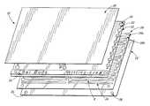
- FIG. 1is an exploded perspective view of a cuvette cartridge of the present invention.
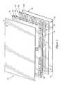
- FIG. 2is a perspective view of an assembled cuvette cartridge of the present invention.
- FIG. 3is a cross-sectional view at 3 — 3 of the cuvette cartridge of FIG. 2 .
- FIG. 4is a top plan view of an alternative embodiment of an adhesive layer of the cuvette cartridge of FIGS. 1-3 .
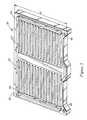
- FIG. 5is a perspective view of a vacuum chuck used to hold the cuvette cartridge of FIGS. 1-3 during spectrophotometric analysis of samples in the cuvette catridge.
- the present inventionprovides a disposable optical cuvette cartridge useful for spectrophotometric measurements of analytes in biological samples such as whole blood.
- the cuvetteholds multiple liquid samples and can be filled rapidly and easily, facilitating high-throughput applications.
- itis manufactured from a material having low fluorescence at the wavelengths of interest, so as not to interfere with the fluorescence measurements being made.
- FIGS. 1 , 2 , and 3show an optical cuvette cartridge 10 of the invention.
- the cuvette cartridge 10contains two substantially flat sheets 12 and 14 that are substantially liquid impermeable. That is, the sheets 12 and 14 contain and are not noticeably affected by or permeable to liquid samples that are placed inside the cuvette for analysis. Additionally, the sheets are preferably flexible, non-rigid, plastic films. One, and typically both, of the sheets 12 and 14 are transparent, i.e., substantially optically transmissive, transmitting at least about 25% and preferably at least about 50% of incident light at wavelengths used during spectrophotometric analysis.
- the material used to make the plastic film of at least one of the flat sheets 12 and 14preferably has a low fluorescence in the excitation and emission wavelength ranges of the experiment. That is, the fluorescence intensity of the material is small (e.g., less than 5%) in comparison to the fluorescence intensity of the sample. It is most important that the sheet through which the sample fluorescence propagates to reach the instrument detector is made of low-fluorescence material. However, both sheets 12 and 14 are preferably made of low-fluorescence material.
- Fluorescencecan be quantified using a fluorimeter, an instrument that excites a sample with light of varying wavelengths and detects the resulting fluorescence emission at varying wavelengths. Measured fluorescence is typically reported in arbitrary units or as a normalized value, e.g., with respect to the fluorescence of fused silica, known to have a very low intrinsic fluorescence over a large wavelength range.
- the material used to form the sheets 12 and 14has a fluorescence that is at most four times the fluorescence of a 150- ⁇ m thick sheet of fused silica. More preferably, the fluorescence is at most equivalent to the fluorescence of a 150- ⁇ m thick sheet of fused silica.
- the intensity of the background fluorescence of the low-fluorescence materialvaries with the excitation and emission wavelengths used, but is preferably low over a wide range of wavelengths. It is most important that the fluorescence be low at the excitation and emission values of the fluorophores used.
- Common fluorophores used in biological measurementsare excited and emit in the red region of the spectrum, i.e., at wavelengths greater than 600nm.
- the fluorophores Cy5, Cy5.5, and Cy7are excited at wavelengths of 649, 675, and 743 nm, respectively, and have emission peaks at 670, 694, and 767 nm, respectively.
- These three fluorophorescan be excited using a helium-neon laser, which has a wavelength of 633 nm.
- Other common lasers usedare argon-krypton lasers, with wavelengths of 488, 568, and 647 nm. Because it is expected that newly developed optical tags will be excited and emit at different frequency ranges, it is desired that the material have low fluorescence over a large range of excitation wavelengths, e.g., from 300 to 700 nm.
- cyclo-olefin copolymersalso referred to as cyclic olefin or cycloolefin polymers or copolymers
- a suitable material for one or both of the sheets 12 and 14is the Zeonor® family of cyclo-olefin copolymers, available from Nippon Zeon Co., Ltd., Tokyo, Japan.
- Additional suitable cyclo-olefin copolymersinclude Topas®, available from Ticona, Frankfurt, Germany. These materials were originally manufactured for electronic and optical applications, for which fluorescence properties are not relevant.
- the polymer materialis extruded as a sheet or film having the desired thickness.
- the two sheets 12 and 14have a thickness selected to ensure sufficient light transmission through the sheets while providing suitable structural stability of the cuvette cartridge 10 .
- the sheet thicknessshould not cause undue manufacturing difficulties. Typical thicknesses range from about 0.25 mils (6.3 ⁇ m) to 25 (640 ⁇ m) or even 100 mils (about 2.5 mm) or more. Preferably, the thickness is between about 0.5 and 25 mils, and most preferably about 10 mils (about 250 ⁇ m).
- the sheetsare shown as substantially rectangular in shape, the actual shape can be varied to adapt the resulting cuvette cartridge to fit the desired spectrophotometric analysis apparatus.
- the two sheets 12 and 14are spaced apart from one another and adhesively secured together by an intermediate adhesive layer 16 , which has one or more discontinuities or cutouts 16 a , areas where the layer 16 has been cut away or is otherwise not present. These areas provide sample zones or optical chambers 18 ( FIG. 3 ) having an optical path length determined by the thickness of the layer 16 , which is preferably between about 50 and 200 ⁇ m, more preferably between about 75 and 175 ⁇ m, and most preferably about 125 ⁇ m.
- the optical chambersare accessed by inlet holes 20 and vent holes 22 in the sheet 12 .
- the inlet hole 20is positioned adjacent to one end of the optical chamber 18
- the vent hole 22is positioned adjacent to the other end.
- the intermediate adhesive layer 16can be provided by an accurately laid down layer of a dimensionally-stable adhesive such as a pressure-sensitive acrylic adhesive, pressure-sensitive butylene polymer adhesive, silicone adhesive, or hot melt thermoplastic adhesive.
- the adhesive layer 16can be printed upon one of the sheets 12 and 14 in a pattern providing the desired discontinuities, e.g., by conventional techniques such as silk screening, lithography, or printing from an x-y adhesive dispenser. In this case, the layer 16 is printed on one of the sheets 12 and 14 , and thereafter the second of the sheets 12 and 14 is laminated on top of the adhesive layer 16 .
- the adhesivecan be laid down as a liquid or as a semisolid and can be cured in place partially before the additional layer is added or cured in place after the top and bottom layers are in place, as long as care is taken to maintain the proper spacing between the layers (e.g., by placement of spacer elements) and to ensure that the integrity of the discontinuities defining the sample zones is maintained.
- Curingcan be effected by, e.g., light, heat, or ultraviolet radiation, depending upon the particular adhesive employed.
- the layer 16is a preformed sheet of adhesive or a preformed sheet of polymer coated on both sides with an adhesive.
- Any adhesive that bonds to the sheets 12 and 14 without being adversely affected by or adversely affecting the liquid samplecan be used. Examples include acrylic adhesives, cyanoacrylates, silicones, epoxies, esters, poly(olefin)-based adhesives, and the like.
- the adhesive layer 16is made from a polyester sheet of approximate thickness 3 mils coated on both sides with approximately 1 mil of adhesive.
- An adhesive-coated sheetis much more structurally stable than a layer made of adhesive only and therefore makes manufacturing of the cuvette cartridge much easier. For example, it is much easier to cut and align the layers when the adhesive layer 16 is more structurally stable.
- the cutouts 16 aare formed by cutting out portions of the layer 16 , e.g., by laser, rotary, or matched metal pattern die cutting, to provide a readily removable piece 16 b having the desired contour of an optical chamber 18 .
- the inlet holes 20 and vent holes 22are cut into the flat sheet 12 , for example, by punching.
- the adhesive sheet 16is applied to one of the flat sheets 12 and 14 , and the removable cut piece 16 b is peeled off.
- the other flat sheetis then applied over the adhesive sheet 16 .
- the three sheets 12 , 14 , and 16are pressed together.
- the adhesive sheet 16holds the two flat sheets 12 and 14 together, providing a liquid-tight seal and forming an optical chamber 18 of defined volume, shape, and optical path length.
- the flat sheet 12is placed over the adhesive sheet 16 such that the inlet hole 20 is located adjacent to one end of the optical chamber 18 and the vent hole 22 is located at the other end of the optical chamber.
- each of the sample chambers 18is filled with a sample for spectrophotometric analysis.
- a drop of liquid sampletypically between about 5 and about 50 microliters, and preferably approximately 15 microliters, is placed on top of the inlet hole 20 .
- the dropis held together by its surface tension, and capillary action resulting from the cross-sectional area of the sample chamber 18 and the wettability of the chamber surface draws the sample into the chamber 18 .
- Excess sampleremains at the inlet hole 20 and can be removed if desired. Note that this mechanism obviates the need for a well-defined sample volume or for accurate measurement of the drop applied to the hole.
- Liquid samples that can be used in the cuvette cartridge 10include but are not limited to blood, serum, urine, saliva, homogenate of cells or tissue, suspension of cells or tissue or constituents thereof, analytes in a reaction mixture, solutions of analytes, or any combinations of these.
- a cover 30can be placed over the flat sheet 12 after filling of the inlet ports to reduce evaporation of the samples or other loss.
- the cover 30is preferably fabricated from the same materials as the flat sheets 12 or 14 .
- the inlet portscan instead be covered with tape or other material to prevent sample evaporation. Of course, the tape or other material should be applied so as not to obscure measurement of the sample in the optical chamber.
- the wettability or surface tension of the sheets 12 and 14is generally important because it determines whether and how quickly sample liquid (typically aqueous) is drawn into the optical chamber 18 . If the sheets are not sufficiently hydrophilic, then it is either impossible to fill the optical chambers with liquid sample, or the chambers fill much more slowly than is desirable. Partially filled chambers are particularly detrimental when measuring a sample such as blood, which contains cells. If the chamber does not fill completely, the cells are not distributed evenly in the sample and their concentration in the blood in the chamber is therefore not representative of their true concentration in the sample.
- the materials having desirable optical properties to form the sheets 12 and 14are not sufficiently hydrophilic to encourage the sample to fill the chamber 18 .
- the low-fluorescence cyclo-olefin copolymer materialssuch as Zeonor® have very high water impermeability and are extremely hydrophobic.
- the wettability of a plastic filmcan be altered by chemically treating the surface of the film, physically modifying the surface, or plasma treating the surface. Any of these treatments can be employed to provide surfaces with a sufficient degree of hydrophilicity to permit the liquid to be drawn into the sample chambers. Depending upon the materials and samples used, different levels of sample treatment are required. Plasma treatment is the currently preferred technique for rendering the cyclo-olefin copolymer surface more hydrophilic.
- the adhesive layer 16only contributes to the edges of each sample chamber 18 , and therefore provides much less contact area for the sample than do the flat layers 12 and 14 . It therefore makes a relatively insignificant contribution to the ability of the sample to fill the chamber.
- many adhesivesoutgas, generating volatile substances that coat the surfaces of the layers 12 and 14 within the same chamber, rendering these surfaces more hydrophobic.
- One way to address this problemis to store the cuvette cartridge at a low temperature, e.g., in a laboratory freezer, to minimize the volatility of the substances.
- the adhesive layer 16is made from a coated polymer film, however, the adhesive surface area exposed to the chamber is reduced. In this case, as determined by the present inventors, the amount of volatile substances generated does not appreciably reduce the hydrophilicity of the layers 12 and 14 , and the cuvette cartridge can be stored at room temperature for extended periods of time (e.g., months).
- the width of the cutout region 16 b and the thickness of the adhesive sheet 16define the cross-sectional area of the chamber 16 .
- the thicknessis selected taking into account the strong light absorption and scattering of blood as well as the effect of the cross-sectional area on the capillary action.
- the cuvette cartridge 10 of the present inventionis its ability to incorporate a plurality of sample zones or optical chambers in a single unit.
- the cartridgecan include a single optical chamber 18
- one cuvette cartridgepreferably provides between two and about 500 optical chambers 18 .
- the cuvette cartridgecan include many more than 500 chambers if desired.
- the number and configuration of sample chambers 18depend upon the instrument used to perform the measurements. It is anticipated that future high-throughput instruments will allow for a higher density of sample zones, and the cuvette cartridge of the present invention can be modified easily for such applications.
- the cartridgeIn many current applications, it is preferable for the cartridge to have between about 8 and about 100 optical chambers, and more preferably from about 30 to about 50 optical chambers. As shown in FIGS. 1-3 , this number of chambers is provided by locating the desired number of removable pieces 16 b on the adhesive sheet 16 and a corresponding number of inlet and vent holes on the flat sheet 12 .
- the inlet and vent holescan be staggered or arranged in an antiparallel orientation, as indicated in FIG. 1 , to provide, for example, closer spacing of optical chambers on the flat sheet.
- the center-to-center spacing of inlet holesis approximately 9 mm, for ease in filling with a multi-channel pipette. In some cases, it is useful for manufacturing ease to position the inlet and vent holes in a slightly offset fashion, as illustrated by the adhesive layer 32 in FIG. 4 . This configuration allows for a higher tolerance in cutting the adhesive layer 32 and the flat sheet 12 .
- At least one additional optical chamber 24is provided in the cartridge as a reference or calibration aide.
- This reference chamber 24may optionally lack an inlet hole 20 and/or vent hole 22 and can either remain empty to serve as a calibration blank or contain a calibration solution or other standard. Measurement of the blank or standard is used to calibrate measurements from samples in the remaining optical chambers 18 .
- the standardcan be a solution of a dye having known optical properties.
- one of the sample chambers 18can serve as a reference chamber, and a dedicated reference chamber 24 is not needed. An entire empty cartridge can be used for calibration instead, allowing all of the sample chambers 18 to be used.
- various identification labels 25can be added to the cartridge 10 , preferably in a location that does not interfere with measurement acquisition from the sample chambers 18 .
- a labelsuch as a bar code can be applied near an edge 29 of the flat sheet 12 .
- the adhesive sheet 16is formed from an adhesive-coated polyester film and is larger than the flat sheets 12 and 14 , extending past the front edge 29 .
- the extended portioncan be covered with a non-adhesive cover if desired.
- the labelis then attached to the adhesive sheet 16 or its non-adhesive cover.
- the disposable cuvette cartridge of the present inventionrequires very precise spectrophotometric measurement methodologies to give accurate information concerning the samples. To this end, it can be important in some cases to accurately and precisely position the cuvette cartridge in the optical measurement apparatus.
- pinholes 26 and 28are provided to assist in the registration of the cartridge on a platform during an optical measurement.
- Such pinholesare sized to engage locating pins present in the optical measurement system and typically range from about 1/32 inch to about 1 ⁇ 4 inch, and are preferably about 1 ⁇ 8 inch, in diameter.
- the adhesive layerextends forward beyond the two flat sheets 12 and 14 , and the pinholes are positioned in the adhesive layer 16 . This allows the pinholes to be cut when the cutout regions 16 b are formed in the adhesive layer 16 . This process can be very accurate and thus ensures accurate and precise placement of the pinholes. Alternatively, the pinholes can be punched in all three layers at the end of the manufacturing process.
- FIG. 5shows a vacuum chuck 40 that serves as a support for the cuvette cartridge 10 during measurement.
- the vacuum chuck 40can hold the cassette in accurate registration and in a very well-defined profile such as a very flat profile.
- the chuck 40presents a generally flat top surface 41 that includes a continuous perimeter 42 and a series of raised areas 43 making up the flat surface 41 upon which the cuvette cartridge 10 is placed.
- the top surface 41also includes a series of depressed areas 44 that are below the plane defined by the perimeter 42 and raised areas 43 .
- the pinholes 26 and 28 in the cartridgeare aligned with pins 45 and 46 to accurately register the cartridge 10 .
- a vacuumis applied to the volume defined by the depressed areas 44 . This pulls the cartridge 10 down onto the chuck 40 and seals it firmly onto the plane defined by the raised areas 43 and perimeter 42 .
- the vacuum chuck 40is fabricated out of a material such as metal, plastic, glass, or ceramic that is stable and nonreactive with the cuvette cartridge 10 , and preferably also with the sample liquid.
- the vacuum chuck 40is mounted on an optical measurement device such as a laser fluorescence scanner.
- Use of such a vacuum chuck 40is advantageous in that the cuvette cartridge 10 is held flat against the chuck 40 , thereby facilitating focusing of the optical instrument and reducing the need to refocus during measurements in multiple optical chambers of the cuvette cartridge 10 .
- the use of a vacuum chuck 40 to hold the cartridge 10 flatobviates any requirement that the cartridge itself be rigid and thus allows the use of flexible materials for the cartridge 10 .
- the vacuum chuck 40should be constructed and employed in ways that cause the cuvette cartridge to reproducibly present a consistent optical path through the various sample and calibration chambers. To this end, it is generally desirable to check the degree of vacuum drawn in the vacuum chuck and the spacing of the vacuum zones in the chuck to ensure that when a particular cuvette cartridge is placed on the vacuum chuck and held in place, there is not an unacceptable degree of distortion.
- the samples in the cuvette cartridgecan be scanned with a laser, and the contents of the sample determined or quantified by reference to the fluorescence emanating from the sample.
- the chuck 40can be fabricated with depressed areas 44 in the form of slots aligned with and positioned directly beneath the optical chambers on the cartridge 10 when the cartridge 10 is placed on the vacuum chuck 40 .
- Such slotscan reduce fluorescence by distancing the material of the chuck from the focal plane of the optical instrument used.
- a liquid sampleis applied to an inlet hole 20 , thereby filling the optical chamber 18 by capillary action.
- the cartridgeis placed in a position for an optical measurement, such as on the platform of a laser scanner.
- a confocal laser scanneris used to scan the optical chamber.
- the scanneruses the natural fluorescence of the adhesive sheet to define the edges of the optical chamber.
- a reference optical chambercan be used by the scanner to detect the edges of the optical chamber.
- the spectrophotometerBy noting the difference in properties, one can determine accurately when the spectrophotometer is reading the spectrophotometric profile of the sample and when it is reading information for areas outside the sample zone.
- sophisticated automated spectrophotometric readerscan locate the edges of the sample zone by noting the properties of the layer 16 and make their readings on the contents of the sample zone safely away from the edges.
- a reference point with known optical properties and known vertical distance from the samplee.g., the surface 41 of the chuck 40 ) can be selected and used to help focus the instrument.
Landscapes
- Chemical & Material Sciences (AREA)
- Health & Medical Sciences (AREA)
- Analytical Chemistry (AREA)
- General Health & Medical Sciences (AREA)
- Physics & Mathematics (AREA)
- General Physics & Mathematics (AREA)
- Biochemistry (AREA)
- Life Sciences & Earth Sciences (AREA)
- Immunology (AREA)
- Pathology (AREA)
- Clinical Laboratory Science (AREA)
- Chemical Kinetics & Catalysis (AREA)
- Dispersion Chemistry (AREA)
- Hematology (AREA)
- Spectroscopy & Molecular Physics (AREA)
- Nuclear Medicine, Radiotherapy & Molecular Imaging (AREA)
- Optical Measuring Cells (AREA)
Abstract
Description
Claims (9)
Priority Applications (2)
| Application Number | Priority Date | Filing Date | Title |
|---|---|---|---|
| US10/421,638US6937330B2 (en) | 1999-04-23 | 2003-04-22 | Disposable optical cuvette cartridge with low fluorescence material |
| US11/185,430US7259845B2 (en) | 1999-04-23 | 2005-07-19 | Disposable optical cuvette cartridge with low fluorescence material |
Applications Claiming Priority (5)
| Application Number | Priority Date | Filing Date | Title |
|---|---|---|---|
| US13091899P | 1999-04-23 | 1999-04-23 | |
| US13087699P | 1999-04-23 | 1999-04-23 | |
| US13087599P | 1999-04-23 | 1999-04-23 | |
| US09/552,872US6552784B1 (en) | 1999-04-23 | 2000-04-20 | Disposable optical cuvette cartridge |
| US10/421,638US6937330B2 (en) | 1999-04-23 | 2003-04-22 | Disposable optical cuvette cartridge with low fluorescence material |
Related Parent Applications (1)
| Application Number | Title | Priority Date | Filing Date |
|---|---|---|---|
| US09/552,872Continuation-In-PartUS6552784B1 (en) | 1999-04-23 | 2000-04-20 | Disposable optical cuvette cartridge |
Related Child Applications (1)
| Application Number | Title | Priority Date | Filing Date |
|---|---|---|---|
| US11/185,430ContinuationUS7259845B2 (en) | 1999-04-23 | 2005-07-19 | Disposable optical cuvette cartridge with low fluorescence material |
Publications (2)
| Publication Number | Publication Date |
|---|---|
| US20030214650A1 US20030214650A1 (en) | 2003-11-20 |
| US6937330B2true US6937330B2 (en) | 2005-08-30 |
Family
ID=27494879
Family Applications (2)
| Application Number | Title | Priority Date | Filing Date |
|---|---|---|---|
| US10/421,638Expired - Fee RelatedUS6937330B2 (en) | 1999-04-23 | 2003-04-22 | Disposable optical cuvette cartridge with low fluorescence material |
| US11/185,430Expired - Fee RelatedUS7259845B2 (en) | 1999-04-23 | 2005-07-19 | Disposable optical cuvette cartridge with low fluorescence material |
Family Applications After (1)
| Application Number | Title | Priority Date | Filing Date |
|---|---|---|---|
| US11/185,430Expired - Fee RelatedUS7259845B2 (en) | 1999-04-23 | 2005-07-19 | Disposable optical cuvette cartridge with low fluorescence material |
Country Status (1)
| Country | Link |
|---|---|
| US (2) | US6937330B2 (en) |
Cited By (17)
| Publication number | Priority date | Publication date | Assignee | Title |
|---|---|---|---|---|
| US20030078739A1 (en)* | 2001-10-05 | 2003-04-24 | Surromed, Inc. | Feature list extraction from data sets such as spectra |
| US20040161143A1 (en)* | 1999-07-21 | 2004-08-19 | Dietz Louis J. | System for microvolume laser scanning cytometry |
| US20050209789A1 (en)* | 2001-08-24 | 2005-09-22 | Hastings Curtis A | Peak selection in multidimensional data |
| US20050280817A1 (en)* | 2004-04-02 | 2005-12-22 | Uwe Horchner | Polychromic laser scanning system and method of use |
| US20060097154A1 (en)* | 2000-11-27 | 2006-05-11 | Curtis Hastings | Median filter for liquid chromatography-mass spectrometry data |
| US20060131222A1 (en)* | 2002-05-09 | 2006-06-22 | Ppd Biomarker Discovery Sciences, Llc | Methods for time-alignment of liquid chromatography-mass spectrometry data |
| US20060259246A1 (en)* | 2000-11-28 | 2006-11-16 | Ppd Biomarker Discovery Sciences, Llc | Methods for efficiently mining broad data sets for biological markers |
| US7259845B2 (en) | 1999-04-23 | 2007-08-21 | Ppd Biomarker Discovery Sciences Llc | Disposable optical cuvette cartridge with low fluorescence material |
| US20080084559A1 (en)* | 2006-10-10 | 2008-04-10 | C Technologies, Inc. | Microvolume sampling device |
| US20080316465A1 (en)* | 2007-06-06 | 2008-12-25 | Alice Wernicki | Sample holder and sample preparation device |
| US20090010388A1 (en)* | 2007-06-06 | 2009-01-08 | Stahly Barbara C | Microplate and methods of using the same |
| US7485454B1 (en) | 2000-03-10 | 2009-02-03 | Bioprocessors Corp. | Microreactor |
| WO2014113370A1 (en)* | 2013-01-15 | 2014-07-24 | Aviv Biomedical, Inc. | Bilirubin hematofluorometer and reagent kit |
| WO2014150374A1 (en)* | 2013-03-15 | 2014-09-25 | Vistalab Technologies, Inc. | Optical volumetric measurement of a dispensed fluid |
| USD804682S1 (en)* | 2015-08-10 | 2017-12-05 | Opko Diagnostics, Llc | Multi-layered sample cassette |
| US10458963B2 (en) | 2016-06-08 | 2019-10-29 | Kathleen Stitzlein | Quantitative HPTLC cannabinoid field testing device and method |
| US20220090999A1 (en)* | 2019-04-12 | 2022-03-24 | Gourgen AMBARTSOULMIAN | Tissue embedding cassette with shield |
Families Citing this family (14)
| Publication number | Priority date | Publication date | Assignee | Title |
|---|---|---|---|---|
| US6627159B1 (en)* | 2000-06-28 | 2003-09-30 | 3M Innovative Properties Company | Centrifugal filling of sample processing devices |
| US8097471B2 (en) | 2000-11-10 | 2012-01-17 | 3M Innovative Properties Company | Sample processing devices |
| US7507376B2 (en)* | 2002-12-19 | 2009-03-24 | 3M Innovative Properties Company | Integrated sample processing devices |
| US7932090B2 (en)* | 2004-08-05 | 2011-04-26 | 3M Innovative Properties Company | Sample processing device positioning apparatus and methods |
| WO2009066752A1 (en)* | 2007-11-22 | 2009-05-28 | Mitsubishi Tanabe Pharma Corporation | Plastic container having cyclic polyolefin layer |
| KR20130086743A (en)* | 2012-01-26 | 2013-08-05 | 삼성전자주식회사 | Microfluidic device and control method thereof |
| US9289761B2 (en)* | 2012-11-16 | 2016-03-22 | Honeywell International Inc. | Card waste storage mechanism |
| JP6927879B2 (en)* | 2015-01-14 | 2021-09-01 | ピクセル メディカル テクノロジーズ リミテッド | Disposable cartridge for sample fluid analysis |
| CA2937046A1 (en)* | 2016-07-25 | 2018-01-25 | Kresimir Franjic | Method for preparing thin sample layers |
| JP6941939B2 (en) | 2016-12-22 | 2021-09-29 | リンテック株式会社 | Inspection member and manufacturing method of inspection member |
| JP6966860B2 (en)* | 2017-04-07 | 2021-11-17 | リンテック株式会社 | Manufacturing method of inspection cover film, inspection member, and inspection cover film |
| WO2019103732A1 (en)* | 2017-11-22 | 2019-05-31 | Hewlett-Packard Development Company, L.P. | Multizonal microfluidic devices |
| WO2019142928A1 (en)* | 2018-01-19 | 2019-07-25 | 日東電工株式会社 | Flow path, measurement tape, and measuring device |
| JP7185423B2 (en)* | 2018-06-08 | 2022-12-07 | 株式会社ヴィーネックス | LIGHT GUIDE, OPTICAL LINE SENSOR USING THE SAME, AND METHOD FOR MANUFACTURING LIGHT GUIDE |
Citations (17)
| Publication number | Priority date | Publication date | Assignee | Title |
|---|---|---|---|---|
| US3552865A (en) | 1968-04-01 | 1971-01-05 | Beckman Instruments Inc | High pressure flow-through cuvette |
| US3690836A (en) | 1966-03-01 | 1972-09-12 | Promoveo | Device for use in the study of chemical and biological reactions and method of making same |
| GB1407247A (en) | 1971-08-27 | 1975-09-24 | Micromedic Systems Inc | Flowthrough cuvette |
| US4405235A (en) | 1981-03-19 | 1983-09-20 | Rossiter Val J | Liquid cell for spectroscopic analysis |
| US4643570A (en) | 1984-04-14 | 1987-02-17 | Carl-Zeiss-Stiftung | Through-flow cuvette |
| US4761381A (en) | 1985-09-18 | 1988-08-02 | Miles Inc. | Volume metering capillary gap device for applying a liquid sample onto a reactive surface |
| US4963498A (en) | 1985-08-05 | 1990-10-16 | Biotrack | Capillary flow device |
| US5430542A (en) | 1992-04-10 | 1995-07-04 | Avox Systems, Inc. | Disposable optical cuvette |
| WO1998016661A2 (en) | 1996-10-17 | 1998-04-23 | Morphagen | Morphatides: novel shape and structure libraries |
| US5910287A (en) | 1997-06-03 | 1999-06-08 | Aurora Biosciences Corporation | Low background multi-well plates with greater than 864 wells for fluorescence measurements of biological and biochemical samples |
| EP0969283A1 (en) | 1998-06-25 | 2000-01-05 | Hewlett-Packard Company | A method for processing measuring values |
| US6063338A (en) | 1997-06-02 | 2000-05-16 | Aurora Biosciences Corporation | Low background multi-well plates and platforms for spectroscopic measurements |
| US6133046A (en) | 1996-12-30 | 2000-10-17 | Commissariat A L'energie Atomique | Microsystems for biological analyses, their use for detecting analytes, and method for producing them |
| WO2000067017A1 (en) | 1999-05-04 | 2000-11-09 | The Rockefeller University | Method for the comparative quantitative analysis of proteins and other biological material by isotopic labeling and mass spectroscopy |
| US6200532B1 (en) | 1998-11-20 | 2001-03-13 | Akzo Nobel Nv | Devices and method for performing blood coagulation assays by piezoelectric sensing |
| US6229603B1 (en) | 1997-06-02 | 2001-05-08 | Aurora Biosciences Corporation | Low background multi-well plates with greater than 864 wells for spectroscopic measurements |
| WO2001035266A2 (en) | 1999-11-08 | 2001-05-17 | Université de Montréal | Measurement signal processing method |
Family Cites Families (117)
| Publication number | Priority date | Publication date | Assignee | Title |
|---|---|---|---|---|
| US3999047A (en) | 1972-09-05 | 1976-12-21 | Green James E | Method and apparatus utilizing color algebra for analyzing scene regions |
| US3997298A (en) | 1975-02-27 | 1976-12-14 | Cornell Research Foundation, Inc. | Liquid chromatography-mass spectrometry system and method |
| US4426451A (en) | 1981-01-28 | 1984-01-17 | Eastman Kodak Company | Multi-zoned reaction vessel having pressure-actuatable control means between zones |
| US4849330A (en)* | 1984-04-27 | 1989-07-18 | Molecular Devices Corporation | Photoresponsive redox detection and discrimination |
| SE455646B (en) | 1984-10-22 | 1988-07-25 | Radians Innova Ab | FLUORESCENT DEVICE |
| JPS61145457A (en) | 1984-12-19 | 1986-07-03 | Hitachi Ltd | Data processing apparatus for chromatograph |
| SE458968B (en) | 1987-06-16 | 1989-05-22 | Wallac Oy | BIOSPECIFIC ANALYTICAL PROCEDURE FOR MULTIPLE ANALYTICS WHICH DO NOT INCLUDE PARTICULAR COATING AND LABELING WITH FLUORESCING LABEL SUBSTANCES |
| US4844617A (en) | 1988-01-20 | 1989-07-04 | Tencor Instruments | Confocal measuring microscope with automatic focusing |
| US5119315A (en) | 1989-04-28 | 1992-06-02 | Amoco Corporation | Method of correlating a record of sample data with a record of reference data |
| US5107422A (en) | 1989-10-02 | 1992-04-21 | Kamentsky Louis A | Method and apparatus for measuring multiple optical properties of biological specimens |
| US5072382A (en) | 1989-10-02 | 1991-12-10 | Kamentsky Louis A | Methods and apparatus for measuring multiple optical properties of biological specimens |
| US5091652A (en) | 1990-01-12 | 1992-02-25 | The Regents Of The University Of California | Laser excited confocal microscope fluorescence scanner and method |
| US5274240A (en) | 1990-01-12 | 1993-12-28 | The Regents Of The University Of California | Capillary array confocal fluorescence scanner and method |
| GB9014263D0 (en) | 1990-06-27 | 1990-08-15 | Dixon Arthur E | Apparatus and method for spatially- and spectrally- resolvedmeasurements |
| GB9015793D0 (en) | 1990-07-18 | 1990-09-05 | Medical Res Council | Confocal scanning optical microscope |
| US5127730A (en) | 1990-08-10 | 1992-07-07 | Regents Of The University Of Minnesota | Multi-color laser scanning confocal imaging system |
| US5239178A (en) | 1990-11-10 | 1993-08-24 | Carl Zeiss | Optical device with an illuminating grid and detector grid arranged confocally to an object |
| DE69124199T2 (en) | 1990-11-16 | 1997-04-30 | Shimadzu Corp | Fraction purity measuring device for chromatogram peak |
| US5739000A (en) | 1991-08-28 | 1998-04-14 | Becton Dickinson And Company | Algorithmic engine for automated N-dimensional subset analysis |
| GB9218482D0 (en) | 1992-09-01 | 1992-10-14 | Dixon Arthur E | Apparatus and method for scanning laser imaging of macroscopic samples |
| JPH07504056A (en) | 1992-02-18 | 1995-04-27 | ネオパス,インク. | Methods for identifying objects using data processing techniques |
| US5377003A (en) | 1992-03-06 | 1994-12-27 | The United States Of America As Represented By The Department Of Health And Human Services | Spectroscopic imaging device employing imaging quality spectral filters |
| WO1993021592A1 (en) | 1992-04-16 | 1993-10-28 | The Dow Chemical Company | Improved method for interpreting complex data and detecting abnormal instrument or process behavior |
| JPH05340865A (en) | 1992-06-09 | 1993-12-24 | Canon Inc | measuring device |
| US5736410A (en) | 1992-09-14 | 1998-04-07 | Sri International | Up-converting reporters for biological and other assays using laser excitation techniques |
| US5889881A (en) | 1992-10-14 | 1999-03-30 | Oncometrics Imaging Corp. | Method and apparatus for automatically detecting malignancy-associated changes |
| US5585246A (en) | 1993-02-17 | 1996-12-17 | Biometric Imaging, Inc. | Method for preparing a sample in a scan capillary for immunofluorescent interrogation |
| US5556764A (en) | 1993-02-17 | 1996-09-17 | Biometric Imaging, Inc. | Method and apparatus for cell counting and cell classification |
| US5547849A (en) | 1993-02-17 | 1996-08-20 | Biometric Imaging, Inc. | Apparatus and method for volumetric capillary cytometry |
| WO1994027146A1 (en) | 1993-05-14 | 1994-11-24 | Coulter Corporation | Reticulocyte analyzing method and apparatus utilizing light scatter techniques |
| US5692220A (en) | 1993-09-02 | 1997-11-25 | Coulter Corporation | Decision support system and method for diagnosis consultation in laboratory hematopathology |
| US5532873A (en) | 1993-09-08 | 1996-07-02 | Dixon; Arthur E. | Scanning beam laser microscope with wide range of magnification |
| US5456252A (en) | 1993-09-30 | 1995-10-10 | Cedars-Sinai Medical Center | Induced fluorescence spectroscopy blood perfusion and pH monitor and method |
| US5412208A (en) | 1994-01-13 | 1995-05-02 | Mds Health Group Limited | Ion spray with intersecting flow |
| FI96452C (en) | 1994-01-26 | 1996-06-25 | Pekka Haenninen | Method for excitation of dyes |
| US5578832A (en) | 1994-09-02 | 1996-11-26 | Affymetrix, Inc. | Method and apparatus for imaging a sample on a device |
| US6017693A (en) | 1994-03-14 | 2000-01-25 | University Of Washington | Identification of nucleotides, amino acids, or carbohydrates by mass spectrometry |
| US5576827A (en) | 1994-04-15 | 1996-11-19 | Micromeritics Instrument Corporation | Apparatus and method for determining the size distribution of particles by light scattering |
| US5453505A (en) | 1994-06-30 | 1995-09-26 | Biometric Imaging, Inc. | N-heteroaromatic ion and iminium ion substituted cyanine dyes for use as fluorescence labels |
| DE4427448B4 (en) | 1994-08-03 | 2008-07-31 | Heidelberger Druckmaschinen Ag | Device for non-contact guiding sheet material |
| USD366938S (en) | 1994-09-02 | 1996-02-06 | Biometric Imaging, Inc. | Cartridge for processing laboratory samples |
| US5627041A (en) | 1994-09-02 | 1997-05-06 | Biometric Imaging, Inc. | Disposable cartridge for an assay of a biological sample |
| AU702267B2 (en) | 1994-09-02 | 1999-02-18 | Biometric Imaging, Inc. | Calibration method and apparatus for optical scanner |
| US5710713A (en) | 1995-03-20 | 1998-01-20 | The Dow Chemical Company | Method of creating standardized spectral libraries for enhanced library searching |
| US5682038A (en) | 1995-04-06 | 1997-10-28 | Becton Dickinson And Company | Fluorescent-particle analyzer with timing alignment for analog pulse subtraction of fluorescent pulses arising from different excitation locations |
| US6017434A (en) | 1995-05-09 | 2000-01-25 | Curagen Corporation | Apparatus and method for the generation, separation, detection, and recognition of biopolymer fragments |
| US5871946A (en) | 1995-05-18 | 1999-02-16 | Coulter Corporation | Method for determining activity of enzymes in metabolically active whole cells |
| US5582705A (en) | 1995-05-19 | 1996-12-10 | Iowa State University Research Foundation, Inc. | Multiplexed capillary electrophoresis system |
| WO1996037777A1 (en) | 1995-05-23 | 1996-11-28 | Nelson Randall W | Mass spectrometric immunoassay |
| US5713364A (en) | 1995-08-01 | 1998-02-03 | Medispectra, Inc. | Spectral volume microprobe analysis of materials |
| US6104945A (en) | 1995-08-01 | 2000-08-15 | Medispectra, Inc. | Spectral volume microprobe arrays |
| US5726751A (en) | 1995-09-27 | 1998-03-10 | University Of Washington | Silicon microchannel optical flow cytometer |
| US5981180A (en) | 1995-10-11 | 1999-11-09 | Luminex Corporation | Multiplexed analysis of clinical specimens apparatus and methods |
| USD383852S (en) | 1995-11-02 | 1997-09-16 | Biometric Imaging, Inc. | Cartridge for aphoresis analysis |
| US5734058A (en) | 1995-11-09 | 1998-03-31 | Biometric Imaging, Inc. | Fluorescent DNA-Intercalating cyanine dyes including a positively charged benzothiazole substituent |
| US5658735A (en) | 1995-11-09 | 1997-08-19 | Biometric Imaging, Inc. | Cyclized fluorescent nucleic acid intercalating cyanine dyes and nucleic acid detection methods |
| EP1300713A3 (en) | 1995-11-30 | 2004-11-03 | Chromavision Medical Systems, Inc. | Method and apparatus for automated image analysis of biological specimens |
| US20010055812A1 (en)* | 1995-12-05 | 2001-12-27 | Alec Mian | Devices and method for using centripetal acceleration to drive fluid movement in a microfluidics system with on-board informatics |
| DE19547546A1 (en) | 1995-12-20 | 1997-07-03 | Heidelberger Druckmasch Ag | Device for acting on sheets in a sheet delivery |
| US5795729A (en) | 1996-02-05 | 1998-08-18 | Biometric Imaging, Inc. | Reductive, energy-transfer fluorogenic probes |
| US5814820A (en) | 1996-02-09 | 1998-09-29 | The Board Of Trustees Of The University Of Illinois | Pump probe cross correlation fluorescence frequency domain microscope and microscopy |
| US5672869A (en) | 1996-04-03 | 1997-09-30 | Eastman Kodak Company | Noise and background reduction method for component detection in chromatography/spectrometry |
| USD382648S (en) | 1996-04-04 | 1997-08-19 | Biometric Imaging, Inc. | Holder for receiving two cuvettes |
| USD395708S (en) | 1996-04-04 | 1998-06-30 | Biometric Imaging, Inc. | Holder for receiving one covette |
| USD391373S (en) | 1996-04-04 | 1998-02-24 | Biometric Imaging, Inc. | Cuvette for laboratory sample |
| US5989835A (en) | 1997-02-27 | 1999-11-23 | Cellomics, Inc. | System for cell-based screening |
| US6103479A (en)* | 1996-05-30 | 2000-08-15 | Cellomics, Inc. | Miniaturized cell array methods and apparatus for cell-based screening |
| US5885841A (en) | 1996-09-11 | 1999-03-23 | Eli Lilly And Company | System and methods for qualitatively and quantitatively comparing complex admixtures using single ion chromatograms derived from spectroscopic analysis of such admixtures |
| CA2270527A1 (en) | 1996-11-04 | 1998-05-14 | 3-Dimensional Pharmaceuticals, Inc. | System, method, and computer program product for the visualization and interactive processing and analysis of chemical data |
| GB9624927D0 (en) | 1996-11-29 | 1997-01-15 | Oxford Glycosciences Uk Ltd | Gels and their use |
| US6059724A (en) | 1997-02-14 | 2000-05-09 | Biosignal, Inc. | System for predicting future health |
| DE19707227A1 (en) | 1997-02-24 | 1998-08-27 | Bodenseewerk Perkin Elmer Co | Light scanner |
| JP3504819B2 (en) | 1997-03-31 | 2004-03-08 | 株式会社日立製作所 | Mass spectrometry method and apparatus |
| US6093573A (en) | 1997-06-20 | 2000-07-25 | Xoma | Three-dimensional structure of bactericidal/permeability-increasing protein (BPI) |
| US6112161A (en) | 1997-09-17 | 2000-08-29 | Hewlett-Packard | Method, apparatus, and article of manufacture for enhanced intergration of signals |
| US6388788B1 (en) | 1998-03-16 | 2002-05-14 | Praelux, Inc. | Method and apparatus for screening chemical compounds |
| US5995989A (en) | 1998-04-24 | 1999-11-30 | Eg&G Instruments, Inc. | Method and apparatus for compression and filtering of data associated with spectrometry |
| US6138117A (en) | 1998-04-29 | 2000-10-24 | International Business Machines Corporation | Method and system for mining long patterns from databases |
| DE19829094C2 (en) | 1998-06-30 | 2002-10-24 | Roland Man Druckmasch | Guide device for sheet-shaped substrates in a printing machine |
| US6008896A (en) | 1998-07-01 | 1999-12-28 | National Research Council Of Canada | Method and apparatus for spectroscopic analysis of heterogeneous materials |
| US6366924B1 (en) | 1998-07-27 | 2002-04-02 | Caliper Technologies Corp. | Distributed database for analytical instruments |
| CA2341359A1 (en) | 1998-08-21 | 2000-03-02 | Ian Walton | Novel optical architectures for microvolume laser-scanning cytometers |
| AU755334C (en) | 1998-08-25 | 2004-02-26 | University Of Washington | Rapid quantitative analysis of proteins or protein function in complex mixtures |
| US6377842B1 (en) | 1998-09-22 | 2002-04-23 | Aurora Optics, Inc. | Method for quantitative measurement of fluorescent and phosphorescent drugs within tissue utilizing a fiber optic probe |
| US6207955B1 (en) | 1998-09-28 | 2001-03-27 | Varian, Inc. | Pneumatically assisted electrospray device with alternating pressure gradients for mass spectrometry |
| US6147344A (en) | 1998-10-15 | 2000-11-14 | Neogenesis, Inc | Method for identifying compounds in a chemical mixture |
| US6134002A (en) | 1999-01-14 | 2000-10-17 | Duke University | Apparatus and method for the rapid spectral resolution of confocal images |
| US6066216A (en) | 1999-02-05 | 2000-05-23 | Biometric Imaging, Inc. | Mesa forming weld depth limitation feature for use with energy director in ultrasonic welding |
| US6253162B1 (en) | 1999-04-07 | 2001-06-26 | Battelle Memorial Institute | Method of identifying features in indexed data |
| AU4366100A (en) | 1999-04-23 | 2000-11-10 | Surromed, Inc. | Disposable optical cuvette cartridge |
| US6937330B2 (en) | 1999-04-23 | 2005-08-30 | Ppd Biomarker Discovery Sciences, Llc | Disposable optical cuvette cartridge with low fluorescence material |
| US6376843B1 (en) | 1999-06-23 | 2002-04-23 | Evotec Oai Ag | Method of characterizing fluorescent molecules or other particles using generating functions |
| US6165541A (en)* | 1999-04-29 | 2000-12-26 | Helena Laboratories Corporation | Gel-template interface and method for depositing liquid on a gel |
| WO2000070340A2 (en) | 1999-05-14 | 2000-11-23 | Karolinska Innovations Ab | Materials and methods relating to disease diagnosis |
| US6334099B1 (en) | 1999-05-25 | 2001-12-25 | Digital Gene Technologies, Inc. | Methods for normalization of experimental data |
| US6687395B1 (en) | 1999-07-21 | 2004-02-03 | Surromed, Inc. | System for microvolume laser scanning cytometry |
| FR2797495B1 (en) | 1999-08-11 | 2003-01-31 | Dilor | SPECTROMETRIC IMAGING APPARATUS |
| ES2584553T3 (en) | 1999-10-06 | 2016-09-28 | Becton Dickinson And Company | Spectroscopically active surface-enhanced compound nanoparticles |
| WO2001044269A2 (en) | 1999-12-17 | 2001-06-21 | Large Scale Proteomics Corporation | Brain protein markers |
| AU2001234779A1 (en) | 2000-02-03 | 2001-08-14 | Nanoscale Combinatorial Synthesis, Inc. | Structure identification methods using mass measurements |
| AR031557A1 (en) | 2000-03-10 | 2003-09-24 | Textron Systems Corp | OPTICAL PROBE AND METHODS FOR SPECTRAL ANALYSIS. |
| CA2307399C (en) | 2000-05-02 | 2006-10-03 | Mds Inc., Doing Business As Mds Sciex | Method for reducing chemical background in mass spectra |
| WO2001098458A2 (en) | 2000-06-19 | 2001-12-27 | Zyomyx, Inc. | Methods for immobilizing polypeptides |
| NL1016034C2 (en) | 2000-08-03 | 2002-02-08 | Tno | Method and system for identifying and quantifying chemical components of a mixture of materials to be investigated. |
| US6947133B2 (en) | 2000-08-08 | 2005-09-20 | Carl Zeiss Jena Gmbh | Method for increasing the spectral and spatial resolution of detectors |
| CA2314398A1 (en)* | 2000-08-10 | 2002-02-10 | Edward Shipwash | Microarrays and microsystems for amino acid analysis and protein sequencing |
| AU2001285063A1 (en) | 2000-08-25 | 2002-03-13 | Genencor International, Inc. | Mass spectrometric analysis of biopolymers |
| US6963807B2 (en) | 2000-09-08 | 2005-11-08 | Oxford Glycosciences (Uk) Ltd. | Automated identification of peptides |
| EP1325322A2 (en) | 2000-10-03 | 2003-07-09 | Dionex Corporation | Method and system for peak parking in liquid chromatography-mass spectrometer (lc-ms) analysis |
| US6962818B2 (en) | 2000-10-19 | 2005-11-08 | Target Discovery | Mass defect labeling for the determination of oligomer sequences |
| US6787761B2 (en) | 2000-11-27 | 2004-09-07 | Surromed, Inc. | Median filter for liquid chromatography-mass spectrometry data |
| JP3663125B2 (en) | 2000-11-28 | 2005-06-22 | 日立ソフトウエアエンジニアリング株式会社 | Fluorescence reading method and fluorescence reading apparatus |
| GB0103030D0 (en) | 2001-02-07 | 2001-03-21 | Univ London | Spectrum processing and processor |
| US6856457B2 (en) | 2001-03-27 | 2005-02-15 | Prairie Technologies, Inc. | Single and multi-aperture, translationally-coupled confocal microscope |
| DE10121064A1 (en) | 2001-04-28 | 2002-10-31 | Evotec Ag | Device and method for the optical measurement of chemical and / or biological samples |
| US6873915B2 (en) | 2001-08-24 | 2005-03-29 | Surromed, Inc. | Peak selection in multidimensional data |
| US6835927B2 (en) | 2001-10-15 | 2004-12-28 | Surromed, Inc. | Mass spectrometric quantification of chemical mixture components |
- 2003
- 2003-04-22USUS10/421,638patent/US6937330B2/ennot_activeExpired - Fee Related
- 2005
- 2005-07-19USUS11/185,430patent/US7259845B2/ennot_activeExpired - Fee Related
Patent Citations (18)
| Publication number | Priority date | Publication date | Assignee | Title |
|---|---|---|---|---|
| US3690836A (en) | 1966-03-01 | 1972-09-12 | Promoveo | Device for use in the study of chemical and biological reactions and method of making same |
| US3552865A (en) | 1968-04-01 | 1971-01-05 | Beckman Instruments Inc | High pressure flow-through cuvette |
| GB1407247A (en) | 1971-08-27 | 1975-09-24 | Micromedic Systems Inc | Flowthrough cuvette |
| US4405235A (en) | 1981-03-19 | 1983-09-20 | Rossiter Val J | Liquid cell for spectroscopic analysis |
| US4643570A (en) | 1984-04-14 | 1987-02-17 | Carl-Zeiss-Stiftung | Through-flow cuvette |
| US4963498A (en) | 1985-08-05 | 1990-10-16 | Biotrack | Capillary flow device |
| US4761381A (en) | 1985-09-18 | 1988-08-02 | Miles Inc. | Volume metering capillary gap device for applying a liquid sample onto a reactive surface |
| US5430542A (en) | 1992-04-10 | 1995-07-04 | Avox Systems, Inc. | Disposable optical cuvette |
| WO1998016661A2 (en) | 1996-10-17 | 1998-04-23 | Morphagen | Morphatides: novel shape and structure libraries |
| US6133046A (en) | 1996-12-30 | 2000-10-17 | Commissariat A L'energie Atomique | Microsystems for biological analyses, their use for detecting analytes, and method for producing them |
| US6063338A (en) | 1997-06-02 | 2000-05-16 | Aurora Biosciences Corporation | Low background multi-well plates and platforms for spectroscopic measurements |
| US6229603B1 (en) | 1997-06-02 | 2001-05-08 | Aurora Biosciences Corporation | Low background multi-well plates with greater than 864 wells for spectroscopic measurements |
| US6232114B1 (en) | 1997-06-02 | 2001-05-15 | Aurora Biosciences Corporation | Low background multi-well plates for fluorescence measurements of biological and biochemical samples |
| US5910287A (en) | 1997-06-03 | 1999-06-08 | Aurora Biosciences Corporation | Low background multi-well plates with greater than 864 wells for fluorescence measurements of biological and biochemical samples |
| EP0969283A1 (en) | 1998-06-25 | 2000-01-05 | Hewlett-Packard Company | A method for processing measuring values |
| US6200532B1 (en) | 1998-11-20 | 2001-03-13 | Akzo Nobel Nv | Devices and method for performing blood coagulation assays by piezoelectric sensing |
| WO2000067017A1 (en) | 1999-05-04 | 2000-11-09 | The Rockefeller University | Method for the comparative quantitative analysis of proteins and other biological material by isotopic labeling and mass spectroscopy |
| WO2001035266A2 (en) | 1999-11-08 | 2001-05-17 | Université de Montréal | Measurement signal processing method |
Non-Patent Citations (20)
| Title |
|---|
| Aach & Church (2001) Bioinformatics 17:495-508. |
| Breen, et al. (2000) Electrophoresis 21:2243-2251. |
| Bryant et al. (2001) Rapid Commun. Mass Spectrom. 15:418-427. |
| Bylund et al. (2002) J. of Chromatography A. 961:237-244. |
| Chace (2001) Chem. Rev. 101:445-477. |
| Chelius et al. (2002) J. Proteome Res. 1:317-323. |
| do Lago et al. (1995) Anal. Chim. Acta, 310:281-288. |
| Flehn et al. (2000) Nat. Biotech. 18:1157-1161. |
| Grung & Kvalheim (1995) Analytica Chimica Acta 304:57-66. |
| Gygi et al. (1999) Nat. Biotech. 17:994-999. |
| Kassidas et al. (1998) AIChE Journal 44(4):864-875. |
| Koradi et al. (1998) J. Mag. Res. 135:288-297. |
| Nielsen et al. (1998) J. of Chromatography A. 805:17-35. |
| Pravdova et al. (2002) Analytica Chimica Acta 456:77-92. |
| Prazen et al. (Jan. 15, 1998) Anal. Chem. 70:217-225. |
| Sakoe & Chiba (2002) IEEE Transactions on Acoustics, Speech and Signal Processing ASSP26(1):43-49. |
| Schoonjans et al. (2000) J. Pharm. & Biomed. Analysis 21:1197-1214. |
| Stein (1999) J. Am. Soc. Mass Spectrom. 10:770-81. |
| Wang et al. (1987) Analytical Chemistry 59:649-654. |
| Windig et al. (1996) Anal. Chem., 68: 3602-3606. |
Cited By (25)
| Publication number | Priority date | Publication date | Assignee | Title |
|---|---|---|---|---|
| US7259845B2 (en) | 1999-04-23 | 2007-08-21 | Ppd Biomarker Discovery Sciences Llc | Disposable optical cuvette cartridge with low fluorescence material |
| US20040161143A1 (en)* | 1999-07-21 | 2004-08-19 | Dietz Louis J. | System for microvolume laser scanning cytometry |
| US7336812B2 (en) | 1999-07-21 | 2008-02-26 | Ppd Biomarker Discovery Sciences, Llc | System for microvolume laser scanning cytometry |
| US7485454B1 (en) | 2000-03-10 | 2009-02-03 | Bioprocessors Corp. | Microreactor |
| US20060097154A1 (en)* | 2000-11-27 | 2006-05-11 | Curtis Hastings | Median filter for liquid chromatography-mass spectrometry data |
| US7253404B2 (en) | 2000-11-27 | 2007-08-07 | Ppd Biomarker Discovery Sciences, Llc | Median filter for liquid chromatography-mass spectrometry data |
| US20060259246A1 (en)* | 2000-11-28 | 2006-11-16 | Ppd Biomarker Discovery Sciences, Llc | Methods for efficiently mining broad data sets for biological markers |
| US7197401B2 (en) | 2001-08-24 | 2007-03-27 | Ppd Biomarker Discovery Sciences, Llc | Peak selection in multidimensional data |
| US20050209789A1 (en)* | 2001-08-24 | 2005-09-22 | Hastings Curtis A | Peak selection in multidimensional data |
| US20030078739A1 (en)* | 2001-10-05 | 2003-04-24 | Surromed, Inc. | Feature list extraction from data sets such as spectra |
| US20060131222A1 (en)* | 2002-05-09 | 2006-06-22 | Ppd Biomarker Discovery Sciences, Llc | Methods for time-alignment of liquid chromatography-mass spectrometry data |
| US7628914B2 (en) | 2002-05-09 | 2009-12-08 | Ppd Biomarker Discovery Sciences, Llc | Methods for time-alignment of liquid chromatography-mass spectrometry data |
| US7248360B2 (en) | 2004-04-02 | 2007-07-24 | Ppd Biomarker Discovery Sciences, Llc | Polychronic laser scanning system and method of use |
| US20050280817A1 (en)* | 2004-04-02 | 2005-12-22 | Uwe Horchner | Polychromic laser scanning system and method of use |
| US20080084559A1 (en)* | 2006-10-10 | 2008-04-10 | C Technologies, Inc. | Microvolume sampling device |
| US20090010388A1 (en)* | 2007-06-06 | 2009-01-08 | Stahly Barbara C | Microplate and methods of using the same |
| US20080316465A1 (en)* | 2007-06-06 | 2008-12-25 | Alice Wernicki | Sample holder and sample preparation device |
| US8018588B2 (en)* | 2007-06-06 | 2011-09-13 | Aptuit, Inc. | Sample holder and sample preparation device |
| WO2014113370A1 (en)* | 2013-01-15 | 2014-07-24 | Aviv Biomedical, Inc. | Bilirubin hematofluorometer and reagent kit |
| WO2014150374A1 (en)* | 2013-03-15 | 2014-09-25 | Vistalab Technologies, Inc. | Optical volumetric measurement of a dispensed fluid |
| USD804682S1 (en)* | 2015-08-10 | 2017-12-05 | Opko Diagnostics, Llc | Multi-layered sample cassette |
| USD817511S1 (en) | 2015-08-10 | 2018-05-08 | Opko Diagnostics, Llc | Multi-layered sample cassette |
| US10458963B2 (en) | 2016-06-08 | 2019-10-29 | Kathleen Stitzlein | Quantitative HPTLC cannabinoid field testing device and method |
| US20220090999A1 (en)* | 2019-04-12 | 2022-03-24 | Gourgen AMBARTSOULMIAN | Tissue embedding cassette with shield |
| US11714033B2 (en)* | 2019-04-12 | 2023-08-01 | Gourgen AMBARTSOUMIAN | Tissue embedding cassette with shield |
Also Published As
| Publication number | Publication date |
|---|---|
| US7259845B2 (en) | 2007-08-21 |
| US20030214650A1 (en) | 2003-11-20 |
| US20060119844A1 (en) | 2006-06-08 |
Similar Documents
| Publication | Publication Date | Title |
|---|---|---|
| US6937330B2 (en) | Disposable optical cuvette cartridge with low fluorescence material | |
| US6552784B1 (en) | Disposable optical cuvette cartridge | |
| CA2205057C (en) | Device and method for dna amplification and assay | |
| CN1134664C (en) | Optically readable strip for analyte detection having on-strip orientation index | |
| KR100413535B1 (en) | Absorbance detection system for lab-on-a-chip | |
| US8361394B2 (en) | Calibration system and dye kit and their uses for characterizing luminescence measurement systems | |
| US20120261256A1 (en) | Sample holders and analytical instrument for point-of-care qualification of clinical samples | |
| RU99102671A (en) | DEVICE AND METHOD FOR CARRYING OUT QUANTITATIVE ANALYSIS OF AN AFFILIATION USING FLUORESCENT TAGS | |
| KR20030078615A (en) | Blood inspection unit and blood inspection apparatus | |
| JP2002501193A (en) | Optical sensor and operation method | |
| KR20160067607A (en) | Test Apparatus and Control Method thereof | |
| US8821790B2 (en) | Diagnostic measuring system | |
| JP2007298375A (en) | Switching device, measuring device using total reflection attenuation, and switching method | |
| CN103430011B (en) | Measuring device for quantitative optical evaluation of chemical reactions | |
| JPH09105708A (en) | Liquid holder and manufacture thereof | |
| EP0621478B1 (en) | Dry analytical film chip | |
| US8899290B2 (en) | Method for producing an analytical consumable | |
| KR20030027713A (en) | Colorimetric test device with reduced error | |
| US20220390381A1 (en) | Test strip | |
| CN120813827A (en) | Method for measuring the concentration of a specific electrolyte in a blood sample | |
| JP3326004B2 (en) | Dry analysis film piece | |
| WO2019002818A1 (en) | Reader for a biological fluid testing device | |
| JPH05172804A (en) | Biochemical analyzing element and biochemical analyzing film | |
| HK1163807A (en) | Method for producing an analytical consumable | |
| JPH0650959A (en) | Dry process analysis film piece with substrate |
Legal Events
| Date | Code | Title | Description |
|---|---|---|---|
| AS | Assignment | Owner name:SURROMED, INC., CALIFORNIA Free format text:ASSIGNMENT OF ASSIGNORS INTEREST;ASSIGNORS:DIETZ, LOUIS J.;TODD, CHRISTOPHER E.;REEL/FRAME:013877/0369;SIGNING DATES FROM 20030513 TO 20030515 | |
| FEPP | Fee payment procedure | Free format text:PAYOR NUMBER ASSIGNED (ORIGINAL EVENT CODE: ASPN); ENTITY STATUS OF PATENT OWNER: LARGE ENTITY | |
| AS | Assignment | Owner name:SM PURCHASE COMPANY, LLC, CALIFORNIA Free format text:ASSIGNMENT OF ASSIGNORS INTEREST;ASSIGNOR:SURROMED, INC.;REEL/FRAME:015972/0122 Effective date:20050131 | |
| AS | Assignment | Owner name:SURROMED, LLC, CALIFORNIA Free format text:CHANGE OF NAME;ASSIGNOR:SM PURCHASE COMPANY, LLC;REEL/FRAME:015972/0085 Effective date:20050209 | |
| AS | Assignment | Owner name:PPD BIOMARKER DISCOVERY SCIENCES, LLC, CALIFORNIA Free format text:CHANGE OF NAME;ASSIGNOR:PPD BIOMARKER SERVICES, LLC;REEL/FRAME:016249/0020 Effective date:20050602 Owner name:PPD BIOMARKER SERVICES, LLC, CALIFORNIA Free format text:CHANGE OF NAME;ASSIGNOR:SURROMED, LLC;REEL/FRAME:016249/0052 Effective date:20050504 | |
| FPAY | Fee payment | Year of fee payment:4 | |
| REMI | Maintenance fee reminder mailed | ||
| LAPS | Lapse for failure to pay maintenance fees | ||
| STCH | Information on status: patent discontinuation | Free format text:PATENT EXPIRED DUE TO NONPAYMENT OF MAINTENANCE FEES UNDER 37 CFR 1.362 | |
| FP | Lapsed due to failure to pay maintenance fee | Effective date:20130830 |