US6893452B2 - Multi-point tension distribution system device and method of tissue approximation using that device to improve wound healing - Google Patents
Multi-point tension distribution system device and method of tissue approximation using that device to improve wound healingDownload PDFInfo
- Publication number
- US6893452B2 US6893452B2US10/140,897US14089702AUS6893452B2US 6893452 B2US6893452 B2US 6893452B2US 14089702 AUS14089702 AUS 14089702AUS 6893452 B2US6893452 B2US 6893452B2
- Authority
- US
- United States
- Prior art keywords
- area
- biodegradable
- implantable device
- attachment points
- tissue
- Prior art date
- Legal status (The legal status is an assumption and is not a legal conclusion. Google has not performed a legal analysis and makes no representation as to the accuracy of the status listed.)
- Expired - Lifetime, expires
Links
Images


Classifications
- A—HUMAN NECESSITIES
- A61—MEDICAL OR VETERINARY SCIENCE; HYGIENE
- A61B—DIAGNOSIS; SURGERY; IDENTIFICATION
- A61B17/00—Surgical instruments, devices or methods
- A61B17/11—Surgical instruments, devices or methods for performing anastomosis; Buttons for anastomosis
- A—HUMAN NECESSITIES
- A61—MEDICAL OR VETERINARY SCIENCE; HYGIENE
- A61B—DIAGNOSIS; SURGERY; IDENTIFICATION
- A61B17/00—Surgical instruments, devices or methods
- A61B17/064—Surgical staples, i.e. penetrating the tissue
- A—HUMAN NECESSITIES
- A61—MEDICAL OR VETERINARY SCIENCE; HYGIENE
- A61B—DIAGNOSIS; SURGERY; IDENTIFICATION
- A61B17/00—Surgical instruments, devices or methods
- A61B17/08—Wound clamps or clips, i.e. not or only partly penetrating the tissue ; Devices for bringing together the edges of a wound
- A—HUMAN NECESSITIES
- A61—MEDICAL OR VETERINARY SCIENCE; HYGIENE
- A61B—DIAGNOSIS; SURGERY; IDENTIFICATION
- A61B17/00—Surgical instruments, devices or methods
- A61B17/11—Surgical instruments, devices or methods for performing anastomosis; Buttons for anastomosis
- A61B17/1128—Surgical instruments, devices or methods for performing anastomosis; Buttons for anastomosis of nerves
- A—HUMAN NECESSITIES
- A61—MEDICAL OR VETERINARY SCIENCE; HYGIENE
- A61B—DIAGNOSIS; SURGERY; IDENTIFICATION
- A61B17/00—Surgical instruments, devices or methods
- A61B17/11—Surgical instruments, devices or methods for performing anastomosis; Buttons for anastomosis
- A61B17/1146—Surgical instruments, devices or methods for performing anastomosis; Buttons for anastomosis of tendons
- A—HUMAN NECESSITIES
- A61—MEDICAL OR VETERINARY SCIENCE; HYGIENE
- A61B—DIAGNOSIS; SURGERY; IDENTIFICATION
- A61B17/00—Surgical instruments, devices or methods
- A61B2017/00004—(bio)absorbable, (bio)resorbable or resorptive
- A—HUMAN NECESSITIES
- A61—MEDICAL OR VETERINARY SCIENCE; HYGIENE
- A61B—DIAGNOSIS; SURGERY; IDENTIFICATION
- A61B17/00—Surgical instruments, devices or methods
- A61B17/04—Surgical instruments, devices or methods for suturing wounds; Holders or packages for needles or suture materials
- A61B2017/0496—Surgical instruments, devices or methods for suturing wounds; Holders or packages for needles or suture materials for tensioning sutures
- A—HUMAN NECESSITIES
- A61—MEDICAL OR VETERINARY SCIENCE; HYGIENE
- A61B—DIAGNOSIS; SURGERY; IDENTIFICATION
- A61B17/00—Surgical instruments, devices or methods
- A61B17/064—Surgical staples, i.e. penetrating the tissue
- A61B2017/0641—Surgical staples, i.e. penetrating the tissue having at least three legs as part of one single body
- A—HUMAN NECESSITIES
- A61—MEDICAL OR VETERINARY SCIENCE; HYGIENE
- A61B—DIAGNOSIS; SURGERY; IDENTIFICATION
- A61B17/00—Surgical instruments, devices or methods
- A61B17/064—Surgical staples, i.e. penetrating the tissue
- A61B2017/0647—Surgical staples, i.e. penetrating the tissue having one single leg, e.g. tacks
- A61B2017/0648—Surgical staples, i.e. penetrating the tissue having one single leg, e.g. tacks threaded, e.g. tacks with a screw thread
- A—HUMAN NECESSITIES
- A61—MEDICAL OR VETERINARY SCIENCE; HYGIENE
- A61B—DIAGNOSIS; SURGERY; IDENTIFICATION
- A61B17/00—Surgical instruments, devices or methods
- A61B17/11—Surgical instruments, devices or methods for performing anastomosis; Buttons for anastomosis
- A61B2017/1107—Surgical instruments, devices or methods for performing anastomosis; Buttons for anastomosis for blood vessels
- A—HUMAN NECESSITIES
- A61—MEDICAL OR VETERINARY SCIENCE; HYGIENE
- A61B—DIAGNOSIS; SURGERY; IDENTIFICATION
- A61B90/00—Instruments, implements or accessories specially adapted for surgery or diagnosis and not covered by any of the groups A61B1/00 - A61B50/00, e.g. for luxation treatment or for protecting wound edges
- A61B90/02—Devices for expanding tissue, e.g. skin tissue
Definitions
- This inventionis in the field of surgery. More particularly, it relates to a tissue approximation device that facilitates wound healing by holding soft tissue together under improved distribution of tension and with minimal disruption of the wound interface and its nutrient supplies.
- the devicehas multiple sites for grasping said tissue using tines or prongs or other generally sharp, projecting points, extending from a single, supportive backing.
- Various processes of using the inventive deviceare also a portion of the invention.
- the surgically induced healing of soft tissue woundsinvolves two phases, the mechanical phase of wound closure followed by the biochemical phase which involves protein bridging and scarring.
- the mechanical phasethe edges of soft tissue are held in contact by essentially two components: 1) The physical properties and device-tissue interactions of the materials holding the tissue edges in contact, e.g. sutures or staples; and 2) An early deposition of proteinaceous material that has adhesive characteristics, e.g. fibrin glue.
- Collagenis the primary constituent of connective tissue and ultimately determines the pliability and tensile strength of the healing wound. Tensile strength is gradually recovered; 60% of ultimate wound strength is achieved after approximately 3 months. However, this process is successful only if the previous mechanical phase has proceeded normally.
- tissueis mechanically approximated until the wound has healed enough to withstand stress without artificial support.
- Optimal healingrequires the application of appropriate tissue tension on the closure to eliminate dead space but not create ischemia within the tissue. Both of these circumstances increase the risk of wound infection and wound dehiscence.
- Soft tissueis well known for its inability to hold tension. Even when optimally placed, sutures gradually tear through soft tissue, producing gaps in wounds and possibly leading to the eventual failure or sub-optimization of wound healing. Furthermore, since sutures require the implementation of high levels of tension to counteract the forces acting to separate tissues, they may strangulate the blood supply of the tissues through which they are placed, thus inhibiting the delivery of wound nutrients and oxygen necessary for healing.
- the devicesminimally improve the biomechanics of wound closure, and do not adequately approximate the deeper layers of the closure, i.e. fascia or dermis. Externally placed attachment points that puncture the skin lateral to the wound also interfere with long-term cosmesis and provide a possible conduit for infecting microorganisms.
- U.S. Pat. No. 5,176,692 to Wilk et al.discloses a device for hernia repair that utilizes mesh with pin-like projections to cover hernia defects. This device, however, is used in a laparoscopic hernia repair in conjunction with an inflatable balloon. Closure devices for deeper tissues are described in U.S. Pat. No. 4,610,250 to Green; U.S. Pat. No. 5,584,859 to Brozt et al.; and U.S. Pat. No. 4,259,959 to Walker. However, these devices either work in conjunction with sutures, are made of materials that do not suggest biodegradability, or are designed in such a way as not to impart uniform tension on the closure, increasing the risk of wound separation and failure of wound healing.
- the present inventionis a biodegradable tissue approximation device.
- the deviceincludes a plurality of attachment points, e.g. tines, prongs, or other generally sharp or blunt parts, connected to a backing that can be manipulated to close wounds, join soft tissue or bone, or create anastomoses.
- This multi-point tension distribution system (MTDS) devicemay be placed with minimal tissue trauma.
- the present inventiontypically incorporates the deeper layers of tissue within the closure, and the multiple attachment points distribute the resulting tension, often uniformly. Approximation from the internal aspect of the wound minimizes the potential for dead space in the closure, thus decreasing the risk of sub-optimal healing. Moreover, because the device is absorbed, a second procedure is not typically needed to remove the device.
- the present inventionimproves the mechanical phase of healing by facilitating wound closure and/or the coaptation of tissues prior to initiation of the biochemical phase of wound healing. Placement of the device maximizes the chance for a good cosmetic result and is not heavily dependent on surgeon skill. Closure time is also shortened, decreasing overall cost and risk of operative complications.
- the present inventionis a device that improves the mechanical phase of wound healing.
- tissue edgesare stabilized by a plurality of attachment points that extend from a supportive backing.
- the density, shape, length, and orientation of attachment points on the backingmay be varied to suit the procedure, type of tissue being approximated, and/or area of the body involved.
- the flexibility of the backingis also variable and dependent on the materials used and dimensions of the backing.
- the deviceis biodegradable, and the attachment points uniformly distribute tension over the contact area between the device and tissue.
- the devicemay be used to close wounds and create vascular anastomoses.
- the devicemay also be manipulated to approximate soft tissue and soft tissue to bone.
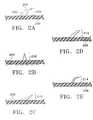
- FIGS. 1A-1Dare plan, perspective views of various MTDS devices.
- FIGS. 2A-2Eare side views of various attachment point shapes and orientations.
- FIGS. 3A-3D and 3 F- 3 Gare side views of various attachment points.
- FIG. 3Eis a side view of a two-sided MTDS device.
- FIG. 3His a plan, reverse perspective view of nubs on the inferior surface of a MTDS device.
- FIG. 4Ais a side, cross-sectional view of attachment points that run through the width of a backing.
- FIG. 4Bis a side view of attachment points on a strip of backing material.
- FIG. 4Cis a plan, perspective view of the embodiment in 4 B on a backing.
- FIG. 4Dis a plan, perspective view of attachment points on a solid backing.
- FIG. 5Ais a plan, perspective view of attachment points canted in one direction.
- FIGS. 5B-5Dare plan, perspective views of attachment points with various orientations on a backing.
- FIG. 5Eis a side view of attachment points becoming progressively shorter the closer they are to the center of the device.
- FIG. 5Fis a side view of attachment points becoming progressively shorter the farther they are from the center of the device.
- FIGS. 6A-6Bare schematic views of a skin wound and wound repair using the MTDS device.
- FIG. 7is a schematic view of an abdominal wound closure using MTDS devices.
- FIGS. 8A-8Bare schematic views of an abdominal hernia and hernia repair using the MTDS device.
- FIGS. 8C-8Dare side and schematic views, respectively, of a MTDS device with attachment points on the edges of the backing and a central area without attachment points.
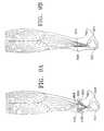
- FIGS. 9A-9Bare schematic views of a ruptured tendon and tendon to bone repair using the MTDS device.
- FIG. 10Ais an axial view of a cross-section of a vessel repaired with the MTDS device.
- FIGS. 10B-10Care side, schematic views of vessel free ends and a vascular anastomosis using the MTDS device.
- FIGS. 11 A and 11 B- 11 Care schematic, side, and cross-sectional side views, respectively, of a transected tendon and a tendon to tendon repair using the MTDS device.
- FIG. 11Dis an axial, cross-sectional view of the MTDS tendon to tendon repair.
- FIG. 11Eis a side view of a vascular anastomosis using the MTDS device on the external surface of a vessel.
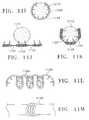
- FIGS. 11F-11Gare side, schematic views, and FIG. 11H is an axial view of the ends of a tubular structure being joined by externally placing strips of a MTDS device on approximated tissue.
- FIG. 11Iis an axial view of a hinge in the backing of a device.
- FIGS. 11J-11Kare axial views of decreased backing material that are areas of enhanced device flexibility.
- FIGS. 11L-11Mare side views of a spring or coil-like MTDS device being used to approximate tissue.
- FIG. 12Ais a schematic view of the MTDS device being used in a brow-lift procedure.
- FIG. 12Bis a plan, perspective view of the MTDS device used in a brow-lift.
- the devicemay be used when working with bone anchors or a variety of soft tissues.
- the deviceis of the general configurations shown in FIGS. 1A-1B and comprises a plurality of attachment points ( 102 ) emanating from a supportive backing ( 100 ) that is a generally a porous material that may have the structure of a mesh, net, or lattice.
- the degree of flexibility of the backingis determined by the material of construction, the shape and dimensions of the device, the type and properties of the approximated tissue, and the area of the body into which the device is placed. For example, a tightly curved or mobile part of the body, e.g. a joint, will require a more flexible backing, as would a tendon or nerve repair due to the amount of bending the device needs for the attachment.
- the thickness of the backing as well as its width and lengthmay determine the flexibility of the device.
- the backingmay be pre-fabricated into different shapes as shown by the sharp corners ( 104 ) and rounded corners ( 106 ) in FIGS. 1C and 1D .
- the fabricated cross-sectional shape and dimensions of the mesh elementsmay vary to promote flexibility in regions of the backing.
- the cross-sectional shape of the mesh elementsmay be chosen to minimize local compressive stress between the backing and surface it rests upon, or have rounded and filleted edges to be less obtrusive to local circulation.
- the plurality of attachment pointsdistribute tension over the contact area between the device and the tissue.
- biodegradable polymersare preferably used to construct the backing and attachment points.
- Polymers synthesized from monomers comprising esters, anhydrides, orthoesters, and amidesare particularly suitable for biodegradation.
- biodegradable polymersare polyglycolide, polylactide, poly- ⁇ -caprolactone, polydiaxanone, polyglyconate, polylactide-co-glycolide, and block and random copolymers of these polymers. Copolymers of glycolic, lactic, and other ⁇ -hydroxy acids are highly desirable.
- an inventive devicemay be made of two or more types of polymers or copolymers (or molecular weights of the same polymer or copolymer).
- the backing materialmight be produced from a more flexible polymer and the points or tines of a stiffer material. The inflammatory response to these polymers is minimal, and they have been safely used in suture materials, stents, drug delivery devices, orthopedic fixation devices, and intestinal anastomotic rings.
- attachment pointsAs “tines” or “prongs”. These tines will refer both to points which are either sharp, i.e. able to separate tissue in a chosen use, or blunt, i.e. not able to separate tissue in that use.
- the attachment pointsmay also be referred to as “barbs” when those points have the retaining point shown in several of the Figures discussed below.
- the shape of the attachment points or barbsmay be varied depending, e.g., on the area of the body involved and the type of tissue requiring closure or reapproximation.
- the tinesmay be canted or erect, but in a preferred variation, the general structure of the tines is of a rose thorn shape.
- the tines ( 200 )have a wide base ( 202 ) that supports a projection ( 204 ) from the backing ( 206 ) against the degree of tension required to close a wound or approximate tissue.
- the attachment pointsmay be erect tine (FIG. 2 B- 208 ), canted tine (FIG. 2 C- 210 ), canted arrowhead (FIG.
- FIGS. 3G-3Hcanted hook
- FIGS. 3G-3Hcanted hook
- FIGS. 3G-3Hmay have a single straight cross-section
- the tipsmay be barbed ( 300 ), arrowhead (double-barb) ( 302 ), or cheese grater ( 304 ).
- a side view of the cheese grater tipsis shown in FIG. 3 D.
- connection of the prong to the backingmay be rounded or filleted, or the backing built-up around the prong, to reduce structural stress concentrations.
- the backing or connecting structuremay branch out away from the center, with each branch in turn branching to grapple tissue in a distributed fashion. All edges of the device may be smooth except where sharpness is needed at the tip of the prong to pierce into the tissue. Once the prongs pierce into the tissue, the tissue may become supported against the backing to minimize additional piercing or irritation by the prong tip.
- the devicemay be molded, stamped, machined, woven, bent, welded or otherwise fabricated to create the desired features and functional properties.
- the MTDS devicemay also have attachment points both on its front side ( 305 ) and on a back side ( 307 ). As shown in FIGS. 3B and 3E , the front and back sides have attachment points.
- the attachment points on the front side ( 309 )generally approximate tissue.
- the attachment points on the back side ( 307 )are auxiliary attachment points that may comprise forms such as round nubs ( 306 ) or pointed nubs ( 308 ).
- the auxiliary attachment pointsmay be used to secure or promote stable implantation of the device. Soft tissue may be gently pressed into open regions of the backing thereby helping to fix the device in place against both underlying and overlying tissue.
- 3Hshows a reverse view of the nubs ( 310 ) on the back side of the device ( 312 ).
- the attachment points on a two-sided deviceare not limited to the combinations disclosed above, but may comprise any combination of the previously mentioned attachment point shapes and orientations.
- the attachment points ( 400 )may be placed through a plurality of openings in the backing ( 402 ) and secured to the backing by a flange ( 404 ) or hub.
- the points ( 406 )may also connect to strips ( 408 ) of the same material as the attachment points which are then secured to a backing ( 410 ).
- the backingmay also be comprised of a solid material ( 412 ) instead of a porous material.
- the extent of porosity, or total surface areamay be used to control the absorption rate of the device, and may also be used to optimize the strength-to-mass properties of the device, increasing the section modulus of structural cross-sections per unit mass.
- the backing structuremay comprise partial folds, waves or grooves to help hold tissue against both surfaces of the backing. Regions of the backing may function as suction cups to help hold tissue to the backing.
- attachment points on the backingmay be modified depending on the type of wound closure.
- Attachment pointsmay be bent or curve gradually, with the tip directed at an optimal angle relative to the backing to aid device penetration and stability within the tissue, and to reduce tissue irritation after device installation.
- Attachment pointsmay be canted in one direction ( 500 ), such as toward the center of the device as shown in FIG. 5 A.
- the attachment pointsmay also be variously oriented, such as toward center ( 502 ) and erect ( 504 ), or toward center ( 502 ) and away from center ( 506 ). It is within the scope of this invention to have attachment points extending in any relative direction or orientation on the backing. Or, as shown in FIG.
- the backingis divided into a first area ( 508 ) and a second area ( 510 ). Attachment points in the first area ( 512 ) and second area ( 514 ) are canted toward each other.
- the inventive devicemay also be sectioned into a plurality of areas, with each section being variously oriented to another section.
- attachment points of various lengthsemanate from a single backing.
- the attachment points ( 515 )are progressively shorter the closer they are to the center of the device ( 516 ).
- the attachment points ( 515 )may also become progressively shorter the farther they are from the center of the device as shown in FIG. 5 F.
- the variations shown in FIGS. 5B and 5Chave regions of attachment points canted toward the center ( 502 ) and with other regions of attachment points with erect points ( 504 in FIG. 5B ) or canted away from the other end ( 506 in FIG. 5C ) of the device. These variations are more difficult to dislodge when situated in an area of the body having both to-and-fro movement, e.g., the inside of an elbow or back of the knee, or during placement of the device.
- FIGS. 6A-6BPortions of simple wound closures are shown in FIGS. 6A-6B . These wound closures involve placing the MTDS device ( 600 ) at the bottom of the wound, usually at the level of the sub-dermis ( 602 ). The edges of the wound ( 604 ) are approximated and then secured by fixation, e.g., by pressing, to the multiple attachment points ( 606 ).
- FIG. 7An example of the MTDS device placement in a laparotomy closure is shown in FIG. 7 . The increased length of this incision requires placement of multiple devices ( 700 ).
- a MTDS devicemay be used to close the hernia defect by joining the edges of the separated fascia ( 804 ) as seen in FIGS. 8A and 8B .
- the devicemay also be modified to aid repair of a difficult hernia resulting from such circumstances as operating on an obese patient or large hernia, or having a wide fascial debridement where the fascial edges cannot be brought together.
- the attachment points ( 800 )are secured to the ends of the backing ( 806 ) and are still used to adhere the device to tissue, but the points are spaced so that the central area of the backing is a flat surface without points ( 802 ) that covers the defect.
- the device in FIG. 8Dis preferably used in an incisional hernia repair.
- the MTDS devicemay also be constructed to reattach soft tissue such as tendons and ligaments to bone, as well as other soft tissue such as cartilage and the free ends of vessels or nerves.
- the inventive devicefunctions similar to a clamp.
- Backings with attachment points ( 900 )are sides of a clamp that has a first end ( 901 ) and a second end ( 904 ). The first end ( 901 ) grasps tissue and the second end ( 904 ) is an anchor for tissue.
- a ruptured tendon ( 906 )may be fixed to the attachment points ( 908 ) of the first end of the clamp ( 901 ) and approximated to bone ( 902 ) with an anchor such as a pin or nail at the second end of the clamp ( 904 ), as seen in FIG. 9 B.
- an anchorsuch as a pin or nail at the second end of the clamp ( 904 ), as seen in FIG. 9 B.
- the biochemical phase of the wound healing processwill begin, eventually forming a natural union between tendon and bone.
- Ligament and cartilage to bone unions using the MTDS devicewould undergo the same mechanical and biochemical processes.
- Vascular anastomosesmay also be constructed with the MTDS device.
- the backinghas a tubular shape ( 1000 ) with attachment points ( 1001 ) on the outside surface ( 1002 ).
- the outside surface ( 1002 )has a first end ( 1003 ) and a second end ( 1005 ) that opposes the first end ( 1003 ).
- the free ends of a vessel(s) ( 1004 )are placed over the device, creating an anastomosis ( 1006 ) that is secured by attachment points fixed into the wall of the vessels ( 1008 ).
- the attachment pointsare preferably pointing towards the anastomosis ( 1006 ), with the attachment points on the first end ( 1003 ) being canted toward the second end ( 1005 ) and vice-versa.
- An axial view of the relationship of the attachment points ( 1010 ) to the vessel wall ( 1012 )is shown in FIG. 10 A.
- FIGS. 11A and 11BVessels and other soft tissue such as nerves, cartilage, tendons, and ligaments may also be joined as seen in FIGS. 11A and 11B .
- Two ends of tissue ( 1100 )are brought and held together by the backing and attachment point construct ( 1102 ) being wrapped around the circumference of the tissue ( 1104 ).
- the attachment points ( 1106 )are on the inside surface of the backing ( 1107 ) and secure the union at a central region ( 1108 ) as seen in FIG. 11 C.
- An axial, cross-sectional view of the relationship between the attachment points ( 1110 ) and tissue ( 1112 )is shown in FIG. 11 D.
- FIG. 11Eshows the device with attachment points ( 1101 ) on the inside surface of the backing ( 1103 ) being wrapped around vessel ends to create an anastomosis ( 1105 ).
- edges ( 1113 ) of tubular structures ( 1115 )can also be joined by externally placing 2 or more strips of backing of a MTDS device ( 1114 ) on approximated tissue as shown in the side views of FIGS. 11F-11G , and the axial view in FIG. 11 H.
- the attachment points ( 1117 )also point toward the area of tissue approximation ( 1116 ).
- FIGS. 11I-11Mare additional variations of the invention which vary the mechanisms used to improve device flexibility.
- the backinghas areas of comparatively higher flexibility than other areas of the backing.
- the backingis equipped with hinges ( 1118 ) that allow bending of the backing ( 1120 ) around tubular soft tissue structures ( 1115 ).
- the amount of material in the areas of the device that fold ( 1122 )is reduced as shown in FIGS. 11J-11K .
- FIGS. 11L-11MAnother variation is seen in FIGS. 11L-11M where attachment points ( 1124 ) of a device extend from a backing in the form of a coil or spring ( 1126 ). The edges of soft tissue are approximated when the coil or spring is reduced ( 1128 ).
- the MTDS devicemay also be used in soft-tissue remodeling, such as a brow-lift, shown in FIG. 12 A.
- the anterior scalp flap ( 1202 )may be raised over the attachment points ( 1204 ) to lift the brow ( 1206 ).
- the ends of both the anterior flap ( 1202 ) and posterior flap ( 1208 )may then be trimmed and fixed onto the attachment points ( 1204 ) to close the wound.
- the devicemay be secured to the skull ( 1210 ) by a screw ( 1212 ).
- the inventive device in this examplemay have a first end ( 1214 ) and a second end ( 1216 ), the first end having a first area ( 1215 ) and the second end having a second area ( 1217 ).
- the first area ( 1215 ) and second area ( 1217 )may have extending attachment points ( 1204 ) or one or more openings ( 1218 ) to accommodate a screw(s) ( 1212 ).
- the second area attachment pointsare canted toward the first end of the device as shown in FIG. 12 B.
Landscapes
- Health & Medical Sciences (AREA)
- Life Sciences & Earth Sciences (AREA)
- Surgery (AREA)
- Heart & Thoracic Surgery (AREA)
- Engineering & Computer Science (AREA)
- Biomedical Technology (AREA)
- Nuclear Medicine, Radiotherapy & Molecular Imaging (AREA)
- Medical Informatics (AREA)
- Molecular Biology (AREA)
- Animal Behavior & Ethology (AREA)
- General Health & Medical Sciences (AREA)
- Public Health (AREA)
- Veterinary Medicine (AREA)
- Prostheses (AREA)
- Materials For Medical Uses (AREA)
Abstract
Description
Claims (27)
Priority Applications (1)
| Application Number | Priority Date | Filing Date | Title |
|---|---|---|---|
| US10/140,897US6893452B2 (en) | 2000-05-19 | 2002-05-06 | Multi-point tension distribution system device and method of tissue approximation using that device to improve wound healing |
Applications Claiming Priority (2)
| Application Number | Priority Date | Filing Date | Title |
|---|---|---|---|
| US09/574,603US6645226B1 (en) | 2000-05-19 | 2000-05-19 | Multi-point tension distribution system device and method of tissue approximation using that device to improve wound healing |
| US10/140,897US6893452B2 (en) | 2000-05-19 | 2002-05-06 | Multi-point tension distribution system device and method of tissue approximation using that device to improve wound healing |
Related Parent Applications (1)
| Application Number | Title | Priority Date | Filing Date |
|---|---|---|---|
| US09/574,603ContinuationUS6645226B1 (en) | 2000-05-19 | 2000-05-19 | Multi-point tension distribution system device and method of tissue approximation using that device to improve wound healing |
Publications (2)
| Publication Number | Publication Date |
|---|---|
| US20020173807A1 US20020173807A1 (en) | 2002-11-21 |
| US6893452B2true US6893452B2 (en) | 2005-05-17 |
Family
ID=24296826
Family Applications (2)
| Application Number | Title | Priority Date | Filing Date |
|---|---|---|---|
| US09/574,603Expired - LifetimeUS6645226B1 (en) | 2000-05-19 | 2000-05-19 | Multi-point tension distribution system device and method of tissue approximation using that device to improve wound healing |
| US10/140,897Expired - LifetimeUS6893452B2 (en) | 2000-05-19 | 2002-05-06 | Multi-point tension distribution system device and method of tissue approximation using that device to improve wound healing |
Family Applications Before (1)
| Application Number | Title | Priority Date | Filing Date |
|---|---|---|---|
| US09/574,603Expired - LifetimeUS6645226B1 (en) | 2000-05-19 | 2000-05-19 | Multi-point tension distribution system device and method of tissue approximation using that device to improve wound healing |
Country Status (1)
| Country | Link |
|---|---|
| US (2) | US6645226B1 (en) |
Cited By (104)
| Publication number | Priority date | Publication date | Assignee | Title |
|---|---|---|---|---|
| US20030142676A1 (en)* | 2002-01-25 | 2003-07-31 | Raymond Zeisz | Method and apparauts for admission control in packet switch |
| US20050197699A1 (en)* | 2004-02-10 | 2005-09-08 | Jacobs Daniel I. | Tissue repair apparatus and method |
| US20060198816A1 (en)* | 2000-09-29 | 2006-09-07 | Milbocker Michael T | In situ bulking composition |
| US20070021779A1 (en)* | 2003-06-27 | 2007-01-25 | Garvin Dennis D | Device for surgical repair, closure, and reconstruction |
| US20070282374A1 (en)* | 2006-05-31 | 2007-12-06 | Boston Scientific Scimed, Inc. | Tissue attachment device, system, and method |
| US20070293946A1 (en)* | 2006-05-12 | 2007-12-20 | Entrigue Surgical, Inc. | Middle Turbinate Medializer |
| US20080200993A1 (en)* | 2007-02-15 | 2008-08-21 | Jenifer Lee Henderson | Temporal Brow Lifting and Fixation Device |
| US20080215090A1 (en)* | 2007-02-14 | 2008-09-04 | Entrigue Surgical, Inc. | Method and System for Tissue Fastening |
| US20090084825A1 (en)* | 2005-01-25 | 2009-04-02 | Entrigue Surgical, Inc. | Septal Stapler Apparatus |
| US20090171400A1 (en)* | 2007-12-28 | 2009-07-02 | Ross Creek Medical | Apparatus for discrete tissue anchoring for soft tissue repair and method of use |
| US20090259263A1 (en)* | 2008-04-11 | 2009-10-15 | Biomet Microfixation, Inc. | Apparatus and methods of fixating bone |
| US20100063542A1 (en)* | 2008-09-08 | 2010-03-11 | Van Der Burg Erik | Knotless suture anchor for soft tissue repair and method of use |
| US20100121348A1 (en)* | 2008-11-12 | 2010-05-13 | Ross Creek Medical | Insertion tool for knotless suture anchor for soft tissue repair and method of use |
| US20110066171A1 (en)* | 2003-12-23 | 2011-03-17 | Enzo Borghi | Device and method for anastomosis |
| US20110098730A1 (en)* | 2004-05-21 | 2011-04-28 | Brian Kelleher | Soft Tissue Anchoring Methods and Devices |
| US20110125287A1 (en)* | 2009-11-24 | 2011-05-26 | Tyco Healthcare Group LP, New Haven CT and Confluent Surgical, Inc. | Reinforced Tissue Patch |
| US20110130774A1 (en)* | 2009-11-30 | 2011-06-02 | Tyco Healthcare Group Lp | Ventral Hernia Repair With Barbed Suture |
| US7996967B2 (en) | 2001-08-31 | 2011-08-16 | Quill Medical, Inc. | System for variable-angle cutting of a suture to create tissue retainers of a desired shape and size |
| US8032996B2 (en) | 2003-05-13 | 2011-10-11 | Quill Medical, Inc. | Apparatus for forming barbs on a suture |
| US20110251640A1 (en)* | 2010-04-12 | 2011-10-13 | Tyco Healthcare Group Lp | Barbed Medical Device And Method |
| US8083770B2 (en) | 2002-08-09 | 2011-12-27 | Quill Medical, Inc. | Suture anchor and method |
| US8118834B1 (en) | 2007-12-20 | 2012-02-21 | Angiotech Pharmaceuticals, Inc. | Composite self-retaining sutures and method |
| EP2444003A2 (en) | 2010-09-28 | 2012-04-25 | Tyco Healthcare Group LP | Means for reversibly connecting a patch to a patch deployment device |
| US8192450B2 (en) | 2008-09-17 | 2012-06-05 | Entrigue Surgical, Inc. | Methods and systems for medializing a turbinate |
| US8216273B1 (en) | 2008-02-25 | 2012-07-10 | Ethicon, Inc. | Self-retainers with supporting structures on a suture |
| US8246652B2 (en) | 1993-05-03 | 2012-08-21 | Ethicon, Inc. | Suture with a pointed end and an anchor end and with equally spaced yieldable tissue grasping barbs located at successive axial locations |
| US8317808B2 (en) | 2008-02-18 | 2012-11-27 | Covidien Lp | Device and method for rolling and inserting a prosthetic patch into a body cavity |
| EP2653133A1 (en) | 2012-04-20 | 2013-10-23 | Covidien LP | Biocompatible sleeve for mesh insertion instrument |
| US8615856B1 (en) | 2008-01-30 | 2013-12-31 | Ethicon, Inc. | Apparatus and method for forming self-retaining sutures |
| US8641732B1 (en) | 2008-02-26 | 2014-02-04 | Ethicon, Inc. | Self-retaining suture with variable dimension filament and method |
| EP2700381A1 (en) | 2012-08-22 | 2014-02-26 | Covidien LP | Clip for implant deployment device |
| EP2700380A1 (en) | 2012-08-22 | 2014-02-26 | Covidien LP | Lock bar spring and clip for implant deployment device |
| EP2700379A1 (en) | 2012-08-22 | 2014-02-26 | Covidien LP | Lock bar spring and clip for implant deployment device |
| EP2700382A1 (en) | 2012-08-22 | 2014-02-26 | Covidien LP | Clip for implant deployment device |
| US8721681B2 (en) | 2002-09-30 | 2014-05-13 | Ethicon, Inc. | Barbed suture in combination with surgical needle |
| US8721664B2 (en) | 2004-05-14 | 2014-05-13 | Ethicon, Inc. | Suture methods and devices |
| US8734485B2 (en) | 2002-09-30 | 2014-05-27 | Ethicon, Inc. | Sutures with barbs that overlap and cover projections |
| US8747437B2 (en) | 2001-06-29 | 2014-06-10 | Ethicon, Inc. | Continuous stitch wound closure utilizing one-way suture |
| US8753359B2 (en) | 2008-02-18 | 2014-06-17 | Covidien Lp | Device and method for deploying and attaching an implant to a biological tissue |
| US8771313B2 (en) | 2007-12-19 | 2014-07-08 | Ethicon, Inc. | Self-retaining sutures with heat-contact mediated retainers |
| US8777987B2 (en) | 2007-09-27 | 2014-07-15 | Ethicon, Inc. | Self-retaining sutures including tissue retainers having improved strength |
| US8793863B2 (en) | 2007-04-13 | 2014-08-05 | Ethicon, Inc. | Method and apparatus for forming retainers on a suture |
| US8808314B2 (en) | 2008-02-18 | 2014-08-19 | Covidien Lp | Device and method for deploying and attaching an implant to a biological tissue |
| US8852214B2 (en) | 2011-02-04 | 2014-10-07 | University Of Utah Research Foundation | System for tissue fixation to bone |
| US8858577B2 (en) | 2010-05-19 | 2014-10-14 | University Of Utah Research Foundation | Tissue stabilization system |
| EP2792307A2 (en) | 2008-10-20 | 2014-10-22 | Covidien LP | A device for attaching a patch to a biological tissue |
| US8875607B2 (en) | 2008-01-30 | 2014-11-04 | Ethicon, Inc. | Apparatus and method for forming self-retaining sutures |
| US8876865B2 (en) | 2008-04-15 | 2014-11-04 | Ethicon, Inc. | Self-retaining sutures with bi-directional retainers or uni-directional retainers |
| US8906045B2 (en) | 2009-08-17 | 2014-12-09 | Covidien Lp | Articulating patch deployment device and method of use |
| US8916077B1 (en) | 2007-12-19 | 2014-12-23 | Ethicon, Inc. | Self-retaining sutures with retainers formed from molten material |
| US8932328B2 (en) | 2008-11-03 | 2015-01-13 | Ethicon, Inc. | Length of self-retaining suture and method and device for using the same |
| US8945156B2 (en) | 2010-05-19 | 2015-02-03 | University Of Utah Research Foundation | Tissue fixation |
| US8961560B2 (en) | 2008-05-16 | 2015-02-24 | Ethicon, Inc. | Bidirectional self-retaining sutures with laser-marked and/or non-laser marked indicia and methods |
| USRE45426E1 (en) | 1997-05-21 | 2015-03-17 | Ethicon, Inc. | Surgical methods using one-way suture |
| US9034002B2 (en) | 2008-02-18 | 2015-05-19 | Covidien Lp | Lock bar spring and clip for implant deployment device |
| US9044235B2 (en) | 2008-02-18 | 2015-06-02 | Covidien Lp | Magnetic clip for implant deployment device |
| US9125647B2 (en) | 2008-02-21 | 2015-09-08 | Ethicon, Inc. | Method and apparatus for elevating retainers on self-retaining sutures |
| US9226737B2 (en) | 2011-02-04 | 2016-01-05 | University Of Massachusetts | Negative pressure wound closure device |
| US9248580B2 (en) | 2002-09-30 | 2016-02-02 | Ethicon, Inc. | Barb configurations for barbed sutures |
| US9398944B2 (en) | 2008-02-18 | 2016-07-26 | Covidien Lp | Lock bar spring and clip for implant deployment device |
| US9421132B2 (en) | 2011-02-04 | 2016-08-23 | University Of Massachusetts | Negative pressure wound closure device |
| US9427309B2 (en) | 2012-07-30 | 2016-08-30 | Conextions, Inc. | Soft tissue repair devices, systems, and methods |
| US20170105724A1 (en)* | 2015-10-15 | 2017-04-20 | Tepha, Inc. | Implantable fastener for attachment of a medical device to tissue |
| US9629632B2 (en) | 2012-07-30 | 2017-04-25 | Conextions, Inc. | Soft tissue repair devices, systems, and methods |
| US9675341B2 (en) | 2010-11-09 | 2017-06-13 | Ethicon Inc. | Emergency self-retaining sutures and packaging |
| US9844472B2 (en) | 2012-05-22 | 2017-12-19 | Smith & Nephew Plc | Wound closure device |
| US9955962B2 (en) | 2010-06-11 | 2018-05-01 | Ethicon, Inc. | Suture delivery tools for endoscopic and robot-assisted surgery and methods |
| US9962295B2 (en) | 2012-07-16 | 2018-05-08 | Smith & Nephew, Inc. | Negative pressure wound closure device |
| US9999424B2 (en) | 2009-08-17 | 2018-06-19 | Covidien Lp | Means and method for reversibly connecting an implant to a deployment device |
| US10070994B2 (en) | 2012-05-22 | 2018-09-11 | Smith & Nephew Plc | Apparatuses and methods for wound therapy |
| US10117782B2 (en) | 2012-05-24 | 2018-11-06 | Smith & Nephew, Inc. | Devices and methods for treating and closing wounds with negative pressure |
| US10124098B2 (en) | 2013-03-13 | 2018-11-13 | Smith & Nephew, Inc. | Negative pressure wound closure device and systems and methods of use in treating wounds with negative pressure |
| US10159771B2 (en) | 2013-03-14 | 2018-12-25 | Smith & Nephew Plc | Compressible wound fillers and systems and methods of use in treating wounds with negative pressure |
| US10188384B2 (en) | 2011-06-06 | 2019-01-29 | Ethicon, Inc. | Methods and devices for soft palate tissue elevation procedures |
| US10201642B2 (en) | 2014-01-21 | 2019-02-12 | Smith & Nephew Plc | Collapsible dressing for negative pressure wound treatment |
| US10219804B2 (en) | 2012-07-30 | 2019-03-05 | Conextions, Inc. | Devices, systems, and methods for repairing soft tissue and attaching soft tissue to bone |
| US10390935B2 (en) | 2012-07-30 | 2019-08-27 | Conextions, Inc. | Soft tissue to bone repair devices, systems, and methods |
| US10420546B2 (en) | 2010-05-04 | 2019-09-24 | Ethicon, Inc. | Self-retaining systems having laser-cut retainers |
| US10492780B2 (en) | 2011-03-23 | 2019-12-03 | Ethicon, Inc. | Self-retaining variable loop sutures |
| US10575991B2 (en) | 2015-12-15 | 2020-03-03 | University Of Massachusetts | Negative pressure wound closure devices and methods |
| US10583228B2 (en) | 2015-07-28 | 2020-03-10 | J&M Shuler Medical, Inc. | Sub-atmospheric wound therapy systems and methods |
| US10660992B2 (en) | 2013-10-21 | 2020-05-26 | Smith & Nephew, Inc. | Negative pressure wound closure device |
| US10814049B2 (en) | 2015-12-15 | 2020-10-27 | University Of Massachusetts | Negative pressure wound closure devices and methods |
| US10835241B2 (en) | 2012-07-30 | 2020-11-17 | Conextions, Inc. | Devices, systems, and methods for repairing soft tissue and attaching soft tissue to bone |
| US10842603B1 (en)* | 2017-10-16 | 2020-11-24 | David Lee Street | Sutureless ventral hernia meshing system and method of fixation |
| US10973509B2 (en) | 2017-12-20 | 2021-04-13 | Conextions, Inc. | Devices, systems, and methods for repairing soft tissue and attaching soft tissue to bone |
| US11007296B2 (en) | 2010-11-03 | 2021-05-18 | Ethicon, Inc. | Drug-eluting self-retaining sutures and methods relating thereto |
| US11071547B2 (en) | 2018-09-12 | 2021-07-27 | Absolutions Med, Inc. | Abdominal closure method and device for ventral hernia |
| US11160917B2 (en) | 2020-01-22 | 2021-11-02 | J&M Shuler Medical Inc. | Negative pressure wound therapy barrier |
| US11253252B2 (en) | 2012-07-30 | 2022-02-22 | Conextions, Inc. | Devices, systems, and methods for repairing soft tissue and attaching soft tissue to bone |
| US11344398B2 (en) | 2019-04-10 | 2022-05-31 | Absolutions Med, Inc. | Abdominal closure method and device variations for closing ventral hernias and reducing recurrence |
| US11382610B2 (en) | 2018-10-03 | 2022-07-12 | Absolutions Med, Inc. | Abdominal closure method and device variations |
| US11439539B2 (en) | 2015-04-29 | 2022-09-13 | University Of Massachusetts | Negative pressure wound closure device |
| US11458004B2 (en) | 2017-10-19 | 2022-10-04 | C.R. Bard, Inc. | Self-gripping hernia prosthesis |
| US11471586B2 (en) | 2015-12-15 | 2022-10-18 | University Of Massachusetts | Negative pressure wound closure devices and methods |
| US11547397B2 (en) | 2017-12-20 | 2023-01-10 | Conextions, Inc. | Devices, systems, and methods for repairing soft tissue and attaching soft tissue to bone |
| US11583384B2 (en) | 2014-03-12 | 2023-02-21 | Conextions, Inc. | Devices, systems, and methods for repairing soft tissue and attaching soft tissue to bone |
| US11696822B2 (en) | 2016-09-28 | 2023-07-11 | Conextions, Inc. | Devices, systems, and methods for repairing soft tissue and attaching soft tissue to bone |
| US11925570B2 (en) | 2018-12-19 | 2024-03-12 | Boston Scientific Scimed, Inc. | Stent including anti-migration capabilities |
| US11944531B2 (en) | 2012-07-30 | 2024-04-02 | Conextions, Inc. | Devices, systems, and methods for repairing soft tissue and attaching soft tissue to bone |
| US11957334B2 (en) | 2012-07-30 | 2024-04-16 | Conextions, Inc. | Devices, systems, and methods for repairing soft tissue and attaching soft tissue to bone |
| US12102317B2 (en) | 2017-12-20 | 2024-10-01 | Conextions, Inc. | Devices, systems, and methods for repairing soft tissue and attaching soft tissue to bone |
| US12144505B2 (en) | 2020-01-13 | 2024-11-19 | Absolutions Med, Inc. | Abdominal approximation device and method |
| US12383246B2 (en) | 2020-10-12 | 2025-08-12 | Abbott Cardiovascular Systems, Inc. | Vessel closure device with improved safety and tract hemostasis |
Families Citing this family (82)
| Publication number | Priority date | Publication date | Assignee | Title |
|---|---|---|---|---|
| US7611521B2 (en) | 1996-09-13 | 2009-11-03 | Tendon Technology, Ltd. | Apparatus and methods for tendon or ligament repair |
| US6991643B2 (en)* | 2000-12-20 | 2006-01-31 | Usgi Medical Inc. | Multi-barbed device for retaining tissue in apposition and methods of use |
| US7156862B2 (en) | 2000-05-19 | 2007-01-02 | Coapt Systems, Inc. | Multi-point tension distribution system device and method of tissue approximation using that device to improve wound healing |
| US20050119694A1 (en)* | 2000-05-19 | 2005-06-02 | Jacobs Daniel I. | Remotely anchored tissue fixation device and method |
| US7510566B2 (en)* | 2000-05-19 | 2009-03-31 | Coapt Systems, Inc. | Multi-point tissue tension distribution device and method, a chin lift variation |
| US6645226B1 (en)* | 2000-05-19 | 2003-11-11 | Coapt Systems, Inc. | Multi-point tension distribution system device and method of tissue approximation using that device to improve wound healing |
| US20040010275A1 (en)* | 2000-05-19 | 2004-01-15 | Daniel Jacobs | Multi-point tissue tension distribution device and method, a custom-fittable variation |
| US7172615B2 (en)* | 2000-05-19 | 2007-02-06 | Coapt Systems, Inc. | Remotely anchored tissue fixation device |
| US6485503B2 (en)* | 2000-05-19 | 2002-11-26 | Coapt Systems, Inc. | Multi-point tissue tension distribution device, a brow and face lift variation, and a method of tissue approximation using the device |
| US20030181936A1 (en)* | 2001-12-20 | 2003-09-25 | Trautman Joseph C. | Skin-piercing microprojections having piercing depth control |
| US20030130621A1 (en)* | 2002-01-04 | 2003-07-10 | Bryan Vincent E. | Spinal needle system |
| US20040034407A1 (en)* | 2002-08-16 | 2004-02-19 | John Sherry | Covered stents with degradable barbs |
| US7351250B2 (en) | 2002-08-21 | 2008-04-01 | Kci Licensing, Inc. | Circumferential medical closure device and method |
| US7413571B2 (en) | 2002-08-21 | 2008-08-19 | Kci Licensing, Inc. | Flexible medical closure screen and method |
| US8062331B2 (en) | 2002-08-21 | 2011-11-22 | Kci Licensing, Inc. | Internal and external medical closure screen systems and methods |
| US7381211B2 (en)* | 2002-08-21 | 2008-06-03 | Kci Licensing, Inc. | Medical closure screen device and method |
| US7410495B2 (en) | 2002-08-21 | 2008-08-12 | Kci Licensing, Inc. | Medical closure clip system and method |
| US7413570B2 (en) | 2002-08-21 | 2008-08-19 | Kci Licensing, Inc. | Medical closure screen installation systems and methods |
| US7976519B2 (en) | 2002-12-31 | 2011-07-12 | Kci Licensing, Inc. | Externally-applied patient interface system and method |
| US10363344B2 (en) | 2002-12-31 | 2019-07-30 | Kci Licensing, Inc. | Externally-applied patient interface system and method with a controlled region for implanted or buried bio-reactor |
| US20040267309A1 (en)* | 2003-06-27 | 2004-12-30 | Garvin Dennis D. | Device for sutureless wound closure |
| US20050268425A1 (en)* | 2004-04-20 | 2005-12-08 | Clemons William E Sr | Surface cleaner |
| US7753101B2 (en)* | 2004-04-27 | 2010-07-13 | Gordon Johnson | Mounting strip for screens |
| US20050256532A1 (en)* | 2004-05-12 | 2005-11-17 | Asha Nayak | Cardiovascular defect patch device and method |
| US20060192943A1 (en)* | 2005-02-25 | 2006-08-31 | William Roberts | Optimizing focal plane fitting functions for an image field on a substrate |
| US7469032B2 (en)* | 2005-04-11 | 2008-12-23 | Gendex Corporation | Structural and patient positioning features of an x-ray system |
| US8663277B2 (en)* | 2005-06-29 | 2014-03-04 | Ethicon, Inc. | Braided barbed suture |
| US20070156175A1 (en)* | 2005-12-29 | 2007-07-05 | Weadock Kevin S | Device for attaching, relocating and reinforcing tissue and methods of using same |
| US20070224237A1 (en)* | 2006-03-24 | 2007-09-27 | Julia Hwang | Barbed sutures having a therapeutic agent thereon |
| US20080281357A1 (en)* | 2007-05-09 | 2008-11-13 | An-Min Jason Sung | Looped tissue-grasping device |
| US8821533B2 (en)* | 2009-05-06 | 2014-09-02 | Cook Biotech Incorporated | Device for minimally invasive plastic surgery lift procedure |
| EP2404557A1 (en)* | 2010-07-06 | 2012-01-11 | Tornier, Inc. | Barbed scaffolds |
| FR2962645B1 (en)* | 2010-07-16 | 2012-06-22 | Sofradim Production | PROSTHETIC MARQUEE |
| FR2962646B1 (en) | 2010-07-16 | 2012-06-22 | Sofradim Production | PROSTHETIC WITH RADIO OPAQUE ELEMENT |
| US9408956B2 (en) | 2010-09-24 | 2016-08-09 | Kci Licensing, Inc. | Cellular control and tissue regeneration systems and methods |
| US9861590B2 (en) | 2010-10-19 | 2018-01-09 | Covidien Lp | Self-supporting films for delivery of therapeutic agents |
| US20140309687A1 (en)* | 2010-10-19 | 2014-10-16 | Innovative Trauma Care Inc. | Wound clamp |
| FR2972626B1 (en) | 2011-03-16 | 2014-04-11 | Sofradim Production | PROSTHETIC COMPRISING A THREE-DIMENSIONAL KNIT AND ADJUSTED |
| FR2977789B1 (en) | 2011-07-13 | 2013-07-19 | Sofradim Production | PROSTHETIC FOR UMBILIC HERNIA |
| FR2977790B1 (en) | 2011-07-13 | 2013-07-19 | Sofradim Production | PROSTHETIC FOR UMBILIC HERNIA |
| US8579924B2 (en) | 2011-07-26 | 2013-11-12 | Covidien Lp | Implantable devices including a mesh and a pivotable film |
| US9782957B2 (en) | 2011-08-24 | 2017-10-10 | Covidien Lp | Medical device films |
| US8585721B2 (en) | 2011-10-12 | 2013-11-19 | Covidien Lp | Mesh fixation system |
| US8932621B2 (en) | 2011-10-25 | 2015-01-13 | Covidien Lp | Implantable film/mesh composite |
| US9179994B2 (en) | 2011-10-25 | 2015-11-10 | Covidien Lp | Implantable film/mesh composite |
| US9005308B2 (en) | 2011-10-25 | 2015-04-14 | Covidien Lp | Implantable film/mesh composite for passage of tissue therebetween |
| US9569566B2 (en) | 2011-12-12 | 2017-02-14 | Zam Research Llc | Simulation and control system and method using contact, pressure waves and factor controls for cell regeneration, tissue closure and related applications |
| FR2985170B1 (en) | 2011-12-29 | 2014-01-24 | Sofradim Production | PROSTHESIS FOR INGUINAL HERNIA |
| US10206769B2 (en) | 2012-03-30 | 2019-02-19 | Covidien Lp | Implantable devices including a film providing folding characteristics |
| FR2992662B1 (en) | 2012-06-28 | 2014-08-08 | Sofradim Production | KNIT WITH PICOTS |
| FR2992547B1 (en) | 2012-06-29 | 2015-04-24 | Sofradim Production | PROSTHETIC FOR HERNIA |
| FR2994185B1 (en) | 2012-08-02 | 2015-07-31 | Sofradim Production | PROCESS FOR THE PREPARATION OF A POROUS CHITOSAN LAYER |
| FR2995788B1 (en) | 2012-09-25 | 2014-09-26 | Sofradim Production | HEMOSTATIC PATCH AND PREPARATION METHOD |
| FR2995779B1 (en) | 2012-09-25 | 2015-09-25 | Sofradim Production | PROSTHETIC COMPRISING A TREILLIS AND A MEANS OF CONSOLIDATION |
| US9220586B2 (en)* | 2012-09-28 | 2015-12-29 | Covidien Lp | Surgical implant and applicator |
| US20140094830A1 (en)* | 2012-09-28 | 2014-04-03 | Covidien Lp | Porous Substrate with Ferromagnetic Darts |
| US9750595B2 (en) | 2012-09-28 | 2017-09-05 | Covidien Lp | Implantable medical devices which include grip-members and methods of use thereof |
| US20160074640A1 (en)* | 2012-10-05 | 2016-03-17 | Miguel A. Linares | Attachable uterine device with integrated and time release medicinal administering component and insertion tool for implanting such a device |
| EP2916786B1 (en) | 2012-11-12 | 2018-03-21 | KCI Licensing, Inc. | Externally-applied wound dressings and closures |
| US10492956B2 (en) | 2013-03-15 | 2019-12-03 | Kci Licensing, Inc. | Topical vacuum-press surgical incisional dressings, surgical adjuncts, hybrids and composites |
| EP2968871B8 (en) | 2013-03-15 | 2017-08-23 | KCI Licensing, Inc. | Topical vacuum-press surgical incisional dressings |
| US20150119986A1 (en)* | 2013-10-24 | 2015-04-30 | Francisco PICO-Fazzi | System and method for performing minimally invasive facelifts |
| US9114013B2 (en)* | 2014-01-24 | 2015-08-25 | J. Randall Jordan | Malar implant with dual-plane adhesion |
| EP3122285A4 (en) | 2014-03-28 | 2017-04-26 | MicroAire Surgical Instruments, LLC | Endotine breast reconstruction devices and methods |
| EP3059255B1 (en) | 2015-02-17 | 2020-05-13 | Sofradim Production | Method for preparing a chitosan-based matrix comprising a fiber reinforcement member |
| EP3085337B1 (en) | 2015-04-24 | 2022-09-14 | Sofradim Production | Prosthesis for supporting a breast structure |
| ES2676072T3 (en) | 2015-06-19 | 2018-07-16 | Sofradim Production | Synthetic prosthesis comprising a knitted fabric and a non-porous film and method of forming it |
| US9474592B1 (en)* | 2015-10-14 | 2016-10-25 | Roderick Andrew Vaughan | Barbed sleeve for use in medical procedures |
| EP3373875B8 (en)* | 2015-11-11 | 2021-10-13 | Bandgrip, Inc. | Bandage |
| EP3195830B1 (en) | 2016-01-25 | 2020-11-18 | Sofradim Production | Prosthesis for hernia repair |
| ES3037878T3 (en) | 2016-09-06 | 2025-10-07 | Biocircuit Tech Inc | Devices for repairing damage to a nerve |
| EP3312325B1 (en) | 2016-10-21 | 2021-09-22 | Sofradim Production | Method for forming a mesh having a barbed suture attached thereto and the mesh thus obtained |
| CN108201454A (en)* | 2016-12-20 | 2018-06-26 | 兰州大学 | A kind of absorbable tissue is closed fixator |
| EP3398554B1 (en) | 2017-05-02 | 2025-06-25 | Sofradim Production | Prosthesis for inguinal hernia repair |
| US10716558B2 (en) | 2017-08-04 | 2020-07-21 | Ethicon, Inc. | Tissue anchors having bi-directional arrays of barbed pins for joining tissue layers |
| US20190125589A1 (en)* | 2017-11-02 | 2019-05-02 | BandGrip, Inc. | Bandage And Anchor For Bandages |
| US11272924B2 (en) | 2018-07-18 | 2022-03-15 | Arthrex, Inc. | Knotless closure sutures and methods of tissue fixation |
| US11083459B2 (en)* | 2019-05-17 | 2021-08-10 | Katerina Grigoropoulos | Apical surgical wound debridement |
| US12064330B2 (en) | 2020-04-28 | 2024-08-20 | Covidien Lp | Implantable prothesis for minimally invasive hernia repair |
| US11672635B2 (en)* | 2020-04-29 | 2023-06-13 | Bvw Holding Ag | Microstructure soft tissue graft |
| US20230310001A1 (en)* | 2020-07-07 | 2023-10-05 | The Johns Hopkins University | Anastomotic coupling device |
| WO2023014883A1 (en)* | 2021-08-04 | 2023-02-09 | Jeffrey Harrison | Fastener and method |
Citations (79)
| Publication number | Priority date | Publication date | Assignee | Title |
|---|---|---|---|---|
| US2421193A (en) | 1943-08-02 | 1947-05-27 | Cleveland Clinic Foundation | Surgical dressing |
| US2472009A (en) | 1945-08-01 | 1949-05-31 | Cleveland Clinic Foundation | Surgical dressing |
| US3031730A (en) | 1958-09-26 | 1962-05-01 | Louis H Morin | Burr-type closure or coupling element |
| US3471903A (en) | 1967-10-24 | 1969-10-14 | Minnesota Mining & Mfg | Stud-backed fasteners |
| US3646615A (en) | 1970-01-26 | 1972-03-07 | Richard A Ness | Reinforcing element for muscles |
| US3914144A (en) | 1971-02-03 | 1975-10-21 | American Velcro Inc | Method of making a fastening device by spin welding |
| US3973277A (en) | 1974-01-30 | 1976-08-10 | James Campbell Semple | Attaching fibrous connective tissue to bone |
| US3981051A (en) | 1970-03-16 | 1976-09-21 | Brumlik George C | Bristle-like gripping device |
| US4259959A (en) | 1978-12-20 | 1981-04-07 | Walker Wesley W | Suturing element |
| US4430998A (en) | 1982-06-01 | 1984-02-14 | Thoratec Laboratories Corporation | Wound closing device |
| US4535772A (en) | 1983-03-10 | 1985-08-20 | Kells Medical, Incorporated | Skin closure device |
| US4610250A (en) | 1985-10-08 | 1986-09-09 | United States Surgical Corporation | Two-part surgical fastener for fascia wound approximation |
| US4651724A (en) | 1984-05-18 | 1987-03-24 | Technomed Gmk | Bone joining plate |
| US4865026A (en) | 1987-04-23 | 1989-09-12 | Barrett David M | Sealing wound closure device |
| US4960420A (en) | 1988-08-23 | 1990-10-02 | Marlowe Goble E | Channel ligament clamp and system |
| US4979956A (en) | 1987-10-30 | 1990-12-25 | Pfizer Hospital Products Group, Inc. | Device and method for tendon and ligament repair |
| US5047047A (en) | 1988-10-26 | 1991-09-10 | Inbae Yoon | Wound closing device |
| US5108422A (en) | 1990-10-22 | 1992-04-28 | United States Surgical Corporation | Skin fastener |
| US5176692A (en) | 1991-12-09 | 1993-01-05 | Wilk Peter J | Method and surgical instrument for repairing hernia |
| US5179964A (en) | 1991-08-30 | 1993-01-19 | Cook Melvin S | Surgical stapling method |
| US5254127A (en) | 1992-02-28 | 1993-10-19 | Shadyside Hospital | Method and apparatus for connecting and closing severed blood vessels |
| US5263973A (en) | 1991-08-30 | 1993-11-23 | Cook Melvin S | Surgical stapling method |
| US5281422A (en) | 1991-09-24 | 1994-01-25 | Purdue Research Foundation | Graft for promoting autogenous tissue growth |
| US5342395A (en) | 1990-07-06 | 1994-08-30 | American Cyanamid Co. | Absorbable surgical repair devices |
| US5352229A (en) | 1993-05-12 | 1994-10-04 | Marlowe Goble E | Arbor press staple and washer and method for its use |
| US5383897A (en) | 1992-10-19 | 1995-01-24 | Shadyside Hospital | Method and apparatus for closing blood vessel punctures |
| US5425747A (en) | 1993-10-12 | 1995-06-20 | Brotz; Gregory R. | Suture |
| US5531790A (en) | 1992-05-07 | 1996-07-02 | Mxm | Device for extending living tissue |
| US5531760A (en) | 1995-04-14 | 1996-07-02 | Alwafaie; Mohammed G. | Skin closure clip |
| USD374286S (en) | 1995-12-12 | 1996-10-01 | Zimmer, Inc. | Orthopaedic washer |
| US5569272A (en) | 1991-01-31 | 1996-10-29 | Carnegie Mellon University | Tissue-connective devices with micromechanical barbs |
| US5571104A (en) | 1993-06-10 | 1996-11-05 | Mitek Surgical Products, Inc. | Surgical anchor and method for using the same |
| US5571216A (en) | 1994-01-19 | 1996-11-05 | The General Hospital Corporation | Methods and apparatus for joining collagen-containing materials |
| US5584859A (en) | 1993-10-12 | 1996-12-17 | Brotz; Gregory R. | Suture assembly |
| US5591203A (en) | 1995-03-24 | 1997-01-07 | Organ, Inc. | Anastomosis cuff manipulator tool |
| US5598610A (en) | 1994-04-01 | 1997-02-04 | Minnesota Mining And Manufacturing Company | Interengaging fastener member, method of producing same, and affixation member with such interengaging fastener member |
| US5611814A (en) | 1994-11-16 | 1997-03-18 | Lorenc; Z. Paul | Resorbable surgical appliances and endoscopic soft tissue suspension procedure |
| FR2744623A1 (en) | 1996-02-08 | 1997-08-14 | Mxm | Surgical scalp retraction implant |
| US5662714A (en) | 1994-01-21 | 1997-09-02 | M.X.M. | Device for extending living tissues |
| US5779706A (en) | 1992-06-15 | 1998-07-14 | Medicon Eg | Surgical system |
| US5785713A (en) | 1995-04-25 | 1998-07-28 | Jobe; Richard P. | Surgical fixation apparatus |
| US5868746A (en) | 1994-03-01 | 1999-02-09 | Biomet, Inc. | Method and apparatus for securing adjacent bone portions |
| US5906617A (en) | 1997-08-15 | 1999-05-25 | Meislin; Robert J. | Surgical repair with hook-and-loop fastener |
| US5916224A (en) | 1997-07-09 | 1999-06-29 | The United States Of America As Represented By The Secretary Of The Army | Tendon repair clip implant |
| US5919234A (en) | 1996-08-19 | 1999-07-06 | Macropore, Inc. | Resorbable, macro-porous, non-collapsing and flexible membrane barrier for skeletal repair and regeneration |
| US5931840A (en) | 1996-10-28 | 1999-08-03 | Innovasive Corp. | Bone fixator for a ligament anchor system |
| US5941878A (en) | 1995-02-14 | 1999-08-24 | Medoff; Robert J. | Implantable, surgical buttressing device |
| US5950633A (en) | 1997-10-02 | 1999-09-14 | Ethicon, Inc. | Microsurgical technique for cosmetic surgery |
| US5954747A (en) | 1997-11-20 | 1999-09-21 | Clark; Ron | Meniscus repair anchor system |
| US5961520A (en) | 1993-06-14 | 1999-10-05 | Beck, Jr.; Charles L. | Endosteal anchoring device for urging a ligament against a bone surface |
| US5968097A (en) | 1996-12-20 | 1999-10-19 | Mxm | Elastic device for extending living tissue and having large capacity for elongation |
| US5984949A (en) | 1997-10-06 | 1999-11-16 | Levin; John M. | Tissue hooks and tools for applying same |
| US5984927A (en) | 1998-03-03 | 1999-11-16 | Ethicon, Inc. | Device for sutureless attachment of soft tissue to bone |
| US6015410A (en) | 1997-12-23 | 2000-01-18 | Bionx Implants Oy | Bioabsorbable surgical implants for endoscopic soft tissue suspension procedure |
| US6066159A (en) | 1995-12-07 | 2000-05-23 | Bergstrom; Bo | Surgical instrument |
| US6080192A (en) | 1994-12-02 | 2000-06-27 | Omeros Medical Systems, Inc. | Tendon and ligament repair system |
| US6083244A (en) | 1996-09-13 | 2000-07-04 | Tendon Technology, Ltd. | Apparatus and method for tendon or ligament repair |
| US6106556A (en) | 1994-12-02 | 2000-08-22 | Omeros Medical Systems, Inc. | Tendon and ligament repair system |
| US6106544A (en) | 1994-10-06 | 2000-08-22 | Theratechnologies, Inc. | Cutaneous harness for sutureless wound closing |
| WO2000049983A1 (en) | 1999-02-23 | 2000-08-31 | Advanced Therapeutic Technologies At2 Inc. | Wound closure system |
| US6132442A (en) | 1999-03-25 | 2000-10-17 | Smith & Nephew | Graft clamp |
| US6165203A (en) | 1998-09-11 | 2000-12-26 | Bio Innovation, Ltd. | Suture anchor installation devices and methods |
| US6168633B1 (en) | 1998-08-10 | 2001-01-02 | Itzhak Shoher | Composite surface composition for an implant structure |
| US6168596B1 (en) | 1999-11-09 | 2001-01-02 | Bioplate, Inc. | Cranial bone flap fixation clip |
| US6235058B1 (en) | 1998-10-19 | 2001-05-22 | Douglas B. Huene | Bone plug anchoring device and methods for anchoring one or more tendons or other grafts using the bone plug anchoring device |
| US6267772B1 (en) | 1992-05-20 | 2001-07-31 | C. R. Bard, Inc. | Implantable prosthesis |
| US6270517B1 (en) | 2000-02-04 | 2001-08-07 | Gregory R. Brotz | Suture assembly and method |
| US20010021875A1 (en) | 1998-12-22 | 2001-09-13 | Robert-Jan Enzerink | Graft material convenience package |
| US6296641B2 (en) | 1998-04-03 | 2001-10-02 | Bionx Implants Oy | Anatomical fixation implant |
| WO2001089392A2 (en) | 2000-05-19 | 2001-11-29 | Coapt Systems, Inc. | Tissue approximation device and a method using it |
| US6328743B2 (en)* | 1996-02-03 | 2001-12-11 | Karl-Dieter Lerch | Device for postoperative fixation back into the cranium of a plug of bone removed therefrom during a surgical operation |
| US20020022861A1 (en) | 2000-05-19 | 2002-02-21 | Daniel Jacobs | Multi-point tissue tension distribution device, a combined orbital rim repair and suspension variation, and a method of tissue approximation using the device |
| US6350284B1 (en) | 1998-09-14 | 2002-02-26 | Bionx Implants, Oy | Bioabsorbable, layered composite material for guided bone tissue regeneration |
| USD462766S1 (en) | 2001-02-16 | 2002-09-10 | Coapt Systems, Inc. | Brow lift device |
| US6482232B1 (en) | 1997-07-23 | 2002-11-19 | Biomet, Inc. | Apparatus and method for tibial fixation of soft tissue |
| US6485493B1 (en)* | 2001-05-24 | 2002-11-26 | Paul W. Bremer | Skull closure |
| US6645226B1 (en) | 2000-05-19 | 2003-11-11 | Coapt Systems, Inc. | Multi-point tension distribution system device and method of tissue approximation using that device to improve wound healing |
| WO2003105703A2 (en) | 2002-06-12 | 2003-12-24 | Coapt Systems, Inc. | Multi-point tension distribution system device and method of tissue approximation using that device to improve wound healing |
| US6712830B2 (en) | 2000-03-15 | 2004-03-30 | Esplin Medical Inventions, L.L.C. | Soft tissue anchor |
Family Cites Families (1)
| Publication number | Priority date | Publication date | Assignee | Title |
|---|---|---|---|---|
| US6445535B1 (en) | 1999-07-29 | 2002-09-03 | Precision Motors Deutsche Minebea Gmbh | Disk drive with spindle motor and electrical connection arrangement |
- 2000
- 2000-05-19USUS09/574,603patent/US6645226B1/ennot_activeExpired - Lifetime
- 2002
- 2002-05-06USUS10/140,897patent/US6893452B2/ennot_activeExpired - Lifetime
Patent Citations (83)
| Publication number | Priority date | Publication date | Assignee | Title |
|---|---|---|---|---|
| US2421193A (en) | 1943-08-02 | 1947-05-27 | Cleveland Clinic Foundation | Surgical dressing |
| US2472009A (en) | 1945-08-01 | 1949-05-31 | Cleveland Clinic Foundation | Surgical dressing |
| US3031730A (en) | 1958-09-26 | 1962-05-01 | Louis H Morin | Burr-type closure or coupling element |
| US3471903A (en) | 1967-10-24 | 1969-10-14 | Minnesota Mining & Mfg | Stud-backed fasteners |
| US3646615A (en) | 1970-01-26 | 1972-03-07 | Richard A Ness | Reinforcing element for muscles |
| US3981051A (en) | 1970-03-16 | 1976-09-21 | Brumlik George C | Bristle-like gripping device |
| US3914144A (en) | 1971-02-03 | 1975-10-21 | American Velcro Inc | Method of making a fastening device by spin welding |
| US3973277A (en) | 1974-01-30 | 1976-08-10 | James Campbell Semple | Attaching fibrous connective tissue to bone |
| US4259959A (en) | 1978-12-20 | 1981-04-07 | Walker Wesley W | Suturing element |
| US4430998A (en) | 1982-06-01 | 1984-02-14 | Thoratec Laboratories Corporation | Wound closing device |
| US4535772A (en) | 1983-03-10 | 1985-08-20 | Kells Medical, Incorporated | Skin closure device |
| US4651724A (en) | 1984-05-18 | 1987-03-24 | Technomed Gmk | Bone joining plate |
| US4610250A (en) | 1985-10-08 | 1986-09-09 | United States Surgical Corporation | Two-part surgical fastener for fascia wound approximation |
| US4865026A (en) | 1987-04-23 | 1989-09-12 | Barrett David M | Sealing wound closure device |
| US4979956A (en) | 1987-10-30 | 1990-12-25 | Pfizer Hospital Products Group, Inc. | Device and method for tendon and ligament repair |
| US4960420A (en) | 1988-08-23 | 1990-10-02 | Marlowe Goble E | Channel ligament clamp and system |
| US5047047A (en) | 1988-10-26 | 1991-09-10 | Inbae Yoon | Wound closing device |
| US5342395A (en) | 1990-07-06 | 1994-08-30 | American Cyanamid Co. | Absorbable surgical repair devices |
| US5108422A (en) | 1990-10-22 | 1992-04-28 | United States Surgical Corporation | Skin fastener |
| US5569272A (en) | 1991-01-31 | 1996-10-29 | Carnegie Mellon University | Tissue-connective devices with micromechanical barbs |
| US5179964A (en) | 1991-08-30 | 1993-01-19 | Cook Melvin S | Surgical stapling method |
| US5263973A (en) | 1991-08-30 | 1993-11-23 | Cook Melvin S | Surgical stapling method |
| US5281422A (en) | 1991-09-24 | 1994-01-25 | Purdue Research Foundation | Graft for promoting autogenous tissue growth |
| US5176692A (en) | 1991-12-09 | 1993-01-05 | Wilk Peter J | Method and surgical instrument for repairing hernia |
| US5254127A (en) | 1992-02-28 | 1993-10-19 | Shadyside Hospital | Method and apparatus for connecting and closing severed blood vessels |
| US5723009A (en) | 1992-05-07 | 1998-03-03 | Mxm | Device for extending living tissue |
| US5531790A (en) | 1992-05-07 | 1996-07-02 | Mxm | Device for extending living tissue |
| US6267772B1 (en) | 1992-05-20 | 2001-07-31 | C. R. Bard, Inc. | Implantable prosthesis |
| US5779706A (en) | 1992-06-15 | 1998-07-14 | Medicon Eg | Surgical system |
| US5383897A (en) | 1992-10-19 | 1995-01-24 | Shadyside Hospital | Method and apparatus for closing blood vessel punctures |
| US5352229A (en) | 1993-05-12 | 1994-10-04 | Marlowe Goble E | Arbor press staple and washer and method for its use |
| US5571104A (en) | 1993-06-10 | 1996-11-05 | Mitek Surgical Products, Inc. | Surgical anchor and method for using the same |
| US5961520A (en) | 1993-06-14 | 1999-10-05 | Beck, Jr.; Charles L. | Endosteal anchoring device for urging a ligament against a bone surface |
| US5584859A (en) | 1993-10-12 | 1996-12-17 | Brotz; Gregory R. | Suture assembly |
| US5425747A (en) | 1993-10-12 | 1995-06-20 | Brotz; Gregory R. | Suture |
| US5571216A (en) | 1994-01-19 | 1996-11-05 | The General Hospital Corporation | Methods and apparatus for joining collagen-containing materials |
| US5925078A (en) | 1994-01-19 | 1999-07-20 | The General Hospital Corporation | Methods and apparatus for joining collagen-containing materials |
| US5662714A (en) | 1994-01-21 | 1997-09-02 | M.X.M. | Device for extending living tissues |
| US5868746A (en) | 1994-03-01 | 1999-02-09 | Biomet, Inc. | Method and apparatus for securing adjacent bone portions |
| US5598610A (en) | 1994-04-01 | 1997-02-04 | Minnesota Mining And Manufacturing Company | Interengaging fastener member, method of producing same, and affixation member with such interengaging fastener member |
| US6106544A (en) | 1994-10-06 | 2000-08-22 | Theratechnologies, Inc. | Cutaneous harness for sutureless wound closing |
| US5611814A (en) | 1994-11-16 | 1997-03-18 | Lorenc; Z. Paul | Resorbable surgical appliances and endoscopic soft tissue suspension procedure |
| US6106556A (en) | 1994-12-02 | 2000-08-22 | Omeros Medical Systems, Inc. | Tendon and ligament repair system |
| US6080192A (en) | 1994-12-02 | 2000-06-27 | Omeros Medical Systems, Inc. | Tendon and ligament repair system |
| US5941878A (en) | 1995-02-14 | 1999-08-24 | Medoff; Robert J. | Implantable, surgical buttressing device |
| US5591203A (en) | 1995-03-24 | 1997-01-07 | Organ, Inc. | Anastomosis cuff manipulator tool |
| US5531760A (en) | 1995-04-14 | 1996-07-02 | Alwafaie; Mohammed G. | Skin closure clip |
| US5785713A (en) | 1995-04-25 | 1998-07-28 | Jobe; Richard P. | Surgical fixation apparatus |
| US6066159A (en) | 1995-12-07 | 2000-05-23 | Bergstrom; Bo | Surgical instrument |
| USD374286S (en) | 1995-12-12 | 1996-10-01 | Zimmer, Inc. | Orthopaedic washer |
| US6328743B2 (en)* | 1996-02-03 | 2001-12-11 | Karl-Dieter Lerch | Device for postoperative fixation back into the cranium of a plug of bone removed therefrom during a surgical operation |
| FR2744623A1 (en) | 1996-02-08 | 1997-08-14 | Mxm | Surgical scalp retraction implant |
| US5919234A (en) | 1996-08-19 | 1999-07-06 | Macropore, Inc. | Resorbable, macro-porous, non-collapsing and flexible membrane barrier for skeletal repair and regeneration |
| US6083244A (en) | 1996-09-13 | 2000-07-04 | Tendon Technology, Ltd. | Apparatus and method for tendon or ligament repair |
| US5931840A (en) | 1996-10-28 | 1999-08-03 | Innovasive Corp. | Bone fixator for a ligament anchor system |
| US5968097A (en) | 1996-12-20 | 1999-10-19 | Mxm | Elastic device for extending living tissue and having large capacity for elongation |
| US5916224A (en) | 1997-07-09 | 1999-06-29 | The United States Of America As Represented By The Secretary Of The Army | Tendon repair clip implant |
| US6482232B1 (en) | 1997-07-23 | 2002-11-19 | Biomet, Inc. | Apparatus and method for tibial fixation of soft tissue |
| US6039741A (en) | 1997-08-15 | 2000-03-21 | Meislin; Robert J. | Method for surgical repair with hook-and-loop fastener |
| US5906617A (en) | 1997-08-15 | 1999-05-25 | Meislin; Robert J. | Surgical repair with hook-and-loop fastener |
| US5950633A (en) | 1997-10-02 | 1999-09-14 | Ethicon, Inc. | Microsurgical technique for cosmetic surgery |
| US5984949A (en) | 1997-10-06 | 1999-11-16 | Levin; John M. | Tissue hooks and tools for applying same |
| US5954747A (en) | 1997-11-20 | 1999-09-21 | Clark; Ron | Meniscus repair anchor system |
| US6015410A (en) | 1997-12-23 | 2000-01-18 | Bionx Implants Oy | Bioabsorbable surgical implants for endoscopic soft tissue suspension procedure |
| US5984927A (en) | 1998-03-03 | 1999-11-16 | Ethicon, Inc. | Device for sutureless attachment of soft tissue to bone |
| US6296641B2 (en) | 1998-04-03 | 2001-10-02 | Bionx Implants Oy | Anatomical fixation implant |
| US6168633B1 (en) | 1998-08-10 | 2001-01-02 | Itzhak Shoher | Composite surface composition for an implant structure |
| US6165203A (en) | 1998-09-11 | 2000-12-26 | Bio Innovation, Ltd. | Suture anchor installation devices and methods |
| US6350284B1 (en) | 1998-09-14 | 2002-02-26 | Bionx Implants, Oy | Bioabsorbable, layered composite material for guided bone tissue regeneration |
| US6235058B1 (en) | 1998-10-19 | 2001-05-22 | Douglas B. Huene | Bone plug anchoring device and methods for anchoring one or more tendons or other grafts using the bone plug anchoring device |
| US20010021875A1 (en) | 1998-12-22 | 2001-09-13 | Robert-Jan Enzerink | Graft material convenience package |
| WO2000049983A1 (en) | 1999-02-23 | 2000-08-31 | Advanced Therapeutic Technologies At2 Inc. | Wound closure system |
| US6132442A (en) | 1999-03-25 | 2000-10-17 | Smith & Nephew | Graft clamp |
| US6168596B1 (en) | 1999-11-09 | 2001-01-02 | Bioplate, Inc. | Cranial bone flap fixation clip |
| US6270517B1 (en) | 2000-02-04 | 2001-08-07 | Gregory R. Brotz | Suture assembly and method |
| US6712830B2 (en) | 2000-03-15 | 2004-03-30 | Esplin Medical Inventions, L.L.C. | Soft tissue anchor |
| WO2001089392A2 (en) | 2000-05-19 | 2001-11-29 | Coapt Systems, Inc. | Tissue approximation device and a method using it |
| US6485503B2 (en) | 2000-05-19 | 2002-11-26 | Coapt Systems, Inc. | Multi-point tissue tension distribution device, a brow and face lift variation, and a method of tissue approximation using the device |
| US6645226B1 (en) | 2000-05-19 | 2003-11-11 | Coapt Systems, Inc. | Multi-point tension distribution system device and method of tissue approximation using that device to improve wound healing |
| US20020022861A1 (en) | 2000-05-19 | 2002-02-21 | Daniel Jacobs | Multi-point tissue tension distribution device, a combined orbital rim repair and suspension variation, and a method of tissue approximation using the device |
| USD462766S1 (en) | 2001-02-16 | 2002-09-10 | Coapt Systems, Inc. | Brow lift device |
| US6485493B1 (en)* | 2001-05-24 | 2002-11-26 | Paul W. Bremer | Skull closure |
| WO2003105703A2 (en) | 2002-06-12 | 2003-12-24 | Coapt Systems, Inc. | Multi-point tension distribution system device and method of tissue approximation using that device to improve wound healing |
Non-Patent Citations (6)
| Title |
|---|
| Dialog English abstract of French patent publication No. 2 744 623 published on Aug. 14, 1997, one page, located in Derwent file 351 on Mar. 25, 2002. |
| U.S. Appl. No. 09/816,641, filed Feb. 22, 2001, Jacobs et al. |
| U.S. Appl. No. 10/170, 530, filed Jun. 12, 2002, Jacobs et al. |
| U.S. Appl. No. 10/246,174, filed Sep. 17, 2002, Jacobs et al. |
| U.S. Appl. No. 10/418,325, filed Apr. 17, 2003, Jacobs et al. |
| U.S. Appl. No. 10/418,541, filed Apr. 17, 2003, Jacobs. |
Cited By (199)
| Publication number | Priority date | Publication date | Assignee | Title |
|---|---|---|---|---|
| US8246652B2 (en) | 1993-05-03 | 2012-08-21 | Ethicon, Inc. | Suture with a pointed end and an anchor end and with equally spaced yieldable tissue grasping barbs located at successive axial locations |
| USRE45426E1 (en) | 1997-05-21 | 2015-03-17 | Ethicon, Inc. | Surgical methods using one-way suture |
| US20060198816A1 (en)* | 2000-09-29 | 2006-09-07 | Milbocker Michael T | In situ bulking composition |
| US7927619B2 (en) | 2000-09-29 | 2011-04-19 | Promethean Surgical Devices Llc | In situ bulking composition |
| US20110135700A1 (en)* | 2000-10-20 | 2011-06-09 | Promethean Surgical Devices, Inc. | In situ bulking composition |
| US8764776B2 (en) | 2001-06-29 | 2014-07-01 | Ethicon, Inc. | Anastomosis method using self-retaining sutures |
| US8764796B2 (en) | 2001-06-29 | 2014-07-01 | Ethicon, Inc. | Suture method |
| US8747437B2 (en) | 2001-06-29 | 2014-06-10 | Ethicon, Inc. | Continuous stitch wound closure utilizing one-way suture |
| US8777988B2 (en) | 2001-06-29 | 2014-07-15 | Ethicon, Inc. | Methods for using self-retaining sutures in endoscopic procedures |
| US8777989B2 (en) | 2001-06-29 | 2014-07-15 | Ethicon, Inc. | Subcutaneous sinusoidal wound closure utilizing one-way suture |
| US8926659B2 (en) | 2001-08-31 | 2015-01-06 | Ethicon, Inc. | Barbed suture created having barbs defined by variable-angle cut |
| US7996967B2 (en) | 2001-08-31 | 2011-08-16 | Quill Medical, Inc. | System for variable-angle cutting of a suture to create tissue retainers of a desired shape and size |
| US8015678B2 (en) | 2001-08-31 | 2011-09-13 | Quill Medical, Inc. | Method for cutting a suture to create tissue retainers of a desired shape and size |
| US7996968B2 (en) | 2001-08-31 | 2011-08-16 | Quill Medical, Inc. | Automated method for cutting tissue retainers on a suture |
| US8011072B2 (en) | 2001-08-31 | 2011-09-06 | Quill Medical, Inc. | Method for variable-angle cutting of a suture to create tissue retainers of a desired shape and size |
| US8028388B2 (en) | 2001-08-31 | 2011-10-04 | Quill Medical, Inc. | System for cutting a suture to create tissue retainers of a desired shape and size |
| US8028387B2 (en) | 2001-08-31 | 2011-10-04 | Quill Medical, Inc. | System for supporting and cutting suture thread to create tissue retainers thereon |
| US8020263B2 (en) | 2001-08-31 | 2011-09-20 | Quill Medical, Inc. | Automated system for cutting tissue retainers on a suture |
| US20030142676A1 (en)* | 2002-01-25 | 2003-07-31 | Raymond Zeisz | Method and apparauts for admission control in packet switch |
| US8679158B2 (en) | 2002-08-09 | 2014-03-25 | Ethicon, Inc. | Multiple suture thread configuration with an intermediate connector |
| US8652170B2 (en) | 2002-08-09 | 2014-02-18 | Ethicon, Inc. | Double ended barbed suture with an intermediate body |
| US8083770B2 (en) | 2002-08-09 | 2011-12-27 | Quill Medical, Inc. | Suture anchor and method |
| US8690914B2 (en) | 2002-08-09 | 2014-04-08 | Ethicon, Inc. | Suture with an intermediate barbed body |
| US8734486B2 (en) | 2002-08-09 | 2014-05-27 | Ethicon, Inc. | Multiple suture thread configuration with an intermediate connector |
| US8721681B2 (en) | 2002-09-30 | 2014-05-13 | Ethicon, Inc. | Barbed suture in combination with surgical needle |
| US9248580B2 (en) | 2002-09-30 | 2016-02-02 | Ethicon, Inc. | Barb configurations for barbed sutures |
| US8852232B2 (en) | 2002-09-30 | 2014-10-07 | Ethicon, Inc. | Self-retaining sutures having effective holding strength and tensile strength |
| US8821540B2 (en) | 2002-09-30 | 2014-09-02 | Ethicon, Inc. | Self-retaining sutures having effective holding strength and tensile strength |
| US8795332B2 (en) | 2002-09-30 | 2014-08-05 | Ethicon, Inc. | Barbed sutures |
| US8734485B2 (en) | 2002-09-30 | 2014-05-27 | Ethicon, Inc. | Sutures with barbs that overlap and cover projections |
| US8032996B2 (en) | 2003-05-13 | 2011-10-11 | Quill Medical, Inc. | Apparatus for forming barbs on a suture |
| US7972347B2 (en) | 2003-06-27 | 2011-07-05 | Surgical Security, Llc | Device for surgical repair, closure, and reconstruction |
| US20070021779A1 (en)* | 2003-06-27 | 2007-01-25 | Garvin Dennis D | Device for surgical repair, closure, and reconstruction |
| US20110066171A1 (en)* | 2003-12-23 | 2011-03-17 | Enzo Borghi | Device and method for anastomosis |
| US8480695B2 (en) | 2003-12-23 | 2013-07-09 | Newman Medical Kft | Device and method for anastomosis |
| US20050197699A1 (en)* | 2004-02-10 | 2005-09-08 | Jacobs Daniel I. | Tissue repair apparatus and method |
| US8721664B2 (en) | 2004-05-14 | 2014-05-13 | Ethicon, Inc. | Suture methods and devices |
| US10548592B2 (en) | 2004-05-14 | 2020-02-04 | Ethicon, Inc. | Suture methods and devices |
| US10779815B2 (en) | 2004-05-14 | 2020-09-22 | Ethicon, Inc. | Suture methods and devices |
| US11723654B2 (en) | 2004-05-14 | 2023-08-15 | Ethicon, Inc. | Suture methods and devices |
| US20110098730A1 (en)* | 2004-05-21 | 2011-04-28 | Brian Kelleher | Soft Tissue Anchoring Methods and Devices |
| US8961545B2 (en)* | 2004-05-21 | 2015-02-24 | Brian Kelleher | Soft tissue anchoring methods and devices |
| US20090084825A1 (en)* | 2005-01-25 | 2009-04-02 | Entrigue Surgical, Inc. | Septal Stapler Apparatus |
| US8579179B2 (en) | 2005-01-25 | 2013-11-12 | Entrigue Surgical, Inc. | Septal stapler apparatus |
| US9241726B2 (en) | 2005-01-25 | 2016-01-26 | Arthrocare Corporation | Methods of using a septal stapler |
| US20070293946A1 (en)* | 2006-05-12 | 2007-12-20 | Entrigue Surgical, Inc. | Middle Turbinate Medializer |
| US7837706B2 (en) | 2006-05-31 | 2010-11-23 | Boston Scientific Scimed, Inc. | Tissue attachment device, system, and method |
| US20070282374A1 (en)* | 2006-05-31 | 2007-12-06 | Boston Scientific Scimed, Inc. | Tissue attachment device, system, and method |
| US20080215090A1 (en)* | 2007-02-14 | 2008-09-04 | Entrigue Surgical, Inc. | Method and System for Tissue Fastening |
| US8585774B2 (en) | 2007-02-15 | 2013-11-19 | Jenifer Lee Henderson | Temporal brow lifting and fixation device |
| US20080200993A1 (en)* | 2007-02-15 | 2008-08-21 | Jenifer Lee Henderson | Temporal Brow Lifting and Fixation Device |
| US8793863B2 (en) | 2007-04-13 | 2014-08-05 | Ethicon, Inc. | Method and apparatus for forming retainers on a suture |
| US8915943B2 (en) | 2007-04-13 | 2014-12-23 | Ethicon, Inc. | Self-retaining systems for surgical procedures |
| US8777987B2 (en) | 2007-09-27 | 2014-07-15 | Ethicon, Inc. | Self-retaining sutures including tissue retainers having improved strength |
| US9498893B2 (en) | 2007-09-27 | 2016-11-22 | Ethicon, Inc. | Self-retaining sutures including tissue retainers having improved strength |
| US8916077B1 (en) | 2007-12-19 | 2014-12-23 | Ethicon, Inc. | Self-retaining sutures with retainers formed from molten material |
| US8771313B2 (en) | 2007-12-19 | 2014-07-08 | Ethicon, Inc. | Self-retaining sutures with heat-contact mediated retainers |
| US9044225B1 (en) | 2007-12-20 | 2015-06-02 | Ethicon, Inc. | Composite self-retaining sutures and method |
| US8118834B1 (en) | 2007-12-20 | 2012-02-21 | Angiotech Pharmaceuticals, Inc. | Composite self-retaining sutures and method |
| US20090171400A1 (en)* | 2007-12-28 | 2009-07-02 | Ross Creek Medical | Apparatus for discrete tissue anchoring for soft tissue repair and method of use |
| US8597336B2 (en) | 2007-12-28 | 2013-12-03 | Howmedica Osteonics Corp. | Apparatus for discrete tissue anchoring for soft tissue repair and method of use |
| US8875607B2 (en) | 2008-01-30 | 2014-11-04 | Ethicon, Inc. | Apparatus and method for forming self-retaining sutures |
| US8615856B1 (en) | 2008-01-30 | 2013-12-31 | Ethicon, Inc. | Apparatus and method for forming self-retaining sutures |
| US8317808B2 (en) | 2008-02-18 | 2012-11-27 | Covidien Lp | Device and method for rolling and inserting a prosthetic patch into a body cavity |
| US9393002B2 (en) | 2008-02-18 | 2016-07-19 | Covidien Lp | Clip for implant deployment device |
| US8753359B2 (en) | 2008-02-18 | 2014-06-17 | Covidien Lp | Device and method for deploying and attaching an implant to a biological tissue |
| US10159554B2 (en) | 2008-02-18 | 2018-12-25 | Covidien Lp | Clip for implant deployment device |
| US9833240B2 (en) | 2008-02-18 | 2017-12-05 | Covidien Lp | Lock bar spring and clip for implant deployment device |
| US10182898B2 (en) | 2008-02-18 | 2019-01-22 | Covidien Lp | Clip for implant deployment device |
| US9398944B2 (en) | 2008-02-18 | 2016-07-26 | Covidien Lp | Lock bar spring and clip for implant deployment device |
| US10695155B2 (en) | 2008-02-18 | 2020-06-30 | Covidien Lp | Device and method for deploying and attaching an implant to a biological tissue |
| US8808314B2 (en) | 2008-02-18 | 2014-08-19 | Covidien Lp | Device and method for deploying and attaching an implant to a biological tissue |
| US9393093B2 (en) | 2008-02-18 | 2016-07-19 | Covidien Lp | Clip for implant deployment device |
| US8758373B2 (en) | 2008-02-18 | 2014-06-24 | Covidien Lp | Means and method for reversibly connecting a patch to a patch deployment device |
| US9301826B2 (en) | 2008-02-18 | 2016-04-05 | Covidien Lp | Lock bar spring and clip for implant deployment device |
| US9107726B2 (en) | 2008-02-18 | 2015-08-18 | Covidien Lp | Device and method for deploying and attaching an implant to a biological tissue |
| US9044235B2 (en) | 2008-02-18 | 2015-06-02 | Covidien Lp | Magnetic clip for implant deployment device |
| US9034002B2 (en) | 2008-02-18 | 2015-05-19 | Covidien Lp | Lock bar spring and clip for implant deployment device |
| US9005241B2 (en) | 2008-02-18 | 2015-04-14 | Covidien Lp | Means and method for reversibly connecting a patch to a patch deployment device |
| US9125647B2 (en) | 2008-02-21 | 2015-09-08 | Ethicon, Inc. | Method and apparatus for elevating retainers on self-retaining sutures |
| US8460338B2 (en) | 2008-02-25 | 2013-06-11 | Ethicon, Inc. | Self-retainers with supporting structures on a suture |
| US8216273B1 (en) | 2008-02-25 | 2012-07-10 | Ethicon, Inc. | Self-retainers with supporting structures on a suture |
| US8641732B1 (en) | 2008-02-26 | 2014-02-04 | Ethicon, Inc. | Self-retaining suture with variable dimension filament and method |
| US20090259263A1 (en)* | 2008-04-11 | 2009-10-15 | Biomet Microfixation, Inc. | Apparatus and methods of fixating bone |
| US9277997B2 (en) | 2008-04-11 | 2016-03-08 | Biomet Microfixation, Llc | Apparatus and methods of fixating bone |
| US8876865B2 (en) | 2008-04-15 | 2014-11-04 | Ethicon, Inc. | Self-retaining sutures with bi-directional retainers or uni-directional retainers |
| US8961560B2 (en) | 2008-05-16 | 2015-02-24 | Ethicon, Inc. | Bidirectional self-retaining sutures with laser-marked and/or non-laser marked indicia and methods |
| US8992573B2 (en) | 2008-09-08 | 2015-03-31 | Howmedica Osteonics Corp. | Knotless suture anchor for soft tissue repair and method of use |
| US10729422B2 (en) | 2008-09-08 | 2020-08-04 | Howmedica Osteonics Corp. | Knotless suture anchor for soft tissue repair and method of use |
| US9867606B2 (en) | 2008-09-08 | 2018-01-16 | Howmedica Osteonics Corp. | Knotless suture anchor for soft tissue repair and method of use |
| US20100063542A1 (en)* | 2008-09-08 | 2010-03-11 | Van Der Burg Erik | Knotless suture anchor for soft tissue repair and method of use |
| US8777990B2 (en) | 2008-09-08 | 2014-07-15 | Howmedica Osteonics Corp. | Knotless suture anchor for soft tissue repair and method of use |
| US8192450B2 (en) | 2008-09-17 | 2012-06-05 | Entrigue Surgical, Inc. | Methods and systems for medializing a turbinate |
| US8979875B2 (en) | 2008-09-17 | 2015-03-17 | Arthrocare Corporation | Methods and systems for medializing a turbinate |
| US8888811B2 (en) | 2008-10-20 | 2014-11-18 | Covidien Lp | Device and method for attaching an implant to biological tissue |
| EP2792307A2 (en) | 2008-10-20 | 2014-10-22 | Covidien LP | A device for attaching a patch to a biological tissue |
| US10441270B2 (en) | 2008-11-03 | 2019-10-15 | Ethicon, Inc. | Length of self-retaining suture and method and device for using the same |
| US8932328B2 (en) | 2008-11-03 | 2015-01-13 | Ethicon, Inc. | Length of self-retaining suture and method and device for using the same |
| US11234689B2 (en) | 2008-11-03 | 2022-02-01 | Ethicon, Inc. | Length of self-retaining suture and method and device for using the same |
| US20100121348A1 (en)* | 2008-11-12 | 2010-05-13 | Ross Creek Medical | Insertion tool for knotless suture anchor for soft tissue repair and method of use |
| US9451942B2 (en) | 2008-11-12 | 2016-09-27 | Howmedica Osteonics Corp. | Insertion tool for knotless suture anchor for soft tissue repair and method of use |
| US8734473B2 (en) | 2009-02-18 | 2014-05-27 | Covidien Lp | Device and method for rolling and inserting a prosthetic patch into a body cavity |
| US9999424B2 (en) | 2009-08-17 | 2018-06-19 | Covidien Lp | Means and method for reversibly connecting an implant to a deployment device |
| EP3508144A1 (en) | 2009-08-17 | 2019-07-10 | Covidien LP | Patch deployment device |
| US8906045B2 (en) | 2009-08-17 | 2014-12-09 | Covidien Lp | Articulating patch deployment device and method of use |
| US20110125287A1 (en)* | 2009-11-24 | 2011-05-26 | Tyco Healthcare Group LP, New Haven CT and Confluent Surgical, Inc. | Reinforced Tissue Patch |
| US8690960B2 (en) | 2009-11-24 | 2014-04-08 | Covidien Lp | Reinforced tissue patch |
| US9510931B2 (en) | 2009-11-24 | 2016-12-06 | Covidien Lp | Reinforced tissue patch |
| US20110130774A1 (en)* | 2009-11-30 | 2011-06-02 | Tyco Healthcare Group Lp | Ventral Hernia Repair With Barbed Suture |
| US9398943B2 (en) | 2009-11-30 | 2016-07-26 | Covidien Lp | Ventral hernia repair with barbed suture |
| AU2011201396B2 (en)* | 2010-04-12 | 2014-03-27 | Covidien Lp | Barbed medical device and method |
| US20110251640A1 (en)* | 2010-04-12 | 2011-10-13 | Tyco Healthcare Group Lp | Barbed Medical Device And Method |
| US9044224B2 (en)* | 2010-04-12 | 2015-06-02 | Covidien Lp | Barbed medical device and method |
| US10952721B2 (en) | 2010-05-04 | 2021-03-23 | Ethicon, Inc. | Laser cutting system and methods for creating self-retaining sutures |
| US10420546B2 (en) | 2010-05-04 | 2019-09-24 | Ethicon, Inc. | Self-retaining systems having laser-cut retainers |
| US11234692B2 (en) | 2010-05-04 | 2022-02-01 | Cilag Gmbh International | Self-retaining system having laser-cut retainers |
| US9451961B2 (en) | 2010-05-19 | 2016-09-27 | University Of Utah Research Foundation | Tissue stabilization system |
| US8858577B2 (en) | 2010-05-19 | 2014-10-14 | University Of Utah Research Foundation | Tissue stabilization system |
| US8945156B2 (en) | 2010-05-19 | 2015-02-03 | University Of Utah Research Foundation | Tissue fixation |
| US9955962B2 (en) | 2010-06-11 | 2018-05-01 | Ethicon, Inc. | Suture delivery tools for endoscopic and robot-assisted surgery and methods |
| EP2444003A2 (en) | 2010-09-28 | 2012-04-25 | Tyco Healthcare Group LP | Means for reversibly connecting a patch to a patch deployment device |
| US11007296B2 (en) | 2010-11-03 | 2021-05-18 | Ethicon, Inc. | Drug-eluting self-retaining sutures and methods relating thereto |
| US9675341B2 (en) | 2010-11-09 | 2017-06-13 | Ethicon Inc. | Emergency self-retaining sutures and packaging |
| US9301742B2 (en) | 2011-02-04 | 2016-04-05 | University Of Massachusetts | Negative pressure wound closure device |
| US9421132B2 (en) | 2011-02-04 | 2016-08-23 | University Of Massachusetts | Negative pressure wound closure device |
| US8852214B2 (en) | 2011-02-04 | 2014-10-07 | University Of Utah Research Foundation | System for tissue fixation to bone |
| US11166726B2 (en) | 2011-02-04 | 2021-11-09 | University Of Massachusetts | Negative pressure wound closure device |
| US9226737B2 (en) | 2011-02-04 | 2016-01-05 | University Of Massachusetts | Negative pressure wound closure device |
| US10405861B2 (en) | 2011-02-04 | 2019-09-10 | University Of Massachusetts | Negative pressure wound closure device |
| US9381019B2 (en) | 2011-02-04 | 2016-07-05 | University Of Utah Research Foundation | System for tissue fixation to bone |
| US11690614B2 (en) | 2011-03-23 | 2023-07-04 | Ethicon, Inc. | Self-retaining variable loop sutures |
| US10492780B2 (en) | 2011-03-23 | 2019-12-03 | Ethicon, Inc. | Self-retaining variable loop sutures |
| US10188384B2 (en) | 2011-06-06 | 2019-01-29 | Ethicon, Inc. | Methods and devices for soft palate tissue elevation procedures |
| EP2653133A1 (en) | 2012-04-20 | 2013-10-23 | Covidien LP | Biocompatible sleeve for mesh insertion instrument |
| US9844472B2 (en) | 2012-05-22 | 2017-12-19 | Smith & Nephew Plc | Wound closure device |
| US11559439B2 (en) | 2012-05-22 | 2023-01-24 | Smith & Nephew Plc | Wound closure device |
| US11123226B2 (en) | 2012-05-22 | 2021-09-21 | Smith & Nephew Plc | Apparatuses and methods for wound therapy |
| US12090264B2 (en) | 2012-05-22 | 2024-09-17 | Smith & Nephew Plc | Apparatuses and methods for wound therapy |
| US10070994B2 (en) | 2012-05-22 | 2018-09-11 | Smith & Nephew Plc | Apparatuses and methods for wound therapy |
| US10702420B2 (en) | 2012-05-22 | 2020-07-07 | Smith & Nephew Plc | Wound closure device |
| US11241337B2 (en) | 2012-05-24 | 2022-02-08 | Smith & Nephew, Inc. | Devices and methods for treating and closing wounds with negative pressure |
| US10117782B2 (en) | 2012-05-24 | 2018-11-06 | Smith & Nephew, Inc. | Devices and methods for treating and closing wounds with negative pressure |
| US11083631B2 (en) | 2012-07-16 | 2021-08-10 | University Of Massachusetts | Negative pressure wound closure device |
| US9962295B2 (en) | 2012-07-16 | 2018-05-08 | Smith & Nephew, Inc. | Negative pressure wound closure device |
| US10130520B2 (en) | 2012-07-16 | 2018-11-20 | Smith & Nephew, Inc. | Negative pressure wound closure device |
| US11564843B2 (en) | 2012-07-16 | 2023-01-31 | University Of Massachusetts | Negative pressure wound closure device |
| US10660643B2 (en) | 2012-07-30 | 2020-05-26 | Conextions, Inc. | Soft tissue repair devices, systems, and methods |
| US9629632B2 (en) | 2012-07-30 | 2017-04-25 | Conextions, Inc. | Soft tissue repair devices, systems, and methods |
| US11446024B2 (en) | 2012-07-30 | 2022-09-20 | Conextions, Inc. | Devices, systems, and methods for repairing soft tissue and attaching soft tissue to bone |
| US10660642B2 (en) | 2012-07-30 | 2020-05-26 | Conextions, Inc. | Soft tissue repair devices, systems, and methods |
| US11253252B2 (en) | 2012-07-30 | 2022-02-22 | Conextions, Inc. | Devices, systems, and methods for repairing soft tissue and attaching soft tissue to bone |
| US9427309B2 (en) | 2012-07-30 | 2016-08-30 | Conextions, Inc. | Soft tissue repair devices, systems, and methods |
| US11980360B2 (en) | 2012-07-30 | 2024-05-14 | Conextions, Inc. | Devices, systems, and methods for repairing soft tissue and attaching soft tissue to bone |
| US10835241B2 (en) | 2012-07-30 | 2020-11-17 | Conextions, Inc. | Devices, systems, and methods for repairing soft tissue and attaching soft tissue to bone |
| US11957334B2 (en) | 2012-07-30 | 2024-04-16 | Conextions, Inc. | Devices, systems, and methods for repairing soft tissue and attaching soft tissue to bone |
| US10390935B2 (en) | 2012-07-30 | 2019-08-27 | Conextions, Inc. | Soft tissue to bone repair devices, systems, and methods |
| US11944531B2 (en) | 2012-07-30 | 2024-04-02 | Conextions, Inc. | Devices, systems, and methods for repairing soft tissue and attaching soft tissue to bone |
| US10219804B2 (en) | 2012-07-30 | 2019-03-05 | Conextions, Inc. | Devices, systems, and methods for repairing soft tissue and attaching soft tissue to bone |
| US9655625B2 (en) | 2012-07-30 | 2017-05-23 | Conextions, Inc. | Soft tissue repair devices, systems, and methods |
| US11701218B2 (en) | 2012-07-30 | 2023-07-18 | Conextions, Inc. | Soft tissue to bone repair devices, systems, and methods |
| EP2700380A1 (en) | 2012-08-22 | 2014-02-26 | Covidien LP | Lock bar spring and clip for implant deployment device |
| EP2700382A1 (en) | 2012-08-22 | 2014-02-26 | Covidien LP | Clip for implant deployment device |
| EP2700379A1 (en) | 2012-08-22 | 2014-02-26 | Covidien LP | Lock bar spring and clip for implant deployment device |
| EP2700381A1 (en) | 2012-08-22 | 2014-02-26 | Covidien LP | Clip for implant deployment device |
| US10124098B2 (en) | 2013-03-13 | 2018-11-13 | Smith & Nephew, Inc. | Negative pressure wound closure device and systems and methods of use in treating wounds with negative pressure |
| US11419767B2 (en) | 2013-03-13 | 2022-08-23 | University Of Massachusetts | Negative pressure wound closure device and systems and methods of use in treating wounds with negative pressure |
| US11097044B2 (en) | 2013-03-14 | 2021-08-24 | Smith & Nephew Plc | Compressible wound fillers and systems and methods of use in treating wounds with negative pressure |
| US10159771B2 (en) | 2013-03-14 | 2018-12-25 | Smith & Nephew Plc | Compressible wound fillers and systems and methods of use in treating wounds with negative pressure |
| US12133790B2 (en) | 2013-10-21 | 2024-11-05 | Smith & Nephew, Inc. | Negative pressure wound closure device |
| US12239509B2 (en) | 2013-10-21 | 2025-03-04 | Smith & Nephew, Inc. | Negative pressure wound closure device |
| US10660992B2 (en) | 2013-10-21 | 2020-05-26 | Smith & Nephew, Inc. | Negative pressure wound closure device |
| US10201642B2 (en) | 2014-01-21 | 2019-02-12 | Smith & Nephew Plc | Collapsible dressing for negative pressure wound treatment |
| US11344665B2 (en) | 2014-01-21 | 2022-05-31 | Smith & Nephew Plc | Collapsible dressing for negative pressure wound treatment |
| US11583384B2 (en) | 2014-03-12 | 2023-02-21 | Conextions, Inc. | Devices, systems, and methods for repairing soft tissue and attaching soft tissue to bone |
| US11439539B2 (en) | 2015-04-29 | 2022-09-13 | University Of Massachusetts | Negative pressure wound closure device |
| US12364625B2 (en) | 2015-07-28 | 2025-07-22 | J&M Shuler Medical, Inc. | Sub-atmospheric wound therapy systems and methods |
| US10583228B2 (en) | 2015-07-28 | 2020-03-10 | J&M Shuler Medical, Inc. | Sub-atmospheric wound therapy systems and methods |
| US20170105724A1 (en)* | 2015-10-15 | 2017-04-20 | Tepha, Inc. | Implantable fastener for attachment of a medical device to tissue |
| US10507020B2 (en)* | 2015-10-15 | 2019-12-17 | Tepha, Inc. | Implantable fastener for attachment of a medical device to tissue |
| US11471586B2 (en) | 2015-12-15 | 2022-10-18 | University Of Massachusetts | Negative pressure wound closure devices and methods |
| US10575991B2 (en) | 2015-12-15 | 2020-03-03 | University Of Massachusetts | Negative pressure wound closure devices and methods |
| US10814049B2 (en) | 2015-12-15 | 2020-10-27 | University Of Massachusetts | Negative pressure wound closure devices and methods |
| US11696822B2 (en) | 2016-09-28 | 2023-07-11 | Conextions, Inc. | Devices, systems, and methods for repairing soft tissue and attaching soft tissue to bone |
| US10842603B1 (en)* | 2017-10-16 | 2020-11-24 | David Lee Street | Sutureless ventral hernia meshing system and method of fixation |
| US12402996B2 (en) | 2017-10-19 | 2025-09-02 | C.R. Bard, Inc. | Self-gripping hernia prosthesis |
| US11458004B2 (en) | 2017-10-19 | 2022-10-04 | C.R. Bard, Inc. | Self-gripping hernia prosthesis |
| US10973509B2 (en) | 2017-12-20 | 2021-04-13 | Conextions, Inc. | Devices, systems, and methods for repairing soft tissue and attaching soft tissue to bone |
| US12102317B2 (en) | 2017-12-20 | 2024-10-01 | Conextions, Inc. | Devices, systems, and methods for repairing soft tissue and attaching soft tissue to bone |
| US11547397B2 (en) | 2017-12-20 | 2023-01-10 | Conextions, Inc. | Devices, systems, and methods for repairing soft tissue and attaching soft tissue to bone |
| US11071547B2 (en) | 2018-09-12 | 2021-07-27 | Absolutions Med, Inc. | Abdominal closure method and device for ventral hernia |
| US11382610B2 (en) | 2018-10-03 | 2022-07-12 | Absolutions Med, Inc. | Abdominal closure method and device variations |
| US11925570B2 (en) | 2018-12-19 | 2024-03-12 | Boston Scientific Scimed, Inc. | Stent including anti-migration capabilities |
| US11877920B2 (en) | 2019-04-10 | 2024-01-23 | Absolutions Med, Inc. | Abdominal closure method and device variations for closing ventral hernias and reducing recurrence |
| US11344398B2 (en) | 2019-04-10 | 2022-05-31 | Absolutions Med, Inc. | Abdominal closure method and device variations for closing ventral hernias and reducing recurrence |
| US12144505B2 (en) | 2020-01-13 | 2024-11-19 | Absolutions Med, Inc. | Abdominal approximation device and method |
| US11766514B2 (en) | 2020-01-22 | 2023-09-26 | J&M Shuler Medical Inc. | Negative pressure wound therapy barrier |
| US11160917B2 (en) | 2020-01-22 | 2021-11-02 | J&M Shuler Medical Inc. | Negative pressure wound therapy barrier |
| US12168090B2 (en) | 2020-01-22 | 2024-12-17 | J&M Shuler Medical Inc. | Barrier to prevent or reduce ingrowth of tissue |
| US12383246B2 (en) | 2020-10-12 | 2025-08-12 | Abbott Cardiovascular Systems, Inc. | Vessel closure device with improved safety and tract hemostasis |
Also Published As
| Publication number | Publication date |
|---|---|
| US6645226B1 (en) | 2003-11-11 |
| US20020173807A1 (en) | 2002-11-21 |
Similar Documents
| Publication | Publication Date | Title |
|---|---|---|
| US6893452B2 (en) | Multi-point tension distribution system device and method of tissue approximation using that device to improve wound healing | |
| US7156862B2 (en) | Multi-point tension distribution system device and method of tissue approximation using that device to improve wound healing | |
| US6485503B2 (en) | Multi-point tissue tension distribution device, a brow and face lift variation, and a method of tissue approximation using the device | |
| US7510566B2 (en) | Multi-point tissue tension distribution device and method, a chin lift variation | |
| EP1289431B1 (en) | Tissue approximation device | |
| EP1737354B1 (en) | Remotely anchored tissue fixation device | |
| US7172615B2 (en) | Remotely anchored tissue fixation device | |
| US20040010275A1 (en) | Multi-point tissue tension distribution device and method, a custom-fittable variation | |
| US20040260340A1 (en) | Remotely anchored tissue fixation device and method | |
| AU2001264739A1 (en) | Tissue approximation device and a method using it | |
| US20180250007A1 (en) | Tissue fastener and methods for using same | |
| US6991643B2 (en) | Multi-barbed device for retaining tissue in apposition and methods of use | |
| US6004333A (en) | Prosthetic with collagen for tissue repair | |
| JP2024038484A (en) | Devices, systems, and methods for repairing soft tissue and attaching soft tissue to bone | |
| JPH06114067A (en) | Binding button for medical suture |
Legal Events
| Date | Code | Title | Description |
|---|---|---|---|
| AS | Assignment | Owner name:COAPT SYSTEMS, INC., CALIFORNIA Free format text:ASSIGNMENT OF ASSIGNORS INTEREST;ASSIGNOR:JACOBS, DANIEL;REEL/FRAME:013100/0681 Effective date:20020620 | |
| REMI | Maintenance fee reminder mailed | ||
| REIN | Reinstatement after maintenance fee payment confirmed | ||
| FP | Lapsed due to failure to pay maintenance fee | Effective date:20090517 | |
| FEPP | Fee payment procedure | Free format text:PETITION RELATED TO MAINTENANCE FEES FILED (ORIGINAL EVENT CODE: PMFP); ENTITY STATUS OF PATENT OWNER: SMALL ENTITY | |
| FEPP | Fee payment procedure | Free format text:PETITION RELATED TO MAINTENANCE FEES GRANTED (ORIGINAL EVENT CODE: PMFG); ENTITY STATUS OF PATENT OWNER: SMALL ENTITY | |
| AS | Assignment | Owner name:SILICON VALLEY BANK, CALIFORNIA Free format text:SECUIRTY AGREEMENT;ASSIGNOR:COAPT SYSTEMS, INC.;REEL/FRAME:023699/0365 Effective date:20091221 Owner name:OXFORD FINANCE CORPORATION, VIRGINIA Free format text:SECUIRTY AGREEMENT;ASSIGNOR:COAPT SYSTEMS, INC.;REEL/FRAME:023699/0365 Effective date:20091221 | |
| AS | Assignment | Owner name:MICROAIRE SURGICAL INSTRUMENTS LLC, VIRGINIA Free format text:ASSIGNMENT OF ASSIGNORS INTEREST;ASSIGNOR:COAPT LLC;REEL/FRAME:025169/0707 Effective date:20100813 | |
| FPAY | Fee payment | Year of fee payment:4 | |
| SULP | Surcharge for late payment | ||
| FEPP | Fee payment procedure | Free format text:PAYER NUMBER DE-ASSIGNED (ORIGINAL EVENT CODE: RMPN); ENTITY STATUS OF PATENT OWNER: SMALL ENTITY Free format text:PAYOR NUMBER ASSIGNED (ORIGINAL EVENT CODE: ASPN); ENTITY STATUS OF PATENT OWNER: SMALL ENTITY | |
| PRDP | Patent reinstated due to the acceptance of a late maintenance fee | Effective date:20101213 | |
| STCF | Information on status: patent grant | Free format text:PATENTED CASE | |
| AS | Assignment | Owner name:MICROAIRE SURGICAL INSTRUMENTS LLC, VIRGINIA Free format text:CORRECTIVE RECORDATION NOTICE;ASSIGNOR:COAPT LLC;REEL/FRAME:026088/0006 Effective date:20100813 | |
| FPAY | Fee payment | Year of fee payment:8 | |
| FPAY | Fee payment | Year of fee payment:12 |