US6860851B2 - Vulnerable plaque diagnosis and treatment - Google Patents
Vulnerable plaque diagnosis and treatmentDownload PDFInfo
- Publication number
- US6860851B2 US6860851B2US10/306,872US30687202AUS6860851B2US 6860851 B2US6860851 B2US 6860851B2US 30687202 AUS30687202 AUS 30687202AUS 6860851 B2US6860851 B2US 6860851B2
- Authority
- US
- United States
- Prior art keywords
- detectors
- marker
- pathologic
- site
- plaque
- Prior art date
- Legal status (The legal status is an assumption and is not a legal conclusion. Google has not performed a legal analysis and makes no representation as to the accuracy of the status listed.)
- Expired - Fee Related, expires
Links
- 238000011282treatmentMethods0.000titledescription6
- 238000003745diagnosisMethods0.000titledescription3
- 230000001575pathological effectEffects0.000claimsabstractdescription50
- 108010074051C-Reactive ProteinProteins0.000claimsabstractdescription29
- 102100032752C-reactive proteinHuman genes0.000claimsabstractdescription29
- 239000003550markerSubstances0.000claimsabstractdescription27
- 210000004204blood vesselAnatomy0.000claimsabstractdescription25
- 238000002405diagnostic procedureMethods0.000claimsabstractdescription3
- 238000000034methodMethods0.000claimsdescription15
- 210000005166vasculatureAnatomy0.000claimsdescription11
- 206010061218InflammationDiseases0.000claimsdescription7
- 238000001514detection methodMethods0.000claimsdescription7
- 230000004054inflammatory processEffects0.000claimsdescription7
- GPRLSGONYQIRFK-UHFFFAOYSA-NhydronChemical group[H+]GPRLSGONYQIRFK-UHFFFAOYSA-N0.000claimsdescription5
- 230000009885systemic effectEffects0.000claimsdescription5
- 210000004369bloodAnatomy0.000description7
- 239000008280bloodSubstances0.000description7
- 150000002632lipidsChemical class0.000description6
- 230000002966stenotic effectEffects0.000description6
- MWUXSHHQAYIFBG-UHFFFAOYSA-NNitric oxideChemical compoundO=[N]MWUXSHHQAYIFBG-UHFFFAOYSA-N0.000description4
- 230000002757inflammatory effectEffects0.000description4
- 238000012014optical coherence tomographyMethods0.000description4
- 230000004044responseEffects0.000description4
- 229910052739hydrogenInorganic materials0.000description3
- 239000001257hydrogenSubstances0.000description3
- -1hydrogen ionsChemical class0.000description3
- 210000004185liverAnatomy0.000description3
- 102000004169proteins and genesHuman genes0.000description3
- 108090000623proteins and genesProteins0.000description3
- 101710155857C-C motif chemokine 2Proteins0.000description2
- 102000000018Chemokine CCL2Human genes0.000description2
- 102000000589Interleukin-1Human genes0.000description2
- 108010002352Interleukin-1Proteins0.000description2
- 238000002583angiographyMethods0.000description2
- 230000017531blood circulationEffects0.000description2
- HVYWMOMLDIMFJA-DPAQBDIFSA-NcholesterolChemical compoundC1C=C2C[C@@H](O)CC[C@]2(C)[C@@H]2[C@@H]1[C@@H]1CC[C@H]([C@H](C)CCCC(C)C)[C@@]1(C)CC2HVYWMOMLDIMFJA-DPAQBDIFSA-N0.000description2
- 150000001875compoundsChemical class0.000description2
- 238000002594fluoroscopyMethods0.000description2
- 230000028709inflammatory responseEffects0.000description2
- 238000002608intravascular ultrasoundMethods0.000description2
- 239000000463materialSubstances0.000description2
- 238000005259measurementMethods0.000description2
- 208000010125myocardial infarctionDiseases0.000description2
- 239000000126substanceSubstances0.000description2
- 238000001931thermographyMethods0.000description2
- 210000001519tissueAnatomy0.000description2
- 206010002383Angina PectorisDiseases0.000description1
- OYPRJOBELJOOCE-UHFFFAOYSA-NCalciumChemical compound[Ca]OYPRJOBELJOOCE-UHFFFAOYSA-N0.000description1
- 206010008479Chest PainDiseases0.000description1
- 102000004127CytokinesHuman genes0.000description1
- 108090000695CytokinesProteins0.000description1
- 206010014476Elevated cholesterolDiseases0.000description1
- 208000035150HypercholesterolemiaDiseases0.000description1
- 206010061216InfarctionDiseases0.000description1
- 208000031481Pathologic ConstrictionDiseases0.000description1
- 208000007536ThrombosisDiseases0.000description1
- 108060008682Tumor Necrosis FactorProteins0.000description1
- 102100040247Tumor necrosis factorHuman genes0.000description1
- 238000004458analytical methodMethods0.000description1
- 238000002399angioplastyMethods0.000description1
- 239000000090biomarkerSubstances0.000description1
- 230000015572biosynthetic processEffects0.000description1
- 229910052791calciumInorganic materials0.000description1
- 239000011575calciumSubstances0.000description1
- 235000012000cholesterolNutrition0.000description1
- 208000029078coronary artery diseaseDiseases0.000description1
- 210000003748coronary sinusAnatomy0.000description1
- 210000004351coronary vesselAnatomy0.000description1
- 230000008021depositionEffects0.000description1
- 201000010099diseaseDiseases0.000description1
- 208000037265diseases, disorders, signs and symptomsDiseases0.000description1
- 210000003038endotheliumAnatomy0.000description1
- 230000003628erosive effectEffects0.000description1
- 208000019622heart diseaseDiseases0.000description1
- 230000001900immune effectEffects0.000description1
- 230000007574infarctionEffects0.000description1
- 208000015181infectious diseaseDiseases0.000description1
- 230000002458infectious effectEffects0.000description1
- 230000003902lesionEffects0.000description1
- 230000004807localizationEffects0.000description1
- 238000012986modificationMethods0.000description1
- 230000004048modificationEffects0.000description1
- 230000035515penetrationEffects0.000description1
- 230000007505plaque formationEffects0.000description1
- 208000037920primary diseaseDiseases0.000description1
- 239000000376reactantSubstances0.000description1
- 238000011160researchMethods0.000description1
- 238000012216screeningMethods0.000description1
- 238000004611spectroscopical analysisMethods0.000description1
- 238000010561standard procedureMethods0.000description1
- 230000036262stenosisEffects0.000description1
- 208000037804stenosisDiseases0.000description1
- 238000001356surgical procedureMethods0.000description1
- 208000024891symptomDiseases0.000description1
- 230000002885thrombogenetic effectEffects0.000description1
- 238000011277treatment modalityMethods0.000description1
- 102000003390tumor necrosis factorHuman genes0.000description1
- 238000011144upstream manufacturingMethods0.000description1
- 210000003462veinAnatomy0.000description1
- 210000001631vena cava inferiorAnatomy0.000description1
- 238000012800visualizationMethods0.000description1
- 238000007794visualization techniqueMethods0.000description1
Images

Classifications
- A—HUMAN NECESSITIES
- A61—MEDICAL OR VETERINARY SCIENCE; HYGIENE
- A61B—DIAGNOSIS; SURGERY; IDENTIFICATION
- A61B5/00—Measuring for diagnostic purposes; Identification of persons
- A61B5/145—Measuring characteristics of blood in vivo, e.g. gas concentration or pH-value ; Measuring characteristics of body fluids or tissues, e.g. interstitial fluid or cerebral tissue
- A61B5/14546—Measuring characteristics of blood in vivo, e.g. gas concentration or pH-value ; Measuring characteristics of body fluids or tissues, e.g. interstitial fluid or cerebral tissue for measuring analytes not otherwise provided for, e.g. ions, cytochromes
- A—HUMAN NECESSITIES
- A61—MEDICAL OR VETERINARY SCIENCE; HYGIENE
- A61B—DIAGNOSIS; SURGERY; IDENTIFICATION
- A61B5/00—Measuring for diagnostic purposes; Identification of persons
- A61B5/145—Measuring characteristics of blood in vivo, e.g. gas concentration or pH-value ; Measuring characteristics of body fluids or tissues, e.g. interstitial fluid or cerebral tissue
- A61B5/14503—Measuring characteristics of blood in vivo, e.g. gas concentration or pH-value ; Measuring characteristics of body fluids or tissues, e.g. interstitial fluid or cerebral tissue invasive, e.g. introduced into the body by a catheter or needle or using implanted sensors
- A—HUMAN NECESSITIES
- A61—MEDICAL OR VETERINARY SCIENCE; HYGIENE
- A61B—DIAGNOSIS; SURGERY; IDENTIFICATION
- A61B5/00—Measuring for diagnostic purposes; Identification of persons
- A61B5/145—Measuring characteristics of blood in vivo, e.g. gas concentration or pH-value ; Measuring characteristics of body fluids or tissues, e.g. interstitial fluid or cerebral tissue
- A61B5/14539—Measuring characteristics of blood in vivo, e.g. gas concentration or pH-value ; Measuring characteristics of body fluids or tissues, e.g. interstitial fluid or cerebral tissue for measuring pH
- A—HUMAN NECESSITIES
- A61—MEDICAL OR VETERINARY SCIENCE; HYGIENE
- A61B—DIAGNOSIS; SURGERY; IDENTIFICATION
- A61B5/00—Measuring for diagnostic purposes; Identification of persons
- A61B5/145—Measuring characteristics of blood in vivo, e.g. gas concentration or pH-value ; Measuring characteristics of body fluids or tissues, e.g. interstitial fluid or cerebral tissue
- A61B5/1455—Measuring characteristics of blood in vivo, e.g. gas concentration or pH-value ; Measuring characteristics of body fluids or tissues, e.g. interstitial fluid or cerebral tissue using optical sensors, e.g. spectral photometrical oximeters
- A61B5/1459—Measuring characteristics of blood in vivo, e.g. gas concentration or pH-value ; Measuring characteristics of body fluids or tissues, e.g. interstitial fluid or cerebral tissue using optical sensors, e.g. spectral photometrical oximeters invasive, e.g. introduced into the body by a catheter
- A—HUMAN NECESSITIES
- A61—MEDICAL OR VETERINARY SCIENCE; HYGIENE
- A61B—DIAGNOSIS; SURGERY; IDENTIFICATION
- A61B5/00—Measuring for diagnostic purposes; Identification of persons
- A61B5/145—Measuring characteristics of blood in vivo, e.g. gas concentration or pH-value ; Measuring characteristics of body fluids or tissues, e.g. interstitial fluid or cerebral tissue
- A61B5/1468—Measuring characteristics of blood in vivo, e.g. gas concentration or pH-value ; Measuring characteristics of body fluids or tissues, e.g. interstitial fluid or cerebral tissue using chemical or electrochemical methods, e.g. by polarographic means
- A61B5/1473—Measuring characteristics of blood in vivo, e.g. gas concentration or pH-value ; Measuring characteristics of body fluids or tissues, e.g. interstitial fluid or cerebral tissue using chemical or electrochemical methods, e.g. by polarographic means invasive, e.g. introduced into the body by a catheter
Definitions
- This inventionpertains to a method and apparatus for diagnosis and treatment of pathologic sites within a blood vessel. More particularly, this invention pertains to such method and apparatus for diagnosis and treatment of vulnerable plaque.
- Stenotic plaqueis commonly characterized as a stable plaque in the form of a thick fibrous cap. The plaque may occlude a blood vessel by 50% or more.
- stenosisi.e., 50% occlusion of a blood vessel
- anginachest pain
- Treatment of such plaquetypically occurs when the blood vessel is occluded by 70% or more.
- the identification of stenotic plaqueis readily determined and visualized through standard procedures such as IVUS (intravascular ultrasound), angiography or fluoroscopy. Once the location of a stenotic plaque is identified, the plaque may be treated through any one of a number of treatment modalities including angioplasty, placement of a stent, or coronary bypass surgery. Recently, the introduction of drug-eluting stents have been used for the treatment of such plaque.
- This second category of plaqueis referred to as vulnerable plaque which appears to be caused by an inflammatory response to unacceptable levels of pathologic compounds in the vessel wall—such materials include cholesterol and other lipids and the like.
- Vulnerable plaqueis believed to be responsible for as much as 85% of heart attacks. Vulnerable plaque has been defined as a plaque which is often not stenotic and has the likelihood of becoming disruptive and forming a thrombogenic focus.
- vulnerable plaqueis commonly associated with a pool of lipid material separated from a blood vessel by a thin fibrous cap. Typically, vulnerable plaque does not protrude into the blood vessel by more than 50%. However, when the thin fibrous cap exposes the lipid through either rupture or erosion, the lipid can instigate rapid thrombus formation within the blood vessel leading to infarction or stroke. When the plaque ruptures there is contact between the blood, the lipid core and other plaque compounds that appears to instigate the majority of coronary thrombi.
- vulnerable plaqueSince the vulnerable plaque does not protrude as significantly within the blood vessel as stenotic plaque, such vulnerable plaque is often not identifiable through traditional visualization technique such as angiography. Also, a patient typically has no symptoms prior to heart attack. Therefore, vulnerable plaque presents a significant diagnostic problem. A significant diagnostic issue is presented in attempting to determine who is prone to vulnerable plaque, identifying the plaques and localizing plaques that are of high vulnerability and capable of causing a catastrophic event.
- C-reactive proteinC-reactive protein
- Elevated cholesterol levelsspecifically LDL levels
- C-reactive proteinas a key heart disease marker
- CRPis a protein most commonly associated with inflammation. CRP levels rise as the amount of inflammation in the body rises. CRP has been shown to be localized at the point of inflammation and to be systemic and disbursed throughout the body. CRP is of interest to clinicians and so may be a primary marker for predicting the existence of vulnerable plaque, which is an inflammatory response.
- CRPT-wave alternans and calcium deposition determined by radiographic temography or MRI.
- CRPis a biomarker produced in the liver in response to multiple inflammatory, infectious and immunological challenges. It is also believed that temperature differentials are a possible way to localize vulnerable plaques.
- Other techniques for identifying plaqueinclude optical coherence tomography (OCT) and thermography. OCT measures a difference in an index of refraction between fibrous tissue and macrophage-laden tissue. OCT is limited by depth of penetration (2 mm) and clinician interpretation of the image. Thermography relies on temperature differentials of 0.1-0.2° C. higher than non-diseased vasculature as a predictor of vulnerable plaque. The thermistors need good contact with the lesion which leads to concerns that the device itself may cause plaque to rupture.
- CRPCRP is a promising marker, it is previously believed to be used only as a general screening technique since it is typically identified as a systemic response not adequate to localize and identify a specific plaque.
- a markeris a change in acidity (i.e., pH) of the blood near a vulnerable plaque.
- the pH of the inflamed vulnerable plaquecan be as low as 6.3 versus a normal blood pH of 7.4. This change in pH affects the local environment as well (e.g., changes in blood pH permitting indirect detection of pH in the inflammatory site).
- a diagnostic method and apparatusfor identifying a pathologic site in a blood vessel of a patient's vasculature.
- the pathologic siteis characterized by a generation of a pathologic marker.
- the methodincludes advancing first and second detectors into the vasculature with each of the first and second detectors selected to detect the marker.
- the first and second detectorsare spaced apart while the detectors are seeking detection of the marker.
- At least one of the detectorsis advanced through the vessel with a concentration differential between the first and second detectors indicating a presence of the pathological site in proximity to at least one of the detectors.
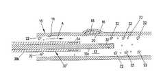
- FIG. 1is a side-sectional schematic view of a blood vessel having a vulnerable plaque as a pathologic site and with an apparatus positioned within the blood vessel to localize and identify the pathologic site;
- FIG. 2is a view of FIG. 1 showing a second embodiment of the apparatus of the present invention where the apparatus includes two telescoping components each containing detectors for a pathologic marker; and
- FIG. 3is a view of FIG. 2 with the apparatus retracted to draw together and focus upon the pathologic site.
- a blood vessel 10(such as a coronary artery) is shown having a lumen 12 defined by an endothelium layer 14 of the blood vessel 10 .
- a pathological site 16exists within the wall of the blood vessel 10 .
- the pathological site 16is a vulnerable plaque site characterized by a lipid pool 18 within the wall of the blood vessel and covered by thin fibrous cap 20 .
- the blood vessel 10contains a flow of blood in the direction of arrow A. Entrained within the blood flow are numerous pathological markers and other components of the blood flow. These pathological markers will include C-reactive proteins illustrated schematically as elements 22 (and shown greatly enlarged in the figures for ease of illustration). Other such markers (evaluated independently or concurrently with C-reactive protein) include excess hydrogen ions 23 (again, shown greatly exaggerated in size) indicating a reduced pH. In the case of such C-reactive proteins 22 , substantial quantities can be generated by the liver in response to inflammation throughout the body and may exist in a relatively high concentration (for example, measured in milligrams per liter) within the blood.
- C-reactive proteins 22substantial quantities can be generated by the liver in response to inflammation throughout the body and may exist in a relatively high concentration (for example, measured in milligrams per liter) within the blood.
- FIG. 1schematically shows additional C-reactive proteins 22 on a distal (downstream) side of the pathologic site 16 in a higher concentration than on a proximal side (upstream) of the pathologic site 16 .
- excess hydrogen ions 23produced at the inflammatory site.
- the number of additional C-reactor proteins 22 shown on the distal sideis greatly exaggerated in FIG. 1 .
- the a systemic concentration of C-reactive proteinsmay be measured in milligrams per liter as previously mentioned, the additional contribution of C-reactive proteins 22 from the pathologic site directly 16 may be measured as an additional micrograms per liter. This relatively small additional contribution directly from the pathological site 16 makes detection and localization of the pathological site 16 complicated.
- an apparatus 30is provided in the form of an elongated flexible catheter sized to be advanced through the blood vessel 10 .
- the catheterincludes a first detector 32 and a second detector 34 contained on the catheter 30 .
- the detector 32 , 34are spaced apart with detector 32 being distal to proximal detector 34 .
- the catheter 30may have an internal lumen (not shown) to advance the catheter over a guide wire (not shown) as is common in conventional catheter and guide wire systems.
- the detectors 32 , 34are selected to detect and measure pathological markers emitted or generated by the pathological site 16 .
- the detectors 32 , 34are selected to detect and measure C-reactive proteins 22 and/or hydrogen ions (pH). While the detection of C-reactive proteins and/or pH may be preferred, the detection of other markers generated by the pathological site may be utilized such as increased temperature of an inflammation site, or the emission of other markers from the pathologic site.
- Other markers of vulnerable plaquethat may be useful for diagnostic purposes include inflammatory cytokines and the similar substances such as TNF alpha (tumor necrosis factor), IL-1 (interleukin 1), MCP-1 (monocyte chemoattractant protein-1), and NO (nitric oxide).
- the detectors 32 , 34may be any one of a variety of detectors. These may include electro-chemical detectors, temperature sensors, photosensitive detectors, spectrometry detectors, or antigen-antibody reactants, which react in response to the presence of a specific protein and with the reaction being detected and measured by the detectors 32 , 34 .
- the detectors 32 , 34may be connected to a controller (not shown) for analyzing signals from the detectors 32 , 34 and outputting an analysis to an operator such as a cardiologist attempting to identify and localize the pathologic site 16 .
- the apparatus 30may be advanced into the vasculature. While being advanced through the vasculature in the blood vessel 10 , the detectors 32 , 34 are providing an output to the operator indicating concentration of C-reactive protein 22 or hydrogen ion 23 (pH) and specifically providing an indication of a concentration differential of C-reactive protein or pH between the detectors 32 , 34 .
- the detectors 32 , 34While being advanced through the vasculature in the blood vessel 10 , the detectors 32 , 34 are providing an output to the operator indicating concentration of C-reactive protein 22 or hydrogen ion 23 (pH) and specifically providing an indication of a concentration differential of C-reactive protein or pH between the detectors 32 , 34 .
- the increased concentration of C-reactive protein 22 distal to the pathologic site 16is noted by detector 32 and compared to the lesser concentration of C-reactive protein or pH on a proximal side of pathological site as measured by detector 34 .
- the existence of a differential concentration measured between detectors 32 , 34provides an indication to the operator that the pathogical site 16 is in close proximity to at least one of the detectors 32 , 34 . After such initial observation, the operator may retract the apparatus 30 until the differential is negligible indicating a precise location of the pathologic site.
- the detectors 32 , 34may be radiopaque or additional radiopaque markers (not shown) may be provided on the catheter 30 , so precise visualization of the location of the catheter 30 can be noted through fluoroscopy.
- the second and first detectors 32 , 34need not be on the same catheter.
- detector 34may be positioned in any blood vessel in the body to provide an indication of the measurement of the systemic level of C-reactive protein or pH in the body.
- a separate catheter having a lone first detector 32can be advanced through the blood vessel 10 to provide an indication of increased concentration of C-reactive protein 22 or pH distal to the pathologic site 16 .
- the detectors 32 , 34can be placed in different vessels of the patient's vasculature.
- detector 34may be outside of the body with blood delivered to it while detector 32 is advanced through vessel 10 .
- FIGS. 2 and 3illustrate an alternative embodiment of the apparatus 30 with the alternative embodiment indicated by the apparatus 30 ′.
- the apparatus 30 ′includes two catheters 30 a and 30 b with catheter 30 a slidable within catheter 30 b .
- a distal end of catheter 30 ais provided with the first detector 32 .
- a distal end of catheter 30 bis provided with the second detector 34 .
- the detectors 32 , 34are maintained spaced apart as the entire apparatus 30 ′ is advanced through the blood vessel until detector 32 is noted to be distal to a pathologic site as indicated by a concentration differential in C-reactive protein or pH measured between detectors 32 and 34 .
- the spacing between the detectors 32 , 34may be narrowed by retracting inner catheter 30 a relative to outer catheter 30 b as illustrated in FIG. 3 to more accurately focus on the precise location of the pathologic site 16 within the blood vessel 10 .
- a first detectorcan be placed in or near the coronary sinus or other coronary vein with a second detector remotely placed (e.g., in the inferior vena cava) to isolate a marker source to the heart.
Landscapes
- Life Sciences & Earth Sciences (AREA)
- Health & Medical Sciences (AREA)
- Physics & Mathematics (AREA)
- Heart & Thoracic Surgery (AREA)
- Molecular Biology (AREA)
- Pathology (AREA)
- Engineering & Computer Science (AREA)
- Biomedical Technology (AREA)
- Optics & Photonics (AREA)
- Medical Informatics (AREA)
- Biophysics (AREA)
- Surgery (AREA)
- Animal Behavior & Ethology (AREA)
- General Health & Medical Sciences (AREA)
- Public Health (AREA)
- Veterinary Medicine (AREA)
- Medicines Containing Antibodies Or Antigens For Use As Internal Diagnostic Agents (AREA)
- Magnetic Resonance Imaging Apparatus (AREA)
Abstract
Description
Claims (8)
Priority Applications (1)
| Application Number | Priority Date | Filing Date | Title |
|---|---|---|---|
| US10/306,872US6860851B2 (en) | 2002-11-27 | 2002-11-27 | Vulnerable plaque diagnosis and treatment |
Applications Claiming Priority (1)
| Application Number | Priority Date | Filing Date | Title |
|---|---|---|---|
| US10/306,872US6860851B2 (en) | 2002-11-27 | 2002-11-27 | Vulnerable plaque diagnosis and treatment |
Publications (2)
| Publication Number | Publication Date |
|---|---|
| US20040102686A1 US20040102686A1 (en) | 2004-05-27 |
| US6860851B2true US6860851B2 (en) | 2005-03-01 |
Family
ID=32325784
Family Applications (1)
| Application Number | Title | Priority Date | Filing Date |
|---|---|---|---|
| US10/306,872Expired - Fee RelatedUS6860851B2 (en) | 2002-11-27 | 2002-11-27 | Vulnerable plaque diagnosis and treatment |
Country Status (1)
| Country | Link |
|---|---|
| US (1) | US6860851B2 (en) |
Cited By (27)
| Publication number | Priority date | Publication date | Assignee | Title |
|---|---|---|---|---|
| US20030149368A1 (en)* | 2000-10-24 | 2003-08-07 | Hennemann Willard W. | Method and apparatus for locating and detecting vascular plaque via impedence and conductivity measurements, and for cryogenically passivating vascular plaque and inhibiting vascular plaque progression and rupture |
| US20040243004A1 (en)* | 2003-06-02 | 2004-12-02 | Carr Kenneth L. | Method and apparatus for detecting and treating vulnerable plaques |
| US20070208257A1 (en)* | 2006-03-03 | 2007-09-06 | Furnish Simon M | Lateral Viewing Optical Catheters |
| WO2007103236A2 (en) | 2006-03-03 | 2007-09-13 | Prescient Medical, Inc. | Endoluminal prostheses for treating vulnerable plaque |
| US20070219451A1 (en)* | 2006-03-03 | 2007-09-20 | John Kula | Optical Imaging Balloon Catheters |
| US20070225795A1 (en)* | 2006-03-24 | 2007-09-27 | Juan Granada | Composite vascular prosthesis |
| US20080085293A1 (en)* | 2006-08-22 | 2008-04-10 | Jenchen Yang | Drug eluting stent and therapeutic methods using c-Jun N-terminal kinase inhibitor |
| US20080140182A1 (en)* | 2006-04-28 | 2008-06-12 | Patricia Scheller | Composite endoluminal prostheses for treating vulnerable plaque |
| US20080304074A1 (en)* | 2007-06-08 | 2008-12-11 | Brennan Iii James F | Optical catheter configurations combining raman spectroscopy with optical fiber-based low coherence reflectometry |
| WO2009018475A1 (en) | 2007-08-01 | 2009-02-05 | Prescient Medical, Inc. | Expandable prostheses for treating atherosclerotic lesions including vulnerable plaques |
| US20100113906A1 (en)* | 2008-11-06 | 2010-05-06 | Prescient Medical, Inc. | Hybrid basket catheters |
| US8731676B2 (en) | 2011-05-19 | 2014-05-20 | Neuros Medical, Inc. | High-frequency electrical nerve block |
| US20150196210A1 (en)* | 2014-01-15 | 2015-07-16 | Medtronic Vascular Galway | Catheter For Providing Vascular Pressure Measurements |
| US9295841B2 (en) | 2011-05-19 | 2016-03-29 | Meuros Medical, Inc. | High-frequency electrical nerve block |
| US10130269B2 (en) | 2013-11-14 | 2018-11-20 | Medtronic Vascular, Inc | Dual lumen catheter for providing a vascular pressure measurement |
| US10194812B2 (en) | 2014-12-12 | 2019-02-05 | Medtronic Vascular, Inc. | System and method of integrating a fractional flow reserve device with a conventional hemodynamic monitoring system |
| US10646122B2 (en) | 2017-04-28 | 2020-05-12 | Medtronic Vascular, Inc. | FFR catheter with covered distal pressure sensor and method of manufacture |
| US10758723B2 (en) | 2011-05-19 | 2020-09-01 | Neuros Medical, Inc. | Nerve cuff electrode for neuromodulation in large human nerve trunks |
| US11116965B2 (en) | 2017-12-13 | 2021-09-14 | Neuros Medical, Inc. | Nerve cuff deployment devices |
| US11185244B2 (en) | 2018-08-13 | 2021-11-30 | Medtronic Vascular, Inc. | FFR catheter with suspended pressure sensor |
| US11213682B2 (en) | 2018-04-09 | 2022-01-04 | Neuros Medical, Inc. | Apparatuses and methods for setting an electrical dose |
| US11219741B2 (en) | 2017-08-09 | 2022-01-11 | Medtronic Vascular, Inc. | Collapsible catheter and method for calculating fractional flow reserve |
| US11235124B2 (en) | 2017-08-09 | 2022-02-01 | Medtronic Vascular, Inc. | Collapsible catheter and method for calculating fractional flow reserve |
| US11272850B2 (en) | 2016-08-09 | 2022-03-15 | Medtronic Vascular, Inc. | Catheter and method for calculating fractional flow reserve |
| US11330994B2 (en) | 2017-03-08 | 2022-05-17 | Medtronic Vascular, Inc. | Reduced profile FFR catheter |
| US11413458B2 (en) | 2011-05-19 | 2022-08-16 | Neuros Medical, Inc. | Nerve cuff electrode for neuromodulation in large human nerve trunks |
| US11878172B2 (en) | 2020-02-11 | 2024-01-23 | Neuros Medical, Inc. | System and method for quantifying qualitative patient-reported data sets |
Families Citing this family (5)
| Publication number | Priority date | Publication date | Assignee | Title |
|---|---|---|---|---|
| US20060142630A1 (en)* | 2004-12-29 | 2006-06-29 | Attila Meretei | Systems and methods for treating a thrombus in a blood vessel |
| US20070185562A1 (en)* | 2006-02-08 | 2007-08-09 | Jgf Company | Medical device for unstable and vulnerable plaque |
| WO2009035631A1 (en)* | 2007-09-11 | 2009-03-19 | Immuneering Corporation | Determining disease or immune system features based on analysis of immune system characteristics |
| EP4087474A4 (en) | 2020-01-08 | 2024-03-06 | Activ Surgical, Inc. | Laser speckle force feedback estimation |
| IL295510B2 (en)* | 2020-02-14 | 2025-08-01 | Activ Surgical Inc | Systems and methods for processing laser spot signals |
Citations (4)
| Publication number | Priority date | Publication date | Assignee | Title |
|---|---|---|---|---|
| US20020115931A1 (en)* | 2001-02-21 | 2002-08-22 | Strauss H. William | Localizing intravascular lesions on anatomic images |
| US6541265B2 (en)* | 2001-05-09 | 2003-04-01 | University Of Florida | Method and system to test a substance for inflammatory or oxidant properties |
| US6712771B2 (en)* | 2000-06-16 | 2004-03-30 | Accumed Systems, Inc. | Temperature sensing catheter |
| US20040073132A1 (en)* | 2002-05-07 | 2004-04-15 | Tracy Maahs | Systems and methods for detecting vulnerable plaque |
- 2002
- 2002-11-27USUS10/306,872patent/US6860851B2/ennot_activeExpired - Fee Related
Patent Citations (4)
| Publication number | Priority date | Publication date | Assignee | Title |
|---|---|---|---|---|
| US6712771B2 (en)* | 2000-06-16 | 2004-03-30 | Accumed Systems, Inc. | Temperature sensing catheter |
| US20020115931A1 (en)* | 2001-02-21 | 2002-08-22 | Strauss H. William | Localizing intravascular lesions on anatomic images |
| US6541265B2 (en)* | 2001-05-09 | 2003-04-01 | University Of Florida | Method and system to test a substance for inflammatory or oxidant properties |
| US20040073132A1 (en)* | 2002-05-07 | 2004-04-15 | Tracy Maahs | Systems and methods for detecting vulnerable plaque |
Cited By (42)
| Publication number | Priority date | Publication date | Assignee | Title |
|---|---|---|---|---|
| US20030149368A1 (en)* | 2000-10-24 | 2003-08-07 | Hennemann Willard W. | Method and apparatus for locating and detecting vascular plaque via impedence and conductivity measurements, and for cryogenically passivating vascular plaque and inhibiting vascular plaque progression and rupture |
| US7197356B2 (en) | 2003-06-02 | 2007-03-27 | Meridian Medical Systems, Llc | Microwave detection apparatus |
| US20040249272A1 (en)* | 2003-06-02 | 2004-12-09 | Carr Kenneth L. | Microwave detection apparatus |
| US6932776B2 (en)* | 2003-06-02 | 2005-08-23 | Meridian Medicalssystems, Llc | Method and apparatus for detecting and treating vulnerable plaques |
| US20050197570A1 (en)* | 2003-06-02 | 2005-09-08 | Carr Kenneth L. | Method and apparatus for detecting and treating vulnerable plaques |
| US20050203388A1 (en)* | 2003-06-02 | 2005-09-15 | Carr Kenneth L. | Method and apparatus for detecting and treating vulnerable plaques |
| US7734330B2 (en) | 2003-06-02 | 2010-06-08 | Meridian Medical Systems, Llc | Method and apparatus for detecting and treating vulnerable plaques |
| US20040243004A1 (en)* | 2003-06-02 | 2004-12-02 | Carr Kenneth L. | Method and apparatus for detecting and treating vulnerable plaques |
| US7933660B2 (en)* | 2003-06-02 | 2011-04-26 | Meridian Medical Systems | Apparatus for detecting and treating vulnerable plaques |
| US20070208257A1 (en)* | 2006-03-03 | 2007-09-06 | Furnish Simon M | Lateral Viewing Optical Catheters |
| WO2007103236A2 (en) | 2006-03-03 | 2007-09-13 | Prescient Medical, Inc. | Endoluminal prostheses for treating vulnerable plaque |
| US20070219451A1 (en)* | 2006-03-03 | 2007-09-20 | John Kula | Optical Imaging Balloon Catheters |
| US20070239262A1 (en)* | 2006-03-03 | 2007-10-11 | Prescient Medical, Inc. | Endoluminal Prostheses for Treating Vulnerable Plaque |
| US20070225795A1 (en)* | 2006-03-24 | 2007-09-27 | Juan Granada | Composite vascular prosthesis |
| US20080140182A1 (en)* | 2006-04-28 | 2008-06-12 | Patricia Scheller | Composite endoluminal prostheses for treating vulnerable plaque |
| US20080085293A1 (en)* | 2006-08-22 | 2008-04-10 | Jenchen Yang | Drug eluting stent and therapeutic methods using c-Jun N-terminal kinase inhibitor |
| US20080304074A1 (en)* | 2007-06-08 | 2008-12-11 | Brennan Iii James F | Optical catheter configurations combining raman spectroscopy with optical fiber-based low coherence reflectometry |
| US7952719B2 (en) | 2007-06-08 | 2011-05-31 | Prescient Medical, Inc. | Optical catheter configurations combining raman spectroscopy with optical fiber-based low coherence reflectometry |
| WO2009018475A1 (en) | 2007-08-01 | 2009-02-05 | Prescient Medical, Inc. | Expandable prostheses for treating atherosclerotic lesions including vulnerable plaques |
| US20100113906A1 (en)* | 2008-11-06 | 2010-05-06 | Prescient Medical, Inc. | Hybrid basket catheters |
| US8731676B2 (en) | 2011-05-19 | 2014-05-20 | Neuros Medical, Inc. | High-frequency electrical nerve block |
| US11413458B2 (en) | 2011-05-19 | 2022-08-16 | Neuros Medical, Inc. | Nerve cuff electrode for neuromodulation in large human nerve trunks |
| US9295841B2 (en) | 2011-05-19 | 2016-03-29 | Meuros Medical, Inc. | High-frequency electrical nerve block |
| US8983612B2 (en) | 2011-05-19 | 2015-03-17 | Neuros Medical, Inc. | High-frequency electrical nerve block |
| US12011597B2 (en) | 2011-05-19 | 2024-06-18 | Neuros Medical, Inc. | Nerve cuff electrode for neuromodulation in large human nerve trunks |
| US10758723B2 (en) | 2011-05-19 | 2020-09-01 | Neuros Medical, Inc. | Nerve cuff electrode for neuromodulation in large human nerve trunks |
| US10130269B2 (en) | 2013-11-14 | 2018-11-20 | Medtronic Vascular, Inc | Dual lumen catheter for providing a vascular pressure measurement |
| US20150196210A1 (en)* | 2014-01-15 | 2015-07-16 | Medtronic Vascular Galway | Catheter For Providing Vascular Pressure Measurements |
| US9913585B2 (en)* | 2014-01-15 | 2018-03-13 | Medtronic Vascular, Inc. | Catheter for providing vascular pressure measurements |
| US10194812B2 (en) | 2014-12-12 | 2019-02-05 | Medtronic Vascular, Inc. | System and method of integrating a fractional flow reserve device with a conventional hemodynamic monitoring system |
| US11272850B2 (en) | 2016-08-09 | 2022-03-15 | Medtronic Vascular, Inc. | Catheter and method for calculating fractional flow reserve |
| US11330994B2 (en) | 2017-03-08 | 2022-05-17 | Medtronic Vascular, Inc. | Reduced profile FFR catheter |
| US10646122B2 (en) | 2017-04-28 | 2020-05-12 | Medtronic Vascular, Inc. | FFR catheter with covered distal pressure sensor and method of manufacture |
| US11219741B2 (en) | 2017-08-09 | 2022-01-11 | Medtronic Vascular, Inc. | Collapsible catheter and method for calculating fractional flow reserve |
| US11235124B2 (en) | 2017-08-09 | 2022-02-01 | Medtronic Vascular, Inc. | Collapsible catheter and method for calculating fractional flow reserve |
| US11116965B2 (en) | 2017-12-13 | 2021-09-14 | Neuros Medical, Inc. | Nerve cuff deployment devices |
| US11752331B2 (en) | 2017-12-13 | 2023-09-12 | Neuros Medical, Inc. | Nerve cuff deployment devices |
| US11213682B2 (en) | 2018-04-09 | 2022-01-04 | Neuros Medical, Inc. | Apparatuses and methods for setting an electrical dose |
| US11730963B2 (en) | 2018-04-09 | 2023-08-22 | Neuros Medical, Inc. | Apparatuses and methods for setting an electrical dose |
| US12201837B2 (en) | 2018-04-09 | 2025-01-21 | Neuros Medical, Inc. | Apparatuses and methods for setting an electrical dose |
| US11185244B2 (en) | 2018-08-13 | 2021-11-30 | Medtronic Vascular, Inc. | FFR catheter with suspended pressure sensor |
| US11878172B2 (en) | 2020-02-11 | 2024-01-23 | Neuros Medical, Inc. | System and method for quantifying qualitative patient-reported data sets |
Also Published As
| Publication number | Publication date |
|---|---|
| US20040102686A1 (en) | 2004-05-27 |
Similar Documents
| Publication | Publication Date | Title |
|---|---|---|
| US6860851B2 (en) | Vulnerable plaque diagnosis and treatment | |
| US20190110776A1 (en) | Methods for Computing Coronary Physiology Indexes Using a High Precision Registration Model | |
| US7077812B2 (en) | Apparatus and method for palpographic characterization of vulnerable plaque and other biological tissue | |
| US7604605B2 (en) | Device, system, and method for detecting and localizing obstruction within a blood vessel | |
| St Goar et al. | Intracoronary ultrasound in cardiac transplant recipients. In vivo evidence of" angiographically silent" intimal thickening. | |
| JP2021517033A (en) | Scoring of intraluminal lesions and stent deployment in medical intraluminal ultrasound imaging | |
| US7570983B2 (en) | Method and data processing device to support diagnosis and/or therapy of a pathological change of a blood vessel | |
| US20150141764A1 (en) | Distributed sensing device for referencing of physiological features | |
| CN109561879A (en) | Position sensing in endovascular procedures | |
| EP1445626A1 (en) | Radiation detector | |
| KR20120028154A (en) | Diagnose method and apparatus for atherosclerotic lesions | |
| Erbel et al. | Electron-beam computed tomography for detection of early signs of coronary arteriosclerosis | |
| Mukai et al. | Tumors of the papilla and distal common bile duct: diagnosis and staging by endoscopic ultrasonography | |
| Jiang et al. | Diagnosis of peripheral pulmonary lesions with transbronchial lung cryobiopsy by guide sheath and radial endobronchial ultrasonography: a prospective control study | |
| Lortz et al. | Hemodynamic changes lead to alterations in aortic diameters and may challenge further stent graft sizing in acute aortic syndrome | |
| Kurashima et al. | Changes in the airway lumen and surrounding parenchyma in chronic obstructive pulmonary disease | |
| Ventura et al. | Assessment of intracoronary morphology in cardiac transplant recipients by angioscopy and intravascular ultrasound | |
| Chopard et al. | How reliable are 40 MHz IVUS and 64-slice MDCT in characterizing coronary plaque composition? An ex vivo study with histopathological comparison | |
| EP3410925B1 (en) | Optical probe for localizing and identifying a target tissue prior to harvesting a biopsy | |
| Ito et al. | Impact of coronary artery remodeling on misinterpretation of angiographic disease eccentricity: evidence from intravascular ultrasound | |
| CN113876297A (en) | Diagnosis and treatment catheter for vascular lesions | |
| Hakim et al. | Value of endoscopic ultrasonography in evaluating unexplained isolated common bile duct dilation on imaging | |
| Ehara et al. | Quantification of Coronary Calcification by Intravascular Ultrasound Do Calcium Deposits With Larger Arcs Have Longer Lengths? | |
| Canto et al. | Endoscopic ultrasonography (EUS) versus cholangiography for diagnosing extrahepatic biliary stones: A prospective, blinded study in pre-and post-cholecystectomy patients | |
| SIEGEL et al. | Comparative studies of angioscopy and ultrasound for the evaluation of arterial disease |
Legal Events
| Date | Code | Title | Description |
|---|---|---|---|
| AS | Assignment | Owner name:ALPHA MEDICAL, INC., MINNESOTA Free format text:ASSIGNMENT OF ASSIGNORS INTEREST;ASSIGNORS:KNUDSON, MARK B.;CONRAD, TIMOTHY R.;TWEDEN, KATHERINE S.;REEL/FRAME:013859/0418 Effective date:20030211 | |
| AS | Assignment | Owner name:ENTEROMEDICS INC., MINNESOTA Free format text:CHANGE OF NAME;ASSIGNOR:BETA MEDICAL, INC.;REEL/FRAME:014916/0339 Effective date:20031114 Owner name:BETA MEDICAL, INC., MINNESOTA Free format text:MERGER;ASSIGNOR:ALPHA MEDICAL, INC.;REEL/FRAME:014914/0917 Effective date:20031113 | |
| FPAY | Fee payment | Year of fee payment:4 | |
| AS | Assignment | Owner name:ENTEROMEDICS INC., A DELAWARE CORPORATION, MINNESO Free format text:MERGER;ASSIGNOR:ENTEROMEDICS INC., A MINNESOTA CORPORATION;REEL/FRAME:023731/0752 Effective date:20040728 Owner name:ENTEROMEDICS INC., A DELAWARE CORPORATION,MINNESOT Free format text:MERGER;ASSIGNOR:ENTEROMEDICS INC., A MINNESOTA CORPORATION;REEL/FRAME:023731/0752 Effective date:20040728 | |
| FPAY | Fee payment | Year of fee payment:8 | |
| REMI | Maintenance fee reminder mailed | ||
| LAPS | Lapse for failure to pay maintenance fees | ||
| STCH | Information on status: patent discontinuation | Free format text:PATENT EXPIRED DUE TO NONPAYMENT OF MAINTENANCE FEES UNDER 37 CFR 1.362 | |
| FP | Lapsed due to failure to pay maintenance fee | Effective date:20170301 | |
| AS | Assignment | Owner name:RESHAPE LIFESCIENCES INC., CALIFORNIA Free format text:CHANGE OF NAME;ASSIGNOR:ENTEROMEDICS INC.;REEL/FRAME:045949/0495 Effective date:20171012 |