US6859570B2 - Target analyte sensors utilizing microspheres - Google Patents
Target analyte sensors utilizing microspheresDownload PDFInfo
- Publication number
- US6859570B2 US6859570B2US09/925,292US92529201AUS6859570B2US 6859570 B2US6859570 B2US 6859570B2US 92529201 AUS92529201 AUS 92529201AUS 6859570 B2US6859570 B2US 6859570B2
- Authority
- US
- United States
- Prior art keywords
- microspheres
- composition according
- substrate
- sites
- bioactive agent
- Prior art date
- Legal status (The legal status is an assumption and is not a legal conclusion. Google has not performed a legal analysis and makes no representation as to the accuracy of the status listed.)
- Expired - Lifetime
Links
- 239000004005microsphereSubstances0.000titleclaimsabstractdescription200
- 239000012491analyteSubstances0.000titleclaimsdescription47
- 239000012867bioactive agentSubstances0.000claimsabstractdescription108
- 230000003287optical effectEffects0.000claimsabstractdescription78
- 239000000758substrateSubstances0.000claimsabstractdescription74
- 239000000835fiberSubstances0.000claimsabstractdescription63
- 238000000034methodMethods0.000claimsabstractdescription63
- 239000011324beadSubstances0.000claimsdescription106
- 239000000203mixtureSubstances0.000claimsdescription70
- 150000007523nucleic acidsChemical group0.000claimsdescription50
- 102000039446nucleic acidsHuman genes0.000claimsdescription45
- 108020004707nucleic acidsProteins0.000claimsdescription45
- 108090000623proteins and genesProteins0.000claimsdescription39
- 102000004169proteins and genesHuman genes0.000claimsdescription37
- 239000013307optical fiberSubstances0.000claimsdescription28
- 102000004190EnzymesHuman genes0.000claimsdescription15
- 108090000790EnzymesProteins0.000claimsdescription15
- 239000007850fluorescent dyeSubstances0.000claimsdescription10
- 239000011521glassSubstances0.000claimsdescription10
- 239000004033plasticSubstances0.000claimsdescription10
- 229920003023plasticPolymers0.000claimsdescription10
- 238000004519manufacturing processMethods0.000claimsdescription7
- 239000002245particleSubstances0.000abstractdescription7
- 238000012216screeningMethods0.000abstractdescription7
- 239000000463materialSubstances0.000abstractdescription4
- 238000009826distributionMethods0.000abstractdescription2
- 239000000975dyeSubstances0.000description86
- 239000000126substanceSubstances0.000description43
- 239000000523sampleSubstances0.000description38
- 238000009739bindingMethods0.000description34
- 230000027455bindingEffects0.000description33
- 235000018102proteinsNutrition0.000description28
- GNBHRKFJIUUOQI-UHFFFAOYSA-NfluoresceinChemical compoundO1C(=O)C2=CC=CC=C2C21C1=CC=C(O)C=C1OC1=CC(O)=CC=C21GNBHRKFJIUUOQI-UHFFFAOYSA-N0.000description23
- 238000003491arrayMethods0.000description22
- 238000001514detection methodMethods0.000description22
- -1polypropylenePolymers0.000description17
- 239000000243solutionSubstances0.000description17
- 238000009396hybridizationMethods0.000description14
- 108090000765processed proteins & peptidesProteins0.000description13
- 150000001413amino acidsChemical class0.000description12
- 235000001014amino acidNutrition0.000description11
- 230000008859changeEffects0.000description11
- 229940088598enzymeDrugs0.000description11
- 230000003993interactionEffects0.000description11
- 239000003446ligandSubstances0.000description10
- 230000008569processEffects0.000description10
- 102000004196processed proteins & peptidesHuman genes0.000description10
- 238000013459approachMethods0.000description9
- 230000015572biosynthetic processEffects0.000description9
- 238000001000micrographMethods0.000description9
- 239000002773nucleotideSubstances0.000description9
- 125000003729nucleotide groupChemical group0.000description9
- 239000000427antigenSubstances0.000description8
- 150000001720carbohydratesChemical class0.000description8
- 108020004414DNAProteins0.000description7
- 239000000853adhesiveSubstances0.000description7
- 230000001070adhesive effectEffects0.000description7
- 238000004458analytical methodMethods0.000description7
- 102000036639antigensHuman genes0.000description7
- 108091007433antigensProteins0.000description7
- 238000003556assayMethods0.000description7
- 235000014633carbohydratesNutrition0.000description7
- 238000010586diagramMethods0.000description7
- 125000000524functional groupChemical group0.000description7
- PEDCQBHIVMGVHV-UHFFFAOYSA-NGlycerineChemical compoundOCC(O)COPEDCQBHIVMGVHV-UHFFFAOYSA-N0.000description6
- ZMXDDKWLCZADIW-UHFFFAOYSA-NN,N-DimethylformamideChemical compoundCN(C)C=OZMXDDKWLCZADIW-UHFFFAOYSA-N0.000description6
- VYPSYNLAJGMNEJ-UHFFFAOYSA-NSilicium dioxideChemical compoundO=[Si]=OVYPSYNLAJGMNEJ-UHFFFAOYSA-N0.000description6
- 125000003178carboxy groupChemical group[H]OC(*)=O0.000description6
- 238000006243chemical reactionMethods0.000description6
- 239000003795chemical substances by applicationSubstances0.000description6
- 230000000295complement effectEffects0.000description6
- 230000000694effectsEffects0.000description6
- 229920000642polymerPolymers0.000description6
- 238000003786synthesis reactionMethods0.000description6
- 229920000557Nafion®Polymers0.000description5
- 230000001580bacterial effectEffects0.000description5
- 150000001875compoundsChemical class0.000description5
- 229910052802copperInorganic materials0.000description5
- 230000002209hydrophobic effectEffects0.000description5
- 229910052749magnesiumInorganic materials0.000description5
- 239000011777magnesiumSubstances0.000description5
- 241000894007speciesSpecies0.000description5
- 230000003612virological effectEffects0.000description5
- XLYOFNOQVPJJNP-UHFFFAOYSA-NwaterSubstancesOXLYOFNOQVPJJNP-UHFFFAOYSA-N0.000description5
- 102000002260Alkaline PhosphataseHuman genes0.000description4
- 108020004774Alkaline PhosphataseProteins0.000description4
- 241000894006BacteriaSpecies0.000description4
- CURLTUGMZLYLDI-UHFFFAOYSA-NCarbon dioxideChemical compoundO=C=OCURLTUGMZLYLDI-UHFFFAOYSA-N0.000description4
- KRHYYFGTRYWZRS-UHFFFAOYSA-NFluoraneChemical compoundFKRHYYFGTRYWZRS-UHFFFAOYSA-N0.000description4
- 108091034117OligonucleotideProteins0.000description4
- 241000700605VirusesSpecies0.000description4
- 150000001412aminesChemical class0.000description4
- 125000003277amino groupChemical group0.000description4
- 239000000872bufferSubstances0.000description4
- VHRGRCVQAFMJIZ-UHFFFAOYSA-NcadaverineChemical compoundNCCCCCNVHRGRCVQAFMJIZ-UHFFFAOYSA-N0.000description4
- 229910052791calciumInorganic materials0.000description4
- 239000011575calciumSubstances0.000description4
- 230000001413cellular effectEffects0.000description4
- 238000005530etchingMethods0.000description4
- 238000003384imaging methodMethods0.000description4
- 150000003839saltsChemical class0.000description4
- 229910001868waterInorganic materials0.000description4
- IMZBXSAKACGTPH-UHFFFAOYSA-N(3-oxo-6'-phosphonooxyspiro[2-benzofuran-1,9'-xanthene]-3'-yl) dihydrogen phosphateChemical compoundO1C(=O)C2=CC=CC=C2C21C1=CC=C(OP(O)(O)=O)C=C1OC1=CC(OP(O)(=O)O)=CC=C21IMZBXSAKACGTPH-UHFFFAOYSA-N0.000description3
- OKTJSMMVPCPJKN-UHFFFAOYSA-NCarbonChemical compound[C]OKTJSMMVPCPJKN-UHFFFAOYSA-N0.000description3
- 206010009944Colon cancerDiseases0.000description3
- 241000283973Oryctolagus cuniculusSpecies0.000description3
- 239000004793PolystyreneSubstances0.000description3
- FKNQFGJONOIPTF-UHFFFAOYSA-NSodium cationChemical compound[Na+]FKNQFGJONOIPTF-UHFFFAOYSA-N0.000description3
- 238000010521absorption reactionMethods0.000description3
- 150000001299aldehydesChemical class0.000description3
- QVGXLLKOCUKJST-UHFFFAOYSA-Natomic oxygenChemical compound[O]QVGXLLKOCUKJST-UHFFFAOYSA-N0.000description3
- 102000005936beta-GalactosidaseHuman genes0.000description3
- 108010005774beta-GalactosidaseProteins0.000description3
- 229910052799carbonInorganic materials0.000description3
- 239000003153chemical reaction reagentSubstances0.000description3
- 238000005253claddingMethods0.000description3
- 229920001577copolymerPolymers0.000description3
- 239000010949copperSubstances0.000description3
- 238000011161developmentMethods0.000description3
- 230000018109developmental processEffects0.000description3
- 238000002474experimental methodMethods0.000description3
- 239000000284extractSubstances0.000description3
- 239000010408filmSubstances0.000description3
- 238000005755formation reactionMethods0.000description3
- 230000002538fungal effectEffects0.000description3
- 150000002632lipidsChemical class0.000description3
- 230000004807localizationEffects0.000description3
- 239000012528membraneSubstances0.000description3
- 230000004048modificationEffects0.000description3
- 238000012986modificationMethods0.000description3
- 239000003068molecular probeSubstances0.000description3
- 230000035772mutationEffects0.000description3
- VLKZOEOYAKHREP-UHFFFAOYSA-Nn-HexaneChemical compoundCCCCCCVLKZOEOYAKHREP-UHFFFAOYSA-N0.000description3
- 239000001301oxygenSubstances0.000description3
- 229910052760oxygenInorganic materials0.000description3
- 230000035699permeabilityEffects0.000description3
- 229920002223polystyrenePolymers0.000description3
- 238000006862quantum yield reactionMethods0.000description3
- 102000005962receptorsHuman genes0.000description3
- 238000011160researchMethods0.000description3
- 239000000377silicon dioxideSubstances0.000description3
- 229910001415sodium ionInorganic materials0.000description3
- 239000007787solidSubstances0.000description3
- 239000002904solventSubstances0.000description3
- 238000012360testing methodMethods0.000description3
- MPLHNVLQVRSVEE-UHFFFAOYSA-Ntexas redChemical compound[O-]S(=O)(=O)C1=CC(S(Cl)(=O)=O)=CC=C1C(C1=CC=2CCCN3CCCC(C=23)=C1O1)=C2C1=C(CCC1)C3=[N+]1CCCC3=C2MPLHNVLQVRSVEE-UHFFFAOYSA-N0.000description3
- 238000005406washingMethods0.000description3
- 108091032973(ribonucleotides)n+mProteins0.000description2
- MYRTYDVEIRVNKP-UHFFFAOYSA-N1,2-DivinylbenzeneChemical compoundC=CC1=CC=CC=C1C=CMYRTYDVEIRVNKP-UHFFFAOYSA-N0.000description2
- JLBJTVDPSNHSKJ-UHFFFAOYSA-N4-MethylstyreneChemical compoundCC1=CC=C(C=C)C=C1JLBJTVDPSNHSKJ-UHFFFAOYSA-N0.000description2
- OBJOZRVSMLPASY-UHFFFAOYSA-N8-hydroxypyrene-1,3,6-trisulfonic acidChemical compoundC1=C2C(O)=CC(S(O)(=O)=O)=C(C=C3)C2=C2C3=C(S(O)(=O)=O)C=C(S(O)(=O)=O)C2=C1OBJOZRVSMLPASY-UHFFFAOYSA-N0.000description2
- 102000004127CytokinesHuman genes0.000description2
- 108090000695CytokinesProteins0.000description2
- ZHNUHDYFZUAESO-UHFFFAOYSA-NFormamideChemical compoundNC=OZHNUHDYFZUAESO-UHFFFAOYSA-N0.000description2
- WQZGKKKJIJFFOK-GASJEMHNSA-NGlucoseNatural productsOC[C@H]1OC(O)[C@H](O)[C@@H](O)[C@@H]1OWQZGKKKJIJFFOK-GASJEMHNSA-N0.000description2
- 108010015776Glucose oxidaseProteins0.000description2
- 239000004366Glucose oxidaseSubstances0.000description2
- MHAJPDPJQMAIIY-UHFFFAOYSA-NHydrogen peroxideChemical compoundOOMHAJPDPJQMAIIY-UHFFFAOYSA-N0.000description2
- 102000000588Interleukin-2Human genes0.000description2
- 108010002350Interleukin-2Proteins0.000description2
- 102000004388Interleukin-4Human genes0.000description2
- 108090000978Interleukin-4Proteins0.000description2
- 102000004889Interleukin-6Human genes0.000description2
- 108090001005Interleukin-6Proteins0.000description2
- 108091092878MicrosatelliteProteins0.000description2
- 206010028980NeoplasmDiseases0.000description2
- 108020004711Nucleic Acid ProbesProteins0.000description2
- 108091028043Nucleic acid sequenceProteins0.000description2
- 108091005461Nucleic proteinsProteins0.000description2
- 239000004677NylonSubstances0.000description2
- 102000015636OligopeptidesHuman genes0.000description2
- 108010038807OligopeptidesProteins0.000description2
- 239000002202Polyethylene glycolSubstances0.000description2
- 229920001213Polysorbate 20Polymers0.000description2
- FAPWRFPIFSIZLT-UHFFFAOYSA-MSodium chlorideChemical compound[Na+].[Cl-]FAPWRFPIFSIZLT-UHFFFAOYSA-M0.000description2
- PPBRXRYQALVLMV-UHFFFAOYSA-NStyreneChemical compoundC=CC1=CC=CC=C1PPBRXRYQALVLMV-UHFFFAOYSA-N0.000description2
- 229910052771TerbiumInorganic materials0.000description2
- GWEVSGVZZGPLCZ-UHFFFAOYSA-NTitan oxideChemical compoundO=[Ti]=OGWEVSGVZZGPLCZ-UHFFFAOYSA-N0.000description2
- ISAKRJDGNUQOIC-UHFFFAOYSA-NUracilChemical compoundO=C1C=CNC(=O)N1ISAKRJDGNUQOIC-UHFFFAOYSA-N0.000description2
- XSQUKJJJFZCRTK-UHFFFAOYSA-NUreaChemical compoundNC(N)=OXSQUKJJJFZCRTK-UHFFFAOYSA-N0.000description2
- JLCPHMBAVCMARE-UHFFFAOYSA-N[3-[[3-[[3-[[3-[[3-[[3-[[3-[[3-[[3-[[3-[[3-[[5-(2-amino-6-oxo-1H-purin-9-yl)-3-[[3-[[3-[[3-[[3-[[3-[[5-(2-amino-6-oxo-1H-purin-9-yl)-3-[[5-(2-amino-6-oxo-1H-purin-9-yl)-3-hydroxyoxolan-2-yl]methoxy-hydroxyphosphoryl]oxyoxolan-2-yl]methoxy-hydroxyphosphoryl]oxy-5-(5-methyl-2,4-dioxopyrimidin-1-yl)oxolan-2-yl]methoxy-hydroxyphosphoryl]oxy-5-(6-aminopurin-9-yl)oxolan-2-yl]methoxy-hydroxyphosphoryl]oxy-5-(6-aminopurin-9-yl)oxolan-2-yl]methoxy-hydroxyphosphoryl]oxy-5-(6-aminopurin-9-yl)oxolan-2-yl]methoxy-hydroxyphosphoryl]oxy-5-(6-aminopurin-9-yl)oxolan-2-yl]methoxy-hydroxyphosphoryl]oxyoxolan-2-yl]methoxy-hydroxyphosphoryl]oxy-5-(5-methyl-2,4-dioxopyrimidin-1-yl)oxolan-2-yl]methoxy-hydroxyphosphoryl]oxy-5-(4-amino-2-oxopyrimidin-1-yl)oxolan-2-yl]methoxy-hydroxyphosphoryl]oxy-5-(5-methyl-2,4-dioxopyrimidin-1-yl)oxolan-2-yl]methoxy-hydroxyphosphoryl]oxy-5-(5-methyl-2,4-dioxopyrimidin-1-yl)oxolan-2-yl]methoxy-hydroxyphosphoryl]oxy-5-(6-aminopurin-9-yl)oxolan-2-yl]methoxy-hydroxyphosphoryl]oxy-5-(6-aminopurin-9-yl)oxolan-2-yl]methoxy-hydroxyphosphoryl]oxy-5-(4-amino-2-oxopyrimidin-1-yl)oxolan-2-yl]methoxy-hydroxyphosphoryl]oxy-5-(4-amino-2-oxopyrimidin-1-yl)oxolan-2-yl]methoxy-hydroxyphosphoryl]oxy-5-(4-amino-2-oxopyrimidin-1-yl)oxolan-2-yl]methoxy-hydroxyphosphoryl]oxy-5-(6-aminopurin-9-yl)oxolan-2-yl]methoxy-hydroxyphosphoryl]oxy-5-(4-amino-2-oxopyrimidin-1-yl)oxolan-2-yl]methyl [5-(6-aminopurin-9-yl)-2-(hydroxymethyl)oxolan-3-yl] hydrogen phosphatePolymersCc1cn(C2CC(OP(O)(=O)OCC3OC(CC3OP(O)(=O)OCC3OC(CC3O)n3cnc4c3nc(N)[nH]c4=O)n3cnc4c3nc(N)[nH]c4=O)C(COP(O)(=O)OC3CC(OC3COP(O)(=O)OC3CC(OC3COP(O)(=O)OC3CC(OC3COP(O)(=O)OC3CC(OC3COP(O)(=O)OC3CC(OC3COP(O)(=O)OC3CC(OC3COP(O)(=O)OC3CC(OC3COP(O)(=O)OC3CC(OC3COP(O)(=O)OC3CC(OC3COP(O)(=O)OC3CC(OC3COP(O)(=O)OC3CC(OC3COP(O)(=O)OC3CC(OC3COP(O)(=O)OC3CC(OC3COP(O)(=O)OC3CC(OC3COP(O)(=O)OC3CC(OC3COP(O)(=O)OC3CC(OC3COP(O)(=O)OC3CC(OC3CO)n3cnc4c(N)ncnc34)n3ccc(N)nc3=O)n3cnc4c(N)ncnc34)n3ccc(N)nc3=O)n3ccc(N)nc3=O)n3ccc(N)nc3=O)n3cnc4c(N)ncnc34)n3cnc4c(N)ncnc34)n3cc(C)c(=O)[nH]c3=O)n3cc(C)c(=O)[nH]c3=O)n3ccc(N)nc3=O)n3cc(C)c(=O)[nH]c3=O)n3cnc4c3nc(N)[nH]c4=O)n3cnc4c(N)ncnc34)n3cnc4c(N)ncnc34)n3cnc4c(N)ncnc34)n3cnc4c(N)ncnc34)O2)c(=O)[nH]c1=OJLCPHMBAVCMARE-UHFFFAOYSA-N0.000description2
- 239000002253acidSubstances0.000description2
- 229920006397acrylic thermoplasticPolymers0.000description2
- 238000013019agitationMethods0.000description2
- 230000036436anti-hivEffects0.000description2
- 230000000692anti-sense effectEffects0.000description2
- 229910002092carbon dioxideInorganic materials0.000description2
- 230000015556catabolic processEffects0.000description2
- 210000004027cellAnatomy0.000description2
- 239000002299complementary DNASubstances0.000description2
- 239000003431cross linking reagentSubstances0.000description2
- 235000018417cysteineNutrition0.000description2
- OPTASPLRGRRNAP-UHFFFAOYSA-NcytosineChemical compoundNC=1C=CNC(=O)N=1OPTASPLRGRRNAP-UHFFFAOYSA-N0.000description2
- 238000000151depositionMethods0.000description2
- 229940000406drug candidateDrugs0.000description2
- 235000021463dry cakeNutrition0.000description2
- 238000000295emission spectrumMethods0.000description2
- 239000003344environmental pollutantSubstances0.000description2
- 230000005284excitationEffects0.000description2
- 238000002189fluorescence spectrumMethods0.000description2
- 239000012634fragmentSubstances0.000description2
- 238000007306functionalization reactionMethods0.000description2
- YFHXZQPUBCBNIP-UHFFFAOYSA-Nfura-2Chemical compoundCC1=CC=C(N(CC(O)=O)CC(O)=O)C(OCCOC=2C(=CC=3OC(=CC=3C=2)C=2OC(=CN=2)C(O)=O)N(CC(O)=O)CC(O)=O)=C1YFHXZQPUBCBNIP-UHFFFAOYSA-N0.000description2
- 239000008103glucoseSubstances0.000description2
- 229940116332glucose oxidaseDrugs0.000description2
- 235000019420glucose oxidaseNutrition0.000description2
- 150000004676glycansChemical class0.000description2
- UYTPUPDQBNUYGX-UHFFFAOYSA-NguanineChemical compoundO=C1NC(N)=NC2=C1N=CN2UYTPUPDQBNUYGX-UHFFFAOYSA-N0.000description2
- 239000005556hormoneSubstances0.000description2
- 229940088597hormoneDrugs0.000description2
- 239000001257hydrogenSubstances0.000description2
- 229910052739hydrogenInorganic materials0.000description2
- 125000002887hydroxy groupChemical group[H]O*0.000description2
- 230000003100immobilizing effectEffects0.000description2
- 238000000338in vitroMethods0.000description2
- 238000001727in vivoMethods0.000description2
- PNDZEEPOYCVIIY-UHFFFAOYSA-Nindo-1Chemical compoundCC1=CC=C(N(CC(O)=O)CC(O)=O)C(OCCOC=2C(=CC=C(C=2)C=2N=C3[CH]C(=CC=C3C=2)C(O)=O)N(CC(O)=O)CC(O)=O)=C1PNDZEEPOYCVIIY-UHFFFAOYSA-N0.000description2
- 238000009830intercalationMethods0.000description2
- DRAVOWXCEBXPTN-UHFFFAOYSA-NisoguanineChemical compoundNC1=NC(=O)NC2=C1NC=N2DRAVOWXCEBXPTN-UHFFFAOYSA-N0.000description2
- 238000011068loading methodMethods0.000description2
- 238000000504luminescence detectionMethods0.000description2
- 229910052748manganeseInorganic materials0.000description2
- 239000003550markerSubstances0.000description2
- 238000005259measurementMethods0.000description2
- 108020004999messenger RNAProteins0.000description2
- 235000012459muffinsNutrition0.000description2
- 229930014626natural productNatural products0.000description2
- 229910052759nickelInorganic materials0.000description2
- 239000002853nucleic acid probeSubstances0.000description2
- 229920001778nylonPolymers0.000description2
- 150000002894organic compoundsChemical class0.000description2
- 239000000575pesticideSubstances0.000description2
- 239000002953phosphate buffered salineSubstances0.000description2
- 229920003229poly(methyl methacrylate)Polymers0.000description2
- 229920001223polyethylene glycolPolymers0.000description2
- 102000054765polymorphisms of proteinsHuman genes0.000description2
- 239000000256polyoxyethylene sorbitan monolaurateSubstances0.000description2
- 235000010486polyoxyethylene sorbitan monolaurateNutrition0.000description2
- 229920001282polysaccharidePolymers0.000description2
- 239000005017polysaccharideSubstances0.000description2
- 238000002360preparation methodMethods0.000description2
- 239000000047productSubstances0.000description2
- 150000003212purinesChemical class0.000description2
- BBEAQIROQSPTKN-UHFFFAOYSA-NpyreneChemical compoundC1=CC=C2C=CC3=CC=CC4=CC=C1C2=C43BBEAQIROQSPTKN-UHFFFAOYSA-N0.000description2
- 230000004044responseEffects0.000description2
- 238000010079rubber tappingMethods0.000description2
- 230000035945sensitivityEffects0.000description2
- 238000000527sonicationMethods0.000description2
- 230000003595spectral effectEffects0.000description2
- 230000008961swellingEffects0.000description2
- ISXSCDLOGDJUNJ-UHFFFAOYSA-Ntert-butyl prop-2-enoateChemical compoundCC(C)(C)OC(=O)C=CISXSCDLOGDJUNJ-UHFFFAOYSA-N0.000description2
- 230000001225therapeutic effectEffects0.000description2
- 125000003396thiol groupChemical group[H]S*0.000description2
- RWQNBRDOKXIBIV-UHFFFAOYSA-NthymineChemical compoundCC1=CNC(=O)NC1=ORWQNBRDOKXIBIV-UHFFFAOYSA-N0.000description2
- 201000008827tuberculosisDiseases0.000description2
- 229910052725zincInorganic materials0.000description2
- JWDFQMWEFLOOED-UHFFFAOYSA-N(2,5-dioxopyrrolidin-1-yl) 3-(pyridin-2-yldisulfanyl)propanoateChemical compoundO=C1CCC(=O)N1OC(=O)CCSSC1=CC=CC=N1JWDFQMWEFLOOED-UHFFFAOYSA-N0.000description1
- UANMYOBKUNUUTR-UHFFFAOYSA-M(2z)-1,3,3-trimethyl-2-[(2e)-5-(1,3,3-trimethylindol-1-ium-2-yl)penta-2,4-dienylidene]indole;iodideChemical compound[I-].CC1(C)C2=CC=CC=C2N(C)C1=CC=CC=CC1=[N+](C)C2=CC=CC=C2C1(C)CUANMYOBKUNUUTR-UHFFFAOYSA-M0.000description1
- QBPPRVHXOZRESW-UHFFFAOYSA-N1,4,7,10-tetraazacyclododecaneChemical classC1CNCCNCCNCCN1QBPPRVHXOZRESW-UHFFFAOYSA-N0.000description1
- SGTJXHYDKUXJRK-UHFFFAOYSA-N1-(2-methoxyethenyl)pyreneChemical compoundC1=C2C(C=COC)=CC=C(C=C3)C2=C2C3=CC=CC2=C1SGTJXHYDKUXJRK-UHFFFAOYSA-N0.000description1
- 1500000039232,5-pyrroledionesChemical class0.000description1
- PDDJAJCJQXFQCW-UHFFFAOYSA-N2-[4-[bis(carboxymethyl)amino]-3-(carboxymethoxy)phenyl]-1h-indole-6-carboxylic acidChemical compoundC1=C(N(CC(O)=O)CC(O)=O)C(OCC(=O)O)=CC(C=2NC3=CC(=CC=C3C=2)C(O)=O)=C1PDDJAJCJQXFQCW-UHFFFAOYSA-N0.000description1
- PDURUKZNVHEHGO-UHFFFAOYSA-N2-[6-[bis(carboxymethyl)amino]-5-(carboxymethoxy)-1-benzofuran-2-yl]-1,3-oxazole-5-carboxylic acidChemical compoundO1C=2C=C(N(CC(O)=O)CC(O)=O)C(OCC(=O)O)=CC=2C=C1C1=NC=C(C(O)=O)O1PDURUKZNVHEHGO-UHFFFAOYSA-N0.000description1
- XQCZBXHVTFVIFE-UHFFFAOYSA-N2-amino-4-hydroxypyrimidineChemical compoundNC1=NC=CC(O)=N1XQCZBXHVTFVIFE-UHFFFAOYSA-N0.000description1
- HCGYMSSYSAKGPK-UHFFFAOYSA-N2-nitro-1h-indoleChemical compoundC1=CC=C2NC([N+](=O)[O-])=CC2=C1HCGYMSSYSAKGPK-UHFFFAOYSA-N0.000description1
- FTBBGQKRYUTLMP-UHFFFAOYSA-N2-nitro-1h-pyrroleChemical compound[O-][N+](=O)C1=CC=CN1FTBBGQKRYUTLMP-UHFFFAOYSA-N0.000description1
- BCHZICNRHXRCHY-UHFFFAOYSA-N2h-oxazineChemical compoundN1OC=CC=C1BCHZICNRHXRCHY-UHFFFAOYSA-N0.000description1
- JRBJSXQPQWSCCF-UHFFFAOYSA-N3,3'-DimethoxybenzidineChemical compoundC1=C(N)C(OC)=CC(C=2C=C(OC)C(N)=CC=2)=C1JRBJSXQPQWSCCF-UHFFFAOYSA-N0.000description1
- YOQMJMHTHWYNIO-UHFFFAOYSA-N4-[6-[16-[2-(2,4-dicarboxyphenyl)-5-methoxy-1-benzofuran-6-yl]-1,4,10,13-tetraoxa-7,16-diazacyclooctadec-7-yl]-5-methoxy-1-benzofuran-2-yl]benzene-1,3-dicarboxylic acidChemical compoundCOC1=CC=2C=C(C=3C(=CC(=CC=3)C(O)=O)C(O)=O)OC=2C=C1N(CCOCCOCC1)CCOCCOCCN1C(C(=CC=1C=2)OC)=CC=1OC=2C1=CC=C(C(O)=O)C=C1C(O)=OYOQMJMHTHWYNIO-UHFFFAOYSA-N0.000description1
- DDFHBQSCUXNBSA-UHFFFAOYSA-N5-(5-carboxythiophen-2-yl)thiophene-2-carboxylic acidChemical compoundS1C(C(=O)O)=CC=C1C1=CC=C(C(O)=O)S1DDFHBQSCUXNBSA-UHFFFAOYSA-N0.000description1
- 102000012440AcetylcholinesteraseHuman genes0.000description1
- 108010022752AcetylcholinesteraseProteins0.000description1
- GFFGJBXGBJISGV-UHFFFAOYSA-NAdenineChemical compoundNC1=NC=NC2=C1N=CN2GFFGJBXGBJISGV-UHFFFAOYSA-N0.000description1
- 229930024421AdenineNatural products0.000description1
- 102000009027AlbuminsHuman genes0.000description1
- 108010088751AlbuminsProteins0.000description1
- 208000024827Alzheimer diseaseDiseases0.000description1
- 208000004881AmebiasisDiseases0.000description1
- 206010001980AmoebiasisDiseases0.000description1
- 108010060159Apolipoprotein E4Proteins0.000description1
- 201000002909AspergillosisDiseases0.000description1
- 208000036641Aspergillus infectionsDiseases0.000description1
- 108700020463BRCA1Proteins0.000description1
- 102000036365BRCA1Human genes0.000description1
- 101150072950BRCA1 geneProteins0.000description1
- 208000035143Bacterial infectionDiseases0.000description1
- 206010006187Breast cancerDiseases0.000description1
- 208000026310Breast neoplasmDiseases0.000description1
- 101100268670Caenorhabditis elegans acc-3 geneProteins0.000description1
- 101100000858Caenorhabditis elegans act-3 geneProteins0.000description1
- OYPRJOBELJOOCE-UHFFFAOYSA-NCalciumChemical compound[Ca]OYPRJOBELJOOCE-UHFFFAOYSA-N0.000description1
- 241000589876CampylobacterSpecies0.000description1
- 241000222122Candida albicansSpecies0.000description1
- 206010007134Candida infectionsDiseases0.000description1
- 108010001857Cell Surface ReceptorsProteins0.000description1
- 206010008631CholeraDiseases0.000description1
- 239000004971Cross linkerSubstances0.000description1
- 201000007336CryptococcosisDiseases0.000description1
- 201000003883Cystic fibrosisDiseases0.000description1
- 241000701022CytomegalovirusSpecies0.000description1
- 229920002307DextranPolymers0.000description1
- 241000588724Escherichia coliSpecies0.000description1
- PIICEJLVQHRZGT-UHFFFAOYSA-NEthylenediamineChemical compoundNCCNPIICEJLVQHRZGT-UHFFFAOYSA-N0.000description1
- 229910052693EuropiumInorganic materials0.000description1
- OZLGRUXZXMRXGP-UHFFFAOYSA-NFluo-3Chemical compoundCC1=CC=C(N(CC(O)=O)CC(O)=O)C(OCCOC=2C(=CC=C(C=2)C2=C3C=C(Cl)C(=O)C=C3OC3=CC(O)=C(Cl)C=C32)N(CC(O)=O)CC(O)=O)=C1OZLGRUXZXMRXGP-UHFFFAOYSA-N0.000description1
- 206010056740Genital dischargeDiseases0.000description1
- SXRSQZLOMIGNAQ-UHFFFAOYSA-NGlutaraldehydeChemical compoundO=CCCCC=OSXRSQZLOMIGNAQ-UHFFFAOYSA-N0.000description1
- 208000009889Herpes SimplexDiseases0.000description1
- 101000599940Homo sapiens Interferon gammaProteins0.000description1
- 108090000144Human ProteinsProteins0.000description1
- 102000003839Human ProteinsHuman genes0.000description1
- 206010020460Human T-cell lymphotropic virus type I infectionDiseases0.000description1
- 241000714260Human T-lymphotropic virus 1Species0.000description1
- 241000714259Human T-lymphotropic virus 2Species0.000description1
- HEFNNWSXXWATRW-UHFFFAOYSA-NIbuprofenChemical compoundCC(C)CC1=CC=C(C(C)C(O)=O)C=C1HEFNNWSXXWATRW-UHFFFAOYSA-N0.000description1
- 108060003951ImmunoglobulinProteins0.000description1
- UGQMRVRMYYASKQ-KQYNXXCUSA-NInosineChemical compoundO[C@@H]1[C@H](O)[C@@H](CO)O[C@H]1N1C2=NC=NC(O)=C2N=C1UGQMRVRMYYASKQ-KQYNXXCUSA-N0.000description1
- 229930010555InosineNatural products0.000description1
- 102100037850Interferon gammaHuman genes0.000description1
- 101710175886Interferon gamma 1Proteins0.000description1
- RHGKLRLOHDJJDR-BYPYZUCNSA-NL-citrullineChemical compoundNC(=O)NCCC[C@H]([NH3+])C([O-])=ORHGKLRLOHDJJDR-BYPYZUCNSA-N0.000description1
- JTTHKOPSMAVJFE-VIFPVBQESA-NL-homophenylalanineChemical compoundOC(=O)[C@@H](N)CCC1=CC=CC=C1JTTHKOPSMAVJFE-VIFPVBQESA-N0.000description1
- LRQKBLKVPFOOQJ-YFKPBYRVSA-NL-norleucineChemical compoundCCCC[C@H]([NH3+])C([O-])=OLRQKBLKVPFOOQJ-YFKPBYRVSA-N0.000description1
- 208000007764Legionnaires' DiseaseDiseases0.000description1
- 241000222722Leishmania <genus>Species0.000description1
- 241000713666LentivirusSpecies0.000description1
- 206010024229LeprosyDiseases0.000description1
- 208000016604Lyme diseaseDiseases0.000description1
- FYYHWMGAXLPEAU-UHFFFAOYSA-NMagnesiumChemical compound[Mg]FYYHWMGAXLPEAU-UHFFFAOYSA-N0.000description1
- 241001465754MetazoaSpecies0.000description1
- CERQOIWHTDAKMF-UHFFFAOYSA-MMethacrylateChemical compoundCC(=C)C([O-])=OCERQOIWHTDAKMF-UHFFFAOYSA-M0.000description1
- 201000008235Mycoplasma pneumoniae pneumoniaDiseases0.000description1
- YGSVGJYZQUHXNS-UHFFFAOYSA-NNC1=C(C2=CC=CC=C2C=C1)N.N(=O)OChemical compoundNC1=C(C2=CC=CC=C2C=C1)N.N(=O)OYGSVGJYZQUHXNS-UHFFFAOYSA-N0.000description1
- IXQIUDNVFVTQLJ-UHFFFAOYSA-NNaphthofluoresceinChemical compoundO1C(=O)C2=CC=CC=C2C21C(C=CC=1C3=CC=C(O)C=1)=C3OC1=C2C=CC2=CC(O)=CC=C21IXQIUDNVFVTQLJ-UHFFFAOYSA-N0.000description1
- RHGKLRLOHDJJDR-UHFFFAOYSA-NNdelta-carbamoyl-DL-ornithineNatural productsOC(=O)C(N)CCCNC(N)=ORHGKLRLOHDJJDR-UHFFFAOYSA-N0.000description1
- 102000048850Neoplasm GenesHuman genes0.000description1
- 108700019961Neoplasm GenesProteins0.000description1
- 239000000020NitrocelluloseSubstances0.000description1
- 101710163270NucleaseProteins0.000description1
- 238000012408PCR amplificationMethods0.000description1
- 229930182555PenicillinNatural products0.000description1
- JGSARLDLIJGVTE-MBNYWOFBSA-NPenicillin GChemical compoundN([C@H]1[C@H]2SC([C@@H](N2C1=O)C(O)=O)(C)C)C(=O)CC1=CC=CC=C1JGSARLDLIJGVTE-MBNYWOFBSA-N0.000description1
- 108010087702PenicillinaseProteins0.000description1
- 108091093037Peptide nucleic acidProteins0.000description1
- 206010035718Pneumonia legionellaDiseases0.000description1
- 206010035724Pneumonia mycoplasmalDiseases0.000description1
- 239000004698PolyethyleneSubstances0.000description1
- 239000004743PolypropyleneSubstances0.000description1
- 238000001069Raman spectroscopyMethods0.000description1
- 108091028664RibonucleotideProteins0.000description1
- 241000606651RickettsialesSpecies0.000description1
- 206010039207Rocky Mountain Spotted FeverDiseases0.000description1
- 241000607142SalmonellaSpecies0.000description1
- 229920002684SepharosePolymers0.000description1
- 208000019802Sexually transmitted diseaseDiseases0.000description1
- XUIMIQQOPSSXEZ-UHFFFAOYSA-NSiliconChemical compound[Si]XUIMIQQOPSSXEZ-UHFFFAOYSA-N0.000description1
- PJANXHGTPQOBST-VAWYXSNFSA-NStilbeneNatural productsC=1C=CC=CC=1/C=C/C1=CC=CC=C1PJANXHGTPQOBST-VAWYXSNFSA-N0.000description1
- QAOWNCQODCNURD-UHFFFAOYSA-LSulfateChemical compound[O-]S([O-])(=O)=OQAOWNCQODCNURD-UHFFFAOYSA-L0.000description1
- 239000004809TeflonSubstances0.000description1
- 229920006362Teflon®Polymers0.000description1
- ATJFFYVFTNAWJD-UHFFFAOYSA-NTinChemical compound[Sn]ATJFFYVFTNAWJD-UHFFFAOYSA-N0.000description1
- 201000005485ToxoplasmosisDiseases0.000description1
- 108010046334UreaseProteins0.000description1
- 241000607626Vibrio choleraeSpecies0.000description1
- APERIXFHHNDFQV-UHFFFAOYSA-N[2-[2-[2-[bis(carboxymethyl)amino]-5-methylphenoxy]ethoxy]-4-[3,6-bis(dimethylamino)xanthen-9-ylidene]cyclohexa-2,5-dien-1-ylidene]-bis(carboxymethyl)azanium;chlorideChemical compound[Cl-].C12=CC=C(N(C)C)C=C2OC2=CC(N(C)C)=CC=C2C1=C(C=1)C=CC(=[N+](CC(O)=O)CC(O)=O)C=1OCCOC1=CC(C)=CC=C1N(CC(O)=O)CC(O)=OAPERIXFHHNDFQV-UHFFFAOYSA-N0.000description1
- 238000002835absorbanceMethods0.000description1
- 238000011481absorbance measurementMethods0.000description1
- 238000004847absorption spectroscopyMethods0.000description1
- 238000000862absorption spectrumMethods0.000description1
- OIPILFWXSMYKGL-UHFFFAOYSA-NacetylcholineChemical compoundCC(=O)OCC[N+](C)(C)COIPILFWXSMYKGL-UHFFFAOYSA-N0.000description1
- 229960004373acetylcholineDrugs0.000description1
- 229940022698acetylcholinesteraseDrugs0.000description1
- 150000007513acidsChemical class0.000description1
- 230000004913activationEffects0.000description1
- 230000010933acylationEffects0.000description1
- 238000005917acylation reactionMethods0.000description1
- 229960000643adenineDrugs0.000description1
- PPQRONHOSHZGFQ-LMVFSUKVSA-Naldehydo-D-ribose 5-phosphateChemical groupOP(=O)(O)OC[C@@H](O)[C@@H](O)[C@@H](O)C=OPPQRONHOSHZGFQ-LMVFSUKVSA-N0.000description1
- 230000029936alkylationEffects0.000description1
- 238000005804alkylation reactionMethods0.000description1
- 230000004075alterationEffects0.000description1
- 150000001408amidesChemical class0.000description1
- 125000000539amino acid groupChemical group0.000description1
- LDDQLRUQCUTJBB-UHFFFAOYSA-Nammonium fluorideChemical compound[NH4+].[F-]LDDQLRUQCUTJBB-UHFFFAOYSA-N0.000description1
- 230000003321amplificationEffects0.000description1
- 239000003242anti bacterial agentSubstances0.000description1
- 230000000840anti-viral effectEffects0.000description1
- 229940088710antibiotic agentDrugs0.000description1
- 239000004599antimicrobialSubstances0.000description1
- 239000012062aqueous bufferSubstances0.000description1
- 239000012736aqueous mediumSubstances0.000description1
- 150000004982aromatic aminesChemical class0.000description1
- 125000003118aryl groupChemical group0.000description1
- 238000002820assay formatMethods0.000description1
- 208000022362bacterial infectious diseaseDiseases0.000description1
- 244000052616bacterial pathogenSpecies0.000description1
- 229910052788bariumInorganic materials0.000description1
- 230000004888barrier functionEffects0.000description1
- 235000013405beerNutrition0.000description1
- 230000008901benefitEffects0.000description1
- 230000001588bifunctional effectEffects0.000description1
- 230000000975bioactive effectEffects0.000description1
- 230000000903blocking effectEffects0.000description1
- 239000007853buffer solutionSubstances0.000description1
- 239000006227byproductSubstances0.000description1
- 229910052793cadmiumInorganic materials0.000description1
- DEGAKNSWVGKMLS-UHFFFAOYSA-NcalceinChemical compoundO1C(=O)C2=CC=CC=C2C21C1=CC(CN(CC(O)=O)CC(O)=O)=C(O)C=C1OC1=C2C=C(CN(CC(O)=O)CC(=O)O)C(O)=C1DEGAKNSWVGKMLS-UHFFFAOYSA-N0.000description1
- 201000011510cancerDiseases0.000description1
- 201000003984candidiasisDiseases0.000description1
- 239000004202carbamideSubstances0.000description1
- 150000001718carbodiimidesChemical class0.000description1
- 239000001569carbon dioxideSubstances0.000description1
- 125000002915carbonyl groupChemical group[*:2]C([*:1])=O0.000description1
- 150000001732carboxylic acid derivativesChemical class0.000description1
- 239000001913celluloseSubstances0.000description1
- 229920002678cellulosePolymers0.000description1
- 239000000919ceramicSubstances0.000description1
- 150000001793charged compoundsChemical class0.000description1
- 125000003636chemical groupChemical group0.000description1
- 238000007385chemical modificationMethods0.000description1
- 125000004218chloromethyl groupChemical group[H]C([H])(Cl)*0.000description1
- 235000013477citrullineNutrition0.000description1
- 229960002173citrullineDrugs0.000description1
- 208000029742colonic neoplasmDiseases0.000description1
- 238000010276constructionMethods0.000description1
- 239000000356contaminantSubstances0.000description1
- 238000007796conventional methodMethods0.000description1
- 238000004132cross linkingMethods0.000description1
- DMSZORWOGDLWGN-UHFFFAOYSA-Nctk1a3526Chemical compoundNP(N)(N)=ODMSZORWOGDLWGN-UHFFFAOYSA-N0.000description1
- XUJNEKJLAYXESH-UHFFFAOYSA-NcysteineNatural productsSCC(N)C(O)=OXUJNEKJLAYXESH-UHFFFAOYSA-N0.000description1
- 150000001945cysteinesChemical class0.000description1
- 229940104302cytosineDrugs0.000description1
- 230000006378damageEffects0.000description1
- 238000007405data analysisMethods0.000description1
- 238000006731degradation reactionMethods0.000description1
- 239000008367deionised waterSubstances0.000description1
- 229910021641deionized waterInorganic materials0.000description1
- 230000001419dependent effectEffects0.000description1
- 230000008021depositionEffects0.000description1
- 238000013461designMethods0.000description1
- 230000000368destabilizing effectEffects0.000description1
- 239000003599detergentSubstances0.000description1
- 238000003745diagnosisMethods0.000description1
- 235000014113dietary fatty acidsNutrition0.000description1
- 238000010790dilutionMethods0.000description1
- 239000012895dilutionSubstances0.000description1
- NAGJZTKCGNOGPW-UHFFFAOYSA-Kdioxido-sulfanylidene-sulfido-$l^{5}-phosphaneChemical compound[O-]P([O-])([S-])=SNAGJZTKCGNOGPW-UHFFFAOYSA-K0.000description1
- LOKCTEFSRHRXRJ-UHFFFAOYSA-Idipotassium trisodium dihydrogen phosphate hydrogen phosphate dichlorideChemical compoundP(=O)(O)(O)[O-].[K+].P(=O)(O)([O-])[O-].[Na+].[Na+].[Cl-].[K+].[Cl-].[Na+]LOKCTEFSRHRXRJ-UHFFFAOYSA-I0.000description1
- 229940042399direct acting antivirals protease inhibitorsDrugs0.000description1
- 201000010099diseaseDiseases0.000description1
- 208000037265diseases, disorders, signs and symptomsDiseases0.000description1
- BFMYDTVEBKDAKJ-UHFFFAOYSA-Ldisodium;(2',7'-dibromo-3',6'-dioxido-3-oxospiro[2-benzofuran-1,9'-xanthene]-4'-yl)mercury;hydrateChemical compoundO.[Na+].[Na+].O1C(=O)C2=CC=CC=C2C21C1=CC(Br)=C([O-])C([Hg])=C1OC1=C2C=C(Br)C([O-])=C1BFMYDTVEBKDAKJ-UHFFFAOYSA-L0.000description1
- 238000010494dissociation reactionMethods0.000description1
- 230000005593dissociationsEffects0.000description1
- 229940079593drugDrugs0.000description1
- 239000003814drugSubstances0.000description1
- 239000003937drug carrierSubstances0.000description1
- 238000005516engineering processMethods0.000description1
- 231100000249enterotoxicToxicity0.000description1
- 230000002242enterotoxic effectEffects0.000description1
- YQGOJNYOYNNSMM-UHFFFAOYSA-NeosinChemical compound[Na+].OC(=O)C1=CC=CC=C1C1=C2C=C(Br)C(=O)C(Br)=C2OC2=C(Br)C(O)=C(Br)C=C21YQGOJNYOYNNSMM-UHFFFAOYSA-N0.000description1
- WTOSNONTQZJEBC-UHFFFAOYSA-NerythrosinChemical compoundOC(=O)C1=CC=CC=C1C(C1C(C(=C(O)C(I)=C1)I)O1)=C2C1=C(I)C(=O)C(I)=C2WTOSNONTQZJEBC-UHFFFAOYSA-N0.000description1
- 230000032050esterificationEffects0.000description1
- 238000005886esterification reactionMethods0.000description1
- ZMMJGEGLRURXTF-UHFFFAOYSA-Nethidium bromideChemical compound[Br-].C12=CC(N)=CC=C2C2=CC=C(N)C=C2[N+](CC)=C1C1=CC=CC=C1ZMMJGEGLRURXTF-UHFFFAOYSA-N0.000description1
- 229960005542ethidium bromideDrugs0.000description1
- DSLLHVISNOIYHR-UHFFFAOYSA-Methyl 2-(6-methoxyquinolin-1-ium-1-yl)acetate;bromideChemical compound[Br-].COC1=CC=C2[N+](CC(=O)OCC)=CC=CC2=C1DSLLHVISNOIYHR-UHFFFAOYSA-M0.000description1
- 210000003527eukaryotic cellAnatomy0.000description1
- OGPBJKLSAFTDLK-UHFFFAOYSA-Neuropium atomChemical compound[Eu]OGPBJKLSAFTDLK-UHFFFAOYSA-N0.000description1
- 230000001747exhibiting effectEffects0.000description1
- 229930195729fatty acidNatural products0.000description1
- 239000000194fatty acidSubstances0.000description1
- 150000004665fatty acidsChemical class0.000description1
- 239000012530fluidSubstances0.000description1
- GVEPBJHOBDJJJI-UHFFFAOYSA-NfluoranthreneNatural productsC1=CC(C2=CC=CC=C22)=C3C2=CC=CC3=C1GVEPBJHOBDJJJI-UHFFFAOYSA-N0.000description1
- MHMNJMPURVTYEJ-UHFFFAOYSA-Nfluorescein-5-isothiocyanateChemical groupO1C(=O)C2=CC(N=C=S)=CC=C2C21C1=CC=C(O)C=C1OC1=CC(O)=CC=C21MHMNJMPURVTYEJ-UHFFFAOYSA-N0.000description1
- 238000001506fluorescence spectroscopyMethods0.000description1
- 235000013305foodNutrition0.000description1
- 238000009472formulationMethods0.000description1
- 125000002485formyl groupChemical group[H]C(*)=O0.000description1
- 230000002068genetic effectEffects0.000description1
- 230000007614genetic variationEffects0.000description1
- 238000003205genotyping methodMethods0.000description1
- 239000004009herbicideSubstances0.000description1
- 125000000623heterocyclic groupChemical group0.000description1
- 235000014304histidineNutrition0.000description1
- 150000002411histidinesChemical class0.000description1
- 230000003054hormonal effectEffects0.000description1
- 230000028993immune responseEffects0.000description1
- 102000018358immunoglobulinHuman genes0.000description1
- 229940072221immunoglobulinsDrugs0.000description1
- 230000006872improvementEffects0.000description1
- 238000011065in-situ storageMethods0.000description1
- MOFVSTNWEDAEEK-UHFFFAOYSA-Mindocyanine greenChemical compound[Na+].[O-]S(=O)(=O)CCCCN1C2=CC=C3C=CC=CC3=C2C(C)(C)C1=CC=CC=CC=CC1=[N+](CCCCS([O-])(=O)=O)C2=CC=C(C=CC=C3)C3=C2C1(C)CMOFVSTNWEDAEEK-UHFFFAOYSA-M0.000description1
- 239000003112inhibitorSubstances0.000description1
- 229960003786inosineDrugs0.000description1
- 239000002917insecticideSubstances0.000description1
- 238000003780insertionMethods0.000description1
- 230000037431insertionEffects0.000description1
- 230000002687intercalationEffects0.000description1
- 229940028885interleukin-4Drugs0.000description1
- 229940100601interleukin-6Drugs0.000description1
- 150000002500ionsChemical class0.000description1
- 230000001788irregularEffects0.000description1
- 229910052747lanthanoidInorganic materials0.000description1
- 150000002602lanthanoidsChemical class0.000description1
- 239000004816latexSubstances0.000description1
- 229920000126latexPolymers0.000description1
- 229910052745leadInorganic materials0.000description1
- 239000011133leadSubstances0.000description1
- 208000032839leukemiaDiseases0.000description1
- 239000007788liquidSubstances0.000description1
- DLBFLQKQABVKGT-UHFFFAOYSA-Llucifer yellow dyeChemical compound[Li+].[Li+].[O-]S(=O)(=O)C1=CC(C(N(C(=O)NN)C2=O)=O)=C3C2=CC(S([O-])(=O)=O)=CC3=C1NDLBFLQKQABVKGT-UHFFFAOYSA-L0.000description1
- 238000002796luminescence methodMethods0.000description1
- 238000004020luminiscence typeMethods0.000description1
- HWYHZTIRURJOHG-UHFFFAOYSA-NluminolChemical compoundO=C1NNC(=O)C2=C1C(N)=CC=C2HWYHZTIRURJOHG-UHFFFAOYSA-N0.000description1
- 238000007403mPCRMethods0.000description1
- NGCVJRFIBJVSFI-UHFFFAOYSA-Imagnesium greenChemical compound[K+].[K+].[K+].[K+].[K+].C1=C(N(CC([O-])=O)CC([O-])=O)C(OCC(=O)[O-])=CC(NC(=O)C=2C=C3C(C4(C5=CC(Cl)=C([O-])C=C5OC5=CC([O-])=C(Cl)C=C54)OC3=O)=CC=2)=C1NGCVJRFIBJVSFI-UHFFFAOYSA-I0.000description1
- 230000005291magnetic effectEffects0.000description1
- 239000011159matrix materialSubstances0.000description1
- 230000007246mechanismEffects0.000description1
- 238000002844meltingMethods0.000description1
- 230000008018meltingEffects0.000description1
- 102000006240membrane receptorsHuman genes0.000description1
- 229910052751metalInorganic materials0.000description1
- 239000002184metalSubstances0.000description1
- 229910021645metal ionInorganic materials0.000description1
- 150000002739metalsChemical class0.000description1
- 125000002496methyl groupChemical group[H]C([H])([H])*0.000description1
- 239000000693micelleSubstances0.000description1
- 238000002156mixingMethods0.000description1
- 238000010369molecular cloningMethods0.000description1
- 238000012544monitoring processMethods0.000description1
- 238000000465mouldingMethods0.000description1
- CLQSKAVTPLZPDL-UHFFFAOYSA-Nn,n-diethylethanamine;3-[(2z)-2-[(2e)-2-[(3e)-3-[(2z)-2-[1,1-dimethyl-3-(3-sulfopropyl)benzo[e]indol-2-ylidene]ethylidene]-2-(4-ethoxycarbonylpiperazin-1-ium-1-ylidene)cyclopentylidene]ethylidene]-1,1-dimethylbenzo[e]indol-3-yl]propane-1-sulfonateChemical compoundCCN(CC)CC.C1CN(C(=O)OCC)CC[N+]1=C(\C(CC\1)=C\C=C\2C(C3=C4C=CC=CC4=CC=C3N/2CCCS(O)(=O)=O)(C)C)C/1=C\C=C/1C(C)(C)C2=C3C=CC=CC3=CC=C2N\1CCCS([O-])(=O)=OCLQSKAVTPLZPDL-UHFFFAOYSA-N0.000description1
- UVLDECUUBLLYRG-UHFFFAOYSA-Mn-[(5e)-2-[2-(5-chloro-3-ethyl-1,3-benzothiazol-3-ium-2-yl)ethenyl]-5-[(2z)-2-(5-chloro-3-ethyl-1,3-benzothiazol-2-ylidene)ethylidene]cyclopenten-1-yl]-n-phenylaniline;perchlorateChemical compound[O-]Cl(=O)(=O)=O.S1C2=CC=C(Cl)C=C2N(CC)C1=CC=C1CCC(C=CC2=[N+](C3=CC(Cl)=CC=C3S2)CC)=C1N(C=1C=CC=CC=1)C1=CC=CC=C1UVLDECUUBLLYRG-UHFFFAOYSA-M0.000description1
- 230000001537neural effectEffects0.000description1
- 230000007935neutral effectEffects0.000description1
- 229920001220nitrocellulosPolymers0.000description1
- 239000012454non-polar solventSubstances0.000description1
- 230000009871nonspecific bindingEffects0.000description1
- 210000001331noseAnatomy0.000description1
- 238000003199nucleic acid amplification methodMethods0.000description1
- 238000007826nucleic acid assayMethods0.000description1
- 239000012038nucleophileSubstances0.000description1
- 239000002777nucleosideSubstances0.000description1
- 125000003835nucleoside groupChemical group0.000description1
- 235000015097nutrientsNutrition0.000description1
- 229960002378oftasceineDrugs0.000description1
- 238000002966oligonucleotide arrayMethods0.000description1
- 239000011368organic materialSubstances0.000description1
- 239000003960organic solventSubstances0.000description1
- 125000004043oxo groupChemical groupO=*0.000description1
- 238000001139pH measurementMethods0.000description1
- 239000002907paramagnetic materialSubstances0.000description1
- 230000001717pathogenic effectEffects0.000description1
- 229940049954penicillinDrugs0.000description1
- 229950009506penicillinaseDrugs0.000description1
- 239000000137peptide hydrolase inhibitorSubstances0.000description1
- 239000000816peptidomimeticSubstances0.000description1
- 239000002831pharmacologic agentSubstances0.000description1
- 230000026731phosphorylationEffects0.000description1
- 238000006366phosphorylation reactionMethods0.000description1
- 238000000206photolithographyMethods0.000description1
- 230000004962physiological conditionEffects0.000description1
- 239000013612plasmidSubstances0.000description1
- 229920002401polyacrylamidePolymers0.000description1
- 229920000058polyacrylatePolymers0.000description1
- 229920001748polybutylenePolymers0.000description1
- 229920000573polyethylenePolymers0.000description1
- 108091033319polynucleotideProteins0.000description1
- 102000040430polynucleotideHuman genes0.000description1
- 239000002157polynucleotideSubstances0.000description1
- 229920001184polypeptidePolymers0.000description1
- 229920001155polypropylenePolymers0.000description1
- 229920002635polyurethanePolymers0.000description1
- 239000004814polyurethaneSubstances0.000description1
- 239000011148porous materialSubstances0.000description1
- 238000009597pregnancy testMethods0.000description1
- 235000013930prolineNutrition0.000description1
- 150000003148prolinesChemical class0.000description1
- 230000000644propagated effectEffects0.000description1
- 150000003230pyrimidinesChemical class0.000description1
- 230000007115recruitmentEffects0.000description1
- 230000001105regulatory effectEffects0.000description1
- 229920005989resinPolymers0.000description1
- 239000011347resinSubstances0.000description1
- 230000000717retained effectEffects0.000description1
- 238000012552reviewMethods0.000description1
- PYWVYCXTNDRMGF-UHFFFAOYSA-Nrhodamine BChemical compound[Cl-].C=12C=CC(=[N+](CC)CC)C=C2OC2=CC(N(CC)CC)=CC=C2C=1C1=CC=CC=C1C(O)=OPYWVYCXTNDRMGF-UHFFFAOYSA-N0.000description1
- 239000002336ribonucleotideSubstances0.000description1
- 125000002652ribonucleotide groupChemical group0.000description1
- 150000003290ribose derivativesChemical group0.000description1
- 230000000630rising effectEffects0.000description1
- 239000012488sample solutionSubstances0.000description1
- 238000007423screening assayMethods0.000description1
- 238000012163sequencing techniqueMethods0.000description1
- 235000004400serineNutrition0.000description1
- 150000003355serinesChemical class0.000description1
- 229910052710siliconInorganic materials0.000description1
- 239000010703siliconSubstances0.000description1
- 150000003376siliconChemical class0.000description1
- 235000012239silicon dioxideNutrition0.000description1
- 239000011780sodium chlorideSubstances0.000description1
- ZSOMPVKQDGLTOT-UHFFFAOYSA-Jsodium greenChemical compoundC[N+](C)(C)C.C[N+](C)(C)C.C[N+](C)(C)C.C[N+](C)(C)C.COC=1C=C(NC(=O)C=2C=C(C(=CC=2)C2=C3C=C(Cl)C(=O)C=C3OC3=CC([O-])=C(Cl)C=C32)C([O-])=O)C(OC)=CC=1N(CCOCC1)CCOCCOCCN1C(C(=C1)OC)=CC(OC)=C1NC(=O)C1=CC=C(C2=C3C=C(Cl)C(=O)C=C3OC3=CC([O-])=C(Cl)C=C32)C(C([O-])=O)=C1ZSOMPVKQDGLTOT-UHFFFAOYSA-J0.000description1
- UGJCNRLBGKEGEH-UHFFFAOYSA-Nsodium-binding benzofuran isophthalateChemical compoundCOC1=CC=2C=C(C=3C(=CC(=CC=3)C(O)=O)C(O)=O)OC=2C=C1N(CCOCC1)CCOCCOCCN1C(C(=CC=1C=2)OC)=CC=1OC=2C1=CC=C(C(O)=O)C=C1C(O)=OUGJCNRLBGKEGEH-UHFFFAOYSA-N0.000description1
- 238000000935solvent evaporationMethods0.000description1
- 238000004611spectroscopical analysisMethods0.000description1
- 238000001228spectrumMethods0.000description1
- 238000010561standard procedureMethods0.000description1
- 150000003431steroidsChemical class0.000description1
- PJANXHGTPQOBST-UHFFFAOYSA-NstilbeneChemical compoundC=1C=CC=CC=1C=CC1=CC=CC=C1PJANXHGTPQOBST-UHFFFAOYSA-N0.000description1
- 235000021286stilbenesNutrition0.000description1
- 238000003756stirringMethods0.000description1
- 239000011550stock solutionSubstances0.000description1
- 235000000346sugarNutrition0.000description1
- BDHFUVZGWQCTTF-UHFFFAOYSA-MsulfonateChemical compound[O-]S(=O)=OBDHFUVZGWQCTTF-UHFFFAOYSA-M0.000description1
- 238000004416surface enhanced Raman spectroscopyMethods0.000description1
- 238000002198surface plasmon resonance spectroscopyMethods0.000description1
- 239000004094surface-active agentSubstances0.000description1
- 230000002195synergetic effectEffects0.000description1
- 239000008399tap waterSubstances0.000description1
- 235000020679tap waterNutrition0.000description1
- GZCRRIHWUXGPOV-UHFFFAOYSA-Nterbium atomChemical compound[Tb]GZCRRIHWUXGPOV-UHFFFAOYSA-N0.000description1
- WGTODYJZXSJIAG-UHFFFAOYSA-Ntetramethylrhodamine chlorideChemical compound[Cl-].C=12C=CC(N(C)C)=CC2=[O+]C2=CC(N(C)C)=CC=C2C=1C1=CC=CC=C1C(O)=OWGTODYJZXSJIAG-UHFFFAOYSA-N0.000description1
- 229940126585therapeutic drugDrugs0.000description1
- 238000002560therapeutic procedureMethods0.000description1
- 239000010409thin filmSubstances0.000description1
- 150000003573thiolsChemical group0.000description1
- RYYWUUFWQRZTIU-UHFFFAOYSA-KthiophosphateChemical compound[O-]P([O-])([O-])=SRYYWUUFWQRZTIU-UHFFFAOYSA-K0.000description1
- ZCUFMDLYAMJYST-UHFFFAOYSA-Nthorium dioxideChemical compoundO=[Th]=OZCUFMDLYAMJYST-UHFFFAOYSA-N0.000description1
- 235000008521threonineNutrition0.000description1
- 150000003588threoninesChemical class0.000description1
- 229940113082thymineDrugs0.000description1
- 239000004408titanium dioxideSubstances0.000description1
- 231100000331toxicToxicity0.000description1
- 230000002588toxic effectEffects0.000description1
- 239000003053toxinSubstances0.000description1
- 231100000765toxinToxicity0.000description1
- 108700012359toxinsProteins0.000description1
- 238000013519translationMethods0.000description1
- 210000004881tumor cellAnatomy0.000description1
- 235000002374tyrosineNutrition0.000description1
- 150000003668tyrosinesChemical class0.000description1
- 229910021642ultra pure waterInorganic materials0.000description1
- 239000012498ultrapure waterSubstances0.000description1
- 241000701161unidentified adenovirusSpecies0.000description1
- 241001529453unidentified herpesvirusSpecies0.000description1
- 241001430294unidentified retrovirusSpecies0.000description1
- 229940035893uracilDrugs0.000description1
- 210000003462veinAnatomy0.000description1
- 229940118696vibrio choleraeDrugs0.000description1
- 108700026220vif GenesProteins0.000description1
Images


Classifications
- G—PHYSICS
- G01—MEASURING; TESTING
- G01N—INVESTIGATING OR ANALYSING MATERIALS BY DETERMINING THEIR CHEMICAL OR PHYSICAL PROPERTIES
- G01N33/00—Investigating or analysing materials by specific methods not covered by groups G01N1/00 - G01N31/00
- G01N33/48—Biological material, e.g. blood, urine; Haemocytometers
- G01N33/50—Chemical analysis of biological material, e.g. blood, urine; Testing involving biospecific ligand binding methods; Immunological testing
- G01N33/53—Immunoassay; Biospecific binding assay; Materials therefor
- G01N33/543—Immunoassay; Biospecific binding assay; Materials therefor with an insoluble carrier for immobilising immunochemicals
- G01N33/54313—Immunoassay; Biospecific binding assay; Materials therefor with an insoluble carrier for immobilising immunochemicals the carrier being characterised by its particulate form
- G01N33/54346—Nanoparticles
- B—PERFORMING OPERATIONS; TRANSPORTING
- B01—PHYSICAL OR CHEMICAL PROCESSES OR APPARATUS IN GENERAL
- B01J—CHEMICAL OR PHYSICAL PROCESSES, e.g. CATALYSIS OR COLLOID CHEMISTRY; THEIR RELEVANT APPARATUS
- B01J19/00—Chemical, physical or physico-chemical processes in general; Their relevant apparatus
- B01J19/0046—Sequential or parallel reactions, e.g. for the synthesis of polypeptides or polynucleotides; Apparatus and devices for combinatorial chemistry or for making molecular arrays
- G—PHYSICS
- G01—MEASURING; TESTING
- G01N—INVESTIGATING OR ANALYSING MATERIALS BY DETERMINING THEIR CHEMICAL OR PHYSICAL PROPERTIES
- G01N15/00—Investigating characteristics of particles; Investigating permeability, pore-volume or surface-area of porous materials
- G01N15/10—Investigating individual particles
- G01N15/14—Optical investigation techniques, e.g. flow cytometry
- G01N15/1456—Optical investigation techniques, e.g. flow cytometry without spatial resolution of the texture or inner structure of the particle, e.g. processing of pulse signals
- G01N15/1459—Optical investigation techniques, e.g. flow cytometry without spatial resolution of the texture or inner structure of the particle, e.g. processing of pulse signals the analysis being performed on a sample stream
- G—PHYSICS
- G01—MEASURING; TESTING
- G01N—INVESTIGATING OR ANALYSING MATERIALS BY DETERMINING THEIR CHEMICAL OR PHYSICAL PROPERTIES
- G01N21/00—Investigating or analysing materials by the use of optical means, i.e. using sub-millimetre waves, infrared, visible or ultraviolet light
- G01N21/62—Systems in which the material investigated is excited whereby it emits light or causes a change in wavelength of the incident light
- G01N21/63—Systems in which the material investigated is excited whereby it emits light or causes a change in wavelength of the incident light optically excited
- G01N21/64—Fluorescence; Phosphorescence
- G01N21/6428—Measuring fluorescence of fluorescent products of reactions or of fluorochrome labelled reactive substances, e.g. measuring quenching effects, using measuring "optrodes"
- G—PHYSICS
- G01—MEASURING; TESTING
- G01N—INVESTIGATING OR ANALYSING MATERIALS BY DETERMINING THEIR CHEMICAL OR PHYSICAL PROPERTIES
- G01N21/00—Investigating or analysing materials by the use of optical means, i.e. using sub-millimetre waves, infrared, visible or ultraviolet light
- G01N21/62—Systems in which the material investigated is excited whereby it emits light or causes a change in wavelength of the incident light
- G01N21/63—Systems in which the material investigated is excited whereby it emits light or causes a change in wavelength of the incident light optically excited
- G01N21/64—Fluorescence; Phosphorescence
- G01N21/645—Specially adapted constructive features of fluorimeters
- G01N21/6452—Individual samples arranged in a regular 2D-array, e.g. multiwell plates
- G—PHYSICS
- G01—MEASURING; TESTING
- G01N—INVESTIGATING OR ANALYSING MATERIALS BY DETERMINING THEIR CHEMICAL OR PHYSICAL PROPERTIES
- G01N21/00—Investigating or analysing materials by the use of optical means, i.e. using sub-millimetre waves, infrared, visible or ultraviolet light
- G01N21/75—Systems in which material is subjected to a chemical reaction, the progress or the result of the reaction being investigated
- G01N21/77—Systems in which material is subjected to a chemical reaction, the progress or the result of the reaction being investigated by observing the effect on a chemical indicator
- G01N21/7703—Systems in which material is subjected to a chemical reaction, the progress or the result of the reaction being investigated by observing the effect on a chemical indicator using reagent-clad optical fibres or optical waveguides
- G—PHYSICS
- G01—MEASURING; TESTING
- G01N—INVESTIGATING OR ANALYSING MATERIALS BY DETERMINING THEIR CHEMICAL OR PHYSICAL PROPERTIES
- G01N33/00—Investigating or analysing materials by specific methods not covered by groups G01N1/00 - G01N31/00
- G01N33/48—Biological material, e.g. blood, urine; Haemocytometers
- G01N33/50—Chemical analysis of biological material, e.g. blood, urine; Testing involving biospecific ligand binding methods; Immunological testing
- G01N33/53—Immunoassay; Biospecific binding assay; Materials therefor
- G01N33/543—Immunoassay; Biospecific binding assay; Materials therefor with an insoluble carrier for immobilising immunochemicals
- B—PERFORMING OPERATIONS; TRANSPORTING
- B01—PHYSICAL OR CHEMICAL PROCESSES OR APPARATUS IN GENERAL
- B01J—CHEMICAL OR PHYSICAL PROCESSES, e.g. CATALYSIS OR COLLOID CHEMISTRY; THEIR RELEVANT APPARATUS
- B01J2219/00—Chemical, physical or physico-chemical processes in general; Their relevant apparatus
- B01J2219/00274—Sequential or parallel reactions; Apparatus and devices for combinatorial chemistry or for making arrays; Chemical library technology
- B01J2219/00277—Apparatus
- B01J2219/00279—Features relating to reactor vessels
- B01J2219/00306—Reactor vessels in a multiple arrangement
- B01J2219/00313—Reactor vessels in a multiple arrangement the reactor vessels being formed by arrays of wells in blocks
- B01J2219/00315—Microtiter plates
- B01J2219/00317—Microwell devices, i.e. having large numbers of wells
- B—PERFORMING OPERATIONS; TRANSPORTING
- B01—PHYSICAL OR CHEMICAL PROCESSES OR APPARATUS IN GENERAL
- B01J—CHEMICAL OR PHYSICAL PROCESSES, e.g. CATALYSIS OR COLLOID CHEMISTRY; THEIR RELEVANT APPARATUS
- B01J2219/00—Chemical, physical or physico-chemical processes in general; Their relevant apparatus
- B01J2219/00274—Sequential or parallel reactions; Apparatus and devices for combinatorial chemistry or for making arrays; Chemical library technology
- B01J2219/00583—Features relative to the processes being carried out
- B01J2219/00603—Making arrays on substantially continuous surfaces
- B01J2219/00646—Making arrays on substantially continuous surfaces the compounds being bound to beads immobilised on the solid supports
- B01J2219/00648—Making arrays on substantially continuous surfaces the compounds being bound to beads immobilised on the solid supports by the use of solid beads
- B—PERFORMING OPERATIONS; TRANSPORTING
- B01—PHYSICAL OR CHEMICAL PROCESSES OR APPARATUS IN GENERAL
- B01J—CHEMICAL OR PHYSICAL PROCESSES, e.g. CATALYSIS OR COLLOID CHEMISTRY; THEIR RELEVANT APPARATUS
- B01J2219/00—Chemical, physical or physico-chemical processes in general; Their relevant apparatus
- B01J2219/00274—Sequential or parallel reactions; Apparatus and devices for combinatorial chemistry or for making arrays; Chemical library technology
- B01J2219/00583—Features relative to the processes being carried out
- B01J2219/00603—Making arrays on substantially continuous surfaces
- B01J2219/00659—Two-dimensional arrays
- B—PERFORMING OPERATIONS; TRANSPORTING
- B01—PHYSICAL OR CHEMICAL PROCESSES OR APPARATUS IN GENERAL
- B01J—CHEMICAL OR PHYSICAL PROCESSES, e.g. CATALYSIS OR COLLOID CHEMISTRY; THEIR RELEVANT APPARATUS
- B01J2219/00—Chemical, physical or physico-chemical processes in general; Their relevant apparatus
- B01J2219/00274—Sequential or parallel reactions; Apparatus and devices for combinatorial chemistry or for making arrays; Chemical library technology
- B01J2219/00718—Type of compounds synthesised
- B01J2219/0072—Organic compounds
- B01J2219/00722—Nucleotides
- B—PERFORMING OPERATIONS; TRANSPORTING
- B01—PHYSICAL OR CHEMICAL PROCESSES OR APPARATUS IN GENERAL
- B01J—CHEMICAL OR PHYSICAL PROCESSES, e.g. CATALYSIS OR COLLOID CHEMISTRY; THEIR RELEVANT APPARATUS
- B01J2219/00—Chemical, physical or physico-chemical processes in general; Their relevant apparatus
- B01J2219/00274—Sequential or parallel reactions; Apparatus and devices for combinatorial chemistry or for making arrays; Chemical library technology
- B01J2219/00718—Type of compounds synthesised
- B01J2219/0072—Organic compounds
- B01J2219/00725—Peptides
- C—CHEMISTRY; METALLURGY
- C40—COMBINATORIAL TECHNOLOGY
- C40B—COMBINATORIAL CHEMISTRY; LIBRARIES, e.g. CHEMICAL LIBRARIES
- C40B40/00—Libraries per se, e.g. arrays, mixtures
- C40B40/04—Libraries containing only organic compounds
- C40B40/06—Libraries containing nucleotides or polynucleotides, or derivatives thereof
- C—CHEMISTRY; METALLURGY
- C40—COMBINATORIAL TECHNOLOGY
- C40B—COMBINATORIAL CHEMISTRY; LIBRARIES, e.g. CHEMICAL LIBRARIES
- C40B40/00—Libraries per se, e.g. arrays, mixtures
- C40B40/04—Libraries containing only organic compounds
- C40B40/10—Libraries containing peptides or polypeptides, or derivatives thereof
- C—CHEMISTRY; METALLURGY
- C40—COMBINATORIAL TECHNOLOGY
- C40B—COMBINATORIAL CHEMISTRY; LIBRARIES, e.g. CHEMICAL LIBRARIES
- C40B60/00—Apparatus specially adapted for use in combinatorial chemistry or with libraries
- C40B60/14—Apparatus specially adapted for use in combinatorial chemistry or with libraries for creating libraries
- G—PHYSICS
- G01—MEASURING; TESTING
- G01N—INVESTIGATING OR ANALYSING MATERIALS BY DETERMINING THEIR CHEMICAL OR PHYSICAL PROPERTIES
- G01N15/00—Investigating characteristics of particles; Investigating permeability, pore-volume or surface-area of porous materials
- G01N15/10—Investigating individual particles
- G01N15/14—Optical investigation techniques, e.g. flow cytometry
- G01N15/1429—Signal processing
- G01N15/1433—Signal processing using image recognition
- G—PHYSICS
- G01—MEASURING; TESTING
- G01N—INVESTIGATING OR ANALYSING MATERIALS BY DETERMINING THEIR CHEMICAL OR PHYSICAL PROPERTIES
- G01N15/00—Investigating characteristics of particles; Investigating permeability, pore-volume or surface-area of porous materials
- G01N15/10—Investigating individual particles
- G01N15/14—Optical investigation techniques, e.g. flow cytometry
- G01N15/1468—Optical investigation techniques, e.g. flow cytometry with spatial resolution of the texture or inner structure of the particle
- G—PHYSICS
- G01—MEASURING; TESTING
- G01N—INVESTIGATING OR ANALYSING MATERIALS BY DETERMINING THEIR CHEMICAL OR PHYSICAL PROPERTIES
- G01N15/00—Investigating characteristics of particles; Investigating permeability, pore-volume or surface-area of porous materials
- G01N15/10—Investigating individual particles
- G01N15/14—Optical investigation techniques, e.g. flow cytometry
- G01N15/1434—Optical arrangements
- G01N2015/1438—Using two lasers in succession
- G—PHYSICS
- G01—MEASURING; TESTING
- G01N—INVESTIGATING OR ANALYSING MATERIALS BY DETERMINING THEIR CHEMICAL OR PHYSICAL PROPERTIES
- G01N35/00—Automatic analysis not limited to methods or materials provided for in any single one of groups G01N1/00 - G01N33/00; Handling materials therefor
- G01N35/00584—Control arrangements for automatic analysers
- G01N2035/0097—Control arrangements for automatic analysers monitoring reactions as a function of time
- Y—GENERAL TAGGING OF NEW TECHNOLOGICAL DEVELOPMENTS; GENERAL TAGGING OF CROSS-SECTIONAL TECHNOLOGIES SPANNING OVER SEVERAL SECTIONS OF THE IPC; TECHNICAL SUBJECTS COVERED BY FORMER USPC CROSS-REFERENCE ART COLLECTIONS [XRACs] AND DIGESTS
- Y10—TECHNICAL SUBJECTS COVERED BY FORMER USPC
- Y10S—TECHNICAL SUBJECTS COVERED BY FORMER USPC CROSS-REFERENCE ART COLLECTIONS [XRACs] AND DIGESTS
- Y10S359/00—Optical: systems and elements
- Y10S359/90—Methods
- Y—GENERAL TAGGING OF NEW TECHNOLOGICAL DEVELOPMENTS; GENERAL TAGGING OF CROSS-SECTIONAL TECHNOLOGIES SPANNING OVER SEVERAL SECTIONS OF THE IPC; TECHNICAL SUBJECTS COVERED BY FORMER USPC CROSS-REFERENCE ART COLLECTIONS [XRACs] AND DIGESTS
- Y10—TECHNICAL SUBJECTS COVERED BY FORMER USPC
- Y10S—TECHNICAL SUBJECTS COVERED BY FORMER USPC CROSS-REFERENCE ART COLLECTIONS [XRACs] AND DIGESTS
- Y10S435/00—Chemistry: molecular biology and microbiology
- Y10S435/808—Optical sensing apparatus
Definitions
- optical fibers and optical fiber strandsin combination with light absorbing dyes for chemical analytical determinations has undergone rapid development, particularly within the last decade.
- the use of optical fibers for such purposes and techniquesis described by Milanovich et al., “Novel Optical Fiber Techniques For Medical Application”, Proceedings of the SPIE 28th Annual International Technical Symposium On Optics and Electro-Optics, Volume 494, 1980; Seitz, W. R., “Chemical Sensors Based On Immobilized Indicators and Fiber Optics” in C.R.C. Critical Reviews In Analytical Chemistry , Vol. 19, 1988, pp. 135-173; Wolfbeis, O.
- one or more light absorbing dyesare located near its distal end.
- light from an appropriate sourceis used to illuminate the dyes through the fiber's proximal end.
- the lightpropagates along the length of the optical fiber; and a portion of this propagated light exits the distal end and is absorbed by the dyes.
- the light absorbing dyemay or may not be immobilized; may or may not be directly attached to the optical fiber itself; may or may not be suspended in a fluid sample containing one or more analytes of interest; and may or may not be retainable for subsequent use in a second optical determination.
- fluorophoresthose more common compositions that emit light after absorption termed “fluorophores” and those which absorb light and internally convert the absorbed light to heat, rather than emit it as light, termed “chromophores.”
- Fluorescenceis a physical phenomenon based upon the ability of some molecules to absorb light (photons) at specified wavelengths and then emit light of a longer wavelength and at a lower energy. Substances able to fluoresce share a number of common characteristics: the ability to absorb light energy at one wavelength ⁇ ab ; reach an excited energy state; and subsequently emit light at another light wavelength, ⁇ em .
- the absorption and fluorescence emission spectraare individual for each fluorophore and are often graphically represented as two separate curves that are slightly overlapping.
- the same fluorescence emission spectrumis generally observed irrespective of the wavelength of the exciting light and, accordingly, the wavelength and energy of the exciting light may be varied within limits; but the light emitted by the fluorophore will always provide the same emission spectrum.
- the strength of the fluorescence signalmay be measured as the quantum yield of light emitted.
- the fluorescence quantum yieldis the ratio of the number of photons emitted in comparison to the number of photons initially absorbed by the fluorophore.
- Dyes which absorb light energy as chromophoresdo so at individual wavelengths of energy and are characterized by a distinctive molar absorption coefficient at that wavelength.
- Chemical analysis employing fiber optic strands, bundles, or arrays and absorption spectroscopy using visible and ultraviolet light wavelengths in combination with the absorption coefficientallow for the determination of concentration for specific analyses of interest by spectral measurement.
- absorbance measurement via optical fibersis to determine concentration which is calculated in accordance with Beers' law; accordingly, at a single absorbance wavelength, the greater the quantity of the composition which absorbs light energy at a given wavelength, the greater the optical density for the sample. In this way, the total quantity of light absorbed directly correlates with the quantity of the composition in the sample.
- fiber optic sensorshave been constructed that permit the use of multiple dyes with a single, discrete fiber optic strand, fiber optic bundles or fiber optic arrays, such as imaging fibers.
- U.S. Pat. Nos. 5,244,636 and 5,250,264 to Walt, et al.disclose systems for affixing multiple, different dyes on the distal end of the bundle, the teachings of each of these patents being incorporated herein by this reference.
- the disclosed configurationsenable separate optical fibers of the bundle to optically access individual dyes.
- compositions and methodsare desirable that allow the generation of large fiber optic arrays including microspheres that can be either encoded or decoded to allow the detection of target analytes.
- compositionscomprising a substrate with a surface comprising discrete sites, and a population of microspheres distributed on the sites.
- the sitesmay be wells or chemically functionalized sites.
- the inventionprovides methods of determining the presence of a target analyte in a sample.
- the methodscomprise contacting the sample with a composition comprising a substrate with a surface comprising discrete sites, and a population of microspheres comprising at least a first and a second subpopulation.
- Each subpopulationcomprises a bioactive agent and an optical signature capable of identifying the bioactive agent.
- the microspheresare distributed on the surface such that the discrete sites contain microspheres. The presence or absence of the target analyte is then determined.
- the inventionprovides methods of making a composition
- a compositioncomprising forming a surface comprising individual sites on a substrate, distributing microspheres on the surface such that the individual sites contain microspheres.
- the microspherescomprise at least a first and a second subpopulation, each comprising a bioactive agent, and an optical signature capable of identifying said bioactive agent.
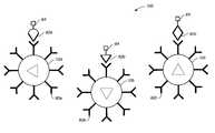
- FIG. 1is a schematic diagram illustrating the optical signature encoding and chemical functionalizing of the microspheres according to the present invention
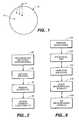
- FIG. 2is a process diagram describing the preparation, encoding, and functionalizing of the microspheres of the present invention
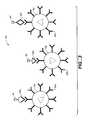
- FIG. 3is a schematic diagram illustrating a microsphere system including microspheres with different chemical functionalities and encoded descriptions of the functionalities;
- FIG. 4is a schematic diagram of the inventive fiber optic sensor and associated instrumentation and control system
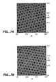
- FIGS. 5A and 5Bare micrographs illustrating the preferred technique for attaching or affixing the microspheres to the distal end of the optical fiber bundle
- FIG. 6is a process diagram describing well formation in the optical fiber bundle and affixation of the microspheres in the wells;
- FIGS. 7A and 7Bare micrographs showing the array of microspheres in their corresponding wells prior and subsequent to physical agitation, tapping and air pulsing, demonstrating the electrostatic binding of the microspheres in the wells;
- FIGS. 8A , 8 B, and 8 Care micrographs from alkaline phosphatase microspheres when exposed to fluorescein diphosphate, at the fluorescein emission wavelength, at an encoding wavelength for DiIC, and at an encoding wavelength for TRC, respectively;
- FIGS. 9A and 9Bare micrographs showing the optical signal from ⁇ -galactosidase microspheres when exposed to fluorescein ⁇ -galactopyranoside at the fluorescein emission wavelength and at an encoding wavelength for DiIC, respectively;
- FIGS. 10A and 10Bare micrographs showing the optical response from rabbit antibody microspheres prior to and post, respectively, exposure to fluorescein labeled antigens.
- the present inventionis based on two synergistic inventions: 1) the development of a bead-based analytic chemistry system in which beads, also termed microspheres, carrying different chemical functionalities may be mixed together while the ability is retained to identify the functionality of each bead using an optically interrogatable encoding scheme (an “optical signature”); and 2) the use of a substrate comprising a patterned surface containing individual sites that can bind or associate individual beads.
- Thisallows the synthesis of the bioactive agents (i.e. compounds such as nucleic acids and antibodies) to be separated from their placement on an array, i.e. the bioactive agents may be synthesized on the beads, and then the beads are randomly distributed on a patterned surface.
- the arraycan later be “decoded”, i.e. after the array is made, a correlation of the location of an individual site on the array with the bead or bioactive agent at that particular site can be made.
- the beadsmay be randomly distributed on the array, a fast and inexpensive process as compared to either the in situ synthesis or spotting techniques of the prior art.
- the present inventionprovides array compositions comprising at least a first substrate with a surface comprising individual sites.
- arrayherein is meant a plurality of bioactive agents in an array format; the size of the array will depend on the composition and end use of the array. Arrays containing from about 2 different bioactive agents (i.e. different beads) to many millions can be made, with very large fiber optic arrays being possible. Generally, the array will comprise from two to as many as a billion or more, depending on the size of the beads and the substrate, as well as the end use of the array, thus very high density, high density, moderate density, low density and very low density arrays may be made.
- Preferred ranges for very high density arraysare from about 10,000,000 to about 2,000,000,000, with from about 100,000,000 to about 1,000,000,000 being preferred.
- High density arraysrange about 100,000 to about 10,000,000, with from about 1,000,000 to about 5,000,000 being particularly preferred.
- Moderate density arraysrange from about 10,000 to about 50,000 being particularly preferred, and from about 20,000 to about 30,000 being especially preferred.
- Low density arraysare generally less than 10,000, with from about 1,000 to about 5,000 being preferred.
- Very low density arraysare less than 1,000, with from about 10 to about 1000 being preferred, and from about 100 to about 500 being particularly preferred.
- the compositions of the inventionmay not be in array format; that is, for some embodiments, compositions comprising a single bioactive agent may be made as well.
- multiple substratesmay be used, either of different or identical compositions. Thus for example, large arrays may comprise a plurality of smaller substrates.
- one advantage of the present compositionsis that particularly through the use of fiber optic technology, extremely high density arrays can be made.
- beads of 200 nmcan be used, and very small fibers are known, it is possible to have as many as 250,000 different fibers and beads in a 1 mm 2 fiber optic array or bundle, with densities of greater than 15,000,000 individual beads and fibers per 0.5 cm 2 obtainable.
- compositionscomprise a substrate.
- substrateor “solid support” or other grammatical equivalents herein is meant any material that can be modified to contain discrete individual sites appropriate for the attachment or association of beads and is amenable to at least one detection method.
- the number of possible substratesare very large, and include, but are not limited to, glass and modified or functionalized glass, plastics (including acrylics, polystyrene and copolymers of styrene and other materials, polypropylene, polyethylene, polybutylene, polyurethanes, TeflonTM, etc.), polysaccharides, nylon or nitrocellulose, resins, silica or silica-based materials including silicon and modified silicon, carbon, metals, inorganic glasses, plastics, optical fiber bundles, and a variety of other polymers.
- the substratesallow optical detection and do not appreciably fluoresce.
- the substrateis planar, although as will be appreciated by those in the art, other configurations of substrates may be used as well; for example, three dimensional configurations can be used, for example by embedding the beads in a porous block of plastic that allows sample access to the beads and using a confocal microscope for detection. Similarly, the beads may be placed on the inside surface of a tube, for flow-through sample analysis to minimize sample volume.
- Preferred substratesinclude optical fiber bundles as discussed below, and flat planar substrates such as glass, polystyrene and other plastics and acrylics.
- At least one surface of the substrateis modified to contain discrete, individual sites for later association of microspheres. These sites may comprise physically altered sites, i.e. physical configurations such as wells or small depressions in the substrate that can retain the beads, such that a microsphere can rest in the well, or the use of other forces (magnetic or compressive), or chemically altered or active sites, such as chemically functionalized sites, electrostatically altered sites, hydrophobically/hydrophilically functionalized sites, spots of adhesive, etc.
- sitesmay comprise physically altered sites, i.e. physical configurations such as wells or small depressions in the substrate that can retain the beads, such that a microsphere can rest in the well, or the use of other forces (magnetic or compressive), or chemically altered or active sites, such as chemically functionalized sites, electrostatically altered sites, hydrophobically/hydrophilically functionalized sites, spots of adhesive, etc.
- the sitesmay be a pattern, i.e. a regular design or configuration, or randomly distributed.
- a preferred embodimentutilizes a regular pattern of sites such that the sites may be addressed in the X-Y coordinate plane.
- “Pattern” in this senseincludes a repeating unit cell, preferably one that allows a high density of beads on the substrate.
- these sitesmay not be discrete sites. That is, it is possible to use a uniform surface of adhesive or chemical functionalities, for example, that allows the attachment of beads at any position. That is, the surface of the substrate is modified to allow attachment of the microspheres at individual sites, whether or not those sites are contiguous or non-contiguous with other sites.
- the surface of the substratemay be modified such that discrete sites are formed that can only have a single associated bead, or alternatively, the surface of the substrate is modified and beads may go down anywhere, but they end up at discrete sites.
- the surface of the substrateis modified to contain wells, i.e. depressions in the surface of the substrate. This may be done as is generally known in the art using a variety of techniques, including, but not limited to, photolithography, stamping techniques, molding techniques and microetching techniques. As will be appreciated by those in the art, the technique used will depend on the composition and shape of the substrate.
- the substrateis a fiber optic bundle or array and the surface of the substrate is a terminal end of the fiber bundle or array.
- wellsare made in a terminal or distal end of a fiber optic bundle or array comprising individual fibers.
- the cores of the individual fibersare etched, with respect to the cladding, such that small wells or depressions are formed at one end of the fibers. The required depth of the wells will depend on the size of the beads to be added to the wells.
- the microspheresare non-covalently associated in the wells, although the wells may additionally be chemically functionalized as is generally described below, cross-linking agents may be used, or a physical barrier may be used, i.e. a film or membrane over the beads.
- the surface of the substrateis modified to contain chemically modified sites, that can be used to attach, either covalently or non-covalently, the microspheres of the invention to the discrete sites or locations on the substrate.
- “Chemically modified sites” in this contextincludes, but is not limited to, the addition of a pattern of chemical functional groups including amino groups, carboxy groups, oxo groups and thiol groups, that can be used to covalently attach microspheres, which generally also contain corresponding reactive functional groups; the addition of a pattern of adhesive that can be used to bind the microspheres (either by prior chemical functionalization for the addition of the adhesive or direct addition of the adhesive); the addition of a pattern of charged groups (similar to the chemical functionalities) for the electrostatic attachment of the microspheres, i.e.
- microspherescomprise charged groups opposite to the sites; the addition of a pattern of chemical functional groups that renders the sites differentially hydrophobic or hydrophilic, such that the addition of similarly hydrophobic or hydrophilic microspheres under suitable experimental conditions will result in association of the microspheres to the sites on the basis of hydroaffinity.
- hydrophobic sites with hydrophobic beadsin an aqueous system, drives the association of the beads preferentially onto the sites.
- patternin this sense includes the use of a uniform treatment of the surface to allow attachment of the beads at discrete sites, as well as treatment of the surface resulting in discrete sites. As will be appreciated by those in the art, this may be accomplished in a variety of ways.
- compositions of the inventionfurther comprise a population of microspheres.
- populationherein is meant a plurality of beads as outlined above for arrays. Within the population are separate subpopulations, which can be a single microsphere or multiple identical microspheres. That is, in some embodiments, as is more fully outlined below, the array may contain only a single bead for each bioactive agent; preferred embodiments utilize a plurality of beads of each type.
- microspheresor “beads” or “particles” or grammatical equivalents herein is meant small discrete particles.
- the composition of the beadswill vary, depending on the class of bioactive agent and the method of synthesis. Suitable bead compositions include those used in peptide, nucleic acid and organic moiety synthesis, including, but not limited to, plastics, ceramics, glass, polystyrene, methylstyrene, acrylic polymers, paramagnetic materials, thoria sol, carbon graphited, titanium dioxide, latex or cross-linked dextrans such as Sepharose, cellulose, nylon, cross-linked micelles and teflon may all be used. “ Microsphere Detection Guide ” from Bangs Laboratories, Fishers Ind. is a helpful guide.
- the beadsneed not be spherical; irregular particles may be used.
- the beadsmay be porous, thus increasing the surface area of the bead available for either bioactive agent attachment or tag attachment.
- the bead sizesrange from nanometers, i.e. 100 nm, to millimeters, i.e. 1 mm, with beads from about 0.2 micron to about 200 microns being preferred, and from about 0.5 to about 5 micron being particularly preferred, although in some embodiments smaller beads may be used.
- FIG. 1illustrates the construction of a bead or microsphere 10 according to the principles of the present invention.
- the microsphere 10is given a bioactive agent 12 , which is typically applied to the microsphere's surface.
- the bioactive agentis designed so that in the presence of the analyte(s) to which it is targeted, an optical signature of the microsphere, possibly including region surrounding it, is changed.
- a key component of the inventionis the use of a substrate/bead pairing that allows the association or attachment of the beads at discrete sites on the surface of the substrate, such that the beads do not move during the course of the assay.
- Each microspherecomprises two components: a bioactive agent and an optical signature.
- a bioactive agentor “bioactive agent” or “chemical functionality” or “binding ligand” herein is meant as used herein describes any molecule, e.g., protein, oligopeptide, small organic molecule, polysaccharide, polynucleotide, etc. which can be attached to the microspheres of the invention.
- the compositions of the inventionhave two primary uses.
- the compositionsare used to detect the presence of a particular target analyte; for example, the presence or absence of a particular nucleotide sequence or a particular protein, such as an enzyme, an antibody or an antigen.
- the compositionsare used to screen bioactive agents, i.e. drug candidates, for binding to a particular target analyte.
- Bioactive agentsencompass numerous chemical classes, though typically they are organic molecules, preferably small organic compounds having a molecular weight of more than 100 and less than about 2,500 daltons. Bioactive agents comprise functional groups necessary for structural interaction with proteins, particularly hydrogen bonding, and typically include at least an amine, carbonyl, hydroxyl or carboxyl group, preferably at least two of the functional chemical groups. The bioactive agents often comprise cyclical carbon or heterocyclic structures and/or aromatic or polyaromatic structures substituted with one or more of the above functional groups. Bioactive agents are also found among biomolecules including peptides, nucleic acids, saccharides, fatty acids, steroids, purines, pyrimidines, derivatives, structural analogs or combinations thereof. Particularly preferred are nucleic acids and proteins.
- Bioactive agentscan be obtained from a wide variety of sources including libraries of synthetic or natural compounds. For example, numerous means are available for random and directed synthesis of a wide variety of organic compounds and biomolecules, including expression of randomized oligonucleotides. Alternatively, libraries of natural compounds in the form of bacterial, fungal, plant and animal extracts are available or readily produced. Additionally, natural or synthetically produced libraries and compounds are readily modified through conventional chemical, physical and biochemical means. Known pharmacological agents may be subjected to directed or random chemical modifications, such as acylation, alkylation, esterification and/or amidification to produce structural analogs.
- the bioactive agentsare proteins.
- proteinherein is meant at least two covalently attached amino acids, which includes proteins, polypeptides, oligopeptides and peptides.
- the proteinmay be made up of naturally occurring amino acids and peptide bonds, or synthetic peptidomimetic structures.
- amino acidor “peptide residue”, as used herein means both naturally occurring and synthetic amino acids.
- homo-phenylalanine, citrulline and norleucineare considered amino acids for the purposes of the invention.
- the side chainsmay be in either the (R) or the (S) configuration. In the preferred embodiment, the amino acids are in the (S) or L-configuration. If non-naturally occurring side chains are used, non-amino acid substituents may be used, for example to prevent or retard in vivo degradations.
- the bioactive agentsare naturally occurring proteins or fragments of naturally occuring proteins.
- cellular extracts containing proteins, or random or directed digests of proteinaceous cellular extractsmay be used.
- libraries of procaryotic and eukaryotic proteinsmay be made for screening in the systems described herein.
- Particularly preferred in this embodimentare libraries of bacterial, fungal, viral, and mammalian proteins, with the latter being preferred, and human proteins being especially preferred.
- the bioactive agentsare peptides of from about 5 to about 30 amino acids, with from about 5 to about 20 amino acids being preferred, and from about 7 to about 15 being particularly preferred.
- the peptidesmay be digests of naturally occurring proteins as is outlined above, random peptides, or “biased” random peptides.
- randomizedor grammatical equivalents herein is meant that each nucleic acid and peptide consists of essentially random nucleotides and amino acids, respectively. Since generally these random peptides (or nucleic acids, discussed below) are chemically synthesized, they may incorporate any nucleotide or amino acid at any position.
- the synthetic processcan be designed to generate randomized proteins or nucleic acids, to allow the formation of all or most of the possible combinations over the length of the sequence, thus forming a library of randomized bioactive proteinaceous agents.
- a library of bioactive agentsare used.
- the libraryshould provide a sufficiently structurally diverse population of bioactive agents to effect a probabilistically sufficient range of binding to target analytes. Accordingly, an interaction library must be large enough so that at least one of its members will have a structure that gives it affinity for the target analyte. Although it is difficult to gauge the required absolute size of an interaction library, nature provides a hint with the immune response: a diversity of 10 7 -10 8 different antibodies provides at least one combination with sufficient affinity to interact with most potential antigens faced by an organism. Published in vitro selection techniques have also shown that a library size of 10 7 to 10 8 is sufficient to find structures with affinity for the target. Thus, in a preferred embodiment, at least 10 6 , preferably at least 10 7 , more preferably at least 10 8 and most preferably at least 10 9 different bioactive agents are simultaneously analyzed in the subject methods. Preferred methods maximize library size and diversity.
- the libraryis fully randomized, with no sequence preferences or constants at any position.
- the libraryis biased. That is, some positions within the sequence are either held constant, or are selected from a limited number of possibilities.
- the nucleotides or amino acid residuesare randomized within a defined class, for example, of hydrophobic amino acids, hydrophilic residues, sterically biased (either small or large) residues, towards the creation of cysteines, for cross-linking, prolines for SH-3 domains, serines, threonines, tyrosines or histidines for phosphorylation sites, etc., or to purines, etc.
- the bioactive agentsare nucleic acids (generally called “probe nucleic acids” or “candidate probes” herein).
- nucleic acidor “oligonucleotide” or grammatical equivalents herein means at least two nucleotides covalently linked together.
- a nucleic acid of the present inventionwill generally contain phosphodiester bonds, although in some cases, as outlined below, nucleic acid analogs are included that may have alternate backbones, comprising, for example, phosphoramide (Beaucage, et al., Tetrahedron, 49(10):1925 (1993) and references therein; Letsinger, J. Org.
- nucleic acidsmay be single stranded or double stranded, as specified, or contain portions of both double stranded or single stranded sequence.
- the nucleic acidmay be DNA, both genomic and cDNA, RNA or a hybrid, where the nucleic acid contains any combination of deoxyribo- and ribo-nucleotides, and any combination of bases, including uracil, adenine, thymine, cytosine, guanine, inosine, xanthanine, hypoxanthanine, isocytosine, isoguanine, and basepair analogs such as nitropyrrole and nitroindole, etc.
- nucleic acid bioactive agentsmay be naturally occuring nucleic acids, random nucleic acids, or “biased” random nucleic acids.
- digests of procaryotic or eukaryotic genomesmay be used as is outlined above for proteins.
- probes of the present inventionare designed to be complementary to a target sequence (either the target analyte sequence of the sample or to other probe sequences, as is described herein), such that hybridization of the target and the probes of the present invention occurs.
- a target sequenceeither the target analyte sequence of the sample or to other probe sequences, as is described herein
- This complementarityneed not be perfect; there may be any number of base pair mismatches that will interfere with hybridization between the target sequence and the single stranded nucleic acids of the present invention. However, if the number of mutations is so great that no hybridization can occur under even the least stringent of hybridization conditions, the sequence is not a complementary target sequence.
- substantially complementaryherein is meant that the probes are sufficiently complementary to the target sequences to hybridize under the selected reaction conditions.
- High stringency conditionsare known in the art; see for example Maniatis et al., Molecular Cloning: A Laboratory Manual, 2d Edition, 1989, and Short Protocols in Molecular Biology, ed. Ausubel, et al., both of which are hereby incorporated by reference. Stringent conditions are sequence-dependent and will be different in different circumstances. Longer sequences hybridize specifically at higher temperatures. An extensive guide to the hybridization of nucleic acids is found in Tijssen, Techniques in Biochemistry and Molecular Biology—Hybridization with Nucleic Acid Probes, “Overview of principles of hybridization and the strategy of nucleic acid assays” (1993). Generally, stringent conditions are selected to be about 5-10° C.
- T mthermal melting point
- the T mis the temperature (under defined ionic strength, pH and nucleic acid concentration) at which 50% of the probes complementary to the target hybridize to the target sequence at equilibrium (as the target sequences are present in excess, at T m , 50% of the probes are occupied at equilibrium).
- Stringent conditionswill be those in which the salt concentration is less than about 1.0 M sodium ion, typically about 0.01 to 1.0 M sodium ion concentration (or other salts) at pH 7.0 to 8.3 and the temperature is at least about 30° C. for short probes (e.g. 10 to 50 nucleotides) and at least about 60° C. for long probes (e.g.
- Stringent conditionsmay also be achieved with the addition of destabilizing agents such as formamide.
- destabilizing agentssuch as formamide.
- less stringent hybridization conditionsare used; for example, moderate or low stringency conditions may be used, as are known in the art; see Maniatis and Ausubel, supra, and Tijssen, supra.
- target sequenceor grammatical equivalents herein means a nucleic acid sequence on a single strand of nucleic acid.
- the target sequencemay be a portion of a gene, a regulatory sequence, genomic DNA, cDNA, RNA including mRNA and rRNA, or others. It may be any length, with the understanding that longer sequences are more specific.
- the complementary target sequencemay take many forms. For example, it may be contained within a larger nucleic acid sequence, i.e. all or part of a gene or mRNA, a restriction fragment of a plasmid or genomic DNA, among others.
- probesare made to hybridize to target sequences to determine the presence or absence of the target sequence in a sample. Generally speaking, this term will be understood by those skilled in the art.
- the bioactive agentsare organic chemical moieties, a wide variety of which are available in the literature.
- each beadcomprises a single type of bioactive agent, although a plurality of individual bioactive agents are preferably attached to each bead.
- preferred embodimentsutilize more than one microsphere containing a unique bioactive agent; that is, there is redundancy built into the system by the use of subpopulations of microspheres, each microsphere in the subpopulation containing the same bioactive agent.
- the bioactive agentsmay either be synthesized directly on the beads, or they may be made and then attached after synthesis.
- linkersare used to attach the bioactive agents to the beads, to allow both good attachment, sufficient flexibility to allow good interaction with the target molecule, and to avoid undesirable binding reactions.
- the bioactive agentsare synthesized directly on the beads.
- many classes of chemical compoundsare currently synthesized on solid supports, such as peptides, organic moieties, and nucleic acids. It is a relatively straightforward matter to adjust the current synthetic techniques to use beads.
- the bioactive agentsare synthesized first, and then covalently attached to the beads. As will be appreciated by those in the art, this will be done depending on the composition of the bioactive agents and the beads.
- the functionalization of solid support surfacessuch as certain polymers with chemically reactive groups such as thiols, amines, carboxyls, etc. is generally known in the art. Accordingly, “blank” microspheres may be used that have surface chemistries that facilitate the attachment of the desired functionality by the user. Some examples of these surface chemistries for blank microspheres are listed in Table I.
- bioactive agents containing carbohydratesmay be attached to an amino-functionalized support; the aldehyde of the carbohydrate is made using standard techniques, and then the aldehyde is reacted with an amino group on the surface.
- a sulfhydryl linkermay be used.
- sulfhydryl reactive linkersknown in the art such as SPDP, maleimides, ⁇ -haloacetyls, and pyridyl disulfides (see for example the 1994 Pierce Chemical Company catalog, technical section on cross-linkers, pages 155-200, incorporated herein by reference) which can be used to attach cysteine containing proteinaceous agents to the support.
- an amino group on the bioactive agentmay be used for attachment to an amino group on the surface.
- a large number of stable bifunctional groupsare well known in the art, including homobifunctional and heterobifunctional linkers (see Pierce Catalog and Handbook, pages 155-200).
- carboxyl groupsmay be derivatized using well known linkers (see the Pierce catalog).
- carbodiimidesactivate carboxyl groups for attack by good nucleophiles such as amines (see Torchilin et al., Critical Rev. Therapeutic Drug Carrier Systems , 7(4):275-308 (1991), expressly incorporated herein).
- Proteinaceous bioactive agentsmay also be attached using other techniques known in the art, for example for the attachment of antibodies to polymers; see Slinkin et al., Bioconj. Chem . 2:342-348 (1991); Torchilin et al., supra; Trubetskoy et al., Bioconj. Chem .
- bioactive agentsmay be attached in a variety of ways, including those listed above. What is important is that manner of attachment does not significantly alter the functionality of the bioactive agent; that is, the bioactive agent should be attached in such a flexible manner as to allow its interaction with a target.
- NH 2 surface chemistry microspheresare used. Surface activation is achieved with a 2.5% glutaraldehyde in phosphate buffered saline (10 mM) providing a pH of 6.9. (138 mM NaCl, 2.7 mM, KCl). This is stirred on a stir bed for approximately 2 hours at room temperature. The microspheres are then rinsed with ultrapure water plus 0.01% tween 20 (surfactant) ⁇ 0.02%, and rinsed again with a pH 7.7 PBS plus 0.01% tween 20. Finally, the enzyme is added to the solution, preferably after being prefiltered using a 0.45 ⁇ m amicon micropure filter.
- the microspherescomprise an optical signature that can be used to identify the attached bioactive agent. That is, each subpopulation of microspheres comprise a unique optical signature or optical tag that can be used to identify the unique bioactive agent of that subpopulation of microspheres; a bead comprising the unique optical signature may be distinguished from beads at other locations with different optical signatures.
- each bioactive agentwill have an associated unique optical signature such that any microspheres comprising that bioactive agent will be identifiable on the basis of the signature.
- it is possible to reuse or duplicate optical signatures within an arrayfor example, when another level of identification is used, for example when beads of different sizes are used, or when the array is loaded sequentially with different batches of beads.
- the optical signatureis generally a mixture of reporter dyes, preferably fluoroscent.
- the composition of the mixturei.e. the ratio of one dye to another
- concentration of the dyeleading to differences in signal intensity
- matrices of unique tagsmay be generated. This may be done by covalently attaching the dyes to the surface of the beads, or alternatively, by entrapping the dye within the bead.
- the dyesmay be chromophores or phosphors but are preferably fluorescent dyes, which due to their strong signals provide a good signal-to-noise ratio for decoding.
- Suitable dyes for use in the inventioninclude, but are not limited to, fluorescent lanthanide complexes, including those of Europium and Terbium, fluorescein, rhodamine, tetramethylrhodamine, eosin, erythrosin, coumarin, methyl-coumarins, pyrene, Malacite green, stilbene, Lucifer Yellow, Cascade BlueTM, Texas Red, and others described in the 1989-1991 Molecular Probes Handbook by Richard P. Haugland, hereby expressly incorporated by reference.
- the encodingcan be accomplished in a ratio of at least two dyes, although more encoding dimensions may be added in the size of the beads, for example.
- the labelsare distinguishable from one another; thus two different labels may comprise different molecules (i.e. two different fluors) or, alternatively, one label at two different concentrations or intensity.
- the dyesare covalently attached to the surface of the beads. This may be done as is generally outlined for the attachment of the bioactive agents, using functional groups on the surface of the beads. As will be appreciated by those in the art, these attachments are done to minimize the effect on the dye.
- the dyesare non-covalently associated with the beads, generally by entrapping the dyes in the bead matrix or pores of the beads.
- reporter dyes 14are added to the microsphere 10 with the encoding occurring in the ratio of two or more dyes.
- the reporter dyes 14may be chromophore-type. Fluorescent dyes, however, are preferred because the strength of the fluorescent signal provides a better signal-to-noise ratio when decoding. Additionally, encoding in the ratios of the two or more dyes, rather than single dye concentrations, is preferred since it provides insensitivity to the intensity of light used to interrogate the reporter dye's signature and detector sensitivity.
- the dyesare added to the bioactive agent, rather than the beads, although this is generally not preferred.
- FIG. 2is a process diagram illustrating the preparation of the microspheres.
- step 50an aliquot of stock microspheres are vacuum filtered to produce a dry cake.
- microsphere copolymers of methylstyrene (87%) and divinylbenzene (13%)are used that have a 3.1 micrometer ( ⁇ m) diameter.
- the dry cakeis then broken apart and a dye solution added to it in step 52 to encode optical signatures of the microspheres with information concerning the intended surface chemical functionalities. Dyes may be covalently bonded to the microspheres' surface, but this consumes surface binding sites desirably reserved for the chemical functionalities.
- the microspheresare placed in a dye solution comprising a ratio of two or more fluorescent reporter dyes dissolved in an organic solvent that will swell the microspheres, e.g., dimethylformamide (DMF).
- a dye solutioncomprising a ratio of two or more fluorescent reporter dyes dissolved in an organic solvent that will swell the microspheres, e.g., dimethylformamide (DMF).
- DMFdimethylformamide
- TRCTexas Red Cadaverine
- DiICindodicarbocyanine
- dyesare selected to be compatible with the chemistries involved in the analysis and to be spectrally compatible. This avoids deconvolution problems associated with determining signal contributions based on the presence of both the analyte and the encoding dye ratios contributing to an overlapping emission spectral region.
- Examples of other dyes that can be usedare Oxazin (662/705), IR-144 (745/825), IR-140 (776/882), IR-125 (786/800) from Exciton, and Bodipy 665/676 from Molecular Probes, and Naphthofluorescein (605/675) also from Molecular Probes. Lathanide complexes may also be used. Fluorescent dyes emitting in other than the near infrared may also be used. Chromophore dyes are still another alternative that produce an optically interrogatable signature, as are more exotic formulations using Raman scattering-based dyes or polarizing dyes, for example. The ability of a particular dye pair to encode for different chemical functionalities depends on the resolution of the ratiometric measurement. Conservatively, any dye pair should provide the ability to discriminate at least twenty different ratios. The number of unique combinations of two dyes made with a particular dye set is shown in the following Table II.
- step 54the microspheres are vacuum filtered to remove excess dye.
- the microspheresare then washed in water or other liquid that does not swell the microspheres, but in which the dyes are still soluble. This allows the residual dye to be rinsed off without rinsing the dye out of the microspheres.
- the bioactive agentis attached to the microsphere surface if not already present. It should be understood that surface chemistries may be present throughout the microsphere's volume, and not limited to the physical circumferential surface.
- the microspheresare added to discrete sites on the surface of the substrate. This can be done in a number of ways, but generally comprises adding the beads to the surface under conditions that will allow the association of the microspheres on or at the discrete sites.
- the association of the beads on the surfacemay comprise a covalent bonding of the bead to the surface, for example when chemical attachment sites are added to both the substrate and the bead; an electrostatic or hydroaffinity, when charge, hydrophobicity or hydrophilicity is used as the basis of the binding; a physical yet non-covalent attachment such as the use of an adhesive; or a spatial attachment, for example the localization of a bead within a well.
- it may be preferable to effect a more permanent attachment after the initial localizationfor example through the use of cross-linking agents, a film or membrane over the array.
- FIG. 3schematically illustrates a microsphere system, or array of microspheres, 100 formed from microsphere populations that have different bioactive agents. While a large number of microspheres and bioactive agents may be employed, in this example only three microsphere populations are shown.
- the individual populations, or subpopulations, of microspheresare represented as 10 a , 10 b , 10 c carrying respective bioactive agents or probe sequences 60 a , 60 b , 60 c , as exemplary functionalities.
- the subpopulationsmay be combined in either a random or ordered fashion on a substrate, with a corresponding distribution of their respective bioactive agents.
- each microsphere in each subpopulationis encoded with a common optical signature.
- the subpopulation represented by microsphere 10 ahas a two reporter dye ratio of 10:1; the subpopulation of microspheres 10 b has a ratio of 1:1 of the same reporter dyes, and subpopulation of microspheres 10 c has a ratio of 1:10 of the reporter dyes.
- the randomly mixed subpopulations of microspheresare useful as an analytic chemistry system based on each of the carried bioactive agents 60 a - 60 c separately.
- the microsphere array or system 100is exposed to an analyte of interest to which some of the bioactive agents may interact. Any interaction changes the optical response of the corresponding microspheres by, for example, binding a fluorescent dye 64 to the microspheres.
- identifying the chemical functionalities of the microsphere in which the optical signature has changedusing the encoded dye combinations, information regarding the chemical identity and concentration of an analyte may be gained based upon the interaction or noninteraction of each bioactive agent contained in the microsphere system 100 .
- the microspheres exhibiting activity or changes in their optical signaturemay be identified by a conventional optical train and optical detection system. Decoding can also be performed either manually or automatically with the aid of a computer. Depending on the particular encoding or reporter dyes used and their operative wavelengths, optical filters designed for a particular wavelengths may be employed for optical interrogation of the microspheres of bioactive agents. In a preferred embodiment, the analytic chemistry microsphere system is used in conjunction with an optical fiber bundle or fiber optic array as a substrate.
- FIG. 4is a schematic block diagram showing a microsphere-based analytic chemistry system employing a fiber optic assembly 200 with an optical detection system.
- the fiber optic assembly 200comprises a fiber optic bundle or array 202 , that is constructed from clad fibers so that light does not mix between fibers.
- a microsphere array or system, 100is attached to the bundle's distal end 212 , with the proximal end 214 being received by a z-translation stage 216 and x-y micropositioner 218 . These two components act in concert to properly position the proximal end 214 of the bundle 202 for a microscope objective lens 220 .
- Light collected by the objective lens 220is passed to a reflected light fluorescence attachment with three pointer cube slider 222 .
- the attachment 222allows insertion of light from a 75 Watt Xe lamp 224 through the objective lens 220 to be coupled into the fiber bundle 202 .
- the light from the source 224is condensed by condensing lens 226 , then filtered and/or shuttered by filter and shutter wheel 228 , and subsequently passes through a ND filter slide 230 .
- a magnification changer 232which enables adjustment of the image size of the fiber's proximal or distal end.
- Light passing through the magnification changer 232is then shuttered and filtered by a second wheel 234 .
- the lightis then imaged on a charge coupled device (CCD) camera 236 .
- a computer 238executes imaging processing software to process the information from the CCD camera 236 and also possibly control the first and second shutter and filter wheels 228 , 234 .
- the instrumentation exclusive of the fiber sensor 200i.e., to the left of the proximal end of the bundle 202 is discussed more completely by Bronk, et al., Anal. Chem. 1995, Vol. 67, number 17, pp. 2750-2752.
- the microsphere array or system 100may be attached to the distal end of the optical fiber bundle or fiber optic array using a variety of compatible processes. It is important that the microspheres are located close to the end of the bundle. This ensures that the light returning in each optical fiber predominantly comes from only a single microsphere. This feature is necessary to enable the interrogation of the optical signature of individual microspheres to identify reactions involving the microsphere's functionality and also to decode the dye ratios contained in those microspheres. The adhesion or affixing technique, however, must not chemically insulate the microspheres from the analyte.
- FIGS. 5A and 5Bare micrographs of the distal end 212 of the bundle 202 illustrating the preferred technique for attaching the microspheres 10 to the bundle 202 .
- Wells 250are formed at the center of each optical fiber 252 of the bundle 202 .
- the size of the wells 250are coordinated with the size of the microspheres 10 so that the microspheres 10 can be placed within the wells 250 .
- each optical fiber 252 of the bundle 202conveys light from the single microsphere 10 contained in 5 its well. Consequently, by imaging the end of the bundle 202 onto the CCD array 236 , the optical signatures of the microspheres 10 are individually interrogatable.
- FIG. 6illustrates how the microwells 250 are formed and microspheres 10 placed in the wells.
- a 1 mm hexagonally-packed imaging fibercontains approximately 20,600 individual optical fibers that have cores approximately 3.7 ⁇ m across (Part No. ET26 from Galileo Fibers).
- the cores of each fiberare hexagonally shaped as a result the starting preform; that is, during drawing the fiber does not usually change shape. In some cases, the shape can be circular, however.
- both the proximal and distal ends 212 , 214 of the fiber bundle 202are successively polished on 12 ⁇ m, 9 ⁇ m, 3 ⁇ m, 1 ⁇ m, and 0.3 ⁇ m lapping films. Subsequently, the ends can be inspected for scratches on an atomic force microscope.
- a representative etchingis performed on the distal end 212 of the bundle 202 .
- a solution of 0.2 grams NH 4 F (ammonium fluoride) with 600 ⁇ l distilled H 2 O and 100 ⁇ l of HF (hydrofluoric acid), 50% stock solution,may be used.
- the distal end 212is etched in this solution for a specified time, preferably approximately 30 to 600 seconds, with about 80 seconds being preferred.
- the bundle endUpon removal from this solution, the bundle end is immediately placed in deionized water to stop any further etching in step 274 .
- the fiberis then rinsed in running tap water. At this stage, sonication is preferably performed for several minutes to remove any salt products from the reaction. The fiber is then allowed to air dry.
- the foregoing procedureproduces wells by the anisotropic etching of the fiber cores 254 favorably with respect to the cladding 256 for each fiber of the bundle 202 .
- the wellshave approximately the diameter of the cores 254 , 3.7 ⁇ m. This diameter is selected to be slightly larger than the diameters of the microspheres used, 3.1 ⁇ m, in the example.
- the preferential etchingoccurs because the pure silica of the cores 254 etches faster in the presence of hydrofluoric acid than the germanium-doped silica claddings 256 .
- microspheresare then placed in the wells 250 in step 276 according to a number of different techniques.
- the placement of the microspheresmay be accomplished by dripping a solution containing the desired randomly mixed subpopulations of the microspheres over the distal end 212 , sonicating the bundle to settle the microspheres in the wells, and allowing the microsphere solvent to evaporate.
- the subpopulationscould be added serially to the bundle end.
- Microspheres 10may then be fixed into the wells 250 by using a dilute solution of sulfonated Nafion that is dripped over the end. Upon solvent evaporation, a thin film of Nafion was formed over the microspheres which holds them in place.
- An alternative fixation approachemploys microsphere swelling to entrap each microsphere 10 in its corresponding microwell 250 .
- the microspheresare first distributed into the microwells 250 by sonicating the microspheres suspended in a non-swelling solvent in the presence of the microwell array on the distal end 212 . After placement into the microwells, the microspheres are subsequently exposed to an aqueous buffer in which they swell, thereby physically entrapping them, analogous to muffins rising in a muffin tin.
- microspheresOne of the most common microsphere formations is tentagel, a styrene-polyethylene glycol co-polymer. These microspheres are unswollen in nonpolar solvents such as hexane and swell approximately 20-40% in volume upon exposure to a more polar or aqueous media. This approach is extremely desirable since it does not significantly compromise the diffusional or permeability properties of the microspheres themselves.
- FIGS. 7A and 7Bshow polymer coated microspheres 12 in wells 250 after their initial placement and then after tapping and exposure to air pulses.
- FIGS. 7A and 7Billustrate that there is no appreciable loss of microspheres from the wells due to mechanical agitation even without a specific fixing technique. This effect is probably due to electrostatic forces between the microspheres and the optical fibers. These forces tend to bind the microspheres within the wells. Thus, in most environments, it may be unnecessary to use any chemical or mechanical fixation for the microspheres.
- a sonication stepmay be used to place beads in the wells.
- sites of an arraymay comprise a bead; that is, there may be some sites on the substrate surface which are empty. In addition, there may be some sites that contain more than one bead, although this is not preferred.
- the beadsin a non-random or ordered way.
- photoactivatible attachment linkers or photoactivatible adhesives or masksselected sites on the array may be sequentially rendered suitable for attachment, such that defined populations of beads are laid down.
- the size of the arraywill be set by the number of unique optical signatures, it is possible to “reuse” a set of unique optical signatures to allow for a greater number of test sites. This may be done in several ways; for example, by using a positional coding scheme within an array; different sub-bundles may reuse the set of optical signatures. Similarly, one embodiment utilizes bead size as a coding modality, thus allowing the reuse of the set of unique optical signatures for each bead size. Alternatively, sequential partial loading of arrays with beads can also allow the reuse of optical signatures.
- a spatial or positional coding systemis done.
- each subarrayis an “area code”, that can have the same tags (i.e. telephone numbers) of other subarrays, that are separated by virtue of the location of the subarray.
- tagsi.e. telephone numbers
- the same unique tagscan be reused from bundle to bundle.
- the use of 50 unique tags in combination with 100 different subarrayscan form an array of 5000 different bioactive agents.
- additional encoding parameterscan be added, such as microsphere size.
- the use of different size beadsmay also allow the reuse of sets of optical signatures; that is, it is possible to use microspheres of different sizes to expand the encoding dimensions of the microspheres.
- Optical fiber arrayscan be fabricated containing pixels with different fiber diameters or cross-sections; alternatively, two or more fiber optic bundles or arrays, each with different cross-sections of the individual fibers, can be added together to form a larger bundle or array; or, fiber optic bundles or arrays with fiber of the same size cross-sections can be used, but just with different sized beads.
- the largest wellscan be filled with the largest microspheres and then moving onto progressively smaller microspheres in the smaller wells until all size wells are then filled.
- the same dye ratiocould be used to encode microspheres of different sizes thereby expanding the number of different oligonucleotide sequences or chemical functionalities present in the array.
- the coding and decodingis accomplished by sequential loading of the microspheres into the array.
- the optical signaturescan be “reused”.
- the library of microspheres each comprising a different bioactive agentor the subpopulations each comprise a different bioactive agent
- the library of microspheres each comprising a different bioactive agentis divided into a plurality of sublibraries; for example, depending on the size of the desired array and the number of unique tags, 10 sublibraries each comprising roughly 10% of the total library may be made, with each sublibrary comprising roughly the same unique tags.
- the first sublibraryis added to the fiber optic bundle or array comprising the wells, and the location of each bioactive agent is determined, using its optical signature.
- the second sublibraryis then added, and the location of each optical signature is again determined.
- the signal in this casewill comprise the “first” optical signature and the “second” optical signature; by comparing the two matrices the location of each bead in each sublibrary can be determined. Similarly, adding the third, fourth, etc. sublibraries sequentially will allow the array to be filled.
- arraysare made of a large spectrum of chemical functionalities utilizing the compositions of invention comprising microspheres and substrates with discrete sites on a surface.
- prior art sensorswhich can be adapted for use in the present invention include four broad classifications of microsphere sensors: 1) basic indicator chemistry sensors; 2) enzyme-based sensors; 3) immuno-based sensors (both of which are part of a broader general class of protein sensors); and 4) geno-sensors.
- the bioactive agentsare used to detect chemical compounds.
- a large number of basic indicator sensorshave been previously demonstrated. Examples include:
- Enzyme-based microsphere sensorshave also been demonstrated and could be manifest on microspheres. Examples include:
- the microsphere-bound enzymee.g., glucose oxidase
- decomposes the target analytee.g., glucose
- consume a co-substratee.g., oxygen
- some by-producte.g., hydrogen peroxide.
- An oxygen sensitive dyeis then used to trigger the signal change.
- Immuno-based microsphere sensorshave been demonstrated for the detection for environmental pollutants such as pesticides, herbicides, PCB's and PAH's. Additionally, these sensors have also been used for diagnostics, such as bacterial (e.g., leprosy, cholera, lyme disease, and tuberculosis), viral (e.g., HIV, herpes simplex, cytomegalovirus), fungal (e.g., aspergillosis, candidiasis, cryptococcoses), Mycoplasmal (e.g., mycoplasmal pneumonia), Protozoal (e.g., amoebiasis, toxoplasmosis), Rickettsial (e.g., Rocky Mountain spotted fever), and pregnancy tests.
- bacteriale.g., leprosy, cholera, lyme disease, and tuberculosis
- virale.g., HIV, herpes simplex, cytomegalovirus
- fungale.
- Microsphere genosensorsmay also be made (see the Examples). These are typically constructed by attaching a probe sequence to the microsphere surface chemistry, typically via an NH 2 group. A fluorescent dye molecule, e.g., fluorescein, is attached to the target sequence, which is in solution. The optically interrogatable signal change occurs with the binding of the target sequences to the microsphere. This produces a higher concentration of dye surrounding the microsphere than in the solution generally.
- a few demonstrated probe and target sequencessee Ferguson, J. A. et al. Nature Biotechnology , Vol. 14, December 1996, are listed below in Table V.
- Hybridization indicatorspreferentially associate with double stranded nucleic acid, usually reversibly.
- Hybridization indicatorsinclude intercalators and minor and/or major groove binding moieties.
- intercalatorsmay be used; since intercalation generally only occurs in the presence of double stranded nucleic acid, only in the presence of target hybridization will the label light up.
- sensorsmay be made to detect nucleic acids, proteins (including enzyme sensors and immunosensors), lipids, carbohydrates, etc; similarly, these sensors may include bioactive agents that are nucleic acids, proteins, lipids, carbohydrates, etc.
- a single array sensormay contain different binding ligands for multiple types of analytes; for example, an array sensor for HIV may contain multiple nucleic acid probes for direct detection of the viral genome, protein binding ligands for direct detection of the viral particle, immuno-components for the detection of anti-HIV antibodies, etc.
- compositions of the inventionmay include other components, such as light sources, optical components such as lenses and filters, detectors, computer components for data analysis, etc.
- the arrays of the present inventionare constructed such that information about the identity of the bioactive agent is built into the array, such that the random deposition of the beads on the surface of the substrate can be “decoded” to allow identification of the bioactive agent at all positions. This may be done in a variety of ways.
- the beadsare loaded onto the substrate and then the array is decoded, prior to running the assay. This is done by detecting the optical signature associated with the bead at each site on the array. This may be done all at once, if unique optical signatures are used, or sequentially, as is generally outlined above for the “reuse” of sets of optical signatures. Alternatively, decoding may occur after the assay is run.
- compositionsfind use in a number of applications.
- a sample containing a target analyte(whether for detection of the target analyte or screening for binding partners of the target analyte) is added to the array, under conditions suitable for binding of the target analyte to at least one of the bioactive agents, i.e. generally physiological conditions.
- the presence or absence of the target analyteis then detected.
- thismay be done in a variety of ways, generally through the use of a change in an optical signal. This change can occur via many different mechanisms.
- a few examplesinclude the binding of a dye-tagged analyte to the bead, the production of a dye species on or near the beads, the destruction of an existing dye species, a change in the optical signature upon analyte interaction with dye on bead, or any other optical interrogatable event.
- the change in optical signaloccurs as a result of the binding of a target analyte that is labeled, either directly or indirectly, with a detectable label, preferably an optical label such as a fluorochrome.
- a detectable labelpreferably an optical label such as a fluorochrome.
- a proteinaceous target analytewhen used, it may be either directly labeled with a fluor, or indirectly, for example through the use of a labeled antibody.
- nucleic acidsare easily labeled with fluorochromes, for example during PCR amplification as is known in the art.
- an intercalating dyee.g. ethidium bromide
- the target analytesuch as an enzyme generates a species (for example, a fluorescent product) that is either directly or indirectly detectable optically.
- a change in the optical signaturemay be the basis of the optical signal.
- the interaction of some chemical target analytes with some fluorescent dyes on the beadsmay alter the optical signature, thus generating a different optical signal.
- fluorophore derivatized receptorsmay be used in which the binding of the ligand alters the signal.
- the presence or absence of the target analytemay be done using changes in other optical or non-optical signals, including, but not limited to, surface enhanced Raman spectroscopy, surface plasmon resonance, radioactivity, etc.
- the assaysmay be run under a variety of experimental conditions, as will be appreciated by those in the art.
- a variety of other reagentsmay be included in the screening assays. These include reagents like salts, neutral proteins, e.g. albumin, detergents, etc which may be used to facilitate optimal protein-protein binding and/or reduce non-specific or background interactions.
- reagents that otherwise improve the efficiency of the assaysuch as protease inhibitors, nuclease inhibitors, anti-microbial agents, etc., may be used.
- the mixture of componentsmay be added in any order that provides for the requisite binding.
- Various blocking and washing stepsmay be utilized as is known in the art.
- the compositionsare used to probe a sample solution for the presence or absence of a target analyte.
- target analyteor “analyte” or grammatical equivalents herein is meant any atom, molecule, ion, molecular ion, compound or particle to be either detected or evaluated for binding partners.
- a large number of analytesmay be used in the present invention; basically, any target analyte can be used which binds a bioactive agent or for which a binding partner (i.e. drug candidate) is sought.
- Suitable analytesinclude organic and inorganic molecules, including biomolecules.
- suitable target analytesinclude, but are not limited to, an environmental pollutant (including pesticides, insecticides, toxins, etc.); a chemical (including solvents, polymers, organic materials, etc.); therapeutic molecules (including therapeutic and abused drugs, antibiotics, etc.); biomolecules (including hormones, cytokines, proteins, nucleic acids, lipids, carbohydrates, cellular membrane antigens and receptors (neural, hormonal, nutrient, and cell surface receptors) or their ligands, etc); whole cells (including procaryotic (such as pathogenic bacteria) and eukaryotic cells, including mammalian tumor cells); viruses (including retroviruses, herpesviruses, adenoviruses, lentiviruses, etc.); and spores; etc.
- Particularly preferred analytesare nucleic acids and proteins.
- the target analyteis a protein.
- Suitable protein target analytesinclude, but are not limited to, (1) immunoglobulins; (2) enzymes (and other proteins); (3) hormones and cytokines (many of which serve as ligands for cellular receptors); and (4) other proteins.
- the target analyteis a nucleic acid.
- the probesare used in genetic diagnosis.
- probescan be made using the techniques disclosed herein to detect target sequences such as the gene for nonpolyposis colon cancer, the BRCA1 breast cancer gene, P53, which is a gene associated with a variety of cancers, the Apo E4 gene that indicates a greater risk of Alzheimer's disease, allowing for easy presymptomatic screening of patients, mutations in the cystic fibrosis gene, or any of the others well known in the art.
- viral and bacterial detectionis done using the complexes of the invention.
- probesare designed to detect target sequences from a variety of bacteria and viruses.
- current blood-screening techniquesrely on the detection of anti-HIV antibodies.
- the methods disclosed hereinallow for direct screening of clinical samples to detect HIV nucleic acid sequences, particularly highly conserved HIV sequences. In addition, this allows direct monitoring of circulating virus within a patient as an improved method of assessing the efficacy of anti-viral therapies.
- viruses associated with leukemia, HTLV-I and HTLV-IImay be detected in this way.
- Bacterial infectionssuch as tuberculosis, clymidia and other sexually transmitted diseases, may also be detected.
- the nucleic acids of the inventionfind use as probes for toxic bacteria in the screening of water and food samples.
- samplesmay be treated to lyse the bacteria to release its nucleic acid, and then probes designed to recognize bacterial strains, including, but not limited to, such pathogenic strains as, Salmonella, Campylobacter, Vibrio cholerae, Leishmania , enterotoxic strains of E. coli , and Legionnaire's disease bacteria.
- bioremediation strategiesmay be evaluated using the compositions of the invention.
- the probesare used for forensic “DNA fingerprinting” to match crime-scene DNA against samples taken from victims and suspects.
- the probes in an arrayare used for sequencing by hybridization.
- the present inventionalso finds use as a methodology for the detection of mutations or mismatches in target nucleic acid sequences.
- recent focushas been on the analysis of the relationship between genetic variation and phenotype by making use of polymorphic DNA markers.
- Previous workutilized short tandem repeats (STRs) as polymorphic positional markers; however, recent focus is on the use of single nucleotide polymorphisms (SNPs), which occur at an average frequency of more than 1 per kilobase in human genomic DNA.
- SNPssingle nucleotide polymorphisms
- compositions of the inventionare used to screen bioactive agents to find an agent that will bind, and preferably modify the function of, a target molecule.
- a target moleculee.g., a target protein
- assay formatsmay be run, as will be appreciated by those in the art.
- the target analyte for which a binding partner is desiredis labeled; binding of the target analyte by the bioactive agent results in the recruitment of the label to the bead, with subsequent detection.
- the binding of the bioactive agent and the target analyteis specific; that is, the bioactive agent specifically binds to the target analyte.
- specifically bindherein is meant that the agent binds the analyte, with specificity sufficient to differentiate between the analyte and other components or contaminants of the test sample.
- bindingwhich is not highly specific; for example, the systems may use different binding ligands, for example an array of different ligands, and detection of any particular analyte is via its “signature” of binding to a panel of binding ligands, similar to the manner in which “electronic noses” work.
- the bindingshould be sufficient to remain bound under the conditions of the assay, including wash steps to remove non-specific binding, although in some embodiments, wash steps are not desired; i.e. for detecting low affinity binding partners.
- the dissociation constants of the analyte to the binding ligandwill be less than about 10 4 -10 ⁇ 6 M ⁇ 1 , with less than about 10 ⁇ 5 to 10 ⁇ 9 M ⁇ 1 being preferred and less than about 10 ⁇ 7 -10 ⁇ 9 M ⁇ 1 being particularly preferred.
- the respective enzymes on the microspherescatalyze the breakdown of the substrates producing fluorescein which is fluorescent, emitting light at 530 nanometers when excited at 490 nm.
- the production of fluorescein localized to particular beadsis then monitored.
- the localization of fluorescein around the microspheresis increased by using a substrate solution of 90% glycerol and 10% substrate. The glycerol inhibits the generated fluorescein from diffusing away from the microsphere reaction sites.
- the substrate solutionis then introduced and CCD images acquired every 30 seconds to a minute for 30 minutes While illuminating the microspheres with 490 nm light and collecting emissions in the 530 nm range. Fiber is then placed back in the buffer solution and another background image captured. Those beads that generate a signal indicative of fluorescein production are decoded. Depending in the ratio of the intensity of light from the two reporter dyes, DiIC:TRC, the bioactive agent of the optically active beads may be decoded according to the following table.
- FIGS. 8A-8Care images generated by the CCD 236 when the bead populations are exposed to fluorescein diphosphate.
- FIG. 8Aillustrates the signals from the alkaline phosphatase microspheres when excited at 490 nm and recording emissions at 530 nm, emissions at this wavelength being indicative of fluorescein production.
- FIG. 8Bshows the image captured by the CCD when the microspheres are excited at 577 nm and emissions at 670 nm are recorded. This wavelength is an encoding wavelength indicative of the concentration of DiIC on the microspheres.
- FIG. 8Cshows the image when the microspheres are excited with 577 nm light and emissions in the 610 nm range are recorded being indicative of the concentration of TRC in the microspheres.
- FIGS. 9A and 9Bare images when the microspheres are exposed to fluorescein ⁇ -d-galactosidase.
- FIG. 9Ashows emissions at 530 nm indicative of the fluorescein production; and
- FIG. 9Bshows light emitted at the 670 nm range indicative of the presence of DiIC.
- FIG. 8A-8C and 9 A- 9 Billustrate that fluorescein production around the microspheres may be detected as an optical signature change indicative of reactions involving the bioactive agent of the microspheres.
- the micrographsalso illustrate that the optical signatures may be decoded to determine the chemical functionalities on each microsphere.
- subpopulation Axrabbit antibodies (Ab) were affixed to the surface of the microspheres; in subpopulation B, xgoat antibodies were affixed to the microspheres; and in subpopulation C, xmouse antibodies were affixed to the microspheres.
- subpopulation Bxrabbit antibodies (Ab) were affixed to the surface of the microspheres; in subpopulation B, xgoat antibodies were affixed to the microspheres; and in subpopulation C, xmouse antibodies were affixed to the microspheres.
- FIGS. 10A and 10Bare micrographs showing the image captured by the CCD prior to and subsequent to exposure to a rabbit antigen, which clearly show reaction of the selected micropheres within the population.
- the fiber bundlemay be placed in a buffer. This removes the background florescence leaving only the Ab-Ag signal.
Landscapes
- Health & Medical Sciences (AREA)
- Chemical & Material Sciences (AREA)
- Immunology (AREA)
- Life Sciences & Earth Sciences (AREA)
- Physics & Mathematics (AREA)
- Engineering & Computer Science (AREA)
- Biochemistry (AREA)
- Analytical Chemistry (AREA)
- General Health & Medical Sciences (AREA)
- General Physics & Mathematics (AREA)
- Pathology (AREA)
- Molecular Biology (AREA)
- Hematology (AREA)
- Urology & Nephrology (AREA)
- Biomedical Technology (AREA)
- Chemical Kinetics & Catalysis (AREA)
- Biotechnology (AREA)
- Cell Biology (AREA)
- Nuclear Medicine, Radiotherapy & Molecular Imaging (AREA)
- Microbiology (AREA)
- Dispersion Chemistry (AREA)
- Organic Chemistry (AREA)
- Food Science & Technology (AREA)
- Medicinal Chemistry (AREA)
- Nanotechnology (AREA)
- Optics & Photonics (AREA)
- Plasma & Fusion (AREA)
- Measuring Or Testing Involving Enzymes Or Micro-Organisms (AREA)
- Investigating Or Analysing Biological Materials (AREA)
- Apparatus Associated With Microorganisms And Enzymes (AREA)
- Investigating, Analyzing Materials By Fluorescence Or Luminescence (AREA)
- Sampling And Sample Adjustment (AREA)
- Investigating Or Analysing Materials By Optical Means (AREA)
Abstract
Description
| TABLE I | |||
| Surface chemistry | Name: | ||
| NH2 | Amine | ||
| COOH | Carboxylic Acid | ||
| CHO | Aldehyde | ||
| CH2—NH2 | Aliphalic Amine | ||
| CO NH2 | Amide | ||
| CH2—Cl | Chloromethyl | ||
| CONH—NH2 | Hydrazide | ||
| OH | Hydroxyl | ||
| SO4 | Sulfate | ||
| SO3 | Sulfonate | ||
| Ar NH2 | Aromatic Amine | ||
| TABLE II | |||
| NUMBER OF | COMBINATIONS | ||
| DYES IN SET | POSSIBLE | ||
| 3 | 3 | ||
| 4 | 6 | ||
| 5 | 10 | ||
| 6 | 15 | ||
| TABLE III | ||
| TARGET | ||
| ANALYTE | BIOACTIVE AGENT | NOTES (λAB/λEM) |
| pH Sensors | seminaphthofluoresceins | e.g., carboxyl-SNAFL |
| based on: | seminaphthorhodafluors | e.g., carboxyl-SNARF |
| 8-hydroxypyrene-1,3,6- | ||
| trisulfonic acid | ||
| fluorescein | ||
| CO2 Sensors | seminaphthofluoresceins | e.g., carboxyl-SNAFL |
| based On: | seminaphthorhodafluors | e.g., carbody-SNARF |
| 8-hydroxypyrene-1,3,6- | ||
| trisulfonic acid | ||
| Metal Ions | desferriozamine B | e.g., Fe |
| Sensors | cyclen derivative | e.g., Cu, Zn |
| based on: | derivatized peptides | e.g., FITC-Gly-Gly-His, |
| and FITC-Gly His, Cu, Zn | ||
| fluorexon (calcine) | e.g., Ca, Mg, Cu, Pb, Ba | |
| calcine blue | e.g., Ca, Mg, Cu | |
| methyl calcine blue | e.g., Ca, Mg, Cu | |
| ortho-dianisidine | e.g., Zn | |
| tetracetic acid (ODTA) | ||
| bis-salicylidene | e.g., Al | |
| ethylenediamine (SED) | ||
| N-(6-methozy-8-quinolyl-p- | e.g., Zn | |
| toluenesulfonamine (TSQ) | ||
| Indo-1 | e.g., Mn, Ni | |
| Fura-2 | e.g., Mn, Ni | |
| Magesium Green | e.g., Mg, Cd, Tb | |
| O2 | Siphenylisobenzofuran | 409/476 |
| Methoxyvinyl pyrene | 352/401 | |
| Nitrite | diaminonaphthalene | 340/377 |
| NO | luminol | 355/411 |
| dihydrohodamine | 289/none | |
| Ca2+ | Bis-fura | 340/380 |
| Calcium Green | visible light/530 | |
| Fura-2 | 340/380 | |
| Indo-1 | 405/485 | |
| Fluo-3 | visible light/525 | |
| Rhod-2 | visible light/570 | |
| Mg2+ | Mag-Fura-2 | 340/380 |
| Mag-Fura-5 | 340/380 | |
| Mag-Indo-1 | 405/485 | |
| Magnesium Green | 475/530 | |
| Magnesium Orange | visible light/545 | |
| Zn2+ | Newport Green | 506/535 |
| TSQ | Methoxy-Quinobyl | 334/385 |
| Cu+ | Phen Green | 492/517 |
| Na+ | SBFI | 339/565 |
| SBFO | 354/575 | |
| Sodium Green | 506/535 | |
| K+ | PBFI | 336/557 |
| Cl− | SPQ | 344/443 |
| MQAE | 350/460 | |
Each of the chemicals listed in Table III directly produces an optically interrogatable signal or a change in the optical signature, as is more fully outlined below, in the presence of the targeted analyte.
| TABLE IV | |||
| SENSOR TARGET | Bioactive agent | ||
| Glucose Sensor | glucose oxidase (enz.) + | ||
| O2-sensitive dye | |||
| (see Table I) | |||
| Penicillin Sensor | penicillinase (enz.) + | ||
| pH-sensitive dye | |||
| (see Table I) | |||
| Urea Sensor | urease (enz.) + | ||
| pH-sensitive dye | |||
| (see Table I) | |||
| Acetylcholine Sensor | acetylcholinesterase (enz.) + | ||
| pH-sensitive dye | |||
| (see Table I) | |||
Generally, as more fully outlined above, the induced change in the optical signal due to the presence of the enzyme-sensitive chemical analyte occurs indirectly in this class of chemical functionalities. The microsphere-bound enzyme, e.g., glucose oxidase, decomposes the target analyte, e.g., glucose, consume a co-substrate, e.g., oxygen, or produce some by-product, e.g., hydrogen peroxide. An oxygen sensitive dye is then used to trigger the signal change.
| TABLE V | |
| PROBE SEQUENCES | TARGET SEQUENCES |
| B-glo(+) (segment of human B-globin) 5′- | B-glo(+)-CF | |
| NH2-(CH2)8-)TT TTT TTT | 5′-Fluorescein-TC AAC GTG GAT GAA | |
| TCC ACG TTC ACC-3 | GTT C-3′ | |
| IFNG(interferon gamma 1)5′-NH2-(CH2)8- | IFNG-CF | |
| T12-TGG CTT CTC TTG GCT GTT ACT-3′ | 5′-Fluorescein-AG TAA CAG CCA AGA | |
| GAA CCC AAA-3′ | ||
| IL2(interleukin-2)5′-NH2-(CH2)8-T12-TA | IL2-CF | |
| ACC GAA TCC CAA ACT CAC CAG-3′ | 5′-Fluorescein-CT GGT GAG TTT GGG | |
| ATT CTT GTA-3′ | ||
| IL4(interleukin-4)5′NH2-(CH2)8-T12-CC | IL4-CF | |
| AAC TGC TTC CCC CTC TGT-3′ | 5′-Fluorescein-AC AGA GGG GGA AGC | |
| AGT TGG-3′ | ||
| IL6(interleukin-6)5′NH2-(CH2)8-T12-GT | IL6-CF | |
| TGG GTC AGG GGT GGT TAT T-3′ | 5′-Fluorescein-AA TAA CCA CCC CTG | |
| ACC CAA C-3′ | ||
It should be further noted that the genosensors can be based on the use of hybridization indicators as the labels. Hybridization indicators preferentially associate with double stranded nucleic acid, usually reversibly. Hybridization indicators include intercalators and minor and/or major groove binding moieties. In a preferred embodiment, intercalators may be used; since intercalation generally only occurs in the presence of double stranded nucleic acid, only in the presence of target hybridization will the label light up.
- Bioactive agent: Alkaline phosphatase
- Target substrate: fluorescein diphosphate (FDP)
- Reported dye ratio: 1:1 ratio of DiIC:TRC, where DiIC is 1,1′,3,3,3′,3′-hexamethyl-indodicarbocyanine iodide and TRC is Texas Red cadaverine
A range of ratios of light intensities are selected that are representative of the optical signature for the dye ratio of the subpopulation based on the quantum yield of the two dyes. The optical signature for this subpopulation is:
Subpopulation B - Bioactive agent: B-Galactosidase;
- Target substrate=fluorescein di-B-galactopyranoside (FDG)
- Reporter dye ratio: 10:1 ratio of DiIC:TRC which translates to an optical signature of:
Subpopulation C - Bioactive agent: B-glucuronidase
- Target substrate=fluorescein di-B-D-glucuronide (FDGicu).
- Reporter dye ratio: 1:10 ratio of DiIC:TRC, which translates to an optical signature of:
| 0.617-1.08 | alkaline phosphatase bead | ||
| 3.188-5.725 | β-galactosidase bead | ||
| 0.183-0.243 | β-glucunonidese bead | ||
This process is then repeated for the remaining two substrates.
Claims (44)
Priority Applications (9)
| Application Number | Priority Date | Filing Date | Title |
|---|---|---|---|
| US09/925,292US6859570B2 (en) | 1997-03-14 | 2001-08-08 | Target analyte sensors utilizing microspheres |
| US10/920,637US7622294B2 (en) | 1997-03-14 | 2004-08-17 | Methods for detecting target analytes and enzymatic reactions |
| US12/265,697US20090149341A1 (en) | 1997-03-14 | 2008-11-05 | Methods for detecting target analytes and enzymatic reactions |
| US12/371,470US20090170728A1 (en) | 1997-03-14 | 2009-02-13 | Methods for detecting target analytes and enzymatic reactions |
| US12/371,449US20090156425A1 (en) | 1997-03-14 | 2009-02-13 | Methods for detecting target analytes and enzymatic reactions |
| US13/108,713US9377388B2 (en) | 1997-03-14 | 2011-05-16 | Methods for detecting target analytes and enzymatic reactions |
| US15/457,672US20170184484A1 (en) | 1997-03-14 | 2017-03-13 | Target analyte sensors utilizing microspheres |
| US15/457,728US20170184578A1 (en) | 1997-03-14 | 2017-03-13 | Methods for detecting target analytes and enzymatic reactions |
| US15/853,022US10241026B2 (en) | 1997-03-14 | 2017-12-22 | Target analyte sensors utilizing microspheres |
Applications Claiming Priority (3)
| Application Number | Priority Date | Filing Date | Title |
|---|---|---|---|
| US08/818,199US6023540A (en) | 1997-03-14 | 1997-03-14 | Fiber optic sensor with encoded microspheres |
| US09/151,877US6327410B1 (en) | 1997-03-14 | 1998-09-11 | Target analyte sensors utilizing Microspheres |
| US09/925,292US6859570B2 (en) | 1997-03-14 | 2001-08-08 | Target analyte sensors utilizing microspheres |
Related Parent Applications (1)
| Application Number | Title | Priority Date | Filing Date |
|---|---|---|---|
| US09/151,877ContinuationUS6327410B1 (en) | 1997-03-14 | 1998-09-11 | Target analyte sensors utilizing Microspheres |
Related Child Applications (1)
| Application Number | Title | Priority Date | Filing Date |
|---|---|---|---|
| US10/920,637Continuation-In-PartUS7622294B2 (en) | 1997-03-14 | 2004-08-17 | Methods for detecting target analytes and enzymatic reactions |
Publications (2)
| Publication Number | Publication Date |
|---|---|
| US20030016897A1 US20030016897A1 (en) | 2003-01-23 |
| US6859570B2true US6859570B2 (en) | 2005-02-22 |
Family
ID=22540620
Family Applications (2)
| Application Number | Title | Priority Date | Filing Date |
|---|---|---|---|
| US09/151,877Expired - LifetimeUS6327410B1 (en) | 1997-03-14 | 1998-09-11 | Target analyte sensors utilizing Microspheres |
| US09/925,292Expired - LifetimeUS6859570B2 (en) | 1997-03-14 | 2001-08-08 | Target analyte sensors utilizing microspheres |
Family Applications Before (1)
| Application Number | Title | Priority Date | Filing Date |
|---|---|---|---|
| US09/151,877Expired - LifetimeUS6327410B1 (en) | 1997-03-14 | 1998-09-11 | Target analyte sensors utilizing Microspheres |
Country Status (6)
| Country | Link |
|---|---|
| US (2) | US6327410B1 (en) |
| EP (1) | EP1125127A2 (en) |
| JP (1) | JP3836323B2 (en) |
| AU (2) | AU773211B2 (en) |
| CA (1) | CA2343377C (en) |
| WO (1) | WO2000016101A2 (en) |
Cited By (314)
| Publication number | Priority date | Publication date | Assignee | Title |
|---|---|---|---|---|
| US20030143112A1 (en)* | 2000-03-21 | 2003-07-31 | Board Of Trustees Of The University Of Illinois | Colorimetric artificial nose having an array of dyes and method for artificial olfaction |
| US20030174923A1 (en)* | 2002-03-12 | 2003-09-18 | Steven Arnold | Detecting and/or measuring a substance based on a resonance shift of photons orbiting within a microsphere |
| US20040206658A1 (en)* | 2000-01-31 | 2004-10-21 | The Penn State Research Foundation | Interrogation of changes in the contents of a sealed container |
| US20050130188A1 (en)* | 1997-03-14 | 2005-06-16 | The Trustees Of Tufts College | Methods for detecting target analytes and enzymatic reactions |
| US20060051874A1 (en)* | 2004-08-19 | 2006-03-09 | Blood Cell Storage Inc. | Fluorescent pH detector system and related methods |
| US20070127863A1 (en)* | 2005-12-07 | 2007-06-07 | Accuri Instruments Inc. | System and method for guiding light from an interrogation zone to a detector system |
| US20070184456A1 (en)* | 1999-05-21 | 2007-08-09 | Illumina, Inc. | Use of microfluidic systems in the detection of target analytes using microsphere arrays |
| US20070224684A1 (en)* | 2006-03-22 | 2007-09-27 | Olson David C | Transportable flow cytometer |
| US20070238140A1 (en)* | 2006-04-07 | 2007-10-11 | Pentoney Stephen L Jr | Method For Multiplex Bead-Based Assays Using Chemiluminescence and Fluorescence |
| US20070251337A1 (en)* | 2004-08-19 | 2007-11-01 | Blood Cell Storage, Inc. | Fluorescent detector systems for the detection of chemical perturbations in sterile storage devices |
| US20070259448A1 (en)* | 2006-02-21 | 2007-11-08 | The Trustees Of Tufts College | Methods and arrays for target analyte detection and determination of target analyte concentration in solution |
| US20070281130A1 (en)* | 2006-06-01 | 2007-12-06 | D Urso Brian R | Multi-tipped optical component |
| US20080032324A1 (en)* | 2004-09-28 | 2008-02-07 | The Trustees Of Tufts College | Apparatus and method for cell migration assays |
| US20080080816A1 (en)* | 2004-07-27 | 2008-04-03 | Ut-Battelle, Llc | Multi-tipped optical component |
| US20080274458A1 (en)* | 2007-05-01 | 2008-11-06 | Latham Gary J | Nucleic acid quantitation methods |
| US7455971B2 (en) | 1998-06-24 | 2008-11-25 | Illumina, Inc. | Multiplex decoding of array sensors with microspheres |
| US20090029347A1 (en)* | 2007-07-27 | 2009-01-29 | Thornthwaite Jerry T | Method for Identifying Multiple Analytes Using Flow Cytometry |
| US20090127589A1 (en)* | 2006-12-14 | 2009-05-21 | Ion Torrent Systems Incorporated | Methods and apparatus for measuring analytes using large scale FET arrays |
| US20090174881A1 (en)* | 2007-12-17 | 2009-07-09 | Rich Collin A | Optical system for a flow cytometer with an interrogation zone |
| US20090201501A1 (en)* | 2006-02-22 | 2009-08-13 | Bair Nathaniel C | Optical System for a Flow Cytometer |
| US20090221087A1 (en)* | 2005-08-10 | 2009-09-03 | The Regents Of The University Of California | Safe biodegradable fluorescent particles |
| US20090325220A1 (en)* | 2004-08-19 | 2009-12-31 | Blood Cell Storage, Inc. | Methods and apparatus for sterility testing |
| US20100075862A1 (en)* | 2008-09-23 | 2010-03-25 | Quanterix Corporation | High sensitivity determination of the concentration of analyte molecules or particles in a fluid sample |
| US20100075407A1 (en)* | 2008-09-23 | 2010-03-25 | Quanterix Corporation | Ultra-sensitive detection of molecules on single molecule arrays |
| US20100075439A1 (en)* | 2008-09-23 | 2010-03-25 | Quanterix Corporation | Ultra-sensitive detection of molecules by capture-and-release using reducing agents followed by quantification |
| US20100118298A1 (en)* | 2006-11-07 | 2010-05-13 | Bair Nathaniel C | Flow cell for a flow cytometer system |
| US20100137143A1 (en)* | 2008-10-22 | 2010-06-03 | Ion Torrent Systems Incorporated | Methods and apparatus for measuring analytes |
| US20100271620A1 (en)* | 2006-12-22 | 2010-10-28 | Clement James Goebel | Detection system and user interface for a flow cytometer system |
| US20100282617A1 (en)* | 2006-12-14 | 2010-11-11 | Ion Torrent Systems Incorporated | Methods and apparatus for detecting molecular interactions using fet arrays |
| US20100301398A1 (en)* | 2009-05-29 | 2010-12-02 | Ion Torrent Systems Incorporated | Methods and apparatus for measuring analytes |
| US20110027914A1 (en)* | 2009-07-29 | 2011-02-03 | Dynex Technologies | Sample plate systems and methods |
| US20110058168A1 (en)* | 2006-02-22 | 2011-03-10 | Rich Collin A | Optical system for a flow cytometer |
| US7914988B1 (en) | 2006-03-31 | 2011-03-29 | Illumina, Inc. | Gene expression profiles to predict relapse of prostate cancer |
| US20110105366A1 (en)* | 2007-06-18 | 2011-05-05 | Illumina, Inc. | Microfabrication methods for the optimal patterning of substrates |
| WO2011093939A1 (en) | 2010-02-01 | 2011-08-04 | Illumina, Inc. | Focusing methods and optical systems and assemblies using the same |
| US20110195852A1 (en)* | 2007-08-30 | 2011-08-11 | Trustees Of Tufts College | Methods for determining the concentration of an analyte in solution |
| US20110212537A1 (en)* | 2010-03-01 | 2011-09-01 | Quanterix Corporation | Methods and systems for extending dynamic range in assays for the detection of molecules or particles |
| US20110212462A1 (en)* | 2010-03-01 | 2011-09-01 | Quanterix Corporation | Ultra-sensitive detection of molecules using dual detection methods |
| US20110212848A1 (en)* | 2010-03-01 | 2011-09-01 | Quanterix Corporation | Ultra-sensitive detection of molecules or particles using beads or other capture objects |
| US20110220775A1 (en)* | 2010-03-06 | 2011-09-15 | Illumina Inc. | Systems, methods, and apparatuses for detecting optical signals from a sample |
| US8187888B2 (en) | 2006-03-08 | 2012-05-29 | Accuri Cytometers, Inc. | Fluidic system for a flow cytometer |
| US8217433B1 (en) | 2010-06-30 | 2012-07-10 | Life Technologies Corporation | One-transistor pixel array |
| WO2012096703A1 (en) | 2011-01-10 | 2012-07-19 | Illumina, Inc. | Systems, methods, and apparatuses to image a sample for biological or chemical analysis |
| US8229707B2 (en) | 2005-08-22 | 2012-07-24 | Accuri Cytometers, Inc. | User interface for a flow cytometer system |
| US8262990B2 (en) | 2006-03-08 | 2012-09-11 | Accuri Cytometers, Inc. | Flow cytometer system with unclogging feature |
| US8262900B2 (en) | 2006-12-14 | 2012-09-11 | Life Technologies Corporation | Methods and apparatus for measuring analytes using large scale FET arrays |
| US8283177B2 (en) | 2006-03-08 | 2012-10-09 | Accuri Cytometers, Inc. | Fluidic system with washing capabilities for a flow cytometer |
| US8303894B2 (en) | 2005-10-13 | 2012-11-06 | Accuri Cytometers, Inc. | Detection and fluidic system of a flow cytometer |
| WO2013011611A1 (en) | 2011-07-19 | 2013-01-24 | 株式会社 日立ハイテクノロジーズ | Nucleic acid analysis method and analysis apparatus |
| US8432541B2 (en) | 2007-12-17 | 2013-04-30 | Accuri Cytometers, Inc. | Optical system for a flow cytometer with an interrogation zone |
| EP2594981A2 (en) | 2005-11-23 | 2013-05-22 | Illumina Inc. | Confocal imaging methods and apparatus |
| US8470164B2 (en) | 2008-06-25 | 2013-06-25 | Life Technologies Corporation | Methods and apparatus for measuring analytes using large scale FET arrays |
| US8481268B2 (en) | 1999-05-21 | 2013-07-09 | Illumina, Inc. | Use of microfluidic systems in the detection of target analytes using microsphere arrays |
| US8507279B2 (en) | 2009-06-02 | 2013-08-13 | Accuri Cytometers, Inc. | System and method of verification of a prepared sample for a flow cytometer |
| US8552771B1 (en) | 2012-05-29 | 2013-10-08 | Life Technologies Corporation | System for reducing noise in a chemical sensor array |
| WO2013151622A1 (en) | 2012-04-03 | 2013-10-10 | Illumina, Inc. | Integrated optoelectronic read head and fluidic cartridge useful for nucleic acid sequencing |
| WO2013188582A1 (en) | 2012-06-15 | 2013-12-19 | Illumina, Inc. | Kinetic exclusion amplification of nucleic acid libraries |
| WO2014021020A1 (en) | 2012-08-03 | 2014-02-06 | 株式会社 日立ハイテクノロジーズ | Immunoanalysis method and immunoanalysis device |
| US8653567B2 (en) | 2010-07-03 | 2014-02-18 | Life Technologies Corporation | Chemically sensitive sensor with lightly doped drains |
| US8673627B2 (en) | 2009-05-29 | 2014-03-18 | Life Technologies Corporation | Apparatus and methods for performing electrochemical reactions |
| US8685324B2 (en) | 2010-09-24 | 2014-04-01 | Life Technologies Corporation | Matched pair transistor circuits |
| US8715573B2 (en) | 2006-10-13 | 2014-05-06 | Accuri Cytometers, Inc. | Fluidic system for a flow cytometer with temporal processing |
| US8747748B2 (en) | 2012-01-19 | 2014-06-10 | Life Technologies Corporation | Chemical sensor with conductive cup-shaped sensor surface |
| US8753812B2 (en) | 2004-11-12 | 2014-06-17 | The Board Of Trustees Of The Leland Stanford Junior University | Charge perturbation detection method for DNA and other molecules |
| US8778848B2 (en) | 2011-06-09 | 2014-07-15 | Illumina, Inc. | Patterned flow-cells useful for nucleic acid analysis |
| US8778849B2 (en) | 2011-10-28 | 2014-07-15 | Illumina, Inc. | Microarray fabrication system and method |
| US8776573B2 (en) | 2009-05-29 | 2014-07-15 | Life Technologies Corporation | Methods and apparatus for measuring analytes |
| US8779387B2 (en) | 2010-02-23 | 2014-07-15 | Accuri Cytometers, Inc. | Method and system for detecting fluorochromes in a flow cytometer |
| US8821798B2 (en) | 2012-01-19 | 2014-09-02 | Life Technologies Corporation | Titanium nitride as sensing layer for microwell structure |
| WO2014142841A1 (en) | 2013-03-13 | 2014-09-18 | Illumina, Inc. | Multilayer fluidic devices and methods for their fabrication |
| US8841217B1 (en) | 2013-03-13 | 2014-09-23 | Life Technologies Corporation | Chemical sensor with protruded sensor surface |
| US8858782B2 (en) | 2010-06-30 | 2014-10-14 | Life Technologies Corporation | Ion-sensing charge-accumulation circuits and methods |
| US8906626B2 (en) | 2000-02-07 | 2014-12-09 | Illumina, Inc. | Multiplex nucleic acid reactions |
| WO2015002789A1 (en) | 2013-07-03 | 2015-01-08 | Illumina, Inc. | Sequencing by orthogonal synthesis |
| US8963216B2 (en) | 2013-03-13 | 2015-02-24 | Life Technologies Corporation | Chemical sensor with sidewall spacer sensor surface |
| US8962366B2 (en) | 2013-01-28 | 2015-02-24 | Life Technologies Corporation | Self-aligned well structures for low-noise chemical sensors |
| US20150125053A1 (en)* | 2013-11-01 | 2015-05-07 | Illumina, Inc. | Image analysis useful for patterned objects |
| US9040307B2 (en) | 2011-05-27 | 2015-05-26 | Blood Cell Storage, Inc. | Fluorescent pH detector system and related methods |
| US9080968B2 (en) | 2013-01-04 | 2015-07-14 | Life Technologies Corporation | Methods and systems for point of use removal of sacrificial material |
| WO2015109234A1 (en) | 2014-01-16 | 2015-07-23 | Illumina, Inc. | Gene expression panel for prognosis of prostate cancer recurrence |
| US9110025B2 (en) | 2010-03-01 | 2015-08-18 | Quanterix Corporation | Methods and systems for extending dynamic range in assays for the detection of molecules or particles |
| US9109251B2 (en) | 2004-06-25 | 2015-08-18 | University Of Hawaii | Ultrasensitive biosensors |
| US9116117B2 (en) | 2013-03-15 | 2015-08-25 | Life Technologies Corporation | Chemical sensor with sidewall sensor surface |
| US9128044B2 (en) | 2013-03-15 | 2015-09-08 | Life Technologies Corporation | Chemical sensors with consistent sensor surface areas |
| US9280635B2 (en) | 2010-10-25 | 2016-03-08 | Accuri Cytometers, Inc. | Systems and user interface for collecting a data set in a flow cytometer |
| US9279148B2 (en) | 1999-04-20 | 2016-03-08 | Illumina, Inc. | Detection of nucleic acid reactions on bead arrays |
| WO2016100196A1 (en) | 2014-12-15 | 2016-06-23 | Illumina, Inc. | Compositions and methods for single molecular placement on a substrate |
| US9388465B2 (en) | 2013-02-08 | 2016-07-12 | 10X Genomics, Inc. | Polynucleotide barcode generation |
| US9410201B2 (en) | 2012-12-14 | 2016-08-09 | 10X Genomics, Inc. | Methods and systems for processing polynucleotides |
| WO2016162309A1 (en) | 2015-04-10 | 2016-10-13 | Spatial Transcriptomics Ab | Spatially distinguished, multiplex nucleic acid analysis of biological specimens |
| WO2016183029A1 (en) | 2015-05-11 | 2016-11-17 | Illumina, Inc. | Platform for discovery and analysis of therapeutic agents |
| WO2016196210A2 (en) | 2015-05-29 | 2016-12-08 | Illumina, Inc. | Sample carrier and assay system for conducting designated reactions |
| US9523701B2 (en) | 2009-07-29 | 2016-12-20 | Dynex Technologies, Inc. | Sample plate systems and methods |
| US9551600B2 (en) | 2010-06-14 | 2017-01-24 | Accuri Cytometers, Inc. | System and method for creating a flow cytometer network |
| WO2017019278A1 (en) | 2015-07-30 | 2017-02-02 | Illumina, Inc. | Orthogonal deblocking of nucleotides |
| DE202017100081U1 (en) | 2016-01-11 | 2017-03-19 | Illumina, Inc. | Detection device with a microfluorometer, a fluidic system and a flow cell detent module |
| US9618475B2 (en) | 2010-09-15 | 2017-04-11 | Life Technologies Corporation | Methods and apparatus for measuring analytes |
| US9671363B2 (en) | 2013-03-15 | 2017-06-06 | Life Technologies Corporation | Chemical sensor with consistent sensor surface areas |
| US9689024B2 (en) | 2012-08-14 | 2017-06-27 | 10X Genomics, Inc. | Methods for droplet-based sample preparation |
| US9694361B2 (en) | 2014-04-10 | 2017-07-04 | 10X Genomics, Inc. | Fluidic devices, systems, and methods for encapsulating and partitioning reagents, and applications of same |
| US9701998B2 (en) | 2012-12-14 | 2017-07-11 | 10X Genomics, Inc. | Methods and systems for processing polynucleotides |
| US9823217B2 (en) | 2013-03-15 | 2017-11-21 | Life Technologies Corporation | Chemical device with thin conductive element |
| US9824068B2 (en) | 2013-12-16 | 2017-11-21 | 10X Genomics, Inc. | Methods and apparatus for sorting data |
| US9828284B2 (en) | 2014-03-28 | 2017-11-28 | Ut-Battelle, Llc | Thermal history-based etching |
| US9829486B2 (en)* | 2012-03-05 | 2017-11-28 | Panasonic Intellectual Property Management Co., Ltd. | Sensor device |
| US9835585B2 (en) | 2013-03-15 | 2017-12-05 | Life Technologies Corporation | Chemical sensor with protruded sensor surface |
| US9841398B2 (en) | 2013-01-08 | 2017-12-12 | Life Technologies Corporation | Methods for manufacturing well structures for low-noise chemical sensors |
| US9932626B2 (en) | 2013-01-15 | 2018-04-03 | Quanterix Corporation | Detection of DNA or RNA using single molecule arrays and other techniques |
| WO2018064116A1 (en) | 2016-09-28 | 2018-04-05 | Illumina, Inc. | Methods and systems for data compression |
| WO2018067472A1 (en) | 2016-10-03 | 2018-04-12 | Illumina, Inc. | Fluorescent detection of amines and hydrazines and assaying methods thereof |
| US9951386B2 (en) | 2014-06-26 | 2018-04-24 | 10X Genomics, Inc. | Methods and systems for processing polynucleotides |
| US9952237B2 (en) | 2011-01-28 | 2018-04-24 | Quanterix Corporation | Systems, devices, and methods for ultra-sensitive detection of molecules or particles |
| US9970984B2 (en) | 2011-12-01 | 2018-05-15 | Life Technologies Corporation | Method and apparatus for identifying defects in a chemical sensor array |
| US9976174B2 (en) | 2015-03-24 | 2018-05-22 | Illumina Cambridge Limited | Methods, carrier assemblies, and systems for imaging samples for biological or chemical analysis |
| US9975122B2 (en) | 2014-11-05 | 2018-05-22 | 10X Genomics, Inc. | Instrument systems for integrated sample processing |
| US10011872B1 (en) | 2016-12-22 | 2018-07-03 | 10X Genomics, Inc. | Methods and systems for processing polynucleotides |
| WO2018128777A1 (en) | 2017-01-05 | 2018-07-12 | Illumina, Inc. | Kinetic exclusion amplification of nucleic acid libraries |
| WO2018136117A1 (en) | 2017-01-20 | 2018-07-26 | Omniome, Inc. | Allele-specific capture of nucleic acids |
| WO2018136118A1 (en) | 2017-01-20 | 2018-07-26 | Omniome, Inc. | Genotyping by polymerase binding |
| WO2018144567A1 (en) | 2017-02-01 | 2018-08-09 | Illumina, Inc. | System and method with fiducials in non-rectilinear layouts |
| WO2018144531A1 (en) | 2017-02-01 | 2018-08-09 | Illumina, Inc. | System and method with fiducials responding to multiple excitation frequencies |
| WO2018144563A1 (en) | 2017-02-01 | 2018-08-09 | Illumina, Inc. | System and method with fiducials of non-closed shapes |
| WO2018144574A1 (en) | 2017-02-01 | 2018-08-09 | Illumina, Inc. | System and method with fiducials having offset layouts |
| WO2018152162A1 (en) | 2017-02-15 | 2018-08-23 | Omniome, Inc. | Distinguishing sequences by detecting polymerase dissociation |
| US10077472B2 (en) | 2014-12-18 | 2018-09-18 | Life Technologies Corporation | High data rate integrated circuit with power management |
| US10100357B2 (en) | 2013-05-09 | 2018-10-16 | Life Technologies Corporation | Windowed sequencing |
| US10107804B2 (en) | 2001-03-23 | 2018-10-23 | Trustees Of Tufts College | Methods for detecting target analytes and enzymatic reactions |
| WO2018200709A1 (en) | 2017-04-25 | 2018-11-01 | Omniome, Inc. | Methods and apparatus that increase sequencing-by-binding efficiency |
| GB201820341D0 (en) | 2018-12-13 | 2019-01-30 | 10X Genomics Inc | Method for transposase-mediated spatial tagging and analysing genomic DNA in a biological specimen |
| GB201820300D0 (en) | 2018-12-13 | 2019-01-30 | 10X Genomics Inc | Method for spatial tagging and analysing genomic DNA in a biological specimen |
| WO2019035897A1 (en) | 2017-08-15 | 2019-02-21 | Omniome, Inc. | Scanning apparatus and methods useful for detection of chemical and biological analytes |
| US10221442B2 (en) | 2012-08-14 | 2019-03-05 | 10X Genomics, Inc. | Compositions and methods for sample processing |
| US10221436B2 (en) | 2015-01-12 | 2019-03-05 | 10X Genomics, Inc. | Processes and systems for preparation of nucleic acid sequencing libraries and libraries prepared using same |
| US10273541B2 (en) | 2012-08-14 | 2019-04-30 | 10X Genomics, Inc. | Methods and systems for processing polynucleotides |
| US10287623B2 (en) | 2014-10-29 | 2019-05-14 | 10X Genomics, Inc. | Methods and compositions for targeted nucleic acid sequencing |
| US10323279B2 (en) | 2012-08-14 | 2019-06-18 | 10X Genomics, Inc. | Methods and systems for processing polynucleotides |
| US10379079B2 (en) | 2014-12-18 | 2019-08-13 | Life Technologies Corporation | Methods and apparatus for measuring analytes using large scale FET arrays |
| US10395758B2 (en) | 2013-08-30 | 2019-08-27 | 10X Genomics, Inc. | Sequencing methods |
| US10393759B2 (en) | 2011-04-12 | 2019-08-27 | Quanterix Corporation | Methods of determining a treatment protocol for and/or a prognosis of a patient's recovery from a brain injury |
| US10400280B2 (en) | 2012-08-14 | 2019-09-03 | 10X Genomics, Inc. | Methods and systems for processing polynucleotides |
| US10400235B2 (en) | 2017-05-26 | 2019-09-03 | 10X Genomics, Inc. | Single cell analysis of transposase accessible chromatin |
| US10428326B2 (en) | 2017-01-30 | 2019-10-01 | 10X Genomics, Inc. | Methods and systems for droplet-based single cell barcoding |
| US10451585B2 (en) | 2009-05-29 | 2019-10-22 | Life Technologies Corporation | Methods and apparatus for measuring analytes |
| WO2019203986A1 (en) | 2018-04-19 | 2019-10-24 | Omniome, Inc. | Improving accuracy of base calls in nucleic acid sequencing methods |
| US10458942B2 (en) | 2013-06-10 | 2019-10-29 | Life Technologies Corporation | Chemical sensor array having multiple sensors per well |
| WO2019209426A1 (en) | 2018-04-26 | 2019-10-31 | Omniome, Inc. | Methods and compositions for stabilizing nucleic acid-nucleotide-polymerase complexes |
| US10480022B2 (en) | 2010-04-05 | 2019-11-19 | Prognosys Biosciences, Inc. | Spatially encoded biological assays |
| WO2019231568A1 (en) | 2018-05-31 | 2019-12-05 | Omniome, Inc. | Increased signal to noise in nucleic acid sequencing |
| US10533221B2 (en) | 2012-12-14 | 2020-01-14 | 10X Genomics, Inc. | Methods and systems for processing polynucleotides |
| WO2020023362A1 (en) | 2018-07-24 | 2020-01-30 | Omniome, Inc. | Serial formation of ternary complex species |
| US10550429B2 (en) | 2016-12-22 | 2020-02-04 | 10X Genomics, Inc. | Methods and systems for processing polynucleotides |
| WO2020047007A2 (en) | 2018-08-28 | 2020-03-05 | 10X Genomics, Inc. | Methods for generating spatially barcoded arrays |
| WO2020047005A1 (en) | 2018-08-28 | 2020-03-05 | 10X Genomics, Inc. | Resolving spatial arrays |
| WO2020060811A1 (en) | 2018-09-17 | 2020-03-26 | Omniome, Inc. | Engineered polymerases for improved sequencing |
| US10605767B2 (en) | 2014-12-18 | 2020-03-31 | Life Technologies Corporation | High data rate integrated circuit with transmitter configuration |
| US10612017B2 (en) | 2009-05-29 | 2020-04-07 | Life Technologies Corporation | Scaffolded nucleic acid polymer particles and methods of making and using |
| EP3633048A1 (en) | 2013-03-27 | 2020-04-08 | BlueGnome Ltd | Assessment of risk of aneuploidy |
| US10650912B2 (en) | 2015-01-13 | 2020-05-12 | 10X Genomics, Inc. | Systems and methods for visualizing structural variation and phasing information |
| US10656368B1 (en) | 2019-07-24 | 2020-05-19 | Omniome, Inc. | Method and system for biological imaging using a wide field objective lens |
| WO2020117653A1 (en) | 2018-12-04 | 2020-06-11 | Omniome, Inc. | Mixed-phase fluids for nucleic acid sequencing and other analytical assays |
| WO2020114918A1 (en) | 2018-12-05 | 2020-06-11 | Illumina Cambridge Limited | Methods and compositions for cluster generation by bridge amplification |
| WO2020123317A2 (en) | 2018-12-10 | 2020-06-18 | 10X Genomics, Inc | Three-dimensional spatial analysis |
| WO2020132103A1 (en) | 2018-12-19 | 2020-06-25 | Illumina, Inc. | Methods for improving polynucleotide cluster clonality priority |
| WO2020126602A1 (en) | 2018-12-18 | 2020-06-25 | Illumina Cambridge Limited | Methods and compositions for paired end sequencing using a single surface primer |
| WO2020132350A2 (en) | 2018-12-20 | 2020-06-25 | Omniome, Inc. | Temperature control for analysis of nucleic acids and other analytes |
| US10697000B2 (en) | 2015-02-24 | 2020-06-30 | 10X Genomics, Inc. | Partition processing methods and systems |
| US10745742B2 (en) | 2017-11-15 | 2020-08-18 | 10X Genomics, Inc. | Functionalized gel beads |
| WO2020167574A1 (en) | 2019-02-14 | 2020-08-20 | Omniome, Inc. | Mitigating adverse impacts of detection systems on nucleic acids and other biological analytes |
| US10752949B2 (en) | 2012-08-14 | 2020-08-25 | 10X Genomics, Inc. | Methods and systems for processing polynucleotides |
| WO2020176788A1 (en) | 2019-02-28 | 2020-09-03 | 10X Genomics, Inc. | Profiling of biological analytes with spatially barcoded oligonucleotide arrays |
| US10774370B2 (en) | 2015-12-04 | 2020-09-15 | 10X Genomics, Inc. | Methods and compositions for nucleic acid analysis |
| WO2020191387A1 (en) | 2019-03-21 | 2020-09-24 | Illumina, Inc. | Artificial intelligence-based base calling |
| WO2020190509A1 (en) | 2019-03-15 | 2020-09-24 | 10X Genomics, Inc. | Methods for using spatial arrays for single cell sequencing |
| NL2023311B1 (en) | 2019-03-21 | 2020-09-28 | Illumina Inc | Artificial intelligence-based generation of sequencing metadata |
| NL2023314B1 (en) | 2019-03-21 | 2020-09-28 | Illumina Inc | Artificial intelligence-based quality scoring |
| NL2023316B1 (en) | 2019-03-21 | 2020-09-28 | Illumina Inc | Artificial intelligence-based sequencing |
| NL2023310B1 (en) | 2019-03-21 | 2020-09-28 | Illumina Inc | Training data generation for artificial intelligence-based sequencing |
| NL2023312B1 (en) | 2019-03-21 | 2020-09-28 | Illumina Inc | Artificial intelligence-based base calling |
| WO2020198071A1 (en) | 2019-03-22 | 2020-10-01 | 10X Genomics, Inc. | Three-dimensional spatial analysis |
| US10815525B2 (en) | 2016-12-22 | 2020-10-27 | 10X Genomics, Inc. | Methods and systems for processing polynucleotides |
| US10829815B2 (en) | 2017-11-17 | 2020-11-10 | 10X Genomics, Inc. | Methods and systems for associating physical and genetic properties of biological particles |
| US10839939B2 (en) | 2014-06-26 | 2020-11-17 | 10X Genomics, Inc. | Processes and systems for nucleic acid sequence assembly |
| WO2020232410A1 (en) | 2019-05-16 | 2020-11-19 | Illumina, Inc. | Base calling using convolutions |
| US10854315B2 (en) | 2015-02-09 | 2020-12-01 | 10X Genomics, Inc. | Systems and methods for determining structural variation and phasing using variant call data |
| WO2020252186A1 (en) | 2019-06-11 | 2020-12-17 | Omniome, Inc. | Calibrated focus sensing |
| WO2021050681A1 (en) | 2019-09-10 | 2021-03-18 | Omniome, Inc. | Reversible modification of nucleotides |
| US10976334B2 (en) | 2015-08-24 | 2021-04-13 | Illumina, Inc. | In-line pressure accumulator and flow-control system for biological or chemical assays |
| WO2021076152A1 (en) | 2019-10-18 | 2021-04-22 | Omniome, Inc. | Methods and compositions for capping nucleic acids |
| US11001881B2 (en) | 2006-08-24 | 2021-05-11 | California Institute Of Technology | Methods for detecting analytes |
| WO2021091611A1 (en) | 2019-11-08 | 2021-05-14 | 10X Genomics, Inc. | Spatially-tagged analyte capture agents for analyte multiplexing |
| WO2021092431A1 (en) | 2019-11-08 | 2021-05-14 | Omniome, Inc. | Engineered polymerases for improved sequencing by binding |
| WO2021102005A1 (en) | 2019-11-22 | 2021-05-27 | 10X Genomics, Inc. | Systems and methods for spatial analysis of analytes using fiducial alignment |
| WO2021102003A1 (en) | 2019-11-18 | 2021-05-27 | 10X Genomics, Inc. | Systems and methods for tissue classification |
| WO2021102039A1 (en) | 2019-11-21 | 2021-05-27 | 10X Genomics, Inc, | Spatial analysis of analytes |
| EP3831484A1 (en) | 2016-03-28 | 2021-06-09 | Illumina, Inc. | Multi-plane microarrays |
| US11081208B2 (en) | 2016-02-11 | 2021-08-03 | 10X Genomics, Inc. | Systems, methods, and media for de novo assembly of whole genome sequence data |
| US11084036B2 (en) | 2016-05-13 | 2021-08-10 | 10X Genomics, Inc. | Microfluidic systems and methods of use |
| WO2021158511A1 (en) | 2020-02-04 | 2021-08-12 | Omniome, Inc. | Flow cells and methods for their manufacture and use |
| WO2021168014A1 (en) | 2020-02-20 | 2021-08-26 | Illumina, Inc. | Knowledge distillation and gradient pruning-based compression of artificial intelligence-based base caller |
| WO2021168287A1 (en) | 2020-02-21 | 2021-08-26 | 10X Genomics, Inc. | Methods and compositions for integrated in situ spatial assay |
| WO2021178467A1 (en) | 2020-03-03 | 2021-09-10 | Omniome, Inc. | Methods and compositions for sequencing double stranded nucleic acids |
| US11155881B2 (en) | 2018-04-06 | 2021-10-26 | 10X Genomics, Inc. | Systems and methods for quality control in single cell processing |
| WO2021226285A1 (en) | 2020-05-05 | 2021-11-11 | Illumina, Inc. | Equalization-based image processing and spatial crosstalk attenuator |
| WO2021225886A1 (en) | 2020-05-05 | 2021-11-11 | Omniome, Inc. | Compositions and methods for modifying polymerase-nucleic acid complexes |
| EP3916108A1 (en) | 2016-11-17 | 2021-12-01 | Spatial Transcriptomics AB | Method for spatial tagging and analysing nucleic acids in a biological specimen |
| US11208684B2 (en) | 2010-04-05 | 2021-12-28 | Prognosys Biosciences, Inc. | Spatially encoded biological assays |
| US11231451B2 (en) | 2010-06-30 | 2022-01-25 | Life Technologies Corporation | Methods and apparatus for testing ISFET arrays |
| US11237171B2 (en) | 2006-02-21 | 2022-02-01 | Trustees Of Tufts College | Methods and arrays for target analyte detection and determination of target analyte concentration in solution |
| US11274343B2 (en) | 2015-02-24 | 2022-03-15 | 10X Genomics, Inc. | Methods and compositions for targeted nucleic acid sequence coverage |
| US11286515B2 (en) | 2013-06-25 | 2022-03-29 | Prognosys Biosciences, Inc. | Methods and systems for determining spatial patterns of biological targets in a sample |
| US11307166B2 (en) | 2010-07-01 | 2022-04-19 | Life Technologies Corporation | Column ADC |
| US11332790B2 (en) | 2019-12-23 | 2022-05-17 | 10X Genomics, Inc. | Methods for spatial analysis using RNA-templated ligation |
| US11339430B2 (en) | 2007-07-10 | 2022-05-24 | Life Technologies Corporation | Methods and apparatus for measuring analytes using large scale FET arrays |
| US11352659B2 (en) | 2011-04-13 | 2022-06-07 | Spatial Transcriptomics Ab | Methods of detecting analytes |
| US11360029B2 (en) | 2019-03-14 | 2022-06-14 | Insilixa, Inc. | Methods and systems for time-gated fluorescent-based detection |
| US11377655B2 (en) | 2019-07-16 | 2022-07-05 | Pacific Biosciences Of California, Inc. | Synthetic nucleic acids having non-natural structures |
| US11407992B2 (en) | 2020-06-08 | 2022-08-09 | 10X Genomics, Inc. | Methods of determining a surgical margin and methods of use thereof |
| US11408029B2 (en) | 2020-06-25 | 2022-08-09 | 10X Genomics, Inc. | Spatial analysis of DNA methylation |
| US11434524B2 (en) | 2020-06-10 | 2022-09-06 | 10X Genomics, Inc. | Methods for determining a location of an analyte in a biological sample |
| US11447816B2 (en) | 2006-07-28 | 2022-09-20 | California Institute Of Technology | Multiplex Q-PCR arrays |
| US11455487B1 (en) | 2021-10-26 | 2022-09-27 | Illumina Software, Inc. | Intensity extraction and crosstalk attenuation using interpolation and adaptation for base calling |
| WO2022204032A1 (en) | 2021-03-22 | 2022-09-29 | Illumina Cambridge Limited | Methods for improving nucleic acid cluster clonality |
| US11485997B2 (en) | 2016-03-07 | 2022-11-01 | Insilixa, Inc. | Nucleic acid sequence identification using solid-phase cyclic single base extension |
| US11514575B2 (en) | 2019-10-01 | 2022-11-29 | 10X Genomics, Inc. | Systems and methods for identifying morphological patterns in tissue samples |
| US11512308B2 (en) | 2020-06-02 | 2022-11-29 | 10X Genomics, Inc. | Nucleic acid library methods |
| US11519033B2 (en) | 2018-08-28 | 2022-12-06 | 10X Genomics, Inc. | Method for transposase-mediated spatial tagging and analyzing genomic DNA in a biological sample |
| US11525156B2 (en) | 2006-07-28 | 2022-12-13 | California Institute Of Technology | Multiplex Q-PCR arrays |
| US11535887B2 (en) | 2020-04-22 | 2022-12-27 | 10X Genomics, Inc. | Methods for spatial analysis using targeted RNA depletion |
| US11560592B2 (en) | 2020-05-26 | 2023-01-24 | 10X Genomics, Inc. | Method for resetting an array |
| US11560588B2 (en) | 2006-08-24 | 2023-01-24 | California Institute Of Technology | Multiplex Q-PCR arrays |
| WO2023003757A1 (en) | 2021-07-19 | 2023-01-26 | Illumina Software, Inc. | Intensity extraction with interpolation and adaptation for base calling |
| US11591637B2 (en) | 2012-08-14 | 2023-02-28 | 10X Genomics, Inc. | Compositions and methods for sample processing |
| WO2023038859A1 (en) | 2021-09-09 | 2023-03-16 | Nautilus Biotechnology, Inc. | Characterization and localization of protein modifications |
| US11608520B2 (en) | 2020-05-22 | 2023-03-21 | 10X Genomics, Inc. | Spatial analysis to detect sequence variants |
| WO2023049212A2 (en) | 2021-09-22 | 2023-03-30 | Illumina, Inc. | State-based base calling |
| US11618897B2 (en) | 2020-12-21 | 2023-04-04 | 10X Genomics, Inc. | Methods, compositions, and systems for capturing probes and/or barcodes |
| US11624086B2 (en) | 2020-05-22 | 2023-04-11 | 10X Genomics, Inc. | Simultaneous spatio-temporal measurement of gene expression and cellular activity |
| US11629344B2 (en) | 2014-06-26 | 2023-04-18 | 10X Genomics, Inc. | Methods and systems for processing polynucleotides |
| US11649485B2 (en) | 2019-01-06 | 2023-05-16 | 10X Genomics, Inc. | Generating capture probes for spatial analysis |
| US11680950B2 (en) | 2019-02-20 | 2023-06-20 | Pacific Biosciences Of California, Inc. | Scanning apparatus and methods for detecting chemical and biological analytes |
| WO2023122363A1 (en) | 2021-12-23 | 2023-06-29 | Illumina Software, Inc. | Dynamic graphical status summaries for nucelotide sequencing |
| WO2023122362A1 (en) | 2021-12-23 | 2023-06-29 | Illumina Software, Inc. | Facilitating secure execution of external workflows for genomic sequencing diagnostics |
| US11692218B2 (en) | 2020-06-02 | 2023-07-04 | 10X Genomics, Inc. | Spatial transcriptomics for antigen-receptors |
| WO2023129764A1 (en) | 2021-12-29 | 2023-07-06 | Illumina Software, Inc. | Automatically switching variant analysis model versions for genomic analysis applications |
| US11702698B2 (en) | 2019-11-08 | 2023-07-18 | 10X Genomics, Inc. | Enhancing specificity of analyte binding |
| US11702693B2 (en) | 2020-01-21 | 2023-07-18 | 10X Genomics, Inc. | Methods for printing cells and generating arrays of barcoded cells |
| US11732299B2 (en) | 2020-01-21 | 2023-08-22 | 10X Genomics, Inc. | Spatial assays with perturbed cells |
| US11732300B2 (en) | 2020-02-05 | 2023-08-22 | 10X Genomics, Inc. | Increasing efficiency of spatial analysis in a biological sample |
| US11733238B2 (en) | 2010-04-05 | 2023-08-22 | Prognosys Biosciences, Inc. | Spatially encoded biological assays |
| US11739381B2 (en) | 2021-03-18 | 2023-08-29 | 10X Genomics, Inc. | Multiplex capture of gene and protein expression from a biological sample |
| US11753673B2 (en) | 2021-09-01 | 2023-09-12 | 10X Genomics, Inc. | Methods, compositions, and kits for blocking a capture probe on a spatial array |
| US11761038B1 (en) | 2020-07-06 | 2023-09-19 | 10X Genomics, Inc. | Methods for identifying a location of an RNA in a biological sample |
| US11768175B1 (en) | 2020-03-04 | 2023-09-26 | 10X Genomics, Inc. | Electrophoretic methods for spatial analysis |
| WO2023183937A1 (en) | 2022-03-25 | 2023-09-28 | Illumina, Inc. | Sequence-to-sequence base calling |
| US11773389B2 (en) | 2017-05-26 | 2023-10-03 | 10X Genomics, Inc. | Single cell analysis of transposase accessible chromatin |
| WO2023196572A1 (en) | 2022-04-07 | 2023-10-12 | Illumina Singapore Pte. Ltd. | Altered cytidine deaminases and methods of use |
| EP4276769A2 (en) | 2019-03-21 | 2023-11-15 | Illumina, Inc. | Training data generation for artificial intelligence-based sequencing |
| US11821035B1 (en) | 2020-01-29 | 2023-11-21 | 10X Genomics, Inc. | Compositions and methods of making gene expression libraries |
| US11827935B1 (en) | 2020-11-19 | 2023-11-28 | 10X Genomics, Inc. | Methods for spatial analysis using rolling circle amplification and detection probes |
| US11835462B2 (en) | 2020-02-11 | 2023-12-05 | 10X Genomics, Inc. | Methods and compositions for partitioning a biological sample |
| WO2023239917A1 (en) | 2022-06-09 | 2023-12-14 | Illumina, Inc. | Dependence of base calling on flow cell tilt |
| US11891654B2 (en) | 2020-02-24 | 2024-02-06 | 10X Genomics, Inc. | Methods of making gene expression libraries |
| US11898206B2 (en) | 2017-05-19 | 2024-02-13 | 10X Genomics, Inc. | Systems and methods for clonotype screening |
| US11898205B2 (en) | 2020-02-03 | 2024-02-13 | 10X Genomics, Inc. | Increasing capture efficiency of spatial assays |
| US11926863B1 (en) | 2020-02-27 | 2024-03-12 | 10X Genomics, Inc. | Solid state single cell method for analyzing fixed biological cells |
| US11926822B1 (en) | 2020-09-23 | 2024-03-12 | 10X Genomics, Inc. | Three-dimensional spatial analysis |
| US11926867B2 (en) | 2019-01-06 | 2024-03-12 | 10X Genomics, Inc. | Generating capture probes for spatial analysis |
| WO2024069581A1 (en) | 2022-09-30 | 2024-04-04 | Illumina Singapore Pte. Ltd. | Helicase-cytidine deaminase complexes and methods of use |
| WO2024073043A1 (en) | 2022-09-30 | 2024-04-04 | Illumina, Inc. | Methods of using cpg binding proteins in mapping modified cytosine nucleotides |
| WO2024073047A1 (en) | 2022-09-30 | 2024-04-04 | Illumina, Inc. | Cytidine deaminases and methods of use in mapping modified cytosine nucleotides |
| US11965213B2 (en) | 2019-05-30 | 2024-04-23 | 10X Genomics, Inc. | Methods of detecting spatial heterogeneity of a biological sample |
| US11981958B1 (en) | 2020-08-20 | 2024-05-14 | 10X Genomics, Inc. | Methods for spatial analysis using DNA capture |
| US11981960B1 (en) | 2020-07-06 | 2024-05-14 | 10X Genomics, Inc. | Spatial analysis utilizing degradable hydrogels |
| WO2024129969A1 (en) | 2022-12-14 | 2024-06-20 | Illumina, Inc. | Systems and methods for capture and enrichment of clustered beads on flow cell substrates |
| US12031177B1 (en) | 2020-06-04 | 2024-07-09 | 10X Genomics, Inc. | Methods of enhancing spatial resolution of transcripts |
| USRE50065E1 (en) | 2012-10-17 | 2024-07-30 | 10X Genomics Sweden Ab | Methods and product for optimising localised or spatial detection of gene expression in a tissue sample |
| US12071655B2 (en) | 2021-06-03 | 2024-08-27 | 10X Genomics, Inc. | Methods, compositions, kits, and systems for enhancing analyte capture for spatial analysis |
| US12070744B2 (en) | 2021-04-22 | 2024-08-27 | Illumina, Inc. | Valve assemblies and related systems |
| US12076701B2 (en) | 2020-01-31 | 2024-09-03 | 10X Genomics, Inc. | Capturing oligonucleotides in spatial transcriptomics |
| US12098985B2 (en) | 2021-02-19 | 2024-09-24 | 10X Genomics, Inc. | Modular assay support devices |
| US12112833B2 (en) | 2020-02-04 | 2024-10-08 | 10X Genomics, Inc. | Systems and methods for index hopping filtering |
| US12110548B2 (en) | 2020-02-03 | 2024-10-08 | 10X Genomics, Inc. | Bi-directional in situ analysis |
| US12110541B2 (en) | 2020-02-03 | 2024-10-08 | 10X Genomics, Inc. | Methods for preparing high-resolution spatial arrays |
| US12117439B2 (en) | 2019-12-23 | 2024-10-15 | 10X Genomics, Inc. | Compositions and methods for using fixed biological samples |
| US12129516B2 (en) | 2020-02-07 | 2024-10-29 | 10X Genomics, Inc. | Quantitative and automated permeabilization performance evaluation for spatial transcriptomics |
| WO2024249200A1 (en) | 2023-05-26 | 2024-12-05 | Illumina, Inc. | Methods for preserving methylation status during clustering |
| US12163191B2 (en) | 2014-06-26 | 2024-12-10 | 10X Genomics, Inc. | Analysis of nucleic acid sequences |
| US12168801B1 (en) | 2020-07-02 | 2024-12-17 | 10X Genomics, Inc. | Hybrid/capture probe designs for full-length cDNA |
| WO2025010160A1 (en) | 2023-07-06 | 2025-01-09 | Pacific Biosciences Of California, Inc. | Methods and compositions for stabilizing concatemers |
| US12195790B2 (en) | 2021-12-01 | 2025-01-14 | 10X Genomics, Inc. | Methods for improved in situ detection of nucleic acids and spatial analysis |
| US12203134B2 (en) | 2021-04-14 | 2025-01-21 | 10X Genomics, Inc. | Methods of measuring mislocalization of an analyte |
| US12209280B1 (en) | 2020-07-06 | 2025-01-28 | 10X Genomics, Inc. | Methods of identifying abundance and location of an analyte in a biological sample using second strand synthesis |
| US12223751B2 (en) | 2021-12-20 | 2025-02-11 | 10X Genomics, Inc. | Self-test for imaging device |
| US12239986B2 (en) | 2021-08-31 | 2025-03-04 | Illumina, Inc. | Flow cell with enhanced well imaging resolution |
| US12249085B2 (en) | 2020-09-18 | 2025-03-11 | 10X Genomics, Inc. | Sample handling apparatus and image registration methods |
| US12265079B1 (en) | 2020-06-02 | 2025-04-01 | 10X Genomics, Inc. | Systems and methods for detecting analytes from captured single biological particles |
| US12264411B2 (en) | 2017-01-30 | 2025-04-01 | 10X Genomics, Inc. | Methods and systems for analysis |
| WO2025072793A1 (en) | 2023-09-28 | 2025-04-03 | Illumina, Inc. | Altered cytidine deaminases and methods of use |
| US12275988B2 (en) | 2021-11-10 | 2025-04-15 | 10X Genomics, Inc. | Methods, compositions, and kits for determining the location of an analyte in a biological sample |
| WO2025081064A2 (en) | 2023-10-11 | 2025-04-17 | Illumina, Inc. | Thermophilic deaminase and methods for identifying modified cytosine |
| US12281357B1 (en) | 2020-02-14 | 2025-04-22 | 10X Genomics, Inc. | In situ spatial barcoding |
| US12297486B2 (en) | 2020-01-24 | 2025-05-13 | 10X Genomics, Inc. | Methods for spatial analysis using proximity ligation |
| US12312640B2 (en) | 2014-06-26 | 2025-05-27 | 10X Genomics, Inc. | Analysis of nucleic acid sequences |
| WO2025137222A1 (en) | 2023-12-19 | 2025-06-26 | Illumina, Inc. | Methylation detection assay |
| US12365942B2 (en) | 2020-01-13 | 2025-07-22 | 10X Genomics, Inc. | Methods of decreasing background on a spatial array |
| US12365935B2 (en) | 2021-05-06 | 2025-07-22 | 10X Genomics, Inc. | Methods for increasing resolution of spatial analysis |
| US12399123B1 (en) | 2020-02-14 | 2025-08-26 | 10X Genomics, Inc. | Spatial targeting of analytes |
| US12403467B2 (en) | 2021-12-23 | 2025-09-02 | Illumina, Inc. | Systems and related temperature calibration methods |
| US12405264B2 (en) | 2020-01-17 | 2025-09-02 | 10X Genomics, Inc. | Electrophoretic system and method for analyte capture |
| US12416603B2 (en) | 2020-05-19 | 2025-09-16 | 10X Genomics, Inc. | Electrophoresis cassettes and instrumentation |
| US12421558B2 (en) | 2020-02-13 | 2025-09-23 | 10X Genomics, Inc. | Systems and methods for joint interactive visualization of gene expression and DNA chromatin accessibility |
| US12427518B2 (en) | 2016-05-12 | 2025-09-30 | 10X Genomics, Inc. | Microfluidic on-chip filters |
| US12435363B1 (en) | 2020-06-10 | 2025-10-07 | 10X Genomics, Inc. | Materials and methods for spatial transcriptomics |
| US12442045B2 (en) | 2024-04-19 | 2025-10-14 | 10X Genomics, Inc. | Methods of detecting spatial heterogeneity of a biological sample |
Families Citing this family (227)
| Publication number | Priority date | Publication date | Assignee | Title |
|---|---|---|---|---|
| USRE43097E1 (en) | 1994-10-13 | 2012-01-10 | Illumina, Inc. | Massively parallel signature sequencing by ligation of encoded adaptors |
| US7144119B2 (en)* | 1996-04-25 | 2006-12-05 | Bioarray Solutions Ltd. | System and method for programmable illumination pattern generation |
| US7041510B2 (en) | 1996-04-25 | 2006-05-09 | Bioarray Solutions Ltd. | System and method for programmable illumination pattern generation |
| US6958245B2 (en) | 1996-04-25 | 2005-10-25 | Bioarray Solutions Ltd. | Array cytometry |
| US6387707B1 (en)* | 1996-04-25 | 2002-05-14 | Bioarray Solutions | Array Cytometry |
| ES2288760T3 (en) | 1996-04-25 | 2008-01-16 | Bioarray Solutions Ltd. | ELECTROCINETIC ASSEMBLY CONTROLLED BY LIGHT OF PARTICLES NEXT TO SURFACES. |
| US6023540A (en) | 1997-03-14 | 2000-02-08 | Trustees Of Tufts College | Fiber optic sensor with encoded microspheres |
| US6327410B1 (en)* | 1997-03-14 | 2001-12-04 | The Trustees Of Tufts College | Target analyte sensors utilizing Microspheres |
| AU736321B2 (en) | 1997-05-23 | 2001-07-26 | Lynx Therapeutics, Inc. | System and apparatus for sequential processing of analytes |
| US7115884B1 (en)* | 1997-10-06 | 2006-10-03 | Trustees Of Tufts College | Self-encoding fiber optic sensor |
| US7348181B2 (en) | 1997-10-06 | 2008-03-25 | Trustees Of Tufts College | Self-encoding sensor with microspheres |
| US6749811B2 (en) | 1998-04-28 | 2004-06-15 | The Johns Hopkins University | Molecularly imprinted polymer solution anion sensor |
| US7510841B2 (en) | 1998-12-28 | 2009-03-31 | Illumina, Inc. | Methods of making and using composite arrays for the detection of a plurality of target analytes |
| US6429027B1 (en) | 1998-12-28 | 2002-08-06 | Illumina, Inc. | Composite arrays utilizing microspheres |
| US7595189B2 (en) | 1999-01-08 | 2009-09-29 | Applied Biosystems, Llc | Integrated optics fiber array |
| ATE343425T1 (en) | 1999-01-08 | 2006-11-15 | Applera Corp | FIBER MATRIX FOR BRINGING CHEMICAL SUBSTANCES TOGETHER, AND METHOD FOR THE PRODUCTION AND USE THEREOF |
| JP2000262460A (en)* | 1999-03-18 | 2000-09-26 | Inst Of Physical & Chemical Res | Biological sample specific site detection method, biological sample physiological measurement method, biological sample specific site detection device, optical fiber holding device |
| US20050100943A1 (en)* | 2000-04-11 | 2005-05-12 | Hideki Kambara | Method of producing probe arrays for biological materials using fine particles |
| ATE553219T1 (en) | 1999-04-20 | 2012-04-15 | Illumina Inc | DETECTION OF NUCLEIC ACID REACTIONS ON BEAD ARRAYS |
| US6355431B1 (en) | 1999-04-20 | 2002-03-12 | Illumina, Inc. | Detection of nucleic acid amplification reactions using bead arrays |
| US20050181440A1 (en)* | 1999-04-20 | 2005-08-18 | Illumina, Inc. | Nucleic acid sequencing using microsphere arrays |
| US6544732B1 (en) | 1999-05-20 | 2003-04-08 | Illumina, Inc. | Encoding and decoding of array sensors utilizing nanocrystals |
| EP1190100B1 (en) | 1999-05-20 | 2012-07-25 | Illumina, Inc. | Combinatorial decoding of random nucleic acid arrays |
| ATE542916T1 (en)* | 1999-08-18 | 2012-02-15 | Illumina Inc | METHODS FOR GENERATING OLIGONUCLEOTIDE SOLUTIONS |
| EP1212599A2 (en) | 1999-08-30 | 2002-06-12 | Illumina, Inc. | Methods for improving signal detection from an array |
| US7244559B2 (en) | 1999-09-16 | 2007-07-17 | 454 Life Sciences Corporation | Method of sequencing a nucleic acid |
| US7211390B2 (en) | 1999-09-16 | 2007-05-01 | 454 Life Sciences Corporation | Method of sequencing a nucleic acid |
| US8076063B2 (en) | 2000-02-07 | 2011-12-13 | Illumina, Inc. | Multiplexed methylation detection methods |
| US7611869B2 (en) | 2000-02-07 | 2009-11-03 | Illumina, Inc. | Multiplexed methylation detection methods |
| US7361488B2 (en) | 2000-02-07 | 2008-04-22 | Illumina, Inc. | Nucleic acid detection methods using universal priming |
| US7955794B2 (en) | 2000-09-21 | 2011-06-07 | Illumina, Inc. | Multiplex nucleic acid reactions |
| DK1259643T3 (en) | 2000-02-07 | 2009-02-23 | Illumina Inc | Method for Detecting Nucleic Acid Using Universal Priming |
| US6913884B2 (en) | 2001-08-16 | 2005-07-05 | Illumina, Inc. | Compositions and methods for repetitive use of genomic DNA |
| WO2001059432A2 (en)* | 2000-02-10 | 2001-08-16 | Illumina, Inc. | Array of individual arrays as substrate for bead-based simultaneous processing of samples and manufacturing method therefor |
| US6770441B2 (en)* | 2000-02-10 | 2004-08-03 | Illumina, Inc. | Array compositions and methods of making same |
| CA2400457A1 (en)* | 2000-02-16 | 2001-08-23 | Illumina, Inc. | Parallel genotyping of multiple patient samples |
| US20010026920A1 (en)* | 2000-02-25 | 2001-10-04 | Mark Chandler | Internal standards and controls for multiplexed assay |
| US6844154B2 (en)* | 2000-04-04 | 2005-01-18 | Polygenyx, Inc. | High throughput methods for haplotyping |
| US9709559B2 (en) | 2000-06-21 | 2017-07-18 | Bioarray Solutions, Ltd. | Multianalyte molecular analysis using application-specific random particle arrays |
| EP1311839B1 (en) | 2000-06-21 | 2006-03-01 | Bioarray Solutions Ltd | Multianalyte molecular analysis using application-specific random particle arrays |
| CA2414618C (en)* | 2000-06-28 | 2009-11-03 | Illumina, Inc. | Composite arrays utilizing microspheres with a hybridization chamber |
| CA2419058A1 (en)* | 2000-08-09 | 2002-02-14 | Illumina, Inc. | Automated information processing in randomly ordered arrays |
| US6934408B2 (en)* | 2000-08-25 | 2005-08-23 | Amnis Corporation | Method and apparatus for reading reporter labeled beads |
| US7057704B2 (en)* | 2000-09-17 | 2006-06-06 | Bioarray Solutions Ltd. | System and method for programmable illumination pattern generation |
| US20030045005A1 (en)* | 2000-10-17 | 2003-03-06 | Michael Seul | Light-controlled electrokinetic assembly of particles near surfaces |
| EP2275819B1 (en)* | 2000-10-19 | 2019-11-27 | Mycartis NV | Method for the manipulation of microcarriers for an identification purpose |
| US20040018491A1 (en)* | 2000-10-26 | 2004-01-29 | Kevin Gunderson | Detection of nucleic acid reactions on bead arrays |
| EP1366364A4 (en) | 2001-03-07 | 2006-05-17 | Bio Rad Laboratories | Assay system for simultaneous detection and measurement of multiple modified cellular proteins |
| EP1262764B1 (en)* | 2001-05-25 | 2007-04-11 | Corning Incorporated | Method for the detection of reactions and metabolic changes with temperature-sensitive fluorescent material |
| US7262063B2 (en)* | 2001-06-21 | 2007-08-28 | Bio Array Solutions, Ltd. | Directed assembly of functional heterostructures |
| CN100399025C (en)* | 2001-09-27 | 2008-07-02 | 北京源德生物医学工程有限公司 | Multi-marker Continuous Injection Immunization and Gene Analysis System |
| JP4754746B2 (en)* | 2001-09-28 | 2011-08-24 | オリンパス株式会社 | Rod-shaped carrier and cylinder reaction vessel equipped with the same |
| JP4377689B2 (en) | 2001-10-15 | 2009-12-02 | バイオアレイ ソリューションズ リミテッド | Combined analysis of polymorphic loci with simultaneous interrogation and enzyme-mediated detection |
| US6551788B1 (en)* | 2001-11-28 | 2003-04-22 | Beckman Coulter, Inc. | Particle-based ligand assay with extended dynamic range |
| US7011945B2 (en)* | 2001-12-21 | 2006-03-14 | Eastman Kodak Company | Random array of micro-spheres for the analysis of nucleic acids |
| US7335153B2 (en) | 2001-12-28 | 2008-02-26 | Bio Array Solutions Ltd. | Arrays of microparticles and methods of preparation thereof |
| US7499806B2 (en)* | 2002-02-14 | 2009-03-03 | Illumina, Inc. | Image processing in microsphere arrays |
| US20030162178A1 (en)* | 2002-02-25 | 2003-08-28 | O'hagan David | Variable microarray and methods of detecting one or more anlaytes in a sample |
| US20030162179A1 (en)* | 2002-02-27 | 2003-08-28 | General Electric Company | Fabrication, performance testing, and screening of three dimensional arrays of materials |
| US6916620B2 (en)* | 2002-03-15 | 2005-07-12 | Eastman Kodak Company | Random array of micro-spheres for the analysis of nucleic acid using enzyme digestion |
| US20030232384A1 (en)* | 2002-06-13 | 2003-12-18 | Eastman Kodak Company | Microarray system utilizing microtiter plates |
| CN100414296C (en)* | 2002-06-14 | 2008-08-27 | 赵翀 | Mosaic high-throughput three-dimensional biological detection method and kit |
| WO2004013628A1 (en)* | 2002-08-02 | 2004-02-12 | Applera Corporation | Fluorescence polarization assay |
| WO2004020456A2 (en)* | 2002-08-28 | 2004-03-11 | Bionexus Ventures L.L.C. | Selecting therapeutic human monoclonal antibodies from disease-specific libraries |
| US7595883B1 (en)* | 2002-09-16 | 2009-09-29 | The Board Of Trustees Of The Leland Stanford Junior University | Biological analysis arrangement and approach therefor |
| US7122384B2 (en)* | 2002-11-06 | 2006-10-17 | E. I. Du Pont De Nemours And Company | Resonant light scattering microparticle methods |
| WO2004046100A2 (en)* | 2002-11-15 | 2004-06-03 | University Of Maryland, Baltimore | Release of the self-quenching of fluorescence near silver metallic surfaces |
| AU2003298655A1 (en) | 2002-11-15 | 2004-06-15 | Bioarray Solutions, Ltd. | Analysis, secure access to, and transmission of array images |
| CA2513985C (en) | 2003-01-21 | 2012-05-29 | Illumina Inc. | Chemical reaction monitor |
| US6943768B2 (en) | 2003-02-21 | 2005-09-13 | Xtellus Inc. | Thermal control system for liquid crystal cell |
| US20040175842A1 (en)* | 2003-03-04 | 2004-09-09 | Roitman Daniel B. | Near-field and far-field encoding of microbeads for bioassays |
| US20040175843A1 (en)* | 2003-03-04 | 2004-09-09 | Roitman Daniel B. | Near-field and far-field encoding and shaping of microbeads for bioassays |
| WO2004097371A2 (en)* | 2003-04-25 | 2004-11-11 | Board Of Regents, The University Of Texas System | System and method for the detection of analytes |
| US8150627B2 (en) | 2003-05-15 | 2012-04-03 | Illumina, Inc. | Methods and compositions for diagnosing lung cancer with specific DNA methylation patterns |
| US8150626B2 (en)* | 2003-05-15 | 2012-04-03 | Illumina, Inc. | Methods and compositions for diagnosing lung cancer with specific DNA methylation patterns |
| WO2004104922A2 (en)* | 2003-05-16 | 2004-12-02 | Board Of Regents, The University Of Texas System | Image and part recognition technology |
| US9317922B2 (en) | 2003-05-16 | 2016-04-19 | Board Of Regents The University Of Texas System | Image and part recognition technology |
| US20050181394A1 (en)* | 2003-06-20 | 2005-08-18 | Illumina, Inc. | Methods and compositions for whole genome amplification and genotyping |
| US20040259100A1 (en)* | 2003-06-20 | 2004-12-23 | Illumina, Inc. | Methods and compositions for whole genome amplification and genotyping |
| AU2004253882B2 (en) | 2003-06-20 | 2010-06-10 | Illumina, Inc. | Methods and compositions for whole genome amplification and genotyping |
| US7927796B2 (en) | 2003-09-18 | 2011-04-19 | Bioarray Solutions, Ltd. | Number coding for identification of subtypes of coded types of solid phase carriers |
| AU2004276761B2 (en) | 2003-09-22 | 2009-12-24 | Bioarray Solutions, Ltd. | Surface immobilized polyelectrolyte with multiple functional groups capable of covalently bonding to biomolecules |
| JP2005098877A (en)* | 2003-09-25 | 2005-04-14 | Hitachi Software Eng Co Ltd | Antibody detector, its manufacturing method, and antibody detecting method |
| EP1692298A4 (en) | 2003-10-28 | 2008-08-13 | Bioarray Solutions Ltd | Optimization of gene expression analysis using immobilized capture probes |
| PT1694859E (en) | 2003-10-29 | 2015-04-13 | Bioarray Solutions Ltd | Multiplexed nucleic acid analysis by fragmentation of double-stranded dna |
| US20050136414A1 (en)* | 2003-12-23 | 2005-06-23 | Kevin Gunderson | Methods and compositions for making locus-specific arrays |
| US7390628B2 (en)* | 2003-12-23 | 2008-06-24 | University Of Florida Research Foundation, Inc. | Microparticle-based diagnostic methods |
| CA2557549A1 (en)* | 2004-02-27 | 2005-09-09 | Board Of Regents, The University Of Texas System | System and method for integrating fluids and reagents in self-contained cartridges containing particle and membrane sensor elements |
| US20060257854A1 (en)* | 2004-02-27 | 2006-11-16 | Mcdevitt John T | Membrane assay system including preloaded particles |
| US8105849B2 (en) | 2004-02-27 | 2012-01-31 | Board Of Regents, The University Of Texas System | Integration of fluids and reagents into self-contained cartridges containing sensor elements |
| US20050191687A1 (en)* | 2004-02-27 | 2005-09-01 | Tianxin Wang | Method for multiplexed analyte detection |
| US7781226B2 (en)* | 2004-02-27 | 2010-08-24 | The Board Of Regents Of The University Of Texas System | Particle on membrane assay system |
| US8101431B2 (en) | 2004-02-27 | 2012-01-24 | Board Of Regents, The University Of Texas System | Integration of fluids and reagents into self-contained cartridges containing sensor elements and reagent delivery systems |
| US20050208504A1 (en)* | 2004-03-17 | 2005-09-22 | Srinka Ghosh | Method and system for testing feature-extractability of high-density microarrays using an embedded pattern block |
| US7776531B1 (en) | 2004-03-25 | 2010-08-17 | Illumina, Inc. | Compositions and methods for stabilizing surface bound probes |
| WO2005093416A1 (en)* | 2004-03-26 | 2005-10-06 | Japan Science And Technology Agency | Substrate for disposing beads and bead disposing method using the same |
| JP2005306171A (en)* | 2004-04-20 | 2005-11-04 | Yamaha Motor Co Ltd | Saddle ride type vehicle |
| US7622281B2 (en)* | 2004-05-20 | 2009-11-24 | The Board Of Trustees Of The Leland Stanford Junior University | Methods and compositions for clonal amplification of nucleic acid |
| EP1602928A1 (en)* | 2004-06-01 | 2005-12-07 | Universiteit Maastricht | Process and kit for determining binding parameters of bioaffinity binding reactions |
| FR2871358B1 (en)* | 2004-06-14 | 2007-02-09 | Mauna Kea Technologies Soc Par | METHOD AND SYSTEM FOR MICROSCOPIC MULTI-MARKING FIBER FLUORESCENCE IMAGING |
| US7702466B1 (en) | 2004-06-29 | 2010-04-20 | Illumina, Inc. | Systems and methods for selection of nucleic acid sequence probes |
| US7848889B2 (en) | 2004-08-02 | 2010-12-07 | Bioarray Solutions, Ltd. | Automated analysis of multiplexed probe-target interaction patterns: pattern matching and allele identification |
| WO2006058334A2 (en)* | 2004-11-29 | 2006-06-01 | Perkinelmer Life And Analytical Sciences | Prticle-based multiplex assay for identifying glycosylation |
| US7647186B2 (en)* | 2004-12-07 | 2010-01-12 | Illumina, Inc. | Oligonucleotide ordering system |
| GB0505971D0 (en)* | 2005-03-23 | 2005-04-27 | Isis Innovation | Delivery of molecules to a lipid bilayer |
| US20060246576A1 (en) | 2005-04-06 | 2006-11-02 | Affymetrix, Inc. | Fluidic system and method for processing biological microarrays in personal instrumentation |
| AU2006309284B2 (en) | 2005-05-31 | 2012-08-02 | Board Of Regents, The University Of Texas System | Methods and compositions related to determination and use of white blood cell counts |
| US8486629B2 (en) | 2005-06-01 | 2013-07-16 | Bioarray Solutions, Ltd. | Creation of functionalized microparticle libraries by oligonucleotide ligation or elongation |
| EP2278303A3 (en)* | 2005-06-10 | 2012-02-22 | Life Technologies Corporation | Method and system for multiplex genetic analysis |
| EP2620510B2 (en) | 2005-06-15 | 2020-02-19 | Complete Genomics Inc. | Single molecule arrays for genetic and chemical analysis |
| NO20053373D0 (en)* | 2005-07-11 | 2005-07-11 | Rikshospitalet Radiumhospitale | Multicolored Particles. |
| US7595473B2 (en)* | 2005-08-22 | 2009-09-29 | Tufts University | Method and system of array imaging |
| US8117902B2 (en)* | 2005-11-03 | 2012-02-21 | University Of Massachusetts | Nanopatterned surfaces and related methods for selective adhesion, sensing and separation |
| DK2054711T3 (en) | 2006-08-03 | 2020-12-14 | Nat Univ Singapore | Method for making microarrays |
| GB0618514D0 (en)* | 2006-09-20 | 2006-11-01 | Univ Nottingham Trent | Method of detecting interactions on a microarray using nuclear magnetic resonance |
| US7813013B2 (en)* | 2006-11-21 | 2010-10-12 | Illumina, Inc. | Hexagonal site line scanning method and system |
| FR2910632B1 (en)* | 2006-12-22 | 2010-08-27 | Commissariat Energie Atomique | OPTICAL PLASMON ENCODING DEVICE AND AUTHENTICATION METHOD EMPLOYING THE SAME |
| US20080243865A1 (en)* | 2007-03-28 | 2008-10-02 | Oracle International Corporation | Maintaining global state of distributed transaction managed by an external transaction manager for clustered database systems |
| EP2164985A4 (en)* | 2007-06-01 | 2014-05-14 | 454 Life Sciences Corp | SYSTEM AND METHOD FOR IDENTIFYING INDIVIDUAL SAMPLES FROM A MULTIPLEX MIXTURE |
| EP2167685B1 (en)* | 2007-06-13 | 2014-12-17 | Attomol GmbH Molekulare Diagnostika | Method and probe/primer system for the "real time" detection of a nucleic acid target |
| US9274056B2 (en)* | 2007-12-03 | 2016-03-01 | Robert Hudak | Use of non-chelated fluorochromes in rapid test systems |
| US20130020507A1 (en) | 2010-06-17 | 2013-01-24 | Life Technologies Corporation | Methods for Detecting Defects in Inorganic-Coated Polymer Surfaces |
| US8202691B2 (en) | 2008-01-25 | 2012-06-19 | Illumina, Inc. | Uniform fragmentation of DNA using binding proteins |
| US20090203086A1 (en)* | 2008-02-06 | 2009-08-13 | 454 Life Sciences Corporation | System and method for improved signal detection in nucleic acid sequencing |
| US8039817B2 (en) | 2008-05-05 | 2011-10-18 | Illumina, Inc. | Compensator for multiple surface imaging |
| EP2294214A2 (en)* | 2008-05-07 | 2011-03-16 | Illumina, Inc. | Compositions and methods for providing substances to and from an array |
| EP2291533B2 (en) | 2008-07-02 | 2020-09-30 | Illumina Cambridge Limited | Using populations of beads for the fabrication of arrays on surfaces |
| US20100087325A1 (en)* | 2008-10-07 | 2010-04-08 | Illumina, Inc. | Biological sample temperature control system and method |
| EP2373817A4 (en)* | 2008-12-10 | 2013-01-02 | Illumina Inc | Methods and compositions for hybridizing nucleic acids |
| FI20095501A0 (en)* | 2009-05-04 | 2009-05-04 | Pekka Haenninen | Procedure for characterization and / or determination of samples |
| US20100330686A1 (en)* | 2009-06-29 | 2010-12-30 | Seung Bum Park | Nanosensor for sugar detection |
| US20120225496A1 (en)* | 2009-11-17 | 2012-09-06 | Satoru Yoshida | Organic colored microparticles, diagnostic reagent kit containing the same, and in vitro diagnosis method |
| US9073033B2 (en) | 2010-01-19 | 2015-07-07 | Illumina, Inc. | Methods and compositions for processing chemical reactions |
| US9353412B2 (en) | 2010-06-18 | 2016-05-31 | Illumina, Inc. | Conformational probes and methods for sequencing nucleic acids |
| CN103097878B (en)* | 2010-09-10 | 2015-07-22 | 奥林巴斯株式会社 | Optical analysis method using optical intensity of single light-emitting particle |
| CN103221806B (en) | 2010-09-10 | 2015-11-25 | 奥林巴斯株式会社 | Use the optical analysis method of the measurement of the light of plural wavelength band |
| US8759038B2 (en) | 2010-09-29 | 2014-06-24 | Illumina Cambridge Limited | Compositions and methods for sequencing nucleic acids |
| WO2012047889A2 (en) | 2010-10-04 | 2012-04-12 | Genapsys Inc. | Systems and methods for automated reusable parallel biological reactions |
| US9184099B2 (en) | 2010-10-04 | 2015-11-10 | The Board Of Trustees Of The Leland Stanford Junior University | Biosensor devices, systems and methods therefor |
| US9399217B2 (en) | 2010-10-04 | 2016-07-26 | Genapsys, Inc. | Chamber free nanoreactor system |
| US8753816B2 (en) | 2010-10-26 | 2014-06-17 | Illumina, Inc. | Sequencing methods |
| EP2631631B1 (en) | 2010-11-25 | 2016-01-20 | Olympus Corporation | Photometric analysis device and photometric analysis method using wavelength characteristic of light emitted from single illuminant particle |
| US9828696B2 (en) | 2011-03-23 | 2017-11-28 | Nanohmics, Inc. | Method for assembly of analyte filter arrays using biomolecules |
| US9252175B2 (en) | 2011-03-23 | 2016-02-02 | Nanohmics, Inc. | Method for assembly of spectroscopic filter arrays using biomolecules |
| US9926596B2 (en) | 2011-05-27 | 2018-03-27 | Genapsys, Inc. | Systems and methods for genetic and biological analysis |
| US8585973B2 (en) | 2011-05-27 | 2013-11-19 | The Board Of Trustees Of The Leland Stanford Junior University | Nano-sensor array |
| US10093975B2 (en) | 2011-12-01 | 2018-10-09 | Genapsys, Inc. | Systems and methods for high efficiency electronic sequencing and detection |
| WO2013096661A1 (en) | 2011-12-22 | 2013-06-27 | Illumina, Inc. | Methylation biomarkers for ovarian cancer |
| US8906320B1 (en) | 2012-04-16 | 2014-12-09 | Illumina, Inc. | Biosensors for biological or chemical analysis and systems and methods for same |
| GB201219487D0 (en) | 2012-10-30 | 2012-12-12 | Cancer Rec Tech Ltd | Anti-S100A4 antibody molecules and their uses |
| GB201220924D0 (en) | 2012-11-21 | 2013-01-02 | Cancer Res Inst Royal | Materials and methods for determining susceptibility or predisposition to cancer |
| US9809852B2 (en) | 2013-03-15 | 2017-11-07 | Genapsys, Inc. | Systems and methods for biological analysis |
| US9352315B2 (en) | 2013-09-27 | 2016-05-31 | Taiwan Semiconductor Manufacturing Company, Ltd. | Method to produce chemical pattern in micro-fluidic structure |
| CA2930836A1 (en) | 2013-11-17 | 2015-05-21 | Quantum-Si Incorporated | Active-source-pixel, integrated device for rapid analysis of biological and chemical specimens |
| JP6201856B2 (en)* | 2013-11-29 | 2017-09-27 | 株式会社Jvcケンウッド | Analysis substrate and method for manufacturing analysis substrate |
| EP3080300B1 (en) | 2013-12-11 | 2020-09-02 | Genapsys Inc. | Systems and methods for biological analysis and computation |
| EP3556864B1 (en) | 2014-04-18 | 2020-12-09 | Genapsys, Inc. | Methods and systems for nucleic acid amplification |
| MX384725B (en) | 2014-08-08 | 2025-03-14 | Quantum Si Inc | INTEGRATED DEVICE WITH EXTERNAL LIGHT SOURCE FOR PROBING, DETECTION AND ANALYSIS OF MOLECULES. |
| BR112017002485B1 (en) | 2014-08-08 | 2020-12-01 | Quantum-Si Incorporated | integrated circuit and photodetection method |
| WO2016023010A1 (en) | 2014-08-08 | 2016-02-11 | Quantum-Si Incorporated | Optical system and assay chip for probing, detecting, and analyzing molecules |
| US10174363B2 (en) | 2015-05-20 | 2019-01-08 | Quantum-Si Incorporated | Methods for nucleic acid sequencing |
| EP3957996B1 (en)* | 2015-10-05 | 2025-09-17 | The University of North Carolina at Chapel Hill | Fluidic devices, kits, and solid supports for decoding methods for multiplexing assays |
| US11371094B2 (en) | 2015-11-19 | 2022-06-28 | 10X Genomics, Inc. | Systems and methods for nucleic acid processing using degenerate nucleotides |
| US11988662B2 (en) | 2015-12-07 | 2024-05-21 | Nanohmics, Inc. | Methods for detecting and quantifying gas species analytes using differential gas species diffusion |
| US10386365B2 (en) | 2015-12-07 | 2019-08-20 | Nanohmics, Inc. | Methods for detecting and quantifying analytes using ionic species diffusion |
| US10386351B2 (en) | 2015-12-07 | 2019-08-20 | Nanohmics, Inc. | Methods for detecting and quantifying analytes using gas species diffusion |
| EP3390657B1 (en) | 2015-12-17 | 2020-09-16 | Illumina, Inc. | Distinguishing methylation levels in complex biological samples |
| KR20180111999A (en) | 2016-02-17 | 2018-10-11 | 테서렉트 헬스, 인코포레이티드 | Sensors and devices for life-time imaging and detection applications |
| WO2018017884A1 (en) | 2016-07-20 | 2018-01-25 | Genapsys, Inc. | Systems and methods for nucleic acid sequencing |
| US20210072255A1 (en) | 2016-12-16 | 2021-03-11 | The Brigham And Women's Hospital, Inc. | System and method for protein corona sensor array for early detection of diseases |
| BR112019012540A2 (en) | 2016-12-22 | 2019-11-12 | Quantum-Si Incorporated | direct-pack pixel integrated photodetector |
| US10995333B2 (en) | 2017-02-06 | 2021-05-04 | 10X Genomics, Inc. | Systems and methods for nucleic acid preparation |
| US10241022B2 (en)* | 2017-03-30 | 2019-03-26 | Intel Corporation | Characterizing a fluid sample based on response of a non-planar structure |
| US12078596B2 (en) | 2017-07-24 | 2024-09-03 | Quantum-Si Incorporated | Hand-held, massively-parallel, bio-optoelectronic instrument |
| WO2019060628A1 (en) | 2017-09-21 | 2019-03-28 | Genapsys, Inc. | Systems and methods for nucleic acid sequencing |
| US10837047B2 (en) | 2017-10-04 | 2020-11-17 | 10X Genomics, Inc. | Compositions, methods, and systems for bead formation using improved polymers |
| WO2019084043A1 (en) | 2017-10-26 | 2019-05-02 | 10X Genomics, Inc. | Methods and systems for nuclecic acid preparation and chromatin analysis |
| CN114525273B (en) | 2017-10-27 | 2025-01-28 | 10X基因组学有限公司 | Methods and systems for sample preparation and analysis |
| WO2019108851A1 (en) | 2017-11-30 | 2019-06-06 | 10X Genomics, Inc. | Systems and methods for nucleic acid preparation and analysis |
| CN118818037A (en) | 2017-12-12 | 2024-10-22 | 10X基因组学有限公司 | Systems and methods for single cell processing |
| WO2019126789A1 (en) | 2017-12-22 | 2019-06-27 | 10X Genomics, Inc. | Systems and methods for processing nucleic acid molecules from one or more cells |
| US10861829B2 (en) | 2017-12-26 | 2020-12-08 | Illumina, Inc. | Sensor system |
| WO2019157529A1 (en) | 2018-02-12 | 2019-08-15 | 10X Genomics, Inc. | Methods characterizing multiple analytes from individual cells or cell populations |
| US11639928B2 (en) | 2018-02-22 | 2023-05-02 | 10X Genomics, Inc. | Methods and systems for characterizing analytes from individual cells or cell populations |
| WO2019169028A1 (en) | 2018-02-28 | 2019-09-06 | 10X Genomics, Inc. | Transcriptome sequencing through random ligation |
| WO2019217758A1 (en) | 2018-05-10 | 2019-11-14 | 10X Genomics, Inc. | Methods and systems for molecular library generation |
| US11932899B2 (en) | 2018-06-07 | 2024-03-19 | 10X Genomics, Inc. | Methods and systems for characterizing nucleic acid molecules |
| BR112020024790A2 (en) | 2018-06-22 | 2021-03-02 | Quantum-Si Incorporated | integrated photodetector with load storage region of varying detection time |
| US11703427B2 (en) | 2018-06-25 | 2023-07-18 | 10X Genomics, Inc. | Methods and systems for cell and bead processing |
| US20210055259A1 (en)* | 2018-06-29 | 2021-02-25 | Beijing Savant Biotechnology Co., Ltd. | Electrochemical Detection Method Based on Tracers Labeling |
| NL2021377B1 (en) | 2018-07-03 | 2020-01-08 | Illumina Inc | Interposer with first and second adhesive layers |
| US12188014B1 (en) | 2018-07-25 | 2025-01-07 | 10X Genomics, Inc. | Compositions and methods for nucleic acid processing using blocking agents |
| US20200032335A1 (en) | 2018-07-27 | 2020-01-30 | 10X Genomics, Inc. | Systems and methods for metabolome analysis |
| SG11202101164TA (en) | 2018-08-03 | 2021-03-30 | 10X Genomics Inc | Methods and systems to minimize barcode exchange |
| WO2020041148A1 (en) | 2018-08-20 | 2020-02-27 | 10X Genomics, Inc. | Methods and systems for detection of protein-dna interactions using proximity ligation |
| US12065688B2 (en) | 2018-08-20 | 2024-08-20 | 10X Genomics, Inc. | Compositions and methods for cellular processing |
| BR112021008999A2 (en) | 2018-11-07 | 2021-08-10 | Seer, Inc. | compositions, methods and systems for the analysis of corona protein and its uses. |
| US11459607B1 (en) | 2018-12-10 | 2022-10-04 | 10X Genomics, Inc. | Systems and methods for processing-nucleic acid molecules from a single cell using sequential co-partitioning and composite barcodes |
| US12169198B2 (en) | 2019-01-08 | 2024-12-17 | 10X Genomics, Inc. | Systems and methods for sample analysis |
| US11845983B1 (en) | 2019-01-09 | 2023-12-19 | 10X Genomics, Inc. | Methods and systems for multiplexing of droplet based assays |
| US12275993B2 (en) | 2019-02-12 | 2025-04-15 | 10X Genomics, Inc. | Analysis of nucleic acid sequences |
| US12305239B2 (en) | 2019-02-12 | 2025-05-20 | 10X Genomics, Inc. | Analysis of nucleic acid sequences |
| US11851683B1 (en) | 2019-02-12 | 2023-12-26 | 10X Genomics, Inc. | Methods and systems for selective analysis of cellular samples |
| WO2020167862A1 (en) | 2019-02-12 | 2020-08-20 | 10X Genomics, Inc. | Systems and methods for transfer of reagents between droplets |
| WO2020167866A1 (en) | 2019-02-12 | 2020-08-20 | 10X Genomics, Inc. | Systems and methods for transposon loading |
| EP3924505A1 (en) | 2019-02-12 | 2021-12-22 | 10X Genomics, Inc. | Methods for processing nucleic acid molecules |
| US11467153B2 (en) | 2019-02-12 | 2022-10-11 | 10X Genomics, Inc. | Methods for processing nucleic acid molecules |
| US11655499B1 (en) | 2019-02-25 | 2023-05-23 | 10X Genomics, Inc. | Detection of sequence elements in nucleic acid molecules |
| EP3938537A1 (en) | 2019-03-11 | 2022-01-19 | 10X Genomics, Inc. | Systems and methods for processing optically tagged beads |
| AU2020326698A1 (en) | 2019-08-05 | 2022-02-24 | Seer, Inc. | Systems and methods for sample preparation, data generation, and protein corona analysis |
| US12235262B1 (en) | 2019-09-09 | 2025-02-25 | 10X Genomics, Inc. | Methods and systems for single cell protein analysis |
| JP7479051B2 (en)* | 2019-09-27 | 2024-05-08 | 国立大学法人広島大学 | Method for detecting plastic particles and kit for detecting plastic particles |
| US11851700B1 (en) | 2020-05-13 | 2023-12-26 | 10X Genomics, Inc. | Methods, kits, and compositions for processing extracellular molecules |
| IL300826A (en) | 2020-08-25 | 2023-04-01 | Seer Inc | Compositions and methods for assaying proteins and nucleic acids |
| US12084715B1 (en) | 2020-11-05 | 2024-09-10 | 10X Genomics, Inc. | Methods and systems for reducing artifactual antisense products |
| US12398262B1 (en) | 2021-01-22 | 2025-08-26 | 10X Genomics, Inc. | Triblock copolymer-based cell stabilization and fixation system and methods of use thereof |
| EP4298244A1 (en) | 2021-02-23 | 2024-01-03 | 10X Genomics, Inc. | Probe-based analysis of nucleic acids and proteins |
| KR20250057855A (en) | 2022-09-01 | 2025-04-29 | 에보니크 오퍼레이션즈 게엠베하 | Method for assessing protein production in CHO cells |
| US20250305067A1 (en) | 2022-09-01 | 2025-10-02 | Evonik Operations Gmbh | Dna-methylation detection in animal-derived products |
| EP4581158A1 (en) | 2022-09-01 | 2025-07-09 | Evonik Operations GmbH | Multi-species chip to detect dna-methylation |
| WO2024165335A1 (en) | 2023-02-07 | 2024-08-15 | Evonik Operations Gmbh | Dna-methylation based evaluation of animal feed |
| CN116413256B (en)* | 2023-02-13 | 2024-05-28 | 上海索昕生物科技有限公司 | Screening method of photosensitive microspheres for photo-excitation chemiluminescence detection |
| AU2024227981A1 (en) | 2023-03-02 | 2025-10-09 | Evonik Operations Gmbh | Detection of viral presence in cells |
| WO2024180052A1 (en) | 2023-03-02 | 2024-09-06 | Evonik Operations Gmbh | Development of cell medium and feed on mammalian cells |
| WO2025087779A1 (en) | 2023-10-27 | 2025-05-01 | Evonik Operations Gmbh | A measure of ageing based on epigenetics |
| WO2025108795A1 (en) | 2023-11-24 | 2025-05-30 | Evonik Operations Gmbh | A method of classifying cho cells |
| WO2025108794A1 (en) | 2023-11-24 | 2025-05-30 | Evonik Operations Gmbh | Predicting the fitness of a cho cell |
Citations (95)
| Publication number | Priority date | Publication date | Assignee | Title |
|---|---|---|---|---|
| US4200110A (en) | 1977-11-28 | 1980-04-29 | United States Of America | Fiber optic pH probe |
| US4499052A (en) | 1982-08-30 | 1985-02-12 | Becton, Dickinson And Company | Apparatus for distinguishing multiple subpopulations of cells |
| US4682895A (en) | 1985-08-06 | 1987-07-28 | Texas A&M University | Fiber optic probe for quantification of colorimetric reactions |
| EP0269764A1 (en) | 1986-12-01 | 1988-06-08 | Molecular Biosystems, Inc. | Method for increasing the sensitivity of nucleic acid hybridization assays |
| US4785814A (en) | 1987-08-11 | 1988-11-22 | Cordis Corporation | Optical probe for measuring pH and oxygen in blood and employing a composite membrane |
| US4822746A (en) | 1986-06-25 | 1989-04-18 | Trustees Of Tufts College | Radiative and non-radiative energy transfer and absorbance modulated fluorescence detection methods and sensors |
| US4824789A (en) | 1986-10-10 | 1989-04-25 | Cardiovascular Devices, Inc. | Gas sensor |
| WO1989011101A1 (en) | 1988-05-11 | 1989-11-16 | Dynal A.S. | Method of assay |
| EP0392546A2 (en) | 1989-04-14 | 1990-10-17 | Ro Institut Za Molekularnu Genetiku I Geneticko Inzenjerstvo | Process for determination of a complete or a partial contents of very short sequences in the samples of nucleic acids connected to the discrete particles of microscopic size by hybridization with oligonucleotide probes |
| US4999306A (en) | 1986-10-10 | 1991-03-12 | Minnesota Mining And Manufacturing Company | Composition, apparatus and method for sensing ionic components |
| US5002867A (en) | 1988-04-25 | 1991-03-26 | Macevicz Stephen C | Nucleic acid sequence determination by multiple mixed oligonucleotide probes |
| US5028545A (en) | 1987-06-16 | 1991-07-02 | Wallac Oy | Biospecific multianalyte assay method |
| EP0478319A1 (en) | 1990-09-28 | 1992-04-01 | Kabushiki Kaisha Toshiba | Gene detection method |
| US5105305A (en) | 1991-01-10 | 1992-04-14 | At&T Bell Laboratories | Near-field scanning optical microscope using a fluorescent probe |
| US5114864A (en) | 1986-06-25 | 1992-05-19 | Trustees Of Tufts College | Fiber optic sensors, apparatus, and detection methods using fluid erodible controlled release polymers for delivery of reagent formulations |
| US5132242A (en) | 1987-07-15 | 1992-07-21 | Cheung Sau W | Fluorescent microspheres and methods of using them |
| US5143853A (en) | 1986-06-25 | 1992-09-01 | Trustees Of Tufts College | Absorbance modulated fluorescence detection methods and sensors |
| WO1993002360A1 (en) | 1991-07-16 | 1993-02-04 | Transmed Biotech Incorporated | Methods and compositions for simultaneous analysis of multiple analytes |
| US5194300A (en) | 1987-07-15 | 1993-03-16 | Cheung Sau W | Methods of making fluorescent microspheres |
| US5244636A (en) | 1991-01-25 | 1993-09-14 | Trustees Of Tufts College | Imaging fiber optic array sensors, apparatus, and methods for concurrently detecting multiple analytes of interest in a fluid sample |
| US5244813A (en) | 1991-01-25 | 1993-09-14 | Trustees Of Tufts College | Fiber optic sensor, apparatus, and methods for detecting an organic analyte in a fluid or vapor sample |
| US5250264A (en) | 1991-01-25 | 1993-10-05 | Trustees Of Tufts College | Method of making imaging fiber optic sensors to concurrently detect multiple analytes of interest in a fluid sample |
| US5252494A (en) | 1986-06-25 | 1993-10-12 | Trustees Of Tufts College | Fiber optic sensors, apparatus, and detection methods using controlled release polymers and reagent formulations held within a polymeric reaction matrix |
| US5254477A (en) | 1986-06-25 | 1993-10-19 | Trustees Of Tufts College | Flourescence intramolecular energy transfer conjugate compositions and detection methods |
| WO1993025563A1 (en) | 1992-06-17 | 1993-12-23 | City Of Hope | A method of detecting and discriminating between nucleic acid sequences |
| US5298741A (en) | 1993-01-13 | 1994-03-29 | Trustees Of Tufts College | Thin film fiber optic sensor array and apparatus for concurrent viewing and chemical sensing of a sample |
| US5302509A (en) | 1989-08-14 | 1994-04-12 | Beckman Instruments, Inc. | Method for sequencing polynucleotides |
| US5320814A (en) | 1991-01-25 | 1994-06-14 | Trustees Of Tufts College | Fiber optic array sensors, apparatus, and methods for concurrently visualizing and chemically detecting multiple analytes of interest in a fluid sample |
| US5357590A (en) | 1992-04-16 | 1994-10-18 | Siemens Aktiengesellshcaft | Device for optically coupling a plurality of first optical waveguides to a plurality of second optical waveguides |
| US5380489A (en) | 1992-02-18 | 1995-01-10 | Eastman Kodak Company | Element and method for nucleic acid amplification and detection using adhered probes |
| US5435724A (en) | 1993-03-04 | 1995-07-25 | International Business Machines Corporation | Dental procedures and apparatus using ultraviolet radiation |
| US5474895A (en) | 1990-11-14 | 1995-12-12 | Siska Diagnostics Inc. | Non-isotopic detection of nucleic acids using a polystyrene support-based sandwich hybridization assay and compositions useful therefor |
| US5481629A (en) | 1993-08-31 | 1996-01-02 | Fujitsu Limited | Hybrid optical IC with optical axes at different levels |
| WO1996003212A1 (en) | 1994-07-26 | 1996-02-08 | Sydney Brenner | Multidimensional conduit combinatorial library synthesis device |
| US5494798A (en) | 1993-12-09 | 1996-02-27 | Gerdt; David W. | Fiber optic evanscent wave sensor for immunoassay |
| US5494810A (en) | 1990-05-03 | 1996-02-27 | Cornell Research Foundation, Inc. | Thermostable ligase-mediated DNA amplifications system for the detection of genetic disease |
| US5496997A (en) | 1994-01-03 | 1996-03-05 | Pope; Edward J. A. | Sensor incorporating an optical fiber and a solid porous inorganic microsphere |
| US5512490A (en) | 1994-08-11 | 1996-04-30 | Trustees Of Tufts College | Optical sensor, optical sensing apparatus, and methods for detecting an analyte of interest using spectral recognition patterns |
| US5516635A (en) | 1991-10-15 | 1996-05-14 | Ekins; Roger P. | Binding assay employing labelled reagent |
| US5518883A (en) | 1992-07-02 | 1996-05-21 | Soini; Erkki J. | Biospecific multiparameter assay method |
| EP0723146A1 (en) | 1992-09-14 | 1996-07-24 | Sri International | Up-converting reporters for biological and other assays using laser excitation techniques |
| US5573909A (en) | 1992-05-13 | 1996-11-12 | Molecular Probes, Inc. | Fluorescent labeling using microparticles with controllable stokes shift |
| US5575849A (en) | 1988-11-25 | 1996-11-19 | Canon Kabushiki Kaisha | Apparatus for producing a substrate having a surface with a plurality of spherical dimples for photoconductive members |
| WO1997014028A2 (en) | 1995-10-11 | 1997-04-17 | Luminex Corporation | Multiplexed analysis of clinical specimens apparatus and method |
| WO1997014928A1 (en) | 1995-10-16 | 1997-04-24 | Norman Trevor Brint | A weapon |
| US5633972A (en) | 1995-11-29 | 1997-05-27 | Trustees Of Tufts College | Superresolution imaging fiber for subwavelength light energy generation and near-field optical microscopy |
| US5639603A (en) | 1991-09-18 | 1997-06-17 | Affymax Technologies N.V. | Synthesizing and screening molecular diversity |
| US5656241A (en) | 1995-09-07 | 1997-08-12 | Optical Sensors Incorporated | Method for manufacturing fiber optic sensors |
| US5660988A (en) | 1993-11-17 | 1997-08-26 | Id Biomedical Corporation | Cycling probe cleavage detection of nucleic acid sequences |
| WO1997031256A2 (en) | 1996-02-09 | 1997-08-28 | Cornell Research Foundation, Inc. | Detection of nucleic acid sequence differences using the ligase detection reaction with addressable arrays |
| US5679524A (en) | 1994-02-07 | 1997-10-21 | Molecular Tool, Inc. | Ligase/polymerase mediated genetic bit analysis of single nucleotide polymorphisms and its use in genetic analysis |
| WO1997040385A1 (en) | 1996-04-25 | 1997-10-30 | Bioarray Solutions, Llc | Light-controlled electrokinetic assembly of particles near surfaces |
| US5690894A (en) | 1995-05-23 | 1997-11-25 | The Regents Of The University Of California | High density array fabrication and readout method for a fiber optic biosensor |
| WO1998013523A1 (en) | 1996-09-27 | 1998-04-02 | Pyrosequencing Ab | Method of sequencing dna |
| US5783401A (en)* | 1987-10-05 | 1998-07-21 | Quidel Corporation | Immunoenzymatic detection device |
| WO1998040726A1 (en) | 1997-03-14 | 1998-09-17 | Trustees Of Tufts College | Fiber optic sensor with encoded microspheres |
| US5814524A (en) | 1995-12-14 | 1998-09-29 | Trustees Of Tufts College | Optical sensor apparatus for far-field viewing and making optical analytical measurements at remote locations |
| WO1998050782A2 (en) | 1997-05-05 | 1998-11-12 | Trustees Of Tufts College | Fiber optic biosensor for selectively detecting oligonucleotide species in a mixed fluid sample |
| US5840256A (en) | 1996-04-09 | 1998-11-24 | David Sarnoff Research Center Inc. | Plate for reaction system |
| WO1998053093A1 (en) | 1997-05-23 | 1998-11-26 | Bioarray Solutions Llc | Color-encoding and in-situ interrogation of matrix-coupled chemical compounds |
| WO1998053300A2 (en) | 1997-05-23 | 1998-11-26 | Lynx Therapeutics, Inc. | System and apparaus for sequential processing of analytes |
| US5854684A (en) | 1996-09-26 | 1998-12-29 | Sarnoff Corporation | Massively parallel detection |
| US5856083A (en) | 1994-05-06 | 1999-01-05 | Pharmacopeia, Inc. | Lawn assay for compounds that affect enzyme activity or bind to target molecules |
| US5858732A (en) | 1995-05-19 | 1999-01-12 | Abbott Laboratories | Wide dynamic range nucleic acid detection using an aggregate primer series |
| US5863708A (en) | 1994-11-10 | 1999-01-26 | Sarnoff Corporation | Partitioned microelectronic device array |
| US5888723A (en) | 1992-02-18 | 1999-03-30 | Johnson & Johnson Clinical Diagnostics, Inc. | Method for nucleic acid amplification and detection using adhered probes |
| WO1999018434A1 (en) | 1997-10-06 | 1999-04-15 | Trustees Of Tufts College | Self-encoding fiber optic sensor |
| US5900481A (en) | 1996-11-06 | 1999-05-04 | Sequenom, Inc. | Bead linkers for immobilizing nucleic acids to solid supports |
| US5928819A (en)* | 1996-12-19 | 1999-07-27 | Xerox Corporation | Methods to fabricate optical equivalents of fiber optic face plates using reactive liquid crystals and polymers |
| WO1999060170A1 (en) | 1998-05-21 | 1999-11-25 | Hyseq, Inc. | Linear arrays of immobilized compounds and methods of using same |
| WO1999067414A1 (en) | 1998-06-24 | 1999-12-29 | Glaxo Group Limited | Nucleotide detection method |
| WO1999067641A2 (en) | 1998-06-24 | 1999-12-29 | Illumina, Inc. | Decoding of array sensors with microspheres |
| US6013456A (en) | 1998-07-24 | 2000-01-11 | Lumigen, Inc. | Methods of sequencing polynucleotides by ligation of multiple oligomers |
| WO2000004372A1 (en) | 1998-07-16 | 2000-01-27 | The Board Of Regents, The University Of Texas System | Sensor arrays for the measurement and identification of multiple analytes in solutions |
| US6027889A (en) | 1996-05-29 | 2000-02-22 | Cornell Research Foundation, Inc. | Detection of nucleic acid sequence differences using coupled ligase detection and polymerase chain reactions |
| US6030581A (en) | 1997-02-28 | 2000-02-29 | Burstein Laboratories | Laboratory in a disk |
| WO2000013004A2 (en) | 1998-08-26 | 2000-03-09 | Trustees Of Tufts College | Combinatorial polymer synthesis of sensors for polymer-based sensor arrays |
| WO2000016101A2 (en) | 1998-09-11 | 2000-03-23 | Trustees Of Tufts College | Target analyte sensors utilizing microspheres |
| US6051380A (en) | 1993-11-01 | 2000-04-18 | Nanogen, Inc. | Methods and procedures for molecular biological analysis and diagnostics |
| US6074754A (en) | 1996-11-14 | 2000-06-13 | Degussa Aktiengesellschaft | Spherical pigments, process for producing them and use thereof |
| US6083763A (en) | 1996-12-31 | 2000-07-04 | Genometrix Inc. | Multiplexed molecular analysis apparatus and method |
| WO2000039587A1 (en) | 1998-12-28 | 2000-07-06 | Illumina, Inc. | Composite arrays utilizing microspheres |
| WO2000047996A2 (en) | 1999-02-09 | 2000-08-17 | Illumina, Inc. | Arrays comprising a fiducial and automated information processing in randomly ordered arrays |
| WO2000048000A1 (en) | 1999-02-09 | 2000-08-17 | Illumina, Inc. | Intrabead screening methods and compositions |
| US6110678A (en) | 1997-05-02 | 2000-08-29 | Gen-Probe Incorporated | Two-step hybridization and capture of a polynucleotide |
| WO2000058516A2 (en) | 1999-03-26 | 2000-10-05 | Whitehead Institute For Biomedical Research | Universal arrays |
| WO2000063437A2 (en) | 1999-04-20 | 2000-10-26 | Illumina, Inc. | Detection of nucleic acid reactions on bead arrays |
| WO2000071992A1 (en) | 1999-05-20 | 2000-11-30 | Illumina, Inc. | Method and apparatus for retaining and presenting at least one microsphere array to solutions and/or to optical imaging systems |
| WO2000071995A2 (en) | 1999-05-20 | 2000-11-30 | Illumina, Inc. | Encoding and decoding of array sensors utilizing nanocrystals |
| WO2000071243A1 (en) | 1999-05-21 | 2000-11-30 | Illumina, Inc. | The use of microfluidic systems in the detection of target analytes using microsphere arrays |
| WO2000075373A2 (en) | 1999-05-20 | 2000-12-14 | Illumina, Inc. | Combinatorial decoding of random nucleic acid arrays |
| US6172218B1 (en) | 1994-10-13 | 2001-01-09 | Lynx Therapeutics, Inc. | Oligonucleotide tags for sorting and identification |
| US6251639B1 (en) | 1999-09-13 | 2001-06-26 | Nugen Technologies, Inc. | Methods and compositions for linear isothermal amplification of polynucleotide sequences, using a RNA-DNA composite primer |
| US6420169B1 (en)* | 1989-06-07 | 2002-07-16 | Affymetrix, Inc. | Apparatus for forming polynucleotides or polypeptides |
| US20020197456A1 (en)* | 1993-06-30 | 2002-12-26 | Pope Edward J. A. | Integrated electro-luminescent biochip |
Family Cites Families (2)
| Publication number | Priority date | Publication date | Assignee | Title |
|---|---|---|---|---|
| US5565324A (en) | 1992-10-01 | 1996-10-15 | The Trustees Of Columbia University In The City Of New York | Complex combinatorial chemical libraries encoded with tags |
| AU6488999A (en) | 1999-07-12 | 2001-01-30 | Evgeny Vasilievich Kuznetsov | Heat-resistant high-temperature steel |
- 1998
- 1998-09-11USUS09/151,877patent/US6327410B1/ennot_activeExpired - Lifetime
- 1999
- 1999-09-10WOPCT/US1999/020914patent/WO2000016101A2/ennot_activeApplication Discontinuation
- 1999-09-10EPEP99945651Apatent/EP1125127A2/ennot_activeWithdrawn
- 1999-09-10CACA2343377Apatent/CA2343377C/ennot_activeExpired - Lifetime
- 1999-09-10JPJP2000570587Apatent/JP3836323B2/ennot_activeExpired - Lifetime
- 1999-09-10AUAU58215/99Apatent/AU773211B2/ennot_activeExpired
- 2001
- 2001-08-08USUS09/925,292patent/US6859570B2/ennot_activeExpired - Lifetime
- 2004
- 2004-03-31AUAU2004201360Apatent/AU2004201360A1/ennot_activeAbandoned
Patent Citations (102)
| Publication number | Priority date | Publication date | Assignee | Title |
|---|---|---|---|---|
| US4200110A (en) | 1977-11-28 | 1980-04-29 | United States Of America | Fiber optic pH probe |
| US4499052A (en) | 1982-08-30 | 1985-02-12 | Becton, Dickinson And Company | Apparatus for distinguishing multiple subpopulations of cells |
| US4682895A (en) | 1985-08-06 | 1987-07-28 | Texas A&M University | Fiber optic probe for quantification of colorimetric reactions |
| US5254477A (en) | 1986-06-25 | 1993-10-19 | Trustees Of Tufts College | Flourescence intramolecular energy transfer conjugate compositions and detection methods |
| US5252494A (en) | 1986-06-25 | 1993-10-12 | Trustees Of Tufts College | Fiber optic sensors, apparatus, and detection methods using controlled release polymers and reagent formulations held within a polymeric reaction matrix |
| US4822746A (en) | 1986-06-25 | 1989-04-18 | Trustees Of Tufts College | Radiative and non-radiative energy transfer and absorbance modulated fluorescence detection methods and sensors |
| US5143853A (en) | 1986-06-25 | 1992-09-01 | Trustees Of Tufts College | Absorbance modulated fluorescence detection methods and sensors |
| US5114864A (en) | 1986-06-25 | 1992-05-19 | Trustees Of Tufts College | Fiber optic sensors, apparatus, and detection methods using fluid erodible controlled release polymers for delivery of reagent formulations |
| US4824789A (en) | 1986-10-10 | 1989-04-25 | Cardiovascular Devices, Inc. | Gas sensor |
| US4999306A (en) | 1986-10-10 | 1991-03-12 | Minnesota Mining And Manufacturing Company | Composition, apparatus and method for sensing ionic components |
| US4824789B1 (en) | 1986-10-10 | 1996-08-13 | Minnesota Mining & Mfg | Gas sensor |
| EP0269764A1 (en) | 1986-12-01 | 1988-06-08 | Molecular Biosystems, Inc. | Method for increasing the sensitivity of nucleic acid hybridization assays |
| US5028545A (en) | 1987-06-16 | 1991-07-02 | Wallac Oy | Biospecific multianalyte assay method |
| US5194300A (en) | 1987-07-15 | 1993-03-16 | Cheung Sau W | Methods of making fluorescent microspheres |
| US5132242A (en) | 1987-07-15 | 1992-07-21 | Cheung Sau W | Fluorescent microspheres and methods of using them |
| US4785814A (en) | 1987-08-11 | 1988-11-22 | Cordis Corporation | Optical probe for measuring pH and oxygen in blood and employing a composite membrane |
| US5783401A (en)* | 1987-10-05 | 1998-07-21 | Quidel Corporation | Immunoenzymatic detection device |
| US5002867A (en) | 1988-04-25 | 1991-03-26 | Macevicz Stephen C | Nucleic acid sequence determination by multiple mixed oligonucleotide probes |
| WO1989011101A1 (en) | 1988-05-11 | 1989-11-16 | Dynal A.S. | Method of assay |
| US5575849A (en) | 1988-11-25 | 1996-11-19 | Canon Kabushiki Kaisha | Apparatus for producing a substrate having a surface with a plurality of spherical dimples for photoconductive members |
| EP0392546A2 (en) | 1989-04-14 | 1990-10-17 | Ro Institut Za Molekularnu Genetiku I Geneticko Inzenjerstvo | Process for determination of a complete or a partial contents of very short sequences in the samples of nucleic acids connected to the discrete particles of microscopic size by hybridization with oligonucleotide probes |
| US6420169B1 (en)* | 1989-06-07 | 2002-07-16 | Affymetrix, Inc. | Apparatus for forming polynucleotides or polypeptides |
| US5302509A (en) | 1989-08-14 | 1994-04-12 | Beckman Instruments, Inc. | Method for sequencing polynucleotides |
| US5830711A (en) | 1990-05-03 | 1998-11-03 | Cornell Research Foundation, Inc. | Thermostable ligase mediated DNA amplification system for the detection of genetic diseases |
| US6054564A (en) | 1990-05-03 | 2000-04-25 | Cornell Research Foundation, Inc. | Thermostable ligase mediated DNA amplification system for the detection of genetic diseases |
| US5494810A (en) | 1990-05-03 | 1996-02-27 | Cornell Research Foundation, Inc. | Thermostable ligase-mediated DNA amplifications system for the detection of genetic disease |
| EP0478319A1 (en) | 1990-09-28 | 1992-04-01 | Kabushiki Kaisha Toshiba | Gene detection method |
| US5474895A (en) | 1990-11-14 | 1995-12-12 | Siska Diagnostics Inc. | Non-isotopic detection of nucleic acids using a polystyrene support-based sandwich hybridization assay and compositions useful therefor |
| US5105305A (en) | 1991-01-10 | 1992-04-14 | At&T Bell Laboratories | Near-field scanning optical microscope using a fluorescent probe |
| US5320814A (en) | 1991-01-25 | 1994-06-14 | Trustees Of Tufts College | Fiber optic array sensors, apparatus, and methods for concurrently visualizing and chemically detecting multiple analytes of interest in a fluid sample |
| US5244636A (en) | 1991-01-25 | 1993-09-14 | Trustees Of Tufts College | Imaging fiber optic array sensors, apparatus, and methods for concurrently detecting multiple analytes of interest in a fluid sample |
| US5250264A (en) | 1991-01-25 | 1993-10-05 | Trustees Of Tufts College | Method of making imaging fiber optic sensors to concurrently detect multiple analytes of interest in a fluid sample |
| US5244813A (en) | 1991-01-25 | 1993-09-14 | Trustees Of Tufts College | Fiber optic sensor, apparatus, and methods for detecting an organic analyte in a fluid or vapor sample |
| WO1993002360A1 (en) | 1991-07-16 | 1993-02-04 | Transmed Biotech Incorporated | Methods and compositions for simultaneous analysis of multiple analytes |
| US5639603A (en) | 1991-09-18 | 1997-06-17 | Affymax Technologies N.V. | Synthesizing and screening molecular diversity |
| US5516635A (en) | 1991-10-15 | 1996-05-14 | Ekins; Roger P. | Binding assay employing labelled reagent |
| US5380489A (en) | 1992-02-18 | 1995-01-10 | Eastman Kodak Company | Element and method for nucleic acid amplification and detection using adhered probes |
| US5888723A (en) | 1992-02-18 | 1999-03-30 | Johnson & Johnson Clinical Diagnostics, Inc. | Method for nucleic acid amplification and detection using adhered probes |
| US5357590A (en) | 1992-04-16 | 1994-10-18 | Siemens Aktiengesellshcaft | Device for optically coupling a plurality of first optical waveguides to a plurality of second optical waveguides |
| US5573909A (en) | 1992-05-13 | 1996-11-12 | Molecular Probes, Inc. | Fluorescent labeling using microparticles with controllable stokes shift |
| WO1993025563A1 (en) | 1992-06-17 | 1993-12-23 | City Of Hope | A method of detecting and discriminating between nucleic acid sequences |
| US5518883A (en) | 1992-07-02 | 1996-05-21 | Soini; Erkki J. | Biospecific multiparameter assay method |
| EP0723146A1 (en) | 1992-09-14 | 1996-07-24 | Sri International | Up-converting reporters for biological and other assays using laser excitation techniques |
| US5298741A (en) | 1993-01-13 | 1994-03-29 | Trustees Of Tufts College | Thin film fiber optic sensor array and apparatus for concurrent viewing and chemical sensing of a sample |
| US5435724A (en) | 1993-03-04 | 1995-07-25 | International Business Machines Corporation | Dental procedures and apparatus using ultraviolet radiation |
| US20020197456A1 (en)* | 1993-06-30 | 2002-12-26 | Pope Edward J. A. | Integrated electro-luminescent biochip |
| US5481629A (en) | 1993-08-31 | 1996-01-02 | Fujitsu Limited | Hybrid optical IC with optical axes at different levels |
| US6051380A (en) | 1993-11-01 | 2000-04-18 | Nanogen, Inc. | Methods and procedures for molecular biological analysis and diagnostics |
| US5660988A (en) | 1993-11-17 | 1997-08-26 | Id Biomedical Corporation | Cycling probe cleavage detection of nucleic acid sequences |
| US5494798A (en) | 1993-12-09 | 1996-02-27 | Gerdt; David W. | Fiber optic evanscent wave sensor for immunoassay |
| US5496997A (en) | 1994-01-03 | 1996-03-05 | Pope; Edward J. A. | Sensor incorporating an optical fiber and a solid porous inorganic microsphere |
| US5679524A (en) | 1994-02-07 | 1997-10-21 | Molecular Tool, Inc. | Ligase/polymerase mediated genetic bit analysis of single nucleotide polymorphisms and its use in genetic analysis |
| US5856083A (en) | 1994-05-06 | 1999-01-05 | Pharmacopeia, Inc. | Lawn assay for compounds that affect enzyme activity or bind to target molecules |
| WO1996003212A1 (en) | 1994-07-26 | 1996-02-08 | Sydney Brenner | Multidimensional conduit combinatorial library synthesis device |
| US5512490A (en) | 1994-08-11 | 1996-04-30 | Trustees Of Tufts College | Optical sensor, optical sensing apparatus, and methods for detecting an analyte of interest using spectral recognition patterns |
| US6172218B1 (en) | 1994-10-13 | 2001-01-09 | Lynx Therapeutics, Inc. | Oligonucleotide tags for sorting and identification |
| US5863708A (en) | 1994-11-10 | 1999-01-26 | Sarnoff Corporation | Partitioned microelectronic device array |
| US5858732A (en) | 1995-05-19 | 1999-01-12 | Abbott Laboratories | Wide dynamic range nucleic acid detection using an aggregate primer series |
| US5690894A (en) | 1995-05-23 | 1997-11-25 | The Regents Of The University Of California | High density array fabrication and readout method for a fiber optic biosensor |
| US5656241A (en) | 1995-09-07 | 1997-08-12 | Optical Sensors Incorporated | Method for manufacturing fiber optic sensors |
| WO1997014028A2 (en) | 1995-10-11 | 1997-04-17 | Luminex Corporation | Multiplexed analysis of clinical specimens apparatus and method |
| WO1997014928A1 (en) | 1995-10-16 | 1997-04-24 | Norman Trevor Brint | A weapon |
| US5633972A (en) | 1995-11-29 | 1997-05-27 | Trustees Of Tufts College | Superresolution imaging fiber for subwavelength light energy generation and near-field optical microscopy |
| US5814524A (en) | 1995-12-14 | 1998-09-29 | Trustees Of Tufts College | Optical sensor apparatus for far-field viewing and making optical analytical measurements at remote locations |
| WO1997031256A2 (en) | 1996-02-09 | 1997-08-28 | Cornell Research Foundation, Inc. | Detection of nucleic acid sequence differences using the ligase detection reaction with addressable arrays |
| US5840256A (en) | 1996-04-09 | 1998-11-24 | David Sarnoff Research Center Inc. | Plate for reaction system |
| WO1997040385A1 (en) | 1996-04-25 | 1997-10-30 | Bioarray Solutions, Llc | Light-controlled electrokinetic assembly of particles near surfaces |
| US6268148B1 (en) | 1996-05-29 | 2001-07-31 | Francis Barany | Detection of nucleic acid sequence differences using coupled ligase detection and polymerase chain reactions |
| US6027889A (en) | 1996-05-29 | 2000-02-22 | Cornell Research Foundation, Inc. | Detection of nucleic acid sequence differences using coupled ligase detection and polymerase chain reactions |
| US5854684A (en) | 1996-09-26 | 1998-12-29 | Sarnoff Corporation | Massively parallel detection |
| WO1998013523A1 (en) | 1996-09-27 | 1998-04-02 | Pyrosequencing Ab | Method of sequencing dna |
| US5900481A (en) | 1996-11-06 | 1999-05-04 | Sequenom, Inc. | Bead linkers for immobilizing nucleic acids to solid supports |
| US6074754A (en) | 1996-11-14 | 2000-06-13 | Degussa Aktiengesellschaft | Spherical pigments, process for producing them and use thereof |
| US5928819A (en)* | 1996-12-19 | 1999-07-27 | Xerox Corporation | Methods to fabricate optical equivalents of fiber optic face plates using reactive liquid crystals and polymers |
| US6083763A (en) | 1996-12-31 | 2000-07-04 | Genometrix Inc. | Multiplexed molecular analysis apparatus and method |
| US6030581A (en) | 1997-02-28 | 2000-02-29 | Burstein Laboratories | Laboratory in a disk |
| US6023540A (en) | 1997-03-14 | 2000-02-08 | Trustees Of Tufts College | Fiber optic sensor with encoded microspheres |
| WO1998040726A1 (en) | 1997-03-14 | 1998-09-17 | Trustees Of Tufts College | Fiber optic sensor with encoded microspheres |
| US6327410B1 (en)* | 1997-03-14 | 2001-12-04 | The Trustees Of Tufts College | Target analyte sensors utilizing Microspheres |
| US6110678A (en) | 1997-05-02 | 2000-08-29 | Gen-Probe Incorporated | Two-step hybridization and capture of a polynucleotide |
| WO1998050782A2 (en) | 1997-05-05 | 1998-11-12 | Trustees Of Tufts College | Fiber optic biosensor for selectively detecting oligonucleotide species in a mixed fluid sample |
| WO1998053093A1 (en) | 1997-05-23 | 1998-11-26 | Bioarray Solutions Llc | Color-encoding and in-situ interrogation of matrix-coupled chemical compounds |
| WO1998053300A2 (en) | 1997-05-23 | 1998-11-26 | Lynx Therapeutics, Inc. | System and apparaus for sequential processing of analytes |
| WO1999018434A1 (en) | 1997-10-06 | 1999-04-15 | Trustees Of Tufts College | Self-encoding fiber optic sensor |
| WO1999060170A1 (en) | 1998-05-21 | 1999-11-25 | Hyseq, Inc. | Linear arrays of immobilized compounds and methods of using same |
| WO1999067414A1 (en) | 1998-06-24 | 1999-12-29 | Glaxo Group Limited | Nucleotide detection method |
| WO1999067641A2 (en) | 1998-06-24 | 1999-12-29 | Illumina, Inc. | Decoding of array sensors with microspheres |
| WO2000004372A1 (en) | 1998-07-16 | 2000-01-27 | The Board Of Regents, The University Of Texas System | Sensor arrays for the measurement and identification of multiple analytes in solutions |
| US6013456A (en) | 1998-07-24 | 2000-01-11 | Lumigen, Inc. | Methods of sequencing polynucleotides by ligation of multiple oligomers |
| WO2000013004A2 (en) | 1998-08-26 | 2000-03-09 | Trustees Of Tufts College | Combinatorial polymer synthesis of sensors for polymer-based sensor arrays |
| WO2000016101A2 (en) | 1998-09-11 | 2000-03-23 | Trustees Of Tufts College | Target analyte sensors utilizing microspheres |
| WO2000039587A1 (en) | 1998-12-28 | 2000-07-06 | Illumina, Inc. | Composite arrays utilizing microspheres |
| US6429027B1 (en)* | 1998-12-28 | 2002-08-06 | Illumina, Inc. | Composite arrays utilizing microspheres |
| WO2000047996A2 (en) | 1999-02-09 | 2000-08-17 | Illumina, Inc. | Arrays comprising a fiducial and automated information processing in randomly ordered arrays |
| WO2000048000A1 (en) | 1999-02-09 | 2000-08-17 | Illumina, Inc. | Intrabead screening methods and compositions |
| WO2000058516A2 (en) | 1999-03-26 | 2000-10-05 | Whitehead Institute For Biomedical Research | Universal arrays |
| WO2000063437A2 (en) | 1999-04-20 | 2000-10-26 | Illumina, Inc. | Detection of nucleic acid reactions on bead arrays |
| WO2000075373A2 (en) | 1999-05-20 | 2000-12-14 | Illumina, Inc. | Combinatorial decoding of random nucleic acid arrays |
| WO2000071995A2 (en) | 1999-05-20 | 2000-11-30 | Illumina, Inc. | Encoding and decoding of array sensors utilizing nanocrystals |
| WO2000071992A1 (en) | 1999-05-20 | 2000-11-30 | Illumina, Inc. | Method and apparatus for retaining and presenting at least one microsphere array to solutions and/or to optical imaging systems |
| WO2000071243A1 (en) | 1999-05-21 | 2000-11-30 | Illumina, Inc. | The use of microfluidic systems in the detection of target analytes using microsphere arrays |
| US6251639B1 (en) | 1999-09-13 | 2001-06-26 | Nugen Technologies, Inc. | Methods and compositions for linear isothermal amplification of polynucleotide sequences, using a RNA-DNA composite primer |
Non-Patent Citations (32)
| Title |
|---|
| Abel et al., "Fiber-Optic Evanescent Wave Biosensor for the Detection of Oligonucleotides," Anal. Chem. 68:2905-2912 (1996). |
| Anonymous, "Fluorescent Microspheres," Tech. Note 19. Bang Laboratories. (Fishers. In) Feb. 1997. |
| Anonymous, "Microsphere Selection Guide," Bandg Laboratories. (Fisher. In) Sep. 1998. |
| Bangs, L.B., "Immunological Applications of Microspheres," The Latex Course, Bangs Laboratories (Carmel. IN) Apr. 1996. |
| Barnard et al., "A Fibre-Optic Chemical Sensor with Discrete Sensing Sites," Nature. 353:338-340 (Sep. 1991). |
| Chen et al., "A Microsphere-Based Assay for Multiplexed Single Nucleotide Polymorphism Analysis Using Single Base Chain Extension," Genome Research. 10(4):549-557 (2000). |
| Czarnik, "Illuminating the SNP Genomic Code," Modern Drug Discovery, 1(2): 49-55 (1998). |
| Drmanac, R. et al., "Prospects for a Miniaturized, Simplified and Frugal Human Genome Project," Scientia Yugoslavica. 16(1-2):97-107 (1990). |
| Drmanac, R. et al., "Sequencing by Hybridization (SBH) with Oligonucleotide Probes as an Integral Approach for the Analysis of Complex Genomes," International Journal of Genome Research, 1(1):59-79 (1992). |
| Drmanac, R. et al., "Sequencing by Hybridization," Automated DNA Sequencing and Analysis, ed. M. Adams, C. Fields and J. Venter. (1994). |
| Drmanac, R. et al., "Sequencing by Oligonucleotide Hybridization: A Promising Framework in Decoding of the Genome Program," The First International Conference on Electrophoresis. Supercomputing and the Human Genome. Proceeding os th Apr. 10-13, 1990 Conference at Florida State University. Ed. C. Cantor and H. Lim. |
| Ferguson et al., "A Fiber-Optic DNA Biosensor Microarray for the Analysis of Gene Expression," Nature Biotechnology, 14:1681-1684 (1996). |
| Fuh et al., "Single Fibre Optic Fluorescence pH Probe," Analyst. 112:1159-1163 (1987). |
| Healey et al., "Development of a Penicillin Biosensor Using a Single Optical Imaging Fiber," SPIE Proc. 2388:568-573 (1995). |
| Healey et al., "Fiberoptic DNA Sensor Array Capable of Detecting Point Mutations," Analytical Biochemistry, 251:270-279 (1997). |
| Healey et al., "Improved Fiber-Optic Chemical Sensor for Penicillin," Anal. Chem. 67(24):4471-4476 (1995). |
| Hirschfeld et al., "Laser-Fiber-Optic "Optrode" for Real Time In Vivo Blood Carbon Dioxide Level Monitoring," Journal of Lightwave Technology, LT-5(7):1027-1033 (1987). |
| Iannone et al., "Multiplexed Single Nucleotide Polymorphism Genotyping by Oligonucleotide Ligation and Flow Cytometry," Cytometry, 39:131-140 (2000). |
| Lyamichev et al., "Polymorphism identification and quantitative detection of genomic DNA by invasive cleavage of oligonucleotide probes," Nature Biotechnolgoy, 17:292-296 (1999). |
| Michael et al., "Fabrication of Micro- and Nanostructures Using Optical Imaging Fibers and there Use as Chemical Sensors," Proc. 3rd Intl. Symp., Microstructures and Microfabricated Systems. ed. P.J. Hesketh, et al., v. 97-5, Electrochem. Soc., 152-157 (Aug. 1997). |
| Michael et al., "Making Sensors out of Disarray: Optical Sensor Microarrays," Proc. SPIE, 3270: 34-41 (1998). |
| Michael et al., "Randomly Ordered Addressable High-Density Optical Sensor Arrays," Anal. Chem. 70(7): 1242-1248 (Apr. 1998). |
| Mignani, et al., "In-Vivo Biomedical Monitoring by Fiber-Optic Systems," Journal of Lightwave Technology, 13(7): 1396-1406 (1995). |
| Pantano et al., "Ordered Nanowell Arrays," Chem. Mater., 8(12): 2832-2835 (1996). |
| Peterson et al., "Fiber-Optic Sensors for Biomedical Applications," Science, 13:123-127 (1984). |
| Peterson, J. et al., "Fiber Optic pH Probe for Physiological Use," Anal. Chem., 52:864-869 (1980). |
| Piunno et al., "Fiber-Optic DNA Sensor for Fluorometric Nucleic Acid Determination," Anal. Chem., 67:2635-2643 (1995). |
| Pope, E. "Fiber Optic Chemical Microsensors Employing Optically Active Silica Microspheres," SPIE, 2388:245-256 (1995). |
| Shoemaker et al., "Quantitative phenotypic analysis of yeast deletion mutants using a highly parallel molecular bar-coding strategy," Nature Genetics, 14:450-456 (1996). (A. AS. NA) added Aug. 1, 2001. |
| Strachan et al., "A Rapid General Method for the Identification of PCR Products Using a Fibre-Optic Biosensor and its Application to the Detection of Listeria," Letters in Applied Microbiology, 21:5-9 (1995). |
| Walt, "Fiber-Optic Sensors for Continuous Clinical Monitoring," Proc. IEEE, 80(6): 903-911 (1992). |
| Walt, D. "Fiber Optic Imaging Sensors," Accounts of Chemical Research, 31(5): 267-278 (1998). |
Cited By (764)
| Publication number | Priority date | Publication date | Assignee | Title |
|---|---|---|---|---|
| US20090149341A1 (en)* | 1997-03-14 | 2009-06-11 | Walt David R | Methods for detecting target analytes and enzymatic reactions |
| US20050130188A1 (en)* | 1997-03-14 | 2005-06-16 | The Trustees Of Tufts College | Methods for detecting target analytes and enzymatic reactions |
| US10241026B2 (en)* | 1997-03-14 | 2019-03-26 | Trustees Of Tufts College | Target analyte sensors utilizing microspheres |
| US20090170728A1 (en)* | 1997-03-14 | 2009-07-02 | Walt David R | Methods for detecting target analytes and enzymatic reactions |
| US20090156425A1 (en)* | 1997-03-14 | 2009-06-18 | Walt David R | Methods for detecting target analytes and enzymatic reactions |
| US9377388B2 (en)* | 1997-03-14 | 2016-06-28 | Trustees Of Tufts College | Methods for detecting target analytes and enzymatic reactions |
| US7622294B2 (en) | 1997-03-14 | 2009-11-24 | Trustees Of Tufts College | Methods for detecting target analytes and enzymatic reactions |
| US8460865B2 (en) | 1998-06-24 | 2013-06-11 | Illumina, Inc. | Multiplex decoding of array sensors with microspheres |
| US7455971B2 (en) | 1998-06-24 | 2008-11-25 | Illumina, Inc. | Multiplex decoding of array sensors with microspheres |
| US8795967B2 (en) | 1998-06-24 | 2014-08-05 | Illumina, Inc | Multiplex decoding of array sensors with microspheres |
| US9399795B2 (en) | 1998-06-24 | 2016-07-26 | Illumina, Inc. | Multiplex decoding of array sensors with microspheres |
| US9279148B2 (en) | 1999-04-20 | 2016-03-08 | Illumina, Inc. | Detection of nucleic acid reactions on bead arrays |
| US9441267B2 (en) | 1999-04-20 | 2016-09-13 | Illumina, Inc. | Detection of nucleic acid reactions on bead arrays |
| US8080380B2 (en) | 1999-05-21 | 2011-12-20 | Illumina, Inc. | Use of microfluidic systems in the detection of target analytes using microsphere arrays |
| US8883424B2 (en) | 1999-05-21 | 2014-11-11 | Illumina, Inc. | Use of microfluidic systems in the detection of target analytes using microsphere arrays |
| US20070184456A1 (en)* | 1999-05-21 | 2007-08-09 | Illumina, Inc. | Use of microfluidic systems in the detection of target analytes using microsphere arrays |
| US9289766B2 (en) | 1999-05-21 | 2016-03-22 | Illumina, Inc. | Use of microfluidic systems in the detection of target analytes using microsphere arrays |
| US8481268B2 (en) | 1999-05-21 | 2013-07-09 | Illumina, Inc. | Use of microfluidic systems in the detection of target analytes using microsphere arrays |
| US20090023203A1 (en)* | 2000-01-31 | 2009-01-22 | The Penn State Research Foundation | Sensor Device for Interrogation of Changes in the Contents of a Sealed Container |
| US7407796B2 (en) | 2000-01-31 | 2008-08-05 | The Penn State Research Foundation | Interrogation of changes in the contents of a sealed container |
| US20040206658A1 (en)* | 2000-01-31 | 2004-10-21 | The Penn State Research Foundation | Interrogation of changes in the contents of a sealed container |
| US8906626B2 (en) | 2000-02-07 | 2014-12-09 | Illumina, Inc. | Multiplex nucleic acid reactions |
| US9850536B2 (en) | 2000-02-07 | 2017-12-26 | Illumina, Inc. | Multiplex nucleic acid reactions |
| US10837059B2 (en) | 2000-02-07 | 2020-11-17 | Illumina, Inc. | Multiplex nucleic acid reactions |
| US7261857B2 (en)* | 2000-03-21 | 2007-08-28 | The Board Of Trustees Of The University Of Illinois | Colorimetric artificial nose having an array of dyes and method for artificial olfaction |
| US20030143112A1 (en)* | 2000-03-21 | 2003-07-31 | Board Of Trustees Of The University Of Illinois | Colorimetric artificial nose having an array of dyes and method for artificial olfaction |
| US10107804B2 (en) | 2001-03-23 | 2018-10-23 | Trustees Of Tufts College | Methods for detecting target analytes and enzymatic reactions |
| US20030174923A1 (en)* | 2002-03-12 | 2003-09-18 | Steven Arnold | Detecting and/or measuring a substance based on a resonance shift of photons orbiting within a microsphere |
| US7491491B2 (en)* | 2002-03-12 | 2009-02-17 | Polytechnic Institute Of New York University | Detecting and/or measuring a substance based on a resonance shift of photons orbiting within a microsphere |
| US10563252B2 (en) | 2004-06-25 | 2020-02-18 | University Of Hawaii | Ultrasensitive biosensors |
| US9109251B2 (en) | 2004-06-25 | 2015-08-18 | University Of Hawaii | Ultrasensitive biosensors |
| US20080080816A1 (en)* | 2004-07-27 | 2008-04-03 | Ut-Battelle, Llc | Multi-tipped optical component |
| US7697808B2 (en)* | 2004-07-27 | 2010-04-13 | Ut-Battelle, Llc | Multi-tipped optical component |
| US8148167B2 (en) | 2004-08-19 | 2012-04-03 | Blood Cell Storage, Inc. | Fluorescent pH detector system and related methods |
| US10156578B2 (en) | 2004-08-19 | 2018-12-18 | Blood Cell Storage, Inc. | Fluorescent detector systems for the detection of chemical perturbations in sterile storage devices |
| US8497134B2 (en) | 2004-08-19 | 2013-07-30 | Blood Cell Storage, Inc. | Fluorescent detector systems for the detection of chemical perturbations in sterile storage devices |
| US7608460B2 (en) | 2004-08-19 | 2009-10-27 | Blood Cell Storage, Inc. | Fluorescent pH detector system and related methods |
| US20090325220A1 (en)* | 2004-08-19 | 2009-12-31 | Blood Cell Storage, Inc. | Methods and apparatus for sterility testing |
| US20070251337A1 (en)* | 2004-08-19 | 2007-11-01 | Blood Cell Storage, Inc. | Fluorescent detector systems for the detection of chemical perturbations in sterile storage devices |
| US9217170B2 (en) | 2004-08-19 | 2015-12-22 | Blood Cell Storage, Inc. | Fluorescent detector systems for the detection of chemical perturbations in sterile storage devices |
| US20060051874A1 (en)* | 2004-08-19 | 2006-03-09 | Blood Cell Storage Inc. | Fluorescent pH detector system and related methods |
| US7968346B2 (en) | 2004-08-19 | 2011-06-28 | Blood Cell Storage, Inc. | Fluorescent pH detector system and related methods |
| US8183052B2 (en) | 2004-08-19 | 2012-05-22 | Blood Cell Storage, Inc. | Methods and apparatus for sterility testing |
| US20080032324A1 (en)* | 2004-09-28 | 2008-02-07 | The Trustees Of Tufts College | Apparatus and method for cell migration assays |
| US8753812B2 (en) | 2004-11-12 | 2014-06-17 | The Board Of Trustees Of The Leland Stanford Junior University | Charge perturbation detection method for DNA and other molecules |
| US12139749B2 (en) | 2004-11-12 | 2024-11-12 | The Board Of Trustees Of The Leland Stanford Junior University | Charge perturbation detection system for DNA and other molecules |
| US9228971B2 (en) | 2004-11-12 | 2016-01-05 | The Board Of Trustees Of The Leland Stanford Junior University | Charge perturbation detection system for DNA and other molecules |
| US10822641B2 (en) | 2004-11-12 | 2020-11-03 | The Board Of Trustees Of The Leland Stanford Junior University | Charge perturbation detection system for DNA and other molecules |
| US7781224B2 (en) | 2005-08-10 | 2010-08-24 | Lawrence Livermore National Security, Llc | Safe biodegradable fluorescent particles |
| US20090221087A1 (en)* | 2005-08-10 | 2009-09-03 | The Regents Of The University Of California | Safe biodegradable fluorescent particles |
| US8229707B2 (en) | 2005-08-22 | 2012-07-24 | Accuri Cytometers, Inc. | User interface for a flow cytometer system |
| US8470246B2 (en) | 2005-10-13 | 2013-06-25 | Accuri Cytometers, Inc. | Detection and fluidic system of a flow cytometer |
| US8303894B2 (en) | 2005-10-13 | 2012-11-06 | Accuri Cytometers, Inc. | Detection and fluidic system of a flow cytometer |
| EP2594981A2 (en) | 2005-11-23 | 2013-05-22 | Illumina Inc. | Confocal imaging methods and apparatus |
| US20070127863A1 (en)* | 2005-12-07 | 2007-06-07 | Accuri Instruments Inc. | System and method for guiding light from an interrogation zone to a detector system |
| US9395359B2 (en) | 2006-02-21 | 2016-07-19 | Trustees Of Tufts College | Methods and arrays for target analyte detection and determination of target analyte concentration in solution |
| US20070259381A1 (en)* | 2006-02-21 | 2007-11-08 | The Trustees Of Tufts College | Methods and arrays for target analyte detection and determination of reaction components that affect a reaction |
| US10261089B2 (en) | 2006-02-21 | 2019-04-16 | Trustees Of Tufts College | Methods and arrays for target analyte detection and determination of target analyte concentration in solution |
| US8460879B2 (en) | 2006-02-21 | 2013-06-11 | The Trustees Of Tufts College | Methods and arrays for target analyte detection and determination of target analyte concentration in solution |
| US8492098B2 (en) | 2006-02-21 | 2013-07-23 | The Trustees Of Tufts College | Methods and arrays for target analyte detection and determination of reaction components that affect a reaction |
| US20070259448A1 (en)* | 2006-02-21 | 2007-11-08 | The Trustees Of Tufts College | Methods and arrays for target analyte detection and determination of target analyte concentration in solution |
| US20070259385A1 (en)* | 2006-02-21 | 2007-11-08 | The Trustees Of Tufts College | Methods and arrays for detecting cells and cellular components in small defined volumes |
| US11237171B2 (en) | 2006-02-21 | 2022-02-01 | Trustees Of Tufts College | Methods and arrays for target analyte detection and determination of target analyte concentration in solution |
| US11874279B2 (en) | 2006-02-21 | 2024-01-16 | Trustees Of Tufts College | Methods and arrays for target analyte detection and determination of target analyte concentration in solution |
| US8460878B2 (en) | 2006-02-21 | 2013-06-11 | The Trustees Of Tufts College | Methods and arrays for detecting cells and cellular components in small defined volumes |
| US8149402B2 (en) | 2006-02-22 | 2012-04-03 | Accuri Cytometers, Inc. | Optical system for a flow cytometer |
| US20110058168A1 (en)* | 2006-02-22 | 2011-03-10 | Rich Collin A | Optical system for a flow cytometer |
| US20090201501A1 (en)* | 2006-02-22 | 2009-08-13 | Bair Nathaniel C | Optical System for a Flow Cytometer |
| US8031340B2 (en) | 2006-02-22 | 2011-10-04 | Accuri Cytometers, Inc. | Optical system for a flow cytometer |
| US8262990B2 (en) | 2006-03-08 | 2012-09-11 | Accuri Cytometers, Inc. | Flow cytometer system with unclogging feature |
| US8187888B2 (en) | 2006-03-08 | 2012-05-29 | Accuri Cytometers, Inc. | Fluidic system for a flow cytometer |
| US8283177B2 (en) | 2006-03-08 | 2012-10-09 | Accuri Cytometers, Inc. | Fluidic system with washing capabilities for a flow cytometer |
| US20070224684A1 (en)* | 2006-03-22 | 2007-09-27 | Olson David C | Transportable flow cytometer |
| US7914988B1 (en) | 2006-03-31 | 2011-03-29 | Illumina, Inc. | Gene expression profiles to predict relapse of prostate cancer |
| US8110363B2 (en) | 2006-03-31 | 2012-02-07 | Illumina, Inc. | Expression profiles to predict relapse of prostate cancer |
| US8440407B2 (en) | 2006-03-31 | 2013-05-14 | Illumina, Inc. | Gene expression profiles to predict relapse of prostate cancer |
| US20110153534A1 (en)* | 2006-03-31 | 2011-06-23 | Illumina, Inc. | Expression Profiles to Predict Relapse of Prostate Cancer |
| US20070238140A1 (en)* | 2006-04-07 | 2007-10-11 | Pentoney Stephen L Jr | Method For Multiplex Bead-Based Assays Using Chemiluminescence and Fluorescence |
| US7697807B2 (en)* | 2006-06-01 | 2010-04-13 | Ut-Battelle, Llc | Multi-tipped optical component |
| US20070281130A1 (en)* | 2006-06-01 | 2007-12-06 | D Urso Brian R | Multi-tipped optical component |
| US11525156B2 (en) | 2006-07-28 | 2022-12-13 | California Institute Of Technology | Multiplex Q-PCR arrays |
| US11447816B2 (en) | 2006-07-28 | 2022-09-20 | California Institute Of Technology | Multiplex Q-PCR arrays |
| US11001881B2 (en) | 2006-08-24 | 2021-05-11 | California Institute Of Technology | Methods for detecting analytes |
| US11560588B2 (en) | 2006-08-24 | 2023-01-24 | California Institute Of Technology | Multiplex Q-PCR arrays |
| US8715573B2 (en) | 2006-10-13 | 2014-05-06 | Accuri Cytometers, Inc. | Fluidic system for a flow cytometer with temporal processing |
| US8445286B2 (en) | 2006-11-07 | 2013-05-21 | Accuri Cytometers, Inc. | Flow cell for a flow cytometer system |
| US20100118298A1 (en)* | 2006-11-07 | 2010-05-13 | Bair Nathaniel C | Flow cell for a flow cytometer system |
| US8685230B2 (en) | 2006-12-14 | 2014-04-01 | Life Technologies Corporation | Methods and apparatus for high-speed operation of a chemically-sensitive sensor array |
| US8317999B2 (en) | 2006-12-14 | 2012-11-27 | Life Technologies Corporation | Methods and apparatus for measuring analytes using large scale FET arrays |
| US8313639B2 (en) | 2006-12-14 | 2012-11-20 | Life Technologies Corporation | Methods and apparatus for measuring analytes using large scale FET arrays |
| US12066399B2 (en) | 2006-12-14 | 2024-08-20 | Life Technologies Corporation | Methods and apparatus for measuring analytes using large scale FET arrays |
| US10633699B2 (en) | 2006-12-14 | 2020-04-28 | Life Technologies Corporation | Methods and apparatus for measuring analytes using large scale FET arrays |
| US8766328B2 (en) | 2006-12-14 | 2014-07-01 | Life Technologies Corporation | Chemically-sensitive sample and hold sensors |
| US8415716B2 (en) | 2006-12-14 | 2013-04-09 | Life Technologies Corporation | Chemically sensitive sensors with feedback circuits |
| US8306757B2 (en) | 2006-12-14 | 2012-11-06 | Life Technologies Corporation | Methods and apparatus for measuring analytes using large scale FET arrays |
| US8426899B2 (en) | 2006-12-14 | 2013-04-23 | Life Technologies Corporation | Methods and apparatus for measuring analytes using large scale FET arrays |
| US8426898B2 (en) | 2006-12-14 | 2013-04-23 | Life Technologies Corporation | Methods and apparatus for measuring analytes using large scale FET arrays |
| US8293082B2 (en) | 2006-12-14 | 2012-10-23 | Life Technologies Corporation | Methods and apparatus for measuring analytes using large scale FET arrays |
| US8764969B2 (en) | 2006-12-14 | 2014-07-01 | Life Technologies Corporation | Methods for operating chemically sensitive sensors with sample and hold capacitors |
| US8519448B2 (en) | 2006-12-14 | 2013-08-27 | Life Technologies Corporation | Chemically-sensitive array with active and reference sensors |
| US8435395B2 (en) | 2006-12-14 | 2013-05-07 | Life Technologies Corporation | Methods and apparatus for measuring analytes using large scale FET arrays |
| US8262900B2 (en) | 2006-12-14 | 2012-09-11 | Life Technologies Corporation | Methods and apparatus for measuring analytes using large scale FET arrays |
| US8441044B2 (en) | 2006-12-14 | 2013-05-14 | Life Technologies Corporation | Methods for manufacturing low noise chemically-sensitive field effect transistors |
| US8264014B2 (en) | 2006-12-14 | 2012-09-11 | Life Technologies Corporation | Methods and apparatus for measuring analytes using large scale FET arrays |
| US8445945B2 (en) | 2006-12-14 | 2013-05-21 | Life Technologies Corporation | Low noise chemically-sensitive field effect transistors |
| US9269708B2 (en) | 2006-12-14 | 2016-02-23 | Life Technologies Corporation | Methods and apparatus for measuring analytes using large scale FET arrays |
| US8450781B2 (en) | 2006-12-14 | 2013-05-28 | Life Technologies Corporation | Methods and apparatus for measuring analytes using large scale FET arrays |
| US10502708B2 (en) | 2006-12-14 | 2019-12-10 | Life Technologies Corporation | Chemically-sensitive sensor array calibration circuitry |
| US11435314B2 (en) | 2006-12-14 | 2022-09-06 | Life Technologies Corporation | Chemically-sensitive sensor array device |
| US9989489B2 (en) | 2006-12-14 | 2018-06-05 | Life Technnologies Corporation | Methods for calibrating an array of chemically-sensitive sensors |
| US20100282617A1 (en)* | 2006-12-14 | 2010-11-11 | Ion Torrent Systems Incorporated | Methods and apparatus for detecting molecular interactions using fet arrays |
| US9951382B2 (en) | 2006-12-14 | 2018-04-24 | Life Technologies Corporation | Methods and apparatus for measuring analytes using large scale FET arrays |
| US9134269B2 (en) | 2006-12-14 | 2015-09-15 | Life Technologies Corporation | Methods and apparatus for measuring analytes using large scale FET arrays |
| US8313625B2 (en) | 2006-12-14 | 2012-11-20 | Life Technologies Corporation | Methods and apparatus for measuring analytes using large scale FET arrays |
| US10203300B2 (en) | 2006-12-14 | 2019-02-12 | Life Technologies Corporation | Methods and apparatus for measuring analytes using large scale FET arrays |
| US20090127589A1 (en)* | 2006-12-14 | 2009-05-21 | Ion Torrent Systems Incorporated | Methods and apparatus for measuring analytes using large scale FET arrays |
| US8492800B2 (en) | 2006-12-14 | 2013-07-23 | Life Technologies Corporation | Chemically sensitive sensors with sample and hold capacitors |
| US8742472B2 (en) | 2006-12-14 | 2014-06-03 | Life Technologies Corporation | Chemically sensitive sensors with sample and hold capacitors |
| US8492799B2 (en) | 2006-12-14 | 2013-07-23 | Life Technologies Corporation | Methods and apparatus for detecting molecular interactions using FET arrays |
| US8496802B2 (en) | 2006-12-14 | 2013-07-30 | Life Technologies Corporation | Methods for operating chemically-sensitive sample and hold sensors |
| US10816506B2 (en) | 2006-12-14 | 2020-10-27 | Life Technologies Corporation | Method for measuring analytes using large scale chemfet arrays |
| US8502278B2 (en) | 2006-12-14 | 2013-08-06 | Life Technologies Corporation | Chemically-sensitive sample and hold sensors |
| US8890216B2 (en) | 2006-12-14 | 2014-11-18 | Life Technologies Corporation | Methods and apparatus for measuring analytes using large scale FET arrays |
| US8269261B2 (en) | 2006-12-14 | 2012-09-18 | Life Technologies Corporation | Methods and apparatus for measuring analytes using large scale FET arrays |
| US8349167B2 (en) | 2006-12-14 | 2013-01-08 | Life Technologies Corporation | Methods and apparatus for detecting molecular interactions using FET arrays |
| US9023189B2 (en) | 2006-12-14 | 2015-05-05 | Life Technologies Corporation | High density sensor array without wells |
| US8530941B2 (en) | 2006-12-14 | 2013-09-10 | Life Technologies Corporation | Methods and apparatus for measuring analytes using large scale FET arrays |
| US8535513B2 (en) | 2006-12-14 | 2013-09-17 | Life Technologies Corporation | Methods and apparatus for measuring analytes using large scale FET arrays |
| US8540865B2 (en) | 2006-12-14 | 2013-09-24 | Life Technologies Corporation | Methods and apparatus for detecting molecular interactions using FET arrays |
| US8540867B2 (en) | 2006-12-14 | 2013-09-24 | Life Technologies Corporation | Methods and apparatus for detecting molecular interactions using FET arrays |
| US8540866B2 (en) | 2006-12-14 | 2013-09-24 | Life Technologies Corporation | Methods and apparatus for detecting molecular interactions using FET arrays |
| US7948015B2 (en) | 2006-12-14 | 2011-05-24 | Life Technologies Corporation | Methods and apparatus for measuring analytes using large scale FET arrays |
| US8540868B2 (en) | 2006-12-14 | 2013-09-24 | Life Technologies Corporation | Methods and apparatus for detecting molecular interactions using FET arrays |
| US10415079B2 (en) | 2006-12-14 | 2019-09-17 | Life Technologies Corporation | Methods and apparatus for detecting molecular interactions using FET arrays |
| US20100197507A1 (en)* | 2006-12-14 | 2010-08-05 | Ion Torrent Systems Incorporated | Methods and apparatus for measuring analytes using large scale fet arrays |
| US8558288B2 (en) | 2006-12-14 | 2013-10-15 | Life Technologies Corporation | Methods and apparatus for measuring analytes using large scale FET arrays |
| US8575664B2 (en) | 2006-12-14 | 2013-11-05 | Life Technologies Corporation | Chemically-sensitive sensor array calibration circuitry |
| US9404920B2 (en) | 2006-12-14 | 2016-08-02 | Life Technologies Corporation | Methods and apparatus for detecting molecular interactions using FET arrays |
| US9039888B2 (en) | 2006-12-14 | 2015-05-26 | Life Technologies Corporation | Methods and apparatus for detecting molecular interactions using FET arrays |
| US8692298B2 (en) | 2006-12-14 | 2014-04-08 | Life Technologies Corporation | Chemical sensor array having multiple sensors per well |
| US20100188073A1 (en)* | 2006-12-14 | 2010-07-29 | Ion Torrent Systems Incorporated | Methods and apparatus for measuring analytes using large scale fet arrays |
| US12140560B2 (en) | 2006-12-14 | 2024-11-12 | Life Technologies Corporation | Methods and apparatus for measuring analytes using large scale FET arrays |
| US8658017B2 (en) | 2006-12-14 | 2014-02-25 | Life Technologies Corporation | Methods for operating an array of chemically-sensitive sensors |
| US20100271620A1 (en)* | 2006-12-22 | 2010-10-28 | Clement James Goebel | Detection system and user interface for a flow cytometer system |
| US8229684B2 (en) | 2006-12-22 | 2012-07-24 | Accuri Cytometers, Inc. | Detection system and user interface for a flow cytometer system |
| US20080274458A1 (en)* | 2007-05-01 | 2008-11-06 | Latham Gary J | Nucleic acid quantitation methods |
| US9677194B2 (en) | 2007-06-18 | 2017-06-13 | Illumina, Inc. | Microfabrication methods for the optimal patterning of substrates |
| US20110105366A1 (en)* | 2007-06-18 | 2011-05-05 | Illumina, Inc. | Microfabrication methods for the optimal patterning of substrates |
| US11339430B2 (en) | 2007-07-10 | 2022-05-24 | Life Technologies Corporation | Methods and apparatus for measuring analytes using large scale FET arrays |
| US20090029347A1 (en)* | 2007-07-27 | 2009-01-29 | Thornthwaite Jerry T | Method for Identifying Multiple Analytes Using Flow Cytometry |
| US9809838B2 (en) | 2007-08-30 | 2017-11-07 | Trustees Of Tufts College | Methods for determining the concentration of an analyte in solution |
| US20110195852A1 (en)* | 2007-08-30 | 2011-08-11 | Trustees Of Tufts College | Methods for determining the concentration of an analyte in solution |
| US8432541B2 (en) | 2007-12-17 | 2013-04-30 | Accuri Cytometers, Inc. | Optical system for a flow cytometer with an interrogation zone |
| US20090174881A1 (en)* | 2007-12-17 | 2009-07-09 | Rich Collin A | Optical system for a flow cytometer with an interrogation zone |
| US7843561B2 (en) | 2007-12-17 | 2010-11-30 | Accuri Cytometers, Inc. | Optical system for a flow cytometer with an interrogation zone |
| US9194000B2 (en) | 2008-06-25 | 2015-11-24 | Life Technologies Corporation | Methods and apparatus for measuring analytes using large scale FET arrays |
| US8470164B2 (en) | 2008-06-25 | 2013-06-25 | Life Technologies Corporation | Methods and apparatus for measuring analytes using large scale FET arrays |
| US8524057B2 (en) | 2008-06-25 | 2013-09-03 | Life Technologies Corporation | Methods and apparatus for measuring analytes using large scale FET arrays |
| US8222047B2 (en) | 2008-09-23 | 2012-07-17 | Quanterix Corporation | Ultra-sensitive detection of molecules on single molecule arrays |
| US8846415B2 (en) | 2008-09-23 | 2014-09-30 | Quanterix Corporation | Ultra-sensitive detection of molecules on single molecule arrays |
| US20100075862A1 (en)* | 2008-09-23 | 2010-03-25 | Quanterix Corporation | High sensitivity determination of the concentration of analyte molecules or particles in a fluid sample |
| US20100075407A1 (en)* | 2008-09-23 | 2010-03-25 | Quanterix Corporation | Ultra-sensitive detection of molecules on single molecule arrays |
| US20100075439A1 (en)* | 2008-09-23 | 2010-03-25 | Quanterix Corporation | Ultra-sensitive detection of molecules by capture-and-release using reducing agents followed by quantification |
| US9944981B2 (en) | 2008-10-22 | 2018-04-17 | Life Technologies Corporation | Methods and apparatus for measuring analytes |
| US11874250B2 (en) | 2008-10-22 | 2024-01-16 | Life Technologies Corporation | Integrated sensor arrays for biological and chemical analysis |
| US11448613B2 (en) | 2008-10-22 | 2022-09-20 | Life Technologies Corporation | ChemFET sensor array including overlying array of wells |
| US20100137143A1 (en)* | 2008-10-22 | 2010-06-03 | Ion Torrent Systems Incorporated | Methods and apparatus for measuring analytes |
| US11137369B2 (en) | 2008-10-22 | 2021-10-05 | Life Technologies Corporation | Integrated sensor arrays for biological and chemical analysis |
| US12146853B2 (en) | 2008-10-22 | 2024-11-19 | Life Technologies Corporation | Methods and apparatus including array of reaction chambers over array of chemFET sensors for measuring analytes |
| US9964515B2 (en) | 2008-10-22 | 2018-05-08 | Life Technologies Corporation | Integrated sensor arrays for biological and chemical analysis |
| US8936763B2 (en) | 2008-10-22 | 2015-01-20 | Life Technologies Corporation | Integrated sensor arrays for biological and chemical analysis |
| US8698212B2 (en) | 2009-05-29 | 2014-04-15 | Life Technologies Corporation | Active chemically-sensitive sensors |
| US12038405B2 (en) | 2009-05-29 | 2024-07-16 | Life Technologies Corporation | Methods and apparatus for measuring analytes |
| US12234452B2 (en) | 2009-05-29 | 2025-02-25 | Life Technologies Corporation | Scaffolded nucleic acid polymer particles and methods of making and using |
| US8776573B2 (en) | 2009-05-29 | 2014-07-15 | Life Technologies Corporation | Methods and apparatus for measuring analytes |
| US9927393B2 (en) | 2009-05-29 | 2018-03-27 | Life Technologies Corporation | Methods and apparatus for measuring analytes |
| US8742469B2 (en) | 2009-05-29 | 2014-06-03 | Life Technologies Corporation | Active chemically-sensitive sensors with correlated double sampling |
| US8822205B2 (en) | 2009-05-29 | 2014-09-02 | Life Technologies Corporation | Active chemically-sensitive sensors with source follower amplifier |
| US10451585B2 (en) | 2009-05-29 | 2019-10-22 | Life Technologies Corporation | Methods and apparatus for measuring analytes |
| US10612017B2 (en) | 2009-05-29 | 2020-04-07 | Life Technologies Corporation | Scaffolded nucleic acid polymer particles and methods of making and using |
| US8912580B2 (en) | 2009-05-29 | 2014-12-16 | Life Technologies Corporation | Active chemically-sensitive sensors with in-sensor current sources |
| US8263336B2 (en) | 2009-05-29 | 2012-09-11 | Life Technologies Corporation | Methods and apparatus for measuring analytes |
| US8748947B2 (en) | 2009-05-29 | 2014-06-10 | Life Technologies Corporation | Active chemically-sensitive sensors with reset switch |
| US8673627B2 (en) | 2009-05-29 | 2014-03-18 | Life Technologies Corporation | Apparatus and methods for performing electrochemical reactions |
| US10718733B2 (en) | 2009-05-29 | 2020-07-21 | Life Technologies Corporation | Methods and apparatus for measuring analytes |
| US10809226B2 (en) | 2009-05-29 | 2020-10-20 | Life Technologies Corporation | Methods and apparatus for measuring analytes |
| US20100301398A1 (en)* | 2009-05-29 | 2010-12-02 | Ion Torrent Systems Incorporated | Methods and apparatus for measuring analytes |
| US8994076B2 (en) | 2009-05-29 | 2015-03-31 | Life Technologies Corporation | Chemically-sensitive field effect transistor based pixel array with protection diodes |
| US11692964B2 (en) | 2009-05-29 | 2023-07-04 | Life Technologies Corporation | Methods and apparatus for measuring analytes |
| US8766327B2 (en) | 2009-05-29 | 2014-07-01 | Life Technologies Corporation | Active chemically-sensitive sensors with in-sensor current sources |
| US8592153B1 (en) | 2009-05-29 | 2013-11-26 | Life Technologies Corporation | Methods for manufacturing high capacitance microwell structures of chemically-sensitive sensors |
| US8592154B2 (en) | 2009-05-29 | 2013-11-26 | Life Technologies Corporation | Methods and apparatus for high speed operation of a chemically-sensitive sensor array |
| US11768171B2 (en) | 2009-05-29 | 2023-09-26 | Life Technologies Corporation | Methods and apparatus for measuring analytes |
| US9523677B2 (en) | 2009-06-02 | 2016-12-20 | Accuri Cytometers, Inc. | System and method of verification of a prepared sample for a flow cytometer |
| US8507279B2 (en) | 2009-06-02 | 2013-08-13 | Accuri Cytometers, Inc. | System and method of verification of a prepared sample for a flow cytometer |
| US20110027914A1 (en)* | 2009-07-29 | 2011-02-03 | Dynex Technologies | Sample plate systems and methods |
| US8541246B2 (en) | 2009-07-29 | 2013-09-24 | Dynex Technologies, Inc. | Sample plate systems and methods |
| US10969386B2 (en) | 2009-07-29 | 2021-04-06 | Dynex Technologies, Inc. | Sample plate systems and methods |
| US9523701B2 (en) | 2009-07-29 | 2016-12-20 | Dynex Technologies, Inc. | Sample plate systems and methods |
| US10207268B2 (en) | 2009-07-29 | 2019-02-19 | Dynex Technologies, Inc. | Sample plate systems and methods |
| US9244069B2 (en) | 2009-07-29 | 2016-01-26 | Dynex Technologies | Sample plate systems and methods |
| US9857367B2 (en) | 2009-07-29 | 2018-01-02 | Dynex Technologies, Inc. | Sample plate systems and methods |
| WO2011093939A1 (en) | 2010-02-01 | 2011-08-04 | Illumina, Inc. | Focusing methods and optical systems and assemblies using the same |
| US8779387B2 (en) | 2010-02-23 | 2014-07-15 | Accuri Cytometers, Inc. | Method and system for detecting fluorochromes in a flow cytometer |
| US9310360B2 (en) | 2010-03-01 | 2016-04-12 | Quanterix Corporation | Ultra-sensitive detection of molecules or particles using beads or other capture objects |
| US8236574B2 (en) | 2010-03-01 | 2012-08-07 | Quanterix Corporation | Ultra-sensitive detection of molecules or particles using beads or other capture objects |
| US12019072B2 (en) | 2010-03-01 | 2024-06-25 | Quanterix Corporation | Methods and systems for extending dynamic range in assays for the detection of molecules or particles |
| US12235267B2 (en) | 2010-03-01 | 2025-02-25 | Quanterix Corporation | Ultra-sensitive detection of molecules or particles using beads or other capture objects |
| US9482662B2 (en) | 2010-03-01 | 2016-11-01 | Quanterix Corporation | Ultra-sensitive detection of molecules or particles using beads or other capture objects |
| US20110212537A1 (en)* | 2010-03-01 | 2011-09-01 | Quanterix Corporation | Methods and systems for extending dynamic range in assays for the detection of molecules or particles |
| US9678068B2 (en) | 2010-03-01 | 2017-06-13 | Quanterix Corporation | Ultra-sensitive detection of molecules using dual detection methods |
| US20110212462A1 (en)* | 2010-03-01 | 2011-09-01 | Quanterix Corporation | Ultra-sensitive detection of molecules using dual detection methods |
| US20110212848A1 (en)* | 2010-03-01 | 2011-09-01 | Quanterix Corporation | Ultra-sensitive detection of molecules or particles using beads or other capture objects |
| US11619631B2 (en) | 2010-03-01 | 2023-04-04 | Quanterix Corporation | Ultra-sensitive detection of molecules or particles using beads or other capture objects |
| US8415171B2 (en) | 2010-03-01 | 2013-04-09 | Quanterix Corporation | Methods and systems for extending dynamic range in assays for the detection of molecules or particles |
| US9110025B2 (en) | 2010-03-01 | 2015-08-18 | Quanterix Corporation | Methods and systems for extending dynamic range in assays for the detection of molecules or particles |
| US10725032B2 (en) | 2010-03-01 | 2020-07-28 | Quanterix Corporation | Ultra-sensitive detection of molecules or particles using beads or other capture objects |
| US10989713B2 (en) | 2010-03-01 | 2021-04-27 | Quanterix Corporation | Methods and systems for extending dynamic range in assays for the detection of molecules or particles |
| US9551663B2 (en) | 2010-03-01 | 2017-01-24 | Quanterix Corporation | Methods and systems for extending dynamic range in assays for the detection of molecules or particles |
| US9846155B2 (en) | 2010-03-01 | 2017-12-19 | Quanterix Corporation | Methods and systems for extending dynamic range in assays for the detection of molecules or particles |
| WO2011112465A1 (en) | 2010-03-06 | 2011-09-15 | Illumina, Inc. | Systems, methods, and apparatuses for detecting optical signals from a sample |
| US20110220775A1 (en)* | 2010-03-06 | 2011-09-15 | Illumina Inc. | Systems, methods, and apparatuses for detecting optical signals from a sample |
| US8748789B2 (en) | 2010-03-06 | 2014-06-10 | Illumina, Inc. | Assay instrument for detecting optical signals from samples |
| US9139875B2 (en) | 2010-03-06 | 2015-09-22 | Illumina, Inc. | Assay instrument for detecting optical signals from samples having a controlled optics adjustment system based on the priority statuses of the samples |
| US8481903B2 (en) | 2010-03-06 | 2013-07-09 | Alexander Triener | Systems, methods, and apparatuses including a moveable optical component for detecting optical signals from a sample |
| DE202011003570U1 (en) | 2010-03-06 | 2012-01-30 | Illumina, Inc. | Systems and apparatus for detecting optical signals from a sample |
| US11156603B2 (en) | 2010-04-05 | 2021-10-26 | Prognosys Biosciences, Inc. | Spatially encoded biological assays |
| US12391980B2 (en) | 2010-04-05 | 2025-08-19 | Prognosys Biosciences, Inc. | Spatially encoded biological assays |
| US11479810B1 (en) | 2010-04-05 | 2022-10-25 | Prognosys Biosciences, Inc. | Spatially encoded biological assays |
| US11866770B2 (en) | 2010-04-05 | 2024-01-09 | Prognosys Biosciences, Inc. | Spatially encoded biological assays |
| US11767550B2 (en) | 2010-04-05 | 2023-09-26 | Prognosys Biosciences, Inc. | Spatially encoded biological assays |
| US12297488B2 (en) | 2010-04-05 | 2025-05-13 | Prognosys Biosciences, Inc. | Spatially encoded biological assays |
| US11761030B2 (en) | 2010-04-05 | 2023-09-19 | Prognosys Biosciences, Inc. | Spatially encoded biological assays |
| US12297487B2 (en) | 2010-04-05 | 2025-05-13 | Prognosys Biosciences, Inc. | Spatially encoded biological assays |
| US11733238B2 (en) | 2010-04-05 | 2023-08-22 | Prognosys Biosciences, Inc. | Spatially encoded biological assays |
| US11208684B2 (en) | 2010-04-05 | 2021-12-28 | Prognosys Biosciences, Inc. | Spatially encoded biological assays |
| US11732292B2 (en) | 2010-04-05 | 2023-08-22 | Prognosys Biosciences, Inc. | Spatially encoded biological assays correlating target nucleic acid to tissue section location |
| US10480022B2 (en) | 2010-04-05 | 2019-11-19 | Prognosys Biosciences, Inc. | Spatially encoded biological assays |
| US11634756B2 (en) | 2010-04-05 | 2023-04-25 | Prognosys Biosciences, Inc. | Spatially encoded biological assays |
| US10662467B2 (en) | 2010-04-05 | 2020-05-26 | Prognosys Biosciences, Inc. | Spatially encoded biological assays |
| US11293917B2 (en) | 2010-04-05 | 2022-04-05 | Prognosys Biosciences, Inc. | Systems for analyzing target biological molecules via sample imaging and delivery of probes to substrate wells |
| US11560587B2 (en) | 2010-04-05 | 2023-01-24 | Prognosys Biosciences, Inc. | Spatially encoded biological assays |
| US11549138B2 (en) | 2010-04-05 | 2023-01-10 | Prognosys Biosciences, Inc. | Spatially encoded biological assays |
| US11542543B2 (en) | 2010-04-05 | 2023-01-03 | Prognosys Biosciences, Inc. | System for analyzing targets of a tissue section |
| US11519022B2 (en) | 2010-04-05 | 2022-12-06 | Prognosys Biosciences, Inc. | Spatially encoded biological assays |
| US11313856B2 (en) | 2010-04-05 | 2022-04-26 | Prognosys Biosciences, Inc. | Spatially encoded biological assays |
| US12391979B2 (en) | 2010-04-05 | 2025-08-19 | Prognosys Biosciences, Inc. | Spatially encoded biological assays |
| US12234505B2 (en) | 2010-04-05 | 2025-02-25 | Prognosys Biosciences, Inc. | Spatially encoded biological assays |
| US11365442B2 (en) | 2010-04-05 | 2022-06-21 | Prognosys Biosciences, Inc. | Spatially encoded biological assays |
| US11371086B2 (en) | 2010-04-05 | 2022-06-28 | Prognosys Biosciences, Inc. | Spatially encoded biological assays |
| US11384386B2 (en) | 2010-04-05 | 2022-07-12 | Prognosys Biosciences, Inc. | Spatially encoded biological assays |
| US11401545B2 (en) | 2010-04-05 | 2022-08-02 | Prognosys Biosciences, Inc. | Spatially encoded biological assays |
| US9551600B2 (en) | 2010-06-14 | 2017-01-24 | Accuri Cytometers, Inc. | System and method for creating a flow cytometer network |
| US8823380B2 (en) | 2010-06-30 | 2014-09-02 | Life Technologies Corporation | Capacitive charge pump |
| US8217433B1 (en) | 2010-06-30 | 2012-07-10 | Life Technologies Corporation | One-transistor pixel array |
| US8247849B2 (en) | 2010-06-30 | 2012-08-21 | Life Technologies Corporation | Two-transistor pixel array |
| US8983783B2 (en) | 2010-06-30 | 2015-03-17 | Life Technologies Corporation | Chemical detection device having multiple flow channels |
| US8858782B2 (en) | 2010-06-30 | 2014-10-14 | Life Technologies Corporation | Ion-sensing charge-accumulation circuits and methods |
| US8772698B2 (en) | 2010-06-30 | 2014-07-08 | Life Technologies Corporation | CCD-based multi-transistor active pixel sensor array |
| US8731847B2 (en) | 2010-06-30 | 2014-05-20 | Life Technologies Corporation | Array configuration and readout scheme |
| US8741680B2 (en) | 2010-06-30 | 2014-06-03 | Life Technologies Corporation | Two-transistor pixel array |
| US10641729B2 (en) | 2010-06-30 | 2020-05-05 | Life Technologies Corporation | Column ADC |
| US8415177B2 (en) | 2010-06-30 | 2013-04-09 | Life Technologies Corporation | Two-transistor pixel array |
| US8742471B2 (en) | 2010-06-30 | 2014-06-03 | Life Technologies Corporation | Chemical sensor array with leakage compensation circuit |
| US8524487B2 (en) | 2010-06-30 | 2013-09-03 | Life Technologies Corporation | One-transistor pixel array with cascoded column circuit |
| US11231451B2 (en) | 2010-06-30 | 2022-01-25 | Life Technologies Corporation | Methods and apparatus for testing ISFET arrays |
| US8432149B2 (en) | 2010-06-30 | 2013-04-30 | Life Technologies Corporation | Array column integrator |
| US8415176B2 (en) | 2010-06-30 | 2013-04-09 | Life Technologies Corporation | One-transistor pixel array |
| US8487790B2 (en) | 2010-06-30 | 2013-07-16 | Life Technologies Corporation | Chemical detection circuit including a serializer circuit |
| US9239313B2 (en) | 2010-06-30 | 2016-01-19 | Life Technologies Corporation | Ion-sensing charge-accumulation circuits and methods |
| US10481123B2 (en) | 2010-06-30 | 2019-11-19 | Life Technologies Corporation | Ion-sensing charge-accumulation circuits and methods |
| US9164070B2 (en) | 2010-06-30 | 2015-10-20 | Life Technologies Corporation | Column adc |
| US8421437B2 (en) | 2010-06-30 | 2013-04-16 | Life Technologies Corporation | Array column integrator |
| US8455927B2 (en) | 2010-06-30 | 2013-06-04 | Life Technologies Corporation | One-transistor pixel array with cascoded column circuit |
| US12038406B2 (en) | 2010-06-30 | 2024-07-16 | Life Technologies Corporation | Semiconductor-based chemical detection device |
| US8432150B2 (en) | 2010-06-30 | 2013-04-30 | Life Technologies Corporation | Methods for operating an array column integrator |
| US11307166B2 (en) | 2010-07-01 | 2022-04-19 | Life Technologies Corporation | Column ADC |
| US9960253B2 (en) | 2010-07-03 | 2018-05-01 | Life Technologies Corporation | Chemically sensitive sensor with lightly doped drains |
| US8653567B2 (en) | 2010-07-03 | 2014-02-18 | Life Technologies Corporation | Chemically sensitive sensor with lightly doped drains |
| US12050195B2 (en) | 2010-09-15 | 2024-07-30 | Life Technologies Corporation | Methods and apparatus for measuring analytes using chemfet arrays |
| US9958415B2 (en) | 2010-09-15 | 2018-05-01 | Life Technologies Corporation | ChemFET sensor including floating gate |
| US9618475B2 (en) | 2010-09-15 | 2017-04-11 | Life Technologies Corporation | Methods and apparatus for measuring analytes |
| US9958414B2 (en) | 2010-09-15 | 2018-05-01 | Life Technologies Corporation | Apparatus for measuring analytes including chemical sensor array |
| US8912005B1 (en) | 2010-09-24 | 2014-12-16 | Life Technologies Corporation | Method and system for delta double sampling |
| US9110015B2 (en) | 2010-09-24 | 2015-08-18 | Life Technologies Corporation | Method and system for delta double sampling |
| US8685324B2 (en) | 2010-09-24 | 2014-04-01 | Life Technologies Corporation | Matched pair transistor circuits |
| US8796036B2 (en) | 2010-09-24 | 2014-08-05 | Life Technologies Corporation | Method and system for delta double sampling |
| US11125674B2 (en) | 2010-10-25 | 2021-09-21 | Becton, Dickinson And Company | Systems and user interface for collecting a data set in a flow cytometer |
| US9280635B2 (en) | 2010-10-25 | 2016-03-08 | Accuri Cytometers, Inc. | Systems and user interface for collecting a data set in a flow cytometer |
| US10481074B2 (en) | 2010-10-25 | 2019-11-19 | Becton, Dickinson And Company | Systems and user interface for collecting a data set in a flow cytometer |
| US10031064B2 (en) | 2010-10-25 | 2018-07-24 | Accuri Cytometers, Inc. | Systems and user interface for collecting a data set in a flow cytometer |
| US11697116B2 (en) | 2011-01-10 | 2023-07-11 | Illumina, Inc. | Systems, methods, and apparatuses to image a sample for biological or chemical analysis |
| WO2012096703A1 (en) | 2011-01-10 | 2012-07-19 | Illumina, Inc. | Systems, methods, and apparatuses to image a sample for biological or chemical analysis |
| EP3378564A1 (en) | 2011-01-10 | 2018-09-26 | Illumina Inc. | Fluidic device holder |
| US8951781B2 (en) | 2011-01-10 | 2015-02-10 | Illumina, Inc. | Systems, methods, and apparatuses to image a sample for biological or chemical analysis |
| EP3714978A1 (en) | 2011-01-10 | 2020-09-30 | Illumina, Inc. | Systems, methods, and apparatuses to image a sample for biological or chemical analysis |
| US12151241B2 (en) | 2011-01-10 | 2024-11-26 | Illumina, Inc. | Systems, methods, and apparatuses to image a sample for biological or chemical analysis |
| US10220386B2 (en) | 2011-01-10 | 2019-03-05 | Illumina, Inc. | Systems, methods, and apparatuses to image a sample for biological or chemical analysis |
| US11117130B2 (en) | 2011-01-10 | 2021-09-14 | Illumina, Inc. | Systems, methods, and apparatuses to image a sample for biological or chemical analysis |
| US11938479B2 (en) | 2011-01-10 | 2024-03-26 | Illumina, Inc. | Systems, methods, and apparatuses to image a sample for biological or chemical analysis |
| US11559805B2 (en) | 2011-01-10 | 2023-01-24 | Illumina, Inc. | Systems, methods, and apparatuses to image a sample for biological or chemical analysis |
| US11112415B2 (en) | 2011-01-28 | 2021-09-07 | Quanterix Corporation | Systems, devices, and methods for ultra-sensitive detection of molecules or particles |
| US9952237B2 (en) | 2011-01-28 | 2018-04-24 | Quanterix Corporation | Systems, devices, and methods for ultra-sensitive detection of molecules or particles |
| US11977087B2 (en) | 2011-01-28 | 2024-05-07 | Quanterix Corporation | Systems, devices, and methods for ultra-sensitive detection of molecules or particles |
| US10393759B2 (en) | 2011-04-12 | 2019-08-27 | Quanterix Corporation | Methods of determining a treatment protocol for and/or a prognosis of a patient's recovery from a brain injury |
| US11275092B2 (en) | 2011-04-12 | 2022-03-15 | Quanterix Corporation | Methods of determining a treatment protocol for and/or a prognosis of a patient's recovery from a brain injury |
| US11479809B2 (en) | 2011-04-13 | 2022-10-25 | Spatial Transcriptomics Ab | Methods of detecting analytes |
| US11352659B2 (en) | 2011-04-13 | 2022-06-07 | Spatial Transcriptomics Ab | Methods of detecting analytes |
| US11795498B2 (en) | 2011-04-13 | 2023-10-24 | 10X Genomics Sweden Ab | Methods of detecting analytes |
| US11788122B2 (en) | 2011-04-13 | 2023-10-17 | 10X Genomics Sweden Ab | Methods of detecting analytes |
| US9040307B2 (en) | 2011-05-27 | 2015-05-26 | Blood Cell Storage, Inc. | Fluorescent pH detector system and related methods |
| US12163180B2 (en) | 2011-06-09 | 2024-12-10 | Illumina, Inc. | Patterned flow-cells useful for nucleic acid analysis |
| US10787698B2 (en) | 2011-06-09 | 2020-09-29 | Illumina, Inc. | Patterned flow-cells useful for nucleic acid analysis |
| US8778848B2 (en) | 2011-06-09 | 2014-07-15 | Illumina, Inc. | Patterned flow-cells useful for nucleic acid analysis |
| WO2013011611A1 (en) | 2011-07-19 | 2013-01-24 | 株式会社 日立ハイテクノロジーズ | Nucleic acid analysis method and analysis apparatus |
| US11834704B2 (en) | 2011-10-28 | 2023-12-05 | Illumina, Inc. | Microarray fabrication system and method |
| US10280454B2 (en) | 2011-10-28 | 2019-05-07 | Illumina, Inc. | Microarray fabrication system and method |
| US11060135B2 (en) | 2011-10-28 | 2021-07-13 | Illumina, Inc. | Microarray fabrication system and method |
| US8778849B2 (en) | 2011-10-28 | 2014-07-15 | Illumina, Inc. | Microarray fabrication system and method |
| US9670535B2 (en) | 2011-10-28 | 2017-06-06 | Illumina, Inc. | Microarray fabrication system and method |
| US10365321B2 (en) | 2011-12-01 | 2019-07-30 | Life Technologies Corporation | Method and apparatus for identifying defects in a chemical sensor array |
| US9970984B2 (en) | 2011-12-01 | 2018-05-15 | Life Technologies Corporation | Method and apparatus for identifying defects in a chemical sensor array |
| US10598723B2 (en) | 2011-12-01 | 2020-03-24 | Life Technologies Corporation | Method and apparatus for identifying defects in a chemical sensor array |
| US8747748B2 (en) | 2012-01-19 | 2014-06-10 | Life Technologies Corporation | Chemical sensor with conductive cup-shaped sensor surface |
| US8821798B2 (en) | 2012-01-19 | 2014-09-02 | Life Technologies Corporation | Titanium nitride as sensing layer for microwell structure |
| US9829486B2 (en)* | 2012-03-05 | 2017-11-28 | Panasonic Intellectual Property Management Co., Ltd. | Sensor device |
| EP4219012A1 (en) | 2012-04-03 | 2023-08-02 | Illumina, Inc. | Method of imaging a substrate comprising fluorescent features and use of the method in nucleic acid sequencing |
| WO2013151622A1 (en) | 2012-04-03 | 2013-10-10 | Illumina, Inc. | Integrated optoelectronic read head and fluidic cartridge useful for nucleic acid sequencing |
| US10404249B2 (en) | 2012-05-29 | 2019-09-03 | Life Technologies Corporation | System for reducing noise in a chemical sensor array |
| US8786331B2 (en) | 2012-05-29 | 2014-07-22 | Life Technologies Corporation | System for reducing noise in a chemical sensor array |
| US9985624B2 (en) | 2012-05-29 | 2018-05-29 | Life Technologies Corporation | System for reducing noise in a chemical sensor array |
| US8552771B1 (en) | 2012-05-29 | 2013-10-08 | Life Technologies Corporation | System for reducing noise in a chemical sensor array |
| US9270264B2 (en) | 2012-05-29 | 2016-02-23 | Life Technologies Corporation | System for reducing noise in a chemical sensor array |
| WO2013188582A1 (en) | 2012-06-15 | 2013-12-19 | Illumina, Inc. | Kinetic exclusion amplification of nucleic acid libraries |
| EP3366781A1 (en) | 2012-06-15 | 2018-08-29 | Illumina, Inc. | Kinetic exclusion amplification of nucleic acid libraries |
| WO2014021020A1 (en) | 2012-08-03 | 2014-02-06 | 株式会社 日立ハイテクノロジーズ | Immunoanalysis method and immunoanalysis device |
| US9689024B2 (en) | 2012-08-14 | 2017-06-27 | 10X Genomics, Inc. | Methods for droplet-based sample preparation |
| US10221442B2 (en) | 2012-08-14 | 2019-03-05 | 10X Genomics, Inc. | Compositions and methods for sample processing |
| US10669583B2 (en) | 2012-08-14 | 2020-06-02 | 10X Genomics, Inc. | Method and systems for processing polynucleotides |
| US10626458B2 (en) | 2012-08-14 | 2020-04-21 | 10X Genomics, Inc. | Methods and systems for processing polynucleotides |
| US10450607B2 (en) | 2012-08-14 | 2019-10-22 | 10X Genomics, Inc. | Methods and systems for processing polynucleotides |
| US11021749B2 (en) | 2012-08-14 | 2021-06-01 | 10X Genomics, Inc. | Methods and systems for processing polynucleotides |
| US11441179B2 (en) | 2012-08-14 | 2022-09-13 | 10X Genomics, Inc. | Methods and systems for processing polynucleotides |
| US11359239B2 (en) | 2012-08-14 | 2022-06-14 | 10X Genomics, Inc. | Methods and systems for processing polynucleotides |
| US10273541B2 (en) | 2012-08-14 | 2019-04-30 | 10X Genomics, Inc. | Methods and systems for processing polynucleotides |
| US11035002B2 (en) | 2012-08-14 | 2021-06-15 | 10X Genomics, Inc. | Methods and systems for processing polynucleotides |
| US11078522B2 (en) | 2012-08-14 | 2021-08-03 | 10X Genomics, Inc. | Capsule array devices and methods of use |
| US10597718B2 (en) | 2012-08-14 | 2020-03-24 | 10X Genomics, Inc. | Methods and systems for sample processing polynucleotides |
| US10053723B2 (en) | 2012-08-14 | 2018-08-21 | 10X Genomics, Inc. | Capsule array devices and methods of use |
| US12037634B2 (en) | 2012-08-14 | 2024-07-16 | 10X Genomics, Inc. | Capsule array devices and methods of use |
| US10584381B2 (en) | 2012-08-14 | 2020-03-10 | 10X Genomics, Inc. | Methods and systems for processing polynucleotides |
| US10400280B2 (en) | 2012-08-14 | 2019-09-03 | 10X Genomics, Inc. | Methods and systems for processing polynucleotides |
| US9695468B2 (en) | 2012-08-14 | 2017-07-04 | 10X Genomics, Inc. | Methods for droplet-based sample preparation |
| US10323279B2 (en) | 2012-08-14 | 2019-06-18 | 10X Genomics, Inc. | Methods and systems for processing polynucleotides |
| US11591637B2 (en) | 2012-08-14 | 2023-02-28 | 10X Genomics, Inc. | Compositions and methods for sample processing |
| US12098423B2 (en) | 2012-08-14 | 2024-09-24 | 10X Genomics, Inc. | Methods and systems for processing polynucleotides |
| US10752950B2 (en) | 2012-08-14 | 2020-08-25 | 10X Genomics, Inc. | Methods and systems for processing polynucleotides |
| US10752949B2 (en) | 2012-08-14 | 2020-08-25 | 10X Genomics, Inc. | Methods and systems for processing polynucleotides |
| USRE50065E1 (en) | 2012-10-17 | 2024-07-30 | 10X Genomics Sweden Ab | Methods and product for optimising localised or spatial detection of gene expression in a tissue sample |
| US11421274B2 (en) | 2012-12-14 | 2022-08-23 | 10X Genomics, Inc. | Methods and systems for processing polynucleotides |
| US11473138B2 (en) | 2012-12-14 | 2022-10-18 | 10X Genomics, Inc. | Methods and systems for processing polynucleotides |
| US10533221B2 (en) | 2012-12-14 | 2020-01-14 | 10X Genomics, Inc. | Methods and systems for processing polynucleotides |
| US9701998B2 (en) | 2012-12-14 | 2017-07-11 | 10X Genomics, Inc. | Methods and systems for processing polynucleotides |
| US10253364B2 (en) | 2012-12-14 | 2019-04-09 | 10X Genomics, Inc. | Method and systems for processing polynucleotides |
| US9410201B2 (en) | 2012-12-14 | 2016-08-09 | 10X Genomics, Inc. | Methods and systems for processing polynucleotides |
| US10676789B2 (en) | 2012-12-14 | 2020-06-09 | 10X Genomics, Inc. | Methods and systems for processing polynucleotides |
| US10227648B2 (en) | 2012-12-14 | 2019-03-12 | 10X Genomics, Inc. | Methods and systems for processing polynucleotides |
| US9567631B2 (en) | 2012-12-14 | 2017-02-14 | 10X Genomics, Inc. | Methods and systems for processing polynucleotides |
| US9856530B2 (en) | 2012-12-14 | 2018-01-02 | 10X Genomics, Inc. | Methods and systems for processing polynucleotides |
| US10612090B2 (en) | 2012-12-14 | 2020-04-07 | 10X Genomics, Inc. | Methods and systems for processing polynucleotides |
| US9080968B2 (en) | 2013-01-04 | 2015-07-14 | Life Technologies Corporation | Methods and systems for point of use removal of sacrificial material |
| US9852919B2 (en) | 2013-01-04 | 2017-12-26 | Life Technologies Corporation | Methods and systems for point of use removal of sacrificial material |
| US10436742B2 (en) | 2013-01-08 | 2019-10-08 | Life Technologies Corporation | Methods for manufacturing well structures for low-noise chemical sensors |
| US9841398B2 (en) | 2013-01-08 | 2017-12-12 | Life Technologies Corporation | Methods for manufacturing well structures for low-noise chemical sensors |
| US10640814B2 (en) | 2013-01-15 | 2020-05-05 | Quanterix Corporation | Detection of DNA or RNA using single molecule arrays and other techniques |
| US9932626B2 (en) | 2013-01-15 | 2018-04-03 | Quanterix Corporation | Detection of DNA or RNA using single molecule arrays and other techniques |
| US8962366B2 (en) | 2013-01-28 | 2015-02-24 | Life Technologies Corporation | Self-aligned well structures for low-noise chemical sensors |
| US9644204B2 (en) | 2013-02-08 | 2017-05-09 | 10X Genomics, Inc. | Partitioning and processing of analytes and other species |
| US10150964B2 (en) | 2013-02-08 | 2018-12-11 | 10X Genomics, Inc. | Partitioning and processing of analytes and other species |
| US11193121B2 (en) | 2013-02-08 | 2021-12-07 | 10X Genomics, Inc. | Partitioning and processing of analytes and other species |
| US9388465B2 (en) | 2013-02-08 | 2016-07-12 | 10X Genomics, Inc. | Polynucleotide barcode generation |
| US10150963B2 (en) | 2013-02-08 | 2018-12-11 | 10X Genomics, Inc. | Partitioning and processing of analytes and other species |
| US10807089B2 (en) | 2013-03-13 | 2020-10-20 | Illumina, Inc. | Multilayer fluidic devices and methods for their fabrication |
| US8841217B1 (en) | 2013-03-13 | 2014-09-23 | Life Technologies Corporation | Chemical sensor with protruded sensor surface |
| WO2014142841A1 (en) | 2013-03-13 | 2014-09-18 | Illumina, Inc. | Multilayer fluidic devices and methods for their fabrication |
| US8963216B2 (en) | 2013-03-13 | 2015-02-24 | Life Technologies Corporation | Chemical sensor with sidewall spacer sensor surface |
| US12017214B2 (en) | 2013-03-13 | 2024-06-25 | Illumina, Inc. | Multilayer fluidic devices and methods for their fabrication |
| US9995708B2 (en) | 2013-03-13 | 2018-06-12 | Life Technologies Corporation | Chemical sensor with sidewall spacer sensor surface |
| US11110452B2 (en) | 2013-03-13 | 2021-09-07 | Illumina, Inc. | Multilayer fluidic devices and methods for their fabrication |
| US9116117B2 (en) | 2013-03-15 | 2015-08-25 | Life Technologies Corporation | Chemical sensor with sidewall sensor surface |
| US9671363B2 (en) | 2013-03-15 | 2017-06-06 | Life Technologies Corporation | Chemical sensor with consistent sensor surface areas |
| US9128044B2 (en) | 2013-03-15 | 2015-09-08 | Life Technologies Corporation | Chemical sensors with consistent sensor surface areas |
| US10481124B2 (en) | 2013-03-15 | 2019-11-19 | Life Technologies Corporation | Chemical device with thin conductive element |
| US9823217B2 (en) | 2013-03-15 | 2017-11-21 | Life Technologies Corporation | Chemical device with thin conductive element |
| US10422767B2 (en) | 2013-03-15 | 2019-09-24 | Life Technologies Corporation | Chemical sensor with consistent sensor surface areas |
| US9835585B2 (en) | 2013-03-15 | 2017-12-05 | Life Technologies Corporation | Chemical sensor with protruded sensor surface |
| EP3633048A1 (en) | 2013-03-27 | 2020-04-08 | BlueGnome Ltd | Assessment of risk of aneuploidy |
| US10655175B2 (en) | 2013-05-09 | 2020-05-19 | Life Technologies Corporation | Windowed sequencing |
| US10100357B2 (en) | 2013-05-09 | 2018-10-16 | Life Technologies Corporation | Windowed sequencing |
| US11028438B2 (en) | 2013-05-09 | 2021-06-08 | Life Technologies Corporation | Windowed sequencing |
| US11774401B2 (en) | 2013-06-10 | 2023-10-03 | Life Technologies Corporation | Chemical sensor array having multiple sensors per well |
| US11499938B2 (en) | 2013-06-10 | 2022-11-15 | Life Technologies Corporation | Chemical sensor array having multiple sensors per well |
| US10458942B2 (en) | 2013-06-10 | 2019-10-29 | Life Technologies Corporation | Chemical sensor array having multiple sensors per well |
| US10816504B2 (en) | 2013-06-10 | 2020-10-27 | Life Technologies Corporation | Chemical sensor array having multiple sensors per well |
| US11821024B2 (en) | 2013-06-25 | 2023-11-21 | Prognosys Biosciences, Inc. | Methods and systems for determining spatial patterns of biological targets in a sample |
| US11753674B2 (en) | 2013-06-25 | 2023-09-12 | Prognosys Biosciences, Inc. | Methods and systems for determining spatial patterns of biological targets in a sample |
| US11359228B2 (en) | 2013-06-25 | 2022-06-14 | Prognosys Biosciences, Inc. | Methods and systems for determining spatial patterns of biological targets in a sample |
| US11286515B2 (en) | 2013-06-25 | 2022-03-29 | Prognosys Biosciences, Inc. | Methods and systems for determining spatial patterns of biological targets in a sample |
| US11618918B2 (en) | 2013-06-25 | 2023-04-04 | Prognosys Biosciences, Inc. | Methods and systems for determining spatial patterns of biological targets in a sample |
| EP3241913A1 (en) | 2013-07-03 | 2017-11-08 | Illumina, Inc. | System for sequencing by orthogonal synthesis |
| US9193999B2 (en) | 2013-07-03 | 2015-11-24 | Illumina, Inc. | Sequencing by orthogonal synthesis |
| WO2015002789A1 (en) | 2013-07-03 | 2015-01-08 | Illumina, Inc. | Sequencing by orthogonal synthesis |
| US9574235B2 (en) | 2013-07-03 | 2017-02-21 | Illumina, Inc. | Sequencing by orthogonal synthesis |
| US12131805B2 (en) | 2013-08-30 | 2024-10-29 | 10X Genomics, Inc. | Sequencing methods |
| US10395758B2 (en) | 2013-08-30 | 2019-08-27 | 10X Genomics, Inc. | Sequencing methods |
| US12249402B2 (en) | 2013-08-30 | 2025-03-11 | 10X Genomics, Inc. | Sequencing methods |
| US10540783B2 (en)* | 2013-11-01 | 2020-01-21 | Illumina, Inc. | Image analysis useful for patterned objects |
| US20150125053A1 (en)* | 2013-11-01 | 2015-05-07 | Illumina, Inc. | Image analysis useful for patterned objects |
| US11308640B2 (en) | 2013-11-01 | 2022-04-19 | Illumina, Inc. | Image analysis useful for patterned objects |
| US9824068B2 (en) | 2013-12-16 | 2017-11-21 | 10X Genomics, Inc. | Methods and apparatus for sorting data |
| WO2015109234A1 (en) | 2014-01-16 | 2015-07-23 | Illumina, Inc. | Gene expression panel for prognosis of prostate cancer recurrence |
| US10155688B2 (en) | 2014-03-28 | 2018-12-18 | Ut-Battelle, Llc | Thermal history-based etching |
| US9828284B2 (en) | 2014-03-28 | 2017-11-28 | Ut-Battelle, Llc | Thermal history-based etching |
| US12005454B2 (en) | 2014-04-10 | 2024-06-11 | 10X Genomics, Inc. | Fluidic devices, systems, and methods for encapsulating and partitioning reagents, and applications of same |
| US10071377B2 (en) | 2014-04-10 | 2018-09-11 | 10X Genomics, Inc. | Fluidic devices, systems, and methods for encapsulating and partitioning reagents, and applications of same |
| US10150117B2 (en) | 2014-04-10 | 2018-12-11 | 10X Genomics, Inc. | Fluidic devices, systems, and methods for encapsulating and partitioning reagents, and applications of same |
| US10343166B2 (en) | 2014-04-10 | 2019-07-09 | 10X Genomics, Inc. | Fluidic devices, systems, and methods for encapsulating and partitioning reagents, and applications of same |
| US9694361B2 (en) | 2014-04-10 | 2017-07-04 | 10X Genomics, Inc. | Fluidic devices, systems, and methods for encapsulating and partitioning reagents, and applications of same |
| US10137449B2 (en) | 2014-04-10 | 2018-11-27 | 10X Genomics, Inc. | Fluidic devices, systems, and methods for encapsulating and partitioning reagents, and applications of same |
| US11629344B2 (en) | 2014-06-26 | 2023-04-18 | 10X Genomics, Inc. | Methods and systems for processing polynucleotides |
| US12163191B2 (en) | 2014-06-26 | 2024-12-10 | 10X Genomics, Inc. | Analysis of nucleic acid sequences |
| US10839939B2 (en) | 2014-06-26 | 2020-11-17 | 10X Genomics, Inc. | Processes and systems for nucleic acid sequence assembly |
| US11133084B2 (en) | 2014-06-26 | 2021-09-28 | 10X Genomics, Inc. | Systems and methods for nucleic acid sequence assembly |
| US11713457B2 (en) | 2014-06-26 | 2023-08-01 | 10X Genomics, Inc. | Methods and systems for processing polynucleotides |
| US10041116B2 (en) | 2014-06-26 | 2018-08-07 | 10X Genomics, Inc. | Methods and systems for processing polynucleotides |
| US10337061B2 (en) | 2014-06-26 | 2019-07-02 | 10X Genomics, Inc. | Methods and systems for processing polynucleotides |
| US10208343B2 (en) | 2014-06-26 | 2019-02-19 | 10X Genomics, Inc. | Methods and systems for processing polynucleotides |
| US10480028B2 (en) | 2014-06-26 | 2019-11-19 | 10X Genomics, Inc. | Methods and systems for processing polynucleotides |
| US9951386B2 (en) | 2014-06-26 | 2018-04-24 | 10X Genomics, Inc. | Methods and systems for processing polynucleotides |
| US10760124B2 (en) | 2014-06-26 | 2020-09-01 | 10X Genomics, Inc. | Methods and systems for processing polynucleotides |
| US10030267B2 (en) | 2014-06-26 | 2018-07-24 | 10X Genomics, Inc. | Methods and systems for processing polynucleotides |
| US10457986B2 (en) | 2014-06-26 | 2019-10-29 | 10X Genomics, Inc. | Methods and systems for processing polynucleotides |
| US10344329B2 (en) | 2014-06-26 | 2019-07-09 | 10X Genomics, Inc. | Methods and systems for processing polynucleotides |
| US12312640B2 (en) | 2014-06-26 | 2025-05-27 | 10X Genomics, Inc. | Analysis of nucleic acid sequences |
| US11739368B2 (en) | 2014-10-29 | 2023-08-29 | 10X Genomics, Inc. | Methods and compositions for targeted nucleic acid sequencing |
| US10287623B2 (en) | 2014-10-29 | 2019-05-14 | 10X Genomics, Inc. | Methods and compositions for targeted nucleic acid sequencing |
| US11135584B2 (en) | 2014-11-05 | 2021-10-05 | 10X Genomics, Inc. | Instrument systems for integrated sample processing |
| US10245587B2 (en) | 2014-11-05 | 2019-04-02 | 10X Genomics, Inc. | Instrument systems for integrated sample processing |
| US9975122B2 (en) | 2014-11-05 | 2018-05-22 | 10X Genomics, Inc. | Instrument systems for integrated sample processing |
| EP3882356A1 (en) | 2014-12-15 | 2021-09-22 | Illumina, Inc. | Compositions and methods for single molecular placement on a substrate |
| WO2016100196A1 (en) | 2014-12-15 | 2016-06-23 | Illumina, Inc. | Compositions and methods for single molecular placement on a substrate |
| US10960377B2 (en) | 2014-12-15 | 2021-03-30 | Illumina, Inc. | Compositions and methods for single molecular placement on a substrate |
| US10350570B2 (en) | 2014-12-15 | 2019-07-16 | Illumina, Inc. | Compositions and methods for single molecular placement on a substrate |
| US10605767B2 (en) | 2014-12-18 | 2020-03-31 | Life Technologies Corporation | High data rate integrated circuit with transmitter configuration |
| US10767224B2 (en) | 2014-12-18 | 2020-09-08 | Life Technologies Corporation | High data rate integrated circuit with power management |
| US12196704B2 (en) | 2014-12-18 | 2025-01-14 | Life Technologies Corporation | High data rate integrated circuit with transmitter configuration |
| US11536688B2 (en) | 2014-12-18 | 2022-12-27 | Life Technologies Corporation | High data rate integrated circuit with transmitter configuration |
| US10077472B2 (en) | 2014-12-18 | 2018-09-18 | Life Technologies Corporation | High data rate integrated circuit with power management |
| US10379079B2 (en) | 2014-12-18 | 2019-08-13 | Life Technologies Corporation | Methods and apparatus for measuring analytes using large scale FET arrays |
| US11414688B2 (en) | 2015-01-12 | 2022-08-16 | 10X Genomics, Inc. | Processes and systems for preparation of nucleic acid sequencing libraries and libraries prepared using same |
| US10557158B2 (en) | 2015-01-12 | 2020-02-11 | 10X Genomics, Inc. | Processes and systems for preparation of nucleic acid sequencing libraries and libraries prepared using same |
| US10221436B2 (en) | 2015-01-12 | 2019-03-05 | 10X Genomics, Inc. | Processes and systems for preparation of nucleic acid sequencing libraries and libraries prepared using same |
| US12387821B2 (en) | 2015-01-13 | 2025-08-12 | 10X Genomics, Inc. | Systems and methods for visualizing structural variation and phasing information |
| US10650912B2 (en) | 2015-01-13 | 2020-05-12 | 10X Genomics, Inc. | Systems and methods for visualizing structural variation and phasing information |
| US10854315B2 (en) | 2015-02-09 | 2020-12-01 | 10X Genomics, Inc. | Systems and methods for determining structural variation and phasing using variant call data |
| US10697000B2 (en) | 2015-02-24 | 2020-06-30 | 10X Genomics, Inc. | Partition processing methods and systems |
| US11603554B2 (en) | 2015-02-24 | 2023-03-14 | 10X Genomics, Inc. | Partition processing methods and systems |
| US11274343B2 (en) | 2015-02-24 | 2022-03-15 | 10X Genomics, Inc. | Methods and compositions for targeted nucleic acid sequence coverage |
| US11479808B2 (en) | 2015-03-24 | 2022-10-25 | Illumina Cambridge Limited | Methods, carrier assemblies, and systems for imaging samples for biological or chemical analysis |
| US10584374B2 (en) | 2015-03-24 | 2020-03-10 | Illumina Cambridge Limited | Methods, carrier assemblies, and systems for imaging samples for biological or chemical analysis |
| EP4089398A1 (en) | 2015-03-24 | 2022-11-16 | Illumina, Inc. | Carrier assemblies and systems for imaging samples for biological or chemical analysis |
| US9976174B2 (en) | 2015-03-24 | 2018-05-22 | Illumina Cambridge Limited | Methods, carrier assemblies, and systems for imaging samples for biological or chemical analysis |
| US11613773B2 (en) | 2015-04-10 | 2023-03-28 | Spatial Transcriptomics Ab | Spatially distinguished, multiplex nucleic acid analysis of biological specimens |
| US11299774B2 (en) | 2015-04-10 | 2022-04-12 | Spatial Transcriptomics Ab | Spatially distinguished, multiplex nucleic acid analysis of biological specimens |
| EP4321627A2 (en) | 2015-04-10 | 2024-02-14 | 10x Genomics Sweden AB | Spatially distinguished, multiplex nucleic acid analysis of biological specimens |
| EP4119677A1 (en) | 2015-04-10 | 2023-01-18 | Spatial Transcriptomics AB | Spatially distinguished, multiplex nucleic acid analysis of biological specimens |
| EP4151748A1 (en) | 2015-04-10 | 2023-03-22 | Spatial Transcriptomics AB | Spatially distinguished, multiplex nucleic acid analysis of biological specimens |
| US11390912B2 (en) | 2015-04-10 | 2022-07-19 | Spatial Transcriptomics Ab | Spatially distinguished, multiplex nucleic acid analysis of biological specimens |
| US11739372B2 (en) | 2015-04-10 | 2023-08-29 | Spatial Transcriptomics Ab | Spatially distinguished, multiplex nucleic acid analysis of biological specimens |
| EP3530752A1 (en) | 2015-04-10 | 2019-08-28 | Spatial Transcriptomics AB | Spatially distinguished, multiplex nucleic acid analysis of biological specimens |
| EP3901281A1 (en) | 2015-04-10 | 2021-10-27 | Spatial Transcriptomics AB | Spatially distinguished, multiplex nucleic acid analysis of biological specimens |
| EP3901282A1 (en) | 2015-04-10 | 2021-10-27 | Spatial Transcriptomics AB | Spatially distinguished, multiplex nucleic acid analysis of biological specimens |
| US11162132B2 (en) | 2015-04-10 | 2021-11-02 | Spatial Transcriptomics Ab | Spatially distinguished, multiplex nucleic acid analysis of biological specimens |
| WO2016162309A1 (en) | 2015-04-10 | 2016-10-13 | Spatial Transcriptomics Ab | Spatially distinguished, multiplex nucleic acid analysis of biological specimens |
| EP4282977A2 (en) | 2015-04-10 | 2023-11-29 | 10x Genomics Sweden AB | Spatially distinguished, multiplex nucleic acid analysis of biological specimens |
| EP4190912A1 (en) | 2015-05-11 | 2023-06-07 | Illumina, Inc. | Platform for discovery and analysis of therapeutic agents |
| EP3822365A1 (en) | 2015-05-11 | 2021-05-19 | Illumina, Inc. | Platform for discovery and analysis of therapeutic agents |
| WO2016183029A1 (en) | 2015-05-11 | 2016-11-17 | Illumina, Inc. | Platform for discovery and analysis of therapeutic agents |
| EP3760737A2 (en) | 2015-05-11 | 2021-01-06 | Illumina, Inc. | Platform for discovery and analysis of therapeutic agents |
| WO2016196210A2 (en) | 2015-05-29 | 2016-12-08 | Illumina, Inc. | Sample carrier and assay system for conducting designated reactions |
| EP4046717A2 (en) | 2015-05-29 | 2022-08-24 | Illumina, Inc. | Sample carrier and assay system for conducting designated reactions |
| WO2017019278A1 (en) | 2015-07-30 | 2017-02-02 | Illumina, Inc. | Orthogonal deblocking of nucleotides |
| US10976334B2 (en) | 2015-08-24 | 2021-04-13 | Illumina, Inc. | In-line pressure accumulator and flow-control system for biological or chemical assays |
| US10774370B2 (en) | 2015-12-04 | 2020-09-15 | 10X Genomics, Inc. | Methods and compositions for nucleic acid analysis |
| US12421539B2 (en) | 2015-12-04 | 2025-09-23 | 10X Genomics, Inc. | Methods and compositions for nucleic acid analysis |
| US11624085B2 (en) | 2015-12-04 | 2023-04-11 | 10X Genomics, Inc. | Methods and compositions for nucleic acid analysis |
| US11473125B2 (en) | 2015-12-04 | 2022-10-18 | 10X Genomics, Inc. | Methods and compositions for nucleic acid analysis |
| US11873528B2 (en) | 2015-12-04 | 2024-01-16 | 10X Genomics, Inc. | Methods and compositions for nucleic acid analysis |
| DE202017100081U1 (en) | 2016-01-11 | 2017-03-19 | Illumina, Inc. | Detection device with a microfluorometer, a fluidic system and a flow cell detent module |
| US11081208B2 (en) | 2016-02-11 | 2021-08-03 | 10X Genomics, Inc. | Systems, methods, and media for de novo assembly of whole genome sequence data |
| US11485997B2 (en) | 2016-03-07 | 2022-11-01 | Insilixa, Inc. | Nucleic acid sequence identification using solid-phase cyclic single base extension |
| EP3831484A1 (en) | 2016-03-28 | 2021-06-09 | Illumina, Inc. | Multi-plane microarrays |
| US12427518B2 (en) | 2016-05-12 | 2025-09-30 | 10X Genomics, Inc. | Microfluidic on-chip filters |
| US12138628B2 (en) | 2016-05-13 | 2024-11-12 | 10X Genomics, Inc. | Microfluidic systems and methods of use |
| US11084036B2 (en) | 2016-05-13 | 2021-08-10 | 10X Genomics, Inc. | Microfluidic systems and methods of use |
| WO2018064116A1 (en) | 2016-09-28 | 2018-04-05 | Illumina, Inc. | Methods and systems for data compression |
| US12060508B2 (en) | 2016-10-03 | 2024-08-13 | Illumina, Inc. | Fluorescent detection of amines and hydrazines and assaying methods thereof |
| WO2018067472A1 (en) | 2016-10-03 | 2018-04-12 | Illumina, Inc. | Fluorescent detection of amines and hydrazines and assaying methods thereof |
| US11530352B2 (en) | 2016-10-03 | 2022-12-20 | Illumina, Inc. | Fluorescent detection of amines and hydrazines and assaying methods thereof |
| EP4148145A1 (en) | 2016-11-17 | 2023-03-15 | Spatial Transcriptomics AB | Method for spatial tagging and analysing nucleic acids in a biological specimen |
| EP4421185A2 (en) | 2016-11-17 | 2024-08-28 | 10x Genomics Sweden AB | Method for spatial tagging and analysing nucleic acids in a biological specimen |
| EP3916108A1 (en) | 2016-11-17 | 2021-12-01 | Spatial Transcriptomics AB | Method for spatial tagging and analysing nucleic acids in a biological specimen |
| US10011872B1 (en) | 2016-12-22 | 2018-07-03 | 10X Genomics, Inc. | Methods and systems for processing polynucleotides |
| US10480029B2 (en) | 2016-12-22 | 2019-11-19 | 10X Genomics, Inc. | Methods and systems for processing polynucleotides |
| US10858702B2 (en) | 2016-12-22 | 2020-12-08 | 10X Genomics, Inc. | Methods and systems for processing polynucleotides |
| US10793905B2 (en) | 2016-12-22 | 2020-10-06 | 10X Genomics, Inc. | Methods and systems for processing polynucleotides |
| US11180805B2 (en) | 2016-12-22 | 2021-11-23 | 10X Genomics, Inc | Methods and systems for processing polynucleotides |
| US12084716B2 (en) | 2016-12-22 | 2024-09-10 | 10X Genomics, Inc. | Methods and systems for processing polynucleotides |
| US10323278B2 (en) | 2016-12-22 | 2019-06-18 | 10X Genomics, Inc. | Methods and systems for processing polynucleotides |
| US10550429B2 (en) | 2016-12-22 | 2020-02-04 | 10X Genomics, Inc. | Methods and systems for processing polynucleotides |
| US10815525B2 (en) | 2016-12-22 | 2020-10-27 | 10X Genomics, Inc. | Methods and systems for processing polynucleotides |
| WO2018128777A1 (en) | 2017-01-05 | 2018-07-12 | Illumina, Inc. | Kinetic exclusion amplification of nucleic acid libraries |
| US11661627B2 (en) | 2017-01-05 | 2023-05-30 | Illumina, Inc. | Kinetic exclusion amplification of nucleic acid libraries |
| US10808277B2 (en) | 2017-01-05 | 2020-10-20 | Illumina, Inc. | Kinetic exclusion amplification of nucleic acid libraries |
| WO2018136118A1 (en) | 2017-01-20 | 2018-07-26 | Omniome, Inc. | Genotyping by polymerase binding |
| WO2018136117A1 (en) | 2017-01-20 | 2018-07-26 | Omniome, Inc. | Allele-specific capture of nucleic acids |
| US12264316B2 (en) | 2017-01-30 | 2025-04-01 | 10X Genomics, Inc. | Methods and systems for droplet-based single cell barcoding |
| US12264411B2 (en) | 2017-01-30 | 2025-04-01 | 10X Genomics, Inc. | Methods and systems for analysis |
| US10428326B2 (en) | 2017-01-30 | 2019-10-01 | 10X Genomics, Inc. | Methods and systems for droplet-based single cell barcoding |
| US11193122B2 (en) | 2017-01-30 | 2021-12-07 | 10X Genomics, Inc. | Methods and systems for droplet-based single cell barcoding |
| WO2018144531A1 (en) | 2017-02-01 | 2018-08-09 | Illumina, Inc. | System and method with fiducials responding to multiple excitation frequencies |
| WO2018144567A1 (en) | 2017-02-01 | 2018-08-09 | Illumina, Inc. | System and method with fiducials in non-rectilinear layouts |
| WO2018144563A1 (en) | 2017-02-01 | 2018-08-09 | Illumina, Inc. | System and method with fiducials of non-closed shapes |
| WO2018144574A1 (en) | 2017-02-01 | 2018-08-09 | Illumina, Inc. | System and method with fiducials having offset layouts |
| WO2018152162A1 (en) | 2017-02-15 | 2018-08-23 | Omniome, Inc. | Distinguishing sequences by detecting polymerase dissociation |
| WO2018200709A1 (en) | 2017-04-25 | 2018-11-01 | Omniome, Inc. | Methods and apparatus that increase sequencing-by-binding efficiency |
| EP3674417A1 (en) | 2017-04-25 | 2020-07-01 | Omniome, Inc. | Methods and apparatus that increase sequencing-by-binding efficiency |
| US11898206B2 (en) | 2017-05-19 | 2024-02-13 | 10X Genomics, Inc. | Systems and methods for clonotype screening |
| US10844372B2 (en) | 2017-05-26 | 2020-11-24 | 10X Genomics, Inc. | Single cell analysis of transposase accessible chromatin |
| US10927370B2 (en) | 2017-05-26 | 2021-02-23 | 10X Genomics, Inc. | Single cell analysis of transposase accessible chromatin |
| US11198866B2 (en) | 2017-05-26 | 2021-12-14 | 10X Genomics, Inc. | Single cell analysis of transposase accessible chromatin |
| US11773389B2 (en) | 2017-05-26 | 2023-10-03 | 10X Genomics, Inc. | Single cell analysis of transposase accessible chromatin |
| US10400235B2 (en) | 2017-05-26 | 2019-09-03 | 10X Genomics, Inc. | Single cell analysis of transposase accessible chromatin |
| US11155810B2 (en) | 2017-05-26 | 2021-10-26 | 10X Genomics, Inc. | Single cell analysis of transposase accessible chromatin |
| WO2019035897A1 (en) | 2017-08-15 | 2019-02-21 | Omniome, Inc. | Scanning apparatus and methods useful for detection of chemical and biological analytes |
| US10858701B2 (en) | 2017-08-15 | 2020-12-08 | Omniome, Inc. | Scanning apparatus and method useful for detection of chemical and biological analytes |
| US10858703B2 (en) | 2017-08-15 | 2020-12-08 | Omniome, Inc. | Scanning apparatus and methods useful for detection of chemical and biological analytes |
| US10876147B2 (en) | 2017-11-15 | 2020-12-29 | 10X Genomics, Inc. | Functionalized gel beads |
| US10745742B2 (en) | 2017-11-15 | 2020-08-18 | 10X Genomics, Inc. | Functionalized gel beads |
| US11884962B2 (en) | 2017-11-15 | 2024-01-30 | 10X Genomics, Inc. | Functionalized gel beads |
| US10829815B2 (en) | 2017-11-17 | 2020-11-10 | 10X Genomics, Inc. | Methods and systems for associating physical and genetic properties of biological particles |
| US11155881B2 (en) | 2018-04-06 | 2021-10-26 | 10X Genomics, Inc. | Systems and methods for quality control in single cell processing |
| WO2019203986A1 (en) | 2018-04-19 | 2019-10-24 | Omniome, Inc. | Improving accuracy of base calls in nucleic acid sequencing methods |
| WO2019209426A1 (en) | 2018-04-26 | 2019-10-31 | Omniome, Inc. | Methods and compositions for stabilizing nucleic acid-nucleotide-polymerase complexes |
| EP4234718A2 (en) | 2018-04-26 | 2023-08-30 | Pacific Biosciences Of California, Inc. | Methods and compositions for stabilizing nucleic acid-nucleotide-polymerase complexes |
| WO2019231568A1 (en) | 2018-05-31 | 2019-12-05 | Omniome, Inc. | Increased signal to noise in nucleic acid sequencing |
| WO2020023362A1 (en) | 2018-07-24 | 2020-01-30 | Omniome, Inc. | Serial formation of ternary complex species |
| WO2020047005A1 (en) | 2018-08-28 | 2020-03-05 | 10X Genomics, Inc. | Resolving spatial arrays |
| US11519033B2 (en) | 2018-08-28 | 2022-12-06 | 10X Genomics, Inc. | Method for transposase-mediated spatial tagging and analyzing genomic DNA in a biological sample |
| WO2020047002A1 (en) | 2018-08-28 | 2020-03-05 | 10X Genomics, Inc. | Method for transposase-mediated spatial tagging and analyzing genomic dna in a biological sample |
| EP4603594A2 (en) | 2018-08-28 | 2025-08-20 | 10x Genomics, Inc. | Increasing spatial array resolution |
| WO2020047010A2 (en) | 2018-08-28 | 2020-03-05 | 10X Genomics, Inc. | Increasing spatial array resolution |
| WO2020047004A2 (en) | 2018-08-28 | 2020-03-05 | 10X Genomics, Inc. | Methods of generating an array |
| US12378607B2 (en) | 2018-08-28 | 2025-08-05 | 10X Genomics, Inc. | Method for transposase-mediated spatial tagging and analyzing genomic DNA in a biological sample |
| WO2020047007A2 (en) | 2018-08-28 | 2020-03-05 | 10X Genomics, Inc. | Methods for generating spatially barcoded arrays |
| EP4464793A2 (en) | 2018-08-28 | 2024-11-20 | 10X Genomics, Inc. | Methods for generating spatially barcoded arrays |
| US12344892B2 (en) | 2018-08-28 | 2025-07-01 | 10X Genomics, Inc. | Method for transposase-mediated spatial tagging and analyzing genomic DNA in a biological sample |
| US12270077B2 (en) | 2018-08-28 | 2025-04-08 | 10X Genomics, Inc. | Method for transposase-mediated spatial tagging and analyzing genomic DNA in a biological sample |
| WO2020060811A1 (en) | 2018-09-17 | 2020-03-26 | Omniome, Inc. | Engineered polymerases for improved sequencing |
| WO2020117653A1 (en) | 2018-12-04 | 2020-06-11 | Omniome, Inc. | Mixed-phase fluids for nucleic acid sequencing and other analytical assays |
| US10710076B2 (en) | 2018-12-04 | 2020-07-14 | Omniome, Inc. | Mixed-phase fluids for nucleic acid sequencing and other analytical assays |
| WO2020114918A1 (en) | 2018-12-05 | 2020-06-11 | Illumina Cambridge Limited | Methods and compositions for cluster generation by bridge amplification |
| WO2020123305A2 (en) | 2018-12-10 | 2020-06-18 | 10X Genomics, Inc. | Generating capture probes for spatial analysis |
| WO2020123319A2 (en) | 2018-12-10 | 2020-06-18 | 10X Genomics, Inc. | Methods of using master / copy arrays for spatial detection |
| WO2020123301A2 (en) | 2018-12-10 | 2020-06-18 | 10X Genomics, Inc. | Generating spatial arrays with gradients |
| WO2020123309A1 (en) | 2018-12-10 | 2020-06-18 | 10X Genomics, Inc. | Resolving spatial arrays by proximity-based deconvolution |
| WO2020123318A1 (en) | 2018-12-10 | 2020-06-18 | 10X Genomics, Inc. | Resolving spatial arrays using deconvolution |
| WO2020123316A2 (en) | 2018-12-10 | 2020-06-18 | 10X Genomics, Inc. | Methods for determining a location of a biological analyte in a biological sample |
| WO2020123311A2 (en) | 2018-12-10 | 2020-06-18 | 10X Genomics, Inc. | Resolving spatial arrays using deconvolution |
| US12404544B2 (en) | 2018-12-10 | 2025-09-02 | 10X Genomics, Inc. | Methods of capturing multiple analytes on a spatial array |
| US11933957B1 (en) | 2018-12-10 | 2024-03-19 | 10X Genomics, Inc. | Imaging system hardware |
| WO2020123320A2 (en) | 2018-12-10 | 2020-06-18 | 10X Genomics, Inc. | Imaging system hardware |
| WO2020123317A2 (en) | 2018-12-10 | 2020-06-18 | 10X Genomics, Inc | Three-dimensional spatial analysis |
| US12180543B2 (en) | 2018-12-10 | 2024-12-31 | 10X Genomics, Inc. | Imaging system hardware |
| US12024741B2 (en) | 2018-12-10 | 2024-07-02 | 10X Genomics, Inc. | Imaging system hardware |
| EP4567127A2 (en) | 2018-12-10 | 2025-06-11 | 10x Genomics, Inc. | Methods for determining a location of a biological analyte in a biological sample |
| US12385083B2 (en) | 2018-12-10 | 2025-08-12 | 10X Genomics, Inc. | Methods of using master / copy arrays for spatial detection |
| GB201820300D0 (en) | 2018-12-13 | 2019-01-30 | 10X Genomics Inc | Method for spatial tagging and analysing genomic DNA in a biological specimen |
| GB201820341D0 (en) | 2018-12-13 | 2019-01-30 | 10X Genomics Inc | Method for transposase-mediated spatial tagging and analysing genomic DNA in a biological specimen |
| WO2020126602A1 (en) | 2018-12-18 | 2020-06-25 | Illumina Cambridge Limited | Methods and compositions for paired end sequencing using a single surface primer |
| WO2020132103A1 (en) | 2018-12-19 | 2020-06-25 | Illumina, Inc. | Methods for improving polynucleotide cluster clonality priority |
| US12077815B2 (en) | 2018-12-20 | 2024-09-03 | Pacific Biosciences Of California, Inc. | Temperature control for analysis of nucleic acids and other analytes |
| US11041199B2 (en) | 2018-12-20 | 2021-06-22 | Omniome, Inc. | Temperature control for analysis of nucleic acids and other analytes |
| WO2020132350A2 (en) | 2018-12-20 | 2020-06-25 | Omniome, Inc. | Temperature control for analysis of nucleic acids and other analytes |
| US11753675B2 (en) | 2019-01-06 | 2023-09-12 | 10X Genomics, Inc. | Generating capture probes for spatial analysis |
| US11926867B2 (en) | 2019-01-06 | 2024-03-12 | 10X Genomics, Inc. | Generating capture probes for spatial analysis |
| US11649485B2 (en) | 2019-01-06 | 2023-05-16 | 10X Genomics, Inc. | Generating capture probes for spatial analysis |
| WO2020167574A1 (en) | 2019-02-14 | 2020-08-20 | Omniome, Inc. | Mitigating adverse impacts of detection systems on nucleic acids and other biological analytes |
| US11499189B2 (en) | 2019-02-14 | 2022-11-15 | Pacific Biosciences Of California, Inc. | Mitigating adverse impacts of detection systems on nucleic acids and other biological analytes |
| US11680950B2 (en) | 2019-02-20 | 2023-06-20 | Pacific Biosciences Of California, Inc. | Scanning apparatus and methods for detecting chemical and biological analytes |
| WO2020176788A1 (en) | 2019-02-28 | 2020-09-03 | 10X Genomics, Inc. | Profiling of biological analytes with spatially barcoded oligonucleotide arrays |
| US11360029B2 (en) | 2019-03-14 | 2022-06-14 | Insilixa, Inc. | Methods and systems for time-gated fluorescent-based detection |
| WO2020190509A1 (en) | 2019-03-15 | 2020-09-24 | 10X Genomics, Inc. | Methods for using spatial arrays for single cell sequencing |
| WO2020205296A1 (en) | 2019-03-21 | 2020-10-08 | Illumina, Inc. | Artificial intelligence-based generation of sequencing metadata |
| WO2020191391A2 (en) | 2019-03-21 | 2020-09-24 | Illumina, Inc. | Artificial intelligence-based sequencing |
| WO2020191387A1 (en) | 2019-03-21 | 2020-09-24 | Illumina, Inc. | Artificial intelligence-based base calling |
| NL2023314B1 (en) | 2019-03-21 | 2020-09-28 | Illumina Inc | Artificial intelligence-based quality scoring |
| EP4276769A2 (en) | 2019-03-21 | 2023-11-15 | Illumina, Inc. | Training data generation for artificial intelligence-based sequencing |
| NL2023311B1 (en) | 2019-03-21 | 2020-09-28 | Illumina Inc | Artificial intelligence-based generation of sequencing metadata |
| NL2023310B1 (en) | 2019-03-21 | 2020-09-28 | Illumina Inc | Training data generation for artificial intelligence-based sequencing |
| NL2023316B1 (en) | 2019-03-21 | 2020-09-28 | Illumina Inc | Artificial intelligence-based sequencing |
| WO2020191390A2 (en) | 2019-03-21 | 2020-09-24 | Illumina, Inc. | Artificial intelligence-based quality scoring |
| WO2020191389A1 (en) | 2019-03-21 | 2020-09-24 | Illumina, Inc. | Training data generation for artificial intelligence-based sequencing |
| NL2023312B1 (en) | 2019-03-21 | 2020-09-28 | Illumina Inc | Artificial intelligence-based base calling |
| WO2020198071A1 (en) | 2019-03-22 | 2020-10-01 | 10X Genomics, Inc. | Three-dimensional spatial analysis |
| WO2020232410A1 (en) | 2019-05-16 | 2020-11-19 | Illumina, Inc. | Base calling using convolutions |
| US11965213B2 (en) | 2019-05-30 | 2024-04-23 | 10X Genomics, Inc. | Methods of detecting spatial heterogeneity of a biological sample |
| WO2020252186A1 (en) | 2019-06-11 | 2020-12-17 | Omniome, Inc. | Calibrated focus sensing |
| US11377655B2 (en) | 2019-07-16 | 2022-07-05 | Pacific Biosciences Of California, Inc. | Synthetic nucleic acids having non-natural structures |
| WO2021015838A1 (en) | 2019-07-24 | 2021-01-28 | Omniome, Inc. | Objective lens of a microscope for imaging an array of nucleic acids and system for dna sequencing |
| US10656368B1 (en) | 2019-07-24 | 2020-05-19 | Omniome, Inc. | Method and system for biological imaging using a wide field objective lens |
| US11644636B2 (en) | 2019-07-24 | 2023-05-09 | Pacific Biosciences Of California, Inc. | Method and system for biological imaging using a wide field objective lens |
| US11180520B2 (en) | 2019-09-10 | 2021-11-23 | Omniome, Inc. | Reversible modifications of nucleotides |
| EP4265628A2 (en) | 2019-09-10 | 2023-10-25 | Pacific Biosciences of California, Inc. | Reversible modification of nucleotides |
| WO2021050681A1 (en) | 2019-09-10 | 2021-03-18 | Omniome, Inc. | Reversible modification of nucleotides |
| US12125260B2 (en) | 2019-10-01 | 2024-10-22 | 10X Genomics, Inc. | Systems and methods for identifying morphological patterns in tissue samples |
| US11514575B2 (en) | 2019-10-01 | 2022-11-29 | 10X Genomics, Inc. | Systems and methods for identifying morphological patterns in tissue samples |
| WO2021076152A1 (en) | 2019-10-18 | 2021-04-22 | Omniome, Inc. | Methods and compositions for capping nucleic acids |
| US11808769B2 (en) | 2019-11-08 | 2023-11-07 | 10X Genomics, Inc. | Spatially-tagged analyte capture agents for analyte multiplexing |
| WO2021091611A1 (en) | 2019-11-08 | 2021-05-14 | 10X Genomics, Inc. | Spatially-tagged analyte capture agents for analyte multiplexing |
| US11702698B2 (en) | 2019-11-08 | 2023-07-18 | 10X Genomics, Inc. | Enhancing specificity of analyte binding |
| US11592447B2 (en) | 2019-11-08 | 2023-02-28 | 10X Genomics, Inc. | Spatially-tagged analyte capture agents for analyte multiplexing |
| WO2021092431A1 (en) | 2019-11-08 | 2021-05-14 | Omniome, Inc. | Engineered polymerases for improved sequencing by binding |
| US12154266B2 (en) | 2019-11-18 | 2024-11-26 | 10X Genomics, Inc. | Systems and methods for binary tissue classification |
| WO2021102003A1 (en) | 2019-11-18 | 2021-05-27 | 10X Genomics, Inc. | Systems and methods for tissue classification |
| WO2021102039A1 (en) | 2019-11-21 | 2021-05-27 | 10X Genomics, Inc, | Spatial analysis of analytes |
| EP4538971A2 (en) | 2019-11-21 | 2025-04-16 | 10x Genomics, Inc. | Spatial analysis of analytes |
| WO2021102005A1 (en) | 2019-11-22 | 2021-05-27 | 10X Genomics, Inc. | Systems and methods for spatial analysis of analytes using fiducial alignment |
| US12229974B2 (en) | 2019-11-22 | 2025-02-18 | 10X Genomics, Inc. | Systems and methods for spatial analysis of analytes using fiducial alignment |
| US11501440B2 (en) | 2019-11-22 | 2022-11-15 | 10X Genomics, Inc. | Systems and methods for spatial analysis of analytes using fiducial alignment |
| EP4435718A2 (en) | 2019-11-22 | 2024-09-25 | 10X Genomics, Inc. | Systems and methods for spatial analysis of analytes using fiducial alignment |
| US11795507B2 (en) | 2019-12-23 | 2023-10-24 | 10X Genomics, Inc. | Methods for spatial analysis using RNA-templated ligation |
| US11560593B2 (en) | 2019-12-23 | 2023-01-24 | 10X Genomics, Inc. | Methods for spatial analysis using RNA-templated ligation |
| US11981965B2 (en) | 2019-12-23 | 2024-05-14 | 10X Genomics, Inc. | Methods for spatial analysis using RNA-templated ligation |
| US11505828B2 (en) | 2019-12-23 | 2022-11-22 | 10X Genomics, Inc. | Methods for spatial analysis using RNA-templated ligation |
| US12117439B2 (en) | 2019-12-23 | 2024-10-15 | 10X Genomics, Inc. | Compositions and methods for using fixed biological samples |
| US12241890B2 (en) | 2019-12-23 | 2025-03-04 | 10X Genomics, Inc. | Methods for generating barcoded nucleic acid molecules using fixed cells |
| US11332790B2 (en) | 2019-12-23 | 2022-05-17 | 10X Genomics, Inc. | Methods for spatial analysis using RNA-templated ligation |
| US12365942B2 (en) | 2020-01-13 | 2025-07-22 | 10X Genomics, Inc. | Methods of decreasing background on a spatial array |
| US12405264B2 (en) | 2020-01-17 | 2025-09-02 | 10X Genomics, Inc. | Electrophoretic system and method for analyte capture |
| US11702693B2 (en) | 2020-01-21 | 2023-07-18 | 10X Genomics, Inc. | Methods for printing cells and generating arrays of barcoded cells |
| US11732299B2 (en) | 2020-01-21 | 2023-08-22 | 10X Genomics, Inc. | Spatial assays with perturbed cells |
| US12297486B2 (en) | 2020-01-24 | 2025-05-13 | 10X Genomics, Inc. | Methods for spatial analysis using proximity ligation |
| US11821035B1 (en) | 2020-01-29 | 2023-11-21 | 10X Genomics, Inc. | Compositions and methods of making gene expression libraries |
| US12076701B2 (en) | 2020-01-31 | 2024-09-03 | 10X Genomics, Inc. | Capturing oligonucleotides in spatial transcriptomics |
| US12110548B2 (en) | 2020-02-03 | 2024-10-08 | 10X Genomics, Inc. | Bi-directional in situ analysis |
| US12110541B2 (en) | 2020-02-03 | 2024-10-08 | 10X Genomics, Inc. | Methods for preparing high-resolution spatial arrays |
| US11898205B2 (en) | 2020-02-03 | 2024-02-13 | 10X Genomics, Inc. | Increasing capture efficiency of spatial assays |
| US12112833B2 (en) | 2020-02-04 | 2024-10-08 | 10X Genomics, Inc. | Systems and methods for index hopping filtering |
| WO2021158511A1 (en) | 2020-02-04 | 2021-08-12 | Omniome, Inc. | Flow cells and methods for their manufacture and use |
| US12286673B2 (en) | 2020-02-05 | 2025-04-29 | 10X Genomics, Inc. | Increasing efficiency of spatial analysis in a biological sample |
| US11732300B2 (en) | 2020-02-05 | 2023-08-22 | 10X Genomics, Inc. | Increasing efficiency of spatial analysis in a biological sample |
| US12129516B2 (en) | 2020-02-07 | 2024-10-29 | 10X Genomics, Inc. | Quantitative and automated permeabilization performance evaluation for spatial transcriptomics |
| US11835462B2 (en) | 2020-02-11 | 2023-12-05 | 10X Genomics, Inc. | Methods and compositions for partitioning a biological sample |
| US12421558B2 (en) | 2020-02-13 | 2025-09-23 | 10X Genomics, Inc. | Systems and methods for joint interactive visualization of gene expression and DNA chromatin accessibility |
| US12281357B1 (en) | 2020-02-14 | 2025-04-22 | 10X Genomics, Inc. | In situ spatial barcoding |
| US12399123B1 (en) | 2020-02-14 | 2025-08-26 | 10X Genomics, Inc. | Spatial targeting of analytes |
| WO2021168014A1 (en) | 2020-02-20 | 2021-08-26 | Illumina, Inc. | Knowledge distillation and gradient pruning-based compression of artificial intelligence-based base caller |
| US12173360B2 (en) | 2020-02-21 | 2024-12-24 | 10X Genomics, Inc. | Methods and compositions for integrated in situ spatial assay |
| EP4484571A2 (en) | 2020-02-21 | 2025-01-01 | 10X Genomics, Inc. | Methods and compositions for integrated in situ spatial assay |
| WO2021168287A1 (en) | 2020-02-21 | 2021-08-26 | 10X Genomics, Inc. | Methods and compositions for integrated in situ spatial assay |
| US11891654B2 (en) | 2020-02-24 | 2024-02-06 | 10X Genomics, Inc. | Methods of making gene expression libraries |
| US11926863B1 (en) | 2020-02-27 | 2024-03-12 | 10X Genomics, Inc. | Solid state single cell method for analyzing fixed biological cells |
| US11608528B2 (en) | 2020-03-03 | 2023-03-21 | Pacific Biosciences Of California, Inc. | Methods and compositions for sequencing double stranded nucleic acids using RCA and MDA |
| US12435370B2 (en) | 2020-03-03 | 2025-10-07 | Pacific Biosciences Of California, Inc. | Methods and compositions for sequencing double stranded nucleic acids |
| WO2021178467A1 (en) | 2020-03-03 | 2021-09-10 | Omniome, Inc. | Methods and compositions for sequencing double stranded nucleic acids |
| US12228544B2 (en) | 2020-03-04 | 2025-02-18 | 10X Genomics, Inc. | Electrophoretic methods for spatial analysis |
| US11768175B1 (en) | 2020-03-04 | 2023-09-26 | 10X Genomics, Inc. | Electrophoretic methods for spatial analysis |
| US11535887B2 (en) | 2020-04-22 | 2022-12-27 | 10X Genomics, Inc. | Methods for spatial analysis using targeted RNA depletion |
| US11773433B2 (en) | 2020-04-22 | 2023-10-03 | 10X Genomics, Inc. | Methods for spatial analysis using targeted RNA depletion |
| US11694309B2 (en) | 2020-05-05 | 2023-07-04 | Illumina, Inc. | Equalizer-based intensity correction for base calling |
| WO2021225886A1 (en) | 2020-05-05 | 2021-11-11 | Omniome, Inc. | Compositions and methods for modifying polymerase-nucleic acid complexes |
| WO2021226285A1 (en) | 2020-05-05 | 2021-11-11 | Illumina, Inc. | Equalization-based image processing and spatial crosstalk attenuator |
| US12361525B2 (en) | 2020-05-05 | 2025-07-15 | Illumina, Inc. | Equalizer-based intensity correction for base calling |
| US12416603B2 (en) | 2020-05-19 | 2025-09-16 | 10X Genomics, Inc. | Electrophoresis cassettes and instrumentation |
| US11624086B2 (en) | 2020-05-22 | 2023-04-11 | 10X Genomics, Inc. | Simultaneous spatio-temporal measurement of gene expression and cellular activity |
| US11608520B2 (en) | 2020-05-22 | 2023-03-21 | 10X Genomics, Inc. | Spatial analysis to detect sequence variants |
| US11959130B2 (en) | 2020-05-22 | 2024-04-16 | 10X Genomics, Inc. | Spatial analysis to detect sequence variants |
| US11866767B2 (en) | 2020-05-22 | 2024-01-09 | 10X Genomics, Inc. | Simultaneous spatio-temporal measurement of gene expression and cellular activity |
| US11560592B2 (en) | 2020-05-26 | 2023-01-24 | 10X Genomics, Inc. | Method for resetting an array |
| US11608498B2 (en) | 2020-06-02 | 2023-03-21 | 10X Genomics, Inc. | Nucleic acid library methods |
| US11859178B2 (en) | 2020-06-02 | 2024-01-02 | 10X Genomics, Inc. | Nucleic acid library methods |
| US12098417B2 (en) | 2020-06-02 | 2024-09-24 | 10X Genomics, Inc. | Spatial transcriptomics for antigen-receptors |
| US12265079B1 (en) | 2020-06-02 | 2025-04-01 | 10X Genomics, Inc. | Systems and methods for detecting analytes from captured single biological particles |
| US11845979B2 (en) | 2020-06-02 | 2023-12-19 | 10X Genomics, Inc. | Spatial transcriptomics for antigen-receptors |
| US11512308B2 (en) | 2020-06-02 | 2022-11-29 | 10X Genomics, Inc. | Nucleic acid library methods |
| US11840687B2 (en) | 2020-06-02 | 2023-12-12 | 10X Genomics, Inc. | Nucleic acid library methods |
| US11692218B2 (en) | 2020-06-02 | 2023-07-04 | 10X Genomics, Inc. | Spatial transcriptomics for antigen-receptors |
| US12031177B1 (en) | 2020-06-04 | 2024-07-09 | 10X Genomics, Inc. | Methods of enhancing spatial resolution of transcripts |
| US11624063B2 (en) | 2020-06-08 | 2023-04-11 | 10X Genomics, Inc. | Methods of determining a surgical margin and methods of use thereof |
| US11407992B2 (en) | 2020-06-08 | 2022-08-09 | 10X Genomics, Inc. | Methods of determining a surgical margin and methods of use thereof |
| US11781130B2 (en) | 2020-06-08 | 2023-10-10 | 10X Genomics, Inc. | Methods of determining a surgical margin and methods of use thereof |
| US11492612B1 (en) | 2020-06-08 | 2022-11-08 | 10X Genomics, Inc. | Methods of determining a surgical margin and methods of use thereof |
| US11434524B2 (en) | 2020-06-10 | 2022-09-06 | 10X Genomics, Inc. | Methods for determining a location of an analyte in a biological sample |
| US12435363B1 (en) | 2020-06-10 | 2025-10-07 | 10X Genomics, Inc. | Materials and methods for spatial transcriptomics |
| US12060604B2 (en) | 2020-06-25 | 2024-08-13 | 10X Genomics, Inc. | Spatial analysis of epigenetic modifications |
| US11661626B2 (en) | 2020-06-25 | 2023-05-30 | 10X Genomics, Inc. | Spatial analysis of DNA methylation |
| US11408029B2 (en) | 2020-06-25 | 2022-08-09 | 10X Genomics, Inc. | Spatial analysis of DNA methylation |
| US12168801B1 (en) | 2020-07-02 | 2024-12-17 | 10X Genomics, Inc. | Hybrid/capture probe designs for full-length cDNA |
| US11761038B1 (en) | 2020-07-06 | 2023-09-19 | 10X Genomics, Inc. | Methods for identifying a location of an RNA in a biological sample |
| US12209280B1 (en) | 2020-07-06 | 2025-01-28 | 10X Genomics, Inc. | Methods of identifying abundance and location of an analyte in a biological sample using second strand synthesis |
| US11952627B2 (en) | 2020-07-06 | 2024-04-09 | 10X Genomics, Inc. | Methods for identifying a location of an RNA in a biological sample |
| US11981960B1 (en) | 2020-07-06 | 2024-05-14 | 10X Genomics, Inc. | Spatial analysis utilizing degradable hydrogels |
| US11981958B1 (en) | 2020-08-20 | 2024-05-14 | 10X Genomics, Inc. | Methods for spatial analysis using DNA capture |
| US12249085B2 (en) | 2020-09-18 | 2025-03-11 | 10X Genomics, Inc. | Sample handling apparatus and image registration methods |
| US11926822B1 (en) | 2020-09-23 | 2024-03-12 | 10X Genomics, Inc. | Three-dimensional spatial analysis |
| US11827935B1 (en) | 2020-11-19 | 2023-11-28 | 10X Genomics, Inc. | Methods for spatial analysis using rolling circle amplification and detection probes |
| US11618897B2 (en) | 2020-12-21 | 2023-04-04 | 10X Genomics, Inc. | Methods, compositions, and systems for capturing probes and/or barcodes |
| US12241060B2 (en) | 2020-12-21 | 2025-03-04 | 10X Genomics, Inc. | Methods, compositions, and systems for capturing probes and/or barcodes |
| US11959076B2 (en) | 2020-12-21 | 2024-04-16 | 10X Genomics, Inc. | Methods, compositions, and systems for capturing probes and/or barcodes |
| US12371688B2 (en) | 2020-12-21 | 2025-07-29 | 10X Genomics, Inc. | Methods, compositions, and systems for spatial analysis of analytes in a biological sample |
| US11680260B2 (en) | 2020-12-21 | 2023-06-20 | 10X Genomics, Inc. | Methods, compositions, and systems for spatial analysis of analytes in a biological sample |
| US11873482B2 (en) | 2020-12-21 | 2024-01-16 | 10X Genomics, Inc. | Methods, compositions, and systems for spatial analysis of analytes in a biological sample |
| US12287264B2 (en) | 2021-02-19 | 2025-04-29 | 10X Genomics, Inc. | Modular assay support devices |
| US12098985B2 (en) | 2021-02-19 | 2024-09-24 | 10X Genomics, Inc. | Modular assay support devices |
| US11970739B2 (en) | 2021-03-18 | 2024-04-30 | 10X Genomics, Inc. | Multiplex capture of gene and protein expression from a biological sample |
| US11739381B2 (en) | 2021-03-18 | 2023-08-29 | 10X Genomics, Inc. | Multiplex capture of gene and protein expression from a biological sample |
| WO2022204032A1 (en) | 2021-03-22 | 2022-09-29 | Illumina Cambridge Limited | Methods for improving nucleic acid cluster clonality |
| US12203134B2 (en) | 2021-04-14 | 2025-01-21 | 10X Genomics, Inc. | Methods of measuring mislocalization of an analyte |
| US12070744B2 (en) | 2021-04-22 | 2024-08-27 | Illumina, Inc. | Valve assemblies and related systems |
| US12365935B2 (en) | 2021-05-06 | 2025-07-22 | 10X Genomics, Inc. | Methods for increasing resolution of spatial analysis |
| US12071655B2 (en) | 2021-06-03 | 2024-08-27 | 10X Genomics, Inc. | Methods, compositions, kits, and systems for enhancing analyte capture for spatial analysis |
| WO2023003757A1 (en) | 2021-07-19 | 2023-01-26 | Illumina Software, Inc. | Intensity extraction with interpolation and adaptation for base calling |
| US12367263B2 (en) | 2021-07-19 | 2025-07-22 | Illumina, Inc. | Intensity extraction for feature values in base calling |
| US11989265B2 (en) | 2021-07-19 | 2024-05-21 | Illumina, Inc. | Intensity extraction from oligonucleotide clusters for base calling |
| US12239986B2 (en) | 2021-08-31 | 2025-03-04 | Illumina, Inc. | Flow cell with enhanced well imaging resolution |
| US11753673B2 (en) | 2021-09-01 | 2023-09-12 | 10X Genomics, Inc. | Methods, compositions, and kits for blocking a capture probe on a spatial array |
| US11840724B2 (en) | 2021-09-01 | 2023-12-12 | 10X Genomics, Inc. | Methods, compositions, and kits for blocking a capture probe on a spatial array |
| WO2023038859A1 (en) | 2021-09-09 | 2023-03-16 | Nautilus Biotechnology, Inc. | Characterization and localization of protein modifications |
| WO2023049215A1 (en) | 2021-09-22 | 2023-03-30 | Illumina, Inc. | Compressed state-based base calling |
| WO2023049212A2 (en) | 2021-09-22 | 2023-03-30 | Illumina, Inc. | State-based base calling |
| US11455487B1 (en) | 2021-10-26 | 2022-09-27 | Illumina Software, Inc. | Intensity extraction and crosstalk attenuation using interpolation and adaptation for base calling |
| US12275988B2 (en) | 2021-11-10 | 2025-04-15 | 10X Genomics, Inc. | Methods, compositions, and kits for determining the location of an analyte in a biological sample |
| US12195790B2 (en) | 2021-12-01 | 2025-01-14 | 10X Genomics, Inc. | Methods for improved in situ detection of nucleic acids and spatial analysis |
| US12223751B2 (en) | 2021-12-20 | 2025-02-11 | 10X Genomics, Inc. | Self-test for imaging device |
| US12403467B2 (en) | 2021-12-23 | 2025-09-02 | Illumina, Inc. | Systems and related temperature calibration methods |
| WO2023122363A1 (en) | 2021-12-23 | 2023-06-29 | Illumina Software, Inc. | Dynamic graphical status summaries for nucelotide sequencing |
| WO2023122362A1 (en) | 2021-12-23 | 2023-06-29 | Illumina Software, Inc. | Facilitating secure execution of external workflows for genomic sequencing diagnostics |
| WO2023129764A1 (en) | 2021-12-29 | 2023-07-06 | Illumina Software, Inc. | Automatically switching variant analysis model versions for genomic analysis applications |
| WO2023183937A1 (en) | 2022-03-25 | 2023-09-28 | Illumina, Inc. | Sequence-to-sequence base calling |
| WO2023196572A1 (en) | 2022-04-07 | 2023-10-12 | Illumina Singapore Pte. Ltd. | Altered cytidine deaminases and methods of use |
| WO2023239917A1 (en) | 2022-06-09 | 2023-12-14 | Illumina, Inc. | Dependence of base calling on flow cell tilt |
| WO2024073043A1 (en) | 2022-09-30 | 2024-04-04 | Illumina, Inc. | Methods of using cpg binding proteins in mapping modified cytosine nucleotides |
| WO2024069581A1 (en) | 2022-09-30 | 2024-04-04 | Illumina Singapore Pte. Ltd. | Helicase-cytidine deaminase complexes and methods of use |
| WO2024073047A1 (en) | 2022-09-30 | 2024-04-04 | Illumina, Inc. | Cytidine deaminases and methods of use in mapping modified cytosine nucleotides |
| WO2024129969A1 (en) | 2022-12-14 | 2024-06-20 | Illumina, Inc. | Systems and methods for capture and enrichment of clustered beads on flow cell substrates |
| WO2024249200A1 (en) | 2023-05-26 | 2024-12-05 | Illumina, Inc. | Methods for preserving methylation status during clustering |
| WO2025010160A1 (en) | 2023-07-06 | 2025-01-09 | Pacific Biosciences Of California, Inc. | Methods and compositions for stabilizing concatemers |
| WO2025072793A1 (en) | 2023-09-28 | 2025-04-03 | Illumina, Inc. | Altered cytidine deaminases and methods of use |
| WO2025072800A2 (en) | 2023-09-28 | 2025-04-03 | Illumina, Inc. | Altered cytidine deaminases and methods of use |
| WO2025072783A1 (en) | 2023-09-28 | 2025-04-03 | Illumina, Inc. | Altered cytidine deaminases and methods of use |
| WO2025081064A2 (en) | 2023-10-11 | 2025-04-17 | Illumina, Inc. | Thermophilic deaminase and methods for identifying modified cytosine |
| WO2025137222A1 (en) | 2023-12-19 | 2025-06-26 | Illumina, Inc. | Methylation detection assay |
| US12442045B2 (en) | 2024-04-19 | 2025-10-14 | 10X Genomics, Inc. | Methods of detecting spatial heterogeneity of a biological sample |
Also Published As
| Publication number | Publication date |
|---|---|
| AU2004201360A1 (en) | 2004-05-13 |
| AU773211B2 (en) | 2004-05-20 |
| JP3836323B2 (en) | 2006-10-25 |
| US6327410B1 (en) | 2001-12-04 |
| AU5821599A (en) | 2000-04-03 |
| WO2000016101A9 (en) | 2000-06-08 |
| US20030016897A1 (en) | 2003-01-23 |
| WO2000016101A2 (en) | 2000-03-23 |
| EP1125127A2 (en) | 2001-08-22 |
| CA2343377C (en) | 2011-11-22 |
| CA2343377A1 (en) | 2000-03-23 |
| JP2002525579A (en) | 2002-08-13 |
| WO2000016101A3 (en) | 2001-05-31 |
Similar Documents
| Publication | Publication Date | Title |
|---|---|---|
| US10241026B2 (en) | Target analyte sensors utilizing microspheres | |
| US6859570B2 (en) | Target analyte sensors utilizing microspheres | |
| US10107804B2 (en) | Methods for detecting target analytes and enzymatic reactions | |
| EP1021718B1 (en) | Self-encoding fiber optic sensor | |
| US9157113B2 (en) | Self-encoding sensor with microspheres | |
| EP1379995A2 (en) | Automated information processing in randomly ordered arrays | |
| AU2003200922B2 (en) | Self-encoding fiber optic sensor | |
| AU2005200653A8 (en) | Self-encoding sensor with microspheres |
Legal Events
| Date | Code | Title | Description |
|---|---|---|---|
| AS | Assignment | Owner name:NAVY, SECRETARY OF THE, UNITED STATES OF AMERICA, Free format text:CONFIRMATORY LICENSE;ASSIGNOR:TUFTS UNIVERSITY;REEL/FRAME:013361/0263 Effective date:20020424 | |
| AS | Assignment | Owner name:TRUSTEES OF TUFTS COLLEGE, THE, MASSACHUSETTS Free format text:ASSIGNMENT OF ASSIGNORS INTEREST;ASSIGNORS:WALT, DAVID R.;MICHAEL-BALLARD, KARRI L.;REEL/FRAME:015528/0041;SIGNING DATES FROM 19990317 TO 20040629 | |
| STCF | Information on status: patent grant | Free format text:PATENTED CASE | |
| FEPP | Fee payment procedure | Free format text:PAT HOLDER NO LONGER CLAIMS SMALL ENTITY STATUS, ENTITY STATUS SET TO UNDISCOUNTED (ORIGINAL EVENT CODE: STOL); ENTITY STATUS OF PATENT OWNER: LARGE ENTITY | |
| REFU | Refund | Free format text:REFUND - SURCHARGE, PETITION TO ACCEPT PYMT AFTER EXP, UNINTENTIONAL (ORIGINAL EVENT CODE: R2551); ENTITY STATUS OF PATENT OWNER: LARGE ENTITY | |
| REFU | Refund | Free format text:REFUND - SURCHARGE FOR LATE PAYMENT, LARGE ENTITY (ORIGINAL EVENT CODE: R1554); ENTITY STATUS OF PATENT OWNER: LARGE ENTITY Free format text:REFUND - PAYMENT OF MAINTENANCE FEE, 4TH YEAR, LARGE ENTITY (ORIGINAL EVENT CODE: R1551); ENTITY STATUS OF PATENT OWNER: LARGE ENTITY | |
| FPAY | Fee payment | Year of fee payment:4 | |
| FPAY | Fee payment | Year of fee payment:8 | |
| FPAY | Fee payment | Year of fee payment:12 |