US6857878B1 - Endoscopic tutorial system - Google Patents
Endoscopic tutorial systemDownload PDFInfo
- Publication number
- US6857878B1 US6857878B1US09/600,952US60095200AUS6857878B1US 6857878 B1US6857878 B1US 6857878B1US 60095200 AUS60095200 AUS 60095200AUS 6857878 B1US6857878 B1US 6857878B1
- Authority
- US
- United States
- Prior art keywords
- simulated
- endoscope
- organ
- instrument
- location
- Prior art date
- Legal status (The legal status is an assumption and is not a legal conclusion. Google has not performed a legal analysis and makes no representation as to the accuracy of the status listed.)
- Expired - Lifetime
Links
- 238000000034methodMethods0.000claimsabstractdescription135
- 230000000007visual effectEffects0.000claimsabstractdescription134
- 210000000056organAnatomy0.000claimsabstractdescription87
- 238000013178mathematical modelMethods0.000claimsabstractdescription37
- 210000001035gastrointestinal tractAnatomy0.000claimsdescription87
- 230000033001locomotionEffects0.000claimsdescription45
- 230000008713feedback mechanismEffects0.000claimsdescription14
- 238000005086pumpingMethods0.000claimsdescription7
- 238000005096rolling processMethods0.000claimsdescription2
- 210000001072colonAnatomy0.000description69
- 238000001839endoscopyMethods0.000description34
- 238000013507mappingMethods0.000description33
- 238000004088simulationMethods0.000description33
- 239000000463materialSubstances0.000description19
- 230000035807sensationEffects0.000description15
- 238000012545processingMethods0.000description12
- 238000001574biopsyMethods0.000description11
- 238000012549trainingMethods0.000description10
- 210000000664rectumAnatomy0.000description8
- 238000009877renderingMethods0.000description8
- 208000037062PolypsDiseases0.000description7
- 238000002595magnetic resonance imagingMethods0.000description7
- 230000009471actionEffects0.000description6
- 230000007246mechanismEffects0.000description6
- 238000004364calculation methodMethods0.000description5
- 230000000694effectsEffects0.000description5
- 230000006870functionEffects0.000description5
- 238000001356surgical procedureMethods0.000description5
- 238000012360testing methodMethods0.000description5
- 238000010586diagramMethods0.000description4
- 230000008901benefitEffects0.000description3
- 230000005540biological transmissionEffects0.000description3
- 238000002360preparation methodMethods0.000description3
- 241000124008MammaliaSpecies0.000description2
- 230000004075alterationEffects0.000description2
- 239000008280bloodSubstances0.000description2
- 210000004369bloodAnatomy0.000description2
- 238000004891communicationMethods0.000description2
- 230000007812deficiencyEffects0.000description2
- 238000002405diagnostic procedureMethods0.000description2
- 230000005611electricityEffects0.000description2
- 230000005484gravityEffects0.000description2
- 238000003780insertionMethods0.000description2
- 230000037431insertionEffects0.000description2
- 230000003993interactionEffects0.000description2
- 239000007788liquidSubstances0.000description2
- 239000002184metalSubstances0.000description2
- 230000008569processEffects0.000description2
- 230000005855radiationEffects0.000description2
- 230000004044responseEffects0.000description2
- 238000003325tomographyMethods0.000description2
- 208000019300CLIPPERSDiseases0.000description1
- 206010009944Colon cancerDiseases0.000description1
- 206010016717FistulaDiseases0.000description1
- 241000272168LaridaeSpecies0.000description1
- 241001465754MetazoaSpecies0.000description1
- 230000003187abdominal effectEffects0.000description1
- 230000004913activationEffects0.000description1
- 230000000740bleeding effectEffects0.000description1
- 230000000903blocking effectEffects0.000description1
- 230000008859changeEffects0.000description1
- 208000021930chronic lymphocytic inflammation with pontine perivascular enhancement responsive to steroidsDiseases0.000description1
- 206010009887colitisDiseases0.000description1
- 208000029742colonic neoplasmDiseases0.000description1
- 238000002591computed tomographyMethods0.000description1
- 238000010276constructionMethods0.000description1
- 230000008602contractionEffects0.000description1
- 230000003247decreasing effectEffects0.000description1
- 238000005516engineering processMethods0.000description1
- 230000003890fistulaEffects0.000description1
- 238000002357laparoscopic surgeryMethods0.000description1
- 238000004519manufacturing processMethods0.000description1
- 238000007620mathematical functionMethods0.000description1
- 238000005259measurementMethods0.000description1
- 238000000968medical method and processMethods0.000description1
- 239000000203mixtureSubstances0.000description1
- 230000008439repair processEffects0.000description1
- 230000003362replicative effectEffects0.000description1
- 238000012552reviewMethods0.000description1
- 210000000813small intestineAnatomy0.000description1
- 230000003068static effectEffects0.000description1
- 210000002784stomachAnatomy0.000description1
- 238000002560therapeutic procedureMethods0.000description1
- 238000013024troubleshootingMethods0.000description1
- 210000002438upper gastrointestinal tractAnatomy0.000description1
Images


Classifications
- G—PHYSICS
- G09—EDUCATION; CRYPTOGRAPHY; DISPLAY; ADVERTISING; SEALS
- G09B—EDUCATIONAL OR DEMONSTRATION APPLIANCES; APPLIANCES FOR TEACHING, OR COMMUNICATING WITH, THE BLIND, DEAF OR MUTE; MODELS; PLANETARIA; GLOBES; MAPS; DIAGRAMS
- G09B23/00—Models for scientific, medical, or mathematical purposes, e.g. full-sized devices for demonstration purposes
- G09B23/28—Models for scientific, medical, or mathematical purposes, e.g. full-sized devices for demonstration purposes for medicine
- G09B23/285—Models for scientific, medical, or mathematical purposes, e.g. full-sized devices for demonstration purposes for medicine for injections, endoscopy, bronchoscopy, sigmoidscopy, insertion of contraceptive devices or enemas
Definitions
- the present inventionrelates to a system and method for teaching and training students in medical procedures, and in particular to a system and method for training students in the procedure of endoscopy.
- Endoscopyand in particular flexible gastro-endoscopy, are examples of minimally invasive medical procedures.
- Flexible gastro-endoscopyis an important medical tool for both surgical and diagnostic procedures in the gastro-intestinal tract.
- gastro-endoscopyis performed by inserting an endoscope, which is a flexible tube, into the gastro-intestinal tract, either through the mouth or the rectum of the subject.
- the tubeis manipulated by a trained physician through specialized controls.
- the end of the tube which is inserted into the subjectcontains a camera and one or more surgical tools, such as a clipper for removing tissue samples from the gastro-intestinal tract.
- the physicianmust maneuver the tube according to images of the gastro-intestinal tract received from the camera and displayed on a video screen.
- simulation devicesIn an attempt to provide more realistic medical training for such procedures, simulation devices have been developed which attempt to replicate the tactile sensations and/or visual feedback for these procedures, in order to provide improved medical training without endangering human patients.
- An example of such a simulation deviceis disclosed in U.S. Pat. No. 5,403,191, in which the disclosed device is a box containing simulated human organs. Various surgical laparoscopic procedures can be performed on the simulated organs. Visual feedback is provided by a system of mirrors. However, the system of both visual and tactile feedback is primitive in this device, and does not provide a true representation of the visual and tactile sensations which would accompany such surgical procedures in a human patient. Furthermore, the box itself is not a realistic representation of the three-dimensional structure of a human patient. Thus, the disclosed device is lacking in many important aspects and fails to meet the needs of a medical simulation device.
- the video processingwould consume such massive amounts of computational time and resources that the entire system would fail to respond in a realistic time period to the actions of the student.
- a dedicated graphics workstationwould be required, rather than a personal computer (PC).
- PCpersonal computer
- U.S. Pat. No. 4,907,973discloses a device for simulating the medical procedure of flexible gastro-endoscopy.
- the disclosed devicealso suffers from the deficiencies of the above-referenced prior art devices, in that the visual feedback system is based upon rendering of video data taken from actual endoscopic procedures. As noted previously, displaying such data would either require massive computational resources, or else would simply require too much time for a realistic visual feedback response. Thus, the disclosed device also suffers from the deficiencies of the prior art.
- a truly useful and efficient medical simulation device for minimally invasive therapeutic procedures such as endoscopywould give real time, accurate and realistic visual feedback of the procedure, and would also give realistic tactile feedback, so that the visual and tactile systems would be accurately linked for the simulation as for an actual medical procedure.
- a simulation deviceis not currently taught or provided by the prior art.
- the present inventionincludes a method and a system to simulate the minimally invasive medical procedure of endoscopy, particularly of flexible gastro-endoscopy.
- the systemis designed to simulate the actual medical procedure of endoscopy as closely as possible by providing both a simulated medical instrument, and tactile and visual feedback as the simulated procedure is performed on the simulated patient.
- a system for performing a simulated medical procedurecomprising: (a) a simulated organ; (b) a simulated instrument for performing the simulated medical procedure on the simulated organ; (c) a locator for determining a location of the simulated instrument within the simulated organ; and (d) a visual display for displaying images according to the location of the simulated instrument within the simulated organ for providing visual feedback, such that the images simulate actual visual data received during an actual medical procedure as performed on an actual subject, the visual display including: (i) a mathematical model for modeling the simulated organ according to a corresponding actual organ, the model being divided into a plurality of segments; (ii) a loader for selecting at least one of the plurality of segments for display, the at least one of the plurality of segments being selected according to the location of the simulated instrument within the simulated organ; (iii) a controller for selecting a simulated image from the segment according to the location of the simulated instrument; and (iv) a displayer
- the visual displayerfurther comprises: (v) a texture mapping database for storing texture mapping data; and (vi) a texture mapping engine for overlaying the simulated image with the texture mapping data substantially before the simulated image is displayed by the displayer.
- the texture mappingis animation of random movement of the simulated instrument and random movement of the simulated organ.
- the texture mappingincludes images obtained from performing the actual medical procedure on the actual subject.
- the imagesare obtained by first recording the visual data during the performance and then selecting the images from the recorded visual data.
- the mathematical modelfeatures a plurality of polygons constructed according to a spline, the spline determining a geometry of the mathematical model in three dimensions.
- a deformation in the mathematical model corresponding to a deformation in the simulated organis determined by altering the spline.
- the deformation in the simulated organis a local deformation, the local deformation of the simulated organ being determined according to the mathematical model by adding polygons to a portion of the mathematical model, such that the portion of the mathematical model is deformed to produce the local deformation.
- the mathematical modelis constructed from the spline by modeling the simulated organ as a straight line and altering the spline until the mathematical model fits the corresponding actual organ. Also most preferably, the controller selects the simulated image according to at least one previous movement of the simulated instrument ok within the simulated organ.
- the displayerfurther displays a graphical user interface.
- the graphical user interfacedisplays tutorial information for aid in performing the medical procedure.
- the simulated organis a gastrointestinal tract.
- the gastro-intestinal tractis constructed from a semi-flexible, smooth material.
- the simulated instrumentis an endoscope, the endoscope featuring a sensor for determining a location of the sensor in the gastro-intestinal tract, the system further comprising: (e) a computer for determining the visual feedback according to the location of the sensor.
- the systemalso features a tactile feedback mechanism for providing simulated tactile feedback according to the location of the tip of the endoscope.
- the tactile feedback mechanismis contained in the gastro-intestinal tract, and the gastrointestinal tract further comprises: (i) a plurality of servo-motors; (ii) a piston operated by each of the plurality of servo-motors, the piston contacting the semi-flexible material; and (iii) a controller for controlling the plurality of servo-motors, such that a position of the piston is determined by the controller, and such that the position of the piston provides the tactile feedback.
- the tactile feedback mechanismis located in the endoscope, and the endoscope further comprises: (i) a guiding sleeve connected to the tip of the endoscope; (ii) at least one ball bearing attached to the guiding sleeve for rolling along an inner surface of the gastrointestinal tract; (iii) at least one linear motor attached to the guiding sleeve; (iv) a piston operated by the linear motor, the piston contacting the inner surface of the gastro-intestinal tract; and (v) a controller for controlling the linear motor, such that a position of the piston is determined by the controller, and such that the position of the piston provides the tactile feedback.
- the tactile feedback mechanismfeatures: (i) a plurality of rings surrounding the endoscope, each ring having a different radius, at least a first ring featuring a radius greater than a radius of the endoscope and at least a second ring featuring a radius less than the radius of the endoscope, the radius of each of the plurality of rings being controlled according to a degree of inflation with air of each of the plurality of rings, the radius of the rings determining movement of the endoscope; (ii) an air pump for pumping air into the plurality of rings; (iii) at least one tube for connecting the air pump to the plurality of rings; and (iv) an air pump controller for determining the degree of inflation with air of the plurality of rings by controlling the air pump.
- the at least one tubeis two tubes, a first tube for pumping air into the plurality of rings and a second tube for suctioning air from the plurality of rings, and the air pump pumps air into the plurality of rings and sucks air from the plurality of rings, such that the degree of inflation with air of the plurality of rings is determined by alternately pumping air into, and suctioning air from, the plurality of rings.
- the gastro-intestinal tractis a substantially straight tube, such that the tactile feedback and the visual feedback are substantially independent of a geometrical shape of the gastrointestinal tract.
- the tactile feedback mechanismis operated according to tactile feedback obtained during the performance of the medical procedure on an actual subject, the tactile feedback being obtained through virtual reality gloves.
- the endoscopefurther features a handle for holding the endoscope and a tool unit, the tool unit comprising: (i) a simulated tool; (ii) a channel for receiving the simulated master of an actual tool, such as forceps or snare, the channel being located in the handle; (iii) a tool control unit for detecting a movement of the simulated tool, the tool control unit being located in the channel and the tool control unit being in communication with the computer, such that the computer determines the visual feedback and the tactile feedback according to the movement of the simulated tool.
- the tool control unitdetects a location of the simulated tool within the gastrointestinal tract for providing visual feedback.
- the tool control unitadditionally detects a roll of the simulated tool for providing visual feedback.
- the tool control unitfurther comprises: (1) a light source for producing light, the light source being located in the channel; (2) a light wheel for alternately blocking and unblocking the light according to the movement of the simulated tool; and (3) a light detector for detecting the light, such that the computer determines a movement of the simulated tool according to the light detector.
- a method for performing a simulated endoscopic procedurecomprising the steps of: (a) providing a system for performing the simulated endoscopic procedure, comprising: (i) a simulated gastro-intestinal tract; (ii) a simulated endoscope for performing the simulated endoscopic procedure on the simulated gastrointestinal tract; (iii) a locator for determining a location of the simulated endoscope within the simulated gastro-intestinal tract; and (iv) a visual display for displaying images according to the simulated endoscope within the simulated gastro-intestinal tract, such that the images simulate visual data received during an actual medical procedure as performed on an actual subject, the visual display including: (1) a three-dimensional mathematical model of the simulated gastro-intestinal tract, the model being divided into a plurality of segments; (2) a loader for selecting at least one of the plurality of segments for display, the at least one of the plurality of segments being selected according to the location of the simulated endoscope within the simulated
- the displayed imageis determined according to at least one previous movement of the simulated endoscope within the simulated gastrointestinal tract.
- a method for displaying simulated visual data of a medical procedure performed on an actual human organ with an actual medical instrumentcomprising the steps of: (a) recording actual data from a performance of an actual medical procedure on a living human patient; (b) abstracting a plurality of individual images from the actual data; (c) digitizing the plurality of individual images to form a plurality of digitized images; (d) selecting at least one of the plurality of digitized images to form a selected digitized image; (e) storing the selected digitized image as texture mapping data in a texture mapping database; (f) providing mathematical model of the actual human organ, the model being divided into a plurality of segments; (g) selecting one of the plurality of segments from the model for display; (h) overlaying the texture mapping data from the texture mapping database onto the segment of the model to form at least one resultant image; and (i) displaying the resultant image.
- the actual data from the performance of the actual medical procedureis selected from the group consisting of video data, MRI (magnetic resonance imaging) data and CAT (computer assisted tomography) scan data.
- video dataMRI (magnetic resonance imaging) data
- CATcomputer assisted tomography
- step (f)further comprises the steps of: (i) modeling the actual human organ as a plurality of polygons according to a spline; (ii) mapping the spline to the actual human organ according to three-dimensional coordinates; (iii) altering the spline such that the spline fits the actual data.
- the texture mapping datafurther include animation.
- the animationincludes random movement of the actual medical instrument and random movement of the actual human organ.
- a method for teaching a particular skill required for performance of an actual medical procedure to a student, the actual medical procedure being performed with an actual medical instrument on an actual organ with visual feedbackcomprising the steps of: (a) providing a simulated instrument for simulating the actual medical instrument; (b) providing a simulated organ for simulating the actual organ; (c) abstracting a portion of the visual feedback of the actual medical procedure; (d) providing the portion of the visual feedback for simulating the visual feedback; and (e) manipulating the simulated instrument within the simulated organ by the student according to the portion of the visual feedback, such that a motion of the simulated instrument is the skill taught to the student.
- the portion of the visual feedbackincludes substantially fewer visual details than the visual feedback of the actual medical procedure.
- the simulated organis a simulation of a gastro-intestinal tract
- the simulated instrumentis a simulation of an endoscope
- the portion of the visual feedbackincludes only a geometrical shape of an interior of the gastrointestinal tract.
- the method of the present invention for preparing a model of the simulated organ, and for rendering the visual feedback of the simulated organ during the simulated medical procedurecan be described as a plurality of instructions being performed by a data processor. As such, these instructions can be implemented in hardware, software or firmware, or a combination thereof. As software, the steps of the method of the present invention could be implemented in substantially any suitable programming language which could easily be selected by one of ordinary skill in the art, including but not limited to, C and C++.
- simulated medical procedurerefers to the simulation of the medical procedure as performed through the system and method of the present invention.
- actual medical procedurerefers to the performance of the medical procedure on an actual, living human patient with an actual endoscope, such that the medical procedure is “real” rather than “simulated”.
- corresponding actual organrefers to the “real” organ of a human being or other mammal which is being simulated by the simulated organ of the present invention.
- endoscopyincludes, but is not limited to, the procedure of flexible gastro-endoscopy, as previously described, and medical diagnostic and surgical procedures in which an endoscope is inserted into the mouth or the rectum of the subject for manipulation within the gastro-intestinal tract of the subject.
- subjectrefers to the human or lower mammal upon which the method and system of the present invention are performed or operated.
- the term “student”refers to any human using the system of the present invention, being trained according to the present invention or being taught according to the present invention including, but not limited to, students attending medical school or a university, a medical doctor, a trained gastro-enterologist or other trained medical specialist.
- FIG. 1is an exemplary illustration of the system for medical simulation according to the present invention
- FIG. 2is an exemplary illustration of a screen display according to the present invention.
- FIG. 3Ais a flowchart of an exemplary method according to the present invention for preparation of the visual model of the simulated organ and rendering of visual feedback and
- FIG. 3Bis a schematic block diagram of an exemplary visual processing and display system according to the present invention.
- FIG. 4is a schematic block diagram of an exemplary tutorial system according to the present invention.
- FIGS. 5A and 5Billustrate an exemplary simulated gastro-intestinal tract according to the present invention
- FIGS. 6A-Cillustrate various aspects of one embodiment of the force-feedback system according to the present invention
- FIGS. 7A-7Dillustrate a second embodiment of the force-feedback system according to the present invention.
- FIGS. 8A-8Eshow another embodiment of the system according to the present invention.
- FIGS. 9A-9Eshow an illustrative embodiment of a tool unit according to the present invention.
- the present inventionincludes a method and a system to simulate the medical procedure of endoscopy, particularly of flexible gastro-endoscopy.
- the systemis designed to simulate the actual medical procedure of endoscopy as closely as possible by providing both a simulated medical instrument, and tactile and visual feedback as the simulated procedure is performed on the simulated patient.
- the discussionis directed toward the medical procedure of endoscopy, the present invention could also be employed to simulate other types of minimally invasive medical procedures.
- the system of the present inventionfeatures both a physical model and a virtual model for the simulation of the medical procedure of endoscopy.
- the physical modelincludes a mannequin into which the simulated endoscope is inserted.
- a simulated organis located within the mannequin.
- the organmay optionally include a simulated rectum and a simulated colon for simulating the procedure of flexible gastro-endoscopy.
- the simulated organmay optionally include a simulated mouth and upper gastro-intestinal tract.
- the simulated endoscopeis inserted into the simulated gastro-intestinal tract.
- the simulated gastro-intestinal tractincludes a tactile feedback system for providing realistic tactile feedback according to the movement of the simulated endoscope within the simulated organ.
- the virtual modelprovides a “virtual reality” for the simulation of images from the endoscope.
- a camera at the tip of the actual endoscopereturns images from the gastro-intestinal tract of the human patient. These images are then viewed by the physician performing the endoscopic procedure, thereby providing visual feedback to the physician.
- the system of the present inventionprovides a “virtual reality” for the realistic simulation of this visual feedback.
- This virtual realityenables the real-time display of realistic images of the gastro-intestinal tract on a video monitor according to the manipulations of the simulated endoscope, preferably in such a manner that the tactile and visual feedback are linked as they would be in a human patient.
- the virtual realityhas two main components: a three-dimensional, mathematical model of the gastrointestinal tract, or a portion thereof, and a database of enhanced digitized images derived from actual visual data obtained from actual endoscopic procedures. These two components are combined to provide realistic visual feedback by using the enhanced images as texture mapping to overlay the mathematical model of the simulated organ, thereby closely simulating images obtained from the actual procedure.
- the virtual reality feedback of the gastro-intestinal tractis particularly advantageous for simulating images because it does not rely on video streams, which require massive computational power for real-time display of visual feedback.
- video streamsprovide only a predetermined flow of images and cannot provide visual data with six degrees of freedom in real time.
- the virtual reality of the present inventiondoes not rely merely on a mathematical model of the gastrointestinal tract, which cannot capture the irregularities and subtle visual features of an actual gastrointestinal tract from a human patient.
- the virtual reality feedback of the gastrointestinal tractprovides the best simulation of realistic images in real time for visual feedback.
- the present inventionis related to a method and a system to simulate the procedure of endoscopy, particularly of flexible gastro-endoscopy.
- the systemincludes a mannequin into which the simulated endoscope is inserted.
- Visual feedbackis provided through a video monitor, which displays realistic images in real time, according to the manipulations of the simulated endoscope.
- Realistic tactile feedbackis also provided, preferably in such a manner that the tactile and visual feedback are linked as they would be in a human patient.
- the present inventionalso features a tutorial system for training students and testing their performance.
- the system and method of the present inventionprovide a realistic simulation of the medical procedure of endoscopy for training and testing students.
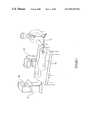
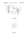
- FIG. 1depicts an exemplary, illustrative system for medical simulation according to the present invention.
- a system 10includes a mannequin 12 representing the subject on which the procedure is to be performed, a simulated endoscope 14 and a computer 16 with a video monitor 18 .
- a student 20is shown interacting with system 10 by manipulating simulated endoscope 14 within mannequin 12 .
- mannequin 12includes a simulated organ into which simulated endoscope 14 is inserted.
- tactile and visual feedbackare determined according to the position of endoscope 14 within the simulated organ (not shown).
- the visual feedbackare provided in the form of a display on video monitor 18 .
- the necessary data calculationsare performed by computer 16 , so that realistic tactile and visual feedback are provided to student 20 .
- FIG. 2is an exemplary illustration of a screen display shown on monitor 18 .
- a screen display 22includes a feedback image 24 .
- Feedback image 24represents the visual image as seen if the endoscope were inserted into a living human patient.
- Feedback image 24is an accurate and realistic simulation of the visual data that would be received from that portion of the gastrointestinal tract in the living human patient.
- feedback image 24is shown as a static image, it is understood that this is for illustrative purposes only and the actual visual feedback data would be in the form of a substantially continuous flow of simulated images based upon actual video stream data obtained from an actual endoscopic procedure.
- the flow of images represented by feedback image 24gives the student (not shown) realistic visual feedback.
- screen display 22preferably includes a number of GUI (graphic user interface) features related to the preferred tutorial functions of the present invention.
- a tracking display 26explicitly shows the location of the simulated endoscope within the simulated gastrointestinal tract.
- Tracking display 26includes a schematic gastro-intestinal tract 28 , into which a schematic endoscope 30 has been inserted.
- tracking display 26can be enabled or disabled, so that the student can only see tracking display 26 if the tracking function is enabled.
- screen display 22Additional, optional but preferred features of screen display 22 include the provision of a “help” button 32 , which upon activation could cause the display of such helpful information as a guide to the controls of the endoscope. Similarly, a preferred “hint” button 34 would give the student one or more suggestions on how to continue the performance of the medical procedure. A preferred “patient history” button 36 would cause screen display 22 to show information related to one of a selection of simulated “patient histories”, which could be of help to the student in deciding upon a further action. Finally, a preferred “performance” button 38 would cause screen display 22 to display a review and rating of the performance of the student. All of these functions are part of the preferred embodiment of a tutorial system for training a student in the medical procedure of endoscopy, as described in further detail in FIG. 4 .
- FIGS. 3A and 3Bare schematic block diagrams of an exemplary visual processing and display system and method according to the present invention.
- FIG. 3Ais a flow chart of the method for visual processing and display according to the present invention, and is intended as a summary of the method employed by the system of FIG. 3 B. Further details concerning particular aspects of the method are described below with reference to FIG. 3 B.
- the method and system of the present inventionprovide a solution to a number of problems in the art of medical simulation, in particular for the simulation of the procedure of gastro-endoscopy.
- This procedureinvolves the visual display of an interior portion of the gastrointestinal tract, such as the colon.
- the colonis a flexible body with a curved structure.
- the inner surface of the colonis generally deformable, as well as being specifically, locally deformable. All of these deformations in space must be calculated according to the mathematical model of the colon, and then rendered visually in real time in order to provide a realistic visual feedback response for the user.
- FIG. 3Ashows a preferred embodiment of the method of the present invention for preparation of the model and rendering of visual feedback, including steps required for preparation of the computerized model of the colon, as well as steps required for display of the colon.
- step 1 of the method of the present inventionactual video data are recorded onto videotape during the performance of the actual medical procedure of endoscopy on a living human patient.
- datacould also include MRI (magnetic resonance imaging) and CAT (computer assisted tomography) scan data from procedures performed on living human patients.
- step 2individual images are abstracted, for example with a framegrabber device, and then digitized.
- the digitized imagesare preferably selected for clarity and lack of visual artifacts, and are then stored in a texture mapping database. More preferably, the digitized images are enhanced before being stored.
- the texture mappingalso include animation. Such animation could simulate effects such as random vibration of the tissue of the colon or of the endoscope, as well as such events as liquid flowing downward due to the influence of gravity.
- a three-dimensional mathematical model of the human colonis constructed.
- the three-dimensional mathematical model of the colonwhich is particularly preferred for the present invention is a polygonal model such as a spline.
- This mathematical functionrepresents the colon as a series of curves, such that the points in the three-dimensional structure of the colon are mapped to the spline.
- the coloncould be modeled as a straight line which is deformed by altering the spline for the model until the model fits the data.
- the splinecould be placed inside the colon and mapped to the colon.
- multiple splinesare used to model the junction of the stomach and small intestine, for example.
- the mappingcan be performed according to three-dimensional coordinates, along the x, y and z axes. Alternatively, the mapping can be performed according to coordinates of time, angle and radius within the colon. A mixture of these two different types of coordinates is also optionally employed, in which the coordinates are time, x and y for example.
- Both the spline itself and the mapping from the spline to the coloncan optionally be altered in order to provide new and different visual representations of the colon, for example in order to provide a plurality of theoretical “test cases” for students to study.
- the alterationis optionally performed according to MRI (magnetic resonance imaging) data, for example.
- data from MRI and/or CAT scan proceduresare cleaned and reassembled according to the mathematical model, in order to more accurately determine the geometry of the simulated colon. Substantially all of these procedures could be performed automatically according to such data or alternatively, these procedures could also be performed partially or wholly manually.
- the preferred mathematical model of the present inventionpermits the data to be rapidly visually rendered onto the model of the colon.
- a “loop” of the endoscope cable itselfis modeled.
- Such a loopoccurs when the person performing the endoscopic procedure, whether “real” or simulated, inadvertently changes direction within the colon by turning the endoscope itself.
- Such a loopcan be very dangerous to the patient, and therefore should be detected as part of a simulation, in order to warn the student as an indication that the procedure has been performed incorrectly thereby causing the loop to appear.
- the loopis constructed with a spline according to the present invention and is coordinated with force feedback.
- the length of cable which has been fed into colonmust be determined, as must the length of the colon from the rectum (entry point of the endoscope) to the current position of the endoscope.
- the size of the loopis then calculated from the differential of these two lengths, and the loop is modeled according to the spline.
- the method of visually rendering the colon according to the present inventionincludes a number of steps, described below, which are performed as software instructions operated by a data processor.
- the methodpreferably includes the step (shown as step 5 in FIG. 3A ) of dividing the colon into a plurality of portions.
- the divisionis made linearly, since the spatial movement of the simulated endoscope is limited. In other words, the simulated endoscope cannot “jump” from one portion of the colon to another, but must instead proceed in a linear fashion along the simulated colon.
- the simulated endoscopecan only be moved at a finite speed through the simulated colon. Thus, the endoscope must pass through each segment of the three-dimensional model of the colon in sequence at a known, limited speed.
- the number of portions which are renderedis not predetermined, since under certain circumstances, the number of portions in the line of sight may vary. For example, when the camera is traveling around a bend in the colon, the line of sight of the camera is very short, such that relatively fewer portions, or else smaller such portions, must be rendered.
- the visual attributes of the area of the colon being scanned by the cameraare determined.
- these visual attributesare determined according to a number of factors, including the location of the tip of the endoscope, which holds the camera, and the direction in which the camera itself is pointed.
- Other important factorsinclude the shape of the colon being modeled and the history of movement of the camera through the colon.
- the previous movements of the endoscope through the colonas determined by the actions of the student, have a significant impact on the area of the colon which is visualized by the camera at any given moment. For example, if the student has caused a “loop” to form by incorrectly operating the endoscope, as previously described, this “loop” can be simulated correctly only through the inclusion of the history of movements to determine the visual feedback.
- step 7preferably a local deformation to at least one of these portions is analyzed to determine if such a deformation affects the spline itself.
- the mapped coordinatesare then rapidly transformed from time, angle and radius to x, y and z.
- step 8preferably the local deformation of the tissue of the colon is determined through interpolation of the radius, in order to determine the degree of such deformation. Since the time, angle and radius may not give sufficient information to perform this calculation, optionally and preferably, the volume of the colon is additionally altered according to predefined mathematical models.
- the level of details in the areais increased by adding more polygons to the calculations performed with the model in order to be able to stretch all or substantially points in the immediate area without distortion.
- the stretchingis preferably performed according to a predetermined function which preferably enables the spline model to be altered locally.
- This preferred method for modeling “stretching” of the coloncan also be used to model local areas of irregularity such as a polyp.
- Polypscan be mapped point by point onto the model of the colon, thereby adjusting the visual representation of the tissue to accommodate both the polyp itself and the structural alterations of the tissue at the base of the polyp.
- step 9the various types of data which were previously described are used to actually render the visual data onto the colon.
- the mapping of such data onto the modeloptionally and preferably involves some adjustments, performed manually by a software programmer. Alternatively, such mapping could be entirely automatically performed.
- step 10texture mapping from the database is overlaid onto the chunk of the model.
- texture mappingincludes both the digitized images and additional animation.
- step 11the resultant images are displayed.
- the imagesare displayed in a continuous flow according to the location of the simulated endoscope within the simulated gastrointestinal tract.
- mapping of coordinatesis preferably performed according to the mathematical model of the colon, which is more preferably a spline.
- FIG. 3Bshows the visual processing and display system according to the present invention in more detail.
- a visual processing and display system 40includes screen display 22 for displaying the processed visual data.
- the visual dataare constructed as follows. First, data are recorded from actual gastro-endoscopic procedures onto videotape, as shown in a recording block 42 . The data are preferably stored on Super-VHF videotape in order to obtain the highest quality representation of the visual images displayed on the screen during the actual endoscopic procedure, as shown in block 44 . Next, at least a portion of the frames of the videotape, and preferably substantially all the frames, are abstracted individually by a frame-grabber 46 to form digitized images. Individual digitized images can then be selected for clarity and lack of artifacts such as reflections from the endoscopic apparatus itself. The images in the selected frames are then preferably enhanced and added to a texture mapping database 48 .
- the first type of texture mappingis intended to enhance the realistic visual aspects of the images, for example by removing visual artifacts.
- the second type of texture mappingis intended to simulate the behavior of a live organ and a real endoscope, as represented by block 50 .
- the tissue of the colonmoves somewhat, and the endoscope itself vibrates and wobbles. This movement is simulated visually by the addition of random animation of the images, and also by the addition of such effects as liquid flowing downward due to the influence of gravity.
- Such animationenhances the realistic nature of the visual representation of the colon.
- the imagesIn order for the enhanced images to be correctly displayed, the images must correspond to the manipulation and location of the simulated endoscope within the simulated colon.
- the texture mapping of the imagesshould correspond to the location of the endoscope within the colon.
- Such correspondence between the location of the endoscope within the colon and the texture mappingis provided by a texture mapping engine 52 .
- the texture mapping datais then readily accessed by the display portion of visual system 40 , as shown by block 54 .
- visual processing and display system 40includes a three-dimensional mathematical model of at least a portion of the gastro-intestinal tract 56 , more preferably constructed as described in FIG. 3 A.
- model 56is herein described as a three-dimensional model of the colon, it being understood that this is not meant to be limiting in any way.
- Model 56preferably features a plurality of segments 58 , more preferably many such segments 58 .
- Locator 60then instructs an object loader 62 to load the relevant segment 58 for access by visual system 40 , as shown in block 54 and previously described.
- object loader 62preferably three segments 58 are ready for access by object loader 62 at any given moment.
- the specific segment 58 in which the endoscope is currently locatedis preferably held in DRAM or RAM, in combination with the texture mapping described previously.
- the next segment 58 and the preceding segment 58preferably are also stored in an easily accessible location, although not necessarily in RAM or DRAM.
- segment optimizer 64receives information from locator 60 , as well as the series of images obtained from overlaying the texture mapping onto the relevant segment 58 , and then feeds each specific image to a display manager 66 for display on screen display 22 .
- display manager 66is assisted by a real-time viewer 68 , preferably implemented in Direct 3DTM (Microsoft Inc., Seattle, Wash.).
- Real-time viewer 68provides the necessary software support to communicate with a graphics card 70 for actual display of the images on screen display 22 .
- graphics card 70can be of any suitable manufacture, preferably graphics card 70 has at least 8, and more preferably at least 16, Mb of VRAM for optimal performance.
- An example of a suitable graphics card 70is the 3Dfx Voodoo RushTM card.
- the performance of real-time viewer 68is enhanced by a math optimizer 72 , preferably implemented in Visual C++.
- segment optimizer 64 and display manager 66 on the one hand, and locator 60 on the other,is provided through a software interface 74 .
- Software interface 74enables locator 60 to communicate with the other components of visual system 40 , in order to provide information regarding the location of the endoscope within the colon.
- locator 60includes a sensor 76 , which can be obtained from Ascension Technology Corp., for example.
- Sensor 76senses positional information from within a simulated organ 77 , which is described herein as a colon for the purposes of discussion and is not meant to be limiting.
- Sensor 76is controlled by a control unit 82 .
- the positional informationis then relayed to a CPU controller 78 , which is connected to a servo-motor 80 (Haydon Switch and Instrument Co.).
- a servo-motor 80Hos Switch and Instrument Co.
- Tactile feedbackis provided by each servo-motor 80 in turn, which manipulates the material of the colon.
- All Visual system 40also includes a user interface 84 , preferably implemented in Visual C++.
- User interface 84includes the GUI features described previously for FIG. 2 .
- user interface 84enables visual system 40 to interact with the preferred feature of a network interface 86 , for example, so that other students can view screen display 22 over a network.
- User interface 84also permits the tutorial functions of at least one, and preferably a plurality of, tutorial modules 88 to be activated.
- tutorial module 88could include a particular scenario, such as a subject with colon cancer, so that different types of diagnostic and medical challenges could be presented to the student. The student would then need to respond correctly to the presented scenario.
- a tutorial system 90starts as shown in block 92 .
- the usermust select whether actual interaction with the simulated endoscope is desired, or if the user prefers to receive tutoring in the theory of endoscopy, as shown in a block 94 .
- the next displayasks if the user is new, as shown in a block 96 . If the answer is “yes”, the user is requested to enter certain information, as shown by block 98 . If the answer is “no”, the user is requested to enter identification information, such as user name or identification number, as shown in block 100 .
- tutoring by subject 102includes, but is not limited to, such subjects as basic manipulation of the endoscope, biopsy and polypectomy.
- Tutoring by subject 102includes on-screen support, as shown in block 108 .
- Tutoring by case studies 106can be selected both according to case number and according to the level of the desired cases, such as beginner, intermediate and expert.
- individual case studiescould be created by a teacher or professor, by combining features of various stored cases.
- a professorcould create a case history appropriate for a 20 year old male with colitis, so that the student would then be able to practice endoscopy on such a patient.
- tutoring system 90preferably has the flexibility to enable many different types of “patients” to be studied.
- on-screen supportcan be provided for both tutoring by case studies 106 and tutoring by procedures 104 , as shown in block 110 . If on-screen support is not desired, the user can indicate whether the tutoring session is actually an official test, as shown in block 112 .
- tutoring system 90includes both the ability to teach and the ability to test the student.
- the tutorial systemalso includes a simplified version of the simulated endoscopic process for teaching the proper manipulation of the endoscope according to visual feedback, as well as for enabling the student to understand the correspondence between the visual feedback and tactile feedback.
- This simplified versionwould emphasize the performance and mastery of one or more specific tasks, such as the manipulation of the endoscope through the colon.
- this preferred embodimentcould be generalized to a method for teaching a particular skill required for performance of an actual medical procedure to a student.
- This methodwould include the step of abstracting a portion of the visual feedback of the actual medical procedure, which would preferably include fewer visual details than the entirety of the visual feedback obtained during the performance of the medical procedure. This portion of the visual feedback would preferably enable the student to learn the motion of the instrument as the required skill.
- the simplified versionmay optionally not feature many, or even most, of the visual details of the colon as visual feedback.
- the colonwould preferably be presented as a smooth, relatively featureless tube having the geometry and dimensions of the colon in order to correlate the motion of the simulated endoscope through the interior space of the colon.
- the simplified versionwould be embodied as a game, in which students would be awarded points for correct manipulation of the endoscope, and would be penalized for incorrect manipulations.
- the studentwould have the opportunity to learn the manipulations required for successful endoscopy without the distraction of visual details, in a low pressure and even “fun” environment.
- FIGS. 5A and 5Billustrate the mechanical aspects of an exemplary simulated gastro-intestinal tract according to the present invention.
- a cut-away view of a mannequin 114is shown in FIG. 5 A.
- mannequin 114is about one meter wide, which is within the dimensions of an actual human subject.
- a simulated gastro-intestinal tract 116is shown within mannequin 114 .
- simulated gastrointestinal tract 116includes only the colon, it being understood that this is not meant to be limiting in any way.
- Simulated gastro-intestinal tract 116is connected to a transmitter 118 and a signal processing device 120 , also placed within mannequin 114 .
- a simulated endoscope 122can be inserted into mannequin 114 through an opening 124 .
- opening 124simulates the rectum of the subject.
- Simulated endoscope 122can be maneuvered left, right, up and down.
- simulated endoscope 122is about 1800 cm long, similar to the length of a real endoscope.
- the diameter of the tip of simulated endoscope 122is about 13.4 mm, while the remainder of endoscope 122 has a diameter of about 10.2 mm, again similar to the dimensions of a real endoscope.
- sensor 76 on the tip of simulated endoscope 122is able to detect the location of simulated endoscope 122 .
- Sensor 76preferably has three degrees of freedom, more preferably six degrees of freedom for effective simulation of manipulation of endoscope 122 . If sensor 76 has six degrees of freedom, the detected directions of orientation include the Cartesian coordinates X, Y, Z, as well as roll, elevation and azimuth.
- sensor 76preferably includes a sensor transmitter 126 , so that the precise angle and location of sensor 76 can be determined relative to gastrointestinal tract 116 . Sensor transmitter 126 transmits data to signal processing device 120 , which then analyzes and processes the signal.
- the processed signalis then given to transmitter 118 for transmission to an electronics unit 128 and a DC drive unit 130 .
- the signalis converted by DC drive unit 130 and passed to electronics unit 128 .
- Electronics unit 128then sends the position and orientation of sensor 76 to software interface 74 , so that the remainder of the display system is able to use the information to display the correct images on display screen 22 for visual feedback.
- the present inventionprovides both visual feedback and tactile feedback.
- Tactile feedbackcan be provided through the exertion of forces on simulated endoscope 122 by simulated gastro-intestinal tract 116 , as shown in FIGS. 6A-6C .
- tactile feedbackcould be provided by the mechanical action of simulated endoscope 122 , as shown in FIGS. 7A-7D .
- simulated gastro-intestinal tract 116is constructed from semi-flexible material, which gives the feel of a smooth and wet material.
- the actual sensations of sliding along a semi-flexible, smooth, wet materialcan also be provided through the mechanism of endoscope 122 itself, as in the second embodiment.
- FIG. 5 BAn additional embodiment of gastro-intestinal tract 116 , in which tract 116 is placed within a box 132 rather than within mannequin 114 , is shown in FIG. 5 B.
- box 132could serve to contain any radiowaves, so that the mechanism of gastro-intestinal tract 116 could be controlled by transmission of radiowaves, for example. Since certain medical equipment is highly sensitive to these radiowaves, they would need to remain within mannequin 114 . Box 132 would therefore act to insulate gastro-intestinal tract 116 from the external environment outside the mannequin. Details of gastrointestinal tract 116 are more readily seen in FIG. 6A , it being understood that FIGS. 5A , 5 B and 6 A illustrate the same gastrointestinal tract 116 .
- FIG. 6Ashows gastro-intestinal tract 116 according to the first embodiment, in which tactile feedback is provided by forces acting on simulated endoscope 122 by a mechanism contained within gastrointestinal tract 116 itself.
- Simulated gastro-intestinal tract 116is made from a semi-flexible material.
- a plurality of motion boxes 134are disposed at intervals along the outer surface of gastro-intestinal tract 116 . For the purposes of illustration, seven motion boxes 134 are shown.
- Each motion box 134shown in greater detail in FIG. 6B , has at least one, and preferably a plurality of, servo-motors 80 , preferably linear motors.
- Each servo-motor 80is connected to a piston 136 .
- the detail of piston 136is shown enlarged in FIG. 6 B.
- Each piston 136is connected to a foot 138 , which contacts a portion of the material of the external surface of gastrointestinal tract 116 .
- foot 138is actually attached to the portion of the material of the external surface, for easier manipulation of the material.
- pistons 136there are two different types of pistons 136 .
- the first typeof which two are shown for illustrative purposes, is a vertical force piston 140 for causing vertical movement of a portion of the external surface of gastro-intestinal tract 116 .
- the second typeof which one is shown for illustrative purposes, is a horizontal force piston 142 for causing horizontal movement of a portion of the external surface of gastro-intestinal tract 116 .
- servomotor 80is an oscillating motor placed directly against the material of gastro-intestinal tract 116 , so that horizontal force piston 142 includes the motor alone, without a structure similar to vertical force piston 140 . Since each piston 136 has an associated servo-motor 80 , the necessary vertical and horizontal movement of the external surface of gastro-intestinal tract 116 can be precisely determined by the activity of servo-motor 80 .
- Each piston 136contacts the material of gastro-intestinal tract 116 in order to manipulate this material to exert a force against the endoscope (not shown).
- a first vertical force piston 144could be moved closer to servo-motor 80
- a second vertical force piston 146is moved away from servo-motor 80 .
- horizontal force piston 142which is preferably an oscillating servo-motor alone as shown, moves horizontally to provide more delicate fine-tuning of the tactile feedback sensations. Since servo-motors 80 are disposed over the three-dimensional surface of gastrointestinal tract 116 , the force on the endoscope can be exerted in three dimensions.
- the activity of servo-motor 80is in turn controlled by digital controller 82 .
- Digital controller 82can be a card inserted within the PC computer which is performing the requisite calculations required for the simulation of the medical process.
- Software operated by the PC computeruses positional and orientation information from sensor 76 on simulated endoscope 122 to determine the position of simulated endoscope 122 .
- the softwaresends instructions to digital controller 82 according to the desired tactile sensations which should be felt by the operator of simulated endoscope 122 at that particular position within simulated gastrointestinal tract 116 .
- Digital controller 82then causes at least one servo-motor 80 to move the associated piston 136 as necessary to provide the tactile feedback sensations.
- Digital controller 82can be connected to servo-motors 80 through some type of radiation, such as infra-red light.
- some type of radiationsuch as infra-red light.
- the limitations on radiation of certain wavelengths, such as radiowaves, within the hospital or medical environmentmake a connection by an actual wire running from digital controller 82 to each servo-motor 80 more preferable.
- each servo-motor 80is connected to a motion box controller 144 by a wire.
- Motion box controller 144is then preferably connected to digital controller 82 by a single wire (not shown). This configuration limits the number of individual connections made to digital controller 82 for greater efficiency.
- FIG. 6Cshows an enlarged cut-away view of servo-motor 80 , which as noted previously is preferably a linear motor.
- servo-motor 80is about 100 mm wide and 45 mm tall.
- FIGS. 7A-7Dshow a second embodiment of the mechanism for providing tactile feedback.
- the mechanismis contained within the simulated endoscope itself, rather than the simulated gastrointestinal tract. Similar to the previous embodiment, the simulated gastrointestinal tract could be contained within a substantially life-size mannequin with an opening for simulating the rectum. Furthermore, from the viewpoint of the student or other individual operating the simulated endoscope, both embodiments should give a suitable simulation of the medical procedure. However, as detailed below, the actual mechanism of providing the tactile portion of the simulation differs.
- FIG. 7Ashows the second embodiment of a simulated endoscope 146 .
- the movements and actions of simulated endoscope 146are controlled through a set of controls 148 .
- the tip of simulated endoscope 146is contained within a guiding sleeve 150 .
- Guiding sleeve 150shown in greater detail in FIG. 7B , preferably remains within the simulated gastrointestinal tract (not shown; see FIG. 7C ) in order to maintain a realistic visual appearance of simulated endoscope 146 before insertion into the mannequin (not shown).
- the tip of endoscope 146has a metal bracket 152 attached, which could be labeled with the word “sample” or with another label in order to clarify that endoscope 146 is only a simulation and not an actual medical instrument.
- the inside of guiding sleeve 150is preferably magnetized, for example with an electric current.
- Guiding sleeve 150has at least one, and preferably a plurality of, ball bearings 154 attached to the exterior surface of guiding sleeve 150 .
- guiding sleeve 150has at least one, and preferably a plurality of, attached plungers 156 .
- one end of guiding sleeve 150preferably features a section of flexible material 158 .
- the tip of endoscope 146is preferably inserted through guiding sleeve 150 , The tip of endoscope 146 features sensor 76 , as for the previous embodiment of the simulated endoscope.
- FIG. 7Cshows simulated endoscope 146 after insertion within the second embodiment of a simulated gastro-intestinal tract 160 .
- Simulated gastro-intestinal tract 160is preferably constructed from a rigid material.
- simulated gastro-intestinal tract 160preferably has the general anatomical shape and features of an actual gastro-intestinal tract for two reasons.
- the general anatomical shapecan be more easily contained within the mannequin because of its bends and turns.
- the general anatomical shapecan provide gross tactile feedback. For example, as any endoscope is inserted more deeply into the colon, the shape of the colon causes the tactile sensations to be altered as the endoscope moves around a bend in the colon.
- the general anatomical shapeis more useful for an effective simulation.
- guiding sleeve 150enables the operator to receive tactile feedback as follows.
- Ball bearings 154roll along the interior surface of gastrointestinal tract 160 .
- Each ball bearing 154has five degrees of freedom for movement.
- Each plunger 156is connected to a linear motor 162 , as shown in cross-section in FIG. 7 D.
- Linear motor 162is controlled in a similar fashion as the servo-motor of the previous embodiment.
- linear motor 162Upon receipt of signals from the computer, linear motor 162 causes plunger 156 to move vertically, thereby causing the operator of simulated endoscope 146 to receive tactile feedback sensations.
- guiding sleeve 150causes tactile feedback to be transmitted back through endoscope 146 .
- guiding sleeve 150preferably has section of flexible material 158 .
- Section of flexible material 158causes the tip of endoscope 146 to encounter some resistance under certain circumstances, such as when the tip is bent back on itself. Thus, section of flexible material 158 restrains movement of the tip from certain angles.
- this second embodimentis that the majority of tactile sensations are determined by the endoscope itself, so that they can be more easily controlled from the PC computer. Furthermore, such anatomical features as a fistula can be added according to instructions from the computer, without the necessity of changing the physical model of the simulated gastro-intestinal tract. Additionally, under certain circumstances the tissue of the actual colon will force the endoscope backwards, a situation which can be more easily replicated in the second embodiment. Thus, the second embodiment of the simulated gastro-intestinal tract and endoscope is more flexible in terns of replicating a greater variety of anatomical features and conditions.
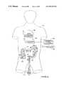
- FIGS. 8A-8Eshow yet another and particularly preferred embodiment of the simulated endoscope and colon according to the present invention.
- FIG. 8Ashows a preferred system for medical simulation according to the present invention.
- a system 164includes a mannequin 166 representing the subject on which the procedure is to be performed, a simulated endoscope (not shown, see FIG. 8D ) and a computer 168 with a video monitor 170 .
- Mannequin 166preferably includes a palpable area 172 for determining the location of the simulated endoscope by feeling the abdominal area of mannequin 166 .
- Palpable area 172preferably features a light (not shown), such that when the student has determined the location of the simulated endoscope, the light is lit to show the actual location of the simulated endoscope.
- Mannequin 166also includes a simulated organ 174 into which the simulated endoscope is inserted.
- simulated organ 174is a colon, which more preferably is constructed as a straight tube, with the force feedback required for the curves in the colon provided through a force feedback mechanism 176 .
- the visual feedback for the simulated medical proceduredoes not depend upon the geometrical shape of simulated organ 174 itself, such that the visual feedback and the tactile feedback are both substantially completely independent of the construction of simulated organ 174 .
- Force feedback mechanism 176preferably includes an air-driven force feedback device 178 (shown in more detail in FIGS. 8B , 8 D and 8 E). More preferably, two such air-driven force feedback devices 178 are provided, one near a mouth 180 of mannequin 166 , and the other near a rectum 182 of mannequin 166 .
- An air tube 184connects each air-driven force feedback device 178 to an air-pump 186 .
- air-pump 186also includes an air-pump control unit 188 which is connected to computer 168 for controlling the amount of air pumped into air-driven force feedback device 178 .
- Computer 168also preferably includes a modem 190 for communication with other computers.
- modem 190could enable computer 168 to connect to the Internet or intranet for performing telemedicine, or to connect to the intranet/computer network of the manufacturer for repair or trouble-shooting.
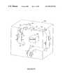
- FIGS. 8B and 8Cshow components of air-driven force feedback device 178 in more detail.
- a portion of a simulated endoscope 192interacts with air-driven force feedback device 178 to provide force feedback to the student.
- Force feedback device 178features a plurality of inflatable rings 194 (shown in more detail in the fully inflated position in FIG. 8 C).
- Each inflatable ring 194preferably has a different radius. More preferably, there are four such rings 194 , at least one of which has a larger radius than endoscope 192 and at least one of which has a smaller radius than endoscope 192 .
- the amount of air fed into rings 194determines the degree of inflation of each ring 194 , preferably separately, thereby determining the amount of force exerted onto endoscope 192 .
- each ring 194requires one second or more preferably less than one second to reach the fully inflated position.
- the air flow rateis preferably up to 100 liters per minute and the pressure is up to 3 atmospheres.
- Rings 194are preferably used both for passive force feedback, such as from the contraction of the rectum, and for active force feedback, for example when air is pumped into simulated organ 174 according to a functional feature of simulated endoscope 192 (see FIG. 8 E).
- FIG. 8Dshows force feedback mechanism 176 in more detail.
- rings 194are connected to air pump 186 through tube 184 , which more preferably is split into two tubes 196 , a first tube 196 for pumping air into rings 194 , and a second tube 196 for pumping air from rings 194 .
- the amount of air pumped by air pump 186is controlled by air pump controller 188 .
- the actions of air pump controller 188are preferably controlled by computer 168 through an I/O (analog-to-digital) card 198 .
- FIG. 8Eshows simulated endoscope 192 in more detail.
- Simulated endoscope 192features a handle 200 with various controls, including a first control 202 for pumping air into simulated organ 174 , and a second control 204 for suctioning air out of simulated organ 174 .
- Simulated endoscope 192preferably features a surgical tool control device 206 into which various surgical tools are optionally and preferably inserted (see FIGS. 9 A- 9 E).
- Simulated endoscope 192also preferably features a receiver 208 , for example a “minibird” sensor (Ascension Ltd., Burlington, Vt., USA). Receiver 208 is located at the tip of simulated endoscope 192 .
- Receiver 208is designed to receive transmissions from a transmitter 210 located in mannequin 166 (see FIG. 8 A), thereby determining a position of the tip of simulated endoscope 192 within simulated organ 174 .
- Transmitter 210is preferably a “minibird” transmitter (Ascension Ltd.).
- Receiver 208then transmits these signals to computer 168 , which uses these signals for determining the amount of force feedback and the visual feedback to be displayed to the student on monitor 178 .
- FIGS. 9A-9Eshow a preferred implementation of surgical tool control device 206 into which various surgical tools are optionally and preferably inserted.
- Surgical tool control device 206preferably features a forceps 212 inserted into a tool sleeve 214 , thereby simulating actual forceps for an endoscope.
- Actual forcepsare used for performing a polypectomy, and feature a loop which emerges from the tip of the forceps upon manipulation of the device. This loop is placed around the polyp and drawn tight. Electricity is then sent through the loop in order to cut the polyp and to cauterize the area.
- Tool sleeve 214features a tool control unit 218 for detecting the motions of forceps 212 , and translating these motions into force feedback and visual feedback.
- Visual feedbackincludes the visual display of the forceps “loop” when appropriate, for example, as well as the display of the polyp before and after the “polypectomy”.
- the location of the loopmust be tracked, preferably including up and down movements within the endoscope, and “roll” movement of the loop.
- Tool control unit 218is connected to an I/O card within the computer (not shown) for performing the necessary calculations for the various types of feedback.
- FIGS. 9B and 9Cshow two views of forceps 212 interacting with tool control unit 218 within tool sleeve 214 .
- Tool control unit 218features a guide wheel 220 and a light wheel 222 for detecting the motions of forceps 212 (FIG. 9 B).
- Light wheel 222features a plurality of notches through which light may pass.
- Tool control unit 218also features a first light 224 and a first light sensor 226 , as well as a second light 228 and a second light sensor 230 (FIG. 9 C).
- first light 224 and second light 228are alternately blocked and unblocked, such that light is alternately detectable and non-detectable by first light sensor 226 and second light sensor 230 .
- FIG. 9Cshows a second embodiment of the tool control unit.
- a tool control unit 232features two guide wheels 234 .
- Guide wheels 234help to guide the movement of forceps 212 within tool sleeve 214 .
- a light wheel 236also features notches through which light is alternately blocked and unblocked as forceps 212 is rotated within tool sleeve 214 .
- a light source(not shown) produces light which is detected, if it passes through light wheel 236 , by a photoelectric eye 238 . Photoelectric eye 238 then sends signals to a PCB (printed circuit board) 240 which is connected to the computer (not shown), such that these signals can be translated by the computer into the required visual feedback and force feedback.
- PCBprinted circuit board
- a foot pedal 242is shown in FIG. 9E for performing a simulated polypectomy.
- Foot pedal 242features an oil piston 244 and a microswitch 246 .
- Microswitch 246is connected to an I/O card on the computer (not shown), again for translating the movement of foot pedal 242 into the required visual feedback and force feedback.
- tactile sensations of an actual endoscope during a medical procedureIn order to accurately replicate the tactile sensations of an actual endoscope during a medical procedure, these sensations must be accurately obtained during an endoscopic procedure in an actual living patient.
- tactile sensationscould be collected from a physician performing the endoscopic procedure while wearing virtual reality gloves, such as the DataGlovesTM Tracking VR System (Greenleaf Medical Systems). These gloves are known for being able to register data regarding tactile sensations and feedback as experienced by the physician during the actual endoscopic procedure.
- Such actual dataare important because the tactile sensations change during the course of the procedure. For example, correlation between the movement of the endoscope and the visual display is gradually decreased as the endoscope is inserted deeper into the gastrointestinal tract.
- the collection of actual datais an important step in the provision of an accurate, realistic endoscopic simulator.
- a simulated biopsy device(not shown).
- This biopsy devicewould simulate the actual biopsy device used to retrieve tissue samples from the gastro-intestinal tract during endoscopy.
- the actual biopsy deviceis contained within the endoscope.
- the biopsy deviceemerges from the tip of the endoscope, at which point it is visible on the display screen.
- the jaws of the biopsy deviceare then opened and pushed onto the tissue.
- the jawsare then closed, and the biopsy device retracted. The removal of the tissue causes pools of blood to appear as the remaining tissue bleeds.
- the simulated biopsy devicewill only appear on the display screen of the present invention when the operator of the simulated endoscope causes the simulated biopsy device to emerge.
- the jaws of the biopsy deviceare preferably rendered as animation, more preferably in relatively high resolution because the jaws are small, so that a high resolution would not prove unduly taxing for the PC computer.
- the bleeding of the tissue and the resultant pools of bloodwill also be animated.
Landscapes
- Engineering & Computer Science (AREA)
- Health & Medical Sciences (AREA)
- General Physics & Mathematics (AREA)
- Physics & Mathematics (AREA)
- Mathematical Analysis (AREA)
- Pure & Applied Mathematics (AREA)
- Medical Informatics (AREA)
- Medicinal Chemistry (AREA)
- Chemical & Material Sciences (AREA)
- Algebra (AREA)
- Computational Mathematics (AREA)
- Radiology & Medical Imaging (AREA)
- Pulmonology (AREA)
- Mathematical Optimization (AREA)
- Mathematical Physics (AREA)
- General Health & Medical Sciences (AREA)
- Business, Economics & Management (AREA)
- Educational Administration (AREA)
- Educational Technology (AREA)
- Theoretical Computer Science (AREA)
- Endoscopes (AREA)
- Apparatus For Radiation Diagnosis (AREA)
- Instructional Devices (AREA)
- Pyrane Compounds (AREA)
- Radiation-Therapy Devices (AREA)
- Prostheses (AREA)
- Processing Or Creating Images (AREA)
Abstract
Description
Claims (4)
Applications Claiming Priority (2)
| Application Number | Priority Date | Filing Date | Title |
|---|---|---|---|
| IL12307398AIL123073A0 (en) | 1998-01-26 | 1998-01-26 | Endoscopic tutorial system |
| PCT/IL1999/000028WO1999038141A1 (en) | 1998-01-26 | 1999-01-15 | Endoscopic tutorial system |
Publications (1)
| Publication Number | Publication Date |
|---|---|
| US6857878B1true US6857878B1 (en) | 2005-02-22 |
Family
ID=11071142
Family Applications (2)
| Application Number | Title | Priority Date | Filing Date |
|---|---|---|---|
| US09/600,952Expired - LifetimeUS6857878B1 (en) | 1998-01-26 | 1999-01-15 | Endoscopic tutorial system |
| US09/714,206Expired - LifetimeUS6863536B1 (en) | 1998-01-26 | 2000-11-17 | Endoscopic tutorial system with a bleeding complication |
Family Applications After (1)
| Application Number | Title | Priority Date | Filing Date |
|---|---|---|---|
| US09/714,206Expired - LifetimeUS6863536B1 (en) | 1998-01-26 | 2000-11-17 | Endoscopic tutorial system with a bleeding complication |
Country Status (12)
| Country | Link |
|---|---|
| US (2) | US6857878B1 (en) |
| EP (1) | EP1051697B1 (en) |
| JP (1) | JP4436966B2 (en) |
| CN (1) | CN1186753C (en) |
| AT (1) | ATE286610T1 (en) |
| AU (1) | AU762444B2 (en) |
| BR (1) | BR9907239A (en) |
| CA (1) | CA2318746A1 (en) |
| DE (1) | DE69923060D1 (en) |
| IL (1) | IL123073A0 (en) |
| SI (1) | SI20559A (en) |
| WO (1) | WO1999038141A1 (en) |
Cited By (100)
| Publication number | Priority date | Publication date | Assignee | Title |
|---|---|---|---|---|
| US20040043368A1 (en)* | 2002-08-30 | 2004-03-04 | Ming-Shium Hsieh | Three-dimensional surgery simulation system |
| US20040126746A1 (en)* | 2000-10-23 | 2004-07-01 | Toly Christopher C. | Medical physiological simulator including a conductive elastomer layer |
| US20050014560A1 (en)* | 2003-05-19 | 2005-01-20 | Yacob Blumenthal | Method and system for simulating interaction with a pictorial representation of a model |
| US20050251017A1 (en)* | 2004-04-23 | 2005-11-10 | Azar Fred S | System and method registered video endoscopy and virtual endoscopy |
| US20060099557A1 (en)* | 2002-09-30 | 2006-05-11 | Anders Hyltander | Device and method for generating a virtual anatomic environment |
| US20060127867A1 (en)* | 2002-12-03 | 2006-06-15 | Jan Grund-Pedersen | Interventional simulator system |
| US20060127864A1 (en)* | 2002-12-03 | 2006-06-15 | Fredrik Ohlsson | Interventional simulation device |
| US20060183095A1 (en)* | 2004-01-23 | 2006-08-17 | The Administrators Of The Tulane Educational Fund | Laparoscopic camera navigation trainer |
| US20060211917A1 (en)* | 2005-03-01 | 2006-09-21 | Pentax Corporation | Data generating device and endoscope system |
| US20060234195A1 (en)* | 2002-12-03 | 2006-10-19 | Jan Grund-Pedersen | Interventional simulator control system |
| US20070141543A1 (en)* | 2002-12-03 | 2007-06-21 | Jan Grund-Pedersen | Interventional simulator system |
| US20070192075A1 (en)* | 2004-02-26 | 2007-08-16 | Kyoto University | Organism stimulation device and program |
| US20070238085A1 (en)* | 2006-01-13 | 2007-10-11 | Colvin Richard T | Computer based system for training workers |
| US20070239409A1 (en)* | 2006-04-08 | 2007-10-11 | Millman Alan | Method and system for interactive simulation of materials |
| US20080020361A1 (en)* | 2006-07-12 | 2008-01-24 | Kron Frederick W | Computerized medical training system |
| US20080131855A1 (en)* | 1996-05-08 | 2008-06-05 | Gaumard Scientific Company, Inc. | Interactive Education System for Teaching Patient Care |
| US20080138778A1 (en)* | 1996-05-08 | 2008-06-12 | Gaumard Scientific Company, Inc. | Interactive Education System for Teaching Patient Care |
| US20080138780A1 (en)* | 2000-08-17 | 2008-06-12 | Gaumard Scientific Company, Inc. | Interactive Education System for Teaching Patient Care |
| US20080138779A1 (en)* | 1996-05-08 | 2008-06-12 | Gaumard Scientific Company, Inc. | Interactive Education System for Teaching Patient Care |
| US20080147087A1 (en)* | 2006-10-20 | 2008-06-19 | Eli Horn | System and method for modeling a tracking curve of and in vivo device |
| US20090148822A1 (en)* | 2007-12-07 | 2009-06-11 | Gaumard Scientific Company, Inc. | Interactive Education System for Teaching Patient Care |
| US7665995B2 (en) | 2000-10-23 | 2010-02-23 | Toly Christopher C | Medical training simulator including contact-less sensors |
| US20100109713A1 (en)* | 2008-11-05 | 2010-05-06 | Harriman Paul J | Current balancing circuit and method |
| US20100251117A1 (en)* | 2009-03-25 | 2010-09-30 | International Business Machines Corporation | Visualization of medical conditions in a virtual universe |
| US7811090B2 (en) | 1996-05-08 | 2010-10-12 | Gaumard Scientific Company, Inc. | Interactive education system for teaching patient care |
| US20100285438A1 (en)* | 2009-03-12 | 2010-11-11 | Thenkurussi Kesavadas | Method And System For Minimally-Invasive Surgery Training |
| US20100291520A1 (en)* | 2006-11-06 | 2010-11-18 | Kurenov Sergei N | Devices and Methods for Utilizing Mechanical Surgical Devices in a Virtual Environment |
| WO2010144663A3 (en)* | 2009-06-11 | 2011-03-31 | University Of Pittsburgh - Of The Commonwealth System Of Higher Education | Real-time x-ray vision for healthcare simulation |
| WO2011035088A3 (en)* | 2009-09-18 | 2011-08-18 | University Of Tennessee Research Foundation | Flexible and rigid endoscopic training device (fred) |
| US20120185099A1 (en)* | 2011-01-19 | 2012-07-19 | Harris Corporation | Telematic interface with control signal scaling based on force sensor feedback |
| US8311791B1 (en) | 2009-10-19 | 2012-11-13 | Surgical Theater LLC | Method and system for simulating surgical procedures |
| US8469716B2 (en)* | 2010-04-19 | 2013-06-25 | Covidien Lp | Laparoscopic surgery simulator |
| US8556635B2 (en) | 2000-10-23 | 2013-10-15 | Christopher C. Toly | Physiological simulator for use as a brachial plexus nerve block trainer |
| US8639386B2 (en) | 2011-05-20 | 2014-01-28 | Harris Corporation | Haptic device for manipulator and vehicle control |
| US8694134B2 (en) | 2011-05-05 | 2014-04-08 | Harris Corporation | Remote control interface |
| US8764452B2 (en) | 2010-10-01 | 2014-07-01 | Applied Medical Resources Corporation | Portable laparoscopic trainer |
| US8786613B2 (en) | 2006-04-08 | 2014-07-22 | Alan Millman | Method and system for interactive simulation of materials and models |
| US20140349266A1 (en)* | 2011-12-06 | 2014-11-27 | Ohio University | Active colonoscopy training model and method of using the same |
| US8918214B2 (en) | 2011-01-19 | 2014-12-23 | Harris Corporation | Telematic interface with directional translation |
| US8954195B2 (en) | 2012-11-09 | 2015-02-10 | Harris Corporation | Hybrid gesture control haptic system |
| US8956165B2 (en) | 2008-01-25 | 2015-02-17 | University Of Florida Research Foundation, Inc. | Devices and methods for implementing endoscopic surgical procedures and instruments within a virtual environment |
| US8961190B2 (en) | 2011-12-20 | 2015-02-24 | Applied Medical Resources Corporation | Advanced surgical simulation |
| US8965620B2 (en) | 2013-02-07 | 2015-02-24 | Harris Corporation | Systems and methods for controlling movement of unmanned vehicles |
| US8996244B2 (en) | 2011-10-06 | 2015-03-31 | Harris Corporation | Improvised explosive device defeat system |
| US9026250B2 (en) | 2011-08-17 | 2015-05-05 | Harris Corporation | Haptic manipulation system for wheelchairs |
| US9022789B2 (en) | 2011-09-09 | 2015-05-05 | Cook Medical Technologies Llc | Sialendoscopy demonstration apparatus |
| WO2013090729A3 (en)* | 2011-12-14 | 2015-06-04 | Brigham And Women's Hospital, Inc. | System and method for part-task training box for flexible endoscopy |
| US9128507B2 (en) | 2013-12-30 | 2015-09-08 | Harris Corporation | Compact haptic interface |
| US9205555B2 (en) | 2011-03-22 | 2015-12-08 | Harris Corporation | Manipulator joint-limit handling algorithm |
| US9218753B2 (en) | 2011-10-21 | 2015-12-22 | Applied Medical Resources Corporation | Simulated tissue structure for surgical training |
| CN105448155A (en)* | 2015-12-22 | 2016-03-30 | 天津市医学堂科技有限公司 | Spine endoscope virtual training system |
| US9449532B2 (en) | 2013-05-15 | 2016-09-20 | Applied Medical Resources Corporation | Hernia model |
| JP2016539767A (en)* | 2013-11-12 | 2016-12-22 | シモンセン ウィール エー/エス | Endoscopy equipment |
| US9548002B2 (en) | 2013-07-24 | 2017-01-17 | Applied Medical Resources Corporation | First entry model |
| US20170110032A1 (en)* | 2015-10-16 | 2017-04-20 | Virtamed Ag | Ultrasound simulation system and tool |
| US9788905B2 (en) | 2011-03-30 | 2017-10-17 | Surgical Theater LLC | Method and system for simulating surgical procedures |
| US9898937B2 (en) | 2012-09-28 | 2018-02-20 | Applied Medical Resources Corporation | Surgical training model for laparoscopic procedures |
| US9922579B2 (en) | 2013-06-18 | 2018-03-20 | Applied Medical Resources Corporation | Gallbladder model |
| US9940849B2 (en) | 2013-03-01 | 2018-04-10 | Applied Medical Resources Corporation | Advanced surgical simulation constructions and methods |
| US9959786B2 (en) | 2012-09-27 | 2018-05-01 | Applied Medical Resources Corporation | Surgical training model for laparoscopic procedures |
| US10037715B2 (en) | 2013-10-16 | 2018-07-31 | Simulab Corporation | Detecting insertion of needle into simulated vessel using a conductive fluid |
| US10056012B2 (en) | 2012-05-25 | 2018-08-21 | Surgical Theatre LLC | Hybrid image/scene renderer with hands free control |
| US10081727B2 (en) | 2015-05-14 | 2018-09-25 | Applied Medical Resources Corporation | Synthetic tissue structures for electrosurgical training and simulation |
| US10097875B2 (en) | 2011-05-25 | 2018-10-09 | Echostar Technologies L.L.C. | Apparatus, systems and methods for presentation management of erotica-related media content |
| US10121391B2 (en) | 2012-09-27 | 2018-11-06 | Applied Medical Resources Corporation | Surgical training model for laparoscopic procedures |
| US10198965B2 (en) | 2012-08-03 | 2019-02-05 | Applied Medical Resources Corporation | Simulated stapling and energy based ligation for surgical training |
| US10198966B2 (en) | 2013-07-24 | 2019-02-05 | Applied Medical Resources Corporation | Advanced first entry model for surgical simulation |
| US10223936B2 (en) | 2015-06-09 | 2019-03-05 | Applied Medical Resources Corporation | Hysterectomy model |
| US10332425B2 (en) | 2015-07-16 | 2019-06-25 | Applied Medical Resources Corporation | Simulated dissectible tissue |
| US10354556B2 (en) | 2015-02-19 | 2019-07-16 | Applied Medical Resources Corporation | Simulated tissue structures and methods |
| US10395559B2 (en) | 2012-09-28 | 2019-08-27 | Applied Medical Resources Corporation | Surgical training model for transluminal laparoscopic procedures |
| US10410542B1 (en) | 2018-07-18 | 2019-09-10 | Simulated Inanimate Models, LLC | Surgical training apparatus, methods and systems |
| US10490105B2 (en) | 2015-07-22 | 2019-11-26 | Applied Medical Resources Corporation | Appendectomy model |
| US10535281B2 (en) | 2012-09-26 | 2020-01-14 | Applied Medical Resources Corporation | Surgical training model for laparoscopic procedures |
| US10679520B2 (en) | 2012-09-27 | 2020-06-09 | Applied Medical Resources Corporation | Surgical training model for laparoscopic procedures |
| US10706743B2 (en) | 2015-11-20 | 2020-07-07 | Applied Medical Resources Corporation | Simulated dissectible tissue |
| US10720084B2 (en) | 2015-10-02 | 2020-07-21 | Applied Medical Resources Corporation | Hysterectomy model |
| US10796606B2 (en) | 2014-03-26 | 2020-10-06 | Applied Medical Resources Corporation | Simulated dissectible tissue |
| US10818201B2 (en) | 2014-11-13 | 2020-10-27 | Applied Medical Resources Corporation | Simulated tissue models and methods |
| CN111915722A (en)* | 2020-08-04 | 2020-11-10 | 广州珠江天然气发电有限公司 | Movement control method and system for gas turbine endoscope |
| US10847057B2 (en) | 2017-02-23 | 2020-11-24 | Applied Medical Resources Corporation | Synthetic tissue structures for electrosurgical training and simulation |
| US10849688B2 (en) | 2016-03-02 | 2020-12-01 | Truinject Corp. | Sensory enhanced environments for injection aid and social training |
| US10861236B2 (en) | 2017-09-08 | 2020-12-08 | Surgical Theater, Inc. | Dual mode augmented reality surgical system and method |
| US10896627B2 (en) | 2014-01-17 | 2021-01-19 | Truinjet Corp. | Injection site training system |
| US10902677B2 (en) | 2010-04-09 | 2021-01-26 | University Of Florida Research Foundation, Incorporated | Interactive mixed reality system and uses thereof |
| US11030922B2 (en) | 2017-02-14 | 2021-06-08 | Applied Medical Resources Corporation | Laparoscopic training system |
| US11094223B2 (en) | 2015-01-10 | 2021-08-17 | University Of Florida Research Foundation, Incorporated | Simulation features combining mixed reality and modular tracking |
| US11120708B2 (en) | 2016-06-27 | 2021-09-14 | Applied Medical Resources Corporation | Simulated abdominal wall |
| US11197722B2 (en) | 2015-10-14 | 2021-12-14 | Surgical Theater, Inc. | Surgical navigation inside a body |
| US11302219B2 (en)* | 2017-12-19 | 2022-04-12 | Applied Medical Resources Corporation | Total mesorectal excision surgical simulator |
| US11403964B2 (en) | 2012-10-30 | 2022-08-02 | Truinject Corp. | System for cosmetic and therapeutic training |
| US11484379B2 (en) | 2017-12-28 | 2022-11-01 | Orbsurgical Ltd. | Microsurgery-specific haptic hand controller |
| CN115281584A (en)* | 2022-06-30 | 2022-11-04 | 中国科学院自动化研究所 | Flexible endoscope robot control system and flexible endoscope robot simulation method |
| US20220375089A1 (en)* | 2020-02-10 | 2022-11-24 | Olympus Corporation | Endoscope apparatus, information processing method, and storage medium |
| US11547499B2 (en) | 2014-04-04 | 2023-01-10 | Surgical Theater, Inc. | Dynamic and interactive navigation in a surgical environment |
| US20230088225A1 (en)* | 2011-12-30 | 2023-03-23 | St. Jude Medical International Holding S.A R.L. | Roll detection and six degrees of freedom sensor assembly |
| US11710424B2 (en) | 2017-01-23 | 2023-07-25 | Truinject Corp. | Syringe dose and position measuring apparatus |
| US12070581B2 (en) | 2015-10-20 | 2024-08-27 | Truinject Corp. | Injection system |
| US12106678B2 (en) | 2021-10-23 | 2024-10-01 | Simulated Inanimate Models, LLC | Procedure guidance and training apparatus, methods and systems |
| US12217626B2 (en) | 2012-10-30 | 2025-02-04 | Truinject Corp. | Injection training apparatus using 3D position sensor |
Families Citing this family (87)
| Publication number | Priority date | Publication date | Assignee | Title |
|---|---|---|---|---|
| US6929481B1 (en) | 1996-09-04 | 2005-08-16 | Immersion Medical, Inc. | Interface device and method for interfacing instruments to medical procedure simulation systems |
| AU6325800A (en) | 1999-07-23 | 2001-02-13 | Replicant Limited | A fibrescope training apparatus |
| DE60115626D1 (en)* | 2000-04-12 | 2006-01-12 | Simbionix Ltd | ENDOSCOPIC LEARNING SYSTEM FOR UROLOGY |
| AU4872701A (en)* | 2000-05-19 | 2001-11-26 | Simbionics Ltd. | Tools for endoscopic tutorial system |
| AU2001260559A1 (en) | 2000-05-19 | 2001-11-26 | Simbionics Ltd. | Endoscopic tutorial system for the pancreatic system |
| SE518252C2 (en)* | 2001-01-24 | 2002-09-17 | Goeteborg University Surgical | Method of simulation of a surgical step, method of simulation of surgical operation and system of simulation of a surgical step |
| US7202851B2 (en) | 2001-05-04 | 2007-04-10 | Immersion Medical Inc. | Haptic interface for palpation simulation |
| IL143255A (en)* | 2001-05-20 | 2015-09-24 | Simbionix Ltd | Endoscopic ultrasonography simulation |
| DE10130485C2 (en) | 2001-06-25 | 2003-06-26 | Robert Riener | Programmable joint simulator |
| US6937033B2 (en) | 2001-06-27 | 2005-08-30 | Immersion Corporation | Position sensor with resistive element |
| US7056123B2 (en) | 2001-07-16 | 2006-06-06 | Immersion Corporation | Interface apparatus with cable-driven force feedback and grounded actuators |
| WO2003023737A1 (en)* | 2001-09-07 | 2003-03-20 | The General Hospital Corporation | Medical procedure training system |
| CH696009A5 (en)* | 2001-09-12 | 2006-11-15 | Xitact Sa | Device for simulation of a rod-shaped surgical instrument with force feedback. |
| GB2380594B (en) | 2001-10-02 | 2004-02-04 | Keymed | Improvements in endoscopy training apparatus |
| EP1472661B1 (en)* | 2002-01-08 | 2008-08-27 | KEYMED (MEDICAL & INDUSTRIAL EQUIPMENT) LIMITED | A dummy medical instrument for use in a simulator |
| GB0200325D0 (en)* | 2002-01-08 | 2002-02-20 | Keymed Medicals & Ind Equip | A dummy medical instrument for use in a simulator |
| IES20030352A2 (en)* | 2002-05-10 | 2003-10-15 | Haptica Ltd | A surgical training simulator |
| CN100377685C (en)* | 2002-08-30 | 2008-04-02 | 奥林巴斯株式会社 | Medical treatment system, endoscope insertion operation program, and endoscope device |
| WO2004029911A1 (en)* | 2002-09-26 | 2004-04-08 | Robert Levine | Medical instruction using a virtual patient |
| WO2004032095A1 (en)* | 2002-10-07 | 2004-04-15 | Xitact S.A. | Interactive medical training system and method |
| US6965370B2 (en) | 2002-11-19 | 2005-11-15 | Immersion Corporation | Haptic feedback devices for simulating an orifice |
| SE526077C2 (en)* | 2002-12-03 | 2005-06-28 | Mentice Ab | Interventional operation simulating device, has control unit to measure movement of carriages and to control movement by speed control and position control loop for providing feedback |
| US7011528B2 (en)* | 2003-02-03 | 2006-03-14 | Tweet Anne G | Method and system for generating a skill sheet |
| US7534107B2 (en)* | 2003-04-14 | 2009-05-19 | The General Hospital Corporation | Inoculation training kit |
| JP3810381B2 (en)* | 2003-04-25 | 2006-08-16 | オリンパス株式会社 | Image display device, image display method, and image display program |
| CN101264001B (en)* | 2003-04-25 | 2010-11-10 | 奥林巴斯株式会社 | Image display apparatus |
| US20050008997A1 (en)* | 2003-07-08 | 2005-01-13 | Mayo Foundation For Medical Education And Research | Portable endoscopic training and research device and methods of use |
| US7594815B2 (en)* | 2003-09-24 | 2009-09-29 | Toly Christopher C | Laparoscopic and endoscopic trainer including a digital camera |
| US7922652B2 (en)* | 2004-02-18 | 2011-04-12 | Osaka University | Endoscope system |
| JP4493383B2 (en)* | 2004-04-01 | 2010-06-30 | オリンパス株式会社 | Procedure support system |
| US20050283046A1 (en)* | 2004-06-21 | 2005-12-22 | Pyles Stephen T | Method for performing percutaneous lumbar sympathectomy |
| US8924334B2 (en)* | 2004-08-13 | 2014-12-30 | Cae Healthcare Inc. | Method and system for generating a surgical training module |
| JP4512820B2 (en)* | 2004-09-07 | 2010-07-28 | 国立大学法人 名古屋工業大学 | Trocar insertion training system |
| DE102004046038B4 (en)* | 2004-09-21 | 2010-07-15 | Karl Storz Gmbh & Co. Kg | Virtual surgery simulator |
| CA2482240A1 (en)* | 2004-09-27 | 2006-03-27 | Claude Choquet | Body motion training and qualification system and method |
| US7835892B2 (en)* | 2004-09-28 | 2010-11-16 | Immersion Medical, Inc. | Ultrasound simulation apparatus and method |
| JP2006130076A (en)* | 2004-11-05 | 2006-05-25 | Olympus Corp | Endoscope |
| JP2006158760A (en)* | 2004-12-09 | 2006-06-22 | Gifu Univ | Medical insertion practice device |
| US7812815B2 (en)* | 2005-01-25 | 2010-10-12 | The Broad of Trustees of the University of Illinois | Compact haptic and augmented virtual reality system |
| WO2006085564A1 (en)* | 2005-02-09 | 2006-08-17 | Koken Co., Ltd. | Medical training model device |
| US7878811B2 (en)* | 2005-04-11 | 2011-02-01 | David B. Earle | Method and system for providing timely performance evaluations to medical students or trainees |
| CN101248442A (en)* | 2005-07-20 | 2008-08-20 | 什拉加·洛特姆 | Method and apparatus for image quality evaluation, practice CME, and enterprise-level evaluation control and analysis |
| US20070207448A1 (en)* | 2006-03-03 | 2007-09-06 | The National Retina Institute | Method and system for using simulation techniques in ophthalmic surgery training |
| US20080085499A1 (en)* | 2006-10-05 | 2008-04-10 | Christopher Horvath | Surgical console operable to simulate surgical procedures |
| US9171484B2 (en) | 2008-03-06 | 2015-10-27 | Immersion Corporation | Determining location and orientation of an object positioned on a surface |
| US9396669B2 (en)* | 2008-06-16 | 2016-07-19 | Microsoft Technology Licensing, Llc | Surgical procedure capture, modelling, and editing interactive playback |
| US9679499B2 (en) | 2008-09-15 | 2017-06-13 | Immersion Medical, Inc. | Systems and methods for sensing hand motion by measuring remote displacement |
| WO2010048475A1 (en) | 2008-10-23 | 2010-04-29 | Immersion Corporation | Systems and methods for ultrasound simulation using depth peeling |
| US20100167253A1 (en)* | 2008-12-31 | 2010-07-01 | Haptica Ltd. | Surgical training simulator |
| US20100167249A1 (en)* | 2008-12-31 | 2010-07-01 | Haptica Ltd. | Surgical training simulator having augmented reality |
| US20100167248A1 (en)* | 2008-12-31 | 2010-07-01 | Haptica Ltd. | Tracking and training system for medical procedures |
| US20100167250A1 (en)* | 2008-12-31 | 2010-07-01 | Haptica Ltd. | Surgical training simulator having multiple tracking systems |
| US20100178644A1 (en)* | 2009-01-15 | 2010-07-15 | Simquest Llc | Interactive simulation of biological tissue |
| US8449301B2 (en) | 2009-02-12 | 2013-05-28 | American Registry for Diagnostic Medical Sonography, Inc. | Systems and methods for assessing a medical ultrasound imaging operator's competency |
| WO2011027329A2 (en)* | 2009-09-04 | 2011-03-10 | Ecole Polytechnique Federale De Lausanne (Epfl) | Haptic interface for simulator, such as a colonoscopy simulator |
| DE102010001084A1 (en)* | 2010-01-21 | 2011-07-28 | Höhne, Jens, Dr., 80331 | Simulator and method for simulating the treatment of a biological tissue |
| US20130323695A1 (en)* | 2010-02-05 | 2013-12-05 | David Zboray | Simulator for skill-oriented training |
| US8442806B2 (en) | 2010-03-03 | 2013-05-14 | Immersion Medical, Inc. | Systems and methods for simulations utilizing a virtual coupling |
| CN102339545A (en)* | 2010-07-20 | 2012-02-01 | 天津市天堰医教科技开发有限公司 | Structure for realizing intubation detection of intubation model |
| US20140287393A1 (en)* | 2010-11-04 | 2014-09-25 | The Johns Hopkins University | System and method for the evaluation of or improvement of minimally invasive surgery skills |
| US10617478B1 (en) | 2011-01-03 | 2020-04-14 | Smith & Nephew Orthopedics AG | Surgical implement selection process |
| US10354555B2 (en)* | 2011-05-02 | 2019-07-16 | Simbionix Ltd. | System and method for performing a hybrid simulation of a medical procedure |
| CN104685551B (en)* | 2012-09-12 | 2017-07-21 | 维塔医疗股份公司 | Mixed reality emulation mode and system |
| US8836937B2 (en)* | 2012-11-19 | 2014-09-16 | General Electric Company | Actuatable visual inspection device |
| EP2811479B1 (en)* | 2013-06-07 | 2017-08-02 | Surgical Science Sweden AB | A user interface for a surgical simulation system |
| CN103761916A (en)* | 2014-01-24 | 2014-04-30 | 成都万先自动化科技有限责任公司 | Dummy for viscera operation exercise service |
| US10290231B2 (en) | 2014-03-13 | 2019-05-14 | Truinject Corp. | Automated detection of performance characteristics in an injection training system |
| JP6022091B2 (en)* | 2014-06-05 | 2016-11-09 | オリンパス株式会社 | Endoscope model |
| CN104200730B (en)* | 2014-09-09 | 2017-05-10 | 华中科技大学 | Device, method and system for virtual laparoscopic surgery |
| WO2016040614A1 (en)* | 2014-09-10 | 2016-03-17 | The University Of North Carolina At Chapel Hill | Radiation-free simulator system and method for simulating medical procedures |
| CN105989769B (en)* | 2015-02-02 | 2019-03-19 | 中国科学院沈阳自动化研究所 | A kind of minimally invasive spine surgical simulation force feedback surgery training device and method |
| CN104887175A (en)* | 2015-06-03 | 2015-09-09 | 皖南医学院 | Virtual gastroscopy and diagnosis system |
| US20180366034A1 (en)* | 2015-12-11 | 2018-12-20 | Fundacio Institut De Recerca De L'hospital De La S Anta Creu I Sant Pau | Device for simulating an endoscopic operation via natural orifice |
| WO2017151441A2 (en) | 2016-02-29 | 2017-09-08 | Truinject Medical Corp. | Cosmetic and therapeutic injection safety systems, methods, and devices |
| CN106157776A (en)* | 2016-08-12 | 2016-11-23 | 北京大学人民医院 | Interactive virtual fractional curettage art teaching system and teaching method |
| WO2018118858A1 (en) | 2016-12-19 | 2018-06-28 | National Board Of Medical Examiners | Medical training and performance assessment instruments, methods, and systems |
| WO2018142297A1 (en) | 2017-02-01 | 2018-08-09 | Indian Institute Of Science | A compliant mechanism for simulating endoscopy |
| CN106706861B (en)* | 2017-02-09 | 2019-07-23 | 华北电力大学(保定) | A kind of reaction effect detection device in analog people stomach pendular ring border |
| CN106935121A (en)* | 2017-05-12 | 2017-07-07 | 刘文博 | Animal model and animal model component |
| CN110869996B (en)* | 2017-06-28 | 2021-07-30 | 电化株式会社 | Ulcer model for practicing procedures including hemostasis |
| WO2019012869A1 (en)* | 2017-07-12 | 2019-01-17 | オリンパス株式会社 | Organ model |
| WO2019012868A1 (en)* | 2017-07-12 | 2019-01-17 | オリンパス株式会社 | Organ model |
| CN108010587A (en)* | 2017-12-04 | 2018-05-08 | 福州大学 | The preparation method of operation on pelvis vision guided navigation simulation video based on CT images |
| CN109035981B (en)* | 2018-08-30 | 2020-07-14 | 林杰 | Milk cow rectum positioning examination teaching method and device |
| US11625825B2 (en) | 2019-01-30 | 2023-04-11 | Covidien Lp | Method for displaying tumor location within endoscopic images |
| JP6632020B1 (en)* | 2019-09-20 | 2020-01-15 | 株式会社Micotoテクノロジー | Endoscope image processing system |
| CN110728872A (en)* | 2019-09-29 | 2020-01-24 | 阿斯利康(无锡)贸易有限公司 | Apparatus and method for simulating endoscope operation |
Citations (55)
| Publication number | Priority date | Publication date | Assignee | Title |
|---|---|---|---|---|
| DE3937035A1 (en) | 1989-11-07 | 1991-05-08 | Maier Roland Dipl Math | SYSTEM FOR COMPUTER SIMULATION OF INTERVENTIONS IN HUMAN BODY |
| US5459382A (en) | 1992-12-02 | 1995-10-17 | Cybernet Systems Corporation | Method and system for providing a tactile virtual reality and manipulator defining an interface device therefor |
| US5513100A (en) | 1993-06-10 | 1996-04-30 | The University Of British Columbia | Velocity controller with force feedback stiffness control |
| WO1996018942A1 (en) | 1994-12-14 | 1996-06-20 | Moore Robert S | Force feedback for virtual reality |
| WO1996028800A1 (en) | 1995-03-10 | 1996-09-19 | High Techsplanations, Inc. | Computer based medical procedure simulation system |
| WO1996030885A1 (en) | 1995-03-29 | 1996-10-03 | Gillio Robert G | Virtual surgery system |
| US5623582A (en) | 1994-07-14 | 1997-04-22 | Immersion Human Interface Corporation | Computer interface or control input device for laparoscopic surgical instrument and other elongated mechanical objects |
| US5629594A (en) | 1992-12-02 | 1997-05-13 | Cybernet Systems Corporation | Force feedback system |
| US5666138A (en) | 1994-11-22 | 1997-09-09 | Culver; Craig F. | Interface control |
| US5691898A (en) | 1995-09-27 | 1997-11-25 | Immersion Human Interface Corp. | Safe and low cost computer peripherals with force feedback for consumer applications |
| US5701140A (en) | 1993-07-16 | 1997-12-23 | Immersion Human Interface Corp. | Method and apparatus for providing a cursor control interface with force feedback |
| US5721566A (en) | 1995-01-18 | 1998-02-24 | Immersion Human Interface Corp. | Method and apparatus for providing damping force feedback |
| US5734373A (en) | 1993-07-16 | 1998-03-31 | Immersion Human Interface Corporation | Method and apparatus for controlling force feedback interface systems utilizing a host computer |
| US5739811A (en) | 1993-07-16 | 1998-04-14 | Immersion Human Interface Corporation | Method and apparatus for controlling human-computer interface systems providing force feedback |
| US5754023A (en) | 1995-10-26 | 1998-05-19 | Cybernet Systems Corporation | Gyro-stabilized platforms for force-feedback applications |
| US5767839A (en) | 1995-01-18 | 1998-06-16 | Immersion Human Interface Corporation | Method and apparatus for providing passive force feedback to human-computer interface systems |
| US5766016A (en)* | 1994-11-14 | 1998-06-16 | Georgia Tech Research Corporation | Surgical simulator and method for simulating surgical procedure |
| US5769640A (en) | 1992-12-02 | 1998-06-23 | Cybernet Systems Corporation | Method and system for simulating medical procedures including virtual reality and control method and system for use therein |
| US5776050A (en) | 1995-07-24 | 1998-07-07 | Medical Media Systems | Anatomical visualization system |
| US5800179A (en)* | 1996-07-23 | 1998-09-01 | Medical Simulation Corporation | System for training persons to perform minimally invasive surgical procedures |
| US5805140A (en) | 1993-07-16 | 1998-09-08 | Immersion Corporation | High bandwidth force feedback interface using voice coils and flexures |
| US5821920A (en) | 1994-07-14 | 1998-10-13 | Immersion Human Interface Corporation | Control input device for interfacing an elongated flexible object with a computer system |
| US5825308A (en) | 1996-11-26 | 1998-10-20 | Immersion Human Interface Corporation | Force feedback interface having isotonic and isometric functionality |
| US5828197A (en) | 1996-10-25 | 1998-10-27 | Immersion Human Interface Corporation | Mechanical interface having multiple grounded actuators |
| US5889672A (en) | 1991-10-24 | 1999-03-30 | Immersion Corporation | Tactiley responsive user interface device and method therefor |
| US5956040A (en)* | 1996-10-04 | 1999-09-21 | Olympus Optical Co., Ltd. | Simulation system using computer graphics and model expression method in simulation system |
| US5956484A (en) | 1995-12-13 | 1999-09-21 | Immersion Corporation | Method and apparatus for providing force feedback over a computer network |
| US5959613A (en) | 1995-12-01 | 1999-09-28 | Immersion Corporation | Method and apparatus for shaping force signals for a force feedback device |
| US5999168A (en) | 1995-09-27 | 1999-12-07 | Immersion Corporation | Haptic accelerator for force feedback computer peripherals |
| US6020875A (en) | 1997-10-31 | 2000-02-01 | Immersion Corporation | High fidelity mechanical transmission system and interface device |
| US6020876A (en) | 1997-04-14 | 2000-02-01 | Immersion Corporation | Force feedback interface with selective disturbance filter |
| US6020967A (en) | 1999-03-31 | 2000-02-01 | Gregorio; Pedro | Differential displacement optical sensor |
| US6024576A (en) | 1996-09-06 | 2000-02-15 | Immersion Corporation | Hemispherical, high bandwidth mechanical interface for computer systems |
| US6028593A (en) | 1995-12-01 | 2000-02-22 | Immersion Corporation | Method and apparatus for providing simulated physical interactions within computer generated environments |
| US6050718A (en) | 1996-03-28 | 2000-04-18 | Immersion Corporation | Method and apparatus for providing high bandwidth force feedback with improved actuator feel |
| US6057828A (en) | 1993-07-16 | 2000-05-02 | Immersion Corporation | Method and apparatus for providing force sensations in virtual environments in accordance with host software |
| US6067077A (en) | 1998-04-10 | 2000-05-23 | Immersion Corporation | Position sensing for force feedback devices |
| US6069634A (en)* | 1997-01-08 | 2000-05-30 | Mitsubishi Electric Information Technology Center America, Inl | System for rapidly deforming a graphical object |
| US6071233A (en)* | 1997-10-31 | 2000-06-06 | Olympus Optical Co., Ltd. | Endoscope |
| US6078308A (en) | 1995-12-13 | 2000-06-20 | Immersion Corporation | Graphical click surfaces for force feedback applications to provide user selection using cursor interaction with a trigger position within a boundary of a graphical object |
| US6088017A (en) | 1995-11-30 | 2000-07-11 | Virtual Technologies, Inc. | Tactile feedback man-machine interface device |
| US6088019A (en) | 1998-06-23 | 2000-07-11 | Immersion Corporation | Low cost force feedback device with actuator for non-primary axis |
| US6100874A (en) | 1995-11-17 | 2000-08-08 | Immersion Corporation | Force feedback mouse interface |
| US6104382A (en) | 1997-10-31 | 2000-08-15 | Immersion Corporation | Force feedback transmission mechanisms |
| US6106301A (en)* | 1996-09-04 | 2000-08-22 | Ht Medical Systems, Inc. | Interventional radiology interface apparatus and method |
| US6125385A (en) | 1996-08-01 | 2000-09-26 | Immersion Corporation | Force feedback implementation in web pages |
| US6128006A (en) | 1998-03-26 | 2000-10-03 | Immersion Corporation | Force feedback mouse wheel and other control wheels |
| US6131097A (en) | 1992-12-02 | 2000-10-10 | Immersion Corporation | Haptic authoring |
| US6147674A (en) | 1995-12-01 | 2000-11-14 | Immersion Corporation | Method and apparatus for designing force sensations in force feedback computer applications |
| US6154198A (en) | 1995-01-18 | 2000-11-28 | Immersion Corporation | Force feedback interface apparatus including backlash and for generating feel sensations |
| US6154201A (en) | 1996-11-26 | 2000-11-28 | Immersion Corporation | Control knob with multiple degrees of freedom and force feedback |
| US6161126A (en) | 1995-12-13 | 2000-12-12 | Immersion Corporation | Implementing force feedback over the World Wide Web and other computer networks |
| EP0609363B1 (en) | 1991-10-24 | 2001-01-10 | Immersion Corporation | Actuator having electronically controllable tactile responsiveness |
| US6283763B1 (en)* | 1997-12-01 | 2001-09-04 | Olympus Optical Co., Ltd. | Medical operation simulation system capable of presenting approach data |
| US6701173B2 (en)* | 1996-02-27 | 2004-03-02 | Kent Ridge Digital Labs | Curved surgical instruments and method of mapping a curved path for stereotactic surgery |
Family Cites Families (68)
| Publication number | Priority date | Publication date | Assignee | Title |
|---|---|---|---|---|
| DE3834553A1 (en)* | 1988-10-11 | 1990-04-12 | Siegfried Dr Med Kubin | Coloscopy simulator |
| US5190554A (en)* | 1992-04-08 | 1993-03-02 | Eastern Virginia Medical School | Appendix extractor |
| US6437771B1 (en) | 1995-01-18 | 2002-08-20 | Immersion Corporation | Force feedback device including flexure member between actuator and user object |
| US5724264A (en) | 1993-07-16 | 1998-03-03 | Immersion Human Interface Corp. | Method and apparatus for tracking the position and orientation of a stylus and for digitizing a 3-D object |
| US5546312A (en) | 1993-09-20 | 1996-08-13 | Texas Instruments Incorporated | Use of spatial models for simultaneous control of various non-uniformity metrics |
| US5408405A (en) | 1993-09-20 | 1995-04-18 | Texas Instruments Incorporated | Multi-variable statistical process controller for discrete manufacturing |
| US5438529A (en) | 1994-01-26 | 1995-08-01 | Immersion Human Interface Corporation | Percussion input device for personal computer systems |
| US5631219A (en) | 1994-03-08 | 1997-05-20 | Somatogen, Inc. | Method of stimulating hematopoiesis with hemoglobin |
| US5920319A (en)* | 1994-10-27 | 1999-07-06 | Wake Forest University | Automatic analysis in virtual endoscopy |
| CA2205361C (en) | 1994-11-23 | 2004-03-16 | Immersion Human Interface Corporation | Method and apparatus for providing mechanical i/o for computer systems interfaced with elongated objects |
| US6400352B1 (en) | 1995-01-18 | 2002-06-04 | Immersion Corporation | Mechanical and force transmission for force feedback devices |
| US5646870A (en) | 1995-02-13 | 1997-07-08 | Advanced Micro Devices, Inc. | Method for setting and adjusting process parameters to maintain acceptable critical dimensions across each die of mass-produced semiconductor wafers |
| US6166723A (en) | 1995-11-17 | 2000-12-26 | Immersion Corporation | Mouse interface device providing force feedback |
| US5956251A (en) | 1995-06-28 | 1999-09-21 | The Boeing Company | Statistical tolerancing |
| US6061004A (en) | 1995-11-26 | 2000-05-09 | Immersion Corporation | Providing force feedback using an interface device including an indexing function |
| US6219032B1 (en) | 1995-12-01 | 2001-04-17 | Immersion Corporation | Method for providing force feedback to a user of an interface device based on interactions of a controlled cursor with graphical elements in a graphical user interface |
| US6169540B1 (en) | 1995-12-01 | 2001-01-02 | Immersion Corporation | Method and apparatus for designing force sensations in force feedback applications |
| US6300936B1 (en) | 1997-11-14 | 2001-10-09 | Immersion Corporation | Force feedback system including multi-tasking graphical host environment and interface device |
| US6141034A (en) | 1995-12-15 | 2000-10-31 | Immersive Media Co. | Immersive imaging method and apparatus |
| US5752961A (en)* | 1996-03-25 | 1998-05-19 | The University Of Kentucky Research Foundation | Angled snare assembly |
| US6374255B1 (en) | 1996-05-21 | 2002-04-16 | Immersion Corporation | Haptic authoring |
| US6057810A (en) | 1996-06-20 | 2000-05-02 | Immersive Technologies, Inc. | Method and apparatus for orientation sensing |
| US5855344A (en) | 1996-06-20 | 1999-01-05 | Immersive Technologies, Inc. | Method and apparatus for counterbalancing |
| US5781430A (en) | 1996-06-27 | 1998-07-14 | International Business Machines Corporation | Optimization method and system having multiple inputs and multiple output-responses |
| US5966527A (en) | 1996-10-28 | 1999-10-12 | Advanced Micro Devices, Inc. | Apparatus, article of manufacture, method and system for simulating a mass-produced semiconductor device behavior |
| US6411276B1 (en) | 1996-11-13 | 2002-06-25 | Immersion Corporation | Hybrid control of haptic feedback for host computer and interface device |
| WO1998033136A1 (en) | 1997-01-27 | 1998-07-30 | Immersion Human Interface Corporation | Method and apparatus for providing high bandwidth, realistic force feedback including an improved actuator |
| US6285351B1 (en) | 1997-04-25 | 2001-09-04 | Immersion Corporation | Designing force sensations for computer applications including sounds |
| US6292170B1 (en) | 1997-04-25 | 2001-09-18 | Immersion Corporation | Designing compound force sensations for computer applications |
| US6125235A (en) | 1997-06-10 | 2000-09-26 | Photon Research Associates, Inc. | Method for generating a refined structural model of a molecule |
| US6252579B1 (en) | 1997-08-23 | 2001-06-26 | Immersion Corporation | Interface device and method for providing enhanced cursor control with force feedback |
| US6292174B1 (en) | 1997-08-23 | 2001-09-18 | Immersion Corporation | Enhanced cursor control using limited-workspace force feedback devices |
| US6281651B1 (en) | 1997-11-03 | 2001-08-28 | Immersion Corporation | Haptic pointing devices |
| US6252583B1 (en) | 1997-11-14 | 2001-06-26 | Immersion Corporation | Memory and force output management for a force feedback system |
| US6211861B1 (en) | 1998-06-23 | 2001-04-03 | Immersion Corporation | Tactile mouse device |
| US6448977B1 (en) | 1997-11-14 | 2002-09-10 | Immersion Corporation | Textures and other spatial sensations for a relative haptic interface device |
| US6243078B1 (en) | 1998-06-23 | 2001-06-05 | Immersion Corporation | Pointing device with forced feedback button |
| US6256011B1 (en) | 1997-12-03 | 2001-07-03 | Immersion Corporation | Multi-function control device with force feedback |
| US5866437A (en) | 1997-12-05 | 1999-02-02 | Advanced Micro Devices, Inc. | Dynamic process window control using simulated wet data from current and previous layer data |
| US6470302B1 (en) | 1998-01-28 | 2002-10-22 | Immersion Medical, Inc. | Interface device and method for interfacing instruments to vascular access simulation systems |
| US6126450A (en)* | 1998-02-04 | 2000-10-03 | Mitsubishi Denki Kabushiki Kaisha | Medical simulator system and medical simulator notifying apparatus |
| US6304091B1 (en) | 1998-02-10 | 2001-10-16 | Immersion Corporation | Absolute position sensing by phase shift detection using a variable capacitor |
| US6300938B1 (en) | 1998-04-13 | 2001-10-09 | Immersion Corporation | Multiple-cylinder control device for computers and other electronic apparatus |
| AU3767999A (en) | 1998-04-30 | 1999-11-16 | Immersion Corporation | Encoder with improved sensing resolution |
| EP1005690B1 (en) | 1998-06-23 | 2017-04-05 | Immersion Corporation | Low cost force feedback devices |
| US6429846B2 (en) | 1998-06-23 | 2002-08-06 | Immersion Corporation | Haptic feedback for touchpads and other touch controls |
| US6184868B1 (en) | 1998-09-17 | 2001-02-06 | Immersion Corp. | Haptic feedback control devices |
| US6686901B2 (en) | 1998-06-23 | 2004-02-03 | Immersion Corporation | Enhancing inertial tactile feedback in computer interface devices having increased mass |
| US6317166B1 (en) | 1998-08-31 | 2001-11-13 | Immersive Media Company | Synchronization frame generator for multichannel imaging system |
| US6195618B1 (en) | 1998-10-15 | 2001-02-27 | Microscribe, Llc | Component position verification using a probe apparatus |
| AU1463700A (en) | 1998-11-04 | 2000-05-22 | Immersion Corporation | Force feedback device including actuator with moving magnet |
| US7124425B1 (en) | 1999-03-08 | 2006-10-17 | Immersion Entertainment, L.L.C. | Audio/video system and method utilizing a head mounted apparatus with noise attenuation |
| AU4701100A (en) | 1999-05-05 | 2000-11-17 | Immersion Corporation | Command of force sensations in a force feedback system using force effect suites |
| US6424356B2 (en) | 1999-05-05 | 2002-07-23 | Immersion Corporation | Command of force sensations in a forceback system using force effect suites |
| AU5131900A (en) | 1999-05-10 | 2000-11-21 | Immersion Corporation | Actuator control providing linear and continuous force output |
| WO2001003105A1 (en) | 1999-07-01 | 2001-01-11 | Immersion Corporation | Controlling vibrotactile sensations for haptic feedback devices |
| FR2796513B1 (en) | 1999-07-16 | 2003-09-26 | Valentin Lefevre | DEVICE FOR INTERACTIVE AND FLUID DIRECTION IN A COMPRESSED DIGITAL FILM AND SOFTWARE MANAGING THIS DEVICE |
| WO2001013354A1 (en) | 1999-08-18 | 2001-02-22 | Immersion Corporation | Vibrotactile haptic feedback devices |
| DE20080209U1 (en) | 1999-09-28 | 2001-08-09 | Immersion Corp | Control of haptic sensations for interface devices with vibrotactile feedback |
| WO2001024158A1 (en) | 1999-09-28 | 2001-04-05 | Immersion Corporation | Controlling haptic sensations for vibrotactile feedback interface devices |
| AU2001260559A1 (en)* | 2000-05-19 | 2001-11-26 | Simbionics Ltd. | Endoscopic tutorial system for the pancreatic system |
| US7159008B1 (en) | 2000-06-30 | 2007-01-02 | Immersion Corporation | Chat interface with haptic feedback functionality |
| AU2001292559A1 (en) | 2000-08-24 | 2002-03-04 | Immersive Technologies Llc | Computerized image system |
| US6864877B2 (en) | 2000-09-28 | 2005-03-08 | Immersion Corporation | Directional tactile feedback for haptic feedback interface devices |
| US6305311B1 (en) | 2000-12-11 | 2001-10-23 | Immersion Research, Inc. | Kayak back-band installation assembly and method for installing a back-band in a kayak |
| WO2002057885A2 (en) | 2001-01-16 | 2002-07-25 | Immersion Corporation | Controlling haptic feedback for enhancing navigation in a graphical environment |
| CN109083345A (en) | 2018-10-12 | 2018-12-25 | 李铁 | A kind of surface fitting installation waterproof strip |
| DE102018008213B4 (en) | 2018-10-17 | 2024-02-29 | Mercedes-Benz Group AG | Method for visualizing a transition over a road |
- 1998
- 1998-01-26ILIL12307398Apatent/IL123073A0/ennot_activeIP Right Cessation
- 1999
- 1999-01-15BRBR9907239-4Apatent/BR9907239A/ennot_activeIP Right Cessation
- 1999-01-15JPJP2000528971Apatent/JP4436966B2/ennot_activeExpired - Lifetime
- 1999-01-15SISI9920020Apatent/SI20559A/enunknown
- 1999-01-15CACA002318746Apatent/CA2318746A1/ennot_activeAbandoned
- 1999-01-15WOPCT/IL1999/000028patent/WO1999038141A1/enactiveIP Right Grant
- 1999-01-15AUAU19817/99Apatent/AU762444B2/ennot_activeExpired
- 1999-01-15ATAT99900631Tpatent/ATE286610T1/ennot_activeIP Right Cessation
- 1999-01-15USUS09/600,952patent/US6857878B1/ennot_activeExpired - Lifetime
- 1999-01-15CNCNB998041742Apatent/CN1186753C/ennot_activeExpired - Lifetime
- 1999-01-15EPEP99900631Apatent/EP1051697B1/ennot_activeExpired - Lifetime
- 1999-01-15DEDE69923060Tpatent/DE69923060D1/ennot_activeExpired - Fee Related
- 2000
- 2000-11-17USUS09/714,206patent/US6863536B1/ennot_activeExpired - Lifetime
Patent Citations (74)
| Publication number | Priority date | Publication date | Assignee | Title |
|---|---|---|---|---|
| WO1991006935A1 (en) | 1989-11-07 | 1991-05-16 | Roland Maier | System for computerized simulation of operations on the human body |
| DE3937035A1 (en) | 1989-11-07 | 1991-05-08 | Maier Roland Dipl Math | SYSTEM FOR COMPUTER SIMULATION OF INTERVENTIONS IN HUMAN BODY |
| US5889670A (en) | 1991-10-24 | 1999-03-30 | Immersion Corporation | Method and apparatus for tactilely responsive user interface |
| US5889672A (en) | 1991-10-24 | 1999-03-30 | Immersion Corporation | Tactiley responsive user interface device and method therefor |
| EP0609363B1 (en) | 1991-10-24 | 2001-01-10 | Immersion Corporation | Actuator having electronically controllable tactile responsiveness |
| US6104158A (en) | 1992-12-02 | 2000-08-15 | Immersion Corporation | Force feedback system |
| US6131097A (en) | 1992-12-02 | 2000-10-10 | Immersion Corporation | Haptic authoring |
| US5831408A (en) | 1992-12-02 | 1998-11-03 | Cybernet Systems Corporation | Force feedback system |
| US5629594A (en) | 1992-12-02 | 1997-05-13 | Cybernet Systems Corporation | Force feedback system |
| US5844392A (en) | 1992-12-02 | 1998-12-01 | Cybernet Systems Corporation | Haptic browsing |
| US5872438A (en) | 1992-12-02 | 1999-02-16 | Cybernet Systems Corporation | Whole-body kinesthetic display |
| US5769640A (en) | 1992-12-02 | 1998-06-23 | Cybernet Systems Corporation | Method and system for simulating medical procedures including virtual reality and control method and system for use therein |
| US5459382A (en) | 1992-12-02 | 1995-10-17 | Cybernet Systems Corporation | Method and system for providing a tactile virtual reality and manipulator defining an interface device therefor |
| US5459382B1 (en) | 1992-12-02 | 1998-06-09 | Cybernet Systems Corp | Method and system for providing a tactile virtual reality and manipulator defining an interface device therefor |
| US5513100A (en) | 1993-06-10 | 1996-04-30 | The University Of British Columbia | Velocity controller with force feedback stiffness control |
| US5701140A (en) | 1993-07-16 | 1997-12-23 | Immersion Human Interface Corp. | Method and apparatus for providing a cursor control interface with force feedback |
| US5739811A (en) | 1993-07-16 | 1998-04-14 | Immersion Human Interface Corporation | Method and apparatus for controlling human-computer interface systems providing force feedback |
| US5734373A (en) | 1993-07-16 | 1998-03-31 | Immersion Human Interface Corporation | Method and apparatus for controlling force feedback interface systems utilizing a host computer |
| US5929846A (en) | 1993-07-16 | 1999-07-27 | Immersion Corporation | Force feedback interface device including grounded sensor system |
| US5805140A (en) | 1993-07-16 | 1998-09-08 | Immersion Corporation | High bandwidth force feedback interface using voice coils and flexures |
| US5880714A (en) | 1993-07-16 | 1999-03-09 | Immersion Corporation | Three-dimensional cursor control interface with force feedback |
| US6046727A (en) | 1993-07-16 | 2000-04-04 | Immersion Corporation | Three dimensional position sensing interface with force output |
| US6057828A (en) | 1993-07-16 | 2000-05-02 | Immersion Corporation | Method and apparatus for providing force sensations in virtual environments in accordance with host software |
| US6037927A (en) | 1994-07-14 | 2000-03-14 | Immersion Corporation | Method and apparatus for providing force feedback to the user of an interactive computer simulation |
| US5623582A (en) | 1994-07-14 | 1997-04-22 | Immersion Human Interface Corporation | Computer interface or control input device for laparoscopic surgical instrument and other elongated mechanical objects |
| US5821920A (en) | 1994-07-14 | 1998-10-13 | Immersion Human Interface Corporation | Control input device for interfacing an elongated flexible object with a computer system |
| US5766016A (en)* | 1994-11-14 | 1998-06-16 | Georgia Tech Research Corporation | Surgical simulator and method for simulating surgical procedure |
| US5666138A (en) | 1994-11-22 | 1997-09-09 | Culver; Craig F. | Interface control |
| WO1996018942A1 (en) | 1994-12-14 | 1996-06-20 | Moore Robert S | Force feedback for virtual reality |
| US5771181A (en) | 1994-12-14 | 1998-06-23 | Moore; Robert S. | Generation for virtual reality simulator systems |
| US5767839A (en) | 1995-01-18 | 1998-06-16 | Immersion Human Interface Corporation | Method and apparatus for providing passive force feedback to human-computer interface systems |
| US6154198A (en) | 1995-01-18 | 2000-11-28 | Immersion Corporation | Force feedback interface apparatus including backlash and for generating feel sensations |
| US5721566A (en) | 1995-01-18 | 1998-02-24 | Immersion Human Interface Corp. | Method and apparatus for providing damping force feedback |
| WO1996028800A1 (en) | 1995-03-10 | 1996-09-19 | High Techsplanations, Inc. | Computer based medical procedure simulation system |
| US5704791A (en) | 1995-03-29 | 1998-01-06 | Gillio; Robert G. | Virtual surgery system instrument |
| US5882206A (en) | 1995-03-29 | 1999-03-16 | Gillio; Robert G. | Virtual surgery system |
| US5800178A (en) | 1995-03-29 | 1998-09-01 | Gillio; Robert G. | Virtual surgery input device |
| US5800177A (en) | 1995-03-29 | 1998-09-01 | Gillio; Robert G. | Surgical simulator user input device |
| WO1996030885A1 (en) | 1995-03-29 | 1996-10-03 | Gillio Robert G | Virtual surgery system |
| US5776050A (en) | 1995-07-24 | 1998-07-07 | Medical Media Systems | Anatomical visualization system |
| US5929607A (en) | 1995-09-27 | 1999-07-27 | Immersion Corporation | Low cost force feedback interface with efficient power sourcing |
| US5999168A (en) | 1995-09-27 | 1999-12-07 | Immersion Corporation | Haptic accelerator for force feedback computer peripherals |
| US5691898A (en) | 1995-09-27 | 1997-11-25 | Immersion Human Interface Corp. | Safe and low cost computer peripherals with force feedback for consumer applications |
| US5907487A (en) | 1995-09-27 | 1999-05-25 | Immersion Corporation | Force feedback device with safety feature |
| US5754023A (en) | 1995-10-26 | 1998-05-19 | Cybernet Systems Corporation | Gyro-stabilized platforms for force-feedback applications |
| US6100874A (en) | 1995-11-17 | 2000-08-08 | Immersion Corporation | Force feedback mouse interface |
| US6088017A (en) | 1995-11-30 | 2000-07-11 | Virtual Technologies, Inc. | Tactile feedback man-machine interface device |
| US6147674A (en) | 1995-12-01 | 2000-11-14 | Immersion Corporation | Method and apparatus for designing force sensations in force feedback computer applications |
| US5959613A (en) | 1995-12-01 | 1999-09-28 | Immersion Corporation | Method and apparatus for shaping force signals for a force feedback device |
| US6028593A (en) | 1995-12-01 | 2000-02-22 | Immersion Corporation | Method and apparatus for providing simulated physical interactions within computer generated environments |
| US6078308A (en) | 1995-12-13 | 2000-06-20 | Immersion Corporation | Graphical click surfaces for force feedback applications to provide user selection using cursor interaction with a trigger position within a boundary of a graphical object |
| US6161126A (en) | 1995-12-13 | 2000-12-12 | Immersion Corporation | Implementing force feedback over the World Wide Web and other computer networks |
| US5956484A (en) | 1995-12-13 | 1999-09-21 | Immersion Corporation | Method and apparatus for providing force feedback over a computer network |
| US6101530A (en) | 1995-12-13 | 2000-08-08 | Immersion Corporation | Force feedback provided over a computer network |
| US6701173B2 (en)* | 1996-02-27 | 2004-03-02 | Kent Ridge Digital Labs | Curved surgical instruments and method of mapping a curved path for stereotactic surgery |
| US6050718A (en) | 1996-03-28 | 2000-04-18 | Immersion Corporation | Method and apparatus for providing high bandwidth force feedback with improved actuator feel |
| US5800179A (en)* | 1996-07-23 | 1998-09-01 | Medical Simulation Corporation | System for training persons to perform minimally invasive surgical procedures |
| US6125385A (en) | 1996-08-01 | 2000-09-26 | Immersion Corporation | Force feedback implementation in web pages |
| US6106301A (en)* | 1996-09-04 | 2000-08-22 | Ht Medical Systems, Inc. | Interventional radiology interface apparatus and method |
| US6024576A (en) | 1996-09-06 | 2000-02-15 | Immersion Corporation | Hemispherical, high bandwidth mechanical interface for computer systems |
| US5956040A (en)* | 1996-10-04 | 1999-09-21 | Olympus Optical Co., Ltd. | Simulation system using computer graphics and model expression method in simulation system |
| US5828197A (en) | 1996-10-25 | 1998-10-27 | Immersion Human Interface Corporation | Mechanical interface having multiple grounded actuators |
| US6154201A (en) | 1996-11-26 | 2000-11-28 | Immersion Corporation | Control knob with multiple degrees of freedom and force feedback |
| US5825308A (en) | 1996-11-26 | 1998-10-20 | Immersion Human Interface Corporation | Force feedback interface having isotonic and isometric functionality |
| US6069634A (en)* | 1997-01-08 | 2000-05-30 | Mitsubishi Electric Information Technology Center America, Inl | System for rapidly deforming a graphical object |
| US6020876A (en) | 1997-04-14 | 2000-02-01 | Immersion Corporation | Force feedback interface with selective disturbance filter |
| US6020875A (en) | 1997-10-31 | 2000-02-01 | Immersion Corporation | High fidelity mechanical transmission system and interface device |
| US6104382A (en) | 1997-10-31 | 2000-08-15 | Immersion Corporation | Force feedback transmission mechanisms |
| US6071233A (en)* | 1997-10-31 | 2000-06-06 | Olympus Optical Co., Ltd. | Endoscope |
| US6283763B1 (en)* | 1997-12-01 | 2001-09-04 | Olympus Optical Co., Ltd. | Medical operation simulation system capable of presenting approach data |
| US6128006A (en) | 1998-03-26 | 2000-10-03 | Immersion Corporation | Force feedback mouse wheel and other control wheels |
| US6067077A (en) | 1998-04-10 | 2000-05-23 | Immersion Corporation | Position sensing for force feedback devices |
| US6088019A (en) | 1998-06-23 | 2000-07-11 | Immersion Corporation | Low cost force feedback device with actuator for non-primary axis |
| US6020967A (en) | 1999-03-31 | 2000-02-01 | Gregorio; Pedro | Differential displacement optical sensor |
Non-Patent Citations (1)
| Title |
|---|
| Cover, Steven A. and Ezquerra, Norberto F. and O'Brien, James F. "Interactively Deformable Models for Surgery Simulation", Nov. 1993, IEEE COmputer Graphics & Applications, pp. 68-75.* |
Cited By (196)
| Publication number | Priority date | Publication date | Assignee | Title |
|---|---|---|---|---|
| US20100304347A1 (en)* | 1996-05-08 | 2010-12-02 | Gaumard Scientific Company, Inc. | Interactive Education System for Teaching Patient Care |
| US7811090B2 (en) | 1996-05-08 | 2010-10-12 | Gaumard Scientific Company, Inc. | Interactive education system for teaching patient care |
| US8696362B2 (en) | 1996-05-08 | 2014-04-15 | Gaumard Scientific Company, Inc. | Interactive education system for teaching patient care |
| US9378659B2 (en) | 1996-05-08 | 2016-06-28 | Gaumard Scientific Company, Inc. | Interactive education system for teaching patient care |
| US8152532B2 (en) | 1996-05-08 | 2012-04-10 | Gaumard Scientific Company, Inc. | Interactive education system for teaching patient care |
| US7976312B2 (en) | 1996-05-08 | 2011-07-12 | Gaumard Scientific Company, Inc. | Interactive education system for teaching patient care |
| US20080131855A1 (en)* | 1996-05-08 | 2008-06-05 | Gaumard Scientific Company, Inc. | Interactive Education System for Teaching Patient Care |
| US8678832B2 (en) | 1996-05-08 | 2014-03-25 | Gaumard Scientific Company, Inc. | Interactive education system for teaching patient care |
| US20080138779A1 (en)* | 1996-05-08 | 2008-06-12 | Gaumard Scientific Company, Inc. | Interactive Education System for Teaching Patient Care |
| US8016598B2 (en) | 1996-05-08 | 2011-09-13 | Gaumard Scientific Company, Inc. | Interactive education system for teaching patient care |
| US20080138778A1 (en)* | 1996-05-08 | 2008-06-12 | Gaumard Scientific Company, Inc. | Interactive Education System for Teaching Patient Care |
| US8951047B2 (en) | 1996-05-08 | 2015-02-10 | Gaumard Scientific Company, Inc. | Interactive education system for teaching patient care |
| US8419438B2 (en) | 1996-05-08 | 2013-04-16 | Gaumard Scientific Company, Inc. | Interactive education system for teaching patient care |
| US20080138780A1 (en)* | 2000-08-17 | 2008-06-12 | Gaumard Scientific Company, Inc. | Interactive Education System for Teaching Patient Care |
| US7976313B2 (en) | 2000-08-17 | 2011-07-12 | Gaumard Scientific Company, Inc. | Interactive education system for teaching patient care |
| US9406244B2 (en) | 2000-10-06 | 2016-08-02 | Gaumard Scientific Company, Inc. | Interactive education system for teaching patient care |
| US9004922B2 (en) | 2000-10-06 | 2015-04-14 | Gaumard Scientific Company, Inc. | Interactive education system for teaching patient care |
| US7857626B2 (en)* | 2000-10-23 | 2010-12-28 | Toly Christopher C | Medical physiological simulator including a conductive elastomer layer |
| US8556635B2 (en) | 2000-10-23 | 2013-10-15 | Christopher C. Toly | Physiological simulator for use as a brachial plexus nerve block trainer |
| US8162668B2 (en) | 2000-10-23 | 2012-04-24 | Simulab Corporation | Medical training simulator including contact-less sensors |
| US20090068627A1 (en)* | 2000-10-23 | 2009-03-12 | Toly Christopher C | Medical physiological simulator including a conductive elastomer layer |
| US7665995B2 (en) | 2000-10-23 | 2010-02-23 | Toly Christopher C | Medical training simulator including contact-less sensors |
| US8323029B2 (en) | 2000-10-23 | 2012-12-04 | Toly Christopher C | Medical physiological simulator including a conductive elastomer layer |
| US20040126746A1 (en)* | 2000-10-23 | 2004-07-01 | Toly Christopher C. | Medical physiological simulator including a conductive elastomer layer |
| US20040043368A1 (en)* | 2002-08-30 | 2004-03-04 | Ming-Shium Hsieh | Three-dimensional surgery simulation system |
| US7121832B2 (en)* | 2002-08-30 | 2006-10-17 | Taipei Medical University | Three-dimensional surgery simulation system |
| US9230452B2 (en)* | 2002-09-30 | 2016-01-05 | Surgical Science Sweden Ab | Device and method for generating a virtual anatomic environment |
| US20060099557A1 (en)* | 2002-09-30 | 2006-05-11 | Anders Hyltander | Device and method for generating a virtual anatomic environment |
| US20060234195A1 (en)* | 2002-12-03 | 2006-10-19 | Jan Grund-Pedersen | Interventional simulator control system |
| US7520749B2 (en)* | 2002-12-03 | 2009-04-21 | Mentice Ab | Interventional simulation device |
| US20070141543A1 (en)* | 2002-12-03 | 2007-06-21 | Jan Grund-Pedersen | Interventional simulator system |
| US8491307B2 (en) | 2002-12-03 | 2013-07-23 | Mentice Ab | Interventional simulator control system |
| US8083524B2 (en) | 2002-12-03 | 2011-12-27 | Mentice Ab | Interventional simulator system |
| US20060127864A1 (en)* | 2002-12-03 | 2006-06-15 | Fredrik Ohlsson | Interventional simulation device |
| US20060127867A1 (en)* | 2002-12-03 | 2006-06-15 | Jan Grund-Pedersen | Interventional simulator system |
| US7993141B2 (en) | 2002-12-03 | 2011-08-09 | Mentice Ab | Interventional simulator system |
| US20050014560A1 (en)* | 2003-05-19 | 2005-01-20 | Yacob Blumenthal | Method and system for simulating interaction with a pictorial representation of a model |
| US9324247B2 (en) | 2003-11-25 | 2016-04-26 | Gaumard Scientific Company, Inc. | Interactive education system for teaching patient care |
| US7802990B2 (en) | 2004-01-23 | 2010-09-28 | Korndorffer James R Jr | Laparoscopic camera navigation trainer |
| US20060183095A1 (en)* | 2004-01-23 | 2006-08-17 | The Administrators Of The Tulane Educational Fund | Laparoscopic camera navigation trainer |
| US20070192075A1 (en)* | 2004-02-26 | 2007-08-16 | Kyoto University | Organism stimulation device and program |
| US9373166B2 (en)* | 2004-04-23 | 2016-06-21 | Siemens Medical Solutions Usa, Inc. | Registered video endoscopy and virtual endoscopy |
| US20050251017A1 (en)* | 2004-04-23 | 2005-11-10 | Azar Fred S | System and method registered video endoscopy and virtual endoscopy |
| US20060211917A1 (en)* | 2005-03-01 | 2006-09-21 | Pentax Corporation | Data generating device and endoscope system |
| US9224303B2 (en) | 2006-01-13 | 2015-12-29 | Silvertree Media, Llc | Computer based system for training workers |
| US20070238085A1 (en)* | 2006-01-13 | 2007-10-11 | Colvin Richard T | Computer based system for training workers |
| US8786613B2 (en) | 2006-04-08 | 2014-07-22 | Alan Millman | Method and system for interactive simulation of materials and models |
| US20070239409A1 (en)* | 2006-04-08 | 2007-10-11 | Millman Alan | Method and system for interactive simulation of materials |
| US8395626B2 (en) | 2006-04-08 | 2013-03-12 | Alan Millman | Method and system for interactive simulation of materials |
| WO2008008893A3 (en)* | 2006-07-12 | 2008-12-11 | Medical Cyberworlds Inc | Computerized medical training system |
| US8469713B2 (en) | 2006-07-12 | 2013-06-25 | Medical Cyberworlds, Inc. | Computerized medical training system |
| US20080020361A1 (en)* | 2006-07-12 | 2008-01-24 | Kron Frederick W | Computerized medical training system |
| US11817007B2 (en) | 2006-10-03 | 2023-11-14 | Gaumard Scientific Company, Inc. | Interactive education system for teaching patient care |
| US9870720B2 (en) | 2006-10-03 | 2018-01-16 | Gaumard Scientific Company, Inc. | Interactive education system for teaching patient care |
| US10964231B2 (en) | 2006-10-03 | 2021-03-30 | Gaumard Scientific Company, Inc. | Interactive education system for teaching patient care |
| US20080147087A1 (en)* | 2006-10-20 | 2008-06-19 | Eli Horn | System and method for modeling a tracking curve of and in vivo device |
| US7761134B2 (en)* | 2006-10-20 | 2010-07-20 | Given Imaging Ltd. | System and method for modeling a tracking curve of an in vivo device |
| US20100291520A1 (en)* | 2006-11-06 | 2010-11-18 | Kurenov Sergei N | Devices and Methods for Utilizing Mechanical Surgical Devices in a Virtual Environment |
| US8834170B2 (en) | 2006-11-06 | 2014-09-16 | University Of Florida Research Foundation, Inc. | Devices and methods for utilizing mechanical surgical devices in a virtual environment |
| US20090148822A1 (en)* | 2007-12-07 | 2009-06-11 | Gaumard Scientific Company, Inc. | Interactive Education System for Teaching Patient Care |
| US8956165B2 (en) | 2008-01-25 | 2015-02-17 | University Of Florida Research Foundation, Inc. | Devices and methods for implementing endoscopic surgical procedures and instruments within a virtual environment |
| US20100109713A1 (en)* | 2008-11-05 | 2010-05-06 | Harriman Paul J | Current balancing circuit and method |
| US20100285438A1 (en)* | 2009-03-12 | 2010-11-11 | Thenkurussi Kesavadas | Method And System For Minimally-Invasive Surgery Training |
| US8977959B2 (en)* | 2009-03-25 | 2015-03-10 | International Business Machines Corporation | Visualization of medical conditions in a virtual universe |
| US20100251117A1 (en)* | 2009-03-25 | 2010-09-30 | International Business Machines Corporation | Visualization of medical conditions in a virtual universe |
| US9053641B2 (en) | 2009-06-11 | 2015-06-09 | University of Pittsburgh—of the Commonwealth System of Higher Education | Real-time X-ray vision for healthcare simulation |
| WO2010144663A3 (en)* | 2009-06-11 | 2011-03-31 | University Of Pittsburgh - Of The Commonwealth System Of Higher Education | Real-time x-ray vision for healthcare simulation |
| US9437118B2 (en)* | 2009-09-18 | 2016-09-06 | University Of Tennessee Research Foundation | Flexible and rigid endoscopic training device (FRED) |
| WO2011035088A3 (en)* | 2009-09-18 | 2011-08-18 | University Of Tennessee Research Foundation | Flexible and rigid endoscopic training device (fred) |
| US20120189997A1 (en)* | 2009-09-18 | 2012-07-26 | University Of Tennessee Research Foundation | Flexible and rigid endoscopic training device (fred) |
| US10178155B2 (en) | 2009-10-19 | 2019-01-08 | Surgical Theater LLC | Method and system for simulating surgical procedures |
| US10178157B2 (en) | 2009-10-19 | 2019-01-08 | Surgical Theater LLC | Method and system for simulating surgical procedures |
| US8831924B2 (en) | 2009-10-19 | 2014-09-09 | Surgical Theater LLC | Method and system for simulating surgical procedures |
| US8311791B1 (en) | 2009-10-19 | 2012-11-13 | Surgical Theater LLC | Method and system for simulating surgical procedures |
| US11361516B2 (en) | 2010-04-09 | 2022-06-14 | University Of Florida Research Foundation, Incorporated | Interactive mixed reality system and uses thereof |
| US10902677B2 (en) | 2010-04-09 | 2021-01-26 | University Of Florida Research Foundation, Incorporated | Interactive mixed reality system and uses thereof |
| US8469716B2 (en)* | 2010-04-19 | 2013-06-25 | Covidien Lp | Laparoscopic surgery simulator |
| US9472121B2 (en) | 2010-10-01 | 2016-10-18 | Applied Medical Resources Corporation | Portable laparoscopic trainer |
| US10854112B2 (en) | 2010-10-01 | 2020-12-01 | Applied Medical Resources Corporation | Portable laparoscopic trainer |
| US8764452B2 (en) | 2010-10-01 | 2014-07-01 | Applied Medical Resources Corporation | Portable laparoscopic trainer |
| US12154454B2 (en) | 2010-10-01 | 2024-11-26 | Applied Medical Resources Corporation | Portable laparoscopic trainer |
| US8918214B2 (en) | 2011-01-19 | 2014-12-23 | Harris Corporation | Telematic interface with directional translation |
| US20120185099A1 (en)* | 2011-01-19 | 2012-07-19 | Harris Corporation | Telematic interface with control signal scaling based on force sensor feedback |
| US8918215B2 (en)* | 2011-01-19 | 2014-12-23 | Harris Corporation | Telematic interface with control signal scaling based on force sensor feedback |
| US9002517B2 (en) | 2011-01-19 | 2015-04-07 | Harris Corporation | Telematic interface with directional translation |
| US9205555B2 (en) | 2011-03-22 | 2015-12-08 | Harris Corporation | Manipulator joint-limit handling algorithm |
| US9788905B2 (en) | 2011-03-30 | 2017-10-17 | Surgical Theater LLC | Method and system for simulating surgical procedures |
| US11024414B2 (en) | 2011-03-30 | 2021-06-01 | Surgical Theater, Inc. | Method and system for simulating surgical procedures |
| US8694134B2 (en) | 2011-05-05 | 2014-04-08 | Harris Corporation | Remote control interface |
| US8639386B2 (en) | 2011-05-20 | 2014-01-28 | Harris Corporation | Haptic device for manipulator and vehicle control |
| US12114033B2 (en) | 2011-05-25 | 2024-10-08 | DISH Technologies L.L.C. | Apparatus, systems and methods for presentation management of media content |
| US11323762B2 (en) | 2011-05-25 | 2022-05-03 | DISH Technologies L.L.C. | Apparatus, systems and methods for presentation management of media content |
| US10555029B2 (en) | 2011-05-25 | 2020-02-04 | DISH Technologies L.L.C. | Apparatus, systems and methods for presentation management of media content |
| US10097875B2 (en) | 2011-05-25 | 2018-10-09 | Echostar Technologies L.L.C. | Apparatus, systems and methods for presentation management of erotica-related media content |
| US9026250B2 (en) | 2011-08-17 | 2015-05-05 | Harris Corporation | Haptic manipulation system for wheelchairs |
| US9022789B2 (en) | 2011-09-09 | 2015-05-05 | Cook Medical Technologies Llc | Sialendoscopy demonstration apparatus |
| US9638497B2 (en) | 2011-10-06 | 2017-05-02 | Harris Corporation | Improvised explosive device defeat system |
| US8996244B2 (en) | 2011-10-06 | 2015-03-31 | Harris Corporation | Improvised explosive device defeat system |
| US12014652B2 (en) | 2011-10-21 | 2024-06-18 | Applied Medical Resources Corporation | Simulated tissue structure for surgical training |
| US9218753B2 (en) | 2011-10-21 | 2015-12-22 | Applied Medical Resources Corporation | Simulated tissue structure for surgical training |
| US11158212B2 (en) | 2011-10-21 | 2021-10-26 | Applied Medical Resources Corporation | Simulated tissue structure for surgical training |
| US9990862B2 (en)* | 2011-12-06 | 2018-06-05 | Ohio University | Active colonoscopy training model and method of using the same |
| US20140349266A1 (en)* | 2011-12-06 | 2014-11-27 | Ohio University | Active colonoscopy training model and method of using the same |
| WO2013090729A3 (en)* | 2011-12-14 | 2015-06-04 | Brigham And Women's Hospital, Inc. | System and method for part-task training box for flexible endoscopy |
| US8961190B2 (en) | 2011-12-20 | 2015-02-24 | Applied Medical Resources Corporation | Advanced surgical simulation |
| US11403968B2 (en) | 2011-12-20 | 2022-08-02 | Applied Medical Resources Corporation | Advanced surgical simulation |
| US20230088225A1 (en)* | 2011-12-30 | 2023-03-23 | St. Jude Medical International Holding S.A R.L. | Roll detection and six degrees of freedom sensor assembly |
| US10056012B2 (en) | 2012-05-25 | 2018-08-21 | Surgical Theatre LLC | Hybrid image/scene renderer with hands free control |
| US10943505B2 (en) | 2012-05-25 | 2021-03-09 | Surgical Theater, Inc. | Hybrid image/scene renderer with hands free control |
| US10198965B2 (en) | 2012-08-03 | 2019-02-05 | Applied Medical Resources Corporation | Simulated stapling and energy based ligation for surgical training |
| US11514819B2 (en) | 2012-09-26 | 2022-11-29 | Applied Medical Resources Corporation | Surgical training model for laparoscopic procedures |
| US10535281B2 (en) | 2012-09-26 | 2020-01-14 | Applied Medical Resources Corporation | Surgical training model for laparoscopic procedures |
| US11990055B2 (en) | 2012-09-27 | 2024-05-21 | Applied Medical Resources Corporation | Surgical training model for laparoscopic procedures |
| US11361679B2 (en) | 2012-09-27 | 2022-06-14 | Applied Medical Resources Corporation | Surgical training model for laparoscopic procedures |
| US10121391B2 (en) | 2012-09-27 | 2018-11-06 | Applied Medical Resources Corporation | Surgical training model for laparoscopic procedures |
| US9959786B2 (en) | 2012-09-27 | 2018-05-01 | Applied Medical Resources Corporation | Surgical training model for laparoscopic procedures |
| US11869378B2 (en) | 2012-09-27 | 2024-01-09 | Applied Medical Resources Corporation | Surgical training model for laparoscopic procedures |
| US10679520B2 (en) | 2012-09-27 | 2020-06-09 | Applied Medical Resources Corporation | Surgical training model for laparoscopic procedures |
| US10395559B2 (en) | 2012-09-28 | 2019-08-27 | Applied Medical Resources Corporation | Surgical training model for transluminal laparoscopic procedures |
| US9898937B2 (en) | 2012-09-28 | 2018-02-20 | Applied Medical Resources Corporation | Surgical training model for laparoscopic procedures |
| US11403964B2 (en) | 2012-10-30 | 2022-08-02 | Truinject Corp. | System for cosmetic and therapeutic training |
| US11854426B2 (en) | 2012-10-30 | 2023-12-26 | Truinject Corp. | System for cosmetic and therapeutic training |
| US12217626B2 (en) | 2012-10-30 | 2025-02-04 | Truinject Corp. | Injection training apparatus using 3D position sensor |
| US8954195B2 (en) | 2012-11-09 | 2015-02-10 | Harris Corporation | Hybrid gesture control haptic system |
| US8965620B2 (en) | 2013-02-07 | 2015-02-24 | Harris Corporation | Systems and methods for controlling movement of unmanned vehicles |
| US9940849B2 (en) | 2013-03-01 | 2018-04-10 | Applied Medical Resources Corporation | Advanced surgical simulation constructions and methods |
| US10991270B2 (en) | 2013-03-01 | 2021-04-27 | Applied Medical Resources Corporation | Advanced surgical simulation constructions and methods |
| US9449532B2 (en) | 2013-05-15 | 2016-09-20 | Applied Medical Resources Corporation | Hernia model |
| US10140889B2 (en) | 2013-05-15 | 2018-11-27 | Applied Medical Resources Corporation | Hernia model |
| US11049418B2 (en) | 2013-06-18 | 2021-06-29 | Applied Medical Resources Corporation | Gallbladder model |
| US11735068B2 (en) | 2013-06-18 | 2023-08-22 | Applied Medical Resources Corporation | Gallbladder model |
| US9922579B2 (en) | 2013-06-18 | 2018-03-20 | Applied Medical Resources Corporation | Gallbladder model |
| US10657845B2 (en) | 2013-07-24 | 2020-05-19 | Applied Medical Resources Corporation | First entry model |
| US9548002B2 (en) | 2013-07-24 | 2017-01-17 | Applied Medical Resources Corporation | First entry model |
| US11450236B2 (en) | 2013-07-24 | 2022-09-20 | Applied Medical Resources Corporation | Advanced first entry model for surgical simulation |
| US11854425B2 (en) | 2013-07-24 | 2023-12-26 | Applied Medical Resources Corporation | First entry model |
| US10198966B2 (en) | 2013-07-24 | 2019-02-05 | Applied Medical Resources Corporation | Advanced first entry model for surgical simulation |
| US10026337B2 (en) | 2013-07-24 | 2018-07-17 | Applied Medical Resources Corporation | First entry model |
| US12288476B2 (en) | 2013-07-24 | 2025-04-29 | Applied Medical Resources Corporation | Advanced first entry model for surgical simulation |
| US10037715B2 (en) | 2013-10-16 | 2018-07-31 | Simulab Corporation | Detecting insertion of needle into simulated vessel using a conductive fluid |
| JP2016539767A (en)* | 2013-11-12 | 2016-12-22 | シモンセン ウィール エー/エス | Endoscopy equipment |
| US9128507B2 (en) | 2013-12-30 | 2015-09-08 | Harris Corporation | Compact haptic interface |
| US10896627B2 (en) | 2014-01-17 | 2021-01-19 | Truinjet Corp. | Injection site training system |
| US10796606B2 (en) | 2014-03-26 | 2020-10-06 | Applied Medical Resources Corporation | Simulated dissectible tissue |
| US11547499B2 (en) | 2014-04-04 | 2023-01-10 | Surgical Theater, Inc. | Dynamic and interactive navigation in a surgical environment |
| US12211394B2 (en) | 2014-11-13 | 2025-01-28 | Applied Medical Resources Corporation | Simulated tissue models and methods |
| US11887504B2 (en) | 2014-11-13 | 2024-01-30 | Applied Medical Resources Corporation | Simulated tissue models and methods |
| US10818201B2 (en) | 2014-11-13 | 2020-10-27 | Applied Medical Resources Corporation | Simulated tissue models and methods |
| US11094223B2 (en) | 2015-01-10 | 2021-08-17 | University Of Florida Research Foundation, Incorporated | Simulation features combining mixed reality and modular tracking |
| US12387621B2 (en) | 2015-01-10 | 2025-08-12 | University Of Florida Research Foundation, Incorporated | Simulation features combining mixed reality and modular tracking |
| US12131664B2 (en) | 2015-02-19 | 2024-10-29 | Applied Medical Resources Corporation | Simulated tissue structures and methods |
| US10354556B2 (en) | 2015-02-19 | 2019-07-16 | Applied Medical Resources Corporation | Simulated tissue structures and methods |
| US11100815B2 (en) | 2015-02-19 | 2021-08-24 | Applied Medical Resources Corporation | Simulated tissue structures and methods |
| US11034831B2 (en) | 2015-05-14 | 2021-06-15 | Applied Medical Resources Corporation | Synthetic tissue structures for electrosurgical training and simulation |
| US10081727B2 (en) | 2015-05-14 | 2018-09-25 | Applied Medical Resources Corporation | Synthetic tissue structures for electrosurgical training and simulation |
| US10223936B2 (en) | 2015-06-09 | 2019-03-05 | Applied Medical Resources Corporation | Hysterectomy model |
| US10733908B2 (en) | 2015-06-09 | 2020-08-04 | Applied Medical Resources Corporation | Hysterectomy model |
| US11721240B2 (en) | 2015-06-09 | 2023-08-08 | Applied Medical Resources Corporation | Hysterectomy model |
| US12175883B2 (en) | 2015-06-09 | 2024-12-24 | Applied Medical Resources Corporation | Hysterectomy model |
| US11587466B2 (en) | 2015-07-16 | 2023-02-21 | Applied Medical Resources Corporation | Simulated dissectible tissue |
| US10755602B2 (en) | 2015-07-16 | 2020-08-25 | Applied Medical Resources Corporation | Simulated dissectible tissue |
| US12087179B2 (en) | 2015-07-16 | 2024-09-10 | Applied Medical Resources Corporation | Simulated dissectible tissue |
| US10332425B2 (en) | 2015-07-16 | 2019-06-25 | Applied Medical Resources Corporation | Simulated dissectible tissue |
| US10490105B2 (en) | 2015-07-22 | 2019-11-26 | Applied Medical Resources Corporation | Appendectomy model |
| US11721242B2 (en) | 2015-10-02 | 2023-08-08 | Applied Medical Resources Corporation | Hysterectomy model |
| US12243441B2 (en) | 2015-10-02 | 2025-03-04 | Applied Medical Resources Corporation | Hysterectomy model |
| US10720084B2 (en) | 2015-10-02 | 2020-07-21 | Applied Medical Resources Corporation | Hysterectomy model |
| US12232817B2 (en) | 2015-10-14 | 2025-02-25 | Surgical Theater, Inc. | Surgical navigation inside a body |
| US11197722B2 (en) | 2015-10-14 | 2021-12-14 | Surgical Theater, Inc. | Surgical navigation inside a body |
| US20170110032A1 (en)* | 2015-10-16 | 2017-04-20 | Virtamed Ag | Ultrasound simulation system and tool |
| US12070581B2 (en) | 2015-10-20 | 2024-08-27 | Truinject Corp. | Injection system |
| US12217625B2 (en) | 2015-11-20 | 2025-02-04 | Applied Medical Resources Corporation | Simulated dissectible tissue |
| US10706743B2 (en) | 2015-11-20 | 2020-07-07 | Applied Medical Resources Corporation | Simulated dissectible tissue |
| CN105448155A (en)* | 2015-12-22 | 2016-03-30 | 天津市医学堂科技有限公司 | Spine endoscope virtual training system |
| US11730543B2 (en) | 2016-03-02 | 2023-08-22 | Truinject Corp. | Sensory enhanced environments for injection aid and social training |
| US10849688B2 (en) | 2016-03-02 | 2020-12-01 | Truinject Corp. | Sensory enhanced environments for injection aid and social training |
| US11830378B2 (en) | 2016-06-27 | 2023-11-28 | Applied Medical Resources Corporation | Simulated abdominal wall |
| US11120708B2 (en) | 2016-06-27 | 2021-09-14 | Applied Medical Resources Corporation | Simulated abdominal wall |
| US11710424B2 (en) | 2017-01-23 | 2023-07-25 | Truinject Corp. | Syringe dose and position measuring apparatus |
| US12350472B2 (en) | 2017-01-23 | 2025-07-08 | Truinject Corp. | Syringe dose and position measuring apparatus |
| US12243439B2 (en) | 2017-02-14 | 2025-03-04 | Applied Medical Resources Corporation | Laparoscopic training system |
| US11030922B2 (en) | 2017-02-14 | 2021-06-08 | Applied Medical Resources Corporation | Laparoscopic training system |
| US10847057B2 (en) | 2017-02-23 | 2020-11-24 | Applied Medical Resources Corporation | Synthetic tissue structures for electrosurgical training and simulation |
| US10861236B2 (en) | 2017-09-08 | 2020-12-08 | Surgical Theater, Inc. | Dual mode augmented reality surgical system and method |
| US11532135B2 (en) | 2017-09-08 | 2022-12-20 | Surgical Theater, Inc. | Dual mode augmented reality surgical system and method |
| US11302219B2 (en)* | 2017-12-19 | 2022-04-12 | Applied Medical Resources Corporation | Total mesorectal excision surgical simulator |
| US11484379B2 (en) | 2017-12-28 | 2022-11-01 | Orbsurgical Ltd. | Microsurgery-specific haptic hand controller |
| US10410542B1 (en) | 2018-07-18 | 2019-09-10 | Simulated Inanimate Models, LLC | Surgical training apparatus, methods and systems |
| US10665134B2 (en) | 2018-07-18 | 2020-05-26 | Simulated Inanimate Models, LLC | Surgical training apparatus, methods and systems |
| US12333727B2 (en)* | 2020-02-10 | 2025-06-17 | Olympus Corporation | Endoscope apparatus, information processing method, and storage medium |
| US20220375089A1 (en)* | 2020-02-10 | 2022-11-24 | Olympus Corporation | Endoscope apparatus, information processing method, and storage medium |
| CN111915722A (en)* | 2020-08-04 | 2020-11-10 | 广州珠江天然气发电有限公司 | Movement control method and system for gas turbine endoscope |
| CN111915722B (en)* | 2020-08-04 | 2024-01-23 | 广州珠江天然气发电有限公司 | Movement control method and system for gas turbine endoscope |
| US12106678B2 (en) | 2021-10-23 | 2024-10-01 | Simulated Inanimate Models, LLC | Procedure guidance and training apparatus, methods and systems |
| CN115281584A (en)* | 2022-06-30 | 2022-11-04 | 中国科学院自动化研究所 | Flexible endoscope robot control system and flexible endoscope robot simulation method |
| CN115281584B (en)* | 2022-06-30 | 2023-08-15 | 中国科学院自动化研究所 | Flexible endoscope robot control system and flexible endoscope robot simulation method |
Also Published As
| Publication number | Publication date |
|---|---|
| CA2318746A1 (en) | 1999-07-29 |
| EP1051697A1 (en) | 2000-11-15 |
| BR9907239A (en) | 2000-11-14 |
| SI20559A (en) | 2001-10-31 |
| AU762444B2 (en) | 2003-06-26 |
| CN1186753C (en) | 2005-01-26 |
| JP4436966B2 (en) | 2010-03-24 |
| JP2002500941A (en) | 2002-01-15 |
| ATE286610T1 (en) | 2005-01-15 |
| US6863536B1 (en) | 2005-03-08 |
| AU1981799A (en) | 1999-08-09 |
| CN1293801A (en) | 2001-05-02 |
| EP1051697B1 (en) | 2005-01-05 |
| DE69923060D1 (en) | 2005-02-10 |
| IL123073A0 (en) | 1998-09-24 |
| WO1999038141A1 (en) | 1999-07-29 |
Similar Documents
| Publication | Publication Date | Title |
|---|---|---|
| US6857878B1 (en) | Endoscopic tutorial system | |
| US7261565B2 (en) | Endoscopic tutorial system for the pancreatic system | |
| US6939138B2 (en) | Endoscopic tutorial system for urology | |
| US5755577A (en) | Apparatus and method for recording data of a surgical procedure | |
| US8956165B2 (en) | Devices and methods for implementing endoscopic surgical procedures and instruments within a virtual environment | |
| US4907973A (en) | Expert system simulator for modeling realistic internal environments and performance | |
| Tendick et al. | A virtual environment testbed for training laparoscopic surgical skills | |
| AU2017202475A1 (en) | Endoscope simulator | |
| Müller et al. | Virtual reality in surgical arthroscopic training | |
| EP1275098B1 (en) | Endoscopic tutorial system for urology | |
| Riener et al. | VR for medical training | |
| Dumay et al. | Endoscopic surgery simulation in a virtual environment | |
| CN114038259A (en) | 5G virtual reality medical ultrasonic training system and method thereof | |
| RU143299U1 (en) | MEDICAL PROCEDURE MODELING SYSTEM (OPTIONS) | |
| WO2001088881A2 (en) | Tools for endoscopic tutorial system | |
| Troccaz et al. | Simulators for medical training: application to vascular ultrasound imaging | |
| John et al. | An endoscope interface for immersive virtual reality | |
| Abolmaesumi et al. | A haptic-based system for medical image examination | |
| Merril | Minimally Invasive Surgical Research: Endoscopic Simulator Development | |
| Müller-Wittig et al. | Advanced training environment for gynecologic endoscopy | |
| Klempous | A novel interaction method for laparoscopic surgery training | |
| Yi et al. | Colonoscopy Training Simulator | |
| Marais | The development of a virtual reality simulator for certain gastrointestinal endoscopic procedures |
Legal Events
| Date | Code | Title | Description |
|---|---|---|---|
| AS | Assignment | Owner name:SIMBIONEX LTD., ISRAEL Free format text:ASSIGNMENT OF ASSIGNORS INTEREST;ASSIGNORS:CHOSACK, EDNA;BARKAY, DAVID;BRONSTEIN, RAN;REEL/FRAME:011123/0037 Effective date:20000717 | |
| STCF | Information on status: patent grant | Free format text:PATENTED CASE | |
| FEPP | Fee payment procedure | Free format text:PAYOR NUMBER ASSIGNED (ORIGINAL EVENT CODE: ASPN); ENTITY STATUS OF PATENT OWNER: LARGE ENTITY | |
| FPAY | Fee payment | Year of fee payment:4 | |
| FEPP | Fee payment procedure | Free format text:PAT HOLDER NO LONGER CLAIMS SMALL ENTITY STATUS, ENTITY STATUS SET TO UNDISCOUNTED (ORIGINAL EVENT CODE: STOL); ENTITY STATUS OF PATENT OWNER: LARGE ENTITY | |
| FPAY | Fee payment | Year of fee payment:8 | |
| AS | Assignment | Owner name:3D SYSTEMS, INC., SOUTH CAROLINA Free format text:ASSIGNMENT OF ASSIGNORS INTEREST;ASSIGNOR:SIMBIONIX LTD.;REEL/FRAME:034261/0647 Effective date:20141119 | |
| AS | Assignment | Owner name:SIMBIONIX LTD., ISRAEL Free format text:ASSIGNMENT OF ASSIGNORS INTEREST;ASSIGNOR:3D SYSTEMS, INC.;REEL/FRAME:034653/0355 Effective date:20141215 | |
| FPAY | Fee payment | Year of fee payment:12 |