US6847490B1 - Optical probe accessory device for use in vivo diagnostic procedures - Google Patents
Optical probe accessory device for use in vivo diagnostic proceduresDownload PDFInfo
- Publication number
- US6847490B1 US6847490B1US09/591,706US59170600AUS6847490B1US 6847490 B1US6847490 B1US 6847490B1US 59170600 AUS59170600 AUS 59170600AUS 6847490 B1US6847490 B1US 6847490B1
- Authority
- US
- United States
- Prior art keywords
- accessory device
- probe
- optical
- optical probe
- encoded
- Prior art date
- Legal status (The legal status is an assumption and is not a legal conclusion. Google has not performed a legal analysis and makes no representation as to the accuracy of the status listed.)
- Expired - Fee Related, expires
Links
Images


Classifications
- A—HUMAN NECESSITIES
- A61—MEDICAL OR VETERINARY SCIENCE; HYGIENE
- A61B—DIAGNOSIS; SURGERY; IDENTIFICATION
- A61B5/00—Measuring for diagnostic purposes; Identification of persons
- A61B5/0059—Measuring for diagnostic purposes; Identification of persons using light, e.g. diagnosis by transillumination, diascopy, fluorescence
- A61B5/0082—Measuring for diagnostic purposes; Identification of persons using light, e.g. diagnosis by transillumination, diascopy, fluorescence adapted for particular medical purposes
- A61B5/0084—Measuring for diagnostic purposes; Identification of persons using light, e.g. diagnosis by transillumination, diascopy, fluorescence adapted for particular medical purposes for introduction into the body, e.g. by catheters
- A—HUMAN NECESSITIES
- A61—MEDICAL OR VETERINARY SCIENCE; HYGIENE
- A61B—DIAGNOSIS; SURGERY; IDENTIFICATION
- A61B1/00—Instruments for performing medical examinations of the interior of cavities or tubes of the body by visual or photographical inspection, e.g. endoscopes; Illuminating arrangements therefor
- A61B1/00002—Operational features of endoscopes
- A61B1/00062—Operational features of endoscopes provided with means for preventing overuse
- A—HUMAN NECESSITIES
- A61—MEDICAL OR VETERINARY SCIENCE; HYGIENE
- A61B—DIAGNOSIS; SURGERY; IDENTIFICATION
- A61B1/00—Instruments for performing medical examinations of the interior of cavities or tubes of the body by visual or photographical inspection, e.g. endoscopes; Illuminating arrangements therefor
- A61B1/00064—Constructional details of the endoscope body
- A61B1/00103—Constructional details of the endoscope body designed for single use
- A—HUMAN NECESSITIES
- A61—MEDICAL OR VETERINARY SCIENCE; HYGIENE
- A61B—DIAGNOSIS; SURGERY; IDENTIFICATION
- A61B1/00—Instruments for performing medical examinations of the interior of cavities or tubes of the body by visual or photographical inspection, e.g. endoscopes; Illuminating arrangements therefor
- A61B1/00131—Accessories for endoscopes
- A61B1/00135—Oversleeves mounted on the endoscope prior to insertion
- A—HUMAN NECESSITIES
- A61—MEDICAL OR VETERINARY SCIENCE; HYGIENE
- A61B—DIAGNOSIS; SURGERY; IDENTIFICATION
- A61B1/00—Instruments for performing medical examinations of the interior of cavities or tubes of the body by visual or photographical inspection, e.g. endoscopes; Illuminating arrangements therefor
- A61B1/00142—Instruments for performing medical examinations of the interior of cavities or tubes of the body by visual or photographical inspection, e.g. endoscopes; Illuminating arrangements therefor with means for preventing contamination, e.g. by using a sanitary sheath
- G—PHYSICS
- G01—MEASURING; TESTING
- G01N—INVESTIGATING OR ANALYSING MATERIALS BY DETERMINING THEIR CHEMICAL OR PHYSICAL PROPERTIES
- G01N21/00—Investigating or analysing materials by the use of optical means, i.e. using sub-millimetre waves, infrared, visible or ultraviolet light
- G01N21/17—Systems in which incident light is modified in accordance with the properties of the material investigated
- G01N21/47—Scattering, i.e. diffuse reflection
- G01N21/4795—Scattering, i.e. diffuse reflection spatially resolved investigating of object in scattering medium
- A—HUMAN NECESSITIES
- A61—MEDICAL OR VETERINARY SCIENCE; HYGIENE
- A61B—DIAGNOSIS; SURGERY; IDENTIFICATION
- A61B1/00—Instruments for performing medical examinations of the interior of cavities or tubes of the body by visual or photographical inspection, e.g. endoscopes; Illuminating arrangements therefor
- A61B1/00002—Operational features of endoscopes
- A61B1/00059—Operational features of endoscopes provided with identification means for the endoscope
- A—HUMAN NECESSITIES
- A61—MEDICAL OR VETERINARY SCIENCE; HYGIENE
- A61B—DIAGNOSIS; SURGERY; IDENTIFICATION
- A61B1/00—Instruments for performing medical examinations of the interior of cavities or tubes of the body by visual or photographical inspection, e.g. endoscopes; Illuminating arrangements therefor
- A61B1/00064—Constructional details of the endoscope body
- A61B1/00071—Insertion part of the endoscope body
- A61B1/0008—Insertion part of the endoscope body characterised by distal tip features
- A61B1/00082—Balloons
- A—HUMAN NECESSITIES
- A61—MEDICAL OR VETERINARY SCIENCE; HYGIENE
- A61B—DIAGNOSIS; SURGERY; IDENTIFICATION
- A61B1/00—Instruments for performing medical examinations of the interior of cavities or tubes of the body by visual or photographical inspection, e.g. endoscopes; Illuminating arrangements therefor
- A61B1/00064—Constructional details of the endoscope body
- A61B1/00071—Insertion part of the endoscope body
- A61B1/0008—Insertion part of the endoscope body characterised by distal tip features
- A61B1/00096—Optical elements
- A—HUMAN NECESSITIES
- A61—MEDICAL OR VETERINARY SCIENCE; HYGIENE
- A61B—DIAGNOSIS; SURGERY; IDENTIFICATION
- A61B1/00—Instruments for performing medical examinations of the interior of cavities or tubes of the body by visual or photographical inspection, e.g. endoscopes; Illuminating arrangements therefor
- A61B1/04—Instruments for performing medical examinations of the interior of cavities or tubes of the body by visual or photographical inspection, e.g. endoscopes; Illuminating arrangements therefor combined with photographic or television appliances
- A61B1/043—Instruments for performing medical examinations of the interior of cavities or tubes of the body by visual or photographical inspection, e.g. endoscopes; Illuminating arrangements therefor combined with photographic or television appliances for fluorescence imaging
- A—HUMAN NECESSITIES
- A61—MEDICAL OR VETERINARY SCIENCE; HYGIENE
- A61B—DIAGNOSIS; SURGERY; IDENTIFICATION
- A61B90/00—Instruments, implements or accessories specially adapted for surgery or diagnosis and not covered by any of the groups A61B1/00 - A61B50/00, e.g. for luxation treatment or for protecting wound edges
- A61B90/08—Accessories or related features not otherwise provided for
- A61B2090/0814—Preventing re-use
- A—HUMAN NECESSITIES
- A61—MEDICAL OR VETERINARY SCIENCE; HYGIENE
- A61B—DIAGNOSIS; SURGERY; IDENTIFICATION
- A61B2562/00—Details of sensors; Constructional details of sensor housings or probes; Accessories for sensors
- A61B2562/08—Sensors provided with means for identification, e.g. barcodes or memory chips
- A—HUMAN NECESSITIES
- A61—MEDICAL OR VETERINARY SCIENCE; HYGIENE
- A61B—DIAGNOSIS; SURGERY; IDENTIFICATION
- A61B5/00—Measuring for diagnostic purposes; Identification of persons
- A61B5/0059—Measuring for diagnostic purposes; Identification of persons using light, e.g. diagnosis by transillumination, diascopy, fluorescence
- A61B5/0071—Measuring for diagnostic purposes; Identification of persons using light, e.g. diagnosis by transillumination, diascopy, fluorescence by measuring fluorescence emission
- A—HUMAN NECESSITIES
- A61—MEDICAL OR VETERINARY SCIENCE; HYGIENE
- A61B—DIAGNOSIS; SURGERY; IDENTIFICATION
- A61B5/00—Measuring for diagnostic purposes; Identification of persons
- A61B5/0059—Measuring for diagnostic purposes; Identification of persons using light, e.g. diagnosis by transillumination, diascopy, fluorescence
- A61B5/0075—Measuring for diagnostic purposes; Identification of persons using light, e.g. diagnosis by transillumination, diascopy, fluorescence by spectroscopy, i.e. measuring spectra, e.g. Raman spectroscopy, infrared absorption spectroscopy
Definitions
- the inventionrelates to an accessory device for an optical probe for use in in vivo diagnostic procedures.
- the accessory deviceprovides an optimal optical path for light from an optical probe while minimizing patient discomfort.
- the accessory devicefeatures optional selectable elements to enhance its versatility in in vivo diagnostic procedures.
- An optical device for detecting tissue featurestypically comprises a console unit which includes a light source, a detector, electronics, and a computer, in communication with an optical probe through which light is transmitted to and from a tissue.
- the optical probecan be the end of a fiber optic cable or can contain complex optical elements intended to shape an output light beam from an optical source into a desired geometry.
- Optical probes coupled to endoscopic deviceshave been used to obtain tissue-specific information from patients.
- Representative organs which can be characterized using an endoscopic approachinclude the colon, uterus, bladder, and stomach. Fluorescence spectroscopy using endoscopic optical probes can distinguish between cancerous and precancerous tissue in these organs.
- the development of optical probes for clinical usehas been hampered due to the difficulty of miniaturizing the optical elements necessary for the collection of optical data. Additional constraints arise because an optical probe, like any medical access device, must be decontaminated and sterilized prior to reuse. The delicate construction of light directing and focussing elements within the optical probe generally make it difficult, if not impossible, to sterilize the probe.
- the present inventionprovides an accessory device for an optical probe which comprises multiple optional features to enhance the versatility of the device in in vivo diagnostic procedures.
- the inventionrecognizes that optical probes function both as medical access devices and as instruments which collect complex optical data.
- the inventionprovides an optical probe accessory device which accesses luminal spaces within the body of a patient without sacrificing the quality of optical data obtained.
- the accessory devicefurther comprises either, singly, or in combination, selectable features which optimize light transmission, maximize patient comfort, and provide single-use capabilities.
- an accessory device for an optical probewhich creates an optimal light path between the optical probe and a target tissue.
- Optional optical elementsare provided which enhance the light transmitting and light receiving functions of the probe.
- an accessory devicecomprises optical elements which create an optical waveguide to improve optical data collection by the probe.
- the accessory deviceincludes a window which functions as an objective for the optical probe's illumination elements.
- the windowis coated with anti-fog and/or anti-glare agents to maximize the passage of diagnostic light to and from the probe.
- the accessory deviceis adapted to function with an optical probe which comprises a plurality of optical fibers and the accessory device comprises a plurality of openings sized to accept a plurality of light transmitting fibers from the optical probe.
- the accessory deviceBy acting as an intermediate between the optical probe and the target tissue being analyzed, the accessory device is not subject to the same design constraints as the optical probe (i.e., does not have to be a certain minimum size to accommodate a plurality of optical elements). Accordingly, in one aspect of the invention, the accessory device can be tailored to conform to a particular body lumen being accessed (e.g., in one embodiment, the cervix, in another embodiment, an ear canal).
- an optical probe accessory devicewhich comprises, at least in portion, a flexible material which conforms to the shape of a body space being accessed is contemplated by the present invention.
- the flexible portionprovides a shield between the tissue being assayed by the optical probe and the probe itself.
- a segment of the flexible portionconforms to an end of the optical probe bearing illumination optics, protecting the illumination optics of the probe from bodily fluids while shielding the patient from contaminants.
- the flexible nature of the accessory deviceallows it to be rolled up before and after use with the probe.
- the attachment deviceis a single-use, disposable device, allowing the optical probe to be used multiple times without transmitting disease from one patient to another.
- the attachment deviceis crippled, either mechanically, or electronically, after a single use, so that an optical probe will not function with an attachment device which has been previously used.
- the accessory devicecomprises a body and an attachment element and is mechanically prevented from re-use.
- the attachment elementattaches the accessory device to the probe and detaches from the body of the accessory device when the accessory device is removed from the probe.
- the accessory deviceis unable to function without the attachment element and so detachment of the accessory device from the probe prevents its reuse.
- the attachment elementcomprises a grasping element, such as a tab or a snap ring which detaches the attachment element from the body of the accessory device.
- the attachment elementis separated from the body of the accessory device by perforations and rupturing the perforations detaches the attachment element from the body of the accessory device.
- a disposable, single-use accessory device for an optical probecomprises an electrical element rather than a mechanical element which prevents its re-use in another patient.
- the accessory devicecomprises an electrical element bearing encoded information (e.g., identification information).
- the electrical elementis remotely programmable and the information contained within the electrical element can be altered by the user.
- a systemwhich comprises a processor and an electrical element reader.
- the electrical element readeraccesses information encoded in the electrical element carried by the accessory device and transmits a signal to the processor relating to identification information carried by the electrical element.
- the processorincludes a memory which stores identification information and which compares the stored information with identification information encoded by the electrical element.
- the processortransmits instructions based on whether or not a match is found between identification information encoded in the electrical element and identification information stored within the memory. If no match is found, the identification information encoded in the electrical element is added to the memory.
- the systemincludes a light source in communication with both the processor and the optical probe. Transmission of light from the light source to the probe relies upon instructions received from the processor.
- instructions from the processorcan include particular operating parameters relating to a tissue-specific diagnostic procedure (for example, but not limited to the diagnosis of cervical cancer). Use of an accessory device with an electrical element which identifies the device as one which is suited for accessing the cervix triggers the processor to implement operating parameters suited to the diagnosis of cervical cancer.
- the systemprovides flexibility that allows the optical probe to be used with a variety of accessory devices in a variety of diagnostic procedures.
- optical probe accessory deviceany or all of the foregoing optional features (the optical features to enhance light transmission, minimally invasive, tissue-conforming structural features, mechanical or electrical disabling elements conferring single-use capabilities) can be combined to meet the needs of a particular diagnostic procedure. Because of the modular nature of the optical probe accessory device, the optical probe itself is not limited for use in a single diagnostic application but can be adapted for a variety of diagnostic applications.
- FIG. 1shows a schematic representation of an accessory device for an optical probe according to one embodiment comprising an optical window located at an end of the device distal from the illumination optics of an optical probe.
- FIGS. 2A and 2Bshow a schematic representation of an accessory device according to one embodiment comprising a single side-looking window.
- FIG. 2Ashows a side view of such a device.
- FIG. 2Bshows a top view.
- FIG. 3shows a schematic representation of a single use accessory probe according to one embodiment comprising a sectional transparent window.
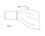
- FIG. 4shows a schematic representation of a single-use accessory device according to one embodiment comprising a flexible tear-away sheath.
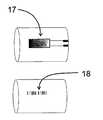
- FIG. 5shows a single-use accessory device according to one embodiment of the invention comprising an electrical element for encoding identification information.
- FIGS. 6A-Cshow schematic representations of accessory devices for optical probes marked with identifying information in the form of a bar code.
- FIG. 6Ashows an accessory device comprising a bar code on the side of the device.
- FIGS. 6B and Cshow an embodiment of the invention in which the bar code is placed on an optical window which forms the end of the device distal to the illumination optics of the optical probe.
- FIG. 6Ashows a view of the end of the device bearing the bar code.
- FIG. 6Cshows a view of the side of the device.
- an accessory devicecomplements the function of an optical probe such accessory devices provide more than merely a sheath for an optical probe.
- An accessory device of the inventioncomprises a number of optional features which a user can select in optimizing the accessory device to suit a particular application. Any or all of these options can be present in an accessory device according to the invention. Because of the many permutations of accessory devices which can be designed according to the invention, the optical probe itself acquires more versatility and can be used in a variety of diagnostic settings. It will be apparent to those of skill in the art after reading this disclosure that other options can additionally be provided, and such options are encompassed within the scope of the invention. All that is required to practice the present invention, is that the accessory device permit optical data collection by an optical probe without obstruction.
- the accessory deviceprovides additional optical features to enhance the transmission of light from the optical probe to the tissue and from the tissue to the optical probe.
- the userselects optical features that are for the accessory device that are compatible with the operation parameters of the optical probe.
- the accessory deviceis fabricated using material which has a high optical transmission over the spectral bandwidth of operation of the probe.
- obtaining an image by the probeis not as important as obtaining a very high signal-to-noise ratio from an optical response in spectral regions that do not overlap, or only partially overlap, the visible region of the spectrum. That is, the inclusion of features to ensure adequate performance of the optics to create a visual image of the sample may degrade the performance of the device in collecting acceptable optical signals such as fluorescence, Raman, or reflectance spectra.
- the portion of the accessory device actually transmitting an ultraviolet (UV) excitation beamcan be made of a very thin Teflon® or can comprise other fluoroplastics such as THV-200P® (a TFE/HPF/VDF terfluoropolymer from the 3M® corporation). These plastics do not demonstrate a significant fluorescent response when irradiated with UV.
- the accessory deviceis used with an optical probe which functions by directing light to a tissue and receiving at least fluorescent light re-emitted from the tissue after absorption of the excitation light, while in other embodiments, the accessory device receives scattered light from a target tissue, such as elastic scattered light (e.g. reflectance spectroscopy) or inelastic scattered light (e.g., as in Raman spectroscopy applications).
- a target tissuesuch as elastic scattered light (e.g. reflectance spectroscopy) or inelastic scattered light (e.g., as in Raman spectroscopy applications).
- the light being directed back to the probeprovides diagnostic information relating to the chemical/structural features of a tissue being analyzed rather than its morphological features.
- An accessory device used in these applicationsis made of materials which provide minimal interference with the light being directed back towards the probe.
- the accessory devicecomprises a low-fluorescing plastic and has high optical transmission through the ultraviolet and visible spectral regions from 300 nm to 750 nm.
- an accessory devicewhich does not fluoresce when illuminated by a laser or other light source and has a sufficiently large aperture or opening to collect low levels of light emitted during fluorescence of some samples such as tissue, good modulation transfer function for good image transmission, and/or a lack of color tint to preserve spectral accuracy.
- the accessory devicecan be fabricated from material including, but not limited to, UVT acrylic or amorphous polyolefin (e.g., Zeonex®, Nippon Zeon CO., Ltd.) and the like. The skilled artisan can recognize and identify equivalent materials using routine experimentation and routine testing.
- optical probeand hence the type of accessory device used, will depend upon the particular diagnostic application required. For example, in diagnosing cervical tissue pathologies, in some instances it is desirable to obtain both imaging and non-imaging optical information. This combination of modalities is important when spatial location of biopsy sites is the output of the optical device.
- an accessory deviceshould be selected which creates minimal interference with the spectroscopic functions of the device, and has good imaging capability to locate specific tissue sites.
- ASCUS TriageAtypical Squamous Cells of Undetermined Significance Triage
- non-imaging informationis more important, because determining the location of the abnormal tissue is not necessary.
- an accessory devicecan be used which is not suited for imaging purposes.
- the optical probecan be used as an adjunct to a standard pap smear test.
- a non-imaging deviceis suitable.
- the optical features of the accessory deviceinclude optical elements which complement the function of the optical probe.
- the accessory deviceincludes a flat window which permits passage of diagnostic light to and from the optical probe without distortion.
- Window materialsinclude, but are not limited to, cast or molded polymethylmetacrylate (PMMA) and other materials which provide no significant fluorescence in response to an excitation beam.
- PMMApolymethylmetacrylate
- polystyrene or polycarbonateare two such materials.
- the placement of the window on the accessory deviceis selected to optimize the collection of light from a tissue being analyzed.
- the window 11is at the end of the accessory body 10 most distal from the probe.
- the windowis provided on the side of the accessory device, giving the opportunity to gather optical information from the side as the device is moved along or through a sample.
- the windowcan be configured in a variety of shapes.
- the accessory devicecomprises a circular window 13 .
- the windowis a transparent section 15 of the accessory device.
- the windowis fastened onto the end of a cylindrical- or toroidal-shaped ring segment that is press-fitted onto the accessory device, forming an annular lens which functions as an objective for the optical probe's illumination elements.
- the wall thickness of the ring segment on which the window/lens sitsis designed to allow the accessory device to act as an optical waveguide to direct light onto target tissues for better visualization or data collection. In one embodiment of the invention, the wall thickness of the ring segment is between about 0.5 mm and 2.0 mm.
- the windowitself can form a lens, or alternatively, a lens can be added to the window as a separate element.
- the windowcan be segmented so that a portion of the structure is flat (i.e., optically passive), while other portions are curved (i.e., forming lens segments).
- a delivery apparatusis operably connected to the window for dispensing a fluid which has an index of refraction matching the window or other exposed optical elements in the accessory device and/or optical probe.
- Delivery devices encompassed within the scope of the inventioninclude a bead or other container residing in a space defined by the ring segment which can be caused to break and discharge its fluid. Fluid from the delivery device spreads downward by capillary force to fill the space between optical elements in the accessory device (e.g., such as the window itself) and the optical probe.
- the windowis coated with an anti-fog agent or an anti-glare agent.
- the accessory deviceis provided with a flexible sleeve which covers the window and serves a protective function.
- the accessory device of the present inventioncan also be adapted to include other optical elements to facilitate the acquisition of diagnostic data, such as filters, polarizers, or light reflecting elements.
- the distal end of the accessory deviceincludes a reflecting element such as an integral faceted mirror.
- the reflecting elementis in the shape of a cone which has a half angle of 45 degrees. A light beam impinging on one of the facets of the reflecting element will be reflected at a 90 degree angle to the incident light causing it to be emitted laterally from the distal end of the accessory device, allowing light to be efficiently directed to the target tissue within the lumen the accessory device is accessing.
- a light-focusing elementcan additionally be provided in optical communication with the reflecting element in order to focus light beams appropriately on the target tissue.
- reflecting elementsare provided within the body of the accessory device in optical communication with a window.
- the reflective elementis a reflective planar surface 14 .
- the reflecting elementis a conical surface which directs light from the optical probe towards the transparent sectional window 15 . It should be apparent to those a skill in the art that a variety of shapes of reflecting surfaces can be provided and positioned to optimize the light path from the optical probe to the window of the accessory device.
- a reflecting elementcan be provided whose number of facets correspond to the number of excitation fibers in the probe, creating an optimal light path between the target tissue and light from the optical probe through the accessory device.
- the accessory devicecan also be configured to attach to the probe in way that further optimizes this light path.
- the accessory deviceis fitted onto the probe via a connecting ring which comprises openings designed to adapt to a particular configuration of optical fibers (e.g., bundled or spaced). Attachment of the probe to the accessory device can only be achieved by correctly aligning optical fibers with appropriate regions in the accessory device.
- the optical probecomprises a plurality of pins which fit into holes in the connecting ring of the accessory device only when the accessory device is positioned in a specific orientation, ensuring the proper orientation of the optical probe with respect to the accessory device.
- the accessory devicecan be adapted to provide a light source for evenly illuminating a tissue being visualized.
- the accessory deviceincludes an illuminating light source positioned around the circumference of the accessory device.
- the illuminating light sourcecan be an integral part of the device or can be snapped on by a ring mechanism.
- the accessory deviceis provided with a dispenser capable of directing a marking fluid toward the sample.
- the fluidcan be applied to localized regions of the sample for identifying selected regions, or it can be dispensed over a broad region of the sample, as a bath or wash.
- the purpose of the bath or washmay be to affect chemical changes in the sample to aid in the identification of substances in or characteristics of the sample. For example, in optical detection of precancerous lesions of the cervix, the application of a mild acetic acid wash increases the contrast and visibility of the regions of suspicious lesions.
- Hyper- or hypo-osmotic solutionscan be generated in a number of ways, such as by using distilled water, either alone, or in combination with ionic or nonionic molecular constituents. Varying the hydrogen ion concentration of a fluid (e.g., pH) can generate additional visualization-enhancing agents. Dye solutions can also be applied such as, for example, Lugol's iodine, toluidine blue or methylene blue, and others.
- the accessory devicecan be designed to conform to a particular lumen being accessed, thus minimizing the invasive effect of the accessory device.
- the accessory devicecomprises a flexible portion which provides a shield between the tissue being assayed and the optical probe while at the same time maximizing patient comfort by adapting itself to any space being accessed by the device.
- the flexible accessory devicecan be in the form of an inflatable balloon into which a fluid (e.g., an index-matching fluid)is inserted to partially inflate the structure.
- Balloonscan be made from compliant materials, such as polyethylene, latex (natural or synthetic), polyurethane, and silicone, or non-compliant materials, such as polyethylene terephthalate (PET).
- the flexible accessory deviceWhen brought into contact with the tissue, the flexible accessory device distributes the contact pressure of the device evenly over the entire contact surface (such as a body lumen), while the index-matching fluid provides good optical communication with the tissue.
- the flexible portionalso conforms to the end of the optical probe bearing illumination optics, shielding the illumination optics of the probe from body fluids, while simultaneously shielding the patient from contamination by the probe.
- the accessory devicecomprises, at least in portion, a shrink-fitted material (e.g., which can be shrunk using heat).
- a heating elementsuch as, but not limited to, a resister
- shrink-fitted materialsuch that shrinkage is triggered when a voltage is applied to the resistor.
- the materialcan be shrunk using a heating device such as a hand-held hairdryer. Because of the flexible nature of the accessory device, it can be packaged in a rolled-up state (e.g., in a sterile wrapper) to be unrolled over the optical probe when it is ready to be used.
- a heating devicesuch as a hand-held hairdryer. Because of the flexible nature of the accessory device, it can be packaged in a rolled-up state (e.g., in a sterile wrapper) to be unrolled over the optical probe when it is ready to be used.
- the accessory devicecomprises both a flexible portion and a rigid tip portion.
- the length and diameter of the tip portionis selected to be optimal for accessing a particular body lumen and to provide for the effective transmission of diagnostic light from the optical probe, while the flexible portion of the accessory device is conformed like a skirt and is proximal to the end of the optical probe bearing illumination optics.
- the flared and flexible nature of the flexible portionminimizes patient discomfort from the entry of any portion of the optical probe itself into the body cavity being accessed.
- the flexible material and the rigid portion of the accessory devicecan be molded as a single unit or can be molded separately and connected together
- the optical probe accessory devicecan also be designed for a particular anatomic application, e.g., for obtaining information relating to tissue features of the gastrointestinal tract, the urinary tract, the peritoneal cavity, the thorax, ear canal, and the female reproductive tract. Other organs suitable for endoscopic or percutaneous access will be apparent to those of ordinary skill in the art.
- the accessory deviceis designed as a probe with a particular geometry adapted for the body region towards which it is directed.
- an accessory deviceis provided for use with an optical probe used in the cervix.
- the accessory devicecovers the sides of the probe that encounters the vaginal walls and additionally covers the end of the optical probe comprising illumination optics.
- the accessory deviceis designed to at least partially cover an optical probe and is capable of passing, with the probe, through a distal aperture of an endoscope.
- the accessory deviceis accordingly limited in sized to conform to the dimensions of the body cavity being accessed and the dimensions of the endoscope.
- the accessory deviceis designed to transmit light from an optical probe to the surface of a tissue which is not accessed through a lumen, for example, the skin, or breast tissue, or tissue within an open surgical field.
- a single-use accessory devicefor at least partially covering an optical probe.
- the accessory deviceentirely covers the probe, while in another embodiment, the accessory device covers or shield those parts of the probe adapted for contact with a body tissue of a patient.
- single-useis understood to mean that the use of the accessory device is restricted to use with a single patient. However, in some embodiments, use can be confined to a single diagnostic measurement.
- the accessory devicecomprises both a body and an attachment element for attaching the accessory device to the probe wherein the device is mechanically prevented from re-use.
- the accessory devicecomprises a breakable element to allow for physical breakage of at least a portion of the device upon removal from the optical probe.
- the attachment elementincludes at least one breakable portion which must be broken in order to remove the accessory device from the probe. Breaking the breakable portion cripples the accessory device, preventing its reattachment and re-use.
- the breakable portionincludes a grasping element, such as a tab or snap ring, and grasping the grasping element results in breaking the body of the accessory device from the attachment element.
- the attachment elementcomprises a flexible material 12 and the accessory device can only be detached from the probe by tearing the flexible material 12 , separating the attachment element portion of the accessory device from the body portion.
- the flexible elementcan comprise a weakened material, or breakpoint, where it joins to the body of the device (e.g., perforations) to facilitate tearing. The breakpoint is more susceptible to mechanical stress than the remaining portions of the device.
- the attachment elementcan be mechanically attached to optical probe by a variety of mechanisms, including, but not limited, to a tab/slot mechanism (such as a tab on the attachment element fits into a slot on the outside of the optical probe or visa versa), a magnetic attachment means, a lock and pin mechanism, a band-latching mechanism, or a string.
- Other types of attachment mechanismssuch as fasteners, elastic bands, strings within the accessory device which can hook onto the probe, Velcro, adhesive, tapes, glues), including those which rely on mating a protruding element (on the accessory device or the probe) to a recessed element (on the probe or the accessory device) will doubtless be apparent to those of skill in the art, and are included within the scope of the invention.
- the actual means of attachment of the attachment elementis the breakable element in the device.
- the attachment elementattaches to the probe by a tab/slot mechanism
- removal of the accessory devicecan only be performed by breaking the tab off, thereby preventing the accessory device from being reattached.
- the protruding mating elementis designed to tear along a tear line, or perforation, in the accessory device upon mechanical stress (e.g., when the protruding element on the attachment element is disengaged from recessed element on the surface of the optical probe), preventing the protruding mating element from functioning in future.
- At least the attachment element of the deviceis made of a flexible material and a “cinch purse” string is provided to both secure the attachment element to the device and to provide a grasping element.
- the stringis attached to a breakable element so that pulling the string breaks the breakable element and permits the flexible portion of the accessory device to be rolled over, away from the optical probe. Once the breakable element is broken, the accessory device is unable to be reattached to the optical probe.
- the attachment elementcan attach directly to the optical probe, it can also attach through an intermediate interfacing element which itself attaches to the probe (e.g., via a ring or a plastic connecting sleeve).
- the attachment element and the body of the accessory deviceare modules which can be fitted together. Different types of interfacing elements can be used to interface different types of attachment elements and bodies to different optical probes, allowing the user to select and combine different desired features of the accessory device with a particular kind of optical probe.
- the accessory deviceis prevented from reuse by degrading the optical quality of the accessory device after use.
- coatings susceptible to ultraviolet radiationcan be placed on the light-transmitting portion of the accessory device. During proper use of the device, the coating is subjected to a sufficient quantity of ultraviolet radiation so that it becomes at least partially opaque, preventing its reuse.
- the inventionalso provides an accessory device which can be disabled after use without physically altering the device, that is, electronically, for example.
- an electrical contact between the accessory and the optical probeis provided.
- an electrical elementis embedded within the accessory device which is capable of making electrical contact with the optical probe when the accessory device is properly affixed to the probe.
- the term “electrical element”encompasses both passive electrical elements (e.g., resistors, capacitors, inductors, diodes, and others) and active electrical elements (e.g., transistors, integrated circuits, such as microchips, and others).
- the optical probedelivers a current to the accessory device sufficient to destroy the electrical element, thus preventing reuse of the accessory device.
- the accessory deviceis provided with an electrical element 17 bearing encoded information.
- the electrical elementcan be secured to the accessory device by insertion at a notch on the surface of the device, or alternatively, can be held in place by a biocompatible adhesive (e.g., a cyanoacrylic adhesive) and can additionally include electrical contact elements for making contact with the probe.
- a biocompatible adhesivee.g., a cyanoacrylic adhesive
- the electrical element 17bears encoded information relating to the identification of the accessory device.
- the encoded informationidentifies the device as one which has already been used with the optical probe.
- the electrical element 17includes encoded information relating a target tissue which is being analyzed. Additional information encoded by the electrical element 17 includes, but is not limited to, time, present date, date of manufacture, materials used in construction, and the condition of the optical probe or the processing system used with the optical probe.
- the electrical element 17can include information regarding the intended use of the optical probe, and can enable only certain modes of operation of the probe. As defined herein, an “operating mode” refers to either, or both, the input or output of the optical probe.
- the operating modeis a functioning or non-functioning state of the optical probe.
- the operating modeis any of a plurality of input or output states of the device.
- the optical probein one operating mode, is directed to provide optical information relating to the location of a sample (e.g., a cancerous tissue) while in another operating mode, the optical probe is directed to provide information relating only to a biochemical feature of a sample (e.g., the presence or absence of fluorescence relating to a cancerous or precancerous state), while in still another operating mode, both types of information are provided.
- the electrical elementcan be a programmable read-only memory chip (PROM).
- PROMprogrammable read-only memory chip
- the electrical elementcan be remotely programmable.
- the electrical elementis an RFID (radiofrequency identification device) or another active semiconductor device.
- Information within the electrical elementcan be passed on to a processor in communication with the optical probe through a electrical element reader which accesses stored information in the electrical element in a non-contacting manner.
- the electrical element readeris capable of receiving electromagnetic signals.
- the electrical element readeris capable of receiving radiosignals from the electrical element.
- the electrical element readerWhen the electrical element reader is placed in a location in which it can access stored identification information encoded in the electrical element, the electrical element reader transfers this information to a processor to which the optical probe is operatively connected.
- the electrical element readercan be either attachable to the optical probe or an integral part of the optical probe itself, such that the reader has access to the electrical element as soon as the accessory device is attached to the optical probe. Information from the electrical element is thus immediately transferred to the processor which provides instructions to the probe to either enable it or prevent it from functioning.
- the “reader”is a transponder for receiving radiosignals from the electrical element.
- the electrical elementcan be re-programmed or programmed with additional information, allowing the optical probe to function with the same accessory device.
- the electrical element readeris configured as an encoding device to conveniently change or add information stored within the electrical element.
- processorincludes a memory which comprises identification information identifying accessory devices that have been used with the optical probe. If a match is found between the identification information obtained by the electrical element reader and the identification information within the memory, the processor transmits instructions to the optical probe which prevents it from functioning. The instructions are then relayed to component(s) of an optical diagnostic system of which the optical probe is a part.
- the optical diagnostic systemcomprises a light source which is in optical communication with the optical probe. The presence of a match between identification information encoded by the electrical element and identification information within the memory of the processor prevents light from being transmitted from the light source to the optical probe.
- the optical diagnostic systemcomprises an optical probe-locking device which prevents the probe from being moved (e.g., to position it within a patient) if a match is found thus effectively preventing the probe from being used with the “wrong” accessory device.
- the identification information relating to the electrical elementis added to the memory. In this way, subsequent use of the accessory device will result in instructions being sent to the probe which prevents it from operating.
- the processorcan transmit instructions to the optical probe which allow it to function if a “correct” accessory device is used with the probe.
- the processortransmits instructions to either the probe itself and/or to other components of the optical diagnostic system when no match is found between identification information encoded in the electrical element and the identification information stored in the memory. The instructions then trigger the optical probe or other component of the optical diagnostic system to function (for example, light can be transmitted through the optical probe or a specific diagnostic application can be run in response to the instructions).
- the electrical elementis encoded with identification information which can only be read if the accessory device is positioned in a correct orientation with respect to the optical probe (for example, in an orientation which maximizes light transmission from the probe to the accessory device).
- the processorwill only transmit instructions to the optical probe to allow the probe to function if the accessory device is positioned correctly.
- Information other than identifying informationcan also be transmitted to the processor via the electrical element.
- information relating to the “readiness” of the optical probe/accessory devicecan be provided to the electrical element by sensors on the accessory device or the optical probe which are responsive to the environment in which the accessory device/and or probe is placed.
- the electrical elementin turn transmits the information to the processor which can alter the functioning of the probe as appropriate.
- the electrical elementcan further include information relating to the target tissue being analyzed.
- information read by the electrical element readertriggers the processor to activate diagnostic programs unique to the analysis of that particular tissue.
- the accessory devicecomprises a electrical element identifying it as an accessory device used to access the cervix.
- the processorwill access specific computer program product(s) (e.g., software applications) relating to the diagnosis of cervical tissue pathologies (e.g., cervical cancer) and will activate particular data input or data display screens that relate to diagnosing these pathologies.
- the electrical elementcan include patient identifying information, including information relating to a history of a particular disease (e.g., whether the patient has a family history of cervical cancer).
- a particular type of accessory deviceis preferred for a particular diagnostic application.
- the electrical element in the accessory deviceis encoded with information indicating that it is suited for a particular use(s). When the processor accesses this information through the electrical element reader, only a proper match between the use and the device will permit the optical probe or other components of the optical system to function.
- an optical probeis provided which is equipped with a light emitting diode and an infrared sensor, while the accessory device is marked with a series of lines on one of its surfaces providing identification information.
- the optical probe sensorobtains information relating to the accessory device's identification information and transfers this information to the processor which sends instructions to the probe or other components of the system to enable or prevent the probe from functioning with that particular accessory device.
- Optical methods for communicating the usage history of the accessory device to the optical probecan also include bar codes.
- a bar code 18 designed to be read by reflectance or fluorescenceis fixed to the body of the accessory device. If it is placed on the side of the accessory device, a separate reader may be needed to scan the code. The lot number, intended use, and other pertinent information is contained in the code and interpreted by the optical scanner.
- the code 19is fixed to a transparent part 20 of the accessory device. This permits the optical system itself to read the contents of the code 19 prior to performing its measurement of the sample (e.g., tissue).
- Other accessory device marker and reader combinationswill be apparent to those of skill in the art, and are encompassed within the scope of the invention.
- an accessory device including optical elementscan also include electrical and/or mechanical elements to disable the devise so that it can only be used a single time.
- Devices with optical elements and/or single-use devicescan include the structural features that make an accessory device minimally invasive and/or tissue-conforming. Any and all of these combinations are encompassed within the scope of the invention.
Landscapes
- Health & Medical Sciences (AREA)
- Life Sciences & Earth Sciences (AREA)
- Surgery (AREA)
- Physics & Mathematics (AREA)
- Pathology (AREA)
- General Health & Medical Sciences (AREA)
- Medical Informatics (AREA)
- Biophysics (AREA)
- Optics & Photonics (AREA)
- Engineering & Computer Science (AREA)
- Biomedical Technology (AREA)
- Heart & Thoracic Surgery (AREA)
- Veterinary Medicine (AREA)
- Molecular Biology (AREA)
- Animal Behavior & Ethology (AREA)
- Public Health (AREA)
- Radiology & Medical Imaging (AREA)
- Nuclear Medicine, Radiotherapy & Molecular Imaging (AREA)
- Chemical & Material Sciences (AREA)
- Analytical Chemistry (AREA)
- Biochemistry (AREA)
- General Physics & Mathematics (AREA)
- Immunology (AREA)
- Investigating, Analyzing Materials By Fluorescence Or Luminescence (AREA)
- Endoscopes (AREA)
Abstract
Description
Claims (38)
Priority Applications (2)
| Application Number | Priority Date | Filing Date | Title |
|---|---|---|---|
| US09/591,706US6847490B1 (en) | 1997-01-13 | 2000-06-09 | Optical probe accessory device for use in vivo diagnostic procedures |
| US11/018,665US20050159646A1 (en) | 1997-01-13 | 2004-12-21 | Optical probe accessory device for use in in vivo diagnostic procedures |
Applications Claiming Priority (6)
| Application Number | Priority Date | Filing Date | Title |
|---|---|---|---|
| US08/782,936US6104945A (en) | 1995-08-01 | 1997-01-13 | Spectral volume microprobe arrays |
| US11537399P | 1999-01-11 | 1999-01-11 | |
| US09/241,806US6411835B1 (en) | 1997-01-13 | 1999-02-02 | Spectral volume microprobe arrays |
| US13823599P | 1999-06-09 | 1999-06-09 | |
| US09/481,762US6826422B1 (en) | 1997-01-13 | 2000-01-11 | Spectral volume microprobe arrays |
| US09/591,706US6847490B1 (en) | 1997-01-13 | 2000-06-09 | Optical probe accessory device for use in vivo diagnostic procedures |
Related Parent Applications (1)
| Application Number | Title | Priority Date | Filing Date |
|---|---|---|---|
| US09/481,762Continuation-In-PartUS6826422B1 (en) | 1997-01-13 | 2000-01-11 | Spectral volume microprobe arrays |
Related Child Applications (1)
| Application Number | Title | Priority Date | Filing Date |
|---|---|---|---|
| US11/018,665DivisionUS20050159646A1 (en) | 1997-01-13 | 2004-12-21 | Optical probe accessory device for use in in vivo diagnostic procedures |
Publications (1)
| Publication Number | Publication Date |
|---|---|
| US6847490B1true US6847490B1 (en) | 2005-01-25 |
Family
ID=34069421
Family Applications (2)
| Application Number | Title | Priority Date | Filing Date |
|---|---|---|---|
| US09/591,706Expired - Fee RelatedUS6847490B1 (en) | 1997-01-13 | 2000-06-09 | Optical probe accessory device for use in vivo diagnostic procedures |
| US11/018,665AbandonedUS20050159646A1 (en) | 1997-01-13 | 2004-12-21 | Optical probe accessory device for use in in vivo diagnostic procedures |
Family Applications After (1)
| Application Number | Title | Priority Date | Filing Date |
|---|---|---|---|
| US11/018,665AbandonedUS20050159646A1 (en) | 1997-01-13 | 2004-12-21 | Optical probe accessory device for use in in vivo diagnostic procedures |
Country Status (1)
| Country | Link |
|---|---|
| US (2) | US6847490B1 (en) |
Cited By (65)
| Publication number | Priority date | Publication date | Assignee | Title |
|---|---|---|---|---|
| US20020127735A1 (en)* | 1999-12-15 | 2002-09-12 | Howard Kaufman | Methods of monitoring effects of chemical agents on a sample |
| US20020133073A1 (en)* | 1998-12-23 | 2002-09-19 | Nordstrom Robert J. | Spectroscopic system employing a plurality of data types |
| US20020177777A1 (en)* | 1998-12-23 | 2002-11-28 | Medispectra, Inc. | Optical methods and systems for rapid screening of the cervix |
| US20030095721A1 (en)* | 1999-12-15 | 2003-05-22 | Thomas Clune | Methods and systems for correcting image misalignment |
| US20030144585A1 (en)* | 1999-12-15 | 2003-07-31 | Howard Kaufman | Image processing using measures of similarity |
| US20040010375A1 (en)* | 2002-07-09 | 2004-01-15 | Medispectra, Inc. | Methods and apparatus for processing spectral data for use in tissue characterization |
| US20040073120A1 (en)* | 2002-04-05 | 2004-04-15 | Massachusetts Institute Of Technology | Systems and methods for spectroscopy of biological tissue |
| US20040207625A1 (en)* | 2003-04-18 | 2004-10-21 | Medispectra, Inc. | Methods and apparatus for displaying diagnostic data |
| US20040206913A1 (en)* | 2003-04-18 | 2004-10-21 | Medispectra, Inc. | Methods and apparatus for characterization of tissue samples |
| US20040206882A1 (en)* | 2003-04-18 | 2004-10-21 | Medispectra, Inc. | Methods and apparatus for evaluating image focus |
| US20040209237A1 (en)* | 2003-04-18 | 2004-10-21 | Medispectra, Inc. | Methods and apparatus for characterization of tissue samples |
| US20040208390A1 (en)* | 2003-04-18 | 2004-10-21 | Medispectra, Inc. | Methods and apparatus for processing image data for use in tissue characterization |
| US20040206914A1 (en)* | 2003-04-18 | 2004-10-21 | Medispectra, Inc. | Methods and apparatus for calibrating spectral data |
| US20040214156A1 (en)* | 2002-07-09 | 2004-10-28 | Medispectra, Inc. | Method and apparatus for identifying spectral artifacts |
| US20050043929A1 (en)* | 2000-12-15 | 2005-02-24 | Medispectra, Inc. | System for normalizing spectra |
| US20050043635A1 (en)* | 2002-07-10 | 2005-02-24 | Medispectra, Inc. | Fluorescent fiberoptic probe for tissue health discrimination and method of use thereof |
| US20050159646A1 (en)* | 1997-01-13 | 2005-07-21 | Medispectra, Inc. | Optical probe accessory device for use in in vivo diagnostic procedures |
| US20050234526A1 (en)* | 2004-04-14 | 2005-10-20 | Gilhuly Terence J | Systems and methods for detection of disease including oral scopes and ambient light management systems (ALMS) |
| US20060055914A1 (en)* | 2004-09-13 | 2006-03-16 | Wojciechowski Joel C | Method and apparatus for determining a vertical intensity profile through a plane of focus in a confocal microscope |
| US20060089629A1 (en)* | 2002-07-25 | 2006-04-27 | Howe Christopher A | Laser system |
| US20060241347A1 (en)* | 2004-10-12 | 2006-10-26 | Peter Whitehead | Systems and methods relating to colposcopic viewing tubes for enhanced viewing and examination |
| US20060293644A1 (en)* | 2005-06-21 | 2006-12-28 | Donald Umstadter | System and methods for laser-generated ionizing radiation |
| US20070083111A1 (en)* | 2005-10-12 | 2007-04-12 | Volcano Corporation | Apparatus and method for use of RFID catheter intelligence |
| US20070167686A1 (en)* | 2003-04-29 | 2007-07-19 | Mcgrath Matthew J | Laryngoscope with camera attachement |
| US20070239083A1 (en)* | 2006-01-18 | 2007-10-11 | Axel Voss | Shock wave generators |
| US20070279621A1 (en)* | 2004-09-13 | 2007-12-06 | Wojciechowski Joel C | Method and apparatus for determining a vertical intensity profile along an illuminating beam |
| US20080045799A1 (en)* | 2006-04-10 | 2008-02-21 | Peter Whitehead | Multipurpose diseased tissue detection devices, systems, and methods |
| US20080076967A1 (en)* | 2004-05-18 | 2008-03-27 | Scimed Life Systems, Inc. | Serialization of single use endoscopes |
| US20080228038A1 (en)* | 2005-04-01 | 2008-09-18 | Welch Allyn, Inc. | Illumination Assembly For Use With Vaginal Speculum Apparatus |
| US20080319510A1 (en)* | 2007-06-19 | 2008-12-25 | Simpson Fred A | Medical Device Access Control Apparatus and Method |
| US20090144022A1 (en)* | 2007-12-03 | 2009-06-04 | Smiths Detection Inc. | Mixed statistical and numerical model for sensor array detection and classification |
| US20090187108A1 (en)* | 2006-09-29 | 2009-07-23 | Cornova, Inc. | Systems and methods for analysis and treatment of a body lumen |
| US20090234228A1 (en)* | 2008-03-17 | 2009-09-17 | Or-Nim Medical Ltd. | Apparatus for non-invasive optical monitoring |
| US20090236541A1 (en)* | 2008-03-24 | 2009-09-24 | General Electric Company | System and Methods for Optical Imaging |
| US20090259220A1 (en)* | 2008-04-09 | 2009-10-15 | Angiodynamics, Inc. | Treatment Devices and Methods |
| US20090287192A1 (en)* | 2005-04-01 | 2009-11-19 | Vivenzio Robert L | Medical diagnostic instrument having portable illuminator |
| US20100049182A1 (en)* | 2006-12-22 | 2010-02-25 | Cornova, Inc. | Fluid media for bio-sensitive applications |
| US20110004447A1 (en)* | 2009-07-01 | 2011-01-06 | Schlumberger Technology Corporation | Method to build 3D digital models of porous media using transmitted laser scanning confocal mircoscopy and multi-point statistics |
| US20110004448A1 (en)* | 2009-07-01 | 2011-01-06 | Schlumberger Technology Corporation | Method to quantify discrete pore shapes, volumes, and surface areas using confocal profilometry |
| US20110037876A1 (en)* | 2009-08-13 | 2011-02-17 | Olive Medical Corp. | System, apparatus and methods for providing a single use imaging device for sterile environments |
| US7914442B1 (en) | 1999-03-01 | 2011-03-29 | Gazdzinski Robert F | Endoscopic smart probe and method |
| US20110137306A1 (en)* | 2009-12-07 | 2011-06-09 | Tyco Healthcare Group Lp | Removable Ink for Surgical Instrument |
| US20110238977A1 (en)* | 2010-03-25 | 2011-09-29 | Olive Medical Corporation | System and method for providing a single use imaging device for medical applications |
| US8068897B1 (en) | 1999-03-01 | 2011-11-29 | Gazdzinski Robert F | Endoscopic smart probe and method |
| US20120281218A1 (en)* | 2005-01-24 | 2012-11-08 | The Board Of Trustees Of The Leland Stanford Junior University | Optical analysis system and approach therefor |
| US8317681B1 (en)* | 1999-03-01 | 2012-11-27 | Gazdzinski Robert F | Endoscopic smart probe and method |
| US20140024931A1 (en)* | 2012-07-20 | 2014-01-23 | Lightlab Imaging, Inc. | Data Encoders for Medical Devices and Related Methods |
| US20140031629A1 (en)* | 2012-07-24 | 2014-01-30 | Olympus Medical Systems Corp. | Medical apparatus, disposable medical device, and medical system |
| US8640944B1 (en) | 2003-12-17 | 2014-02-04 | West View Research, Llc | Portable computerized wireless payment apparatus and methods |
| US8676587B1 (en) | 1999-06-10 | 2014-03-18 | West View Research, Llc | Computerized information and display apparatus and methods |
| US8725477B2 (en) | 2008-04-10 | 2014-05-13 | Schlumberger Technology Corporation | Method to generate numerical pseudocores using borehole images, digital rock samples, and multi-point statistics |
| US8789243B2 (en)* | 2012-09-19 | 2014-07-29 | Ecolab Usa Inc. | Disposable handle cover for a surgical lighthead |
| WO2014114290A1 (en)* | 2013-01-25 | 2014-07-31 | Nawa Heilmittel Gmbh | Hand-held measuring device |
| US8812368B1 (en) | 1999-03-01 | 2014-08-19 | West View Research, Llc | Computerized information collection and processing apparatus |
| US20150119639A1 (en)* | 2012-07-12 | 2015-04-30 | Olympus Corporation | Medical system |
| US9433468B2 (en) | 2013-10-04 | 2016-09-06 | Tidi Products, Llc | Sheath for a medical or dental instrument |
| US9581723B2 (en) | 2008-04-10 | 2017-02-28 | Schlumberger Technology Corporation | Method for characterizing a geological formation traversed by a borehole |
| US9913573B2 (en) | 2003-04-01 | 2018-03-13 | Boston Scientific Scimed, Inc. | Endoscopic imaging system |
| US10238363B2 (en) | 2014-08-21 | 2019-03-26 | Richard D. Striano | Needle guide for ultrasound transducer |
| US10436716B2 (en)* | 2014-09-24 | 2019-10-08 | Smiths Detection, Inc. | Ubiquitous transmissive raman spectroscopy for stand-off detection |
| US20200345216A1 (en)* | 2017-05-17 | 2020-11-05 | Auris Health, Inc. | Exchangeable working channel |
| US20210121049A1 (en)* | 2019-10-24 | 2021-04-29 | Align Technology, Inc. | Window self-detection |
| US20220304559A1 (en)* | 2021-03-24 | 2022-09-29 | PacificMD Biotech, LLC | Endoscope and endoscope sheath with diagnostic and therapeutic interfaces |
| US11583177B2 (en)* | 2017-08-06 | 2023-02-21 | Biop—Medical Ltd. | Optical probe for cervical examination |
| US12251186B2 (en)* | 2019-07-29 | 2025-03-18 | Align Technology, Inc. | Full-scanner barrier for an intra-oral device |
Families Citing this family (44)
| Publication number | Priority date | Publication date | Assignee | Title |
|---|---|---|---|---|
| IL132944A (en)* | 1999-11-15 | 2009-05-04 | Arkady Glukhovsky | Method for activating an image collecting process |
| WO2004049947A2 (en)* | 2002-11-29 | 2004-06-17 | Given Imaging Ltd. | Methods device and system for in vivo diagnosis |
| JP4787004B2 (en)* | 2005-11-15 | 2011-10-05 | Hoya株式会社 | Endoscope light source device |
| CN101453942B (en)* | 2006-05-30 | 2012-01-11 | 皇家飞利浦电子股份有限公司 | Apparatus for depth-resolved measurements of properties of tissue |
| JP5314841B2 (en)* | 2006-08-22 | 2013-10-16 | オリンパス株式会社 | Endoscope device and endoscope probe |
| US8068890B2 (en) | 2006-09-29 | 2011-11-29 | Nellcor Puritan Bennett Llc | Pulse oximetry sensor switchover |
| JP4986646B2 (en)* | 2007-02-05 | 2012-07-25 | オリンパス株式会社 | Endoscope device |
| US8862194B2 (en) | 2008-06-30 | 2014-10-14 | Covidien Lp | Method for improved oxygen saturation estimation in the presence of noise |
| US9254123B2 (en) | 2009-04-29 | 2016-02-09 | Hansen Medical, Inc. | Flexible and steerable elongate instruments with shape control and support elements |
| US7931149B2 (en)* | 2009-05-27 | 2011-04-26 | Given Imaging Ltd. | System for storing and activating an in vivo imaging capsule |
| US9113844B2 (en)* | 2009-12-01 | 2015-08-25 | David S. Hollstien | Non-invasive implant rupture detection system |
| US10426320B2 (en)* | 2010-04-28 | 2019-10-01 | Xiaolong OuYang | Single use medical devices |
| US20120071752A1 (en) | 2010-09-17 | 2012-03-22 | Sewell Christopher M | User interface and method for operating a robotic medical system |
| DE102010061133A1 (en)* | 2010-12-09 | 2012-06-14 | G & H Medical Gmbh & Co. Kg | Protective cover for a laparoscope |
| US20130030363A1 (en) | 2011-07-29 | 2013-01-31 | Hansen Medical, Inc. | Systems and methods utilizing shape sensing fibers |
| US10080576B2 (en) | 2013-03-08 | 2018-09-25 | Auris Health, Inc. | Method, apparatus, and a system for facilitating bending of an instrument in a surgical or medical robotic environment |
| US10149720B2 (en) | 2013-03-08 | 2018-12-11 | Auris Health, Inc. | Method, apparatus, and a system for facilitating bending of an instrument in a surgical or medical robotic environment |
| US10376672B2 (en) | 2013-03-15 | 2019-08-13 | Auris Health, Inc. | Catheter insertion system and method of fabrication |
| WO2014144307A2 (en)* | 2013-03-15 | 2014-09-18 | Gebhart Steven C | Probe assembly and disposable cover particularly for use in endoscope applications of low coherence interferometry |
| EP3060157B1 (en) | 2013-10-24 | 2019-12-11 | Auris Health, Inc. | System for robotic-assisted endolumenal surgery |
| TWI547256B (en)* | 2014-02-19 | 2016-09-01 | 群曜醫電股份有限公司 | Alimentary tract survey device with portable endocope and control method for the same |
| US9561083B2 (en) | 2014-07-01 | 2017-02-07 | Auris Surgical Robotics, Inc. | Articulating flexible endoscopic tool with roll capabilities |
| US10792464B2 (en) | 2014-07-01 | 2020-10-06 | Auris Health, Inc. | Tool and method for using surgical endoscope with spiral lumens |
| US9744335B2 (en) | 2014-07-01 | 2017-08-29 | Auris Surgical Robotics, Inc. | Apparatuses and methods for monitoring tendons of steerable catheters |
| US10869592B2 (en) | 2015-02-23 | 2020-12-22 | Uroviu Corp. | Handheld surgical endoscope |
| WO2016137838A1 (en) | 2015-02-23 | 2016-09-01 | Xiaolong Ouyang | Handheld surgical endoscope |
| US11819636B2 (en) | 2015-03-30 | 2023-11-21 | Auris Health, Inc. | Endoscope pull wire electrical circuit |
| US10463439B2 (en) | 2016-08-26 | 2019-11-05 | Auris Health, Inc. | Steerable catheter with shaft load distributions |
| US11684248B2 (en) | 2017-09-25 | 2023-06-27 | Micronvision Corp. | Endoscopy/stereo colposcopy medical instrument |
| US11832797B2 (en) | 2016-09-25 | 2023-12-05 | Micronvision Corp. | Endoscopic fluorescence imaging |
| US12268358B2 (en) | 2019-12-05 | 2025-04-08 | Uroviu Corp. | Portable endoscope with side-mountable disposable portion |
| US11980342B2 (en) | 2020-11-12 | 2024-05-14 | Micronvision Corp. | Minimally invasive endoscope |
| US11771304B1 (en) | 2020-11-12 | 2023-10-03 | Micronvision Corp. | Minimally invasive endoscope |
| DE102017126408A1 (en)* | 2017-11-10 | 2019-05-16 | Böllhoff Verbindungstechnik GmbH | Hygiene cap |
| CN117017505A (en) | 2018-03-28 | 2023-11-10 | 奥瑞斯健康公司 | Composite instrument and robotic system |
| DE102018110082A1 (en)* | 2018-04-26 | 2019-10-31 | avateramedical GmBH | Sterile endoscope cover |
| WO2020033318A1 (en) | 2018-08-07 | 2020-02-13 | Auris Health, Inc. | Combining strain-based shape sensing with catheter control |
| CN112804933B (en) | 2018-09-26 | 2024-10-18 | 奥瑞斯健康公司 | Articulating medical device |
| WO2020139973A1 (en) | 2018-12-28 | 2020-07-02 | Auris Health, Inc. | Medical instrument with articulable segment |
| US11617627B2 (en) | 2019-03-29 | 2023-04-04 | Auris Health, Inc. | Systems and methods for optical strain sensing in medical instruments |
| EP4003138A4 (en) | 2019-07-25 | 2023-08-30 | Uroviu Corp. | DISPOSABLE ENDOSCOPY NEEDLE WITH INTEGRATED GRIPPER |
| WO2021028883A1 (en) | 2019-08-15 | 2021-02-18 | Auris Health, Inc. | Medical device having multiple bending sections |
| CN114901188A (en) | 2019-12-31 | 2022-08-12 | 奥瑞斯健康公司 | Dynamic pulley system |
| DE102020132454B3 (en)* | 2020-12-07 | 2021-11-25 | Karl Storz Se & Co. Kg | Endoscopic device and method for checking the identity of a component of an endoscopic device |
Citations (241)
| Publication number | Priority date | Publication date | Assignee | Title |
|---|---|---|---|---|
| US3013467A (en) | 1957-11-07 | 1961-12-19 | Minsky Marvin | Microscopy apparatus |
| US3632865A (en) | 1969-12-23 | 1972-01-04 | Bell Telephone Labor Inc | Predictive video encoding using measured subject velocity |
| US3809072A (en) | 1971-10-07 | 1974-05-07 | Med General Inc | Sterile sheath apparatus for fiber optic illuminator with compatible lens |
| US3890462A (en) | 1974-04-17 | 1975-06-17 | Bell Telephone Labor Inc | Speed and direction indicator for video systems |
| US3963019A (en) | 1974-11-25 | 1976-06-15 | Quandt Robert S | Ocular testing method and apparatus |
| US4017192A (en) | 1975-02-06 | 1977-04-12 | Neotec Corporation | Optical analysis of biomedical specimens |
| US4071020A (en) | 1976-06-03 | 1978-01-31 | Xienta, Inc. | Apparatus and methods for performing in-vivo measurements of enzyme activity |
| US4198571A (en) | 1977-04-28 | 1980-04-15 | National Research Development Corporation | Scanning microscopes |
| US4218703A (en) | 1979-03-16 | 1980-08-19 | Bell Telephone Laboratories, Incorporated | Technique for estimation of displacement and/or velocity of objects in video scenes |
| US4254421A (en) | 1979-12-05 | 1981-03-03 | Communications Satellite Corporation | Integrated confocal electromagnetic wave lens and feed antenna system |
| US4273110A (en) | 1978-07-13 | 1981-06-16 | Jean Groux | Ultraviolet endoscope |
| US4357075A (en) | 1979-07-02 | 1982-11-02 | Hunter Thomas M | Confocal reflector system |
| US4397557A (en) | 1979-12-20 | 1983-08-09 | Heimann Gmbh | Optical arrangement for a light scattering type smoke detector |
| EP0135134A2 (en) | 1983-08-11 | 1985-03-27 | Vitacomm Ltd | Esophageal stethoscope and vital signs monitor system |
| US4515165A (en) | 1980-02-04 | 1985-05-07 | Energy Conversion Devices, Inc. | Apparatus and method for detecting tumors |
| US4549229A (en) | 1982-02-01 | 1985-10-22 | Sony Corporation | Method and apparatus for compensating for tape jitter during recording and reproducing of a video signal and PCM audio signal |
| US4558462A (en) | 1982-09-02 | 1985-12-10 | Hitachi Medical Corporation | Apparatus for correcting image distortions automatically by inter-image processing |
| SU1223092A1 (en) | 1984-03-11 | 1986-04-07 | Предприятие П/Я Р-6681 | Small-angle nephelometer |
| US4641352A (en) | 1984-07-12 | 1987-02-03 | Paul Fenster | Misregistration correction |
| US4646722A (en) | 1984-12-10 | 1987-03-03 | Opielab, Inc. | Protective endoscope sheath and method of installing same |
| US4662360A (en) | 1984-10-23 | 1987-05-05 | Intelligent Medical Systems, Inc. | Disposable speculum |
| US4733063A (en) | 1985-12-13 | 1988-03-22 | Hitachi, Ltd. | Scanning laser microscope with aperture alignment |
| US4741326A (en) | 1986-10-01 | 1988-05-03 | Fujinon, Inc. | Endoscope disposable sheath |
| US4753530A (en) | 1980-08-21 | 1988-06-28 | Oriel Scientific Ltd. | Analytical optical instruments |
| EP0280418A1 (en) | 1987-02-02 | 1988-08-31 | Wyatt Technology Corporation | Method and apparatus for examining the interior of semi-opaque objects |
| US4768513A (en) | 1986-04-21 | 1988-09-06 | Agency Of Industrial Science And Technology | Method and device for measuring and processing light |
| US4800571A (en) | 1988-01-11 | 1989-01-24 | Tektronix, Inc. | Timing jitter measurement display |
| US4845352A (en) | 1986-12-08 | 1989-07-04 | U.S. Philips Corporation | Scanning differential phase contrast microscope |
| US4844617A (en) | 1988-01-20 | 1989-07-04 | Tencor Instruments | Confocal measuring microscope with automatic focusing |
| US4852955A (en) | 1986-09-16 | 1989-08-01 | Laser Precision Corporation | Microscope for use in modular FTIR spectrometer system |
| EP0335725A2 (en) | 1988-04-01 | 1989-10-04 | Syntex (U.S.A.) Inc. | Apparatus and method for detection of fluorescence or light scatter |
| US4877033A (en) | 1988-05-04 | 1989-10-31 | Seitz Jr H Michael | Disposable needle guide and examination sheath for transvaginal ultrasound procedures |
| US4878485A (en) | 1989-02-03 | 1989-11-07 | Adair Edwin Lloyd | Rigid video endoscope with heat sterilizable sheath |
| US4891829A (en) | 1986-11-19 | 1990-01-02 | Exxon Research And Engineering Company | Method and apparatus for utilizing an electro-optic detector in a microtomography system |
| US4930516A (en) | 1985-11-13 | 1990-06-05 | Alfano Robert R | Method for detecting cancerous tissue using visible native luminescence |
| US4945478A (en) | 1987-11-06 | 1990-07-31 | Center For Innovative Technology | Noninvasive medical imaging system and method for the identification and 3-D display of atherosclerosis and the like |
| US4965441A (en) | 1988-01-27 | 1990-10-23 | Commissariat A L'energie Atomique | Method for the scanning confocal light-optical microscopic and indepth examination of an extended field and devices for implementing said method |
| US4972258A (en) | 1989-07-31 | 1990-11-20 | E. I. Du Pont De Nemours And Company | Scanning laser microscope system and methods of use |
| US4974580A (en) | 1988-06-25 | 1990-12-04 | Effner Gmbh | Endoscope protective member |
| US4979498A (en) | 1989-10-30 | 1990-12-25 | Machida Incorporated | Video cervicoscope system |
| US4997242A (en) | 1988-03-07 | 1991-03-05 | Medical Research Council | Achromatic scanning system |
| US5003979A (en) | 1989-02-21 | 1991-04-02 | University Of Virginia | System and method for the noninvasive identification and display of breast lesions and the like |
| US5011243A (en) | 1986-09-16 | 1991-04-30 | Laser Precision Corporation | Reflectance infrared microscope having high radiation throughput |
| US5022757A (en) | 1989-01-23 | 1991-06-11 | Modell Mark D | Heterodyne system and method for sensing a target substance |
| US5028802A (en) | 1990-01-11 | 1991-07-02 | Eye Research Institute Of Retina Foundation | Imaging apparatus and methods utilizing scannable microlaser source |
| US5032720A (en) | 1988-04-21 | 1991-07-16 | White John G | Confocal imaging system |
| US5034613A (en) | 1989-11-14 | 1991-07-23 | Cornell Research Foundation, Inc. | Two-photon laser microscopy |
| US5036853A (en) | 1988-08-26 | 1991-08-06 | Polartechnics Ltd. | Physiological probe |
| US5042494A (en) | 1985-11-13 | 1991-08-27 | Alfano Robert R | Method and apparatus for detecting cancerous tissue using luminescence excitation spectra |
| EP0444689A2 (en) | 1990-03-01 | 1991-09-04 | X-Rite, Inc. | A compensation method adapted for use in color measuring apparatus |
| US5048946A (en) | 1990-05-15 | 1991-09-17 | Phoenix Laser Systems, Inc. | Spectral division of reflected light in complex optical diagnostic and therapeutic systems |
| US5054926A (en) | 1987-03-24 | 1991-10-08 | Commonwealth Scientific And Industrial Research Organisation | Distance measuring device |
| US5065008A (en) | 1989-10-18 | 1991-11-12 | Fuji Photo Film Co., Ltd. | Scanning microscope and scanning mechanism for the same |
| US5071246A (en) | 1989-10-27 | 1991-12-10 | Carl-Zeiss-Stiftung | Confocal scanning ophthalmoscope |
| US5074306A (en) | 1990-02-22 | 1991-12-24 | The General Hospital Corporation | Measurement of burn depth in skin |
| US5083220A (en) | 1990-03-22 | 1992-01-21 | Tandem Scanning Corporation | Scanning disks for use in tandem scanning reflected light microscopes and other optical systems |
| US5091652A (en) | 1990-01-12 | 1992-02-25 | The Regents Of The University Of California | Laser excited confocal microscope fluorescence scanner and method |
| US5101825A (en) | 1988-10-28 | 1992-04-07 | Blackbox, Inc. | Method for noninvasive intermittent and/or continuous hemoglobin, arterial oxygen content, and hematocrit determination |
| US5120953A (en) | 1988-07-13 | 1992-06-09 | Harris Martin R | Scanning confocal microscope including a single fibre for transmitting light to and receiving light from an object |
| US5122653A (en) | 1989-08-22 | 1992-06-16 | Nikon Corporation | Confocal type laser scan microscope with integrated illumination, detection and waveguide system |
| US5132526A (en) | 1990-04-10 | 1992-07-21 | Fuji Photo Film Co., Ltd. | Confocal scanning microscope having a signal output regulating means |
| US5139025A (en) | 1983-10-14 | 1992-08-18 | Somanetics Corporation | Method and apparatus for in vivo optical spectroscopic examination |
| US5154166A (en) | 1990-02-01 | 1992-10-13 | Machida Endoscope Co., Ltd. | Endoscope cover |
| US5159919A (en) | 1990-02-01 | 1992-11-03 | Machida Endoscope Co., Ltd. | Endoscope cover |
| US5161053A (en) | 1988-08-01 | 1992-11-03 | Commonwealth Scientific & Industrial Research | Confocal microscope |
| US5162941A (en) | 1991-07-23 | 1992-11-10 | The Board Of Governors Of Wayne State University | Confocal microscope |
| US5162641A (en) | 1991-02-19 | 1992-11-10 | Phoenix Laser Systems, Inc. | System and method for detecting, correcting and measuring depth movement of target tissue in a laser surgical system |
| WO1992019148A1 (en) | 1991-05-06 | 1992-11-12 | Adair Edwin Lloyd | Cervical videoscope with detachable camera unit |
| US5168157A (en) | 1990-11-20 | 1992-12-01 | Fuji Photo Film Co., Ltd. | Scanning microscope with means for detecting a first and second polarized light beams along first and second optical receiving paths |
| US5192980A (en) | 1990-06-27 | 1993-03-09 | A. E. Dixon | Apparatus and method for method for spatially- and spectrally-resolved measurements |
| US5193525A (en) | 1990-11-30 | 1993-03-16 | Vision Sciences | Antiglare tip in a sheath for an endoscope |
| US5199431A (en) | 1985-03-22 | 1993-04-06 | Massachusetts Institute Of Technology | Optical needle for spectroscopic diagnosis |
| USRE34214E (en) | 1984-03-15 | 1993-04-06 | Molecular Dynamics, Inc. | Method and apparatus for microphotometering microscope specimens |
| US5201908A (en) | 1991-06-10 | 1993-04-13 | Endomedical Technologies, Inc. | Sheath for protecting endoscope from contamination |
| US5201318A (en) | 1989-04-24 | 1993-04-13 | Rava Richard P | Contour mapping of spectral diagnostics |
| US5203328A (en) | 1991-07-17 | 1993-04-20 | Georgia Tech Research Corporation | Apparatus and methods for quantitatively measuring molecular changes in the ocular lens |
| EP0474264A3 (en) | 1991-04-24 | 1993-06-16 | Kaman Aerospace Corporation | Spectrally dispersive imaging lidar method and apparatus |
| US5225671A (en) | 1991-05-29 | 1993-07-06 | Olympus Optical Co., Ltd. | Confocal optical apparatus |
| WO1993014688A1 (en) | 1992-01-24 | 1993-08-05 | Frantz Medical Development, Ltd. | Endoscope sterile liquid supply system |
| US5235457A (en) | 1987-09-24 | 1993-08-10 | Washington University | Kit for converting a standard microscope into a single aperture confocal scanning epi-illumination microscope |
| US5239178A (en) | 1990-11-10 | 1993-08-24 | Carl Zeiss | Optical device with an illuminating grid and detector grid arranged confocally to an object |
| US5237984A (en) | 1991-06-24 | 1993-08-24 | Xomed-Treace Inc. | Sheath for endoscope |
| US5248876A (en) | 1992-04-21 | 1993-09-28 | International Business Machines Corporation | Tandem linear scanning confocal imaging system with focal volumes at different heights |
| US5253071A (en) | 1991-12-20 | 1993-10-12 | Sony Corporation Of America | Method and apparatus for stabilizing an image produced in a video camera |
| US5257617A (en) | 1989-12-25 | 1993-11-02 | Asahi Kogaku Kogyo Kabushiki Kaisha | Sheathed endoscope and sheath therefor |
| US5260569A (en) | 1991-07-25 | 1993-11-09 | Fuji Photo Film Co., Ltd. | Scanning microscope and scanning mechanism |
| US5260578A (en) | 1991-04-10 | 1993-11-09 | Mayo Foundation For Medical Education And Research | Confocal imaging system for visible and ultraviolet light |
| US5262646A (en) | 1990-07-27 | 1993-11-16 | Booker Graham R | Infra-red scanning microscopy |
| US5261410A (en) | 1991-02-07 | 1993-11-16 | Alfano Robert R | Method for determining if a tissue is a malignant tumor tissue, a benign tumor tissue, or a normal or benign tissue using Raman spectroscopy |
| US5274240A (en) | 1990-01-12 | 1993-12-28 | The Regents Of The University Of California | Capillary array confocal fluorescence scanner and method |
| US5284149A (en) | 1992-01-23 | 1994-02-08 | Dhadwal Harbans S | Method and apparatus for determining the physical characteristics of ocular tissue |
| US5289274A (en) | 1991-02-06 | 1994-02-22 | Sony Corporation | Electronic image stabilization apparatus |
| US5294799A (en) | 1993-02-01 | 1994-03-15 | Aslund Nils R D | Apparatus for quantitative imaging of multiple fluorophores |
| US5296700A (en) | 1991-09-12 | 1994-03-22 | Nikon Corporation | Fluorescent confocal microscope with chromatic aberration compensation |
| US5303026A (en) | 1991-02-26 | 1994-04-12 | The Regents Of The University Of California Los Alamos National Laboratory | Apparatus and method for spectroscopic analysis of scattering media |
| US5306902A (en) | 1992-09-01 | 1994-04-26 | International Business Machines Corporation | Confocal method and apparatus for focusing in projection lithography |
| US5313567A (en) | 1991-06-13 | 1994-05-17 | At&T Bell Laboratories | Arrangement for determining and displaying volumetric data in an imaging system |
| US5319200A (en) | 1991-06-05 | 1994-06-07 | Zeltex, Inc. | Rapid near-infrared measurement of nonhomogeneous samples |
| US5321501A (en) | 1991-04-29 | 1994-06-14 | Massachusetts Institute Of Technology | Method and apparatus for optical imaging with means for controlling the longitudinal range of the sample |
| US5324979A (en) | 1990-09-26 | 1994-06-28 | Futrex, Inc. | Method and means for generating synthetic spectra allowing quantitative measurement in near infrared measuring instruments |
| US5325846A (en) | 1992-07-27 | 1994-07-05 | Linvatec Corporation | Endoscopic draping apparatus and method |
| US5329352A (en) | 1991-04-12 | 1994-07-12 | Bayer Aktiengesellschaft | Spectroscopically correlated light scanning microscopy |
| US5337734A (en) | 1992-10-29 | 1994-08-16 | Advanced Polymers, Incorporated | Disposable sheath with optically transparent window formed continuously integral therewith |
| US5343038A (en) | 1991-12-12 | 1994-08-30 | Matsushita Electric Industrial Co., Ltd. | Scanning laser microscope with photo coupling and detecting unit |
| US5345306A (en) | 1990-05-22 | 1994-09-06 | Research Development Corporation Of Japan | Method and apparatus for measuring spectral absorption in an opaque specimen and method and apparatus for measuring the microscopic absorption distribution |
| WO1994026168A1 (en) | 1993-05-12 | 1994-11-24 | Board Of Regents, The University Of Texas System | Diagnosis of dysplasia using laser induced fluorescence |
| WO1995000067A1 (en) | 1993-06-18 | 1995-01-05 | Infra-Temp, Inc. | Electronic thermometer probe cover |
| WO1995004385A1 (en) | 1993-07-27 | 1995-02-09 | Microsulis Limited | Treatment apparatus |
| US5398685A (en) | 1992-01-10 | 1995-03-21 | Wilk; Peter J. | Endoscopic diagnostic system and associated method |
| US5402768A (en) | 1992-09-01 | 1995-04-04 | Adair; Edwin L. | Endoscope with reusable core and disposable sheath with passageways |
| US5406939A (en) | 1994-02-14 | 1995-04-18 | Bala; Harry | Endoscope sheath |
| US5413108A (en) | 1993-04-21 | 1995-05-09 | The Research Foundation Of City College Of New York | Method and apparatus for mapping a tissue sample for and distinguishing different regions thereof based on luminescence measurements of cancer-indicative native fluorophor |
| US5415157A (en) | 1993-02-05 | 1995-05-16 | Welcome; Steven | Damage preventing endoscope head cover |
| US5418797A (en) | 1993-01-15 | 1995-05-23 | The United States Of America As Represented By The Secretary Of The Navy | Time gated imaging through scattering material using polarization and stimulated raman amplification |
| US5419323A (en) | 1988-12-21 | 1995-05-30 | Massachusetts Institute Of Technology | Method for laser induced fluorescence of tissue |
| US5419311A (en) | 1993-02-18 | 1995-05-30 | Olympus Optical Co., Ltd. | Endoscope apparatus of a type having cover for covering the endoscope |
| US5421337A (en) | 1989-04-14 | 1995-06-06 | Massachusetts Institute Of Technology | Spectral diagnosis of diseased tissue |
| US5424543A (en) | 1993-04-19 | 1995-06-13 | Surface Optics Corporation | Imaging spectroradiometer |
| US5450857A (en) | 1994-05-19 | 1995-09-19 | Board Of Regents, The University Of Texas System | Method for the diagnosis of cervical changes |
| US5451931A (en) | 1992-09-14 | 1995-09-19 | Cerberus Ag | Optical smoke detector |
| US5458133A (en) | 1993-03-15 | 1995-10-17 | Olympus Optical Co., Ltd. | Cover type endoscope apparatus |
| US5458132A (en) | 1993-03-15 | 1995-10-17 | Olympus Optical Co., Ltd. | Endoscope cover-sheathed endoscope system |
| US5467767A (en) | 1991-11-25 | 1995-11-21 | Alfano; Robert R. | Method for determining if tissue is malignant as opposed to non-malignant using time-resolved fluorescence spectroscopy |
| US5469853A (en) | 1992-12-11 | 1995-11-28 | Tetrad Corporation | Bendable ultrasonic probe and sheath for use therewith |
| US5477382A (en) | 1994-08-05 | 1995-12-19 | Northrop Grumman Corporation | Optical correlator system |
| EP0689045A1 (en) | 1994-06-23 | 1995-12-27 | Koninklijke Philips Electronics N.V. | Multi-frequency modulation spectrometer |
| US5480775A (en) | 1990-01-26 | 1996-01-02 | Canon Kabushiki Kaisha | Method for measuring a specimen by the use of fluorescent light |
| US5493444A (en) | 1994-04-28 | 1996-02-20 | The United States Of America As Represented By The Secretary Of The Air Force | Photorefractive two-beam coupling nonlinear joint transform correlator |
| US5496259A (en) | 1993-09-13 | 1996-03-05 | Welch Allyn, Inc. | Sterile protective sheath and drape for video laparoscope and method of use |
| US5507295A (en) | 1992-07-01 | 1996-04-16 | British Technology Group Limited | Medical devices |
| US5516010A (en) | 1984-10-23 | 1996-05-14 | Sherwood Medical Company | Sanitary speculum for tympanic thermometer probe |
| US5519545A (en) | 1993-11-30 | 1996-05-21 | Sony Corporation | Digital signal recording circuit using a rotary transformer with a reduced-jitter low-frequency compensation circuit |
| US5529235A (en) | 1994-04-28 | 1996-06-25 | Ethicon Endo-Surgery, Inc. | Identification device for surgical instrument |
| US5536236A (en) | 1993-02-12 | 1996-07-16 | Olympus Optical Co., Ltd. | Covered endoscope system |
| US5545121A (en) | 1993-02-02 | 1996-08-13 | Olympus Optical Co., Ltd. | Cover-type endoscope apparatus |
| US5551945A (en) | 1993-03-16 | 1996-09-03 | Olympus Optical Co., Ltd. | Endoscope system including endoscope and protection cover |
| US5556367A (en) | 1993-03-05 | 1996-09-17 | Olympus Optical Co., Ltd. | Cover type endoscope apparatus |
| EP0737849A2 (en) | 1995-04-12 | 1996-10-16 | Kyoto Daiichi Kagaku Co., Ltd. | Method of stabilizing spectra in spectometry |
| JPH08280602A (en) | 1995-04-14 | 1996-10-29 | Olympus Optical Co Ltd | Cover type endoscope |
| US5579773A (en) | 1994-09-30 | 1996-12-03 | Martin Marietta Energy Systems, Inc. | Laser-induced differential normalized fluorescence method for cancer diagnosis |
| US5587832A (en) | 1993-10-20 | 1996-12-24 | Biophysica Technologies, Inc. | Spatially light modulated confocal microscope and method |
| US5596992A (en) | 1993-06-30 | 1997-01-28 | Sandia Corporation | Multivariate classification of infrared spectra of cell and tissue samples |
| US5599717A (en) | 1994-09-02 | 1997-02-04 | Martin Marietta Energy Systems, Inc. | Advanced synchronous luminescence system |
| WO1997005473A1 (en) | 1995-08-01 | 1997-02-13 | Medispectra, Inc. | Optical microprobes and methods for spectral analysis of materials |
| US5609560A (en) | 1992-08-19 | 1997-03-11 | Olympus Optical Co., Ltd. | Medical operation device control system for controlling a operation devices accessed respectively by ID codes |
| US5612540A (en) | 1995-03-31 | 1997-03-18 | Board Of Regents, The University Of Texas Systems | Optical method for the detection of cervical neoplasias using fluorescence spectroscopy |
| US5643175A (en) | 1992-09-01 | 1997-07-01 | Adair; Edwin L. | Sterilizable endoscope with separable disposable tube assembly |
| US5647368A (en) | 1996-02-28 | 1997-07-15 | Xillix Technologies Corp. | Imaging system for detecting diseased tissue using native fluorsecence in the gastrointestinal and respiratory tract |
| US5662588A (en) | 1994-05-31 | 1997-09-02 | Olympus Optical Co., Ltd. | Endoscope apparatus |
| US5685822A (en) | 1996-08-08 | 1997-11-11 | Vision-Sciences, Inc. | Endoscope with sheath retaining device |
| US5690106A (en) | 1995-06-30 | 1997-11-25 | Siemens Corporate Research, Inc. | Flexible image registration for rotational angiography |
| US5693043A (en) | 1985-03-22 | 1997-12-02 | Massachusetts Institute Of Technology | Catheter for laser angiosurgery |
| US5695448A (en) | 1994-08-29 | 1997-12-09 | Olympus Optical Co., Ltd. | Endoscopic sheath |
| US5697373A (en) | 1995-03-14 | 1997-12-16 | Board Of Regents, The University Of Texas System | Optical method and apparatus for the diagnosis of cervical precancers using raman and fluorescence spectroscopies |
| US5699795A (en) | 1995-03-31 | 1997-12-23 | Board Of Regents, The University Of Texas System | Optical probe for the detection of cervical neoplasia using fluorescence spectroscopy and apparatus incorporating same |
| WO1997048331A1 (en) | 1996-06-19 | 1997-12-24 | Board Of Regents, The University Of Texas System | Method and apparatus for diagnosing squamous intraepithelial lesions of the cervix using fluorescence spectroscopy |
| US5704892A (en) | 1992-09-01 | 1998-01-06 | Adair; Edwin L. | Endoscope with reusable core and disposable sheath with passageways |
| US5713364A (en) | 1995-08-01 | 1998-02-03 | Medispectra, Inc. | Spectral volume microprobe analysis of materials |
| US5717209A (en) | 1996-04-29 | 1998-02-10 | Petrometrix Ltd. | System for remote transmission of spectral information through communication optical fibers for real-time on-line hydrocarbons process analysis by near infra red spectroscopy |
| WO1998005253A1 (en) | 1996-08-02 | 1998-02-12 | The Board Of Regents, The University Of Texas System | Method and apparatus for the characterization of tissue of epithelial lined viscus |
| US5720293A (en)* | 1991-01-29 | 1998-02-24 | Baxter International Inc. | Diagnostic catheter with memory |
| US5730701A (en) | 1995-09-12 | 1998-03-24 | Olympus Optical Co., Ltd. | Endoscope |
| US5733244A (en) | 1995-03-13 | 1998-03-31 | Asahi Kogaku Kogyo Kabushiki Kaisha | Distal end part of endoscope |
| US5735276A (en) | 1995-03-21 | 1998-04-07 | Lemelson; Jerome | Method and apparatus for scanning and evaluating matter |
| US5746695A (en) | 1993-11-18 | 1998-05-05 | Asahi Kogaku Kogyo Kabushiki Kaisha | Front end structure of endoscope |
| WO1998024369A1 (en) | 1996-12-02 | 1998-06-11 | The University Of Texas System | Spectroscopic detection of cervical pre-cancer using radial basis function networks |
| US5768333A (en) | 1996-12-02 | 1998-06-16 | Philips Electronics N.A. Corporation | Mass detection in digital radiologic images using a two stage classifier |
| US5769792A (en) | 1991-07-03 | 1998-06-23 | Xillix Technologies Corp. | Endoscopic imaging system for diseased tissue |
| US5773835A (en) | 1996-06-07 | 1998-06-30 | Rare Earth Medical, Inc. | Fiber optic spectroscopy |
| WO1998030889A1 (en) | 1997-01-13 | 1998-07-16 | Medispectra, Inc. | Spatially resolved optical measurements |
| US5791346A (en) | 1996-08-22 | 1998-08-11 | Western Research Company, Inc. | Colposcope device and method for measuring areas of cervical lesions |
| US5795632A (en) | 1996-02-06 | 1998-08-18 | Parker Laboratories | Protective cover set for a medical probe |
| US5800350A (en) | 1993-11-01 | 1998-09-01 | Polartechnics, Limited | Apparatus for tissue type recognition |
| US5807248A (en) | 1996-05-15 | 1998-09-15 | Ohmeda Inc. | Medical monitoring probe with modular device housing |
| WO1998041176A1 (en) | 1997-03-17 | 1998-09-24 | Polartechnics Limited | Sheath for a side view probe |
| US5813987A (en) | 1995-08-01 | 1998-09-29 | Medispectra, Inc. | Spectral volume microprobe for analysis of materials |
| US5817015A (en) | 1993-06-22 | 1998-10-06 | Adair; Edwin L. | Endoscope with reusable core and disposable sheath with passageways |
| US5830146A (en) | 1997-03-17 | 1998-11-03 | Polartechnics Limited | Sheathed probes for tissue type recognition |
| US5833617A (en) | 1996-03-06 | 1998-11-10 | Fuji Photo Film Co., Ltd. | Fluorescence detecting apparatus |
| US5840035A (en) | 1995-02-07 | 1998-11-24 | Siemens Aktiengesellschaft | Method for the spectroscopic examination of a biological tissue |
| US5842995A (en) | 1996-06-28 | 1998-12-01 | Board Of Regents, The Univerisity Of Texas System | Spectroscopic probe for in vivo measurement of raman signals |
| US5855551A (en) | 1997-03-17 | 1999-01-05 | Polartechnics Limited | Integral sheathing apparatus for tissue recognition probes |
| US5860913A (en) | 1996-05-16 | 1999-01-19 | Olympus Optical Co., Ltd. | Endoscope whose distal cover can be freely detachably attached to main distal part thereof with high positioning precision |
| US5863287A (en) | 1995-10-04 | 1999-01-26 | Fuji Photo Optical Co., Ltd. | Removable protector sheath for use with endoscopic insertion instrument |
| US5865726A (en) | 1996-03-27 | 1999-02-02 | Asahi Kogaku Kogyo Kabushiki Kaisha | Front end structure of side-view type endoscope |
| WO1999018847A1 (en) | 1997-10-15 | 1999-04-22 | Accumed International, Inc. | Imaging diseased tissue using autofluorescence |
| WO1999020313A1 (en) | 1997-10-20 | 1999-04-29 | The Board Of Regents, The University Of Texas System | Acetic acid as a contrast agent in reflectance confocal imaging of tissue |
| US5920399A (en) | 1997-03-18 | 1999-07-06 | Sandia Corporation | Multispectral imaging method and apparatus |
| US5921926A (en) | 1997-07-28 | 1999-07-13 | University Of Central Florida | Three dimensional optical imaging colposcopy |
| US5929985A (en) | 1997-03-18 | 1999-07-27 | Sandia Corporation | Multispectral imaging probe |
| US5931779A (en) | 1996-06-06 | 1999-08-03 | Wisconsin Alumni Research Foundation | Real-time in-vivo measurement of myoglobin oxygen saturation |
| WO1999047041A1 (en) | 1998-03-19 | 1999-09-23 | Board Of Regents, The University Of Texas System | Fiber-optic confocal imaging apparatus and methods of use |
| US5983125A (en) | 1993-12-13 | 1999-11-09 | The Research Foundation Of City College Of New York | Method and apparatus for in vivo examination of subcutaneous tissues inside an organ of a body using optical spectroscopy |
| WO1999057507A1 (en) | 1998-05-01 | 1999-11-11 | Board Of Regents, The University Of Texas System | Method and apparatus for subsurface imaging |
| US5987343A (en)* | 1997-11-07 | 1999-11-16 | Datascope Investment Corp. | Method for storing pulse oximetry sensor characteristics |
| US5991653A (en) | 1995-03-14 | 1999-11-23 | Board Of Regents, The University Of Texas System | Near-infrared raman spectroscopy for in vitro and in vivo detection of cervical precancers |
| US5989184A (en) | 1997-04-04 | 1999-11-23 | Medtech Research Corporation | Apparatus and method for digital photography useful in cervical cancer detection |
| US5995645A (en) | 1993-08-18 | 1999-11-30 | Applied Spectral Imaging Ltd. | Method of cancer cell detection |
| US6021344A (en) | 1996-12-04 | 2000-02-01 | Derma Technologies, Inc. | Fluorescence scope system for dermatologic diagnosis |
| WO1999057529A9 (en) | 1998-05-04 | 2000-03-02 | Univ Texas | Combined fluorescence and reflectance spectroscopy |
| WO2000015101A1 (en) | 1998-09-11 | 2000-03-23 | Spectrx, Inc. | Multi-modal optical tissue diagnostic system |
| US6058322A (en) | 1997-07-25 | 2000-05-02 | Arch Development Corporation | Methods for improving the accuracy in differential diagnosis on radiologic examinations |
| US6069689A (en) | 1997-04-16 | 2000-05-30 | Derma Technologies, Inc. | Apparatus and methods relating to optical systems for diagnosis of skin diseases |
| US6083487A (en) | 1997-08-25 | 2000-07-04 | Advanced Photodynamic Technologies, Inc. | Methylene blue and toluidene blue mediated fluorescence diagnosis of cancer |
| US6091985A (en) | 1998-01-23 | 2000-07-18 | Research Foundation Of City College Of New York | Detection of cancer and precancerous conditions in tissues and/or cells using native fluorescence excitation spectroscopy |
| WO2000041615A1 (en) | 1999-01-11 | 2000-07-20 | Medispectra, Inc. | Spectral volume microprobe arrays |
| US6096065A (en) | 1997-09-29 | 2000-08-01 | Boston Scientific Corporation | Sheath for tissue spectroscopy |
| US6099464A (en) | 1995-04-10 | 2000-08-08 | Olympus Optical Co., Ltd. | Bending sheath for probe |
| US6119031A (en) | 1996-11-21 | 2000-09-12 | Boston Scientific Corporation | Miniature spectrometer |
| US6124597A (en) | 1997-07-07 | 2000-09-26 | Cedars-Sinai Medical Center | Method and devices for laser induced fluorescence attenuation spectroscopy |
| WO2000057361A1 (en) | 1999-03-19 | 2000-09-28 | Isis Innovation Limited | Method and apparatus for image processing |
| US6146897A (en) | 1995-11-13 | 2000-11-14 | Bio-Rad Laboratories | Method for the detection of cellular abnormalities using Fourier transform infrared spectroscopy |
| US6169817B1 (en) | 1998-11-04 | 2001-01-02 | University Of Rochester | System and method for 4D reconstruction and visualization |
| US6208887B1 (en) | 1999-06-24 | 2001-03-27 | Richard H. Clarke | Catheter-delivered low resolution Raman scattering analyzing system for detecting lesions |
| US6243601B1 (en) | 1998-09-18 | 2001-06-05 | Abund Ottokar Wist | Transillumination imaging instrumentation with scattered light discrimination |
| US6246471B1 (en) | 1998-06-08 | 2001-06-12 | Lj Laboratories, Llc | Apparatus and method for measuring optical characteristics of an object |
| US6246479B1 (en) | 1998-06-08 | 2001-06-12 | Lj Laboratories, L.L.C. | Integrated spectrometer assembly and methods |
| WO2000059366A3 (en) | 1999-04-07 | 2001-06-28 | Blue Lake Products Inc | Identification of protective covers for medical imaging devices |
| US6285639B1 (en) | 1998-04-29 | 2001-09-04 | Sony Corporation | On-the-fly of jitter during optical disc mastering/recording |
| US6312385B1 (en) | 2000-05-01 | 2001-11-06 | Ge Medical Systems Global Technology Company, Llc | Method and apparatus for automatic detection and sizing of cystic objects |
| US6317617B1 (en) | 1997-07-25 | 2001-11-13 | Arch Development Corporation | Method, computer program product, and system for the automated analysis of lesions in magnetic resonance, mammogram and ultrasound images |
| US20010041843A1 (en) | 1999-02-02 | 2001-11-15 | Mark Modell | Spectral volume microprobe arrays |
| US20020007123A1 (en) | 2000-03-28 | 2002-01-17 | Constantinos Balas | Method and system for characterization and mapping of tissue lesions |
| USD453832S1 (en) | 2001-02-09 | 2002-02-19 | Medispectra, Inc. | Sheath for cervical optical probe |
| USD453963S1 (en) | 2001-02-09 | 2002-02-26 | Medispectra, Inc. | Sheath for cervical optical probe |
| USD453962S1 (en) | 2001-02-09 | 2002-02-26 | Medispectra, Inc. | Sheath for cervical optical probe |
| USD453964S1 (en) | 2001-02-09 | 2002-02-26 | Medispectra, Inc. | Sheath for cervical optical probe |
| US6377842B1 (en) | 1998-09-22 | 2002-04-23 | Aurora Optics, Inc. | Method for quantitative measurement of fluorescent and phosphorescent drugs within tissue utilizing a fiber optic probe |
| US6385484B2 (en) | 1998-12-23 | 2002-05-07 | Medispectra, Inc. | Spectroscopic system employing a plurality of data types |
| USD460821S1 (en) | 2001-02-09 | 2002-07-23 | Medispectra, Inc. | Sheath for cervical optical probe |
| US6427082B1 (en) | 1998-12-23 | 2002-07-30 | Medispectra, Incorporated | Optical methods and systems for rapid screening of the cervix |
| US20020107668A1 (en) | 2000-12-15 | 2002-08-08 | Costa Peter J. | System for normalizing spectra |
| EP0641542B1 (en) | 1993-09-03 | 2002-11-27 | Ken Ishihara | Non-invasive blood analyzer |
| US6497659B1 (en)* | 1999-04-09 | 2002-12-24 | Spacelabs Medical, Inc. | System for identifying a cable transmitting a signal from a sensor to an electronic instrument |
| US20020197728A1 (en) | 1999-12-15 | 2002-12-26 | Howard Kaufman | Methods of monitoring effects of chemical agents on a sample |
| US6574502B2 (en) | 1999-12-02 | 2003-06-03 | Fuji Photo Film Co., Ltd. | Apparatus for displaying fluorescence images |
| US20040010195A1 (en) | 2002-07-10 | 2004-01-15 | Zelenchuk Alex R. | Fluorescent fiberoptic probe for tissue health discrimination and method of use thereof |
| US20040007674A1 (en) | 2002-07-09 | 2004-01-15 | Schomacker Kevin T. | Method and apparatus for identifying spectral artifacts |
| US20040010187A1 (en) | 2002-07-10 | 2004-01-15 | Schomacker Kevin T. | Colonic polyp discrimination by tissue fluorescence and fiberoptic probe |
| WO2004005895A1 (en) | 2002-07-09 | 2004-01-15 | Medispectra, Inc. | Methods and apparatus for characterization of tissue samples |
| WO2004005885A3 (en) | 2002-07-09 | 2004-03-25 | Medispectra Inc | Optimal windows for obtaining optical data for characterization of tissue samples |
Family Cites Families (15)
| Publication number | Priority date | Publication date | Assignee | Title |
|---|---|---|---|---|
| US4803049A (en)* | 1984-12-12 | 1989-02-07 | The Regents Of The University Of California | pH-sensitive optrode |
| HU9300005D0 (en)* | 1991-05-24 | 1993-04-28 | British Broadcasting Corp | Method for processing video picture |
| US5383874A (en)* | 1991-11-08 | 1995-01-24 | Ep Technologies, Inc. | Systems for identifying catheters and monitoring their use |
| US5400267A (en)* | 1992-12-08 | 1995-03-21 | Hemostatix Corporation | Local in-device memory feature for electrically powered medical equipment |
| US5412563A (en)* | 1993-09-16 | 1995-05-02 | General Electric Company | Gradient image segmentation method |
| US5740808A (en)* | 1996-10-28 | 1998-04-21 | Ep Technologies, Inc | Systems and methods for guilding diagnostic or therapeutic devices in interior tissue regions |
| JP3732865B2 (en)* | 1995-01-18 | 2006-01-11 | ペンタックス株式会社 | Endoscope device |
| US5894340A (en)* | 1995-02-17 | 1999-04-13 | The Regents Of The University Of California | Method for quantifying optical properties of the human lens |
| US5732401A (en)* | 1996-03-29 | 1998-03-24 | Intellitecs International Ltd. | Activity based cost tracking systems |
| AU6184196A (en)* | 1996-06-26 | 1998-01-14 | Morphometrix Technologies Inc. | Confocal ultrasonic imaging system |
| US6847490B1 (en)* | 1997-01-13 | 2005-01-25 | Medispectra, Inc. | Optical probe accessory device for use in vivo diagnostic procedures |
| FR2763721B1 (en)* | 1997-05-21 | 1999-08-06 | Inst Nat Rech Inf Automat | ELECTRONIC IMAGE PROCESSING DEVICE FOR DETECTING DIMENSIONAL VARIATIONS |
| US6697666B1 (en)* | 1999-06-22 | 2004-02-24 | Board Of Regents, The University Of Texas System | Apparatus for the characterization of tissue of epithelial lined viscus |
| US6193510B1 (en)* | 1999-07-28 | 2001-02-27 | Efraim Tsimerman | Medical device with time-out feature |
| US7282723B2 (en)* | 2002-07-09 | 2007-10-16 | Medispectra, Inc. | Methods and apparatus for processing spectral data for use in tissue characterization |
- 2000
- 2000-06-09USUS09/591,706patent/US6847490B1/ennot_activeExpired - Fee Related
- 2004
- 2004-12-21USUS11/018,665patent/US20050159646A1/ennot_activeAbandoned
Patent Citations (265)
| Publication number | Priority date | Publication date | Assignee | Title |
|---|---|---|---|---|
| US3013467A (en) | 1957-11-07 | 1961-12-19 | Minsky Marvin | Microscopy apparatus |
| US3632865A (en) | 1969-12-23 | 1972-01-04 | Bell Telephone Labor Inc | Predictive video encoding using measured subject velocity |
| US3809072A (en) | 1971-10-07 | 1974-05-07 | Med General Inc | Sterile sheath apparatus for fiber optic illuminator with compatible lens |
| US3890462A (en) | 1974-04-17 | 1975-06-17 | Bell Telephone Labor Inc | Speed and direction indicator for video systems |
| US3963019A (en) | 1974-11-25 | 1976-06-15 | Quandt Robert S | Ocular testing method and apparatus |
| US4017192A (en) | 1975-02-06 | 1977-04-12 | Neotec Corporation | Optical analysis of biomedical specimens |
| US4071020A (en) | 1976-06-03 | 1978-01-31 | Xienta, Inc. | Apparatus and methods for performing in-vivo measurements of enzyme activity |
| US4198571A (en) | 1977-04-28 | 1980-04-15 | National Research Development Corporation | Scanning microscopes |
| US4273110A (en) | 1978-07-13 | 1981-06-16 | Jean Groux | Ultraviolet endoscope |
| US4218703A (en) | 1979-03-16 | 1980-08-19 | Bell Telephone Laboratories, Incorporated | Technique for estimation of displacement and/or velocity of objects in video scenes |
| US4357075A (en) | 1979-07-02 | 1982-11-02 | Hunter Thomas M | Confocal reflector system |
| US4254421A (en) | 1979-12-05 | 1981-03-03 | Communications Satellite Corporation | Integrated confocal electromagnetic wave lens and feed antenna system |
| US4397557A (en) | 1979-12-20 | 1983-08-09 | Heimann Gmbh | Optical arrangement for a light scattering type smoke detector |
| US4515165A (en) | 1980-02-04 | 1985-05-07 | Energy Conversion Devices, Inc. | Apparatus and method for detecting tumors |
| US4753530A (en) | 1980-08-21 | 1988-06-28 | Oriel Scientific Ltd. | Analytical optical instruments |
| US4549229A (en) | 1982-02-01 | 1985-10-22 | Sony Corporation | Method and apparatus for compensating for tape jitter during recording and reproducing of a video signal and PCM audio signal |
| US4558462A (en) | 1982-09-02 | 1985-12-10 | Hitachi Medical Corporation | Apparatus for correcting image distortions automatically by inter-image processing |
| EP0135134A2 (en) | 1983-08-11 | 1985-03-27 | Vitacomm Ltd | Esophageal stethoscope and vital signs monitor system |
| US5349961A (en) | 1983-10-14 | 1994-09-27 | Somanetics Corporation | Method and apparatus for in vivo optical spectroscopic examination |
| US5139025A (en) | 1983-10-14 | 1992-08-18 | Somanetics Corporation | Method and apparatus for in vivo optical spectroscopic examination |
| SU1223092A1 (en) | 1984-03-11 | 1986-04-07 | Предприятие П/Я Р-6681 | Small-angle nephelometer |
| USRE34214E (en) | 1984-03-15 | 1993-04-06 | Molecular Dynamics, Inc. | Method and apparatus for microphotometering microscope specimens |
| US4641352A (en) | 1984-07-12 | 1987-02-03 | Paul Fenster | Misregistration correction |
| US5516010A (en) | 1984-10-23 | 1996-05-14 | Sherwood Medical Company | Sanitary speculum for tympanic thermometer probe |
| US5707343A (en) | 1984-10-23 | 1998-01-13 | O'hara; Gary J. | Disposable sanitary speculum for timpanic thermometer probe |
| US4662360A (en) | 1984-10-23 | 1987-05-05 | Intelligent Medical Systems, Inc. | Disposable speculum |
| US4646722A (en) | 1984-12-10 | 1987-03-03 | Opielab, Inc. | Protective endoscope sheath and method of installing same |
| US5199431A (en) | 1985-03-22 | 1993-04-06 | Massachusetts Institute Of Technology | Optical needle for spectroscopic diagnosis |
| US5693043A (en) | 1985-03-22 | 1997-12-02 | Massachusetts Institute Of Technology | Catheter for laser angiosurgery |
| US4930516B1 (en) | 1985-11-13 | 1998-08-04 | Laser Diagnostic Instr Inc | Method for detecting cancerous tissue using visible native luminescence |
| US5042494A (en) | 1985-11-13 | 1991-08-27 | Alfano Robert R | Method and apparatus for detecting cancerous tissue using luminescence excitation spectra |
| US4930516A (en) | 1985-11-13 | 1990-06-05 | Alfano Robert R | Method for detecting cancerous tissue using visible native luminescence |
| US4733063A (en) | 1985-12-13 | 1988-03-22 | Hitachi, Ltd. | Scanning laser microscope with aperture alignment |
| US4768513A (en) | 1986-04-21 | 1988-09-06 | Agency Of Industrial Science And Technology | Method and device for measuring and processing light |
| US4852955A (en) | 1986-09-16 | 1989-08-01 | Laser Precision Corporation | Microscope for use in modular FTIR spectrometer system |
| US5011243A (en) | 1986-09-16 | 1991-04-30 | Laser Precision Corporation | Reflectance infrared microscope having high radiation throughput |
| US4741326A (en) | 1986-10-01 | 1988-05-03 | Fujinon, Inc. | Endoscope disposable sheath |
| US4891829A (en) | 1986-11-19 | 1990-01-02 | Exxon Research And Engineering Company | Method and apparatus for utilizing an electro-optic detector in a microtomography system |
| US4845352A (en) | 1986-12-08 | 1989-07-04 | U.S. Philips Corporation | Scanning differential phase contrast microscope |
| EP0280418A1 (en) | 1987-02-02 | 1988-08-31 | Wyatt Technology Corporation | Method and apparatus for examining the interior of semi-opaque objects |
| US5054926A (en) | 1987-03-24 | 1991-10-08 | Commonwealth Scientific And Industrial Research Organisation | Distance measuring device |
| US5235457A (en) | 1987-09-24 | 1993-08-10 | Washington University | Kit for converting a standard microscope into a single aperture confocal scanning epi-illumination microscope |
| US4945478A (en) | 1987-11-06 | 1990-07-31 | Center For Innovative Technology | Noninvasive medical imaging system and method for the identification and 3-D display of atherosclerosis and the like |
| US4800571A (en) | 1988-01-11 | 1989-01-24 | Tektronix, Inc. | Timing jitter measurement display |
| US4844617A (en) | 1988-01-20 | 1989-07-04 | Tencor Instruments | Confocal measuring microscope with automatic focusing |
| US4965441A (en) | 1988-01-27 | 1990-10-23 | Commissariat A L'energie Atomique | Method for the scanning confocal light-optical microscopic and indepth examination of an extended field and devices for implementing said method |
| US4997242A (en) | 1988-03-07 | 1991-03-05 | Medical Research Council | Achromatic scanning system |
| EP0335725A2 (en) | 1988-04-01 | 1989-10-04 | Syntex (U.S.A.) Inc. | Apparatus and method for detection of fluorescence or light scatter |
| US5032720A (en) | 1988-04-21 | 1991-07-16 | White John G | Confocal imaging system |
| US4877033A (en) | 1988-05-04 | 1989-10-31 | Seitz Jr H Michael | Disposable needle guide and examination sheath for transvaginal ultrasound procedures |
| US4974580A (en) | 1988-06-25 | 1990-12-04 | Effner Gmbh | Endoscope protective member |
| US5120953A (en) | 1988-07-13 | 1992-06-09 | Harris Martin R | Scanning confocal microscope including a single fibre for transmitting light to and receiving light from an object |
| US5161053A (en) | 1988-08-01 | 1992-11-03 | Commonwealth Scientific & Industrial Research | Confocal microscope |
| US5036853A (en) | 1988-08-26 | 1991-08-06 | Polartechnics Ltd. | Physiological probe |
| US5101825A (en) | 1988-10-28 | 1992-04-07 | Blackbox, Inc. | Method for noninvasive intermittent and/or continuous hemoglobin, arterial oxygen content, and hematocrit determination |
| US5562100A (en) | 1988-12-21 | 1996-10-08 | Massachusetts Institute Of Technology | Method for laser induced fluorescence of tissue |
| US5419323A (en) | 1988-12-21 | 1995-05-30 | Massachusetts Institute Of Technology | Method for laser induced fluorescence of tissue |
| US5022757A (en) | 1989-01-23 | 1991-06-11 | Modell Mark D | Heterodyne system and method for sensing a target substance |
| US4878485A (en) | 1989-02-03 | 1989-11-07 | Adair Edwin Lloyd | Rigid video endoscope with heat sterilizable sheath |
| US5003979A (en) | 1989-02-21 | 1991-04-02 | University Of Virginia | System and method for the noninvasive identification and display of breast lesions and the like |
| US5421337A (en) | 1989-04-14 | 1995-06-06 | Massachusetts Institute Of Technology | Spectral diagnosis of diseased tissue |
| US5201318A (en) | 1989-04-24 | 1993-04-13 | Rava Richard P | Contour mapping of spectral diagnostics |
| US5345941A (en) | 1989-04-24 | 1994-09-13 | Massachusetts Institute Of Technology | Contour mapping of spectral diagnostics |
| US4972258A (en) | 1989-07-31 | 1990-11-20 | E. I. Du Pont De Nemours And Company | Scanning laser microscope system and methods of use |
| US5122653A (en) | 1989-08-22 | 1992-06-16 | Nikon Corporation | Confocal type laser scan microscope with integrated illumination, detection and waveguide system |
| US5065008A (en) | 1989-10-18 | 1991-11-12 | Fuji Photo Film Co., Ltd. | Scanning microscope and scanning mechanism for the same |
| US5071246A (en) | 1989-10-27 | 1991-12-10 | Carl-Zeiss-Stiftung | Confocal scanning ophthalmoscope |
| US4979498A (en) | 1989-10-30 | 1990-12-25 | Machida Incorporated | Video cervicoscope system |
| US5034613A (en) | 1989-11-14 | 1991-07-23 | Cornell Research Foundation, Inc. | Two-photon laser microscopy |
| US5257617A (en) | 1989-12-25 | 1993-11-02 | Asahi Kogaku Kogyo Kabushiki Kaisha | Sheathed endoscope and sheath therefor |
| US5028802A (en) | 1990-01-11 | 1991-07-02 | Eye Research Institute Of Retina Foundation | Imaging apparatus and methods utilizing scannable microlaser source |
| US5274240A (en) | 1990-01-12 | 1993-12-28 | The Regents Of The University Of California | Capillary array confocal fluorescence scanner and method |
| US5091652A (en) | 1990-01-12 | 1992-02-25 | The Regents Of The University Of California | Laser excited confocal microscope fluorescence scanner and method |
| US5480775A (en) | 1990-01-26 | 1996-01-02 | Canon Kabushiki Kaisha | Method for measuring a specimen by the use of fluorescent light |
| US5159919A (en) | 1990-02-01 | 1992-11-03 | Machida Endoscope Co., Ltd. | Endoscope cover |
| US5154166A (en) | 1990-02-01 | 1992-10-13 | Machida Endoscope Co., Ltd. | Endoscope cover |
| US5074306A (en) | 1990-02-22 | 1991-12-24 | The General Hospital Corporation | Measurement of burn depth in skin |
| EP0444689A2 (en) | 1990-03-01 | 1991-09-04 | X-Rite, Inc. | A compensation method adapted for use in color measuring apparatus |
| US5083220A (en) | 1990-03-22 | 1992-01-21 | Tandem Scanning Corporation | Scanning disks for use in tandem scanning reflected light microscopes and other optical systems |
| US5132526A (en) | 1990-04-10 | 1992-07-21 | Fuji Photo Film Co., Ltd. | Confocal scanning microscope having a signal output regulating means |
| US5048946A (en) | 1990-05-15 | 1991-09-17 | Phoenix Laser Systems, Inc. | Spectral division of reflected light in complex optical diagnostic and therapeutic systems |
| US5345306A (en) | 1990-05-22 | 1994-09-06 | Research Development Corporation Of Japan | Method and apparatus for measuring spectral absorption in an opaque specimen and method and apparatus for measuring the microscopic absorption distribution |
| US5192980A (en) | 1990-06-27 | 1993-03-09 | A. E. Dixon | Apparatus and method for method for spatially- and spectrally-resolved measurements |
| US5262646A (en) | 1990-07-27 | 1993-11-16 | Booker Graham R | Infra-red scanning microscopy |
| US5324979A (en) | 1990-09-26 | 1994-06-28 | Futrex, Inc. | Method and means for generating synthetic spectra allowing quantitative measurement in near infrared measuring instruments |
| US5239178A (en) | 1990-11-10 | 1993-08-24 | Carl Zeiss | Optical device with an illuminating grid and detector grid arranged confocally to an object |
| US5168157A (en) | 1990-11-20 | 1992-12-01 | Fuji Photo Film Co., Ltd. | Scanning microscope with means for detecting a first and second polarized light beams along first and second optical receiving paths |
| US5193525A (en) | 1990-11-30 | 1993-03-16 | Vision Sciences | Antiglare tip in a sheath for an endoscope |
| US5720293A (en)* | 1991-01-29 | 1998-02-24 | Baxter International Inc. | Diagnostic catheter with memory |
| US5289274A (en) | 1991-02-06 | 1994-02-22 | Sony Corporation | Electronic image stabilization apparatus |
| US5261410A (en) | 1991-02-07 | 1993-11-16 | Alfano Robert R | Method for determining if a tissue is a malignant tumor tissue, a benign tumor tissue, or a normal or benign tissue using Raman spectroscopy |
| US5162641A (en) | 1991-02-19 | 1992-11-10 | Phoenix Laser Systems, Inc. | System and method for detecting, correcting and measuring depth movement of target tissue in a laser surgical system |
| US5286964A (en) | 1991-02-19 | 1994-02-15 | Phoenix Laser Systems, Inc. | System for detecting, correcting and measuring depth movement of a target |
| US5303026A (en) | 1991-02-26 | 1994-04-12 | The Regents Of The University Of California Los Alamos National Laboratory | Apparatus and method for spectroscopic analysis of scattering media |
| US5260578A (en) | 1991-04-10 | 1993-11-09 | Mayo Foundation For Medical Education And Research | Confocal imaging system for visible and ultraviolet light |
| US5329352A (en) | 1991-04-12 | 1994-07-12 | Bayer Aktiengesellschaft | Spectroscopically correlated light scanning microscopy |
| EP0474264A3 (en) | 1991-04-24 | 1993-06-16 | Kaman Aerospace Corporation | Spectrally dispersive imaging lidar method and apparatus |
| US5321501A (en) | 1991-04-29 | 1994-06-14 | Massachusetts Institute Of Technology | Method and apparatus for optical imaging with means for controlling the longitudinal range of the sample |
| WO1992019148A1 (en) | 1991-05-06 | 1992-11-12 | Adair Edwin Lloyd | Cervical videoscope with detachable camera unit |
| US5225671A (en) | 1991-05-29 | 1993-07-06 | Olympus Optical Co., Ltd. | Confocal optical apparatus |
| US5319200A (en) | 1991-06-05 | 1994-06-07 | Zeltex, Inc. | Rapid near-infrared measurement of nonhomogeneous samples |
| US5201908A (en) | 1991-06-10 | 1993-04-13 | Endomedical Technologies, Inc. | Sheath for protecting endoscope from contamination |
| US5313567A (en) | 1991-06-13 | 1994-05-17 | At&T Bell Laboratories | Arrangement for determining and displaying volumetric data in an imaging system |
| US5413092A (en) | 1991-06-24 | 1995-05-09 | Xomed-Treace, Inc. | Sheath for endoscope |
| US5237984A (en) | 1991-06-24 | 1993-08-24 | Xomed-Treace Inc. | Sheath for endoscope |
| US5769792A (en) | 1991-07-03 | 1998-06-23 | Xillix Technologies Corp. | Endoscopic imaging system for diseased tissue |
| US5582168A (en) | 1991-07-17 | 1996-12-10 | Georgia Tech Research Corp. | Apparatus and methods for measuring characteristics of biological tissues and similar materials |
| US5203328A (en) | 1991-07-17 | 1993-04-20 | Georgia Tech Research Corporation | Apparatus and methods for quantitatively measuring molecular changes in the ocular lens |
| US5162941A (en) | 1991-07-23 | 1992-11-10 | The Board Of Governors Of Wayne State University | Confocal microscope |
| US5260569A (en) | 1991-07-25 | 1993-11-09 | Fuji Photo Film Co., Ltd. | Scanning microscope and scanning mechanism |
| US5296700A (en) | 1991-09-12 | 1994-03-22 | Nikon Corporation | Fluorescent confocal microscope with chromatic aberration compensation |
| US5467767A (en) | 1991-11-25 | 1995-11-21 | Alfano; Robert R. | Method for determining if tissue is malignant as opposed to non-malignant using time-resolved fluorescence spectroscopy |
| US5343038A (en) | 1991-12-12 | 1994-08-30 | Matsushita Electric Industrial Co., Ltd. | Scanning laser microscope with photo coupling and detecting unit |
| US5253071A (en) | 1991-12-20 | 1993-10-12 | Sony Corporation Of America | Method and apparatus for stabilizing an image produced in a video camera |
| US5398685A (en) | 1992-01-10 | 1995-03-21 | Wilk; Peter J. | Endoscopic diagnostic system and associated method |
| US5284149A (en) | 1992-01-23 | 1994-02-08 | Dhadwal Harbans S | Method and apparatus for determining the physical characteristics of ocular tissue |
| WO1993014688A1 (en) | 1992-01-24 | 1993-08-05 | Frantz Medical Development, Ltd. | Endoscope sterile liquid supply system |
| US5248876A (en) | 1992-04-21 | 1993-09-28 | International Business Machines Corporation | Tandem linear scanning confocal imaging system with focal volumes at different heights |
| US5507295A (en) | 1992-07-01 | 1996-04-16 | British Technology Group Limited | Medical devices |
| US5325846A (en) | 1992-07-27 | 1994-07-05 | Linvatec Corporation | Endoscopic draping apparatus and method |
| US5609560A (en) | 1992-08-19 | 1997-03-11 | Olympus Optical Co., Ltd. | Medical operation device control system for controlling a operation devices accessed respectively by ID codes |
| US5704892A (en) | 1992-09-01 | 1998-01-06 | Adair; Edwin L. | Endoscope with reusable core and disposable sheath with passageways |
| US5306902A (en) | 1992-09-01 | 1994-04-26 | International Business Machines Corporation | Confocal method and apparatus for focusing in projection lithography |
| US5643175A (en) | 1992-09-01 | 1997-07-01 | Adair; Edwin L. | Sterilizable endoscope with separable disposable tube assembly |
| US5402768A (en) | 1992-09-01 | 1995-04-04 | Adair; Edwin L. | Endoscope with reusable core and disposable sheath with passageways |
| US5451931A (en) | 1992-09-14 | 1995-09-19 | Cerberus Ag | Optical smoke detector |
| US5337734A (en) | 1992-10-29 | 1994-08-16 | Advanced Polymers, Incorporated | Disposable sheath with optically transparent window formed continuously integral therewith |
| US5469853A (en) | 1992-12-11 | 1995-11-28 | Tetrad Corporation | Bendable ultrasonic probe and sheath for use therewith |
| US5418797A (en) | 1993-01-15 | 1995-05-23 | The United States Of America As Represented By The Secretary Of The Navy | Time gated imaging through scattering material using polarization and stimulated raman amplification |
| US5294799A (en) | 1993-02-01 | 1994-03-15 | Aslund Nils R D | Apparatus for quantitative imaging of multiple fluorophores |
| US5545121A (en) | 1993-02-02 | 1996-08-13 | Olympus Optical Co., Ltd. | Cover-type endoscope apparatus |
| US5415157A (en) | 1993-02-05 | 1995-05-16 | Welcome; Steven | Damage preventing endoscope head cover |
| US5536236A (en) | 1993-02-12 | 1996-07-16 | Olympus Optical Co., Ltd. | Covered endoscope system |
| US5419311A (en) | 1993-02-18 | 1995-05-30 | Olympus Optical Co., Ltd. | Endoscope apparatus of a type having cover for covering the endoscope |
| US5556367A (en) | 1993-03-05 | 1996-09-17 | Olympus Optical Co., Ltd. | Cover type endoscope apparatus |
| US5458133A (en) | 1993-03-15 | 1995-10-17 | Olympus Optical Co., Ltd. | Cover type endoscope apparatus |
| US5458132A (en) | 1993-03-15 | 1995-10-17 | Olympus Optical Co., Ltd. | Endoscope cover-sheathed endoscope system |
| US5551945A (en) | 1993-03-16 | 1996-09-03 | Olympus Optical Co., Ltd. | Endoscope system including endoscope and protection cover |
| US5424543A (en) | 1993-04-19 | 1995-06-13 | Surface Optics Corporation | Imaging spectroradiometer |
| US5413108A (en) | 1993-04-21 | 1995-05-09 | The Research Foundation Of City College Of New York | Method and apparatus for mapping a tissue sample for and distinguishing different regions thereof based on luminescence measurements of cancer-indicative native fluorophor |
| US5421339A (en) | 1993-05-12 | 1995-06-06 | Board Of Regents, The University Of Texas System | Diagnosis of dysplasia using laser induced fluoroescence |
| US5623932A (en) | 1993-05-12 | 1997-04-29 | Board Of Regents, The University Of Texas System | Diagnosis of dysplasia using laser induced fluorescence |
| WO1994026168A1 (en) | 1993-05-12 | 1994-11-24 | Board Of Regents, The University Of Texas System | Diagnosis of dysplasia using laser induced fluorescence |
| WO1995000067A1 (en) | 1993-06-18 | 1995-01-05 | Infra-Temp, Inc. | Electronic thermometer probe cover |
| US5817015A (en) | 1993-06-22 | 1998-10-06 | Adair; Edwin L. | Endoscope with reusable core and disposable sheath with passageways |
| US5596992A (en) | 1993-06-30 | 1997-01-28 | Sandia Corporation | Multivariate classification of infrared spectra of cell and tissue samples |
| WO1995004385A1 (en) | 1993-07-27 | 1995-02-09 | Microsulis Limited | Treatment apparatus |
| US5995645A (en) | 1993-08-18 | 1999-11-30 | Applied Spectral Imaging Ltd. | Method of cancer cell detection |
| EP0641542B1 (en) | 1993-09-03 | 2002-11-27 | Ken Ishihara | Non-invasive blood analyzer |
| US5496259A (en) | 1993-09-13 | 1996-03-05 | Welch Allyn, Inc. | Sterile protective sheath and drape for video laparoscope and method of use |
| US5587832A (en) | 1993-10-20 | 1996-12-24 | Biophysica Technologies, Inc. | Spatially light modulated confocal microscope and method |
| US5800350A (en) | 1993-11-01 | 1998-09-01 | Polartechnics, Limited | Apparatus for tissue type recognition |
| US5746695A (en) | 1993-11-18 | 1998-05-05 | Asahi Kogaku Kogyo Kabushiki Kaisha | Front end structure of endoscope |
| US5519545A (en) | 1993-11-30 | 1996-05-21 | Sony Corporation | Digital signal recording circuit using a rotary transformer with a reduced-jitter low-frequency compensation circuit |
| US5983125A (en) | 1993-12-13 | 1999-11-09 | The Research Foundation Of City College Of New York | Method and apparatus for in vivo examination of subcutaneous tissues inside an organ of a body using optical spectroscopy |
| US5406939A (en) | 1994-02-14 | 1995-04-18 | Bala; Harry | Endoscope sheath |
| US5529235A (en) | 1994-04-28 | 1996-06-25 | Ethicon Endo-Surgery, Inc. | Identification device for surgical instrument |
| US5493444A (en) | 1994-04-28 | 1996-02-20 | The United States Of America As Represented By The Secretary Of The Air Force | Photorefractive two-beam coupling nonlinear joint transform correlator |
| US5450857A (en) | 1994-05-19 | 1995-09-19 | Board Of Regents, The University Of Texas System | Method for the diagnosis of cervical changes |
| US5662588A (en) | 1994-05-31 | 1997-09-02 | Olympus Optical Co., Ltd. | Endoscope apparatus |
| EP0689045A1 (en) | 1994-06-23 | 1995-12-27 | Koninklijke Philips Electronics N.V. | Multi-frequency modulation spectrometer |
| US5477382A (en) | 1994-08-05 | 1995-12-19 | Northrop Grumman Corporation | Optical correlator system |
| US5695448A (en) | 1994-08-29 | 1997-12-09 | Olympus Optical Co., Ltd. | Endoscopic sheath |
| US5938617A (en) | 1994-09-02 | 1999-08-17 | Lockhead Martin Energy Research Corporation | Advanced synchronous luminescence system for the detection of biological agents and infectious pathogens |
| US5599717A (en) | 1994-09-02 | 1997-02-04 | Martin Marietta Energy Systems, Inc. | Advanced synchronous luminescence system |
| US5579773A (en) | 1994-09-30 | 1996-12-03 | Martin Marietta Energy Systems, Inc. | Laser-induced differential normalized fluorescence method for cancer diagnosis |
| US5840035A (en) | 1995-02-07 | 1998-11-24 | Siemens Aktiengesellschaft | Method for the spectroscopic examination of a biological tissue |
| US5733244A (en) | 1995-03-13 | 1998-03-31 | Asahi Kogaku Kogyo Kabushiki Kaisha | Distal end part of endoscope |
| US5991653A (en) | 1995-03-14 | 1999-11-23 | Board Of Regents, The University Of Texas System | Near-infrared raman spectroscopy for in vitro and in vivo detection of cervical precancers |
| US6095982A (en) | 1995-03-14 | 2000-08-01 | Board Of Regents, The University Of Texas System | Spectroscopic method and apparatus for optically detecting abnormal mammalian epithelial tissue |
| US5697373A (en) | 1995-03-14 | 1997-12-16 | Board Of Regents, The University Of Texas System | Optical method and apparatus for the diagnosis of cervical precancers using raman and fluorescence spectroscopies |
| US5735276A (en) | 1995-03-21 | 1998-04-07 | Lemelson; Jerome | Method and apparatus for scanning and evaluating matter |
| US5699795A (en) | 1995-03-31 | 1997-12-23 | Board Of Regents, The University Of Texas System | Optical probe for the detection of cervical neoplasia using fluorescence spectroscopy and apparatus incorporating same |
| US5612540A (en) | 1995-03-31 | 1997-03-18 | Board Of Regents, The University Of Texas Systems | Optical method for the detection of cervical neoplasias using fluorescence spectroscopy |
| US6099464A (en) | 1995-04-10 | 2000-08-08 | Olympus Optical Co., Ltd. | Bending sheath for probe |
| EP0737849A2 (en) | 1995-04-12 | 1996-10-16 | Kyoto Daiichi Kagaku Co., Ltd. | Method of stabilizing spectra in spectometry |
| JPH08280602A (en) | 1995-04-14 | 1996-10-29 | Olympus Optical Co Ltd | Cover type endoscope |
| US5690106A (en) | 1995-06-30 | 1997-11-25 | Siemens Corporate Research, Inc. | Flexible image registration for rotational angiography |
| WO1997005473A1 (en) | 1995-08-01 | 1997-02-13 | Medispectra, Inc. | Optical microprobes and methods for spectral analysis of materials |
| US5713364A (en) | 1995-08-01 | 1998-02-03 | Medispectra, Inc. | Spectral volume microprobe analysis of materials |
| US6104945A (en) | 1995-08-01 | 2000-08-15 | Medispectra, Inc. | Spectral volume microprobe arrays |
| US5813987A (en) | 1995-08-01 | 1998-09-29 | Medispectra, Inc. | Spectral volume microprobe for analysis of materials |
| US5730701A (en) | 1995-09-12 | 1998-03-24 | Olympus Optical Co., Ltd. | Endoscope |
| US5863287A (en) | 1995-10-04 | 1999-01-26 | Fuji Photo Optical Co., Ltd. | Removable protector sheath for use with endoscopic insertion instrument |
| US6146897A (en) | 1995-11-13 | 2000-11-14 | Bio-Rad Laboratories | Method for the detection of cellular abnormalities using Fourier transform infrared spectroscopy |
| US5795632A (en) | 1996-02-06 | 1998-08-18 | Parker Laboratories | Protective cover set for a medical probe |
| US5647368A (en) | 1996-02-28 | 1997-07-15 | Xillix Technologies Corp. | Imaging system for detecting diseased tissue using native fluorsecence in the gastrointestinal and respiratory tract |
| US5833617A (en) | 1996-03-06 | 1998-11-10 | Fuji Photo Film Co., Ltd. | Fluorescence detecting apparatus |
| US5865726A (en) | 1996-03-27 | 1999-02-02 | Asahi Kogaku Kogyo Kabushiki Kaisha | Front end structure of side-view type endoscope |
| US5717209A (en) | 1996-04-29 | 1998-02-10 | Petrometrix Ltd. | System for remote transmission of spectral information through communication optical fibers for real-time on-line hydrocarbons process analysis by near infra red spectroscopy |
| US5807248A (en) | 1996-05-15 | 1998-09-15 | Ohmeda Inc. | Medical monitoring probe with modular device housing |
| US5860913A (en) | 1996-05-16 | 1999-01-19 | Olympus Optical Co., Ltd. | Endoscope whose distal cover can be freely detachably attached to main distal part thereof with high positioning precision |
| US5931779A (en) | 1996-06-06 | 1999-08-03 | Wisconsin Alumni Research Foundation | Real-time in-vivo measurement of myoglobin oxygen saturation |
| US5773835A (en) | 1996-06-07 | 1998-06-30 | Rare Earth Medical, Inc. | Fiber optic spectroscopy |
| WO1997048331A1 (en) | 1996-06-19 | 1997-12-24 | Board Of Regents, The University Of Texas System | Method and apparatus for diagnosing squamous intraepithelial lesions of the cervix using fluorescence spectroscopy |
| US5842995A (en) | 1996-06-28 | 1998-12-01 | Board Of Regents, The Univerisity Of Texas System | Spectroscopic probe for in vivo measurement of raman signals |
| WO1998005253A1 (en) | 1996-08-02 | 1998-02-12 | The Board Of Regents, The University Of Texas System | Method and apparatus for the characterization of tissue of epithelial lined viscus |
| US5876329A (en) | 1996-08-08 | 1999-03-02 | Vision-Sciences, Inc. | Endoscope with sheath retaining device |
| US5685822A (en) | 1996-08-08 | 1997-11-11 | Vision-Sciences, Inc. | Endoscope with sheath retaining device |
| US5791346A (en) | 1996-08-22 | 1998-08-11 | Western Research Company, Inc. | Colposcope device and method for measuring areas of cervical lesions |
| US6119031A (en) | 1996-11-21 | 2000-09-12 | Boston Scientific Corporation | Miniature spectrometer |
| US5768333A (en) | 1996-12-02 | 1998-06-16 | Philips Electronics N.A. Corporation | Mass detection in digital radiologic images using a two stage classifier |
| WO1998024369A1 (en) | 1996-12-02 | 1998-06-11 | The University Of Texas System | Spectroscopic detection of cervical pre-cancer using radial basis function networks |
| US6021344A (en) | 1996-12-04 | 2000-02-01 | Derma Technologies, Inc. | Fluorescence scope system for dermatologic diagnosis |
| US6411835B1 (en) | 1997-01-13 | 2002-06-25 | Medispectra, Inc. | Spectral volume microprobe arrays |
| WO1998030889A1 (en) | 1997-01-13 | 1998-07-16 | Medispectra, Inc. | Spatially resolved optical measurements |
| US5855551A (en) | 1997-03-17 | 1999-01-05 | Polartechnics Limited | Integral sheathing apparatus for tissue recognition probes |
| US5941834A (en) | 1997-03-17 | 1999-08-24 | Polartechnics Limited | Sheath for a side view probe |
| WO1998041176A1 (en) | 1997-03-17 | 1998-09-24 | Polartechnics Limited | Sheath for a side view probe |
| US5830146A (en) | 1997-03-17 | 1998-11-03 | Polartechnics Limited | Sheathed probes for tissue type recognition |
| US5920399A (en) | 1997-03-18 | 1999-07-06 | Sandia Corporation | Multispectral imaging method and apparatus |
| US5929985A (en) | 1997-03-18 | 1999-07-27 | Sandia Corporation | Multispectral imaging probe |
| US5989184A (en) | 1997-04-04 | 1999-11-23 | Medtech Research Corporation | Apparatus and method for digital photography useful in cervical cancer detection |
| US6069689A (en) | 1997-04-16 | 2000-05-30 | Derma Technologies, Inc. | Apparatus and methods relating to optical systems for diagnosis of skin diseases |
| US6124597A (en) | 1997-07-07 | 2000-09-26 | Cedars-Sinai Medical Center | Method and devices for laser induced fluorescence attenuation spectroscopy |
| US6317617B1 (en) | 1997-07-25 | 2001-11-13 | Arch Development Corporation | Method, computer program product, and system for the automated analysis of lesions in magnetic resonance, mammogram and ultrasound images |
| US6058322A (en) | 1997-07-25 | 2000-05-02 | Arch Development Corporation | Methods for improving the accuracy in differential diagnosis on radiologic examinations |
| US5921926A (en) | 1997-07-28 | 1999-07-13 | University Of Central Florida | Three dimensional optical imaging colposcopy |
| US6083487A (en) | 1997-08-25 | 2000-07-04 | Advanced Photodynamic Technologies, Inc. | Methylene blue and toluidene blue mediated fluorescence diagnosis of cancer |
| US6096065A (en) | 1997-09-29 | 2000-08-01 | Boston Scientific Corporation | Sheath for tissue spectroscopy |
| WO1999018847A1 (en) | 1997-10-15 | 1999-04-22 | Accumed International, Inc. | Imaging diseased tissue using autofluorescence |
| US6241662B1 (en) | 1997-10-20 | 2001-06-05 | Lifespex, Inc. | Acetic acid as a signal enhancing contrast agent in fluorescence spectroscopy |
| WO1999020313A1 (en) | 1997-10-20 | 1999-04-29 | The Board Of Regents, The University Of Texas System | Acetic acid as a contrast agent in reflectance confocal imaging of tissue |
| WO1999020314A1 (en) | 1997-10-20 | 1999-04-29 | Board Of Regents, The University Of Texas System | Acetic acid as a signal enhancing contrast agent in fluorescence spectroscopy |
| US6187289B1 (en) | 1997-10-20 | 2001-02-13 | Board Of Regents, The University Of Texas System | Acetic acid as a contrast in reflectance confocal imaging of tissue |
| US5987343A (en)* | 1997-11-07 | 1999-11-16 | Datascope Investment Corp. | Method for storing pulse oximetry sensor characteristics |
| US6091985A (en) | 1998-01-23 | 2000-07-18 | Research Foundation Of City College Of New York | Detection of cancer and precancerous conditions in tissues and/or cells using native fluorescence excitation spectroscopy |
| WO1999047041A1 (en) | 1998-03-19 | 1999-09-23 | Board Of Regents, The University Of Texas System | Fiber-optic confocal imaging apparatus and methods of use |
| US6285639B1 (en) | 1998-04-29 | 2001-09-04 | Sony Corporation | On-the-fly of jitter during optical disc mastering/recording |
| WO1999057507A1 (en) | 1998-05-01 | 1999-11-11 | Board Of Regents, The University Of Texas System | Method and apparatus for subsurface imaging |
| WO1999057529A9 (en) | 1998-05-04 | 2000-03-02 | Univ Texas | Combined fluorescence and reflectance spectroscopy |
| US6571118B1 (en) | 1998-05-04 | 2003-05-27 | Board Of Regents, The University Of Texas System | Combined fluorescence and reflectance spectroscopy |
| US6246471B1 (en) | 1998-06-08 | 2001-06-12 | Lj Laboratories, Llc | Apparatus and method for measuring optical characteristics of an object |
| US6246479B1 (en) | 1998-06-08 | 2001-06-12 | Lj Laboratories, L.L.C. | Integrated spectrometer assembly and methods |
| WO2000015101A1 (en) | 1998-09-11 | 2000-03-23 | Spectrx, Inc. | Multi-modal optical tissue diagnostic system |
| US6243601B1 (en) | 1998-09-18 | 2001-06-05 | Abund Ottokar Wist | Transillumination imaging instrumentation with scattered light discrimination |
| US6377842B1 (en) | 1998-09-22 | 2002-04-23 | Aurora Optics, Inc. | Method for quantitative measurement of fluorescent and phosphorescent drugs within tissue utilizing a fiber optic probe |
| US6169817B1 (en) | 1998-11-04 | 2001-01-02 | University Of Rochester | System and method for 4D reconstruction and visualization |
| US6427082B1 (en) | 1998-12-23 | 2002-07-30 | Medispectra, Incorporated | Optical methods and systems for rapid screening of the cervix |
| US20020133073A1 (en) | 1998-12-23 | 2002-09-19 | Nordstrom Robert J. | Spectroscopic system employing a plurality of data types |
| US6421553B1 (en) | 1998-12-23 | 2002-07-16 | Mediaspectra, Inc. | Spectral data classification of samples |
| US6411838B1 (en) | 1998-12-23 | 2002-06-25 | Medispectra, Inc. | Systems and methods for optical examination of samples |
| US6385484B2 (en) | 1998-12-23 | 2002-05-07 | Medispectra, Inc. | Spectroscopic system employing a plurality of data types |
| WO2000041615A1 (en) | 1999-01-11 | 2000-07-20 | Medispectra, Inc. | Spectral volume microprobe arrays |
| US20010041843A1 (en) | 1999-02-02 | 2001-11-15 | Mark Modell | Spectral volume microprobe arrays |
| WO2000057361A1 (en) | 1999-03-19 | 2000-09-28 | Isis Innovation Limited | Method and apparatus for image processing |
| WO2000059366A3 (en) | 1999-04-07 | 2001-06-28 | Blue Lake Products Inc | Identification of protective covers for medical imaging devices |
| US6497659B1 (en)* | 1999-04-09 | 2002-12-24 | Spacelabs Medical, Inc. | System for identifying a cable transmitting a signal from a sensor to an electronic instrument |
| US6208887B1 (en) | 1999-06-24 | 2001-03-27 | Richard H. Clarke | Catheter-delivered low resolution Raman scattering analyzing system for detecting lesions |
| US6574502B2 (en) | 1999-12-02 | 2003-06-03 | Fuji Photo Film Co., Ltd. | Apparatus for displaying fluorescence images |
| US20030207250A1 (en) | 1999-12-15 | 2003-11-06 | Medispectra, Inc. | Methods of diagnosing disease |
| US20020197728A1 (en) | 1999-12-15 | 2002-12-26 | Howard Kaufman | Methods of monitoring effects of chemical agents on a sample |
| US20020007123A1 (en) | 2000-03-28 | 2002-01-17 | Constantinos Balas | Method and system for characterization and mapping of tissue lesions |
| US6312385B1 (en) | 2000-05-01 | 2001-11-06 | Ge Medical Systems Global Technology Company, Llc | Method and apparatus for automatic detection and sizing of cystic objects |
| US20020107668A1 (en) | 2000-12-15 | 2002-08-08 | Costa Peter J. | System for normalizing spectra |
| USD453832S1 (en) | 2001-02-09 | 2002-02-19 | Medispectra, Inc. | Sheath for cervical optical probe |
| USD453964S1 (en) | 2001-02-09 | 2002-02-26 | Medispectra, Inc. | Sheath for cervical optical probe |
| USD453963S1 (en) | 2001-02-09 | 2002-02-26 | Medispectra, Inc. | Sheath for cervical optical probe |
| USD460821S1 (en) | 2001-02-09 | 2002-07-23 | Medispectra, Inc. | Sheath for cervical optical probe |
| USD453962S1 (en) | 2001-02-09 | 2002-02-26 | Medispectra, Inc. | Sheath for cervical optical probe |
| US20040007674A1 (en) | 2002-07-09 | 2004-01-15 | Schomacker Kevin T. | Method and apparatus for identifying spectral artifacts |
| WO2004005895A1 (en) | 2002-07-09 | 2004-01-15 | Medispectra, Inc. | Methods and apparatus for characterization of tissue samples |
| WO2004005885A3 (en) | 2002-07-09 | 2004-03-25 | Medispectra Inc | Optimal windows for obtaining optical data for characterization of tissue samples |
| US20040010195A1 (en) | 2002-07-10 | 2004-01-15 | Zelenchuk Alex R. | Fluorescent fiberoptic probe for tissue health discrimination and method of use thereof |
| US20040010187A1 (en) | 2002-07-10 | 2004-01-15 | Schomacker Kevin T. | Colonic polyp discrimination by tissue fluorescence and fiberoptic probe |
Non-Patent Citations (111)
| Title |
|---|
| Agrawal et al. (1999), "Fluorescence Spectroscopy of the Cervix: Influence of Acetic Acid, Cervical Mucus, and Vaginal Medications," Lasers in Surgery and Medicine, 25:237-249. |
| Althof et al. (1997), "A rapid and automatic image registration algorithm with subpixel accuracy," IEEE Transactions on Medical Imaging, 16(3):308-316. |
| Aström et al. (1999), "Motion estimation in image sequences using the deformation of apparent contours," IEEE Transactions on Pattern Analysis and Machine Intelligence, 21(2):114-127. |
| Balakrishnama et al, "Linear Discriminant Analysis -A Brief Tutorial," Institute for Signal and Information Processing Department of Electrical and Computer Engineering, 8 pages. |
| Balas (1997), "An Imaging Colorimeter for Noncontact Tissue Color Mapping," IEEE Transactions on Biomedical Engineering, 44(6):468-474. |
| Balas (2001), "A Novel Optical Imaging Method for the Early Detection, Quantitative Grading, and Mapping of Cancerous and Precancerous Lesions of Cervix," IEEE Transactions on Biomedical Engineering,48(1):96-104. |
| Balas et al. (1997), "A modular diffuse reflection and fluorescence emission imaging colorimeter for the in-vivo study of parameters related with the phototoxic effect in PDT," SPIE, 3191:50-57. |
| Balas et al. (1998), "In Vivo Assessment of Acetic Acid-Cervical Tissue Interaction Using Quantitative Imaging of Back-Scattered Light: Its Potential Use for the In Vivo Cervical Cancer Detection Grading and Mapping," Part of EUROPTO Conference on Optical Biopsy, Stockholm, Sweden, SPIE, vol. 3568:31-37. |
| Balas et al. (1999), "In Vivo Detection and Staging of Epithelial Dysplasias and Malignancies Based on the Quantitative Assessment of Acetic Acid-Tissue Interaction Kinetics," Journal of Photochemistry and Photobiology B: Biology, 53:153-157. |
| Bessey et al. (1949), "The Fluorometric measurement of the nucleotides of riboflavin and their concentration in tissues," J. Biol.-Chem.; 180:755-769. |
| Bors et al. (1998), "Optical flow estimation and moving object segmentation based on median radial basis function network," IEEE Transactions on Image Processing, 7(5):693-702. |
| Bouthemy et al. (1999), "A unified approach to shot change detection and camera motion characterization," IEEE Transactions on Circuits and Systems for Video Technology, 9(7):1030-1044. |
| Briachotte et al. (1995), "Clinical Pharmacokinetic Studies of Photofrin by Fluorescence Spectroscopy in the Oral Cavity, the Esophagus, and the Bronchi," Cancer 75(11):2760-2778. |
| Brown (1990), "Chemometrics," Anal. Chem., 62:84R-101R. |
| C. Koester, "Comparison of Optical Sectioning Methods: The Scanning Slit Confocal Microscope", Confocal Microscope Handbook, pp. 189-194. |
| C.J. Koester, "Scanning Mirror Microscope with Optical Sectioning Characteristics: Applications in Ophthalmology", Applied Optics, vol. 19, No. 11, Jun. 1980, pp. 1749-1757. |
| C.J.R. Sheppard et al. "Depth of Field in the Scanning Microscope", Optics Letters, vol. 3, No. 3, Sep. 1978, pp. 115-117. |
| Camus et al. (1997), "Real-time quantized optical flow," Real-Time Imaging, 3:71-86. |
| Caplier et al. (1998), "Real-time implementation of a MRF-based motion detection algorithm," Real-Time Imaging, 4:41-54. |
| Contini et al. (1989), "Colposcopy and Computer Graphics: a New Method?" Amer. J. Obstet. Gynecol., 160(3):535-538. |
| Craine et al. (1993), "Digital Imaging Colposcopy: basic concepts and applications," Amer. J. Obstet. Gynecol., 82(5):869-873. |
| Craine et al. (1998), "Digital imaging colposcopy: Corrected area measurements using shape-from-shading," IEEE Transactions on Medical Imaging, 17(6):1003-1010. |
| Crisp et al. (1990), "The Computerized Digital Imaging Colposcope: Future Directions," Amer. J. Obstet. Gynecol., 162(6):1491-1497. |
| Cronjé et al. (1997), "Effects of Dilute Acetic Acid on the Cervical Smear," Acta. Cytol., 41:1091-1094. |
| Dickman et al. (2001), "Identification of Cervical Neoplasia Using a Simulation of Human Vision," Journal of Lower Genital Tract Disease, 5(3):144-152. |
| Drezek et al. (1999), "Light scattering from cells: finite-difference time-domain simulations and goniometric measurements," Applied Optics 38(16):3651-3661. |
| Drezek et al. (2000), "Laser Scanning Confocal Microscopy of Cervical Tissue Before and After Application of Acetic Acid," Am. J. Obstet. Gynecol., 182(5):1135-1139. |
| Dumontier et al. (1999), "Real-time DSP implementation for MRF-based video motion detection," IEEE Transactions on Image Processing, 8(10):1341-1347. |
| Earnshaw et al. (1996), "The Performance of Camera Translation Direction Estimators from Optical Flow: Analysis, Comparison, and Theoretical Limits," IEEE Transactions on Pattern Analysis and Machine Intelligence, 18(9):927-932. |
| Edebiri, A.A. (1990), "The relative significance of colposcopic discriptive appearances in the dianosis of cervical intraepithelial neoplasia," Int. J. Gynecol. Obstet., 33:23-29. |
| Eisner et al. (1987), "Use of Cross-Correlation Function to Detect Patient Motion During Spectral Imaging," Journal of Nuclear Medicine, 28(1):97-101. |
| European Search Report for Pending European Patent Application No. 02019837-0, Jan. 14, 2004, 4 pgs. |
| Ferris et al. (1998), "Colposcopy Quality Control: Establishing Colposcopy Criterion Standards for the NCI ALTS Trial Using Cervigrams," J. Lower Genital Tract Disease, 2(4):195-203. |
| Fleet et al. (1995), "Recursive Filters for Optical Flow," IEEE Transactions on Pattern Analysis and Machine Intelligence, 17(1):61-67. |
| Gao et al. (1998), "A work minimization approach to image morphing," The Visual Computer, 14:390-400. |
| Gauch (1999), "Image Segmentation and Analysis Via Multiscale Gradient Watershed Hierarchies," IEEE Transactions on Image Processing, 8(1):69-79. |
| Haralick (1984), "Digital Step Edges from Zero Crossing of Second Directional Derivatives," IEEE Transactions on Pattern Analysis and Machine Intelligence, 6(1):58-68. |
| Haris et al. (1998), "Hybrid Image Segmentation Using Watersheds and Fast Region Merging," IEEE Transactions on Image Processing, 7(12):1684-1699. |
| Helmerhorst et al. (1987), "The accuracy of colposcopically directed biopsy in diagnosis of CIN 2/3." Eur. J. Obstet. Gyn. Reprod. Biol., 24, 221-229. |
| Horn et al. (1981), "Determining Optical Flow," Artificial Intelligence, 17(1-3):185-203. |
| Horn et al. (1993), "Determining Optical Flow": a retrospective, Artificial Intelligence, 59:81-87. |
| Huang et al. (1979), "A fast two-dimensional median filtering algorithm," IEEE Transactions on Acoustics, Speech, and Signal Processing, 27(1):13-18. |
| J.M. Schmitt et al. "Confocal Microscopy in Turbid Media", J. Opt. Soc. Am., vol. 11, pp. 2225-2235, Aug. 1994. |
| J.M. Schmitt et al. "Interferometric Versus Confocal Techniques for Imaging Microstructures in Turbid Biological Media", Proc. SPIE, 2135 (1994), pp. 1-12. |
| Jackway (1996), "Gradient Watersheds in Morphological Scale-Space," IEEE Transactions on Image Processing, 5(6):913-921. |
| Jeffrey W. Hall, et al. "Near-Infrared Spectrophotometry: A New Dimension in Clinical Chemistry", Clin. Chem 38/9, 1623-1631 (1992). |
| Ji et al. (2000), "Texture Analysis for Classification of Cervix Lesions," IEEE Transactions on Medical Imaging, 19(11):1144-1149. |
| Kevin T. Schomacker, et al. "Ultraviolet Laser-Induced Fluorescence of Colonic Tissue; Basic Biology and Diagnostic Potential", Lasers in Surgery and Medicine, 12: 63-78, (1992). |
| Kierkegaard et al. (1995), "Association between Colposcopic Findings and Histology in Cervical Lesions: The Significance of the Size of the Lesion" Gynecologic Oncology, 57:66-71. |
| Ko et al., "Multiresolution Registration of Coronary Artery Image Sequences," International Journal of Medical Informatics, vol. 44 (1997), pp. 93-104. |
| Kumar et al. (1996), "Optical Flow: A Curve Evolution Approach," IEEE Transactions on Image Processing, 5(4):598-610. |
| Linde et al. (1980), An algorithm for vector quantizer design,: IEEE Transactions on Communications, 28(1):84-95. |
| MacAulay et al. (2002), "Variation of fluorescence spectroscopy during the menstrual cycle," Optics Express, 10(12):493-504. |
| MacLean A.B. (1999), "What is Acetowhite Epithelium," Abstract Book: 10<th >World Congress of Cervical Pathology and Colposcopy, Nov. 7-11, Buenos Aires, Argentina 41. |
| MacLean A.B. (1999), "What is Acetowhite Epithelium," Abstract Book: 10th World Congress of Cervical Pathology and Colposcopy, Nov. 7-11, Buenos Aires, Argentina 41. |
| Marzetta et al. (1999), "A surprising radon transform result and its application to motion detection," IEEE Transactions on Image Processing, 8(8):1039-1049. |
| Miike et al. (1999), "Motion enhancement for preprocessing of optical flow and scientific visualization," Pattern Recognition Letters, 20:451-461. |
| Mikhail et al. (1995), "Computerized colposcopy and conservative management of cervical intraepithelial neoplasia in pregnancy," Acta Obstet. Gynecol. Scand., 74:376-378. |
| Milanfar (1999), "Two-dimensional matched filtering for motion estimation," IEEE Transactions on Image Processing, 8(3):438-444. |
| Mitchell et al. (1998), "Colposcopy for the diagnosis of squamous intraepithelial lesions: a meta-analysis," Obstet. Gynecol., 91(4):626-631. |
| Mycek et al. (1998), "Colonic polyp differentiation using time-resolved autofluorescence spectroscopy," Gastrointestinal Endoscopy, 48(4):390-394. |
| N. Ramanujarn et al. "In vivo diagnosis of cervical intraepithelial neoplasia using 337-nm-exited laser-induced fluorescence", Pro. Natl. Acad. Sci. USA, vol. 91, pp. 10193-10197, Oct. 1994. |
| N. Ramanujarn et al. Fluorescence Spectroscopy; A Diagnostic Tool for Cervical Intraepithelial Neoplasia (CIN), Gynecologic Oncology 52, pp. 31-38 (1994). |
| Nanda et al. (2000), "Accuracy of the Papanicolaou test in screening for and follow-up of cervical cytologic abnormalities: a systematic review," Ann Intern Med., 132(10):810-819. |
| Nesi et al. (1998), "RETIMAC REalTIme Motion Analysis Chip," IEEE Transactions on Circuits and Systems-II: Analog and Digital Signal Processing, 45(3):361-375. |
| Noble et al., "Automated, Nonrigid Alignment of Clinical Myocardial Contrast Echocardiography Image Sequences: Comparison with Manual Alignment," Ultrasound in Medicine and Biology, vol. 28, No. 1 (2002), pp. 115-123. |
| Noumeir et al. (1996), "Detection of Motion During Tomographic Acquisition by an Optical Flow Algorithm," Computers and Biomedical Research, 29(1):1-15. |
| Ogura et al. (1995), "A cost effective motion estimation processor LSI using a simple and efficient algorithm," IEEE Transactions on Consumer Electronics, 41(3):690-698. |
| Okatani et al. (1997), "Shape reconstruction from an endoscope image by shape from shading technique for a point light source at the projection center," Computer Vision and Image Understanding, 66(2):119-131. |
| O'Sullivan et al. (1994), "Interobserver variation in the diagnosis and grading of dyskaryosis in cervical smears: specialist cytopathologists compared with non-specialists," J. Clin. Pathol., 47(6):515-518. |
| P. Davidovits et al. "Scanning Laser Microscope for Biological Investigations", Applied Optics, vol. 10, No. 7, pp. 1615-1619, Jul. 1971. |
| Pan et al. (1998), "Correlation-feedback Technique in Optical Flow Determination," IEEE Transactions on Image Processing, 7(7):1061-1067. |
| Perona et al. (1990), "Scale-space and edge detection using anisotropic diffusion," IEEE Transactions on Pattern Analysis and Machine Intelligence, 12(7):629-639. |
| Pogue et al. (2001), "Analysis of Acetic Acid-Induced Whitening of High-Grade Squamous Intraepithelial Lesions," Journal of Biomedical Optics, 6(4):397-403. |
| R. Richards-Kortum et al. Description and Performance of a Fiber-optic Confocal Fluorescence Spectrometer, Applied Spectroscopy, vol. 48, No. 3 pp. 350-355. (1994). |
| Radjadhyaksha et al. (2000), "Confocal microscopy of excised human skin using acetic acid and crossed polarization: rapid detection of non-melanoma skin cancers," Proceedings of SPIE, 3907:84-88. |
| Rakshit et al. (1997), "Computation of Optical Flow Using Basis Functions," IEEE Transactions on Image Processing, 6(9):1246-1254. |
| Reid et al. (1985), "Genital warts and cervical cancer. VII. An improved colposcopic index for differentiating benign papillomaviral infections from high-grade CIN," Am. J. Obstet. Gynecol., 153(6):611-618. |
| Romano et al. (1997), "Spectroscopic study of human leukocytes," Physica Medica, 13:291-295. |
| Ruprecht et al. (1995), "Image warping with scattered data interpolation methods," IEEE Computer Graphics and Applications, 37-43. |
| S. Schwartz, "Real-time laser-scanning Confocal ratio imaging", American Laboratory, pp. 53-62 Apr. 1993. |
| S.G. Anderson, "Confocal Laser Microscopes See A Wider Field of Application", Laser Focus World, pp. 83-86, Feb. 1994. |
| Sakuma (1985), "Quantitative Analysis of the Whiteness of the Atypical Cervical Transformation Zone", The Journal of Reproductive Medicine, 30(10):773-776. |
| Schmid (1999), "Lesion Detection in Dermatoscopic Images Using Anisotropic Diffusion and Morphological Flooding," Proceedings of the International Conference on Image Processing (ICIP-99), 3:449-453. |
| Schmid (1999), "Segmentation and Symmetry Measure for Image Analysis: Application to Digital Dermatoscopy," Ph.D. Thesis, Swiss Federal Institute of Technology (EPFL), Signal Processing Laboratory (LTS). |
| Schmid (1999), "Segmentation of Digitized Dermatoscopic Images by 2D Color Clustering," IEEE Transactions on Medical Imaging, 18(2):164-171. |
| Schomacker et al. (1992), "Ultraviolet Laser-Induced Fluorescence of Colonic Polyps," Gastroenterology, 102:1155-1160. |
| Shafarenko et al. (1997), "Automatic Watershed Segmentation of Randomly Textured Color Images," IEEE Transactions on Image Processing, 6(11):1530-1544. |
| Shafi et al. (1995), "Modern image capture and data collection technology," Clin. Obstet. Gynecol., 38(3):640-643. |
| Szarewski et al., (1996), "Effect of smoking cessation on cervical lesions size," Lancet, 347:941-943. |
| Szeliski et al. (1997), "Spline-based image registration," International Journal of Computer Vision, 22(3):199-218. |
| T. Wilson., "The Role of the Pinhold in Confocal Imaging Systems", Confocal Microscopy Handbook, pp. 99-113. |
| Tadrous (2000), "Methods for Imaging the Structure and Function of Living Tissues and Cells: 2. Fluorescence Lifetime Imaging," Journal of Pathology, 191(3):229-234. |
| Thirion et al. (1999), "Deformation analysis to detect and quantify active lesions in three-dimensional medical image sequences,"IEEE Transactions on Medical Imaging, 18(5):429-441. |
| Toglia et al. (1997), "Evaluation of colposcopic skills in an obstetrics and gynecology residency training program," J. Lower Gen. Tract. Dis., 1(1):5-8. |
| Treameau et al. (1997), "A Region Growing and Merging Algorithm to Color Segmentation," Pattern Recognition, 30(7):1191-1203. |
| Van den Elsen et al. (1995), "Automatic registration of ct and mr brain images using correlation of geometrical features," IEEE Transactions on medical imaging, 14(2):384-396. |
| Vernon (1999), "Computation of Instantaneous Optical Flow Using the Phase of Fourier Components," Image and Vision Computing, 17:189-199. |
| Vincent et al. (1991), "Watersheds in Digital Spaces: An Efficient Algorithm Based on Immersion Simulations," IEEE Transactions on Patterns Analysis and Machine Intelligence, 13(6):583-598. |
| Vincent et al. (1993), "Morphological grayscale reconstruction in image analysis: Applications and efficient algorithms," IEEE Transactions on Image Processing, 2(2):176-201. |
| Wang et al. (1999), "Fast algorithms for the estimation of motion vectors," IEEE Transactions on Image Processing, 8(3):435-438. |
| Weng et al. (1997), "Three-Dimensional Surface Reconstruction Using Optical Flow for Medical Imaging," IEEE Transactions on Medical Imaging, 16(5):630-641. |
| Wolberg et al. (1998) "Image morphing: a survey," The Visual Computer, 14:360-372. |
| You et al. (1996), "Behavioral analysis of anisotropic diffusion in image processing," IEEE Transactions on Image Processing, 5(11):1539-1553. |
| Zahm et al. (1998), "Colposcopic appearance of cervical intraepithelial neoplasia is age dependent," Am. J. Obstet. Gynecol., 179(5):1298-1304. |
| Zeger et al. (1992), "Globally optimal vector quantizer design by stochastic relaxation," IEEE Transactions on Signal Processing, 40(2):310-322. |
| Zeng et al. (1993), "A computerized autofluorescence and diffuse reflectance spectroanalyser system for in vivo skin studies," Phys. Med. Biol., 38:231-240. |
| Zeng et al. (1997), "Optimization of fast block motion estimation algorithms," IEEE Transactions on Circuits and Systems for Video Technology, 7(6):833-844. |
| Zhang et al. (1999), "Shape from shading: a survey," IEEE Transactions on Pattern Analysis and Machine Intelligence, 21(8):690-706. |
| Zheng et al. (1991), "Estimation of illumination direction, albedo, and shape from shading," IEEE Transactions on Pattern Analysis and Machine Intelligence, 13(7):680-702. |
| Zhengfang et al. (1998), "Identification of Colonic Dysplasia and Neoplasia by Diffuse Reflectance Spectroscopy and Pattern Recognition Techniques," Applied Spectroscopy, 52(6):833-839. |
Cited By (149)
| Publication number | Priority date | Publication date | Assignee | Title |
|---|---|---|---|---|
| US20050159646A1 (en)* | 1997-01-13 | 2005-07-21 | Medispectra, Inc. | Optical probe accessory device for use in in vivo diagnostic procedures |
| US20050033186A1 (en)* | 1998-12-23 | 2005-02-10 | Medispectra, Inc. | Substantially monostatic, substantially confocal optical systems for examination of samples |
| US20020133073A1 (en)* | 1998-12-23 | 2002-09-19 | Nordstrom Robert J. | Spectroscopic system employing a plurality of data types |
| US20020177777A1 (en)* | 1998-12-23 | 2002-11-28 | Medispectra, Inc. | Optical methods and systems for rapid screening of the cervix |
| US7127282B2 (en) | 1998-12-23 | 2006-10-24 | Medispectra, Inc. | Optical methods and systems for rapid screening of the cervix |
| US9861268B2 (en) | 1999-03-01 | 2018-01-09 | West View Research, Llc | Methods of processing data obtained from medical device |
| US8812368B1 (en) | 1999-03-01 | 2014-08-19 | West View Research, Llc | Computerized information collection and processing apparatus |
| US10098568B2 (en) | 1999-03-01 | 2018-10-16 | West View Research, Llc | Computerized apparatus with ingestible probe |
| US8068897B1 (en) | 1999-03-01 | 2011-11-29 | Gazdzinski Robert F | Endoscopic smart probe and method |
| US8317681B1 (en)* | 1999-03-01 | 2012-11-27 | Gazdzinski Robert F | Endoscopic smart probe and method |
| US8636649B1 (en) | 1999-03-01 | 2014-01-28 | West View Research, Llc | Endoscopic smart probe and method |
| US9861296B2 (en) | 1999-03-01 | 2018-01-09 | West View Research, Llc | Ingestible probe with agent delivery |
| US10973397B2 (en) | 1999-03-01 | 2021-04-13 | West View Research, Llc | Computerized information collection and processing apparatus |
| US10028646B2 (en) | 1999-03-01 | 2018-07-24 | West View Research, Llc | Computerized information collection and processing apparatus |
| US7914442B1 (en) | 1999-03-01 | 2011-03-29 | Gazdzinski Robert F | Endoscopic smart probe and method |
| US10028645B2 (en) | 1999-03-01 | 2018-07-24 | West View Research, Llc | Computerized information collection and processing apparatus |
| US10154777B2 (en) | 1999-03-01 | 2018-12-18 | West View Research, Llc | Computerized information collection and processing apparatus and methods |
| US9913575B2 (en) | 1999-03-01 | 2018-03-13 | West View Research, Llc | Methods of processing data obtained from medical device |
| US9709972B2 (en) | 1999-06-10 | 2017-07-18 | West View Research, Llc | Computerized information and display apparatus with remote environment control |
| US8781839B1 (en) | 1999-06-10 | 2014-07-15 | West View Research, Llc | Computerized information and display apparatus |
| US9710225B2 (en) | 1999-06-10 | 2017-07-18 | West View Research, Llc | Computerized information and display apparatus with automatic context determination |
| US8719038B1 (en) | 1999-06-10 | 2014-05-06 | West View Research, Llc | Computerized information and display apparatus |
| US9715368B2 (en) | 1999-06-10 | 2017-07-25 | West View Research, Llc | Computerized information and display apparatus with rapid convergence algorithm |
| US8676587B1 (en) | 1999-06-10 | 2014-03-18 | West View Research, Llc | Computerized information and display apparatus and methods |
| US20020127735A1 (en)* | 1999-12-15 | 2002-09-12 | Howard Kaufman | Methods of monitoring effects of chemical agents on a sample |
| US20050064602A1 (en)* | 1999-12-15 | 2005-03-24 | Medispectra, Inc. | Methods of monitoring effects of chemical agents on a sample |
| US7187810B2 (en) | 1999-12-15 | 2007-03-06 | Medispectra, Inc. | Methods and systems for correcting image misalignment |
| US20030207250A1 (en)* | 1999-12-15 | 2003-11-06 | Medispectra, Inc. | Methods of diagnosing disease |
| US20030144585A1 (en)* | 1999-12-15 | 2003-07-31 | Howard Kaufman | Image processing using measures of similarity |
| US20030095721A1 (en)* | 1999-12-15 | 2003-05-22 | Thomas Clune | Methods and systems for correcting image misalignment |
| US7260248B2 (en) | 1999-12-15 | 2007-08-21 | Medispectra, Inc. | Image processing using measures of similarity |
| US20050043929A1 (en)* | 2000-12-15 | 2005-02-24 | Medispectra, Inc. | System for normalizing spectra |
| US20040073120A1 (en)* | 2002-04-05 | 2004-04-15 | Massachusetts Institute Of Technology | Systems and methods for spectroscopy of biological tissue |
| US20040214156A1 (en)* | 2002-07-09 | 2004-10-28 | Medispectra, Inc. | Method and apparatus for identifying spectral artifacts |
| US20040010375A1 (en)* | 2002-07-09 | 2004-01-15 | Medispectra, Inc. | Methods and apparatus for processing spectral data for use in tissue characterization |
| US7282723B2 (en) | 2002-07-09 | 2007-10-16 | Medispectra, Inc. | Methods and apparatus for processing spectral data for use in tissue characterization |
| US7310547B2 (en) | 2002-07-10 | 2007-12-18 | Medispectra, Inc. | Fluorescent fiberoptic probe for tissue health discrimination |
| US8005527B2 (en) | 2002-07-10 | 2011-08-23 | Luma Imaging Corporation | Method of determining a condition of a tissue |
| US20080091110A1 (en)* | 2002-07-10 | 2008-04-17 | Zelenchuk Alex R | Fluorescent Fiberoptic Probe for Tissue Health Discrimination and Method of Use Thereof |
| US20050043635A1 (en)* | 2002-07-10 | 2005-02-24 | Medispectra, Inc. | Fluorescent fiberoptic probe for tissue health discrimination and method of use thereof |
| US7907643B2 (en) | 2002-07-25 | 2011-03-15 | Angiodynamics, Inc. | Laser system |
| US20060089629A1 (en)* | 2002-07-25 | 2006-04-27 | Howe Christopher A | Laser system |
| US20090112194A1 (en)* | 2002-07-25 | 2009-04-30 | Christopher Andrew Howe | Laser system |
| US9913573B2 (en) | 2003-04-01 | 2018-03-13 | Boston Scientific Scimed, Inc. | Endoscopic imaging system |
| US10765307B2 (en) | 2003-04-01 | 2020-09-08 | Boston Scientific Scimed, Inc. | Endoscopic imaging system |
| US11324395B2 (en) | 2003-04-01 | 2022-05-10 | Boston Scientific Scimed, Inc. | Endoscopic imaging system |
| US7469160B2 (en) | 2003-04-18 | 2008-12-23 | Banks Perry S | Methods and apparatus for evaluating image focus |
| US7459696B2 (en) | 2003-04-18 | 2008-12-02 | Schomacker Kevin T | Methods and apparatus for calibrating spectral data |
| US20040206914A1 (en)* | 2003-04-18 | 2004-10-21 | Medispectra, Inc. | Methods and apparatus for calibrating spectral data |
| US20040208390A1 (en)* | 2003-04-18 | 2004-10-21 | Medispectra, Inc. | Methods and apparatus for processing image data for use in tissue characterization |
| US20040209237A1 (en)* | 2003-04-18 | 2004-10-21 | Medispectra, Inc. | Methods and apparatus for characterization of tissue samples |
| US20040206882A1 (en)* | 2003-04-18 | 2004-10-21 | Medispectra, Inc. | Methods and apparatus for evaluating image focus |
| US20040206913A1 (en)* | 2003-04-18 | 2004-10-21 | Medispectra, Inc. | Methods and apparatus for characterization of tissue samples |
| US20040207625A1 (en)* | 2003-04-18 | 2004-10-21 | Medispectra, Inc. | Methods and apparatus for displaying diagnostic data |
| US7136518B2 (en) | 2003-04-18 | 2006-11-14 | Medispectra, Inc. | Methods and apparatus for displaying diagnostic data |
| US7309867B2 (en) | 2003-04-18 | 2007-12-18 | Medispectra, Inc. | Methods and apparatus for characterization of tissue samples |
| US9820641B2 (en) | 2003-04-29 | 2017-11-21 | Aircraft Medical Limited | Laryngoscope with camera attachment |
| US9737202B2 (en) | 2003-04-29 | 2017-08-22 | Aircraft Medical Limited | Laryngoscope with camera attachment |
| US9687141B2 (en) | 2003-04-29 | 2017-06-27 | Aircraft Medical Limited | Laryngoscope with means to restrict re-use of blades |
| US11612313B2 (en) | 2003-04-29 | 2023-03-28 | Covidien Ag | Laryngoscope with camera attachment |
| US20070299313A1 (en)* | 2003-04-29 | 2007-12-27 | Mcgrath Matthew J | Laryngoscope With Means to Restrict Re-Use of Blades |
| US20070167686A1 (en)* | 2003-04-29 | 2007-07-19 | Mcgrath Matthew J | Laryngoscope with camera attachement |
| US10178947B2 (en) | 2003-04-29 | 2019-01-15 | Aircraft Medical Limited | Laryngoscope with camera attachment |
| US10786146B2 (en) | 2003-04-29 | 2020-09-29 | Aircraft Medical Limited | Laryngoscope with camera attachment |
| US10057265B2 (en) | 2003-12-17 | 2018-08-21 | West View Research, Llc | Computerized vehicular apparatus for location-based service provision |
| US8640944B1 (en) | 2003-12-17 | 2014-02-04 | West View Research, Llc | Portable computerized wireless payment apparatus and methods |
| US9607280B2 (en) | 2003-12-17 | 2017-03-28 | West View Research, Llc | Methods for shipping element processing |
| US9781110B2 (en) | 2003-12-17 | 2017-10-03 | West View Research, Llc | Computerized methods for location-based service provision |
| US11870778B2 (en) | 2003-12-17 | 2024-01-09 | West View Research, Llc | Computerized apparatus and methods for user authentication and object handling |
| US9349112B2 (en) | 2003-12-17 | 2016-05-24 | West View Research, Llc | Computerized apparatus for transfer between locations |
| US9396450B2 (en) | 2003-12-17 | 2016-07-19 | West View Research, Llc | Computerized apparatus and methods for transfer between locations |
| US10686784B2 (en) | 2003-12-17 | 2020-06-16 | West View Research, Llc | Computerized apparatus and methods for location-based service provision |
| US11240238B2 (en) | 2003-12-17 | 2022-02-01 | West View Research, Llc | Computerized apparatus and methods for location-based service provision |
| US9299053B2 (en) | 2003-12-17 | 2016-03-29 | West View Research, Llc | Portable computerized wireless apparatus |
| US9424547B2 (en) | 2003-12-17 | 2016-08-23 | West View Research, Llc | Methods of transport of one or more items between locations |
| US20050234526A1 (en)* | 2004-04-14 | 2005-10-20 | Gilhuly Terence J | Systems and methods for detection of disease including oral scopes and ambient light management systems (ALMS) |
| US10226163B2 (en) | 2004-05-18 | 2019-03-12 | Boston Scientific Scimed, Inc. | Serialization of single-use endoscopes |
| US20080076967A1 (en)* | 2004-05-18 | 2008-03-27 | Scimed Life Systems, Inc. | Serialization of single use endoscopes |
| US9230324B2 (en)* | 2004-05-18 | 2016-01-05 | Boston Scientific Scimed, Inc. | Serialization of single use endoscopes |
| US11116386B2 (en) | 2004-05-18 | 2021-09-14 | Boston Scientific Scimed, Inc. | Serialization of single-use endoscopes |
| US20070279621A1 (en)* | 2004-09-13 | 2007-12-06 | Wojciechowski Joel C | Method and apparatus for determining a vertical intensity profile along an illuminating beam |
| US7239379B2 (en)* | 2004-09-13 | 2007-07-03 | Technology Innovations, Llc | Method and apparatus for determining a vertical intensity profile through a plane of focus in a confocal microscope |
| US7474393B2 (en) | 2004-09-13 | 2009-01-06 | Joel C. Wojciechowski | Method and apparatus for determining a vertical intensity profile along an illuminating beam |
| US20060055914A1 (en)* | 2004-09-13 | 2006-03-16 | Wojciechowski Joel C | Method and apparatus for determining a vertical intensity profile through a plane of focus in a confocal microscope |
| WO2006031759A3 (en)* | 2004-09-13 | 2006-12-21 | Joel C Wojciechowski | Method and apparatus for determining a vertical intensity profile through a plane of focus in a confocal microscope |
| US20060241347A1 (en)* | 2004-10-12 | 2006-10-26 | Peter Whitehead | Systems and methods relating to colposcopic viewing tubes for enhanced viewing and examination |
| US9161694B2 (en)* | 2005-01-24 | 2015-10-20 | The Board Of Trustees Of The Leland Stanford Junior University | Optical analysis system and approach therefor |
| US9839361B2 (en) | 2005-01-24 | 2017-12-12 | The Board Of Trustees Of The Leland Stanford Junior University | Optical analysis system and approach therefor |
| US20120281218A1 (en)* | 2005-01-24 | 2012-11-08 | The Board Of Trustees Of The Leland Stanford Junior University | Optical analysis system and approach therefor |
| US8388523B2 (en) | 2005-04-01 | 2013-03-05 | Welch Allyn, Inc. | Medical diagnostic instrument having portable illuminator |
| US20090287192A1 (en)* | 2005-04-01 | 2009-11-19 | Vivenzio Robert L | Medical diagnostic instrument having portable illuminator |
| US20080228038A1 (en)* | 2005-04-01 | 2008-09-18 | Welch Allyn, Inc. | Illumination Assembly For Use With Vaginal Speculum Apparatus |
| US20060293644A1 (en)* | 2005-06-21 | 2006-12-28 | Donald Umstadter | System and methods for laser-generated ionizing radiation |
| US7988633B2 (en) | 2005-10-12 | 2011-08-02 | Volcano Corporation | Apparatus and method for use of RFID catheter intelligence |
| US9101298B2 (en) | 2005-10-12 | 2015-08-11 | Volcano Corporation | Apparatus and method for use of RFID catheter intelligence |
| US20070083111A1 (en)* | 2005-10-12 | 2007-04-12 | Volcano Corporation | Apparatus and method for use of RFID catheter intelligence |
| US10052082B2 (en) | 2005-10-12 | 2018-08-21 | Volcano Corporation | Apparatus and method for use of RFID catheter intelligence |
| US20070239083A1 (en)* | 2006-01-18 | 2007-10-11 | Axel Voss | Shock wave generators |
| US20080045799A1 (en)* | 2006-04-10 | 2008-02-21 | Peter Whitehead | Multipurpose diseased tissue detection devices, systems, and methods |
| US20090187108A1 (en)* | 2006-09-29 | 2009-07-23 | Cornova, Inc. | Systems and methods for analysis and treatment of a body lumen |
| US9113785B2 (en)* | 2006-12-22 | 2015-08-25 | Cornova, Inc. | Fluid media for bio-sensitive applications |
| US20100049182A1 (en)* | 2006-12-22 | 2010-02-25 | Cornova, Inc. | Fluid media for bio-sensitive applications |
| US20080319510A1 (en)* | 2007-06-19 | 2008-12-25 | Simpson Fred A | Medical Device Access Control Apparatus and Method |
| US20090144022A1 (en)* | 2007-12-03 | 2009-06-04 | Smiths Detection Inc. | Mixed statistical and numerical model for sensor array detection and classification |
| US7672813B2 (en) | 2007-12-03 | 2010-03-02 | Smiths Detection Inc. | Mixed statistical and numerical model for sensor array detection and classification |
| US9078617B2 (en)* | 2008-03-17 | 2015-07-14 | Or-Nim Medical Ltd. | Apparatus for non-invasive optical monitoring |
| US20090234228A1 (en)* | 2008-03-17 | 2009-09-17 | Or-Nim Medical Ltd. | Apparatus for non-invasive optical monitoring |
| US20090236541A1 (en)* | 2008-03-24 | 2009-09-24 | General Electric Company | System and Methods for Optical Imaging |
| US20090259220A1 (en)* | 2008-04-09 | 2009-10-15 | Angiodynamics, Inc. | Treatment Devices and Methods |
| US8725477B2 (en) | 2008-04-10 | 2014-05-13 | Schlumberger Technology Corporation | Method to generate numerical pseudocores using borehole images, digital rock samples, and multi-point statistics |
| US9581723B2 (en) | 2008-04-10 | 2017-02-28 | Schlumberger Technology Corporation | Method for characterizing a geological formation traversed by a borehole |
| US20110004448A1 (en)* | 2009-07-01 | 2011-01-06 | Schlumberger Technology Corporation | Method to quantify discrete pore shapes, volumes, and surface areas using confocal profilometry |
| US8311788B2 (en) | 2009-07-01 | 2012-11-13 | Schlumberger Technology Corporation | Method to quantify discrete pore shapes, volumes, and surface areas using confocal profilometry |
| US20110004447A1 (en)* | 2009-07-01 | 2011-01-06 | Schlumberger Technology Corporation | Method to build 3D digital models of porous media using transmitted laser scanning confocal mircoscopy and multi-point statistics |
| US8648932B2 (en)* | 2009-08-13 | 2014-02-11 | Olive Medical Corporation | System, apparatus and methods for providing a single use imaging device for sterile environments |
| US20110037876A1 (en)* | 2009-08-13 | 2011-02-17 | Olive Medical Corp. | System, apparatus and methods for providing a single use imaging device for sterile environments |
| US20140350544A1 (en)* | 2009-12-07 | 2014-11-27 | Covidien Lp | Removable ink for surgical instrument |
| US20130261619A1 (en)* | 2009-12-07 | 2013-10-03 | Covidien Lp | Removable ink for surgical instrument |
| US8808285B2 (en)* | 2009-12-07 | 2014-08-19 | Covidien Lp | Removable ink for surgical instrument |
| US8821490B2 (en)* | 2009-12-07 | 2014-09-02 | Covidien Lp | Removable ink for surgical instrument |
| US20110137306A1 (en)* | 2009-12-07 | 2011-06-09 | Tyco Healthcare Group Lp | Removable Ink for Surgical Instrument |
| US8540709B2 (en)* | 2009-12-07 | 2013-09-24 | Covidien Lp | Removable ink for surgical instrument |
| US9168086B2 (en)* | 2009-12-07 | 2015-10-27 | Covidien Lp | Removable ink for surgical instrument |
| US20140018796A1 (en)* | 2009-12-07 | 2014-01-16 | Covidien Lp | Removable ink for surgical instrument |
| US11601622B2 (en) | 2010-03-25 | 2023-03-07 | DePuy Synthes Products, Inc. | System and method for providing a single use imaging device for medical applications |
| US10413165B2 (en) | 2010-03-25 | 2019-09-17 | DePuy Synthes Products, Inc. | System and method for providing a single use imaging device for medical applications |
| US8972714B2 (en)* | 2010-03-25 | 2015-03-03 | Olive Medical Corporation | System and method for providing a single use imaging device for medical applications |
| US20110238977A1 (en)* | 2010-03-25 | 2011-09-29 | Olive Medical Corporation | System and method for providing a single use imaging device for medical applications |
| US10874292B2 (en) | 2010-03-25 | 2020-12-29 | DePuy Synthes Products, Inc. | System and method for providing a single use imaging device for medical applications |
| US12047714B2 (en) | 2010-03-25 | 2024-07-23 | DePuy Synthes Products, Inc. | Systems, methods and devices for providing illumination in an endoscopic imaging environment |
| US20150119639A1 (en)* | 2012-07-12 | 2015-04-30 | Olympus Corporation | Medical system |
| US9936859B2 (en)* | 2012-07-12 | 2018-04-10 | Olympus Corporation | Medical system |
| US20140024931A1 (en)* | 2012-07-20 | 2014-01-23 | Lightlab Imaging, Inc. | Data Encoders for Medical Devices and Related Methods |
| US20140031629A1 (en)* | 2012-07-24 | 2014-01-30 | Olympus Medical Systems Corp. | Medical apparatus, disposable medical device, and medical system |
| US9211053B2 (en)* | 2012-07-24 | 2015-12-15 | Olympus Corporation | Medical apparatus, disposable medical device, and medical system |
| US8789243B2 (en)* | 2012-09-19 | 2014-07-29 | Ecolab Usa Inc. | Disposable handle cover for a surgical lighthead |
| WO2014114290A1 (en)* | 2013-01-25 | 2014-07-31 | Nawa Heilmittel Gmbh | Hand-held measuring device |
| US10117607B2 (en) | 2013-01-25 | 2018-11-06 | Nawa Heilmittel Gmbh | Hand-held measuring device |
| US9433468B2 (en) | 2013-10-04 | 2016-09-06 | Tidi Products, Llc | Sheath for a medical or dental instrument |
| US10238363B2 (en) | 2014-08-21 | 2019-03-26 | Richard D. Striano | Needle guide for ultrasound transducer |
| US10436716B2 (en)* | 2014-09-24 | 2019-10-08 | Smiths Detection, Inc. | Ubiquitous transmissive raman spectroscopy for stand-off detection |
| US20200345216A1 (en)* | 2017-05-17 | 2020-11-05 | Auris Health, Inc. | Exchangeable working channel |
| US11730351B2 (en)* | 2017-05-17 | 2023-08-22 | Auris Health, Inc. | Exchangeable working channel |
| US11583177B2 (en)* | 2017-08-06 | 2023-02-21 | Biop—Medical Ltd. | Optical probe for cervical examination |
| US12251186B2 (en)* | 2019-07-29 | 2025-03-18 | Align Technology, Inc. | Full-scanner barrier for an intra-oral device |
| US20210121049A1 (en)* | 2019-10-24 | 2021-04-29 | Align Technology, Inc. | Window self-detection |
| US12042124B2 (en)* | 2019-10-24 | 2024-07-23 | Align Technology, Inc. | Reflection suppression using fluorescence |
| US20220304559A1 (en)* | 2021-03-24 | 2022-09-29 | PacificMD Biotech, LLC | Endoscope and endoscope sheath with diagnostic and therapeutic interfaces |
| US11707182B2 (en)* | 2021-03-24 | 2023-07-25 | PacificMD Biotech, LLC | Endoscope and endoscope sheath with diagnostic and therapeutic interfaces |
Also Published As
| Publication number | Publication date |
|---|---|
| US20050159646A1 (en) | 2005-07-21 |
Similar Documents
| Publication | Publication Date | Title |
|---|---|---|
| US6847490B1 (en) | Optical probe accessory device for use in vivo diagnostic procedures | |
| AU768940B2 (en) | An optical probe accessory device for use in in vivo diagnostic procedures | |
| AU763413B2 (en) | Spectral volume microprobe arrays | |
| ES2336774T3 (en) | MINIATURE ENDOSCOPE WITH IMAGE FORMATION SYSTEM BY OPTICAL FIBER. | |
| US11058286B2 (en) | Ear inspection device and method of determining a condition of a subject's ear | |
| US8024027B2 (en) | Infrared endoscopic balloon probes | |
| US20040186382A1 (en) | Spectral volume microprobe arrays | |
| US6741884B1 (en) | Infrared endoscopic balloon probes | |
| US20150351607A1 (en) | Method for identifying objects in a subject's ear | |
| JP5681162B2 (en) | Disease detection system and method including an oral mirror and an ambient light processing system (ALMS) | |
| US20240185597A1 (en) | Endoscopic device, method for verifying an identity of a component of an endoscopic device, and computer program product | |
| EP1182963B1 (en) | Optical biopsy system | |
| WO2000013578A1 (en) | Infrared endoscopic balloon probes | |
| AU2003254731B2 (en) | Spectral volume microprobe arrays |
Legal Events
| Date | Code | Title | Description |
|---|---|---|---|
| AS | Assignment | Owner name:PRODUCT GENESIS, INC., MASSACHUSETTS Free format text:ASSIGNMENT OF ASSIGNORS INTEREST;ASSIGNORS:KWO, JENNIE;EMANS, MATTHEW;REEL/FRAME:012904/0219 Effective date:20010103 Owner name:MEDISPECTRA, INC., MASSACHUSETTS Free format text:ASSIGNMENT OF ASSIGNORS INTEREST;ASSIGNORS:NORDSTROM, ROBERT J.;BEE, DAVID;MODELL, MARK;AND OTHERS;REEL/FRAME:012907/0192;SIGNING DATES FROM 20010120 TO 20011212 | |
| FEPP | Fee payment procedure | Free format text:PAYOR NUMBER ASSIGNED (ORIGINAL EVENT CODE: ASPN); ENTITY STATUS OF PATENT OWNER: SMALL ENTITY Free format text:PAYER NUMBER DE-ASSIGNED (ORIGINAL EVENT CODE: RMPN); ENTITY STATUS OF PATENT OWNER: SMALL ENTITY | |
| FPAY | Fee payment | Year of fee payment:4 | |
| AS | Assignment | Owner name:LUMA IMAGING CORPORATION, CALIFORNIA Free format text:ASSIGNMENT OF ASSIGNORS INTEREST;ASSIGNORS:EUCLIDSR PARTNERS IV, L.P.;EUCLIDSR PARTNERS, L.P.;EUCLIDSR BIOTECHNOLOGY PARTNERS, L.P.;SIGNING DATES FROM 20070907 TO 20070910;REEL/FRAME:025531/0814 Owner name:EUCLIDSR PARTNERS, L.P., NEW YORK Free format text:ASSIGNMENT OF ASSIGNORS INTEREST;ASSIGNOR:EUCLIDSR PARTNERS, LP, COLLATERAL AGENT;REEL/FRAME:025531/0720 Effective date:20070907 Owner name:EUCLIDSR PARTNERS IV, L.P., NEW YORK Free format text:ASSIGNMENT OF ASSIGNORS INTEREST;ASSIGNOR:EUCLIDSR PARTNERS, LP, COLLATERAL AGENT;REEL/FRAME:025531/0720 Effective date:20070907 Owner name:EUCLIDSR BIOTECHNOLOGY PARTNERS, L.P., NEW YORK Free format text:ASSIGNMENT OF ASSIGNORS INTEREST;ASSIGNOR:EUCLIDSR PARTNERS, LP, COLLATERAL AGENT;REEL/FRAME:025531/0720 Effective date:20070907 Owner name:EUCLIDSR PARTNERS, LP, COLLATERAL AGENT, NEW YORK Free format text:ASSIGNMENT OF ASSIGNORS INTEREST;ASSIGNOR:MEDISPECTRA, INC.;REEL/FRAME:025531/0686 Effective date:20070830 | |
| REMI | Maintenance fee reminder mailed | ||
| LAPS | Lapse for failure to pay maintenance fees | ||
| STCH | Information on status: patent discontinuation | Free format text:PATENT EXPIRED DUE TO NONPAYMENT OF MAINTENANCE FEES UNDER 37 CFR 1.362 | |
| FP | Lapsed due to failure to pay maintenance fee | Effective date:20130125 |