US6819956B2 - Optimal method and apparatus for neural modulation for the treatment of neurological disease, particularly movement disorders - Google Patents
Optimal method and apparatus for neural modulation for the treatment of neurological disease, particularly movement disordersDownload PDFInfo
- Publication number
- US6819956B2 US6819956B2US10/008,576US857601AUS6819956B2US 6819956 B2US6819956 B2US 6819956B2US 857601 AUS857601 AUS 857601AUS 6819956 B2US6819956 B2US 6819956B2
- Authority
- US
- United States
- Prior art keywords
- neural
- electrodes
- disease
- stimulating
- signal
- Prior art date
- Legal status (The legal status is an assumption and is not a legal conclusion. Google has not performed a legal analysis and makes no representation as to the accuracy of the status listed.)
- Expired - Lifetime
Links
Images


Classifications
- A—HUMAN NECESSITIES
- A61—MEDICAL OR VETERINARY SCIENCE; HYGIENE
- A61B—DIAGNOSIS; SURGERY; IDENTIFICATION
- A61B5/00—Measuring for diagnostic purposes; Identification of persons
- A61B5/24—Detecting, measuring or recording bioelectric or biomagnetic signals of the body or parts thereof
- A61B5/316—Modalities, i.e. specific diagnostic methods
- A61B5/369—Electroencephalography [EEG]
- A61B5/372—Analysis of electroencephalograms
- A61B5/374—Detecting the frequency distribution of signals, e.g. detecting delta, theta, alpha, beta or gamma waves
- A—HUMAN NECESSITIES
- A61—MEDICAL OR VETERINARY SCIENCE; HYGIENE
- A61B—DIAGNOSIS; SURGERY; IDENTIFICATION
- A61B5/00—Measuring for diagnostic purposes; Identification of persons
- A61B5/24—Detecting, measuring or recording bioelectric or biomagnetic signals of the body or parts thereof
- A61B5/316—Modalities, i.e. specific diagnostic methods
- A61B5/389—Electromyography [EMG]
- A61B5/395—Details of stimulation, e.g. nerve stimulation to elicit EMG response
- A—HUMAN NECESSITIES
- A61—MEDICAL OR VETERINARY SCIENCE; HYGIENE
- A61B—DIAGNOSIS; SURGERY; IDENTIFICATION
- A61B5/00—Measuring for diagnostic purposes; Identification of persons
- A61B5/40—Detecting, measuring or recording for evaluating the nervous system
- A61B5/4076—Diagnosing or monitoring particular conditions of the nervous system
- A61B5/4082—Diagnosing or monitoring movement diseases, e.g. Parkinson, Huntington or Tourette
- A—HUMAN NECESSITIES
- A61—MEDICAL OR VETERINARY SCIENCE; HYGIENE
- A61B—DIAGNOSIS; SURGERY; IDENTIFICATION
- A61B7/00—Instruments for auscultation
- A—HUMAN NECESSITIES
- A61—MEDICAL OR VETERINARY SCIENCE; HYGIENE
- A61N—ELECTROTHERAPY; MAGNETOTHERAPY; RADIATION THERAPY; ULTRASOUND THERAPY
- A61N1/00—Electrotherapy; Circuits therefor
- A61N1/18—Applying electric currents by contact electrodes
- A61N1/32—Applying electric currents by contact electrodes alternating or intermittent currents
- A61N1/36—Applying electric currents by contact electrodes alternating or intermittent currents for stimulation
- A61N1/3605—Implantable neurostimulators for stimulating central or peripheral nerve system
- A—HUMAN NECESSITIES
- A61—MEDICAL OR VETERINARY SCIENCE; HYGIENE
- A61B—DIAGNOSIS; SURGERY; IDENTIFICATION
- A61B5/00—Measuring for diagnostic purposes; Identification of persons
- A61B5/103—Measuring devices for testing the shape, pattern, colour, size or movement of the body or parts thereof, for diagnostic purposes
- A61B5/11—Measuring movement of the entire body or parts thereof, e.g. head or hand tremor or mobility of a limb
- A—HUMAN NECESSITIES
- A61—MEDICAL OR VETERINARY SCIENCE; HYGIENE
- A61B—DIAGNOSIS; SURGERY; IDENTIFICATION
- A61B5/00—Measuring for diagnostic purposes; Identification of persons
- A61B5/45—For evaluating or diagnosing the musculoskeletal system or teeth
- A61B5/4528—Joints
- A—HUMAN NECESSITIES
- A61—MEDICAL OR VETERINARY SCIENCE; HYGIENE
- A61B—DIAGNOSIS; SURGERY; IDENTIFICATION
- A61B5/00—Measuring for diagnostic purposes; Identification of persons
- A61B5/72—Signal processing specially adapted for physiological signals or for diagnostic purposes
- A61B5/7203—Signal processing specially adapted for physiological signals or for diagnostic purposes for noise prevention, reduction or removal
- A61B5/7217—Signal processing specially adapted for physiological signals or for diagnostic purposes for noise prevention, reduction or removal of noise originating from a therapeutic or surgical apparatus, e.g. from a pacemaker
- A—HUMAN NECESSITIES
- A61—MEDICAL OR VETERINARY SCIENCE; HYGIENE
- A61N—ELECTROTHERAPY; MAGNETOTHERAPY; RADIATION THERAPY; ULTRASOUND THERAPY
- A61N1/00—Electrotherapy; Circuits therefor
- A61N1/02—Details
- A61N1/04—Electrodes
- A61N1/05—Electrodes for implantation or insertion into the body, e.g. heart electrode
- A61N1/0526—Head electrodes
- A61N1/0529—Electrodes for brain stimulation
- A—HUMAN NECESSITIES
- A61—MEDICAL OR VETERINARY SCIENCE; HYGIENE
- A61N—ELECTROTHERAPY; MAGNETOTHERAPY; RADIATION THERAPY; ULTRASOUND THERAPY
- A61N1/00—Electrotherapy; Circuits therefor
- A61N1/18—Applying electric currents by contact electrodes
- A61N1/32—Applying electric currents by contact electrodes alternating or intermittent currents
- A61N1/36—Applying electric currents by contact electrodes alternating or intermittent currents for stimulation
- A61N1/36014—External stimulators, e.g. with patch electrodes
- A61N1/36025—External stimulators, e.g. with patch electrodes for treating a mental or cerebral condition
- A—HUMAN NECESSITIES
- A61—MEDICAL OR VETERINARY SCIENCE; HYGIENE
- A61N—ELECTROTHERAPY; MAGNETOTHERAPY; RADIATION THERAPY; ULTRASOUND THERAPY
- A61N1/00—Electrotherapy; Circuits therefor
- A61N1/18—Applying electric currents by contact electrodes
- A61N1/32—Applying electric currents by contact electrodes alternating or intermittent currents
- A61N1/36—Applying electric currents by contact electrodes alternating or intermittent currents for stimulation
- A61N1/3605—Implantable neurostimulators for stimulating central or peripheral nerve system
- A61N1/3606—Implantable neurostimulators for stimulating central or peripheral nerve system adapted for a particular treatment
- A61N1/36064—Epilepsy
- A—HUMAN NECESSITIES
- A61—MEDICAL OR VETERINARY SCIENCE; HYGIENE
- A61N—ELECTROTHERAPY; MAGNETOTHERAPY; RADIATION THERAPY; ULTRASOUND THERAPY
- A61N1/00—Electrotherapy; Circuits therefor
- A61N1/18—Applying electric currents by contact electrodes
- A61N1/32—Applying electric currents by contact electrodes alternating or intermittent currents
- A61N1/36—Applying electric currents by contact electrodes alternating or intermittent currents for stimulation
- A61N1/3605—Implantable neurostimulators for stimulating central or peripheral nerve system
- A61N1/3606—Implantable neurostimulators for stimulating central or peripheral nerve system adapted for a particular treatment
- A61N1/36067—Movement disorders, e.g. tremor or Parkinson disease
- A—HUMAN NECESSITIES
- A61—MEDICAL OR VETERINARY SCIENCE; HYGIENE
- A61N—ELECTROTHERAPY; MAGNETOTHERAPY; RADIATION THERAPY; ULTRASOUND THERAPY
- A61N1/00—Electrotherapy; Circuits therefor
- A61N1/18—Applying electric currents by contact electrodes
- A61N1/32—Applying electric currents by contact electrodes alternating or intermittent currents
- A61N1/36—Applying electric currents by contact electrodes alternating or intermittent currents for stimulation
- A61N1/3605—Implantable neurostimulators for stimulating central or peripheral nerve system
- A61N1/3606—Implantable neurostimulators for stimulating central or peripheral nerve system adapted for a particular treatment
- A61N1/36082—Cognitive or psychiatric applications, e.g. dementia or Alzheimer's disease
Definitions
- the present inventionrelates generally to neurological disease and, more particularly, to intracranial stimulation for optimal control of movement disorders and other neurological disease.
- Treatment modalities for neurological diseaseincluding movement disorders such as Parkinson's Disease, Huntington's Disease, and Restless Leg Syndrome, as well as psychiatric disease including depression, bipolar disorder and borderline personality disorders. These treatment modalities are moderately efficacious; however, they suffer from sever severe drawbacks. Each of these traditional treatment modalities and their associated limitations are described below.
- One common conventional technique for controlling neurological diseaseincludes the use of dopaminergic agonists or anticholinerigic agents. Medical management using these techniques requires considerable iteration in dosing adjustments before an “optimal” balance between efficacy and side effect minimalization is achieved. Variation, including both circadian and postprandial variations, causes wide fluctuation in symptomatology. This commonly results in alternation between “on” and “off” periods during which the patient possesses and loses motor functionality, respectively.
- Tissue ablationis most commonly accomplished through stereotactic neurosurgical procedures, including pallidotomy, thalamotomy, subthalamotomy, and other lesioning procedures. These procedures have been found to be moderately efficatious. However, in addition to posing risks that are inherent to neurosurgical operations, these procedures suffer from a number of fundamental limitations.
- tissue removal or destructionis irreversible. As a result, excessive or inadvertent removal of tissue cannot be remedied.
- tissue transplantationtypically from animal or human mesencephalic cells.
- tissue transplantation in humanshas been performed for many years, it remains experimental and is limited by ethical concerns when performed using a human source.
- graft survival, as well as subsequent functional connection with intracranial nucleiare problematic.
- the yield, or percentage of surviving cellsis relatively small and is not always predictable, posing difficulties with respect to the control of treatment “magnitude.”
- Another traditional approach for controlling neurological diseaseis the continuous electrical stimulation of a predetermined neurological region.
- Chronic high frequency intracranial electrical stimulationis typically used to inhibit cellular activity in an attempt to functionally replicate the effect of tissue ablation, such as pallidotomy and thalamotomy.
- Acute electrical stimulation and electrical recording and impedance measuring of neural tissuehave been used for several decades in the identification of brain structures for both research purposes as well as for target localization during neurosurgical operations for a variety of neurological diseases.
- reduction in tremorhas been achieved using frequencies typically on the order of 75 to 330 Hz.
- chronically implanted constant-amplitude electrical stimulatorshave been implanted in such sites as the thalamus, subthalamic nucleus and globus pallidus.
- Chronic constant-amplitude stimulationhas been shown to be moderately efficacious. However, it has also been found to be limited by the lack of responsiveness to change in patient system symptomatology and neuromotor function. Following implantation, a protracted phase of parameter adjustment, typically lasting several weeks to months, is endured by the patient while stimulation parameters are interactively adjusted during a series of patient appointments. Once determined, an “acceptable” treatment magnitude is maintained as a constant stimulation level.
- Stimulationis typically augmented with pharmacological treatment to accommodate such changes, causing fluctuation of the net magnitude of treatment with the plasma levels of the pharmacologic agent.
- a particularly significant drawback to the above and other traditional treatment modalitiesis that they suffer from inconsistencies in treatment magnitude.
- a decrease in responsiveness to pharmacologic agentseventually progresses to eventually preclude effective pharmacologic treatment.
- progression of diseaseoften necessitates reoperation to extend pallidotomy and thalamotomy lesion dimensions.
- tissue transplantationimbalances between cell transplant formation rates and cell death rates cause unanticipated fluctuations in treatment magnitude.
- changes in electrode position, electrode impedance, as well as patient responsiveness to stimulation and augmentative pharmacologic agentscause a change in response to a constant magnitude of therapy.
- magnetscommonly serve as input devices used by patients with implantable stimulators, including deep brain stimulators, pacemakers, and spinal cord stimulators.
- Current systemsrequire the patient to manually turn the system off at night time to conserve battery power and use such magnets to maintain system power. This presents considerable difficulty to many patients whose tremor significantly impairs arm function, as they are unable to hold a magnet in a stable manner over the implanted electronics module. Consequently, many patients are unable to turn their stimulators on in the morning without assistance.
- the apparatus and methodshould be responsive to unpredictable changes in symptomatology and minimize alternations between states of overtreatment and undertreatment.
- the systemshould also be capable of anticipating future changes in symptomatology and neuromotor functionality, and being responsive to such changes when the occur.
- the present inventionis a neurological control system for modulating activity of any component or structure comprising the entirety or portion of the nervous system, or any structure interfaced thereto, generally referred to herein as a “nervous system component.”
- the neurological control systemgenerates neural modulation signals delivered to a nervous system component through one or more intracranial (IC) stimulating electrodes in accordance with treatment parameters.
- ICintracranial
- Such treatment parametersmay be derived from a neural response to previously delivered neural modulation signals sensed by one or more sensors, each configured to sense a particular characteristic indicative of a neurological or psychiatric condition.
- Neural modulation signalsinclude any control signal which enhances or inhibits cell activity.
- the neurological control systemconsiders neural response, in the form of the sensory feedback, as an indication of neurological disease state and/or responsiveness to therapy, in the determination of treatment parameters.
- a neural modulation systemfor use in treating disease which provides stimulus intensity which may be varied.
- the stimulationmay be at least one of activating, inhibitory, and a combination of activating and inhibitory and the disease is at least one of neurologic and psychiatric.
- the neurologic diseasemay include Parkinson's disease, Huntington's disease, Parkinsonism, rigidity, hemiballism, choreoathetosis, dystonia, akinesia, bradykinesia, hyperkinesia, other movement disorder, epilepsy, or the seizure disorder.
- the psychiatric diseasemay include, for example, depression, bipolar disorder, other affective disorder, anxiety, phobia, schizophrenia, multiple personality disorder.
- the psychiatric disordermay also include substance abuse, attention deficit hyperactivity disorder, impaired control of aggression, or impaired control of sexual behavior.
- a neurological control systemmodulates the activity of at least one nervous system component, and includes at least one intracranial stimulating electrode, each constructed and arranged to deliver a neural modulation signal to at least one nervous system component; at least one sensor, each constructed and arranged to sense at least one parameter, including but not limited to physiologic values and neural signals, which is indicative of at least one of disease state, magnitude of symptoms, and response to therapy; and a stimulating and recording unit constructed and arranged to generate said neural modulation signal based upon a neural response sensed by said at least one sensor in response to a previously delivered neural modulation signal.
- an apparatus for modulating the activity of at least one nervous system componentincludes means for delivering neural modulation signal to said nervous system component; and means for sensing neural response to said neural modulation signal.
- the delivery meanscomprises means for generating said neural modulation signal, said generating means includes signal conditioning means for conditioning sensed neural response signals, said conditioning including but not limited to at least one of amplification, lowpass filtering, highpass filtering, bandpass filtering, notch filtering, root-mean square calculation, envelope determination, and rectification; signal processing means for processing said conditioned sensed neural response signals to determine neural system states, including but not limited to a single or plurality of physiologic states and a single or plurality of disease states; and controller means for adjusting neural modulation signal in response to sensed neural response to signal.
- aspects of the neurological control systemare capable of incorporating quantitative and qualitative measures of patient symptomatology and neuromotor circuitry function in the regulation of treatment magnitude.
- Another advantage of certain aspects of the present inventionis that it performs automated determination of the optimum magnitude of treatment.
- a quantitative representation of the level or “state” of the diseaseis determined.
- the disease stateis monitored as treatment parameters are automatically varied, and the local or absolute minimum in disease state is achieved as the optimal set of stimulation parameters is converged upon.
- the disease statemay be represented as a single value or a vector or matrix of values; in the latter two cases, a multi variable optimization algorithm is employed with appropriate weighting factors.
- Automated optimization of treatment parametersexpedites achievement of satisfactory treatment of the patient, reducing the time and number of interactions, typically in physician visits, endured by the patient. This optimization includes selection of electrode polarities, electrode configurations stimulating parameter waveforms, temporal profile of stimulation magnitude, stimulation duty cycles, baseline stimulation magnitude, intermittent stimulation magnitude and timing, and other stimulation parameters.
- Sensory feedback signals provided to the clinician via a clinician-patient interfaceinclude but are not limited to tremor estimates, electromyography (EMG) signals, EEG signals, accelerometer signals, acoustic signals, peripheral nerve signals, cranial nerve signals, cerebral or cerebellar cortical signals, signals from basal ganglia, signals from other brain or spinal cord structures, and other signals.
- EMGelectromyography
- a further advantage of certain aspects of the present inventionis that it provides modulation of treatment magnitude to compensate for predictable fluctuations in symptomatology and cognitive and neuromotor functionality.
- Such fluctuationsinclude those due to, for example, the circadian cycle, postprandial and nutritional changes in symptomatology, and variations in plasma levels of pharmacologic agents.
- a further advantage of certain aspects of the present inventionis that it is responsive to patient symptomatology, as tremor typically abates during sleep. This overcomes the above-noted problems of patient inability to hold a magnet in a stable manner over the implanted electronics module and the resulting problem of not being able to turn their stimulators on in the morning without assistance.
- a still further advantage of certain aspects of the present inventionis that it provides prediction of future symptomatology, cognitive and neuromotor functionality, and treatment magnitude requirements. Such predictions may be based on preset, learned and real-time sensed parameters as well as input from the patient, physician or other person or system.
- a still further advantage of certain aspects of the present inventionis that it optimizes the efficiency of energy used in the treatment given to the patient. Stimulation intensity may be minimized to provide the level of treatment magnitude necessary to control disease symptoms to a satisfactory level without extending additional energy delivering unnecessary overtreatment.
- FIG. 1is a schematic diagram of one embodiment of the present invention implanted bilaterally in a human patient.
- FIG. 2is an architectural block diagram of one embodiment of the neurological control system of the present invention.
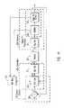
- FIG. 3is a block diagram of one embodiment of an intracranial recording electrode (ICRE) signal processor and an intracranial stimulating electrode (ICSE) signal processor each of which are included within the signal processor illustrated in FIG. 2 .
- ICREintracranial recording electrode
- ICSEintracranial stimulating electrode
- FIG. 4is a schematic diagram of a globus pallidus implanted with stimulating and recording electrodes in accordance with one embodiment of the present invention.
- FIG. 5is a block diagram of one embodiment of an EMG signal processor which is included in one embodiment of the signal processor illustrated in FIG. 2 .
- FIG. 6is a block diagram of one embodiment of an EEG signal processor module which is included in one embodiment of the signal processor illustrated in FIG. 2 .
- FIG. 7is a block diagram of one embodiment of an accelerometer signal processor which is incorporated into certain embodiments of the signal processor illustrated in FIG. 2 .
- FIG. 8is a block diagram of one embodiment of an acoustic signal processor which is included in certain embodiments of the signal processor illustrated in FIG. 2 .
- FIG. 9is block diagram of one embodiment of a peripheral nerve electrode (PNE) signal processor 237 which is implemented in certain embodiments of signal processor 71 .
- PNE signalPNE signal
- FIG. 10is a schematic diagram of one embodiment of the signal processor illustrated in FIG. 2 .
- FIG. 11is a schematic diagram of the patient-neural modulator system illustrated in FIG. 2 illustrated to show its controller and observer components.
- FIG. 12is a schematic diagram of one embodiment of the control circuit illustrated in FIG. 2 .
- FIG. 13is a schematic diagram of electrical stimulation waveforms for neural modulation.
- FIG. 14is a schematic diagram of one example of the recorded waveforms.
- FIG. 15is a schematic block diagram of an analog switch used to connect one or an opposing polarity pair of Zener diodes across the noninverting and inverting inputs of an intracranial recording electrode amplifier.
- FIG. 1is a schematic diagram of one embodiment of the intracranial stimulator of the present invention implanted bilaterally in a human patient.
- two neurological control systems 999are shown implanted bilaterally.
- Each system 999includes a stimulating and recording unit 26 and one or more intracranial components described below.
- the intracranial componentspreferably include a stimulating electrode array 37 .
- the stimulating electrodesmay also be extracranial; that is, attached to a peripheral nerve in addition to or in place of being located within the cranium.
- stimulating and recording unit 26 of each neurological control system 999is preferably implanted contralateral to the intracranial components of the device.
- the configuration illustrated in FIG. 1is just one example of the present invention. Many other configurations are contemplated.
- the stimulating and recording unit 26is implanted ipsilateral or bilateral to the intracranial components. It should also be understood that the stimulating and recording unit 26 can receive ipsilateral, contralateral or bilateral inputs from sensors and deliver ipsilateral, contralateral, or bilateral outputs to a single or a plurality of intracranial stimulating electrode arrays 37 .
- these inputsare direct or preamplified signals from at least one of EMG electrode array 50 , EEG electrode array 51 , Accelerometer Array 52 , Acoustic Transducer Array 53 , Peripheral Nerve Electrode Array 54 , and Intracranial Recording Electrode Array 38 .
- the signals input from these sensorswill be referred to herein as “sensory input modalities” 247 .
- the outputsinclude but are not limited to one or more stimulating current signals or stimulating voltage signals to Intracranial Stimulating Electrode Array 37 .
- the two unilateral systems 26are shown to receive sensory inputs from the side contralateral as well as the intracranial stimulating electrode arrays 37 .
- systems 26also receive sensory inputs from intracranial recording electrode arrays 38 .
- intracranial recording electrode arrays 38may provide valuable feedback information.
- stimulating and recording units 26may be a single device, two communicating devices, or two independent devices. Accordingly, these and other configurations are considered to be within the scope of the present invention. It is anticipated that stimulating and recording units 26 , if implemented as distinct units, would likely be implanted in separate procedures (soon after clinical introduction) to minimize the likelihood of drastic neurological complications.
- the intracranial stimulating electrode array 37includes a plurality of intracranial stimulating electrodes 1 , 2 , 3 and 4 .
- Array 37may, of course, have more or fewer electrodes than that depicted in FIG. 1 .
- These intracranial stimulating electrodes 1 - 4may be used to provide stimulation to a predetermined nervous system component.
- the electrical stimulation provided by the intracranial stimulating electrodes 1 - 4may be excitatory or inhibitory, and this may vary in a manner which is preprogrammed, varied in real-time, computed in advance using a predictive algorithm, or determined using another technique now or latter developed.
- the intracranial recording electrode arrays 38includes intracranial recording electrodes 5 and 6 .
- the intracranial recording electrodes 5 , 6are used to record cortical activity as a measure of response to treatment and as a predictor of impeding treatment magnitude requirements.
- intracranial recording electrodes 5 and 6are depicted in a location superficial to the intracranial stimulating electrodes 1 - 4 . However, this positioning may be reversed or the intracranial stimulating electrodes 1 - 4 and intracranial recording electrodes 5 and 6 may be interspersed in alternative embodiments.
- these electrodesmay be placed in at least one of motor cortex, premotor cortex, supplementary motor cortex, other motor cortical areas, somatosensory cortex, other sensory cortical areas, Wernicke's area, Broca's area, other cortical region, other intracranial region, and other extracranial region.
- an intracranial catheter 7is provided to mechanically support and facilitate electrical connection between intracranial and extracranial structures.
- intracranial catheter 7contains one or more wires connecting extracranial stimulating and recording circuit 26 to the intracranial electrodes, including but not limited to, intracranial stimulating electrodes 1 - 4 and intracranial recording electrodes 5 , 6 .
- the wires contained within intracranial catheter 7transmit stimulating electrode output signal (SEOS) to intracranial stimulating electrode array 37 .
- Such wiresadditionally transmit stimulating electrode input signal (SEIS) and recording electrode input signal (REIS), from intracranial stimulating electrode array 37 and intracranial recording electrode array 38 respectively, to stimulating and recording circuit 26 .
- SEOSstimulating electrode output signal
- SEISstimulating electrode input signal
- REISrecording electrode input signal
- Stimulating and recording circuit 26is protected within a circuit enclosure 44 .
- Circuit enclosure 44 and contained components, including stimulating and recording circuit 26comprise stimulating and recording unit 43 .
- stimulating and recording circuit 26can be placed extra cranially in a subclavian pocket as shown in FIG. 1, or it may be placed in other extracranial or intracranial locations.
- Connecting cable 8generally provides electrical connection between intracranial or intracranial locations.
- a set of electrical wiresprovides the means for communication between the intracranial and extracranial components; however, it should be understood that alternate systems and techniques such as radiofrequency links, optical (including infrared) links with transcranial optical windows, magnetic links, and electrical links using the body components as conductors, may be used without departing from the present invention.
- connecting cable 8provides electrical connection between intracranial components 246 and stimulating and recording circuit 26 . In embodiments wherein stimulating and recording circuit 26 has an intracranial location, connecting cable 8 would likely be entirely intracranial.
- connecting cable 8may be confined entirely to subcutaneous region under the scalp 10 .
- a catheter anchor 29provides mechanical connection between intracranial catheter 7 and calvarum 9 .
- Catheter anchor 29is preferably deep to the overlying scalp 10 .
- Such a subcutaneous connecting cable 8provides electrical connection between intracranial electrodes 246 and stimulating and recording circuit 26 .
- Cable 8may also connect any other sensors, including but not limited to any of sensory input modalities 247 , or other stimulating electrodes, medication dispensers, or actuators with stimulating and recording circuit 26 .
- Sensory feedbackis provided to recording and stimulating unit 26 from a multiplicity of sensors, collectively referred to as sensory input modalities 247 .
- Intracranial recording electrode array 38previously described, is intracranial in location. Additional sensors, most of which are located extracranially in the preferred embodiment, comprise the remainder of sensory input modalities 247 .
- Sensory input modalities 247provide information to stimulating and recording unit 26 . As will be described in greater detail below, such information is processed by stimulating and recording unit 26 to deduce the disease state and progression and its response to therapy.
- a head-mounted acoustic sensor 11is used to monitor any number of vibratory characteristics such as high frequency head vibration, muscle vibration, and/or speech production.
- Head-mounted acoustic sensor 11is connected to stimulating and recording circuit 26 with an acoustic sensor connecting cable 30 .
- a head-mounted accelerometer 12is implemented in certain embodiments of the present invention to monitor head movement and position with respect to gravity.
- Head-mounted accelerometer 12may be mounted to any structure or structures that enables it to accurately sense a desired movement. Such structures include, for example, the skull base, calvarum, clavicle, mandible, extraocular structures, soft tissues and vertebrae.
- Head-mounted accelerometer 12is connected to stimulating and recording circuit 26 with an accelerometer connecting cable 31 .
- a proximal electromyography (EMG) electrode array 45is also included in certain preferred embodiments of the invention.
- Proximal EMG electrode array 45includes a positive proximal EMG electrode 13 , a reference proximal EMG electrode 14 , and a negative proximal EMG electrode 15 .
- proximal EMG electrode array 45may include any number of type of electrodes.
- Proximal EMG electrode array 45is implanted in or adjacent to muscle tissue. In the embodiment illustrated in FIG. 1, proximal EMG electrode array 45 is shown implanted within the neck of the human patient. However, it should be understood that this location is illustrative only and that proximal EMG electrode array 45 may be implanted in or adjacent to any muscle without departing from the spirit of the present invention.
- a proximal acoustic sensor 27may also be implemented in the present invention.
- Proximal acoustic sensor 27senses muscle vibration and may be used to augment, supplement or replace EMG recording.
- a proximal accelerometer 28may be used to sense movement, including tremor and voluntary activity, and orientation with respect to gravity.
- Proximal connecting cable 16provides electrical connection from the proximal EMG electrodes 14 and 15 , proximal acoustic sensor 27 , and proximal accelerometer 28 to stimulating and recording circuit 26 . In the illustrative embodiment, these sensors are shown connected to a common proximal connecting cable 16 .
- this configurationmay include the use of multiple connecting cables or implement other types of communication media without departing from the present invention. It should also be understood from the preceding description that the number of each type of sensor may also be increased or decreased, some sensor types may be eliminated, and other sensor types may be included without departing from the spirit of the present invention.
- a distal EMG electrode array 47may also be included in certain embodiments of the present invention.
- distal EMG electrode array 47typically includes a positive distal EMG electrode 17 , a reference distal EMG electrode 42 , and a negative distal EMG electrode 18 .
- Positive distal EMG electrode 17is connected to stimulating and recording circuit 26 by positive distal EMG connecting cable 20 .
- Negative distal EMG electrode 18is connected to stimulating and recording circuit 26 by negative distal EMG connecting cable 21 .
- Reference distal EMG electrode 42is connected to stimulating and recording circuit 26 by reference distal EMG connecting cable 48 .
- a distal acoustic sensor 19is connected to stimulating and recording circuit 26 by distal acoustic connecting cable 22 .
- Distal accelerometer 33is connected to stimulating and recording circuit 26 by distal accelerometer connecting cable 34 .
- Distal accelerometer 33is connected to stimulating and recording circuit 26 by distal accelerometer connecting cable 34 .
- distal EMG electrode array 47In the embodiment illustrated in FIG. 1, distal EMG electrode array 47 , distal acoustic sensor 19 , and distal accelerometer 33 are shown located in the shoulder region. However, the distal EMG electrode array 47 may be located in other locations, including, for example, the masseter, temporalis, sternocleidomastoid, other portion of the head and neck, pectoralis, torso, abdomen, upper extremities, lower extremities, and other locations. The number of each type of sensor may be increased or decreased, some sensor types may be eliminated, and other sensor types may be included without departing from the spirit of the present invention.
- Enclosure-mounted EMG electrode array 46includes enclosure-mounted positive EMG electrode 23 , enclosure-mounted negative EMG electrode 24 and enclosure-mounted reference EMG electrode 25 , all of which are attached to the circuit enclosure 44 that encloses stimulating and recording unit 26 .
- the circuit enclosure 44is preferably included to provide robustness against potential lead entanglement and fracture.
- circuit enclosure 44is constructed of titanium and epoxy, or other single or combination of bio-compatible materials.
- Enclosure-mounted acoustic sensor 35 and enclosure-mounted accelerometer 36are mounted to stimulating and recording unit 43 . The number of each type of sensor may be increased or decreased, their locations changed, some sensor types eliminated, and other sensor types included without departing from the spirit of the present invention.
- EEG electrodes 39 , 40 , 41are provided.
- the EEG electrodesmay be mounted directly to connecting cable 8 or may be connected via intermediate cables. Any one of the numerous standard and new electrode configurations, or montages, may be employed in EEG electrodes 39 - 41 without departing from the present invention.
- a proximal peripheral nerve electrode array 98is connected to stimulating and recording circuit 26 by proximal peripheral nerve electrode array connecting cable 100 .
- Proximal peripheral nerve electrode array 98is shown located in the neck region. In this location proximal peripheral nerve electrode array 98 can interface with the vagus nerve, spinal accessory nerve, or nerve arising from cervical roots.
- a distal peripheral nerve electrode array 99is connected to stimulating and recording circuit 26 by distal peripheral nerve electrode array connecting cable 32 .
- Distal peripheral nerve electrode array 99is shown located by the proximal arm, in position to interface with the brachial plexus or proximal arm nerve.
- One or more of these peripheral nerve electrode arraysmay be implanted in these or other locations, including but not limited to the head, cranial nerves, neck, torso, abdomen, upper extremities, and lower extremities, without departing from the present invention.
- the peripheral nerve electrode arraysare each comprised of three epineural platinum-iridium ring electrodes, each in with an internal diameter approximately 30% larger than that of the epineurium, longitudinally spaced along the nerve. Electrodes of differing dimensions and geometries and constructed from different materials may alternatively be used without departing from the present invention.
- Alternative electrode configurationsinclude but are not limited to epineural, intrafascicular, or other intraneural electrodes; and materials include but are not limited to platinum, gold, stainless steel, carbon, and other element or alloy.
- FIG. 2is an architectural block diagram of one embodiment of the neurological control system 248 of the present invention for modulating the activity of at least one nervous system component in a patient.
- a nervous system componentincludes any component or structure comprising an entirety or portion of the nervous system, or any structure interfaced thereto.
- the nervous system component that is controlled by the present inventionincludes the globus pallidus internus.
- the controlled nervous system componentis the subthalamic nucleus.
- the neurological control system 248includes one or more implantable components 249 including a plurality of sensors each configured to sense a particular characteristic indicative of a neurological or psychiatric condition.
- One or more intracranial (IC) stimulating electrodes in an IC stimulating electrode array 37delivers a neural modulation signal to the same or other nervous system component as that being monitored by the system 26 .
- One or more sensors 38 , 51 , 52 , 53 , and 54sense the occurrence of neural responses to the neural modulation signals.
- Stimulating and recording unit 26generates the neural modulation signal based on the neural response sensed by the sensors.
- the neurological control system 248preferably also includes a patient interface module 55 and a supervisory module 56 .
- a control circuit 72(described below) is communicably coupled to the patient interface module 55 and receives signal inputs from and provides signal outputs to patient interface module 55 and supervisory module 56 .
- patient interface module 55 and supervisory module 56remain external to the body of the patient. However either of these devices may be connected via percutaneous leads or be partially or totally implanted without departing from the present invention.
- Patient interface module 55 and supervisory module 56facilitate adjustment of control parameters, monitoring of disease state, monitoring of response to therapy, monitoring of stimulating and recording circuit 26 , monitoring of impedance and other characteristics of intracranial stimulating electrode array 37 , monitoring of physiologic parameters, monitoring of vital signs, monitoring of any other characteristic or function of components of the present invention, including but not limited to the stimulating and recording circuit 26 , stimulating and recording unit 43 , circuit enclosure 44 , EMG electrode array 50 , EEG electrode array 51 , accelerometer array 52 , acoustic transducer array 53 , peripheral nerve electrode array 54 , and intracranial recording electrode array 38 .
- Such monitoring and adjustmentis accomplished through the use of any well known bi-directional communication between control circuit 72 and supervisory module 56 .
- a radio frequency linkis employed.
- other communication technologiesincluding but not limited to optical, percutaneous, or electromagnetic, may be used.
- patient interface module 55 and supervisory module 56are placed adjacent to the patients garments overlying the implanted stimulating and recording unit 43 .
- a communications handshaking protocolis executed.
- Communication handshaking routinesare known to those or ordinary skill in the art, and they enable establishment of a communication rate and protocol and facilitate mutual identification of devices.
- Patient interface module 55automatically downloads parameters from stimulating and recording circuit 26 and stores values of such parameters in a memory. When the transfer of these parameter values is complete, patient interface module 55 emits a audible signal such as a series of beeps, and the patient turns off patient interface module 55 and removes it from its position overlying the implanted stimulating and recording unit 43 .
- Parameter valuesmay then be retrieved by the patient by a routine including but not limited to a menu driven interface, and the values may be transmitted via telephone conversation or other communication method to a health care professional.
- Supervisory module 56operates in the same manner with one addition; a step is provided during which the health care professional may upload parameters to stimulating and recording circuit 26 to alter its function including by means of changing parameters including but not limited to control laws gains and thresholds, filter parameters, signal processing parameters, stimulation waveform modes (including at least one of current regulated, voltage regulated, frequency regulated, or pulse width regulated), and stimulation waveform parameters.
- Control lawsare defined by a set of parameters specific to the particular control law. Common parameters include preset gains, threshold levels, saturation amplitudes, sampling rates, and others. Adaptive controllers change in response to the behavior of the system being controlled; as such, in addition to preset parameters, adaptive controllers possess a set of varying parameters. These varying parameters contain information indicative of the behavior of the system being controlled; downloading of these parameters provides one set of measures of the disease state and its response to therapy.
- Such monitoringincludes observation of time history of disease state, stimulation parameters, response to therapy, and control law parameters, including time-varying adaptive controller parameters.
- Such adjustmentsincludes modification of actual stimulation parameters and allowable ranges thereof, including but not limited to pulse width, pulse amplitude, interpulse interval, pulse frequency, number of pulses per burst frequency. Adjustments can further include modification of actual control law parameters and allowable ranges thereof, including but not limited to gains, thresholds and sampling rates of said stimulation waveforms.
- Signal processor 71contains signal processor modules for each of the sensory input modalities 247 . Signal processing algorithms for each of the said sensory input modalities 247 may be independent.
- the said sensory input modalities 247may be coupled, such that the processing of one of the sensory input modalities 247 is dependent on another of the sensory input modalities 247 . Adjustments may additionally include modification of actual signal processor parameters and allowable ranges thereof, including but not limited to gains, filter cutoff frequencies, filter time constants, thresholds, and sampling rates.
- the stimulation and control law parametersare stored in at least one of random access memory and central processing unit registers (not shown).
- patient interface module 55is to be used by the patient, a family member or associate, or home health care personnel to monitor the functions and performance of neurological control system 248 .
- the use of the patient interface module 55is restricted to monitoring operations; adjustment of stimulation and control parameters is not enabled.
- adjustment of all or a subset of stimulation and control parametersmay be facilitated by patient interface module 55 without departing from the present invention.
- Supervisory module 56is used by a physician or other health care personnel to monitor function and performance of neurological control system 248 and to adjust stimulation and control parameters.
- Control parameters controlled by patient interface module 55 and supervisory module 56include allowable stimulation magnitude range, such as maximum combination of stimulation voltage, current, pulse width, pulse frequency, train frequency, pulse train count, pulse train duration.
- Control parametersmay also include variables and constants used to define control laws implemented in control circuit 72 .
- Such control parametersinclude, but are not limited to, control law gains 197 - 203 , and other parameters for control laws, including but not limited to proportional controller 230 , differential controller 204 , integral controller 205 , nonlinear controller 206 , adaptive controller 207 , sliding controller 208 , model reference controller 209 , and other controllers.
- amplitudes for other controller parametersincluding but not limited to amplitudes for controller weights 210 - 216 may be set by supervisory module 56 . Additionally, the parameters specifying the maximum amplitudes, or saturation values, may be set by supervisory module 56 . Control circuit 72 (FIG. 12) will be described in detail below.
- control parametersfurther includes signal conditioning parameters.
- Signal conditioning parametersmay include, for example, amplifier gains, filter gains and bandwidths, threshold values, and other parameters.
- control parametersadditionally include signal processing parameters, including envelope determinator gains and time constants, filter passbands, filter gains, threshold values, integrator gains, analyzer parameters, disease state estimator parameters, artifact rejecter thresholds, envelope determinator time constants, rectifier parameters, spectral analyzer parameters and timer parameters.
- signal processing parametersincluding envelope determinator gains and time constants, filter passbands, filter gains, threshold values, integrator gains, analyzer parameters, disease state estimator parameters, artifact rejecter thresholds, envelope determinator time constants, rectifier parameters, spectral analyzer parameters and timer parameters.
- control parametersfurther include spike detector 188 (FIG. 9) parameters, spike characterizer 189 (FIG. 9) parameters, spike analyzer 190 (FIG. 9) parameters, spectral energy characterizer 192 (FIG. 9) parameters, spectral energy analyzer 193 (FIG. 9) parameters, aggregate disease state estimator 195 (FIG. 10) parameters.
- tremorare quantified and monitored by any sensors over time as indicators of disease state.
- sensorsinclude but are not limited to EMG electrode array 50 , EEG electrode array 51 , accelerometer array 52 , acoustic transducer array 53 , peripheral nerve electrode array 54 , intracranial recording electrode array 38 , and intracranial stimulating electrode array 37 .
- the sensed tremor characteristicsinclude, but are not limited to, magnitude, frequency, duration and frequency of occurrence of tremors. Changes in these and other parameters are compared to current levels of, and changes in, treatment parameters. These changes are then used by aggregate disease state estimator 195 to estimate the response to therapy as functions of various electrical stimulation treatment parameters. Electrical stimulation treatment parameters are adjusted by control circuit 72 in real-time to provide optimal control of disease state.
- Modulation parametersare optimized to achieve at least one of minimization of disease state, minimization of symptoms of disease, minimization of stimulation magnitude, minimization of side effects, and any constant or time-varying weighted combination of these.
- Patient interface module 55 and supervisory module 56also preferably monitor the function and operation of other components of neurological control system 248 , including stimulating and recording unit 26 and implanted components 249 .
- Stimulating and recording unit 26receives and processes signals generated by implanted components 249 to provide conditioned signals 78 - 84 to a signal processor 71 .
- signal conditioning circuit 76preferably includes an associated amplifier and filter. Each amplifier and associated filter is configured to receive and process the signal generated by the associated one of the set of sensors 38 , 51 , 52 , 53 , and 54 .
- implanted components 249include an electromyography (EMG) electrode array 50 which generate EMG signals.
- EMG electrode array 50comprises of all EMG electrodes implemented in the particular embodiment of the present invention. These include, in the exemplary embodiment illustrated in FIG. 1, proximal EMG electrode array 45 , enclosure-mounted EMG electrode array 46 and distal EMG electrode array 47 .
- Array 50may also include, for example, EMG electrodes implanted in the head or other location, and surface EMG electrodes.
- Implanted components 249also include an electroencephalography (EEG) electrode array 51 which generate EEG signals and accelerometer array 52 which generates acceleration signals.
- EEG electrodes 39 , 40 , 41 illustrated in FIG. 1are representative of EEG electrode array 51 .
- EEG electrodes 39 - 41may be mounted directly to connecting cable 8 or connected via intermediate cables.
- EEG electrode array 51may include more or fewer elements than EEG electrodes 39 - 41 depicted; and any of numerous standard and new electrode configurations, or montages, may be employed without departing from the present invention.
- Accelerometer array 52which produces well-known acceleration signals, preferably includes all accelerometers implemented in the patient associated with the present invention.
- accelerometer array 52includes head-mounted accelerometer 12 , proximal accelerometer 28 , enclosure-mounted accelerometer 36 and distal accelerometer 33 .
- Accelerometer array 52may include more or fewer accelerometers than these accelerometers, and accelerometers of any types and locations may be employed without departing from the present invention.
- Acoustic transducer array 53includes all acoustic Sensors utilized by the present invention.
- acoustic transducer array 53includes head-mounted acoustic sensor 11 , proximal acoustic sensor 27 , enclosure-mounted acoustic sensor 35 and distal acoustic sensor 19 . It should be understood that acoustic transducer array 53 may include more or fewer elements than said acoustic sensors listed above; and any of numerous acoustic sensor types and locations may be employed without departing from the present invention.
- Peripheral nerve electrode array 54generates peripheral neural signals, including but not limited to efferent and afferent axonal signals.
- peripheral nerve electrode array 54includes all peripheral nerve electrodes implemented in present invention.
- peripheral nerve electrode array 54includes proximal peripheral nerve electrode array 98 and distal peripheral nerve electrode array 99 .
- the single or plurality of individual peripheral nerve electrode arrays which comprise peripheral nerve electrode array 54may be implanted in the illustrated or other locations, as noted above.
- Intracranial (IC) recording electrode array 38generates central neural signals, including but not limited to cortical, white matter, and deep brain nuclear signals. Neural activity to be sensed includes but is not limited to that found in the primary motor cortex, premotor cortex, supplementary motor cortex, somatosensory cortex, white matter tracts associated with these cortical areas, the globus pallidus internal segment, the globus pallidus external segment, the caudate, the putamen, and other cortical and subcortical areas. As one of ordinary skill in the relevant art will find apparent, the present invention may include additional or different types of sensors that sense neural responses for the type and particular patient. Such sensors generate sensed signals that may be conditioned to generate conditioned signals as described below. One example of the placement of these electrodes is described above with reference to the embodiment illustrated in FIG. 1 . Many others are contemplated by the present invention.
- signal conditioning circuit 76includes an associated amplifier and filter in the illustrative embodiment. Accordingly, signal conditioning circuit 76 includes an EMG amplifier 59 and filter 66 , each constructed and arranged to amplify and filter, respectively, the EMG signals received from EMG electrode array 50 . Similarly, signal conditioning circuit 76 also includes an EEG amplifier 60 and filter 67 , accelerometer (ACC) amplifier 61 and filter 68 , acoustic (ACO) amplifier 62 and filter 69 , peripheral nerve electrode (PNE) amplifier 63 and filter 70 and intracranial (IC) recording electrode (ICRE) amplifier 58 and filter 65 .
- EEG amplifier 60 and filter 67the EEG signals received from EMG electrode array 50 .
- signal conditioning circuit 76also includes an EEG amplifier 60 and filter 67 , accelerometer (ACC) amplifier 61 and filter 68 , acoustic (ACO) amplifier 62 and filter 69 , peripheral nerve electrode (PNE) amplifier 63 and filter 70 and intracranial (IC) recording electrode (
- Simplifiers 57 - 63may be single or multi-channel amplifiers depending upon the number of electrodes with which it interfaces.
- amplifiers 57 - 63are physically located in the same enclosure as filters 64 - 70 ; that is, in a single signal conditioning circuit 76 .
- signal conditioning circuit 76is physically contained within stimulating and recording unit 102 .
- amplifiers 57 - 63may be located separately from stimulating recording unit 102 .
- amplifiers 57 - 63may be affixed to or situated proximate to their associated electrode arrays 38 , 50 - 54 .
- Amplifiers 57 - 63may be any known voltage amplifier now or later developed suitable for amplifying the particular signals generated by their associated electrodes.
- filters 64 - 70may be physically separate from or incorporated into signal conditioning circuit 76 and stimulating and recording unit 26 .
- filters 64 - 70are low pass filters having a cut-off frequency of, for example, 3,000 Hz.
- filters 64 - 70may include a notch filter to remove, for example, 60 Hz noise, or other types of filters appropriate for the type of signals generated by the associated sensors 38 , 51 , 52 , 53 , and 54 . Selection of the appropriate frequencies for the cut-off and notch filter frequencies is considered to be well known in the relevant art and within the scope of the present invention.
- Filters 66 - 70 , 65 and 64generate conditioned sensed signals 84 , 83 and 78 - 82 , respectively.
- Signal processor 71processes the conditioned sensed neural response signals 78 - 84 generated by signal conditioning circuit 76 in accordance with the present invention to determine neural system states.
- Signal processor 71generally performs well known filtering operations in the time and frequency domains.
- the neural system statesinclude one or more physiologic or disease states.
- Signal processor 71which can be implemented in a fast microprocessor, a DSP (digital signal processor) chip, or as analog circuitry, for example, is described in detail below.
- Control circuit 72responsive to the signal processor 71 , patient interface module 55 and supervisory module 56 , adjusts the magnitude of a neural modulation signal in response to the sensed neural response.
- Signal processor 71extracts relevant information from the sensed conditione signals, and control circuit 72 uses this extracted information in the calculation of an output neuromodulation signal (NMS) 998 .
- Neuromodulation signal 998subsequently travels along stimulator output path 111 to IC stimulating electrode array 37 .
- control circuit 72is a state machine, utilizing current and past system behavior in the calculation of a control signal.
- control circuit 72includes an embedded microprocessor to process nonlinear control laws. Alternative embodiments of the control circuit 72 appropriate for the particular application may be also be used.
- Control circuit 72receives control law selection information, control law parameter information, stimulation waveform parameter range information, stimulation modulation mode, output stage regulation mode, and medication dose and timing information from patient interface module 55 and supervisory module 56 .
- the waveform parameter or parameters which are modulated by control law output signal U 997are determined by the stimulation modulation mode; these parameters include but are not limited to pulse amplitude, pulse width, pulse frequency, pulses per burst, and burst frequency. Selection between regulation of pulse voltage or pulse current as the regulated pulse amplitude is determined by the output stage regulation mode.
- Control circuit 72provides stimulation waveform parameter history information, disease state history information, control law state variable history information, control law error history information, control law input variable history information, control law output variable history information, stimulating electrode impedance history information, sensory input history information, battery voltage history information, and power consumption history information to patient interface module 55 and supervisory module 56 .
- Provision of stimulating electrode impedance history informationallows monitoring of stimulating electrode performance and functionality. If an electrode is determined to be fractured, shorted, or encapsulated by fibrotic tissue, any of various control law parameters, output stage parameters, and waveform range parameters may be adjusted to allow compensation for these changes. Additionally, the Neuromodulation Signal (NMS) 998 may be delivered to different sets of electrodes to insure that it reaches neural tissue 250 . Sensory input history information allows evaluation of validity of any given sensory input. This is useful in determining the functionality of a given sensor and serves as an indicator for sensor replacement or adjustment of the signal processing parameters or algorithm or the control law parameters or algorithm to continue to generate reliable disease state estimate signals X and control law outputs U despite the loss of any particular individual or set of sensory signals.
- NMSNeuromodulation Signal
- Signal processor 71receives amplifier gain setting information, filter parameter information, weighting information, and disease state estimator parameter and algorithm information from patient interface module 55 and supervisory module 56 .
- patient interface module 55may be used by the patient or home health care personnel to monitor disease state, stimulation parameters, and response to therapy. Limited adjustment of stimulation parameters and ranges is facilitated.
- Patient interface module 55may be used by the patient or home health care personnel to provide information to the physician, avoiding the need for an office visit for the obtainment of said information.
- Patient information module 55queries signal processor 71 for present and time histories of monitored values. Time histories of selected variables in signal processor 71 and control circuit 72 are stored in memory module 240 for subsequent retrieval by patient interface module 55 and supervisory module 56 .
- Selected variablesinclude but are not limited to disease state, tremor frequency, tremor magnitude, EMG magnitude, EMG frequency spectra (EMG magnitude within frequency ranges), and acceleration of limb, head, mandible, or torso. Selected variables may also include disease state, frequency spectra of limb, torso, and head movements, as determined by EMG and accelerometer signals.
- Stimulating and recording unit 26also includes an output stage circuit 77 .
- Output stage circuit 77takes for an input the control law output signal U, which may be comprised of a single or multiplicity of channels or signals, from control circuit 72 .
- This control law output signal U 997modulates the magnitude of the sequence of waveforms comprising the desired output neuromodulation signal (NMS D ) which is produced by output stage circuit 77 and delivered via intracranial stimulating electrode array 37 to neural tissue 250 .
- NMS Ddesired output neuromodulation signal
- Output stage circuit 77generates a neuromodulation signal (NMS D ) 998 with a magnitude specified by control law output signal U 997 received from control circuit 72 .
- the waveform parameter of the desired output neuromodulation signal (NMS D ) which is modulated by control law output signal Uis the stimulation current magnitude.
- the capability to specifically modulate the stimulation currentconfers efficacy resistance to perturbations or changes in electrode impedance.
- Presently implanted systemssuffer from a decline in efficacy which results from an increase in electrode impedance which accompanies the normal tissue response to a foreign body, that is fibrotic encapsulation of the electrode.
- a the magnitude of the current delivered to the neural tissue 250will not vary as the electrode becomes encapsulated with fibrotic tissue or its impedance otherwise changes over time.
- a further advantage conferred by current modulationis the ability to monitor electrode impedance. If a current-modulated waveform, preferably a sinusoid, is delivered to the electrodes, and the resultant voltage potential waveform is concurrently monitored, the relative magnitudes and phase shifts of these waveforms may be computed. From these magnitudes and phases, the complex impedance and hence the resistive and capacitive components of the electrode impedance may be calculated.
- the waveform parameter of the desired output neuromodulation signal (NMS D ) which is modulated by control law output signal U 997is the stimulation voltage magnitude.
- NMS Ddesired output neuromodulation signal
- This designwould not enjoy the independence of the stimulation current and efficacy from impedance variation enjoyed by the embodiment described above. If fibrosis was uneven around the surface of the electrode, this embodiment would avoid potentially undesirably large current densities along narrow tracts of remaining low resistance unfibrosed regions of neural tissue 250 .
- regulation of stimulus pulse widthmay be desired.
- the available resolution or bits for specifying the magnitude of pulse widthmay be greater than that for specifying the pulse voltage or current.
- the spatial neuron recruitment characteristics of a pulse width modulated neuromodulation signal (NMS) 998may provide a more linear, predictable, or controllable response than that obtained with current or voltage modulation.
- Selection between regulation of pulse voltage, pulse current, or pulse width as the regulated pulse amplitude parameteris determined by the output stage regulation mode, which may be set using supervisory module 56 .
- the modulation of pulse frequency and the modulation of the number of pulses per burstare regulated. As one of ordinary skill in the relevant art would find apparent. Other such characteristics may be regulated in addition to or instead of the ones noted above.
- Output stage circuit 77includes a pulse generator 73 , an output amplifier 74 and a multiplexor 75 .
- Pulse generator 73generates one or more stimulus waveforms, each of which is characterized by several parameters, including but not limited to pulse amplitude, pulse width, pulse frequency, number of pulses per burst, and burst frequency. As noted above, pulse amplitude may comprise pulse voltage or pulse current. Preferably, each of these parameters may be independently varied, as specified by control law output signal U 997 generated by control circuit 72 .
- the stimulus waveforms comprising the neuromodulation signal (NMS) generated by output stage circuit 77are applied to patient through intracranial (IC) stimulating electrode array 37 .
- Pulse generator 73generates a single waveform when single channel stimulation is to be used, and a plurality of waveforms when multiple channel stimulation is to be used. It may generate monophasic or biphasic waveforms.
- charge balanced biphasic waveformsare produced.
- the net charge contained in a given pulseis given by the time integral of the stimulus current over the duration of the pulse.
- a pair of pulses of opposite polarityis generated, and the pulse current amplitude and pulse width are chosen such that the charge amplitude is equal in magnitude and opposite in polarity.
- the pulses comprising the biphasic pulse pairit is desirable for the pulses comprising the biphasic pulse pair to have different amplitudes; in this case, the pulse widths are chosen to insure equal and opposite charges so the pulse par introduces zero net charge to the neural tissue 250 .
- the capability to deliver pulse pairs with balanced chargesis yet a further advantage conferred by the current regulation mode described above.
- a direct current (DC) blocking capacitoris employed. This is a technique that is well known to those or ordinary skill in the art.
- a DC blocking capacitoris included within multiplexor 75 in series with stimulator output path 111 .
- multichannel stimulationis used in the case of bilateral stimulation. Since the disease progression is typically asymmetrical, and the normal motor control systems governing movement on the left and right side of the body are also highly independent of each other, the delivery of treatment to the left and right sides of the body should be controlled separately. This represents one need for a multiple channel neuromodulation signal (NMS) 998 .
- NMSneuromodulation signal
- Multichannel stimulationis also expected to be beneficial in treating patients with variable involvement of different limbs. For example, the magnitude neuromodulation of a portion of the globus pallidus required to achieve optimal controls of arm tremor may be different from the optimal level of neuromodulation of separate portion of the globus pallidus to achieve optimal control of leg tremor.
- a further need for multichannel neuromodulation signalis the control of multiple symptoms of the movement disorder and the side effects arising from pharmacologic treatment.
- Optimal control of tremor, dyskinesias, and rigidityare not achieved by modulation of the same site at the same intensity.
- multiple and separately controlled channels of neuromodulationare required to simultaneously achieve optimal control of these multiple symptoms and side effects.
- Each of these symptoms and side effectsmay be considered to comprise one or more element in a multivariable disease state.
- a multivariable control systemwill be required to optimally drive each of these disease state elements to its desired value, ideally toward a target minimum level and thus achieve optimal control of this multiplicity of disease states.
- This multivariable control systemmay be implemented as multiple independent control laws each with separate though potentially overlapping sensory inputs or as a multivariable control law matrix.
- Stimulation via each of the multiple channels comprising the neuromodulation signal (NMS) 998is characterized by separate though possibly overlapping sets of one or more of the following parameters: stimulation voltage, stimulation current stimulation frequency of pulses within the same burst, frequency of bursts, pulse width, pulses per burst, duration of burst, and interpulse interval.
- the stimulus waveformsare amplified by output amplifier 74 to generate an amplified stimulus waveform.
- pulse generator 73transfers information to output amplifier 74 which includes information that uniquely specifies the desired stimulation waveform.
- the informationis in the form of an analog signal which represents a sealed version of the voltage or current waveform to be delivered to the tissue.
- Output amplifier 74performs amplification and regulation of the received stimulus waveform generated by the pulse generator 73 . This may be regulation of electrical current to achieve desired voltage or regulation of electrical voltage to achieve desired current, depending on whether a voltage or current waveform is to be delivered to the nervous system component.
- voltage regulationis simpler to implement, and is a technique which is commonly used by many conventional stimulators.
- Current regulationis more complex but allows for more precise control of the applied stimulation.
- Current regulationinsures that a specified amount of current is delivered, regardless of the impedance of the electrode.
- Current regulationis advantageous in that it allows for precise control of stimulation level despite changes in electrode impedance which invariably occur over time. Since electrode impedances often change, typically increasing as they become encapsulated by fibrosis, current regulation is preferred to avoid the decrease in current which would occur if voltage regulation were to be used in such circumstances.
- the amplified stimulus waveform generated by output amplifier 74is conducted along stimulator amplifier output path 112 to multiplexor 75 .
- Multiplexor 75allows for delivery of a stimulating electrode output signal (SEOS) to the intracranial stimulating electrode array 37 , multiplexed with sensing of a stimulating electrode input signal (SEIS).
- SEOSstimulating electrode output signal
- multiplexor 75serves to alternately connect intracranial stimulating electrode (ICSE) array 37 to output amplifier 74 and intracranial stimulating electrode amplifier 57 .
- Connection of intracranial stimulating electrode (ICSE) array 37 to output amplifier 74facilitates delivery of neural modulation signal to neural tissue, while connection of intracranial stimulating electrode (ICSE) array 37 to intracranial stimulating electrode amplifier 57 facilitates monitoring of neural activity in the region being stimulated.
- Multiplexor 75allows delivery of neural modulation signals to neural tissue concurrent with monitoring of activity of same neural tissue; this facilitates real-time monitoring of disease state and response to treatment.
- Stimulating electrode output signal (SEOS) from output amplifier 74is conducted along stimulator amplifier output path 112 to multiplexor 75 .
- Multiplexor 75conducts output from output amplifier 74 to stimulator output path 111 which conducts the stimulating electrode output signal to intracranial stimulating electrode array 37 .
- multiplexor 75alternatively conducts signal arising from stimulated tissue via intracranial stimulating electrode array (ICSE) 37 and stimulator output path 111 to multiplexed stimulator recording input path 113 and intracranial stimulating electrode amplifier 57 .
- ISCintracranial stimulating electrode array
- Multiplexor 75selectively conducts the signal on multiplexed stimulator recording input path 113 to amplifier 57 .
- Multiplexor 75may alternate conduction between path 111 and path 112 or path 113 using temporal multiplexing, frequency multiplexing or other techniques to allow concurrent access to the intracranial stimulating electrode (ICSE) array 37 for modulation of tissue activity and monitoring of tissue activity.
- Temporal multiplexingis a well known technique and frequency multiplexing of stimulation and recording signals in known to those skilled in the art. In this embodiment, temporal multiplexing is accomplished by alternately connecting stimulator output path 111 to stimulator amplifier output path 112 and multiplexed stimulator recording input path 113 .
- frequency multiplexingis accomplished by passing a band-limited portion of stimulating electrode output signal SEOS via the stimulator output path 111 to intracranial stimulating electrode array 37 while simultaneously monitoring activity on intracranial stimulating electrode array 37 within a separate frequency band, thereby generating a stimulating electrode input signal SEIS.
- stimulating electrode input signal SEISis conducted from the intracranial stimulating electrode array 37 to stimulator output path 111 to multiplexor 75 and via multiplexed stimulator recording input path 113 to intracranial stimulating electrode array amplifier 57 .
- Multiplexor 75facilitates conduction between stimulator amplifier output path 112 and multiplexed stimulator recording input path 113 to allow automated calibration.
- a calibration signal of known amplitudeis generated by pulse generator 73 and amplified by output amplifier 74 which, for calibration purposes, delivers a voltage regulated signal via stimulator amplifier output path 112 to multiplexor 75 .
- Multiplexor 75conducts amplified calibration signal to multiplexed stimulator recording input path 113 which conducts signal to intracranial stimulating electrode amplifier 57 .
- multiplexed or intermittent connection of stimulator amplifier output path 112 to the inputs of at least on of the other amplifiersmay be implemented without departing from the present invention.
- the same multiplexed connectionsmay be used to calibrate the pulse generator 73 and output amplifier 74 .
- an analog switchmay be used to connect one or an opposing polarity pair of Zener diodes across the noninverting and inverting inputs of intracranial recording electrode amplifier 58 .
- the Zener diodeswould limit the maximal amplitude of the calibration signal in one or both polarities to known values, allowing for accurate calibration of intracranial recording electrode amplifier 58 .
- the analog switchmay then be deactivated, removing the cathode of the single or pair of Zener diodes from the input of intracranial recording electrode amplifier 58 to allow measurement of stimulating electrode output signal (SEOS) for calibration of pulse generator 73 and output amplifier 74 . This is described in greater detail below.
- SEOSstimulating electrode output signal
- Multiplexor 75also facilitates conduction between stimulator amplifier output path 112 , multiplexed stimulator recording input path 113 , and stimulator output path 111 to allow measurement of impedances of components of intracranial stimulating electrode array 37 .
- this electrode impedance measurement modea three way connection between stimulator amplifier output path 112 , multiplexed stimulator recording input path 113 , and stimulator output path 111 is created.
- output amplifier 74When output amplifier 74 is operated in current regulated mode, it delivers an SEOS of known current via stimulator output path 111 to intracranial stimulating electrode array 37 .
- the voltages generated across the elements of intracranial stimulating electrode array 37generally are the products of the electrode impedances and the known stimulating currents. These voltages are sensed as the stimulating electrode input signal SEIS by the intracranial stimulating electrical amplifier 57 .
- Reference module 116contains memory registers in which control law reference values are stored. Such reference values include but are not limited to target disease state levels, target symptom levels, including target tremor level, and threshold levels. Threshold levels include but are not limited to disease and symptom levels, including tremor threshold levels. Neural modulation amplitude may be increased when at least one of disease state and symptom level exceed the corresponding threshold. Similarly neural modulation amplitude may be decreased or reduced to zero when either the disease state or symptom level falls below the corresponding threshold.
- Reference module 116is connected to patient interface module 55 , facilitating both monitoring and adjustment of reference values by patient.
- Reference module 116is also connected to supervisory module 56 , facilitating both monitoring and adjustment of reference values by physician or other health care provider.
- Supervisory module 56may be used by the neurologist, neurosurgeon, or other health care professional, to adjust disease state reference R values for the one or more control laws implemented in control circuit 72 .
- the disease state reference R valuesspecify the target level at which the corresponding disease states are to be maintained, as quantified by the disease state estimate X values, providing reference values for control laws implemented in control law circuit block 231 (FIG. 11; discussed below) and contained within control circuit 72 .
- Reference module 116may also receive input from control circuit 72 , facilitating the dynamic adjustment of reference disease state “r” (discussed below). Reference module 116 may additionally receive input from disease state estimator module array (DSEMA) 229 (FIG. 11; discussed below) and aggregate disease state estimator 195 (FIG. 11; discussed below) and components of signal processor 71 , for use in dynamically determining reference disease state “r”.
- DSEMAdisease state estimator module array
- FIG. 10is a schematic diagram of signal processor 71 .
- signal processor 71includes a disease state estimator module array 229 that includes one or more signal processor modules that generate a quantitative estimate of at least one disease state or parameter thereof based upon its respective input.
- a disease state estimator module array 229that includes one or more signal processor modules that generate a quantitative estimate of at least one disease state or parameter thereof based upon its respective input.
- magnitude of tremor in the 3 to 5 Hz rangerepresents one possible representation of a disease state. This could be an absolute or normalized quantification of limb acceleration in meters per second squared. This component of the disease state would be calculated almost exclusively from sensory feedback from accelerometer array 52 .
- Another possible disease stateis the frequency of occurrence of episodes of tremor activity per hour. This element of the disease state may be estimated from any of several of the sensory feedback signals.
- the most accurate representation of this disease state elementis obtained by applying a filter such as a Kalman filter to calculate this parameter based upon a weighted combination of the sensory feedback signals.
- a filtersuch as a Kalman filter
- weighting coefficientsare calculated from quantified measures of the accuracy of and noise present upon each sensory feedback channel.
- disease state estimator module array 229includes an EMG signal processor 233 , EEG signal processor 234 , accelerometer signal processor 235 , acoustic signal processor 236 , peripheral nerve electrode (PNE) signal processor 237 , intracranial recording electrode (ICRE) signal processor 238 , and intracranial stimulating electrode (ICSE) signal processor 239 . It should be understood that other signal processors may also be included in the array 229 .
- Inputs to these modulesinclude conditioned EMG signal path 78 , conditioned EEG signal path 79 , conditioned accelerometer signal path 80 , conditioned acoustic signal path 81 , conditioned peripheral nerve electrode (PNE) signal path 82 , conditioned intracranial recording electrode (ICRE) signal path 83 , and conditioned intracranial stimulating electrode (ICSE) signal path 84 , respectively. Communication between these modules is facilitated.
- the output(s) of each of the modulesis connected to an aggregate disease state estimator 195 .
- Aggregate disease state estimator 195generates a single or plurality of disease state estimates “X” indicative of state of disease and response to treatment.
- the acceleration of at least one of the affected limb and the headserves as respective elements in the disease state estimate X.
- These elements of disease state estimate Xare inputs to respective control laws implemented in control circuit 72 , of input to the control law.
- a control law governing the function of a proportional controller using acceleration as its single sensory feedback channelis given by equation (1):
- u 1 and u 1are the stimulation voltage given in volts; and ACC is the limb, mandible, or head acceleration given in meters per second squared (m/s 2 ).
- the stimulation siteis the ventroposterolateral pallidum
- the output stage modeis voltage regulated
- the waveformis a continuous train of square waves
- the amplitude u 1is given in volts (typically approximately 1 volt)
- the remaining stimulation parametersinclude a pulse width of 210 microseconds, and a stimulation frequency of 130 Hz.
- the stimulation siteis the ventral intermediate thalamic nucleus (Vim)
- the output stage modeis voltage regulated
- the waveformis an intermittent train of square waves with an on time of 5 minutes and an off time of 45 seconds
- the amplitude u 2is given in volts (typically approximately 3 volts)
- the remaining stimulation parametersinclude a pulse width of 60 microseconds, and a stimulation frequency of 130 Hz.
- the ACC signalrepresents the average acceleration over a finite time window, typically 15 to 60 seconds.
- This effective lowpass filteringprovides a stable sensory feedback signal for which a proportional control law is appropriate. If stability and performance requirements dictate, as is familiar to those practiced in the art of feedback control, other components, including an integrator and a differentiator may be added to the control law to produce a proportional-integral-differential (PID) controller, as needed.
- PIDproportional-integral-differential
- One preferred embodimentalso includes electromyographic (EMG) signals as sensory feedback in the calculation of at least one element of the disease state estimate X which is an input to the control law.
- EMGelectromyographic
- the EMG signalsare rectified by full wave rectifier 123 , passed through envelope determiner 124 , passed through several bandpass filters 125 , 127 , 129 , 131 , 133 and associated threshold discriminators 126 , 128 , 130 , 132 , 134 and then passed in parallel to each of integrator 135 and counter 136 .
- Integrator 135generates an output which is a weighted function of it inputs and represents the average magnitude of tremor activity over a given time window ⁇ w/2 to +w/2.
- control law output Uis given as the product of a transfer function H(s) and the disease estimate X, the input to the control law:
- H ⁇ ( s )u ⁇ ( s )
- X EMG ⁇ ( s )G V / EMG ⁇ ⁇ ( 0.1 ⁇ s + 1 ) ( 2 ⁇ s + 1 ) ( 8 )
- G V/EMGis the gain in neuromodulation signal (NMS) (volts per volt of EMG signal).
- signal amplitudesare on the order of 100 microvolts.
- NMSneuromodulation signal
- the appropriate overall gain G′ V/EMGis 20,000 volts NMS /volts EMG . Since the preamplifier stage performs amplification, 1000, in the preferred embodiment, the actual value for G V/EMG as implemented in the control law is 20 volts NMS /volts PREAMPL EMG .
- Disease state estimator 195determines estimates of disease state including but not limited to long-term, or baseline, components, circadian components, postprandial components, medication induced alleviation of components, medication induced components, and future predicted behavior of said components.
- Output of disease state estimator 195includes output of observer 228 , depicted in FIG. 11, which makes use of an adaptive model of disease behavior to estimate disease states which are not directly detectable from sensors. Such sensors, provide input to the adaptive model to correct state estimates and model parameters.
- Each of the signal processor modules in disease state estimator module array 229are described below.
- FIG. 3is a block diagram of intracranial recording electrode (ICRE) signal processor 238 and intracranial stimulating electrode (ICSE) signal processor 239 , each of which are included within signal processor 71 in the illustrative embodiment illustrated in FIGS. 2 and 10.
- ICRE signal processor module 238 and ICSE signal processor module 239process signals from one or more intracranial electrodes, including but not limited to those comprising intracranial recording electrode array 38 and intracranial stimulating electrode array 37 .
- intracranial stimulating electrode array 37is comprised of one or more intracranial stimulating electrodes while intracranial recording electrode array 38 is comprised of one or more intracranial recording electrodes.
- ICRE signal processor 238Input to ICRE signal processor 238 is conditioned intracranial recording electrode (ICRE) signal path 83 noted above. This input is connected to a spike detector 85 which identifies action potentials. Spike detection techniques are well known to those skilled in the art and generally employ low and high amplitude thresholds. Waveforms having amplitudes greater than the low threshold and lower than the high threshold are determined to be action potentials. These thresholds may be predetermined or adjusted manually using supervisory module 56 or may be adapted in real-time by an algorithm which sweeps the threshold through a range of values to search for values at which action potential spikes are consistently recorded.
- the low amplitude thresholdis set above the amplitude of background noise and that of nearby cells not of interest, and the high amplitude threshold is set above the amplitude of the desired action potentials to allow their passage while eliminating higher amplitude noise spikes, such as artifacts arising from electrical stimulation currents.
- Bandpass, notch, and other filtering techniquesmay also be used to improve signal to noise ratio and the sensitivity and specificity of spike detectors.
- Individual neuron action potentialsare usually recorded using fine point high-impedance electrodes, with impedances typically ranging from 1 to 5 megohms. Alternatively, larger lower-impedance electrodes may be used for recording, in which case the signals obtained typically represent aggregate activity of populations of neurons rather than action potentials from individual neurons.
- Spike detector 85passes the waveform(s) to a spike characterizer 86 .
- Spike characterizer 86determines firing patterns of individual neurons. The patterns include, for example, tonic activity, episodic activity, and burst firing.
- Spike characterizer 86calculates parameters that characterize the behavior of the individual and groups of neurons, the activity of which is sensed by intracranial recording electrode array 38 .
- the characterizationincludes parameterization of recorded action potentials, also referred to as spikes, bursts of spikes, and overall neural activity patterns.
- This parameterizationincludes, but is not limited to, calculation of frequencies of spikes, frequencies of bursts of spikes, inter-spike intervals, spike amplitudes, peak-to-valley times, valley-to-peak times, spectral composition, positive phase amplitudes, negative phase amplitudes, and positive-negative phase differential amplitudes. These parameters are depicted in FIG. 14 and are discussed below.
- spike characterizer 86discriminates individual spikes and bursts originating from different neurons. This discrimination facilitates serial monitoring of activity of individual and groups of neurons and the assessment and quantification of activity change, reflective of change in disease state and of response to therapy.
- a spike analyzer 87receives as input the parameters from spike characterizer 86 .
- Spike analyzer 87extracts higher level information, including but not limited to average spike frequencies, average interspike intervals, average amplitudes, standard deviations thereof, trends, and temporal patterning. By comparing current spike frequency rates to historical spike frequency data, spike analyzer 87 additionally calculates the rates of change of spike parameters. Prior trends and current rates of change may then be used to predict future behaviors. Rates of change of the parameters include but are not limited to autocorrelation and digital filtering.
- Spike analyzer 87may receive additional input from accelerometers, including but not limited to at least one of head mounted accelerometer 12 , proximal accelerometer 28 , enclosure mounted accelerometer 36 , and distal accelerometer 33 .
- Spike analyzer 87may receive indirect input from accelerometers, such as from conditioned or processed signals arising therefrom. This may include, for example, the signal transmitted by conditioned accelerometer signal path 80 .
- Spike analyzer 87may also receive additional input from EMG arrays 50 , such as a proximal EMG electrode array 45 , enclosure-mounted EMG electrode array 46 , or distal EMG electrode array 47 .
- Spike analyzer 87may receive indirect input from such EMG electrode arrays 50 , such as from conditioned or processed signals arising therefrom, including but not limited to the signal transmitted by conditioned EMG signal path 78 .
- accelerometers and EMG arraysfacilitates the characterization of neuronal firing patterns relative to activity of muscle groups and movement of joints, including but not limited to characterization of neuronal spike amplitudes and tuning of firing to movement, including but not limited to movement velocity and direction.
- the characterizationmay be used to assess functioning of the sensorimotor system, including but not limited to motor response time, and to measure the disease state and response to therapy.
- Intracranial recording electrode (ICRE) single unit-based (SU) disease state estimator 88receives input from spike characterizer 86 and/or spike analyzer 87 .
- Spike analyzer 87provides higher level information, including but not limited to average spike frequencies, average interspike intervals, average amplitudes, standard deviations thereof, trends, and temporal patterning to disease state estimator 88 .
- These inputsare representative of the current neuronal activity in the tissue from which the intracranial recording electrodes (ICRE) are recording.
- ICRE SU disease state estimator 88may also receive input representative of one or more signals, including desired neuronal activity, from control circuit 72 .
- the ICRE SU disease state estimate X ICRE — SU calculated by ICRE SU disease state estimator 88may be comprised of a single or a plurality of signals, consistent with a representation of the disease state by a single or a multitude of state variables, respectively.
- the ICRE MU disease state estimate X ICRE MU calculated by ICRE MU disease state estimator 88may be comprised of a single or a plurality of signals, each representative of multiunit neurophysiological signals, i.e. reflective of concurrent activity of numerous neurons.
- Both ICRE SU disease state estimate X ICRE SU and ICRE MU disease state estimate X ICRE — MUare output to aggregate disease state estimator 195 .
- conditioned intracranial recording electrode (ICRE) signal path 83additionally connects to filter 101 .
- Filter 101is preferably of the bandpass type filter.
- the bandpass filter 101has a passband of 0.1 to 100 Hz, although other ranges may be used.
- Output of filter 101connects to spectral energy characterizer 102 , which may be implemented in any of several hardware or software forms.
- the spectral energy characterizer 102is implemented using real-time fast Fourier transform (FFT) techniques. Alternatively, other digital or analog techniques may also be used.
- FFTreal-time fast Fourier transform
- spike detector 85spike characterizer 86 , spike analyzer 87 , disease state estimator 88 , filter 101 , spectral energy characterizer 102 , spectral energy analyzer 103 , and disease state estimator 104 may be comprised of individual signals or a plurality of signals. Further, spike detector 85 , spike characterizer 86 , spike analyzer 87 , disease state estimator 88 , filter 101 , spectral energy characterizer 102 , spectral energy analyzer 103 , and disease state estimator 104 may each have different parameters and signal processing characteristics for each of the multiple signals processed.
- baseline neuronal firing ratesdiffer among various anatomical and functional regions of the brain, and their involvement in disease states and susceptibility to change in firing patterns varies, the respective signal processing circuitry and logic will vary correspondingly. For example, baseline firing rates among neurons in the globus pallidus externus are approximately 43 Hz and those in the globus pallidus internus are 59 Hz.
- the input to intracranial stimulating electrode ICSE signal processor 239connects to spike detector 89 .
- Spike detector 89identifies action potentials in a manner similar to that described above with reference to spike detector 85 .
- Intracranial stimulating electrode ICSE signal processor 239performs a similar set of functions as intracranial recording electrode ICRE signal processor 238 on a different set of sensory feedback signals.
- spike detection techniquesare well known to those skilled in the art.
- Spike detector 89passes waveforms to spike characterizer 90 , which uses well known techniques to calculate parameters than characterize the behavior of the individual and groups of neurons, the activity of which is sensed by intracranial stimulating electrode array 37 .
- this characterizationmay include parameterization of spikes, bursts of spikes, and overall neural activity patterns.
- the parameterizationmay include calculation of spike frequencies, burst frequencies, inter-spike intervals, amplitudes, peak-to-valley times, valley-to-peak times, spectral composition, positive phase amplitudes, negative phase amplitudes, and positive-negative phase differential amplitudes.
- Such characterization of neural spikesis known to those skilled in the art of neurophysiology.
- Spike analyzer 91receives the parameters from spike characterizer 90 , and extracts higher level information, including average spike frequencies, average interspike intervals, average amplitudes, standard deviations thereof, trends, and temporal patterning.
- the function and operation of spike analyzer 91is similar to that described herein with reference to spike analyzer 87 .
- spike analyzer 91may receive additional input directly or indirectly from accelerometers and/or EMG arrays to facilitate the characterization of neuronal firing patterns relative to activity of muscle groups and movement of joints. This may include, for example, characterization of neuronal spike amplitudes and tuning of firing to movement, including but not limited to movement velocity and direction. Such characterization may be used to asses functioning of the sensorimotor system, including but not limited to motor response time, and to measure the disease state and response to therapy.
- Intracranial stimulating electrode (ICSE) single unit-based (SU) disease state estimator 92receives input from either or both spike characterizer 90 and spike analyzer 91 .
- ICSE SU disease state estimator 92receives input representative of the current neuronal activity from spike characterizer 90 .
- ICSE SU disease state estimator 92may receive input representative of at least one of several signals, including desired neuronal activity, actual neuronal activity, and the difference between these quantities.
- the ICSE SU disease state estimate, calculated by ICSE SU disease state estimator 92may be comprised of a single or a plurality of signals, consistent with a representation of the disease state by a single or a multitude of state variables, respectively.
- inputs and outputs from spike detector 89 , spike characterizer 90 , spike analyzer 91 , disease state estimator 92 , filter 106 , spectral energy characterizer 107 , spectral energy analyzer 108 , and disease state estimator 109may include individual or a plurality of signals, and each may have different parameters and signal processing characteristics for each of the multiple signals processed. Because baseline neuronal firing rates differ among various anatomical and functional regions of the brain, and their involvement in disease states and susceptibility to change in firing patters varies, the respective signal processing circuitry and logic varies correspondingly.
- FIG. 4is a schematic diagram of a globus pallidus 119 implanted with stimulating and recording electrodes. Intracranial catheter 7 is shown in place with electrode of the intracranial stimulating electrode array 37 located within the globus pallidus internus (Gpi) 120 , including globus pallidus internus internal segment (GPi,i) 94 and globus pallidus internus external segment (GPi,e) 95 , and globus pallidus externus (GPe) 96 .
- Gpiglobus pallidus internus
- GPi,iglobus pallidus internus internal segment
- GPi,eglobus pallidus internus external segment
- GPeglobus pallidus externus
- Intracranial stimulating electrodes 1 and 2are shown implanted in the globus pallidus internus internal segment (GPi,i) 94 ; and intracranial stimulating electrodes 3 and 4 are shown implanted in the globus pallidus internus external segment (GPi,e) 95 and globus pallidus externus (GPe) 96 , respectively. It should be understood that this arrangement is illustrative of one preferred embodiment, and other stimulating and recording electrode configurations may be employed without departing from the present invention.
- the optic tract 97is shown in its close anatomical relationship to the globus pallidus internus (Gpi) 120 .
- the risk inherent in treatment modalities involving irreversible tissue ablationshould be apparent; stereotactic errors of only one to several millimeters during lesioning of the globus pallidus internus (Gpi) 12 o may result in irreversible damage or complete destruction of the optic tract 97 .
- the advantage of a system which dynamically adjusts the amplitude of inhibitory electrical stimulus to the globus pallidus 119 to minimize said amplitudeoffers the potential advantage of minimization of side effects including interference with visual signals of the optic tract 97 and prevention of overtreatment.
- Intracranial stimulating electrodes 1 , 2 , 3 , 4are shown implanted in the GPi,i 94 , GPi,e 95 , GPe 96 , respectively. This is one preferred embodiment. Numerous permutations of electrode stimulation site configuration may be employed, including more or fewer electrodes in each of these said regions, without departing from the present invention.
- Electrodesmay be implanted within or adjacent to other regions in addition to or instead of those listed above without departing from the present invention, said other reasons including but not limited to the ventral medial Vim thalamic nucleus, other portion of the thalamus, subthalamic nucleus (STN), caudate, putamen, other basal ganglia components, cingulate gyrus, other subcortical nuclei, nucleus locus ceruleus, pedunculopontine nuclei of the reticular formation, red nucleus, substantia nigra, other brainstem structure, cerebellum, internal capsule, external capsule, corticospinal tract, pyramidal tract, ansa lenticularis, white matter tracts, motor cortex, premotor cortex, supplementary motor cortex, other motor cortical regions, somatosensory cortex, other sensory cortical regions, Broca's area, Wernickie's area, other cortical regions, other central nervous system structure, other peripheral nervous system
- a small percentage of cells in the globus pallidus internus internal segment 94 and globus pallidus internus external segment 95exhibit tremor-synchronous discharges.
- signal processor 71At least one of single unit recordings from individual cells and multiple unit recordings from a plurality of cells are processed by signal processor 71 .
- the single and multiple unit recordingsmay be derived from signals arising from intracranial stimulating electrode array 37 , intracranial recording electrode array 38 , or other sources.
- the output from signal processor 71is connected to control circuit 72 and the output may represent at least one of disease state, magnitude of symptomatology, response to therapy, other parameter, and combination thereof.
- intracranial stimulating electrode array 37 and intracranial recording electrode array 38may each be of the microelectrode type for single unit recordings, macroelectrode type for multiple unit recordings, other electrode type, or a combination thereof, without departing from the spirit of the present invention.
- intracranial stimulating electrode array 37consists of macroelectrodes.
- the macroelectrodesfacilitate delivery of stimulation current at a lower charge density (coulombs per unit of electrode surface area) than microelectrodes of the same chemistry and surface treatment.
- the dimensions of intracranial stimulating electrodes 1 - 4are selected such that the current density, or electrical current divided by electrode surface area, is below the threshold of reversible charge injection for the given electrode material.
- Standard single cell recording techniqueusing an electrode with an impedance of typically 1-2 Megohms, involves bandpass filtering with ⁇ 6 decibel (dB) points at 300 and 10,000 Hertz.
- This filtering, or a modification thereof,may be accomplished by ICRE filter 65 and ICSE filter 64 ; alternatively, it may be performed in spike detector 85 and spike detector 89 , respectively, or other portion of stimulating and recording circuit 26 .
- FIG. 5is a block diagram of one embodiment of an EMG signal processor 233 which is included in a preferred embodiment of signal processor 71 .
- EMG signal processor 233processes signals from EMG electrode array 50 , performing functions including but not limited to full wave rectification, envelope determination, bandpass filtering, threshold discrimination, and others described in more detail below, to produce signals indicative of the overall magnitude of tremor as well as the frequency at which tremor episodes occur.
- EMG electrode array 50includes, but is not limited to, proximal EMG electrode array 45 , enclosure-mounted EMG electrode array 46 , and distal EMG electrode array 47 .
- EMG electrodesmay be located in any implanted or external location without departing from the present invention. For example, electrodes may be located within or in proximity to the hand, forearm, arm foot, calf, leg, abdomen, torso, neck, head, haw, lip, eyelid, larynx, vocal cords, and tongue.
- Conditioned EMG signal path 78is also connected to a well-known full wave rectifier 123 now or later developed. Output from the full wave rectifier 123 is coupled to an input of an envelope determiner 124 . Determination of the envelope of a modulated signal is well known to those skilled in the art of electronics; this may be readily implemented in analog or digital hardware or in software. Output of envelope determiner 124 is connected to inputs of filters 125 , 127 , 129 , 131 and 133 . In one embodiment, filters 125 , 127 , 129 , 131 , 133 implement passbands of approximately 0.1-2 Hz, 2-3 Hz, 3-5 Hz, 7-8 Hz, and 8-13 Hz, respectively. Outputs of filters 125 , 127 , 129 , 131 and 133 are connected to threshold discriminators 126 , 128 , 130 , 132 , 134 , respectively.
- Threshold discriminators 126 , 128 , 130 , 132 , and 134generate outputs representing episodes of normal voluntary movement (Mv), low frequency intention tremor (Til) resting tremor (Tr), high frequency intention tremor (Tih), and physiologic tremor (Tp), respectively. These outputs are each connected to both of integrator 135 and counter 136 . Integrator 135 generates outputs representative of the total activity of each of the above types of movement over at least one period of time.
- One such time periodmay be, for example, time since implantation, time since last visit to physician or health care provider, month internal, week interval, day interval, interval since last medication dose, interval since last change in stimulation parameters, weighted average of multiple time windows, and convolution of said activity with arbitrary time window function.
- Counter 136generates outputs representative of the number of episodes of each of the above types of movement over at least one period of time. Such period of time may be, for example, time since implantation, time since last visit to physician or health care provider, month interval, week internal, day interval, interval since last medication dose, interval since last change in stimulation parameters, and weighted average of said number of episodes over multiple time windows.
- Outputs from integrator 135 and counter 136are connect to EMG analyzer 137 .
- EMG analyzer 137performs a number of functions including, for example, calculation of proportions of tremor activity which are of the rest and the intention type, ratios of different types of tremor activity, the level of suppression of resting tremor activity with voluntary movement, assessment of temporal patterns of EMG activity.
- EMG disease state estimator 138receives inputs from EMG analyzer 137 and generates output representative of disease state based upon said input.
- two disease statesare calculated, including a signal representative of the overall magnitude of tremor activity and a signal representative of the frequency of occurrence of tremor events. It should be understood that all signals paths may transmit one or more signals without departing from the present invention.
- EMG signalsmay be sensed from any individual or group of muscles and processed in a manner including but not limited to the determination of severity and frequency of occurrence of various tremor types.
- Normal or physiologic tremorincludes movement in the 8-13 Hz range and may be used as a normalization for the other types of sensed tremor.
- the predominant pathological form of tremor exhibited in Parkinson's disease patientsis the classical “resting” tremor which includes movements in the 3 ⁇ 5 Hz range which are present at rest and suppressed in the presence of voluntary movement.
- quantification of this tremor typeserves as a heavily weighted sensory input in the assessment of disease state and response to therapy. Parkinson's disease patients may also exhibit intention tremor, of which there are two types.
- the first type of intention tremoris referred to as “low frequency intention tremor” (Til in the present invention) and consists of movements in the 2-3 Hz range.
- a second type of intention tremoris referred to as “high frequency intention tremor” Tih in the present invention and consists of irregular movements in the 7-8 Hz range which persist throughout voluntary movement.
- Other types of tremor having associated movement in other rangesmay be sensed and represented by the EMG signals.
- EMG signals from at least one of orbicularis oculi (effecting eye closure), levator palpebrae (effecting eye opening), and other muscles contributing to eyelid movementmay be sensed and processed to determine frequency of eye blinking. Patients with Parkinson's disease exhibit a reduction in eye blinking frequency from the normal of 20 per minute to 5 to 10 per minute, and this parameter is sensed as a measure of disease severity and response to treatment. Additionally, said EMG signals may be sensed and processed for detection and quantification of blepharoclonus, or rhythmic fluttering of the eyelids, and used as a measure of disease state and response to therapy. EMG signals, including baseline levels thereof, may be used to quantify rigidity and hypertonus as measures of disease state and response to therapy. Discharge patterns of individual motor units, including but not limited to synchronization of multiple units and distribution of intervals preceding and following discharge, may be used as measures of disease state and response to therapy.
- FIG. 6is a block diagram of one embodiment of an EEG signal processor module 234 which is included in embodiments of signal processor 71 .
- the EEG signal processor module 234processes signals from EEG electrode array 51 .
- Conditioned EEG signal path 79connects to an input of artifact rejecter 139 which rejects signals with amplitudes above a threshold. In one embodiment, this threshold is 0.1 mV.
- An output from artifact rejecter 139connects to an input of each of supplementary motor area signal extractor 140 and filters 143 , 146 , 149 , 152 , 219 .
- Filters 143 , 146 , 149 , 152 , and 219are preferably of the bandpass type with passbands of 13-30 Hz, 8-13 Hz, 4-7 Hz, 0.1-4 Hz, and 0.1-0.3 Hz, respectively.
- Each filter outputis connected to an input of an associated full wave rectifier 141 , 144 , 147 , 150 , 153 , 220 .
- Each full wave rectifier 141 , 144 , 147 , 150 , 153 , 220is connected to an input of an associated envelope determiner 142 , 145 , 148 , 151 , 154 , and 221 , respectively.
- the envelope determinersgenerate a signal representative of the envelope of the input signal, typically performed by lowpass filtering with a time constant of 5 seconds. Finally, outputs of envelope determiners 142 , 145 , 148 , 151 , 154 , and 221 are connected to EEG disease state estimator 155 .
- Signal SMA generated by supplementary motor area signal extractor 140represents activity in the supplementary motor area ipsilateral to the intracranial stimulating electrode array (ISEA) 37 .
- Supplementary motor area signal extractor 140amplifies signals which are unique to elements of the EEG electrode array 51 which overlie the supplementary motor area.
- the supplementary motor areareceives neural signals via neural projections from the basal ganglia and exhibits decreased activity in patients with Parkinson disease.
- the SMAis essential for sequential movements, which are often impaired in Parkinson's disease patients.
- the SMA signalprovides a quantitative measure of disease state and response to therapy.
- the SMA signalis extracted from the anterior EEG leads, predominantly from those in the vicinity of the frontal cortex, and provides a quantitative measure of disease state and response to therapy.
- Signals beta, alpha, theta, and deltaconsist of 13-30 Hz, 8-13 Hz, 4-7 Hz, and 0.1-4 Hz activity, respective
- Signal “resp”consists of 0.1-0.3 Hz activity and reflects respiration. Parkinson's disease patients exhibit irregular respiratory patterns characterized by pauses and by abnormally deep breathing while at rest and preceding speech. Assessment of respiratory irregularity as well as other parameters derived from such resp signal serve as quantitative measures of disease state and response to therapy.
- Anterior EEG electrodesare also used to sense EMG signals, and the EMG signals are processed to determine activity of muscles including but not limited to those related to eye blinking activity. Processing of the EMG signals is included in the FIG. 6 circuit block diagram which contains the EEG signal processing component of signal processor 71 . However, the processing could be incorporated into EMG signal processing component of signal processor 71 without departing from scope of the present invention.
- Conditioned EEG signal path 79is additionally connected to input of full wave rectifier 222 , the output of which is connected to the input of an envelope determiner 223 .
- Envelope determiner 223includes an output connected to input of filter 224 .
- Filter 224is preferably of the bandpass type with a passband range of 0.1 to 20 Hz. Filter 224 has an output connected to input of threshold discriminator 225 , the output of which is connected to EEG disease state estimator 155 .
- EMG signalsarising from activity of at least one of orbicularis oculi (effecting eye closure), levator palpebrae (effecting eye opening), and other muscles the activity of which is associated with eyelid movement are sensed by anterior EEG electrodes. These EMG signals are processed to determine eye blink events, and the rates and regularity of eye blinking activity are calculated. Frequency and irregularity of eyeblinking as well as blepharoclonus, or rhythmic fluttering of the eyelids, are quantified as measures of disease state and response to therapy.
- FIG. 7is a block diagram of one embodiment of all accelerometer signal processor 235 which is incorporated into certain embodiments of signal processor 71 .
- the accelerometer signal processor 235processes signals from accelerometer array 52 .
- Conditioned accelerometer signal path 80is connected to an input of each of a plurality of filters 156 , 160 , 164 , 168 , 172 .
- the filtersare preferably of the bandpass type with passbands of 0.1-2 Hz, 2-3 Hz, 3-5 Hz, 7-8 Hz, and 8-13 Hz, respectively. Other passband frequency ranges may also be used.
- Outputs of threshold discriminators 159 , 163 , 167 , 171 , 175represent episodes of normal voluntary movement (Mv), low frequency intention tremor (Til), resting tremor (Tr), high frequency intention tremor (Tih), and physiologic tremor (Tp), respectively. These outputs are each connected to an integrator 176 and a counter 177 . Integrator 176 generates outputs representative of the total activity of each of the above types of movement over at least one period of time. As noted, such a time period may be, for example, time since implementation, time since last visit to physician or health care provider, or some other time interval, weighted average of multiple time windows, or convolution of selected activities with an arbitrary time window function.
- Counter 177generates outputs representative of the number of episodes of each of the above types of movements over at least one such period of time.
- Outputs from integrator 176 and counter 177are connect to an acceleration analyzer 178 .
- Acceleration analyzer 178calculates proportions of tremor types, such as the rest and intention types, ratios of different types of tremor activity, the level of suppression of resting tremor activity with voluntary movement, and assessment of temporal patterns of movement and acceleration. Acceleration analyzer 178 may perform some or all of these calculations, as well as other calculations, on alternative embodiments of the present invention.
- Acceleration-based disease state estimator 179receives input from acceleration analyzer 178 and generates output representative of disease state based upon such input.
- accelerometer signalsmay be sensed from any individual or group of body components.
- signalsmay be sensed from joints, bones, and muscles.
- signalsmay be processed in any well known manner, including the determination of severity and frequency of occurrence of various tremor types. The types of tremor have been described above with respect to FIG. 5 .
- FIG. 8is a block diagram of one embodiment of an acoustic signal processor 236 which is included in certain embodiments of signal processor 71 .
- Acoustic signal processor 236processes signals from acoustic transducer array 53 .
- Conditioned acoustic signal path 81is connected to a full wave rectifier 180 and a spectral analyzer 185 .
- the output of full wave rectifier 180is connected to an input of an envelope determiner 181 , an output of which is connected to an input of a low threshold discriminator 182 and a high threshold discriminator 183 .
- Low threshold discriminator 182 and high threshold discriminator 183each have an output connected to an input of timer 184 .
- Timer 184generates an output signal representing latency (Lat) and is connected to acoustic analyzer 186 .
- An output of acoustic analyzer 186is connected to an input of acoustic-based disease state estimator 187 .
- Latency (Lat)represents the latency between initiation of vocal utterance and the subsequent achievement of a threshold level of vocal amplitude. Such a vocal amplitude level is set by high threshold discriminator 183 and may represent steady state vocal amplitude or a preset or dynamically varying threshold. Latency from voice onset to achievement of steady state volume may be delayed in patients with Parkinson's disease and is calculated as a measure of disease state and response to therapy.
- Acoustic analyzer 186receives input from spectral analyzer 185 .
- the respiratory patternis determined from rhythmic modulation of voice and breathing sounds, sensed by elements of the acoustic transducer array 53 . Irregularity and pauses in respiration as well as abnormally deep breathing patterns at rest and preceding speech are exhibited in Parkinson's disease patients. Such parameters are quantified and used as estimates of disease state and response to therapy. Respiration durations are quantified; abnormally deep respiration both during rest and preceding speech are identified and used as indicators of disease state and response to therapy. Pauses in speech and decline in speech amplitude, or fading, are additionally monitored as indicators of disease state and response to therapy.
- Spectral composition of speechis monitored and the change in spectral composition, reflective of changes of pharyngeal and laryngeal geometry, are quantified. Additionally, the fundamental vocal frequency; that is, the frequency at which the epiglottis vibrates, is extracted an that standard deviation of the fundamental vocal frequency is calculated over various time intervals as a quantified measure of the monotonic quality of speech characteristic of Parkinson's disease. This serves as yet another indicator of disease state and response to therapy.
- FIG. 9is block diagram of one embodiment of a peripheral nerve electrode (PNE) signal processor 237 which is implemented in certain embodiments of signal processor 71 .
- PNE signal processor 237processes signals from peripheral nerve electrode array 54 . These signals provided by peripheral nerve electrode array 54 are provided to PNE signal processor 237 via conditioned PNE signal path 82 .
- Conditioned PNE signal path 82is connected to an input of a spike detector 188 and a filter 191 .
- Spike detector 188identifies action potentials.
- spike detection techniquesare well known to those skilled in the art, and generally employ low and high amplitude thresholds. Waveforms with amplitudes greater than the low threshold and lower than the high threshold are determined to be action potentials. These thresholds may be adjusted in real-time, and the low amplitude threshold is set above the amplitude of background noise and that of nearby cells not of interest, and the high amplitude threshold is set above the amplitude of the desired action potentials to allow their passage while eliminating higher amplitude noise spikes, such as artifacts arising from electrical stimulation currents. It should be understood that bandpass, notch, and other filtering techniques may also used to improve signal to noise ratio and the sensitivity and specific of spike detectors.
- peripheral nerve electrode array 54may include such electrodes as single unit recording microelectrodes, multiple unit recording microelectrodes, intrafascicular electrodes, other intraneural electrodes, epineural electrodes, and any combination thereof.
- a spike characterizer 189determines firing patterns of individual neurons, including, for example, tonic activity, episodic activity and burst firing.
- Spike characterizer 189receives the signals passed by spike detector 188 and calculates parameters that characterize the behavior of the individual and groups of neurons, the activity of which is sensed by peripheral nerve electrode array 54 .
- Such characterizationincludes but is not limited to parameterization of spikes, bursts of spikes, and overall neural activity patterns.
- Parameterizationincludes but is not limited to calculation of frequencies of spikes, frequencies of bursts of spikes, inter-spike intervals, spike amplitudes, peak-to-valley times, valley-to-peak times, spectral composition, positive phase amplitudes, negative phase amplitudes, and positive-negative phase differential amplitudes. These parameters are described in further detail below with reference to FIG. 14 .
- spike characterizer 189discriminates individual spikes and bursts originating from different neurons. The discrimination facilitates aerial monitoring of activity of individual and groups of neurons and the assessment and quantification of activity change, reflective of change in disease state and of response to therapy.
- a spike analyzer 190receives as input the parameters from spike characterizer 189 , and extracts higher level information, including but not limited to average spike frequencies, average frequencies o bursts of spikes, average interspike intervals, average spike amplitudes, standard deviations thereof, trends, and temporal patterning.
- spike analyzer 190additionally calculates the rates of change of spike parameters. From prior and current rates of change, future behaviors may be predicted. Rates of change of the parameters include but are not limited to first, second, and third time derivatives. In alternative embodiments, spike analyzer 190 additionally calculates weighted combinations of spike characteristics and performs convolutions of spike waveforms with other spike waveforms, and other preset and varying waveforms. Such operations may be performed, for example, for purposes including but not limited to autocorrelation and digital filtering.
- Spike analyzer 190may receive additional input from accelerometers, such as those described above, including head mounted accelerometer 12 , proximal accelerometer 28 , enclosure mounted accelerometer 36 , and distal accelerometer 33 .
- Spike analyzer 190may receive indirect input from these or other accelerometers, as well as from conditioned or processed signals arising therefrom.
- Such conditioned or processed signalsinclude, for example, the signal transmitted by conditioned accelerometer signal path 80 (FIG. 7 ).
- Spike analyzer 190may receive additional input from EMG arrays.
- EMG arraysmay include, for example, proximal EMG electrode array 45 , enclosure-mounted EMG electrode array 46 , and distal EMG electrode array 47 .
- Spike analyzer 190may also receive indirect input from these or other EMG electrode arrays, as well as from conditioned or processed signals arising therefrom.
- conditioned or processed signalsinclude but are not limited to the signal transmitted by conditioned EMG signal path 78 (FIG. 5 ).
- These additional inputs from accelerometers and EMG arraysfacilitates the characterization of neuronal firing patterns relative to activity of muscle groups and movement of joints.
- Such characterizationmay include, for example, characterization of neuronal spike amplitudes and tuning of neuronal spike frequencies to movement, including but not limited to the signal transmitted by conditioned EMG signal path 78 .
- the additional input from accelerometers and EMG arraysalso facilitates the characterization of neuronal firing patterns relative to activity of muscle groups and movement of joints, including but not limited to characterization of neuronal spike amplitudes and tuning of neuronal spike frequencies to movement, including but not limited to movement velocity and direction. These characterizations may be used to assess functioning of the sensorimotor system, including but not limited to motor response time, and to measure the disease state and response to therapy.
- Peripheral nerve electrode (PNE)-based single unit (SU) disease state estimator 194receives an input representative of the current neuronal activity from spike characterizer 189 .
- PNE-based single unit disease state estimator 194may receive input representative of at least one of several signals, including desired neuronal activity, actual neuronal activity, and the difference between these quantities.
- the output from estimator 194may carry a single or a plurality of signals, consistent with a representation of the disease state by a single or a multitude of state variables, respectively.
- Filter 191has an output connected to an input of spectral energy characterizer 192 .
- Spectral energy characterizer 192calculates the spectral composition of the signals sensed by the peripheral nerve electrode array 54 .
- Spectral energy characterizer 192provides outputs to each of spectral energy analyzer 193 and peripheral nerve electrode (PNE)-based multiple unit disease state estimator 232 .
- Output of spectral energy analyzer 193is connected to an input of PNE-based multiple unit (MU) disease state estimator 232 .
- PNE SU disease state estimator 194both receives input from and provides output to PNE MU disease state estimator 232 .
- PNE MU disease state estimator 232receives as an input signals representative of the current neuronal activity from spectral energy characterizer 192 .
- PNE MU disease state estimator 232may receive input representative of at least one of several signals, including desired neuronal activity, actual neuronal activity, and the difference between these quantities.
- the output from PNE MU disease state estimator 232may carry a single or a plurality of signals, consistent with a representation of the disease state by a single or a multitude of state variables, respectively.
- inputs and outputs from each spike detector 188 , spike characterizer 189 , spike analyzer 190 , filter 191 , spectral energy characterizer 192 , spectral energy analyzer 193 , and PNE-based single unit disease state estimator 194 , and PNE-based multiple unit disease state estimator 232may each be comprised of individual signals or a plurality of signals.
- each of these the units, spike detector 188 , spike characterizer 189 , spike analyzer 190 , filter 191 , spectral energy characterizer 192 , spectral energy analyzer 193 , and PNE-based single unit disease state estimator 194 , and PNE MU disease state estimator 232may each have different parameters and signal processing characteristics for each of the multiple signals processed. Modifications of this processing circuitry may be made to accommodate various combinations of intraneural electrodes, used for single and multiple unit recordings, and epineural electrodes, used for compound action potential recordings, without departing from the present invention.
- FIG. 11is a schematic diagram of one embodiment of a patient-neural modulator system 999 illustrated in FIG. 2 with feedback control.
- Patient-neural modulator system 999primarily includes an observer 228 and a controller 229 .
- An observeris a component of a control system that is known to those or ordinary skill in the art of control systems.
- An observeris a functional block in which variables, typically represented in software as parameter values or in hardware as electrical signal amplitudes, represent states of the controlled system. Such a component is used in controlling systems in which one or more of the state variables are not directly observable from the sensed signals.
- An observeressentially includes a simulated version of the controlled system.
- Its inputare the same control law output signals delivered to the controlled system, and its outputs are desired to match those sensed outputs of the controlled system.
- the difference between the outputs of the observer and the measured outputs of the controlled systemthat is, the outputs of a motor control portion of the patient's nervous system in this case, ale used to calculate an observer error signal which may then be used to correct the observer error. Since the observer is implemented in software or hardware, all of its signals, including all state variables, are accessible. In a system such as the complex neural circuitry of the patient, one or more of the state variables may not be “observable”, that is directly measurable or calculatable based on measured values.
- the state variables present in the observermay be used as “estimates” of the actual state variables and included in the control law.
- the general use of “observers” for estimation of “unobservable” state variablesis known to those skilled in the art of control theory.
- the use of observers for the estimation of neural state variables, disease states, and responses to therapyis one of the teachings of the present invention.
- Observer 228includes signal conditioning circuit 76 (FIG. 2) and signal processor 71 (FIGS. 2, 10 ).
- Signal processor 71includes disease state estimator module array (DSEMA) 229 and aggregate disease state estimator 195 .
- Observer 228receives patient output “y” from patient 227 .
- Patient output “y”is comprised of one or more signals arising from patient 227 .
- patient output “y”includes one or more signals from EMG electrode array 50 , EEG electrode array 51 , accelerometer array 52 , acoustic transducer array 53 , peripheral nerve electrode array 54 , intracranial recording electrode array 38 , and intracranial stimulating electrode array 37 . It should be understood that additional signals f the same or different type may also be included.
- Control circuit 72(FIG. 2) includes summator 226 which receives an input from reference module 116 , and a control law circuit block 231 .
- Controller 229includes the control law circuit lock 231 and output stage circuit 77 .
- Controller 229generates a neural modulation waveforms “u”, described in detail below with reference to FIG. 13 . The function and operation of each of these modules is described in detail below.
- Reference disease state “r”, generated by reference module 116 ,is a non-inverting input to summator 226 , providing disease state and target reference values for the single or plurality of control laws implemented in control law circuit block 231 introduced above with reference to FIG. 2 .
- Reference module 116may also receive input from control circuit 72 , facilitating the dynamic adjustment of reference values.
- Reference disease state “r”may comprise a single or plurality of signals, each of which may be zero, constant, or time-varying independent of the other.
- Disease state error “e”is output from summator 226 and input to controller 229 .
- Controller 229is comprised of control law circuit block 231 and output stage circuit 77 .
- Control law circuit block 231which generates a control circuit output “uc.”
- Control law circuit block 231is connected to an input of output stage circuit 77 .
- the output of the controller 229which is generated by the output stage circuit 77 , “u”, is delivered to patient 227 in the form of neural modulation waveforms, described in detail below with reference to FIG. 13 .
- Patient output “y”is input to signal conditioning circuit 76 , the output of which is connected to the input of DSEMA 229 .
- the output of DSEMA 229is provided to an aggregate disease state estimator 195 , the output of which is the disease state estimate x.
- Disease state estimate xwhich may be comprised of a single or plurality of signals, is an inverting input to summator 226 .
- Control law circuit block 231receives disease state estimate x as an additional input, for use in nonlinear, adaptive and other control laws.
- Reference module 116receives input from DSEMA 229 and aggregate disease state estimator 195 for use in dynamically determining reference disease state r.
- Other modifications, including substitutions, additions, and deletions,may be made to the control loop without departing from the present invention.
- Control law circuit block 231has an autocalibration mode in which multivariable sweeps through stimulation parameters and stimulating electrode configurations are performed to automate and expedite parameter and configuration optimization.
- This autocalibration featureenables rapid optimization of treatment, eliminating months of iterations of trial and error in optimizing stimulation parameters and electrode configuration necessitated by the prior technique of constant parameter stimulation. Additionally, this autocalibration feature permits real-time adjustment and optimization of stimulation parameters and electrode configuration. This is particularly useful to overcome increases in electrode impedance which result from the body's normal response to implanted foreign bodies in which a fibrotic capsule is commonly formed around the electrodes. Effects of shifts in electrode position relative to a target structures may be minimized by said autocalibration feature.
- the autocalibration featurefacilities detection of changes in electrode impedance and position. Notification of patient and health care provider allows proactive action, including automated or manual adjustment of treatment parameters and advance knowledge of impending electrode replacement needs.
- FIG. 12is a schematic diagram of control circuit 72 .
- control circuit 72comprises control laws circuit block 231 and summator 226 .
- Disease state error “e”is input to gain stages of control laws, including but not limited to at least one of proportional gain 197 , differential gain 198 , integral gain 199 , nonlinear gain 200 , adaptive gain 201 , sliding gain 202 , and model reference gain 203 .
- control law stagesincludes proportional controller 230 , differential controller 204 , integral controller 205 , nonlinear controller 206 , adaptive controller 207 , sliding controller 208 , and model reference controller 209 , respectively.
- Outputs of these control law stagesare connected to weight stages, including proportional controller weight 210 , differential controller weight 211 , integral controller weight 212 , nonlinear controller weight 213 , adaptive controller weight 214 , sliding controller weight 215 , and model reference controller weight 216 .
- Outputs of the weight stagesare noninverting inputs to summator 217 , the output of which is control circuit output “uc”.
- the weight stagesmay be any combination of at least one of constant, time varying, and nonlinear without departing from the present invention.
- Disease state estimate xis input to nonlinear controller 206 , adaptive controller 207 , sliding controller 208 , and model reference controller 209 .
- the control laws depictedare representative of one possible implementation; numerous variations, including substitutions, additions, and deletions, may be made without departing from the present invention.
- the present inventionoptimizes the efficiency of energy used in the treatment given to the patient by minimizing to a satisfactory level the stimulation intensity to provide the level of treatment magnitude necessary to control disease symptoms without extending additional energy delivering unnecessary overtreatment.
- a command input or reference input(denoted as r in FIGS. 11 and 12) specifies the target disease state.
- rspecifies the target amplitude of tremor.
- the control lawgenerates an electrical stimulation magnitude just sufficient to reduce the patient's tremor to the target value. With this apparatus and method, the precise amount of electrical energy required is delivered, and overstimulation is avoided.
- a constant level of stimulationis delivered, resulting in either of two undesirable scenarios when disease state and symptoms fluctuate: (1) undertreatment, i.e. tremor amplitude exceeds desirable level or (2) overtreatment or excess stimulation, in which more electrical energy is delivered than is actually needed.
- undertreatmenti.e. tremor amplitude exceeds desirable level
- overtreatment or excess stimulationin which more electrical energy is delivered than is actually needed.
- battery lifeis unnecessarily reduced.
- the energy delivered to the tissue in the form of a stimulation signalrepresents a substantial portion of the energy consumed by the implanted device; minimization of this energy substantially extends battery life, with a consequent extension of time in between reoperations to replace expended batteries.
- FIG. 13is a schematic diagram of electrical stimulation waveforms for neural modulation.
- the illustrated ideal stimulus waveformis a charge balanced biphasic current controlled electrical pulse train. Two cycles of this waveform are depicted, each of which is made of a smaller cathodic phase followed, after a short delay, by a larger anodic phase.
- a current controlled stimulusis delivered; and the “Stimulus Amplitude” represents stimulation current.
- a voltage controlled or other stimulusmay be used without departing from the present invention.
- other waveformsincluding an anodic phase preceding a cathodic phase, a monophasic pulse, a triphasic pulse, or the waveform may be used without departing from the present invention.
- the amplitude of the first phaseis given by pulse amplitude 1 PA 1 ; the amplitude of the second phase, depicted here as anodic, is given by pulse amplitude 2 PA 2 .
- the durations of the first and second phasesare pulse width 1 PW 1 and pulse width 1 PW 2 , respectively.
- Q 1PA 1 *PW 1
- Q 2PA 2 *PW 2
- the stimulus amplitudes PA 1 and PA 2 , durations PW 1 and PW 2 , frequency f, or a combination thereofmay be varied to modulate the intensity of the said stimulus.
- a series of stimulus waveformsmay be delivered as a burst, in which case the number of stimuli per burst, the frequency of waveforms within the said burst, the frequency at which the bursts are repeated, or a combination thereof may additionally be varied to modulate the stimulus intensity.
- FIG. 14is a schematic diagram of one example of the recorded waveforms.
- Thisrepresents an individual action potential from a single cell recording, typically recorded from intracranial microelectrodes. Aggregates of multiple such waveforms are recorded from larger intracranial electrodes.
- the action potentialsmay be characterized according to a set of parameters including but not limited to time to valley 1 TV 1 , time to peak 1 TP 1 , time to valley 2 TV 2 , amplitude of valley 1 AV 1 , amplitude of peak 1 AP 1 , amplitude of valley 2 AV 2 , and algebraic combinations and polarity reversals thereof.
- said characterizationWhen recording activity from more than one cell, said characterization facilitates discrimination of waveforms by individual recorded cell.
- the discriminationallows activity of a plurality of cells to be individually followed over time.
- the parameterizationmay be performed separately on signals recorded from different electrodes. Alternatively, said parameterization may be performed on signals pooled from multiple electrodes.
- Disease State DSis a vector of individual disease states, including intrinsic disease states DS 1 and extrinsic disease states DSE:
- Intrinsic disease states and extrinsic disease statesare, themselves vectors of individual disease states:
- DS 1[DS 11 DS 12 DS 13 . . . DS IN ]
- Intrinsic Disease Statesinclude those disease states which characterize the state of disease at a given point in time. Extrinsic Disease States include variations of intrinsic disease states, including but not limited to cyclical variations in Intrinsic Disease States, variations in Intrinsic Disease States which occur in response to external events, and variations in Intrinsic Disease States which occur in response to levels of and changes in levels of electrical stimulation. Said external events include but are not limited to pharmacologic dosing, consumption of meals, awakening, falling asleep, transitioning from Parkinsonian “on” state to Parkinsonian “off” state, transitioning from Parkinsonian “off” state to Parkinsonian “on” state.
- Intrinsic Disease States and Extrinsic Disease Statesinclude but are not limited to those defined herein; additional disease states and definitions thereof may be added without departing from the present invention.
- the first intrinsic disease state DS 11represents the level of resting tremor
- RT NT A,3-5 *W TA,3-5 +T E,3-5 *W TE,3-5 +T P,3-5 *W PE,3-5 +T C,3-5 +W IC,3-5 +T N,3-5 *W TN,3-5 +T S,3-5 *W F,3-5 *W TE,3-5
- T A,3-5Tremor level determined by acceleration monitoring
- W TA,3-5Weighting factor for tremor T A,3-5
- T E,3-5Tremor level determined by electromyographic (EMG) monitoring
- W TE,3-5Weighting factor for tremor T E,3-5
- T P,3-5Tremor level determined by peripheral nerve electrode monitoring
- W TP,3-5Weighting factor for tremor T P,3-5
- T C,3-5Tremor level determined by cortical electrode monitoring
- W TC,3-5Weighting factor for tremor T C,3-5
- T N,3-5Tremor level determined by neural monitoring, including subcortical nuclei, white matter tracts, and spinal cord neurons
- W TN,3-5Weighting factor for tremor T N,3-5
- T S,3-5Tremor level determined by acoustic sensor monitoring
- W TS,3-5Weighting factor for tremor T S,3-5
- Weighting factorsare adjusted after implantation to achieve normalization of RT N and to allow for selective weighting of tremor levels as determined from signals arising from various sensors, including but not limited to those listed.
- valuesare implemented in analog hardware, digital hardware, software, or other form.
- valuesare implemented as 16-bit variables; ranges for said weighting factors and tremor levels are 0 to 65535. These ranges may be changed or implemented in analog form without departing from the present invention.
- the second intrinsic disease state DS 12represents the level of dyskinesia:
- D ND A *W DA +T E *W TE +T P *W PE +T C +W TC +T N *W TN + TS +T E *W TE
- D A,3-5Dyskinesia level determined by acceleration monitoring
- W DA,3-5Weighting factor for Dyskinesia D A,3-5
- D E,3-5Dyskinesia level determined by electromyographic (EMG) monitoring
- D P,3-5Dyskinesia level determined by peripheral nerve electrode monitoring
- W DP,3-5Weighting factor for Dyskinesia D P,3-5
- D C,3-5Dyskinesia level determined by cortical electrode monitoring
- W DC,3-5Weighting factor for Dyskinesia D C,3-5
- D N,3-5Dyskinesia level determined by neural monitoring, including subcortical nuclei, white matter tracts, and spinal cord neurons
- W DN,3-5Weighting factor for Dyskinesia D N,3-5
- D S,3-5Dyskinesia level determined by acoustic sensor monitoring
- W DS,3-5Weighting factor for Dyskinesia D S,3-5
- the third intrinsic disease state DS 13represents the level of rigidity.
- R NR A *W RA +R E *W RE +R P *W RE +R C +W RC +R N *W RN +R S *W RS *W RE
- R A,3-5Rigidity level determined by acceleration monitoring
- W RA,3-5Weighting factor for Rigidity
- R E,3-5Rigidity level determined by electromyographic (EMG) monitoring
- W RE,3-5Weighting factor for Rigidity
- R P,3-5Rigidity level determined by peripheral nerve electrode monitoring
- W RP,3-5Weighting factor for Rigidity
- R C,3-5Rigidity level determined by cortical electrode monitoring
- W RC,3-5Weighting factor for Rigidity
- R N,3-5Rigidity level determined by neural monitoring, including subcortical nuclei, white matter tracts, and spinal cord neurons
- W RN,3-5Weighting factor for Rigidity
- R S,3-5Rigidity level determined by acoustic sensor monitoring
- W RS,3-5Weighting factor for Rigidity
- the fourth intrinsic disease state DS 14represents the level of bradykinesia.
- B NB A *W BA +B E *W BE +B P *W PE +B C +W BC +B N *W BN +B S *W BS +B E *W BE
- R ABradykinesia level determined by acceleration monitoring
- W RAWeighting factor for Bradykinesia
- R EBradykinesia level determined by electromyographic (EMG) monitoring
- R PBradykinesia level determined by peripheral nerve electrode monitoring
- W RPWeighting factor for Bradykinesia R P
- R CBradykinesia level determined by cortical electrode monitoring
- R NBradykinesia level determined by neural monitoring, including subcortical nuclei, white matter tracts, and spinal cord neurons
- W RNWeighting factor for Bradykinesia R N
- R SBradykinesia level determined by acoustic sensor monitoring
- W RSWeighting factor for Bradykinesia
- control lawdrives these disease states toward their reference values, nominally 0, according to a vector of weights, establishing a prioritization
- One advantage of the present inventionis that it provides prediction of future symptomatology, cognitive and neuromotor functionality, and treatment magnitude requirements. Such predictions may be based on preset, learned and real-time sensed parameters as well as input from the patient, physician or other person or system.
- the prediction of future symptomatologyis based upon any of several weighted combination of parameters. Based upon prior characterization of the circadian fluctuation in symptomatology (that is, tremor magnitude for deep brain stimulation or level of depression for stimulation of other sites including locus cerulius), future fluctuations may be predicted.
- An estimate, or model, of fluctuationmay be based upon a combination of preset, learned, and real-time sensed parameters.
- Preset parametersare derived from clinical studies designed specifically for the purpose of gathering such data, or from estimates extracted from data gleaned from published literature.
- Real-time sensed parametersare derived from the current states (and changes, i.e. derivatives and other processed signals, thereof) of sensed and processed signals.
- Learned parametersare based upon the time histories of previously sensed signals. For example, the circadian fluctuation in tremor amplitude may be sensed; a weighted average of this data collected over numerous prior days provides as estimate of the expected tremor amplitude as well as a standard deviation and other statistical parameters to characterize the anticipated tremor amplitude.
- the level of stimulation required to reduce or eliminate tremormay be used as an estimate of the “amplitude” or state of the underlying disease.
- Another advantage of the present inventionis that it performs automated determination of the optimum magnitude of treatment—by sensing and quantifying the magnitude and frequency of tremor activity in the patient, a quantitative representation of the level or “state” of the disease is determined.
- the disease stateis monitored as treatment parameters are automatically varied, and the local or absolute minimum in disease state is achieved as the optimal set of stimulation parameters is converged upon.
- the disease statemay be represented as a single value or a vector or matrix of values; in the latter two cases, a multivariable optimization algorithm is employed with appropriate weighting factors.
Landscapes
- Health & Medical Sciences (AREA)
- Life Sciences & Earth Sciences (AREA)
- General Health & Medical Sciences (AREA)
- Biomedical Technology (AREA)
- Animal Behavior & Ethology (AREA)
- Public Health (AREA)
- Veterinary Medicine (AREA)
- Engineering & Computer Science (AREA)
- Heart & Thoracic Surgery (AREA)
- Medical Informatics (AREA)
- Molecular Biology (AREA)
- Surgery (AREA)
- Neurology (AREA)
- Pathology (AREA)
- Neurosurgery (AREA)
- Physics & Mathematics (AREA)
- Biophysics (AREA)
- Physiology (AREA)
- Developmental Disabilities (AREA)
- Nuclear Medicine, Radiotherapy & Molecular Imaging (AREA)
- Radiology & Medical Imaging (AREA)
- Psychiatry (AREA)
- Psychology (AREA)
- Electrotherapy Devices (AREA)
Abstract
Description


Claims (45)
Priority Applications (26)
| Application Number | Priority Date | Filing Date | Title |
|---|---|---|---|
| US10/008,576US6819956B2 (en) | 1998-08-05 | 2001-11-11 | Optimal method and apparatus for neural modulation for the treatment of neurological disease, particularly movement disorders |
| US10/718,248US7209787B2 (en) | 1998-08-05 | 2003-11-20 | Apparatus and method for closed-loop intracranial stimulation for optimal control of neurological disease |
| US10/753,205US7242984B2 (en) | 1998-08-05 | 2004-01-06 | Apparatus and method for closed-loop intracranial stimulation for optimal control of neurological disease |
| US10/818,333US7277758B2 (en) | 1998-08-05 | 2004-04-05 | Methods and systems for predicting future symptomatology in a patient suffering from a neurological or psychiatric disorder |
| US10/858,899US7324851B1 (en) | 1998-08-05 | 2004-06-01 | Closed-loop feedback-driven neuromodulation |
| US10/872,549US7529582B1 (en) | 1998-08-05 | 2004-06-21 | Method and apparatus for neuromodulation and physiologic modulation for the treatment of metabolic and neuropsychiatric disease |
| US10/889,844US7231254B2 (en) | 1998-08-05 | 2004-07-12 | Closed-loop feedback-driven neuromodulation |
| US11/138,621US7403820B2 (en) | 1998-08-05 | 2005-05-25 | Closed-loop feedback-driven neuromodulation |
| US11/159,842US8762065B2 (en) | 1998-08-05 | 2005-06-22 | Closed-loop feedback-driven neuromodulation |
| PCT/US2005/024675WO2006017277A2 (en) | 1998-08-05 | 2005-07-12 | Closed-loop neuromodulation for optimal control of neurological disease |
| US11/187,315US7974696B1 (en) | 1998-08-05 | 2005-07-23 | Closed-loop autonomic neuromodulation for optimal control of neurological and metabolic disease |
| US11/234,873US9375573B2 (en) | 1998-08-05 | 2005-09-23 | Systems and methods for monitoring a patient's neurological disease state |
| US11/239,653US7747325B2 (en) | 1998-08-05 | 2005-09-28 | Systems and methods for monitoring a patient's neurological disease state |
| US11/282,317US9042988B2 (en) | 1998-08-05 | 2005-11-17 | Closed-loop vagus nerve stimulation |
| US11/333,979US9345880B1 (en) | 1998-08-05 | 2006-01-17 | Closed-loop feedback-driven sympathetic neuromodulation for affect control |
| US11/600,527US9421373B2 (en) | 1998-08-05 | 2006-11-15 | Apparatus and method for closed-loop intracranial stimulation for optimal control of neurological disease |
| US11/647,700US9320900B2 (en) | 1998-08-05 | 2006-12-29 | Methods and systems for determining subject-specific parameters for a neuromodulation therapy |
| US11/647,900US7853329B2 (en) | 1998-08-05 | 2006-12-29 | Monitoring efficacy of neural modulation therapy |
| US11/647,785US9113801B2 (en) | 1998-08-05 | 2006-12-29 | Methods and systems for continuous EEG monitoring |
| US11/743,607US7623928B2 (en) | 1998-08-05 | 2007-05-02 | Controlling a subject's susceptibility to a seizure |
| US11/743,611US7930035B2 (en) | 1998-08-05 | 2007-05-02 | Providing output indicative of subject's disease state |
| US12/177,060US9415222B2 (en) | 1998-08-05 | 2008-07-21 | Monitoring an epilepsy disease state with a supervisory module |
| US12/571,068US20100023089A1 (en) | 1998-08-05 | 2009-09-30 | Controlling a Subject's Susceptibility to a Seizure |
| US12/774,550US8781597B2 (en) | 1998-08-05 | 2010-05-05 | Systems for monitoring a patient's neurological disease state |
| US13/439,531US8396557B2 (en) | 1998-08-05 | 2012-04-04 | Extracranial monitoring of brain activity |
| US15/136,651US20160235352A1 (en) | 1998-08-05 | 2016-04-22 | Methods and systems for determining subject-specific parameters for a neuromodulation therapy |
Applications Claiming Priority (3)
| Application Number | Priority Date | Filing Date | Title |
|---|---|---|---|
| US9541398P | 1998-08-05 | 1998-08-05 | |
| US09/340,326US6366813B1 (en) | 1998-08-05 | 1999-06-25 | Apparatus and method for closed-loop intracranical stimulation for optimal control of neurological disease |
| US10/008,576US6819956B2 (en) | 1998-08-05 | 2001-11-11 | Optimal method and apparatus for neural modulation for the treatment of neurological disease, particularly movement disorders |
Related Parent Applications (4)
| Application Number | Title | Priority Date | Filing Date |
|---|---|---|---|
| US09/340,326ContinuationUS6366813B1 (en) | 1998-08-05 | 1999-06-25 | Apparatus and method for closed-loop intracranical stimulation for optimal control of neurological disease |
| US09/340,326Continuation-In-PartUS6366813B1 (en) | 1998-08-05 | 1999-06-25 | Apparatus and method for closed-loop intracranical stimulation for optimal control of neurological disease |
| US10/718,248Continuation-In-PartUS7209787B2 (en) | 1998-08-05 | 2003-11-20 | Apparatus and method for closed-loop intracranial stimulation for optimal control of neurological disease |
| US10/872,549ContinuationUS7529582B1 (en) | 1998-08-05 | 2004-06-21 | Method and apparatus for neuromodulation and physiologic modulation for the treatment of metabolic and neuropsychiatric disease |
Related Child Applications (6)
| Application Number | Title | Priority Date | Filing Date |
|---|---|---|---|
| US10/198,871ContinuationUS7599736B2 (en) | 1998-08-05 | 2002-07-19 | Method and apparatus for neuromodulation and physiologic modulation for the treatment of metabolic and neuropsychiatric disease |
| US10/718,248Continuation-In-PartUS7209787B2 (en) | 1998-08-05 | 2003-11-20 | Apparatus and method for closed-loop intracranial stimulation for optimal control of neurological disease |
| US10/818,333ContinuationUS7277758B2 (en) | 1998-08-05 | 2004-04-05 | Methods and systems for predicting future symptomatology in a patient suffering from a neurological or psychiatric disorder |
| US10/858,899ContinuationUS7324851B1 (en) | 1998-08-05 | 2004-06-01 | Closed-loop feedback-driven neuromodulation |
| US10/858,899Continuation-In-PartUS7324851B1 (en) | 1998-08-05 | 2004-06-01 | Closed-loop feedback-driven neuromodulation |
| US10/889,844Continuation-In-PartUS7231254B2 (en) | 1998-08-05 | 2004-07-12 | Closed-loop feedback-driven neuromodulation |
Publications (2)
| Publication Number | Publication Date |
|---|---|
| US20020177882A1 US20020177882A1 (en) | 2002-11-28 |
| US6819956B2true US6819956B2 (en) | 2004-11-16 |
Family
ID=22251884
Family Applications (2)
| Application Number | Title | Priority Date | Filing Date |
|---|---|---|---|
| US09/340,326Expired - LifetimeUS6366813B1 (en) | 1998-08-05 | 1999-06-25 | Apparatus and method for closed-loop intracranical stimulation for optimal control of neurological disease |
| US10/008,576Expired - LifetimeUS6819956B2 (en) | 1998-08-05 | 2001-11-11 | Optimal method and apparatus for neural modulation for the treatment of neurological disease, particularly movement disorders |
Family Applications Before (1)
| Application Number | Title | Priority Date | Filing Date |
|---|---|---|---|
| US09/340,326Expired - LifetimeUS6366813B1 (en) | 1998-08-05 | 1999-06-25 | Apparatus and method for closed-loop intracranical stimulation for optimal control of neurological disease |
Country Status (6)
| Country | Link |
|---|---|
| US (2) | US6366813B1 (en) |
| EP (1) | EP1102607B1 (en) |
| AU (1) | AU5548099A (en) |
| CA (1) | CA2339971C (en) |
| DE (1) | DE69940814D1 (en) |
| WO (1) | WO2000007494A2 (en) |
Cited By (280)
| Publication number | Priority date | Publication date | Assignee | Title |
|---|---|---|---|---|
| US20040138580A1 (en)* | 2002-10-15 | 2004-07-15 | Medtronic, Inc. | Signal quality monitoring and control for a medical device system |
| US20040225335A1 (en)* | 2003-05-08 | 2004-11-11 | Whitehurst Todd K. | Treatment of Huntington's disease by brain stimulation |
| US20050209511A1 (en)* | 2004-03-16 | 2005-09-22 | Heruth Kenneth T | Collecting activity and sleep quality information via a medical device |
| US20050209645A1 (en)* | 2004-03-16 | 2005-09-22 | Heruth Kenneth T | Collecting posture information to evaluate therapy |
| US20050209643A1 (en)* | 2004-03-16 | 2005-09-22 | Heruth Kenneth T | Controlling therapy based on sleep quality |
| US20050215847A1 (en)* | 2004-03-16 | 2005-09-29 | Heruth Kenneth T | Collecting sleep quality information via a medical device |
| US20050222641A1 (en)* | 2000-04-05 | 2005-10-06 | Pless Benjamin D | Neurostimulator involving stimulation strategies and process for using it |
| US20050251220A1 (en)* | 2001-07-28 | 2005-11-10 | Barrett Burke T | Treatment of neuropsychiatric disorders by near-diaphragmatic nerve stimulation |
| US20050282149A1 (en)* | 2004-05-28 | 2005-12-22 | Kovacs Gregory T A | Closed-loop electrical stimulation system for cell cultures |
| US20060106430A1 (en)* | 2004-11-12 | 2006-05-18 | Brad Fowler | Electrode configurations for reducing invasiveness and/or enhancing neural stimulation efficacy, and associated methods |
| US20060167369A1 (en)* | 2004-12-22 | 2006-07-27 | Montgomery Erwin B Jr | Methods for identifying neuronal spikes |
| US20060173493A1 (en)* | 2005-01-28 | 2006-08-03 | Cyberonics, Inc. | Multi-phasic signal for stimulation by an implantable device |
| US20060195157A1 (en)* | 2004-10-05 | 2006-08-31 | Dartmouth College | Apparatus and method for modulating neurochemical levels in the brain |
| US20060212091A1 (en)* | 2005-03-01 | 2006-09-21 | Functional Neuroscience Inc. | Method of treating depression, mood disorders and anxiety disorders using neuromodulation |
| US20060253171A1 (en)* | 2004-11-12 | 2006-11-09 | Northstar Neuroscience, Inc. | Systems and methods for selecting stimulation sites and applying treatment, including treatment of symptoms of parkinson's disease, other movement disorders, and/or drug side effects |
| US20070027486A1 (en)* | 2005-07-29 | 2007-02-01 | Cyberonics, Inc. | Medical devices for enhancing intrinsic neural activity |
| US20070027500A1 (en)* | 2005-07-29 | 2007-02-01 | Cyberonics, Inc. | Selective neurostimulation for treating mood disorders |
| US20070032834A1 (en)* | 2000-07-13 | 2007-02-08 | Northstar Neuroscience, Inc. | Systems and methods for automatically optimizing stimulus parameters and electrode configurations for neuro-stimulators |
| US7209787B2 (en) | 1998-08-05 | 2007-04-24 | Bioneuronics Corporation | Apparatus and method for closed-loop intracranial stimulation for optimal control of neurological disease |
| US20070100391A1 (en)* | 2005-10-27 | 2007-05-03 | Cyberonics, Inc. | Sequenced therapy protocols for an implantable medical device |
| US20070100392A1 (en)* | 2005-10-28 | 2007-05-03 | Cyberonics, Inc. | Selective neurostimulation for treating epilepsy |
| US20070100377A1 (en)* | 2005-10-28 | 2007-05-03 | Cyberonics, Inc. | Providing multiple signal modes for a medical device |
| US20070097593A1 (en)* | 2005-10-28 | 2007-05-03 | Cyberonics, Inc. | Variable output ramping for an implantable medical device |
| US7231254B2 (en) | 1998-08-05 | 2007-06-12 | Bioneuronics Corporation | Closed-loop feedback-driven neuromodulation |
| US7242984B2 (en) | 1998-08-05 | 2007-07-10 | Neurovista Corporation | Apparatus and method for closed-loop intracranial stimulation for optimal control of neurological disease |
| US20070198062A1 (en)* | 2003-09-25 | 2007-08-23 | Nuvasive, Inc. | Surgical access system and related methods |
| US20070213796A1 (en)* | 2006-03-10 | 2007-09-13 | Mcginnis William J | Ring electrode |
| US7277758B2 (en) | 1998-08-05 | 2007-10-02 | Neurovista Corporation | Methods and systems for predicting future symptomatology in a patient suffering from a neurological or psychiatric disorder |
| US20070244408A1 (en)* | 2002-09-13 | 2007-10-18 | Neuropace, Inc. | Spatiotemporal pattern recognition for neurological event detection and prediction in an implantable device |
| US20070250121A1 (en)* | 2004-03-16 | 2007-10-25 | Medtronic, Inc. | Collecting activity information to evaluate therapy |
| WO2007047852A3 (en)* | 2005-10-19 | 2007-11-29 | Northstar Neuroscience Inc | Systems and methods for patient interactive neural stimulation and/or chemical substance delivery |
| US20070293737A1 (en)* | 2004-03-16 | 2007-12-20 | Medtronic, Inc. | Collecting activity information to evaluate incontinence therapy |
| US20070293782A1 (en)* | 1999-11-24 | 2007-12-20 | Nu Vasive, Inc. | Electromyography system |
| US7313442B2 (en) | 2004-04-30 | 2007-12-25 | Advanced Neuromodulation Systems, Inc. | Method of treating mood disorders and/or anxiety disorders by brain stimulation |
| US7313440B2 (en) | 2004-04-14 | 2007-12-25 | Medtronic, Inc. | Collecting posture and activity information to evaluate therapy |
| US7324851B1 (en) | 1998-08-05 | 2008-01-29 | Neurovista Corporation | Closed-loop feedback-driven neuromodulation |
| US20080103548A1 (en)* | 2006-08-02 | 2008-05-01 | Northstar Neuroscience, Inc. | Methods for treating neurological disorders, including neuropsychiatric and neuropsychological disorders, and associated systems |
| US7403820B2 (en) | 1998-08-05 | 2008-07-22 | Neurovista Corporation | Closed-loop feedback-driven neuromodulation |
| US20080183226A1 (en)* | 2007-01-25 | 2008-07-31 | Cyberonics, Inc. | Modulation of drug effects by vagus nerve stimulation |
| US20080221473A1 (en)* | 2005-09-22 | 2008-09-11 | Blair Calancie | System and Methods for Performing Pedicle Integrity Assessments of the Thoracic Spine |
| US7483747B2 (en) | 2004-07-15 | 2009-01-27 | Northstar Neuroscience, Inc. | Systems and methods for enhancing or affecting neural stimulation efficiency and/or efficacy |
| US20090030476A1 (en)* | 2002-02-04 | 2009-01-29 | Hargrove Jeffrey B | Methods and Apparatus for Electrical Stimulation of Tissues Using Signals that Minimize the Effects of Tissue Impedance |
| US7489561B2 (en) | 2005-10-24 | 2009-02-10 | Cyberonics, Inc. | Implantable medical device with reconfigurable non-volatile program |
| US7499752B2 (en) | 2005-07-29 | 2009-03-03 | Cyberonics, Inc. | Selective nerve stimulation for the treatment of eating disorders |
| US20090076340A1 (en)* | 2007-09-14 | 2009-03-19 | Corventis, Inc. | Adherent Cardiac Monitor with Advanced Sensing Capabilities |
| US20090099624A1 (en)* | 2007-10-12 | 2009-04-16 | Intelect Medical, Inc. | Inplantable system with inputs |
| US7522953B2 (en) | 2001-09-25 | 2009-04-21 | Nuvasive, Inc. | System and methods for performing surgical procedures and assessments |
| WO2009064408A1 (en) | 2007-11-12 | 2009-05-22 | Dilorenzo Daniel J | Method and apparatus for programming of autonomic neuromodulation for the treatment of obesity |
| US7542803B2 (en) | 2004-03-16 | 2009-06-02 | Medtronic, Inc. | Sensitivity analysis for selecting therapy parameter sets |
| US20090157138A1 (en)* | 2006-04-18 | 2009-06-18 | Electrocore, Inc. | Methods And Apparatus For Treating Ileus Condition Using Electrical Signals |
| US20090177112A1 (en)* | 2005-02-02 | 2009-07-09 | James Gharib | System and Methods for Performing Neurophysiologic Assessments During Spine Surgery |
| US7582058B1 (en) | 2002-06-26 | 2009-09-01 | Nuvasive, Inc. | Surgical access system and related methods |
| US20090259274A1 (en)* | 2008-04-10 | 2009-10-15 | Electrocore, Inc. | Methods And Apparatus For Electrical Treatment Using Balloon And Electrode |
| US20090276005A1 (en)* | 2008-05-01 | 2009-11-05 | Benjamin David Pless | Method and Device for the Treatment of Headache |
| US7620455B2 (en) | 2005-10-25 | 2009-11-17 | Cyberonics, Inc. | Cranial nerve stimulation to treat eating disorders |
| US7657310B2 (en) | 2006-01-26 | 2010-02-02 | Cyberonics, Inc. | Treatment of reproductive endocrine disorders by vagus nerve stimulation |
| US7657308B2 (en) | 2003-08-05 | 2010-02-02 | Nuvasive, Inc. | System and methods for performing dynamic pedicle integrity assessments |
| US20100036453A1 (en)* | 2008-08-05 | 2010-02-11 | Northstar Neuroscience, Inc. | Techniques for selecting signal delivery sites and other parameters for treating depression and other neurological disorders, and associated systems and methods |
| US20100057159A1 (en)* | 2008-04-30 | 2010-03-04 | Lozano Andres M | Methods for targeting deep brain sites to treat mood and/or anxiety disorders |
| US7684866B2 (en) | 2003-08-01 | 2010-03-23 | Advanced Neuromodulation Systems, Inc. | Apparatus and methods for applying neural stimulation to a patient |
| US7691057B2 (en) | 2003-01-16 | 2010-04-06 | Nuvasive, Inc. | Surgical access system and related methods |
| US7711430B2 (en) | 2006-02-10 | 2010-05-04 | Electrocore Llc | Methods and apparatus for treating anaphylaxis using electrical modulation |
| US7725188B2 (en) | 2006-02-10 | 2010-05-25 | Electrocore Llc | Electrical stimulation treatment of hypotension |
| US20100160996A1 (en)* | 2008-12-18 | 2010-06-24 | Electrocore, Inc. | Methods and apparatus for electrical stimulation treatment using esophageal balloon and electrode |
| US7747325B2 (en) | 1998-08-05 | 2010-06-29 | Neurovista Corporation | Systems and methods for monitoring a patient's neurological disease state |
| US7747324B2 (en) | 2005-11-10 | 2010-06-29 | Electrocore Llc | Electrical stimulation treatment of bronchial constriction |
| US7756584B2 (en) | 2000-07-13 | 2010-07-13 | Advanced Neuromodulation Systems, Inc. | Methods and apparatus for effectuating a lasting change in a neural-function of a patient |
| US7775993B2 (en) | 2004-03-16 | 2010-08-17 | Medtronic, Inc. | Detecting sleep |
| US7792583B2 (en) | 2004-03-16 | 2010-09-07 | Medtronic, Inc. | Collecting posture information to evaluate therapy |
| US7801601B2 (en) | 2006-01-27 | 2010-09-21 | Cyberonics, Inc. | Controlling neuromodulation using stimulus modalities |
| US7819801B2 (en) | 2003-02-27 | 2010-10-26 | Nuvasive, Inc. | Surgical access system and related methods |
| US7831305B2 (en) | 2001-10-15 | 2010-11-09 | Advanced Neuromodulation Systems, Inc. | Neural stimulation system and method responsive to collateral neural activity |
| US20100312305A1 (en)* | 2004-10-05 | 2010-12-09 | The Trustees Of Dartmouth College | Apparatus And Method For Modulating Neurochemical Levels In the Brain |
| US7869867B2 (en) | 2006-10-27 | 2011-01-11 | Cyberonics, Inc. | Implantable neurostimulator with refractory stimulation |
| US7869885B2 (en) | 2006-04-28 | 2011-01-11 | Cyberonics, Inc | Threshold optimization for tissue stimulation therapy |
| US7869884B2 (en) | 2007-04-26 | 2011-01-11 | Cyberonics, Inc. | Non-surgical device and methods for trans-esophageal vagus nerve stimulation |
| US7881798B2 (en) | 2004-03-16 | 2011-02-01 | Medtronic Inc. | Controlling therapy based on sleep quality |
| US7904175B2 (en) | 2007-04-26 | 2011-03-08 | Cyberonics, Inc. | Trans-esophageal vagus nerve stimulation |
| US7905840B2 (en) | 2003-10-17 | 2011-03-15 | Nuvasive, Inc. | Surgical access system and related methods |
| US7920922B2 (en) | 2001-07-11 | 2011-04-05 | Nuvasive, Inc. | System and methods for determining nerve proximity, direction, and pathology during surgery |
| US7942826B1 (en) | 2005-06-06 | 2011-05-17 | Nuvasive, Inc. | Insulated pedicle access system and related methods |
| US7957796B2 (en) | 2005-10-28 | 2011-06-07 | Cyberonics, Inc. | Using physiological sensor data with an implantable medical device |
| US20110137372A1 (en)* | 2009-12-04 | 2011-06-09 | Boston Scientific Neuromodulation Corporation | Methods and apparatus for using sensors with a deep brain stimulation system |
| US7962214B2 (en) | 2007-04-26 | 2011-06-14 | Cyberonics, Inc. | Non-surgical device and methods for trans-esophageal vagus nerve stimulation |
| US7962220B2 (en) | 2006-04-28 | 2011-06-14 | Cyberonics, Inc. | Compensation reduction in tissue stimulation therapy |
| US7974701B2 (en) | 2007-04-27 | 2011-07-05 | Cyberonics, Inc. | Dosing limitation for an implantable medical device |
| US7974697B2 (en) | 2006-01-26 | 2011-07-05 | Cyberonics, Inc. | Medical imaging feedback for an implantable medical device |
| US7996079B2 (en) | 2006-01-24 | 2011-08-09 | Cyberonics, Inc. | Input response override for an implantable medical device |
| US20110218591A1 (en)* | 2000-04-05 | 2011-09-08 | Neuropace, Inc. | Differential Neurostimulation Therapy Driven By Physiological Therapy |
| US8041428B2 (en) | 2006-02-10 | 2011-10-18 | Electrocore Llc | Electrical stimulation treatment of hypotension |
| US8055348B2 (en) | 2004-03-16 | 2011-11-08 | Medtronic, Inc. | Detecting sleep to evaluate therapy |
| US20110282179A1 (en)* | 2008-11-13 | 2011-11-17 | Proteus Biomedical, Inc. | Shielded stimulation and sensing system and method |
| US8068918B2 (en) | 2007-03-09 | 2011-11-29 | Enteromedics Inc. | Remote monitoring and control of implantable devices |
| US8088127B2 (en) | 2008-05-09 | 2012-01-03 | Innovative Pulmonary Solutions, Inc. | Systems, assemblies, and methods for treating a bronchial tree |
| US8090436B2 (en) | 2000-05-18 | 2012-01-03 | Nuvasive, Inc. | Tissue discrimination and applications in medical procedures |
| US8135473B2 (en) | 2004-04-14 | 2012-03-13 | Medtronic, Inc. | Collecting posture and activity information to evaluate therapy |
| US8137284B2 (en) | 2002-10-08 | 2012-03-20 | Nuvasive, Inc. | Surgical access system and related methods |
| US8140167B2 (en) | 2007-05-31 | 2012-03-20 | Enteromedics, Inc. | Implantable therapy system with external component having multiple operating modes |
| US8147421B2 (en) | 2003-01-15 | 2012-04-03 | Nuvasive, Inc. | System and methods for determining nerve direction to a surgical instrument |
| US8150508B2 (en) | 2006-03-29 | 2012-04-03 | Catholic Healthcare West | Vagus nerve stimulation method |
| US8172827B2 (en) | 2003-05-13 | 2012-05-08 | Innovative Pulmonary Solutions, Inc. | Apparatus for treating asthma using neurotoxin |
| US8204603B2 (en) | 2008-04-25 | 2012-06-19 | Cyberonics, Inc. | Blocking exogenous action potentials by an implantable medical device |
| US8206312B2 (en) | 2005-09-22 | 2012-06-26 | Nuvasive, Inc. | Multi-channel stimulation threshold detection algorithm for use in neurophysiology monitoring |
| US8224438B2 (en) | 1997-07-21 | 2012-07-17 | Levin Bruce H | Method for directed intranasal administration of a composition |
| US8239028B2 (en) | 2009-04-24 | 2012-08-07 | Cyberonics, Inc. | Use of cardiac parameters in methods and systems for treating a chronic medical condition |
| US8255045B2 (en)* | 2007-04-03 | 2012-08-28 | Nuvasive, Inc. | Neurophysiologic monitoring system |
| US8260426B2 (en) | 2008-01-25 | 2012-09-04 | Cyberonics, Inc. | Method, apparatus and system for bipolar charge utilization during stimulation by an implantable medical device |
| US8287597B1 (en) | 2009-04-16 | 2012-10-16 | Nuvasive, Inc. | Method and apparatus for performing spine surgery |
| US8308661B2 (en) | 2004-03-16 | 2012-11-13 | Medtronic, Inc. | Collecting activity and sleep quality information via a medical device |
| US8313430B1 (en) | 2006-01-11 | 2012-11-20 | Nuvasive, Inc. | Surgical access system and related methods |
| US8326426B2 (en) | 2009-04-03 | 2012-12-04 | Enteromedics, Inc. | Implantable device with heat storage |
| US8328851B2 (en) | 2005-07-28 | 2012-12-11 | Nuvasive, Inc. | Total disc replacement system and related methods |
| US8337404B2 (en) | 2010-10-01 | 2012-12-25 | Flint Hills Scientific, Llc | Detecting, quantifying, and/or classifying seizures using multimodal data |
| US8382667B2 (en) | 2010-10-01 | 2013-02-26 | Flint Hills Scientific, Llc | Detecting, quantifying, and/or classifying seizures using multimodal data |
| US8412336B2 (en) | 2008-12-29 | 2013-04-02 | Autonomic Technologies, Inc. | Integrated delivery and visualization tool for a neuromodulation system |
| US8417344B2 (en) | 2008-10-24 | 2013-04-09 | Cyberonics, Inc. | Dynamic cranial nerve stimulation based on brain state determination from cardiac data |
| US8452387B2 (en) | 2010-09-16 | 2013-05-28 | Flint Hills Scientific, Llc | Detecting or validating a detection of a state change from a template of heart rate derivative shape or heart beat wave complex |
| US8457747B2 (en) | 2008-10-20 | 2013-06-04 | Cyberonics, Inc. | Neurostimulation with signal duration determined by a cardiac cycle |
| US8483831B1 (en) | 2008-02-15 | 2013-07-09 | Holaira, Inc. | System and method for bronchial dilation |
| US8494641B2 (en) | 2009-04-22 | 2013-07-23 | Autonomic Technologies, Inc. | Implantable neurostimulator with integral hermetic electronic enclosure, circuit substrate, monolithic feed-through, lead assembly and anchoring mechanism |
| US8538539B2 (en) | 2004-10-07 | 2013-09-17 | Nu Vasive, Inc. | System and methods for assessing the neuromuscular pathway prior to nerve testing |
| US8538537B2 (en) | 2007-12-07 | 2013-09-17 | Advanced Neuromodulations Systems, Inc. | Systems and methods for providing targeted neural stimulation therapy to address neurological disorders, including neuropyschiatric and neuropyschological disorders |
| US8543211B2 (en) | 2008-04-10 | 2013-09-24 | ElectroCore, LLC | Methods and apparatus for deep brain stimulation |
| US8543199B2 (en) | 2007-03-21 | 2013-09-24 | Cyberonics, Inc. | Implantable systems and methods for identifying a contra-ictal condition in a subject |
| US8562523B2 (en) | 2011-03-04 | 2013-10-22 | Flint Hills Scientific, Llc | Detecting, assessing and managing extreme epileptic events |
| US8565867B2 (en) | 2005-01-28 | 2013-10-22 | Cyberonics, Inc. | Changeable electrode polarity stimulation by an implantable medical device |
| US8562524B2 (en) | 2011-03-04 | 2013-10-22 | Flint Hills Scientific, Llc | Detecting, assessing and managing a risk of death in epilepsy |
| US8562536B2 (en) | 2010-04-29 | 2013-10-22 | Flint Hills Scientific, Llc | Algorithm for detecting a seizure from cardiac data |
| US8568331B2 (en) | 2005-02-02 | 2013-10-29 | Nuvasive, Inc. | System and methods for monitoring during anterior surgery |
| US8568317B1 (en) | 2005-09-27 | 2013-10-29 | Nuvasive, Inc. | System and methods for nerve monitoring |
| US8588933B2 (en) | 2009-01-09 | 2013-11-19 | Cyberonics, Inc. | Medical lead termination sleeve for implantable medical devices |
| US8641646B2 (en) | 2010-07-30 | 2014-02-04 | Cyberonics, Inc. | Seizure detection using coordinate data |
| US8649871B2 (en) | 2010-04-29 | 2014-02-11 | Cyberonics, Inc. | Validity test adaptive constraint modification for cardiac data used for detection of state changes |
| US8682449B2 (en) | 2008-04-10 | 2014-03-25 | ElectroCore, LLC | Methods and apparatus for transcranial stimulation |
| US8679009B2 (en) | 2010-06-15 | 2014-03-25 | Flint Hills Scientific, Llc | Systems approach to comorbidity assessment |
| US8684921B2 (en) | 2010-10-01 | 2014-04-01 | Flint Hills Scientific Llc | Detecting, assessing and managing epilepsy using a multi-variate, metric-based classification analysis |
| US8718777B2 (en) | 2002-11-27 | 2014-05-06 | Advanced Neuromodulation Systems, Inc. | Methods and systems for intracranial neurostimulation and/or sensing |
| US8725239B2 (en) | 2011-04-25 | 2014-05-13 | Cyberonics, Inc. | Identifying seizures using heart rate decrease |
| US8725244B2 (en) | 2004-03-16 | 2014-05-13 | Medtronic, Inc. | Determination of sleep quality for neurological disorders |
| US8725243B2 (en) | 2005-12-28 | 2014-05-13 | Cyberonics, Inc. | Methods and systems for recommending an appropriate pharmacological treatment to a patient for managing epilepsy and other neurological disorders |
| US8744587B2 (en) | 2006-03-24 | 2014-06-03 | Medtronic, Inc. | Collecting gait information for evaluation and control of therapy |
| US8740895B2 (en) | 2009-10-27 | 2014-06-03 | Holaira, Inc. | Delivery devices with coolable energy emitting assemblies |
| US8740783B2 (en) | 2005-07-20 | 2014-06-03 | Nuvasive, Inc. | System and methods for performing neurophysiologic assessments with pressure monitoring |
| US8762065B2 (en) | 1998-08-05 | 2014-06-24 | Cyberonics, Inc. | Closed-loop feedback-driven neuromodulation |
| US8786624B2 (en) | 2009-06-02 | 2014-07-22 | Cyberonics, Inc. | Processing for multi-channel signals |
| US8790406B1 (en) | 2011-04-01 | 2014-07-29 | William D. Smith | Systems and methods for performing spine surgery |
| US8812112B2 (en) | 2005-11-10 | 2014-08-19 | ElectroCore, LLC | Electrical treatment of bronchial constriction |
| US8831732B2 (en) | 2010-04-29 | 2014-09-09 | Cyberonics, Inc. | Method, apparatus and system for validating and quantifying cardiac beat data quality |
| US8827912B2 (en) | 2009-04-24 | 2014-09-09 | Cyberonics, Inc. | Methods and systems for detecting epileptic events using NNXX, optionally with nonlinear analysis parameters |
| US8840537B2 (en) | 2005-11-10 | 2014-09-23 | ElectroCore, LLC | Non-invasive treatment of bronchial constriction |
| US8849390B2 (en) | 2008-12-29 | 2014-09-30 | Cyberonics, Inc. | Processing for multi-channel signals |
| US8868172B2 (en) | 2005-12-28 | 2014-10-21 | Cyberonics, Inc. | Methods and systems for recommending an appropriate action to a patient for managing epilepsy and other neurological disorders |
| US8911439B2 (en) | 2009-11-11 | 2014-12-16 | Holaira, Inc. | Non-invasive and minimally invasive denervation methods and systems for performing the same |
| WO2015006489A1 (en)* | 2013-07-11 | 2015-01-15 | University Of North Texas Health Science Center At Fort Worth | Blood-based screen for detecting neurological disease in primary care settings |
| US9002760B2 (en) | 2012-08-23 | 2015-04-07 | Qualcomm Incorporated | Neural system of adaptive behavior |
| US9042988B2 (en) | 1998-08-05 | 2015-05-26 | Cyberonics, Inc. | Closed-loop vagus nerve stimulation |
| WO2015081166A1 (en)* | 2013-11-26 | 2015-06-04 | University Of North Texas Health Science Center At Fort Worth | Personalized medicine approach for treating cognitive loss |
| US9050469B1 (en) | 2003-11-26 | 2015-06-09 | Flint Hills Scientific, Llc | Method and system for logging quantitative seizure information and assessing efficacy of therapy using cardiac signals |
| US9056195B2 (en) | 2013-03-15 | 2015-06-16 | Cyberonics, Inc. | Optimization of cranial nerve stimulation to treat seizure disorderse during sleep |
| US9066701B1 (en) | 2012-02-06 | 2015-06-30 | Nuvasive, Inc. | Systems and methods for performing neurophysiologic monitoring during spine surgery |
| US9101769B2 (en) | 2011-01-03 | 2015-08-11 | The Regents Of The University Of California | High density epidural stimulation for facilitation of locomotion, posture, voluntary movement, and recovery of autonomic, sexual, vasomotor, and cognitive function after neurological injury |
| US9149328B2 (en) | 2009-11-11 | 2015-10-06 | Holaira, Inc. | Systems, apparatuses, and methods for treating tissue and controlling stenosis |
| US9179875B2 (en) | 2009-12-21 | 2015-11-10 | Sherwin Hua | Insertion of medical devices through non-orthogonal and orthogonal trajectories within the cranium and methods of using |
| US9198765B1 (en) | 2011-10-31 | 2015-12-01 | Nuvasive, Inc. | Expandable spinal fusion implants and related methods |
| US9248288B2 (en) | 2007-09-26 | 2016-02-02 | Medtronic, Inc. | Patient directed therapy control |
| US9302109B2 (en) | 2014-04-25 | 2016-04-05 | Cyberonics, Inc. | Cranial nerve stimulation to treat depression during sleep |
| US9314633B2 (en) | 2008-01-25 | 2016-04-19 | Cyberonics, Inc. | Contingent cardio-protection for epilepsy patients |
| US9320908B2 (en) | 2009-01-15 | 2016-04-26 | Autonomic Technologies, Inc. | Approval per use implanted neurostimulator |
| US9351845B1 (en) | 2009-04-16 | 2016-05-31 | Nuvasive, Inc. | Method and apparatus for performing spine surgery |
| US9375573B2 (en) | 1998-08-05 | 2016-06-28 | Cyberonics, Inc. | Systems and methods for monitoring a patient's neurological disease state |
| US9393409B2 (en) | 2011-11-11 | 2016-07-19 | Neuroenabling Technologies, Inc. | Non invasive neuromodulation device for enabling recovery of motor, sensory, autonomic, sexual, vasomotor and cognitive function |
| US9392953B1 (en) | 2010-09-17 | 2016-07-19 | Nuvasive, Inc. | Neurophysiologic monitoring |
| US9398933B2 (en) | 2012-12-27 | 2016-07-26 | Holaira, Inc. | Methods for improving drug efficacy including a combination of drug administration and nerve modulation |
| US9402550B2 (en) | 2011-04-29 | 2016-08-02 | Cybertronics, Inc. | Dynamic heart rate threshold for neurological event detection |
| US9409023B2 (en) | 2011-03-24 | 2016-08-09 | California Institute Of Technology | Spinal stimulator systems for restoration of function |
| US9409011B2 (en) | 2011-01-21 | 2016-08-09 | California Institute Of Technology | Method of constructing an implantable microelectrode array |
| US9415218B2 (en) | 2011-11-11 | 2016-08-16 | The Regents Of The University Of California | Transcutaneous spinal cord stimulation: noninvasive tool for activation of locomotor circuitry |
| US9452287B2 (en) | 2013-01-21 | 2016-09-27 | Cala Health, Inc. | Devices and methods for controlling tremor |
| US9504390B2 (en) | 2011-03-04 | 2016-11-29 | Globalfoundries Inc. | Detecting, assessing and managing a risk of death in epilepsy |
| US9585611B2 (en) | 2014-04-25 | 2017-03-07 | Cyberonics, Inc. | Detecting seizures based on heartbeat data |
| US9622732B2 (en) | 2004-10-08 | 2017-04-18 | Nuvasive, Inc. | Surgical access system and related methods |
| US9622675B2 (en) | 2007-01-25 | 2017-04-18 | Cyberonics, Inc. | Communication error alerting in an epilepsy monitoring system |
| US9643019B2 (en) | 2010-02-12 | 2017-05-09 | Cyberonics, Inc. | Neurological monitoring and alerts |
| US9655505B1 (en) | 2012-02-06 | 2017-05-23 | Nuvasive, Inc. | Systems and methods for performing neurophysiologic monitoring during spine surgery |
| US9681836B2 (en) | 2012-04-23 | 2017-06-20 | Cyberonics, Inc. | Methods, systems and apparatuses for detecting seizure and non-seizure states |
| US9706957B2 (en) | 2008-01-25 | 2017-07-18 | Medtronic, Inc. | Sleep stage detection |
| US9750508B1 (en) | 2009-11-11 | 2017-09-05 | Nuvasive, Inc. | Insulated pedicle access system and related methods |
| US9757067B1 (en) | 2012-11-09 | 2017-09-12 | Nuvasive, Inc. | Systems and methods for performing neurophysiologic monitoring during spine surgery |
| US9757072B1 (en) | 2013-02-11 | 2017-09-12 | Nuvasive, Inc. | Waveform marker placement algorithm for use in neurophysiologic monitoring |
| US9770204B2 (en) | 2009-11-11 | 2017-09-26 | Medtronic, Inc. | Deep brain stimulation for sleep and movement disorders |
| US9802041B2 (en) | 2014-06-02 | 2017-10-31 | Cala Health, Inc. | Systems for peripheral nerve stimulation to treat tremor |
| US9827109B2 (en) | 1999-03-07 | 2017-11-28 | Nuvasive, Inc. | Methods and apparatus for performing spine surgery |
| US9849025B2 (en) | 2012-09-07 | 2017-12-26 | Yale University | Brain cooling system |
| US9956393B2 (en) | 2015-02-24 | 2018-05-01 | Elira, Inc. | Systems for increasing a delay in the gastric emptying time for a patient using a transcutaneous electro-dermal patch |
| US9993642B2 (en) | 2013-03-15 | 2018-06-12 | The Regents Of The University Of California | Multi-site transcutaneous electrical stimulation of the spinal cord for facilitation of locomotion |
| US10092750B2 (en) | 2011-11-11 | 2018-10-09 | Neuroenabling Technologies, Inc. | Transcutaneous neuromodulation system and methods of using same |
| US10118035B2 (en) | 2015-02-24 | 2018-11-06 | Elira, Inc. | Systems and methods for enabling appetite modulation and/or improving dietary compliance using an electro-dermal patch |
| US10137299B2 (en) | 2013-09-27 | 2018-11-27 | The Regents Of The University Of California | Engaging the cervical spinal cord circuitry to re-enable volitional control of hand function in tetraplegic subjects |
| US10154922B1 (en) | 2015-02-24 | 2018-12-18 | Elira, Inc. | Systems and methods for enabling appetite modulation and/or improving dietary compliance using percutaneous electrical neurostimulation |
| US10206591B2 (en) | 2011-10-14 | 2019-02-19 | Flint Hills Scientific, Llc | Seizure detection methods, apparatus, and systems using an autoregression algorithm |
| US10220211B2 (en) | 2013-01-22 | 2019-03-05 | Livanova Usa, Inc. | Methods and systems to diagnose depression |
| US10226209B2 (en) | 2010-10-15 | 2019-03-12 | Brain Sentinel, Inc. | Method and apparatus for classification of seizure type and severity using electromyography |
| US10335302B2 (en) | 2015-02-24 | 2019-07-02 | Elira, Inc. | Systems and methods for using transcutaneous electrical stimulation to enable dietary interventions |
| US10376145B2 (en) | 2015-02-24 | 2019-08-13 | Elira, Inc. | Systems and methods for enabling a patient to achieve a weight loss objective using an electrical dermal patch |
| US10420480B1 (en) | 2014-09-16 | 2019-09-24 | Nuvasive, Inc. | Systems and methods for performing neurophysiologic monitoring |
| US10448839B2 (en) | 2012-04-23 | 2019-10-22 | Livanova Usa, Inc. | Methods, systems and apparatuses for detecting increased risk of sudden death |
| US10470707B2 (en) | 2001-10-30 | 2019-11-12 | Nuvasive, Inc. | System and methods for performing percutaneous pedicle integrity assessments |
| US10596376B2 (en) | 2015-09-17 | 2020-03-24 | Albany Medical College | Stimulation of the ventral pallidum for the treatment of epilepsy |
| US10610148B2 (en) | 2015-04-17 | 2020-04-07 | Brain Sentinel, Inc. | Method of monitoring a patient for seizure activity |
| US10653883B2 (en) | 2009-01-23 | 2020-05-19 | Livanova Usa, Inc. | Implantable medical device for providing chronic condition therapy and acute condition therapy using vagus nerve stimulation |
| US10736525B2 (en) | 2016-04-19 | 2020-08-11 | Brain Sentinel, Inc. | Systems and methods for characterization of seizures |
| US10751533B2 (en) | 2014-08-21 | 2020-08-25 | The Regents Of The University Of California | Regulation of autonomic control of bladder voiding after a complete spinal cord injury |
| US10765863B2 (en) | 2015-02-24 | 2020-09-08 | Elira, Inc. | Systems and methods for using a transcutaneous electrical stimulation device to deliver titrated therapy |
| US10765856B2 (en) | 2015-06-10 | 2020-09-08 | Cala Health, Inc. | Systems and methods for peripheral nerve stimulation to treat tremor with detachable therapy and monitoring units |
| US10773074B2 (en) | 2014-08-27 | 2020-09-15 | The Regents Of The University Of California | Multi-electrode array for spinal cord epidural stimulation |
| US10786673B2 (en) | 2014-01-13 | 2020-09-29 | California Institute Of Technology | Neuromodulation systems and methods of using same |
| US10814130B2 (en) | 2016-07-08 | 2020-10-27 | Cala Health, Inc. | Dry electrodes for transcutaneous nerve stimulation |
| US10864367B2 (en) | 2015-02-24 | 2020-12-15 | Elira, Inc. | Methods for using an electrical dermal patch in a manner that reduces adverse patient reactions |
| US10864373B2 (en) | 2015-12-15 | 2020-12-15 | Case Western Reserve University | Systems for treatment of a neurological disorder using electrical nerve conduction block |
| US10912648B2 (en) | 2016-08-30 | 2021-02-09 | Longeviti Neuro Solutions Llc | Method for manufacturing a low-profile intercranial device and the low-profile intercranial device manufactured thereby |
| US10959860B2 (en) | 2008-12-26 | 2021-03-30 | Pantheon Spinal, Llc | Method of retroperitoneal lateral insertion of spinal implants |
| US11027126B2 (en) | 2017-04-03 | 2021-06-08 | Presidio Medical, Inc. | Systems and methods for direct current nerve conduction block |
| US11026627B2 (en) | 2013-03-15 | 2021-06-08 | Cadwell Laboratories, Inc. | Surgical instruments for determining a location of a nerve during a procedure |
| US11033734B2 (en) | 2012-06-15 | 2021-06-15 | Case Western Reserve University | Treatment of pain using electrical nerve conduction block |
| US11058541B2 (en) | 2015-09-04 | 2021-07-13 | The Johns Hopkins University | Low-profile intercranial device |
| US11097122B2 (en) | 2015-11-04 | 2021-08-24 | The Regents Of The University Of California | Magnetic stimulation of the spinal cord to restore control of bladder and/or bowel |
| US11160580B2 (en) | 2019-04-24 | 2021-11-02 | Spine23 Inc. | Systems and methods for pedicle screw stabilization of spinal vertebrae |
| US11177610B2 (en) | 2017-01-23 | 2021-11-16 | Cadwell Laboratories, ino. | Neuromonitoring connection system |
| US11229789B2 (en) | 2013-05-30 | 2022-01-25 | Neurostim Oab, Inc. | Neuro activator with controller |
| US11253182B2 (en) | 2018-05-04 | 2022-02-22 | Cadwell Laboratories, Inc. | Apparatus and method for polyphasic multi-output constant-current and constant-voltage neurophysiological stimulation |
| US11259737B2 (en) | 2012-11-06 | 2022-03-01 | Nuvasive, Inc. | Systems and methods for performing neurophysiologic monitoring during spine surgery |
| US11273283B2 (en) | 2017-12-31 | 2022-03-15 | Neuroenhancement Lab, LLC | Method and apparatus for neuroenhancement to enhance emotional response |
| US11291828B2 (en) | 2013-05-30 | 2022-04-05 | Neurostim Solutions LLC | Topical neurological stimulation |
| US11298533B2 (en) | 2015-08-26 | 2022-04-12 | The Regents Of The University Of California | Concerted use of noninvasive neuromodulation device with exoskeleton to enable voluntary movement and greater muscle activation when stepping in a chronically paralyzed subject |
| US11331480B2 (en) | 2017-04-03 | 2022-05-17 | Cala Health, Inc. | Systems, methods and devices for peripheral neuromodulation for treating diseases related to overactive bladder |
| US11338129B2 (en) | 2019-04-17 | 2022-05-24 | Boston Scientific Neuromodulation Corporation | Selective electrical modulation of neural populations |
| US11344722B2 (en) | 2016-01-21 | 2022-05-31 | Cala Health, Inc. | Systems, methods and devices for peripheral neuromodulation for treating diseases related to overactive bladder |
| US11364361B2 (en) | 2018-04-20 | 2022-06-21 | Neuroenhancement Lab, LLC | System and method for inducing sleep by transplanting mental states |
| US11406317B2 (en) | 2007-12-28 | 2022-08-09 | Livanova Usa, Inc. | Method for detecting neurological and clinical manifestations of a seizure |
| US11443649B2 (en) | 2018-06-29 | 2022-09-13 | Cadwell Laboratories, Inc. | Neurophysiological monitoring training simulator |
| US11452839B2 (en) | 2018-09-14 | 2022-09-27 | Neuroenhancement Lab, LLC | System and method of improving sleep |
| US11504527B2 (en) | 2012-06-15 | 2022-11-22 | Case Western Reserve University | Therapy delivery devices and methods for non-damaging neural tissue conduction block |
| US11589992B2 (en) | 2018-01-09 | 2023-02-28 | Longeviti Neuro Solutions Llc | Universal low-profile intercranial assembly |
| US11596785B2 (en) | 2015-09-23 | 2023-03-07 | Cala Health, Inc. | Systems and methods for peripheral nerve stimulation in the finger or hand to treat hand tremors |
| US11596795B2 (en) | 2017-07-31 | 2023-03-07 | Medtronic, Inc. | Therapeutic electrical stimulation therapy for patient gait freeze |
| US11672983B2 (en) | 2018-11-13 | 2023-06-13 | Onward Medical N.V. | Sensor in clothing of limbs or footwear |
| US11672982B2 (en) | 2018-11-13 | 2023-06-13 | Onward Medical N.V. | Control system for movement reconstruction and/or restoration for a patient |
| US11691015B2 (en) | 2017-06-30 | 2023-07-04 | Onward Medical N.V. | System for neuromodulation |
| US11717686B2 (en) | 2017-12-04 | 2023-08-08 | Neuroenhancement Lab, LLC | Method and apparatus for neuroenhancement to facilitate learning and performance |
| US11717683B2 (en) | 2013-05-30 | 2023-08-08 | Neurostim Oab, Inc. | Non-invasive nerve stimulation |
| US11723579B2 (en) | 2017-09-19 | 2023-08-15 | Neuroenhancement Lab, LLC | Method and apparatus for neuroenhancement |
| US11730964B2 (en) | 2019-11-24 | 2023-08-22 | Presidio Medical, Inc. | Pulse generation and stimulation engine systems |
| US11752329B2 (en) | 2018-07-01 | 2023-09-12 | Presidio Medical, Inc. | Systems and methods for nerve conduction block |
| US11752342B2 (en) | 2019-02-12 | 2023-09-12 | Onward Medical N.V. | System for neuromodulation |
| US11759238B2 (en) | 2008-10-01 | 2023-09-19 | Sherwin Hua | Systems and methods for pedicle screw stabilization of spinal vertebrae |
| US11779761B2 (en) | 2013-05-30 | 2023-10-10 | Neurostim Oab, Inc. | Non-invasive nerve stimulation |
| US11786694B2 (en) | 2019-05-24 | 2023-10-17 | NeuroLight, Inc. | Device, method, and app for facilitating sleep |
| US11793504B2 (en) | 2011-08-19 | 2023-10-24 | Nuvasive, Inc. | Surgical retractor system and methods of use |
| US11813459B2 (en) | 2018-02-20 | 2023-11-14 | Presidio Medical, Inc. | Methods and systems for nerve conduction block |
| US11839766B2 (en) | 2019-11-27 | 2023-12-12 | Onward Medical N.V. | Neuromodulation system |
| US11857778B2 (en) | 2018-01-17 | 2024-01-02 | Cala Health, Inc. | Systems and methods for treating inflammatory bowel disease through peripheral nerve stimulation |
| US11877860B2 (en) | 2012-11-06 | 2024-01-23 | Nuvasive, Inc. | Systems and methods for performing neurophysiologic monitoring during spine surgery |
| US11890468B1 (en) | 2019-10-03 | 2024-02-06 | Cala Health, Inc. | Neurostimulation systems with event pattern detection and classification |
| US11957895B2 (en) | 2015-02-24 | 2024-04-16 | Elira, Inc. | Glucose-based modulation of electrical stimulation to enable weight loss |
| US11986645B2 (en) | 2017-11-07 | 2024-05-21 | Neurostim Technologies Llc | Restless legs syndrome treatment system |
| US11992684B2 (en) | 2017-12-05 | 2024-05-28 | Ecole Polytechnique Federale De Lausanne (Epfl) | System for planning and/or providing neuromodulation |
| US11992339B2 (en) | 2018-05-04 | 2024-05-28 | Cadwell Laboratories, Inc. | Systems and methods for dynamic neurophysiological stimulation |
| US12076058B2 (en) | 2021-05-12 | 2024-09-03 | Spine23 Inc. | Systems and methods for pedicle screw stabilization of spinal vertebrae |
| US12233265B2 (en) | 2016-08-25 | 2025-02-25 | Cala Health, Inc. | Systems and methods for treating cardiac dysfunction through peripheral nerve stimulation |
| US12251560B1 (en) | 2019-08-13 | 2025-03-18 | Cala Health, Inc. | Connection quality determination for wearable neurostimulation systems |
| US12268865B2 (en) | 2019-11-24 | 2025-04-08 | Presidio Medical, Inc. | Current bias as a control mechanism for electrode operation |
| US12268878B2 (en) | 2017-02-17 | 2025-04-08 | The University Of British Columbia | Apparatus and methods for maintaining physiological functions |
| US12268422B2 (en) | 2019-11-27 | 2025-04-08 | Spine23 Inc. | Systems, devices and methods for treating a lateral curvature of a spine |
| US12280219B2 (en) | 2017-12-31 | 2025-04-22 | NeuroLight, Inc. | Method and apparatus for neuroenhancement to enhance emotional response |
| US12287324B2 (en) | 2018-01-18 | 2025-04-29 | University Of North Texas Health Science Center At Fort Worth | Companion diagnostics for NSAIDS and donepezil for treating specific subpopulations of patients suffering from Alzheimer's disease |
| US12343069B2 (en) | 2017-02-01 | 2025-07-01 | Avent, Inc. | EMG guidance for probe placement, nearby tissue preservation, and lesion confirmation |
| US12357828B2 (en) | 2017-12-05 | 2025-07-15 | Ecole Polytechnique Federale De Lausanne (Epfl) | System for planning and/or providing neuromodulation |
| US12383190B2 (en) | 2011-03-04 | 2025-08-12 | Flint Hills Scientific, Llc | Detecting, assessing and managing extreme seizure events |
| US12415079B2 (en) | 2019-11-27 | 2025-09-16 | Onward Medical N.V. | Neuromodulation system |
| US12434068B2 (en) | 2017-05-23 | 2025-10-07 | The Regents Of The University Of California | Accessing spinal networks to address sexual dysfunction |
Families Citing this family (391)
| Publication number | Priority date | Publication date | Assignee | Title |
|---|---|---|---|---|
| US9320900B2 (en)* | 1998-08-05 | 2016-04-26 | Cyberonics, Inc. | Methods and systems for determining subject-specific parameters for a neuromodulation therapy |
| US9415222B2 (en) | 1998-08-05 | 2016-08-16 | Cyberonics, Inc. | Monitoring an epilepsy disease state with a supervisory module |
| US6560493B1 (en)* | 1999-02-26 | 2003-05-06 | Massachusetts Institute Of Technology | Architecture for distributed control of actuator and sensor arrays |
| US6539263B1 (en)* | 1999-06-11 | 2003-03-25 | Cornell Research Foundation, Inc. | Feedback mechanism for deep brain stimulation |
| CA2276962A1 (en)* | 1999-07-07 | 2001-01-07 | Universite De Montreal | Electromyogram signal analysis method and system for use with electrode array |
| AUPQ404899A0 (en)* | 1999-11-16 | 1999-12-09 | Price, Gregory Walter Dr | Aprocess of interactive stimulation of the brain as a means of modifying the electroencephalogram so as to modify particular electrophysiological respon ses |
| US6873872B2 (en)* | 1999-12-07 | 2005-03-29 | George Mason University | Adaptive electric field modulation of neural systems |
| ATE323528T1 (en)* | 1999-12-07 | 2006-05-15 | Univ George Mason | ADAPTIVE MODULATION OF AN ELECTRICAL FIELD FOR NEURAL SYSTEMS |
| US8046076B2 (en) | 2000-06-20 | 2011-10-25 | Boston Scientific Neuromodulation Corporation | Treatment of mood and/or anxiety disorders by electrical brain stimulation and/or drug infusion |
| US20050021118A1 (en)* | 2000-07-13 | 2005-01-27 | Chris Genau | Apparatuses and systems for applying electrical stimulation to a patient |
| US7236831B2 (en)* | 2000-07-13 | 2007-06-26 | Northstar Neuroscience, Inc. | Methods and apparatus for effectuating a lasting change in a neural-function of a patient |
| US20030125786A1 (en)* | 2000-07-13 | 2003-07-03 | Gliner Bradford Evan | Methods and apparatus for effectuating a lasting change in a neural-function of a patient |
| US7672730B2 (en)* | 2001-03-08 | 2010-03-02 | Advanced Neuromodulation Systems, Inc. | Methods and apparatus for effectuating a lasting change in a neural-function of a patient |
| US7010351B2 (en)* | 2000-07-13 | 2006-03-07 | Northstar Neuroscience, Inc. | Methods and apparatus for effectuating a lasting change in a neural-function of a patient |
| US7146217B2 (en)* | 2000-07-13 | 2006-12-05 | Northstar Neuroscience, Inc. | Methods and apparatus for effectuating a change in a neural-function of a patient |
| US7024247B2 (en) | 2001-10-15 | 2006-04-04 | Northstar Neuroscience, Inc. | Systems and methods for reducing the likelihood of inducing collateral neural activity during neural stimulation threshold test procedures |
| US6505079B1 (en)* | 2000-09-13 | 2003-01-07 | Foster Bio Technology Corp. | Electrical stimulation of tissue for therapeutic and diagnostic purposes |
| AUPR061600A0 (en)* | 2000-10-10 | 2000-11-02 | D & E Consulting Pty Ltd | Method and apparatus for controlling repetitive nervous system malfunction |
| US6678548B1 (en)* | 2000-10-20 | 2004-01-13 | The Trustees Of The University Of Pennsylvania | Unified probabilistic framework for predicting and detecting seizure onsets in the brain and multitherapeutic device |
| US6594524B2 (en)* | 2000-12-12 | 2003-07-15 | The Trustees Of The University Of Pennsylvania | Adaptive method and apparatus for forecasting and controlling neurological disturbances under a multi-level control |
| US7299096B2 (en)* | 2001-03-08 | 2007-11-20 | Northstar Neuroscience, Inc. | System and method for treating Parkinson's Disease and other movement disorders |
| US7288085B2 (en)* | 2001-04-10 | 2007-10-30 | Medtronic, Inc. | Permanent magnet solenoid pump for an implantable therapeutic substance delivery device |
| US8047979B2 (en)* | 2001-04-20 | 2011-11-01 | Mclean Hospital Corporation | Magnetic field treatment techniques |
| US6572528B2 (en)* | 2001-04-20 | 2003-06-03 | Mclean Hospital Corporation | Magnetic field stimulation techniques |
| US6892098B2 (en)* | 2001-04-26 | 2005-05-10 | Biocontrol Medical Ltd. | Nerve stimulation for treating spasticity, tremor, muscle weakness, and other motor disorders |
| US6907295B2 (en) | 2001-08-31 | 2005-06-14 | Biocontrol Medical Ltd. | Electrode assembly for nerve control |
| US6839594B2 (en)* | 2001-04-26 | 2005-01-04 | Biocontrol Medical Ltd | Actuation and control of limbs through motor nerve stimulation |
| EP1392161A4 (en)* | 2001-05-04 | 2008-02-20 | Univ Virginia | PROCESS, DEVICE AND COMPUTER PROGRAM PRODUCT FOR ASSESSING ATTENTION DEFICITS |
| US6810285B2 (en)* | 2001-06-28 | 2004-10-26 | Neuropace, Inc. | Seizure sensing and detection using an implantable device |
| US7013177B1 (en) | 2001-07-05 | 2006-03-14 | Advanced Bionics Corporation | Treatment of pain by brain stimulation |
| DE60223548T2 (en)* | 2001-07-06 | 2008-10-23 | Aspect Medical Systems, Inc., Newton | SYSTEM AND METHOD FOR MEASURING BIOELECTRIC RESISTANCE IN THE EVENT OF INTERFERENCE |
| US7778703B2 (en)* | 2001-08-31 | 2010-08-17 | Bio Control Medical (B.C.M.) Ltd. | Selective nerve fiber stimulation for treating heart conditions |
| US7904176B2 (en)* | 2006-09-07 | 2011-03-08 | Bio Control Medical (B.C.M.) Ltd. | Techniques for reducing pain associated with nerve stimulation |
| US7734355B2 (en) | 2001-08-31 | 2010-06-08 | Bio Control Medical (B.C.M.) Ltd. | Treatment of disorders by unidirectional nerve stimulation |
| US8571653B2 (en)* | 2001-08-31 | 2013-10-29 | Bio Control Medical (B.C.M.) Ltd. | Nerve stimulation techniques |
| US7778711B2 (en) | 2001-08-31 | 2010-08-17 | Bio Control Medical (B.C.M.) Ltd. | Reduction of heart rate variability by parasympathetic stimulation |
| US7885709B2 (en)* | 2001-08-31 | 2011-02-08 | Bio Control Medical (B.C.M.) Ltd. | Nerve stimulation for treating disorders |
| US7974693B2 (en) | 2001-08-31 | 2011-07-05 | Bio Control Medical (B.C.M.) Ltd. | Techniques for applying, configuring, and coordinating nerve fiber stimulation |
| WO2003026736A2 (en)* | 2001-09-28 | 2003-04-03 | Northstar Neuroscience, Inc. | Methods and implantable apparatus for electrical therapy |
| US20030083716A1 (en)* | 2001-10-23 | 2003-05-01 | Nicolelis Miguel A.L. | Intelligent brain pacemaker for real-time monitoring and controlling of epileptic seizures |
| AU2002363447A1 (en)* | 2001-11-06 | 2003-05-19 | Neurometrix, Inc. | Neuromuscular disease detection using disease specific evoked neuromuscular response analysis |
| US20030105409A1 (en) | 2001-11-14 | 2003-06-05 | Donoghue John Philip | Neurological signal decoding |
| US20030149450A1 (en)* | 2002-02-01 | 2003-08-07 | Mayberg Marc R. | Brainstem and cerebellar modulation of cardiovascular response and disease |
| US7110820B2 (en) | 2002-02-05 | 2006-09-19 | Tcheng Thomas K | Responsive electrical stimulation for movement disorders |
| US7221981B2 (en)* | 2002-03-28 | 2007-05-22 | Northstar Neuroscience, Inc. | Electrode geometries for efficient neural stimulation |
| US7123967B2 (en)* | 2002-05-13 | 2006-10-17 | Pacesetter, Inc. | Implantable neural stimulation device providing activity, rest, and long term closed-loop peripheral vascular disease therapy and method |
| US7321793B2 (en) | 2003-06-13 | 2008-01-22 | Biocontrol Medical Ltd. | Vagal stimulation for atrial fibrillation therapy |
| US8204591B2 (en)* | 2002-05-23 | 2012-06-19 | Bio Control Medical (B.C.M.) Ltd. | Techniques for prevention of atrial fibrillation |
| US7561922B2 (en)* | 2004-12-22 | 2009-07-14 | Biocontrol Medical Ltd. | Construction of electrode assembly for nerve control |
| US7885711B2 (en)* | 2003-06-13 | 2011-02-08 | Bio Control Medical (B.C.M.) Ltd. | Vagal stimulation for anti-embolic therapy |
| US7844346B2 (en)* | 2002-05-23 | 2010-11-30 | Biocontrol Medical Ltd. | Electrode assembly for nerve control |
| AU2003265509A1 (en) | 2002-08-19 | 2004-03-03 | Arizona Board Regents | Neurostimulator |
| EP1400261A1 (en) | 2002-09-13 | 2004-03-24 | BrainLAB AG | Method for planning the stimulation of hyper/hypometabolic cortex areas |
| US6717804B1 (en)* | 2002-09-30 | 2004-04-06 | Hewlett-Packard Development Company, L.P. | Light-emitting lock device control element and electronic device including the same |
| US7460904B2 (en)* | 2002-10-09 | 2008-12-02 | Wake Forest University Health Sciences | Wireless systems and methods for the detection of neural events using onboard processing |
| EP1558334B1 (en)* | 2002-10-15 | 2015-03-18 | Medtronic, Inc. | Configuring and testing treatment therapy parameters for a medical device system |
| WO2004034982A2 (en)* | 2002-10-15 | 2004-04-29 | Medtronic Inc. | Treatment termination in a medical device |
| AU2003301255A1 (en)* | 2002-10-15 | 2004-05-04 | Medtronic Inc. | Screening techniques for management of a nervous system disorder |
| EP1565102A4 (en) | 2002-10-15 | 2008-05-28 | Medtronic Inc | Synchronization and calibration of clocks for a medical device and calibrated clock |
| WO2004034998A2 (en) | 2002-10-15 | 2004-04-29 | Medtronic Inc. | Control of treatment therapy during start-up and during operation of a medical device system |
| US8594798B2 (en)* | 2002-10-15 | 2013-11-26 | Medtronic, Inc. | Multi-modal operation of a medical device system |
| EP1583464B1 (en)* | 2002-10-15 | 2014-04-09 | Medtronic, Inc. | Clustering of recorded patient neurological activity to determine length of a neurological event |
| AU2003285888A1 (en)* | 2002-10-15 | 2004-05-04 | Medtronic Inc. | Medical device system with relaying module for treatment of nervous system disorders |
| ATE449561T1 (en)* | 2002-10-15 | 2009-12-15 | Medtronic Inc | PHASE SHIFT OF NEUROLOGICAL SIGNALS IN A MEDICAL DEVICE SYSTEM |
| US7242983B2 (en)* | 2002-10-15 | 2007-07-10 | Medtronic, Inc. | Channel-selective blanking for a medical device system |
| WO2004036454A2 (en)* | 2002-10-15 | 2004-04-29 | Creo Inc. | Automated information management system and methods |
| US8738136B2 (en)* | 2002-10-15 | 2014-05-27 | Medtronic, Inc. | Clustering of recorded patient neurological activity to determine length of a neurological event |
| EP1558132B1 (en) | 2002-10-15 | 2011-12-21 | Medtronic, Inc. | Medical device system for scoring of sensed neurological events |
| AU2003286526A1 (en)* | 2002-10-21 | 2004-05-13 | The Cleveland Clinic Foundation | Electrical stimulation of the brain |
| US20050049649A1 (en)* | 2002-10-21 | 2005-03-03 | The Cleveland Clinic Foundation | Electrical stimulation of the brain |
| US7212851B2 (en)* | 2002-10-24 | 2007-05-01 | Brown University Research Foundation | Microstructured arrays for cortex interaction and related methods of manufacture and use |
| US7236830B2 (en)* | 2002-12-10 | 2007-06-26 | Northstar Neuroscience, Inc. | Systems and methods for enhancing or optimizing neural stimulation therapy for treating symptoms of Parkinson's disease and/or other movement disorders |
| US7302298B2 (en)* | 2002-11-27 | 2007-11-27 | Northstar Neuroscience, Inc | Methods and systems employing intracranial electrodes for neurostimulation and/or electroencephalography |
| US7189204B2 (en) | 2002-12-04 | 2007-03-13 | Cardiac Pacemakers, Inc. | Sleep detection using an adjustable threshold |
| US6959215B2 (en)* | 2002-12-09 | 2005-10-25 | Northstar Neuroscience, Inc. | Methods for treating essential tremor |
| US7242982B2 (en)* | 2002-12-09 | 2007-07-10 | Medtronic, Inc. | Overmold for a modular implantable medical device |
| US7596408B2 (en)* | 2002-12-09 | 2009-09-29 | Medtronic, Inc. | Implantable medical device with anti-infection agent |
| AU2003297761A1 (en) | 2002-12-09 | 2004-06-30 | Northstar Neuroscience, Inc. | Methods for treating neurological language disorders |
| US7627384B2 (en) | 2004-11-15 | 2009-12-01 | Bio Control Medical (B.C.M.) Ltd. | Techniques for nerve stimulation |
| US8880192B2 (en) | 2012-04-02 | 2014-11-04 | Bio Control Medical (B.C.M.) Ltd. | Electrode cuffs |
| US6990377B2 (en)* | 2003-04-24 | 2006-01-24 | Northstar Neuroscience, Inc. | Systems and methods for facilitating and/or effectuating development, rehabilitation, restoration, and/or recovery of visual function through neural stimulation |
| US20050004637A1 (en)* | 2003-05-16 | 2005-01-06 | Ruchika Singhal | Explantation of implantable medical device |
| US7263401B2 (en)* | 2003-05-16 | 2007-08-28 | Medtronic, Inc. | Implantable medical device with a nonhermetic battery |
| US7317947B2 (en)* | 2003-05-16 | 2008-01-08 | Medtronic, Inc. | Headset recharger for cranially implantable medical devices |
| US20050003268A1 (en)* | 2003-05-16 | 2005-01-06 | Scott Erik R. | Battery housing configuration |
| US8060197B2 (en)* | 2003-05-23 | 2011-11-15 | Bio Control Medical (B.C.M.) Ltd. | Parasympathetic stimulation for termination of non-sinus atrial tachycardia |
| US7107104B2 (en)* | 2003-05-30 | 2006-09-12 | Medtronic, Inc. | Implantable cortical neural lead and method |
| US7738952B2 (en)* | 2003-06-09 | 2010-06-15 | Palo Alto Investors | Treatment of conditions through modulation of the autonomic nervous system |
| US20040249302A1 (en)* | 2003-06-09 | 2004-12-09 | Cyberkinetics, Inc. | Methods and systems for processing of brain signals |
| US7149574B2 (en) | 2003-06-09 | 2006-12-12 | Palo Alto Investors | Treatment of conditions through electrical modulation of the autonomic nervous system |
| WO2004110549A2 (en)* | 2003-06-13 | 2004-12-23 | Biocontrol Medical Ltd. | Applications of vagal stimulation |
| WO2004112894A1 (en)* | 2003-06-19 | 2004-12-29 | Advanced Neuromodulation Systems, Inc. | Method of treating depression, mood disorders and anxiety disorders using neuromodulation |
| CA2432810A1 (en) | 2003-06-19 | 2004-12-19 | Andres M. Lozano | Method of treating depression, mood disorders and anxiety disorders by brian infusion |
| US7396333B2 (en)* | 2003-08-18 | 2008-07-08 | Cardiac Pacemakers, Inc. | Prediction of disordered breathing |
| US7668591B2 (en)* | 2003-09-18 | 2010-02-23 | Cardiac Pacemakers, Inc. | Automatic activation of medical processes |
| US8192376B2 (en) | 2003-08-18 | 2012-06-05 | Cardiac Pacemakers, Inc. | Sleep state classification |
| US8606356B2 (en) | 2003-09-18 | 2013-12-10 | Cardiac Pacemakers, Inc. | Autonomic arousal detection system and method |
| US7662101B2 (en)* | 2003-09-18 | 2010-02-16 | Cardiac Pacemakers, Inc. | Therapy control based on cardiopulmonary status |
| US7680537B2 (en)* | 2003-08-18 | 2010-03-16 | Cardiac Pacemakers, Inc. | Therapy triggered by prediction of disordered breathing |
| US7610094B2 (en)* | 2003-09-18 | 2009-10-27 | Cardiac Pacemakers, Inc. | Synergistic use of medical devices for detecting medical disorders |
| US7510531B2 (en)* | 2003-09-18 | 2009-03-31 | Cardiac Pacemakers, Inc. | System and method for discrimination of central and obstructive disordered breathing events |
| US7887493B2 (en) | 2003-09-18 | 2011-02-15 | Cardiac Pacemakers, Inc. | Implantable device employing movement sensing for detecting sleep-related disorders |
| US7720541B2 (en)* | 2003-08-18 | 2010-05-18 | Cardiac Pacemakers, Inc. | Adaptive therapy for disordered breathing |
| US7575553B2 (en)* | 2003-09-18 | 2009-08-18 | Cardiac Pacemakers, Inc. | Methods and systems for assessing pulmonary disease |
| US7591265B2 (en) | 2003-09-18 | 2009-09-22 | Cardiac Pacemakers, Inc. | Coordinated use of respiratory and cardiac therapies for sleep disordered breathing |
| EP1670547B1 (en) | 2003-08-18 | 2008-11-12 | Cardiac Pacemakers, Inc. | Patient monitoring system |
| US8002553B2 (en) | 2003-08-18 | 2011-08-23 | Cardiac Pacemakers, Inc. | Sleep quality data collection and evaluation |
| US7206632B2 (en)* | 2003-10-02 | 2007-04-17 | Medtronic, Inc. | Patient sensory response evaluation for neuromodulation efficacy rating |
| US20050153885A1 (en)* | 2003-10-08 | 2005-07-14 | Yun Anthony J. | Treatment of conditions through modulation of the autonomic nervous system |
| US20050143378A1 (en) | 2003-12-29 | 2005-06-30 | Yun Anthony J. | Treatment of conditions through pharmacological modulation of the autonomic nervous system |
| US9278208B1 (en) | 2003-10-20 | 2016-03-08 | University Of Central Florida Research Foundation, Inc. | Spatial-temporal deep brain stimulation methods and systems |
| US8024049B1 (en) | 2003-10-20 | 2011-09-20 | University Of Central Florida Research Foundation, Inc. | Spatial-temporal deep brain stimulation methods and systems |
| US10589087B2 (en) | 2003-11-26 | 2020-03-17 | Wicab, Inc. | Systems and methods for altering brain and body functions and for treating conditions and diseases of the same |
| US20060161218A1 (en)* | 2003-11-26 | 2006-07-20 | Wicab, Inc. | Systems and methods for treating traumatic brain injury |
| US20090312817A1 (en)* | 2003-11-26 | 2009-12-17 | Wicab, Inc. | Systems and methods for altering brain and body functions and for treating conditions and diseases of the same |
| DE10355652A1 (en)* | 2003-11-28 | 2005-06-30 | Forschungszentrum Jülich GmbH | Method and apparatus for desynchronizing neuronal brain activity |
| US20050124897A1 (en)* | 2003-12-03 | 2005-06-09 | Scimed Life Systems, Inc. | Apparatus and methods for delivering acoustic energy to body tissue |
| US7676269B2 (en)* | 2003-12-29 | 2010-03-09 | Palo Alto Investors | Treatment of female fertility conditions through modulation of the autonomic nervous system |
| US7647097B2 (en)* | 2003-12-29 | 2010-01-12 | Braingate Co., Llc | Transcutaneous implant |
| US20050203366A1 (en)* | 2004-03-12 | 2005-09-15 | Donoghue John P. | Neurological event monitoring and therapy systems and related methods |
| US20070276439A1 (en)* | 2004-03-16 | 2007-11-29 | Medtronic, Inc. | Collecting sleep quality information via a medical device |
| AU2005223637A1 (en)* | 2004-03-18 | 2005-09-29 | University Of Virginia Patent Foundation | Method, apparatus, and computer program product for stochastic psycho-physiological assessment of attentional impairments |
| US7454250B1 (en) | 2004-04-21 | 2008-11-18 | Pacesetter, Inc. | System and method for applying therapy during hyperpnea phase of periodic breathing using an implantable medical device |
| US7245971B2 (en)* | 2004-04-21 | 2007-07-17 | Pacesetter, Inc. | System and method for applying therapy during hyperpnea phase of periodic breathing using an implantable medical device |
| US7082331B1 (en) | 2004-04-21 | 2006-07-25 | Pacesetter, Inc. | System and method for applying therapy during hyperpnea phase of periodic breathing using an implantable medical device |
| GB0409109D0 (en)* | 2004-04-23 | 2004-05-26 | Medtronic Inc | Deep brain stimulation |
| US7596399B2 (en)* | 2004-04-29 | 2009-09-29 | Medtronic, Inc | Implantation of implantable medical device |
| US20050245984A1 (en)* | 2004-04-30 | 2005-11-03 | Medtronic, Inc. | Implantable medical device with lubricious material |
| WO2005107854A2 (en) | 2004-05-04 | 2005-11-17 | The Cleveland Clinic Foundation | Corpus callosum neuromodulation assembly |
| WO2005107859A1 (en)* | 2004-05-04 | 2005-11-17 | The Cleveland Clinic Foundation | Methods of treating medical conditions by neuromodulation of the cerebellar pathways |
| NL1026137C2 (en)* | 2004-05-07 | 2005-11-08 | Vanderlande Ind Nederland | Device for sorting products. |
| US20100260669A1 (en)* | 2004-05-13 | 2010-10-14 | Anthony Joonkyoo Yun | Treatment of Seasonal Conditions Through Modulation of the Autonomic Nervous System |
| US7899527B2 (en)* | 2004-05-13 | 2011-03-01 | Palo Alto Investors | Treatment of conditions through modulation of the autonomic nervous system during at least one predetermined menstrual cycle phase |
| DE102004025825A1 (en)* | 2004-05-24 | 2005-12-29 | Forschungszentrum Jülich GmbH | Apparatus for treating patients by brain stimulation, an electronic component and the use of the device and the electronic component in medicine and medical treatment method |
| DE102004025945A1 (en)* | 2004-05-27 | 2005-12-29 | Forschungszentrum Jülich GmbH | Method and device for decoupling and / or desynchronizing neuronal brain activity |
| PL1759536T3 (en)* | 2004-06-01 | 2011-10-31 | Kwalata Trading Ltd | In vitro techniques for use with stem cells |
| US7483751B2 (en)* | 2004-06-08 | 2009-01-27 | Second Sight Medical Products, Inc. | Automatic fitting for a visual prosthesis |
| US7747323B2 (en) | 2004-06-08 | 2010-06-29 | Cardiac Pacemakers, Inc. | Adaptive baroreflex stimulation therapy for disordered breathing |
| US7346382B2 (en) | 2004-07-07 | 2008-03-18 | The Cleveland Clinic Foundation | Brain stimulation models, systems, devices, and methods |
| US8569277B2 (en)* | 2004-08-11 | 2013-10-29 | Palo Alto Investors | Methods of treating a subject for a condition |
| US20060058627A1 (en)* | 2004-08-13 | 2006-03-16 | Flaherty J C | Biological interface systems with wireless connection and related methods |
| WO2006021957A2 (en)* | 2004-08-23 | 2006-03-02 | Brainsgate Ltd. | Concurrent bilateral spg modulation |
| CN101022849A (en)* | 2004-09-20 | 2007-08-22 | 皇家飞利浦电子股份有限公司 | Deep brain stimulation system |
| US7639146B2 (en)* | 2004-09-29 | 2009-12-29 | Baura Gail D | Blink monitor for detecting blink occurrence in a living subject |
| WO2006041738A2 (en)* | 2004-10-04 | 2006-04-20 | Cyberkinetics Neurotechnology Systems, Inc. | Biological interface system |
| US20060110374A1 (en)* | 2004-11-24 | 2006-05-25 | Dudy Czeiger | Method to accelerate stem cell recruitment and homing |
| US7613509B2 (en)* | 2004-11-29 | 2009-11-03 | Duke University | Systems, methods, and computer program products for transmitting neural signal information |
| US7366571B2 (en)* | 2004-12-10 | 2008-04-29 | Cyberonics, Inc. | Neurostimulator with activation based on changes in body temperature |
| US8485979B2 (en)* | 2004-12-17 | 2013-07-16 | Medtronic, Inc. | System and method for monitoring or treating nervous system disorders |
| US20070239060A1 (en)* | 2004-12-17 | 2007-10-11 | Medtronic, Inc. | System and method for regulating cardiac triggered therapy to the brain |
| US8112148B2 (en)* | 2004-12-17 | 2012-02-07 | Medtronic, Inc. | System and method for monitoring cardiac signal activity in patients with nervous system disorders |
| US8209019B2 (en) | 2004-12-17 | 2012-06-26 | Medtronic, Inc. | System and method for utilizing brain state information to modulate cardiac therapy |
| WO2006066280A1 (en)* | 2004-12-17 | 2006-06-22 | Medtronic, Inc. | System and method for monitoring or treating nervous system disorders |
| US8214035B2 (en)* | 2004-12-17 | 2012-07-03 | Medtronic, Inc. | System and method for utilizing brain state information to modulate cardiac therapy |
| US8108038B2 (en) | 2004-12-17 | 2012-01-31 | Medtronic, Inc. | System and method for segmenting a cardiac signal based on brain activity |
| US8108046B2 (en)* | 2004-12-17 | 2012-01-31 | Medtronic, Inc. | System and method for using cardiac events to trigger therapy for treating nervous system disorders |
| US8112153B2 (en)* | 2004-12-17 | 2012-02-07 | Medtronic, Inc. | System and method for monitoring or treating nervous system disorders |
| US8209009B2 (en)* | 2004-12-17 | 2012-06-26 | Medtronic, Inc. | System and method for segmenting a cardiac signal based on brain stimulation |
| US8095209B2 (en)* | 2005-01-06 | 2012-01-10 | Braingate Co., Llc | Biological interface system with gated control signal |
| US7991461B2 (en)* | 2005-01-06 | 2011-08-02 | Braingate Co., Llc | Patient training routine for biological interface system |
| US7901368B2 (en)* | 2005-01-06 | 2011-03-08 | Braingate Co., Llc | Neurally controlled patient ambulation system |
| US20060189901A1 (en)* | 2005-01-10 | 2006-08-24 | Flaherty J C | Biological interface system with surrogate controlled device |
| US20060167564A1 (en)* | 2005-01-10 | 2006-07-27 | Flaherty J C | Limb and digit movement system |
| US8060194B2 (en)* | 2005-01-18 | 2011-11-15 | Braingate Co., Llc | Biological interface system with automated configuration |
| US8788044B2 (en) | 2005-01-21 | 2014-07-22 | Michael Sasha John | Systems and methods for tissue stimulation in medical treatment |
| US8609082B2 (en) | 2005-01-25 | 2013-12-17 | Bio Control Medical Ltd. | Administering bone marrow progenitor cells or myoblasts followed by application of an electrical current for cardiac repair, increasing blood supply or enhancing angiogenesis |
| US7571004B2 (en)* | 2005-01-26 | 2009-08-04 | Second Sight Medical Products, Inc. | Neural stimulation for increased persistence |
| US8600521B2 (en)* | 2005-01-27 | 2013-12-03 | Cyberonics, Inc. | Implantable medical device having multiple electrode/sensor capability and stimulation based on sensed intrinsic activity |
| US7454245B2 (en) | 2005-01-28 | 2008-11-18 | Cyberonics, Inc. | Trained and adaptive response in a neurostimulator |
| US7561918B2 (en) | 2005-01-28 | 2009-07-14 | Cyberonics, Inc. | Autocapture in a neurostimulator |
| US7657316B2 (en)* | 2005-02-25 | 2010-02-02 | Boston Scientific Neuromodulation Corporation | Methods and systems for stimulating a motor cortex of the brain to treat a medical condition |
| US7680534B2 (en) | 2005-02-28 | 2010-03-16 | Cardiac Pacemakers, Inc. | Implantable cardiac device with dyspnea measurement |
| WO2006094072A2 (en)* | 2005-03-01 | 2006-09-08 | Functional Neuroscience Inc. | Method of treating cognitive disorders using neuromodulation |
| EP1707121A1 (en)* | 2005-03-30 | 2006-10-04 | Universite Libre De Bruxelles | Device for measuring the rhythmic activity of muscle fibres |
| US8831739B2 (en)* | 2005-06-02 | 2014-09-09 | Huntington Medical Research Institutes | Microelectrode array for chronic deep-brain microstimulation for recording |
| DE602006016692D1 (en)* | 2005-08-02 | 2010-10-21 | Neurotherm Inc | Device to diagnose and treat nerve dysfunction |
| US9913985B2 (en) | 2006-04-28 | 2018-03-13 | Second Sight Medical Products, Inc. | Method and apparatus to provide safety checks for neural stimulation |
| US8165685B1 (en)* | 2005-09-29 | 2012-04-24 | Case Western Reserve University | System and method for therapeutic neuromuscular electrical stimulation |
| US8929991B2 (en) | 2005-10-19 | 2015-01-06 | Advanced Neuromodulation Systems, Inc. | Methods for establishing parameters for neural stimulation, including via performance of working memory tasks, and associated kits |
| US7729773B2 (en) | 2005-10-19 | 2010-06-01 | Advanced Neuromodualation Systems, Inc. | Neural stimulation and optical monitoring systems and methods |
| US20070088403A1 (en)* | 2005-10-19 | 2007-04-19 | Allen Wyler | Methods and systems for establishing parameters for neural stimulation |
| US20070088404A1 (en)* | 2005-10-19 | 2007-04-19 | Allen Wyler | Methods and systems for improving neural functioning, including cognitive functioning and neglect disorders |
| ITMI20052061A1 (en)* | 2005-10-28 | 2007-04-29 | Fondazione Irccs | EQUIPMENT FOR THE TREATMENT OF NEUROLOGICAL DISORDERS THROUGH CHRONIC ADAPTIVE CEREBRAL STIMULATION RETROACTIVATED BY LOCAL BIOPOTENTIALS |
| US20070106143A1 (en)* | 2005-11-08 | 2007-05-10 | Flaherty J C | Electrode arrays and related methods |
| US20090234417A1 (en)* | 2005-11-10 | 2009-09-17 | Electrocore, Inc. | Methods And Apparatus For The Treatment Of Metabolic Disorders |
| US20070106337A1 (en)* | 2005-11-10 | 2007-05-10 | Electrocore, Inc. | Methods And Apparatus For Treating Disorders Through Neurological And/Or Muscular Intervention |
| US20070156126A1 (en)* | 2005-12-29 | 2007-07-05 | Flaherty J C | Medical device insertion system and related methods |
| TW200734462A (en) | 2006-03-08 | 2007-09-16 | In Motion Invest Ltd | Regulating stem cells |
| US20070239059A1 (en)* | 2006-03-21 | 2007-10-11 | Mciver Christopher R | Neurophysiology testing system |
| US8326431B2 (en)* | 2006-04-28 | 2012-12-04 | Medtronic, Inc. | Implantable medical device for the concurrent treatment of a plurality of neurological disorders and method therefore |
| US8306624B2 (en)* | 2006-04-28 | 2012-11-06 | Medtronic, Inc. | Patient-individualized efficacy rating |
| US9084901B2 (en) | 2006-04-28 | 2015-07-21 | Medtronic, Inc. | Cranial implant |
| US8380300B2 (en) | 2006-04-28 | 2013-02-19 | Medtronic, Inc. | Efficacy visualization |
| US20080021341A1 (en) | 2006-06-23 | 2008-01-24 | Neurovista Corporation A Delware Corporation | Methods and Systems for Facilitating Clinical Trials |
| US8682445B2 (en)* | 2006-07-28 | 2014-03-25 | Cyberonics, Inc. | Patient management system for treating depression using an implantable medical device |
| US9162051B2 (en) | 2006-09-21 | 2015-10-20 | Neuropace, Inc. | Treatment of language, behavior and social disorders |
| US10786669B2 (en) | 2006-10-02 | 2020-09-29 | Emkinetics, Inc. | Method and apparatus for transdermal stimulation over the palmar and plantar surfaces |
| US20090306741A1 (en)* | 2006-10-26 | 2009-12-10 | Wicab, Inc. | Systems and methods for altering brain and body functions and for treating conditions and diseases of the same |
| US8295934B2 (en) | 2006-11-14 | 2012-10-23 | Neurovista Corporation | Systems and methods of reducing artifact in neurological stimulation systems |
| EP2126785A2 (en) | 2007-01-25 | 2009-12-02 | NeuroVista Corporation | Systems and methods for identifying a contra-ictal condition in a subject |
| US20080249591A1 (en)* | 2007-04-06 | 2008-10-09 | Northstar Neuroscience, Inc. | Controllers for implantable medical devices, and associated methods |
| US8000788B2 (en) | 2007-04-27 | 2011-08-16 | Medtronic, Inc. | Implantable medical device for treating neurological conditions including ECG sensing |
| DK2144560T3 (en)* | 2007-04-30 | 2017-01-16 | Ictalcare As | Apparatus and method for monitoring muscle activity |
| US8131352B2 (en)* | 2007-06-20 | 2012-03-06 | Neuropace, Inc. | System and method for automatically adjusting detection thresholds in a feedback-controlled neurological event detector |
| WO2009009724A1 (en)* | 2007-07-11 | 2009-01-15 | Mayo Foundation For Medical Education And Research | Seizure forecasting, microseizure precursor events, and related therapeutic methods and devices |
| US9788744B2 (en) | 2007-07-27 | 2017-10-17 | Cyberonics, Inc. | Systems for monitoring brain activity and patient advisory device |
| US8027737B2 (en)* | 2007-08-01 | 2011-09-27 | Intelect Medical, Inc. | Lead extension with input capabilities |
| WO2009042170A1 (en)* | 2007-09-26 | 2009-04-02 | Medtronic, Inc. | Therapy program selection |
| US20090264789A1 (en)* | 2007-09-26 | 2009-10-22 | Medtronic, Inc. | Therapy program selection |
| WO2009051638A1 (en) | 2007-10-16 | 2009-04-23 | Medtronic, Inc. | Therapy control based on a patient movement state |
| US9259591B2 (en) | 2007-12-28 | 2016-02-16 | Cyberonics, Inc. | Housing for an implantable medical device |
| US8849407B1 (en) | 2008-01-04 | 2014-09-30 | Yuri P. Danilov | Non-invasive neuromodulation (NINM) for rehabilitation of brain function |
| US9579506B2 (en) | 2008-01-25 | 2017-02-28 | Flint Hills Scientific, L.L.C. | Contingent cardio-protection for epilepsy patients |
| US9220889B2 (en) | 2008-02-11 | 2015-12-29 | Intelect Medical, Inc. | Directional electrode devices with locating features |
| US8019440B2 (en) | 2008-02-12 | 2011-09-13 | Intelect Medical, Inc. | Directional lead assembly |
| US20090281623A1 (en)* | 2008-05-12 | 2009-11-12 | Medtronic, Inc. | Customization of implantable medical devices |
| US9272153B2 (en) | 2008-05-15 | 2016-03-01 | Boston Scientific Neuromodulation Corporation | VOA generation system and method using a fiber specific analysis |
| CA2732309C (en) | 2008-07-30 | 2018-04-10 | Ecole Polytechnique Federale De Lausanne (Epfl) | Apparatus and method for optimized stimulation of a neurological target |
| WO2010031180A1 (en)* | 2008-09-19 | 2010-03-25 | Terry William Burton Moore | A method and device for reducing muscle tension through electrical manipulation |
| US8798755B2 (en) | 2008-10-03 | 2014-08-05 | Duke University | Non-regular electrical stimulation patterns for treating neurological disorders |
| US11013924B2 (en) | 2008-10-03 | 2021-05-25 | Duke University | Non-regular electrical stimulation patterns for treating neurological disorders |
| CA3178974A1 (en) | 2008-10-03 | 2010-04-08 | Duke University | Non-regular electrical stimulation patterns for treating neurological disorders |
| US8923981B2 (en) | 2008-10-03 | 2014-12-30 | Duke University | Non-regular electrical stimulation patterns designed with a cost function for treating neurological disorders |
| US9802046B2 (en) | 2008-10-03 | 2017-10-31 | Duke University | Non-regular electrical stimulation patterns for improved efficiency in treating Parkinson's Disease |
| US9662502B2 (en) | 2008-10-14 | 2017-05-30 | Great Lakes Neurotechnologies Inc. | Method and system for tuning of movement disorder therapy devices |
| US9393418B2 (en) | 2011-06-03 | 2016-07-19 | Great Lakes Neuro Technologies Inc. | Movement disorder therapy system, devices and methods of tuning |
| EP2370160A1 (en)* | 2008-10-29 | 2011-10-05 | Sorin CRM SAS | Optimal cardiac pacing with q learning |
| US8608480B2 (en)* | 2008-10-31 | 2013-12-17 | The Invention Science Fund I, Llc | System and method of training by providing motional feedback |
| US9393432B2 (en) | 2008-10-31 | 2016-07-19 | Medtronic, Inc. | Non-hermetic direct current interconnect |
| US8838230B2 (en)* | 2008-10-31 | 2014-09-16 | The Invention Science Fund I, Llc | System for altering motional response to music |
| US9446308B2 (en)* | 2008-10-31 | 2016-09-20 | Gearbox, Llc | System and method for game playing using vestibular stimulation |
| US10220311B2 (en) | 2008-10-31 | 2019-03-05 | Gearbox, Llc | System and method for game playing using vestibular stimulation |
| US8326415B2 (en)* | 2008-10-31 | 2012-12-04 | The Invention Science Fund / LLC | System for altering motional response to sensory input |
| US8340757B2 (en)* | 2008-10-31 | 2012-12-25 | The Invention Science Fund I Llc | System and method for providing therapy by altering the motion of a person |
| US8548581B2 (en)* | 2008-10-31 | 2013-10-01 | The Invention Science Fund I Llc | Adaptive system and method for altering the motion of a person |
| US8265746B2 (en)* | 2008-10-31 | 2012-09-11 | Searete Llc | System and method for providing feedback control in a vestibular stimulation system |
| US20100112535A1 (en)* | 2008-10-31 | 2010-05-06 | Searete Llc | System and method of altering motions of a user to meet an objective |
| JP5667987B2 (en) | 2008-11-12 | 2015-02-12 | エコーレ ポリテクニーク フェデラーレ デ ローザンヌ (イーピーエフエル) | Micromachined nerve stimulation device |
| US8774923B2 (en) | 2009-03-22 | 2014-07-08 | Sorin Crm Sas | Optimal deep brain stimulation therapy with Q learning |
| US20100280336A1 (en)* | 2009-04-30 | 2010-11-04 | Medtronic, Inc. | Anxiety disorder monitoring |
| WO2011013041A1 (en)* | 2009-07-30 | 2011-02-03 | Koninklijke Philips Electronics N.V. | System and method for deep brain stimulation |
| US9636239B2 (en)* | 2009-08-20 | 2017-05-02 | Case Western Reserve University | System and method for mapping activity in peripheral nerves |
| WO2011025865A1 (en) | 2009-08-27 | 2011-03-03 | The Cleveland Clinic Foundation | System and method to estimate region of tissue activation |
| CA2782710C (en) | 2009-12-01 | 2019-01-22 | Ecole Polytechnique Federale De Lausanne | Microfabricated neurostimulation device and methods of making and using the same |
| WO2011068997A1 (en) | 2009-12-02 | 2011-06-09 | The Cleveland Clinic Foundation | Reversing cognitive-motor impairments in patients having a neuro-degenerative disease using a computational modeling approach to deep brain stimulation programming |
| WO2011098937A1 (en)* | 2010-02-12 | 2011-08-18 | Koninklijke Philips Electronics N.V. | Method and system for determining settings for deep brain stimulation |
| US9717439B2 (en) | 2010-03-31 | 2017-08-01 | Medtronic, Inc. | Patient data display |
| CA2795159C (en) | 2010-04-01 | 2020-11-03 | Ecole Polytechnique Federale De Lausanne | Device for interacting with neurological tissue and methods of making and using the same |
| US9980662B2 (en)* | 2010-05-25 | 2018-05-29 | Neurowave Systems Inc. | Method and system for electrode impedance measurement |
| JP5847167B2 (en) | 2010-05-27 | 2016-01-20 | エヌディーアイ メディカル, エルエルシー | Waveform shapes for neurological disorder treatment optimized for energy efficiency |
| US8788045B2 (en) | 2010-06-08 | 2014-07-22 | Bluewind Medical Ltd. | Tibial nerve stimulation |
| US8913804B2 (en) | 2010-06-14 | 2014-12-16 | Boston Scientific Neuromodulation Corporation | Programming interface for spinal cord neuromodulation |
| US10085689B1 (en) | 2010-06-18 | 2018-10-02 | Great Lakes NeuroTechnolgies Inc. | Device and method for monitoring and assessment of movement disorder symptoms |
| US9211411B2 (en) | 2010-08-26 | 2015-12-15 | Medtronic, Inc. | Therapy for rapid eye movement behavior disorder (RBD) |
| US8983591B2 (en) | 2010-10-15 | 2015-03-17 | Brain Sentinel, Inc. | Method and apparatus for detecting seizures |
| US9457186B2 (en) | 2010-11-15 | 2016-10-04 | Bluewind Medical Ltd. | Bilateral feedback |
| US9186504B2 (en) | 2010-11-15 | 2015-11-17 | Rainbow Medical Ltd | Sleep apnea treatment |
| CA2859648C (en) | 2010-12-17 | 2018-05-29 | The Regents Of The University Of California | Site specific deep brain stimulation for enhancement of memory |
| JP2014518516A (en) | 2011-03-29 | 2014-07-31 | ボストン サイエンティフィック ニューロモデュレイション コーポレイション | System and method for atlas alignment |
| US9227074B2 (en) | 2012-08-28 | 2016-01-05 | Boston Scientific Neuromodulation Corporation | Parameter visualization, selection, and annotation interface |
| US9592389B2 (en) | 2011-05-27 | 2017-03-14 | Boston Scientific Neuromodulation Corporation | Visualization of relevant stimulation leadwire electrodes relative to selected stimulation information |
| BR112014000106A8 (en) | 2011-07-05 | 2018-02-06 | Brain Sentinel Inc | METHOD AND DEVICE FOR DETECTING SEIZURES |
| EP2742450A2 (en) | 2011-08-09 | 2014-06-18 | Boston Scientific Neuromodulation Corporation | Systems and methods for stimulation-related volume analysis, creation, and sharing |
| EP2766087A4 (en)* | 2011-10-11 | 2015-05-06 | Univ Duke | NON-REGULAR ELECTRIC STIMULATION REASONS FOR THE TREATMENT OF NEUROLOGICAL DISORDERS |
| US10123717B2 (en) | 2011-11-10 | 2018-11-13 | Neuropace, Inc. | Multimodal brain sensing lead |
| WO2013111137A2 (en) | 2012-01-26 | 2013-08-01 | Rainbow Medical Ltd. | Wireless neurqstimulatqrs |
| US8994409B2 (en) | 2012-05-22 | 2015-03-31 | Agency For Science, Technology And Research | Stimulator and method for processing a stimulation signal |
| US9283389B2 (en)* | 2012-06-11 | 2016-03-15 | Eberhard-Karls-Universität Tübingen Universitätsklinikum | Method of treating movement disorders of a living being |
| EP2879757B1 (en) | 2012-08-04 | 2019-06-26 | Boston Scientific Neuromodulation Corporation | Systems and methods for storing and transferring registration, atlas, and lead information between medical devices |
| US8985057B2 (en) | 2012-08-24 | 2015-03-24 | Donald J. Woodward | System for development of therapeutic drugs and procedures |
| US11786735B1 (en)* | 2012-09-10 | 2023-10-17 | Great Lakes Neurotechnologies Inc. | Movement disorder therapy system, devices and methods of remotely tuning |
| US9211417B2 (en) | 2012-09-10 | 2015-12-15 | Great Lakes Neurotechnologies Inc | Movement disorder therapy system, devices and methods, and intelligent methods of tuning |
| WO2014040023A1 (en) | 2012-09-10 | 2014-03-13 | Great Lakes Neurotechnologies Inc. | Movement disorder therapy system and methods of tuning remotely, intelligently and/or automatically |
| US9238142B2 (en) | 2012-09-10 | 2016-01-19 | Great Lakes Neurotechnologies Inc. | Movement disorder therapy system and methods of tuning remotely, intelligently and/or automatically |
| US9792412B2 (en) | 2012-11-01 | 2017-10-17 | Boston Scientific Neuromodulation Corporation | Systems and methods for VOA model generation and use |
| WO2014087337A1 (en) | 2012-12-06 | 2014-06-12 | Bluewind Medical Ltd. | Delivery of implantable neurostimulators |
| US10292647B1 (en)* | 2012-12-19 | 2019-05-21 | Alert Core, Inc. | System and method for developing core muscle usage in athletics and therapy |
| US9226706B2 (en)* | 2012-12-19 | 2016-01-05 | Alert Core, Inc. | System, apparatus, and method for promoting usage of core muscles and other applications |
| US8849408B1 (en) | 2013-01-04 | 2014-09-30 | University Of Central Florida Research Foundation, Inc. | Methods for electronic directionality of deep-brain stimulation |
| US9435830B2 (en) | 2013-01-18 | 2016-09-06 | Cyberonics, Inc. | Implantable medical device depth estimation |
| EP2961477A4 (en)* | 2013-02-26 | 2016-12-07 | Univ California | FUNCTIONAL STIMULATION ELECTRIC LOAD BALANCING METHOD USING PRECISION PULSE WIDTH COMPENSATION |
| US9370660B2 (en) | 2013-03-29 | 2016-06-21 | Rainbow Medical Ltd. | Independently-controlled bidirectional nerve stimulation |
| CA2916241C (en) | 2013-05-22 | 2023-07-04 | Deep Brain Innovations LLC | Deep brain stimulator and method of use |
| US10124169B2 (en)* | 2013-06-28 | 2018-11-13 | Cyberonics, Inc. | Cranial nerve stimulation to treat seizure disorders |
| US9037269B2 (en) | 2013-08-14 | 2015-05-19 | N & C Holdings, Llc | Applicator head and method for treatment of pain by transcutaneous electrical nerve stimulation |
| US20150157864A1 (en)* | 2013-12-05 | 2015-06-11 | Pacesetter, Inc. | Apparatus, method and system for noise stimulation |
| AU2014369910B2 (en) | 2013-12-23 | 2019-05-16 | Deep Brain Innovations LLC | Programming systems for deep brain stimulator system |
| US9445737B2 (en)* | 2014-02-04 | 2016-09-20 | Cardioinsight Technologies, Inc. | Signal analysis related to treatment sites |
| US9603573B2 (en)* | 2014-04-14 | 2017-03-28 | Brain Sentinel, Inc. | Detection of EMG activity using sensors on both sides of the body |
| US11311718B2 (en) | 2014-05-16 | 2022-04-26 | Aleva Neurotherapeutics Sa | Device for interacting with neurological tissue and methods of making and using the same |
| WO2015173787A1 (en) | 2014-05-16 | 2015-11-19 | Aleva Neurotherapeutics Sa | Device for interacting with neurological tissue and methods of making and using the same |
| US9959388B2 (en) | 2014-07-24 | 2018-05-01 | Boston Scientific Neuromodulation Corporation | Systems, devices, and methods for providing electrical stimulation therapy feedback |
| US10272247B2 (en) | 2014-07-30 | 2019-04-30 | Boston Scientific Neuromodulation Corporation | Systems and methods for stimulation-related volume analysis, creation, and sharing with integrated surgical planning and stimulation programming |
| US10265528B2 (en) | 2014-07-30 | 2019-04-23 | Boston Scientific Neuromodulation Corporation | Systems and methods for electrical stimulation-related patient population volume analysis and use |
| US9474894B2 (en) | 2014-08-27 | 2016-10-25 | Aleva Neurotherapeutics | Deep brain stimulation lead |
| US9403011B2 (en) | 2014-08-27 | 2016-08-02 | Aleva Neurotherapeutics | Leadless neurostimulator |
| US9925376B2 (en) | 2014-08-27 | 2018-03-27 | Aleva Neurotherapeutics | Treatment of autoimmune diseases with deep brain stimulation |
| US9974959B2 (en) | 2014-10-07 | 2018-05-22 | Boston Scientific Neuromodulation Corporation | Systems, devices, and methods for electrical stimulation using feedback to adjust stimulation parameters |
| US10004900B2 (en) | 2014-10-29 | 2018-06-26 | Pacesetter, Inc. | Systems and methods for correlating measurements in neurostimulation systems |
| GB2532044A (en) | 2014-11-06 | 2016-05-11 | Phagenesis Ltd | Catheter for recovery of dysphagia |
| USD750794S1 (en) | 2014-12-03 | 2016-03-01 | Neurohabilitation Corporation | Non-invasive neurostimulation device |
| US9616222B2 (en) | 2014-12-03 | 2017-04-11 | Neurohabilitation Corporation | Systems for providing non-invasive neurorehabilitation of a patient |
| USD751214S1 (en) | 2014-12-03 | 2016-03-08 | Neurohabilitation Corporation | Non-invasive neurostimulation device |
| US9283377B1 (en) | 2014-12-03 | 2016-03-15 | Neurohabilitation Corporation | Devices for delivering non-invasive neuromodulation to a patient |
| USD749746S1 (en) | 2014-12-03 | 2016-02-16 | Neurohabilitation Corporation | Non-invasive neurostimulation device |
| US9272133B1 (en) | 2014-12-03 | 2016-03-01 | Neurohabilitation Corporation | Methods of manufacturing devices for the neurorehabilitation of a patient |
| USD751722S1 (en) | 2014-12-03 | 2016-03-15 | Neurohabilitation Corporation | Non-invasive neurostimulation device |
| USD751213S1 (en) | 2014-12-03 | 2016-03-08 | Neurohabilitation Corporation | Non-invasive neurostimulation device |
| USD750265S1 (en) | 2014-12-03 | 2016-02-23 | Neurohabilitation Corporation | Non-invasive neurostimulation device |
| US9656060B2 (en) | 2014-12-03 | 2017-05-23 | Neurohabilitation Corporation | Methods of manufacturing devices for the neurorehabilitation of a patient |
| USD750266S1 (en) | 2014-12-03 | 2016-02-23 | Neurohabilitation Corporation | Non-invasive neurostimulation device |
| US9789306B2 (en) | 2014-12-03 | 2017-10-17 | Neurohabilitation Corporation | Systems and methods for providing non-invasive neurorehabilitation of a patient |
| US9227051B1 (en) | 2014-12-03 | 2016-01-05 | Neurohabilitation Corporation | Devices for delivering non-invasive neuromodulation to a patient |
| US9415209B2 (en) | 2014-12-03 | 2016-08-16 | Neurohabilitation Corporation | Methods of manufacturing devices for the neurorehabilitation of a patient |
| USD759830S1 (en) | 2014-12-03 | 2016-06-21 | Neurohabilitation Corporation | Non-invasive neurostimulation device |
| USD750268S1 (en) | 2014-12-03 | 2016-02-23 | Neurohabilitation Corporation | Non-invasive neurostimulation device |
| USD753315S1 (en) | 2014-12-03 | 2016-04-05 | Neurohabilitation Corporation | Non-invasive neurostimulation device |
| USD760397S1 (en) | 2014-12-03 | 2016-06-28 | Neurohabilitation Corporation | Non-invasive neurostimulation device |
| USD750264S1 (en) | 2014-12-03 | 2016-02-23 | Neurohabilitation Corporation | Non-invasive neurostimulation device |
| USD752766S1 (en) | 2014-12-03 | 2016-03-29 | Neurohabilitation Corporation | Non-invasive neurostimulation device |
| US9072889B1 (en) | 2014-12-03 | 2015-07-07 | Neurohabilitation Corporation | Systems for providing non-invasive neurorehabilitation of a patient |
| US9993640B2 (en) | 2014-12-03 | 2018-06-12 | Neurohabilitation Corporation | Devices for delivering non-invasive neuromodulation to a patient |
| USD753316S1 (en) | 2014-12-03 | 2016-04-05 | Neurohabilitation Corporation | Non-invasive neurostimulation device |
| US9981127B2 (en) | 2014-12-03 | 2018-05-29 | Neurohabilitation Corporation | Systems and methods for providing non-invasive neurorehabilitation of a patient |
| US9415210B2 (en) | 2014-12-03 | 2016-08-16 | Neurohabilitation Corporation | Methods of manufacturing devices for the neurorehabilitation of a patient |
| USD750267S1 (en) | 2014-12-03 | 2016-02-23 | Neurohabilitation Corporation | Non-invasive neurostimulation device |
| USD752236S1 (en) | 2014-12-03 | 2016-03-22 | Neurohabilitation Corporation | Non-invasive neurostimulation device |
| US9764146B2 (en) | 2015-01-21 | 2017-09-19 | Bluewind Medical Ltd. | Extracorporeal implant controllers |
| US10004896B2 (en) | 2015-01-21 | 2018-06-26 | Bluewind Medical Ltd. | Anchors and implant devices |
| US9597521B2 (en) | 2015-01-21 | 2017-03-21 | Bluewind Medical Ltd. | Transmitting coils for neurostimulation |
| EP3259020B1 (en) | 2015-02-16 | 2020-11-11 | Newronika S.p.A. | Apparatus for treating neurological disorders |
| US11167139B2 (en) | 2015-03-20 | 2021-11-09 | Medtronic Sg, Llc | Method and apparatus for multi modal electrical modulation of pain using composite electromagnetic fields |
| AU2016235457B2 (en) | 2015-03-20 | 2021-01-07 | Medtronic Sg, Llc | Method and apparatus for multimodal electrical modulation of pain |
| US10850102B2 (en) | 2015-03-20 | 2020-12-01 | Medtronic Sg, Llc | Method and apparatus for multimodal electrical modulation of pain |
| US12311177B2 (en) | 2015-03-20 | 2025-05-27 | Medtronic Sg, Llc | Method and apparatus for multi modal electrical modulation of pain using composite electromagnetic fields |
| US9724521B2 (en) | 2015-04-09 | 2017-08-08 | Medtronic, Inc. | Frequency based therapy generation |
| WO2016183337A2 (en)* | 2015-05-12 | 2016-11-17 | National University Of Ireland Galway | Devices for therapeutic nasal neuromodulation and associated methods and systems |
| CN107530542B (en) | 2015-05-26 | 2020-10-13 | 波士顿科学神经调制公司 | System for analyzing electrical stimulation and selecting or manipulating activation volume |
| US10780283B2 (en) | 2015-05-26 | 2020-09-22 | Boston Scientific Neuromodulation Corporation | Systems and methods for analyzing electrical stimulation and selecting or manipulating volumes of activation |
| WO2016196790A1 (en)* | 2015-06-02 | 2016-12-08 | Battelle Memorial Institute | Closed loop deep brain stimulation systems and methods |
| US9782589B2 (en) | 2015-06-10 | 2017-10-10 | Bluewind Medical Ltd. | Implantable electrostimulator for improving blood flow |
| ES2940303T3 (en) | 2015-06-29 | 2023-05-05 | Boston Scient Neuromodulation Corp | Stimulation parameter selection systems by use of targets and direction |
| EP3111991B1 (en) | 2015-06-29 | 2020-01-29 | Sorin CRM SAS | Stimulation therapy system, in particular for vagus nerve stimulation, by implementing a state transition model that is self-adaptable in accordance with physical or physiological levels |
| EP3111992B1 (en) | 2015-06-29 | 2018-08-01 | Sorin CRM SAS | Stimulation therapy system, in particular for vagus nerve stimulation, by implementing a state transition model operating at multiple space or time resolutions |
| EP3111990B1 (en) | 2015-06-29 | 2019-02-27 | Sorin CRM SAS | Stimulation therapy system, in particular for vagus nerve stimulation, by implementing a state transition model with prior learning |
| EP3113033B1 (en) | 2015-06-29 | 2019-01-09 | Sorin CRM SAS | Stimulation therapy system, in particular of the vagus nerve, by implementing a state transition model |
| EP3280490B1 (en) | 2015-06-29 | 2021-09-01 | Boston Scientific Neuromodulation Corporation | Systems for selecting stimulation parameters based on stimulation target region, effects, or side effects |
| GB201513989D0 (en)* | 2015-08-07 | 2015-09-23 | Phagenesis Ltd | Method of diagnosis of dysphagia |
| MX2018004355A (en) | 2015-10-08 | 2018-05-22 | Brain Sentinel Inc | Method and apparatus for detecting and classifying seizure activity. |
| WO2017062378A1 (en) | 2015-10-09 | 2017-04-13 | Boston Scientific Neuromodulation Corporation | System and methods for clinical effects mapping for directional stimulations leads |
| US10105540B2 (en) | 2015-11-09 | 2018-10-23 | Bluewind Medical Ltd. | Optimization of application of current |
| US9713707B2 (en) | 2015-11-12 | 2017-07-25 | Bluewind Medical Ltd. | Inhibition of implant migration |
| US10070812B2 (en)* | 2016-03-03 | 2018-09-11 | SBB Research Group LLC | Method for improved seizure detection |
| US10716942B2 (en) | 2016-04-25 | 2020-07-21 | Boston Scientific Neuromodulation Corporation | System and methods for directional steering of electrical stimulation |
| EP3458152B1 (en) | 2016-06-24 | 2021-04-21 | Boston Scientific Neuromodulation Corporation | Systems and methods for visual analytics of clinical effects |
| WO2018044881A1 (en) | 2016-09-02 | 2018-03-08 | Boston Scientific Neuromodulation Corporation | Systems and methods for visualizing and directing stimulation of neural elements |
| US10780282B2 (en) | 2016-09-20 | 2020-09-22 | Boston Scientific Neuromodulation Corporation | Systems and methods for steering electrical stimulation of patient tissue and determining stimulation parameters |
| ES2869184T3 (en) | 2016-10-14 | 2021-10-25 | Boston Scient Neuromodulation Corp | Systems to determine in closed loop the configuration of the stimulation parameters of an electrical stimulation system |
| US10124178B2 (en) | 2016-11-23 | 2018-11-13 | Bluewind Medical Ltd. | Implant and delivery tool therefor |
| JP6834005B2 (en) | 2017-01-03 | 2021-02-24 | ボストン サイエンティフィック ニューロモデュレイション コーポレイション | Systems and methods for selecting MRI-matched stimulus parameters |
| EP3519043B1 (en) | 2017-01-10 | 2020-08-12 | Boston Scientific Neuromodulation Corporation | Systems and methods for creating stimulation programs based on user-defined areas or volumes |
| US10625082B2 (en) | 2017-03-15 | 2020-04-21 | Boston Scientific Neuromodulation Corporation | Visualization of deep brain stimulation efficacy |
| EP4252653A3 (en)* | 2017-03-28 | 2023-12-06 | Ecole Polytechnique Fédérale de Lausanne (EPFL) EPFL-TTO | A neurostimulation system for central nervous stimulation (cns) and peripheral nervous stimulation (pns) |
| US11357986B2 (en) | 2017-04-03 | 2022-06-14 | Boston Scientific Neuromodulation Corporation | Systems and methods for estimating a volume of activation using a compressed database of threshold values |
| AU2018251564B2 (en)* | 2017-04-11 | 2020-07-09 | Boston Scientific Neuromodulation Corporation | Variation of spatial patterns in time for coordinated reset stimulation |
| US20180353764A1 (en) | 2017-06-13 | 2018-12-13 | Bluewind Medical Ltd. | Antenna configuration |
| JP6932835B2 (en) | 2017-07-14 | 2021-09-08 | ボストン サイエンティフィック ニューロモデュレイション コーポレイション | Systems and methods for estimating the clinical effects of electrical stimulation |
| EP3634569A1 (en) | 2017-08-15 | 2020-04-15 | Boston Scientific Neuromodulation Corporation | Systems and methods for controlling electrical stimulation using multiple stimulation fields |
| TWI690298B (en)* | 2017-11-27 | 2020-04-11 | 國立成功大學 | Apparatus for preventing and/or treating anxiety disorder |
| IT201800002962A1 (en) | 2018-02-22 | 2019-08-22 | Newronika Srl | APPARATUS FOR THE TREATMENT OF NEUROLOGICAL DISORDERS USING ELECTROSTIMULATION AND METHOD OF PROCESSING THE NEUROLOGICAL SIGNAL COLLECTED BY THIS APP |
| US10702692B2 (en) | 2018-03-02 | 2020-07-07 | Aleva Neurotherapeutics | Neurostimulation device |
| EP3784332B1 (en) | 2018-04-27 | 2023-04-26 | Boston Scientific Neuromodulation Corporation | Systems for visualizing and programming electrical stimulation |
| US11298553B2 (en) | 2018-04-27 | 2022-04-12 | Boston Scientific Neuromodulation Corporation | Multi-mode electrical stimulation systems and methods of making and using |
| US20210316144A1 (en)* | 2018-07-19 | 2021-10-14 | The Regents Of The University Of California | Systems Methods And Devices For Closed-Loop Stimulation To Enhance Stroke Recovery |
| CN109171656B (en)* | 2018-09-19 | 2021-09-03 | 东南大学 | Early detection equipment for restless leg syndrome |
| EP3984590B1 (en)* | 2018-10-15 | 2022-12-07 | Novocure GmbH | Generating tumor treating fields (ttfields) with high uniformity throughout the brain |
| US11273311B2 (en) | 2018-12-10 | 2022-03-15 | Medtronic, Inc. | Selection of parameters for electrical stimulation |
| US11318309B2 (en) | 2018-12-13 | 2022-05-03 | Newronika S.P.A. | Method and apparatus for treating Tourette Syndrome by brain stimulation |
| GB201905156D0 (en) | 2019-04-11 | 2019-05-29 | Phagenesis Ltd | Saftey clasp and garment clip |
| US11918811B2 (en) | 2019-05-06 | 2024-03-05 | Medtronic Sg, Llc | Method and apparatus for multi modal or multiplexed electrical modulation of pain using composite electromagnetic fields |
| US11325256B2 (en)* | 2020-05-04 | 2022-05-10 | Intrinsic Innovation Llc | Trajectory planning for path-based applications |
| AU2021310547A1 (en) | 2020-07-17 | 2023-02-09 | Newronika S.P.A. | Systems and methods for adaptive deep brain stimulation |
| US12263336B2 (en) | 2020-07-22 | 2025-04-01 | Nexalin Technology, Inc. | Alternating current dynamic frequency stimulation system and method for opioid use disorder (OUD) and substance use disorder (SUD) |
| US11872397B2 (en)* | 2020-07-22 | 2024-01-16 | Nexalin Technology, Inc. | Transcranial alternating current dynamic frequency stimulation (TACS) system |
| US11992681B2 (en) | 2020-11-20 | 2024-05-28 | Phagenesis Limited | Devices, systems, and methods for treating disease using electrical stimulation |
| US12403315B2 (en) | 2021-04-27 | 2025-09-02 | Boston Scientific Neuromodulation Corporation | Systems and methods for automated programming of electrical stimulation |
| US12403313B2 (en) | 2021-06-15 | 2025-09-02 | Boston Scientific Neuromodulation Corporation | Methods and systems for estimating neural activation by stimulation using a stimulation system |
| US11400299B1 (en) | 2021-09-14 | 2022-08-02 | Rainbow Medical Ltd. | Flexible antenna for stimulator |
| US20230104450A1 (en)* | 2021-10-05 | 2023-04-06 | Koa Health B.V. | Continuous Monitoring To Detect Changes in a User's Mental State to Implement Stepped Care |
| CN116407140A (en)* | 2023-03-13 | 2023-07-11 | 中国科学院深圳先进技术研究院 | A pulse signal detection method, device, equipment and storage medium |
| WO2024204823A1 (en)* | 2023-03-30 | 2024-10-03 | 大学共同利用機関法人自然科学研究機構 | Deep brain stimulation device |
Citations (26)
| Publication number | Priority date | Publication date | Assignee | Title |
|---|---|---|---|---|
| US3575162A (en) | 1968-12-23 | 1971-04-20 | Kenneth R Gaarder | Physiological monitors and method of using the same in treatment of disease |
| US3850161A (en) | 1973-04-09 | 1974-11-26 | S Liss | Method and apparatus for monitoring and counteracting excess brain electrical energy to prevent epileptic seizures and the like |
| US4612934A (en) | 1981-06-30 | 1986-09-23 | Borkan William N | Non-invasive multiprogrammable tissue stimulator |
| US4702254A (en) | 1983-09-14 | 1987-10-27 | Jacob Zabara | Neurocybernetic prosthesis |
| US4793353A (en) | 1981-06-30 | 1988-12-27 | Borkan William N | Non-invasive multiprogrammable tissue stimulator and method |
| US4817628A (en) | 1985-10-18 | 1989-04-04 | David L. Zealear | System and method for evaluating neurological function controlling muscular movements |
| US4838272A (en) | 1987-08-19 | 1989-06-13 | The Regents Of The University Of California | Method and apparatus for adaptive closed loop electrical stimulation of muscles |
| US4867164A (en) | 1983-09-14 | 1989-09-19 | Jacob Zabara | Neurocybernetic prosthesis |
| US4926865A (en) | 1987-10-01 | 1990-05-22 | Oman Paul S | Microcomputer-based nerve and muscle stimulator |
| US5016635A (en) | 1988-11-29 | 1991-05-21 | Sigmedics, Inc. Of Delaware | Control of FNS via pattern variations of response EMG |
| US5025807A (en) | 1983-09-14 | 1991-06-25 | Jacob Zabara | Neurocybernetic prosthesis |
| US5031618A (en) | 1990-03-07 | 1991-07-16 | Medtronic, Inc. | Position-responsive neuro stimulator |
| US5070873A (en) | 1987-02-13 | 1991-12-10 | Sigmedics, Inc. | Method of and apparatus for electrically stimulating quadriceps muscles of an upper motor unit paraplegic |
| US5167229A (en) | 1986-03-24 | 1992-12-01 | Case Western Reserve University | Functional neuromuscular stimulation system |
| US5205285A (en) | 1991-06-14 | 1993-04-27 | Cyberonics, Inc. | Voice suppression of vagal stimulation |
| US5215086A (en) | 1991-05-03 | 1993-06-01 | Cyberonics, Inc. | Therapeutic treatment of migraine symptoms by stimulation |
| US5265619A (en) | 1989-04-10 | 1993-11-30 | Bruno Comby | Process and device for measuring vibrations, in particular nervous trembling in living organisms |
| US5269303A (en) | 1991-02-22 | 1993-12-14 | Cyberonics, Inc. | Treatment of dementia by nerve stimulation |
| US5300094A (en) | 1991-01-09 | 1994-04-05 | Medtronic, Inc. | Servo muscle control |
| US5299569A (en) | 1991-05-03 | 1994-04-05 | Cyberonics, Inc. | Treatment of neuropsychiatric disorders by nerve stimulation |
| US5304206A (en) | 1991-11-18 | 1994-04-19 | Cyberonics, Inc. | Activation techniques for implantable medical device |
| US5405365A (en) | 1989-05-22 | 1995-04-11 | Siemens Aktiengesellschaft | Implantable medical device having means for stimulating tissue contractions with adjustable stimulation intensity and a method for the operation of such a device |
| US5474547A (en) | 1989-06-21 | 1995-12-12 | Brown University Research Foundation | Implanting devices for the focal release of neuroinhibitory compounds |
| US5540734A (en) | 1994-09-28 | 1996-07-30 | Zabara; Jacob | Cranial nerve stimulation treatments using neurocybernetic prosthesis |
| US5549656A (en) | 1993-08-16 | 1996-08-27 | Med Serve Group, Inc. | Combination neuromuscular stimulator and electromyograph system |
| US6094598A (en)* | 1996-04-25 | 2000-07-25 | Medtronics, Inc. | Method of treating movement disorders by brain stimulation and drug infusion |
Family Cites Families (2)
| Publication number | Priority date | Publication date | Assignee | Title |
|---|---|---|---|---|
| US5571150A (en)* | 1994-12-19 | 1996-11-05 | Cyberonics, Inc. | Treatment of patients in coma by nerve stimulation |
| US5540730A (en)* | 1995-06-06 | 1996-07-30 | Cyberonics, Inc. | Treatment of motility disorders by nerve stimulation |
- 1999
- 1999-06-25USUS09/340,326patent/US6366813B1/ennot_activeExpired - Lifetime
- 1999-08-05EPEP99942014Apatent/EP1102607B1/ennot_activeExpired - Lifetime
- 1999-08-05AUAU55480/99Apatent/AU5548099A/ennot_activeAbandoned
- 1999-08-05DEDE69940814Tpatent/DE69940814D1/ennot_activeExpired - Lifetime
- 1999-08-05CACA002339971Apatent/CA2339971C/ennot_activeExpired - Fee Related
- 1999-08-05WOPCT/US1999/017761patent/WO2000007494A2/enactiveApplication Filing
- 2001
- 2001-11-11USUS10/008,576patent/US6819956B2/ennot_activeExpired - Lifetime
Patent Citations (27)
| Publication number | Priority date | Publication date | Assignee | Title |
|---|---|---|---|---|
| US3575162A (en) | 1968-12-23 | 1971-04-20 | Kenneth R Gaarder | Physiological monitors and method of using the same in treatment of disease |
| US3850161A (en) | 1973-04-09 | 1974-11-26 | S Liss | Method and apparatus for monitoring and counteracting excess brain electrical energy to prevent epileptic seizures and the like |
| US4612934A (en) | 1981-06-30 | 1986-09-23 | Borkan William N | Non-invasive multiprogrammable tissue stimulator |
| US4793353A (en) | 1981-06-30 | 1988-12-27 | Borkan William N | Non-invasive multiprogrammable tissue stimulator and method |
| US5025807A (en) | 1983-09-14 | 1991-06-25 | Jacob Zabara | Neurocybernetic prosthesis |
| US4702254A (en) | 1983-09-14 | 1987-10-27 | Jacob Zabara | Neurocybernetic prosthesis |
| US4867164A (en) | 1983-09-14 | 1989-09-19 | Jacob Zabara | Neurocybernetic prosthesis |
| US4817628A (en) | 1985-10-18 | 1989-04-04 | David L. Zealear | System and method for evaluating neurological function controlling muscular movements |
| US5167229A (en) | 1986-03-24 | 1992-12-01 | Case Western Reserve University | Functional neuromuscular stimulation system |
| US5070873A (en) | 1987-02-13 | 1991-12-10 | Sigmedics, Inc. | Method of and apparatus for electrically stimulating quadriceps muscles of an upper motor unit paraplegic |
| US4838272A (en) | 1987-08-19 | 1989-06-13 | The Regents Of The University Of California | Method and apparatus for adaptive closed loop electrical stimulation of muscles |
| US4926865A (en) | 1987-10-01 | 1990-05-22 | Oman Paul S | Microcomputer-based nerve and muscle stimulator |
| US5016635A (en) | 1988-11-29 | 1991-05-21 | Sigmedics, Inc. Of Delaware | Control of FNS via pattern variations of response EMG |
| US5265619A (en) | 1989-04-10 | 1993-11-30 | Bruno Comby | Process and device for measuring vibrations, in particular nervous trembling in living organisms |
| US5405365A (en) | 1989-05-22 | 1995-04-11 | Siemens Aktiengesellschaft | Implantable medical device having means for stimulating tissue contractions with adjustable stimulation intensity and a method for the operation of such a device |
| US5474547A (en) | 1989-06-21 | 1995-12-12 | Brown University Research Foundation | Implanting devices for the focal release of neuroinhibitory compounds |
| US5342409A (en) | 1990-03-07 | 1994-08-30 | Medtronic, Inc. | Position-responsive neuro stimulator |
| US5031618A (en) | 1990-03-07 | 1991-07-16 | Medtronic, Inc. | Position-responsive neuro stimulator |
| US5300094A (en) | 1991-01-09 | 1994-04-05 | Medtronic, Inc. | Servo muscle control |
| US5269303A (en) | 1991-02-22 | 1993-12-14 | Cyberonics, Inc. | Treatment of dementia by nerve stimulation |
| US5215086A (en) | 1991-05-03 | 1993-06-01 | Cyberonics, Inc. | Therapeutic treatment of migraine symptoms by stimulation |
| US5299569A (en) | 1991-05-03 | 1994-04-05 | Cyberonics, Inc. | Treatment of neuropsychiatric disorders by nerve stimulation |
| US5205285A (en) | 1991-06-14 | 1993-04-27 | Cyberonics, Inc. | Voice suppression of vagal stimulation |
| US5304206A (en) | 1991-11-18 | 1994-04-19 | Cyberonics, Inc. | Activation techniques for implantable medical device |
| US5549656A (en) | 1993-08-16 | 1996-08-27 | Med Serve Group, Inc. | Combination neuromuscular stimulator and electromyograph system |
| US5540734A (en) | 1994-09-28 | 1996-07-30 | Zabara; Jacob | Cranial nerve stimulation treatments using neurocybernetic prosthesis |
| US6094598A (en)* | 1996-04-25 | 2000-07-25 | Medtronics, Inc. | Method of treating movement disorders by brain stimulation and drug infusion |
Cited By (629)
| Publication number | Priority date | Publication date | Assignee | Title |
|---|---|---|---|---|
| US9302096B2 (en) | 1997-07-21 | 2016-04-05 | Bruce H. Levin | Apparatus for treating cerebral neurovascular disorders including headaches by neural stimulation |
| US8224438B2 (en) | 1997-07-21 | 2012-07-17 | Levin Bruce H | Method for directed intranasal administration of a composition |
| US9381349B2 (en) | 1997-07-21 | 2016-07-05 | Bhl Patent Holdings Llc | Apparatus for treating cerebral neurovascular disorders including headaches by neural stimulation |
| US8781597B2 (en) | 1998-08-05 | 2014-07-15 | Cyberonics, Inc. | Systems for monitoring a patient's neurological disease state |
| US7747325B2 (en) | 1998-08-05 | 2010-06-29 | Neurovista Corporation | Systems and methods for monitoring a patient's neurological disease state |
| US8762065B2 (en) | 1998-08-05 | 2014-06-24 | Cyberonics, Inc. | Closed-loop feedback-driven neuromodulation |
| US7403820B2 (en) | 1998-08-05 | 2008-07-22 | Neurovista Corporation | Closed-loop feedback-driven neuromodulation |
| US7324851B1 (en) | 1998-08-05 | 2008-01-29 | Neurovista Corporation | Closed-loop feedback-driven neuromodulation |
| US7277758B2 (en) | 1998-08-05 | 2007-10-02 | Neurovista Corporation | Methods and systems for predicting future symptomatology in a patient suffering from a neurological or psychiatric disorder |
| US9042988B2 (en) | 1998-08-05 | 2015-05-26 | Cyberonics, Inc. | Closed-loop vagus nerve stimulation |
| US7242984B2 (en) | 1998-08-05 | 2007-07-10 | Neurovista Corporation | Apparatus and method for closed-loop intracranial stimulation for optimal control of neurological disease |
| US7231254B2 (en) | 1998-08-05 | 2007-06-12 | Bioneuronics Corporation | Closed-loop feedback-driven neuromodulation |
| US7209787B2 (en) | 1998-08-05 | 2007-04-24 | Bioneuronics Corporation | Apparatus and method for closed-loop intracranial stimulation for optimal control of neurological disease |
| US9375573B2 (en) | 1998-08-05 | 2016-06-28 | Cyberonics, Inc. | Systems and methods for monitoring a patient's neurological disease state |
| US9827109B2 (en) | 1999-03-07 | 2017-11-28 | Nuvasive, Inc. | Methods and apparatus for performing spine surgery |
| US20080064976A1 (en)* | 1999-11-24 | 2008-03-13 | Nuvasive, Inc. | Electromyography system |
| US8641638B2 (en) | 1999-11-24 | 2014-02-04 | Nuvasive, Inc. | Electromyography system |
| US7963927B2 (en) | 1999-11-24 | 2011-06-21 | Nuvasive, Inc. | Electromyography system |
| US8562539B2 (en) | 1999-11-24 | 2013-10-22 | Nuvasive, Inc. | Electromyography system |
| US20070293782A1 (en)* | 1999-11-24 | 2007-12-20 | Nu Vasive, Inc. | Electromyography system |
| US8337410B2 (en) | 1999-11-24 | 2012-12-25 | Nu Vasive, Inc. | Electromyography system |
| US7470236B1 (en) | 1999-11-24 | 2008-12-30 | Nuvasive, Inc. | Electromyography system |
| US8958869B2 (en) | 1999-11-24 | 2015-02-17 | Nuvasive, Inc. | Electromyography system |
| US9743853B2 (en) | 1999-11-24 | 2017-08-29 | Nuvasive, Inc. | Electromyography system |
| US7991463B2 (en) | 1999-11-24 | 2011-08-02 | Nuvasive, Inc. | Electromyography system |
| US20080064977A1 (en)* | 1999-11-24 | 2008-03-13 | Nuvasive, Inc. | Electromyography system |
| US8423145B2 (en) | 2000-04-05 | 2013-04-16 | Neuropace, Inc. | Differential neurostimulation therapy driven by physiological therapy |
| US8934980B2 (en) | 2000-04-05 | 2015-01-13 | Neuropace, Inc. | Differential neurostimulation therapy driven by physiological therapy |
| US8326417B2 (en) | 2000-04-05 | 2012-12-04 | Neuropace, Inc. | Neurostimulator involving stimulation strategies and process for using it |
| US9943690B2 (en) | 2000-04-05 | 2018-04-17 | Neuropace, Inc. | Differential neurostimulation therapy driven by physiological therapy |
| US8694106B2 (en) | 2000-04-05 | 2014-04-08 | Neuropace, Inc. | Neurostimulator involving stimulation strategies and process for using it |
| US8412333B2 (en) | 2000-04-05 | 2013-04-02 | Neuropace, Inc. | Neurostimulator involving stimulation strategies and process for using it |
| US8073544B2 (en) | 2000-04-05 | 2011-12-06 | Neuropace, Inc. | Neurostimulator involving stimulation strategies and process for using it |
| US20050222641A1 (en)* | 2000-04-05 | 2005-10-06 | Pless Benjamin D | Neurostimulator involving stimulation strategies and process for using it |
| US20110213441A1 (en)* | 2000-04-05 | 2011-09-01 | Neuropace, Inc. | Neurostimulator involving stimulation strategies and process for using it |
| US20110213442A1 (en)* | 2000-04-05 | 2011-09-01 | Neuropace, Inc. | Neurostimulator involving stimulation strategies and process for using it |
| US8224452B2 (en) | 2000-04-05 | 2012-07-17 | Neuropace Inc. | Differential neurostimulation therapy driven by physiological therapy |
| US20110218591A1 (en)* | 2000-04-05 | 2011-09-08 | Neuropace, Inc. | Differential Neurostimulation Therapy Driven By Physiological Therapy |
| US9451891B2 (en) | 2000-04-05 | 2016-09-27 | Neuropace Inc. | Differential neurostimulation therapy driven by physiological therapy |
| US9878160B2 (en) | 2000-04-05 | 2018-01-30 | Neuropace, Inc. | Differential neurostimulation therapy driven by physiological therapy |
| US8090436B2 (en) | 2000-05-18 | 2012-01-03 | Nuvasive, Inc. | Tissue discrimination and applications in medical procedures |
| US8195300B2 (en) | 2000-07-13 | 2012-06-05 | Advanced Neuromodulation Systems, Inc. | Systems and methods for automatically optimizing stimulus parameters and electrode configurations for neuro-stimulators |
| US8073546B2 (en) | 2000-07-13 | 2011-12-06 | Advanced Neuromodulation Systems, Inc. | Methods and apparatus for effectuating a lasting change in a neural-function of a patient |
| US7756584B2 (en) | 2000-07-13 | 2010-07-13 | Advanced Neuromodulation Systems, Inc. | Methods and apparatus for effectuating a lasting change in a neural-function of a patient |
| US8412335B2 (en) | 2000-07-13 | 2013-04-02 | Advanced Neuromodulation Systems, Inc. | Systems and methods for automatically optimizing stimulus parameters and electrode configurations for neuro-stimulators |
| US20070032834A1 (en)* | 2000-07-13 | 2007-02-08 | Northstar Neuroscience, Inc. | Systems and methods for automatically optimizing stimulus parameters and electrode configurations for neuro-stimulators |
| US7920922B2 (en) | 2001-07-11 | 2011-04-05 | Nuvasive, Inc. | System and methods for determining nerve proximity, direction, and pathology during surgery |
| US8812116B2 (en) | 2001-07-11 | 2014-08-19 | Nuvasive, Inc. | System and methods for determining nerve proximity, direction, and pathology during surgery |
| US9931077B2 (en) | 2001-07-11 | 2018-04-03 | Nuvasive, Inc. | System and methods for determining nerve proximity, direction and pathology during surgery |
| US10716509B2 (en) | 2001-07-11 | 2020-07-21 | Nuvasive, Inc. | System and methods for determining nerve proximity, direction and pathology during surgery |
| US9037250B2 (en) | 2001-07-11 | 2015-05-19 | Nuvasive, Inc. | System and methods for determining nerve proximity, direction and pathology during surgery |
| US8634904B2 (en) | 2001-07-11 | 2014-01-21 | Nuvasive, Inc. | System and methods for determining nerve proximity, direction, and pathology during surgery |
| US9456783B2 (en) | 2001-07-11 | 2016-10-04 | Nuvasive, Inc. | System and methods for determining nerve proximity, direction and pathology during surgery |
| US8050769B2 (en) | 2001-07-11 | 2011-11-01 | Nuvasive, Inc. | System and methods for determining nerve proximity, direction, and pathology during surgery |
| US8068912B2 (en)* | 2001-07-11 | 2011-11-29 | Nuvasive, Inc. | System and methods for determining nerve proximity, direction, and pathology during surgery |
| US20050251220A1 (en)* | 2001-07-28 | 2005-11-10 | Barrett Burke T | Treatment of neuropsychiatric disorders by near-diaphragmatic nerve stimulation |
| US8000782B2 (en) | 2001-09-25 | 2011-08-16 | Nuvasive, Inc. | System and methods for performing surgical procedures and assessments |
| US8244343B2 (en) | 2001-09-25 | 2012-08-14 | Nuvasive, Inc. | System and methods for performing surgical procedures and assessments |
| US10507120B2 (en) | 2001-09-25 | 2019-12-17 | Nuvasive, Inc. | Systems and methods for performing surgical procedures and assessments |
| US8768450B2 (en) | 2001-09-25 | 2014-07-01 | Nuvasive, Inc. | System and methods for performing surgical procedures and assessments |
| US7522953B2 (en) | 2001-09-25 | 2009-04-21 | Nuvasive, Inc. | System and methods for performing surgical procedures and assessments |
| US8027716B2 (en) | 2001-09-25 | 2011-09-27 | Nuvasive, Inc. | System and methods for performing surgical procedures and assessments |
| US8738123B2 (en) | 2001-09-25 | 2014-05-27 | Nuvasive, Inc. | System and methods for performing surgical procedures and assessments |
| US8977352B2 (en) | 2001-09-25 | 2015-03-10 | Nuvasive, Inc. | Systems and methods for performing surgical procedures and assessments |
| US8005535B2 (en) | 2001-09-25 | 2011-08-23 | Nuvasive, Inc. | System and methods for performing surgical procedures and assessments |
| US8548579B2 (en) | 2001-09-25 | 2013-10-01 | Nuvasive, Inc. | System and methods for performing surgical procedures and assessments |
| US8265744B2 (en) | 2001-09-25 | 2012-09-11 | Nuvasive, Inc. | Systems and methods for performing surgical procedures and assessments |
| US7831305B2 (en) | 2001-10-15 | 2010-11-09 | Advanced Neuromodulation Systems, Inc. | Neural stimulation system and method responsive to collateral neural activity |
| US10470707B2 (en) | 2001-10-30 | 2019-11-12 | Nuvasive, Inc. | System and methods for performing percutaneous pedicle integrity assessments |
| US20090030476A1 (en)* | 2002-02-04 | 2009-01-29 | Hargrove Jeffrey B | Methods and Apparatus for Electrical Stimulation of Tissues Using Signals that Minimize the Effects of Tissue Impedance |
| US8494625B2 (en)* | 2002-02-04 | 2013-07-23 | Cerephex Corporation | Methods and apparatus for electrical stimulation of tissues using signals that minimize the effects of tissue impedance |
| US8708899B2 (en) | 2002-06-26 | 2014-04-29 | Nuvasive, Inc. | Surgical access system and related methods |
| US8187179B2 (en) | 2002-06-26 | 2012-05-29 | Nuvasive, Inc. | Surgical access system and related methods |
| US9848861B2 (en) | 2002-06-26 | 2017-12-26 | Nuvasive, Inc. | Surgical access system and related methods |
| US10251633B2 (en) | 2002-06-26 | 2019-04-09 | Nuvasive, Inc. | Surgical access system and related methods |
| US9750490B2 (en) | 2002-06-26 | 2017-09-05 | Nuvasive, Inc. | Surgical access system and related methods |
| US8182423B2 (en) | 2002-06-26 | 2012-05-22 | Nuvasive, Inc. | Surgical access system and related methods |
| US7935051B2 (en) | 2002-06-26 | 2011-05-03 | Nuvasive, Inc. | Surgical access system and related methods |
| US10980524B2 (en) | 2002-06-26 | 2021-04-20 | Nuvasive, Inc. | Surgical access system and related methods |
| US8192356B2 (en) | 2002-06-26 | 2012-06-05 | Nuvasive, Inc. | Surgical access system and related methods |
| US8915846B2 (en) | 2002-06-26 | 2014-12-23 | Nuvasive, Inc. | Surgical access system and related methods |
| US8672840B2 (en) | 2002-06-26 | 2014-03-18 | Nuvasive, Inc. | Surgical access system and related methods |
| US9833227B2 (en) | 2002-06-26 | 2017-12-05 | Nuvasive, Inc. | Surgical access system and related methods |
| US9826968B2 (en) | 2002-06-26 | 2017-11-28 | Nuvasive, Inc. | Surgical access system and related methods |
| US7582058B1 (en) | 2002-06-26 | 2009-09-01 | Nuvasive, Inc. | Surgical access system and related methods |
| US9314182B2 (en) | 2002-09-13 | 2016-04-19 | Neuropace, Inc. | Spatiotemporal pattern recognition for neurological event detection and prediction in an implantable device |
| US9186082B2 (en) | 2002-09-13 | 2015-11-17 | Neuropace, Inc. | Spatiotemporal pattern recognition for neurological event detection and prediction in an implantable device |
| US20070244408A1 (en)* | 2002-09-13 | 2007-10-18 | Neuropace, Inc. | Spatiotemporal pattern recognition for neurological event detection and prediction in an implantable device |
| US8956283B2 (en) | 2002-10-08 | 2015-02-17 | Nuvasive, Inc. | Surgical access system and related methods |
| US9204871B2 (en) | 2002-10-08 | 2015-12-08 | Nuvasive, Inc. | Surgical access system and related methods |
| US8679006B2 (en) | 2002-10-08 | 2014-03-25 | Nuvasive, Inc. | Surgical access system and related methods |
| US10695044B2 (en) | 2002-10-08 | 2020-06-30 | Nuvasive, Inc. | Surgical access system and related methods |
| US8192357B2 (en) | 2002-10-08 | 2012-06-05 | Nuvasive, Inc. | Surgical access system and related methods |
| US8137284B2 (en) | 2002-10-08 | 2012-03-20 | Nuvasive, Inc. | Surgical access system and related methods |
| US8663100B2 (en) | 2002-10-08 | 2014-03-04 | Nuvasive, Inc. | Surgical access system and related methods |
| US9572562B2 (en) | 2002-10-08 | 2017-02-21 | Nuvasive, Inc. | Surgical access system and related methods |
| US9820729B2 (en) | 2002-10-08 | 2017-11-21 | Nuvasive, Inc. | Surgical access system and related methods |
| US8512235B2 (en) | 2002-10-08 | 2013-08-20 | Nuvasive, Inc. | Surgical access system and related methods |
| US7146211B2 (en)* | 2002-10-15 | 2006-12-05 | Medtronic, Inc. | Signal quality monitoring and control for a medical device system |
| US20040138580A1 (en)* | 2002-10-15 | 2004-07-15 | Medtronic, Inc. | Signal quality monitoring and control for a medical device system |
| US8718777B2 (en) | 2002-11-27 | 2014-05-06 | Advanced Neuromodulation Systems, Inc. | Methods and systems for intracranial neurostimulation and/or sensing |
| US10993650B2 (en) | 2003-01-15 | 2021-05-04 | Nuvasive, Inc. | System for determining nerve direction to a surgical instrument |
| US8147421B2 (en) | 2003-01-15 | 2012-04-03 | Nuvasive, Inc. | System and methods for determining nerve direction to a surgical instrument |
| US9301743B2 (en) | 2003-01-16 | 2016-04-05 | Nuvasive, Inc. | Surgical access system and related methods |
| US8562521B2 (en) | 2003-01-16 | 2013-10-22 | Nuvasive, Inc. | Surgical access system and related methods |
| US10357238B2 (en) | 2003-01-16 | 2019-07-23 | Nuvasive, Inc. | Surgical access system and related methods |
| US7691057B2 (en) | 2003-01-16 | 2010-04-06 | Nuvasive, Inc. | Surgical access system and related methods |
| US8753270B2 (en) | 2003-01-16 | 2014-06-17 | Nuvasive, Inc. | Surgical access system and related methods |
| US11219440B2 (en) | 2003-01-16 | 2022-01-11 | Nuvasive, Inc. | Surgical access system and related methods |
| US8747307B2 (en) | 2003-01-16 | 2014-06-10 | Nuvasive, Inc. | Surgical access system and related methods |
| US8172750B2 (en) | 2003-01-16 | 2012-05-08 | Nuvasive, Inc. | Surgical access system and related methods |
| US8602982B2 (en) | 2003-01-16 | 2013-12-10 | Nuvasive, Inc. | Surgical access system and related methods |
| US8133173B2 (en) | 2003-01-16 | 2012-03-13 | Nuvasive, Inc. | Surgical access system and related methods |
| US9795371B2 (en) | 2003-01-16 | 2017-10-24 | Nuvasive, Inc. | Surgical access system and related methods |
| US8114019B2 (en) | 2003-01-16 | 2012-02-14 | Nuvasive, Inc. | Surgical access system and related methods |
| US8343046B2 (en) | 2003-01-16 | 2013-01-01 | Nuvasive, Inc. | Surgical access system and related methods |
| US8403841B2 (en) | 2003-01-16 | 2013-03-26 | Nuvasive, Inc. | Surgical access system and related methods |
| US8439832B2 (en) | 2003-01-16 | 2013-05-14 | Nuvasive, Inc. | Surgical access system and related methods |
| US8523768B2 (en) | 2003-01-16 | 2013-09-03 | Nuvasive, Inc. | Surgical access system and related methods |
| US8550994B2 (en) | 2003-02-27 | 2013-10-08 | Nuvasive, Inc. | Surgical access system and related methods |
| US8303498B2 (en) | 2003-02-27 | 2012-11-06 | Nuvasive, Inc. | Surgical access system and related methods |
| US9468405B2 (en) | 2003-02-27 | 2016-10-18 | Nuvasive, Inc. | Surgical access system and related methods |
| US7892173B2 (en) | 2003-02-27 | 2011-02-22 | Nuvasive, Inc. | Surgical access system and related methods |
| US8696559B2 (en) | 2003-02-27 | 2014-04-15 | Nuvasive, Inc. | Surgical access system and related methods |
| US7819801B2 (en) | 2003-02-27 | 2010-10-26 | Nuvasive, Inc. | Surgical access system and related methods |
| US20040225335A1 (en)* | 2003-05-08 | 2004-11-11 | Whitehurst Todd K. | Treatment of Huntington's disease by brain stimulation |
| US8172827B2 (en) | 2003-05-13 | 2012-05-08 | Innovative Pulmonary Solutions, Inc. | Apparatus for treating asthma using neurotoxin |
| US10953170B2 (en) | 2003-05-13 | 2021-03-23 | Nuvaira, Inc. | Apparatus for treating asthma using neurotoxin |
| US9339618B2 (en) | 2003-05-13 | 2016-05-17 | Holaira, Inc. | Method and apparatus for controlling narrowing of at least one airway |
| US7684866B2 (en) | 2003-08-01 | 2010-03-23 | Advanced Neuromodulation Systems, Inc. | Apparatus and methods for applying neural stimulation to a patient |
| US7657308B2 (en) | 2003-08-05 | 2010-02-02 | Nuvasive, Inc. | System and methods for performing dynamic pedicle integrity assessments |
| US8255044B2 (en) | 2003-08-05 | 2012-08-28 | Nuvasive, Inc. | System and methods for performing dynamic pedicle integrity assessments |
| US8500634B2 (en) | 2003-09-25 | 2013-08-06 | Nuvasive, Inc. | Surgical access system and related methods |
| US9314152B2 (en) | 2003-09-25 | 2016-04-19 | Nuvasive, Inc. | Surgical access system and related methods |
| US20070198062A1 (en)* | 2003-09-25 | 2007-08-23 | Nuvasive, Inc. | Surgical access system and related methods |
| US10357233B2 (en) | 2003-09-25 | 2019-07-23 | Nuvasive, Inc. | Surgical access system and related methods |
| US8628469B2 (en) | 2003-09-25 | 2014-01-14 | Nuvasive, Inc. | Surgical access system and related methods |
| US8016767B2 (en) | 2003-09-25 | 2011-09-13 | Nuvasive, Inc. | Surgical access system and related methods |
| US9610071B2 (en) | 2003-09-25 | 2017-04-04 | Nuvasive, Inc. | Surgical access system and related methods |
| US8945004B2 (en) | 2003-09-25 | 2015-02-03 | Nuvasive, Inc. | Surgical access system and related methods |
| US8942801B2 (en) | 2003-09-25 | 2015-01-27 | Nuvasive, Inc. | Surgical access system and related methods |
| US8591432B2 (en) | 2003-09-25 | 2013-11-26 | Nuvasive, Inc. | Surgical access system and related methods |
| US8821396B1 (en) | 2003-09-25 | 2014-09-02 | Nuvasive, Inc. | Surgical access system and related methods |
| US8388527B2 (en) | 2003-09-25 | 2013-03-05 | Nuvasive, Inc. | Surgical access system and related method |
| US8355780B2 (en) | 2003-09-25 | 2013-01-15 | Nuvasive, Inc. | Surgical access system and related methods |
| US8753271B1 (en) | 2003-09-25 | 2014-06-17 | Nuvasive, Inc. | Surgical access system and related methods |
| US8764649B2 (en) | 2003-09-25 | 2014-07-01 | Nuvasive, Inc. | Surgical access system and related methods |
| US11064934B2 (en) | 2003-09-25 | 2021-07-20 | Nuvasive, Inc. | Surgical access system and related methods |
| US9974531B2 (en) | 2003-09-25 | 2018-05-22 | Nuvasive, Inc. | Surgical access system and related methods |
| US9265493B2 (en) | 2003-09-25 | 2016-02-23 | Nuvasive, Inc. | Surgical access system and related methods |
| US8303515B2 (en) | 2003-09-25 | 2012-11-06 | Nuvasive, Inc. | Surgical access system and related methods |
| US8556808B2 (en) | 2003-09-25 | 2013-10-15 | Nuvasive, Inc. | Surgical access system and related methods |
| US9788822B2 (en) | 2003-09-25 | 2017-10-17 | Nuvasive, Inc. | Surgical access system and related methods |
| US7905840B2 (en) | 2003-10-17 | 2011-03-15 | Nuvasive, Inc. | Surgical access system and related methods |
| US10653308B2 (en) | 2003-10-17 | 2020-05-19 | Nuvasive, Inc. | Surgical access system and related methods |
| US11185695B1 (en) | 2003-11-26 | 2021-11-30 | Flint Hills Scientific, L.L.C. | Method and system for logging quantitative seizure information and assessing efficacy of therapy using cardiac signals |
| US9050469B1 (en) | 2003-11-26 | 2015-06-09 | Flint Hills Scientific, Llc | Method and system for logging quantitative seizure information and assessing efficacy of therapy using cardiac signals |
| US7366572B2 (en) | 2004-03-16 | 2008-04-29 | Medtronic, Inc. | Controlling therapy based on sleep quality |
| US7447545B2 (en) | 2004-03-16 | 2008-11-04 | Medtronic, Inc. | Collecting posture information to evaluate therapy |
| US7805196B2 (en)* | 2004-03-16 | 2010-09-28 | Medtronic, Inc. | Collecting activity information to evaluate therapy |
| US20050234518A1 (en)* | 2004-03-16 | 2005-10-20 | Heruth Kenneth T | Collecting activity and sleep quality information via a medical device |
| US7330760B2 (en) | 2004-03-16 | 2008-02-12 | Medtronic, Inc. | Collecting posture information to evaluate therapy |
| US20050234514A1 (en)* | 2004-03-16 | 2005-10-20 | Heruth Kenneth T | Collecting posture information to evaluate therapy |
| US7792583B2 (en) | 2004-03-16 | 2010-09-07 | Medtronic, Inc. | Collecting posture information to evaluate therapy |
| US7775993B2 (en) | 2004-03-16 | 2010-08-17 | Medtronic, Inc. | Detecting sleep |
| US8032224B2 (en) | 2004-03-16 | 2011-10-04 | Medtronic, Inc. | Sensitivity analysis for selecting therapy parameter sets |
| US8447401B2 (en) | 2004-03-16 | 2013-05-21 | Medtronic, Inc. | Collecting posture information to evaluate therapy |
| US7881798B2 (en) | 2004-03-16 | 2011-02-01 | Medtronic Inc. | Controlling therapy based on sleep quality |
| US7717848B2 (en) | 2004-03-16 | 2010-05-18 | Medtronic, Inc. | Collecting sleep quality information via a medical device |
| US11096591B2 (en) | 2004-03-16 | 2021-08-24 | Medtronic, Inc. | Determination of sleep quality for neurological disorders |
| US8190253B2 (en) | 2004-03-16 | 2012-05-29 | Medtronic, Inc. | Collecting activity information to evaluate incontinence therapy |
| US8055348B2 (en) | 2004-03-16 | 2011-11-08 | Medtronic, Inc. | Detecting sleep to evaluate therapy |
| US7395113B2 (en)* | 2004-03-16 | 2008-07-01 | Medtronic, Inc. | Collecting activity information to evaluate therapy |
| US8725244B2 (en) | 2004-03-16 | 2014-05-13 | Medtronic, Inc. | Determination of sleep quality for neurological disorders |
| US20070250121A1 (en)* | 2004-03-16 | 2007-10-25 | Medtronic, Inc. | Collecting activity information to evaluate therapy |
| US7908013B2 (en) | 2004-03-16 | 2011-03-15 | Medtronic, Inc. | Collecting activity information to evaluate therapy |
| US8758242B2 (en) | 2004-03-16 | 2014-06-24 | Medtronic, Inc. | Collecting sleep quality information via a medical device |
| US20050215847A1 (en)* | 2004-03-16 | 2005-09-29 | Heruth Kenneth T | Collecting sleep quality information via a medical device |
| US8308661B2 (en) | 2004-03-16 | 2012-11-13 | Medtronic, Inc. | Collecting activity and sleep quality information via a medical device |
| US20070293737A1 (en)* | 2004-03-16 | 2007-12-20 | Medtronic, Inc. | Collecting activity information to evaluate incontinence therapy |
| US7542803B2 (en) | 2004-03-16 | 2009-06-02 | Medtronic, Inc. | Sensitivity analysis for selecting therapy parameter sets |
| US8792982B2 (en) | 2004-03-16 | 2014-07-29 | Medtronic, Inc. | Collecting posture information to evaluate therapy |
| US7590453B2 (en)* | 2004-03-16 | 2009-09-15 | Medtronic, Inc. | Collecting activity information to evaluate incontinence therapy |
| US20050209511A1 (en)* | 2004-03-16 | 2005-09-22 | Heruth Kenneth T | Collecting activity and sleep quality information via a medical device |
| US8332038B2 (en) | 2004-03-16 | 2012-12-11 | Medtronic, Inc. | Detecting sleep to evaluate therapy |
| US8335568B2 (en) | 2004-03-16 | 2012-12-18 | Medtronic, Inc. | Controlling therapy based on sleep quality |
| US7590455B2 (en) | 2004-03-16 | 2009-09-15 | Medtronic, Inc. | Controlling therapy based on sleep quality |
| US8337431B2 (en) | 2004-03-16 | 2012-12-25 | Medtronic, Inc. | Collecting activity and sleep quality information via a medical device |
| US10300283B2 (en) | 2004-03-16 | 2019-05-28 | Medtronic, Inc. | Determination of sleep quality for neurological disorders |
| US20050209643A1 (en)* | 2004-03-16 | 2005-09-22 | Heruth Kenneth T | Controlling therapy based on sleep quality |
| US20050209645A1 (en)* | 2004-03-16 | 2005-09-22 | Heruth Kenneth T | Collecting posture information to evaluate therapy |
| US9623248B2 (en) | 2004-03-16 | 2017-04-18 | Medtronic, Inc. | Collecting sleep quality information via a medical device |
| US7491181B2 (en) | 2004-03-16 | 2009-02-17 | Medtronic, Inc. | Collecting activity and sleep quality information via a medical device |
| US8396554B2 (en) | 2004-03-16 | 2013-03-12 | Medtronic, Inc. | Collecting posture information to evaluate therapy |
| US8135473B2 (en) | 2004-04-14 | 2012-03-13 | Medtronic, Inc. | Collecting posture and activity information to evaluate therapy |
| US8688221B2 (en) | 2004-04-14 | 2014-04-01 | Medtronic, Inc. | Collecting posture and activity information to evaluate therapy |
| US7313440B2 (en) | 2004-04-14 | 2007-12-25 | Medtronic, Inc. | Collecting posture and activity information to evaluate therapy |
| US7313442B2 (en) | 2004-04-30 | 2007-12-25 | Advanced Neuromodulation Systems, Inc. | Method of treating mood disorders and/or anxiety disorders by brain stimulation |
| US7993906B2 (en) | 2004-05-28 | 2011-08-09 | The Board Of Trustees Of The Leland Stanford Junior University | Closed-loop electrical stimulation system for cell cultures |
| US20050282149A1 (en)* | 2004-05-28 | 2005-12-22 | Kovacs Gregory T A | Closed-loop electrical stimulation system for cell cultures |
| US7983762B2 (en) | 2004-07-15 | 2011-07-19 | Advanced Neuromodulation Systems, Inc. | Systems and methods for enhancing or affecting neural stimulation efficiency and/or efficacy |
| US7483747B2 (en) | 2004-07-15 | 2009-01-27 | Northstar Neuroscience, Inc. | Systems and methods for enhancing or affecting neural stimulation efficiency and/or efficacy |
| US8606361B2 (en) | 2004-07-15 | 2013-12-10 | Advanced Neuromodulation Systems, Inc. | Systems and methods for enhancing or affecting neural stimulation efficiency and/or efficacy |
| US11786729B2 (en) | 2004-07-15 | 2023-10-17 | Advanced Neuromodulation Systems, Inc. | Systems and methods for enhancing or affecting neural stimulation efficiency and/or efficacy |
| US20100312305A1 (en)* | 2004-10-05 | 2010-12-09 | The Trustees Of Dartmouth College | Apparatus And Method For Modulating Neurochemical Levels In the Brain |
| US20100305658A1 (en)* | 2004-10-05 | 2010-12-02 | Dartmouth College | Apparatus and method for modulating neurochemical levels in the brain |
| US20060195157A1 (en)* | 2004-10-05 | 2006-08-31 | Dartmouth College | Apparatus and method for modulating neurochemical levels in the brain |
| US8433415B2 (en) | 2004-10-05 | 2013-04-30 | The Trustees Of Dartmouth College | Apparatus and method for modulating neurochemical levels in the brain |
| US8473060B2 (en) | 2004-10-05 | 2013-06-25 | The Trustees Of Dartmouth College | Apparatus and method for modulating neurochemical levels in the brain |
| WO2006041871A3 (en)* | 2004-10-05 | 2007-03-08 | Dartmouth College | Apparatus and method for modulating neurochemical levels in the brain |
| US8989866B2 (en) | 2004-10-07 | 2015-03-24 | Nuvasive, Inc. | System and methods for assessing the neuromuscular pathway prior to nerve testing |
| US8538539B2 (en) | 2004-10-07 | 2013-09-17 | Nu Vasive, Inc. | System and methods for assessing the neuromuscular pathway prior to nerve testing |
| US11723644B2 (en) | 2004-10-08 | 2023-08-15 | Nuvasive, Inc. | Surgical access system and related methods |
| US9622732B2 (en) | 2004-10-08 | 2017-04-18 | Nuvasive, Inc. | Surgical access system and related methods |
| US20060253171A1 (en)* | 2004-11-12 | 2006-11-09 | Northstar Neuroscience, Inc. | Systems and methods for selecting stimulation sites and applying treatment, including treatment of symptoms of parkinson's disease, other movement disorders, and/or drug side effects |
| US20060106430A1 (en)* | 2004-11-12 | 2006-05-18 | Brad Fowler | Electrode configurations for reducing invasiveness and/or enhancing neural stimulation efficacy, and associated methods |
| US7437196B2 (en) | 2004-11-12 | 2008-10-14 | Northstar Neuroscience, Inc. | Systems and methods for selecting stimulation sites and applying treatment, including treatment of symptoms of Parkinson's disease, other movement disorders, and/or drug side effects |
| US7565200B2 (en) | 2004-11-12 | 2009-07-21 | Advanced Neuromodulation Systems, Inc. | Systems and methods for selecting stimulation sites and applying treatment, including treatment of symptoms of Parkinson's disease, other movement disorders, and/or drug side effects |
| US7957793B2 (en)* | 2004-12-22 | 2011-06-07 | Wisconsin Alumni Research Foundation | Methods for identifying neuronal spikes |
| US20060167369A1 (en)* | 2004-12-22 | 2006-07-27 | Montgomery Erwin B Jr | Methods for identifying neuronal spikes |
| US8565867B2 (en) | 2005-01-28 | 2013-10-22 | Cyberonics, Inc. | Changeable electrode polarity stimulation by an implantable medical device |
| US9586047B2 (en) | 2005-01-28 | 2017-03-07 | Cyberonics, Inc. | Contingent cardio-protection for epilepsy patients |
| US20060173493A1 (en)* | 2005-01-28 | 2006-08-03 | Cyberonics, Inc. | Multi-phasic signal for stimulation by an implantable device |
| US20090177112A1 (en)* | 2005-02-02 | 2009-07-09 | James Gharib | System and Methods for Performing Neurophysiologic Assessments During Spine Surgery |
| US11793447B2 (en) | 2005-02-02 | 2023-10-24 | Nuvasive, Inc. | System and methods for performing neurophysiologic assessments during spine surgery |
| US11033218B2 (en) | 2005-02-02 | 2021-06-15 | Nuvasive, Inc. | System and methods for performing neurophysiologic assessments during spine surgery |
| US8568331B2 (en) | 2005-02-02 | 2013-10-29 | Nuvasive, Inc. | System and methods for monitoring during anterior surgery |
| US9700228B2 (en) | 2005-02-02 | 2017-07-11 | Nuvasive, Inc. | System and methods for performing neurophysiologic assessments during spine surgery |
| US10362957B2 (en) | 2005-02-02 | 2019-07-30 | Nuvasive, Inc. | System and methods for performing neurophysiologic assessments during spine surgery |
| US9931500B2 (en) | 2005-03-01 | 2018-04-03 | Andres M. Lozano | Method of treating depression, mood disorders and anxiety disorders using neuromodulation |
| US20060212091A1 (en)* | 2005-03-01 | 2006-09-21 | Functional Neuroscience Inc. | Method of treating depression, mood disorders and anxiety disorders using neuromodulation |
| US11213236B2 (en) | 2005-06-06 | 2022-01-04 | Nuvasive, Inc. | Insulated pedicle access system and related methods |
| US8784330B1 (en) | 2005-06-06 | 2014-07-22 | Nu Vasive, Inc. | Insulated pedicle access system and related methods |
| US7942826B1 (en) | 2005-06-06 | 2011-05-17 | Nuvasive, Inc. | Insulated pedicle access system and related methods |
| US10517502B1 (en) | 2005-06-06 | 2019-12-31 | Nuvasive, Inc. | Insulated pedicle access system and related methods |
| US8740783B2 (en) | 2005-07-20 | 2014-06-03 | Nuvasive, Inc. | System and methods for performing neurophysiologic assessments with pressure monitoring |
| US8870960B2 (en) | 2005-07-28 | 2014-10-28 | Nuvasive, Inc. | Total disc replacement system and related methods |
| US9168149B2 (en) | 2005-07-28 | 2015-10-27 | NaVasive, Inc. | Total disc replacement system and related methods |
| US8328851B2 (en) | 2005-07-28 | 2012-12-11 | Nuvasive, Inc. | Total disc replacement system and related methods |
| US9610171B2 (en) | 2005-07-28 | 2017-04-04 | Nuvasive, Inc. | Total disc replacement system and related methods |
| US20070027486A1 (en)* | 2005-07-29 | 2007-02-01 | Cyberonics, Inc. | Medical devices for enhancing intrinsic neural activity |
| US7532935B2 (en) | 2005-07-29 | 2009-05-12 | Cyberonics, Inc. | Selective neurostimulation for treating mood disorders |
| US20070027500A1 (en)* | 2005-07-29 | 2007-02-01 | Cyberonics, Inc. | Selective neurostimulation for treating mood disorders |
| US20070025608A1 (en)* | 2005-07-29 | 2007-02-01 | Cyberonics, Inc. | Enhancing intrinsic neural activity using a medical device to treat a patient |
| US7499752B2 (en) | 2005-07-29 | 2009-03-03 | Cyberonics, Inc. | Selective nerve stimulation for the treatment of eating disorders |
| US10441183B2 (en) | 2005-09-22 | 2019-10-15 | Nuvasive, Inc. | Multi-channel stimulation threshold detection algorithm for use with neurophysiology monitoring systems |
| US11457857B2 (en) | 2005-09-22 | 2022-10-04 | Nuvasive, Inc. | Multi-channel stimulation threshold detection algorithm for use with neurophysiology monitoring systems |
| US8500653B2 (en) | 2005-09-22 | 2013-08-06 | Nuvasive, Inc. | Neurophysiology monitoring system configured for rapid stimulation threshold acquisition |
| US8206312B2 (en) | 2005-09-22 | 2012-06-26 | Nuvasive, Inc. | Multi-channel stimulation threshold detection algorithm for use in neurophysiology monitoring |
| US20080221473A1 (en)* | 2005-09-22 | 2008-09-11 | Blair Calancie | System and Methods for Performing Pedicle Integrity Assessments of the Thoracic Spine |
| US8591431B2 (en) | 2005-09-22 | 2013-11-26 | Nuvasive, Inc. | System and methods for performing pedicle integrity assessments of the thoracic spine |
| US11617562B2 (en) | 2005-09-27 | 2023-04-04 | Nuvasive, Inc. | System and methods for nerve monitoring |
| US11653894B2 (en) | 2005-09-27 | 2023-05-23 | Nuvasive, Inc. | System and methods for nerve monitoring |
| US12127877B2 (en) | 2005-09-27 | 2024-10-29 | Nuvasive Inc. | System and methods for nerve monitoring |
| US11540804B2 (en) | 2005-09-27 | 2023-01-03 | Nuvasive, Inc. | System and methods for nerve monitoring |
| US10299756B1 (en) | 2005-09-27 | 2019-05-28 | Nuvasive, Inc. | System and methods for nerve monitoring |
| US11712218B2 (en) | 2005-09-27 | 2023-08-01 | Nuvasive, Inc. | System and methods for nerve monitoring |
| US8568317B1 (en) | 2005-09-27 | 2013-10-29 | Nuvasive, Inc. | System and methods for nerve monitoring |
| EP1960036A4 (en)* | 2005-10-19 | 2009-12-02 | Northstar Neuroscience Inc | Systems and methods for patient interactive neural stimulation and/or chemical substance delivery |
| WO2007047852A3 (en)* | 2005-10-19 | 2007-11-29 | Northstar Neuroscience Inc | Systems and methods for patient interactive neural stimulation and/or chemical substance delivery |
| US8706241B2 (en) | 2005-10-19 | 2014-04-22 | Advanced Neuromodulation Systems, Inc. | System for patent interactive neural stimulation with robotic facilitation of limb movement |
| US7856264B2 (en) | 2005-10-19 | 2010-12-21 | Advanced Neuromodulation Systems, Inc. | Systems and methods for patient interactive neural stimulation and/or chemical substance delivery |
| US7489561B2 (en) | 2005-10-24 | 2009-02-10 | Cyberonics, Inc. | Implantable medical device with reconfigurable non-volatile program |
| US7620455B2 (en) | 2005-10-25 | 2009-11-17 | Cyberonics, Inc. | Cranial nerve stimulation to treat eating disorders |
| US20070100391A1 (en)* | 2005-10-27 | 2007-05-03 | Cyberonics, Inc. | Sequenced therapy protocols for an implantable medical device |
| US8428731B2 (en) | 2005-10-27 | 2013-04-23 | Cyberonics, Inc. | Sequenced therapy protocols for an implantable medical device |
| US20070097593A1 (en)* | 2005-10-28 | 2007-05-03 | Cyberonics, Inc. | Variable output ramping for an implantable medical device |
| US20070100392A1 (en)* | 2005-10-28 | 2007-05-03 | Cyberonics, Inc. | Selective neurostimulation for treating epilepsy |
| US9802048B2 (en) | 2005-10-28 | 2017-10-31 | Cyberonics, Inc. | Variable output ramping for an implantable medical device |
| US20070100377A1 (en)* | 2005-10-28 | 2007-05-03 | Cyberonics, Inc. | Providing multiple signal modes for a medical device |
| US8694118B2 (en) | 2005-10-28 | 2014-04-08 | Cyberonics, Inc. | Variable output ramping for an implantable medical device |
| US7957796B2 (en) | 2005-10-28 | 2011-06-07 | Cyberonics, Inc. | Using physiological sensor data with an implantable medical device |
| US7555344B2 (en) | 2005-10-28 | 2009-06-30 | Cyberonics, Inc. | Selective neurostimulation for treating epilepsy |
| US8972013B2 (en) | 2005-10-28 | 2015-03-03 | Cyberonics, Inc. | Using physiological sensor data with an implantable medical device |
| US8812112B2 (en) | 2005-11-10 | 2014-08-19 | ElectroCore, LLC | Electrical treatment of bronchial constriction |
| US7747324B2 (en) | 2005-11-10 | 2010-06-29 | Electrocore Llc | Electrical stimulation treatment of bronchial constriction |
| US9037247B2 (en) | 2005-11-10 | 2015-05-19 | ElectroCore, LLC | Non-invasive treatment of bronchial constriction |
| US8840537B2 (en) | 2005-11-10 | 2014-09-23 | ElectroCore, LLC | Non-invasive treatment of bronchial constriction |
| US8868172B2 (en) | 2005-12-28 | 2014-10-21 | Cyberonics, Inc. | Methods and systems for recommending an appropriate action to a patient for managing epilepsy and other neurological disorders |
| US8725243B2 (en) | 2005-12-28 | 2014-05-13 | Cyberonics, Inc. | Methods and systems for recommending an appropriate pharmacological treatment to a patient for managing epilepsy and other neurological disorders |
| US9592004B2 (en) | 2005-12-28 | 2017-03-14 | Cyberonics, Inc. | Methods and systems for managing epilepsy and other neurological disorders |
| US9044188B2 (en) | 2005-12-28 | 2015-06-02 | Cyberonics, Inc. | Methods and systems for managing epilepsy and other neurological disorders |
| US8827900B1 (en) | 2006-01-11 | 2014-09-09 | Nuvasive, Inc. | Surgical access system and related methods |
| US8313430B1 (en) | 2006-01-11 | 2012-11-20 | Nuvasive, Inc. | Surgical access system and related methods |
| US7996079B2 (en) | 2006-01-24 | 2011-08-09 | Cyberonics, Inc. | Input response override for an implantable medical device |
| US7657310B2 (en) | 2006-01-26 | 2010-02-02 | Cyberonics, Inc. | Treatment of reproductive endocrine disorders by vagus nerve stimulation |
| US7974697B2 (en) | 2006-01-26 | 2011-07-05 | Cyberonics, Inc. | Medical imaging feedback for an implantable medical device |
| US7801601B2 (en) | 2006-01-27 | 2010-09-21 | Cyberonics, Inc. | Controlling neuromodulation using stimulus modalities |
| US8204598B2 (en) | 2006-02-10 | 2012-06-19 | Electrocore Llc | Methods and apparatus for treating bronchial restriction using electrical modulation |
| US7869879B2 (en) | 2006-02-10 | 2011-01-11 | Electrocore Llc | Electrical stimulation treatment of hypotension |
| US8099167B1 (en) | 2006-02-10 | 2012-01-17 | Electrocore Llc | Methods and apparatus for treating anaphylaxis using electrical modulation |
| US8041428B2 (en) | 2006-02-10 | 2011-10-18 | Electrocore Llc | Electrical stimulation treatment of hypotension |
| US8483835B2 (en) | 2006-02-10 | 2013-07-09 | ElectroCore, LLC | Methods and apparatus for treating anaphylaxis using electrical modulation |
| US8010197B2 (en) | 2006-02-10 | 2011-08-30 | Electrocore Llc | Methods and apparatus for treating anaphylaxis using electrical modulation |
| US8612004B2 (en) | 2006-02-10 | 2013-12-17 | ElectroCore, LLC | Electrical stimulation treatment of hypotension |
| US7725188B2 (en) | 2006-02-10 | 2010-05-25 | Electrocore Llc | Electrical stimulation treatment of hypotension |
| US7711430B2 (en) | 2006-02-10 | 2010-05-04 | Electrocore Llc | Methods and apparatus for treating anaphylaxis using electrical modulation |
| US8233988B2 (en) | 2006-02-10 | 2012-07-31 | Electrocore Llc | Electrical stimulation treatment of hypotension |
| US20070213796A1 (en)* | 2006-03-10 | 2007-09-13 | Mcginnis William J | Ring electrode |
| US8744587B2 (en) | 2006-03-24 | 2014-06-03 | Medtronic, Inc. | Collecting gait information for evaluation and control of therapy |
| US9592379B2 (en) | 2006-03-24 | 2017-03-14 | Medtronic, Inc. | Collecting gait information for evaluation and control of therapy |
| US10251595B2 (en) | 2006-03-24 | 2019-04-09 | Medtronic, Inc. | Collecting gait information for evaluation and control of therapy |
| US8615309B2 (en) | 2006-03-29 | 2013-12-24 | Catholic Healthcare West | Microburst electrical stimulation of cranial nerves for the treatment of medical conditions |
| US9533151B2 (en) | 2006-03-29 | 2017-01-03 | Dignity Health | Microburst electrical stimulation of cranial nerves for the treatment of medical conditions |
| US9289599B2 (en) | 2006-03-29 | 2016-03-22 | Dignity Health | Vagus nerve stimulation method |
| US8738126B2 (en) | 2006-03-29 | 2014-05-27 | Catholic Healthcare West | Synchronization of vagus nerve stimulation with the cardiac cycle of a patient |
| US8280505B2 (en) | 2006-03-29 | 2012-10-02 | Catholic Healthcare West | Vagus nerve stimulation method |
| US8150508B2 (en) | 2006-03-29 | 2012-04-03 | Catholic Healthcare West | Vagus nerve stimulation method |
| US8660666B2 (en) | 2006-03-29 | 2014-02-25 | Catholic Healthcare West | Microburst electrical stimulation of cranial nerves for the treatment of medical conditions |
| US9108041B2 (en) | 2006-03-29 | 2015-08-18 | Dignity Health | Microburst electrical stimulation of cranial nerves for the treatment of medical conditions |
| US8219188B2 (en) | 2006-03-29 | 2012-07-10 | Catholic Healthcare West | Synchronization of vagus nerve stimulation with the cardiac cycle of a patient |
| US20090157138A1 (en)* | 2006-04-18 | 2009-06-18 | Electrocore, Inc. | Methods And Apparatus For Treating Ileus Condition Using Electrical Signals |
| US7962220B2 (en) | 2006-04-28 | 2011-06-14 | Cyberonics, Inc. | Compensation reduction in tissue stimulation therapy |
| US7869885B2 (en) | 2006-04-28 | 2011-01-11 | Cyberonics, Inc | Threshold optimization for tissue stimulation therapy |
| US20080103548A1 (en)* | 2006-08-02 | 2008-05-01 | Northstar Neuroscience, Inc. | Methods for treating neurological disorders, including neuropsychiatric and neuropsychological disorders, and associated systems |
| US7869867B2 (en) | 2006-10-27 | 2011-01-11 | Cyberonics, Inc. | Implantable neurostimulator with refractory stimulation |
| US9622675B2 (en) | 2007-01-25 | 2017-04-18 | Cyberonics, Inc. | Communication error alerting in an epilepsy monitoring system |
| US7706875B2 (en) | 2007-01-25 | 2010-04-27 | Cyberonics, Inc. | Modulation of drug effects by vagus nerve stimulation |
| US20080183226A1 (en)* | 2007-01-25 | 2008-07-31 | Cyberonics, Inc. | Modulation of drug effects by vagus nerve stimulation |
| US8068918B2 (en) | 2007-03-09 | 2011-11-29 | Enteromedics Inc. | Remote monitoring and control of implantable devices |
| US8521299B2 (en) | 2007-03-09 | 2013-08-27 | Enteromedics Inc. | Remote monitoring and control of implantable devices |
| US8543199B2 (en) | 2007-03-21 | 2013-09-24 | Cyberonics, Inc. | Implantable systems and methods for identifying a contra-ictal condition in a subject |
| US9445730B2 (en) | 2007-03-21 | 2016-09-20 | Cyberonics, Inc. | Implantable systems and methods for identifying a contra-ictal condition in a subject |
| US9295396B2 (en) | 2007-04-03 | 2016-03-29 | Nuvasive, Inc. | Neurophysiologic monitoring system |
| US8255045B2 (en)* | 2007-04-03 | 2012-08-28 | Nuvasive, Inc. | Neurophysiologic monitoring system |
| US20220265189A1 (en)* | 2007-04-03 | 2022-08-25 | Nuvasive, Inc. | Neurophysiologic monitoring system |
| US7904175B2 (en) | 2007-04-26 | 2011-03-08 | Cyberonics, Inc. | Trans-esophageal vagus nerve stimulation |
| US7962214B2 (en) | 2007-04-26 | 2011-06-14 | Cyberonics, Inc. | Non-surgical device and methods for trans-esophageal vagus nerve stimulation |
| US7869884B2 (en) | 2007-04-26 | 2011-01-11 | Cyberonics, Inc. | Non-surgical device and methods for trans-esophageal vagus nerve stimulation |
| US7974701B2 (en) | 2007-04-27 | 2011-07-05 | Cyberonics, Inc. | Dosing limitation for an implantable medical device |
| US8140167B2 (en) | 2007-05-31 | 2012-03-20 | Enteromedics, Inc. | Implantable therapy system with external component having multiple operating modes |
| US8532787B2 (en) | 2007-05-31 | 2013-09-10 | Enteromedics Inc. | Implantable therapy system having multiple operating modes |
| US9579020B2 (en) | 2007-09-14 | 2017-02-28 | Medtronic Monitoring, Inc. | Adherent cardiac monitor with advanced sensing capabilities |
| US8460189B2 (en)* | 2007-09-14 | 2013-06-11 | Corventis, Inc. | Adherent cardiac monitor with advanced sensing capabilities |
| US20090076340A1 (en)* | 2007-09-14 | 2009-03-19 | Corventis, Inc. | Adherent Cardiac Monitor with Advanced Sensing Capabilities |
| US10258798B2 (en) | 2007-09-26 | 2019-04-16 | Medtronic, Inc. | Patient directed therapy control |
| US9248288B2 (en) | 2007-09-26 | 2016-02-02 | Medtronic, Inc. | Patient directed therapy control |
| US8825153B2 (en) | 2007-10-12 | 2014-09-02 | Intelect Medical, Inc. | Implantable system with inputs |
| US8260425B2 (en)* | 2007-10-12 | 2012-09-04 | Intelect Medical, Inc. | Deep brain stimulation system with inputs |
| US20090099624A1 (en)* | 2007-10-12 | 2009-04-16 | Intelect Medical, Inc. | Inplantable system with inputs |
| WO2009064408A1 (en) | 2007-11-12 | 2009-05-22 | Dilorenzo Daniel J | Method and apparatus for programming of autonomic neuromodulation for the treatment of obesity |
| US8538537B2 (en) | 2007-12-07 | 2013-09-17 | Advanced Neuromodulations Systems, Inc. | Systems and methods for providing targeted neural stimulation therapy to address neurological disorders, including neuropyschiatric and neuropyschological disorders |
| US11406317B2 (en) | 2007-12-28 | 2022-08-09 | Livanova Usa, Inc. | Method for detecting neurological and clinical manifestations of a seizure |
| US8260426B2 (en) | 2008-01-25 | 2012-09-04 | Cyberonics, Inc. | Method, apparatus and system for bipolar charge utilization during stimulation by an implantable medical device |
| US10165977B2 (en) | 2008-01-25 | 2019-01-01 | Medtronic, Inc. | Sleep stage detection |
| US9706957B2 (en) | 2008-01-25 | 2017-07-18 | Medtronic, Inc. | Sleep stage detection |
| US9314633B2 (en) | 2008-01-25 | 2016-04-19 | Cyberonics, Inc. | Contingent cardio-protection for epilepsy patients |
| US8483831B1 (en) | 2008-02-15 | 2013-07-09 | Holaira, Inc. | System and method for bronchial dilation |
| US11058879B2 (en) | 2008-02-15 | 2021-07-13 | Nuvaira, Inc. | System and method for bronchial dilation |
| US9125643B2 (en) | 2008-02-15 | 2015-09-08 | Holaira, Inc. | System and method for bronchial dilation |
| US8731672B2 (en) | 2008-02-15 | 2014-05-20 | Holaira, Inc. | System and method for bronchial dilation |
| US12357827B2 (en) | 2008-02-15 | 2025-07-15 | Nuvaira, Inc. | System and method for bronchial dilation |
| US8489192B1 (en) | 2008-02-15 | 2013-07-16 | Holaira, Inc. | System and method for bronchial dilation |
| US8682449B2 (en) | 2008-04-10 | 2014-03-25 | ElectroCore, LLC | Methods and apparatus for transcranial stimulation |
| US8666496B2 (en) | 2008-04-10 | 2014-03-04 | ElectroCore, LLC | Methods and apparatus for electrical treatment using balloon and electrode |
| US8543211B2 (en) | 2008-04-10 | 2013-09-24 | ElectroCore, LLC | Methods and apparatus for deep brain stimulation |
| US20090259274A1 (en)* | 2008-04-10 | 2009-10-15 | Electrocore, Inc. | Methods And Apparatus For Electrical Treatment Using Balloon And Electrode |
| US8401650B2 (en) | 2008-04-10 | 2013-03-19 | Electrocore Llc | Methods and apparatus for electrical treatment using balloon and electrode |
| US8204603B2 (en) | 2008-04-25 | 2012-06-19 | Cyberonics, Inc. | Blocking exogenous action potentials by an implantable medical device |
| US20100057159A1 (en)* | 2008-04-30 | 2010-03-04 | Lozano Andres M | Methods for targeting deep brain sites to treat mood and/or anxiety disorders |
| US8315703B2 (en) | 2008-04-30 | 2012-11-20 | Advanced Neuromodulation Systems, Inc. | Methods for targeting deep brain sites to treat mood and/or anxiety disorders |
| US8473062B2 (en)* | 2008-05-01 | 2013-06-25 | Autonomic Technologies, Inc. | Method and device for the treatment of headache |
| US20090276005A1 (en)* | 2008-05-01 | 2009-11-05 | Benjamin David Pless | Method and Device for the Treatment of Headache |
| US9668809B2 (en) | 2008-05-09 | 2017-06-06 | Holaira, Inc. | Systems, assemblies, and methods for treating a bronchial tree |
| US8226638B2 (en) | 2008-05-09 | 2012-07-24 | Innovative Pulmonary Solutions, Inc. | Systems, assemblies, and methods for treating a bronchial tree |
| US8961507B2 (en) | 2008-05-09 | 2015-02-24 | Holaira, Inc. | Systems, assemblies, and methods for treating a bronchial tree |
| US11937868B2 (en) | 2008-05-09 | 2024-03-26 | Nuvaira, Inc. | Systems, assemblies, and methods for treating a bronchial tree |
| US8961508B2 (en) | 2008-05-09 | 2015-02-24 | Holaira, Inc. | Systems, assemblies, and methods for treating a bronchial tree |
| US8808280B2 (en) | 2008-05-09 | 2014-08-19 | Holaira, Inc. | Systems, assemblies, and methods for treating a bronchial tree |
| US10149714B2 (en) | 2008-05-09 | 2018-12-11 | Nuvaira, Inc. | Systems, assemblies, and methods for treating a bronchial tree |
| US8088127B2 (en) | 2008-05-09 | 2012-01-03 | Innovative Pulmonary Solutions, Inc. | Systems, assemblies, and methods for treating a bronchial tree |
| US8821489B2 (en) | 2008-05-09 | 2014-09-02 | Holaira, Inc. | Systems, assemblies, and methods for treating a bronchial tree |
| US8262714B2 (en) | 2008-08-05 | 2012-09-11 | Advanced Neuromodulation Systems, Inc. | Techniques for selecting signal delivery sites and other parameters for treating depression and other neurological disorders, and associated systems and methods |
| US20100036453A1 (en)* | 2008-08-05 | 2010-02-11 | Northstar Neuroscience, Inc. | Techniques for selecting signal delivery sites and other parameters for treating depression and other neurological disorders, and associated systems and methods |
| US11759238B2 (en) | 2008-10-01 | 2023-09-19 | Sherwin Hua | Systems and methods for pedicle screw stabilization of spinal vertebrae |
| US8457747B2 (en) | 2008-10-20 | 2013-06-04 | Cyberonics, Inc. | Neurostimulation with signal duration determined by a cardiac cycle |
| US8874218B2 (en) | 2008-10-20 | 2014-10-28 | Cyberonics, Inc. | Neurostimulation with signal duration determined by a cardiac cycle |
| US8768471B2 (en) | 2008-10-24 | 2014-07-01 | Cyberonics, Inc. | Dynamic cranial nerve stimulation based on brain state determination from cardiac data |
| US8417344B2 (en) | 2008-10-24 | 2013-04-09 | Cyberonics, Inc. | Dynamic cranial nerve stimulation based on brain state determination from cardiac data |
| US8849409B2 (en) | 2008-10-24 | 2014-09-30 | Cyberonics, Inc. | Dynamic cranial nerve stimulation based on brain state determination from cardiac data |
| US8644919B2 (en)* | 2008-11-13 | 2014-02-04 | Proteus Digital Health, Inc. | Shielded stimulation and sensing system and method |
| US20110282179A1 (en)* | 2008-11-13 | 2011-11-17 | Proteus Biomedical, Inc. | Shielded stimulation and sensing system and method |
| US8209034B2 (en) | 2008-12-18 | 2012-06-26 | Electrocore Llc | Methods and apparatus for electrical stimulation treatment using esophageal balloon and electrode |
| US20100160996A1 (en)* | 2008-12-18 | 2010-06-24 | Electrocore, Inc. | Methods and apparatus for electrical stimulation treatment using esophageal balloon and electrode |
| US10959860B2 (en) | 2008-12-26 | 2021-03-30 | Pantheon Spinal, Llc | Method of retroperitoneal lateral insertion of spinal implants |
| US11969359B2 (en) | 2008-12-26 | 2024-04-30 | Pantheon Spinal, Llc | Method of retroperitoneal lateral insertion of spinal implants |
| US12383411B2 (en) | 2008-12-26 | 2025-08-12 | Pantheon Spinal, Llc | Spinal surgery methods and devices |
| US9554694B2 (en) | 2008-12-29 | 2017-01-31 | Autonomic Technologies, Inc. | Integrated delivery and visualization tool for a neuromodulation system |
| US8412336B2 (en) | 2008-12-29 | 2013-04-02 | Autonomic Technologies, Inc. | Integrated delivery and visualization tool for a neuromodulation system |
| US8849390B2 (en) | 2008-12-29 | 2014-09-30 | Cyberonics, Inc. | Processing for multi-channel signals |
| US8781574B2 (en) | 2008-12-29 | 2014-07-15 | Autonomic Technologies, Inc. | Integrated delivery and visualization tool for a neuromodulation system |
| US9289595B2 (en) | 2009-01-09 | 2016-03-22 | Cyberonics, Inc. | Medical lead termination sleeve for implantable medical devices |
| US8588933B2 (en) | 2009-01-09 | 2013-11-19 | Cyberonics, Inc. | Medical lead termination sleeve for implantable medical devices |
| US9320908B2 (en) | 2009-01-15 | 2016-04-26 | Autonomic Technologies, Inc. | Approval per use implanted neurostimulator |
| US10653883B2 (en) | 2009-01-23 | 2020-05-19 | Livanova Usa, Inc. | Implantable medical device for providing chronic condition therapy and acute condition therapy using vagus nerve stimulation |
| US8326426B2 (en) | 2009-04-03 | 2012-12-04 | Enteromedics, Inc. | Implantable device with heat storage |
| US9757246B1 (en) | 2009-04-16 | 2017-09-12 | Nuvasive, Inc. | Methods and apparatus for performing spine surgery |
| US8920500B1 (en) | 2009-04-16 | 2014-12-30 | Nuvasive, Inc. | Methods and apparatus for performing spine surgery |
| US10426627B2 (en) | 2009-04-16 | 2019-10-01 | Nuvasive, Inc. | Methods and apparatus for performing spine surgery |
| US8287597B1 (en) | 2009-04-16 | 2012-10-16 | Nuvasive, Inc. | Method and apparatus for performing spine surgery |
| US9192482B1 (en) | 2009-04-16 | 2015-11-24 | Nuvasive, Inc. | Methods and apparatus for performing spine surgery |
| US11647999B1 (en) | 2009-04-16 | 2023-05-16 | Nuvasive, Inc. | Method and apparatus for performing spine surgery |
| US9351845B1 (en) | 2009-04-16 | 2016-05-31 | Nuvasive, Inc. | Method and apparatus for performing spine surgery |
| US10327750B1 (en) | 2009-04-16 | 2019-06-25 | Nuvasive, Inc. | Method and apparatus for performing spine surgery |
| US12396863B2 (en) | 2009-04-16 | 2025-08-26 | Globus Medical Inc. | Methods and apparatus for performing spine surgery |
| US11246713B2 (en) | 2009-04-16 | 2022-02-15 | Nuvasive, Inc. | Methods and apparatus for performing spine surgery |
| US8886325B2 (en) | 2009-04-22 | 2014-11-11 | Autonomic Technologies, Inc. | Implantable neurostimulator with integral hermetic electronic enclosure, circuit substrate, monolithic feed-through, lead assembly and anchoring mechanism |
| US8494641B2 (en) | 2009-04-22 | 2013-07-23 | Autonomic Technologies, Inc. | Implantable neurostimulator with integral hermetic electronic enclosure, circuit substrate, monolithic feed-through, lead assembly and anchoring mechanism |
| US8827912B2 (en) | 2009-04-24 | 2014-09-09 | Cyberonics, Inc. | Methods and systems for detecting epileptic events using NNXX, optionally with nonlinear analysis parameters |
| US8239028B2 (en) | 2009-04-24 | 2012-08-07 | Cyberonics, Inc. | Use of cardiac parameters in methods and systems for treating a chronic medical condition |
| US8786624B2 (en) | 2009-06-02 | 2014-07-22 | Cyberonics, Inc. | Processing for multi-channel signals |
| US8740895B2 (en) | 2009-10-27 | 2014-06-03 | Holaira, Inc. | Delivery devices with coolable energy emitting assemblies |
| US9675412B2 (en) | 2009-10-27 | 2017-06-13 | Holaira, Inc. | Delivery devices with coolable energy emitting assemblies |
| US9649153B2 (en) | 2009-10-27 | 2017-05-16 | Holaira, Inc. | Delivery devices with coolable energy emitting assemblies |
| US9017324B2 (en) | 2009-10-27 | 2015-04-28 | Holaira, Inc. | Delivery devices with coolable energy emitting assemblies |
| US9005195B2 (en) | 2009-10-27 | 2015-04-14 | Holaira, Inc. | Delivery devices with coolable energy emitting assemblies |
| US8777943B2 (en) | 2009-10-27 | 2014-07-15 | Holaira, Inc. | Delivery devices with coolable energy emitting assemblies |
| US9931162B2 (en) | 2009-10-27 | 2018-04-03 | Nuvaira, Inc. | Delivery devices with coolable energy emitting assemblies |
| US8932289B2 (en) | 2009-10-27 | 2015-01-13 | Holaira, Inc. | Delivery devices with coolable energy emitting assemblies |
| US9649154B2 (en) | 2009-11-11 | 2017-05-16 | Holaira, Inc. | Non-invasive and minimally invasive denervation methods and systems for performing the same |
| US12343060B2 (en) | 2009-11-11 | 2025-07-01 | Nuvaira, Inc. | Non-invasive and minimally invasive denervation methods and systems for performing the same |
| US10610283B2 (en) | 2009-11-11 | 2020-04-07 | Nuvaira, Inc. | Non-invasive and minimally invasive denervation methods and systems for performing the same |
| US12290309B2 (en) | 2009-11-11 | 2025-05-06 | Nuvaira, Inc. | Systems, apparatuses, and methods for treating tissue and controlling stenosis |
| US8911439B2 (en) | 2009-11-11 | 2014-12-16 | Holaira, Inc. | Non-invasive and minimally invasive denervation methods and systems for performing the same |
| US9750508B1 (en) | 2009-11-11 | 2017-09-05 | Nuvasive, Inc. | Insulated pedicle access system and related methods |
| US11712283B2 (en) | 2009-11-11 | 2023-08-01 | Nuvaira, Inc. | Non-invasive and minimally invasive denervation methods and systems for performing the same |
| US9149328B2 (en) | 2009-11-11 | 2015-10-06 | Holaira, Inc. | Systems, apparatuses, and methods for treating tissue and controlling stenosis |
| US9770204B2 (en) | 2009-11-11 | 2017-09-26 | Medtronic, Inc. | Deep brain stimulation for sleep and movement disorders |
| US11389233B2 (en) | 2009-11-11 | 2022-07-19 | Nuvaira, Inc. | Systems, apparatuses, and methods for treating tissue and controlling stenosis |
| US20110137372A1 (en)* | 2009-12-04 | 2011-06-09 | Boston Scientific Neuromodulation Corporation | Methods and apparatus for using sensors with a deep brain stimulation system |
| US8670833B2 (en) | 2009-12-04 | 2014-03-11 | Boston Scientific Neuromodulation Corporation | Methods and apparatus for using sensors with a deep brain stimulation system |
| US9179875B2 (en) | 2009-12-21 | 2015-11-10 | Sherwin Hua | Insertion of medical devices through non-orthogonal and orthogonal trajectories within the cranium and methods of using |
| US9820668B2 (en) | 2009-12-21 | 2017-11-21 | Sherwin Hua | Insertion of medical devices through non-orthogonal and orthogonal trajectories within the cranium and methods of using |
| US9642552B2 (en) | 2009-12-21 | 2017-05-09 | Sherwin Hua | Insertion of medical devices through non-orthogonal and orthogonal trajectories within the cranium and methods of using |
| US10736533B2 (en) | 2009-12-21 | 2020-08-11 | Sherwin Hua | Insertion of medical devices through non-orthogonal and orthogonal trajectories within the cranium and methods of using |
| US9643019B2 (en) | 2010-02-12 | 2017-05-09 | Cyberonics, Inc. | Neurological monitoring and alerts |
| US9241647B2 (en) | 2010-04-29 | 2016-01-26 | Cyberonics, Inc. | Algorithm for detecting a seizure from cardiac data |
| US8649871B2 (en) | 2010-04-29 | 2014-02-11 | Cyberonics, Inc. | Validity test adaptive constraint modification for cardiac data used for detection of state changes |
| US8562536B2 (en) | 2010-04-29 | 2013-10-22 | Flint Hills Scientific, Llc | Algorithm for detecting a seizure from cardiac data |
| US9700256B2 (en) | 2010-04-29 | 2017-07-11 | Cyberonics, Inc. | Algorithm for detecting a seizure from cardiac data |
| US8831732B2 (en) | 2010-04-29 | 2014-09-09 | Cyberonics, Inc. | Method, apparatus and system for validating and quantifying cardiac beat data quality |
| US8679009B2 (en) | 2010-06-15 | 2014-03-25 | Flint Hills Scientific, Llc | Systems approach to comorbidity assessment |
| US8641646B2 (en) | 2010-07-30 | 2014-02-04 | Cyberonics, Inc. | Seizure detection using coordinate data |
| US9220910B2 (en) | 2010-07-30 | 2015-12-29 | Cyberonics, Inc. | Seizure detection using coordinate data |
| US8948855B2 (en) | 2010-09-16 | 2015-02-03 | Flint Hills Scientific, Llc | Detecting and validating a detection of a state change from a template of heart rate derivative shape or heart beat wave complex |
| US8571643B2 (en) | 2010-09-16 | 2013-10-29 | Flint Hills Scientific, Llc | Detecting or validating a detection of a state change from a template of heart rate derivative shape or heart beat wave complex |
| US9020582B2 (en) | 2010-09-16 | 2015-04-28 | Flint Hills Scientific, Llc | Detecting or validating a detection of a state change from a template of heart rate derivative shape or heart beat wave complex |
| US8452387B2 (en) | 2010-09-16 | 2013-05-28 | Flint Hills Scientific, Llc | Detecting or validating a detection of a state change from a template of heart rate derivative shape or heart beat wave complex |
| US9392953B1 (en) | 2010-09-17 | 2016-07-19 | Nuvasive, Inc. | Neurophysiologic monitoring |
| US8888702B2 (en) | 2010-10-01 | 2014-11-18 | Flint Hills Scientific, Llc | Detecting, quantifying, and/or classifying seizures using multimodal data |
| US8945006B2 (en) | 2010-10-01 | 2015-02-03 | Flunt Hills Scientific, LLC | Detecting, assessing and managing epilepsy using a multi-variate, metric-based classification analysis |
| US8382667B2 (en) | 2010-10-01 | 2013-02-26 | Flint Hills Scientific, Llc | Detecting, quantifying, and/or classifying seizures using multimodal data |
| US8337404B2 (en) | 2010-10-01 | 2012-12-25 | Flint Hills Scientific, Llc | Detecting, quantifying, and/or classifying seizures using multimodal data |
| US8852100B2 (en) | 2010-10-01 | 2014-10-07 | Flint Hills Scientific, Llc | Detecting, quantifying, and/or classifying seizures using multimodal data |
| US8684921B2 (en) | 2010-10-01 | 2014-04-01 | Flint Hills Scientific Llc | Detecting, assessing and managing epilepsy using a multi-variate, metric-based classification analysis |
| US10226209B2 (en) | 2010-10-15 | 2019-03-12 | Brain Sentinel, Inc. | Method and apparatus for classification of seizure type and severity using electromyography |
| US11957910B2 (en) | 2011-01-03 | 2024-04-16 | California Institute Of Technology | High density epidural stimulation for facilitation of locomotion, posture, voluntary movement, and recovery of autonomic, sexual, vasomotor, and cognitive function after neurological injury |
| US9101769B2 (en) | 2011-01-03 | 2015-08-11 | The Regents Of The University Of California | High density epidural stimulation for facilitation of locomotion, posture, voluntary movement, and recovery of autonomic, sexual, vasomotor, and cognitive function after neurological injury |
| US9907958B2 (en) | 2011-01-03 | 2018-03-06 | The Regents Of The University Of California | High density epidural stimulation for facilitation of locomotion, posture, voluntary movement, and recovery of autonomic, sexual, vasomotor, and cognitive function after neurological injury |
| US11116976B2 (en) | 2011-01-03 | 2021-09-14 | The Regents Of The University Of California | High density epidural stimulation for facilitation of locomotion, posture, voluntary movement, and recovery of autonomic, sexual, vasomotor, and cognitive function after neurological injury |
| US9409011B2 (en) | 2011-01-21 | 2016-08-09 | California Institute Of Technology | Method of constructing an implantable microelectrode array |
| US8562524B2 (en) | 2011-03-04 | 2013-10-22 | Flint Hills Scientific, Llc | Detecting, assessing and managing a risk of death in epilepsy |
| US8562523B2 (en) | 2011-03-04 | 2013-10-22 | Flint Hills Scientific, Llc | Detecting, assessing and managing extreme epileptic events |
| US9504390B2 (en) | 2011-03-04 | 2016-11-29 | Globalfoundries Inc. | Detecting, assessing and managing a risk of death in epilepsy |
| US12383190B2 (en) | 2011-03-04 | 2025-08-12 | Flint Hills Scientific, Llc | Detecting, assessing and managing extreme seizure events |
| US9409023B2 (en) | 2011-03-24 | 2016-08-09 | California Institute Of Technology | Spinal stimulator systems for restoration of function |
| US10737095B2 (en) | 2011-03-24 | 2020-08-11 | Californina Institute of Technology | Neurostimulator |
| US9931508B2 (en) | 2011-03-24 | 2018-04-03 | California Institute Of Technology | Neurostimulator devices using a machine learning method implementing a gaussian process optimization |
| US8790406B1 (en) | 2011-04-01 | 2014-07-29 | William D. Smith | Systems and methods for performing spine surgery |
| US9949840B1 (en) | 2011-04-01 | 2018-04-24 | William D. Smith | Systems and methods for performing spine surgery |
| US8725239B2 (en) | 2011-04-25 | 2014-05-13 | Cyberonics, Inc. | Identifying seizures using heart rate decrease |
| US9498162B2 (en) | 2011-04-25 | 2016-11-22 | Cyberonics, Inc. | Identifying seizures using heart data from two or more windows |
| US9402550B2 (en) | 2011-04-29 | 2016-08-02 | Cybertronics, Inc. | Dynamic heart rate threshold for neurological event detection |
| US12256916B2 (en) | 2011-08-19 | 2025-03-25 | Nuvasive, Inc. | Surgical retractor system and methods of use |
| US11793504B2 (en) | 2011-08-19 | 2023-10-24 | Nuvasive, Inc. | Surgical retractor system and methods of use |
| US10206591B2 (en) | 2011-10-14 | 2019-02-19 | Flint Hills Scientific, Llc | Seizure detection methods, apparatus, and systems using an autoregression algorithm |
| US9198765B1 (en) | 2011-10-31 | 2015-12-01 | Nuvasive, Inc. | Expandable spinal fusion implants and related methods |
| US9655744B1 (en) | 2011-10-31 | 2017-05-23 | Nuvasive, Inc. | Expandable spinal fusion implants and related methods |
| US10124166B2 (en) | 2011-11-11 | 2018-11-13 | Neuroenabling Technologies, Inc. | Non invasive neuromodulation device for enabling recovery of motor, sensory, autonomic, sexual, vasomotor and cognitive function |
| US12023492B2 (en) | 2011-11-11 | 2024-07-02 | The Regents Of The University Of California | Non invasive neuromodulation device for enabling recovery of motor, sensory, autonomic, sexual, vasomotor and cognitive function |
| US12226631B2 (en) | 2011-11-11 | 2025-02-18 | The Regents Of The University Of California | Non invasive neuromodulation device for enabling recovery of motor, sensory, autonomic, sexual, vasomotor and cognitive function |
| US12201833B2 (en) | 2011-11-11 | 2025-01-21 | The Regents Of The University Of California | Transcutaneous neuromodulation system and methods of using same |
| US9393409B2 (en) | 2011-11-11 | 2016-07-19 | Neuroenabling Technologies, Inc. | Non invasive neuromodulation device for enabling recovery of motor, sensory, autonomic, sexual, vasomotor and cognitive function |
| US10092750B2 (en) | 2011-11-11 | 2018-10-09 | Neuroenabling Technologies, Inc. | Transcutaneous neuromodulation system and methods of using same |
| US10806927B2 (en) | 2011-11-11 | 2020-10-20 | The Regents Of The University Of California | Transcutaneous spinal cord stimulation: noninvasive tool for activation of locomotor circuitry |
| US11033736B2 (en) | 2011-11-11 | 2021-06-15 | The Regents Of The University Of California | Non invasive neuromodulation device for enabling recovery of motor, sensory, autonomic, sexual, vasomotor and cognitive function |
| US11638820B2 (en) | 2011-11-11 | 2023-05-02 | The Regents Of The University Of California | Transcutaneous neuromodulation system and methods of using same |
| US9415218B2 (en) | 2011-11-11 | 2016-08-16 | The Regents Of The University Of California | Transcutaneous spinal cord stimulation: noninvasive tool for activation of locomotor circuitry |
| US10881853B2 (en) | 2011-11-11 | 2021-01-05 | The Regents Of The University Of California, A California Corporation | Transcutaneous neuromodulation system and methods of using same |
| US9066701B1 (en) | 2012-02-06 | 2015-06-30 | Nuvasive, Inc. | Systems and methods for performing neurophysiologic monitoring during spine surgery |
| US9655505B1 (en) | 2012-02-06 | 2017-05-23 | Nuvasive, Inc. | Systems and methods for performing neurophysiologic monitoring during spine surgery |
| US10448839B2 (en) | 2012-04-23 | 2019-10-22 | Livanova Usa, Inc. | Methods, systems and apparatuses for detecting increased risk of sudden death |
| US9681836B2 (en) | 2012-04-23 | 2017-06-20 | Cyberonics, Inc. | Methods, systems and apparatuses for detecting seizure and non-seizure states |
| US11596314B2 (en) | 2012-04-23 | 2023-03-07 | Livanova Usa, Inc. | Methods, systems and apparatuses for detecting increased risk of sudden death |
| US11318300B2 (en) | 2012-06-15 | 2022-05-03 | Case Western Reserve University | Treatment of pain using electrical nerve conduction block |
| US12161861B2 (en) | 2012-06-15 | 2024-12-10 | Case Western Reserve University | Therapy delivery devices and methods for non-damaging neural tissue conduction block |
| US11504527B2 (en) | 2012-06-15 | 2022-11-22 | Case Western Reserve University | Therapy delivery devices and methods for non-damaging neural tissue conduction block |
| US11033734B2 (en) | 2012-06-15 | 2021-06-15 | Case Western Reserve University | Treatment of pain using electrical nerve conduction block |
| US11872394B2 (en) | 2012-06-15 | 2024-01-16 | Case Western Reserve Univeraity | Treatment of pain using electrical nerve conduction block |
| US9002760B2 (en) | 2012-08-23 | 2015-04-07 | Qualcomm Incorporated | Neural system of adaptive behavior |
| US9849025B2 (en) | 2012-09-07 | 2017-12-26 | Yale University | Brain cooling system |
| US11259737B2 (en) | 2012-11-06 | 2022-03-01 | Nuvasive, Inc. | Systems and methods for performing neurophysiologic monitoring during spine surgery |
| US11877860B2 (en) | 2012-11-06 | 2024-01-23 | Nuvasive, Inc. | Systems and methods for performing neurophysiologic monitoring during spine surgery |
| US9757067B1 (en) | 2012-11-09 | 2017-09-12 | Nuvasive, Inc. | Systems and methods for performing neurophysiologic monitoring during spine surgery |
| US9398933B2 (en) | 2012-12-27 | 2016-07-26 | Holaira, Inc. | Methods for improving drug efficacy including a combination of drug administration and nerve modulation |
| US10625074B2 (en) | 2013-01-21 | 2020-04-21 | Cala Health, Inc. | Devices and methods for controlling tremor |
| US12161858B2 (en) | 2013-01-21 | 2024-12-10 | Cala Health, Inc. | Devices and methods for controlling tremor |
| US10850090B2 (en) | 2013-01-21 | 2020-12-01 | Cala Health, Inc. | Devices and methods for controlling tremor |
| US9452287B2 (en) | 2013-01-21 | 2016-09-27 | Cala Health, Inc. | Devices and methods for controlling tremor |
| US11103707B2 (en) | 2013-01-22 | 2021-08-31 | Livanova Usa, Inc. | Methods and systems to diagnose depression |
| US12144992B2 (en) | 2013-01-22 | 2024-11-19 | Livanova Usa, Inc. | Methods and systems to diagnose depression |
| US10220211B2 (en) | 2013-01-22 | 2019-03-05 | Livanova Usa, Inc. | Methods and systems to diagnose depression |
| US9757072B1 (en) | 2013-02-11 | 2017-09-12 | Nuvasive, Inc. | Waveform marker placement algorithm for use in neurophysiologic monitoring |
| US10080895B2 (en) | 2013-03-15 | 2018-09-25 | Cyberonics, Inc. | Optimization of cranial nerve stimulation to treat seizure disorders during sleep |
| US9993642B2 (en) | 2013-03-15 | 2018-06-12 | The Regents Of The University Of California | Multi-site transcutaneous electrical stimulation of the spinal cord for facilitation of locomotion |
| US11026627B2 (en) | 2013-03-15 | 2021-06-08 | Cadwell Laboratories, Inc. | Surgical instruments for determining a location of a nerve during a procedure |
| US9056195B2 (en) | 2013-03-15 | 2015-06-16 | Cyberonics, Inc. | Optimization of cranial nerve stimulation to treat seizure disorderse during sleep |
| US12311169B2 (en) | 2013-03-15 | 2025-05-27 | The Regents Of The University Of California | Multi-site transcutaneous electrical stimulation of the spinal cord for facilitation of locomotion |
| US11400284B2 (en) | 2013-03-15 | 2022-08-02 | The Regents Of The University Of California | Method of transcutaneous electrical spinal cord stimulation for facilitation of locomotion |
| US9700723B2 (en) | 2013-03-15 | 2017-07-11 | Cyberonics, Inc. | Optimization of cranial nerve stimulation to treat seizure disorders during sleep |
| US12178606B2 (en) | 2013-03-15 | 2024-12-31 | Cadwell Laboratories, Inc. | Neuromonitoring systems and methods |
| US11291828B2 (en) | 2013-05-30 | 2022-04-05 | Neurostim Solutions LLC | Topical neurological stimulation |
| US11779761B2 (en) | 2013-05-30 | 2023-10-10 | Neurostim Oab, Inc. | Non-invasive nerve stimulation |
| US11229789B2 (en) | 2013-05-30 | 2022-01-25 | Neurostim Oab, Inc. | Neuro activator with controller |
| US11717683B2 (en) | 2013-05-30 | 2023-08-08 | Neurostim Oab, Inc. | Non-invasive nerve stimulation |
| WO2015006489A1 (en)* | 2013-07-11 | 2015-01-15 | University Of North Texas Health Science Center At Fort Worth | Blood-based screen for detecting neurological disease in primary care settings |
| US10137299B2 (en) | 2013-09-27 | 2018-11-27 | The Regents Of The University Of California | Engaging the cervical spinal cord circuitry to re-enable volitional control of hand function in tetraplegic subjects |
| US12076301B2 (en) | 2013-09-27 | 2024-09-03 | The Regents Of The University Of California | Engaging the cervical spinal cord circuitry to re-enable volitional control of hand function in tetraplegic subjects |
| US11123312B2 (en) | 2013-09-27 | 2021-09-21 | The Regents Of The University Of California | Engaging the cervical spinal cord circuitry to re-enable volitional control of hand function in tetraplegic subjects |
| WO2015081166A1 (en)* | 2013-11-26 | 2015-06-04 | University Of North Texas Health Science Center At Fort Worth | Personalized medicine approach for treating cognitive loss |
| US11885816B2 (en) | 2013-11-26 | 2024-01-30 | University Of North Texas Health Science Center At Forth Worth | Personalized medicine approach for treating cognitive loss |
| US10786673B2 (en) | 2014-01-13 | 2020-09-29 | California Institute Of Technology | Neuromodulation systems and methods of using same |
| US9302109B2 (en) | 2014-04-25 | 2016-04-05 | Cyberonics, Inc. | Cranial nerve stimulation to treat depression during sleep |
| US9585611B2 (en) | 2014-04-25 | 2017-03-07 | Cyberonics, Inc. | Detecting seizures based on heartbeat data |
| US9918670B2 (en) | 2014-04-25 | 2018-03-20 | Cyberonics, Inc. | Detecting seizures based on heartbeat data |
| US10960207B2 (en) | 2014-06-02 | 2021-03-30 | Cala Health, Inc. | Systems for peripheral nerve stimulation |
| US10179238B2 (en) | 2014-06-02 | 2019-01-15 | Cala Health, Inc. | Systems for peripheral nerve stimulation |
| US12109413B2 (en) | 2014-06-02 | 2024-10-08 | Cala Health, Inc. | Systems and methods for peripheral nerve stimulation to treat tremor |
| US9802041B2 (en) | 2014-06-02 | 2017-10-31 | Cala Health, Inc. | Systems for peripheral nerve stimulation to treat tremor |
| US10561839B2 (en) | 2014-06-02 | 2020-02-18 | Cala Health, Inc. | Systems for peripheral nerve stimulation |
| US10549093B2 (en) | 2014-06-02 | 2020-02-04 | Cala Health, Inc. | Method for peripheral nerve stimulation |
| US10905879B2 (en) | 2014-06-02 | 2021-02-02 | Cala Health, Inc. | Methods for peripheral nerve stimulation |
| US10173060B2 (en) | 2014-06-02 | 2019-01-08 | Cala Health, Inc. | Methods for peripheral nerve stimulation |
| US10751533B2 (en) | 2014-08-21 | 2020-08-25 | The Regents Of The University Of California | Regulation of autonomic control of bladder voiding after a complete spinal cord injury |
| US10773074B2 (en) | 2014-08-27 | 2020-09-15 | The Regents Of The University Of California | Multi-electrode array for spinal cord epidural stimulation |
| US10420480B1 (en) | 2014-09-16 | 2019-09-24 | Nuvasive, Inc. | Systems and methods for performing neurophysiologic monitoring |
| US11471086B2 (en) | 2014-09-16 | 2022-10-18 | Nuvasive, Inc. | Systems and methods for performing neurophysiologic monitoring |
| US10765863B2 (en) | 2015-02-24 | 2020-09-08 | Elira, Inc. | Systems and methods for using a transcutaneous electrical stimulation device to deliver titrated therapy |
| US10864367B2 (en) | 2015-02-24 | 2020-12-15 | Elira, Inc. | Methods for using an electrical dermal patch in a manner that reduces adverse patient reactions |
| US9956393B2 (en) | 2015-02-24 | 2018-05-01 | Elira, Inc. | Systems for increasing a delay in the gastric emptying time for a patient using a transcutaneous electro-dermal patch |
| US10118035B2 (en) | 2015-02-24 | 2018-11-06 | Elira, Inc. | Systems and methods for enabling appetite modulation and/or improving dietary compliance using an electro-dermal patch |
| US10154922B1 (en) | 2015-02-24 | 2018-12-18 | Elira, Inc. | Systems and methods for enabling appetite modulation and/or improving dietary compliance using percutaneous electrical neurostimulation |
| US10335302B2 (en) | 2015-02-24 | 2019-07-02 | Elira, Inc. | Systems and methods for using transcutaneous electrical stimulation to enable dietary interventions |
| US11712562B2 (en) | 2015-02-24 | 2023-08-01 | Elira, Inc. | Systems and methods for using a transcutaneous electrical stimulation device to deliver titrated therapy |
| US11369501B2 (en) | 2015-02-24 | 2022-06-28 | Elira, Inc. | Percutaneous electrical dermal patch for enabling appetite modulation and/or improving dietary compliance |
| US11957895B2 (en) | 2015-02-24 | 2024-04-16 | Elira, Inc. | Glucose-based modulation of electrical stimulation to enable weight loss |
| US10376145B2 (en) | 2015-02-24 | 2019-08-13 | Elira, Inc. | Systems and methods for enabling a patient to achieve a weight loss objective using an electrical dermal patch |
| US11197613B2 (en) | 2015-02-24 | 2021-12-14 | Elira, Inc. | Systems and methods for enabling a patient to achieve a weight loss objective using an electrical dermal patch |
| US10610148B2 (en) | 2015-04-17 | 2020-04-07 | Brain Sentinel, Inc. | Method of monitoring a patient for seizure activity |
| US10765856B2 (en) | 2015-06-10 | 2020-09-08 | Cala Health, Inc. | Systems and methods for peripheral nerve stimulation to treat tremor with detachable therapy and monitoring units |
| US12157001B2 (en) | 2015-06-10 | 2024-12-03 | Cala Health, Inc. | Systems and methods for peripheral nerve stimulation to treat tremor with detachable therapy and monitoring units |
| US11298533B2 (en) | 2015-08-26 | 2022-04-12 | The Regents Of The University Of California | Concerted use of noninvasive neuromodulation device with exoskeleton to enable voluntary movement and greater muscle activation when stepping in a chronically paralyzed subject |
| US12213884B2 (en) | 2015-09-04 | 2025-02-04 | The Johns Hopkins University | Low-profile intercranial device |
| US12161555B2 (en) | 2015-09-04 | 2024-12-10 | The Johns Hopkins University | Low-profile intercranial device |
| US12150858B2 (en) | 2015-09-04 | 2024-11-26 | The Johns Hopkins University | Low-profile intercranial device |
| US11058541B2 (en) | 2015-09-04 | 2021-07-13 | The Johns Hopkins University | Low-profile intercranial device |
| US10596376B2 (en) | 2015-09-17 | 2020-03-24 | Albany Medical College | Stimulation of the ventral pallidum for the treatment of epilepsy |
| US12420082B2 (en) | 2015-09-23 | 2025-09-23 | Cala Health, Inc. | Systems and methods for peripheral nerve stimulation in the finger or hand |
| US11596785B2 (en) | 2015-09-23 | 2023-03-07 | Cala Health, Inc. | Systems and methods for peripheral nerve stimulation in the finger or hand to treat hand tremors |
| US11097122B2 (en) | 2015-11-04 | 2021-08-24 | The Regents Of The University Of California | Magnetic stimulation of the spinal cord to restore control of bladder and/or bowel |
| US10864373B2 (en) | 2015-12-15 | 2020-12-15 | Case Western Reserve University | Systems for treatment of a neurological disorder using electrical nerve conduction block |
| US11779762B2 (en) | 2015-12-15 | 2023-10-10 | Case Western Reserve University | Systems for treatment of a neurological disorder using electrical nerve conduction block |
| US12357824B2 (en) | 2016-01-21 | 2025-07-15 | Cala Health, Inc. | Systems, methods and devices for peripheral neuromodulation |
| US11344722B2 (en) | 2016-01-21 | 2022-05-31 | Cala Health, Inc. | Systems, methods and devices for peripheral neuromodulation for treating diseases related to overactive bladder |
| US11918806B2 (en) | 2016-01-21 | 2024-03-05 | Cala Health, Inc. | Systems, methods and devices for peripheral neuromodulation of the leg |
| US10736525B2 (en) | 2016-04-19 | 2020-08-11 | Brain Sentinel, Inc. | Systems and methods for characterization of seizures |
| US10814130B2 (en) | 2016-07-08 | 2020-10-27 | Cala Health, Inc. | Dry electrodes for transcutaneous nerve stimulation |
| US12233265B2 (en) | 2016-08-25 | 2025-02-25 | Cala Health, Inc. | Systems and methods for treating cardiac dysfunction through peripheral nerve stimulation |
| US11446148B2 (en) | 2016-08-30 | 2022-09-20 | Longeviti Neuro Solutions Llc | Method for manufacturing a low-profile intercranial device and the low-profile intercranial device manufactured thereby |
| US10912648B2 (en) | 2016-08-30 | 2021-02-09 | Longeviti Neuro Solutions Llc | Method for manufacturing a low-profile intercranial device and the low-profile intercranial device manufactured thereby |
| US11949188B2 (en) | 2017-01-23 | 2024-04-02 | Cadwell Laboratories, Inc. | Methods for concurrently forming multiple electrical connections in a neuro-monitoring system |
| US11177610B2 (en) | 2017-01-23 | 2021-11-16 | Cadwell Laboratories, ino. | Neuromonitoring connection system |
| US12343069B2 (en) | 2017-02-01 | 2025-07-01 | Avent, Inc. | EMG guidance for probe placement, nearby tissue preservation, and lesion confirmation |
| US12268878B2 (en) | 2017-02-17 | 2025-04-08 | The University Of British Columbia | Apparatus and methods for maintaining physiological functions |
| US11918803B2 (en) | 2017-04-03 | 2024-03-05 | Presidio Medical, Inc. | Systems and methods for direct current nerve conduction block |
| US11331480B2 (en) | 2017-04-03 | 2022-05-17 | Cala Health, Inc. | Systems, methods and devices for peripheral neuromodulation for treating diseases related to overactive bladder |
| US11027126B2 (en) | 2017-04-03 | 2021-06-08 | Presidio Medical, Inc. | Systems and methods for direct current nerve conduction block |
| US12161865B2 (en) | 2017-04-03 | 2024-12-10 | Cala Health, Inc. | Systems, methods and devices for peripheral neuromodulation |
| US12434068B2 (en) | 2017-05-23 | 2025-10-07 | The Regents Of The University Of California | Accessing spinal networks to address sexual dysfunction |
| US11691015B2 (en) | 2017-06-30 | 2023-07-04 | Onward Medical N.V. | System for neuromodulation |
| US11596795B2 (en) | 2017-07-31 | 2023-03-07 | Medtronic, Inc. | Therapeutic electrical stimulation therapy for patient gait freeze |
| US11723579B2 (en) | 2017-09-19 | 2023-08-15 | Neuroenhancement Lab, LLC | Method and apparatus for neuroenhancement |
| US11986645B2 (en) | 2017-11-07 | 2024-05-21 | Neurostim Technologies Llc | Restless legs syndrome treatment system |
| US11717686B2 (en) | 2017-12-04 | 2023-08-08 | Neuroenhancement Lab, LLC | Method and apparatus for neuroenhancement to facilitate learning and performance |
| US12357828B2 (en) | 2017-12-05 | 2025-07-15 | Ecole Polytechnique Federale De Lausanne (Epfl) | System for planning and/or providing neuromodulation |
| US11992684B2 (en) | 2017-12-05 | 2024-05-28 | Ecole Polytechnique Federale De Lausanne (Epfl) | System for planning and/or providing neuromodulation |
| US11318277B2 (en) | 2017-12-31 | 2022-05-03 | Neuroenhancement Lab, LLC | Method and apparatus for neuroenhancement to enhance emotional response |
| US11478603B2 (en) | 2017-12-31 | 2022-10-25 | Neuroenhancement Lab, LLC | Method and apparatus for neuroenhancement to enhance emotional response |
| US12383696B2 (en) | 2017-12-31 | 2025-08-12 | NeuroLight, Inc. | Method and apparatus for neuroenhancement to enhance emotional response |
| US12397128B2 (en) | 2017-12-31 | 2025-08-26 | NeuroLight, Inc. | Method and apparatus for neuroenhancement to enhance emotional response |
| US12280219B2 (en) | 2017-12-31 | 2025-04-22 | NeuroLight, Inc. | Method and apparatus for neuroenhancement to enhance emotional response |
| US11273283B2 (en) | 2017-12-31 | 2022-03-15 | Neuroenhancement Lab, LLC | Method and apparatus for neuroenhancement to enhance emotional response |
| US11589992B2 (en) | 2018-01-09 | 2023-02-28 | Longeviti Neuro Solutions Llc | Universal low-profile intercranial assembly |
| US11857778B2 (en) | 2018-01-17 | 2024-01-02 | Cala Health, Inc. | Systems and methods for treating inflammatory bowel disease through peripheral nerve stimulation |
| US12287324B2 (en) | 2018-01-18 | 2025-04-29 | University Of North Texas Health Science Center At Fort Worth | Companion diagnostics for NSAIDS and donepezil for treating specific subpopulations of patients suffering from Alzheimer's disease |
| US11813459B2 (en) | 2018-02-20 | 2023-11-14 | Presidio Medical, Inc. | Methods and systems for nerve conduction block |
| US11364361B2 (en) | 2018-04-20 | 2022-06-21 | Neuroenhancement Lab, LLC | System and method for inducing sleep by transplanting mental states |
| US11998338B2 (en) | 2018-05-04 | 2024-06-04 | Cadwell Laboratories, Inc. | Systems and methods for dynamically switching output port cathode and anode designations |
| US11992339B2 (en) | 2018-05-04 | 2024-05-28 | Cadwell Laboratories, Inc. | Systems and methods for dynamic neurophysiological stimulation |
| US11253182B2 (en) | 2018-05-04 | 2022-02-22 | Cadwell Laboratories, Inc. | Apparatus and method for polyphasic multi-output constant-current and constant-voltage neurophysiological stimulation |
| US11978360B2 (en) | 2018-06-29 | 2024-05-07 | Cadwell Laboratories, Inc. | Systems and methods for neurophysiological simulation |
| US11443649B2 (en) | 2018-06-29 | 2022-09-13 | Cadwell Laboratories, Inc. | Neurophysiological monitoring training simulator |
| US11752329B2 (en) | 2018-07-01 | 2023-09-12 | Presidio Medical, Inc. | Systems and methods for nerve conduction block |
| US11452839B2 (en) | 2018-09-14 | 2022-09-27 | Neuroenhancement Lab, LLC | System and method of improving sleep |
| US11672982B2 (en) | 2018-11-13 | 2023-06-13 | Onward Medical N.V. | Control system for movement reconstruction and/or restoration for a patient |
| US11672983B2 (en) | 2018-11-13 | 2023-06-13 | Onward Medical N.V. | Sensor in clothing of limbs or footwear |
| US11752342B2 (en) | 2019-02-12 | 2023-09-12 | Onward Medical N.V. | System for neuromodulation |
| US12172001B2 (en) | 2019-04-17 | 2024-12-24 | Boston Scientific Neuromodulation Corporation | Selective electrical modulation of neural populations |
| US11338129B2 (en) | 2019-04-17 | 2022-05-24 | Boston Scientific Neuromodulation Corporation | Selective electrical modulation of neural populations |
| US11160580B2 (en) | 2019-04-24 | 2021-11-02 | Spine23 Inc. | Systems and methods for pedicle screw stabilization of spinal vertebrae |
| US11786694B2 (en) | 2019-05-24 | 2023-10-17 | NeuroLight, Inc. | Device, method, and app for facilitating sleep |
| US12251560B1 (en) | 2019-08-13 | 2025-03-18 | Cala Health, Inc. | Connection quality determination for wearable neurostimulation systems |
| US11890468B1 (en) | 2019-10-03 | 2024-02-06 | Cala Health, Inc. | Neurostimulation systems with event pattern detection and classification |
| US12268865B2 (en) | 2019-11-24 | 2025-04-08 | Presidio Medical, Inc. | Current bias as a control mechanism for electrode operation |
| US11730964B2 (en) | 2019-11-24 | 2023-08-22 | Presidio Medical, Inc. | Pulse generation and stimulation engine systems |
| US12268422B2 (en) | 2019-11-27 | 2025-04-08 | Spine23 Inc. | Systems, devices and methods for treating a lateral curvature of a spine |
| US11839766B2 (en) | 2019-11-27 | 2023-12-12 | Onward Medical N.V. | Neuromodulation system |
| US12415079B2 (en) | 2019-11-27 | 2025-09-16 | Onward Medical N.V. | Neuromodulation system |
| US12076058B2 (en) | 2021-05-12 | 2024-09-03 | Spine23 Inc. | Systems and methods for pedicle screw stabilization of spinal vertebrae |
Also Published As
| Publication number | Publication date |
|---|---|
| WO2000007494A2 (en) | 2000-02-17 |
| AU5548099A (en) | 2000-02-28 |
| EP1102607B1 (en) | 2009-04-29 |
| EP1102607A2 (en) | 2001-05-30 |
| DE69940814D1 (en) | 2009-06-10 |
| WO2000007494A3 (en) | 2000-06-22 |
| US20020177882A1 (en) | 2002-11-28 |
| CA2339971C (en) | 2004-06-29 |
| EP1102607A4 (en) | 2003-01-02 |
| US6366813B1 (en) | 2002-04-02 |
| CA2339971A1 (en) | 2000-02-17 |
Similar Documents
| Publication | Publication Date | Title |
|---|---|---|
| US6819956B2 (en) | Optimal method and apparatus for neural modulation for the treatment of neurological disease, particularly movement disorders | |
| US7747325B2 (en) | Systems and methods for monitoring a patient's neurological disease state | |
| US9375573B2 (en) | Systems and methods for monitoring a patient's neurological disease state | |
| US7324851B1 (en) | Closed-loop feedback-driven neuromodulation | |
| US9345880B1 (en) | Closed-loop feedback-driven sympathetic neuromodulation for affect control | |
| US8762065B2 (en) | Closed-loop feedback-driven neuromodulation | |
| US7403820B2 (en) | Closed-loop feedback-driven neuromodulation | |
| US9415222B2 (en) | Monitoring an epilepsy disease state with a supervisory module | |
| US7209787B2 (en) | Apparatus and method for closed-loop intracranial stimulation for optimal control of neurological disease | |
| US7242984B2 (en) | Apparatus and method for closed-loop intracranial stimulation for optimal control of neurological disease | |
| US7277758B2 (en) | Methods and systems for predicting future symptomatology in a patient suffering from a neurological or psychiatric disorder | |
| US7231254B2 (en) | Closed-loop feedback-driven neuromodulation | |
| US9042988B2 (en) | Closed-loop vagus nerve stimulation | |
| US8396557B2 (en) | Extracranial monitoring of brain activity |
Legal Events
| Date | Code | Title | Description |
|---|---|---|---|
| STCF | Information on status: patent grant | Free format text:PATENTED CASE | |
| AS | Assignment | Owner name:BIONEURONICS CORPORATION, WASHINGTON Free format text:ASSIGNMENT OF ASSIGNORS INTEREST;ASSIGNOR:DILORENZO, DANIEL JOHN;REEL/FRAME:016610/0103 Effective date:20050801 | |
| AS | Assignment | Owner name:NEUROVISTA CORPORATION,WASHINGTON Free format text:CHANGE OF NAME;ASSIGNOR:BIONEURONICS CORPORATION;REEL/FRAME:019385/0171 Effective date:20070427 Owner name:NEUROVISTA CORPORATION, WASHINGTON Free format text:CHANGE OF NAME;ASSIGNOR:BIONEURONICS CORPORATION;REEL/FRAME:019385/0171 Effective date:20070427 | |
| FPAY | Fee payment | Year of fee payment:4 | |
| FEPP | Fee payment procedure | Free format text:PAT HOLDER NO LONGER CLAIMS SMALL ENTITY STATUS, ENTITY STATUS SET TO UNDISCOUNTED (ORIGINAL EVENT CODE: STOL); ENTITY STATUS OF PATENT OWNER: LARGE ENTITY | |
| REFU | Refund | Free format text:REFUND - PAYMENT OF MAINTENANCE FEE, 8TH YR, SMALL ENTITY (ORIGINAL EVENT CODE: R2552); ENTITY STATUS OF PATENT OWNER: LARGE ENTITY | |
| FPAY | Fee payment | Year of fee payment:8 | |
| AS | Assignment | Owner name:CYBERONICS, INC., TEXAS Free format text:SECURITY AGREEMENT;ASSIGNOR:NEUROVISTA CORPORATION;REEL/FRAME:028959/0395 Effective date:20120914 | |
| AS | Assignment | Owner name:CYBERONICS, INC., TEXAS Free format text:ASSIGNMENT OF ASSIGNORS INTEREST;ASSIGNOR:NEUROVISTA CORPORATION;REEL/FRAME:030192/0408 Effective date:20130228 | |
| FPAY | Fee payment | Year of fee payment:12 | |
| AS | Assignment | Owner name:DILORENZO BIOMEDICAL, LLC, CALIFORNIA Free format text:ASSIGNMENT OF CONTRACT;ASSIGNOR:DILORENZO, DANIEL J;REEL/FRAME:055694/0913 Effective date:20210323 Owner name:DILORENZO, DANIEL J, CALIFORNIA Free format text:CERTIFICATE OF ADMINISTRATIVE DISSOLUTION;ASSIGNOR:DILORENZO BIOMEDICAL, LLC;REEL/FRAME:055694/0903 Effective date:20140902 | |
| AS | Assignment | Owner name:DILORENZO BIOMEDICAL, LLC, CALIFORNIA Free format text:ASSIGNMENT OF ASSIGNORS INTEREST;ASSIGNORS:LIVANOVA INC.;LIVANOVA USA INC.;REEL/FRAME:067508/0068 Effective date:20240422 |