US6805715B2 - Method and device for treating intervertebral disc herniations - Google Patents
Method and device for treating intervertebral disc herniationsDownload PDFInfo
- Publication number
- US6805715B2 US6805715B2US10/147,580US14758002AUS6805715B2US 6805715 B2US6805715 B2US 6805715B2US 14758002 AUS14758002 AUS 14758002AUS 6805715 B2US6805715 B2US 6805715B2
- Authority
- US
- United States
- Prior art keywords
- balloon
- disc space
- disc
- access port
- endoscopic
- Prior art date
- Legal status (The legal status is an assumption and is not a legal conclusion. Google has not performed a legal analysis and makes no representation as to the accuracy of the status listed.)
- Expired - Fee Related, expires
Links
- 238000000034methodMethods0.000titleclaimsabstractdescription87
- 208000003618Intervertebral Disc DisplacementDiseases0.000titleclaimsabstractdescription18
- 239000012530fluidSubstances0.000claimsabstractdescription12
- 239000000463materialSubstances0.000claimsdescription15
- 206010050296Intervertebral disc protrusionDiseases0.000claimsdescription10
- 238000004113cell cultureMethods0.000claimsdescription10
- 229910052588hydroxylapatiteInorganic materials0.000claimsdescription7
- 238000003384imaging methodMethods0.000claimsdescription6
- 238000003780insertionMethods0.000claimsdescription6
- 230000037431insertionEffects0.000claimsdescription6
- 238000010899nucleationMethods0.000claimsdescription6
- FAPWRFPIFSIZLT-UHFFFAOYSA-MSodium chlorideChemical compound[Na+].[Cl-]FAPWRFPIFSIZLT-UHFFFAOYSA-M0.000claimsdescription4
- XYJRXVWERLGGKC-UHFFFAOYSA-Dpentacalcium;hydroxide;triphosphateChemical compound[OH-].[Ca+2].[Ca+2].[Ca+2].[Ca+2].[Ca+2].[O-]P([O-])([O-])=O.[O-]P([O-])([O-])=O.[O-]P([O-])([O-])=OXYJRXVWERLGGKC-UHFFFAOYSA-D0.000claimsdescription4
- 239000000853adhesiveSubstances0.000claimsdescription3
- 230000001070adhesive effectEffects0.000claimsdescription3
- 239000007767bonding agentSubstances0.000claimsdescription3
- 239000000523sampleSubstances0.000claimsdescription2
- 238000000137annealingMethods0.000claims3
- 238000002674endoscopic surgeryMethods0.000claims3
- 238000001356surgical procedureMethods0.000abstractdescription8
- 238000011084recoveryMethods0.000abstractdescription7
- 230000000694effectsEffects0.000abstractdescription6
- 238000002324minimally invasive surgeryMethods0.000abstractdescription3
- 239000000835fiberSubstances0.000abstractdescription2
- 239000011800void materialSubstances0.000abstractdescription2
- 210000001519tissueAnatomy0.000description12
- 238000002594fluoroscopyMethods0.000description4
- 238000012800visualizationMethods0.000description4
- 208000008035Back PainDiseases0.000description3
- 208000002193PainDiseases0.000description3
- 239000000203mixtureSubstances0.000description3
- 238000012544monitoring processMethods0.000description3
- 230000036407painEffects0.000description3
- 210000000115thoracic cavityAnatomy0.000description3
- UCTWMZQNUQWSLP-VIFPVBQESA-N(R)-adrenalineChemical compoundCNC[C@H](O)C1=CC=C(O)C(O)=C1UCTWMZQNUQWSLP-VIFPVBQESA-N0.000description2
- 229930182837(R)-adrenalineNatural products0.000description2
- 239000002872contrast mediaSubstances0.000description2
- 229960005139epinephrineDrugs0.000description2
- 210000005036nerveAnatomy0.000description2
- 231100000862numbnessToxicity0.000description2
- 206010002091AnaesthesiaDiseases0.000description1
- 229910052689HolmiumInorganic materials0.000description1
- 206010055040Intervertebral disc injuryDiseases0.000description1
- NNJVILVZKWQKPM-UHFFFAOYSA-NLidocaineChemical compoundCCN(CC)CC(=O)NC1=C(C)C=CC=C1CNNJVILVZKWQKPM-UHFFFAOYSA-N0.000description1
- 206010060860Neurological symptomDiseases0.000description1
- 230000032683agingEffects0.000description1
- 230000037005anaesthesiaEffects0.000description1
- 238000011882arthroplastyMethods0.000description1
- 230000000712assemblyEffects0.000description1
- 238000000429assemblyMethods0.000description1
- 230000003115biocidal effectEffects0.000description1
- 230000000740bleeding effectEffects0.000description1
- SIEYLFHKZGLBNX-UHFFFAOYSA-Nbupivacaine hydrochloride (anhydrous)Chemical compound[Cl-].CCCC[NH+]1CCCCC1C(=O)NC1=C(C)C=CC=C1CSIEYLFHKZGLBNX-UHFFFAOYSA-N0.000description1
- 229940032122clarisDrugs0.000description1
- 230000007423decreaseEffects0.000description1
- 230000008021depositionEffects0.000description1
- UREBDLICKHMUKA-CXSFZGCWSA-NdexamethasoneChemical compoundC1CC2=CC(=O)C=C[C@]2(C)[C@]2(F)[C@@H]1[C@@H]1C[C@@H](C)[C@@](C(=O)CO)(O)[C@@]1(C)C[C@@H]2OUREBDLICKHMUKA-CXSFZGCWSA-N0.000description1
- 229960003957dexamethasoneDrugs0.000description1
- 238000002567electromyographyMethods0.000description1
- 238000011156evaluationMethods0.000description1
- 230000000763evoking effectEffects0.000description1
- 239000012634fragmentSubstances0.000description1
- 230000004927fusionEffects0.000description1
- 230000035876healingEffects0.000description1
- KJZYNXUDTRRSPN-UHFFFAOYSA-Nholmium atomChemical compound[Ho]KJZYNXUDTRRSPN-UHFFFAOYSA-N0.000description1
- 208000014674injuryDiseases0.000description1
- 238000001990intravenous administrationMethods0.000description1
- 230000002262irrigationEffects0.000description1
- 238000003973irrigationMethods0.000description1
- 239000002085irritantSubstances0.000description1
- 231100000021irritantToxicity0.000description1
- 229960004194lidocaineDrugs0.000description1
- 229940106885marcaineDrugs0.000description1
- 210000003205muscleAnatomy0.000description1
- 210000000944nerve tissueAnatomy0.000description1
- 239000000049pigmentSubstances0.000description1
- 229920001296polysiloxanePolymers0.000description1
- 230000000069prophylactic effectEffects0.000description1
- 208000000029referred painDiseases0.000description1
- 238000002271resectionMethods0.000description1
- 230000000392somatic effectEffects0.000description1
- 125000006850spacer groupChemical group0.000description1
- 230000035882stressEffects0.000description1
- IHCDKJZZFOUARO-UHFFFAOYSA-Msulfacetamide sodiumChemical compoundO.[Na+].CC(=O)[N-]S(=O)(=O)C1=CC=C(N)C=C1IHCDKJZZFOUARO-UHFFFAOYSA-M0.000description1
- 229920002994synthetic fiberPolymers0.000description1
- 230000008467tissue growthEffects0.000description1
- 230000008736traumatic injuryEffects0.000description1
Images

Classifications
- A—HUMAN NECESSITIES
- A61—MEDICAL OR VETERINARY SCIENCE; HYGIENE
- A61F—FILTERS IMPLANTABLE INTO BLOOD VESSELS; PROSTHESES; DEVICES PROVIDING PATENCY TO, OR PREVENTING COLLAPSING OF, TUBULAR STRUCTURES OF THE BODY, e.g. STENTS; ORTHOPAEDIC, NURSING OR CONTRACEPTIVE DEVICES; FOMENTATION; TREATMENT OR PROTECTION OF EYES OR EARS; BANDAGES, DRESSINGS OR ABSORBENT PADS; FIRST-AID KITS
- A61F2/00—Filters implantable into blood vessels; Prostheses, i.e. artificial substitutes or replacements for parts of the body; Appliances for connecting them with the body; Devices providing patency to, or preventing collapsing of, tubular structures of the body, e.g. stents
- A61F2/02—Prostheses implantable into the body
- A61F2/30—Joints
- A61F2/46—Special tools for implanting artificial joints
- A61F2/4603—Special tools for implanting artificial joints for insertion or extraction of endoprosthetic joints or of accessories thereof
- A61F2/4611—Special tools for implanting artificial joints for insertion or extraction of endoprosthetic joints or of accessories thereof of spinal prostheses
- A—HUMAN NECESSITIES
- A61—MEDICAL OR VETERINARY SCIENCE; HYGIENE
- A61F—FILTERS IMPLANTABLE INTO BLOOD VESSELS; PROSTHESES; DEVICES PROVIDING PATENCY TO, OR PREVENTING COLLAPSING OF, TUBULAR STRUCTURES OF THE BODY, e.g. STENTS; ORTHOPAEDIC, NURSING OR CONTRACEPTIVE DEVICES; FOMENTATION; TREATMENT OR PROTECTION OF EYES OR EARS; BANDAGES, DRESSINGS OR ABSORBENT PADS; FIRST-AID KITS
- A61F2/00—Filters implantable into blood vessels; Prostheses, i.e. artificial substitutes or replacements for parts of the body; Appliances for connecting them with the body; Devices providing patency to, or preventing collapsing of, tubular structures of the body, e.g. stents
- A61F2/02—Prostheses implantable into the body
- A61F2/30—Joints
- A61F2/44—Joints for the spine, e.g. vertebrae, spinal discs
- A61F2/441—Joints for the spine, e.g. vertebrae, spinal discs made of inflatable pockets or chambers filled with fluid, e.g. with hydrogel
- A—HUMAN NECESSITIES
- A61—MEDICAL OR VETERINARY SCIENCE; HYGIENE
- A61F—FILTERS IMPLANTABLE INTO BLOOD VESSELS; PROSTHESES; DEVICES PROVIDING PATENCY TO, OR PREVENTING COLLAPSING OF, TUBULAR STRUCTURES OF THE BODY, e.g. STENTS; ORTHOPAEDIC, NURSING OR CONTRACEPTIVE DEVICES; FOMENTATION; TREATMENT OR PROTECTION OF EYES OR EARS; BANDAGES, DRESSINGS OR ABSORBENT PADS; FIRST-AID KITS
- A61F2/00—Filters implantable into blood vessels; Prostheses, i.e. artificial substitutes or replacements for parts of the body; Appliances for connecting them with the body; Devices providing patency to, or preventing collapsing of, tubular structures of the body, e.g. stents
- A61F2/02—Prostheses implantable into the body
- A61F2/30—Joints
- A61F2002/30001—Additional features of subject-matter classified in A61F2/28, A61F2/30 and subgroups thereof
- A61F2002/30003—Material related properties of the prosthesis or of a coating on the prosthesis
- A61F2002/3006—Properties of materials and coating materials
- A61F2002/30062—(bio)absorbable, biodegradable, bioerodable, (bio)resorbable, resorptive
- A—HUMAN NECESSITIES
- A61—MEDICAL OR VETERINARY SCIENCE; HYGIENE
- A61F—FILTERS IMPLANTABLE INTO BLOOD VESSELS; PROSTHESES; DEVICES PROVIDING PATENCY TO, OR PREVENTING COLLAPSING OF, TUBULAR STRUCTURES OF THE BODY, e.g. STENTS; ORTHOPAEDIC, NURSING OR CONTRACEPTIVE DEVICES; FOMENTATION; TREATMENT OR PROTECTION OF EYES OR EARS; BANDAGES, DRESSINGS OR ABSORBENT PADS; FIRST-AID KITS
- A61F2/00—Filters implantable into blood vessels; Prostheses, i.e. artificial substitutes or replacements for parts of the body; Appliances for connecting them with the body; Devices providing patency to, or preventing collapsing of, tubular structures of the body, e.g. stents
- A61F2/02—Prostheses implantable into the body
- A61F2/30—Joints
- A61F2002/30001—Additional features of subject-matter classified in A61F2/28, A61F2/30 and subgroups thereof
- A61F2002/30003—Material related properties of the prosthesis or of a coating on the prosthesis
- A61F2002/3006—Properties of materials and coating materials
- A61F2002/3008—Properties of materials and coating materials radio-opaque, e.g. radio-opaque markers
- A—HUMAN NECESSITIES
- A61—MEDICAL OR VETERINARY SCIENCE; HYGIENE
- A61F—FILTERS IMPLANTABLE INTO BLOOD VESSELS; PROSTHESES; DEVICES PROVIDING PATENCY TO, OR PREVENTING COLLAPSING OF, TUBULAR STRUCTURES OF THE BODY, e.g. STENTS; ORTHOPAEDIC, NURSING OR CONTRACEPTIVE DEVICES; FOMENTATION; TREATMENT OR PROTECTION OF EYES OR EARS; BANDAGES, DRESSINGS OR ABSORBENT PADS; FIRST-AID KITS
- A61F2/00—Filters implantable into blood vessels; Prostheses, i.e. artificial substitutes or replacements for parts of the body; Appliances for connecting them with the body; Devices providing patency to, or preventing collapsing of, tubular structures of the body, e.g. stents
- A61F2/02—Prostheses implantable into the body
- A61F2/30—Joints
- A61F2002/30001—Additional features of subject-matter classified in A61F2/28, A61F2/30 and subgroups thereof
- A61F2002/30667—Features concerning an interaction with the environment or a particular use of the prosthesis
- A61F2002/30672—Features concerning an interaction with the environment or a particular use of the prosthesis temporary
- A—HUMAN NECESSITIES
- A61—MEDICAL OR VETERINARY SCIENCE; HYGIENE
- A61F—FILTERS IMPLANTABLE INTO BLOOD VESSELS; PROSTHESES; DEVICES PROVIDING PATENCY TO, OR PREVENTING COLLAPSING OF, TUBULAR STRUCTURES OF THE BODY, e.g. STENTS; ORTHOPAEDIC, NURSING OR CONTRACEPTIVE DEVICES; FOMENTATION; TREATMENT OR PROTECTION OF EYES OR EARS; BANDAGES, DRESSINGS OR ABSORBENT PADS; FIRST-AID KITS
- A61F2/00—Filters implantable into blood vessels; Prostheses, i.e. artificial substitutes or replacements for parts of the body; Appliances for connecting them with the body; Devices providing patency to, or preventing collapsing of, tubular structures of the body, e.g. stents
- A61F2/02—Prostheses implantable into the body
- A61F2/30—Joints
- A61F2/44—Joints for the spine, e.g. vertebrae, spinal discs
- A61F2/442—Intervertebral or spinal discs, e.g. resilient
- A61F2002/444—Intervertebral or spinal discs, e.g. resilient for replacing the nucleus pulposus
- A—HUMAN NECESSITIES
- A61—MEDICAL OR VETERINARY SCIENCE; HYGIENE
- A61F—FILTERS IMPLANTABLE INTO BLOOD VESSELS; PROSTHESES; DEVICES PROVIDING PATENCY TO, OR PREVENTING COLLAPSING OF, TUBULAR STRUCTURES OF THE BODY, e.g. STENTS; ORTHOPAEDIC, NURSING OR CONTRACEPTIVE DEVICES; FOMENTATION; TREATMENT OR PROTECTION OF EYES OR EARS; BANDAGES, DRESSINGS OR ABSORBENT PADS; FIRST-AID KITS
- A61F2210/00—Particular material properties of prostheses classified in groups A61F2/00 - A61F2/26 or A61F2/82 or A61F9/00 or A61F11/00 or subgroups thereof
- A61F2210/0004—Particular material properties of prostheses classified in groups A61F2/00 - A61F2/26 or A61F2/82 or A61F9/00 or A61F11/00 or subgroups thereof bioabsorbable
- A—HUMAN NECESSITIES
- A61—MEDICAL OR VETERINARY SCIENCE; HYGIENE
- A61F—FILTERS IMPLANTABLE INTO BLOOD VESSELS; PROSTHESES; DEVICES PROVIDING PATENCY TO, OR PREVENTING COLLAPSING OF, TUBULAR STRUCTURES OF THE BODY, e.g. STENTS; ORTHOPAEDIC, NURSING OR CONTRACEPTIVE DEVICES; FOMENTATION; TREATMENT OR PROTECTION OF EYES OR EARS; BANDAGES, DRESSINGS OR ABSORBENT PADS; FIRST-AID KITS
- A61F2250/00—Special features of prostheses classified in groups A61F2/00 - A61F2/26 or A61F2/82 or A61F9/00 or A61F11/00 or subgroups thereof
- A61F2250/0058—Additional features; Implant or prostheses properties not otherwise provided for
- A61F2250/0096—Markers and sensors for detecting a position or changes of a position of an implant, e.g. RF sensors, ultrasound markers
- A61F2250/0098—Markers and sensors for detecting a position or changes of a position of an implant, e.g. RF sensors, ultrasound markers radio-opaque, e.g. radio-opaque markers
Definitions
- the present inventionrelates generally to a method and device for treating intervertebral disc herniations using an endoscopic procedure.
- this inventionrelates to a distraction disc anthroplasty device and method for treating intervertebral disc herniations. More particularly, the invention relates to a device and method of treating intervertebral disc herniations using a temporary flexible balloon device in the treated disc to alleviate pressure between adjacent vertebrae located in the cervical, thoracic, or lumber areas of the spine.
- Intervertebral disc herniationsare a major source of back pain. Herniations and ruptures of intervertebral discs may also cause pain and numbness in the leg, feet and arms of affected patients. Herniated, or ruptured, discs may be caused by traumatic injury due to accident, illness, the aging process as well as a multiplicity of undefined causes.
- Intervertebral discsare located between adjacent vertebrae of the spine and are comprised of an annulus portion surrounding the nucleus pulposus or pulp.
- a herniation of an intervertebral discresults from a weakened, torn or stretched area of the annulus. Pulp from the nucleus extrudes through the herniated area in the annulus producing pressure on the spinal column and/or adjacent nerves and thereby causing pain. Removing the pulp reduces pressure on the spinal column or adjacent nerves caused by the herniation.
- the present inventionrelates to method and device for treating a herniated intervertebral disc.
- the intervertebral discmay be located in the cervical, thoracic or lumbar area of the spine.
- the method of the inventionincludes an endoscopic procedure to create an access port in the annulus portion of the herniated or ruptured intervertebral disc. Using a guide tube through the access port, pulp is removed from the nucleus area of the disc. Next, the tissues of the inner surface of the annulus may be annealed to shrink and tighten the annulus so that any ruptured or injured areas can continue the ingrowth process of fibrocartiligenous tissue deposition. A natural or synthetic material may be placed into the disc space in order to promote tissue growth.
- a balloon assembly having a valveis inserted into the disc space via the endoscopic guide tube.
- the balloonis then filled with fluid to distract the adjacent vertebrae or to occupy a portion of the intervertebral disc space.
- the guide tubeis then removed from the access port.
- fibrocollagenous tissuehas grown into the distracted space, usually a few months to a few years, another endoscopic procedure is performed to remove the balloon assembly.
- the balloon assemblyincludes a nubbin or end portion which may be incorporated into the balloon structure to engage and maintain the access port in the disc annulus.
- the nubbin and/or other portions of the balloon structuremay be radiolucent to improve visualization of the balloon assembly during insertion, expansion and removal.
- the balloon assemblyalso includes a valve member for filling and deflating the balloon member.
- the balloon assemblymay be constructed of a dissolvable material.
- An object of the present inventionis to provide a novel method and device of treating intervertebral disc herniations, known as a major source of back pain.
- the processprovides a minimally invasive procedure which allows for short-term recovery from surgery and the patient's early return to normal activity.
- Another object of the inventionis to improve visualization of the balloon assembly during insertion, expansion and removal from the intervertebral disc space.
- a further object of the inventionis to promote tissue ingrowth in the intervertebral disc space.
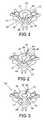
- FIG. 1is a top sectional view showing a ruptured intervertebral disc
- FIG. 2is a top sectional view showing a guide tube advanced through the access port in the anterior of the disc annulus;
- FIG. 3is a top sectional view showing the pulp removed from the disc
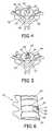
- FIG. 4is a top sectional view showing the balloon assembly expanded by a fluid
- FIG. 5is a top sectional view showing the filled balloon structure and the instrumentation removed.
- FIG. 6is a lateral sectional side view showing the filled balloon maintaining distraction of the adjacent vertebrae.
- the present inventionrelates to a method and device for treating intervertebral disc herniations using endoscopic procedure.
- the methodprovides a minimally invasive procedure which allows for short-term recovery from surgery and a patient's early return to normal activity.
- FIG. 1shows a top sectional view of a herniated disc 15 of a spine 12 .
- Spine 12is shown to have a vertebral canal 28 and having posterior region 19 and anterior area 23 .
- Disc 15is shown having annulus portion 16 surrounding nucleus portion 17 .
- Nucleus portion 17is made up of pulp 18 .
- the disc 15is shown to be herniated or ruptured at herniation 20 , whereby pulp 21 is shown extruding from nucleus 17 and through annulus 16 into the posterior region 19 of the spine thereby causing pain to the patient.
- the expressed or extruded pulp 21 from the disc spacemay be an irritant to nerve tissue that lie posterior to the vertebral column and may be a cause of back pain. It may also be the cause of referred pain and numbness to affected arm, hand, leg or foot areas.
- FIG. 2shows guide tube 14 inserted into disc 15 through annulus 16 and into nucleus 17 .
- Trocar 24is inserted within the guide tube 14 and is used to aide in forming access port 22 so that extruded pulp 21 may be removed.
- FIG. 3shows pulp 18 being removed from the disc interspace through guide tube 14 .
- the guide tube 14is preferably a long radiolucent needle-like probe having an internal diameter of about 2.5 millimeters.
- the guide tube 14is manually guided by imaging technique to the proposed entry or access port 22 to be created in the targeted annulus.
- the access port 22then allows for removal of the nucleus pulposus.
- Disc removal instrument or grasper 25is shown within the guide tube 14 to remove the pulp 18 .
- the annulus 16may next be laser annealed to cause shrinking and tightening of the tissues of the annulus 16 to reduce the size of any lateral or posterior tears in the annulus from which nucleus pulposus 18 may have expressed out from the intervertebral disc space.
- FIG. 4shows balloon assembly 10 being inserted through guide tube 14 and into nucleus 17 .
- Expandable balloon structure 27is shown within the disc 15 of spine 12 .
- FIGS. 5 and 6show balloon assembly 10 secured in disc 15 with the guide tube and instrumentation removed through the incision.
- Disc 15is shown positioned between vertebrae 13 of spine 12 .
- the nubbin or end portion 11 of balloon assembly 10is shown secured in the annulus 16 .
- Balloon assembly 10is shown having valve 26 in nubbin 11 and an expandable balloon structure 27 .
- the nubbin 11 or other portions of the entire balloon assembly 10may be radiolucent in order to make it easier to locate during the endoscopic procedures.
- the balloon assembly 10may have radiolucent markers added thereto for purposes of locating and maneuvering the assembly 10 during the process steps of the invention.
- the balloon structuremay utilize silicone pigments or expansion fluid which is radiolucent.
- the balloon assembly 10is also preferably constructed of MRI and CT compatible materials.
- a physiologically compatible fluidsuch as physiological normal saline or the like is preferably used to fill the balloon via the valve.
- the filled balloondistracts the adjacent vertebrae 13 thereby providing structure in the void or space formerly filled with the removed pulp.
- the filled balloonmay also occupy the intervertebral disc space after the pulp has been removed, whereby the space is substantially occupied.
- a medically suitable optionsuch as powdered hydroxyapatite or cell culture material or the like to facilitate the ingrowth of structured tissue in the intervertebral space formerly occupied by the removed pulp may be used.
- Homologous tissue cell culture seedingmay also be used to facilitate the ingrowth of structured tissue in the distracted space.
- the approximately 3 millimeter skin incisionis closed by suture, staple, bonding agent, adhesive bandage, or like procedure.
- a second endoscopic procedureis performed to remove the balloon assembly 10 .
- the fluidis removed from the balloon structure and the balloon is then removed via the guide tube 14 and through the port in the annulus that the nubbin has kept patent or open.
- An imaging techniquefacilitates guiding the endoscopic guide tube to the radiolucent nubbin.
- suture, bonding agent, adhesive bandage or like procedureis used to close the skin incision.
- the procedure of the inventionemploys a minimally invasive endoscopic procedure which provides for a reduced cost, less time involved surgical procedure, and a patient's short term surgical recovery and early return to normal activity.
- the procedurecan be performed at all areas of the spine, including cervical, thoracic and lumbar areas.
- the procedure of this inventionmay be performed in an outpatient surgical setting.
- the patientis initially placed under general endotracheal anesthesia and wired for intaoperative electromyography and somatic evoked potential spinal monitoring.
- Dexamethasone and a prophylactic antibioticare administered by intravenous route.
- the patientis positioned supine with gentle cervical extension.
- a Phillips standard operative fluoroscopy C-arm or the likeis adjusted to confirm imaging in both anterior-posterior and lateral projections.
- the anterior neckis surgically prepared and draped in sterile fashion. Under fluoroscopic guidance and with digital pressure retracting the carotid sheath the affected disc level is identified.
- the overlying skinis anesthetized with Marcaine 0.25% with epinephrine or the like.
- a short transverse incisionis made in the anterior neck region anterior to the sternocleidomastoid muscle while holding retraction on the carotid sheath and providing a safe interval between the paratrachael and carotid sheath structures.
- the incisionis then bluntly dissected to allow for placement of the discogram needle into the safe interval.
- a trocharis then placed down into the anterolateral aspect of the intended disc.
- Anterior-posterior and lateral radiographsconfirm the position.
- the discogram needleis then placed into the central portion of the disc with position again confirmed by fluoroscopy.
- Lopamidol 51%, or other suitable contrast agentmay be added in a mixture with lidocaine 1% without epinephrine is instilled into the interspace. This step is performed to identify any epidural leaks and marking of the disc to assist with directing and assessing the disc removal.
- the hub of the discogram needleis removed and a 2.5 millimeter dilator is placed over the needle/trocar.
- This process stepis monitored under fluoroscopic guidance down to the anterolateral aspect of the annulus.
- the cannula and the dilatorare both replaced seating the cannula on the anterior annulus.
- the 2.5 millimeter trephineis then inserted into the interspace under fluoroscopic guidance providing an anterior anulotomy.
- the trephine and trocharare then removed.
- the 2.5 millimeter disc removal instrumentis placed into the central region of the disc and the position confirmed with fluoroscopy.
- the 2.5 millimeter grasperis then employed to remove the trephine annular core if the disc removal instrument did not evacuate it.
- Irrigation and aspiration of the disc with resectionis then commenced with approximately 1 to 3 cubic centimeters of disc material collected in about 20 to 30 minutes of combined aspiration and cutting.
- the discectomyis focused in the posterior region of the interspace in the area of the predominant disc herniation. Once a quantitative amount of contrast agent and disc material is removed the graspers are used to remove any free fragments.
- the flexible LASE endoscope by Claris Medical Systems, Inc., Minneapolis, Minn., U.S.A., or the likeis then placed into the interspace with the position confirmed by fluoroscopy and direct vision.
- direct visionlaser discoplasty is accomplished with 800 to 1500 kilojoules using holmium laser by New Star Lasers, Roseville, Calif., U.S.A. or the like.
- the posterior annulus fibersare identified and treated. Additional laser modulation in the uncinate regions further stabilizes the segment and decreases discogenic neuroforaminal encroachment.
- the balloon assemblyis then inserted into the anterior aspect of the interspace and then inflated.
- Spinal monitoringis utilized continuously introperatively to confirm satisfactory response and no neurologic changes.
- the balloon device positionis then confirmed by direct endoscopic and fluoroscopic evaluation.
- the interspaceis irrigated and the instruments are removed.
- the skin incisionis cleansed with a physiologically normal saline soaked sponge and approximated with a 4.0 ethilon stitch while maintaining pressure to minimize bleeding.
- a Philadelphia firm collar or like cervical collaris then placed onto the patient.
- the patientis then extubated and leaves the operating room awake and in the care of an anesthesiologist. After about 3 hours of post surgical monitoring the patient is released to limited home activity.
- the present inventionis a method and assembly which permits surgery for a disc herniation which is relatively non-invasive and which permits the patient a relatively short recovery time.
- An access portis created in the annulus portion of a herniated intervertebral disc.
- extruded pulpis removed from the herniated disc and the annulus may be annealed to aide healing.
- a balloon assemblyis inserted through the guide tube into the nucleus portion of the disc for distraction or occupation of the intervertebral disc space.
- the balloon assemblymay be utilized with a material which acts as a fibrocartiligenous seeding material to enhance the surgical outcome.
- the balloon assemblyis filled with a physiologically compatible fluid to expand it for occupation and distraction purposes.
- the balloon assemblymay have a nubbin, which is secured in the annulus of the disc to ensure that the balloon assembly stays in place and permit easy access to the balloon assembly for removal.
- the balloon assembly elements, such as the nubbinmay be radiolucent to improve visualization of the assembly during insertion, expansion and removal processes.
- the balloon assemblymay be constructed of a dissolvable physiologically compatible composition which would dissolve over a specified period of time to provide support in the disc space at the time of insertion and to dissolve during and after the ingrowth of tissue. This latter structure would alleviate the need for the subsequent balloon assembly removal procedure.
Landscapes
- Health & Medical Sciences (AREA)
- Engineering & Computer Science (AREA)
- Biomedical Technology (AREA)
- Orthopedic Medicine & Surgery (AREA)
- Transplantation (AREA)
- Oral & Maxillofacial Surgery (AREA)
- Animal Behavior & Ethology (AREA)
- Cardiology (AREA)
- Neurology (AREA)
- Heart & Thoracic Surgery (AREA)
- Vascular Medicine (AREA)
- Life Sciences & Earth Sciences (AREA)
- Veterinary Medicine (AREA)
- General Health & Medical Sciences (AREA)
- Public Health (AREA)
- Physical Education & Sports Medicine (AREA)
- Chemical & Material Sciences (AREA)
- Dispersion Chemistry (AREA)
- Surgical Instruments (AREA)
Abstract
Description
This application claims the benefit of U.S. Provisional Application No. 60/326,009 filed on Oct. 9, 2001.
The present invention relates generally to a method and device for treating intervertebral disc herniations using an endoscopic procedure. Particularly, this invention relates to a distraction disc anthroplasty device and method for treating intervertebral disc herniations. More particularly, the invention relates to a device and method of treating intervertebral disc herniations using a temporary flexible balloon device in the treated disc to alleviate pressure between adjacent vertebrae located in the cervical, thoracic, or lumber areas of the spine.
Intervertebral disc herniations are a major source of back pain. Herniations and ruptures of intervertebral discs may also cause pain and numbness in the leg, feet and arms of affected patients. Herniated, or ruptured, discs may be caused by traumatic injury due to accident, illness, the aging process as well as a multiplicity of undefined causes.
Intervertebral discs are located between adjacent vertebrae of the spine and are comprised of an annulus portion surrounding the nucleus pulposus or pulp. A herniation of an intervertebral disc results from a weakened, torn or stretched area of the annulus. Pulp from the nucleus extrudes through the herniated area in the annulus producing pressure on the spinal column and/or adjacent nerves and thereby causing pain. Removing the pulp reduces pressure on the spinal column or adjacent nerves caused by the herniation.
In the past, intervertebral disc injuries have been treated with implantable disc spacers, for example. These prior art methods typically involve invasive surgery which requires relatively long recovery times for the patient.
It is an object of this invention to produce a minimally invasive interposition arthoplasty procedure which allows for short-term recovery from surgery and the patient's early return to normal activity.
The present invention relates to method and device for treating a herniated intervertebral disc. The intervertebral disc may be located in the cervical, thoracic or lumbar area of the spine. The method of the invention includes an endoscopic procedure to create an access port in the annulus portion of the herniated or ruptured intervertebral disc. Using a guide tube through the access port, pulp is removed from the nucleus area of the disc. Next, the tissues of the inner surface of the annulus may be annealed to shrink and tighten the annulus so that any ruptured or injured areas can continue the ingrowth process of fibrocartiligenous tissue deposition. A natural or synthetic material may be placed into the disc space in order to promote tissue growth. A balloon assembly having a valve is inserted into the disc space via the endoscopic guide tube. The balloon is then filled with fluid to distract the adjacent vertebrae or to occupy a portion of the intervertebral disc space. The guide tube is then removed from the access port. When fibrocollagenous tissue has grown into the distracted space, usually a few months to a few years, another endoscopic procedure is performed to remove the balloon assembly.
The balloon assembly includes a nubbin or end portion which may be incorporated into the balloon structure to engage and maintain the access port in the disc annulus. The nubbin and/or other portions of the balloon structure may be radiolucent to improve visualization of the balloon assembly during insertion, expansion and removal. The balloon assembly also includes a valve member for filling and deflating the balloon member. Alternatively, the balloon assembly may be constructed of a dissolvable material.
An object of the present invention is to provide a novel method and device of treating intervertebral disc herniations, known as a major source of back pain. The process provides a minimally invasive procedure which allows for short-term recovery from surgery and the patient's early return to normal activity.
Another object of the invention is to improve visualization of the balloon assembly during insertion, expansion and removal from the intervertebral disc space. A further object of the invention is to promote tissue ingrowth in the intervertebral disc space.
These and other benefits of this invention will become clear from the following description by reference to the drawings.
FIG. 1 is a top sectional view showing a ruptured intervertebral disc;
FIG. 2 is a top sectional view showing a guide tube advanced through the access port in the anterior of the disc annulus;
FIG. 3 is a top sectional view showing the pulp removed from the disc;
FIG. 4 is a top sectional view showing the balloon assembly expanded by a fluid;
FIG. 5 is a top sectional view showing the filled balloon structure and the instrumentation removed; and
FIG. 6 is a lateral sectional side view showing the filled balloon maintaining distraction of the adjacent vertebrae.
The present invention relates to a method and device for treating intervertebral disc herniations using endoscopic procedure. The method provides a minimally invasive procedure which allows for short-term recovery from surgery and a patient's early return to normal activity.
FIG. 1 shows a top sectional view of a herniateddisc 15 of aspine 12.Spine 12 is shown to have avertebral canal 28 and havingposterior region 19 andanterior area 23.Disc 15 is shown havingannulus portion 16 surroundingnucleus portion 17.Nucleus portion 17 is made up ofpulp 18. Thedisc 15 is shown to be herniated or ruptured atherniation 20, wherebypulp 21 is shown extruding fromnucleus 17 and throughannulus 16 into theposterior region 19 of the spine thereby causing pain to the patient. The expressed orextruded pulp 21 from the disc space may be an irritant to nerve tissue that lie posterior to the vertebral column and may be a cause of back pain. It may also be the cause of referred pain and numbness to affected arm, hand, leg or foot areas.
The endoscopic procedure initially involves a surgical skin incision of approximately 3 mm in the skin and through which anendoscopic guide tube 14 is passed. FIG. 2 showsguide tube 14 inserted intodisc 15 throughannulus 16 and intonucleus 17. Trocar24 is inserted within theguide tube 14 and is used to aide in formingaccess port 22 so thatextruded pulp 21 may be removed. FIG. 3 showspulp 18 being removed from the disc interspace throughguide tube 14. Theguide tube 14 is preferably a long radiolucent needle-like probe having an internal diameter of about 2.5 millimeters. Theguide tube 14 is manually guided by imaging technique to the proposed entry oraccess port 22 to be created in the targeted annulus. Theaccess port 22 then allows for removal of the nucleus pulposus. Disc removal instrument orgrasper 25 is shown within theguide tube 14 to remove thepulp 18.
Theannulus 16 may next be laser annealed to cause shrinking and tightening of the tissues of theannulus 16 to reduce the size of any lateral or posterior tears in the annulus from which nucleus pulposus18 may have expressed out from the intervertebral disc space.
FIG. 4 showsballoon assembly 10 being inserted throughguide tube 14 and intonucleus 17.Expandable balloon structure 27 is shown within thedisc 15 ofspine 12. FIGS. 5 and 6show balloon assembly 10 secured indisc 15 with the guide tube and instrumentation removed through the incision.Disc 15 is shown positioned betweenvertebrae 13 ofspine 12. The nubbin orend portion 11 ofballoon assembly 10 is shown secured in theannulus 16.Balloon assembly 10 is shown havingvalve 26 innubbin 11 and anexpandable balloon structure 27.
Thenubbin 11 or other portions of theentire balloon assembly 10 may be radiolucent in order to make it easier to locate during the endoscopic procedures. Alternatively, theballoon assembly 10 may have radiolucent markers added thereto for purposes of locating and maneuvering theassembly 10 during the process steps of the invention. Further, the balloon structure may utilize silicone pigments or expansion fluid which is radiolucent. Theballoon assembly 10 is also preferably constructed of MRI and CT compatible materials. A physiologically compatible fluid such as physiological normal saline or the like is preferably used to fill the balloon via the valve. The filled balloon distracts theadjacent vertebrae 13 thereby providing structure in the void or space formerly filled with the removed pulp. The filled balloon may also occupy the intervertebral disc space after the pulp has been removed, whereby the space is substantially occupied.
It is within the purview of this invention to utilize a balloon assembly constructed of a dissolvable material composition. The utilization of a dissolvable balloon structure, which preferably would dissolve in a specified time period, would alleviate the need of the subsequent removal of the balloon assembly from the patient.
Prior to the placement of the balloon, a medically suitable option such as powdered hydroxyapatite or cell culture material or the like to facilitate the ingrowth of structured tissue in the intervertebral space formerly occupied by the removed pulp may be used. Homologous tissue cell culture seeding may also be used to facilitate the ingrowth of structured tissue in the distracted space.
After removing the endoscopic instruments the approximately 3 millimeter skin incision is closed by suture, staple, bonding agent, adhesive bandage, or like procedure.
After about 1 month to about 3 years, when fibrocollagenous tissue has grown into the intervertebral disc space, a second endoscopic procedure is performed to remove theballoon assembly 10. In this endoscopic procedure the fluid is removed from the balloon structure and the balloon is then removed via theguide tube 14 and through the port in the annulus that the nubbin has kept patent or open. An imaging technique facilitates guiding the endoscopic guide tube to the radiolucent nubbin.
Finally, after all endoscopic instruments are removed, suture, bonding agent, adhesive bandage or like procedure is used to close the skin incision.
The procedure of the invention employs a minimally invasive endoscopic procedure which provides for a reduced cost, less time involved surgical procedure, and a patient's short term surgical recovery and early return to normal activity. The procedure can be performed at all areas of the spine, including cervical, thoracic and lumbar areas.
A more detailed description of the procedure as applied to an anterior cervical discectomy is as follows:
The procedure of this invention may be performed in an outpatient surgical setting. The patient is initially placed under general endotracheal anesthesia and wired for intaoperative electromyography and somatic evoked potential spinal monitoring. Dexamethasone and a prophylactic antibiotic are administered by intravenous route. The patient is positioned supine with gentle cervical extension. A Phillips standard operative fluoroscopy C-arm or the like is adjusted to confirm imaging in both anterior-posterior and lateral projections. The anterior neck is surgically prepared and draped in sterile fashion. Under fluoroscopic guidance and with digital pressure retracting the carotid sheath the affected disc level is identified. The overlying skin is anesthetized with Marcaine 0.25% with epinephrine or the like.
A short transverse incision is made in the anterior neck region anterior to the sternocleidomastoid muscle while holding retraction on the carotid sheath and providing a safe interval between the paratrachael and carotid sheath structures. The incision is then bluntly dissected to allow for placement of the discogram needle into the safe interval. Utilizing digital pressure a trochar is then placed down into the anterolateral aspect of the intended disc. Anterior-posterior and lateral radiographs confirm the position. The discogram needle is then placed into the central portion of the disc with position again confirmed by fluoroscopy. Lopamidol 51%, or other suitable contrast agent may be added in a mixture with lidocaine 1% without epinephrine is instilled into the interspace. This step is performed to identify any epidural leaks and marking of the disc to assist with directing and assessing the disc removal.
Next, the hub of the discogram needle is removed and a 2.5 millimeter dilator is placed over the needle/trocar. This process step is monitored under fluoroscopic guidance down to the anterolateral aspect of the annulus. The cannula and the dilator are both replaced seating the cannula on the anterior annulus. The 2.5 millimeter trephine is then inserted into the interspace under fluoroscopic guidance providing an anterior anulotomy. The trephine and trochar are then removed. The 2.5 millimeter disc removal instrument is placed into the central region of the disc and the position confirmed with fluoroscopy. The 2.5 millimeter grasper is then employed to remove the trephine annular core if the disc removal instrument did not evacuate it. Irrigation and aspiration of the disc with resection is then commenced with approximately 1 to 3 cubic centimeters of disc material collected in about 20 to 30 minutes of combined aspiration and cutting. The discectomy is focused in the posterior region of the interspace in the area of the predominant disc herniation. Once a quantitative amount of contrast agent and disc material is removed the graspers are used to remove any free fragments.
Following the latter step the flexible LASE endoscope by Claris Medical Systems, Inc., Minneapolis, Minn., U.S.A., or the like is then placed into the interspace with the position confirmed by fluoroscopy and direct vision. Using direct vision, laser discoplasty is accomplished with 800 to 1500 kilojoules using holmium laser by New Star Lasers, Roseville, Calif., U.S.A. or the like. Under endoscopic visualization the posterior annulus fibers are identified and treated. Additional laser modulation in the uncinate regions further stabilizes the segment and decreases discogenic neuroforaminal encroachment.
Under fluoroscopic guidance, the balloon assembly is then inserted into the anterior aspect of the interspace and then inflated. Spinal monitoring is utilized continuously introperatively to confirm satisfactory response and no neurologic changes. The balloon device position is then confirmed by direct endoscopic and fluoroscopic evaluation. The interspace is irrigated and the instruments are removed.
The skin incision is cleansed with a physiologically normal saline soaked sponge and approximated with a 4.0 ethilon stitch while maintaining pressure to minimize bleeding. A Philadelphia firm collar or like cervical collar is then placed onto the patient. The patient is then extubated and leaves the operating room awake and in the care of an anesthesiologist. After about 3 hours of post surgical monitoring the patient is released to limited home activity.
While the above described procedure offers patients an additional 5 or 10 years or more without spinal fusion, this procedure, employing the distracted disc arthroplasty device, not only lessens the stresses on adjacent vertebral disc segments but leaves open the possibility of procedure to place a functional prosthetic device that may very likely appear in the near future.
After about a few weeks to about a few years post surgery when fibrocollagenous tissue has grown into the distracted space another endoscopic procedure is performed to remove the balloon device. This second procedure removes fluid from the balloon device to deflate the device for removal via the access port in the annulus. Again, after the endoscopic instruments are removed, the skin incision is closed as previously described. The ingrowth of fibrocollagenous tissue continues to fill the intervertebral disc space that has been vacated by the removal of the balloon device.
In summary, the present invention is a method and assembly which permits surgery for a disc herniation which is relatively non-invasive and which permits the patient a relatively short recovery time. An access port is created in the annulus portion of a herniated intervertebral disc. Using a guide tube through the access port, extruded pulp is removed from the herniated disc and the annulus may be annealed to aide healing. A balloon assembly is inserted through the guide tube into the nucleus portion of the disc for distraction or occupation of the intervertebral disc space. The balloon assembly may be utilized with a material which acts as a fibrocartiligenous seeding material to enhance the surgical outcome. The balloon assembly is filled with a physiologically compatible fluid to expand it for occupation and distraction purposes. The balloon assembly may have a nubbin, which is secured in the annulus of the disc to ensure that the balloon assembly stays in place and permit easy access to the balloon assembly for removal. The balloon assembly elements, such as the nubbin may be radiolucent to improve visualization of the assembly during insertion, expansion and removal processes. Further, the balloon assembly may be constructed of a dissolvable physiologically compatible composition which would dissolve over a specified period of time to provide support in the disc space at the time of insertion and to dissolve during and after the ingrowth of tissue. This latter structure would alleviate the need for the subsequent balloon assembly removal procedure.
As many changes are possible to the method and embodiments of the assemblies of this invention utilizing the teachings thereof, the descriptions above, and the accompanying drawing should be interpreted in the illustrative and not in the limited sense.
Claims (35)
1. In a method for treating a herniated intervertebral disc utilizing an endoscopic procedure, comprising the steps of:
a) creating an access port in the annulus of the herniated disc;
b) removing nucleus pulposus from the disc through said access port and creating a disc space;
c) inserting and inflating a balloon structure in said disc space;
d) allowing the growth of fibrocartilegenus tissue in said disc space; and
e) removing said balloon structure between one month and three years and subsequent the ingrowth of said fibrocartilegenus tissue.
2. The method ofclaim 1 , wherein the disc space and the access port in the annulus are treated prior to step c).
3. The method ofclaim 2 , wherein a powdered hydroxyapatite cell culture material or a homologous tissue cell culture seeding is placed in the disc space and wherein the access port is annealed by means of laser irradiation after step b).
4. The method ofclaim 1 , wherein said access port provided has a diameter between 2.0 mm and 6.0 mm and wherein said balloon structure occupies or distracts said disc space.
5. The method ofclaim 1 , wherein said balloon structure includes a radiolucent material.
6. The method ofclaim 1 , wherein said balloon structure provided comprises a flexible balloon member and a nubbin portion and further wherein a valve member is positioned in said nubbin member.
7. The method ofclaim 6 , wherein said balloon structure is filled with a physiological fluid.
8. A method of treating a herniated intervertebral disc comprising:
a) utilizing an endoscopic procedure and creating an access port having a diameter between 2.0 mm and 6.0 mm in the annulus of the herniated disc;
b) removing the nucleus pulposus of said herniated disc to thereby reduce pressure caused by the herniation and thereby creating a disc space;
c) annealing the inner surface of the annulus by means of laser irradiation;
d) inserting a flexible balloon assembly through said annulus and into the disc space and inflating the balloon in the disc space;
e) permitting fibrocartiligenous tissue to grow in the disc space; and
f) removing the balloon from the disc space.
9. The method ofclaim 8 , wherein the access port created has a diameter of approximately 2.5 to 3.5 mm.
10. The method ofclaim 8 , wherein the flexible balloon has a nubbin and a valve in said nubbin and wherein the nubbin is placed within in the access port of the annulus of the affected intervertebral disc.
11. The method ofclaim 10 , wherein at least a portion of said balloon assembly is radiolucent.
12. The method ofclaim 8 , wherein a second endoscopic surgery procedure is performed to remove the balloon assembly.
13. The method ofclaim 8 , wherein hydroxyapatite is infused into the intervertebral disc space prior to the balloon assembly placement, said hydroxyapatite to facilitate the ingrowth of tissue in the disc space.
14. The method ofclaim 8 , wherein said endoscopic procedure includes incising the skin and passing an endoscopic guide tube therethrough.
15. The method ofclaim 14 , wherein said guide tube is a long radiolucent needle-like probe having an internal diameter of approximately 2.5 millimeters.
16. The method ofclaim 14 , wherein said endoscopic instrument is removed after the insertion of the balloon assembly and whereby the incision is closed by a suture, bonding agent or an adhesive bandage.
17. The method ofclaim 14 , wherein an imaging technique is utilized to guide the endoscopic guide tube.
18. The method ofclaim 8 , wherein said flexible balloon assembly is expanded with physiological normal saline.
19. The method ofclaim 8 , wherein powdered hydroxyapatite, cell culture material or a homologous tissue cell culture seeding is placed in the disc space prior to the placement of the balloon assembly to facilitate the ingrowth of structural tissue.
20. The method ofclaim 8 , wherein said flexible balloon assembly occupies or distracts the disc space.
21. The method ofclaim 8 , wherein the balloon assembly is removed from the disc space between one month to three years after insertion.
22. The method ofclaim 21 , wherein the balloon is deflated and removed via a guide tube and through the port in the annulus.
23. A method for treating a herniated intervertebral disc utilizing an endoscopic procedure, comprising the steps of:
a) creating an access port in the annulus of the herniated disc;
b) removing nucleus pulposus from the disc through said access port and creating a disc space;
c) annealing said disc space and said access port by means of laser irradiation;
d) placing a powdered hydroxyapatite cell culture material or a homologous tissue cell culture seeding in said disc space; and
e) inserting and inflating a balloon structure in said disc space.
24. The method ofclaim 23 , wherein said balloon structure is removed from said disc space subsequent the ingrowth of fibrocartilegenus tissue in said disc space.
25. The method ofclaim 23 , wherein said balloon structure is inflated to occupy or distract said disc space.
26. The method ofclaim 23 , wherein said balloon structure provided comprises a flexible balloon member and a nubbin portion and further wherein a valve member is positioned in said nubbin member and wherein said balloon structure is filled with a physiological fluid.
27. The method ofclaim 23 wherein said balloon structure includes a radiolucent material.
28. The method ofclaim 23 , wherein said access port provided has a diameter between 2.0 mm and 6.0 mm and wherein said endoscopic procedure includes incising the skin and passing an endoscopic guide tube therethrough, wherein an imaging technique is utilized to guide the endoscopic guide tube and wherein a second endoscopic surgery procedure is performed to remove the balloon assembly.
29. The method ofclaim 23 , wherein said balloon structure includes a radiolucent material.
30. A method for treating a herniated intervertebral disc utilizing an endoscopic procedure, comprising the steps of:
a) creating an access port in the annulus of the herniated disc,
b) removing nucleus pulposus from the disc through said access port and creating a disc space;
c) inserting and inflating a balloon structure in said disc space, said balloon structure provided being comprised of a flexible balloon member and a nubbin portion having a valve member; and
d) filling said balloon structure with a physiological fluid.
31. The method ofclaim 30 , wherein said balloon structure is removed from said disc space subsequent the ingrowth of fibrocartilegenus tissue in said disc space.
32. The method ofclaim 30 , annealing the disc space and the access port in the annulus by means of laser irradiation after step b) and placing a powdered hydroxyapatite cell culture material or a homologous tissue cell culture seeding in said disc space.
33. The method ofclaim 30 , wherein said access port provided has a diameter between 2.0 mm and 6.0 mm and wherein said endoscopic procedure includes incising the skin and passing an endoscopic guide tube therethrough, wherein an imaging technique is utilized to guide the endoscopic guide tube and wherein a second endoscopic surgery procedure is preformed to remove the balloon assembly.
34. The method ofclaim 30 , wherein said flexible balloon assembly is expanded with physiological normal saline.
35. The method ofclaim 30 , wherein said flexible balloon assembly is inflated to occupy or distract the disc space.
Priority Applications (2)
| Application Number | Priority Date | Filing Date | Title |
|---|---|---|---|
| US10/147,580US6805715B2 (en) | 2001-10-09 | 2002-05-16 | Method and device for treating intervertebral disc herniations |
| US10/965,988US7128746B2 (en) | 2002-05-16 | 2004-10-15 | Device for treating intervertebral disc herniations |
Applications Claiming Priority (2)
| Application Number | Priority Date | Filing Date | Title |
|---|---|---|---|
| US32600901P | 2001-10-09 | 2001-10-09 | |
| US10/147,580US6805715B2 (en) | 2001-10-09 | 2002-05-16 | Method and device for treating intervertebral disc herniations |
Related Child Applications (1)
| Application Number | Title | Priority Date | Filing Date |
|---|---|---|---|
| US10/965,988Continuation-In-PartUS7128746B2 (en) | 2002-05-16 | 2004-10-15 | Device for treating intervertebral disc herniations |
Publications (2)
| Publication Number | Publication Date |
|---|---|
| US20030069641A1 US20030069641A1 (en) | 2003-04-10 |
| US6805715B2true US6805715B2 (en) | 2004-10-19 |
Family
ID=26845043
Family Applications (1)
| Application Number | Title | Priority Date | Filing Date |
|---|---|---|---|
| US10/147,580Expired - Fee RelatedUS6805715B2 (en) | 2001-10-09 | 2002-05-16 | Method and device for treating intervertebral disc herniations |
Country Status (1)
| Country | Link |
|---|---|
| US (1) | US6805715B2 (en) |
Cited By (68)
| Publication number | Priority date | Publication date | Assignee | Title |
|---|---|---|---|---|
| US20030033017A1 (en)* | 2001-06-29 | 2003-02-13 | The Regents Of The University Of California | Biodegradable/bioactive nucleus pulposus implant and method for treating degenerated intervertebral discs |
| WO2006092015A1 (en)* | 2005-03-01 | 2006-09-08 | Columna Pty Ltd | Intervertebral disc restoration |
| US20060241566A1 (en)* | 2005-04-11 | 2006-10-26 | Orthox, Llc | Nucleus Extraction from Spine Intervertebral Disc |
| US20060247776A1 (en)* | 2005-05-02 | 2006-11-02 | The Board Of Trustees Of The Leland Stanford Junior University | Systems and methods for augmenting intervertebral discs |
| US7189235B2 (en) | 1999-10-20 | 2007-03-13 | Anulex Technologies, Inc. | Spinal disc annulus reconstruction method and spinal disc annulus stent |
| US20070213583A1 (en)* | 2006-03-10 | 2007-09-13 | Kim Daniel H | Percutaneous access and visualization of the spine |
| US20070213584A1 (en)* | 2006-03-10 | 2007-09-13 | Kim Daniel H | Percutaneous access and visualization of the spine |
| US20070232905A1 (en)* | 2006-04-04 | 2007-10-04 | Francis Tom J | Unconstrained Balloon Sizer |
| US20070233252A1 (en)* | 2006-02-23 | 2007-10-04 | Kim Daniel H | Devices, systems and methods for treating intervertebral discs |
| US20070265633A1 (en)* | 2006-05-11 | 2007-11-15 | Moon Jon K | Implement and method to extract nucleus from spine intervertebral disc |
| US20080009826A1 (en)* | 2004-04-16 | 2008-01-10 | Kyphon, Inc. | Spinal diagnostic methods and apparatus |
| US20080091167A1 (en)* | 2002-12-07 | 2008-04-17 | Warsaw Orthopedic, Inc. | Method and Apparatus for Intervertebral Disc Expansion |
| US20080154381A1 (en)* | 2006-12-21 | 2008-06-26 | Rob Gene Parrish | Intervertebral disc spacer |
| US20080161929A1 (en)* | 2006-12-29 | 2008-07-03 | Mccormack Bruce | Cervical distraction device |
| US20080215033A1 (en)* | 2004-04-16 | 2008-09-04 | Kyphon, Inc. | Spinal diagnostic methods and apparatus |
| US20080269754A1 (en)* | 2007-03-06 | 2008-10-30 | Orthobond, Inc. | Preparation Tools and Methods of Using the Same |
| US20090062872A1 (en)* | 2007-08-27 | 2009-03-05 | Singfatt Chin | Balloon cannula system for accessing and visualizing spine and related methods |
| US7520888B2 (en) | 2006-02-14 | 2009-04-21 | Warsaw Orthopedic, Inc. | Treatment of the vertebral column |
| US7544208B1 (en) | 2004-05-03 | 2009-06-09 | Theken Spine, Llc | Adjustable corpectomy apparatus |
| US20090198239A1 (en)* | 2008-02-01 | 2009-08-06 | White William L | Apparatus and procedure for anterior cervical microdiskectomy |
| US20090234457A1 (en)* | 2001-06-29 | 2009-09-17 | The Regents Of The University Of California | Systems, devices and methods for treatment of intervertebral disorders |
| US7615076B2 (en) | 1999-10-20 | 2009-11-10 | Anulex Technologies, Inc. | Method and apparatus for the treatment of the intervertebral disc annulus |
| US7628800B2 (en)* | 2005-06-03 | 2009-12-08 | Warsaw Orthopedic, Inc. | Formed in place corpectomy device |
| US7632294B2 (en) | 2003-09-29 | 2009-12-15 | Promethean Surgical Devices, Llc | Devices and methods for spine repair |
| US7713301B2 (en) | 1994-05-06 | 2010-05-11 | Disc Dynamics, Inc. | Intervertebral disc prosthesis |
| US7828850B2 (en) | 1999-10-20 | 2010-11-09 | Anulex Technologies, Inc. | Methods and devices for spinal disc annulus reconstruction and repair |
| US7918876B2 (en) | 2003-03-24 | 2011-04-05 | Theken Spine, Llc | Spinal implant adjustment device |
| US7922768B2 (en) | 1999-10-20 | 2011-04-12 | Anulex Technologies, Inc. | Spinal disc annulus reconstruction method and deformable spinal disc annulus stent |
| US7935147B2 (en) | 1999-10-20 | 2011-05-03 | Anulex Technologies, Inc. | Method and apparatus for enhanced delivery of treatment device to the intervertebral disc annulus |
| US7951201B2 (en) | 1999-10-20 | 2011-05-31 | Anulex Technologies, Inc. | Method and apparatus for the treatment of the intervertebral disc annulus |
| US8092536B2 (en) | 2006-05-24 | 2012-01-10 | Disc Dynamics, Inc. | Retention structure for in situ formation of an intervertebral prosthesis |
| US8128698B2 (en) | 1999-10-20 | 2012-03-06 | Anulex Technologies, Inc. | Method and apparatus for the treatment of the intervertebral disc annulus |
| US8163022B2 (en) | 2008-10-14 | 2012-04-24 | Anulex Technologies, Inc. | Method and apparatus for the treatment of the intervertebral disc annulus |
| US8267966B2 (en) | 2008-06-06 | 2012-09-18 | Providence Medical Technology, Inc. | Facet joint implants and delivery tools |
| US8361152B2 (en) | 2008-06-06 | 2013-01-29 | Providence Medical Technology, Inc. | Facet joint implants and delivery tools |
| US8460319B2 (en) | 2010-01-11 | 2013-06-11 | Anulex Technologies, Inc. | Intervertebral disc annulus repair system and method |
| US8512347B2 (en) | 2008-06-06 | 2013-08-20 | Providence Medical Technology, Inc. | Cervical distraction/implant delivery device |
| US8556977B2 (en) | 1999-10-20 | 2013-10-15 | Anulex Technologies, Inc. | Tissue anchoring system and method |
| US20140378980A1 (en)* | 2013-06-24 | 2014-12-25 | Roman Lomeli | Cortical Rim-Supporting Interbody Device |
| US9005288B2 (en) | 2008-01-09 | 2015-04-14 | Providence Medical Techonlogy, Inc. | Methods and apparatus for accessing and treating the facet joint |
| USD732667S1 (en) | 2012-10-23 | 2015-06-23 | Providence Medical Technology, Inc. | Cage spinal implant |
| USD745156S1 (en) | 2012-10-23 | 2015-12-08 | Providence Medical Technology, Inc. | Spinal implant |
| US9333086B2 (en) | 2008-06-06 | 2016-05-10 | Providence Medical Technology, Inc. | Spinal facet cage implant |
| US9370295B2 (en) | 2014-01-13 | 2016-06-21 | Trice Medical, Inc. | Fully integrated, disposable tissue visualization device |
| US9381049B2 (en) | 2008-06-06 | 2016-07-05 | Providence Medical Technology, Inc. | Composite spinal facet implant with textured surfaces |
| US9737294B2 (en) | 2013-01-28 | 2017-08-22 | Cartiva, Inc. | Method and system for orthopedic repair |
| US10045686B2 (en) | 2008-11-12 | 2018-08-14 | Trice Medical, Inc. | Tissue visualization and modification device |
| US10179012B2 (en) | 2013-01-28 | 2019-01-15 | Cartiva, Inc. | Systems and methods for orthopedic repair |
| US10201375B2 (en) | 2014-05-28 | 2019-02-12 | Providence Medical Technology, Inc. | Lateral mass fixation system |
| USD841165S1 (en) | 2015-10-13 | 2019-02-19 | Providence Medical Technology, Inc. | Cervical cage |
| US10342579B2 (en) | 2014-01-13 | 2019-07-09 | Trice Medical, Inc. | Fully integrated, disposable tissue visualization device |
| US10405886B2 (en) | 2015-08-11 | 2019-09-10 | Trice Medical, Inc. | Fully integrated, disposable tissue visualization device |
| US10682243B2 (en) | 2015-10-13 | 2020-06-16 | Providence Medical Technology, Inc. | Spinal joint implant delivery device and system |
| USD887552S1 (en) | 2016-07-01 | 2020-06-16 | Providence Medical Technology, Inc. | Cervical cage |
| US10806593B2 (en) | 2013-06-24 | 2020-10-20 | DePuy Synthes Products, Inc. | Cortical rim-supporting interbody device |
| USD911525S1 (en) | 2019-06-21 | 2021-02-23 | Providence Medical Technology, Inc. | Spinal cage |
| US11065039B2 (en) | 2016-06-28 | 2021-07-20 | Providence Medical Technology, Inc. | Spinal implant and methods of using the same |
| USD933230S1 (en) | 2019-04-15 | 2021-10-12 | Providence Medical Technology, Inc. | Cervical cage |
| US11224521B2 (en) | 2008-06-06 | 2022-01-18 | Providence Medical Technology, Inc. | Cervical distraction/implant delivery device |
| USD945621S1 (en) | 2020-02-27 | 2022-03-08 | Providence Medical Technology, Inc. | Spinal cage |
| US11272964B2 (en) | 2008-06-06 | 2022-03-15 | Providence Medical Technology, Inc. | Vertebral joint implants and delivery tools |
| US11547446B2 (en) | 2014-01-13 | 2023-01-10 | Trice Medical, Inc. | Fully integrated, disposable tissue visualization device |
| US11622753B2 (en) | 2018-03-29 | 2023-04-11 | Trice Medical, Inc. | Fully integrated endoscope with biopsy capabilities and methods of use |
| US11648128B2 (en) | 2018-01-04 | 2023-05-16 | Providence Medical Technology, Inc. | Facet screw and delivery device |
| US11871968B2 (en) | 2017-05-19 | 2024-01-16 | Providence Medical Technology, Inc. | Spinal fixation access and delivery system |
| US12004781B2 (en) | 2014-05-27 | 2024-06-11 | Providence Medical Technology, Inc. | Lateral mass fixation implant |
| US12144513B2 (en) | 2018-09-21 | 2024-11-19 | Providence Medical Technology, Inc. | Vertebral joint access and decortication devices and methods of using |
| USD1098433S1 (en) | 2023-12-28 | 2025-10-14 | Providence Medical Technology, Inc. | Spinal cage |
Families Citing this family (18)
| Publication number | Priority date | Publication date | Assignee | Title |
|---|---|---|---|---|
| US6551574B2 (en)* | 1995-06-07 | 2003-04-22 | Rhomed Incorporated | Tuftsin metallopeptide analogs and uses thereof |
| US6805695B2 (en) | 2000-04-04 | 2004-10-19 | Spinalabs, Llc | Devices and methods for annular repair of intervertebral discs |
| WO2004064673A2 (en)* | 2003-01-17 | 2004-08-05 | Psinergi Corporation | Artificial nucleus pulposus and method of injecting same |
| US8029511B2 (en)* | 2004-03-22 | 2011-10-04 | Disc Dynamics, Inc. | Multi-stage biomaterial injection system for spinal implants |
| US20060135959A1 (en)* | 2004-03-22 | 2006-06-22 | Disc Dynamics, Inc. | Nuclectomy method and apparatus |
| US8697139B2 (en) | 2004-09-21 | 2014-04-15 | Frank M. Phillips | Method of intervertebral disc treatment using articular chondrocyte cells |
| US20090264939A9 (en)* | 2004-12-16 | 2009-10-22 | Martz Erik O | Instrument set and method for performing spinal nuclectomy |
| US7608108B2 (en)* | 2005-04-29 | 2009-10-27 | Jmea Corporation | Tissue repair system |
| US7632313B2 (en) | 2005-04-29 | 2009-12-15 | Jmea Corporation | Disc repair system |
| US8702718B2 (en) | 2005-04-29 | 2014-04-22 | Jmea Corporation | Implantation system for tissue repair |
| US20060253199A1 (en)* | 2005-05-03 | 2006-11-09 | Disc Dynamics, Inc. | Lordosis creating nucleus replacement method and apparatus |
| US20060253198A1 (en)* | 2005-05-03 | 2006-11-09 | Disc Dynamics, Inc. | Multi-lumen mold for intervertebral prosthesis and method of using same |
| US8226722B2 (en)* | 2006-06-08 | 2012-07-24 | Francis Pflum | Sac for use in spinal surgery |
| US10143560B2 (en) | 2006-06-08 | 2018-12-04 | Francis Pflum | Sac for use in spinal surgery |
| EP2285312A4 (en) | 2008-05-01 | 2014-03-12 | Columna Pty Ltd | Systems methods and apparatuses for formation and insertion of tissue prostheses |
| US8906094B2 (en)* | 2008-12-31 | 2014-12-09 | Spineology, Inc. | System and method for performing percutaneous spinal interbody fusion |
| US8211126B2 (en) | 2009-09-22 | 2012-07-03 | Jmea Corporation | Tissue repair system |
| CN110037767A (en)* | 2019-05-27 | 2019-07-23 | 川北医学院 | Interverbebral disc Minimally Invasive Surgery channel device and rotary cut device |
Citations (9)
| Publication number | Priority date | Publication date | Assignee | Title |
|---|---|---|---|---|
| US4685447A (en) | 1985-03-25 | 1987-08-11 | Pmt Corporation | Tissue expander system |
| US5313962A (en)* | 1991-10-18 | 1994-05-24 | Obenchain Theodore G | Method of performing laparoscopic lumbar discectomy |
| US5888220A (en)* | 1994-05-06 | 1999-03-30 | Advanced Bio Surfaces, Inc. | Articulating joint repair |
| US5948008A (en)* | 1995-12-28 | 1999-09-07 | S.L.T. Japan Co., Ltd. | Apparatus for treatment of lumbar disc herniation |
| US5972015A (en) | 1997-08-15 | 1999-10-26 | Kyphon Inc. | Expandable, asymetric structures for deployment in interior body regions |
| US6066154A (en) | 1994-01-26 | 2000-05-23 | Kyphon Inc. | Inflatable device for use in surgical protocol relating to fixation of bone |
| US6224630B1 (en) | 1998-05-29 | 2001-05-01 | Advanced Bio Surfaces, Inc. | Implantable tissue repair device |
| US6248110B1 (en) | 1994-01-26 | 2001-06-19 | Kyphon, Inc. | Systems and methods for treating fractured or diseased bone using expandable bodies |
| US20030033017A1 (en)* | 2001-06-29 | 2003-02-13 | The Regents Of The University Of California | Biodegradable/bioactive nucleus pulposus implant and method for treating degenerated intervertebral discs |
Family Cites Families (1)
| Publication number | Priority date | Publication date | Assignee | Title |
|---|---|---|---|---|
| US5947008A (en)* | 1998-07-10 | 1999-09-07 | Fullmer; Hazel J. | Steamware system |
- 2002
- 2002-05-16USUS10/147,580patent/US6805715B2/ennot_activeExpired - Fee Related
Patent Citations (11)
| Publication number | Priority date | Publication date | Assignee | Title |
|---|---|---|---|---|
| US4685447A (en) | 1985-03-25 | 1987-08-11 | Pmt Corporation | Tissue expander system |
| US5313962A (en)* | 1991-10-18 | 1994-05-24 | Obenchain Theodore G | Method of performing laparoscopic lumbar discectomy |
| US6066154A (en) | 1994-01-26 | 2000-05-23 | Kyphon Inc. | Inflatable device for use in surgical protocol relating to fixation of bone |
| US6235043B1 (en) | 1994-01-26 | 2001-05-22 | Kyphon, Inc. | Inflatable device for use in surgical protocol relating to fixation of bone |
| US6248110B1 (en) | 1994-01-26 | 2001-06-19 | Kyphon, Inc. | Systems and methods for treating fractured or diseased bone using expandable bodies |
| US5888220A (en)* | 1994-05-06 | 1999-03-30 | Advanced Bio Surfaces, Inc. | Articulating joint repair |
| US5948008A (en)* | 1995-12-28 | 1999-09-07 | S.L.T. Japan Co., Ltd. | Apparatus for treatment of lumbar disc herniation |
| US5972015A (en) | 1997-08-15 | 1999-10-26 | Kyphon Inc. | Expandable, asymetric structures for deployment in interior body regions |
| US6280456B1 (en) | 1997-08-15 | 2001-08-28 | Kyphon Inc | Methods for treating bone |
| US6224630B1 (en) | 1998-05-29 | 2001-05-01 | Advanced Bio Surfaces, Inc. | Implantable tissue repair device |
| US20030033017A1 (en)* | 2001-06-29 | 2003-02-13 | The Regents Of The University Of California | Biodegradable/bioactive nucleus pulposus implant and method for treating degenerated intervertebral discs |
Cited By (149)
| Publication number | Priority date | Publication date | Assignee | Title |
|---|---|---|---|---|
| US7713301B2 (en) | 1994-05-06 | 2010-05-11 | Disc Dynamics, Inc. | Intervertebral disc prosthesis |
| US7766965B2 (en) | 1994-05-06 | 2010-08-03 | Disc Dynamics, Inc. | Method of making an intervertebral disc prosthesis |
| US8048160B2 (en) | 1999-10-20 | 2011-11-01 | Anulex Technologies, Inc. | Intervertebral disc annulus stent |
| US7922768B2 (en) | 1999-10-20 | 2011-04-12 | Anulex Technologies, Inc. | Spinal disc annulus reconstruction method and deformable spinal disc annulus stent |
| US8088165B2 (en) | 1999-10-20 | 2012-01-03 | Anulex Technologies, Inc. | Spinal disc annulus reconstruction method and deformable spinal disc annulus stent |
| US7615076B2 (en) | 1999-10-20 | 2009-11-10 | Anulex Technologies, Inc. | Method and apparatus for the treatment of the intervertebral disc annulus |
| US7189235B2 (en) | 1999-10-20 | 2007-03-13 | Anulex Technologies, Inc. | Spinal disc annulus reconstruction method and spinal disc annulus stent |
| US9675347B2 (en) | 1999-10-20 | 2017-06-13 | Krt Investors, Inc. | Apparatus for the treatment of tissue |
| US8034112B2 (en) | 1999-10-20 | 2011-10-11 | Anulex Technologies, Inc. | Spinal disc annulus reconstruction method and spinal disc annulus stent |
| US7993405B2 (en) | 1999-10-20 | 2011-08-09 | Anulex Technologies, Inc. | Spinal disc annulus repair system and methods |
| US7985257B2 (en) | 1999-10-20 | 2011-07-26 | Anulex Technologies, Inc. | Methods and devices for spinal disc annulus reconstruction and repair |
| US7963992B2 (en) | 1999-10-20 | 2011-06-21 | Anulex Technologies, Inc. | Method and apparatus for the treatment of the intervertebral disc annulus |
| US7951201B2 (en) | 1999-10-20 | 2011-05-31 | Anulex Technologies, Inc. | Method and apparatus for the treatment of the intervertebral disc annulus |
| US7935147B2 (en) | 1999-10-20 | 2011-05-03 | Anulex Technologies, Inc. | Method and apparatus for enhanced delivery of treatment device to the intervertebral disc annulus |
| US7670379B2 (en) | 1999-10-20 | 2010-03-02 | Anulex Technologies, Inc. | Spinal disc annulus reconstruction method |
| US7749273B2 (en) | 1999-10-20 | 2010-07-06 | Anulex Technologies, Inc. | Method and apparatus for the treatment of the intervertebral disc annulus |
| US9114025B2 (en) | 1999-10-20 | 2015-08-25 | Krt Investors, Inc. | Methods and devices for spinal disc annulus reconstruction and repair |
| US7909879B2 (en) | 1999-10-20 | 2011-03-22 | Anulex Technologies, Inc. | Intervertebral disc annulus stent |
| US8128698B2 (en) | 1999-10-20 | 2012-03-06 | Anulex Technologies, Inc. | Method and apparatus for the treatment of the intervertebral disc annulus |
| US8632590B2 (en) | 1999-10-20 | 2014-01-21 | Anulex Technologies, Inc. | Apparatus and methods for the treatment of the intervertebral disc |
| US7828850B2 (en) | 1999-10-20 | 2010-11-09 | Anulex Technologies, Inc. | Methods and devices for spinal disc annulus reconstruction and repair |
| US8556977B2 (en) | 1999-10-20 | 2013-10-15 | Anulex Technologies, Inc. | Tissue anchoring system and method |
| US9095442B2 (en) | 1999-10-20 | 2015-08-04 | Krt Investors, Inc. | Method and apparatus for the treatment of the intervertebral disc annulus |
| US20030033017A1 (en)* | 2001-06-29 | 2003-02-13 | The Regents Of The University Of California | Biodegradable/bioactive nucleus pulposus implant and method for treating degenerated intervertebral discs |
| US7641691B2 (en) | 2001-06-29 | 2010-01-05 | The Regents Of The University Of California | Biodegradable/bioactive nucleus pulposus implant and method for treating degenerated intervertebral discs |
| US20090234457A1 (en)* | 2001-06-29 | 2009-09-17 | The Regents Of The University Of California | Systems, devices and methods for treatment of intervertebral disorders |
| US7156877B2 (en) | 2001-06-29 | 2007-01-02 | The Regents Of The University Of California | Biodegradable/bioactive nucleus pulposus implant and method for treating degenerated intervertebral discs |
| US20080091167A1 (en)* | 2002-12-07 | 2008-04-17 | Warsaw Orthopedic, Inc. | Method and Apparatus for Intervertebral Disc Expansion |
| US7918876B2 (en) | 2003-03-24 | 2011-04-05 | Theken Spine, Llc | Spinal implant adjustment device |
| US7632294B2 (en) | 2003-09-29 | 2009-12-15 | Promethean Surgical Devices, Llc | Devices and methods for spine repair |
| US7905874B2 (en)* | 2004-04-16 | 2011-03-15 | Kyphon Sarl | Spinal diagnostic methods and apparatus |
| US7955312B2 (en)* | 2004-04-16 | 2011-06-07 | Kyphon Sarl | Spinal diagnostic methods and apparatus |
| US8157786B2 (en)* | 2004-04-16 | 2012-04-17 | Kyphon Sarl | Spinal diagnostic methods and apparatus |
| US20080009826A1 (en)* | 2004-04-16 | 2008-01-10 | Kyphon, Inc. | Spinal diagnostic methods and apparatus |
| US20080009828A1 (en)* | 2004-04-16 | 2008-01-10 | Kyphon, Inc. | Spinal diagnostic methods and apparatus |
| US20080215033A1 (en)* | 2004-04-16 | 2008-09-04 | Kyphon, Inc. | Spinal diagnostic methods and apparatus |
| US7452351B2 (en)* | 2004-04-16 | 2008-11-18 | Kyphon Sarl | Spinal diagnostic methods and apparatus |
| US7824390B2 (en)* | 2004-04-16 | 2010-11-02 | Kyphon SÀRL | Spinal diagnostic methods and apparatus |
| US7544208B1 (en) | 2004-05-03 | 2009-06-09 | Theken Spine, Llc | Adjustable corpectomy apparatus |
| US20080195210A1 (en)* | 2005-03-01 | 2008-08-14 | Columna Pty Ltd | Intervertebral Disc Restoration |
| CN101132748B (en)* | 2005-03-01 | 2010-06-16 | 斯拜恩塞尔有限公司 | intervertebral disc repair |
| WO2006092015A1 (en)* | 2005-03-01 | 2006-09-08 | Columna Pty Ltd | Intervertebral disc restoration |
| US20060241566A1 (en)* | 2005-04-11 | 2006-10-26 | Orthox, Llc | Nucleus Extraction from Spine Intervertebral Disc |
| US7857857B2 (en) | 2005-05-02 | 2010-12-28 | The Board Of Trustees Of The Leland Stanford Junior University | Devices, systems and methods for augmenting intervertebral discs |
| US20060247784A1 (en)* | 2005-05-02 | 2006-11-02 | Kim Daniel H | Devices, systems and methods for augmenting intervertebral discs |
| US20060247776A1 (en)* | 2005-05-02 | 2006-11-02 | The Board Of Trustees Of The Leland Stanford Junior University | Systems and methods for augmenting intervertebral discs |
| US20090312806A1 (en)* | 2005-06-03 | 2009-12-17 | Sherman Michael C | Formed in Place Corpectomy Device |
| US7628800B2 (en)* | 2005-06-03 | 2009-12-08 | Warsaw Orthopedic, Inc. | Formed in place corpectomy device |
| US7520888B2 (en) | 2006-02-14 | 2009-04-21 | Warsaw Orthopedic, Inc. | Treatment of the vertebral column |
| US8163018B2 (en) | 2006-02-14 | 2012-04-24 | Warsaw Orthopedic, Inc. | Treatment of the vertebral column |
| US20070233252A1 (en)* | 2006-02-23 | 2007-10-04 | Kim Daniel H | Devices, systems and methods for treating intervertebral discs |
| US20070213584A1 (en)* | 2006-03-10 | 2007-09-13 | Kim Daniel H | Percutaneous access and visualization of the spine |
| US20070213583A1 (en)* | 2006-03-10 | 2007-09-13 | Kim Daniel H | Percutaneous access and visualization of the spine |
| US20070232905A1 (en)* | 2006-04-04 | 2007-10-04 | Francis Tom J | Unconstrained Balloon Sizer |
| US20070265633A1 (en)* | 2006-05-11 | 2007-11-15 | Moon Jon K | Implement and method to extract nucleus from spine intervertebral disc |
| US8092536B2 (en) | 2006-05-24 | 2012-01-10 | Disc Dynamics, Inc. | Retention structure for in situ formation of an intervertebral prosthesis |
| US20080154381A1 (en)* | 2006-12-21 | 2008-06-26 | Rob Gene Parrish | Intervertebral disc spacer |
| US8951300B2 (en) | 2006-12-21 | 2015-02-10 | Rob Gene Parrish | Intervertebral disc spacer |
| US7824431B2 (en) | 2006-12-29 | 2010-11-02 | Providence Medical Technology, Inc. | Cervical distraction method |
| US9622873B2 (en) | 2006-12-29 | 2017-04-18 | Providence Medical Technology, Inc. | Cervical distraction method |
| US8834530B2 (en) | 2006-12-29 | 2014-09-16 | Providence Medical Technology, Inc. | Cervical distraction method |
| US20080161929A1 (en)* | 2006-12-29 | 2008-07-03 | Mccormack Bruce | Cervical distraction device |
| US11285010B2 (en) | 2006-12-29 | 2022-03-29 | Providence Medical Technology, Inc. | Cervical distraction method |
| US8348979B2 (en) | 2006-12-29 | 2013-01-08 | Providence Medical Technology, Inc. | Cervical distraction method |
| US10219910B2 (en) | 2006-12-29 | 2019-03-05 | Providence Medical Technology, Inc. | Cervical distraction method |
| US20080269754A1 (en)* | 2007-03-06 | 2008-10-30 | Orthobond, Inc. | Preparation Tools and Methods of Using the Same |
| WO2009029639A1 (en)* | 2007-08-27 | 2009-03-05 | Spine View, Inc. | Balloon cannula system for accessing and visualizing spine and related methods |
| US20090062871A1 (en)* | 2007-08-27 | 2009-03-05 | Singfatt Chin | Balloon cannula system for accessing and visualizing spine and related methods |
| US20090062872A1 (en)* | 2007-08-27 | 2009-03-05 | Singfatt Chin | Balloon cannula system for accessing and visualizing spine and related methods |
| US20090216284A1 (en)* | 2007-08-27 | 2009-08-27 | Singfatt Chin | Balloon cannula system for accessing and visualizing spine and related methods |
| US9005288B2 (en) | 2008-01-09 | 2015-04-14 | Providence Medical Techonlogy, Inc. | Methods and apparatus for accessing and treating the facet joint |
| US11559408B2 (en) | 2008-01-09 | 2023-01-24 | Providence Medical Technology, Inc. | Methods and apparatus for accessing and treating the facet joint |
| US8118845B2 (en)* | 2008-02-01 | 2012-02-21 | William M White | Apparatus and procedure for anterior cervical microdiskectomy |
| US20090198239A1 (en)* | 2008-02-01 | 2009-08-06 | White William L | Apparatus and procedure for anterior cervical microdiskectomy |
| US8753377B2 (en) | 2008-06-06 | 2014-06-17 | Providence Medical Technology, Inc. | Vertebral joint implants and delivery tools |
| US11224521B2 (en) | 2008-06-06 | 2022-01-18 | Providence Medical Technology, Inc. | Cervical distraction/implant delivery device |
| US8828062B2 (en) | 2008-06-06 | 2014-09-09 | Providence Medical Technology, Inc. | Vertebral joint implants and delivery tools |
| US8753347B2 (en) | 2008-06-06 | 2014-06-17 | Providence Medical Technology, Inc. | Vertebral joint implants and delivery tools |
| US8834472B2 (en) | 2008-06-06 | 2014-09-16 | Providence Medical Technology, Inc. | Vertebral joint implants and delivery tools |
| US12409044B2 (en) | 2008-06-06 | 2025-09-09 | Providence Medical Technology, Inc. | Cervical distraction/implant delivery device |
| US11890038B2 (en) | 2008-06-06 | 2024-02-06 | Providence Medical Technology, Inc. | Vertebral joint implants and delivery tools |
| US8623054B2 (en) | 2008-06-06 | 2014-01-07 | Providence Medical Technology, Inc. | Vertebral joint implants and delivery tools |
| US9011492B2 (en) | 2008-06-06 | 2015-04-21 | Providence Medical Technology, Inc. | Facet joint implants and delivery tools |
| US11344339B2 (en) | 2008-06-06 | 2022-05-31 | Providence Medical Technology, Inc. | Vertebral joint implants and delivery tools |
| US8512347B2 (en) | 2008-06-06 | 2013-08-20 | Providence Medical Technology, Inc. | Cervical distraction/implant delivery device |
| US8267966B2 (en) | 2008-06-06 | 2012-09-18 | Providence Medical Technology, Inc. | Facet joint implants and delivery tools |
| US11272964B2 (en) | 2008-06-06 | 2022-03-15 | Providence Medical Technology, Inc. | Vertebral joint implants and delivery tools |
| US8753345B2 (en) | 2008-06-06 | 2014-06-17 | Providence Medical Technology, Inc. | Vertebral joint implants and delivery tools |
| US9333086B2 (en) | 2008-06-06 | 2016-05-10 | Providence Medical Technology, Inc. | Spinal facet cage implant |
| US11141144B2 (en) | 2008-06-06 | 2021-10-12 | Providence Medical Technology, Inc. | Facet joint implants and delivery tools |
| US9381049B2 (en) | 2008-06-06 | 2016-07-05 | Providence Medical Technology, Inc. | Composite spinal facet implant with textured surfaces |
| US11058553B2 (en) | 2008-06-06 | 2021-07-13 | Providence Medical Technology, Inc. | Spinal facet cage implant |
| US10588672B2 (en) | 2008-06-06 | 2020-03-17 | Providence Medical Technology, Inc. | Vertebral joint implants and delivery tools |
| US9622791B2 (en) | 2008-06-06 | 2017-04-18 | Providence Medical Technology, Inc. | Vertebral joint implants and delivery tools |
| US9622874B2 (en) | 2008-06-06 | 2017-04-18 | Providence Medical Technology, Inc. | Cervical distraction/implant delivery device |
| US9629665B2 (en) | 2008-06-06 | 2017-04-25 | Providence Medical Technology, Inc. | Vertebral joint implants and delivery tools |
| US8425558B2 (en) | 2008-06-06 | 2013-04-23 | Providence Medical Technology, Inc. | Vertebral joint implants and delivery tools |
| US10568666B2 (en) | 2008-06-06 | 2020-02-25 | Providence Medical Technology, Inc. | Vertebral joint implants and delivery tools |
| US10456175B2 (en) | 2008-06-06 | 2019-10-29 | Providence Medical Technology, Inc. | Vertebral joint implants and delivery tools |
| US10039649B2 (en) | 2008-06-06 | 2018-08-07 | Providence Medical Technology, Inc. | Composite spinal facet implant with textured surfaces |
| US10238501B2 (en) | 2008-06-06 | 2019-03-26 | Providence Medical Technology, Inc. | Cervical distraction/implant delivery device |
| US10226285B2 (en) | 2008-06-06 | 2019-03-12 | Providence Medical Technology, Inc. | Vertebral joint implants and delivery tools |
| US8361152B2 (en) | 2008-06-06 | 2013-01-29 | Providence Medical Technology, Inc. | Facet joint implants and delivery tools |
| US10149673B2 (en) | 2008-06-06 | 2018-12-11 | Providence Medical Technology, Inc. | Facet joint implants and delivery tools |
| US10172721B2 (en) | 2008-06-06 | 2019-01-08 | Providence Technology, Inc. | Spinal facet cage implant |
| US8163022B2 (en) | 2008-10-14 | 2012-04-24 | Anulex Technologies, Inc. | Method and apparatus for the treatment of the intervertebral disc annulus |
| US8454697B2 (en) | 2008-10-14 | 2013-06-04 | Anulex Technologies, Inc. | Method and apparatus for the treatment of tissue |
| US9192372B2 (en) | 2008-10-14 | 2015-11-24 | Krt Investors, Inc. | Method for the treatment of tissue |
| US10045686B2 (en) | 2008-11-12 | 2018-08-14 | Trice Medical, Inc. | Tissue visualization and modification device |
| US8652153B2 (en) | 2010-01-11 | 2014-02-18 | Anulex Technologies, Inc. | Intervertebral disc annulus repair system and bone anchor delivery tool |
| US8460319B2 (en) | 2010-01-11 | 2013-06-11 | Anulex Technologies, Inc. | Intervertebral disc annulus repair system and method |
| US9795372B2 (en) | 2010-01-11 | 2017-10-24 | Krt Investors, Inc. | Intervertebral disc annulus repair system and bone anchor delivery tool |
| USD745156S1 (en) | 2012-10-23 | 2015-12-08 | Providence Medical Technology, Inc. | Spinal implant |
| USRE48501E1 (en) | 2012-10-23 | 2021-04-06 | Providence Medical Technology, Inc. | Cage spinal implant |
| USD732667S1 (en) | 2012-10-23 | 2015-06-23 | Providence Medical Technology, Inc. | Cage spinal implant |
| US9737294B2 (en) | 2013-01-28 | 2017-08-22 | Cartiva, Inc. | Method and system for orthopedic repair |
| US11471199B2 (en) | 2013-01-28 | 2022-10-18 | Cartiva, Inc. | Systems and methods for orthopedic repair |
| US10179012B2 (en) | 2013-01-28 | 2019-01-15 | Cartiva, Inc. | Systems and methods for orthopedic repair |
| US20180271576A1 (en)* | 2013-06-24 | 2018-09-27 | DePuy Synthes Products, Inc. | Cortical Rim-Supporting Interbody Device |
| US20140378980A1 (en)* | 2013-06-24 | 2014-12-25 | Roman Lomeli | Cortical Rim-Supporting Interbody Device |
| US10758288B2 (en)* | 2013-06-24 | 2020-09-01 | DePuy Synthes Products, Inc. | Cortical rim-supporting interbody device |
| US10806593B2 (en) | 2013-06-24 | 2020-10-20 | DePuy Synthes Products, Inc. | Cortical rim-supporting interbody device |
| US12035954B2 (en) | 2013-06-24 | 2024-07-16 | DePuy Synthes Products, Inc. | Cortical rim-supporting interbody device |
| US9370295B2 (en) | 2014-01-13 | 2016-06-21 | Trice Medical, Inc. | Fully integrated, disposable tissue visualization device |
| US10342579B2 (en) | 2014-01-13 | 2019-07-09 | Trice Medical, Inc. | Fully integrated, disposable tissue visualization device |
| US9610007B2 (en) | 2014-01-13 | 2017-04-04 | Trice Medical, Inc. | Fully integrated, disposable tissue visualization device |
| US11547446B2 (en) | 2014-01-13 | 2023-01-10 | Trice Medical, Inc. | Fully integrated, disposable tissue visualization device |
| US10092176B2 (en) | 2014-01-13 | 2018-10-09 | Trice Medical, Inc. | Fully integrated, disposable tissue visualization device |
| US10398298B2 (en) | 2014-01-13 | 2019-09-03 | Trice Medical, Inc. | Fully integrated, disposable tissue visualization device |
| US12004781B2 (en) | 2014-05-27 | 2024-06-11 | Providence Medical Technology, Inc. | Lateral mass fixation implant |
| US11058466B2 (en) | 2014-05-28 | 2021-07-13 | Providence Medical Technology, Inc. | Lateral mass fixation system |
| US10201375B2 (en) | 2014-05-28 | 2019-02-12 | Providence Medical Technology, Inc. | Lateral mass fixation system |
| US10405886B2 (en) | 2015-08-11 | 2019-09-10 | Trice Medical, Inc. | Fully integrated, disposable tissue visualization device |
| US10945588B2 (en) | 2015-08-11 | 2021-03-16 | Trice Medical, Inc. | Fully integrated, disposable tissue visualization device |
| USD841165S1 (en) | 2015-10-13 | 2019-02-19 | Providence Medical Technology, Inc. | Cervical cage |
| USD884895S1 (en) | 2015-10-13 | 2020-05-19 | Providence Medical Technology, Inc. | Cervical cage |
| US10682243B2 (en) | 2015-10-13 | 2020-06-16 | Providence Medical Technology, Inc. | Spinal joint implant delivery device and system |
| US11065039B2 (en) | 2016-06-28 | 2021-07-20 | Providence Medical Technology, Inc. | Spinal implant and methods of using the same |
| USD887552S1 (en) | 2016-07-01 | 2020-06-16 | Providence Medical Technology, Inc. | Cervical cage |
| US11871968B2 (en) | 2017-05-19 | 2024-01-16 | Providence Medical Technology, Inc. | Spinal fixation access and delivery system |
| US11648128B2 (en) | 2018-01-04 | 2023-05-16 | Providence Medical Technology, Inc. | Facet screw and delivery device |
| US11813172B2 (en) | 2018-01-04 | 2023-11-14 | Providence Medical Technology, Inc. | Facet screw and delivery device |
| US11622753B2 (en) | 2018-03-29 | 2023-04-11 | Trice Medical, Inc. | Fully integrated endoscope with biopsy capabilities and methods of use |
| US12144513B2 (en) | 2018-09-21 | 2024-11-19 | Providence Medical Technology, Inc. | Vertebral joint access and decortication devices and methods of using |
| USD933230S1 (en) | 2019-04-15 | 2021-10-12 | Providence Medical Technology, Inc. | Cervical cage |
| USD911525S1 (en) | 2019-06-21 | 2021-02-23 | Providence Medical Technology, Inc. | Spinal cage |
| USD945621S1 (en) | 2020-02-27 | 2022-03-08 | Providence Medical Technology, Inc. | Spinal cage |
| USD1098431S1 (en) | 2023-02-27 | 2025-10-14 | Providence Medical Technology, Inc. | Spinal cage |
| USD1098433S1 (en) | 2023-12-28 | 2025-10-14 | Providence Medical Technology, Inc. | Spinal cage |
Also Published As
| Publication number | Publication date |
|---|---|
| US20030069641A1 (en) | 2003-04-10 |
Similar Documents
| Publication | Publication Date | Title |
|---|---|---|
| US6805715B2 (en) | Method and device for treating intervertebral disc herniations | |
| US7128746B2 (en) | Device for treating intervertebral disc herniations | |
| US5562736A (en) | Method for surgical implantation of a prosthetic spinal disc nucleus | |
| ES2308014T5 (en) | Apparatus for distraction and spinal fusion | |
| US7947079B2 (en) | Method and system for fusing a spinal region | |
| US8709087B2 (en) | Methods and apparatus for performing therapeutic procedures in the spine | |
| US7744599B2 (en) | Articulating spinal implant | |
| US6899716B2 (en) | Method and apparatus for spinal augmentation | |
| US20050143826A1 (en) | Disk repair structures with anchors | |
| US20020016583A1 (en) | Methods of performing procedures in the spine | |
| Leu et al. | Percutaneous endoscopic lumbar spine fusion | |
| US8226722B2 (en) | Sac for use in spinal surgery | |
| US20120136401A1 (en) | Medical Procedure Involving Protective Pad | |
| JP2009532078A (en) | Percutaneous access and visualization of the spine | |
| CN113520677A (en) | Minimally invasive endoscopic implantation expandable lumbar interbody fusion cage | |
| Klekamp | Syringomyelia | |
| ES2370992T3 (en) | FUSION SYSTEM OF A REGION OF THE Dorsal Spine. | |
| McCord et al. | (iv) Anterior endoscopic thoracolumbar instrumentation and implants | |
| PRZYBYLSKI | JAMES S. HARROP | |
| Lilla et al. | Using Natural Anatomical Gaps-Posterior Cervical Approach to Skull Base and Upper Craniocervical Meningiomas without Bone Removal | |
| Harrop et al. | Operative Techniques: Posterior Thoracolumbar Techniques and Surgical Approaches | |
| Van de Kelft | Surgical Procedures: Discectomy and Herniectomy | |
| Crock | Disc Prolapses | |
| UA34754A (en) | Method for surgical treatment of penetrating backbone fractures |
Legal Events
| Date | Code | Title | Description |
|---|---|---|---|
| AS | Assignment | Owner name:PMT CORPORATION, MINNESOTA Free format text:ASSIGNMENT OF ASSIGNORS INTEREST;ASSIGNOR:REHDER, CAREY D.;REEL/FRAME:013008/0866 Effective date:20020606 | |
| FPAY | Fee payment | Year of fee payment:4 | |
| REMI | Maintenance fee reminder mailed | ||
| LAPS | Lapse for failure to pay maintenance fees | ||
| STCH | Information on status: patent discontinuation | Free format text:PATENT EXPIRED DUE TO NONPAYMENT OF MAINTENANCE FEES UNDER 37 CFR 1.362 | |
| FP | Lapsed due to failure to pay maintenance fee | Effective date:20121019 |