US6786900B2 - Cryotherapy methods for treating vessel dissections and side branch occlusion - Google Patents
Cryotherapy methods for treating vessel dissections and side branch occlusionDownload PDFInfo
- Publication number
- US6786900B2 US6786900B2US09/953,500US95350001AUS6786900B2US 6786900 B2US6786900 B2US 6786900B2US 95350001 AUS95350001 AUS 95350001AUS 6786900 B2US6786900 B2US 6786900B2
- Authority
- US
- United States
- Prior art keywords
- blood vessel
- balloon
- vessel
- plaque
- cooling
- Prior art date
- Legal status (The legal status is an assumption and is not a legal conclusion. Google has not performed a legal analysis and makes no representation as to the accuracy of the status listed.)
- Expired - Lifetime
Links
Images


Classifications
- A—HUMAN NECESSITIES
- A61—MEDICAL OR VETERINARY SCIENCE; HYGIENE
- A61B—DIAGNOSIS; SURGERY; IDENTIFICATION
- A61B18/00—Surgical instruments, devices or methods for transferring non-mechanical forms of energy to or from the body
- A61B18/02—Surgical instruments, devices or methods for transferring non-mechanical forms of energy to or from the body by cooling, e.g. cryogenic techniques
- A—HUMAN NECESSITIES
- A61—MEDICAL OR VETERINARY SCIENCE; HYGIENE
- A61B—DIAGNOSIS; SURGERY; IDENTIFICATION
- A61B17/00—Surgical instruments, devices or methods
- A61B17/22—Implements for squeezing-off ulcers or the like on inner organs of the body; Implements for scraping-out cavities of body organs, e.g. bones; for invasive removal or destruction of calculus using mechanical vibrations; for removing obstructions in blood vessels, not otherwise provided for
- A61B2017/22051—Implements for squeezing-off ulcers or the like on inner organs of the body; Implements for scraping-out cavities of body organs, e.g. bones; for invasive removal or destruction of calculus using mechanical vibrations; for removing obstructions in blood vessels, not otherwise provided for with an inflatable part, e.g. balloon, for positioning, blocking, or immobilisation
- A61B2017/22062—Implements for squeezing-off ulcers or the like on inner organs of the body; Implements for scraping-out cavities of body organs, e.g. bones; for invasive removal or destruction of calculus using mechanical vibrations; for removing obstructions in blood vessels, not otherwise provided for with an inflatable part, e.g. balloon, for positioning, blocking, or immobilisation to be filled with liquid
- A—HUMAN NECESSITIES
- A61—MEDICAL OR VETERINARY SCIENCE; HYGIENE
- A61B—DIAGNOSIS; SURGERY; IDENTIFICATION
- A61B18/00—Surgical instruments, devices or methods for transferring non-mechanical forms of energy to or from the body
- A61B2018/00005—Cooling or heating of the probe or tissue immediately surrounding the probe
- A61B2018/00011—Cooling or heating of the probe or tissue immediately surrounding the probe with fluids
- A61B2018/00023—Cooling or heating of the probe or tissue immediately surrounding the probe with fluids closed, i.e. without wound contact by the fluid
- A—HUMAN NECESSITIES
- A61—MEDICAL OR VETERINARY SCIENCE; HYGIENE
- A61B—DIAGNOSIS; SURGERY; IDENTIFICATION
- A61B18/00—Surgical instruments, devices or methods for transferring non-mechanical forms of energy to or from the body
- A61B2018/00053—Mechanical features of the instrument of device
- A61B2018/00214—Expandable means emitting energy, e.g. by elements carried thereon
- A61B2018/0022—Balloons
- A—HUMAN NECESSITIES
- A61—MEDICAL OR VETERINARY SCIENCE; HYGIENE
- A61B—DIAGNOSIS; SURGERY; IDENTIFICATION
- A61B18/00—Surgical instruments, devices or methods for transferring non-mechanical forms of energy to or from the body
- A61B18/02—Surgical instruments, devices or methods for transferring non-mechanical forms of energy to or from the body by cooling, e.g. cryogenic techniques
- A61B2018/0212—Surgical instruments, devices or methods for transferring non-mechanical forms of energy to or from the body by cooling, e.g. cryogenic techniques using an instrument inserted into a body lumen, e.g. catheter
- A—HUMAN NECESSITIES
- A61—MEDICAL OR VETERINARY SCIENCE; HYGIENE
- A61B—DIAGNOSIS; SURGERY; IDENTIFICATION
- A61B18/00—Surgical instruments, devices or methods for transferring non-mechanical forms of energy to or from the body
- A61B18/02—Surgical instruments, devices or methods for transferring non-mechanical forms of energy to or from the body by cooling, e.g. cryogenic techniques
- A61B2018/0231—Characteristics of handpieces or probes
- A61B2018/0262—Characteristics of handpieces or probes using a circulating cryogenic fluid
Definitions
- the present inventionrelates generally to medical methods and kits. More particularly, the present invention provides methods and kits for cryogenically cooling a blood vessel within a patient's vasculature to treat potential or existing dissections in the blood vessel. The present invention further provides methods and kits for cryogenically cooling a bifurcated blood vessel to treat side branch occlusion. Vessel dissections and side branch occlusion often result from angioplasty or other intravascular procedures for treating atherosclerosis and other diseases of the vasculature.
- PTApercutaneous transluminal angioplasty
- Vessel dissectionscompromise the dilated vessel, often constricting or blocking blood flow within the vessel.
- a number of strategieshave been proposed to treat vessel dissections. Previously proposed strategies include prolonged balloon inflation, treatment of the blood vessel with a heated balloon, stenting of the region following balloon angioplasty, and the like. While these proposal have enjoyed varying levels of success, no one of these procedures is proven to be entirely successful.
- stenting of the dilated regionmay address acute problems of intimal dissection and vessel recoil, however stents are believed to actually cause a marked increase in the degree of intimal restenosis or hyperplasia (re-narrowing of the an artery following an initially successful angioplasty). This in turn leads to greater late luminal loss, especially in smaller vessels which are more susceptible to re-closure due to restenosis. Moreover, stents may prove to be an impractical solution when dilating long periphery arteries that may require multiple stent placements. Stents may additionally not always be easily maneuvered to and positioned in dilated regions, especially in the coronary arteries.
- side branch occlusionin a bifurcated blood vessel during dilatation of a primary vessel lesion.
- Side branch occlusioncan occur by several mechanisms.
- the “snow plow” effectmay be the most common mode of side branch occlusion, in which plaque from a primary vessel is literally “plowed” or “shifted” into the adjacent side vessel during dilatation, narrowing or occluding the side vessel lumen.
- Known procedures for treating side branch occlusioninclude the “kissing balloon technique” where two guiding catheters are positioned in the bifurcated vessel, one in the primary vessel and the other in the side branch, and the balloons are inflated simultaneously or sequentially so that they potentially touch or “kiss.”
- angioplasty techniquesalone or in combination with stents, has not been entirely successful in preventing side branch occlusion.
- Balloon catheters for intravascular cooling or heating a patientare described in U.S. Pat. No. 5,486,208 and WO 91/05528.
- a cryosurgical probe with an inflatable bladder for performing intrauterine ablationis described in U.S. Pat. No. 5,501,681.
- Cryosurgical probes relying on Joule-Thomson coolingare described in U.S. Pat. Nos. 5,275,595; 5,190,539; 5,147,355; 5,078,713; and 3,901,241.
- Catheters with heated balloons for post-angioplasty and other treatmentsare described in U.S. Pat. Nos.
- the present inventionprovides cryotherapy treatment of dissections in a blood vessel of a patient.
- the present inventionfurther provides cryotherapy treatment of side branch occlusion in a bifurcated blood vessel.
- the blood vesselmay be any blood vessel in the patient's vasculature, including veins, arteries, and particularly coronary arteries.
- the blood vesselwill be at least partially stenosed, typically by eccentric calcified plaque (i.e. the plaque compromises a vessel lumen predominantly from one side) in the coronary and peripheral arteries.
- the present inventionmay limit, reduce, minimize, prevent, mitigate, and/or inhibit potential or existing dissections of a vessel and/or plaque shift from a main branch into a side branch of a bifurcated blood vessel so as to inhibit acute coronary syndrome.
- the present inventionprovides a method for treating potential or existing dissections in a blood vessel.
- the methodcomprises cooling the blood vessel to a temperature and for a time sufficient to remodel the blood vessel such that dissections of the blood vessel are reduced.
- the cooling treatmentwill often be directed against all or a portion of a circumferential surface of a lumen of the blood vessel.
- Cooling of a vesselmay be effected by introducing a catheter into a lumen of a blood vessel.
- a balloonis positioned within the vessel lumen adjacent the potential or existing dissection.
- Cryogenic cooling fluidis introduced into the balloon and exhausted.
- the balloonexpands to radially engage the vessel lumen.
- the cooling temperature at the cell surface of the blood vessel lumenis in a range from about ⁇ 3° C. to about ⁇ 15° C.
- the tissueis typically maintained at the desired temperature for a time period in the range from about 10 seconds to about 60 seconds, more preferably from about 20 seconds to 40 seconds.
- Vessel dissection treatmentmay be enhanced by repeating cooling in cycles, typically with from about 1 cycle to 3 cycles, with the cycles being repeated at a rate of about one cycle every 60 seconds.
- the dissectionsmay comprise flaps, residual plaque, and/or pieces of tissue resulting from fissuring or tearing of the intima of the blood vessel wall or plaque thereon.
- dissectionsoccur at a junction between the plaque and the vessel wall, wherein the plaque tears at its margins and sends a plane of dissection deep into the media of the vessel wall.
- Dissectionsare undesirable as they often compromise the integrity of the blood vessel by at least partially blocking the blood vessel. Such blockage can limit blood flow and potentially create a threat to acute vessel closure.
- the dissectionsmay further create flow in an abnormal pattern (i.e. flow in planes other than the true vessel lumen or non laminar flow.)
- the blood vesselis subject to dissections resulting from treatment of a stenosis, wherein the treatment of the stenosis typically comprises percutaneous transluminal angioplasty.
- the cooling stepmay be performed before or after balloon angioplasty, and will preferably be performed during balloon angioplasty.
- work in connection with the present inventionhas shown that cooling of the blood vessel reduces and/or inhibits potential or existing dissections so as to produce a “stent-like” angiographic result (i.e. dissection free lumen without the use of a stent).
- coolingmay further minimize or inhibit restenosis or hyperplasia (re-narrowing of the an artery following an initially successful angioplasty) and help maintain the patency of a body lumen. Cooling may also be efficiently effected in long periphery arteries and the cooling apparatus easily maneuvered to and positioned in the treatment vessel, especially in the coronary arteries, so that cooling may be effected in difficult to access areas.
- the cooling stepmay alter mechanical properties of the blood vessel wall or plaque thereon so the that fissuring or tearing of the blood vessel wall or plaque thereon is reduced.
- the blood vessel wall and/or plaqueis solidified so that there is not such a great disparity in compliance between the two.
- the dilatation force applied by the angioplasty cooling balloonis more evenly distributed around a circumference of the vessel wall so that tearing of the vessel at the junction between the vessel wall and plaque is minimized (i.e. any resulting fissures in the vessel wall and plaque are small or micro cracks that do not compromise flow in the vessel). Cooling may also alter a fail mode of the vessel resulting from the modified mechanical properties.
- Coolingmay alternatively or additionally enhance bonding between layers of the blood vessel wall so that fissuring or tearing of the blood vessel wall is reduced.
- the cooling stepmay tack or re-attach existing vessel dissections, resulting from a prior angioplasty procedure, into the blood vessel wall.
- coolingmay soften or weaken the vessel wall or plaque thereon, particularly eccentric calcified plaque, so that the vessel can be dilated or expanded at much lower pressures than is used with conventional angioplasty.
- cooling temperaturesof about ⁇ 10° C. may freeze fluid between spaces in the calcium which in turn breaks up the calcified plaque, softening the vessel so that it can dilated at a lower pressure.
- cooling at low temperaturesmay also freeze and harden non-treatment tissue adjacent to the calcified plaque so that the vessel wall may exhibit more uniform properties against the dilation force applied by the angioplasty cooling balloon.
- the present inventionprovides a method for treating potential or existing dissections in a blood vessel, said method comprising introducing a catheter into a lumen of the blood vessel and positioning a balloon within the vessel lumen adjacent the potential or existing dissection. Cryogenic cooling fluid is introduced into the balloon and exhausted. The balloon is expanded to radially engage the vessel lumen and cool the vessel lumen to a temperature and for a time sufficient to remodel the blood vessel such that dissections of the blood vessel are reduced and/or inhibited. Cooling may comprise adhering the cooling balloon to the blood vessel or plaque thereon so as to minimize any slippage of the cooling balloon.
- coolingprevents the creation of any additional dissections by minimizing such slippage of the cooling balloon and in so doing further allows for controlled dilatation at the stenosis.
- the present inventionprovides a method for treating side branch occlusion in a bifurcated blood vessel, the bifurcated blood vessel having a side branch and a main branch, the main branch having plaque disposed thereon.
- the side branchmay also be at least partially stenosed.
- the methodcomprises introducing a catheter into a lumen of the main branch and positioning a balloon within the main branch adjacent the plaque. Cryogenic cooling fluid is introduced into the balloon and exhausted. The balloon is expanded to radially engage the main branch lumen and an inner surface of the main branch is cooled to a temperature and for a time sufficient to inhibit plaque shift from the main or primary branch into the adjacent or side branch.
- the plaquecomprises a combination of calcified, fatty, and fibrous tissues and is fairly amorphous and slippery so that it easily shifts by its structural nature.
- the side branchis often subject to occlusion by plaque shift from the main branch into the side branch as a result of treatment of plaque in the main branch.
- Treatment of the plaquetypically comprises balloon angioplasty, wherein the cooling step may be performed before, after, or preferably during balloon angioplasty.
- the treatment of plaque in the main branchmay be accompanied by simultaneous or sequential treatment of stenosis in the side branch. It is believed that the cooling step alters mechanical properties of the plaque (i.e. plaque compliance) so that plaque shift from the main branch to the side branch is inhibited.
- coolingmay solidify the plaque so that it less amorphous and thus less susceptible to shifting.
- Plaque solidificationmay further be enhanced by the formation of a temporary ice cap on an orifice of the side branch due to a small portion of the cryoplasty balloon coming into contact with blood cells.
- the present inventionprovides a kit for treating potential or existing dissections in a blood vessel.
- the kitcomprises a catheter having a proximal end, a distal end, and a cooling member. Instructions are included in the kit for use of the catheter. These instructions may comprise the step of cooling the blood vessel adjacent the potential or existing dissection to remodel the blood vessel such that dissections of the blood vessel are reduced.
- the kitmay additionally or alternatively provide for the treatment of side branch occlusion in a bifurcate vessel, wherein the instructions recite the step of cooling a main branch lumen adjacent the plaque to inhibit plaque shift from the main branch into the side branch.
- Such kitsmay include instructions for performing one or more of the above described methods.
- the instructionswill often be printed, optionally being at least in-part disposed on packaging.
- the instructionsmay alternatively comprise a videotape, a CD-ROM or other machine readable code, a graphical representation, or the like showing any of the above described methods.
- the present inventionprovides a method for treating potential elastic recoil in a blood vessel, the method comprising introducing a catheter into a lumen of the blood vessel and positioning a balloon within the vessel lumen adjacent tissue that may potentially recoil.
- the balloonis expanded to radially engage the vessel lumen and cool the vessel lumen to a temperature and for a time sufficient to remodel the blood vessel such that actual elastic recoil is inhibited.
- the cooling stepmay alter structural properties of collagen fibers of the vessel wall such that elastic recoil of the vessel is reduced.
- induction of a phase change in an aqueous component of the adjacent tissue during coolingmay cause acute structural changes to the tissue matrix.
- Dilatation of the vessel by the cooling balloonmay be accompanied by a drop in a balloon surface temperature below a phase transition threshold of physiologic saline.
- the aqueous saline in interstitial spacesi.e. spaces between cells and fibers that constitute the vessel wall
- icemay be nucleated in the interstitial spaces and propagate radially outward through the tissue.
- the expanding icemay in turn impose mechanical compressive forces on collagen fibers and vessel cells.
- the collagen fibers and cellsmay undergo morphological deformation in response to the mechanical forces. Any plastic deformation of the collagen fibers may produce permanent or semi-permanent alteration of the vessel tissue, and consequently may yield an alternation in the structural properties of the tissue.
- possible compacting or compression of collagen fibers by coolingmay substantially alter structural properties, such as elasticity, of the collagen fibers so that elastic recoil of the vessel is reduced.
- the blood vesselis typically subject to elastic recoil resulting from treatment of a stenosis.
- the treatment of stenosistypically comprises balloon angioplasty, wherein the cooling step may be performed before, after, or preferably during balloon angioplasty.
- the vesselis being expanded by balloon expansion which may exert radially directed mechanical forces on the vessel tissue.
- the present inventionprovides a method for producing a smooth luminal surface in a blood vessel that is at least partially stenosed by fatty plaque, said method comprising introducing a catheter in a lumen of the blood vessel and positioning a balloon within the vessel lumen adjacent the fatty plaque.
- the balloonis expanded to radially engage the vessel lumen and cool the vessel lumen to a temperature and for a time sufficient to remodel the blood vessel so as to produce a smooth luminal surface.
- fatty lipid based plaquemay undergo chemical or physical alterations in response to cooling of the plaque below a lipid phase change temperature (typically being in a range from about +15° C. to about 0° C.).
- This remodeling of the vessel and plaque thereonmay in turn produce a smoother luminal surface than can be achieved with conventional angioplasty.
- a smoother luminal surfaceadvantageously provides more laminar flow through the vessel wall, and further reduces any shear stresses on the vessel wall.
- FIGS. 1A-1Care cross-sectional views of blood vessels having dissections.
- FIG. 2Ais a cross-sectional view of a stenosed bifurcated blood vessel.
- FIG. 2Bis a cross-sectional view illustrating plaque shift in the bifurcated blood vessel.
- FIG. 3illustrates an exemplary cryotherapy catheter for treating vessel dissections and side branch occlusion constructed in accordance with the principles of the present invention.
- FIG. 4is a cross-sectional view of the catheter taken along line 4 — 4 in FIG. 3 .
- FIG. 5is a functional flow diagram illustrating the operation of automatic fluid shutoff mechanism of the catheter of FIG. 3 .
- FIG. 6illustrates a cross-sectional view of a cryotherapy catheter with multiple vacuum lumens.
- FIG. 7illustrates an inflation unit for use with the cryotherapy catheter of FIG. 3 .
- FIGS. 8A-8Dillustrate use of the catheter of FIG. 3 for treatment of potential vessel dissections.
- FIGS. 9A-9Cillustrate use of the catheter of FIG. 3 for treatment of existing vessel dissections.
- FIGS. 10-10Cillustrate use of the catheter of FIG. 3 for treatment of side branch occlusion.
- FIG. 11illustrates a vessel dissection or side branch occlusion treatment kit including the apparatus of FIG. 3 and instructions for use.
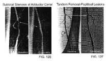
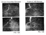
- FIGS. 12A through 13Eare angiographic results of experiments showing an actual and observed reduction in stenosis with a minimum amount of vessel dissection and side branch occlusion as described in the two Experimental sections provided hereinbelow.
- FIGS. 1A-1Cillustrate cross-sectional views of a blood vessel 100 having dissections 102 within a lumen 104 of the vessel.
- the dissections 102generally comprise flaps, residual plaque, and/or pieces of tissue resulting from fissuring or tearing of the intima of the blood vessel wall or plaque thereon from primary treatments like balloon angioplasty.
- Such dissections 102occur at a junction between the plaque and the vessel wall, wherein the plaque tears at its margins and sends a plane of dissection deep into the media of the vessel wall. As shown in FIG.
- dissections 102are undesirable as they often compromise the integrity of the blood vessel by at least partially blocking the blood vessel 100 .
- the dissection 102may shift in direction 106 to limit blood flow and potentially create a threat to acute vessel closure (FIG. 1 B).
- Dissections 102may further create flow in planes other than the true vessel lumen as depicted by arrow 108 .
- FIG. 2Aillustrates a bifurcated blood vessel having a main or primary branch 112 and an adjacent or side branch 114 .
- the main branch 112is at least partially stenosed 116 .
- the plaque 116generally comprises a combination of calcium, fat, and lipids and is fairly amorphous and slippery so that it easily shifts by its structural nature.
- the side branch 114is often subject to occlusion by plaque shift from the main branch 112 into the side branch 114 , as depicted by arrows 118 , as a result of treatment of plaque in the main branch.
- Treatment of plaquetypically comprises balloon angioplasty, wherein balloon dilatation shifts the plaque 116 into the side branch 114 as shown in FIG. 2B, narrowing or occluding the side branch lumen and potentially creating a threat to acute vessel closure.
- an exemplary cryotherapy catheter 10(which is more fully described in co-pending application Ser. No. 09/619,583 filed Jul. 19, 2000 (Attorney Docket No. 018468-000610US) and in application Ser. No. 09/953,464 being filed herewith (Attorney Docket No. 081468-000620US), the full disclosure of which is incorporated herein by reference) for treating dissections 102 in a blood vessel 100 (see FIG. 1A) and/or side branch occlusion (see FIG. 2B) will be described.
- the catheter 10comprises a catheter body 12 having a proximal end 14 and a distal end 16 with a cooling fluid supply lumen 18 and an exhaust lumen 20 extending therebetween.
- a first balloon 22is disposed near the distal end 16 of the catheter body 12 in fluid communication with the supply and exhaust lumens.
- a second balloonis disposed 24 over the first balloon 22 with a barrier 26 , typically a thermal barrier, therebetween.
- the balloons 22 , 24may be an integral extension of the catheter body 12 , but such a structure is not required by the present invention.
- the balloons 22 , 24could be formed from the same or a different material as the catheter body 12 and, in the latter case, attached to the distal end 16 of the catheter body 12 by suitable adhesives, heat welding, or the like.
- the catheter body 12may be formed from conventional materials, such as polyethylenes, polyimides, nylons, polyesters, and copolymers and derivatives thereof.
- the balloons 22 , 24may also be formed from conventional materials used for angioplasty, preferably being inelastic, such as nylon, polyethylene terephthalate (PET), or polyethylene, elastic, such as urethane, latex, or silicone, or other medical grade material suitable for constructing a strong non-distensible balloon. Additionally, balloons 22 and 24 could be formed from different material to provide improved protection. For example, the first balloon 22 could be formed from PET to provide strength while the second balloon 24 could be formed from polyethylene to provide durability.
- the balloons 22 , 24have a length of at least 1 cm each, more preferably in the range from 2 cm to 5 cm each in a coronary artery and 2 cm to 10 cm each in a periphery artery. The balloons 22 , 24 will have diameters in the range from 2 mm to 5 mm each in a coronary artery and 2 mm to 10 mm each in a periphery artery.
- the thermal barrier 26may comprise a separation or gap maintained between the balloons 22 , 24 by a polyester layer.
- the polyester layertypically comprises a woven, braided, helically wound, or knotted polyester material (e.g., Saatifil Polyester PES 38/31 M sold commercially by SaatiTech located in Somers, N.Y.) which may be affixed to the first balloon 22 by adhesion bonding, heat welding, fasteners, or the like.
- the polyester layermay be puncture resistant so as to provide further protection against any leakage of fluid from the first balloon 22 .
- the polyester materialmay additionally help retain balloon folds of the first and/or second balloons 22 , 24 after they are expanded so that the balloons may be more easily drawn back after a treatment procedure.
- a radiopaque marker 30may be disposed on the polyester layer 26 for proper positioning of the cryotherapy balloons 22 , 24 within the target portion of the blood vessel under fluoroscopy.
- the radiopaque marker 30may be disposed on the polyester layer 26 in a stent-like pattern as depicted in FIG. 3 .
- the radiopaque marker 30is preferably a non-toxic contrast medium, such as, gold, and preferably tungsten and does not significantly alter the thermal insulation properties of the barrier.
- the marker 30may be incorporated into an ink and printed onto the polyester layer itself. Areas of the polyester layer adjacent the balloon folds 32 may be free of any radiopaque marking so as to further minimize the balloon profile.
- the radiopaque marker 30may also be electrically conductive so as to monitor any leaks in the separation between the balloons 22 , 24 .
- the thermal barrier 26may alternatively comprise a separation maintained between the balloons by a fluid layer.
- the fluidwill be chosen dependent on its specific thermal properties (e.g., predetermined phase change temperature), viscosity effects, miscibility with a radiopaque contrast agent, and like characteristics.
- the fluid layerwill generally comprise at least one liquid selected from the group consisting of propylene glycol, propylene glycol 200, propylene glycol 300, propylene glycol 400, propylene glycol 600, glycerin, ethyl alcohol 75%, ethyl alcohol 95%, dimethyl sulfoxide, glyceryl formal, N-methyl-2-pyrrolidose, tetrahydrofurfuryl, dimethyl acetamide, and monthiol glycerol.
- the liquidwill generally be diluted with an aqueous solution, such as saline, dextrose 5%, or lactated Ringer's® solution.
- the gap thickness maintained by the fluid layerdepends on several criteria, such as, the pressure or temperature within the first balloon, the desired surface temperature of the second balloon, and the thermal transport properties of the fluid within the gap.
- the gap thickness maintained between the balloons 22 , 24 by the fluid layerwill be in a range from about 0.1 mm to about 0.4 mm, preferably from about 0.25 mm to about 0.3 mm.
- Hubs 38 and 40are secured to the proximal end 14 of the catheter body 12 .
- Hub 38provides a port 42 for a guidewire which extends through a guidewire lumen 44 in the catheter body 12 .
- the guidewire lumen 44will extend through the exhaust lumen 20 , as shown in FIG. 4 .
- the guidewire lumen 44may also extend axially outside the exhaust lumen 20 to minimize the occurrence of cryogenic fluid entering the blood stream via the guidewire lumen 44 .
- the guidewire lumen 44may extend outside the inner surface of the first balloon 22 or the guidewire lumen 44 may allow for a guidewire to extend outside both balloons 22 , 24 .
- Hub 38further provides a balloon deflation port 43 which allows final deflation of the balloon after a treatment procedure.
- Hub 40provides a port 46 for connecting a cryogenic fluid source to the fluid supply lumen 18 which in turn is in fluid communication with the inner surface of the first balloon 22 .
- Hub 40further provides a port 48 for exhausting the cryogenic fluid which travels from balloon 22 in a proximal direction through the exhaust lumen 20 .
- the proximal and distal balloon stems of the first 22 and second 24 balloonmay be staggered along the distal end 16 of the catheter body 10 .
- the balloons stems or bond jointsare staggered to provide a lower balloon folding profile and to allow positioning of a vacuum port 54 , which will be discussed in more detail below, between the proximal balloon stems of the first and second balloons 22 , 24 .
- the balloon stems of the first and second balloonswill be staggered from each other by a distance of 5 mm or less, preferably from about 2 mm to about 3 mm.
- the cryotherapy cathetermay further comprise at least one rupture disk molded into a proximal or distal balloon stem of the first balloon.
- the cryotherapy balloonsmay fail or burst on inner bond joints in a peel mode or the inner balloon may simply burst.
- Such discswill reduce a stem thickness to about 0.0005 inches or less.
- the cryotherapy catheter 10 in FIG. 3additionally illustrates several safety mechanisms that monitor the containment of the cryotherapy system.
- the first balloon 22defines a volume in fluid communication with the supply 18 and exhaust 20 lumens.
- a fluid shutoffcouples a cryogenic fluid supply with the supply lumen 18 .
- the second balloon 24is disposed over the first balloon 22 with a vacuum space 60 therebetween.
- the vacuum space 60is coupled to the fluid shutoff by a plurality of vacuum lumens 62 , as shown in FIG. 6, or optionally by a single vacuum lumen 62 , as shown in FIG. 4 .
- the fluid shutoffinhibits flow of cryogenic fluid into the first balloon 22 in response to a change in at least one of the vacuum lumens 62 and/or in response to a change in the vacuum space 60 .
- FIG. 5illustrates a functional flow diagram of the automatic fluid shutoff mechanism 64 .
- the fluid shutoff 64typically comprises a vacuum switch 66 connected to a shutoff valve 68 by a circuit, the circuit being powered by a battery 70 .
- the switch 66may remain closed only when a predetermined level of vacuum is detected.
- the closed switch 66allows the shutoff valve 68 , in fluid communication with the cryogenic fluid supply 72 , to be open.
- the circuitmay be arranged so that the switch 66 is open only when the predetermined vacuum is present, with the shutoff valve 68 being open when the switch is open.
- the vacuumis reduced when there is a breach in the catheter body 12 , allowing cryogenic fluid or blood to enter at least one vacuum lumen 62 , the first balloon 22 is punctured, allowing cryogenic fluid to enter the vacuum space 52 , or the second balloon 24 is punctured, allowing blood to enter the vacuum space 52 .
- the vacuum switch 66will be triggered to prevent the delivery of additional cryogenic fluid from the fluid supply 72 into the supply lumen 18 .
- the second balloon 24also acts to contain any cryogenic fluid that may have escaped the first balloon 22 .
- the exhaust lumen 20is fluidly connected to a pressure relief valve 21 which in turn will typically vent to atmosphere.
- the vacuummay be provided by a positive displacement pump, such as a syringe 74 , coupled to the vacuum space 60 by a plurality of vacuum lumens 62 of the body 12 via vacuum ports 76 , 54 .
- the syringe 74is disposed within an inflation unit handle 78 .
- a valvesuch as a simple stopcock 80 , may be disposed between the syringe 74 and the vacuum lumens 62 so as to isolate a relatively large syringe volume from a relatively small vacuum volume. This in turn facilitates detection of small fluid leaks, as a change in the vacuum as small as 0.2 mL may be monitored.
- the vacuum space 60comprises a small volume of vacuum in the range from 1 mL to 100 mL, preferably 10 mL or less.
- the battery 70may be electrically coupled to a heater 61 for heating the fluid supply 72 and cryogenic fluid therein to room temperature or warmer so as to enhance the fluid pressure and cooling system performance, as is more fully described in co-pending application Ser. No. 09/268,205.
- the cryogenic fluid supply 72 , battery 70 for powering the circuit, and heater 61may be packaged together in an energy pack 82 that is detachable from the inflation unit 78 via a latch 84 and disposable.
- An overlay 86 of the cryo-inflation unit handle 78is also illustrated in FIG. 7 .
- the fluid supply 72will typically comprise a metallic canister or cartridge that may be transported, stored, and optionally, used at room temperature or warmer. Suitable canisters will hold quantities of cryogenic cooling fluid that are sufficient to cool the target tissue to the treatment temperature range for a time in the predetermined time range. Canisters might have volumes between 10 cc and 20 cc (depending in part on flash expansion temperatures of the cryogenic fluid), and may contain between about 8 g to about 25 g of cooling fluid. Conveniently, such canisters are commercially available for use in whipped cream dispensers. As explained above, the canister 72 may be at room temperature or even chilled, but will preferably be warmed by heater 61 prior to use.
- Suitable cryogenic fluidswill preferably be non-toxic and may include liquid nitrous oxide, liquid carbon dioxide, cooled saline and the like.
- the cryogenic fluidwill flow through the supply lumen 18 as a liquid at an elevated pressure and will vaporize at a lower pressure within the first balloon 22 .
- a delivery pressure within the supply lumen 18will typically be in the range from 600 psi to 1000 psi at a temperature below the associated boiling point.
- the nitrous oxide gas within the first balloon 22 near its centerwill have a pressure typically in the range from 50 psi to 150 psi.
- the nitrous oxide gaswill have a pressure in the range from 75 psi to 125 psi in a peripheral artery and a range from about 75 psi to 125 psi in a coronary artery.
- the cryotherapy cathetermay additionally comprise a thermocouple 90 , pressure transducer, capillary tube, thermistor, or the like coupled to the first balloon 22 to determine the pressure and/or temperature of fluid in the first balloon 22 .
- a thermocouple 90such as a warning light or audio signal on the handle overlay 86 , may additionally be coupled to the thermocouple 90 to provide a signal to an operator of the system when the first balloon temperature is above 0° C.
- an indicatorlets an operator of the system know when to safely remove the cooling balloon following treatment so that any potential tearing of the vessel resulting from a frozen cooling balloon is minimized.
- the cryotherapy catheter 10 in FIG. 3may also monitor the containment of the system (i.e. catheter body 12 , supply lumen 18 , guidewire lumen 44 , balloons 22 , 24 ) prior to any treatment procedures.
- a pressure transducer 23is coupled to the exhaust lumen 20 so as to measure a gas pulse pressure therein.
- the pressure transducer 23is also coupled to the fluid shutoff valve 68 so as to inhibit flow of cryogenic fluid into the first balloon 22 if the pressure measured by the pressure transducer 23 is below 60 psi or above 80 psi.
- the fluid shutoff valve 68may inhibit flow of cryogenic fluid into the first balloon 22 if a pressure decay measured by the pressure transducer 23 is greater than 5 psi.
- the pretreatment testmay be effected by introducing a short pulse of gas, typically nitrous oxide, for a fraction of a second into the first balloon 22 with the supply lumen and exhausting the gas. Containment of the supply lumen 18 , balloon 22 , guidewire lumen 44 , and catheter body 12 are monitored by measuring a gas pulse pressure therein. In the case of a breach in the system, the system will not enter the treatment mode.
- a short pulse of gastypically nitrous oxide
- catheter 10will be introduced into a lumen 104 of the blood vessel 100 over a guidewire GW.
- the blood vessel 100may be any blood vessel in the patient's vasculature, including veins, arteries, and particularly coronary arteries.
- the blood vessel 100will typically be at least partially stenosed 116 .
- the first balloon 22is positioned within the blood vessel lumen 104 adjacent the potential dissection 102 .
- Cryogenic cooling fluidis introduced into the first balloon 22 (in which it often vaporizes) and exhausted.
- the second balloon 24expands to radially engage the vessel wall, as illustrated by FIG. 8 C.
- the vaporized fluidserves to inflate balloon 22 (and expand balloon 24 ) so as to simultaneously dilate and cool the stenosed blood vessel 100 .
- the blood vessel 100is cooled to a temperature and for a time sufficient to remodel the blood vessel such that dissections 102 of the blood vessel wall 100 are reduced.
- the cooling treatmentwill be directed at all or a portion of a circumferential surface the vessel lumen 104 .
- coolingwill reduce and/or inhibit potential dissections so as to produce a “stent-like” angiographic result (i.e. dissection free lumen without the use of a stent), as shown in FIG. 8 D.
- Coolingmay alter mechanical properties of the blood vessel 100 ′ or plaque 116 ′ thereon so the that fissuring or tearing of the blood vessel wall or plaque thereon is reduced.
- the blood vessel wall 100 ′ and/or plaque 116 ′is solidified so that there is not such a great disparity in compliance between the two.
- the dilatation force applied by the angioplasty cooling balloonis more evenly distributed so that tearing of the vessel at the junction between the vessel wall and plaque is minimized.
- Coolingmay further enhance bonding between layer of the blood vessel wall 100 ′ (i.e., intimal layer, medial layer, adventitial layer) so that fissuring or tearing of the blood vessel wall is reduced. Heat transfer will also be inhibited between the first and second balloons 22 , 24 by the thermal barrier 26 so as to limit cooling to a desired temperature profile. Additionally, containment of the first and second balloons 22 , 24 will be monitored during cooling by the fluid shutoff mechanism (see FIG. 5 ).
- Suitable cryogenic fluidswill preferably be non-toxic and may include liquid nitrous oxide, liquid carbon dioxide, cooled saline and the like.
- the cryogenic fluidwill flow through the supply lumen 18 as a liquid at an elevated pressure and will vaporize at a lower pressure within the first balloon 22 .
- a delivery pressure within the supply lumen 18will typically be in the range from 600 psi to 1000 psi at a temperature below the associated boiling point.
- the nitrous oxide gas within the first balloon 22 near its centerwill have a pressure typically in the range from 50 psi to 150 psi.
- the nitrous oxide gaswill have a pressure in the range from 75 psi to 125 psi in a peripheral artery and a range from about 75 psi to 125 psi in a coronary artery.
- the temperature of the outer surface of the first balloon 22will be in a range from about 0° C. to about ⁇ 50° C.
- the temperature of the outer surface of the first balloon 22 in a peripheral arterywill be in a range from about 0° C. to about ⁇ 40° C.
- the temperature of the outer surface of the second balloon 24will be in a range from about ⁇ 3° C. to about ⁇ 15° C. This will provide a desired treatment temperature in a range from about ⁇ 3° C. to about ⁇ 15° C.
- the tissueis typically maintained at the desired temperature for a time period in the range from about 1 to 60 seconds, preferably being from 20 to 40 seconds. Vessel dissection minimization may be further enhanced by repeating cooling in cycles, typically with from about 1 to 3 cycles, with the cycles being repeated at a rate of about one cycle every 60 seconds.
- the cooling stepmay also tack or re-attach existing vessel dissections 102 resulting from a prior angioplasty procedure into the blood vessel wall 100 to produce a “stent-like” angiographic result, as shown in FIG. 9 C.
- Coolingmay additionally comprise adhering the cooling balloon 24 to the blood vessel or plaque thereon so as to minimize any slippage of the cooling balloon and in so doing further allow for controlled dilatation of the vessel.
- the bifurcated blood vessel 110has a side branch 114 and a main branch 112 , the main branch 112 having plaque 116 disposed thereon.
- the side branch 114may also be at least partially stenosed 116 .
- a catheter 10is introduced into a lumen of the main branch 112 and a first balloon 22 positioned within the main branch 112 adjacent the plaque 116 .
- Cryogenic cooling fluidis introduced into the balloon 22 and exhausted.
- a second balloon 24is expanded to radially engage the main branch lumen, as seen in FIG.
- an inner surface of the main branch 112is dilated and cooled to a temperature and for a time sufficient to inhibit plaque shift from the main or primary branch 112 into the adjacent or side branch 114 .
- Coolingmay alter mechanical properties of the plaque 116 ′ (i.e. plaque compliance) so that plaque shift from the main branch 112 to the side branch 114 is inhibited, as seen in FIG. 10 C.
- coolingmay solidify the plaque 116 ′ so that it is less amorphous and thus less susceptible to shifting.
- Plaque solidification 116 ′may further be enhanced by the formation of a temporary ice cap on an orifice of the side branch 114 due to a small portion of the cryoplasty balloon 24 coming into contact with blood cells (not shown).
- FIG. 11A kit 126 including a catheter 10 and instructions for use 128 is illustrated in FIG. 11 .
- Catheter 10may comprise the dual balloon catheter of FIG. 3 or a catheter having a proximal end, a distal end, and a cooling member near its distal end.
- Instructions for use 128may describe any of the associated method steps set forth above for treatment of vessel dissections and/or side branch occlusion. Instructions for use 128 will often be printed, optionally appearing at least in part on a sterile package 130 for balloon catheter 10 .
- instructions for use 128may comprise a machine readable code, digital or analog data graphically illustrating or demonstrating the use of balloon catheter 10 to treat vessel dissections and/or side branch occlusion. Still further alternatives are possible, including printing of the instructions for use on packaging 132 of kit 126 , and the like.
- angiographic imagesshow a baseline where a significant amount of plaque is present within a lumen of a vessel wall, particularly the superficial femoral artery or popliteal artery.
- the effects of cryoplasty dilatationare captured by another set of angiographic images which generally show alleviation of the stenotic condition with a minimum amount of vessel dissection and side branch occlusion.
- the interventionalists for these casesidentified patients with stenotic lesions in the superficial femoral artery (SFA) and popliteal arteries, which were amenable to percutaneous treatment. Eligibility requirements included the following: the patient was at least 18 years of age; the patient had stenotic lesions within the SFA or popliteal artery which required treatment; the target lesion contained a stenosis between 50% and 100%; the patient had a clinical examination within one month prior to treatment including ABI measurement.
- Patientswere excluded if any of the following applied: the patient had an evolving myocardial infarction, or suffered a myocardial infarction within the past 48 hours; the patient was participating currently in another investigational drug or device trial; or the patient suffered a stroke or transient ischemic neurological attack (TIA) within the past two months.
- TIAtransient ischemic neurological attack
- Equipment used for these casesmay be found in any interventional catheterization laboratory (including but not limited to fluoroscopy unit with cine film acquisition; sterile accessories such as sheaths and guidewires).
- the test equipmentwas the CVSi CryoPlasty System, which is composed of the CryoPlasty Catheter, CryoInflation Unit and accessories (battery, battery receptacle, nitrous oxide cylinder).
- the operating parameters of the current casesare treatment time of 50 seconds and surface temperature of ⁇ 10 ⁇ 5° C.
- Procedures usedwere no different than typical interventional percutaneous transluminal angioplasty (PTA) procedures.
- the investigatorshad a thorough understanding of the technical principals, clinical applications, and risks associated with PTA.
- training on the CVSi CryoPlasty System and its Instructions For Usewere given prior to clinical use of the device.
- diagnostic angiography of the target vesselconfirmed the presence of a lesion suitable for endovascular therapy
- the patientsreceived the following treatment.
- the patientwas pre-treated with conventional doses of heparin and aspirin.
- Baseline angiographic imageswere recorded as illustrated in FIGS. 12A through 12L. The operators chose whether or not to predilate the lesion with other percutaneous transluminal devices.
- Results of the percutaneous interventionswere assessed for acute outcomes and in-hospital adverse events.
- Baseline and post-cryoplasty dilation angiographywere compared in each treated section.
- ankle-brachial indexes(ABI) were measured and recorded prior to intervention, 24 hours post-intervention, and at one-month (see Results Table I).
- ABIis an assessment of flow distal to the treated sections.
- Technical successwas based primarily on the acute angiographic appearance and flow characteristics (24-hour ABI) immediately post-cryoplasty dilation compared to the baseline image and the pre-procedural ABI, respectively.
- Procedural safetywas demonstrated by the absence of any incidence of acute serious adverse events or acute percutaneous access site and hemorrhagic adverse events.
- Investigatorswere able to use the CryoPlasty device for primary treatment (i.e. were able to cross severe stenosis and dilate the lesion with the CVSi device without predilation) in ten of the fourteen cases (71%). Twelve of the fourteen cases (86%) were technical successes based on acute angiographic appearance and flow characteristics, with ⁇ 30% residual stenosis by visual assessment of the final angiographic results in the treated segments.
- the 24-hour post-procedural ABIshowed improved blood flow to the lower extremity compared to the pre-procedural measurement.
- Bail-out stentingwas not required in any of the cryotreated segments.
- CVSi CryoPlasty SystemFive patients with coronary artery disease were treated with the CVSi CryoPlasty System to evaluate the safety and effectiveness of the CVSi System in the treatment of de novo and in-stent restenotic lesions. Patients were assessed for acute outcomes and in-hospital adverse events.
- Lesion inclusion criteriawas very broad and included very complex lesions, such as, lesion lengths ranging from 10 mm to 25 mm, stenoses from 90% to total occlusions, and calcified, fibrotic, and soft plaque. Other contributing factors were severe access angulations, high cholesterol, diabetes, and smoking.
- each patientreceived the following treatment.
- the patientwas pretreated with conventional doses of heparin and aspirin.
- Baseline angiographic imageswere recorded as illustrated in FIGS. 13A through 13E.
- the operatorcould choose whether or not to predilate the lesion with conventional transluminal percutaneous procedures.
- the CVSi CryoPlasty Catheterwas advanced across the lesion, and the balloon inflated one or more times with the CVSi CryoInflation Unit. After the final cryotreatment, the balloon was deflated and the CVSi Catheter was removed. At the conclusion of the procedure, a final angiography was recorded as illustrated in FIGS. 13A through 13E.
- MACEmajor adverse cardiac events
- Cryogenic treatment of atherosclerotic stenosesmay effectively limit restenosis in both coronary and peripheral arteries, as well as minimize the amount of vessel dissections and plaque shift post-CryoPlasty dilation.
- Clinical trial dataillustrates that CryoPlasty may represent a simple solution to one of the most vexing problems experienced to date in interventional therapy.
Landscapes
- Health & Medical Sciences (AREA)
- Surgery (AREA)
- Life Sciences & Earth Sciences (AREA)
- Nuclear Medicine, Radiotherapy & Molecular Imaging (AREA)
- Medical Informatics (AREA)
- Engineering & Computer Science (AREA)
- Biomedical Technology (AREA)
- Heart & Thoracic Surgery (AREA)
- Otolaryngology (AREA)
- Molecular Biology (AREA)
- Animal Behavior & Ethology (AREA)
- General Health & Medical Sciences (AREA)
- Public Health (AREA)
- Veterinary Medicine (AREA)
- Media Introduction/Drainage Providing Device (AREA)
- Materials For Medical Uses (AREA)
Abstract
Description
| RESULTS TABLE I: CLINICAL SUMMARY OF PERIPHERAL PATIENTS |
| Ankle-Brachial | |||
| Patient | Age/ | Lesion | Indices |
| No. | Gender | Description | Pre | 24 hr | 1 M | ||
| 1G | 65 yr | LSFA | 80% stenosis distal to stent, 90% stenosis | 0.70 | 1 | 1 | |
| M | (ISR) | proximal to stent | |||||
| 2G | 88 yr | LSFA- | Total occlusion, 10-12 cm | 0.30 | 0.30 | 0 | |
| pop | |||||||
| 3G | |||||||
| 74 yr | LSFA | 98% stenosis with calcified plaque | 0.65 | 1 | 1 | ||
| M | |||||||
| 4G | 75 yr | LSFA | (4) subtotal focal occlusions, calcified, 8-10 | 0.70 | 1 | 1 | |
| M | cm | ||||||
| 5G | |||||||
| 61 | R pop | 70% focal stenosis | 0.70 | 1 | 1 | ||
| M | |||||||
| 6G | 67 yr | L pop | Subtotal popliteal occlusion, 12-15 cm long | 0.25 | 0.70 | 1 | |
| F | |||||||
| 1C | 52 yr | RSFA | 70% stenotis, focal calcified lesion | 0.75 | 1 | 1 | |
| 2C | |||||||
| 61 yr | L pop | 15 mm long, 80% stenotic lesion in proximal | 0.70 | 1.06 | 1 | ||
| F | popliteal and a 10 mm long, 50% stenotic | ||||||
| lesion in the mid popliteal | |||||||
| 3C | 63 yr | LSFA | 10 mm long, 70% stenotic lesion in the | 0.85 | 1.06 | 1 | |
| M | proximal SFA and a 10 mm long, 80% | ||||||
| stenotic lesion in the distal SFA | |||||||
| 4C | 41 yr | Right | heavily calcified sub-occlusive focal lesion in | 0.60 | 1.0 | 1 | |
| M | com. | the right common femoral artery (15 mm | |||||
| fem. | long, 90% stenotic) | ||||||
| 5C | 73 yr | LSFA- | diffuse sub-occlusive (12-16 cm long, 70- | 0.57 | 0.84 | 0.75 | |
| pop | 80% stenotic) disease in proximal to distal | ||||||
| SFA, and 2 focal occlusive lesions in mid | |||||||
| 6C | |||||||
| 74 yr | LSFA | 6-8 cm long total occlusion | 0.57 | 0.85 | 0.80 | ||
| F | |||||||
| 7C | 57 yr | LSFA- | calcified focal lesions in SFA (15 mm long, | 0.58 | 0.89 | 0.97 | |
| M | 80% stenotic) and in proximal popliteal | ||||||
| artery (10 mm long, 70% stenotic). | |||||||
| 61 yr | RSFA | two adjacent 95% stenotic focal lesions | 0.60 | 1.08 | 0.90 | ||
| M | |||||||
| RESULTS TABLE II: CLINICAL SUMMARY OF CORONARY PATIENTS |
| BUC | Age/ | Baseline | Residual | |||||
| # | Sex | Site | Type | Factors | Stenosis | Treatments/ | Stenosis | |
| 1 | 64/F | LCx prox | D | Total occlusion, severe | 100% | (2) CryoPlasty Inflations; distal | 20% | |
| 20 mm × | angulation >100°, some | dissection grade B-not | ||||||
| 2.75 mm | calcification | requiring stent placement; No | ||||||
| 2 | 55/M | LAD prox | D | Stress angina, | 90% | (1) CryoPlasty inflation; | 10% | |
| 15 mm × | changes after 25 meters, | Occlusion angina | ||||||
| 3.0 mm | 237 chol | |||||||
| 3 | 49/F | LAD prox | D | Unstable angina 7 days | 95% sub- | (1) CryoPlasty inflation | 0% | |
| 10 mm × | before, admitted; acute | total | ||||||
| 3.0 mm | MI 1996; sig. angulation | |||||||
| at bifurcation; allergic to | ||||||||
| ASA; narrowing back to | ||||||||
| 4 | 58/F | RCA mid | I | Diabetic, hypertension, | 95% | (4) CryoPlasty inflations | 30% | |
| 25 mm × | dyslipidemia, | |||||||
| 3.5 | PTCA/stent 11/00; | |||||||
| 2/01 | ||||||||
| D | 90% | (2) CryoPlasty inflations > | 0% | |||||
| 10 mm × | Grade B-C dissection > 3.5 mm × | |||||||
| 3.5 | 16 mm stent (post cryo) | |||||||
| 5 | 74/M | LCx mid | D | Colon tumor, surgery | 95% | (1) CryoPlasty | 0% | |
| 12 mm × | prep; calcification | |||||||
| 3.5 mm | ||||||||
Claims (16)
Priority Applications (4)
| Application Number | Priority Date | Filing Date | Title |
|---|---|---|---|
| US09/953,500US6786900B2 (en) | 2001-08-13 | 2001-09-14 | Cryotherapy methods for treating vessel dissections and side branch occlusion |
| PCT/US2002/025758WO2004000092A2 (en) | 2001-08-13 | 2002-08-13 | Cryotherapy methods for treating vessel dissections and side branch occlusion |
| US10/867,986US7862557B2 (en) | 2001-08-13 | 2004-06-14 | Cryotherapy methods for treating vessel dissections and side branch occlusion |
| US12/980,662US20110125141A1 (en) | 2001-08-13 | 2010-12-29 | Cryotherapy Methods for Treating Vessel Dissections and Side Branch Occlusion |
Applications Claiming Priority (2)
| Application Number | Priority Date | Filing Date | Title |
|---|---|---|---|
| US31229501P | 2001-08-13 | 2001-08-13 | |
| US09/953,500US6786900B2 (en) | 2001-08-13 | 2001-09-14 | Cryotherapy methods for treating vessel dissections and side branch occlusion |
Related Child Applications (2)
| Application Number | Title | Priority Date | Filing Date |
|---|---|---|---|
| US10/867,986ContinuationUS7862557B2 (en) | 2001-08-13 | 2004-06-14 | Cryotherapy methods for treating vessel dissections and side branch occlusion |
| US10/867,986DivisionUS7862557B2 (en) | 2001-08-13 | 2004-06-14 | Cryotherapy methods for treating vessel dissections and side branch occlusion |
Publications (2)
| Publication Number | Publication Date |
|---|---|
| US20030036752A1 US20030036752A1 (en) | 2003-02-20 |
| US6786900B2true US6786900B2 (en) | 2004-09-07 |
Family
ID=26978326
Family Applications (3)
| Application Number | Title | Priority Date | Filing Date |
|---|---|---|---|
| US09/953,500Expired - LifetimeUS6786900B2 (en) | 2001-08-13 | 2001-09-14 | Cryotherapy methods for treating vessel dissections and side branch occlusion |
| US10/867,986Expired - LifetimeUS7862557B2 (en) | 2001-08-13 | 2004-06-14 | Cryotherapy methods for treating vessel dissections and side branch occlusion |
| US12/980,662AbandonedUS20110125141A1 (en) | 2001-08-13 | 2010-12-29 | Cryotherapy Methods for Treating Vessel Dissections and Side Branch Occlusion |
Family Applications After (2)
| Application Number | Title | Priority Date | Filing Date |
|---|---|---|---|
| US10/867,986Expired - LifetimeUS7862557B2 (en) | 2001-08-13 | 2004-06-14 | Cryotherapy methods for treating vessel dissections and side branch occlusion |
| US12/980,662AbandonedUS20110125141A1 (en) | 2001-08-13 | 2010-12-29 | Cryotherapy Methods for Treating Vessel Dissections and Side Branch Occlusion |
Country Status (1)
| Country | Link |
|---|---|
| US (3) | US6786900B2 (en) |
Cited By (105)
| Publication number | Priority date | Publication date | Assignee | Title |
|---|---|---|---|---|
| US20040243116A1 (en)* | 2001-08-13 | 2004-12-02 | Cryovascular Systems, Inc., A Delaware Corporation | Cryotherapy methods for treating vessel dissections and side branch occlusion |
| US20070282316A1 (en)* | 2006-06-05 | 2007-12-06 | Cryocath Technologies Inc. | Method of prophylactically treating an artery to make it resistant to the subsequent development of atherosclerosis |
| US20090299420A1 (en)* | 2008-06-02 | 2009-12-03 | Shuros Allan C | Method and apparatus for cryotherapy and pacing preconditioning |
| WO2011082279A2 (en) | 2009-12-31 | 2011-07-07 | Boston Scientific Scimed, Inc. | Patterned denervation therapy for innervated renal vasculature |
| WO2011082278A1 (en) | 2009-12-31 | 2011-07-07 | Boston Scientific Scimed,Inc. | Compliant cryoballon apparatus for denervating ostia of the renal arteries |
| WO2011130531A2 (en) | 2010-04-14 | 2011-10-20 | Boston Scientific Scimed,Inc. | Focused ultrasonic renal denervation |
| WO2012016135A1 (en) | 2010-07-30 | 2012-02-02 | Boston Scientific Scimed, Inc. | Balloon with surface electrodes and integral cooling for renal nerve ablation |
| US8597720B2 (en) | 2007-01-21 | 2013-12-03 | Hemoteq Ag | Medical product for treating stenosis of body passages and for preventing threatening restenosis |
| US8669360B2 (en) | 2011-08-05 | 2014-03-11 | Boston Scientific Scimed, Inc. | Methods of converting amorphous drug substance into crystalline form |
| US8880185B2 (en) | 2010-06-11 | 2014-11-04 | Boston Scientific Scimed, Inc. | Renal denervation and stimulation employing wireless vascular energy transfer arrangement |
| US8889211B2 (en) | 2010-09-02 | 2014-11-18 | Boston Scientific Scimed, Inc. | Coating process for drug delivery balloons using heat-induced rewrap memory |
| US8939970B2 (en) | 2004-09-10 | 2015-01-27 | Vessix Vascular, Inc. | Tuned RF energy and electrical tissue characterization for selective treatment of target tissues |
| US8951251B2 (en) | 2011-11-08 | 2015-02-10 | Boston Scientific Scimed, Inc. | Ostial renal nerve ablation |
| US8974451B2 (en) | 2010-10-25 | 2015-03-10 | Boston Scientific Scimed, Inc. | Renal nerve ablation using conductive fluid jet and RF energy |
| US9023034B2 (en) | 2010-11-22 | 2015-05-05 | Boston Scientific Scimed, Inc. | Renal ablation electrode with force-activatable conduction apparatus |
| US9028472B2 (en) | 2011-12-23 | 2015-05-12 | Vessix Vascular, Inc. | Methods and apparatuses for remodeling tissue of or adjacent to a body passage |
| US9028485B2 (en) | 2010-11-15 | 2015-05-12 | Boston Scientific Scimed, Inc. | Self-expanding cooling electrode for renal nerve ablation |
| US9050106B2 (en) | 2011-12-29 | 2015-06-09 | Boston Scientific Scimed, Inc. | Off-wall electrode device and methods for nerve modulation |
| US9056152B2 (en) | 2011-08-25 | 2015-06-16 | Boston Scientific Scimed, Inc. | Medical device with crystalline drug coating |
| US9060761B2 (en) | 2010-11-18 | 2015-06-23 | Boston Scientific Scime, Inc. | Catheter-focused magnetic field induced renal nerve ablation |
| US9079000B2 (en) | 2011-10-18 | 2015-07-14 | Boston Scientific Scimed, Inc. | Integrated crossing balloon catheter |
| US9084609B2 (en) | 2010-07-30 | 2015-07-21 | Boston Scientific Scime, Inc. | Spiral balloon catheter for renal nerve ablation |
| US9089350B2 (en) | 2010-11-16 | 2015-07-28 | Boston Scientific Scimed, Inc. | Renal denervation catheter with RF electrode and integral contrast dye injection arrangement |
| US9119632B2 (en) | 2011-11-21 | 2015-09-01 | Boston Scientific Scimed, Inc. | Deflectable renal nerve ablation catheter |
| US9119600B2 (en) | 2011-11-15 | 2015-09-01 | Boston Scientific Scimed, Inc. | Device and methods for renal nerve modulation monitoring |
| US9125666B2 (en) | 2003-09-12 | 2015-09-08 | Vessix Vascular, Inc. | Selectable eccentric remodeling and/or ablation of atherosclerotic material |
| US9125667B2 (en) | 2004-09-10 | 2015-09-08 | Vessix Vascular, Inc. | System for inducing desirable temperature effects on body tissue |
| US9155589B2 (en) | 2010-07-30 | 2015-10-13 | Boston Scientific Scimed, Inc. | Sequential activation RF electrode set for renal nerve ablation |
| US9162046B2 (en) | 2011-10-18 | 2015-10-20 | Boston Scientific Scimed, Inc. | Deflectable medical devices |
| US9173696B2 (en) | 2012-09-17 | 2015-11-03 | Boston Scientific Scimed, Inc. | Self-positioning electrode system and method for renal nerve modulation |
| US9186209B2 (en) | 2011-07-22 | 2015-11-17 | Boston Scientific Scimed, Inc. | Nerve modulation system having helical guide |
| US9186210B2 (en) | 2011-10-10 | 2015-11-17 | Boston Scientific Scimed, Inc. | Medical devices including ablation electrodes |
| US9192435B2 (en) | 2010-11-22 | 2015-11-24 | Boston Scientific Scimed, Inc. | Renal denervation catheter with cooled RF electrode |
| US9192697B2 (en) | 2007-07-03 | 2015-11-24 | Hemoteq Ag | Balloon catheter for treating stenosis of body passages and for preventing threatening restenosis |
| US9220558B2 (en) | 2010-10-27 | 2015-12-29 | Boston Scientific Scimed, Inc. | RF renal denervation catheter with multiple independent electrodes |
| US9220561B2 (en) | 2011-01-19 | 2015-12-29 | Boston Scientific Scimed, Inc. | Guide-compatible large-electrode catheter for renal nerve ablation with reduced arterial injury |
| US9265969B2 (en) | 2011-12-21 | 2016-02-23 | Cardiac Pacemakers, Inc. | Methods for modulating cell function |
| US9277955B2 (en) | 2010-04-09 | 2016-03-08 | Vessix Vascular, Inc. | Power generating and control apparatus for the treatment of tissue |
| US9297845B2 (en) | 2013-03-15 | 2016-03-29 | Boston Scientific Scimed, Inc. | Medical devices and methods for treatment of hypertension that utilize impedance compensation |
| US9326751B2 (en) | 2010-11-17 | 2016-05-03 | Boston Scientific Scimed, Inc. | Catheter guidance of external energy for renal denervation |
| US9327100B2 (en) | 2008-11-14 | 2016-05-03 | Vessix Vascular, Inc. | Selective drug delivery in a lumen |
| US9358365B2 (en) | 2010-07-30 | 2016-06-07 | Boston Scientific Scimed, Inc. | Precision electrode movement control for renal nerve ablation |
| US9364284B2 (en) | 2011-10-12 | 2016-06-14 | Boston Scientific Scimed, Inc. | Method of making an off-wall spacer cage |
| US9408661B2 (en) | 2010-07-30 | 2016-08-09 | Patrick A. Haverkost | RF electrodes on multiple flexible wires for renal nerve ablation |
| US9420955B2 (en) | 2011-10-11 | 2016-08-23 | Boston Scientific Scimed, Inc. | Intravascular temperature monitoring system and method |
| US9433760B2 (en) | 2011-12-28 | 2016-09-06 | Boston Scientific Scimed, Inc. | Device and methods for nerve modulation using a novel ablation catheter with polymeric ablative elements |
| US9439708B2 (en) | 2010-10-26 | 2016-09-13 | Medtronic Ardian Luxembourg S.A.R.L. | Neuromodulation cryotherapeutic devices and associated systems and methods |
| US9463062B2 (en) | 2010-07-30 | 2016-10-11 | Boston Scientific Scimed, Inc. | Cooled conductive balloon RF catheter for renal nerve ablation |
| US9486355B2 (en) | 2005-05-03 | 2016-11-08 | Vessix Vascular, Inc. | Selective accumulation of energy with or without knowledge of tissue topography |
| US9579030B2 (en) | 2011-07-20 | 2017-02-28 | Boston Scientific Scimed, Inc. | Percutaneous devices and methods to visualize, target and ablate nerves |
| US9649156B2 (en) | 2010-12-15 | 2017-05-16 | Boston Scientific Scimed, Inc. | Bipolar off-wall electrode device for renal nerve ablation |
| US9668811B2 (en) | 2010-11-16 | 2017-06-06 | Boston Scientific Scimed, Inc. | Minimally invasive access for renal nerve ablation |
| US9687166B2 (en) | 2013-10-14 | 2017-06-27 | Boston Scientific Scimed, Inc. | High resolution cardiac mapping electrode array catheter |
| US9693821B2 (en) | 2013-03-11 | 2017-07-04 | Boston Scientific Scimed, Inc. | Medical devices for modulating nerves |
| US9707036B2 (en) | 2013-06-25 | 2017-07-18 | Boston Scientific Scimed, Inc. | Devices and methods for nerve modulation using localized indifferent electrodes |
| US9713730B2 (en) | 2004-09-10 | 2017-07-25 | Boston Scientific Scimed, Inc. | Apparatus and method for treatment of in-stent restenosis |
| US9757193B2 (en) | 2002-04-08 | 2017-09-12 | Medtronic Ardian Luxembourg S.A.R.L. | Balloon catheter apparatus for renal neuromodulation |
| US9770606B2 (en) | 2013-10-15 | 2017-09-26 | Boston Scientific Scimed, Inc. | Ultrasound ablation catheter with cooling infusion and centering basket |
| US9808311B2 (en) | 2013-03-13 | 2017-11-07 | Boston Scientific Scimed, Inc. | Deflectable medical devices |
| US9808300B2 (en) | 2006-05-02 | 2017-11-07 | Boston Scientific Scimed, Inc. | Control of arterial smooth muscle tone |
| US9827040B2 (en) | 2002-04-08 | 2017-11-28 | Medtronic Adrian Luxembourg S.a.r.l. | Methods and apparatus for intravascularly-induced neuromodulation |
| US9827039B2 (en) | 2013-03-15 | 2017-11-28 | Boston Scientific Scimed, Inc. | Methods and apparatuses for remodeling tissue of or adjacent to a body passage |
| US9833283B2 (en) | 2013-07-01 | 2017-12-05 | Boston Scientific Scimed, Inc. | Medical devices for renal nerve ablation |
| US9872718B2 (en) | 2012-04-27 | 2018-01-23 | Medtronic Adrian Luxembourg S.a.r.l. | Shafts with pressure relief in cryotherapeutic catheters and associated devices, systems, and methods |
| US9895194B2 (en) | 2013-09-04 | 2018-02-20 | Boston Scientific Scimed, Inc. | Radio frequency (RF) balloon catheter having flushing and cooling capability |
| US9907609B2 (en) | 2014-02-04 | 2018-03-06 | Boston Scientific Scimed, Inc. | Alternative placement of thermal sensors on bipolar electrode |
| US9919144B2 (en) | 2011-04-08 | 2018-03-20 | Medtronic Adrian Luxembourg S.a.r.l. | Iontophoresis drug delivery system and method for denervation of the renal sympathetic nerve and iontophoretic drug delivery |
| US9925001B2 (en) | 2013-07-19 | 2018-03-27 | Boston Scientific Scimed, Inc. | Spiral bipolar electrode renal denervation balloon |
| US9943365B2 (en) | 2013-06-21 | 2018-04-17 | Boston Scientific Scimed, Inc. | Renal denervation balloon catheter with ride along electrode support |
| US9956033B2 (en) | 2013-03-11 | 2018-05-01 | Boston Scientific Scimed, Inc. | Medical devices for modulating nerves |
| US9962223B2 (en) | 2013-10-15 | 2018-05-08 | Boston Scientific Scimed, Inc. | Medical device balloon |
| US9974607B2 (en) | 2006-10-18 | 2018-05-22 | Vessix Vascular, Inc. | Inducing desirable temperature effects on body tissue |
| US10004550B2 (en) | 2010-08-05 | 2018-06-26 | Medtronic Ardian Luxembourg S.A.R.L. | Cryoablation apparatuses, systems, and methods for renal neuromodulation |
| US10022182B2 (en) | 2013-06-21 | 2018-07-17 | Boston Scientific Scimed, Inc. | Medical devices for renal nerve ablation having rotatable shafts |
| US10028781B2 (en) | 2013-09-30 | 2018-07-24 | Arrinex, Inc. | Apparatus and methods for treating rhinitis |
| US10080821B2 (en) | 2009-07-17 | 2018-09-25 | Boston Scientific Scimed, Inc. | Nucleation of drug delivery balloons to provide improved crystal size and density |
| US10085799B2 (en) | 2011-10-11 | 2018-10-02 | Boston Scientific Scimed, Inc. | Off-wall electrode device and methods for nerve modulation |
| US10159538B2 (en) | 2014-07-25 | 2018-12-25 | Arrinex, Inc. | Apparatus and method for treating rhinitis |
| US10265122B2 (en) | 2013-03-15 | 2019-04-23 | Boston Scientific Scimed, Inc. | Nerve ablation devices and related methods of use |
| US10271898B2 (en) | 2013-10-25 | 2019-04-30 | Boston Scientific Scimed, Inc. | Embedded thermocouple in denervation flex circuit |
| US10321946B2 (en) | 2012-08-24 | 2019-06-18 | Boston Scientific Scimed, Inc. | Renal nerve modulation devices with weeping RF ablation balloons |
| US10342609B2 (en) | 2013-07-22 | 2019-07-09 | Boston Scientific Scimed, Inc. | Medical devices for renal nerve ablation |
| US10369256B2 (en) | 2009-07-10 | 2019-08-06 | Boston Scientific Scimed, Inc. | Use of nanocrystals for drug delivery from a balloon |
| US10398464B2 (en) | 2012-09-21 | 2019-09-03 | Boston Scientific Scimed, Inc. | System for nerve modulation and innocuous thermal gradient nerve block |
| US10413357B2 (en) | 2013-07-11 | 2019-09-17 | Boston Scientific Scimed, Inc. | Medical device with stretchable electrode assemblies |
| US10492842B2 (en) | 2014-03-07 | 2019-12-03 | Medtronic Ardian Luxembourg S.A.R.L. | Monitoring and controlling internally administered cryotherapy |
| US10549127B2 (en) | 2012-09-21 | 2020-02-04 | Boston Scientific Scimed, Inc. | Self-cooling ultrasound ablation catheter |
| US10588682B2 (en) | 2011-04-25 | 2020-03-17 | Medtronic Ardian Luxembourg S.A.R.L. | Apparatus and methods related to constrained deployment of cryogenic balloons for limited cryogenic ablation of vessel walls |
| US10660703B2 (en) | 2012-05-08 | 2020-05-26 | Boston Scientific Scimed, Inc. | Renal nerve modulation devices |
| US10660698B2 (en) | 2013-07-11 | 2020-05-26 | Boston Scientific Scimed, Inc. | Devices and methods for nerve modulation |
| US10695124B2 (en) | 2013-07-22 | 2020-06-30 | Boston Scientific Scimed, Inc. | Renal nerve ablation catheter having twist balloon |
| US10709490B2 (en) | 2014-05-07 | 2020-07-14 | Medtronic Ardian Luxembourg S.A.R.L. | Catheter assemblies comprising a direct heating element for renal neuromodulation and associated systems and methods |
| US10722300B2 (en) | 2013-08-22 | 2020-07-28 | Boston Scientific Scimed, Inc. | Flexible circuit having improved adhesion to a renal nerve modulation balloon |
| US10835305B2 (en) | 2012-10-10 | 2020-11-17 | Boston Scientific Scimed, Inc. | Renal nerve modulation devices and methods |
| US10905490B2 (en) | 2012-04-27 | 2021-02-02 | Medtronic Ardian Luxembourg S.A.R.L. | Cryotherapeutic devices for renal neuromodulation and associated systems and methods |
| US10939965B1 (en) | 2016-07-20 | 2021-03-09 | Arrinex, Inc. | Devices and methods for treating a nerve of the nasal cavity using image guidance |
| US10945786B2 (en) | 2013-10-18 | 2021-03-16 | Boston Scientific Scimed, Inc. | Balloon catheters with flexible conducting wires and related methods of use and manufacture |
| US10952790B2 (en) | 2013-09-13 | 2021-03-23 | Boston Scientific Scimed, Inc. | Ablation balloon with vapor deposited cover layer |
| US11000679B2 (en) | 2014-02-04 | 2021-05-11 | Boston Scientific Scimed, Inc. | Balloon protection and rewrapping devices and related methods of use |
| US11026738B2 (en) | 2016-06-15 | 2021-06-08 | Arrinex, Inc. | Devices and methods for treating a lateral surface of a nasal cavity |
| US11202671B2 (en) | 2014-01-06 | 2021-12-21 | Boston Scientific Scimed, Inc. | Tear resistant flex circuit assembly |
| US11246654B2 (en) | 2013-10-14 | 2022-02-15 | Boston Scientific Scimed, Inc. | Flexible renal nerve ablation devices and related methods of use and manufacture |
| US11253312B2 (en) | 2016-10-17 | 2022-02-22 | Arrinex, Inc. | Integrated nasal nerve detector ablation-apparatus, nasal nerve locator, and methods of use |
| US11278356B2 (en) | 2017-04-28 | 2022-03-22 | Arrinex, Inc. | Systems and methods for locating blood vessels in the treatment of rhinitis |
| US11602260B2 (en) | 2016-02-11 | 2023-03-14 | Arrinex, Inc. | Method and device for image guided post-nasal nerve ablation |
Families Citing this family (67)
| Publication number | Priority date | Publication date | Assignee | Title |
|---|---|---|---|---|
| US7818053B2 (en) | 2003-02-21 | 2010-10-19 | Dtherapeutics, Llc | Devices, systems and methods for plaque type determination |
| US10172538B2 (en) | 2003-02-21 | 2019-01-08 | 3Dt Holdings, Llc | Body lumen junction localization |
| US8078274B2 (en) | 2003-02-21 | 2011-12-13 | Dtherapeutics, Llc | Device, system and method for measuring cross-sectional areas in luminal organs |
| US8465452B2 (en)* | 2003-02-21 | 2013-06-18 | 3Dt Holdings, Llc | Devices, systems, and methods for removing stenotic lesions from vessels |
| US10413211B2 (en) | 2003-02-21 | 2019-09-17 | 3Dt Holdings, Llc | Systems, devices, and methods for mapping organ profiles |
| US8491636B2 (en) | 2004-03-23 | 2013-07-23 | Medtronic Cryopath LP | Method and apparatus for inflating and deflating balloon catheters |
| US7727228B2 (en) | 2004-03-23 | 2010-06-01 | Medtronic Cryocath Lp | Method and apparatus for inflating and deflating balloon catheters |
| US9555223B2 (en)* | 2004-03-23 | 2017-01-31 | Medtronic Cryocath Lp | Method and apparatus for inflating and deflating balloon catheters |
| US8177779B2 (en)* | 2004-06-02 | 2012-05-15 | Boston Scientific Scimed, Inc. | Controllable pressure cryogenic balloon treatment system and method |
| US7850683B2 (en)* | 2005-05-20 | 2010-12-14 | Myoscience, Inc. | Subdermal cryogenic remodeling of muscles, nerves, connective tissue, and/or adipose tissue (fat) |
| US7713266B2 (en)* | 2005-05-20 | 2010-05-11 | Myoscience, Inc. | Subdermal cryogenic remodeling of muscles, nerves, connective tissue, and/or adipose tissue (fat) |
| US8784336B2 (en) | 2005-08-24 | 2014-07-22 | C. R. Bard, Inc. | Stylet apparatuses and methods of manufacture |
| US8388546B2 (en) | 2006-10-23 | 2013-03-05 | Bard Access Systems, Inc. | Method of locating the tip of a central venous catheter |
| US7794407B2 (en) | 2006-10-23 | 2010-09-14 | Bard Access Systems, Inc. | Method of locating the tip of a central venous catheter |
| US9254162B2 (en) | 2006-12-21 | 2016-02-09 | Myoscience, Inc. | Dermal and transdermal cryogenic microprobe systems |
| US8409185B2 (en)* | 2007-02-16 | 2013-04-02 | Myoscience, Inc. | Replaceable and/or easily removable needle systems for dermal and transdermal cryogenic remodeling |
| WO2009065061A1 (en) | 2007-11-14 | 2009-05-22 | Myoscience, Inc. | Pain management using cryogenic remodeling |
| US9649048B2 (en) | 2007-11-26 | 2017-05-16 | C. R. Bard, Inc. | Systems and methods for breaching a sterile field for intravascular placement of a catheter |
| US8849382B2 (en) | 2007-11-26 | 2014-09-30 | C. R. Bard, Inc. | Apparatus and display methods relating to intravascular placement of a catheter |
| ES2465915T3 (en) | 2007-11-26 | 2014-06-09 | C.R. Bard, Inc. | Integrated system for intravascular catheter placement |
| US8781555B2 (en) | 2007-11-26 | 2014-07-15 | C. R. Bard, Inc. | System for placement of a catheter including a signal-generating stylet |
| US10751509B2 (en) | 2007-11-26 | 2020-08-25 | C. R. Bard, Inc. | Iconic representations for guidance of an indwelling medical device |
| US10449330B2 (en) | 2007-11-26 | 2019-10-22 | C. R. Bard, Inc. | Magnetic element-equipped needle assemblies |
| US9636031B2 (en) | 2007-11-26 | 2017-05-02 | C.R. Bard, Inc. | Stylets for use with apparatus for intravascular placement of a catheter |
| US9521961B2 (en) | 2007-11-26 | 2016-12-20 | C. R. Bard, Inc. | Systems and methods for guiding a medical instrument |
| US10524691B2 (en) | 2007-11-26 | 2020-01-07 | C. R. Bard, Inc. | Needle assembly including an aligned magnetic element |
| US9901714B2 (en) | 2008-08-22 | 2018-02-27 | C. R. Bard, Inc. | Catheter assembly including ECG sensor and magnetic assemblies |
| US8437833B2 (en) | 2008-10-07 | 2013-05-07 | Bard Access Systems, Inc. | Percutaneous magnetic gastrostomy |
| JP5642087B2 (en) | 2008-12-22 | 2014-12-17 | ミオサイエンス インコーポレーティッド | Integrated cryosurgery system with refrigerant and power supply |
| JP5795576B2 (en) | 2009-06-12 | 2015-10-14 | バード・アクセス・システムズ,インコーポレーテッド | Method of operating a computer-based medical device that uses an electrocardiogram (ECG) signal to position an intravascular device in or near the heart |
| US9532724B2 (en) | 2009-06-12 | 2017-01-03 | Bard Access Systems, Inc. | Apparatus and method for catheter navigation using endovascular energy mapping |
| EP2464407A4 (en) | 2009-08-10 | 2014-04-02 | Bard Access Systems Inc | Devices and methods for endovascular electrography |
| WO2011097312A1 (en) | 2010-02-02 | 2011-08-11 | C.R. Bard, Inc. | Apparatus and method for catheter navigation and tip location |
| EP2912999B1 (en) | 2010-05-28 | 2022-06-29 | C. R. Bard, Inc. | Apparatus for use with needle insertion guidance system |
| EP4122385A1 (en) | 2010-05-28 | 2023-01-25 | C. R. Bard, Inc. | Insertion guidance system for needles and medical components |
| US8936592B2 (en)* | 2010-06-03 | 2015-01-20 | Ams Research Corporation | Laser tissue ablation system |
| US8647336B2 (en)* | 2010-06-16 | 2014-02-11 | Medtronic Ablation Frontiers Llc | Cryogenic medical device with thermal guard and method |
| BR112013002431B1 (en) | 2010-08-20 | 2021-06-29 | C.R. Bard, Inc | SYSTEM FOR RECONFIRMING THE POSITION OF A CATHETER INSIDE A PATIENT |
| US9060754B2 (en) | 2010-10-26 | 2015-06-23 | Medtronic Ardian Luxembourg S.A.R.L. | Neuromodulation cryotherapeutic devices and associated systems and methods |
| US8801693B2 (en) | 2010-10-29 | 2014-08-12 | C. R. Bard, Inc. | Bioimpedance-assisted placement of a medical device |
| RU2609203C2 (en) | 2011-07-06 | 2017-01-30 | Си.Ар. Бард, Инк. | Determination and calibration of needle length for needle guidance system |
| US9084592B2 (en) | 2011-07-11 | 2015-07-21 | C2 Therapeutics, Inc. | Focal ablation assembly |
| EP2802279B1 (en) | 2012-01-13 | 2017-08-16 | Myoscience, Inc. | Cryogenic needle with freeze zone regulation |
| WO2013106860A1 (en) | 2012-01-13 | 2013-07-18 | Myoscience, Inc. | Cryogenic probe filtration system |
| CN104159534B (en) | 2012-01-13 | 2017-02-22 | 肌肉科技股份有限公司 | Skin protection for subdermal cryogenic remodeling for cosmetic and other treatments |
| US9017318B2 (en) | 2012-01-20 | 2015-04-28 | Myoscience, Inc. | Cryogenic probe system and method |
| US20130204125A1 (en)* | 2012-02-03 | 2013-08-08 | Ninepoint Medical, Inc. | Inflation apparatus with pressure relief, related systems, methods and kits |
| US10159531B2 (en) | 2012-04-05 | 2018-12-25 | C. R. Bard, Inc. | Apparatus and methods relating to intravascular positioning of distal end of catheter |
| US11759268B2 (en) | 2012-04-05 | 2023-09-19 | C. R. Bard, Inc. | Apparatus and methods relating to intravascular positioning of distal end of catheter |
| JP2015516846A (en) | 2012-04-05 | 2015-06-18 | バード・アクセス・システムズ,インコーポレーテッド | Device and system for navigating and positioning a central venous catheter within a patient |
| US9095321B2 (en) | 2012-11-21 | 2015-08-04 | Medtronic Ardian Luxembourg S.A.R.L. | Cryotherapeutic devices having integral multi-helical balloons and methods of making the same |
| US9017317B2 (en) | 2012-12-06 | 2015-04-28 | Medtronic Ardian Luxembourg S.A.R.L. | Refrigerant supply system for cryotherapy including refrigerant recompression and associated devices, systems, and methods |
| US9867660B2 (en)* | 2013-03-13 | 2018-01-16 | Medtronic Cryocath Lp | Vein occlusion assessment using temperature |
| US9610112B2 (en) | 2013-03-15 | 2017-04-04 | Myoscience, Inc. | Cryogenic enhancement of joint function, alleviation of joint stiffness and/or alleviation of pain associated with osteoarthritis |
| US9295512B2 (en) | 2013-03-15 | 2016-03-29 | Myoscience, Inc. | Methods and devices for pain management |
| WO2014146126A1 (en) | 2013-03-15 | 2014-09-18 | Myoscience, Inc. | Cryogenic blunt dissection methods and devices |
| WO2014146127A1 (en) | 2013-03-15 | 2014-09-18 | Myoscience, Inc. | Methods and systems for treatment of spasticity |
| US10130409B2 (en) | 2013-11-05 | 2018-11-20 | Myoscience, Inc. | Secure cryosurgical treatment system |
| WO2015120256A2 (en) | 2014-02-06 | 2015-08-13 | C.R. Bard, Inc. | Systems and methods for guidance and placement of an intravascular device |
| US10973584B2 (en) | 2015-01-19 | 2021-04-13 | Bard Access Systems, Inc. | Device and method for vascular access |
| WO2016210325A1 (en) | 2015-06-26 | 2016-12-29 | C.R. Bard, Inc. | Connector interface for ecg-based catheter positioning system |
| US11000207B2 (en) | 2016-01-29 | 2021-05-11 | C. R. Bard, Inc. | Multiple coil system for tracking a medical device |
| EP4349396A3 (en) | 2016-05-13 | 2024-05-01 | Pacira CryoTech, Inc. | Systems for locating and treating with cold therapy |
| US10524859B2 (en)* | 2016-06-07 | 2020-01-07 | Metavention, Inc. | Therapeutic tissue modulation devices and methods |
| EP4186450B1 (en)* | 2016-12-09 | 2025-01-22 | St. Jude Medical, Cardiology Division, Inc. | Pulmonary vein isolation balloon catheter |
| EP3709918B1 (en) | 2017-11-15 | 2025-06-18 | Pacira CryoTech, Inc. | Integrated cold therapy and electrical stimulation systems for locating and treating nerves |
| US10992079B2 (en) | 2018-10-16 | 2021-04-27 | Bard Access Systems, Inc. | Safety-equipped connection systems and methods thereof for establishing electrical connections |
Citations (30)
| Publication number | Priority date | Publication date | Assignee | Title |
|---|---|---|---|---|
| US3901241A (en) | 1973-05-31 | 1975-08-26 | Al Corp Du | Disposable cryosurgical instrument |
| US4336691A (en) | 1979-12-13 | 1982-06-29 | The Board Of Trustees Of The Leland Stanford Junior University | Cryojet rapid freezing apparatus |
| US4754752A (en) | 1986-07-28 | 1988-07-05 | Robert Ginsburg | Vascular catheter |
| WO1991005528A1 (en) | 1989-10-19 | 1991-05-02 | Granulab B.V. | Device for cooling or heating a person |
| US5019075A (en) | 1984-10-24 | 1991-05-28 | The Beth Israel Hospital | Method and apparatus for angioplasty |
| US5041089A (en) | 1987-12-11 | 1991-08-20 | Devices For Vascular Intervention, Inc. | Vascular dilation catheter construction |
| US5078713A (en) | 1988-12-01 | 1992-01-07 | Spembly Medical Limited | Cryosurgical probe |
| US5092841A (en) | 1990-05-17 | 1992-03-03 | Wayne State University | Method for treating an arterial wall injured during angioplasty |
| US5106360A (en) | 1987-09-17 | 1992-04-21 | Olympus Optical Co., Ltd. | Thermotherapeutic apparatus |
| US5147355A (en) | 1988-09-23 | 1992-09-15 | Brigham And Womens Hospital | Cryoablation catheter and method of performing cryoablation |
| US5151100A (en) | 1988-10-28 | 1992-09-29 | Boston Scientific Corporation | Heating catheters |
| US5190539A (en) | 1990-07-10 | 1993-03-02 | Texas A & M University System | Micro-heat-pipe catheter |
| US5191883A (en) | 1988-10-28 | 1993-03-09 | Prutech Research And Development Partnership Ii | Device for heating tissue in a patient's body |
| US5196024A (en) | 1990-07-03 | 1993-03-23 | Cedars-Sinai Medical Center | Balloon catheter with cutting edge |
| US5275595A (en) | 1992-07-06 | 1994-01-04 | Dobak Iii John D | Cryosurgical instrument |
| US5411466A (en)* | 1991-09-05 | 1995-05-02 | Robert L. Hess | Apparatus for restenosis treatment |
| US5458612A (en) | 1994-01-06 | 1995-10-17 | Origin Medsystems, Inc. | Prostatic ablation method and apparatus for perineal approach |
| US5486208A (en) | 1993-02-10 | 1996-01-23 | Ginsburg; Robert | Method and apparatus for controlling a patient's body temperature by in situ blood temperature modification |
| US5501681A (en) | 1993-11-12 | 1996-03-26 | Neuwirth; Robert S. | Intrauterine cryoablation cauterizing apparatus and method |
| US5545195A (en) | 1994-08-01 | 1996-08-13 | Boston Scientific Corporation | Interstitial heating of tissue |
| US5617739A (en) | 1995-03-29 | 1997-04-08 | Mmr Technologies, Inc. | Self-cleaning low-temperature refrigeration system |
| US5644502A (en) | 1995-05-04 | 1997-07-01 | Mmr Technologies, Inc. | Method for efficient counter-current heat exchange using optimized mixtures |
| US5733280A (en) | 1995-11-15 | 1998-03-31 | Avitall; Boaz | Cryogenic epicardial mapping and ablation |
| WO1998038934A1 (en) | 1997-03-06 | 1998-09-11 | Scimed Life Systems, Inc. | Cryoplasty device and method |
| US5902299A (en)* | 1997-07-29 | 1999-05-11 | Jayaraman; Swaminathan | Cryotherapy method for reducing tissue injury after balloon angioplasty or stent implantation |
| US5971979A (en)* | 1997-12-02 | 1999-10-26 | Odyssey Technologies, Inc. | Method for cryogenic inhibition of hyperplasia |
| US6241718B1 (en) | 1998-11-30 | 2001-06-05 | Cryocath Technologies, Inc. | Method for inhibiting restenosis |
| WO2001064145A1 (en) | 2000-03-01 | 2001-09-07 | Innercool Therapies, Inc. | Cooling therapies/device for angioplasty with restenosis |
| WO2002004042A2 (en) | 2000-07-06 | 2002-01-17 | Cryogen, Inc. | Balloon cryogenic catheter |
| WO2002007625A2 (en) | 2000-07-25 | 2002-01-31 | Boston Scientific Limited | Cryotreatment device and method |
Family Cites Families (6)
| Publication number | Priority date | Publication date | Assignee | Title |
|---|---|---|---|---|
| US6009877A (en)* | 1994-06-24 | 2000-01-04 | Edwards; Stuart D. | Method for treating a sphincter |
| US6514245B1 (en)* | 1999-03-15 | 2003-02-04 | Cryovascular Systems, Inc. | Safety cryotherapy catheter |
| US6749628B1 (en)* | 2001-05-17 | 2004-06-15 | Advanced Cardiovascular Systems, Inc. | Stent and catheter assembly and method for treating bifurcations |
| US6786900B2 (en)* | 2001-08-13 | 2004-09-07 | Cryovascular Systems, Inc. | Cryotherapy methods for treating vessel dissections and side branch occlusion |
| WO2003026719A2 (en)* | 2001-09-27 | 2003-04-03 | Galil Medical Ltd. | Cryoplasty apparatus and method |
| US7008411B1 (en)* | 2002-09-30 | 2006-03-07 | Advanced Cardiovascular Systems, Inc. | Method and apparatus for treating vulnerable plaque |
- 2001
- 2001-09-14USUS09/953,500patent/US6786900B2/ennot_activeExpired - Lifetime
- 2004
- 2004-06-14USUS10/867,986patent/US7862557B2/ennot_activeExpired - Lifetime
- 2010
- 2010-12-29USUS12/980,662patent/US20110125141A1/ennot_activeAbandoned
Patent Citations (34)
| Publication number | Priority date | Publication date | Assignee | Title |
|---|---|---|---|---|
| US3901241A (en) | 1973-05-31 | 1975-08-26 | Al Corp Du | Disposable cryosurgical instrument |
| US4336691A (en) | 1979-12-13 | 1982-06-29 | The Board Of Trustees Of The Leland Stanford Junior University | Cryojet rapid freezing apparatus |
| US5019075A (en) | 1984-10-24 | 1991-05-28 | The Beth Israel Hospital | Method and apparatus for angioplasty |
| US4754752A (en) | 1986-07-28 | 1988-07-05 | Robert Ginsburg | Vascular catheter |
| US5106360A (en) | 1987-09-17 | 1992-04-21 | Olympus Optical Co., Ltd. | Thermotherapeutic apparatus |
| US5041089A (en) | 1987-12-11 | 1991-08-20 | Devices For Vascular Intervention, Inc. | Vascular dilation catheter construction |
| US5147355A (en) | 1988-09-23 | 1992-09-15 | Brigham And Womens Hospital | Cryoablation catheter and method of performing cryoablation |
| US5151100A (en) | 1988-10-28 | 1992-09-29 | Boston Scientific Corporation | Heating catheters |
| US5191883A (en) | 1988-10-28 | 1993-03-09 | Prutech Research And Development Partnership Ii | Device for heating tissue in a patient's body |
| US5078713A (en) | 1988-12-01 | 1992-01-07 | Spembly Medical Limited | Cryosurgical probe |
| WO1991005528A1 (en) | 1989-10-19 | 1991-05-02 | Granulab B.V. | Device for cooling or heating a person |
| US5092841A (en) | 1990-05-17 | 1992-03-03 | Wayne State University | Method for treating an arterial wall injured during angioplasty |
| US5196024A (en) | 1990-07-03 | 1993-03-23 | Cedars-Sinai Medical Center | Balloon catheter with cutting edge |
| US5190539A (en) | 1990-07-10 | 1993-03-02 | Texas A & M University System | Micro-heat-pipe catheter |
| US5411466A (en)* | 1991-09-05 | 1995-05-02 | Robert L. Hess | Apparatus for restenosis treatment |
| US5275595A (en) | 1992-07-06 | 1994-01-04 | Dobak Iii John D | Cryosurgical instrument |
| US5486208A (en) | 1993-02-10 | 1996-01-23 | Ginsburg; Robert | Method and apparatus for controlling a patient's body temperature by in situ blood temperature modification |
| US5501681A (en) | 1993-11-12 | 1996-03-26 | Neuwirth; Robert S. | Intrauterine cryoablation cauterizing apparatus and method |
| US5458612A (en) | 1994-01-06 | 1995-10-17 | Origin Medsystems, Inc. | Prostatic ablation method and apparatus for perineal approach |
| US5545195A (en) | 1994-08-01 | 1996-08-13 | Boston Scientific Corporation | Interstitial heating of tissue |
| US5617739A (en) | 1995-03-29 | 1997-04-08 | Mmr Technologies, Inc. | Self-cleaning low-temperature refrigeration system |
| US5644502A (en) | 1995-05-04 | 1997-07-01 | Mmr Technologies, Inc. | Method for efficient counter-current heat exchange using optimized mixtures |
| US5733280A (en) | 1995-11-15 | 1998-03-31 | Avitall; Boaz | Cryogenic epicardial mapping and ablation |
| US6290696B1 (en) | 1997-03-06 | 2001-09-18 | Scimed Life Systems, Inc. | Cryoplasty device and method |
| WO1998038934A1 (en) | 1997-03-06 | 1998-09-11 | Scimed Life Systems, Inc. | Cryoplasty device and method |
| US5868735A (en) | 1997-03-06 | 1999-02-09 | Scimed Life Systems, Inc. | Cryoplasty device and method |
| US5902299A (en)* | 1997-07-29 | 1999-05-11 | Jayaraman; Swaminathan | Cryotherapy method for reducing tissue injury after balloon angioplasty or stent implantation |
| US5971979A (en)* | 1997-12-02 | 1999-10-26 | Odyssey Technologies, Inc. | Method for cryogenic inhibition of hyperplasia |
| US6355029B1 (en)* | 1997-12-02 | 2002-03-12 | Cryovascular Systems, Inc. | Apparatus and method for cryogenic inhibition of hyperplasia |
| US6241718B1 (en) | 1998-11-30 | 2001-06-05 | Cryocath Technologies, Inc. | Method for inhibiting restenosis |
| WO2001064145A1 (en) | 2000-03-01 | 2001-09-07 | Innercool Therapies, Inc. | Cooling therapies/device for angioplasty with restenosis |
| WO2002004042A2 (en) | 2000-07-06 | 2002-01-17 | Cryogen, Inc. | Balloon cryogenic catheter |
| US6537271B1 (en)* | 2000-07-06 | 2003-03-25 | Cryogen, Inc. | Balloon cryogenic catheter |
| WO2002007625A2 (en) | 2000-07-25 | 2002-01-31 | Boston Scientific Limited | Cryotreatment device and method |
Non-Patent Citations (7)
| Title |
|---|
| "Case of the Day: IVUS Still Images-Case 19," found at http://ccal.stanford.edu/case/case19/stills.html. |
| "Case of the Day: IVUS Still Images-Case 29," Adapted from Geber et al. Am. J. Cardiol., 1992; 70; pp 1546-1554. found at http://ccal.stanford.edu/case/case29/stills.html. |
| Chevalier et al., "Placement of coronary stents in bifurcation lesions by the 'Culotte' technique," Am. J. Cardiol (1998) 82:943-949. |
| Lefevre et al., "Stenting of bifurcation lesions: Classification, treatments, and results," Cathet. Cardiovasc. Intervent (2000) 49:274-283. |
| Oesterle, "Angioplasty techniques for stenoses involving coronary artery bifurcations," Am. J. Cardiol. (1988) 61:29G-32G. |
| Web Page entitled "Cutting Balloon: A new generation of dilatation," printed from http://www.Interventionaltech.com/Products/CuttingBalloon.html on Oct. 24, 2001, 1 page total. |
| Zorger et al., "Clinical Studies: Peripheral Arterial Balloon Angioplasty: Effect of Short versus Long Balloon Inflation Times on the Morphologic Results," JVIR, vol, 13, No. 4, Apr. 2002, pp. 355-359. |
Cited By (142)
| Publication number | Priority date | Publication date | Assignee | Title |
|---|---|---|---|---|
| US7862557B2 (en)* | 2001-08-13 | 2011-01-04 | Boston Scientific Scimed, Inc. | Cryotherapy methods for treating vessel dissections and side branch occlusion |
| US20040243116A1 (en)* | 2001-08-13 | 2004-12-02 | Cryovascular Systems, Inc., A Delaware Corporation | Cryotherapy methods for treating vessel dissections and side branch occlusion |
| US20110125141A1 (en)* | 2001-08-13 | 2011-05-26 | Boston Scientific Scimed, Inc. | Cryotherapy Methods for Treating Vessel Dissections and Side Branch Occlusion |
| US10105180B2 (en) | 2002-04-08 | 2018-10-23 | Medtronic Ardian Luxembourg S.A.R.L. | Methods and apparatus for intravascularly-induced neuromodulation |
| US10420606B2 (en) | 2002-04-08 | 2019-09-24 | Medtronic Ardian Luxembourg S.A.R.L. | Methods and apparatus for performing a non-continuous circumferential treatment of a body lumen |
| US9827041B2 (en) | 2002-04-08 | 2017-11-28 | Medtronic Ardian Luxembourg S.A.R.L. | Balloon catheter apparatuses for renal denervation |
| US9757193B2 (en) | 2002-04-08 | 2017-09-12 | Medtronic Ardian Luxembourg S.A.R.L. | Balloon catheter apparatus for renal neuromodulation |
| US9827040B2 (en) | 2002-04-08 | 2017-11-28 | Medtronic Adrian Luxembourg S.a.r.l. | Methods and apparatus for intravascularly-induced neuromodulation |
| US10376311B2 (en) | 2002-04-08 | 2019-08-13 | Medtronic Ardian Luxembourg S.A.R.L. | Methods and apparatus for intravascularly-induced neuromodulation |
| US9125666B2 (en) | 2003-09-12 | 2015-09-08 | Vessix Vascular, Inc. | Selectable eccentric remodeling and/or ablation of atherosclerotic material |
| US9510901B2 (en) | 2003-09-12 | 2016-12-06 | Vessix Vascular, Inc. | Selectable eccentric remodeling and/or ablation |
| US10188457B2 (en) | 2003-09-12 | 2019-01-29 | Vessix Vascular, Inc. | Selectable eccentric remodeling and/or ablation |
| US9125667B2 (en) | 2004-09-10 | 2015-09-08 | Vessix Vascular, Inc. | System for inducing desirable temperature effects on body tissue |
| US8939970B2 (en) | 2004-09-10 | 2015-01-27 | Vessix Vascular, Inc. | Tuned RF energy and electrical tissue characterization for selective treatment of target tissues |
| US9713730B2 (en) | 2004-09-10 | 2017-07-25 | Boston Scientific Scimed, Inc. | Apparatus and method for treatment of in-stent restenosis |
| US9486355B2 (en) | 2005-05-03 | 2016-11-08 | Vessix Vascular, Inc. | Selective accumulation of energy with or without knowledge of tissue topography |
| US9808300B2 (en) | 2006-05-02 | 2017-11-07 | Boston Scientific Scimed, Inc. | Control of arterial smooth muscle tone |
| US20070282316A1 (en)* | 2006-06-05 | 2007-12-06 | Cryocath Technologies Inc. | Method of prophylactically treating an artery to make it resistant to the subsequent development of atherosclerosis |
| US12161392B2 (en) | 2006-10-18 | 2024-12-10 | Boston Scientific Scimed, Inc. | System for inducing desirable temperature effects on body tissue |
| US9974607B2 (en) | 2006-10-18 | 2018-05-22 | Vessix Vascular, Inc. | Inducing desirable temperature effects on body tissue |
| US10413356B2 (en) | 2006-10-18 | 2019-09-17 | Boston Scientific Scimed, Inc. | System for inducing desirable temperature effects on body tissue |
| US10213252B2 (en) | 2006-10-18 | 2019-02-26 | Vessix, Inc. | Inducing desirable temperature effects on body tissue |
| US8597720B2 (en) | 2007-01-21 | 2013-12-03 | Hemoteq Ag | Medical product for treating stenosis of body passages and for preventing threatening restenosis |
| US9192697B2 (en) | 2007-07-03 | 2015-11-24 | Hemoteq Ag | Balloon catheter for treating stenosis of body passages and for preventing threatening restenosis |
| US20090299420A1 (en)* | 2008-06-02 | 2009-12-03 | Shuros Allan C | Method and apparatus for cryotherapy and pacing preconditioning |
| US9327100B2 (en) | 2008-11-14 | 2016-05-03 | Vessix Vascular, Inc. | Selective drug delivery in a lumen |
| US11278648B2 (en) | 2009-07-10 | 2022-03-22 | Boston Scientific Scimed, Inc. | Use of nanocrystals for drug delivery from a balloon |
| US10369256B2 (en) | 2009-07-10 | 2019-08-06 | Boston Scientific Scimed, Inc. | Use of nanocrystals for drug delivery from a balloon |
| US10080821B2 (en) | 2009-07-17 | 2018-09-25 | Boston Scientific Scimed, Inc. | Nucleation of drug delivery balloons to provide improved crystal size and density |
| WO2011082278A1 (en) | 2009-12-31 | 2011-07-07 | Boston Scientific Scimed,Inc. | Compliant cryoballon apparatus for denervating ostia of the renal arteries |
| WO2011082279A2 (en) | 2009-12-31 | 2011-07-07 | Boston Scientific Scimed, Inc. | Patterned denervation therapy for innervated renal vasculature |
| US9277955B2 (en) | 2010-04-09 | 2016-03-08 | Vessix Vascular, Inc. | Power generating and control apparatus for the treatment of tissue |
| US9192790B2 (en) | 2010-04-14 | 2015-11-24 | Boston Scientific Scimed, Inc. | Focused ultrasonic renal denervation |
| WO2011130531A2 (en) | 2010-04-14 | 2011-10-20 | Boston Scientific Scimed,Inc. | Focused ultrasonic renal denervation |
| US8880185B2 (en) | 2010-06-11 | 2014-11-04 | Boston Scientific Scimed, Inc. | Renal denervation and stimulation employing wireless vascular energy transfer arrangement |
| US9463062B2 (en) | 2010-07-30 | 2016-10-11 | Boston Scientific Scimed, Inc. | Cooled conductive balloon RF catheter for renal nerve ablation |
| US9155589B2 (en) | 2010-07-30 | 2015-10-13 | Boston Scientific Scimed, Inc. | Sequential activation RF electrode set for renal nerve ablation |
| US9408661B2 (en) | 2010-07-30 | 2016-08-09 | Patrick A. Haverkost | RF electrodes on multiple flexible wires for renal nerve ablation |
| US9084609B2 (en) | 2010-07-30 | 2015-07-21 | Boston Scientific Scime, Inc. | Spiral balloon catheter for renal nerve ablation |
| US9358365B2 (en) | 2010-07-30 | 2016-06-07 | Boston Scientific Scimed, Inc. | Precision electrode movement control for renal nerve ablation |
| WO2012016135A1 (en) | 2010-07-30 | 2012-02-02 | Boston Scientific Scimed, Inc. | Balloon with surface electrodes and integral cooling for renal nerve ablation |
| US10004550B2 (en) | 2010-08-05 | 2018-06-26 | Medtronic Ardian Luxembourg S.A.R.L. | Cryoablation apparatuses, systems, and methods for renal neuromodulation |
| US8889211B2 (en) | 2010-09-02 | 2014-11-18 | Boston Scientific Scimed, Inc. | Coating process for drug delivery balloons using heat-induced rewrap memory |
| US8974451B2 (en) | 2010-10-25 | 2015-03-10 | Boston Scientific Scimed, Inc. | Renal nerve ablation using conductive fluid jet and RF energy |
| US12251150B2 (en) | 2010-10-26 | 2025-03-18 | Medtronic Ardian Luxembourg S.A.R.L. | Neuromodulation cryotherapeutic devices and associated systems and methods |
| US10842547B2 (en) | 2010-10-26 | 2020-11-24 | Medtronic Ardian Luxembourg S.A.R.L. | Neuromodulation cryotherapeutic devices and associated systems and methods |
| US10188445B2 (en) | 2010-10-26 | 2019-01-29 | Medtronic Ardian Luxembourg S.A.R.L. | Neuromodulation cryotherapeutic devices and associated systems and methods |
| US9439708B2 (en) | 2010-10-26 | 2016-09-13 | Medtronic Ardian Luxembourg S.A.R.L. | Neuromodulation cryotherapeutic devices and associated systems and methods |
| US9220558B2 (en) | 2010-10-27 | 2015-12-29 | Boston Scientific Scimed, Inc. | RF renal denervation catheter with multiple independent electrodes |
| US9028485B2 (en) | 2010-11-15 | 2015-05-12 | Boston Scientific Scimed, Inc. | Self-expanding cooling electrode for renal nerve ablation |
| US9848946B2 (en) | 2010-11-15 | 2017-12-26 | Boston Scientific Scimed, Inc. | Self-expanding cooling electrode for renal nerve ablation |
| US9089350B2 (en) | 2010-11-16 | 2015-07-28 | Boston Scientific Scimed, Inc. | Renal denervation catheter with RF electrode and integral contrast dye injection arrangement |
| US9668811B2 (en) | 2010-11-16 | 2017-06-06 | Boston Scientific Scimed, Inc. | Minimally invasive access for renal nerve ablation |
| US9326751B2 (en) | 2010-11-17 | 2016-05-03 | Boston Scientific Scimed, Inc. | Catheter guidance of external energy for renal denervation |
| US9060761B2 (en) | 2010-11-18 | 2015-06-23 | Boston Scientific Scime, Inc. | Catheter-focused magnetic field induced renal nerve ablation |
| US9192435B2 (en) | 2010-11-22 | 2015-11-24 | Boston Scientific Scimed, Inc. | Renal denervation catheter with cooled RF electrode |
| US9023034B2 (en) | 2010-11-22 | 2015-05-05 | Boston Scientific Scimed, Inc. | Renal ablation electrode with force-activatable conduction apparatus |
| US9649156B2 (en) | 2010-12-15 | 2017-05-16 | Boston Scientific Scimed, Inc. | Bipolar off-wall electrode device for renal nerve ablation |
| US9220561B2 (en) | 2011-01-19 | 2015-12-29 | Boston Scientific Scimed, Inc. | Guide-compatible large-electrode catheter for renal nerve ablation with reduced arterial injury |
| US9919144B2 (en) | 2011-04-08 | 2018-03-20 | Medtronic Adrian Luxembourg S.a.r.l. | Iontophoresis drug delivery system and method for denervation of the renal sympathetic nerve and iontophoretic drug delivery |
| US10588682B2 (en) | 2011-04-25 | 2020-03-17 | Medtronic Ardian Luxembourg S.A.R.L. | Apparatus and methods related to constrained deployment of cryogenic balloons for limited cryogenic ablation of vessel walls |
| US9579030B2 (en) | 2011-07-20 | 2017-02-28 | Boston Scientific Scimed, Inc. | Percutaneous devices and methods to visualize, target and ablate nerves |
| US9186209B2 (en) | 2011-07-22 | 2015-11-17 | Boston Scientific Scimed, Inc. | Nerve modulation system having helical guide |
| US8669360B2 (en) | 2011-08-05 | 2014-03-11 | Boston Scientific Scimed, Inc. | Methods of converting amorphous drug substance into crystalline form |
| US9056152B2 (en) | 2011-08-25 | 2015-06-16 | Boston Scientific Scimed, Inc. | Medical device with crystalline drug coating |
| US9186210B2 (en) | 2011-10-10 | 2015-11-17 | Boston Scientific Scimed, Inc. | Medical devices including ablation electrodes |
| US9420955B2 (en) | 2011-10-11 | 2016-08-23 | Boston Scientific Scimed, Inc. | Intravascular temperature monitoring system and method |
| US10085799B2 (en) | 2011-10-11 | 2018-10-02 | Boston Scientific Scimed, Inc. | Off-wall electrode device and methods for nerve modulation |
| US9364284B2 (en) | 2011-10-12 | 2016-06-14 | Boston Scientific Scimed, Inc. | Method of making an off-wall spacer cage |
| US9079000B2 (en) | 2011-10-18 | 2015-07-14 | Boston Scientific Scimed, Inc. | Integrated crossing balloon catheter |
| US9162046B2 (en) | 2011-10-18 | 2015-10-20 | Boston Scientific Scimed, Inc. | Deflectable medical devices |
| US8951251B2 (en) | 2011-11-08 | 2015-02-10 | Boston Scientific Scimed, Inc. | Ostial renal nerve ablation |
| US9119600B2 (en) | 2011-11-15 | 2015-09-01 | Boston Scientific Scimed, Inc. | Device and methods for renal nerve modulation monitoring |
| US9119632B2 (en) | 2011-11-21 | 2015-09-01 | Boston Scientific Scimed, Inc. | Deflectable renal nerve ablation catheter |
| US9265969B2 (en) | 2011-12-21 | 2016-02-23 | Cardiac Pacemakers, Inc. | Methods for modulating cell function |
| US9186211B2 (en) | 2011-12-23 | 2015-11-17 | Boston Scientific Scimed, Inc. | Methods and apparatuses for remodeling tissue of or adjacent to a body passage |
| US9402684B2 (en) | 2011-12-23 | 2016-08-02 | Boston Scientific Scimed, Inc. | Methods and apparatuses for remodeling tissue of or adjacent to a body passage |
| US9028472B2 (en) | 2011-12-23 | 2015-05-12 | Vessix Vascular, Inc. | Methods and apparatuses for remodeling tissue of or adjacent to a body passage |
| US9072902B2 (en) | 2011-12-23 | 2015-07-07 | Vessix Vascular, Inc. | Methods and apparatuses for remodeling tissue of or adjacent to a body passage |
| US9037259B2 (en) | 2011-12-23 | 2015-05-19 | Vessix Vascular, Inc. | Methods and apparatuses for remodeling tissue of or adjacent to a body passage |
| US9174050B2 (en) | 2011-12-23 | 2015-11-03 | Vessix Vascular, Inc. | Methods and apparatuses for remodeling tissue of or adjacent to a body passage |
| US9592386B2 (en) | 2011-12-23 | 2017-03-14 | Vessix Vascular, Inc. | Methods and apparatuses for remodeling tissue of or adjacent to a body passage |
| US9433760B2 (en) | 2011-12-28 | 2016-09-06 | Boston Scientific Scimed, Inc. | Device and methods for nerve modulation using a novel ablation catheter with polymeric ablative elements |
| US9050106B2 (en) | 2011-12-29 | 2015-06-09 | Boston Scientific Scimed, Inc. | Off-wall electrode device and methods for nerve modulation |
| US11751931B2 (en) | 2012-04-27 | 2023-09-12 | Medtronic Ardian Luxembourg S.A.R.L. | Cryotherapeutic devices for renal neuromodulation and associated systems and methods |
| US9872718B2 (en) | 2012-04-27 | 2018-01-23 | Medtronic Adrian Luxembourg S.a.r.l. | Shafts with pressure relief in cryotherapeutic catheters and associated devices, systems, and methods |
| US10905490B2 (en) | 2012-04-27 | 2021-02-02 | Medtronic Ardian Luxembourg S.A.R.L. | Cryotherapeutic devices for renal neuromodulation and associated systems and methods |
| US10660703B2 (en) | 2012-05-08 | 2020-05-26 | Boston Scientific Scimed, Inc. | Renal nerve modulation devices |
| US10321946B2 (en) | 2012-08-24 | 2019-06-18 | Boston Scientific Scimed, Inc. | Renal nerve modulation devices with weeping RF ablation balloons |
| US9173696B2 (en) | 2012-09-17 | 2015-11-03 | Boston Scientific Scimed, Inc. | Self-positioning electrode system and method for renal nerve modulation |
| US10549127B2 (en) | 2012-09-21 | 2020-02-04 | Boston Scientific Scimed, Inc. | Self-cooling ultrasound ablation catheter |
| US10398464B2 (en) | 2012-09-21 | 2019-09-03 | Boston Scientific Scimed, Inc. | System for nerve modulation and innocuous thermal gradient nerve block |
| US10835305B2 (en) | 2012-10-10 | 2020-11-17 | Boston Scientific Scimed, Inc. | Renal nerve modulation devices and methods |
| US9956033B2 (en) | 2013-03-11 | 2018-05-01 | Boston Scientific Scimed, Inc. | Medical devices for modulating nerves |
| US9693821B2 (en) | 2013-03-11 | 2017-07-04 | Boston Scientific Scimed, Inc. | Medical devices for modulating nerves |
| US9808311B2 (en) | 2013-03-13 | 2017-11-07 | Boston Scientific Scimed, Inc. | Deflectable medical devices |
| US10265122B2 (en) | 2013-03-15 | 2019-04-23 | Boston Scientific Scimed, Inc. | Nerve ablation devices and related methods of use |
| US9297845B2 (en) | 2013-03-15 | 2016-03-29 | Boston Scientific Scimed, Inc. | Medical devices and methods for treatment of hypertension that utilize impedance compensation |
| US9827039B2 (en) | 2013-03-15 | 2017-11-28 | Boston Scientific Scimed, Inc. | Methods and apparatuses for remodeling tissue of or adjacent to a body passage |
| US10022182B2 (en) | 2013-06-21 | 2018-07-17 | Boston Scientific Scimed, Inc. | Medical devices for renal nerve ablation having rotatable shafts |
| US9943365B2 (en) | 2013-06-21 | 2018-04-17 | Boston Scientific Scimed, Inc. | Renal denervation balloon catheter with ride along electrode support |
| US9707036B2 (en) | 2013-06-25 | 2017-07-18 | Boston Scientific Scimed, Inc. | Devices and methods for nerve modulation using localized indifferent electrodes |
| US9833283B2 (en) | 2013-07-01 | 2017-12-05 | Boston Scientific Scimed, Inc. | Medical devices for renal nerve ablation |
| US10413357B2 (en) | 2013-07-11 | 2019-09-17 | Boston Scientific Scimed, Inc. | Medical device with stretchable electrode assemblies |
| US10660698B2 (en) | 2013-07-11 | 2020-05-26 | Boston Scientific Scimed, Inc. | Devices and methods for nerve modulation |
| US9925001B2 (en) | 2013-07-19 | 2018-03-27 | Boston Scientific Scimed, Inc. | Spiral bipolar electrode renal denervation balloon |
| US10342609B2 (en) | 2013-07-22 | 2019-07-09 | Boston Scientific Scimed, Inc. | Medical devices for renal nerve ablation |
| US10695124B2 (en) | 2013-07-22 | 2020-06-30 | Boston Scientific Scimed, Inc. | Renal nerve ablation catheter having twist balloon |
| US10722300B2 (en) | 2013-08-22 | 2020-07-28 | Boston Scientific Scimed, Inc. | Flexible circuit having improved adhesion to a renal nerve modulation balloon |
| US12167889B2 (en) | 2013-08-22 | 2024-12-17 | Boston Scientific Scimed, Inc. | Flexible circuit having improved adhesion to a renal nerve modulation balloon |
| US9895194B2 (en) | 2013-09-04 | 2018-02-20 | Boston Scientific Scimed, Inc. | Radio frequency (RF) balloon catheter having flushing and cooling capability |
| US10952790B2 (en) | 2013-09-13 | 2021-03-23 | Boston Scientific Scimed, Inc. | Ablation balloon with vapor deposited cover layer |
| US10512498B2 (en) | 2013-09-30 | 2019-12-24 | Arrinex, Inc. | Apparatus and methods for treating rhinitis |
| US10307200B2 (en) | 2013-09-30 | 2019-06-04 | Arrinex, Inc. | Apparatus and methods for treating rhinitis |
| US10028781B2 (en) | 2013-09-30 | 2018-07-24 | Arrinex, Inc. | Apparatus and methods for treating rhinitis |
| US12256981B2 (en) | 2013-09-30 | 2025-03-25 | Arrinex, Inc. | Apparatus and methods for treating rhinitis |
| US10448985B2 (en) | 2013-09-30 | 2019-10-22 | Arrinex, Inc. | Apparatus and methods for treating rhinitis |
| US9687166B2 (en) | 2013-10-14 | 2017-06-27 | Boston Scientific Scimed, Inc. | High resolution cardiac mapping electrode array catheter |
| US11246654B2 (en) | 2013-10-14 | 2022-02-15 | Boston Scientific Scimed, Inc. | Flexible renal nerve ablation devices and related methods of use and manufacture |
| US9770606B2 (en) | 2013-10-15 | 2017-09-26 | Boston Scientific Scimed, Inc. | Ultrasound ablation catheter with cooling infusion and centering basket |
| US9962223B2 (en) | 2013-10-15 | 2018-05-08 | Boston Scientific Scimed, Inc. | Medical device balloon |
| US10945786B2 (en) | 2013-10-18 | 2021-03-16 | Boston Scientific Scimed, Inc. | Balloon catheters with flexible conducting wires and related methods of use and manufacture |
| US10271898B2 (en) | 2013-10-25 | 2019-04-30 | Boston Scientific Scimed, Inc. | Embedded thermocouple in denervation flex circuit |
| US11202671B2 (en) | 2014-01-06 | 2021-12-21 | Boston Scientific Scimed, Inc. | Tear resistant flex circuit assembly |
| US11000679B2 (en) | 2014-02-04 | 2021-05-11 | Boston Scientific Scimed, Inc. | Balloon protection and rewrapping devices and related methods of use |
| US9907609B2 (en) | 2014-02-04 | 2018-03-06 | Boston Scientific Scimed, Inc. | Alternative placement of thermal sensors on bipolar electrode |
| US12274483B2 (en) | 2014-03-07 | 2025-04-15 | Medtronic Ardian Luxembourg S.A.R.L. | Monitoring and controlling internally administered cryotherapy |
| US10492842B2 (en) | 2014-03-07 | 2019-12-03 | Medtronic Ardian Luxembourg S.A.R.L. | Monitoring and controlling internally administered cryotherapy |
| US11406437B2 (en) | 2014-03-07 | 2022-08-09 | Medtronic Ardian Luxembourg S.A.R.L. | Monitoring and controlling internally administered cryotherapy |
| US10709490B2 (en) | 2014-05-07 | 2020-07-14 | Medtronic Ardian Luxembourg S.A.R.L. | Catheter assemblies comprising a direct heating element for renal neuromodulation and associated systems and methods |
| US10159538B2 (en) | 2014-07-25 | 2018-12-25 | Arrinex, Inc. | Apparatus and method for treating rhinitis |
| US12193883B2 (en) | 2014-07-25 | 2025-01-14 | Arrinex, Inc. | Apparatus and method for treating rhinitis |
| US10470837B2 (en) | 2014-07-25 | 2019-11-12 | Arrinex, Inc. | Apparatus and method for treating rhinitis |
| US11602260B2 (en) | 2016-02-11 | 2023-03-14 | Arrinex, Inc. | Method and device for image guided post-nasal nerve ablation |
| US12161380B2 (en) | 2016-06-15 | 2024-12-10 | Arrinex, Inc. | Devices and methods for treating a lateral surface of a nasal cavity |
| US11026738B2 (en) | 2016-06-15 | 2021-06-08 | Arrinex, Inc. | Devices and methods for treating a lateral surface of a nasal cavity |
| US12257001B2 (en) | 2016-07-20 | 2025-03-25 | Arrinex, Inc. | Devices and methods for treating a nerve of the nasal cavity using image guidance |
| US10939965B1 (en) | 2016-07-20 | 2021-03-09 | Arrinex, Inc. | Devices and methods for treating a nerve of the nasal cavity using image guidance |
| US11786292B2 (en) | 2016-10-17 | 2023-10-17 | Arrinex, Inc. | Integrated nasal nerve detector ablation-apparatus, nasal nerve locator, and methods of use |
| US11253312B2 (en) | 2016-10-17 | 2022-02-22 | Arrinex, Inc. | Integrated nasal nerve detector ablation-apparatus, nasal nerve locator, and methods of use |
| US12232799B2 (en) | 2016-10-17 | 2025-02-25 | Arrinex, Inc. | Integrated nasal nerve detector ablation-apparatus, nasal nerve locator, and methods of use |
| US11278356B2 (en) | 2017-04-28 | 2022-03-22 | Arrinex, Inc. | Systems and methods for locating blood vessels in the treatment of rhinitis |
Also Published As
| Publication number | Publication date |
|---|---|
| US7862557B2 (en) | 2011-01-04 |
| US20040243116A1 (en) | 2004-12-02 |
| US20110125141A1 (en) | 2011-05-26 |
| US20030036752A1 (en) | 2003-02-20 |
Similar Documents
| Publication | Publication Date | Title |
|---|---|---|
| US6786900B2 (en) | Cryotherapy methods for treating vessel dissections and side branch occlusion | |
| US6648879B2 (en) | Safety cryotherapy catheter | |
| EP1311201B1 (en) | Cryotherapy catheter for detecting and treating vulnerable plaque | |
| Ginsburg et al. | Salvage of an ischemic limb by laser angioplasty: Description of a new technique | |
| US7081112B2 (en) | Cryogenically enhanced intravascular interventions | |
| EP1603475B1 (en) | Cryotherapy catheter for detecting and treating vulnerable plaque | |
| CA2666334C (en) | Endovascular cryotreatment catheter | |
| AU2001284647B2 (en) | Improved safety cryotherapy catheter | |
| JP6066217B2 (en) | System and method for operating a system for supplying cryoenergy | |
| Ludman | Percutaneous coronary intervention | |
| Moon et al. | The role of percutaneous transhepatic papillary balloon dilation in percutaneous choledochoscopic lithotomy | |
| WO2004000092A2 (en) | Cryotherapy methods for treating vessel dissections and side branch occlusion | |
| McKenzie et al. | How should I treat severe coronary artery calcification when it is not possible to dilate a balloon or deliver a RotaWire™ | |
| Cull et al. | Experience with laser—assisted balloon angioplasty and a rotary angioplasty instrument: Lessons learned | |
| Reidy | Angioplasty in peripheral vascular disease | |
| Guarnieri et al. | Successful recanalization after stenting in acute basilar occlusion from vertebral V2 dissection: A telescoping stents technique with long term follow-up | |
| Abu-Shaban et al. | Diffuse Cerebral Air Emboli After an Esophagogastroduodenoscopy One Month Post Left Atrial Ablation for Atrial Fibrillation | |
| Eton et al. | Trends in endovascular surgery | |
| Fogarty et al. | Peroperative transluminal angioplasty |
Legal Events
| Date | Code | Title | Description |
|---|---|---|---|
| AS | Assignment | Owner name:CRYOVASCULAR SYSTEMS, INC., CALIFORNIA Free format text:ASSIGNMENT OF ASSIGNORS INTEREST;ASSIGNORS:JOYE, JAMES;TATSUTANI, KRISTINE;REEL/FRAME:012508/0532;SIGNING DATES FROM 20011009 TO 20011010 | |
| AS | Assignment | Owner name:2180 ASSOCIATES FUND V, L.P., CALIFORNIA Free format text:SECURITY INTEREST;ASSIGNOR:CRYOVASCULAR SYSTEMS, INC.;REEL/FRAME:014097/0141 Effective date:20030516 Owner name:DENOVO (Q) VENTURES I, L.P., CALIFORNIA Free format text:SECURITY INTEREST;ASSIGNOR:CRYOVASCULAR SYSTEMS, INC.;REEL/FRAME:014097/0141 Effective date:20030516 Owner name:DEVOVO VENTURES, I, L.P., CALIFORNIA Free format text:SECURITY INTEREST;ASSIGNOR:CRYOVASCULAR SYSTEMS, INC.;REEL/FRAME:014097/0141 Effective date:20030516 Owner name:GUIDANT INVESTMENT CORPORATION, CALIFORNIA Free format text:SECURITY INTEREST;ASSIGNOR:CRYOVASCULAR SYSTEMS, INC.;REEL/FRAME:014097/0141 Effective date:20030516 Owner name:PEQOUT PRIVATE EQUITY FUND III, L.P., CONNECTICUT Free format text:SECURITY INTEREST;ASSIGNOR:CRYOVASCULAR SYSTEMS, INC.;REEL/FRAME:014097/0141 Effective date:20030516 Owner name:SAPIENT CAPITAL, L.P., WYOMING Free format text:SECURITY INTEREST;ASSIGNOR:CRYOVASCULAR SYSTEMS, INC.;REEL/FRAME:014097/0141 Effective date:20030516 Owner name:SARATOGA VENTURES III, L.P., CALIFORNIA Free format text:SECURITY INTEREST;ASSIGNOR:CRYOVASCULAR SYSTEMS, INC.;REEL/FRAME:014097/0141 Effective date:20030516 Owner name:U.S. VENTURE PARTNERS V, L.P., CALIFORNIA Free format text:SECURITY INTEREST;ASSIGNOR:CRYOVASCULAR SYSTEMS, INC.;REEL/FRAME:014097/0141 Effective date:20030516 Owner name:USVP V ENTREPRENEUR PARTNERS, L.P., CALIFORNIA Free format text:SECURITY INTEREST;ASSIGNOR:CRYOVASCULAR SYSTEMS, INC.;REEL/FRAME:014097/0141 Effective date:20030516 Owner name:USVP V. INTERNATIONAL L.P., CALIFORNIA Free format text:SECURITY INTEREST;ASSIGNOR:CRYOVASCULAR SYSTEMS, INC.;REEL/FRAME:014097/0141 Effective date:20030516 | |
| STCF | Information on status: patent grant | Free format text:PATENTED CASE | |
| AS | Assignment | Owner name:CRYOVASCULAR SYSTEMS, INC., CALIFORNIA Free format text:TERMINATION OF PATENT SECURITY INTEREST;ASSIGNOR:PRESIDIO MANAGEMENT GROUP, INC., AS COLLATERAL AGENT;REEL/FRAME:015931/0804 Effective date:20050422 | |
| FEPP | Fee payment procedure | Free format text:PAT HOLDER NO LONGER CLAIMS SMALL ENTITY STATUS, ENTITY STATUS SET TO UNDISCOUNTED (ORIGINAL EVENT CODE: STOL); ENTITY STATUS OF PATENT OWNER: LARGE ENTITY | |
| REFU | Refund | Free format text:REFUND - SURCHARGE, PETITION TO ACCEPT PYMT AFTER EXP, UNINTENTIONAL (ORIGINAL EVENT CODE: R2551); ENTITY STATUS OF PATENT OWNER: LARGE ENTITY | |
| FPAY | Fee payment | Year of fee payment:4 | |
| AS | Assignment | Owner name:BOSTON SCIENTIFIC SCIMED, INC.,MINNESOTA Free format text:ASSIGNMENT OF ASSIGNORS INTEREST;ASSIGNOR:CRYOVASCULAR SYSTEMS, INC.;REEL/FRAME:024626/0933 Effective date:20060101 Owner name:BOSTON SCIENTIFIC SCIMED, INC., MINNESOTA Free format text:ASSIGNMENT OF ASSIGNORS INTEREST;ASSIGNOR:CRYOVASCULAR SYSTEMS, INC.;REEL/FRAME:024626/0933 Effective date:20060101 | |
| FPAY | Fee payment | Year of fee payment:8 | |
| FPAY | Fee payment | Year of fee payment:12 |