US6778850B1 - Frameless radiosurgery treatment system and method - Google Patents
Frameless radiosurgery treatment system and methodDownload PDFInfo
- Publication number
- US6778850B1 US6778850B1US09/663,104US66310400AUS6778850B1US 6778850 B1US6778850 B1US 6778850B1US 66310400 AUS66310400 AUS 66310400AUS 6778850 B1US6778850 B1US 6778850B1
- Authority
- US
- United States
- Prior art keywords
- diagnostic
- treatment
- image
- target region
- accordance
- Prior art date
- Legal status (The legal status is an assumption and is not a legal conclusion. Google has not performed a legal analysis and makes no representation as to the accuracy of the status listed.)
- Expired - Lifetime
Links
Images


Classifications
- A—HUMAN NECESSITIES
- A61—MEDICAL OR VETERINARY SCIENCE; HYGIENE
- A61B—DIAGNOSIS; SURGERY; IDENTIFICATION
- A61B6/00—Apparatus or devices for radiation diagnosis; Apparatus or devices for radiation diagnosis combined with radiation therapy equipment
- A61B6/12—Arrangements for detecting or locating foreign bodies
- A—HUMAN NECESSITIES
- A61—MEDICAL OR VETERINARY SCIENCE; HYGIENE
- A61N—ELECTROTHERAPY; MAGNETOTHERAPY; RADIATION THERAPY; ULTRASOUND THERAPY
- A61N5/00—Radiation therapy
- A61N5/10—X-ray therapy; Gamma-ray therapy; Particle-irradiation therapy
- A61N5/103—Treatment planning systems
- A—HUMAN NECESSITIES
- A61—MEDICAL OR VETERINARY SCIENCE; HYGIENE
- A61B—DIAGNOSIS; SURGERY; IDENTIFICATION
- A61B34/00—Computer-aided surgery; Manipulators or robots specially adapted for use in surgery
- A61B34/20—Surgical navigation systems; Devices for tracking or guiding surgical instruments, e.g. for frameless stereotaxis
- A—HUMAN NECESSITIES
- A61—MEDICAL OR VETERINARY SCIENCE; HYGIENE
- A61B—DIAGNOSIS; SURGERY; IDENTIFICATION
- A61B6/00—Apparatus or devices for radiation diagnosis; Apparatus or devices for radiation diagnosis combined with radiation therapy equipment
- A61B6/52—Devices using data or image processing specially adapted for radiation diagnosis
- A61B6/5258—Devices using data or image processing specially adapted for radiation diagnosis involving detection or reduction of artifacts or noise
- A61B6/5264—Devices using data or image processing specially adapted for radiation diagnosis involving detection or reduction of artifacts or noise due to motion
- A61B6/527—Devices using data or image processing specially adapted for radiation diagnosis involving detection or reduction of artifacts or noise due to motion using data from a motion artifact sensor
- A—HUMAN NECESSITIES
- A61—MEDICAL OR VETERINARY SCIENCE; HYGIENE
- A61B—DIAGNOSIS; SURGERY; IDENTIFICATION
- A61B90/00—Instruments, implements or accessories specially adapted for surgery or diagnosis and not covered by any of the groups A61B1/00 - A61B50/00, e.g. for luxation treatment or for protecting wound edges
- A61B90/10—Instruments, implements or accessories specially adapted for surgery or diagnosis and not covered by any of the groups A61B1/00 - A61B50/00, e.g. for luxation treatment or for protecting wound edges for stereotaxic surgery, e.g. frame-based stereotaxis
- A—HUMAN NECESSITIES
- A61—MEDICAL OR VETERINARY SCIENCE; HYGIENE
- A61N—ELECTROTHERAPY; MAGNETOTHERAPY; RADIATION THERAPY; ULTRASOUND THERAPY
- A61N5/00—Radiation therapy
- A61N5/10—X-ray therapy; Gamma-ray therapy; Particle-irradiation therapy
- A61N5/1048—Monitoring, verifying, controlling systems and methods
- A61N5/1049—Monitoring, verifying, controlling systems and methods for verifying the position of the patient with respect to the radiation beam
- A—HUMAN NECESSITIES
- A61—MEDICAL OR VETERINARY SCIENCE; HYGIENE
- A61B—DIAGNOSIS; SURGERY; IDENTIFICATION
- A61B10/00—Instruments for taking body samples for diagnostic purposes; Other methods or instruments for diagnosis, e.g. for vaccination diagnosis, sex determination or ovulation-period determination; Throat striking implements
- A61B10/02—Instruments for taking cell samples or for biopsy
- A61B10/0233—Pointed or sharp biopsy instruments
- A—HUMAN NECESSITIES
- A61—MEDICAL OR VETERINARY SCIENCE; HYGIENE
- A61B—DIAGNOSIS; SURGERY; IDENTIFICATION
- A61B18/00—Surgical instruments, devices or methods for transferring non-mechanical forms of energy to or from the body
- A61B18/18—Surgical instruments, devices or methods for transferring non-mechanical forms of energy to or from the body by applying electromagnetic radiation, e.g. microwaves
- A61B18/20—Surgical instruments, devices or methods for transferring non-mechanical forms of energy to or from the body by applying electromagnetic radiation, e.g. microwaves using laser
- A—HUMAN NECESSITIES
- A61—MEDICAL OR VETERINARY SCIENCE; HYGIENE
- A61B—DIAGNOSIS; SURGERY; IDENTIFICATION
- A61B17/00—Surgical instruments, devices or methods
- A61B2017/00681—Aspects not otherwise provided for
- A61B2017/00694—Aspects not otherwise provided for with means correcting for movement of or for synchronisation with the body
- A61B2017/00699—Aspects not otherwise provided for with means correcting for movement of or for synchronisation with the body correcting for movement caused by respiration, e.g. by triggering
- A—HUMAN NECESSITIES
- A61—MEDICAL OR VETERINARY SCIENCE; HYGIENE
- A61B—DIAGNOSIS; SURGERY; IDENTIFICATION
- A61B34/00—Computer-aided surgery; Manipulators or robots specially adapted for use in surgery
- A61B34/20—Surgical navigation systems; Devices for tracking or guiding surgical instruments, e.g. for frameless stereotaxis
- A61B2034/2046—Tracking techniques
- A61B2034/2055—Optical tracking systems
- A—HUMAN NECESSITIES
- A61—MEDICAL OR VETERINARY SCIENCE; HYGIENE
- A61B—DIAGNOSIS; SURGERY; IDENTIFICATION
- A61B34/00—Computer-aided surgery; Manipulators or robots specially adapted for use in surgery
- A61B34/20—Surgical navigation systems; Devices for tracking or guiding surgical instruments, e.g. for frameless stereotaxis
- A61B2034/2072—Reference field transducer attached to an instrument or patient
- A—HUMAN NECESSITIES
- A61—MEDICAL OR VETERINARY SCIENCE; HYGIENE
- A61B—DIAGNOSIS; SURGERY; IDENTIFICATION
- A61B90/00—Instruments, implements or accessories specially adapted for surgery or diagnosis and not covered by any of the groups A61B1/00 - A61B50/00, e.g. for luxation treatment or for protecting wound edges
- A61B90/10—Instruments, implements or accessories specially adapted for surgery or diagnosis and not covered by any of the groups A61B1/00 - A61B50/00, e.g. for luxation treatment or for protecting wound edges for stereotaxic surgery, e.g. frame-based stereotaxis
- A61B2090/101—Instruments, implements or accessories specially adapted for surgery or diagnosis and not covered by any of the groups A61B1/00 - A61B50/00, e.g. for luxation treatment or for protecting wound edges for stereotaxic surgery, e.g. frame-based stereotaxis for stereotaxic radiosurgery
- A—HUMAN NECESSITIES
- A61—MEDICAL OR VETERINARY SCIENCE; HYGIENE
- A61B—DIAGNOSIS; SURGERY; IDENTIFICATION
- A61B90/00—Instruments, implements or accessories specially adapted for surgery or diagnosis and not covered by any of the groups A61B1/00 - A61B50/00, e.g. for luxation treatment or for protecting wound edges
- A61B90/36—Image-producing devices or illumination devices not otherwise provided for
- A61B90/37—Surgical systems with images on a monitor during operation
- A61B2090/376—Surgical systems with images on a monitor during operation using X-rays, e.g. fluoroscopy
- A—HUMAN NECESSITIES
- A61—MEDICAL OR VETERINARY SCIENCE; HYGIENE
- A61B—DIAGNOSIS; SURGERY; IDENTIFICATION
- A61B90/00—Instruments, implements or accessories specially adapted for surgery or diagnosis and not covered by any of the groups A61B1/00 - A61B50/00, e.g. for luxation treatment or for protecting wound edges
- A61B90/39—Markers, e.g. radio-opaque or breast lesions markers
- A61B2090/3983—Reference marker arrangements for use with image guided surgery
- A—HUMAN NECESSITIES
- A61—MEDICAL OR VETERINARY SCIENCE; HYGIENE
- A61B—DIAGNOSIS; SURGERY; IDENTIFICATION
- A61B6/00—Apparatus or devices for radiation diagnosis; Apparatus or devices for radiation diagnosis combined with radiation therapy equipment
- A61B6/44—Constructional features of apparatus for radiation diagnosis
- A61B6/4429—Constructional features of apparatus for radiation diagnosis related to the mounting of source units and detector units
- A61B6/4458—Constructional features of apparatus for radiation diagnosis related to the mounting of source units and detector units the source unit or the detector unit being attached to robotic arms
- A—HUMAN NECESSITIES
- A61—MEDICAL OR VETERINARY SCIENCE; HYGIENE
- A61B—DIAGNOSIS; SURGERY; IDENTIFICATION
- A61B90/00—Instruments, implements or accessories specially adapted for surgery or diagnosis and not covered by any of the groups A61B1/00 - A61B50/00, e.g. for luxation treatment or for protecting wound edges
- A61B90/39—Markers, e.g. radio-opaque or breast lesions markers
- A—HUMAN NECESSITIES
- A61—MEDICAL OR VETERINARY SCIENCE; HYGIENE
- A61N—ELECTROTHERAPY; MAGNETOTHERAPY; RADIATION THERAPY; ULTRASOUND THERAPY
- A61N5/00—Radiation therapy
- A61N5/10—X-ray therapy; Gamma-ray therapy; Particle-irradiation therapy
- A61N5/1048—Monitoring, verifying, controlling systems and methods
- A61N5/1049—Monitoring, verifying, controlling systems and methods for verifying the position of the patient with respect to the radiation beam
- A61N2005/1061—Monitoring, verifying, controlling systems and methods for verifying the position of the patient with respect to the radiation beam using an x-ray imaging system having a separate imaging source
- A—HUMAN NECESSITIES
- A61—MEDICAL OR VETERINARY SCIENCE; HYGIENE
- A61N—ELECTROTHERAPY; MAGNETOTHERAPY; RADIATION THERAPY; ULTRASOUND THERAPY
- A61N5/00—Radiation therapy
- A61N5/10—X-ray therapy; Gamma-ray therapy; Particle-irradiation therapy
- A61N5/1048—Monitoring, verifying, controlling systems and methods
- A61N5/1064—Monitoring, verifying, controlling systems and methods for adjusting radiation treatment in response to monitoring
- A61N5/1065—Beam adjustment
- A61N5/1067—Beam adjustment in real time, i.e. during treatment
- A—HUMAN NECESSITIES
- A61—MEDICAL OR VETERINARY SCIENCE; HYGIENE
- A61N—ELECTROTHERAPY; MAGNETOTHERAPY; RADIATION THERAPY; ULTRASOUND THERAPY
- A61N7/00—Ultrasound therapy
- A61N7/02—Localised ultrasound hyperthermia
Definitions
- This inventionrelates generally to a system and method for treating a patient and in particular to a system and method for controlling a treatment to administer a precise dose to a patient.
- the inventionrelates to an apparatus and method for performing accurate surgical procedures on a particular target region within a patient utilizing previously obtained reference data indicating the position of the target region with respect to its surrounding which may contain certain reference points.
- the patientwas positioned on a treatment bed and then his/her head was immobilized by a frame that was securely attached to the person's head with some attachment means and that was also securely attached to an immovable object such as a treatment table.
- an immovable objectsuch as a treatment table.
- the patientwas not able to move his/her head at all which permitted an accurate targeting of the treatment.
- the problemis that a frame-based system cannot be used for fractionated treatment in which repeated smaller dose are given to the patient over some predetermined period of time, such as a couple of weeks or a month.
- a fractionated treatment planis often desirable since it permits larger overall doses of treatment, such as radiation, to be applied to the target region while still permitting the healthy tissue to heal.
- the frame based stereotaxyprovides the desired accuracy, but cannot be used with various desirable treatment schedules.
- Another typical positioning systemis a frameless stereotaxy system wherein a physical frame attached to the patient is not necessary.
- An example of a frameless stereotaxy systemis disclosed in U.S. Pat. No. 5,207,223 which is owned by the same assignee as the present invention and is incorporated herein by reference.
- a preoperative imaging of the region surrounding the target regionis completed, such as by computer tomography.
- a stereo imageis generated, such as by X-ray imaging.
- the stereo imageis then correlated to the preoperative image in order to locate the target region accurately.
- a radiation source located on a robotis automatically positioned based on the correlation between the preoperative scans and the stereo images in order to accurately treat the target region without unnecessarily damaging the healthy tissue surrounding the target region.
- the current frameless stereotaxic techniqueshave some limitations which limit their effectiveness.
- These systemsalso typically require some form of implanted fiducials, such as markers that are viewable using an X-ray, to track soft tissue targets.
- breathing and other patient motionmay interfere with the target region identification and tracking due to a degradation of the images.
- a method and apparatus for selectively and accurately localizing and treating a target within a patientare provided.
- a three dimensional mapping of a region surrounding the targetis coupled to a surgical intervention.
- Two or more diagnostic beams at a known non-zero angle to one anothermay pass through the mapping region to produce images of projections within the mapping region in order to accurately localize and treat the target.
- a three-dimensional (“3-D”) mapping of the patientis generated for a portion of the patient's body having the target region and stored as reference data. Then, two or more diagnostic beams are passed through the mapping region wherein the beams are at predetermined non-zero angle with respect to each other.
- a single image camera or recording mediummay be used to capture the images from the one or more diagnostic beams such as shown in U.S. Pat. No. 5,207,223 to Adler.
- the single image camera or recording mediummay be segmented into one or more pieces so that the image from the first diagnostic beam is captured on a first piece of the recording medium, the image from the second diagnostic beam is captured on a second piece of the recording medium, the images are downloaded to a computer and then images from the subsequent diagnostic beams are captured.
- the diagnostic imagesare generated, they are compared to the stored 3-D reference data to generate information about the patient and the location of the target region as is known from the Adler patent. At predetermined small time intervals, the diagnostic images are obtained and compared to the reference data. The results of the comparison may be used to adjust the targeting of the treatment beam on the target region to ensure that the dose of the surgical treatment beam remains focused on the target region. This results in a more accurate treatment so that fewer healthy cells and tissue are damaged by the treatment which results in fewer complications following the treatment and permits more aggressive and effective treatments.
- a diagnostic beam deviceor one or more diagnostic beam devices and a single recording medium underneath the patient or close to the patient couch.
- the single diagnostic beam devicemoves in a predetermined manner to predetermined different positions so that the diagnostic beam, at each position, passes through the target region at predetermined angles.
- each image generated by the diagnostic beam deviceis at a predetermined non-zero angle with respect to the other images.
- a diagnostic beam deviceis used and a recording medium is located underneath the patient as described above.
- the diagnostic images of the target region, formed by moving the diagnostic beam deviceare gated with respect to real-time measurement of involuntary patient motion, such as respiration or pulsation.
- the motionis compensated for as the treatment of the patient occurs and the images acquired by the diagnostic beams are not degraded by the movement of the target region.
- the series of diagnostic beam images formed by the moving diagnostic beamgenerates a rough computer tomography (CT) scan of the patient that may be compared to the more precise pre-operative CT scan.
- CTcomputer tomography
- the diagnostic beams and treatment beammay be energized and triggered during predetermined times during the respiration cycle of the patient to ensure accurate positioning of the target region.
- a system for directing a treatment beam towards a patientmay comprise a treatment bed that supports the patient during the treatment and one or more diagnostic beam generators for generating diagnostic beams towards the patient during the treatment.
- the diagnostic beam generatorsmay be located at different predetermined positions so that the beam from each diagnostic beam generator is at a predetermined non-zero angle with respect to the beams of the other diagnostic beam generators.
- the systemmay further comprise a single image recording device located adjacent to the treatment bed for receiving the diagnostic beams from the two or more diagnostic beam generators so that the image recording device captures the images from all of the diagnostic beams.
- a system for directing a treatment beam towards a patientcomprises a treatment bed that supports the patient during the treatment and a diagnostic beam generator for generating a diagnostic beam directed towards the patient during the treatment.
- the systemfurther comprises a track that supports the diagnostic beam generator to move the diagnostic beam generator between one or more different positions so that the beam from each diagnostic beam generator is at a predetermined non-zero angle with respect to the beam of the diagnostic beam generator at a different position.
- the systemfurther comprises one or more image recording device(s) located adjacent to the treatment bed for receiving the diagnostic beams from the diagnostic beam generator at the different positions in a sequential manner so that the image recording device captures the images from all of the diagnostic beams.
- a method for treating a patientwherein a three-dimensional mapping of a region of the patient including a target region to be treated by a treatment beam is utilized and one or more diagnostic beams directed towards the patient are generated. Then, one or more images are captured in one or more image recorder(s) when the diagnostic beams pass through the target region of the patient wherein the diagnostic beams pass through the patient at non-zero angles with respect to each other. Finally, the images from the diagnostic beams and the three-dimensional mapping are compared in order to control the movement of the treatment beam during the treatment.
- the intra-treatment/live imagesare correlated to the pre-operation data as is well known.
- the pre-operative dataprovides spatial information on the relative placement of the anatomical structures from which the current intra-treatment position of the target region may be computed.
- the correlation methodmay comprise deforming the pre-operative data so that it optimally corresponds to the intra-treatment image data, or vice versa so that the deformation of the intra-treatment data better matches the pre-operative data.
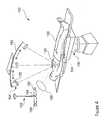
- FIG. 1is a diagram illustrating a typical frameless radiosurgical treatment system
- FIG. 2is a diagram illustrating the diagnostic and treatment beams of the system shown in FIG. 1;
- FIG. 3is a block diagram illustrating the treatment system of FIG. 1;
- FIG. 4is a diagram illustrating a preferred embodiment of the frameless treatment system in accordance with the invention.
- FIG. 5is a block diagram illustrating more of the details of the treatment system of FIG. 4;
- FIG. 6is a flowchart illustrating a method for treatment in accordance with the invention using the system of FIG. 4;
- FIG. 7is a diagram illustration a respiration cycle of a patient
- FIG. 8is a flowchart illustrating a method for treating a patient with respiration tracking in accordance with the invention.
- FIG. 9is a diagram illustrating a second embodiment of the frameless treatment system in accordance with the invention.
- FIG. 10is a flowchart illustrating a method for treatment in accordance with the invention using the system of FIG. 9;
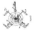
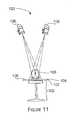
- FIG. 11is a diagram illustrating a third embodiment of the frameless treatment system in accordance with the invention.
- FIG. 12is a flowchart illustrating a method for treatment in accordance with the invention using the system of FIG. 11;
- FIG. 13illustrates the deformation of the pre-treatment and/or intra-treatment data to establish optimal correspondence to infer better target positions.
- the inventionis particularly applicable to a radiosurgical treatment system and method and it is in this context that the invention will be described. It will be appreciated, however, that the system and method in accordance with the invention has greater utility, such as to other types of treatments wherein it is necessary to accurately position a treatment at a target region within the patient in order to avoid damaging healthy tissue such as to other types of medical procedures with other types of medical instruments, such as positioning biopsy needles, ablative, ultrasound or other focused energy treatments, positioning a laser beam for laser beam treatment or positioning radioactive seeds for brachytherapy.
- a typical radiosurgery devicewill be described to provide a better understanding of the invention.
- FIGS. 1-3are diagram illustrating an example of a stereotaxic radiation treatment device 10 .
- the radiation treatment device 10may include a data processor 12 , such as a microprocessor, and a disc or tape storage unit 13 (shown in FIG. 3) which may store a three dimensional image of a patient 14 .
- the three dimensional imagemay be loaded into the data processor, if not already there, to compare the three dimensional image to images generated during the surgical procedure.
- the three dimensional imagemay be generated by various conventional techniques such as computer aided tomography (CAT) scan or magnetic resonance imaging (MR).
- the radiation treatment device 10may also include a beaming apparatus 20 which, when activated, emits a collimated surgical ionizing beam directed at a target region 18 (shown in FIG.
- the collimated surgical ionizing beammay have sufficient strength to cause the target region to become necrotic.
- a variety of different beaming apparatusmay be used which generate an ionizing radiation or heavy particle beam such as a linear accelerator and preferably an x-ray linear accelerator. Such an x-ray beaming apparatus is commercially available.
- the beaming apparatusmay be activated by the operator throwing a switch 23 at a control console 24 connected to the beaming apparatus 20 by a cable 22 .
- the radiation treatment device 10may also include an apparatus for passing a first diagnostic beam 26 and a second diagnostic beam 28 through the region previously imaged by the three-dimensional image.
- the diagnostic beamsare positioned at a predetermined non-zero angle with respect to each other, such as being orthogonal as shown in FIG. 2 .
- the diagnostic beamsmay be generated by a first x-ray generator 30 and a second x-ray generator 32 , respectively.
- a first and second image receiver 34 , 36 or a single receivermay receive the diagnostic beams 26 , 28 to generate an image from the diagnostic beams which is fed into the microprocessor 12 (as shown in FIG. 4) so that the diagnostic images may be compared to the three-dimensional image.
- the radiation treatment device 10may also include a device for adjusting the relative positions of the beaming apparatus 20 and/or the patient 14 so that the ionizing beam is continuously focused on the target region 18 .
- the positions of the beaming apparatus and the patientmay be altered with six degrees of freedom by a gantry 40 and a moveable operating table 38 with a tilting top 44 .
- the positions of the beaming apparatus relative to the patientmay also be accomplished by using a processor controllable robotic arm mechanism that permits the beaming apparatus to be moved freely about the patient's body including up, down, longitudinally along or laterally along the body of the patient.
- FIG. 3is a block diagram of the radiation treatment device 10 including the microprocessor 12 , the tape drive 13 , the beaming apparatus 20 , the robotic arm 46 or the gantry 40 , the x-ray cameras 30 , 32 , 34 and 36 , and the operator control console 24 as described above.
- the device 10may include safety interlocks 50 to ensure that the beaming apparatus is not activated accidentally.
- the device 10may also include an operator display 48 for tracking the progress of the treatment and controlling the treatment. Any further details of the radiosurgery device may be found in U.S. Pat. No. 5,207,223 which is owned by the assignee of this application and which is incorporated herein by reference.
- the above systemis well suited for the treatment of stationary target regions (e.g., stationary with respect to bony structures that can be seen on an image) wherein respiratory motion or pulsation motion do not affect the accuracy of the treatment beam.
- the drawback of the above systemis that anatomic sites subject to respiratory motion are difficult to treat.
- the frameless treatment systemmay improve upon the system shown in FIGS. 1-3. The frameless treatment system and method in accordance with the invention with the above advantages will now be described.
- FIG. 4is a diagram illustrating a preferred embodiment of the frameless treatment system 180 in accordance with the invention.
- This embodiment of the inventionis particular applicable to the targeting of a target region without embedded markers wherein there is no surrounding region that can be easily located (e.g., no bones are present) and respiration motion may affect the position of the target region.
- An example of a target region for this embodimentis a lung tumor.
- the treatment system 180may include a patient treatment table or couch 102 on which a patient 103 may rest during the treatment.
- the treatment systemmay also include a diagnostic beam recording device 104 that may be located underneath the treatment table and underneath the patient and one or more diagnostic beam generators 106 (one is shown in this example).
- the recording device 104may record the images generated when the diagnostic beam device is energized at one or more different predetermined positions.
- the recording device 104may be any device that can be used to capture the image generated by the diagnostic beams.
- the recording device 104may be the amorphous silicon plate that captures the x-ray beams being generated by the diagnostic beam generators 106 .
- the recording device 104may be connected to a computer that controls the operation of the recording device and the diagnostic beam generator.
- the recording device in this embodimentmay also have a first portion 105 and a second portion 107 wherein the first diagnostic beam is captured by the first portion and the second diagnostic beam is captured by the second portion.
- the diagnostic beamsmay be simultaneously energized or may be sequentially energized.
- a recording medium with one or more diagnostic beamsis also shown in U.S. Pat. No. 5,207,223 to Adler which is owned by the same assignee as the present invention.
- the robot and the treatment beam generator(shown in FIG. 5) are not shown in FIG. 4 .
- the systemmay further include a track 152 in which the diagnostic beam generator moves so that the diagnostic beam generator may be moved to different positions (see the diagnostic beam generator 106 in a first position 154 and the other positions shown by the phantom pictures of the generator) wherein the diagnostic beam generator is at a different non-zero angle with respect to the other positions.
- the diagnostic beam generator 106is moved from the first position 154 to other positions at periodic times in order to generate the images of the target region as described above.
- the systemmay also include a controller, to position of the diagnostic beam generator, that may be controlled by the computer.
- this system 180may also include an external marker tracking device 182 that may include one or more external marker tracking generators 184 that generate one or more external marker tracking beams 184 , such as infrared beams or passive markers whose position is detectable with optical cameras.
- the systemmay also include one or more external markers 188 attached to the patient that measure the external movement of the patient during respiratory motion as described in more detail in the co-pending application that was incorporated by reference. Now, the system will be described in more detail.
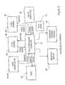
- FIG. 5is a block diagram illustrating more of the details of the treatment system 100 of FIG. 4 .
- the system 100may include a computer 110 that controls the operation of the various elements of the system including the beam generators 106 , 108 as well as the image recorder 104 .
- the systemmay also include a treatment beam device 112 , such as a linear accelerator (LINAC) in this embodiment, that generates a treatment beam and a robot 114 that positions the treatment beam (a LINAC manipulator in this embodiment) that are both controlled by the computer 110 that may be a multi-processor computer in this embodiment.
- the computermay issue control commands and receive back status commands from the treatment beam generator 112 , the robot 114 and the beam generators 106 , 108 .
- the computermay issue control signals to control the operation of the image recorder as described above and may receive image data from the image recorder.
- the systemmay also include safety interlocks 116 that ensures that the diagnostic beams and the treatment beam cannot be activated (the beams are only energized when a status signal is received by the computer) unless all people other than the patient are out of the treatment room due to the radiation danger.
- the systemmay also include a tape drive 118 for storing the images generated by the image recorder, the pre-operative CT three-dimensional images and any treatment planning software that may perform the comparison of the images and control the movement of the treatment beam.
- the systemmay further include an operator control console 120 and an operator display 122 that permit a user of the system, such as a surgeon, to interact with and operate the system and monitor the treatment.
- the treatment planning software in the computermay compare the pre-operative image to the images from the diagnostic beam generators to determine how to control the treatment robot and therefore the treatment beam during the treatment.
- the computerbased on the comparison and the surgeon's manual commands, may then control the treatment beam in order to deliver the appropriate dose to the patient without damaging the healthy tissue surrounding the target region.
- FIG. 6is a flowchart illustrating a method 200 for treatment in accordance with the invention using the system of FIG. 4 .
- a three-dimensional mapping of a region of the patient including the target regionis generated prior to the treatment.
- the three-dimensional mappingmay be done using typical equipment such as computer tomography, magnetic resonance tomography or the like.
- the three-dimensional mapping of the regionis stored in the storage device 118 .
- the mappingshows the relative locations of the target region with respect to other surrounding regions that may be seen in the mapping to locate the target region relative to the surrounding regions.
- the target regionmay be a lung tumor.
- the patientOn the day of treatment, the patient may be positioned on the treatment bed as shown in FIG. 9 .
- the respiratory cycle of the patientmay then be determined in step 203 and at various different times during the treatment.
- the respiratory cyclemay be determined by monitoring chest wall surface movement with optical or ultrasound digitizers, and/or by using a strain gauge, by the measurement of the airflow exiting the patient or by other well known methods.
- the systemmay determine if the treatment can begin based on the status of the safety interlocks. If it is not safe to begin the treatment, then the method loops back to test the safety interlocks until a safe condition is indicated.
- a diagnostic beam generatoris positioned along the track in the appropriate position and energized by the computer in order to generate an image on the recording device.
- the diagnostic beam generatoris an x-ray generators and the image recorder is an amorphous silicon imager that generates an image in response to x-rays as is well known.
- the image generated by the first diagnostic beam in the image recordermay then be downloaded by the computer to the storage device attached to the computer in step 208 and the image recorder may be reset. Each image is acquired at the same phase of the respiratory cycle as described below with respect to FIGS. 7 and 8.
- step 210the method determines if there are any other positions for the diagnostic beam. If there are other positions for the diagnostic beam, the method loops back to step 206 to energize that generator at the other position, generate an image and download the image to the storage device.
- the movement of the diagnostic beam generator along the trackgenerates multiple images wherein each image is at a non-zero angle with respect to the other images and acquired during the same phase of the respiratory cycle.
- the methodsequentially energizes the diagnostic beam generator at different positions to generate the images in a sequential manner.
- repeated sequence of images from the diagnostic beam generatormay be generated at periodic times so that the location of the target region at different times may be determined.
- the series of diagnostic beam imagesmay be processed using a CT-like algorithm to generate a 3-D image of the patient during the treatment.
- the 3-D imageis compared to the three-dimensional pre-operative mapping as is well known to determine the location of the target region at the particular time in step 212 .
- the targeting of the treatment beamis adjusted based on the comparison so that the treatment beam is always focused on the target region. If there are repeated diagnostic images generated, after each new set of images is generated, the images are compared to the mapping and the treatment beam targeting is adjusted to compensate for changes in the position of the target region. In this manner, the target region is accurately tracked so that the treatment beam is focused on the target region.
- the systemmay comprise the step of deforming the intra-treatment images in such a way that the positions of the clearly visible structures best match the pre-operative image data. From this, the exact deformation pattern of the entire anatomical area can be inferred.
- the exact position of the target and/or healthy critical tissue visible in the pre-operative image data, but not clearly visible in the intra-treatment datamay be inferred as described in more detail with reference to FIG. 13 .
- FIG. 7is a chart 260 illustrating a typical respiration cycle for a human being wherein the respiration cycle is represented by a sine wave.
- the y-axis of the chartis the movement of the chest wall thus showing that the chest wall moves out and in during the respiration cycle.
- a first point 262 in the respiration cycle with maximum expansion of the chest and a second point 264 in the respiration cycle with no chest movementare shown.
- the respiration cyclemay be determined using the various techniques described above.
- the energizing of the diagnostic beams and the treatment beammay be periodically timed so that the energizing occurs at the corresponding points in the respiration cycle such as at the first point or the second point.
- the energizing of the beamsmay occur at more than one time during the respiration cycle.
- the accuracy of the treatmentis improved since the beams are energized at the same time in the respiration cycle.
- FIG. 8is a flowchart illustration of a method 270 for energizing a diagnostic or treatment beam based on the respiration cycle in accordance with the invention.
- the treatmentis started and the respiration cycle of the patient is determined.
- the systemdetermines if at predetermined point in the respiration cycle has occurred and waits until the predetermined point has occurred. Once the predetermined point in the respiration cycle is reached, the system may energize the beam in step 276 .
- FIG. 9is a diagram illustrating a second embodiment of the frameless treatment system 150 in accordance with the invention.
- This embodiment of the inventionis particular applicable to fiducial-less targeting of a target region wherein a surrounding region can be located, but the surrounding region does not have a fixed relationship with the target region (e.g., no bones are present) and respiration motion does not affect the position of the target region.
- An example of a target region for this embodimentis the prostate.
- the system 150may include the same elements as the prior embodiment as designated by like reference numerals such as the treatment table 102 , the image recorder 104 and the diagnostic beam generator 106 . As with the prior embodiment, the robot and the treatment beam generator are not shown. In this embodiment, a single diagnostic beam generator 106 may be used to further reduce the cost of the treatment system. In this embodiment, the system may further include a track 152 in which the diagnostic beam generator moves so that the diagnostic beam generator may be moved to different positions (see the diagnostic beam generator 106 in a first position 154 and the other positions shown by the phantom pictures of the generator) wherein the diagnostic beam generator is at a different non-zero angle with respect to the other positions.
- the diagnostic beam generator 106is moved from the first position 154 to other positions at periodic times in order to generate the images of the target region as described above.
- the embodimentmay have similar elements as those shown in FIG. 5 and may also include a controller, to position the diagnostic beam generator, that may be controlled by the computer. Now, the method of treatment using the second embodiment will be described.
- FIG. 10is a flowchart illustrating a method 160 for treatment in accordance with the invention using the system of FIG. 9 .
- a three-dimensional mapping of a region of the patient including the target regionis generated prior to the treatment.
- the three-dimensional mappingmay be done using typical equipment such as computer tomography or the like.
- the three-dimensional mapping of the regionis stored in the storage device 118 .
- the mappingshows the location of the target region with respect to other surrounding regions that may be seen in the mapping to locate the target region relative to the surrounding regions.
- the target regionmay be a prostate tumor and the other surrounding regions may be the bladder.
- the patientOn the day of treatment, the patient may be positioned on the treatment bed as shown in FIG. 7 .
- the systemmay determine if the treatment can begin based on the status of the safety interlocks. If it is not safe to begin the treatment, then the method loops back to test the safety interlocks until a safe condition is indicated.
- a diagnostic beam generatoris positioned along the track in the appropriate position and energized by the computer in order to generate an image on the recording device.
- the diagnostic beam generatorsis an x-ray generator and the image recorder is an amorphous silicon imager that generates an image in response to x-rays as is well known.
- the image generated by the first diagnostic beam in the image recordermay then be downloaded by the computer to the storage device attached to the computer in step 168 and the image recorder may be reset.
- the methoddetermines if there are any other positions for the diagnostic beam.
- the methodloops back to step 166 to energize that generator at the other position, generate an image and download the image to the storage device.
- the movement of the diagnostic beam generator along the trackgenerates multiple images wherein each image is at a non-zero angle with respect to the other images.
- the methodsequentially energizes the diagnostic beam generator at different positions to generate the images in a time sequential manner.
- repeated sequence of images from the diagnostic beam generatormay be generated at periodic times so that the location of the target region at different times may be determined.
- the 2-D images generated by the diagnostic beamsare processed to yield a CT-like image which may then be compared to the pre-operative 3-D mapping.
- the two or more imagesare compared to the three-dimensional pre-operative mapping as is well known to determine the location of the target region at the particular time in step 172 .
- the comparisonmay again include the step of deformation as described above.
- the targeting of the treatment beamis adjusted based on the comparison so that the treatment beam is always focused on the target region. If there are repeated diagnostic images generated, after each new set of images is generated, the images are compared to the mapping and the treatment beam targeting is adjusted to compensate for changes in the position of the target region. In this manner, the target region is accurately tracked so that the treatment beam is focused on the target region.
- FIG. 11is a diagram illustrating another embodiment of the frameless treatment system 100 in accordance with the invention that may be particularly suited for treating target regions that have a fixed relationship to a fixed reference point, such as bones.
- this embodiment of the inventionmay be used for treating, for example, the spine of a patient or the brain of the patient since these target regions are near or surrounded by bones.
- the other embodiments of the invention described belowmay be particularly suited for the treatment of other target regions.
- only one detector under the patient couchis used.
- the two diagnostic beamsin this case may either be activated sequentially or the two beams may be activated simultaneously while projecting their respective images to a different portion of the single detector plate/camera.
- the simultaneous activation of the diagnostic beamsis particularly useful when time-stamps are needed so that the exact time of a given 3-D position is known.
- the treatment system 100may include a patient treatment table or couch 102 on which a patient 103 may rest during the treatment. In the example shown, the brain of the patient is being treated.
- the treatment systemmay also include a diagnostic beam recording device 104 that may be located underneath the treatment table and underneath the patient and one or more diagnostic beam generators 106 , 108 (two are shown in this example).
- the recording device 104may record the images generated when each diagnostic beam device 106 , 108 is energized.
- the recording device 104may be any device that can be used to capture the image generated by the diagnostic beams.
- the recording device 104may be the amorphous silicon plate that captures the x-ray beams being generated by the diagnostic beam generators 106 , 108 .
- the recording device 104may be connected to a computer that controls the operation of the recording device and the diagnostic beam generators.
- the recording device in this embodimentmay have a first portion 105 and a second portion 107 wherein the first diagnostic beam is captured by the first portion and the second diagnostic beam is captured by the second portion.
- the diagnostic beamsmay be simultaneously energized or may be sequentially energized.
- the diagnostic beam generators 106 , 108may be controlled by the computer to be energized at different predetermined time intervals or simultaneously so that each diagnostic beam generator is producing an image on the recording device at a different time or simultaneously.
- the diagnostic beam generatorsare located at different positions so that the diagnostic beams pass through the patient at different non-zero angles so that the angle between the two diagnostic beams is also non-zero which permits a two-dimensional image of the target region to be generated from the two images.
- the first diagnostic beam generator 106may be energized to emit a diagnostic beam that passes through the target region and generates an image on the recording device.
- the image developed by the recording deviceis then downloaded to the computer and the recording device is erased.
- the second diagnostic beam 108is energized and an image generated by the second diagnostic beam is received by the recording device.
- This imageis also downloaded to the computer where it is stored with the first image.
- FIG. 12is a flowchart illustrating a method 130 for treatment in accordance with the invention using the system of FIG. 11 .
- a three-dimensional mapping of a region of the patient including the target regionis generated prior to the treatment.
- the three-dimensional mappingmay be done using typical equipment such as computer tomography or the like.
- the three-dimensional mapping of the regionis stored in the storage device 118 .
- the mappingshows the location of the target region with respect to other surrounding regions that may be seen in the mapping and appear on X-ray images made with the image recorder.
- the target regionmay be a brain tumor and the other surrounding regions may be the skull bones.
- the patientOn the day of treatment, the patient may be positioned on the treatment bed as shown in FIG. 4 .
- the systemmay determine if the treatment can begin based on the status of the safety interlocks. If it is not safe to begin the treatment, then the method loops back to test the safety interlocks until a safe condition is indicated.
- a first diagnostic beam generatoris energized by the computer in order to generate an image on the recording device.
- the diagnostic beam generatorsare x-ray generators and the image recorder is an amorphous silicon imager that generates an image in response to x-rays as is well known.
- the image generated by the first diagnostic beam in the image recordermay then be downloaded by the computer to the storage device attached to the computer in step 138 and the image recorder may be reset.
- the methoddetermines if there are any other diagnostic beams to be energized. If there are other diagnostic beams to energize, the method loops back to step 136 to energize that generator, generate an image and download the image to the storage device.
- the methodsequentially energizes the diagnostic beam generators to generate the images from each of the diagnostic beams in a time sequential manner.
- repeated pairs of images from the diagnostic beam generatorsmay be generated at periodic times so that the location of the target region at different times may be determined.
- the two imagesare compared to the three-dimensional pre-operative mapping as is well known to determine the location of the target region at the particular time in step 142 .
- the targeting of the treatment beamis adjusted based on the comparison so that the treatment beam is always focused on the target region. If there are repeated diagnostic images generated, after each new set of images is generated, the images are compared to the mapping and the treatment beam targeting is adjusted to compensate for changes in the position of the target region. In this manner, the target region is accurately tracked so that the treatment beam is focused on the target region.
- FIG. 13illustrates a pre-operative image 250 and intra-treatment image data 252 generated by the diagnostic beams.
- the intra-treatment images generated by the diagnostic beamsare less clear and it is difficult to make out all of the structures or even the target region in the image.
- the pre-operative image 250is very clear and each structure of the body can be clearly seen. Therefore, in order to make it possible to infer the position of the target region from the intra-treatment images shown, the intra-treatment image is deformed, using various well known deformation techniques such as linear interpolation or warping, to form a deformed image 254 until the intra-treatment images and its structures form the best match with the pre-operative images. Once the deformation is completed, the position of the target region may be inferred from the position of the structures. This deformation technique may be used with all of the embodiments of the invention described above.
- the inventionis not limited to a single diagnostic beam source.
- the systemmay use five fixed sources that generate the diagnostic beams and two or more moving sources that generate the diagnostic beams.
- the fixed sourcesthey may be activated at specific time points throughout the respiration cycle. More detailed information about the deformation model corresponding to respiratory motion may then be obtained as set forth in the U.S. patent application Ser. No. 09/270,404.
Landscapes
- Health & Medical Sciences (AREA)
- Engineering & Computer Science (AREA)
- Life Sciences & Earth Sciences (AREA)
- Biomedical Technology (AREA)
- Public Health (AREA)
- Nuclear Medicine, Radiotherapy & Molecular Imaging (AREA)
- Veterinary Medicine (AREA)
- Animal Behavior & Ethology (AREA)
- General Health & Medical Sciences (AREA)
- Surgery (AREA)
- Medical Informatics (AREA)
- Pathology (AREA)
- Radiology & Medical Imaging (AREA)
- Heart & Thoracic Surgery (AREA)
- Molecular Biology (AREA)
- High Energy & Nuclear Physics (AREA)
- Optics & Photonics (AREA)
- Biophysics (AREA)
- Physics & Mathematics (AREA)
- Robotics (AREA)
- Oral & Maxillofacial Surgery (AREA)
- Computer Vision & Pattern Recognition (AREA)
- Radiation-Therapy Devices (AREA)
- Apparatus For Radiation Diagnosis (AREA)
- Magnetic Resonance Imaging Apparatus (AREA)
Abstract
Description
Claims (21)
Priority Applications (11)
| Application Number | Priority Date | Filing Date | Title |
|---|---|---|---|
| US09/663,104US6778850B1 (en) | 1999-03-16 | 2000-09-15 | Frameless radiosurgery treatment system and method |
| AU2001290891AAU2001290891A1 (en) | 2000-09-15 | 2001-09-14 | Frameless radiosurgery treatment system and method |
| EP01970945.0AEP1328195B1 (en) | 2000-09-15 | 2001-09-14 | Frameless radiosurgery treatment system |
| JP2002526277AJP2004529665A (en) | 2000-09-15 | 2001-09-14 | Frameless radiosurgery therapy system and method |
| PCT/US2001/028686WO2002022019A1 (en) | 2000-09-15 | 2001-09-14 | Frameless radiosurgery treatment system and method |
| US10/919,765US20050027194A1 (en) | 1999-03-16 | 2004-08-17 | Frameless radiosurgery treatment system and method |
| JP2007212216AJP4974164B2 (en) | 2000-09-15 | 2007-08-16 | Frameless radiosurgery system and method |
| US12/356,442US8086299B2 (en) | 1999-03-16 | 2009-01-20 | Frameless radiosurgery treatment system and method |
| US13/306,951US8634898B2 (en) | 1999-03-16 | 2011-11-29 | Frameless radiosurgery treatment system and method |
| US14/133,111US9572997B2 (en) | 1999-03-16 | 2013-12-18 | Frameless radiosurgery treatment system and method |
| US15/412,958US20170128744A1 (en) | 1999-03-16 | 2017-01-23 | Frameless radiosurgery treatment system and method |
Applications Claiming Priority (2)
| Application Number | Priority Date | Filing Date | Title |
|---|---|---|---|
| US09/270,404US6144875A (en) | 1999-03-16 | 1999-03-16 | Apparatus and method for compensating for respiratory and patient motion during treatment |
| US09/663,104US6778850B1 (en) | 1999-03-16 | 2000-09-15 | Frameless radiosurgery treatment system and method |
Related Parent Applications (1)
| Application Number | Title | Priority Date | Filing Date |
|---|---|---|---|
| US09/270,404Continuation-In-PartUS6144875A (en) | 1999-03-16 | 1999-03-16 | Apparatus and method for compensating for respiratory and patient motion during treatment |
Related Child Applications (1)
| Application Number | Title | Priority Date | Filing Date |
|---|---|---|---|
| US10/919,765ContinuationUS20050027194A1 (en) | 1999-03-16 | 2004-08-17 | Frameless radiosurgery treatment system and method |
Publications (1)
| Publication Number | Publication Date |
|---|---|
| US6778850B1true US6778850B1 (en) | 2004-08-17 |
Family
ID=24660487
Family Applications (6)
| Application Number | Title | Priority Date | Filing Date |
|---|---|---|---|
| US09/663,104Expired - LifetimeUS6778850B1 (en) | 1999-03-16 | 2000-09-15 | Frameless radiosurgery treatment system and method |
| US10/919,765AbandonedUS20050027194A1 (en) | 1999-03-16 | 2004-08-17 | Frameless radiosurgery treatment system and method |
| US12/356,442Expired - Fee RelatedUS8086299B2 (en) | 1999-03-16 | 2009-01-20 | Frameless radiosurgery treatment system and method |
| US13/306,951Expired - Fee RelatedUS8634898B2 (en) | 1999-03-16 | 2011-11-29 | Frameless radiosurgery treatment system and method |
| US14/133,111Expired - Fee RelatedUS9572997B2 (en) | 1999-03-16 | 2013-12-18 | Frameless radiosurgery treatment system and method |
| US15/412,958AbandonedUS20170128744A1 (en) | 1999-03-16 | 2017-01-23 | Frameless radiosurgery treatment system and method |
Family Applications After (5)
| Application Number | Title | Priority Date | Filing Date |
|---|---|---|---|
| US10/919,765AbandonedUS20050027194A1 (en) | 1999-03-16 | 2004-08-17 | Frameless radiosurgery treatment system and method |
| US12/356,442Expired - Fee RelatedUS8086299B2 (en) | 1999-03-16 | 2009-01-20 | Frameless radiosurgery treatment system and method |
| US13/306,951Expired - Fee RelatedUS8634898B2 (en) | 1999-03-16 | 2011-11-29 | Frameless radiosurgery treatment system and method |
| US14/133,111Expired - Fee RelatedUS9572997B2 (en) | 1999-03-16 | 2013-12-18 | Frameless radiosurgery treatment system and method |
| US15/412,958AbandonedUS20170128744A1 (en) | 1999-03-16 | 2017-01-23 | Frameless radiosurgery treatment system and method |
Country Status (5)
| Country | Link |
|---|---|
| US (6) | US6778850B1 (en) |
| EP (1) | EP1328195B1 (en) |
| JP (2) | JP2004529665A (en) |
| AU (1) | AU2001290891A1 (en) |
| WO (1) | WO2002022019A1 (en) |
Cited By (85)
| Publication number | Priority date | Publication date | Assignee | Title |
|---|---|---|---|---|
| US20030206610A1 (en)* | 2002-05-01 | 2003-11-06 | Collins William F. | Patient positioning system |
| US20040037390A1 (en)* | 2001-08-24 | 2004-02-26 | Kazumasa Mihara | Radiotherapy device |
| US20040077939A1 (en)* | 2001-02-22 | 2004-04-22 | Rainer Graumann | Device and method for controlling surgical instruments |
| US20050008208A1 (en)* | 2003-06-25 | 2005-01-13 | Brett Cowan | Acquisition-time modeling for automated post-processing |
| US20050123289A1 (en)* | 2003-12-05 | 2005-06-09 | Moller-Wedel Gmbh | Method and apparatus for observing objects with a microscope |
| US20050161051A1 (en)* | 2003-01-08 | 2005-07-28 | Cyberheart, Inc. | System for non-invasive heart treatment |
| US20050180544A1 (en)* | 2004-02-17 | 2005-08-18 | Frank Sauer | System and method for patient positioning for radiotherapy in the presence of respiratory motion |
| US20050197564A1 (en)* | 2004-02-20 | 2005-09-08 | University Of Florida Research Foundation, Inc. | System for delivering conformal radiation therapy while simultaneously imaging soft tissue |
| US20050201510A1 (en)* | 1998-10-23 | 2005-09-15 | Hassan Mostafavi | Method and system for predictive physiological gating |
| US20050228256A1 (en)* | 2004-03-22 | 2005-10-13 | Vanderbilt University | System and method for surgical instrument disablement via image-guided position feedback |
| US20050267457A1 (en)* | 2004-05-25 | 2005-12-01 | Hruschka James A | Tissue ablation device using a lens to three dimensionally focus electromagnetic energy |
| US20050283068A1 (en)* | 2004-06-17 | 2005-12-22 | Psychology Software Tools, Inc. | Magnetic resonance imaging having patient video, microphone and motion tracking |
| US20060064008A1 (en)* | 2004-09-17 | 2006-03-23 | Elekta Ab | Therapeutic use of radiation and apparatus therefor |
| US20060074304A1 (en)* | 2004-10-02 | 2006-04-06 | Sohail Sayeh | Correlation model selection for internal target movement |
| US20070015991A1 (en)* | 2005-06-29 | 2007-01-18 | Dongshan Fu | Dynamic tracking of soft tissue targets with ultrasound images, without using fiducial markers |
| US20070053491A1 (en)* | 2005-09-07 | 2007-03-08 | Eastman Kodak Company | Adaptive radiation therapy method with target detection |
| WO2007044692A1 (en)* | 2005-10-11 | 2007-04-19 | University Of Florida Research Foundation, Inc. | Preplanning of guided medical procedures |
| US20070110665A1 (en)* | 2005-11-17 | 2007-05-17 | Bolan Patrick J | Tissue marker for multimodality radiographic imaging |
| US7221733B1 (en)* | 2002-01-02 | 2007-05-22 | Varian Medical Systems Technologies, Inc. | Method and apparatus for irradiating a target |
| US7227925B1 (en) | 2002-10-02 | 2007-06-05 | Varian Medical Systems Technologies, Inc. | Gantry mounted stereoscopic imaging system |
| US20070142751A1 (en)* | 2002-03-06 | 2007-06-21 | Hyosig Kang | Apparatus and method for haptic rendering |
| US20070238977A1 (en)* | 2006-03-30 | 2007-10-11 | Sweeney Patrick J | Method and apparatus for treatment of discogenic pain |
| US20070274446A1 (en)* | 2006-05-23 | 2007-11-29 | Eastman Kodak Company | System for the real-time detection of targets for radiation therapy |
| US20070276233A1 (en)* | 2003-06-06 | 2007-11-29 | Besson Guy M | Integrated X-Ray and Ultrasound Medical Imaging System |
| US20080086023A1 (en)* | 2006-10-05 | 2008-04-10 | Bruker Biospin Mri Gmbh | Installation for investigating objects using magnetic resonance |
| US20080091101A1 (en)* | 2006-10-16 | 2008-04-17 | Perfint Engineering Services | Needle positioning apparatus and method |
| US20080089481A1 (en)* | 2006-10-16 | 2008-04-17 | Oraya Therapeutics, Inc. | Portable orthovoltage radiotherapy |
| US20080212738A1 (en)* | 2006-12-13 | 2008-09-04 | Oraya Therapeutics, Inc. | Orthovoltage radiotherapy |
| US20090110238A1 (en)* | 2007-10-26 | 2009-04-30 | Shutian Li | Automatic correlation modeling of an internal target |
| US20090129545A1 (en)* | 1999-03-16 | 2009-05-21 | Accuray, Inc. | Frameless radiosurgery treatment system and method |
| US20090163898A1 (en)* | 2007-06-04 | 2009-06-25 | Oraya Therapeutics, Inc. | Method and device for ocular alignment and coupling of ocular structures |
| US20090180666A1 (en)* | 2008-01-10 | 2009-07-16 | Ye Sheng | Constrained-curve correlation model |
| US20090296886A1 (en)* | 2008-05-30 | 2009-12-03 | Siemens Medical Solutions Usa, Inc. | Digital tomosynthesis in robotic stereotactic radiosurgery |
| US20100204714A1 (en)* | 2000-07-24 | 2010-08-12 | Moshe Shoham | Miniature bone-attached surgical robot |
| US7792249B2 (en) | 2007-12-23 | 2010-09-07 | Oraya Therapeutics, Inc. | Methods and devices for detecting, controlling, and predicting radiation delivery |
| US7801271B2 (en) | 2007-12-23 | 2010-09-21 | Oraya Therapeutics, Inc. | Methods and devices for orthovoltage ocular radiotherapy and treatment planning |
| US20100290586A1 (en)* | 2009-05-18 | 2010-11-18 | Werner Friedrich | Radiation therapy device |
| US20110137102A1 (en)* | 2009-06-04 | 2011-06-09 | Mayo Foundation For Medical Education And Research | Stereotactic intracranial target localization guidance systems and methods |
| US20110166408A1 (en)* | 2009-07-17 | 2011-07-07 | Cyberheart, Inc. | Heart tissue surface contour-based radiosurgical treatment planning |
| WO2011106433A1 (en) | 2010-02-24 | 2011-09-01 | Accuray Incorporated | Gantry image guided radiotherapy system and related treatment delivery methods |
| WO2012099747A2 (en) | 2011-01-20 | 2012-07-26 | Accuray, Inc. | Radiation treatment delivery system with ring gantry |
| US8287522B2 (en) | 2006-05-19 | 2012-10-16 | Mako Surgical Corp. | Method and apparatus for controlling a haptic device |
| DE102011080371A1 (en) | 2011-08-03 | 2013-02-07 | Siemens Aktiengesellschaft | Radiotherapy device for overlapping-free three-dimensional computed tomography imaging for recording image data during treatment session of patient, has control unit for coordinated controlling of radiation of therapeutic radiation source |
| US8391954B2 (en) | 2002-03-06 | 2013-03-05 | Mako Surgical Corp. | System and method for interactive haptic positioning of a medical device |
| WO2013029649A1 (en)* | 2011-08-26 | 2013-03-07 | Elekta Ab (Publ) | Intra-fraction motion management system |
| US8506558B2 (en) | 2008-01-11 | 2013-08-13 | Oraya Therapeutics, Inc. | System and method for performing an ocular irradiation procedure |
| US8559596B2 (en) | 2010-06-08 | 2013-10-15 | Accuray Incorporated | Target Tracking for image-guided radiation treatment |
| US8613748B2 (en) | 2010-11-10 | 2013-12-24 | Perfint Healthcare Private Limited | Apparatus and method for stabilizing a needle |
| US8663210B2 (en) | 2009-05-13 | 2014-03-04 | Novian Health, Inc. | Methods and apparatus for performing interstitial laser therapy and interstitial brachytherapy |
| US8761863B2 (en) | 2009-02-26 | 2014-06-24 | National University Corporation Hokkaido University | Target tracking device and radiation therapy apparatus |
| US8758263B1 (en) | 2009-10-31 | 2014-06-24 | Voxel Rad, Ltd. | Systems and methods for frameless image-guided biopsy and therapeutic intervention |
| US20140270090A1 (en)* | 2013-03-15 | 2014-09-18 | Proto Manufacturing Ltd. | X-Ray Diffraction Apparatus And Method |
| US20150063537A1 (en)* | 2013-08-29 | 2015-03-05 | Samsung Electronics Co., Ltd. | X-ray imaging apparatus and control method thereof |
| US9091628B2 (en) | 2012-12-21 | 2015-07-28 | L-3 Communications Security And Detection Systems, Inc. | 3D mapping with two orthogonal imaging views |
| US9119655B2 (en) | 2012-08-03 | 2015-09-01 | Stryker Corporation | Surgical manipulator capable of controlling a surgical instrument in multiple modes |
| US9226796B2 (en) | 2012-08-03 | 2016-01-05 | Stryker Corporation | Method for detecting a disturbance as an energy applicator of a surgical instrument traverses a cutting path |
| CN105616004A (en)* | 2015-12-09 | 2016-06-01 | 北京工业大学 | Surgical robot system for radioactive seed interventional therapy of tumors and operating method |
| CN105748148A (en)* | 2016-01-31 | 2016-07-13 | 宓兵 | Operative treatment device for interventional imaging monitoring |
| US9480534B2 (en) | 2012-08-03 | 2016-11-01 | Stryker Corporation | Navigation system and method for removing a volume of tissue from a patient |
| US20170128750A1 (en)* | 2014-07-25 | 2017-05-11 | Varian Medical Systems, Inc. | Imaging based calibration systems, devices, and methods |
| US9687200B2 (en) | 2010-06-08 | 2017-06-27 | Accuray Incorporated | Radiation treatment delivery system with translatable ring gantry |
| US9795455B2 (en) | 2014-08-22 | 2017-10-24 | Breast-Med, Inc. | Tissue marker for multimodality radiographic imaging |
| US9801686B2 (en) | 2003-03-06 | 2017-10-31 | Mako Surgical Corp. | Neural monitor-based dynamic haptics |
| US9820818B2 (en) | 2012-08-03 | 2017-11-21 | Stryker Corporation | System and method for controlling a surgical manipulator based on implant parameters |
| US9921712B2 (en) | 2010-12-29 | 2018-03-20 | Mako Surgical Corp. | System and method for providing substantially stable control of a surgical tool |
| US9966160B2 (en) | 2015-11-24 | 2018-05-08 | Viewray Technologies, Inc. | Radiation beam collimating systems and methods |
| US20180129284A1 (en)* | 2012-11-01 | 2018-05-10 | Eyecam Llc | Wireless wrist computing and control device and method for 3d imaging, mapping, networking and interfacing |
| US10413751B2 (en) | 2016-03-02 | 2019-09-17 | Viewray Technologies, Inc. | Particle therapy with magnetic resonance imaging |
| US10463884B2 (en) | 2013-03-15 | 2019-11-05 | Viewray Technologies, Inc. | Systems and methods for linear accelerator radiotherapy with magnetic resonance imaging |
| US10561861B2 (en) | 2012-05-02 | 2020-02-18 | Viewray Technologies, Inc. | Videographic display of real-time medical treatment |
| US10646188B2 (en) | 1998-10-23 | 2020-05-12 | Varian Medical Systems, Inc. | Method and system for radiation application |
| US10667727B2 (en) | 2008-09-05 | 2020-06-02 | Varian Medical Systems, Inc. | Systems and methods for determining a state of a patient |
| WO2020112681A1 (en) | 2018-11-30 | 2020-06-04 | Accuray Inc. | Multimodal radiation apparatus and methods |
| US10821303B2 (en) | 2012-10-26 | 2020-11-03 | Viewray Technologies, Inc. | Assessment and improvement of treatment using imaging of physiological responses to radiation therapy |
| US11000706B2 (en) | 2016-12-13 | 2021-05-11 | Viewray Technologies, Inc. | Radiation therapy systems and methods |
| US11033758B2 (en) | 2017-12-06 | 2021-06-15 | Viewray Technologies, Inc. | Radiotherapy systems, methods and software |
| US11202676B2 (en) | 2002-03-06 | 2021-12-21 | Mako Surgical Corp. | Neural monitor-based dynamic haptics |
| US11202682B2 (en) | 2016-12-16 | 2021-12-21 | Mako Surgical Corp. | Techniques for modifying tool operation in a surgical robotic system based on comparing actual and commanded states of the tool relative to a surgical site |
| US11209509B2 (en) | 2018-05-16 | 2021-12-28 | Viewray Technologies, Inc. | Resistive electromagnet systems and methods |
| US11241296B2 (en) | 2005-11-17 | 2022-02-08 | Breast-Med, Inc. | Imaging fiducial markers and methods |
| US11378629B2 (en) | 2016-06-22 | 2022-07-05 | Viewray Technologies, Inc. | Magnetic resonance imaging |
| USD956981S1 (en)* | 2019-04-25 | 2022-07-05 | Elekta Instrument Ab | Patient clearance tool |
| US11478662B2 (en) | 2017-04-05 | 2022-10-25 | Accuray Incorporated | Sequential monoscopic tracking |
| US11617503B2 (en) | 2018-12-12 | 2023-04-04 | Voxel Rad, Ltd. | Systems and methods for treating cancer using brachytherapy |
| US20240100362A1 (en)* | 2012-07-27 | 2024-03-28 | H. Lee Moffitt Cancer Center And Research Institute, Inc. | Multi-spectral fluorescence for in-vivo determination of proton energy and range in proton therapy |
Families Citing this family (186)
| Publication number | Priority date | Publication date | Assignee | Title |
|---|---|---|---|---|
| DE10051370A1 (en) | 2000-10-17 | 2002-05-02 | Brainlab Ag | Method and appliance for exact positioning of patient for radiation therapy and radio surgery with which only one camera is used to determine and compensate for positional error |
| US7070327B2 (en) | 2002-05-01 | 2006-07-04 | Siemens Medical Solutions Usa, Inc. | Focused radiation visualization |
| US20040042582A1 (en)* | 2002-09-03 | 2004-03-04 | Moshe Ein-Gal | Method and apparatus for locating a medical target |
| US8989349B2 (en) | 2004-09-30 | 2015-03-24 | Accuray, Inc. | Dynamic tracking of moving targets |
| FR2876896B1 (en)* | 2004-10-21 | 2007-10-26 | Gen Electric | METHOD FOR USING A TOMOGRAPHY DEVICE FOR OBTAINING RADIOSCOPIC IMAGES AND DEVICE FOR CARRYING OUT SAID METHOD |
| US7539284B2 (en)* | 2005-02-11 | 2009-05-26 | Besson Guy M | Method and system for dynamic low dose X-ray imaging |
| US7340032B2 (en)* | 2005-02-11 | 2008-03-04 | Besson Guy M | System for dynamic low dose x-ray imaging and tomosynthesis |
| US7751869B2 (en) | 2005-12-09 | 2010-07-06 | Boston Scientific Scimed, Inc. | Radiation ablation tracking system and method |
| US10357184B2 (en) | 2012-06-21 | 2019-07-23 | Globus Medical, Inc. | Surgical tool systems and method |
| US10653497B2 (en) | 2006-02-16 | 2020-05-19 | Globus Medical, Inc. | Surgical tool systems and methods |
| US10893912B2 (en) | 2006-02-16 | 2021-01-19 | Globus Medical Inc. | Surgical tool systems and methods |
| PL2023812T3 (en) | 2006-05-19 | 2017-07-31 | The Queen's Medical Center | Motion tracking system for real time adaptive imaging and spectroscopy |
| US10279196B2 (en)* | 2006-09-28 | 2019-05-07 | Accuray Incorporated | Radiation treatment planning using four-dimensional imaging data |
| US7623679B2 (en)* | 2006-12-13 | 2009-11-24 | Accuray Incorporated | Temporal smoothing of a deformation model |
| CA2737938C (en)* | 2007-09-19 | 2016-09-13 | Walter A. Roberts | Direct visualization robotic intra-operative radiation therapy applicator device |
| US20090088625A1 (en)* | 2007-10-01 | 2009-04-02 | Kenneth Oosting | Photonic Based Non-Invasive Surgery System That Includes Automated Cell Control and Eradication Via Pre-Calculated Feed-Forward Control Plus Image Feedback Control For Targeted Energy Delivery |
| DE102008019114A1 (en)* | 2008-04-16 | 2009-10-22 | Siemens Aktiengesellschaft | Medical device and method for docking a positioning device to a shuttle |
| DE102008019128A1 (en) | 2008-04-16 | 2009-10-29 | Siemens Aktiengesellschaft | Apparatus for carrying out an irradiation and method for monitoring such |
| US7903781B2 (en)* | 2008-05-02 | 2011-03-08 | L-3 Communications Security And Detection Systems, Inc. | Determination of heavy particle stopping power |
| US8848974B2 (en) | 2008-09-29 | 2014-09-30 | Restoration Robotics, Inc. | Object-tracking systems and methods |
| EP2357022A4 (en)* | 2008-11-12 | 2014-01-08 | Univ Tsukuba | RADIOTHERAPY SYSTEM |
| US8792614B2 (en) | 2009-03-31 | 2014-07-29 | Matthew R. Witten | System and method for radiation therapy treatment planning using a memetic optimization algorithm |
| CN102858229B (en) | 2010-02-18 | 2015-11-25 | 皇家飞利浦电子股份有限公司 | Systems and methods for tumor motion simulation and motion compensation using tracked bronchoscopy |
| JP5347070B2 (en)* | 2010-09-09 | 2013-11-20 | 三菱電機株式会社 | Particle beam therapy system |
| US8768029B2 (en)* | 2010-10-20 | 2014-07-01 | Medtronic Navigation, Inc. | Selected image acquisition technique to optimize patient model construction |
| US9498289B2 (en) | 2010-12-21 | 2016-11-22 | Restoration Robotics, Inc. | Methods and systems for directing movement of a tool in hair transplantation procedures |
| US8911453B2 (en) | 2010-12-21 | 2014-12-16 | Restoration Robotics, Inc. | Methods and systems for directing movement of a tool in hair transplantation procedures |
| JP5667489B2 (en)* | 2011-03-22 | 2015-02-12 | 国立大学法人北海道大学 | Radiation therapy tracking system |
| US9308050B2 (en) | 2011-04-01 | 2016-04-12 | Ecole Polytechnique Federale De Lausanne (Epfl) | Robotic system and method for spinal and other surgeries |
| KR20120117510A (en)* | 2011-04-15 | 2012-10-24 | 알피니언메디칼시스템 주식회사 | Apparatus for ultrasound treatment and driving method thereof |
| WO2013032933A2 (en) | 2011-08-26 | 2013-03-07 | Kinecticor, Inc. | Methods, systems, and devices for intra-scan motion correction |
| US9170214B2 (en)* | 2011-09-13 | 2015-10-27 | Applied Minds, Llc | Motion-based radiograph interlock systems, structures, and processes |
| US11298196B2 (en) | 2012-06-21 | 2022-04-12 | Globus Medical Inc. | Surgical robotic automation with tracking markers and controlled tool advancement |
| US12004905B2 (en) | 2012-06-21 | 2024-06-11 | Globus Medical, Inc. | Medical imaging systems using robotic actuators and related methods |
| US11317971B2 (en) | 2012-06-21 | 2022-05-03 | Globus Medical, Inc. | Systems and methods related to robotic guidance in surgery |
| US10842461B2 (en) | 2012-06-21 | 2020-11-24 | Globus Medical, Inc. | Systems and methods of checking registrations for surgical systems |
| US11116576B2 (en) | 2012-06-21 | 2021-09-14 | Globus Medical Inc. | Dynamic reference arrays and methods of use |
| US11399900B2 (en) | 2012-06-21 | 2022-08-02 | Globus Medical, Inc. | Robotic systems providing co-registration using natural fiducials and related methods |
| US11963755B2 (en) | 2012-06-21 | 2024-04-23 | Globus Medical Inc. | Apparatus for recording probe movement |
| EP2863827B1 (en) | 2012-06-21 | 2022-11-16 | Globus Medical, Inc. | Surgical robot platform |
| US11857149B2 (en) | 2012-06-21 | 2024-01-02 | Globus Medical, Inc. | Surgical robotic systems with target trajectory deviation monitoring and related methods |
| US10646280B2 (en) | 2012-06-21 | 2020-05-12 | Globus Medical, Inc. | System and method for surgical tool insertion using multiaxis force and moment feedback |
| US12220120B2 (en) | 2012-06-21 | 2025-02-11 | Globus Medical, Inc. | Surgical robotic system with retractor |
| US12329593B2 (en) | 2012-06-21 | 2025-06-17 | Globus Medical, Inc. | Surgical robotic automation with tracking markers |
| US11864745B2 (en) | 2012-06-21 | 2024-01-09 | Globus Medical, Inc. | Surgical robotic system with retractor |
| US11786324B2 (en) | 2012-06-21 | 2023-10-17 | Globus Medical, Inc. | Surgical robotic automation with tracking markers |
| US12310683B2 (en) | 2012-06-21 | 2025-05-27 | Globus Medical, Inc. | Surgical tool systems and method |
| US20150032164A1 (en) | 2012-06-21 | 2015-01-29 | Globus Medical, Inc. | Methods for Performing Invasive Medical Procedures Using a Surgical Robot |
| US10136954B2 (en) | 2012-06-21 | 2018-11-27 | Globus Medical, Inc. | Surgical tool systems and method |
| US10799298B2 (en) | 2012-06-21 | 2020-10-13 | Globus Medical Inc. | Robotic fluoroscopic navigation |
| US11395706B2 (en) | 2012-06-21 | 2022-07-26 | Globus Medical Inc. | Surgical robot platform |
| US11864839B2 (en) | 2012-06-21 | 2024-01-09 | Globus Medical Inc. | Methods of adjusting a virtual implant and related surgical navigation systems |
| US11253327B2 (en) | 2012-06-21 | 2022-02-22 | Globus Medical, Inc. | Systems and methods for automatically changing an end-effector on a surgical robot |
| US11589771B2 (en) | 2012-06-21 | 2023-02-28 | Globus Medical Inc. | Method for recording probe movement and determining an extent of matter removed |
| US11793570B2 (en) | 2012-06-21 | 2023-10-24 | Globus Medical Inc. | Surgical robotic automation with tracking markers |
| US11607149B2 (en) | 2012-06-21 | 2023-03-21 | Globus Medical Inc. | Surgical tool systems and method |
| US11974822B2 (en) | 2012-06-21 | 2024-05-07 | Globus Medical Inc. | Method for a surveillance marker in robotic-assisted surgery |
| US11045267B2 (en) | 2012-06-21 | 2021-06-29 | Globus Medical, Inc. | Surgical robotic automation with tracking markers |
| US10231791B2 (en) | 2012-06-21 | 2019-03-19 | Globus Medical, Inc. | Infrared signal based position recognition system for use with a robot-assisted surgery |
| US12262954B2 (en) | 2012-06-21 | 2025-04-01 | Globus Medical, Inc. | Surgical robotic automation with tracking markers |
| US11857266B2 (en) | 2012-06-21 | 2024-01-02 | Globus Medical, Inc. | System for a surveillance marker in robotic-assisted surgery |
| US11896446B2 (en) | 2012-06-21 | 2024-02-13 | Globus Medical, Inc | Surgical robotic automation with tracking markers |
| US10624710B2 (en) | 2012-06-21 | 2020-04-21 | Globus Medical, Inc. | System and method for measuring depth of instrumentation |
| US10758315B2 (en) | 2012-06-21 | 2020-09-01 | Globus Medical Inc. | Method and system for improving 2D-3D registration convergence |
| US10350013B2 (en) | 2012-06-21 | 2019-07-16 | Globus Medical, Inc. | Surgical tool systems and methods |
| US10874466B2 (en) | 2012-06-21 | 2020-12-29 | Globus Medical, Inc. | System and method for surgical tool insertion using multiaxis force and moment feedback |
| FR2993101B1 (en)* | 2012-07-06 | 2015-07-17 | Commissariat Energie Atomique | METHOD FOR ASSEMBLING AND ENCAPSULATING LITHIUM MICROBATTERIES AND MICROBATTERIES THUS OBTAINED |
| US9305365B2 (en) | 2013-01-24 | 2016-04-05 | Kineticor, Inc. | Systems, devices, and methods for tracking moving targets |
| US10327708B2 (en) | 2013-01-24 | 2019-06-25 | Kineticor, Inc. | Systems, devices, and methods for tracking and compensating for patient motion during a medical imaging scan |
| US9717461B2 (en) | 2013-01-24 | 2017-08-01 | Kineticor, Inc. | Systems, devices, and methods for tracking and compensating for patient motion during a medical imaging scan |
| CN109008972A (en) | 2013-02-01 | 2018-12-18 | 凯内蒂科尔股份有限公司 | The motion tracking system of real-time adaptive motion compensation in biomedical imaging |
| KR102117273B1 (en)* | 2013-03-21 | 2020-06-01 | 삼성전자주식회사 | Surgical robot system and method for controlling the same |
| US9283048B2 (en) | 2013-10-04 | 2016-03-15 | KB Medical SA | Apparatus and systems for precise guidance of surgical tools |
| JP6259283B2 (en)* | 2013-12-27 | 2018-01-10 | 株式会社日立製作所 | Model generation apparatus, radiotherapy apparatus control apparatus, and model generation method |
| US9241771B2 (en) | 2014-01-15 | 2016-01-26 | KB Medical SA | Notched apparatus for guidance of an insertable instrument along an axis during spinal surgery |
| WO2015121311A1 (en) | 2014-02-11 | 2015-08-20 | KB Medical SA | Sterile handle for controlling a robotic surgical system from a sterile field |
| WO2015148391A1 (en) | 2014-03-24 | 2015-10-01 | Thomas Michael Ernst | Systems, methods, and devices for removing prospective motion correction from medical imaging scans |
| EP3134022B1 (en) | 2014-04-24 | 2018-01-10 | KB Medical SA | Surgical instrument holder for use with a robotic surgical system |
| CN106999248B (en) | 2014-06-19 | 2021-04-06 | Kb医疗公司 | Systems and methods for performing minimally invasive surgery |
| US10357257B2 (en) | 2014-07-14 | 2019-07-23 | KB Medical SA | Anti-skid surgical instrument for use in preparing holes in bone tissue |
| US10765438B2 (en) | 2014-07-14 | 2020-09-08 | KB Medical SA | Anti-skid surgical instrument for use in preparing holes in bone tissue |
| EP3188660A4 (en) | 2014-07-23 | 2018-05-16 | Kineticor, Inc. | Systems, devices, and methods for tracking and compensating for patient motion during a medical imaging scan |
| US9950194B2 (en) | 2014-09-09 | 2018-04-24 | Mevion Medical Systems, Inc. | Patient positioning system |
| EP3226781B1 (en) | 2014-12-02 | 2018-08-01 | KB Medical SA | Robot assisted volume removal during surgery |
| US10013808B2 (en) | 2015-02-03 | 2018-07-03 | Globus Medical, Inc. | Surgeon head-mounted display apparatuses |
| WO2016131903A1 (en) | 2015-02-18 | 2016-08-25 | KB Medical SA | Systems and methods for performing minimally invasive spinal surgery with a robotic surgical system using a percutaneous technique |
| US9943247B2 (en) | 2015-07-28 | 2018-04-17 | The University Of Hawai'i | Systems, devices, and methods for detecting false movements for motion correction during a medical imaging scan |
| US10646298B2 (en) | 2015-07-31 | 2020-05-12 | Globus Medical, Inc. | Robot arm and methods of use |
| US10058394B2 (en) | 2015-07-31 | 2018-08-28 | Globus Medical, Inc. | Robot arm and methods of use |
| US10080615B2 (en) | 2015-08-12 | 2018-09-25 | Globus Medical, Inc. | Devices and methods for temporary mounting of parts to bone |
| JP6894431B2 (en) | 2015-08-31 | 2021-06-30 | ケービー メディカル エスアー | Robotic surgical system and method |
| US10034716B2 (en) | 2015-09-14 | 2018-07-31 | Globus Medical, Inc. | Surgical robotic systems and methods thereof |
| US9771092B2 (en) | 2015-10-13 | 2017-09-26 | Globus Medical, Inc. | Stabilizer wheel assembly and methods of use |
| US10716515B2 (en) | 2015-11-23 | 2020-07-21 | Kineticor, Inc. | Systems, devices, and methods for tracking and compensating for patient motion during a medical imaging scan |
| WO2017093034A1 (en) | 2015-12-01 | 2017-06-08 | Brainlab Ag | Method and apparatus for determining or predicting the position of a target |
| US10842453B2 (en) | 2016-02-03 | 2020-11-24 | Globus Medical, Inc. | Portable medical imaging system |
| US10117632B2 (en) | 2016-02-03 | 2018-11-06 | Globus Medical, Inc. | Portable medical imaging system with beam scanning collimator |
| US11058378B2 (en) | 2016-02-03 | 2021-07-13 | Globus Medical, Inc. | Portable medical imaging system |
| US11883217B2 (en) | 2016-02-03 | 2024-01-30 | Globus Medical, Inc. | Portable medical imaging system and method |
| US10448910B2 (en) | 2016-02-03 | 2019-10-22 | Globus Medical, Inc. | Portable medical imaging system |
| US10866119B2 (en) | 2016-03-14 | 2020-12-15 | Globus Medical, Inc. | Metal detector for detecting insertion of a surgical device into a hollow tube |
| EP3241518B1 (en) | 2016-04-11 | 2024-10-23 | Globus Medical, Inc | Surgical tool systems |
| WO2017210500A1 (en)* | 2016-06-03 | 2017-12-07 | Covidien Lp | Robotic surgical system with an embedded imager |
| US10939963B2 (en) | 2016-09-01 | 2021-03-09 | Covidien Lp | Systems and methods for providing proximity awareness to pleural boundaries, vascular structures, and other critical intra-thoracic structures during electromagnetic navigation bronchoscopy |
| US11039893B2 (en) | 2016-10-21 | 2021-06-22 | Globus Medical, Inc. | Robotic surgical systems |
| EP3351202B1 (en) | 2017-01-18 | 2021-09-08 | KB Medical SA | Universal instrument guide for robotic surgical systems |
| JP7233841B2 (en) | 2017-01-18 | 2023-03-07 | ケービー メディカル エスアー | Robotic Navigation for Robotic Surgical Systems |
| JP7583513B2 (en) | 2017-01-18 | 2024-11-14 | ケービー メディカル エスアー | Universal instrument guide for robotic surgical systems, surgical instrument system |
| US11071594B2 (en) | 2017-03-16 | 2021-07-27 | KB Medical SA | Robotic navigation of robotic surgical systems |
| US20180289432A1 (en) | 2017-04-05 | 2018-10-11 | Kb Medical, Sa | Robotic surgical systems for preparing holes in bone tissue and methods of their use |
| US11135015B2 (en) | 2017-07-21 | 2021-10-05 | Globus Medical, Inc. | Robot surgical platform |
| US11794338B2 (en) | 2017-11-09 | 2023-10-24 | Globus Medical Inc. | Robotic rod benders and related mechanical and motor housings |
| EP3492032B1 (en) | 2017-11-09 | 2023-01-04 | Globus Medical, Inc. | Surgical robotic systems for bending surgical rods |
| US11357548B2 (en) | 2017-11-09 | 2022-06-14 | Globus Medical, Inc. | Robotic rod benders and related mechanical and motor housings |
| US11134862B2 (en) | 2017-11-10 | 2021-10-05 | Globus Medical, Inc. | Methods of selecting surgical implants and related devices |
| US11298565B2 (en) | 2017-11-16 | 2022-04-12 | Ebamed Sa | Heart arrhythmia non-invasive treatment device and method |
| US20190254753A1 (en) | 2018-02-19 | 2019-08-22 | Globus Medical, Inc. | Augmented reality navigation systems for use with robotic surgical systems and methods of their use |
| US10573023B2 (en) | 2018-04-09 | 2020-02-25 | Globus Medical, Inc. | Predictive visualization of medical imaging scanner component movement |
| EP3566670A1 (en)* | 2018-05-07 | 2019-11-13 | Koninklijke Philips N.V. | Safety system for surgical robot |
| US11337742B2 (en) | 2018-11-05 | 2022-05-24 | Globus Medical Inc | Compliant orthopedic driver |
| US11278360B2 (en) | 2018-11-16 | 2022-03-22 | Globus Medical, Inc. | End-effectors for surgical robotic systems having sealed optical components |
| US11744655B2 (en) | 2018-12-04 | 2023-09-05 | Globus Medical, Inc. | Drill guide fixtures, cranial insertion fixtures, and related methods and robotic systems |
| US11602402B2 (en) | 2018-12-04 | 2023-03-14 | Globus Medical, Inc. | Drill guide fixtures, cranial insertion fixtures, and related methods and robotic systems |
| US11175366B2 (en)* | 2019-02-05 | 2021-11-16 | Siemens Healthcare Gmbh | Free-breathing MRI with motion compensation |
| US11918313B2 (en) | 2019-03-15 | 2024-03-05 | Globus Medical Inc. | Active end effectors for surgical robots |
| US20200297357A1 (en) | 2019-03-22 | 2020-09-24 | Globus Medical, Inc. | System for neuronavigation registration and robotic trajectory guidance, robotic surgery, and related methods and devices |
| US11571265B2 (en) | 2019-03-22 | 2023-02-07 | Globus Medical Inc. | System for neuronavigation registration and robotic trajectory guidance, robotic surgery, and related methods and devices |
| US11317978B2 (en) | 2019-03-22 | 2022-05-03 | Globus Medical, Inc. | System for neuronavigation registration and robotic trajectory guidance, robotic surgery, and related methods and devices |
| US11806084B2 (en) | 2019-03-22 | 2023-11-07 | Globus Medical, Inc. | System for neuronavigation registration and robotic trajectory guidance, and related methods and devices |
| US11382549B2 (en) | 2019-03-22 | 2022-07-12 | Globus Medical, Inc. | System for neuronavigation registration and robotic trajectory guidance, and related methods and devices |
| US11419616B2 (en) | 2019-03-22 | 2022-08-23 | Globus Medical, Inc. | System for neuronavigation registration and robotic trajectory guidance, robotic surgery, and related methods and devices |
| EP3958740A4 (en)* | 2019-04-25 | 2023-04-12 | Memorial Sloan Kettering Cancer Center | MAGNETIC RESONANCE SIGNATURE MATCHING (MRSIGMA) FOR REAL-TIME VOLUMETRIC MOTION TRACKING AND ADAPTIVE RADIATION THERAPY |
| US11045179B2 (en) | 2019-05-20 | 2021-06-29 | Global Medical Inc | Robot-mounted retractor system |
| US11628023B2 (en) | 2019-07-10 | 2023-04-18 | Globus Medical, Inc. | Robotic navigational system for interbody implants |
| US12396692B2 (en) | 2019-09-24 | 2025-08-26 | Globus Medical, Inc. | Compound curve cable chain |
| US11571171B2 (en) | 2019-09-24 | 2023-02-07 | Globus Medical, Inc. | Compound curve cable chain |
| US11864857B2 (en) | 2019-09-27 | 2024-01-09 | Globus Medical, Inc. | Surgical robot with passive end effector |
| US12408929B2 (en) | 2019-09-27 | 2025-09-09 | Globus Medical, Inc. | Systems and methods for navigating a pin guide driver |
| US11890066B2 (en) | 2019-09-30 | 2024-02-06 | Globus Medical, Inc | Surgical robot with passive end effector |
| US11426178B2 (en) | 2019-09-27 | 2022-08-30 | Globus Medical Inc. | Systems and methods for navigating a pin guide driver |
| US12329391B2 (en) | 2019-09-27 | 2025-06-17 | Globus Medical, Inc. | Systems and methods for robot-assisted knee arthroplasty surgery |
| US11510684B2 (en) | 2019-10-14 | 2022-11-29 | Globus Medical, Inc. | Rotary motion passive end effector for surgical robots in orthopedic surgeries |
| US12156760B2 (en) | 2019-11-14 | 2024-12-03 | Ebamed Sa | Cardiac phase gating system for radiation therapy |
| US12220176B2 (en) | 2019-12-10 | 2025-02-11 | Globus Medical, Inc. | Extended reality instrument interaction zone for navigated robotic |
| US12133772B2 (en) | 2019-12-10 | 2024-11-05 | Globus Medical, Inc. | Augmented reality headset for navigated robotic surgery |
| US11992373B2 (en) | 2019-12-10 | 2024-05-28 | Globus Medical, Inc | Augmented reality headset with varied opacity for navigated robotic surgery |
| US12064189B2 (en) | 2019-12-13 | 2024-08-20 | Globus Medical, Inc. | Navigated instrument for use in robotic guided surgery |
| US11464581B2 (en) | 2020-01-28 | 2022-10-11 | Globus Medical, Inc. | Pose measurement chaining for extended reality surgical navigation in visible and near infrared spectrums |
| US11382699B2 (en) | 2020-02-10 | 2022-07-12 | Globus Medical Inc. | Extended reality visualization of optical tool tracking volume for computer assisted navigation in surgery |
| US12414752B2 (en) | 2020-02-17 | 2025-09-16 | Globus Medical, Inc. | System and method of determining optimal 3-dimensional position and orientation of imaging device for imaging patient bones |
| US11207150B2 (en) | 2020-02-19 | 2021-12-28 | Globus Medical, Inc. | Displaying a virtual model of a planned instrument attachment to ensure correct selection of physical instrument attachment |
| CN111359106B (en)* | 2020-03-09 | 2021-08-31 | 青岛市中医医院(青岛市海慈医院、青岛市康复医学研究所) | Radiotherapy positioning bed |
| US11253216B2 (en) | 2020-04-28 | 2022-02-22 | Globus Medical Inc. | Fixtures for fluoroscopic imaging systems and related navigation systems and methods |
| US11510750B2 (en) | 2020-05-08 | 2022-11-29 | Globus Medical, Inc. | Leveraging two-dimensional digital imaging and communication in medicine imagery in three-dimensional extended reality applications |
| US11382700B2 (en) | 2020-05-08 | 2022-07-12 | Globus Medical Inc. | Extended reality headset tool tracking and control |
| US11153555B1 (en) | 2020-05-08 | 2021-10-19 | Globus Medical Inc. | Extended reality headset camera system for computer assisted navigation in surgery |
| US12070276B2 (en) | 2020-06-09 | 2024-08-27 | Globus Medical Inc. | Surgical object tracking in visible light via fiducial seeding and synthetic image registration |
| US11317973B2 (en) | 2020-06-09 | 2022-05-03 | Globus Medical, Inc. | Camera tracking bar for computer assisted navigation during surgery |
| US11382713B2 (en) | 2020-06-16 | 2022-07-12 | Globus Medical, Inc. | Navigated surgical system with eye to XR headset display calibration |
| US11877807B2 (en) | 2020-07-10 | 2024-01-23 | Globus Medical, Inc | Instruments for navigated orthopedic surgeries |
| US11793588B2 (en) | 2020-07-23 | 2023-10-24 | Globus Medical, Inc. | Sterile draping of robotic arms |
| US11737831B2 (en) | 2020-09-02 | 2023-08-29 | Globus Medical Inc. | Surgical object tracking template generation for computer assisted navigation during surgical procedure |
| US11523785B2 (en) | 2020-09-24 | 2022-12-13 | Globus Medical, Inc. | Increased cone beam computed tomography volume length without requiring stitching or longitudinal C-arm movement |
| US12076091B2 (en) | 2020-10-27 | 2024-09-03 | Globus Medical, Inc. | Robotic navigational system |
| US11911112B2 (en) | 2020-10-27 | 2024-02-27 | Globus Medical, Inc. | Robotic navigational system |
| US11941814B2 (en) | 2020-11-04 | 2024-03-26 | Globus Medical Inc. | Auto segmentation using 2-D images taken during 3-D imaging spin |
| US11717350B2 (en) | 2020-11-24 | 2023-08-08 | Globus Medical Inc. | Methods for robotic assistance and navigation in spinal surgery and related systems |
| EP4267243A1 (en) | 2020-12-23 | 2023-11-01 | Ebamed SA | A multiplanar motion management system |
| US12161433B2 (en) | 2021-01-08 | 2024-12-10 | Globus Medical, Inc. | System and method for ligament balancing with robotic assistance |
| US12150728B2 (en) | 2021-04-14 | 2024-11-26 | Globus Medical, Inc. | End effector for a surgical robot |
| US12178523B2 (en) | 2021-04-19 | 2024-12-31 | Globus Medical, Inc. | Computer assisted surgical navigation system for spine procedures |
| US11857273B2 (en) | 2021-07-06 | 2024-01-02 | Globus Medical, Inc. | Ultrasonic robotic surgical navigation |
| US11439444B1 (en) | 2021-07-22 | 2022-09-13 | Globus Medical, Inc. | Screw tower and rod reduction tool |
| US12213745B2 (en) | 2021-09-16 | 2025-02-04 | Globus Medical, Inc. | Extended reality systems for visualizing and controlling operating room equipment |
| US12238087B2 (en) | 2021-10-04 | 2025-02-25 | Globus Medical, Inc. | Validating credential keys based on combinations of credential value strings and input order strings |
| US12184636B2 (en) | 2021-10-04 | 2024-12-31 | Globus Medical, Inc. | Validating credential keys based on combinations of credential value strings and input order strings |
| US20230368330A1 (en) | 2021-10-20 | 2023-11-16 | Globus Medical, Inc. | Interpolation of medical images |
| US20230165639A1 (en) | 2021-12-01 | 2023-06-01 | Globus Medical, Inc. | Extended reality systems with three-dimensional visualizations of medical image scan slices |
| US11911115B2 (en) | 2021-12-20 | 2024-02-27 | Globus Medical Inc. | Flat panel registration fixture and method of using same |
| US12103480B2 (en) | 2022-03-18 | 2024-10-01 | Globus Medical Inc. | Omni-wheel cable pusher |
| US12048493B2 (en) | 2022-03-31 | 2024-07-30 | Globus Medical, Inc. | Camera tracking system identifying phantom markers during computer assisted surgery navigation |
| US12394086B2 (en) | 2022-05-10 | 2025-08-19 | Globus Medical, Inc. | Accuracy check and automatic calibration of tracked instruments |
| US12161427B2 (en) | 2022-06-08 | 2024-12-10 | Globus Medical, Inc. | Surgical navigation system with flat panel registration fixture |
| US12226169B2 (en) | 2022-07-15 | 2025-02-18 | Globus Medical, Inc. | Registration of 3D and 2D images for surgical navigation and robotic guidance without using radiopaque fiducials in the images |
| US20240020840A1 (en) | 2022-07-15 | 2024-01-18 | Globus Medical, Inc. | REGISTRATION OF 3D and 2D IMAGES FOR SURGICAL NAVIGATION AND ROBOTIC GUIDANCE WITHOUT USING RADIOPAQUE FIDUCIALS IN THE IMAGES |
| US12318150B2 (en) | 2022-10-11 | 2025-06-03 | Globus Medical Inc. | Camera tracking system for computer assisted surgery navigation |
Citations (17)
| Publication number | Priority date | Publication date | Assignee | Title |
|---|---|---|---|---|
| US5207223A (en)* | 1990-10-19 | 1993-05-04 | Accuray, Inc. | Apparatus for and method of performing stereotaxic surgery |
| US5427097A (en)* | 1992-12-10 | 1995-06-27 | Accuray, Inc. | Apparatus for and method of carrying out stereotaxic radiosurgery and radiotherapy |
| US5446548A (en)* | 1993-10-08 | 1995-08-29 | Siemens Medical Systems, Inc. | Patient positioning and monitoring system |
| US5727554A (en)* | 1996-09-19 | 1998-03-17 | University Of Pittsburgh Of The Commonwealth System Of Higher Education | Apparatus responsive to movement of a patient during treatment/diagnosis |
| US5748700A (en)* | 1994-05-10 | 1998-05-05 | Shepherd; Joseph S. | Radiation therapy and radiation surgery treatment system and methods of use of same |
| US5797849A (en)* | 1995-03-28 | 1998-08-25 | Sonometrics Corporation | Method for carrying out a medical procedure using a three-dimensional tracking and imaging system |
| US6019724A (en)* | 1995-02-22 | 2000-02-01 | Gronningsaeter; Aage | Method for ultrasound guidance during clinical procedures |
| US6031888A (en)* | 1997-11-26 | 2000-02-29 | Picker International, Inc. | Fluoro-assist feature for a diagnostic imaging device |
| US6144875A (en)* | 1999-03-16 | 2000-11-07 | Accuray Incorporated | Apparatus and method for compensating for respiratory and patient motion during treatment |
| US6149592A (en) | 1997-11-26 | 2000-11-21 | Picker International, Inc. | Integrated fluoroscopic projection image data, volumetric image data, and surgical device position data |
| US6256372B1 (en) | 1999-03-16 | 2001-07-03 | General Electric Company | Apparatus and methods for stereo radiography |
| US6285902B1 (en) | 1999-02-10 | 2001-09-04 | Surgical Insights, Inc. | Computer assisted targeting device for use in orthopaedic surgery |
| US6301495B1 (en)* | 1999-04-27 | 2001-10-09 | International Business Machines Corporation | System and method for intra-operative, image-based, interactive verification of a pre-operative surgical plan |
| US20020032453A1 (en)* | 1995-01-31 | 2002-03-14 | Cosman Eric R. | Repositioner for head, neck, and body |
| US20020065461A1 (en)* | 1991-01-28 | 2002-05-30 | Cosman Eric R. | Surgical positioning system |
| US6470207B1 (en) | 1999-03-23 | 2002-10-22 | Surgical Navigation Technologies, Inc. | Navigational guidance via computer-assisted fluoroscopic imaging |
| US20020154728A1 (en) | 2001-03-12 | 2002-10-24 | Eiichi Morita | Radiographic apparatus |
Family Cites Families (46)
| Publication number | Priority date | Publication date | Assignee | Title |
|---|---|---|---|---|
| DE2616714A1 (en)* | 1976-04-15 | 1977-10-27 | Philips Patentverwaltung | METHOD FOR LAYERED REPRESENTATION OF OBJECTS FROM OVERLAY IMAGES OF DIFFERENT IMAGE LEVELS |
| US4237901A (en)* | 1978-08-30 | 1980-12-09 | Picker Corporation | Low and constant pressure transducer probe for ultrasonic diagnostic system |
| FR2441264A1 (en)* | 1978-11-08 | 1980-06-06 | Hitachi Ltd | RADIATION SENSITIVE SCREEN |
| US4583538A (en)* | 1984-05-04 | 1986-04-22 | Onik Gary M | Method and apparatus for stereotaxic placement of probes in the body utilizing CT scanner localization |
| JPS62206798A (en)* | 1986-03-06 | 1987-09-11 | 三菱電機株式会社 | linear accelerator |
| US4991579A (en)* | 1987-11-10 | 1991-02-12 | Allen George S | Method and apparatus for providing related images over time of a portion of the anatomy using fiducial implants |
| NL8802398A (en) | 1988-09-29 | 1990-04-17 | Pelt & Hooykaas | METHOD FOR HARMFULING TOXIC WASTE. |
| US5222499A (en)* | 1989-11-15 | 1993-06-29 | Allen George S | Method and apparatus for imaging the anatomy |
| US6006126A (en)* | 1991-01-28 | 1999-12-21 | Cosman; Eric R. | System and method for stereotactic registration of image scan data |
| JP3305348B2 (en)* | 1992-01-16 | 2002-07-22 | 株式会社日立メディコ | Stereotactic radiotherapy device |
| FR2694881B1 (en)* | 1992-07-31 | 1996-09-06 | Univ Joseph Fourier | METHOD FOR DETERMINING THE POSITION OF AN ORGAN. |
| JP3290726B2 (en)* | 1992-12-18 | 2002-06-10 | 浜松ホトニクス株式会社 | Transmission three-dimensional tomography system |
| US5411026A (en)* | 1993-10-08 | 1995-05-02 | Nomos Corporation | Method and apparatus for lesion position verification |
| US5394875A (en)* | 1993-10-21 | 1995-03-07 | Lewis; Judith T. | Automatic ultrasonic localization of targets implanted in a portion of the anatomy |
| US5538494A (en)* | 1994-03-17 | 1996-07-23 | Hitachi, Ltd. | Radioactive beam irradiation method and apparatus taking movement of the irradiation area into consideration |
| DE4432890B4 (en)* | 1994-09-15 | 2004-02-19 | Brainlab Ag | Device for detecting the position of irradiation target points |
| US5622187A (en)* | 1994-09-30 | 1997-04-22 | Nomos Corporation | Method and apparatus for patient positioning for radiation therapy |
| JPH08112272A (en)* | 1994-10-14 | 1996-05-07 | Hitachi Medical Corp | 2-way roentgenography apparatus |
| US5971997A (en)* | 1995-02-03 | 1999-10-26 | Radionics, Inc. | Intraoperative recalibration apparatus for stereotactic navigators |
| US5588430A (en) | 1995-02-14 | 1996-12-31 | University Of Florida Research Foundation, Inc. | Repeat fixation for frameless stereotactic procedure |
| US6246898B1 (en)* | 1995-03-28 | 2001-06-12 | Sonometrics Corporation | Method for carrying out a medical procedure using a three-dimensional tracking and imaging system |
| US5769861A (en)* | 1995-09-28 | 1998-06-23 | Brainlab Med. Computersysteme Gmbh | Method and devices for localizing an instrument |
| US5764723A (en)* | 1996-10-16 | 1998-06-09 | The Trustees Of Columbia University In The City Of New York | Apparatus and method to gate a source for radiation therapy |
| US5784431A (en)* | 1996-10-29 | 1998-07-21 | University Of Pittsburgh Of The Commonwealth System Of Higher Education | Apparatus for matching X-ray images with reference images |
| IL119545A (en)* | 1996-11-01 | 2002-11-10 | Philips Medical Systems Techno | Method and device for precise invasive procedures |
| JPH1119082A (en)* | 1997-06-30 | 1999-01-26 | Toshiba Corp | Image reconstruction device |
| US5967981A (en)* | 1997-09-26 | 1999-10-19 | Siemens Corporate Research, Inc. | Time series prediction for event triggering |
| US6325758B1 (en)* | 1997-10-27 | 2001-12-04 | Nomos Corporation | Method and apparatus for target position verification |
| US6120453A (en)* | 1997-11-17 | 2000-09-19 | Sharp; William A. | Three-dimensional ultrasound system based on the coordination of multiple ultrasonic transducers |
| JPH10201863A (en) | 1998-02-19 | 1998-08-04 | Hitachi Ltd | Radiation irradiation method and radiation irradiation device |
| US6076005A (en)* | 1998-02-25 | 2000-06-13 | St. Jude Children's Research Hospital | Respiration responsive gating means and apparatus and methods using the same |
| DE19809460C1 (en)* | 1998-03-06 | 1999-09-30 | Siemens Ag | Medical target device for breathing-compensated punction |
| JP3053389B1 (en)* | 1998-12-03 | 2000-06-19 | 三菱電機株式会社 | Moving object tracking irradiation device |
| NZ509667A (en)* | 1998-08-06 | 2003-08-29 | Wisconsin Alumni Res Found | Delivery modification system for radiation therapy |
| DE19842239A1 (en) | 1998-09-15 | 2000-03-16 | Siemens Ag | Medical technical arrangement for diagnosis and treatment |
| ATE265253T1 (en)* | 1998-10-23 | 2004-05-15 | Varian Med Sys Inc | METHOD AND SYSTEM FOR PHYSIOLOGICAL CONTROL OF RADIOTHERAPY |
| US6236708B1 (en)* | 1998-11-25 | 2001-05-22 | Picker International, Inc. | 2D and 3D tomographic X-ray imaging using flat panel detectors |
| JP3326597B2 (en)* | 1999-01-14 | 2002-09-24 | 独立行政法人放射線医学総合研究所 | Respiratory gating control radiotherapy equipment |
| JP3897925B2 (en)* | 1999-01-29 | 2007-03-28 | 株式会社日立メディコ | Cone beam CT system |
| US6501981B1 (en)* | 1999-03-16 | 2002-12-31 | Accuray, Inc. | Apparatus and method for compensating for respiratory and patient motions during treatment |
| US6778850B1 (en)* | 1999-03-16 | 2004-08-17 | Accuray, Inc. | Frameless radiosurgery treatment system and method |
| DE19914455B4 (en)* | 1999-03-30 | 2005-07-14 | Siemens Ag | Method for determining the movement of an organ or therapeutic area of a patient and a system suitable for this purpose |
| US6275721B1 (en) | 1999-06-10 | 2001-08-14 | General Electriccompany | Interactive MRI scan control using an in-bore scan control device |
| JP4674948B2 (en) | 2000-09-29 | 2011-04-20 | オリンパス株式会社 | Surgical navigation device and method of operating surgical navigation device |
| US6473634B1 (en) | 2000-11-22 | 2002-10-29 | Koninklijke Philips Electronics N.V. | Medical imaging at two temporal resolutions for tumor treatment planning |
| JP5188199B2 (en) | 2008-02-22 | 2013-04-24 | 株式会社ユニバーサルエンターテインメント | Paper sheet processing equipment |
- 2000
- 2000-09-15USUS09/663,104patent/US6778850B1/ennot_activeExpired - Lifetime
- 2001
- 2001-09-14JPJP2002526277Apatent/JP2004529665A/enactivePending
- 2001-09-14WOPCT/US2001/028686patent/WO2002022019A1/enactiveApplication Filing
- 2001-09-14AUAU2001290891Apatent/AU2001290891A1/ennot_activeAbandoned
- 2001-09-14EPEP01970945.0Apatent/EP1328195B1/ennot_activeExpired - Lifetime
- 2004
- 2004-08-17USUS10/919,765patent/US20050027194A1/ennot_activeAbandoned
- 2007
- 2007-08-16JPJP2007212216Apatent/JP4974164B2/ennot_activeExpired - Lifetime
- 2009
- 2009-01-20USUS12/356,442patent/US8086299B2/ennot_activeExpired - Fee Related
- 2011
- 2011-11-29USUS13/306,951patent/US8634898B2/ennot_activeExpired - Fee Related
- 2013
- 2013-12-18USUS14/133,111patent/US9572997B2/ennot_activeExpired - Fee Related
- 2017
- 2017-01-23USUS15/412,958patent/US20170128744A1/ennot_activeAbandoned
Patent Citations (18)
| Publication number | Priority date | Publication date | Assignee | Title |
|---|---|---|---|---|
| US5207223A (en)* | 1990-10-19 | 1993-05-04 | Accuray, Inc. | Apparatus for and method of performing stereotaxic surgery |
| US6405072B1 (en)* | 1991-01-28 | 2002-06-11 | Sherwood Services Ag | Apparatus and method for determining a location of an anatomical target with reference to a medical apparatus |
| US20020065461A1 (en)* | 1991-01-28 | 2002-05-30 | Cosman Eric R. | Surgical positioning system |
| US5427097A (en)* | 1992-12-10 | 1995-06-27 | Accuray, Inc. | Apparatus for and method of carrying out stereotaxic radiosurgery and radiotherapy |
| US5446548A (en)* | 1993-10-08 | 1995-08-29 | Siemens Medical Systems, Inc. | Patient positioning and monitoring system |
| US5748700A (en)* | 1994-05-10 | 1998-05-05 | Shepherd; Joseph S. | Radiation therapy and radiation surgery treatment system and methods of use of same |
| US20020032453A1 (en)* | 1995-01-31 | 2002-03-14 | Cosman Eric R. | Repositioner for head, neck, and body |
| US6019724A (en)* | 1995-02-22 | 2000-02-01 | Gronningsaeter; Aage | Method for ultrasound guidance during clinical procedures |
| US5797849A (en)* | 1995-03-28 | 1998-08-25 | Sonometrics Corporation | Method for carrying out a medical procedure using a three-dimensional tracking and imaging system |
| US5727554A (en)* | 1996-09-19 | 1998-03-17 | University Of Pittsburgh Of The Commonwealth System Of Higher Education | Apparatus responsive to movement of a patient during treatment/diagnosis |
| US6149592A (en) | 1997-11-26 | 2000-11-21 | Picker International, Inc. | Integrated fluoroscopic projection image data, volumetric image data, and surgical device position data |
| US6031888A (en)* | 1997-11-26 | 2000-02-29 | Picker International, Inc. | Fluoro-assist feature for a diagnostic imaging device |
| US6285902B1 (en) | 1999-02-10 | 2001-09-04 | Surgical Insights, Inc. | Computer assisted targeting device for use in orthopaedic surgery |
| US6144875A (en)* | 1999-03-16 | 2000-11-07 | Accuray Incorporated | Apparatus and method for compensating for respiratory and patient motion during treatment |
| US6256372B1 (en) | 1999-03-16 | 2001-07-03 | General Electric Company | Apparatus and methods for stereo radiography |
| US6470207B1 (en) | 1999-03-23 | 2002-10-22 | Surgical Navigation Technologies, Inc. | Navigational guidance via computer-assisted fluoroscopic imaging |
| US6301495B1 (en)* | 1999-04-27 | 2001-10-09 | International Business Machines Corporation | System and method for intra-operative, image-based, interactive verification of a pre-operative surgical plan |
| US20020154728A1 (en) | 2001-03-12 | 2002-10-24 | Eiichi Morita | Radiographic apparatus |
Cited By (276)
| Publication number | Priority date | Publication date | Assignee | Title |
|---|---|---|---|---|
| US10646188B2 (en) | 1998-10-23 | 2020-05-12 | Varian Medical Systems, Inc. | Method and system for radiation application |
| US20050201510A1 (en)* | 1998-10-23 | 2005-09-15 | Hassan Mostafavi | Method and system for predictive physiological gating |
| US9232928B2 (en)* | 1998-10-23 | 2016-01-12 | Varian Medical Systems, Inc. | Method and system for predictive physiological gating |
| US20090129545A1 (en)* | 1999-03-16 | 2009-05-21 | Accuray, Inc. | Frameless radiosurgery treatment system and method |
| US8086299B2 (en) | 1999-03-16 | 2011-12-27 | Accuray Incorporated | Frameless radiosurgery treatment system and method |
| US8634898B2 (en) | 1999-03-16 | 2014-01-21 | Accuray Incorporated | Frameless radiosurgery treatment system and method |
| US20100204714A1 (en)* | 2000-07-24 | 2010-08-12 | Moshe Shoham | Miniature bone-attached surgical robot |
| US10058338B2 (en)* | 2000-07-24 | 2018-08-28 | Mazor Robotics Ltd. | Miniature bone-attached surgical robot |
| US20040077939A1 (en)* | 2001-02-22 | 2004-04-22 | Rainer Graumann | Device and method for controlling surgical instruments |
| US7085347B2 (en)* | 2001-08-24 | 2006-08-01 | Mitsubishi Heavy Industries, Ltd. | Radiotherapy device |
| US20040037390A1 (en)* | 2001-08-24 | 2004-02-26 | Kazumasa Mihara | Radiotherapy device |
| US7221733B1 (en)* | 2002-01-02 | 2007-05-22 | Varian Medical Systems Technologies, Inc. | Method and apparatus for irradiating a target |
| US11202676B2 (en) | 2002-03-06 | 2021-12-21 | Mako Surgical Corp. | Neural monitor-based dynamic haptics |
| US11298191B2 (en) | 2002-03-06 | 2022-04-12 | Mako Surgical Corp. | Robotically-assisted surgical guide |
| US10231790B2 (en) | 2002-03-06 | 2019-03-19 | Mako Surgical Corp. | Haptic guidance system and method |
| US10610301B2 (en) | 2002-03-06 | 2020-04-07 | Mako Surgical Corp. | System and method for using a haptic device as an input device |
| US10058392B2 (en) | 2002-03-06 | 2018-08-28 | Mako Surgical Corp. | Neural monitor-based dynamic boundaries |
| US8391954B2 (en) | 2002-03-06 | 2013-03-05 | Mako Surgical Corp. | System and method for interactive haptic positioning of a medical device |
| US11076918B2 (en) | 2002-03-06 | 2021-08-03 | Mako Surgical Corp. | Robotically-assisted constraint mechanism |
| US8010180B2 (en)* | 2002-03-06 | 2011-08-30 | Mako Surgical Corp. | Haptic guidance system and method |
| US8571628B2 (en) | 2002-03-06 | 2013-10-29 | Mako Surgical Corp. | Apparatus and method for haptic rendering |
| US9002426B2 (en) | 2002-03-06 | 2015-04-07 | Mako Surgical Corp. | Haptic guidance system and method |
| US11298190B2 (en) | 2002-03-06 | 2022-04-12 | Mako Surgical Corp. | Robotically-assisted constraint mechanism |
| US11426245B2 (en) | 2002-03-06 | 2022-08-30 | Mako Surgical Corp. | Surgical guidance system and method with acoustic feedback |
| US9636185B2 (en) | 2002-03-06 | 2017-05-02 | Mako Surgical Corp. | System and method for performing surgical procedure using drill guide and robotic device operable in multiple modes |
| US20070142751A1 (en)* | 2002-03-06 | 2007-06-21 | Hyosig Kang | Apparatus and method for haptic rendering |
| US9775682B2 (en) | 2002-03-06 | 2017-10-03 | Mako Surgical Corp. | Teleoperation system with visual indicator and method of use during surgical procedures |
| US8911499B2 (en) | 2002-03-06 | 2014-12-16 | Mako Surgical Corp. | Haptic guidance method |
| US9775681B2 (en) | 2002-03-06 | 2017-10-03 | Mako Surgical Corp. | Haptic guidance system and method |
| US20030206610A1 (en)* | 2002-05-01 | 2003-11-06 | Collins William F. | Patient positioning system |
| US7227925B1 (en) | 2002-10-02 | 2007-06-05 | Varian Medical Systems Technologies, Inc. | Gantry mounted stereoscopic imaging system |
| US11305132B2 (en)* | 2003-01-08 | 2022-04-19 | Varian Medical Systems, Inc. | System for non-invasive heart treatment |
| US7645276B2 (en)* | 2003-01-08 | 2010-01-12 | Cyberheart, Inc. | System for non-invasive heart treatment |
| US20100160775A1 (en)* | 2003-01-08 | 2010-06-24 | Cyberheart, Inc. | System For Non-Invasive Heart Treatment |
| US10792511B2 (en)* | 2003-01-08 | 2020-10-06 | Cyberheart, Inc. | System for non-invasive heart treatment |
| US9907978B2 (en)* | 2003-01-08 | 2018-03-06 | Cyberheart, Inc. | System for non-invasive heart treatment |
| US20050161051A1 (en)* | 2003-01-08 | 2005-07-28 | Cyberheart, Inc. | System for non-invasive heart treatment |
| US8696658B2 (en) | 2003-01-08 | 2014-04-15 | Cyberheart, Inc. | System for non-invasive heart treatment |
| US20190030361A1 (en)* | 2003-01-08 | 2019-01-31 | Cyberheart, Inc. | System for non-invasive heart treatment |
| US9801686B2 (en) | 2003-03-06 | 2017-10-31 | Mako Surgical Corp. | Neural monitor-based dynamic haptics |
| US20070276233A1 (en)* | 2003-06-06 | 2007-11-29 | Besson Guy M | Integrated X-Ray and Ultrasound Medical Imaging System |
| US20050008208A1 (en)* | 2003-06-25 | 2005-01-13 | Brett Cowan | Acquisition-time modeling for automated post-processing |
| US20050123289A1 (en)* | 2003-12-05 | 2005-06-09 | Moller-Wedel Gmbh | Method and apparatus for observing objects with a microscope |
| US20050180544A1 (en)* | 2004-02-17 | 2005-08-18 | Frank Sauer | System and method for patient positioning for radiotherapy in the presence of respiratory motion |
| US7853308B2 (en)* | 2004-02-17 | 2010-12-14 | Siemens Medical Solutions Usa, Inc. | System and method for patient positioning for radiotherapy in the presence of respiratory motion |
| US7907987B2 (en) | 2004-02-20 | 2011-03-15 | University Of Florida Research Foundation, Inc. | System for delivering conformal radiation therapy while simultaneously imaging soft tissue |
| US10688319B2 (en) | 2004-02-20 | 2020-06-23 | University Of Florida Research Foundation, Inc. | System for delivering conformal radiation therapy while simultaneously imaging soft tissue |
| US11497937B2 (en) | 2004-02-20 | 2022-11-15 | University Of Florida Research Foundation, Inc. | System for delivering conformal radiation therapy while simultaneously imaging soft tissue |
| US8190233B2 (en) | 2004-02-20 | 2012-05-29 | University Of Florida Research Foundation, Inc. | System for delivering conformal radiation therapy while simultaneously imaging soft tissue |
| US20050197564A1 (en)* | 2004-02-20 | 2005-09-08 | University Of Florida Research Foundation, Inc. | System for delivering conformal radiation therapy while simultaneously imaging soft tissue |
| US20100113911A1 (en)* | 2004-02-20 | 2010-05-06 | University Of Florida Research Foundation, Inc. | System for Delivering Conformal Radiation Therapy While Simultaneously Imaging Soft Tissue |
| US7899512B2 (en)* | 2004-03-22 | 2011-03-01 | Vanderbilt University | System and method for surgical instrument disablement via image-guided position feedback |
| US20110118597A1 (en)* | 2004-03-22 | 2011-05-19 | Vanderbilt University | System and method for surgical instrument disablement via image-guided position feedback |
| US20050228256A1 (en)* | 2004-03-22 | 2005-10-13 | Vanderbilt University | System and method for surgical instrument disablement via image-guided position feedback |
| US20050267457A1 (en)* | 2004-05-25 | 2005-12-01 | Hruschka James A | Tissue ablation device using a lens to three dimensionally focus electromagnetic energy |
| US20050283068A1 (en)* | 2004-06-17 | 2005-12-22 | Psychology Software Tools, Inc. | Magnetic resonance imaging having patient video, microphone and motion tracking |
| US8214012B2 (en) | 2004-06-17 | 2012-07-03 | Psychology Software Tools, Inc. | Magnetic resonance imaging having patient video, microphone and motion tracking |
| WO2006030181A1 (en)* | 2004-09-17 | 2006-03-23 | Elekta Ab (Publ) | Improvements in or relating to the therapeutic use of radiation and apparatus therefor |
| US20060064008A1 (en)* | 2004-09-17 | 2006-03-23 | Elekta Ab | Therapeutic use of radiation and apparatus therefor |
| US20060074299A1 (en)* | 2004-10-02 | 2006-04-06 | Sohail Sayeh | Non-linear correlation models for internal target movement |
| WO2006039586A3 (en)* | 2004-10-02 | 2006-06-08 | Accuray Inc | Non-linear correlation models for internal target movement |
| US20060074304A1 (en)* | 2004-10-02 | 2006-04-06 | Sohail Sayeh | Correlation model selection for internal target movement |
| US8027715B2 (en) | 2004-10-02 | 2011-09-27 | Accuray Incorporated | Non-linear correlation models for internal target movement |
| US8180432B2 (en) | 2004-10-02 | 2012-05-15 | Accuray Incorporated | Correlation model selection for internal target movement |
| US20070015991A1 (en)* | 2005-06-29 | 2007-01-18 | Dongshan Fu | Dynamic tracking of soft tissue targets with ultrasound images, without using fiducial markers |
| US20100183196A1 (en)* | 2005-06-29 | 2010-07-22 | Accuray Incorporated | Dynamic tracking of soft tissue targets with ultrasound images, without using fiducial markers |
| US20110160589A1 (en)* | 2005-06-29 | 2011-06-30 | Accuray Incorporated | Dynamic tracking of soft tissue targets with ultrasound images |
| US7713205B2 (en) | 2005-06-29 | 2010-05-11 | Accuray Incorporated | Dynamic tracking of soft tissue targets with ultrasound images, without using fiducial markers |
| US20070053491A1 (en)* | 2005-09-07 | 2007-03-08 | Eastman Kodak Company | Adaptive radiation therapy method with target detection |
| US7697969B2 (en) | 2005-10-11 | 2010-04-13 | University Of Florida Research Foundation, Inc. | Preplanning of guided medical procedures |
| WO2007044692A1 (en)* | 2005-10-11 | 2007-04-19 | University Of Florida Research Foundation, Inc. | Preplanning of guided medical procedures |
| US20070100224A1 (en)* | 2005-10-11 | 2007-05-03 | University Of Florida Research Foundation, Inc. | Preplanning of guided medical procedures |
| US7702378B2 (en) | 2005-11-17 | 2010-04-20 | Breast-Med, Inc. | Tissue marker for multimodality radiographic imaging |
| US8544162B2 (en) | 2005-11-17 | 2013-10-01 | Breast-Med, Inc. | Tissue marker for multimodality radiographic imaging |
| US20100287887A1 (en)* | 2005-11-17 | 2010-11-18 | Breast-Med, Inc. | Tissue marker for multimodality radiographic imaging |
| US20070110665A1 (en)* | 2005-11-17 | 2007-05-17 | Bolan Patrick J | Tissue marker for multimodality radiographic imaging |
| US11241296B2 (en) | 2005-11-17 | 2022-02-08 | Breast-Med, Inc. | Imaging fiducial markers and methods |
| US9861450B2 (en) | 2005-11-17 | 2018-01-09 | Breast-Med, Inc. | Imaging fiducial markers and methods |
| US8966735B2 (en) | 2005-11-17 | 2015-03-03 | Breast-Med, Inc. | Tissue marker for multimodality radiographic imaging |
| US9241773B2 (en) | 2005-11-17 | 2016-01-26 | Breast-Med, Inc. | Imaging fiducial markers and methods |
| US20110224476A1 (en)* | 2006-03-30 | 2011-09-15 | Spinal Generations, Llc | Method and Apparatus for Treatment of Discogenic Pain |
| US7953472B2 (en) | 2006-03-30 | 2011-05-31 | Spinal Generations, Llc | Method and apparatus for treatment of discogenic pain |
| US20070238977A1 (en)* | 2006-03-30 | 2007-10-11 | Sweeney Patrick J | Method and apparatus for treatment of discogenic pain |
| US20100010677A1 (en)* | 2006-03-30 | 2010-01-14 | Spinal Generations, Llc | Method and apparatus for treatment of discogenic pain |
| US7634307B2 (en) | 2006-03-30 | 2009-12-15 | Spinal Generations, Llc | Method and apparatus for treatment of discogenic pain |
| US11844577B2 (en) | 2006-05-19 | 2023-12-19 | Mako Surgical Corp. | System and method for verifying calibration of a surgical system |
| US11950856B2 (en) | 2006-05-19 | 2024-04-09 | Mako Surgical Corp. | Surgical device with movement compensation |
| US9724165B2 (en) | 2006-05-19 | 2017-08-08 | Mako Surgical Corp. | System and method for verifying calibration of a surgical device |
| US11937884B2 (en) | 2006-05-19 | 2024-03-26 | Mako Surgical Corp. | Method and apparatus for controlling a haptic device |
| US9492237B2 (en) | 2006-05-19 | 2016-11-15 | Mako Surgical Corp. | Method and apparatus for controlling a haptic device |
| US10350012B2 (en) | 2006-05-19 | 2019-07-16 | MAKO Surgiccal Corp. | Method and apparatus for controlling a haptic device |
| US11771504B2 (en) | 2006-05-19 | 2023-10-03 | Mako Surgical Corp. | Surgical system with base and arm tracking |
| US11123143B2 (en) | 2006-05-19 | 2021-09-21 | Mako Surgical Corp. | Method and apparatus for controlling a haptic device |
| US12004817B2 (en) | 2006-05-19 | 2024-06-11 | Mako Surgical Corp. | Method and apparatus for controlling a haptic device |
| US12357396B2 (en) | 2006-05-19 | 2025-07-15 | Mako Surgical Corp. | Surgical system with free mode registration |
| US11712308B2 (en) | 2006-05-19 | 2023-08-01 | Mako Surgical Corp. | Surgical system with base tracking |
| US12383344B2 (en) | 2006-05-19 | 2025-08-12 | Mako Surgical Corp. | Surgical system with occlusion detection |
| US10952796B2 (en) | 2006-05-19 | 2021-03-23 | Mako Surgical Corp. | System and method for verifying calibration of a surgical device |
| US8287522B2 (en) | 2006-05-19 | 2012-10-16 | Mako Surgical Corp. | Method and apparatus for controlling a haptic device |
| US11291506B2 (en) | 2006-05-19 | 2022-04-05 | Mako Surgical Corp. | Method and apparatus for controlling a haptic device |
| US10028789B2 (en) | 2006-05-19 | 2018-07-24 | Mako Surgical Corp. | Method and apparatus for controlling a haptic device |
| US7418079B2 (en) | 2006-05-23 | 2008-08-26 | Carestream Health, Inc. | System for the real-time detection of targets for radiation therapy |
| US20070274446A1 (en)* | 2006-05-23 | 2007-11-29 | Eastman Kodak Company | System for the real-time detection of targets for radiation therapy |
| US20080086023A1 (en)* | 2006-10-05 | 2008-04-10 | Bruker Biospin Mri Gmbh | Installation for investigating objects using magnetic resonance |
| US7723986B2 (en)* | 2006-10-05 | 2010-05-25 | Bruker Biospin Mri Gmbh | Installation for investigating objects using magnetic resonance |
| US8855267B2 (en) | 2006-10-16 | 2014-10-07 | Oraya Therapeutics, Inc. | Orthovoltage radiosurgery |
| US8837675B2 (en) | 2006-10-16 | 2014-09-16 | Oraya Therapeutics, Inc. | Ocular radiosurgery |
| US7822175B2 (en) | 2006-10-16 | 2010-10-26 | Oraya Therapeutics, Inc. | Portable orthovoltage radiotherapy |
| US8320524B2 (en) | 2006-10-16 | 2012-11-27 | Oraya Therapeutics, Inc. | Orthovoltage radiotherapy |
| US7697663B2 (en) | 2006-10-16 | 2010-04-13 | Oraya Therapeutics, Inc. | Orthovoltage radiotherapy |
| US8401620B2 (en) | 2006-10-16 | 2013-03-19 | Perfint Healthcare Private Limited | Needle positioning apparatus and method |
| US7693258B2 (en) | 2006-10-16 | 2010-04-06 | Oraya Therapeutics, Inc. | Orthovoltage radiotherapy |
| US8442185B2 (en) | 2006-10-16 | 2013-05-14 | Oraya Therapeutics, Inc. | Orthovoltage radiosurgery |
| US7912178B2 (en) | 2006-10-16 | 2011-03-22 | Oraya Therapeutics, Inc. | Orthovoltage radiotherapy |
| US7693259B2 (en) | 2006-10-16 | 2010-04-06 | Oraya Therapeutics, Inc. | Orthovoltage radiotherapy |
| US7680244B2 (en) | 2006-10-16 | 2010-03-16 | Oraya Therapeutics, Inc. | Ocular radiosurgery |
| US7680245B2 (en) | 2006-10-16 | 2010-03-16 | Oraya Therapeutics, Inc. | Orthovoltage radiotherapy |
| US7564946B2 (en) | 2006-10-16 | 2009-07-21 | Oraya Therapeutics, Inc. | Orthovoltage radiotherapy |
| US8059784B2 (en) | 2006-10-16 | 2011-11-15 | Oraya Therapeutics, Inc. | Portable orthovoltage radiotherapy |
| US7535991B2 (en) | 2006-10-16 | 2009-05-19 | Oraya Therapeutics, Inc. | Portable orthovoltage radiotherapy |
| US7496174B2 (en) | 2006-10-16 | 2009-02-24 | Oraya Therapeutics, Inc. | Portable orthovoltage radiotherapy |
| US8073105B2 (en) | 2006-10-16 | 2011-12-06 | Oraya Therapeutics, Inc. | Ocular radiosurgery |
| US8611497B2 (en) | 2006-10-16 | 2013-12-17 | Oraya Therapeutics, Inc. | Portable orthovoltage radiotherapy |
| US20080187100A1 (en)* | 2006-10-16 | 2008-08-07 | Oraya Therapeutics, Inc. | Orthovoltage radiotherapy |
| US20080089480A1 (en)* | 2006-10-16 | 2008-04-17 | Oraya Therapeutics, Inc. | Portable orthovoltage radiotherapy |
| US8094779B2 (en) | 2006-10-16 | 2012-01-10 | Oraya Therapeutics, Inc. | Orthovoltage radiotherapy |
| US8180021B2 (en) | 2006-10-16 | 2012-05-15 | Oraya Therapeutics, Inc. | Orthovoltage radiotherapy |
| US8995618B2 (en) | 2006-10-16 | 2015-03-31 | Oraya Therapeutics, Inc. | Portable orthovoltage radiotherapy |
| US20080089481A1 (en)* | 2006-10-16 | 2008-04-17 | Oraya Therapeutics, Inc. | Portable orthovoltage radiotherapy |
| US8761336B2 (en) | 2006-10-16 | 2014-06-24 | Oraya Therapeutics, Inc. | Orthovoltage radiotherapy |
| US8189739B2 (en) | 2006-10-16 | 2012-05-29 | Oraya Therapeutics, Inc. | Orthovoltage radiotherapy |
| US8774901B2 (en) | 2006-10-16 | 2014-07-08 | Perfint Healthcare Private Limited | Needle positioning apparatus and method |
| US20080091101A1 (en)* | 2006-10-16 | 2008-04-17 | Perfint Engineering Services | Needle positioning apparatus and method |
| US8787524B2 (en) | 2006-12-13 | 2014-07-22 | Oraya Therapeutics, Inc. | Orthovoltage radiotherapy |
| US7620147B2 (en) | 2006-12-13 | 2009-11-17 | Oraya Therapeutics, Inc. | Orthovoltage radiotherapy |
| US8238517B2 (en) | 2006-12-13 | 2012-08-07 | Oraya Therapeutics, Inc. | Orthovoltage radiotherapy |
| US20080212738A1 (en)* | 2006-12-13 | 2008-09-04 | Oraya Therapeutics, Inc. | Orthovoltage radiotherapy |
| US8229069B2 (en) | 2006-12-13 | 2012-07-24 | Oraya Therapeutics, Inc. | Orthovoltage radiotherapy |
| US8229073B2 (en) | 2006-12-13 | 2012-07-24 | Oraya Therapeutics, Inc. | Orthovoltage radiotherapy |
| US9272161B2 (en) | 2006-12-13 | 2016-03-01 | Oraya Therapeutics, Inc. | Orthovoltage radiotherapy |
| US8295437B2 (en) | 2006-12-13 | 2012-10-23 | Oraya Therapeutics, Inc. | Orthovoltage radiotherapy |
| US8306186B2 (en) | 2006-12-13 | 2012-11-06 | Oraya Therapeutics, Inc. | Orthovoltage radiotherapy |
| US7961845B2 (en) | 2006-12-13 | 2011-06-14 | Oraya Therapeutics, Inc. | Orthovoltage radiotherapy |
| US7978819B2 (en) | 2006-12-13 | 2011-07-12 | Oraya Therapeutics, Inc. | Orthovoltage radiotherapy |
| US7978818B2 (en) | 2006-12-13 | 2011-07-12 | Oraya Therapeutics, Inc. | Orthovoltage radiotherapy |
| US8457277B2 (en) | 2007-04-09 | 2013-06-04 | Oraya Therapeutics, Inc. | Orthovoltage radiosurgery |
| US8184772B2 (en) | 2007-04-09 | 2012-05-22 | Oraya Therapeutics, Inc. | Orthovoltage radiosurgery |
| US7953203B2 (en) | 2007-04-09 | 2011-05-31 | Oraya Therapeutics, Inc. | Orthovoltage radiosurgery |
| US7912179B2 (en) | 2007-04-09 | 2011-03-22 | Oraya Therapeutics, Inc. | Orthovoltage radiosurgery |
| US7693260B2 (en) | 2007-04-09 | 2010-04-06 | Oraya Therapeutics, Inc. | Orthovoltage radiosurgery |
| US20090163898A1 (en)* | 2007-06-04 | 2009-06-25 | Oraya Therapeutics, Inc. | Method and device for ocular alignment and coupling of ocular structures |
| US8363783B2 (en) | 2007-06-04 | 2013-01-29 | Oraya Therapeutics, Inc. | Method and device for ocular alignment and coupling of ocular structures |
| US8923479B2 (en) | 2007-06-04 | 2014-12-30 | Oraya Therapeutics, Inc. | Method and device for ocular alignment and coupling of ocular structures |
| US8630388B2 (en) | 2007-06-04 | 2014-01-14 | Oraya Therapeutics, Inc. | Method and device for ocular alignment and coupling of ocular structures |
| US10046178B2 (en) | 2007-10-26 | 2018-08-14 | Accuray Incorporated | Automatic correlation modeling of an internal target |
| US20090110238A1 (en)* | 2007-10-26 | 2009-04-30 | Shutian Li | Automatic correlation modeling of an internal target |
| US11235175B2 (en) | 2007-10-26 | 2022-02-01 | Accuray Incorporated | Automatic correlation modeling of an internal target |
| US9248312B2 (en) | 2007-10-26 | 2016-02-02 | Accuray Incorporated | Automatic correlation modeling of an internal target |
| US9025727B2 (en) | 2007-12-23 | 2015-05-05 | Oraya Therapeutics, Inc. | Methods and devices for orthovoltage ocular radiotherapy and treatment planning |
| US7792249B2 (en) | 2007-12-23 | 2010-09-07 | Oraya Therapeutics, Inc. | Methods and devices for detecting, controlling, and predicting radiation delivery |
| US8503609B2 (en) | 2007-12-23 | 2013-08-06 | Oraya Therapeutics, Inc. | Methods and devices for detecting, controlling, and predicting radiation delivery |
| US8494116B2 (en) | 2007-12-23 | 2013-07-23 | Oraya Therapeutics, Inc. | Methods and devices for orthovoltage ocular radiotherapy and treatment planning |
| US8848869B2 (en) | 2007-12-23 | 2014-09-30 | Oraya Therapeutics, Inc. | Methods and devices for detecting, controlling, and predicting radiation delivery |
| US7801271B2 (en) | 2007-12-23 | 2010-09-21 | Oraya Therapeutics, Inc. | Methods and devices for orthovoltage ocular radiotherapy and treatment planning |
| US20090180666A1 (en)* | 2008-01-10 | 2009-07-16 | Ye Sheng | Constrained-curve correlation model |
| US8064642B2 (en) | 2008-01-10 | 2011-11-22 | Accuray Incorporated | Constrained-curve correlation model |
| US8506558B2 (en) | 2008-01-11 | 2013-08-13 | Oraya Therapeutics, Inc. | System and method for performing an ocular irradiation procedure |
| US8920406B2 (en) | 2008-01-11 | 2014-12-30 | Oraya Therapeutics, Inc. | Device and assembly for positioning and stabilizing an eye |
| US8512236B2 (en) | 2008-01-11 | 2013-08-20 | Oraya Therapeutics, Inc. | System and method for positioning and stabilizing an eye |
| US7724870B2 (en)* | 2008-05-30 | 2010-05-25 | Siemens Medical Solutions Usa, Inc. | Digital tomosynthesis in robotic stereotactic radiosurgery |
| US20090296886A1 (en)* | 2008-05-30 | 2009-12-03 | Siemens Medical Solutions Usa, Inc. | Digital tomosynthesis in robotic stereotactic radiosurgery |
| US10667727B2 (en) | 2008-09-05 | 2020-06-02 | Varian Medical Systems, Inc. | Systems and methods for determining a state of a patient |
| US8761863B2 (en) | 2009-02-26 | 2014-06-24 | National University Corporation Hokkaido University | Target tracking device and radiation therapy apparatus |
| US8663210B2 (en) | 2009-05-13 | 2014-03-04 | Novian Health, Inc. | Methods and apparatus for performing interstitial laser therapy and interstitial brachytherapy |
| US8428219B2 (en)* | 2009-05-18 | 2013-04-23 | Siemens Aktiengesellschaft | Radiation therapy device |
| US20100290586A1 (en)* | 2009-05-18 | 2010-11-18 | Werner Friedrich | Radiation therapy device |
| US20110137102A1 (en)* | 2009-06-04 | 2011-06-09 | Mayo Foundation For Medical Education And Research | Stereotactic intracranial target localization guidance systems and methods |
| US20110166408A1 (en)* | 2009-07-17 | 2011-07-07 | Cyberheart, Inc. | Heart tissue surface contour-based radiosurgical treatment planning |
| US11097127B2 (en) | 2009-07-17 | 2021-08-24 | Varian Medical Systems, Inc. | Heart tissue surface contour-based radiosurgical treatment planning |
| US12036423B2 (en) | 2009-07-17 | 2024-07-16 | Varian Medical Systems, Inc. | Heart tissue surface contour-based radiosurgical treatment planning |
| US9205279B2 (en)* | 2009-07-17 | 2015-12-08 | Cyberheart, Inc. | Heart tissue surface contour-based radiosurgical treatment planning |
| US9649168B2 (en) | 2009-10-31 | 2017-05-16 | Voxel Rad, Ltd. | Systems and methods for frameless image-guided biopsy and therapeutic intervention |
| US8758263B1 (en) | 2009-10-31 | 2014-06-24 | Voxel Rad, Ltd. | Systems and methods for frameless image-guided biopsy and therapeutic intervention |
| WO2011106433A1 (en) | 2010-02-24 | 2011-09-01 | Accuray Incorporated | Gantry image guided radiotherapy system and related treatment delivery methods |
| US10709903B2 (en) | 2010-02-24 | 2020-07-14 | Accuracy Incorporated | Gantry image guided radiotherapy system and related treatment delivery methods |
| EP3569289A1 (en) | 2010-02-24 | 2019-11-20 | Accuray, Inc. | Gantry image guided radiotherapy system and related treatment delivery methods |
| US9327141B2 (en) | 2010-02-24 | 2016-05-03 | Accuray Incorporated | Gantry image guided radiotherapy system and related treatment delivery methods |
| US9387347B2 (en) | 2010-02-24 | 2016-07-12 | Accuray Incorporated | Gantry image guided radiotherapy system and related treatment delivery methods |
| US10315050B2 (en) | 2010-02-24 | 2019-06-11 | Accuray Incorporated | Gantry image guided radiotherapy system and related treatment delivery methods |
| US8934605B2 (en) | 2010-02-24 | 2015-01-13 | Accuray Incorporated | Gantry image guided radiotherapy system and related treatment delivery methods |
| US10500415B2 (en) | 2010-02-24 | 2019-12-10 | Accuray Incorporated | Gantry image guided radiotherapy system and related treatment delivery methods |
| US20110211665A1 (en)* | 2010-02-24 | 2011-09-01 | Accuray Incorporated | Gantry Image Guided Radiotherapy System And Related Treatment Delivery Methods |
| EP3195902A1 (en) | 2010-02-24 | 2017-07-26 | Accuray, Inc. | Gantry image guided radiotherapy system and related treatment delivery methods |
| US9700740B2 (en) | 2010-02-24 | 2017-07-11 | Accuray Incorporated | Rotatable gantry radiation treatment system |
| EP2664360A2 (en) | 2010-02-24 | 2013-11-20 | Accuray Incorporated | Gantry image guided radiotherapy system and related treatment delivery methods |
| US8917813B2 (en) | 2010-02-24 | 2014-12-23 | Accuray Incorporated | Gantry image guided radiotherapy system and related treatment delivery methods |
| US10335611B2 (en) | 2010-02-24 | 2019-07-02 | Accuray Incorporated | Gantry image guided radiotherapy system and related treatment delivery methods |
| US9687200B2 (en) | 2010-06-08 | 2017-06-27 | Accuray Incorporated | Radiation treatment delivery system with translatable ring gantry |
| US9943707B2 (en) | 2010-06-08 | 2018-04-17 | Accuray Incorporated | Tracking a target structure contained within a target volume using an X-ray tomosynthesis imaging detector |
| US9895555B2 (en) | 2010-06-08 | 2018-02-20 | Accuray Incorporated | Imaging methods for image-guided radiation treatment |
| US8559596B2 (en) | 2010-06-08 | 2013-10-15 | Accuray Incorporated | Target Tracking for image-guided radiation treatment |
| US10173078B2 (en) | 2010-06-08 | 2019-01-08 | Accuray Incorporated | Two-dimensional x-ray imaging of a target volume |
| US8804901B2 (en) | 2010-06-08 | 2014-08-12 | Accuray Incorporated | Imaging methods for image-guided radiation treatment |
| US8613748B2 (en) | 2010-11-10 | 2013-12-24 | Perfint Healthcare Private Limited | Apparatus and method for stabilizing a needle |
| US9921712B2 (en) | 2010-12-29 | 2018-03-20 | Mako Surgical Corp. | System and method for providing substantially stable control of a surgical tool |
| WO2012099747A2 (en) | 2011-01-20 | 2012-07-26 | Accuray, Inc. | Radiation treatment delivery system with ring gantry |
| EP3299064A1 (en) | 2011-01-20 | 2018-03-28 | Accuray Incorporated | Radiation treatment delivery system with ring gantry |
| US10610175B2 (en) | 2011-01-20 | 2020-04-07 | Accuray Incorporated | Radiation treatment delivery system with translatable ring gantry |
| DE102011080371A1 (en) | 2011-08-03 | 2013-02-07 | Siemens Aktiengesellschaft | Radiotherapy device for overlapping-free three-dimensional computed tomography imaging for recording image data during treatment session of patient, has control unit for coordinated controlling of radiation of therapeutic radiation source |
| WO2013029649A1 (en)* | 2011-08-26 | 2013-03-07 | Elekta Ab (Publ) | Intra-fraction motion management system |
| US10561861B2 (en) | 2012-05-02 | 2020-02-18 | Viewray Technologies, Inc. | Videographic display of real-time medical treatment |
| US20240100362A1 (en)* | 2012-07-27 | 2024-03-28 | H. Lee Moffitt Cancer Center And Research Institute, Inc. | Multi-spectral fluorescence for in-vivo determination of proton energy and range in proton therapy |
| US12128254B2 (en)* | 2012-07-27 | 2024-10-29 | H. Lee Moffitt Cancer Center And Research Institute, Inc. | Multi-spectral fluorescence for in-vivo determination of proton energy and range in proton therapy |
| US9119655B2 (en) | 2012-08-03 | 2015-09-01 | Stryker Corporation | Surgical manipulator capable of controlling a surgical instrument in multiple modes |
| US9226796B2 (en) | 2012-08-03 | 2016-01-05 | Stryker Corporation | Method for detecting a disturbance as an energy applicator of a surgical instrument traverses a cutting path |
| US9480534B2 (en) | 2012-08-03 | 2016-11-01 | Stryker Corporation | Navigation system and method for removing a volume of tissue from a patient |
| US12004836B2 (en) | 2012-08-03 | 2024-06-11 | Stryker Corporation | Surgical manipulator and method of operating the same using virtual rigid body modeling preliminary |
| US10463440B2 (en) | 2012-08-03 | 2019-11-05 | Stryker Corporation | Surgical manipulator and method for resuming semi-autonomous tool path position |
| US10426560B2 (en) | 2012-08-03 | 2019-10-01 | Stryker Corporation | Robotic system and method for reorienting a surgical instrument moving along a tool path |
| US9566122B2 (en) | 2012-08-03 | 2017-02-14 | Stryker Corporation | Robotic system and method for transitioning between operating modes |
| US11672620B2 (en) | 2012-08-03 | 2023-06-13 | Stryker Corporation | Robotic system and method for removing a volume of material from a patient |
| US10420619B2 (en) | 2012-08-03 | 2019-09-24 | Stryker Corporation | Surgical manipulator and method for transitioning between operating modes |
| US12364561B2 (en) | 2012-08-03 | 2025-07-22 | Stryker Corporation | Hand-held pendant for controlling a surgical robotic manipulator in a semi-autonomous mode |
| US11639001B2 (en) | 2012-08-03 | 2023-05-02 | Stryker Corporation | Robotic system and method for reorienting a surgical instrument |
| US9566125B2 (en) | 2012-08-03 | 2017-02-14 | Stryker Corporation | Surgical manipulator having a feed rate calculator |
| US11045958B2 (en) | 2012-08-03 | 2021-06-29 | Stryker Corporation | Surgical robotic system and method for commanding instrument position based on iterative boundary evaluation |
| US11471232B2 (en) | 2012-08-03 | 2022-10-18 | Stryker Corporation | Surgical system and method utilizing impulse modeling for controlling an instrument |
| US9681920B2 (en) | 2012-08-03 | 2017-06-20 | Stryker Corporation | Robotic system and method for reorienting a surgical instrument moving along a tool path |
| US10350017B2 (en) | 2012-08-03 | 2019-07-16 | Stryker Corporation | Manipulator and method for controlling the manipulator based on joint limits |
| US10314661B2 (en) | 2012-08-03 | 2019-06-11 | Stryker Corporation | Surgical robotic system and method for controlling an instrument feed rate |
| US11179210B2 (en) | 2012-08-03 | 2021-11-23 | Stryker Corporation | Surgical manipulator and method for controlling pose of an instrument based on virtual rigid body modelling |
| US9795445B2 (en) | 2012-08-03 | 2017-10-24 | Stryker Corporation | System and method for controlling a manipulator in response to backdrive forces |
| US9820818B2 (en) | 2012-08-03 | 2017-11-21 | Stryker Corporation | System and method for controlling a surgical manipulator based on implant parameters |
| US12070288B2 (en) | 2012-08-03 | 2024-08-27 | Stryker Corporation | Robotic system and method for removing a volume of material from a patient |
| US10835763B2 (en) | 2012-10-26 | 2020-11-17 | Viewray Technologies, Inc. | Assessment and improvement of treatment using imaging of physiological responses to radiation therapy |
| US11040222B2 (en) | 2012-10-26 | 2021-06-22 | Viewray Technologies, Inc. | Assessment and improvement of treatment using imaging of physiological responses to radiation therapy |
| US10821303B2 (en) | 2012-10-26 | 2020-11-03 | Viewray Technologies, Inc. | Assessment and improvement of treatment using imaging of physiological responses to radiation therapy |
| US20180129284A1 (en)* | 2012-11-01 | 2018-05-10 | Eyecam Llc | Wireless wrist computing and control device and method for 3d imaging, mapping, networking and interfacing |
| US11262841B2 (en)* | 2012-11-01 | 2022-03-01 | Eyecam Llc | Wireless wrist computing and control device and method for 3D imaging, mapping, networking and interfacing |
| US9091628B2 (en) | 2012-12-21 | 2015-07-28 | L-3 Communications Security And Detection Systems, Inc. | 3D mapping with two orthogonal imaging views |
| US9613728B2 (en)* | 2013-03-15 | 2017-04-04 | Proto Manufacturing Ltd. | X-ray diffraction apparatus and method |
| US11083912B2 (en) | 2013-03-15 | 2021-08-10 | Viewray Technologies, Inc. | Systems and methods for linear accelerator radiotherapy with magnetic resonance imaging |
| US10463884B2 (en) | 2013-03-15 | 2019-11-05 | Viewray Technologies, Inc. | Systems and methods for linear accelerator radiotherapy with magnetic resonance imaging |
| US11612764B2 (en) | 2013-03-15 | 2023-03-28 | Viewray Technologies, Inc. | Systems and methods for linear accelerator radiotherapy with magnetic resonance imaging |
| US20140270090A1 (en)* | 2013-03-15 | 2014-09-18 | Proto Manufacturing Ltd. | X-Ray Diffraction Apparatus And Method |
| US9579071B2 (en)* | 2013-08-29 | 2017-02-28 | Samsung Electronics Co., Ltd. | X-ray imaging apparatus and control method thereof |
| US20150063537A1 (en)* | 2013-08-29 | 2015-03-05 | Samsung Electronics Co., Ltd. | X-ray imaging apparatus and control method thereof |
| US11324970B2 (en) | 2014-07-25 | 2022-05-10 | Varian Medical Systems International Ag | Imaging based calibration systems, devices, and methods |
| US10507339B2 (en)* | 2014-07-25 | 2019-12-17 | Varian Medical Systems, Inc. | Imaging based calibration systems, devices, and methods |
| US12042670B2 (en) | 2014-07-25 | 2024-07-23 | Varian Medical Systems, Inc. | Imaging based calibration systems, devices, and methods |
| US20170128750A1 (en)* | 2014-07-25 | 2017-05-11 | Varian Medical Systems, Inc. | Imaging based calibration systems, devices, and methods |
| US9795455B2 (en) | 2014-08-22 | 2017-10-24 | Breast-Med, Inc. | Tissue marker for multimodality radiographic imaging |
| US9966160B2 (en) | 2015-11-24 | 2018-05-08 | Viewray Technologies, Inc. | Radiation beam collimating systems and methods |
| CN105616004A (en)* | 2015-12-09 | 2016-06-01 | 北京工业大学 | Surgical robot system for radioactive seed interventional therapy of tumors and operating method |
| CN105616004B (en)* | 2015-12-09 | 2018-10-26 | 北京工业大学 | A kind of surgical robot system and operating method of radioactive particle interventional treatment tumour |
| CN105748148B (en)* | 2016-01-31 | 2018-04-24 | 青岛市肿瘤医院 | Image intervention monitoring operative treatment device |
| CN105748148A (en)* | 2016-01-31 | 2016-07-13 | 宓兵 | Operative treatment device for interventional imaging monitoring |
| US12017090B2 (en) | 2016-03-02 | 2024-06-25 | Viewray Systems, Inc. | Particle therapy with magnetic resonance imaging |
| US10413751B2 (en) | 2016-03-02 | 2019-09-17 | Viewray Technologies, Inc. | Particle therapy with magnetic resonance imaging |
| US11351398B2 (en) | 2016-03-02 | 2022-06-07 | Viewray Technologies, Inc. | Particle therapy with magnetic resonance imaging |
| US11378629B2 (en) | 2016-06-22 | 2022-07-05 | Viewray Technologies, Inc. | Magnetic resonance imaging |
| US11768257B2 (en) | 2016-06-22 | 2023-09-26 | Viewray Technologies, Inc. | Magnetic resonance imaging |
| US11892523B2 (en) | 2016-06-22 | 2024-02-06 | Viewray Technologies, Inc. | Magnetic resonance imaging |
| US11000706B2 (en) | 2016-12-13 | 2021-05-11 | Viewray Technologies, Inc. | Radiation therapy systems and methods |
| US11931602B2 (en) | 2016-12-13 | 2024-03-19 | Viewray Technologies, Inc. | Radiation therapy systems and methods |
| US11202682B2 (en) | 2016-12-16 | 2021-12-21 | Mako Surgical Corp. | Techniques for modifying tool operation in a surgical robotic system based on comparing actual and commanded states of the tool relative to a surgical site |
| US11850011B2 (en) | 2016-12-16 | 2023-12-26 | Mako Surgical Corp. | Techniques for modifying tool operation in a surgical robotic system based on comparing actual and commanded states of the tool relative to a surgical site |
| US12349994B2 (en) | 2016-12-16 | 2025-07-08 | Mako Surgical Corp. | Techniques for modifying tool operation in a surgical robotic system based on comparing actual and commanded states of the tool relative to a surgical site |
| US11478662B2 (en) | 2017-04-05 | 2022-10-25 | Accuray Incorporated | Sequential monoscopic tracking |
| US11033758B2 (en) | 2017-12-06 | 2021-06-15 | Viewray Technologies, Inc. | Radiotherapy systems, methods and software |
| US11209509B2 (en) | 2018-05-16 | 2021-12-28 | Viewray Technologies, Inc. | Resistive electromagnet systems and methods |
| US12000914B2 (en) | 2018-05-16 | 2024-06-04 | Viewray Systems, Inc. | Resistive electromagnet systems and methods |
| WO2020112681A1 (en) | 2018-11-30 | 2020-06-04 | Accuray Inc. | Multimodal radiation apparatus and methods |
| US11617503B2 (en) | 2018-12-12 | 2023-04-04 | Voxel Rad, Ltd. | Systems and methods for treating cancer using brachytherapy |
| US12137882B2 (en) | 2018-12-12 | 2024-11-12 | Voxel Rad, Ltd. | Systems and methods for treating cancer using brachytherapy |
| USD956981S1 (en)* | 2019-04-25 | 2022-07-05 | Elekta Instrument Ab | Patient clearance tool |
Also Published As
| Publication number | Publication date |
|---|---|
| US20090129545A1 (en) | 2009-05-21 |
| US20120078090A1 (en) | 2012-03-29 |
| WO2002022019A1 (en) | 2002-03-21 |
| AU2001290891A1 (en) | 2002-03-26 |
| US20050027194A1 (en) | 2005-02-03 |
| EP1328195B1 (en) | 2017-07-12 |
| EP1328195A1 (en) | 2003-07-23 |
| US9572997B2 (en) | 2017-02-21 |
| JP2008023347A (en) | 2008-02-07 |
| US8086299B2 (en) | 2011-12-27 |
| JP4974164B2 (en) | 2012-07-11 |
| EP1328195A4 (en) | 2006-11-15 |
| US20170128744A1 (en) | 2017-05-11 |
| US20140107477A1 (en) | 2014-04-17 |
| US8634898B2 (en) | 2014-01-21 |
| JP2004529665A (en) | 2004-09-30 |
Similar Documents
| Publication | Publication Date | Title |
|---|---|---|
| US6778850B1 (en) | Frameless radiosurgery treatment system and method | |
| US7318805B2 (en) | Apparatus and method for compensating for respiratory and patient motion during treatment | |
| US6144875A (en) | Apparatus and method for compensating for respiratory and patient motion during treatment | |
| US7260426B2 (en) | Method and apparatus for tracking an internal target region without an implanted fiducial | |
| US6325758B1 (en) | Method and apparatus for target position verification | |
| Murphy et al. | Image-guided radiosurgery for the spine and pancreas | |
| US7171257B2 (en) | Apparatus and method for radiosurgery | |
| US7657301B2 (en) | Guided radiation therapy system | |
| US5107839A (en) | Computer controlled stereotaxic radiotherapy system and method | |
| US20050033149A1 (en) | Method and system for registering a medical situation associated with a first coordinate system, in a second coordinate system using an MPS system | |
| CN104869912A (en) | Manipulation of imaging probe during medical procedure | |
| JP2006515187A5 (en) | ||
| Forster et al. | Departments zyxwvutsrqponml |
Legal Events
| Date | Code | Title | Description |
|---|---|---|---|
| AS | Assignment | Owner name:ACCURAY, INC., CALIFORNIA Free format text:ASSIGNMENT OF ASSIGNORS INTEREST;ASSIGNORS:SCHWEIKARD, ACHIM;ADLER, JOHN;REEL/FRAME:011140/0810 Effective date:20000914 | |
| AS | Assignment | Owner name:PRESIDENT (BVI) INTERNATIONAL INVESTMENT HOLDINGS Free format text:SECURITY AGREEMENT;ASSIGNOR:ACCURAY INCORPORATED;REEL/FRAME:013158/0561 Effective date:20020728 | |
| AS | Assignment | Owner name:SILICON VALLEY BANK, CALIFORNIA Free format text:SECURITY INTEREST;ASSIGNOR:ACCURAY INCORPORATED;REEL/FRAME:015116/0753 Effective date:20040303 | |
| STCF | Information on status: patent grant | Free format text:PATENTED CASE | |
| FEPP | Fee payment procedure | Free format text:PAT HOLDER NO LONGER CLAIMS SMALL ENTITY STATUS, ENTITY STATUS SET TO UNDISCOUNTED (ORIGINAL EVENT CODE: STOL); ENTITY STATUS OF PATENT OWNER: LARGE ENTITY | |
| AS | Assignment | Owner name:ACCURAY INCORPORATED, CALIFORNIA Free format text:RELEASE OF SECURITY INTEREST;ASSIGNOR:SILICON VALLEY BANK;REEL/FRAME:018490/0163 Effective date:20060620 | |
| AS | Assignment | Owner name:ACCURAY INCORPORATED, CALIFORNIA Free format text:RELEASE OF SECURITY INTEREST;ASSIGNOR:PRESIDENT (BVI) INTERNATIONAL INVESTMENT HOLDINGS LTD.;REEL/FRAME:018627/0093 Effective date:20061124 | |
| FPAY | Fee payment | Year of fee payment:4 | |
| REMI | Maintenance fee reminder mailed | ||
| FPAY | Fee payment | Year of fee payment:8 | |
| AS | Assignment | Owner name:CERBERUS BUSINESS FINANCE, LLC, AS COLLATERAL AGENT, NEW YORK Free format text:ASSIGNMENT FOR SECURITY - PATENTS;ASSIGNORS:ACCURAY INCORPORATED;TOMOTHERAPY INCORPORATED;REEL/FRAME:037513/0170 Effective date:20160111 Owner name:CERBERUS BUSINESS FINANCE, LLC, AS COLLATERAL AGEN Free format text:ASSIGNMENT FOR SECURITY - PATENTS;ASSIGNORS:ACCURAY INCORPORATED;TOMOTHERAPY INCORPORATED;REEL/FRAME:037513/0170 Effective date:20160111 | |
| FPAY | Fee payment | Year of fee payment:12 | |
| AS | Assignment | Owner name:MIDCAP FUNDING IV TRUST (AS SUCCESSOR BY ASSIGNMENT FROM MIDCAP FINANCIAL TRUST), MARYLAND Free format text:SECURITY INTEREST;ASSIGNORS:ACCURAY INCORPORATED;TOMOTHERAPY INCORPORATED;REEL/FRAME:042826/0358 Effective date:20170614 Owner name:TOMOTHERAPY INCORPORATED, CALIFORNIA Free format text:RELEASE BY SECURED PARTY;ASSIGNOR:CERBERUS BUSINESS FINANCE, LLC. AS COLLATERAL AGENT;REEL/FRAME:042821/0580 Effective date:20170614 Owner name:ACCURAY INCORPORATED, CALIFORNIA Free format text:RELEASE BY SECURED PARTY;ASSIGNOR:CERBERUS BUSINESS FINANCE, LLC. AS COLLATERAL AGENT;REEL/FRAME:042821/0580 Effective date:20170614 Owner name:MIDCAP FUNDING IV TRUST (AS SUCCESSOR BY ASSIGNMEN Free format text:SECURITY INTEREST;ASSIGNORS:ACCURAY INCORPORATED;TOMOTHERAPY INCORPORATED;REEL/FRAME:042826/0358 Effective date:20170614 | |
| AS | Assignment | Owner name:MIDCAP FINANCIAL TRUST, MARYLAND Free format text:SECURITY INTEREST;ASSIGNORS:ACCURAY INCORPORATED;TOMOTHERAPY INCORPORATED;REEL/FRAME:044910/0685 Effective date:20171215 | |
| AS | Assignment | Owner name:MIDCAP FUNDING IV TRUST, AS SUCCESSOR TO EXISTING Free format text:ASSIGNMENT OF SECURITY AGREEMENTS;ASSIGNOR:MIDCAP FUNDING X TRUST (AS SUCCESSOR BY ASSIGNMENT FROM MIDCAP FUNDING IV TRUST, AS SUCCESSOR BY ASSIGNMENT FROM MIDCAP FINANCIAL TRUST), AS EXISTING ADMINISTRATIVE AGENT;REEL/FRAME:048481/0804 Effective date:20190221 Owner name:MIDCAP FUNDING IV TRUST, AS SUCCESSOR TO EXISTING ADMINISTRATIVE AGENT, MARYLAND Free format text:ASSIGNMENT OF SECURITY AGREEMENTS;ASSIGNOR:MIDCAP FUNDING X TRUST (AS SUCCESSOR BY ASSIGNMENT FROM MIDCAP FUNDING IV TRUST, AS SUCCESSOR BY ASSIGNMENT FROM MIDCAP FINANCIAL TRUST), AS EXISTING ADMINISTRATIVE AGENT;REEL/FRAME:048481/0804 Effective date:20190221 | |
| AS | Assignment | Owner name:SILICON VALLEY BANK, AS ADMINISTRATIVE AND COLLATERAL AGENT, CALIFORNIA Free format text:SECURITY INTEREST;ASSIGNORS:ACCURAY INCORPORATED;TOMOTHERAPY INCORPORATED;REEL/FRAME:056247/0001 Effective date:20210514 | |
| AS | Assignment | Owner name:ACCURAY INCORPORATED, CALIFORNIA Free format text:RELEASE BY SECURED PARTY;ASSIGNOR:MIDCAP FUNDING IV TRUST (AS SUCCESSOR BY ASSIGNMENT FROM MIDCAP FUNDING X TRUST, AS SUCCESSOR BY ASSIGNMENT FROM MIDCAP FUNDING IV TRUST, AS SUCCESSOR BY ASSIGNMENT FROM MIDCAP FINANCIAL TRUST);REEL/FRAME:056318/0559 Effective date:20210514 Owner name:TOMOTHERAPY INCORPORATED, CALIFORNIA Free format text:RELEASE BY SECURED PARTY;ASSIGNOR:MIDCAP FUNDING IV TRUST (AS SUCCESSOR BY ASSIGNMENT FROM MIDCAP FUNDING X TRUST, AS SUCCESSOR BY ASSIGNMENT FROM MIDCAP FUNDING IV TRUST, AS SUCCESSOR BY ASSIGNMENT FROM MIDCAP FINANCIAL TRUST);REEL/FRAME:056318/0559 Effective date:20210514 Owner name:ACCURAY INCORPORATED, CALIFORNIA Free format text:RELEASE BY SECURED PARTY;ASSIGNOR:MIDCAP FINANCIAL TRUST;REEL/FRAME:056318/0751 Effective date:20210514 Owner name:TOMOTHERAPY INCORPORATED, CALIFORNIA Free format text:RELEASE BY SECURED PARTY;ASSIGNOR:MIDCAP FINANCIAL TRUST;REEL/FRAME:056318/0751 Effective date:20210514 | |
| AS | Assignment | Owner name:ACCURAY INCORPORATED, WISCONSIN Free format text:RELEASE BY SECURED PARTY;ASSIGNOR:FIRST-CITIZENS BANK & TRUST COMPANY;REEL/FRAME:071638/0034 Effective date:20250606 |