US6761887B1 - Alginate layer system for chondrogenic differentiation of human mesenchymal stem cells - Google Patents
Alginate layer system for chondrogenic differentiation of human mesenchymal stem cellsDownload PDFInfo
- Publication number
- US6761887B1 US6761887B1US09/831,424US83142401AUS6761887B1US 6761887 B1US6761887 B1US 6761887B1US 83142401 AUS83142401 AUS 83142401AUS 6761887 B1US6761887 B1US 6761887B1
- Authority
- US
- United States
- Prior art keywords
- cells
- mesenchymal stem
- stem cells
- alginate
- gel layer
- Prior art date
- Legal status (The legal status is an assumption and is not a legal conclusion. Google has not performed a legal analysis and makes no representation as to the accuracy of the status listed.)
- Expired - Lifetime
Links
- 210000002901mesenchymal stem cellAnatomy0.000titleclaimsabstractdescription74
- 229940072056alginateDrugs0.000titleclaimsdescription66
- 229920000615alginic acidPolymers0.000titleclaimsdescription66
- FHVDTGUDJYJELY-UHFFFAOYSA-N6-{[2-carboxy-4,5-dihydroxy-6-(phosphanyloxy)oxan-3-yl]oxy}-4,5-dihydroxy-3-phosphanyloxane-2-carboxylic acidChemical compoundO1C(C(O)=O)C(P)C(O)C(O)C1OC1C(C(O)=O)OC(OP)C(O)C1OFHVDTGUDJYJELY-UHFFFAOYSA-N0.000titleclaimsdescription65
- 235000010443alginic acidNutrition0.000titleclaimsdescription65
- 230000009816chondrogenic differentiationEffects0.000titledescription10
- 238000000034methodMethods0.000claimsabstractdescription26
- 230000002648chondrogenic effectEffects0.000claimsabstractdescription25
- 239000000203mixtureSubstances0.000claimsabstractdescription23
- 210000001612chondrocyteAnatomy0.000claimsabstractdescription17
- 238000000338in vitroMethods0.000claimsabstractdescription17
- 230000022159cartilage developmentEffects0.000claimsabstractdescription14
- 210000004027cellAnatomy0.000claimsdescription86
- 210000000845cartilageAnatomy0.000claimsdescription18
- 239000003795chemical substances by applicationSubstances0.000claimsdescription15
- 230000004069differentiationEffects0.000claimsdescription15
- 229920002674hyaluronanPolymers0.000claimsdescription9
- KIUKXJAPPMFGSW-DNGZLQJQSA-N(2S,3S,4S,5R,6R)-6-[(2S,3R,4R,5S,6R)-3-Acetamido-2-[(2S,3S,4R,5R,6R)-6-[(2R,3R,4R,5S,6R)-3-acetamido-2,5-dihydroxy-6-(hydroxymethyl)oxan-4-yl]oxy-2-carboxy-4,5-dihydroxyoxan-3-yl]oxy-5-hydroxy-6-(hydroxymethyl)oxan-4-yl]oxy-3,4,5-trihydroxyoxane-2-carboxylic acidChemical compoundCC(=O)N[C@H]1[C@H](O)O[C@H](CO)[C@@H](O)[C@@H]1O[C@H]1[C@H](O)[C@@H](O)[C@H](O[C@H]2[C@@H]([C@@H](O[C@H]3[C@@H]([C@@H](O)[C@H](O)[C@H](O3)C(O)=O)O)[C@H](O)[C@@H](CO)O2)NC(C)=O)[C@@H](C(O)=O)O1KIUKXJAPPMFGSW-DNGZLQJQSA-N0.000claimsdescription6
- 102000056172Transforming growth factor beta-3Human genes0.000claimsdescription6
- 108090000097Transforming growth factor beta-3Proteins0.000claimsdescription6
- 229960003160hyaluronic acidDrugs0.000claimsdescription6
- 102000009618Transforming Growth FactorsHuman genes0.000claimsdescription4
- 108010009583Transforming Growth FactorsProteins0.000claimsdescription4
- 239000003862glucocorticoidSubstances0.000claimsdescription4
- 230000035800maturationEffects0.000claimsdescription4
- 230000001172regenerating effectEffects0.000claimsdescription4
- IXPNQXFRVYWDDI-UHFFFAOYSA-N1-methyl-2,4-dioxo-1,3-diazinane-5-carboximidamideChemical groupCN1CC(C(N)=N)C(=O)NC1=OIXPNQXFRVYWDDI-UHFFFAOYSA-N0.000claimsdescription3
- 235000010413sodium alginateNutrition0.000claimsdescription3
- 239000000661sodium alginateSubstances0.000claimsdescription3
- 229940005550sodium alginateDrugs0.000claimsdescription3
- 230000000379polymerizing effectEffects0.000claims1
- 238000001727in vivoMethods0.000abstractdescription11
- 230000006698inductionEffects0.000abstractdescription4
- 210000000130stem cellAnatomy0.000abstractdescription4
- 239000000499gelSubstances0.000description22
- 229920002683GlycosaminoglycanPolymers0.000description17
- 239000008188pelletSubstances0.000description16
- 230000000694effectsEffects0.000description15
- 210000001519tissueAnatomy0.000description15
- 230000015572biosynthetic processEffects0.000description13
- 239000000725suspensionSubstances0.000description11
- 230000007547defectEffects0.000description10
- 239000011159matrix materialSubstances0.000description10
- 102000000503Collagen Type IIHuman genes0.000description9
- 108010041390Collagen Type IIProteins0.000description9
- 238000003786synthesis reactionMethods0.000description9
- LFQSCWFLJHTTHZ-UHFFFAOYSA-NEthanolChemical compoundCCOLFQSCWFLJHTTHZ-UHFFFAOYSA-N0.000description8
- 210000001185bone marrowAnatomy0.000description7
- 238000002513implantationMethods0.000description7
- 238000010348incorporationMethods0.000description7
- 239000002609mediumSubstances0.000description7
- 238000010186stainingMethods0.000description7
- 239000006144Dulbecco’s modified Eagle's mediumSubstances0.000description6
- FAPWRFPIFSIZLT-UHFFFAOYSA-MSodium chlorideChemical compound[Na+].[Cl-]FAPWRFPIFSIZLT-UHFFFAOYSA-M0.000description6
- 210000001188articular cartilageAnatomy0.000description6
- 210000000988bone and boneAnatomy0.000description6
- 239000012528membraneSubstances0.000description6
- 241000906034OrthopsSpecies0.000description5
- 238000009825accumulationMethods0.000description5
- 239000001963growth mediumSubstances0.000description5
- 230000001939inductive effectEffects0.000description5
- 239000000243solutionSubstances0.000description5
- UXVMQQNJUSDDNG-UHFFFAOYSA-LCalcium chlorideChemical compound[Cl-].[Cl-].[Ca+2]UXVMQQNJUSDDNG-UHFFFAOYSA-L0.000description4
- ONIBWKKTOPOVIA-UHFFFAOYSA-NProlineNatural productsOC(=O)C1CCCN1ONIBWKKTOPOVIA-UHFFFAOYSA-N0.000description4
- 238000004458analytical methodMethods0.000description4
- 230000000975bioactive effectEffects0.000description4
- 239000001110calcium chlorideSubstances0.000description4
- 229910001628calcium chlorideInorganic materials0.000description4
- 230000008021depositionEffects0.000description4
- KIUKXJAPPMFGSW-MNSSHETKSA-NhyaluronanChemical compoundCC(=O)N[C@H]1[C@H](O)O[C@H](CO)[C@@H](O)C1O[C@H]1[C@H](O)[C@@H](O)[C@H](O[C@H]2[C@@H](C(O[C@H]3[C@@H]([C@@H](O)[C@H](O)[C@H](O3)C(O)=O)O)[C@H](O)[C@@H](CO)O2)NC(C)=O)[C@@H](C(O)=O)O1KIUKXJAPPMFGSW-MNSSHETKSA-N0.000description4
- 239000000843powderSubstances0.000description4
- 230000008569processEffects0.000description4
- 230000008439repair processEffects0.000description4
- YRWWOAFMPXPHEJ-OFBPEYICSA-Ksodium L-ascorbic acid 2-phosphateChemical compound[Na+].[Na+].[Na+].OC[C@H](O)[C@H]1OC(=O)C(OP([O-])([O-])=O)=C1[O-]YRWWOAFMPXPHEJ-OFBPEYICSA-K0.000description4
- DAEPDZWVDSPTHF-UHFFFAOYSA-Msodium pyruvateChemical compound[Na+].CC(=O)C([O-])=ODAEPDZWVDSPTHF-UHFFFAOYSA-M0.000description4
- UCSJYZPVAKXKNQ-HZYVHMACSA-NstreptomycinChemical compoundCN[C@H]1[C@H](O)[C@@H](O)[C@H](CO)O[C@H]1O[C@@H]1[C@](C=O)(O)[C@H](C)O[C@H]1O[C@@H]1[C@@H](NC(N)=N)[C@H](O)[C@@H](NC(N)=N)[C@H](O)[C@H]1OUCSJYZPVAKXKNQ-HZYVHMACSA-N0.000description4
- 102000010834Extracellular Matrix ProteinsHuman genes0.000description3
- 108010037362Extracellular Matrix ProteinsProteins0.000description3
- 208000006735PeriostitisDiseases0.000description3
- QAOWNCQODCNURD-UHFFFAOYSA-LSulfateChemical compound[O-]S([O-])(=O)=OQAOWNCQODCNURD-UHFFFAOYSA-L0.000description3
- 230000008901benefitEffects0.000description3
- UREBDLICKHMUKA-CXSFZGCWSA-NdexamethasoneChemical compoundC1CC2=CC(=O)C=C[C@]2(C)[C@]2(F)[C@@H]1[C@@H]1C[C@@H](C)[C@@](C(=O)CO)(O)[C@@]1(C)C[C@@H]2OUREBDLICKHMUKA-CXSFZGCWSA-N0.000description3
- 229960003957dexamethasoneDrugs0.000description3
- 210000002744extracellular matrixAnatomy0.000description3
- 230000012010growthEffects0.000description3
- 229940099552hyaluronanDrugs0.000description3
- 239000007943implantSubstances0.000description3
- 239000007788liquidSubstances0.000description3
- 210000003460periosteumAnatomy0.000description3
- 238000002360preparation methodMethods0.000description3
- 239000011780sodium chlorideSubstances0.000description3
- 239000000126substanceSubstances0.000description3
- APKFDSVGJQXUKY-KKGHZKTASA-NAmphotericin-BNatural productsO[C@H]1[C@@H](N)[C@H](O)[C@@H](C)O[C@H]1O[C@H]1C=CC=CC=CC=CC=CC=CC=C[C@H](C)[C@@H](O)[C@@H](C)[C@H](C)OC(=O)C[C@H](O)C[C@H](O)CC[C@@H](O)[C@H](O)C[C@H](O)C[C@](O)(C[C@H](O)[C@H]2C(O)=O)O[C@H]2C1APKFDSVGJQXUKY-KKGHZKTASA-N0.000description2
- CIWBSHSKHKDKBQ-JLAZNSOCSA-NAscorbic acidChemical compoundOC[C@H](O)[C@H]1OC(=O)C(O)=C1OCIWBSHSKHKDKBQ-JLAZNSOCSA-N0.000description2
- 101001011741Bos taurus InsulinProteins0.000description2
- 102000008186CollagenHuman genes0.000description2
- 108010035532CollagenProteins0.000description2
- WQZGKKKJIJFFOK-GASJEMHNSA-NGlucoseNatural productsOC[C@H]1OC(O)[C@H](O)[C@@H](O)[C@@H]1OWQZGKKKJIJFFOK-GASJEMHNSA-N0.000description2
- XEEYBQQBJWHFJM-UHFFFAOYSA-NIronChemical compound[Fe]XEEYBQQBJWHFJM-UHFFFAOYSA-N0.000description2
- ONIBWKKTOPOVIA-BYPYZUCNSA-NL-ProlineChemical compoundOC(=O)[C@@H]1CCCN1ONIBWKKTOPOVIA-BYPYZUCNSA-N0.000description2
- OYHQOLUKZRVURQ-HZJYTTRNSA-NLinoleic acidChemical compoundCCCCC\C=C/C\C=C/CCCCCCCC(O)=OOYHQOLUKZRVURQ-HZJYTTRNSA-N0.000description2
- 102000055008Matrilin ProteinsHuman genes0.000description2
- 108010072582Matrilin ProteinsProteins0.000description2
- 241001465754MetazoaSpecies0.000description2
- 241000283973Oryctolagus cuniculusSpecies0.000description2
- 229910019142PO4Inorganic materials0.000description2
- 229930182555PenicillinNatural products0.000description2
- JGSARLDLIJGVTE-MBNYWOFBSA-NPenicillin GChemical compoundN([C@H]1[C@H]2SC([C@@H](N2C1=O)C(O)=O)(C)C)C(=O)CC1=CC=CC=C1JGSARLDLIJGVTE-MBNYWOFBSA-N0.000description2
- 102000004338TransferrinHuman genes0.000description2
- 108090000901TransferrinProteins0.000description2
- 239000004480active ingredientSubstances0.000description2
- APKFDSVGJQXUKY-INPOYWNPSA-Namphotericin BChemical compoundO[C@H]1[C@@H](N)[C@H](O)[C@@H](C)O[C@H]1O[C@H]1/C=C/C=C/C=C/C=C/C=C/C=C/C=C/[C@H](C)[C@@H](O)[C@@H](C)[C@H](C)OC(=O)C[C@H](O)C[C@H](O)CC[C@@H](O)[C@H](O)C[C@H](O)C[C@](O)(C[C@H](O)[C@H]2C(O)=O)O[C@H]2C1APKFDSVGJQXUKY-INPOYWNPSA-N0.000description2
- 229960003942amphotericin bDrugs0.000description2
- 230000001857anti-mycotic effectEffects0.000description2
- 239000002543antimycoticSubstances0.000description2
- 239000011324beadSubstances0.000description2
- 210000004369bloodAnatomy0.000description2
- 239000008280bloodSubstances0.000description2
- IXIBAKNTJSCKJM-BUBXBXGNSA-Nbovine insulinChemical compoundC([C@@H](C(=O)N[C@@H](CC(C)C)C(=O)N[C@H]1CSSC[C@H]2C(=O)N[C@@H](C)C(=O)N[C@@H](CO)C(=O)N[C@H](C(=O)N[C@H](C(N[C@@H](CO)C(=O)N[C@@H](CC(C)C)C(=O)N[C@@H](CC=3C=CC(O)=CC=3)C(=O)N[C@@H](CCC(N)=O)C(=O)N[C@@H](CC(C)C)C(=O)N[C@@H](CCC(O)=O)C(=O)N[C@@H](CC(N)=O)C(=O)N[C@@H](CC=3C=CC(O)=CC=3)C(=O)N[C@@H](CSSC[C@H](NC(=O)[C@H](C(C)C)NC(=O)[C@H](CC(C)C)NC(=O)[C@H](CC=3C=CC(O)=CC=3)NC(=O)[C@H](CC(C)C)NC(=O)[C@H](C)NC(=O)[C@H](CCC(O)=O)NC(=O)[C@H](C(C)C)NC(=O)[C@H](CC(C)C)NC(=O)[C@H](CC=3NC=NC=3)NC(=O)[C@H](CO)NC(=O)CNC1=O)C(=O)NCC(=O)N[C@@H](CCC(O)=O)C(=O)N[C@@H](CCCNC(N)=N)C(=O)NCC(=O)N[C@@H](CC=1C=CC=CC=1)C(=O)N[C@@H](CC=1C=CC=CC=1)C(=O)N[C@@H](CC=1C=CC(O)=CC=1)C(=O)N[C@@H]([C@@H](C)O)C(=O)N1[C@@H](CCC1)C(=O)N[C@@H](CCCCN)C(=O)N[C@@H](C)C(O)=O)C(=O)N[C@@H](CC(N)=O)C(O)=O)=O)CSSC[C@@H](C(N2)=O)NC(=O)[C@H](CCC(N)=O)NC(=O)[C@H](CCC(O)=O)NC(=O)[C@H](C(C)C)NC(=O)[C@@H](NC(=O)CN)[C@@H](C)CC)C(C)C)NC(=O)[C@H](CCC(N)=O)NC(=O)[C@H](CC(N)=O)NC(=O)[C@@H](NC(=O)[C@@H](N)CC=1C=CC=CC=1)C(C)C)C1=CN=CN1IXIBAKNTJSCKJM-BUBXBXGNSA-N0.000description2
- 230000003848cartilage regenerationEffects0.000description2
- 229920001436collagenPolymers0.000description2
- 150000001875compoundsChemical class0.000description2
- 210000002808connective tissueAnatomy0.000description2
- 210000004207dermisAnatomy0.000description2
- 238000010790dilutionMethods0.000description2
- 239000012895dilutionSubstances0.000description2
- 238000002474experimental methodMethods0.000description2
- 239000008103glucoseSubstances0.000description2
- 239000003102growth factorSubstances0.000description2
- 238000011532immunohistochemical stainingMethods0.000description2
- 239000004615ingredientSubstances0.000description2
- NOESYZHRGYRDHS-UHFFFAOYSA-NinsulinChemical compoundN1C(=O)C(NC(=O)C(CCC(N)=O)NC(=O)C(CCC(O)=O)NC(=O)C(C(C)C)NC(=O)C(NC(=O)CN)C(C)CC)CSSCC(C(NC(CO)C(=O)NC(CC(C)C)C(=O)NC(CC=2C=CC(O)=CC=2)C(=O)NC(CCC(N)=O)C(=O)NC(CC(C)C)C(=O)NC(CCC(O)=O)C(=O)NC(CC(N)=O)C(=O)NC(CC=2C=CC(O)=CC=2)C(=O)NC(CSSCC(NC(=O)C(C(C)C)NC(=O)C(CC(C)C)NC(=O)C(CC=2C=CC(O)=CC=2)NC(=O)C(CC(C)C)NC(=O)C(C)NC(=O)C(CCC(O)=O)NC(=O)C(C(C)C)NC(=O)C(CC(C)C)NC(=O)C(CC=2NC=NC=2)NC(=O)C(CO)NC(=O)CNC2=O)C(=O)NCC(=O)NC(CCC(O)=O)C(=O)NC(CCCNC(N)=N)C(=O)NCC(=O)NC(CC=3C=CC=CC=3)C(=O)NC(CC=3C=CC=CC=3)C(=O)NC(CC=3C=CC(O)=CC=3)C(=O)NC(C(C)O)C(=O)N3C(CCC3)C(=O)NC(CCCCN)C(=O)NC(C)C(O)=O)C(=O)NC(CC(N)=O)C(O)=O)=O)NC(=O)C(C(C)CC)NC(=O)C(CO)NC(=O)C(C(C)O)NC(=O)C1CSSCC2NC(=O)C(CC(C)C)NC(=O)C(NC(=O)C(CCC(N)=O)NC(=O)C(CC(N)=O)NC(=O)C(NC(=O)C(N)CC=1C=CC=CC=1)C(C)C)CC1=CN=CN1NOESYZHRGYRDHS-UHFFFAOYSA-N0.000description2
- OYHQOLUKZRVURQ-IXWMQOLASA-Nlinoleic acidNatural productsCCCCC\C=C/C\C=C\CCCCCCCC(O)=OOYHQOLUKZRVURQ-IXWMQOLASA-N0.000description2
- 235000020778linoleic acidNutrition0.000description2
- 238000004519manufacturing processMethods0.000description2
- 239000000463materialSubstances0.000description2
- 238000005259measurementMethods0.000description2
- 238000010899nucleationMethods0.000description2
- 235000015097nutrientsNutrition0.000description2
- 235000016709nutritionNutrition0.000description2
- 229940049954penicillinDrugs0.000description2
- NBIIXXVUZAFLBC-UHFFFAOYSA-KphosphateChemical compound[O-]P([O-])([O-])=ONBIIXXVUZAFLBC-UHFFFAOYSA-K0.000description2
- 239000010452phosphateSubstances0.000description2
- 230000008092positive effectEffects0.000description2
- MCAHWIHFGHIESP-UHFFFAOYSA-Nselenous acidChemical compoundO[Se](O)=OMCAHWIHFGHIESP-UHFFFAOYSA-N0.000description2
- 229940054269sodium pyruvateDrugs0.000description2
- 229960005322streptomycinDrugs0.000description2
- 239000013589supplementSubstances0.000description2
- 230000008093supporting effectEffects0.000description2
- 210000001179synovial fluidAnatomy0.000description2
- 229950003937toloniumDrugs0.000description2
- HNONEKILPDHFOL-UHFFFAOYSA-Mtolonium chlorideChemical compound[Cl-].C1=C(C)C(N)=CC2=[S+]C3=CC(N(C)C)=CC=C3N=C21HNONEKILPDHFOL-UHFFFAOYSA-M0.000description2
- 239000012581transferrinSubstances0.000description2
- AEMOLEFTQBMNLQ-AZLKCVHYSA-N(2r,3s,4s,5s,6r)-3,4,5,6-tetrahydroxyoxane-2-carboxylic acidChemical compoundO[C@@H]1O[C@@H](C(O)=O)[C@@H](O)[C@H](O)[C@@H]1OAEMOLEFTQBMNLQ-AZLKCVHYSA-N0.000description1
- AEMOLEFTQBMNLQ-SYJWYVCOSA-N(2s,3s,4s,5s,6r)-3,4,5,6-tetrahydroxyoxane-2-carboxylic acidChemical compoundO[C@@H]1O[C@H](C(O)=O)[C@@H](O)[C@H](O)[C@@H]1OAEMOLEFTQBMNLQ-SYJWYVCOSA-N0.000description1
- 241000271566AvesSpecies0.000description1
- 108010049931Bone Morphogenetic Protein 2Proteins0.000description1
- 108010049955Bone Morphogenetic Protein 4Proteins0.000description1
- 102100024506Bone morphogenetic protein 2Human genes0.000description1
- 102100024505Bone morphogenetic protein 4Human genes0.000description1
- 206010061762ChondropathyDiseases0.000description1
- 102000004127CytokinesHuman genes0.000description1
- 108090000695CytokinesProteins0.000description1
- 208000035859Drug effect increasedDiseases0.000description1
- 101500026551Homo sapiens Transforming growth factor beta-3Proteins0.000description1
- 102000004877InsulinHuman genes0.000description1
- 108090001061InsulinProteins0.000description1
- 108090000723Insulin-Like Growth Factor IProteins0.000description1
- 102000014429Insulin-like growth factorHuman genes0.000description1
- 241000124008MammaliaSpecies0.000description1
- 108700020796OncogeneProteins0.000description1
- 201000009859OsteochondrosisDiseases0.000description1
- 108090001012Transforming Growth Factor betaProteins0.000description1
- 102000004887Transforming Growth Factor betaHuman genes0.000description1
- 230000004913activationEffects0.000description1
- 210000004102animal cellAnatomy0.000description1
- 229940072107ascorbateDrugs0.000description1
- 235000010323ascorbic acidNutrition0.000description1
- 239000011668ascorbic acidSubstances0.000description1
- 210000002798bone marrow cellAnatomy0.000description1
- 210000003321cartilage cellAnatomy0.000description1
- 150000001768cationsChemical class0.000description1
- 238000004113cell cultureMethods0.000description1
- 239000006285cell suspensionSubstances0.000description1
- 230000001413cellular effectEffects0.000description1
- 230000007423decreaseEffects0.000description1
- 230000003247decreasing effectEffects0.000description1
- 238000001514detection methodMethods0.000description1
- 238000009792diffusion processMethods0.000description1
- IPZJQDSFZGZEOY-UHFFFAOYSA-NdimethylmethyleneChemical groupC[C]CIPZJQDSFZGZEOY-UHFFFAOYSA-N0.000description1
- 238000005516engineering processMethods0.000description1
- 238000011156evaluationMethods0.000description1
- 238000011124ex vivo cultureMethods0.000description1
- 239000000284extractSubstances0.000description1
- 235000019197fatsNutrition0.000description1
- 230000002349favourable effectEffects0.000description1
- 210000003754fetusAnatomy0.000description1
- 238000009472formulationMethods0.000description1
- 238000005194fractionationMethods0.000description1
- 239000007789gasSubstances0.000description1
- 150000004676glycansChemical class0.000description1
- 238000003306harvestingMethods0.000description1
- 230000035876healingEffects0.000description1
- 229940089982healonDrugs0.000description1
- 238000013537high throughput screeningMethods0.000description1
- 238000007654immersionMethods0.000description1
- 230000001965increasing effectEffects0.000description1
- 108010067471inhibin AProteins0.000description1
- 230000005764inhibitory processEffects0.000description1
- 229910052500inorganic mineralInorganic materials0.000description1
- 229940125396insulinDrugs0.000description1
- 229910052742ironInorganic materials0.000description1
- 230000003902lesionEffects0.000description1
- 210000000982limb budAnatomy0.000description1
- 239000008176lyophilized powderSubstances0.000description1
- 230000002503metabolic effectEffects0.000description1
- 239000011707mineralSubstances0.000description1
- 239000007758minimum essential mediumSubstances0.000description1
- 230000004048modificationEffects0.000description1
- 238000012986modificationMethods0.000description1
- 230000000921morphogenic effectEffects0.000description1
- 210000003205muscleAnatomy0.000description1
- 230000003387muscularEffects0.000description1
- 210000000963osteoblastAnatomy0.000description1
- 230000037361pathwayEffects0.000description1
- 210000005259peripheral bloodAnatomy0.000description1
- 239000011886peripheral bloodSubstances0.000description1
- 230000035699permeabilityEffects0.000description1
- 229920001282polysaccharidePolymers0.000description1
- 239000005017polysaccharideSubstances0.000description1
- 239000011148porous materialSubstances0.000description1
- 230000000750progressive effectEffects0.000description1
- 102000004169proteins and genesHuman genes0.000description1
- 108090000623proteins and genesProteins0.000description1
- 230000009257reactivityEffects0.000description1
- 230000008929regenerationEffects0.000description1
- 238000011069regeneration methodMethods0.000description1
- 230000004043responsivenessEffects0.000description1
- 238000012552reviewMethods0.000description1
- OARRHUQTFTUEOS-UHFFFAOYSA-NsafraninChemical compound[Cl-].C=12C=C(N)C(C)=CC2=NC2=CC(C)=C(N)C=C2[N+]=1C1=CC=CC=C1OARRHUQTFTUEOS-UHFFFAOYSA-N0.000description1
- 238000000926separation methodMethods0.000description1
- 239000012679serum free mediumSubstances0.000description1
- 230000011664signalingEffects0.000description1
- YWIVKILSMZOHHF-QJZPQSOGSA-Nsodium;(2s,3s,4s,5r,6r)-6-[(2s,3r,4r,5s,6r)-3-acetamido-2-[(2s,3s,4r,5r,6r)-6-[(2r,3r,4r,5s,6r)-3-acetamido-2,5-dihydroxy-6-(hydroxymethyl)oxan-4-yl]oxy-2-carboxy-4,5-dihydroxyoxan-3-yl]oxy-5-hydroxy-6-(hydroxymethyl)oxan-4-yl]oxy-3,4,5-trihydroxyoxane-2-Chemical compound[Na+].CC(=O)N[C@H]1[C@H](O)O[C@H](CO)[C@@H](O)[C@@H]1O[C@H]1[C@H](O)[C@@H](O)[C@H](O[C@H]2[C@@H]([C@@H](O[C@H]3[C@@H]([C@@H](O)[C@H](O)[C@H](O3)C(O)=O)O)[C@H](O)[C@@H](CO)O2)NC(C)=O)[C@@H](C(O)=O)O1YWIVKILSMZOHHF-QJZPQSOGSA-N0.000description1
- 238000007711solidificationMethods0.000description1
- 230000008023solidificationEffects0.000description1
- 230000004936stimulating effectEffects0.000description1
- 230000009885systemic effectEffects0.000description1
- 238000012360testing methodMethods0.000description1
- ZRKFYGHZFMAOKI-QMGMOQQFSA-NtgfbetaChemical compoundC([C@H](NC(=O)[C@H](C(C)C)NC(=O)CNC(=O)[C@H](CCC(O)=O)NC(=O)[C@H](CCCNC(N)=N)NC(=O)[C@H](CC(N)=O)NC(=O)[C@H](CC(C)C)NC(=O)[C@H]([C@@H](C)O)NC(=O)[C@H](CCC(O)=O)NC(=O)[C@H]([C@@H](C)O)NC(=O)[C@H](CC(C)C)NC(=O)CNC(=O)[C@H](C)NC(=O)[C@H](CO)NC(=O)[C@H](CCC(N)=O)NC(=O)[C@@H](NC(=O)[C@H](C)NC(=O)[C@H](C)NC(=O)[C@@H](NC(=O)[C@H](CC(C)C)NC(=O)[C@@H](N)CCSC)C(C)C)[C@@H](C)CC)C(=O)N[C@@H]([C@@H](C)O)C(=O)N[C@@H](C(C)C)C(=O)N[C@@H](CC=1C=CC=CC=1)C(=O)N[C@@H](C)C(=O)N1[C@@H](CCC1)C(=O)N[C@@H]([C@@H](C)O)C(=O)N[C@@H](CC(N)=O)C(=O)N[C@@H](CCC(O)=O)C(=O)N[C@@H](C)C(=O)N[C@@H](CC=1C=CC=CC=1)C(=O)N[C@@H](CCCNC(N)=N)C(=O)N[C@@H](C)C(=O)N[C@@H](CC(C)C)C(=O)N1[C@@H](CCC1)C(=O)N1[C@@H](CCC1)C(=O)N[C@@H](CCCNC(N)=N)C(=O)N[C@@H](CCC(O)=O)C(=O)N[C@@H](CCCNC(N)=N)C(=O)N[C@@H](CO)C(=O)N[C@@H](CCCNC(N)=N)C(=O)N[C@@H](CC(C)C)C(=O)N[C@@H](CC(C)C)C(O)=O)C1=CC=C(O)C=C1ZRKFYGHZFMAOKI-QMGMOQQFSA-N0.000description1
- 230000001225therapeutic effectEffects0.000description1
- 230000017423tissue regenerationEffects0.000description1
- 238000002054transplantationMethods0.000description1
- 230000007306turnoverEffects0.000description1
- 230000002792vascularEffects0.000description1
- 230000035899viabilityEffects0.000description1
Images


Classifications
- A—HUMAN NECESSITIES
- A61—MEDICAL OR VETERINARY SCIENCE; HYGIENE
- A61L—METHODS OR APPARATUS FOR STERILISING MATERIALS OR OBJECTS IN GENERAL; DISINFECTION, STERILISATION OR DEODORISATION OF AIR; CHEMICAL ASPECTS OF BANDAGES, DRESSINGS, ABSORBENT PADS OR SURGICAL ARTICLES; MATERIALS FOR BANDAGES, DRESSINGS, ABSORBENT PADS OR SURGICAL ARTICLES
- A61L27/00—Materials for grafts or prostheses or for coating grafts or prostheses
- A61L27/36—Materials for grafts or prostheses or for coating grafts or prostheses containing ingredients of undetermined constitution or reaction products thereof, e.g. transplant tissue, natural bone, extracellular matrix
- A61L27/38—Materials for grafts or prostheses or for coating grafts or prostheses containing ingredients of undetermined constitution or reaction products thereof, e.g. transplant tissue, natural bone, extracellular matrix containing added animal cells
- A61L27/3804—Materials for grafts or prostheses or for coating grafts or prostheses containing ingredients of undetermined constitution or reaction products thereof, e.g. transplant tissue, natural bone, extracellular matrix containing added animal cells characterised by specific cells or progenitors thereof, e.g. fibroblasts, connective tissue cells, kidney cells
- A61L27/3817—Cartilage-forming cells, e.g. pre-chondrocytes
- A—HUMAN NECESSITIES
- A61—MEDICAL OR VETERINARY SCIENCE; HYGIENE
- A61L—METHODS OR APPARATUS FOR STERILISING MATERIALS OR OBJECTS IN GENERAL; DISINFECTION, STERILISATION OR DEODORISATION OF AIR; CHEMICAL ASPECTS OF BANDAGES, DRESSINGS, ABSORBENT PADS OR SURGICAL ARTICLES; MATERIALS FOR BANDAGES, DRESSINGS, ABSORBENT PADS OR SURGICAL ARTICLES
- A61L27/00—Materials for grafts or prostheses or for coating grafts or prostheses
- A61L27/14—Macromolecular materials
- A61L27/20—Polysaccharides
- A—HUMAN NECESSITIES
- A61—MEDICAL OR VETERINARY SCIENCE; HYGIENE
- A61L—METHODS OR APPARATUS FOR STERILISING MATERIALS OR OBJECTS IN GENERAL; DISINFECTION, STERILISATION OR DEODORISATION OF AIR; CHEMICAL ASPECTS OF BANDAGES, DRESSINGS, ABSORBENT PADS OR SURGICAL ARTICLES; MATERIALS FOR BANDAGES, DRESSINGS, ABSORBENT PADS OR SURGICAL ARTICLES
- A61L27/00—Materials for grafts or prostheses or for coating grafts or prostheses
- A61L27/36—Materials for grafts or prostheses or for coating grafts or prostheses containing ingredients of undetermined constitution or reaction products thereof, e.g. transplant tissue, natural bone, extracellular matrix
- A61L27/38—Materials for grafts or prostheses or for coating grafts or prostheses containing ingredients of undetermined constitution or reaction products thereof, e.g. transplant tissue, natural bone, extracellular matrix containing added animal cells
- A61L27/3839—Materials for grafts or prostheses or for coating grafts or prostheses containing ingredients of undetermined constitution or reaction products thereof, e.g. transplant tissue, natural bone, extracellular matrix containing added animal cells characterised by the site of application in the body
- A61L27/3843—Connective tissue
- A61L27/3852—Cartilage, e.g. meniscus
- A—HUMAN NECESSITIES
- A61—MEDICAL OR VETERINARY SCIENCE; HYGIENE
- A61L—METHODS OR APPARATUS FOR STERILISING MATERIALS OR OBJECTS IN GENERAL; DISINFECTION, STERILISATION OR DEODORISATION OF AIR; CHEMICAL ASPECTS OF BANDAGES, DRESSINGS, ABSORBENT PADS OR SURGICAL ARTICLES; MATERIALS FOR BANDAGES, DRESSINGS, ABSORBENT PADS OR SURGICAL ARTICLES
- A61L27/00—Materials for grafts or prostheses or for coating grafts or prostheses
- A61L27/36—Materials for grafts or prostheses or for coating grafts or prostheses containing ingredients of undetermined constitution or reaction products thereof, e.g. transplant tissue, natural bone, extracellular matrix
- A61L27/38—Materials for grafts or prostheses or for coating grafts or prostheses containing ingredients of undetermined constitution or reaction products thereof, e.g. transplant tissue, natural bone, extracellular matrix containing added animal cells
- A61L27/3895—Materials for grafts or prostheses or for coating grafts or prostheses containing ingredients of undetermined constitution or reaction products thereof, e.g. transplant tissue, natural bone, extracellular matrix containing added animal cells using specific culture conditions, e.g. stimulating differentiation of stem cells, pulsatile flow conditions
- A—HUMAN NECESSITIES
- A61—MEDICAL OR VETERINARY SCIENCE; HYGIENE
- A61L—METHODS OR APPARATUS FOR STERILISING MATERIALS OR OBJECTS IN GENERAL; DISINFECTION, STERILISATION OR DEODORISATION OF AIR; CHEMICAL ASPECTS OF BANDAGES, DRESSINGS, ABSORBENT PADS OR SURGICAL ARTICLES; MATERIALS FOR BANDAGES, DRESSINGS, ABSORBENT PADS OR SURGICAL ARTICLES
- A61L27/00—Materials for grafts or prostheses or for coating grafts or prostheses
- A61L27/50—Materials characterised by their function or physical properties, e.g. injectable or lubricating compositions, shape-memory materials, surface modified materials
- A61L27/52—Hydrogels or hydrocolloids
- C—CHEMISTRY; METALLURGY
- C12—BIOCHEMISTRY; BEER; SPIRITS; WINE; VINEGAR; MICROBIOLOGY; ENZYMOLOGY; MUTATION OR GENETIC ENGINEERING
- C12N—MICROORGANISMS OR ENZYMES; COMPOSITIONS THEREOF; PROPAGATING, PRESERVING, OR MAINTAINING MICROORGANISMS; MUTATION OR GENETIC ENGINEERING; CULTURE MEDIA
- C12N5/00—Undifferentiated human, animal or plant cells, e.g. cell lines; Tissues; Cultivation or maintenance thereof; Culture media therefor
- C12N5/06—Animal cells or tissues; Human cells or tissues
- C12N5/0602—Vertebrate cells
- C12N5/0652—Cells of skeletal and connective tissues; Mesenchyme
- C12N5/0655—Chondrocytes; Cartilage
- A—HUMAN NECESSITIES
- A61—MEDICAL OR VETERINARY SCIENCE; HYGIENE
- A61L—METHODS OR APPARATUS FOR STERILISING MATERIALS OR OBJECTS IN GENERAL; DISINFECTION, STERILISATION OR DEODORISATION OF AIR; CHEMICAL ASPECTS OF BANDAGES, DRESSINGS, ABSORBENT PADS OR SURGICAL ARTICLES; MATERIALS FOR BANDAGES, DRESSINGS, ABSORBENT PADS OR SURGICAL ARTICLES
- A61L2430/00—Materials or treatment for tissue regeneration
- A61L2430/06—Materials or treatment for tissue regeneration for cartilage reconstruction, e.g. meniscus
- C—CHEMISTRY; METALLURGY
- C12—BIOCHEMISTRY; BEER; SPIRITS; WINE; VINEGAR; MICROBIOLOGY; ENZYMOLOGY; MUTATION OR GENETIC ENGINEERING
- C12N—MICROORGANISMS OR ENZYMES; COMPOSITIONS THEREOF; PROPAGATING, PRESERVING, OR MAINTAINING MICROORGANISMS; MUTATION OR GENETIC ENGINEERING; CULTURE MEDIA
- C12N2500/00—Specific components of cell culture medium
- C12N2500/30—Organic components
- C12N2500/42—Organic phosphate, e.g. beta glycerophosphate
- C—CHEMISTRY; METALLURGY
- C12—BIOCHEMISTRY; BEER; SPIRITS; WINE; VINEGAR; MICROBIOLOGY; ENZYMOLOGY; MUTATION OR GENETIC ENGINEERING
- C12N—MICROORGANISMS OR ENZYMES; COMPOSITIONS THEREOF; PROPAGATING, PRESERVING, OR MAINTAINING MICROORGANISMS; MUTATION OR GENETIC ENGINEERING; CULTURE MEDIA
- C12N2501/00—Active agents used in cell culture processes, e.g. differentation
- C12N2501/10—Growth factors
- C12N2501/15—Transforming growth factor beta (TGF-β)
- C—CHEMISTRY; METALLURGY
- C12—BIOCHEMISTRY; BEER; SPIRITS; WINE; VINEGAR; MICROBIOLOGY; ENZYMOLOGY; MUTATION OR GENETIC ENGINEERING
- C12N—MICROORGANISMS OR ENZYMES; COMPOSITIONS THEREOF; PROPAGATING, PRESERVING, OR MAINTAINING MICROORGANISMS; MUTATION OR GENETIC ENGINEERING; CULTURE MEDIA
- C12N2501/00—Active agents used in cell culture processes, e.g. differentation
- C12N2501/30—Hormones
- C12N2501/38—Hormones with nuclear receptors
- C12N2501/39—Steroid hormones
- C—CHEMISTRY; METALLURGY
- C12—BIOCHEMISTRY; BEER; SPIRITS; WINE; VINEGAR; MICROBIOLOGY; ENZYMOLOGY; MUTATION OR GENETIC ENGINEERING
- C12N—MICROORGANISMS OR ENZYMES; COMPOSITIONS THEREOF; PROPAGATING, PRESERVING, OR MAINTAINING MICROORGANISMS; MUTATION OR GENETIC ENGINEERING; CULTURE MEDIA
- C12N2501/00—Active agents used in cell culture processes, e.g. differentation
- C12N2501/90—Polysaccharides
- C12N2501/905—Hyaluronic acid
- C—CHEMISTRY; METALLURGY
- C12—BIOCHEMISTRY; BEER; SPIRITS; WINE; VINEGAR; MICROBIOLOGY; ENZYMOLOGY; MUTATION OR GENETIC ENGINEERING
- C12N—MICROORGANISMS OR ENZYMES; COMPOSITIONS THEREOF; PROPAGATING, PRESERVING, OR MAINTAINING MICROORGANISMS; MUTATION OR GENETIC ENGINEERING; CULTURE MEDIA
- C12N2533/00—Supports or coatings for cell culture, characterised by material
- C12N2533/50—Proteins
- C12N2533/56—Fibrin; Thrombin
- C—CHEMISTRY; METALLURGY
- C12—BIOCHEMISTRY; BEER; SPIRITS; WINE; VINEGAR; MICROBIOLOGY; ENZYMOLOGY; MUTATION OR GENETIC ENGINEERING
- C12N—MICROORGANISMS OR ENZYMES; COMPOSITIONS THEREOF; PROPAGATING, PRESERVING, OR MAINTAINING MICROORGANISMS; MUTATION OR GENETIC ENGINEERING; CULTURE MEDIA
- C12N2533/00—Supports or coatings for cell culture, characterised by material
- C12N2533/70—Polysaccharides
- C12N2533/74—Alginate
Definitions
- the present inventionrelates to the field of methods and compositions for directing human mesenchymal stem cells in vitro and in vivo to differentiate into chondrocytes prior to or at the time of, or after their implantation into a recipient or host for the therapeutic treatment of articular cartilage defects.
- MSCsMesenchymal stem cells
- the specific differentiation pathway which these cells enterdepends upon various influences such as mechanical influences and/or endogenous bioactive factors, including growth factors cytokines and/or local microenvironmental conditions established by host tissues.
- a clonal rat fetus calvarial cell linehas been shown to differentiate into muscle, fat, cartilage and bone (Goshima et al., Clin Orthop Rel Res . 269:274-283, 1991). Bone marrow cells form bone and cartilage following their encasement in diffusion chambers and in vivo transplantation (Ashton et al., Clin Orthop Rel Res . 151:294-307, 1980 (rabbit): Bruder et al., Bone Mineral . 11:141-151, 1990 (avian)). Cultured chick periosteum cells have been shown to differentiate into cartilage and bone in vitro (Nakahara et al., Exp.
- Pre-molded biodegradable multilayer matriceshave been described for repair of articular cartilage, which have been packed into or press-fitted into regularly shaped osteochondral defects (Athanasiou U.S. Pat. No. 5,607,474). Cultured chondrocytes added to a collagen matrix for implantation into an articular cartilage lesion have also been described (Frenkel, S R et al., J Bone Joint Surg , 79-B(5):831-6 (1997)).
- Alginate spongeshave been used in studies of cartilage repair (see review Messner K. and J. Gilquist, Acta Orthop. Scand 67(5):523-529 (1996)).
- Mesenchymal cells from 12 day old mouse limb budsthat were phenotypically undifferentiated but committed to differentiate to the chondrocytic lineage in an alginate bead culture system differentiated to cartilage cells and formed a pericellular matrix (Shakibaci, M. and P. De Souza, Cell Biology International . 21(2):75-86 (1997)).
- Adult human chondrocytes cultured in alginate beadsformed a compartmentalized cartilage matrix (Häuselmann H J et al., Am. J. Physiol .
- the constructwhich supports the differentiation and maturation of human mesenchymal stem cells into chondrocytes.
- the constructcomprises human mesenchymal stem cells in association with a gel and preferably in an alginate suspension.
- the constructcan be utilized for in vivo cartilage regeneration.
- soluble hyaluronic acidmay be added to the construct to support chondrogenesis.
- composition for regenerating cartilagecomprising human mesenchymal stem cells and an alginate gel.
- the matrixsupports the differentiation and maturation of human mesenchymal stem cells into chondrocytes.
- the constructcan be placed in vitro in culture media which will provide conditions favorable for chondrogenic differentiation of the MSCs in the gel.
- the constructsare cultured in this media and may be modified to determine the effect of specific agents on chondrogenic differentiation and/or the chondrocytic phenotype.
- the MSCsare added to the gel in vitro under conditions such that the MSCs attach to the gel to form an MSC-gel construct.
- the constructcan then be placed in vivo, i.e. implanted at a target site.
- the MSCsare not induced to differentiate into chondrocytes prior to implantation.
- the constructWhen placed in vivo, the construct will be exposed to naturally occurring chondrogenic inducing factors, found for example in synovial fluid, to stimulate chondrogenic differentiation of the MSCs.
- the MSCsare added to the alginate solution and the MSC-alginate solution is placed in contact with chondrogenic medium in vitro for a period of time sufficient to direct the MSCs into the chondrogenic lineage.
- the culture periodmay be long enough to obtain either mature chondrocytes or may be interrupted at any stage of the chondrogenic differentiation. The entire construct or portions thereof may then be implanted into the defect site.
- the MSCsare added to the alginate solution and the MSC alginate suspension is spread on a support.
- the alginate suspensionis contacted with a CaCl 2 solution.
- the alginatepolymerizes and forms a gel layer encasing the MSCs.
- the layermay then be contacted with a chondrogenesis inducing factor.
- the MSCscan be culture-expanded MSCs, freshly isolated MSCs or unpurified populations of MSCs.
- the MSCsmay further be exposed to at least one chondroinductive agent.
- Hyaluronic acidmay be further added to the above embodiments to support chondrogenesis.
- the inventionalso provides a process for producing chondrocytes from mesenchymal stem cells by contacting mesenchymal stem cells with a chondroinductive agent in vitro wherein the stem cells are associated with the alginate gel and then placed into the implant site.
- the inventionalso provides a process for inducing chondrogenesis in mesenchymal stem cells by contacting mesenchymal stem cells with a chondroinductive agent in vitro wherein the stem cells are associated with an alginate gel.
- the culture periodmay be long enough to obtain either mature chondrocytes or may be interrupted at any stage of the chondrogenic differentiation.
- the entire construct or portion of the constructmay be delivered to the defect site.
- the inventionfurther provides a method of repairing or regenerating damaged cartilage, comprising administering to an individual in need thereof a biocompatible construct comprising an alginate gel which supports the differentiation of human mesenchymal stem cells into the chondrogenic lineage.
- the above methodscan also preferably comprise steps where the cells are cultured with the chondroinductive composition and thereafter mixed in alginate gel suspension.
- the above methodscan further comprise steps where the cells are cultured with soluble hyaluronic acid and thereafter are mixed in alginate gel suspension.
- the MSC-gel layer systemmay be delivered directly to the implant site without prior induction of differentiation of the MSCs to the chondrogenic lineage.
- the MSCsare allowed to attach to the gel for a period of up to 24 hours and then implanted without attempting to direct them into the chondrogenic lineage prior to implantation.
- the MSC-loaded gelis placed into chondrogenic medium in vitro for a finite period to direct the MSCs into the chondrogenic lineage.
- the culture periodmay be long enough to obtain mature chondrocytes or may be interrupted at any stage of the chondrogenic differentiation.
- the entire construct or portions thereofmay be delivered to the defect site.
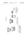
- FIG. 1illustrates an MSC-alginate-HA suspension layered onto the membrane surface of a transwell insert tissue culture well.
- FIG. 2illustrates the layer system of MSC-alginate-HA coated transwell in a tissue culture well with complete chondrogenic media and chondrogenesis inducing factor above and incomplete chondrogenic media below the transmembrane layer.
- FIG. 3shows a cross section view of the alginate-MSC layer after 14 days in culture after histological staining with toluidine-blue, safrinin-O and immunohistochemical staining of collagen Type II tissue.
- FIG. 4MSCs cultured under chondrogenic conditions in (A) alginate layer and (B) pellets for up to 21 days and then stained for reactivity with a collagen Type II-specific antibody, (C) Accumulated GAG and rate of biosynthesis measured after 14 days for both alginate and pellet cultures.
- FIG. 5Accumulation of GAG and rate of biosynthesis measured as a function of concentration of added HA.
- the HA concentrationranted from 50 to 1,000 ⁇ g/ml. Measurements were taken after 14 days in culture.
- FIG. 6Accumulation of GAG, DNA content, and rate of 35 S-sulfate incorporation in MSCs to which hyaluronan was or was not added.
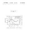
- FIG. 7Rate of 35 S-sulfate incorporation in MSCs plated at different initial cell densities.
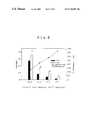
- FIG. 8Effect of HA on GAG synthesis at varying initial cell densities of MSCs.
- the present inventionprovides a composition for the repair of cartilage defects by the rapid regeneration of cartilage tissue.
- the compositionis, for example, inserted or implanted into the defect resulting in articular cartilage regeneration and repair of the defect.
- the compositioncomprises an alginate layer in combination with isolated mesenchymal stem cells.
- the alginatecan be combined with the cartilage regenerative cells and optionally other active ingredients by forming a suspension of the MSCs and the alginate where the suspension liquid can have other active ingredients dissolved.
- Alginateis an unbranched linear polysaccharide consisting of ⁇ -D mannuronic acid and ⁇ -L guluronic acid. It polymerizes and forms a gel in the presence of divalent cations such as Ca ++ .
- the compositioncan contain additional components such as chondroinductive factors.
- the cells and/or the alginate gelcan be contacted with a chondroinductive factor.
- chondroinductive agentor “chondroinductive factor” refers to any natural or synthetic, organic or inorganic chemical or biochemical compound or combination or mixture of compounds, or any mechanical or other physical device, container, influence or force that can be applied to human mesenchymal stem cells which are in a three dimensional format so as to effect their in vitro chondrogenic induction or the production of chondrocytes.
- the chondroinductive agentis preferably selected, individually or in combination, from the group consisting of (i) a glucocorticoid, such as dexamethasone, and (ii) a member of the transforming growth factor superfamily, such as a bone morphogenic protein (preferably BMP-2 or BMP-4). TGF- ⁇ , inhibin A or chondrogenic stimulating activity factor (CSA).
- a glucocorticoidsuch as dexamethasone
- a member of the transforming growth factor superfamilysuch as a bone morphogenic protein (preferably BMP-2 or BMP-4).
- the inventionalso provides a method for treating a cartilage detect in an animal, particularly a mammal, and more particularly a human in need thereof, which comprises administering to the cartilage defect of said animal a cartilage-regenerative amount of the composition of the invention.
- the cellsare contacted with a chondroinductive factor while in the alginate gel layer ex vivo.
- the methodcan further comprise administering at least one chondroinductive factor which further induces or accelerates the differentiation of such mesenchymal stein cells into the chondrogenic lineage.
- the MSCsare first combined with the alginate suspension and polymerized within the gel and are then implanted without induction to chondrogenesis.
- MSCsare loaded into the alginate and then placed in chondrogenesis-inducing media to induce differentiation prior to delivery to the defect site.
- the cellsare grown and maintained in a growth or culture medium in which the composition of the invention can undergo in vitro chondrogenesis, particularly in accordance with the methods of the invention, such as serum-free animal cell culture preparation or medium of known composition which will support the viability of human mesenchymal stem cells in vitro.
- the human mesenchymal stem cells utilized for purposes of the present inventioncan be derived, for example, from hone marrow. Although these cells are normally present at very low frequencies in bone marrow, a process for isolating, and culture expanding the population of these cells in tissue culture is reported in Caplan et al. U.S. Pat. No. 5,486,359.
- the mesenchymal stem cellsare preferably isolated, culture expanded human mesenchymal stem cells in a chemically defined serum-free medium which comprises (1) a chemically defined minimum essential medium (e.g., any of the Eagle's based media, i.e., Dulbecco's Modified Eagle's Medium (DMEM); Iscove's Modified Eagle's Medium, alpha Modified Eagle's Medium, and also McCoy's 5A and BGJb (Fitton-Jackson Modification)); (2) ascorbate or an analog thereof; (3) an iron source; (4) insulin or an insulin-like growth factor; and (5) at least one chondroinductive agent or factor.
- a chemically defined minimum essential mediume.g., any of the Eagle's based media, i.e., Dulbecco's Modified Eagle's Medium (DMEM); Iscove's Modified Eagle's Medium, alpha Modified Eagle's Medium, and also McCoy's 5A and BG
- MSCscan be isolated as a non-cultured preparation, such as by density gradient fractionation, from tissue such as bone marrow, blood (including peripheral blood), periosteum and dermis, and other tissues which have mesodermal origins, so as to be substantially free of other types of cells in the marrow.
- a monoclonal antibody separationis then performed as follows.
- Dynabeads M-450(Dynal Inc., Lake Success, N.Y.) are coupled to anti-MSC monoclonal antibodies having ATCC Accession Numbers HB 10743, HB 10744 and HB 10745, by incubating antibody with secondary antibody coated Dynabeads (2.0 g anti-MSC antibody/mg Dynabead; 1 ⁇ 10 7 Dynabeads/ml) in PBS for 30 minutes at 4° C.
- the cellsare suspended in a solution of sodium alginate and the suspension is distributed on a semiporous membrane as a layer, the shape and dimensions of which can be easily modified.
- the alginate mixtureis solidified by immersion of the layer and supporting membrane in a pool of calcium chloride.
- the alginate layer systemcomprises a component that provides for two separate media-tissue interfaces. This has the advantage of simulating the division of nutritional support seen in vivo between the subchondral vascular supply and the synovial fluid proximal to the articular surface. Nutritional or signalling gradients may be established by manipulating the media to formulations in either media compartment.
- Simple disks of materialmay be cast and cultured for use as an in vitro testing platform permitting easier sample manipulations and multiple analysis from the same sample. Biochemical, molecular and biomechanical analysis is possible from the same sample.
- the systemis ideally suited for studying the effects of bioactive substances involved in modulating the differentiation of MSCs and represents a format for high throughput screening of substances involved in the turnover of the extracellular matrix molecules in cartilage under normal and osteoarthrtic conditions.
- the cell density within the constructcan easily be varied. Construct shape is defined by the dimensions and conformation of the supporting membrane and may be further modified by progressive layering and solidifying of the alginate mixture. Layering in this fashion could also be used to modify cellularity as a function of depth. Thicknesses approaching articular cartilage (0.5-4.0 mm) are achievable. This leads to possible uses as an implantable tissue construct for surgical applications.
- MSCsare culture expanded to appropriate numbers, cast in an alginate layer, sized and shaped to fit an individual's cartilage defect (or larger). The construct is cultured under conditions which are conducive to chondrogenic differentiation.
- the newly formed chondrocytesexpress and organize extracellular matrix molecules into a tissue which is comparable to articular cartilage in its responsiveness to physiological and biomechanical stresses. Within a matter of days of ex vivo culture, this tissue is surgically implanted or grafted into the site of the defect.
- Mesenchymal stein cellwere obtained from human bone marrows (Poetic Technologies. Gaithersburg. Md.). Following normal expansion culture, 4 ⁇ 10 6 hMSCs were spun down and washed with 0.5 M NaCl. The cells were resuspended in 10 ⁇ l sodium alginate (Monsanto, San Diego, Calif.) (2.4% (v/v) in 0.15 M NaCl, sterilized using a 0.45 ⁇ m filter) and 10 ⁇ l soluble HA (HEALON (Pharmacia, Piseataway, N.J.) 2 ⁇ g/ml in sterile MQ H 2 O).
- 10 ⁇ l sodium alginateMonsanto, San Diego, Calif.
- HEALONPharmacia, Piseataway, N.J.
- This cell suspensionwas spread onto the membrane surface of a FALCON® transwell-insert tissue culture well (Becton-Dickinson, N.J.) (see FIG. 1 ).
- the transwell membrane(1.0 ⁇ m pore size) serves as a support for the layer while the alginate polymerizes and forms a gel when the transwell is immersed in sterile 100 mM CaCl 2 for 10 minutes.
- the CaCl 2 as removed and the layer with transwellwas washed in 0.15M NaCl three times and twice with complete chondrogenic media (Table 1) containing 10 ng/ml recombinant human TGF- ⁇ 3 (Oncogene Sciences. Cambridge. Mass.) was added above the layer, incomplete chondrogenic media (Table 2) below (see FIG. 2 ).
- Culture of the layerproceeded for 14 days feeding twice per day due to the high cell density. At the end of this period the tissue was fixed for histological evaluation.
- Histological stainingrevealed positive markers of chondrogenesis in >90% of the tissue. Uniform staining by toluidine blue and safranin-O indicated sulfated proteoglycan production. Likewise, evenly distributed immunohistochemical staining of collagen type II indicated a cartilage-like tissue (see FIG. 3 ).
- TGF- ⁇ 3 stockis made by resuspending lyophilized powder in sterile liquid using siliconized pipet tips and 0.5 ml tubes. 1 ⁇ g of TGF- ⁇ 3 is resuspended in 50 ⁇ l of 10% ethanol/10 mM HCl, then divided into aliquots and stored at ⁇ 80° C. for up to 2 months.
- MSCswere isolated from human bone marrow and cultured under chondrogenic conditions in pellet format and in alginate layers. Samples were harvested for immunocytochemical detection of collagen Type II at 7, 14 and 21 days. The cells were seeded on the alginate at a density of 25 ⁇ 10 6 cells/ml of alginate gel. Pellets were prepared using 2 ⁇ 10 5 cells/pellet. Within the alginate cultures deposition of collagen Type II was evident at day 7 (FIG. 4A) and was distributed uniformily throughout the inter-territorial matrix by day 14. After 21 days, the layer noticeably as thicker with a dense extracellular matrix and the cells were similar morphologically to chondrocytes.
- the outer periphery of the pellethad cells which were flattened and did not express collagen Type II, an observation that has been made previously.
- the alginate layerit appeared that all the cells, including those in the superficial zones, were differentiated, showing both chondrocytic morphology and collagen Type II staining.
- GAGglycosaminoglycans
- the alginate layer systemoffers the ideal format for evaluating factors such as cell density because the cultures are prepared as a suspension of cells in liquid alginate.
- cellswere seeded into alginate layers at densities ranging from 1.56 to 50 ⁇ 10 6 cells/ml. Each layer was cultured for 14 days under chondrogenic conditions and in the presence of TGF- ⁇ 3. The rate of 35 S-sulfate incorporation was determined by adding labeled sulfate 24 hours prior to harvesting (FIG. 7 ). The DNA content was also measured. Some clumping of cells was apparent at all cell densities.
- the rate of chondrogenic differentiationis enhanced when MSCs are cultured in alginate layers rather than pellets; (2) the rate of chondrogenesis is enhanced when HA is added at either 100 or 250 ⁇ g/ml to the culture medium; (3) the rate of chondrogenesis is enhanced when cultures in alginate are seeded with cells at a density 25 ⁇ 10 6 cells/ml, and reduced at lower densities; (4) the positive effect of added HA is evident at all cell densities up to 50 ⁇ 10 6 cells/ml and the magnitude of the effect increases as the cell density decreases.
Landscapes
- Health & Medical Sciences (AREA)
- Life Sciences & Earth Sciences (AREA)
- Engineering & Computer Science (AREA)
- Chemical & Material Sciences (AREA)
- Biomedical Technology (AREA)
- General Health & Medical Sciences (AREA)
- Cell Biology (AREA)
- Epidemiology (AREA)
- Animal Behavior & Ethology (AREA)
- Transplantation (AREA)
- Public Health (AREA)
- Veterinary Medicine (AREA)
- Zoology (AREA)
- Oral & Maxillofacial Surgery (AREA)
- Medicinal Chemistry (AREA)
- Dermatology (AREA)
- Rheumatology (AREA)
- Chemical Kinetics & Catalysis (AREA)
- Botany (AREA)
- Biotechnology (AREA)
- Bioinformatics & Cheminformatics (AREA)
- Genetics & Genomics (AREA)
- Organic Chemistry (AREA)
- Wood Science & Technology (AREA)
- Microbiology (AREA)
- Dispersion Chemistry (AREA)
- Developmental Biology & Embryology (AREA)
- General Engineering & Computer Science (AREA)
- Biochemistry (AREA)
- Urology & Nephrology (AREA)
- Vascular Medicine (AREA)
- Materials For Medical Uses (AREA)
- Micro-Organisms Or Cultivation Processes Thereof (AREA)
Abstract
Description
| TABLE 1 |
| Complete Chondrogenic Medium |
| Ingredient | Stock | Dilution | Final Concentration |
| DMEM | as supplied | n/a | Undiluted |
| (high glucose) | |||
| ITS + | as supplied | 1:99 | 6.25 μg/ml bovine insulin |
| supplement | 6.25 μg/ml transferrin | ||
| 6.25 μg/ml selenous acid | |||
| 5.33 μg/ml linoleic acid | |||
| 1.25 mg/ | |||
| Dexamethasone | |||
| 1 | 1 mM in EtOH | 2 serial | 100 nM |
| (FW = 392) | 1:99 each | ||
| Ascorbic acid-2- | 5 mg/ml | 1:99 | 50 μg/ml |
| phosphate | (FW = 290) | ||
| (AA2P)2 | |||
| Proline2 | 4 mg/ml | 1:99 | 40 μg/ml |
| (FW = 115) | |||
| 100 mM | 1:99 | 1 mM | |
| Antibiotic- | as supplied | 1:99 | 100 U/ml penicillin |
| antimycotic | 100 μg/ | ||
| 250 ng/ml amphotericin | |||
| B | |||
| TGF-β33 | 5 μg/ml | 1:500 | 10 ng/ml |
| 1Dexamethasone powder is dissolved in absolute ethanol (3.92 mg per 10 ml), filter sterilized and stored at 4° C. | |||
| 2Stocks of AA2P and proline are made by dissolving powder into DMEM and filter-sterilizing. Aliquots of these stocks may be frozen at −20° C. and stored for two weeks. | |||
| 3TGF-β3 stock is made by resuspending lyophilized powder in sterile liquid using siliconized pipet tips and 0.5 ml tubes. 1 μg of TGF-β3 is resuspended in 50 μl of 10% ethanol/10 mM HCl, then divided into aliquots and stored at −80° C. for up to 2 months. | |||
| TABLE 2 |
| Incomplete Chondrogenic Medium |
| Ingredient | Stock | Dilution | Final Concentration |
| DMEM | as supplied | n/a | Undiluted |
| (high glucose) | |||
| ITS + | as supplied | 1:99 | 6.25 μg/ml bovine insulin |
| supplement | 6.25 μg/ml transferrin | ||
| 6.25 μg/ml selenous acid | |||
| 5.33 μg/ml linoleic acid | |||
| 1.25 mg/ | |||
| Dexamethasone | |||
| 1 | 1 mM in EtOH | 2 serial | 100 nM |
| (FW = 392) | 1:99 each | ||
| Ascorbic acid-2- | 5 mg/ml | 1:99 | 50 μg/ml |
| phosphate | (FW = 290) | ||
| (AA2P)2 | |||
| Proline2 | 4 mg/ml | 1:99 | 40 μg/ml |
| (FW = 115) | |||
| 100 mM | 1:99 | 1 mM | |
| Antibiotic- | as supplied | 1:99 | 100 U/ml penicillin |
| antimycotic | 100 μg/ | ||
| 250 ng/ml amphotericin | |||
| B | |||
| 1Dexamethasone powder is dissolved in absolute ethanol (3.92 mg per 10 ml), filter sterilized and stored at 4° C. | |||
| 2Stocks of AA2P and proline are made by dissolving powder into DMEM and filter-sterilizing. Aliquots of these stocks may be frozen at −20° C. and stored for two weeks. | |||
Claims (16)
Priority Applications (1)
| Application Number | Priority Date | Filing Date | Title |
|---|---|---|---|
| US09/831,424US6761887B1 (en) | 1998-11-16 | 1999-11-16 | Alginate layer system for chondrogenic differentiation of human mesenchymal stem cells |
Applications Claiming Priority (3)
| Application Number | Priority Date | Filing Date | Title |
|---|---|---|---|
| US10859498P | 1998-11-16 | 1998-11-16 | |
| PCT/US1999/027129WO2000029552A1 (en) | 1998-11-16 | 1999-11-16 | Alginate layer system for chondrogenic differentiation of human mesenchymal stem cells |
| US09/831,424US6761887B1 (en) | 1998-11-16 | 1999-11-16 | Alginate layer system for chondrogenic differentiation of human mesenchymal stem cells |
Publications (1)
| Publication Number | Publication Date |
|---|---|
| US6761887B1true US6761887B1 (en) | 2004-07-13 |
Family
ID=32684474
Family Applications (1)
| Application Number | Title | Priority Date | Filing Date |
|---|---|---|---|
| US09/831,424Expired - LifetimeUS6761887B1 (en) | 1998-11-16 | 1999-11-16 | Alginate layer system for chondrogenic differentiation of human mesenchymal stem cells |
Country Status (1)
| Country | Link |
|---|---|
| US (1) | US6761887B1 (en) |
Cited By (51)
| Publication number | Priority date | Publication date | Assignee | Title |
|---|---|---|---|---|
| US20020169122A1 (en)* | 2001-02-23 | 2002-11-14 | Wyeth | Chondrogenic potential of human bone marrow-derived CD105+ cells by BMP |
| US20040208845A1 (en)* | 2003-04-15 | 2004-10-21 | Michal Eugene T. | Methods and compositions to treat myocardial conditions |
| US20040219182A1 (en)* | 2003-04-29 | 2004-11-04 | Gomes Katherine A. | Novel glue for cartilage repair |
| US20050196423A1 (en)* | 2004-03-05 | 2005-09-08 | Batich Christopher D. | Novel tissue engineered scaffolds derived from copper capillary alginate gels |
| US20060154235A1 (en)* | 2005-01-07 | 2006-07-13 | Takahiro Ochiya | Human hepatocyte-like cells and uses thereof |
| US20070116680A1 (en)* | 2005-11-18 | 2007-05-24 | Rensselaer Polytechnic Institute | Stem cells within gel microenvironments |
| US20080114293A1 (en)* | 2002-06-28 | 2008-05-15 | Claude Charles D | Device and method for combining a treatment agent and a gel |
| US7488348B2 (en) | 2003-05-16 | 2009-02-10 | Musculoskeletal Transplant Foundation | Cartilage allograft plug |
| US20090226519A1 (en)* | 2005-04-19 | 2009-09-10 | Charles Claude | Hydrogel bioscaffoldings and biomedical device coatings |
| US20100204053A1 (en)* | 2007-04-26 | 2010-08-12 | President And Fellows Of Harvard College | Assays for the identification of compounds that modulate bone formation and mineralization |
| US7815926B2 (en) | 2005-07-11 | 2010-10-19 | Musculoskeletal Transplant Foundation | Implant for articular cartilage repair |
| US20100291042A1 (en)* | 2007-05-03 | 2010-11-18 | The Brigham And Women's Hospital, Inc. | Multipotent stem cells and uses thereof |
| US7837740B2 (en) | 2007-01-24 | 2010-11-23 | Musculoskeletal Transplant Foundation | Two piece cancellous construct for cartilage repair |
| US7901457B2 (en) | 2003-05-16 | 2011-03-08 | Musculoskeletal Transplant Foundation | Cartilage allograft plug |
| US20110070205A1 (en)* | 2007-05-03 | 2011-03-24 | The Brigham And Women's Hospital, Inc. | Multipotent stem cells and uses thereof |
| US8038991B1 (en)* | 2003-04-15 | 2011-10-18 | Abbott Cardiovascular Systems Inc. | High-viscosity hyaluronic acid compositions to treat myocardial conditions |
| US8187621B2 (en) | 2005-04-19 | 2012-05-29 | Advanced Cardiovascular Systems, Inc. | Methods and compositions for treating post-myocardial infarction damage |
| US8192760B2 (en) | 2006-12-04 | 2012-06-05 | Abbott Cardiovascular Systems Inc. | Methods and compositions for treating tissue using silk proteins |
| US8292968B2 (en) | 2004-10-12 | 2012-10-23 | Musculoskeletal Transplant Foundation | Cancellous constructs, cartilage particles and combinations of cancellous constructs and cartilage particles |
| US8303972B2 (en) | 2005-04-19 | 2012-11-06 | Advanced Cardiovascular Systems, Inc. | Hydrogel bioscaffoldings and biomedical device coatings |
| US8435551B2 (en) | 2007-03-06 | 2013-05-07 | Musculoskeletal Transplant Foundation | Cancellous construct with support ring for repair of osteochondral defects |
| US8486387B2 (en) | 2006-07-31 | 2013-07-16 | Abbott Cardiovascular Systems Inc. | Modified two-component gelation systems, methods of use and methods of manufacture |
| US8521259B2 (en) | 2001-06-20 | 2013-08-27 | Advanced Cardiovascular Systems, Inc. | Agents that stimulate therapeutic angiogenesis and techniques and devices that enable their delivery |
| US20130315876A1 (en)* | 2009-11-24 | 2013-11-28 | University Of Connecticut | Differentiation of human embryonic and induced pluripotent stem cells |
| US8608661B1 (en) | 2001-11-30 | 2013-12-17 | Advanced Cardiovascular Systems, Inc. | Method for intravascular delivery of a treatment agent beyond a blood vessel wall |
| US8741326B2 (en) | 2006-11-17 | 2014-06-03 | Abbott Cardiovascular Systems Inc. | Modified two-component gelation systems, methods of use and methods of manufacture |
| US8747385B2 (en) | 2003-04-15 | 2014-06-10 | Abbott Cardiovascular Systems Inc. | Methods and compositions to treat myocardial conditions |
| US9005672B2 (en) | 2006-11-17 | 2015-04-14 | Abbott Cardiovascular Systems Inc. | Methods of modifying myocardial infarction expansion |
| US9242005B1 (en) | 2006-08-21 | 2016-01-26 | Abbott Cardiovascular Systems Inc. | Pro-healing agent formulation compositions, methods and treatments |
| US9301975B2 (en) | 2009-05-01 | 2016-04-05 | Biocardia, Inc. | Method of preparing autologous cells and method of use for therapy |
| US9539410B2 (en) | 2005-04-19 | 2017-01-10 | Abbott Cardiovascular Systems Inc. | Methods and compositions for treating post-cardial infarction damage |
| US9687630B2 (en) | 2005-04-19 | 2017-06-27 | Abbott Cardiovascular Systems Inc. | Methods and compositions for treating post-cardial infarction damage |
| US9701940B2 (en) | 2005-09-19 | 2017-07-11 | Histogenics Corporation | Cell-support matrix having narrowly defined uniformly vertically and non-randomly organized porosity and pore density and a method for preparation thereof |
| US9745589B2 (en) | 2010-01-14 | 2017-08-29 | Cornell University | Methods for modulating skeletal remodeling and patterning by modulating SHN2 activity, SHN3 activity, or SHN2 and SHN3 activity in combination |
| US10077420B2 (en) | 2014-12-02 | 2018-09-18 | Histogenics Corporation | Cell and tissue culture container |
| US10668102B2 (en) | 2010-06-02 | 2020-06-02 | Rutgers, The State University Of New Jersey | Lineage differentiation of encapsulated embryonic stem cells |
| IT201900025126A1 (en)* | 2019-12-20 | 2021-06-20 | Sildeha Swiss S A | USE OF HYALURONIC ACID DERIVATIVES IN THE REGENERATION OF BONE TISSUES AND CARTILAGE |
| US11608486B2 (en) | 2015-07-02 | 2023-03-21 | Terumo Bct, Inc. | Cell growth with mechanical stimuli |
| US11613727B2 (en) | 2010-10-08 | 2023-03-28 | Terumo Bct, Inc. | Configurable methods and systems of growing and harvesting cells in a hollow fiber bioreactor system |
| US11624046B2 (en) | 2017-03-31 | 2023-04-11 | Terumo Bct, Inc. | Cell expansion |
| US11629332B2 (en) | 2017-03-31 | 2023-04-18 | Terumo Bct, Inc. | Cell expansion |
| US11634677B2 (en) | 2016-06-07 | 2023-04-25 | Terumo Bct, Inc. | Coating a bioreactor in a cell expansion system |
| US11667876B2 (en) | 2013-11-16 | 2023-06-06 | Terumo Bct, Inc. | Expanding cells in a bioreactor |
| US11667881B2 (en) | 2014-09-26 | 2023-06-06 | Terumo Bct, Inc. | Scheduled feed |
| US11685883B2 (en) | 2016-06-07 | 2023-06-27 | Terumo Bct, Inc. | Methods and systems for coating a cell growth surface |
| US11795432B2 (en) | 2014-03-25 | 2023-10-24 | Terumo Bct, Inc. | Passive replacement of media |
| US11965175B2 (en) | 2016-05-25 | 2024-04-23 | Terumo Bct, Inc. | Cell expansion |
| US12043823B2 (en) | 2021-03-23 | 2024-07-23 | Terumo Bct, Inc. | Cell capture and expansion |
| US12152699B2 (en) | 2022-02-28 | 2024-11-26 | Terumo Bct, Inc. | Multiple-tube pinch valve assembly |
| US12234441B2 (en) | 2017-03-31 | 2025-02-25 | Terumo Bct, Inc. | Cell expansion |
| US12410405B2 (en) | 2010-10-08 | 2025-09-09 | Mesoblast International Sàrl | Enhanced MSC preparations |
Citations (3)
| Publication number | Priority date | Publication date | Assignee | Title |
|---|---|---|---|---|
| WO1996028539A1 (en)* | 1995-03-14 | 1996-09-19 | Morphogen Pharmaceuticals, Inc. | Mesenchymal stem cells for cartilage repair |
| WO1998025653A2 (en)* | 1996-12-10 | 1998-06-18 | Reprogenesis, Inc. | Improved hydrogel for tissue engineering |
| WO1998032333A1 (en)* | 1996-12-06 | 1998-07-30 | Osiris Therapeutics, Inc. | Improved chondrogenic differentiation of human mesenchymal stem cells |
- 1999
- 1999-11-16USUS09/831,424patent/US6761887B1/ennot_activeExpired - Lifetime
Patent Citations (3)
| Publication number | Priority date | Publication date | Assignee | Title |
|---|---|---|---|---|
| WO1996028539A1 (en)* | 1995-03-14 | 1996-09-19 | Morphogen Pharmaceuticals, Inc. | Mesenchymal stem cells for cartilage repair |
| WO1998032333A1 (en)* | 1996-12-06 | 1998-07-30 | Osiris Therapeutics, Inc. | Improved chondrogenic differentiation of human mesenchymal stem cells |
| WO1998025653A2 (en)* | 1996-12-10 | 1998-06-18 | Reprogenesis, Inc. | Improved hydrogel for tissue engineering |
Cited By (88)
| Publication number | Priority date | Publication date | Assignee | Title |
|---|---|---|---|---|
| US20020169122A1 (en)* | 2001-02-23 | 2002-11-14 | Wyeth | Chondrogenic potential of human bone marrow-derived CD105+ cells by BMP |
| US8521259B2 (en) | 2001-06-20 | 2013-08-27 | Advanced Cardiovascular Systems, Inc. | Agents that stimulate therapeutic angiogenesis and techniques and devices that enable their delivery |
| US8608661B1 (en) | 2001-11-30 | 2013-12-17 | Advanced Cardiovascular Systems, Inc. | Method for intravascular delivery of a treatment agent beyond a blood vessel wall |
| US20080114293A1 (en)* | 2002-06-28 | 2008-05-15 | Claude Charles D | Device and method for combining a treatment agent and a gel |
| US8500680B2 (en) | 2002-06-28 | 2013-08-06 | Abbott Cardiovascular Systems Inc. | Device and method for combining a treatment agent and a gel |
| US8637069B2 (en) | 2002-06-28 | 2014-01-28 | Abbott Cardiovascular Systems Inc. | Device and method for combining a treatment agent and a gel |
| US8715265B2 (en) | 2002-06-28 | 2014-05-06 | Abbott Cardiovascular Systems Inc. | Device and method for combining a treatment agent and a gel |
| US8795652B1 (en) | 2003-04-15 | 2014-08-05 | Abbott Cardiovascular Systems Inc. | Methods and compositions to treat myocardial conditions |
| US8747385B2 (en) | 2003-04-15 | 2014-06-10 | Abbott Cardiovascular Systems Inc. | Methods and compositions to treat myocardial conditions |
| US20040208845A1 (en)* | 2003-04-15 | 2004-10-21 | Michal Eugene T. | Methods and compositions to treat myocardial conditions |
| US8038991B1 (en)* | 2003-04-15 | 2011-10-18 | Abbott Cardiovascular Systems Inc. | High-viscosity hyaluronic acid compositions to treat myocardial conditions |
| US8821473B2 (en) | 2003-04-15 | 2014-09-02 | Abbott Cardiovascular Systems Inc. | Methods and compositions to treat myocardial conditions |
| US8383158B2 (en) | 2003-04-15 | 2013-02-26 | Abbott Cardiovascular Systems Inc. | Methods and compositions to treat myocardial conditions |
| US7067123B2 (en) | 2003-04-29 | 2006-06-27 | Musculoskeletal Transplant Foundation | Glue for cartilage repair |
| USRE42208E1 (en) | 2003-04-29 | 2011-03-08 | Musculoskeletal Transplant Foundation | Glue for cartilage repair |
| US20040219182A1 (en)* | 2003-04-29 | 2004-11-04 | Gomes Katherine A. | Novel glue for cartilage repair |
| USRE43258E1 (en) | 2003-04-29 | 2012-03-20 | Musculoskeletal Transplant Foundation | Glue for cartilage repair |
| US7901457B2 (en) | 2003-05-16 | 2011-03-08 | Musculoskeletal Transplant Foundation | Cartilage allograft plug |
| US7488348B2 (en) | 2003-05-16 | 2009-02-10 | Musculoskeletal Transplant Foundation | Cartilage allograft plug |
| US8221500B2 (en) | 2003-05-16 | 2012-07-17 | Musculoskeletal Transplant Foundation | Cartilage allograft plug |
| US20050196423A1 (en)* | 2004-03-05 | 2005-09-08 | Batich Christopher D. | Novel tissue engineered scaffolds derived from copper capillary alginate gels |
| US7601525B2 (en) | 2004-03-05 | 2009-10-13 | University Of Florida Research Foundation, Inc. | Alginate gel scaffold having a plurality of continuous parallel microtubular copper capillaries |
| US8292968B2 (en) | 2004-10-12 | 2012-10-23 | Musculoskeletal Transplant Foundation | Cancellous constructs, cartilage particles and combinations of cancellous constructs and cartilage particles |
| US20060154235A1 (en)* | 2005-01-07 | 2006-07-13 | Takahiro Ochiya | Human hepatocyte-like cells and uses thereof |
| US8187621B2 (en) | 2005-04-19 | 2012-05-29 | Advanced Cardiovascular Systems, Inc. | Methods and compositions for treating post-myocardial infarction damage |
| US9539410B2 (en) | 2005-04-19 | 2017-01-10 | Abbott Cardiovascular Systems Inc. | Methods and compositions for treating post-cardial infarction damage |
| US20090226519A1 (en)* | 2005-04-19 | 2009-09-10 | Charles Claude | Hydrogel bioscaffoldings and biomedical device coatings |
| US8828433B2 (en) | 2005-04-19 | 2014-09-09 | Advanced Cardiovascular Systems, Inc. | Hydrogel bioscaffoldings and biomedical device coatings |
| US8609126B2 (en) | 2005-04-19 | 2013-12-17 | Advanced Cardiovascular Systems, Inc. | Methods and compositions for treating post-myocardial infarction damage |
| US8303972B2 (en) | 2005-04-19 | 2012-11-06 | Advanced Cardiovascular Systems, Inc. | Hydrogel bioscaffoldings and biomedical device coatings |
| US9687630B2 (en) | 2005-04-19 | 2017-06-27 | Abbott Cardiovascular Systems Inc. | Methods and compositions for treating post-cardial infarction damage |
| US7815926B2 (en) | 2005-07-11 | 2010-10-19 | Musculoskeletal Transplant Foundation | Implant for articular cartilage repair |
| US9701940B2 (en) | 2005-09-19 | 2017-07-11 | Histogenics Corporation | Cell-support matrix having narrowly defined uniformly vertically and non-randomly organized porosity and pore density and a method for preparation thereof |
| US20070116680A1 (en)* | 2005-11-18 | 2007-05-24 | Rensselaer Polytechnic Institute | Stem cells within gel microenvironments |
| US8486386B2 (en) | 2006-07-31 | 2013-07-16 | Abbott Cardiovascular Systems Inc. | Modified two-component gelation systems, methods of use and methods of manufacture |
| US8486387B2 (en) | 2006-07-31 | 2013-07-16 | Abbott Cardiovascular Systems Inc. | Modified two-component gelation systems, methods of use and methods of manufacture |
| US9242005B1 (en) | 2006-08-21 | 2016-01-26 | Abbott Cardiovascular Systems Inc. | Pro-healing agent formulation compositions, methods and treatments |
| US9775930B2 (en) | 2006-11-17 | 2017-10-03 | Abbott Cardiovascular Systems Inc. | Composition for modifying myocardial infarction expansion |
| US9005672B2 (en) | 2006-11-17 | 2015-04-14 | Abbott Cardiovascular Systems Inc. | Methods of modifying myocardial infarction expansion |
| US8741326B2 (en) | 2006-11-17 | 2014-06-03 | Abbott Cardiovascular Systems Inc. | Modified two-component gelation systems, methods of use and methods of manufacture |
| US8828436B2 (en) | 2006-12-04 | 2014-09-09 | Abbott Cardiovascular Systems Inc. | Methods and compositions for treating tissue using silk proteins |
| US8192760B2 (en) | 2006-12-04 | 2012-06-05 | Abbott Cardiovascular Systems Inc. | Methods and compositions for treating tissue using silk proteins |
| US8465772B2 (en) | 2006-12-04 | 2013-06-18 | Abbott Cardiovascular Systems Inc. | Methods and compositions for treating tissue using silk proteins |
| US8465773B2 (en) | 2006-12-04 | 2013-06-18 | Abbott Cardiovascular Systems Inc. | Methods and compositions for treating tissue using silk proteins |
| US8906110B2 (en) | 2007-01-24 | 2014-12-09 | Musculoskeletal Transplant Foundation | Two piece cancellous construct for cartilage repair |
| US7837740B2 (en) | 2007-01-24 | 2010-11-23 | Musculoskeletal Transplant Foundation | Two piece cancellous construct for cartilage repair |
| US8435551B2 (en) | 2007-03-06 | 2013-05-07 | Musculoskeletal Transplant Foundation | Cancellous construct with support ring for repair of osteochondral defects |
| US20100204053A1 (en)* | 2007-04-26 | 2010-08-12 | President And Fellows Of Harvard College | Assays for the identification of compounds that modulate bone formation and mineralization |
| US20110070205A1 (en)* | 2007-05-03 | 2011-03-24 | The Brigham And Women's Hospital, Inc. | Multipotent stem cells and uses thereof |
| US8574567B2 (en) | 2007-05-03 | 2013-11-05 | The Brigham And Women's Hospital, Inc. | Multipotent stem cells and uses thereof |
| US10568911B2 (en) | 2007-05-03 | 2020-02-25 | The Brigham And Women's Hospital, Inc. | Multipotent stem cells and uses thereof |
| US9127252B2 (en) | 2007-05-03 | 2015-09-08 | The Brigham And Women's Hospital, Inc. | Multipotent stem cells and uses thereof |
| US20100291042A1 (en)* | 2007-05-03 | 2010-11-18 | The Brigham And Women's Hospital, Inc. | Multipotent stem cells and uses thereof |
| US9301975B2 (en) | 2009-05-01 | 2016-04-05 | Biocardia, Inc. | Method of preparing autologous cells and method of use for therapy |
| US10035982B2 (en) | 2009-05-01 | 2018-07-31 | Biocardia, Inc. | Method of preparing autologous cells and methods of use for therapy |
| US9752123B2 (en) | 2009-05-01 | 2017-09-05 | Biocardia, Inc. | Method of preparing autologous cells and methods of use for therapy |
| US8927275B2 (en)* | 2009-11-24 | 2015-01-06 | University Of Connecticut | Differentiation of human embryonic and induced pluripotent stem cells |
| US20130315876A1 (en)* | 2009-11-24 | 2013-11-28 | University Of Connecticut | Differentiation of human embryonic and induced pluripotent stem cells |
| US9745589B2 (en) | 2010-01-14 | 2017-08-29 | Cornell University | Methods for modulating skeletal remodeling and patterning by modulating SHN2 activity, SHN3 activity, or SHN2 and SHN3 activity in combination |
| US10668102B2 (en) | 2010-06-02 | 2020-06-02 | Rutgers, The State University Of New Jersey | Lineage differentiation of encapsulated embryonic stem cells |
| US12410405B2 (en) | 2010-10-08 | 2025-09-09 | Mesoblast International Sàrl | Enhanced MSC preparations |
| US11613727B2 (en) | 2010-10-08 | 2023-03-28 | Terumo Bct, Inc. | Configurable methods and systems of growing and harvesting cells in a hollow fiber bioreactor system |
| US11773363B2 (en) | 2010-10-08 | 2023-10-03 | Terumo Bct, Inc. | Configurable methods and systems of growing and harvesting cells in a hollow fiber bioreactor system |
| US11746319B2 (en) | 2010-10-08 | 2023-09-05 | Terumo Bct, Inc. | Customizable methods and systems of growing and harvesting cells in a hollow fiber bioreactor system |
| US11708554B2 (en) | 2013-11-16 | 2023-07-25 | Terumo Bct, Inc. | Expanding cells in a bioreactor |
| US11667876B2 (en) | 2013-11-16 | 2023-06-06 | Terumo Bct, Inc. | Expanding cells in a bioreactor |
| US11795432B2 (en) | 2014-03-25 | 2023-10-24 | Terumo Bct, Inc. | Passive replacement of media |
| US12065637B2 (en) | 2014-09-26 | 2024-08-20 | Terumo Bct, Inc. | Scheduled feed |
| US11667881B2 (en) | 2014-09-26 | 2023-06-06 | Terumo Bct, Inc. | Scheduled feed |
| US10077420B2 (en) | 2014-12-02 | 2018-09-18 | Histogenics Corporation | Cell and tissue culture container |
| US11555172B2 (en) | 2014-12-02 | 2023-01-17 | Ocugen, Inc. | Cell and tissue culture container |
| US11608486B2 (en) | 2015-07-02 | 2023-03-21 | Terumo Bct, Inc. | Cell growth with mechanical stimuli |
| US11965175B2 (en) | 2016-05-25 | 2024-04-23 | Terumo Bct, Inc. | Cell expansion |
| US11634677B2 (en) | 2016-06-07 | 2023-04-25 | Terumo Bct, Inc. | Coating a bioreactor in a cell expansion system |
| US11685883B2 (en) | 2016-06-07 | 2023-06-27 | Terumo Bct, Inc. | Methods and systems for coating a cell growth surface |
| US12077739B2 (en) | 2016-06-07 | 2024-09-03 | Terumo Bct, Inc. | Coating a bioreactor in a cell expansion system |
| US11999929B2 (en) | 2016-06-07 | 2024-06-04 | Terumo Bct, Inc. | Methods and systems for coating a cell growth surface |
| US11702634B2 (en) | 2017-03-31 | 2023-07-18 | Terumo Bct, Inc. | Expanding cells in a bioreactor |
| US12359170B2 (en) | 2017-03-31 | 2025-07-15 | Terumo Bct, Inc. | Expanding cells in a bioreactor |
| US11629332B2 (en) | 2017-03-31 | 2023-04-18 | Terumo Bct, Inc. | Cell expansion |
| US12234441B2 (en) | 2017-03-31 | 2025-02-25 | Terumo Bct, Inc. | Cell expansion |
| US11624046B2 (en) | 2017-03-31 | 2023-04-11 | Terumo Bct, Inc. | Cell expansion |
| WO2021124249A1 (en)* | 2019-12-20 | 2021-06-24 | Sildeha Swiss S.A. | Use of hyaluronic acid derivatives in the regeneration of bone and cartilage tissues |
| IT201900025126A1 (en)* | 2019-12-20 | 2021-06-20 | Sildeha Swiss S A | USE OF HYALURONIC ACID DERIVATIVES IN THE REGENERATION OF BONE TISSUES AND CARTILAGE |
| CN115135318A (en)* | 2019-12-20 | 2022-09-30 | Trb化药国际股份有限公司 | Use of hyaluronic acid derivatives in the regeneration of bone and cartilage tissue " |
| US12043823B2 (en) | 2021-03-23 | 2024-07-23 | Terumo Bct, Inc. | Cell capture and expansion |
| US12152699B2 (en) | 2022-02-28 | 2024-11-26 | Terumo Bct, Inc. | Multiple-tube pinch valve assembly |
| US12209689B2 (en) | 2022-02-28 | 2025-01-28 | Terumo Kabushiki Kaisha | Multiple-tube pinch valve assembly |
Similar Documents
| Publication | Publication Date | Title |
|---|---|---|
| US6761887B1 (en) | Alginate layer system for chondrogenic differentiation of human mesenchymal stem cells | |
| US6482231B1 (en) | Biological material for the repair of connective tissue defects comprising mesenchymal stem cells and hyaluronic acid derivative | |
| US5197985A (en) | Method for enhancing the implantation and differentiation of marrow-derived mesenchymal cells | |
| US5226914A (en) | Method for treating connective tissue disorders | |
| US6489165B2 (en) | Chondrocyte-like cells useful for tissue engineering and methods | |
| Kadiyala et al. | Culture-expanded, bone marrow-derived mesenchymal stem cells can regenerate a critical-sized segmental bone defect | |
| US7863045B2 (en) | Isolation of skeletal precursor cells | |
| WO2000029552A1 (en) | Alginate layer system for chondrogenic differentiation of human mesenchymal stem cells | |
| AU731468B2 (en) | Regeneration and augmentation of bone using mesenchymal stem cells | |
| DE60028666T2 (en) | Use of adipose-derived stromal cells for differentiation into chondrocytes and their use to repair cartilage tissue | |
| JP3808900B2 (en) | Biological material composed of a three-dimensional biocompatible and biodegradable matrix comprising an effective culture of bone marrow stem cells partially or fully differentiated into connective tissue cells and a hyaluronic acid derivative | |
| JP2003510108A (en) | Biological joint structures | |
| JP2003517259A (en) | Method for isolating bone and precursor chondrocytes | |
| KR102479530B1 (en) | Method of Preparing Pellets of Chondrocytes differentiated from human induced pluripotent stem cell and use of the same | |
| JP2009530334A (en) | Method for maintaining, improving and repairing the cartilage phenotype of chondrocytes | |
| JP2002525043A (en) | Improved submucosal tissue graft composition | |
| US20030215426A1 (en) | Redifferentiated cells for repairing cartilage defects | |
| EP3025735B1 (en) | Methods for complex tissue engineering | |
| Hendriksa et al. | and Jens Riesleb | |
| MXPA97005612A (en) | Induction directed by lineage of the differentiation of human mesenquimatosas cells of ori |
Legal Events
| Date | Code | Title | Description |
|---|---|---|---|
| AS | Assignment | Owner name:OSIRIS THERAPEUTICS, INC., MARYLAND Free format text:ASSIGNMENT OF ASSIGNORS INTEREST;ASSIGNORS:KAVALKOVICH, KARL;BOYNTON, RAYMOND;MURPHY, MARY;AND OTHERS;REEL/FRAME:012030/0588;SIGNING DATES FROM 20010619 TO 20010723 | |
| STCF | Information on status: patent grant | Free format text:PATENTED CASE | |
| FEPP | Fee payment procedure | Free format text:PAYOR NUMBER ASSIGNED (ORIGINAL EVENT CODE: ASPN); ENTITY STATUS OF PATENT OWNER: LARGE ENTITY | |
| FPAY | Fee payment | Year of fee payment:4 | |
| FPAY | Fee payment | Year of fee payment:8 | |
| FEPP | Fee payment procedure | Free format text:PAYOR NUMBER ASSIGNED (ORIGINAL EVENT CODE: ASPN); ENTITY STATUS OF PATENT OWNER: LARGE ENTITY Free format text:PAYER NUMBER DE-ASSIGNED (ORIGINAL EVENT CODE: RMPN); ENTITY STATUS OF PATENT OWNER: LARGE ENTITY | |
| AS | Assignment | Owner name:MESOBLAST INTERNATIONAL S?RL, SWITZERLAND Free format text:ASSIGNMENT OF ASSIGNORS INTEREST;ASSIGNOR:OSIRIS THERAPEUTICS, INC.;REEL/FRAME:031533/0828 Effective date:20131010 Owner name:MESOBLAST INTERNATIONAL SARL, SWITZERLAND Free format text:ASSIGNMENT OF ASSIGNORS INTEREST;ASSIGNOR:OSIRIS THERAPEUTICS, INC.;REEL/FRAME:031533/0828 Effective date:20131010 | |
| FPAY | Fee payment | Year of fee payment:12 |