US6712822B2 - Apparatus and method for the repair of articular cartilage defects - Google Patents
Apparatus and method for the repair of articular cartilage defectsDownload PDFInfo
- Publication number
- US6712822B2 US6712822B2US10/261,899US26189902AUS6712822B2US 6712822 B2US6712822 B2US 6712822B2US 26189902 AUS26189902 AUS 26189902AUS 6712822 B2US6712822 B2US 6712822B2
- Authority
- US
- United States
- Prior art keywords
- cartilage defect
- bone
- leg structure
- encapsulation device
- area
- Prior art date
- Legal status (The legal status is an assumption and is not a legal conclusion. Google has not performed a legal analysis and makes no representation as to the accuracy of the status listed.)
- Expired - Lifetime
Links
- 206010061762ChondropathyDiseases0.000titleclaimsabstractdescription39
- 238000000034methodMethods0.000titledescription22
- 238000005538encapsulationMethods0.000claimsabstractdescription102
- 230000007547defectEffects0.000claimsabstractdescription49
- 210000000988bone and boneAnatomy0.000claimsabstractdescription36
- 210000000845cartilageAnatomy0.000claimsabstractdescription27
- 239000000463materialSubstances0.000claimsdescription6
- 208000006735PeriostitisDiseases0.000claimsdescription5
- 210000003460periosteumAnatomy0.000claimsdescription5
- 230000003014reinforcing effectEffects0.000claimsdescription3
- 102000008186CollagenHuman genes0.000claimsdescription2
- 108010035532CollagenProteins0.000claimsdescription2
- 230000010261cell growthEffects0.000claimsdescription2
- 229920001436collagenPolymers0.000claimsdescription2
- 230000002093peripheral effectEffects0.000claims3
- 238000003780insertionMethods0.000description43
- 230000037431insertionEffects0.000description43
- 210000004027cellAnatomy0.000description22
- 210000001188articular cartilageAnatomy0.000description13
- 238000005299abrasionMethods0.000description11
- 208000013201Stress fractureDiseases0.000description10
- 238000011882arthroplastyMethods0.000description10
- 230000003902lesionEffects0.000description10
- 210000001778pluripotent stem cellAnatomy0.000description8
- 238000002054transplantationMethods0.000description7
- 239000008280bloodSubstances0.000description4
- 210000004369bloodAnatomy0.000description4
- 210000003321cartilage cellAnatomy0.000description4
- 230000035876healingEffects0.000description4
- 210000001185bone marrowAnatomy0.000description3
- 238000010276constructionMethods0.000description3
- 239000002002slurrySubstances0.000description3
- 238000001356surgical procedureMethods0.000description3
- 230000000295complement effectEffects0.000description2
- 230000006378damageEffects0.000description2
- 230000000694effectsEffects0.000description2
- 210000000968fibrocartilageAnatomy0.000description2
- 239000003102growth factorSubstances0.000description2
- 210000003035hyaline cartilageAnatomy0.000description2
- 238000004519manufacturing processMethods0.000description2
- 239000011159matrix materialSubstances0.000description2
- 230000002250progressing effectEffects0.000description2
- 230000008929regenerationEffects0.000description2
- 238000011069regeneration methodMethods0.000description2
- 239000007787solidSubstances0.000description2
- 208000027418Wounds and injuryDiseases0.000description1
- 238000004873anchoringMethods0.000description1
- 206010003246arthritisDiseases0.000description1
- 208000015100cartilage diseaseDiseases0.000description1
- 201000005043chondromalaciaDiseases0.000description1
- 230000015271coagulationEffects0.000description1
- 238000005345coagulationMethods0.000description1
- 238000000151depositionMethods0.000description1
- 230000004064dysfunctionEffects0.000description1
- 230000012010growthEffects0.000description1
- 238000003306harvestingMethods0.000description1
- 239000007943implantSubstances0.000description1
- 208000014674injuryDiseases0.000description1
- 230000014759maintenance of locationEffects0.000description1
- 238000010899nucleationMethods0.000description1
- 235000015097nutrientsNutrition0.000description1
- 230000000399orthopedic effectEffects0.000description1
- 230000003389potentiating effectEffects0.000description1
- 230000001737promoting effectEffects0.000description1
- 238000004080punchingMethods0.000description1
- 230000001172regenerating effectEffects0.000description1
- 239000000523sampleSubstances0.000description1
- 230000000638stimulationEffects0.000description1
- 239000002699waste materialSubstances0.000description1
Images


Classifications
- A—HUMAN NECESSITIES
- A61—MEDICAL OR VETERINARY SCIENCE; HYGIENE
- A61F—FILTERS IMPLANTABLE INTO BLOOD VESSELS; PROSTHESES; DEVICES PROVIDING PATENCY TO, OR PREVENTING COLLAPSING OF, TUBULAR STRUCTURES OF THE BODY, e.g. STENTS; ORTHOPAEDIC, NURSING OR CONTRACEPTIVE DEVICES; FOMENTATION; TREATMENT OR PROTECTION OF EYES OR EARS; BANDAGES, DRESSINGS OR ABSORBENT PADS; FIRST-AID KITS
- A61F2/00—Filters implantable into blood vessels; Prostheses, i.e. artificial substitutes or replacements for parts of the body; Appliances for connecting them with the body; Devices providing patency to, or preventing collapsing of, tubular structures of the body, e.g. stents
- A61F2/02—Prostheses implantable into the body
- A61F2/30—Joints
- A61F2/46—Special tools for implanting artificial joints
- A61F2/4603—Special tools for implanting artificial joints for insertion or extraction of endoprosthetic joints or of accessories thereof
- A61F2/4618—Special tools for implanting artificial joints for insertion or extraction of endoprosthetic joints or of accessories thereof of cartilage
- A—HUMAN NECESSITIES
- A61—MEDICAL OR VETERINARY SCIENCE; HYGIENE
- A61B—DIAGNOSIS; SURGERY; IDENTIFICATION
- A61B17/00—Surgical instruments, devices or methods
- A61B17/064—Surgical staples, i.e. penetrating the tissue
- A61B17/0642—Surgical staples, i.e. penetrating the tissue for bones, e.g. for osteosynthesis or connecting tendon to bone
- A—HUMAN NECESSITIES
- A61—MEDICAL OR VETERINARY SCIENCE; HYGIENE
- A61B—DIAGNOSIS; SURGERY; IDENTIFICATION
- A61B17/00—Surgical instruments, devices or methods
- A61B17/068—Surgical staplers, e.g. containing multiple staples or clamps
- A—HUMAN NECESSITIES
- A61—MEDICAL OR VETERINARY SCIENCE; HYGIENE
- A61F—FILTERS IMPLANTABLE INTO BLOOD VESSELS; PROSTHESES; DEVICES PROVIDING PATENCY TO, OR PREVENTING COLLAPSING OF, TUBULAR STRUCTURES OF THE BODY, e.g. STENTS; ORTHOPAEDIC, NURSING OR CONTRACEPTIVE DEVICES; FOMENTATION; TREATMENT OR PROTECTION OF EYES OR EARS; BANDAGES, DRESSINGS OR ABSORBENT PADS; FIRST-AID KITS
- A61F2/00—Filters implantable into blood vessels; Prostheses, i.e. artificial substitutes or replacements for parts of the body; Appliances for connecting them with the body; Devices providing patency to, or preventing collapsing of, tubular structures of the body, e.g. stents
- A61F2/02—Prostheses implantable into the body
- A61F2/30—Joints
- A61F2/30721—Accessories
- A61F2/30749—Fixation appliances for connecting prostheses to the body
- A—HUMAN NECESSITIES
- A61—MEDICAL OR VETERINARY SCIENCE; HYGIENE
- A61F—FILTERS IMPLANTABLE INTO BLOOD VESSELS; PROSTHESES; DEVICES PROVIDING PATENCY TO, OR PREVENTING COLLAPSING OF, TUBULAR STRUCTURES OF THE BODY, e.g. STENTS; ORTHOPAEDIC, NURSING OR CONTRACEPTIVE DEVICES; FOMENTATION; TREATMENT OR PROTECTION OF EYES OR EARS; BANDAGES, DRESSINGS OR ABSORBENT PADS; FIRST-AID KITS
- A61F2/00—Filters implantable into blood vessels; Prostheses, i.e. artificial substitutes or replacements for parts of the body; Appliances for connecting them with the body; Devices providing patency to, or preventing collapsing of, tubular structures of the body, e.g. stents
- A61F2/02—Prostheses implantable into the body
- A61F2/30—Joints
- A61F2/30756—Cartilage endoprostheses
- A—HUMAN NECESSITIES
- A61—MEDICAL OR VETERINARY SCIENCE; HYGIENE
- A61B—DIAGNOSIS; SURGERY; IDENTIFICATION
- A61B17/00—Surgical instruments, devices or methods
- A61B17/16—Instruments for performing osteoclasis; Drills or chisels for bones; Trepans
- A61B17/1635—Instruments for performing osteoclasis; Drills or chisels for bones; Trepans for grafts, harvesting or transplants
- A—HUMAN NECESSITIES
- A61—MEDICAL OR VETERINARY SCIENCE; HYGIENE
- A61B—DIAGNOSIS; SURGERY; IDENTIFICATION
- A61B17/00—Surgical instruments, devices or methods
- A61B17/16—Instruments for performing osteoclasis; Drills or chisels for bones; Trepans
- A61B17/1655—Instruments for performing osteoclasis; Drills or chisels for bones; Trepans for tapping
- A—HUMAN NECESSITIES
- A61—MEDICAL OR VETERINARY SCIENCE; HYGIENE
- A61B—DIAGNOSIS; SURGERY; IDENTIFICATION
- A61B17/00—Surgical instruments, devices or methods
- A61B2017/00004—(bio)absorbable, (bio)resorbable or resorptive
- A—HUMAN NECESSITIES
- A61—MEDICAL OR VETERINARY SCIENCE; HYGIENE
- A61B—DIAGNOSIS; SURGERY; IDENTIFICATION
- A61B17/00—Surgical instruments, devices or methods
- A61B17/04—Surgical instruments, devices or methods for suturing wounds; Holders or packages for needles or suture materials
- A61B17/0401—Suture anchors, buttons or pledgets, i.e. means for attaching sutures to bone, cartilage or soft tissue; Instruments for applying or removing suture anchors
- A61B2017/0417—T-fasteners
- A—HUMAN NECESSITIES
- A61—MEDICAL OR VETERINARY SCIENCE; HYGIENE
- A61B—DIAGNOSIS; SURGERY; IDENTIFICATION
- A61B17/00—Surgical instruments, devices or methods
- A61B17/064—Surgical staples, i.e. penetrating the tissue
- A61B2017/0641—Surgical staples, i.e. penetrating the tissue having at least three legs as part of one single body
- A—HUMAN NECESSITIES
- A61—MEDICAL OR VETERINARY SCIENCE; HYGIENE
- A61B—DIAGNOSIS; SURGERY; IDENTIFICATION
- A61B17/00—Surgical instruments, devices or methods
- A61B17/064—Surgical staples, i.e. penetrating the tissue
- A61B2017/0646—Surgical staples, i.e. penetrating the tissue for insertion into cartillege, e.g. meniscus
- A—HUMAN NECESSITIES
- A61—MEDICAL OR VETERINARY SCIENCE; HYGIENE
- A61B—DIAGNOSIS; SURGERY; IDENTIFICATION
- A61B17/00—Surgical instruments, devices or methods
- A61B17/064—Surgical staples, i.e. penetrating the tissue
- A61B2017/0647—Surgical staples, i.e. penetrating the tissue having one single leg, e.g. tacks
- A—HUMAN NECESSITIES
- A61—MEDICAL OR VETERINARY SCIENCE; HYGIENE
- A61F—FILTERS IMPLANTABLE INTO BLOOD VESSELS; PROSTHESES; DEVICES PROVIDING PATENCY TO, OR PREVENTING COLLAPSING OF, TUBULAR STRUCTURES OF THE BODY, e.g. STENTS; ORTHOPAEDIC, NURSING OR CONTRACEPTIVE DEVICES; FOMENTATION; TREATMENT OR PROTECTION OF EYES OR EARS; BANDAGES, DRESSINGS OR ABSORBENT PADS; FIRST-AID KITS
- A61F2/00—Filters implantable into blood vessels; Prostheses, i.e. artificial substitutes or replacements for parts of the body; Appliances for connecting them with the body; Devices providing patency to, or preventing collapsing of, tubular structures of the body, e.g. stents
- A61F2/02—Prostheses implantable into the body
- A61F2/28—Bones
- A61F2002/2835—Bone graft implants for filling a bony defect or an endoprosthesis cavity, e.g. by synthetic material or biological material
- A—HUMAN NECESSITIES
- A61—MEDICAL OR VETERINARY SCIENCE; HYGIENE
- A61F—FILTERS IMPLANTABLE INTO BLOOD VESSELS; PROSTHESES; DEVICES PROVIDING PATENCY TO, OR PREVENTING COLLAPSING OF, TUBULAR STRUCTURES OF THE BODY, e.g. STENTS; ORTHOPAEDIC, NURSING OR CONTRACEPTIVE DEVICES; FOMENTATION; TREATMENT OR PROTECTION OF EYES OR EARS; BANDAGES, DRESSINGS OR ABSORBENT PADS; FIRST-AID KITS
- A61F2/00—Filters implantable into blood vessels; Prostheses, i.e. artificial substitutes or replacements for parts of the body; Appliances for connecting them with the body; Devices providing patency to, or preventing collapsing of, tubular structures of the body, e.g. stents
- A61F2/02—Prostheses implantable into the body
- A61F2/30—Joints
- A61F2002/30001—Additional features of subject-matter classified in A61F2/28, A61F2/30 and subgroups thereof
- A61F2002/30003—Material related properties of the prosthesis or of a coating on the prosthesis
- A61F2002/3006—Properties of materials and coating materials
- A61F2002/30062—(bio)absorbable, biodegradable, bioerodable, (bio)resorbable, resorptive
- A—HUMAN NECESSITIES
- A61—MEDICAL OR VETERINARY SCIENCE; HYGIENE
- A61F—FILTERS IMPLANTABLE INTO BLOOD VESSELS; PROSTHESES; DEVICES PROVIDING PATENCY TO, OR PREVENTING COLLAPSING OF, TUBULAR STRUCTURES OF THE BODY, e.g. STENTS; ORTHOPAEDIC, NURSING OR CONTRACEPTIVE DEVICES; FOMENTATION; TREATMENT OR PROTECTION OF EYES OR EARS; BANDAGES, DRESSINGS OR ABSORBENT PADS; FIRST-AID KITS
- A61F2/00—Filters implantable into blood vessels; Prostheses, i.e. artificial substitutes or replacements for parts of the body; Appliances for connecting them with the body; Devices providing patency to, or preventing collapsing of, tubular structures of the body, e.g. stents
- A61F2/02—Prostheses implantable into the body
- A61F2/30—Joints
- A61F2002/30001—Additional features of subject-matter classified in A61F2/28, A61F2/30 and subgroups thereof
- A61F2002/30108—Shapes
- A61F2002/3011—Cross-sections or two-dimensional shapes
- A61F2002/30159—Concave polygonal shapes
- A61F2002/30181—Y-shaped
- A—HUMAN NECESSITIES
- A61—MEDICAL OR VETERINARY SCIENCE; HYGIENE
- A61F—FILTERS IMPLANTABLE INTO BLOOD VESSELS; PROSTHESES; DEVICES PROVIDING PATENCY TO, OR PREVENTING COLLAPSING OF, TUBULAR STRUCTURES OF THE BODY, e.g. STENTS; ORTHOPAEDIC, NURSING OR CONTRACEPTIVE DEVICES; FOMENTATION; TREATMENT OR PROTECTION OF EYES OR EARS; BANDAGES, DRESSINGS OR ABSORBENT PADS; FIRST-AID KITS
- A61F2/00—Filters implantable into blood vessels; Prostheses, i.e. artificial substitutes or replacements for parts of the body; Appliances for connecting them with the body; Devices providing patency to, or preventing collapsing of, tubular structures of the body, e.g. stents
- A61F2/02—Prostheses implantable into the body
- A61F2/30—Joints
- A61F2002/30001—Additional features of subject-matter classified in A61F2/28, A61F2/30 and subgroups thereof
- A61F2002/30108—Shapes
- A61F2002/30199—Three-dimensional shapes
- A61F2002/302—Three-dimensional shapes toroidal, e.g. rings
- A—HUMAN NECESSITIES
- A61—MEDICAL OR VETERINARY SCIENCE; HYGIENE
- A61F—FILTERS IMPLANTABLE INTO BLOOD VESSELS; PROSTHESES; DEVICES PROVIDING PATENCY TO, OR PREVENTING COLLAPSING OF, TUBULAR STRUCTURES OF THE BODY, e.g. STENTS; ORTHOPAEDIC, NURSING OR CONTRACEPTIVE DEVICES; FOMENTATION; TREATMENT OR PROTECTION OF EYES OR EARS; BANDAGES, DRESSINGS OR ABSORBENT PADS; FIRST-AID KITS
- A61F2/00—Filters implantable into blood vessels; Prostheses, i.e. artificial substitutes or replacements for parts of the body; Appliances for connecting them with the body; Devices providing patency to, or preventing collapsing of, tubular structures of the body, e.g. stents
- A61F2/02—Prostheses implantable into the body
- A61F2/30—Joints
- A61F2/30721—Accessories
- A61F2/30749—Fixation appliances for connecting prostheses to the body
- A61F2002/30751—Fixation appliances for connecting prostheses to the body for attaching cartilage scaffolds to underlying bone
- A—HUMAN NECESSITIES
- A61—MEDICAL OR VETERINARY SCIENCE; HYGIENE
- A61F—FILTERS IMPLANTABLE INTO BLOOD VESSELS; PROSTHESES; DEVICES PROVIDING PATENCY TO, OR PREVENTING COLLAPSING OF, TUBULAR STRUCTURES OF THE BODY, e.g. STENTS; ORTHOPAEDIC, NURSING OR CONTRACEPTIVE DEVICES; FOMENTATION; TREATMENT OR PROTECTION OF EYES OR EARS; BANDAGES, DRESSINGS OR ABSORBENT PADS; FIRST-AID KITS
- A61F2/00—Filters implantable into blood vessels; Prostheses, i.e. artificial substitutes or replacements for parts of the body; Appliances for connecting them with the body; Devices providing patency to, or preventing collapsing of, tubular structures of the body, e.g. stents
- A61F2/02—Prostheses implantable into the body
- A61F2/30—Joints
- A61F2/30756—Cartilage endoprostheses
- A61F2002/30761—Support means for artificial cartilage, e.g. cartilage defect covering membranes
- A—HUMAN NECESSITIES
- A61—MEDICAL OR VETERINARY SCIENCE; HYGIENE
- A61F—FILTERS IMPLANTABLE INTO BLOOD VESSELS; PROSTHESES; DEVICES PROVIDING PATENCY TO, OR PREVENTING COLLAPSING OF, TUBULAR STRUCTURES OF THE BODY, e.g. STENTS; ORTHOPAEDIC, NURSING OR CONTRACEPTIVE DEVICES; FOMENTATION; TREATMENT OR PROTECTION OF EYES OR EARS; BANDAGES, DRESSINGS OR ABSORBENT PADS; FIRST-AID KITS
- A61F2/00—Filters implantable into blood vessels; Prostheses, i.e. artificial substitutes or replacements for parts of the body; Appliances for connecting them with the body; Devices providing patency to, or preventing collapsing of, tubular structures of the body, e.g. stents
- A61F2/02—Prostheses implantable into the body
- A61F2/30—Joints
- A61F2/30767—Special external or bone-contacting surface, e.g. coating for improving bone ingrowth
- A61F2/30771—Special external or bone-contacting surface, e.g. coating for improving bone ingrowth applied in original prostheses, e.g. holes or grooves
- A61F2002/30772—Apertures or holes, e.g. of circular cross section
- A—HUMAN NECESSITIES
- A61—MEDICAL OR VETERINARY SCIENCE; HYGIENE
- A61F—FILTERS IMPLANTABLE INTO BLOOD VESSELS; PROSTHESES; DEVICES PROVIDING PATENCY TO, OR PREVENTING COLLAPSING OF, TUBULAR STRUCTURES OF THE BODY, e.g. STENTS; ORTHOPAEDIC, NURSING OR CONTRACEPTIVE DEVICES; FOMENTATION; TREATMENT OR PROTECTION OF EYES OR EARS; BANDAGES, DRESSINGS OR ABSORBENT PADS; FIRST-AID KITS
- A61F2/00—Filters implantable into blood vessels; Prostheses, i.e. artificial substitutes or replacements for parts of the body; Appliances for connecting them with the body; Devices providing patency to, or preventing collapsing of, tubular structures of the body, e.g. stents
- A61F2/02—Prostheses implantable into the body
- A61F2/30—Joints
- A61F2/30767—Special external or bone-contacting surface, e.g. coating for improving bone ingrowth
- A61F2/30771—Special external or bone-contacting surface, e.g. coating for improving bone ingrowth applied in original prostheses, e.g. holes or grooves
- A61F2002/30878—Special external or bone-contacting surface, e.g. coating for improving bone ingrowth applied in original prostheses, e.g. holes or grooves with non-sharp protrusions, for instance contacting the bone for anchoring, e.g. keels, pegs, pins, posts, shanks, stems, struts
- A61F2002/30891—Plurality of protrusions
- A61F2002/30892—Plurality of protrusions parallel
- A—HUMAN NECESSITIES
- A61—MEDICAL OR VETERINARY SCIENCE; HYGIENE
- A61F—FILTERS IMPLANTABLE INTO BLOOD VESSELS; PROSTHESES; DEVICES PROVIDING PATENCY TO, OR PREVENTING COLLAPSING OF, TUBULAR STRUCTURES OF THE BODY, e.g. STENTS; ORTHOPAEDIC, NURSING OR CONTRACEPTIVE DEVICES; FOMENTATION; TREATMENT OR PROTECTION OF EYES OR EARS; BANDAGES, DRESSINGS OR ABSORBENT PADS; FIRST-AID KITS
- A61F2/00—Filters implantable into blood vessels; Prostheses, i.e. artificial substitutes or replacements for parts of the body; Appliances for connecting them with the body; Devices providing patency to, or preventing collapsing of, tubular structures of the body, e.g. stents
- A61F2/02—Prostheses implantable into the body
- A61F2/30—Joints
- A61F2/30767—Special external or bone-contacting surface, e.g. coating for improving bone ingrowth
- A61F2/30771—Special external or bone-contacting surface, e.g. coating for improving bone ingrowth applied in original prostheses, e.g. holes or grooves
- A61F2002/30904—Special external or bone-contacting surface, e.g. coating for improving bone ingrowth applied in original prostheses, e.g. holes or grooves serrated profile, i.e. saw-toothed
- A—HUMAN NECESSITIES
- A61—MEDICAL OR VETERINARY SCIENCE; HYGIENE
- A61F—FILTERS IMPLANTABLE INTO BLOOD VESSELS; PROSTHESES; DEVICES PROVIDING PATENCY TO, OR PREVENTING COLLAPSING OF, TUBULAR STRUCTURES OF THE BODY, e.g. STENTS; ORTHOPAEDIC, NURSING OR CONTRACEPTIVE DEVICES; FOMENTATION; TREATMENT OR PROTECTION OF EYES OR EARS; BANDAGES, DRESSINGS OR ABSORBENT PADS; FIRST-AID KITS
- A61F2/00—Filters implantable into blood vessels; Prostheses, i.e. artificial substitutes or replacements for parts of the body; Appliances for connecting them with the body; Devices providing patency to, or preventing collapsing of, tubular structures of the body, e.g. stents
- A61F2/02—Prostheses implantable into the body
- A61F2/30—Joints
- A61F2/46—Special tools for implanting artificial joints
- A61F2/4603—Special tools for implanting artificial joints for insertion or extraction of endoprosthetic joints or of accessories thereof
- A61F2002/4625—Special tools for implanting artificial joints for insertion or extraction of endoprosthetic joints or of accessories thereof with relative movement between parts of the instrument during use
- A61F2002/4627—Special tools for implanting artificial joints for insertion or extraction of endoprosthetic joints or of accessories thereof with relative movement between parts of the instrument during use with linear motion along or rotating motion about the instrument axis or the implantation direction, e.g. telescopic, along a guiding rod, screwing inside the instrument
- A—HUMAN NECESSITIES
- A61—MEDICAL OR VETERINARY SCIENCE; HYGIENE
- A61F—FILTERS IMPLANTABLE INTO BLOOD VESSELS; PROSTHESES; DEVICES PROVIDING PATENCY TO, OR PREVENTING COLLAPSING OF, TUBULAR STRUCTURES OF THE BODY, e.g. STENTS; ORTHOPAEDIC, NURSING OR CONTRACEPTIVE DEVICES; FOMENTATION; TREATMENT OR PROTECTION OF EYES OR EARS; BANDAGES, DRESSINGS OR ABSORBENT PADS; FIRST-AID KITS
- A61F2210/00—Particular material properties of prostheses classified in groups A61F2/00 - A61F2/26 or A61F2/82 or A61F9/00 or A61F11/00 or subgroups thereof
- A61F2210/0004—Particular material properties of prostheses classified in groups A61F2/00 - A61F2/26 or A61F2/82 or A61F9/00 or A61F11/00 or subgroups thereof bioabsorbable
- A—HUMAN NECESSITIES
- A61—MEDICAL OR VETERINARY SCIENCE; HYGIENE
- A61F—FILTERS IMPLANTABLE INTO BLOOD VESSELS; PROSTHESES; DEVICES PROVIDING PATENCY TO, OR PREVENTING COLLAPSING OF, TUBULAR STRUCTURES OF THE BODY, e.g. STENTS; ORTHOPAEDIC, NURSING OR CONTRACEPTIVE DEVICES; FOMENTATION; TREATMENT OR PROTECTION OF EYES OR EARS; BANDAGES, DRESSINGS OR ABSORBENT PADS; FIRST-AID KITS
- A61F2230/00—Geometry of prostheses classified in groups A61F2/00 - A61F2/26 or A61F2/82 or A61F9/00 or A61F11/00 or subgroups thereof
- A61F2230/0002—Two-dimensional shapes, e.g. cross-sections
- A61F2230/0028—Shapes in the form of latin or greek characters
- A61F2230/006—Y-shaped
- A—HUMAN NECESSITIES
- A61—MEDICAL OR VETERINARY SCIENCE; HYGIENE
- A61F—FILTERS IMPLANTABLE INTO BLOOD VESSELS; PROSTHESES; DEVICES PROVIDING PATENCY TO, OR PREVENTING COLLAPSING OF, TUBULAR STRUCTURES OF THE BODY, e.g. STENTS; ORTHOPAEDIC, NURSING OR CONTRACEPTIVE DEVICES; FOMENTATION; TREATMENT OR PROTECTION OF EYES OR EARS; BANDAGES, DRESSINGS OR ABSORBENT PADS; FIRST-AID KITS
- A61F2230/00—Geometry of prostheses classified in groups A61F2/00 - A61F2/26 or A61F2/82 or A61F9/00 or A61F11/00 or subgroups thereof
- A61F2230/0063—Three-dimensional shapes
- A61F2230/0065—Three-dimensional shapes toroidal, e.g. ring-shaped, doughnut-shaped
- A—HUMAN NECESSITIES
- A61—MEDICAL OR VETERINARY SCIENCE; HYGIENE
- A61F—FILTERS IMPLANTABLE INTO BLOOD VESSELS; PROSTHESES; DEVICES PROVIDING PATENCY TO, OR PREVENTING COLLAPSING OF, TUBULAR STRUCTURES OF THE BODY, e.g. STENTS; ORTHOPAEDIC, NURSING OR CONTRACEPTIVE DEVICES; FOMENTATION; TREATMENT OR PROTECTION OF EYES OR EARS; BANDAGES, DRESSINGS OR ABSORBENT PADS; FIRST-AID KITS
- A61F2310/00—Prostheses classified in A61F2/28 or A61F2/30 - A61F2/44 being constructed from or coated with a particular material
- A61F2310/00005—The prosthesis being constructed from a particular material
- A61F2310/00365—Proteins; Polypeptides; Degradation products thereof
Definitions
- This inventionrelates to surgical apparatus and methods in general, and more particularly to surgical apparatus and methods for the repair of articular cartilage defects.
- Articular cartilage defectshave traditionally been treated with chondroplasty, shaving, microfracture, abrasion arthroplasty and, most recently, autologous transplantation.
- the treatment of articular cartilage defectswas principally concerned with preventing a progression of the defect. More recently, attention has been focused on developing ways to actually repair the defect and effect articular cartilage healing.
- chondroplasty and shavingare principally concerned with removing offending portions of the articular cartilage (e.g., loose flaps, rough edges, etc.) so as to prevent the enlargement of an existing articular cartilage defect. While chondroplasty and shaving have proven helpful in preventing the spread of an existing articular cartilage defect, they do not actually repair the defect or effect articular cartilage healing.
- microfracture and abrasion arthroplastyThe basic idea behind microfracture and abrasion arthroplasty is to violate the subchondral plate, thereby allowing blood (preferably including marrow cells) to fill the defect and initiate an injury repair. This may be done in a variety of ways well known in the art, e.g., with a rasp to abrade the defect, a pick to pick away the area of the defect, a drill to microdrill the area of the defect, an RF probe (or otherwise) to heat and thereby disrupt the region of the defect, etc. It is known that such a procedure does not actually cause articular cartilage to grow in the defect.
- a fibrocartilage/Hyaline cartilageregenerates which, while generally not as good as articular cartilage since it lacks the mechanical properties of the articular cartilage, is certainly better than bare bone.
- a common problem with this techniqueis that the blood (and marrow) cells do not tend to stay seeded in the defect, since they are commonly wiped away by joint motion and/or other factors.
- osteocondral graftinga plug of healthy articular cartilage and underlying bone is harvested from a donor site and transplanted to the defect site. While this technique has proven effective, it typically causes serious damage to the donor site. In addition, it can be difficult to find donor sites with the proper surface profiles, and it can be difficult to properly align the layers (i.e., cartilage and underlying bone) of the graft plug with the layers of the defect site.
- one object of the present inventionis to provide improved apparatus for the repair of articular cartilage defects, wherein the apparatus can be used with microfracture and abrasion arthroplasty.
- Another object of the present inventionis to provide an improved method for the repair of articular cartilage defects, wherein the method can be used with microfracture and abrasion arthroplasty.
- Another object of the present inventionis to provide improved apparatus for the repair of articular cartilage defects, wherein the apparatus can be used with autologous cell transplantation.
- Still another object of the present inventionis to provide an improved method for the repair of articular cartilage defects, wherein the method can be used with autologous cell transplantation.
- the present inventionwhich, in one form of the invention, comprises an encapsulation device adapted to encapsulate loose-bodied cells (e.g., blood and marrow cells, pluripotent stem cells, autologous cartilage cells, etc.) so as to facilitate the repair of articular cartilage defects.
- the encapsulation deviceis preferably formed out of bioabsorbable or bioremodelable materials, such that the encapsulation device will only be present at the surgical site for a limited period of time following surgery.
- an encapsulation devicefor the repair of articular cartilage defects.
- the devicecomprises a body for disposition adjacent a bone in an area of the cartilage defect, and elongated leg structure extending from the body for disposition in the bone in the area of the cartilage defect.
- the leg structureis provided with a length which is a plurality of magnitudes greater than a thickness of the body, and is of a generally conical configuration.
- a system for effecting articular cartilage defect repairincludes an encapsulation device comprising a body for disposition adjacent a bone in an area of the cartilage defect, and elongated leg structure extending from a distal surface of the body for disposition in the bone in the area of the cartilage defect, wherein the elongated leg structure comprises one or more legs.
- Each leg of the leg structureis provided with a central opening therein extending from a proximal surface of the body.
- a pilot hole devicecomprising a head portion, at least one elongated foot extending distally from the head portion, and a handle portion extending proximally from the head portion, the pilot hole device elongated foot being adapted to form a pilot hole in the bone to receive a leg member of the leg structure.
- An insertion toolis provided comprising a head portion, at least one elongated foot extending from a distal end of the head portion, each elongated foot of the insertion tool head portion being adapted to be received by the central opening of one of the encapsulation device legs.
- the insertion tool head portionis adapted to engage a proximal surface of the encapsulation device.
- the encapsulation deviceis adapted to be mounted on the insertion tool, and the insertion tool may be manipulated to drive the encapsulation device leg structure into at least one hole in the bone, to place the encapsulation device distal surface adjacent the bone and in the area of the cartilage defect.
- a tool for in-bone placement of an encapsulation device for repair of an articular cartilage defectcomprising a body portion and a cannulated leg extending distally from a center of a distal surface of the body portion.
- the toolcomprises a head portion having a distal surface configured generally complementary to a proximal surface of the encapsulation device, a handle portion extending proximally from the head portion, the head portion and handle portion forming a bore extending axially of the head portion and handle portion, and an insertion spike extending through the bore and adapted to extend through the encapsulation device leg, with a pointed distal end of the spike extending distally from a distal end of the encapsulation device leg.
- the insertion spikeis adapted to form a hole in the bone and the tool is adapted to push the encapsulation device leg into the hole and the encapsulation device body into engagement with the bone.
- a method for effecting a repair to an articular cartilage defectincludes the steps of providing an encapsulation device comprising a body for disposition adjacent a bone in an area of the cartilage defect, and an elongated leg structure extending from the body for disposition in the bone in the area of the cartilage defect, wherein the elongated leg structure comprises at least one leg, producing a hole in the bone for each leg of the encapsulation device leg structure, and driving each leg of the leg structure of the encapsulation device into a hole in the bone to bring a distal surface of the encapsulation device body into adjacency with the bone.
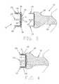
- FIG. 1is a schematic view of an encapsulation device formed in accordance with the present invention
- FIG. 2is a schematic view of an insertion tool for deploying an encapsulation device of the type shown in FIG. 1;
- FIG. 3is a sectional view showing the encapsulation device of FIG. 1 about to be engaged by the insertion tool of FIG. 2;
- FIG. 4is a sectional view showing the encapsulation device of FIG. 1 engaged by the insertion tool of FIG. 2;
- FIG. 5is a schematic view of an articular cartilage defect
- FIG. 6is a schematic view of a pilot hole device for forming pilot holes in the articular cartilage defect shown in FIG. 5;
- FIG. 7is a sectional view showing the pilot hole device of FIG. 6 approaching the articular cartilage defect
- FIG. 8is a sectional view showing the pilot hole device of FIG. 6 engaged with the articular cartilage defect
- FIG. 9is a sectional view showing the articular cartilage defect after it has had pilot holes formed therein;
- FIG. 10is a sectional view showing the encapsulation device of FIG. 1 about to be deployed in the articular cartilage defect by the insertion tool of FIG. 2;
- FIG. 11is a sectional view showing the insertion tool of FIG. 2 deploying the encapsulation device of FIG. 1 in the articular cartilage defect;
- FIG. 12is a sectional view showing the encapsulation device of FIG. 1 deployed in the articular cartilage defect, with the insertion tool of FIG. 2 having been removed from the surgical site;
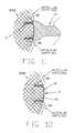
- FIG. 13is a schematic view of an alternative form of encapsulation device formed in accordance with the present invention.
- FIG. 14is a schematic view of another alternative form of encapsulation device formed in accordance with the present invention.
- FIG. 15is a schematic view showing still another alternative form of encapsulation device formed in accordance with the present invention, wherein the encapsulation device is adapted to have a piece of harvested periosteum secured thereto;
- FIG. 15Ais a schematic view like that of FIG. 15, except showing the piece of harvested periosteum being secured to the encapsulation device;
- FIG. 15Bis a schematic view like those of FIGS. 15 and 15A, except showing the piece of harvested periosteum secured to the encapsulation device;
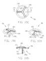
- FIGS. 16A and 16Bare perspective views of an alternative embodiment of the encapsulation device
- FIG. 16Cis a top plan view of the device of FIGS. 16A and 16B;
- FIG. 16Dis a sectional view taken along line XVI—XVI of FIG. 16C;
- FIGS. 17A-17Dare views similar to those of FIGS. 16A-16D, respectively, but illustrative of a further alternative embodiment of encapsulation device;
- FIGS. 18A-18Dare views similar to those of FIGS. 17A-17D, respectively, but illustrative of a further alternative embodiment
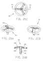
- FIGS. 19A-19Dare views similar to those of FIGS. 18A-18D, respectively, but illustrative of a still further alternative embodiment
- FIGS. 20A-20Dare views similar to those of FIGS. 19A-19D, respectively, but illustrative of a still further alternative embodiment
- FIGS. 21A-21Dare views similar to those of FIGS. 20A-20D, respectively, but illustrative of a still further alternative embodiment
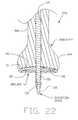
- FIG. 22is a sectional view of the encapsulation device of FIG. 21 shown in conjunction with an inserter tool.
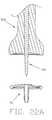
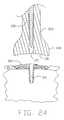
- FIGS. 22A, 22 B, 23 and 24are sectional views illustrating steps in the use of the inserter tool of FIG. 22 to set the encapsulation device of FIG. 21 into bone.
- Encapsulation device 5formed in accordance with the present invention.
- Encapsulation device 5generally comprises a body 10 including a cover 14 , and having a distal surface 15 (FIG. 3) and a proximal surface 20 .
- At least one elongated leg 25extends distally from the distal surface 15 .
- the at least one leg 25is of a length which is a plurality of magnitudes greater than a thickness of the body 10 , to provide for secure anchoring of the body.
- the at least one leg 25is preferably of a slightly conical configuration to aid in insertion of the leg into a bone, as described hereinbelow.
- the distal end of the at least one leg 25may be pointed, and/or the shaft of the at least one leg 25 may be provided with locking ribs, barbs, or other protrusions, 26 , so as to enhance fixation.
- An opening 30is formed in the at least one leg 25 and extends through, and opens on, the body's proximal surface 20 .
- Encapsulation device 5may be formed out of a single member, or it may be formed out of several members joined together during manufacture.
- Encapsulation device 5is intended to encapsulate loose-bodied cells (e.g., blood and marrow cells, pluripotent stem cells, autologous cartilage cells, etc.) so as to facilitate the repair of articular cartilage defects.
- encapsulation device 5is preferably formed out of bioabsorbable or bioremodelable materials, such that the encapsulation device will only be present at the surgical site for a limited period of time following surgery (e.g., 8-12 weeks). If desired, encapsulation device 5 may be impregnated with various cell growth factors so as to assist in cell stimulation or cell regeneration.
- Insertion tool 35generally comprises a head 40 , at least one elongated foot 45 extending distally from head 40 , and a shaft 50 extending proximally from head 40 .
- Head 40preferably has a distal end profile generally matching the proximal end profile of the encapsulation device's body 10 .
- the insertion tool's at least one foot 45has a configuration which matches the encapsulation device's at least one opening 30 , whereby the at least one foot 45 may be received within the at least one opening 30 .
- insertion tool 35preferably has a plurality of feet 45 , with feet 45 matching legs 25 and holes 30 in number and configuration.
- encapsulation device 5may be mounted to head 40 of insertion tool 35 by passing the insertion tool's feet 45 into the encapsulation device's openings 30 . See FIGS. 3 and 4. This may be done during manufacture or at the time of use.
- Shaft 50permits the insertion tool to be gripped by the user and have its head 40 , and hence the encapsulation device 5 , directed to the surgical site.
- Encapsulation device 5may be used as follows.
- an articular cartilage defect 55is first prepared by microfracture or abrasion arthroplasty.
- pilot hole device 60having a construction generally similar to insertion tool 35 (e.g., a head 65 , at least one elongated and conically shaped foot 70 and a shaft 75 ) is pushed against articular cartilage defect 55 (FIGS. 7 and 8) so as to create one or more pilot holes 80 (FIG. 9 ).
- pilot hole device 60is constructed so as to form a pattern of pilot holes 80 which conforms to the pattern of the encapsulation device's at least one leg 25 .
- encapsulation device 5is mounted to insertion tool 35 (FIG. 4 ), if it has not already been mounted to insertion tool 35 , and then shaft 50 of insertion tool 35 is manipulated (FIGS. 10 and 11) so as to implant encapsulation device 5 directly over the treated lesion, with the encapsulation device's at least one leg 25 deployed in the at least one pilot hole 80 (FIG. 12 ), and with the encapsulation device's body 10 encapsulating the treated lesion, thus encouraging coagulation at the treated lesion and superior adhesion of regenerating fibrocartilage/Hyaline cartilage cells. As the cartilage cells are regenerated, encapsulation device 5 will be absorbed or remodeled, until only cartilage cells remain at the site of the original defect.
- the pluripotent stem cellsare harvested by aspirating bone marrow from various regions of the body (e.g., the femoral notch, the iliac crest, the spine, etc.) and then depositing the bone marrow on the lesion site either before, or concurrently with, deployment of encapsulation device 5 .
- the harvested bone marrowmay be filtered prior to seeding so as to produce a more potent slurry of pluripotent stem cells.
- autologous articular cartilage cellsare harvested, prepared outside the body (e.g., isolated and/or enhanced with growth factors and/or multiplied, etc.) and then deposited at the defect site, either before, or concurrently with, deployment of encapsulation device 5 .
- Encapsulation device 5 Awhich comprises an alternative form of the invention.
- Encapsulation device 5 Ais similar to the encapsulation device 5 described above, except that body 10 A includes a matrix or mesh 85 A at one or more locations within the body 10 A, rather than the aforementioned cover 14 .
- One or more legs 25 Aextend from the body 10 A.
- Forming a body 10 A with a matrix or mesh 85 Acan be advantageous over a solid body 10 , e.g., in the case of microfracture and abrasion arthroplasty, it can provide a superior flow of nutrients to the site and a superior flow of waste products away from the site; or in the case of microfracture and abrasion arthroplasty with pluripotent stem cells, it can permit a slurry of such cells to be placed on the encapsulation device prior to deploying the encapsulation device at the defect site; or in the case of autologous cell transplantation, it can permit a slurry of graft articular cartilage cells to be placed on the encapsulation device prior to deploying the encapsulation device at the defect site.
- Encapsulation device 5 Bwhich comprises another alternative form of the invention.
- Encapsulation device SBis similar to encapsulation device 5 described above, except that body 10 B thereof includes a collagen scaffold 90 B for promoting the growth of replacement cartilage across the defect site.
- Encapsulation device 5 Cwhich comprises still another alternative form of the invention.
- Encapsulation device 5 Cis similar to the encapsulation device 5 described above, except that body 10 C thereof comprises an empty frame 12 C into which a mass of harvested periosteum 95 may be secured, e.g., with sutures 100 (FIGS. 15 A and 15 B).
- body portion 10 Dincludes a cover 14 D which comprises a shell-like structure, and is provided with a distal surface 15 D and a proximal surface 20 D, with solid (i.e., non-cannulated) legs 25 D extending distally from the distal surface 15 D.
- the legs 25 Dmay be provided with enlarged distal end portions 32 D for increased retention properties.
- the enlarged end portions 32 Dinclude a proximal end 34 having a larger diameter than the adjacent portion of the leg 25 D.
- the distal end 36 of the end portions 32 Dmay be pointed or rounded, but are of reduced diameter.
- encapsulation device 5 Dis deployed by an insertion tool (not shown) lacking the elongated feet of insertion tool 35 .
- an insertion toolpreferably includes means (not shown) for releasably holding the encapsulation device to the distal end of the insertion tool (e.g., a mechanical coupler, a vacuum coupler, etc.).
- FIGS. 17A-17Dthere is shown another alternative embodiment of encapsulation device, 5 E, generally similar to device SD, but in which the body 10 E comprises an open frame 12 E, without a cover portion 14 D or other similar component.
- FIGS. 18A-18DIllustrated in FIGS. 18A-18D is a further alternative embodiment of encapsulation device, 5 F, in which the body 10 F includes a cover portion 14 F comprising a shell 16 provided with a distal surface 15 F and a proximal surface 20 F.
- a single central leg 25 Fextends from the distal surface 15 F and is provided with an enlarged end portion 32 F.
- the proximal surface 20 Fis provided with reinforcing struts 105 .
- a further alternative embodimentfeatures an encapsulation device 5 G similar in appearance to device 5 F, but in which the body 10 G includes a substantially open frame 12 G supporting only the reinforcing struts 105 from which depends a single central leg 25 G having the enlarged distal end portion 32 G.
- FIGS. 20A-20DAn alternative embodiment of device, 5 H, shown in FIGS. 20A-20D, is generally similar to the embodiment shown in FIGS. 19A-19D, but with a cannulated leg portion 25 H depending from the struts 105 . As shown in the drawings, the leg 25 H may be provided with an increased diameter relative to the leg 25 G to accommodate a desired central passageway 110 therethrough.
- FIGS. 21A-21Dthere is shown an alternative embodiment of encapsulation device, 5 I, which is similar to the device 5 F shown in FIGS. 18A-18D, but in which the single depending leg 25 I is provided with the axial passageway 110 therethrough.
- An insertion tool 35 Aincludes a head portion 40 A and a shaft 50 A.
- the head portion 40 Ais provided with a distal end surface 116 which is generally complementary in configuration to the encapsulation device proximal surface 20 I.
- the insertion tool 35 Ais provided with a bore 115 in which is disposed an insertion spike 120 .
- the encapsulation device axial passageway 110receives the insertion tool insertion spike 120 .
- the tool 35 Amay then be used as a combination pilot hole device and insertion tool, by punching a hole 80 and, simultaneously, introducing the device 5 I into the hole. More particularly, insertion spike 120 is mounted in the insertion tool so that the distal end of the insertion spike protrudes from the distal end of the insertion tool (FIG. 22 A), encapsulation device 5 I is mounted to insertion tool 35 A and insertion spike 120 (FIG. 22 ), and the assembly is advanced into the defect site (FIG. 22 B). Thereafter, the insertion spike 120 is withdrawn from the device 5 I (FIG. 23 ), and the insertion tool 35 A is withdrawn from the device 5 I (FIG. 24 ).
- the encapsulation device of the present inventionis ideal for full thickness defects, but it also has a role for chondromalacia, cartilage fissures, partial thickness tears, and abrasions. These lesions have a high potential for progressing into full thickness defects.
- the encapsulation device of the present inventionwith any of the techniques described above, can be used as a temporary patch, creating a microenvironment to aid in healing and regeneration.
Landscapes
- Health & Medical Sciences (AREA)
- Life Sciences & Earth Sciences (AREA)
- Public Health (AREA)
- Veterinary Medicine (AREA)
- Engineering & Computer Science (AREA)
- Biomedical Technology (AREA)
- Heart & Thoracic Surgery (AREA)
- Orthopedic Medicine & Surgery (AREA)
- Animal Behavior & Ethology (AREA)
- General Health & Medical Sciences (AREA)
- Transplantation (AREA)
- Surgery (AREA)
- Vascular Medicine (AREA)
- Oral & Maxillofacial Surgery (AREA)
- Cardiology (AREA)
- Rheumatology (AREA)
- Nuclear Medicine, Radiotherapy & Molecular Imaging (AREA)
- Medical Informatics (AREA)
- Molecular Biology (AREA)
- Physical Education & Sports Medicine (AREA)
- Prostheses (AREA)
- Materials For Medical Uses (AREA)
- Surgical Instruments (AREA)
- Application Of Or Painting With Fluid Materials (AREA)
Abstract
Description
Claims (14)
Priority Applications (2)
| Application Number | Priority Date | Filing Date | Title |
|---|---|---|---|
| US10/261,899US6712822B2 (en) | 2001-10-01 | 2002-10-01 | Apparatus and method for the repair of articular cartilage defects |
| US10/812,609US8157805B2 (en) | 2001-10-01 | 2004-03-30 | Apparatus and method for the repair of articular cartilage defects |
Applications Claiming Priority (2)
| Application Number | Priority Date | Filing Date | Title |
|---|---|---|---|
| US32629301P | 2001-10-01 | 2001-10-01 | |
| US10/261,899US6712822B2 (en) | 2001-10-01 | 2002-10-01 | Apparatus and method for the repair of articular cartilage defects |
Related Child Applications (1)
| Application Number | Title | Priority Date | Filing Date |
|---|---|---|---|
| US10/812,609ContinuationUS8157805B2 (en) | 2001-10-01 | 2004-03-30 | Apparatus and method for the repair of articular cartilage defects |
Publications (2)
| Publication Number | Publication Date |
|---|---|
| US20030083665A1 US20030083665A1 (en) | 2003-05-01 |
| US6712822B2true US6712822B2 (en) | 2004-03-30 |
Family
ID=23271614
Family Applications (2)
| Application Number | Title | Priority Date | Filing Date |
|---|---|---|---|
| US10/261,899Expired - LifetimeUS6712822B2 (en) | 2001-10-01 | 2002-10-01 | Apparatus and method for the repair of articular cartilage defects |
| US10/812,609Expired - Fee RelatedUS8157805B2 (en) | 2001-10-01 | 2004-03-30 | Apparatus and method for the repair of articular cartilage defects |
Family Applications After (1)
| Application Number | Title | Priority Date | Filing Date |
|---|---|---|---|
| US10/812,609Expired - Fee RelatedUS8157805B2 (en) | 2001-10-01 | 2004-03-30 | Apparatus and method for the repair of articular cartilage defects |
Country Status (9)
| Country | Link |
|---|---|
| US (2) | US6712822B2 (en) |
| EP (1) | EP1437960B1 (en) |
| JP (2) | JP4330991B2 (en) |
| AT (1) | ATE416682T1 (en) |
| AU (1) | AU2002330168B2 (en) |
| CA (1) | CA2462256C (en) |
| DE (1) | DE60230306D1 (en) |
| ES (1) | ES2318040T3 (en) |
| WO (1) | WO2003028535A2 (en) |
Cited By (61)
| Publication number | Priority date | Publication date | Assignee | Title |
|---|---|---|---|---|
| US20030135209A1 (en)* | 1999-12-03 | 2003-07-17 | Bahaa Seedhom | Fixation technology |
| US20040170663A1 (en)* | 2002-10-09 | 2004-09-02 | Jennifer H. Elisseeff | Method and material for enhanced tissue-biomaterial integration |
| US20040191106A1 (en)* | 2002-11-08 | 2004-09-30 | Howmedica Osteonics Corp. | Laser-produced porous surface |
| US20050069572A1 (en)* | 2002-10-09 | 2005-03-31 | Jennifer Elisseeff | Multi-layered polymerizing hydrogels for tissue regeneration |
| US20060147332A1 (en)* | 2004-12-30 | 2006-07-06 | Howmedica Osteonics Corp. | Laser-produced porous structure |
| US20070098675A1 (en)* | 2002-09-25 | 2007-05-03 | Johns Hopkins University School Of Medicine | Cross-linked polymer matrices, and methods of making and using same |
| US20080004709A1 (en)* | 2005-12-30 | 2008-01-03 | Howmedica Osteonics Corp. | Laser-produced implants |
| US20080031962A1 (en)* | 2004-10-08 | 2008-02-07 | Boyan Barbara D | Microencapsulation of Cells in Hydrogels Using Electrostatic Potentials |
| US20080050412A1 (en)* | 2006-08-15 | 2008-02-28 | Howmedica Osteonics Corp. | Antimicrobial implant |
| US20090082792A1 (en)* | 2007-09-26 | 2009-03-26 | Ethicon, Inc. | Hernia mesh support device |
| US20100003329A1 (en)* | 2005-02-18 | 2010-01-07 | Jennifer Elisseeff | Biological adhesive |
| US20100196489A1 (en)* | 2006-12-21 | 2010-08-05 | Zimmer Orthobiologics, Inc. | Bone growth particles and osteoinductive composition thereof |
| US20100256765A1 (en)* | 2009-04-01 | 2010-10-07 | Butler Michael S | Spinal Implants and Deployment Instruments For Covering Traumatized Spinal Disc Areas |
| US20110015675A1 (en)* | 2009-07-16 | 2011-01-20 | Howmedica Osteonics Corp. | Suture anchor implantation instrumentation system |
| US20110034945A1 (en)* | 2008-04-15 | 2011-02-10 | Paulos Lonnie E | Tissue microfracture apparatus and methods of use |
| US8142886B2 (en) | 2007-07-24 | 2012-03-27 | Howmedica Osteonics Corp. | Porous laser sintered articles |
| US20120172880A1 (en)* | 2008-12-04 | 2012-07-05 | Derek Dee | Method and device for ameliorating joint conditions and diseases |
| US8497236B2 (en) | 1998-02-13 | 2013-07-30 | Zimmer Orthobiologics, Inc. | Implantable putty material |
| US8556981B2 (en) | 2005-12-06 | 2013-10-15 | Howmedica Osteonics Corp. | Laser-produced porous surface |
| US8613938B2 (en) | 2010-11-15 | 2013-12-24 | Zimmer Orthobiologics, Inc. | Bone void fillers |
| US8690874B2 (en) | 2000-12-22 | 2014-04-08 | Zimmer Orthobiologics, Inc. | Composition and process for bone growth and repair |
| US8821494B2 (en) | 2012-08-03 | 2014-09-02 | Howmedica Osteonics Corp. | Surgical instruments and methods of use |
| US9078740B2 (en) | 2013-01-21 | 2015-07-14 | Howmedica Osteonics Corp. | Instrumentation and method for positioning and securing a graft |
| US9135374B2 (en) | 2012-04-06 | 2015-09-15 | Howmedica Osteonics Corp. | Surface modified unit cell lattice structures for optimized secure freeform fabrication |
| US9168140B2 (en) | 2013-03-15 | 2015-10-27 | Allosource | Perforated osteochondral allograft compositions |
| US9180010B2 (en) | 2012-04-06 | 2015-11-10 | Howmedica Osteonics Corp. | Surface modified unit cell lattice structures for optimized secure freeform fabrication |
| US9186253B2 (en) | 2013-02-22 | 2015-11-17 | Allosource | Cartilage mosaic compositions and methods |
| US9186380B2 (en) | 2012-11-15 | 2015-11-17 | Allosource | Minced cartilage systems and methods |
| US9211126B2 (en) | 2012-03-09 | 2015-12-15 | Arthrosurface, Inc. | Microfracture apparatuses and methods |
| US9232954B2 (en) | 2009-08-20 | 2016-01-12 | Howmedica Osteonics Corp. | Flexible ACL instrumentation, kit and method |
| US9364896B2 (en) | 2012-02-07 | 2016-06-14 | Medical Modeling Inc. | Fabrication of hybrid solid-porous medical implantable devices with electron beam melting technology |
| US9402620B2 (en) | 2013-03-04 | 2016-08-02 | Howmedica Osteonics Corp. | Knotless filamentary fixation devices, assemblies and systems and methods of assembly and use |
| US9463013B2 (en) | 2013-03-13 | 2016-10-11 | Stryker Corporation | Adjustable continuous filament structure and method of manufacture and use |
| US9649108B2 (en) | 2015-02-24 | 2017-05-16 | Orthovestments, Llc | Orthopedic bone staple with polyaxial compression capability |
| US9693856B2 (en) | 2015-04-22 | 2017-07-04 | DePuy Synthes Products, LLC | Biceps repair device |
| US9788826B2 (en) | 2013-03-11 | 2017-10-17 | Howmedica Osteonics Corp. | Filamentary fixation device and assembly and method of assembly, manufacture and use |
| US9795398B2 (en) | 2011-04-13 | 2017-10-24 | Howmedica Osteonics Corp. | Flexible ACL instrumentation, kit and method |
| US9839458B2 (en)* | 2010-11-23 | 2017-12-12 | Cc Innovation | Surgical implant |
| US9986992B2 (en) | 2014-10-28 | 2018-06-05 | Stryker Corporation | Suture anchor and associated methods of use |
| US10034742B2 (en) | 2014-10-23 | 2018-07-31 | Medos International Sarl | Biceps tenodesis implants and delivery tools |
| WO2017179807A3 (en)* | 2016-04-12 | 2018-08-02 | 가톨릭대학교 산학협력단 | Device for fixing damaged cartilage of epiphysis |
| US10076374B2 (en) | 2014-10-23 | 2018-09-18 | Medos International Sárl | Biceps tenodesis delivery tools |
| US10231824B2 (en) | 2016-04-08 | 2019-03-19 | Medos International Sárl | Tenodesis anchoring systems and tools |
| US10231823B2 (en) | 2016-04-08 | 2019-03-19 | Medos International Sarl | Tenodesis implants and tools |
| US10238401B2 (en) | 2013-09-23 | 2019-03-26 | Arthrosurface, Inc. | Microfracture apparatuses and methods |
| USD851250S1 (en)* | 2015-11-19 | 2019-06-11 | Orthovestments, Llc | Bone staple |
| US10448944B2 (en) | 2011-11-23 | 2019-10-22 | Howmedica Osteonics Corp. | Filamentary fixation device |
| US10492781B2 (en)* | 2015-12-23 | 2019-12-03 | Inovedis Gmbh | Tendon fixation plate |
| US10568616B2 (en) | 2014-12-17 | 2020-02-25 | Howmedica Osteonics Corp. | Instruments and methods of soft tissue fixation |
| US10610364B2 (en) | 2008-12-04 | 2020-04-07 | Subchondral Solutions, Inc. | Method for ameliorating joint conditions and diseases and preventing bone hypertrophy |
| US10610211B2 (en) | 2013-12-12 | 2020-04-07 | Howmedica Osteonics Corp. | Filament engagement system and methods of use |
| US10702395B2 (en) | 2014-10-01 | 2020-07-07 | Arthrosurface, Inc. | Microfracture apparatuses and methods |
| US10729419B2 (en) | 2014-10-23 | 2020-08-04 | Medos International Sarl | Biceps tenodesis implants and delivery tools |
| US10751161B2 (en) | 2014-10-23 | 2020-08-25 | Medos International Sárl | Biceps tenodesis anchor implants |
| USD902405S1 (en) | 2018-02-22 | 2020-11-17 | Stryker Corporation | Self-punching bone anchor inserter |
| US10842603B1 (en)* | 2017-10-16 | 2020-11-24 | David Lee Street | Sutureless ventral hernia meshing system and method of fixation |
| US10856966B2 (en) | 2014-10-23 | 2020-12-08 | Medos International Sarl | Biceps tenodesis implants and delivery tools |
| US11039927B2 (en) | 2015-11-25 | 2021-06-22 | Subchondral Solutions, Inc. | Methods, systems and devices for repairing anatomical joint conditions |
| US11229725B2 (en) | 2013-03-15 | 2022-01-25 | Allosource | Cell repopulated collagen matrix for soft tissue repair and regeneration |
| US11298747B2 (en) | 2017-05-18 | 2022-04-12 | Howmedica Osteonics Corp. | High fatigue strength porous structure |
| US11331094B2 (en) | 2013-04-22 | 2022-05-17 | Stryker Corporation | Method and apparatus for attaching tissue to bone |
Families Citing this family (22)
| Publication number | Priority date | Publication date | Assignee | Title |
|---|---|---|---|---|
| EP1099443A1 (en) | 1999-11-11 | 2001-05-16 | Sulzer Orthopedics Ltd. | Transplant/implant device and method for its production |
| EP1613364B1 (en)* | 2003-02-26 | 2013-01-16 | Zimmer Orthobiologics, Inc. | Preparation for repairing cartilage tissue, especially articular cartilage defects |
| US7666230B2 (en)* | 2003-12-08 | 2010-02-23 | Depuy Products, Inc. | Implant device for cartilage regeneration in load bearing articulation regions |
| WO2006060416A2 (en)* | 2004-11-30 | 2006-06-08 | Osteobiologics, Inc. | Implants and delivery system for treating defects in articulating surfaces |
| DE102005010989A1 (en)* | 2005-03-03 | 2006-09-14 | Karl Storz Gmbh & Co. Kg | Medical instrument for introducing microfractures into bone |
| WO2006108045A2 (en) | 2005-04-05 | 2006-10-12 | Ans Research Corporation | Articles, devices, and methods for pelvic surgery |
| EP1764117A1 (en) | 2005-09-20 | 2007-03-21 | Zimmer GmbH | Implant for the repair of a cartilage defect and method for manufacturing the implant |
| US20080015691A1 (en)* | 2006-06-15 | 2008-01-17 | Depuy Products, Inc. | Orthopaedic implants having bioresorbable posts |
| GB0623065D0 (en)* | 2006-11-18 | 2006-12-27 | Smith & Nephew | Annular ring |
| WO2009064509A1 (en)* | 2007-11-16 | 2009-05-22 | Smith & Nephew, Inc. | Annular ring implant |
| KR101450943B1 (en)* | 2007-12-21 | 2014-10-21 | 쿄세라 메디칼 가부시키가이샤 | Device for cell transplantation |
| EP2221014B1 (en)* | 2009-02-23 | 2015-05-20 | Inion Oy | Implant, implantation tool and kit |
| US8308814B2 (en) | 2009-03-27 | 2012-11-13 | Depuy Mitek, Inc. | Methods and devices for preparing and implanting tissue scaffolds |
| US8241298B2 (en)* | 2009-03-27 | 2012-08-14 | Depuy Mitek, Inc. | Methods and devices for delivering and affixing tissue scaffolds |
| US20130190873A1 (en)* | 2012-01-20 | 2013-07-25 | Kevin A. Mansmann | Rim anchoring systems for flexible surgical implants for replacing cartilage |
| US20130158588A1 (en)* | 2011-12-14 | 2013-06-20 | Innospan Enterprises, Inc. | Abdominoplasty Umbilicus Finder Device and Method |
| US9895519B2 (en) | 2013-10-07 | 2018-02-20 | Regentis Biomaterials Ltd. | Treatment of cavities in a human body |
| US9872705B2 (en) | 2013-10-07 | 2018-01-23 | Regentis Biomaterials Ltd. | Treatment of cavities in a human body |
| JP6307875B2 (en)* | 2013-12-26 | 2018-04-11 | 大日本印刷株式会社 | Cell sheet collection method and collection device |
| DE102017115404A1 (en)* | 2017-07-10 | 2019-01-10 | Karl Leibinger Medizintechnik Gmbh & Co. Kg | Bone augmentation piece and bone augmentation piece kit with inserted (dental) implant |
| US10786370B2 (en) | 2017-12-28 | 2020-09-29 | Industrial Technology Research Institute | Cartilage repair implant, auxiliary surgical tool kit and cartilage repair system |
| CN109966029A (en)* | 2017-12-28 | 2019-07-05 | 财团法人工业技术研究院 | Cartilage repair carrier, matched surgical instrument set and cartilage repair system |
Citations (10)
| Publication number | Priority date | Publication date | Assignee | Title |
|---|---|---|---|---|
| US4060089A (en)* | 1975-09-03 | 1977-11-29 | United States Surgical Corporation | Surgical fastening method and device therefor |
| US4512038A (en) | 1979-04-27 | 1985-04-23 | University Of Medicine And Dentistry Of New Jersey | Bio-absorbable composite tissue scaffold |
| US4548202A (en) | 1983-06-20 | 1985-10-22 | Ethicon, Inc. | Mesh tissue fasteners |
| US4932973A (en) | 1983-09-30 | 1990-06-12 | El Gendler | Cartilage and bone induction by artificially perforated organic bone matrix |
| US4976715A (en) | 1986-05-20 | 1990-12-11 | Concept, Inc. | Repair tack for bodily tissue |
| US5769899A (en) | 1994-08-12 | 1998-06-23 | Matrix Biotechnologies, Inc. | Cartilage repair unit |
| US6179840B1 (en)* | 1999-07-23 | 2001-01-30 | Ethicon, Inc. | Graft fixation device and method |
| US6251143B1 (en) | 1999-06-04 | 2001-06-26 | Depuy Orthopaedics, Inc. | Cartilage repair unit |
| US6267772B1 (en) | 1992-05-20 | 2001-07-31 | C. R. Bard, Inc. | Implantable prosthesis |
| US6283980B1 (en) | 1996-08-30 | 2001-09-04 | Verigen Transplantation Services Internt'l | Method, instruments, and kit for autologous transplantation |
Family Cites Families (11)
| Publication number | Priority date | Publication date | Assignee | Title |
|---|---|---|---|---|
| CH669724A5 (en)* | 1986-04-15 | 1989-04-14 | Sulzer Ag | |
| US5139499A (en)* | 1989-02-06 | 1992-08-18 | American Cyanamid Company | Screw and driver |
| US5258016A (en)* | 1990-07-13 | 1993-11-02 | American Cyanamid Company | Suture anchor and driver assembly |
| CA2090000A1 (en)* | 1992-02-24 | 1993-08-25 | H. Jonathan Tovey | Articulating mesh deployment apparatus |
| US5632745A (en)* | 1995-02-07 | 1997-05-27 | R&D Biologicals, Inc. | Surgical implantation of cartilage repair unit |
| US5634926A (en)* | 1995-04-25 | 1997-06-03 | Jobe; Richard P. | Surgical bone fixation apparatus |
| ES2297092T3 (en)* | 1997-02-11 | 2008-05-01 | Warsaw Orthopedic, Inc. | PREVIOUS CERVICAL PLATE OF UNIQUE BLOCK. |
| ES2202575T3 (en)* | 1997-02-28 | 2004-04-01 | Synthes Ag Chur | IMPLANT FOR OSTEOSYNTHESIS. |
| CA2311803A1 (en)* | 1997-10-24 | 1999-05-06 | Robert S. Bray, Jr. | Bone plate and bone screw guide mechanism |
| US6533454B1 (en)* | 1999-09-30 | 2003-03-18 | Bionx Implants Oy | Surgical system for tissue fixation |
| US6371958B1 (en)* | 2000-03-02 | 2002-04-16 | Ethicon, Inc. | Scaffold fixation device for use in articular cartilage repair |
- 2002
- 2002-10-01USUS10/261,899patent/US6712822B2/ennot_activeExpired - Lifetime
- 2002-10-01EPEP02766431Apatent/EP1437960B1/ennot_activeExpired - Lifetime
- 2002-10-01AUAU2002330168Apatent/AU2002330168B2/ennot_activeCeased
- 2002-10-01CACA2462256Apatent/CA2462256C/ennot_activeExpired - Fee Related
- 2002-10-01JPJP2003531880Apatent/JP4330991B2/ennot_activeExpired - Fee Related
- 2002-10-01DEDE60230306Tpatent/DE60230306D1/ennot_activeExpired - Lifetime
- 2002-10-01ESES02766431Tpatent/ES2318040T3/ennot_activeExpired - Lifetime
- 2002-10-01ATAT02766431Tpatent/ATE416682T1/ennot_activeIP Right Cessation
- 2002-10-01WOPCT/US2002/031142patent/WO2003028535A2/enactiveApplication Filing
- 2004
- 2004-03-30USUS10/812,609patent/US8157805B2/ennot_activeExpired - Fee Related
- 2009
- 2009-05-07JPJP2009112669Apatent/JP4964272B2/ennot_activeExpired - Fee Related
Patent Citations (10)
| Publication number | Priority date | Publication date | Assignee | Title |
|---|---|---|---|---|
| US4060089A (en)* | 1975-09-03 | 1977-11-29 | United States Surgical Corporation | Surgical fastening method and device therefor |
| US4512038A (en) | 1979-04-27 | 1985-04-23 | University Of Medicine And Dentistry Of New Jersey | Bio-absorbable composite tissue scaffold |
| US4548202A (en) | 1983-06-20 | 1985-10-22 | Ethicon, Inc. | Mesh tissue fasteners |
| US4932973A (en) | 1983-09-30 | 1990-06-12 | El Gendler | Cartilage and bone induction by artificially perforated organic bone matrix |
| US4976715A (en) | 1986-05-20 | 1990-12-11 | Concept, Inc. | Repair tack for bodily tissue |
| US6267772B1 (en) | 1992-05-20 | 2001-07-31 | C. R. Bard, Inc. | Implantable prosthesis |
| US5769899A (en) | 1994-08-12 | 1998-06-23 | Matrix Biotechnologies, Inc. | Cartilage repair unit |
| US6283980B1 (en) | 1996-08-30 | 2001-09-04 | Verigen Transplantation Services Internt'l | Method, instruments, and kit for autologous transplantation |
| US6251143B1 (en) | 1999-06-04 | 2001-06-26 | Depuy Orthopaedics, Inc. | Cartilage repair unit |
| US6179840B1 (en)* | 1999-07-23 | 2001-01-30 | Ethicon, Inc. | Graft fixation device and method |
Cited By (151)
| Publication number | Priority date | Publication date | Assignee | Title |
|---|---|---|---|---|
| US8497236B2 (en) | 1998-02-13 | 2013-07-30 | Zimmer Orthobiologics, Inc. | Implantable putty material |
| US20030135209A1 (en)* | 1999-12-03 | 2003-07-17 | Bahaa Seedhom | Fixation technology |
| US7867232B2 (en) | 1999-12-03 | 2011-01-11 | University Of Leeds | Fixation technology |
| US7427284B2 (en)* | 1999-12-03 | 2008-09-23 | University Of Leeds | Fixation technology |
| US20080183300A1 (en)* | 1999-12-03 | 2008-07-31 | University Of Leeds | Fixation technology |
| US20080147069A1 (en)* | 1999-12-03 | 2008-06-19 | University Of Leeds | Fixation technology |
| US8690874B2 (en) | 2000-12-22 | 2014-04-08 | Zimmer Orthobiologics, Inc. | Composition and process for bone growth and repair |
| US7897165B2 (en) | 2002-09-25 | 2011-03-01 | Synthasome, Inc. | Method and material for enhanced tissue-biomaterial integration |
| US20110111034A1 (en)* | 2002-09-25 | 2011-05-12 | Wang Dong-An | Method and material for enhanced tissue-biomaterial integration |
| US20070098675A1 (en)* | 2002-09-25 | 2007-05-03 | Johns Hopkins University School Of Medicine | Cross-linked polymer matrices, and methods of making and using same |
| US20070048291A1 (en)* | 2002-09-25 | 2007-03-01 | Pongan Mang | Method and material for enhanced tissue-biomaterial integration |
| US8673333B2 (en) | 2002-09-25 | 2014-03-18 | The Johns Hopkins University | Cross-linked polymer matrices, and methods of making and using same |
| US7862831B2 (en) | 2002-10-09 | 2011-01-04 | Synthasome, Inc. | Method and material for enhanced tissue-biomaterial integration |
| US20050069572A1 (en)* | 2002-10-09 | 2005-03-31 | Jennifer Elisseeff | Multi-layered polymerizing hydrogels for tissue regeneration |
| US20040170663A1 (en)* | 2002-10-09 | 2004-09-02 | Jennifer H. Elisseeff | Method and material for enhanced tissue-biomaterial integration |
| US7537664B2 (en) | 2002-11-08 | 2009-05-26 | Howmedica Osteonics Corp. | Laser-produced porous surface |
| US10525688B2 (en) | 2002-11-08 | 2020-01-07 | Howmedica Osteonics Corp. | Laser-produced porous surface |
| US8268100B2 (en) | 2002-11-08 | 2012-09-18 | Howmedica Osteonics Corp. | Laser-produced porous surface |
| US8268099B2 (en) | 2002-11-08 | 2012-09-18 | Howmedica Osteonics Corp. | Laser-produced porous surface |
| US11155073B2 (en) | 2002-11-08 | 2021-10-26 | Howmedica Osteonics Corp. | Laser-produced porous surface |
| US11510783B2 (en) | 2002-11-08 | 2022-11-29 | Howmedica Osteonics Corp. | Laser-produced porous surface |
| US20040191106A1 (en)* | 2002-11-08 | 2004-09-30 | Howmedica Osteonics Corp. | Laser-produced porous surface |
| US11186077B2 (en) | 2002-11-08 | 2021-11-30 | Howmedica Osteonics Corp. | Laser-produced porous surface |
| US8992703B2 (en) | 2002-11-08 | 2015-03-31 | Howmedica Osteonics Corp. | Laser-produced porous surface |
| US20080031962A1 (en)* | 2004-10-08 | 2008-02-07 | Boyan Barbara D | Microencapsulation of Cells in Hydrogels Using Electrostatic Potentials |
| US8202701B2 (en) | 2004-10-08 | 2012-06-19 | Georgia Tech Research Corporation | Microencapsulation of cells in hydrogels using electrostatic potentials |
| US20060147332A1 (en)* | 2004-12-30 | 2006-07-06 | Howmedica Osteonics Corp. | Laser-produced porous structure |
| US11660195B2 (en) | 2004-12-30 | 2023-05-30 | Howmedica Osteonics Corp. | Laser-produced porous structure |
| US9456901B2 (en) | 2004-12-30 | 2016-10-04 | Howmedica Osteonics Corp. | Laser-produced porous structure |
| US20100003329A1 (en)* | 2005-02-18 | 2010-01-07 | Jennifer Elisseeff | Biological adhesive |
| US8728387B2 (en) | 2005-12-06 | 2014-05-20 | Howmedica Osteonics Corp. | Laser-produced porous surface |
| US12011355B2 (en) | 2005-12-06 | 2024-06-18 | Howmedica Osteonics Corp. | Laser-produced porous surface |
| US11918474B2 (en) | 2005-12-06 | 2024-03-05 | The University Of Liverpool | Laser-produced porous surface |
| US8556981B2 (en) | 2005-12-06 | 2013-10-15 | Howmedica Osteonics Corp. | Laser-produced porous surface |
| US10398559B2 (en) | 2005-12-06 | 2019-09-03 | Howmedica Osteonics Corp. | Laser-produced porous surface |
| US10716673B2 (en) | 2005-12-06 | 2020-07-21 | Howmedica Osteonics Corp. | Laser-produced porous surface |
| US8350186B2 (en) | 2005-12-30 | 2013-01-08 | Howmedica Osteonics Corp. | Laser-produced implants |
| US20080004709A1 (en)* | 2005-12-30 | 2008-01-03 | Howmedica Osteonics Corp. | Laser-produced implants |
| US8147861B2 (en) | 2006-08-15 | 2012-04-03 | Howmedica Osteonics Corp. | Antimicrobial implant |
| US20080050412A1 (en)* | 2006-08-15 | 2008-02-28 | Howmedica Osteonics Corp. | Antimicrobial implant |
| US20100196489A1 (en)* | 2006-12-21 | 2010-08-05 | Zimmer Orthobiologics, Inc. | Bone growth particles and osteoinductive composition thereof |
| US8742072B2 (en) | 2006-12-21 | 2014-06-03 | Zimmer Orthobiologics, Inc. | Bone growth particles and osteoinductive composition thereof |
| US8142886B2 (en) | 2007-07-24 | 2012-03-27 | Howmedica Osteonics Corp. | Porous laser sintered articles |
| US20090082792A1 (en)* | 2007-09-26 | 2009-03-26 | Ethicon, Inc. | Hernia mesh support device |
| US8500759B2 (en)* | 2007-09-26 | 2013-08-06 | Ethicon, Inc. | Hernia mesh support device |
| US9295538B2 (en) | 2007-09-26 | 2016-03-29 | Ethicon, Inc. | Methods of repairing a hernia using a hernia support device |
| US20110034945A1 (en)* | 2008-04-15 | 2011-02-10 | Paulos Lonnie E | Tissue microfracture apparatus and methods of use |
| US11058434B2 (en) | 2008-04-15 | 2021-07-13 | The Lonnie And Shannon Paulos Trust (As Amended And Restated) | Tissue microfracture apparatus and methods of use |
| US10172626B2 (en) | 2008-04-15 | 2019-01-08 | The Lonnie and Shannon Paulos Trust | Tissue microfracture apparatus and methods of use |
| US11812975B2 (en) | 2008-04-15 | 2023-11-14 | The Lonnie And Shannon Paulos Trust (As Amended And Restated) | Tissue microfracture apparatus and methods of use |
| US8968404B2 (en)* | 2008-12-04 | 2015-03-03 | Subchondral Solutions, Inc. | Method and device for ameliorating joint conditions and diseases |
| US20120172880A1 (en)* | 2008-12-04 | 2012-07-05 | Derek Dee | Method and device for ameliorating joint conditions and diseases |
| US10610364B2 (en) | 2008-12-04 | 2020-04-07 | Subchondral Solutions, Inc. | Method for ameliorating joint conditions and diseases and preventing bone hypertrophy |
| US9532878B2 (en) | 2008-12-04 | 2017-01-03 | Subchondral Solutions, Inc. | Method and device for ameliorating joint conditions and diseases |
| US9155625B2 (en) | 2008-12-04 | 2015-10-13 | Subchondral Solutions, Inc. | Joint support and subchondral support system |
| US11298235B2 (en) | 2008-12-04 | 2022-04-12 | Subchondral Solutions, Inc. | Ameliorating joint conditions including injuries and diseases |
| US20100256765A1 (en)* | 2009-04-01 | 2010-10-07 | Butler Michael S | Spinal Implants and Deployment Instruments For Covering Traumatized Spinal Disc Areas |
| US8439947B2 (en) | 2009-07-16 | 2013-05-14 | Howmedica Osteonics Corp. | Suture anchor implantation instrumentation system |
| US10159478B2 (en) | 2009-07-16 | 2018-12-25 | Howmedica Osteonics Corp. | Suture anchor implantation instrumentation system |
| US11304690B2 (en) | 2009-07-16 | 2022-04-19 | Howmedica Osteonics Corp. | Suture anchor implantation instrumentation system |
| US8911474B2 (en) | 2009-07-16 | 2014-12-16 | Howmedica Osteonics Corp. | Suture anchor implantation instrumentation system |
| US9545252B2 (en) | 2009-07-16 | 2017-01-17 | Howmedica Osteonics Corp. | Suture anchor implantation instrumentation system |
| US12016548B2 (en) | 2009-07-16 | 2024-06-25 | Howmedica Osteonics Corp. | Suture anchor implantation instrumentation system |
| US20110015675A1 (en)* | 2009-07-16 | 2011-01-20 | Howmedica Osteonics Corp. | Suture anchor implantation instrumentation system |
| US11364041B2 (en) | 2009-08-20 | 2022-06-21 | Howmedica Osteonics Corp. | Flexible ACL instrumentation, kit and method |
| US9232954B2 (en) | 2009-08-20 | 2016-01-12 | Howmedica Osteonics Corp. | Flexible ACL instrumentation, kit and method |
| US12419655B2 (en) | 2009-08-20 | 2025-09-23 | Howmedica Osteonics Corp. | Flexible ACL instrumentation, kit and method |
| US10238404B2 (en) | 2009-08-20 | 2019-03-26 | Howmedica Osteonics Corp. | Flexible ACL instrumentation, kit and method |
| US10231744B2 (en) | 2009-08-20 | 2019-03-19 | Howmedica Osteonics Corp. | Flexible ACL instrumentation, kit and method |
| US8613938B2 (en) | 2010-11-15 | 2013-12-24 | Zimmer Orthobiologics, Inc. | Bone void fillers |
| US9839458B2 (en)* | 2010-11-23 | 2017-12-12 | Cc Innovation | Surgical implant |
| US10058366B2 (en) | 2010-11-23 | 2018-08-28 | Cc Innovation | Surgical implant |
| US10285743B2 (en) | 2010-11-23 | 2019-05-14 | Cc Innovation | Surgical implant |
| US9795398B2 (en) | 2011-04-13 | 2017-10-24 | Howmedica Osteonics Corp. | Flexible ACL instrumentation, kit and method |
| US10448944B2 (en) | 2011-11-23 | 2019-10-22 | Howmedica Osteonics Corp. | Filamentary fixation device |
| US11844508B2 (en) | 2011-11-23 | 2023-12-19 | Howmedica Osteonics Corp. | Filamentary fixation device |
| US9364896B2 (en) | 2012-02-07 | 2016-06-14 | Medical Modeling Inc. | Fabrication of hybrid solid-porous medical implantable devices with electron beam melting technology |
| US10039554B2 (en) | 2012-03-09 | 2018-08-07 | Arthrosurface, Inc. | Microfracture apparatuses and methods |
| US11678892B2 (en) | 2012-03-09 | 2023-06-20 | Arthrosurface, Inc. | Microfracture apparatuses and methods |
| US9510840B2 (en) | 2012-03-09 | 2016-12-06 | Arthrosurface, Inc. | Microfracture apparatuses and methods |
| US10531880B2 (en) | 2012-03-09 | 2020-01-14 | Arthrosurface, Inc. | Microfracture apparatuses and methods |
| US10842506B2 (en) | 2012-03-09 | 2020-11-24 | Arthrosurface, Inc. | Microfracture apparatuses and methods |
| US9918721B2 (en) | 2012-03-09 | 2018-03-20 | Arthrosurface, Inc. | Microfracture apparatuses and methods |
| US9572587B2 (en) | 2012-03-09 | 2017-02-21 | Arthrosurface, Inc. | Microfracture apparatuses and methods |
| US9211126B2 (en) | 2012-03-09 | 2015-12-15 | Arthrosurface, Inc. | Microfracture apparatuses and methods |
| US11376019B2 (en) | 2012-03-09 | 2022-07-05 | Arthrosurface, Inc. | Microfracture apparatuses and methods |
| US12102538B2 (en) | 2012-04-06 | 2024-10-01 | Howmedica Osteonics Corp. | Surface modified unit cell lattice structures for optimized secure freeform fabrication |
| US10614176B2 (en) | 2012-04-06 | 2020-04-07 | Howmedica Osteonics Corp. | Surface modified unit cell lattice structures for optimized secure freeform fabrication |
| US9135374B2 (en) | 2012-04-06 | 2015-09-15 | Howmedica Osteonics Corp. | Surface modified unit cell lattice structures for optimized secure freeform fabrication |
| US11759323B2 (en) | 2012-04-06 | 2023-09-19 | Howmedica Osteonics Corp. | Surface modified unit cell lattice structures for optimized secure freeform fabrication |
| US9180010B2 (en) | 2012-04-06 | 2015-11-10 | Howmedica Osteonics Corp. | Surface modified unit cell lattice structures for optimized secure freeform fabrication |
| US9226744B2 (en) | 2012-08-03 | 2016-01-05 | Howmedica Osteonics Corp. | Surgical instruments and methods of use |
| US10123792B2 (en) | 2012-08-03 | 2018-11-13 | Howmedica Osteonics Corp. | Soft tissue fixation devices and methods |
| US12171422B2 (en) | 2012-08-03 | 2024-12-24 | Howmedica Osteonics Corp. | Soft tissue fixation device and methods |
| US10653410B2 (en) | 2012-08-03 | 2020-05-19 | Howmedica Osteonics Corp. | Soft tissue fixation devices and methods |
| US8821494B2 (en) | 2012-08-03 | 2014-09-02 | Howmedica Osteonics Corp. | Surgical instruments and methods of use |
| US9186380B2 (en) | 2012-11-15 | 2015-11-17 | Allosource | Minced cartilage systems and methods |
| US9078740B2 (en) | 2013-01-21 | 2015-07-14 | Howmedica Osteonics Corp. | Instrumentation and method for positioning and securing a graft |
| US9186253B2 (en) | 2013-02-22 | 2015-11-17 | Allosource | Cartilage mosaic compositions and methods |
| US9700415B2 (en) | 2013-02-22 | 2017-07-11 | Allosource | Cartilage mosaic compositions and methods |
| US10335281B2 (en) | 2013-02-22 | 2019-07-02 | Allosource | Cartilage mosaic compositions and methods |
| US11123193B2 (en) | 2013-02-22 | 2021-09-21 | Allosource | Cartilage mosaic compositions and methods |
| US12127943B2 (en) | 2013-02-22 | 2024-10-29 | Allosource | Cartilage mosaic compositions and methods |
| US10285685B2 (en) | 2013-03-04 | 2019-05-14 | Howmedica Osteonics Corp. | Knotless filamentary fixation devices, assemblies and systems and methods of assembly and use |
| US9402620B2 (en) | 2013-03-04 | 2016-08-02 | Howmedica Osteonics Corp. | Knotless filamentary fixation devices, assemblies and systems and methods of assembly and use |
| US9788826B2 (en) | 2013-03-11 | 2017-10-17 | Howmedica Osteonics Corp. | Filamentary fixation device and assembly and method of assembly, manufacture and use |
| US9463013B2 (en) | 2013-03-13 | 2016-10-11 | Stryker Corporation | Adjustable continuous filament structure and method of manufacture and use |
| US11229725B2 (en) | 2013-03-15 | 2022-01-25 | Allosource | Cell repopulated collagen matrix for soft tissue repair and regeneration |
| US9168140B2 (en) | 2013-03-15 | 2015-10-27 | Allosource | Perforated osteochondral allograft compositions |
| US9603710B2 (en) | 2013-03-15 | 2017-03-28 | Allosource | Methods of manufacturing perforated osteochondral allograft compositions |
| US12048427B2 (en) | 2013-04-22 | 2024-07-30 | Stryker Corporation | Method and apparatus for attaching tissue to bone |
| US11331094B2 (en) | 2013-04-22 | 2022-05-17 | Stryker Corporation | Method and apparatus for attaching tissue to bone |
| US12016574B2 (en) | 2013-09-23 | 2024-06-25 | Arthrosurface, Inc. | Microfracture apparatuses and methods |
| US10980550B2 (en) | 2013-09-23 | 2021-04-20 | Arthrosurface, Inc. | Microfracture apparatuses and methods |
| US10238401B2 (en) | 2013-09-23 | 2019-03-26 | Arthrosurface, Inc. | Microfracture apparatuses and methods |
| US10610211B2 (en) | 2013-12-12 | 2020-04-07 | Howmedica Osteonics Corp. | Filament engagement system and methods of use |
| US11660207B2 (en) | 2014-10-01 | 2023-05-30 | Arthrosurface, Inc. | Microfracture apparatuses and methods |
| US10702395B2 (en) | 2014-10-01 | 2020-07-07 | Arthrosurface, Inc. | Microfracture apparatuses and methods |
| US11284877B2 (en) | 2014-10-23 | 2022-03-29 | Medos International Sarl | Biceps tenodesis implants and delivery tools |
| US11576769B2 (en) | 2014-10-23 | 2023-02-14 | Medos International Sarl | Method for anchoring biceps tenodesis |
| US10751161B2 (en) | 2014-10-23 | 2020-08-25 | Medos International Sárl | Biceps tenodesis anchor implants |
| US10869751B2 (en) | 2014-10-23 | 2020-12-22 | Medos International Sarl | Biceps tenodesis implants and delivery tools |
| US10856966B2 (en) | 2014-10-23 | 2020-12-08 | Medos International Sarl | Biceps tenodesis implants and delivery tools |
| US10034742B2 (en) | 2014-10-23 | 2018-07-31 | Medos International Sarl | Biceps tenodesis implants and delivery tools |
| US10076374B2 (en) | 2014-10-23 | 2018-09-18 | Medos International Sárl | Biceps tenodesis delivery tools |
| US10709488B2 (en) | 2014-10-23 | 2020-07-14 | Medos International Sárl | Biceps tenodesis delivery tools |
| US11622848B2 (en) | 2014-10-23 | 2023-04-11 | Medos International Sarl | Biceps tenodesis anchor implants |
| US10729419B2 (en) | 2014-10-23 | 2020-08-04 | Medos International Sarl | Biceps tenodesis implants and delivery tools |
| US9986992B2 (en) | 2014-10-28 | 2018-06-05 | Stryker Corporation | Suture anchor and associated methods of use |
| US11006945B2 (en) | 2014-10-28 | 2021-05-18 | Stryker Corporation | Suture anchor and associated methods of use |
| US10568616B2 (en) | 2014-12-17 | 2020-02-25 | Howmedica Osteonics Corp. | Instruments and methods of soft tissue fixation |
| US9649108B2 (en) | 2015-02-24 | 2017-05-16 | Orthovestments, Llc | Orthopedic bone staple with polyaxial compression capability |
| US9693856B2 (en) | 2015-04-22 | 2017-07-04 | DePuy Synthes Products, LLC | Biceps repair device |
| US11672647B2 (en) | 2015-04-22 | 2023-06-13 | Medos International Sarl | Biceps repair device |
| US10758337B2 (en) | 2015-04-22 | 2020-09-01 | Medos International Sarl | Biceps repair device |
| USD851250S1 (en)* | 2015-11-19 | 2019-06-11 | Orthovestments, Llc | Bone staple |
| US11039927B2 (en) | 2015-11-25 | 2021-06-22 | Subchondral Solutions, Inc. | Methods, systems and devices for repairing anatomical joint conditions |
| US11744707B2 (en) | 2015-11-25 | 2023-09-05 | Subchondral Solutions, Inc. | Methods for repairing anatomical joint conditions |
| US10492781B2 (en)* | 2015-12-23 | 2019-12-03 | Inovedis Gmbh | Tendon fixation plate |
| US11071621B2 (en) | 2016-04-08 | 2021-07-27 | Medos International Sarl | Tenodesis implants and tools |
| US11793624B2 (en) | 2016-04-08 | 2023-10-24 | Medos International Sarl | Tenodesis implants and tools |
| US10231823B2 (en) | 2016-04-08 | 2019-03-19 | Medos International Sarl | Tenodesis implants and tools |
| US10231824B2 (en) | 2016-04-08 | 2019-03-19 | Medos International Sárl | Tenodesis anchoring systems and tools |
| US11065104B2 (en) | 2016-04-08 | 2021-07-20 | Medos International Sarl | Tenodesis anchoring systems and tools |
| WO2017179807A3 (en)* | 2016-04-12 | 2018-08-02 | 가톨릭대학교 산학협력단 | Device for fixing damaged cartilage of epiphysis |
| US11298747B2 (en) | 2017-05-18 | 2022-04-12 | Howmedica Osteonics Corp. | High fatigue strength porous structure |
| US11684478B2 (en) | 2017-05-18 | 2023-06-27 | Howmedica Osteonics Corp. | High fatigue strength porous structure |
| US10842603B1 (en)* | 2017-10-16 | 2020-11-24 | David Lee Street | Sutureless ventral hernia meshing system and method of fixation |
| USD902405S1 (en) | 2018-02-22 | 2020-11-17 | Stryker Corporation | Self-punching bone anchor inserter |
| USD976405S1 (en) | 2018-02-22 | 2023-01-24 | Stryker Corporation | Self-punching bone anchor inserter |
| USD958989S1 (en) | 2018-02-22 | 2022-07-26 | Stryker Corporation | Self-punching bone anchor inserter |
Also Published As
| Publication number | Publication date |
|---|---|
| JP2005528927A (en) | 2005-09-29 |
| WO2003028535A3 (en) | 2003-11-20 |
| US20030083665A1 (en) | 2003-05-01 |
| CA2462256A1 (en) | 2003-04-10 |
| AU2002330168B2 (en) | 2006-11-09 |
| ATE416682T1 (en) | 2008-12-15 |
| CA2462256C (en) | 2010-06-01 |
| JP4964272B2 (en) | 2012-06-27 |
| DE60230306D1 (en) | 2009-01-22 |
| US20040181232A1 (en) | 2004-09-16 |
| EP1437960B1 (en) | 2008-12-10 |
| EP1437960A2 (en) | 2004-07-21 |
| JP2009195732A (en) | 2009-09-03 |
| JP4330991B2 (en) | 2009-09-16 |
| ES2318040T3 (en) | 2009-05-01 |
| US8157805B2 (en) | 2012-04-17 |
| WO2003028535A2 (en) | 2003-04-10 |
| EP1437960A4 (en) | 2006-04-12 |
Similar Documents
| Publication | Publication Date | Title |
|---|---|---|
| US6712822B2 (en) | Apparatus and method for the repair of articular cartilage defects | |
| AU2002330168A1 (en) | Apparatus and method for the repair of articular cartilage defects | |
| US10058435B2 (en) | Interbody vertebral prosthetic and orthopedic fusion device with self-deploying anchors | |
| AU767246B2 (en) | Graft fixation device and method | |
| US8142502B2 (en) | Stable cartilage defect repair plug | |
| EP1233707B1 (en) | Repair of damaged tissue | |
| US6364884B1 (en) | Method of securing a graft using a graft fixation device | |
| JP2003250808A (en) | Combination body of implant fixing device | |
| US10155068B2 (en) | Connective tissue repair technology | |
| US20020052628A1 (en) | Graft fixation device and method | |
| EP1138263B1 (en) | Graft fixation system | |
| US11684477B2 (en) | Repair of shoulder-joint lesions | |
| JPH10504217A (en) | Surgical implantation of a cartilage repair unit | |
| AU2007200564B2 (en) | Apparatus and method for the repair of articular cartilage defects | |
| US20240000578A1 (en) | Partial glenoid arthroplasty | |
| AU2815101A (en) | Method of securing a graft using a graft fixation device | |
| AU771896B2 (en) | Graft fixation device combination | |
| EA001334B1 (en) | Fusion implant device and method of use | |
| AU2005200675B2 (en) | Fixation technology |
Legal Events
| Date | Code | Title | Description |
|---|---|---|---|
| AS | Assignment | Owner name:SCANDIUS BIOMEDICAL, INC., MASSACHUSETTS Free format text:ASSIGNMENT OF ASSIGNORS INTEREST;ASSIGNORS:RE, PAUL;JOHANSON, MARK A.;MARSHALL, PETER F.;REEL/FRAME:013646/0311 Effective date:20021213 | |
| STCF | Information on status: patent grant | Free format text:PATENTED CASE | |
| AS | Assignment | Owner name:VENTURE LENDING & LEASING IV, INC., CALIFORNIA Free format text:SECURITY INTEREST;ASSIGNOR:SCANDIUS BIOMEDICAL, INC.;REEL/FRAME:017626/0085 Effective date:20060124 | |
| FPAY | Fee payment | Year of fee payment:4 | |
| AS | Assignment | Owner name:SCANDIUS BIOMEDICAL, INC., CONNECTICUT Free format text:RELEASE BY SECURED PARTY;ASSIGNOR:VENTURE LENDING & LEASING IV, INC.;REEL/FRAME:022109/0269 Effective date:20081119 | |
| FPAY | Fee payment | Year of fee payment:8 | |
| AS | Assignment | Owner name:TYCO HEALTHCARE GROUP LP, MASSACHUSETTS Free format text:ASSIGNMENT OF ASSIGNORS INTEREST;ASSIGNOR:SCANDIUS BIOMEDICAL, INC. N/K/A ENTERPRISES HOLDINGS, INC.;REEL/FRAME:027075/0672 Effective date:20111017 | |
| AS | Assignment | Owner name:ENTERPRISES HOLDINGS, INC., CONNECTICUT Free format text:CHANGE OF NAME;ASSIGNOR:SCANDIUS BIOMEDICAL, INC.;REEL/FRAME:027146/0583 Effective date:20110527 | |
| AS | Assignment | Owner name:COVIDIEN LP, MASSACHUSETTS Free format text:CHANGE OF NAME;ASSIGNOR:TYCO HEALTHCARE GROUP LP;REEL/FRAME:029065/0448 Effective date:20120928 | |
| FEPP | Fee payment procedure | Free format text:PAT HOLDER NO LONGER CLAIMS SMALL ENTITY STATUS, ENTITY STATUS SET TO UNDISCOUNTED (ORIGINAL EVENT CODE: STOL); ENTITY STATUS OF PATENT OWNER: LARGE ENTITY | |
| SULP | Surcharge for late payment | ||
| FPAY | Fee payment | Year of fee payment:12 |