US6701174B1 - Computer-aided bone distraction - Google Patents
Computer-aided bone distractionDownload PDFInfo
- Publication number
- US6701174B1 US6701174B1US09/545,685US54568500AUS6701174B1US 6701174 B1US6701174 B1US 6701174B1US 54568500 AUS54568500 AUS 54568500AUS 6701174 B1US6701174 B1US 6701174B1
- Authority
- US
- United States
- Prior art keywords
- bone
- template
- model
- patient
- ray images
- Prior art date
- Legal status (The legal status is an assumption and is not a legal conclusion. Google has not performed a legal analysis and makes no representation as to the accuracy of the status listed.)
- Expired - Lifetime
Links
Images


Classifications
- A—HUMAN NECESSITIES
- A61—MEDICAL OR VETERINARY SCIENCE; HYGIENE
- A61B—DIAGNOSIS; SURGERY; IDENTIFICATION
- A61B17/00—Surgical instruments, devices or methods
- A61B17/02—Surgical instruments, devices or methods for holding wounds open, e.g. retractors; Tractors
- A61B17/025—Joint distractors
- G—PHYSICS
- G06—COMPUTING OR CALCULATING; COUNTING
- G06T—IMAGE DATA PROCESSING OR GENERATION, IN GENERAL
- G06T15/00—3D [Three Dimensional] image rendering
- G06T15/10—Geometric effects
- G06T15/20—Perspective computation
- G—PHYSICS
- G06—COMPUTING OR CALCULATING; COUNTING
- G06T—IMAGE DATA PROCESSING OR GENERATION, IN GENERAL
- G06T17/00—Three dimensional [3D] modelling, e.g. data description of 3D objects
- G06T17/10—Constructive solid geometry [CSG] using solid primitives, e.g. cylinders, cubes
- A—HUMAN NECESSITIES
- A61—MEDICAL OR VETERINARY SCIENCE; HYGIENE
- A61B—DIAGNOSIS; SURGERY; IDENTIFICATION
- A61B17/00—Surgical instruments, devices or methods
- A61B17/02—Surgical instruments, devices or methods for holding wounds open, e.g. retractors; Tractors
- A61B17/025—Joint distractors
- A61B2017/0275—Joint distractors for the hip
- A—HUMAN NECESSITIES
- A61—MEDICAL OR VETERINARY SCIENCE; HYGIENE
- A61B—DIAGNOSIS; SURGERY; IDENTIFICATION
- A61B34/00—Computer-aided surgery; Manipulators or robots specially adapted for use in surgery
- A61B34/10—Computer-aided planning, simulation or modelling of surgical operations
- A61B2034/101—Computer-aided simulation of surgical operations
- A—HUMAN NECESSITIES
- A61—MEDICAL OR VETERINARY SCIENCE; HYGIENE
- A61B—DIAGNOSIS; SURGERY; IDENTIFICATION
- A61B90/00—Instruments, implements or accessories specially adapted for surgery or diagnosis and not covered by any of the groups A61B1/00 - A61B50/00, e.g. for luxation treatment or for protecting wound edges
- A61B90/36—Image-producing devices or illumination devices not otherwise provided for
- A61B90/37—Surgical systems with images on a monitor during operation
- A61B2090/376—Surgical systems with images on a monitor during operation using X-rays, e.g. fluoroscopy
- G—PHYSICS
- G06—COMPUTING OR CALCULATING; COUNTING
- G06T—IMAGE DATA PROCESSING OR GENERATION, IN GENERAL
- G06T2210/00—Indexing scheme for image generation or computer graphics
- G06T2210/41—Medical
Definitions
- surgeonsmanually gather or determine the required data (e.g., fixator frame size, bone dimensions, fixator frame mounting location and orientation, etc.) and make their decisions based on hand-drawn two-dimensional sketches or using digitized drawings obtained by tracing X-ray images
- the CDA systema computerized deformity analysis (CDA) and pre-operative planning system (hereafter “the CDA system”) developed by Orthographics of Salt Lake City, Utah, USA, rates the boundary geometry of bones using X-ray images that are first digitized manually, i.e., by placing an X-ray image on a light table and then tracing the outline with a digitizing stylus, and then the digital data are fed into the CDA system. Thereafter, the CDA system assists the surgeon in measuring the degree of deformity and to make a surgical plan.
- the entire processis based on two-dimensional drawings and there is no teaching of showing or utilizing three-dimensional bone deformity
- the software developed by Texas Scottish Rite Hospital for Childrenutilizes primitive digitization of the radiographs to generate three-dimensional representations of bones without any simulation. Additionally, the generated models are very primitive and do not show any kind of detail on the bone. For further reference, see Hong Lin, John G. Birch, Mikhail L. Samchukov and Richard B. Ashman, “Computer Assisted Surgery Planning For Lower Extremity Deformity Correction By The Ilizarov Method,” Texas Scottish Rite Hospital for Children.
- the SERF (Simulation Environment of a Robotic Fixator) softwarehas capability to represent a three-dimensional bone model.
- the graphical representations of the fixator frame and the bone by the SERF softwareare over-simplified.
- Such a feedback approachis currently used to help register acetabular implants in artificial hip surgery using an Optotrak optical tracking camera from Northern Digital Inc. of Ontario, Canada.
- the Optotrak camerais capable of tracking the positions of special LEDs or targets attached to bones, surgical tools and other pieces of operating room equipment.
- the Optotrak camera and additional display hardwareare too expensive to consider for a widespread bone distraction commercialization strategy.
- a computer assisted orthopedic surgery planner softwaremay identify the 2D fiducial geometry of a patient's bone (or other anatomical part under consideration) on the 3D template bone model prior to deforming the 3D template bone model to substantially conform to the contours of the actual patient's bone.
- the computer assisted orthopedic surgery planner softwareafter detecting the bone contour, creates a 3D lattice in which the 3D template bone model is embedded. Thereafter, a free-form deformation process is applied to the 3D lattice to match with the contour of the patient's bone, deforming the 3D template bone model in the process.
- Sequential quadratic programing (SQP) techniquesmay be used to minimize error between 2D X-ray images data and the deformed template bone data.
- the duration of fixation(of a fixator frame) may be reduced by an average of four to six weeks. Additionally, by lowering the frequency of prolonged fixations, substantial cost savings per patient may be achieved. Shortening of the treatment time and reduction of complications may lead to better surgical results and higher patient satisfaction.
- the use of the computer assisted orthopedic surgery planner software of the present inventione.g., in an Internet-based bone distraction surgery planning service
- FIG. 16is an exemplary three-dimensional surgical simulation on a computer screen depicting a fixator, a bone model and the coordinate axes used to identify the bone's deformity and the osteotomy site;
- the network-based bone distraction planning servicemay be implemented without the aid of the computer assisted orthopedic surgery planner software of the present invention.
- the expert surgeon at the computer assisted orthopedic surgery planner terminal 30may utilize any other software or manual assistance (e.g., from a colleague) to efficiently evaluate the bone distraction cm at hand and to transit the response back to the surgeon or user at the remote site 32 .
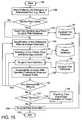
- module Cis a database module 50 that contains a variety of databases including, for example, a 3D template geometry database 52 , a deformation mode database 54 , a fixator database 56 , a surgical tool database 58 and a surgical plan database 60 . All of these modules arm shown residing (in a suitable memory or storage area) in the computer assisted orthopedic surgery planner terminal 30 .
- the discussion hereinbelowfocuses on modules A, B and C; however, it is understood that these modules do not function independently of a platform (here, the computer assisted orthopedic surgery planner computer 30 ) that executes the program code or instructions for the respective module. In other words, the screen displays and printouts discussed hereinbelow may be generated only after the program code for a corresponding module is executed by the computer assisted orthopedic surgery planner computer 30 .
- module A 42After detecting the bone contour at step 86 , module A 42 first identifies (at step 88 ) the corresponding fiducial geometry on the 3D template bone model prior to any deformation discussed hereinbelow. Module A 42 also optimizes (at steps 90 and 92 ) the 3D positioning and scaling parameters for the 3D template bone model until the size and position of the 3D template bone model is optimum with respect to the patient's bone 63 (as judged from the X-ray images 65 , 66 of the patient's bone 63 ). Upon finding the optimum values for positioning and scaling parameters, module A 42 updates (at step 94 ) the 3D template bone model with new positioning and scaling parameters. The resultant 3D template bone model 112 is shown in FIG.
- the three-dimensional graphics for the surgical plan 48may be generated using the OpenGL (open graphics library) software interface developed by Silicon Graphics, Inc., of Mountainview, Calif., USA.
- OpenGLopen graphics library
- the OpenGL graphics software interfacemay be implemented on a conventional PC (personal computer) platform to show animations of the bone distraction process.
Landscapes
- Engineering & Computer Science (AREA)
- Physics & Mathematics (AREA)
- Theoretical Computer Science (AREA)
- Health & Medical Sciences (AREA)
- Geometry (AREA)
- Surgery (AREA)
- Life Sciences & Earth Sciences (AREA)
- General Physics & Mathematics (AREA)
- Computer Graphics (AREA)
- Heart & Thoracic Surgery (AREA)
- Software Systems (AREA)
- Public Health (AREA)
- Veterinary Medicine (AREA)
- Animal Behavior & Ethology (AREA)
- Molecular Biology (AREA)
- Medical Informatics (AREA)
- General Health & Medical Sciences (AREA)
- Biomedical Technology (AREA)
- Nuclear Medicine, Radiotherapy & Molecular Imaging (AREA)
- Computing Systems (AREA)
- Processing Or Creating Images (AREA)
- Surgical Instruments (AREA)
- Apparatus For Radiation Diagnosis (AREA)
- Image Processing (AREA)
- Pharmaceuticals Containing Other Organic And Inorganic Compounds (AREA)
Abstract
Description


Claims (20)
Priority Applications (8)
| Application Number | Priority Date | Filing Date | Title |
|---|---|---|---|
| US09/545,685US6701174B1 (en) | 2000-04-07 | 2000-04-07 | Computer-aided bone distraction |
| CA002405738ACA2405738A1 (en) | 2000-04-07 | 2001-04-06 | Computer-aided bone distraction |
| AU2001253216AAU2001253216A1 (en) | 2000-04-07 | 2001-04-06 | Computer-aided bone distraction |
| JP2001575384AJP2003530177A (en) | 2000-04-07 | 2001-04-06 | Computer aided bone lengthening |
| EP01926698AEP1303841A2 (en) | 2000-04-07 | 2001-04-06 | Computer-aided bone distraction |
| PCT/US2001/011272WO2001078015A2 (en) | 2000-04-07 | 2001-04-06 | Computer-aided bone distraction |
| US10/636,311US20040068187A1 (en) | 2000-04-07 | 2003-08-07 | Computer-aided orthopedic surgery |
| US10/636,052US7837621B2 (en) | 2000-04-07 | 2003-08-07 | Computer-aided bone distraction |
Applications Claiming Priority (1)
| Application Number | Priority Date | Filing Date | Title |
|---|---|---|---|
| US09/545,685US6701174B1 (en) | 2000-04-07 | 2000-04-07 | Computer-aided bone distraction |
Related Child Applications (2)
| Application Number | Title | Priority Date | Filing Date |
|---|---|---|---|
| US10/636,311Continuation-In-PartUS20040068187A1 (en) | 2000-04-07 | 2003-08-07 | Computer-aided orthopedic surgery |
| US10/636,052ContinuationUS7837621B2 (en) | 2000-04-07 | 2003-08-07 | Computer-aided bone distraction |
Publications (1)
| Publication Number | Publication Date |
|---|---|
| US6701174B1true US6701174B1 (en) | 2004-03-02 |
Family
ID=24177160
Family Applications (2)
| Application Number | Title | Priority Date | Filing Date |
|---|---|---|---|
| US09/545,685Expired - LifetimeUS6701174B1 (en) | 2000-04-07 | 2000-04-07 | Computer-aided bone distraction |
| US10/636,052Expired - LifetimeUS7837621B2 (en) | 2000-04-07 | 2003-08-07 | Computer-aided bone distraction |
Family Applications After (1)
| Application Number | Title | Priority Date | Filing Date |
|---|---|---|---|
| US10/636,052Expired - LifetimeUS7837621B2 (en) | 2000-04-07 | 2003-08-07 | Computer-aided bone distraction |
Country Status (6)
| Country | Link |
|---|---|
| US (2) | US6701174B1 (en) |
| EP (1) | EP1303841A2 (en) |
| JP (1) | JP2003530177A (en) |
| AU (1) | AU2001253216A1 (en) |
| CA (1) | CA2405738A1 (en) |
| WO (1) | WO2001078015A2 (en) |
Cited By (285)
| Publication number | Priority date | Publication date | Assignee | Title |
|---|---|---|---|---|
| US20030181831A1 (en)* | 2000-09-18 | 2003-09-25 | Fuji Photo Film Co., Ltd. | Artificial bone template selection system, artificial bone template display system, artificial bone template storage system and artificial bone template recording medium |
| US20040024311A1 (en)* | 2002-03-06 | 2004-02-05 | Quaid Arthur E. | System and method for haptic sculpting of physical objects |
| US20040039259A1 (en)* | 2000-04-07 | 2004-02-26 | Norman Krause | Computer-aided bone distraction |
| US20040068187A1 (en)* | 2000-04-07 | 2004-04-08 | Krause Norman M. | Computer-aided orthopedic surgery |
| US20040073211A1 (en)* | 2002-04-05 | 2004-04-15 | Ed Austin | Orthopaedic fixation method and device with delivery and presentation features |
| US20040106916A1 (en)* | 2002-03-06 | 2004-06-03 | Z-Kat, Inc. | Guidance system and method for surgical procedures with improved feedback |
| US20040122790A1 (en)* | 2002-12-18 | 2004-06-24 | Walker Matthew J. | Computer-assisted data processing system and method incorporating automated learning |
| US20040122702A1 (en)* | 2002-12-18 | 2004-06-24 | Sabol John M. | Medical data processing system and method |
| US20040122703A1 (en)* | 2002-12-19 | 2004-06-24 | Walker Matthew J. | Medical data operating model development system and method |
| US20040122719A1 (en)* | 2002-12-18 | 2004-06-24 | Sabol John M. | Medical resource processing system and method utilizing multiple resource type data |
| US20040120558A1 (en)* | 2002-12-18 | 2004-06-24 | Sabol John M | Computer assisted data reconciliation method and apparatus |
| US20040148145A1 (en)* | 2003-01-29 | 2004-07-29 | Yifan Chen | System and method of interactively generating a family of mesh models |
| US20040208780A1 (en)* | 2001-10-22 | 2004-10-21 | Faries Durward I. | Heated medical instrument stand with surgical drape and method of detecting fluid and leaks in the stand tray |
| US20040234116A1 (en)* | 2002-07-22 | 2004-11-25 | Xiaoli Bi | Method, code, and system for assaying joint deformity |
| US20050004446A1 (en)* | 2003-06-25 | 2005-01-06 | Brett Cowan | Model assisted planning of medical imaging |
| US20050002556A1 (en)* | 2001-09-07 | 2005-01-06 | Michael Kaus | Method of measuring geometric variables of a structure contained in an image |
| US20050020913A1 (en)* | 2003-04-10 | 2005-01-27 | Christian Georg | Method and x-ray system for detecting position changes of a medical implant |
| US20050054917A1 (en)* | 2002-09-26 | 2005-03-10 | David Kitson | Orthopaedic surgery planning |
| US20050059873A1 (en)* | 2003-08-26 | 2005-03-17 | Zeev Glozman | Pre-operative medical planning system and method for use thereof |
| US20050149040A1 (en)* | 1994-09-02 | 2005-07-07 | Haines Timothy G. | Methods and apparatus for orthopedic surgical navigation and alignment |
| WO2004070580A3 (en)* | 2003-02-04 | 2005-07-21 | Z Kat Inc | Computer-assisted knee replacement apparatus and method |
| US20050222793A1 (en)* | 2004-04-02 | 2005-10-06 | Lloyd Charles F | Method and system for calibrating deformed instruments |
| US20050228270A1 (en)* | 2004-04-02 | 2005-10-13 | Lloyd Charles F | Method and system for geometric distortion free tracking of 3-dimensional objects from 2-dimensional measurements |
| US20050238223A1 (en)* | 2004-04-21 | 2005-10-27 | Jens Guhring | Flexible generation of digitally reconstructed radiographs |
| US20050246154A1 (en)* | 2004-04-29 | 2005-11-03 | Align Technology, Inc. | Dynamically specifying a view |
| US20050256389A1 (en)* | 2001-11-16 | 2005-11-17 | Yoshio Koga | Calculation method, calculation program and calculation system for information supporting arthroplasty |
| US20050267353A1 (en)* | 2004-02-04 | 2005-12-01 | Joel Marquart | Computer-assisted knee replacement apparatus and method |
| US20060015116A1 (en)* | 2004-01-14 | 2006-01-19 | Haines Timothy G | Methods and apparatus for improved drilling and milling tools for resection |
| US20060015117A1 (en)* | 2004-01-14 | 2006-01-19 | Haines Timothy G | Methods and apparatus for minimally invasive arthroplasty |
| US20060015109A1 (en)* | 2004-01-14 | 2006-01-19 | Haines Timothy G | Methods and apparatus for improved cutting tools for resection |
| US20060030853A1 (en)* | 2004-01-14 | 2006-02-09 | Haines Timothy G | Methods and apparatus for pinplasty bone resection |
| US20060029917A1 (en)* | 2004-08-06 | 2006-02-09 | Sui Leung K | Navigation surgical training model, apparatus having the same and method thereof |
| US20060058882A1 (en)* | 2004-01-14 | 2006-03-16 | Haines Timothy G | Methods and apparatus for conformable prosthetic implants |
| WO2005041812A3 (en)* | 2003-10-22 | 2006-06-08 | Implant Brace Inc | Implantable brace for a fracture and methods |
| WO2006063324A1 (en)* | 2004-12-10 | 2006-06-15 | Virginia Tech Intellectual Properties, Inc. | Systems and methods for multi-dimensional characterization and classification of spinal shape |
| US20060142657A1 (en)* | 2002-03-06 | 2006-06-29 | Mako Surgical Corporation | Haptic guidance system and method |
| US20060195198A1 (en)* | 2005-02-22 | 2006-08-31 | Anthony James | Interactive orthopaedic biomechanics system |
| US20060204067A1 (en)* | 2005-02-18 | 2006-09-14 | Gregor Tuma | Determining shaft and femur neck axes and three-dimensional reconstruction |
| US20060224088A1 (en)* | 2005-03-29 | 2006-10-05 | Roche Martin W | Body parameter detecting sensor and method for detecting body parameters |
| US20060262112A1 (en)* | 2005-05-23 | 2006-11-23 | Carnegie Mellon University | System and method for three-dimensional shape generation from partial and incomplete views, and interactive design system using same |
| USD533875S1 (en) | 2003-10-17 | 2006-12-19 | Nuvasive, Inc. | Graphic user interface for a medical monitor |
| US20070038223A1 (en)* | 2003-02-04 | 2007-02-15 | Joel Marquart | Computer-assisted knee replacement apparatus and method |
| US20070055234A1 (en)* | 2005-06-10 | 2007-03-08 | Mcgrath William M | External fixation system with provisional brace |
| US20070078678A1 (en)* | 2005-09-30 | 2007-04-05 | Disilvestro Mark R | System and method for performing a computer assisted orthopaedic surgical procedure |
| US20070089753A1 (en)* | 2005-09-01 | 2007-04-26 | Faries Durward I Jr | Method and apparatus for protecting sterile drapes in surgical thermal treatment systems |
| US20070129626A1 (en)* | 2005-11-23 | 2007-06-07 | Prakash Mahesh | Methods and systems for facilitating surgical procedures |
| US20070133848A1 (en)* | 2003-10-17 | 2007-06-14 | Mcnutt Todd R | Manual tools for model based image segmentation |
| EP1803413A2 (en) | 2005-12-30 | 2007-07-04 | DePuy Products, Inc. | Magnetic sensor array for bone registration in computer-assisted orthopaedic surgery |
| EP1803412A1 (en)* | 2005-12-30 | 2007-07-04 | DePuy Products, Inc. | System and method for registering a bone of a patient with a computer assisted orhtopaedic surgery system |
| US20070167741A1 (en)* | 2005-12-30 | 2007-07-19 | Sherman Jason T | Apparatus and method for registering a bone of a patient with a computer assisted orthopaedic surgery system |
| US20070167703A1 (en)* | 2005-12-30 | 2007-07-19 | Sherman Jason T | Method for determining a position of a magnetic source |
| US20070208234A1 (en)* | 2004-04-13 | 2007-09-06 | Bhandarkar Suchendra M | Virtual Surgical System and Methods |
| US20070225704A1 (en)* | 2006-03-23 | 2007-09-27 | Ziran Bruce H | Electromechanically driven external fixator and methods of use |
| US20070225813A1 (en)* | 2006-01-24 | 2007-09-27 | Timothy Haines | Dynamic spinal implants incorporating cartilage bearing graft material |
| US20070233141A1 (en)* | 2006-02-15 | 2007-10-04 | Ilwhan Park | Arthroplasty devices and related methods |
| US20070226986A1 (en)* | 2006-02-15 | 2007-10-04 | Ilwhan Park | Arthroplasty devices and related methods |
| US20070255288A1 (en)* | 2006-03-17 | 2007-11-01 | Zimmer Technology, Inc. | Methods of predetermining the contour of a resected bone surface and assessing the fit of a prosthesis on the bone |
| US20080002889A1 (en)* | 2006-06-02 | 2008-01-03 | Kenji Shimada | Systems and methods for extracting an approximated medial surface from a thin-wall solid |
| US20080004517A1 (en)* | 2006-03-29 | 2008-01-03 | University Of Georgia Research Foundation, Inc. | Virtual Surgical Systems and Methods |
| US20080009738A1 (en)* | 2003-11-17 | 2008-01-10 | Koninklijke Philips Electronics N.V. | Method for Utilizing User Input for Feature Detection in Diagnostic Imaging |
| US20080051685A1 (en)* | 2006-08-24 | 2008-02-28 | Benenati Vincent A | External fixator linkage |
| US20080098322A1 (en)* | 2006-06-01 | 2008-04-24 | Simquest Llc | Method and apparatus for collecting and analyzing surface wound data |
| US20080137926A1 (en)* | 2006-11-21 | 2008-06-12 | General Electric Company | Method and system for adjusting 3D CT vessel segmentation |
| US20080147072A1 (en)* | 2006-12-18 | 2008-06-19 | Ilwhan Park | Arthroplasty devices and related methods |
| US20080154127A1 (en)* | 2006-12-21 | 2008-06-26 | Disilvestro Mark R | Method and system for registering a bone of a patient with a computer assisted orthopaedic surgery system |
| US20080163118A1 (en)* | 2006-12-29 | 2008-07-03 | Jason Wolf | Representation of file relationships |
| US20080161815A1 (en)* | 2006-02-27 | 2008-07-03 | Biomet Manufacturing Corp. | Patient Specific Knee Alignment Guide And Associated Method |
| US20080175464A1 (en)* | 2007-01-16 | 2008-07-24 | Optasia Medical, Ltd. | Computer program products and methods for detection and tracking of rheumatoid arthritis |
| US20080187193A1 (en)* | 2007-02-01 | 2008-08-07 | Ralph Thomas Hoctor | Method and Apparatus for Forming a Guide Image for an Ultrasound Image Scanner |
| US20080194957A1 (en)* | 2007-02-14 | 2008-08-14 | Ralph Thomas Hoctor | Method and Apparatus for Generating an Ultrasound Image of Moving Objects Using Deformable Models |
| US20080243194A1 (en)* | 2005-09-26 | 2008-10-02 | The Regents Of The University Of California | Articulating instrumentation for dynamic spinal stabilization |
| US20090048597A1 (en)* | 2007-08-14 | 2009-02-19 | Zimmer, Inc. | Method of determining a contour of an anatomical structure and selecting an orthopaedic implant to replicate the anatomical structure |
| US20090082773A1 (en)* | 2004-01-14 | 2009-03-26 | Haines Timothy G | Method and apparatus for wireplasty bone resection |
| US20090110498A1 (en)* | 2007-10-25 | 2009-04-30 | Ilwhan Park | Arthroplasty systems and devices, and related methods |
| US20090138020A1 (en)* | 2007-11-27 | 2009-05-28 | Otismed Corporation | Generating mri images usable for the creation of 3d bone models employed to make customized arthroplasty jigs |
| US20090157083A1 (en)* | 2007-12-18 | 2009-06-18 | Ilwhan Park | System and method for manufacturing arthroplasty jigs |
| US20090198236A1 (en)* | 2008-02-01 | 2009-08-06 | Stryker Trauma Sa | Clamping pin |
| US20090198235A1 (en)* | 2008-02-01 | 2009-08-06 | Stryker Trauma Sa | Strut joint for an external fixator |
| US20090198234A1 (en)* | 2008-02-01 | 2009-08-06 | Stryker Trauma Sa | Telescopic strut for an external fixator |
| US20090216645A1 (en)* | 2008-02-21 | 2009-08-27 | What's In It For Me.Com Llc | System and method for generating leads for the sale of goods and services |
| US20090220132A1 (en)* | 2008-01-10 | 2009-09-03 | Yves Trousset | Method for processing images of interventional radiology |
| US20090222015A1 (en)* | 2008-02-29 | 2009-09-03 | Otismed Corporation | Hip resurfacing surgical guide tool |
| US20090220050A1 (en)* | 2006-05-04 | 2009-09-03 | Jens Guhring | Method for Determining and Displaying at Least One Piece of Information on a Target Volume |
| US20090254326A1 (en)* | 2008-04-04 | 2009-10-08 | Vilaspine Ltd. | System and Device for Designing and Forming a Surgical Implant |
| US20090249851A1 (en)* | 2008-04-04 | 2009-10-08 | Vilaspine Ltd. | System and Device for Designing and Forming a Surgical Implant |
| US20090254097A1 (en)* | 2008-04-04 | 2009-10-08 | Isaacs Robert E | System and device for designing and forming a surgical implant |
| US20090254367A1 (en)* | 2007-04-17 | 2009-10-08 | Biomet Manufacturing Corp. | Method and Apparatus for Manufacturing an Implant |
| US20090270868A1 (en)* | 2008-04-29 | 2009-10-29 | Otismed Corporation | Generation of a computerized bone model representative of a pre-degenerated state and useable in the design and manufacture of arthroplasty devices |
| US20090274350A1 (en)* | 2008-04-30 | 2009-11-05 | Otismed Corporation | System and method for image segmentation in generating computer models of a joint to undergo arthroplasty |
| US20090319049A1 (en)* | 2008-02-18 | 2009-12-24 | Maxx Orthopedics, Inc. | Total Knee Replacement Prosthesis With High Order NURBS Surfaces |
| US20100035212A1 (en)* | 2008-08-11 | 2010-02-11 | Jean Robichaud | Preparation of dental implant surgery and prosthesis installation |
| US20100042105A1 (en)* | 2007-12-18 | 2010-02-18 | Otismed Corporation | Arthroplasty system and related methods |
| US20100100192A1 (en)* | 2001-03-05 | 2010-04-22 | Haines Timothy G | Femoral prosthetic implant |
| US20100116810A1 (en)* | 2004-03-23 | 2010-05-13 | O.R. Solutions, Inc. | Thermal Treatment System Instrument Rack and Method of Selectively Thermally Treating Medical Instrument Portions |
| US7728262B1 (en)* | 2004-03-23 | 2010-06-01 | O.R. Solutions, Inc. | Thermal treatment system instrument rack and method of selectively thermally treating medical instrument portions |
| US20100152741A1 (en)* | 2008-12-16 | 2010-06-17 | Otismed Corporation | Unicompartmental customized arthroplasty cutting jigs and methods of making the same |
| US20100172557A1 (en)* | 2002-01-16 | 2010-07-08 | Alain Richard | Method and apparatus for reconstructing bone surfaces during surgery |
| US7760923B2 (en)* | 2005-03-24 | 2010-07-20 | Optasia Medical Limited | Method and system for characterization of knee joint morphology |
| US20100217109A1 (en)* | 2009-02-20 | 2010-08-26 | Biomet Manufacturing Corp. | Mechanical Axis Alignment Using MRI Imaging |
| US20100234844A1 (en)* | 2009-03-10 | 2010-09-16 | Stryker Trauma Sa | Exernal fixation system |
| US20100249790A1 (en)* | 2009-03-26 | 2010-09-30 | Martin Roche | System and method for soft tissue tensioning in extension and flexion |
| US20100256479A1 (en)* | 2007-12-18 | 2010-10-07 | Otismed Corporation | Preoperatively planning an arthroplasty procedure and generating a corresponding patient specific arthroplasty resection guide |
| US20100311028A1 (en)* | 2009-06-04 | 2010-12-09 | Zimmer Dental, Inc. | Dental Implant Surgical Training Simulation System |
| US20100326194A1 (en)* | 2009-06-30 | 2010-12-30 | Orthosensor | Edge-detect receiver for orthopedic parameter sensing |
| US20100327880A1 (en)* | 2009-06-30 | 2010-12-30 | Orthosensor | Pulsed waveguide sensing device and method for measuring a parameter |
| US20100331679A1 (en)* | 2009-06-30 | 2010-12-30 | Orthosensor | Pulsed echo sensing device and method for an orthopedic joint |
| US20100331738A1 (en)* | 2009-06-30 | 2010-12-30 | Orthosensor | Integrated sensor and interconnect for measuring a parameter of the muscular-skeletal system |
| US7909610B1 (en) | 2006-12-21 | 2011-03-22 | Amato Craniofacial Engineering, LLC | Computer-aided system of orthopedic surgery |
| US20110087465A1 (en)* | 2007-08-17 | 2011-04-14 | Mohamed Rashwan Mahfouz | Implant design analysis suite |
| US7959860B2 (en) | 2001-10-22 | 2011-06-14 | Faries Jr Durward I | System and method of detecting fluid and leaks in thermal treatment system basins |
| US7967868B2 (en) | 2007-04-17 | 2011-06-28 | Biomet Manufacturing Corp. | Patient-modified implant and associated method |
| US20110160616A1 (en)* | 2009-06-30 | 2011-06-30 | Orthosensor | System and method for orthopedic load and location sensing |
| US20110164798A1 (en)* | 2008-04-03 | 2011-07-07 | Fujifilm Corporation | Apparatus, method, and program for detecting three dimenmsional abdominal cavity regions |
| USD642263S1 (en) | 2007-10-25 | 2011-07-26 | Otismed Corporation | Arthroplasty jig blank |
| US20110213221A1 (en)* | 2005-03-29 | 2011-09-01 | Roche Martin W | Method for Detecting Body Parameters |
| US20110218545A1 (en)* | 2010-03-04 | 2011-09-08 | Biomet Manufacturing Corp. | Patient-specific computed tomography guides |
| US8070752B2 (en) | 2006-02-27 | 2011-12-06 | Biomet Manufacturing Corp. | Patient specific alignment guide and inter-operative adjustment |
| US20110313418A1 (en)* | 2010-05-19 | 2011-12-22 | Arkadijus Nikonovas | Orthopedic fixation with imagery analysis |
| US8092465B2 (en) | 2006-06-09 | 2012-01-10 | Biomet Manufacturing Corp. | Patient specific knee alignment guide and associated method |
| US8133234B2 (en) | 2006-02-27 | 2012-03-13 | Biomet Manufacturing Corp. | Patient specific acetabular guide and method |
| US8160345B2 (en) | 2008-04-30 | 2012-04-17 | Otismed Corporation | System and method for image segmentation in generating computer models of a joint to undergo arthroplasty |
| WO2012033530A3 (en)* | 2010-09-08 | 2012-06-14 | University Of Houston | Devices, systems and methods for multimodal biosensing and imaging |
| US20120189185A1 (en)* | 2011-01-20 | 2012-07-26 | Siemens Aktiengesellschaft | Method and System for 3D Cardiac Motion Estimation from Single Scan of C-Arm Angiography |
| US8241293B2 (en) | 2006-02-27 | 2012-08-14 | Biomet Manufacturing Corp. | Patient specific high tibia osteotomy |
| US8265949B2 (en) | 2007-09-27 | 2012-09-11 | Depuy Products, Inc. | Customized patient surgical plan |
| US20120250975A1 (en)* | 2005-10-12 | 2012-10-04 | Mohammed Homman | Identification and classification of virus particles in textured electron micrographs |
| US8287538B2 (en) | 2008-01-14 | 2012-10-16 | Conventus Orthopaedics, Inc. | Apparatus and methods for fracture repair |
| US8287522B2 (en) | 2006-05-19 | 2012-10-16 | Mako Surgical Corp. | Method and apparatus for controlling a haptic device |
| US8298237B2 (en) | 2006-06-09 | 2012-10-30 | Biomet Manufacturing Corp. | Patient-specific alignment guide for multiple incisions |
| US8343159B2 (en) | 2007-09-30 | 2013-01-01 | Depuy Products, Inc. | Orthopaedic bone saw and method of use thereof |
| US8357111B2 (en) | 2007-09-30 | 2013-01-22 | Depuy Products, Inc. | Method and system for designing patient-specific orthopaedic surgical instruments |
| US8377066B2 (en) | 2006-02-27 | 2013-02-19 | Biomet Manufacturing Corp. | Patient-specific elbow guides and associated methods |
| US8407067B2 (en) | 2007-04-17 | 2013-03-26 | Biomet Manufacturing Corp. | Method and apparatus for manufacturing an implant |
| US8532807B2 (en) | 2011-06-06 | 2013-09-10 | Biomet Manufacturing, Llc | Pre-operative planning and manufacturing method for orthopedic procedure |
| US8535063B1 (en) | 2006-12-21 | 2013-09-17 | Amato Craniofacial Engineering, LLC | Craniofacial anatomic simulator with cephalometer |
| US8535387B2 (en) | 2006-02-27 | 2013-09-17 | Biomet Manufacturing, Llc | Patient-specific tools and implants |
| US8545509B2 (en) | 2007-12-18 | 2013-10-01 | Otismed Corporation | Arthroplasty system and related methods |
| US8568487B2 (en) | 2006-02-27 | 2013-10-29 | Biomet Manufacturing, Llc | Patient-specific hip joint devices |
| US8591516B2 (en) | 2006-02-27 | 2013-11-26 | Biomet Manufacturing, Llc | Patient-specific orthopedic instruments |
| US8597365B2 (en) | 2011-08-04 | 2013-12-03 | Biomet Manufacturing, Llc | Patient-specific pelvic implants for acetabular reconstruction |
| US8603180B2 (en) | 2006-02-27 | 2013-12-10 | Biomet Manufacturing, Llc | Patient-specific acetabular alignment guides |
| US8603095B2 (en) | 1994-09-02 | 2013-12-10 | Puget Bio Ventures LLC | Apparatuses for femoral and tibial resection |
| US8608748B2 (en) | 2006-02-27 | 2013-12-17 | Biomet Manufacturing, Llc | Patient specific guides |
| US8608749B2 (en) | 2006-02-27 | 2013-12-17 | Biomet Manufacturing, Llc | Patient-specific acetabular guides and associated instruments |
| US8617171B2 (en) | 2007-12-18 | 2013-12-31 | Otismed Corporation | Preoperatively planning an arthroplasty procedure and generating a corresponding patient specific arthroplasty resection guide |
| US8632547B2 (en) | 2010-02-26 | 2014-01-21 | Biomet Sports Medicine, Llc | Patient-specific osteotomy devices and methods |
| US8654150B2 (en)* | 2012-02-03 | 2014-02-18 | Orthohub, Inc. | External fixator deformity correction systems and methods |
| US8668700B2 (en) | 2011-04-29 | 2014-03-11 | Biomet Manufacturing, Llc | Patient-specific convertible guides |
| US20140081400A1 (en)* | 2010-08-25 | 2014-03-20 | Siemens Corporation | Semi-Automatic Customization Of Plates For Internal Fracture Fixation |
| US8710407B2 (en) | 2010-09-02 | 2014-04-29 | Ecolab Usa Inc. | Selective thermal treatment of medical instrument portions with thermal treatment system instrument holder |
| US8715289B2 (en) | 2011-04-15 | 2014-05-06 | Biomet Manufacturing, Llc | Patient-specific numerically controlled instrument |
| US8764760B2 (en) | 2011-07-01 | 2014-07-01 | Biomet Manufacturing, Llc | Patient-specific bone-cutting guidance instruments and methods |
| US8777875B2 (en) | 2008-07-23 | 2014-07-15 | Otismed Corporation | System and method for manufacturing arthroplasty jigs having improved mating accuracy |
| US8789534B2 (en) | 2008-04-09 | 2014-07-29 | Patented Medical Solutions, Llc | Method and apparatus for warming medical solutions in a thermal treatment system employing a removable basin |
| US8801720B2 (en) | 2002-05-15 | 2014-08-12 | Otismed Corporation | Total joint arthroplasty system |
| US20140236153A1 (en)* | 2013-02-19 | 2014-08-21 | Stryker Trauma Gmbh | Software for use with deformity correction |
| US20140257461A1 (en)* | 2013-03-05 | 2014-09-11 | Merit Medical Systems, Inc. | Reinforced valve |
| US8834467B2 (en) | 2010-08-11 | 2014-09-16 | Stryker Trauma Sa | External fixator system |
| WO2014160170A1 (en)* | 2013-03-13 | 2014-10-02 | Curexo Technology Corporation | Systems and methods for pre-operative planning and precise bone tunnel placement for ligament reconstruction |
| US8858561B2 (en) | 2006-06-09 | 2014-10-14 | Blomet Manufacturing, LLC | Patient-specific alignment guide |
| US8864769B2 (en) | 2006-02-27 | 2014-10-21 | Biomet Manufacturing, Llc | Alignment guides with patient-specific anchoring elements |
| US8864763B2 (en) | 2013-03-13 | 2014-10-21 | DePuy Synthes Products, LLC | External bone fixation device |
| US8908937B2 (en) | 2010-07-08 | 2014-12-09 | Biomet Manufacturing, Llc | Method and device for digital image templating |
| US8906022B2 (en) | 2010-03-08 | 2014-12-09 | Conventus Orthopaedics, Inc. | Apparatus and methods for securing a bone implant |
| US8917290B2 (en) | 2011-01-31 | 2014-12-23 | Biomet Manufacturing, Llc | Digital image templating |
| US8926530B2 (en) | 2011-09-23 | 2015-01-06 | Orthosensor Inc | Orthopedic insert measuring system for having a sterilized cavity |
| US8945128B2 (en) | 2010-08-11 | 2015-02-03 | Stryker Trauma Sa | External fixator system |
| US8956364B2 (en) | 2011-04-29 | 2015-02-17 | Biomet Manufacturing, Llc | Patient-specific partial knee guides and other instruments |
| US8961518B2 (en) | 2010-01-20 | 2015-02-24 | Conventus Orthopaedics, Inc. | Apparatus and methods for bone access and cavity preparation |
| US8979758B2 (en) | 2010-06-29 | 2015-03-17 | Orthosensor Inc | Sensing module for orthopedic load sensing insert device |
| US9039706B2 (en) | 2013-03-13 | 2015-05-26 | DePuy Synthes Products, Inc. | External bone fixation device |
| US9060788B2 (en) | 2012-12-11 | 2015-06-23 | Biomet Manufacturing, Llc | Patient-specific acetabular guide for anterior approach |
| US9066734B2 (en) | 2011-08-31 | 2015-06-30 | Biomet Manufacturing, Llc | Patient-specific sacroiliac guides and associated methods |
| US9084618B2 (en) | 2011-06-13 | 2015-07-21 | Biomet Manufacturing, Llc | Drill guides for confirming alignment of patient-specific alignment guides |
| US9101398B2 (en) | 2012-08-23 | 2015-08-11 | Stryker Trauma Sa | Bone transport external fixation frame |
| US9113971B2 (en) | 2006-02-27 | 2015-08-25 | Biomet Manufacturing, Llc | Femoral acetabular impingement guide |
| US9119722B1 (en) | 2011-08-18 | 2015-09-01 | Sharat Kusuma | Measurement and placement techniques in hip resurfacing and the like |
| US9129054B2 (en) | 2012-09-17 | 2015-09-08 | DePuy Synthes Products, Inc. | Systems and methods for surgical and interventional planning, support, post-operative follow-up, and, functional recovery tracking |
| US9173661B2 (en) | 2006-02-27 | 2015-11-03 | Biomet Manufacturing, Llc | Patient specific alignment guide with cutting surface and laser indicator |
| US9204977B2 (en) | 2012-12-11 | 2015-12-08 | Biomet Manufacturing, Llc | Patient-specific acetabular guide for anterior approach |
| US9237950B2 (en) | 2012-02-02 | 2016-01-19 | Biomet Manufacturing, Llc | Implant with patient-specific porous structure |
| US9241745B2 (en) | 2011-03-07 | 2016-01-26 | Biomet Manufacturing, Llc | Patient-specific femoral version guide |
| US9248002B2 (en) | 2013-09-26 | 2016-02-02 | Howmedica Osteonics Corp. | Method for aligning an acetabular cup |
| US9262802B2 (en) | 2010-07-21 | 2016-02-16 | Arthromeda, Inc. | Independent digital templating software, and methods and systems using same |
| US9271744B2 (en) | 2010-09-29 | 2016-03-01 | Biomet Manufacturing, Llc | Patient-specific guide for partial acetabular socket replacement |
| US20160078682A1 (en)* | 2013-04-24 | 2016-03-17 | Kawasaki Jukogyo Kabushiki Kaisha | Component mounting work support system and component mounting method |
| US9289253B2 (en) | 2006-02-27 | 2016-03-22 | Biomet Manufacturing, Llc | Patient-specific shoulder guide |
| US9295497B2 (en) | 2011-08-31 | 2016-03-29 | Biomet Manufacturing, Llc | Patient-specific sacroiliac and pedicle guides |
| US9301812B2 (en) | 2011-10-27 | 2016-04-05 | Biomet Manufacturing, Llc | Methods for patient-specific shoulder arthroplasty |
| US9332943B2 (en) | 2011-09-23 | 2016-05-10 | Orthosensor Inc | Flexible surface parameter measurement system for the muscular-skeletal system |
| US9339278B2 (en) | 2006-02-27 | 2016-05-17 | Biomet Manufacturing, Llc | Patient-specific acetabular guides and associated instruments |
| US9345548B2 (en) | 2006-02-27 | 2016-05-24 | Biomet Manufacturing, Llc | Patient-specific pre-operative planning |
| US9351743B2 (en) | 2011-10-27 | 2016-05-31 | Biomet Manufacturing, Llc | Patient-specific glenoid guides |
| US9386993B2 (en) | 2011-09-29 | 2016-07-12 | Biomet Manufacturing, Llc | Patient-specific femoroacetabular impingement instruments and methods |
| US9393028B2 (en) | 2009-08-13 | 2016-07-19 | Biomet Manufacturing, Llc | Device for the resection of bones, method for producing such a device, endoprosthesis suited for this purpose and method for producing such an endoprosthesis |
| US9402637B2 (en) | 2012-10-11 | 2016-08-02 | Howmedica Osteonics Corporation | Customized arthroplasty cutting guides and surgical methods using the same |
| US9408616B2 (en) | 2014-05-12 | 2016-08-09 | Biomet Manufacturing, Llc | Humeral cut guide |
| US9451973B2 (en) | 2011-10-27 | 2016-09-27 | Biomet Manufacturing, Llc | Patient specific glenoid guide |
| US9462964B2 (en) | 2011-09-23 | 2016-10-11 | Orthosensor Inc | Small form factor muscular-skeletal parameter measurement system |
| US9498233B2 (en) | 2013-03-13 | 2016-11-22 | Biomet Manufacturing, Llc. | Universal acetabular guide and associated hardware |
| US9508149B2 (en) | 2012-05-23 | 2016-11-29 | Stryker European Holdings I, Llc | Virtual 3D overlay as reduction aid for complex fractures |
| US9510771B1 (en) | 2011-10-28 | 2016-12-06 | Nuvasive, Inc. | Systems and methods for performing spine surgery |
| US9517107B2 (en) | 2010-07-16 | 2016-12-13 | Stryker European Holdings I, Llc | Surgical targeting system and method |
| US9517145B2 (en) | 2013-03-15 | 2016-12-13 | Biomet Manufacturing, Llc | Guide alignment system and method |
| US9554910B2 (en) | 2011-10-27 | 2017-01-31 | Biomet Manufacturing, Llc | Patient-specific glenoid guide and implants |
| US9561040B2 (en) | 2014-06-03 | 2017-02-07 | Biomet Manufacturing, Llc | Patient-specific glenoid depth control |
| US9579107B2 (en) | 2013-03-12 | 2017-02-28 | Biomet Manufacturing, Llc | Multi-point fit for patient specific guide |
| US9610102B2 (en) | 2013-09-26 | 2017-04-04 | Stryker European Holdings I, Llc | Bone position tracking system |
| US9675382B2 (en) | 2013-03-13 | 2017-06-13 | DePuy Synthes Products, Inc. | External bone fixation device |
| US9675400B2 (en) | 2011-04-19 | 2017-06-13 | Biomet Manufacturing, Llc | Patient-specific fracture fixation instrumentation and method |
| US9687367B2 (en) | 2012-06-05 | 2017-06-27 | Merit Medical Systems, Inc. | Esophageal stent |
| US9730739B2 (en) | 2010-01-15 | 2017-08-15 | Conventus Orthopaedics, Inc. | Rotary-rigid orthopaedic rod |
| US20170277859A1 (en)* | 2011-06-23 | 2017-09-28 | Stryker European Holdings I, Llc | Methods and systems for adjusting an external fixation frame |
| US9795399B2 (en) | 2006-06-09 | 2017-10-24 | Biomet Manufacturing, Llc | Patient-specific knee alignment guide and associated method |
| US9801686B2 (en) | 2003-03-06 | 2017-10-31 | Mako Surgical Corp. | Neural monitor-based dynamic haptics |
| US9820868B2 (en) | 2015-03-30 | 2017-11-21 | Biomet Manufacturing, Llc | Method and apparatus for a pin apparatus |
| US9826981B2 (en) | 2013-03-13 | 2017-11-28 | Biomet Manufacturing, Llc | Tangential fit of patient-specific guides |
| US9826994B2 (en) | 2014-09-29 | 2017-11-28 | Biomet Manufacturing, Llc | Adjustable glenoid pin insertion guide |
| US9833245B2 (en) | 2014-09-29 | 2017-12-05 | Biomet Sports Medicine, Llc | Tibial tubercule osteotomy |
| US9839436B2 (en) | 2014-06-03 | 2017-12-12 | Biomet Manufacturing, Llc | Patient-specific glenoid depth control |
| US9839438B2 (en) | 2013-03-11 | 2017-12-12 | Biomet Manufacturing, Llc | Patient-specific glenoid guide with a reusable guide holder |
| US9848922B2 (en) | 2013-10-09 | 2017-12-26 | Nuvasive, Inc. | Systems and methods for performing spine surgery |
| US9855104B2 (en) | 2012-05-23 | 2018-01-02 | Stryker European Holdings I, Llc | Locking screw length measurement |
| US9895167B2 (en) | 2016-04-20 | 2018-02-20 | Stryker European Holdings I, Llc | Ring hole planning for external fixation frames |
| US9907659B2 (en) | 2007-04-17 | 2018-03-06 | Biomet Manufacturing, Llc | Method and apparatus for manufacturing an implant |
| US9913669B1 (en) | 2014-10-17 | 2018-03-13 | Nuvasive, Inc. | Systems and methods for performing spine surgery |
| US9918740B2 (en) | 2006-02-27 | 2018-03-20 | Biomet Manufacturing, Llc | Backup surgical instrument system and method |
| US9924950B2 (en) | 2013-09-25 | 2018-03-27 | Zimmer, Inc. | Patient specific instrumentation (PSI) for orthopedic surgery and systems and methods for using X-rays to produce same |
| US20180085183A1 (en)* | 2016-09-26 | 2018-03-29 | Texas Scottish Rite Hospital For Children | Radiography aid for an external fixator |
| US9937046B2 (en)* | 2009-02-25 | 2018-04-10 | Zimmer, Inc. | Method of generating a patient-specific bone shell |
| US9968408B1 (en) | 2013-03-15 | 2018-05-15 | Nuvasive, Inc. | Spinal balance assessment |
| US9968376B2 (en) | 2010-11-29 | 2018-05-15 | Biomet Manufacturing, Llc | Patient-specific orthopedic instruments |
| US10004564B1 (en) | 2016-01-06 | 2018-06-26 | Paul Beck | Accurate radiographic calibration using multiple images |
| US10010350B2 (en) | 2016-06-14 | 2018-07-03 | Stryker European Holdings I, Llc | Gear mechanisms for fixation frame struts |
| US10010372B1 (en) | 2016-01-06 | 2018-07-03 | Paul Beck | Marker Positioning Apparatus |
| US10022132B2 (en) | 2013-12-12 | 2018-07-17 | Conventus Orthopaedics, Inc. | Tissue displacement tools and methods |
| US10039606B2 (en)* | 2012-09-27 | 2018-08-07 | Stryker European Holdings I, Llc | Rotational position determination |
| US10070903B2 (en) | 2008-01-09 | 2018-09-11 | Stryker European Holdings I, Llc | Stereotactic computer assisted surgery method and system |
| US10082384B1 (en) | 2015-09-10 | 2018-09-25 | Stryker European Holdings I, Llc | Systems and methods for detecting fixation frame parameters |
| US10102347B2 (en) | 2008-09-26 | 2018-10-16 | Koninklijke Philips N.V. | Patient specific anatiomical sketches for medical reports |
| US10154884B2 (en) | 2016-06-02 | 2018-12-18 | Stryker European Holdings I, Llc | Software for use with deformity correction |
| US10226262B2 (en) | 2015-06-25 | 2019-03-12 | Biomet Manufacturing, Llc | Patient-specific humeral guide designs |
| US10282488B2 (en) | 2014-04-25 | 2019-05-07 | Biomet Manufacturing, Llc | HTO guide with optional guided ACL/PCL tunnels |
| US10278711B2 (en) | 2006-02-27 | 2019-05-07 | Biomet Manufacturing, Llc | Patient-specific femoral guide |
| US10285798B2 (en) | 2011-06-03 | 2019-05-14 | Merit Medical Systems, Inc. | Esophageal stent |
| US10321961B2 (en) | 2015-11-05 | 2019-06-18 | Howmedica Osteonics Corp. | Patient specific implantation method for range of motion hip impingement |
| US10467752B2 (en)* | 2013-06-11 | 2019-11-05 | Atsushi Tanji | Bone cutting support system, information processing apparatus, image processing method, and image processing program |
| US10492798B2 (en) | 2011-07-01 | 2019-12-03 | Biomet Manufacturing, Llc | Backup kit for a patient-specific arthroplasty kit assembly |
| US10499961B2 (en) | 2012-05-23 | 2019-12-10 | Stryker European Holdings I, Llc | Entry portal navigation |
| US20190380782A1 (en)* | 2016-12-12 | 2019-12-19 | Medicrea International | Systems, methods, and devices for developing patient-specific medical treatments, operations, and procedures |
| US10568647B2 (en) | 2015-06-25 | 2020-02-25 | Biomet Manufacturing, Llc | Patient-specific humeral guide designs |
| US10575905B2 (en) | 2017-03-13 | 2020-03-03 | Zimmer, Inc. | Augmented reality diagnosis guidance |
| US10588647B2 (en) | 2010-03-01 | 2020-03-17 | Stryker European Holdings I, Llc | Computer assisted surgery system |
| US10588762B2 (en) | 2013-03-15 | 2020-03-17 | Merit Medical Systems, Inc. | Esophageal stent |
| US10603179B2 (en) | 2006-02-27 | 2020-03-31 | Biomet Manufacturing, Llc | Patient-specific augments |
| US10722310B2 (en) | 2017-03-13 | 2020-07-28 | Zimmer Biomet CMF and Thoracic, LLC | Virtual surgery planning system and method |
| US20200349308A1 (en)* | 2019-04-30 | 2020-11-05 | Children's National Medical Center | Predictive Modeling Platform for Serial Casting to Correct Orthopedic Deformities |
| WO2020222169A1 (en) | 2019-05-02 | 2020-11-05 | DePuy Synthes Products, Inc. | Orthopaedic implant placement system and method |
| US10835318B2 (en) | 2016-08-25 | 2020-11-17 | DePuy Synthes Products, Inc. | Orthopedic fixation control and manipulation |
| US10874433B2 (en) | 2017-01-30 | 2020-12-29 | Stryker European Holdings I, Llc | Strut attachments for external fixation frame |
| US10918426B2 (en) | 2017-07-04 | 2021-02-16 | Conventus Orthopaedics, Inc. | Apparatus and methods for treatment of a bone |
| US20210166424A1 (en)* | 2018-07-24 | 2021-06-03 | Amdt Holdings, Inc. | Methods and systems of registering a radiographic image and a 3d model of an external fixation device |
| US11051829B2 (en) | 2018-06-26 | 2021-07-06 | DePuy Synthes Products, Inc. | Customized patient-specific orthopaedic surgical instrument |
| US11055648B2 (en) | 2006-05-25 | 2021-07-06 | DePuy Synthes Products, Inc. | Method and system for managing inventories of orthopaedic implants |
| EP3672495A4 (en)* | 2017-08-24 | 2021-10-06 | AMDT Holdings, Inc. | PROCEDURES AND SYSTEMS FOR DETERMINING SETTING INSTRUCTIONS FOR EXTERNAL FASTENING DEVICES |
| US11141196B2 (en) | 2010-08-11 | 2021-10-12 | Stryker European Operations Holdings Llc | External fixator system |
| US11179165B2 (en) | 2013-10-21 | 2021-11-23 | Biomet Manufacturing, Llc | Ligament guide registration |
| US11202676B2 (en) | 2002-03-06 | 2021-12-21 | Mako Surgical Corp. | Neural monitor-based dynamic haptics |
| US11207132B2 (en) | 2012-03-12 | 2021-12-28 | Nuvasive, Inc. | Systems and methods for performing spinal surgery |
| US11304757B2 (en) | 2019-03-28 | 2022-04-19 | Synthes Gmbh | Orthopedic fixation control and visualization |
| US11334997B2 (en) | 2020-04-03 | 2022-05-17 | Synthes Gmbh | Hinge detection for orthopedic fixation |
| US11419618B2 (en) | 2011-10-27 | 2022-08-23 | Biomet Manufacturing, Llc | Patient-specific glenoid guides |
| US11419726B2 (en) | 2012-01-20 | 2022-08-23 | Conformis, Inc. | Systems and methods for manufacturing, preparation and use of blanks in orthopedic implants |
| US11432877B2 (en) | 2017-08-02 | 2022-09-06 | Medtech S.A. | Surgical field camera system that only uses images from cameras with an unobstructed sight line for tracking |
| US11439436B2 (en) | 2019-03-18 | 2022-09-13 | Synthes Gmbh | Orthopedic fixation strut swapping |
| US11457813B2 (en) | 2005-03-29 | 2022-10-04 | Martin W. Roche | Method for detecting body parameters |
| US11457932B2 (en) | 2018-03-15 | 2022-10-04 | Mako Surgical Corp. | Robotically controlled water jet cutting |
| US11576727B2 (en) | 2016-03-02 | 2023-02-14 | Nuvasive, Inc. | Systems and methods for spinal correction surgical planning |
| US11596419B2 (en) | 2017-03-09 | 2023-03-07 | Flower Orthopedics Corporation | Plating depth gauge and countersink instrument |
| US11877802B2 (en) | 2020-12-30 | 2024-01-23 | DePuy Synthes Products, Inc. | Perspective frame matching process for deformed fixation rings |
| US11998242B2 (en) | 2015-02-13 | 2024-06-04 | Nuvasive, Inc. | Systems and methods for planning, performing, and assessing spinal correction during surgery |
| US12106515B2 (en) | 2019-03-12 | 2024-10-01 | Arthrex, Inc. | Monoscopic radiographic image and three-dimensional model registration methods and systems |
| US12357393B2 (en) | 2014-06-17 | 2025-07-15 | Nuvasive, Inc. | Systems and methods for planning, performing, and assessing spinal correction during surgery |
| US12369981B2 (en) | 2023-02-07 | 2025-07-29 | Depuy Ireland Unlimited Company | Systems and methods for bone model registration with adaptive soft tissue thickness |
| US12444141B2 (en) | 2017-09-13 | 2025-10-14 | Zimmer, Inc. | Augmented reality surgical technique guidance |
Families Citing this family (85)
| Publication number | Priority date | Publication date | Assignee | Title |
|---|---|---|---|---|
| US6859511B2 (en)* | 1999-03-12 | 2005-02-22 | Hitachi, Ltd. | X-ray sensor signal processor and x-ray computed tomography system using the same |
| BR0116855B1 (en)* | 2001-02-07 | 2012-06-12 | process for establishing a virtual three-dimensional representation of a bone or bone fragment from x-ray images. | |
| JP3712234B2 (en)* | 2002-03-19 | 2005-11-02 | 株式会社日立製作所 | Region of interest extraction method and image processing server |
| WO2003090022A2 (en)* | 2002-04-16 | 2003-10-30 | Noble Philip C | Computer-based training methods for surgical procedures |
| US7787932B2 (en) | 2002-04-26 | 2010-08-31 | Brainlab Ag | Planning and navigation assistance using two-dimensionally adapted generic and detected patient data |
| US7720522B2 (en) | 2003-02-25 | 2010-05-18 | Medtronic, Inc. | Fiducial marker devices, tools, and methods |
| JP5323885B2 (en)* | 2003-02-12 | 2013-10-23 | 剛 村瀬 | Method, member, system and program for bone correction |
| WO2004071314A1 (en)* | 2003-02-12 | 2004-08-26 | Tsuyoshi Murase | Member for assisting cutting of diseased bone and member for assisting judgment of corrected position |
| CN100416336C (en)* | 2003-06-12 | 2008-09-03 | 美国西门子医疗解决公司 | Calibrate real and virtual views |
| US7369101B2 (en) | 2003-06-12 | 2008-05-06 | Siemens Medical Solutions Usa, Inc. | Calibrating real and virtual views |
| US7725206B2 (en)* | 2003-11-12 | 2010-05-25 | The Boeing Company | System and method for manufacturing and after-market support using as-built data |
| JP2007534416A (en)* | 2004-04-28 | 2007-11-29 | コーニンクレッカ フィリップス エレクトロニクス エヌ ヴィ | Method, computer program, apparatus, image analysis system, and imaging system for mapping an object to a multidimensional data set |
| US8989349B2 (en) | 2004-09-30 | 2015-03-24 | Accuray, Inc. | Dynamic tracking of moving targets |
| JP4507097B2 (en)* | 2005-03-24 | 2010-07-21 | 国立大学法人大阪大学 | Implant three-dimensional surgical planning system based on optimal balance between morphological and functional evaluation |
| US8439688B2 (en) | 2005-04-07 | 2013-05-14 | Jason D. Wilkins | Orthopedic procedures training simulator |
| JP5366356B2 (en)* | 2005-04-15 | 2013-12-11 | 株式会社東芝 | Medical image processing apparatus and medical image processing method |
| US8112292B2 (en)* | 2006-04-21 | 2012-02-07 | Medtronic Navigation, Inc. | Method and apparatus for optimizing a therapy |
| US8560047B2 (en) | 2006-06-16 | 2013-10-15 | Board Of Regents Of The University Of Nebraska | Method and apparatus for computer aided surgery |
| US7953612B1 (en)* | 2006-07-17 | 2011-05-31 | Ecomglobalmedical Research & Development, Inc | System and method for providing a searchable database of surgical information |
| US8660635B2 (en) | 2006-09-29 | 2014-02-25 | Medtronic, Inc. | Method and apparatus for optimizing a computer assisted surgical procedure |
| US20080262540A1 (en)* | 2007-04-19 | 2008-10-23 | Searete Llc, A Limited Liability Corporation Of The State Of Delaware | Systems and methods for approximating surfaces |
| US20080262524A1 (en)* | 2007-04-19 | 2008-10-23 | Searete Llc, A Limited Liability Corporation Of The State Of Delaware | Systems and methods for closing of fascia |
| GB2479307B (en)* | 2007-04-19 | 2012-03-07 | Searete Llc | Fiducials for placement of tissue closures |
| US20080262390A1 (en)* | 2007-04-19 | 2008-10-23 | Searete Llc, A Limited Liability Corporation Of The State Of Delaware | Fiducials for placement of tissue closures |
| EP2175810B1 (en) | 2007-07-27 | 2016-03-16 | Vorum Research Corporation | Method, apparatus, media and signals for producing a representation of a mold |
| WO2011106430A1 (en) | 2010-02-25 | 2011-09-01 | Depuy Products, Inc | Customized patient-specific bone cutting blocks |
| US8979855B2 (en) | 2007-09-30 | 2015-03-17 | DePuy Synthes Products, Inc. | Customized patient-specific bone cutting blocks |
| WO2011106400A1 (en) | 2010-02-25 | 2011-09-01 | Depuy Products, Inc. | Customized patient-specific tibial cutting blocks |
| US9173662B2 (en) | 2007-09-30 | 2015-11-03 | DePuy Synthes Products, Inc. | Customized patient-specific tibial cutting blocks |
| US8576250B2 (en) | 2007-10-24 | 2013-11-05 | Vorum Research Corporation | Method, apparatus, media, and signals for applying a shape transformation to a three dimensional representation |
| EP2056255B1 (en) | 2007-11-02 | 2019-07-03 | Ecole Nationale Supérieure d'Arts et Métiers (ENSAM) | Method for reconstruction of a three-dimensional model of an osteo-articular structure |
| EP2103259B1 (en) | 2008-03-19 | 2012-01-25 | BrainLAB AG | Method and system for determination of a degree of deformity of an anatomical joint |
| CA2731193A1 (en)* | 2008-07-18 | 2010-01-21 | Vorum Research Corporation | Method, apparatus, signals and media for producing a computer representation of a three-dimensional surface of an appliance for a living body |
| US8126234B1 (en)* | 2008-07-25 | 2012-02-28 | O.N.Diagnostics, LLC | Automated patient-specific bone-implant biomechanical analysis |
| US8644568B1 (en)* | 2008-07-25 | 2014-02-04 | O.N.Diagnostics, LLC | Automated patient-specific bone-implant biomechanical analysis |
| US9095274B2 (en)* | 2008-08-31 | 2015-08-04 | Empire Technology Development Llc | Real time medical data analysis system |
| US8165658B2 (en) | 2008-09-26 | 2012-04-24 | Medtronic, Inc. | Method and apparatus for positioning a guide relative to a base |
| DE102008052680A1 (en) | 2008-10-22 | 2010-04-29 | Surgitaix Ag | Device for the controlled adjustment of a surgical positioning unit |
| JP2010107861A (en)* | 2008-10-31 | 2010-05-13 | Tokuda Industry Ltd | Geography-model order reception system |
| US9078755B2 (en) | 2009-02-25 | 2015-07-14 | Zimmer, Inc. | Ethnic-specific orthopaedic implants and custom cutting jigs |
| EP2415025B1 (en) | 2009-03-31 | 2017-07-26 | Vorum Research Corporation | Method and apparatus for applying a rotational transform to a portion of a three-dimensional representation of an appliance for a living body |
| WO2010120990A1 (en) | 2009-04-15 | 2010-10-21 | James Schroeder | Personal fit medical implants and orthopedic surgical instruments and methods for making |
| US9168106B2 (en) | 2009-05-05 | 2015-10-27 | Blue Ortho | Device and method for instrument adjustment in computer assisted surgery |
| US9655628B2 (en) | 2009-05-06 | 2017-05-23 | Blue Ortho | Reduced invasivity fixation system for trackers in computer assisted surgery |
| US8425519B2 (en)* | 2009-05-27 | 2013-04-23 | Synthes Usa, Llc | Robotic arms |
| WO2010150156A1 (en)* | 2009-06-24 | 2010-12-29 | Koninklijke Philips Electronics N.V. | Establishing a contour of a structure based on image information |
| ES2545398T3 (en) | 2009-06-30 | 2015-09-10 | Blue Ortho | Adjustable guide for computer-assisted orthopedic surgery |
| WO2011007806A1 (en)* | 2009-07-15 | 2011-01-20 | Murase Tsuyoshi | Prosthetic bone model, method for forming a prosthetic bone, and simulation system for medical use |
| WO2011106407A1 (en) | 2010-02-25 | 2011-09-01 | Depuy Products, Inc. | Method of fabricating customized patient-specific bone cutting blocks |
| DE102010020783B4 (en) | 2010-05-18 | 2022-05-12 | Siemens Healthcare Gmbh | Method and system for determining 3D geometry data of objects |
| WO2012123852A1 (en)* | 2011-03-17 | 2012-09-20 | Koninklijke Philips Electronics N.V. | Modeling of a body volume from projections |
| JP6290780B2 (en)* | 2011-05-13 | 2018-03-07 | コーニンクレッカ フィリップス エヌ ヴェKoninklijke Philips N.V. | System for generating slicing scheme for slicing specimen, image acquisition device, workstation, method and computer program |
| US9498231B2 (en) | 2011-06-27 | 2016-11-22 | Board Of Regents Of The University Of Nebraska | On-board tool tracking system and methods of computer assisted surgery |
| CN103764061B (en) | 2011-06-27 | 2017-03-08 | 内布拉斯加大学评议会 | On Tool Tracking System and Computer Assisted Surgery Method |
| US11911117B2 (en) | 2011-06-27 | 2024-02-27 | Board Of Regents Of The University Of Nebraska | On-board tool tracking system and methods of computer assisted surgery |
| US8641721B2 (en) | 2011-06-30 | 2014-02-04 | DePuy Synthes Products, LLC | Customized patient-specific orthopaedic pin guides |
| US8908918B2 (en)* | 2012-11-08 | 2014-12-09 | Navigate Surgical Technologies, Inc. | System and method for determining the three-dimensional location and orientation of identification markers |
| US10200655B2 (en) | 2011-11-08 | 2019-02-05 | The Trustees Of Columbia University In The City Of New York | Tomographic imaging methods, devices, and systems |
| EP2624211A1 (en)* | 2012-02-06 | 2013-08-07 | Samsung Medison Co., Ltd. | Image processing apparatus and method |
| TR201205660A2 (en)* | 2012-05-15 | 2012-12-21 | Tuna Medi̇kal Ürünleri̇ Paz. Ltd. Şti̇ | Parametric navigation method for computer-assisted, circular fixator application |
| US9607528B2 (en)* | 2012-08-24 | 2017-03-28 | Simquest International, Llc | Combined soft tissue and bone surgical simulator |
| JP6157864B2 (en)* | 2013-01-31 | 2017-07-05 | 東芝メディカルシステムズ株式会社 | Medical diagnostic imaging apparatus and puncture support apparatus |
| US11086970B2 (en) | 2013-03-13 | 2021-08-10 | Blue Belt Technologies, Inc. | Systems and methods for using generic anatomy models in surgical planning |
| BR112015023127B1 (en)* | 2013-03-15 | 2022-02-08 | Texas Scottish Rite Hospital For Children | METHOD TO DETERMINE THE POSITION OF AN OBJECT USING MARKER OR RUBBER PROJECTIONS |
| US10105149B2 (en) | 2013-03-15 | 2018-10-23 | Board Of Regents Of The University Of Nebraska | On-board tool tracking system and methods of computer assisted surgery |
| EP2996589B1 (en) | 2013-03-15 | 2022-01-19 | Howmedica Osteonics Corporation | Generation of a mating surface model for patient specific cutting guide based on anatomical model segmentation |
| EP3057524B1 (en) | 2013-10-10 | 2019-11-20 | Imascap | Method for designing and producing a shoulder surgery guide |
| US9715739B2 (en)* | 2013-11-07 | 2017-07-25 | The Johns Hopkins University | Bone fragment tracking |
| EP3925574A1 (en) | 2013-11-08 | 2021-12-22 | Imascap | Pre-operatively planned adaptive glenoid implants and method for planning its design |
| US10405993B2 (en) | 2013-11-13 | 2019-09-10 | Tornier Sas | Shoulder patient specific instrument |
| EP3110344B1 (en)* | 2014-02-28 | 2020-02-19 | Blue Belt Technologies, Inc. | System and methods for positioning bone cut guide |
| JP2017506574A (en) | 2014-02-28 | 2017-03-09 | ブルー・ベルト・テクノロジーズ・インコーポレーテッド | System and method for positioning a bone cutting guide |
| US10991070B2 (en)* | 2015-12-18 | 2021-04-27 | OrthoGrid Systems, Inc | Method of providing surgical guidance |
| EP3398551B1 (en) | 2017-05-03 | 2024-10-30 | Stryker European Operations Holdings LLC | Methods of pose estimation of three-dimensional bone models in surgical planning a total ankle replacement |
| CN109036520B (en)* | 2018-07-14 | 2021-11-16 | 杭州三坛医疗科技有限公司 | Fracture or broken bone positioning system and positioning method thereof |
| US11540794B2 (en) | 2018-09-12 | 2023-01-03 | Orthogrid Systesm Holdings, LLC | Artificial intelligence intra-operative surgical guidance system and method of use |
| WO2020056086A1 (en) | 2018-09-12 | 2020-03-19 | Orthogrid Systems, Inc. | An artificial intelligence intra-operative surgical guidance system and method of use |
| FR3095331A1 (en) | 2019-04-26 | 2020-10-30 | Ganymed Robotics | Computer-assisted orthopedic surgery procedure |
| US20200390503A1 (en)* | 2019-06-13 | 2020-12-17 | Carlos Quiles Casas | Systems and methods for surgical navigation and orthopaedic fixation |
| US12165301B2 (en) | 2019-09-27 | 2024-12-10 | DePuy Synthes Products, Inc. | Technologies for determining the accuracy of three-dimensional models for use in an orthopedic surgical procedure |
| US11348216B2 (en)* | 2019-09-27 | 2022-05-31 | DePuy Synthes Products, Inc. | Technologies for determining the accuracy of three-dimensional models for use in an orthopaedic surgical procedure |
| JP7741497B2 (en)* | 2020-04-20 | 2025-09-18 | フォームス ラブス リミティッド | Computer program for surgical system |
| EP4137058A1 (en)* | 2021-08-17 | 2023-02-22 | Koninklijke Philips N.V. | Method for providing guidance data in an x-ray system |
| WO2023220696A2 (en)* | 2022-05-13 | 2023-11-16 | Jointvue Llc | Methods and apparatus for three-dimensional reconstruction |
| CN114848400B (en)* | 2022-05-26 | 2024-12-27 | 南开大学 | A motion planning and application method for tibial fracture healing and functional rehabilitation |
Citations (14)
| Publication number | Priority date | Publication date | Assignee | Title |
|---|---|---|---|---|
| US5251127A (en) | 1988-02-01 | 1993-10-05 | Faro Medical Technologies Inc. | Computer-aided surgery apparatus |
| US5279309A (en) | 1991-06-13 | 1994-01-18 | International Business Machines Corporation | Signaling device and method for monitoring positions in a surgical operation |
| US5526812A (en) | 1993-06-21 | 1996-06-18 | General Electric Company | Display system for enhancing visualization of body structures during medical procedures |
| US5740802A (en) | 1993-04-20 | 1998-04-21 | General Electric Company | Computer graphic and live video system for enhancing visualization of body structures during surgery |
| US5749362A (en) | 1992-05-27 | 1998-05-12 | International Business Machines Corporation | Method of creating an image of an anatomical feature where the feature is within a patient's body |
| US5765561A (en) | 1994-10-07 | 1998-06-16 | Medical Media Systems | Video-based surgical targeting system |
| US5769092A (en) | 1996-02-22 | 1998-06-23 | Integrated Surgical Systems, Inc. | Computer-aided system for revision total hip replacement surgery |
| US5799055A (en) | 1996-05-15 | 1998-08-25 | Northwestern University | Apparatus and method for planning a stereotactic surgical procedure using coordinated fluoroscopy |
| US5806518A (en) | 1995-09-11 | 1998-09-15 | Integrated Surgical Systems | Method and system for positioning surgical robot |
| US5824085A (en) | 1996-09-30 | 1998-10-20 | Integrated Surgical Systems, Inc. | System and method for cavity generation for surgical planning and initial placement of a bone prosthesis |
| US5871018A (en) | 1995-12-26 | 1999-02-16 | Delp; Scott L. | Computer-assisted surgical method |
| US5880976A (en) | 1997-02-21 | 1999-03-09 | Carnegie Mellon University | Apparatus and method for facilitating the implantation of artificial components in joints |
| US5970499A (en) | 1997-04-11 | 1999-10-19 | Smith; Kurt R. | Method and apparatus for producing and accessing composite data |
| US6100862A (en) | 1998-04-20 | 2000-08-08 | Dimensional Media Associates, Inc. | Multi-planar volumetric display system and method of operation |
Family Cites Families (5)
| Publication number | Priority date | Publication date | Assignee | Title |
|---|---|---|---|---|
| US5172695A (en) | 1990-09-10 | 1992-12-22 | Cann Christopher E | Method for improved prediction of bone fracture risk using bone mineral density in structural analysis |
| DE69432023T2 (en) | 1993-09-10 | 2003-10-23 | The University Of Queensland, Santa Lucia | STEREOLITHOGRAPHIC ANATOMIC MODELING PROCESS |
| US5926568A (en) | 1997-06-30 | 1999-07-20 | The University Of North Carolina At Chapel Hill | Image object matching using core analysis and deformable shape loci |
| US6701174B1 (en) | 2000-04-07 | 2004-03-02 | Carnegie Mellon University | Computer-aided bone distraction |
| EP1188421B1 (en)* | 2000-09-18 | 2005-03-23 | Fuji Photo Film Co., Ltd. | Artificial bone template selection, display and storage system and recording medium |
- 2000
- 2000-04-07USUS09/545,685patent/US6701174B1/ennot_activeExpired - Lifetime
- 2001
- 2001-04-06EPEP01926698Apatent/EP1303841A2/ennot_activeWithdrawn
- 2001-04-06JPJP2001575384Apatent/JP2003530177A/ennot_activeWithdrawn
- 2001-04-06AUAU2001253216Apatent/AU2001253216A1/ennot_activeAbandoned
- 2001-04-06WOPCT/US2001/011272patent/WO2001078015A2/ennot_activeApplication Discontinuation
- 2001-04-06CACA002405738Apatent/CA2405738A1/ennot_activeAbandoned
- 2003
- 2003-08-07USUS10/636,052patent/US7837621B2/ennot_activeExpired - Lifetime
Patent Citations (15)
| Publication number | Priority date | Publication date | Assignee | Title |
|---|---|---|---|---|
| US5251127A (en) | 1988-02-01 | 1993-10-05 | Faro Medical Technologies Inc. | Computer-aided surgery apparatus |
| US5279309A (en) | 1991-06-13 | 1994-01-18 | International Business Machines Corporation | Signaling device and method for monitoring positions in a surgical operation |
| US5749362A (en) | 1992-05-27 | 1998-05-12 | International Business Machines Corporation | Method of creating an image of an anatomical feature where the feature is within a patient's body |
| US5740802A (en) | 1993-04-20 | 1998-04-21 | General Electric Company | Computer graphic and live video system for enhancing visualization of body structures during surgery |
| US5526812A (en) | 1993-06-21 | 1996-06-18 | General Electric Company | Display system for enhancing visualization of body structures during medical procedures |
| US5765561A (en) | 1994-10-07 | 1998-06-16 | Medical Media Systems | Video-based surgical targeting system |
| US5806518A (en) | 1995-09-11 | 1998-09-15 | Integrated Surgical Systems | Method and system for positioning surgical robot |
| US5871018A (en) | 1995-12-26 | 1999-02-16 | Delp; Scott L. | Computer-assisted surgical method |
| US5769092A (en) | 1996-02-22 | 1998-06-23 | Integrated Surgical Systems, Inc. | Computer-aided system for revision total hip replacement surgery |
| US5799055A (en) | 1996-05-15 | 1998-08-25 | Northwestern University | Apparatus and method for planning a stereotactic surgical procedure using coordinated fluoroscopy |
| US5824085A (en) | 1996-09-30 | 1998-10-20 | Integrated Surgical Systems, Inc. | System and method for cavity generation for surgical planning and initial placement of a bone prosthesis |
| US5880976A (en) | 1997-02-21 | 1999-03-09 | Carnegie Mellon University | Apparatus and method for facilitating the implantation of artificial components in joints |
| US5995738A (en) | 1997-02-21 | 1999-11-30 | Carnegie Mellon University | Apparatus and method for facilitating the implantation of artificial components in joints |
| US5970499A (en) | 1997-04-11 | 1999-10-19 | Smith; Kurt R. | Method and apparatus for producing and accessing composite data |
| US6100862A (en) | 1998-04-20 | 2000-08-08 | Dimensional Media Associates, Inc. | Multi-planar volumetric display system and method of operation |
Non-Patent Citations (111)
| Title |
|---|
| A. H. Barr, "Global and Local Deformations of Solid Primitives", Computer Graphics, vol. 18, No. 3, Jul. 1984, pp. 21-31. |
| Abovitz, Human-Interactive Medical Robotics, presented Fourth Annual North American Program on Computer Assisted Orthopaedic Surgery, Jun. 15-17, 2000. |
| Barrick, et al., TOSCO Technique of Orthopaedic Surgery Computer Assistance Iliosacral Screw Insertion, presented Fourth Annual North American Program on Computer Assisted Orthopaedic Surgery, Jun. 15-17, 2002. |
| Bauer, et al., Pitfalls in Robotic Surgery, presented Fourth Annual North American Program on Computer Assisted Orthopaedic Surgery, Jun. 15-17, 2000. |
| Bauer, et al., Primary and Revision THR Using the Robodoc System, presented Fourth Annual North American Program on Computer Assisted Orthopaedic Surgery, Jun. 15-17, 2002. |
| Bauer, et al., Rationale for the Development of a new Robotic System for Computer Assisted Orthopedic Surgery, presented Fourth Annual North American Program on Computer Assisted Orthopaedic Surgery, Jun. 15-17, 2002. |
| Bauer, et al., Robotics for Orthopedics, presented Fourth Annual North American Program on Computer Assisted Orthopaedic Surgery, Jun. 15-17, 2000. |
| Beom-Seok Shin and Yeong Gil Shin, "Fast 3D Solid Model Reconstruction From Orthographic Views", Computer-Aided Design, vol. 30, No. 1, 1998, pp. 63-76. |
| Boljevic Z et al. "Computer-Assisted Three-Dimensional Modeling for Definition and Correction of Deformities in Orthopaedic Surgery," Proceedings of the International Conference on Information Technology Interfaces, pp. 357-364, 1993. |
| Boone et al., Analysis and correction of imperfections in the image intensifier-TV-digitizer imaging chain, Medical Physics, vol. 18, No. 2, Mar./Apr. 1991, pp. 236-242. |
| Brack et al., Towards Accurate X-Ray Camera Calibration in Computer-Assisted Robotic Surgery, 1996 Computer Assisted Radiology, pp. 721-728. |
| Burkart, et al., A Method to Determine Precision and Repeatability of Tunnel Placement for ACL Reconstruction: A Comparison of Robotic and Traditional Techniques, presented Fourth Annual North American Program on Computer Assisted Orthopaedic Surgery, Jun. 15-17, 2002. |
| C. Lawrence, J. Zhou, and A. Tits, "User's Guide for CFSQP Version 2.3: A C Mode for Solving (Large Scale) Constrained Nonlinear (Minimax) Optimization Problems, Generating Iterates Satisfying All Inequality Constraints", published by the Electrical Engineering Department and the Institute for Systems Research, University of Maryland, College Park, Maryland 20742 (1995), pp. 1-69. |
| Casperson, et al., Characterization of aberrations in image-intensified fluoroscopy, Medical Physics, vol. 3, No. 2, Mar./Apr. 1976, pp. 103-107. |
| Chakraborty, P., Image intensifier distortion correction, Medical Physics, vol. 14, No. 2, Mar./Apr. 1987, pp. 249-252. |
| Champleboux, et al., Accurate Calibration of Cameras and Range Imaging Sensors: the NPBS Method, IEEE 1992, pp. 1552-1557. |
| Champleboux, et al., Form accurate range imaging sensor calibration to accurate model-based 3-D object localization, IEEE 1992, pp. 83-89. |
| Cherkashin, A.M., et al., "Interactive Ilizarov Database: An Electronic Patient Record for Orthopaedics", Proceedings. Towards an Electronic Patient Record '96, Twelfth International Symposium on the Creation of Electronic Health Record System and Global Conference on Patient Cards, San Diego, CA, USA, vol. 2, May, 13-18, 1996, p. 293-295, Newton, MA, USA. |
| Choi, et al., Computer Assisted Fluoroscopic Targeting System with a Robotic Arm for Pedicle Screw Insertion, presented Fourth Annual North American Program on Computer Assisted Orthopaedic Surgery, Jun. 15-17, 2002. |
| D. Paley, H. F. Kovelman, and J.E. Herzenberg, "Ilizarov Technology", Advances in Operative Orthopaedics, vol. 1, Mosby Year Book, Inc., 1993, pp. 243-287. |
| Debski, et al., The Application of Robotics Technology to Joint Biomechanics Research, presented Fourth Annual North American Program on Computer Assisted Orthopaedic Surgery, Jun. 15-17, 2002. |
| Decking, et al., The Caspar system for Cementless THR: Surgical Technique and Early Results, presented Fourth Annual North American Program on Computer Assisted Orthopaedics Surgery, Jun. 15-17, 2002. |
| Delorme S. et al. "Three dimensional modeling and rendering of the human skeletal trunk from 2D radiographic images" 3-D Digital Imaging and Modeling. 1999. Proceedings Second International Conference on Ottawa, Ont., Canada Oct. 4-8, 1999, Los Alamitos, CA, USA, IEEE Comput. Soc., US, pp. 497-505. |
| Delp, et al., Computer Assisted Knee Replacement, Clinical Orthopaedics, vol. 354, Sep., 1998, pp. 29-56. |
| DiGioia III, et al., Computer-Assisted tools and Interventional Technologies, The Lancet 2000, vol. 354, Dec., 1999. |
| DiGioia III, et al., Image Guided Navigation System to Measure Intraoperatively Acetabular Implant Alignment, Clinical Orthopaedics, vol. 355, Oct., 1998. |
| DiGioia III, et al., Unreliability of Mechanical Acetabular Alignment Guides and Ways to Improve Alignment, presented Fourth Annual North American Program on Computer Assisted Orthopaedic Surgery, Jun. 15-17, 2002. |
| DiGioia III, Minimally Invasive Joint Resurfacing: Merging Biologics with Computer Assisted Surgical Technologies, presented Fourth Annual North American Program on Computer Assisted Orthopaedic Surgery, Jun. 15-17, 2002. |
| DiGioia III, Surgical Navigation and Image Guided Reconstructive Hip Surgery, presented Fourth Annual North American Program on Computer Assisted Orthopaedic Surgery, Jun. 15-17, 2002. |
| DiGioia, et al., Mini Incision THR Assisted with Surgical Navigation, presented Fourth Annual North American Program on Computer Assisted Orthopaedic Surgery, Jun. 15-17, 2000. |
| DiGioia, III, et al., Computer Assisted Orthopaedic Surgery, Clinical Orthopaedics, vol. 354, Sep., 1998. |
| Ellis, et al., Planning and Guidance of Tibial Osteotomies: Clinical Results, presented Fourth Annual North American Program on Computer Assisted Orthopaedic Surgery, Jun. 15-17, 2000. |
| Firoozbakhsh, et al., Pelvis Image Guided Surgery Phantom Study, presented Fourth Annual North American Program on Computer Assisted Orthopaedic Surgery, Jun. 15-17, 2002. |
| Foley, et al., Virtual Fluoroscopy for Cervical Spine Surgery, presented Fourth Annual North American Program on Computer Assisted Orthopaedic Surgery, Jun. 15-17, 2002. |
| Foley, et al., Virtual Fluoroscopy: Multiplanar X-Ray Guidance with Minimal Radiation Exposure, presented Fourth Annual North American Program on Computer Assisted Orthopaedic Surgery, Jun. 15-17, 2002. |
| Fukuda, et al., High and Low Payload-Robotic Systems to Study Knee Joint Function, presented Fourth Annual North American Program on Computer Assisted Orthopaedic Surgery, Jun. 15-17, 2002. |
| Gabriel, MicroElectroMechanical Systems (MEMS), presented Fourth Annual North American Program on Computer Assisted Orthopaedic Surgery, Jun. 15-17, 2002. |
| Gerhardt, et al., Improved Quality Control in Total Hip Replacement by the Finite Element Method Based on Computer Assisted Preoperative Planning, presented Fourth Annual North American Program on Computer Assisted Orthopaedic Surgery, Jun. 15-17, 2002. |
| Gruetzner et al., Virtual Fluoroscopy in Acute Treatment of Pelvic Ring Disruptions, presented Fourth Annual North American Program on computer Assisted Surgery, Jun. 15-17, 2002. |
| H. Lin, J. G. Birch, M. L. Samchukov, and R. B. Ashman, "Computer-Assisted Surgery Planning for Lower Extremity Deformity Correction by the Ilizarov Method", Journal of Image Guided Surgery, 1:103-108 (1995). |
| Hasselbach, A Failure Protocol of the First 100 Robodoc THR, presented Fourth Annual North American Program on Computer Assisted Orthopaedic Surgery, Jun. 15-17, 2002. |
| Hasselbach, Case Report: Computer Assisted THR in a Girdleston Hip with Malunion of the Femur, presented Fourth Annual North American Program on Computer Assisted Orthopaedic Surgery, Jun. 15-17, 2002. |
| Hiroshi Masuda and Masayuki Numao, "A Cell-Based Approach for Generating Solid Objects from Orthographic Projections", a Research Report published by IBM Research, Tokyo Research Laboratory, IBM Japan, Ltd., on Nov. 13, 1995, pp. 1-29. |
| Hong, Lin, et al., "The Cross-Sectional Image Guided Ilizanov Frame Positioning for the Surgery Planning of Lower Extremely Deformity Correction", Proceedings-19<th >International Conference IEEE/MBS, Oct. 30, 1997; Nov. 2, 1997, p. 767-769, Chicago, IL, USA. |
| Hong, Lin, et al., "The Cross-Sectional Image Guided Ilizanov Frame Positioning for the Surgery Planning of Lower Extremely Deformity Correction", Proceedings—19th International Conference IEEE/MBS, Oct. 30, 1997; Nov. 2, 1997, p. 767-769, Chicago, IL, USA. |
| Jaramaz, et al., HipNav Femur-Development of a Complete THR Application, presented Fourth Annual North American Program on Computer Assisted Orthopaedic Surgery, Jun. 15-17, 2002. |
| Jaramaz, et al., Role of Bone vs. Prostheticlmpingment in ROM Following THR, presented Fourth Annual North American Program on Computer Assisted Orthopaedic Surgery, Jun. 15-17, 2002. |
| Jaramaz, et al., Variation in Radiographically Measured Cup Orientation, presented Fourth Annual North American Program on Computer Assisted Orthopaedic Surgery, Jun. 15-17, 2002. |
| Jenny, et al., Computer-Assisted Total Knee Prosthesis Implantation Without Pre-Operative Imaging, presented Fourth Annual North American Program on Computer Assisted Orthopaedic Surgery, Jun. 15-17, 2000. |
| Kahler, et al., Computer Guided Percutaneous Iliosacral Screw Fixation of Posterior Pelvic Ring Disruption Compared to Conventional Technique, presented Fourth Annual North American Program on Computer Assisted Orthopaedic Surgery, Jun. 15-17, 2002. |
| Kahler, The Evolution of Computer Assisted Orthopaedic Surgery in Fracture Management, presented Fourth Annual North American Program on Computer Assisted Orthopaedic Surgery, Jun. 15-17, 2002. |
| Kahler, Virtual Fluoroscopy: A Tool for Decreasing Radiation Exposure During Femoral Intramedullary Nailing, presented Fourth Annual North American Program on Computer Assisted Orthopaedic Surgery, Jun. 15-17, 2002. |
| Kothe, et al., Computer Navigation of Parapedicular Screw Fixation in the Thoracic Spine, presented Fourth Annual North American Program on Computer Assisted Orthopaedic Surgery, Jun. 15-17, 2002. |
| Krivonos, et al., Minimal Invasive Surgery of the Pelvis Using Ultrasound, presented Fourth Annual North American Program on Computer Assisted Orthopaedic Surgery, Jun. 15-17, 2002. |
| Kunz, et al., A Novel Concept for Soft Tissue Balancing and Joint Line Navigation Criteria for Tool Knee Arthroplasty, presented Fourth Annual North American Program on Computer Assisted Orthopaedic Surgery, Jun. 15-17, 2000. |
| Kunz, et al., Advanced Intraoperative Registration of Mechanical Limb Axes for Total Knee Arthroplasty Surgery, presented Fourth Annual North American Program on Computer Assisted Orthopaedic Surgery, Jun. 15-17, 2000. |
| Lahmer, et al., Is Computer-Assisted Positioning of the Cup Necessary in Total Hip Replacement, presented Fourth Annual North American Program on Computer Assisted Orthopaedic Surgery, Jun. 15-17, 2002. |
| Leenders, et al., Reduction of Abduction Angle of Acetabular Cup Position Using Computer Assisted Surgery, presented Fourth Annual North American Program on Computer Assisted Orthopaedic Surgery, Jun. 15-17, 2002. |
| Long, et al., 3D Model of Long Bone from Two X-Ray Images By Matching with 2D/3D Database, presented Fourth Annual North American Program on Computer Assisted Orthopaedic Surgery, Jun. 15-17, 2002. |
| Luites, et al., Computer-Assisted Anatomical Placement of a Double-Bundle ACL Through 3D Fitting or a Statistically Generated Femoral Template into Individual Knee Geometry, presented Fourth Annual North American Program on Computer Assisted Orthopaedic Surgery, Jun. 15-17, 2002. |
| M. R. Stytz, G. Frieder, and O. Frieder, "Three-Dimensional Medical Imaging: Algorithms and Computer Systems", ACM Computing Surveys, vol. 23, No. 4, Dec. 1991, pp. 421-499. |
| M. Viceconti, A. Sudanese, A. Toni, and A. Giunti, "A Software Simulation of Tibial Fracture Reduction with External Fixator", Computer Methods and Programs in Biomedicine, 40 (1993), pp. 89-94. |
| Mallik, et al., Optimizing Registration Accuracy in Computer Assisted Percutaneous Pelvic Surgery, presented Fourth Annual North American Program on Computer Assisted Orthopaedic Surgery, Jun. 15-17, 2002. |
| Malvisi, et al., Milling Bone: Comparison of the Temperature Elevation and Clinical Performances During Cutting, presented Fourth Annual North American Program on Computer Assisted Orthopaedic Surgery, Jun. 15-17, 2002. |
| Merloz, et al., Computer-Assisted Surgical Navigation Using Fluoroscopy First Clinical Use in Spine Surgery, presented Fourth Annual North American Program on Computer Assisted Orthopaedic Surgery, Jun. 15-17, 2002. |
| Messmer, et al., Interactive Preoperative Planning of Internal Fixation on a Virtual 3D Model, presented Fourth Annual North American Program on Computer Assisted Orthopaedic Surgery, Jun. 15-17, 2002. |
| Miehlke, et al., Computer Integrated Instrumentation in Knee Arthroplasty, A comparative Study of Conventional and Computerized Technique, presented Fourth Annual North American Program on Computer Assisted Orthopaedic Surgery, Jun. 15-17, 2000. |
| Milner, et al., Application of CT Image Guided Computer Assisted Surgical Technology in Placement of Distal Interlocking Screws, presented Fourth Annual North American Program on Computer Assisted Orthopaedic Surgery, Jun. 15-17, 2002. |
| Muller, et al., Computer Assisted Preoperative Planning System for Total Knee Replacement, presented Fourth Annual North American Program on Computer Assisted Orthopaedic Surgery, Jun. 15-17, 2000. |
| Murphy, Total Hip Anthroplasty with an Uncemented Femoral Component Using Intra-Operative Machining, presented Fourth Annual North American Program on Computer Assisted Orthopaedic Surgery, Jun. 15-17, 2002. |
| Myoung-Hee Kim, et al., "Telediagnosis System for Orthopedic Deformity Analysis Based on 3D Medical Imaging", Proceeding of SPIE, vol. 3976, Feb. 13-15, 2000, p. 324-333, Seoul, Korea. |
| Nakamura, et al., Real Time Laser-Pointing Endoscope Using Galvano Scanner and 955FPS High Speed Camera, presented Fourth Annual North American Program on Computer Assisted Orthopaedic Surgery, Jun. 15-17, 2002. |
| Navab et al., Dynamic Geometrical Calibration for 3-D Cerebral Angiography, SPIE vol. 2708, (date unknown) pp. 361-370. |
| Nikou, et al., Augmented Reality Imaging Technology for Orthopaedic Surgery, Operative Techniques in Orthopaedics, No. 10, No. 1 (Jan. 2000), pp. 82-86. |
| Nikou, et al., Hybrid Reality Visualization Devices, presented Fourth Annual North American Program on Computer Assisted Orthopaedic Surgery, Jun. 15-17, 2000. |
| Pandya, et al., The Application Accuracy of the Neuromate Robot-A Quantitative Comparison with Frameless Infrared and Frame-Based Surgical Localization Systems, presented Fourth Annual North American Program on Computer Assisted Orthopaedic Surgery, Jun. 15-17, 2002. |
| Partial International Search Report, PCT Application Serial No. PCT/US 01/11272, Feb. 27, 2002. |
| Petermann, et al., The Caspar-System (Computer Assisted Surgery Planning and Robotics) in the ACL Reconstruction Experiences, Preliminary Results and New Developments, presented Fourth Annual North American Program on Computer Assisted Orthopaedic Surgery, Jun. 15-17, 2002. |
| Picard, et al., A Classification Proposal For Computer-Assisted Knee Systems, presented Fourth Annual North American Program on Computer Assisted Orthopaedic Surgery, Jun. 15-17, 2000. |
| Picard, et al., Computer-Assisted ALC Reconstruction System: Rational and Preliminary Results, presented Fourth Annual North American Program on Computer Assisted Orthopaedic Surgery, Jun. 15-17, 2000. |
| Picard, et al., Intraoperative Navigation For TKR: Location of a Rotational Center of the Knee and Hip, presented Fourth Annual North American Program on Computer Assisted Orthopaedic Surgery, Jun. 15-17, 2000. |
| Picard, et al., Kneenav-TKR: Concept and Clinical Application, presented Fourth Annual North American Program on Computer Assisted Orthopaedic Surgery, Jun. 15-17, 2000. |
| Picard, et al., Surgical Navigation: "No Pre-Operative Images Necessary", presented Fourth Annual North American Program on Computer Assisted Orthopaedic Surgery, Jun. 15-17, 2000. |
| Pichora, et al., Case Report: A new Computer-Assisted Technique for Distal Radius Osteotomy, presented Fourth Annual North American Program on Computer Assisted Orthopaedic Surgery, Jun. 15-17, 2002. |
| Qing-Wen Yan, C L Philip Chen, and Zesheng Tang, "Efficient Algorithm for the Reconstruction of 3D Objects from Orthographic Projections", Computer-Aided Design, vol. 26, No. 9, Sep. 1994. |
| Rampersaud, et al., Radiation Exposure to the Spine Surgeon During Fluoroscopically-Assisted Pedicle Screw Insertion, presented Fourth Annual North American Program on Computer Assisted Orthopaedic Surgery, Jun. 15-17, 2002. |
| Robertson, et al., The Sensitivity of Carpal Bone Indices to Rotation Determined Using Digitally Reconstructed Radiographs, presented Fourth Annual North American Program on Computer Assisted Orthopaedic Surgery, Jun. 15-17, 2002. |
| Rudin, et al, Accurate characterization of image intensifier distortion, Medical Physics vol. 18, No. 6, Nov./Dec. 1991, pp. 1145-1151. |
| S. Coquillart, "Extended Free-Form Deformation: A Sculpturing Tool for 3D Geometric Modeling", INRIA, Recherche, No. 1250, Programme 6, France (Jun. 1990), pp. 1-18. |
| Sabine Coquillart, "Extended Free-Form Deformation: A Sculpturing Tool for 3D Geometric Modeling", Computer Graphics, vol. 24, No. 4, Aug. 1990, pp. 187-196. |
| Sánchez, et al., A Computer Assisted Surgery System with Pre-Operative Navigation and Semi-Active Robotic Operation. Application to Traumatology and Orthopaedic Surgery, presented Fourth Annual North American Program on Computer Assisted Orthopaedic Surgery, Jun. 15-17, 2002. |
| Sati, A Review: Robotics and Navigation Systems for Reconstructive Ligament Surgery, presented Fourth Annual North American Program on Computer Assisted Orthopaedic Surgery, Jun. 15-17, 2000. |
| Sati, et al., Considering Anatomic and Functional Factors in ACL Reconstruction: New Technology, presented Fourth Annual North American Program on Computer Assisted Orthopaedic Surgery, Jun. 15-17, 2000. |
| Schreiner et al., Accuracy assessment of a clinical biplane fluoroscope for three-dimensional measurements and targeting, SPIE, 1997, pp. 160-166. |
| Schueler, et al., Correction of image intensifier distortion for three-dimensional x-ray angiography, SPIE, vol. 2432, pp. 272-279. |
| Simon, A Framework to Evaluate Acuracy in CAOS, presented Fourth Annual North American Program on Computer Assisted Orthopaedic Surgery, Jun. 15-17, 2000. |
| Simon, et al., The Fundamentals of Virtual Fluoroscopy, presented Fourth Annual North American Program on Computer Assisted Orthopaedic Surgery, Jun. 15-17, 2000. |
| Stäubli, et al,, Surface Anatomy Based Realtime Navigation for ACL-Reconstruction, presented Fourth Annual North American Program on Computer Assisted Orthopaedic Surgery, Jun. 15-17, 2000. |
| Stäubli, et al., Gender Specific Morphometric Surface Data for Computer Assisted ACL-Navigation, presented Fourth Annual North American Program on Computer Assisted Orthopaedic Surgery, Jun. 15-17, 2002. |
| Stöckle, et al., Virtual Fluoroscopy: Safe Zones for Pelvic Screw Fixations, presented Fourth Annual North American Program on Computer Assisted Orthopaedic Surgery, Jun. 15-17, 2002. |
| Sugano, et al., Optotrak Navigation for Birmingham Hip Resurfacing, presented Fourth Annual North American Program on Computer Assisted Orthopaedic Surgery, Jun. 15-17, 2002. |
| T. Sederberg and S. Parry, "Free-Form Deformation of Solid Geometric Models", presented at SIGGRAPH '86 Proceedings, Dallas, Texas (1986), vol. 20, No. 4, pp. 151-160. |
| Tamura, et al., Registration Accuracy of Computer Aided Lumbar Spine Surgery, presented Fourth Annual North American Program on Computer Assisted Orthopaedic Surgery, Jun. 15-17, 2002. |
| Taylor, What does the Future Hold for the Next Generation of Medical Robotics, presented Fourth Annual North American Program on Computer Assisted Orthopaedic Surgery, Jun. 15-17, 2002. |
| Tonet, et al., An Augmented Reality Navigation System for Computer Assisted Arthroscopic Surgery of the Knee, presented Fourth Annual North American Program on Computer Assisted Orthopaedic Surgery, Jun. 15-17, 2002. |
| Tonetti, et al., Clinical Experience of Ultrasound Registration. Application to Percutaneous Iliosacral Screwing of the Pelvic Ring, presented Fourth Annual North American Program on Computer Assisted Orthopaedic Surgery, Jun. 15-17, 2002. |
| Vandevelde, et al., Computer Planning and Image Guided Placement of Pedicle Screws for Spinal Deformities presented Fourth Annual North American Program on Computer Assisted Orthopaedic Surgery, Jun. 15-17, 2002. |
| Vandevelde, et al., The Use of Computer Assisted Technology for Navigation In Tumor Surgery, presented Fourth Annual North American Program on Computer Assisted Orthopaedic Surgery, Jun. 15-17, 2002. |
| Vilijam Zdravkovic and Ranko Bilic, "Computer-assisted preoperative planning (CAPP) in orthopaedic surgery", Computer Methods and Programs in Biomedicine, 32 (1990), pp. 141-146, COMMET 01093, Elsevier Science Publishers B.V. (Biomedical Division). |
| Wiesel, et al., Comparison of Hand-Broached Versus Robot-Assisted Total Hip Replacement, presented Fourth Annual North American Program on Computer Assisted Orthopaedic Surgery, Jun. 15-17, 2002. |
| Zdravkovic V et al. "Computer-Assisted Preoperative Planning (CAPP) in Orthopaedic Surgery," Computer Methods and Programs in Biomedicine, Elsivier, Amsterdam, NL, 32(2): Jun. 1990, pp. 141-146. |
Cited By (680)
| Publication number | Priority date | Publication date | Assignee | Title |
|---|---|---|---|---|
| US20050149040A1 (en)* | 1994-09-02 | 2005-07-07 | Haines Timothy G. | Methods and apparatus for orthopedic surgical navigation and alignment |
| US8603095B2 (en) | 1994-09-02 | 2013-12-10 | Puget Bio Ventures LLC | Apparatuses for femoral and tibial resection |
| US7967822B2 (en) | 1994-09-02 | 2011-06-28 | Hudson Surgical Design, Inc. | Methods and apparatus for orthopedic implants |
| US9066804B2 (en) | 1994-09-02 | 2015-06-30 | Puget Bioventures Llc | Method and apparatus for femoral and tibial resection |
| US7837621B2 (en) | 2000-04-07 | 2010-11-23 | Carnegie Mellon University | Computer-aided bone distraction |
| US20040068187A1 (en)* | 2000-04-07 | 2004-04-08 | Krause Norman M. | Computer-aided orthopedic surgery |
| US20040039259A1 (en)* | 2000-04-07 | 2004-02-26 | Norman Krause | Computer-aided bone distraction |
| US20030181831A1 (en)* | 2000-09-18 | 2003-09-25 | Fuji Photo Film Co., Ltd. | Artificial bone template selection system, artificial bone template display system, artificial bone template storage system and artificial bone template recording medium |
| US7039225B2 (en)* | 2000-09-18 | 2006-05-02 | Fuji Photo Film Co., Ltd. | Artificial bone template selection system, artificial bone template display system, artificial bone template storage system and artificial bone template recording medium |
| US9421022B2 (en) | 2001-03-05 | 2016-08-23 | Puget Bioventures Llc | Method and apparatus for total knee arthroplasty |
| US7935151B2 (en) | 2001-03-05 | 2011-05-03 | Hudson Surgical Design, Inc. | Femoral prosthetic implant |
| US8062377B2 (en) | 2001-03-05 | 2011-11-22 | Hudson Surgical Design, Inc. | Methods and apparatus for knee arthroplasty |
| US8088167B2 (en) | 2001-03-05 | 2012-01-03 | Hudson Surgical Design, Inc. | Femoral prosthetic implant |
| US20100185203A1 (en)* | 2001-03-05 | 2010-07-22 | Hudson Surgical Design, Inc. | Femoral prosthetic implant |
| US20100100192A1 (en)* | 2001-03-05 | 2010-04-22 | Haines Timothy G | Femoral prosthetic implant |
| US9192391B2 (en) | 2001-03-05 | 2015-11-24 | Puget Bioventures Llc | Method for minimally invasive total knee arthroplasty |
| US8430932B2 (en) | 2001-03-05 | 2013-04-30 | Puget Bio Ventures LLC | Femoral prosthetic implant |
| US20050002556A1 (en)* | 2001-09-07 | 2005-01-06 | Michael Kaus | Method of measuring geometric variables of a structure contained in an image |
| US8478023B2 (en)* | 2001-09-07 | 2013-07-02 | Koninklijke Philips Electronics N.V. | Method of measuring geometric variables of a structure contained in an image |
| US7854230B2 (en)* | 2001-10-22 | 2010-12-21 | O.R. Solutions, Inc. | Heated medical instrument stand with surgical drape and method of detecting fluid and leaks in the stand tray |
| US20040208780A1 (en)* | 2001-10-22 | 2004-10-21 | Faries Durward I. | Heated medical instrument stand with surgical drape and method of detecting fluid and leaks in the stand tray |
| US7959860B2 (en) | 2001-10-22 | 2011-06-14 | Faries Jr Durward I | System and method of detecting fluid and leaks in thermal treatment system basins |
| US20050256389A1 (en)* | 2001-11-16 | 2005-11-17 | Yoshio Koga | Calculation method, calculation program and calculation system for information supporting arthroplasty |
| US20100172557A1 (en)* | 2002-01-16 | 2010-07-08 | Alain Richard | Method and apparatus for reconstructing bone surfaces during surgery |
| US20060142657A1 (en)* | 2002-03-06 | 2006-06-29 | Mako Surgical Corporation | Haptic guidance system and method |
| US9775681B2 (en) | 2002-03-06 | 2017-10-03 | Mako Surgical Corp. | Haptic guidance system and method |
| US11426245B2 (en) | 2002-03-06 | 2022-08-30 | Mako Surgical Corp. | Surgical guidance system and method with acoustic feedback |
| US8391954B2 (en) | 2002-03-06 | 2013-03-05 | Mako Surgical Corp. | System and method for interactive haptic positioning of a medical device |
| US10610301B2 (en) | 2002-03-06 | 2020-04-07 | Mako Surgical Corp. | System and method for using a haptic device as an input device |
| US20040106916A1 (en)* | 2002-03-06 | 2004-06-03 | Z-Kat, Inc. | Guidance system and method for surgical procedures with improved feedback |
| US11298190B2 (en) | 2002-03-06 | 2022-04-12 | Mako Surgical Corp. | Robotically-assisted constraint mechanism |
| US7831292B2 (en) | 2002-03-06 | 2010-11-09 | Mako Surgical Corp. | Guidance system and method for surgical procedures with improved feedback |
| US8911499B2 (en) | 2002-03-06 | 2014-12-16 | Mako Surgical Corp. | Haptic guidance method |
| US8010180B2 (en) | 2002-03-06 | 2011-08-30 | Mako Surgical Corp. | Haptic guidance system and method |
| US9636185B2 (en) | 2002-03-06 | 2017-05-02 | Mako Surgical Corp. | System and method for performing surgical procedure using drill guide and robotic device operable in multiple modes |
| US20040024311A1 (en)* | 2002-03-06 | 2004-02-05 | Quaid Arthur E. | System and method for haptic sculpting of physical objects |
| US20100137882A1 (en)* | 2002-03-06 | 2010-06-03 | Z-Kat, Inc. | System and method for interactive haptic positioning of a medical device |
| US7206627B2 (en) | 2002-03-06 | 2007-04-17 | Z-Kat, Inc. | System and method for intra-operative haptic planning of a medical procedure |
| US9002426B2 (en) | 2002-03-06 | 2015-04-07 | Mako Surgical Corp. | Haptic guidance system and method |
| US9775682B2 (en) | 2002-03-06 | 2017-10-03 | Mako Surgical Corp. | Teleoperation system with visual indicator and method of use during surgical procedures |
| US11076918B2 (en) | 2002-03-06 | 2021-08-03 | Mako Surgical Corp. | Robotically-assisted constraint mechanism |
| US20090000627A1 (en)* | 2002-03-06 | 2009-01-01 | Mako Surgical Corp. | Haptic guidance system and method |
| US7206626B2 (en) | 2002-03-06 | 2007-04-17 | Z-Kat, Inc. | System and method for haptic sculpting of physical objects |
| US7747311B2 (en) | 2002-03-06 | 2010-06-29 | Mako Surgical Corp. | System and method for interactive haptic positioning of a medical device |
| US10058392B2 (en) | 2002-03-06 | 2018-08-28 | Mako Surgical Corp. | Neural monitor-based dynamic boundaries |
| US11202676B2 (en) | 2002-03-06 | 2021-12-21 | Mako Surgical Corp. | Neural monitor-based dynamic haptics |
| US20040034302A1 (en)* | 2002-03-06 | 2004-02-19 | Abovitz Rony A. | System and method for intra-operative haptic planning of a medical procedure |
| US8095200B2 (en) | 2002-03-06 | 2012-01-10 | Mako Surgical Corp. | System and method for using a haptic device as an input device |
| US11298191B2 (en) | 2002-03-06 | 2022-04-12 | Mako Surgical Corp. | Robotically-assisted surgical guide |
| US20040034282A1 (en)* | 2002-03-06 | 2004-02-19 | Quaid Arthur E. | System and method for using a haptic device as an input device |
| US10231790B2 (en) | 2002-03-06 | 2019-03-19 | Mako Surgical Corp. | Haptic guidance system and method |
| US20040034283A1 (en)* | 2002-03-06 | 2004-02-19 | Quaid Arthur E. | System and method for interactive haptic positioning of a medical device |
| US8571628B2 (en) | 2002-03-06 | 2013-10-29 | Mako Surgical Corp. | Apparatus and method for haptic rendering |
| US20040073211A1 (en)* | 2002-04-05 | 2004-04-15 | Ed Austin | Orthopaedic fixation method and device with delivery and presentation features |
| US8801720B2 (en) | 2002-05-15 | 2014-08-12 | Otismed Corporation | Total joint arthroplasty system |
| US8801719B2 (en) | 2002-05-15 | 2014-08-12 | Otismed Corporation | Total joint arthroplasty system |
| US20040234116A1 (en)* | 2002-07-22 | 2004-11-25 | Xiaoli Bi | Method, code, and system for assaying joint deformity |
| US7280683B2 (en)* | 2002-07-22 | 2007-10-09 | Compumed, Inc. | Method, code, and system for assaying joint deformity |
| US20050054917A1 (en)* | 2002-09-26 | 2005-03-10 | David Kitson | Orthopaedic surgery planning |
| US7388972B2 (en)* | 2002-09-26 | 2008-06-17 | Meridian Technique Limited | Orthopaedic surgery planning |
| US20040120558A1 (en)* | 2002-12-18 | 2004-06-24 | Sabol John M | Computer assisted data reconciliation method and apparatus |
| US20040122790A1 (en)* | 2002-12-18 | 2004-06-24 | Walker Matthew J. | Computer-assisted data processing system and method incorporating automated learning |
| US7490085B2 (en)* | 2002-12-18 | 2009-02-10 | Ge Medical Systems Global Technology Company, Llc | Computer-assisted data processing system and method incorporating automated learning |
| US20040122719A1 (en)* | 2002-12-18 | 2004-06-24 | Sabol John M. | Medical resource processing system and method utilizing multiple resource type data |
| US20040122702A1 (en)* | 2002-12-18 | 2004-06-24 | Sabol John M. | Medical data processing system and method |
| US20040122703A1 (en)* | 2002-12-19 | 2004-06-24 | Walker Matthew J. | Medical data operating model development system and method |
| US7275023B2 (en)* | 2003-01-29 | 2007-09-25 | Ford Motor Company | System and method of interactively generating a family of mesh models |
| US20040148145A1 (en)* | 2003-01-29 | 2004-07-29 | Yifan Chen | System and method of interactively generating a family of mesh models |
| US20070038223A1 (en)* | 2003-02-04 | 2007-02-15 | Joel Marquart | Computer-assisted knee replacement apparatus and method |
| WO2004070580A3 (en)* | 2003-02-04 | 2005-07-21 | Z Kat Inc | Computer-assisted knee replacement apparatus and method |
| US9801686B2 (en) | 2003-03-06 | 2017-10-31 | Mako Surgical Corp. | Neural monitor-based dynamic haptics |
| US20050020913A1 (en)* | 2003-04-10 | 2005-01-27 | Christian Georg | Method and x-ray system for detecting position changes of a medical implant |
| US7660624B2 (en)* | 2003-04-10 | 2010-02-09 | Siemens Aktiengesellschaft | Method and x-ray system for detecting position changes of a medical implant |
| US20050004446A1 (en)* | 2003-06-25 | 2005-01-06 | Brett Cowan | Model assisted planning of medical imaging |
| US8484001B2 (en)* | 2003-08-26 | 2013-07-09 | Voyant Health Ltd. | Pre-operative medical planning system and method for use thereof |
| US20050059873A1 (en)* | 2003-08-26 | 2005-03-17 | Zeev Glozman | Pre-operative medical planning system and method for use thereof |
| US20070133848A1 (en)* | 2003-10-17 | 2007-06-14 | Mcnutt Todd R | Manual tools for model based image segmentation |
| USD752646S1 (en) | 2003-10-17 | 2016-03-29 | Patrick Miles | Medical monitor or portion thereof with graphical user interface |
| USD533875S1 (en) | 2003-10-17 | 2006-12-19 | Nuvasive, Inc. | Graphic user interface for a medical monitor |
| US7796790B2 (en)* | 2003-10-17 | 2010-09-14 | Koninklijke Philips Electronics N.V. | Manual tools for model based image segmentation |
| WO2005041812A3 (en)* | 2003-10-22 | 2006-06-08 | Implant Brace Inc | Implantable brace for a fracture and methods |
| US20080009738A1 (en)* | 2003-11-17 | 2008-01-10 | Koninklijke Philips Electronics N.V. | Method for Utilizing User Input for Feature Detection in Diagnostic Imaging |
| US9814539B2 (en) | 2004-01-14 | 2017-11-14 | Puget Bioventures Llc | Methods and apparatus for conformable prosthetic implants |
| US20090138018A1 (en)* | 2004-01-14 | 2009-05-28 | Haines Timothy G | Methods and apparatus for pivotable guide surfaces for arthroplasty |
| US20060015116A1 (en)* | 2004-01-14 | 2006-01-19 | Haines Timothy G | Methods and apparatus for improved drilling and milling tools for resection |
| US20060015117A1 (en)* | 2004-01-14 | 2006-01-19 | Haines Timothy G | Methods and apparatus for minimally invasive arthroplasty |
| US8298238B2 (en) | 2004-01-14 | 2012-10-30 | Hudson Surgical Design, Inc. | Methods and apparatus for pivotable guide surfaces for arthroplasty |
| US8287545B2 (en) | 2004-01-14 | 2012-10-16 | Hudson Surgical Design, Inc. | Methods and apparatus for enhanced retention of prosthetic implants |
| US7815645B2 (en) | 2004-01-14 | 2010-10-19 | Hudson Surgical Design, Inc. | Methods and apparatus for pinplasty bone resection |
| US20060015109A1 (en)* | 2004-01-14 | 2006-01-19 | Haines Timothy G | Methods and apparatus for improved cutting tools for resection |
| US8740906B2 (en) | 2004-01-14 | 2014-06-03 | Hudson Surgical Design, Inc. | Method and apparatus for wireplasty bone resection |
| US20060030853A1 (en)* | 2004-01-14 | 2006-02-09 | Haines Timothy G | Methods and apparatus for pinplasty bone resection |
| US7857814B2 (en) | 2004-01-14 | 2010-12-28 | Hudson Surgical Design, Inc. | Methods and apparatus for minimally invasive arthroplasty |
| US20090082773A1 (en)* | 2004-01-14 | 2009-03-26 | Haines Timothy G | Method and apparatus for wireplasty bone resection |
| US20060058882A1 (en)* | 2004-01-14 | 2006-03-16 | Haines Timothy G | Methods and apparatus for conformable prosthetic implants |
| US8114083B2 (en) | 2004-01-14 | 2012-02-14 | Hudson Surgical Design, Inc. | Methods and apparatus for improved drilling and milling tools for resection |
| US8021368B2 (en) | 2004-01-14 | 2011-09-20 | Hudson Surgical Design, Inc. | Methods and apparatus for improved cutting tools for resection |
| US20090076514A1 (en)* | 2004-02-02 | 2009-03-19 | Haines Timothy G | Methods and apparatus for improved profile based resection |
| US8353914B2 (en) | 2004-02-02 | 2013-01-15 | Hudson Surgical Design, Inc. | Methods and apparatus for improved profile based resection |
| US20050267353A1 (en)* | 2004-02-04 | 2005-12-01 | Joel Marquart | Computer-assisted knee replacement apparatus and method |
| US20100116810A1 (en)* | 2004-03-23 | 2010-05-13 | O.R. Solutions, Inc. | Thermal Treatment System Instrument Rack and Method of Selectively Thermally Treating Medical Instrument Portions |
| US7728262B1 (en)* | 2004-03-23 | 2010-06-01 | O.R. Solutions, Inc. | Thermal treatment system instrument rack and method of selectively thermally treating medical instrument portions |
| US8148667B2 (en) | 2004-03-23 | 2012-04-03 | Ecolab Inc. | Thermal treatment system instrument rack and method of selectively thermally treating medical instrument portions |
| US8153937B2 (en) | 2004-03-23 | 2012-04-10 | Ecolab Inc. | Thermal treatment system instrument rack and method of selectively thermally treating medical instrument portions |
| US20050222793A1 (en)* | 2004-04-02 | 2005-10-06 | Lloyd Charles F | Method and system for calibrating deformed instruments |
| US20050228270A1 (en)* | 2004-04-02 | 2005-10-13 | Lloyd Charles F | Method and system for geometric distortion free tracking of 3-dimensional objects from 2-dimensional measurements |
| US8860753B2 (en)* | 2004-04-13 | 2014-10-14 | University Of Georgia Research Foundation, Inc. | Virtual surgical system and methods |
| US20070208234A1 (en)* | 2004-04-13 | 2007-09-06 | Bhandarkar Suchendra M | Virtual Surgical System and Methods |
| US20050238223A1 (en)* | 2004-04-21 | 2005-10-27 | Jens Guhring | Flexible generation of digitally reconstructed radiographs |
| US7653226B2 (en)* | 2004-04-21 | 2010-01-26 | Siemens Medical Solutions Usa, Inc. | Flexible generation of digitally reconstructed radiographs |
| US20050246154A1 (en)* | 2004-04-29 | 2005-11-03 | Align Technology, Inc. | Dynamically specifying a view |
| US20080163100A1 (en)* | 2004-04-29 | 2008-07-03 | Align Technology, Inc. | Dynamically specifying a view |
| US8260591B2 (en)* | 2004-04-29 | 2012-09-04 | Align Technology, Inc. | Dynamically specifying a view |
| US8021162B2 (en)* | 2004-08-06 | 2011-09-20 | The Chinese University Of Hong Kong | Navigation surgical training model, apparatus having the same and method thereof |
| US20060029917A1 (en)* | 2004-08-06 | 2006-02-09 | Sui Leung K | Navigation surgical training model, apparatus having the same and method thereof |
| WO2006063324A1 (en)* | 2004-12-10 | 2006-06-15 | Virginia Tech Intellectual Properties, Inc. | Systems and methods for multi-dimensional characterization and classification of spinal shape |
| US20060204067A1 (en)* | 2005-02-18 | 2006-09-14 | Gregor Tuma | Determining shaft and femur neck axes and three-dimensional reconstruction |
| US8463004B2 (en)* | 2005-02-18 | 2013-06-11 | Brainlab Ag | Determining shaft and femur neck axes and three-dimensional reconstruction |
| US8055487B2 (en) | 2005-02-22 | 2011-11-08 | Smith & Nephew, Inc. | Interactive orthopaedic biomechanics system |
| US20060195198A1 (en)* | 2005-02-22 | 2006-08-31 | Anthony James | Interactive orthopaedic biomechanics system |
| US7929745B2 (en)* | 2005-03-24 | 2011-04-19 | Optasia Medical Limited | Method and system for characterization of knee joint morphology |
| US7760923B2 (en)* | 2005-03-24 | 2010-07-20 | Optasia Medical Limited | Method and system for characterization of knee joint morphology |
| US20100272332A1 (en)* | 2005-03-24 | 2010-10-28 | Kevin Walker | Method and System for Characterization of Knee Joint Morphology |
| US20060224088A1 (en)* | 2005-03-29 | 2006-10-05 | Roche Martin W | Body parameter detecting sensor and method for detecting body parameters |
| US20110213221A1 (en)* | 2005-03-29 | 2011-09-01 | Roche Martin W | Method for Detecting Body Parameters |
| US20110118567A1 (en)* | 2005-03-29 | 2011-05-19 | Roche Martin W | Method for Detecting Body Parameters |
| US20110118565A1 (en)* | 2005-03-29 | 2011-05-19 | Roche Martin W | Method for Detecting Body Parameters |
| US20110124981A1 (en)* | 2005-03-29 | 2011-05-26 | Roche Martin W | Method for Detecting Body Parameters |
| US7918887B2 (en) | 2005-03-29 | 2011-04-05 | Roche Martin W | Body parameter detecting sensor and method for detecting body parameters |
| US9451919B2 (en) | 2005-03-29 | 2016-09-27 | Orthosensor Inc. | Method for detecting body parameters |
| US11457813B2 (en) | 2005-03-29 | 2022-10-04 | Martin W. Roche | Method for detecting body parameters |
| US8761859B2 (en) | 2005-03-29 | 2014-06-24 | Martin W. Roche | Method for detecting body parameters |
| US20110118566A1 (en)* | 2005-03-29 | 2011-05-19 | Roche Martin W | Method for Detecting Body Parameters |
| US8372147B2 (en) | 2005-03-29 | 2013-02-12 | Martin W. Roche | Method for detecting body parameters |
| US8372153B2 (en) | 2005-03-29 | 2013-02-12 | Martin W. Roche | Method for detecting body parameters |
| US12213761B2 (en) | 2005-03-29 | 2025-02-04 | Martin W. Roche | Method for detecting body parameters |
| US12226184B2 (en) | 2005-03-29 | 2025-02-18 | Martin W. Roche | Method for detecting body parameters |
| US8449556B2 (en) | 2005-03-29 | 2013-05-28 | Martin W. Roche | Method for detecting body parameters |
| US8444654B2 (en) | 2005-03-29 | 2013-05-21 | Martin W. Roche | Method for detecting body parameters |
| US20060262112A1 (en)* | 2005-05-23 | 2006-11-23 | Carnegie Mellon University | System and method for three-dimensional shape generation from partial and incomplete views, and interactive design system using same |
| US7306601B2 (en) | 2005-06-10 | 2007-12-11 | Quantum Medical Concepts, Inc. | External fixation system with provisional brace |
| US20070055234A1 (en)* | 2005-06-10 | 2007-03-08 | Mcgrath William M | External fixation system with provisional brace |
| US20070089753A1 (en)* | 2005-09-01 | 2007-04-26 | Faries Durward I Jr | Method and apparatus for protecting sterile drapes in surgical thermal treatment systems |
| US8148666B2 (en) | 2005-09-01 | 2012-04-03 | Patented Medical Solutions, Llc | Method and apparatus for protecting sterile drapes in surgical thermal treatment systems |
| US20080243194A1 (en)* | 2005-09-26 | 2008-10-02 | The Regents Of The University Of California | Articulating instrumentation for dynamic spinal stabilization |
| US20070078678A1 (en)* | 2005-09-30 | 2007-04-05 | Disilvestro Mark R | System and method for performing a computer assisted orthopaedic surgical procedure |
| US20120250975A1 (en)* | 2005-10-12 | 2012-10-04 | Mohammed Homman | Identification and classification of virus particles in textured electron micrographs |
| US8805039B2 (en)* | 2005-10-12 | 2014-08-12 | Intelligent Virus Imaging Inc | Identification and classification of virus particles in textured electron micrographs |
| US20070129626A1 (en)* | 2005-11-23 | 2007-06-07 | Prakash Mahesh | Methods and systems for facilitating surgical procedures |
| EP1803412A1 (en)* | 2005-12-30 | 2007-07-04 | DePuy Products, Inc. | System and method for registering a bone of a patient with a computer assisted orhtopaedic surgery system |
| US20070161888A1 (en)* | 2005-12-30 | 2007-07-12 | Sherman Jason T | System and method for registering a bone of a patient with a computer assisted orthopaedic surgery system |
| US20070163367A1 (en)* | 2005-12-30 | 2007-07-19 | Sherman Jason T | Magnetic sensor array |
| US20070167741A1 (en)* | 2005-12-30 | 2007-07-19 | Sherman Jason T | Apparatus and method for registering a bone of a patient with a computer assisted orthopaedic surgery system |
| US20070167703A1 (en)* | 2005-12-30 | 2007-07-19 | Sherman Jason T | Method for determining a position of a magnetic source |
| EP1803413A2 (en) | 2005-12-30 | 2007-07-04 | DePuy Products, Inc. | Magnetic sensor array for bone registration in computer-assisted orthopaedic surgery |
| US7525309B2 (en) | 2005-12-30 | 2009-04-28 | Depuy Products, Inc. | Magnetic sensor array |
| US8862200B2 (en) | 2005-12-30 | 2014-10-14 | DePuy Synthes Products, LLC | Method for determining a position of a magnetic source |
| AU2006252293B2 (en)* | 2005-12-30 | 2011-10-13 | Depuy Products, Inc. | System and method for registering a bone of a patient with a computer assisted orthopaedic surgery system |
| US8148978B2 (en) | 2005-12-30 | 2012-04-03 | Depuy Products, Inc. | Magnetic sensor array |
| US20070225813A1 (en)* | 2006-01-24 | 2007-09-27 | Timothy Haines | Dynamic spinal implants incorporating cartilage bearing graft material |
| US20100174374A1 (en)* | 2006-01-24 | 2010-07-08 | Haines Timothy G | Dynamic spinal implants incorporating cartilage bearing graft material |
| US8603174B2 (en) | 2006-01-24 | 2013-12-10 | Timothy G. Haines | Dynamic spinal implants incorporating cartilage bearing graft material |
| US9295563B2 (en) | 2006-01-24 | 2016-03-29 | Timothy G. Haines | Dynamic spinal implants incorporating cartilage bearing graft material |
| US7662183B2 (en) | 2006-01-24 | 2010-02-16 | Timothy Haines | Dynamic spinal implants incorporating cartilage bearing graft material |
| US9808262B2 (en) | 2006-02-15 | 2017-11-07 | Howmedica Osteonics Corporation | Arthroplasty devices and related methods |
| US20070226986A1 (en)* | 2006-02-15 | 2007-10-04 | Ilwhan Park | Arthroplasty devices and related methods |
| US20070233141A1 (en)* | 2006-02-15 | 2007-10-04 | Ilwhan Park | Arthroplasty devices and related methods |
| US9017336B2 (en) | 2006-02-15 | 2015-04-28 | Otismed Corporation | Arthroplasty devices and related methods |
| US8282646B2 (en) | 2006-02-27 | 2012-10-09 | Biomet Manufacturing Corp. | Patient specific knee alignment guide and associated method |
| US8568487B2 (en) | 2006-02-27 | 2013-10-29 | Biomet Manufacturing, Llc | Patient-specific hip joint devices |
| US9700329B2 (en) | 2006-02-27 | 2017-07-11 | Biomet Manufacturing, Llc | Patient-specific orthopedic instruments |
| US8828087B2 (en) | 2006-02-27 | 2014-09-09 | Biomet Manufacturing, Llc | Patient-specific high tibia osteotomy |
| US9662216B2 (en) | 2006-02-27 | 2017-05-30 | Biomet Manufacturing, Llc | Patient-specific hip joint devices |
| US9913734B2 (en) | 2006-02-27 | 2018-03-13 | Biomet Manufacturing, Llc | Patient-specific acetabular alignment guides |
| US9005297B2 (en) | 2006-02-27 | 2015-04-14 | Biomet Manufacturing, Llc | Patient-specific elbow guides and associated methods |
| US9173661B2 (en) | 2006-02-27 | 2015-11-03 | Biomet Manufacturing, Llc | Patient specific alignment guide with cutting surface and laser indicator |
| US10743937B2 (en) | 2006-02-27 | 2020-08-18 | Biomet Manufacturing, Llc | Backup surgical instrument system and method |
| US9662127B2 (en) | 2006-02-27 | 2017-05-30 | Biomet Manufacturing, Llc | Patient-specific acetabular guides and associated instruments |
| US9539013B2 (en) | 2006-02-27 | 2017-01-10 | Biomet Manufacturing, Llc | Patient-specific elbow guides and associated methods |
| US8608749B2 (en) | 2006-02-27 | 2013-12-17 | Biomet Manufacturing, Llc | Patient-specific acetabular guides and associated instruments |
| US8608748B2 (en) | 2006-02-27 | 2013-12-17 | Biomet Manufacturing, Llc | Patient specific guides |
| US9522010B2 (en) | 2006-02-27 | 2016-12-20 | Biomet Manufacturing, Llc | Patient-specific orthopedic instruments |
| US8241293B2 (en) | 2006-02-27 | 2012-08-14 | Biomet Manufacturing Corp. | Patient specific high tibia osteotomy |
| US10507029B2 (en) | 2006-02-27 | 2019-12-17 | Biomet Manufacturing, Llc | Patient-specific acetabular guides and associated instruments |
| US9289253B2 (en) | 2006-02-27 | 2016-03-22 | Biomet Manufacturing, Llc | Patient-specific shoulder guide |
| US11534313B2 (en) | 2006-02-27 | 2022-12-27 | Biomet Manufacturing, Llc | Patient-specific pre-operative planning |
| US9480490B2 (en) | 2006-02-27 | 2016-11-01 | Biomet Manufacturing, Llc | Patient-specific guides |
| US9113971B2 (en) | 2006-02-27 | 2015-08-25 | Biomet Manufacturing, Llc | Femoral acetabular impingement guide |
| US9480580B2 (en) | 2006-02-27 | 2016-11-01 | Biomet Manufacturing, Llc | Patient-specific acetabular alignment guides |
| US10603179B2 (en) | 2006-02-27 | 2020-03-31 | Biomet Manufacturing, Llc | Patient-specific augments |
| US10206695B2 (en) | 2006-02-27 | 2019-02-19 | Biomet Manufacturing, Llc | Femoral acetabular impingement guide |
| US9339278B2 (en) | 2006-02-27 | 2016-05-17 | Biomet Manufacturing, Llc | Patient-specific acetabular guides and associated instruments |
| US10426492B2 (en) | 2006-02-27 | 2019-10-01 | Biomet Manufacturing, Llc | Patient specific alignment guide with cutting surface and laser indicator |
| US8535387B2 (en) | 2006-02-27 | 2013-09-17 | Biomet Manufacturing, Llc | Patient-specific tools and implants |
| US10390845B2 (en) | 2006-02-27 | 2019-08-27 | Biomet Manufacturing, Llc | Patient-specific shoulder guide |
| US9345548B2 (en) | 2006-02-27 | 2016-05-24 | Biomet Manufacturing, Llc | Patient-specific pre-operative planning |
| US8377066B2 (en) | 2006-02-27 | 2013-02-19 | Biomet Manufacturing Corp. | Patient-specific elbow guides and associated methods |
| US8864769B2 (en) | 2006-02-27 | 2014-10-21 | Biomet Manufacturing, Llc | Alignment guides with patient-specific anchoring elements |
| US8900244B2 (en) | 2006-02-27 | 2014-12-02 | Biomet Manufacturing, Llc | Patient-specific acetabular guide and method |
| US20080161815A1 (en)* | 2006-02-27 | 2008-07-03 | Biomet Manufacturing Corp. | Patient Specific Knee Alignment Guide And Associated Method |
| US9918740B2 (en) | 2006-02-27 | 2018-03-20 | Biomet Manufacturing, Llc | Backup surgical instrument system and method |
| US8133234B2 (en) | 2006-02-27 | 2012-03-13 | Biomet Manufacturing Corp. | Patient specific acetabular guide and method |
| US8591516B2 (en) | 2006-02-27 | 2013-11-26 | Biomet Manufacturing, Llc | Patient-specific orthopedic instruments |
| US8603180B2 (en) | 2006-02-27 | 2013-12-10 | Biomet Manufacturing, Llc | Patient-specific acetabular alignment guides |
| US8070752B2 (en) | 2006-02-27 | 2011-12-06 | Biomet Manufacturing Corp. | Patient specific alignment guide and inter-operative adjustment |
| US10278711B2 (en) | 2006-02-27 | 2019-05-07 | Biomet Manufacturing, Llc | Patient-specific femoral guide |
| US20070255288A1 (en)* | 2006-03-17 | 2007-11-01 | Zimmer Technology, Inc. | Methods of predetermining the contour of a resected bone surface and assessing the fit of a prosthesis on the bone |
| US9504579B2 (en) | 2006-03-17 | 2016-11-29 | Zimmer, Inc. | Methods of predetermining the contour of a resected bone surface and assessing the fit of a prosthesis on the bone |
| US8231634B2 (en) | 2006-03-17 | 2012-07-31 | Zimmer, Inc. | Methods of predetermining the contour of a resected bone surface and assessing the fit of a prosthesis on the bone |
| US20070225704A1 (en)* | 2006-03-23 | 2007-09-27 | Ziran Bruce H | Electromechanically driven external fixator and methods of use |
| US8702705B2 (en)* | 2006-03-23 | 2014-04-22 | Bruce H. Ziran | Electromechanically driven external fixator and methods of use |
| US8078255B2 (en)* | 2006-03-29 | 2011-12-13 | University Of Georgia Research Foundation, Inc. | Virtual surgical systems and methods |
| US20080004517A1 (en)* | 2006-03-29 | 2008-01-03 | University Of Georgia Research Foundation, Inc. | Virtual Surgical Systems and Methods |
| US8290226B2 (en)* | 2006-05-04 | 2012-10-16 | Siemens Aktiengesellschaft | Method for determining and displaying at least one piece of information on a target volume |
| US20090220050A1 (en)* | 2006-05-04 | 2009-09-03 | Jens Guhring | Method for Determining and Displaying at Least One Piece of Information on a Target Volume |
| US10350012B2 (en) | 2006-05-19 | 2019-07-16 | MAKO Surgiccal Corp. | Method and apparatus for controlling a haptic device |
| US11123143B2 (en) | 2006-05-19 | 2021-09-21 | Mako Surgical Corp. | Method and apparatus for controlling a haptic device |
| US11844577B2 (en) | 2006-05-19 | 2023-12-19 | Mako Surgical Corp. | System and method for verifying calibration of a surgical system |
| US12004817B2 (en) | 2006-05-19 | 2024-06-11 | Mako Surgical Corp. | Method and apparatus for controlling a haptic device |
| US11950856B2 (en) | 2006-05-19 | 2024-04-09 | Mako Surgical Corp. | Surgical device with movement compensation |
| US11291506B2 (en) | 2006-05-19 | 2022-04-05 | Mako Surgical Corp. | Method and apparatus for controlling a haptic device |
| US12357396B2 (en) | 2006-05-19 | 2025-07-15 | Mako Surgical Corp. | Surgical system with free mode registration |
| US11712308B2 (en) | 2006-05-19 | 2023-08-01 | Mako Surgical Corp. | Surgical system with base tracking |
| US9492237B2 (en) | 2006-05-19 | 2016-11-15 | Mako Surgical Corp. | Method and apparatus for controlling a haptic device |
| US8287522B2 (en) | 2006-05-19 | 2012-10-16 | Mako Surgical Corp. | Method and apparatus for controlling a haptic device |
| US9724165B2 (en) | 2006-05-19 | 2017-08-08 | Mako Surgical Corp. | System and method for verifying calibration of a surgical device |
| US10028789B2 (en) | 2006-05-19 | 2018-07-24 | Mako Surgical Corp. | Method and apparatus for controlling a haptic device |
| US12383344B2 (en) | 2006-05-19 | 2025-08-12 | Mako Surgical Corp. | Surgical system with occlusion detection |
| US11937884B2 (en) | 2006-05-19 | 2024-03-26 | Mako Surgical Corp. | Method and apparatus for controlling a haptic device |
| US10952796B2 (en) | 2006-05-19 | 2021-03-23 | Mako Surgical Corp. | System and method for verifying calibration of a surgical device |
| US11771504B2 (en) | 2006-05-19 | 2023-10-03 | Mako Surgical Corp. | Surgical system with base and arm tracking |
| US11928625B2 (en) | 2006-05-25 | 2024-03-12 | DePuy Synthes Products, Inc. | System and method for performing a computer assisted orthopaedic surgical procedure |
| US11068822B2 (en) | 2006-05-25 | 2021-07-20 | DePuy Synthes Products, Inc. | System and method for performing a computer assisted orthopaedic surgical procedure |
| US11055648B2 (en) | 2006-05-25 | 2021-07-06 | DePuy Synthes Products, Inc. | Method and system for managing inventories of orthopaedic implants |
| US20080098333A1 (en)* | 2006-06-01 | 2008-04-24 | Simquest Llc | Method and apparatus for collecting and analyzing surface wound data |
| WO2007143085A3 (en)* | 2006-06-01 | 2008-11-27 | Simquest Llc | Collecting and analyzing surface wound data |
| US20080098322A1 (en)* | 2006-06-01 | 2008-04-24 | Simquest Llc | Method and apparatus for collecting and analyzing surface wound data |
| US8063915B2 (en) | 2006-06-01 | 2011-11-22 | Simquest Llc | Method and apparatus for collecting and analyzing surface wound data |
| US20080002889A1 (en)* | 2006-06-02 | 2008-01-03 | Kenji Shimada | Systems and methods for extracting an approximated medial surface from a thin-wall solid |
| US10893879B2 (en) | 2006-06-09 | 2021-01-19 | Biomet Manufacturing, Llc | Patient-specific knee alignment guide and associated method |
| US9993344B2 (en) | 2006-06-09 | 2018-06-12 | Biomet Manufacturing, Llc | Patient-modified implant |
| US8092465B2 (en) | 2006-06-09 | 2012-01-10 | Biomet Manufacturing Corp. | Patient specific knee alignment guide and associated method |
| US8979936B2 (en) | 2006-06-09 | 2015-03-17 | Biomet Manufacturing, Llc | Patient-modified implant |
| US11576689B2 (en) | 2006-06-09 | 2023-02-14 | Biomet Manufacturing, Llc | Patient-specific knee alignment guide and associated method |
| US8398646B2 (en) | 2006-06-09 | 2013-03-19 | Biomet Manufacturing Corp. | Patient-specific knee alignment guide and associated method |
| US8858561B2 (en) | 2006-06-09 | 2014-10-14 | Blomet Manufacturing, LLC | Patient-specific alignment guide |
| US8298237B2 (en) | 2006-06-09 | 2012-10-30 | Biomet Manufacturing Corp. | Patient-specific alignment guide for multiple incisions |
| US10206697B2 (en) | 2006-06-09 | 2019-02-19 | Biomet Manufacturing, Llc | Patient-specific knee alignment guide and associated method |
| US9861387B2 (en) | 2006-06-09 | 2018-01-09 | Biomet Manufacturing, Llc | Patient-specific knee alignment guide and associated method |
| US9795399B2 (en) | 2006-06-09 | 2017-10-24 | Biomet Manufacturing, Llc | Patient-specific knee alignment guide and associated method |
| US20080051685A1 (en)* | 2006-08-24 | 2008-02-28 | Benenati Vincent A | External fixator linkage |
| US20080137926A1 (en)* | 2006-11-21 | 2008-06-12 | General Electric Company | Method and system for adjusting 3D CT vessel segmentation |
| US7940974B2 (en)* | 2006-11-21 | 2011-05-10 | General Electric Company | Method and system for adjusting 3D CT vessel segmentation |
| US20080147072A1 (en)* | 2006-12-18 | 2008-06-19 | Ilwhan Park | Arthroplasty devices and related methods |
| US20080154127A1 (en)* | 2006-12-21 | 2008-06-26 | Disilvestro Mark R | Method and system for registering a bone of a patient with a computer assisted orthopaedic surgery system |
| US7909610B1 (en) | 2006-12-21 | 2011-03-22 | Amato Craniofacial Engineering, LLC | Computer-aided system of orthopedic surgery |
| US8068648B2 (en) | 2006-12-21 | 2011-11-29 | Depuy Products, Inc. | Method and system for registering a bone of a patient with a computer assisted orthopaedic surgery system |
| US8535063B1 (en) | 2006-12-21 | 2013-09-17 | Amato Craniofacial Engineering, LLC | Craniofacial anatomic simulator with cephalometer |
| US20080163118A1 (en)* | 2006-12-29 | 2008-07-03 | Jason Wolf | Representation of file relationships |
| US20080175464A1 (en)* | 2007-01-16 | 2008-07-24 | Optasia Medical, Ltd. | Computer program products and methods for detection and tracking of rheumatoid arthritis |
| US8126242B2 (en) | 2007-01-16 | 2012-02-28 | Optasia Medical Limited | Computer program products and methods for detection and tracking of rheumatoid arthritis |
| US8282635B1 (en) | 2007-01-18 | 2012-10-09 | Amato Cyrus J | Intra-oral devices for craniofacial surgery |
| US7925068B2 (en)* | 2007-02-01 | 2011-04-12 | General Electric Company | Method and apparatus for forming a guide image for an ultrasound image scanner |
| US20080187193A1 (en)* | 2007-02-01 | 2008-08-07 | Ralph Thomas Hoctor | Method and Apparatus for Forming a Guide Image for an Ultrasound Image Scanner |
| US8574157B2 (en) | 2007-02-14 | 2013-11-05 | General Electric Company | Method and apparatus for generating an ultrasound image of moving objects using deformable models |
| US20080194957A1 (en)* | 2007-02-14 | 2008-08-14 | Ralph Thomas Hoctor | Method and Apparatus for Generating an Ultrasound Image of Moving Objects Using Deformable Models |
| US8473305B2 (en) | 2007-04-17 | 2013-06-25 | Biomet Manufacturing Corp. | Method and apparatus for manufacturing an implant |
| US20090254367A1 (en)* | 2007-04-17 | 2009-10-08 | Biomet Manufacturing Corp. | Method and Apparatus for Manufacturing an Implant |
| US8486150B2 (en) | 2007-04-17 | 2013-07-16 | Biomet Manufacturing Corp. | Patient-modified implant |
| US8407067B2 (en) | 2007-04-17 | 2013-03-26 | Biomet Manufacturing Corp. | Method and apparatus for manufacturing an implant |
| US9907659B2 (en) | 2007-04-17 | 2018-03-06 | Biomet Manufacturing, Llc | Method and apparatus for manufacturing an implant |
| US11554019B2 (en) | 2007-04-17 | 2023-01-17 | Biomet Manufacturing, Llc | Method and apparatus for manufacturing an implant |
| US7967868B2 (en) | 2007-04-17 | 2011-06-28 | Biomet Manufacturing Corp. | Patient-modified implant and associated method |
| US20090048597A1 (en)* | 2007-08-14 | 2009-02-19 | Zimmer, Inc. | Method of determining a contour of an anatomical structure and selecting an orthopaedic implant to replicate the anatomical structure |
| US9179983B2 (en) | 2007-08-14 | 2015-11-10 | Zimmer, Inc. | Method of determining a contour of an anatomical structure and selecting an orthopaedic implant to replicate the anatomical structure |
| US10881462B2 (en) | 2007-08-14 | 2021-01-05 | Zimmer, Inc. | Method of determining a contour of an anatomical structure and selecting an orthopaedic implant to replicate the anatomical structure |
| US8831302B2 (en)* | 2007-08-17 | 2014-09-09 | Mohamed Rashwan Mahfouz | Implant design analysis suite |
| US20110087465A1 (en)* | 2007-08-17 | 2011-04-14 | Mohamed Rashwan Mahfouz | Implant design analysis suite |
| US8265949B2 (en) | 2007-09-27 | 2012-09-11 | Depuy Products, Inc. | Customized patient surgical plan |
| US12070231B2 (en) | 2007-09-27 | 2024-08-27 | DePuy Synthes Products, Inc. | Customized patient surgical plan |
| US8361076B2 (en) | 2007-09-30 | 2013-01-29 | Depuy Products, Inc. | Patient-customizable device and system for performing an orthopaedic surgical procedure |
| US11931049B2 (en) | 2007-09-30 | 2024-03-19 | DePuy Synthes Products, Inc. | Apparatus and method for fabricating a customized patient-specific orthopaedic instrument |
| US8357111B2 (en) | 2007-09-30 | 2013-01-22 | Depuy Products, Inc. | Method and system for designing patient-specific orthopaedic surgical instruments |
| US10828046B2 (en) | 2007-09-30 | 2020-11-10 | DePuy Synthes Products, Inc. | Apparatus and method for fabricating a customized patient-specific orthopaedic instrument |
| US8343159B2 (en) | 2007-09-30 | 2013-01-01 | Depuy Products, Inc. | Orthopaedic bone saw and method of use thereof |
| US8398645B2 (en) | 2007-09-30 | 2013-03-19 | DePuy Synthes Products, LLC | Femoral tibial customized patient-specific orthopaedic surgical instrumentation |
| US11696768B2 (en) | 2007-09-30 | 2023-07-11 | DePuy Synthes Products, Inc. | Apparatus and method for fabricating a customized patient-specific orthopaedic instrument |
| US8357166B2 (en) | 2007-09-30 | 2013-01-22 | Depuy Products, Inc. | Customized patient-specific instrumentation and method for performing a bone re-cut |
| US8377068B2 (en) | 2007-09-30 | 2013-02-19 | DePuy Synthes Products, LLC. | Customized patient-specific instrumentation for use in orthopaedic surgical procedures |
| US10028750B2 (en) | 2007-09-30 | 2018-07-24 | DePuy Synthes Products, Inc. | Apparatus and method for fabricating a customized patient-specific orthopaedic instrument |
| USD642263S1 (en) | 2007-10-25 | 2011-07-26 | Otismed Corporation | Arthroplasty jig blank |
| US20090110498A1 (en)* | 2007-10-25 | 2009-04-30 | Ilwhan Park | Arthroplasty systems and devices, and related methods |
| USD691719S1 (en) | 2007-10-25 | 2013-10-15 | Otismed Corporation | Arthroplasty jig blank |
| US8460303B2 (en) | 2007-10-25 | 2013-06-11 | Otismed Corporation | Arthroplasty systems and devices, and related methods |
| US10582934B2 (en) | 2007-11-27 | 2020-03-10 | Howmedica Osteonics Corporation | Generating MRI images usable for the creation of 3D bone models employed to make customized arthroplasty jigs |
| US20090138020A1 (en)* | 2007-11-27 | 2009-05-28 | Otismed Corporation | Generating mri images usable for the creation of 3d bone models employed to make customized arthroplasty jigs |
| US8617171B2 (en) | 2007-12-18 | 2013-12-31 | Otismed Corporation | Preoperatively planning an arthroplasty procedure and generating a corresponding patient specific arthroplasty resection guide |
| US20090157083A1 (en)* | 2007-12-18 | 2009-06-18 | Ilwhan Park | System and method for manufacturing arthroplasty jigs |
| US8968320B2 (en) | 2007-12-18 | 2015-03-03 | Otismed Corporation | System and method for manufacturing arthroplasty jigs |
| US20100042105A1 (en)* | 2007-12-18 | 2010-02-18 | Otismed Corporation | Arthroplasty system and related methods |
| US20100256479A1 (en)* | 2007-12-18 | 2010-10-07 | Otismed Corporation | Preoperatively planning an arthroplasty procedure and generating a corresponding patient specific arthroplasty resection guide |
| US8221430B2 (en) | 2007-12-18 | 2012-07-17 | Otismed Corporation | System and method for manufacturing arthroplasty jigs |
| US8715291B2 (en) | 2007-12-18 | 2014-05-06 | Otismed Corporation | Arthroplasty system and related methods |
| US9649170B2 (en) | 2007-12-18 | 2017-05-16 | Howmedica Osteonics Corporation | Arthroplasty system and related methods |
| US8737700B2 (en) | 2007-12-18 | 2014-05-27 | Otismed Corporation | Preoperatively planning an arthroplasty procedure and generating a corresponding patient specific arthroplasty resection guide |
| US8545509B2 (en) | 2007-12-18 | 2013-10-01 | Otismed Corporation | Arthroplasty system and related methods |
| US11642155B2 (en) | 2008-01-09 | 2023-05-09 | Stryker European Operations Holdings Llc | Stereotactic computer assisted surgery method and system |
| US10070903B2 (en) | 2008-01-09 | 2018-09-11 | Stryker European Holdings I, Llc | Stereotactic computer assisted surgery method and system |
| US10105168B2 (en) | 2008-01-09 | 2018-10-23 | Stryker European Holdings I, Llc | Stereotactic computer assisted surgery based on three-dimensional visualization |
| US20090220132A1 (en)* | 2008-01-10 | 2009-09-03 | Yves Trousset | Method for processing images of interventional radiology |
| US9517093B2 (en) | 2008-01-14 | 2016-12-13 | Conventus Orthopaedics, Inc. | Apparatus and methods for fracture repair |
| US9788870B2 (en) | 2008-01-14 | 2017-10-17 | Conventus Orthopaedics, Inc. | Apparatus and methods for fracture repair |
| US10603087B2 (en) | 2008-01-14 | 2020-03-31 | Conventus Orthopaedics, Inc. | Apparatus and methods for fracture repair |
| US8287538B2 (en) | 2008-01-14 | 2012-10-16 | Conventus Orthopaedics, Inc. | Apparatus and methods for fracture repair |
| US11399878B2 (en) | 2008-01-14 | 2022-08-02 | Conventus Orthopaedics, Inc. | Apparatus and methods for fracture repair |
| US8641714B2 (en) | 2008-02-01 | 2014-02-04 | Stryker Trauma Sa | Clamping pin |
| US20090198235A1 (en)* | 2008-02-01 | 2009-08-06 | Stryker Trauma Sa | Strut joint for an external fixator |
| US20090198234A1 (en)* | 2008-02-01 | 2009-08-06 | Stryker Trauma Sa | Telescopic strut for an external fixator |
| US20090198236A1 (en)* | 2008-02-01 | 2009-08-06 | Stryker Trauma Sa | Clamping pin |
| US8114077B2 (en) | 2008-02-01 | 2012-02-14 | Stryker Trauma Sa | Clamping pin |
| US8062293B2 (en) | 2008-02-01 | 2011-11-22 | Stryker Trauma Sa | Strut joint for an external fixator |
| US8057474B2 (en) | 2008-02-01 | 2011-11-15 | Stryker Trauma Sa | Telescopic strut for an external fixator |
| US9788955B2 (en)* | 2008-02-18 | 2017-10-17 | Maxx Orthopedics, Inc. | Total knee replacement prosthesis with high order NURBS surfaces |
| US20090319049A1 (en)* | 2008-02-18 | 2009-12-24 | Maxx Orthopedics, Inc. | Total Knee Replacement Prosthesis With High Order NURBS Surfaces |
| US20090216645A1 (en)* | 2008-02-21 | 2009-08-27 | What's In It For Me.Com Llc | System and method for generating leads for the sale of goods and services |
| US8734455B2 (en) | 2008-02-29 | 2014-05-27 | Otismed Corporation | Hip resurfacing surgical guide tool |
| US9408618B2 (en) | 2008-02-29 | 2016-08-09 | Howmedica Osteonics Corporation | Total hip replacement surgical guide tool |
| US20090222016A1 (en)* | 2008-02-29 | 2009-09-03 | Otismed Corporation | Total hip replacement surgical guide tool |
| US20090222015A1 (en)* | 2008-02-29 | 2009-09-03 | Otismed Corporation | Hip resurfacing surgical guide tool |
| US8116544B2 (en)* | 2008-04-03 | 2012-02-14 | Fujifilm Corporation | Apparatus, method, and program for detecting three dimensional abdominal cavity regions |
| US20110164798A1 (en)* | 2008-04-03 | 2011-07-07 | Fujifilm Corporation | Apparatus, method, and program for detecting three dimenmsional abdominal cavity regions |
| US11453041B2 (en)* | 2008-04-04 | 2022-09-27 | Nuvasive, Inc | Systems, devices, and methods for designing and forming a surgical implant |
| US11701703B2 (en)* | 2008-04-04 | 2023-07-18 | Nuvasive, Inc. | Systems, devices, and methods for designing and forming a surgical implant |
| US8549888B2 (en) | 2008-04-04 | 2013-10-08 | Nuvasive, Inc. | System and device for designing and forming a surgical implant |
| US20090254097A1 (en)* | 2008-04-04 | 2009-10-08 | Isaacs Robert E | System and device for designing and forming a surgical implant |
| US7957831B2 (en) | 2008-04-04 | 2011-06-07 | Isaacs Robert E | System and device for designing and forming a surgical implant |
| US20090249851A1 (en)* | 2008-04-04 | 2009-10-08 | Vilaspine Ltd. | System and Device for Designing and Forming a Surgical Implant |
| US20090254326A1 (en)* | 2008-04-04 | 2009-10-08 | Vilaspine Ltd. | System and Device for Designing and Forming a Surgical Implant |
| US10500630B2 (en)* | 2008-04-04 | 2019-12-10 | Nuvasive, Inc. | Systems, devices, and methods for designing and forming a surgical implant |
| KR20110021744A (en)* | 2008-04-04 | 2011-03-04 | 빌라스파인, 리미티드 | Surgical implant design and formation method |
| US20230047218A1 (en)* | 2008-04-04 | 2023-02-16 | Nuvasive, Inc. | Systems, devices, and methods for designing and forming a surgical implant |
| US9636181B2 (en) | 2008-04-04 | 2017-05-02 | Nuvasive, Inc. | Systems, devices, and methods for designing and forming a surgical implant |
| US8789534B2 (en) | 2008-04-09 | 2014-07-29 | Patented Medical Solutions, Llc | Method and apparatus for warming medical solutions in a thermal treatment system employing a removable basin |
| US10159498B2 (en) | 2008-04-16 | 2018-12-25 | Biomet Manufacturing, Llc | Method and apparatus for manufacturing an implant |
| US20090270868A1 (en)* | 2008-04-29 | 2009-10-29 | Otismed Corporation | Generation of a computerized bone model representative of a pre-degenerated state and useable in the design and manufacture of arthroplasty devices |
| US9646113B2 (en) | 2008-04-29 | 2017-05-09 | Howmedica Osteonics Corporation | Generation of a computerized bone model representative of a pre-degenerated state and useable in the design and manufacture of arthroplasty devices |
| US8480679B2 (en) | 2008-04-29 | 2013-07-09 | Otismed Corporation | Generation of a computerized bone model representative of a pre-degenerated state and useable in the design and manufacture of arthroplasty devices |
| US8532361B2 (en) | 2008-04-30 | 2013-09-10 | Otismed Corporation | System and method for image segmentation in generating computer models of a joint to undergo arthroplasty |
| US8160345B2 (en) | 2008-04-30 | 2012-04-17 | Otismed Corporation | System and method for image segmentation in generating computer models of a joint to undergo arthroplasty |
| US8311306B2 (en) | 2008-04-30 | 2012-11-13 | Otismed Corporation | System and method for image segmentation in generating computer models of a joint to undergo arthroplasty |
| US20090274350A1 (en)* | 2008-04-30 | 2009-11-05 | Otismed Corporation | System and method for image segmentation in generating computer models of a joint to undergo arthroplasty |
| US8483469B2 (en) | 2008-04-30 | 2013-07-09 | Otismed Corporation | System and method for image segmentation in generating computer models of a joint to undergo arthroplasty |
| US9208263B2 (en) | 2008-04-30 | 2015-12-08 | Howmedica Osteonics Corporation | System and method for image segmentation in generating computer models of a joint to undergo arthroplasty |
| US8777875B2 (en) | 2008-07-23 | 2014-07-15 | Otismed Corporation | System and method for manufacturing arthroplasty jigs having improved mating accuracy |
| US20100035212A1 (en)* | 2008-08-11 | 2010-02-11 | Jean Robichaud | Preparation of dental implant surgery and prosthesis installation |
| US10102347B2 (en) | 2008-09-26 | 2018-10-16 | Koninklijke Philips N.V. | Patient specific anatiomical sketches for medical reports |
| US20100152741A1 (en)* | 2008-12-16 | 2010-06-17 | Otismed Corporation | Unicompartmental customized arthroplasty cutting jigs and methods of making the same |
| US8617175B2 (en) | 2008-12-16 | 2013-12-31 | Otismed Corporation | Unicompartmental customized arthroplasty cutting jigs and methods of making the same |
| US20100217109A1 (en)* | 2009-02-20 | 2010-08-26 | Biomet Manufacturing Corp. | Mechanical Axis Alignment Using MRI Imaging |
| US8170641B2 (en) | 2009-02-20 | 2012-05-01 | Biomet Manufacturing Corp. | Method of imaging an extremity of a patient |
| US11219526B2 (en)* | 2009-02-25 | 2022-01-11 | Zimmer, Inc. | Method of generating a patient-specific bone shell |
| US9937046B2 (en)* | 2009-02-25 | 2018-04-10 | Zimmer, Inc. | Method of generating a patient-specific bone shell |
| US8333766B2 (en) | 2009-03-10 | 2012-12-18 | Stryker Trauma Sa | External fixation system |
| US20100234844A1 (en)* | 2009-03-10 | 2010-09-16 | Stryker Trauma Sa | Exernal fixation system |
| US9044271B2 (en) | 2009-03-10 | 2015-06-02 | Stryker Trauma Sa | External fixation system |
| US20100250276A1 (en)* | 2009-03-26 | 2010-09-30 | Jay Pierce | System and method for an orthopedic dynamic data repository and registry for clinical |
| US20100249791A1 (en)* | 2009-03-26 | 2010-09-30 | Martin Roche | System and method for orthopedic measurement and alignment |
| US8906027B2 (en) | 2009-03-26 | 2014-12-09 | Martin Roche | System and method for orthopedic distraction and stabilization |
| US20100250571A1 (en)* | 2009-03-26 | 2010-09-30 | Jay Pierce | System and method for an orthopedic dynamic data repository and registry for range |
| US20100249790A1 (en)* | 2009-03-26 | 2010-09-30 | Martin Roche | System and method for soft tissue tensioning in extension and flexion |
| US20100249665A1 (en)* | 2009-03-26 | 2010-09-30 | Martin Roche | System and method for orthopedic distraction and cutting block |
| US20100249535A1 (en)* | 2009-03-26 | 2010-09-30 | Jay Pierce | System and method for an orthopedic dynamic data repository and registry for recall |
| US20100249787A1 (en)* | 2009-03-26 | 2010-09-30 | Martin Roche | System and method for orthopedic dynamic distraction |
| US20100249788A1 (en)* | 2009-03-26 | 2010-09-30 | Martin Roche | System and method for orthopedic distraction and stabilization |
| US20100250284A1 (en)* | 2009-03-26 | 2010-09-30 | Martin Roche | System and method for an orthopedic dynamic data repository and registry for request |
| US20100249534A1 (en)* | 2009-03-26 | 2010-09-30 | Jay Pierce | System and method for an orthopedic dynamic data repository and registry for efficacy |
| US8662900B2 (en) | 2009-06-04 | 2014-03-04 | Zimmer Dental Inc. | Dental implant surgical training simulation system |
| US20100311028A1 (en)* | 2009-06-04 | 2010-12-09 | Zimmer Dental, Inc. | Dental Implant Surgical Training Simulation System |
| US9269275B2 (en) | 2009-06-04 | 2016-02-23 | Zimmer Dental, Inc. | Dental implant surgical training simulation system |
| US20100331735A1 (en)* | 2009-06-30 | 2010-12-30 | Orthosensor | Wireless power modulation telemetry for measuring a parameter of the muscular-skeletal system |
| US20100328098A1 (en)* | 2009-06-30 | 2010-12-30 | Orthosensor | System and method for integrated antenna in a sensing module for measurement of the muscular-skeletal system |
| US20100331683A1 (en)* | 2009-06-30 | 2010-12-30 | Orthosensor | Sensing module for orthopedic load sensing insert device |
| US20100326194A1 (en)* | 2009-06-30 | 2010-12-30 | Orthosensor | Edge-detect receiver for orthopedic parameter sensing |
| US20100331685A1 (en)* | 2009-06-30 | 2010-12-30 | Orthosensor | Transducer driver for measuring a parameter of the muscularskeletal system |
| US20100331679A1 (en)* | 2009-06-30 | 2010-12-30 | Orthosensor | Pulsed echo sensing device and method for an orthopedic joint |
| US8668646B2 (en) | 2009-06-30 | 2014-03-11 | Orthoseusor Inc. | Integrated sensor for medical applications |
| US8516907B2 (en) | 2009-06-30 | 2013-08-27 | Orthosensor Inc. | Load sensing platform for measuring a parameter of the muscular-skeletal system |
| US8490488B2 (en) | 2009-06-30 | 2013-07-23 | Orthosensor Inc | Edge-detect receiver for orthopedic parameter sensing |
| US8427176B2 (en) | 2009-06-30 | 2013-04-23 | Orthosensor Inc | Pulsed waveguide sensing device and method for measuring a parameter |
| US9301720B2 (en) | 2009-06-30 | 2016-04-05 | Orthosensor Inc | Integrated position and parameter sensing for the muscular-skeletal system |
| US8424384B2 (en) | 2009-06-30 | 2013-04-23 | Orthosensor Inc. | System for continuous wave, pulsed, and pulsed-echo parameter measurement |
| US20100331681A1 (en)* | 2009-06-30 | 2010-12-30 | Orthosensor | Zero-crossing receiver for orthopedic parameter sensing |
| US8421479B2 (en) | 2009-06-30 | 2013-04-16 | Navisense | Pulsed echo propagation device and method for measuring a parameter |
| US8690929B2 (en) | 2009-06-30 | 2014-04-08 | Orthosensor Inc. | Orthopedic screw for measuring a parameter of the muscularskeletal system |
| US20100327880A1 (en)* | 2009-06-30 | 2010-12-30 | Orthosensor | Pulsed waveguide sensing device and method for measuring a parameter |
| US9125627B2 (en) | 2009-06-30 | 2015-09-08 | Orthosensor Inc. | Wireless power modulation telemetry for measuring a parameter of the muscular-skeletal system |
| US8337428B2 (en) | 2009-06-30 | 2012-12-25 | Orthosensor Inc | Zero-crossing receiver for orthopedic parameter sensing |
| US9492119B2 (en) | 2009-06-30 | 2016-11-15 | Orthosensor Inc. | Sensing module for orthopedic load sensing insert device |
| US8324975B2 (en) | 2009-06-30 | 2012-12-04 | Marc Stein | Propagation tuned oscillator for orthopedic parameter measurement |
| US20100331704A1 (en)* | 2009-06-30 | 2010-12-30 | Orthosensor | Integrated sensor for medical applications |
| US8245583B2 (en) | 2009-06-30 | 2012-08-21 | Marc Stein | Sensing module having a piezo-resistive sensor for orthopedic load sensing insert device |
| US8146422B2 (en) | 2009-06-30 | 2012-04-03 | Marc Stein | High precision sensing for parameter measurement of the muscular-skeletal system |
| US20100331633A1 (en)* | 2009-06-30 | 2010-12-30 | Orthosensor | System and method for short range telemetry to measure a parameter of the muscular-skeletal system |
| US20100331737A1 (en)* | 2009-06-30 | 2010-12-30 | Orthosensor | Encapsulated force sensor for measuring a parameter of the muscular-skeletal system |
| US20110160616A1 (en)* | 2009-06-30 | 2011-06-30 | Orthosensor | System and method for orthopedic load and location sensing |
| US20100331682A1 (en)* | 2009-06-30 | 2010-12-30 | Orthosensor | Device and method for advanced low-power management of a sensor to measure a parameter of the muscular-skeletal system |
| US20100331733A1 (en)* | 2009-06-30 | 2010-12-30 | Orthosensor | Sensing device and method for an orthopedic joint |
| US20100326187A1 (en)* | 2009-06-30 | 2010-12-30 | Orthosensor | High precision sensing for parameter measurement of the muscular-skeletal system |
| US20100326210A1 (en)* | 2009-06-30 | 2010-12-30 | Orthosensor | Load sensing platform for measuring a parameter of the muscular-skeletal system |
| US20100331738A1 (en)* | 2009-06-30 | 2010-12-30 | Orthosensor | Integrated sensor and interconnect for measuring a parameter of the muscular-skeletal system |
| US20100326211A1 (en)* | 2009-06-30 | 2010-12-30 | Orthosensor | Sensing module having a piezo-resistive sensor for orthopedic load sensing insert device |
| US20100328077A1 (en)* | 2009-06-30 | 2010-12-30 | Orthosensor | Wireless power modulation telemetry for measuring a parameter of the muscular-skeletal system |
| US9943265B2 (en) | 2009-06-30 | 2018-04-17 | Orthosensor Inc. | Integrated sensor for medical applications |
| US9592010B2 (en) | 2009-06-30 | 2017-03-14 | Orthosensor Inc. | Dual mode closed-loop system and method for measuring a parameter of the muscular-skeletal system |
| US20100331894A1 (en)* | 2009-06-30 | 2010-12-30 | Orthosensor | Orthopedic screw for measuring a parameter of the muscularskeletal system |
| US20100332152A1 (en)* | 2009-06-30 | 2010-12-30 | Orthosensor | Integrated position and parameter sensing for the muscularskeletal system |
| US20100331687A1 (en)* | 2009-06-30 | 2010-12-30 | Orthosensor | System for continuous wave, pulsed, and pulsed-echo parameter measurement |
| US20100331736A1 (en)* | 2009-06-30 | 2010-12-30 | Orthosensor | Wireless sensing module for sensing a parameter of the muscular-skeletal system |
| US20100331680A1 (en)* | 2009-06-30 | 2010-12-30 | Orthosensor | High precision processing of measurement data for the muscular-skeletal system |
| US20100331663A1 (en)* | 2009-06-30 | 2010-12-30 | Orthosensor | Dual mode closed-loop system and method for measuring a parameter of the muscular-skeletal system |
| US20100327848A1 (en)* | 2009-06-30 | 2010-12-30 | Orthosensor | Pulsed echo propagation device and method for measuring a parameter |
| US20100331718A1 (en)* | 2009-06-30 | 2010-12-30 | Orthosensor | Propagation tuned oscillator for orthopedic parameter measurement |
| US9839433B2 (en) | 2009-08-13 | 2017-12-12 | Biomet Manufacturing, Llc | Device for the resection of bones, method for producing such a device, endoprosthesis suited for this purpose and method for producing such an endoprosthesis |
| US9393028B2 (en) | 2009-08-13 | 2016-07-19 | Biomet Manufacturing, Llc | Device for the resection of bones, method for producing such a device, endoprosthesis suited for this purpose and method for producing such an endoprosthesis |
| US10052110B2 (en) | 2009-08-13 | 2018-08-21 | Biomet Manufacturing, Llc | Device for the resection of bones, method for producing such a device, endoprosthesis suited for this purpose and method for producing such an endoprosthesis |
| US11324522B2 (en) | 2009-10-01 | 2022-05-10 | Biomet Manufacturing, Llc | Patient specific alignment guide with cutting surface and laser indicator |
| US9730739B2 (en) | 2010-01-15 | 2017-08-15 | Conventus Orthopaedics, Inc. | Rotary-rigid orthopaedic rod |
| US9848889B2 (en) | 2010-01-20 | 2017-12-26 | Conventus Orthopaedics, Inc. | Apparatus and methods for bone access and cavity preparation |
| US8961518B2 (en) | 2010-01-20 | 2015-02-24 | Conventus Orthopaedics, Inc. | Apparatus and methods for bone access and cavity preparation |
| US9456833B2 (en) | 2010-02-26 | 2016-10-04 | Biomet Sports Medicine, Llc | Patient-specific osteotomy devices and methods |
| US8632547B2 (en) | 2010-02-26 | 2014-01-21 | Biomet Sports Medicine, Llc | Patient-specific osteotomy devices and methods |
| US10588647B2 (en) | 2010-03-01 | 2020-03-17 | Stryker European Holdings I, Llc | Computer assisted surgery system |
| US20110218545A1 (en)* | 2010-03-04 | 2011-09-08 | Biomet Manufacturing Corp. | Patient-specific computed tomography guides |
| US9066727B2 (en) | 2010-03-04 | 2015-06-30 | Materialise Nv | Patient-specific computed tomography guides |
| US9579112B2 (en) | 2010-03-04 | 2017-02-28 | Materialise N.V. | Patient-specific computed tomography guides |
| US10893876B2 (en) | 2010-03-05 | 2021-01-19 | Biomet Manufacturing, Llc | Method and apparatus for manufacturing an implant |
| US8906022B2 (en) | 2010-03-08 | 2014-12-09 | Conventus Orthopaedics, Inc. | Apparatus and methods for securing a bone implant |
| US9993277B2 (en) | 2010-03-08 | 2018-06-12 | Conventus Orthopaedics, Inc. | Apparatus and methods for securing a bone implant |
| US9642649B2 (en)* | 2010-05-19 | 2017-05-09 | DePuy Synthes Products, Inc. | Orthopedic fixation with imagery analysis |
| US20210153944A1 (en)* | 2010-05-19 | 2021-05-27 | DePuy Synthes Products, Inc. | Orthopedic Fixation With Imagery Analysis |
| US11896313B2 (en)* | 2010-05-19 | 2024-02-13 | DePuy Synthes Products, Inc. | Orthopedic fixation with imagery analysis |
| US20170181800A1 (en)* | 2010-05-19 | 2017-06-29 | DePuy Synthes Products, Inc. | Orthopedic fixation with imagery analysis |
| US20110313418A1 (en)* | 2010-05-19 | 2011-12-22 | Arkadijus Nikonovas | Orthopedic fixation with imagery analysis |
| US10932857B2 (en)* | 2010-05-19 | 2021-03-02 | DePuy Synthes Products, Inc. | Orthopedic fixation with imagery analysis |
| US8979758B2 (en) | 2010-06-29 | 2015-03-17 | Orthosensor Inc | Sensing module for orthopedic load sensing insert device |
| US8908937B2 (en) | 2010-07-08 | 2014-12-09 | Biomet Manufacturing, Llc | Method and device for digital image templating |
| US9517107B2 (en) | 2010-07-16 | 2016-12-13 | Stryker European Holdings I, Llc | Surgical targeting system and method |
| US9262802B2 (en) | 2010-07-21 | 2016-02-16 | Arthromeda, Inc. | Independent digital templating software, and methods and systems using same |
| US9595105B2 (en) | 2010-07-21 | 2017-03-14 | Arthromeda, Inc. | Independent digital templating software, and methods and systems for using same |
| US12035944B2 (en) | 2010-08-11 | 2024-07-16 | Stryker European Operations Holdings Llc | External fixator system |
| US10080585B2 (en) | 2010-08-11 | 2018-09-25 | Stryker European Holdings I, Llc | External fixator system |
| US10376285B2 (en) | 2010-08-11 | 2019-08-13 | Stryker European Holdings I, Llc | External fixator system |
| US9839445B2 (en) | 2010-08-11 | 2017-12-12 | Stryker European Holdings I, Llc | External fixator system |
| US8834467B2 (en) | 2010-08-11 | 2014-09-16 | Stryker Trauma Sa | External fixator system |
| US11141196B2 (en) | 2010-08-11 | 2021-10-12 | Stryker European Operations Holdings Llc | External fixator system |
| US9717527B2 (en) | 2010-08-11 | 2017-08-01 | Stryker European Holdings I, Llc | External fixator system |
| US8945128B2 (en) | 2010-08-11 | 2015-02-03 | Stryker Trauma Sa | External fixator system |
| US10285734B2 (en) | 2010-08-11 | 2019-05-14 | Stryker European Holdings I, Llc | External fixator system |
| US9730730B2 (en) | 2010-08-11 | 2017-08-15 | Stryker European Holdings I, Llc | External fixator system |
| US9220533B2 (en) | 2010-08-11 | 2015-12-29 | Stryker Trauma Sa | External fixator system |
| US20140081400A1 (en)* | 2010-08-25 | 2014-03-20 | Siemens Corporation | Semi-Automatic Customization Of Plates For Internal Fracture Fixation |
| US9014835B2 (en)* | 2010-08-25 | 2015-04-21 | Siemens Aktiengesellschaft | Semi-automatic customization of plates for internal fracture fixation |
| US8710407B2 (en) | 2010-09-02 | 2014-04-29 | Ecolab Usa Inc. | Selective thermal treatment of medical instrument portions with thermal treatment system instrument holder |
| WO2012033530A3 (en)* | 2010-09-08 | 2012-06-14 | University Of Houston | Devices, systems and methods for multimodal biosensing and imaging |
| US10098648B2 (en) | 2010-09-29 | 2018-10-16 | Biomet Manufacturing, Llc | Patient-specific guide for partial acetabular socket replacement |
| US9271744B2 (en) | 2010-09-29 | 2016-03-01 | Biomet Manufacturing, Llc | Patient-specific guide for partial acetabular socket replacement |
| US11234719B2 (en) | 2010-11-03 | 2022-02-01 | Biomet Manufacturing, Llc | Patient-specific shoulder guide |
| US9968376B2 (en) | 2010-11-29 | 2018-05-15 | Biomet Manufacturing, Llc | Patient-specific orthopedic instruments |
| US8923590B2 (en)* | 2011-01-20 | 2014-12-30 | Siemens Aktiengesellschaft | Method and system for 3D cardiac motion estimation from single scan of C-arm angiography |
| US20120189185A1 (en)* | 2011-01-20 | 2012-07-26 | Siemens Aktiengesellschaft | Method and System for 3D Cardiac Motion Estimation from Single Scan of C-Arm Angiography |
| US8917290B2 (en) | 2011-01-31 | 2014-12-23 | Biomet Manufacturing, Llc | Digital image templating |
| US9241745B2 (en) | 2011-03-07 | 2016-01-26 | Biomet Manufacturing, Llc | Patient-specific femoral version guide |
| US9743935B2 (en) | 2011-03-07 | 2017-08-29 | Biomet Manufacturing, Llc | Patient-specific femoral version guide |
| US9445907B2 (en) | 2011-03-07 | 2016-09-20 | Biomet Manufacturing, Llc | Patient-specific tools and implants |
| US8715289B2 (en) | 2011-04-15 | 2014-05-06 | Biomet Manufacturing, Llc | Patient-specific numerically controlled instrument |
| US9717510B2 (en) | 2011-04-15 | 2017-08-01 | Biomet Manufacturing, Llc | Patient-specific numerically controlled instrument |
| US9675400B2 (en) | 2011-04-19 | 2017-06-13 | Biomet Manufacturing, Llc | Patient-specific fracture fixation instrumentation and method |
| US10251690B2 (en) | 2011-04-19 | 2019-04-09 | Biomet Manufacturing, Llc | Patient-specific fracture fixation instrumentation and method |
| US9474539B2 (en) | 2011-04-29 | 2016-10-25 | Biomet Manufacturing, Llc | Patient-specific convertible guides |
| US8956364B2 (en) | 2011-04-29 | 2015-02-17 | Biomet Manufacturing, Llc | Patient-specific partial knee guides and other instruments |
| US9743940B2 (en) | 2011-04-29 | 2017-08-29 | Biomet Manufacturing, Llc | Patient-specific partial knee guides and other instruments |
| US8668700B2 (en) | 2011-04-29 | 2014-03-11 | Biomet Manufacturing, Llc | Patient-specific convertible guides |
| US10285798B2 (en) | 2011-06-03 | 2019-05-14 | Merit Medical Systems, Inc. | Esophageal stent |
| US9757238B2 (en) | 2011-06-06 | 2017-09-12 | Biomet Manufacturing, Llc | Pre-operative planning and manufacturing method for orthopedic procedure |
| US8903530B2 (en) | 2011-06-06 | 2014-12-02 | Biomet Manufacturing, Llc | Pre-operative planning and manufacturing method for orthopedic procedure |
| US8532807B2 (en) | 2011-06-06 | 2013-09-10 | Biomet Manufacturing, Llc | Pre-operative planning and manufacturing method for orthopedic procedure |
| US9084618B2 (en) | 2011-06-13 | 2015-07-21 | Biomet Manufacturing, Llc | Drill guides for confirming alignment of patient-specific alignment guides |
| US9687261B2 (en) | 2011-06-13 | 2017-06-27 | Biomet Manufacturing, Llc | Drill guides for confirming alignment of patient-specific alignment guides |
| US10349981B2 (en)* | 2011-06-23 | 2019-07-16 | Stryker European Holdings I, Llc | Methods and systems for adjusting an external fixation frame |
| US12150677B2 (en) | 2011-06-23 | 2024-11-26 | Stryker European Operations Holdings Llc | Methods and systems for adjusting an external fixation frame |
| US20170277859A1 (en)* | 2011-06-23 | 2017-09-28 | Stryker European Holdings I, Llc | Methods and systems for adjusting an external fixation frame |
| US11419635B2 (en) | 2011-06-23 | 2022-08-23 | Stryker European Operations Holdings Llc | Methods and systems for adjusting an external fixation frame |
| US9668747B2 (en) | 2011-07-01 | 2017-06-06 | Biomet Manufacturing, Llc | Patient-specific-bone-cutting guidance instruments and methods |
| US11253269B2 (en) | 2011-07-01 | 2022-02-22 | Biomet Manufacturing, Llc | Backup kit for a patient-specific arthroplasty kit assembly |
| US10492798B2 (en) | 2011-07-01 | 2019-12-03 | Biomet Manufacturing, Llc | Backup kit for a patient-specific arthroplasty kit assembly |
| US9173666B2 (en) | 2011-07-01 | 2015-11-03 | Biomet Manufacturing, Llc | Patient-specific-bone-cutting guidance instruments and methods |
| US8764760B2 (en) | 2011-07-01 | 2014-07-01 | Biomet Manufacturing, Llc | Patient-specific bone-cutting guidance instruments and methods |
| US8597365B2 (en) | 2011-08-04 | 2013-12-03 | Biomet Manufacturing, Llc | Patient-specific pelvic implants for acetabular reconstruction |
| US9427320B2 (en) | 2011-08-04 | 2016-08-30 | Biomet Manufacturing, Llc | Patient-specific pelvic implants for acetabular reconstruction |
| US9119722B1 (en) | 2011-08-18 | 2015-09-01 | Sharat Kusuma | Measurement and placement techniques in hip resurfacing and the like |
| US9066734B2 (en) | 2011-08-31 | 2015-06-30 | Biomet Manufacturing, Llc | Patient-specific sacroiliac guides and associated methods |
| US9603613B2 (en) | 2011-08-31 | 2017-03-28 | Biomet Manufacturing, Llc | Patient-specific sacroiliac guides and associated methods |
| US9439659B2 (en) | 2011-08-31 | 2016-09-13 | Biomet Manufacturing, Llc | Patient-specific sacroiliac guides and associated methods |
| US9295497B2 (en) | 2011-08-31 | 2016-03-29 | Biomet Manufacturing, Llc | Patient-specific sacroiliac and pedicle guides |
| US9462964B2 (en) | 2011-09-23 | 2016-10-11 | Orthosensor Inc | Small form factor muscular-skeletal parameter measurement system |
| US9332943B2 (en) | 2011-09-23 | 2016-05-10 | Orthosensor Inc | Flexible surface parameter measurement system for the muscular-skeletal system |
| US8926530B2 (en) | 2011-09-23 | 2015-01-06 | Orthosensor Inc | Orthopedic insert measuring system for having a sterilized cavity |
| US10456205B2 (en) | 2011-09-29 | 2019-10-29 | Biomet Manufacturing, Llc | Patient-specific femoroacetabular impingement instruments and methods |
| US11406398B2 (en) | 2011-09-29 | 2022-08-09 | Biomet Manufacturing, Llc | Patient-specific femoroacetabular impingement instruments and methods |
| US9386993B2 (en) | 2011-09-29 | 2016-07-12 | Biomet Manufacturing, Llc | Patient-specific femoroacetabular impingement instruments and methods |
| US10842510B2 (en) | 2011-10-27 | 2020-11-24 | Biomet Manufacturing, Llc | Patient specific glenoid guide |
| US12089898B2 (en) | 2011-10-27 | 2024-09-17 | Biomet Manufacturing, Llc | Methods for patient-specific shoulder arthroplasty |
| US10426493B2 (en) | 2011-10-27 | 2019-10-01 | Biomet Manufacturing, Llc | Patient-specific glenoid guides |
| US9936962B2 (en) | 2011-10-27 | 2018-04-10 | Biomet Manufacturing, Llc | Patient specific glenoid guide |
| US9351743B2 (en) | 2011-10-27 | 2016-05-31 | Biomet Manufacturing, Llc | Patient-specific glenoid guides |
| US11602360B2 (en) | 2011-10-27 | 2023-03-14 | Biomet Manufacturing, Llc | Patient specific glenoid guide |
| US11419618B2 (en) | 2011-10-27 | 2022-08-23 | Biomet Manufacturing, Llc | Patient-specific glenoid guides |
| US9554910B2 (en) | 2011-10-27 | 2017-01-31 | Biomet Manufacturing, Llc | Patient-specific glenoid guide and implants |
| US9301812B2 (en) | 2011-10-27 | 2016-04-05 | Biomet Manufacturing, Llc | Methods for patient-specific shoulder arthroplasty |
| US10426549B2 (en) | 2011-10-27 | 2019-10-01 | Biomet Manufacturing, Llc | Methods for patient-specific shoulder arthroplasty |
| US9451973B2 (en) | 2011-10-27 | 2016-09-27 | Biomet Manufacturing, Llc | Patient specific glenoid guide |
| US11298188B2 (en) | 2011-10-27 | 2022-04-12 | Biomet Manufacturing, Llc | Methods for patient-specific shoulder arthroplasty |
| USRE49094E1 (en) | 2011-10-28 | 2022-06-07 | Nuvasive, Inc. | Systems and methods for performing spine surgery |
| US9510771B1 (en) | 2011-10-28 | 2016-12-06 | Nuvasive, Inc. | Systems and methods for performing spine surgery |
| US11419726B2 (en) | 2012-01-20 | 2022-08-23 | Conformis, Inc. | Systems and methods for manufacturing, preparation and use of blanks in orthopedic implants |
| US9237950B2 (en) | 2012-02-02 | 2016-01-19 | Biomet Manufacturing, Llc | Implant with patient-specific porous structure |
| US9827106B2 (en) | 2012-02-02 | 2017-11-28 | Biomet Manufacturing, Llc | Implant with patient-specific porous structure |
| US11957419B2 (en) | 2012-02-03 | 2024-04-16 | Stryker European Operations Holding LLC | External fixator deformity correction systems and methods |
| US11259873B2 (en) | 2012-02-03 | 2022-03-01 | Stryker European Operations Holdings Llc | External fixator deformity correction systems and methods |
| US12226165B2 (en) | 2012-02-03 | 2025-02-18 | Stryker European Operations Holdings Llc | Orthopedic treatment device co-display systems and methods |
| US8654150B2 (en)* | 2012-02-03 | 2014-02-18 | Orthohub, Inc. | External fixator deformity correction systems and methods |
| US11653978B2 (en) | 2012-02-03 | 2023-05-23 | Stryker European Operations Holdings Llc | External fixator deformity correction systems and methods |
| US10610304B2 (en) | 2012-02-03 | 2020-04-07 | Stryker European Holdings I, Llc | Orthopedic treatment device co-display systems and methods |
| US9524581B2 (en) | 2012-02-03 | 2016-12-20 | Stryker European Holdings I, Llc | Orthopedic treatment device co-display systems and methods |
| US9788908B1 (en) | 2012-02-03 | 2017-10-17 | Stryker European Holdings I, Llc | External fixator deformity correction systems and methods |
| US10213261B2 (en) | 2012-02-03 | 2019-02-26 | Stryker European Holdings I, Llc | External fixator deformity correction systems and methods |
| US11207132B2 (en) | 2012-03-12 | 2021-12-28 | Nuvasive, Inc. | Systems and methods for performing spinal surgery |
| US10499961B2 (en) | 2012-05-23 | 2019-12-10 | Stryker European Holdings I, Llc | Entry portal navigation |
| US9508149B2 (en) | 2012-05-23 | 2016-11-29 | Stryker European Holdings I, Llc | Virtual 3D overlay as reduction aid for complex fractures |
| US9855104B2 (en) | 2012-05-23 | 2018-01-02 | Stryker European Holdings I, Llc | Locking screw length measurement |
| US9687367B2 (en) | 2012-06-05 | 2017-06-27 | Merit Medical Systems, Inc. | Esophageal stent |
| US9820775B2 (en) | 2012-08-23 | 2017-11-21 | Styker European Holdings I, LLC | Bone transport external fixation frame |
| US11744616B2 (en) | 2012-08-23 | 2023-09-05 | Stryker European Operations Holdings Llc | Bone transport external fixation frame |
| US10405888B2 (en) | 2012-08-23 | 2019-09-10 | Stryker European Holdings I, Llc | Bone transport external fixation frame |
| US11090086B2 (en) | 2012-08-23 | 2021-08-17 | Stryker European Operations Holdings Llc | Bone transport external fixation frame |
| US9101398B2 (en) | 2012-08-23 | 2015-08-11 | Stryker Trauma Sa | Bone transport external fixation frame |
| US11798676B2 (en) | 2012-09-17 | 2023-10-24 | DePuy Synthes Products, Inc. | Systems and methods for surgical and interventional planning, support, post-operative follow-up, and functional recovery tracking |
| US10595844B2 (en) | 2012-09-17 | 2020-03-24 | DePuy Synthes Products, Inc. | Systems and methods for surgical and interventional planning, support, post-operative follow-up, and functional recovery tracking |
| US9129054B2 (en) | 2012-09-17 | 2015-09-08 | DePuy Synthes Products, Inc. | Systems and methods for surgical and interventional planning, support, post-operative follow-up, and, functional recovery tracking |
| US11923068B2 (en) | 2012-09-17 | 2024-03-05 | DePuy Synthes Products, Inc. | Systems and methods for surgical and interventional planning, support, post-operative follow-up, and functional recovery tracking |
| US11749396B2 (en) | 2012-09-17 | 2023-09-05 | DePuy Synthes Products, Inc. | Systems and methods for surgical and interventional planning, support, post-operative follow-up, and, functional recovery tracking |
| US9700292B2 (en) | 2012-09-17 | 2017-07-11 | DePuy Synthes Products, Inc. | Systems and methods for surgical and interventional planning, support, post-operative follow-up, and functional recovery tracking |
| US10166019B2 (en) | 2012-09-17 | 2019-01-01 | DePuy Synthes Products, Inc. | Systems and methods for surgical and interventional planning, support, post-operative follow-up, and, functional recovery tracking |
| US10039606B2 (en)* | 2012-09-27 | 2018-08-07 | Stryker European Holdings I, Llc | Rotational position determination |
| US9402637B2 (en) | 2012-10-11 | 2016-08-02 | Howmedica Osteonics Corporation | Customized arthroplasty cutting guides and surgical methods using the same |
| US9060788B2 (en) | 2012-12-11 | 2015-06-23 | Biomet Manufacturing, Llc | Patient-specific acetabular guide for anterior approach |
| US9597201B2 (en) | 2012-12-11 | 2017-03-21 | Biomet Manufacturing, Llc | Patient-specific acetabular guide for anterior approach |
| US9204977B2 (en) | 2012-12-11 | 2015-12-08 | Biomet Manufacturing, Llc | Patient-specific acetabular guide for anterior approach |
| US10194944B2 (en) | 2013-02-19 | 2019-02-05 | Stryker European Holdings I, Llc | Software for use with deformity correction |
| US11819246B2 (en) | 2013-02-19 | 2023-11-21 | Stryker European Operations Holdings Llc | Software for use with deformity correction |
| US9724129B2 (en) | 2013-02-19 | 2017-08-08 | Stryker European Holdings I, Llc | Software for use with deformity correction |
| US20140236153A1 (en)* | 2013-02-19 | 2014-08-21 | Stryker Trauma Gmbh | Software for use with deformity correction |
| US9204937B2 (en)* | 2013-02-19 | 2015-12-08 | Stryker Trauma Gmbh | Software for use with deformity correction |
| US10881433B2 (en) | 2013-02-19 | 2021-01-05 | Stryker European Operations Holdings Llc | Software for use with deformity correction |
| US9474638B2 (en)* | 2013-03-05 | 2016-10-25 | Merit Medical Systems, Inc. | Reinforced valve |
| US20140257461A1 (en)* | 2013-03-05 | 2014-09-11 | Merit Medical Systems, Inc. | Reinforced valve |
| US9839438B2 (en) | 2013-03-11 | 2017-12-12 | Biomet Manufacturing, Llc | Patient-specific glenoid guide with a reusable guide holder |
| US10441298B2 (en) | 2013-03-11 | 2019-10-15 | Biomet Manufacturing, Llc | Patient-specific glenoid guide with a reusable guide holder |
| US11617591B2 (en) | 2013-03-11 | 2023-04-04 | Biomet Manufacturing, Llc | Patient-specific glenoid guide with a reusable guide holder |
| US9700325B2 (en) | 2013-03-12 | 2017-07-11 | Biomet Manufacturing, Llc | Multi-point fit for patient specific guide |
| US9579107B2 (en) | 2013-03-12 | 2017-02-28 | Biomet Manufacturing, Llc | Multi-point fit for patient specific guide |
| US9498233B2 (en) | 2013-03-13 | 2016-11-22 | Biomet Manufacturing, Llc. | Universal acetabular guide and associated hardware |
| US11191549B2 (en) | 2013-03-13 | 2021-12-07 | Biomet Manufacturing, Llc | Tangential fit of patient-specific guides |
| US9788861B2 (en) | 2013-03-13 | 2017-10-17 | DePuy Synthes Products, Inc. | External bone fixation device |
| US9826981B2 (en) | 2013-03-13 | 2017-11-28 | Biomet Manufacturing, Llc | Tangential fit of patient-specific guides |
| US9039706B2 (en) | 2013-03-13 | 2015-05-26 | DePuy Synthes Products, Inc. | External bone fixation device |
| US10034675B2 (en) | 2013-03-13 | 2018-07-31 | Think Surgical, Inc. | Systems and methods for pre-operative planning and precise bone tunnel placement for ligament reconstruction |
| US8864763B2 (en) | 2013-03-13 | 2014-10-21 | DePuy Synthes Products, LLC | External bone fixation device |
| WO2014160170A1 (en)* | 2013-03-13 | 2014-10-02 | Curexo Technology Corporation | Systems and methods for pre-operative planning and precise bone tunnel placement for ligament reconstruction |
| US10470800B2 (en) | 2013-03-13 | 2019-11-12 | DePuy Synthes Products, Inc. | External bone fixation device |
| US9675382B2 (en) | 2013-03-13 | 2017-06-13 | DePuy Synthes Products, Inc. | External bone fixation device |
| US10426491B2 (en) | 2013-03-13 | 2019-10-01 | Biomet Manufacturing, Llc | Tangential fit of patient-specific guides |
| US10376270B2 (en) | 2013-03-13 | 2019-08-13 | Biomet Manufacturing, Llc | Universal acetabular guide and associated hardware |
| US9517145B2 (en) | 2013-03-15 | 2016-12-13 | Biomet Manufacturing, Llc | Guide alignment system and method |
| US10588762B2 (en) | 2013-03-15 | 2020-03-17 | Merit Medical Systems, Inc. | Esophageal stent |
| US9968408B1 (en) | 2013-03-15 | 2018-05-15 | Nuvasive, Inc. | Spinal balance assessment |
| US10507060B2 (en) | 2013-03-15 | 2019-12-17 | Nuvasive, Inc. | Spinal balance assessment |
| US12357384B2 (en) | 2013-03-15 | 2025-07-15 | Nuvasive, Inc. | Spinal balance assessment |
| US11207136B2 (en)* | 2013-03-15 | 2021-12-28 | Nuvasive, Inc. | Spinal balance assessment |
| US10507061B2 (en) | 2013-03-15 | 2019-12-17 | Nuvasive, Inc. | Spinal balance assessment |
| US20160078682A1 (en)* | 2013-04-24 | 2016-03-17 | Kawasaki Jukogyo Kabushiki Kaisha | Component mounting work support system and component mounting method |
| US10467752B2 (en)* | 2013-06-11 | 2019-11-05 | Atsushi Tanji | Bone cutting support system, information processing apparatus, image processing method, and image processing program |
| US11302005B2 (en) | 2013-06-11 | 2022-04-12 | Atsushi Tanji | Bone cutting support system, information processing apparatus, image processing method, and image processing program |
| US10881416B2 (en) | 2013-09-25 | 2021-01-05 | Zimmer Inc. | Patient specific instrumentation (PSI) for orthopedic surgery |
| US9924950B2 (en) | 2013-09-25 | 2018-03-27 | Zimmer, Inc. | Patient specific instrumentation (PSI) for orthopedic surgery and systems and methods for using X-rays to produce same |
| US11490902B2 (en) | 2013-09-25 | 2022-11-08 | Zimmer, Inc. | Patient specific instrumentation (PSI) for orthopedic surgery and systems and methods for using X-rays to produce same |
| US10716579B2 (en) | 2013-09-25 | 2020-07-21 | Zimmer Inc. | Patient specific instrumentation (PSI) for orthopedic surgery and systems and methods for using X-rays to produce same |
| US9248002B2 (en) | 2013-09-26 | 2016-02-02 | Howmedica Osteonics Corp. | Method for aligning an acetabular cup |
| US9610102B2 (en) | 2013-09-26 | 2017-04-04 | Stryker European Holdings I, Llc | Bone position tracking system |
| US9662228B2 (en) | 2013-09-26 | 2017-05-30 | Howmedica Osteonics Corp. | Method for aligning an acetabular cup |
| US10500067B2 (en) | 2013-09-26 | 2019-12-10 | Howmedica Osteonics Corp. | Method for aligning an acetabular cup |
| US9848922B2 (en) | 2013-10-09 | 2017-12-26 | Nuvasive, Inc. | Systems and methods for performing spine surgery |
| US11179165B2 (en) | 2013-10-21 | 2021-11-23 | Biomet Manufacturing, Llc | Ligament guide registration |
| US10022132B2 (en) | 2013-12-12 | 2018-07-17 | Conventus Orthopaedics, Inc. | Tissue displacement tools and methods |
| US10076342B2 (en) | 2013-12-12 | 2018-09-18 | Conventus Orthopaedics, Inc. | Tissue displacement tools and methods |
| US10282488B2 (en) | 2014-04-25 | 2019-05-07 | Biomet Manufacturing, Llc | HTO guide with optional guided ACL/PCL tunnels |
| US9408616B2 (en) | 2014-05-12 | 2016-08-09 | Biomet Manufacturing, Llc | Humeral cut guide |
| US9561040B2 (en) | 2014-06-03 | 2017-02-07 | Biomet Manufacturing, Llc | Patient-specific glenoid depth control |
| US9839436B2 (en) | 2014-06-03 | 2017-12-12 | Biomet Manufacturing, Llc | Patient-specific glenoid depth control |
| US12357393B2 (en) | 2014-06-17 | 2025-07-15 | Nuvasive, Inc. | Systems and methods for planning, performing, and assessing spinal correction during surgery |
| US11026699B2 (en) | 2014-09-29 | 2021-06-08 | Biomet Manufacturing, Llc | Tibial tubercule osteotomy |
| US9833245B2 (en) | 2014-09-29 | 2017-12-05 | Biomet Sports Medicine, Llc | Tibial tubercule osteotomy |
| US9826994B2 (en) | 2014-09-29 | 2017-11-28 | Biomet Manufacturing, Llc | Adjustable glenoid pin insertion guide |
| US10335162B2 (en) | 2014-09-29 | 2019-07-02 | Biomet Sports Medicine, Llc | Tibial tubercle osteotomy |
| US12251142B2 (en) | 2014-10-17 | 2025-03-18 | Nuvasive, Inc. | Systems and methods for performing spine surgery |
| US10433893B1 (en) | 2014-10-17 | 2019-10-08 | Nuvasive, Inc. | Systems and methods for performing spine surgery |
| US10485589B2 (en) | 2014-10-17 | 2019-11-26 | Nuvasive, Inc. | Systems and methods for performing spine surgery |
| US9913669B1 (en) | 2014-10-17 | 2018-03-13 | Nuvasive, Inc. | Systems and methods for performing spine surgery |
| US11213326B2 (en) | 2014-10-17 | 2022-01-04 | Nuvasive, Inc. | Systems and methods for performing spine surgery |
| US11998242B2 (en) | 2015-02-13 | 2024-06-04 | Nuvasive, Inc. | Systems and methods for planning, performing, and assessing spinal correction during surgery |
| US9820868B2 (en) | 2015-03-30 | 2017-11-21 | Biomet Manufacturing, Llc | Method and apparatus for a pin apparatus |
| US10925622B2 (en) | 2015-06-25 | 2021-02-23 | Biomet Manufacturing, Llc | Patient-specific humeral guide designs |
| US10226262B2 (en) | 2015-06-25 | 2019-03-12 | Biomet Manufacturing, Llc | Patient-specific humeral guide designs |
| US11801064B2 (en) | 2015-06-25 | 2023-10-31 | Biomet Manufacturing, Llc | Patient-specific humeral guide designs |
| US10568647B2 (en) | 2015-06-25 | 2020-02-25 | Biomet Manufacturing, Llc | Patient-specific humeral guide designs |
| US10082384B1 (en) | 2015-09-10 | 2018-09-25 | Stryker European Holdings I, Llc | Systems and methods for detecting fixation frame parameters |
| US10321961B2 (en) | 2015-11-05 | 2019-06-18 | Howmedica Osteonics Corp. | Patient specific implantation method for range of motion hip impingement |
| US10010372B1 (en) | 2016-01-06 | 2018-07-03 | Paul Beck | Marker Positioning Apparatus |
| US10149724B2 (en) | 2016-01-06 | 2018-12-11 | Paul Beck | Accurate radiographic calibration using multiple images |
| US10004564B1 (en) | 2016-01-06 | 2018-06-26 | Paul Beck | Accurate radiographic calibration using multiple images |
| US11576727B2 (en) | 2016-03-02 | 2023-02-14 | Nuvasive, Inc. | Systems and methods for spinal correction surgical planning |
| US11903655B2 (en) | 2016-03-02 | 2024-02-20 | Nuvasive Inc. | Systems and methods for spinal correction surgical planning |
| US12220153B2 (en) | 2016-04-20 | 2025-02-11 | Stryker European Operations Holdings Llc | Ring hole planning for external fixation frames |
| US11083495B2 (en) | 2016-04-20 | 2021-08-10 | Stryker European Holdings I, Llc | Ring hole planning for external fixation frames |
| US9895167B2 (en) | 2016-04-20 | 2018-02-20 | Stryker European Holdings I, Llc | Ring hole planning for external fixation frames |
| US11771466B2 (en) | 2016-04-20 | 2023-10-03 | Stryker European Operations Holdings Llc | Ring hole planning for external fixation frames |
| US10010346B2 (en) | 2016-04-20 | 2018-07-03 | Stryker European Holdings I, Llc | Ring hole planning for external fixation frames |
| US11553965B2 (en) | 2016-06-02 | 2023-01-17 | Stryker European Operations Holdings Llc | Software for use with deformity correction |
| US10251705B2 (en) | 2016-06-02 | 2019-04-09 | Stryker European Holdings I, Llc | Software for use with deformity correction |
| US11020186B2 (en) | 2016-06-02 | 2021-06-01 | Stryker European Operations Holdings Llc | Software for use with deformity correction |
| US10154884B2 (en) | 2016-06-02 | 2018-12-18 | Stryker European Holdings I, Llc | Software for use with deformity correction |
| US12029496B2 (en) | 2016-06-02 | 2024-07-09 | Stryker European Operations Holdings Llc | Software for use with deformity correction |
| US10603112B2 (en) | 2016-06-02 | 2020-03-31 | Stryker European Holdings I, Llc | Software for use with deformity correction |
| US12295665B2 (en) | 2016-06-02 | 2025-05-13 | Stryker European Operations Holdings Llc | Software for use with deformity correction |
| US12201325B2 (en) | 2016-06-14 | 2025-01-21 | Stryker European Operations Holdings Llc | Gear mechanisms for fixation frame struts |
| US10010350B2 (en) | 2016-06-14 | 2018-07-03 | Stryker European Holdings I, Llc | Gear mechanisms for fixation frame struts |
| US11504160B2 (en) | 2016-06-14 | 2022-11-22 | Stryker European Operations Holdings Llc | Gear mechanisms for fixation frame struts |
| US11974781B2 (en) | 2016-06-14 | 2024-05-07 | Stryker European Operations Holdings Llc | Gear mechanisms for fixation frame struts |
| US10835318B2 (en) | 2016-08-25 | 2020-11-17 | DePuy Synthes Products, Inc. | Orthopedic fixation control and manipulation |
| US11918292B2 (en) | 2016-08-25 | 2024-03-05 | DePuy Synthes Products, Inc. | Orthopedic fixation control and manipulation |
| US11638623B2 (en)* | 2016-09-26 | 2023-05-02 | Texas Scottish Rite Hospital For Children | Radiography aid for an external fixator |
| US20180085183A1 (en)* | 2016-09-26 | 2018-03-29 | Texas Scottish Rite Hospital For Children | Radiography aid for an external fixator |
| US20190380782A1 (en)* | 2016-12-12 | 2019-12-19 | Medicrea International | Systems, methods, and devices for developing patient-specific medical treatments, operations, and procedures |
| US11612436B2 (en)* | 2016-12-12 | 2023-03-28 | Medicrea International | Systems, methods, and devices for developing patient-specific medical treatments, operations, and procedures |
| US12178516B2 (en) | 2016-12-12 | 2024-12-31 | Medicrea International | Systems, methods, and devices for developing patient-specific medical treatments, operations, and procedures |
| US12369948B2 (en) | 2017-01-30 | 2025-07-29 | Stryker European Operations Holdings Llc | Strut attachments for external fixation frame |
| US10874433B2 (en) | 2017-01-30 | 2020-12-29 | Stryker European Holdings I, Llc | Strut attachments for external fixation frame |
| US11723690B2 (en) | 2017-01-30 | 2023-08-15 | Stryker European Operations Holdings Llc | Strut attachments for external fixation frame |
| US11596419B2 (en) | 2017-03-09 | 2023-03-07 | Flower Orthopedics Corporation | Plating depth gauge and countersink instrument |
| US10722310B2 (en) | 2017-03-13 | 2020-07-28 | Zimmer Biomet CMF and Thoracic, LLC | Virtual surgery planning system and method |
| US10575905B2 (en) | 2017-03-13 | 2020-03-03 | Zimmer, Inc. | Augmented reality diagnosis guidance |
| US10918426B2 (en) | 2017-07-04 | 2021-02-16 | Conventus Orthopaedics, Inc. | Apparatus and methods for treatment of a bone |
| US11432877B2 (en) | 2017-08-02 | 2022-09-06 | Medtech S.A. | Surgical field camera system that only uses images from cameras with an unobstructed sight line for tracking |
| EP3672495A4 (en)* | 2017-08-24 | 2021-10-06 | AMDT Holdings, Inc. | PROCEDURES AND SYSTEMS FOR DETERMINING SETTING INSTRUCTIONS FOR EXTERNAL FASTENING DEVICES |
| US12444141B2 (en) | 2017-09-13 | 2025-10-14 | Zimmer, Inc. | Augmented reality surgical technique guidance |
| US11457932B2 (en) | 2018-03-15 | 2022-10-04 | Mako Surgical Corp. | Robotically controlled water jet cutting |
| US11950786B2 (en) | 2018-06-26 | 2024-04-09 | DePuy Synthes Products, Inc. | Customized patient-specific orthopaedic surgical instrument |
| US11051829B2 (en) | 2018-06-26 | 2021-07-06 | DePuy Synthes Products, Inc. | Customized patient-specific orthopaedic surgical instrument |
| US20210166424A1 (en)* | 2018-07-24 | 2021-06-03 | Amdt Holdings, Inc. | Methods and systems of registering a radiographic image and a 3d model of an external fixation device |
| US12106515B2 (en) | 2019-03-12 | 2024-10-01 | Arthrex, Inc. | Monoscopic radiographic image and three-dimensional model registration methods and systems |
| US11648035B2 (en) | 2019-03-18 | 2023-05-16 | Synthes Gmbh | Orthopedic fixation strut swapping |
| US11439436B2 (en) | 2019-03-18 | 2022-09-13 | Synthes Gmbh | Orthopedic fixation strut swapping |
| US11304757B2 (en) | 2019-03-28 | 2022-04-19 | Synthes Gmbh | Orthopedic fixation control and visualization |
| US11783102B2 (en)* | 2019-04-30 | 2023-10-10 | BabySteps Orthopedics Inc. | Predictive modeling platform for serial casting to correct orthopedic deformities |
| US20200349308A1 (en)* | 2019-04-30 | 2020-11-05 | Children's National Medical Center | Predictive Modeling Platform for Serial Casting to Correct Orthopedic Deformities |
| WO2020222169A1 (en) | 2019-05-02 | 2020-11-05 | DePuy Synthes Products, Inc. | Orthopaedic implant placement system and method |
| US11334997B2 (en) | 2020-04-03 | 2022-05-17 | Synthes Gmbh | Hinge detection for orthopedic fixation |
| US11893737B2 (en) | 2020-04-03 | 2024-02-06 | Synthes Gmbh | Hinge detection for orthopedic fixation |
| US11877802B2 (en) | 2020-12-30 | 2024-01-23 | DePuy Synthes Products, Inc. | Perspective frame matching process for deformed fixation rings |
| US12369981B2 (en) | 2023-02-07 | 2025-07-29 | Depuy Ireland Unlimited Company | Systems and methods for bone model registration with adaptive soft tissue thickness |
Also Published As
| Publication number | Publication date |
|---|---|
| CA2405738A1 (en) | 2001-10-18 |
| AU2001253216A1 (en) | 2001-10-23 |
| US7837621B2 (en) | 2010-11-23 |
| WO2001078015A3 (en) | 2002-08-29 |
| WO2001078015A2 (en) | 2001-10-18 |
| JP2003530177A (en) | 2003-10-14 |
| EP1303841A2 (en) | 2003-04-23 |
| US20040039259A1 (en) | 2004-02-26 |
Similar Documents
| Publication | Publication Date | Title |
|---|---|---|
| US6701174B1 (en) | Computer-aided bone distraction | |
| US20040068187A1 (en) | Computer-aided orthopedic surgery | |
| US11963889B2 (en) | System for designing and fabricating a customised device | |
| JP7269222B2 (en) | Methods and systems for determining adjustment prescriptions for external fixation devices | |
| US9524581B2 (en) | Orthopedic treatment device co-display systems and methods | |
| US6711432B1 (en) | Computer-aided orthopedic surgery | |
| JP2023014084A5 (en) | ||
| JP5474303B2 (en) | Surgery planning | |
| US7747305B2 (en) | Computer-aided-design of skeletal implants | |
| JP2019528144A (en) | System and method for computer-aided mandibular orthopedic surgery planning | |
| US20080319449A1 (en) | Computer-assisted planning method for correcting changes in the shape of joint bones | |
| US20160331463A1 (en) | Method for generating a 3d reference computer model of at least one anatomical structure | |
| EP2709540A1 (en) | Generating patient specific instruments for use as surgical aids | |
| CN114862780B (en) | Method and device for determining femoral trochlear groove curve | |
| EP4440468A1 (en) | Automatic orthopedic surgery planning systems and methods | |
| Burghart et al. | Computer aided planning device for preoperative bending of osteosynthesis plates | |
| Jiang et al. | 2D/3D Shape Model Registration with X-ray Images for Patient-Specific Spine Geometry Reconstruction | |
| Krol et al. | Computer-aided osteotomy design for harvesting autologous bone grafts in reconstructive surgery | |
| Arboit et al. | Software Used for Virtual Surgical Planning | |
| Hancharenka et al. | Preoperative planning of pelvic and lower limbs surgery by CT image processing | |
| Gong et al. | 3D Surface-Closed Mesh Clipping Based on Polygonal Partitioning for Surgical Planning | |
| Sagbo et al. | Design and implementation of deformation algorithms for computer assisted orthopedic surgery: application to virtual implant database and preliminary results | |
| De Villiers | Automatic Intermodal Image Registration for Alignment of Robotic Surgical Tools | |
| Ramm | From Medical Images to Finite Element Models-Decision Support For Joint Replacement Surgery | |
| Ramme | High throughput patient-specific orthopaedic analysis: development of interactive tools and application to graft placement in anterior cruciate ligament reconstruction |
Legal Events
| Date | Code | Title | Description |
|---|---|---|---|
| AS | Assignment | Owner name:CARNEGIE MELLON UNIVERSITY, PENNSYLVANIA Free format text:ASSIGNMENT OF ASSIGNORS INTEREST;ASSIGNORS:SHIMADA, KENJI;WEISS, LEE;KANADE, TAKEO;REEL/FRAME:011212/0091 Effective date:20000918 | |
| STCF | Information on status: patent grant | Free format text:PATENTED CASE | |
| REMI | Maintenance fee reminder mailed | ||
| FPAY | Fee payment | Year of fee payment:4 | |
| SULP | Surcharge for late payment | ||
| FPAY | Fee payment | Year of fee payment:8 | |
| AS | Assignment | Owner name:CARNEGIE MELLON UNIVERSITY, PENNSYLVANIA Free format text:ASSIGNMENT OF ASSIGNORS INTEREST;ASSIGNOR:KRAUSE, NORMAN;REEL/FRAME:035609/0130 Effective date:20030430 | |
| FPAY | Fee payment | Year of fee payment:12 | |
| AS | Assignment | Owner name:CARNEGIE MELLON UNIVERSITY, PENNSYLVANIA Free format text:ASSIGNMENT OF ASSIGNORS INTEREST;ASSIGNOR:MENDICINO, ROBERT;REEL/FRAME:044422/0081 Effective date:20171206 | |
| FEPP | Fee payment procedure | Free format text:PETITION RELATED TO MAINTENANCE FEES FILED (ORIGINAL EVENT CODE: PMFP); ENTITY STATUS OF PATENT OWNER: SMALL ENTITY | |
| FEPP | Fee payment procedure | Free format text:PETITION RELATED TO MAINTENANCE FEES GRANTED (ORIGINAL EVENT CODE: PTGR); ENTITY STATUS OF PATENT OWNER: SMALL ENTITY |