US6697657B1 - Method and devices for laser induced fluorescence attenuation spectroscopy (LIFAS) - Google Patents
Method and devices for laser induced fluorescence attenuation spectroscopy (LIFAS)Download PDFInfo
- Publication number
- US6697657B1 US6697657B1US09/605,176US60517600AUS6697657B1US 6697657 B1US6697657 B1US 6697657B1US 60517600 AUS60517600 AUS 60517600AUS 6697657 B1US6697657 B1US 6697657B1
- Authority
- US
- United States
- Prior art keywords
- sample
- tissue
- attenuation
- return light
- lifas
- Prior art date
- Legal status (The legal status is an assumption and is not a legal conclusion. Google has not performed a legal analysis and makes no representation as to the accuracy of the status listed.)
- Expired - Fee Related, expires
Links
- 238000000034methodMethods0.000titleclaimsabstractdescription69
- 238000004611spectroscopical analysisMethods0.000titleclaimsabstractdescription8
- 238000001499laser induced fluorescence spectroscopyMethods0.000titleabstractdescription24
- 208000028867ischemiaDiseases0.000claimsabstractdescription38
- 238000001514detection methodMethods0.000claimsabstractdescription12
- 239000000523sampleSubstances0.000claimsdescription164
- 238000006213oxygenation reactionMethods0.000claimsdescription12
- 238000002834transmittanceMethods0.000claimsdescription10
- 239000012472biological sampleSubstances0.000claimsdescription6
- 238000002189fluorescence spectrumMethods0.000claims8
- 230000005855radiationEffects0.000abstractdescription62
- 206010021143HypoxiaDiseases0.000abstractdescription54
- 230000001419dependent effectEffects0.000abstractdescription32
- 230000000302ischemic effectEffects0.000abstractdescription32
- 230000007954hypoxiaEffects0.000abstractdescription28
- 230000001146hypoxic effectEffects0.000abstractdescription21
- 238000012544monitoring processMethods0.000abstractdescription11
- 230000008569processEffects0.000abstractdescription8
- 210000001519tissueAnatomy0.000description127
- 238000001228spectrumMethods0.000description55
- 230000003287optical effectEffects0.000description39
- 239000013307optical fiberSubstances0.000description37
- 230000005284excitationEffects0.000description31
- 229930027945nicotinamide-adenine dinucleotideNatural products0.000description26
- BOPGDPNILDQYTO-NNYOXOHSSA-Nnicotinamide-adenine dinucleotideChemical compoundC1=CCC(C(=O)N)=CN1[C@H]1[C@H](O)[C@H](O)[C@@H](COP(O)(=O)OP(O)(=O)OC[C@@H]2[C@H]([C@@H](O)[C@@H](O2)N2C3=NC=NC(N)=C3N=C2)O)O1BOPGDPNILDQYTO-NNYOXOHSSA-N0.000description26
- 239000000835fiberSubstances0.000description22
- 230000000694effectsEffects0.000description20
- 108010054147HemoglobinsProteins0.000description19
- 102000001554HemoglobinsHuman genes0.000description19
- 238000005259measurementMethods0.000description19
- 230000003595spectral effectEffects0.000description19
- 238000010521absorption reactionMethods0.000description18
- 230000036982action potentialEffects0.000description15
- 230000008602contractionEffects0.000description15
- 238000002835absorbanceMethods0.000description11
- 239000008280bloodSubstances0.000description10
- 210000004369bloodAnatomy0.000description10
- 210000003734kidneyAnatomy0.000description10
- 238000010586diagramMethods0.000description9
- 102000008186CollagenHuman genes0.000description8
- 108010035532CollagenProteins0.000description8
- 102000016942ElastinHuman genes0.000description8
- 108010014258ElastinProteins0.000description8
- 229920001436collagenPolymers0.000description8
- 229920002549elastinPolymers0.000description8
- 238000012549trainingMethods0.000description8
- 238000013528artificial neural networkMethods0.000description7
- 230000006870functionEffects0.000description7
- 230000010287polarizationEffects0.000description7
- 239000013598vectorSubstances0.000description7
- VYPSYNLAJGMNEJ-UHFFFAOYSA-NSilicium dioxideChemical compoundO=[Si]=OVYPSYNLAJGMNEJ-UHFFFAOYSA-N0.000description6
- 238000012512characterization methodMethods0.000description6
- 230000000875corresponding effectEffects0.000description6
- 239000000203mixtureSubstances0.000description6
- 241000283973Oryctolagus cuniculusSpecies0.000description5
- QVGXLLKOCUKJST-UHFFFAOYSA-Natomic oxygenChemical compound[O]QVGXLLKOCUKJST-UHFFFAOYSA-N0.000description5
- 238000003745diagnosisMethods0.000description5
- 238000001727in vivoMethods0.000description5
- 239000000463materialSubstances0.000description5
- 239000011159matrix materialSubstances0.000description5
- 229910052760oxygenInorganic materials0.000description5
- 239000001301oxygenSubstances0.000description5
- 238000000862absorption spectrumMethods0.000description4
- 230000008859changeEffects0.000description4
- 210000002216heartAnatomy0.000description4
- 238000005286illuminationMethods0.000description4
- 230000002107myocardial effectEffects0.000description4
- 210000004165myocardiumAnatomy0.000description4
- 230000010412perfusionEffects0.000description4
- 238000011160researchMethods0.000description4
- 239000000126substanceSubstances0.000description4
- 238000012546transferMethods0.000description4
- 239000011149active materialSubstances0.000description3
- 238000004458analytical methodMethods0.000description3
- 230000008081blood perfusionEffects0.000description3
- 239000012530fluidSubstances0.000description3
- 239000005350fused silica glassSubstances0.000description3
- 210000005003heart tissueAnatomy0.000description3
- 230000000222hyperoxic effectEffects0.000description3
- 229950006238nadideDrugs0.000description3
- 210000000056organAnatomy0.000description3
- 230000001766physiological effectEffects0.000description3
- 239000010453quartzSubstances0.000description3
- 210000005084renal tissueAnatomy0.000description3
- 238000001356surgical procedureMethods0.000description3
- 208000037260Atherosclerotic PlaqueDiseases0.000description2
- IJGRMHOSHXDMSA-UHFFFAOYSA-NAtomic nitrogenChemical compoundN#NIJGRMHOSHXDMSA-UHFFFAOYSA-N0.000description2
- 101710172711Structural proteinProteins0.000description2
- 208000027418Wounds and injuryDiseases0.000description2
- 238000013459approachMethods0.000description2
- 230000005540biological transmissionEffects0.000description2
- 201000011510cancerDiseases0.000description2
- 230000000747cardiac effectEffects0.000description2
- 239000005515coenzymeSubstances0.000description2
- 230000006378damageEffects0.000description2
- 230000002950deficientEffects0.000description2
- VWWQXMAJTJZDQX-UYBVJOGSSA-Nflavin adenine dinucleotideChemical compoundC1=NC2=C(N)N=CN=C2N1[C@@H]([C@H](O)[C@@H]1O)O[C@@H]1CO[P@](O)(=O)O[P@@](O)(=O)OC[C@@H](O)[C@@H](O)[C@@H](O)CN1C2=NC(=O)NC(=O)C2=NC2=C1C=C(C)C(C)=C2VWWQXMAJTJZDQX-UYBVJOGSSA-N0.000description2
- 235000019162flavin adenine dinucleotideNutrition0.000description2
- 239000011714flavin adenine dinucleotideSubstances0.000description2
- 229940093632flavin-adenine dinucleotideDrugs0.000description2
- 229910052736halogenInorganic materials0.000description2
- 150000002367halogensChemical class0.000description2
- 230000001939inductive effectEffects0.000description2
- 208000014674injuryDiseases0.000description2
- 230000031700light absorptionEffects0.000description2
- 230000015654memoryEffects0.000description2
- 230000002503metabolic effectEffects0.000description2
- 230000004060metabolic processEffects0.000description2
- 238000010238partial least squares regressionMethods0.000description2
- 230000001575pathological effectEffects0.000description2
- 230000037361pathwayEffects0.000description2
- 230000002093peripheral effectEffects0.000description2
- 230000001902propagating effectEffects0.000description2
- 230000009467reductionEffects0.000description2
- 230000035945sensitivityEffects0.000description2
- 238000012306spectroscopic techniqueMethods0.000description2
- WQZGKKKJIJFFOK-GASJEMHNSA-NGlucoseNatural productsOC[C@H]1OC(O)[C@H](O)[C@@H](O)[C@@H]1OWQZGKKKJIJFFOK-GASJEMHNSA-N0.000description1
- 208000032843HemorrhageDiseases0.000description1
- 206010020674HypermetabolismDiseases0.000description1
- QIVBCDIJIAJPQS-VIFPVBQESA-NL-tryptophaneChemical compoundC1=CC=C2C(C[C@H](N)C(O)=O)=CNC2=C1QIVBCDIJIAJPQS-VIFPVBQESA-N0.000description1
- 206010028980NeoplasmDiseases0.000description1
- 206010035664PneumoniaDiseases0.000description1
- 206010063897Renal ischaemiaDiseases0.000description1
- 208000005392SpasmDiseases0.000description1
- QIVBCDIJIAJPQS-UHFFFAOYSA-NTryptophanNatural productsC1=CC=C2C(CC(N)C(O)=O)=CNC2=C1QIVBCDIJIAJPQS-UHFFFAOYSA-N0.000description1
- 238000002679ablationMethods0.000description1
- 230000002159abnormal effectEffects0.000description1
- 230000005856abnormalityEffects0.000description1
- 238000009825accumulationMethods0.000description1
- 230000009471actionEffects0.000description1
- 230000004099anaerobic respirationEffects0.000description1
- 238000002399angioplastyMethods0.000description1
- 210000001367arteryAnatomy0.000description1
- 230000002238attenuated effectEffects0.000description1
- 235000013361beverageNutrition0.000description1
- 238000012742biochemical analysisMethods0.000description1
- 230000017531blood circulationEffects0.000description1
- 210000000746body regionAnatomy0.000description1
- 210000004556brainAnatomy0.000description1
- UIZLQMLDSWKZGC-UHFFFAOYSA-Ncadmium heliumChemical compound[He].[Cd]UIZLQMLDSWKZGC-UHFFFAOYSA-N0.000description1
- 238000004364calculation methodMethods0.000description1
- 210000000170cell membraneAnatomy0.000description1
- 239000011248coating agentSubstances0.000description1
- 238000000576coating methodMethods0.000description1
- 239000003086colorantSubstances0.000description1
- 239000000470constituentSubstances0.000description1
- 238000007796conventional methodMethods0.000description1
- 238000012937correctionMethods0.000description1
- 230000007812deficiencyEffects0.000description1
- 230000014541detection of hypoxiaEffects0.000description1
- 239000003599detergentSubstances0.000description1
- 201000010099diseaseDiseases0.000description1
- 208000037265diseases, disorders, signs and symptomsDiseases0.000description1
- 239000003814drugSubstances0.000description1
- 230000009977dual effectEffects0.000description1
- 239000012799electrically-conductive coatingSubstances0.000description1
- 230000007613environmental effectEffects0.000description1
- 238000011156evaluationMethods0.000description1
- 238000009472formulationMethods0.000description1
- 239000011521glassSubstances0.000description1
- 239000008103glucoseSubstances0.000description1
- 238000003306harvestingMethods0.000description1
- 108010036302hemoglobin ASProteins0.000description1
- 238000011065in-situ storageMethods0.000description1
- 239000012770industrial materialSubstances0.000description1
- 238000007689inspectionMethods0.000description1
- 150000002500ionsChemical class0.000description1
- 238000012417linear regressionMethods0.000description1
- 239000007788liquidSubstances0.000description1
- 230000036210malignancyEffects0.000description1
- 230000003211malignant effectEffects0.000description1
- 238000012806monitoring deviceMethods0.000description1
- 210000003205muscleAnatomy0.000description1
- 208000031225myocardial ischemiaDiseases0.000description1
- 210000005036nerveAnatomy0.000description1
- 229910052757nitrogenInorganic materials0.000description1
- 239000003921oilSubstances0.000description1
- 238000001139pH measurementMethods0.000description1
- 239000003973paintSubstances0.000description1
- 230000000704physical effectEffects0.000description1
- 239000004033plasticSubstances0.000description1
- 238000004321preservationMethods0.000description1
- 238000004451qualitative analysisMethods0.000description1
- 238000000985reflectance spectrumMethods0.000description1
- 230000004044responseEffects0.000description1
- 239000004065semiconductorSubstances0.000description1
- 238000000926separation methodMethods0.000description1
- 230000037384skin absorptionEffects0.000description1
- 231100000274skin absorptionToxicity0.000description1
- 239000007787solidSubstances0.000description1
- 238000012360testing methodMethods0.000description1
- 239000004753textileSubstances0.000description1
- 230000000287tissue oxygenationEffects0.000description1
- 230000007704transitionEffects0.000description1
- 238000000411transmission spectrumMethods0.000description1
- 238000002054transplantationMethods0.000description1
- 230000008733traumaEffects0.000description1
Images


Classifications
- G—PHYSICS
- G01—MEASURING; TESTING
- G01N—INVESTIGATING OR ANALYSING MATERIALS BY DETERMINING THEIR CHEMICAL OR PHYSICAL PROPERTIES
- G01N21/00—Investigating or analysing materials by the use of optical means, i.e. using sub-millimetre waves, infrared, visible or ultraviolet light
- G01N21/62—Systems in which the material investigated is excited whereby it emits light or causes a change in wavelength of the incident light
- G01N21/63—Systems in which the material investigated is excited whereby it emits light or causes a change in wavelength of the incident light optically excited
- G01N21/64—Fluorescence; Phosphorescence
- G01N21/6486—Measuring fluorescence of biological material, e.g. DNA, RNA, cells
- A—HUMAN NECESSITIES
- A61—MEDICAL OR VETERINARY SCIENCE; HYGIENE
- A61B—DIAGNOSIS; SURGERY; IDENTIFICATION
- A61B5/00—Measuring for diagnostic purposes; Identification of persons
- A61B5/0059—Measuring for diagnostic purposes; Identification of persons using light, e.g. diagnosis by transillumination, diascopy, fluorescence
- A61B5/0075—Measuring for diagnostic purposes; Identification of persons using light, e.g. diagnosis by transillumination, diascopy, fluorescence by spectroscopy, i.e. measuring spectra, e.g. Raman spectroscopy, infrared absorption spectroscopy
- A—HUMAN NECESSITIES
- A61—MEDICAL OR VETERINARY SCIENCE; HYGIENE
- A61B—DIAGNOSIS; SURGERY; IDENTIFICATION
- A61B5/00—Measuring for diagnostic purposes; Identification of persons
- A61B5/0059—Measuring for diagnostic purposes; Identification of persons using light, e.g. diagnosis by transillumination, diascopy, fluorescence
- A61B5/0082—Measuring for diagnostic purposes; Identification of persons using light, e.g. diagnosis by transillumination, diascopy, fluorescence adapted for particular medical purposes
- A61B5/0084—Measuring for diagnostic purposes; Identification of persons using light, e.g. diagnosis by transillumination, diascopy, fluorescence adapted for particular medical purposes for introduction into the body, e.g. by catheters
- G—PHYSICS
- G01—MEASURING; TESTING
- G01N—INVESTIGATING OR ANALYSING MATERIALS BY DETERMINING THEIR CHEMICAL OR PHYSICAL PROPERTIES
- G01N21/00—Investigating or analysing materials by the use of optical means, i.e. using sub-millimetre waves, infrared, visible or ultraviolet light
- G01N21/17—Systems in which incident light is modified in accordance with the properties of the material investigated
- G01N21/47—Scattering, i.e. diffuse reflection
- G01N21/49—Scattering, i.e. diffuse reflection within a body or fluid
- G—PHYSICS
- G01—MEASURING; TESTING
- G01N—INVESTIGATING OR ANALYSING MATERIALS BY DETERMINING THEIR CHEMICAL OR PHYSICAL PROPERTIES
- G01N21/00—Investigating or analysing materials by the use of optical means, i.e. using sub-millimetre waves, infrared, visible or ultraviolet light
- G01N21/62—Systems in which the material investigated is excited whereby it emits light or causes a change in wavelength of the incident light
- G01N21/63—Systems in which the material investigated is excited whereby it emits light or causes a change in wavelength of the incident light optically excited
- G01N21/64—Fluorescence; Phosphorescence
- G01N21/6402—Atomic fluorescence; Laser induced fluorescence
- G—PHYSICS
- G01—MEASURING; TESTING
- G01N—INVESTIGATING OR ANALYSING MATERIALS BY DETERMINING THEIR CHEMICAL OR PHYSICAL PROPERTIES
- G01N21/00—Investigating or analysing materials by the use of optical means, i.e. using sub-millimetre waves, infrared, visible or ultraviolet light
- G01N21/62—Systems in which the material investigated is excited whereby it emits light or causes a change in wavelength of the incident light
- G01N21/63—Systems in which the material investigated is excited whereby it emits light or causes a change in wavelength of the incident light optically excited
- G01N21/64—Fluorescence; Phosphorescence
- G01N21/6428—Measuring fluorescence of fluorescent products of reactions or of fluorochrome labelled reactive substances, e.g. measuring quenching effects, using measuring "optrodes"
- A—HUMAN NECESSITIES
- A61—MEDICAL OR VETERINARY SCIENCE; HYGIENE
- A61B—DIAGNOSIS; SURGERY; IDENTIFICATION
- A61B5/00—Measuring for diagnostic purposes; Identification of persons
- A61B5/0059—Measuring for diagnostic purposes; Identification of persons using light, e.g. diagnosis by transillumination, diascopy, fluorescence
- A61B5/0071—Measuring for diagnostic purposes; Identification of persons using light, e.g. diagnosis by transillumination, diascopy, fluorescence by measuring fluorescence emission
- A—HUMAN NECESSITIES
- A61—MEDICAL OR VETERINARY SCIENCE; HYGIENE
- A61B—DIAGNOSIS; SURGERY; IDENTIFICATION
- A61B5/00—Measuring for diagnostic purposes; Identification of persons
- A61B5/0059—Measuring for diagnostic purposes; Identification of persons using light, e.g. diagnosis by transillumination, diascopy, fluorescence
- A61B5/0082—Measuring for diagnostic purposes; Identification of persons using light, e.g. diagnosis by transillumination, diascopy, fluorescence adapted for particular medical purposes
- A61B5/0084—Measuring for diagnostic purposes; Identification of persons using light, e.g. diagnosis by transillumination, diascopy, fluorescence adapted for particular medical purposes for introduction into the body, e.g. by catheters
- A61B5/0086—Measuring for diagnostic purposes; Identification of persons using light, e.g. diagnosis by transillumination, diascopy, fluorescence adapted for particular medical purposes for introduction into the body, e.g. by catheters using infrared radiation
- A—HUMAN NECESSITIES
- A61—MEDICAL OR VETERINARY SCIENCE; HYGIENE
- A61B—DIAGNOSIS; SURGERY; IDENTIFICATION
- A61B5/00—Measuring for diagnostic purposes; Identification of persons
- A61B5/145—Measuring characteristics of blood in vivo, e.g. gas concentration or pH-value ; Measuring characteristics of body fluids or tissues, e.g. interstitial fluid or cerebral tissue
- A61B5/1455—Measuring characteristics of blood in vivo, e.g. gas concentration or pH-value ; Measuring characteristics of body fluids or tissues, e.g. interstitial fluid or cerebral tissue using optical sensors, e.g. spectral photometrical oximeters
- A—HUMAN NECESSITIES
- A61—MEDICAL OR VETERINARY SCIENCE; HYGIENE
- A61B—DIAGNOSIS; SURGERY; IDENTIFICATION
- A61B5/00—Measuring for diagnostic purposes; Identification of persons
- A61B5/72—Signal processing specially adapted for physiological signals or for diagnostic purposes
- A61B5/7235—Details of waveform analysis
- A61B5/7264—Classification of physiological signals or data, e.g. using neural networks, statistical classifiers, expert systems or fuzzy systems
- A61B5/7267—Classification of physiological signals or data, e.g. using neural networks, statistical classifiers, expert systems or fuzzy systems involving training the classification device
- G—PHYSICS
- G01—MEASURING; TESTING
- G01N—INVESTIGATING OR ANALYSING MATERIALS BY DETERMINING THEIR CHEMICAL OR PHYSICAL PROPERTIES
- G01N21/00—Investigating or analysing materials by the use of optical means, i.e. using sub-millimetre waves, infrared, visible or ultraviolet light
- G01N21/62—Systems in which the material investigated is excited whereby it emits light or causes a change in wavelength of the incident light
- G01N21/63—Systems in which the material investigated is excited whereby it emits light or causes a change in wavelength of the incident light optically excited
- G01N21/64—Fluorescence; Phosphorescence
- G01N21/645—Specially adapted constructive features of fluorimeters
- G01N2021/6484—Optical fibres
- G—PHYSICS
- G01—MEASURING; TESTING
- G01N—INVESTIGATING OR ANALYSING MATERIALS BY DETERMINING THEIR CHEMICAL OR PHYSICAL PROPERTIES
- G01N2201/00—Features of devices classified in G01N21/00
- G01N2201/06—Illumination; Optics
- G01N2201/061—Sources
- G01N2201/06193—Secondary in situ sources, e.g. fluorescent particles
Definitions
- the present inventionis directed to methods and devices for determining a spectroscopic characteristic of a sample utilizing laser induced fluorescence attenuation spectroscopy (“LIFAS”). More particularly, the invention is directed to methods and devices for measurement of the wavelength-dependent attenuation of the sample and subsequent restoration of the intrinsic laser induced fluorescence (“LIF”) for phsyioloical monitoring, biological tissue characterization and biochemical analysis.
- LIFASlaser induced fluorescence attenuation spectroscopy
- sampleshave been characterized by determining the attenuation and laser induced fluorescence (“LIF”).
- LIFlaser induced fluorescence
- these spectroscopic propertiescan be utilized to determine a physical or physiological property of the sample.
- the attenuation of a samplecan be used to determine the concentration of a mixture components or turbidity of a fluid.
- the LIF of a samplehas been used in fields such as analytical chemistry, environmental monitoring, industrial inspection and medical diagnosis. In the medical field, for example, LIF spectroscopic techniques have been used for tissue characterization, malignant tumor identification, atherosclerotic plaque diagnosis, metabolism evaluation, and the like.
- the attenuation or the absorption of a sampleis determined by placing the sample between a light source and a detector and measuring any reduction in the intensity of the light as it passes through the sample.
- the thickness of the sample and the wavelength of the incident lightare important factors affecting the reliability of the resulting measurement.
- it is necessary to place the sample between the light source and the detectorit is difficult to perform attenuation measurements on certain types of samples, such as living tissue.
- fiber optic techniqueshave been developed for the measurement of attenuation in which an optical fiber is used to guide the incident light to illuminate a sample inside a small chamber at the tip of a probe.
- a reflectoris placed on the opposite side of the chamber to reflect the incident light into a second fiber which is associated with a detector.
- these techniquesfind limited application with materials, such as fluids, that can readily pass into the chamber in the tip of the probe.
- LIFSlaser induced fluorescence
- spectroscopic techniquesutilize various optical configurations in which a laser is directed at a sample using an optical fiber and the LIF from the sample is collected using a second optical fiber.
- the same fibercan be used for excitation of the sample and the collection of LIF.
- the LIF collected by the fiberis modulated by the sample, e.g. by the wavelength-dependent absorption and scattering of constituents of the sample. Therefore, existing LIFS methods are limited by the fact that the “intrinsic” or “true” fluorescence of the sample's fluorophores cannot be determined.
- Recent reportshave suggested measuring the diffuse reflectance spectrum of the tissue as a means for correcting the intrinsic LIF using Monte-Carlo mathematical formulations. However, such correction methods are critically dependent on the backscattering characteristics of the tissue. Furthermore, backscattering does not account for the effects of absorption and scattering suffered by the intrinsic fluorescence prior to its measurement.
- fluorophores in the sampleIn biological LIF techniques, lasers are used to cause fluorophores in the sample to emit fluorescence.
- the main fluorophores in normal biological tissueare tryptophan, collagen and elastin.
- Other fluorophoressuch as NAD (nicotinamide adenine dinucleotide) and FAD (flavin adenine dinucleotide), are also normally present, but at much lower concentrations.
- NADnicotinamide adenine dinucleotide
- FADflavin adenine dinucleotide
- the weak fluorophore NADwill be converted into the strong fluorophore reduced nicotinamide adenine dinucleotide (“NADH”).
- NADHnicotinamide adenine dinucleotide
- the LIF collected from the samplewill reflect an increased contribution from NADH. This change is typically observed as a rise in the intensity of the LIF spectrum in the region of peak NADH emission, from about 470 to 490 nm.
- the metabolic state of the tissuecan be a determined by measuring the relative change in LIF intensity at the wavelength of peak NADH emission as compared to the relative change of the LIF intensity of fluorophores that are normally present in the tissue, such as elastin or collagen.
- the LIF of biological tissueis heavily modulated in the 390-450 nm range by the peak absorption of the main tissue chromophore, hemoglobin.
- the measured LIF spectrahas a valley in the 400-450 nm region that is associated with the hemoglobin absorption.
- the measured LIF spectrum tissueappears to have a double peak instead of the single peak spectrum associated with the pure fluorophores of the tissue.
- the LIF spectrum of normal tissuebegins to resemble the spectrum associated with tissue suffering hypoxia or ischemia, which makes it more difficult to identify tissue abnormalities.
- the measured LIFwill vary with the level of blood perfusion throughout the cardiac cycle.
- the LIF of contractile tissuesuch as the myocardium, is highly dependent on its state of contraction. Contraction increases the concentration of the fluorophore NADH and, hence, its contribution to tissue fluorescence. During contraction, however, blood is pumped out of the tissue, thereby reducing its hemoglobin concentration and, hence, light attenuation. Thus, a contracted myocardium retains less blood (i.e., a lower hemoglobin concentration) and, therefore, exhibits lower light absorption, than when relaxed.
- Organs such as the brain, heart and kidneyare the most sensitive to oxygen deficiency and can suffer permanent damage following an ischemic or hypoxic event.
- ischemic or hypoxic eventDuring open heart surgery, for example, continuous monitoring of kidney perfusion, i.e., ischemia, is required.
- ischemiai.e., ischemia
- continuous monitoring of the organis typically required.
- Ischemia and hypoxiaare both conditions that deprive tissue of oxygen, leading to anaerobic metabolism and the accumulation of the metabolic coenzyme NADH.
- the coenzyme NADHis a fluorescent molecule. Therefore, ischemia and hypoxia can be indirectly detected using LIF techniques by sensing increased concentrations of NADH and interpreting its elevation as a sign of oxygen deficiency.
- a common indicator of oxygen deficiencyis the ratio between the LIF intensity at wavelengths associated with the peak fluorescence emission of NADH, collagen and elastin.
- such methodshave not been practically applied for the detection of ischemia because of several complications. First, these methods cannot determine whether the elevated NADH concentration is caused by ischemia, hypoxia or hypermetabolism.
- the indicator ratiosare calculated by normalizing the intensity of NADH peak fluorescence by that of collagen or elastin. Although the fluorescence of the structural proteins elastin and collagen does not vary with tissue oxygenation, their fluorescence with the site of measurement.
- the intrinsic LIF of biological tissuehas a single peak spectral profile resembling that of the pure structural proteins which are primarily elastin and collagen.
- this single peak profileis modulated by the attenuation of hemoglobin, especially about 410 nm, to yield a distorted, double-peak spectrum.
- the second spurious peak at about 470 nmresults from the spectral valley created by heavy hemoglobin absorption at about 410 nm.
- this second peakoverlaps and obscures the peak fluorescence emission of NADH at about 470-490 nm, which may impede ischemia detection techniques that are based on NADH concentration.
- the methods and devices of the present inventionwill enable measurement of several LIF parameters that directly relate to the state of tissue perfusion and/or hemoglobin oxygenation. This offer superior accuracy over the currently available LIF techniques which detect ischemia and hypoxia indirectly by sensing an upsurge in NADH fluorescence.
- the methods and devices of the present inventioncan directly indicate the presence and the level of hypoxia or ischemia.
- the methods of the present inventionutilize novel criteria for discrimination between normal and ischemic or hypoxic tissue.
- the present inventionis embodied in a system and related method for laser induced fluorescence attenuation spectroscopy (“LIFAS”) in which the attenuation and intrinsic LIF of a sample can be determined.
- the LIFAS systememploys a source, a first sensor, a second sensor and a processor.
- the sourcepreferably a laser, emits light to irradiate a sample volume in a sample so that the sample volume produces return light, which preferably includes laser induced fluorescence.
- the first sensormonitors the return light at a first distance from the sample volume and generates a plurality of signals representing the intensity of the return light in predetermined wavelength bands.
- the second sensormonitors the return light at a second distance from the sample volume and generates a plurality of signals representing the intensity of the return light, preferably over the same wavelength bands.
- the processorcan be used to determine the wavelength-dependent attenuation of the sample using the signals of both sensors.
- the measured attenuationwill typically reflect the effects of both absorption and scattering by the sample.
- the first and second detectorsmonitor return light from the sample volume at a location in proximity to the sample volume.
- the attenuationcan be used to restore the intrinsic LIF of the fluorophores of the sample. Once the attenuation and intrinsic fluorescence have been determined, these factors can be used in a variety of applications, including medical diagnosis.
- the attenuation and fluorescenceare used for the detection of ischemia and hypoxia of biological tissue. Ischemia and hypoxia are detected by monitoring spectral changes in the intrinsic LIF to detect a rise in the concentration of NADH in the tissue. Since ischemia is caused by a decrease in the blood content of the tissue, ischemic tissue will have lower concentrations of hemoglobin.
- multicriteria associative memoriescan be used as an attenuation or fluorescence spectral classifier.
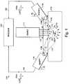
- FIG. 1is a schematic diagram of a spectroscopic system in accordance with the invention
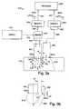
- FIG. 2is a schematic diagram of a Laser Induced Fluorescence Attenuation Spectroscopy system (“LIFAS”)in accordance with the present invention having an excitation waveguide and a first and second collection waveguides;
- LIFASLaser Induced Fluorescence Attenuation Spectroscopy system
- FIG. 3is a schematic diagram of a LIFAS system in accordance with the present invention having a first excitation-collection waveguide and a second collection only waveguide;
- FIG. 4is schematic diagram of a LIFAS system for biomedical applications in which excitation radiation is used to induce fluorescence of tissue;
- FIGS. 5 ( a ) and ( b )are end perspective views of a LIFAS probe having an optical fiber and an optical fiber bundle as the central waveguide;
- FIGS. 6 ( a ) and ( b )are partial perspective views of an electrical subsystem used to synchronize a LIFAS system with the electrical activity of tissue using electrodes integrated into the optical probe;
- FIGS. 7 ( a ) and ( b )are schematic diagrams showing the relative positions of the action potential and electrodes integrated into the optical probe; and FIGS. 7 ( c ) and ( d ) are the corresponding action potential timing diagrams;
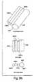
- FIG. 8is a perspective view of a probe incorporating a visible light source for probe positioning and a circular LED array for indicating the direction of contraction propagation;
- FIGS. 9 ( a )-( c )are partial perspective views of three LIFAS probes having waveguides arranged in different geometrical configurations adapted for different applications;
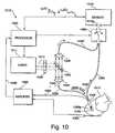
- FIG. 10is a schematic diagram of a biomedical LIFAS system in accordance with the invention in which a single optical detector is used to measure the return light from two collection fibers;
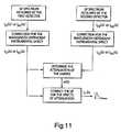
- FIG. 11is a block diagram of the method for determining the attenuation and the intrinsic fluorescence of a sample
- FIG. 12 ( a )is an illustration depicting an LIFAS probe device in accordance with the invention
- FIG. 12 ( b )is a graph that depicts fluorescent intensity along the radius of the excitation-collection fiber, the axis of the excitation-collection fiber and the axis of a collection-only fiber at 0.4 mm range
- FIG. 12 ( c )is a graph that depicts fluorescent intensity along the axis of the excitation waveguide at a range of 0.08 mm to 3 mm
- FIG. 12 ( d )is a graph that depicts fluorescent intensity along the axis of the collection-only fiber at a range of 0.6 mm to 2.6 mm;
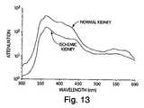
- FIG. 13is a graph of the attenuation spectrum measured from normal and ischemic renal cortex of rabbit kidney using LIFAS devices and methods in accordance with the invention.
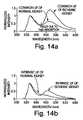
- FIG. 14 ( a )is a graph of the modulated LIF spectra of normal and ischemic kidney
- FIG. 14 ( b )is a graph of the intrinsic LIF spectra of normal and ischemic kidney
- FIG. 15is a scatter plot of the fluorescence intensity at 480 nm acquired from hypoxic (+), normal (o) and hyperoxic (x) tissue through excitation-collection and collection-only waveguide;
- FIG. 16is a graph of the intensity I co ( ⁇ ) c of the signal measured by a collection-only waveguide from normal, hyperoxic and hypoxic tissue;
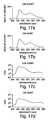
- FIGS. 17 ( a )-( b )are graphs of the mean LIF intensity of live kidney and heart tissue and FIGS. 17 ( c )-( d ) are the corresponding LIFAS-derived attenuation spectra.
- the present inventionis embodied in a spectroscopic system 110 and related method for measuring the attenuation and/or optical rotation caused by a sample 114 .
- the spectroscopic system 110 shown in FIG. 1includes a source 111 that produces radiation 112 , which is directed at a sample volume 113 within the sample 114 .
- the source 111is preferably a laser and the radiation 112 is preferably monochromatic ultraviolet (UV), visible or infrared (IR) radiation 112 . Nevertheless, the source 111 can be selected to produce other types of radiation, such as broad-band or polarized radiation, useful for a particular application.
- the spectroscopic system 110further includes a first sensor 116 displaced by a distance Z 1 from the sample volume 113 , and a second sensor 118 displaced by a distance Z 2 from the sample volume 113 , which are used to monitor return radiation 120 from the sample 114 .
- the return radiation 120will include fluorescence of the flurophores of the sample 114 .
- the sensors 116 and 118each include an aperature 116 a and 118 a , respectively, through which the return radiation 120 is observed and collected.
- the sensors 116 and 118typically can only observe the events occurring within the volume defined by a solid angle emerging from the aperture of the sensor. This volume is known as the numerical aperture or the field of view of the sensor.
- first and second sensors 116 and 118include numerical apertures 117 and 119 , respectively, that are adapted to receive a portion of the return radiation 120 from the sample 114 .
- the sensors 116 and 118can be tilted at different angles ⁇ 1 and ⁇ 2 and selected to have different numerical apertures 117 and 119 so that the sensors 116 and 118 can selectively monitor return radiation 120 from all or part of the sample volume 113 .
- the sensors 116 and 118each generate a signal 124 a and 125 a , respectively, representing the intensity I c1 ( ⁇ ) and I c2 ( ⁇ ) and/or polarization of the return radiation 120 .
- the signals 124 a and 125 aare generated at a plurality of wavelengths within predetermined wavelength bands.
- the signal 124 a from the first sensor 116 and the signal 125 a from the second sensor 118are communicated by signal paths 124 and 125 , respectively, to a processor 123 .
- the processor 123processes the signals 124 a and 125 a to determine a spectral characteristic of interest, such as the attenuation and/or optical rotation of the return radiation 120 caused by the sample 114 .
- the sensors 116 and 118are positioned at non-equivalent distances Zl and Z 2 from the sample volume 113 .
- the first and second sensors 116 and 118will be positioned so that one of the sensors will be in the immediate proximity of the sample volume 113 while the other sensor is positioned at a location adjacent to the first sensor, but displaced slightly further from the sample volume 113 . It has been found that this arrangement advantageously improves the signal-to-noise ratio and, hence, measurement accuracy of the spectroscopic system 110 .
- the attenuation of the return radiation 120is dependent on the distance traveled by the return radiation 120 through the sample 114 .
- the signal 124 a measured by the first sensor 116 and the signal 125 a measured by the second sensor 118will suffer different attenuation where the distances Z 1 and Z 2 from the sample volume 113 are not equivalent.
- the processor 123can be used to process the signals 124 a and 125 a to determine the wavelength-dependent attenuation of the sample 114 .
- the attenuation determined by the spectroscopic system 110will reflect the effects of light absorption and scattering by the sample 114 .
- the attenuation of the sample 114can be used to determine the intrinsic fluorescence 129 of the fluorophores in the sample volume 113 .
- the methods by which the attenuation and intrinsic LIF are determinedare discussed in detail below.
- the source 111can be any radiation source, such as a laser or a lamp emitting excitation radiation 112 at a wavelength capable of interacting with the sample 114 .
- the sensors 116 and 118will preferably include a detector, which can be as simple as individual light-sensitive diodes with appropriate band pass filters, or more complicated sensors such as an optical spectrum analyzer.
- the sensoris a suitable spectrograph or spectrometer equipped with a suitable sensor.
- the sensorcan include a multispectral CCD camera.
- the source 111is an XeCl excimer laser emitting monochromatic ultraviolet radiation at 308 nm.
- Each of the sensors 116 and 118is a spectrograph (Model FF250, ARIES Inc., Concorde, Mass.) associated with a 1024 element intensified photo diode array (PDA) to image the resolved spectrum.
- PDAphoto diode array
- Each PDAis connected to an optical multichannel analyzer (OMA III, EG&G Princeton Applied Research Corporation, Princeton, N.J.). The OMA reads the light spectrum imaged by the PDA to produce the signals 124 a and 125 a .
- a 335 nm longpass filter(Schott WG335) is placed in front of the aperture of each of the sensors 116 and 118 to remove any backscattered 308 nm excitation radiation.
- the processor 123is a personal computer that is networked to each of the sensors 116 and 118 via the signal paths 124 and 125 to receive the signals 124 a and 125 a , respectively.
- optical rotationis observed in optically active materials.
- An optically active materialis characterized by a lack of symmetry in its molecular or crystalline structure which causes a rotation of the plane of polarization in incident-plane-polarized radiation.
- the extent to which the plane of polarization is rotatedwill typically vary from one optically active material to another.
- the extent of rotationmay depend on the number of molecules in the path of the radiation, the wavelength of the radiation, and the temperature of the material.
- Anisotrophyis caused by a selective response of the sample to radiation travelling in different directions.
- anisotrophycan result from microscopic inhomogenetics in the tissue structure.
- Use of equivalent distances Z 1 and Z 2will minimize the contribution of the path-length-dependent attenuation to the overall attenuation measured by the system 110 . Therefore, the optical rotation or anisotropy of the sample can be determined more easily.
- the LIFA system 110 shown in FIG. 1can be adapted to measure optical rotation by utilizing first and second sensors 116 and 118 which are adapted to measure the polarization-dependent intensity of the return radiation.
- the first sensor 116can be adapted to measure a first optical angle ⁇ 1 (not shown) yielding the maximum intensity of the first portion of the return radiation 120 .
- the second sensor 118can be adapted to measure a second optical angle ⁇ 2 (not shown) yielding the maximum intensity of the second portion of the return radiation 120 .
- the optical rotation of the return light 120will be given by the difference between the angles ⁇ 1 and ⁇ 2 , provided that the system has been properly calibrated.
- the same sensoris used for determination of the attenuation of the sample will find utility for monitoring the optical rotation.
- These sensorscan be adapted to such measurements by placing a rotatable polarizing filter at each of the apertures 116 a and 118 a of the first and second sensors 116 and 118 , respectively.
- FIG. 2 ( a )An alternative embodiment of the present invention is shown in FIG. 2 ( a ).
- the spectroscopic system 110 of FIG. 1has been adapted as a Laser Induced Fluorescence Attenuation Spectroscopy (“LIFAS”) system 210 .
- LIFASLaser Induced Fluorescence Attenuation Spectroscopy
- a source 211emits laser radiation 212 at an intensity and a wavelength capable of inducing fluorescence of the sample 214 .
- the laser radiation 212is transmitted through an excitation waveguide 215 to a sample volume 213 within the sample 214 where the laser radiation 212 excites local fluorophores in the sample volume 213 to emit intrinsic fluorescence 229 .
- the intrinsic fluorescence 229 of the fluorophores in the sample volume 213is modulated, for example, by the absorption and scattering of the local chromophores (not shown) and scatterers (not shown) present in the sample 214 , respectively.
- Two collection waveguides 221 and 222 having apertures 221 a and 222 a and numerical aperatures 217 and 219are disposed about the excitation waveguide 215 .
- the collection waveguides 221 and 222may be positioned such that their apertures 221 a and 222 b are laterally displaced from the excitation waveguide 215 by the distances x 1 ′ and x 2 ′ and/or axially displaced from the aperture 215 a of the excitation waveguide 215 by the distances y 1 ′ and y 2 ′.
- the lateral distances x 1 ′ and x 2 ′are small or zero.
- the axial distances y 1 ′ and y 2 ′are preferably selected to be small and unequal, while one of the axial distances is preferably zero. Also, it is desirable to position the apertures 215 a , 221 a and 222 a of the waveguides 215 , 221 and 222 in close proximity to or in contact with the sample 214 during use of the LIFAS system 210 , as shown in FIG. 2 ( a ).
- FIG. 2 ( c )An alternative configuration of the collection waveguides is shown in FIG. 2 ( c ).
- the axial distances y 1 ′ and y 2 ′are both zero.
- the lateral distances x 1 ′ and x 2 ′are selected to be small and unequal.
- the apertures 240 a and 242 a of the waveguides 240 and 242are oblique such that the fields of view 244 and 246 are directed toward the sample volume 213 and to advantageously encompass the bulk of the sample volume 213 . As shown in FIG.
- the distances z 1 ′ and z 2 ′are directed along the respective axes of the numerical aperatures 244 and 246 and extend from the surface of the apertures 240 a and 242 a to the point of intersection of the axes of the numerical aperatures 244 and 246 , respectively.
- the collection waveguide 221collects a first portion 228 of the return light 220 and transmits the first portion 228 of the return light 220 to a first sensor 216 .
- the first sensor 216generates a first signal 224 a , representing the intensity I c1 ( ⁇ ) of the first portion 228 of the return light 220 at a plurality of wavelengths within predetermined wavelength bands.
- the collection waveguide 222collects a second portion 230 of the return light 220 and transmits the second portion 230 of the return light 220 to the second sensor 218 .
- the second sensor 218generates a second signal 225 a representing the intensity I c2 ( ⁇ ) of the second portion 230 of the return light 220 at a plurality of wavelengths preferably within the same wavelengths bands monitored by the first sensor 216 .
- the processor 223then processes the first and second signals 224 a and 225 a to determine the wavelength-dependent attenuation of the sample 214 . As discussed in detail, below, once the attenuation is known, either of the signals 224 a or 225 a can be processed to minimize the effects of attenuation and determine the intrinsic LIF 229 of the fluorophores in the sample volume 213 . Alternatively, if the sensors 216 and 218 are selected to monitor the polarization of the first portion 228 and the second portion 230 of the return light 220 , the processor 223 can be used to determine the optical rotation of the return light 220 caused by the sample 214 .
- the intensity and wavelength of the laser radiation 212should be sufficient to create a sample volume 213 that is large enough to have some overlap with the numerical apertures 217 and 219 of the collection waveguides 221 and 222 , respectively.
- the numerical apertures 217 and 219can be selected to encompass all or parts of the sample volume 213 .
- longpass filtersare placed before the inputs of the sensors 216 and 218 to selectively block backscattered excitation radiation.
- the source 211is an Xe—Cl excimer laser emitting 308 nm ultraviolet excitation radiation 212 .
- the waveguides 215 , 221 and 222are advantageously comprised of optical fibers or optical fiber bundles, which can be integrated into a probe for ease of use and durability.
- the optical bundle and fibersare made of fused silica which is transparent to ultraviolet radiation. Specifically, a 1.4 mm diameter optical bundle is used as the excitation waveguide 215 while the collection waveguides 221 and 222 are 0.4 mm optical fibers disposed about the excitation bundle.
- the lateral range x 1 ′ and x 2 ′are equal to zero while the axial distance y 1 ′ is about 0.6 mm and the axial distance y 2 ′ is about zero.
- the probemay be configured within, for example, the shaft of a hypodermic needle, so that the in-vivo physiological or pathological properties of biological tissue can be determined.
- each of the sensors 216 and 218is a spectrograph (Model FF250, ARIES Inc., Concorde, Mass.) associated with a 1024 element intensified photo diode array (PDA).
- PDA1024 element intensified photo diode array
- Each PDAis connected to an optical multichannel analyzer (OMA III, EG&G Princeton Applied Research Corporation, Princeton, N.J.) which measures the intensity of the light spectrum imaged by the PDA and produces the signals 224 a and 225 a .
- OMA IIIoptical multichannel analyzer
- the processor 223is a personal computer that is networked to each of the sensors 216 and 218 via the signal paths 224 and 225 to receive the signals 224 a and 225 a , respectively.
- FIG. 11illustrates the main steps involved in determining the wavelength-dependent attenuation coefficient ⁇ ( ⁇ ) and the intrinsic fluorescence I T ( ⁇ ) of a sample in accordance with the current invention.
- the first and second portions 228 and 230 of the return light 220experience different attenuation effects due to the unequal path lengths traversed by the return light 220 from the sample volume 213 to the apertures 221 a and 222 a of the collection waveguides 221 and 222 .
- the first portion 228 of the return light 220 collected by the aperture 221 atravels an additional path-length of ⁇ y 1 ′ ⁇ y 2 ′ ⁇ through the tissue as compared to the second portion 230 of the return light 220 which is collected by the aperture 222 a .
- the first portion 228 of the return light 220suffers more path-length-dependent attenuation as compared to the second portion 230 of the return light 220 .
- the signals 224 a and 225 arepresenting the intensity I c1 ( ⁇ ) and I c2 ( ⁇ ) at different wavelengths will exhibit differing levels of modulation caused by the attenuation of the sample 214 .
- the signals 224 a and 225 awill also exhibit wavelength-dependent modulations caused by the instrumental effects.
- the wavelength-dependent modulations due to the instrumental effectscan be determined and, in turn, compensated for by conducting a calibration of the LIFAS system 210 .
- System calibrationcan be performed using light from a standard lamp (Quartz Halogen Lamp, Model No. 63358, Oriel Instruments, Stratford, Conn.) having a predetermined continuous spectrum to measure the wavelength-dependent instrumental effects of the LIFAS system 210 .
- the processor 223can be adapted to correct the measured intensities I c1 ( ⁇ ) and I c2 ( ⁇ ) for modulations caused by the wavelength-dependent instrumental effects.
- the corrected intensities I c1 ( ⁇ ) c and I c2 ( ⁇ ) c representing the intensity of the first and second portions 228 and 230 of the return light 220 at different wavelengthscan then used to determine the attenuation coefficient ⁇ ( ⁇ ) of the sample 214 as described below.
- the regions ⁇ circle around ( 1 ) ⁇ and ⁇ circle around ( 2 ) ⁇ , of FIG. 2 ( a )may be at an effective range, symbolized as “R,” from which the collection waveguides 221 and 222 collect the majority of the first and second portions of return light 228 and 230 , respectively.
- the effective range “R”will vary with the attenuation of the sample.
- the regions ⁇ circle around ( 1 ) ⁇ and ⁇ circle around ( 2 ) ⁇are geometrically symmetric with respect to the aperture 215 a of the excitation waveguide 215 and, hence, have identical intensity of the intrinsic fluorescence 229 represented by I T ( ⁇ ).
- the wavelength-dependent intensities I c1 ( ⁇ ) c and I c2 ( ⁇ ) ccan be described by the following equations:
- the attenuation coefficient ⁇ ( ⁇ )can be calculated independent of the value of “R” from (1) and (2) as follows:
- the intrinsic fluorescence, I T ( ⁇ )can be restored from either of the signals I c1 ( ⁇ ) c or I c2 ( ⁇ ) c (preferably I c2 ( ⁇ ) c where y 2 ′ ⁇ y 1 ′) by assuming an average effective range “R” from which most of the intrinsic fluorescence is collected.
- the constant “R”is approximately about 0.2 mm at 308 nm excitation radiation. Therefore, the intrinsic fluorescence I T ( ⁇ ) can be obtained by substituting the measured I c2 ( ⁇ ) c into equation (2) and solving for I T ( ⁇ ) using the constant R and the known value y 2 ′:
- I T ( ⁇ )I c2 ( ⁇ ) c e ⁇ ( ⁇ ) ⁇ (R+y2′) (5)
- the absorbance A( ⁇ ) and the percent transmittance %T( ⁇ ) of the samplecan be calculated as follows:
- the source 311emits laser radiation 312 at a wavelength and intensity capable of inducing fluorescence of the sample 314 .
- the laser radiation 312is reflected by a dichroic mirror 326 into an excitation-collection waveguide 321 , which transmits the laser radiation 312 to the sample volume 313 where the laser radiation 312 excites local fluorophores to emit intrinsic fluorescence 329 .
- the intensity of the laser 312should be sufficient to create a sample volume 313 that overlaps with the numerical aperture 319 of the collection-only waveguide 322 .
- the dimensions of the sample volume 313will also depend on the numerical aperture of the excitation-collection waveguide 321 , on the wavelength and intensity of the excitation radiation 312 and on the optical properties of the sample 314 .
- the intrinsic fluorescence 329is modulated, for example, by the absorption and scattering of the local chromophores (not shown) and scatterers (not shown) of the sample 314 .
- the excitation-collection waveguide 321collects a first portion 328 of the return light 320 directly from the sample volume 313 .
- the first portion 328 of the return light 320is transmitted through the dichroic mirror 326 to the first sensor 316 .
- the first sensor 316generates a first signal 324 a representing the intensity I xc ( ⁇ ) of the first portion 328 of the return light 320 at a plurality of wavelengths within a predetermined wavelength band.
- the collection-only waveguide 322may be positioned such that the aperture 322 a is laterally displaced from the excitation-collection waveguide 321 by the distance x 3 and axially displaced from the aperture 321 a of the excitation-collection waveguide 321 by the range y 3 .
- the lateral distance x 3is zero while the axial distance y 3 is non-zero.
- the apertures 321 a and 322 a of the waveguides 321 and 322are preferably positioned in close proximity to or in contact with the sample 314 during use of the LIFAS system 310 , as shown in FIG. 3 ( a ).
- the numerical aperture 319 of the collection-only waveguide 322is selected to include at least a portion of the sample volume 313 .
- the collection-only waveguide 322collects a second portion 330 of the return light 320 which is transmitted to a second sensor 318 .
- the second sensor 318generates a second signal 325 a representing the intensity I co ( ⁇ ) of the second portion 330 of the return light 320 at a plurality of wavelengths preferably within the same wavelength bands as the signal 324 a generated by the first sensor 316 .
- a longpass filteris placed in front of the aperture of each of the sensors 316 and 318 to selectively block backscattered excitation radiation.
- the first and second portions 328 and 330 of the return light 320experience different attenuation effects due to the unequal path-lengths traversed by the return light 320 from the sample volume 313 to the apertures 321 a and 322 a of the waveguides 321 and 322 , respectively.
- the second portion 330 of the return light 220 collected by the aperture 322 atravels an additional path-length through the tissue as compared to the first portion 328 of the return light 320 which is collected by the aperture 321 a directly from the sample volume 313 .
- the first portion 328 of the return light 320suffers less path-length-dependent attenuation as compared to the second portion 330 of the return light 320 .
- the signals 324 a and 325 arepresenting intensity I xc ( ⁇ ) and I co ( ⁇ ) at different wavelengths will exhibit different levels of modulation caused by the sample 314 .
- the signals 324 a and 325 awill also exhibit wavelength-dependent modulations caused by the instrumental effects.
- the wavelength-dependent modulations due to instrumental effectscan be determined and, in turn, compensated for by conducting a calibration of the LIFAS system 310 .
- System calibrationis performed using light from a standard lamp (Quartz Halogen Lamp, Model No. 63358, Oriel Instruments, Stratford, Conn.) having a predetermined continuous spectrum to measure the wavelength-dependent instrumental effects of the LIFAS system 310 .
- the processor 323can be adapted to correct the measured intensities I xc ( ⁇ ) and I co ( ⁇ ) for modulations caused by the wavelength-dependent instrumental effects.
- the corrected intensities I xc ( ⁇ ) c and I co ( ⁇ ) c representing the intensity of the first and second portions 328 and 330 of the return light 320 at different wavelengthscan then be used by the processor 323 to determine the wavelength-dependent attenuation ⁇ ( ⁇ ) of the sample 314 .
- the processor 323can then be used by the processor 323 to determine the wavelength-dependent attenuation ⁇ ( ⁇ ) of the sample 314 .
- either of the signals 324 a or 325 acan be corrected for the effects of attenuation to restore the intrinsic LIF 329 of the fluorophores in the sample volume 313 .
- the source 311is an Xe—Cl excimer laser emitting ultraviolet excitation radiation at a wavelength of 308 nm.
- the waveguides 321 and 322are advantageously comprised of optical fibers or optical fiber bundles, which can be integrated into a probe for ease of use and durability.
- a 1.4 mm diameter optical bundle and a 0.4 mm optical fiberare used as the excitation-collection and the collection-only waveguides, respectively.
- the collection-only waveguideis a plurality of 0.4 mm optical fibers disposed about the periphery of the excitation-collection waveguide.
- the optical bundle and fibersare made of fused silica which is transparent to the 308 nm ultraviolet radiation.
- the probemay be configured within, for example, the shaft of a hypodermic needle, so that the in-vivo physiological or pathological properties of biological tissue can be determined.
- Each of the sensors 316 and 318is a spectrograph (Model FF250, ARIES Inc., Concorde, Mass.) associated with a 1024 element intensified photo diode array (PDA) to image the resolved spectrum.
- PDAelement intensified photo diode array
- Each PDAis connected to an optical multichannel analyzer (OMA III, EG&G Princeton Applied Research Corporation, Princeton, N.J.) which measures the intensity of the light spectrum imaged by the PDA and produce the signals 324 a and 325 a .
- the processor 323is a personal computer that is networked to each of the sensors 316 and 318 via the signal paths 324 and 325 to receive the signals 324 a and 325 a , respectively.
- the method of determining the attenuation coefficient ⁇ ( ⁇ ) and the intrinsic fluorescence I T ( ⁇ ) of a sample using the embodiment of FIG. 3 ( a ) in accordance with the current inventionis as follows.
- “D”represents the effective range, from which the collection waveguide 321 collects a majority of the portion 328 of the return light 320 .
- the portion 328 of the return light 320is collected directly from the sample volume 313 by the excitation-collection waveguide 321 .
- the wavelength-dependent intensity I xc ( ⁇ ) c of the portion 328 of the return light 320 collected by the excitation-collection waveguide 321will be substantially similar to the wavelength-dependent intensity of the return light 320 , represented as I o ( ⁇ ).
- the return light 320 with the intensity I o ( ⁇ )will suffer additional wavelength-dependent attenuation as it travels the extra path-length to reach the aperture 322 a and is collected as the second portion 330 of the return light 320 with the intensity I co ( ⁇ ) c .
- the wavelength-dependent intensity I co ( ⁇ ) ccan be approximated by the following equations:
- the attenuation coefficientcan be approximated as follows:
- the intrinsic fluorescence, I T ( ⁇ )can be restored from either of the signals I xc ( ⁇ ) c or I co ( ⁇ ) c , preferably I xc ( ⁇ ) c , by assuming an average effective range “D” from which most of the intrinsic fluorescence is collected.
- the constant “D”is approximately about 0.2 mm at 308 nm excitation radiation.
- I T ( ⁇ )I xc ( ⁇ ) c e ⁇ ( ⁇ ) ⁇ D (12)
- the measured attenuationmay account for absorption and/or scattering depending on the wavelength band of interest and the nature of the sample. Below about 600 nm, the optical attenuation of biological tissue is primarily due to absorption and, hence, the attenuation coefficient ⁇ ( ⁇ ) will represent absorptivity a( ⁇ ). Also, the absorbance A( ⁇ ) and the percent transmittance %T( ⁇ ) of such samples can be calculated as follows:
- a LIFAS system 410 in accordance with the present inventionhas been adapted for biomedical applications.
- the LIFAS system 410has been adapted to determine the optical attenuation of biological tissue.
- the laser 411emits radiation 412 at a wavelength capable of exciting the tissue 414 to emit fluorescence.
- the radiation 412is directed through an iris 440 at a dichroic mirror 442 which reflects the radiation 412 onto a lens 444 which focuses the radiation 412 onto the proximal tip 445 of an optical fiber 446 .
- the adjustable iris 440is advantageously used to reduce the energy of the radiation 412 .
- the adjustable iris 440can be replaced by any suitable attenuator.
- the probe 448includes a central optical fiber 446 that is used as the excitation-collection fiber and peripheral optical fibers 450 a-h are used as the collection-only fibers. The distal end of the optical fibers 446 and 450 a-h are incorporated into the optical probe 448 .
- the aperture 448 a the optical fiber probe 448is placed in proximity to the tissue 414 so that the aperture 446 a of the excitation-collection fiber 446 and apertures 451 a-h of the collection-only fibers 450 a-h are in contact with the tissue 414 . As shown in a partial perspective view in FIG.
- the optical fiber probe 448includes a central optical fiber 446 and a plurality of optical fibers 450 a-h disposed about the periphery of the central optical fiber 446 .
- the apertures 451 a-h of the distal ends of the collection-only fibers 450 a-hare axially displaced by a small distance y 3 with respect to the aperture 446 a of the excitation-collection fiber 446 .
- the return light 420 collected by apertures 451 a-h of the collection-only fibers 450 a-hpool into the aperture 418 a of the sensor 418 .
- optical fibers 446 and 450 a-hcan advantageously be replaced by optical fiber bundles in order to obtain greater flexibility and durability.
- the central optical fiber 446can be replaced with an optical fiber bundle 454 .
- the excitation radiation 412is transmitted through the optical fiber 446 to the tissue 414 to induce intrinsic fluorescence of the fluorophores of the tissue 414 .
- the intrinsic fluorescenceis modulated, for example, by the chromophores and/or scatterers of the tissue 414 .
- a first portion 428 of the return light 420is collected by the optical fiber 446 from the tissue volume that is directly irradiated by the excitation radiation 412 and transmitted through the optical fiber 446 to a first lens 444 where the return light is directed through the dichroic mirror 442 and then focused by a second lens 445 onto the aperture 416 a of a first sensor 416 .
- the collection-only optical fibers 450 a-hcollect a second portion 430 of the return light 420 and transmit the second portion 430 of the return light 420 to the second sensor 418 .
- longpass filtersare placed in front of the apertures 416 a and 418 a of the sensors 416 and 418 to selectively block backscattered excitation radiation.
- a first signal 424 a and a second signal 425 a representing the intensity of the first and second portions 428 and 430 , respectively, of the return light 420are generated by the first and second sensors 416 and 418 and transmitted via signal paths 424 and 425 to the processor 423 .
- the processor 423uses the first and second signals 424 a and 425 a generated by the detectors 416 and 418 to determine the wavelength-dependent attenuation of the sample 414 using equations (8), (9) and (10) as described in the previous embodiment.
- an illumination source 490 emitting visible light 492is mounted on the probe 448 to act as a spotlight for the operator. Because room illumination can contaminate the spectral measurements of the system 410 by adding background light, use of the illumination source 490 will allow the operator to see and accurately position the probe 448 under low-light conditions.
- the illumination source 490is configured to illuminate the sample at all times, except when the LIFAS system is monitoring return light from the sample 414 .
- the biological electrical signal arising in nerves or contractile tissue, such as muscle,is known as the “action potential” and is caused by sudden changes in the ion conductivity of the cell membrane.
- the occurrence of an action potential in contractile tissueinitiates a contraction.
- the action potentialpropagates and spreads in a wave-like manner to induce a local myocardial contraction wherever it travels.
- An action potential propagating in the tissue local to the aperture 448 a of the probe 448can be detected by the electrode 464 which is incorporated into the probe 448 as shown, for example, in FIG. 6 ( a ). Voltage alternations caused by the occurrence of the action potential are picked up by the electrode 464 in reference to the common electrode 466 and are transmitted to the amplifier 460 .
- the common electrode 466provides the electrical ground for the amplifier 460 by maintaining contact with the tissue of interest 414 or the whole body.
- the amplified action potential 468is transmitted to the processor 423 to trigger the acquisition process of the LIFAS system 410 at a pre-selected phase of the tissue contraction or of the cardiac cycle, whichever is applicable.
- a plurality of electrodes or fibers with a conductive coatingcan be distributed circumferentially about the tip of the optical fiber probe 448 .
- the probe 448can be equipped with three electrodes 470 , 472 and 474 arranged in a triangular configuration.
- the action potentials measured by each of the electrodes 470 , 472 and 474are amplified through separate channels of the amplifier 460 and transmitted to the processor 423 .
- the processor 423processes the received signals to determine the direction of propagation of the contraction vector 462 .
- the processordetects the phase lead/lag between the action potentials collected by the electrodes 470 , 472 and 474 to determine the orientation of the contraction vector 462 with respect to the location of the electrodes 470 , 472 and 474 .
- the contraction vector 462 shown in FIG. 7 ( a )is propagating from the tissue site 476 a to the site 478 a and, hence, as shown in FIG. 7 ( c ), the action potential 474 a arrives before the action potential 472 a which in turn arrives before the action potential 470 a .
- the phase difference or time delay between a pair of action potentialsindicates how the contraction vector 462 is centered between the location of the corresponding pair of electrodes. For example, as shown in FIG. 7 ( b ), the contraction vector 462 propagates from the tissue site 476 b to the site 478 b .
- the processor 423processes the signals and indicates the direction of the propagation of the contraction to the system operator.
- the direction of propagationcan be indicated, for example, by a circular array 496 of light emitting diodes (LED) 498 mounted cirumferentially on the probe 448 , as shown in FIG. 8 .
- the processor 423transmits a signal to the LED array 496 so that, for example, only the LED element pointing in the direction of contraction propagation would glow.
- the amplifier 460can be replaced by any device that can measure the electrical activity of biological tissue such as a differential amplifier, an electrocardiogram (ECG), an electromyogram (EMG), an electroencephalogram (EEG), depending on the LIFAS application.
- ECGelectrocardiogram
- EMGelectromyogram
- EEGelectroencephalogram
- optical fibers with a metallic or electrically conductive coatingcan be used in place of one or all of the electrodes 464 , 470 , 472 or 474 to measure the action potential of the tissue.
- conventional ECGis generally not suitable for triggering the acquisition of LIFS or LIFAS systems, since it does not accurately indicate the instantaneous state of myocardial contraction at the sample volume.
- customary ECG using limb or chest leadscan be used to trigger data acquisition of the LIFAS system 410 where the sample 414 is non-contractile tissue.
- the light source 411is preferably a lamp or a laser that emits ultraviolet, visible or infrared radiation.
- the source 411is an XeCl excimer laser emitting pulses of ultraviolet excitation radiation at 308 nm.
- the optical components used in the acquisition systembe made of synthetic quartz (fused silica) to ensures maximal ultraviolet transmission and minimal instrumental fluorescence.
- a nitrogen laser, a helium-cadmium laser, a frequency-multiplied laser, a solid-state laser, an arc lamp or a light-emitting diodecan be used as the light source 411 .
- the energy of the excitation light 412is typically between 0.001-10 m Joules. However, it will be appreciated that the selected energy level should be low enough to avoid tissue ablation and/or photobleaching while still being adequate to produce detectable LIF.
- the sensors 416 and 418are each comprised of a spectrograph (Model FF250, ARIES Inc., Concorde, Mass.) associated with a 1024 element intensified photo diode array (PDA) detector.
- An optional low fluorescence, long-pass filter(not shown) with a cutoff wavelength above 308 nm, preferably 335 nm (Schott WG335), is placed before the entrance slit of each spectrograph to selectively block any backscattered excitation radiation from reaching the sensors 416 and 418 .
- the entrance slit of the spectrographpreferably has a width of 100 micrometers.
- the spectrographuses a 150 lines per millimeter diffraction grating to disperse the incoming return light 420 into its spectral components.
- the spectrum formed by the spectrographis imaged by a detector, preferably an intensified linear photodiode array (Model 1420, EG&G Princeton Applied Research Corporation, Princeton, N.J.) facing the output port of the spectrograph.
- the photodiode arraygenerates a plurality of electrical signals representing the intensity of the return light 420 at wavelengths within predetermined wavelength bands.
- the sensorscan be constructed of any suitable materials, such as individual light-sensitive diodes with appropriate band-pass filters for the analysis of spectral bands of the return light or an optical spectrum analyzer (“OSA”) for analysis of a broader spectrum. Selection of the return light monitoring device will depend on a variety of factors, including cost, accuracy, resolution, and whether the user is interested in monitoring a single wavelength, a wavelength band or an entire spectrum.
- the optical fiber probeincludes a central excitation-collection optical fiber or optical fiber bundle with a plurality of collection-only optical fibers disposed around its periphery
- the probecan take many forms.
- the central optical fibercan be used as the collection-only waveguide while all or some of the peripheral fibers can be used for excitation-collection or collection-only. The latter arrangement is preferred when testing highly-attenuating samples to achieve a better signal-to-noise ratio.
- FIG. 9shows three alternative geometrical configurations of the collection-only optical fibers 950 a-h about the excitation-collection waveguide 946 .
- the collection-only waveguides 950 a-hare arranged about the excitation-collection waveguide 946 so that their apertures have a helical configuration.
- each of the collection-only waveguides 950 a-hwill be attenuated in varying degrees.
- Use of a probe arrangement having a plurality of collection distanceswill be useful in measuring the polarization and/or the attenuation of especially, a sample having a higher sensitivity to attenuation due to absorption.
- a probe incorporating a plurality of collection distanceshave application to a larger variety of samples. For example, where the sample is highly attenuating, the collection-only waveguides having apertures close to the excitation site can be used to collect the return light. Where the sample is lightly attenuating, the return light collected by the waveguides having apertures that are further from the excitation site will be useful in determining the attenuation.
- the collection-only waveguides 950 a-care displaced laterally with respect to the excitation-collection waveguide 946 .
- Such a probe configurationwill be useful in measuring the polarization and/or the attenuation of the sample where the sample has a higher sensitivity to attenuation due to scattering.
- the return light collected by the waveguide 950 cwill be useful in measuring attenuation of a lightly attenuating sample.
- the return light measured by the closest waveguide 950 awill be useful in measuring the attenuation of a heavily-attenuating sample.
- the collection-only waveguides 950 a-care both axially and laterally displaced with respect to the excitation-collection waveguide 946 .
- This configurationcombines the advantages of both of the aforementioned configurations shown in FIGS. 9 ( a ) and 9 ( b ).
- a single sensor 1016 and an associated optical multiplexer 1080are utilized.
- the multiplexer 1080is used to switch the input of the sensor 1016 between the excitation-collection optical path ( 1046 , 1044 , 1042 , 1049 , 1046 ′) and the collection-only optical path ( 1050 ) such that the return light from each pathway is measured sequentially.
- the processor 1023sends trigger signals to the source 1011 , the sensor 1016 and the optical multiplexer 1080 to synchronize their actions. If the source 1011 is a pulsed laser, two or more radiation pulses are typically required to acquire a single attenuation measurement using the multiplexed system 1010 .
- a single laser pulsecan be used if the laser pulse lasts long enough to sustain the emission of return light 1020 during the minimum period of time required by the optical multiplexer 1080 and associated sensor 1016 to acquire the spectral measurements from both the excitation-collection and the collection only optical paths.
- the preferred embodiment of the LIFAS system 1010is similar to that of 410 , except that the system 1010 utilizes an electromagnetic dual port shutter as the optical multiplexer 1080 associated with a single sensor 1016 in a configuration where the portion 1028 and the portion 1030 of the return light 1020 are measured sequentially.
- the LIFAS methods and devices of the present inventioncan be used advantageously for the in-vivo diagnosis of hypoxia and ischemia of biological tissue.
- Hypoxiais a deficiency in the amount of oxygen reaching the tissue, e.g., due to pneumonia
- ischemiais a localized reduction in arterial blood perfusion, e.g., due to a narrowing of arteries by spasm or disease.
- Ischemiacan also result from hemorrhage of an arterial wound or during surgical procedure that temporarily interrupts the blood flow to a body region.
- the present inventiondescribes new criteria and methods for the discrimination between normal, ischemic and hypoxic biological tissue, including, in particular renal and myocardial tissue, as follows.
- LIFASlaser induced fluorescence attenuation
- the LIFAS-derived LIFA spectra of normal and ischemic rabbit kidneyare shown in FIG. 13 .
- the LIFA spectra shown in FIG. 13are acquired using a LIFAS system employing 308 nm excitation radiation produced by an XeCl excimer laser.
- This LIFAS systemuses a 335 nm longpass filter (Schott WG335) to cutoff backscattered excitation radiation from the collected return light.
- the LIFA values below 350 nmare not reliable.
- the LIFA values in the wavelength band about 480 nmhave the highest signal-to-noise ratio and hence measurement accuracy.
- the LIFA of ischemic tissueis lower than the LIFA of normal tissue over the entire spectrum and is particularly low in the region from 350 to 450 nm.
- the LIFA, absorbance or percent transmittance at a predetermined wavelength or wavelength bandscan be used for the detection of ischemia or hypoxia.
- predictive models, spectral recognition techniques and associated classifierscan be applied to identify whether a given LIFA, absorbance or percent transmittance spectrum has been acquired from normal, ischemic or hypoxic tissue.
- the classifierscan be initially trained with LIFA, at a predetermined wavelength or wavelength bands acquired from tissue with a known state of perfusion or oxygenation.
- the [I xc ( 480 ) c , I co ( 480 ) c ] from normal and oxygen deficient tissuetend to cluster in two linearly separable regions of the two dimensional I xc ( ⁇ ) c -I co ( ⁇ ) c space.
- a simple linear or nonlinear classifier functioncan be trained on a set of [I xc ( ⁇ ) c , I co ( ⁇ ) c ] pairs measured using a LIFAS system from normal, ischemic and hypoxic tissue.
- Other classifierssuch as artificial neural networks (ANN) are also being used.
- the trained classifier functioncan then be used to classify an unknown [I xc ( ⁇ ) c , I co ( ⁇ ) c ] pair as normal, ischemic or hypoxic.
- a “nearest neighbor” (NN) classifierhas been found to perform satisfactorily.
- the NN classifierchecks the proximity of an unknown [I xc ( 480 ) c , I co ( 480 ) c ] pair to clusters of predetermined [I xc ( 480 ) c , I co ( 480 ) c ] pairs measured from known normal, ischemic and hypoxic tissue.
- Other classifierssuch as artificial neural networks (ANN) can also be utilized.
- [I xc ( ⁇ )c, I co ( ⁇ ) c ] at other single or multiple pre-selected wavelengthscan also be used.
- ⁇ max-Tthe wavelength of the peak transmittance of the tissue
- ⁇ max-cothe wavelength of the peak I co ( ⁇ ) c in the 450-500 nm band.
- ⁇ max-cothe wavelength of the peak I co ( ⁇ ) c in the 450-500 nm band
- ⁇ max-cothe wavelength of the peak I co ( ⁇ ) c in the 450-500 nm band
- ⁇ max-cothe wavelength of the peak I co ( ⁇ ) c in the 450-500 nm band
- ⁇ max-coBoth ⁇ max-T and ⁇ max-co shift towards shorter wavelengths as the hemoglobin in the tissue becomes deoxygenated.
- FIG. 16shows I co ( ⁇ ) c spectra that are acquired using the LEFAS system shown in FIG. 10 employing 308 nm excitation radiation produced by an XeCl excimer laser.
- ⁇ max-covaries between about 480 and 500 nm as blood or hemoglobin oxygenation varies between deoxygenated to oxygenated, respectively.
- a separation bordercan be identified at about 489.5 nm to separate ⁇ max-co of normal tissue (peaks above 489.5 nm) from hypoxic/ischemic tissue (peaks below 489.5 nm).
- a simple classifiercan be trained to identify tissue as hypoxic if its ⁇ max-co is below 489.5, and vice versa. The degree of hypoxia is determined from the magnitude of the shift in ⁇ max-co from the normal value, the smaller the shift the subtle the hypoxia.
- the presence of renal or myocardial ischemiacan be detected from the shape of the main lobe of the common LIF (e.g. I xc ( ⁇ ) c ) spectrum in the wavelength band 350-450 nm.
- the common LIFcan be acquired via an excitation-collection waveguide of a LIFAS or a conventional LIFS system.
- the shape of the main lobe of the LIF acquired from normal tissueis skewed to the right (a positive skewness value).
- the shape of the main lobe of the LIF acquired from ischemic tissueis almost symmetric (a very small skewness number).
- Skewnesscan be measured by a single parameter which indicates the degree of asymmetry of a lobe or curve around its mean value. For a symmetrical lobe the skewness is zero, however for an asymmetrical lobe the skewness can be either positive or negative depending on the shape.
- xis the intensity of the fluorescence at the wavelength ⁇
- ⁇ overscore (x) ⁇is the mean intensity of the lobe
- ⁇is the wavelength
- Nis the number of wavelength measurements in the lobe.
- tissue ischemiacan be detected by monitoring the skewness the main lobe of a common (i.e. modulated) LIF acquired using an excitation-collection fiber from the tissue.
- a zero or negative skewness valueindicates that the LIF spectra is acquired from ischemic tissue, while a positive skewness will indicate normally perfused tissue.
- the bottom of the central spectral valleyis considered as the baseline for the definition of the main lobe.
- the intrinsic LIF of normal biological tissuewill have a spectrum with a single peak resembling that of the main tissue fluorophores, collagen and elastin.
- the intrinsic LIFdevelops a secondary peak or a hump at about 470-490 nm which is associated with the peak fluorescence emission of NADH.
- the appearance of this hump in an intrinsic LIF spectrumis a direct indication of oxygen deficiency resulting from hypoxia or ischemia.
- this NADH-related humpis not readily detectable in common LIF spectra because of an overlapping spurious hump created by an intensity dip associated with peak hemoglobin absorption at about 410 nm.
- the intrinsic LIF spectrum of normal kidneydoes not exhibit the second hump created by the spectral valley associated with hemoglobin absorption.
- the intrinsic fluorescence of hypoxic myocardiumexhibits a secondary hump indicating high NADH concentration.
- ischemia or hypoxiacan be detected from a LIFAS-derived intrinsic LIF spectrum by assessing its NADH-related secondary hump either visually or through simple qualitative analysis. The magnitude of the NADH hump will indicate the level of oxygen deficiency caused by hypoxia or ischemia while its absence indicate normal oxygenation.
- ischemia or hypoxia detection techniquesuse predetermined ratios of spectral intensities at wavelengths associated with NADH, elastin and collagen to discriminate normal from abnormal tissue.
- the magnitude of the intensitydoes not fully characterize spectral changes associated with ischemia or hypoxia. Instead, it is desirable to undertake spectral classification based on the shape of wavelength bands or the whole spectrum.
- the introduction of the LIFAS methods and devices of the present inventionenables the application of a new modeling approach for the measurement of ischemia and hypoxia.
- the approachrelies on methods of multivariate linear regression (MLR), principle component regression (PCR) and the method of partial least squares (PLS).
- MLRmultivariate linear regression
- PCRprinciple component regression
- PLSpartial least squares
- the MLR, PCR and the PLS modelsare trained with LIFA or absorbance spectra as their input and the concentration of both oxygenated and deoxygenated hemoglobin as their outputs.
- the blood content (i.e. quantity of blood) in the tissueis determined by combining the concentrations of both oxygenated and deoxygenated hemoglobin.
- the MLR, PCR and the PLS modelsare trained with LIFA spectra from normal, hypoxic or ischemic rabbit heart and kidney.
- a modelcan be used to predict the concentrations of oxygenated and deoxygenated hemoglobin if given a LIFA spectrum measured from tissue with unknown oxygenation.
- the presence and level of hypoxiacan be determined by comparing the latter-predicted concentrations values to those known values previously measured from normal tissue. Hypoxic tissue have less oxygenated and more deoxygenated hemoglobin concentrations than normal tissue.
- the presence and level of ischemiacan be determined by combining the predicted concentrations to determine the total blood content and then comparing the total blood content to known values of blood content measured in normal tissue. Ischemic tissue will have a lower blood content than normally perfused tissue.
- the PLS modelwas found to have the optimal accuracy in detecting the presence and level of ischemia and hypoxia.
- a new mathematical classifierhas been developed and applied to categorize spectral data for ischemia or hypoxia detection.
- the recent multicriteria associative memories (MAM) techniqueis modified to perform as a data classifier. Similar to existing classifiers, the MAM requires initial training on a set of input-output data pairs.
- the training process of the MAM classifierinvolves the calculation of the weights matrix M( ⁇ ) from the input-output data set using the following learning rule:
- the matrices “S” and “R”holds the input and output training vectors as their columns, respectively.
- the superscripts T and ⁇ 1indicate matrix transpose and inversion, respectively, and “I” is the identity matrix.
- the parameter ⁇is initially set to 0.98; however, it can assume any value between 0 and 1 depending on the noise of the system.
- the output “r” for an unknown input “s”can be readily calculated from the dot product:
- the MAM classifieris initially trained with LIFA (or absorbance) spectra acquired from normal or ischemic tissue as the training inputs.
- the corresponding normal or ischemic statecan be encoded as, for example, “ ⁇ 1” or “1,” respectively and used as the training outputs. Therefore, the training LIFA spectra are placed as columns of the input matrix “S” while their corresponding state-coded values are arranged in the same order as elements of the output row vector “R.” The number of columns in “S” and elements in “R” are equal to the number of available training sets.
- the MAM classifiercan be applied for the detection of hypoxia and the discrimination between normal, ischemic and hypoxic tissue.
- this inputcan be an entire spectrum, a re-sampled version of a spectrum or a set of statistical parameters or features characterizing a spectrum.
- actual ischemia or hypoxia levelscan be used as the classifier outputs instead of the binary coded output values employed in the above demonstration. In this case a linear transfer function may be used to categorize the output “r” of the MAM classifier.
- the MAM techniqueoutperformed the commonly-used artificial neural network (ANN) classifier in accurately classifying LIF/LIFA spectra resulting from normal and ischemic tissue.
- ANNartificial neural network
- the superiority of the MAM classifieris most probably due to its insensitivity to spectral noise that might be present in LIF/LIFA spectra measured from biological systems.
- the foregoing MAM classifieris currently applied to discriminate spectral data for the purpose of ischemia or hypoxia detection, it is understood that those skilled in the art may apply it in various ways for different classification purposes.
- LIFAS devices and methodscan be applied to tissue characterization, i.e., to differentiate between normal and diseased tissue, for tissue diagnostics and malignancy detection.
- Current LIFS techniquesuse the intensity spectrum of modulated LIF to identify malignant (cancerous and pre-cancerous) tissue and classify its type.
- LIFAS techniquesoffer a unique tissue characterizing capability not offered by conventional LIFS, based upon measurement of the attenuation spectrum.
- FIGS. 17 ( a )-( d )A simple demonstration of LIFAS diagnostic capability is shown in FIGS. 17 ( a )-( d ).
- FIGS. 17 ( a ) and ( b )A simple demonstration of LIFAS diagnostic capability is shown in FIGS. 17 ( a )-( d ).
- FIGS. 17 ( a ) and ( b )A simple demonstration of LIFAS diagnostic capability is shown in FIGS. 17 ( a )-( d ).
- FIGS. 17 ( a ) and ( b )are almost identical and, hence, are not so usefl for classifiation purposes.
- heart and kidney LIFA spectra, shown in FIGS. 17 ( c ) and dare different in terms of both shape and peak attenuation values.
- LIFAS techniquesoffer better tissue identification power than conventional LIFS techniques.
- LIFAS methods and deviceswill include laser removal of decorative tattoos, detection of the in-vivo glucose level, assessment of the degree of burn trauma, detection of atherosclerotic plaque, angioplasty, measurement of acidity or alkalinity, pH measurement, the analysis of biochemical fluids, and the like.
- measurement of the in vivo skin absorption using LIFAS methods and devices in accordance with the present inventioncan said in the selection of optimal laser wavelengths for removing tattoos of different colors.
- LIFAS techniquescan be used to determine absorbance from a hypodermic sample volume, the skin color and the depth of the tattoo dye can be more accurately characterized than in surface reflectance techniques.
- burn injury assessmentcan be accomplished by using LIFAS techniques to measure the depth of burn by probing for the presence of blood perfusion at varying locations within the tissue.
- LIFAS techniquescan also be used to determine the absorbance or turbidity of a liquid in-situ without the necessity of extracting a sample for use in a spectrometer.
- LIFAS system and methodis shown in the exemplary method as applied to biological tissue, it is also readily applicable to chemical and industrial material.
- LIFAS devices and methodscan be used to measure the absorbance and/or turbidity of materials and mixtures in medical, food, beverage, detergent, plastic, glass, oil, paint, textile, and semiconductor applications.
- concentration of the pure components of the mixturecan be determined from the absorbance spectrum using chemometric techniques such as multivariate regression (MLR), partial least squares (PLS) or artificial neural networks described above.
- MLRmultivariate regression
- PLSpartial least squares
Landscapes
- Health & Medical Sciences (AREA)
- Life Sciences & Earth Sciences (AREA)
- Physics & Mathematics (AREA)
- Pathology (AREA)
- General Health & Medical Sciences (AREA)
- Immunology (AREA)
- Chemical & Material Sciences (AREA)
- Analytical Chemistry (AREA)
- Biochemistry (AREA)
- General Physics & Mathematics (AREA)
- Engineering & Computer Science (AREA)
- Molecular Biology (AREA)
- Nuclear Medicine, Radiotherapy & Molecular Imaging (AREA)
- Biomedical Technology (AREA)
- Veterinary Medicine (AREA)
- Biophysics (AREA)
- Heart & Thoracic Surgery (AREA)
- Medical Informatics (AREA)
- Surgery (AREA)
- Animal Behavior & Ethology (AREA)
- Public Health (AREA)
- Optics & Photonics (AREA)
- Chemical Kinetics & Catalysis (AREA)
- Spectroscopy & Molecular Physics (AREA)
- Investigating, Analyzing Materials By Fluorescence Or Luminescence (AREA)
- Investigating Or Analysing Materials By Optical Means (AREA)
- Measurement Of The Respiration, Hearing Ability, Form, And Blood Characteristics Of Living Organisms (AREA)
Abstract
Description


Claims (6)
Priority Applications (2)
| Application Number | Priority Date | Filing Date | Title |
|---|---|---|---|
| US09/605,176US6697657B1 (en) | 1997-07-07 | 2000-06-28 | Method and devices for laser induced fluorescence attenuation spectroscopy (LIFAS) |
| US10/021,368US7033746B2 (en) | 1995-06-06 | 2001-12-12 | NES-1 polypeptides, DNA, and related molecules and methods |
Applications Claiming Priority (2)
| Application Number | Priority Date | Filing Date | Title |
|---|---|---|---|
| US08/889,017US6124597A (en) | 1997-07-07 | 1997-07-07 | Method and devices for laser induced fluorescence attenuation spectroscopy |
| US09/605,176US6697657B1 (en) | 1997-07-07 | 2000-06-28 | Method and devices for laser induced fluorescence attenuation spectroscopy (LIFAS) |
Related Parent Applications (2)
| Application Number | Title | Priority Date | Filing Date |
|---|---|---|---|
| US08/889,017DivisionUS6124597A (en) | 1997-07-07 | 1997-07-07 | Method and devices for laser induced fluorescence attenuation spectroscopy |
| US09/201,038ContinuationUS6153387A (en) | 1995-06-06 | 1998-11-30 | NES-1 polypeptides, DNA, and related molecules and methods |
Related Child Applications (1)
| Application Number | Title | Priority Date | Filing Date |
|---|---|---|---|
| US10/021,368DivisionUS7033746B2 (en) | 1995-06-06 | 2001-12-12 | NES-1 polypeptides, DNA, and related molecules and methods |
Publications (1)
| Publication Number | Publication Date |
|---|---|
| US6697657B1true US6697657B1 (en) | 2004-02-24 |
Family
ID=25394356
Family Applications (3)
| Application Number | Title | Priority Date | Filing Date |
|---|---|---|---|
| US08/889,017Expired - LifetimeUS6124597A (en) | 1997-07-07 | 1997-07-07 | Method and devices for laser induced fluorescence attenuation spectroscopy |
| US09/605,176Expired - Fee RelatedUS6697657B1 (en) | 1995-06-06 | 2000-06-28 | Method and devices for laser induced fluorescence attenuation spectroscopy (LIFAS) |
| US10/056,335Expired - LifetimeUSRE39672E1 (en) | 1997-07-07 | 2002-01-23 | Method and devices for laser induced fluorescence attenuation spectroscopy |
Family Applications Before (1)
| Application Number | Title | Priority Date | Filing Date |
|---|---|---|---|
| US08/889,017Expired - LifetimeUS6124597A (en) | 1997-07-07 | 1997-07-07 | Method and devices for laser induced fluorescence attenuation spectroscopy |
Family Applications After (1)
| Application Number | Title | Priority Date | Filing Date |
|---|---|---|---|
| US10/056,335Expired - LifetimeUSRE39672E1 (en) | 1997-07-07 | 2002-01-23 | Method and devices for laser induced fluorescence attenuation spectroscopy |
Country Status (6)
| Country | Link |
|---|---|
| US (3) | US6124597A (en) |
| EP (2) | EP0995095B1 (en) |
| JP (1) | JP2001509589A (en) |
| AU (1) | AU752061B2 (en) |
| DE (1) | DE69829664D1 (en) |
| WO (1) | WO1999002956A2 (en) |
Cited By (286)
| Publication number | Priority date | Publication date | Assignee | Title |
|---|---|---|---|---|
| US20020140675A1 (en)* | 1999-01-25 | 2002-10-03 | Ali Ammar Al | System and method for altering a display mode based on a gravity-responsive sensor |
| US20040068164A1 (en)* | 1991-03-07 | 2004-04-08 | Diab Mohamed K. | Signal processing apparatus |
| US20050065417A1 (en)* | 1999-01-25 | 2005-03-24 | Ali Ammar Al | Dual-mode pulse oximeter |
| WO2005110206A1 (en)* | 2004-05-16 | 2005-11-24 | Dürr Dental GmbH & Co. KG | Medical camera |
| US20050283195A1 (en)* | 2004-06-18 | 2005-12-22 | Pastore Joseph M | Methods and apparatuses for localizing myocardial infarction during catheterization |
| US20060226992A1 (en)* | 2005-03-01 | 2006-10-12 | Ammar Al-Ali | Noninvasive multi-parameter patient monitor |
| US20060264719A1 (en)* | 2004-08-11 | 2006-11-23 | Schurman Matthew J | Method for data reduction and calibration of an OCT-based blood glucose monitor |
| US20070180140A1 (en)* | 2005-12-03 | 2007-08-02 | Welch James P | Physiological alarm notification system |
| US20070219437A1 (en)* | 2006-03-17 | 2007-09-20 | Glucolight Corporation | System and method for creating a stable optical interface |
| US20070244377A1 (en)* | 2006-03-14 | 2007-10-18 | Cozad Jenny L | Pulse oximeter sleeve |
| US20070265514A1 (en)* | 2006-05-15 | 2007-11-15 | Kiani Massi E | Systems and methods for calibrating minimally invasive and non-invasive physiological sensor devices |
| US20070282212A1 (en)* | 2004-04-08 | 2007-12-06 | Gilberto Sierra | Non-Invasive Monitoring of Respiratory Rate, Heart Rate and Apnea |
| US20080009689A1 (en)* | 2002-04-09 | 2008-01-10 | Benaron David A | Difference-weighted somatic spectroscopy |
| US20080021293A1 (en)* | 2004-08-11 | 2008-01-24 | Glucolight Corporation | Method and apparatus for monitoring glucose levels in a biological tissue |
| US20080027294A1 (en)* | 2003-08-28 | 2008-01-31 | Ammar Al-Ali | Physiological parameter tracking system |
| US20080036752A1 (en)* | 1997-04-14 | 2008-02-14 | Diab Mohamed K | Signal processing apparatus and method |
| US20080039701A1 (en)* | 1999-01-25 | 2008-02-14 | Masimo Corporation | Dual-mode pulse oximeter |
| US20080045810A1 (en)* | 2001-06-29 | 2008-02-21 | Weber Walter M | Sine saturation transform |
| US20080064965A1 (en)* | 2006-09-08 | 2008-03-13 | Jay Gregory D | Devices and methods for measuring pulsus paradoxus |
| US20080064936A1 (en)* | 2001-07-02 | 2008-03-13 | Ammar Al-Ali | Low power pulse oximeter |
| US20080071153A1 (en)* | 2006-09-20 | 2008-03-20 | Ammar Al-Ali | Duo connector patient cable |
| US20080071155A1 (en)* | 2006-09-20 | 2008-03-20 | Kiani Massi E | Congenital heart disease monitor |
| US7355512B1 (en) | 2002-01-24 | 2008-04-08 | Masimo Corporation | Parallel alarm processor |
| US20080091093A1 (en)* | 2006-10-12 | 2008-04-17 | Ammar Al-Ali | Perfusion index smoother |
| US20080094228A1 (en)* | 2006-10-12 | 2008-04-24 | Welch James P | Patient monitor using radio frequency identification tags |
| US20080103375A1 (en)* | 2006-09-22 | 2008-05-01 | Kiani Massi E | Patient monitor user interface |
| US20080108884A1 (en)* | 2006-09-22 | 2008-05-08 | Kiani Massi E | Modular patient monitor |
| US20080154104A1 (en)* | 2004-07-07 | 2008-06-26 | Masimo Laboratories, Inc. | Multi-Wavelength Physiological Monitor |
| US20080188760A1 (en)* | 2006-12-09 | 2008-08-07 | Ammar Al-Ali | Plethysmograph variability processor |
| US20080197301A1 (en)* | 2006-12-22 | 2008-08-21 | Diab Mohamed K | Detector shield |
| US20080221464A1 (en)* | 2007-01-20 | 2008-09-11 | Ammar Al-Ali | Perfusion trend indicator |
| US20080242958A1 (en)* | 2007-03-27 | 2008-10-02 | Ammar Al-Ali | Multiple wavelength optical sensor |
| US20080255435A1 (en)* | 2007-04-16 | 2008-10-16 | Masimo Corporation | Low noise oximetry cable including conductive cords |
| US20080300471A1 (en)* | 2004-03-08 | 2008-12-04 | Masimo Corporation | Physiological parameter system |
| US7471969B2 (en) | 1999-03-25 | 2008-12-30 | Masimo Corporation | Pulse oximeter probe-off detector |
| US20090030330A1 (en)* | 2007-06-28 | 2009-01-29 | Kiani Massi E | Disposable active pulse sensor |
| US20090048495A1 (en)* | 2004-03-04 | 2009-02-19 | Masimo Corporation | Application identification sensor |
| USD587657S1 (en) | 2007-10-12 | 2009-03-03 | Masimo Corporation | Connector assembly |
| US20090076400A1 (en)* | 1991-03-07 | 2009-03-19 | Diab Mohamed K | Signal processing apparatus |
| US20090099423A1 (en)* | 2007-10-12 | 2009-04-16 | Ammar Al-Ali | Connector assembly |
| US20090099430A1 (en)* | 1991-03-07 | 2009-04-16 | Masimo Corporation | Signal processing apparatus |
| US7530942B1 (en) | 2005-10-18 | 2009-05-12 | Masimo Corporation | Remote sensing infant warmer |
| US20090137885A1 (en)* | 2003-11-05 | 2009-05-28 | Ammar Al-Ali | Pulse oximeter access apparatus and method |
| US20090143657A1 (en)* | 1991-03-21 | 2009-06-04 | Mohamed Diab | Low-noise optical probes for reducing ambient noise |
| US20090156913A1 (en)* | 2007-10-12 | 2009-06-18 | Macneish Iii William Jack | Ceramic emitter substrate |
| US20090204371A1 (en)* | 2000-06-05 | 2009-08-13 | Masimo Corporation | Variable indication estimator |
| US20090275844A1 (en)* | 2008-05-02 | 2009-11-05 | Masimo Corporation | Monitor configuration system |
| US20090299157A1 (en)* | 2008-05-05 | 2009-12-03 | Masimo Corporation | Pulse oximetry system with electrical decoupling circuitry |
| US20090306533A1 (en)* | 2006-01-26 | 2009-12-10 | Rousche Patrick J | Stroke Inducing and Monitoring System and Method for Using the Same |
| US20090306488A1 (en)* | 2005-02-18 | 2009-12-10 | Ammar Al-Ali | Portable patient monitor |
| USD609193S1 (en) | 2007-10-12 | 2010-02-02 | Masimo Corporation | Connector assembly |
| US20100026995A1 (en)* | 2008-08-04 | 2010-02-04 | Masimo Laboratories, Inc. | Multi-stream sensor for noninvasive measurement of blood constituents |
| US20100069725A1 (en)* | 2008-09-15 | 2010-03-18 | Masimo Corporation | Patient monitor including multi-parameter graphical display |
| US20100094107A1 (en)* | 2008-10-13 | 2010-04-15 | Masimo Corporation | Reflection-detector sensor position indicator |
| USD614305S1 (en) | 2008-02-29 | 2010-04-20 | Masimo Corporation | Connector assembly |
| USRE41317E1 (en) | 1998-10-15 | 2010-05-04 | Masimo Corporation | Universal modular pulse oximeter probe for use with reusable and disposable patient attachment devices |
| US7734320B2 (en) | 1999-12-09 | 2010-06-08 | Masimo Corporation | Sensor isolation |
| USD621516S1 (en) | 2008-08-25 | 2010-08-10 | Masimo Laboratories, Inc. | Patient monitoring sensor |
| US20100234718A1 (en)* | 2009-03-12 | 2010-09-16 | Anand Sampath | Open architecture medical communication system |
| US20100274099A1 (en)* | 2008-12-30 | 2010-10-28 | Masimo Corporation | Acoustic sensor assembly |
| USRE41912E1 (en) | 1998-10-15 | 2010-11-02 | Masimo Corporation | Reusable pulse oximeter probe and disposable bandage apparatus |
| US20100298675A1 (en)* | 2009-05-20 | 2010-11-25 | Ammar Al-Ali | Hemoglobin Display and Patient Treatment |
| US20100317936A1 (en)* | 2009-05-19 | 2010-12-16 | Masimo Corporation | Disposable components for reusable physiological sensor |
| US20100331639A1 (en)* | 2009-06-30 | 2010-12-30 | O'reilly Michael | Pulse Oximetry System for Adjusting Medical Ventilation |
| US7880626B2 (en) | 2006-10-12 | 2011-02-01 | Masimo Corporation | System and method for monitoring the life of a physiological sensor |
| US20110023575A1 (en)* | 2009-06-12 | 2011-02-03 | Masimo Corporation | Non-invasive sensor calibration device |
| US20110028806A1 (en)* | 2009-07-29 | 2011-02-03 | Sean Merritt | Reflectance calibration of fluorescence-based glucose measurements |
| US20110028809A1 (en)* | 2009-07-29 | 2011-02-03 | Masimo Corporation | Patient monitor ambient display device |
| US20110040197A1 (en)* | 2009-07-20 | 2011-02-17 | Masimo Corporation | Wireless patient monitoring system |
| US20110071370A1 (en)* | 2002-03-25 | 2011-03-24 | Masimo Corporation | Physiological measurement communications adapter |
| US20110082711A1 (en)* | 2009-10-06 | 2011-04-07 | Masimo Laboratories, Inc. | Personal digital assistant or organizer for monitoring glucose levels |
| US20110087083A1 (en)* | 2009-09-17 | 2011-04-14 | Jeroen Poeze | Analyte monitoring using one or more accelerometers |
| US20110087081A1 (en)* | 2009-08-03 | 2011-04-14 | Kiani Massi Joe E | Personalized physiological monitor |
| US20110098543A1 (en)* | 2003-07-08 | 2011-04-28 | Masimo Laboratories, Inc | Method and apparatus for reducing coupling between signals in a measurement system |
| US7941199B2 (en) | 2006-05-15 | 2011-05-10 | Masimo Laboratories, Inc. | Sepsis monitor |
| US20110109459A1 (en)* | 2009-07-24 | 2011-05-12 | Masimo Laboratories, Inc. | Interference detector for patient monitor |
| US20110172561A1 (en)* | 2009-10-15 | 2011-07-14 | Kiani Massi Joe E | Physiological acoustic monitoring system |
| US20110172942A1 (en)* | 1999-08-26 | 2011-07-14 | Ammar Al-Ali | Systems and methods for indicating an amount of use of a sensor |
| US20110169644A1 (en)* | 2008-10-10 | 2011-07-14 | Bilal Muhsin | Systems and methods for storing, analyzing, retrieving and displaying streaming medical data |
| US7990382B2 (en) | 2006-01-03 | 2011-08-02 | Masimo Corporation | Virtual display |
| US20110208025A1 (en)* | 2004-07-09 | 2011-08-25 | Ammar Al-Ali | Cyanotic infant sensor |
| US20110208015A1 (en)* | 2009-07-20 | 2011-08-25 | Masimo Corporation | Wireless patient monitoring system |
| US20110213274A1 (en)* | 2009-10-15 | 2011-09-01 | Telfort Valery G | Acoustic respiratory monitoring sensor having multiple sensing elements |
| US20110213212A1 (en)* | 2010-03-01 | 2011-09-01 | Masimo Corporation | Adaptive alarm system |
| US20110218816A1 (en)* | 2009-09-14 | 2011-09-08 | Masimo Laboratories, Inc. | Spot check monitor credit system |
| US20110230733A1 (en)* | 2010-01-19 | 2011-09-22 | Masimo Corporation | Wellness analysis system |
| US20110237911A1 (en)* | 2004-07-07 | 2011-09-29 | Masimo Laboratories, Inc. | Multiple-wavelength physiological monitor |
| US8028701B2 (en) | 2006-05-31 | 2011-10-04 | Masimo Corporation | Respiratory monitoring |
| US8036727B2 (en) | 2004-08-11 | 2011-10-11 | Glt Acquisition Corp. | Methods for noninvasively measuring analyte levels in a subject |
| US8048040B2 (en) | 2007-09-13 | 2011-11-01 | Masimo Corporation | Fluid titration system |
| US8175672B2 (en) | 1999-04-12 | 2012-05-08 | Masimo Corporation | Reusable pulse oximeter probe and disposable bandage apparatii |
| US8203438B2 (en) | 2008-07-29 | 2012-06-19 | Masimo Corporation | Alarm suspend system |
| US8255026B1 (en) | 2006-10-12 | 2012-08-28 | Masimo Corporation, Inc. | Patient monitor capable of monitoring the quality of attached probes and accessories |
| US8265723B1 (en) | 2006-10-12 | 2012-09-11 | Cercacor Laboratories, Inc. | Oximeter probe off indicator defining probe off space |
| US8274360B2 (en) | 2007-10-12 | 2012-09-25 | Masimo Corporation | Systems and methods for storing, analyzing, and retrieving medical data |
| US8374665B2 (en) | 2007-04-21 | 2013-02-12 | Cercacor Laboratories, Inc. | Tissue profile wellness monitor |
| US8401602B2 (en) | 2008-10-13 | 2013-03-19 | Masimo Corporation | Secondary-emitter sensor position indicator |
| US8430817B1 (en) | 2009-10-15 | 2013-04-30 | Masimo Corporation | System for determining confidence in respiratory rate measurements |
| US8437825B2 (en) | 2008-07-03 | 2013-05-07 | Cercacor Laboratories, Inc. | Contoured protrusion for improving spectroscopic measurement of blood constituents |
| US8447374B2 (en) | 2002-12-04 | 2013-05-21 | Ceracor Laboratories, Inc. | Systems and methods for determining blood oxygen saturation values using complex number encoding |
| US8473020B2 (en) | 2009-07-29 | 2013-06-25 | Cercacor Laboratories, Inc. | Non-invasive physiological sensor cover |
| US8548550B2 (en) | 2005-11-29 | 2013-10-01 | Cercacor Laboratories, Inc. | Optical sensor including disposable and reusable elements |
| US8560034B1 (en) | 1993-10-06 | 2013-10-15 | Masimo Corporation | Signal processing apparatus |
| US8571617B2 (en) | 2008-03-04 | 2013-10-29 | Glt Acquisition Corp. | Flowometry in optical coherence tomography for analyte level estimation |
| US8571618B1 (en) | 2009-09-28 | 2013-10-29 | Cercacor Laboratories, Inc. | Adaptive calibration system for spectrophotometric measurements |
| US8584345B2 (en) | 2010-03-08 | 2013-11-19 | Masimo Corporation | Reprocessing of a physiological sensor |
| US8588880B2 (en) | 2009-02-16 | 2013-11-19 | Masimo Corporation | Ear sensor |
| US8600467B2 (en) | 2006-11-29 | 2013-12-03 | Cercacor Laboratories, Inc. | Optical sensor including disposable and reusable elements |
| US8666468B1 (en) | 2010-05-06 | 2014-03-04 | Masimo Corporation | Patient monitor for determining microcirculation state |
| US8688183B2 (en) | 2009-09-03 | 2014-04-01 | Ceracor Laboratories, Inc. | Emitter driver for noninvasive patient monitor |
| US8712494B1 (en) | 2010-05-03 | 2014-04-29 | Masimo Corporation | Reflective non-invasive sensor |
| US8718737B2 (en) | 1997-04-14 | 2014-05-06 | Masimo Corporation | Method and apparatus for demodulating signals in a pulse oximetry system |
| US8723677B1 (en) | 2010-10-20 | 2014-05-13 | Masimo Corporation | Patient safety system with automatically adjusting bed |
| US8721541B2 (en) | 1998-06-03 | 2014-05-13 | Masimo Corporation | Physiological monitor |
| US8740792B1 (en) | 2010-07-12 | 2014-06-03 | Masimo Corporation | Patient monitor capable of accounting for environmental conditions |
| US8755872B1 (en) | 2011-07-28 | 2014-06-17 | Masimo Corporation | Patient monitoring system for indicating an abnormal condition |
| US8781549B2 (en) | 2003-01-24 | 2014-07-15 | Cercacor Laboratories, Inc. | Noninvasive oximetry optical sensor including disposable and reusable elements |
| US8801613B2 (en) | 2009-12-04 | 2014-08-12 | Masimo Corporation | Calibration for multi-stage physiological monitors |
| US8821397B2 (en) | 2010-09-28 | 2014-09-02 | Masimo Corporation | Depth of consciousness monitor including oximeter |
| US8830449B1 (en) | 2011-04-18 | 2014-09-09 | Cercacor Laboratories, Inc. | Blood analysis system |
| US8852094B2 (en) | 2006-12-22 | 2014-10-07 | Masimo Corporation | Physiological parameter system |
| US8870792B2 (en) | 2009-10-15 | 2014-10-28 | Masimo Corporation | Physiological acoustic monitoring system |
| US8897847B2 (en) | 2009-03-23 | 2014-11-25 | Masimo Corporation | Digit gauge for noninvasive optical sensor |
| US8920317B2 (en) | 2003-07-25 | 2014-12-30 | Masimo Corporation | Multipurpose sensor port |
| RU2539817C1 (en)* | 2013-07-16 | 2015-01-27 | Федеральное государственное бюджетное учреждение "Федеральный Центр сердца, крови и Эндокринологии имени В.А. Алмазова" Министерства здравоохранения Российской Федерации | Apparatus for fluorescent spectroscopy of biological tissue |
| US8996085B2 (en) | 2005-10-14 | 2015-03-31 | Masimo Corporation | Robust alarm system |
| US9042967B2 (en) | 2008-05-20 | 2015-05-26 | University Health Network | Device and method for wound imaging and monitoring |
| US9066666B2 (en) | 2011-02-25 | 2015-06-30 | Cercacor Laboratories, Inc. | Patient monitor for monitoring microcirculation |
| US9095316B2 (en) | 2011-04-20 | 2015-08-04 | Masimo Corporation | System for generating alarms based on alarm patterns |
| US9106038B2 (en) | 2009-10-15 | 2015-08-11 | Masimo Corporation | Pulse oximetry system with low noise cable hub |
| US9131881B2 (en) | 2012-04-17 | 2015-09-15 | Masimo Corporation | Hypersaturation index |
| US9138180B1 (en) | 2010-05-03 | 2015-09-22 | Masimo Corporation | Sensor adapter cable |
| US9153112B1 (en) | 2009-12-21 | 2015-10-06 | Masimo Corporation | Modular patient monitor |
| US9161696B2 (en) | 2006-09-22 | 2015-10-20 | Masimo Corporation | Modular patient monitor |
| US9176141B2 (en) | 2006-05-15 | 2015-11-03 | Cercacor Laboratories, Inc. | Physiological monitor calibration system |
| US9192329B2 (en) | 2006-10-12 | 2015-11-24 | Masimo Corporation | Variable mode pulse indicator |
| US9192351B1 (en) | 2011-07-22 | 2015-11-24 | Masimo Corporation | Acoustic respiratory monitoring sensor with probe-off detection |
| US9195385B2 (en) | 2012-03-25 | 2015-11-24 | Masimo Corporation | Physiological monitor touchscreen interface |
| US9211095B1 (en) | 2010-10-13 | 2015-12-15 | Masimo Corporation | Physiological measurement logic engine |
| US9218454B2 (en) | 2009-03-04 | 2015-12-22 | Masimo Corporation | Medical monitoring system |
| US9245668B1 (en) | 2011-06-29 | 2016-01-26 | Cercacor Laboratories, Inc. | Low noise cable providing communication between electronic sensor components and patient monitor |
| US9307928B1 (en) | 2010-03-30 | 2016-04-12 | Masimo Corporation | Plethysmographic respiration processor |
| US20160112684A1 (en)* | 2013-05-23 | 2016-04-21 | Medibotics Llc | Spectroscopic Finger Ring for Compositional Analysis of Food or Other Environmental Objects |
| US9323894B2 (en) | 2011-08-19 | 2016-04-26 | Masimo Corporation | Health care sanitation monitoring system |
| USD755392S1 (en) | 2015-02-06 | 2016-05-03 | Masimo Corporation | Pulse oximetry sensor |
| US9326712B1 (en) | 2010-06-02 | 2016-05-03 | Masimo Corporation | Opticoustic sensor |
| US9386953B2 (en) | 1999-12-09 | 2016-07-12 | Masimo Corporation | Method of sterilizing a reusable portion of a noninvasive optical probe |
| US9386961B2 (en) | 2009-10-15 | 2016-07-12 | Masimo Corporation | Physiological acoustic monitoring system |
| US9392945B2 (en) | 2012-01-04 | 2016-07-19 | Masimo Corporation | Automated CCHD screening and detection |
| US9408542B1 (en) | 2010-07-22 | 2016-08-09 | Masimo Corporation | Non-invasive blood pressure measurement system |
| US9436645B2 (en) | 2011-10-13 | 2016-09-06 | Masimo Corporation | Medical monitoring hub |
| US9445759B1 (en) | 2011-12-22 | 2016-09-20 | Cercacor Laboratories, Inc. | Blood glucose calibration system |
| US9474474B2 (en) | 2013-03-14 | 2016-10-25 | Masimo Corporation | Patient monitor as a minimally invasive glucometer |
| US9480435B2 (en) | 2012-02-09 | 2016-11-01 | Masimo Corporation | Configurable patient monitoring system |
| US9517024B2 (en) | 2009-09-17 | 2016-12-13 | Masimo Corporation | Optical-based physiological monitoring system |
| US9532722B2 (en) | 2011-06-21 | 2017-01-03 | Masimo Corporation | Patient monitoring system |
| US9560996B2 (en) | 2012-10-30 | 2017-02-07 | Masimo Corporation | Universal medical system |
| US9579039B2 (en) | 2011-01-10 | 2017-02-28 | Masimo Corporation | Non-invasive intravascular volume index monitor |
| US9622692B2 (en) | 2011-05-16 | 2017-04-18 | Masimo Corporation | Personal health device |
| US9636055B2 (en) | 1999-01-07 | 2017-05-02 | Masimo Corporation | Pulse and confidence indicator displayed proximate plethysmograph |
| US9649054B2 (en) | 2010-08-26 | 2017-05-16 | Cercacor Laboratories, Inc. | Blood pressure measurement method |
| USD788312S1 (en) | 2012-02-09 | 2017-05-30 | Masimo Corporation | Wireless patient monitoring device |
| US9675286B2 (en) | 1998-12-30 | 2017-06-13 | Masimo Corporation | Plethysmograph pulse recognition processor |
| US9697928B2 (en) | 2012-08-01 | 2017-07-04 | Masimo Corporation | Automated assembly sensor cable |
| US9717458B2 (en) | 2012-10-20 | 2017-08-01 | Masimo Corporation | Magnetic-flap optical sensor |
| US9724025B1 (en) | 2013-01-16 | 2017-08-08 | Masimo Corporation | Active-pulse blood analysis system |
| US9724016B1 (en) | 2009-10-16 | 2017-08-08 | Masimo Corp. | Respiration processor |
| US9749232B2 (en) | 2012-09-20 | 2017-08-29 | Masimo Corporation | Intelligent medical network edge router |
| US9750461B1 (en) | 2013-01-02 | 2017-09-05 | Masimo Corporation | Acoustic respiratory monitoring sensor with probe-off detection |
| US9750442B2 (en) | 2013-03-09 | 2017-09-05 | Masimo Corporation | Physiological status monitor |
| US9778079B1 (en) | 2011-10-27 | 2017-10-03 | Masimo Corporation | Physiological monitor gauge panel |
| US9775545B2 (en) | 2010-09-28 | 2017-10-03 | Masimo Corporation | Magnetic electrical connector for patient monitors |
| US9782077B2 (en) | 2011-08-17 | 2017-10-10 | Masimo Corporation | Modulated physiological sensor |
| US9787568B2 (en) | 2012-11-05 | 2017-10-10 | Cercacor Laboratories, Inc. | Physiological test credit method |
| US9808188B1 (en) | 2011-10-13 | 2017-11-07 | Masimo Corporation | Robust fractional saturation determination |
| US9839379B2 (en) | 2013-10-07 | 2017-12-12 | Masimo Corporation | Regional oximetry pod |
| US9839381B1 (en) | 2009-11-24 | 2017-12-12 | Cercacor Laboratories, Inc. | Physiological measurement system with automatic wavelength adjustment |
| US9861305B1 (en) | 2006-10-12 | 2018-01-09 | Masimo Corporation | Method and apparatus for calibration to reduce coupling between signals in a measurement system |
| US9891079B2 (en) | 2013-07-17 | 2018-02-13 | Masimo Corporation | Pulser with double-bearing position encoder for non-invasive physiological monitoring |
| US9924897B1 (en) | 2014-06-12 | 2018-03-27 | Masimo Corporation | Heated reprocessing of physiological sensors |
| US9936917B2 (en) | 2013-03-14 | 2018-04-10 | Masimo Laboratories, Inc. | Patient monitor placement indicator |
| US9943269B2 (en) | 2011-10-13 | 2018-04-17 | Masimo Corporation | System for displaying medical monitoring data |
| US9955937B2 (en) | 2012-09-20 | 2018-05-01 | Masimo Corporation | Acoustic patient sensor coupler |
| US9986952B2 (en) | 2013-03-14 | 2018-06-05 | Masimo Corporation | Heart sound simulator |
| US9986919B2 (en) | 2011-06-21 | 2018-06-05 | Masimo Corporation | Patient monitoring system |
| US10007758B2 (en) | 2009-03-04 | 2018-06-26 | Masimo Corporation | Medical monitoring system |
| US10032002B2 (en) | 2009-03-04 | 2018-07-24 | Masimo Corporation | Medical monitoring system |
| US10076238B2 (en) | 2011-09-22 | 2018-09-18 | The George Washington University | Systems and methods for visualizing ablated tissue |
| US10086138B1 (en) | 2014-01-28 | 2018-10-02 | Masimo Corporation | Autonomous drug delivery system |
| USD835283S1 (en) | 2017-04-28 | 2018-12-04 | Masimo Corporation | Medical monitoring device |
| US10143517B2 (en) | 2014-11-03 | 2018-12-04 | LuxCath, LLC | Systems and methods for assessment of contact quality |
| USD835284S1 (en) | 2017-04-28 | 2018-12-04 | Masimo Corporation | Medical monitoring device |
| USD835285S1 (en) | 2017-04-28 | 2018-12-04 | Masimo Corporation | Medical monitoring device |
| USD835282S1 (en) | 2017-04-28 | 2018-12-04 | Masimo Corporation | Medical monitoring device |
| US10154815B2 (en) | 2014-10-07 | 2018-12-18 | Masimo Corporation | Modular physiological sensors |
| US10159412B2 (en) | 2010-12-01 | 2018-12-25 | Cercacor Laboratories, Inc. | Handheld processing device including medical applications for minimally and non invasive glucose measurements |
| US10188348B2 (en) | 2006-06-05 | 2019-01-29 | Masimo Corporation | Parameter upgrade system |
| US10205291B2 (en) | 2015-02-06 | 2019-02-12 | Masimo Corporation | Pogo pin connector |
| US10205272B2 (en) | 2009-03-11 | 2019-02-12 | Masimo Corporation | Magnetic connector |
| US10226187B2 (en) | 2015-08-31 | 2019-03-12 | Masimo Corporation | Patient-worn wireless physiological sensor |
| US10231670B2 (en) | 2014-06-19 | 2019-03-19 | Masimo Corporation | Proximity sensor in pulse oximeter |
| US10231657B2 (en) | 2014-09-04 | 2019-03-19 | Masimo Corporation | Total hemoglobin screening sensor |
| US10279247B2 (en) | 2013-12-13 | 2019-05-07 | Masimo Corporation | Avatar-incentive healthcare therapy |
| US10307111B2 (en) | 2012-02-09 | 2019-06-04 | Masimo Corporation | Patient position detection system |
| US10327337B2 (en) | 2015-02-06 | 2019-06-18 | Masimo Corporation | Fold flex circuit for LNOP |
| US10327713B2 (en) | 2017-02-24 | 2019-06-25 | Masimo Corporation | Modular multi-parameter patient monitoring device |
| US10332630B2 (en) | 2011-02-13 | 2019-06-25 | Masimo Corporation | Medical characterization system |
| US10357209B2 (en) | 2009-10-15 | 2019-07-23 | Masimo Corporation | Bidirectional physiological information display |
| US10383520B2 (en) | 2014-09-18 | 2019-08-20 | Masimo Semiconductor, Inc. | Enhanced visible near-infrared photodiode and non-invasive physiological sensor |
| US10388120B2 (en) | 2017-02-24 | 2019-08-20 | Masimo Corporation | Localized projection of audible noises in medical settings |
| US10438356B2 (en) | 2014-07-24 | 2019-10-08 | University Health Network | Collection and analysis of data for diagnostic purposes |
| US10441181B1 (en) | 2013-03-13 | 2019-10-15 | Masimo Corporation | Acoustic pulse and respiration monitoring system |
| US10448871B2 (en) | 2015-07-02 | 2019-10-22 | Masimo Corporation | Advanced pulse oximetry sensor |
| US10456038B2 (en) | 2013-03-15 | 2019-10-29 | Cercacor Laboratories, Inc. | Cloud-based physiological monitoring system |
| US10505311B2 (en) | 2017-08-15 | 2019-12-10 | Masimo Corporation | Water resistant connector for noninvasive patient monitor |
| US10524738B2 (en) | 2015-05-04 | 2020-01-07 | Cercacor Laboratories, Inc. | Noninvasive sensor system with visual infographic display |
| US10532174B2 (en) | 2014-02-21 | 2020-01-14 | Masimo Corporation | Assistive capnography device |
| US10537285B2 (en) | 2016-03-04 | 2020-01-21 | Masimo Corporation | Nose sensor |
| US10542903B2 (en) | 2012-06-07 | 2020-01-28 | Masimo Corporation | Depth of consciousness monitor |
| US10555678B2 (en) | 2013-08-05 | 2020-02-11 | Masimo Corporation | Blood pressure monitor with valve-chamber assembly |
| US10568553B2 (en) | 2015-02-06 | 2020-02-25 | Masimo Corporation | Soft boot pulse oximetry sensor |
| US10617302B2 (en) | 2016-07-07 | 2020-04-14 | Masimo Corporation | Wearable pulse oximeter and respiration monitor |
| US10672260B2 (en) | 2013-03-13 | 2020-06-02 | Masimo Corporation | Systems and methods for monitoring a patient health network |
| US10667764B2 (en) | 2018-04-19 | 2020-06-02 | Masimo Corporation | Mobile patient alarm display |
| US10721785B2 (en) | 2017-01-18 | 2020-07-21 | Masimo Corporation | Patient-worn wireless physiological sensor with pairing functionality |
| USD890708S1 (en) | 2017-08-15 | 2020-07-21 | Masimo Corporation | Connector |
| US10722301B2 (en) | 2014-11-03 | 2020-07-28 | The George Washington University | Systems and methods for lesion assessment |
| US10736512B2 (en) | 2011-09-22 | 2020-08-11 | The George Washington University | Systems and methods for visualizing ablated tissue |
| US10743749B2 (en) | 2018-09-14 | 2020-08-18 | Canon U.S.A., Inc. | System and method for detecting optical probe connection |
| US10750984B2 (en) | 2016-12-22 | 2020-08-25 | Cercacor Laboratories, Inc. | Methods and devices for detecting intensity of light with translucent detector |
| US10779098B2 (en) | 2018-07-10 | 2020-09-15 | Masimo Corporation | Patient monitor alarm speaker analyzer |
| US10779904B2 (en) | 2015-07-19 | 2020-09-22 | 460Medical, Inc. | Systems and methods for lesion formation and assessment |
| US10825568B2 (en) | 2013-10-11 | 2020-11-03 | Masimo Corporation | Alarm notification system |
| US10827961B1 (en) | 2012-08-29 | 2020-11-10 | Masimo Corporation | Physiological measurement calibration |
| US10828007B1 (en) | 2013-10-11 | 2020-11-10 | Masimo Corporation | Acoustic sensor with attachment portion |
| US10849554B2 (en) | 2017-04-18 | 2020-12-01 | Masimo Corporation | Nose sensor |
| US10856750B2 (en) | 2017-04-28 | 2020-12-08 | Masimo Corporation | Spot check measurement system |
| US10874797B2 (en) | 2006-01-17 | 2020-12-29 | Masimo Corporation | Drug administration controller |
| USD906970S1 (en) | 2017-08-15 | 2021-01-05 | Masimo Corporation | Connector |
| US10918281B2 (en) | 2017-04-26 | 2021-02-16 | Masimo Corporation | Medical monitoring device having multiple configurations |
| US10932705B2 (en) | 2017-05-08 | 2021-03-02 | Masimo Corporation | System for displaying and controlling medical monitoring data |
| US10932729B2 (en) | 2018-06-06 | 2021-03-02 | Masimo Corporation | Opioid overdose monitoring |
| US10956950B2 (en) | 2017-02-24 | 2021-03-23 | Masimo Corporation | Managing dynamic licenses for physiological parameters in a patient monitoring environment |
| US10952655B1 (en)* | 2020-09-21 | 2021-03-23 | The Florida International University Board Of Trustees | Retinal blood vessel oximetry using retinal auofluorescence |
| US10952616B2 (en) | 2018-03-30 | 2021-03-23 | Canon U.S.A., Inc. | Fluorescence imaging apparatus |
| US10991135B2 (en) | 2015-08-11 | 2021-04-27 | Masimo Corporation | Medical monitoring analysis and replay including indicia responsive to light attenuated by body tissue |
| US10987066B2 (en) | 2017-10-31 | 2021-04-27 | Masimo Corporation | System for displaying oxygen state indications |
| US10993662B2 (en) | 2016-03-04 | 2021-05-04 | Masimo Corporation | Nose sensor |
| US11024064B2 (en) | 2017-02-24 | 2021-06-01 | Masimo Corporation | Augmented reality system for displaying patient data |
| US11026604B2 (en) | 2017-07-13 | 2021-06-08 | Cercacor Laboratories, Inc. | Medical monitoring device for harmonizing physiological measurements |
| USD925597S1 (en) | 2017-10-31 | 2021-07-20 | Masimo Corporation | Display screen or portion thereof with graphical user interface |
| US11076777B2 (en) | 2016-10-13 | 2021-08-03 | Masimo Corporation | Systems and methods for monitoring orientation to reduce pressure ulcer formation |
| US11086609B2 (en) | 2017-02-24 | 2021-08-10 | Masimo Corporation | Medical monitoring hub |
| US11147518B1 (en) | 2013-10-07 | 2021-10-19 | Masimo Corporation | Regional oximetry signal processor |
| US11172890B2 (en) | 2012-01-04 | 2021-11-16 | Masimo Corporation | Automated condition screening and detection |
| US11185262B2 (en) | 2017-03-10 | 2021-11-30 | Masimo Corporation | Pneumonia screener |
| US11191484B2 (en) | 2016-04-29 | 2021-12-07 | Masimo Corporation | Optical sensor tape |
| US11202606B2 (en)* | 2015-04-17 | 2021-12-21 | Koninklijke Philips N.V. | Detection of anisotropic biological tissue |
| US11259745B2 (en) | 2014-01-28 | 2022-03-01 | Masimo Corporation | Autonomous drug delivery system |
| US11272839B2 (en) | 2018-10-12 | 2022-03-15 | Ma Simo Corporation | System for transmission of sensor data using dual communication protocol |
| US11298021B2 (en) | 2017-10-19 | 2022-04-12 | Masimo Corporation | Medical monitoring system |
| US11389093B2 (en) | 2018-10-11 | 2022-07-19 | Masimo Corporation | Low noise oximetry cable |
| US11417426B2 (en) | 2017-02-24 | 2022-08-16 | Masimo Corporation | System for displaying medical monitoring data |
| US11439329B2 (en) | 2011-07-13 | 2022-09-13 | Masimo Corporation | Multiple measurement mode in a physiological sensor |
| US11445948B2 (en) | 2018-10-11 | 2022-09-20 | Masimo Corporation | Patient connector assembly with vertical detents |
| US11457817B2 (en) | 2013-11-20 | 2022-10-04 | The George Washington University | Systems and methods for hyperspectral analysis of cardiac tissue |
| US11464410B2 (en) | 2018-10-12 | 2022-10-11 | Masimo Corporation | Medical systems and methods |
| US11504058B1 (en) | 2016-12-02 | 2022-11-22 | Masimo Corporation | Multi-site noninvasive measurement of a physiological parameter |
| US11504066B1 (en) | 2015-09-04 | 2022-11-22 | Cercacor Laboratories, Inc. | Low-noise sensor system |
| US11596363B2 (en) | 2013-09-12 | 2023-03-07 | Cercacor Laboratories, Inc. | Medical device management system |
| US11653862B2 (en) | 2015-05-22 | 2023-05-23 | Cercacor Laboratories, Inc. | Non-invasive optical physiological differential pathlength sensor |
| US11679579B2 (en) | 2015-12-17 | 2023-06-20 | Masimo Corporation | Varnish-coated release liner |
| US11766198B2 (en) | 2018-02-02 | 2023-09-26 | Cercacor Laboratories, Inc. | Limb-worn patient monitoring device |
| US11872156B2 (en) | 2018-08-22 | 2024-01-16 | Masimo Corporation | Core body temperature measurement |
| US11883129B2 (en) | 2018-04-24 | 2024-01-30 | Cercacor Laboratories, Inc. | Easy insert finger sensor for transmission based spectroscopy sensor |
| US11986289B2 (en) | 2018-11-27 | 2024-05-21 | Willow Laboratories, Inc. | Assembly for medical monitoring device with multiple physiological sensors |
| US12004869B2 (en) | 2018-11-05 | 2024-06-11 | Masimo Corporation | System to monitor and manage patient hydration via plethysmograph variablity index in response to the passive leg raising |
| US12004881B2 (en) | 2012-01-04 | 2024-06-11 | Masimo Corporation | Automated condition screening and detection |
| US12036014B2 (en) | 2015-01-23 | 2024-07-16 | Masimo Corporation | Nasal/oral cannula system and manufacturing |
| US12076081B2 (en) | 2020-01-08 | 2024-09-03 | 460Medical, Inc. | Systems and methods for optical interrogation of ablation lesions |
| US12097043B2 (en) | 2018-06-06 | 2024-09-24 | Masimo Corporation | Locating a locally stored medication |
| US12178572B1 (en) | 2013-06-11 | 2024-12-31 | Masimo Corporation | Blood glucose sensing system |
| US12198790B1 (en) | 2010-10-07 | 2025-01-14 | Masimo Corporation | Physiological monitor sensor systems and methods |
| US12343114B2 (en) | 2013-11-14 | 2025-07-01 | The George Washington University | Systems and methods for determining lesion depth using fluorescence imaging |
| US12367973B2 (en) | 2013-09-12 | 2025-07-22 | Willow Laboratories, Inc. | Medical device calibration |
| US12440171B2 (en) | 2021-01-05 | 2025-10-14 | Masimo Corporation | Modular patient monitor |
Families Citing this family (198)
| Publication number | Priority date | Publication date | Assignee | Title |
|---|---|---|---|---|
| US5758644A (en) | 1995-06-07 | 1998-06-02 | Masimo Corporation | Manual and automatic probe calibration |
| US6826422B1 (en) | 1997-01-13 | 2004-11-30 | Medispectra, Inc. | Spectral volume microprobe arrays |
| US6847490B1 (en) | 1997-01-13 | 2005-01-25 | Medispectra, Inc. | Optical probe accessory device for use in vivo diagnostic procedures |
| US6124597A (en) | 1997-07-07 | 2000-09-26 | Cedars-Sinai Medical Center | Method and devices for laser induced fluorescence attenuation spectroscopy |
| JP3667947B2 (en)* | 1997-08-29 | 2005-07-06 | 富士写真フイルム株式会社 | Fluorescence image reader |
| EP1051104B1 (en)* | 1998-01-26 | 2008-04-09 | Massachusetts Institute Of Technology | Fluorescence imaging endoscope |
| US6728560B2 (en) | 1998-04-06 | 2004-04-27 | The General Hospital Corporation | Non-invasive tissue glucose level monitoring |
| US20020091324A1 (en)* | 1998-04-06 | 2002-07-11 | Nikiforos Kollias | Non-invasive tissue glucose level monitoring |
| US7899518B2 (en) | 1998-04-06 | 2011-03-01 | Masimo Laboratories, Inc. | Non-invasive tissue glucose level monitoring |
| US6678541B1 (en)* | 1998-10-28 | 2004-01-13 | The Governmemt Of The United States Of America | Optical fiber probe and methods for measuring optical properties |
| CA2356195A1 (en) | 1998-12-23 | 2000-06-29 | Medispectra, Inc. | Optical methods and systems for cervical screening |
| CA2356623C (en) | 1998-12-23 | 2005-10-18 | Medispectra, Inc. | Systems and methods for optical examination of samples |
| US6587702B1 (en) | 1999-01-22 | 2003-07-01 | Instrumentation Metrics, Inc | Classification and characterization of tissue through features related to adipose tissue |
| KR20010110420A (en)* | 1999-01-26 | 2001-12-13 | 추후제출 | Autofluorescence imaging system for endoscopy |
| DE19922812C2 (en)* | 1999-05-19 | 2001-10-25 | Merck Patent Gmbh | Measurement of turbidity using reflectometry |
| US8229549B2 (en) | 2004-07-09 | 2012-07-24 | Tyco Healthcare Group Lp | Surgical imaging device |
| US6902935B2 (en) | 1999-12-15 | 2005-06-07 | Medispectra, Inc. | Methods of monitoring effects of chemical agents on a sample |
| US7260248B2 (en) | 1999-12-15 | 2007-08-21 | Medispectra, Inc. | Image processing using measures of similarity |
| US7187810B2 (en) | 1999-12-15 | 2007-03-06 | Medispectra, Inc. | Methods and systems for correcting image misalignment |
| WO2001060246A2 (en)* | 2000-02-18 | 2001-08-23 | Argose, Inc. | Multivariate analysis of green to ultraviolet spectra of cell and tissue samples |
| AU2001237066A1 (en) | 2000-02-18 | 2001-08-27 | Argose, Inc. | Generation of spatially-averaged excitation-emission map in heterogeneous tissue |
| US6377841B1 (en) | 2000-03-31 | 2002-04-23 | Vanderbilt University | Tumor demarcation using optical spectroscopy |
| JP4954418B2 (en)* | 2000-03-31 | 2012-06-13 | コーニンクレッカ フィリップス エレクトロニクス エヌ ヴィ | Method and apparatus for locating anomalous regions in turbid media |
| CA2688795C (en) | 2000-06-01 | 2014-07-08 | Science Application International Corporation | Systems and methods for monitoring health and delivering drugs transdermally |
| US20030020030A1 (en)* | 2000-09-22 | 2003-01-30 | Toshio Harada | Concentration measurer |
| IL138683A0 (en) | 2000-09-25 | 2001-10-31 | Vital Medical Ltd | Apparatus and method for monitoring tissue vitality parameters |
| US6839661B2 (en)* | 2000-12-15 | 2005-01-04 | Medispectra, Inc. | System for normalizing spectra |
| US6704109B2 (en)* | 2001-01-23 | 2004-03-09 | Schlumberger Technology Corporation | Downhole fluorescence detection apparatus |
| US7142306B2 (en)* | 2001-01-23 | 2006-11-28 | Schlumberger Technology Corporation | Optical probes and probe systems for monitoring fluid flow in a well |
| US7139598B2 (en)* | 2002-04-04 | 2006-11-21 | Veralight, Inc. | Determination of a measure of a glycation end-product or disease state using tissue fluorescence |
| US20070276199A1 (en)* | 2002-04-04 | 2007-11-29 | Ediger Marwood N | Determination of a Measure of a Glycation End-Product or Disease State Using Tissue Fluorescence |
| ATE551955T1 (en) | 2001-04-20 | 2012-04-15 | Tyco Healthcare | SURGICAL DEVICE HAVING BIPOLAR OR ULTRASONIC FEATURES |
| JP4617059B2 (en)* | 2001-04-20 | 2011-01-19 | パワー メディカル インターベンションズ, エルエルシー | Imaging device |
| US6796710B2 (en)* | 2001-06-08 | 2004-09-28 | Ethicon Endo-Surgery, Inc. | System and method of measuring and controlling temperature of optical fiber tip in a laser system |
| US6766188B2 (en) | 2001-10-15 | 2004-07-20 | University Of Massachusetts | Tissue oxygen measurement system |
| US7186990B2 (en)* | 2002-01-22 | 2007-03-06 | Microbiosystems, Limited Partnership | Method and apparatus for detecting and imaging the presence of biological materials |
| US7211377B1 (en) | 2002-01-22 | 2007-05-01 | Microbiosystems, Limited Partnership | Method for detecting the presence of dormant cryptobiotic microorganisms |
| US6750006B2 (en) | 2002-01-22 | 2004-06-15 | Microbiosystems, Limited Partnership | Method for detecting the presence of microbes and determining their physiological status |
| EP1478263B1 (en) | 2002-01-30 | 2011-03-09 | Tyco Healthcare Group LP | Surgical imaging device |
| IL148795A0 (en) | 2002-03-20 | 2002-09-12 | Vital Medical Ltd | Apparatus and method for monitoring tissue vitality parameters for the diagnosis of body metabolic emergency state |
| US8140147B2 (en)* | 2002-04-04 | 2012-03-20 | Veralight, Inc. | Determination of a measure of a glycation end-product or disease state using a flexible probe to determine tissue fluorescence of various sites |
| US8131332B2 (en)* | 2002-04-04 | 2012-03-06 | Veralight, Inc. | Determination of a measure of a glycation end-product or disease state using tissue fluorescence of various sites |
| AU2003304709A1 (en)* | 2002-05-03 | 2005-08-25 | Martin Casstevens | Evaluation of multicomponent mixtures using modulated light beams |
| US20070096039A1 (en)* | 2002-05-03 | 2007-05-03 | Rakesh Kapoor | Evaluation Of Multicomponent Mixtures Using Modulated Light Beams |
| US7469160B2 (en) | 2003-04-18 | 2008-12-23 | Banks Perry S | Methods and apparatus for evaluating image focus |
| US7136518B2 (en) | 2003-04-18 | 2006-11-14 | Medispectra, Inc. | Methods and apparatus for displaying diagnostic data |
| US6933154B2 (en) | 2002-07-09 | 2005-08-23 | Medispectra, Inc. | Optimal windows for obtaining optical data for characterization of tissue samples |
| US7459696B2 (en) | 2003-04-18 | 2008-12-02 | Schomacker Kevin T | Methods and apparatus for calibrating spectral data |
| US7282723B2 (en) | 2002-07-09 | 2007-10-16 | Medispectra, Inc. | Methods and apparatus for processing spectral data for use in tissue characterization |
| EP1532431A4 (en)* | 2002-07-09 | 2010-03-31 | Medispectra Inc | Methods and apparatus for characterization of tissue samples |
| US7309867B2 (en) | 2003-04-18 | 2007-12-18 | Medispectra, Inc. | Methods and apparatus for characterization of tissue samples |
| US6818903B2 (en)* | 2002-07-09 | 2004-11-16 | Medispectra, Inc. | Method and apparatus for identifying spectral artifacts |
| US6768918B2 (en) | 2002-07-10 | 2004-07-27 | Medispectra, Inc. | Fluorescent fiberoptic probe for tissue health discrimination and method of use thereof |
| US7103401B2 (en)* | 2002-07-10 | 2006-09-05 | Medispectra, Inc. | Colonic polyp discrimination by tissue fluorescence and fiberoptic probe |
| AU2003275336A1 (en) | 2002-09-30 | 2004-04-23 | Power Medical Interventions, Inc. | Self-contained sterilizable surgical system |
| CN1493250A (en)* | 2002-10-31 | 2004-05-05 | ƽ | Device using endoscope to diagnose precancer affection |
| CA2520862A1 (en)* | 2003-03-31 | 2004-10-21 | Zolo Technologies, Inc. | Method and apparatus for the monitoring and control of combustion |
| DE10315541A1 (en)* | 2003-04-04 | 2004-10-28 | Fraunhofer-Gesellschaft zur Förderung der angewandten Forschung e.V. | Method of determining the degree of freshness of food products is based on the wavelength and radiation intensity of emitted florescent radiation |
| US7181219B2 (en)* | 2003-05-22 | 2007-02-20 | Lucent Technologies Inc. | Wireless handover using anchor termination |
| EP1651109A1 (en)* | 2003-07-31 | 2006-05-03 | Koninklijke Philips Electronics N.V. | Method and apparatus for determining a property of a fluid which flows through a biological tubular structure with variable numerical aperture |
| CN1882278A (en)* | 2003-10-28 | 2006-12-20 | 薇拉莱特公司 | Use tissue fluorescence to identify a glycation end product or disease state |
| US7587236B2 (en)* | 2004-01-08 | 2009-09-08 | Lawrence Livermore National Security, Llc | Optical spectroscopy for the detection of ischemic tissue injury |
| JP3721408B2 (en)* | 2004-04-06 | 2005-11-30 | 国立大学法人 岡山大学 | Brain ischemia monitoring monitor |
| CN101137323A (en)* | 2004-12-09 | 2008-03-05 | 弹性医疗系统有限责任公司 | Apparatus and method for continuous real-time trace biomolecular sampling, analysis and delivery |
| US7491943B2 (en)* | 2005-01-13 | 2009-02-17 | Whitehead Institute For Biomedical Research | Method and apparatus for UV imaging |
| EP1893975A4 (en)* | 2005-06-13 | 2010-08-04 | Stratagene California | System and method for fluorescence excitation and detection having distinct optical paths |
| US12014328B2 (en) | 2005-07-13 | 2024-06-18 | Vccb Holdings, Inc. | Medicine bottle cap with electronic embedded curved display |
| DE102005045906B4 (en)* | 2005-09-26 | 2007-11-29 | Siemens Ag | Device for receiving a tissue containing a fluorescent dye |
| US7847941B2 (en)* | 2005-12-07 | 2010-12-07 | Los Alamos National Security, Llc | Fiber optical assembly for fluorescence spectrometry |
| US8333874B2 (en)* | 2005-12-09 | 2012-12-18 | Flexible Medical Systems, Llc | Flexible apparatus and method for monitoring and delivery |
| GB0604231D0 (en)* | 2006-03-02 | 2006-04-12 | Ucl Biomedica Plc | Cancer detection system |
| US7846390B2 (en)* | 2006-03-30 | 2010-12-07 | King Fahd University Of Petroleum And Minerals | Apparatus and method for measuring concentrations of fuel mixtures using depth-resolved laser-induced fluorescence |
| US20080039735A1 (en)* | 2006-06-06 | 2008-02-14 | Hickerson Barry L | Respiratory monitor display |
| DE102006048317A1 (en)* | 2006-10-12 | 2008-04-17 | Robert Bosch Gmbh | Optical fiber sensor for optical measuring device i.e. interferometric measuring device, has optical fiber modularly provided with mechanically stabilizing coating in area of fiber end piece and/or in area of mechanical receiver |
| EP2118642B1 (en)* | 2007-02-12 | 2015-09-02 | Koninklijke Philips N.V. | Wiregrid monitor device |
| US8101426B2 (en)* | 2007-03-02 | 2012-01-24 | Icyt Mission Technology, Inc. | System and method for the measurement of multiple fluorescence emissions in a flow cytometry system |
| EP2187804B1 (en) | 2007-07-13 | 2018-05-30 | University of Massachusetts | Physical performance monitoring and monitors |
| WO2009064746A2 (en)* | 2007-11-12 | 2009-05-22 | Cornell University | Multi-path, multi-magnification, non-confocal fluorescence emission endoscopy apparatus and methods |
| WO2009064753A1 (en)* | 2007-11-12 | 2009-05-22 | Cornell University | Non-imaging, weakly focused fluorescence emission apparatus and methods |
| US9672471B2 (en) | 2007-12-18 | 2017-06-06 | Gearbox Llc | Systems, devices, and methods for detecting occlusions in a biological subject including spectral learning |
| US20090287120A1 (en) | 2007-12-18 | 2009-11-19 | Searete Llc, A Limited Liability Corporation Of The State Of Delaware | Circulatory monitoring systems and methods |
| US8636670B2 (en) | 2008-05-13 | 2014-01-28 | The Invention Science Fund I, Llc | Circulatory monitoring systems and methods |
| US9717896B2 (en) | 2007-12-18 | 2017-08-01 | Gearbox, Llc | Treatment indications informed by a priori implant information |
| US8280484B2 (en) | 2007-12-18 | 2012-10-02 | The Invention Science Fund I, Llc | System, devices, and methods for detecting occlusions in a biological subject |
| DE202008009859U1 (en)* | 2007-12-21 | 2009-03-05 | Berthold Technologies Gmbh & Co. Kg | Device for the optional measurement of in particular luminescence and / or fluorescence radiation |
| US8512975B2 (en) | 2008-07-24 | 2013-08-20 | Biomerieux, Inc. | Method for detection and characterization of a microorganism in a sample using time dependent spectroscopic measurements |
| SE532941C2 (en) | 2008-09-15 | 2010-05-18 | Phasein Ab | Gas sampling line for breathing gases |
| EP2364356B1 (en)* | 2008-10-31 | 2019-06-19 | Biomerieux, Inc | Methods for detection and identification of microorganisms |
| PL2376840T3 (en) | 2009-01-09 | 2019-04-30 | John Zink Co Llc | Method and apparatus for monitoring combustion properties in an interior of a boiler |
| JP2012522221A (en) | 2009-03-25 | 2012-09-20 | ロス アラモス ナショナル セキュリティー,エルエルシー | Optical fiber assembly for fluorescence spectroscopy |
| JP2012522222A (en)* | 2009-03-25 | 2012-09-20 | ロス アラモス ナショナル セキュリティー,エルエルシー | Rapid prenatal detection of infectious agents |
| JP2011053135A (en)* | 2009-09-03 | 2011-03-17 | Japan Aerospace Exploration Agency | Device and method for multi-wavelength fluorescence imaging for evaluation of biological metabolic function |
| US8401608B2 (en)* | 2009-09-30 | 2013-03-19 | Covidien Lp | Method of analyzing photon density waves in a medical monitor |
| WO2011063032A1 (en)* | 2009-11-17 | 2011-05-26 | Veralight, Inc. | Method and apparatus to detect coronary artery calcification or disease |
| JP2011185842A (en) | 2010-03-10 | 2011-09-22 | Fujifilm Corp | Method and device for analyzing low oxygen region of biosample by time resolving measurement of light induced self-fluorescence |
| US8655807B2 (en) | 2010-04-05 | 2014-02-18 | Applied Research Associates, Inc. | Methods for forming recognition algorithms for laser-induced breakdown spectroscopy |
| US9995681B2 (en)* | 2010-09-28 | 2018-06-12 | Authentix, Inc. | Determining the quantity of a taggant in a liquid sample |
| EP2620093A1 (en)* | 2010-10-29 | 2013-07-31 | Olympus Corporation | Optical measurement device and probe |
| US9877673B2 (en) | 2010-12-10 | 2018-01-30 | Clinitech, Llc | Transdermal sampling and analysis device |
| US9451913B2 (en) | 2010-12-10 | 2016-09-27 | Touchtek Labs, Llc | Transdermal sampling and analysis device |
| JP5970785B2 (en)* | 2011-11-16 | 2016-08-17 | ソニー株式会社 | Biological measuring device, biological measuring method, program, and recording medium |
| US9968284B2 (en) | 2011-12-02 | 2018-05-15 | Clinitech, Llc | Anti-interferent barrier layers for non-invasive transdermal sampling and analysis device |
| US9267572B2 (en) | 2012-02-08 | 2016-02-23 | Masimo Corporation | Cable tether system |
| CN104471376B (en) | 2012-04-19 | 2017-05-17 | 佐勒技术公司 | Furnace Retroreflector Incorporating Steerable and Tunable Diode Laser Absorption Spectrometer |
| RU2511262C2 (en)* | 2012-05-29 | 2014-04-10 | Общество с ограниченной ответственностью "Научно-производственный центр медицинских и промышленных биотехнологий Спектролюкс" | Method for monitoring of treatment of disease involving fluorescence diagnostics of disease, and device for implementing it |
| US9877650B2 (en) | 2012-09-20 | 2018-01-30 | Masimo Corporation | Physiological monitor with mobile computing device connectivity |
| US10004434B1 (en) | 2013-03-15 | 2018-06-26 | Georgetown University | Microfluidic systems for electrochemical transdermal analyte sensing using a capillary-located electrode |
| US10898116B2 (en) | 2013-03-15 | 2021-01-26 | Cambridge Medical Technologies LLC | Methods of manufacture to optimize performance of transdermal sampling and analysis device |
| WO2014168734A1 (en)* | 2013-03-15 | 2014-10-16 | Cedars-Sinai Medical Center | Time-resolved laser-induced fluorescence spectroscopy systems and uses thereof |
| RU2551634C1 (en)* | 2014-04-28 | 2015-05-27 | Закрытое акционерное общество "Полупроводниковые приборы" | Device for fluorescent diagnostics of malignant neoplasms |
| US10123729B2 (en) | 2014-06-13 | 2018-11-13 | Nanthealth, Inc. | Alarm fatigue management systems and methods |
| US10111591B2 (en) | 2014-08-26 | 2018-10-30 | Nanthealth, Inc. | Real-time monitoring systems and methods in a healthcare environment |
| CN104483299A (en)* | 2014-12-10 | 2015-04-01 | 安徽理工大学 | Method for identifying water hazard water source of mining area |
| US11656180B2 (en) | 2015-08-03 | 2023-05-23 | Ysi, Inc. | Multi excitation-multi emission fluorometer for multiparameter water quality monitoring |
| CN108351300A (en)* | 2015-08-03 | 2018-07-31 | Ysi公司 | Multi-excitation-multi-emission fluorometer for multi-parameter water quality monitoring |
| WO2017023931A1 (en) | 2015-08-03 | 2017-02-09 | Georgetown University | Apparatus and method for delivery of antimicrobial during a transdermal sampling and delivery process |
| US10799129B2 (en)* | 2016-01-07 | 2020-10-13 | Panasonic Intellectual Property Management Co., Ltd. | Biological information measuring device including light source, light detector, and control circuit |
| US10471159B1 (en) | 2016-02-12 | 2019-11-12 | Masimo Corporation | Diagnosis, removal, or mechanical damaging of tumor using plasmonic nanobubbles |
| WO2017173315A1 (en) | 2016-04-01 | 2017-10-05 | Black Light Surgical, Inc. | Systems, devices, and methods for time-resolved fluorescent spectroscopy |
| WO2018009612A1 (en) | 2016-07-06 | 2018-01-11 | Patient Doctor Technologies, Inc. | Secure and zero knowledge data sharing for cloud applications |
| GB2557199B (en) | 2016-11-30 | 2020-11-04 | Lidco Group Plc | Haemodynamic monitor with improved filtering |
| USD880477S1 (en) | 2017-08-15 | 2020-04-07 | Masimo Corporation | Connector |
| JP2019078715A (en)* | 2017-10-27 | 2019-05-23 | セイコーエプソン株式会社 | Spectrometry device and method for spectrometry |
| US12089930B2 (en) | 2018-03-05 | 2024-09-17 | Marquette University | Method and apparatus for non-invasive hemoglobin level prediction |
| KR20200020341A (en)* | 2018-08-17 | 2020-02-26 | 주식회사 올리브헬스케어 | Apparatus and method for bio-signal analysis using machine learning |
| USD999246S1 (en) | 2018-10-11 | 2023-09-19 | Masimo Corporation | Display screen or portion thereof with a graphical user interface |
| USD917550S1 (en) | 2018-10-11 | 2021-04-27 | Masimo Corporation | Display screen or portion thereof with a graphical user interface |
| US11406286B2 (en) | 2018-10-11 | 2022-08-09 | Masimo Corporation | Patient monitoring device with improved user interface |
| USD916135S1 (en) | 2018-10-11 | 2021-04-13 | Masimo Corporation | Display screen or portion thereof with a graphical user interface |
| USD917564S1 (en) | 2018-10-11 | 2021-04-27 | Masimo Corporation | Display screen or portion thereof with graphical user interface |
| USD1041511S1 (en) | 2018-10-11 | 2024-09-10 | Masimo Corporation | Display screen or portion thereof with a graphical user interface |
| USD998631S1 (en) | 2018-10-11 | 2023-09-12 | Masimo Corporation | Display screen or portion thereof with a graphical user interface |
| USD998630S1 (en) | 2018-10-11 | 2023-09-12 | Masimo Corporation | Display screen or portion thereof with a graphical user interface |
| USD897098S1 (en) | 2018-10-12 | 2020-09-29 | Masimo Corporation | Card holder set |
| CN109270044A (en)* | 2018-11-28 | 2019-01-25 | 安徽理工大学 | A kind of laser induced fluorescence vegetable oil doping strong tea water identification method |
| US11684296B2 (en) | 2018-12-21 | 2023-06-27 | Cercacor Laboratories, Inc. | Noninvasive physiological sensor |
| US12066426B1 (en) | 2019-01-16 | 2024-08-20 | Masimo Corporation | Pulsed micro-chip laser for malaria detection |
| WO2020163640A1 (en) | 2019-02-07 | 2020-08-13 | Masimo Corporation | Combining multiple qeeg features to estimate drug-independent sedation level using machine learning |
| US12220207B2 (en) | 2019-02-26 | 2025-02-11 | Masimo Corporation | Non-contact core body temperature measurement systems and methods |
| US11633129B2 (en) | 2019-04-05 | 2023-04-25 | Cambridge Medical Technologies LLC | Non-invasive transdermal sampling and analysis device incorporating redox cofactors |
| CA3134842A1 (en) | 2019-04-17 | 2020-10-22 | Masimo Corporation | Patient monitoring systems, devices, and methods |
| US11375931B2 (en) | 2019-08-08 | 2022-07-05 | Cambridge Medical Technologies LLC | Non-invasive transdermal sampling and analysis device incorporating an electrochemical bioassay |
| US12207901B1 (en) | 2019-08-16 | 2025-01-28 | Masimo Corporation | Optical detection of transient vapor nanobubbles in a microfluidic device |
| USD985498S1 (en) | 2019-08-16 | 2023-05-09 | Masimo Corporation | Connector |
| USD919094S1 (en) | 2019-08-16 | 2021-05-11 | Masimo Corporation | Blood pressure device |
| USD917704S1 (en) | 2019-08-16 | 2021-04-27 | Masimo Corporation | Patient monitor |
| USD919100S1 (en) | 2019-08-16 | 2021-05-11 | Masimo Corporation | Holder for a patient monitor |
| USD921202S1 (en) | 2019-08-16 | 2021-06-01 | Masimo Corporation | Holder for a blood pressure device |
| US11832940B2 (en) | 2019-08-27 | 2023-12-05 | Cercacor Laboratories, Inc. | Non-invasive medical monitoring device for blood analyte measurements |
| US12131661B2 (en) | 2019-10-03 | 2024-10-29 | Willow Laboratories, Inc. | Personalized health coaching system |
| JP2022552007A (en) | 2019-10-18 | 2022-12-14 | マシモ・コーポレイション | Display layouts and interactive objects for patient monitoring |
| USD927699S1 (en) | 2019-10-18 | 2021-08-10 | Masimo Corporation | Electrode pad |
| CN115176155A (en) | 2019-10-25 | 2022-10-11 | 塞卡科实验室有限公司 | Indicator compounds, devices including indicator compounds, and methods of making and using the same |
| US12272445B1 (en) | 2019-12-05 | 2025-04-08 | Masimo Corporation | Automated medical coding |
| WO2021146333A1 (en) | 2020-01-13 | 2021-07-22 | Masimo Corporation | Wearable device with physiological parameters monitoring |
| EP4096752A4 (en) | 2020-01-30 | 2024-06-12 | Willow Laboratories, Inc. | Redundant staggered glucose sensor disease management system |
| US11879960B2 (en) | 2020-02-13 | 2024-01-23 | Masimo Corporation | System and method for monitoring clinical activities |
| EP4104037A1 (en) | 2020-02-13 | 2022-12-21 | Masimo Corporation | System and method for monitoring clinical activities |
| US12048534B2 (en) | 2020-03-04 | 2024-07-30 | Willow Laboratories, Inc. | Systems and methods for securing a tissue site to a sensor |
| US12390114B2 (en) | 2020-03-20 | 2025-08-19 | Masimo Corporation | Wearable device for monitoring health status |
| USD933232S1 (en) | 2020-05-11 | 2021-10-12 | Masimo Corporation | Blood pressure monitor |
| US12127838B2 (en) | 2020-04-22 | 2024-10-29 | Willow Laboratories, Inc. | Self-contained minimal action invasive blood constituent system |
| USD979516S1 (en) | 2020-05-11 | 2023-02-28 | Masimo Corporation | Connector |
| US12029844B2 (en) | 2020-06-25 | 2024-07-09 | Willow Laboratories, Inc. | Combination spirometer-inhaler |
| USD974193S1 (en) | 2020-07-27 | 2023-01-03 | Masimo Corporation | Wearable temperature measurement device |
| USD980091S1 (en) | 2020-07-27 | 2023-03-07 | Masimo Corporation | Wearable temperature measurement device |
| US12082926B2 (en) | 2020-08-04 | 2024-09-10 | Masimo Corporation | Optical sensor with multiple detectors or multiple emitters |
| JP2023538378A (en) | 2020-08-19 | 2023-09-07 | マシモ・コーポレイション | Straps for wearable devices |
| USD946596S1 (en) | 2020-09-30 | 2022-03-22 | Masimo Corporation | Display screen or portion thereof with graphical user interface |
| USD946597S1 (en) | 2020-09-30 | 2022-03-22 | Masimo Corporation | Display screen or portion thereof with graphical user interface |
| WO2022072383A1 (en) | 2020-09-30 | 2022-04-07 | Cercacor Laboratories, Inc. | Insulin formulations and uses in infusion devices |
| USD946598S1 (en) | 2020-09-30 | 2022-03-22 | Masimo Corporation | Display screen or portion thereof with graphical user interface |
| USD1061585S1 (en) | 2020-10-16 | 2025-02-11 | Masimo Corporation | Display screen or portion thereof with graphical user interface |
| USD1072836S1 (en) | 2020-10-16 | 2025-04-29 | Masimo Corporation | Display screen or portion thereof with graphical user interface |
| USD1072837S1 (en) | 2020-10-27 | 2025-04-29 | Masimo Corporation | Display screen or portion thereof with graphical user interface |
| USD1085102S1 (en) | 2021-03-19 | 2025-07-22 | Masimo Corporation | Display screen or portion thereof with graphical user interface |
| USD997365S1 (en) | 2021-06-24 | 2023-08-29 | Masimo Corporation | Physiological nose sensor |
| EP4370022A1 (en) | 2021-07-13 | 2024-05-22 | Masimo Corporation | Wearable device with physiological parameters monitoring |
| USD1036293S1 (en) | 2021-08-17 | 2024-07-23 | Masimo Corporation | Straps for a wearable device |
| US12362596B2 (en) | 2021-08-19 | 2025-07-15 | Masimo Corporation | Wearable physiological monitoring devices |
| WO2023034879A1 (en) | 2021-08-31 | 2023-03-09 | Masimo Corporation | Privacy switch for mobile communications device |
| USD1000975S1 (en) | 2021-09-22 | 2023-10-10 | Masimo Corporation | Wearable temperature measurement device |
| USD1048571S1 (en) | 2021-10-07 | 2024-10-22 | Masimo Corporation | Bite block |
| US20230222805A1 (en) | 2022-01-11 | 2023-07-13 | Masimo Corporation | Machine learning based monitoring system |
| USD1063893S1 (en) | 2022-03-11 | 2025-02-25 | Masimo Corporation | Electronic device |
| USD1057159S1 (en) | 2022-03-29 | 2025-01-07 | Masimo Corporation | Electronic measurement device |
| USD1057160S1 (en) | 2022-03-29 | 2025-01-07 | Masimo Corporation | Electronic measurement device |
| USD1095288S1 (en) | 2022-07-20 | 2025-09-30 | Masimo Corporation | Set of straps for a wearable device |
| USD1083653S1 (en) | 2022-09-09 | 2025-07-15 | Masimo Corporation | Band |
| USD1092244S1 (en) | 2023-07-03 | 2025-09-09 | Masimo Corporation | Band for an electronic device |
| USD1095483S1 (en) | 2022-09-23 | 2025-09-30 | Masimo Corporation | Caregiver notification device |
| USD1048908S1 (en) | 2022-10-04 | 2024-10-29 | Masimo Corporation | Wearable sensor |
| USD1071195S1 (en) | 2022-10-06 | 2025-04-15 | Masimo Corporation | Mounting device for a medical transducer |
| USD1042596S1 (en) | 2022-12-12 | 2024-09-17 | Masimo Corporation | Monitoring camera |
| USD1078689S1 (en) | 2022-12-12 | 2025-06-10 | Masimo Corporation | Electronic device |
| USD1068656S1 (en) | 2023-05-11 | 2025-04-01 | Masimo Corporation | Charger |
| USD1066244S1 (en) | 2023-05-11 | 2025-03-11 | Masimo Corporation | Charger |
| USD1094735S1 (en) | 2023-05-25 | 2025-09-23 | Masimo Corporation | Wearable device for physiological monitoring |
Citations (6)
| Publication number | Priority date | Publication date | Assignee | Title |
|---|---|---|---|---|
| US4569354A (en)* | 1982-03-22 | 1986-02-11 | Boston University | Method and apparatus for measuring natural retinal fluorescence |
| US4996421A (en)* | 1988-10-31 | 1991-02-26 | Amoco Corporation | Method an system of geophysical exploration |
| US5349954A (en)* | 1993-07-23 | 1994-09-27 | General Electric Company | Tumor tissue characterization apparatus and method |
| US5419323A (en)* | 1988-12-21 | 1995-05-30 | Massachusetts Institute Of Technology | Method for laser induced fluorescence of tissue |
| US5452723A (en)* | 1992-07-24 | 1995-09-26 | Massachusetts Institute Of Technology | Calibrated spectrographic imaging |
| US6219575B1 (en)* | 1998-10-23 | 2001-04-17 | Babak Nemati | Method and apparatus to enhance optical transparency of biological tissues |
Family Cites Families (19)
| Publication number | Priority date | Publication date | Assignee | Title |
|---|---|---|---|---|
| ATE23752T1 (en)* | 1980-08-21 | 1986-12-15 | Oriel Scient Ltd | OPTICAL ANALYZER. |
| JPS60209146A (en)* | 1984-03-31 | 1985-10-21 | Olympus Optical Co Ltd | Fluorescence spectrochemical analysis device |
| SE455646B (en)* | 1984-10-22 | 1988-07-25 | Radians Innova Ab | FLUORESCENT DEVICE |
| DE3535652A1 (en)* | 1984-11-01 | 1986-04-30 | Jenoptik Jena Gmbh, Ddr 6900 Jena | ARRANGEMENT FOR OPTICAL RADIATION IN PHOTOMETRIC ANALYZER MEASURING DEVICES |
| US4641650A (en)* | 1985-03-11 | 1987-02-10 | Mcm Laboratories, Inc. | Probe-and-fire lasers |
| CN85100424B (en)* | 1985-04-01 | 1986-10-29 | 上海医疗器械研究所 | Inherent fluorescence diagnostic instrument for malignant tumor |
| US4945245A (en)* | 1986-01-14 | 1990-07-31 | Levin Herman W | Evanescent wave background fluorescence/absorbance detection |
| JP3154997B2 (en)* | 1988-06-08 | 2001-04-09 | エル. バーバー,ランダル | System for imaging random media |
| US5122974A (en)* | 1989-02-06 | 1992-06-16 | Nim, Inc. | Phase modulated spectrophotometry |
| US5187672A (en)* | 1989-02-06 | 1993-02-16 | Nim Incorporated | Phase modulation spectroscopic system |
| CA2042075C (en)* | 1991-05-08 | 2001-01-23 | Branko Palcic | Endoscopic imaging system |
| US5635402A (en)* | 1992-03-05 | 1997-06-03 | Alfano; Robert R. | Technique for determining whether a cell is malignant as opposed to non-malignant using extrinsic fluorescence spectroscopy |
| US5341805A (en)* | 1993-04-06 | 1994-08-30 | Cedars-Sinai Medical Center | Glucose fluorescence monitor and method |
| US5456252A (en)* | 1993-09-30 | 1995-10-10 | Cedars-Sinai Medical Center | Induced fluorescence spectroscopy blood perfusion and pH monitor and method |
| US5515864A (en)* | 1994-04-21 | 1996-05-14 | Zuckerman; Ralph | Method and apparatus for the in vivo measurement of oxygen concentration levels by the indirect determination of fluoescence lifetime |
| JP2780935B2 (en)* | 1994-09-22 | 1998-07-30 | 浜松ホトニクス株式会社 | Method and apparatus for measuring concentration of absorption component of scattering absorber |
| AU1130797A (en)* | 1995-08-24 | 1997-03-19 | Purdue Research Foundation | Fluorescence lifetime-based imaging and spectroscopy in tissues and other random media |
| US5760406A (en)* | 1996-06-03 | 1998-06-02 | Powers; Linda | Method and apparatus for sensing the presence of microbes |
| US6124597A (en) | 1997-07-07 | 2000-09-26 | Cedars-Sinai Medical Center | Method and devices for laser induced fluorescence attenuation spectroscopy |
- 1997
- 1997-07-07USUS08/889,017patent/US6124597A/ennot_activeExpired - Lifetime
- 1998
- 1998-06-09AUAU78306/98Apatent/AU752061B2/ennot_activeCeased
- 1998-06-09DEDE69829664Tpatent/DE69829664D1/ennot_activeExpired - Lifetime
- 1998-06-09EPEP98926475Apatent/EP0995095B1/ennot_activeExpired - Lifetime
- 1998-06-09EPEP05007322Apatent/EP1562038A3/ennot_activeWithdrawn
- 1998-06-09WOPCT/US1998/011937patent/WO1999002956A2/enactiveIP Right Grant
- 1998-06-09JPJP2000502389Apatent/JP2001509589A/enactivePending
- 2000
- 2000-06-28USUS09/605,176patent/US6697657B1/ennot_activeExpired - Fee Related
- 2002
- 2002-01-23USUS10/056,335patent/USRE39672E1/ennot_activeExpired - Lifetime
Patent Citations (6)
| Publication number | Priority date | Publication date | Assignee | Title |
|---|---|---|---|---|
| US4569354A (en)* | 1982-03-22 | 1986-02-11 | Boston University | Method and apparatus for measuring natural retinal fluorescence |
| US4996421A (en)* | 1988-10-31 | 1991-02-26 | Amoco Corporation | Method an system of geophysical exploration |
| US5419323A (en)* | 1988-12-21 | 1995-05-30 | Massachusetts Institute Of Technology | Method for laser induced fluorescence of tissue |
| US5452723A (en)* | 1992-07-24 | 1995-09-26 | Massachusetts Institute Of Technology | Calibrated spectrographic imaging |
| US5349954A (en)* | 1993-07-23 | 1994-09-27 | General Electric Company | Tumor tissue characterization apparatus and method |
| US6219575B1 (en)* | 1998-10-23 | 2001-04-17 | Babak Nemati | Method and apparatus to enhance optical transparency of biological tissues |
Cited By (942)
| Publication number | Priority date | Publication date | Assignee | Title |
|---|---|---|---|---|
| US8036728B2 (en) | 1991-03-07 | 2011-10-11 | Masimo Corporation | Signal processing apparatus |
| US20080004514A1 (en)* | 1991-03-07 | 2008-01-03 | Diab Mohamed K | Signal processing apparatus |
| US20040068164A1 (en)* | 1991-03-07 | 2004-04-08 | Diab Mohamed K. | Signal processing apparatus |
| US7962190B1 (en) | 1991-03-07 | 2011-06-14 | Masimo Corporation | Signal processing apparatus |
| US20050209517A1 (en)* | 1991-03-07 | 2005-09-22 | Diab Mohamed K | Signal processing apparatus |
| US7937130B2 (en) | 1991-03-07 | 2011-05-03 | Masimo Corporation | Signal processing apparatus |
| US8364226B2 (en) | 1991-03-07 | 2013-01-29 | Masimo Corporation | Signal processing apparatus |
| US8046041B2 (en) | 1991-03-07 | 2011-10-25 | Masimo Corporation | Signal processing apparatus |
| US20070291832A1 (en)* | 1991-03-07 | 2007-12-20 | Diab Mohamed K | Signal processing apparatus |
| US8128572B2 (en) | 1991-03-07 | 2012-03-06 | Masimo Corporation | Signal processing apparatus |
| US8046042B2 (en) | 1991-03-07 | 2011-10-25 | Masimo Corporation | Signal processing apparatus |
| US8942777B2 (en) | 1991-03-07 | 2015-01-27 | Masimo Corporation | Signal processing apparatus |
| US20070225581A1 (en)* | 1991-03-07 | 2007-09-27 | Diab Mohamed K | Signal processing apparatus |
| US8948834B2 (en) | 1991-03-07 | 2015-02-03 | Masimo Corporation | Signal processing apparatus |
| US20090099430A1 (en)* | 1991-03-07 | 2009-04-16 | Masimo Corporation | Signal processing apparatus |
| US20090076400A1 (en)* | 1991-03-07 | 2009-03-19 | Diab Mohamed K | Signal processing apparatus |
| US8670814B2 (en) | 1991-03-21 | 2014-03-11 | Masimo Corporation | Low-noise optical probes for reducing ambient noise |
| US20090143657A1 (en)* | 1991-03-21 | 2009-06-04 | Mohamed Diab | Low-noise optical probes for reducing ambient noise |
| US8229533B2 (en) | 1991-03-21 | 2012-07-24 | Masimo Corporation | Low-noise optical probes for reducing ambient noise |
| US8560034B1 (en) | 1993-10-06 | 2013-10-15 | Masimo Corporation | Signal processing apparatus |
| US8019400B2 (en) | 1994-10-07 | 2011-09-13 | Masimo Corporation | Signal processing apparatus |
| US8126528B2 (en) | 1994-10-07 | 2012-02-28 | Masimo Corporation | Signal processing apparatus |
| US8463349B2 (en) | 1994-10-07 | 2013-06-11 | Masimo Corporation | Signal processing apparatus |
| US8359080B2 (en) | 1994-10-07 | 2013-01-22 | Masimo Corporation | Signal processing apparatus |
| US8755856B2 (en) | 1994-10-07 | 2014-06-17 | Masimo Corporation | Signal processing apparatus |
| US9351673B2 (en) | 1997-04-14 | 2016-05-31 | Masimo Corporation | Method and apparatus for demodulating signals in a pulse oximetry system |
| US9289167B2 (en) | 1997-04-14 | 2016-03-22 | Masimo Corporation | Signal processing apparatus and method |
| US8888708B2 (en) | 1997-04-14 | 2014-11-18 | Masimo Corporation | Signal processing apparatus and method |
| US20080036752A1 (en)* | 1997-04-14 | 2008-02-14 | Diab Mohamed K | Signal processing apparatus and method |
| US8180420B2 (en) | 1997-04-14 | 2012-05-15 | Masimo Corporation | Signal processing apparatus and method |
| US8718737B2 (en) | 1997-04-14 | 2014-05-06 | Masimo Corporation | Method and apparatus for demodulating signals in a pulse oximetry system |
| US9492110B2 (en) | 1998-06-03 | 2016-11-15 | Masimo Corporation | Physiological monitor |
| US8721541B2 (en) | 1998-06-03 | 2014-05-13 | Masimo Corporation | Physiological monitor |
| US10335072B2 (en) | 1998-06-03 | 2019-07-02 | Masimo Corporation | Physiological monitor |
| USRE44823E1 (en) | 1998-10-15 | 2014-04-01 | Masimo Corporation | Universal modular pulse oximeter probe for use with reusable and disposable patient attachment devices |
| USRE41317E1 (en) | 1998-10-15 | 2010-05-04 | Masimo Corporation | Universal modular pulse oximeter probe for use with reusable and disposable patient attachment devices |
| USRE43860E1 (en) | 1998-10-15 | 2012-12-11 | Masimo Corporation | Reusable pulse oximeter probe and disposable bandage apparatus |
| USRE43169E1 (en) | 1998-10-15 | 2012-02-07 | Masimo Corporation | Universal modular pulse oximeter probe for use with reusable and disposable patient attachment devices |
| US8706179B2 (en) | 1998-10-15 | 2014-04-22 | Masimo Corporation | Reusable pulse oximeter probe and disposable bandage apparatii |
| USRE41912E1 (en) | 1998-10-15 | 2010-11-02 | Masimo Corporation | Reusable pulse oximeter probe and disposable bandage apparatus |
| US9675286B2 (en) | 1998-12-30 | 2017-06-13 | Masimo Corporation | Plethysmograph pulse recognition processor |
| US9636055B2 (en) | 1999-01-07 | 2017-05-02 | Masimo Corporation | Pulse and confidence indicator displayed proximate plethysmograph |
| US10130289B2 (en) | 1999-01-07 | 2018-11-20 | Masimo Corporation | Pulse and confidence indicator displayed proximate plethysmograph |
| US7530949B2 (en) | 1999-01-25 | 2009-05-12 | Masimo Corporation | Dual-mode pulse oximeter |
| US20030197679A1 (en)* | 1999-01-25 | 2003-10-23 | Ali Ammar Al | Systems and methods for acquiring calibration data usable in a pause oximeter |
| US20080039701A1 (en)* | 1999-01-25 | 2008-02-14 | Masimo Corporation | Dual-mode pulse oximeter |
| US10231676B2 (en) | 1999-01-25 | 2019-03-19 | Masimo Corporation | Dual-mode patient monitor |
| US20050065417A1 (en)* | 1999-01-25 | 2005-03-24 | Ali Ammar Al | Dual-mode pulse oximeter |
| US7428432B2 (en) | 1999-01-25 | 2008-09-23 | Masimo Corporation | Systems and methods for acquiring calibration data usable in a pulse oximeter |
| US20080030468A1 (en)* | 1999-01-25 | 2008-02-07 | Ali Ammar A | Systems and methods for acquiring calibration data usable in a pulse oximeter |
| US7991446B2 (en) | 1999-01-25 | 2011-08-02 | Masimo Corporation | Systems and methods for acquiring calibration data usable in a pulse oximeter |
| US9375185B2 (en) | 1999-01-25 | 2016-06-28 | Masimo Corporation | Systems and methods for acquiring calibration data usable in a pulse oximeter |
| US8532727B2 (en) | 1999-01-25 | 2013-09-10 | Masimo Corporation | Dual-mode pulse oximeter |
| US8405608B2 (en) | 1999-01-25 | 2013-03-26 | Masimo Corporation | System and method for altering a display mode |
| US20020140675A1 (en)* | 1999-01-25 | 2002-10-03 | Ali Ammar Al | System and method for altering a display mode based on a gravity-responsive sensor |
| US8532728B2 (en) | 1999-03-25 | 2013-09-10 | Masimo Corporation | Pulse oximeter probe-off detector |
| US7471969B2 (en) | 1999-03-25 | 2008-12-30 | Masimo Corporation | Pulse oximeter probe-off detector |
| US20090112073A1 (en)* | 1999-03-25 | 2009-04-30 | Diab Mohamed K | Pulse oximeter probe-off detector |
| US9730640B2 (en) | 1999-03-25 | 2017-08-15 | Masimo Corporation | Pulse oximeter probe-off detector |
| US8175672B2 (en) | 1999-04-12 | 2012-05-08 | Masimo Corporation | Reusable pulse oximeter probe and disposable bandage apparatii |
| US8399822B2 (en) | 1999-08-26 | 2013-03-19 | Masimo Corporation | Systems and methods for indicating an amount of use of a sensor |
| US20110172942A1 (en)* | 1999-08-26 | 2011-07-14 | Ammar Al-Ali | Systems and methods for indicating an amount of use of a sensor |
| US9386953B2 (en) | 1999-12-09 | 2016-07-12 | Masimo Corporation | Method of sterilizing a reusable portion of a noninvasive optical probe |
| US7734320B2 (en) | 1999-12-09 | 2010-06-08 | Masimo Corporation | Sensor isolation |
| US10357206B2 (en) | 2000-06-05 | 2019-07-23 | Masimo Corporation | Variable indication estimator |
| US8489364B2 (en) | 2000-06-05 | 2013-07-16 | Masimo Corporation | Variable indication estimator |
| US9138192B2 (en) | 2000-06-05 | 2015-09-22 | Masimo Corporation | Variable indication estimator |
| US8260577B2 (en) | 2000-06-05 | 2012-09-04 | Masimo Corporation | Variable indication estimator |
| US20090204371A1 (en)* | 2000-06-05 | 2009-08-13 | Masimo Corporation | Variable indication estimator |
| US7873497B2 (en) | 2000-06-05 | 2011-01-18 | Masimo Corporation | Variable indication estimator |
| US20110112799A1 (en)* | 2000-06-05 | 2011-05-12 | Masimo Corporation | Variable indication estimator |
| US20110160552A1 (en)* | 2001-06-29 | 2011-06-30 | Weber Walter M | Sine saturation transform |
| US7377899B2 (en) | 2001-06-29 | 2008-05-27 | Masimo Corporation | Sine saturation transform |
| US8892180B2 (en) | 2001-06-29 | 2014-11-18 | Masimo Corporation | Sine saturation transform |
| US20080045810A1 (en)* | 2001-06-29 | 2008-02-21 | Weber Walter M | Sine saturation transform |
| US9814418B2 (en) | 2001-06-29 | 2017-11-14 | Masimo Corporation | Sine saturation transform |
| US7467002B2 (en) | 2001-06-29 | 2008-12-16 | Masimo Corporation | Sine saturation transform |
| US8498684B2 (en) | 2001-06-29 | 2013-07-30 | Masimo Corporation | Sine saturation transform |
| US7904132B2 (en) | 2001-06-29 | 2011-03-08 | Masimo Corporation | Sine saturation transform |
| US10433776B2 (en) | 2001-07-02 | 2019-10-08 | Masimo Corporation | Low power pulse oximeter |
| US20080064936A1 (en)* | 2001-07-02 | 2008-03-13 | Ammar Al-Ali | Low power pulse oximeter |
| US11219391B2 (en) | 2001-07-02 | 2022-01-11 | Masimo Corporation | Low power pulse oximeter |
| US10980455B2 (en) | 2001-07-02 | 2021-04-20 | Masimo Corporation | Low power pulse oximeter |
| US8457703B2 (en) | 2001-07-02 | 2013-06-04 | Masimo Corporation | Low power pulse oximeter |
| US10959652B2 (en) | 2001-07-02 | 2021-03-30 | Masimo Corporation | Low power pulse oximeter |
| US9848806B2 (en) | 2001-07-02 | 2017-12-26 | Masimo Corporation | Low power pulse oximeter |
| US9636056B2 (en) | 2002-01-24 | 2017-05-02 | Masimo Corporation | Physiological trend monitor |
| US20080228052A1 (en)* | 2002-01-24 | 2008-09-18 | Ammar Al-Ali | Physiological trend monitor |
| US9131883B2 (en) | 2002-01-24 | 2015-09-15 | Masimo Corporation | Physiological trend monitor |
| USRE49034E1 (en) | 2002-01-24 | 2022-04-19 | Masimo Corporation | Physiological trend monitor |
| US20110124990A1 (en)* | 2002-01-24 | 2011-05-26 | Ammar Al-Ali | Physiological trend monitor |
| US7355512B1 (en) | 2002-01-24 | 2008-04-08 | Masimo Corporation | Parallel alarm processor |
| US7880606B2 (en) | 2002-01-24 | 2011-02-01 | Masimo Corporation | Physiological trend monitor |
| US8570167B2 (en) | 2002-01-24 | 2013-10-29 | Masimo Corporation | Physiological trend monitor |
| US8228181B2 (en) | 2002-01-24 | 2012-07-24 | Masimo Corporation | Physiological trend monitor |
| US8548548B2 (en) | 2002-03-25 | 2013-10-01 | Masimo Corporation | Physiological measurement communications adapter |
| US10869602B2 (en) | 2002-03-25 | 2020-12-22 | Masimo Corporation | Physiological measurement communications adapter |
| US9872623B2 (en) | 2002-03-25 | 2018-01-23 | Masimo Corporation | Arm mountable portable patient monitor |
| US9113832B2 (en) | 2002-03-25 | 2015-08-25 | Masimo Corporation | Wrist-mounted physiological measurement device |
| US10213108B2 (en) | 2002-03-25 | 2019-02-26 | Masimo Corporation | Arm mountable portable patient monitor |
| US10219706B2 (en) | 2002-03-25 | 2019-03-05 | Masimo Corporation | Physiological measurement device |
| US9113831B2 (en) | 2002-03-25 | 2015-08-25 | Masimo Corporation | Physiological measurement communications adapter |
| US20110071370A1 (en)* | 2002-03-25 | 2011-03-24 | Masimo Corporation | Physiological measurement communications adapter |
| US10335033B2 (en) | 2002-03-25 | 2019-07-02 | Masimo Corporation | Physiological measurement device |
| US11484205B2 (en) | 2002-03-25 | 2022-11-01 | Masimo Corporation | Physiological measurement device |
| US9795300B2 (en) | 2002-03-25 | 2017-10-24 | Masimo Corporation | Wearable portable patient monitor |
| US9788735B2 (en) | 2002-03-25 | 2017-10-17 | Masimo Corporation | Body worn mobile medical patient monitor |
| US20080009689A1 (en)* | 2002-04-09 | 2008-01-10 | Benaron David A | Difference-weighted somatic spectroscopy |
| US9622693B2 (en) | 2002-12-04 | 2017-04-18 | Masimo Corporation | Systems and methods for determining blood oxygen saturation values using complex number encoding |
| US8447374B2 (en) | 2002-12-04 | 2013-05-21 | Ceracor Laboratories, Inc. | Systems and methods for determining blood oxygen saturation values using complex number encoding |
| US8948835B2 (en) | 2002-12-04 | 2015-02-03 | Cercacor Laboratories, Inc. | Systems and methods for determining blood oxygen saturation values using complex number encoding |
| US20110174517A1 (en)* | 2002-12-19 | 2011-07-21 | Ammar Al-Ali | Low noise oximetry cable including conductive cords |
| US8921699B2 (en) | 2002-12-19 | 2014-12-30 | Masimo Corporation | Low noise oximetry cable including conductive cords |
| US10973447B2 (en) | 2003-01-24 | 2021-04-13 | Masimo Corporation | Noninvasive oximetry optical sensor including disposable and reusable elements |
| US9693719B2 (en) | 2003-01-24 | 2017-07-04 | Masimo Corporation | Noninvasive oximetry optical sensor including disposable and reusable elements |
| US10201298B2 (en) | 2003-01-24 | 2019-02-12 | Masimo Corporation | Noninvasive oximetry optical sensor including disposable and reusable elements |
| US8781549B2 (en) | 2003-01-24 | 2014-07-15 | Cercacor Laboratories, Inc. | Noninvasive oximetry optical sensor including disposable and reusable elements |
| US9801588B2 (en) | 2003-07-08 | 2017-10-31 | Cercacor Laboratories, Inc. | Method and apparatus for reducing coupling between signals in a measurement system |
| US20110098543A1 (en)* | 2003-07-08 | 2011-04-28 | Masimo Laboratories, Inc | Method and apparatus for reducing coupling between signals in a measurement system |
| US9084569B2 (en) | 2003-07-08 | 2015-07-21 | Cercacor Laboratories, Inc. | Method and apparatus for reducing coupling between signals in a measurement system |
| US8676286B2 (en) | 2003-07-08 | 2014-03-18 | Cercacor Laboratories, Inc. | Method and apparatus for reducing coupling between signals in a measurement system |
| US11020029B2 (en) | 2003-07-25 | 2021-06-01 | Masimo Corporation | Multipurpose sensor port |
| US8920317B2 (en) | 2003-07-25 | 2014-12-30 | Masimo Corporation | Multipurpose sensor port |
| US10058275B2 (en) | 2003-07-25 | 2018-08-28 | Masimo Corporation | Multipurpose sensor port |
| US9788768B2 (en) | 2003-08-28 | 2017-10-17 | Masimo Corporation | Physiological parameter tracking system |
| US8385995B2 (en) | 2003-08-28 | 2013-02-26 | Masimo Corporation | Physiological parameter tracking system |
| US20080027294A1 (en)* | 2003-08-28 | 2008-01-31 | Ammar Al-Ali | Physiological parameter tracking system |
| US9072474B2 (en) | 2003-11-05 | 2015-07-07 | Masimo Corporation | Pulse oximeter access apparatus and method |
| US10531835B2 (en) | 2003-11-05 | 2020-01-14 | Masimo Corporation | Pulse oximeter access apparatus and method |
| US9743887B2 (en) | 2003-11-05 | 2017-08-29 | Masimo Corporation | Pulse oximeter access apparatus and method |
| US11690574B2 (en) | 2003-11-05 | 2023-07-04 | Masimo Corporation | Pulse oximeter access apparatus and method |
| US20090137885A1 (en)* | 2003-11-05 | 2009-05-28 | Ammar Al-Ali | Pulse oximeter access apparatus and method |
| US8337403B2 (en) | 2004-03-04 | 2012-12-25 | Masimo Corporation | Patient monitor having context-based sensitivity adjustments |
| US20090048495A1 (en)* | 2004-03-04 | 2009-02-19 | Masimo Corporation | Application identification sensor |
| US9161713B2 (en) | 2004-03-04 | 2015-10-20 | Masimo Corporation | Multi-mode patient monitor configured to self-configure for a selected or determined mode of operation |
| US11109814B2 (en) | 2004-03-08 | 2021-09-07 | Masimo Corporation | Physiological parameter system |
| US20080300471A1 (en)* | 2004-03-08 | 2008-12-04 | Masimo Corporation | Physiological parameter system |
| US10098591B2 (en) | 2004-03-08 | 2018-10-16 | Masimo Corporation | Physiological parameter system |
| US8721542B2 (en) | 2004-03-08 | 2014-05-13 | Masimo Corporation | Physiological parameter system |
| US20070282212A1 (en)* | 2004-04-08 | 2007-12-06 | Gilberto Sierra | Non-Invasive Monitoring of Respiratory Rate, Heart Rate and Apnea |
| US8641631B2 (en) | 2004-04-08 | 2014-02-04 | Masimo Corporation | Non-invasive monitoring of respiratory rate, heart rate and apnea |
| WO2005110206A1 (en)* | 2004-05-16 | 2005-11-24 | Dürr Dental GmbH & Co. KG | Medical camera |
| CN100548209C (en)* | 2004-05-16 | 2009-10-14 | 杜尔牙科器械两合公司 | medical camera |
| EP2363060A1 (en)* | 2004-05-16 | 2011-09-07 | Dürr Dental AG | Medical camera |
| US20080082000A1 (en)* | 2004-05-16 | 2008-04-03 | Michael Thoms | Medical Camera |
| EP2338410A1 (en)* | 2004-05-16 | 2011-06-29 | Dürr Dental AG | Medical camera |
| EA013414B1 (en)* | 2004-05-16 | 2010-04-30 | Дюрр Денталь Аг | Medical camera |
| US20100081948A1 (en)* | 2004-06-18 | 2010-04-01 | Pastore Joseph M | Method and apparatus for localizing myocardial infarction during catheterization |
| US9008746B2 (en) | 2004-06-18 | 2015-04-14 | Cardiac Pacemakers, Inc. | Method and apparatus for localizing myocardial infarction during catheterization |
| US7640046B2 (en)* | 2004-06-18 | 2009-12-29 | Cardiac Pacemakers, Inc. | Methods and apparatuses for localizing myocardial infarction during catheterization |
| US20050283195A1 (en)* | 2004-06-18 | 2005-12-22 | Pastore Joseph M | Methods and apparatuses for localizing myocardial infarction during catheterization |
| US20110237911A1 (en)* | 2004-07-07 | 2011-09-29 | Masimo Laboratories, Inc. | Multiple-wavelength physiological monitor |
| US9341565B2 (en) | 2004-07-07 | 2016-05-17 | Masimo Corporation | Multiple-wavelength physiological monitor |
| US20080154104A1 (en)* | 2004-07-07 | 2008-06-26 | Masimo Laboratories, Inc. | Multi-Wavelength Physiological Monitor |
| US8423106B2 (en) | 2004-07-07 | 2013-04-16 | Cercacor Laboratories, Inc. | Multi-wavelength physiological monitor |
| US9339220B2 (en) | 2004-07-07 | 2016-05-17 | Masimo Corporation | Multi-wavelength physiological monitor |
| US9480422B2 (en) | 2004-07-09 | 2016-11-01 | Masimo Corporation | Cyanotic infant sensor |
| US8682407B2 (en) | 2004-07-09 | 2014-03-25 | Masimo Corporation | Cyanotic infant sensor |
| US20110208025A1 (en)* | 2004-07-09 | 2011-08-25 | Ammar Al-Ali | Cyanotic infant sensor |
| US7822452B2 (en) | 2004-08-11 | 2010-10-26 | Glt Acquisition Corp. | Method for data reduction and calibration of an OCT-based blood glucose monitor |
| US20110015505A1 (en)* | 2004-08-11 | 2011-01-20 | GLT Acquistition Corp. | Method for data reduction and calibration of an oct-based physiological monitor |
| US8204566B2 (en) | 2004-08-11 | 2012-06-19 | Glt Acquisition Corp. | Method and apparatus for monitoring blood constituent levels in biological tissue |
| US9078560B2 (en) | 2004-08-11 | 2015-07-14 | Glt Acquisition Corp. | Method for data reduction and calibration of an OCT-based physiological monitor |
| US8788003B2 (en) | 2004-08-11 | 2014-07-22 | Glt Acquisition Corp. | Monitoring blood constituent levels in biological tissue |
| US8548549B2 (en) | 2004-08-11 | 2013-10-01 | Glt Acquisition Corp. | Methods for noninvasively measuring analyte levels in a subject |
| US11426104B2 (en) | 2004-08-11 | 2022-08-30 | Masimo Corporation | Method for data reduction and calibration of an OCT-based physiological monitor |
| US8306596B2 (en) | 2004-08-11 | 2012-11-06 | Glt Acquisition Corp. | Method for data reduction and calibration of an OCT-based physiological monitor |
| US10791971B2 (en) | 2004-08-11 | 2020-10-06 | Masimo Corporation | Method for data reduction and calibration of an OCT-based physiological monitor |
| US20080021293A1 (en)* | 2004-08-11 | 2008-01-24 | Glucolight Corporation | Method and apparatus for monitoring glucose levels in a biological tissue |
| US20060264719A1 (en)* | 2004-08-11 | 2006-11-23 | Schurman Matthew J | Method for data reduction and calibration of an OCT-based blood glucose monitor |
| US10130291B2 (en) | 2004-08-11 | 2018-11-20 | Masimo Corporation | Method for data reduction and calibration of an OCT-based physiological monitor |
| US8036727B2 (en) | 2004-08-11 | 2011-10-11 | Glt Acquisition Corp. | Methods for noninvasively measuring analyte levels in a subject |
| US9554737B2 (en) | 2004-08-11 | 2017-01-31 | Masimo Corporation | Noninvasively measuring analyte levels in a subject |
| US9668679B2 (en) | 2004-08-11 | 2017-06-06 | Masimo Corporation | Method for data reduction and calibration of an OCT-based physiological monitor |
| US8353842B2 (en) | 2005-02-18 | 2013-01-15 | Masimo Corporation | Portable patient monitor |
| US20090306488A1 (en)* | 2005-02-18 | 2009-12-10 | Ammar Al-Ali | Portable patient monitor |
| US20110009719A1 (en)* | 2005-03-01 | 2011-01-13 | Glt Acquisition Corp | Multiple wavelength sensor substrate |
| US10251585B2 (en) | 2005-03-01 | 2019-04-09 | Cercacor Laboratories, Inc. | Noninvasive multi-parameter patient monitor |
| US10856788B2 (en) | 2005-03-01 | 2020-12-08 | Cercacor Laboratories, Inc. | Noninvasive multi-parameter patient monitor |
| US8718735B2 (en) | 2005-03-01 | 2014-05-06 | Cercacor Laboratories, Inc. | Physiological parameter confidence measure |
| US20110237914A1 (en)* | 2005-03-01 | 2011-09-29 | Masimo Laboratories, Inc. | Physiological parameter confidence measure |
| US12283374B2 (en) | 2005-03-01 | 2025-04-22 | Willow Laboratories, Inc. | Noninvasive multi-parameter patient monitor |
| US20080220633A1 (en)* | 2005-03-01 | 2008-09-11 | Ammar Al-Ali | Multiple wavelength sensor interconnect |
| US11430572B2 (en) | 2005-03-01 | 2022-08-30 | Cercacor Laboratories, Inc. | Multiple wavelength sensor emitters |
| US8224411B2 (en) | 2005-03-01 | 2012-07-17 | Masimo Laboratories, Inc. | Noninvasive multi-parameter patient monitor |
| US12230393B2 (en) | 2005-03-01 | 2025-02-18 | Willow Laboratories, Inc. | Multiple wavelength sensor emitters |
| US9131882B2 (en) | 2005-03-01 | 2015-09-15 | Cercacor Laboratories, Inc. | Noninvasive multi-parameter patient monitor |
| US8849365B2 (en) | 2005-03-01 | 2014-09-30 | Cercacor Laboratories, Inc. | Multiple wavelength sensor emitters |
| US9750443B2 (en) | 2005-03-01 | 2017-09-05 | Cercacor Laboratories, Inc. | Multiple wavelength sensor emitters |
| US10123726B2 (en) | 2005-03-01 | 2018-11-13 | Cercacor Laboratories, Inc. | Configurable physiological measurement system |
| US20060238358A1 (en)* | 2005-03-01 | 2006-10-26 | Ammar Al-Ali | Noninvasive multi-parameter patient monitor |
| US8050728B2 (en) | 2005-03-01 | 2011-11-01 | Masimo Laboratories, Inc. | Multiple wavelength sensor drivers |
| US9549696B2 (en) | 2005-03-01 | 2017-01-24 | Cercacor Laboratories, Inc. | Physiological parameter confidence measure |
| US8255027B2 (en) | 2005-03-01 | 2012-08-28 | Cercacor Laboratories, Inc. | Multiple wavelength sensor substrate |
| US8483787B2 (en) | 2005-03-01 | 2013-07-09 | Cercacor Laboratories, Inc. | Multiple wavelength sensor drivers |
| US11545263B2 (en) | 2005-03-01 | 2023-01-03 | Cercacor Laboratories, Inc. | Multiple wavelength sensor emitters |
| US8912909B2 (en) | 2005-03-01 | 2014-12-16 | Cercacor Laboratories, Inc. | Noninvasive multi-parameter patient monitor |
| US20060226992A1 (en)* | 2005-03-01 | 2006-10-12 | Ammar Al-Ali | Noninvasive multi-parameter patient monitor |
| US7563110B2 (en) | 2005-03-01 | 2009-07-21 | Masimo Laboratories, Inc. | Multiple wavelength sensor interconnect |
| US8929964B2 (en) | 2005-03-01 | 2015-01-06 | Cercacor Laboratories, Inc. | Multiple wavelength sensor drivers |
| US8301217B2 (en) | 2005-03-01 | 2012-10-30 | Cercacor Laboratories, Inc. | Multiple wavelength sensor emitters |
| US8130105B2 (en) | 2005-03-01 | 2012-03-06 | Masimo Laboratories, Inc. | Noninvasive multi-parameter patient monitor |
| US8190223B2 (en) | 2005-03-01 | 2012-05-29 | Masimo Laboratories, Inc. | Noninvasive multi-parameter patient monitor |
| US9167995B2 (en) | 2005-03-01 | 2015-10-27 | Cercacor Laboratories, Inc. | Physiological parameter confidence measure |
| US8634889B2 (en) | 2005-03-01 | 2014-01-21 | Cercacor Laboratories, Inc. | Configurable physiological measurement system |
| US20100228108A1 (en)* | 2005-03-01 | 2010-09-09 | Masimo Laboratories, Inc. | Configurable physiological measurement system |
| US8560032B2 (en) | 2005-03-01 | 2013-10-15 | Cercacor Laboratories, Inc. | Noninvasive multi-parameter patient monitor |
| US8385996B2 (en) | 2005-03-01 | 2013-02-26 | Cercacor Laboratories, Inc. | Multiple wavelength sensor emitters |
| US8626255B2 (en) | 2005-03-01 | 2014-01-07 | Cercacor Laboratories, Inc. | Noninvasive multi-parameter patient monitor |
| US20100049020A1 (en)* | 2005-03-01 | 2010-02-25 | Masimo Laboratories, Inc. | Multiple wavelength sensor emitters |
| US9351675B2 (en) | 2005-03-01 | 2016-05-31 | Cercacor Laboratories, Inc. | Noninvasive multi-parameter patient monitor |
| US10984911B2 (en) | 2005-03-01 | 2021-04-20 | Cercacor Laboratories, Inc. | Multiple wavelength sensor emitters |
| US10327683B2 (en) | 2005-03-01 | 2019-06-25 | Cercacor Laboratories, Inc. | Multiple wavelength sensor emitters |
| US8581732B2 (en) | 2005-03-01 | 2013-11-12 | Carcacor Laboratories, Inc. | Noninvasive multi-parameter patient monitor |
| US20100022859A1 (en)* | 2005-03-01 | 2010-01-28 | Masimo Laboratories, Inc. | Multiple wavelength sensor emitters |
| US9241662B2 (en) | 2005-03-01 | 2016-01-26 | Cercacor Laboratories, Inc. | Configurable physiological measurement system |
| US8996085B2 (en) | 2005-10-14 | 2015-03-31 | Masimo Corporation | Robust alarm system |
| US12178620B2 (en) | 2005-10-14 | 2024-12-31 | Masimo Corporation | Robust alarm system |
| US10092249B2 (en) | 2005-10-14 | 2018-10-09 | Masimo Corporation | Robust alarm system |
| US10939877B2 (en) | 2005-10-14 | 2021-03-09 | Masimo Corporation | Robust alarm system |
| US11839498B2 (en) | 2005-10-14 | 2023-12-12 | Masimo Corporation | Robust alarm system |
| US7530942B1 (en) | 2005-10-18 | 2009-05-12 | Masimo Corporation | Remote sensing infant warmer |
| US8548550B2 (en) | 2005-11-29 | 2013-10-01 | Cercacor Laboratories, Inc. | Optical sensor including disposable and reusable elements |
| US8868150B2 (en) | 2005-11-29 | 2014-10-21 | Cercacor Laboratories, Inc. | Optical sensor including disposable and reusable elements |
| US10420493B2 (en) | 2005-11-29 | 2019-09-24 | Masimo Corporation | Optical sensor including disposable and reusable elements |
| US20070180140A1 (en)* | 2005-12-03 | 2007-08-02 | Welch James P | Physiological alarm notification system |
| US7990382B2 (en) | 2006-01-03 | 2011-08-02 | Masimo Corporation | Virtual display |
| US10874797B2 (en) | 2006-01-17 | 2020-12-29 | Masimo Corporation | Drug administration controller |
| US11724031B2 (en) | 2006-01-17 | 2023-08-15 | Masimo Corporation | Drug administration controller |
| US20090306533A1 (en)* | 2006-01-26 | 2009-12-10 | Rousche Patrick J | Stroke Inducing and Monitoring System and Method for Using the Same |
| US20070244377A1 (en)* | 2006-03-14 | 2007-10-18 | Cozad Jenny L | Pulse oximeter sleeve |
| US11207007B2 (en) | 2006-03-17 | 2021-12-28 | Masimo Corporation | Apparatus and method for creating a stable optical interface |
| US8219172B2 (en) | 2006-03-17 | 2012-07-10 | Glt Acquisition Corp. | System and method for creating a stable optical interface |
| US11944431B2 (en) | 2006-03-17 | 2024-04-02 | Masimo Corportation | Apparatus and method for creating a stable optical interface |
| US9924893B2 (en) | 2006-03-17 | 2018-03-27 | Masimo Corporation | Apparatus and method for creating a stable optical interface |
| US20070219437A1 (en)* | 2006-03-17 | 2007-09-20 | Glucolight Corporation | System and method for creating a stable optical interface |
| US8831700B2 (en) | 2006-03-17 | 2014-09-09 | Glt Acquisition Corp. | Apparatus and method for creating a stable optical interface |
| US10278626B2 (en) | 2006-03-17 | 2019-05-07 | Masimo Corporation | Apparatus and method for creating a stable optical interface |
| US7941199B2 (en) | 2006-05-15 | 2011-05-10 | Masimo Laboratories, Inc. | Sepsis monitor |
| US9176141B2 (en) | 2006-05-15 | 2015-11-03 | Cercacor Laboratories, Inc. | Physiological monitor calibration system |
| US20110208018A1 (en)* | 2006-05-15 | 2011-08-25 | Kiani Massi E | Sepsis monitor |
| US10226576B2 (en) | 2006-05-15 | 2019-03-12 | Masimo Corporation | Sepsis monitor |
| US8998809B2 (en) | 2006-05-15 | 2015-04-07 | Cercacor Laboratories, Inc. | Systems and methods for calibrating minimally invasive and non-invasive physiological sensor devices |
| US8663107B2 (en) | 2006-05-15 | 2014-03-04 | Cercacor Laboratories, Inc. | Sepsis monitor |
| US20070265514A1 (en)* | 2006-05-15 | 2007-11-15 | Kiani Massi E | Systems and methods for calibrating minimally invasive and non-invasive physiological sensor devices |
| US8028701B2 (en) | 2006-05-31 | 2011-10-04 | Masimo Corporation | Respiratory monitoring |
| US9566019B2 (en) | 2006-05-31 | 2017-02-14 | Masimo Corporation | Respiratory monitoring |
| US8667967B2 (en) | 2006-05-31 | 2014-03-11 | Masimo Corporation | Respiratory monitoring |
| US11191485B2 (en) | 2006-06-05 | 2021-12-07 | Masimo Corporation | Parameter upgrade system |
| US10188348B2 (en) | 2006-06-05 | 2019-01-29 | Masimo Corporation | Parameter upgrade system |
| US12109048B2 (en) | 2006-06-05 | 2024-10-08 | Masimo Corporation | Parameter upgrade system |
| US20080064965A1 (en)* | 2006-09-08 | 2008-03-13 | Jay Gregory D | Devices and methods for measuring pulsus paradoxus |
| US20080071155A1 (en)* | 2006-09-20 | 2008-03-20 | Kiani Massi E | Congenital heart disease monitor |
| US9397448B2 (en) | 2006-09-20 | 2016-07-19 | Masimo Corporation | Shielded connector assembly |
| US20080071153A1 (en)* | 2006-09-20 | 2008-03-20 | Ammar Al-Ali | Duo connector patient cable |
| US8457707B2 (en) | 2006-09-20 | 2013-06-04 | Masimo Corporation | Congenital heart disease monitor |
| US10588518B2 (en) | 2006-09-20 | 2020-03-17 | Masimo Corporation | Congenital heart disease monitor |
| US8315683B2 (en) | 2006-09-20 | 2012-11-20 | Masimo Corporation | Duo connector patient cable |
| US9687160B2 (en) | 2006-09-20 | 2017-06-27 | Masimo Corporation | Congenital heart disease monitor |
| US11607139B2 (en) | 2006-09-20 | 2023-03-21 | Masimo Corporation | Congenital heart disease monitor |
| US20080103375A1 (en)* | 2006-09-22 | 2008-05-01 | Kiani Massi E | Patient monitor user interface |
| US10912524B2 (en) | 2006-09-22 | 2021-02-09 | Masimo Corporation | Modular patient monitor |
| US9161696B2 (en) | 2006-09-22 | 2015-10-20 | Masimo Corporation | Modular patient monitor |
| US8840549B2 (en) | 2006-09-22 | 2014-09-23 | Masimo Corporation | Modular patient monitor |
| US20080108884A1 (en)* | 2006-09-22 | 2008-05-08 | Kiani Massi E | Modular patient monitor |
| US8983564B2 (en) | 2006-10-12 | 2015-03-17 | Masimo Corporation | Perfusion index smoother |
| US20080094228A1 (en)* | 2006-10-12 | 2008-04-24 | Welch James P | Patient monitor using radio frequency identification tags |
| US9949676B2 (en) | 2006-10-12 | 2018-04-24 | Masimo Corporation | Patient monitor capable of monitoring the quality of attached probes and accessories |
| US9370326B2 (en) | 2006-10-12 | 2016-06-21 | Masimo Corporation | Oximeter probe off indicator defining probe off space |
| US10342470B2 (en) | 2006-10-12 | 2019-07-09 | Masimo Corporation | System and method for monitoring the life of a physiological sensor |
| US10863938B2 (en) | 2006-10-12 | 2020-12-15 | Masimo Corporation | System and method for monitoring the life of a physiological sensor |
| US9861305B1 (en) | 2006-10-12 | 2018-01-09 | Masimo Corporation | Method and apparatus for calibration to reduce coupling between signals in a measurement system |
| US11857319B2 (en) | 2006-10-12 | 2024-01-02 | Masimo Corporation | System and method for monitoring the life of a physiological sensor |
| US11317837B2 (en) | 2006-10-12 | 2022-05-03 | Masimo Corporation | System and method for monitoring the life of a physiological sensor |
| US8922382B2 (en) | 2006-10-12 | 2014-12-30 | Masimo Corporation | System and method for monitoring the life of a physiological sensor |
| US10039482B2 (en) | 2006-10-12 | 2018-08-07 | Masimo Corporation | System and method for monitoring the life of a physiological sensor |
| US10064562B2 (en) | 2006-10-12 | 2018-09-04 | Masimo Corporation | Variable mode pulse indicator |
| US10993643B2 (en) | 2006-10-12 | 2021-05-04 | Masimo Corporation | Patient monitor capable of monitoring the quality of attached probes and accessories |
| US12029586B2 (en) | 2006-10-12 | 2024-07-09 | Masimo Corporation | Oximeter probe off indicator defining probe off space |
| US10219746B2 (en) | 2006-10-12 | 2019-03-05 | Masimo Corporation | Oximeter probe off indicator defining probe off space |
| US11857315B2 (en) | 2006-10-12 | 2024-01-02 | Masimo Corporation | Patient monitor capable of monitoring the quality of attached probes and accessories |
| US11006867B2 (en) | 2006-10-12 | 2021-05-18 | Masimo Corporation | Perfusion index smoother |
| US9560998B2 (en) | 2006-10-12 | 2017-02-07 | Masimo Corporation | System and method for monitoring the life of a physiological sensor |
| US11672447B2 (en) | 2006-10-12 | 2023-06-13 | Masimo Corporation | Method and apparatus for calibration to reduce coupling between signals in a measurement system |
| US20080091093A1 (en)* | 2006-10-12 | 2008-04-17 | Ammar Al-Ali | Perfusion index smoother |
| US9107626B2 (en) | 2006-10-12 | 2015-08-18 | Masimo Corporation | System and method for monitoring the life of a physiological sensor |
| US10799163B2 (en) | 2006-10-12 | 2020-10-13 | Masimo Corporation | Perfusion index smoother |
| US11224381B2 (en) | 2006-10-12 | 2022-01-18 | Masimo Corporation | Oximeter probe off indicator defining probe off space |
| US7880626B2 (en) | 2006-10-12 | 2011-02-01 | Masimo Corporation | System and method for monitoring the life of a physiological sensor |
| US12171552B2 (en) | 2006-10-12 | 2024-12-24 | Masimo Corporation | Method and apparatus for calibration to reduce coupling between signals in a measurement system |
| US12127835B2 (en) | 2006-10-12 | 2024-10-29 | Masimo Corporation | System and method for monitoring the life of a physiological sensor |
| US10194847B2 (en) | 2006-10-12 | 2019-02-05 | Masimo Corporation | Perfusion index smoother |
| US8255026B1 (en) | 2006-10-12 | 2012-08-28 | Masimo Corporation, Inc. | Patient monitor capable of monitoring the quality of attached probes and accessories |
| US8265723B1 (en) | 2006-10-12 | 2012-09-11 | Cercacor Laboratories, Inc. | Oximeter probe off indicator defining probe off space |
| US10772542B2 (en) | 2006-10-12 | 2020-09-15 | Masimo Corporation | Method and apparatus for calibration to reduce coupling between signals in a measurement system |
| US9192329B2 (en) | 2006-10-12 | 2015-11-24 | Masimo Corporation | Variable mode pulse indicator |
| US8280473B2 (en) | 2006-10-12 | 2012-10-02 | Masino Corporation, Inc. | Perfusion index smoother |
| US8600467B2 (en) | 2006-11-29 | 2013-12-03 | Cercacor Laboratories, Inc. | Optical sensor including disposable and reusable elements |
| US9138182B2 (en) | 2006-11-29 | 2015-09-22 | Cercacor Laboratories, Inc. | Optical sensor including disposable and reusable elements |
| US9861304B2 (en) | 2006-11-29 | 2018-01-09 | Cercacor Laboratories, Inc. | Optical sensor including disposable and reusable elements |
| US10463284B2 (en) | 2006-11-29 | 2019-11-05 | Cercacor Laboratories, Inc. | Optical sensor including disposable and reusable elements |
| US12109012B2 (en) | 2006-12-09 | 2024-10-08 | Masimo Corporation | Plethysmograph variability processor |
| US11229374B2 (en) | 2006-12-09 | 2022-01-25 | Masimo Corporation | Plethysmograph variability processor |
| US8414499B2 (en) | 2006-12-09 | 2013-04-09 | Masimo Corporation | Plethysmograph variability processor |
| US20080188760A1 (en)* | 2006-12-09 | 2008-08-07 | Ammar Al-Ali | Plethysmograph variability processor |
| US10092200B2 (en) | 2006-12-09 | 2018-10-09 | Masimo Corporation | Plethysmograph variability processor |
| US12089968B2 (en) | 2006-12-22 | 2024-09-17 | Masimo Corporation | Optical patient monitor |
| US10918341B2 (en) | 2006-12-22 | 2021-02-16 | Masimo Corporation | Physiological parameter system |
| US8852094B2 (en) | 2006-12-22 | 2014-10-07 | Masimo Corporation | Physiological parameter system |
| US12357243B2 (en) | 2006-12-22 | 2025-07-15 | Masimo Corporation | Physiological parameter system |
| US20080197301A1 (en)* | 2006-12-22 | 2008-08-21 | Diab Mohamed K | Detector shield |
| US11229408B2 (en) | 2006-12-22 | 2022-01-25 | Masimo Corporation | Optical patient monitor |
| US7791155B2 (en) | 2006-12-22 | 2010-09-07 | Masimo Laboratories, Inc. | Detector shield |
| US20080221464A1 (en)* | 2007-01-20 | 2008-09-11 | Ammar Al-Ali | Perfusion trend indicator |
| US11234655B2 (en) | 2007-01-20 | 2022-02-01 | Masimo Corporation | Perfusion trend indicator |
| US8652060B2 (en) | 2007-01-20 | 2014-02-18 | Masimo Corporation | Perfusion trend indicator |
| US8781544B2 (en) | 2007-03-27 | 2014-07-15 | Cercacor Laboratories, Inc. | Multiple wavelength optical sensor |
| US20080242958A1 (en)* | 2007-03-27 | 2008-10-02 | Ammar Al-Ali | Multiple wavelength optical sensor |
| US20080255435A1 (en)* | 2007-04-16 | 2008-10-16 | Masimo Corporation | Low noise oximetry cable including conductive cords |
| US7919713B2 (en) | 2007-04-16 | 2011-04-05 | Masimo Corporation | Low noise oximetry cable including conductive cords |
| US12156733B2 (en) | 2007-04-21 | 2024-12-03 | Masimo Corporation | Tissue profile wellness monitor |
| US8374665B2 (en) | 2007-04-21 | 2013-02-12 | Cercacor Laboratories, Inc. | Tissue profile wellness monitor |
| US9848807B2 (en) | 2007-04-21 | 2017-12-26 | Masimo Corporation | Tissue profile wellness monitor |
| US10980457B2 (en) | 2007-04-21 | 2021-04-20 | Masimo Corporation | Tissue profile wellness monitor |
| US10251586B2 (en) | 2007-04-21 | 2019-04-09 | Masimo Corporation | Tissue profile wellness monitor |
| US11647923B2 (en) | 2007-04-21 | 2023-05-16 | Masimo Corporation | Tissue profile wellness monitor |
| US8965471B2 (en) | 2007-04-21 | 2015-02-24 | Cercacor Laboratories, Inc. | Tissue profile wellness monitor |
| US9211072B2 (en) | 2007-06-28 | 2015-12-15 | Masimo Corporation | Disposable active pulse sensor |
| US8764671B2 (en) | 2007-06-28 | 2014-07-01 | Masimo Corporation | Disposable active pulse sensor |
| US20090030330A1 (en)* | 2007-06-28 | 2009-01-29 | Kiani Massi E | Disposable active pulse sensor |
| US9820691B2 (en) | 2007-09-13 | 2017-11-21 | Masimo Corporation | Fluid titration system |
| US8048040B2 (en) | 2007-09-13 | 2011-11-01 | Masimo Corporation | Fluid titration system |
| US8355766B2 (en) | 2007-10-12 | 2013-01-15 | Masimo Corporation | Ceramic emitter substrate |
| US9142117B2 (en) | 2007-10-12 | 2015-09-22 | Masimo Corporation | Systems and methods for storing, analyzing, retrieving and displaying streaming medical data |
| US20090099423A1 (en)* | 2007-10-12 | 2009-04-16 | Ammar Al-Ali | Connector assembly |
| USD587657S1 (en) | 2007-10-12 | 2009-03-03 | Masimo Corporation | Connector assembly |
| US20090156913A1 (en)* | 2007-10-12 | 2009-06-18 | Macneish Iii William Jack | Ceramic emitter substrate |
| USD609193S1 (en) | 2007-10-12 | 2010-02-02 | Masimo Corporation | Connector assembly |
| US8274360B2 (en) | 2007-10-12 | 2012-09-25 | Masimo Corporation | Systems and methods for storing, analyzing, and retrieving medical data |
| US8529301B2 (en) | 2007-10-12 | 2013-09-10 | Masimo Corporation | Shielded connector assembly |
| US8118620B2 (en) | 2007-10-12 | 2012-02-21 | Masimo Corporation | Connector assembly with reduced unshielded area |
| US8888539B2 (en) | 2007-10-12 | 2014-11-18 | Masimo Corporation | Shielded connector assembly |
| USD614305S1 (en) | 2008-02-29 | 2010-04-20 | Masimo Corporation | Connector assembly |
| US11660028B2 (en) | 2008-03-04 | 2023-05-30 | Masimo Corporation | Multispot monitoring for use in optical coherence tomography |
| US9060721B2 (en) | 2008-03-04 | 2015-06-23 | Glt Acquisition Corp. | Flowometry in optical coherence tomography for analyte level estimation |
| US11426105B2 (en) | 2008-03-04 | 2022-08-30 | Masimo Corporation | Flowometry in optical coherence tomography for analyte level estimation |
| US8571617B2 (en) | 2008-03-04 | 2013-10-29 | Glt Acquisition Corp. | Flowometry in optical coherence tomography for analyte level estimation |
| US10368787B2 (en) | 2008-03-04 | 2019-08-06 | Masimo Corporation | Flowometry in optical coherence tomography for analyte level estimation |
| US9833180B2 (en) | 2008-03-04 | 2017-12-05 | Masimo Corporation | Multispot monitoring for use in optical coherence tomography |
| US11033210B2 (en) | 2008-03-04 | 2021-06-15 | Masimo Corporation | Multispot monitoring for use in optical coherence tomography |
| US8768423B2 (en) | 2008-03-04 | 2014-07-01 | Glt Acquisition Corp. | Multispot monitoring for use in optical coherence tomography |
| US10292664B2 (en) | 2008-05-02 | 2019-05-21 | Masimo Corporation | Monitor configuration system |
| US11622733B2 (en) | 2008-05-02 | 2023-04-11 | Masimo Corporation | Monitor configuration system |
| US20090275844A1 (en)* | 2008-05-02 | 2009-11-05 | Masimo Corporation | Monitor configuration system |
| US20090299157A1 (en)* | 2008-05-05 | 2009-12-03 | Masimo Corporation | Pulse oximetry system with electrical decoupling circuitry |
| US9107625B2 (en) | 2008-05-05 | 2015-08-18 | Masimo Corporation | Pulse oximetry system with electrical decoupling circuitry |
| US11412964B2 (en) | 2008-05-05 | 2022-08-16 | Masimo Corporation | Pulse oximetry system with electrical decoupling circuitry |
| US10524706B2 (en) | 2008-05-05 | 2020-01-07 | Masimo Corporation | Pulse oximetry system with electrical decoupling circuitry |
| US9042967B2 (en) | 2008-05-20 | 2015-05-26 | University Health Network | Device and method for wound imaging and monitoring |
| US11154198B2 (en) | 2008-05-20 | 2021-10-26 | University Health Network | Method and system for imaging and collection of data for diagnostic purposes |
| US11284800B2 (en) | 2008-05-20 | 2022-03-29 | University Health Network | Devices, methods, and systems for fluorescence-based endoscopic imaging and collection of data with optical filters with corresponding discrete spectral bandwidth |
| US12251191B2 (en) | 2008-05-20 | 2025-03-18 | University Health Network | Diagnostic method and system with optical and temperature sensors for imaging and mapping fluorescence intensities of tissue |
| US12226186B2 (en) | 2008-05-20 | 2025-02-18 | University Health Network | Devices, methods, and systems with spectral filtering for detecting wound and identifying bacteria based on fluorescence signature |
| US11375898B2 (en) | 2008-05-20 | 2022-07-05 | University Health Network | Method and system with spectral filtering and thermal mapping for imaging and collection of data for diagnostic purposes from bacteria |
| US11426103B2 (en) | 2008-07-03 | 2022-08-30 | Masimo Corporation | Multi-stream data collection system for noninvasive measurement of blood constituents |
| US10702195B1 (en) | 2008-07-03 | 2020-07-07 | Masimo Corporation | Multi-stream data collection system for noninvasive measurement of blood constituents |
| US10258265B1 (en) | 2008-07-03 | 2019-04-16 | Masimo Corporation | Multi-stream data collection system for noninvasive measurement of blood constituents |
| US10258266B1 (en) | 2008-07-03 | 2019-04-16 | Masimo Corporation | Multi-stream data collection system for noninvasive measurement of blood constituents |
| US9591975B2 (en) | 2008-07-03 | 2017-03-14 | Masimo Corporation | Contoured protrusion for improving spectroscopic measurement of blood constituents |
| US10335068B2 (en) | 2008-07-03 | 2019-07-02 | Masimo Corporation | Multi-stream data collection system for noninvasive measurement of blood constituents |
| US9277880B2 (en) | 2008-07-03 | 2016-03-08 | Masimo Corporation | Multi-stream data collection system for noninvasive measurement of blood constituents |
| US10376190B1 (en) | 2008-07-03 | 2019-08-13 | Masimo Corporation | Multi-stream data collection system for noninvasive measurement of blood constituents |
| US10945648B2 (en) | 2008-07-03 | 2021-03-16 | Masimo Corporation | User-worn device for noninvasively measuring a physiological parameter of a user |
| US11751773B2 (en) | 2008-07-03 | 2023-09-12 | Masimo Corporation | Emitter arrangement for physiological measurements |
| US10624564B1 (en) | 2008-07-03 | 2020-04-21 | Masimo Corporation | Multi-stream data collection system for noninvasive measurement of blood constituents |
| US10624563B2 (en) | 2008-07-03 | 2020-04-21 | Masimo Corporation | Multi-stream data collection system for noninvasive measurement of blood constituents |
| US10912502B2 (en) | 2008-07-03 | 2021-02-09 | Masimo Corporation | User-worn device for noninvasively measuring a physiological parameter of a user |
| US10617338B2 (en) | 2008-07-03 | 2020-04-14 | Masimo Corporation | Multi-stream data collection system for noninvasive measurement of blood constituents |
| US10912501B2 (en) | 2008-07-03 | 2021-02-09 | Masimo Corporation | User-worn device for noninvasively measuring a physiological parameter of a user |
| US11484229B2 (en) | 2008-07-03 | 2022-11-01 | Masimo Corporation | User-worn device for noninvasively measuring a physiological parameter of a user |
| US10912500B2 (en) | 2008-07-03 | 2021-02-09 | Masimo Corporation | Multi-stream data collection system for noninvasive measurement of blood constituents |
| US8437825B2 (en) | 2008-07-03 | 2013-05-07 | Cercacor Laboratories, Inc. | Contoured protrusion for improving spectroscopic measurement of blood constituents |
| US10376191B1 (en) | 2008-07-03 | 2019-08-13 | Masimo Corporation | Multi-stream data collection system for noninvasive measurement of blood constituents |
| US12036009B1 (en) | 2008-07-03 | 2024-07-16 | Masimo Corporation | User-worn device for noninvasively measuring a physiological parameter of a user |
| US9717425B2 (en) | 2008-07-03 | 2017-08-01 | Masimo Corporation | Noise shielding for a noninvaise device |
| US10292628B1 (en) | 2008-07-03 | 2019-05-21 | Masimo Corporation | Multi-stream data collection system for noninvasive measurement of blood constituents |
| US11647914B2 (en) | 2008-07-03 | 2023-05-16 | Masimo Corporation | User-worn device for noninvasively measuring a physiological parameter of a user |
| US11484230B2 (en) | 2008-07-03 | 2022-11-01 | Masimo Corporation | User-worn device for noninvasively measuring a physiological parameter of a user |
| US10758166B2 (en) | 2008-07-03 | 2020-09-01 | Masimo Corporation | Multi-stream data collection system for noninvasive measurement of blood constituents |
| US10743803B2 (en) | 2008-07-03 | 2020-08-18 | Masimo Corporation | Multi-stream data collection system for noninvasive measurement of blood constituents |
| US11642036B2 (en) | 2008-07-03 | 2023-05-09 | Masimo Corporation | User-worn device for noninvasively measuring a physiological parameter of a user |
| US10610138B2 (en) | 2008-07-03 | 2020-04-07 | Masimo Corporation | Multi-stream data collection system for noninvasive measurement of blood constituents |
| US11642037B2 (en) | 2008-07-03 | 2023-05-09 | Masimo Corporation | User-worn device for noninvasively measuring a physiological parameter of a user |
| US11638532B2 (en) | 2008-07-03 | 2023-05-02 | Masimo Corporation | User-worn device for noninvasively measuring a physiological parameter of a user |
| US12023139B1 (en) | 2008-07-03 | 2024-07-02 | Masimo Corporation | User-worn device for noninvasively measuring a physiological parameter of a user |
| US10702194B1 (en) | 2008-07-03 | 2020-07-07 | Masimo Corporation | Multi-stream data collection system for noninvasive measurement of blood constituents |
| US10631765B1 (en) | 2008-07-03 | 2020-04-28 | Masimo Corporation | Multi-stream data collection system for noninvasive measurement of blood constituents |
| US10588554B2 (en) | 2008-07-03 | 2020-03-17 | Masimo Corporation | Multi-stream data collection system for noninvasive measurement of blood constituents |
| US20110004082A1 (en)* | 2008-07-03 | 2011-01-06 | Jeroen Poeze | Multi-stream data collection system for noninvasive measurement of blood constituents |
| US8577431B2 (en) | 2008-07-03 | 2013-11-05 | Cercacor Laboratories, Inc. | Noise shielding for a noninvasive device |
| US10299708B1 (en) | 2008-07-03 | 2019-05-28 | Masimo Corporation | Multi-stream data collection system for noninvasive measurement of blood constituents |
| US10709366B1 (en) | 2008-07-03 | 2020-07-14 | Masimo Corporation | Multi-stream data collection system for noninvasive measurement of blood constituents |
| US10582886B2 (en) | 2008-07-03 | 2020-03-10 | Masimo Corporation | Multi-stream data collection system for noninvasive measurement of blood constituents |
| US10588553B2 (en) | 2008-07-03 | 2020-03-17 | Masimo Corporation | Multi-stream data collection system for noninvasive measurement of blood constituents |
| US9153121B2 (en) | 2008-07-29 | 2015-10-06 | Masimo Corporation | Alarm suspend system |
| USRE47353E1 (en) | 2008-07-29 | 2019-04-16 | Masimo Corporation | Alarm suspend system |
| USRE47249E1 (en) | 2008-07-29 | 2019-02-19 | Masimo Corporation | Alarm suspend system |
| US8203438B2 (en) | 2008-07-29 | 2012-06-19 | Masimo Corporation | Alarm suspend system |
| US8847740B2 (en) | 2008-07-29 | 2014-09-30 | Masimo Corporation | Alarm suspend system |
| USRE47244E1 (en) | 2008-07-29 | 2019-02-19 | Masimo Corporation | Alarm suspend system |
| US8547209B2 (en) | 2008-07-29 | 2013-10-01 | Masimo Corporation | Alarm suspend system |
| US20100026995A1 (en)* | 2008-08-04 | 2010-02-04 | Masimo Laboratories, Inc. | Multi-stream sensor for noninvasive measurement of blood constituents |
| US8630691B2 (en) | 2008-08-04 | 2014-01-14 | Cercacor Laboratories, Inc. | Multi-stream sensor front ends for noninvasive measurement of blood constituents |
| US20100030041A1 (en)* | 2008-08-04 | 2010-02-04 | Masimo Laboratories, Inc. | Multi-stream emitter for noninvasive measurement of blood constituents |
| US20100030039A1 (en)* | 2008-08-04 | 2010-02-04 | Masimo Laboratories, Inc. | Multi-stream sensor front ends for noninvasive measurement of blood constituents |
| US8570503B2 (en) | 2008-08-04 | 2013-10-29 | Cercacor Laboratories, Inc. | Heat sink for noninvasive medical sensor |
| US8909310B2 (en) | 2008-08-04 | 2014-12-09 | Cercacor Laboratories, Inc. | Multi-stream sensor front ends for noninvasive measurement of blood constituents |
| US20100030040A1 (en)* | 2008-08-04 | 2010-02-04 | Masimo Laboratories, Inc. | Multi-stream data collection system for noninvasive measurement of blood constituents |
| US8203704B2 (en) | 2008-08-04 | 2012-06-19 | Cercacor Laboratories, Inc. | Multi-stream sensor for noninvasive measurement of blood constituents |
| US8515509B2 (en) | 2008-08-04 | 2013-08-20 | Cercacor Laboratories, Inc. | Multi-stream emitter for noninvasive measurement of blood constituents |
| USD621516S1 (en) | 2008-08-25 | 2010-08-10 | Masimo Laboratories, Inc. | Patient monitoring sensor |
| US8911377B2 (en) | 2008-09-15 | 2014-12-16 | Masimo Corporation | Patient monitor including multi-parameter graphical display |
| US20100069725A1 (en)* | 2008-09-15 | 2010-03-18 | Masimo Corporation | Patient monitor including multi-parameter graphical display |
| US8310336B2 (en) | 2008-10-10 | 2012-11-13 | Masimo Corporation | Systems and methods for storing, analyzing, retrieving and displaying streaming medical data |
| US20110169644A1 (en)* | 2008-10-10 | 2011-07-14 | Bilal Muhsin | Systems and methods for storing, analyzing, retrieving and displaying streaming medical data |
| US20100094107A1 (en)* | 2008-10-13 | 2010-04-15 | Masimo Corporation | Reflection-detector sensor position indicator |
| US8761850B2 (en) | 2008-10-13 | 2014-06-24 | Masimo Corporation | Reflection-detector sensor position indicator |
| US8346330B2 (en) | 2008-10-13 | 2013-01-01 | Masimo Corporation | Reflection-detector sensor position indicator |
| US8401602B2 (en) | 2008-10-13 | 2013-03-19 | Masimo Corporation | Secondary-emitter sensor position indicator |
| US9119595B2 (en) | 2008-10-13 | 2015-09-01 | Masimo Corporation | Reflection-detector sensor position indicator |
| US8700112B2 (en) | 2008-10-13 | 2014-04-15 | Masimo Corporation | Secondary-emitter sensor position indicator |
| US9131917B2 (en) | 2008-12-30 | 2015-09-15 | Masimo Corporation | Acoustic sensor assembly |
| US9795358B2 (en) | 2008-12-30 | 2017-10-24 | Masimo Corporation | Acoustic sensor assembly |
| US8771204B2 (en) | 2008-12-30 | 2014-07-08 | Masimo Corporation | Acoustic sensor assembly |
| US20100274099A1 (en)* | 2008-12-30 | 2010-10-28 | Masimo Corporation | Acoustic sensor assembly |
| US9028429B2 (en) | 2008-12-30 | 2015-05-12 | Masimo Corporation | Acoustic sensor assembly |
| US11559275B2 (en) | 2008-12-30 | 2023-01-24 | Masimo Corporation | Acoustic sensor assembly |
| US10548561B2 (en) | 2008-12-30 | 2020-02-04 | Masimo Corporation | Acoustic sensor assembly |
| US12232905B2 (en) | 2008-12-30 | 2025-02-25 | Masimo Corporation | Acoustic sensor assembly |
| US11426125B2 (en) | 2009-02-16 | 2022-08-30 | Masimo Corporation | Physiological measurement device |
| US11877867B2 (en) | 2009-02-16 | 2024-01-23 | Masimo Corporation | Physiological measurement device |
| US8588880B2 (en) | 2009-02-16 | 2013-11-19 | Masimo Corporation | Ear sensor |
| US9259185B2 (en) | 2009-02-16 | 2016-02-16 | Masimo Corporation | Ear sensor |
| US10292657B2 (en) | 2009-02-16 | 2019-05-21 | Masimo Corporation | Ear sensor |
| US11432771B2 (en) | 2009-02-16 | 2022-09-06 | Masimo Corporation | Physiological measurement device |
| US10007758B2 (en) | 2009-03-04 | 2018-06-26 | Masimo Corporation | Medical monitoring system |
| US11923080B2 (en) | 2009-03-04 | 2024-03-05 | Masimo Corporation | Medical monitoring system |
| US10255994B2 (en) | 2009-03-04 | 2019-04-09 | Masimo Corporation | Physiological parameter alarm delay |
| US9218454B2 (en) | 2009-03-04 | 2015-12-22 | Masimo Corporation | Medical monitoring system |
| US11133105B2 (en) | 2009-03-04 | 2021-09-28 | Masimo Corporation | Medical monitoring system |
| US12057222B2 (en) | 2009-03-04 | 2024-08-06 | Masimo Corporation | Physiological alarm threshold determination |
| US11087875B2 (en) | 2009-03-04 | 2021-08-10 | Masimo Corporation | Medical monitoring system |
| US10032002B2 (en) | 2009-03-04 | 2018-07-24 | Masimo Corporation | Medical monitoring system |
| US11145408B2 (en) | 2009-03-04 | 2021-10-12 | Masimo Corporation | Medical communication protocol translator |
| US10366787B2 (en) | 2009-03-04 | 2019-07-30 | Masimo Corporation | Physiological alarm threshold determination |
| US10325681B2 (en) | 2009-03-04 | 2019-06-18 | Masimo Corporation | Physiological alarm threshold determination |
| US11158421B2 (en) | 2009-03-04 | 2021-10-26 | Masimo Corporation | Physiological parameter alarm delay |
| US11515664B2 (en) | 2009-03-11 | 2022-11-29 | Masimo Corporation | Magnetic connector |
| US10855023B2 (en) | 2009-03-11 | 2020-12-01 | Masimo Corporation | Magnetic connector for a data communications cable |
| US10205272B2 (en) | 2009-03-11 | 2019-02-12 | Masimo Corporation | Magnetic connector |
| US11848515B1 (en) | 2009-03-11 | 2023-12-19 | Masimo Corporation | Magnetic connector |
| US20100234718A1 (en)* | 2009-03-12 | 2010-09-16 | Anand Sampath | Open architecture medical communication system |
| US8897847B2 (en) | 2009-03-23 | 2014-11-25 | Masimo Corporation | Digit gauge for noninvasive optical sensor |
| US8989831B2 (en) | 2009-05-19 | 2015-03-24 | Masimo Corporation | Disposable components for reusable physiological sensor |
| US11331042B2 (en) | 2009-05-19 | 2022-05-17 | Masimo Corporation | Disposable components for reusable physiological sensor |
| US9895107B2 (en) | 2009-05-19 | 2018-02-20 | Masimo Corporation | Disposable components for reusable physiological sensor |
| US10342487B2 (en) | 2009-05-19 | 2019-07-09 | Masimo Corporation | Disposable components for reusable physiological sensor |
| US20100317936A1 (en)* | 2009-05-19 | 2010-12-16 | Masimo Corporation | Disposable components for reusable physiological sensor |
| US12408869B2 (en) | 2009-05-19 | 2025-09-09 | Masimo Corporation | Disposable components for reusable physiological sensor |
| US9370325B2 (en) | 2009-05-20 | 2016-06-21 | Masimo Corporation | Hemoglobin display and patient treatment |
| US10953156B2 (en) | 2009-05-20 | 2021-03-23 | Masimo Corporation | Hemoglobin display and patient treatment |
| US10413666B2 (en) | 2009-05-20 | 2019-09-17 | Masimo Corporation | Hemoglobin display and patient treatment |
| US9037207B2 (en) | 2009-05-20 | 2015-05-19 | Masimo Corporation | Hemoglobin display and patient treatment |
| US11752262B2 (en) | 2009-05-20 | 2023-09-12 | Masimo Corporation | Hemoglobin display and patient treatment |
| US20100298675A1 (en)* | 2009-05-20 | 2010-11-25 | Ammar Al-Ali | Hemoglobin Display and Patient Treatment |
| US9795739B2 (en) | 2009-05-20 | 2017-10-24 | Masimo Corporation | Hemoglobin display and patient treatment |
| US12318580B2 (en) | 2009-05-20 | 2025-06-03 | Masimo Corporation | Hemoglobin display and patient treatment |
| US8571619B2 (en) | 2009-05-20 | 2013-10-29 | Masimo Corporation | Hemoglobin display and patient treatment |
| US8720249B2 (en) | 2009-06-12 | 2014-05-13 | Masimo Corporation | Non-invasive sensor calibration device |
| US8418524B2 (en) | 2009-06-12 | 2013-04-16 | Masimo Corporation | Non-invasive sensor calibration device |
| US20110023575A1 (en)* | 2009-06-12 | 2011-02-03 | Masimo Corporation | Non-invasive sensor calibration device |
| US8670811B2 (en) | 2009-06-30 | 2014-03-11 | Masimo Corporation | Pulse oximetry system for adjusting medical ventilation |
| US20100331639A1 (en)* | 2009-06-30 | 2010-12-30 | O'reilly Michael | Pulse Oximetry System for Adjusting Medical Ventilation |
| US20110040197A1 (en)* | 2009-07-20 | 2011-02-17 | Masimo Corporation | Wireless patient monitoring system |
| US11963736B2 (en) | 2009-07-20 | 2024-04-23 | Masimo Corporation | Wireless patient monitoring system |
| US20110208015A1 (en)* | 2009-07-20 | 2011-08-25 | Masimo Corporation | Wireless patient monitoring system |
| US8471713B2 (en) | 2009-07-24 | 2013-06-25 | Cercacor Laboratories, Inc. | Interference detector for patient monitor |
| US9989560B2 (en) | 2009-07-24 | 2018-06-05 | Masimo Corporation | Interference detector for patient monitor |
| US8754776B2 (en) | 2009-07-24 | 2014-06-17 | Cercacor Laboratories, Inc. | Interference detector for patient monitor |
| US20110109459A1 (en)* | 2009-07-24 | 2011-05-12 | Masimo Laboratories, Inc. | Interference detector for patient monitor |
| US10588556B2 (en) | 2009-07-29 | 2020-03-17 | Masimo Corporation | Non-invasive physiological sensor cover |
| US9295421B2 (en) | 2009-07-29 | 2016-03-29 | Masimo Corporation | Non-invasive physiological sensor cover |
| US8886271B2 (en) | 2009-07-29 | 2014-11-11 | Cercacor Laboratories, Inc. | Non-invasive physiological sensor cover |
| US10194848B1 (en) | 2009-07-29 | 2019-02-05 | Masimo Corporation | Non-invasive physiological sensor cover |
| US20110028809A1 (en)* | 2009-07-29 | 2011-02-03 | Masimo Corporation | Patient monitor ambient display device |
| US20110028806A1 (en)* | 2009-07-29 | 2011-02-03 | Sean Merritt | Reflectance calibration of fluorescence-based glucose measurements |
| US11559227B2 (en) | 2009-07-29 | 2023-01-24 | Masimo Corporation | Non-invasive physiological sensor cover |
| US9980667B2 (en) | 2009-07-29 | 2018-05-29 | Masimo Corporation | Non-invasive physiological sensor cover |
| US11779247B2 (en) | 2009-07-29 | 2023-10-10 | Masimo Corporation | Non-invasive physiological sensor cover |
| US8473020B2 (en) | 2009-07-29 | 2013-06-25 | Cercacor Laboratories, Inc. | Non-invasive physiological sensor cover |
| US10188331B1 (en) | 2009-07-29 | 2019-01-29 | Masimo Corporation | Non-invasive physiological sensor cover |
| US11369293B2 (en) | 2009-07-29 | 2022-06-28 | Masimo Corporation | Non-invasive physiological sensor cover |
| US12042283B2 (en) | 2009-07-29 | 2024-07-23 | Masimo Corporation | Non-invasive physiological sensor cover |
| US10478107B2 (en) | 2009-07-29 | 2019-11-19 | Masimo Corporation | Non-invasive physiological sensor cover |
| US20110087081A1 (en)* | 2009-08-03 | 2011-04-14 | Kiani Massi Joe E | Personalized physiological monitor |
| US9186102B2 (en) | 2009-09-03 | 2015-11-17 | Cercacor Laboratories, Inc. | Emitter driver for noninvasive patient monitor |
| US9668680B2 (en) | 2009-09-03 | 2017-06-06 | Masimo Corporation | Emitter driver for noninvasive patient monitor |
| US8688183B2 (en) | 2009-09-03 | 2014-04-01 | Ceracor Laboratories, Inc. | Emitter driver for noninvasive patient monitor |
| US20110218816A1 (en)* | 2009-09-14 | 2011-09-08 | Masimo Laboratories, Inc. | Spot check monitor credit system |
| US8428967B2 (en) | 2009-09-14 | 2013-04-23 | Cercacor Laboratories, Inc. | Spot check monitor credit system |
| US10687715B2 (en) | 2009-09-15 | 2020-06-23 | Masimo Corporation | Non-invasive intravascular volume index monitor |
| US11103143B2 (en) | 2009-09-17 | 2021-08-31 | Masimo Corporation | Optical-based physiological monitoring system |
| US11744471B2 (en) | 2009-09-17 | 2023-09-05 | Masimo Corporation | Optical-based physiological monitoring system |
| US9833152B2 (en) | 2009-09-17 | 2017-12-05 | Masimo Corporation | Optical-based physiological monitoring system |
| US9517024B2 (en) | 2009-09-17 | 2016-12-13 | Masimo Corporation | Optical-based physiological monitoring system |
| US9510779B2 (en) | 2009-09-17 | 2016-12-06 | Masimo Corporation | Analyte monitoring using one or more accelerometers |
| US20110087083A1 (en)* | 2009-09-17 | 2011-04-14 | Jeroen Poeze | Analyte monitoring using one or more accelerometers |
| US10398320B2 (en) | 2009-09-17 | 2019-09-03 | Masimo Corporation | Optical-based physiological monitoring system |
| US8571618B1 (en) | 2009-09-28 | 2013-10-29 | Cercacor Laboratories, Inc. | Adaptive calibration system for spectrophotometric measurements |
| US11114188B2 (en) | 2009-10-06 | 2021-09-07 | Cercacor Laboratories, Inc. | System for monitoring a physiological parameter of a user |
| US20110082711A1 (en)* | 2009-10-06 | 2011-04-07 | Masimo Laboratories, Inc. | Personal digital assistant or organizer for monitoring glucose levels |
| US20110213271A1 (en)* | 2009-10-15 | 2011-09-01 | Telfort Valery G | Acoustic respiratory monitoring sensor having multiple sensing elements |
| US8430817B1 (en) | 2009-10-15 | 2013-04-30 | Masimo Corporation | System for determining confidence in respiratory rate measurements |
| US20110213272A1 (en)* | 2009-10-15 | 2011-09-01 | Telfort Valery G | Acoustic patient sensor |
| US20110172561A1 (en)* | 2009-10-15 | 2011-07-14 | Kiani Massi Joe E | Physiological acoustic monitoring system |
| US10980507B2 (en) | 2009-10-15 | 2021-04-20 | Masimo Corporation | Physiological acoustic monitoring system |
| US11998362B2 (en) | 2009-10-15 | 2024-06-04 | Masimo Corporation | Acoustic respiratory monitoring sensor having multiple sensing elements |
| US20110213273A1 (en)* | 2009-10-15 | 2011-09-01 | Telfort Valery G | Acoustic respiratory monitoring sensor having multiple sensing elements |
| US9066680B1 (en) | 2009-10-15 | 2015-06-30 | Masimo Corporation | System for determining confidence in respiratory rate measurements |
| US10925544B2 (en) | 2009-10-15 | 2021-02-23 | Masimo Corporation | Acoustic respiratory monitoring sensor having multiple sensing elements |
| US9877686B2 (en) | 2009-10-15 | 2018-01-30 | Masimo Corporation | System for determining confidence in respiratory rate measurements |
| US9370335B2 (en) | 2009-10-15 | 2016-06-21 | Masimo Corporation | Physiological acoustic monitoring system |
| US10357209B2 (en) | 2009-10-15 | 2019-07-23 | Masimo Corporation | Bidirectional physiological information display |
| US9386961B2 (en) | 2009-10-15 | 2016-07-12 | Masimo Corporation | Physiological acoustic monitoring system |
| US20110213274A1 (en)* | 2009-10-15 | 2011-09-01 | Telfort Valery G | Acoustic respiratory monitoring sensor having multiple sensing elements |
| US8690799B2 (en) | 2009-10-15 | 2014-04-08 | Masimo Corporation | Acoustic respiratory monitoring sensor having multiple sensing elements |
| US10463340B2 (en) | 2009-10-15 | 2019-11-05 | Masimo Corporation | Acoustic respiratory monitoring systems and methods |
| US9538980B2 (en) | 2009-10-15 | 2017-01-10 | Masimo Corporation | Acoustic respiratory monitoring sensor having multiple sensing elements |
| US10098610B2 (en) | 2009-10-15 | 2018-10-16 | Masimo Corporation | Physiological acoustic monitoring system |
| US8821415B2 (en) | 2009-10-15 | 2014-09-02 | Masimo Corporation | Physiological acoustic monitoring system |
| US10349895B2 (en) | 2009-10-15 | 2019-07-16 | Masimo Corporation | Acoustic respiratory monitoring sensor having multiple sensing elements |
| US8702627B2 (en) | 2009-10-15 | 2014-04-22 | Masimo Corporation | Acoustic respiratory monitoring sensor having multiple sensing elements |
| US8870792B2 (en) | 2009-10-15 | 2014-10-28 | Masimo Corporation | Physiological acoustic monitoring system |
| US8715206B2 (en) | 2009-10-15 | 2014-05-06 | Masimo Corporation | Acoustic patient sensor |
| US9867578B2 (en) | 2009-10-15 | 2018-01-16 | Masimo Corporation | Physiological acoustic monitoring system |
| US8755535B2 (en) | 2009-10-15 | 2014-06-17 | Masimo Corporation | Acoustic respiratory monitoring sensor having multiple sensing elements |
| US10813598B2 (en) | 2009-10-15 | 2020-10-27 | Masimo Corporation | System and method for monitoring respiratory rate measurements |
| US9106038B2 (en) | 2009-10-15 | 2015-08-11 | Masimo Corporation | Pulse oximetry system with low noise cable hub |
| US12257081B2 (en) | 2009-10-15 | 2025-03-25 | Masimo Corporation | Bidirectional physiological information display |
| US10342497B2 (en) | 2009-10-15 | 2019-07-09 | Masimo Corporation | Physiological acoustic monitoring system |
| US9848800B1 (en) | 2009-10-16 | 2017-12-26 | Masimo Corporation | Respiratory pause detector |
| US9724016B1 (en) | 2009-10-16 | 2017-08-08 | Masimo Corp. | Respiration processor |
| US11974841B2 (en) | 2009-10-16 | 2024-05-07 | Masimo Corporation | Respiration processor |
| US10595747B2 (en) | 2009-10-16 | 2020-03-24 | Masimo Corporation | Respiration processor |
| US11534087B2 (en) | 2009-11-24 | 2022-12-27 | Cercacor Laboratories, Inc. | Physiological measurement system with automatic wavelength adjustment |
| US9839381B1 (en) | 2009-11-24 | 2017-12-12 | Cercacor Laboratories, Inc. | Physiological measurement system with automatic wavelength adjustment |
| US10750983B2 (en) | 2009-11-24 | 2020-08-25 | Cercacor Laboratories, Inc. | Physiological measurement system with automatic wavelength adjustment |
| US12127833B2 (en) | 2009-11-24 | 2024-10-29 | Willow Laboratories, Inc. | Physiological measurement system with automatic wavelength adjustment |
| US8801613B2 (en) | 2009-12-04 | 2014-08-12 | Masimo Corporation | Calibration for multi-stage physiological monitors |
| US12186079B2 (en) | 2009-12-04 | 2025-01-07 | Masimo Corporation | Calibration for multi-stage physiological monitors |
| US10729402B2 (en) | 2009-12-04 | 2020-08-04 | Masimo Corporation | Calibration for multi-stage physiological monitors |
| US11571152B2 (en) | 2009-12-04 | 2023-02-07 | Masimo Corporation | Calibration for multi-stage physiological monitors |
| US9847002B2 (en) | 2009-12-21 | 2017-12-19 | Masimo Corporation | Modular patient monitor |
| US9153112B1 (en) | 2009-12-21 | 2015-10-06 | Masimo Corporation | Modular patient monitor |
| US10943450B2 (en) | 2009-12-21 | 2021-03-09 | Masimo Corporation | Modular patient monitor |
| US10354504B2 (en) | 2009-12-21 | 2019-07-16 | Masimo Corporation | Modular patient monitor |
| US11900775B2 (en) | 2009-12-21 | 2024-02-13 | Masimo Corporation | Modular patient monitor |
| US11289199B2 (en) | 2010-01-19 | 2022-03-29 | Masimo Corporation | Wellness analysis system |
| US20110230733A1 (en)* | 2010-01-19 | 2011-09-22 | Masimo Corporation | Wellness analysis system |
| USRE47218E1 (en) | 2010-03-01 | 2019-02-05 | Masimo Corporation | Adaptive alarm system |
| US9775570B2 (en) | 2010-03-01 | 2017-10-03 | Masimo Corporation | Adaptive alarm system |
| US20110213212A1 (en)* | 2010-03-01 | 2011-09-01 | Masimo Corporation | Adaptive alarm system |
| USRE47882E1 (en) | 2010-03-01 | 2020-03-03 | Masimo Corporation | Adaptive alarm system |
| US9724024B2 (en) | 2010-03-01 | 2017-08-08 | Masimo Corporation | Adaptive alarm system |
| USRE49007E1 (en) | 2010-03-01 | 2022-04-05 | Masimo Corporation | Adaptive alarm system |
| US12109021B2 (en) | 2010-03-08 | 2024-10-08 | Masimo Corporation | Reprocessing of a physiological sensor |
| US10729362B2 (en) | 2010-03-08 | 2020-08-04 | Masimo Corporation | Reprocessing of a physiological sensor |
| US11484231B2 (en) | 2010-03-08 | 2022-11-01 | Masimo Corporation | Reprocessing of a physiological sensor |
| US8584345B2 (en) | 2010-03-08 | 2013-11-19 | Masimo Corporation | Reprocessing of a physiological sensor |
| US9662052B2 (en) | 2010-03-08 | 2017-05-30 | Masimo Corporation | Reprocessing of a physiological sensor |
| US10098550B2 (en) | 2010-03-30 | 2018-10-16 | Masimo Corporation | Plethysmographic respiration rate detection |
| US9307928B1 (en) | 2010-03-30 | 2016-04-12 | Masimo Corporation | Plethysmographic respiration processor |
| US11399722B2 (en) | 2010-03-30 | 2022-08-02 | Masimo Corporation | Plethysmographic respiration rate detection |
| US8712494B1 (en) | 2010-05-03 | 2014-04-29 | Masimo Corporation | Reflective non-invasive sensor |
| US9138180B1 (en) | 2010-05-03 | 2015-09-22 | Masimo Corporation | Sensor adapter cable |
| US9876320B2 (en) | 2010-05-03 | 2018-01-23 | Masimo Corporation | Sensor adapter cable |
| US11330996B2 (en) | 2010-05-06 | 2022-05-17 | Masimo Corporation | Patient monitor for determining microcirculation state |
| US10271748B2 (en) | 2010-05-06 | 2019-04-30 | Masimo Corporation | Patient monitor for determining microcirculation state |
| US9795310B2 (en) | 2010-05-06 | 2017-10-24 | Masimo Corporation | Patient monitor for determining microcirculation state |
| US12178559B2 (en) | 2010-05-06 | 2024-12-31 | Masimo Corporation | Patient monitor for determining microcirculation state |
| US8666468B1 (en) | 2010-05-06 | 2014-03-04 | Masimo Corporation | Patient monitor for determining microcirculation state |
| US9192312B2 (en) | 2010-05-06 | 2015-11-24 | Masimo Corporation | Patient monitor for determining microcirculation state |
| US9326712B1 (en) | 2010-06-02 | 2016-05-03 | Masimo Corporation | Opticoustic sensor |
| US9782110B2 (en) | 2010-06-02 | 2017-10-10 | Masimo Corporation | Opticoustic sensor |
| US8740792B1 (en) | 2010-07-12 | 2014-06-03 | Masimo Corporation | Patient monitor capable of accounting for environmental conditions |
| US9408542B1 (en) | 2010-07-22 | 2016-08-09 | Masimo Corporation | Non-invasive blood pressure measurement system |
| US11234602B2 (en) | 2010-07-22 | 2022-02-01 | Masimo Corporation | Non-invasive blood pressure measurement system |
| US10052037B2 (en) | 2010-07-22 | 2018-08-21 | Masimo Corporation | Non-invasive blood pressure measurement system |
| US9649054B2 (en) | 2010-08-26 | 2017-05-16 | Cercacor Laboratories, Inc. | Blood pressure measurement method |
| US10531811B2 (en) | 2010-09-28 | 2020-01-14 | Masimo Corporation | Depth of consciousness monitor including oximeter |
| US9538949B2 (en) | 2010-09-28 | 2017-01-10 | Masimo Corporation | Depth of consciousness monitor including oximeter |
| US9775545B2 (en) | 2010-09-28 | 2017-10-03 | Masimo Corporation | Magnetic electrical connector for patient monitors |
| US8821397B2 (en) | 2010-09-28 | 2014-09-02 | Masimo Corporation | Depth of consciousness monitor including oximeter |
| US11717210B2 (en) | 2010-09-28 | 2023-08-08 | Masimo Corporation | Depth of consciousness monitor including oximeter |
| US12198790B1 (en) | 2010-10-07 | 2025-01-14 | Masimo Corporation | Physiological monitor sensor systems and methods |
| US10405804B2 (en) | 2010-10-13 | 2019-09-10 | Masimo Corporation | Physiological measurement logic engine |
| US9211095B1 (en) | 2010-10-13 | 2015-12-15 | Masimo Corporation | Physiological measurement logic engine |
| US9693737B2 (en) | 2010-10-13 | 2017-07-04 | Masimo Corporation | Physiological measurement logic engine |
| US11399774B2 (en) | 2010-10-13 | 2022-08-02 | Masimo Corporation | Physiological measurement logic engine |
| US9226696B2 (en) | 2010-10-20 | 2016-01-05 | Masimo Corporation | Patient safety system with automatically adjusting bed |
| US8723677B1 (en) | 2010-10-20 | 2014-05-13 | Masimo Corporation | Patient safety system with automatically adjusting bed |
| US10159412B2 (en) | 2010-12-01 | 2018-12-25 | Cercacor Laboratories, Inc. | Handheld processing device including medical applications for minimally and non invasive glucose measurements |
| US10729335B2 (en) | 2010-12-01 | 2020-08-04 | Cercacor Laboratories, Inc. | Handheld processing device including medical applications for minimally and non invasive glucose measurements |
| US12016661B2 (en) | 2011-01-10 | 2024-06-25 | Masimo Corporation | Non-invasive intravascular volume index monitor |
| US9579039B2 (en) | 2011-01-10 | 2017-02-28 | Masimo Corporation | Non-invasive intravascular volume index monitor |
| US10332630B2 (en) | 2011-02-13 | 2019-06-25 | Masimo Corporation | Medical characterization system |
| US11488715B2 (en) | 2011-02-13 | 2022-11-01 | Masimo Corporation | Medical characterization system |
| US9066666B2 (en) | 2011-02-25 | 2015-06-30 | Cercacor Laboratories, Inc. | Patient monitor for monitoring microcirculation |
| US11363960B2 (en) | 2011-02-25 | 2022-06-21 | Masimo Corporation | Patient monitor for monitoring microcirculation |
| US10271749B2 (en) | 2011-02-25 | 2019-04-30 | Masimo Corporation | Patient monitor for monitoring microcirculation |
| US9801556B2 (en) | 2011-02-25 | 2017-10-31 | Masimo Corporation | Patient monitor for monitoring microcirculation |
| US8830449B1 (en) | 2011-04-18 | 2014-09-09 | Cercacor Laboratories, Inc. | Blood analysis system |
| US9095316B2 (en) | 2011-04-20 | 2015-08-04 | Masimo Corporation | System for generating alarms based on alarm patterns |
| US9622692B2 (en) | 2011-05-16 | 2017-04-18 | Masimo Corporation | Personal health device |
| US9986919B2 (en) | 2011-06-21 | 2018-06-05 | Masimo Corporation | Patient monitoring system |
| US11109770B2 (en) | 2011-06-21 | 2021-09-07 | Masimo Corporation | Patient monitoring system |
| US12318175B2 (en) | 2011-06-21 | 2025-06-03 | Masimo Corporation | Patient monitoring system |
| US9532722B2 (en) | 2011-06-21 | 2017-01-03 | Masimo Corporation | Patient monitoring system |
| US11925445B2 (en) | 2011-06-21 | 2024-03-12 | Masimo Corporation | Patient monitoring system |
| US11272852B2 (en) | 2011-06-21 | 2022-03-15 | Masimo Corporation | Patient monitoring system |
| US9245668B1 (en) | 2011-06-29 | 2016-01-26 | Cercacor Laboratories, Inc. | Low noise cable providing communication between electronic sensor components and patient monitor |
| US11439329B2 (en) | 2011-07-13 | 2022-09-13 | Masimo Corporation | Multiple measurement mode in a physiological sensor |
| US9192351B1 (en) | 2011-07-22 | 2015-11-24 | Masimo Corporation | Acoustic respiratory monitoring sensor with probe-off detection |
| US8755872B1 (en) | 2011-07-28 | 2014-06-17 | Masimo Corporation | Patient monitoring system for indicating an abnormal condition |
| US11877824B2 (en) | 2011-08-17 | 2024-01-23 | Masimo Corporation | Modulated physiological sensor |
| US9782077B2 (en) | 2011-08-17 | 2017-10-10 | Masimo Corporation | Modulated physiological sensor |
| US10952614B2 (en) | 2011-08-17 | 2021-03-23 | Masimo Corporation | Modulated physiological sensor |
| US12357181B2 (en) | 2011-08-17 | 2025-07-15 | Masimo Coporation | Modulated physiological sensor |
| US11816973B2 (en) | 2011-08-19 | 2023-11-14 | Masimo Corporation | Health care sanitation monitoring system |
| US11176801B2 (en) | 2011-08-19 | 2021-11-16 | Masimo Corporation | Health care sanitation monitoring system |
| US9323894B2 (en) | 2011-08-19 | 2016-04-26 | Masimo Corporation | Health care sanitation monitoring system |
| US10076238B2 (en) | 2011-09-22 | 2018-09-18 | The George Washington University | Systems and methods for visualizing ablated tissue |
| US10716462B2 (en) | 2011-09-22 | 2020-07-21 | The George Washington University | Systems and methods for visualizing ablated tissue |
| US11559192B2 (en) | 2011-09-22 | 2023-01-24 | The George Washington University | Systems and methods for visualizing ablated tissue |
| US10736512B2 (en) | 2011-09-22 | 2020-08-11 | The George Washington University | Systems and methods for visualizing ablated tissue |
| US12075980B2 (en) | 2011-09-22 | 2024-09-03 | The George Washington University | Systems and methods for visualizing ablated tissue |
| US10925550B2 (en) | 2011-10-13 | 2021-02-23 | Masimo Corporation | Medical monitoring hub |
| US11786183B2 (en) | 2011-10-13 | 2023-10-17 | Masimo Corporation | Medical monitoring hub |
| US12402843B2 (en) | 2011-10-13 | 2025-09-02 | Masimo Corporation | System for displaying medical monitoring data |
| US10299709B2 (en) | 2011-10-13 | 2019-05-28 | Masimo Corporation | Robust fractional saturation determination |
| US11179114B2 (en) | 2011-10-13 | 2021-11-23 | Masimo Corporation | Medical monitoring hub |
| US12329548B2 (en) | 2011-10-13 | 2025-06-17 | Masimo Corporation | Medical monitoring hub |
| US9943269B2 (en) | 2011-10-13 | 2018-04-17 | Masimo Corporation | System for displaying medical monitoring data |
| US10512436B2 (en) | 2011-10-13 | 2019-12-24 | Masimo Corporation | System for displaying medical monitoring data |
| US11241199B2 (en) | 2011-10-13 | 2022-02-08 | Masimo Corporation | System for displaying medical monitoring data |
| US9913617B2 (en) | 2011-10-13 | 2018-03-13 | Masimo Corporation | Medical monitoring hub |
| US12226206B2 (en) | 2011-10-13 | 2025-02-18 | Masimo Corporation | Robust fractional saturation determination |
| US9993207B2 (en) | 2011-10-13 | 2018-06-12 | Masimo Corporation | Medical monitoring hub |
| US9436645B2 (en) | 2011-10-13 | 2016-09-06 | Masimo Corporation | Medical monitoring hub |
| US11089982B2 (en) | 2011-10-13 | 2021-08-17 | Masimo Corporation | Robust fractional saturation determination |
| US9808188B1 (en) | 2011-10-13 | 2017-11-07 | Masimo Corporation | Robust fractional saturation determination |
| US9778079B1 (en) | 2011-10-27 | 2017-10-03 | Masimo Corporation | Physiological monitor gauge panel |
| US11747178B2 (en) | 2011-10-27 | 2023-09-05 | Masimo Corporation | Physiological monitor gauge panel |
| US10955270B2 (en) | 2011-10-27 | 2021-03-23 | Masimo Corporation | Physiological monitor gauge panel |
| US9445759B1 (en) | 2011-12-22 | 2016-09-20 | Cercacor Laboratories, Inc. | Blood glucose calibration system |
| US10278648B2 (en) | 2012-01-04 | 2019-05-07 | Masimo Corporation | Automated CCHD screening and detection |
| US11172890B2 (en) | 2012-01-04 | 2021-11-16 | Masimo Corporation | Automated condition screening and detection |
| US10349898B2 (en) | 2012-01-04 | 2019-07-16 | Masimo Corporation | Automated CCHD screening and detection |
| US9392945B2 (en) | 2012-01-04 | 2016-07-19 | Masimo Corporation | Automated CCHD screening and detection |
| US11179111B2 (en) | 2012-01-04 | 2021-11-23 | Masimo Corporation | Automated CCHD screening and detection |
| US12004881B2 (en) | 2012-01-04 | 2024-06-11 | Masimo Corporation | Automated condition screening and detection |
| US10729384B2 (en) | 2012-01-04 | 2020-08-04 | Masimo Corporation | Automated condition screening and detection |
| US12011300B2 (en) | 2012-01-04 | 2024-06-18 | Masimo Corporation | Automated condition screening and detection |
| US11083397B2 (en) | 2012-02-09 | 2021-08-10 | Masimo Corporation | Wireless patient monitoring device |
| US11918353B2 (en) | 2012-02-09 | 2024-03-05 | Masimo Corporation | Wireless patient monitoring device |
| US10307111B2 (en) | 2012-02-09 | 2019-06-04 | Masimo Corporation | Patient position detection system |
| US9480435B2 (en) | 2012-02-09 | 2016-11-01 | Masimo Corporation | Configurable patient monitoring system |
| US10149616B2 (en) | 2012-02-09 | 2018-12-11 | Masimo Corporation | Wireless patient monitoring device |
| US10188296B2 (en) | 2012-02-09 | 2019-01-29 | Masimo Corporation | Wireless patient monitoring device |
| USD788312S1 (en) | 2012-02-09 | 2017-05-30 | Masimo Corporation | Wireless patient monitoring device |
| US10503379B2 (en) | 2012-03-25 | 2019-12-10 | Masimo Corporation | Physiological monitor touchscreen interface |
| US9195385B2 (en) | 2012-03-25 | 2015-11-24 | Masimo Corporation | Physiological monitor touchscreen interface |
| US11132117B2 (en) | 2012-03-25 | 2021-09-28 | Masimo Corporation | Physiological monitor touchscreen interface |
| US12167913B2 (en) | 2012-04-17 | 2024-12-17 | Masimo Corporation | Hypersaturation index |
| US10531819B2 (en) | 2012-04-17 | 2020-01-14 | Masimo Corporation | Hypersaturation index |
| US10674948B2 (en) | 2012-04-17 | 2020-06-09 | Mastmo Corporation | Hypersaturation index |
| US11071480B2 (en) | 2012-04-17 | 2021-07-27 | Masimo Corporation | Hypersaturation index |
| US9775546B2 (en) | 2012-04-17 | 2017-10-03 | Masimo Corporation | Hypersaturation index |
| US9131881B2 (en) | 2012-04-17 | 2015-09-15 | Masimo Corporation | Hypersaturation index |
| US12383194B2 (en) | 2012-06-07 | 2025-08-12 | Masimo Corporation | Depth of consciousness monitor |
| US10542903B2 (en) | 2012-06-07 | 2020-01-28 | Masimo Corporation | Depth of consciousness monitor |
| US11557407B2 (en) | 2012-08-01 | 2023-01-17 | Masimo Corporation | Automated assembly sensor cable |
| US9697928B2 (en) | 2012-08-01 | 2017-07-04 | Masimo Corporation | Automated assembly sensor cable |
| US11069461B2 (en) | 2012-08-01 | 2021-07-20 | Masimo Corporation | Automated assembly sensor cable |
| US10827961B1 (en) | 2012-08-29 | 2020-11-10 | Masimo Corporation | Physiological measurement calibration |
| US12042285B1 (en) | 2012-08-29 | 2024-07-23 | Masimo Corporation | Physiological measurement calibration |
| US11887728B2 (en) | 2012-09-20 | 2024-01-30 | Masimo Corporation | Intelligent medical escalation process |
| US11020084B2 (en) | 2012-09-20 | 2021-06-01 | Masimo Corporation | Acoustic patient sensor coupler |
| US9749232B2 (en) | 2012-09-20 | 2017-08-29 | Masimo Corporation | Intelligent medical network edge router |
| US10833983B2 (en) | 2012-09-20 | 2020-11-10 | Masimo Corporation | Intelligent medical escalation process |
| US11992361B2 (en) | 2012-09-20 | 2024-05-28 | Masimo Corporation | Acoustic patient sensor coupler |
| US9955937B2 (en) | 2012-09-20 | 2018-05-01 | Masimo Corporation | Acoustic patient sensor coupler |
| US9717458B2 (en) | 2012-10-20 | 2017-08-01 | Masimo Corporation | Magnetic-flap optical sensor |
| US11452449B2 (en) | 2012-10-30 | 2022-09-27 | Masimo Corporation | Universal medical system |
| US9560996B2 (en) | 2012-10-30 | 2017-02-07 | Masimo Corporation | Universal medical system |
| US12310695B2 (en) | 2012-10-30 | 2025-05-27 | Masimo Corporation | Universal medical system |
| US12230391B2 (en) | 2012-11-05 | 2025-02-18 | Willow Laboratories, Inc. | Physiological test credit method |
| US10305775B2 (en) | 2012-11-05 | 2019-05-28 | Cercacor Laboratories, Inc. | Physiological test credit method |
| US11367529B2 (en) | 2012-11-05 | 2022-06-21 | Cercacor Laboratories, Inc. | Physiological test credit method |
| US9787568B2 (en) | 2012-11-05 | 2017-10-10 | Cercacor Laboratories, Inc. | Physiological test credit method |
| US9750461B1 (en) | 2013-01-02 | 2017-09-05 | Masimo Corporation | Acoustic respiratory monitoring sensor with probe-off detection |
| US11992342B2 (en) | 2013-01-02 | 2024-05-28 | Masimo Corporation | Acoustic respiratory monitoring sensor with probe-off detection |
| US11224363B2 (en) | 2013-01-16 | 2022-01-18 | Masimo Corporation | Active-pulse blood analysis system |
| US9724025B1 (en) | 2013-01-16 | 2017-08-08 | Masimo Corporation | Active-pulse blood analysis system |
| US11839470B2 (en) | 2013-01-16 | 2023-12-12 | Masimo Corporation | Active-pulse blood analysis system |
| US12193813B2 (en) | 2013-01-16 | 2025-01-14 | Masimo Corporation | Active-pulse blood analysis system |
| US10610139B2 (en) | 2013-01-16 | 2020-04-07 | Masimo Corporation | Active-pulse blood analysis system |
| US9750442B2 (en) | 2013-03-09 | 2017-09-05 | Masimo Corporation | Physiological status monitor |
| US11963749B2 (en) | 2013-03-13 | 2024-04-23 | Masimo Corporation | Acoustic physiological monitoring system |
| US10441181B1 (en) | 2013-03-13 | 2019-10-15 | Masimo Corporation | Acoustic pulse and respiration monitoring system |
| US12142136B2 (en) | 2013-03-13 | 2024-11-12 | Masimo Corporation | Systems and methods for monitoring a patient health network |
| US11645905B2 (en) | 2013-03-13 | 2023-05-09 | Masimo Corporation | Systems and methods for monitoring a patient health network |
| US10672260B2 (en) | 2013-03-13 | 2020-06-02 | Masimo Corporation | Systems and methods for monitoring a patient health network |
| US9986952B2 (en) | 2013-03-14 | 2018-06-05 | Masimo Corporation | Heart sound simulator |
| US11504062B2 (en) | 2013-03-14 | 2022-11-22 | Masimo Corporation | Patient monitor placement indicator |
| US9474474B2 (en) | 2013-03-14 | 2016-10-25 | Masimo Corporation | Patient monitor as a minimally invasive glucometer |
| US10575779B2 (en) | 2013-03-14 | 2020-03-03 | Masimo Corporation | Patient monitor placement indicator |
| US9936917B2 (en) | 2013-03-14 | 2018-04-10 | Masimo Laboratories, Inc. | Patient monitor placement indicator |
| US12042300B2 (en) | 2013-03-14 | 2024-07-23 | Masimo Corporation | Patient monitor placement indicator |
| US10456038B2 (en) | 2013-03-15 | 2019-10-29 | Cercacor Laboratories, Inc. | Cloud-based physiological monitoring system |
| US12343108B2 (en) | 2013-03-15 | 2025-07-01 | Willow Laboratories, Inc. | Cloud-based physiological monitoring system |
| US20160112684A1 (en)* | 2013-05-23 | 2016-04-21 | Medibotics Llc | Spectroscopic Finger Ring for Compositional Analysis of Food or Other Environmental Objects |
| US12178572B1 (en) | 2013-06-11 | 2024-12-31 | Masimo Corporation | Blood glucose sensing system |
| RU2539817C1 (en)* | 2013-07-16 | 2015-01-27 | Федеральное государственное бюджетное учреждение "Федеральный Центр сердца, крови и Эндокринологии имени В.А. Алмазова" Министерства здравоохранения Российской Федерации | Apparatus for fluorescent spectroscopy of biological tissue |
| US11022466B2 (en) | 2013-07-17 | 2021-06-01 | Masimo Corporation | Pulser with double-bearing position encoder for non-invasive physiological monitoring |
| US9891079B2 (en) | 2013-07-17 | 2018-02-13 | Masimo Corporation | Pulser with double-bearing position encoder for non-invasive physiological monitoring |
| US11988532B2 (en) | 2013-07-17 | 2024-05-21 | Masimo Corporation | Pulser with double-bearing position encoder for non-invasive physiological monitoring |
| US10980432B2 (en) | 2013-08-05 | 2021-04-20 | Masimo Corporation | Systems and methods for measuring blood pressure |
| US12318176B2 (en) | 2013-08-05 | 2025-06-03 | Masimo Corporation | Systems and methods for measuring blood pressure |
| US10555678B2 (en) | 2013-08-05 | 2020-02-11 | Masimo Corporation | Blood pressure monitor with valve-chamber assembly |
| US11944415B2 (en) | 2013-08-05 | 2024-04-02 | Masimo Corporation | Systems and methods for measuring blood pressure |
| US12367973B2 (en) | 2013-09-12 | 2025-07-22 | Willow Laboratories, Inc. | Medical device calibration |
| US11596363B2 (en) | 2013-09-12 | 2023-03-07 | Cercacor Laboratories, Inc. | Medical device management system |
| US11751780B2 (en) | 2013-10-07 | 2023-09-12 | Masimo Corporation | Regional oximetry sensor |
| US11147518B1 (en) | 2013-10-07 | 2021-10-19 | Masimo Corporation | Regional oximetry signal processor |
| US12357203B2 (en) | 2013-10-07 | 2025-07-15 | Masimo Corporation | Regional oximetry pod |
| US12357237B1 (en) | 2013-10-07 | 2025-07-15 | Masimo Corporation | Regional oximetry signal processor |
| US10617335B2 (en) | 2013-10-07 | 2020-04-14 | Masimo Corporation | Regional oximetry sensor |
| US9839379B2 (en) | 2013-10-07 | 2017-12-12 | Masimo Corporation | Regional oximetry pod |
| US11717194B2 (en) | 2013-10-07 | 2023-08-08 | Masimo Corporation | Regional oximetry pod |
| US10010276B2 (en) | 2013-10-07 | 2018-07-03 | Masimo Corporation | Regional oximetry user interface |
| US11076782B2 (en) | 2013-10-07 | 2021-08-03 | Masimo Corporation | Regional oximetry user interface |
| US10799160B2 (en) | 2013-10-07 | 2020-10-13 | Masimo Corporation | Regional oximetry pod |
| US12318196B2 (en) | 2013-10-07 | 2025-06-03 | Masimo Corporation | Regional oximetry user interface |
| US10828007B1 (en) | 2013-10-11 | 2020-11-10 | Masimo Corporation | Acoustic sensor with attachment portion |
| US12016721B2 (en) | 2013-10-11 | 2024-06-25 | Masimo Corporation | Acoustic sensor with attachment portion |
| US12230396B2 (en) | 2013-10-11 | 2025-02-18 | Masimo Corporation | Alarm notification system |
| US12009098B2 (en) | 2013-10-11 | 2024-06-11 | Masimo Corporation | Alarm notification system |
| US10832818B2 (en) | 2013-10-11 | 2020-11-10 | Masimo Corporation | Alarm notification system |
| US11488711B2 (en) | 2013-10-11 | 2022-11-01 | Masimo Corporation | Alarm notification system |
| US10825568B2 (en) | 2013-10-11 | 2020-11-03 | Masimo Corporation | Alarm notification system |
| US11699526B2 (en) | 2013-10-11 | 2023-07-11 | Masimo Corporation | Alarm notification system |
| US12343114B2 (en) | 2013-11-14 | 2025-07-01 | The George Washington University | Systems and methods for determining lesion depth using fluorescence imaging |
| US11457817B2 (en) | 2013-11-20 | 2022-10-04 | The George Washington University | Systems and methods for hyperspectral analysis of cardiac tissue |
| US10279247B2 (en) | 2013-12-13 | 2019-05-07 | Masimo Corporation | Avatar-incentive healthcare therapy |
| US10881951B2 (en) | 2013-12-13 | 2021-01-05 | Masimo Corporation | Avatar-incentive healthcare therapy |
| US10086138B1 (en) | 2014-01-28 | 2018-10-02 | Masimo Corporation | Autonomous drug delivery system |
| US11883190B2 (en) | 2014-01-28 | 2024-01-30 | Masimo Corporation | Autonomous drug delivery system |
| US11259745B2 (en) | 2014-01-28 | 2022-03-01 | Masimo Corporation | Autonomous drug delivery system |
| US12402832B2 (en) | 2014-01-28 | 2025-09-02 | Masimo Corporation | Autonomous drug delivery system |
| US10532174B2 (en) | 2014-02-21 | 2020-01-14 | Masimo Corporation | Assistive capnography device |
| US9924897B1 (en) | 2014-06-12 | 2018-03-27 | Masimo Corporation | Heated reprocessing of physiological sensors |
| US11000232B2 (en) | 2014-06-19 | 2021-05-11 | Masimo Corporation | Proximity sensor in pulse oximeter |
| US10231670B2 (en) | 2014-06-19 | 2019-03-19 | Masimo Corporation | Proximity sensor in pulse oximeter |
| US12011292B2 (en) | 2014-06-19 | 2024-06-18 | Masimo Corporation | Proximity sensor in pulse oximeter |
| US11676276B2 (en) | 2014-07-24 | 2023-06-13 | University Health Network | Collection and analysis of data for diagnostic purposes |
| US11954861B2 (en) | 2014-07-24 | 2024-04-09 | University Health Network | Systems, devices, and methods for visualization of tissue and collection and analysis of data regarding same |
| US11961236B2 (en) | 2014-07-24 | 2024-04-16 | University Health Network | Collection and analysis of data for diagnostic purposes |
| US12169935B2 (en) | 2014-07-24 | 2024-12-17 | University Health Network | Systems, devices, and methods for visualization of tissue and collection and analysis of data regarding same |
| US10438356B2 (en) | 2014-07-24 | 2019-10-08 | University Health Network | Collection and analysis of data for diagnostic purposes |
| US12387335B2 (en) | 2014-07-24 | 2025-08-12 | University Health Network | Systems, devices, and methods for visualization of tissue and collection and analysis of data regarding same |
| US11331013B2 (en) | 2014-09-04 | 2022-05-17 | Masimo Corporation | Total hemoglobin screening sensor |
| US10231657B2 (en) | 2014-09-04 | 2019-03-19 | Masimo Corporation | Total hemoglobin screening sensor |
| US12201420B2 (en) | 2014-09-04 | 2025-01-21 | Masimo Corporation | Total hemoglobin screening sensor |
| US11850024B2 (en) | 2014-09-18 | 2023-12-26 | Masimo Semiconductor, Inc. | Enhanced visible near-infrared photodiode and non-invasive physiological sensor |
| US10568514B2 (en) | 2014-09-18 | 2020-02-25 | Masimo Semiconductor, Inc. | Enhanced visible near-infrared photodiode and non-invasive physiological sensor |
| US12220205B2 (en) | 2014-09-18 | 2025-02-11 | Masimo Semiconductor, Inc. | Enhanced visible near-infrared photodiode and non-invasive physiological sensor |
| US11103134B2 (en) | 2014-09-18 | 2021-08-31 | Masimo Semiconductor, Inc. | Enhanced visible near-infrared photodiode and non-invasive physiological sensor |
| US10383520B2 (en) | 2014-09-18 | 2019-08-20 | Masimo Semiconductor, Inc. | Enhanced visible near-infrared photodiode and non-invasive physiological sensor |
| US11717218B2 (en) | 2014-10-07 | 2023-08-08 | Masimo Corporation | Modular physiological sensor |
| US10765367B2 (en) | 2014-10-07 | 2020-09-08 | Masimo Corporation | Modular physiological sensors |
| US10154815B2 (en) | 2014-10-07 | 2018-12-18 | Masimo Corporation | Modular physiological sensors |
| US10722301B2 (en) | 2014-11-03 | 2020-07-28 | The George Washington University | Systems and methods for lesion assessment |
| US11596472B2 (en) | 2014-11-03 | 2023-03-07 | 460Medical, Inc. | Systems and methods for assessment of contact quality |
| US11559352B2 (en) | 2014-11-03 | 2023-01-24 | The George Washington University | Systems and methods for lesion assessment |
| US10143517B2 (en) | 2014-11-03 | 2018-12-04 | LuxCath, LLC | Systems and methods for assessment of contact quality |
| US10682179B2 (en) | 2014-11-03 | 2020-06-16 | 460Medical, Inc. | Systems and methods for determining tissue type |
| US12036014B2 (en) | 2015-01-23 | 2024-07-16 | Masimo Corporation | Nasal/oral cannula system and manufacturing |
| USD755392S1 (en) | 2015-02-06 | 2016-05-03 | Masimo Corporation | Pulse oximetry sensor |
| US12374843B2 (en) | 2015-02-06 | 2025-07-29 | Masimo Corporation | Pogo pin connector |
| US12015226B2 (en) | 2015-02-06 | 2024-06-18 | Masimo Corporation | Pogo pin connector |
| US10784634B2 (en) | 2015-02-06 | 2020-09-22 | Masimo Corporation | Pogo pin connector |
| US10205291B2 (en) | 2015-02-06 | 2019-02-12 | Masimo Corporation | Pogo pin connector |
| US11178776B2 (en) | 2015-02-06 | 2021-11-16 | Masimo Corporation | Fold flex circuit for LNOP |
| US10568553B2 (en) | 2015-02-06 | 2020-02-25 | Masimo Corporation | Soft boot pulse oximetry sensor |
| US12127834B2 (en) | 2015-02-06 | 2024-10-29 | Masimo Corporation | Soft boot pulse oximetry sensor |
| US12207419B2 (en) | 2015-02-06 | 2025-01-21 | Masimo Corporation | Fold flex circuit for LNOP |
| US11894640B2 (en) | 2015-02-06 | 2024-02-06 | Masimo Corporation | Pogo pin connector |
| US11602289B2 (en) | 2015-02-06 | 2023-03-14 | Masimo Corporation | Soft boot pulse oximetry sensor |
| US11903140B2 (en) | 2015-02-06 | 2024-02-13 | Masimo Corporation | Fold flex circuit for LNOP |
| US10327337B2 (en) | 2015-02-06 | 2019-06-18 | Masimo Corporation | Fold flex circuit for LNOP |
| US11437768B2 (en) | 2015-02-06 | 2022-09-06 | Masimo Corporation | Pogo pin connector |
| US11202606B2 (en)* | 2015-04-17 | 2021-12-21 | Koninklijke Philips N.V. | Detection of anisotropic biological tissue |
| US12004883B2 (en) | 2015-05-04 | 2024-06-11 | Willow Laboratories, Inc. | Noninvasive sensor system with visual infographic display |
| US10524738B2 (en) | 2015-05-04 | 2020-01-07 | Cercacor Laboratories, Inc. | Noninvasive sensor system with visual infographic display |
| US11291415B2 (en) | 2015-05-04 | 2022-04-05 | Cercacor Laboratories, Inc. | Noninvasive sensor system with visual infographic display |
| US11653862B2 (en) | 2015-05-22 | 2023-05-23 | Cercacor Laboratories, Inc. | Non-invasive optical physiological differential pathlength sensor |
| US12150760B2 (en) | 2015-05-22 | 2024-11-26 | Willow Laboratories, Inc. | Non-invasive optical physiological differential pathlength sensor |
| US10687745B1 (en) | 2015-07-02 | 2020-06-23 | Masimo Corporation | Physiological monitoring devices, systems, and methods |
| US10638961B2 (en) | 2015-07-02 | 2020-05-05 | Masimo Corporation | Physiological measurement devices, systems, and methods |
| US10470695B2 (en) | 2015-07-02 | 2019-11-12 | Masimo Corporation | Advanced pulse oximetry sensor |
| US10722159B2 (en) | 2015-07-02 | 2020-07-28 | Masimo Corporation | Physiological monitoring devices, systems, and methods |
| US10646146B2 (en) | 2015-07-02 | 2020-05-12 | Masimo Corporation | Physiological monitoring devices, systems, and methods |
| US10687744B1 (en) | 2015-07-02 | 2020-06-23 | Masimo Corporation | Physiological measurement devices, systems, and methods |
| US10448871B2 (en) | 2015-07-02 | 2019-10-22 | Masimo Corporation | Advanced pulse oximetry sensor |
| US10687743B1 (en) | 2015-07-02 | 2020-06-23 | Masimo Corporation | Physiological measurement devices, systems, and methods |
| US10779904B2 (en) | 2015-07-19 | 2020-09-22 | 460Medical, Inc. | Systems and methods for lesion formation and assessment |
| US12295795B2 (en) | 2015-07-19 | 2025-05-13 | 460Medical, Inc. | Systems and methods for lesion formation and assessment |
| US11967009B2 (en) | 2015-08-11 | 2024-04-23 | Masimo Corporation | Medical monitoring analysis and replay including indicia responsive to light attenuated by body tissue |
| US11605188B2 (en) | 2015-08-11 | 2023-03-14 | Masimo Corporation | Medical monitoring analysis and replay including indicia responsive to light attenuated by body tissue |
| US10991135B2 (en) | 2015-08-11 | 2021-04-27 | Masimo Corporation | Medical monitoring analysis and replay including indicia responsive to light attenuated by body tissue |
| US12150739B2 (en) | 2015-08-31 | 2024-11-26 | Masimo Corporation | Systems and methods for patient fall detection |
| US12133717B2 (en) | 2015-08-31 | 2024-11-05 | Masimo Corporation | Systems and methods for patient fall detection |
| US10448844B2 (en) | 2015-08-31 | 2019-10-22 | Masimo Corporation | Systems and methods for patient fall detection |
| US10226187B2 (en) | 2015-08-31 | 2019-03-12 | Masimo Corporation | Patient-worn wireless physiological sensor |
| US11576582B2 (en) | 2015-08-31 | 2023-02-14 | Masimo Corporation | Patient-worn wireless physiological sensor |
| US11089963B2 (en) | 2015-08-31 | 2021-08-17 | Masimo Corporation | Systems and methods for patient fall detection |
| US10736518B2 (en) | 2015-08-31 | 2020-08-11 | Masimo Corporation | Systems and methods to monitor repositioning of a patient |
| US10383527B2 (en) | 2015-08-31 | 2019-08-20 | Masimo Corporation | Wireless patient monitoring systems and methods |
| US11504066B1 (en) | 2015-09-04 | 2022-11-22 | Cercacor Laboratories, Inc. | Low-noise sensor system |
| US11864922B2 (en) | 2015-09-04 | 2024-01-09 | Cercacor Laboratories, Inc. | Low-noise sensor system |
| US11679579B2 (en) | 2015-12-17 | 2023-06-20 | Masimo Corporation | Varnish-coated release liner |
| US11272883B2 (en) | 2016-03-04 | 2022-03-15 | Masimo Corporation | Physiological sensor |
| US10537285B2 (en) | 2016-03-04 | 2020-01-21 | Masimo Corporation | Nose sensor |
| US11931176B2 (en) | 2016-03-04 | 2024-03-19 | Masimo Corporation | Nose sensor |
| US10993662B2 (en) | 2016-03-04 | 2021-05-04 | Masimo Corporation | Nose sensor |
| US12004877B2 (en) | 2016-04-29 | 2024-06-11 | Masimo Corporation | Optical sensor tape |
| US11191484B2 (en) | 2016-04-29 | 2021-12-07 | Masimo Corporation | Optical sensor tape |
| US12070293B2 (en) | 2016-07-07 | 2024-08-27 | Masimo Corporation | Wearable pulse oximeter and respiration monitor |
| US11202571B2 (en) | 2016-07-07 | 2021-12-21 | Masimo Corporation | Wearable pulse oximeter and respiration monitor |
| US10617302B2 (en) | 2016-07-07 | 2020-04-14 | Masimo Corporation | Wearable pulse oximeter and respiration monitor |
| US11076777B2 (en) | 2016-10-13 | 2021-08-03 | Masimo Corporation | Systems and methods for monitoring orientation to reduce pressure ulcer formation |
| US12232888B2 (en) | 2016-12-02 | 2025-02-25 | Masimo Corporation | Multi-site noninvasive measurement of a physiological parameter |
| US11504058B1 (en) | 2016-12-02 | 2022-11-22 | Masimo Corporation | Multi-site noninvasive measurement of a physiological parameter |
| US11864890B2 (en) | 2016-12-22 | 2024-01-09 | Cercacor Laboratories, Inc. | Methods and devices for detecting intensity of light with translucent detector |
| US10750984B2 (en) | 2016-12-22 | 2020-08-25 | Cercacor Laboratories, Inc. | Methods and devices for detecting intensity of light with translucent detector |
| US11291061B2 (en) | 2017-01-18 | 2022-03-29 | Masimo Corporation | Patient-worn wireless physiological sensor with pairing functionality |
| US12302426B2 (en) | 2017-01-18 | 2025-05-13 | Masimo Corporation | Patient-worn wireless physiological sensor with pairing functionality |
| US10721785B2 (en) | 2017-01-18 | 2020-07-21 | Masimo Corporation | Patient-worn wireless physiological sensor with pairing functionality |
| US11825536B2 (en) | 2017-01-18 | 2023-11-21 | Masimo Corporation | Patient-worn wireless physiological sensor with pairing functionality |
| US12205208B2 (en) | 2017-02-24 | 2025-01-21 | Masimo Corporation | Augmented reality system for displaying patient data |
| US12318229B2 (en) | 2017-02-24 | 2025-06-03 | Masimo Corporation | Modular multi-parameter patient monitoring device |
| US10327713B2 (en) | 2017-02-24 | 2019-06-25 | Masimo Corporation | Modular multi-parameter patient monitoring device |
| US11096631B2 (en) | 2017-02-24 | 2021-08-24 | Masimo Corporation | Modular multi-parameter patient monitoring device |
| US11969269B2 (en) | 2017-02-24 | 2024-04-30 | Masimo Corporation | Modular multi-parameter patient monitoring device |
| US11086609B2 (en) | 2017-02-24 | 2021-08-10 | Masimo Corporation | Medical monitoring hub |
| US11596365B2 (en) | 2017-02-24 | 2023-03-07 | Masimo Corporation | Modular multi-parameter patient monitoring device |
| US12394285B2 (en) | 2017-02-24 | 2025-08-19 | Masimo Corporation | Localized projection of audible noises in medical settings |
| US11410507B2 (en) | 2017-02-24 | 2022-08-09 | Masimo Corporation | Localized projection of audible noises in medical settings |
| US10667762B2 (en) | 2017-02-24 | 2020-06-02 | Masimo Corporation | Modular multi-parameter patient monitoring device |
| US11417426B2 (en) | 2017-02-24 | 2022-08-16 | Masimo Corporation | System for displaying medical monitoring data |
| US10956950B2 (en) | 2017-02-24 | 2021-03-23 | Masimo Corporation | Managing dynamic licenses for physiological parameters in a patient monitoring environment |
| US10388120B2 (en) | 2017-02-24 | 2019-08-20 | Masimo Corporation | Localized projection of audible noises in medical settings |
| US11024064B2 (en) | 2017-02-24 | 2021-06-01 | Masimo Corporation | Augmented reality system for displaying patient data |
| US11901070B2 (en) | 2017-02-24 | 2024-02-13 | Masimo Corporation | System for displaying medical monitoring data |
| US11816771B2 (en) | 2017-02-24 | 2023-11-14 | Masimo Corporation | Augmented reality system for displaying patient data |
| US11886858B2 (en) | 2017-02-24 | 2024-01-30 | Masimo Corporation | Medical monitoring hub |
| US11830349B2 (en) | 2017-02-24 | 2023-11-28 | Masimo Corporation | Localized projection of audible noises in medical settings |
| US12211617B2 (en) | 2017-02-24 | 2025-01-28 | Masimo Corporation | System for displaying medical monitoring data |
| US11185262B2 (en) | 2017-03-10 | 2021-11-30 | Masimo Corporation | Pneumonia screener |
| US12220257B2 (en) | 2017-04-18 | 2025-02-11 | Masimo Corporation | Nose sensor |
| US11534110B2 (en) | 2017-04-18 | 2022-12-27 | Masimo Corporation | Nose sensor |
| US12004875B2 (en) | 2017-04-18 | 2024-06-11 | Masimo Corporation | Nose sensor |
| US10849554B2 (en) | 2017-04-18 | 2020-12-01 | Masimo Corporation | Nose sensor |
| US10918281B2 (en) | 2017-04-26 | 2021-02-16 | Masimo Corporation | Medical monitoring device having multiple configurations |
| US11813036B2 (en) | 2017-04-26 | 2023-11-14 | Masimo Corporation | Medical monitoring device having multiple configurations |
| USD835284S1 (en) | 2017-04-28 | 2018-12-04 | Masimo Corporation | Medical monitoring device |
| USD835282S1 (en) | 2017-04-28 | 2018-12-04 | Masimo Corporation | Medical monitoring device |
| USD835285S1 (en) | 2017-04-28 | 2018-12-04 | Masimo Corporation | Medical monitoring device |
| US10856750B2 (en) | 2017-04-28 | 2020-12-08 | Masimo Corporation | Spot check measurement system |
| USD835283S1 (en) | 2017-04-28 | 2018-12-04 | Masimo Corporation | Medical monitoring device |
| US12263018B2 (en) | 2017-04-28 | 2025-04-01 | Masimo Corporation | Spot check measurement system |
| US12343142B2 (en) | 2017-05-08 | 2025-07-01 | Masimo Corporation | System for displaying and controlling medical monitoring data |
| US12011264B2 (en) | 2017-05-08 | 2024-06-18 | Masimo Corporation | System for displaying and controlling medical monitoring data |
| US10932705B2 (en) | 2017-05-08 | 2021-03-02 | Masimo Corporation | System for displaying and controlling medical monitoring data |
| US11026604B2 (en) | 2017-07-13 | 2021-06-08 | Cercacor Laboratories, Inc. | Medical monitoring device for harmonizing physiological measurements |
| US12402816B2 (en) | 2017-07-13 | 2025-09-02 | Willow Laboratories, Inc. | Medical monitoring device for harmonizing physiological measurements |
| US11992311B2 (en) | 2017-07-13 | 2024-05-28 | Willow Laboratories, Inc. | Medical monitoring device for harmonizing physiological measurements |
| US12142875B2 (en) | 2017-08-15 | 2024-11-12 | Masimo Corporation | Water resistant connector for noninvasive patient monitor |
| USD906970S1 (en) | 2017-08-15 | 2021-01-05 | Masimo Corporation | Connector |
| US11095068B2 (en) | 2017-08-15 | 2021-08-17 | Masimo Corporation | Water resistant connector for noninvasive patient monitor |
| US10505311B2 (en) | 2017-08-15 | 2019-12-10 | Masimo Corporation | Water resistant connector for noninvasive patient monitor |
| US10637181B2 (en) | 2017-08-15 | 2020-04-28 | Masimo Corporation | Water resistant connector for noninvasive patient monitor |
| USD890708S1 (en) | 2017-08-15 | 2020-07-21 | Masimo Corporation | Connector |
| US11705666B2 (en) | 2017-08-15 | 2023-07-18 | Masimo Corporation | Water resistant connector for noninvasive patient monitor |
| US11298021B2 (en) | 2017-10-19 | 2022-04-12 | Masimo Corporation | Medical monitoring system |
| USD1044828S1 (en) | 2017-10-31 | 2024-10-01 | Masimo Corporation | Display screen or portion thereof with graphical user interface |
| USD925597S1 (en) | 2017-10-31 | 2021-07-20 | Masimo Corporation | Display screen or portion thereof with graphical user interface |
| US10987066B2 (en) | 2017-10-31 | 2021-04-27 | Masimo Corporation | System for displaying oxygen state indications |
| US12059274B2 (en) | 2017-10-31 | 2024-08-13 | Masimo Corporation | System for displaying oxygen state indications |
| US11766198B2 (en) | 2018-02-02 | 2023-09-26 | Cercacor Laboratories, Inc. | Limb-worn patient monitoring device |
| US10952616B2 (en) | 2018-03-30 | 2021-03-23 | Canon U.S.A., Inc. | Fluorescence imaging apparatus |
| US12193849B2 (en) | 2018-04-19 | 2025-01-14 | Masimo Corporation | Mobile patient alarm display |
| US11109818B2 (en) | 2018-04-19 | 2021-09-07 | Masimo Corporation | Mobile patient alarm display |
| US11844634B2 (en) | 2018-04-19 | 2023-12-19 | Masimo Corporation | Mobile patient alarm display |
| US10667764B2 (en) | 2018-04-19 | 2020-06-02 | Masimo Corporation | Mobile patient alarm display |
| US11883129B2 (en) | 2018-04-24 | 2024-01-30 | Cercacor Laboratories, Inc. | Easy insert finger sensor for transmission based spectroscopy sensor |
| US12097043B2 (en) | 2018-06-06 | 2024-09-24 | Masimo Corporation | Locating a locally stored medication |
| US11564642B2 (en) | 2018-06-06 | 2023-01-31 | Masimo Corporation | Opioid overdose monitoring |
| US11627919B2 (en) | 2018-06-06 | 2023-04-18 | Masimo Corporation | Opioid overdose monitoring |
| US10932729B2 (en) | 2018-06-06 | 2021-03-02 | Masimo Corporation | Opioid overdose monitoring |
| US10939878B2 (en) | 2018-06-06 | 2021-03-09 | Masimo Corporation | Opioid overdose monitoring |
| US11812229B2 (en) | 2018-07-10 | 2023-11-07 | Masimo Corporation | Patient monitor alarm speaker analyzer |
| US10779098B2 (en) | 2018-07-10 | 2020-09-15 | Masimo Corporation | Patient monitor alarm speaker analyzer |
| US11082786B2 (en) | 2018-07-10 | 2021-08-03 | Masimo Corporation | Patient monitor alarm speaker analyzer |
| US12238489B2 (en) | 2018-07-10 | 2025-02-25 | Masimo Corporation | Patient monitor alarm speaker analyzer |
| US11872156B2 (en) | 2018-08-22 | 2024-01-16 | Masimo Corporation | Core body temperature measurement |
| US12257183B2 (en) | 2018-08-22 | 2025-03-25 | Masimo Corporation | Core body temperature measurement |
| US10743749B2 (en) | 2018-09-14 | 2020-08-18 | Canon U.S.A., Inc. | System and method for detecting optical probe connection |
| US11445948B2 (en) | 2018-10-11 | 2022-09-20 | Masimo Corporation | Patient connector assembly with vertical detents |
| US12053280B2 (en) | 2018-10-11 | 2024-08-06 | Masimo Corporation | Low noise oximetry cable |
| US12156732B2 (en) | 2018-10-11 | 2024-12-03 | Masimo Corporation | Patient connector assembly with vertical detents |
| US11389093B2 (en) | 2018-10-11 | 2022-07-19 | Masimo Corporation | Low noise oximetry cable |
| US11464410B2 (en) | 2018-10-12 | 2022-10-11 | Masimo Corporation | Medical systems and methods |
| US11272839B2 (en) | 2018-10-12 | 2022-03-15 | Ma Simo Corporation | System for transmission of sensor data using dual communication protocol |
| US12257022B2 (en) | 2018-10-12 | 2025-03-25 | Masimo Corporation | System for transmission of sensor data using dual communication protocol |
| US12042245B2 (en) | 2018-10-12 | 2024-07-23 | Masimo Corporation | Medical systems and methods |
| US12004869B2 (en) | 2018-11-05 | 2024-06-11 | Masimo Corporation | System to monitor and manage patient hydration via plethysmograph variablity index in response to the passive leg raising |
| US11986289B2 (en) | 2018-11-27 | 2024-05-21 | Willow Laboratories, Inc. | Assembly for medical monitoring device with multiple physiological sensors |
| US12076081B2 (en) | 2020-01-08 | 2024-09-03 | 460Medical, Inc. | Systems and methods for optical interrogation of ablation lesions |
| US10952655B1 (en)* | 2020-09-21 | 2021-03-23 | The Florida International University Board Of Trustees | Retinal blood vessel oximetry using retinal auofluorescence |
| US12440171B2 (en) | 2021-01-05 | 2025-10-14 | Masimo Corporation | Modular patient monitor |
Also Published As
| Publication number | Publication date |
|---|---|
| JP2001509589A (en) | 2001-07-24 |
| WO1999002956A3 (en) | 1999-04-29 |
| EP1562038A3 (en) | 2005-11-23 |
| WO1999002956A2 (en) | 1999-01-21 |
| EP0995095A2 (en) | 2000-04-26 |
| DE69829664D1 (en) | 2005-05-12 |
| USRE39672E1 (en) | 2007-06-05 |
| AU752061B2 (en) | 2002-09-05 |
| EP1562038A2 (en) | 2005-08-10 |
| AU7830698A (en) | 1999-02-08 |
| EP0995095B1 (en) | 2005-04-06 |
| US6124597A (en) | 2000-09-26 |
Similar Documents
| Publication | Publication Date | Title |
|---|---|---|
| US6697657B1 (en) | Method and devices for laser induced fluorescence attenuation spectroscopy (LIFAS) | |
| JP4936203B2 (en) | Glucose concentration determination device | |
| US6668181B2 (en) | Method for quantification of stratum corneum hydration using diffuse reflectance spectroscopy | |
| US7330746B2 (en) | Non-invasive biochemical analysis | |
| US8532726B2 (en) | Invasive chemometry | |
| US11147482B2 (en) | Method and system for non-invasive blood glucose measurement using signal change of the non-glucose components induced by the presence of glucose | |
| EP2198268B1 (en) | Optical apparatus for determining a blood characteristic | |
| US5876121A (en) | Substrate temperature measurement by infrared spectroscopy | |
| US7961305B2 (en) | Optical device components | |
| US6088087A (en) | Transcutaneous measurement of substance in body tissues or fluid | |
| JP4472794B2 (en) | Glucose concentration determination device | |
| US7961304B2 (en) | Optical device components | |
| US12433513B2 (en) | Device for non-invasive blood glucose concentration measurement | |
| KR100300960B1 (en) | Method and device for noninvasive determination of the concentrations of blood components | |
| KR100883153B1 (en) | Non-invasive measuring device of blood sugar level | |
| US20040152089A1 (en) | Method for the determination of a light transport parameter in a biological matrix | |
| US20240225449A9 (en) | Non-invasive bilirubin detection using induced photoreaction | |
| Benaron et al. | Automated quantitation of tissue components using real-time spectroscopy | |
| GB2632096A (en) | A method and a device for testing a model |
Legal Events
| Date | Code | Title | Description |
|---|---|---|---|
| AS | Assignment | Owner name:CEDARS-SINAI MEDICAL CENTER, CALIFORNIA Free format text:ASSIGNMENT OF ASSIGNORS INTEREST;ASSIGNORS:SHEHADA, RAMEZ E.N.;MARMARELIS, VASILIS Z.;GRUNDFEST, WARREN S.;REEL/FRAME:013633/0756;SIGNING DATES FROM 19980211 TO 19980220 | |
| FEPP | Fee payment procedure | Free format text:PAT HOLDER NO LONGER CLAIMS SMALL ENTITY STATUS, ENTITY STATUS SET TO UNDISCOUNTED (ORIGINAL EVENT CODE: STOL); ENTITY STATUS OF PATENT OWNER: LARGE ENTITY Free format text:PAYOR NUMBER ASSIGNED (ORIGINAL EVENT CODE: ASPN); ENTITY STATUS OF PATENT OWNER: LARGE ENTITY | |
| REFU | Refund | Free format text:REFUND - SURCHARGE, PETITION TO ACCEPT PYMT AFTER EXP, UNINTENTIONAL (ORIGINAL EVENT CODE: R2551); ENTITY STATUS OF PATENT OWNER: LARGE ENTITY | |
| FPAY | Fee payment | Year of fee payment:4 | |
| FPAY | Fee payment | Year of fee payment:8 | |
| REMI | Maintenance fee reminder mailed | ||
| LAPS | Lapse for failure to pay maintenance fees | ||
| STCH | Information on status: patent discontinuation | Free format text:PATENT EXPIRED DUE TO NONPAYMENT OF MAINTENANCE FEES UNDER 37 CFR 1.362 | |
| FP | Lapsed due to failure to pay maintenance fee | Effective date:20160224 |