US6689067B2 - Method and apparatus for ultrasound guidance of needle biopsies - Google Patents
Method and apparatus for ultrasound guidance of needle biopsiesDownload PDFInfo
- Publication number
- US6689067B2 US6689067B2US10/304,427US30442702AUS6689067B2US 6689067 B2US6689067 B2US 6689067B2US 30442702 AUS30442702 AUS 30442702AUS 6689067 B2US6689067 B2US 6689067B2
- Authority
- US
- United States
- Prior art keywords
- image
- guide
- light
- imaging plane
- ultrasound
- Prior art date
- Legal status (The legal status is an assumption and is not a legal conclusion. Google has not performed a legal analysis and makes no representation as to the accuracy of the status listed.)
- Expired - Lifetime
Links
- 238000002604ultrasonographyMethods0.000titleclaimsabstractdescription62
- 238000000034methodMethods0.000titleclaimsabstractdescription16
- 238000013188needle biopsyMethods0.000titleclaimsdescription9
- 238000003384imaging methodMethods0.000claimsabstractdescription30
- 238000005286illuminationMethods0.000claimsabstractdescription9
- 239000002131composite materialSubstances0.000claimsdescription4
- 230000003287optical effectEffects0.000description13
- 238000012285ultrasound imagingMethods0.000description7
- 230000003190augmentative effectEffects0.000description6
- 238000001574biopsyMethods0.000description6
- 238000003780insertionMethods0.000description2
- 230000037431insertionEffects0.000description2
- 210000003484anatomyAnatomy0.000description1
- 238000013459approachMethods0.000description1
- 230000005540biological transmissionEffects0.000description1
- 210000000481breastAnatomy0.000description1
- 238000005553drillingMethods0.000description1
- 238000005516engineering processMethods0.000description1
- 239000000835fiberSubstances0.000description1
- 238000011065in-situ storageMethods0.000description1
- 239000000463materialSubstances0.000description1
- 239000002184metalSubstances0.000description1
- 238000012544monitoring processMethods0.000description1
- 238000012545processingMethods0.000description1
- 238000012800visualizationMethods0.000description1
Images


Classifications
- A—HUMAN NECESSITIES
- A61—MEDICAL OR VETERINARY SCIENCE; HYGIENE
- A61B—DIAGNOSIS; SURGERY; IDENTIFICATION
- A61B8/00—Diagnosis using ultrasonic, sonic or infrasonic waves
- A61B8/08—Clinical applications
- A61B8/0833—Clinical applications involving detecting or locating foreign bodies or organic structures
- A—HUMAN NECESSITIES
- A61—MEDICAL OR VETERINARY SCIENCE; HYGIENE
- A61B—DIAGNOSIS; SURGERY; IDENTIFICATION
- A61B17/00—Surgical instruments, devices or methods
- A61B17/34—Trocars; Puncturing needles
- A61B17/3403—Needle locating or guiding means
- A—HUMAN NECESSITIES
- A61—MEDICAL OR VETERINARY SCIENCE; HYGIENE
- A61B—DIAGNOSIS; SURGERY; IDENTIFICATION
- A61B8/00—Diagnosis using ultrasonic, sonic or infrasonic waves
- A61B8/08—Clinical applications
- A61B8/0833—Clinical applications involving detecting or locating foreign bodies or organic structures
- A61B8/0841—Clinical applications involving detecting or locating foreign bodies or organic structures for locating instruments
- A—HUMAN NECESSITIES
- A61—MEDICAL OR VETERINARY SCIENCE; HYGIENE
- A61B—DIAGNOSIS; SURGERY; IDENTIFICATION
- A61B8/00—Diagnosis using ultrasonic, sonic or infrasonic waves
- A61B8/46—Ultrasonic, sonic or infrasonic diagnostic devices with special arrangements for interfacing with the operator or the patient
- A61B8/461—Displaying means of special interest
- A—HUMAN NECESSITIES
- A61—MEDICAL OR VETERINARY SCIENCE; HYGIENE
- A61B—DIAGNOSIS; SURGERY; IDENTIFICATION
- A61B17/00—Surgical instruments, devices or methods
- A61B17/34—Trocars; Puncturing needles
- A61B17/3403—Needle locating or guiding means
- A61B2017/3413—Needle locating or guiding means guided by ultrasound
- A—HUMAN NECESSITIES
- A61—MEDICAL OR VETERINARY SCIENCE; HYGIENE
- A61B—DIAGNOSIS; SURGERY; IDENTIFICATION
- A61B90/00—Instruments, implements or accessories specially adapted for surgery or diagnosis and not covered by any of the groups A61B1/00 - A61B50/00, e.g. for luxation treatment or for protecting wound edges
- A61B90/36—Image-producing devices or illumination devices not otherwise provided for
- A61B2090/364—Correlation of different images or relation of image positions in respect to the body
- A61B2090/365—Correlation of different images or relation of image positions in respect to the body augmented reality, i.e. correlating a live optical image with another image
- A—HUMAN NECESSITIES
- A61—MEDICAL OR VETERINARY SCIENCE; HYGIENE
- A61B—DIAGNOSIS; SURGERY; IDENTIFICATION
- A61B90/00—Instruments, implements or accessories specially adapted for surgery or diagnosis and not covered by any of the groups A61B1/00 - A61B50/00, e.g. for luxation treatment or for protecting wound edges
- A61B90/36—Image-producing devices or illumination devices not otherwise provided for
- A61B2090/364—Correlation of different images or relation of image positions in respect to the body
- A61B2090/366—Correlation of different images or relation of image positions in respect to the body using projection of images directly onto the body
- A—HUMAN NECESSITIES
- A61—MEDICAL OR VETERINARY SCIENCE; HYGIENE
- A61B—DIAGNOSIS; SURGERY; IDENTIFICATION
- A61B90/00—Instruments, implements or accessories specially adapted for surgery or diagnosis and not covered by any of the groups A61B1/00 - A61B50/00, e.g. for luxation treatment or for protecting wound edges
- A61B90/39—Markers, e.g. radio-opaque or breast lesions markers
- A61B2090/3937—Visible markers
- A—HUMAN NECESSITIES
- A61—MEDICAL OR VETERINARY SCIENCE; HYGIENE
- A61B—DIAGNOSIS; SURGERY; IDENTIFICATION
- A61B8/00—Diagnosis using ultrasonic, sonic or infrasonic waves
- A61B8/46—Ultrasonic, sonic or infrasonic diagnostic devices with special arrangements for interfacing with the operator or the patient
- A61B8/461—Displaying means of special interest
- A61B8/462—Displaying means of special interest characterised by constructional features of the display
Definitions
- This inventionrelates to generally to a method and apparatus for manipulating needle-like medical instruments such as performing ultrasound guided needle biopsies and similar ultrasound guided procedures.
- Needle-like instrumentsare often used in medical procedures, and must be manipulated accurately. For example, in a needle biopsy, the needle has to be inserted into an anatomical target to remove a tissue sample. Ultrasound guidance is routinely used for example, when performing breast needle biopsies. The real time ultrasound images allow the physician to locate the target and to monitor the needle position.
- An example of a typical ultrasound imaging apparatusis disclosed in U.S. Pat. No. 5,503,152, entitled ULTRASOUND TRANSDUCER ASSEMBLY AND METHOD FOR THREE DIMENSIONAL IMAGING, issued on Apr. 2, 1996 to Oakley, et al.
- the biopsy procedureis usually performed within the projected ultrasound image plane.
- the insertion point of the needleis ideally chosen so that the point resides along a line residing at the intersection of the projected ultrasound plane and the patient's skin surface.
- the needleis then preferably oriented so that it lies within this plane and points towards the target. When the needle is inserted it will appear in the ultrasound image, and the progress of the needle along a path towards the target can be visually monitored.
- the physicianis utilizing a mechanical guide, she must look away from the patient at the ultrasound image display and manipulate the needle without direct reference to either her hand or the target. This causes an unnatural eye hand coordination problem that creates additional complications for the physician performing the procedure. Ideally, the physician would be able to look directly at the desired path and destination point of the needle, but the opacity of the human body normally prevents such a view.
- a guide according to principles of the present inventionutilizes a light beam or video camera as a guide for placing the needle in the ultrasound plane.
- the present inventionincludes an optical guide for a needle-like instrument comprising an ultrasonic transducer that projects a planar ultrasound beam into an imaging plane beneath a surface and a source of illumination, aligned with the ultrasonic transducer.
- the source of illuminationprojects a light beam onto the surface, the light beam being coplanar with the imaging plane.
- a viewing devicedisplays an image produced by the ultrasonic transducer which appears to be superimposed on the surface while the light beam is visible on the surface.
- the line of light on the patient's skinmarks the intersection of the ultrasound plane with the patient's skin surface and hence marks the location of possible in-plane entry points for the needle.
- the userplaces the tip of the needle on the line of light projected onto the skin. Then the needle is oriented. When the needle is aligned in an in-plane pose, the needle is seen to be illuminated by the light along its length.
- the image displayed on the viewing deviceprovides guidance to adjust the tilt of the needle within the ultrasound plane towards the target.
- the light beamis replaced by a video camera.
- An apparatus for video assisted guidance of a biopsy needlecomprises means for projecting an ultrasonic imaging plane beneath a surface, means for creating a video image of the surface, and means for inserting graphic markers on the video image of the surface.
- the inserted graphic markersare substantially coplanar with the ultrasonic imaging plane.
- the physicianmay place the needle on the skin along the superimposed line, and then orient the needle to be co-linear with the line.
- the viewing devicegenerates a composite image containing the image produced by the ultrasonic transducer and the image of the surface along with the graphic markers.
- FIG. 1is an exploded view of a first embodiment of an ultrasonic transducer and optical guide constructed according to the principles of the present invention
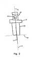
- FIG. 2is an elevation view of the assembled transducer and optical guide depicted in FIG. 1;
- FIG. 3is an elevation view of a second embodiment of an ultrasonic transducer and optical guide constructed according to the principles of the present invention
- FIG. 4is a plan view of the transducer and optical guide depicted in FIG. 3;
- FIG. 5is a pictorial depiction of a guide display embodying the principles of the present invention.
- FIG. 6is a pictorial depiction of a truncated display embodying the principles of the present invention.
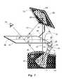
- FIG. 7is a pictorial representation of an ultrasound guidance apparatus constructed according to the principles of the present invention.
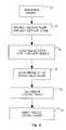
- FIG. 8is a flow chart outlining the manner of use of the device depicted in FIG. 7 .
- the transducer and optical guide assembly 1 of the present inventionis seen to include an ultrasonic transducer 10 capable of transmitting a planar ultrasonic beam into an imaging plane within a human body and receiving the reflected sonic energy for further image processing.
- the transducer 10includes a handle 12 that is gripped the operator's hand during use in order to position the transducer so as to ultrasonically illuminate the area of interest in the patient 13 .
- a cable 14extends from the handle 12 and supplies a path for power and data transmission to and from transducer 10 .
- the transducer 10projects an imaging plane 4 of ultrasonic energy.
- the ultrasound plane or ultrasound imaging plane 4denotes the plane, determined by the pose and geometry of the ultrasound transducer 10 , in which the ultrasound imaging system collects image data.
- a mounting unit 16includes a platform portion 6 connected to a cylindrical portion 18 .
- Mounting unit 16may be an integrally formed unitary piece or it may be fabricated by joining separate parts that form platform portion 6 and cylindrical portion 18 .
- the cylindrical portion 18includes a slot 20 sufficiently wide to allow passage of cable 14 into at least some part of the interior 2 of cylindrical portion 18 .
- Mounting unit 16is placed onto the handle 12 of transducer 10 by slipping the cylindrical portion 18 over and onto the handle 12 . Being essentially in the form of a hollow cylinder adapted to fit over the handle 12 , mounting unit 16 may be made of any convenient and suitable material such as metal or plastic.
- a light mount 24Rigidly affixed to the platform portion 6 is a light mount 24 .
- a light source 22is secured within the mount 24 which permits adjustment of the light source 22 about vertical axis 8 and lateral axis 9 . Suitable adjustment of the mount 24 causes light source 22 to project its beam 3 in a desired direction.
- the light source 22is a laser, such as the laser diode module L54-17, manufactured by the Edmund Scientific Company located at 60 Pearce Avenue, Tonawanda, N.Y. 14150-6711.
- the light source 22is powered by power supply 26 .
- the beam 3 projected by a laserwill be a relatively thin line 5 that defines and is collinear with the longitudinal axis 7 of the light source 22 .
- the light source 22is mounted to platform portion 6 and adjusted so that the projected laser beam 5 is coplanar with the ultrasound imaging plane 4 .
- the operator of the unit 1grips the cylindrical portion 18 and places the surface 11 of transducer 10 against the patient's body 13 .
- the platform portion 6serves as a stop or brace for the operator's hand.
- the transducer 10projects ultrasonic imaging plane 4 into the patient 13 .
- the laser beam 3 that resides within the imaging plane 4is projected onto some portion of the patient's skin and creates a line visible to the operator 32 . Since the operator 32 wishes to place a needle into the patient so that it will reside within the imaging plane 4 , the portion of beam 3 visible on the patient's skin will define a series of points at which the needle should enter the skin.
- the needleitself is manipulated by the user so that it is also aligned with the beam 3 , and therefore with the imaging plane 4 .
- the usercan see this as a reflection of the light beam 3 off the surface of the needle.
- the needleWhen the needle is placed on the appropriate place on the patient's skin, and aligned with the light beam 3 properly, it will stay in the imaging plane 4 of the ultrasonic transducer, and remain visible in the ultrasonic image.
- the needlemay then be inserted to the desired location to take the biopsy sample.
- the mount 16is replaced by a clip 21 which surrounds at least a portion of transducer 10 .
- the lateral surfaces 19 of the clip 21serve as a base for a series of linearly arranged light emitting diodes 17 .
- the diodes 17can be replaced by suitable fiber optics or other light sources that will create a line 3 residing within the imaging plane 4 . As seen in FIG. 4, the line 3 projected by the diodes 17 is collinear with the edge of ultrasonic imaging plane 4 .
- the light source or laser 22is replaced by a video camera.
- a miniature video camerais attached to the transducer via mount 24 and oriented toward the patient 13 .
- the mechanical mount 24would only need to be adapted from the laser diameter to the diameter of the camera.
- the camera's optical axisis collinear with the laser beam 3 of the previous embodiment and hence lies in the ultrasound imaging plane. Referring also to FIG. 5, the ultrasound plane 4 corresponds to a line 15 appearing in the video image 23 .
- the camerais aligned around its optical axis so that the line is oriented either vertically or horizontally within the video image 23 .
- graphical markers 25 and 27are overlaid onto the video image 23 to bracket or define the location of the ultrasound plane line 15 .
- the cameraprovides the user with an aerial view from the transducer head 10 and displays the region on the patient 13 where the needle will be inserted.
- Graphical markers 25 and 27 in the image 23indicate to the user the location of the ultrasound transducer plane 4 which contains line 15 .
- the user 32can readily choose a needle entry point that lies in the ultrasound imaging plane 4 and can further align the entire needle to lie within the plane 4 , by aligning the needle as viewed in the video image 23 with the markers 15 , 25 and 27 overlaid onto the video image 23 .
- the correct in-plane needle alignmentcan be monitored on the video image 23 .
- the video image 23can be displayed on a separate monitor or it can be shown as an inset on the ultrasound system's monitor 29 . If the camera body has a cylindrical shape, the mount 24 may only define the orientation of the camera's vertical axis 8 . In an alignment step, the camera is turned around its axis until ultrasound plane 4 corresponds to vertical or horizontal direction in the video image 23 . Referring also to FIG. 6, there is no need to display the full video image 23 . Rather, the user can choose to display a truncated region of interest 28 that includes the ultrasound plane line 15 .
- One such embodiment of the present inventionis preferably constructed as a handheld device that includes the ultrasound transducer 10 , the handle 16 for gripping the transducer 10 , the light source 22 mounted to the handle 16 , the flat panel monitor or display 29 and a half silvered mirror 30 linked together by mounting elements 44 and 45 .
- the monitor or display 29 , half silvered mirror 30 and transducer 10are mechanically oriented and mounted so that the mirror 30 , which is substantially planar, bisects the angle 31 formed between the transducer image plane 4 and the plane occupied by the display 29 .
- the user 32perceives the reflected image 33 on the mirror 30 , which is the same image 33 residing on the ultrasonic image plane 4 inside the patient 13 that is being scanned by the ultrasonic transducer 10 .
- the user 32When the ultrasound image 33 on the monitor 29 is positioned and scaled to preserve these geometric relationships, the user 32 will perceive the structures 34 and 35 depicted in the ultrasound image 33 as if those structures 34 and 35 were at their actual physical locations inside the patient 13 .
- the image 33 produced by transducer 10is projected along path 43 to the half silvered mirror, creating a composite image to the user 32 such that the image 33 appears as if it were originating along path 36 , path 36 being the line of sight viewed by user 32 when observing patient 13 .
- the pose of the user 32is thus adapted for viewing the patient 13 rather than the monitor 29 .
- Line of sight 36coincides with a direct view of patient 13 . Since the image 33 is an internal view of patient 13 , the user 32 has the illusion of looking through the patient's skin and viewing the underlying region inside patient 13 .
- the optical effect of the half silvered mirror 30depicts the ultrasound image 33 in the location of the actual internal structures 34 , it provides natural or intuitive feedback for guidance of needle placement by the user 32 .
- the needleis inserted towards a target 35 that can be seen in its actual physical location.
- the user 32can easily appreciate whether the needle points towards the target in a lateral sense, it is not so easy to see whether the needle will hit the target at the right depth. This is similar to the task of drilling a vertical hole, where it is relatively easy for the user to adjust the tilt of the drill correctly towards the left and right but it is relatively more difficult to adjust the tilt of the drill towards the front and back, that is, directly toward and away from the user.
- the optical or video guidance provided by laser beam 3 or video line 15helps the user place the needle in the ultrasound plane 4 , while the image overlay of the half silvered mirror 30 provides guidance to user 32 to adjust the tilt of the needle within the ultrasound plane 4 towards the target 35 .
- the video image 23is preferably displayed on the monitor 29 as an inset of the ultrasound image 33 so as to not occlude relevant information.
- the ultrasound scanning systemincludes the flat panel monitor 29 mounted with the ultrasonic transducer 10 and light source 22 .
- the ultrasonic image 33is processed and displayed directly on monitor 29 .
- a second embodimentutilizes a computer connected to the ultrasound scanning system which receives the ultrasound images 33 either in digital or in an analog format such as NTSC or PAL.
- the computeris equipped with a frame grabber or other video capture device. The computer scales and positions the ultrasound images and outputs them to the monitor 29 . If the video guide line 15 is used instead of a laser beam 3 , the video images are also read into the computer to be combined with the ultrasound images 33 for display on the monitor 29 .
- the present devicecan also be combined with a complete augmented reality visualization system, with which the user observes the ultrasound images and other patient data, possibly in image form, in-situ, overlaid onto her view of the patient, registered in a way that structures seen in the ultrasound images appear in the location of the actual anatomical structures.
- a completely integrated systemthe user wears a head-mounted display to watch all of the available augmented images.
- the video image that facilitates the in-plane needle alignmentis preferably shown as an inset in the augmented images.
- the procedure for utilizing the present inventioncan be better understood.
- the user 32searches for the target 35 with the transducer 10 .
- the user 32brings the transducer 10 into a position where the target 35 is visible on the ultrasound image 33 and where the ultrasound plane 4 is appropriate for introducing the needle or other similar instrument into the patient 13 .
- the user 32places the needle tip at an appropriate entry point on the skin surface of patient 13 , guided by the light beam 3 or the video guide line 15 , either of which depicts a line on the surface of the skin. By touching the needle tip to the skin at an appropriate entry point there is still no guarantee that the needle is coplanar with imaging plane 4 . Rather, at the completion of step 39 the user 32 is only assured that the needle tip intersects the imaging plane 4 .
- the useraligns the needle to actually lie within the ultrasound plane 4 , again guided by either the laser beam 3 or the video guide line 15 . This is accomplished by causing the needle to completely overlie or project onto the beam 3 or guide line 15 .
- the user 32tilts the needle within the ultrasound plane 4 so as to point towards the target 35 , guided by the overlaid or superimposed ultrasound image 4 and by monitoring the in-plane alignment with the optical beam 3 or the video guide line 15 .
- the userinserts the needle towards the target 35 .
Landscapes
- Health & Medical Sciences (AREA)
- Life Sciences & Earth Sciences (AREA)
- Surgery (AREA)
- Nuclear Medicine, Radiotherapy & Molecular Imaging (AREA)
- Medical Informatics (AREA)
- Pathology (AREA)
- Veterinary Medicine (AREA)
- Engineering & Computer Science (AREA)
- Biomedical Technology (AREA)
- Heart & Thoracic Surgery (AREA)
- Public Health (AREA)
- Molecular Biology (AREA)
- General Health & Medical Sciences (AREA)
- Animal Behavior & Ethology (AREA)
- Biophysics (AREA)
- Physics & Mathematics (AREA)
- Radiology & Medical Imaging (AREA)
- Ultra Sonic Daignosis Equipment (AREA)
Abstract
Description
The present patent application is based on and claims priority from Provisional U.S. Patent Application No. 60/339,151 of the same title filed on Nov. 28, 2001.
The present application is related to U.S. patent application Ser. No. 10/202,352 [2001P13330 US 01], filed Jul. 24, 2002, and entitled OPTICAL NEEDLE GUIDE FOR ULTRASOUND GUIDED NEEDLE BIOPSY and U.S. patent application Ser. No. 10/222,170 [2001 P 15267 US 01], filed Aug. 16, 2002, and entitled VIDEO-ASSISTANCE FOR ULTRASOUND GUIDED NEEDLE BIOPSY, both of which are incorporated by reference herein.
1. Field of the Invention
This invention relates to generally to a method and apparatus for manipulating needle-like medical instruments such as performing ultrasound guided needle biopsies and similar ultrasound guided procedures.
2. Discussion of the Related Art
Needle-like instruments are often used in medical procedures, and must be manipulated accurately. For example, in a needle biopsy, the needle has to be inserted into an anatomical target to remove a tissue sample. Ultrasound guidance is routinely used for example, when performing breast needle biopsies. The real time ultrasound images allow the physician to locate the target and to monitor the needle position. An example of a typical ultrasound imaging apparatus is disclosed in U.S. Pat. No. 5,503,152, entitled ULTRASOUND TRANSDUCER ASSEMBLY AND METHOD FOR THREE DIMENSIONAL IMAGING, issued on Apr. 2, 1996 to Oakley, et al.
The biopsy procedure is usually performed within the projected ultrasound image plane. With the ultrasound transducer being in a position where the target is visible in the displayed ultrasound image, the insertion point of the needle is ideally chosen so that the point resides along a line residing at the intersection of the projected ultrasound plane and the patient's skin surface. The needle is then preferably oriented so that it lies within this plane and points towards the target. When the needle is inserted it will appear in the ultrasound image, and the progress of the needle along a path towards the target can be visually monitored.
One difficulty inherent with performing an ultrasound guided needle biopsy is to correctly position and orient the needle so that the needle resides within the same plane as the displayed ultrasound image. Mechanical needle guides are commercially available to facilitate this task. They are clipped onto the transducer and constrain the movement of the needle so that it is forced to stay in a plane aligned with the transducer. Examples of such mechanical guides are disclosed in U.S. Pat. No. 5,076,279 entitled NEEDLE GUIDE FOR ASSEMBLY UPON AN ULTRASOUND IMAGING TRANSDUCER, issued to Arenson et al. on Dec. 31, 1991 and U.S. Pat. No. 6,475,152 entitled BIOPSY NEEDLE GUIDE FOR ATTACHMENT TO AN ULTRASOUND TRANSDUCER, issued on Nov. 5, 2002 to Kelly, Jr. et al. Even though the needle can be reliably placed in the plane of the ultrasound image, many physicians find the rigid constraint imposed by the use of a mechanical guide bothersome and consequently do not use one. Physicians typically want to be able to make corrective adjustments to the path of the needle as it approaches the target, which is not easily achieved with the constraints of the mechanical needle guide. In addition, because a mechanical guide constrains the needle entry point to be close to the transducer, it is not possible to insert the needle at the distance from the transducer, as is required for shallow needle angles.
Whether or not the physician is utilizing a mechanical guide, she must look away from the patient at the ultrasound image display and manipulate the needle without direct reference to either her hand or the target. This causes an unnatural eye hand coordination problem that creates additional complications for the physician performing the procedure. Ideally, the physician would be able to look directly at the desired path and destination point of the needle, but the opacity of the human body normally prevents such a view.
One technology that provides a simulated view inside the body during the performance of a needle biopsy is Real Time Tomographic Reflection. A discussion of this technique is provided in REAL TIME TOMOGRAPHIC REFLECTION: PHANTOMS FOR CALIBRATION AND BIOPSY by George Stetten et al., Proceedings IEEE and ACM International Symposium on Augmented Reality, 29-30 October 2001, N.Y. City, N.Y., pages 11-19. In this technique, the ultrasound image is visually merged with the normal exterior view of the patient. The physician's hands and the needle appear in the physician's natural field of view while the biopsy is being performed.
A guide according to principles of the present invention utilizes a light beam or video camera as a guide for placing the needle in the ultrasound plane. In one embodiment, the present invention includes an optical guide for a needle-like instrument comprising an ultrasonic transducer that projects a planar ultrasound beam into an imaging plane beneath a surface and a source of illumination, aligned with the ultrasonic transducer. The source of illumination projects a light beam onto the surface, the light beam being coplanar with the imaging plane. A viewing device, displays an image produced by the ultrasonic transducer which appears to be superimposed on the surface while the light beam is visible on the surface. The line of light on the patient's skin marks the intersection of the ultrasound plane with the patient's skin surface and hence marks the location of possible in-plane entry points for the needle. The user places the tip of the needle on the line of light projected onto the skin. Then the needle is oriented. When the needle is aligned in an in-plane pose, the needle is seen to be illuminated by the light along its length. The image displayed on the viewing device provides guidance to adjust the tilt of the needle within the ultrasound plane towards the target.
In a different embodiment of the present invention, the light beam is replaced by a video camera. An apparatus for video assisted guidance of a biopsy needle comprises means for projecting an ultrasonic imaging plane beneath a surface, means for creating a video image of the surface, and means for inserting graphic markers on the video image of the surface. The inserted graphic markers are substantially coplanar with the ultrasonic imaging plane. By looking at the image, the physician may place the needle on the skin along the superimposed line, and then orient the needle to be co-linear with the line. The viewing device generates a composite image containing the image produced by the ultrasonic transducer and the image of the surface along with the graphic markers.
FIG. 1 is an exploded view of a first embodiment of an ultrasonic transducer and optical guide constructed according to the principles of the present invention;
FIG. 2 is an elevation view of the assembled transducer and optical guide depicted in FIG. 1;
FIG. 3 is an elevation view of a second embodiment of an ultrasonic transducer and optical guide constructed according to the principles of the present invention;
FIG. 4 is a plan view of the transducer and optical guide depicted in FIG. 3;
FIG. 5 is a pictorial depiction of a guide display embodying the principles of the present invention;
FIG. 6 is a pictorial depiction of a truncated display embodying the principles of the present invention;
FIG. 7 is a pictorial representation of an ultrasound guidance apparatus constructed according to the principles of the present invention; and
FIG. 8 is a flow chart outlining the manner of use of the device depicted in FIG.7.
Referring to FIGS. 1,2 and7, the transducer andoptical guide assembly 1 of the present invention is seen to include anultrasonic transducer 10 capable of transmitting a planar ultrasonic beam into an imaging plane within a human body and receiving the reflected sonic energy for further image processing. Thetransducer 10 includes ahandle 12 that is gripped the operator's hand during use in order to position the transducer so as to ultrasonically illuminate the area of interest in thepatient 13. Acable 14 extends from thehandle 12 and supplies a path for power and data transmission to and fromtransducer 10. Thetransducer 10 projects animaging plane 4 of ultrasonic energy. The ultrasound plane orultrasound imaging plane 4 denotes the plane, determined by the pose and geometry of theultrasound transducer 10, in which the ultrasound imaging system collects image data.
A mountingunit 16 includes aplatform portion 6 connected to acylindrical portion 18. Mountingunit 16 may be an integrally formed unitary piece or it may be fabricated by joining separate parts that formplatform portion 6 andcylindrical portion 18. Thecylindrical portion 18 includes aslot 20 sufficiently wide to allow passage ofcable 14 into at least some part of theinterior 2 ofcylindrical portion 18. Mountingunit 16 is placed onto thehandle 12 oftransducer 10 by slipping thecylindrical portion 18 over and onto thehandle 12. Being essentially in the form of a hollow cylinder adapted to fit over thehandle 12, mountingunit 16 may be made of any convenient and suitable material such as metal or plastic.
Rigidly affixed to theplatform portion 6 is alight mount 24. Alight source 22 is secured within themount 24 which permits adjustment of thelight source 22 about vertical axis8 andlateral axis 9. Suitable adjustment of themount 24 causeslight source 22 to project itsbeam 3 in a desired direction. Preferably thelight source 22 is a laser, such as the laser diode module L54-17, manufactured by the Edmund Scientific Company located at 60 Pearce Avenue, Tonawanda, N.Y. 14150-6711. Thelight source 22 is powered bypower supply 26.
Thebeam 3 projected by a laser will be a relativelythin line 5 that defines and is collinear with the longitudinal axis7 of thelight source 22. Thelight source 22 is mounted toplatform portion 6 and adjusted so that the projectedlaser beam 5 is coplanar with theultrasound imaging plane 4. The operator of theunit 1 grips thecylindrical portion 18 and places thesurface 11 oftransducer 10 against the patient'sbody 13. Theplatform portion 6 serves as a stop or brace for the operator's hand. Thetransducer 10 projectsultrasonic imaging plane 4 into thepatient 13. Thelaser beam 3 that resides within theimaging plane 4 is projected onto some portion of the patient's skin and creates a line visible to theoperator 32. Since theoperator 32 wishes to place a needle into the patient so that it will reside within theimaging plane 4, the portion ofbeam 3 visible on the patient's skin will define a series of points at which the needle should enter the skin.
Once the tip of the needle is placed on the patient's skin somewhere along thebeam 3, the needle itself is manipulated by the user so that it is also aligned with thebeam 3, and therefore with theimaging plane 4. The user can see this as a reflection of thelight beam 3 off the surface of the needle. When the needle is placed on the appropriate place on the patient's skin, and aligned with thelight beam 3 properly, it will stay in theimaging plane 4 of the ultrasonic transducer, and remain visible in the ultrasonic image. The needle may then be inserted to the desired location to take the biopsy sample.
Referring to FIG. 3, in one alternate embodiment of the invention, themount 16 is replaced by aclip 21 which surrounds at least a portion oftransducer 10. The lateral surfaces19 of theclip 21 serve as a base for a series of linearly arrangedlight emitting diodes 17. Thediodes 17 can be replaced by suitable fiber optics or other light sources that will create aline 3 residing within theimaging plane 4. As seen in FIG. 4, theline 3 projected by thediodes 17 is collinear with the edge ofultrasonic imaging plane 4.
In another alternate embodiment of the needle guide, the light source orlaser 22 is replaced by a video camera. A miniature video camera is attached to the transducer viamount 24 and oriented toward thepatient 13. In particular, if the camera is of the lipstick variety, that is, has a cylindrical body, themechanical mount 24 would only need to be adapted from the laser diameter to the diameter of the camera. The camera's optical axis is collinear with thelaser beam 3 of the previous embodiment and hence lies in the ultrasound imaging plane. Referring also to FIG. 5, theultrasound plane 4 corresponds to aline 15 appearing in thevideo image 23.
Ideally, the camera is aligned around its optical axis so that the line is oriented either vertically or horizontally within thevideo image 23. Preferably,graphical markers video image 23 to bracket or define the location of theultrasound plane line 15. The camera provides the user with an aerial view from thetransducer head 10 and displays the region on the patient13 where the needle will be inserted.Graphical markers image 23 indicate to the user the location of theultrasound transducer plane 4 which containsline 15.
Theuser 32 can readily choose a needle entry point that lies in theultrasound imaging plane 4 and can further align the entire needle to lie within theplane 4, by aligning the needle as viewed in thevideo image 23 with themarkers video image 23. During needle insertion, the correct in-plane needle alignment can be monitored on thevideo image 23. Thevideo image 23 can be displayed on a separate monitor or it can be shown as an inset on the ultrasound system'smonitor 29. If the camera body has a cylindrical shape, themount 24 may only define the orientation of the camera's vertical axis8. In an alignment step, the camera is turned around its axis untilultrasound plane 4 corresponds to vertical or horizontal direction in thevideo image 23. Referring also to FIG. 6, there is no need to display thefull video image 23. Rather, the user can choose to display a truncated region ofinterest 28 that includes theultrasound plane line 15.
The above embodiments may also be combined with augmented reality systems which would allow the physician to see both thelight beam 3 oroptical image 23 with overlaidmarkers ultrasound transducer 10, thehandle 16 for gripping thetransducer 10, thelight source 22 mounted to thehandle 16, the flat panel monitor ordisplay 29 and a halfsilvered mirror 30 linked together by mountingelements 44 and45. The monitor ordisplay 29, half silveredmirror 30 andtransducer 10 are mechanically oriented and mounted so that themirror 30, which is substantially planar, bisects theangle 31 formed between thetransducer image plane 4 and the plane occupied by thedisplay 29.
As long as theaxis 46 ofmirror 30 is perpendicular to the collinear andequal length lines user 32 perceives the reflectedimage 33 on themirror 30, which is thesame image 33 residing on theultrasonic image plane 4 inside the patient13 that is being scanned by theultrasonic transducer 10.
When theultrasound image 33 on themonitor 29 is positioned and scaled to preserve these geometric relationships, theuser 32 will perceive thestructures ultrasound image 33 as if thosestructures patient 13. In other words, theimage 33 produced bytransducer 10 is projected alongpath 43 to the half silvered mirror, creating a composite image to theuser 32 such that theimage 33 appears as if it were originating alongpath 36,path 36 being the line of sight viewed byuser 32 when observingpatient 13. The pose of theuser 32 is thus adapted for viewing thepatient 13 rather than themonitor 29. Line ofsight 36 coincides with a direct view ofpatient 13. Since theimage 33 is an internal view ofpatient 13, theuser 32 has the illusion of looking through the patient's skin and viewing the underlying region insidepatient 13.
Since the optical effect of the halfsilvered mirror 30 depicts theultrasound image 33 in the location of the actualinternal structures 34, it provides natural or intuitive feedback for guidance of needle placement by theuser 32. The needle is inserted towards atarget 35 that can be seen in its actual physical location. However, though theuser 32 can easily appreciate whether the needle points towards the target in a lateral sense, it is not so easy to see whether the needle will hit the target at the right depth. This is similar to the task of drilling a vertical hole, where it is relatively easy for the user to adjust the tilt of the drill correctly towards the left and right but it is relatively more difficult to adjust the tilt of the drill towards the front and back, that is, directly toward and away from the user. In the present invention, the optical or video guidance provided bylaser beam 3 orvideo line 15 helps the user place the needle in theultrasound plane 4, while the image overlay of the halfsilvered mirror 30 provides guidance touser 32 to adjust the tilt of the needle within theultrasound plane 4 towards thetarget 35.
In case of thevideo guide line 15, thevideo image 23 is preferably displayed on themonitor 29 as an inset of theultrasound image 33 so as to not occlude relevant information.
In one embodiment the ultrasound scanning system includes the flat panel monitor29 mounted with theultrasonic transducer 10 andlight source 22. Theultrasonic image 33 is processed and displayed directly onmonitor 29. However, if such an integrated version is not possible, a second embodiment utilizes a computer connected to the ultrasound scanning system which receives theultrasound images 33 either in digital or in an analog format such as NTSC or PAL. In the latter version, the computer is equipped with a frame grabber or other video capture device. The computer scales and positions the ultrasound images and outputs them to themonitor 29. If thevideo guide line 15 is used instead of alaser beam 3, the video images are also read into the computer to be combined with theultrasound images 33 for display on themonitor 29.
The present device can also be combined with a complete augmented reality visualization system, with which the user observes the ultrasound images and other patient data, possibly in image form, in-situ, overlaid onto her view of the patient, registered in a way that structures seen in the ultrasound images appear in the location of the actual anatomical structures. Preferably, in such a completely integrated system the user wears a head-mounted display to watch all of the available augmented images. In such an augmented reality system the video image that facilitates the in-plane needle alignment is preferably shown as an inset in the augmented images.
Referring also to FIG. 8, the procedure for utilizing the present invention can be better understood. Atstep 37, theuser 32 searches for thetarget 35 with thetransducer 10. Atstep 38, theuser 32 brings thetransducer 10 into a position where thetarget 35 is visible on theultrasound image 33 and where theultrasound plane 4 is appropriate for introducing the needle or other similar instrument into thepatient 13. Atstep 39, theuser 32 places the needle tip at an appropriate entry point on the skin surface ofpatient 13, guided by thelight beam 3 or thevideo guide line 15, either of which depicts a line on the surface of the skin. By touching the needle tip to the skin at an appropriate entry point there is still no guarantee that the needle is coplanar withimaging plane 4. Rather, at the completion ofstep 39 theuser 32 is only assured that the needle tip intersects theimaging plane 4.
Atstep 40, the user aligns the needle to actually lie within theultrasound plane 4, again guided by either thelaser beam 3 or thevideo guide line 15. This is accomplished by causing the needle to completely overlie or project onto thebeam 3 or guideline 15. Atstep 41, theuser 32 tilts the needle within theultrasound plane 4 so as to point towards thetarget 35, guided by the overlaid or superimposedultrasound image 4 and by monitoring the in-plane alignment with theoptical beam 3 or thevideo guide line 15. Finally at step42 the user inserts the needle towards thetarget 35.
Claims (20)
1. A guide for a needle-like instrument comprising:
an ultrasonic transducer that projects a planar ultrasound beam into an ultrasonic imaging plane beneath a surface;
a source of illumination, aligned with the ultrasonic transducer, the source of illumination projecting a light beam onto the surface, the light beam being coplanar with the imaging plane and forming a line of light on the surface that is coincident with the ultrasonic imaging plane; and
a viewing device, causing an image produced by the ultrasonic transducer to appear to be superimposed on the surface while the line of light is visible on the surface.
2. The guide ofclaim 1 , wherein the image appearing on the viewing device is visible to a user of the guide while the user views the actual surface on which the light beam is projected.
3. The guide ofclaim 2 , further comprising means for scaling the image produced by the ultrasonic transducer such that objects appearing in the image appear to have their actual physical dimensions.
4. The guide ofclaim 3 , further comprising a mounting apparatus, the mounting apparatus maintaining required geometric relationships between the source of illumination, the imaging plane and the viewing device.
5. The guide ofclaim 4 , further comprising a handle, a mounting apparatus being affixed to the handle, the handle being adapted to be manipulated by a user of the guide so as to place the ultrasonic transducer in a desired position with respect to the surface.
6. The guide ofclaim 5 , wherein the viewing device further comprises:
a monitor, the monitor displaying the image produced by the ultrasonic transducer; and
a mirror, the mirror redirecting the image to coincide with a line of sight of the user when the user is viewing the surface.
7. The guide ofclaim 6 , wherein the mirror resides in a plane that bisects an angle formed between the imaging plane and a plane in which the monitor resides.
8. The guide ofclaim 7 , wherein the mirror resides in a plane that is equidistant from the imaging plane and the plane in which the monitor resides.
9. The guide ofclaim 1 , wherein the source of illumination is a laser.
10. The guide ofclaim 9 , wherein the laser is rotatable about a lateral axis.
11. The guide ofclaim 1 , wherein the source of illumination is formed as a series of light emitting diodes arranged so as to reside within the imaging plane.
12. The guide ofclaim 11 , wherein the light emitting diodes are arranged in two rows positioned on opposing sides of the ultrasonic transducer.
13. An apparatus for video assisted guidance of a needle-like instrument comprising:
means for projecting a planar ultrasound beam into an ultrasonic imaging plane beneath a surface;
means for creating a video image of the surface;
means for inserting graphic markers on the video image of the surface, the graphic markers being substantially coplanar and coincident with the ultrasonic imaging plane and indicating a line of potential entry points for a needle biopsy; and
means for simultaneously viewing an image produced by the ultrasonic transducer with the video image of the surface.
14. The apparatus ofclaim 13 , wherein the image produced by the ultrasonic transducer is superimposed on the video image of the surface.
15. The apparatus ofclaim 14 , further comprising means for reorienting the graphic markers appearing in the video image.
16. A method of inserting an instrument into a comprising the steps of:
projecting a planar ultrasound beam into an ultrasonic imaging plane into the body;
projecting a linear beam of light onto a surface of the body such that a line of light is formed on the surface and is coincident with the ultrasonic imaging plane;
deriving image information from the ultrasound transducer;
superimposing derived ultrasonic image information onto a view of the line of light appearing on the surface of the body so as to create a composite image; and
viewing the composite image while inserting instrument into the body.
17. The method ofclaim 16 , further comprising the steps of:
moving the ultrasonic transducer on the surface of the body until a desired object is visible in the derived ultrasonic image; and
orienting the ultrasonic transducer such that the line of light appears at a desired location.
18. The method ofclaim 16 , further comprising the steps of:
placing an insertable portion of the instrument on a point contained in the line of light appearing on the surface; and
aligning the insertable portion of the instrument with the line of light, thereby insuring that the insertable portion of the instrument is coplanar with the imaging plane.
19. The method ofclaim 18 , further comprising the steps of:
tilting the insertable portion of the instrument so that the insertable portion is pointed at the desired object; and
advancing the insertable portion of the instrument toward the desired object.
20. The method ofclaim 18 wherein said step of aligning the insertable portion of the instrument further comprises the step of:
aligning the instrument with the line of light such that the instrument is illuminated along its length by the linear beam of light.
Priority Applications (1)
| Application Number | Priority Date | Filing Date | Title |
|---|---|---|---|
| US10/304,427US6689067B2 (en) | 2001-11-28 | 2002-11-26 | Method and apparatus for ultrasound guidance of needle biopsies |
Applications Claiming Priority (2)
| Application Number | Priority Date | Filing Date | Title |
|---|---|---|---|
| US33915101P | 2001-11-28 | 2001-11-28 | |
| US10/304,427US6689067B2 (en) | 2001-11-28 | 2002-11-26 | Method and apparatus for ultrasound guidance of needle biopsies |
Publications (2)
| Publication Number | Publication Date |
|---|---|
| US20030120154A1 US20030120154A1 (en) | 2003-06-26 |
| US6689067B2true US6689067B2 (en) | 2004-02-10 |
Family
ID=26974016
Family Applications (1)
| Application Number | Title | Priority Date | Filing Date |
|---|---|---|---|
| US10/304,427Expired - LifetimeUS6689067B2 (en) | 2001-11-28 | 2002-11-26 | Method and apparatus for ultrasound guidance of needle biopsies |
Country Status (1)
| Country | Link |
|---|---|
| US (1) | US6689067B2 (en) |
Cited By (54)
| Publication number | Priority date | Publication date | Assignee | Title |
|---|---|---|---|---|
| WO2005025424A1 (en)* | 2003-09-12 | 2005-03-24 | Austin Health | Ultrasound apparatus |
| US20050240102A1 (en)* | 2002-07-12 | 2005-10-27 | Daniel Rachlin | Ultrasound interfacing device for tissue imaging |
| US20060285635A1 (en)* | 2005-04-15 | 2006-12-21 | Boppart Stephen A | Contrast enhanced spectroscopic optical coherence tomography |
| US20070073155A1 (en)* | 2005-09-02 | 2007-03-29 | Ultrasound Ventures, Llc | Ultrasound guidance system |
| US20070203404A1 (en)* | 2006-01-31 | 2007-08-30 | Zysk Adam M | Method and apparatus for measurement of optical properties in tissue |
| US20070276241A1 (en)* | 2006-05-26 | 2007-11-29 | Ultrasound Ventures, Llc | Sterile cover |
| US20070276253A1 (en)* | 2006-05-26 | 2007-11-29 | Ultrasound Ventures, Llc | Needle guide |
| US20070293787A1 (en)* | 2003-08-13 | 2007-12-20 | Taylor James D | Targeted biopsy delivery system |
| US20080030578A1 (en)* | 2006-08-02 | 2008-02-07 | Inneroptic Technology Inc. | System and method of providing real-time dynamic imagery of a medical procedure site using multiple modalities |
| US20090024039A1 (en)* | 2006-05-02 | 2009-01-22 | U-Systems, Inc. | Handheld volumetric ultrasound scanning device |
| US20090171192A1 (en)* | 2007-12-21 | 2009-07-02 | Carticept Medical, Inc. | Method of injecting fluids into multiple patients |
| US20090300537A1 (en)* | 2008-05-27 | 2009-12-03 | Park Kenneth J | Method and system for changing format for displaying information on handheld device |
| US20090312629A1 (en)* | 2008-06-13 | 2009-12-17 | Inneroptic Technology Inc. | Correction of relative tracking errors based on a fiducial |
| US20100045783A1 (en)* | 2001-10-19 | 2010-02-25 | Andrei State | Methods and systems for dynamic virtual convergence and head mountable display using same |
| US20100081965A1 (en)* | 2008-10-01 | 2010-04-01 | John Mugan | Needle biopsy device |
| US20100106015A1 (en)* | 2008-10-23 | 2010-04-29 | Norris Perry R | Medical device alignment |
| US20100106056A1 (en)* | 2008-10-23 | 2010-04-29 | Norris Perry R | Methods for medical device alignment |
| US20100121218A1 (en)* | 2008-10-01 | 2010-05-13 | Boston Endoscopic Engineering Corporation | Device for needle biopsy with integrated needle protection |
| US7751057B2 (en) | 2008-01-18 | 2010-07-06 | The Board Of Trustees Of The University Of Illinois | Magnetomotive optical coherence tomography |
| US20100268067A1 (en)* | 2009-02-17 | 2010-10-21 | Inneroptic Technology Inc. | Systems, methods, apparatuses, and computer-readable media for image guided surgery |
| US20110021905A1 (en)* | 2007-12-21 | 2011-01-27 | Carticept Medical, Inc. | Injection system for delivering multiple fluids within the anatomy |
| US20110046483A1 (en)* | 2008-01-24 | 2011-02-24 | Henry Fuchs | Methods, systems, and computer readable media for image guided ablation |
| US20110043612A1 (en)* | 2009-07-31 | 2011-02-24 | Inneroptic Technology Inc. | Dual-tube stereoscope |
| US20110057930A1 (en)* | 2006-07-26 | 2011-03-10 | Inneroptic Technology Inc. | System and method of using high-speed, high-resolution depth extraction to provide three-dimensional imagery for endoscopy |
| US20110082351A1 (en)* | 2009-10-07 | 2011-04-07 | Inneroptic Technology, Inc. | Representing measurement information during a medical procedure |
| US20110087105A1 (en)* | 2009-10-09 | 2011-04-14 | Soma Development, Llc | Ultrasound Guided Probe Device and Sterilizable Shield for Same |
| US20110137156A1 (en)* | 2009-02-17 | 2011-06-09 | Inneroptic Technology, Inc. | Systems, methods, apparatuses, and computer-readable media for image management in image-guided medical procedures |
| US8115934B2 (en) | 2008-01-18 | 2012-02-14 | The Board Of Trustees Of The University Of Illinois | Device and method for imaging the ear using optical coherence tomography |
| US8340379B2 (en) | 2008-03-07 | 2012-12-25 | Inneroptic Technology, Inc. | Systems and methods for displaying guidance data based on updated deformable imaging data |
| US8554307B2 (en) | 2010-04-12 | 2013-10-08 | Inneroptic Technology, Inc. | Image annotation in image-guided medical procedures |
| US8670816B2 (en) | 2012-01-30 | 2014-03-11 | Inneroptic Technology, Inc. | Multiple medical device guidance |
| US8758256B2 (en) | 2010-07-12 | 2014-06-24 | Best Medical International, Inc. | Apparatus for brachytherapy that uses a scanning probe for treatment of malignant tissue |
| US8983580B2 (en) | 2008-01-18 | 2015-03-17 | The Board Of Trustees Of The University Of Illinois | Low-coherence interferometry and optical coherence tomography for image-guided surgical treatment of solid tumors |
| US9044216B2 (en) | 2010-07-12 | 2015-06-02 | Best Medical International, Inc. | Biopsy needle assembly |
| US9044542B2 (en) | 2007-12-21 | 2015-06-02 | Carticept Medical, Inc. | Imaging-guided anesthesia injection systems and methods |
| CN105232120A (en)* | 2015-10-22 | 2016-01-13 | 张旭 | Ultrasonic guided in-plane puncture guide apparatus |
| US9282947B2 (en) | 2009-12-01 | 2016-03-15 | Inneroptic Technology, Inc. | Imager focusing based on intraoperative data |
| US9332973B2 (en) | 2008-10-01 | 2016-05-10 | Covidien Lp | Needle biopsy device with exchangeable needle and integrated needle protection |
| US9675319B1 (en) | 2016-02-17 | 2017-06-13 | Inneroptic Technology, Inc. | Loupe display |
| US9782565B2 (en) | 2008-10-01 | 2017-10-10 | Covidien Lp | Endoscopic ultrasound-guided biliary access system |
| US9901406B2 (en) | 2014-10-02 | 2018-02-27 | Inneroptic Technology, Inc. | Affected region display associated with a medical device |
| US9949700B2 (en) | 2015-07-22 | 2018-04-24 | Inneroptic Technology, Inc. | Medical device approaches |
| US10188467B2 (en) | 2014-12-12 | 2019-01-29 | Inneroptic Technology, Inc. | Surgical guidance intersection display |
| US10238363B2 (en) | 2014-08-21 | 2019-03-26 | Richard D. Striano | Needle guide for ultrasound transducer |
| US10278778B2 (en) | 2016-10-27 | 2019-05-07 | Inneroptic Technology, Inc. | Medical device navigation using a virtual 3D space |
| US10314559B2 (en) | 2013-03-14 | 2019-06-11 | Inneroptic Technology, Inc. | Medical device guidance |
| US10667789B2 (en) | 2017-10-11 | 2020-06-02 | Geoffrey Steven Hastings | Laser assisted ultrasound guidance |
| US10786224B2 (en) | 2016-04-21 | 2020-09-29 | Covidien Lp | Biopsy devices and methods of use thereof |
| US11259879B2 (en) | 2017-08-01 | 2022-03-01 | Inneroptic Technology, Inc. | Selective transparency to assist medical device navigation |
| US11298113B2 (en) | 2008-10-01 | 2022-04-12 | Covidien Lp | Device for needle biopsy with integrated needle protection |
| US11331161B2 (en) | 2018-03-23 | 2022-05-17 | Covidien Lp | Surgical assemblies facilitating tissue marking and methods of use thereof |
| US11464578B2 (en) | 2009-02-17 | 2022-10-11 | Inneroptic Technology, Inc. | Systems, methods, apparatuses, and computer-readable media for image management in image-guided medical procedures |
| US11484365B2 (en) | 2018-01-23 | 2022-11-01 | Inneroptic Technology, Inc. | Medical image guidance |
| US11517294B2 (en) | 2019-05-07 | 2022-12-06 | Covidien Lp | Biopsy devices and methods of use thereof |
Families Citing this family (95)
| Publication number | Priority date | Publication date | Assignee | Title |
|---|---|---|---|---|
| US6702749B2 (en)* | 2001-07-24 | 2004-03-09 | Siemens Corporate Research, Inc. | Optical needle guide for ultrasound guided needle biopsy |
| US6918892B2 (en)* | 2003-04-23 | 2005-07-19 | Howard Martin | Intraosseous needle |
| US7244234B2 (en)* | 2003-11-11 | 2007-07-17 | Soma Development Llc | Ultrasound guided probe device and method of using same |
| GB0511520D0 (en)* | 2005-06-07 | 2005-07-13 | Smiths Group Plc | Tracheostomy instruments |
| US8784336B2 (en) | 2005-08-24 | 2014-07-22 | C. R. Bard, Inc. | Stylet apparatuses and methods of manufacture |
| US8478386B2 (en) | 2006-01-10 | 2013-07-02 | Accuvein Inc. | Practitioner-mounted micro vein enhancer |
| US12408865B2 (en) | 2006-01-10 | 2025-09-09 | Accuvein Inc. | Vein imaging device with differential image resolution at the center and the extremities of the vein image |
| US9492117B2 (en) | 2006-01-10 | 2016-11-15 | Accuvein, Inc. | Practitioner-mounted micro vein enhancer |
| US12295744B2 (en) | 2006-01-10 | 2025-05-13 | Accuvein, Inc. | Micro vein enhancer with two lasers and two optical detectors configured for removing surface topology |
| US11278240B2 (en) | 2006-01-10 | 2022-03-22 | Accuvein, Inc. | Trigger-actuated laser vein contrast enhancer |
| US8489178B2 (en) | 2006-06-29 | 2013-07-16 | Accuvein Inc. | Enhanced laser vein contrast enhancer with projection of analyzed vein data |
| US8838210B2 (en) | 2006-06-29 | 2014-09-16 | AccuView, Inc. | Scanned laser vein contrast enhancer using a single laser |
| US12089951B2 (en) | 2006-01-10 | 2024-09-17 | AccuVeiw, Inc. | Scanned laser vein contrast enhancer with scanning correlated to target distance |
| US10238294B2 (en) | 2006-06-29 | 2019-03-26 | Accuvein, Inc. | Scanned laser vein contrast enhancer using one laser |
| US11253198B2 (en) | 2006-01-10 | 2022-02-22 | Accuvein, Inc. | Stand-mounted scanned laser vein contrast enhancer |
| US10813588B2 (en) | 2006-01-10 | 2020-10-27 | Accuvein, Inc. | Micro vein enhancer |
| US9854977B2 (en) | 2006-01-10 | 2018-01-02 | Accuvein, Inc. | Scanned laser vein contrast enhancer using a single laser, and modulation circuitry |
| US8730321B2 (en) | 2007-06-28 | 2014-05-20 | Accuvein, Inc. | Automatic alignment of a contrast enhancement system |
| US8463364B2 (en) | 2009-07-22 | 2013-06-11 | Accuvein Inc. | Vein scanner |
| US8594770B2 (en) | 2006-06-29 | 2013-11-26 | Accuvein, Inc. | Multispectral detection and presentation of an object's characteristics |
| DE102006035292B4 (en)* | 2006-07-26 | 2010-08-19 | Deutsches Zentrum für Luft- und Raumfahrt e.V. | Method and system for transferring position-related information from a virtual to an actual reality and for displaying this information in the actual reality and use of such a system |
| EP2079358B1 (en)* | 2006-09-27 | 2011-08-10 | University of Connecticut | Implantable biosensor and methods of use thereof |
| US7794407B2 (en) | 2006-10-23 | 2010-09-14 | Bard Access Systems, Inc. | Method of locating the tip of a central venous catheter |
| US8388546B2 (en) | 2006-10-23 | 2013-03-05 | Bard Access Systems, Inc. | Method of locating the tip of a central venous catheter |
| US8781555B2 (en) | 2007-11-26 | 2014-07-15 | C. R. Bard, Inc. | System for placement of a catheter including a signal-generating stylet |
| US9649048B2 (en) | 2007-11-26 | 2017-05-16 | C. R. Bard, Inc. | Systems and methods for breaching a sterile field for intravascular placement of a catheter |
| US10524691B2 (en) | 2007-11-26 | 2020-01-07 | C. R. Bard, Inc. | Needle assembly including an aligned magnetic element |
| US10751509B2 (en) | 2007-11-26 | 2020-08-25 | C. R. Bard, Inc. | Iconic representations for guidance of an indwelling medical device |
| ES2465915T3 (en) | 2007-11-26 | 2014-06-09 | C.R. Bard, Inc. | Integrated system for intravascular catheter placement |
| US8849382B2 (en) | 2007-11-26 | 2014-09-30 | C. R. Bard, Inc. | Apparatus and display methods relating to intravascular placement of a catheter |
| US10449330B2 (en) | 2007-11-26 | 2019-10-22 | C. R. Bard, Inc. | Magnetic element-equipped needle assemblies |
| US9636031B2 (en) | 2007-11-26 | 2017-05-02 | C.R. Bard, Inc. | Stylets for use with apparatus for intravascular placement of a catheter |
| US9521961B2 (en) | 2007-11-26 | 2016-12-20 | C. R. Bard, Inc. | Systems and methods for guiding a medical instrument |
| US8478382B2 (en) | 2008-02-11 | 2013-07-02 | C. R. Bard, Inc. | Systems and methods for positioning a catheter |
| US20090247876A1 (en)* | 2008-03-28 | 2009-10-01 | Cannon Jr Charles W | Laparoscopic probe guidance system |
| US9901714B2 (en) | 2008-08-22 | 2018-02-27 | C. R. Bard, Inc. | Catheter assembly including ECG sensor and magnetic assemblies |
| US8437833B2 (en) | 2008-10-07 | 2013-05-07 | Bard Access Systems, Inc. | Percutaneous magnetic gastrostomy |
| EP2413829A1 (en)* | 2009-04-03 | 2012-02-08 | Deutsches Krebsforschungszentrum, Stiftung des öffentlichen Rechts | System and computer assisted surgery |
| US9532724B2 (en) | 2009-06-12 | 2017-01-03 | Bard Access Systems, Inc. | Apparatus and method for catheter navigation using endovascular energy mapping |
| JP5795576B2 (en) | 2009-06-12 | 2015-10-14 | バード・アクセス・システムズ,インコーポレーテッド | Method of operating a computer-based medical device that uses an electrocardiogram (ECG) signal to position an intravascular device in or near the heart |
| US9061109B2 (en) | 2009-07-22 | 2015-06-23 | Accuvein, Inc. | Vein scanner with user interface |
| EP2464407A4 (en) | 2009-08-10 | 2014-04-02 | Bard Access Systems Inc | Devices and methods for endovascular electrography |
| WO2011044421A1 (en) | 2009-10-08 | 2011-04-14 | C. R. Bard, Inc. | Spacers for use with an ultrasound probe |
| WO2011097312A1 (en) | 2010-02-02 | 2011-08-11 | C.R. Bard, Inc. | Apparatus and method for catheter navigation and tip location |
| EP4122385A1 (en)* | 2010-05-28 | 2023-01-25 | C. R. Bard, Inc. | Insertion guidance system for needles and medical components |
| EP2912999B1 (en) | 2010-05-28 | 2022-06-29 | C. R. Bard, Inc. | Apparatus for use with needle insertion guidance system |
| CN103228219B (en) | 2010-08-09 | 2016-04-27 | C·R·巴德股份有限公司 | Support and Covering Structures for Ultrasound Probe Heads |
| BR112013002431B1 (en) | 2010-08-20 | 2021-06-29 | C.R. Bard, Inc | SYSTEM FOR RECONFIRMING THE POSITION OF A CATHETER INSIDE A PATIENT |
| US8801693B2 (en) | 2010-10-29 | 2014-08-12 | C. R. Bard, Inc. | Bioimpedance-assisted placement of a medical device |
| RU2609203C2 (en) | 2011-07-06 | 2017-01-30 | Си.Ар. Бард, Инк. | Determination and calibration of needle length for needle guidance system |
| USD724745S1 (en) | 2011-08-09 | 2015-03-17 | C. R. Bard, Inc. | Cap for an ultrasound probe |
| USD699359S1 (en) | 2011-08-09 | 2014-02-11 | C. R. Bard, Inc. | Ultrasound probe head |
| US9211107B2 (en) | 2011-11-07 | 2015-12-15 | C. R. Bard, Inc. | Ruggedized ultrasound hydrogel insert |
| DE102012002412A1 (en)* | 2012-02-09 | 2013-08-14 | Bernd Meier | Device for determination of position of puncture needle in workspace of ultrasound probe for human body, has processor unit introducing stitch projection and penetration depth of needle together with image for representation of projection |
| EP2861153A4 (en) | 2012-06-15 | 2016-10-19 | Bard Inc C R | Apparatus and methods for detection of a removable cap on an ultrasound probe |
| US9072426B2 (en) | 2012-08-02 | 2015-07-07 | AccuVein, Inc | Device for detecting and illuminating vasculature using an FPGA |
| US10517483B2 (en) | 2012-12-05 | 2019-12-31 | Accuvein, Inc. | System for detecting fluorescence and projecting a representative image |
| CN203289635U (en) | 2013-05-10 | 2013-11-13 | 瑞声声学科技(深圳)有限公司 | Spring plate and multifunctional sounder applying spring plate |
| EP3003181B1 (en) | 2013-06-03 | 2018-02-21 | Faculty Physicians and Surgeons of Loma Linda University School of Medicine | Apparatuses for fluoro- less or near fluoro-less percutaneous surgery access |
| US10792067B2 (en)* | 2013-06-03 | 2020-10-06 | Faculty Physicians And Surgeons Of Loma Linda University Of Medicine | Methods and apparatuses for fluoro-less or near fluoro-less percutaneous surgery access |
| WO2015120256A2 (en) | 2014-02-06 | 2015-08-13 | C.R. Bard, Inc. | Systems and methods for guidance and placement of an intravascular device |
| US10973584B2 (en) | 2015-01-19 | 2021-04-13 | Bard Access Systems, Inc. | Device and method for vascular access |
| WO2016210325A1 (en) | 2015-06-26 | 2016-12-29 | C.R. Bard, Inc. | Connector interface for ecg-based catheter positioning system |
| EP3344146B1 (en)* | 2015-08-31 | 2020-05-06 | Buljubasic, Neda | Systems and methods for providing ultrasound guidance to target structures within a body |
| EP3352834A4 (en) | 2015-09-22 | 2019-05-08 | Faculty Physicians and Surgeons of Loma Linda University School of Medicine | Kit and method for reduced radiation procedures |
| CA3050807A1 (en)* | 2016-01-20 | 2017-07-27 | Loughborough University | Needle guides |
| US11000207B2 (en) | 2016-01-29 | 2021-05-11 | C. R. Bard, Inc. | Multiple coil system for tracking a medical device |
| US20200118465A1 (en)* | 2017-04-17 | 2020-04-16 | University Of South Carolina | Laser Line Directional System for 3D Anatomy Ultrasound Phantom Trainer |
| CN109247910B (en)* | 2017-07-12 | 2020-12-15 | 京东方科技集团股份有限公司 | Blood vessel display device and blood vessel display method |
| US10992079B2 (en) | 2018-10-16 | 2021-04-27 | Bard Access Systems, Inc. | Safety-equipped connection systems and methods thereof for establishing electrical connections |
| US11759166B2 (en) | 2019-09-20 | 2023-09-19 | Bard Access Systems, Inc. | Automatic vessel detection tools and methods |
| US11877810B2 (en) | 2020-07-21 | 2024-01-23 | Bard Access Systems, Inc. | System, method and apparatus for magnetic tracking of ultrasound probe and generation of 3D visualization thereof |
| EP4185209A1 (en) | 2020-08-04 | 2023-05-31 | Bard Access Systems, Inc. | System and method for optimized medical component insertion monitoring and imaging enhancement |
| WO2022035760A1 (en) | 2020-08-10 | 2022-02-17 | Bard Access Systems, Inc. | System and method for generating vessel representations in mixed reality/virtual reality |
| US11890139B2 (en) | 2020-09-03 | 2024-02-06 | Bard Access Systems, Inc. | Portable ultrasound systems |
| US11992363B2 (en) | 2020-09-08 | 2024-05-28 | Bard Access Systems, Inc. | Dynamically adjusting ultrasound-imaging systems and methods thereof |
| CN216257185U (en) | 2020-09-10 | 2022-04-12 | 巴德阿克塞斯系统股份有限公司 | Ultrasound Probes and Ultrasound Systems |
| CN112022300A (en)* | 2020-09-23 | 2020-12-04 | 南京康友医疗科技有限公司 | Auxiliary puncture device and medical equipment |
| WO2022067101A1 (en) | 2020-09-25 | 2022-03-31 | Bard Access Systems, Inc. | Minimum catheter length tool |
| WO2022072727A2 (en)* | 2020-10-02 | 2022-04-07 | Bard Access Systems, Inc. | Ultrasound systems and methods for sustained spatial attention |
| EP4228516A1 (en) | 2020-10-15 | 2023-08-23 | Bard Access Systems, Inc. | Ultrasound imaging system for generation of a three-dimensional ultrasound image |
| CN216933458U (en) | 2020-11-24 | 2022-07-12 | 巴德阿克塞斯系统股份有限公司 | Object recognition and needle guidance system |
| CN114569156A (en) | 2020-12-01 | 2022-06-03 | 巴德阿克塞斯系统股份有限公司 | Ultrasound imaging system and method for identifying one or more of a plurality of blood vessels |
| CN114569155A (en) | 2020-12-01 | 2022-06-03 | 巴德阿克塞斯系统股份有限公司 | Ultrasound imaging system and method for obtaining ultrasound image by the same |
| EP4258997A1 (en) | 2020-12-14 | 2023-10-18 | Bard Access Systems, Inc. | Securement of hands-free ultrasound probe |
| US12349983B2 (en) | 2021-03-05 | 2025-07-08 | Bard Access Systems, Inc. | Systems and methods for ultrasound-and-bioimpedance-based guidance of medical devices |
| CN217960146U (en) | 2021-04-15 | 2022-12-06 | 巴德阿克塞斯系统股份有限公司 | Ultrasound imaging system |
| CN218419895U (en)* | 2021-06-22 | 2023-02-03 | 巴德阿克塞斯系统股份有限公司 | Ultrasound imaging system configured to guide insertion of medical device |
| CN116058873A (en) | 2021-11-03 | 2023-05-05 | 巴德阿克塞斯系统股份有限公司 | Interoperation optimization function through Doppler and image-based vessel discrimination |
| US12433567B2 (en) | 2022-03-16 | 2025-10-07 | Bard Access Systems, Inc. | Ultrasound imaging system |
| US12207967B2 (en) | 2022-04-20 | 2025-01-28 | Bard Access Systems, Inc. | Ultrasound imaging system |
| US12102481B2 (en) | 2022-06-03 | 2024-10-01 | Bard Access Systems, Inc. | Ultrasound probe with smart accessory |
| US12137989B2 (en) | 2022-07-08 | 2024-11-12 | Bard Access Systems, Inc. | Systems and methods for intelligent ultrasound probe guidance |
| JP2024060296A (en)* | 2022-10-19 | 2024-05-02 | 富士フイルム株式会社 | Ultrasonic diagnostic device and control method of ultrasonic diagnostic device |
| EP4527324A1 (en)* | 2023-09-25 | 2025-03-26 | Atlas Medical Technologies GmbH | Puncture guide unit and computer program |
Citations (17)
| Publication number | Priority date | Publication date | Assignee | Title |
|---|---|---|---|---|
| US4484569A (en)* | 1981-03-13 | 1984-11-27 | Riverside Research Institute | Ultrasonic diagnostic and therapeutic transducer assembly and method for using |
| US4527569A (en)* | 1982-11-26 | 1985-07-09 | South African Inventions Develop. Corp. | Device for guiding a surgical needle into a blood vessel |
| US4651732A (en)* | 1983-03-17 | 1987-03-24 | Frederick Philip R | Three-dimensional light guidance system for invasive procedures |
| US4763662A (en)* | 1985-06-07 | 1988-08-16 | Olympus Optical Co., Ltd. | Ultrasonic biopsy endoscope with extensible guide sheath |
| US4848569A (en)* | 1988-09-20 | 1989-07-18 | Leishman Layne S | Apparatus and method for disposing of contaminated needles |
| US4932414A (en)* | 1987-11-02 | 1990-06-12 | Cornell Research Foundation, Inc. | System of therapeutic ultrasound and real-time ultrasonic scanning |
| US5056917A (en)* | 1990-04-02 | 1991-10-15 | Christopher Nowacki | Lithotripter reflector inspection |
| US5316014A (en)* | 1992-02-07 | 1994-05-31 | Livingston Products, Inc. | Biopsy locator and guide |
| US5647373A (en)* | 1993-11-07 | 1997-07-15 | Ultra-Guide Ltd. | Articulated needle guide for ultrasound imaging and method of using same |
| US5810841A (en)* | 1997-01-22 | 1998-09-22 | Minrad Inc. | Energy guided apparatus and method with indication of alignment |
| US6030348A (en)* | 1997-01-27 | 2000-02-29 | Imarx Pharmaceutical Corp. | Leveling device especially adapted for use in apparatus for performing light beam guided biopsies and the like |
| US6096049A (en)* | 1998-07-27 | 2000-08-01 | Minrad Inc. | Light guiding device and method |
| US6110112A (en)* | 1998-03-06 | 2000-08-29 | Siemens Aktiengesellschaft | Medical guide apparatus for breath-coordinated puncturing of the body or a body cavity |
| US6146390A (en)* | 1992-04-21 | 2000-11-14 | Sofamor Danek Holdings, Inc. | Apparatus and method for photogrammetric surgical localization |
| US6206890B1 (en)* | 1997-05-15 | 2001-03-27 | Regents Of The University Of Minnesota | Remote actuation of trajectory guide |
| US6216029B1 (en)* | 1995-07-16 | 2001-04-10 | Ultraguide Ltd. | Free-hand aiming of a needle guide |
| US20030038112A1 (en)* | 2000-03-30 | 2003-02-27 | Lianjun Liu | Optical monitoring and control system and method for plasma reactors |
Family Cites Families (1)
| Publication number | Priority date | Publication date | Assignee | Title |
|---|---|---|---|---|
| US6702749B2 (en)* | 2001-07-24 | 2004-03-09 | Siemens Corporate Research, Inc. | Optical needle guide for ultrasound guided needle biopsy |
- 2002
- 2002-11-26USUS10/304,427patent/US6689067B2/ennot_activeExpired - Lifetime
Patent Citations (17)
| Publication number | Priority date | Publication date | Assignee | Title |
|---|---|---|---|---|
| US4484569A (en)* | 1981-03-13 | 1984-11-27 | Riverside Research Institute | Ultrasonic diagnostic and therapeutic transducer assembly and method for using |
| US4527569A (en)* | 1982-11-26 | 1985-07-09 | South African Inventions Develop. Corp. | Device for guiding a surgical needle into a blood vessel |
| US4651732A (en)* | 1983-03-17 | 1987-03-24 | Frederick Philip R | Three-dimensional light guidance system for invasive procedures |
| US4763662A (en)* | 1985-06-07 | 1988-08-16 | Olympus Optical Co., Ltd. | Ultrasonic biopsy endoscope with extensible guide sheath |
| US4932414A (en)* | 1987-11-02 | 1990-06-12 | Cornell Research Foundation, Inc. | System of therapeutic ultrasound and real-time ultrasonic scanning |
| US4848569A (en)* | 1988-09-20 | 1989-07-18 | Leishman Layne S | Apparatus and method for disposing of contaminated needles |
| US5056917A (en)* | 1990-04-02 | 1991-10-15 | Christopher Nowacki | Lithotripter reflector inspection |
| US5316014A (en)* | 1992-02-07 | 1994-05-31 | Livingston Products, Inc. | Biopsy locator and guide |
| US6146390A (en)* | 1992-04-21 | 2000-11-14 | Sofamor Danek Holdings, Inc. | Apparatus and method for photogrammetric surgical localization |
| US5647373A (en)* | 1993-11-07 | 1997-07-15 | Ultra-Guide Ltd. | Articulated needle guide for ultrasound imaging and method of using same |
| US6216029B1 (en)* | 1995-07-16 | 2001-04-10 | Ultraguide Ltd. | Free-hand aiming of a needle guide |
| US5810841A (en)* | 1997-01-22 | 1998-09-22 | Minrad Inc. | Energy guided apparatus and method with indication of alignment |
| US6030348A (en)* | 1997-01-27 | 2000-02-29 | Imarx Pharmaceutical Corp. | Leveling device especially adapted for use in apparatus for performing light beam guided biopsies and the like |
| US6206890B1 (en)* | 1997-05-15 | 2001-03-27 | Regents Of The University Of Minnesota | Remote actuation of trajectory guide |
| US6110112A (en)* | 1998-03-06 | 2000-08-29 | Siemens Aktiengesellschaft | Medical guide apparatus for breath-coordinated puncturing of the body or a body cavity |
| US6096049A (en)* | 1998-07-27 | 2000-08-01 | Minrad Inc. | Light guiding device and method |
| US20030038112A1 (en)* | 2000-03-30 | 2003-02-27 | Lianjun Liu | Optical monitoring and control system and method for plasma reactors |
Cited By (123)
| Publication number | Priority date | Publication date | Assignee | Title |
|---|---|---|---|---|
| US20100045783A1 (en)* | 2001-10-19 | 2010-02-25 | Andrei State | Methods and systems for dynamic virtual convergence and head mountable display using same |
| US20050240102A1 (en)* | 2002-07-12 | 2005-10-27 | Daniel Rachlin | Ultrasound interfacing device for tissue imaging |
| US7931596B2 (en)* | 2002-07-12 | 2011-04-26 | Iscience Interventional Corporation | Ultrasound interfacing device for tissue imaging |
| US8317724B2 (en) | 2003-08-13 | 2012-11-27 | Envisioneering, Llc | Targeted treatment delivery system |
| US20110144492A1 (en)* | 2003-08-13 | 2011-06-16 | Taylor James D | Targeted Treatment Delivery System |
| US20090054807A1 (en)* | 2003-08-13 | 2009-02-26 | Taylor James D | Targeted biopsy delivery system |
| US7833168B2 (en) | 2003-08-13 | 2010-11-16 | Envisioneering Medical Technologies, Llc | Targeted biopsy delivery system |
| US20070293787A1 (en)* | 2003-08-13 | 2007-12-20 | Taylor James D | Targeted biopsy delivery system |
| WO2005025424A1 (en)* | 2003-09-12 | 2005-03-24 | Austin Health | Ultrasound apparatus |
| US20100030082A1 (en)* | 2003-09-12 | 2010-02-04 | Austin Health | Ultrasound Apparatus |
| US20080027325A1 (en)* | 2003-09-12 | 2008-01-31 | Austin Health | Ultrasound Apparatus |
| US7725169B2 (en) | 2005-04-15 | 2010-05-25 | The Board Of Trustees Of The University Of Illinois | Contrast enhanced spectroscopic optical coherence tomography |
| US20060285635A1 (en)* | 2005-04-15 | 2006-12-21 | Boppart Stephen A | Contrast enhanced spectroscopic optical coherence tomography |
| US8852111B2 (en) | 2005-09-02 | 2014-10-07 | Ultrasound Ventures, Llc | Ultrasound guidance system |
| US20070112272A1 (en)* | 2005-09-02 | 2007-05-17 | Ultrasound Ventures, Llc | Ultrasonic probe with a needle clip and method of using same |
| US20070073155A1 (en)* | 2005-09-02 | 2007-03-29 | Ultrasound Ventures, Llc | Ultrasound guidance system |
| US8257264B2 (en) | 2005-09-02 | 2012-09-04 | Ultrasound Ventures, Llc | Ultrasonic probe with a needle clip and method of using same |
| US20070078346A1 (en)* | 2005-09-02 | 2007-04-05 | Ultrasound Ventures, Llc | Sterile shell for an ultrasonic probe and method of using same |
| US7787129B2 (en) | 2006-01-31 | 2010-08-31 | The Board Of Trustees Of The University Of Illinois | Method and apparatus for measurement of optical properties in tissue |
| US20070203404A1 (en)* | 2006-01-31 | 2007-08-30 | Zysk Adam M | Method and apparatus for measurement of optical properties in tissue |
| US8128568B2 (en) | 2006-05-02 | 2012-03-06 | U-Systems, Inc. | Handheld volumetric ultrasound scanning device |
| US20090024039A1 (en)* | 2006-05-02 | 2009-01-22 | U-Systems, Inc. | Handheld volumetric ultrasound scanning device |
| US8496593B2 (en) | 2006-05-26 | 2013-07-30 | Robert Park | Needle guide |
| US8118743B2 (en) | 2006-05-26 | 2012-02-21 | Ultrasound Ventures, Llc | Sterile cover |
| US20070276241A1 (en)* | 2006-05-26 | 2007-11-29 | Ultrasound Ventures, Llc | Sterile cover |
| US20070276253A1 (en)* | 2006-05-26 | 2007-11-29 | Ultrasound Ventures, Llc | Needle guide |
| US20110057930A1 (en)* | 2006-07-26 | 2011-03-10 | Inneroptic Technology Inc. | System and method of using high-speed, high-resolution depth extraction to provide three-dimensional imagery for endoscopy |
| US8482606B2 (en) | 2006-08-02 | 2013-07-09 | Inneroptic Technology, Inc. | System and method of providing real-time dynamic imagery of a medical procedure site using multiple modalities |
| US7728868B2 (en) | 2006-08-02 | 2010-06-01 | Inneroptic Technology, Inc. | System and method of providing real-time dynamic imagery of a medical procedure site using multiple modalities |
| US9659345B2 (en) | 2006-08-02 | 2017-05-23 | Inneroptic Technology, Inc. | System and method of providing real-time dynamic imagery of a medical procedure site using multiple modalities |
| US20100198045A1 (en)* | 2006-08-02 | 2010-08-05 | Inneroptic Technology Inc. | System and method of providing real-time dynamic imagery of a medical procedure site using multiple modalities |
| US10127629B2 (en) | 2006-08-02 | 2018-11-13 | Inneroptic Technology, Inc. | System and method of providing real-time dynamic imagery of a medical procedure site using multiple modalities |
| US20080030578A1 (en)* | 2006-08-02 | 2008-02-07 | Inneroptic Technology Inc. | System and method of providing real-time dynamic imagery of a medical procedure site using multiple modalities |
| US10733700B2 (en) | 2006-08-02 | 2020-08-04 | Inneroptic Technology, Inc. | System and method of providing real-time dynamic imagery of a medical procedure site using multiple modalities |
| US11481868B2 (en) | 2006-08-02 | 2022-10-25 | Inneroptic Technology, Inc. | System and method of providing real-time dynamic imagery of a medical procedure she using multiple modalities |
| US8350902B2 (en) | 2006-08-02 | 2013-01-08 | Inneroptic Technology, Inc. | System and method of providing real-time dynamic imagery of a medical procedure site using multiple modalities |
| US20090171193A1 (en)* | 2007-12-21 | 2009-07-02 | Carticept Medical, Inc. | Imaging-assisted articular injection system and method |
| US8142414B2 (en) | 2007-12-21 | 2012-03-27 | Carticept Medical, Inc. | Methods of injecting fluids into joints using a handpiece assembly |
| US9398894B2 (en) | 2007-12-21 | 2016-07-26 | Carticept Medical, Inc. | Removable cassette for articular injection system |
| US20090171192A1 (en)* | 2007-12-21 | 2009-07-02 | Carticept Medical, Inc. | Method of injecting fluids into multiple patients |
| US20110021905A1 (en)* | 2007-12-21 | 2011-01-27 | Carticept Medical, Inc. | Injection system for delivering multiple fluids within the anatomy |
| US9067015B2 (en) | 2007-12-21 | 2015-06-30 | Carticept Medical, Inc. | System for injecting fluids in a subject |
| US8545440B2 (en) | 2007-12-21 | 2013-10-01 | Carticept Medical, Inc. | Injection system for delivering multiple fluids within the anatomy |
| US8002736B2 (en) | 2007-12-21 | 2011-08-23 | Carticept Medical, Inc. | Injection systems for delivery of fluids to joints |
| US8007487B2 (en) | 2007-12-21 | 2011-08-30 | Carticept Medical, Inc. | Method of treating a joint using an articular injection system |
| US8079976B2 (en) | 2007-12-21 | 2011-12-20 | Carticept Medical, Inc. | Articular injection system |
| US20090171194A1 (en)* | 2007-12-21 | 2009-07-02 | Carticept Medical, Inc. | Methods of injecting fluids into joints using a handpiece assembly |
| US8425463B2 (en) | 2007-12-21 | 2013-04-23 | Carticept Medical, Inc. | Anesthetic injection system |
| US8425464B2 (en) | 2007-12-21 | 2013-04-23 | Carticept Medical, Inc. | Imaging-guided anesthetic injection method |
| US20090171316A1 (en)* | 2007-12-21 | 2009-07-02 | Carticept Medical, Inc. | Method of treating a joint using an articular injection system |
| US9044542B2 (en) | 2007-12-21 | 2015-06-02 | Carticept Medical, Inc. | Imaging-guided anesthesia injection systems and methods |
| US20090171191A1 (en)* | 2007-12-21 | 2009-07-02 | Carticept Medical, Inc. | Articular injection system |
| US11779219B2 (en) | 2008-01-18 | 2023-10-10 | The Board Of Trustees Of The University Of Illinois | Low-coherence interferometry and optical coherence tomography for image-guided surgical treatment of solid tumors |
| US8115934B2 (en) | 2008-01-18 | 2012-02-14 | The Board Of Trustees Of The University Of Illinois | Device and method for imaging the ear using optical coherence tomography |
| US7751057B2 (en) | 2008-01-18 | 2010-07-06 | The Board Of Trustees Of The University Of Illinois | Magnetomotive optical coherence tomography |
| US8983580B2 (en) | 2008-01-18 | 2015-03-17 | The Board Of Trustees Of The University Of Illinois | Low-coherence interferometry and optical coherence tomography for image-guided surgical treatment of solid tumors |
| US20110046483A1 (en)* | 2008-01-24 | 2011-02-24 | Henry Fuchs | Methods, systems, and computer readable media for image guided ablation |
| US9265572B2 (en) | 2008-01-24 | 2016-02-23 | The University Of North Carolina At Chapel Hill | Methods, systems, and computer readable media for image guided ablation |
| US8340379B2 (en) | 2008-03-07 | 2012-12-25 | Inneroptic Technology, Inc. | Systems and methods for displaying guidance data based on updated deformable imaging data |
| US8831310B2 (en) | 2008-03-07 | 2014-09-09 | Inneroptic Technology, Inc. | Systems and methods for displaying guidance data based on updated deformable imaging data |
| US20090300537A1 (en)* | 2008-05-27 | 2009-12-03 | Park Kenneth J | Method and system for changing format for displaying information on handheld device |
| US20090312629A1 (en)* | 2008-06-13 | 2009-12-17 | Inneroptic Technology Inc. | Correction of relative tracking errors based on a fiducial |
| US10076316B2 (en) | 2008-10-01 | 2018-09-18 | Covidien Lp | Needle biopsy device |
| US9186128B2 (en) | 2008-10-01 | 2015-11-17 | Covidien Lp | Needle biopsy device |
| US9913630B2 (en) | 2008-10-01 | 2018-03-13 | Covidien Lp | Device for needle biopsy with integrated needle protection |
| US11039816B2 (en) | 2008-10-01 | 2021-06-22 | Covidien Lp | Needle biopsy device with exchangeable needle and integrated needle protection |
| US11298113B2 (en) | 2008-10-01 | 2022-04-12 | Covidien Lp | Device for needle biopsy with integrated needle protection |
| US9782565B2 (en) | 2008-10-01 | 2017-10-10 | Covidien Lp | Endoscopic ultrasound-guided biliary access system |
| US20100121218A1 (en)* | 2008-10-01 | 2010-05-13 | Boston Endoscopic Engineering Corporation | Device for needle biopsy with integrated needle protection |
| US8968210B2 (en) | 2008-10-01 | 2015-03-03 | Covidien LLP | Device for needle biopsy with integrated needle protection |
| US10888689B2 (en) | 2008-10-01 | 2021-01-12 | Covidien Lp | Endoscopic ultrasound-guided biliary access system |
| US9332973B2 (en) | 2008-10-01 | 2016-05-10 | Covidien Lp | Needle biopsy device with exchangeable needle and integrated needle protection |
| US20100081965A1 (en)* | 2008-10-01 | 2010-04-01 | John Mugan | Needle biopsy device |
| US20100106015A1 (en)* | 2008-10-23 | 2010-04-29 | Norris Perry R | Medical device alignment |
| US20100106056A1 (en)* | 2008-10-23 | 2010-04-29 | Norris Perry R | Methods for medical device alignment |
| US8162852B2 (en)* | 2008-10-23 | 2012-04-24 | Devicor Medical Products, Inc. | Methods for medical device alignment |
| US12419695B2 (en) | 2009-02-17 | 2025-09-23 | Inneroptic Technology, Inc. | Systems, methods, apparatuses, and computer-readable media for image management in image-guided medical procedures |
| US20110137156A1 (en)* | 2009-02-17 | 2011-06-09 | Inneroptic Technology, Inc. | Systems, methods, apparatuses, and computer-readable media for image management in image-guided medical procedures |
| US10398513B2 (en) | 2009-02-17 | 2019-09-03 | Inneroptic Technology, Inc. | Systems, methods, apparatuses, and computer-readable media for image management in image-guided medical procedures |
| US11464578B2 (en) | 2009-02-17 | 2022-10-11 | Inneroptic Technology, Inc. | Systems, methods, apparatuses, and computer-readable media for image management in image-guided medical procedures |
| US9364294B2 (en) | 2009-02-17 | 2016-06-14 | Inneroptic Technology, Inc. | Systems, methods, apparatuses, and computer-readable media for image management in image-guided medical procedures |
| US10136951B2 (en) | 2009-02-17 | 2018-11-27 | Inneroptic Technology, Inc. | Systems, methods, apparatuses, and computer-readable media for image guided surgery |
| US9398936B2 (en) | 2009-02-17 | 2016-07-26 | Inneroptic Technology, Inc. | Systems, methods, apparatuses, and computer-readable media for image guided surgery |
| US11464575B2 (en) | 2009-02-17 | 2022-10-11 | Inneroptic Technology, Inc. | Systems, methods, apparatuses, and computer-readable media for image guided surgery |
| US8585598B2 (en) | 2009-02-17 | 2013-11-19 | Inneroptic Technology, Inc. | Systems, methods, apparatuses, and computer-readable media for image guided surgery |
| US8641621B2 (en) | 2009-02-17 | 2014-02-04 | Inneroptic Technology, Inc. | Systems, methods, apparatuses, and computer-readable media for image management in image-guided medical procedures |
| US20100268067A1 (en)* | 2009-02-17 | 2010-10-21 | Inneroptic Technology Inc. | Systems, methods, apparatuses, and computer-readable media for image guided surgery |
| US8690776B2 (en) | 2009-02-17 | 2014-04-08 | Inneroptic Technology, Inc. | Systems, methods, apparatuses, and computer-readable media for image guided surgery |
| US20110043612A1 (en)* | 2009-07-31 | 2011-02-24 | Inneroptic Technology Inc. | Dual-tube stereoscope |
| US20110082351A1 (en)* | 2009-10-07 | 2011-04-07 | Inneroptic Technology, Inc. | Representing measurement information during a medical procedure |
| US20110087105A1 (en)* | 2009-10-09 | 2011-04-14 | Soma Development, Llc | Ultrasound Guided Probe Device and Sterilizable Shield for Same |
| US8761862B2 (en) | 2009-10-09 | 2014-06-24 | Stephen F. Ridley | Ultrasound guided probe device and sterilizable shield for same |
| US9282947B2 (en) | 2009-12-01 | 2016-03-15 | Inneroptic Technology, Inc. | Imager focusing based on intraoperative data |
| US8554307B2 (en) | 2010-04-12 | 2013-10-08 | Inneroptic Technology, Inc. | Image annotation in image-guided medical procedures |
| US9107698B2 (en) | 2010-04-12 | 2015-08-18 | Inneroptic Technology, Inc. | Image annotation in image-guided medical procedures |
| US8758256B2 (en) | 2010-07-12 | 2014-06-24 | Best Medical International, Inc. | Apparatus for brachytherapy that uses a scanning probe for treatment of malignant tissue |
| US9044216B2 (en) | 2010-07-12 | 2015-06-02 | Best Medical International, Inc. | Biopsy needle assembly |
| US8670816B2 (en) | 2012-01-30 | 2014-03-11 | Inneroptic Technology, Inc. | Multiple medical device guidance |
| US10314559B2 (en) | 2013-03-14 | 2019-06-11 | Inneroptic Technology, Inc. | Medical device guidance |
| US10238363B2 (en) | 2014-08-21 | 2019-03-26 | Richard D. Striano | Needle guide for ultrasound transducer |
| US11684429B2 (en) | 2014-10-02 | 2023-06-27 | Inneroptic Technology, Inc. | Affected region display associated with a medical device |
| US9901406B2 (en) | 2014-10-02 | 2018-02-27 | Inneroptic Technology, Inc. | Affected region display associated with a medical device |
| US12262960B2 (en) | 2014-10-02 | 2025-04-01 | Inneroptic Technology, Inc. | Affected region display associated with a medical device |
| US10820944B2 (en) | 2014-10-02 | 2020-11-03 | Inneroptic Technology, Inc. | Affected region display based on a variance parameter associated with a medical device |
| US10188467B2 (en) | 2014-12-12 | 2019-01-29 | Inneroptic Technology, Inc. | Surgical guidance intersection display |
| US10820946B2 (en) | 2014-12-12 | 2020-11-03 | Inneroptic Technology, Inc. | Surgical guidance intersection display |
| US11931117B2 (en) | 2014-12-12 | 2024-03-19 | Inneroptic Technology, Inc. | Surgical guidance intersection display |
| US11534245B2 (en) | 2014-12-12 | 2022-12-27 | Inneroptic Technology, Inc. | Surgical guidance intersection display |
| US11103200B2 (en) | 2015-07-22 | 2021-08-31 | Inneroptic Technology, Inc. | Medical device approaches |
| US9949700B2 (en) | 2015-07-22 | 2018-04-24 | Inneroptic Technology, Inc. | Medical device approaches |
| CN105232120A (en)* | 2015-10-22 | 2016-01-13 | 张旭 | Ultrasonic guided in-plane puncture guide apparatus |
| US10433814B2 (en) | 2016-02-17 | 2019-10-08 | Inneroptic Technology, Inc. | Loupe display |
| US11179136B2 (en) | 2016-02-17 | 2021-11-23 | Inneroptic Technology, Inc. | Loupe display |
| US9675319B1 (en) | 2016-02-17 | 2017-06-13 | Inneroptic Technology, Inc. | Loupe display |
| US10786224B2 (en) | 2016-04-21 | 2020-09-29 | Covidien Lp | Biopsy devices and methods of use thereof |
| US10278778B2 (en) | 2016-10-27 | 2019-05-07 | Inneroptic Technology, Inc. | Medical device navigation using a virtual 3D space |
| US10772686B2 (en) | 2016-10-27 | 2020-09-15 | Inneroptic Technology, Inc. | Medical device navigation using a virtual 3D space |
| US11369439B2 (en) | 2016-10-27 | 2022-06-28 | Inneroptic Technology, Inc. | Medical device navigation using a virtual 3D space |
| US11259879B2 (en) | 2017-08-01 | 2022-03-01 | Inneroptic Technology, Inc. | Selective transparency to assist medical device navigation |
| US10667789B2 (en) | 2017-10-11 | 2020-06-02 | Geoffrey Steven Hastings | Laser assisted ultrasound guidance |
| US11484365B2 (en) | 2018-01-23 | 2022-11-01 | Inneroptic Technology, Inc. | Medical image guidance |
| US11331161B2 (en) | 2018-03-23 | 2022-05-17 | Covidien Lp | Surgical assemblies facilitating tissue marking and methods of use thereof |
| US11517294B2 (en) | 2019-05-07 | 2022-12-06 | Covidien Lp | Biopsy devices and methods of use thereof |
Also Published As
| Publication number | Publication date |
|---|---|
| US20030120154A1 (en) | 2003-06-26 |
Similar Documents
| Publication | Publication Date | Title |
|---|---|---|
| US6689067B2 (en) | Method and apparatus for ultrasound guidance of needle biopsies | |
| US11877888B2 (en) | Ultrasound probe with an integrated needle assembly and a computer program product, a method and a system for providing a path for inserting a needle of the ultrasound probe | |
| US8211020B2 (en) | Combining tomographic images in situ with direct vision in sterile environments | |
| US7559895B2 (en) | Combining tomographic images in situ with direct vision using a holographic optical element | |
| EP1301127B1 (en) | System for merging tomographic images with human vision | |
| US8253779B2 (en) | System for remote guidance by expert for imaging device | |
| US6612991B2 (en) | Video-assistance for ultrasound guided needle biopsy | |
| US8521255B2 (en) | Registration pointer and method for registering a bone of a patient to a computer assisted orthopaedic surgery system | |
| US5961456A (en) | System and method for displaying concurrent video and reconstructed surgical views | |
| EP0571827B1 (en) | System and method for augmentation of endoscopic surgery | |
| TW572749B (en) | A guide system | |
| TW572748B (en) | A guide system and a probe therefor | |
| US5891158A (en) | Method and system for directing an instrument to a target | |
| US6850794B2 (en) | Endoscopic targeting method and system | |
| US20030164172A1 (en) | Method and apparatus for guiding a surgical instrument | |
| US9204116B2 (en) | Portable laser projection device for medical image display | |
| CA2226938A1 (en) | Free-hand aiming of a needle guide | |
| Stetten et al. | Tomographic reflection to merge ultrasound images with direct vision | |
| Stetten et al. | Real Time Tomographic Reflection with ultrasound: Stationary and hand-held implementations |
Legal Events
| Date | Code | Title | Description |
|---|---|---|---|
| AS | Assignment | Owner name:SIEMENS CORPORATE RESEARCH, INC., NEW JERSEY Free format text:ASSIGNMENT OF ASSIGNORS INTEREST;ASSIGNORS:KHAMENE, ALI;PALADINI, GIANLUCA;SAUER, FRANK;REEL/FRAME:013764/0165 Effective date:20030128 | |
| STCF | Information on status: patent grant | Free format text:PATENTED CASE | |
| FPAY | Fee payment | Year of fee payment:4 | |
| AS | Assignment | Owner name:SIEMENS CORPORATION,NEW JERSEY Free format text:MERGER;ASSIGNOR:SIEMENS CORPORATE RESEARCH, INC.;REEL/FRAME:024185/0042 Effective date:20090902 | |
| FPAY | Fee payment | Year of fee payment:8 | |
| FPAY | Fee payment | Year of fee payment:12 |