US6675029B2 - Apparatus and method for quantification of tissue hydration using diffuse reflectance spectroscopy - Google Patents
Apparatus and method for quantification of tissue hydration using diffuse reflectance spectroscopyDownload PDFInfo
- Publication number
- US6675029B2 US6675029B2US10/183,660US18366002AUS6675029B2US 6675029 B2US6675029 B2US 6675029B2US 18366002 AUS18366002 AUS 18366002AUS 6675029 B2US6675029 B2US 6675029B2
- Authority
- US
- United States
- Prior art keywords
- led
- tissue
- energy
- hydration
- skin
- Prior art date
- Legal status (The legal status is an assumption and is not a legal conclusion. Google has not performed a legal analysis and makes no representation as to the accuracy of the status listed.)
- Expired - Fee Related
Links
- 230000036571hydrationEffects0.000titleclaimsabstractdescription65
- 238000006703hydration reactionMethods0.000titleclaimsabstractdescription65
- 238000000034methodMethods0.000titleabstractdescription38
- 238000011002quantificationMethods0.000titleabstractdescription4
- 238000001055reflectance spectroscopyMethods0.000title1
- 210000001519tissueAnatomy0.000claimsabstractdescription64
- 238000005259measurementMethods0.000claimsabstractdescription48
- 210000003491skinAnatomy0.000claimsabstractdescription38
- 230000003595spectral effectEffects0.000claimsabstractdescription21
- 238000001228spectrumMethods0.000claimsabstractdescription20
- XLYOFNOQVPJJNP-UHFFFAOYSA-NwaterSubstancesOXLYOFNOQVPJJNP-UHFFFAOYSA-N0.000claimsabstractdescription19
- 238000007781pre-processingMethods0.000claimsabstractdescription10
- 238000012545processingMethods0.000claimsabstractdescription10
- 239000000523sampleSubstances0.000claimsdescription45
- 239000000835fiberSubstances0.000claimsdescription39
- 210000000434stratum corneumAnatomy0.000claimsdescription36
- 238000002835absorbanceMethods0.000claimsdescription16
- 238000001514detection methodMethods0.000claimsdescription16
- 238000005286illuminationMethods0.000claimsdescription16
- 238000004458analytical methodMethods0.000claimsdescription8
- HVYWMOMLDIMFJA-DPAQBDIFSA-NcholesterolChemical compoundC1C=C2C[C@@H](O)CC[C@]2(C)[C@@H]2[C@@H]1[C@@H]1CC[C@H]([C@H](C)CCCC(C)C)[C@@]1(C)CC2HVYWMOMLDIMFJA-DPAQBDIFSA-N0.000claimsdescription8
- 238000013450outlier detectionMethods0.000claimsdescription8
- 239000012491analyteSubstances0.000claimsdescription7
- WQZGKKKJIJFFOK-GASJEMHNSA-NGlucoseNatural productsOC[C@H]1OC(O)[C@H](O)[C@@H](O)[C@@H]1OWQZGKKKJIJFFOK-GASJEMHNSA-N0.000claimsdescription5
- 239000008280bloodSubstances0.000claimsdescription5
- 210000004369bloodAnatomy0.000claimsdescription5
- 239000008103glucoseSubstances0.000claimsdescription5
- 230000003287optical effectEffects0.000claimsdescription5
- 102000008186CollagenHuman genes0.000claimsdescription4
- 108010035532CollagenProteins0.000claimsdescription4
- 210000000577adipose tissueAnatomy0.000claimsdescription4
- 238000004364calculation methodMethods0.000claimsdescription4
- 235000012000cholesterolNutrition0.000claimsdescription4
- 229920001436collagenPolymers0.000claimsdescription4
- 238000012417linear regressionMethods0.000claimsdescription4
- 239000000203mixtureSubstances0.000claimsdescription4
- 229910000530Gallium indium arsenideInorganic materials0.000claimsdescription3
- 238000010238partial least squares regressionMethods0.000claimsdescription3
- 239000010453quartzSubstances0.000claimsdescription3
- VYPSYNLAJGMNEJ-UHFFFAOYSA-Nsilicon dioxideInorganic materialsO=[Si]=OVYPSYNLAJGMNEJ-UHFFFAOYSA-N0.000claimsdescription3
- 238000007920subcutaneous administrationMethods0.000claimsdescription3
- 102000016942ElastinHuman genes0.000claimsdescription2
- 108010014258ElastinProteins0.000claimsdescription2
- DGAQECJNVWCQMB-PUAWFVPOSA-MIlexoside XXIXChemical compoundC[C@@H]1CC[C@@]2(CC[C@@]3(C(=CC[C@H]4[C@]3(CC[C@@H]5[C@@]4(CC[C@@H](C5(C)C)OS(=O)(=O)[O-])C)C)[C@@H]2[C@]1(C)O)C)C(=O)O[C@H]6[C@@H]([C@H]([C@@H]([C@H](O6)CO)O)O)O.[Na+]DGAQECJNVWCQMB-PUAWFVPOSA-M0.000claimsdescription2
- 206010030113OedemaDiseases0.000claimsdescription2
- 206010051246PhotodermatosisDiseases0.000claimsdescription2
- 208000000453Skin NeoplasmsDiseases0.000claimsdescription2
- XSQUKJJJFZCRTK-UHFFFAOYSA-NUreaChemical compoundNC(N)=OXSQUKJJJFZCRTK-UHFFFAOYSA-N0.000claimsdescription2
- WQZGKKKJIJFFOK-VFUOTHLCSA-Nbeta-D-glucoseChemical compoundOC[C@H]1O[C@@H](O)[C@H](O)[C@@H](O)[C@@H]1OWQZGKKKJIJFFOK-VFUOTHLCSA-N0.000claimsdescription2
- 239000004202carbamideSubstances0.000claimsdescription2
- 230000002500effect on skinEffects0.000claimsdescription2
- 229920002549elastinPolymers0.000claimsdescription2
- 229910052736halogenInorganic materials0.000claimsdescription2
- 150000002367halogensChemical class0.000claimsdescription2
- 230000010412perfusionEffects0.000claimsdescription2
- 230000008845photoagingEffects0.000claimsdescription2
- 230000008832photodamageEffects0.000claimsdescription2
- 230000020509sex determinationEffects0.000claimsdescription2
- 201000000849skin cancerDiseases0.000claimsdescription2
- 229910052708sodiumInorganic materials0.000claimsdescription2
- 239000011734sodiumSubstances0.000claimsdescription2
- 210000004243sweatAnatomy0.000claimsdescription2
- 150000003626triacylglycerolsChemical class0.000claimsdescription2
- 238000012628principal component regressionMethods0.000claims1
- 210000002615epidermisAnatomy0.000abstractdescription3
- 230000009897systematic effectEffects0.000abstractdescription3
- 238000001727in vivoMethods0.000abstractdescription2
- 230000035515penetrationEffects0.000abstractdescription2
- 150000001875compoundsChemical class0.000abstract1
- 230000001678irradiating effectEffects0.000abstract1
- 238000010183spectrum analysisMethods0.000abstract1
- 238000000862absorption spectrumMethods0.000description7
- 238000004611spectroscopical analysisMethods0.000description6
- 238000010521absorption reactionMethods0.000description4
- 230000000875corresponding effectEffects0.000description4
- 238000010586diagramMethods0.000description4
- 239000011159matrix materialSubstances0.000description4
- 102000004169proteins and genesHuman genes0.000description4
- 108090000623proteins and genesProteins0.000description4
- 238000004497NIR spectroscopyMethods0.000description3
- 238000005102attenuated total reflectionMethods0.000description3
- 238000012937correctionMethods0.000description3
- 238000010168coupling processMethods0.000description3
- 238000012623in vivo measurementMethods0.000description3
- 230000005855radiationEffects0.000description3
- 238000005070samplingMethods0.000description3
- 239000000126substanceSubstances0.000description3
- 238000013528artificial neural networkMethods0.000description2
- 230000008901benefitEffects0.000description2
- 238000004422calculation algorithmMethods0.000description2
- 230000002596correlated effectEffects0.000description2
- 230000008878couplingEffects0.000description2
- 238000005859coupling reactionMethods0.000description2
- 210000004207dermisAnatomy0.000description2
- 210000000624ear auricleAnatomy0.000description2
- 238000005516engineering processMethods0.000description2
- 210000003722extracellular fluidAnatomy0.000description2
- 238000000556factor analysisMethods0.000description2
- 238000000338in vitroMethods0.000description2
- 238000003909pattern recognitionMethods0.000description2
- 238000002834transmittanceMethods0.000description2
- 229910001369BrassInorganic materials0.000description1
- LFQSCWFLJHTTHZ-UHFFFAOYSA-NEthanolChemical compoundCCOLFQSCWFLJHTTHZ-UHFFFAOYSA-N0.000description1
- 238000004566IR spectroscopyMethods0.000description1
- 238000012404In vitro experimentMethods0.000description1
- 230000003044adaptive effectEffects0.000description1
- 238000013459approachMethods0.000description1
- 238000000149argon plasma sinteringMethods0.000description1
- 238000001210attenuated total reflectance infrared spectroscopyMethods0.000description1
- 230000005540biological transmissionEffects0.000description1
- 239000010951brassSubstances0.000description1
- 230000001276controlling effectEffects0.000description1
- 239000002537cosmeticSubstances0.000description1
- 238000002790cross-validationMethods0.000description1
- 239000003937drug carrierSubstances0.000description1
- 230000009977dual effectEffects0.000description1
- 238000011156evaluationMethods0.000description1
- 238000002474experimental methodMethods0.000description1
- 238000001914filtrationMethods0.000description1
- 238000009499grossingMethods0.000description1
- 210000002977intracellular fluidAnatomy0.000description1
- 235000013372meatNutrition0.000description1
- 238000012544monitoring processMethods0.000description1
- 238000001320near-infrared absorption spectroscopyMethods0.000description1
- 238000010606normalizationMethods0.000description1
- 239000013307optical fiberSubstances0.000description1
- 230000002085persistent effectEffects0.000description1
- 238000000513principal component analysisMethods0.000description1
- 230000008569processEffects0.000description1
- 238000000985reflectance spectrumMethods0.000description1
- 238000011160researchMethods0.000description1
- 238000012552reviewMethods0.000description1
- 238000012306spectroscopic techniqueMethods0.000description1
- 229940126585therapeutic drugDrugs0.000description1
- 230000009466transformationEffects0.000description1
- 238000000844transformationMethods0.000description1
- 230000035899viabilityEffects0.000description1
Images


Classifications
- A—HUMAN NECESSITIES
- A61—MEDICAL OR VETERINARY SCIENCE; HYGIENE
- A61B—DIAGNOSIS; SURGERY; IDENTIFICATION
- A61B5/00—Measuring for diagnostic purposes; Identification of persons
- A61B5/44—Detecting, measuring or recording for evaluating the integumentary system, e.g. skin, hair or nails
- A61B5/441—Skin evaluation, e.g. for skin disorder diagnosis
- A61B5/444—Evaluating skin marks, e.g. mole, nevi, tumour, scar
- A—HUMAN NECESSITIES
- A61—MEDICAL OR VETERINARY SCIENCE; HYGIENE
- A61B—DIAGNOSIS; SURGERY; IDENTIFICATION
- A61B5/00—Measuring for diagnostic purposes; Identification of persons
- A61B5/0059—Measuring for diagnostic purposes; Identification of persons using light, e.g. diagnosis by transillumination, diascopy, fluorescence
- A61B5/0062—Arrangements for scanning
- A—HUMAN NECESSITIES
- A61—MEDICAL OR VETERINARY SCIENCE; HYGIENE
- A61B—DIAGNOSIS; SURGERY; IDENTIFICATION
- A61B5/00—Measuring for diagnostic purposes; Identification of persons
- A61B5/0059—Measuring for diagnostic purposes; Identification of persons using light, e.g. diagnosis by transillumination, diascopy, fluorescence
- A61B5/0075—Measuring for diagnostic purposes; Identification of persons using light, e.g. diagnosis by transillumination, diascopy, fluorescence by spectroscopy, i.e. measuring spectra, e.g. Raman spectroscopy, infrared absorption spectroscopy
- A—HUMAN NECESSITIES
- A61—MEDICAL OR VETERINARY SCIENCE; HYGIENE
- A61B—DIAGNOSIS; SURGERY; IDENTIFICATION
- A61B5/00—Measuring for diagnostic purposes; Identification of persons
- A61B5/145—Measuring characteristics of blood in vivo, e.g. gas concentration or pH-value ; Measuring characteristics of body fluids or tissues, e.g. interstitial fluid or cerebral tissue
- A61B5/14532—Measuring characteristics of blood in vivo, e.g. gas concentration or pH-value ; Measuring characteristics of body fluids or tissues, e.g. interstitial fluid or cerebral tissue for measuring glucose, e.g. by tissue impedance measurement
- A—HUMAN NECESSITIES
- A61—MEDICAL OR VETERINARY SCIENCE; HYGIENE
- A61B—DIAGNOSIS; SURGERY; IDENTIFICATION
- A61B5/00—Measuring for diagnostic purposes; Identification of persons
- A61B5/145—Measuring characteristics of blood in vivo, e.g. gas concentration or pH-value ; Measuring characteristics of body fluids or tissues, e.g. interstitial fluid or cerebral tissue
- A61B5/1455—Measuring characteristics of blood in vivo, e.g. gas concentration or pH-value ; Measuring characteristics of body fluids or tissues, e.g. interstitial fluid or cerebral tissue using optical sensors, e.g. spectral photometrical oximeters
- G—PHYSICS
- G01—MEASURING; TESTING
- G01N—INVESTIGATING OR ANALYSING MATERIALS BY DETERMINING THEIR CHEMICAL OR PHYSICAL PROPERTIES
- G01N21/00—Investigating or analysing materials by the use of optical means, i.e. using sub-millimetre waves, infrared, visible or ultraviolet light
- G01N21/17—Systems in which incident light is modified in accordance with the properties of the material investigated
- G01N21/25—Colour; Spectral properties, i.e. comparison of effect of material on the light at two or more different wavelengths or wavelength bands
- G01N21/31—Investigating relative effect of material at wavelengths characteristic of specific elements or molecules, e.g. atomic absorption spectrometry
- G01N21/35—Investigating relative effect of material at wavelengths characteristic of specific elements or molecules, e.g. atomic absorption spectrometry using infrared light
- G01N21/359—Investigating relative effect of material at wavelengths characteristic of specific elements or molecules, e.g. atomic absorption spectrometry using infrared light using near infrared light
- G—PHYSICS
- G01—MEASURING; TESTING
- G01N—INVESTIGATING OR ANALYSING MATERIALS BY DETERMINING THEIR CHEMICAL OR PHYSICAL PROPERTIES
- G01N21/00—Investigating or analysing materials by the use of optical means, i.e. using sub-millimetre waves, infrared, visible or ultraviolet light
- G01N21/17—Systems in which incident light is modified in accordance with the properties of the material investigated
- G01N21/47—Scattering, i.e. diffuse reflection
- G01N21/4785—Standardising light scatter apparatus; Standards therefor
- G—PHYSICS
- G01—MEASURING; TESTING
- G01N—INVESTIGATING OR ANALYSING MATERIALS BY DETERMINING THEIR CHEMICAL OR PHYSICAL PROPERTIES
- G01N21/00—Investigating or analysing materials by the use of optical means, i.e. using sub-millimetre waves, infrared, visible or ultraviolet light
- G01N21/17—Systems in which incident light is modified in accordance with the properties of the material investigated
- G01N21/47—Scattering, i.e. diffuse reflection
- G01N21/49—Scattering, i.e. diffuse reflection within a body or fluid
- A—HUMAN NECESSITIES
- A61—MEDICAL OR VETERINARY SCIENCE; HYGIENE
- A61B—DIAGNOSIS; SURGERY; IDENTIFICATION
- A61B5/00—Measuring for diagnostic purposes; Identification of persons
- A61B5/103—Measuring devices for testing the shape, pattern, colour, size or movement of the body or parts thereof, for diagnostic purposes
- A61B5/107—Measuring physical dimensions, e.g. size of the entire body or parts thereof
- A61B5/1075—Measuring physical dimensions, e.g. size of the entire body or parts thereof for measuring dimensions by non-invasive methods, e.g. for determining thickness of tissue layer
- A—HUMAN NECESSITIES
- A61—MEDICAL OR VETERINARY SCIENCE; HYGIENE
- A61B—DIAGNOSIS; SURGERY; IDENTIFICATION
- A61B5/00—Measuring for diagnostic purposes; Identification of persons
- A61B5/72—Signal processing specially adapted for physiological signals or for diagnostic purposes
- A61B5/7235—Details of waveform analysis
- A61B5/7264—Classification of physiological signals or data, e.g. using neural networks, statistical classifiers, expert systems or fuzzy systems
- G—PHYSICS
- G01—MEASURING; TESTING
- G01N—INVESTIGATING OR ANALYSING MATERIALS BY DETERMINING THEIR CHEMICAL OR PHYSICAL PROPERTIES
- G01N21/00—Investigating or analysing materials by the use of optical means, i.e. using sub-millimetre waves, infrared, visible or ultraviolet light
- G01N21/17—Systems in which incident light is modified in accordance with the properties of the material investigated
- G01N21/25—Colour; Spectral properties, i.e. comparison of effect of material on the light at two or more different wavelengths or wavelength bands
- G01N21/31—Investigating relative effect of material at wavelengths characteristic of specific elements or molecules, e.g. atomic absorption spectrometry
- G01N21/35—Investigating relative effect of material at wavelengths characteristic of specific elements or molecules, e.g. atomic absorption spectrometry using infrared light
- G01N21/3554—Investigating relative effect of material at wavelengths characteristic of specific elements or molecules, e.g. atomic absorption spectrometry using infrared light for determining moisture content
- G—PHYSICS
- G01—MEASURING; TESTING
- G01N—INVESTIGATING OR ANALYSING MATERIALS BY DETERMINING THEIR CHEMICAL OR PHYSICAL PROPERTIES
- G01N2201/00—Features of devices classified in G01N21/00
- G01N2201/12—Circuits of general importance; Signal processing
- G01N2201/129—Using chemometrical methods
Definitions
- the inventionrelates to the use of spectroscopy to characterize living tissue. More particularly, the invention relates to an apparatus and method for quantifying tissue hydration in a living subject non-destructively, based on irradiation of the skin tissue with near infrared light energy.
- NIR tissue spectroscopyis a promising nondestructive technology that bases measurements on the irradiation of a tissue site with NIR energy in the 700-2500 nanometer wavelength range.
- the energyis focused onto an area of the skin and propagates according to the scattering and absorption properties of the skin tissue. Therefore, the reflected or transmitted energy that escapes and is detected provides information about the tissue volume that is encountered.
- the attenuation of the light energy at each wavelengthis a function of the structural properties and chemical composition of the tissue.
- Tissue layers, each containing a unique heterogeneous particulate distributionaffect light absorbance through scattering. Chemical components such as water, protein, fat and blood analytes absorb light approximately proportionally to their concentration through unique absorption profiles or signatures.
- the measurement of tissue properties, characteristics or compositionis based on detecting the magnitude of light attenuation resulting from its respective scattering and/or absorption properties.
- the quantification of hydration of the stratum corneumhas commercial benefits in certain industries for monitoring skin condition and for attaining a better understanding of how hydration affects the stratum corneum.
- the current method of measuring the hydration of the stratum corneum non-invasivelyis based on the electrical characteristics of the stratum corneum.
- the technologymeasures the capacitance, admittance, impedance, or susceptance of the stratum corneum.
- Martindid a series of experiments related to in vivo measurement using diffuse reflectance near infrared spectroscopy. See K. Martin, Direct Measurement of Moisture in Skin by NIR Spectroscopy, Journal of Society of Cosmetic Chemists , vol. 44 (1993). Martin's work lead to the finding that three different types of water may be detected in the spectra of skin. The different types of water were found in the overtone region (1058-1950 nm) using the second derivative of the spectrum; second derivative intensities were found to correlate with ambient humidity levels. It was found that the bulk water of the stratum corneum correlates most directly with ambient humidity. Bulk water was water that mostly resembled that of regular water and was not bound to any protein. It was also found that the primary hydration water correlated the least with ambient humidity.
- the present inventionprovides a novel apparatus and related procedures for the quantification of hydration of the tissue through NIR tissue spectroscopy having particular benefit in several areas, including tissue state evaluation and analyte estimation.
- the inventionutilizes a spectroscopic technique such as NIR diffuse reflectance to measure the hydration of the stratum corneum.
- a spectroscopic apparatus in conjunction with an optical subject interfaceis used to measure tissue properties and characteristics non-destructively, that are manifested spectrally and vary systematically according to the hydration of the subject's stratum corneum.
- the procedure for quantifying tissue hydration, particularly the stratum corneuminvolves a calibration model that is empirically derived from a set of exemplary samples consisting of NIR tissue measurements and corresponding independent measurements made with a corneometer.
- the modelis a set of parameters and computer generated code that is implemented to predict the hydration of the subject's stratum corneum.
- the general procedureinvolves the steps of taking spectral measurements, typically in the near IR region of 700 to 2500 nm; detecting outliers, invalid measurements resulting from poor sampling technique, or instrument problems, or a subject outside of the calibration set; preprocessing, in which the spectral measurements are subjected to various operations that attenuate noise and instrumental variation; and estimation, in which the previously mentioned calibration model is applied to arrive at an estimation of the hydration of the subject's stratum corneum.
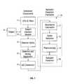
- FIG. 1provides a block diagram of a system for predicting stratum corneum hydration, according to the invention
- FIG. 2illustrates a typical noninvasive NIR absorbance spectrum
- FIG. 3provides a block diagram of a hydration meter, according to the invention.
- FIG. 4illustrates an arrangement of illumination and detection fibers in the hydration meter of FIG. 3, according to the invention
- FIG. 5illustrates an arrangement of illumination and detection fibers in a sample probe head of the hydration meter of FIG. 3, according to the invention
- FIG. 6illustrates an arrangement of illumination and detection fibers in a reference probe head of the hydration meter of FIG. 3, according to the invention
- FIG. 7shows a plot of actual SC hydration measurements vs. estimations in a calibration model for predicting SC hydration, according to the invention.
- FIGS. 8 and 9show plots of actual SC hydration measurements vs. predicted for two different subjects, based on the calibration model of FIG. 7, according to the invention.
- the system for quantifying the hydration of a tissuesuch as the stratum corneum non-destructively provides an apparatus for measuring the near infrared absorption by tissue irradiated with near infrared energy and a procedure for determining the tissue's hydration.
- SCstratum corneum
- the invented apparatus and procedureare described herein with respect to quantifying hydration of the stratum corneum (SC), this description is intended to be exemplary only.
- the inventionhas application in quantifying hydration of other tissue components of skin besides the stratum corneum: the epidermis, the dermis, and the subcutaneous layer, for example.
- the invented apparatusfinds utility in a large number of other applications, among them:
- Tissue analyte determinationincluding at least: glucose, alcohol, cholesterol, triglycerides, sodium, urea, elastin and collagen.
- the apparatusincludes an energy source 11 , one or more sensor elements, an interface 12 to the subject 10 , a means for wavelength selection and an analyzer.
- the sourcegenerates and transmits near-infrared energy in the wavelength range of 700-2500 nanometers.
- the source 11 and the wavelength selection meansconstitute a LED (light emitting diode) array 11 and successive illumination through the elements of the LED array 11 .
- the sourcemay constitute a light source, such as a quartz halogen lamp, and the wavelength selection means may constitute any of a spectrometer, a spectrograph, a monochromator or an interferometer (not shown).
- wavelength selectionoccurs before the tissue is irradiated.
- the energy reaching the tissueconstitutes monochromatic light.
- the tissueis irradiated by polychromatic light, for example from an LED array emitting polychromatic light.
- wavelength selectionoccurs after the tissue is irradiated, prior to being detected by the sensor elements.
- the sensor elementsare detectors 14 that are responsive to a set of targeted wavelengths.
- the interface to the subjectcomprises a means of transmitting energy from the source 11 to a target skin tissue measurement site and includes, for example, direct illumination, a light pipe, fiber-optic probes, a lens system or a light directing mirror system. Energy is collected from the surrounding tissue areas in reflectance mode at an optimally determined distance(s) through the use of detector optics 13 or fiber optics.
- energyis collected in a transmission mode through a skin fold, ear lobe, finger or other extremity.
- the collected light, constituting an analog signalis converted to a current by the sensor elements 14 and sampled through an analog-to-digital converter 15 for analysis on a data processing system.
- a group of LED's 11is employed to transmit energy of pre-selected wavelengths to the skin; the LED's are radially surrounded by detection fibers 13 at specific distances.
- the LED'sare alternately energized and the detected energy of each LED reflected or transmitted through the skin is used to form one spectrum.
- the edge-to-edge distance between each of the LED's and the detector elements, or the distance between the point of illumination, comprising the light-emitting surface of the LED's, and the point of detectionis a minimum of 40 ⁇ m and a maximum of 1 mm. Distances of less than 40 ⁇ m produce too much surface reflection of the NIR radiation and distances of greater than 1 mm result in too much penetration of the NIR radiation.
- the set of wavelengthsincludes 1070, 1180, 1280 nm and 1110, 1190, and 1280 nm. However, other wavelength ranges, corresponding to water bands in the NIR absorbance spectrum, are also suitable. Coupling of the illumination and detector elements, shown in detail in FIG. 4, is accomplished through fiber optics. One skilled in the art will appreciate that other coupling methods are suitable, including direct illumination/detection, optics and lens systems, subject to the criterion for the distances between the point of illumination and detection.
- the detected intensity from the sampleis converted to a current through analog electronics 14 and digitized through an analog-to-digital converter (ADC) 15 .
- ADCanalog-to-digital converter
- the spectrumis passed to the hydration estimation procedure 16 for processing.
- the absorbanceis calculated 17 on the basis of the detected light through ⁇ log(R/R 0 ) where R is the reflected light and R 0 is the light incident on the sample determined by scanning a reference standard. Subsequent processing steps, described below, result in either hydration estimation or a message indicating an invalid scan.
- FIG. 1A block diagram of the integrated system is shown in FIG. 1 .
- the measurementcan be accomplished with existing NIR spectrometers that are commercially available, including a FOSS-NIR Systems NIRS 5000 spectrometer, provided by FOSS NIR SYSTEMS, INC. of Eden Prairie Minn., or a Nicolet Magna-IR 760 spectrometer, provided by THERMO NICOLET, INC of Madison Wis.
- the measurementcan be made by collecting reflected light off the surface of the skin or light transmitted through a portion of the skin, such as the finger or the ear lobe.
- the use of transmittance to determine absorbancecan replace the preferred reflectance measurement.
- intensitycan be used to replace absorbance.
- the spacing of the illumination and detection fibersis performed on the basis of the skin fold or tissue through which NIR radiation is transmitted.
- the general procedure for quantifying hydration based on the measured spectrumis implemented in a data processing system such as a microcomputer 44 that automatically receives the measurement information from the ADC 15 .
- the hydration quantifying procedurecomprises a series of steps, including outlier detection 18 (optional), preprocessing 19 , and estimation 20 wherein each step is a procedure in itself.
- Each procedurerelies on a calibration set of exemplary measurements.
- the general steps of the Hydration Estimation Procedureare summarized, with a detailed description following in the subsequent section titled “Implementation.”
- the measurementis a spectrum denoted by the vector m ⁇ R N of absorbance values pertaining to a set of N wavelengths ⁇ R N that span the near infrared (700 to 2500 nm).
- a typical plot 30 of m versus ⁇is shown in FIG. 2 .
- the outlier detection procedureprovides a method of detecting invalid measurements through spectral variations that result from problems in the instrument, poor sampling of the subject or a subject outside the calibration set.
- the preferred method for the detection of spectral outliersis through a principal component analysis and an analysis of the residuals. See H. Martens, T. Naes, Multivariate Calibration , John Wiley & Sons, New York (1989).
- the sampleis reported to be an outlier and the hydration measurement procedure is terminated.
- Preprocessingincludes operations such as scaling, normalization smoothing, derivatives, filtering and other transformations that attenuate the noise and instrumental variation without affecting the signal of interest.
- the preprocessed measurement, x ⁇ R Nis determined according to
- Estimationmay include operations such as multiple linear least squares regression (MLR), principle component regression (PCR), and partial least squares regression (PLR) analysis that process the measurement, y ⁇ R N , according to
- This sectiondescribes a particular embodiment of the apparatus and specific procedures for quantifying tissue characteristics, and in particular, tissue hydration.
- the structure of the proceduresrelies on a priori knowledge of the systematic variation of the skin structure, namely, the hydration state of the stratum corneum and the variation in path depth of the irradiated light.
- the parameters of each procedureare determined on the basis of an experimental data set, the “calibration set”, providing exemplary information.
- FIG. 3provides a block diagram for the hydration meter 40 .
- the light source 11 for this deviceincludes an array 11 a of three light emitting diodes (LED's).
- the current source for the LED'sis an LED driver 41 connected to a power supply 42 that pulses the LED's at a frequency of between 1 kHz and 10 kHz.
- the LED driver 41supplies a current of up to 3.0 amperes.
- the LED's used for this devicehave a peak wavelength at 1.07_m, 1.22_m, and 1.25_m.
- Each LEDis equipped with a bandpass interference filter 11 b ; the bandpass interference filters of the preferred embodiment have center wavelengths of 1080 nm, 1180 nm, and 1280 nm, respectively, with their full width half maximum ranging from 11.0 to 14.8 nm.
- the lightis transmitted to the probe heads 45 , 46 via fiber optics 51 a-c , 52 a-c .
- FIG. 4illustrates the coupling of the LED's 11 a with the probe heads 45 , 46 by means of fiber optics 51 a-c , 52 a-c.
- Each LEDhas seven 100_m core diameter fiber optics associated with it. Six of these fiber optics 51 a-c go to the sample probe head 45 , and one 52 a-c goes to the reference probe head 46 .
- the sample probe head 45is the subject interface 12 of the device that comes into contact with the stratum corneum.
- FIG. 5shows a preferred fiber optic arrangement for the sample probe head 45 , comprising a total of eighteen illuminating fibers 51 a-c and sixty-nine detecting fibers 51 d . Each illuminating fiber 51 a-c is completely surrounded, in a closed, packed arrangement, by detection fibers 51 d for greatest light collection. Shown in FIG.
- the reference probe head 46is used to collect a dual beam reference of an internal diffuse reflectance standard having known spectral characteristics.
- the reference probehas a total of three illuminating fibers 52 a-c and from 20 to 30 detecting fibers 52 d .
- the diffuse reflected light from each of the probe heads, sample and referencetravels via optical fibers 51 d , 52 d to an optical system 53 a, b that focuses the light onto the 1.9_m InGaAs detectors 14 .
- the fiberopticsare coupled to the various components with connecting elements 54 a-f .
- the connecting elements 54are brass connectors, but other equally suitable alternatives will be apparent to those skilled in the art.
- the signals from the detectorsare amplified in the analog front end 47 (AFE).
- the AFEalso converts the current signal from the detectors to a voltage signal before transmitting the signal to the lock-in amplifier 48 .
- the phase modulating lock-in amplifier 48receives the signal from the AFE 47 and a reference signal from the LED driver 41 .
- the lock-in amplifier 48amplifies signals that are in phase with the reference signal. This increases the signal-to-noise ratio, and gives a DC (direct current) output.
- the output from the lock-in amplifier 48goes through a 16-bit analog to digital converter (ADC) 15 .
- ADCanalog to digital converter
- a laptop computer 44 or other analyzerreceives the signal from the ADC 15 , and predicts the hydration based on the invented algorithm 16 described further below. After the signal is processed, the estimation result is displayed on a display device 43 attached to the laptop 44 .
- the laptopalso controls the master sequence 49 on the LED's, controlling both of: which LED is emitting, and the time period for which each LED is emitting.
- x 1 , x 2 , and x 3are the absorbance of each LED
- a 1 , a 2 and a 3are the coefficients to the absorbance of each LED
- eis the error associated with the model. The coefficients are calculated by
- xis the matrix of absorbance values after the preprocessing techniques are complete
- yis the corneometer readings for each spectral measurement
- mis the absorbance spectrum
- Ris the intensity signal from the sample probe head
- R 0is the intensity signal from the reference probe head.
- the absorbance spectrum, mis passed through the outlier detection system 18 to remove any bad measurements or readings outside the estimation model's range.
- the signalis preprocessed 19 to attenuate any noise and instrumental variation.
- the preprocessing techniques employedare multiplicative scatter correction and mean centering.
- the spectrumis processed, using multivariate scatter correction through a rotation that fits it to the expected or reference spectrum ⁇ overscore (n) ⁇ , determined from the calibration set. See P. Geladi, D. McDougall, H.
- xis the processed absorbance spectrum. From this spectrum, the mean from an exemplary data set is calculated for each LED absorbance. The mean is then subtracted from each LED absorbance in the measured data set. After mean centering the data, it is passed through the multiple linear regression model described above for the estimation of SC hydration.
- the coefficients for the multiple regression model, a 1 , a 2 , and a 3are 2411.4, ⁇ 2486.6, and 257.2, respectively.
- a calibration model for the hydration of the stratum corneummay be used, for example, using factor analysis to develop a set of abstract features capable of representing the spectral variation related to hydration.
- factor analysisthe spectral measurements, NIR absorbance spectra similar to that of FIG. 2, are used.
- the spectrumis sub-divided into one or more regions according to wavelength (wavelength selection) and is preprocessed and normalized to enhance spectral variation related to SC hydration.
- the measurementsare projected onto one or more sets of previously determined factors (eigenvectors) to determine the scores.
- the scoresconstitute the extracted features and are subjected to an estimation procedure, such as linear discriminate analysis, SIMCA, k nearest-neighbor, fuzzy classification and various forms of artificial neural networks to predict hydration of the stratum corneum.
- SIMCAlinear discriminate analysis
- k nearest-neighborfuzzy classification
- various forms of artificial neural networksto predict hydration of the stratum corneum.
- the spectroscopic measurementswere made using a spectrometer instrument according to an embodiment of the invention, comprising a quartz lamp, a monochromator, a fiber optic probe, and a detector set-up.
- the studyconsisted of four human subjects (3 males and 1 female), in which the hydration of the SC at the measurement site was modified through occlusion of the skin. Different occlusion times were employed to develop a range of hydration values, with no treatment of the skin at the sampling site prior to measurement.
- Stratum corneum hydrationwas measured independently by the corneometer CM 825, produced by COURAGE & KHAZAKA of Cologne, Germany.
- Each subjecthad a minimum of eight spectral scans with corresponding corneometer readings over a period of at least two days in duration, each scan constituting a sample.
- the spectral measurements and the corresponding corneometer readingsare referred to as the “Experimental Data Set” herein below.
- the Experimental Data Setwas analyzed using the previously described procedures. Outliers were removed using the outlier detection procedure previously described. Subsequently the data were preprocessed using MSC, followed by mean centering based on the mean of the emitting region of the filters based on their full width half maximum characteristics. The regions used were 1073-1087, 1175-1185, and 1275-1285 nm. Finally, MLR was applied to the data set. The calibration model was first developed using the samples of all four subjects, and subsequently validated using a “leave five out” cross-validation strategy. FIG. 7 shows a plot of actual corneometer measurements vs. estimations for the entire experimental data set.
- the standard error of estimation (SEE) for the experimental data setwas 3.6995. Subsequently, a calibration model was developed and validated by using three subjects to develop the calibration model, and using the resulting model to predict SC hydration for the samples of the remaining subject.
- FIGS. 8 and 9show plots of actual corneometer measurements vs. estimations for subjects four and three, respectively. The SEE was 4.2851 for subject four estimations and 6.1179 for subject three measurements.
Landscapes
- Health & Medical Sciences (AREA)
- Life Sciences & Earth Sciences (AREA)
- Physics & Mathematics (AREA)
- Pathology (AREA)
- General Health & Medical Sciences (AREA)
- Surgery (AREA)
- Veterinary Medicine (AREA)
- Engineering & Computer Science (AREA)
- Biomedical Technology (AREA)
- Heart & Thoracic Surgery (AREA)
- Medical Informatics (AREA)
- Molecular Biology (AREA)
- Biophysics (AREA)
- Animal Behavior & Ethology (AREA)
- Public Health (AREA)
- Spectroscopy & Molecular Physics (AREA)
- Chemical & Material Sciences (AREA)
- Analytical Chemistry (AREA)
- Biochemistry (AREA)
- General Physics & Mathematics (AREA)
- Immunology (AREA)
- Optics & Photonics (AREA)
- Emergency Medicine (AREA)
- Nuclear Medicine, Radiotherapy & Molecular Imaging (AREA)
- Radiology & Medical Imaging (AREA)
- Dermatology (AREA)
- Investigating Or Analysing Materials By Optical Means (AREA)
Abstract
Description


Claims (48)
Priority Applications (1)
| Application Number | Priority Date | Filing Date | Title |
|---|---|---|---|
| US10/183,660US6675029B2 (en) | 1999-07-22 | 2002-06-25 | Apparatus and method for quantification of tissue hydration using diffuse reflectance spectroscopy |
Applications Claiming Priority (3)
| Application Number | Priority Date | Filing Date | Title |
|---|---|---|---|
| US09/359,191US6280381B1 (en) | 1999-07-22 | 1999-07-22 | Intelligent system for noninvasive blood analyte prediction |
| US09/669,781US6442408B1 (en) | 1999-07-22 | 2000-09-25 | Method for quantification of stratum corneum hydration using diffuse reflectance spectroscopy |
| US10/183,660US6675029B2 (en) | 1999-07-22 | 2002-06-25 | Apparatus and method for quantification of tissue hydration using diffuse reflectance spectroscopy |
Related Parent Applications (1)
| Application Number | Title | Priority Date | Filing Date |
|---|---|---|---|
| US09/669,781Continuation-In-PartUS6442408B1 (en) | 1999-07-22 | 2000-09-25 | Method for quantification of stratum corneum hydration using diffuse reflectance spectroscopy |
Publications (2)
| Publication Number | Publication Date |
|---|---|
| US20030060693A1 US20030060693A1 (en) | 2003-03-27 |
| US6675029B2true US6675029B2 (en) | 2004-01-06 |
Family
ID=27000377
Family Applications (1)
| Application Number | Title | Priority Date | Filing Date |
|---|---|---|---|
| US10/183,660Expired - Fee RelatedUS6675029B2 (en) | 1999-07-22 | 2002-06-25 | Apparatus and method for quantification of tissue hydration using diffuse reflectance spectroscopy |
Country Status (1)
| Country | Link |
|---|---|
| US (1) | US6675029B2 (en) |
Cited By (61)
| Publication number | Priority date | Publication date | Assignee | Title |
|---|---|---|---|---|
| US20040217290A1 (en)* | 2003-04-29 | 2004-11-04 | Davis Bruce R. | Infrared imaging for evaluation of corrosion test coupons |
| US20040230106A1 (en)* | 2001-03-16 | 2004-11-18 | Nellcor Puritan Bennett Incorporated | Device and method for monitoring body fluid and electrolyte disorders |
| US20040256564A1 (en)* | 2003-06-20 | 2004-12-23 | Allen Rex H. | Method of measuring coating using two-wavelength infrared reflectance |
| US20050067569A1 (en)* | 2003-09-30 | 2005-03-31 | Shelley Paul H. | Method for measurement of composite heat damage with infrared spectroscopy |
| US20050197579A1 (en)* | 2004-03-08 | 2005-09-08 | Nellcor Puritan Bennett Incorporated | Method and apparatus for optical detection of mixed venous and arterial blood pulsation in tissue |
| US20050203357A1 (en)* | 2004-03-09 | 2005-09-15 | Nellcor Puritan Bennett Incorporated | Pulse oximetry motion artifact rejection using near infrared absorption by water |
| US20060020181A1 (en)* | 2001-03-16 | 2006-01-26 | Schmitt Joseph M | Device and method for monitoring body fluid and electrolyte disorders |
| US20060189858A1 (en)* | 2005-02-14 | 2006-08-24 | Sterling Bernhard B | Analyte detection system for multiple analytes |
| US20060253016A1 (en)* | 2001-03-16 | 2006-11-09 | R Baker Clark Jr | Systems and methods to assess one or more body fluid metrics |
| US20070118027A1 (en)* | 2001-03-16 | 2007-05-24 | Baker Clark R Jr | Method for evaluating extracellular water concentration in tissue |
| US20070299357A1 (en)* | 2006-06-09 | 2007-12-27 | Diana Villegas | Bronchial or tracheal tissular water content sensor and system |
| US20080076983A1 (en)* | 2006-09-27 | 2008-03-27 | Nellcor Puritan Bennett Incorporated | Tissue hydration estimation by spectral absorption bandwidth measurement |
| US20080081975A1 (en)* | 2006-09-28 | 2008-04-03 | Geeta Agashe | System and method for detection of brain edema using spectrophotometry |
| US20080146906A1 (en)* | 2006-09-29 | 2008-06-19 | Nellcor Puritan Bennett Incorporated | System and method for detection of skin wounds and compartment syndromes |
| US20080221409A1 (en)* | 2007-03-09 | 2008-09-11 | Nellcor Puritan Bennett Llc | System and method for controlling tissue treatment |
| US20080221414A1 (en)* | 2007-03-09 | 2008-09-11 | Nellcor Puritan Bennett Llc | Method for detection of aberrant tissue spectra |
| US20080221463A1 (en)* | 2007-03-09 | 2008-09-11 | Nellcor Puritan Bennett Llc | System and method for venous pulsation detection using near infrared wavelengths |
| US20080220512A1 (en)* | 2007-03-09 | 2008-09-11 | Nellcor Puritan Bennett Llc | Tunable laser-based spectroscopy system for non-invasively measuring body water content |
| US20080221406A1 (en)* | 2007-03-09 | 2008-09-11 | Baker Clark R | Method and apparatus for estimating water reserves |
| US20080221411A1 (en)* | 2007-03-09 | 2008-09-11 | Nellcor Puritan Bennett Llc | System and method for tissue hydration estimation |
| US20090134331A1 (en)* | 2005-09-02 | 2009-05-28 | Yuta Miyamae | Method of evaluating skin conditions and method of estimating skin thickness |
| US20090318786A1 (en)* | 2002-03-08 | 2009-12-24 | Blank Thomas B | Channeled tissue sample probe method and apparatus |
| US20100081916A1 (en)* | 2008-09-29 | 2010-04-01 | Searete Llc, A Limited Liability Corporation Of The State Of Delaware. | Histological facilitation systems and methods |
| US20100081915A1 (en)* | 2008-09-29 | 2010-04-01 | Searete Llc, Alimited Liability Corporation Of The State Of Delaware | Histological facilitation systems and methods |
| US20100081927A1 (en)* | 2008-09-29 | 2010-04-01 | Searete Llc, A Limited Liability Corporation Of The State Of Delaware | Histological facilitation systems and methods |
| US20100081924A1 (en)* | 2008-09-29 | 2010-04-01 | Searete Llc, A Limited Liability Corporation Of The State Of Delaware | Histological facilitation systems and methods |
| US20100081926A1 (en)* | 2008-09-29 | 2010-04-01 | Searete Llc, A Limited Liability Corporation Of The State Of Delaware | Histological facilitation systems and methods |
| US20100081928A1 (en)* | 2008-09-29 | 2010-04-01 | Searete Llc, A Limited Liability Corporation Of The State Of Delaware | Histological Facilitation systems and methods |
| US20100081960A1 (en)* | 2008-09-30 | 2010-04-01 | Nellcor Puritan Bennett Llc | Bioimpedance System and Sensor and Technique for Using the Same |
| US20100113909A1 (en)* | 2008-10-31 | 2010-05-06 | Nellcor Puritan Bennett Llc | System And Method For Facilitating Observation Of Monitored Physiologic Data |
| US20100113908A1 (en)* | 2008-10-31 | 2010-05-06 | Nellcor Puritan Bennett Llc | System And Method For Facilitating Observation Of Monitored Physiologic Data |
| US20100280393A1 (en)* | 2009-05-04 | 2010-11-04 | Battelle Memorial Institute | Optical Reflectance Spectroscopy for Evaluation of Radiation Injury |
| US20110071376A1 (en)* | 2009-09-24 | 2011-03-24 | Nellcor Puritan Bennett Llc | Determination Of A Physiological Parameter |
| US20110071366A1 (en)* | 2009-09-24 | 2011-03-24 | Nellcor Puritan Bennett Llc | Determination Of A Physiological Parameter |
| US20110071374A1 (en)* | 2009-09-24 | 2011-03-24 | Nellcor Puritan Bennett Llc | Minimax Filtering For Pulse Oximetry |
| US20110077485A1 (en)* | 2009-09-30 | 2011-03-31 | Nellcor Puritan Bennett Llc | Method Of Analyzing Photon Density Waves In A Medical Monitor |
| US8175665B2 (en) | 2007-03-09 | 2012-05-08 | Nellcor Puritan Bennett Llc | Method and apparatus for spectroscopic tissue analyte measurement |
| US8346327B2 (en) | 2007-03-09 | 2013-01-01 | Covidien Lp | Method for identification of sensor site by local skin spectrum data |
| US8391943B2 (en) | 2010-03-31 | 2013-03-05 | Covidien Lp | Multi-wavelength photon density wave system using an optical switch |
| US8498683B2 (en) | 2010-04-30 | 2013-07-30 | Covidien LLP | Method for respiration rate and blood pressure alarm management |
| US20130210058A1 (en)* | 2012-02-15 | 2013-08-15 | Lakeland Ventures Development, Llc | System for noninvasive determination of water in tissue |
| US8610769B2 (en) | 2011-02-28 | 2013-12-17 | Covidien Lp | Medical monitor data collection system and method |
| CN104048939A (en)* | 2014-06-05 | 2014-09-17 | 中国肉类食品综合研究中心 | Near infrared rapid detection method for blood sugar content in live pig blood |
| US9091676B2 (en) | 2010-06-09 | 2015-07-28 | Optiscan Biomedical Corp. | Systems and methods for measuring multiple analytes in a sample |
| US9289169B2 (en) | 2007-05-18 | 2016-03-22 | Optiscan Biomedical Corp. | Analyte monitoring systems and methods |
| US9326686B2 (en) | 2012-03-12 | 2016-05-03 | Ivwatch, Llc | System and method for mitigating the effects of tissue blood volume changes to aid in diagnosing infiltration or extravasation in animalia tissue |
| US9351671B2 (en) | 2012-07-16 | 2016-05-31 | Timothy Ruchti | Multiplexed pathlength resolved noninvasive analyzer apparatus and method of use thereof |
| US9351672B2 (en) | 2012-07-16 | 2016-05-31 | Timothy Ruchti | Multiplexed pathlength resolved noninvasive analyzer apparatus with stacked filters and method of use thereof |
| US9380982B2 (en) | 2010-07-28 | 2016-07-05 | Covidien Lp | Adaptive alarm system and method |
| US9442065B2 (en) | 2014-09-29 | 2016-09-13 | Zyomed Corp. | Systems and methods for synthesis of zyotons for use in collision computing for noninvasive blood glucose and other measurements |
| US9554738B1 (en) | 2016-03-30 | 2017-01-31 | Zyomed Corp. | Spectroscopic tomography systems and methods for noninvasive detection and measurement of analytes using collision computing |
| US9585604B2 (en) | 2012-07-16 | 2017-03-07 | Zyomed Corp. | Multiplexed pathlength resolved noninvasive analyzer apparatus with dynamic optical paths and method of use thereof |
| US9766126B2 (en) | 2013-07-12 | 2017-09-19 | Zyomed Corp. | Dynamic radially controlled light input to a noninvasive analyzer apparatus and method of use thereof |
| US9833146B2 (en) | 2012-04-17 | 2017-12-05 | Covidien Lp | Surgical system and method of use of the same |
| US20180228412A1 (en)* | 2017-02-15 | 2018-08-16 | Craig M. Gardner | Spectroscopy through thin skin |
| US10159412B2 (en) | 2010-12-01 | 2018-12-25 | Cercacor Laboratories, Inc. | Handheld processing device including medical applications for minimally and non invasive glucose measurements |
| US10966655B2 (en) | 2018-04-27 | 2021-04-06 | Hyrostasis, Inc. | Tissue hydration monitor |
| US10980478B2 (en) | 2015-12-21 | 2021-04-20 | Koninklijke Philips N.V. | Device for tissue condition measurement |
| US11457872B2 (en) | 2017-12-01 | 2022-10-04 | Samsung Electronics Co., Ltd. | Bio-signal quality assessment apparatus and bio-signal quality assessment method |
| US11471102B2 (en) | 2019-06-20 | 2022-10-18 | Medici Technologies Llc | Hydration assessment system |
| US11911184B2 (en) | 2017-12-01 | 2024-02-27 | Samsung Electronics Co., Ltd. | Bio-signal quality assessment apparatus and bio-signal quality assessment method |
Families Citing this family (111)
| Publication number | Priority date | Publication date | Assignee | Title |
|---|---|---|---|---|
| US7758503B2 (en)* | 1997-01-27 | 2010-07-20 | Lynn Lawrence A | Microprocessor system for the analysis of physiologic and financial datasets |
| US6018673A (en) | 1996-10-10 | 2000-01-25 | Nellcor Puritan Bennett Incorporated | Motion compatible sensor for non-invasive optical blood analysis |
| US9042952B2 (en) | 1997-01-27 | 2015-05-26 | Lawrence A. Lynn | System and method for automatic detection of a plurality of SPO2 time series pattern types |
| US20060161071A1 (en) | 1997-01-27 | 2006-07-20 | Lynn Lawrence A | Time series objectification system and method |
| US8932227B2 (en) | 2000-07-28 | 2015-01-13 | Lawrence A. Lynn | System and method for CO2 and oximetry integration |
| US9521971B2 (en) | 1997-07-14 | 2016-12-20 | Lawrence A. Lynn | System and method for automatic detection of a plurality of SPO2 time series pattern types |
| US20070191697A1 (en) | 2006-02-10 | 2007-08-16 | Lynn Lawrence A | System and method for SPO2 instability detection and quantification |
| US6675031B1 (en) | 1999-04-14 | 2004-01-06 | Mallinckrodt Inc. | Method and circuit for indicating quality and accuracy of physiological measurements |
| US9053222B2 (en) | 2002-05-17 | 2015-06-09 | Lawrence A. Lynn | Patient safety processor |
| US20060195041A1 (en) | 2002-05-17 | 2006-08-31 | Lynn Lawrence A | Centralized hospital monitoring system for automatically detecting upper airway instability and for preventing and aborting adverse drug reactions |
| US20070093721A1 (en)* | 2001-05-17 | 2007-04-26 | Lynn Lawrence A | Microprocessor system for the analysis of physiologic and financial datasets |
| US6754516B2 (en) | 2001-07-19 | 2004-06-22 | Nellcor Puritan Bennett Incorporated | Nuisance alarm reductions in a physiological monitor |
| US20080200775A1 (en)* | 2007-02-20 | 2008-08-21 | Lynn Lawrence A | Maneuver-based plethysmographic pulse variation detection system and method |
| US7006856B2 (en)* | 2003-01-10 | 2006-02-28 | Nellcor Puritan Bennett Incorporated | Signal quality metrics design for qualifying data for a physiological monitor |
| US7016715B2 (en) | 2003-01-13 | 2006-03-21 | Nellcorpuritan Bennett Incorporated | Selection of preset filter parameters based on signal quality |
| EP1475637A1 (en)* | 2003-05-09 | 2004-11-10 | Institut Dr. Schrader Creachem GmbH | Metod for examining skin compatibility of substances |
| US7190985B2 (en)* | 2004-02-25 | 2007-03-13 | Nellcor Puritan Bennett Inc. | Oximeter ambient light cancellation |
| US7120479B2 (en) | 2004-02-25 | 2006-10-10 | Nellcor Puritan Bennett Inc. | Switch-mode oximeter LED drive with a single inductor |
| US7194293B2 (en)* | 2004-03-08 | 2007-03-20 | Nellcor Puritan Bennett Incorporated | Selection of ensemble averaging weights for a pulse oximeter based on signal quality metrics |
| US7534212B2 (en)* | 2004-03-08 | 2009-05-19 | Nellcor Puritan Bennett Llc | Pulse oximeter with alternate heart-rate determination |
| US7392075B2 (en) | 2005-03-03 | 2008-06-24 | Nellcor Puritan Bennett Incorporated | Method for enhancing pulse oximetry calculations in the presence of correlated artifacts |
| US20070069898A1 (en)* | 2005-09-28 | 2007-03-29 | White Mark J | Glove with attached security device |
| US7725147B2 (en) | 2005-09-29 | 2010-05-25 | Nellcor Puritan Bennett Llc | System and method for removing artifacts from waveforms |
| US7725146B2 (en) | 2005-09-29 | 2010-05-25 | Nellcor Puritan Bennett Llc | System and method for pre-processing waveforms |
| US20070106126A1 (en) | 2005-09-30 | 2007-05-10 | Mannheimer Paul D | Patient monitoring alarm escalation system and method |
| US20070100220A1 (en)* | 2005-10-28 | 2007-05-03 | Baker Clark R Jr | Adjusting parameters used in pulse oximetry analysis |
| US7668579B2 (en) | 2006-02-10 | 2010-02-23 | Lynn Lawrence A | System and method for the detection of physiologic response to stimulation |
| US20070208259A1 (en)* | 2006-03-06 | 2007-09-06 | Mannheimer Paul D | Patient monitoring alarm escalation system and method |
| US8702606B2 (en)* | 2006-03-21 | 2014-04-22 | Covidien Lp | Patient monitoring help video system and method |
| US8380271B2 (en) | 2006-06-15 | 2013-02-19 | Covidien Lp | System and method for generating customizable audible beep tones and alarms |
| US8064975B2 (en) | 2006-09-20 | 2011-11-22 | Nellcor Puritan Bennett Llc | System and method for probability based determination of estimated oxygen saturation |
| US20080076977A1 (en)* | 2006-09-26 | 2008-03-27 | Nellcor Puritan Bennett Inc. | Patient monitoring device snapshot feature system and method |
| US8696593B2 (en) | 2006-09-27 | 2014-04-15 | Covidien Lp | Method and system for monitoring intracranial pressure |
| US7922665B2 (en) | 2006-09-28 | 2011-04-12 | Nellcor Puritan Bennett Llc | System and method for pulse rate calculation using a scheme for alternate weighting |
| US8175667B2 (en) | 2006-09-29 | 2012-05-08 | Nellcor Puritan Bennett Llc | Symmetric LED array for pulse oximetry |
| US8068890B2 (en) | 2006-09-29 | 2011-11-29 | Nellcor Puritan Bennett Llc | Pulse oximetry sensor switchover |
| US20080082338A1 (en)* | 2006-09-29 | 2008-04-03 | O'neil Michael P | Systems and methods for secure voice identification and medical device interface |
| US20080081956A1 (en)* | 2006-09-29 | 2008-04-03 | Jayesh Shah | System and method for integrating voice with a medical device |
| US8728059B2 (en)* | 2006-09-29 | 2014-05-20 | Covidien Lp | System and method for assuring validity of monitoring parameter in combination with a therapeutic device |
| US7706896B2 (en)* | 2006-09-29 | 2010-04-27 | Nellcor Puritan Bennett Llc | User interface and identification in a medical device system and method |
| US8160668B2 (en)* | 2006-09-29 | 2012-04-17 | Nellcor Puritan Bennett Llc | Pathological condition detector using kernel methods and oximeters |
| US7698002B2 (en)* | 2006-09-29 | 2010-04-13 | Nellcor Puritan Bennett Llc | Systems and methods for user interface and identification in a medical device |
| US20080097175A1 (en)* | 2006-09-29 | 2008-04-24 | Boyce Robin S | System and method for display control of patient monitor |
| US8068891B2 (en) | 2006-09-29 | 2011-11-29 | Nellcor Puritan Bennett Llc | Symmetric LED array for pulse oximetry |
| US7925511B2 (en)* | 2006-09-29 | 2011-04-12 | Nellcor Puritan Bennett Llc | System and method for secure voice identification in a medical device |
| US7848891B2 (en) | 2006-09-29 | 2010-12-07 | Nellcor Puritan Bennett Llc | Modulation ratio determination with accommodation of uncertainty |
| US8652040B2 (en) | 2006-12-19 | 2014-02-18 | Valencell, Inc. | Telemetric apparatus for health and environmental monitoring |
| US20080200819A1 (en)* | 2007-02-20 | 2008-08-21 | Lynn Lawrence A | Orthostasis detection system and method |
| US20080221426A1 (en)* | 2007-03-09 | 2008-09-11 | Nellcor Puritan Bennett Llc | Methods and apparatus for detecting misapplied optical sensors |
| US8265724B2 (en) | 2007-03-09 | 2012-09-11 | Nellcor Puritan Bennett Llc | Cancellation of light shunting |
| WO2009036561A1 (en)* | 2007-09-21 | 2009-03-26 | National Research Council Of Canada | Method and apparatus for periodontal diagnosis |
| JP4569615B2 (en)* | 2007-09-25 | 2010-10-27 | ブラザー工業株式会社 | Printing device |
| US8204567B2 (en)* | 2007-12-13 | 2012-06-19 | Nellcor Puritan Bennett Llc | Signal demodulation |
| US20090171167A1 (en)* | 2007-12-27 | 2009-07-02 | Nellcor Puritan Bennett Llc | System And Method For Monitor Alarm Management |
| US8744775B2 (en)* | 2007-12-28 | 2014-06-03 | Weyerhaeuser Nr Company | Methods for classification of somatic embryos comprising hyperspectral line imaging |
| US8092993B2 (en) | 2007-12-31 | 2012-01-10 | Nellcor Puritan Bennett Llc | Hydrogel thin film for use as a biosensor |
| US20090171226A1 (en)* | 2007-12-31 | 2009-07-02 | Nellcor Puritan Bennett Llc | System and method for evaluating variation in the timing of physiological events |
| US20090171173A1 (en)* | 2007-12-31 | 2009-07-02 | Nellcor Puritan Bennett Llc | System and method for reducing motion artifacts in a sensor |
| US20090171174A1 (en)* | 2007-12-31 | 2009-07-02 | Nellcor Puritan Bennett Llc | System and method for maintaining battery life |
| US8275553B2 (en)* | 2008-02-19 | 2012-09-25 | Nellcor Puritan Bennett Llc | System and method for evaluating physiological parameter data |
| US8750953B2 (en)* | 2008-02-19 | 2014-06-10 | Covidien Lp | Methods and systems for alerting practitioners to physiological conditions |
| US20090247851A1 (en)* | 2008-03-26 | 2009-10-01 | Nellcor Puritan Bennett Llc | Graphical User Interface For Monitor Alarm Management |
| US8140272B2 (en)* | 2008-03-27 | 2012-03-20 | Nellcor Puritan Bennett Llc | System and method for unmixing spectroscopic observations with nonnegative matrix factorization |
| US8437822B2 (en) | 2008-03-28 | 2013-05-07 | Covidien Lp | System and method for estimating blood analyte concentration |
| US20090247850A1 (en)* | 2008-03-28 | 2009-10-01 | Nellcor Puritan Bennett Llc | Manually Powered Oximeter |
| US8364224B2 (en)* | 2008-03-31 | 2013-01-29 | Covidien Lp | System and method for facilitating sensor and monitor communication |
| US8112375B2 (en) | 2008-03-31 | 2012-02-07 | Nellcor Puritan Bennett Llc | Wavelength selection and outlier detection in reduced rank linear models |
| US8292809B2 (en) | 2008-03-31 | 2012-10-23 | Nellcor Puritan Bennett Llc | Detecting chemical components from spectroscopic observations |
| JP5474937B2 (en) | 2008-05-07 | 2014-04-16 | ローレンス エー. リン, | Medical disorder pattern search engine |
| USD626562S1 (en) | 2008-06-30 | 2010-11-02 | Nellcor Puritan Bennett Llc | Triangular saturation pattern detection indicator for a patient monitor display panel |
| US20090327515A1 (en)* | 2008-06-30 | 2009-12-31 | Thomas Price | Medical Monitor With Network Connectivity |
| US9895068B2 (en)* | 2008-06-30 | 2018-02-20 | Covidien Lp | Pulse oximeter with wait-time indication |
| US8862194B2 (en) | 2008-06-30 | 2014-10-14 | Covidien Lp | Method for improved oxygen saturation estimation in the presence of noise |
| USD626561S1 (en) | 2008-06-30 | 2010-11-02 | Nellcor Puritan Bennett Llc | Circular satseconds indicator and triangular saturation pattern detection indicator for a patient monitor display panel |
| US8968193B2 (en)* | 2008-09-30 | 2015-03-03 | Covidien Lp | System and method for enabling a research mode on physiological monitors |
| US8386000B2 (en)* | 2008-09-30 | 2013-02-26 | Covidien Lp | System and method for photon density wave pulse oximetry and pulse hemometry |
| US8433382B2 (en)* | 2008-09-30 | 2013-04-30 | Covidien Lp | Transmission mode photon density wave system and method |
| US8417309B2 (en) | 2008-09-30 | 2013-04-09 | Covidien Lp | Medical sensor |
| US20090171172A1 (en)* | 2008-12-19 | 2009-07-02 | Nellcor Puritan Bennett Llc | Method and system for pulse gating |
| US20100240972A1 (en)* | 2009-03-20 | 2010-09-23 | Nellcor Puritan Bennett Llc | Slider Spot Check Pulse Oximeter |
| US8221319B2 (en) | 2009-03-25 | 2012-07-17 | Nellcor Puritan Bennett Llc | Medical device for assessing intravascular blood volume and technique for using the same |
| US8509869B2 (en) | 2009-05-15 | 2013-08-13 | Covidien Lp | Method and apparatus for detecting and analyzing variations in a physiologic parameter |
| US8494786B2 (en) | 2009-07-30 | 2013-07-23 | Covidien Lp | Exponential sampling of red and infrared signals |
| US20110029865A1 (en)* | 2009-07-31 | 2011-02-03 | Nellcor Puritan Bennett Llc | Control Interface For A Medical Monitor |
| US8494606B2 (en)* | 2009-08-19 | 2013-07-23 | Covidien Lp | Photoplethysmography with controlled application of sensor pressure |
| US8494604B2 (en)* | 2009-09-21 | 2013-07-23 | Covidien Lp | Wavelength-division multiplexing in a multi-wavelength photon density wave system |
| US8704666B2 (en)* | 2009-09-21 | 2014-04-22 | Covidien Lp | Medical device interface customization systems and methods |
| US8788001B2 (en)* | 2009-09-21 | 2014-07-22 | Covidien Lp | Time-division multiplexing in a multi-wavelength photon density wave system |
| US8798704B2 (en)* | 2009-09-24 | 2014-08-05 | Covidien Lp | Photoacoustic spectroscopy method and system to discern sepsis from shock |
| US8515511B2 (en) | 2009-09-29 | 2013-08-20 | Covidien Lp | Sensor with an optical coupling material to improve plethysmographic measurements and method of using the same |
| US9554739B2 (en) | 2009-09-29 | 2017-01-31 | Covidien Lp | Smart cable for coupling a medical sensor to an electronic patient monitor |
| US8376955B2 (en)* | 2009-09-29 | 2013-02-19 | Covidien Lp | Spectroscopic method and system for assessing tissue temperature |
| US20110077470A1 (en)* | 2009-09-30 | 2011-03-31 | Nellcor Puritan Bennett Llc | Patient Monitor Symmetry Control |
| US20110074342A1 (en)* | 2009-09-30 | 2011-03-31 | Nellcor Puritan Bennett Llc | Wireless electricity for electronic devices |
| US8452362B2 (en)* | 2010-01-26 | 2013-05-28 | Chromologic Llc | Method and system for monitoring hydration |
| US20110218448A1 (en)* | 2010-03-03 | 2011-09-08 | Buntic Rudolf F | Perfusion detection devices and methods of using the same |
| US8930145B2 (en) | 2010-07-28 | 2015-01-06 | Covidien Lp | Light focusing continuous wave photoacoustic spectroscopy and its applications to patient monitoring |
| CN104394766B (en)* | 2012-06-29 | 2017-06-20 | 皇家飞利浦有限公司 | Real-time tumor perfusion imaging during radiation disposal delivering |
| CN103048278B (en)* | 2012-12-25 | 2016-01-13 | 浙江工业大学 | Longjing tea moisture online test method fried by machine |
| US10244987B2 (en)* | 2015-08-13 | 2019-04-02 | Pixart Imaging Inc. | Physiological detection system with adjustable signal source and operating method thereof |
| US20170261427A1 (en)* | 2016-03-14 | 2017-09-14 | Analog Devices, Inc. | Optical measurements of chemical content |
| US11209358B2 (en) | 2016-03-14 | 2021-12-28 | Analog Devices, Inc. | Blocking specular reflections |
| KR102599205B1 (en)* | 2016-06-17 | 2023-11-07 | 삼성전자 주식회사 | Mobile device and mesurement method of skin hydration using the same |
| WO2018009670A1 (en) | 2016-07-06 | 2018-01-11 | Chemimage Corporation | Systems and methods for detecting edema |
| US20190261869A1 (en)* | 2016-11-14 | 2019-08-29 | The General Hospital Corporation | Systems and methods for multi-distance, multi-wavelength diffuse correlation spectroscopy |
| PL424897A1 (en)* | 2018-03-15 | 2019-09-23 | Tex Life&Healthcare Spółka Z Ograniczoną Odpowiedzialnością | Method for non-invasive monitoring of body hydration |
| US12295743B2 (en) | 2019-02-04 | 2025-05-13 | Chemimage Corporation | Quantification of heart failure using molecular chemical imaging |
| CN111297374B (en)* | 2020-02-24 | 2022-05-27 | 京东方科技集团股份有限公司 | A physical parameter detection device and a physical parameter detection method |
| US20230255519A1 (en)* | 2020-03-11 | 2023-08-17 | Robert Joseph Petcavich | Wearable wireless non-invasive blood glucose measurement system |
| EP4023150B1 (en) | 2020-07-07 | 2025-05-21 | Shenzhen Goodix Technology Co., Ltd. | Signal adjustment method for ppg apparatus and ppg apparatus |
| CN114652269A (en)* | 2022-03-16 | 2022-06-24 | 桂林电子科技大学 | Multispectral skin cancer imaging system based on hybrid fiber bundle |
Citations (2)
| Publication number | Priority date | Publication date | Assignee | Title |
|---|---|---|---|---|
| CA2210791A1 (en)* | 1997-07-18 | 1999-01-18 | The University Of Manitoba | Diagnosis of edema |
| US6442408B1 (en)* | 1999-07-22 | 2002-08-27 | Instrumentation Metrics, Inc. | Method for quantification of stratum corneum hydration using diffuse reflectance spectroscopy |
- 2002
- 2002-06-25USUS10/183,660patent/US6675029B2/ennot_activeExpired - Fee Related
Patent Citations (2)
| Publication number | Priority date | Publication date | Assignee | Title |
|---|---|---|---|---|
| CA2210791A1 (en)* | 1997-07-18 | 1999-01-18 | The University Of Manitoba | Diagnosis of edema |
| US6442408B1 (en)* | 1999-07-22 | 2002-08-27 | Instrumentation Metrics, Inc. | Method for quantification of stratum corneum hydration using diffuse reflectance spectroscopy |
Cited By (114)
| Publication number | Priority date | Publication date | Assignee | Title |
|---|---|---|---|---|
| US20060020181A1 (en)* | 2001-03-16 | 2006-01-26 | Schmitt Joseph M | Device and method for monitoring body fluid and electrolyte disorders |
| US7239902B2 (en) | 2001-03-16 | 2007-07-03 | Nellor Puritan Bennett Incorporated | Device and method for monitoring body fluid and electrolyte disorders |
| US8135448B2 (en) | 2001-03-16 | 2012-03-13 | Nellcor Puritan Bennett Llc | Systems and methods to assess one or more body fluid metrics |
| US7657292B2 (en) | 2001-03-16 | 2010-02-02 | Nellcor Puritan Bennett Llc | Method for evaluating extracellular water concentration in tissue |
| US20060253016A1 (en)* | 2001-03-16 | 2006-11-09 | R Baker Clark Jr | Systems and methods to assess one or more body fluid metrics |
| US8229529B2 (en) | 2001-03-16 | 2012-07-24 | Nellcor Puritan Bennett Llc | Device and method for monitoring body fluid and electrolyte disorders |
| US8457722B2 (en)* | 2001-03-16 | 2013-06-04 | Covidien Lp | Device and method for monitoring body fluid and electrolyte disorders |
| US20070129614A1 (en)* | 2001-03-16 | 2007-06-07 | Nellcor Puritan Bennett Inc. | Device and method for monitoring body fluid and electrolyte disorders |
| US20040230106A1 (en)* | 2001-03-16 | 2004-11-18 | Nellcor Puritan Bennett Incorporated | Device and method for monitoring body fluid and electrolyte disorders |
| US8509866B2 (en) | 2001-03-16 | 2013-08-13 | Covidien Lp | Device and method for monitoring body fluid and electrolyte disorders |
| US20070118027A1 (en)* | 2001-03-16 | 2007-05-24 | Baker Clark R Jr | Method for evaluating extracellular water concentration in tissue |
| US7236811B2 (en) | 2001-03-16 | 2007-06-26 | Nellcor Puritan Bennett Incorporated | Device and method for monitoring body fluid and electrolyte disorders |
| US20060084864A1 (en)* | 2001-03-16 | 2006-04-20 | Schmitt Joseph M | Device and method for monitoring body fluid and electrolyte disorders |
| US20090318786A1 (en)* | 2002-03-08 | 2009-12-24 | Blank Thomas B | Channeled tissue sample probe method and apparatus |
| US7312453B2 (en) | 2003-04-29 | 2007-12-25 | The Boeing Company | Methods for determining corrosion products on substrates using infrared imaging |
| US20040217290A1 (en)* | 2003-04-29 | 2004-11-04 | Davis Bruce R. | Infrared imaging for evaluation of corrosion test coupons |
| US7135683B2 (en) | 2003-04-29 | 2006-11-14 | The Boeing Company | Infrared imaging for evaluation of corrosion test coupons |
| US20060192121A1 (en)* | 2003-04-29 | 2006-08-31 | The Boeing Company | Infrared Imaging for Evaluation of Corrosion Test Coupons |
| US20070020762A1 (en)* | 2003-04-29 | 2007-01-25 | The Boeing Company | Methods for Determining Corrosion Products on Substrates Using Infrared Imaging |
| US7057177B2 (en)* | 2003-04-29 | 2006-06-06 | The Boeing Company | Infrared imaging for evaluation of corrosion test coupons |
| US20040256564A1 (en)* | 2003-06-20 | 2004-12-23 | Allen Rex H. | Method of measuring coating using two-wavelength infrared reflectance |
| US7119336B2 (en) | 2003-06-20 | 2006-10-10 | The Boeing Company | Method of measuring coating using two-wavelength infrared reflectance |
| US7115869B2 (en) | 2003-09-30 | 2006-10-03 | The Boeing Company | Method for measurement of composite heat damage with infrared spectroscopy |
| US20050067569A1 (en)* | 2003-09-30 | 2005-03-31 | Shelley Paul H. | Method for measurement of composite heat damage with infrared spectroscopy |
| US20050197579A1 (en)* | 2004-03-08 | 2005-09-08 | Nellcor Puritan Bennett Incorporated | Method and apparatus for optical detection of mixed venous and arterial blood pulsation in tissue |
| US8611977B2 (en) | 2004-03-08 | 2013-12-17 | Covidien Lp | Method and apparatus for optical detection of mixed venous and arterial blood pulsation in tissue |
| US20050203357A1 (en)* | 2004-03-09 | 2005-09-15 | Nellcor Puritan Bennett Incorporated | Pulse oximetry motion artifact rejection using near infrared absorption by water |
| US20070106137A1 (en)* | 2004-03-09 | 2007-05-10 | Baker Clark R Jr | Pulse oximetry signal correction using near infrared absorption by water |
| US8175670B2 (en) | 2004-03-09 | 2012-05-08 | Nellcor Puritan Bennett Llc | Pulse oximetry signal correction using near infrared absorption by water |
| US20080009690A1 (en)* | 2004-03-09 | 2008-01-10 | Nellcor Puritan Bennett Llc | Pulse oximetry motion artifact rejection using near infrared absorption by water |
| US7277741B2 (en) | 2004-03-09 | 2007-10-02 | Nellcor Puritan Bennett Incorporated | Pulse oximetry motion artifact rejection using near infrared absorption by water |
| US8195263B2 (en) | 2004-03-09 | 2012-06-05 | Nellcor Puritan Bennett Llc | Pulse oximetry motion artifact rejection using near infrared absorption by water |
| US20060189858A1 (en)* | 2005-02-14 | 2006-08-24 | Sterling Bernhard B | Analyte detection system for multiple analytes |
| US20100234703A1 (en)* | 2005-02-14 | 2010-09-16 | Sterling Bernhard B | Method and apparatus for detection of multiple analytes |
| US8140140B2 (en) | 2005-02-14 | 2012-03-20 | Optiscan Biomedical Corporation | Analyte detection system for multiple analytes |
| US20090134331A1 (en)* | 2005-09-02 | 2009-05-28 | Yuta Miyamae | Method of evaluating skin conditions and method of estimating skin thickness |
| US7820972B2 (en)* | 2005-09-02 | 2010-10-26 | Pola Chemical Industries Inc. | Method of evaluating skin conditions and method of estimating skin thickness |
| US8255025B2 (en) | 2006-06-09 | 2012-08-28 | Nellcor Puritan Bennett Llc | Bronchial or tracheal tissular water content sensor and system |
| US20070299357A1 (en)* | 2006-06-09 | 2007-12-27 | Diana Villegas | Bronchial or tracheal tissular water content sensor and system |
| US20080076983A1 (en)* | 2006-09-27 | 2008-03-27 | Nellcor Puritan Bennett Incorporated | Tissue hydration estimation by spectral absorption bandwidth measurement |
| US8180419B2 (en) | 2006-09-27 | 2012-05-15 | Nellcor Puritan Bennett Llc | Tissue hydration estimation by spectral absorption bandwidth measurement |
| US7643858B2 (en) | 2006-09-28 | 2010-01-05 | Nellcor Puritan Bennett Llc | System and method for detection of brain edema using spectrophotometry |
| US20080081975A1 (en)* | 2006-09-28 | 2008-04-03 | Geeta Agashe | System and method for detection of brain edema using spectrophotometry |
| US20080146906A1 (en)* | 2006-09-29 | 2008-06-19 | Nellcor Puritan Bennett Incorporated | System and method for detection of skin wounds and compartment syndromes |
| US8116852B2 (en) | 2006-09-29 | 2012-02-14 | Nellcor Puritan Bennett Llc | System and method for detection of skin wounds and compartment syndromes |
| US20080221409A1 (en)* | 2007-03-09 | 2008-09-11 | Nellcor Puritan Bennett Llc | System and method for controlling tissue treatment |
| US8175665B2 (en) | 2007-03-09 | 2012-05-08 | Nellcor Puritan Bennett Llc | Method and apparatus for spectroscopic tissue analyte measurement |
| US20080220512A1 (en)* | 2007-03-09 | 2008-09-11 | Nellcor Puritan Bennett Llc | Tunable laser-based spectroscopy system for non-invasively measuring body water content |
| US20080221414A1 (en)* | 2007-03-09 | 2008-09-11 | Nellcor Puritan Bennett Llc | Method for detection of aberrant tissue spectra |
| US8357090B2 (en) | 2007-03-09 | 2013-01-22 | Covidien Lp | Method and apparatus for estimating water reserves |
| US8346327B2 (en) | 2007-03-09 | 2013-01-01 | Covidien Lp | Method for identification of sensor site by local skin spectrum data |
| US8280469B2 (en) | 2007-03-09 | 2012-10-02 | Nellcor Puritan Bennett Llc | Method for detection of aberrant tissue spectra |
| US20080221463A1 (en)* | 2007-03-09 | 2008-09-11 | Nellcor Puritan Bennett Llc | System and method for venous pulsation detection using near infrared wavelengths |
| US20080221406A1 (en)* | 2007-03-09 | 2008-09-11 | Baker Clark R | Method and apparatus for estimating water reserves |
| US8690864B2 (en) | 2007-03-09 | 2014-04-08 | Covidien Lp | System and method for controlling tissue treatment |
| US8109882B2 (en) | 2007-03-09 | 2012-02-07 | Nellcor Puritan Bennett Llc | System and method for venous pulsation detection using near infrared wavelengths |
| US20080221411A1 (en)* | 2007-03-09 | 2008-09-11 | Nellcor Puritan Bennett Llc | System and method for tissue hydration estimation |
| WO2008112522A1 (en)* | 2007-03-09 | 2008-09-18 | Nellcor Puritan Bennett Llc | Method for detection of aberrant tissue spectra |
| US9289169B2 (en) | 2007-05-18 | 2016-03-22 | Optiscan Biomedical Corp. | Analyte monitoring systems and methods |
| US20100081928A1 (en)* | 2008-09-29 | 2010-04-01 | Searete Llc, A Limited Liability Corporation Of The State Of Delaware | Histological Facilitation systems and methods |
| US20100081927A1 (en)* | 2008-09-29 | 2010-04-01 | Searete Llc, A Limited Liability Corporation Of The State Of Delaware | Histological facilitation systems and methods |
| US20100081916A1 (en)* | 2008-09-29 | 2010-04-01 | Searete Llc, A Limited Liability Corporation Of The State Of Delaware. | Histological facilitation systems and methods |
| US20100081924A1 (en)* | 2008-09-29 | 2010-04-01 | Searete Llc, A Limited Liability Corporation Of The State Of Delaware | Histological facilitation systems and methods |
| US20100081926A1 (en)* | 2008-09-29 | 2010-04-01 | Searete Llc, A Limited Liability Corporation Of The State Of Delaware | Histological facilitation systems and methods |
| US20100081915A1 (en)* | 2008-09-29 | 2010-04-01 | Searete Llc, Alimited Liability Corporation Of The State Of Delaware | Histological facilitation systems and methods |
| US8406865B2 (en) | 2008-09-30 | 2013-03-26 | Covidien Lp | Bioimpedance system and sensor and technique for using the same |
| US20100081960A1 (en)* | 2008-09-30 | 2010-04-01 | Nellcor Puritan Bennett Llc | Bioimpedance System and Sensor and Technique for Using the Same |
| US9993208B2 (en) | 2008-10-31 | 2018-06-12 | Covidien Lp | System and method for facilitating observation of monitored physiologic data |
| US8622916B2 (en) | 2008-10-31 | 2014-01-07 | Covidien Lp | System and method for facilitating observation of monitored physiologic data |
| US20100113909A1 (en)* | 2008-10-31 | 2010-05-06 | Nellcor Puritan Bennett Llc | System And Method For Facilitating Observation Of Monitored Physiologic Data |
| US20100113908A1 (en)* | 2008-10-31 | 2010-05-06 | Nellcor Puritan Bennett Llc | System And Method For Facilitating Observation Of Monitored Physiologic Data |
| US20100280393A1 (en)* | 2009-05-04 | 2010-11-04 | Battelle Memorial Institute | Optical Reflectance Spectroscopy for Evaluation of Radiation Injury |
| US8855749B2 (en) | 2009-09-24 | 2014-10-07 | Covidien Lp | Determination of a physiological parameter |
| US8923945B2 (en) | 2009-09-24 | 2014-12-30 | Covidien Lp | Determination of a physiological parameter |
| US8571621B2 (en) | 2009-09-24 | 2013-10-29 | Covidien Lp | Minimax filtering for pulse oximetry |
| US20110071376A1 (en)* | 2009-09-24 | 2011-03-24 | Nellcor Puritan Bennett Llc | Determination Of A Physiological Parameter |
| US20110071366A1 (en)* | 2009-09-24 | 2011-03-24 | Nellcor Puritan Bennett Llc | Determination Of A Physiological Parameter |
| US20110071374A1 (en)* | 2009-09-24 | 2011-03-24 | Nellcor Puritan Bennett Llc | Minimax Filtering For Pulse Oximetry |
| US8401608B2 (en) | 2009-09-30 | 2013-03-19 | Covidien Lp | Method of analyzing photon density waves in a medical monitor |
| US20110077485A1 (en)* | 2009-09-30 | 2011-03-31 | Nellcor Puritan Bennett Llc | Method Of Analyzing Photon Density Waves In A Medical Monitor |
| US8391943B2 (en) | 2010-03-31 | 2013-03-05 | Covidien Lp | Multi-wavelength photon density wave system using an optical switch |
| US8498683B2 (en) | 2010-04-30 | 2013-07-30 | Covidien LLP | Method for respiration rate and blood pressure alarm management |
| US9091676B2 (en) | 2010-06-09 | 2015-07-28 | Optiscan Biomedical Corp. | Systems and methods for measuring multiple analytes in a sample |
| US9380982B2 (en) | 2010-07-28 | 2016-07-05 | Covidien Lp | Adaptive alarm system and method |
| US10159412B2 (en) | 2010-12-01 | 2018-12-25 | Cercacor Laboratories, Inc. | Handheld processing device including medical applications for minimally and non invasive glucose measurements |
| US10729335B2 (en) | 2010-12-01 | 2020-08-04 | Cercacor Laboratories, Inc. | Handheld processing device including medical applications for minimally and non invasive glucose measurements |
| US8610769B2 (en) | 2011-02-28 | 2013-12-17 | Covidien Lp | Medical monitor data collection system and method |
| US20130210058A1 (en)* | 2012-02-15 | 2013-08-15 | Lakeland Ventures Development, Llc | System for noninvasive determination of water in tissue |
| US9326686B2 (en) | 2012-03-12 | 2016-05-03 | Ivwatch, Llc | System and method for mitigating the effects of tissue blood volume changes to aid in diagnosing infiltration or extravasation in animalia tissue |
| US9833146B2 (en) | 2012-04-17 | 2017-12-05 | Covidien Lp | Surgical system and method of use of the same |
| US9351672B2 (en) | 2012-07-16 | 2016-05-31 | Timothy Ruchti | Multiplexed pathlength resolved noninvasive analyzer apparatus with stacked filters and method of use thereof |
| US9375170B2 (en) | 2012-07-16 | 2016-06-28 | Timothy Ruchti | Multiplexed pathlength resolved noninvasive analyzer apparatus with stacked filters and method of use thereof |
| US9351671B2 (en) | 2012-07-16 | 2016-05-31 | Timothy Ruchti | Multiplexed pathlength resolved noninvasive analyzer apparatus and method of use thereof |
| US9585604B2 (en) | 2012-07-16 | 2017-03-07 | Zyomed Corp. | Multiplexed pathlength resolved noninvasive analyzer apparatus with dynamic optical paths and method of use thereof |
| US9766126B2 (en) | 2013-07-12 | 2017-09-19 | Zyomed Corp. | Dynamic radially controlled light input to a noninvasive analyzer apparatus and method of use thereof |
| CN104048939A (en)* | 2014-06-05 | 2014-09-17 | 中国肉类食品综合研究中心 | Near infrared rapid detection method for blood sugar content in live pig blood |
| US9453794B2 (en) | 2014-09-29 | 2016-09-27 | Zyomed Corp. | Systems and methods for blood glucose and other analyte detection and measurement using collision computing |
| US9442065B2 (en) | 2014-09-29 | 2016-09-13 | Zyomed Corp. | Systems and methods for synthesis of zyotons for use in collision computing for noninvasive blood glucose and other measurements |
| US9459202B2 (en) | 2014-09-29 | 2016-10-04 | Zyomed Corp. | Systems and methods for collision computing for detection and noninvasive measurement of blood glucose and other substances and events |
| US9610018B2 (en) | 2014-09-29 | 2017-04-04 | Zyomed Corp. | Systems and methods for measurement of heart rate and other heart-related characteristics from photoplethysmographic (PPG) signals using collision computing |
| US9459203B2 (en) | 2014-09-29 | 2016-10-04 | Zyomed, Corp. | Systems and methods for generating and using projector curve sets for universal calibration for noninvasive blood glucose and other measurements |
| US9459201B2 (en) | 2014-09-29 | 2016-10-04 | Zyomed Corp. | Systems and methods for noninvasive blood glucose and other analyte detection and measurement using collision computing |
| US9448165B2 (en) | 2014-09-29 | 2016-09-20 | Zyomed Corp. | Systems and methods for control of illumination or radiation collection for blood glucose and other analyte detection and measurement using collision computing |
| US9448164B2 (en) | 2014-09-29 | 2016-09-20 | Zyomed Corp. | Systems and methods for noninvasive blood glucose and other analyte detection and measurement using collision computing |
| US10980478B2 (en) | 2015-12-21 | 2021-04-20 | Koninklijke Philips N.V. | Device for tissue condition measurement |
| US9554738B1 (en) | 2016-03-30 | 2017-01-31 | Zyomed Corp. | Spectroscopic tomography systems and methods for noninvasive detection and measurement of analytes using collision computing |
| US20180228412A1 (en)* | 2017-02-15 | 2018-08-16 | Craig M. Gardner | Spectroscopy through thin skin |
| US11457872B2 (en) | 2017-12-01 | 2022-10-04 | Samsung Electronics Co., Ltd. | Bio-signal quality assessment apparatus and bio-signal quality assessment method |
| US11911184B2 (en) | 2017-12-01 | 2024-02-27 | Samsung Electronics Co., Ltd. | Bio-signal quality assessment apparatus and bio-signal quality assessment method |
| US10966655B2 (en) | 2018-04-27 | 2021-04-06 | Hyrostasis, Inc. | Tissue hydration monitor |
| US11690567B2 (en) | 2018-04-27 | 2023-07-04 | Hydrostasis, Inc. | Tissue hydration monitor |
| US11471102B2 (en) | 2019-06-20 | 2022-10-18 | Medici Technologies Llc | Hydration assessment system |
| US11911179B2 (en) | 2019-06-20 | 2024-02-27 | Medici Technologies Llc | Hydration assessment system |
| US12369855B2 (en) | 2019-06-20 | 2025-07-29 | Medici Technologies Llc | Hydration assessment system |
Also Published As
| Publication number | Publication date |
|---|---|
| US20030060693A1 (en) | 2003-03-27 |
Similar Documents
| Publication | Publication Date | Title |
|---|---|---|
| US6675029B2 (en) | Apparatus and method for quantification of tissue hydration using diffuse reflectance spectroscopy | |
| US6668181B2 (en) | Method for quantification of stratum corneum hydration using diffuse reflectance spectroscopy | |
| EP1250082B1 (en) | Classification and characterization of tissue through features related to adipose tissue | |
| EP1250083B1 (en) | Sex determination | |
| US6501982B1 (en) | System for the noninvasive estimation of relative age | |
| JP4071113B2 (en) | Noninvasive measurement of glucose by optical properties of tissue | |
| US6697654B2 (en) | Targeted interference subtraction applied to near-infrared measurement of analytes | |
| US7010336B2 (en) | Measurement site dependent data preprocessing method for robust calibration and prediction | |
| US6456870B1 (en) | Non-invasive method of determining skin thickness and characterizing layers of skin tissue in vivo | |
| JP2006126219A (en) | Method and apparatus for multi-spectral analysis in noninvasive infrared spectroscopy | |
| JP2002236097A (en) | Method and apparatus for multi-spectral analysis in noninvasive near-infrared spectroscopy | |
| US20010041829A1 (en) | Non-invasive method of determining skin thickness and characterizing layers of skin tissue in vivo | |
| JPWO2003042180A1 (en) | Method and apparatus for measuring biological component concentration | |
| WO2019208561A1 (en) | Blood component in-blood concentration measurement method, in-blood concentration measurement device and program | |
| JPH10190A (en) | Measuring device for properties of organism tissue | |
| HK1052847B (en) | Classification and characterization of tissue through features related to adipose tissue |
Legal Events
| Date | Code | Title | Description |
|---|---|---|---|
| AS | Assignment | Owner name:SENSYS MEDICAL, INC., ARIZONA Free format text:CHANGE OF NAME;ASSIGNOR:INSTRUMENTATION METRICS, INC.;REEL/FRAME:013221/0126 Effective date:20020712 | |
| AS | Assignment | Owner name:SENSYS MEDICAL, INC., ARIZONA Free format text:ASSIGNMENT OF ASSIGNORS INTEREST;ASSIGNORS:MONFRE, STEPHEN L.;RUCHTI, TIMOTHY L.;BLANK, THOMAS B.;AND OTHERS;REEL/FRAME:014690/0887;SIGNING DATES FROM 20030904 TO 20031111 | |
| AS | Assignment | Owner name:SILICON VALLEY BANK, CALIFORNIA Free format text:SECURITY AGREEMENT;ASSIGNOR:SENSYS MEDICAL, INC.;REEL/FRAME:017344/0643 Effective date:20060310 | |
| AS | Assignment | Owner name:SILICON VALLEY BANK, CALIFORNIA Free format text:SECURITY AGREEMENT;ASSIGNOR:SENSYS MEDICAL INC.;REEL/FRAME:017921/0274 Effective date:20060310 | |
| FPAY | Fee payment | Year of fee payment:4 | |
| AS | Assignment | Owner name:SENSYS MEDICAL INC., ARIZONA Free format text:RELEASE;ASSIGNOR:SILICON VALLEY BANK;REEL/FRAME:021603/0397 Effective date:20080929 Owner name:SENSYS MEDICAL INC., ARIZONA Free format text:RELEASE;ASSIGNOR:SILICON VALLEY BANK;REEL/FRAME:021603/0415 Effective date:20080929 | |
| AS | Assignment | Owner name:GLENN PATENT GROUP, CALIFORNIA Free format text:LIEN;ASSIGNOR:SENSYS MEDICAL, INC.;REEL/FRAME:022117/0887 Effective date:20090120 Owner name:GLENN PATENT GROUP,CALIFORNIA Free format text:LIEN;ASSIGNOR:SENSYS MEDICAL, INC.;REEL/FRAME:022117/0887 Effective date:20090120 | |
| AS | Assignment | Owner name:SENSYS MEDICAL, INC., ARIZONA Free format text:LIEN RELEASE;ASSIGNOR:GLENN PATENT GROUP;REEL/FRAME:022542/0360 Effective date:20090414 Owner name:SENSYS MEDICAL, INC.,ARIZONA Free format text:LIEN RELEASE;ASSIGNOR:GLENN PATENT GROUP;REEL/FRAME:022542/0360 Effective date:20090414 | |
| FPAY | Fee payment | Year of fee payment:8 | |
| SULP | Surcharge for late payment | Year of fee payment:7 | |
| FEPP | Fee payment procedure | Free format text:PAYOR NUMBER ASSIGNED (ORIGINAL EVENT CODE: ASPN); ENTITY STATUS OF PATENT OWNER: LARGE ENTITY Free format text:PAT HOLDER NO LONGER CLAIMS SMALL ENTITY STATUS, ENTITY STATUS SET TO UNDISCOUNTED (ORIGINAL EVENT CODE: STOL); ENTITY STATUS OF PATENT OWNER: LARGE ENTITY | |
| AS | Assignment | Owner name:SENSYS MEDICAL, LTD, MALTA Free format text:ASSIGNMENT OF ASSIGNORS INTEREST;ASSIGNOR:SENSYS MEDICAL, INC.;REEL/FRAME:028714/0623 Effective date:20120428 | |
| AS | Assignment | Owner name:GLT ACQUISITION CORP., CALIFORNIA Free format text:ASSIGNMENT OF ASSIGNORS INTEREST;ASSIGNOR:SENSYS MEDICAL, LIMITED;REEL/FRAME:028912/0036 Effective date:20120829 | |
| REMI | Maintenance fee reminder mailed | ||
| LAPS | Lapse for failure to pay maintenance fees | ||
| STCH | Information on status: patent discontinuation | Free format text:PATENT EXPIRED DUE TO NONPAYMENT OF MAINTENANCE FEES UNDER 37 CFR 1.362 | |
| FP | Lapsed due to failure to pay maintenance fee | Effective date:20160106 |