US6648833B2 - Respiratory analysis with capnography - Google Patents
Respiratory analysis with capnographyDownload PDFInfo
- Publication number
- US6648833B2 US6648833B2US09/978,831US97883101AUS6648833B2US 6648833 B2US6648833 B2US 6648833B2US 97883101 AUS97883101 AUS 97883101AUS 6648833 B2US6648833 B2US 6648833B2
- Authority
- US
- United States
- Prior art keywords
- lung disease
- patient
- carbon dioxide
- concentration
- function
- Prior art date
- Legal status (The legal status is an assumption and is not a legal conclusion. Google has not performed a legal analysis and makes no representation as to the accuracy of the status listed.)
- Expired - Lifetime
Links
Images


Classifications
- A—HUMAN NECESSITIES
- A61—MEDICAL OR VETERINARY SCIENCE; HYGIENE
- A61B—DIAGNOSIS; SURGERY; IDENTIFICATION
- A61B5/00—Measuring for diagnostic purposes; Identification of persons
- A61B5/08—Measuring devices for evaluating the respiratory organs
- A61B5/083—Measuring rate of metabolism by using breath test, e.g. measuring rate of oxygen consumption
- A61B5/0836—Measuring rate of CO2 production
Definitions
- the inventionrelates to medical devices, and in particular, to medical devices used to guide diagnosis, monitoring and/or treatment of respiratory conditions.
- obstructive lung diseaseA patient with obstructive lung disease suffers from a narrowing of the airways leading to the alveoli in the lungs. This narrowing, often caused by inflammatory reactions, results in a reduction of the patient's ability to ventilate the alveoli, because the narrowed airways reduce the maximum velocity of flow through the airways.
- Chronic obstructive pulmonary diseasessuch as asthma, bronchitis and emphysema, are some of the disorders that can cause narrowing of the airway.
- a second category of respiratory disorder that may cause shortness of breathis restrictive lung disease.
- Restrictive lung diseaseis characterized by a reduction of the overall gas-exchange area in the lungs.
- a restrictive lung diseasemay be temporary, such as a short-term filling of the alveoli with fluid, or more long-lasting, such as fibrosis that prevents the alveoli from expanding during inhalation.
- a restrictive lung diseasemay also be caused by congestive heart failure leading to pulmonary edema.
- the patient's medical historymay be of no help, or the patient may be incapable of giving a medical history due to age or a language barrier.
- a spirometeris a device that measures the flow and volume of air breathed in and out. The patient breathes into the device at the direction of a health professional. The measurements recorded in a spirogram can be used to distinguish obstructive lung disease from restrictive lung disease.
- spirometersare rarely available to health professionals treating a patient away from a hospital. Many emergency medical professionals are not trained in spirometry. Getting the patient to a spirometer and to a health professional trained in spirometry often takes time, and the patient's need for treatment may be urgent. Breathing difficulties can be life-threatening if not diagnosed accurately and treated promptly.
- a proper spirogramrequires the patient to exert effort to follow the directions of the health professional, such as directions to inhale as much air as possible, to exhale as hard as possible and to expel as much breath as possible. Patients that are short of breath may be incapable of following the directions. Young children also have difficulty with the effort-dependent system.
- Beta agonist therapycan significantly increase myocardial oxygen consumption and worsen ischemia for that patient.
- the inventionis directed to techniques for rapidly and reliably distinguishing obstructive lung disease from restrictive lung disease.
- the inventionis directed to techniques for monitoring the response of the patient to treatment for the condition.
- the inventionemploys measurements of the concentration of carbon dioxide in the breath of the patient.
- a devicesuch as a capnograph can be used to take these measurements, and the measurements taken by the capnograph are called a capnogram.
- the capnographtracks the concentration of carbon dioxide during each exhaled breath.
- the carbon dioxide concentration in the breathrises as a patient begins to exhale.
- the shape of the curve that follows the carbon dioxide concentrationis correlated to the ventilatory status of the patient.
- measurements of carbon dioxide concentrationcan be used to distinguish obstructive lung disease from restrictive lung disease.
- the inventionis directed to a method comprising measuring a concentration of carbon dioxide in a breath expired by a patient and using this measurement to determine the presence of obstructive lung disease or restrictive lung disease.
- the methodmay take into consideration, for example, the duration of a steady rise of the concentration of carbon dioxide in the breath or the rate of increase of the concentration of carbon dioxide, as measured by the initial angle and slope of the capnogram.
- the methodmay also compare the carbon dioxide concentration in the breath with a characteristic curve.
- the methodmay further include monitoring the condition of the patient following treatment.
- the inventionpresents a device comprising a gas sensor that measures the concentration of carbon dioxide in a breath expired by a patient and a processor that determines the presence of obstructive lung disease or restrictive lung disease as a function of the measurement.
- the deviceusually includes an output device that reports the determination.
- the inventionpresents a method comprising measuring a concentration of carbon dioxide in a breath expired by a patient and guiding treatment as a function of the measurement. Guiding treatment may include determining the presence of lung conditions, determining the severity of the conditions, and selecting medications to treat the conditions.
- the inventionmay provide a number of advantages. For example, the invention quickly provides information to a health professional to guide treatment of the patient. In an exemplary usage, the invention rapidly and reliably distinguishes obstructive lung disease from restrictive lung disease without the need for a spirometer. Moreover, unlike a spirometer, the techniques of the invention may benefit patients that are incapable of following breathing directions. Furthermore, the invention may be small and easily portable, and may be brought to the patient by an emergency medical professional. As a result, the ventilatory status of the patient may be assessed quickly.
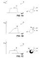
- FIG. 1Aincludes a chart and a diagram of a capnogram and a respiratory condition of a normal patient, for comparison to FIGS. 1B and 1C.
- FIG. 1Bincludes a chart and a diagram of a capnogram and a respiratory condition of a patient with an obstructive lung disease.
- FIG. 1Cincludes a chart and a diagram of a capnogram and a respiratory condition of a patient with a restrictive lung disease.
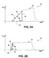
- FIG. 2Aincludes a chart of a capnogram of a patient with an obstructive lung disease.
- FIG. 2Bincludes a chart of a capnogram of a patient with a restrictive lung disease.
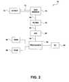
- FIG. 3is a block diagram of an apparatus that is one embodiment of the invention.
- FIG. 4is a flow diagram illustrating techniques for using capnography to analyze respiratory conditions.
- FIG. 5is a flow diagram illustrating techniques for using capnography to monitor respiratory conditions following treatment.
- FIGS. 1A, 1 B and 1 Cshow a series of three charts 10 , 20 and 30 , each chart accompanied by a diagram of an alveolus 14 .
- Chart 20shows a representative capnogram of a patient with obstructive lung disease
- chart 30shows a representative capnogram of a patient with restrictive lung disease.
- Capnograms 20 and 30are shown in reference to a capnogram 10 for a normal patient, i.e., a patient with no substantial lung disease.
- the alveoli accompanying capnograms 10 , 20 and 30illustrate the nature of the condition of the patient.
- Each alveolus 14includes a thin-walled inflatable sac 18 and a conducting airway 16 .
- the alveolus accompanying capuogram 20shows obstructions 24 in airway 16 . Sac 18 may be able to expand and perform gas exchange, but expulsion of gas from sac 18 is hampered by obstructions 24 , which narrow the lumen of airway 16 . Obstructions 24 are characteristic of obstructive lung disease.
- the alveolus accompanying capnogram 30shows restriction 34 in sac 18 , characteristic of restrictive lung disease. Restriction 34 may prevent sac 18 from expanding, or may limit the gas exchange performed by sac 18 . Airway 16 is clear, allowing unimpeded expulsion of breath, but restriction 34 limits the volume of gas in the breath.
- Capnograms 10 , 20 and 30include tracings 12 , 22 and 32 , which plot the measured concentration of carbon dioxide in the breath as a function of time. Each tracing 12 , 22 and 32 shows the concentration of carbon dioxide rise, reach a plateau and drop. The shapes of tracings 12 , 22 and 32 , however, are different. As will be shown in more detail below, analysis of the shapes of tracings 22 and 32 may be used to distinguish obstructive lung disease from restrictive lung disease.
- Tracing 22 from a patient with obstructive lung diseaseshows a more gradual rise in the ascending slope of the carbon dioxide concentration, as compared with tracings 12 and 32 from a normal patient and a patient with restrictive lung disease, respectively.
- the more gradual riseis caused by the inability of the patient to exhale rapidly due to obstructions 24 .
- the patientventilates adequately because sac 18 is clear, but the patient is not able easily to expel the contents of sac 18 through airway 16 .
- the ascending slope of tracing 32 from a patient with restrictive lung diseaseshows a rapid rise in carbon dioxide concentration when compared with tracing 22 , but a nearly normal rise in carbon dioxide concentration when compared with tracing 12 .
- a patient with restrictive lung diseasehas restriction 34 in sac 18 but no obstructions to prevent exhalation of carbon dioxide, so the rise in carbon dioxide concentration is initially normal, or nearly so.
- the carbon dioxide concentration in tracing 32plateaus at a lower concentration when compared to tracings 12 and 22 , indicating that the patient is less adequately ventilated than the normal patient and the patient with obstructive lung disease.
- FIGS. 2A and 2Bprovide a more detailed analysis of capnograms 20 and 30 .
- the first exhaled gasesgenerally carry air from so-called “dead space,” i.e., the trachea, bronchi and other structures in the brigs in which no gas exchange takes place.
- the volume of the dead spaceis approximately 150 mL.
- gases from alveoliare expelled with air from the dead space, the concentration of carbon dioxide in the breath rises.
- the concentration of carbon dioxidebegins to reach a plateau. The plateau is typically not flat.
- the rise in concentrationmay be approximated by a straight line.
- the straight linemay form the hypotenuse of a right triangle.
- the rise of carbon dioxide concentrationis approximated by hypotenuse 42 of right triangle 40
- the rise of carbon dioxide concentrationis approximated by hypotenuse 52 of right triangle 50 .
- Base 46 of triangle 40represents the duration of the rise of carbon dioxide concentration, i.e., the approximate time it takes for the carbon dioxide concentration in the breath of a patient with obstructive lung disease to reach a plateau.
- Height 44 of triangle 40represents the concentration of carbon dioxide when the patient reaches the plateau.
- base 56represents the duration of the rise of carbon dioxide concentration
- height 54represents the concentration of carbon dioxide when the patient reaches the plateau.
- the quantitiesare related, and other quantities can be derived, by the application of trigonometry.
- the areas of triangles 40 and 50can be computed and the lengths of hypotenuses 42 and 52 can be determined.
- the rate of increase of carbon dioxide concentrationcan also be determined by taking the derivative of the beginning of tracings 22 and 32 , which gives the slope.
- take-off angles 48 and 58can be found. Take-off angles 48 and 58 are one measure of the slope of hypotenuses 42 and 52 , and are a function of how rapidly carbon dioxide concentration in the breath rises. Although take-off angles 48 and 58 can be derived by trigonometry from other measurements, take-off angles 48 and 58 can also be measured directly, independent of other parameters.
- hypotenuse 42As shown by tracing 22 , a patient with obstructive lung disease takes a longer time than a patient with restrictive lung disease to expel dead space air. This is shown by the more gradual slope of hypotenuse 42 , as compared to hypotenuse 52 .
- the gradual slope of hypotenuse 42is indicative of obstructive lung disease because the gradual slope represents that it takes longer for the patient to move carbon dioxide-rich gas from his alveoli.
- hypotenuse 52is considerably steeper than hypotenuse 42 .
- the steep slope of hypotenuse 52is not indicative of obstructive lung disease because it suggests a rapid expulsion of carbon dioxide-rich gas from the alveoli.
- the extent of hypotenuse 52 , height 54 and base 56are small, however, when compared to the counterparts of triangle 40 .
- Another measure of the differenceis the area of triangle 50 , which is considerably smaller than the area of triangle 40 .
- the smaller area of triangle 50is indicative of restrictive lung disease because the patient suffers from restricted gas exchange, and cannot expel as large a volume of carbon dioxide-rich gas from the alveoli.
- the initial carbon dioxide concentration in the exhalation of a patientcan be used to distinguish obstructive lung disease from restrictive lung disease.
- a patient with obstructive lung diseaseexpels carbon dioxide more slowly, but in greater volume, than a patient with restrictive lung disease.
- capnograms 20 and 30need not be effort-dependent. Unlike spirograms, in which the patient must follow a set of instructions, capnograms 20 and 30 may be taken while the patient is breathing as comfortably as he is able, without requiring the patient to follow any breathing instructions. The clarity of tracings 22 and 32 may be improved if the patient is able to follow simple breathing instructions from a health professional, but following the instructions is not essential to the invention.
- FIG. 3is a block diagram of a system 70 that may be used to practice the invention.
- System 70includes intake apparatus 72 .
- the patientexhales into intake apparatus 72 , which may be an apparatus such as a nasal cannula or a mask.
- the exhalation from the patientpasses through tube 74 to gas sensor 76 , which measures the concentration of carbon dioxide in the breath.
- Gas sensor 76may be part of a capnograph.
- Gas sensor 76may measure carbon dioxide concentration using techniques such as infrared detection, which can track changes in concentration in real time.
- Gas sensor 76passes measurements 90 to low-pass filter 78 , which prevents aliasing.
- Filter 78passes filtered measurements 92 to analog-to-digital converter 80 , which converts filtered analog measurements 92 to digital measurement data 94 .
- Processor 82receives digital measurement data 94 .
- Digital measurement data 94may be stored in random access memory (RAM) 84 .
- processor 82Based upon digital measurement data 94 , processor 82 evaluates the carbon dioxide concentration in the patient's breath over time. Processor 82 may, for example, construct tracings such as tracings 22 or 32 shown in FIGS. 2A and 2B, and derive triangles such as triangles 40 or 50 . Processor 82 may find quantities such as duration of the rise of carbon dioxide concentration or take-off angle. Using quantities such as these, processor 82 may determine whether the data support a diagnosis of obstructive lung disease or restrictive lung disease.
- Processor 82may, for example, measuring the duration of a steady rise of the concentration of carbon dioxide. A long duration is indicative of obstructive lung disease and a short duration is indicative of restrictive lung disease. Accordingly, processor 82 may determine that the patient probably suffers from obstructive lung disease when the duration is longer than a threshold duration, and may determine that the patient probably suffers from restrictive lung disease when the duration is shorter than the threshold duration.
- processor 82may measure the rate of increase of the concentration of carbon dioxide.
- the rate of increasemay be quantified by, for example, the steepness of the hypotenuse of the ascending slope, or by the magnitude of the take-off angle, or both.
- Processor 82may determine that the patient probably suffers from obstructive lung disease when the rate of increase is lower than a threshold rate, and may determine that the patient probably suffers from restrictive lung disease when the rate of increase is higher than the threshold rate.
- processor 82may compare digital measurement data 94 to one or more characteristic curves.
- Memorysuch as read-only memory (ROM) 86 may store data that are characteristic of obstructive lung disease and data that are characteristic of restrictive lung disease.
- Processor 82may correlate the measurements of the concentration of carbon dioxide from the patient with the characteristic curves. When the correlation exceeds a preselected threshold value, processor 82 may determine that the data support a diagnosis of obstructive lung disease or restrictive lung disease.
- processor 82may also gauge the severity of the condition. Processor 82 may report a severe case of obstructive lung disease, for example, when take-off angle 48 is below a particular value, indicating that the patient has extreme difficulty pushing out his breath. Degrees of severity may also be reported, such as “critical,” “moderate” and “mild.”
- Processor 82reports the results of the analysis to the health professional via input/output (I/O) device 88 .
- I/O device 88may include, for example, a display screen that displays text or graphics, or a collection of light emitting diodes.
- Processor 82may report an analysis, such as “Patient's exhaled carbon dioxide concentration indicates a greater likelihood of obstructive lung disease than restrictive lung disease,” or “Patient's exhaled carbon dioxide concentration indicates a high probability of obstructive lung disease.”
- Processor 82may further report on the severity of the condition, and/or may display the tracing of the carbon dioxide concentration.
- processor 82may suggest an appropriate treatment based upon the analysis.
- system 70may be small and easily portable. Accordingly, system 70 may be included in first aid packages in public venues such as airports and health clubs, or may be carried to the patient by an emergency medical professional. Furthermore, unlike a spirometer, system 70 may provide guidance for treatment of the patient very quickly, and need not be effort-dependent.
- system 70is an example of one system that may be used to practice the invention, and the invention is not limited to the system shown.
- digital measurement data 94may be supplied to RAM 84 via a direct memory access module (not shown in FIG. 3 ), rather than via processor 82 .
- ROM 86may include erasable programmable read-only memory (EPROM).
- I/O device 88may be one of several input and/or output devices. The invention encompasses all of these variations.
- FIG. 4is a flow diagram illustrating an embodiment of the invention in an exemplary application, such as the case of a patient suffering from a shortness of breath.
- System 70receives expired breath from the patient via intake apparatus 72 ( 100 ).
- Gas sensor 76measures the carbon dioxide concentration ( 102 ) and reports the measurements to processor 82 .
- system 70helps in determine the nature of the condition and further helps guide treatment of the patient.
- processor 82analyzes the measurements over time ( 104 ) using techniques such as those described above and ascertains whether the data support a determination that lung disease is present ( 106 ). When the data support a determination that obstructive lung disease is present, processor 82 may so report via I/O device 88 ( 108 ). Similarly, when the data support a determination that restrictive lung disease is present, processor 82 may so report ( 110 ). In some circumstances, the data may support neither case, and processor 82 may so report ( 112 ).
- Processor 82may also report additional information ( 114 ) that may guide the treatment of the patient. For example, processor 82 may report the severity of the condition, or may suggest a medicine for the condition, or may recommend that the measurements be repeated, or may suggest that the patient be instructed to breathe in a particular manner.
- FIG. 5is a flow diagram showing how the invention may be implemented to monitor the effectiveness of treatment.
- System 70receives expired breath from a patient via intake apparatus 72 ( 120 ), gas sensor 76 measures the carbon dioxide concentration ( 122 ) and processor 82 analyzes the measurements ( 124 ). Instead of reporting a determination of lung disease, however, processor 82 monitors changes in the condition of the patient, and reports the changes via I/O device 88 . In this way, the invention may be used to observe the responsiveness of the patient to treatment.
- capnogramsfrom a plurality of breaths, and may process the capnograms by techniques such as averaging. These and other embodiments are within the scope of the following claims.
Landscapes
- Health & Medical Sciences (AREA)
- Life Sciences & Earth Sciences (AREA)
- Biomedical Technology (AREA)
- Heart & Thoracic Surgery (AREA)
- Pulmonology (AREA)
- Physics & Mathematics (AREA)
- Obesity (AREA)
- Biophysics (AREA)
- Pathology (AREA)
- Engineering & Computer Science (AREA)
- Emergency Medicine (AREA)
- Physiology (AREA)
- Medical Informatics (AREA)
- Molecular Biology (AREA)
- Surgery (AREA)
- Animal Behavior & Ethology (AREA)
- General Health & Medical Sciences (AREA)
- Public Health (AREA)
- Veterinary Medicine (AREA)
- Measurement Of The Respiration, Hearing Ability, Form, And Blood Characteristics Of Living Organisms (AREA)
- Investigating Or Analysing Biological Materials (AREA)
- Analysing Materials By The Use Of Radiation (AREA)
Abstract
Description
Claims (42)
Priority Applications (5)
| Application Number | Priority Date | Filing Date | Title |
|---|---|---|---|
| US09/978,831US6648833B2 (en) | 2001-10-15 | 2001-10-15 | Respiratory analysis with capnography |
| PCT/US2002/032775WO2003032831A1 (en) | 2001-10-15 | 2002-10-15 | Respiratory analysis with capnography |
| AT02784100TATE296056T1 (en) | 2001-10-15 | 2002-10-15 | RESPIRATORY FUNCTION ANALYSIS USING CAPNOGRAPHY |
| DE60204336TDE60204336T2 (en) | 2001-10-15 | 2002-10-15 | ATOMIC FUNCTION ANALYSIS BY CAPAPIOGRAPHY |
| EP02784100AEP1435836B1 (en) | 2001-10-15 | 2002-10-15 | Respiratory analysis with capnography |
Applications Claiming Priority (1)
| Application Number | Priority Date | Filing Date | Title |
|---|---|---|---|
| US09/978,831US6648833B2 (en) | 2001-10-15 | 2001-10-15 | Respiratory analysis with capnography |
Publications (2)
| Publication Number | Publication Date |
|---|---|
| US20030073919A1 US20030073919A1 (en) | 2003-04-17 |
| US6648833B2true US6648833B2 (en) | 2003-11-18 |
Family
ID=25526431
Family Applications (1)
| Application Number | Title | Priority Date | Filing Date |
|---|---|---|---|
| US09/978,831Expired - LifetimeUS6648833B2 (en) | 2001-10-15 | 2001-10-15 | Respiratory analysis with capnography |
Country Status (5)
| Country | Link |
|---|---|
| US (1) | US6648833B2 (en) |
| EP (1) | EP1435836B1 (en) |
| AT (1) | ATE296056T1 (en) |
| DE (1) | DE60204336T2 (en) |
| WO (1) | WO2003032831A1 (en) |
Cited By (8)
| Publication number | Priority date | Publication date | Assignee | Title |
|---|---|---|---|---|
| US20040210151A1 (en)* | 2003-04-15 | 2004-10-21 | Ross Tsukashima | Respiratory monitoring, diagnostic and therapeutic system |
| US20040236240A1 (en)* | 2000-12-07 | 2004-11-25 | Kraus Baruch Shlomo | Automated interpretive medical care system and methodology |
| US20080009762A1 (en)* | 2006-06-27 | 2008-01-10 | Medtronic Emergency Response Systems, Inc. | Method and apparatus for interpreting capnographic data |
| US8221327B2 (en) | 2003-09-18 | 2012-07-17 | Cardiac Pacemakers, Inc. | Therapy control based on cardiopulmonary status |
| US8915741B2 (en) | 2003-08-18 | 2014-12-23 | Cardiac Pacemakers, Inc. | Sleep quality data collection and evaluation |
| US9790161B2 (en) | 2010-03-26 | 2017-10-17 | Dioxide Materials, Inc | Process for the sustainable production of acrylic acid |
| US10388405B2 (en) | 2013-03-22 | 2019-08-20 | Massachusetts Institute Of Technology | Systems and methods for predicting adverse events and assessing level of sedation during medical procedures |
| US11972843B2 (en) | 2015-08-12 | 2024-04-30 | Massachusetts Institute Of Technology | Systems and methods for predicting adverse events and assessing level of sedation during medical procedures |
Families Citing this family (38)
| Publication number | Priority date | Publication date | Assignee | Title |
|---|---|---|---|---|
| US8932227B2 (en)* | 2000-07-28 | 2015-01-13 | Lawrence A. Lynn | System and method for CO2 and oximetry integration |
| US8152991B2 (en) | 2005-10-27 | 2012-04-10 | Nanomix, Inc. | Ammonia nanosensors, and environmental control system |
| US8154093B2 (en) | 2002-01-16 | 2012-04-10 | Nanomix, Inc. | Nano-electronic sensors for chemical and biological analytes, including capacitance and bio-membrane devices |
| US20050129573A1 (en)* | 2003-09-12 | 2005-06-16 | Nanomix, Inc. | Carbon dioxide nanoelectronic sensor |
| US20070048181A1 (en)* | 2002-09-05 | 2007-03-01 | Chang Daniel M | Carbon dioxide nanosensor, and respiratory CO2 monitors |
| US7714398B2 (en)* | 2002-09-05 | 2010-05-11 | Nanomix, Inc. | Nanoelectronic measurement system for physiologic gases and improved nanosensor for carbon dioxide |
| US20070048180A1 (en)* | 2002-09-05 | 2007-03-01 | Gabriel Jean-Christophe P | Nanoelectronic breath analyzer and asthma monitor |
| US7522040B2 (en)* | 2004-04-20 | 2009-04-21 | Nanomix, Inc. | Remotely communicating, battery-powered nanostructure sensor devices |
| US7547931B2 (en)* | 2003-09-05 | 2009-06-16 | Nanomix, Inc. | Nanoelectronic capnometer adaptor including a nanoelectric sensor selectively sensitive to at least one gaseous constituent of exhaled breath |
| US7024235B2 (en)* | 2002-06-20 | 2006-04-04 | University Of Florida Research Foundation, Inc. | Specially configured nasal pulse oximeter/photoplethysmography probes, and combined nasal probe/cannula, selectively with sampler for capnography, and covering sleeves for same |
| US7948041B2 (en) | 2005-05-19 | 2011-05-24 | Nanomix, Inc. | Sensor having a thin-film inhibition layer |
| US20070114573A1 (en)* | 2002-09-04 | 2007-05-24 | Tzong-Ru Han | Sensor device with heated nanostructure |
| US20060263255A1 (en)* | 2002-09-04 | 2006-11-23 | Tzong-Ru Han | Nanoelectronic sensor system and hydrogen-sensitive functionalization |
| US7189204B2 (en) | 2002-12-04 | 2007-03-13 | Cardiac Pacemakers, Inc. | Sleep detection using an adjustable threshold |
| IL155955A0 (en)* | 2003-05-15 | 2003-12-23 | Widemed Ltd | Adaptive prediction of changes of physiological/pathological states using processing of biomedical signal |
| US7591265B2 (en) | 2003-09-18 | 2009-09-22 | Cardiac Pacemakers, Inc. | Coordinated use of respiratory and cardiac therapies for sleep disordered breathing |
| US7575553B2 (en)* | 2003-09-18 | 2009-08-18 | Cardiac Pacemakers, Inc. | Methods and systems for assessing pulmonary disease |
| US7510531B2 (en)* | 2003-09-18 | 2009-03-31 | Cardiac Pacemakers, Inc. | System and method for discrimination of central and obstructive disordered breathing events |
| US7757690B2 (en) | 2003-09-18 | 2010-07-20 | Cardiac Pacemakers, Inc. | System and method for moderating a therapy delivered during sleep using physiologic data acquired during non-sleep |
| EP1670547B1 (en) | 2003-08-18 | 2008-11-12 | Cardiac Pacemakers, Inc. | Patient monitoring system |
| US8606356B2 (en) | 2003-09-18 | 2013-12-10 | Cardiac Pacemakers, Inc. | Autonomic arousal detection system and method |
| US7610094B2 (en)* | 2003-09-18 | 2009-10-27 | Cardiac Pacemakers, Inc. | Synergistic use of medical devices for detecting medical disorders |
| US7720541B2 (en) | 2003-08-18 | 2010-05-18 | Cardiac Pacemakers, Inc. | Adaptive therapy for disordered breathing |
| US7887493B2 (en) | 2003-09-18 | 2011-02-15 | Cardiac Pacemakers, Inc. | Implantable device employing movement sensing for detecting sleep-related disorders |
| US8251061B2 (en)* | 2003-09-18 | 2012-08-28 | Cardiac Pacemakers, Inc. | Methods and systems for control of gas therapy |
| US7970470B2 (en)* | 2003-09-18 | 2011-06-28 | Cardiac Pacemakers, Inc. | Diagnosis and/or therapy using blood chemistry/expired gas parameter analysis |
| US20050053549A1 (en)* | 2003-09-10 | 2005-03-10 | Aperon Biosystems Corp. | Method for treating airway disorders |
| RU2245171C1 (en)* | 2004-04-29 | 2005-01-27 | Бутейко Людмила Дмитриевна | Method for treating hypocarbic diseases and states |
| US20080269583A1 (en)* | 2005-02-07 | 2008-10-30 | Widemed Ltd. | Detection and Monitoring of Stress Events During Sleep |
| US20080300500A1 (en)* | 2007-05-30 | 2008-12-04 | Widemed Ltd. | Apnea detection using a capnograph |
| CN102471051B (en) | 2009-08-07 | 2014-06-11 | 纳诺米克斯公司 | Magnetic carbon nanotube based biodetection |
| RU2476149C1 (en)* | 2011-06-29 | 2013-02-27 | Государственное образовательное учреждение высшего профессионального образования "Воронежская государственная медицинская академия им. Н.Н. Бурденко" Министерства здравоохранения и социального развития Российской Федерации | Method of predicting respiratory disorders in patients with ventral hernia in postoperative period |
| DE102012220565B4 (en)* | 2012-11-12 | 2016-04-07 | Bluepoint Medical Gmbh & Co. Kg | Gas sampling line, gas analysis system, use and method for analyzing an analysis gas |
| US11026595B2 (en)* | 2014-02-03 | 2021-06-08 | Oridion Medical 1987 Ltd. | Feature trend display |
| US11147507B2 (en) | 2014-02-03 | 2021-10-19 | Oridion Medical 1987 Ltd. | Decision support system for cardiopulmonary resuscitation (CPR) |
| EP4393532A3 (en)* | 2016-04-29 | 2024-09-25 | Fisher & Paykel Healthcare Limited | System for determining airway patency |
| EP3624690B1 (en) | 2017-05-15 | 2023-12-20 | Agency for Science, Technology and Research | Method and system for respiratory measurement |
| CN115153499B (en)* | 2022-07-04 | 2025-02-18 | 北京万联达信科仪器有限公司 | A breath sampling method and device |
Citations (21)
| Publication number | Priority date | Publication date | Assignee | Title |
|---|---|---|---|---|
| US3687130A (en) | 1969-11-26 | 1972-08-29 | Pelam Inc | Instrument to measure pulmonary function |
| DE2812379A1 (en) | 1978-03-21 | 1979-09-27 | Siemens Ag | Pulmonary diagnostic system - comparing carbon di:oxide concn. difference and breath vol. by microcomputer |
| US4169465A (en) | 1977-05-04 | 1979-10-02 | James A. Walls | Method and apparatus for obtaining non-invasive cardio-pulmonary measurements |
| US4772559A (en) | 1985-10-10 | 1988-09-20 | Monell Chemical Senses Center | Method of detecting the presence of bronchogenic carcinoma by analysis of expired lung air |
| US4796639A (en) | 1987-11-05 | 1989-01-10 | Medical Graphics Corporation | Pulmonary diagnostic system |
| US4821736A (en) | 1988-03-22 | 1989-04-18 | Dale Medical Products, Inc. | Head-mounted device for supporting breathing circuit tubes and sensor |
| US5058601A (en) | 1988-02-10 | 1991-10-22 | Sherwood Medical Company | Pulmonary function tester |
| US5159935A (en) | 1990-03-08 | 1992-11-03 | Nims, Inc. | Non-invasive estimation of individual lung function |
| EP0699414A1 (en) | 1994-08-30 | 1996-03-06 | The BOC Group plc | Disease management system |
| US5515859A (en) | 1993-08-24 | 1996-05-14 | Colorado Health Care Research Corp. | Myocardial infarction and ischemia detection method and apparatus |
| US5632281A (en) | 1995-02-06 | 1997-05-27 | Rayburn; Daniel B. | Non-invasive estimation of arterial blood gases |
| US5682877A (en) | 1991-12-30 | 1997-11-04 | Mondry; Adolph J. | System and method for automatically maintaining a blood oxygen saturation level |
| US5800361A (en) | 1995-02-06 | 1998-09-01 | Ntc Technology Inc. | Non-invasive estimation of arterial blood gases |
| US5971934A (en) | 1996-10-04 | 1999-10-26 | Trustees Of The University Of Pennsylvania | Noninvasive method and apparatus for determining cardiac output |
| US5984872A (en) | 1997-07-14 | 1999-11-16 | W. H. Vriend | Area and shape of the flow-volume curve in lung diagnostics |
| US6044843A (en) | 1997-05-28 | 2000-04-04 | Nellcor Puritan Bennett Incorporated | Moisture resistant airway adapter for monitoring constituent gases |
| US6066101A (en) | 1998-04-20 | 2000-05-23 | University Of Maryland | Airflow perturbation device and method for measuring respiratory resistance |
| US6068602A (en) | 1997-09-26 | 2000-05-30 | Ohmeda Inc. | Method and apparatus for determining airway resistance and lung compliance |
| US6102868A (en) | 1998-10-16 | 2000-08-15 | University Of Florida | Method and system for measuring the cardiac output of a living being |
| US6142952A (en) | 1997-10-29 | 2000-11-07 | The Board Of Regents, The University Of Texas System | Method and apparatus for detection and diagnosis of airway obstruction degree |
| US6174289B1 (en) | 1999-05-28 | 2001-01-16 | Orca Diagnostics Corporation | Cardiopulmonary exercise testing apparatus and method |
- 2001
- 2001-10-15USUS09/978,831patent/US6648833B2/ennot_activeExpired - Lifetime
- 2002
- 2002-10-15DEDE60204336Tpatent/DE60204336T2/ennot_activeExpired - Lifetime
- 2002-10-15WOPCT/US2002/032775patent/WO2003032831A1/ennot_activeApplication Discontinuation
- 2002-10-15EPEP02784100Apatent/EP1435836B1/ennot_activeExpired - Lifetime
- 2002-10-15ATAT02784100Tpatent/ATE296056T1/ennot_activeIP Right Cessation
Patent Citations (22)
| Publication number | Priority date | Publication date | Assignee | Title |
|---|---|---|---|---|
| US3687130A (en) | 1969-11-26 | 1972-08-29 | Pelam Inc | Instrument to measure pulmonary function |
| US4169465A (en) | 1977-05-04 | 1979-10-02 | James A. Walls | Method and apparatus for obtaining non-invasive cardio-pulmonary measurements |
| DE2812379A1 (en) | 1978-03-21 | 1979-09-27 | Siemens Ag | Pulmonary diagnostic system - comparing carbon di:oxide concn. difference and breath vol. by microcomputer |
| US4772559A (en) | 1985-10-10 | 1988-09-20 | Monell Chemical Senses Center | Method of detecting the presence of bronchogenic carcinoma by analysis of expired lung air |
| US4796639A (en) | 1987-11-05 | 1989-01-10 | Medical Graphics Corporation | Pulmonary diagnostic system |
| US5058601A (en) | 1988-02-10 | 1991-10-22 | Sherwood Medical Company | Pulmonary function tester |
| US4821736A (en) | 1988-03-22 | 1989-04-18 | Dale Medical Products, Inc. | Head-mounted device for supporting breathing circuit tubes and sensor |
| US5159935A (en) | 1990-03-08 | 1992-11-03 | Nims, Inc. | Non-invasive estimation of individual lung function |
| US5682877A (en) | 1991-12-30 | 1997-11-04 | Mondry; Adolph J. | System and method for automatically maintaining a blood oxygen saturation level |
| US5515859A (en) | 1993-08-24 | 1996-05-14 | Colorado Health Care Research Corp. | Myocardial infarction and ischemia detection method and apparatus |
| EP0699414A1 (en) | 1994-08-30 | 1996-03-06 | The BOC Group plc | Disease management system |
| US5632281A (en) | 1995-02-06 | 1997-05-27 | Rayburn; Daniel B. | Non-invasive estimation of arterial blood gases |
| US5800361A (en) | 1995-02-06 | 1998-09-01 | Ntc Technology Inc. | Non-invasive estimation of arterial blood gases |
| US6251082B1 (en) | 1995-02-06 | 2001-06-26 | Ntc Technology, Inc. | Non-invasive estimation of arterial blood gases |
| US5971934A (en) | 1996-10-04 | 1999-10-26 | Trustees Of The University Of Pennsylvania | Noninvasive method and apparatus for determining cardiac output |
| US6044843A (en) | 1997-05-28 | 2000-04-04 | Nellcor Puritan Bennett Incorporated | Moisture resistant airway adapter for monitoring constituent gases |
| US5984872A (en) | 1997-07-14 | 1999-11-16 | W. H. Vriend | Area and shape of the flow-volume curve in lung diagnostics |
| US6068602A (en) | 1997-09-26 | 2000-05-30 | Ohmeda Inc. | Method and apparatus for determining airway resistance and lung compliance |
| US6142952A (en) | 1997-10-29 | 2000-11-07 | The Board Of Regents, The University Of Texas System | Method and apparatus for detection and diagnosis of airway obstruction degree |
| US6066101A (en) | 1998-04-20 | 2000-05-23 | University Of Maryland | Airflow perturbation device and method for measuring respiratory resistance |
| US6102868A (en) | 1998-10-16 | 2000-08-15 | University Of Florida | Method and system for measuring the cardiac output of a living being |
| US6174289B1 (en) | 1999-05-28 | 2001-01-16 | Orca Diagnostics Corporation | Cardiopulmonary exercise testing apparatus and method |
Non-Patent Citations (9)
| Title |
|---|
| Brown et al., "Can Quantitative Capnometry Differentiate Between Cardiac and Obstructive Causes of Respiratory Distress?", Chest 113(2), pp. 323-326 (1998). |
| C.V. Egleston et al., "Capnography for monitoring non-intubated spontaneously breathing patients in an emergency room setting", vol. 14, No. 4, Jul. 1997, pp. 222-224, Journal of Accident and Emergency Medicine. |
| PCT Application Serial No. PCT/IL01/01127, filed Dec. 6, 2001. |
| U.S. patent application Ser. No. 09/590,202, filed Jun. 8, 2000. |
| U.S. Provisional Application Ser. No. 60/251,829, filed Dec. 7, 2000. |
| www.emedicine.com online article on restrictive lung disease-update Jan. 20, 2003, author Sat Sharma, Editor Laurie Robin Grier, 30 pages.** |
| www.emedicine.com online article on restrictive lung disease—update Jan. 20, 2003, author Sat Sharma, Editor Laurie Robin Grier, 30 pages.* |
| Yaron et al., "Utility of the Expiratory Capnogram in the Assessment of Bronchospasm," Annals of Emergency Medicine 28(4), pp. 403-407 (1996). |
| You et al., "Expiratory Capnography in Asthma: Evaluation of Various Shape Indices," The European Respiratory Journal 7(2), pp. 318,323 (1994). |
Cited By (19)
| Publication number | Priority date | Publication date | Assignee | Title |
|---|---|---|---|---|
| US8679029B2 (en) | 2000-12-07 | 2014-03-25 | Oridion Medical (1987) Ltd. | Automated interpretive medical care system and methodology |
| US9895065B2 (en) | 2000-12-07 | 2018-02-20 | Children's Medical Center Corporation | Automated interpretive medical care system and methodology |
| US9895066B2 (en) | 2000-12-07 | 2018-02-20 | Oridion Medical 1987 Ltd. | Automated interpretive medical care system and methodology |
| US8147419B2 (en)* | 2000-12-07 | 2012-04-03 | Baruch Shlomo Krauss | Automated interpretive medical care system and methodology |
| US10610110B2 (en) | 2000-12-07 | 2020-04-07 | Children's Medical Center Corporation | Automated interpretive medical care system and methodology |
| US20090143694A1 (en)* | 2000-12-07 | 2009-06-04 | Baruch Shlomo Krauss | Automated interpretive medical care system and methodology |
| US9993163B2 (en) | 2000-12-07 | 2018-06-12 | Oridion Medical 1987 Ltd. | Automated interpretive medical care system and methodology |
| US20090149723A1 (en)* | 2000-12-07 | 2009-06-11 | Baruch Shlomo Krauss | Automated interpretive medical care system and methodology |
| US20040236240A1 (en)* | 2000-12-07 | 2004-11-25 | Kraus Baruch Shlomo | Automated interpretive medical care system and methodology |
| US9955875B2 (en) | 2000-12-07 | 2018-05-01 | Oridion Medical 1987 Ltd. | Automated interpretive medical care system and methodology |
| US20040210151A1 (en)* | 2003-04-15 | 2004-10-21 | Ross Tsukashima | Respiratory monitoring, diagnostic and therapeutic system |
| US7101341B2 (en)* | 2003-04-15 | 2006-09-05 | Ross Tsukashima | Respiratory monitoring, diagnostic and therapeutic system |
| WO2004100762A3 (en)* | 2003-04-15 | 2005-07-28 | Seirra Medical Technology Inc | Respiratory monitoring, diagnostic and therapeutic system |
| US8915741B2 (en) | 2003-08-18 | 2014-12-23 | Cardiac Pacemakers, Inc. | Sleep quality data collection and evaluation |
| US8221327B2 (en) | 2003-09-18 | 2012-07-17 | Cardiac Pacemakers, Inc. | Therapy control based on cardiopulmonary status |
| US20080009762A1 (en)* | 2006-06-27 | 2008-01-10 | Medtronic Emergency Response Systems, Inc. | Method and apparatus for interpreting capnographic data |
| US9790161B2 (en) | 2010-03-26 | 2017-10-17 | Dioxide Materials, Inc | Process for the sustainable production of acrylic acid |
| US10388405B2 (en) | 2013-03-22 | 2019-08-20 | Massachusetts Institute Of Technology | Systems and methods for predicting adverse events and assessing level of sedation during medical procedures |
| US11972843B2 (en) | 2015-08-12 | 2024-04-30 | Massachusetts Institute Of Technology | Systems and methods for predicting adverse events and assessing level of sedation during medical procedures |
Also Published As
| Publication number | Publication date |
|---|---|
| US20030073919A1 (en) | 2003-04-17 |
| DE60204336D1 (en) | 2005-06-30 |
| ATE296056T1 (en) | 2005-06-15 |
| EP1435836B1 (en) | 2005-05-25 |
| DE60204336T2 (en) | 2006-01-26 |
| WO2003032831A1 (en) | 2003-04-24 |
| EP1435836A1 (en) | 2004-07-14 |
Similar Documents
| Publication | Publication Date | Title |
|---|---|---|
| US6648833B2 (en) | Respiratory analysis with capnography | |
| US6428483B1 (en) | Waveform interpreter for respiratory analysis | |
| Al-Ashkar et al. | Interpreting pulmonary function tests: recognize the pattern, and the diagnosis will follow | |
| US8414488B2 (en) | Medical system, apparatus and method | |
| CN103989478B (en) | Detection and the method and device for the treatment of respiratory insufficiency | |
| JP6294081B2 (en) | Real-time airway check status indicator | |
| US7635339B2 (en) | Method for non-cooperative lung function diagnosis using ultrasound | |
| US20210244900A1 (en) | Method for operating a ventilator for artificial ventilation of a patient, and such a ventilator | |
| EP3148426B1 (en) | Method, system and software for assessing extubation failure | |
| EP3359035B1 (en) | Device and system for determining a respiratory feature of a subject based on a breathing gas | |
| EP3334339B1 (en) | Capnography with decision support system architecture | |
| CN108024758A (en) | End-tidal CO2Simplify display | |
| Balakrishnan et al. | Clinical application of pulmonary function testing in small animals | |
| US20150151072A1 (en) | Ventilation analysis and monitoring | |
| KR20240158489A (en) | mart spirometer capable of measuring expiratory temperature (EBT) and COPD patient screening system using the same | |
| Voter et al. | Pulmonary function testing in childhood asthma | |
| Nagler et al. | Capnographic monitoring in respiratory emergencies | |
| JP4738021B2 (en) | Early lung lesion detection apparatus and method of using the apparatus | |
| RU2324508C2 (en) | Method of diagnostics of obstructive disorders of external respiration by use of capnography | |
| Ermer et al. | Detecting low respiratory rates using myriad, low-cost sensors | |
| KR20240156003A (en) | Lung disease prediction method using PFT-based AI | |
| KR20240156011A (en) | Autologous lung function test system using computer vision technology | |
| Scanlan et al. | Pulmonary Function Testing | |
| Ermer et al. | Identification of Respiratory Distress Through Combining Modern Sensors in Patients Receiving Opioids and Anesthetics | |
| Byalovsky et al. | Elamed CP-01 capnographic hardware-software system for general practitioners |
Legal Events
| Date | Code | Title | Description |
|---|---|---|---|
| STCF | Information on status: patent grant | Free format text:PATENTED CASE | |
| CC | Certificate of correction | ||
| FPAY | Fee payment | Year of fee payment:4 | |
| FPAY | Fee payment | Year of fee payment:8 | |
| AS | Assignment | Owner name:BANK OF NEW YORK MELLON TRUST COMPANY, N.A., AS *C Free format text:SECURITY AGREEMENT;ASSIGNOR:PHYSIO-CONTROL, INC.;REEL/FRAME:027765/0861 Effective date:20120130 | |
| AS | Assignment | Owner name:CITIBANK, N.A., AS COLLATERAL AGENT, NEW YORK Free format text:SECURITY AGREEMENT;ASSIGNOR:PHYSIO-CONTROL, INC.;REEL/FRAME:027763/0881 Effective date:20120130 | |
| AS | Assignment | Owner name:PHYSIO-CONTROL, INC., WASHINGTON Free format text:ASSIGNMENT OF ASSIGNORS INTEREST;ASSIGNOR:HAMPTON, DAVID R.;REEL/FRAME:027908/0018 Effective date:20120322 | |
| FPAY | Fee payment | Year of fee payment:12 | |
| AS | Assignment | Owner name:PHYSIO-CONTROL, INC., WASHINGTON Free format text:RELEASE BY SECURED PARTY;ASSIGNOR:THE BANK OF NEW YORK MELLON TRUST COMPANY, N.A.;REEL/FRAME:037519/0240 Effective date:20150605 | |
| AS | Assignment | Owner name:CITIBANK, N.A., AS COLLATERAL AGENT, NEW YORK Free format text:FIRST LIEN SECURITY AGREEMENT;ASSIGNORS:PHYSIO-CONTROL, INC.;PHYSIO-CONTROL INTERNATIONAL, INC.;REEL/FRAME:037532/0828 Effective date:20150605 | |
| AS | Assignment | Owner name:CITIBANK, N.A., AS COLLATERAL AGENT, NEW YORK Free format text:SECOND LIEN SECURITY AGREEMENT;ASSIGNORS:PHYSIO-CONTROL, INC.;PHYSIO-CONTROL INTERNATIONAL, INC.;REEL/FRAME:037559/0601 Effective date:20150605 | |
| AS | Assignment | Owner name:PHYSIO-CONTROL, INC., WASHINGTON Free format text:RELEASE BY SECURED PARTY;ASSIGNOR:CITIBANK, N.A.;REEL/FRAME:038376/0806 Effective date:20160405 Owner name:PHYSIO-CONTROL, INC., WASHINGTON Free format text:RELEASE BY SECURED PARTY;ASSIGNOR:CITIBANK, N.A.;REEL/FRAME:038379/0001 Effective date:20160405 Owner name:PHYSIO-CONTROL INTERNATIONAL, INC., WASHINGTON Free format text:RELEASE BY SECURED PARTY;ASSIGNOR:CITIBANK, N.A.;REEL/FRAME:038379/0001 Effective date:20160405 Owner name:PHYSIO-CONTROL INTERNATIONAL, INC., WASHINGTON Free format text:RELEASE BY SECURED PARTY;ASSIGNOR:CITIBANK, N.A.;REEL/FRAME:038378/0028 Effective date:20160405 Owner name:PHYSIO-CONTROL, INC., WASHINGTON Free format text:RELEASE BY SECURED PARTY;ASSIGNOR:CITIBANK, N.A.;REEL/FRAME:038378/0028 Effective date:20160405 |