US6632197B2 - Clear view cannula - Google Patents
Clear view cannulaDownload PDFInfo
- Publication number
- US6632197B2 US6632197B2US09/293,284US29328499AUS6632197B2US 6632197 B2US6632197 B2US 6632197B2US 29328499 AUS29328499 AUS 29328499AUS 6632197 B2US6632197 B2US 6632197B2
- Authority
- US
- United States
- Prior art keywords
- cannula
- tubular body
- cylindrical sleeve
- shield members
- distal end
- Prior art date
- Legal status (The legal status is an assumption and is not a legal conclusion. Google has not performed a legal analysis and makes no representation as to the accuracy of the status listed.)
- Expired - Lifetime
Links
- 230000035515penetrationEffects0.000claimsdescription8
- 238000003780insertionMethods0.000claimsdescription7
- 230000037431insertionEffects0.000claimsdescription7
- 238000005452bendingMethods0.000claims2
- 238000001356surgical procedureMethods0.000abstractdescription10
- 210000004872soft tissueAnatomy0.000abstractdescription8
- 210000001519tissueAnatomy0.000description13
- 230000000717retained effectEffects0.000description6
- 230000001154acute effectEffects0.000description3
- 210000000629knee jointAnatomy0.000description3
- 239000000463materialSubstances0.000description3
- 238000000034methodMethods0.000description3
- 210000001015abdomenAnatomy0.000description2
- 210000000683abdominal cavityAnatomy0.000description2
- 210000000577adipose tissueAnatomy0.000description2
- 238000004873anchoringMethods0.000description2
- 229920001971elastomerPolymers0.000description2
- 239000012530fluidSubstances0.000description2
- 210000003127kneeAnatomy0.000description2
- 239000004816latexSubstances0.000description2
- 229920000126latexPolymers0.000description2
- 210000004417patellaAnatomy0.000description2
- 230000000149penetrating effectEffects0.000description2
- 210000005222synovial tissueAnatomy0.000description2
- 230000000007visual effectEffects0.000description2
- 241001631457CannulaSpecies0.000description1
- 230000003187abdominal effectEffects0.000description1
- 210000003423ankleAnatomy0.000description1
- 238000004140cleaningMethods0.000description1
- 238000010276constructionMethods0.000description1
- 230000007812deficiencyEffects0.000description1
- 210000001513elbowAnatomy0.000description1
- 210000003414extremityAnatomy0.000description1
- 230000004927fusionEffects0.000description1
- 208000014674injuryDiseases0.000description1
- 239000007788liquidSubstances0.000description1
- 238000004519manufacturing processMethods0.000description1
- 238000012986modificationMethods0.000description1
- 230000004048modificationEffects0.000description1
- 210000002445nippleAnatomy0.000description1
- 239000004033plasticSubstances0.000description1
- 210000002832shoulderAnatomy0.000description1
- 230000001954sterilising effectEffects0.000description1
- 238000004659sterilization and disinfectionMethods0.000description1
- 210000001258synovial membraneAnatomy0.000description1
- 230000008733traumaEffects0.000description1
- 238000012800visualizationMethods0.000description1
- XLYOFNOQVPJJNP-UHFFFAOYSA-NwaterSubstancesOXLYOFNOQVPJJNP-UHFFFAOYSA-N0.000description1
- 238000003466weldingMethods0.000description1
Images


Classifications
- A—HUMAN NECESSITIES
- A61—MEDICAL OR VETERINARY SCIENCE; HYGIENE
- A61B—DIAGNOSIS; SURGERY; IDENTIFICATION
- A61B17/00—Surgical instruments, devices or methods
- A61B17/34—Trocars; Puncturing needles
- A61B17/3417—Details of tips or shafts, e.g. grooves, expandable, bendable; Multiple coaxial sliding cannulas, e.g. for dilating
- A61B17/3421—Cannulas
- A—HUMAN NECESSITIES
- A61—MEDICAL OR VETERINARY SCIENCE; HYGIENE
- A61M—DEVICES FOR INTRODUCING MEDIA INTO, OR ONTO, THE BODY; DEVICES FOR TRANSDUCING BODY MEDIA OR FOR TAKING MEDIA FROM THE BODY; DEVICES FOR PRODUCING OR ENDING SLEEP OR STUPOR
- A61M25/00—Catheters; Hollow probes
- A61M25/01—Introducing, guiding, advancing, emplacing or holding catheters
- A61M25/02—Holding devices, e.g. on the body
- A—HUMAN NECESSITIES
- A61—MEDICAL OR VETERINARY SCIENCE; HYGIENE
- A61B—DIAGNOSIS; SURGERY; IDENTIFICATION
- A61B17/00—Surgical instruments, devices or methods
- A61B17/34—Trocars; Puncturing needles
- A61B2017/348—Means for supporting the trocar against the body or retaining the trocar inside the body
- A61B2017/3482—Means for supporting the trocar against the body or retaining the trocar inside the body inside
- A61B2017/3484—Anchoring means, e.g. spreading-out umbrella-like structure
- A—HUMAN NECESSITIES
- A61—MEDICAL OR VETERINARY SCIENCE; HYGIENE
- A61M—DEVICES FOR INTRODUCING MEDIA INTO, OR ONTO, THE BODY; DEVICES FOR TRANSDUCING BODY MEDIA OR FOR TAKING MEDIA FROM THE BODY; DEVICES FOR PRODUCING OR ENDING SLEEP OR STUPOR
- A61M25/00—Catheters; Hollow probes
- A61M25/01—Introducing, guiding, advancing, emplacing or holding catheters
- A61M25/02—Holding devices, e.g. on the body
- A61M2025/0213—Holding devices, e.g. on the body where the catheter is attached by means specifically adapted to a part of the human body
- A61M2025/0233—Holding devices, e.g. on the body where the catheter is attached by means specifically adapted to a part of the human body specifically adapted for attaching to a body wall by means which are on both sides of the wall, e.g. for attaching to an abdominal wall
Definitions
- This inventionrelates to a clear view cannula for use in surgical procedures. More particularly, this invention relates to a clear view cannula that permits an operator to insert the cannula through a body wall into an anatomical cavity, maintain the cannula in its inserted position, and introduce an auxiliary surgical instrument into the anatomical cavity through the cannula without visual or mechanical obstruction within the body wall.
- a typical procedurecan include making a small incision in the desired portion of the body wall, inserting a cannula into and through the incision and introducing an auxiliary surgical instrument through the cannula into the body to perform a further procedure.
- the cannulabe capable of being anchored or secured and not slip out of the anatomical cavity thereby preventing its reinsertion and that fragmented or torn soft tissue not have to be removed from the area surrounding the insertion point of the cannula in order to ensure that the auxiliary instrument can be used without visual or mechanical obstruction.
- U.S. Pat. No. 5,217,451 to Freitasdiscloses a trocar assembly having first and second cylindrical members secured to one another at the distal end of the assembly and a sleeve portion having a series of radially extending flexible members. This device has many small parts and is operable through the use of an interacting gear mechanism.
- the radially extending memberswhen fully deployed, form an acute angle substantially less than 90 degrees with respect to the longitudinal axis of the cylindrical members, they are not capable of effectively retaining or retracting torn or fragmented soft tissue within a body cavity; that is, the spaces between the fully deployed members permit torn or fragmented soft tissue to visually and mechanically obstruct the use of an instrument such as a camera that is typically introduced through a cannula prior to performing a surgical procedure.
- U.S. Pat. No. 5,632,761 to Smith, et.al.discloses a device used to dissect and retract layers of tissue while a portion of the device is retained in a patient.
- the deviceutilizes two balloons, the first of which is inserted between layers of tissue and inflated to dissect the tissue layers after which the balloon is deflated. The second balloon is then positioned between the tissue layers and inflated to retract the tissue layers.
- the deviceincludes a tube coaxially mounted to a delivery portion.
- the tubehas a contracting portion and is provided with a number of deformable, longitudinally extending segments.
- This devicealso has many small moving parts and, due to the spherical shape of the second balloon, is not capable of fully retracting torn or fragmented soft tissue. Consequently, the device would have to be inserted deeper into a patient in order to be fully effective.
- U.S. Pat. No. 5,637,097 to Yoondiscloses an instrument used to penetrate an anatomical cavity having a fixed or retractable penetrating member, the distal end of which is used for penetration, and a portal sleeve having an expandable portion fixed relative to the penetrating member.
- This instrumentas with the devices described above, comprises many components and functions primarily to anchor the instrument within an anatomical cavity.
- the anchoring componentis not designed to effectively retract or retain torn or fragmented soft tissue within the anatomical cavity.
- These illustrative devicestypically comprise many parts requiring costly and time consuming assembly. Since they are of relatively complex construction, subsequent cleaning and sterilization would also be costly and time consuming. In addition, these devices are not designed to effectively retract and retain torn or fragmented tissue within an anatomical cavity while, at the same time, anchoring the device within the cavity so that only a minimal portion of the device is retained in the cavity. Due to their complex structures, these devices are cumbersome to handle and manipulate by an operator. Furthermore, these illustrative devices are typically designed to function within a relatively large body cavity such as the abdomen where maneuverability is relatively unrestricted.
- the clear view cannula of the inventioncomprises: a tubular body having a proximate end and a distal end; a plurality of closely spaced teeth members formed on the outer surface and extending parallel to the longitudinal axis of said tubular body intermediate its proximate and distal ends; a cylindrical sleeve having a proximate end and a distal end concentrically mounted about and slidably secured to said tubular body; means at the proximate end of said cylindrical sleeve to engage said teeth members; and, a plurality of spaced apart shield members circumferentially disposed about and longitudinally co-extensive with said tubular body secured to the distal end of said cylindrical sleeve such that when said cylindrical sleeve is slidably urged along said tubular body toward the distal end of said tubular body, said shield members are caused to expand and deploy enabling
- the means to secure the cylindrical sleeve to the teeth members as the cylindrical sleeve is slidably moved along the tubular bodyis readily provided by a detent depending from a raised shoulder at the proximate end of the cylindrical sleeve.
- the shield members at the distal end of the cylindrical sleeveare manufactured so as to be capable of being flexed intermediate their ends enabling them to be fully deployed and expanded within an anatomical cavity.
- the shield membersare provided with an expandable web member so that when the shield members are fully deployed, the expandable web member fills the spaces between them thereby further assuring that any torn or fragmented tissue is completely retracted and retained within the body of a cavity.
- cannula of the inventioncan readily be used in large body cavities such as the abdomen, it is particularly useful in smaller cavities such as joints; i.e., knees, shoulders, elbows, ankles, and the like.
- the jointis typically inflated with water as opposed to a gas which is typically used in abdominal surgical procedures as the surgical procedures performed within a joint are significantly different from those performed within an abdominal cavity.
- the inside of a jointsuch as the knee is lined with a layer of a friable tissue called the synovium which is about 1 ⁇ 2 cm. thick.
- the synovial tissueIn patients about to undergo arthroscopic surgery, the synovial tissue is often inflamed and is also frequently torn and fragmented.
- a patella fat pador blob of fat tissue which generally measures about 3 ⁇ 5 cm. square.
- inflamed and/or torn and fragmented synovial tissue and the patella fat pad in the knee jointserve to restrict and impede visualization of the joint cavity by the surgeon.
- this restricted visionis completely overcome when using the clear view cannula of the invention.
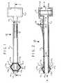
- FIG. 1is a plan view of one embodiment of the clear view cannula of the invention, part shown in phantom to illustrate further details thereof;
- FIG. 2is an elevation sectional view of the clear view cannula shown in FIG. 1 taken substantially on the line 2 — 2 of FIG. 1;
- FIG. 3is a view looking substantially in the direction of arrow A of FIG. 2;
- FIG. 4is a view looking substantially in the direction of arrow B of FIG. 2;
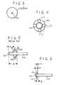
- FIG. 5is a schematic, partly fragmented side elevational view of a prior art cannula showing deployment of flexible members at its distal end;
- FIG. 6is a schematic, partly fragmented side elevational view of the cannula of the invention showing deployment of the shield members at its distal end;
- FIG. 7is a schematic side sectional view illustrating insertion of the clear view cannula of FIG. 1 through a body wall and into an anatomical cavity;
- FIG. 8is a schematic, partly fragmented side sectional view illustrating partial expansion and deployment of the shield members of the clear view cannula shown in FIG. 7;
- FIG. 9is a schematic, partly fragmented side sectional view illustrating the shield members shown in FIG. 8 in a fully expanded and deployed position within an anatomical cavity.
- FIGS. 1 and 2one embodiment of the clear view cannula of the invention generally identified by reference numeral 10 which typically comprises a tubular cannula body 11 having a proximate end 12 and a distal end 13 which is normally tapered as indicated at 14 to facilitate entry or penetration of the cannula 10 through a body wall and into an anatomical cavity.
- reference numeral 10typically comprises a tubular cannula body 11 having a proximate end 12 and a distal end 13 which is normally tapered as indicated at 14 to facilitate entry or penetration of the cannula 10 through a body wall and into an anatomical cavity.
- a cylindrical sleeve 15is concentrically mounted about and slidably secured to the cannula body 11 in a close fitting relationship.
- the cylindrical sleeve 15extends from adjacent the proximate end 12 of cannula body 11 toward the distal end 13 of cannula body 11 a distance of from about 1 ⁇ 2 to about 3 ⁇ 4 the length of the cannula body 11 .
- Cylindrical sleeve 15is provided with a raised shoulder 16 at its proximate end and a plurality of spaced apart shield members 17 secured to its distal end.
- the shield members 17shown partially deployed in FIGS. 1 and 2, are circumferentially disposed about and longitudinally co-extensive with the cannula body 11 .
- Cylindrical sleeve 15can be secured to the cannula body 11 by any suitable and conventional means such as apertures formed in the cannula body 11 which mate with nipples on the inner surface of the cylindrical sleeve 15 in a snap-fit relationship (not shown).
- Other meanssuch as spot welding, fusion, and the like, can also be readily used as will be apparent to those skilled in this art.
- a plurality of closely spaced apart teeth members 18are provided on the outer, circumferential wall of the cannula body 11 and extend from the proximate end 12 toward the distal end 13 a distance of from about 1 ⁇ 2 to about 3 ⁇ 4 of the length of the cannula body 11 ; that is, teeth members 18 are provided along the cannula body 11 for a distance about equal to the length of cylindrical sleeve 15 .
- Teeth members 18are formed to have a substantially perpendicular face 19 and a rear portion 20 that slopes toward proximate end 12 .
- the sloping rear portion 20 of each tooth member 18terminates at the base of the perpendicular face 19 of each preceding tooth member 18 .
- the inner end of shoulder 16is formed to terminate in a depending detent 16 a which engages the faces 19 of the teeth members 18 as described more fully hereinbelow.
- the proximate end 12 of cannula body 11is provided with a handle 21 which can be in any geometrical form that will enable a user to readily grasp the handle 21 with the fingers one hand and concurrently manipulate the cylindrical sleeve 15 toward and away from the distal end 13 of the cannula body 11 with another finger of the same hand.
- the geometric form of handle 21can be oblong, spherical, round, square or rectangular as such forms will readily enable a user to comfortably and easily grasp the handle 21 and manipulate the cylindrical sleeve 15 with the fingers of one hand while retaining complete control of the cannula 10 .
- valve members 22 and 22 aare secured within handle 21 to prevent liquid within a joint or body cavity from leaking out of the proximate end 12 of the cannula 10 after the cannula 10 has been inserted into a body cavity.
- Valve members 22 and 22 acan be provided from any suitable flexible material such as rubber or latex that will prevent seepage or leakage of fluid from a body cavity through the proximate end 12 of the cannula 10 .
- a cross-shaped or X-shaped slit 23is formed in each of the valve members 22 and 22 a to enable an instrument to be inserted through the valve members 22 and 22 a and through the cannula body 11 .
- a gasketsuch as a rubber or latex O-ring 24 is seated between the outer circumferential surface of cannula body 11 and the inner circumferential surface of cylindrical sleeve 15 intermediate the proximate end 12 and the distal end 13 of cannula 10 as shown in FIG. 2 .
- shield members 17are formed to have a cross-sectional thickness at their approximate mid-points indicated by 17 a that is thinner than the cross-sectional thickness at their extremities indicated by 17 b . This enables shield members 17 to be readily flexed from a substantially flat, at rest condition to a fully deployed condition after the cannula 10 has been inserted into a body cavity.
- FIG. 4illustrates the configuration of the shield members 17 when fully deployed within a body cavity.
- an expandable web member 25can be secured to the shield members 17 to further assure that any torn or fragmented tissue is completely retracted and retained within a body cavity when the shield members 17 are fully deployed.
- FIGS. 7, 8 and 9 of the drawingillustrate typical, sequential steps that can be employed in using the clear view cannula of the invention.
- the cannula body 11is shown being inserted through a body wall 26 and into an anatomical cavity such as a knee joint.
- anatomical cavitysuch as a knee joint.
- insertion and penetrationgenerally results in torn and fragmented soft tissue 27 which can obstruct or otherwise interfere with the use of an auxiliary surgical instrument.
- Such obstruction or interferenceis virtually eliminated by using the clear view cannula of the invention.
- FIG. 7also shows the insertion of a trocar 28 through cannula body 11 which is typically initially introduced into a body cavity and then subsequently removed and replaced with a surgical instrument such as a camera.
- the shield members 17are maintained in their partially expanded and deployed condition by annular detent 16 a firmly and securely engaging the perpendicular face 19 of a tooth member 18 . Urging of the cylindrical sleeve 15 to slide along the cannula body 11 is facilitated by the sloping rear portions 20 of the teeth members 18 .
- an operatorwill continue to slidably urge the cylindrical sleeve 15 along the annular body 11 and withdraw the cannula body 11 through the body wall 26 in this manner until the shield members 17 are fully expanded and deployed retaining the torn and fragmented tissue 27 against the inner surface of the body wall 26 with only a small portion of the distal end 13 of the cannula body 11 retained and locked in place within the body wall 26 as illustrated in FIG. 9 .
- the shield members 17are locked in their fully expanded and deployed condition and the cannula body 11 is firmly secured within the body wall 26 by means of the detent 16 a engaging the perpendicular face 19 of a tooth member 18 .
- FIG. 5there is illustrated a typical prior art cannula such as that disclosed in U.S. Pat. No. 5,217,451 to Freitas discussed above.
- the flexible members 30 of this prior art cannulawhen fully deployed, they form an acute angle of substantially less than 90 degrees with respect to the longitudinal axis of a cannula body 31 . Consequently, torn and fragmented tissue can not be effectively retained within an anatomical cavity which would, in turn, interfere with an operator's view within the cavity.
- the flexible members 30when fully deployed form an acute angle, they encompass a distance “y” along the cannula body 31 . This prevents the cannula body 31 from being withdrawn from within a body cavity which, in turn, creates a further distance “x” from the distal end 32 of the cannula body 31 . Consequently, the distal end 32 of this cannula body 31 will extend into a body cavity the distance indicated by “z”; i.e., the sum of distances “y” and “x”.
- the structure and operation of the clear view cannula of the inventionpermits its shield members 17 to be deployed at an angle that is substantially coincidental to 90 degrees with respect to the longitudinal axis of the cannula body as is illustrated in FIG. 6 .
- the total distance “z” that the cannula body 11 of the present invention extends into a body cavityis significantly and substantially less than the prior art cannula illustrated in FIG. 5 .
- This significant and substantial reduction of extension into a body cavityis highly advantageous, particularly when surgical procedures are to be performed within the very limited confines of a body joint.
- the materials used to fabricate the clear view cannula of the inventionare not critical provided they are suitable for use in surgical procedures.
- all components of the clear view cannula of the inventioni.e., the cannula body 11 carrying teeth members 18 , cylindrical sleeve with annular collar 16 and detent 16 a , and shield members 17 are preferably fabricated from well known and commercially available plastic materials that are suitable for use in surgical procedures.
Landscapes
- Health & Medical Sciences (AREA)
- Life Sciences & Earth Sciences (AREA)
- Public Health (AREA)
- General Health & Medical Sciences (AREA)
- Veterinary Medicine (AREA)
- Engineering & Computer Science (AREA)
- Biomedical Technology (AREA)
- Heart & Thoracic Surgery (AREA)
- Surgery (AREA)
- Animal Behavior & Ethology (AREA)
- Molecular Biology (AREA)
- Medical Informatics (AREA)
- Pathology (AREA)
- Nuclear Medicine, Radiotherapy & Molecular Imaging (AREA)
- Biophysics (AREA)
- Pulmonology (AREA)
- Anesthesiology (AREA)
- Hematology (AREA)
- Surgical Instruments (AREA)
Abstract
Description
This invention relates to a clear view cannula for use in surgical procedures. More particularly, this invention relates to a clear view cannula that permits an operator to insert the cannula through a body wall into an anatomical cavity, maintain the cannula in its inserted position, and introduce an auxiliary surgical instrument into the anatomical cavity through the cannula without visual or mechanical obstruction within the body wall.
The use of surgical instruments such as cannulas to introduce an auxiliary surgical instrument into a body such as a joint, abdominal cavity, or the like is well known and widely used. A typical procedure can include making a small incision in the desired portion of the body wall, inserting a cannula into and through the incision and introducing an auxiliary surgical instrument through the cannula into the body to perform a further procedure.
In order to be thoroughly effective and minimize trauma to a patient, it is desirable in such procedures that the cannula be capable of being anchored or secured and not slip out of the anatomical cavity thereby preventing its reinsertion and that fragmented or torn soft tissue not have to be removed from the area surrounding the insertion point of the cannula in order to ensure that the auxiliary instrument can be used without visual or mechanical obstruction.
Several attempts have been proposed to overcome these problems and deficiencies. For example, U.S. Pat. No. 5,217,451 to Freitas discloses a trocar assembly having first and second cylindrical members secured to one another at the distal end of the assembly and a sleeve portion having a series of radially extending flexible members. This device has many small parts and is operable through the use of an interacting gear mechanism. Since the radially extending members, when fully deployed, form an acute angle substantially less than 90 degrees with respect to the longitudinal axis of the cylindrical members, they are not capable of effectively retaining or retracting torn or fragmented soft tissue within a body cavity; that is, the spaces between the fully deployed members permit torn or fragmented soft tissue to visually and mechanically obstruct the use of an instrument such as a camera that is typically introduced through a cannula prior to performing a surgical procedure.
U.S. Pat. No. 5,632,761 to Smith, et.al. discloses a device used to dissect and retract layers of tissue while a portion of the device is retained in a patient. The device utilizes two balloons, the first of which is inserted between layers of tissue and inflated to dissect the tissue layers after which the balloon is deflated. The second balloon is then positioned between the tissue layers and inflated to retract the tissue layers. The device includes a tube coaxially mounted to a delivery portion. The tube has a contracting portion and is provided with a number of deformable, longitudinally extending segments. This device also has many small moving parts and, due to the spherical shape of the second balloon, is not capable of fully retracting torn or fragmented soft tissue. Consequently, the device would have to be inserted deeper into a patient in order to be fully effective.
U.S. Pat. No. 5,637,097 to Yoon discloses an instrument used to penetrate an anatomical cavity having a fixed or retractable penetrating member, the distal end of which is used for penetration, and a portal sleeve having an expandable portion fixed relative to the penetrating member. This instrument, as with the devices described above, comprises many components and functions primarily to anchor the instrument within an anatomical cavity. The anchoring component is not designed to effectively retract or retain torn or fragmented soft tissue within the anatomical cavity.
These illustrative devices typically comprise many parts requiring costly and time consuming assembly. Since they are of relatively complex construction, subsequent cleaning and sterilization would also be costly and time consuming. In addition, these devices are not designed to effectively retract and retain torn or fragmented tissue within an anatomical cavity while, at the same time, anchoring the device within the cavity so that only a minimal portion of the device is retained in the cavity. Due to their complex structures, these devices are cumbersome to handle and manipulate by an operator. Furthermore, these illustrative devices are typically designed to function within a relatively large body cavity such as the abdomen where maneuverability is relatively unrestricted.
It has now been found that the shortcomings of such prior art devices are overcome by the clear view cannula of this invention. In general, the clear view cannula of the invention comprises: a tubular body having a proximate end and a distal end; a plurality of closely spaced teeth members formed on the outer surface and extending parallel to the longitudinal axis of said tubular body intermediate its proximate and distal ends; a cylindrical sleeve having a proximate end and a distal end concentrically mounted about and slidably secured to said tubular body; means at the proximate end of said cylindrical sleeve to engage said teeth members; and, a plurality of spaced apart shield members circumferentially disposed about and longitudinally co-extensive with said tubular body secured to the distal end of said cylindrical sleeve such that when said cylindrical sleeve is slidably urged along said tubular body toward the distal end of said tubular body, said shield members are caused to expand and deploy enabling said shield members to retract and retain torn or fragmented soft tissue within an anatomical cavity and anchor said clear view cannula within an anatomical cavity with a minimum of penetration of said clear view cannula into an anatomical cavity.
The means to secure the cylindrical sleeve to the teeth members as the cylindrical sleeve is slidably moved along the tubular body is readily provided by a detent depending from a raised shoulder at the proximate end of the cylindrical sleeve.
The shield members at the distal end of the cylindrical sleeve and are manufactured so as to be capable of being flexed intermediate their ends enabling them to be fully deployed and expanded within an anatomical cavity.
In one embodiment, the shield members are provided with an expandable web member so that when the shield members are fully deployed, the expandable web member fills the spaces between them thereby further assuring that any torn or fragmented tissue is completely retracted and retained within the body of a cavity.
Although the clear view cannula of the invention can readily be used in large body cavities such as the abdomen, it is particularly useful in smaller cavities such as joints; i.e., knees, shoulders, elbows, ankles, and the like. During arthroscopic surgery of a joint, the joint is typically inflated with water as opposed to a gas which is typically used in abdominal surgical procedures as the surgical procedures performed within a joint are significantly different from those performed within an abdominal cavity.
For example, the inside of a joint such as the knee is lined with a layer of a friable tissue called the synovium which is about ½ cm. thick. In patients about to undergo arthroscopic surgery, the synovial tissue is often inflamed and is also frequently torn and fragmented. In addition, there is present in the anterior portion of the knee joint a patella fat pad (or blob of fat tissue) which generally measures about 3×5 cm. square. Thus, inflamed and/or torn and fragmented synovial tissue and the patella fat pad in the knee joint serve to restrict and impede visualization of the joint cavity by the surgeon. However, this restricted vision is completely overcome when using the clear view cannula of the invention.
The clear view cannula of the invention will become more apparent from the ensuing description when considered together with the accompanying drawing wherein:
FIG. 1 is a plan view of one embodiment of the clear view cannula of the invention, part shown in phantom to illustrate further details thereof;
FIG. 2 is an elevation sectional view of the clear view cannula shown in FIG. 1 taken substantially on theline 2—2 of FIG. 1;
FIG. 3 is a view looking substantially in the direction of arrow A of FIG. 2;
FIG. 4 is a view looking substantially in the direction of arrow B of FIG. 2;
FIG. 5 is a schematic, partly fragmented side elevational view of a prior art cannula showing deployment of flexible members at its distal end;
FIG. 6 is a schematic, partly fragmented side elevational view of the cannula of the invention showing deployment of the shield members at its distal end;
FIG. 7 is a schematic side sectional view illustrating insertion of the clear view cannula of FIG. 1 through a body wall and into an anatomical cavity;
FIG. 8 is a schematic, partly fragmented side sectional view illustrating partial expansion and deployment of the shield members of the clear view cannula shown in FIG. 7; and,
FIG. 9 is a schematic, partly fragmented side sectional view illustrating the shield members shown in FIG. 8 in a fully expanded and deployed position within an anatomical cavity.
Turning now to the drawing wherein like reference numerals and letters identify like parts, there is shown in FIGS. 1 and 2 one embodiment of the clear view cannula of the invention generally identified byreference numeral 10 which typically comprises atubular cannula body 11 having aproximate end 12 and adistal end 13 which is normally tapered as indicated at14 to facilitate entry or penetration of thecannula 10 through a body wall and into an anatomical cavity.
Acylindrical sleeve 15 is concentrically mounted about and slidably secured to thecannula body 11 in a close fitting relationship. Thecylindrical sleeve 15 extends from adjacent theproximate end 12 ofcannula body 11 toward thedistal end 13 of cannula body11 a distance of from about ½ to about ¾ the length of thecannula body 11.Cylindrical sleeve 15 is provided with a raisedshoulder 16 at its proximate end and a plurality of spaced apartshield members 17 secured to its distal end. Theshield members 17, shown partially deployed in FIGS. 1 and 2, are circumferentially disposed about and longitudinally co-extensive with thecannula body 11.Cylindrical sleeve 15 can be secured to thecannula body 11 by any suitable and conventional means such as apertures formed in thecannula body 11 which mate with nipples on the inner surface of thecylindrical sleeve 15 in a snap-fit relationship (not shown). Other means such as spot welding, fusion, and the like, can also be readily used as will be apparent to those skilled in this art.
As can be best seen in FIG. 2, a plurality of closely spaced apartteeth members 18 are provided on the outer, circumferential wall of thecannula body 11 and extend from theproximate end 12 toward the distal end13 a distance of from about ½ to about ¾ of the length of thecannula body 11; that is,teeth members 18 are provided along thecannula body 11 for a distance about equal to the length ofcylindrical sleeve 15.Teeth members 18 are formed to have a substantiallyperpendicular face 19 and arear portion 20 that slopes towardproximate end 12. Preferably, the slopingrear portion 20 of eachtooth member 18 terminates at the base of theperpendicular face 19 of each precedingtooth member 18. The inner end ofshoulder 16 is formed to terminate in a depending detent16awhich engages thefaces 19 of theteeth members 18 as described more fully hereinbelow.
Theproximate end 12 ofcannula body 11 is provided with ahandle 21 which can be in any geometrical form that will enable a user to readily grasp thehandle 21 with the fingers one hand and concurrently manipulate thecylindrical sleeve 15 toward and away from thedistal end 13 of thecannula body 11 with another finger of the same hand. For example, the geometric form ofhandle 21 can be oblong, spherical, round, square or rectangular as such forms will readily enable a user to comfortably and easily grasp thehandle 21 and manipulate thecylindrical sleeve 15 with the fingers of one hand while retaining complete control of thecannula 10.
Apair valve members handle 21 to prevent liquid within a joint or body cavity from leaking out of theproximate end 12 of thecannula 10 after thecannula 10 has been inserted into a body cavity.Valve members proximate end 12 of thecannula 10. A cross-shaped orX-shaped slit 23 is formed in each of thevalve members valve members cannula body 11.
To further prevent leakage or seepage of fluid from a body cavity, a gasket such as a rubber or latex O-ring 24 is seated between the outer circumferential surface ofcannula body 11 and the inner circumferential surface ofcylindrical sleeve 15 intermediate theproximate end 12 and thedistal end 13 ofcannula 10 as shown in FIG.2.
As illustrated in FIGS. 1 and 2,shield members 17 are formed to have a cross-sectional thickness at their approximate mid-points indicated by17athat is thinner than the cross-sectional thickness at their extremities indicated by17b. This enablesshield members 17 to be readily flexed from a substantially flat, at rest condition to a fully deployed condition after thecannula 10 has been inserted into a body cavity.
FIG. 4 illustrates the configuration of theshield members 17 when fully deployed within a body cavity. In order to fill the spaces between theshield members 17, anexpandable web member 25 can be secured to theshield members 17 to further assure that any torn or fragmented tissue is completely retracted and retained within a body cavity when theshield members 17 are fully deployed.
FIGS. 7,8 and9 of the drawing illustrate typical, sequential steps that can be employed in using the clear view cannula of the invention. As can be seen in FIG. 7, thecannula body 11, is shown being inserted through abody wall 26 and into an anatomical cavity such as a knee joint. Although insertion is typically made through a pre-formed incision, such insertion and penetration generally results in torn and fragmentedsoft tissue 27 which can obstruct or otherwise interfere with the use of an auxiliary surgical instrument. Such obstruction or interference is virtually eliminated by using the clear view cannula of the invention.
FIG. 7 also shows the insertion of a trocar28 throughcannula body 11 which is typically initially introduced into a body cavity and then subsequently removed and replaced with a surgical instrument such as a camera.
After thecannula body 11 has been inserted through thebody wall 26 as shown in FIG. 7, an operator, using only finger tip pressure againstshoulder 16, simply urgescylindrical sleeve 15 to slide along thecannula body 11 in the direction of arrows C as illustrated in FIG.8. This causesshield members 17 to expand and deploy in the direction of arrows D toward the fragmented and torntissue 27. As an operator continues to expand and deploy theshield members 17 in this manner, the operator can also withdraw thecannula body 11 outwardly through thebody wall 26. Accidently fully withdrawing thecannula body 11 through thebody wall 26 is prevented by the partially expanded and deployedshield members 17 contacting and engaging the inner surface of thebody wall 26. As the operator withdraws thecannula body 11 outwardly, theshield members 17 are maintained in their partially expanded and deployed condition by annular detent16afirmly and securely engaging theperpendicular face 19 of atooth member 18. Urging of thecylindrical sleeve 15 to slide along thecannula body 11 is facilitated by the slopingrear portions 20 of theteeth members 18.
Typically, an operator will continue to slidably urge thecylindrical sleeve 15 along theannular body 11 and withdraw thecannula body 11 through thebody wall 26 in this manner until theshield members 17 are fully expanded and deployed retaining the torn andfragmented tissue 27 against the inner surface of thebody wall 26 with only a small portion of thedistal end 13 of thecannula body 11 retained and locked in place within thebody wall 26 as illustrated in FIG.9. At this time, theshield members 17 are locked in their fully expanded and deployed condition and thecannula body 11 is firmly secured within thebody wall 26 by means of the detent16aengaging theperpendicular face 19 of atooth member 18.
In FIG. 5 there is illustrated a typical prior art cannula such as that disclosed in U.S. Pat. No. 5,217,451 to Freitas discussed above. As can be seen in FIG. 5, when theflexible members 30 of this prior art cannula are fully deployed, they form an acute angle of substantially less than 90 degrees with respect to the longitudinal axis of a cannula body31. Consequently, torn and fragmented tissue can not be effectively retained within an anatomical cavity which would, in turn, interfere with an operator's view within the cavity.
Since the flexible members30 (FIG. 5) when fully deployed form an acute angle, they encompass a distance “y” along the cannula body31. This prevents the cannula body31 from being withdrawn from within a body cavity which, in turn, creates a further distance “x” from thedistal end 32 of the cannula body31. Consequently, thedistal end 32 of this cannula body31 will extend into a body cavity the distance indicated by “z”; i.e., the sum of distances “y” and “x”.
By contrast, the structure and operation of the clear view cannula of the invention permits itsshield members 17 to be deployed at an angle that is substantially coincidental to 90 degrees with respect to the longitudinal axis of the cannula body as is illustrated in FIG.6. This results in substantially reducing both the “x” and “y” distances enabling thecannula body 11 to be withdrawn through the wall of a body cavity until its withdrawal is arrested by the deployedshield members 17 and optionally, theexpandable web member 25, leaving only a relatively small portion of itsdistal end 13 extending into a body cavity as is shown in FIG.6. Thus, the total distance “z” that thecannula body 11 of the present invention extends into a body cavity is significantly and substantially less than the prior art cannula illustrated in FIG.5. This significant and substantial reduction of extension into a body cavity is highly advantageous, particularly when surgical procedures are to be performed within the very limited confines of a body joint.
The materials used to fabricate the clear view cannula of the invention are not critical provided they are suitable for use in surgical procedures. For ease of fabrication, assembly and use, all components of the clear view cannula of the invention; i.e., thecannula body 11 carryingteeth members 18, cylindrical sleeve withannular collar 16 and detent16a, andshield members 17 are preferably fabricated from well known and commercially available plastic materials that are suitable for use in surgical procedures.
Although the clear view cannula of the invention has been described in detail and with particularity, it will be appreciated by those skilled in this art that changes and modifications can be made therein without departing from the scope and spirit of the invention.
Claims (10)
1. A clear view cannula adapted for use by a single hand of a surgeon and configured for an arthroscopic penetration through a body wall of a patient, the clear view cannula comprising:
a cannula including a tubular body having a distal end and a proximal end portion, the tubular body defining a through hole aligned with the longitudinal axis, the tubular body including a plurality of teeth members on the outer surface and extending parallel to the longitudinal axis of the tubular body, the distal end having a tapered tip adapted for making an arthroscopic portal through a body wall of a patient, a single piece handle positioned on the proximal end portion of the tubular body, the handle being adapted for readily grasping by a single hand of a surgeon; and
a cylindrical sleeve having a distal end portion and a proximal end portion, the cylindrical sleeve being concentrically mounted about and slidably secured for translation relative to the tubular body, a distal edge of the distal end portion of the cylindrical sleeve being connected to the distal end of the tubular body, the cylindrical sleeve and tubular body defining a first position of the cannula, the first position being adapted for insertion through the body wall of the patient,
the proximal end portion of the sleeve including a shoulder, the shoulder having a depending detent configured for selectively engaging the plurality of teeth and limiting the movement of the cylindrical sleeve relative to the tubular body, the shoulder being adapted for manipulation by the fingers of the single hand of the surgeon such that the relative positions of the tubular body and cylindrical sleeve of the cannula can be controlled by the single hand of the surgeon while gripping the handle, and
the distal end portion including a plurality of shield members connected to the distal end of the tubular body, the plurality of shield members being configured for moving between the first position of the cannula parallel with the longitudinal axis and a second position of the cannula substantially perpendicular to the longitudinal axis, the cannula being adapted for manipulation by a single hand of a surgeon for the movement between the first and the second positions, the shield members being configured for bending about the mid-points upon the application of distally directed pressure upon the shoulder relative to the tubular body, the shield members in the second position being configured to improve the internal visibility through the arthroscopic portal by retracting and retaining the torn or fragmented tissue associated with the arthroscopic portal against the inner surface of the body wall.
2. The clear view cannula ofclaim 1 , wherein the cylindrical sleeve has four shield members, the shield members having a first cross-sectional thickness at their mid-points less than a second cross-sectional thickness at the extremities.
3. The clear view cannula ofclaim 2 , wherein the shield members are configured to retain the clear view cannula in position within the arthroscopic portal.
4. The clear view cannula ofclaim 1 , wherein the distal end of the tubular body is tapered to facilitate penetration of the tubular body into the body wall of the patient and into an anatomical cavity.
5. The clear view cannula ofclaim 1 , wherein the shield members are positioned from the first position to the second position by a finger tip pressure by the single hand against the shoulder of the cylindrical sleeve in a distal direction relative to the tubular body.
6. A clear view cannula adapted for use by a single hand of a surgeon and adapted for minimizing the depth of an arthroscopic penetration through a body wall of a patient, the clear view cannula comprising:
a cannula including a tubular body having a distal end and a proximal end portion, the tubular body defining a through hole aligned with the longitudinal axis, the tubular body including a plurality of teeth members on the outer surface and extending parallel to the longitudinal axis of the tubular body, the distal end having a tapered tip adapted for making an arthroscopic portal through a body wall of a patient, a single piece handle positioned on the proximal end portion of the tubular body, the handle being adapted for readily grasping by a single hand of a surgeon; and
a cylindrical sleeve having a distal end portion and a proximal end portion, the cylindrical sleeve being concentrically mounted about and slidably secured for translation relative to the tubular body, a distal edge of the distal end portion of the cylindrical sleeve being connected to the distal end of the tubular body, the cylindrical sleeve and tubular body defining a first position of the cannula, the first position being adapted for insertion through the body wall of the patient,
the proximal end portion of the sleeve including a shoulder, the shoulder having a depending detent configured for selectively engaging the plurality of teeth and limiting the movement of the cylindrical sleeve relative to the tubular body, the shoulder being adapted for manipulation by the fingers of the single hand of the surgeon such that the relative positions of the tubular body and cylindrical sleeve of the cannula can be controlled by the single hand of the surgeon while gripping the handle, and
the distal end portion including a plurality of shield members connected to the distal end of the tubular body, the plurality of shield members being configured for moving between the first position of the cannula parallel with the longitudinal axis and a second position of the cannula substantially perpendicular to the longitudinal axis, the tapered tip being configured to extend a minimal finite distance distally beyond the plurality of shield members, the cannula in the second position being adapted to extend a minimal finite distance into the patient defined by the tapered tip and the shield members in the expanded position, the cannula being adapted for manipulation by a single hand of a surgeon for the movement between the first and the second positions, the shield members being configured for bending about the mid-points upon the application of distally directed pressure upon the shoulder relative to the tubular body.
7. The clear view cannula ofclaim 6 , wherein the cylindrical sleeve has four shield members, the shield members having a first cross-sectional thickness at their mid-points less than a second cross-sectional thickness at the extremities.
8. The clear view cannula ofclaim 6 , wherein the shield members are configured to retain the clear view cannula in position within the arthroscopic portal and configured to improve the internal visibility through the arthroscopic portal by retracting and retaining the torn or fragmented tissue associated with the arthroscopic portal against the inner surface of the body wall.
9. The clear view cannula ofclaim 6 , wherein the distal end of the tubular body is tapered to facilitate penetration of the tubular body into the body wall of the patient and into an anatomical cavity.
10. The clear view cannula ofclaim 6 , wherein the shield members are positioned between the first position to the second position by a finger tip pressure by the single hand against the shoulder of the cylindrical sleeve in a distal direction relative to the tubular body.
Priority Applications (3)
| Application Number | Priority Date | Filing Date | Title |
|---|---|---|---|
| US09/293,284US6632197B2 (en) | 1999-04-16 | 1999-04-16 | Clear view cannula |
| US10/627,315US20050075605A1 (en) | 1999-04-16 | 2003-07-25 | Clear view cannula |
| US10/984,704US20050085771A1 (en) | 1999-04-16 | 2004-11-09 | Clear view cannula |
Applications Claiming Priority (1)
| Application Number | Priority Date | Filing Date | Title |
|---|---|---|---|
| US09/293,284US6632197B2 (en) | 1999-04-16 | 1999-04-16 | Clear view cannula |
Related Child Applications (1)
| Application Number | Title | Priority Date | Filing Date |
|---|---|---|---|
| US10/627,315DivisionUS20050075605A1 (en) | 1999-04-16 | 2003-07-25 | Clear view cannula |
Publications (2)
| Publication Number | Publication Date |
|---|---|
| US20010049493A1 US20010049493A1 (en) | 2001-12-06 |
| US6632197B2true US6632197B2 (en) | 2003-10-14 |
Family
ID=23128463
Family Applications (2)
| Application Number | Title | Priority Date | Filing Date |
|---|---|---|---|
| US09/293,284Expired - LifetimeUS6632197B2 (en) | 1999-04-16 | 1999-04-16 | Clear view cannula |
| US10/627,315AbandonedUS20050075605A1 (en) | 1999-04-16 | 2003-07-25 | Clear view cannula |
Family Applications After (1)
| Application Number | Title | Priority Date | Filing Date |
|---|---|---|---|
| US10/627,315AbandonedUS20050075605A1 (en) | 1999-04-16 | 2003-07-25 | Clear view cannula |
Country Status (1)
| Country | Link |
|---|---|
| US (2) | US6632197B2 (en) |
Cited By (106)
| Publication number | Priority date | Publication date | Assignee | Title |
|---|---|---|---|---|
| US20040153064A1 (en)* | 2000-08-11 | 2004-08-05 | Foley Kevin T. | Surgical instrumentation and method for treatment of the spine |
| US20050075605A1 (en)* | 1999-04-16 | 2005-04-07 | Lyon Thomas R. | Clear view cannula |
| US20050085771A1 (en)* | 1999-04-16 | 2005-04-21 | Lyon Thomas R. | Clear view cannula |
| US20050113836A1 (en)* | 2003-11-25 | 2005-05-26 | Lozier Antony J. | Expandable reamer |
| US20050119685A1 (en)* | 2003-10-17 | 2005-06-02 | Smith Robert C. | Expandible surgical access device |
| US20050154370A1 (en)* | 1999-10-29 | 2005-07-14 | Medtronic, Inc. | Methods and systems for providing therapies into the pericardial space |
| US20050182417A1 (en)* | 2004-02-12 | 2005-08-18 | Pagano Paul J. | Surgical instrumentation and method for treatment of a spinal structure |
| US20050237364A1 (en)* | 2004-04-26 | 2005-10-27 | Hirosumi Ito | Printed wiring board and electric device using the same |
| US20060025797A1 (en)* | 2004-07-15 | 2006-02-02 | James Lock | Cannula for in utero surgery |
| US20060052788A1 (en)* | 2003-02-04 | 2006-03-09 | Thelen Sarah L | Expandable fixation devices for minimally invasive surgery |
| US20070005093A1 (en)* | 2005-07-01 | 2007-01-04 | Cox John A | System for tissue dissection and retraction |
| US20070016134A1 (en)* | 2003-04-28 | 2007-01-18 | Yutaka Suzuki | Catheter kit for burrow |
| US20070162066A1 (en)* | 2006-01-10 | 2007-07-12 | Lyon Thomas R | Clear view cannula |
| US20070239108A1 (en)* | 2006-03-13 | 2007-10-11 | Applied Medical Resources Corporation | Balloon trocar |
| US20080086165A1 (en)* | 2006-01-10 | 2008-04-10 | Lyon Thomas R | Expanding cannula |
| US7377897B1 (en) | 2002-05-02 | 2008-05-27 | Kunkel Sanford S | Portal device |
| US20080275463A1 (en)* | 2003-05-01 | 2008-11-06 | High Kenneth A | Cystotomy catheter capture device and methods of using same |
| US20090024149A1 (en)* | 2007-07-17 | 2009-01-22 | Wilson-Cook Medical Inc. | Rivet introduction system |
| US20090088599A1 (en)* | 2007-09-27 | 2009-04-02 | Zook Ronald E | Method of accessing a bladder and associated apparatus therefor |
| US20090204081A1 (en)* | 2008-02-13 | 2009-08-13 | Depuy Mitek, Inc. | Compression expanded cannula |
| US20100179567A1 (en)* | 2009-01-09 | 2010-07-15 | Abbott Vascular Inc. | Closure devices, systems, and methods |
| US20100211012A1 (en)* | 2009-02-17 | 2010-08-19 | Tyco Healthcare Group Lp | Port fixation with filament actuating member |
| US20100240959A1 (en)* | 2007-12-05 | 2010-09-23 | Donahue John R | Retractor and sealing system for surgical/non-surgical instruments |
| US20100274095A1 (en)* | 2009-02-24 | 2010-10-28 | The Hospital For Special Surgery | Expanding cannula and retractor device and methods of use |
| US20100285907A1 (en)* | 2007-12-31 | 2010-11-11 | Dynamic Inertia Fitness Inc. | Swing exercising apparatus |
| US20100298857A1 (en)* | 2007-09-27 | 2010-11-25 | Zook Ronald E | Apparatus and Method for Performing Cystotomy Procedures |
| US20100331825A1 (en)* | 2009-06-24 | 2010-12-30 | Hakky Said I | Indwelling urinary catheter with self-retaining mechanism |
| US20110034775A1 (en)* | 2009-02-25 | 2011-02-10 | Lozman Philip R | Surgical Retention Port and Method of Use |
| US20110224495A1 (en)* | 2010-03-12 | 2011-09-15 | Tyco Healthcare Group Lp | Surgical access port |
| US8236026B2 (en) | 2000-12-07 | 2012-08-07 | Integrated Vascular Systems, Inc. | Closure device and methods for making and using them |
| US20120238959A1 (en)* | 2011-03-14 | 2012-09-20 | C.R. Bard, Inc. | Biased Internal Bolster for a Medical Device |
| US8287503B2 (en) | 2006-03-13 | 2012-10-16 | Applied Medical Resources Corporation | Balloon trocar |
| US8313497B2 (en) | 2005-07-01 | 2012-11-20 | Abbott Laboratories | Clip applier and methods of use |
| US8323312B2 (en) | 2008-12-22 | 2012-12-04 | Abbott Laboratories | Closure device |
| US8328837B2 (en) | 2004-12-08 | 2012-12-11 | Xlumena, Inc. | Method and apparatus for performing needle guided interventions |
| US8357193B2 (en) | 2009-05-29 | 2013-01-22 | Xlumena, Inc. | Apparatus and method for deploying stent across adjacent tissue layers |
| US8398676B2 (en) | 2008-10-30 | 2013-03-19 | Abbott Vascular Inc. | Closure device |
| US8398656B2 (en) | 2003-01-30 | 2013-03-19 | Integrated Vascular Systems, Inc. | Clip applier and methods of use |
| US8425539B2 (en) | 2004-04-12 | 2013-04-23 | Xlumena, Inc. | Luminal structure anchoring devices and methods |
| US8454632B2 (en) | 2008-05-12 | 2013-06-04 | Xlumena, Inc. | Tissue anchor for securing tissue layers |
| US8469995B2 (en) | 2002-06-04 | 2013-06-25 | Abbott Vascular Inc. | Blood vessel closure clip and delivery device |
| US8529587B2 (en) | 2003-01-30 | 2013-09-10 | Integrated Vascular Systems, Inc. | Methods of use of a clip applier |
| US8556930B2 (en) | 2006-06-28 | 2013-10-15 | Abbott Laboratories | Vessel closure device |
| US8579932B2 (en) | 2002-02-21 | 2013-11-12 | Integrated Vascular Systems, Inc. | Sheath apparatus and methods for delivering a closure device |
| US8585836B2 (en) | 2002-12-31 | 2013-11-19 | Integrated Vascular Systems, Inc. | Methods for manufacturing a clip and clip |
| US8590760B2 (en) | 2004-05-25 | 2013-11-26 | Abbott Vascular Inc. | Surgical stapler |
| US8597325B2 (en) | 2000-12-07 | 2013-12-03 | Integrated Vascular Systems, Inc. | Apparatus and methods for providing tactile feedback while delivering a closure device |
| US8603116B2 (en) | 2010-08-04 | 2013-12-10 | Abbott Cardiovascular Systems, Inc. | Closure device with long tines |
| US8672953B2 (en) | 2007-12-17 | 2014-03-18 | Abbott Laboratories | Tissue closure system and methods of use |
| US8690910B2 (en) | 2000-12-07 | 2014-04-08 | Integrated Vascular Systems, Inc. | Closure device and methods for making and using them |
| US8728119B2 (en) | 2001-06-07 | 2014-05-20 | Abbott Vascular Inc. | Surgical staple |
| US8758400B2 (en) | 2000-01-05 | 2014-06-24 | Integrated Vascular Systems, Inc. | Closure system and methods of use |
| US8758396B2 (en) | 2000-01-05 | 2014-06-24 | Integrated Vascular Systems, Inc. | Vascular sheath with bioabsorbable puncture site closure apparatus and methods of use |
| US8758398B2 (en) | 2006-09-08 | 2014-06-24 | Integrated Vascular Systems, Inc. | Apparatus and method for delivering a closure element |
| US8758399B2 (en) | 2010-08-02 | 2014-06-24 | Abbott Cardiovascular Systems, Inc. | Expandable bioabsorbable plug apparatus and method |
| US8777967B2 (en) | 2005-06-09 | 2014-07-15 | Xlumena, Inc. | Methods and devices for anchoring to tissue |
| US8784447B2 (en) | 2000-09-08 | 2014-07-22 | Abbott Vascular Inc. | Surgical stapler |
| US8784437B2 (en) | 2005-06-09 | 2014-07-22 | Xlumena, Inc. | Methods and devices for endosonography-guided fundoplexy |
| US8808310B2 (en) | 2006-04-20 | 2014-08-19 | Integrated Vascular Systems, Inc. | Resettable clip applier and reset tools |
| US8821392B2 (en) | 2009-02-25 | 2014-09-02 | Joint Product Solutions, Llc | Surgical retention port and method of use |
| US8821534B2 (en) | 2010-12-06 | 2014-09-02 | Integrated Vascular Systems, Inc. | Clip applier having improved hemostasis and methods of use |
| US8820602B2 (en) | 2007-12-18 | 2014-09-02 | Abbott Laboratories | Modular clip applier |
| US20140303444A1 (en)* | 2008-06-25 | 2014-10-09 | Covidien Lp | Button port |
| US8858594B2 (en) | 2008-12-22 | 2014-10-14 | Abbott Laboratories | Curved closure device |
| US8888692B1 (en) | 2011-08-26 | 2014-11-18 | Applied Medical Resources Corporation | Trocar cannula assembly and method of manufacture |
| US8893947B2 (en) | 2007-12-17 | 2014-11-25 | Abbott Laboratories | Clip applier and methods of use |
| US8905937B2 (en) | 2009-02-26 | 2014-12-09 | Integrated Vascular Systems, Inc. | Methods and apparatus for locating a surface of a body lumen |
| US8926633B2 (en) | 2005-06-24 | 2015-01-06 | Abbott Laboratories | Apparatus and method for delivering a closure element |
| US8926656B2 (en) | 2003-01-30 | 2015-01-06 | Integated Vascular Systems, Inc. | Clip applier and methods of use |
| US8956388B2 (en) | 2000-01-05 | 2015-02-17 | Integrated Vascular Systems, Inc. | Integrated vascular device with puncture site closure component and sealant |
| US20150081007A1 (en)* | 2013-09-19 | 2015-03-19 | Karyna, Inc. | Systems and methods for deploying a luminal prosthesis over a carina |
| US9089674B2 (en) | 2000-10-06 | 2015-07-28 | Integrated Vascular Systems, Inc. | Apparatus and methods for positioning a vascular sheath |
| US9089311B2 (en) | 2009-01-09 | 2015-07-28 | Abbott Vascular Inc. | Vessel closure devices and methods |
| US9149276B2 (en) | 2011-03-21 | 2015-10-06 | Abbott Cardiovascular Systems, Inc. | Clip and deployment apparatus for tissue closure |
| US9173644B2 (en) | 2009-01-09 | 2015-11-03 | Abbott Vascular Inc. | Closure devices, systems, and methods |
| US9271752B2 (en) | 2013-03-13 | 2016-03-01 | Swan Valley Medical Incorporated | Method and apparatus for placing a cannula in a bladder |
| US9282965B2 (en) | 2008-05-16 | 2016-03-15 | Abbott Laboratories | Apparatus and methods for engaging tissue |
| US9314230B2 (en) | 2009-01-09 | 2016-04-19 | Abbott Vascular Inc. | Closure device with rapidly eroding anchor |
| US20160106939A1 (en)* | 2014-10-20 | 2016-04-21 | Talal Sharaiha LLC | Expandable intubation assemblies |
| US9332976B2 (en) | 2011-11-30 | 2016-05-10 | Abbott Cardiovascular Systems, Inc. | Tissue closure device |
| US9364259B2 (en) | 2009-04-21 | 2016-06-14 | Xlumena, Inc. | System and method for delivering expanding trocar through a sheath |
| US9364209B2 (en) | 2012-12-21 | 2016-06-14 | Abbott Cardiovascular Systems, Inc. | Articulating suturing device |
| US9381041B2 (en) | 2009-04-21 | 2016-07-05 | Xlumena, Inc. | Methods and devices for access across adjacent tissue layers |
| US9414820B2 (en) | 2009-01-09 | 2016-08-16 | Abbott Vascular Inc. | Closure devices, systems, and methods |
| US9414824B2 (en) | 2009-01-16 | 2016-08-16 | Abbott Vascular Inc. | Closure devices, systems, and methods |
| EP3085408A1 (en) | 2009-04-21 | 2016-10-26 | Xlumena, Inc. | Apparatus for advancing a device from one body lumen to another |
| US9486191B2 (en) | 2009-01-09 | 2016-11-08 | Abbott Vascular, Inc. | Closure devices |
| US20160354113A1 (en)* | 2015-06-04 | 2016-12-08 | DePuy Synthes Products, Inc. | Surgical Cannula System and Method of Use |
| US9522265B2 (en) | 2013-03-15 | 2016-12-20 | Applied Medical Resources Corporation | Trocar cannula assembly with low profile insertion configuration and method of manufacture |
| US9572751B2 (en) | 2009-07-07 | 2017-02-21 | C. R. Bard, Inc. | Extensible internal bolster for a medical device |
| US9579091B2 (en) | 2000-01-05 | 2017-02-28 | Integrated Vascular Systems, Inc. | Closure system and methods of use |
| US9585647B2 (en) | 2009-08-26 | 2017-03-07 | Abbott Laboratories | Medical device for repairing a fistula |
| US9682224B2 (en) | 2004-06-29 | 2017-06-20 | C. R. Bard, Inc. | Method and systems for providing fluid communication with a gastrostomy tube |
| US10327809B2 (en) | 2016-02-29 | 2019-06-25 | Covidien Lp | Clip collar advanced fixation |
| US10441746B2 (en)* | 2015-09-04 | 2019-10-15 | Petrus A. Besselink | Flexible and steerable device |
| US10952732B2 (en) | 2013-02-21 | 2021-03-23 | Boston Scientific Scimed Inc. | Devices and methods for forming an anastomosis |
| US20210154424A1 (en)* | 2017-05-03 | 2021-05-27 | Lancashire Teaching Hospitals NHS Foundation Trust | Improved tracheostomy device |
| US11633211B2 (en) | 2020-05-01 | 2023-04-25 | Cilag Gmbh International | Pinch to release cannula depth limiter |
| US11712267B2 (en) | 2020-05-01 | 2023-08-01 | Cilag Gmbh International | Tilting tang cannula depth limiter |
| US11980392B2 (en) | 2020-05-01 | 2024-05-14 | Cilag Gmbh International | Pinch-to-clamp cannula depth limiter |
| US11986215B2 (en) | 2020-05-01 | 2024-05-21 | Cilag Gmbh International | Universal size multi-walled elastomer cannula depth limiter |
| US12213699B2 (en) | 2020-05-01 | 2025-02-04 | Cilag Gmbh International | Threaded cannula depth limiter |
| US12251168B2 (en) | 2021-05-28 | 2025-03-18 | Medos International Sarl | Systems, methods, and devices for localized tracking of a vertebral body or other anatomic structure |
| US12303105B2 (en) | 2004-04-12 | 2025-05-20 | Boston Scientific Scimed, Inc. | Luminal structure anchoring devices and methods |
| US12349915B1 (en)* | 2013-08-21 | 2025-07-08 | Crh Medical Corporation | Elastic band ligation device with anti-pinch feature and method for treatment of hemorrhoids |
| US12402912B2 (en) | 2020-05-01 | 2025-09-02 | Cilag Gmbh International | Multi-diameter cannula depth limiter |
Families Citing this family (11)
| Publication number | Priority date | Publication date | Assignee | Title |
|---|---|---|---|---|
| US20030093104A1 (en)* | 1999-10-29 | 2003-05-15 | Bonner Matthew D. | Methods and apparatus for providing intra-pericardial access |
| US7854724B2 (en) | 2003-04-08 | 2010-12-21 | Surgiquest, Inc. | Trocar assembly with pneumatic sealing |
| WO2005067999A2 (en)* | 2004-01-13 | 2005-07-28 | Centripeta Limited | Device for fluid delivery to an enclosed space |
| US8007477B2 (en)* | 2004-03-22 | 2011-08-30 | Applied Medical Resources Corporation | Surgical access port and method of using same |
| WO2008045316A2 (en)* | 2006-10-06 | 2008-04-17 | Surgiquest, Incorporated | Visualization trocar |
| USD663838S1 (en) | 2007-10-05 | 2012-07-17 | Surgiquest, Inc. | Visualization trocar |
| USD667954S1 (en) | 2007-10-05 | 2012-09-25 | Surgiquest, Inc. | Visualization trocar |
| JP5733831B2 (en) | 2008-10-10 | 2015-06-10 | サージクェスト,インコーポレーテッド | System and method for improved gas recirculation in a surgical trocar with a gas sealing mechanism |
| WO2012122263A2 (en) | 2011-03-08 | 2012-09-13 | Surgiquest, Inc. | Trocar assembly with pneumatic sealing |
| CN108420463B (en)* | 2018-03-20 | 2024-09-06 | 温州市中心医院 | Bone marrow puncture needle for hematology department |
| CN108543197A (en)* | 2018-05-17 | 2018-09-18 | 朱建轩 | A kind of fixer of drainage tube |
Citations (33)
| Publication number | Priority date | Publication date | Assignee | Title |
|---|---|---|---|---|
| US698447A (en)* | 1901-06-01 | 1902-04-29 | Corydon I Bush | Syringe. |
| US827193A (en)* | 1905-09-08 | 1906-07-31 | Arthur N Penland | Surgical instrument. |
| US872217A (en)* | 1907-02-04 | 1907-11-26 | Arthur E Bonesteel | Syringe. |
| US1155169A (en)* | 1914-11-28 | 1915-09-28 | John Starkweather | Surgical instrument. |
| US2482622A (en) | 1948-10-11 | 1949-09-20 | Kahn Edward | Self-retaining uterine cannula |
| US2556783A (en)* | 1950-05-16 | 1951-06-12 | American Cystoscope Makers Inc | Surgical forceps |
| US3108595A (en)* | 1960-08-08 | 1963-10-29 | Alfred P Overment | Retention catheter |
| US3241554A (en)* | 1963-08-14 | 1966-03-22 | Baxter Don Inc | Peritoneal dialysis entry device |
| US3261357A (en)* | 1963-08-26 | 1966-07-19 | Baxter Don Inc | Extractor for peritoneal entry device |
| US3397699A (en)* | 1966-05-05 | 1968-08-20 | Gerald C. Kohl | Retaining catheter having resiliently biased wing flanges |
| US3821956A (en) | 1973-08-14 | 1974-07-02 | D Gordhamer | Bovine teat dilator and medicament dispenser |
| US4608965A (en)* | 1985-03-27 | 1986-09-02 | Anspach Jr William E | Endoscope retainer and tissue retracting device |
| US4699611A (en) | 1985-04-19 | 1987-10-13 | C. R. Bard, Inc. | Biliary stent introducer |
| US4995868A (en)* | 1988-10-12 | 1991-02-26 | Bard Limited | Catheter |
| US5002557A (en) | 1989-04-06 | 1991-03-26 | Hasson Harrith M | Laparoscopic cannula |
| US5009643A (en) | 1989-08-09 | 1991-04-23 | Richard Wolf Medical Instruments Corp. | Self-retaining electrically insulative trocar sleeve and trocar |
| US5171223A (en)* | 1989-04-07 | 1992-12-15 | Renate Dunsch-Herzberg Und Gudrun Voss | Drainage and instrument duct for the arthroscopy |
| US5197971A (en)* | 1990-03-02 | 1993-03-30 | Bonutti Peter M | Arthroscopic retractor and method of using the same |
| US5217451A (en) | 1991-05-24 | 1993-06-08 | Dexide, Inc. | Gear activated trocar assembly |
| US5279575A (en) | 1992-08-13 | 1994-01-18 | Brigham & Women's Hospital | Locking pivotal surgical orifice |
| US5295994A (en)* | 1991-11-15 | 1994-03-22 | Bonutti Peter M | Active cannulas |
| US5325848A (en)* | 1992-09-10 | 1994-07-05 | Ethicon, Inc. | Endoscopic tissue manipulator with expandable frame |
| US5423763A (en) | 1993-06-17 | 1995-06-13 | Pacesetter, Inc. | Protective, visible suture sleeve for anchoring transvenous lead bodies |
| US5484442A (en) | 1988-10-24 | 1996-01-16 | Cook Incorporated | Intraosseous needle |
| US5556411A (en) | 1993-07-09 | 1996-09-17 | Nissho Corporation | Trocar assembly having a cannula retaining member |
| US5591191A (en) | 1994-01-26 | 1997-01-07 | Kieturakis; Maciej J. | Surgical instrument and method for helically incising a pathway into the interior of the body |
| USRE35459E (en) | 1991-08-30 | 1997-02-18 | Medtronic, Inc. | Aortic root cannula |
| US5632761A (en) | 1991-05-29 | 1997-05-27 | Origin Medsystems, Inc. | Inflatable devices for separating layers of tissue, and methods of using |
| US5637097A (en) | 1992-04-15 | 1997-06-10 | Yoon; Inbae | Penetrating instrument having an expandable anchoring portion |
| US5749883A (en)* | 1995-08-30 | 1998-05-12 | Halpern; David Marcos | Medical instrument |
| US5885258A (en)* | 1996-02-23 | 1999-03-23 | Memory Medical Systems, Inc. | Medical instrument with slotted memory metal tube |
| US5928260A (en)* | 1997-07-10 | 1999-07-27 | Scimed Life Systems, Inc. | Removable occlusion system for aneurysm neck |
| US5957900A (en)* | 1996-07-10 | 1999-09-28 | Asahi Kogaku Kogyo Kabushiki Kaisha | Treatment accessory for endoscope |
Family Cites Families (20)
| Publication number | Priority date | Publication date | Assignee | Title |
|---|---|---|---|---|
| US248622A (en)* | 1881-10-25 | Railroad pile-driver | ||
| US3717151A (en)* | 1971-03-11 | 1973-02-20 | R Collett | Flesh penetrating apparatus |
| US3799172A (en)* | 1972-09-25 | 1974-03-26 | R Szpur | Retention catheter |
| US3946741A (en)* | 1974-12-09 | 1976-03-30 | Adair Edwin Lloyd | Urethral catheter and body drainage device |
| US4111326A (en)* | 1976-03-04 | 1978-09-05 | Becton, Dickinson And Company | Closure for air evacuated container |
| US4840613A (en)* | 1988-04-27 | 1989-06-20 | Menlo Care, Inc. | Protective sheath for catheter assembly |
| US5073166A (en)* | 1989-02-15 | 1991-12-17 | Medical Innovations Corporation | Method and apparatus for emplacement of a gastrostomy catheter |
| US5345927A (en)* | 1990-03-02 | 1994-09-13 | Bonutti Peter M | Arthroscopic retractors |
| US5454365A (en)* | 1990-11-05 | 1995-10-03 | Bonutti; Peter M. | Mechanically expandable arthroscopic retractors |
| DE4021153A1 (en)* | 1990-07-03 | 1992-01-16 | Wolf Gmbh Richard | ORGAN MANIPULATOR |
| US5203773A (en)* | 1991-10-18 | 1993-04-20 | United States Surgical Corporation | Tissue gripping apparatus for use with a cannula or trocar assembly |
| US5256149A (en)* | 1992-02-14 | 1993-10-26 | Ethicon, Inc. | Trocar having transparent cannula and method of using |
| US5232440A (en)* | 1992-02-26 | 1993-08-03 | Wilk Peter J | Method and device for draining abscess |
| US5707362A (en)* | 1992-04-15 | 1998-01-13 | Yoon; Inbae | Penetrating instrument having an expandable anchoring portion for triggering protrusion of a safety member and/or retraction of a penetrating member |
| US5702365A (en)* | 1992-09-08 | 1997-12-30 | King; Toby St. John | Daul-lumen catheter |
| US5380288A (en)* | 1993-03-30 | 1995-01-10 | Innovasive Devices, Inc. | Surgical cannula and trocar system and method of using the same |
| AU4763296A (en)* | 1995-02-03 | 1996-08-21 | Inbae Yoon | Cannula with distal end valve |
| US6030406A (en)* | 1998-10-05 | 2000-02-29 | Origin Medsystems, Inc. | Method and apparatus for tissue dissection |
| US6632197B2 (en)* | 1999-04-16 | 2003-10-14 | Thomas R. Lyon | Clear view cannula |
| US6743207B2 (en)* | 2001-04-19 | 2004-06-01 | Scimed Life Systems, Inc. | Apparatus and method for the insertion of a medical device |
- 1999
- 1999-04-16USUS09/293,284patent/US6632197B2/ennot_activeExpired - Lifetime
- 2003
- 2003-07-25USUS10/627,315patent/US20050075605A1/ennot_activeAbandoned
Patent Citations (33)
| Publication number | Priority date | Publication date | Assignee | Title |
|---|---|---|---|---|
| US698447A (en)* | 1901-06-01 | 1902-04-29 | Corydon I Bush | Syringe. |
| US827193A (en)* | 1905-09-08 | 1906-07-31 | Arthur N Penland | Surgical instrument. |
| US872217A (en)* | 1907-02-04 | 1907-11-26 | Arthur E Bonesteel | Syringe. |
| US1155169A (en)* | 1914-11-28 | 1915-09-28 | John Starkweather | Surgical instrument. |
| US2482622A (en) | 1948-10-11 | 1949-09-20 | Kahn Edward | Self-retaining uterine cannula |
| US2556783A (en)* | 1950-05-16 | 1951-06-12 | American Cystoscope Makers Inc | Surgical forceps |
| US3108595A (en)* | 1960-08-08 | 1963-10-29 | Alfred P Overment | Retention catheter |
| US3241554A (en)* | 1963-08-14 | 1966-03-22 | Baxter Don Inc | Peritoneal dialysis entry device |
| US3261357A (en)* | 1963-08-26 | 1966-07-19 | Baxter Don Inc | Extractor for peritoneal entry device |
| US3397699A (en)* | 1966-05-05 | 1968-08-20 | Gerald C. Kohl | Retaining catheter having resiliently biased wing flanges |
| US3821956A (en) | 1973-08-14 | 1974-07-02 | D Gordhamer | Bovine teat dilator and medicament dispenser |
| US4608965A (en)* | 1985-03-27 | 1986-09-02 | Anspach Jr William E | Endoscope retainer and tissue retracting device |
| US4699611A (en) | 1985-04-19 | 1987-10-13 | C. R. Bard, Inc. | Biliary stent introducer |
| US4995868A (en)* | 1988-10-12 | 1991-02-26 | Bard Limited | Catheter |
| US5484442A (en) | 1988-10-24 | 1996-01-16 | Cook Incorporated | Intraosseous needle |
| US5002557A (en) | 1989-04-06 | 1991-03-26 | Hasson Harrith M | Laparoscopic cannula |
| US5171223A (en)* | 1989-04-07 | 1992-12-15 | Renate Dunsch-Herzberg Und Gudrun Voss | Drainage and instrument duct for the arthroscopy |
| US5009643A (en) | 1989-08-09 | 1991-04-23 | Richard Wolf Medical Instruments Corp. | Self-retaining electrically insulative trocar sleeve and trocar |
| US5197971A (en)* | 1990-03-02 | 1993-03-30 | Bonutti Peter M | Arthroscopic retractor and method of using the same |
| US5217451A (en) | 1991-05-24 | 1993-06-08 | Dexide, Inc. | Gear activated trocar assembly |
| US5632761A (en) | 1991-05-29 | 1997-05-27 | Origin Medsystems, Inc. | Inflatable devices for separating layers of tissue, and methods of using |
| USRE35459E (en) | 1991-08-30 | 1997-02-18 | Medtronic, Inc. | Aortic root cannula |
| US5295994A (en)* | 1991-11-15 | 1994-03-22 | Bonutti Peter M | Active cannulas |
| US5637097A (en) | 1992-04-15 | 1997-06-10 | Yoon; Inbae | Penetrating instrument having an expandable anchoring portion |
| US5279575A (en) | 1992-08-13 | 1994-01-18 | Brigham & Women's Hospital | Locking pivotal surgical orifice |
| US5325848A (en)* | 1992-09-10 | 1994-07-05 | Ethicon, Inc. | Endoscopic tissue manipulator with expandable frame |
| US5423763A (en) | 1993-06-17 | 1995-06-13 | Pacesetter, Inc. | Protective, visible suture sleeve for anchoring transvenous lead bodies |
| US5556411A (en) | 1993-07-09 | 1996-09-17 | Nissho Corporation | Trocar assembly having a cannula retaining member |
| US5591191A (en) | 1994-01-26 | 1997-01-07 | Kieturakis; Maciej J. | Surgical instrument and method for helically incising a pathway into the interior of the body |
| US5749883A (en)* | 1995-08-30 | 1998-05-12 | Halpern; David Marcos | Medical instrument |
| US5885258A (en)* | 1996-02-23 | 1999-03-23 | Memory Medical Systems, Inc. | Medical instrument with slotted memory metal tube |
| US5957900A (en)* | 1996-07-10 | 1999-09-28 | Asahi Kogaku Kogyo Kabushiki Kaisha | Treatment accessory for endoscope |
| US5928260A (en)* | 1997-07-10 | 1999-07-27 | Scimed Life Systems, Inc. | Removable occlusion system for aneurysm neck |
Cited By (192)
| Publication number | Priority date | Publication date | Assignee | Title |
|---|---|---|---|---|
| US20050075605A1 (en)* | 1999-04-16 | 2005-04-07 | Lyon Thomas R. | Clear view cannula |
| US20050085771A1 (en)* | 1999-04-16 | 2005-04-21 | Lyon Thomas R. | Clear view cannula |
| US8852173B2 (en) | 1999-10-29 | 2014-10-07 | Medtronic, Inc. | Methods and systems for providing therapies into the pericardial space |
| US20050154370A1 (en)* | 1999-10-29 | 2005-07-14 | Medtronic, Inc. | Methods and systems for providing therapies into the pericardial space |
| US8758396B2 (en) | 2000-01-05 | 2014-06-24 | Integrated Vascular Systems, Inc. | Vascular sheath with bioabsorbable puncture site closure apparatus and methods of use |
| US8956388B2 (en) | 2000-01-05 | 2015-02-17 | Integrated Vascular Systems, Inc. | Integrated vascular device with puncture site closure component and sealant |
| US9050087B2 (en) | 2000-01-05 | 2015-06-09 | Integrated Vascular Systems, Inc. | Integrated vascular device with puncture site closure component and sealant and methods of use |
| US8758400B2 (en) | 2000-01-05 | 2014-06-24 | Integrated Vascular Systems, Inc. | Closure system and methods of use |
| US10111664B2 (en) | 2000-01-05 | 2018-10-30 | Integrated Vascular Systems, Inc. | Closure system and methods of use |
| US9579091B2 (en) | 2000-01-05 | 2017-02-28 | Integrated Vascular Systems, Inc. | Closure system and methods of use |
| US20040153064A1 (en)* | 2000-08-11 | 2004-08-05 | Foley Kevin T. | Surgical instrumentation and method for treatment of the spine |
| US9402625B2 (en) | 2000-09-08 | 2016-08-02 | Abbott Vascular Inc. | Surgical stapler |
| US8784447B2 (en) | 2000-09-08 | 2014-07-22 | Abbott Vascular Inc. | Surgical stapler |
| US9060769B2 (en) | 2000-09-08 | 2015-06-23 | Abbott Vascular Inc. | Surgical stapler |
| US9089674B2 (en) | 2000-10-06 | 2015-07-28 | Integrated Vascular Systems, Inc. | Apparatus and methods for positioning a vascular sheath |
| US10245013B2 (en) | 2000-12-07 | 2019-04-02 | Integrated Vascular Systems, Inc. | Closure device and methods for making and using them |
| US9320522B2 (en) | 2000-12-07 | 2016-04-26 | Integrated Vascular Systems, Inc. | Closure device and methods for making and using them |
| US8690910B2 (en) | 2000-12-07 | 2014-04-08 | Integrated Vascular Systems, Inc. | Closure device and methods for making and using them |
| US8236026B2 (en) | 2000-12-07 | 2012-08-07 | Integrated Vascular Systems, Inc. | Closure device and methods for making and using them |
| US9554786B2 (en) | 2000-12-07 | 2017-01-31 | Integrated Vascular Systems, Inc. | Closure device and methods for making and using them |
| US9585646B2 (en) | 2000-12-07 | 2017-03-07 | Integrated Vascular Systems, Inc. | Closure device and methods for making and using them |
| US8486092B2 (en) | 2000-12-07 | 2013-07-16 | Integrated Vascular Systems, Inc. | Closure device and methods for making and using them |
| US8486108B2 (en) | 2000-12-07 | 2013-07-16 | Integrated Vascular Systems, Inc. | Closure device and methods for making and using them |
| US8603136B2 (en) | 2000-12-07 | 2013-12-10 | Integrated Vascular Systems, Inc. | Apparatus and methods for providing tactile feedback while delivering a closure device |
| US8597325B2 (en) | 2000-12-07 | 2013-12-03 | Integrated Vascular Systems, Inc. | Apparatus and methods for providing tactile feedback while delivering a closure device |
| US8728119B2 (en) | 2001-06-07 | 2014-05-20 | Abbott Vascular Inc. | Surgical staple |
| US10201340B2 (en) | 2002-02-21 | 2019-02-12 | Integrated Vascular Systems, Inc. | Sheath apparatus and methods for delivering a closure device |
| US9498196B2 (en) | 2002-02-21 | 2016-11-22 | Integrated Vascular Systems, Inc. | Sheath apparatus and methods for delivering a closure device |
| US8579932B2 (en) | 2002-02-21 | 2013-11-12 | Integrated Vascular Systems, Inc. | Sheath apparatus and methods for delivering a closure device |
| US7377897B1 (en) | 2002-05-02 | 2008-05-27 | Kunkel Sanford S | Portal device |
| US9295469B2 (en) | 2002-06-04 | 2016-03-29 | Abbott Vascular Inc. | Blood vessel closure clip and delivery device |
| US9980728B2 (en) | 2002-06-04 | 2018-05-29 | Abbott Vascular Inc | Blood vessel closure clip and delivery device |
| US8469995B2 (en) | 2002-06-04 | 2013-06-25 | Abbott Vascular Inc. | Blood vessel closure clip and delivery device |
| US8585836B2 (en) | 2002-12-31 | 2013-11-19 | Integrated Vascular Systems, Inc. | Methods for manufacturing a clip and clip |
| US9398914B2 (en) | 2003-01-30 | 2016-07-26 | Integrated Vascular Systems, Inc. | Methods of use of a clip applier |
| US10398418B2 (en) | 2003-01-30 | 2019-09-03 | Integrated Vascular Systems, Inc. | Clip applier and methods of use |
| US8398656B2 (en) | 2003-01-30 | 2013-03-19 | Integrated Vascular Systems, Inc. | Clip applier and methods of use |
| US9271707B2 (en) | 2003-01-30 | 2016-03-01 | Integrated Vascular Systems, Inc. | Clip applier and methods of use |
| US8926656B2 (en) | 2003-01-30 | 2015-01-06 | Integated Vascular Systems, Inc. | Clip applier and methods of use |
| US8529587B2 (en) | 2003-01-30 | 2013-09-10 | Integrated Vascular Systems, Inc. | Methods of use of a clip applier |
| US11589856B2 (en) | 2003-01-30 | 2023-02-28 | Integrated Vascular Systems, Inc. | Clip applier and methods of use |
| US20060052788A1 (en)* | 2003-02-04 | 2006-03-09 | Thelen Sarah L | Expandable fixation devices for minimally invasive surgery |
| US20070016134A1 (en)* | 2003-04-28 | 2007-01-18 | Yutaka Suzuki | Catheter kit for burrow |
| US7625361B2 (en)* | 2003-04-28 | 2009-12-01 | Sumitomo Bakelite Company Limited | Catheter kit for burrow |
| US20080275463A1 (en)* | 2003-05-01 | 2008-11-06 | High Kenneth A | Cystotomy catheter capture device and methods of using same |
| US8002764B2 (en) | 2003-05-01 | 2011-08-23 | Swan Valley Medical Incorporated | Cystotomy catheter capture device and methods of using same |
| US20050119685A1 (en)* | 2003-10-17 | 2005-06-02 | Smith Robert C. | Expandible surgical access device |
| US20050113836A1 (en)* | 2003-11-25 | 2005-05-26 | Lozier Antony J. | Expandable reamer |
| US7641664B2 (en) | 2004-02-12 | 2010-01-05 | Warsaw Orthopedic, Inc. | Surgical instrumentation and method for treatment of a spinal structure |
| US8034088B2 (en) | 2004-02-12 | 2011-10-11 | Warsaw Orthopedic, Inc. | Surgical instrumentation and method for treatment of a spinal structure |
| US20050182417A1 (en)* | 2004-02-12 | 2005-08-18 | Pagano Paul J. | Surgical instrumentation and method for treatment of a spinal structure |
| US20060116690A1 (en)* | 2004-02-12 | 2006-06-01 | Pagano Paul J | Surgical instrumentation and method for treatment of a spinal structure |
| US11857160B2 (en) | 2004-04-12 | 2024-01-02 | Boston Scientific Scimed, Inc. | Luminal structure anchoring devices and methods |
| US8425539B2 (en) | 2004-04-12 | 2013-04-23 | Xlumena, Inc. | Luminal structure anchoring devices and methods |
| US10945735B2 (en) | 2004-04-12 | 2021-03-16 | Boston Scientific Scimed, Inc. | Luminal structure anchoring devices and methods |
| US12303105B2 (en) | 2004-04-12 | 2025-05-20 | Boston Scientific Scimed, Inc. | Luminal structure anchoring devices and methods |
| US20050237364A1 (en)* | 2004-04-26 | 2005-10-27 | Hirosumi Ito | Printed wiring board and electric device using the same |
| US8590760B2 (en) | 2004-05-25 | 2013-11-26 | Abbott Vascular Inc. | Surgical stapler |
| US9682224B2 (en) | 2004-06-29 | 2017-06-20 | C. R. Bard, Inc. | Method and systems for providing fluid communication with a gastrostomy tube |
| US20060025797A1 (en)* | 2004-07-15 | 2006-02-02 | James Lock | Cannula for in utero surgery |
| US8328837B2 (en) | 2004-12-08 | 2012-12-11 | Xlumena, Inc. | Method and apparatus for performing needle guided interventions |
| US8617196B2 (en) | 2004-12-08 | 2013-12-31 | Xlumena, Inc. | Method and apparatus for performing needle guided interventions |
| US8784437B2 (en) | 2005-06-09 | 2014-07-22 | Xlumena, Inc. | Methods and devices for endosonography-guided fundoplexy |
| US8777967B2 (en) | 2005-06-09 | 2014-07-15 | Xlumena, Inc. | Methods and devices for anchoring to tissue |
| US8926633B2 (en) | 2005-06-24 | 2015-01-06 | Abbott Laboratories | Apparatus and method for delivering a closure element |
| US8007508B2 (en)* | 2005-07-01 | 2011-08-30 | Cox John A | System for tissue dissection and retraction |
| US12070214B2 (en) | 2005-07-01 | 2024-08-27 | Abbott Laboratories | Clip applier and methods of use |
| US11344304B2 (en) | 2005-07-01 | 2022-05-31 | Abbott Laboratories | Clip applier and methods of use |
| US8518057B2 (en) | 2005-07-01 | 2013-08-27 | Abbott Laboratories | Clip applier and methods of use |
| US10085753B2 (en) | 2005-07-01 | 2018-10-02 | Abbott Laboratories | Clip applier and methods of use |
| US9050068B2 (en) | 2005-07-01 | 2015-06-09 | Abbott Laboratories | Clip applier and methods of use |
| US20070005093A1 (en)* | 2005-07-01 | 2007-01-04 | Cox John A | System for tissue dissection and retraction |
| US8313497B2 (en) | 2005-07-01 | 2012-11-20 | Abbott Laboratories | Clip applier and methods of use |
| US20080086165A1 (en)* | 2006-01-10 | 2008-04-10 | Lyon Thomas R | Expanding cannula |
| WO2007081468A2 (en) | 2006-01-10 | 2007-07-19 | Lyon Thomas R | Clear view cannula |
| US20070162066A1 (en)* | 2006-01-10 | 2007-07-12 | Lyon Thomas R | Clear view cannula |
| US8043319B2 (en)* | 2006-01-10 | 2011-10-25 | Arthrex, Inc. | Expanding cannula |
| US8287503B2 (en) | 2006-03-13 | 2012-10-16 | Applied Medical Resources Corporation | Balloon trocar |
| US8939946B2 (en) | 2006-03-13 | 2015-01-27 | Applied Medical Resources Corporation | Balloon trocar |
| US9259238B2 (en) | 2006-03-13 | 2016-02-16 | Applied Medical Resources Corporation | Balloon trocar |
| US20070239108A1 (en)* | 2006-03-13 | 2007-10-11 | Applied Medical Resources Corporation | Balloon trocar |
| US8147453B2 (en) | 2006-03-13 | 2012-04-03 | Applied Medical Resources Corporation | Balloon trocar |
| US8808310B2 (en) | 2006-04-20 | 2014-08-19 | Integrated Vascular Systems, Inc. | Resettable clip applier and reset tools |
| US9962144B2 (en) | 2006-06-28 | 2018-05-08 | Abbott Laboratories | Vessel closure device |
| US8556930B2 (en) | 2006-06-28 | 2013-10-15 | Abbott Laboratories | Vessel closure device |
| US8758398B2 (en) | 2006-09-08 | 2014-06-24 | Integrated Vascular Systems, Inc. | Apparatus and method for delivering a closure element |
| US20090024149A1 (en)* | 2007-07-17 | 2009-01-22 | Wilson-Cook Medical Inc. | Rivet introduction system |
| US20090088786A1 (en)* | 2007-09-27 | 2009-04-02 | Zook Ronald E | Method of Performing a Suprapubic Transurethral Cystostomy and Associated Procedures and Apparatus Therefor |
| US8118736B2 (en) | 2007-09-27 | 2012-02-21 | Swan Valley Medical, Incorporated | Method of accessing a bladder and associated apparatus therefor |
| US8118826B2 (en) | 2007-09-27 | 2012-02-21 | Swan Valley Medical, Incorporated | Method of performing a suprapubic transurethral cystostomy and associated procedures and apparatus therefor |
| US20090088599A1 (en)* | 2007-09-27 | 2009-04-02 | Zook Ronald E | Method of accessing a bladder and associated apparatus therefor |
| US8382785B2 (en) | 2007-09-27 | 2013-02-26 | Swan Valley Medical Incorporated | Apparatus and method for performing cystotomy procedures |
| US20100298857A1 (en)* | 2007-09-27 | 2010-11-25 | Zook Ronald E | Apparatus and Method for Performing Cystotomy Procedures |
| US20100240959A1 (en)* | 2007-12-05 | 2010-09-23 | Donahue John R | Retractor and sealing system for surgical/non-surgical instruments |
| US8893947B2 (en) | 2007-12-17 | 2014-11-25 | Abbott Laboratories | Clip applier and methods of use |
| US8672953B2 (en) | 2007-12-17 | 2014-03-18 | Abbott Laboratories | Tissue closure system and methods of use |
| US8820602B2 (en) | 2007-12-18 | 2014-09-02 | Abbott Laboratories | Modular clip applier |
| US8491422B2 (en)* | 2007-12-31 | 2013-07-23 | Dynamic Inertia Fitness Inc. | Swing exercising apparatus |
| US20100285907A1 (en)* | 2007-12-31 | 2010-11-11 | Dynamic Inertia Fitness Inc. | Swing exercising apparatus |
| US20090204081A1 (en)* | 2008-02-13 | 2009-08-13 | Depuy Mitek, Inc. | Compression expanded cannula |
| US20130303858A1 (en)* | 2008-02-13 | 2013-11-14 | Depuy Mitek, Llc | Compression expanded cannula |
| US9161747B2 (en)* | 2008-02-13 | 2015-10-20 | Depuy Mitek, Llc | Compression expanded cannula |
| US10076330B2 (en) | 2008-05-12 | 2018-09-18 | Xlumena, Inc. | Tissue anchor for securing tissue layers |
| US8454632B2 (en) | 2008-05-12 | 2013-06-04 | Xlumena, Inc. | Tissue anchor for securing tissue layers |
| US10413295B2 (en) | 2008-05-16 | 2019-09-17 | Abbott Laboratories | Engaging element for engaging tissue |
| US9282965B2 (en) | 2008-05-16 | 2016-03-15 | Abbott Laboratories | Apparatus and methods for engaging tissue |
| US20140303444A1 (en)* | 2008-06-25 | 2014-10-09 | Covidien Lp | Button port |
| US9636142B2 (en)* | 2008-06-25 | 2017-05-02 | Covidien Lp | Button port |
| US9241696B2 (en) | 2008-10-30 | 2016-01-26 | Abbott Vascular Inc. | Closure device |
| US8398676B2 (en) | 2008-10-30 | 2013-03-19 | Abbott Vascular Inc. | Closure device |
| US8657852B2 (en) | 2008-10-30 | 2014-02-25 | Abbott Vascular Inc. | Closure device |
| US8858594B2 (en) | 2008-12-22 | 2014-10-14 | Abbott Laboratories | Curved closure device |
| US8323312B2 (en) | 2008-12-22 | 2012-12-04 | Abbott Laboratories | Closure device |
| US9089311B2 (en) | 2009-01-09 | 2015-07-28 | Abbott Vascular Inc. | Vessel closure devices and methods |
| US20100179567A1 (en)* | 2009-01-09 | 2010-07-15 | Abbott Vascular Inc. | Closure devices, systems, and methods |
| US9314230B2 (en) | 2009-01-09 | 2016-04-19 | Abbott Vascular Inc. | Closure device with rapidly eroding anchor |
| US10537313B2 (en) | 2009-01-09 | 2020-01-21 | Abbott Vascular, Inc. | Closure devices and methods |
| US12383247B2 (en) | 2009-01-09 | 2025-08-12 | Abbott Vascular, Inc. | Closure devices and methods |
| US9173644B2 (en) | 2009-01-09 | 2015-11-03 | Abbott Vascular Inc. | Closure devices, systems, and methods |
| US9486191B2 (en) | 2009-01-09 | 2016-11-08 | Abbott Vascular, Inc. | Closure devices |
| US11439378B2 (en) | 2009-01-09 | 2022-09-13 | Abbott Cardiovascular Systems, Inc. | Closure devices and methods |
| US9414820B2 (en) | 2009-01-09 | 2016-08-16 | Abbott Vascular Inc. | Closure devices, systems, and methods |
| US9414824B2 (en) | 2009-01-16 | 2016-08-16 | Abbott Vascular Inc. | Closure devices, systems, and methods |
| US20100211012A1 (en)* | 2009-02-17 | 2010-08-19 | Tyco Healthcare Group Lp | Port fixation with filament actuating member |
| US7988669B2 (en) | 2009-02-17 | 2011-08-02 | Tyco Healthcare Group Lp | Port fixation with filament actuating member |
| US8292870B2 (en) | 2009-02-17 | 2012-10-23 | Tyco Healthcare Group Lp | Port fixation with filament actuating member |
| US8221317B2 (en)* | 2009-02-24 | 2012-07-17 | Hospital For Special Surgery | Expanding cannula and retractor device and methods of use |
| US20100274095A1 (en)* | 2009-02-24 | 2010-10-28 | The Hospital For Special Surgery | Expanding cannula and retractor device and methods of use |
| US8771179B2 (en) | 2009-02-25 | 2014-07-08 | Joint Product Solutions, Llc | Surgical retention port and method of use |
| US8821392B2 (en) | 2009-02-25 | 2014-09-02 | Joint Product Solutions, Llc | Surgical retention port and method of use |
| US20110034775A1 (en)* | 2009-02-25 | 2011-02-10 | Lozman Philip R | Surgical Retention Port and Method of Use |
| US8409085B2 (en) | 2009-02-25 | 2013-04-02 | Joint Product Solutions, Llc | Surgical retention port and method of use |
| US8905937B2 (en) | 2009-02-26 | 2014-12-09 | Integrated Vascular Systems, Inc. | Methods and apparatus for locating a surface of a body lumen |
| US9364259B2 (en) | 2009-04-21 | 2016-06-14 | Xlumena, Inc. | System and method for delivering expanding trocar through a sheath |
| EP3085408A1 (en) | 2009-04-21 | 2016-10-26 | Xlumena, Inc. | Apparatus for advancing a device from one body lumen to another |
| US9381041B2 (en) | 2009-04-21 | 2016-07-05 | Xlumena, Inc. | Methods and devices for access across adjacent tissue layers |
| EP3730180A1 (en) | 2009-04-21 | 2020-10-28 | Boston Scientific Scimed Inc. | Apparatus for advancing a device from one body lumen to another |
| US8357193B2 (en) | 2009-05-29 | 2013-01-22 | Xlumena, Inc. | Apparatus and method for deploying stent across adjacent tissue layers |
| US9888926B2 (en) | 2009-05-29 | 2018-02-13 | Boston Scientific Scimed, Inc. | Apparatus and method for deploying stent across adjacent tissue layers |
| US8137337B2 (en)* | 2009-06-24 | 2012-03-20 | Hakky Said I | Indwelling urinary catheter with self-retaining mechanism |
| US20100331825A1 (en)* | 2009-06-24 | 2010-12-30 | Hakky Said I | Indwelling urinary catheter with self-retaining mechanism |
| US9572751B2 (en) | 2009-07-07 | 2017-02-21 | C. R. Bard, Inc. | Extensible internal bolster for a medical device |
| US9585647B2 (en) | 2009-08-26 | 2017-03-07 | Abbott Laboratories | Medical device for repairing a fistula |
| US20110224495A1 (en)* | 2010-03-12 | 2011-09-15 | Tyco Healthcare Group Lp | Surgical access port |
| US8758399B2 (en) | 2010-08-02 | 2014-06-24 | Abbott Cardiovascular Systems, Inc. | Expandable bioabsorbable plug apparatus and method |
| US8603116B2 (en) | 2010-08-04 | 2013-12-10 | Abbott Cardiovascular Systems, Inc. | Closure device with long tines |
| US8821534B2 (en) | 2010-12-06 | 2014-09-02 | Integrated Vascular Systems, Inc. | Clip applier having improved hemostasis and methods of use |
| JP2012192182A (en)* | 2011-03-14 | 2012-10-11 | C R Bard Inc | Biased internal bolster for medical device |
| US20120238959A1 (en)* | 2011-03-14 | 2012-09-20 | C.R. Bard, Inc. | Biased Internal Bolster for a Medical Device |
| US9149276B2 (en) | 2011-03-21 | 2015-10-06 | Abbott Cardiovascular Systems, Inc. | Clip and deployment apparatus for tissue closure |
| US11058407B2 (en) | 2011-08-26 | 2021-07-13 | Applied Medical Resources Corporation | Trocar cannula assembly and method of manufacture |
| US9655607B2 (en) | 2011-08-26 | 2017-05-23 | Applied Medical Resources Corporation | Trocar cannula assembly and method of manufacture |
| US10357234B2 (en) | 2011-08-26 | 2019-07-23 | Applied Medical Resources Corporation | Trocar cannula assembly and method of manufacture |
| US8888692B1 (en) | 2011-08-26 | 2014-11-18 | Applied Medical Resources Corporation | Trocar cannula assembly and method of manufacture |
| US9332976B2 (en) | 2011-11-30 | 2016-05-10 | Abbott Cardiovascular Systems, Inc. | Tissue closure device |
| US12343072B2 (en) | 2012-05-17 | 2025-07-01 | Boston Scientific Scimed, Inc. | Methods and devices for access across adjacent tissue layers |
| US9364209B2 (en) | 2012-12-21 | 2016-06-14 | Abbott Cardiovascular Systems, Inc. | Articulating suturing device |
| US11672518B2 (en) | 2012-12-21 | 2023-06-13 | Abbott Cardiovascular Systems, Inc. | Articulating suturing device |
| US10537312B2 (en) | 2012-12-21 | 2020-01-21 | Abbott Cardiovascular Systems, Inc. | Articulating suturing device |
| US12201299B2 (en) | 2013-02-21 | 2025-01-21 | Boston Scientific Scimed, Inc. | Devices and methods for forming an anastomosis |
| US10952732B2 (en) | 2013-02-21 | 2021-03-23 | Boston Scientific Scimed Inc. | Devices and methods for forming an anastomosis |
| US9271752B2 (en) | 2013-03-13 | 2016-03-01 | Swan Valley Medical Incorporated | Method and apparatus for placing a cannula in a bladder |
| US12185975B2 (en) | 2013-03-15 | 2025-01-07 | Applied Medical Resources Corporation | Trocar cannula assembly with low profile insertion configuration and method of manufacture |
| US11382659B2 (en) | 2013-03-15 | 2022-07-12 | Applied Medical Resources Corporation | Trocar cannula assembly with low profile insertion configuration and method of manufacture |
| US10420584B2 (en) | 2013-03-15 | 2019-09-24 | Applied Medical Resources Corporation | Trocar cannula assembly with low profile insertion configuration and method of manufacture |
| US9522265B2 (en) | 2013-03-15 | 2016-12-20 | Applied Medical Resources Corporation | Trocar cannula assembly with low profile insertion configuration and method of manufacture |
| US12349915B1 (en)* | 2013-08-21 | 2025-07-08 | Crh Medical Corporation | Elastic band ligation device with anti-pinch feature and method for treatment of hemorrhoids |
| US12349916B1 (en)* | 2013-08-21 | 2025-07-08 | Crh Medical Corporation | Elastic band ligation device with anti-pinch feature and method for treatment of hemorrhoids |
| US9636242B2 (en)* | 2013-09-19 | 2017-05-02 | New Karyna, Llc | Systems and methods for deploying a luminal prosthesis over a carina |
| US10716690B2 (en) | 2013-09-19 | 2020-07-21 | New Karyna, Llc | Systems and methods for deploying a luminal prosthesis over a carina |
| US11712352B2 (en) | 2013-09-19 | 2023-08-01 | Karyna Of Arizona, Llc | Systems and methods for deploying a luminal prosthesis over a Carina |
| US20150081007A1 (en)* | 2013-09-19 | 2015-03-19 | Karyna, Inc. | Systems and methods for deploying a luminal prosthesis over a carina |
| US20160106939A1 (en)* | 2014-10-20 | 2016-04-21 | Talal Sharaiha LLC | Expandable intubation assemblies |
| US20180042642A1 (en)* | 2015-06-04 | 2018-02-15 | Medos International Sarl | Obturator for Expandable Catheter |
| US9808282B2 (en)* | 2015-06-04 | 2017-11-07 | Medos International Sarl | Surgical cannula system and method of use |
| US10357282B2 (en)* | 2015-06-04 | 2019-07-23 | Medos International Sarl | Orburator for expandable catheter |
| US20160354113A1 (en)* | 2015-06-04 | 2016-12-08 | DePuy Synthes Products, Inc. | Surgical Cannula System and Method of Use |
| US11730921B2 (en) | 2015-09-04 | 2023-08-22 | Petrus A. Besselink | Flexible and steerable device |
| US10441746B2 (en)* | 2015-09-04 | 2019-10-15 | Petrus A. Besselink | Flexible and steerable device |
| US11241557B2 (en) | 2015-09-04 | 2022-02-08 | Petrus A. Besselink | Flexible and steerable device |
| US11278316B2 (en) | 2016-02-29 | 2022-03-22 | Covidien Lp | Clip collar advanced fixation |
| US10327809B2 (en) | 2016-02-29 | 2019-06-25 | Covidien Lp | Clip collar advanced fixation |
| US20210154424A1 (en)* | 2017-05-03 | 2021-05-27 | Lancashire Teaching Hospitals NHS Foundation Trust | Improved tracheostomy device |
| US11980392B2 (en) | 2020-05-01 | 2024-05-14 | Cilag Gmbh International | Pinch-to-clamp cannula depth limiter |
| US12213699B2 (en) | 2020-05-01 | 2025-02-04 | Cilag Gmbh International | Threaded cannula depth limiter |
| US12144520B2 (en) | 2020-05-01 | 2024-11-19 | Cilag Gmbh International | Pinch-to-clamp cannula depth limiter |
| US12343038B2 (en) | 2020-05-01 | 2025-07-01 | Cilag Gmbh International | Pinch-to-release cannula depth limiter |
| US11986215B2 (en) | 2020-05-01 | 2024-05-21 | Cilag Gmbh International | Universal size multi-walled elastomer cannula depth limiter |
| US11712267B2 (en) | 2020-05-01 | 2023-08-01 | Cilag Gmbh International | Tilting tang cannula depth limiter |
| US11633211B2 (en) | 2020-05-01 | 2023-04-25 | Cilag Gmbh International | Pinch to release cannula depth limiter |
| US12402912B2 (en) | 2020-05-01 | 2025-09-02 | Cilag Gmbh International | Multi-diameter cannula depth limiter |
| US12251168B2 (en) | 2021-05-28 | 2025-03-18 | Medos International Sarl | Systems, methods, and devices for localized tracking of a vertebral body or other anatomic structure |
Also Published As
| Publication number | Publication date |
|---|---|
| US20050075605A1 (en) | 2005-04-07 |
| US20010049493A1 (en) | 2001-12-06 |
Similar Documents
| Publication | Publication Date | Title |
|---|---|---|
| US6632197B2 (en) | Clear view cannula | |
| US8043319B2 (en) | Expanding cannula | |
| US7001405B2 (en) | Apparatus and method for developing an anatomic space for laparoscopic procedures with laparoscopic visualization | |
| US5772680A (en) | Apparatus and method for developing an anatomic space for laparoscopic procedures with laparoscopic visualization | |
| US5549595A (en) | Gear activated trocar assembly | |
| US7179272B2 (en) | Apparatus and method for dissecting tissue layers | |
| US6432121B1 (en) | Apparatus and method for guiding placement of a minimally invasive surgical instrument | |
| US5401247A (en) | Safety penetrating instrument | |
| US20050085771A1 (en) | Clear view cannula | |
| WO1996000531A9 (en) | Apparatus and method for developing anatomic space for laparoscopic procedures | |
| EP1143860B1 (en) | Balloon dissector | |
| EP1908428B1 (en) | Expanding cannula |
Legal Events
| Date | Code | Title | Description |
|---|---|---|---|
| STCF | Information on status: patent grant | Free format text:PATENTED CASE | |
| FPAY | Fee payment | Year of fee payment:4 | |
| AS | Assignment | Owner name:STRATHMORE INDUSTRIES, NEW YORK Free format text:ASSIGNMENT OF ASSIGNORS INTEREST;ASSIGNOR:LYON, THOMAS R.;REEL/FRAME:021339/0477 Effective date:20080424 | |
| FEPP | Fee payment procedure | Free format text:PAYOR NUMBER ASSIGNED (ORIGINAL EVENT CODE: ASPN); ENTITY STATUS OF PATENT OWNER: LARGE ENTITY | |
| FEPP | Fee payment procedure | Free format text:PAT HOLDER NO LONGER CLAIMS SMALL ENTITY STATUS, ENTITY STATUS SET TO UNDISCOUNTED (ORIGINAL EVENT CODE: STOL); ENTITY STATUS OF PATENT OWNER: LARGE ENTITY | |
| FPAY | Fee payment | Year of fee payment:8 | |
| FPAY | Fee payment | Year of fee payment:12 |