US6616684B1 - Endovascular splinting devices and methods - Google Patents
Endovascular splinting devices and methodsDownload PDFInfo
- Publication number
- US6616684B1 US6616684B1US09/679,550US67955000AUS6616684B1US 6616684 B1US6616684 B1US 6616684B1US 67955000 AUS67955000 AUS 67955000AUS 6616684 B1US6616684 B1US 6616684B1
- Authority
- US
- United States
- Prior art keywords
- heart
- tension member
- catheter
- anchor
- chamber wall
- Prior art date
- Legal status (The legal status is an assumption and is not a legal conclusion. Google has not performed a legal analysis and makes no representation as to the accuracy of the status listed.)
- Expired - Lifetime, expires
Links
Images


Classifications
- A—HUMAN NECESSITIES
- A61—MEDICAL OR VETERINARY SCIENCE; HYGIENE
- A61F—FILTERS IMPLANTABLE INTO BLOOD VESSELS; PROSTHESES; DEVICES PROVIDING PATENCY TO, OR PREVENTING COLLAPSING OF, TUBULAR STRUCTURES OF THE BODY, e.g. STENTS; ORTHOPAEDIC, NURSING OR CONTRACEPTIVE DEVICES; FOMENTATION; TREATMENT OR PROTECTION OF EYES OR EARS; BANDAGES, DRESSINGS OR ABSORBENT PADS; FIRST-AID KITS
- A61F2/00—Filters implantable into blood vessels; Prostheses, i.e. artificial substitutes or replacements for parts of the body; Appliances for connecting them with the body; Devices providing patency to, or preventing collapsing of, tubular structures of the body, e.g. stents
- A61F2/02—Prostheses implantable into the body
- A61F2/24—Heart valves ; Vascular valves, e.g. venous valves; Heart implants, e.g. passive devices for improving the function of the native valve or the heart muscle; Transmyocardial revascularisation [TMR] devices; Valves implantable in the body
- A61F2/2478—Passive devices for improving the function of the heart muscle, i.e. devices for reshaping the external surface of the heart, e.g. bags, strips or bands
- A61F2/2487—Devices within the heart chamber, e.g. splints
- A—HUMAN NECESSITIES
- A61—MEDICAL OR VETERINARY SCIENCE; HYGIENE
- A61B—DIAGNOSIS; SURGERY; IDENTIFICATION
- A61B17/00—Surgical instruments, devices or methods
- A61B17/00234—Surgical instruments, devices or methods for minimally invasive surgery
- A—HUMAN NECESSITIES
- A61—MEDICAL OR VETERINARY SCIENCE; HYGIENE
- A61B—DIAGNOSIS; SURGERY; IDENTIFICATION
- A61B17/00—Surgical instruments, devices or methods
- A61B17/064—Surgical staples, i.e. penetrating the tissue
- A—HUMAN NECESSITIES
- A61—MEDICAL OR VETERINARY SCIENCE; HYGIENE
- A61B—DIAGNOSIS; SURGERY; IDENTIFICATION
- A61B17/00—Surgical instruments, devices or methods
- A61B17/068—Surgical staplers, e.g. containing multiple staples or clamps
- A61B17/0682—Surgical staplers, e.g. containing multiple staples or clamps for applying U-shaped staples or clamps, e.g. without a forming anvil
- A—HUMAN NECESSITIES
- A61—MEDICAL OR VETERINARY SCIENCE; HYGIENE
- A61B—DIAGNOSIS; SURGERY; IDENTIFICATION
- A61B17/00—Surgical instruments, devices or methods
- A61B17/00234—Surgical instruments, devices or methods for minimally invasive surgery
- A61B2017/00238—Type of minimally invasive operation
- A61B2017/00243—Type of minimally invasive operation cardiac
- A—HUMAN NECESSITIES
- A61—MEDICAL OR VETERINARY SCIENCE; HYGIENE
- A61B—DIAGNOSIS; SURGERY; IDENTIFICATION
- A61B17/00—Surgical instruments, devices or methods
- A61B17/00234—Surgical instruments, devices or methods for minimally invasive surgery
- A61B2017/00292—Surgical instruments, devices or methods for minimally invasive surgery mounted on or guided by flexible, e.g. catheter-like, means
- A61B2017/003—Steerable
- A—HUMAN NECESSITIES
- A61—MEDICAL OR VETERINARY SCIENCE; HYGIENE
- A61B—DIAGNOSIS; SURGERY; IDENTIFICATION
- A61B17/00—Surgical instruments, devices or methods
- A61B2017/00535—Surgical instruments, devices or methods pneumatically or hydraulically operated
- A61B2017/00557—Surgical instruments, devices or methods pneumatically or hydraulically operated inflatable
- A—HUMAN NECESSITIES
- A61—MEDICAL OR VETERINARY SCIENCE; HYGIENE
- A61B—DIAGNOSIS; SURGERY; IDENTIFICATION
- A61B17/00—Surgical instruments, devices or methods
- A61B2017/00831—Material properties
- A61B2017/00902—Material properties transparent or translucent
- A61B2017/00907—Material properties transparent or translucent for light
- A—HUMAN NECESSITIES
- A61—MEDICAL OR VETERINARY SCIENCE; HYGIENE
- A61B—DIAGNOSIS; SURGERY; IDENTIFICATION
- A61B17/00—Surgical instruments, devices or methods
- A61B17/04—Surgical instruments, devices or methods for suturing wounds; Holders or packages for needles or suture materials
- A61B17/0401—Suture anchors, buttons or pledgets, i.e. means for attaching sutures to bone, cartilage or soft tissue; Instruments for applying or removing suture anchors
- A61B2017/0404—Buttons
- A—HUMAN NECESSITIES
- A61—MEDICAL OR VETERINARY SCIENCE; HYGIENE
- A61B—DIAGNOSIS; SURGERY; IDENTIFICATION
- A61B17/00—Surgical instruments, devices or methods
- A61B17/04—Surgical instruments, devices or methods for suturing wounds; Holders or packages for needles or suture materials
- A61B17/0401—Suture anchors, buttons or pledgets, i.e. means for attaching sutures to bone, cartilage or soft tissue; Instruments for applying or removing suture anchors
- A61B2017/0409—Instruments for applying suture anchors
- A—HUMAN NECESSITIES
- A61—MEDICAL OR VETERINARY SCIENCE; HYGIENE
- A61B—DIAGNOSIS; SURGERY; IDENTIFICATION
- A61B17/00—Surgical instruments, devices or methods
- A61B17/04—Surgical instruments, devices or methods for suturing wounds; Holders or packages for needles or suture materials
- A61B17/0401—Suture anchors, buttons or pledgets, i.e. means for attaching sutures to bone, cartilage or soft tissue; Instruments for applying or removing suture anchors
- A61B2017/0412—Suture anchors, buttons or pledgets, i.e. means for attaching sutures to bone, cartilage or soft tissue; Instruments for applying or removing suture anchors having anchoring barbs or pins extending outwardly from suture anchor body
- A—HUMAN NECESSITIES
- A61—MEDICAL OR VETERINARY SCIENCE; HYGIENE
- A61B—DIAGNOSIS; SURGERY; IDENTIFICATION
- A61B17/00—Surgical instruments, devices or methods
- A61B17/04—Surgical instruments, devices or methods for suturing wounds; Holders or packages for needles or suture materials
- A61B17/0401—Suture anchors, buttons or pledgets, i.e. means for attaching sutures to bone, cartilage or soft tissue; Instruments for applying or removing suture anchors
- A61B2017/0427—Suture anchors, buttons or pledgets, i.e. means for attaching sutures to bone, cartilage or soft tissue; Instruments for applying or removing suture anchors having anchoring barbs or pins extending outwardly from the anchor body
- A61B2017/0437—Suture anchors, buttons or pledgets, i.e. means for attaching sutures to bone, cartilage or soft tissue; Instruments for applying or removing suture anchors having anchoring barbs or pins extending outwardly from the anchor body the barbs being resilient or spring-like
- A—HUMAN NECESSITIES
- A61—MEDICAL OR VETERINARY SCIENCE; HYGIENE
- A61B—DIAGNOSIS; SURGERY; IDENTIFICATION
- A61B17/00—Surgical instruments, devices or methods
- A61B17/04—Surgical instruments, devices or methods for suturing wounds; Holders or packages for needles or suture materials
- A61B17/0401—Suture anchors, buttons or pledgets, i.e. means for attaching sutures to bone, cartilage or soft tissue; Instruments for applying or removing suture anchors
- A61B2017/0464—Suture anchors, buttons or pledgets, i.e. means for attaching sutures to bone, cartilage or soft tissue; Instruments for applying or removing suture anchors for soft tissue
- A—HUMAN NECESSITIES
- A61—MEDICAL OR VETERINARY SCIENCE; HYGIENE
- A61B—DIAGNOSIS; SURGERY; IDENTIFICATION
- A61B17/00—Surgical instruments, devices or methods
- A61B17/04—Surgical instruments, devices or methods for suturing wounds; Holders or packages for needles or suture materials
- A61B17/0469—Suturing instruments for use in minimally invasive surgery, e.g. endoscopic surgery
- A61B2017/048—Suturing instruments for use in minimally invasive surgery, e.g. endoscopic surgery for reducing heart wall tension, e.g. sutures with a pad on each extremity
- A—HUMAN NECESSITIES
- A61—MEDICAL OR VETERINARY SCIENCE; HYGIENE
- A61B—DIAGNOSIS; SURGERY; IDENTIFICATION
- A61B17/00—Surgical instruments, devices or methods
- A61B17/04—Surgical instruments, devices or methods for suturing wounds; Holders or packages for needles or suture materials
- A61B2017/0496—Surgical instruments, devices or methods for suturing wounds; Holders or packages for needles or suture materials for tensioning sutures
- A—HUMAN NECESSITIES
- A61—MEDICAL OR VETERINARY SCIENCE; HYGIENE
- A61B—DIAGNOSIS; SURGERY; IDENTIFICATION
- A61B90/00—Instruments, implements or accessories specially adapted for surgery or diagnosis and not covered by any of the groups A61B1/00 - A61B50/00, e.g. for luxation treatment or for protecting wound edges
- A61B90/39—Markers, e.g. radio-opaque or breast lesions markers
Definitions
- the present inventionpertains to devices for treating a failing heart and related methods for placing the devices.
- the inventionpertains to splinting devices placed on the heart to reduce the radius of curvature and/or alter the geometry or shape of the heart to thereby reduce wall stress in the heart and improve the heart's pumping performance.
- the devices and methods of the present inventionare directed toward endovascular techniques used to facilitate placement of the splinting devices on the heart.
- Heart failureis a common outcome in the progression of many forms of heart disease.
- Heart failuremay be considered as the condition in which an abnormality of cardiac function is responsible for the inability of the heart to pump blood at a rate commensurate with the requirements of the metabolizing tissues, or can do so only at an abnormally elevated filling pressure.
- the process of ventricular dilatationis generally the result of chronic volume overload or specific damage to the myocardium.
- cardiac output requirementsfor example, that of an athlete
- damage to the myocardium or chronic volume overloadthere are increased it requirements put on the contracting myocardium to such a level that this compensated state is never achieved and the heart continues to dilate.
- Mitral regurgitationis a condition whereby blood leaks through the mitral valve due to an improper positioning of the valve structures that causes the valve not to close entirely.
- Geometric abnormalities resulting from a dilated left ventriclemay cause or exacerbate improper functioning of the mitral valve, including mitral valve regurgitation, by altering the normal position and dimension of the valve, particularly the Valve annulus.
- Prior treatments for heart failure associated with such dilatationfall into three general categories. The first being pharmacological, for example, diuretics and ACE inhibitors. The second being assist systems, for example, pumps. Finally, surgical treatments also have been experimented with.
- diureticshave been used to reduce the workload of the heart by reducing blood volume and preload.
- Diureticstypically reduce extra cellular fluid which builds in congestive heart failure patients increasing preload conditions.
- Nitrates, arteriolar vasodilators, angiotensin converting enzyme (ACE) inhibitorshave been used to treat heart failure through the reduction of cardiac workload by reducing afterload.
- Inotropesfunction to increase cardiac output by increasing the force and speed of cardiac muscle contraction.
- Assist devicesinclude mechanical pumps. Mechanical pumps reduce the load on the heart by performing all or part of the pumping function normally done by the heart. Currently, mechanical pumps are used to sustain the patient while a donor heart for transplantation becomes available for the patient.
- a more recent procedure for treating the various forms of heart failure discussed aboveincludes placing devices on the heart to reduce the radius of curvature of the heart and/or alter the cross-sectional shape of the heart to reduce wall stress.
- the devicesare configured to reduce the tension in the heart wall, and thereby reverse, stop or slow the disease process of a failing heart as it reduces the energy consumption of the failing heart, decreases isovolumetric contraction, increases isotonic contraction (sarcomere shortening), which in turn increases stroke volume.
- the devicesreduce wall tension by changing chamber geometry or shape and/or changing the radius of curvature or cross-section of a heart chamber. These changes may occur during the entire cardiac cycle or during only a portion of the cardiac cycle.
- splintsThe devices of the present invention which reduce heart wall stress in this way can be referred to generally as “splints.”
- These splintscan be in the form of external devices, as described in U.S. application Ser. No. 09/157,486, filed Sep. 21, 1998, entitled “External Stress Reduction Device and Method,” the entire disclosure of which is incorporated by reference herein, or in the form of transventricular elongate tension members with heart-engaging assemblies, typically in the form of anchor pads, disposed on each end configured to engage substantially opposite portions of the chamber wall, embodiments of which are disclosed in the '049 application incorporated above.
- An aspect of the present inventionpertains to splint devices, and related splinting methods, for endovascular implantation on the heart.
- the splints of the present inventionmay be implanted endovascularly through remote vascular access sites.
- the inventive techniques and devicesthus are minimally invasive and less risky to patients.
- a method for placing a splint assembly transverse a heart chambercomprises providing an elongate member having a first end and a second end and a deployable heart-engaging assembly connected to at least the first end. The method further includes advancing the elongate member through vasculature structure and into the heart chamber such that the first end of the elongate member extends through a first location of a wall surrounding the heart chamber and the second end extends through a second location of the heart chamber wall substantially opposite the first location.
- a deployable heart-engaging assemblyis deployed such that it engages with a first exterior surface portion of the heart chamber wall adjacent the first location.
- the elongate memberis secured with respect to the heart with a second heart-engaging assembly connected to the second end.
- the second heart-engaging assemblyengages with a second exterior surface portion of the heart chamber wall adjacent the second location.
- a splint assemblyfor treating a heart, comprising an elongate member configured to extend transverse a chamber of the heart and at least one heart-engaging assembly formed at least partially from portions forming the elongate member.
- the heart-engaging assemblyhas a collapsed configuration adapted to travel through a heart wall and an expanded configuration adapted to engage the heart wall.
- Yet another aspect of the inventionincludes a delivery tool for delivering a transventricular splint assembly to a chamber of the heart, comprising a tubular member having a distal end and a proximal end, the distal end having a curved configuration and the tube defining a lumen configured to carry at least a portion of the splint assembly.
- the delivery toolfurther includes at least one support mechanism disposed proximate the distal end of the tubular member, the support mechanism being configured to stabilize the tubular member with respect to a heart wall surrounding the chamber.
- the tubular memberis configured to be advanced through vasculature structure and into the heart chamber.
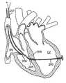
- FIG. 1is a vertical cross-sectional view of the heart showing a delivery catheter inserted endovascularly into the right ventricule according to an aspect of the present invention
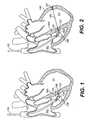
- FIG. 2is a vertical cross-sectional view of the heart showing a guide wire extending from the catheter of FIG. 1 through the septal wall, across the left ventricular chamber and into the free wall according to an aspect of the present invention
- FIG. 3is a vertical cross-sectional view of the heart showing the deliver catheter of FIG. 1 positioned over the guidewire of FIG. 2 with positioning balloons inflated on either side of the free wall according to an aspect of the present invention
- FIG. 4is a vertical cross-sectional view of the heart showing the insertion of a tension member into the delivery catheter of FIG. 3 for placement with respect to the left ventricle according to an aspect of the present invention
- FIG. 5is a vertical cross-sectional view of the heart showing a deployed fixed anchor on the distal end of the tension member of FIG. 4 after being extended past the distal end of the delivery catheter according to an aspect of the present invention
- FIG. 6is a vertical cross-sectional view of the heart showing the removal of the delivery catheter from the tension member of FIG. 5 according to an aspect of the present invention
- FIG. 7is a vertical cross-section of the heart showing the delivery of an adjustable anchor to be placed on the tension member of FIG. 6 adjacent the septal wall according to an aspect of the present invention
- FIG. 8is a vertical cross-section of the heart showing the securing of the adjustable anchor of FIG. 7 to the tension member to change the shape of the left ventricle according to an aspect of the present invention
- FIG. 9is a vertical cross-section of the heart showing a cutting snare inserted into the right ventricle to cut excess tension member length from the splint assembly of FIG. 8 according to an aspect of the present invention
- FIG. 10is a vertical cross-section of the heart showing a splint assembly positioned with respect to the left ventricle according to an aspect of the present invention
- FIG. 11is a partial side view of the deployable anchor and tension member of FIGS. 5 and 6 according to an aspect of the present invention.
- FIG. 12is a top view of the anchor of FIG. 11 according to an aspect of the invention.
- FIG. 13is a partial perspective view of the deployable anchor and tension member of FIG. 11 prior to a securing band being placed to tighten the filament bundles on the elastic ring portion of the anchor according to an aspect of the invention
- FIG. 14is a partial side view of the anchor and tension member of FIG. 11 showing the placement of the securing/tightening band according to an aspect of the present invention
- FIG. 15is a close-up, partial side view of the guidewire of FIG. 2 according to an aspect of the present invention.
- FIG. 16is a detailed, partial side cross-sectional view of the delivery catheter of FIGS. 1-6 according to an aspect of the present invention.
- FIG. 17is a vertical cross-sectional view of a heart showing a delivery catheter with two curved catheters inserted endovascularly through the aorta into the left ventricle according to an aspect of the present invention
- FIG. 18is a vertical cross-sectional view of a heart showing the curved delivery catheters of FIG. 17 with inflated distal balloons respectively in contact with the free wall and septal wall of the heart and with sharpened wires respectively extending through the free wall and septal wall of the heart according to an aspect of the present invention
- FIG. 19is a vertical cross-sectional view of a heart showing a tension member delivered through the curved delivery catheter contacting the free wall of FIG. 18 according to an aspect of the present invention
- FIG. 20is a vertical cross-sectional view of a heart showing the curved catheter contacting the free wall of FIG. 18 removed from the patient and the tension member being fed into a proximal end of the curved catheter contacting the septal wall of FIG. 18 according to an aspect of the present invention
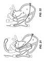
- FIG. 21is a vertical cross-sectional view of the heart showing the tension member of FIG. 20 being advanced through the curved catheter, through the septal wall, into the right ventricle and out of the heart according to an aspect of the present invention
- FIG. 22is a vertical cross-sectional view of the heart showing the tension member of FIG. 21 extended across the left ventricle after the curved delivery catheter of FIG. 21 has been removed according to an aspect of the present invention
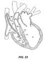
- FIG. 23is a vertical cross-sectional view of the heart showing a delivery catheter with a curved distal tip inserted into the right ventricle proximate the septal wall for delivering a splint assembly according to an aspect of the present invention
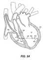
- FIG. 24is a vertical cross-sectional view of the heart with a guidewire with inflated balloons on a distal end extending from the delivery catheter of FIG. 23, through the septal wall, and across the left ventricle according to an aspect of the present invention
- FIG. 25is a vertical cross-sectional view of the heart showing the guidewire of FIG. 24 with the distal balloon deflated and about to be advanced through the free wall of the left ventricle according to an aspect of the present invention
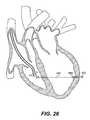
- FIG. 26is a vertical cross-sectional view of the heart showing the guidewire of FIG. 25 advanced through the free wall until the proximal inflated balloon abuts the inside of the free wall and with the distal balloon inflated according to an aspect of the invention;
- FIG. 27is a vertical cross-sectional view of the heart showing the guidewire in the position of FIG. 26 with the proximal balloon deflated according to an aspect of the present invention
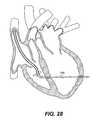
- FIG. 28is a vertical cross-sectional view of the heart showing a splint advancement catheter placed over the guidewire of FIG. 27 according to an aspect of the present invention
- FIG. 29is a vertical cross-sectional view of the heart showing the splint advancement catheter of FIG. 28 being removed from a tension member and deployable anchor of the splint according to the present invention
- FIG. 30is a vertical cross-sectional view of the heart showing the splint advancement catheter of FIG. 28 entirely removed and the splint assembly deployed across the left ventricle according to an aspect of the present invention
- FIG. 31is a partial, detailed cross-sectional view of the splint advancement catheter, tension member and distal anchor of FIG. 28 according to an aspect of the present invention.
- FIG. 32is a partial side view of a braided tension member according to an aspect of the present invention.
- FIG. 33is a partial side view of a braided tension member having a diametrically expandable portion according to an aspect of the present invention.
- FIG. 34is a partial perspective view of the tension member of FIG. 33; forming a free wall anchor at the diametrically expandable portion according to an aspect of the present invention
- FIG. 34 ais a partial perspective view of a spiral-shaped deployable wire used to diametrically expand the tension member of FIG. 33 to form the anchor of FIG. 34 according to an aspect of the present invention
- FIG. 34 bis a partial perspective view of a spiral-shaped deployable wire used to diametrically expand the tension member to form the anchor having a spiral formed in an opposite direction to the spiral of FIG. 34 a according to another aspect of the present invention
- FIG. 35is a partial perspective view of a diametrically expandable tension member forming an anchor portion by using an inflated balloon within the expandable portion of the tension member to cause diametric expansion according to an aspect of the present invention
- FIG. 36is a transverse cross-sectional view of the heart showing a preferred placement of a tension member of a splint assembly to treat a mitral valve according to an aspect of the present invention
- FIG. 37is a transverse cross-sectional view of the heart showing a splint assembly placed with respect to the heart to treat a mitral valve according to an aspect of the present invention.
- FIG. 38is a cross-sectional view of an anchor assembly with an inner surface permitting movement with respect to a tension member in only one direction according to an aspect of the present invention.
- Each device of the present inventionpreferably operates passively in that, once placed in the heart, it does not require an active stimulus, either mechanical, electrical, or otherwise, to function. Implanting one or more of these devices alters the shape or geometry of the heart, both locally and globally, and thereby increases the heart's efficiency. That is, the heart experiences an increased pumping efficiency through an alteration in its shape or geometry and concomitant reduction in stress on the heart walls.
- the devices of the present inventionmay operate to assist in the apposition of heart valve leaflets to improve valve function.
- the inventive devices and related methodsoffer numerous advantages over the existing treatments for various heart conditions, including valve incompetencies.
- the devicesare relatively easy to manufacture and use, and the surgical techniques and tools for implanting the devices of the present invention do not require the invasive procedures of current surgical techniques.
- the endovascular techniqueswhich will be described do not require performing a sternotomy or removing portions of the heart tissue, nor do they require opening the heart chamber or stopping the heart during operation.
- Such percutaneous insertionpermits the splinting procedures to be performed in a wide variety of laboratories in the hospital.
- the techniques for implanting the devices of the present inventionalso are less risky to the patient, both during and after the implantation, and may be performed more quickly than other techniques.
- the procedures of the inventioncause less pain to patients and permit quicker healing.
- certain endovascular splinting techniques to be describedmay limit bleeding at access sites, allowing relatively large catheters, cannula, and other similar implantation tools to be inserted in a percutaneous manner.
- the disclosed inventive devices and related methodsinvolve geometric reshaping of the heart.
- substantially the entire chamber geometryis altered to return the heart to a more normal state of stress.
- Models of this geometric reshapingwhich includes a reduction in radius of curvature of the chamber walls, can be found in U.S. Pat. No. 5,961,440, issued Oct. 5, 1999, entitled “Heart Wall Tension Reduction Apparatus and Method,” and incorporated by reference herein.
- the heart wallsPrior to reshaping the chamber geometry, the heart walls experience high stress due to a combination of both the relatively large increased diameter of the chamber and the thinning of the chamber wall. Filling pressures and systolic pressures are typically high as well, further increasing wall stress.
- Geometric reshaping according to the present inventionreduces the stress in the walls of the heart chamber to increase the heart's pumping efficiency, as well as to stop further dilatation of the heart.
- inventive endovascular splinting devices and methodswill be used to support an infarcted heart wall to prevent further dilatation, or to treat aneurysms in the heart.
- the devices and methods of using and implanting the devicescould be used to treat heart valves, for example to aid in apposition of the leaflets of a mitral valve or modify the shape of the mitral valve, as described in U.S. application Ser. No. 09/680,435, to Richard F. Schroeder et al., filed on the same day as this application, assigned to the same assignee as the present application, and entitled “Methods and Devices for Improving Mitral Valve Function,” and incorporated by reference herein.
- One of ordinary skill in the artwould understand that the use of the devices and methods described herein also could be employed in other chambers and for other valves associated with those chambers.
- the devices and methods of the inventionmight be used to reduce stress in the left atrium to treat atrial fibrillation.
- the left ventriclehas been selected for illustrative purposes because a large number of the disorders that the present invention treats occur in the left ventricle.
- FIGS. 1-10An embodiment of an endovascular splinting technique according to the present invention is shown in FIGS. 1-10.
- this splinting techniqueaccess to the left ventricle LV and delivery of the splint occurs from within the right ventricle RV.
- An approach from within the right ventricleis preferred for a number of reasons.
- the right ventricleis highly accessible through venous structure that leads into the superior vena cava VC, for example from the right or left jugular veins. Since these veins typically are at a relatively low pressure, bleeding at the access sites is limited, and rather large catheters, cannula and the other like surgical tools can be inserted into the veins in a percutaneous manner.
- this techniquepermits access to vascular structure without a sternotomy or other open chest surgical access, thereby minimizing trauma to the patient. Additionally, patients are less likely to experience embolic events. Recovery times for the operation also are reduced, due to the minimally invasive nature of such procedures.
- delivery through the right ventricleallows for straightforward positioning of the splints on the ventricular septal wall SW.
- Such positioning on the septal wallis preferable because it results in good left ventricle bisection, in a manner believed to have minimal negative impact on mitral valve function, and in some instances, a positive impact on mitral valve function and performance.
- delivery through it the right ventricledoes not involve the free wall of the right ventricle and also does not restrict outflow of the blood from the heart.
- a shaped guide device in the form of a delivery catheter 100is advanced into right ventricle RV from an access site preferably in the left or right jugular vein.
- Other access sitessuch as, for example, the left or right subclavian vein also are contemplated.
- the catheter 100has a tip portion 101 configured to be adjustably and variably curved through the use of an adjusting pull-wire 104 .
- the pull-wire 104attaches to the distal most end of the catheter, has a portion that extends exterior the catheter at the distal end of the catheter, and then extends through the catheter to a proximal end of the catheter where it is controlled.
- pull wire 104may be an essentially straight wire that, when pulled (or tensioned), causes tip portion 101 to curve.
- a pull wiremay take the form of a tether, such as described below with reference to the curved catheter having pull wire 405 in FIG. 23 .
- the proximal end of the pull-wire 405can be pulled and released to thereby cause the distal tip of the catheter to curve and to straighten as desired.
- the position of the catheter tipcan be curved by adjusting the pull-wire and also advanced or rotated, or both, by advancing or rotating the catheter with respect to the right ventricle and septal wall.
- two anchoring balloons 102 , 103are disposed near the distal end of catheter 100 .
- Each balloon 102 , 103is in fluid communication with a corresponding inflation lumen 102 ′, 103 ′ that extends proximally to an inflating means (not shown).
- a lumen 101 ′ configured to carry a piercing needlealso extends through the length of catheter 100 .
- delivery catheter 100additionally defines a lumen 106 ′ for carrying a preformed support wire 106 , which expands upon advancement of support wire 106 relative to catheter 100 .
- the wire 106takes on a hoop-like shape which gives mechanical “back up” support to delivery catheter 100 .
- the support wire 106also helps to position the catheter 100 within the right ventricle to allow for positioning within the right ventricle RV and with respect to the septal wall SW.
- the support wire 106is preferably made from an elastic material, such as a nickel-titanium alloy or the like, and has a preformed shape at or near a distal end of the wire configured to stabilize and position the catheter 100 .
- the catheter 100preferably also includes radiographic and echogenic markers (not shown), such as metallic or gas-filled structures, or relatively small balloons filled with a contrast media, to facilitate positioning of the catheter under fluoroscopic and/or ultrasonic guidance, such as transesophageal echo (TEE).
- TEEtransesophageal echo
- needle 105is then advanced through the lumen in catheter 100 and out of tip portion 101 , piercing the septal wall SW, and extending across the left ventricle chamber LV.
- needle 105is fabricated of a highly elastic material such as, for example, nickel titanium alloy, which will allow the needle to traverse the bend at the tip of the delivery catheter, and then to straighten out for controlled traversing across left ventricle LV.
- FIG. 15shows the distal portion of needle 105 in greater detail.
- needle 105includes a sharpened tip which may have threads 107 disposed around the outer surface of the tip portion. These threads 107 preferably are flexible such that they can lay substantially flat along the length of needle 105 as the needle traverses through the catheter lumen. Alternatively, the tip may include barbs or other similar structures that aid in anchoring the tip in the heart wall.
- needle 105Once needle 105 is across the left ventricle chamber, its position is confirmed by TEE, X-Ray, or other visualization techniques, to assure good bisection and avoidance of key mitral valve and other heart structure.
- Conventional angiography utilizing a “pigtail” catheter. i.e., a dye injection catheter with a loop shape at the distal end, in the left ventricle LV and angiography catheters in one or both coronary artery ostiamay also be used to aid in proper positioning of the associated delivery devices in the LV. It also is important to assure that needle 105 will not penetrate or damage any significant coronary vasculature. To assure this, an angiogram may be performed.
- the angiographic imageis aligned to a position that looks down the axis of the needle in the portion of the needle which traverses the left ventricle LV.
- This anglewill limit parallax to ensure that if the tip of the needle is not coincident with a significant vessel it will not pierce such vessel. Any small variation in the position of the needle tip can be adjusted by gentle manipulation of the delivery catheter.
- needle 105has soft threads 107 disposed on the surface of a tip portion of the needle, as shown in FIG. 15 .
- Needle 105can be advanced into the free wall HW of the left ventricle LV by rotating the needle, essentially causing the tip portion of the needle to be pulled or screwed into the myocardium. Threads 107 also serve to anchor needle 105 and provide support for the further advancement of delivery catheter 100 .
- delivery catheter 100is straightened and advanced over needle 105 into left ventricle LV.
- a tapered tip 101 on delivery catheter 100enables catheter 100 to penetrate the septal and free walls SW, HW.
- both balloons 102 and 103are inflated, as shown in FIG. 3, to stabilize catheter 100 with respect to the heart chamber.
- these balloons 102 , 103are made of an elastomeric material, such as latex or silicone, for example, to provide a relatively low profile in the non-inflated state.
- balloons 102 , 103preferably have a flattened, “pancake” shape.
- This shapemay be particularly important for distal balloon 103 , as it lies in the space between the outside of the myocardium and the pericardial sac.
- an elongate tension member 200 with a heart-engaging assemblypreferably in the form of a collapsible fixed anchor mechanism 201 (free wall anchor), on its distal end can be inserted into the lumen of catheter 100 .
- Tension member 200is advanced until it begins to emerge from the tip portion 101 of delivery catheter 100 , as shown in FIG. 4 .
- FIG. 11shows a preferred structure for fixed anchor mechanism 201 in its fully expanded state after being secured with respect to the heart wall.
- tension member 200is comprised of a braided polymer, such as that disclosed in the '049 application incorporated by reference above.
- a cover of expanded polytetrafluoroethylene (ePTFE)(not shown) preferably covers the majority of the length of tension member 200 .
- ePTFEexpanded polytetrafluoroethylene
- Each bundle 210 in the braid structureis attached via suturing, adhesive, or other suitable attachment mechanism, to a flexible elastic ring 203 .
- Ring 203preferably is comprised of nickel-titanium, or an elastomeric polymer such as silicone or urethane, or other suitable like materials. This attachment of the bundles to the ring is best shown in FIG. 13 .
- the braided structuretransitions from a tight woven braid to a region that is primarily unbraided at a position slightly proximal to the ring.
- flexible elastic ring 203can be easily deformed into a flattened hoop, without bundles 210 inhibiting this deformation.
- a stiffening mandrelmay be disposed either inside or adjacent the braided portion of the tension member.
- tension member 200is advanced until flexible ring 203 fully emerges from the lumen of delivery catheter 100 .
- anchor mechanism 201has sufficient strength to serve as an anchor and allows bundles 210 to take on a funnel shape, as shown in FIG. 13 .
- a securing band 204(FIG. 14) is advanced along the outside of braided tension member 200 , until the bundles tighten into a generally spoke-like configuration, as shown in FIGS. 11 and 12.
- a flexible pushing tube(not shown), or other suitable mechanism, may be used to advance securing band 204 .
- Securing band 204preferably has circumferential ribs 204 ′ on its inner surface that are oriented proximally, as shown in FIG.
- Ribs 204 ′allow for the band 204 to be advanced distally, while preventing proximal slipping. Once positioned, the securing band 204 maintains anchor mechanism 201 in a relatively flat profile, as shown in FIG. 11 .
- FIG. 6shows tension member 200 and fixed anchor 201 in a fully deployed configuration with respect to the heart. After fixed anchor 201 of tension member 200 is deployed, anchor balloons 102 , 103 on delivery catheter 100 are deflated, and the delivery catheter is removed from tension member 200 and out of the heart.
- a second heart-engaging assemblypreferably in the form of an adjustable anchor pad 205 (septal wall anchor) is advanced over tension member 200 using a deployment tool 209 , as shown in FIG. 7 .
- Adjustable anchor pad 205is similar in many ways to the adjustable pad assembly and deployment mechanism disclosed in the '049 application incorporated above, as will be explained.
- pad 205preferably has an oval, as opposed to circular, configuration. Such an oval configuration facilitates introduction of the pad into the access site in the vasculature.
- a through hole 205 ′ extending through this padis angled relative to the pad surface, to allow pad 205 to be oriented in a more parallel fashion to the tension member 200 as it is advanced along the tension member 200 , as shown in FIG. 7 .
- Adjustable pad 205is advanced using deployment tool 209 over tension member 200 in essentially a “monorail” fashion, allowing anchor pad 205 to be oriented substantially adjacent and parallel to tension member 200 as tension member 200 slides through throughhole 205 ′.
- a tightening device 206preferably in the form of a tube, is advanced over the outside of the tension member until the distal end of the tightening device 206 engages the adjustable pad 205 .
- Manipulation of the tightening device 206 relative to tension member 200positions adjustable pad 205 and tension member 200 into a position so as to alter the shape of the left ventricle LV.
- adjustable pad 205is deployed by manipulation of the deployment tool 209 , in a manner similar to the technique disclosed in the '049 application. That is, the deployment tool 209 includes an actuator wire that is pre-engaged with an engagement collar (not shown) in adjustable pad assembly 205 such that when the actuator wire is pulled, the engagement collar travels through various channels disposed within the adjustable anchor pad 205 .
- the engagement collarcauses securement members, preferably in the form of pins or staples, such as staple 218 shown in FIG. 10, to move within the pad to engage with the braided tension member structure running through the pad.
- FIG. 9shows adjustable pad 205 secured onto tension member 200 adjacent septal wall SW within right ventricle RV after the tightening device 206 and the deployment tool 209 have been removed.
- a trimming catheter 207 containing a wire in a snare-like loop 208is advanced along the excess length of tension member 200 to a position proximate the secured adjustable pad 205 .
- the wire forming snare like loop 208can be heated such that upon retraction of snare loop 208 within the lumen of catheter 207 , the excess length of tension member 200 is thermally severed and can be removed.
- the wire loopmay also have a sharpened edge along its inside periphery to cut tension member 200 as loop 208 is retracted into catheter 207 .
- Other suitable cutting mechanismsmay be used and are contemplated as within the scope of the invention.
- FIGS. 10 and 37show fully deployed splints 220 , 1000 in position with respect to the left ventricle LV of the heart.
- additional splintsmay be positioned as needed or desired in the left ventricle LV or other chambers of the heart, including near the mitral valve to help improve valve function, as disclosed in the “Methods and Devices for Improving Valve Function” application filed on the same day as this application and incorporated by reference above.
- three splintsare positioned in a spaced, approximately parallel relationship from positions on the ventricular septum SW to positions on the ventricular free wall HW.
- the splintsare oriented perpendicular to the long axis of the left ventricle, as shown in FIGS. 10 and 37.
- the access site in the vasculatureis closed by conventional means, such as sutures and the like.
- splintscan be positioned across the left ventricle via an endovascular route leading directly into the left ventricle rather than through the right ventricle.
- the access siteis located in one of the femoral arteries, in a manner similar to many cardiology procedures, for example.
- this routerequires advancing delivery tools retrograde across the aortic valve, this delivery route permits the delivery catheter to be placed in approximately the middle of, rather than outside, the left ventricle, thus yielding a more symmetrical approach.
- the ability to position the splint to achieve a good bisection of the left ventricletherefore may be enhanced since the bisection may be easier to visualize prior to implanting the splints.
- the direct left ventricle delivery approachuses a guide device, preferably in the form of a delivery catheter, of a different structure than that used in the right ventricle delivery approach.
- a delivery catheter 300 for the left ventricle delivery approachis positioned in the left ventricle LV from the aorta A, with access through the femoral artery.
- Delivery catheter 300includes a main catheter 301 and two curved catheters 302 , 303 extending from main catheter 301 and configured to curve in substantially opposite directions to one another.
- Main catheter 301defines two side-by-side lumens (not shown) extending along the length of the catheter.
- Each curved catheter 302 , 303is disposed inside a respective lumen of catheter 300 and is capable of moving relative to main catheter 300 within the lumen.
- Curved catheters 302 , 303each have two anchoring balloons disposed near their distal ends and lumens in fluid communication with each balloon to facilitate inflation, in a manner similar to that described with respect to the right ventricle delivery catheter shown in FIG. 16 .
- Curved catheters 302 , 303are independently manipulable, both in axial translation and in rotation relative to the main catheter.
- curved catheters 302 , 303can have the form of the adjustably curvable catheters discussed with reference to FIGS. 1 and 23. That is, it is contemplated that a pull-wire could be used to independently and adjustably curve the end portions of each catheter, thereby allowing for more control over the curve of the tip portion of each catheter.
- curved catheters 302 , 303are advanced with their respective distal anchoring balloons 304 , 305 inflated.
- Distal balloons 304 , 305serve to act as protective bumpers on the curved catheters so as to avoid damaging various heart structures as the catheters traverse the ventricle.
- the curvature of catheters 302 , 303causes the tips of the catheters to deflect laterally until the distal balloons 304 , 305 of each catheter 302 , 303 contact the inside surface of the left ventricle LV, at the septal wall SW and free wall HW respectively.
- the curved catheterspress against each other to form a self-supporting structure which remains in place during the beating of the heart.
- sharpened wires 306 , 307similar to the one described above in the right ventricle delivery method and shown in detail in FIG. 15, are advanced into the myocardium, as shown in FIG. 18 .
- catheters 302 , 303are manipulated under ultrasonic and/or fluoroscopic guidance until the tips of the curved catheters are in a desired position on the free wall and septal wall for splint attachment. This permits a good bisection of the left ventricle LV and the avoidance of significant coronary structure.
- a “pigtail” cathetermay also be used to help visualization and positioning of the devices, preferably with a diagnostic catheter in the coronary ostia.
- sharpened wires 306 , 307also have soft, preferably polymeric, threads 306 ′, 307 ′ disposed on their surfaces around their distal ends, to allow for screwing into the myocardium.
- Curved catheters 302 , 303then are advanced with both anchor balloons deflated over wires 306 , 307 , similar to the step described above in the right ventricle approach. After catheters 302 , 303 have been advanced across the ventricular walls SW, HW at the appropriate positions, both balloons on each of curved catheters 302 , 303 are inflated to keep the catheters securely positioned and stabilized with respect to the chamber walls, as shown in FIG. 19 .
- a tension member 200with a first heart-engaging assembly, preferably in the form of a deployable fixed anchor pad mechanism 201 (free wall anchor), on its distal end, similar to the tension member and deployable fixed pad mechanism discussed with respect to the right ventricle delivery method, is inserted into curved catheter 303 engaging the free wall HW, as shown in FIGS. 19 and 20.
- Fixed pad 201deploys in a manner similar to that of the right ventricle delivery approach. After fixed pad 201 is deployed, curved catheter 303 is removed, as shown in FIG. 20 .
- the free end of tension member 200 opposite to the end on which fixed pad 201 is securedis inserted into the proximal end of curved catheter 302 that is engaged with septal wall SW.
- Tension member 200is then advanced through the lumen of catheter 302 until it extends out of the distal end of the catheter and into right ventricle RV.
- a conventional snare 315for example with a wire loop on its distal end, may be positioned in the right ventricle through an access site, preferably in a jugular vein, for example.
- snare 315captures tension member 200 and pulls tension member 200 out of right ventricle RV and out of the patient's body.
- FIG. 21shows tension member 200 after the free end has been snared and pulled out of the jugular vein access site.
- Tension member 200preferably is long enough to allow for the withdrawal of catheter 303 that engages the free wall HW, the re-advancement of tension member 200 into catheter 302 that engages the septal wall SW, and the withdrawal of tension member 200 out of the right ventricle RV and the access site. Additionally, the proximal loop extending out the femoral access site (shown in FIG. 20) must still have enough length for the second catheter to be withdrawn.
- FIG. 22shows tension member 200 after curved catheter 302 has been fully removed.
- tension member 200is in a configuration similar to that shown in FIG. 7, and the technique described with reference to the right ventricle approach above to deliver and secure a second heart-engaging assembly, preferably in the form of an adjustable anchor pad (septal wall anchor), onto tension member 200 adjacent the septal wall SW to finish the splint deployment across the left ventricle LV can be used.
- a second heart-engaging assemblypreferably in the form of an adjustable anchor pad (septal wall anchor) onto tension member 200 adjacent the septal wall SW to finish the splint deployment across the left ventricle LV
- the left ventricle delivery method and right ventricle delivery methoddiffer only up to the point of delivery of the adjustable pad, and after that the steps may be the same.
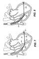
- FIGS. 23-30illustrate yet another embodiment of a method for delivering and implanting a splint across the left ventricle from a free wall HW to a septal wall SW.
- the method shown in these figuresis similar in many respects to the right ventricle delivery technique described above. However, the method to be described differs from the previously discussed right ventricle approach in that the splint is advanced across the left ventricle LV over a small hollow guidewire or needle of the type shown in FIGS. 24-27. Additionally, an alternative free wall deployable anchor structure is described.
- a guide deviceagain preferably in the form of a delivery catheter 400 , is positioned in the right ventricle RV from an access point, such as, preferably the right jugular vein, for example.
- Delivery catheter 400has a similar structure as delivery catheter 100 used in the right ventricle delivery technique described above. However, delivery catheter 400 does not advance into and across the left ventricle LV, as did delivery catheter 100 .
- Catheter 400has a curved distal tip portion 400 ′.
- a tether, or pull-wire, 405 connected to distal tip portion 400 ′is configured to adjust the angle or curvature of the tip portion 400 ′.
- Tether 405runs inside a lumen 420 disposed adjacent catheter 400 , or, alternatively, within catheter 400 .
- Delivery catheter 400also includes a pre-formed support wire 410 configured to extend via advancement of the support wire from another lumen 421 disposed adjacent catheter 400 on a side substantially opposite to the side lumen 420 is.
- Support wire 410not only assists to maintain the placement of tip portion 400 ′ of delivery catheter 400 within right ventricle RV in the appropriate position, but also assists in positioning the tip portion 400 ′ near the center of right ventricle RV relative to the anterior and posterior ends of the right ventricle, as a result of the shape and size of the support wire.
- Alternative shapes of the pre-formed support wirealso are contemplated which would facilitate tip positioning and support in other desired positions within right ventricle RV.
- a hollow sharpened metallic guidewire, or needle, 402is advanced through a central lumen 422 of delivery catheter 400 , across the ventricular septum SW, and across the left ventricular chamber LV to free wall HW, as shown in FIG. 24 .
- a combination of fluoroscopic and ultrasonic imagingare performed to assist in the guidance and confirmation of positioning for this delivery technique.
- Appropriate radiographic or other suitable visible markersare positioned on the devices to facilitate this imaging, as described above.
- Hollow guidewire 402has a sharpened tip 402 ′ and defines a central lumen plugged near tip 402 ′.
- the material used to make guidewire 402preferably includes a superelastic nickel titanium alloy, or other similar like material.
- Two elastomeric balloons, a distal balloon 403 and a proximal balloon 404are secured near the distal end of guidewire 402 slightly proximal to sharpened tip 402 ′.
- Distal balloon 403is in flow communication with central lumen 422 of guidewire 402 .
- Proximal balloon 404is in fluid communication with an additional tube (not shown) positioned inside hollow guidewire 402 . In this manner, each balloon 403 , 404 can be independently inflated and deflated as required.
- Balloons 403 , 404preferably are in a deflated condition as they are advanced across septal wall SW and then are inflated during advancement across the left ventricle LV. Inflating the balloons during advancement across the left ventricle LV may assist in visualizing the advancement path of the guidewire. To assist in such visualization, preferably the balloons are inflated with a radiographic contrast agent. The ability to visualize the advancement path of guidewire 402 may prevent damage to various cardiac structure as well as assist in ensuring proper positioning of the guidewire on the free wall HW.
- distal balloon 403As guidewire tip 402 ′ approaches free wall HW, distal balloon 403 is deflated, as shown in FIG. 25, and the wire is further advanced into the free wall. Proximnal balloon 404 acts as a stop to limit advancement of guidewire 402 through free wall HW. This may eliminate or minimize any damage to tissue outside free wall HW of left ventricle LV. Once fully advanced, distal balloon 403 is re-inflated to secure the position of guidewire 402 across the left ventricular chamber, as shown in FIG. 26 . It is preferred that the distance between balloons 403 , 404 approximates the thickness of the heart wall.
- Proximal balloon 404is then deflated, as shown in FIG. 27, and a splint advancement catheter 406 carrying the tension member 500 and fixed deployable anchor 502 is advanced over guidewire 402 , as shown in FIG. 28 .
- catheter 406defines a lumen 406 ′ through which braided tension, member 500 is configured to extend.
- Tension member 500is secured within a distal adhesive portion 502 ′ of a deployable anchor 502 .
- This adhesive portionpreferably is made of a high strength adhesive such as epoxy, or the like and is also configured to slide through lumen 406 ′.
- a lumen 509extends through fixed deployable anchor 502 adjacent to tension member braid 500 . This lumen also is formed simultaneously within adhesive portion 502 ′ of anchor 502 . Lumen 509 and lumen 406 ′ both pass over the outside of guidewire 402 (not shown) as advancement catheter 406 carrying tension member 500 with deployable fixed anchor 502 on one end is advanced across the left ventricle LV and through the free wall HW.
- Anchor 502preferably is in the form of an elastic or super-elastic metallic tube including a plurality of pre-formed tabs 508 extending proximally from adhesive tube portion 502 ′. The tabs 508 may be formed by several longitudinally-oriented cuts along a portion of the length of the tube.
- tabs 508are prevented from flaring outward by the sheath defining lumen 406 ′ of splint advancement catheter 406 , as shown in FIG. 31 .
- tabs 508Upon retraction of the sheath of splint advancement catheter 406 , tabs 508 are able to expand radially outwardly to their preformed shape, thus defining distal anchor 502 .
- a separate push tube 520 for pushing on anchor 502 as the catheter 406 is retracted from the tension member and fixed anchor assemblyalso is shown in FIG. 31 .
- Push tube 520is configured to pass over the outside of guidewire 402 within lumen 406 ′ adjacent tension member 500 to engage with the adhesive portion 502 ′ of anchor 502 .
- the deployable fixed anchormay have a structure similar to that described above with reference to the right ventricle and left ventricle delivery techniques.
- the deployable anchor configurations described in connection with FIGS. 29-31may be used in conjunction with other delivery techniques described above.
- the deployable anchor structures described in connection with the previous splint embodimentscan be utilized in conjunction with this embodiment.
- Elongate tension member 500preferably is similar to that described above in connection with the right ventricle delivery method and comprises a braid of high strength polymer fibers, preferably Spectra or other suitable like ultra-high molecular weight polyethylene.
- Tension member 500may also include a covering along its full length made of a thin expanded polytetrafluoroethylene. Alternatively, only, the region of tension member 500 which is disposed inside the ventricular chamber could include a covering.
- Tension member 500is thus advanced into position by sliding splint advancement catheter 406 carrying tension member 500 and anchor 502 over guidewire 402 . That is, guidewire 402 will be placed within lumen 509 of anchor 502 and then within lumen 406 ′ of the catheter 406 . The lumen 406 ′ and the lumen 509 will move relative to guidewire 402 to advance catheter 406 , tension member 500 , and anchor 502 in the configuration shown in FIG. 31 until deployable anchor 502 protrudes beyond the myocardium of free wall HW. Once tension member 500 and anchor 502 are positioned appropriately with respect to the left ventricle and free wall HW, that is, when anchor 502 retained within the catheter 406 protrudes beyond the freewall HW as shown in FIG.
- catheter 406is retracted off tabs 508 . This retraction of catheter 406 enables tabs 508 to expand radially outward from the remainder of deployable anchor 502 .
- Push tube 520is used to maintain the position of the tension member 500 during the catheter's retraction to overcome any friction between catheter 406 and tabs 508 . After anchor 502 is deployed, both catheter 406 and push tube 520 are removed from guidewire 402 and then guidewire 402 also is removed.
- the proximal anchormay have a similar structure as the distal fixed deployable anchor or may be separately slidable and adjustable on the tension member (such as the adjustable anchor shown in FIGS. 7 - 10 ). The proximal anchor also may be pre-attached at an appropriate position on the tension member to provide the desired amount of ventricular shape change.
- a one way “ratchet” or friction surfacemay be disposed on the inner surface of the tubular portion of the anchor to prevent its displacement in one direction.
- the inner surface of the tubular portion of the anchorcan be in the form of rings or flared protrusions 380 that are angled with respect to the longitudinal axis of a tension member 385 as it is inserted into an anchor 388 .
- the angled rings or protrusions 380are configured so as to permit movement of the anchor with respect to the tension member in one direction but prevent movement in the opposite direction.
- the rings or protrusionswould permit movement in the direction of the solid arrow, but prevent movement in the direction of the dotted arrow by essentially digging into the tension member surface.
- a tightening devicesuch as that described and shown in FIG. 8 may be utilized to advance the deployed anchor into position.
- the anchormay be initially positioned such that when the sheath of the splint advancement catheter is further withdrawn, the proximal anchor also would deploy within the right ventricle RV adjacent to the septal wall SW.
- the tightening devicecould then be used to advance the position of the proximal anchor to a desired position against the septal wall SW, as shown in FIG. 30 .
- the delivery catheteritself could be used to advance the deployed proximal anchor to the desired position. Once the proximal anchor is to positioned to its appropriate location, any excess tension member length extending beyond the proximal anchor may be severed in a manner similar to that described above in connection with FIG. 9 .
- proximal anchorIn the alternative case where proximal anchor is pre-attached at a specified distance from the distal anchor, the left ventricle should be deformed prior to the pad deployment.
- the delivery cathetercan act as a temporary proximal anchor, while the tension member and distal anchor are pulled proximally.
- the proximal anchormay be deployed, upon further retraction of the sheath of the splint advancement catheter.
- the distance between the distal and proximal anchorswill be selected prior to delivery such that a desired shape change of the heart chamber may be obtained, since the adjustability of the shape change will be limited by the fixed position of the proximal anchor on the tension member.
- the delivery cathetermay then be removed and excess tension member severed, again as described with reference to FIG. 9 .
- splint delivery methods and devices just described in connection with FIGS. 23-30were in the context of a right ventricle approach, it is also contemplated to utilize the delivery devices and methods in a direct left ventricle approach as well.
- a direct left ventricle approachtwo delivery catheters simultaneously could be utilized to position a splint from within the left ventricle in manners similar to those described with reference to FIGS. 17-22.
- FIGS. 32-35Other embodiments of a deployable, fixed heart-engaging assembly, or anchor, also are contemplated as within the scope of the present invention and are shown in FIGS. 32-35.
- a preferred tension member used in the various embodiments of the present inventionis described in the '049 application, incorporated by reference above, and is formed of several multifilament bundles of a high strength polymer. These bundles are braided together in a relatively tight configuration. Certain combinations of bundle size, number of bundles, and pick count, described in more detail in the '049 application, result in a braid with several preferred properties as also described in the '049 application, incorporated by reference above.
- One property that may result from such a braid constructionincludes a relatively stable braid diameter that does not deform to a great extent if subjected to axial compression or internal radially-outward directed forces.
- a braid formed with a lower pick counthas a greater diametric expandability when subjected to such forces.
- a braid woven of the same material and having approximately 16 to approximately 64 bundles and approximately 2 to approximately 15 picks per inchmay more readily expand in diameter, upon the application of a radial force directed outwardly from within the braid.
- This expandable property of a braidcan be utilized in the formation of yet another alternative deployable anchor structure.
- FIG. 32is a relatively simplified schematic illustrating a tension member 50 formed of a braid of relatively low pick count in its natural (i.e., non-stressed) as-braided condition.
- the braidis uniformly relatively small in outer diameter D 1 .
- the diameter D 1 of the braidremains relatively small.
- FIG. 33illustrates the same braided tension member 50 as in FIG. 32, but shows the braid with a local application of an outward radially directed force from within the braid. Since the braid pick count is relatively low, the braid has the capacity to expand at the point of application of the radial force to a diameter D 2 , which is several times its original diameter D 1 .
- FIG. 34shows a tension member 340 having a braid configuration such as that shown in FIGS. 32 and 33 utilized for an integral expandable distal anchor 342 of tension member 340 . At least a portion of the tension member 340 is woven at a relatively low pick count to allow for that portion to form into the expandable distal anchor 342 . Alternatively, the entire tension member 340 can be uniformly woven at the relatively low pick count.
- Several methods for creating an outward radial force from within the braid to form anchor 342are contemplated. These methods include using an elastic or shape memory wire disposed inside the braid of tension member 340 during placement across the ventricular wall HW. A preferred wire 345 is shown in FIG. 34 a .
- Wire 345preferably has a natural shape in the form of a disc-shaped spiral.
- the spiralwill have a straightened configuration.
- the spiral shape of wire 34may be re-established, thereby forcing the braided portion surrounding it to expand in diameter into a disc-like shape, as shown in FIG. 34 .
- the wiremay either be pre-loaded into the tension member or may be advanced into the tension member once the tension member has been positioned with respect to the heart wall and the catheter has been retracted enough to expose a portion of the tension member that is outside the heart wall HW.
- the force of the catheter on the wirekeeps the wire in its straightened configuration.
- the smaller diameter portion of the spiralforms first, and as more of the wire is advanced through the tension member beyond the catheter, the spiral grows in diameter until the full spiral is re-established.
- the large diameter portion of the spiralmay form first, as the wire 345 ′ is advanced.
- a thin membranepreferably made of an elastic material for example, is disposed along the inside of the braid in the area where the spiral portion of wire 345 is positioned.
- wire 345preferably will remain together with the braid of tension member 340 even after diametric expansion in order provide the anchoring rigidity needed to secure the splint in place on the heart.
- the spiral portion of wire 345may carry a significant portion of the load of anchor 342 .
- the expanded braid forming anchor 342would become ingrown with scar tissue, and therefore a relatively large portion of the chronic mechanical loading may be carried by the filaments of the braid.
- filaments of ultra-high molecular weight polyethylenehas been shown to produce a tension member and anchor having high strength and high fatigue resistance.
- a portion of the wire that does not form the spiralmay be removed, for example by torquing the wire and breaking it at a location slightly proximal to the spiral.
- the distal most region of the braidpreferably is either fused or banded. This will prevent expansibility in those regions. Alternatively, those regions of the braided tension member could be woven with a higher pick count.
- An alternative device for causing expansion of an expandable braid portion of a tension member 540includes an inflatable balloon disposed inside braided tension member at the expandable portion forming the anchor, as shown in FIG. 35 .
- Inflatable balloon 545can be positioned in the desired location within tension member 540 either before advancement of the tension member across the ventricular wall, or after, as shown in FIG. 35 .
- balloon 545is formed of an elastomeric material such as silicone or urethane, or the like, and has a disc-like shape upon expansion.
- a lumen 543 connecting the interior of balloon 545 to an inflation device (not shown)may extend along the inside of braided tension member 540 .
- the material used to expand balloon 545includes a curable material such as RTV silicone, epoxy, urethane, or the like. Similar to the spiral anchor embodiment discussed above, the cured material forming the balloon may carry a significant load initially, but upon ingrowth of the expanded braided region of the tension member, the filaments of the braid would be the primary chronic load carrying members.
- an expandable anchorwould utilize an anchor similar to the expandable tab anchor described above with reference to FIGS. 29-31.
- that anchorwould be merely temporary, and could be designed to be relatively small and low profile, for easy delivery, but may not have the adequate strength and durability to be a permanent chronic anchor.
- a permanent anchorsimilar to the adjustable pad anchor assembly described in the '049 application incorporated above could be delivered as a replacement to that temporary anchor.
- a small surgical incisionmay be made between the ribs adjacent the free wall HW of the left ventricle LV, thus creating an access port to deliver the permanent anchor.
- a trocarmay be positioned in that same location.
- a snare device positioned within the it port or trocarcan be used to grab the temporary anchor and tension member and pull the anchor off of the tension member and outside of the patient.
- An adjustable anchor pad as described in the '049 application and similar to the description of FIGS. 7 and 8 abovethen may be attached to the braid outside of the patient, using the staple methodology described previously with reference to FIG. 8 and the '049 application.
- the anchorcan then be pulled back into position by retracting the other end of the tension member via the length of the tension member that remains outside the jugular vein.
- the septal wall anchorin this case preferably would be in the form of the solid anchor and delivered in the manner described above in conjunction with FIGS. 7-10. Overall, this procedure would be a combination endovascular and “minimally invasive” surgical operation.
- both anchor padswould be of a solid type construction.
- An alternative proximal anchoralso may utilize the expandable capability of a relatively low pick count braid, in a manner similar to that described above for the distal, or free wall, anchor.
- the entire braid of the tension memberpreferably includes the relatively low pick count permitting diametric expansion.
- the tension member and distal anchorcould be delivered using any of the approaches described herein, but preferably one of the right ventricle approach methods. After the distal, or free wall, anchor is delivered, the proper ventricle shape change can be induced using a tightening device in the form of a simple tube, such as the one described above, but without the anchor shown in FIG. 8.
- a balloon or spiral wire type of expander deviceas described in connection with the distal anchors shown in FIGS.
- a balloon type expansion devicemay also include a detachable inflation tube, such that when the balloon is inflated with a curable material, the inflation tube can be removed prior to the excess tension member length being severed. It is also contemplated that such an expandable proximal anchor can be secured to the end of the tension member at a location adjacent to an exterior surface of a heart wall other than the septal wall, such as a wall surrounding the right ventricle, for example.
- the methods described above to place the splint assemblies with respect to the heartcan be repeated for any desired number of splint assemblies to achieve a particular configuration.
- the length of the tension members extending between the free wall and septal wall anchorsalso can be optimally determined based upon the size and condition of the patient's heart. It should also be noted that although the left ventricle has been referred to here for illustrative purposes, the apparatus and methods of this invention can be used to splint multiple chambers of a patient's heart, including the right ventricle or either atrium, or can be used to aid the function of valves, such as the mitral valve.
- An example of a preferred position of a splint assembly 1000improves mitral valve function, as well as reducing stress in the left ventricular walls.
- the valve functionis improved by aiding in the apposition of valve leaftlets when positioned as shown in FIG. 37 .
- three splintsare placed in a coplanar fashion, bisecting the left ventricle LV of the heart.
- the superior-most splint 1000is placed at approximately the level of the heads of the papillary muscles PM, and the additional two splints (not shown in FIG. 37) are placed inferiorly toward the apex.
- the preferred orientation shown in FIG. 37both bisects the left ventricle LV and avoids key structures such as coronary vessels and the like.
- the splints according to this orientationalso extend through the septum SW and enter a portion of the right ventricle RV.
- heart-engaging assemblies 1002 , 1003will be positioned adjacent an exterior surface of a free wall HW surrounding the left ventricle LV and adjacent an exterior surface of the septal wall SW within the right ventricle RV.
- Further details regarding splinting devices and methods for treating heart valvescan be found in the application entitled “Methods and Devices for Improving Mitral Valve Function,” to Richard F. Schroeder et al., filed on a date even herewith and incorporated by reference above. Although any of the delivery methods described above could be used to implant the splint device in this manner, FIG.
- FIG. 36shows a short axis cross-section of a heart and a preferred endovascular technique wherein the elongate tension member 1001 having a deployable fixed anchor 1002 on its distal end is delivered through the right ventricle RV and then into the left ventricle LV.
- the alignments of the splints with respect to the heartthat are described above are illustrative only and may be shifted or rotated about a vertical axis generally disposed through the left ventricle and still avoid the major coronary vessels and papillary muscles.
- inventive devices and methodscan be implanted to treat a heart having aneurysms or infarcted regions similar to those described in prior U.S. application Ser. No. 09/422,328 discussed earlier herein and incorporated above.
- the various components of the splint assembly to be implanted in the heartshould be made of biocompatible material that can remain in the human body indefinitely. Any surface engaging portions of the heart should be a traumatic; in order to avoid tissue damage and preferably formed of a material promoting tissue ingrowth to stabilize the anchor pad with respect to the surfaces of the heart.
Landscapes
- Health & Medical Sciences (AREA)
- Life Sciences & Earth Sciences (AREA)
- Cardiology (AREA)
- Surgery (AREA)
- General Health & Medical Sciences (AREA)
- Public Health (AREA)
- Heart & Thoracic Surgery (AREA)
- Biomedical Technology (AREA)
- Veterinary Medicine (AREA)
- Animal Behavior & Ethology (AREA)
- Engineering & Computer Science (AREA)
- Nuclear Medicine, Radiotherapy & Molecular Imaging (AREA)
- Molecular Biology (AREA)
- Medical Informatics (AREA)
- Oral & Maxillofacial Surgery (AREA)
- Transplantation (AREA)
- Vascular Medicine (AREA)
- Prostheses (AREA)
- Surgical Instruments (AREA)
- Acyclic And Carbocyclic Compounds In Medicinal Compositions (AREA)
- Agricultural Chemicals And Associated Chemicals (AREA)
- Medicines Containing Material From Animals Or Micro-Organisms (AREA)
Abstract
Description
Claims (25)
Priority Applications (11)
| Application Number | Priority Date | Filing Date | Title |
|---|---|---|---|
| US09/679,550US6616684B1 (en) | 2000-10-06 | 2000-10-06 | Endovascular splinting devices and methods |
| DE60119944TDE60119944T2 (en) | 2000-10-06 | 2001-10-02 | ENOVOVASCULAR SPLINING DEVICES |
| AT01975613TATE326921T1 (en) | 2000-10-06 | 2001-10-02 | ENDOVASCULAR SPLIN DEVICES |
| AU2001294921AAU2001294921A1 (en) | 2000-10-06 | 2001-10-02 | Endovascular splinting devices |
| EP01975613AEP1322259B1 (en) | 2000-10-06 | 2001-10-02 | Endovascular splinting devices |
| EP06114286AEP1695674A3 (en) | 2000-10-06 | 2001-10-02 | Endovascular splinting devices |
| PCT/US2001/030629WO2002030335A2 (en) | 2000-10-06 | 2001-10-02 | Endovascular splinting devices |
| EP08166280AEP2014242A3 (en) | 2000-10-06 | 2001-10-02 | Endovascular splinting devices |
| US10/410,279US20030181928A1 (en) | 2000-10-06 | 2003-04-10 | Endovascular splinting devices and methods |
| US10/735,269US20040225304A1 (en) | 2000-10-06 | 2003-12-12 | Endovascular splinting devices and methods |
| US11/543,892US20070055303A1 (en) | 2000-10-06 | 2006-10-06 | Endovascular splinting devices and methods |
Applications Claiming Priority (1)
| Application Number | Priority Date | Filing Date | Title |
|---|---|---|---|
| US09/679,550US6616684B1 (en) | 2000-10-06 | 2000-10-06 | Endovascular splinting devices and methods |
Related Child Applications (2)
| Application Number | Title | Priority Date | Filing Date |
|---|---|---|---|
| US10/410,279ContinuationUS20030181928A1 (en) | 2000-10-06 | 2003-04-10 | Endovascular splinting devices and methods |
| US10/410,279DivisionUS20030181928A1 (en) | 2000-10-06 | 2003-04-10 | Endovascular splinting devices and methods |
Publications (1)
| Publication Number | Publication Date |
|---|---|
| US6616684B1true US6616684B1 (en) | 2003-09-09 |
Family
ID=24727366
Family Applications (4)
| Application Number | Title | Priority Date | Filing Date |
|---|---|---|---|
| US09/679,550Expired - LifetimeUS6616684B1 (en) | 2000-10-06 | 2000-10-06 | Endovascular splinting devices and methods |
| US10/410,279AbandonedUS20030181928A1 (en) | 2000-10-06 | 2003-04-10 | Endovascular splinting devices and methods |
| US10/735,269AbandonedUS20040225304A1 (en) | 2000-10-06 | 2003-12-12 | Endovascular splinting devices and methods |
| US11/543,892AbandonedUS20070055303A1 (en) | 2000-10-06 | 2006-10-06 | Endovascular splinting devices and methods |
Family Applications After (3)
| Application Number | Title | Priority Date | Filing Date |
|---|---|---|---|
| US10/410,279AbandonedUS20030181928A1 (en) | 2000-10-06 | 2003-04-10 | Endovascular splinting devices and methods |
| US10/735,269AbandonedUS20040225304A1 (en) | 2000-10-06 | 2003-12-12 | Endovascular splinting devices and methods |
| US11/543,892AbandonedUS20070055303A1 (en) | 2000-10-06 | 2006-10-06 | Endovascular splinting devices and methods |
Country Status (6)
| Country | Link |
|---|---|
| US (4) | US6616684B1 (en) |
| EP (3) | EP1322259B1 (en) |
| AT (1) | ATE326921T1 (en) |
| AU (1) | AU2001294921A1 (en) |
| DE (1) | DE60119944T2 (en) |
| WO (1) | WO2002030335A2 (en) |
Cited By (223)
| Publication number | Priority date | Publication date | Assignee | Title |
|---|---|---|---|---|
| US20030216761A1 (en)* | 1990-03-27 | 2003-11-20 | Samuel Shiber | Guidewire system |
| US20040122503A1 (en)* | 2002-12-20 | 2004-06-24 | Campbell Carey V. | Implantable medical device assembly |
| US20040127981A1 (en)* | 2000-09-20 | 2004-07-01 | Ample Medical, Inc. | Devices, systems, and methods for retaining a native heart valve leaflet |
| US20040133243A1 (en)* | 2002-07-26 | 2004-07-08 | Santamore William P. | Cardiac rhythm management system with intramural myocardial pacing leads and electrodes |
| US6776754B1 (en)* | 2000-10-04 | 2004-08-17 | Wilk Patent Development Corporation | Method for closing off lower portion of heart ventricle |
| US20040260393A1 (en)* | 2000-09-20 | 2004-12-23 | Ample Medical, Inc. | Devices, systems, and methods for reshaping a heart valve annulus |
| US20040260317A1 (en)* | 2003-06-20 | 2004-12-23 | Elliot Bloom | Tensioning device, system, and method for treating mitral valve regurgitation |
| US20050015110A1 (en)* | 2003-07-18 | 2005-01-20 | Fogarty Thomas J. | Embolization device and a method of using the same |
| US20050027309A1 (en)* | 2003-06-17 | 2005-02-03 | Samuel Shiber | Guidewire system |
| US20050055089A1 (en)* | 2000-09-20 | 2005-03-10 | Ample Medical, Inc. | Devices, systems, and methods for reshaping a heart valve annulus |
| US20050143768A1 (en)* | 2003-06-17 | 2005-06-30 | Samuel Shiber | Sleeved guidewire system method of use |
| US20050177073A1 (en)* | 2003-06-17 | 2005-08-11 | Samuel Shiber | Guidewire system with a deflectable distal tip |
| WO2005087140A1 (en)* | 2004-03-11 | 2005-09-22 | Percutaneous Cardiovascular Solutions Pty Limited | Percutaneous heart valve prosthesis |
| US20050222488A1 (en)* | 2003-10-01 | 2005-10-06 | Ample Medical, Inc. | Devices, systems, and methods for reshaping a heart valve annulus |
| US20050228422A1 (en)* | 2002-11-26 | 2005-10-13 | Ample Medical, Inc. | Devices, systems, and methods for reshaping a heart valve annulus, including the use of magnetic tools |
| US20050245939A1 (en)* | 2002-06-14 | 2005-11-03 | Joseph Ferrante | Device and methods for placing external fixation elements |
| US20050288613A1 (en)* | 2004-06-25 | 2005-12-29 | Cardiac Pacemakers, Inc. | Endocardial splint and method therefor |
| US20060089711A1 (en)* | 2004-10-27 | 2006-04-27 | Medtronic Vascular, Inc. | Multifilament anchor for reducing a compass of a lumen or structure in mammalian body |
| US20060106279A1 (en)* | 2004-05-14 | 2006-05-18 | Ample Medical, Inc. | Devices, systems, and methods for reshaping a heart valve annulus, including the use of a bridge implant having an adjustable bridge stop |
| US20060106420A1 (en)* | 2004-11-12 | 2006-05-18 | Medtronic Vascular, Inc. | Patch for treating a septal defect |
| US20060106278A1 (en)* | 2004-05-14 | 2006-05-18 | Ample Medical, Inc. | Devices, systems, and methods for reshaping a heart valve annulus, including the use of an adjustable bridge implant system |
| US7077862B2 (en) | 2002-01-09 | 2006-07-18 | Myocor, Inc. | Devices and methods for heart valve treatment |
| US20060167494A1 (en)* | 2005-01-21 | 2006-07-27 | Loubert Suddaby | Aneurysm repair method and apparatus |
| US7112219B2 (en) | 2002-11-12 | 2006-09-26 | Myocor, Inc. | Devices and methods for heart valve treatment |
| US20060270896A1 (en)* | 2005-05-31 | 2006-11-30 | Ethicon, Inc. | Method and device for deployment of a sub-pericardial sack |
| US20060287661A1 (en)* | 2001-11-28 | 2006-12-21 | Aptus Endosystems, Inc. | Devices, systems, and methods for supporting tissue and/or structures within a hollow body organ |
| US20060292206A1 (en)* | 2001-11-26 | 2006-12-28 | Kim Steven W | Devices and methods for treatment of vascular aneurysms |
| US20070025009A1 (en)* | 2005-07-29 | 2007-02-01 | Fuji Photo Film Co., Ltd. | Magnetic recorder |
| WO2007022519A2 (en) | 2005-08-19 | 2007-02-22 | Chf Technologies, Inc. | Steerable heart implants for congestive heart failure |
| US20070049971A1 (en)* | 2005-06-09 | 2007-03-01 | Sing-Fatt Chin | Method and apparatus for closing off a portion of a heart ventricle |
| US7186262B2 (en)* | 1999-06-25 | 2007-03-06 | Vahid Saadat | Apparatus and methods for treating tissue |
| US20070066863A1 (en)* | 2005-08-31 | 2007-03-22 | Medtronic Vascular, Inc. | Device for treating mitral valve regurgitation |
| US20070073274A1 (en)* | 2005-08-19 | 2007-03-29 | Sing-Fatt Chin | Method and device for treating dysfunctional cardiac tissue |
| US7247134B2 (en) | 2002-11-12 | 2007-07-24 | Myocor, Inc. | Devices and methods for heart valve treatment |
| US20070185530A1 (en)* | 2005-09-01 | 2007-08-09 | Chao Chin-Chen | Patent foramen ovale closure method |
| US20070203391A1 (en)* | 2006-02-24 | 2007-08-30 | Medtronic Vascular, Inc. | System for Treating Mitral Valve Regurgitation |
| US20070225681A1 (en)* | 2006-03-21 | 2007-09-27 | Medtronic Vascular | Catheter Having a Selectively Formable Distal Section |
| US20070225759A1 (en)* | 2006-03-22 | 2007-09-27 | Daniel Thommen | Method for delivering a medical device to the heart of a patient |
| US7291168B2 (en) | 2001-10-01 | 2007-11-06 | Ample Medical, Inc. | Methods and devices for heart valve treatments |
| US20070265658A1 (en)* | 2006-05-12 | 2007-11-15 | Aga Medical Corporation | Anchoring and tethering system |
| US20080065204A1 (en)* | 2000-09-20 | 2008-03-13 | Ample Medical, Inc. | Devices, systems, and methods for supplementing, repairing, or replacing a native heart valve leaflet |
| US20080082132A1 (en)* | 2006-09-28 | 2008-04-03 | Chf Technologies, Inc., A California Corporation | Signal Transmitting and Lesion Excluding Heart Implants for Pacing Defibrillating and/or Sensing of Heart Beat |
| US20080091059A1 (en)* | 2004-05-14 | 2008-04-17 | Ample Medical, Inc. | Devices, systems, and methods for reshaping a heart valve annulus, including the use of a bridge implant having an adjustable bridge stop |
| US20080097148A1 (en)* | 2004-10-13 | 2008-04-24 | Chf Technologies, Inc. | Method and device for percutaneous left ventricular reconstruction |
| US7381182B2 (en) | 2001-09-07 | 2008-06-03 | Mardil, Inc. | Method and apparatus for external heart stabilization |
| JP2008534084A (en)* | 2005-03-25 | 2008-08-28 | アンプル メディカル, インコーポレイテッド | Apparatus, system, and method for reshaping a heart valve annulus |
| US20080294251A1 (en)* | 2006-09-28 | 2008-11-27 | Bioventrix (A Chf Technologies' Company) | Location, time, and/or pressure determining devices, systems, and methods for deployment of lesion-excluding heart implants for treatment of cardiac heart failure and other disease states |
| US20090093670A1 (en)* | 2007-10-03 | 2009-04-09 | Bioventrix (A Chf Technologies, Inc.) | Treating Dysfunctional Cardiac Tissue |
| US20090099410A1 (en)* | 2005-06-09 | 2009-04-16 | De Marchena Eduardo | Papillary Muscle Attachment for Left Ventricular Reduction |
| US20090105748A1 (en)* | 2002-11-12 | 2009-04-23 | Thomas J. Fogarty | Embolization device and a method of using the same |
| US7544203B2 (en)* | 2001-12-26 | 2009-06-09 | Maquet Cardiovascular Llc | Temporary seal and method for facilitating anastomosis |
| US20090234318A1 (en)* | 2007-10-19 | 2009-09-17 | Guided Delivery Systems, Inc. | Systems and methods for cardiac remodeling |
| US20090287179A1 (en)* | 2003-10-01 | 2009-11-19 | Ample Medical, Inc. | Devices, systems, and methods for reshaping a heart valve annulus, including the use of magnetic tools |
| US7632308B2 (en) | 2005-11-23 | 2009-12-15 | Didier Loulmet | Methods, devices, and kits for treating mitral valve prolapse |
| US20100010608A1 (en)* | 2003-09-30 | 2010-01-14 | Tockman Bruce A | Guide Wire Stylet |
| US20100016655A1 (en)* | 2008-07-21 | 2010-01-21 | Bio Ventrix (a CHF Technologies, Inc. | Cardiac Anchor Structures, Methods, and Systems for Treatment of Congestive Heart Failure and Other Conditions |
| US7695425B2 (en) | 1997-01-02 | 2010-04-13 | Edwards Lifesciences Llc | Heart wall tension reduction apparatus and method |
| US7704269B2 (en) | 1999-04-09 | 2010-04-27 | Evalve, Inc. | Methods and apparatus for cardiac valve repair |
| US7722523B2 (en) | 1998-07-29 | 2010-05-25 | Edwards Lifesciences Llc | Transventricular implant tools and devices |
| US7736388B2 (en) | 1999-04-09 | 2010-06-15 | Evalve, Inc. | Fixation devices, systems and methods for engaging tissue |
| US7766812B2 (en) | 2000-10-06 | 2010-08-03 | Edwards Lifesciences Llc | Methods and devices for improving mitral valve function |
| US20100222810A1 (en)* | 2002-01-18 | 2010-09-02 | Debeer Nicholas | Methods and devices for delivering injections |
| US7811296B2 (en) | 1999-04-09 | 2010-10-12 | Evalve, Inc. | Fixation devices for variation in engagement of tissue |
| US7883539B2 (en) | 1997-01-02 | 2011-02-08 | Edwards Lifesciences Llc | Heart wall tension reduction apparatus and method |
| US20110071626A1 (en)* | 2008-05-12 | 2011-03-24 | Wright John T M | Device and Method for the Surgical Treatment of Ischemic Mitral Regurgitation |
| US7938827B2 (en) | 2001-11-15 | 2011-05-10 | Evalva, Inc. | Cardiac valve leaflet attachment device and methods thereof |
| US7976539B2 (en) | 2004-03-05 | 2011-07-12 | Hansen Medical, Inc. | System and method for denaturing and fixing collagenous tissue |
| US7981123B2 (en) | 1997-09-12 | 2011-07-19 | Evalve, Inc. | Surgical device for connecting soft tissue |
| US7981139B2 (en) | 2002-03-01 | 2011-07-19 | Evalve, Inc | Suture anchors and methods of use |
| US20110184510A1 (en)* | 2010-01-22 | 2011-07-28 | 4Tech, Sarl | Tricuspid valve repair using tension |
| US20110196492A1 (en)* | 2007-09-07 | 2011-08-11 | Intrinsic Therapeutics, Inc. | Bone anchoring systems |
| US8021425B2 (en)* | 1999-08-18 | 2011-09-20 | Intrinsic Therapeutics, Inc. | Versatile method of repairing an intervertebral disc |
| US8025698B2 (en) | 1999-08-18 | 2011-09-27 | Intrinsic Therapeutics, Inc. | Method of rehabilitating an anulus fibrosus |
| US8029518B2 (en) | 1999-04-09 | 2011-10-04 | Evalve, Inc. | Methods and devices for capturing and fixing leaflets in valve repair |
| US8052592B2 (en) | 2005-09-27 | 2011-11-08 | Evalve, Inc. | Methods and devices for tissue grasping and assessment |
| AU2006212750B2 (en)* | 2005-02-07 | 2011-11-17 | Evalve, Inc. | Methods, systems and devices for cardiac valve repair |
| US8092363B2 (en) | 2007-09-05 | 2012-01-10 | Mardil, Inc. | Heart band with fillable chambers to modify heart valve function |
| US8114082B2 (en) | 2005-12-28 | 2012-02-14 | Intrinsic Therapeutics, Inc. | Anchoring system for disc repair |
| US8123703B2 (en) | 1999-04-09 | 2012-02-28 | Evalve, Inc. | Steerable access sheath and methods of use |
| US20120101525A1 (en)* | 2010-10-25 | 2012-04-26 | Boston Scientific Scimed, Inc. | Fibrous containment for hemostasis plug |
| US8216256B2 (en) | 1999-04-09 | 2012-07-10 | Evalve, Inc. | Detachment mechanism for implantable fixation devices |
| US8226711B2 (en) | 1997-12-17 | 2012-07-24 | Edwards Lifesciences, Llc | Valve to myocardium tension members device and method |
| US8231678B2 (en) | 1999-08-18 | 2012-07-31 | Intrinsic Therapeutics, Inc. | Method of treating a herniated disc |
| US8323341B2 (en) | 2007-09-07 | 2012-12-04 | Intrinsic Therapeutics, Inc. | Impaction grafting for vertebral fusion |
| US8343174B2 (en) | 1999-04-09 | 2013-01-01 | Evalve, Inc. | Locking mechanisms for fixation devices and methods of engaging tissue |
| US8398672B2 (en) | 2003-11-12 | 2013-03-19 | Nitinol Devices And Components, Inc. | Method for anchoring a medical device |
| WO2013049766A1 (en) | 2011-09-30 | 2013-04-04 | Bioventrix, Inc. | Over-the-wire cardiac implant delivery system for treatment of chf and other conditions |
| US8465500B2 (en) | 2005-01-21 | 2013-06-18 | Mayo Foundation For Medical Education And Research | Thorascopic heart valve repair method and apparatus |
| US8470028B2 (en) | 2005-02-07 | 2013-06-25 | Evalve, Inc. | Methods, systems and devices for cardiac valve repair |
| US8685044B2 (en) | 2001-11-28 | 2014-04-01 | Aptus Endosystems, Inc. | Systems and methods for attaching a prosthesis with a body lumen or hollow organ |
| US8690897B2 (en) | 2001-11-28 | 2014-04-08 | Aptus Endosystems, Inc. | Devices, systems, and methods for prosthesis delivery and implantation, including the use of a fastener tool |
| US8758393B2 (en) | 2007-10-18 | 2014-06-24 | Neochord, Inc. | Minimally invasive repair of a valve leaflet in a beating heart |
| EP2111243A4 (en)* | 2006-11-09 | 2014-07-02 | Corlife Ohg | CARDIOVASCULAR OPERATIONS |
| USD717954S1 (en) | 2013-10-14 | 2014-11-18 | Mardil, Inc. | Heart treatment device |
| US8961594B2 (en) | 2012-05-31 | 2015-02-24 | 4Tech Inc. | Heart valve repair system |
| US8961596B2 (en) | 2010-01-22 | 2015-02-24 | 4Tech Inc. | Method and apparatus for tricuspid valve repair using tension |
| US9044221B2 (en) | 2010-12-29 | 2015-06-02 | Neochord, Inc. | Exchangeable system for minimally invasive beating heart repair of heart valve leaflets |
| US9060858B2 (en) | 2009-09-15 | 2015-06-23 | Evalve, Inc. | Methods, systems and devices for cardiac valve repair |
| US9078749B2 (en) | 2007-09-13 | 2015-07-14 | Georg Lutter | Truncated cone heart valve stent |
| US9241702B2 (en) | 2010-01-22 | 2016-01-26 | 4Tech Inc. | Method and apparatus for tricuspid valve repair using tension |
| US9307980B2 (en) | 2010-01-22 | 2016-04-12 | 4Tech Inc. | Tricuspid valve repair using tension |
| US9320503B2 (en) | 2001-11-28 | 2016-04-26 | Medtronic Vascular, Inc. | Devices, system, and methods for guiding an operative tool into an interior body region |
| US9320589B2 (en) | 2001-11-28 | 2016-04-26 | Medtronic Vascular, Inc. | Endovascular aneurysm repair system |
| US9370425B2 (en) | 2012-10-12 | 2016-06-21 | Mardil, Inc. | Cardiac treatment system and method |
| US9452016B2 (en) | 2011-01-21 | 2016-09-27 | Kardium Inc. | Catheter system |
| US9480559B2 (en) | 2011-08-11 | 2016-11-01 | Tendyne Holdings, Inc. | Prosthetic valves and related inventions |
| US9480525B2 (en) | 2011-01-21 | 2016-11-01 | Kardium, Inc. | High-density electrode-based medical device system |
| US9486306B2 (en) | 2013-04-02 | 2016-11-08 | Tendyne Holdings, Inc. | Inflatable annular sealing device for prosthetic mitral valve |
| US9492228B2 (en) | 2011-01-21 | 2016-11-15 | Kardium Inc. | Enhanced medical device for use in bodily cavities, for example an atrium |
| US9526611B2 (en) | 2013-10-29 | 2016-12-27 | Tendyne Holdings, Inc. | Apparatus and methods for delivery of transcatheter prosthetic valves |
| USD777926S1 (en)* | 2012-01-20 | 2017-01-31 | Kardium Inc. | Intra-cardiac procedure device |
| USD777925S1 (en)* | 2012-01-20 | 2017-01-31 | Kardium Inc. | Intra-cardiac procedure device |
| US9585717B2 (en) | 2007-11-16 | 2017-03-07 | Kardium Inc. | Medical device for use in bodily lumens, for example an atrium |
| US9597181B2 (en) | 2013-06-25 | 2017-03-21 | Tendyne Holdings, Inc. | Thrombus management and structural compliance features for prosthetic heart valves |
| US20170086975A1 (en)* | 2014-06-19 | 2017-03-30 | 4Tech Inc. | Cardiac tissue cinching |
| US9610159B2 (en) | 2013-05-30 | 2017-04-04 | Tendyne Holdings, Inc. | Structural members for prosthetic mitral valves |
| US9615912B2 (en) | 2003-02-12 | 2017-04-11 | Thomas J. Fogarty | Intravascular implants and methods of using the same |
| US9649211B2 (en) | 2009-11-04 | 2017-05-16 | Confluent Medical Technologies, Inc. | Alternating circumferential bridge stent design and methods for use thereof |
| WO2017087688A1 (en)* | 2015-11-17 | 2017-05-26 | Edwards Lifesciences Corporation | Ultrasound probe for cardiac treatment |
| US9675454B2 (en) | 2012-07-30 | 2017-06-13 | Tendyne Holdings, Inc. | Delivery systems and methods for transcatheter prosthetic valves |
| US9693865B2 (en) | 2013-01-09 | 2017-07-04 | 4 Tech Inc. | Soft tissue depth-finding tool |
| US9706947B2 (en) | 1999-08-18 | 2017-07-18 | Intrinsic Therapeutics, Inc. | Method of performing an anchor implantation procedure within a disc |
| US20170216032A1 (en)* | 2016-01-29 | 2017-08-03 | Bioventrix, Inc. | Percutaneous arterial access to position trans-myocardial implant devices and methods |
| US9737403B2 (en) | 2006-03-03 | 2017-08-22 | Mardil, Inc. | Self-adjusting attachment structure for a cardiac support device |
| US9827092B2 (en) | 2011-12-16 | 2017-11-28 | Tendyne Holdings, Inc. | Tethers for prosthetic mitral valve |
| US9861475B2 (en) | 2000-09-20 | 2018-01-09 | Mvrx Inc. | Devices, systems, and methods for reshaping a heart valve annulus |
| US9895221B2 (en) | 2012-07-28 | 2018-02-20 | Tendyne Holdings, Inc. | Multi-component designs for heart valve retrieval device, sealing structures and stent assembly |
| US9907547B2 (en) | 2014-12-02 | 2018-03-06 | 4Tech Inc. | Off-center tissue anchors |
| US9907681B2 (en) | 2013-03-14 | 2018-03-06 | 4Tech Inc. | Stent with tether interface |
| US9968353B2 (en) | 2001-06-04 | 2018-05-15 | Medtronic Vascular, Inc. | Catheter based fastener implantation apparatus and methods |
| US9987084B2 (en) | 2006-06-28 | 2018-06-05 | Kardium Inc. | Apparatus and method for intra-cardiac mapping and ablation |
| US9986993B2 (en) | 2014-02-11 | 2018-06-05 | Tendyne Holdings, Inc. | Adjustable tether and epicardial pad system for prosthetic heart valve |
| US10022114B2 (en) | 2013-10-30 | 2018-07-17 | 4Tech Inc. | Percutaneous tether locking |
| US10028783B2 (en) | 2006-06-28 | 2018-07-24 | Kardium Inc. | Apparatus and method for intra-cardiac mapping and ablation |
| US10039643B2 (en) | 2013-10-30 | 2018-08-07 | 4Tech Inc. | Multiple anchoring-point tension system |
| US10052095B2 (en) | 2013-10-30 | 2018-08-21 | 4Tech Inc. | Multiple anchoring-point tension system |
| US10058323B2 (en) | 2010-01-22 | 2018-08-28 | 4 Tech Inc. | Tricuspid valve repair using tension |
| US10092427B2 (en) | 2009-11-04 | 2018-10-09 | Confluent Medical Technologies, Inc. | Alternating circumferential bridge stent design and methods for use thereof |
| US10098770B2 (en) | 2001-11-28 | 2018-10-16 | Medtronic Vascular, Inc. | Endovascular aneurysm devices, systems, and methods |
| US10166014B2 (en) | 2008-11-21 | 2019-01-01 | Percutaneous Cardiovascular Solutions Pty Ltd | Heart valve prosthesis and method |
| US10172621B2 (en) | 2007-09-21 | 2019-01-08 | Mvrx, Inc. | Devices, systems, and methods for reshaping a heart valve annulus, including the use of magnetic tools |
| US10188392B2 (en) | 2014-12-19 | 2019-01-29 | Abbott Cardiovascular Systems, Inc. | Grasping for tissue repair |
| US10194905B2 (en) | 2001-11-28 | 2019-02-05 | Medtronic Vascular, Inc. | Devices, systems, and methods for endovascular staple and/or prosthesis delivery and implantation |
| US10201419B2 (en) | 2014-02-05 | 2019-02-12 | Tendyne Holdings, Inc. | Apparatus and methods for transfemoral delivery of prosthetic mitral valve |
| US10201423B2 (en) | 2015-03-11 | 2019-02-12 | Mvrx, Inc. | Devices, systems, and methods for reshaping a heart valve annulus |
| US10206779B2 (en) | 2015-09-10 | 2019-02-19 | Bioventrix, Inc. | Systems and methods for deploying a cardiac anchor |
| US10219902B2 (en) | 2005-03-25 | 2019-03-05 | Mvrx, Inc. | Devices, systems, and methods for reshaping a heart valve anulus, including the use of a bridge implant having an adjustable bridge stop |
| US10238494B2 (en) | 2015-06-29 | 2019-03-26 | Evalve, Inc. | Self-aligning radiopaque ring |
| US10238495B2 (en) | 2015-10-09 | 2019-03-26 | Evalve, Inc. | Delivery catheter handle and methods of use |
| US10278818B2 (en) | 2015-12-10 | 2019-05-07 | Mvrx, Inc. | Devices, systems, and methods for reshaping a heart valve annulus |
| CN109745149A (en)* | 2017-11-07 | 2019-05-14 | 先健科技(深圳)有限公司 | Heart valve anchoring device and heart valve |
| US10314586B2 (en) | 2016-12-13 | 2019-06-11 | Evalve, Inc. | Rotatable device and method for fixing tricuspid valve tissue |
| US10314498B2 (en) | 2013-05-24 | 2019-06-11 | Bioventrix, Inc. | Cardiac tissue penetrating devices, methods, and systems for treatment of congestive heart failure and other conditions |
| US10327894B2 (en) | 2015-09-18 | 2019-06-25 | Tendyne Holdings, Inc. | Methods for delivery of prosthetic mitral valves |
| US10327743B2 (en) | 1999-04-09 | 2019-06-25 | Evalve, Inc. | Device and methods for endoscopic annuloplasty |
| US10363138B2 (en) | 2016-11-09 | 2019-07-30 | Evalve, Inc. | Devices for adjusting the curvature of cardiac valve structures |
| US10376673B2 (en) | 2015-06-19 | 2019-08-13 | Evalve, Inc. | Catheter guiding system and methods |
| US10390943B2 (en) | 2014-03-17 | 2019-08-27 | Evalve, Inc. | Double orifice device for transcatheter mitral valve replacement |
| US10398445B2 (en) | 2011-01-11 | 2019-09-03 | Amsel Medical Corporation | Method and apparatus for clamping tissue layers and occluding tubular body structures |
| US10398553B2 (en) | 2016-11-11 | 2019-09-03 | Evalve, Inc. | Opposing disk device for grasping cardiac valve tissue |
| US10413408B2 (en) | 2015-08-06 | 2019-09-17 | Evalve, Inc. | Delivery catheter systems, methods, and devices |
| US10426616B2 (en) | 2016-11-17 | 2019-10-01 | Evalve, Inc. | Cardiac implant delivery system |
| US10463494B2 (en) | 2013-04-02 | 2019-11-05 | Tendyne Holdings, Inc. | Prosthetic heart valve and systems and methods for delivering the same |
| US10463489B2 (en) | 2013-04-02 | 2019-11-05 | Tendyne Holdings, Inc. | Prosthetic heart valve and systems and methods for delivering the same |
| US10470877B2 (en) | 2016-05-03 | 2019-11-12 | Tendyne Holdings, Inc. | Apparatus and methods for anterior valve leaflet management |
| US10478293B2 (en) | 2013-04-04 | 2019-11-19 | Tendyne Holdings, Inc. | Retrieval and repositioning system for prosthetic heart valve |
| US10517728B2 (en) | 2014-03-10 | 2019-12-31 | Tendyne Holdings, Inc. | Devices and methods for positioning and monitoring tether load for prosthetic mitral valve |
| US10524912B2 (en) | 2015-04-02 | 2020-01-07 | Abbott Cardiovascular Systems, Inc. | Tissue fixation devices and methods |
| US10555718B2 (en) | 2013-10-17 | 2020-02-11 | Tendyne Holdings, Inc. | Apparatus and methods for alignment and deployment of intracardiac devices |
| US10575953B2 (en) | 2013-08-30 | 2020-03-03 | Bioventrix, Inc. | Heart anchor positioning devices, methods, and systems for treatment of congestive heart failure and other conditions |
| US10588613B2 (en) | 2013-08-30 | 2020-03-17 | Bioventrix, Inc. | Cardiac tissue anchoring devices, methods, and systems for treatment of congestive heart failure and other conditions |
| US10588620B2 (en) | 2018-03-23 | 2020-03-17 | Neochord, Inc. | Device for suture attachment for minimally invasive heart valve repair |
| US10610356B2 (en) | 2015-02-05 | 2020-04-07 | Tendyne Holdings, Inc. | Expandable epicardial pads and devices and methods for delivery of same |
| US10610358B2 (en) | 2015-12-28 | 2020-04-07 | Tendyne Holdings, Inc. | Atrial pocket closures for prosthetic heart valves |
| US10610354B2 (en) | 2013-08-01 | 2020-04-07 | Tendyne Holdings, Inc. | Epicardial anchor devices and methods |
| US10631871B2 (en) | 2003-05-19 | 2020-04-28 | Evalve, Inc. | Fixation devices, systems and methods for engaging tissue |
| US10667815B2 (en) | 2015-07-21 | 2020-06-02 | Evalve, Inc. | Tissue grasping devices and related methods |
| US10667905B2 (en) | 2015-04-16 | 2020-06-02 | Tendyne Holdings, Inc. | Apparatus and methods for delivery, repositioning, and retrieval of transcatheter prosthetic valves |
| US10667804B2 (en) | 2014-03-17 | 2020-06-02 | Evalve, Inc. | Mitral valve fixation device removal devices and methods |
| US10695178B2 (en) | 2011-06-01 | 2020-06-30 | Neochord, Inc. | Minimally invasive repair of heart valve leaflets |
| US10736632B2 (en) | 2016-07-06 | 2020-08-11 | Evalve, Inc. | Methods and devices for valve clip excision |
| US10743876B2 (en) | 2011-09-13 | 2020-08-18 | Abbott Cardiovascular Systems Inc. | System for fixation of leaflets of a heart valve |
| US10765517B2 (en) | 2015-10-01 | 2020-09-08 | Neochord, Inc. | Ringless web for repair of heart valves |
| US10779837B2 (en) | 2016-12-08 | 2020-09-22 | Evalve, Inc. | Adjustable arm device for grasping tissues |
| US10786351B2 (en) | 2015-01-07 | 2020-09-29 | Tendyne Holdings, Inc. | Prosthetic mitral valves and apparatus and methods for delivery of same |
| US10806579B2 (en) | 2017-10-20 | 2020-10-20 | Boston Scientific Scimed, Inc. | Heart valve repair implant for treating tricuspid regurgitation |
| US10820895B2 (en) | 2011-01-11 | 2020-11-03 | Amsel Medical Corporation | Methods and apparatus for fastening and clamping tissue |
| US10918391B2 (en) | 2011-01-11 | 2021-02-16 | Amsel Medical Corporation | Method and apparatus for clamping tissue and occluding tubular body lumens |
| US10966709B2 (en) | 2018-09-07 | 2021-04-06 | Neochord, Inc. | Device for suture attachment for minimally invasive heart valve repair |
| US11039921B2 (en) | 2016-06-13 | 2021-06-22 | Tendyne Holdings, Inc. | Sequential delivery of two-part prosthetic mitral valve |
| US11065119B2 (en) | 2017-05-12 | 2021-07-20 | Evalve, Inc. | Long arm valve repair clip |
| US11065116B2 (en) | 2016-07-12 | 2021-07-20 | Tendyne Holdings, Inc. | Apparatus and methods for trans-septal retrieval of prosthetic heart valves |
| US11071564B2 (en) | 2016-10-05 | 2021-07-27 | Evalve, Inc. | Cardiac valve cutting device |
| US20210228200A1 (en)* | 2011-11-14 | 2021-07-29 | Arthrocare Corporation | Tissue repair assembly |
| US11090157B2 (en) | 2016-06-30 | 2021-08-17 | Tendyne Holdings, Inc. | Prosthetic heart valves and apparatus and methods for delivery of same |
| US11096782B2 (en) | 2015-12-03 | 2021-08-24 | Tendyne Holdings, Inc. | Frame features for prosthetic mitral valves |
| US11135062B2 (en) | 2017-11-20 | 2021-10-05 | Valtech Cardio Ltd. | Cinching of dilated heart muscle |
| US11154399B2 (en) | 2017-07-13 | 2021-10-26 | Tendyne Holdings, Inc. | Prosthetic heart valves and apparatus and methods for delivery of same |
| US11173030B2 (en) | 2018-05-09 | 2021-11-16 | Neochord, Inc. | Suture length adjustment for minimally invasive heart valve repair |
| US11179236B2 (en) | 2009-12-08 | 2021-11-23 | Colorado State University Research Foundation | Device and system for transcatheter mitral valve replacement |
| US11191639B2 (en) | 2017-08-28 | 2021-12-07 | Tendyne Holdings, Inc. | Prosthetic heart valves with tether coupling features |
| US11224510B2 (en) | 2013-04-02 | 2022-01-18 | Tendyne Holdings, Inc. | Prosthetic heart valve and systems and methods for delivering the same |
| US11253360B2 (en) | 2018-05-09 | 2022-02-22 | Neochord, Inc. | Low profile tissue anchor for minimally invasive heart valve repair |
| US11259867B2 (en) | 2011-01-21 | 2022-03-01 | Kardium Inc. | High-density electrode-based medical device system |
| US11304715B2 (en) | 2004-09-27 | 2022-04-19 | Evalve, Inc. | Methods and devices for tissue grasping and assessment |
| US11376126B2 (en) | 2019-04-16 | 2022-07-05 | Neochord, Inc. | Transverse helical cardiac anchor for minimally invasive heart valve repair |
| US11389232B2 (en) | 2006-06-28 | 2022-07-19 | Kardium Inc. | Apparatus and method for intra-cardiac mapping and ablation |
| US11439357B2 (en)* | 2017-06-05 | 2022-09-13 | Biosense Webster (Israel) Ltd. | Integrated assessment of electrical activation and myocardial strain |
| US11589989B2 (en) | 2017-03-31 | 2023-02-28 | Neochord, Inc. | Minimally invasive heart valve repair in a beating heart |
| US11648110B2 (en) | 2019-12-05 | 2023-05-16 | Tendyne Holdings, Inc. | Braided anchor for mitral valve |
| US11648114B2 (en) | 2019-12-20 | 2023-05-16 | Tendyne Holdings, Inc. | Distally loaded sheath and loading funnel |
| US11678980B2 (en) | 2020-08-19 | 2023-06-20 | Tendyne Holdings, Inc. | Fully-transseptal apical pad with pulley for tensioning |
| US11857417B2 (en) | 2020-08-16 | 2024-01-02 | Trilio Medical Ltd. | Leaflet support |
| US11951002B2 (en) | 2020-03-30 | 2024-04-09 | Tendyne Holdings, Inc. | Apparatus and methods for valve and tether fixation |
| US12048448B2 (en) | 2020-05-06 | 2024-07-30 | Evalve, Inc. | Leaflet grasping and cutting device |
| US12048624B2 (en) | 2019-07-15 | 2024-07-30 | Evalve, Inc. | Independent proximal element actuation methods |
| US12102531B2 (en) | 2018-10-22 | 2024-10-01 | Evalve, Inc. | Tissue cutting systems, devices and methods |
| US12171486B2 (en) | 2020-05-06 | 2024-12-24 | Evalve, Inc. | Devices and methods for clip separation |
| US12171485B2 (en) | 2020-05-06 | 2024-12-24 | Evalve, Inc. | Systems and methods for leaflet cutting using a hook catheter |
| US12178444B2 (en) | 2020-05-06 | 2024-12-31 | Evalve, Inc. | Clip removal systems and methods |
| US12208007B2 (en) | 2020-01-16 | 2025-01-28 | Neochord, Inc. | Helical cardiac anchors for minimally invasive heart valve repair |
| US12414811B2 (en) | 2020-05-06 | 2025-09-16 | Evalve, Inc. | Devices and methods for leaflet cutting |
| US20250302464A1 (en)* | 2024-04-02 | 2025-10-02 | inQB8 Medical Technologies, LLC | Systems for percutaneous ventriculoplasty using ventricular anchors |
Families Citing this family (116)
| Publication number | Priority date | Publication date | Assignee | Title |
|---|---|---|---|---|
| US20040122456A1 (en)* | 2002-12-11 | 2004-06-24 | Saadat Vahid C. | Methods and apparatus for gastric reduction |
| US8500795B2 (en) | 1999-08-09 | 2013-08-06 | Cardiokinetix, Inc. | Retrievable devices for improving cardiac function |
| US7582051B2 (en) | 2005-06-10 | 2009-09-01 | Cardiokinetix, Inc. | Peripheral seal for a ventricular partitioning device |
| US8257428B2 (en)* | 1999-08-09 | 2012-09-04 | Cardiokinetix, Inc. | System for improving cardiac function |
| US9694121B2 (en) | 1999-08-09 | 2017-07-04 | Cardiokinetix, Inc. | Systems and methods for improving cardiac function |
| US7279007B2 (en) | 1999-08-09 | 2007-10-09 | Cardioklnetix, Inc. | Method for improving cardiac function |
| US8388672B2 (en) | 1999-08-09 | 2013-03-05 | Cardiokinetix, Inc. | System for improving cardiac function by sealing a partitioning membrane within a ventricle |
| US10307147B2 (en) | 1999-08-09 | 2019-06-04 | Edwards Lifesciences Corporation | System for improving cardiac function by sealing a partitioning membrane within a ventricle |
| US8529430B2 (en) | 2002-08-01 | 2013-09-10 | Cardiokinetix, Inc. | Therapeutic methods and devices following myocardial infarction |
| US9332992B2 (en) | 2004-08-05 | 2016-05-10 | Cardiokinetix, Inc. | Method for making a laminar ventricular partitioning device |
| US10064696B2 (en) | 2000-08-09 | 2018-09-04 | Edwards Lifesciences Corporation | Devices and methods for delivering an endocardial device |
| US7862500B2 (en) | 2002-08-01 | 2011-01-04 | Cardiokinetix, Inc. | Multiple partitioning devices for heart treatment |
| US9332993B2 (en) | 2004-08-05 | 2016-05-10 | Cardiokinetix, Inc. | Devices and methods for delivering an endocardial device |
| US7399271B2 (en) | 2004-01-09 | 2008-07-15 | Cardiokinetix, Inc. | Ventricular partitioning device |
| US9078660B2 (en) | 2000-08-09 | 2015-07-14 | Cardiokinetix, Inc. | Devices and methods for delivering an endocardial device |
| US8172856B2 (en) | 2002-08-02 | 2012-05-08 | Cedars-Sinai Medical Center | Methods and apparatus for atrioventricular valve repair |
| US7083630B2 (en) | 2002-08-29 | 2006-08-01 | Scimed Life Systems, Inc. | Devices and methods for fastening tissue layers |
| US20040044364A1 (en)* | 2002-08-29 | 2004-03-04 | Devries Robert | Tissue fasteners and related deployment systems and methods |
| JP4691017B2 (en)* | 2003-03-18 | 2011-06-01 | セント ジュード メディカル インコーポレイテッド | Body tissue remodeling method and apparatus |
| US20060276684A1 (en)* | 2003-11-07 | 2006-12-07 | Giovanni Speziali | Device and method for treating congestive heart failure |
| US7942927B2 (en) | 2004-03-15 | 2011-05-17 | Baker Medical Research Institute | Treating valve failure |
| US20080288060A1 (en)* | 2004-07-06 | 2008-11-20 | Baker Medical Research Institute | Treating Valvular Insufficiency |
| US20060135966A1 (en)* | 2004-11-15 | 2006-06-22 | Laurent Schaller | Catheter-based tissue remodeling devices and methods |
| US8128640B2 (en) | 2005-02-07 | 2012-03-06 | Ivy Sports Medicine LLC | System and method for all-inside suture fixation for implant attachment and soft tissue repair |
| JP5030797B2 (en) | 2005-02-07 | 2012-09-19 | アイビー スポーツ メディシン、エルエルシー | System and method for fully internal suture fixation of implant placement and soft tissue repair |
| US7993362B2 (en)* | 2005-02-16 | 2011-08-09 | Boston Scientific Scimed, Inc. | Filter with positioning and retrieval devices and methods |
| US8333777B2 (en) | 2005-04-22 | 2012-12-18 | Benvenue Medical, Inc. | Catheter-based tissue remodeling devices and methods |
| US20060247672A1 (en)* | 2005-04-27 | 2006-11-02 | Vidlund Robert M | Devices and methods for pericardial access |
| US8834492B2 (en) | 2005-05-20 | 2014-09-16 | Neotract, Inc. | Continuous indentation lateral lobe apparatus and method |
| US8668705B2 (en) | 2005-05-20 | 2014-03-11 | Neotract, Inc. | Latching anchor device |
| US8333776B2 (en) | 2005-05-20 | 2012-12-18 | Neotract, Inc. | Anchor delivery system |
| US9364212B2 (en) | 2005-05-20 | 2016-06-14 | Neotract, Inc. | Suture anchoring devices and methods for use |
| US8425535B2 (en) | 2005-05-20 | 2013-04-23 | Neotract, Inc. | Multi-actuating trigger anchor delivery system |
| US8628542B2 (en) | 2005-05-20 | 2014-01-14 | Neotract, Inc. | Median lobe destruction apparatus and method |
| US7645286B2 (en) | 2005-05-20 | 2010-01-12 | Neotract, Inc. | Devices, systems and methods for retracting, lifting, compressing, supporting or repositioning tissues or anatomical structures |
| US7758594B2 (en) | 2005-05-20 | 2010-07-20 | Neotract, Inc. | Devices, systems and methods for treating benign prostatic hyperplasia and other conditions |
| US10925587B2 (en) | 2005-05-20 | 2021-02-23 | Neotract, Inc. | Anchor delivery system |
| US8529584B2 (en) | 2005-05-20 | 2013-09-10 | Neotract, Inc. | Median lobe band implant apparatus and method |
| US7896891B2 (en) | 2005-05-20 | 2011-03-01 | Neotract, Inc. | Apparatus and method for manipulating or retracting tissue and anatomical structure |
| US9149266B2 (en) | 2005-05-20 | 2015-10-06 | Neotract, Inc. | Deforming anchor device |
| US10195014B2 (en) | 2005-05-20 | 2019-02-05 | Neotract, Inc. | Devices, systems and methods for treating benign prostatic hyperplasia and other conditions |
| US8603106B2 (en) | 2005-05-20 | 2013-12-10 | Neotract, Inc. | Integrated handle assembly for anchor delivery system |
| US8394113B2 (en) | 2005-05-20 | 2013-03-12 | Neotract, Inc. | Coiled anchor device |
| US9504461B2 (en) | 2005-05-20 | 2016-11-29 | Neotract, Inc. | Anchor delivery system |
| US8945152B2 (en) | 2005-05-20 | 2015-02-03 | Neotract, Inc. | Multi-actuating trigger anchor delivery system |
| US9549739B2 (en) | 2005-05-20 | 2017-01-24 | Neotract, Inc. | Devices, systems and methods for treating benign prostatic hyperplasia and other conditions |
| CA2612130C (en)* | 2005-06-30 | 2014-05-06 | Rox Medical, Inc. | Devices, systems, and methods for creation of a peripherally located fistula |
| US8951285B2 (en) | 2005-07-05 | 2015-02-10 | Mitralign, Inc. | Tissue anchor, anchoring system and methods of using the same |
| US9492277B2 (en) | 2005-08-30 | 2016-11-15 | Mayo Foundation For Medical Education And Research | Soft body tissue remodeling methods and apparatus |
| US7867169B2 (en)* | 2005-12-02 | 2011-01-11 | Abbott Cardiovascular Systems Inc. | Echogenic needle catheter configured to produce an improved ultrasound image |
| US8430863B2 (en)* | 2005-12-02 | 2013-04-30 | Abbott Cardiovascular Systems Inc. | Visualization of a catheter viewed under ultrasound imaging |
| US20070265702A1 (en)* | 2006-01-27 | 2007-11-15 | Lattouf Omar M | Percutaneous treatment for heart valves |
| WO2007136532A2 (en)* | 2006-05-03 | 2007-11-29 | St. Jude Medical, Inc. | Soft body tissue remodeling methods and apparatus |
| US7794402B2 (en)* | 2006-05-15 | 2010-09-14 | Advanced Cardiovascular Systems, Inc. | Echogenic needle catheter configured to produce an improved ultrasound image |
| WO2008043044A2 (en) | 2006-10-04 | 2008-04-10 | Ndo Surgical, Inc. | Devices and methods for endoluminal gastric restriction tissue manipulation, and drug delivery |
| EP3345572A1 (en) | 2007-02-14 | 2018-07-11 | Edwards Lifesciences Corporation | Suture and method for repairing heart |
| US11660190B2 (en) | 2007-03-13 | 2023-05-30 | Edwards Lifesciences Corporation | Tissue anchors, systems and methods, and devices |
| US8911461B2 (en)* | 2007-03-13 | 2014-12-16 | Mitralign, Inc. | Suture cutter and method of cutting suture |
| US20080228266A1 (en) | 2007-03-13 | 2008-09-18 | Mitralign, Inc. | Plication assistance devices and methods |
| US20080228199A1 (en)* | 2007-03-16 | 2008-09-18 | Ethicon Endo-Surgery, Inc. | Endoscopic tissue approximation method |
| WO2009099644A1 (en)* | 2008-02-08 | 2009-08-13 | Heartware, Inc. | Ventricular assist device for intraventricular placement |
| WO2010014825A1 (en) | 2008-07-30 | 2010-02-04 | Neotract, Inc. | Slotted anchor device |
| US8945211B2 (en)* | 2008-09-12 | 2015-02-03 | Mitralign, Inc. | Tissue plication device and method for its use |
| US20100268029A1 (en)* | 2009-04-21 | 2010-10-21 | Xlumena, Inc. | Methods and apparatus for advancing a device from one body lumen to another |
| AU2010264057A1 (en)* | 2009-06-21 | 2012-02-02 | Aesthetics Point Ltd. | An implanted medical device useful for cosmetic surgery |
| US8790242B2 (en) | 2009-10-26 | 2014-07-29 | Cardiokinetix, Inc. | Ventricular volume reduction |
| US9107749B2 (en) | 2010-02-03 | 2015-08-18 | Edwards Lifesciences Corporation | Methods for treating a heart |
| US8579964B2 (en) | 2010-05-05 | 2013-11-12 | Neovasc Inc. | Transcatheter mitral valve prosthesis |
| DE102010049455A1 (en)* | 2010-10-22 | 2012-04-26 | Igor Nikiforov | Instrument for minimally invasive surgery in the field of spinal fusion on the human or animal body |
| US9161749B2 (en) | 2011-04-14 | 2015-10-20 | Neotract, Inc. | Method and apparatus for treating sexual dysfunction |
| US9308087B2 (en) | 2011-04-28 | 2016-04-12 | Neovasc Tiara Inc. | Sequentially deployed transcatheter mitral valve prosthesis |
| US9554897B2 (en) | 2011-04-28 | 2017-01-31 | Neovasc Tiara Inc. | Methods and apparatus for engaging a valve prosthesis with tissue |
| US10292801B2 (en) | 2012-03-29 | 2019-05-21 | Neotract, Inc. | System for delivering anchors for treating incontinence |
| US9345573B2 (en) | 2012-05-30 | 2016-05-24 | Neovasc Tiara Inc. | Methods and apparatus for loading a prosthesis onto a delivery system |
| US10130353B2 (en) | 2012-06-29 | 2018-11-20 | Neotract, Inc. | Flexible system for delivering an anchor |
| US10080657B2 (en) | 2013-03-07 | 2018-09-25 | Cedars-Sinai Medical Center | Catheter based apical approach heart prostheses delivery system |
| WO2014138482A1 (en) | 2013-03-07 | 2014-09-12 | Cedars-Sinai Medical Center | Method and apparatus for percutaneous delivery and deployment of a cardiovascular prosthesis |
| US9572665B2 (en) | 2013-04-04 | 2017-02-21 | Neovasc Tiara Inc. | Methods and apparatus for delivering a prosthetic valve to a beating heart |
| US12426922B2 (en) | 2013-04-15 | 2025-09-30 | Transseptal Solutions Ltd. | Fossa ovalis penetration catheter |
| US9788858B2 (en) | 2013-04-15 | 2017-10-17 | Transseptal Solutions Ltd. | Fossa ovalis penetration using probing elements |
| US9700351B2 (en) | 2013-04-15 | 2017-07-11 | Transseptal Solutions Ltd. | Fossa ovalis penetration |
| US9545265B2 (en) | 2013-04-15 | 2017-01-17 | Transseptal Solutions Ltd. | Fossa ovalis penetration using balloons |
| US10070857B2 (en) | 2013-08-31 | 2018-09-11 | Mitralign, Inc. | Devices and methods for locating and implanting tissue anchors at mitral valve commissure |
| US20170156861A1 (en)* | 2013-10-23 | 2017-06-08 | Lc Therapeutics, Inc. | Percutaneous or Minimally Invasive Cardiac Valve Repair System and Methods of Using the Same |
| WO2016040526A1 (en) | 2014-09-10 | 2016-03-17 | Cedars-Sinai Medical Center | Method and apparatus for percutaneous delivery and deployment of a cardiac valve prosthesis |
| KR20170066470A (en) | 2014-09-28 | 2017-06-14 | 카디오키네틱스 인크. | Apparatuses for treating cardiac dysfunction |
| US10500371B2 (en) | 2014-10-14 | 2019-12-10 | Transseptal Solutions Ltd. | Fossa ovalis penetration |
| US10758265B2 (en) | 2014-11-14 | 2020-09-01 | Cedars-Sinai Medical Center | Cardiovascular access and device delivery system |
| US9706982B2 (en) | 2015-03-03 | 2017-07-18 | Transseptal Solutions Ltd. | Treatment of appendage openings |
| US9668674B2 (en) | 2015-03-03 | 2017-06-06 | Transseptal Solutions Ltd. | Measurement of appendage openings |
| EP3324855B1 (en) | 2015-07-23 | 2024-03-20 | Cedars-Sinai Medical Center | Device for securing heart valve leaflets |
| US10398503B2 (en) | 2015-10-14 | 2019-09-03 | Transseptal Soulutions Ltd. | Fossa ovalis penetration |
| CA3007660A1 (en) | 2015-12-15 | 2017-06-22 | Neovasc Tiara Inc. | Transseptal delivery system |
| US10433952B2 (en) | 2016-01-29 | 2019-10-08 | Neovasc Tiara Inc. | Prosthetic valve for avoiding obstruction of outflow |
| US11058538B2 (en) | 2016-03-10 | 2021-07-13 | Charles Somers Living Trust | Synthetic chord for cardiac valve repair applications |
| US10932769B2 (en) | 2016-05-26 | 2021-03-02 | Ivy Sports Medicine, Llc | System and method for all-inside suture fixation for implant attachment and soft tissue repair |
| CA3042588A1 (en) | 2016-11-21 | 2018-05-24 | Neovasc Tiara Inc. | Methods and systems for rapid retraction of a transcatheter heart valve delivery system |
| WO2018140535A1 (en) | 2017-01-25 | 2018-08-02 | Cedars-Sinai Medical Center | Device for securing heart valve leaflets |
| JP6718189B2 (en) | 2017-02-08 | 2020-07-08 | 4テック インコーポレイテッド | Tensioning after implantation in heart implantation |
| US10441266B2 (en) | 2017-03-01 | 2019-10-15 | 4Tech Inc. | Post-implantation tension adjustment in cardiac implants |
| US10898330B2 (en) | 2017-03-28 | 2021-01-26 | Edwards Lifesciences Corporation | Positioning, deploying, and retrieving implantable devices |
| CA3073834A1 (en) | 2017-08-25 | 2019-02-28 | Neovasc Tiara Inc. | Sequentially deployed transcatheter mitral valve prosthesis |
| ES2953556T3 (en) | 2017-12-23 | 2023-11-14 | Teleflex Life Sciences Ltd | Expandable Tissue Docking Apparatus |
| US11291544B2 (en) | 2018-02-02 | 2022-04-05 | Cedars-Sinai Medical Center | Delivery platforms, devices, and methods for tricuspid valve repair |
| CN113271890B (en) | 2018-11-08 | 2024-08-30 | 内奥瓦斯克迪亚拉公司 | Ventricular deployment of transcatheter mitral valve prosthesis |
| JP2022513793A (en)* | 2018-12-12 | 2022-02-09 | パイプライン メディカル テクノロジーズ, インコーポレイテッド | Methods and equipment for mitral valve chordae tendineae repair |
| CA3132873A1 (en) | 2019-03-08 | 2020-09-17 | Neovasc Tiara Inc. | Retrievable prosthesis delivery system |
| CA3135753C (en) | 2019-04-01 | 2023-10-24 | Neovasc Tiara Inc. | Controllably deployable prosthetic valve |
| US11491006B2 (en) | 2019-04-10 | 2022-11-08 | Neovasc Tiara Inc. | Prosthetic valve with natural blood flow |
| US11779742B2 (en) | 2019-05-20 | 2023-10-10 | Neovasc Tiara Inc. | Introducer with hemostasis mechanism |
| JP7520897B2 (en) | 2019-06-20 | 2024-07-23 | ニオバスク ティアラ インコーポレイテッド | Thin prosthetic mitral valve |
| EP4017384A1 (en) | 2019-08-22 | 2022-06-29 | Edwards Lifesciences Corporation | Puncture needles |
| CR20220218A (en) | 2019-11-14 | 2022-08-22 | Edwards Lifesciences Corp | Transcatheter medical implant delivery |
| CN114286646B (en) | 2020-08-03 | 2024-03-08 | 泰利福生命科学有限公司 | Handle and cassette system for medical intervention |
| US11129701B1 (en)* | 2021-03-11 | 2021-09-28 | Steve A. Haworth | Lip implant and associated surgical method |
| EP4351479A1 (en)* | 2021-07-28 | 2024-04-17 | Edwards Lifesciences Corporation | Balloon epicardial anchor |
Citations (70)
| Publication number | Priority date | Publication date | Assignee | Title |
|---|---|---|---|---|
| US4690134A (en) | 1985-07-01 | 1987-09-01 | Snyders Robert V | Ventricular assist device |
| US4705040A (en) | 1985-11-18 | 1987-11-10 | Medi-Tech, Incorporated | Percutaneous fixation of hollow organs |
| US5104407A (en) | 1989-02-13 | 1992-04-14 | Baxter International Inc. | Selectively flexible annuloplasty ring |
| WO1997024083A1 (en) | 1993-11-01 | 1997-07-10 | Cox James L | Method of replacing heart valves using flexible tubes |
| WO1997024082A1 (en) | 1993-11-01 | 1997-07-10 | Cox James L | Heart valve replacement using flexible tubes |
| US5665092A (en) | 1993-03-01 | 1997-09-09 | Mangiardi; John R. | Marker for surgical procedures |
| US5755783A (en) | 1996-07-29 | 1998-05-26 | Stobie; Robert | Suture rings for rotatable artificial heart valves |
| US5758663A (en) | 1992-04-10 | 1998-06-02 | Wilk; Peter J. | Coronary artery by-pass method |
| US5800531A (en) | 1996-09-30 | 1998-09-01 | Baxter International Inc. | Bioprosthetic heart valve implantation device |
| US5855601A (en) | 1996-06-21 | 1999-01-05 | The Trustees Of Columbia University In The City Of New York | Artificial heart valve and method and device for implanting the same |
| WO1999000059A1 (en) | 1997-06-27 | 1999-01-07 | The Trustees Of Columbia University In The City Of New York | Method and apparatus for circulatory valve repair |
| US5902229A (en) | 1998-03-30 | 1999-05-11 | Cardio Technologies, Inc. | Drive system for controlling cardiac compression |
| US5928281A (en) | 1997-03-27 | 1999-07-27 | Baxter International Inc. | Tissue heart valves |
| US5961549A (en) | 1997-04-03 | 1999-10-05 | Baxter International Inc. | Multi-leaflet bioprosthetic heart valve |
| WO1999052470A1 (en) | 1998-04-10 | 1999-10-21 | Cardio Technologies, Inc. | Cardiac remodeling and reinforcement device |
| US5972022A (en) | 1994-09-26 | 1999-10-26 | Ethicon, Inc. | Tissue attachment device having elastomeric section |
| US5972030A (en) | 1993-02-22 | 1999-10-26 | Heartport, Inc. | Less-invasive devices and methods for treatment of cardiac valves |
| US5971910A (en) | 1996-10-18 | 1999-10-26 | Cardio Technologies, Inc. | Method and apparatus for assisting a heart to pump blood by applying substantially uniform pressure to the ventricles |
| US5971911A (en) | 1993-06-17 | 1999-10-26 | Wilk; Peter J. | Intrapericardial assist device and associated method |
| US6001126A (en) | 1997-12-24 | 1999-12-14 | Baxter International Inc. | Stentless bioprosthetic heart valve with coronary protuberances and related methods for surgical repair of defective heart valves |
| WO2000003759A2 (en) | 1998-07-15 | 2000-01-27 | St. Jude Medical, Inc. | Mitral and tricuspid valve repair |
| WO2000006026A2 (en) | 1998-07-29 | 2000-02-10 | Myocor, Inc. | Heart wall tension reduction apparatus and method |
| WO2000006028A1 (en) | 1998-07-29 | 2000-02-10 | Myocor, Inc. | Transventricular implant tools and devices |
| DE19947885A1 (en) | 1998-10-05 | 2000-04-06 | Cardiothoracic Sys Inc | Heart positioning and manipulating unit for use during operations comprises asymmetrically expandable section with primary and secondary sections that contact posterior and lateroposterior heart sections |
| WO2000027304A1 (en) | 1998-11-10 | 2000-05-18 | Sofradim Production | Suspension device for treating prolapsus and urinary incontinence |
| WO2000042950A2 (en) | 1999-01-26 | 2000-07-27 | Edwards Lifesciences Corporation | Flexible heart valve |
| WO2000042951A1 (en) | 1999-01-26 | 2000-07-27 | Edwards Lifesciences Corporation | Anatomical orifice sizers and methods of orifice sizing |
| WO2000061033A1 (en) | 1999-04-08 | 2000-10-19 | Ethicon | Flexible prosthesis in particular for curing hernias by the celioscopic path |
| WO2000060995A2 (en) | 1999-04-09 | 2000-10-19 | Evalve, Inc. | Methods and apparatus for cardiac valve repair |
| WO2000062715A1 (en) | 1999-04-16 | 2000-10-26 | Edwards Lifesciences Corporation | Aortic annuloplasty ring |
| WO2001000111A1 (en) | 1999-06-29 | 2001-01-04 | Jan Otto Solem | Device and method for treatment of mitral insufficiency |
| US6174279B1 (en) | 1999-09-21 | 2001-01-16 | Acorn Cardiovascular, Inc. | Cardiac constraint with tension indicator |
| WO2001003608A1 (en) | 1999-07-07 | 2001-01-18 | Acorn Cardiovascular, Inc. | Cardiac reinforcement devices and methods |
| US6179791B1 (en) | 1999-09-21 | 2001-01-30 | Acorn Cardiovascular, Inc. | Device for heart measurement |
| US6182664B1 (en) | 1996-02-19 | 2001-02-06 | Edwards Lifesciences Corporation | Minimally invasive cardiac valve surgery procedure |
| US6183512B1 (en) | 1999-04-16 | 2001-02-06 | Edwards Lifesciences Corporation | Flexible annuloplasty system |
| US6190408B1 (en) | 1998-03-05 | 2001-02-20 | The University Of Cincinnati | Device and method for restructuring the heart chamber geometry |
| US6193648B1 (en) | 1999-09-21 | 2001-02-27 | Acorn Cardiovascular, Inc. | Cardiac constraint with draw string tensioning |
| WO2001019292A1 (en) | 1999-09-17 | 2001-03-22 | Cardiac Concepts, Inc. | Mitral valve annuloplasty ring and method |
| WO2001019291A1 (en) | 1999-09-14 | 2001-03-22 | Edwards Lifesciences Corporation | Methods and apparatus for measuring valve annuluses during heart valve-replacement surgery |
| US6206820B1 (en) | 1995-10-18 | 2001-03-27 | Fraunhofer Gesellschaft Zur Foerderung Der Angewandten Forschung | Device for supporting cardiac function having elastic filling chambers |
| US6217610B1 (en) | 1994-07-29 | 2001-04-17 | Edwards Lifesciences Corporation | Expandable annuloplasty ring |
| WO2001026557A1 (en) | 1999-10-13 | 2001-04-19 | The Hospital Corporation, Doing Business As Massachusetts General Hospital General | Devices and methods for percutaneous mitral valve repair |
| US6221104B1 (en) | 1998-05-01 | 2001-04-24 | Cor Restore, Inc. | Anterior and interior segment cardiac restoration apparatus and method |
| US6221103B1 (en) | 1996-01-02 | 2001-04-24 | The University Of Cincinnati | Device and method for restructuring heart chamber geometry |
| WO2001028432A1 (en) | 1999-10-21 | 2001-04-26 | Edwards Lifesciences Corporation | Minimally invasive mitral valve repair method and apparatus |
| US6224540B1 (en) | 1995-06-13 | 2001-05-01 | Abiomed, Inc. | Passive girdle for heart ventricle for therapeutic aid to patients having ventricular dilatation |
| US6230714B1 (en) | 1998-11-18 | 2001-05-15 | Acorn Cardiovascular, Inc. | Cardiac constraint with prior venus occlusion methods |
| US6238334B1 (en) | 1997-11-03 | 2001-05-29 | Cardio Technologies, Inc. | Method and apparatus for assisting a heart to pump blood |
| US6258021B1 (en) | 1993-06-17 | 2001-07-10 | Peter J. Wilk | Intrapericardial assist method |
| WO2001050981A1 (en) | 2000-01-14 | 2001-07-19 | Acorn Cardiovascular, Inc. | Delivery of cardiac constraint jacket |
| WO2001054745A2 (en) | 2000-01-25 | 2001-08-02 | Edwards Lifesciences Corporation | Bioactive coatings to prevent tissue overgrowth on artificial heart valves |
| WO2001054618A1 (en) | 2000-01-31 | 2001-08-02 | Mitralife | Percutaneous mitral annuloplasty and cardiac reinforcement |
| US20010014811A1 (en) | 1999-03-03 | 2001-08-16 | Hany Hussein | Novel device and method for treatment of congestive heart failure |
| US20010018611A1 (en) | 1999-06-30 | 2001-08-30 | Solem Jan Otto | Method and device for treatment of mitral insufficiency |
| US6283993B1 (en) | 1989-07-31 | 2001-09-04 | Edwards Lifesciences Corporation | Annuloplasty ring delivery system |
| EP1129736A1 (en) | 2000-03-02 | 2001-09-05 | Levram Medical Devices, Ltd. | Ventricular-assist method and apparatus |
| WO2001067985A1 (en) | 2000-03-10 | 2001-09-20 | Paracor Surgical, Inc. | Expandable cardiac harness for treating congestive heart failure |
| US20010029314A1 (en) | 1998-07-13 | 2001-10-11 | Acorn Cardiovascular, Inc. | Cardiac disease treatment and device |
| US6314322B1 (en) | 1998-03-02 | 2001-11-06 | Abiomed, Inc. | System and method for treating dilated cardiomyopathy using end diastolic volume (EDV) sensing |
| US20010041821A1 (en) | 1993-06-17 | 2001-11-15 | Wilk Peter J. | Intrapericardial assist method |
| WO2001085061A2 (en) | 2000-05-10 | 2001-11-15 | Acorn Cardiovascular, Inc. | Cardiac disease treatment and device |
| WO2001091667A2 (en) | 2000-05-31 | 2001-12-06 | Cardioclasp, Inc. | Devices and methods for assisting natural heart function |
| WO2001095830A2 (en) | 2000-06-13 | 2001-12-20 | Acorn Cardiovascular, Inc. | Cardiac disease treatment and device |
| WO2001095831A2 (en) | 2000-06-12 | 2001-12-20 | Acorn Cardiovascular, Inc. | Cardiac disease treatment and device |
| WO2001095832A2 (en) | 2000-06-12 | 2001-12-20 | Acorn Cardiovascular, Inc. | Cardiac disease treatment and device |
| US6332893B1 (en) | 1997-12-17 | 2001-12-25 | Myocor, Inc. | Valve to myocardium tension members device and method |
| US20020007216A1 (en) | 1996-01-02 | 2002-01-17 | Melvin David Boyd | Heart wall actuation device for the natural heart |
| US20020013571A1 (en) | 1999-04-09 | 2002-01-31 | Evalve, Inc. | Methods and devices for capturing and fixing leaflets in valve repair |
| US6343605B1 (en) | 2000-08-08 | 2002-02-05 | Scimed Life Systems, Inc. | Percutaneous transluminal myocardial implantation device and method |
Family Cites Families (101)
| Publication number | Priority date | Publication date | Assignee | Title |
|---|---|---|---|---|
| US3019790A (en)* | 1960-07-15 | 1962-02-06 | Robert J Militana | Combination hemostat and intravenous needle |
| NL143127B (en)* | 1969-02-04 | 1974-09-16 | Rhone Poulenc Sa | REINFORCEMENT DEVICE FOR A DEFECTIVE HEART VALVE. |
| US4192293A (en)* | 1978-09-05 | 1980-03-11 | Manfred Asrican | Cardiac assist device |
| ES474582A1 (en)* | 1978-10-26 | 1979-11-01 | Aranguren Duo Iker | Process for installing mitral valves in their anatomical space by attaching cords to an artificial stent |
| US4372293A (en)* | 1980-12-24 | 1983-02-08 | Vijil Rosales Cesar A | Apparatus and method for surgical correction of ptotic breasts |
| US4579120A (en)* | 1982-09-30 | 1986-04-01 | Cordis Corporation | Strain relief for percutaneous lead |
| US4592342A (en)* | 1983-05-02 | 1986-06-03 | Salmasian Samuel S | Method for appetite suppression and weight loss maintenance and device |
| US5104392A (en)* | 1985-03-22 | 1992-04-14 | Massachusetts Institute Of Technology | Laser spectro-optic imaging for diagnosis and treatment of diseased tissue |
| SU1604377A1 (en)* | 1987-02-23 | 1990-11-07 | Благовещенский государственный медицинский институт | Artificial pericardium |
| US4991578A (en)* | 1989-04-04 | 1991-02-12 | Siemens-Pacesetter, Inc. | Method and system for implanting self-anchoring epicardial defibrillation electrodes |
| US4997431A (en)* | 1989-08-30 | 1991-03-05 | Angeion Corporation | Catheter |
| US5389096A (en)* | 1990-12-18 | 1995-02-14 | Advanced Cardiovascular Systems | System and method for percutaneous myocardial revascularization |
| CA2059245C (en)* | 1991-02-08 | 2004-07-06 | Michael P. Chesterfield | Method and apparatus for calendering and coating/filling sutures |
| US5300087A (en)* | 1991-03-22 | 1994-04-05 | Knoepfler Dennis J | Multiple purpose forceps |
| DK168419B1 (en)* | 1991-11-25 | 1994-03-28 | Cook Inc A Cook Group Company | Abdominal wall support device and apparatus for insertion thereof |
| US5192314A (en)* | 1991-12-12 | 1993-03-09 | Daskalakis Michael K | Synthetic intraventricular implants and method of inserting |
| US5383840A (en)* | 1992-07-28 | 1995-01-24 | Vascor, Inc. | Biocompatible ventricular assist and arrhythmia control device including cardiac compression band-stay-pad assembly |
| US5718725A (en)* | 1992-12-03 | 1998-02-17 | Heartport, Inc. | Devices and methods for intracardiac procedures |
| US5284488A (en)* | 1992-12-23 | 1994-02-08 | Sideris Eleftherios B | Adjustable devices for the occlusion of cardiac defects |
| US5522884A (en)* | 1993-02-19 | 1996-06-04 | Medtronic, Inc. | Holder for adjustable mitral & tricuspid annuloplasty rings |
| US5797960A (en)* | 1993-02-22 | 1998-08-25 | Stevens; John H. | Method and apparatus for thoracoscopic intracardiac procedures |
| US5385528A (en)* | 1993-06-17 | 1995-01-31 | Wilk; Peter J. | Intrapericardial assist device and associated method |
| FR2708458B1 (en)* | 1993-08-03 | 1995-09-15 | Seguin Jacques | Prosthetic ring for cardiac surgery. |
| US5417709A (en)* | 1994-04-12 | 1995-05-23 | Symbiosis Corporation | Endoscopic instrument with end effectors forming suction and/or irrigation lumens |
| US5509428A (en)* | 1994-05-31 | 1996-04-23 | Dunlop; Richard W. | Method and apparatus for the creation of tricuspid regurgitation |
| US5695457A (en)* | 1994-07-28 | 1997-12-09 | Heartport, Inc. | Cardioplegia catheter system |
| US5593435A (en)* | 1994-07-29 | 1997-01-14 | Baxter International Inc. | Distensible annuloplasty ring for surgical remodelling of an atrioventricular valve and nonsurgical method for post-implantation distension thereof to accommodate patient growth |
| US5593424A (en)* | 1994-08-10 | 1997-01-14 | Segmed, Inc. | Apparatus and method for reducing and stabilizing the circumference of a vascular structure |
| IL115680A (en)* | 1994-10-21 | 1999-03-12 | St Jude Medical | Rotatable cuff assembly for a heart valve prosthesis |
| US5634936A (en)* | 1995-02-06 | 1997-06-03 | Scimed Life Systems, Inc. | Device for closing a septal defect |
| US6132438A (en)* | 1995-06-07 | 2000-10-17 | Ep Technologies, Inc. | Devices for installing stasis reducing means in body tissue |
| US5713954A (en)* | 1995-06-13 | 1998-02-03 | Abiomed R&D, Inc. | Extra cardiac ventricular assist device |
| US6520904B1 (en)* | 1996-01-02 | 2003-02-18 | The University Of Cincinnati | Device and method for restructuring heart chamber geometry |
| US5800478A (en)* | 1996-03-07 | 1998-09-01 | Light Sciences Limited Partnership | Flexible microcircuits for internal light therapy |
| US5853422A (en)* | 1996-03-22 | 1998-12-29 | Scimed Life Systems, Inc. | Apparatus and method for closing a septal defect |
| US5738649A (en)* | 1996-04-16 | 1998-04-14 | Cardeon Corporation | Peripheral entry biventricular catheter system for providing access to the heart for cardiopulmonary surgery or for prolonged circulatory support of the heart |
| SE510577C2 (en)* | 1996-05-08 | 1999-06-07 | Carag Ag | Device for implants |
| US5972019A (en)* | 1996-07-25 | 1999-10-26 | Target Therapeutics, Inc. | Mechanical clot treatment device |
| US5702343A (en)* | 1996-10-02 | 1997-12-30 | Acorn Medical, Inc. | Cardiac reinforcement device |
| EP0839497A1 (en)* | 1996-11-01 | 1998-05-06 | EndoSonics Corporation | A method for measuring volumetric fluid flow and its velocity profile in a lumen or other body cavity |
| US6102926A (en)* | 1996-12-02 | 2000-08-15 | Angiotrax, Inc. | Apparatus for percutaneously performing myocardial revascularization having means for sensing tissue parameters and methods of use |
| US6206004B1 (en)* | 1996-12-06 | 2001-03-27 | Comedicus Incorporated | Treatment method via the pericardial space |
| US6071303A (en)* | 1996-12-08 | 2000-06-06 | Hearten Medical, Inc. | Device for the treatment of infarcted tissue and method of treating infarcted tissue |
| US6183411B1 (en)* | 1998-09-21 | 2001-02-06 | Myocor, Inc. | External stress reduction device and method |
| US6406420B1 (en) | 1997-01-02 | 2002-06-18 | Myocor, Inc. | Methods and devices for improving cardiac function in hearts |
| US6077214A (en)* | 1998-07-29 | 2000-06-20 | Myocor, Inc. | Stress reduction apparatus and method |
| US6050936A (en)* | 1997-01-02 | 2000-04-18 | Myocor, Inc. | Heart wall tension reduction apparatus |
| US5961440A (en) | 1997-01-02 | 1999-10-05 | Myocor, Inc. | Heart wall tension reduction apparatus and method |
| US6245102B1 (en)* | 1997-05-07 | 2001-06-12 | Iowa-India Investments Company Ltd. | Stent, stent graft and stent valve |
| US6019722A (en)* | 1997-09-17 | 2000-02-01 | Guidant Corporation | Device to permit offpump beating heart coronary bypass surgery |
| US6338712B2 (en)* | 1997-09-17 | 2002-01-15 | Origin Medsystems, Inc. | Device to permit offpump beating heart coronary bypass surgery |
| US6361545B1 (en)* | 1997-09-26 | 2002-03-26 | Cardeon Corporation | Perfusion filter catheter |
| US6086532A (en)* | 1997-09-26 | 2000-07-11 | Ep Technologies, Inc. | Systems for recording use of structures deployed in association with heart tissue |
| US6174332B1 (en)* | 1997-12-05 | 2001-01-16 | St. Jude Medical, Inc. | Annuloplasty ring with cut zone |
| US5944738A (en)* | 1998-02-06 | 1999-08-31 | Aga Medical Corporation | Percutaneous catheter directed constricting occlusion device |
| US6024096A (en)* | 1998-05-01 | 2000-02-15 | Correstore Inc | Anterior segment ventricular restoration apparatus and method |
| US6544167B2 (en)* | 1998-05-01 | 2003-04-08 | Correstore, Inc. | Ventricular restoration patch |
| US6231518B1 (en)* | 1998-05-26 | 2001-05-15 | Comedicus Incorporated | Intrapericardial electrophysiological procedures |
| US6511426B1 (en)* | 1998-06-02 | 2003-01-28 | Acuson Corporation | Medical diagnostic ultrasound system and method for versatile processing |
| US6251061B1 (en)* | 1998-09-09 | 2001-06-26 | Scimed Life Systems, Inc. | Cardiac assist device using field controlled fluid |
| US6360749B1 (en)* | 1998-10-09 | 2002-03-26 | Swaminathan Jayaraman | Modification of properties and geometry of heart tissue to influence heart function |
| US6685627B2 (en)* | 1998-10-09 | 2004-02-03 | Swaminathan Jayaraman | Modification of properties and geometry of heart tissue to influence heart function |
| US6169922B1 (en)* | 1998-11-18 | 2001-01-02 | Acorn Cardiovascular, Inc. | Defibrillating cardiac jacket with interwoven electrode grids |
| US6695768B1 (en)* | 1999-03-30 | 2004-02-24 | Robert A. Levine | Adjustable periventricular ring/ring like device/method for control of ischemic mitral regurgitation and congestive heart disease |
| US6997951B2 (en)* | 1999-06-30 | 2006-02-14 | Edwards Lifesciences Ag | Method and device for treatment of mitral insufficiency |
| US6258023B1 (en)* | 1999-07-08 | 2001-07-10 | Chase Medical, Inc. | Device and method for isolating a surface of a beating heart during surgery |
| US6231561B1 (en)* | 1999-09-20 | 2001-05-15 | Appriva Medical, Inc. | Method and apparatus for closing a body lumen |
| US6409759B1 (en)* | 1999-12-30 | 2002-06-25 | St. Jude Medical, Inc. | Harvested tissue heart valve with sewing rim |
| US6989028B2 (en)* | 2000-01-31 | 2006-01-24 | Edwards Lifesciences Ag | Medical system and method for remodeling an extravascular tissue structure |
| US6797002B2 (en)* | 2000-02-02 | 2004-09-28 | Paul A. Spence | Heart valve repair apparatus and methods |
| US6537198B1 (en) | 2000-03-21 | 2003-03-25 | Myocor, Inc. | Splint assembly for improving cardiac function in hearts, and method for implanting the splint assembly |
| US20030023132A1 (en)* | 2000-05-31 | 2003-01-30 | Melvin David B. | Cyclic device for restructuring heart chamber geometry |
| US6572533B1 (en)* | 2000-08-17 | 2003-06-03 | Acorn Cardiovascular, Inc. | Cardiac disease treatment and device |
| US6846296B1 (en)* | 2000-09-14 | 2005-01-25 | Abiomed, Inc. | Apparatus and method for detachably securing a device to a natural heart |
| US6723038B1 (en) | 2000-10-06 | 2004-04-20 | Myocor, Inc. | Methods and devices for improving mitral valve function |
| US6673009B1 (en)* | 2000-11-08 | 2004-01-06 | Acorn Cardiovascular, Inc. | Adjustment clamp |
| US6994093B2 (en)* | 2001-02-28 | 2006-02-07 | Chase Medical, L.P. | Ventricular restoration shaping apparatus and method of use |
| US6786924B2 (en)* | 2001-03-15 | 2004-09-07 | Medtronic, Inc. | Annuloplasty band and method |
| US6619291B2 (en)* | 2001-04-24 | 2003-09-16 | Edwin J. Hlavka | Method and apparatus for catheter-based annuloplasty |
| US6676702B2 (en)* | 2001-05-14 | 2004-01-13 | Cardiac Dimensions, Inc. | Mitral valve therapy assembly and method |
| US6800090B2 (en)* | 2001-05-14 | 2004-10-05 | Cardiac Dimensions, Inc. | Mitral valve therapy device, system and method |
| US6858039B2 (en)* | 2002-07-08 | 2005-02-22 | Edwards Lifesciences Corporation | Mitral valve annuloplasty ring having a posterior bow |
| ITMI20011012A1 (en)* | 2001-05-17 | 2002-11-17 | Ottavio Alfieri | ANNULAR PROSTHESIS FOR MITRAL VALVE |
| US6685620B2 (en)* | 2001-09-25 | 2004-02-03 | The Foundry Inc. | Ventricular infarct assist device and methods for using it |
| US7052487B2 (en)* | 2001-10-26 | 2006-05-30 | Cohn William E | Method and apparatus for reducing mitral regurgitation |
| US7635387B2 (en)* | 2001-11-01 | 2009-12-22 | Cardiac Dimensions, Inc. | Adjustable height focal tissue deflector |
| US7179282B2 (en)* | 2001-12-05 | 2007-02-20 | Cardiac Dimensions, Inc. | Device and method for modifying the shape of a body organ |
| US7011688B2 (en)* | 2002-05-10 | 2006-03-14 | C.R. Bard, Inc. | Prosthetic repair fabric |
| US6682475B2 (en)* | 2002-06-11 | 2004-01-27 | Acorn Cardiovascular, Inc. | Tension indicator for cardiac support device and method therefore |
| US7081084B2 (en)* | 2002-07-16 | 2006-07-25 | University Of Cincinnati | Modular power system and method for a heart wall actuation system for the natural heart |
| US20040015040A1 (en)* | 2002-07-18 | 2004-01-22 | The University Of Cincinnati | Flexible, torsionable cardiac framework for heart wall actuation of the natural heart |
| US20040015041A1 (en)* | 2002-07-18 | 2004-01-22 | The University Of Cincinnati | Protective sheath apparatus and method for use with a heart wall actuation system for the natural heart |
| US6988982B2 (en)* | 2002-08-19 | 2006-01-24 | Cardioenergetics | Heart wall actuation system for the natural heart with shape limiting elements |
| US7485143B2 (en)* | 2002-11-15 | 2009-02-03 | Abbott Cardiovascular Systems Inc. | Apparatuses and methods for heart valve repair |
| US7887582B2 (en)* | 2003-06-05 | 2011-02-15 | Cardiac Dimensions, Inc. | Device and method for modifying the shape of a body organ |
| US7351259B2 (en)* | 2003-06-05 | 2008-04-01 | Cardiac Dimensions, Inc. | Device, system and method to affect the mitral valve annulus of a heart |
| US20050004665A1 (en)* | 2003-07-02 | 2005-01-06 | Lishan Aklog | Annuloplasty rings and methods for repairing cardiac valves |
| US8052751B2 (en)* | 2003-07-02 | 2011-11-08 | Flexcor, Inc. | Annuloplasty rings for repairing cardiac valves |
| US20050010283A1 (en)* | 2003-07-11 | 2005-01-13 | Vedic Biotechnology, Inc. | Heart failure mitral annuloplasty ring with multiple sets of suture placement indicia |
| US20050010286A1 (en)* | 2003-07-11 | 2005-01-13 | Vedic Biotechnology, Inc. | Heart failure mitral annuloplasty ring with removable central posterior portion |
| US20050038509A1 (en)* | 2003-08-14 | 2005-02-17 | Ashe Kassem Ali | Valve prosthesis including a prosthetic leaflet |
- 2000
- 2000-10-06USUS09/679,550patent/US6616684B1/ennot_activeExpired - Lifetime
- 2001
- 2001-10-02ATAT01975613Tpatent/ATE326921T1/ennot_activeIP Right Cessation
- 2001-10-02EPEP01975613Apatent/EP1322259B1/ennot_activeExpired - Lifetime
- 2001-10-02EPEP06114286Apatent/EP1695674A3/ennot_activeCeased
- 2001-10-02WOPCT/US2001/030629patent/WO2002030335A2/enactiveIP Right Grant
- 2001-10-02AUAU2001294921Apatent/AU2001294921A1/ennot_activeAbandoned
- 2001-10-02EPEP08166280Apatent/EP2014242A3/ennot_activeCeased
- 2001-10-02DEDE60119944Tpatent/DE60119944T2/ennot_activeExpired - Lifetime
- 2003
- 2003-04-10USUS10/410,279patent/US20030181928A1/ennot_activeAbandoned
- 2003-12-12USUS10/735,269patent/US20040225304A1/ennot_activeAbandoned
- 2006
- 2006-10-06USUS11/543,892patent/US20070055303A1/ennot_activeAbandoned
Patent Citations (89)
| Publication number | Priority date | Publication date | Assignee | Title |
|---|---|---|---|---|
| US4690134A (en) | 1985-07-01 | 1987-09-01 | Snyders Robert V | Ventricular assist device |
| US4705040A (en) | 1985-11-18 | 1987-11-10 | Medi-Tech, Incorporated | Percutaneous fixation of hollow organs |
| US5104407B1 (en) | 1989-02-13 | 1999-09-21 | Baxter Int | Selectively flexible annuloplasty ring |
| US5104407A (en) | 1989-02-13 | 1992-04-14 | Baxter International Inc. | Selectively flexible annuloplasty ring |
| US6283993B1 (en) | 1989-07-31 | 2001-09-04 | Edwards Lifesciences Corporation | Annuloplasty ring delivery system |
| US5758663A (en) | 1992-04-10 | 1998-06-02 | Wilk; Peter J. | Coronary artery by-pass method |
| US5972030A (en) | 1993-02-22 | 1999-10-26 | Heartport, Inc. | Less-invasive devices and methods for treatment of cardiac valves |
| US5665092A (en) | 1993-03-01 | 1997-09-09 | Mangiardi; John R. | Marker for surgical procedures |
| US6258021B1 (en) | 1993-06-17 | 2001-07-10 | Peter J. Wilk | Intrapericardial assist method |
| US20010041821A1 (en) | 1993-06-17 | 2001-11-15 | Wilk Peter J. | Intrapericardial assist method |
| US5971911A (en) | 1993-06-17 | 1999-10-26 | Wilk; Peter J. | Intrapericardial assist device and associated method |
| WO1997024082A1 (en) | 1993-11-01 | 1997-07-10 | Cox James L | Heart valve replacement using flexible tubes |
| WO1997024083A1 (en) | 1993-11-01 | 1997-07-10 | Cox James L | Method of replacing heart valves using flexible tubes |
| US6217610B1 (en) | 1994-07-29 | 2001-04-17 | Edwards Lifesciences Corporation | Expandable annuloplasty ring |
| US5972022A (en) | 1994-09-26 | 1999-10-26 | Ethicon, Inc. | Tissue attachment device having elastomeric section |
| US6224540B1 (en) | 1995-06-13 | 2001-05-01 | Abiomed, Inc. | Passive girdle for heart ventricle for therapeutic aid to patients having ventricular dilatation |
| US6206820B1 (en) | 1995-10-18 | 2001-03-27 | Fraunhofer Gesellschaft Zur Foerderung Der Angewandten Forschung | Device for supporting cardiac function having elastic filling chambers |
| US6221103B1 (en) | 1996-01-02 | 2001-04-24 | The University Of Cincinnati | Device and method for restructuring heart chamber geometry |
| US20020007216A1 (en) | 1996-01-02 | 2002-01-17 | Melvin David Boyd | Heart wall actuation device for the natural heart |
| US20010003986A1 (en) | 1996-02-19 | 2001-06-21 | Cosgrove Delos M. | Minimally invasive cardiac surgery procedure |
| US6182664B1 (en) | 1996-02-19 | 2001-02-06 | Edwards Lifesciences Corporation | Minimally invasive cardiac valve surgery procedure |
| US5855601A (en) | 1996-06-21 | 1999-01-05 | The Trustees Of Columbia University In The City Of New York | Artificial heart valve and method and device for implanting the same |
| US5755783A (en) | 1996-07-29 | 1998-05-26 | Stobie; Robert | Suture rings for rotatable artificial heart valves |
| US6143025A (en) | 1996-07-29 | 2000-11-07 | Edwards Lifesciences Corporation | Suture rings for rotatable artificial heart valves |
| US6197053B1 (en) | 1996-09-30 | 2001-03-06 | Edwards Lifesciences Corporation | Bioprosthetic heart valve implantation device |
| US5800531A (en) | 1996-09-30 | 1998-09-01 | Baxter International Inc. | Bioprosthetic heart valve implantation device |
| US5971910A (en) | 1996-10-18 | 1999-10-26 | Cardio Technologies, Inc. | Method and apparatus for assisting a heart to pump blood by applying substantially uniform pressure to the ventricles |
| US5928281A (en) | 1997-03-27 | 1999-07-27 | Baxter International Inc. | Tissue heart valves |
| US6102944A (en) | 1997-03-27 | 2000-08-15 | Edwards Lifesciences Corporation | Methods of tissue heart valve assembly |
| US6245105B1 (en) | 1997-04-03 | 2001-06-12 | Edwards Lifesciences Corporation | Method of testing bioprosthetic heart valve leaflets |
| US5961549A (en) | 1997-04-03 | 1999-10-05 | Baxter International Inc. | Multi-leaflet bioprosthetic heart valve |
| US6269819B1 (en) | 1997-06-27 | 2001-08-07 | The Trustees Of Columbia University In The City Of New York | Method and apparatus for circulatory valve repair |
| WO1999000059A1 (en) | 1997-06-27 | 1999-01-07 | The Trustees Of Columbia University In The City Of New York | Method and apparatus for circulatory valve repair |
| US6238334B1 (en) | 1997-11-03 | 2001-05-29 | Cardio Technologies, Inc. | Method and apparatus for assisting a heart to pump blood |
| US6332893B1 (en) | 1997-12-17 | 2001-12-25 | Myocor, Inc. | Valve to myocardium tension members device and method |
| US6001126A (en) | 1997-12-24 | 1999-12-14 | Baxter International Inc. | Stentless bioprosthetic heart valve with coronary protuberances and related methods for surgical repair of defective heart valves |
| US6314322B1 (en) | 1998-03-02 | 2001-11-06 | Abiomed, Inc. | System and method for treating dilated cardiomyopathy using end diastolic volume (EDV) sensing |
| US6190408B1 (en) | 1998-03-05 | 2001-02-20 | The University Of Cincinnati | Device and method for restructuring the heart chamber geometry |
| US5902229A (en) | 1998-03-30 | 1999-05-11 | Cardio Technologies, Inc. | Drive system for controlling cardiac compression |
| WO1999052470A1 (en) | 1998-04-10 | 1999-10-21 | Cardio Technologies, Inc. | Cardiac remodeling and reinforcement device |
| US6095968A (en) | 1998-04-10 | 2000-08-01 | Cardio Technologies, Inc. | Reinforcement device |
| US6221104B1 (en) | 1998-05-01 | 2001-04-24 | Cor Restore, Inc. | Anterior and interior segment cardiac restoration apparatus and method |
| US20010034551A1 (en) | 1998-06-16 | 2001-10-25 | Cardiac Concepts, Inc. | Mitral valve annuloplasty ring and method of implanting |
| US6250308B1 (en) | 1998-06-16 | 2001-06-26 | Cardiac Concepts, Inc. | Mitral valve annuloplasty ring and method of implanting |
| US20010029314A1 (en) | 1998-07-13 | 2001-10-11 | Acorn Cardiovascular, Inc. | Cardiac disease treatment and device |
| WO2000003759A2 (en) | 1998-07-15 | 2000-01-27 | St. Jude Medical, Inc. | Mitral and tricuspid valve repair |
| WO2000006026A2 (en) | 1998-07-29 | 2000-02-10 | Myocor, Inc. | Heart wall tension reduction apparatus and method |
| WO2000006028A1 (en) | 1998-07-29 | 2000-02-10 | Myocor, Inc. | Transventricular implant tools and devices |
| DE19947885A1 (en) | 1998-10-05 | 2000-04-06 | Cardiothoracic Sys Inc | Heart positioning and manipulating unit for use during operations comprises asymmetrically expandable section with primary and secondary sections that contact posterior and lateroposterior heart sections |
| WO2000027304A1 (en) | 1998-11-10 | 2000-05-18 | Sofradim Production | Suspension device for treating prolapsus and urinary incontinence |
| US6230714B1 (en) | 1998-11-18 | 2001-05-15 | Acorn Cardiovascular, Inc. | Cardiac constraint with prior venus occlusion methods |
| WO2000042950A2 (en) | 1999-01-26 | 2000-07-27 | Edwards Lifesciences Corporation | Flexible heart valve |
| WO2000042951A1 (en) | 1999-01-26 | 2000-07-27 | Edwards Lifesciences Corporation | Anatomical orifice sizers and methods of orifice sizing |
| US20010014811A1 (en) | 1999-03-03 | 2001-08-16 | Hany Hussein | Novel device and method for treatment of congestive heart failure |
| WO2000061033A1 (en) | 1999-04-08 | 2000-10-19 | Ethicon | Flexible prosthesis in particular for curing hernias by the celioscopic path |
| WO2000060995A2 (en) | 1999-04-09 | 2000-10-19 | Evalve, Inc. | Methods and apparatus for cardiac valve repair |
| US20020013571A1 (en) | 1999-04-09 | 2002-01-31 | Evalve, Inc. | Methods and devices for capturing and fixing leaflets in valve repair |
| US6231602B1 (en) | 1999-04-16 | 2001-05-15 | Edwards Lifesciences Corporation | Aortic annuloplasty ring |
| WO2000062715A1 (en) | 1999-04-16 | 2000-10-26 | Edwards Lifesciences Corporation | Aortic annuloplasty ring |
| US6183512B1 (en) | 1999-04-16 | 2001-02-06 | Edwards Lifesciences Corporation | Flexible annuloplasty system |
| WO2001000111A1 (en) | 1999-06-29 | 2001-01-04 | Jan Otto Solem | Device and method for treatment of mitral insufficiency |
| US20010018611A1 (en) | 1999-06-30 | 2001-08-30 | Solem Jan Otto | Method and device for treatment of mitral insufficiency |
| US6241654B1 (en) | 1999-07-07 | 2001-06-05 | Acorn Cardiovasculr, Inc. | Cardiac reinforcement devices and methods |
| WO2001003608A1 (en) | 1999-07-07 | 2001-01-18 | Acorn Cardiovascular, Inc. | Cardiac reinforcement devices and methods |
| WO2001019291A1 (en) | 1999-09-14 | 2001-03-22 | Edwards Lifesciences Corporation | Methods and apparatus for measuring valve annuluses during heart valve-replacement surgery |
| WO2001019292A1 (en) | 1999-09-17 | 2001-03-22 | Cardiac Concepts, Inc. | Mitral valve annuloplasty ring and method |
| US6193648B1 (en) | 1999-09-21 | 2001-02-27 | Acorn Cardiovascular, Inc. | Cardiac constraint with draw string tensioning |
| WO2001021099A1 (en) | 1999-09-21 | 2001-03-29 | Acorn Cardiovascular, Inc. | Cardiac constraint with tension indicator |
| WO2001021098A1 (en) | 1999-09-21 | 2001-03-29 | Acorn Cardiovascular, Inc. | Cardiac constraint with draw string tensioning |
| US6179791B1 (en) | 1999-09-21 | 2001-01-30 | Acorn Cardiovascular, Inc. | Device for heart measurement |
| WO2001021070A1 (en) | 1999-09-21 | 2001-03-29 | Acorn Cardiovascular, Inc. | Device for heart measurement |
| US6174279B1 (en) | 1999-09-21 | 2001-01-16 | Acorn Cardiovascular, Inc. | Cardiac constraint with tension indicator |
| US6312447B1 (en) | 1999-10-13 | 2001-11-06 | The General Hospital Corporation | Devices and methods for percutaneous mitral valve repair |
| WO2001026557A1 (en) | 1999-10-13 | 2001-04-19 | The Hospital Corporation, Doing Business As Massachusetts General Hospital General | Devices and methods for percutaneous mitral valve repair |
| WO2001028432A1 (en) | 1999-10-21 | 2001-04-26 | Edwards Lifesciences Corporation | Minimally invasive mitral valve repair method and apparatus |
| US6293906B1 (en) | 2000-01-14 | 2001-09-25 | Acorn Cardiovascular, Inc. | Delivery of cardiac constraint jacket |
| WO2001050981A1 (en) | 2000-01-14 | 2001-07-19 | Acorn Cardiovascular, Inc. | Delivery of cardiac constraint jacket |
| US20010047122A1 (en) | 2000-01-14 | 2001-11-29 | Acorn Cardiovascular, Inc. | Delivery of cardiac constraint jacket |
| WO2001054745A2 (en) | 2000-01-25 | 2001-08-02 | Edwards Lifesciences Corporation | Bioactive coatings to prevent tissue overgrowth on artificial heart valves |
| WO2001054618A1 (en) | 2000-01-31 | 2001-08-02 | Mitralife | Percutaneous mitral annuloplasty and cardiac reinforcement |
| US20010044568A1 (en) | 2000-01-31 | 2001-11-22 | Langberg Jonathan J. | Endoluminal ventricular retention |
| EP1129736A1 (en) | 2000-03-02 | 2001-09-05 | Levram Medical Devices, Ltd. | Ventricular-assist method and apparatus |
| WO2001067985A1 (en) | 2000-03-10 | 2001-09-20 | Paracor Surgical, Inc. | Expandable cardiac harness for treating congestive heart failure |
| WO2001085061A2 (en) | 2000-05-10 | 2001-11-15 | Acorn Cardiovascular, Inc. | Cardiac disease treatment and device |
| WO2001091667A2 (en) | 2000-05-31 | 2001-12-06 | Cardioclasp, Inc. | Devices and methods for assisting natural heart function |
| WO2001095831A2 (en) | 2000-06-12 | 2001-12-20 | Acorn Cardiovascular, Inc. | Cardiac disease treatment and device |
| WO2001095832A2 (en) | 2000-06-12 | 2001-12-20 | Acorn Cardiovascular, Inc. | Cardiac disease treatment and device |
| WO2001095830A2 (en) | 2000-06-13 | 2001-12-20 | Acorn Cardiovascular, Inc. | Cardiac disease treatment and device |
| US6343605B1 (en) | 2000-08-08 | 2002-02-05 | Scimed Life Systems, Inc. | Percutaneous transluminal myocardial implantation device and method |
Non-Patent Citations (15)
| Title |
|---|
| Bailey et al. "The Surgical Correction of Mitral Insufficiency By The Use of Pericardial Grafts", The Journal of Thoracic Surgery, 28:551-603, Dec./1954. |
| Bailey et al., "Closed Intracardiac Tactile Surgery", Diseases of the Chest, XXII:1-24, Jul./1952. |
| Batista, MD et al., "Partial Left Ventriculectomy to Treat End-Stage Heart Disease", Ann. Thorac. Surg., 64:634-8, 1997. |
| Glenn et al., "The Surgical Treatment of Mitral Insufficiency: The Fate of A Vascularized Transchamber Intracardiac Graft", Annals of Surgery, 141:510-518, Apr./1995. |
| Harken et al., "The Surgical Correction of Mitral Insufficiency", Surgical forum, 4:4-7, 1953. |
| Harken et al., "The Surgical Correction of Mitral Insufficiency", The Journal of Thoracic Surgery, 28:604-627, 1954. |
| Kay et al., "Surgical Treatment of Mitral Insufficiency", Surgery, 37:697-706, May/1955. |
| Kay et al., "Surgical Treatment of Mitral Insufficiency", The Journal of Thoracic Surgery, 29: 618-620, 1955. |
| McCarthy, Transcription of Mar. 13, 2000 Presentation by Patrick McCarthy at the American College of Cardiology. |
| Melvin et al., "Reduction of Ventricular Wall Tensile Stress by Geometric Remodeling Device," 1 page, undated. |
| Melvin, DB et al., "Reduction of Ventricular Wall Tensile Stress by Geometric Remodeling Device," Poster Text, ASAIO, 1999. |
| Melvin, DB, "Ventricular Radius-Reduction Without Resection, A Computational Assessment", undated. |
| Sakakibara, "A Surgical Approach to the Correction of Mitral Insufficiency", Annals of Surgery, 142:196-203, 1955. |
| Shumacker, Jr., "Attempts to Control Mitral Regurgitation", The Evolution of Cardiac Surgery, 203-210, 1992. |
| US 6,197,052, 3/2001, Cosgrove et al. (withdrawn) |
Cited By (587)
| Publication number | Priority date | Publication date | Assignee | Title |
|---|---|---|---|---|
| US20030216761A1 (en)* | 1990-03-27 | 2003-11-20 | Samuel Shiber | Guidewire system |
| US8460173B2 (en) | 1997-01-02 | 2013-06-11 | Edwards Lifesciences, Llc | Heart wall tension reduction apparatus and method |
| US8267852B2 (en) | 1997-01-02 | 2012-09-18 | Edwards Lifesciences, Llc | Heart wall tension reduction apparatus and method |
| US7883539B2 (en) | 1997-01-02 | 2011-02-08 | Edwards Lifesciences Llc | Heart wall tension reduction apparatus and method |
| US7695425B2 (en) | 1997-01-02 | 2010-04-13 | Edwards Lifesciences Llc | Heart wall tension reduction apparatus and method |
| US8740918B2 (en) | 1997-09-12 | 2014-06-03 | Evalve, Inc. | Surgical device for connecting soft tissue |
| US9510837B2 (en) | 1997-09-12 | 2016-12-06 | Evalve, Inc. | Surgical device for connecting soft tissue |
| US7981123B2 (en) | 1997-09-12 | 2011-07-19 | Evalve, Inc. | Surgical device for connecting soft tissue |
| US8226711B2 (en) | 1997-12-17 | 2012-07-24 | Edwards Lifesciences, Llc | Valve to myocardium tension members device and method |
| US20140100420A1 (en)* | 1998-07-29 | 2014-04-10 | Edwards Lifesciences Corporation | Transventricular implant tools and devices |
| US7722523B2 (en) | 1998-07-29 | 2010-05-25 | Edwards Lifesciences Llc | Transventricular implant tools and devices |
| US8998794B2 (en)* | 1998-07-29 | 2015-04-07 | Edwards Lifesciences, Llc | Transventricular implant tools and devices |
| US7736388B2 (en) | 1999-04-09 | 2010-06-15 | Evalve, Inc. | Fixation devices, systems and methods for engaging tissue |
| US9044246B2 (en) | 1999-04-09 | 2015-06-02 | Abbott Vascular Inc. | Methods and devices for capturing and fixing leaflets in valve repair |
| US7811296B2 (en) | 1999-04-09 | 2010-10-12 | Evalve, Inc. | Fixation devices for variation in engagement of tissue |
| US8734505B2 (en) | 1999-04-09 | 2014-05-27 | Evalve, Inc. | Methods and apparatus for cardiac valve repair |
| US8409273B2 (en) | 1999-04-09 | 2013-04-02 | Abbott Vascular Inc | Multi-catheter steerable guiding system and methods of use |
| US8057493B2 (en) | 1999-04-09 | 2011-11-15 | Evalve, Inc. | Fixation devices, systems and methods for engaging tissue |
| US9510829B2 (en) | 1999-04-09 | 2016-12-06 | Evalve, Inc. | Fixation devices, systems and methods for engaging tissue |
| US8029518B2 (en) | 1999-04-09 | 2011-10-04 | Evalve, Inc. | Methods and devices for capturing and fixing leaflets in valve repair |
| US8323334B2 (en) | 1999-04-09 | 2012-12-04 | Evalve, Inc. | Methods and apparatus for cardiac valve repair |
| US8123703B2 (en) | 1999-04-09 | 2012-02-28 | Evalve, Inc. | Steerable access sheath and methods of use |
| US10327743B2 (en) | 1999-04-09 | 2019-06-25 | Evalve, Inc. | Device and methods for endoscopic annuloplasty |
| US7704269B2 (en) | 1999-04-09 | 2010-04-27 | Evalve, Inc. | Methods and apparatus for cardiac valve repair |
| US8343174B2 (en) | 1999-04-09 | 2013-01-01 | Evalve, Inc. | Locking mechanisms for fixation devices and methods of engaging tissue |
| US7998151B2 (en) | 1999-04-09 | 2011-08-16 | Evalve, Inc. | Leaflet suturing |
| US8740920B2 (en) | 1999-04-09 | 2014-06-03 | Evalve, Inc. | Fixation devices, systems and methods for engaging tissue |
| US8187299B2 (en) | 1999-04-09 | 2012-05-29 | Evalve, Inc. | Methods and apparatus for cardiac valve repair |
| US7753923B2 (en) | 1999-04-09 | 2010-07-13 | Evalve, Inc. | Leaflet suturing |
| US8216256B2 (en) | 1999-04-09 | 2012-07-10 | Evalve, Inc. | Detachment mechanism for implantable fixation devices |
| US8500761B2 (en) | 1999-04-09 | 2013-08-06 | Abbott Vascular | Fixation devices, systems and methods for engaging tissue |
| US20070129758A1 (en)* | 1999-06-25 | 2007-06-07 | Vahid Saadat | Apparatus and methods for treating tissue |
| US8523883B2 (en) | 1999-06-25 | 2013-09-03 | Hansen Medical, Inc. | Apparatus and methods for treating tissue |
| US8333204B2 (en) | 1999-06-25 | 2012-12-18 | Hansen Medical, Inc. | Apparatus and methods for treating tissue |
| US7562660B2 (en) | 1999-06-25 | 2009-07-21 | Hansen Medical, Inc. | Apparatus and methods for treating tissue |
| US7186262B2 (en)* | 1999-06-25 | 2007-03-06 | Vahid Saadat | Apparatus and methods for treating tissue |
| US8409284B2 (en) | 1999-08-18 | 2013-04-02 | Intrinsic Therapeutics, Inc. | Methods of repairing herniated segments in the disc |
| US8257437B2 (en) | 1999-08-18 | 2012-09-04 | Intrinsic Therapeutics, Inc. | Methods of intervertebral disc augmentation |
| US9333087B2 (en) | 1999-08-18 | 2016-05-10 | Intrinsic Therapeutics, Inc. | Herniated disc repair |
| US8231678B2 (en) | 1999-08-18 | 2012-07-31 | Intrinsic Therapeutics, Inc. | Method of treating a herniated disc |
| US8021425B2 (en)* | 1999-08-18 | 2011-09-20 | Intrinsic Therapeutics, Inc. | Versatile method of repairing an intervertebral disc |
| US8025698B2 (en) | 1999-08-18 | 2011-09-27 | Intrinsic Therapeutics, Inc. | Method of rehabilitating an anulus fibrosus |
| US9706947B2 (en) | 1999-08-18 | 2017-07-18 | Intrinsic Therapeutics, Inc. | Method of performing an anchor implantation procedure within a disc |
| US20050055089A1 (en)* | 2000-09-20 | 2005-03-10 | Ample Medical, Inc. | Devices, systems, and methods for reshaping a heart valve annulus |
| US20080065204A1 (en)* | 2000-09-20 | 2008-03-13 | Ample Medical, Inc. | Devices, systems, and methods for supplementing, repairing, or replacing a native heart valve leaflet |
| US9498331B2 (en) | 2000-09-20 | 2016-11-22 | Mvrx, Inc. | Devices, systems, and methods for reshaping a heart valve annulus |
| US20040260393A1 (en)* | 2000-09-20 | 2004-12-23 | Ample Medical, Inc. | Devices, systems, and methods for reshaping a heart valve annulus |
| US8142494B2 (en) | 2000-09-20 | 2012-03-27 | Mvrx, Inc. | Devices, systems, and methods for retaining a native heart valve leaflet |
| US8016882B2 (en) | 2000-09-20 | 2011-09-13 | Mvrx, Inc. | Devices, systems, and methods for supplementing, repairing, or replacing a native heart valve leaflet |
| US8858622B2 (en) | 2000-09-20 | 2014-10-14 | Mvrx, Inc. | Devices, systems, and methods for reshaping a heart valve annulus, including the use of magnetic tools |
| US20090228099A1 (en)* | 2000-09-20 | 2009-09-10 | Ample Medical, Inc. | Devices, systems, and methods for retaining a native heart valve leaflet |
| US20040127981A1 (en)* | 2000-09-20 | 2004-07-01 | Ample Medical, Inc. | Devices, systems, and methods for retaining a native heart valve leaflet |
| US9861475B2 (en) | 2000-09-20 | 2018-01-09 | Mvrx Inc. | Devices, systems, and methods for reshaping a heart valve annulus |
| US20060069430A9 (en)* | 2000-09-20 | 2006-03-30 | Ample Medical, Inc. | Devices, systems, and methods for retaining a native heart valve leaflet |
| US9610161B2 (en) | 2000-09-20 | 2017-04-04 | Mvrx, Inc. | Devices, systems, and methods for supplementing, repairing or replacing a native heart valve leaflet |
| US8784482B2 (en) | 2000-09-20 | 2014-07-22 | Mvrx, Inc. | Method of reshaping a heart valve annulus using an intravascular device |
| US7527646B2 (en)* | 2000-09-20 | 2009-05-05 | Ample Medical, Inc. | Devices, systems, and methods for retaining a native heart valve leaflet |
| US7381220B2 (en) | 2000-09-20 | 2008-06-03 | Ample Medical, Inc. | Devices, systems, and methods for supplementing, repairing, or replacing a native heart valve leaflet |
| US8956407B2 (en) | 2000-09-20 | 2015-02-17 | Mvrx, Inc. | Methods for reshaping a heart valve annulus using a tensioning implant |
| US8979925B2 (en) | 2000-09-20 | 2015-03-17 | Mvrx, Inc. | Devices, systems, and methods for reshaping a heart valve annulus |
| US6776754B1 (en)* | 2000-10-04 | 2004-08-17 | Wilk Patent Development Corporation | Method for closing off lower portion of heart ventricle |
| US7766812B2 (en) | 2000-10-06 | 2010-08-03 | Edwards Lifesciences Llc | Methods and devices for improving mitral valve function |
| US9198757B2 (en) | 2000-10-06 | 2015-12-01 | Edwards Lifesciences, Llc | Methods and devices for improving mitral valve function |
| US9968353B2 (en) | 2001-06-04 | 2018-05-15 | Medtronic Vascular, Inc. | Catheter based fastener implantation apparatus and methods |
| US10624618B2 (en) | 2001-06-27 | 2020-04-21 | Evalve, Inc. | Methods and devices for capturing and fixing leaflets in valve repair |
| US10653427B2 (en) | 2001-06-27 | 2020-05-19 | Evalve, Inc. | Fixation devices, systems and methods for engaging tissue |
| US8128553B2 (en) | 2001-09-07 | 2012-03-06 | Mardil, Inc. | Method and apparatus for external stabilization of the heart |
| US7381182B2 (en) | 2001-09-07 | 2008-06-03 | Mardil, Inc. | Method and apparatus for external heart stabilization |
| US8715160B2 (en) | 2001-09-07 | 2014-05-06 | Mardil, Inc. | Method and apparatus for external stabilization of the heart |
| US9289298B2 (en) | 2001-09-07 | 2016-03-22 | Mardil, Inc. | Method and apparatus for external stabilization of the heart |
| US8092367B2 (en) | 2001-09-07 | 2012-01-10 | Mardil, Inc. | Method for external stabilization of the base of the heart |
| US10292821B2 (en) | 2001-09-07 | 2019-05-21 | Phoenix Cardiac Devices, Inc. | Method and apparatus for external stabilization of the heart |
| US20080140188A1 (en)* | 2001-10-01 | 2008-06-12 | Ample Medical, Inc. | Devices, systems, and methods for reshaping a heart valve annulus |
| US20080140190A1 (en)* | 2001-10-01 | 2008-06-12 | Ample Medical, Inc. | Methods and devices for heart valve treatments |
| US8163013B2 (en) | 2001-10-01 | 2012-04-24 | Mvrx, Inc. | Devices, systems, and methods for reshaping a heart valve annulus, including the use of magnetic tools |
| US7291168B2 (en) | 2001-10-01 | 2007-11-06 | Ample Medical, Inc. | Methods and devices for heart valve treatments |
| US7938827B2 (en) | 2001-11-15 | 2011-05-10 | Evalva, Inc. | Cardiac valve leaflet attachment device and methods thereof |
| US8216230B2 (en) | 2001-11-15 | 2012-07-10 | Evalve, Inc. | Cardiac valve leaflet attachment device and methods thereof |
| US8231666B2 (en) | 2001-11-26 | 2012-07-31 | Thomas J. Fogarty | Devices and methods for treatment of vascular aneurysms |
| US8562662B2 (en) | 2001-11-26 | 2013-10-22 | Thomas J. Foarty | Devices and methods for treatment of vascular aneurysms |
| US8647377B2 (en) | 2001-11-26 | 2014-02-11 | Thomas J. Fogarty | Devices and methods for treatment of vascular aneurysms |
| US10470868B2 (en) | 2001-11-26 | 2019-11-12 | Thomas J. Fogarty | Devices and methods for treatment of vascular aneurysms |
| US8936633B2 (en) | 2001-11-26 | 2015-01-20 | Thomas J. Fogarty | Devices and methods for treatment of vascular aneurysms |
| US9561097B1 (en) | 2001-11-26 | 2017-02-07 | Thomas J. Fogarty | Devices and methods for treatment of abdominal aortic aneurysm |
| US8535367B2 (en) | 2001-11-26 | 2013-09-17 | Thomas J. Fogarty | Devices and methods for treatment of vascular aneurysms |
| US20070061005A1 (en)* | 2001-11-26 | 2007-03-15 | Thomas Fogarty | Devices and methods for treatment of vascular aneurysms |
| US20070055355A1 (en)* | 2001-11-26 | 2007-03-08 | Thomas J. Fogarty | Devices and methods for treatment of vascular aneurysms |
| US8231665B2 (en) | 2001-11-26 | 2012-07-31 | Thomas J. Fogarty | Devices and methods for treatment of vascular aneurysms |
| US20070050008A1 (en)* | 2001-11-26 | 2007-03-01 | Thomas Fogarty | Devices and methods for treatment of vascular aneurysms |
| US9295569B2 (en) | 2001-11-26 | 2016-03-29 | Thomas J. Fogarty | Devices and methods for treatment of vascular aneurysms |
| US9561096B2 (en) | 2001-11-26 | 2017-02-07 | Thomas J. Fogarty | Devices and methods for treatment of vascular aneurysms |
| US10470869B2 (en) | 2001-11-26 | 2019-11-12 | Thomas J. Fogarty | Devices and methods for treatment of vascular aneurysms |
| US20060292206A1 (en)* | 2001-11-26 | 2006-12-28 | Kim Steven W | Devices and methods for treatment of vascular aneurysms |
| US10357230B2 (en) | 2001-11-28 | 2019-07-23 | Medtronic Vascular, Inc. | Devices, system, and methods for guiding an operative tool into an interior body region |
| US9320589B2 (en) | 2001-11-28 | 2016-04-26 | Medtronic Vascular, Inc. | Endovascular aneurysm repair system |
| US10098770B2 (en) | 2001-11-28 | 2018-10-16 | Medtronic Vascular, Inc. | Endovascular aneurysm devices, systems, and methods |
| US9808250B2 (en) | 2001-11-28 | 2017-11-07 | Medtronic Vascular, Inc. | Systems and methods for attaching a prosthesis within a body lumen or hollow organ |
| US9744021B2 (en) | 2001-11-28 | 2017-08-29 | Medtronic Vascular, Inc. | Devices, systems, and methods for prosthesis delivery and implantation, including the use of a fastener tool |
| US9023065B2 (en)* | 2001-11-28 | 2015-05-05 | Aptus Endosystems, Inc. | Devices, systems, and methods for supporting tissue and/or structures within a hollow body organ |
| US9320591B2 (en) | 2001-11-28 | 2016-04-26 | Medtronic Vascular, Inc. | Devices, systems, and methods for prosthesis delivery and implantation, including the use of a fastener tool |
| US20060287661A1 (en)* | 2001-11-28 | 2006-12-21 | Aptus Endosystems, Inc. | Devices, systems, and methods for supporting tissue and/or structures within a hollow body organ |
| US9320503B2 (en) | 2001-11-28 | 2016-04-26 | Medtronic Vascular, Inc. | Devices, system, and methods for guiding an operative tool into an interior body region |
| US10194905B2 (en) | 2001-11-28 | 2019-02-05 | Medtronic Vascular, Inc. | Devices, systems, and methods for endovascular staple and/or prosthesis delivery and implantation |
| US10595867B2 (en) | 2001-11-28 | 2020-03-24 | Medtronic Vascular, Inc. | Systems and methods for attaching a prosthesis within a body lumen or hollow organ |
| US8690897B2 (en) | 2001-11-28 | 2014-04-08 | Aptus Endosystems, Inc. | Devices, systems, and methods for prosthesis delivery and implantation, including the use of a fastener tool |
| US8685044B2 (en) | 2001-11-28 | 2014-04-01 | Aptus Endosystems, Inc. | Systems and methods for attaching a prosthesis with a body lumen or hollow organ |
| US20110238088A1 (en)* | 2001-11-28 | 2011-09-29 | Aptus Endosystems, Inc. | Devices, systems, and methods for supporting tissue and/or structures within a hollow body organ |
| US10299791B2 (en) | 2001-11-28 | 2019-05-28 | Medtronic Vascular, Inc. | Endovascular aneurysm repair system |
| US7947062B2 (en) | 2001-12-26 | 2011-05-24 | Maquet Cardiovascular Llc | Temporary anastomotic seal and method |
| US9345461B2 (en) | 2001-12-26 | 2016-05-24 | Maquet Cardiovascular Llc | Temporary anastomotic seal and method |
| US11123052B2 (en) | 2001-12-26 | 2021-09-21 | Maquet Cardiovascular Llc | Temporary anastomotic seal and method |
| US7544203B2 (en)* | 2001-12-26 | 2009-06-09 | Maquet Cardiovascular Llc | Temporary seal and method for facilitating anastomosis |
| US7077862B2 (en) | 2002-01-09 | 2006-07-18 | Myocor, Inc. | Devices and methods for heart valve treatment |
| US7678145B2 (en) | 2002-01-09 | 2010-03-16 | Edwards Lifesciences Llc | Devices and methods for heart valve treatment |
| US8070805B2 (en) | 2002-01-09 | 2011-12-06 | Edwards Lifesciences Llc | Devices and methods for heart valve treatment |
| US8506624B2 (en) | 2002-01-09 | 2013-08-13 | Edwards Lifesciences, Llc | Devices and methods for heart valve treatment |
| US20100222810A1 (en)* | 2002-01-18 | 2010-09-02 | Debeer Nicholas | Methods and devices for delivering injections |
| US7981139B2 (en) | 2002-03-01 | 2011-07-19 | Evalve, Inc | Suture anchors and methods of use |
| US20050245939A1 (en)* | 2002-06-14 | 2005-11-03 | Joseph Ferrante | Device and methods for placing external fixation elements |
| US20040133243A1 (en)* | 2002-07-26 | 2004-07-08 | Santamore William P. | Cardiac rhythm management system with intramural myocardial pacing leads and electrodes |
| US9005235B2 (en) | 2002-11-12 | 2015-04-14 | Thomas J. Fogarty | Embolization device and a method of using the same |
| US7112219B2 (en) | 2002-11-12 | 2006-09-26 | Myocor, Inc. | Devices and methods for heart valve treatment |
| US20090105748A1 (en)* | 2002-11-12 | 2009-04-23 | Thomas J. Fogarty | Embolization device and a method of using the same |
| US10383636B2 (en) | 2002-11-12 | 2019-08-20 | Thomas J. Fogarty | Embolization device and a method of using the same |
| US7666224B2 (en) | 2002-11-12 | 2010-02-23 | Edwards Lifesciences Llc | Devices and methods for heart valve treatment |
| US7247134B2 (en) | 2002-11-12 | 2007-07-24 | Myocor, Inc. | Devices and methods for heart valve treatment |
| US8562636B2 (en) | 2002-11-12 | 2013-10-22 | Thomas J. Fogarty | Embolization device and a method of using the same |
| US9629636B2 (en) | 2002-11-12 | 2017-04-25 | Thomas J. Fogarty | Embolization device and a method of using the same |
| US8262686B2 (en) | 2002-11-12 | 2012-09-11 | Thomas J. Fogarty | Embolization device and a method of using the same |
| US10842497B2 (en) | 2002-11-12 | 2020-11-24 | Thomas J. Fogarty | Embolization device and a method of using the same |
| US9913651B2 (en) | 2002-11-12 | 2018-03-13 | Thomas J. Fogarty | Embolization device and a method of using the same |
| US20050228422A1 (en)* | 2002-11-26 | 2005-10-13 | Ample Medical, Inc. | Devices, systems, and methods for reshaping a heart valve annulus, including the use of magnetic tools |
| US6984242B2 (en)* | 2002-12-20 | 2006-01-10 | Gore Enterprise Holdings, Inc. | Implantable medical device assembly |
| US20040122503A1 (en)* | 2002-12-20 | 2004-06-24 | Campbell Carey V. | Implantable medical device assembly |
| US10959825B2 (en) | 2003-02-12 | 2021-03-30 | Thomas J. Fogarty | Intravascular implants and methods of using the same |
| US9615912B2 (en) | 2003-02-12 | 2017-04-11 | Thomas J. Fogarty | Intravascular implants and methods of using the same |
| US9744026B2 (en) | 2003-02-12 | 2017-08-29 | Thomas J. Fogarty | Intravascular implants and methods of using the same |
| US10667823B2 (en) | 2003-05-19 | 2020-06-02 | Evalve, Inc. | Fixation devices, systems and methods for engaging tissue |
| US10828042B2 (en) | 2003-05-19 | 2020-11-10 | Evalve, Inc. | Fixation devices, systems and methods for engaging tissue |
| US10646229B2 (en) | 2003-05-19 | 2020-05-12 | Evalve, Inc. | Fixation devices, systems and methods for engaging tissue |
| US10631871B2 (en) | 2003-05-19 | 2020-04-28 | Evalve, Inc. | Fixation devices, systems and methods for engaging tissue |
| US20050027309A1 (en)* | 2003-06-17 | 2005-02-03 | Samuel Shiber | Guidewire system |
| US20050177073A1 (en)* | 2003-06-17 | 2005-08-11 | Samuel Shiber | Guidewire system with a deflectable distal tip |
| US20050143768A1 (en)* | 2003-06-17 | 2005-06-30 | Samuel Shiber | Sleeved guidewire system method of use |
| WO2004112658A1 (en) | 2003-06-20 | 2004-12-29 | Medtronic Vascular Inc. | Tensioning device and system for treating mitral valve regurgitation |
| US20040260317A1 (en)* | 2003-06-20 | 2004-12-23 | Elliot Bloom | Tensioning device, system, and method for treating mitral valve regurgitation |
| US7316706B2 (en) | 2003-06-20 | 2008-01-08 | Medtronic Vascular, Inc. | Tensioning device, system, and method for treating mitral valve regurgitation |
| US9561037B2 (en) | 2003-07-18 | 2017-02-07 | Thomas J. Fogarty | Embolization device and a method of using the same |
| US20050015110A1 (en)* | 2003-07-18 | 2005-01-20 | Fogarty Thomas J. | Embolization device and a method of using the same |
| US9750504B2 (en) | 2003-07-18 | 2017-09-05 | Thomas J. Fogarty | Embolization device and a method of using the same |
| US20100010608A1 (en)* | 2003-09-30 | 2010-01-14 | Tockman Bruce A | Guide Wire Stylet |
| US7974709B2 (en)* | 2003-09-30 | 2011-07-05 | Cardiac Pacemakers, Inc. | Guide wire stylet |
| US20050222488A1 (en)* | 2003-10-01 | 2005-10-06 | Ample Medical, Inc. | Devices, systems, and methods for reshaping a heart valve annulus |
| US20100161044A1 (en)* | 2003-10-01 | 2010-06-24 | Mvrx, Inc. | Devices, systems, and methods for reshaping a heart valve annulus |
| US20050222489A1 (en)* | 2003-10-01 | 2005-10-06 | Ample Medical, Inc. | Devices, systems, and methods for reshaping a heart valve annulus, including the use of a bridge implant |
| US20090287179A1 (en)* | 2003-10-01 | 2009-11-19 | Ample Medical, Inc. | Devices, systems, and methods for reshaping a heart valve annulus, including the use of magnetic tools |
| US10219905B2 (en) | 2003-10-01 | 2019-03-05 | Mvrx, Inc. | Devices, systems, and methods for reshaping a heart valve annulus |
| US7691144B2 (en) | 2003-10-01 | 2010-04-06 | Mvrx, Inc. | Devices, systems, and methods for reshaping a heart valve annulus |
| US8398672B2 (en) | 2003-11-12 | 2013-03-19 | Nitinol Devices And Components, Inc. | Method for anchoring a medical device |
| US9283065B2 (en) | 2003-11-12 | 2016-03-15 | Nitinol Devices And Components, Inc. | Medical device anchor and delivery system |
| US8409239B2 (en) | 2003-11-12 | 2013-04-02 | Nitinol Devices And Components, Inc. | Medical device anchor and delivery system |
| US7976539B2 (en) | 2004-03-05 | 2011-07-12 | Hansen Medical, Inc. | System and method for denaturing and fixing collagenous tissue |
| US8926603B2 (en) | 2004-03-05 | 2015-01-06 | Hansen Medical, Inc. | System and method for denaturing and fixing collagenous tissue |
| US10085835B2 (en) | 2004-03-11 | 2018-10-02 | Percutaneous Cardiovascular Solutions Pty Ltd | Percutaneous heart valve prosthesis |
| US11213390B2 (en) | 2004-03-11 | 2022-01-04 | Percutaneous Cardiovascular Solutions Pty Ltd | Method of implanting a heart valve prosthesis |
| US10213298B2 (en) | 2004-03-11 | 2019-02-26 | Percutaneous Cardiovascular Solutions Pty Ltd | Percutaneous heart valve prosthesis |
| US10993806B2 (en) | 2004-03-11 | 2021-05-04 | Percutaneous Cardiovascular Solutions Pty Ltd | Percutaneous heart valve prosthesis |
| US8979922B2 (en) | 2004-03-11 | 2015-03-17 | Percutaneous Cardiovascular Solutions Pty Limited | Percutaneous heart valve prosthesis |
| US11622856B2 (en) | 2004-03-11 | 2023-04-11 | Percutaneous Cardiovascular Solutions Pty Ltd | Percutaneous heart valve prosthesis |
| US11744705B2 (en) | 2004-03-11 | 2023-09-05 | Percutaneous Cardiovascular Solutions Pty Ltd | Method of implanting a heart valve prosthesis |
| US11974918B2 (en) | 2004-03-11 | 2024-05-07 | Percutaneous Cardiovascular Solutions Pty Ltd | Percutaneous heart valve prosthesis |
| WO2005087140A1 (en)* | 2004-03-11 | 2005-09-22 | Percutaneous Cardiovascular Solutions Pty Limited | Percutaneous heart valve prosthesis |
| US20080091059A1 (en)* | 2004-05-14 | 2008-04-17 | Ample Medical, Inc. | Devices, systems, and methods for reshaping a heart valve annulus, including the use of a bridge implant having an adjustable bridge stop |
| US9597184B2 (en) | 2004-05-14 | 2017-03-21 | Mvrx, Inc. | Devices, systems, and methods for reshaping a heart valve annulus, including the use of a bridge implant having an adjustable bridge stop |
| US9179896B2 (en) | 2004-05-14 | 2015-11-10 | Mvrx, Inc. | Devices, systems, and methods for reshaping a heart valve annulus, including the use of a bridge implant having an adjustable bridge stop |
| US20060106279A1 (en)* | 2004-05-14 | 2006-05-18 | Ample Medical, Inc. | Devices, systems, and methods for reshaping a heart valve annulus, including the use of a bridge implant having an adjustable bridge stop |
| US20060106278A1 (en)* | 2004-05-14 | 2006-05-18 | Ample Medical, Inc. | Devices, systems, and methods for reshaping a heart valve annulus, including the use of an adjustable bridge implant system |
| US20080033236A1 (en)* | 2004-06-25 | 2008-02-07 | Cardiac Pacemakers, Inc. | Endocardial splint and method therefor |
| US20050288613A1 (en)* | 2004-06-25 | 2005-12-29 | Cardiac Pacemakers, Inc. | Endocardial splint and method therefor |
| US7288104B2 (en)* | 2004-06-25 | 2007-10-30 | Cardiac Pacemakers, Inc. | Endocardial splint and method therefor |
| US7530989B2 (en) | 2004-06-25 | 2009-05-12 | Cardiac Pacemakers, Inc. | Endocardial splint and method therefor |
| JP2008506494A (en)* | 2004-07-19 | 2008-03-06 | アンプル メディカル, インコーポレイテッド | Device, system and method for reshaping a heart valve annulus |
| US12121231B2 (en) | 2004-09-27 | 2024-10-22 | Evalve, Inc. | Methods and devices for tissue grasping and assessment |
| US11304715B2 (en) | 2004-09-27 | 2022-04-19 | Evalve, Inc. | Methods and devices for tissue grasping and assessment |
| US11484331B2 (en) | 2004-09-27 | 2022-11-01 | Evalve, Inc. | Methods and devices for tissue grasping and assessment |
| US9119720B2 (en) | 2004-10-13 | 2015-09-01 | Bioventrix, Inc. | Method and device for percutaneous left ventricular reconstruction |
| US11273040B2 (en) | 2004-10-13 | 2022-03-15 | Bioventrix, Inc. | Method and device for percutaneous left ventricular reconstruction |
| US8449442B2 (en) | 2004-10-13 | 2013-05-28 | Bioventrix, Inc. | Method and device for percutaneous left ventricular reconstruction |
| US20080097148A1 (en)* | 2004-10-13 | 2008-04-24 | Chf Technologies, Inc. | Method and device for percutaneous left ventricular reconstruction |
| US9526618B2 (en) | 2004-10-13 | 2016-12-27 | Bioventrix, Inc. | Method and device for percutaneous left ventricular reconstruction |
| US10398557B2 (en) | 2004-10-13 | 2019-09-03 | Bioventrix, Inc. | Method and device for percutaneous left ventricular reconstruction |
| US20060089711A1 (en)* | 2004-10-27 | 2006-04-27 | Medtronic Vascular, Inc. | Multifilament anchor for reducing a compass of a lumen or structure in mammalian body |
| US20060106420A1 (en)* | 2004-11-12 | 2006-05-18 | Medtronic Vascular, Inc. | Patch for treating a septal defect |
| US20060167494A1 (en)* | 2005-01-21 | 2006-07-27 | Loubert Suddaby | Aneurysm repair method and apparatus |
| US8465500B2 (en) | 2005-01-21 | 2013-06-18 | Mayo Foundation For Medical Education And Research | Thorascopic heart valve repair method and apparatus |
| US9700300B2 (en) | 2005-01-21 | 2017-07-11 | Mayo Foundation For Medical Education And Research | Thorascopic heart valve repair apparatus |
| US8968338B2 (en) | 2005-01-21 | 2015-03-03 | Mayo Foundation For Medical Education And Research | Thorascopic heart valve repair method and apparatus |
| US10582924B2 (en) | 2005-01-21 | 2020-03-10 | Mayo Foundation For Medical Education And Research | Thorascopic heart valve repair method |
| US11534156B2 (en) | 2005-01-21 | 2022-12-27 | Mayo Foundation For Medical Education And Research | Thorascopic heart valve repair method and apparatus |
| US9364213B2 (en) | 2005-01-21 | 2016-06-14 | Mayo Foundation For Medical Education And Research | Thorascopic heart valve repair method |
| AU2006212750B2 (en)* | 2005-02-07 | 2011-11-17 | Evalve, Inc. | Methods, systems and devices for cardiac valve repair |
| US10667911B2 (en) | 2005-02-07 | 2020-06-02 | Evalve, Inc. | Methods, systems and devices for cardiac valve repair |
| US8470028B2 (en) | 2005-02-07 | 2013-06-25 | Evalve, Inc. | Methods, systems and devices for cardiac valve repair |
| US10398437B2 (en) | 2005-03-25 | 2019-09-03 | Mvrx, Inc. | Devices, systems, and methods for reshaping a heart valve annulus, including the use of magnetic tools |
| US10219902B2 (en) | 2005-03-25 | 2019-03-05 | Mvrx, Inc. | Devices, systems, and methods for reshaping a heart valve anulus, including the use of a bridge implant having an adjustable bridge stop |
| JP2008534084A (en)* | 2005-03-25 | 2008-08-28 | アンプル メディカル, インコーポレイテッド | Apparatus, system, and method for reshaping a heart valve annulus |
| JP2008534085A (en)* | 2005-03-25 | 2008-08-28 | アンプル メディカル, インコーポレイテッド | Apparatus, system, and method for reshaping a heart valve annulus |
| US7621866B2 (en) | 2005-05-31 | 2009-11-24 | Ethicon, Inc. | Method and device for deployment of a sub-pericardial sack |
| US20060270896A1 (en)* | 2005-05-31 | 2006-11-30 | Ethicon, Inc. | Method and device for deployment of a sub-pericardial sack |
| US20100036194A1 (en)* | 2005-05-31 | 2010-02-11 | Ethicon, Inc. | Method and device for deployment of a sub-pericardial sack |
| US7959555B2 (en) | 2005-05-31 | 2011-06-14 | Ethicon, Inc. | Method and device for deployment of a sub-pericardial sack |
| US20100268020A1 (en)* | 2005-06-09 | 2010-10-21 | Bioventrix (A Chf Technologies, Inc.) | Method and Apparatus For Closing Off a Portion of a Heart Ventricle |
| US11793643B2 (en) | 2005-06-09 | 2023-10-24 | Bioventrix, Inc. | Method and apparatus for closing off a portion of a heart ventricle |
| US20090099410A1 (en)* | 2005-06-09 | 2009-04-16 | De Marchena Eduardo | Papillary Muscle Attachment for Left Ventricular Reduction |
| US10022226B2 (en) | 2005-06-09 | 2018-07-17 | Bioventrix, Inc. | Method and apparatus for closing off a portion of a heart ventricle |
| US20070049971A1 (en)* | 2005-06-09 | 2007-03-01 | Sing-Fatt Chin | Method and apparatus for closing off a portion of a heart ventricle |
| US7766816B2 (en)* | 2005-06-09 | 2010-08-03 | Chf Technologies, Inc. | Method and apparatus for closing off a portion of a heart ventricle |
| US10624745B2 (en) | 2005-06-09 | 2020-04-21 | Bioventrix, Inc. | Method and apparatus for closing off a portion of a heart ventricle |
| US8986189B2 (en) | 2005-06-09 | 2015-03-24 | Bioventrix, Inc. | Method and apparatus for closing off a portion of a heart ventricle |
| US20070025009A1 (en)* | 2005-07-29 | 2007-02-01 | Fuji Photo Film Co., Ltd. | Magnetic recorder |
| US11259929B2 (en) | 2005-08-19 | 2022-03-01 | Bioventrix, Inc. | Method and device for treating dysfunctional cardiac tissue |
| US9259319B2 (en) | 2005-08-19 | 2016-02-16 | Bioventrix, Inc. | Method and device for treating dysfunctional cardiac tissue |
| US10478305B2 (en) | 2005-08-19 | 2019-11-19 | Bioventrix, Inc. | Steerable lesion excluding heart implants for congestive heart failure |
| US20110160750A1 (en)* | 2005-08-19 | 2011-06-30 | BioVentrix (a CHF Technologies Company) | Steerable Lesion Excluding Heart Implants For Congestive Heart Failure |
| WO2007022519A2 (en) | 2005-08-19 | 2007-02-22 | Chf Technologies, Inc. | Steerable heart implants for congestive heart failure |
| US11331190B2 (en) | 2005-08-19 | 2022-05-17 | Bioventrix, Inc. | Steerable lesion excluding heart implants for congestive heart failure |
| US10335279B2 (en) | 2005-08-19 | 2019-07-02 | Bioventrix, Inc. | Method and device for treating dysfunctional cardiac tissue |
| US9044231B2 (en) | 2005-08-19 | 2015-06-02 | Bioventrix, Inc. | Steerable lesion excluding heart implants for congestive heart failure |
| US9744040B2 (en) | 2005-08-19 | 2017-08-29 | Bioventrix, Inc. | Steerable lesion excluding heart implants for congestive heart failure |
| US8394008B2 (en) | 2005-08-19 | 2013-03-12 | Bioventrix, Inc. | Steerable lesion excluding heart implants for congestive heart failure |
| US20070073274A1 (en)* | 2005-08-19 | 2007-03-29 | Sing-Fatt Chin | Method and device for treating dysfunctional cardiac tissue |
| US8506474B2 (en) | 2005-08-19 | 2013-08-13 | Bioventrix, Inc. | Method and device for treating dysfunctional cardiac tissue |
| US9402722B2 (en) | 2005-08-19 | 2016-08-02 | Bioventrix, Inc. | Steerable lesion excluding heart implants for congestive heart failure |
| US20070066863A1 (en)* | 2005-08-31 | 2007-03-22 | Medtronic Vascular, Inc. | Device for treating mitral valve regurgitation |
| US20070185530A1 (en)* | 2005-09-01 | 2007-08-09 | Chao Chin-Chen | Patent foramen ovale closure method |
| US10945716B2 (en) | 2005-09-01 | 2021-03-16 | Cordis Corporation | Patent foramen ovale closure method |
| US9861347B2 (en) | 2005-09-01 | 2018-01-09 | Cordis Corporation | Patent foramen ovale closure method |
| US8052592B2 (en) | 2005-09-27 | 2011-11-08 | Evalve, Inc. | Methods and devices for tissue grasping and assessment |
| US8545551B2 (en) | 2005-11-23 | 2013-10-01 | Hansen Medical, Inc. | Methods, devices, and kits for treating mitral valve prolapse |
| US7632308B2 (en) | 2005-11-23 | 2009-12-15 | Didier Loulmet | Methods, devices, and kits for treating mitral valve prolapse |
| US10470804B2 (en) | 2005-12-28 | 2019-11-12 | Intrinsic Therapeutics, Inc. | Bone anchor delivery systems and methods |
| US9039741B2 (en) | 2005-12-28 | 2015-05-26 | Intrinsic Therapeutics, Inc. | Bone anchor systems |
| US8114082B2 (en) | 2005-12-28 | 2012-02-14 | Intrinsic Therapeutics, Inc. | Anchoring system for disc repair |
| US9610106B2 (en) | 2005-12-28 | 2017-04-04 | Intrinsic Therapeutics, Inc. | Bone anchor systems |
| US11185354B2 (en) | 2005-12-28 | 2021-11-30 | Intrinsic Therapeutics, Inc. | Bone anchor delivery systems and methods |
| US8394146B2 (en) | 2005-12-28 | 2013-03-12 | Intrinsic Therapeutics, Inc. | Vertebral anchoring methods |
| US20070203391A1 (en)* | 2006-02-24 | 2007-08-30 | Medtronic Vascular, Inc. | System for Treating Mitral Valve Regurgitation |
| US10806580B2 (en) | 2006-03-03 | 2020-10-20 | Mardil, Inc. | Self-adjusting attachment structure for a cardiac support device |
| US9737403B2 (en) | 2006-03-03 | 2017-08-22 | Mardil, Inc. | Self-adjusting attachment structure for a cardiac support device |
| US20070225681A1 (en)* | 2006-03-21 | 2007-09-27 | Medtronic Vascular | Catheter Having a Selectively Formable Distal Section |
| US20070225759A1 (en)* | 2006-03-22 | 2007-09-27 | Daniel Thommen | Method for delivering a medical device to the heart of a patient |
| US20070265658A1 (en)* | 2006-05-12 | 2007-11-15 | Aga Medical Corporation | Anchoring and tethering system |
| US11389231B2 (en) | 2006-06-28 | 2022-07-19 | Kardium Inc. | Apparatus and method for intra-cardiac mapping and ablation |
| US11399890B2 (en) | 2006-06-28 | 2022-08-02 | Kardium Inc. | Apparatus and method for intra-cardiac mapping and ablation |
| US10828094B2 (en) | 2006-06-28 | 2020-11-10 | Kardium Inc. | Apparatus and method for intra-cardiac mapping and ablation |
| US10028783B2 (en) | 2006-06-28 | 2018-07-24 | Kardium Inc. | Apparatus and method for intra-cardiac mapping and ablation |
| US11389232B2 (en) | 2006-06-28 | 2022-07-19 | Kardium Inc. | Apparatus and method for intra-cardiac mapping and ablation |
| US10828093B2 (en) | 2006-06-28 | 2020-11-10 | Kardium Inc. | Apparatus and method for intra-cardiac mapping and ablation |
| US10820941B2 (en) | 2006-06-28 | 2020-11-03 | Kardium Inc. | Apparatus and method for intra-cardiac mapping and ablation |
| US9987083B2 (en) | 2006-06-28 | 2018-06-05 | Kardium Inc. | Apparatus and method for intra-cardiac mapping and ablation |
| US9987084B2 (en) | 2006-06-28 | 2018-06-05 | Kardium Inc. | Apparatus and method for intra-cardiac mapping and ablation |
| US9913719B2 (en) | 2006-09-28 | 2018-03-13 | Bioventrix, Inc. | Location, time, and/or pressure determining devices, systems, and methods for deployment of lesion-excluding heart implants for treatment of cardiac heart failure and other disease states |
| US8636639B2 (en) | 2006-09-28 | 2014-01-28 | Bioventrix, Inc. | Signal transmitting and lesion excluding heart implants for pacing, defibrillating, and/or sensing of heart beat |
| US9039594B2 (en) | 2006-09-28 | 2015-05-26 | Bioventrix, Inc. | Signal transmitting and lesion excluding heart implants for pacing, defibrillating, and/or sensing of heart beat |
| US9211115B2 (en) | 2006-09-28 | 2015-12-15 | Bioventrix, Inc. | Location, time, and/or pressure determining devices, systems, and methods for deployment of lesion-excluding heart implants for treatment of cardiac heart failure and other disease states |
| US8123668B2 (en) | 2006-09-28 | 2012-02-28 | Bioventrix (A Chf Technologies' Company) | Signal transmitting and lesion excluding heart implants for pacing defibrillating and/or sensing of heart beat |
| US20080294251A1 (en)* | 2006-09-28 | 2008-11-27 | Bioventrix (A Chf Technologies' Company) | Location, time, and/or pressure determining devices, systems, and methods for deployment of lesion-excluding heart implants for treatment of cardiac heart failure and other disease states |
| US20080082132A1 (en)* | 2006-09-28 | 2008-04-03 | Chf Technologies, Inc., A California Corporation | Signal Transmitting and Lesion Excluding Heart Implants for Pacing Defibrillating and/or Sensing of Heart Beat |
| EP2111243A4 (en)* | 2006-11-09 | 2014-07-02 | Corlife Ohg | CARDIOVASCULAR OPERATIONS |
| EP2150312A4 (en)* | 2007-05-21 | 2015-03-11 | Bioventrix Aka Chf Technologies Inc | METHODS AND IMPLANTS USED IN THE TREATMENT OF CARDIAC INSUFFICIENCY |
| WO2008144696A1 (en) | 2007-05-21 | 2008-11-27 | Bioventrix (Aka Chf Technologies, Inc.) | Methods and implants for treatment of cardiac failure |
| US10617525B2 (en) | 2007-05-21 | 2020-04-14 | Bioventrix, Inc. | Location, time, and/or pressure determining devices, systems, and methods for deployment of lesion-excluding heart implants for treatment of cardiac heart failure and other disease states |
| US11419723B2 (en) | 2007-05-21 | 2022-08-23 | Bioventrix, Inc. | Location, time, and/or pressure determining devices, systems, and methods for deployment of lesion-excluding heart implants for treatment of cardiac heart failure and other disease states |
| US8092363B2 (en) | 2007-09-05 | 2012-01-10 | Mardil, Inc. | Heart band with fillable chambers to modify heart valve function |
| USRE46927E1 (en) | 2007-09-05 | 2018-07-03 | Mardil, Inc. | Heart band with fillable chambers to modify heart valve function |
| US10076424B2 (en) | 2007-09-07 | 2018-09-18 | Intrinsic Therapeutics, Inc. | Impaction systems |
| US20110196492A1 (en)* | 2007-09-07 | 2011-08-11 | Intrinsic Therapeutics, Inc. | Bone anchoring systems |
| US8454612B2 (en) | 2007-09-07 | 2013-06-04 | Intrinsic Therapeutics, Inc. | Method for vertebral endplate reconstruction |
| US8361155B2 (en) | 2007-09-07 | 2013-01-29 | Intrinsic Therapeutics, Inc. | Soft tissue impaction methods |
| US8323341B2 (en) | 2007-09-07 | 2012-12-04 | Intrinsic Therapeutics, Inc. | Impaction grafting for vertebral fusion |
| US10716685B2 (en) | 2007-09-07 | 2020-07-21 | Intrinsic Therapeutics, Inc. | Bone anchor delivery systems |
| US9226832B2 (en) | 2007-09-07 | 2016-01-05 | Intrinsic Therapeutics, Inc. | Interbody fusion material retention methods |
| US12383398B2 (en) | 2007-09-13 | 2025-08-12 | Georg Lutter | Truncated cone heart valve stent |
| US9730792B2 (en) | 2007-09-13 | 2017-08-15 | Georg Lutter | Truncated cone heart valve stent |
| US9254192B2 (en) | 2007-09-13 | 2016-02-09 | Georg Lutter | Truncated cone heart valve stent |
| US10456248B2 (en) | 2007-09-13 | 2019-10-29 | Georg Lutter | Truncated cone heart valve stent |
| US9095433B2 (en) | 2007-09-13 | 2015-08-04 | Georg Lutter | Truncated cone heart valve stent |
| US11213387B2 (en) | 2007-09-13 | 2022-01-04 | Georg Lutter | Truncated cone heart valve stent |
| US9078749B2 (en) | 2007-09-13 | 2015-07-14 | Georg Lutter | Truncated cone heart valve stent |
| US10172621B2 (en) | 2007-09-21 | 2019-01-08 | Mvrx, Inc. | Devices, systems, and methods for reshaping a heart valve annulus, including the use of magnetic tools |
| WO2009038724A1 (en)* | 2007-09-21 | 2009-03-26 | Ample Medical, Inc. | Devices, systems, and methods for reshaping a heart valve annulus, including the use of a bridge implant having an adjustable bridge stop |
| WO2009046343A1 (en) | 2007-10-03 | 2009-04-09 | Bioventrix (A Chf Technologies, Inc.) | Treating dysfunctional cardiac tissue |
| US9889008B2 (en) | 2007-10-03 | 2018-02-13 | Bioventrix, Inc. | Treating dysfunctional cardiac tissue |
| US8491455B2 (en) | 2007-10-03 | 2013-07-23 | Bioventrix, Inc. | Treating dysfunctional cardiac tissue |
| US10624744B2 (en) | 2007-10-03 | 2020-04-21 | Bioventrix, Inc. | Treating dysfunctional cardiac tissue |
| US9486206B2 (en) | 2007-10-03 | 2016-11-08 | Bioventrix, Inc. | Treating dysfunctional cardiac tissue |
| EP3782556A1 (en) | 2007-10-03 | 2021-02-24 | Bioventrix, Inc. | System for treating dysfunctional cardiac tissue |
| US20090093670A1 (en)* | 2007-10-03 | 2009-04-09 | Bioventrix (A Chf Technologies, Inc.) | Treating Dysfunctional Cardiac Tissue |
| US11399942B2 (en) | 2007-10-03 | 2022-08-02 | Bioventrix, Inc. | Treating dysfunctional cardiac tissue |
| US8968175B2 (en) | 2007-10-03 | 2015-03-03 | Bioventrix, Inc. | Treating dysfunctional cardiac tissue |
| US10507018B2 (en) | 2007-10-18 | 2019-12-17 | Neochord, Inc. | Minimally invasive repair of a valve leaflet in a beating heart |
| US8758393B2 (en) | 2007-10-18 | 2014-06-24 | Neochord, Inc. | Minimally invasive repair of a valve leaflet in a beating heart |
| US9192374B2 (en) | 2007-10-18 | 2015-11-24 | Neochord, Inc. | Minimally invasive repair of a valve leaflet in a beating heart |
| US11419602B2 (en) | 2007-10-18 | 2022-08-23 | Neochord, Inc. | Minimally invasive repair of a valve leaflet in a beating heart |
| US12426874B2 (en) | 2007-10-18 | 2025-09-30 | Neochord, Inc. | Minimally invasive repair of a valve leaflet in a beating heart |
| US20090234318A1 (en)* | 2007-10-19 | 2009-09-17 | Guided Delivery Systems, Inc. | Systems and methods for cardiac remodeling |
| US9125632B2 (en) | 2007-10-19 | 2015-09-08 | Guided Delivery Systems, Inc. | Systems and methods for cardiac remodeling |
| US9877779B2 (en) | 2007-11-16 | 2018-01-30 | Kardium Inc. | Medical device for use in bodily lumens, for example an atrium |
| US11801091B2 (en) | 2007-11-16 | 2023-10-31 | Kardium Inc. | Medical device for use in bodily lumens, for example an atrium |
| US10828095B2 (en) | 2007-11-16 | 2020-11-10 | Kardium Inc. | Medical device for use in bodily lumens, for example an atrium |
| US10828098B2 (en) | 2007-11-16 | 2020-11-10 | Kardium Inc. | Medical device for use in bodily lumens, for example an atrium |
| US10828097B2 (en) | 2007-11-16 | 2020-11-10 | Kardium Inc. | Medical device for use in bodily lumens, for example an atrium |
| US9585717B2 (en) | 2007-11-16 | 2017-03-07 | Kardium Inc. | Medical device for use in bodily lumens, for example an atrium |
| US9839474B2 (en) | 2007-11-16 | 2017-12-12 | Kardium Inc. | Medical device for use in bodily lumens, for example an atrium |
| US9820810B2 (en) | 2007-11-16 | 2017-11-21 | Kardium Inc. | Medical device for use in bodily lumens, for example an atrium |
| US10828096B2 (en) | 2007-11-16 | 2020-11-10 | Kardium Inc. | Medical device for use in bodily lumens, for example an atrium |
| US9603661B2 (en) | 2007-11-16 | 2017-03-28 | Kardium Inc. | Medical device for use in bodily lumens, for example an atrium |
| US9750569B2 (en) | 2007-11-16 | 2017-09-05 | Kardium Inc. | Medical device for use in bodily lumens, for example an atrium |
| US11331141B2 (en) | 2007-11-16 | 2022-05-17 | Kardium Inc. | Medical device for use in bodily lumens, for example an atrium |
| US11076913B2 (en) | 2007-11-16 | 2021-08-03 | Kardium Inc. | Medical device for use in bodily lumens, for example an atrium |
| US11413091B2 (en) | 2007-11-16 | 2022-08-16 | Kardium Inc. | Medical device for use in bodily lumens, for example an atrium |
| US11304751B2 (en) | 2007-11-16 | 2022-04-19 | Kardium Inc. | Medical device for use in bodily lumens, for example an atrium |
| US10499986B2 (en) | 2007-11-16 | 2019-12-10 | Kardium Inc. | Medical device for use in bodily lumens, for example an atrium |
| US11432874B2 (en) | 2007-11-16 | 2022-09-06 | Kardium Inc. | Medical device for use in bodily lumens, for example an atrium |
| US11633231B2 (en) | 2007-11-16 | 2023-04-25 | Kardium Inc. | Medical device for use in bodily lumens, for example an atrium |
| US11751940B2 (en) | 2007-11-16 | 2023-09-12 | Kardium Inc. | Medical device for use in bodily lumens, for example an atrium |
| US20110071626A1 (en)* | 2008-05-12 | 2011-03-24 | Wright John T M | Device and Method for the Surgical Treatment of Ischemic Mitral Regurgitation |
| WO2010011674A1 (en) | 2008-07-21 | 2010-01-28 | Bioventrix | Cardiac anchor structures |
| US20100016655A1 (en)* | 2008-07-21 | 2010-01-21 | Bio Ventrix (a CHF Technologies, Inc. | Cardiac Anchor Structures, Methods, and Systems for Treatment of Congestive Heart Failure and Other Conditions |
| US8425402B2 (en) | 2008-07-21 | 2013-04-23 | Bioventrix, Inc. | Cardiac anchor structures, methods, and systems for treatment of congestive heart failure and other conditions |
| US10856858B2 (en) | 2008-11-21 | 2020-12-08 | Percutaneous Cardiovascular Solutions Pty Ltd | Heart valve prosthesis and method |
| US10842476B2 (en) | 2008-11-21 | 2020-11-24 | Percutaneous Cardiovascular Solutions Pty Ltd | Heart valve prosthesis and method |
| US10166014B2 (en) | 2008-11-21 | 2019-01-01 | Percutaneous Cardiovascular Solutions Pty Ltd | Heart valve prosthesis and method |
| US9060858B2 (en) | 2009-09-15 | 2015-06-23 | Evalve, Inc. | Methods, systems and devices for cardiac valve repair |
| US9649211B2 (en) | 2009-11-04 | 2017-05-16 | Confluent Medical Technologies, Inc. | Alternating circumferential bridge stent design and methods for use thereof |
| US10092427B2 (en) | 2009-11-04 | 2018-10-09 | Confluent Medical Technologies, Inc. | Alternating circumferential bridge stent design and methods for use thereof |
| US11179236B2 (en) | 2009-12-08 | 2021-11-23 | Colorado State University Research Foundation | Device and system for transcatheter mitral valve replacement |
| US10433963B2 (en) | 2010-01-22 | 2019-10-08 | 4Tech Inc. | Tissue anchor and delivery tool |
| US10238491B2 (en) | 2010-01-22 | 2019-03-26 | 4Tech Inc. | Tricuspid valve repair using tension |
| US10405978B2 (en) | 2010-01-22 | 2019-09-10 | 4Tech Inc. | Tricuspid valve repair using tension |
| US10058323B2 (en) | 2010-01-22 | 2018-08-28 | 4 Tech Inc. | Tricuspid valve repair using tension |
| US8475525B2 (en) | 2010-01-22 | 2013-07-02 | 4Tech Inc. | Tricuspid valve repair using tension |
| US20110184510A1 (en)* | 2010-01-22 | 2011-07-28 | 4Tech, Sarl | Tricuspid valve repair using tension |
| US9307980B2 (en) | 2010-01-22 | 2016-04-12 | 4Tech Inc. | Tricuspid valve repair using tension |
| US9241702B2 (en) | 2010-01-22 | 2016-01-26 | 4Tech Inc. | Method and apparatus for tricuspid valve repair using tension |
| US8961596B2 (en) | 2010-01-22 | 2015-02-24 | 4Tech Inc. | Method and apparatus for tricuspid valve repair using tension |
| US20120101525A1 (en)* | 2010-10-25 | 2012-04-26 | Boston Scientific Scimed, Inc. | Fibrous containment for hemostasis plug |
| US9044221B2 (en) | 2010-12-29 | 2015-06-02 | Neochord, Inc. | Exchangeable system for minimally invasive beating heart repair of heart valve leaflets |
| US10080659B1 (en) | 2010-12-29 | 2018-09-25 | Neochord, Inc. | Devices and methods for minimally invasive repair of heart valves |
| US10130474B2 (en) | 2010-12-29 | 2018-11-20 | Neochord, Inc. | Exchangeable system for minimally invasive beating heart repair of heart valve leaflets |
| US12150633B2 (en) | 2011-01-11 | 2024-11-26 | Amsel Medical Corporation | Methods and apparatus for fastening and clamping tissue |
| US10398445B2 (en) | 2011-01-11 | 2019-09-03 | Amsel Medical Corporation | Method and apparatus for clamping tissue layers and occluding tubular body structures |
| US10918391B2 (en) | 2011-01-11 | 2021-02-16 | Amsel Medical Corporation | Method and apparatus for clamping tissue and occluding tubular body lumens |
| US10820895B2 (en) | 2011-01-11 | 2020-11-03 | Amsel Medical Corporation | Methods and apparatus for fastening and clamping tissue |
| US11259867B2 (en) | 2011-01-21 | 2022-03-01 | Kardium Inc. | High-density electrode-based medical device system |
| US11596463B2 (en) | 2011-01-21 | 2023-03-07 | Kardium Inc. | Enhanced medical device for use in bodily cavities, for example an atrium |
| US9452016B2 (en) | 2011-01-21 | 2016-09-27 | Kardium Inc. | Catheter system |
| US11399881B2 (en) | 2011-01-21 | 2022-08-02 | Kardium Inc. | Enhanced medical device for use in bodily cavities, for example an atrium |
| US9480525B2 (en) | 2011-01-21 | 2016-11-01 | Kardium, Inc. | High-density electrode-based medical device system |
| US12059202B2 (en) | 2011-01-21 | 2024-08-13 | Kardium Inc. | Catheter system |
| US11298173B2 (en) | 2011-01-21 | 2022-04-12 | Kardium Inc. | Enhanced medical device for use in bodily cavities, for example an atrium |
| US11896295B2 (en) | 2011-01-21 | 2024-02-13 | Kardium Inc. | High-density electrode-based medical device system |
| US9492228B2 (en) | 2011-01-21 | 2016-11-15 | Kardium Inc. | Enhanced medical device for use in bodily cavities, for example an atrium |
| US12178490B2 (en) | 2011-01-21 | 2024-12-31 | Kardium Inc. | Enhanced medical device for use in bodily cavities, for example an atrium |
| US9492227B2 (en) | 2011-01-21 | 2016-11-15 | Kardium Inc. | Enhanced medical device for use in bodily cavities, for example an atrium |
| US12383325B2 (en) | 2011-01-21 | 2025-08-12 | Kardium Inc. | Enhanced medical device for use in bodily cavities, for example an atrium |
| US12349955B2 (en) | 2011-01-21 | 2025-07-08 | Kardium Inc. | Enhanced medical device for use in bodily cavities, for example an atrium |
| US11607261B2 (en) | 2011-01-21 | 2023-03-21 | Kardium Inc. | Enhanced medical device for use in bodily cavities, for example an atrium |
| US11350989B2 (en) | 2011-01-21 | 2022-06-07 | Kardium Inc. | Catheter system |
| US9675401B2 (en) | 2011-01-21 | 2017-06-13 | Kardium Inc. | Enhanced medical device for use in bodily cavities, for example an atrium |
| US9526573B2 (en) | 2011-01-21 | 2016-12-27 | Kardium Inc. | Enhanced medical device for use in bodily cavities, for example an atrium |
| US10485608B2 (en) | 2011-01-21 | 2019-11-26 | Kardium Inc. | Catheter system |
| US10695178B2 (en) | 2011-06-01 | 2020-06-30 | Neochord, Inc. | Minimally invasive repair of heart valve leaflets |
| US11974920B2 (en) | 2011-06-01 | 2024-05-07 | Neochord, Inc. | Minimally invasive repair of heart valve leaflets |
| US11364116B2 (en) | 2011-08-11 | 2022-06-21 | Tendyne Holdings, Inc. | Prosthetic valves and related inventions |
| US9833315B2 (en) | 2011-08-11 | 2017-12-05 | Tendyne Holdings, Inc. | Prosthetic valves and related inventions |
| US11123180B2 (en) | 2011-08-11 | 2021-09-21 | Tendyne Holdings, Inc. | Prosthetic valves and related inventions |
| US11123181B2 (en) | 2011-08-11 | 2021-09-21 | Tendyne Holdings, Inc. | Prosthetic valves and related inventions |
| US9480559B2 (en) | 2011-08-11 | 2016-11-01 | Tendyne Holdings, Inc. | Prosthetic valves and related inventions |
| US12059343B2 (en) | 2011-08-11 | 2024-08-13 | Tendyne Holdings, Inc. | Prosthetic valves and related inventions |
| US10617519B2 (en) | 2011-08-11 | 2020-04-14 | Tendyne Holdings, Inc. | Prosthetic valves and related inventions |
| US11382737B2 (en) | 2011-08-11 | 2022-07-12 | Tendyne Holdings, Inc. | Prosthetic valves and related inventions |
| US10639145B2 (en) | 2011-08-11 | 2020-05-05 | Tendyne Holdings, Inc. | Prosthetic valves and related inventions |
| US11135055B2 (en) | 2011-08-11 | 2021-10-05 | Tendyne Holdings, Inc. | Prosthetic valves and related inventions |
| US12121434B2 (en) | 2011-08-11 | 2024-10-22 | Tendyne Holdings, Inc. | Prosthetic valves and related inventions |
| US11311374B2 (en) | 2011-08-11 | 2022-04-26 | Tendyne Holdings, Inc. | Prosthetic valves and related inventions |
| US11484404B2 (en) | 2011-08-11 | 2022-11-01 | Tendyne Holdings, Inc. | Prosthetic valves and related inventions |
| US12016561B2 (en) | 2011-09-13 | 2024-06-25 | Abbott Cardiovascular Systems Inc. | System for fixation of leaflets of a heart valve |
| US10792039B2 (en) | 2011-09-13 | 2020-10-06 | Abbott Cardiovascular Systems Inc. | Gripper pusher mechanism for tissue apposition systems |
| US10743876B2 (en) | 2011-09-13 | 2020-08-18 | Abbott Cardiovascular Systems Inc. | System for fixation of leaflets of a heart valve |
| US20130090672A1 (en)* | 2011-09-30 | 2013-04-11 | Bioventrix, Inc. | Cardiac implant migration inhibiting systems |
| US9662212B2 (en) | 2011-09-30 | 2017-05-30 | Bioventrix, Inc. | Trans-catheter ventricular reconstruction structures, methods, and systems for treatment of congestive heart failure and other conditions |
| US10179049B2 (en) | 2011-09-30 | 2019-01-15 | Bioventrix, Inc. | Trans-catheter ventricular reconstruction structures, methods, and systems for treatment of congestive heart failure and other conditions |
| WO2013049761A1 (en) | 2011-09-30 | 2013-04-04 | Bioventrix, Inc. | Cardiac implant migration inhibiting systems |
| WO2013049682A1 (en) | 2011-09-30 | 2013-04-04 | Bioventrix, Inc. | Remote pericardial hemostasis for ventricular access and reconstruction or other organ therapies |
| US9095363B2 (en) | 2011-09-30 | 2015-08-04 | Bioventrix, Inc. | Remote pericardial hemostasis for ventricular access and reconstruction or other organ therapies |
| US8979750B2 (en) | 2011-09-30 | 2015-03-17 | Bioventrix, Inc. | Trans-catheter ventricular reconstruction structures, methods, and systems for treatment of congestive heart failure and other conditions |
| US9320513B2 (en) | 2011-09-30 | 2016-04-26 | Bioventrix, Inc. | Trans-catheter ventricular reconstruction structures, methods, and systems for treatment of congestive heart failure and other conditions |
| WO2013049766A1 (en) | 2011-09-30 | 2013-04-04 | Bioventrix, Inc. | Over-the-wire cardiac implant delivery system for treatment of chf and other conditions |
| US9937043B2 (en) | 2011-09-30 | 2018-04-10 | Bioventrix, Inc. | Remote pericardial hemostasis for ventricular access and reconstruction or other organ therapies |
| US9173712B2 (en)* | 2011-09-30 | 2015-11-03 | Bioventrix, Inc. | Over-the-wire cardiac implant delivery system for treatment of CHF and other conditions |
| US11051941B2 (en) | 2011-09-30 | 2021-07-06 | Bioventrix, Inc. | Over-the-wire cardiac implant delivery system for treatment of CHF and other conditions |
| EP3175797A1 (en) | 2011-09-30 | 2017-06-07 | Bioventrix, Inc. | Trans-catheter ventricular reconstruction structures and systems for treatment of congestive heart failure and other conditions |
| US11051942B2 (en) | 2011-09-30 | 2021-07-06 | Bioventrix, Inc. | Trans-catheter ventricular reconstruction structures, methods, and systems for treatment of congestive heart failure and other conditions |
| US20130096579A1 (en)* | 2011-09-30 | 2013-04-18 | Bioventrix, Inc | Over-the-wire cardiac implant delivery system for treatment of chf and other conditions |
| US10219904B2 (en) | 2011-09-30 | 2019-03-05 | Bioventrix, Inc. | Cardiac implant migration inhibiting systems |
| EP3689258A1 (en) | 2011-09-30 | 2020-08-05 | Bioventrix, Inc. | Trans-catheter ventricular reconstruction structures, methods, and systems for treatment of confestive heart failure and other conditions |
| WO2013049708A1 (en) | 2011-09-30 | 2013-04-04 | Bioventrix, Inc. | Trans-catheter ventricular reconstruction structures, methods, and systems for treatment of congestive heart failure and other conditions |
| US9173711B2 (en)* | 2011-09-30 | 2015-11-03 | Bioventrix, Inc. | Cardiac implant migration inhibiting systems |
| US20210228200A1 (en)* | 2011-11-14 | 2021-07-29 | Arthrocare Corporation | Tissue repair assembly |
| US10952844B2 (en) | 2011-12-16 | 2021-03-23 | Tendyne Holdings, Inc. | Tethers for prosthetic mitral valve |
| US9827092B2 (en) | 2011-12-16 | 2017-11-28 | Tendyne Holdings, Inc. | Tethers for prosthetic mitral valve |
| USD777925S1 (en)* | 2012-01-20 | 2017-01-31 | Kardium Inc. | Intra-cardiac procedure device |
| USD777926S1 (en)* | 2012-01-20 | 2017-01-31 | Kardium Inc. | Intra-cardiac procedure device |
| US8961594B2 (en) | 2012-05-31 | 2015-02-24 | 4Tech Inc. | Heart valve repair system |
| US10206673B2 (en) | 2012-05-31 | 2019-02-19 | 4Tech, Inc. | Suture-securing for cardiac valve repair |
| US11759318B2 (en) | 2012-07-28 | 2023-09-19 | Tendyne Holdings, Inc. | Multi-component designs for heart valve retrieval device, sealing structures and stent assembly |
| US9895221B2 (en) | 2012-07-28 | 2018-02-20 | Tendyne Holdings, Inc. | Multi-component designs for heart valve retrieval device, sealing structures and stent assembly |
| US11090155B2 (en) | 2012-07-30 | 2021-08-17 | Tendyne Holdings, Inc. | Delivery systems and methods for transcatheter prosthetic valves |
| US9675454B2 (en) | 2012-07-30 | 2017-06-13 | Tendyne Holdings, Inc. | Delivery systems and methods for transcatheter prosthetic valves |
| US10219900B2 (en) | 2012-07-30 | 2019-03-05 | Tendyne Holdings, Inc. | Delivery systems and methods for transcatheter prosthetic valves |
| US9421101B2 (en) | 2012-10-12 | 2016-08-23 | Mardil, Inc. | Cardiac treatment system |
| US11517437B2 (en) | 2012-10-12 | 2022-12-06 | Diaxamed, Llc | Cardiac treatment system |
| US10064723B2 (en) | 2012-10-12 | 2018-09-04 | Mardil, Inc. | Cardiac treatment system and method |
| US11406500B2 (en) | 2012-10-12 | 2022-08-09 | Diaxamed, Llc | Cardiac treatment system and method |
| US9370425B2 (en) | 2012-10-12 | 2016-06-21 | Mardil, Inc. | Cardiac treatment system and method |
| US9844437B2 (en) | 2012-10-12 | 2017-12-19 | Mardil, Inc. | Cardiac treatment system and method |
| US9421102B2 (en) | 2012-10-12 | 2016-08-23 | Mardil, Inc. | Cardiac treatment system and method |
| US10405981B2 (en) | 2012-10-12 | 2019-09-10 | Mardil, Inc. | Cardiac treatment system |
| US10420644B2 (en) | 2012-10-12 | 2019-09-24 | Mardil, Inc. | Cardiac treatment system and method |
| US9788948B2 (en) | 2013-01-09 | 2017-10-17 | 4 Tech Inc. | Soft tissue anchors and implantation techniques |
| US10449050B2 (en) | 2013-01-09 | 2019-10-22 | 4 Tech Inc. | Soft tissue depth-finding tool |
| US9693865B2 (en) | 2013-01-09 | 2017-07-04 | 4 Tech Inc. | Soft tissue depth-finding tool |
| US9907681B2 (en) | 2013-03-14 | 2018-03-06 | 4Tech Inc. | Stent with tether interface |
| US11311379B2 (en) | 2013-04-02 | 2022-04-26 | Tendyne Holdings, Inc. | Prosthetic heart valve and systems and methods for delivering the same |
| US11224510B2 (en) | 2013-04-02 | 2022-01-18 | Tendyne Holdings, Inc. | Prosthetic heart valve and systems and methods for delivering the same |
| US10463494B2 (en) | 2013-04-02 | 2019-11-05 | Tendyne Holdings, Inc. | Prosthetic heart valve and systems and methods for delivering the same |
| US10463489B2 (en) | 2013-04-02 | 2019-11-05 | Tendyne Holdings, Inc. | Prosthetic heart valve and systems and methods for delivering the same |
| US9486306B2 (en) | 2013-04-02 | 2016-11-08 | Tendyne Holdings, Inc. | Inflatable annular sealing device for prosthetic mitral valve |
| US11364119B2 (en) | 2013-04-04 | 2022-06-21 | Tendyne Holdings, Inc. | Retrieval and repositioning system for prosthetic heart valve |
| US10478293B2 (en) | 2013-04-04 | 2019-11-19 | Tendyne Holdings, Inc. | Retrieval and repositioning system for prosthetic heart valve |
| US11559212B2 (en) | 2013-05-24 | 2023-01-24 | Bioventrix, Inc. | Cardiac tissue penetrating devices, methods, and systems for treatment of congestive heart failure and other conditions |
| US10314498B2 (en) | 2013-05-24 | 2019-06-11 | Bioventrix, Inc. | Cardiac tissue penetrating devices, methods, and systems for treatment of congestive heart failure and other conditions |
| US9610159B2 (en) | 2013-05-30 | 2017-04-04 | Tendyne Holdings, Inc. | Structural members for prosthetic mitral valves |
| US10405976B2 (en) | 2013-05-30 | 2019-09-10 | Tendyne Holdings, Inc. | Structural members for prosthetic mitral valves |
| US11617645B2 (en) | 2013-05-30 | 2023-04-04 | Tendyne Holdings, Inc. | Structural members for prosthetic mitral valves |
| US10595996B2 (en) | 2013-06-25 | 2020-03-24 | Tendyne Holdings, Inc. | Thrombus management and structural compliance features for prosthetic heart valves |
| US9597181B2 (en) | 2013-06-25 | 2017-03-21 | Tendyne Holdings, Inc. | Thrombus management and structural compliance features for prosthetic heart valves |
| US11471281B2 (en) | 2013-06-25 | 2022-10-18 | Tendyne Holdings, Inc. | Thrombus management and structural compliance features for prosthetic heart valves |
| US10610354B2 (en) | 2013-08-01 | 2020-04-07 | Tendyne Holdings, Inc. | Epicardial anchor devices and methods |
| US11612480B2 (en) | 2013-08-01 | 2023-03-28 | Tendyne Holdings, Inc. | Epicardial anchor devices and methods |
| US12274615B2 (en) | 2013-08-01 | 2025-04-15 | Tendyne Holdings, Inc. | Epicardial anchor devices and methods |
| US11540822B2 (en) | 2013-08-30 | 2023-01-03 | Bioventrix, Inc. | Cardiac tissue anchoring devices, methods, and systems for treatment of congestive heart failure and other conditions |
| US11903834B2 (en) | 2013-08-30 | 2024-02-20 | Bioventrix, Inc. | Heart anchor positioning devices, methods, and systems for treatment of congestive heart failure and other conditions |
| US10588613B2 (en) | 2013-08-30 | 2020-03-17 | Bioventrix, Inc. | Cardiac tissue anchoring devices, methods, and systems for treatment of congestive heart failure and other conditions |
| US10575953B2 (en) | 2013-08-30 | 2020-03-03 | Bioventrix, Inc. | Heart anchor positioning devices, methods, and systems for treatment of congestive heart failure and other conditions |
| USD717954S1 (en) | 2013-10-14 | 2014-11-18 | Mardil, Inc. | Heart treatment device |
| US11246562B2 (en) | 2013-10-17 | 2022-02-15 | Tendyne Holdings, Inc. | Apparatus and methods for alignment and deployment of intracardiac devices |
| US10555718B2 (en) | 2013-10-17 | 2020-02-11 | Tendyne Holdings, Inc. | Apparatus and methods for alignment and deployment of intracardiac devices |
| US9526611B2 (en) | 2013-10-29 | 2016-12-27 | Tendyne Holdings, Inc. | Apparatus and methods for delivery of transcatheter prosthetic valves |
| US11096783B2 (en) | 2013-10-29 | 2021-08-24 | Tendyne Holdings, Inc. | Apparatus and methods for delivery of transcatheter prosthetic valves |
| US10363135B2 (en) | 2013-10-29 | 2019-07-30 | Tendyne Holdings, Inc. | Apparatus and methods for delivery of transcatheter prosthetic valves |
| US10039643B2 (en) | 2013-10-30 | 2018-08-07 | 4Tech Inc. | Multiple anchoring-point tension system |
| US10022114B2 (en) | 2013-10-30 | 2018-07-17 | 4Tech Inc. | Percutaneous tether locking |
| US10052095B2 (en) | 2013-10-30 | 2018-08-21 | 4Tech Inc. | Multiple anchoring-point tension system |
| US11464628B2 (en) | 2014-02-05 | 2022-10-11 | Tendyne Holdings, Inc. | Expandable epicardial pads and devices and methods for delivery of same |
| US10201419B2 (en) | 2014-02-05 | 2019-02-12 | Tendyne Holdings, Inc. | Apparatus and methods for transfemoral delivery of prosthetic mitral valve |
| US11589985B2 (en) | 2014-02-05 | 2023-02-28 | Tendyne Holdings, Inc. | Apparatus and methods for transfemoral delivery of prosthetic mitral valve |
| US11045183B2 (en) | 2014-02-11 | 2021-06-29 | Tendyne Holdings, Inc. | Adjustable tether and epicardial pad system for prosthetic heart valve |
| US9986993B2 (en) | 2014-02-11 | 2018-06-05 | Tendyne Holdings, Inc. | Adjustable tether and epicardial pad system for prosthetic heart valve |
| US10517728B2 (en) | 2014-03-10 | 2019-12-31 | Tendyne Holdings, Inc. | Devices and methods for positioning and monitoring tether load for prosthetic mitral valve |
| US11382753B2 (en) | 2014-03-10 | 2022-07-12 | Tendyne Holdings, Inc. | Devices and methods for positioning and monitoring tether load for prosthetic mitral valve |
| US10390943B2 (en) | 2014-03-17 | 2019-08-27 | Evalve, Inc. | Double orifice device for transcatheter mitral valve replacement |
| US10667804B2 (en) | 2014-03-17 | 2020-06-02 | Evalve, Inc. | Mitral valve fixation device removal devices and methods |
| US12213661B2 (en) | 2014-03-17 | 2025-02-04 | Evalve, Inc. | Mitral valve fixation device removal devices and methods |
| US11666433B2 (en) | 2014-03-17 | 2023-06-06 | Evalve, Inc. | Double orifice device for transcatheter mitral valve replacement |
| US10660755B2 (en)* | 2014-06-19 | 2020-05-26 | 4Tech Inc. | Cardiac tissue cinching |
| US20170086975A1 (en)* | 2014-06-19 | 2017-03-30 | 4Tech Inc. | Cardiac tissue cinching |
| US9801720B2 (en)* | 2014-06-19 | 2017-10-31 | 4Tech Inc. | Cardiac tissue cinching |
| US11497485B2 (en) | 2014-12-02 | 2022-11-15 | 4Tech Inc. | Off-center tissue anchors |
| US11389152B2 (en) | 2014-12-02 | 2022-07-19 | 4Tech Inc. | Off-center tissue anchors with tension members |
| US12226093B2 (en) | 2014-12-02 | 2025-02-18 | 4Tech Inc. | Off-center tissue anchors |
| US9907547B2 (en) | 2014-12-02 | 2018-03-06 | 4Tech Inc. | Off-center tissue anchors |
| US10463358B2 (en) | 2014-12-02 | 2019-11-05 | 4 Tech Inc. | Off-center tissue anchors |
| US11484303B2 (en) | 2014-12-02 | 2022-11-01 | 4Tech Inc. | Off-center tissue anchors |
| US10588618B2 (en) | 2014-12-02 | 2020-03-17 | 4Tech Inc. | Tissue anchors with hemostasis seal |
| US11337686B2 (en) | 2014-12-02 | 2022-05-24 | 4Tech Inc. | Off-center tissue anchors |
| US12137909B2 (en) | 2014-12-19 | 2024-11-12 | Abbott Cardiovascular Systems Inc. | Grasping for tissue repair |
| US11006956B2 (en) | 2014-12-19 | 2021-05-18 | Abbott Cardiovascular Systems Inc. | Grasping for tissue repair |
| US11229435B2 (en) | 2014-12-19 | 2022-01-25 | Abbott Cardiovascular Systems Inc. | Grasping for tissue repair |
| US11109863B2 (en) | 2014-12-19 | 2021-09-07 | Abbott Cardiovascular Systems, Inc. | Grasping for tissue repair |
| US10188392B2 (en) | 2014-12-19 | 2019-01-29 | Abbott Cardiovascular Systems, Inc. | Grasping for tissue repair |
| US10786351B2 (en) | 2015-01-07 | 2020-09-29 | Tendyne Holdings, Inc. | Prosthetic mitral valves and apparatus and methods for delivery of same |
| US10610356B2 (en) | 2015-02-05 | 2020-04-07 | Tendyne Holdings, Inc. | Expandable epicardial pads and devices and methods for delivery of same |
| US10201423B2 (en) | 2015-03-11 | 2019-02-12 | Mvrx, Inc. | Devices, systems, and methods for reshaping a heart valve annulus |
| US11083578B2 (en) | 2015-03-11 | 2021-08-10 | Mvrx, Inc. | Devices, systems, and methods for reshaping a heart valve annulus |
| US12178443B2 (en) | 2015-04-02 | 2024-12-31 | Abbott Cardiovascular Systems, Inc. | Tissue fixation devices and methods |
| US10893941B2 (en) | 2015-04-02 | 2021-01-19 | Abbott Cardiovascular Systems, Inc. | Tissue fixation devices and methods |
| US10524912B2 (en) | 2015-04-02 | 2020-01-07 | Abbott Cardiovascular Systems, Inc. | Tissue fixation devices and methods |
| US10667905B2 (en) | 2015-04-16 | 2020-06-02 | Tendyne Holdings, Inc. | Apparatus and methods for delivery, repositioning, and retrieval of transcatheter prosthetic valves |
| US11523902B2 (en) | 2015-04-16 | 2022-12-13 | Tendyne Holdings, Inc. | Apparatus and methods for delivery, repositioning, and retrieval of transcatheter prosthetic valves |
| US10376673B2 (en) | 2015-06-19 | 2019-08-13 | Evalve, Inc. | Catheter guiding system and methods |
| US10238494B2 (en) | 2015-06-29 | 2019-03-26 | Evalve, Inc. | Self-aligning radiopaque ring |
| US10856988B2 (en) | 2015-06-29 | 2020-12-08 | Evalve, Inc. | Self-aligning radiopaque ring |
| US12137910B2 (en) | 2015-07-21 | 2024-11-12 | Evalve, Inc. | Tissue grasping devices and related methods |
| US11759209B2 (en) | 2015-07-21 | 2023-09-19 | Evalve, Inc. | Tissue grasping devices and related methods |
| US10667815B2 (en) | 2015-07-21 | 2020-06-02 | Evalve, Inc. | Tissue grasping devices and related methods |
| US11096691B2 (en) | 2015-07-21 | 2021-08-24 | Evalve, Inc. | Tissue grasping devices and related methods |
| US10413408B2 (en) | 2015-08-06 | 2019-09-17 | Evalve, Inc. | Delivery catheter systems, methods, and devices |
| US11185414B2 (en) | 2015-09-10 | 2021-11-30 | Bioventrix, Inc. | Systems and methods for deploying a cardiac anchor |
| US10206779B2 (en) | 2015-09-10 | 2019-02-19 | Bioventrix, Inc. | Systems and methods for deploying a cardiac anchor |
| US10327894B2 (en) | 2015-09-18 | 2019-06-25 | Tendyne Holdings, Inc. | Methods for delivery of prosthetic mitral valves |
| US11318012B2 (en) | 2015-09-18 | 2022-05-03 | Tendyne Holdings, Inc. | Apparatus and methods for delivery of prosthetic mitral valve |
| US10765517B2 (en) | 2015-10-01 | 2020-09-08 | Neochord, Inc. | Ringless web for repair of heart valves |
| US11484409B2 (en) | 2015-10-01 | 2022-11-01 | Neochord, Inc. | Ringless web for repair of heart valves |
| US10238495B2 (en) | 2015-10-09 | 2019-03-26 | Evalve, Inc. | Delivery catheter handle and methods of use |
| US11109972B2 (en) | 2015-10-09 | 2021-09-07 | Evalve, Inc. | Delivery catheter handle and methods of use |
| US11931263B2 (en) | 2015-10-09 | 2024-03-19 | Evalve, Inc. | Delivery catheter handle and methods of use |
| WO2017087688A1 (en)* | 2015-11-17 | 2017-05-26 | Edwards Lifesciences Corporation | Ultrasound probe for cardiac treatment |
| US10555814B2 (en) | 2015-11-17 | 2020-02-11 | Edwards Lifesciences Corporation | Ultrasound probe for cardiac treatment |
| US11446146B2 (en) | 2015-11-17 | 2022-09-20 | Edwards Lifesciences Corporation | Heart reshaping system |
| US11883294B2 (en) | 2015-11-17 | 2024-01-30 | Edwards Lifesciences Corporation | Systems and devices for setting an anchor |
| US11331189B2 (en) | 2015-11-17 | 2022-05-17 | Edwards Lifesciences Corporation | Systems and devices for setting an anchor |
| US10463492B2 (en) | 2015-11-17 | 2019-11-05 | Edwards Lifesciences Corporation | Systems and devices for setting an anchor |
| US11096782B2 (en) | 2015-12-03 | 2021-08-24 | Tendyne Holdings, Inc. | Frame features for prosthetic mitral valves |
| US10278818B2 (en) | 2015-12-10 | 2019-05-07 | Mvrx, Inc. | Devices, systems, and methods for reshaping a heart valve annulus |
| US11793639B2 (en) | 2015-12-10 | 2023-10-24 | Mvrx, Inc. | Devices, systems and methods for reshaping a heart valve annulus |
| US10799354B2 (en) | 2015-12-10 | 2020-10-13 | Mvrx, Inc. | Devices, systems, and methods for reshaping a heart valve annulus |
| US10610358B2 (en) | 2015-12-28 | 2020-04-07 | Tendyne Holdings, Inc. | Atrial pocket closures for prosthetic heart valves |
| US11464629B2 (en) | 2015-12-28 | 2022-10-11 | Tendyne Holdings, Inc. | Atrial pocket closures for prosthetic heart valves |
| WO2017132516A1 (en)* | 2016-01-29 | 2017-08-03 | Kevin Van Bladel | Percutaneous arterial access to position transmyocardial implant devices and methods |
| US11478353B2 (en)* | 2016-01-29 | 2022-10-25 | Bioventrix, Inc. | Percutaneous arterial access to position trans-myocardial implant devices and methods |
| US20170216032A1 (en)* | 2016-01-29 | 2017-08-03 | Bioventrix, Inc. | Percutaneous arterial access to position trans-myocardial implant devices and methods |
| US10470877B2 (en) | 2016-05-03 | 2019-11-12 | Tendyne Holdings, Inc. | Apparatus and methods for anterior valve leaflet management |
| US11253354B2 (en) | 2016-05-03 | 2022-02-22 | Tendyne Holdings, Inc. | Apparatus and methods for anterior valve leaflet management |
| US11039921B2 (en) | 2016-06-13 | 2021-06-22 | Tendyne Holdings, Inc. | Sequential delivery of two-part prosthetic mitral valve |
| US11701226B2 (en) | 2016-06-30 | 2023-07-18 | Tendyne Holdings, Inc. | Prosthetic heart valves and apparatus and methods for delivery of same |
| US11090157B2 (en) | 2016-06-30 | 2021-08-17 | Tendyne Holdings, Inc. | Prosthetic heart valves and apparatus and methods for delivery of same |
| US12408917B2 (en) | 2016-07-06 | 2025-09-09 | Evalve, Inc. | Methods and devices for valve clip excision |
| US10736632B2 (en) | 2016-07-06 | 2020-08-11 | Evalve, Inc. | Methods and devices for valve clip excision |
| US11065116B2 (en) | 2016-07-12 | 2021-07-20 | Tendyne Holdings, Inc. | Apparatus and methods for trans-septal retrieval of prosthetic heart valves |
| US11653947B2 (en) | 2016-10-05 | 2023-05-23 | Evalve, Inc. | Cardiac valve cutting device |
| US11071564B2 (en) | 2016-10-05 | 2021-07-27 | Evalve, Inc. | Cardiac valve cutting device |
| US10363138B2 (en) | 2016-11-09 | 2019-07-30 | Evalve, Inc. | Devices for adjusting the curvature of cardiac valve structures |
| US11166818B2 (en) | 2016-11-09 | 2021-11-09 | Evalve, Inc. | Devices for adjusting the curvature of cardiac valve structures |
| US11116633B2 (en) | 2016-11-11 | 2021-09-14 | Evalve, Inc. | Opposing disk device for grasping cardiac valve tissue |
| US10398553B2 (en) | 2016-11-11 | 2019-09-03 | Evalve, Inc. | Opposing disk device for grasping cardiac valve tissue |
| US10426616B2 (en) | 2016-11-17 | 2019-10-01 | Evalve, Inc. | Cardiac implant delivery system |
| US11957358B2 (en) | 2016-12-08 | 2024-04-16 | Evalve, Inc. | Adjustable arm device for grasping tissues |
| US10779837B2 (en) | 2016-12-08 | 2020-09-22 | Evalve, Inc. | Adjustable arm device for grasping tissues |
| US11406388B2 (en) | 2016-12-13 | 2022-08-09 | Evalve, Inc. | Rotatable device and method for fixing tricuspid valve tissue |
| US10314586B2 (en) | 2016-12-13 | 2019-06-11 | Evalve, Inc. | Rotatable device and method for fixing tricuspid valve tissue |
| US11589989B2 (en) | 2017-03-31 | 2023-02-28 | Neochord, Inc. | Minimally invasive heart valve repair in a beating heart |
| US12208009B2 (en) | 2017-03-31 | 2025-01-28 | Neochord, Inc. | Minimally invasive heart valve repair in a beating heart |
| US12295846B2 (en) | 2017-05-12 | 2025-05-13 | Evalve, Inc | Long arm valve repair clip |
| US11065119B2 (en) | 2017-05-12 | 2021-07-20 | Evalve, Inc. | Long arm valve repair clip |
| US11439357B2 (en)* | 2017-06-05 | 2022-09-13 | Biosense Webster (Israel) Ltd. | Integrated assessment of electrical activation and myocardial strain |
| US11154399B2 (en) | 2017-07-13 | 2021-10-26 | Tendyne Holdings, Inc. | Prosthetic heart valves and apparatus and methods for delivery of same |
| US11191639B2 (en) | 2017-08-28 | 2021-12-07 | Tendyne Holdings, Inc. | Prosthetic heart valves with tether coupling features |
| US10806579B2 (en) | 2017-10-20 | 2020-10-20 | Boston Scientific Scimed, Inc. | Heart valve repair implant for treating tricuspid regurgitation |
| CN109745149A (en)* | 2017-11-07 | 2019-05-14 | 先健科技(深圳)有限公司 | Heart valve anchoring device and heart valve |
| US12364605B2 (en) | 2017-11-20 | 2025-07-22 | Edwards Lifesciences Innovation (Israel) Ltd. | Cinching of dilated heart muscle |
| US11135062B2 (en) | 2017-11-20 | 2021-10-05 | Valtech Cardio Ltd. | Cinching of dilated heart muscle |
| US12310577B2 (en) | 2018-03-23 | 2025-05-27 | Neochord, Inc. | Device for suture attachment for minimally invasive heart valve repair |
| US11612389B2 (en) | 2018-03-23 | 2023-03-28 | Neochord, Inc. | Device for suture attachment for minimally invasive heart valve repair |
| US10588620B2 (en) | 2018-03-23 | 2020-03-17 | Neochord, Inc. | Device for suture attachment for minimally invasive heart valve repair |
| US11957584B2 (en) | 2018-05-09 | 2024-04-16 | Neochord, Inc. | Suture length adjustment for minimally invasive heart valve repair |
| US11173030B2 (en) | 2018-05-09 | 2021-11-16 | Neochord, Inc. | Suture length adjustment for minimally invasive heart valve repair |
| US11253360B2 (en) | 2018-05-09 | 2022-02-22 | Neochord, Inc. | Low profile tissue anchor for minimally invasive heart valve repair |
| US12137897B2 (en) | 2018-09-07 | 2024-11-12 | Neochord, Inc. | Device for suture attachment for minimally invasive heart valve repair |
| US10966709B2 (en) | 2018-09-07 | 2021-04-06 | Neochord, Inc. | Device for suture attachment for minimally invasive heart valve repair |
| US12102531B2 (en) | 2018-10-22 | 2024-10-01 | Evalve, Inc. | Tissue cutting systems, devices and methods |
| US11376126B2 (en) | 2019-04-16 | 2022-07-05 | Neochord, Inc. | Transverse helical cardiac anchor for minimally invasive heart valve repair |
| US11918468B2 (en) | 2019-04-16 | 2024-03-05 | Neochord, Inc. | Transverse helical cardiac anchor for minimally invasive heart valve repair |
| US12048624B2 (en) | 2019-07-15 | 2024-07-30 | Evalve, Inc. | Independent proximal element actuation methods |
| US11648110B2 (en) | 2019-12-05 | 2023-05-16 | Tendyne Holdings, Inc. | Braided anchor for mitral valve |
| US11648114B2 (en) | 2019-12-20 | 2023-05-16 | Tendyne Holdings, Inc. | Distally loaded sheath and loading funnel |
| US12208007B2 (en) | 2020-01-16 | 2025-01-28 | Neochord, Inc. | Helical cardiac anchors for minimally invasive heart valve repair |
| US11951002B2 (en) | 2020-03-30 | 2024-04-09 | Tendyne Holdings, Inc. | Apparatus and methods for valve and tether fixation |
| US12171485B2 (en) | 2020-05-06 | 2024-12-24 | Evalve, Inc. | Systems and methods for leaflet cutting using a hook catheter |
| US12048448B2 (en) | 2020-05-06 | 2024-07-30 | Evalve, Inc. | Leaflet grasping and cutting device |
| US12178444B2 (en) | 2020-05-06 | 2024-12-31 | Evalve, Inc. | Clip removal systems and methods |
| US12414811B2 (en) | 2020-05-06 | 2025-09-16 | Evalve, Inc. | Devices and methods for leaflet cutting |
| US12171486B2 (en) | 2020-05-06 | 2024-12-24 | Evalve, Inc. | Devices and methods for clip separation |
| US11857417B2 (en) | 2020-08-16 | 2024-01-02 | Trilio Medical Ltd. | Leaflet support |
| US11678980B2 (en) | 2020-08-19 | 2023-06-20 | Tendyne Holdings, Inc. | Fully-transseptal apical pad with pulley for tensioning |
| US20250302464A1 (en)* | 2024-04-02 | 2025-10-02 | inQB8 Medical Technologies, LLC | Systems for percutaneous ventriculoplasty using ventricular anchors |
Also Published As
| Publication number | Publication date |
|---|---|
| WO2002030335A3 (en) | 2003-01-16 |
| EP2014242A3 (en) | 2009-08-05 |
| AU2001294921A1 (en) | 2002-04-22 |
| EP1322259B1 (en) | 2006-05-24 |
| EP1695674A3 (en) | 2007-01-17 |
| EP1695674A2 (en) | 2006-08-30 |
| DE60119944D1 (en) | 2006-06-29 |
| ATE326921T1 (en) | 2006-06-15 |
| US20070055303A1 (en) | 2007-03-08 |
| WO2002030335A2 (en) | 2002-04-18 |
| US20040225304A1 (en) | 2004-11-11 |
| EP1322259A2 (en) | 2003-07-02 |
| US20030181928A1 (en) | 2003-09-25 |
| DE60119944T2 (en) | 2007-01-18 |
| EP2014242A2 (en) | 2009-01-14 |
Similar Documents
| Publication | Publication Date | Title |
|---|---|---|
| US6616684B1 (en) | Endovascular splinting devices and methods | |
| US9198757B2 (en) | Methods and devices for improving mitral valve function | |
| US11883294B2 (en) | Systems and devices for setting an anchor | |
| US11311380B2 (en) | Device and method for modifying the shape of a body organ | |
| US7316706B2 (en) | Tensioning device, system, and method for treating mitral valve regurgitation | |
| US7828841B2 (en) | Device and method for modifying the shape of a body organ | |
| US6547821B1 (en) | Surgical procedures and devices for increasing cardiac output of the heart | |
| US7674287B2 (en) | Device and method for modifying the shape of a body organ | |
| US7189199B2 (en) | Methods and devices for improving cardiac function in hearts | |
| US7311729B2 (en) | Device and method for modifying the shape of a body organ | |
| US20070203391A1 (en) | System for Treating Mitral Valve Regurgitation | |
| US20130226288A1 (en) | Minimally invasive surgical techniques | |
| WO2001028455A1 (en) | Methods and devices for improving cardiac function in hearts | |
| US20130197559A1 (en) | Minimally invasive surgical techniques | |
| US20130197571A1 (en) | Minimally invasive surgical techniques | |
| US20230255616A1 (en) | Intra-lumen suture knot deployment | |
| US20240423798A1 (en) | Flexible valve anchors | |
| JP2024500328A (en) | Methods for crossing anatomical vessel walls | |
| HK1261569A1 (en) | Systems and devices for setting an anchor | |
| HK1261569B (en) | Systems and devices for setting an anchor |
Legal Events
| Date | Code | Title | Description |
|---|---|---|---|
| AS | Assignment | Owner name:MYOCOR, INC., MINNESOTA Free format text:ASSIGNMENT OF ASSIGNORS INTEREST;ASSIGNORS:VIDLUND, ROBERT M.;SIMMON, MARC A.;MORTIER, TODD J.;AND OTHERS;REEL/FRAME:011584/0400 Effective date:20010305 | |
| AS | Assignment | Owner name:MYOCOR, INC., MINNESOTA Free format text:ASSIGNMENT OF ASSIGNORS INTEREST;ASSIGNOR:KALGREEN, JASON;REEL/FRAME:013938/0444 Effective date:20030407 Owner name:MYOCOR, INC., MINNESOTA Free format text:ASSIGNMENT OF ASSIGNORS INTEREST;ASSIGNOR:KALGREEN, JASON;REEL/FRAME:013942/0897 Effective date:20030407 | |
| STCF | Information on status: patent grant | Free format text:PATENTED CASE | |
| CC | Certificate of correction | ||
| FPAY | Fee payment | Year of fee payment:4 | |
| AS | Assignment | Owner name:VENTURE LENDING & LEASING IV, INC., CALIFORNIA Free format text:SECURITY AGREEMMENT;ASSIGNOR:MYOCOR, INC.;REEL/FRAME:019805/0072 Effective date:20070820 Owner name:VENTURE LENDING & LEASING IV, INC.,CALIFORNIA Free format text:SECURITY AGREEMMENT;ASSIGNOR:MYOCOR, INC.;REEL/FRAME:019805/0072 Effective date:20070820 | |
| FEPP | Fee payment procedure | Free format text:PAT HOLDER NO LONGER CLAIMS SMALL ENTITY STATUS, ENTITY STATUS SET TO UNDISCOUNTED (ORIGINAL EVENT CODE: STOL); ENTITY STATUS OF PATENT OWNER: LARGE ENTITY | |
| FEPP | Fee payment procedure | Free format text:PAYER NUMBER DE-ASSIGNED (ORIGINAL EVENT CODE: RMPN); ENTITY STATUS OF PATENT OWNER: LARGE ENTITY Free format text:PAYOR NUMBER ASSIGNED (ORIGINAL EVENT CODE: ASPN); ENTITY STATUS OF PATENT OWNER: LARGE ENTITY | |
| AS | Assignment | Owner name:EDWARDS LIFESCIENCES LLC, CALIFORNIA Free format text:ASSIGNMENT OF ASSIGNORS INTEREST;ASSIGNOR:MYOCOR, INC.;REEL/FRAME:022277/0011 Effective date:20081029 Owner name:EDWARDS LIFESCIENCES LLC,CALIFORNIA Free format text:ASSIGNMENT OF ASSIGNORS INTEREST;ASSIGNOR:MYOCOR, INC.;REEL/FRAME:022277/0011 Effective date:20081029 | |
| FPAY | Fee payment | Year of fee payment:8 | |
| FPAY | Fee payment | Year of fee payment:12 |