US6616610B2 - Method for determination of the direction of introduction and for controlling the introduction path of biopsy needles - Google Patents
Method for determination of the direction of introduction and for controlling the introduction path of biopsy needlesDownload PDFInfo
- Publication number
- US6616610B2 US6616610B2US10/002,095US209501AUS6616610B2US 6616610 B2US6616610 B2US 6616610B2US 209501 AUS209501 AUS 209501AUS 6616610 B2US6616610 B2US 6616610B2
- Authority
- US
- United States
- Prior art keywords
- introduction
- image
- sectional plane
- introduction path
- organism
- Prior art date
- Legal status (The legal status is an assumption and is not a legal conclusion. Google has not performed a legal analysis and makes no representation as to the accuracy of the status listed.)
- Expired - Lifetime
Links
- 238000000034methodMethods0.000titleclaimsabstractdescription27
- 238000001574biopsyMethods0.000titleabstractdescription26
- 238000003384imaging methodMethods0.000claimsabstractdescription4
- 238000012285ultrasound imagingMethods0.000claimsabstract5
- 238000012800visualizationMethods0.000claims1
- 238000002604ultrasonographyMethods0.000abstractdescription23
- 239000000523sampleSubstances0.000abstract1
- 210000000056organAnatomy0.000description11
- 238000003780insertionMethods0.000description5
- 230000037431insertionEffects0.000description5
- 238000012937correctionMethods0.000description3
- 230000008878couplingEffects0.000description2
- 238000010168coupling processMethods0.000description2
- 238000005859coupling reactionMethods0.000description2
- 230000008859changeEffects0.000description1
- 239000011248coating agentSubstances0.000description1
- 238000000576coating methodMethods0.000description1
- 238000009795derivationMethods0.000description1
- 238000013461designMethods0.000description1
- 238000011156evaluationMethods0.000description1
- 239000012530fluidSubstances0.000description1
- 238000012986modificationMethods0.000description1
- 230000004048modificationEffects0.000description1
- 238000011282treatmentMethods0.000description1
Images


Classifications
- A—HUMAN NECESSITIES
- A61—MEDICAL OR VETERINARY SCIENCE; HYGIENE
- A61B—DIAGNOSIS; SURGERY; IDENTIFICATION
- A61B8/00—Diagnosis using ultrasonic, sonic or infrasonic waves
- A61B8/08—Clinical applications
- A61B8/0833—Clinical applications involving detecting or locating foreign bodies or organic structures
Definitions
- the present inventionrelates to a method according to the superordinate concept of Claim 1.
- the “ideal” direction within the sectional planecan be displayed in the B-mode image by way of a superposed auxiliary line. Also the biopsy needle or similar are being observed during the introduction. Even with a non-moving organism it is then difficult to determine whether an inexactly introduced biopsy needle leaves the displayed sectional plane in or against the aspect direction and to make appropriate corrections. In case of moving organisms, however, modifications in addition to corrections of the biopsy needle also of the contact direction, especially the contact inclination of the ultrasound transducer must be made, so that only very experienced medical doctors are able to really achieve the theoretically possible accuracy of this known method. From DE 24 25 724 A it is known to equip biopsy needles with a coating well reflecting ultrasound waves or with profiles to improve the visibility in the ultrasound image, but even this does not change anything in the above mentioned problems.
- two ultrasound transducersare used for a B-scan image which are connected at a distance and perpendicular to each other by a guiding device for a biopsy needle, so that the scan planes of the two ultrasound transducers intersect in a straight line within the object which is the introduction axis of the biopsy needle, determined by the biopsy needle guide. So if the aimed point in the object is on the straight line and is displayed in both images, the introduced biopsy needle will hit it with a high probability.
- the adjustment and readjustment of the ultrasound transducersbecomes complicated and the examining doctor has practically no hand free to introduce the biopsy needle.
- an instrument equipped this waycan only be used for this single purpose and it is not always possible to fix the two ultrasound transducers to the object maintaining sufficient or good coupling quality, respectively.
- the purpose of the invention to be described hereinis thus the creation of a novel method compared to those mentioned in the introduction, by which the accuracy of aiming and of introducing the biopsy needle or anything similar is improved at less critical coupling accuracy of the ultrasound transducer, ensuring also a high accuracy in case of moving organs or organs changing their position and which enables less experienced doctors to work safely with high accuracy.
- an ultrasound machine with an adequate ultrasound transducerwhich is subject of our own EP 0 962 785 A can be used to perform the method according to this invention.
- This machineis to be adapted to the conditions of the method according to the invention, especially as regarding the software, whereas in addition the mentioned sectional plane shall be displayed as a whole.
- the sectional planehowever can be selected especially in case of machines without compulsory guiding device for the biopsy needle within the scanned total volume, whereby the adjustment is only necessary in so far as the partial volume surrounding the insertion path remains safely present within the scanned total volume.
- ultrasound machines with compulsory guiding devicebasically one will select that sectional plane for the display which lies in the target direction of the biopsy needle guide.
- the 3D-displayonly the actual areas destined for the introduction of the biopsy needle and their close surroundings are made visible, this way of display is less confusing compared to a display in whole, but it offers the possibility to also visualize the curvatures of shapes within the organism being transverse to the imaged B-scan plane, which e.g. result from the surface shape or the shape of separating layers, so that the introduction point of the biopsy needle can be very accurately aimed at. Due to the 3D-display, the needle itself is well visible even when it slightly leaves the sectional image plane, thus facilitating corrections of the guidance of the needle in a way unknown before. In case of unmoving organs the limits of the selected partial volume can be set relatively close to the aiming axis.
- the limitsare set such that even in case of movements and without major position changes of the ultrasound transducer the aiming axis remains always within this selected partial volume. Hence in most cases a compulsory guiding device connected to the ultrasound transducer will even be unnecessary.
- the limiting areas of the partial volumecan be selected according to different criteria.
- An extremely simple methodis described in Sub-Claim 2.
- the 3D-image displayoffers several further possibilities to increase the accuracy of introduction and for the precise determination of the direction of introduction. It is possible to rotate, tilt and turn round the partial image obtained by the 3D-method with methods already known, in order to examine it from all sides and to then select the direction of introduction and introduction point as wanted. Due to the customary way of image examination it also can show advantageous to align the 3D-image according to Sub-Claim 3.
- the method according to the inventionis not restricted to the sectional image and 3D-image technique, but can also be used with compatible methods as indicated in Claim 4.
- a substantial decrease of the possible scan velocitieswill result in order to allow the Doppler evaluation.
- Claim 5a design is specified which proves excellent with a forced guidance of the instruments.
- FIG. 1shows the schematic configuration of a 3D ultrasound transducer on an object, the scanned sectional plane being indicated by broken lines,
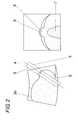
- FIG. 2shows a B-image obtained from the scanned volume as per the method of the invention and the 3D-display of the selected partial volume in the neighboring screen area.
- a 3D ultrasound transducer of a not further described ultrasound machineis coupled to an organism 2 , which it scans by the 3D volume method in a volume range obtained by moving a B-scanning plane 3 transverse to the scan area.
- a B-scanning plane 3transverse to the scan area.
- several B-image planesare being selected, in which the introduction range of a biopsy needle is adjusted, according to the preferred possibility of aligning the ultrasound transducer, to place it at half of the scan movement transverse to the image plane, which procedure is anyway necessary with a compulsory needle guide used. Otherwise from the further B-images that one may be selected which shows best the insertion range.
- the auxiliary biopsy line 4showing the course of the insertion direction as wanted resp. fixed by the needle guide is visible in the B-image 3 a of FIG. 2 . Also within that B-image additional lines 5 on both sides of the biopsy line 4 mark the limits of the partial volume as wanted for the 3D display mode.
- the thickness of the partial volumeis defined by the scan movement range normal to the B-image plane.
- the 3D-image 6 of the partial volumewhich is to be displayed beside of the sectional image plane 3 a , in which direction to the insertion direction of the biopsy needle (to be displayed horizontal or vertical, respectively) it shall lie, and if the 3D-aspect is to be shown in a front or a rear view, the left and the right side limits being defined by the scan movement range normal to the B-image.
- the image 6shows the inclusion 7 within the organ and a prominent part into which the biopsy needle should enter are incomparably better visible than in the B-image.
- the biopsy needle to be introduced therewill be also visible in image 6 , it is however not pictured.
- the direction of introductionis vertical from above downward in the middle of the image.
Landscapes
- Life Sciences & Earth Sciences (AREA)
- Health & Medical Sciences (AREA)
- Biomedical Technology (AREA)
- Biophysics (AREA)
- Nuclear Medicine, Radiotherapy & Molecular Imaging (AREA)
- Pathology (AREA)
- Radiology & Medical Imaging (AREA)
- Engineering & Computer Science (AREA)
- Physics & Mathematics (AREA)
- Heart & Thoracic Surgery (AREA)
- Medical Informatics (AREA)
- Molecular Biology (AREA)
- Surgery (AREA)
- Animal Behavior & Ethology (AREA)
- General Health & Medical Sciences (AREA)
- Public Health (AREA)
- Veterinary Medicine (AREA)
- Ultra Sonic Daignosis Equipment (AREA)
Abstract
Description
The present invention relates to a method according to the superordinate concept ofClaim 1.
Known methods of this kind are working with relatively simple B-scan imaging units, in which the position of the displayed sectional plane is determined only by way of the contact position of the ultrasound transducer and its orientation to the organ being examined. By even only small swings of the ultrasound transducer around the horizontal or vertical axis the position of the displayed sectional plane will be varied too. In addition, when scanning living organisms, by changes of the position of either the total body, or the organ being aimed at within the body or tissue layers, corrective actions will occur within the displayed sectional plane containing the “aiming point” within the organism. It is known to provide rigidly mounted guides on the ultrasound transducer for the biopsy needle or similar to be inserted mostly at a fixed distance from the ultrasound transducer toward the sectional plane determined by that ultrasound transducer. The “ideal” direction within the sectional plane can be displayed in the B-mode image by way of a superposed auxiliary line. Also the biopsy needle or similar are being observed during the introduction. Even with a non-moving organism it is then difficult to determine whether an inexactly introduced biopsy needle leaves the displayed sectional plane in or against the aspect direction and to make appropriate corrections. In case of moving organisms, however, modifications in addition to corrections of the biopsy needle also of the contact direction, especially the contact inclination of the ultrasound transducer must be made, so that only very experienced medical doctors are able to really achieve the theoretically possible accuracy of this known method. From DE 24 25 724 A it is known to equip biopsy needles with a coating well reflecting ultrasound waves or with profiles to improve the visibility in the ultrasound image, but even this does not change anything in the above mentioned problems.
To increase the accuracy, according to AT 344 872 B two ultrasound transducers are used for a B-scan image which are connected at a distance and perpendicular to each other by a guiding device for a biopsy needle, so that the scan planes of the two ultrasound transducers intersect in a straight line within the object which is the introduction axis of the biopsy needle, determined by the biopsy needle guide. So if the aimed point in the object is on the straight line and is displayed in both images, the introduced biopsy needle will hit it with a high probability. Here also the above mentioned readjustment difficulties with moving organs or organs changing their position, the adjustment and readjustment of the ultrasound transducers becomes complicated and the examining doctor has practically no hand free to introduce the biopsy needle. Furthermore an instrument equipped this way can only be used for this single purpose and it is not always possible to fix the two ultrasound transducers to the object maintaining sufficient or good coupling quality, respectively.
It has to be mentioned here that such methods generally can be used for a great variety of purposes, i.e. for the withdrawal of fluid, tissue or other samples of organs, for punctures as well as for specific punctual treatments within organisms.
The purpose of the invention to be described herein is thus the creation of a novel method compared to those mentioned in the introduction, by which the accuracy of aiming and of introducing the biopsy needle or anything similar is improved at less critical coupling accuracy of the ultrasound transducer, ensuring also a high accuracy in case of moving organs or organs changing their position and which enables less experienced doctors to work safely with high accuracy.
The problem is solved in full by the characteristics of the method described inClaim 1. In principle, an ultrasound machine with an adequate ultrasound transducer which is subject of our own EP 0 962 785 A can be used to perform the method according to this invention. This machine is to be adapted to the conditions of the method according to the invention, especially as regarding the software, whereas in addition the mentioned sectional plane shall be displayed as a whole. The sectional plane however can be selected especially in case of machines without compulsory guiding device for the biopsy needle within the scanned total volume, whereby the adjustment is only necessary in so far as the partial volume surrounding the insertion path remains safely present within the scanned total volume. In case of ultrasound machines with compulsory guiding device, basically one will select that sectional plane for the display which lies in the target direction of the biopsy needle guide.
As in the 3D-display only the actual areas destined for the introduction of the biopsy needle and their close surroundings are made visible, this way of display is less confusing compared to a display in whole, but it offers the possibility to also visualize the curvatures of shapes within the organism being transverse to the imaged B-scan plane, which e.g. result from the surface shape or the shape of separating layers, so that the introduction point of the biopsy needle can be very accurately aimed at. Due to the 3D-display, the needle itself is well visible even when it slightly leaves the sectional image plane, thus facilitating corrections of the guidance of the needle in a way unknown before. In case of unmoving organs the limits of the selected partial volume can be set relatively close to the aiming axis. With moving organs or organs changing their position, the limits are set such that even in case of movements and without major position changes of the ultrasound transducer the aiming axis remains always within this selected partial volume. Hence in most cases a compulsory guiding device connected to the ultrasound transducer will even be unnecessary.
As already mentioned, the limiting areas of the partial volume can be selected according to different criteria. An extremely simple method is described inSub-Claim 2.
The 3D-image display offers several further possibilities to increase the accuracy of introduction and for the precise determination of the direction of introduction. It is possible to rotate, tilt and turn round the partial image obtained by the 3D-method with methods already known, in order to examine it from all sides and to then select the direction of introduction and introduction point as wanted. Due to the customary way of image examination it also can show advantageous to align the 3D-image according toSub-Claim 3.
The method according to the invention is not restricted to the sectional image and 3D-image technique, but can also be used with compatible methods as indicated inClaim 4. However, in case of Doppler-coded scanning a substantial decrease of the possible scan velocities will result in order to allow the Doppler evaluation. In Claim 5 a design is specified which proves excellent with a forced guidance of the instruments.
Further details and advantages of the subject of the present invention are explained in the below mentioned description of the drawings.
In the drawing the present invention is explained by an example illustration.
FIG. 1 shows the schematic configuration of a 3D ultrasound transducer on an object, the scanned sectional plane being indicated by broken lines,
FIG. 2 shows a B-image obtained from the scanned volume as per the method of the invention and the 3D-display of the selected partial volume in the neighboring screen area.
According to FIG. 1 a 3D ultrasound transducer of a not further described ultrasound machine, but principally a 3D-image machine according to EP 0 962 785 A, is coupled to anorganism 2, which it scans by the 3D volume method in a volume range obtained by moving a B-scanning plane 3 transverse to the scan area. By a corresponding derivation several B-image planes are being selected, in which the introduction range of a biopsy needle is adjusted, according to the preferred possibility of aligning the ultrasound transducer, to place it at half of the scan movement transverse to the image plane, which procedure is anyway necessary with a compulsory needle guide used. Otherwise from the further B-images that one may be selected which shows best the insertion range. Within this insertion range theauxiliary biopsy line 4 showing the course of the insertion direction as wanted resp. fixed by the needle guide is visible in the B-image 3aof FIG.2. Also within that B-imageadditional lines 5 on both sides of thebiopsy line 4 mark the limits of the partial volume as wanted for the 3D display mode. The thickness of the partial volume is defined by the scan movement range normal to the B-image plane. Moreover it will be defined for the 3D-image 6 of the partial volume, which is to be displayed beside of thesectional image plane 3a, in which direction to the insertion direction of the biopsy needle (to be displayed horizontal or vertical, respectively) it shall lie, and if the 3D-aspect is to be shown in a front or a rear view, the left and the right side limits being defined by the scan movement range normal to the B-image. According to FIG. 2 theimage 6 shows theinclusion 7 within the organ and a prominent part into which the biopsy needle should enter are incomparably better visible than in the B-image. The biopsy needle to be introduced there will be also visible inimage 6, it is however not pictured. The direction of introduction is vertical from above downward in the middle of the image.
As indicated by the drawing in broken lines in FIG. 1 it is also possible to couple permanently a forced guiding device (9) for the biopsy needle or other instrument to the scanhead (1). Hereby, in some applications, the procedure to locate the spot of the introduction and to aim in the direction of the introduction is eased.
Claims (5)
1. A method of determining a direction of introduction and tracing an introduction path along the direction of introduction of a medical instrument into an organism, with the use of an ultrasound imaging machine suitable for 3D-imaging, comprising the steps of scanning a larger volume of the organism with a 3D-scanning Unit of the ultrasound imaging machine, generating an image of an intended introduction range in a sectional plane within which the introduction path runs, observing the introduction path in an image display of the sectional plane, the sectional plane being located by predeterminable selection criteria and the introduction path being virtually displayed, and selecting a prismatic or cylindrical part of the larger volume for display in a 3D-image in which the introduction path may be observed, the prismatic or cylindrical part of the larger volume having a main axis coaxial with the direction of introduction.
2. The method ofclaim 1 , wherein the prismatic or cylindrical part of the larger volume has lateral limits displayed in a B-image of the sectional plane, and volume limits in front of and behind the sectional plane are determined by the 3D-image generated by the 3D-scanning unit transversely to the B-image.
3. The method ofclaim 2 , wherein the 3D-image is displayed in an orientation extending vertically or horizonally with respect to the direction of introduction.
4. The method ofclaim 2 , wherein in the medical instrument is vibrated during the introduction into the organism and while remaining therein to improve the visualization of B- and 3D-images additionally coded by the Doppler-method.
5. A method of determining a direction of introduction and tracing an introduction path along the direction of introduction of a medical instrument into an organism, with the use of an ultrasound imaging machine suitable for 3D-imaging, comprising the steps of scanning a larger volume of the organism with a 3D-scanning unit of the ultrasound imaging machine, determining the direction of introduction of the medical instrument with a guiding device coupled to the 3D-scanning unit, the direction of introduction being determined by the position of the guiding device relative to the 3D-scanning unit, generating a B-image of an intended introduction range in a sectional plane within which the introduction path runs, aligning the 3D-scanning unit with the guiding device coupled thereto with the sectional plane in the generated B-image, observing the introduction path in an image display of the sectional plane, the sectional plane being located by predeterminable selection criteria and the introduction path being virtually displayed, and selecting a prismatic or cylindrical part of the larger volume for display in a 3D-image in which the introduction path may be observed, the primsmatic or cylindrical part of the larger volume having a main axis coaxial with the direction of introduction.
Applications Claiming Priority (3)
| Application Number | Priority Date | Filing Date | Title |
|---|---|---|---|
| EP00890342 | 2000-11-16 | ||
| EP00890342.9 | 2000-11-16 | ||
| EP00890342AEP1208799A1 (en) | 2000-11-16 | 2000-11-16 | Method for determining the insertion direction of a biopsy needle and for controlling its trajectory |
Publications (2)
| Publication Number | Publication Date |
|---|---|
| US20020058872A1 US20020058872A1 (en) | 2002-05-16 |
| US6616610B2true US6616610B2 (en) | 2003-09-09 |
Family
ID=8175985
Family Applications (1)
| Application Number | Title | Priority Date | Filing Date |
|---|---|---|---|
| US10/002,095Expired - LifetimeUS6616610B2 (en) | 2000-11-16 | 2001-11-15 | Method for determination of the direction of introduction and for controlling the introduction path of biopsy needles |
Country Status (3)
| Country | Link |
|---|---|
| US (1) | US6616610B2 (en) |
| EP (1) | EP1208799A1 (en) |
| JP (2) | JP4041307B2 (en) |
Cited By (53)
| Publication number | Priority date | Publication date | Assignee | Title |
|---|---|---|---|---|
| US6786870B2 (en)* | 2001-10-23 | 2004-09-07 | Olympus Corporation | Device for examining a subject capable of marking a boundary range for insertion/retraction of an insertion/retraction member that is inserted in and retracted from the subject |
| US20050093889A1 (en)* | 2001-03-27 | 2005-05-05 | Frank Sauer | Augmented reality guided instrument positioning with guiding graphics |
| US20050131291A1 (en)* | 2003-12-10 | 2005-06-16 | Sonosite, Inc. | Device for assisting the positioning of medical devices |
| US20090005687A1 (en)* | 2007-06-27 | 2009-01-01 | Sotaro Kawae | Ultrasonic imaging apparatus |
| US20090171219A1 (en)* | 2007-12-27 | 2009-07-02 | Masami Uchibori | Biopsy guide mounting structure, ultrasonic probe, and ultrasonic diagnostic apparatus |
| US20100217117A1 (en)* | 2009-02-25 | 2010-08-26 | Neil David Glossop | Method, system and devices for transjugular intrahepatic portosystemic shunt (tips) procedures |
| US20110160588A1 (en)* | 2008-09-09 | 2011-06-30 | Olympus Medical Systems Corp. | Ultrasound image display apparatus and ultrasound image display method |
| US20120289820A1 (en)* | 2011-05-10 | 2012-11-15 | The University Of British Columbia | Apparatus And Method For Imaging A Medical Instrument |
| US8482606B2 (en) | 2006-08-02 | 2013-07-09 | Inneroptic Technology, Inc. | System and method of providing real-time dynamic imagery of a medical procedure site using multiple modalities |
| US8554307B2 (en) | 2010-04-12 | 2013-10-08 | Inneroptic Technology, Inc. | Image annotation in image-guided medical procedures |
| US8585598B2 (en) | 2009-02-17 | 2013-11-19 | Inneroptic Technology, Inc. | Systems, methods, apparatuses, and computer-readable media for image guided surgery |
| US8641621B2 (en) | 2009-02-17 | 2014-02-04 | Inneroptic Technology, Inc. | Systems, methods, apparatuses, and computer-readable media for image management in image-guided medical procedures |
| US8670816B2 (en) | 2012-01-30 | 2014-03-11 | Inneroptic Technology, Inc. | Multiple medical device guidance |
| US8781555B2 (en) | 2007-11-26 | 2014-07-15 | C. R. Bard, Inc. | System for placement of a catheter including a signal-generating stylet |
| US8784336B2 (en) | 2005-08-24 | 2014-07-22 | C. R. Bard, Inc. | Stylet apparatuses and methods of manufacture |
| US8831310B2 (en) | 2008-03-07 | 2014-09-09 | Inneroptic Technology, Inc. | Systems and methods for displaying guidance data based on updated deformable imaging data |
| US8849382B2 (en) | 2007-11-26 | 2014-09-30 | C. R. Bard, Inc. | Apparatus and display methods relating to intravascular placement of a catheter |
| US8858455B2 (en) | 2006-10-23 | 2014-10-14 | Bard Access Systems, Inc. | Method of locating the tip of a central venous catheter |
| US9125578B2 (en) | 2009-06-12 | 2015-09-08 | Bard Access Systems, Inc. | Apparatus and method for catheter navigation and tip location |
| US9265572B2 (en) | 2008-01-24 | 2016-02-23 | The University Of North Carolina At Chapel Hill | Methods, systems, and computer readable media for image guided ablation |
| US9265443B2 (en) | 2006-10-23 | 2016-02-23 | Bard Access Systems, Inc. | Method of locating the tip of a central venous catheter |
| US9339206B2 (en) | 2009-06-12 | 2016-05-17 | Bard Access Systems, Inc. | Adaptor for endovascular electrocardiography |
| US9415188B2 (en) | 2010-10-29 | 2016-08-16 | C. R. Bard, Inc. | Bioimpedance-assisted placement of a medical device |
| US9445734B2 (en) | 2009-06-12 | 2016-09-20 | Bard Access Systems, Inc. | Devices and methods for endovascular electrography |
| US9456766B2 (en) | 2007-11-26 | 2016-10-04 | C. R. Bard, Inc. | Apparatus for use with needle insertion guidance system |
| US9492097B2 (en) | 2007-11-26 | 2016-11-15 | C. R. Bard, Inc. | Needle length determination and calibration for insertion guidance system |
| US9521961B2 (en) | 2007-11-26 | 2016-12-20 | C. R. Bard, Inc. | Systems and methods for guiding a medical instrument |
| US9532724B2 (en) | 2009-06-12 | 2017-01-03 | Bard Access Systems, Inc. | Apparatus and method for catheter navigation using endovascular energy mapping |
| US9554716B2 (en) | 2007-11-26 | 2017-01-31 | C. R. Bard, Inc. | Insertion guidance system for needles and medical components |
| US9636031B2 (en) | 2007-11-26 | 2017-05-02 | C.R. Bard, Inc. | Stylets for use with apparatus for intravascular placement of a catheter |
| US9649048B2 (en) | 2007-11-26 | 2017-05-16 | C. R. Bard, Inc. | Systems and methods for breaching a sterile field for intravascular placement of a catheter |
| US9675319B1 (en) | 2016-02-17 | 2017-06-13 | Inneroptic Technology, Inc. | Loupe display |
| US9681823B2 (en) | 2007-11-26 | 2017-06-20 | C. R. Bard, Inc. | Integrated system for intravascular placement of a catheter |
| US9839372B2 (en) | 2014-02-06 | 2017-12-12 | C. R. Bard, Inc. | Systems and methods for guidance and placement of an intravascular device |
| US9901406B2 (en) | 2014-10-02 | 2018-02-27 | Inneroptic Technology, Inc. | Affected region display associated with a medical device |
| US9901714B2 (en) | 2008-08-22 | 2018-02-27 | C. R. Bard, Inc. | Catheter assembly including ECG sensor and magnetic assemblies |
| US9907513B2 (en) | 2008-10-07 | 2018-03-06 | Bard Access Systems, Inc. | Percutaneous magnetic gastrostomy |
| US9949700B2 (en) | 2015-07-22 | 2018-04-24 | Inneroptic Technology, Inc. | Medical device approaches |
| US10046139B2 (en) | 2010-08-20 | 2018-08-14 | C. R. Bard, Inc. | Reconfirmation of ECG-assisted catheter tip placement |
| US10188467B2 (en) | 2014-12-12 | 2019-01-29 | Inneroptic Technology, Inc. | Surgical guidance intersection display |
| US10278778B2 (en) | 2016-10-27 | 2019-05-07 | Inneroptic Technology, Inc. | Medical device navigation using a virtual 3D space |
| US10314559B2 (en) | 2013-03-14 | 2019-06-11 | Inneroptic Technology, Inc. | Medical device guidance |
| US10349890B2 (en) | 2015-06-26 | 2019-07-16 | C. R. Bard, Inc. | Connector interface for ECG-based catheter positioning system |
| US10449330B2 (en) | 2007-11-26 | 2019-10-22 | C. R. Bard, Inc. | Magnetic element-equipped needle assemblies |
| US10524691B2 (en) | 2007-11-26 | 2020-01-07 | C. R. Bard, Inc. | Needle assembly including an aligned magnetic element |
| US10751509B2 (en) | 2007-11-26 | 2020-08-25 | C. R. Bard, Inc. | Iconic representations for guidance of an indwelling medical device |
| US10973584B2 (en) | 2015-01-19 | 2021-04-13 | Bard Access Systems, Inc. | Device and method for vascular access |
| US10992079B2 (en) | 2018-10-16 | 2021-04-27 | Bard Access Systems, Inc. | Safety-equipped connection systems and methods thereof for establishing electrical connections |
| US11000207B2 (en) | 2016-01-29 | 2021-05-11 | C. R. Bard, Inc. | Multiple coil system for tracking a medical device |
| US11259879B2 (en) | 2017-08-01 | 2022-03-01 | Inneroptic Technology, Inc. | Selective transparency to assist medical device navigation |
| US11464578B2 (en) | 2009-02-17 | 2022-10-11 | Inneroptic Technology, Inc. | Systems, methods, apparatuses, and computer-readable media for image management in image-guided medical procedures |
| US11484365B2 (en) | 2018-01-23 | 2022-11-01 | Inneroptic Technology, Inc. | Medical image guidance |
| US11918795B2 (en) | 2019-05-01 | 2024-03-05 | Bard Access Systems, Inc. | Puncturing devices, puncturing systems including the puncturing devices, and methods thereof |
Families Citing this family (16)
| Publication number | Priority date | Publication date | Assignee | Title |
|---|---|---|---|---|
| US6755789B2 (en)* | 2002-02-05 | 2004-06-29 | Inceptio Medical Technologies, Llc | Ultrasonic vascular imaging system and method of blood vessel cannulation |
| US7806828B2 (en)* | 2002-02-05 | 2010-10-05 | Inceptio Medical Technologies, Lc | Multiplanar ultrasonic vascular sensor assembly and apparatus for movably affixing a sensor assembly to a body |
| JP4205957B2 (en)* | 2003-01-09 | 2009-01-07 | アロカ株式会社 | Ultrasonic diagnostic equipment |
| US7660623B2 (en)* | 2003-01-30 | 2010-02-09 | Medtronic Navigation, Inc. | Six degree of freedom alignment display for medical procedures |
| JP4575033B2 (en)* | 2004-06-02 | 2010-11-04 | 株式会社東芝 | Ultrasonic diagnostic apparatus and method of operating ultrasonic diagnostic apparatus |
| US10022202B2 (en) | 2013-03-15 | 2018-07-17 | Triagenics, Llc | Therapeutic tooth bud ablation |
| CA2761652C (en)* | 2009-05-11 | 2019-10-01 | Leigh E. Colby | Therapeutic tooth bud ablation |
| WO2014143014A1 (en) | 2013-03-15 | 2014-09-18 | Triagenics, Llc | Therapeutic tooth bud ablation |
| US9438264B1 (en) | 2015-09-10 | 2016-09-06 | Realtek Semiconductor Corp. | High-speed capacitive digital-to-analog converter and method thereof |
| US9820723B2 (en) | 2013-12-04 | 2017-11-21 | Choon Kee Lee | Positioning guide apparatus with friction lock |
| US9649161B2 (en) | 2014-01-21 | 2017-05-16 | Choon Kee Lee | Stereotactic positioning guide apparatus |
| US9492232B2 (en) | 2014-02-23 | 2016-11-15 | Choon Kee Lee | Powered stereotactic positioning guide apparatus |
| US9649162B2 (en) | 2014-06-22 | 2017-05-16 | Choon Kee Lee | Stereotactic positioning guide apparatus |
| US9655686B2 (en) | 2014-08-18 | 2017-05-23 | Choon Kee Lee | Automated stereotactic apparatus |
| US9687209B2 (en) | 2014-10-09 | 2017-06-27 | Choon Kee Lee | Invasive device positioning assembly |
| EP4413935A3 (en) | 2019-06-06 | 2024-09-18 | TriAgenics, Inc. | Ablation probe systems |
Citations (5)
| Publication number | Priority date | Publication date | Assignee | Title |
|---|---|---|---|---|
| US5967991A (en) | 1996-12-03 | 1999-10-19 | Echocath, Inc. | Drive apparatus for an interventional medical device used in an ultrasonic imaging system |
| EP0962785A1 (en) | 1998-06-04 | 1999-12-08 | Kretztechnik Aktiengesellschaft | Method for examining objects with ultrasound |
| US6019724A (en) | 1995-02-22 | 2000-02-01 | Gronningsaeter; Aage | Method for ultrasound guidance during clinical procedures |
| US6336899B1 (en)* | 1998-10-14 | 2002-01-08 | Kabushiki Kaisha Toshiba | Ultrasonic diagnosis apparatus |
| US6416476B1 (en)* | 1999-01-12 | 2002-07-09 | Kabushiki Kaisha Toshiba | Three-dimensional ultrasonic diagnosis apparatus |
Family Cites Families (4)
| Publication number | Priority date | Publication date | Assignee | Title |
|---|---|---|---|---|
| DE2425724C3 (en) | 1974-05-28 | 1979-09-27 | Siemens Ag, 1000 Berlin Und 8000 Muenchen | Puncture cannula |
| AT344872B (en) | 1975-12-23 | 1978-08-10 | Kretztechnik Gmbh | DEVICE FOR SIGHTING AREAS LOCATING IN A OBJECT |
| JPH07116164A (en)* | 1993-10-25 | 1995-05-09 | Hitachi Medical Corp | Ultrasonic tomographic apparatus |
| JPH1057376A (en)* | 1996-08-16 | 1998-03-03 | Ge Yokogawa Medical Syst Ltd | Stab needle position detection method, stab needle vibrating device, vibrating liquid injection device and ultrosonograph |
- 2000
- 2000-11-16EPEP00890342Apatent/EP1208799A1/ennot_activeWithdrawn
- 2001
- 2001-11-09JPJP2001380714Apatent/JP4041307B2/ennot_activeExpired - Fee Related
- 2001-11-15USUS10/002,095patent/US6616610B2/ennot_activeExpired - Lifetime
- 2007
- 2007-06-29JPJP2007195186Apatent/JP4729018B2/ennot_activeExpired - Fee Related
Patent Citations (5)
| Publication number | Priority date | Publication date | Assignee | Title |
|---|---|---|---|---|
| US6019724A (en) | 1995-02-22 | 2000-02-01 | Gronningsaeter; Aage | Method for ultrasound guidance during clinical procedures |
| US5967991A (en) | 1996-12-03 | 1999-10-19 | Echocath, Inc. | Drive apparatus for an interventional medical device used in an ultrasonic imaging system |
| EP0962785A1 (en) | 1998-06-04 | 1999-12-08 | Kretztechnik Aktiengesellschaft | Method for examining objects with ultrasound |
| US6336899B1 (en)* | 1998-10-14 | 2002-01-08 | Kabushiki Kaisha Toshiba | Ultrasonic diagnosis apparatus |
| US6416476B1 (en)* | 1999-01-12 | 2002-07-09 | Kabushiki Kaisha Toshiba | Three-dimensional ultrasonic diagnosis apparatus |
Cited By (109)
| Publication number | Priority date | Publication date | Assignee | Title |
|---|---|---|---|---|
| US7239330B2 (en)* | 2001-03-27 | 2007-07-03 | Siemens Corporate Research, Inc. | Augmented reality guided instrument positioning with guiding graphics |
| US20050093889A1 (en)* | 2001-03-27 | 2005-05-05 | Frank Sauer | Augmented reality guided instrument positioning with guiding graphics |
| US6786870B2 (en)* | 2001-10-23 | 2004-09-07 | Olympus Corporation | Device for examining a subject capable of marking a boundary range for insertion/retraction of an insertion/retraction member that is inserted in and retracted from the subject |
| US7588541B2 (en) | 2003-12-10 | 2009-09-15 | Sonosite, Inc. | Method and system for positioning a medical device at one or more angles relative to an imaging probe |
| US20050131291A1 (en)* | 2003-12-10 | 2005-06-16 | Sonosite, Inc. | Device for assisting the positioning of medical devices |
| US20090270722A1 (en)* | 2003-12-10 | 2009-10-29 | Sonosite, Inc. | Device for assisting the positioning of medical devices |
| US10004875B2 (en) | 2005-08-24 | 2018-06-26 | C. R. Bard, Inc. | Stylet apparatuses and methods of manufacture |
| US8784336B2 (en) | 2005-08-24 | 2014-07-22 | C. R. Bard, Inc. | Stylet apparatuses and methods of manufacture |
| US11207496B2 (en) | 2005-08-24 | 2021-12-28 | C. R. Bard, Inc. | Stylet apparatuses and methods of manufacture |
| US10733700B2 (en) | 2006-08-02 | 2020-08-04 | Inneroptic Technology, Inc. | System and method of providing real-time dynamic imagery of a medical procedure site using multiple modalities |
| US9659345B2 (en) | 2006-08-02 | 2017-05-23 | Inneroptic Technology, Inc. | System and method of providing real-time dynamic imagery of a medical procedure site using multiple modalities |
| US10127629B2 (en) | 2006-08-02 | 2018-11-13 | Inneroptic Technology, Inc. | System and method of providing real-time dynamic imagery of a medical procedure site using multiple modalities |
| US8482606B2 (en) | 2006-08-02 | 2013-07-09 | Inneroptic Technology, Inc. | System and method of providing real-time dynamic imagery of a medical procedure site using multiple modalities |
| US11481868B2 (en) | 2006-08-02 | 2022-10-25 | Inneroptic Technology, Inc. | System and method of providing real-time dynamic imagery of a medical procedure she using multiple modalities |
| US9833169B2 (en) | 2006-10-23 | 2017-12-05 | Bard Access Systems, Inc. | Method of locating the tip of a central venous catheter |
| US9265443B2 (en) | 2006-10-23 | 2016-02-23 | Bard Access Systems, Inc. | Method of locating the tip of a central venous catheter |
| US9345422B2 (en) | 2006-10-23 | 2016-05-24 | Bard Acess Systems, Inc. | Method of locating the tip of a central venous catheter |
| US8858455B2 (en) | 2006-10-23 | 2014-10-14 | Bard Access Systems, Inc. | Method of locating the tip of a central venous catheter |
| US20090005687A1 (en)* | 2007-06-27 | 2009-01-01 | Sotaro Kawae | Ultrasonic imaging apparatus |
| US10602958B2 (en) | 2007-11-26 | 2020-03-31 | C. R. Bard, Inc. | Systems and methods for guiding a medical instrument |
| US9456766B2 (en) | 2007-11-26 | 2016-10-04 | C. R. Bard, Inc. | Apparatus for use with needle insertion guidance system |
| US10751509B2 (en) | 2007-11-26 | 2020-08-25 | C. R. Bard, Inc. | Iconic representations for guidance of an indwelling medical device |
| US8849382B2 (en) | 2007-11-26 | 2014-09-30 | C. R. Bard, Inc. | Apparatus and display methods relating to intravascular placement of a catheter |
| US10849695B2 (en) | 2007-11-26 | 2020-12-01 | C. R. Bard, Inc. | Systems and methods for breaching a sterile field for intravascular placement of a catheter |
| US9999371B2 (en) | 2007-11-26 | 2018-06-19 | C. R. Bard, Inc. | Integrated system for intravascular placement of a catheter |
| US10524691B2 (en) | 2007-11-26 | 2020-01-07 | C. R. Bard, Inc. | Needle assembly including an aligned magnetic element |
| US10966630B2 (en) | 2007-11-26 | 2021-04-06 | C. R. Bard, Inc. | Integrated system for intravascular placement of a catheter |
| US11123099B2 (en) | 2007-11-26 | 2021-09-21 | C. R. Bard, Inc. | Apparatus for use with needle insertion guidance system |
| US10449330B2 (en) | 2007-11-26 | 2019-10-22 | C. R. Bard, Inc. | Magnetic element-equipped needle assemblies |
| US11134915B2 (en) | 2007-11-26 | 2021-10-05 | C. R. Bard, Inc. | System for placement of a catheter including a signal-generating stylet |
| US10342575B2 (en) | 2007-11-26 | 2019-07-09 | C. R. Bard, Inc. | Apparatus for use with needle insertion guidance system |
| US10238418B2 (en) | 2007-11-26 | 2019-03-26 | C. R. Bard, Inc. | Apparatus for use with needle insertion guidance system |
| US10231753B2 (en) | 2007-11-26 | 2019-03-19 | C. R. Bard, Inc. | Insertion guidance system for needles and medical components |
| US10165962B2 (en) | 2007-11-26 | 2019-01-01 | C. R. Bard, Inc. | Integrated systems for intravascular placement of a catheter |
| US8781555B2 (en) | 2007-11-26 | 2014-07-15 | C. R. Bard, Inc. | System for placement of a catheter including a signal-generating stylet |
| US9492097B2 (en) | 2007-11-26 | 2016-11-15 | C. R. Bard, Inc. | Needle length determination and calibration for insertion guidance system |
| US9521961B2 (en) | 2007-11-26 | 2016-12-20 | C. R. Bard, Inc. | Systems and methods for guiding a medical instrument |
| US9526440B2 (en) | 2007-11-26 | 2016-12-27 | C.R. Bard, Inc. | System for placement of a catheter including a signal-generating stylet |
| US11529070B2 (en) | 2007-11-26 | 2022-12-20 | C. R. Bard, Inc. | System and methods for guiding a medical instrument |
| US9549685B2 (en) | 2007-11-26 | 2017-01-24 | C. R. Bard, Inc. | Apparatus and display methods relating to intravascular placement of a catheter |
| US9554716B2 (en) | 2007-11-26 | 2017-01-31 | C. R. Bard, Inc. | Insertion guidance system for needles and medical components |
| US9636031B2 (en) | 2007-11-26 | 2017-05-02 | C.R. Bard, Inc. | Stylets for use with apparatus for intravascular placement of a catheter |
| US9649048B2 (en) | 2007-11-26 | 2017-05-16 | C. R. Bard, Inc. | Systems and methods for breaching a sterile field for intravascular placement of a catheter |
| US11707205B2 (en) | 2007-11-26 | 2023-07-25 | C. R. Bard, Inc. | Integrated system for intravascular placement of a catheter |
| US10105121B2 (en) | 2007-11-26 | 2018-10-23 | C. R. Bard, Inc. | System for placement of a catheter including a signal-generating stylet |
| US9681823B2 (en) | 2007-11-26 | 2017-06-20 | C. R. Bard, Inc. | Integrated system for intravascular placement of a catheter |
| US11779240B2 (en) | 2007-11-26 | 2023-10-10 | C. R. Bard, Inc. | Systems and methods for breaching a sterile field for intravascular placement of a catheter |
| US20090171219A1 (en)* | 2007-12-27 | 2009-07-02 | Masami Uchibori | Biopsy guide mounting structure, ultrasonic probe, and ultrasonic diagnostic apparatus |
| US9265572B2 (en) | 2008-01-24 | 2016-02-23 | The University Of North Carolina At Chapel Hill | Methods, systems, and computer readable media for image guided ablation |
| US8831310B2 (en) | 2008-03-07 | 2014-09-09 | Inneroptic Technology, Inc. | Systems and methods for displaying guidance data based on updated deformable imaging data |
| US9901714B2 (en) | 2008-08-22 | 2018-02-27 | C. R. Bard, Inc. | Catheter assembly including ECG sensor and magnetic assemblies |
| US11027101B2 (en) | 2008-08-22 | 2021-06-08 | C. R. Bard, Inc. | Catheter assembly including ECG sensor and magnetic assemblies |
| US20110160588A1 (en)* | 2008-09-09 | 2011-06-30 | Olympus Medical Systems Corp. | Ultrasound image display apparatus and ultrasound image display method |
| US9907513B2 (en) | 2008-10-07 | 2018-03-06 | Bard Access Systems, Inc. | Percutaneous magnetic gastrostomy |
| US11464575B2 (en) | 2009-02-17 | 2022-10-11 | Inneroptic Technology, Inc. | Systems, methods, apparatuses, and computer-readable media for image guided surgery |
| US11464578B2 (en) | 2009-02-17 | 2022-10-11 | Inneroptic Technology, Inc. | Systems, methods, apparatuses, and computer-readable media for image management in image-guided medical procedures |
| US9364294B2 (en) | 2009-02-17 | 2016-06-14 | Inneroptic Technology, Inc. | Systems, methods, apparatuses, and computer-readable media for image management in image-guided medical procedures |
| US10136951B2 (en) | 2009-02-17 | 2018-11-27 | Inneroptic Technology, Inc. | Systems, methods, apparatuses, and computer-readable media for image guided surgery |
| US8585598B2 (en) | 2009-02-17 | 2013-11-19 | Inneroptic Technology, Inc. | Systems, methods, apparatuses, and computer-readable media for image guided surgery |
| US10398513B2 (en) | 2009-02-17 | 2019-09-03 | Inneroptic Technology, Inc. | Systems, methods, apparatuses, and computer-readable media for image management in image-guided medical procedures |
| US8641621B2 (en) | 2009-02-17 | 2014-02-04 | Inneroptic Technology, Inc. | Systems, methods, apparatuses, and computer-readable media for image management in image-guided medical procedures |
| US12419695B2 (en) | 2009-02-17 | 2025-09-23 | Inneroptic Technology, Inc. | Systems, methods, apparatuses, and computer-readable media for image management in image-guided medical procedures |
| US9398936B2 (en) | 2009-02-17 | 2016-07-26 | Inneroptic Technology, Inc. | Systems, methods, apparatuses, and computer-readable media for image guided surgery |
| US8690776B2 (en) | 2009-02-17 | 2014-04-08 | Inneroptic Technology, Inc. | Systems, methods, apparatuses, and computer-readable media for image guided surgery |
| US20100217117A1 (en)* | 2009-02-25 | 2010-08-26 | Neil David Glossop | Method, system and devices for transjugular intrahepatic portosystemic shunt (tips) procedures |
| US8632468B2 (en)* | 2009-02-25 | 2014-01-21 | Koninklijke Philips N.V. | Method, system and devices for transjugular intrahepatic portosystemic shunt (TIPS) procedures |
| US11419517B2 (en) | 2009-06-12 | 2022-08-23 | Bard Access Systems, Inc. | Apparatus and method for catheter navigation using endovascular energy mapping |
| US10231643B2 (en) | 2009-06-12 | 2019-03-19 | Bard Access Systems, Inc. | Apparatus and method for catheter navigation and tip location |
| US9445734B2 (en) | 2009-06-12 | 2016-09-20 | Bard Access Systems, Inc. | Devices and methods for endovascular electrography |
| US10912488B2 (en) | 2009-06-12 | 2021-02-09 | Bard Access Systems, Inc. | Apparatus and method for catheter navigation and tip location |
| US9339206B2 (en) | 2009-06-12 | 2016-05-17 | Bard Access Systems, Inc. | Adaptor for endovascular electrocardiography |
| US9125578B2 (en) | 2009-06-12 | 2015-09-08 | Bard Access Systems, Inc. | Apparatus and method for catheter navigation and tip location |
| US10271762B2 (en) | 2009-06-12 | 2019-04-30 | Bard Access Systems, Inc. | Apparatus and method for catheter navigation using endovascular energy mapping |
| US9532724B2 (en) | 2009-06-12 | 2017-01-03 | Bard Access Systems, Inc. | Apparatus and method for catheter navigation using endovascular energy mapping |
| US8554307B2 (en) | 2010-04-12 | 2013-10-08 | Inneroptic Technology, Inc. | Image annotation in image-guided medical procedures |
| US9107698B2 (en) | 2010-04-12 | 2015-08-18 | Inneroptic Technology, Inc. | Image annotation in image-guided medical procedures |
| US10046139B2 (en) | 2010-08-20 | 2018-08-14 | C. R. Bard, Inc. | Reconfirmation of ECG-assisted catheter tip placement |
| US9415188B2 (en) | 2010-10-29 | 2016-08-16 | C. R. Bard, Inc. | Bioimpedance-assisted placement of a medical device |
| US8696582B2 (en)* | 2011-05-10 | 2014-04-15 | The University Of British Columbia | Apparatus and method for imaging a medical instrument |
| US20120289820A1 (en)* | 2011-05-10 | 2012-11-15 | The University Of British Columbia | Apparatus And Method For Imaging A Medical Instrument |
| US8670816B2 (en) | 2012-01-30 | 2014-03-11 | Inneroptic Technology, Inc. | Multiple medical device guidance |
| US10314559B2 (en) | 2013-03-14 | 2019-06-11 | Inneroptic Technology, Inc. | Medical device guidance |
| US10863920B2 (en) | 2014-02-06 | 2020-12-15 | C. R. Bard, Inc. | Systems and methods for guidance and placement of an intravascular device |
| US9839372B2 (en) | 2014-02-06 | 2017-12-12 | C. R. Bard, Inc. | Systems and methods for guidance and placement of an intravascular device |
| US10820944B2 (en) | 2014-10-02 | 2020-11-03 | Inneroptic Technology, Inc. | Affected region display based on a variance parameter associated with a medical device |
| US9901406B2 (en) | 2014-10-02 | 2018-02-27 | Inneroptic Technology, Inc. | Affected region display associated with a medical device |
| US11684429B2 (en) | 2014-10-02 | 2023-06-27 | Inneroptic Technology, Inc. | Affected region display associated with a medical device |
| US12262960B2 (en) | 2014-10-02 | 2025-04-01 | Inneroptic Technology, Inc. | Affected region display associated with a medical device |
| US10820946B2 (en) | 2014-12-12 | 2020-11-03 | Inneroptic Technology, Inc. | Surgical guidance intersection display |
| US11534245B2 (en) | 2014-12-12 | 2022-12-27 | Inneroptic Technology, Inc. | Surgical guidance intersection display |
| US10188467B2 (en) | 2014-12-12 | 2019-01-29 | Inneroptic Technology, Inc. | Surgical guidance intersection display |
| US11931117B2 (en) | 2014-12-12 | 2024-03-19 | Inneroptic Technology, Inc. | Surgical guidance intersection display |
| US10973584B2 (en) | 2015-01-19 | 2021-04-13 | Bard Access Systems, Inc. | Device and method for vascular access |
| US10349890B2 (en) | 2015-06-26 | 2019-07-16 | C. R. Bard, Inc. | Connector interface for ECG-based catheter positioning system |
| US11026630B2 (en) | 2015-06-26 | 2021-06-08 | C. R. Bard, Inc. | Connector interface for ECG-based catheter positioning system |
| US9949700B2 (en) | 2015-07-22 | 2018-04-24 | Inneroptic Technology, Inc. | Medical device approaches |
| US11103200B2 (en) | 2015-07-22 | 2021-08-31 | Inneroptic Technology, Inc. | Medical device approaches |
| US11000207B2 (en) | 2016-01-29 | 2021-05-11 | C. R. Bard, Inc. | Multiple coil system for tracking a medical device |
| US10433814B2 (en) | 2016-02-17 | 2019-10-08 | Inneroptic Technology, Inc. | Loupe display |
| US9675319B1 (en) | 2016-02-17 | 2017-06-13 | Inneroptic Technology, Inc. | Loupe display |
| US11179136B2 (en) | 2016-02-17 | 2021-11-23 | Inneroptic Technology, Inc. | Loupe display |
| US10278778B2 (en) | 2016-10-27 | 2019-05-07 | Inneroptic Technology, Inc. | Medical device navigation using a virtual 3D space |
| US10772686B2 (en) | 2016-10-27 | 2020-09-15 | Inneroptic Technology, Inc. | Medical device navigation using a virtual 3D space |
| US11369439B2 (en) | 2016-10-27 | 2022-06-28 | Inneroptic Technology, Inc. | Medical device navigation using a virtual 3D space |
| US11259879B2 (en) | 2017-08-01 | 2022-03-01 | Inneroptic Technology, Inc. | Selective transparency to assist medical device navigation |
| US11484365B2 (en) | 2018-01-23 | 2022-11-01 | Inneroptic Technology, Inc. | Medical image guidance |
| US11621518B2 (en) | 2018-10-16 | 2023-04-04 | Bard Access Systems, Inc. | Safety-equipped connection systems and methods thereof for establishing electrical connections |
| US10992079B2 (en) | 2018-10-16 | 2021-04-27 | Bard Access Systems, Inc. | Safety-equipped connection systems and methods thereof for establishing electrical connections |
| US11918795B2 (en) | 2019-05-01 | 2024-03-05 | Bard Access Systems, Inc. | Puncturing devices, puncturing systems including the puncturing devices, and methods thereof |
Also Published As
| Publication number | Publication date |
|---|---|
| JP2007301399A (en) | 2007-11-22 |
| JP4041307B2 (en) | 2008-01-30 |
| JP4729018B2 (en) | 2011-07-20 |
| JP2002219128A (en) | 2002-08-06 |
| EP1208799A1 (en) | 2002-05-29 |
| US20020058872A1 (en) | 2002-05-16 |
Similar Documents
| Publication | Publication Date | Title |
|---|---|---|
| US6616610B2 (en) | Method for determination of the direction of introduction and for controlling the introduction path of biopsy needles | |
| JP5992539B2 (en) | Ultrasound guidance of needle path in biopsy | |
| JP4467927B2 (en) | Ultrasonic diagnostic equipment | |
| US6733458B1 (en) | Diagnostic medical ultrasound systems and methods using image based freehand needle guidance | |
| JP4828802B2 (en) | Ultrasonic diagnostic equipment for puncture therapy | |
| US10624607B2 (en) | Method for guiding the insertion of a surgical instrument with three dimensional ultrasonic imaging | |
| CN115348839A (en) | Ultrasound probe, user console, system and method | |
| US20090069679A1 (en) | Ultrasound diagnostic apparatus | |
| US20130096430A1 (en) | Ultrasonic diagnostic apparatus and ultrasonic scanning method | |
| US6572549B1 (en) | High frame rate extended field of view ultrasound imaging system and method | |
| JP5015580B2 (en) | Ultrasonic diagnostic apparatus and report image creation method | |
| JP2001526927A (en) | Method and apparatus for calibrating a position sensor on a scanning transducer | |
| KR20070061466A (en) | Ultrasound Probe and Ultrasound Diagnostic Device | |
| KR20100087521A (en) | Ultrasound system and method for providing image indicator | |
| RU2598048C2 (en) | Three-dimensional ultrasonic control of surgical tools | |
| US20230380805A1 (en) | Systems and methods for tissue characterization using multiple aperture ultrasound | |
| WO2018195824A1 (en) | Ultrasound imaging device, ultrasound image enhancement method and guided puncture display method | |
| EP3537981B1 (en) | Ultrasound system for enhanced instrument visualization | |
| WO2015092628A1 (en) | Ultrasound imaging systems and methods for tracking locations of an invasive medical device | |
| JP4943969B2 (en) | Ultrasonic diagnostic equipment | |
| EP3471619B1 (en) | Image orientation identification for an external microconvex-linear ultrasound probe | |
| JP4634814B2 (en) | Ultrasonic diagnostic equipment | |
| JP7359414B2 (en) | medical imaging equipment | |
| JP2004141346A (en) | Puncture difficulty evaluation device | |
| JP2023172273A (en) | Ultrasonic diagnostic device and attenuation measurement method |
Legal Events
| Date | Code | Title | Description |
|---|---|---|---|
| AS | Assignment | Owner name:KRETZTECHNIK AG, AUSTRIA Free format text:ASSIGNMENT OF ASSIGNORS INTEREST;ASSIGNORS:STEININGER, JOSEF;GRITZKY, ARTHUR;REEL/FRAME:012352/0148 Effective date:20011024 | |
| AS | Assignment | Owner name:GE MEDICAL SYSTEMS KRETZTECHNIK GMBH & CO OHG, AUS Free format text:CHANGE OF NAME;ASSIGNOR:KRETZTECHNIK AKTIENGESELLSCHAFT;REEL/FRAME:014319/0695 Effective date:20020518 | |
| STCF | Information on status: patent grant | Free format text:PATENTED CASE | |
| FEPP | Fee payment procedure | Free format text:PAYOR NUMBER ASSIGNED (ORIGINAL EVENT CODE: ASPN); ENTITY STATUS OF PATENT OWNER: LARGE ENTITY | |
| FPAY | Fee payment | Year of fee payment:4 | |
| FPAY | Fee payment | Year of fee payment:8 | |
| FPAY | Fee payment | Year of fee payment:12 |